superoxide induces enos protein thiyl radical formation: a novel mechanism regulating enos function...
TRANSCRIPT

1
Superoxide Induces eNOS Protein Thiyl Radical Formation: a novel mechanism regulating eNOS
function and coupling
Chun-An Chen, Cho-Hao Lin, Lawrence J. Druhan, Tse-Yao Wang, Yeong-Renn Chen,
Jay L Zweier
From the Davis Heart and Lung Research Institute and Division of Cardiovascular Medicine,
Department of Internal Medicine, College of Medicine, The Ohio State University, Columbus, OH
43210, USA.
Running title: eNOS Protein Thiyl Radical Formation
Address for correspondence: Jay L. Zweier, Davis Heart and Lung Research Institute, 473 W. 12th Ave.,
Columbus, Ohio 43210, Tel. 614-247-7788, Fax. 614-247-7845, email: [email protected]
An increase in production of reactive oxygen
species (ROS) resulting in a decrease in nitric
oxide bioavailability in the endothelium
contributes to many cardiovascular diseases
and these ROS can oxidize cellular
macromolecules. Protein thiols are critical
reducing equivalents that maintain cellular
redox-state and are primary targets for
oxidative modification. We demonstrate eNOS
oxidant-induced protein thiyl radical formation
from BH4-free enzyme or following exposure to
exogenous superoxide using immunoblotting,
immunostaining, and mass spectrometry. Spin-
trapping with 5,5-dimethyl-1-pyrroline N-oxide
(DMPO) followed by immunoblotting using an
anti-DMPO antibody, demonstrated the
formation of eNOS protein radicals, which
were abolished by SOD and L-NAME,
indicating that protein radical formation was
due to superoxide generation from the eNOS
heme. With BH4-reconstituted eNOS, eNOS
protein radical formation was completely
inhibited. Using mass spectrometric and
mutagenesis analysis, we identified Cys908 as
the residue involved in protein radical
formation. Mutagenesis of this key cysteine to
alanine abolished eNOS thiyl radical formation
and uncoupled eNOS leading to increased
superoxide generation. Protein thiyl radical
formation leads to oxidation or modification of
cysteine with either disulfide bond formation or
S-glutathionylation, which induces eNOS
uncoupling. Furthermore, in endothelial cells
treated with menadione to trigger cellular
superoxide generation eNOS protein radical
formation, as visualized with confocal
microscopy, was increased and these results
were confirmed by immunoprecipitation with
anti-eNOS antibody, followed by
immunoblotting with an anti-DMPO antibody.
Thus, eNOS protein radical formation provides
the basis for a mechanism of superoxide-
directed regulation of eNOS, involving thiol
oxidation; defining a unique pathway for the
redox regulation of cardiovascular function.
INTRODUCTION
Nitric oxide synthase (NOS) is an important
enzyme that converts L-arginine (L-Arg) to L-
citrulline and NO with the consumption of
NADPH. Nitric oxide (NO), a labile free radical,
promotes vascular smooth muscle relaxation and
functions as an endogenous mediator of diverse
effects in numerous tissues (1,2). During ischemia
and reperfusion, the burst production of
superoxide (·O2-) and its derived oxidants,
including peroxynitrite, hydrogen peroxide, and
hydroxyl radical (·OH), cause NOS dysfunction
(3,4). Under this oxidative stress, depletion or
damage of NOS cofactors, including
tetrahydrobiopterin (BH4) or heme, can switch
NOS from NO production to ·O2- generation (5,6).
Studies have shown that the imbalance of NO
and ·O2- contributes to many cardiovascular
diseases, including hypertension, atherosclerosis,
and heart failure (7,8).
Under oxidative stress, a number of
mechanisms have been proposed to trigger ROS
generation, with the enzymes xanthine oxidase,
cyclooxygenase, leukocyte NADPH oxidase,
mitochondria and uncoupled eNOS as putative
sources (9,10). Uncoupling of eNOS derived NO
from NADPH oxidation, which increases ·O2-
generation, is a primary factor contributing to or
triggering many cardiovascular diseases, including
hypertension, atherosclerosis and
ischemia/reperfusion injury. There are several
molecular mechanisms proposed to cause eNOS
http://www.jbc.org/cgi/doi/10.1074/jbc.M111.240127The latest version is at JBC Papers in Press. Published on June 10, 2011 as Manuscript M111.240127
Copyright 2011 by The American Society for Biochemistry and Molecular Biology, Inc.
by guest, on February 19, 2013
ww
w.jbc.org
Dow
nloaded from

2
uncoupling, including oxidation of BH4, arginine
depletion, increased asymmetric dimethyl L-
arginine production, and eNOS S-
glutathionylation (4,5,10-13).
To prevent and reduce the risks of coronary
disease, and cardiovascular morbidity and
mortality, several therapeutic approaches have
been proposed to lower these risks, including
supplementation of BH4 and its precursor,
antioxidants such as vitamin C and E, and
superoxide dismutase (SOD) or related enzyme
mimetics (10,14). However, these approaches
may not completely prevent or rescue eNOS
function and coupling (4). As such, it is important
to fully investigate the molecular mechanism of
eNOS redox regulation in response to oxidative
stress, and the fundamental mechanisms leading to
eNOS uncoupling.
Previously it has been demonstrated that eNOS
phosphorylation and protein-protein interaction
play important roles in regulating eNOS function
(1,15). In our earlier study, we demonstrated that
phosphorylation of eNOS S1177 greatly increased
eNOS ·O2- generation from uncoupled enzyme and
altered eNOS Ca2+
sensitivity (16). In contrast,
phosphorylation of eNOS T495 increased the
association with caveolin, by which ·O2-
generation from the uncoupled eNOS was
inhibited.
There is growing evidence that increasing
oxidative stress alters the function of several
enzymes through oxidative post-translational
modification, such as S-glutathionylation, nitration,
or nitrosylation (17-19). These modifications can
either decrease or increase enzymatic activities in
order to protect cells from further damage caused
by the oxidative stress. Human eNOS, which is of
critical importance in maintaining cardiovascular
function, contains 27 cysteines that are conserved
in all known mammalian eNOS. Thiol depletion in
vivo greatly reduces NO production from eNOS
(20-22). It has been shown that reducing agents,
such as dithiothreitol (DTT) or GSH, can enhance
NO synthase activity (23,24). Moreover, S-
nitrosylation of eNOS has been shown to regulate
its enzymatic activity (25), and proteomic analysis
identified several cysteinyl residues involved in
this modification.
Recently, we demonstrated that eNOS S-
glutathionylation uncouples the enzyme leading to
the increase in ·O2- generation from hypertensive
vessels as well as bovine aortic endothelial cells
(BAECs) treated with N,N'-bis(2-chloroethyl)-N-
nitroso-urea (13). Two highly conserved cysteine
residues were identified as primary sites for this
eNOS S-glutathionylation. Together, these
findings suggest that the sulfhydryl groups of
cysteines in eNOS are of critical importance in
maintaining and regulating NOS function. Thus, it
is important to investigate the fundamental
mechanism of eNOS thiol oxidation and the effect
this has on the function of this critical enzyme.
The recently developed immuno-spin trapping
technique using an anti-DMPO antibody (26) was
used to characterize the mechanism of eNOS thiol
oxidation. We demonstrate that ·O2- oxidizes
eNOS sulfhydryl groups to form protein radicals
visualized by immunoblotting using an anti-
DMPO antibody. Mass spectrometric analysis and
site-directed mutagenesis are also used to identify
the precise site of eNOS thiyl radical formation.
This eNOS protein radical formation leads to
modification of the function and coupling of the
enzyme through subsequent S-glutathionylation or
disulfide formation and is shown to occur in
endothelial cells under oxidant stress. Thus, eNOS
protein thiol-radical formation provides a unique
mechanism for ·O2--directed regulation of eNOS
and cardiovascular function.
EXPERIMENTAL PROCEDURES
Materials- DMPO was obtained from Dojindo
Molecular Technologies, Inc. (Rockville, MD), 5-
Diethoxyphosphoryl-5-methyl-1-pyrroline N-
oxide (DEPMPO) from Radical Vision (Jerome-
Marseille, France), and anti-DMPO antibody from
Alexis Biochemicals (San Diego, CA). Anti-
NOS3 (C-20) HRP and anti-NOS3 (C-20) agarose
conjugate (AC) antibodies were both obtained
from Santa Cruz (Santa Cruz, CA), anti-eNOS
(mouse IgG1) from BD Biosciences (Sparks, MD),
NADPH, L-Arg, calmodulin, Mn-SOD, and Tris
from Sigma-Aldrich (St. Louis, MO). Secondary
anti-rabbit Alexa fluor-568 and anti-mouse Alexa
fluor-488 conjugated antibodies were purchased
from Invitrogen (Carlsbad, CA). Rat nNOS
reductase domain (695-1429 aa, a gift from Dr.
Dennis J. Stuehr) (27).
Protein expression and Purification Human
eNOS was purified from an E. coli overexpression
system in which plasmids eNOS (pCWeNOS) and
by guest, on February 19, 2013
ww
w.jbc.org
Dow
nloaded from

3
calmodulin (pCaM) were co-expressed in
BL21(DE3). The detailed expression and
purification procedures have been previously
described (16,28,29).
Protein and heme content determination
Protein concentration of purified eNOS was
determined by the Bradford assay from Bio-Rad
(Hercules, CA) using bovine serum albumin as the
standard. The heme content of eNOS was
determined by pyridine hemochromogen assay
(16).
Measurement of ·O2- generation by EPR spin-
trapping Spin-trapping measurements (16) of
oxygen radical production from eNOS were
performed in 50 mM Tris-HCl buffer, pH 7.4,
containing 1 mM NADPH, 0.5 mM Ca2+
, 10
g/mL calmodulin, 15 g/mL purified WT eNOS
or C908A eNOS, and 25 mM DEPMPO. EPR
spectra were recorded in a 50 µL capillary at room
temperature with a Bruker EMX spectrometer
operating at 9.86 GHz with 100 kHz modulation
frequency as described (16). Spectra were
measured with: center field, 3510 G; sweep width,
140 G; power, 20 milliwatt; receiver gain, 2 x 10
5;
modulation amplitude, 0.5 G; time of conversion,
41 ms; time constant, 328 ms.
Immunoblotting of eNOS protein radicals- To
form the eNOS DMPO adduct, BH4-free eNOS (5
g) was used to self generate ·O2- in presence of
0.5 mM Ca2+
, 10 g/mL calmodulin or nNOS
reductase domain (4 g) was used as an
exogenous ·O2- source, which in turn can modify
sulfhydryl groups of eNOS to form protein
radicals. The reaction mixture (20 L) was
initiated by 1 mM NADPH. When nNOS
reductase was used as an exogenous ·O2- source,
no camodulin/calcium was included in the reaction.
These short-lived protein radicals were trapped
with the spin trap DMPO (50 mM) (30) at room
temperature for 30 min, and the reaction mixtures
were then subjected to immunoblotting analysis.
Standard procedures for SDS-PAGE and
immunoblotting were as described previously (16).
Mass spectrometry analysis of eNOS protein
radicals- The protein sample was subjected to
SDS-PAGE using 4-20% gradient polyacrylamide.
Protein bands on the gel were then stained with
Coomassie blue. The band containing the eNOS
DMPO protein radical adduct, which was
confirmed by immunoblotting with an anti-DMPO
antibody, was cut and digested in-gel with trypsin,
chymotrypsin, or both before MS measurement
(30). The eNOS DMPO protein radical adduct was
determined using a capillary-liquid
chromatography tandem mass spectrometry
(Nano-LC MS/MS), which was performed on a
Micromass hybrid quadrupole time-of-flight Q-
Tof II mass spectrometer (Micromass,
Wythenshaw, UK) equipped with an orthogonal
nanospray source (New Objective, Woburn MA)
operative in positive ion mode. The detailed
parameters used in the MS measurements were as
described previously (13,30). Sequence
information from MS/MS data was processed
using the Mascot Distiller software with standard
data processing parameters. Database searches
were performed using MASCOT (Matrix science,
Boston MA) and PEAKS (bioinformatics
Solutions, Waterloo, ON Canada) programs.
Molecular Modeling of eNOS reductase
domainThe three-dimensional structure of
human eNOS reductase domain was generated
using the reductase domain of rat neuronal NOS
(PDB ID 1F20) by Swiss molecular modeling as
previous described (13).
Mutagenesis of wild-type (WT) eNOS Cys908
to AlaThe bacterial expression plasmid
pCWeNOS (29,31) was used to generate the eNOS
Cys mutant, using the QuikChange site-directed
mutagenesis kit (Stratagene, La Jolla, CA). The
sequence of primers was as follows: Sense: 5'
GAAGTGGTTCCGCGCCCCCACGCTGCTG 3'
Antisense: 5'
CAGCAGCGTGGGGGCGCGGAACCACTTC 3'.
The sequence of the eNOS (C908A) mutant in
pCWeNOS was confirmed by DNA sequencing at
Plant-Microbe genomic facility at The Ohio State
University.
EPR spin-trapping measurement of NO
production- Spin-trapping measurements of NO
from purified WT eNOS or C908A eNOS were
performed using a Bruker EMX spectrometer with
Fe-N-methyl-D-glucamine dithiocarbamate
(Fe-
MGD) (0.025 mM Fe2+
and 0.25 mM MGD) as the
spin trap, as previously described (32,33). 0.4
g/L of eNOS was used in the presence of 10
M BH4 and the reaction was initiated with
addition of 1 mM NADPH in the presence of
eNOS cofactors.
by guest, on February 19, 2013
ww
w.jbc.org
Dow
nloaded from

4
Determination of reductase activity of eNOS
using cytochrome c assay- The reductase activities
of WT or C908A mutant eNOS were determined
using cytochrome c assay (34). The reaction was
carried out in a total volume of 500 L containing
eNOS (10-15 g) in 50 mM Tris-HCl pH 7.2,
CaM, 0.2 mM CaCl, Mn-SOD (400 U/mL),
and 100 M cytochrome C. The reaction was
initiated by addition of NADPH to a final
concentration of 0.5 mM. The heme reduction of
cytochrome c was monitored at 550 nm using an
Agilent 8453 UV-Visible Spectrophotometer. Mn-
SOD (400 U/mL) was included to eliminate the
cytochrome c reduction contributed by ·O2-. The
linear portion of the kinetic traces was used to
calculate the rate of cytochrome c reduction and
reductase activity of eNOS.
FAD/FMN measurement using HPLC
chromatography- The ratio of FAD/FMN of WT
or C908A eNOS was determined using HPLC
chromatography (35,36). The purified eNOS (70
g in 300 L) was first boiled for 10 min to
release FAD and FMN from the protein. Protein
was removed by filtration through a microcon-3
(Millipore). 20 L of the FAD/FMN containing
filtrates were separated on an ESA HPLC system
with a C18 reverse phase column (TSK-GEL
ODS-80T, 4.6 mm × 25 cm). After injection,
buffer A (5 mM ammonium acetate pH 6.0 and
7% methanol) with a flow rate at 1 mL/min was
used for 4 min. Then, a linear gradient was
developed to 70% methanol using buffer B (5 mM
ammonium acetate pH 6.0, 70% methanol) over
16 min. An ESA fluorometer with excitation
wavelength set at 460 nm and emission
wavelength set at 530 nm was used to detect FAD
and FMN. FAD and FMN were completely
separated with elution times of 14.2 min and 15.8
min, respectively. The peak area was used to
calculate the ratio of FAD/FMN compared to the
FAD and FMN standards.
Measurement of ·O2--induced S-
glutathionylation and disulfide bond formation-
BH4-free heNOS (0.25 μg/μl, 1.85 μM) was
incubated in the presence of 2 mM GSH in a
volume of 20 μl in 50 mM Tris-HCl pH 7.2, and
the reaction was initiated by adding 1 mM
NADPH in the presence or absence of 1 mM L-
Arg at room temperature for 30 min. The control
experiment without addition of NADPH was
always performed. The reaction mixture was then
subjected for 4-20% SDS-PAGE separation under
non-reducing conditions and immunoblotting
analysis using anti-GSH monoclonal antibody
(ViroGen, MA) or anti-eNOS polyclonal antibody
(Santa Cruz, CA). The signal intensity of the
immunoblots were digitized and quantified with
ImageJ from the National Institutes of Health.
Measurement of ·O2-
generation from BAECs
by EPR spin-trapping- Spin-trapping
measurements of oxygen radical production from
BAECs were performed in PBS in the presence of
50 mM DMPO. ·O2- generation from BAECs was
activated by addition of 10 M menadione (37).
EPR spectra were recorded in a 50 µL capillary at
room temperature with a Bruker EMX
spectrometer operating at 9.86 GHz with 100 kHz
modulation frequency as described (5).
Cellular studies and immunofluorescence
microscopy- BAECs cultured on sterile cover slips
(Harvard Apparatus, 22 mm2) in 35-mm sterile
dishes at a density of 104 cells/dish were subjected
to 10 M menadione treatment in the presence of
50 mM DMPO. At the end of the experiment (16),
cells attached to cover slips were washed with
phosphate buffered saline (PBS) and fixed with
3.7% paraformaldehyde for 10 min, permeabilized
with 0.25% Triton X-100 in Tris buffered saline
with Tween (TBST) containing 0.01% Tween-20
for 10 min, and blocked for 1 hr with 5% goat
serum in TBST. For visualization of eNOS
DMPO protein radical adducts, the fixed and
permeabilized cells were incubated with rabbit
anti-DMPO and mouse anti-eNOS primary
antibodies at a dilution of 1:1000 in TBST
containing 5% goat serum for 2.5 hr at room
temperature, followed by secondary anti-rabbit
Alexa fluor-568 and anti-mouse Alexa fluor-488
conjugated antibody (1:1000 dilution) for 1 h at
room temperature. The cover slips with cells were
then mounted on a glass slide with antifade
mounting medium, and viewed with a Zeiss
Confocal microscope (LSM 510; Zeiss Inc.,
Peabody, MA) at a magnification of 60X, and data
were captured digitally (16).
Immunoprecipitation- BAECs were treated
with 10 M menadione to activate cellular ·O2-
generation. eNOS protein radicals were trapped in
vivo using 50 mM DMPO. Cellular eNOS DMPO
adducts were first immunoprecipitated using either
anti-DMPO or anti-eNOS antibodies, followed by
by guest, on February 19, 2013
ww
w.jbc.org
Dow
nloaded from
5
immunoblotting with anti-eNOS or anti-DMPO
antibodies, respectively.
RESULTS
DEPMPO EPR spin-trapping of ·O2-
generation from purified eNOS in the absence of
BH4- In the absence of BH4, eNOS is uncoupled
and generates ·O2-. Consistent with prior reports,
this ·O2- generation from eNOS is inhibited by L-
NAME and requires Ca2+
/CaM (Fig. 1a) (5,11).
Addition of 10 M or 1 mM L-Arg further
increases its ·O2- generation by 2.5-fold. L-NAME
(1 mM) or Mn-SOD (200 U/mL) fully quenched
the observed ·O2- generation. Upon reconstitution
with BH4 no ·O2--DEPMPO adduct was detected
from eNOS. These results along with inhibition by
L-NAME and Ca2+
/CaM dependence demonstrate
that ·O2- generated from BH4-depleted eNOS is
derived from the heme center.
Characterization of eNOS protein radicals
using uncoupled enzyme or an exogenous ·O2-
source- We have previously identified the site of
protein radical formation in NADH dehydrogenase
induced by ·O2- using immunospin-trapping (30).
In this study, BH4-free eNOS was used, as noted
above, to self generate ·O2- or a purified nNOS
reductase domain was used to provide an
exogenous ·O2- source. This ·O2
- in turn can
modify labile functional groups of eNOS to form
protein radicals. We trapped these short-lived
protein radicals with the spin trap DMPO.
Immunoblotting with anti-DMPO antibody
demonstrated that BH4-free eNOS formed the
protein-centered radicals, by self-generated ·O2- or
exogenous ·O2-, that were abolished by Mn-SOD
(Fig. 1b). Addition of the NOS substrate, L-Arg
(10 M and 1 mM) increased eNOS–
mediated ·O2-
generation (Fig. 1a) and also
increased protein radical formation. Protein radical
formation from BH4-free eNOS was also largely
inhibited by the NOS specific inhibitor, L-NAME
(1 mM). Following addition of BH4, which
abolishes eNOS-mediated ·O2- generation, no
eNOS-derived protein radical was observed. These
results demonstrate that ·O2-, generated from either
eNOS or from an exogenous source, oxidatively
modifies eNOS.
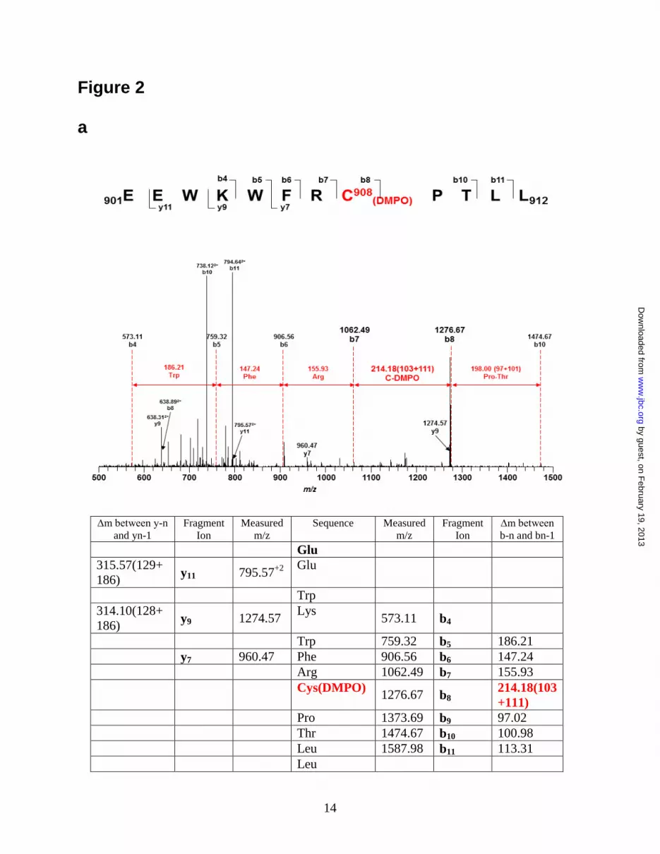
Identification of the eNOS protein radical by
mass spectrometry - To begin to understand how
the ·O2--induced eNOS protein radical formation
could affect eNOS function, it is important to
determine the precise sites of the DMPO-trapped
eNOS protein radicals. Purified BH4-free eNOS
was incubated with NADPH (1 mM) and 50 mM
DMPO at room temperature for 1 hour. This
reaction mixture was then subjected to SDS-PAGE
separation. The band corresponding to the DMPO-
trapped eNOS protein radical adduct was cut and
digested with trypsin, chymotrypsin or both, and
then the digested peptide fragments were
determined by LC/MS/MS as described in
“experimental procedures”.
With the addition of one molecule of DMPO,
the molecular weight of the DMPO-modified
peptide fragment will increase by 111 Da
compared to the non-modified peptide fragment.
The peptides with a mass difference of 111 Da
were identified by LC/MS, and the sequence of the
modified peptides was determined by MS/MS.
From the mass determination, one specific
modified cysteine residue, Cys908, was identified
in the tryptic/ chymotryptic fragment
901EEWKWFRC*PTLL912 (aa 901-912, Fig. 2a).
Molecular modeling of human eNOS reductase
domain- From the three-dimensional structure of
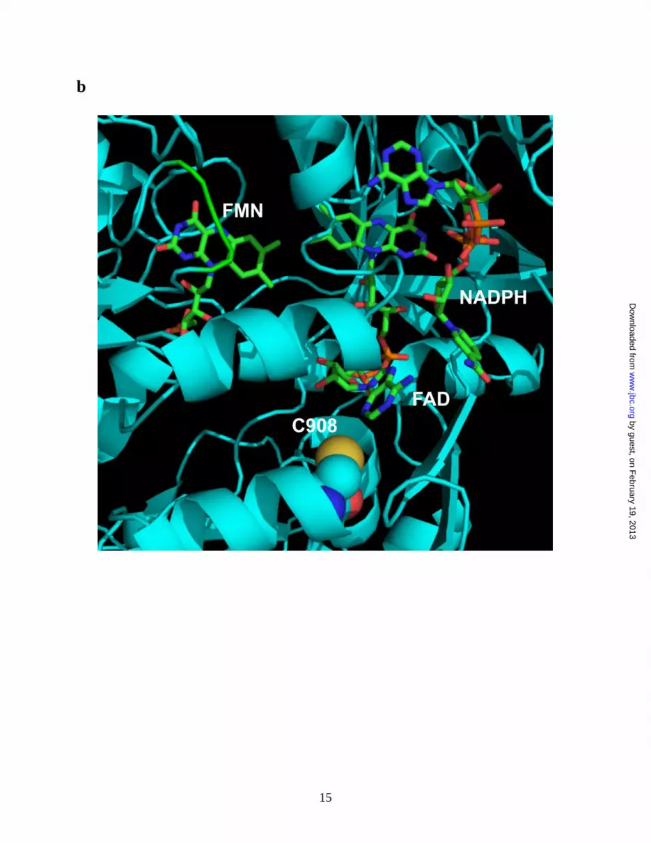
human eNOS reductase domain, Cys908 is located
at the interface of FAD and FMN domain (Fig.
2b). This residue is conserved throughout all
mammalian species. Structural perturbation on this
interface will be expected when this residue is
mutated to Ala, but not to Ser, allowing oxygen to
access this pocket.
Mutagenesis of eNOS Cys908 to Ala resists
thiyl radical formation and uncouples eNOS- Site-
directed mutagenesis of eNOS Cys908 to Ala was
used to further confirm this assignment and to
determine the importance of C908 on the eNOS
function and redox regulation. C908A mutant
totally abolished eNOS protein radical formation
in BH4-free eNOS as determined by
immunoblotting (Fig. 3a). The NO production
from WT and C908A eNOS was directly
measured by EPR spin-trapping using Fe-MGD.
The mutation of this Cys residue greatly decreased
measured NO production from the enzyme with
more than a 10-fold decrease compared to WT
eNOS (Fig. 3b). Addition of Mn-SOD
significantly increased the measured NO,
indicating that the C908A mutation alters the
function of eNOS to produce ·O2-, in lieu of NO,
which in turn scavenges and decreases the trapped
by guest, on February 19, 2013
ww
w.jbc.org
Dow
nloaded from

6
NO. With the mutation to Ala, because of a
change in size and polarity of this residue, the
perturbation at the FAD/FMN interface is
expected, and this can lead to a decrease in NO
production and electron leakage. Previously, it has
been demonstrated that site-directed mutagenesis
of electronegative residues at FMN module of
neuronal NOS affects electron transfer and NOS
catalytic activity (38). This further supports our
results of Cys 908 playing an important role in
electron transfer in the reductase domain as well as
regulating eNOS activity.
To further study the role of Cys908 in eNOS
function, ·O2- generation from WT and C908A
mutant was measured in the presence of specific
NOS inhibitors. Under BH4-free conditions eNOS-
derived ·O2- is inhibited by L-NAME and
imidazole, and is sensitive to Ca2+
/CaM (Fig. 3c).
However, ·O2- generated from the C908A mutant
is not inhibited by either L-NAME or imidazole,
and is not Ca2+
/CaM dependent (Fig. 3c). This
demonstrates that the ·O2- generated from the
C908A eNOS is not from the heme center, but
rather derived from the reductase domain.
Mutagenesis of eNOS Cys908 to Ala affects
reductase activity of eNOS and FAD/FMN
binding- Cytochrome c reductase activity, which is
a useful predictor of changes in the equilibrium
between FAD and FMN of NOS (37), was used to
determine the effect of mutation of Cys908 to Ala
on the reductase activity of eNOS. The reductase
activity of WT eNOS is 473.8 ± 13.4
nmol/mg/min at room temperature. The reductase
activity of C908A eNOS is 198.9 ± 12.5
nmol/mg/min. The content of FAD/FMN of eNOS
was measured to determine the effect of Cys
mutation on the binding of FAD/FMN to eNOS.
The ratio of FAD/FMN was determined using
HPLC. The ratio of FAD/FMN for WT eNOS is
0.98 ± 0.02. The ratio of FAD/FMN for C908A
eNOS is 0.80 ± 0.02. The results are expressed as
mean ± s.e.m. (n = 3).
·O2- generated from uncoupled eNOS induces
S-glutathionylation and disulfide bond formation
through protein radical intermediates- BH4-free
eNOS was used to self generate ·O2-. This ·O2
- in
turn reacted with eNOS thiols to form protein thiyl
radical intermediates. In the presence of GSH (0.1,
0.5, and 2 mM), this eNOS thiyl radical can react
with GSH to generate S-glutathionylated eNOS
(Fig. 4a). This protein S-glutathionylation is
reversible when it is treated with 1 mM DTT. In
the absence of GSH, this eNOS thiyl radical can
react with a vicinal free thiol to form inter-
disulfide bonds. Under non-reducing eNOS
immunoblotting analysis, the formation of eNOS
dimer is increased when eNOS is uncoupled (Fig.
4b). This eNOS inter-disulfide bond formation can
be prevented with addition of GSH and is reversed
with 1 mM DTT.
Glutathione and DMPO compete for eNOS
protein radical- An increase in ·O2- production has
been shown to increase eNOS DMPO-adduct
formation (see the prior section). We also show
that the formation of eNOS protein radical can
further react with GSH to form eNOS S-
glutathionylation. To examine whether the eNOS
protein radical trapped by DMPO, also contributes
to eNOS S-glutathionylation, GSH competition for
eNOS DMPO protein radical formation or DMPO
competition for ·O2--induced eNOS S-
glutathionylation experiments were performed.
GSH (1-10 mM) dose-dependently inhibits eNOS
DMPO protein radical formation (Fig. 5a). No
eNOS DMPO protein radical is seen when
concentration of GSH is 5 mM. DMPO (20-100
mM) also dose-dependently inhibits ·O2--induced
eNOS S-glutathionylation in the presence of 2 mM
GSH (Fig. 5b). Thus, this suggests that the
mechanism of formation of eNOS S-
glutathionylation at Cys908 is from eNOS thiyl
radical intermediates that are induced by ·O2-.
Formation and trapping of eNOS protein
radicals in endothelial cells under oxidative
stress- Experiments were performed to determine
if eNOS protein radicals occur in endothelial cells
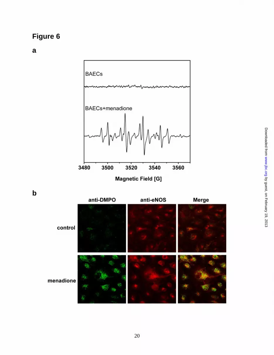
under oxidative stress. DMPO EPR spin-trapping
was first used to determine the extent of ·O2-
generation from BAECs induced by menadione,
which uncouples flavin-containing reductases (37).
Menadione-treated BAECs exhibited a prominent
DMPO radical-adduct signal composed mostly of
DMPO-OH with a smaller DMPO-R EPR signal
(Fig. 6a) (39-41). As previously reported, this
indicates that menadione activates ·O2--derived
radical generation (37). In endothelial cells
(BAECs) treated with menadione, confocal
microscopy with anti-DMPO and anti-eNOS
antibodies demonstrated a marked increase in
cellular DMPO protein radicals that colocalized
with eNOS (Fig. 6b). Immunoprecipitation with
anti-eNOS and anti-DMPO followed by
by guest, on February 19, 2013
ww
w.jbc.org
Dow
nloaded from

7
immunoblotting with anti-DMPO or anti-eNOS
antibodies confirmed that eNOS protein radical
formation occurred in these cells (Fig. 6c).
DISCUSSION
Oxidative post-translational modifications of
many enzymes, such as S-glutathionylation,
nitration, or nitrosylation, have been implicated in
signal transduction (42-48). Recently, we have
demonstrated that eNOS can be S-glutathionylated
under oxidative stress in vivo and in vitro, leading
to an uncoupling of the enzyme (13). Indeed,
several reports have also shown that protein S-
glutathionylation plays an important role in redox
signaling and can be protective against irreversible
oxidation of the protein thiols in cardiovascular
diseases (17,49). As such, it is important to
understand the mechanism of this redox-directed
enzyme regulation, opening another potential
mode of therapeutic intervention to treat
cardiovascular or other oxidant-derived diseases.
Under oxidative stress or disease, ·O2-
generated from eNOS is expected to increase with
the reversible depletion of cellular BH4 (4,16,50).
The increased ·O2- will in turn react with
functional groups of proteins, including eNOS,
with the formation of labile intermediate protein
radicals, leading to protein oxidation or oxidative
modification. Irreversible oxidation is the primary
cause for protein damage under oxidative stress
leading to cell death; in contrast, reversible
oxidation, such as the formation of disulfide or
mixed disulfide bonds, can protect cells from
oxidative damage and act as a redox sensor.
In the current report, we demonstrate that
eNOS can be oxidized by eNOS-derived ·O2- or an
exogenous ·O2- source leading to protein-centered
radical formation. This eNOS protein radical
formation is dependent upon NOS uncoupling,
substrate availability, or an exogenous ·O2- source,
while antioxidant enzymes, such as SOD, can
protect eNOS from this thiol oxidation. As such,
identification of the precise site of eNOS protein
radical formation provides key insight regarding
how oxidative modification of this enzyme is
involved in redox regulation.
Using mass spectrometric analysis, we
identified eNOS Cys 908 as the site of protein
radical formation. The formation of the thiyl
radical is one of the primary mechanisms for thiol
oxidation. Thiyl radical formation is a one electron
oxidation, which in turn can react with a vicinal
protein thiol or glutathione (GSH) to form a
disulfide bond or a mixed disulfide bond (i.e. S-
glutathionylation) (45,46). Protein S-
glutathionylation occurs through the reversible
formation of a mixed disulfide bond between
protein cysteine thiols and GSH, the most
abundant low molecular weight thiol in cells. The
identification of Cys 908 as the site of protein
radical formation provides direct mechanistic
evidence for an oxidant-induced process of eNOS
regulation that can lead to S-glutathionylation (13).
There are several proposed mechanisms of S-
glutathionylation (45,46). Thiol-disulfide
exchange with oxidized glutathione (GSSG) is one
of the mechanisms for S-glutathionylation. In
addition, ROS and reactive nitrogen species
(RNS)–derived thiyl radicals can in turn react with
GSH and protein thiols to generate protein S-
glutathionylation. It has been suggested that thiol-
disulfide exchange with GSSG can only occur
when cellular GSSG/GSH ratio is high (42,43,46).
It is believed that formation of thiol intermediates,
such as the thiyl radical, sulphenic acid, or S-
nitrosyl, is a more rapid and efficient mechanism
for protein S-glutathionylation in vivo, and that
these mechanisms could play a role in signal
transduction.
In our current study, we demonstrate that
eNOS thiols can be oxidized by ·O2- generated
from uncoupled eNOS, an exogenous source, or
cellular oxidative stress through the formation of a
thiyl radical in vitro and in vivo. The specific site
of protein radical formation at Cys 908 provides a
novel mechanism of eNOS thiol modification that
can lead to further redox modifications, such as S-
glutathionylation, which has been reported to
regulate eNOS function in response to oxidative
stress (13,51,52). We demonstrate that protein
thiyl radicals generated from the uncoupled eNOS
do indeed react with reduced glutathione to form
eNOS S-glutathionylation. Thus, the formation of
an eNOS protein radical on C908 provides a post-
translational mechanism that can regulate eNOS
function through oxidative modification to form
eNOS S-glutathionylation or disulfide bond
formation as demonstrated in this study.
Furthermore, an increase in GSH can inhibit eNOS
DMPO radical adduct formation, while increasing
DMPO concentrations inhibit eNOS S-
by guest, on February 19, 2013
ww
w.jbc.org
Dow
nloaded from
8
glutathionylation, suggesting that Cys908 is the
residue for eNOS thiyl radical formation, which in
turn can react with GSH to form S-
glutathionylated eNOS.
Site-directed mutagenesis of eNOS Cys908 to
Ala confirmed that C908 is of critical importance
for the regulation of eNOS function in response to
oxidative stress. Mutagenesis of this residue
demonstrated that C908 is the primary site for
the ·O2--induced formation of the eNOS protein
radical in response to oxidative stress. Moreover,
this mutation greatly decreases the NOS activity
and cytochrome c reductase activity of the C908A.
The cytochrome c reductase activity of NOS has
been used to measure electron efflux from the
reductase domain of NOS, and is a useful
predictor of changes in the equilibrium between
FAD and FMN of NOS (53). Thus, the decrease in
the cytochrome c reductase activity of C908A
mutant suggests that this residue is involved in
regulating electron transfer in the reductase
domain of eNOS by altering the electron transfer
between FAD and FMN leading to the possible
electron leakage from the site and a decrease in
NO production from the enzyme. In our previous
report (13), when Cys908 was mutated to Ser, a
conservative mutation, there was no effect on NO
production from the enzyme. However, when
mutated to Ala, there is a change in size and
polarity of this residue, the perturbation at the
FAD/FMN interface is expected, and this can lead
to a decrease in NO production and electron
leakage from the site.
·O2- generated from the C908A mutant is not
inhibited by either L-NAME, a NOS competitive
inhibitor, or imidazole, a heme-binding ligand, and
is also independent of Ca2+
/CaM, which is
necessary for transfer of electrons from the flavins
in the reductase domain to the heme center. The
mutation of Cys908 to Ala slightly alters the
binding of FAD and FMN to the enzyme.
Moreover, from the predicted three-dimensional
structure of the eNOS reductase domain, we found
that Cys 908 is located in the eNOS reductase
domain near the FAD binding site (Fig. 2a). As
such, we hypothesize that mutagenesis of eNOS
Cys908 to alanine could alter the structure of this
FAD and FMN interface and increase oxygen
accessibility to the flavins contributing to electron
leakage from this domain leading to the observed
increase in ·O2- generation and a decrease in
electron transfer from the reductase domain to the
heme center decreasing NO production. Thus, the
observed ·O2--induced eNOS thiol oxidation at
C908 and subsequent S-glutathionylation could
play an important role in regulating eNOS
function by allowing for the regulation of electron
flow from the reductase domain to the oxygenase
domain, and given the known reactions of protein
thiol-radicals this residue could be involved in a
redox-signaling based regulation of eNOS and,
ultimately, cardiovascular function.
The oxidative stress produced during
ischemia/reperfusion injury has been found to lead
to the S-glutathionylation of several important
proteins, such as aconitase and triose phosphate
isomerase (54). eNOS is of critical importance in
maintaining proper cardiovascular function, as
such, the detailed understanding of how oxidants
alter its structure/function with increasing
oxidative stress as occurs during
ischemia/reperfusion will enable the development
of strategies to prevent these injuries and alleviate
the underlying processes of oxidant induced
disease. We have demonstrated that ·O2-
generation in endothelium, leads to increases in
eNOS protein radical formation, and this
observation is consistent with our results with
purified eNOS, which demonstrated eNOS protein
radical formation via self-generated ·O2- or an
exogenous ·O2--generating source. Thus, in vivo,
eNOS cysteine residues are potential targets for
both reversible and irreversible oxidative
modifications and are potentially involved in
redox signaling during oxidative stress.
In conclusion, the increase in ·O2- generation
found under oxidative stress can induce eNOS
protein radical formation contributing to oxidative
modification. The formation of the eNOS protein
radical is dependent on the availability of
cofactors, and substrates of eNOS, or an
exogenous ·O2-
source. These eNOS protein
radical intermediates can in turn react with vicinal
protein thiol or GSH to form a disulfide bond or S-
glutathionylation. The identification of Cys 908 as
a primary target for radical formation provides a
novel mechanism by which eNOS is regulated in
response to oxidative stress. This site-specific thiol
redox modification can modulate cardiovascular
function as demonstrated here and in our prior
study (13) by altering eNOS function switching
the enzyme from NO production to ·O2- generation.
by guest, on February 19, 2013
ww
w.jbc.org
Dow
nloaded from

9
Thus, the molecular mechanism of eNOS thiol
oxidation delineated in this work serves as a
critical step toward understanding the redox-
regulation of eNOS and endothelial-mediated
vascular function as well as the alterations that
occur in diseases associated with oxidative stress.
REFERENCES
1. Sessa, W. C. (2004) J Cell Sci 117(Pt 12), 2427-2429
2. Kone, B. C., Kuncewicz, T., Zhang, W., and Yu, Z. Y. (2003) Am J Physiol Renal
Physiol 285(2), F178-190
3. Wang, P., and Zweier, J. L. (1996) J Biol Chem 271(46), 29223-29230
4. Dumitrescu, C., Biondi, R., Xia, Y., Cardounel, A. J., Druhan, L. J., Ambrosio, G., and
Zweier, J. L. (2007) Proc Natl Acad Sci U S A 104(38), 15081-15086
5. Xia, Y., Tsai, A. L., Berka, V., and Zweier, J. L. (1998) J Biol Chem 273(40), 25804-
25808
6. Vasquez-Vivar, J., Kalyanaraman, B., Martasek, P., Hogg, N., Masters, B. S., Karoui, H.,
Tordo, P., and Pritchard, K. A., Jr. (1998) Proc Natl Acad Sci U S A 95(16), 9220-9225
7. Kojda, G., and Harrison, D. (1999) Cardiovasc Res 43(3), 562-571
8. Cave, A. C., Brewer, A. C., Narayanapanicker, A., Ray, R., Grieve, D. J., Walker, S., and
Shah, A. M. (2006) Antioxid Redox Signal 8(5-6), 691-728
9. Zweier, J. L., and Talukder, M. A. (2006) Cardiovasc Res 70(2), 181-190
10. Forstermann, U. (2006) Biol Chem 387(12), 1521-1533
11. Druhan, L. J., Forbes, S. P., Pope, A. J., Chen, C. A., Zweier, J. L., and Cardounel, A. J.
(2008) Biochemistry 47(27), 7256-7263
12. Pou, S., Pou, W. S., Bredt, D. S., Snyder, S. H., and Rosen, G. M. (1992) J Biol Chem
267(34), 24173-24176
13. Chen, C. A., Wang, T. Y., Varadharaj, S., Reyes, L. A., Hemann, C., Talukder, M. A.,
Chen, Y. R., Druhan, L. J., and Zweier, J. L. (2010) Nature 468(7327), 1115-1118
14. Muscoli, C., Cuzzocrea, S., Riley, D. P., Zweier, J. L., Thiemermann, C., Wang, Z. Q.,
and Salvemini, D. (2003) Br J Pharmacol 140(3), 445-460
15. Stuehr, D. J. (1999) Biochim Biophys Acta 1411(2-3), 217-230
16. Chen, C. A., Druhan, L. J., Varadharaj, S., Chen, Y. R., and Zweier, J. L. (2008) J Biol
Chem 283(40), 27038-27047
17. Martinez-Ruiz, A., and Lamas, S. (2007) Cardiovasc Res 75(2), 220-228
18. Parodi, O., De Maria, R., and Roubina, E. (2007) J Cardiovasc Med (Hagerstown) 8(10),
765-774
19. Peluffo, G., and Radi, R. (2007) Cardiovasc Res 75(2), 291-302
20. Hothersall, J. S., Cunha, F. Q., Neild, G. H., and Norohna-Dutra, A. A. (1997) Biochem J
322 ( Pt 2), 477-481
21. Harbrecht, B. G., Di Silvio, M., Chough, V., Kim, Y. M., Simmons, R. L., and Billiar, T.
R. (1997) Ann Surg 225(1), 76-87
22. Cuzzocrea, S., Zingarelli, B., O'Connor, M., Salzman, A. L., and Szabo, C. (1998) Br J
Pharmacol 123(3), 525-537
23. Hofmann, H., and Schmidt, H. H. (1995) Biochemistry 34(41), 13443-13452
24. Huang, A., Xiao, H., Samii, J. M., Vita, J. A., and Keaney, J. F., Jr. (2001) Am J Physiol
Cell Physiol 281(2), C719-725
25. Tummala, M., Ryzhov, V., Ravi, K., and Black, S. M. (2008) DNA Cell Biol 27(1), 25-33
26. Mason, R. P. (2004) Free Radic Biol Med 36(10), 1214-1223
by guest, on February 19, 2013
ww
w.jbc.org
Dow
nloaded from

10
27. Ilagan, R. P., Tejero, J., Aulak, K. S., Ray, S. S., Hemann, C., Wang, Z. Q., Gangoda, M.,
Zweier, J. L., and Stuehr, D. J. (2009) Biochemistry 48(18), 3864-3876
28. Martasek, P., Liu, Q., Liu, J., Roman, L. J., Gross, S. S., Sessa, W. C., and Masters, B. S.
(1996) Biochem Biophys Res Commun 219(2), 359-365
29. Rodriguez-Crespo, I., and Ortiz de Montellano, P. R. (1996) Arch Biochem Biophys
336(1), 151-156
30. Chen, Y. R., Chen, C. L., Zhang, L., Green-Church, K. B., and Zweier, J. L. (2005) J Biol
Chem 280(45), 37339-37348
31. Gerber, N. C., and Ortiz de Montellano, P. R. (1995) J Biol Chem 270(30), 17791-17796
32. Xia, Y., Cardounel, A. J., Vanin, A. F., and Zweier, J. L. (2000) Free Radic Biol Med
29(8), 793-797
33. Vanin, A. F., Liu, X., Samouilov, A., Stukan, R. A., and Zweier, J. L. (2000) Biochim
Biophys Acta 1474(3), 365-377
34. Abu-Soud, H. M., Loftus, M., and Stuehr, D. J. (1995) Biochemistry 34(35), 11167-
11175
35. Gnanaiah, W., and Omdahl, J. L. (1986) J Biol Chem 261(27), 12649-12654
36. Stuehr, D. J., Cho, H. J., Kwon, N. S., Weise, M. F., and Nathan, C. F. (1991) Proc Natl
Acad Sci U S A 88(17), 7773-7777
37. Rosen, G. M., and Freeman, B. A. (1984) Proc Natl Acad Sci U S A 81(23), 7269-7273
38. Panda, K., Haque, M. M., Garcin-Hosfield, E. D., Durra, D., Getzoff, E. D., and Stuehr,
D. J. (2006) J Biol Chem 281(48), 36819-36827
39. Zweier, J. L. (1988) J Biol Chem 263(3), 1353-1357
40. Zweier, J. L., Kuppusamy, P., Williams, R., Rayburn, B. K., Smith, D., Weisfeldt, M. L.,
and Flaherty, J. T. (1989) J Biol Chem 264(32), 18890-18895
41. Buettner, G. R. (1987) Free Radic Biol Med 3(4), 259-303
42. Winterbourn, C. C., and Hampton, M. B. (2008) Free Radic Biol Med 45(5), 549-561
43. Gallogly, M. M., and Mieyal, J. J. (2007) Curr Opin Pharmacol 7(4), 381-391
44. Foster, M. W., Hess, D. T., and Stamler, J. S. (2009) Trends Mol Med 15(9), 391-404
45. Adachi, T., Schoneich, C., and Cohen, R. A. (2005) Drug Discovery Today: Disease
Mechanisms 2(1), 39-46
46. Biswas, S., Chida, A. S., and Rahman, I. (2006) Biochem Pharmacol 71(5), 551-564
47. Turko, I. V., and Murad, F. (2002) Pharmacol Rev 54(4), 619-634
48. Zhang, H., Xu, Y., Joseph, J., and Kalyanaraman, B. (2008) Methods Enzymol 440, 65-94
49. Mieyal, J. J., Gallogly, M. M., Qanungo, S., Sabens, E. A., and Shelton, M. D. (2008)
Antioxid Redox Signal 10(11), 1941-1988
50. Zweier, J. L., Flaherty, J. T., and Weisfeldt, M. L. (1987) Proc Natl Acad Sci U S A 84(5),
1404-1407
51. Chen, C.-A., Druhan LJ, Wang TY, Chen YR, Zweier JL. (2008) Circulation 118, S274
52. Chen, C.-A. T.-Y. W., Saradhadevi Varadharaj, Levy Reyes, M.A. Hassan Talukder,
Yeong-Renn Chen, Lawrence J. Druhan, and Jay L. Zweier. (2009) Circulation 120,
S1070
53. Stuehr, D. J., Tejero, J., and Haque, M. M. (2009) Febs J 276(15), 3959-3974
54. Eaton, P., Byers, H. L., Leeds, N., Ward, M. A., and Shattock, M. J. (2002) J Biol Chem
277(12), 9806-9811
by guest, on February 19, 2013
ww
w.jbc.org
Dow
nloaded from
11
FOOTNOTES
We thank Dr. Liwen Zhang and Dr. Kari Green-Church at OSU CCIC proteomics center for support with
mass spectrometric analysis. We thank Dr. Dennis J. Stuehr (Cleveland Clinic) for providing us rat
nNOS reductase domain. This work was supported by R01 Grants HL63744, HL65608, HL38324 (J. L.
Z.), HL081734 (L.J.D.), HL83237 (Y.-R. C.) and K99HL103846 (C.-A. C.) from the National Institutes
of Health.
The abbreviations used are: BAECs, bovine aortic endothelial cells; CaM, calmodulin; DEPMPO, 5-
diethoxyphosphoryl-5-methyl-1-pyrroline N-oxide; EPR, electron paramagnetic resonance; Fe-MGD, Fe-
N-methyl-D-glucamine dithiocarbamate; GSH, glutathione; L-NAME, L-NG-nitroarginine methyl ester
hydrochloride; NO, nitric oxide; eNOS, endothelial nitric oxide synthase; PBS, phosphate buffered saline;
RT, room temperature; SDS-PAGE, sodium dodecyl sulfate polyacrylamide gel electrophoresis; Mn-SOD,
superoxide dismutase; ·O2-, superoxide anion; BH4, tetrahydrobiopterin; WT, wild-type.
FIGURE LEGENDS
Figure 1. ·O2- generated from uncoupled eNOS induces eNOS protein radical formation. a. EPR
spin-trapping of ·O2- generated by eNOS. The reaction system contained 50 mM Tris-HCl buffer, pH 7.4,
1 mM NADPH, 0.5 mM Ca2+
, 10 mg/mL CaM, 15 g/mL purified eNOS, and 25 mM spin trap DEPMPO.
Spectra were recorded at room temperature with a microwave frequency of 9.863 GHz, 20 milliwatts of
microwave power, and 1.0 G modulation amplitude. b. Immunoblotting analysis of protein radical
formation. 1-6. ·O2- is generated from BH4-Free eNOS. 1. BH4-Free eNOS. 2. +10 M Arg. 3. +1 mM
Arg. 4. +1 mM L-NAME. 5. +Mn-SOD. 6. +10 M BH4. 7-8. ·O2- is generated from nNOS reductase
domain (nNOSR). 7. BH4-Free eNOS + nNOSR. 8. +Mn-SOD.
Figure 2. Mass spectrometry and molecular modeling reveal C908 is the site for eNOS protein
radical formation. a. LC/MS/MS mass spectrometric analysis of eNOS protein radical formation.
Protein radical formation was demonstrated to be on Cys908 using DMPO as a trap. The molecular
weight difference between fragment ions b7 and b8 demonstrated a mass shift of 111 Da compared to the
native fragment ions, therefore, this allowed unequivocal assignment of the DMPO adduct to Cys-908.
EPR and immunoblotting experiments were performed 3-5 times. b. Molecular modeling of human eNOS
reductase domain. The three-dimensional structure of human eNOS reductase domain was generated
using the reductase domain of rat neuronal NOS (PDB ID 1F20) by Swiss molecular modeling. Cys908 is
located at the interface of FAD and FMN domain. This residue is conserved throughout all mammalian
species. The structural perturbation on this domain will be expected when this residue is mutated to Ala
allowing oxygen accessing this pocket.
Figure 3. C908A mutation of eNOS prohibits thiyl radical formation and uncouples eNOS. a.
Immunoblotting of WT eNOS and eNOS C908A on protein radical formation. Upper panel is eNOS
protein radical formation trapped by DMPO, when eNOS is uncoupled, and immunoblotting against anti-
DMPO antibody. Lane 1 is protein radical formation from WT eNOS and trapped by DMPO. Lane 2 is
same as in lane 1 with addition of Mn-SOD. Lane 3, as in lane 1, eNOS C908A and trapped DMPO. Lane
4 is same as in lane 3 with addition of Mn-SOD. Lower panel is immunoblotting for eNOS. b. EPR Fe-
MGD NOS activity determination. Upper spectrum is the NO generated from WT eNOS. Middle
spectrum is the NO generated from eNOS C908A. Lower spectrum is the NO generated from eNOS
C908A plus Mn-SOD. c. Determination of ·O2- generation from WT eNOS and eNOS C908A by
DEPMPO EPR spin-trapping. The left panel is the ·O2- -DEPMPO EPR spectra of WT eNOS. The right
by guest, on February 19, 2013
ww
w.jbc.org
Dow
nloaded from

12
panel is the ·O2- -DEPMPO EPR spectra of eNOS C908A. All experiments were performed at least in
triplicate.
Figure 4. Mechanistic study of eNOS S-glutathionylation and inter-disulfide bond formation. a. ·O2-
generated from BH4-free eNOS induces protein S-glutathionylation formation. Upper panel is eNOS S-
glutathionylation formation through eNOS protein thiyl radical in the presence of GSH, when eNOS is
uncoupled, and immunoblotting against anti-GSH antibody. Lane 1 is eNOS only. Lane 2 is eNOS with
0.5 mM GSH. Lane 3 is the uncoupled eNOS. Lane 4 is the uncoupled eNOS with 0.1 mM GSH (20 min).
Lane 5 is the uncoupled eNOS with 0.5 mM GSH (20 min). Lane 6 is the uncoupled eNOS with 2 mM
GSH (20 min). Lane 7 and 8 are samples from Lane 3 and 5, respective, treated with 1 mM DTT. Lower
panel is immunoblotting for eNOS. b. ·O2- generated from BH4-free eNOS induces eNOS inter-disulfide
formation. Immunoblotting against anti-eNOS antibody is used to determine eNOS monomer and dimer
under the non-reducing SDS-PAGE separation. Lane 1 is the uncoupled eNOS. Lane 2 and 3 are eNOS
with 0.5 or 2 mM GSH, respectively. Lane 4 is the uncoupled eNOS with 0.5 mM GSH (20 min). Lane 5
is the uncoupled eNOS with 2 mM GSH (20 min). Lane 6 is the uncoupled eNOS with 0.5 mM (60 min).
Lane 7 is sample from lane 5 treated with 1 mM DTT.
Figure 5. Glutathione and DMPO compete for ·O2--induced eNOS protein radical. a. GSH dose-
dependently inhibits ·O2--induced eNOS DMPO-adduct. Upper panel is a blot against anti-DMPO
antibody. Lower panel is a blot against anti-eNOS antibody. Lane 1. BH4-free eNOS in the presence of all
NOS cofactors and 50 mM DMPO, and the reaction is initiated with 1 mM NADPH for 30 min at room
temperature. Lane 2. The reaction is the same as in lane 1 with addition of 1 mM GSH. Lane 3. With
addition of 5 mM GSH. Lane 4. With addition of 10 mM GSH. b. DMPO dose-dependently inhibits ·O2--
induced eNOS S-glutathionylation. Upper panel is a blot against anti-GSH antibody. Lower panel is a blot
against anti-eNOS antibody. Lane 1. BH4-free eNOS in the presence of all NOS cofactors and 2 mM
GSH, with the reaction initiated by 1 mM NADPH for 30 min at room temperature. Lane 2. The reaction
is the same as in lane 1 with addition of 20 mM DMPO. Lane 3. With addition of 50 mM DMPO. Lane 4.
With addition of 100 mM DMPO.
Figure 6. In vivo protein radical formation from endothelium under oxidative stress. a. DMPO EPR
spin-trapping of ·O2- generated from BAECs induced by 10 M menadione. Upper spectrum is the EPR
signal of control BAECs with 50 mM DMPO spin trap. Lower spectrum is the EPR signal of BAECs with
50 mM DMPO spin trap activated by addition of 10 M menadione. b. Immuno-microscopic detection of
eNOS protein radical formation in endothelium using anti-DMPO antibody (green) and anti-eNOS
antibody (red). Upper panels are control BAECs with addition of DMPO trap, demonstrating no
detectable protein radical formation. Lower panels are BAECs treated with 10 M menadione with
addition of the DMPO trap, demonstrating marked protein radical formation (green) that colocalized with
eNOS (red). c. Immunoprecipitation of DMPO protein radical adducts from BACEs. 1. Control BAECs. 2.
BAECs are treated with menadione. Upper panel is the immunoprecipitation with anti-DMPO antibody
followed by immunoblotting against anti-eNOS. Lower panels are the immunoprecipitation with anti-
eNOS antibody followed immunoblotting with anti-DMPO antibody. All experiments were performed at
least in triplicate.
by guest, on February 19, 2013
ww
w.jbc.org
Dow
nloaded from

14
Figure 2 a
∆m between y-n
and yn-1
Fragment
Ion
Measured
m/z
Sequence Measured
m/z
Fragment
Ion
∆m between
b-n and bn-1
Glu
315.57(129+
186) y11 795.57
+2
Glu
Trp
314.10(128+
186) y9 1274.57
Lys 573.11 b4
Trp 759.32 b5 186.21
y7 960.47 Phe 906.56 b6 147.24
Arg 1062.49 b7 155.93
Cys(DMPO)
1276.67 b8 214.18(103
+111)
Pro 1373.69 b9 97.02
Thr 1474.67 b10 100.98
Leu 1587.98 b11 113.31
Leu
by guest, on February 19, 2013
ww
w.jbc.org
Dow
nloaded from