characterization of the endocytic pathways regulating
TRANSCRIPT

CHARACTERIZATION OF THE ENDOCYTIC PATHWAYS REGULATING
RIBOFLAVIN (VITAMIN B2) ABSORPTION AND TRAFFICKING IN HUMAN
EPITHELIAL CELLS
DISSERTATION
Presented in Partial Fulfillment of the Requirements for the Degree Doctor of Philosophy
in the Graduate School of The Ohio State University
By
Amy B. Foraker, B. S.
*****
The Ohio State University 2007
Dissertation Committee:
Dr. Peter W. Swaan, Advisor
Dr. Thomas D. Schmittgen, Advisor
Dr. James T. Dalton
Dr. William L. Hayton
Approved by
Advisor Graduate Program in Pharmacy

Copyright© 2007 by Amy B. Foraker
All rights reserved.

ii
ABSTRACT
Drug bioavailability is greatly compromised by the body’s innate defense
mechanisms that result in low membrane permeability of hydrophilic drugs and
macromolecules, reduced cellular retention by efflux transporters, and metabolic
degradation by a multitude of enzymes. These factors, combined with underlying genetic
aberrations and pathologies, drastically reduce drug bioavailability and, in certain
instances, increase drug-related toxicity. To address many of these challenges, scientists
have devised ways to target therapeutics into cells by coupling drugs directly (i.e.,
prodrug) or indirectly (e.g., polymer-mediated carriers) to natural ligands that are
recognized by membrane receptors, and consequently gain cellular entry through
receptor-mediated endocytosis (RME).
Although various receptors have been exploited to improve drug delivery, several
receptor mechanisms remain to be defined. One RME target that has gained attention for
its therapeutic potential in contraception, breast- and liver cancers involves the riboflavin
(vitamin B2) RME machinery. Our laboratory recently revealed riboflavin absorption to
be regulated, in part, by clathrin-dependent RME (CME) in human epithelia. However,
the identity of proteins involved in this process, and an understanding of the intracellular
distribution of absorbed B2 remain to be established.

iii
The work presented in this dissertation addresses some of these areas of question
using a three-tiered approach involving subcellular fractionation of [3H]-B2 dosed cells,
3D fluorescence colocalization analyses of rhodamine-labeled B2 and immunostained
endocytic markers, and RNA interference of the endocytic scission enzyme, dynamin 2
GTPase. Herein, we reveal dynamic enrichment of internalized riboflavin to CME
endosomes, and to a lesser extent to the Golgi and mitochondria in human placental
trophoblasts and enterocytes (Chapter 2). Furthermore, absorbed riboflavin is shown to
be negatively coupled to cAMP levels. In Chapter 3, ~ 80% dynamin 2 protein
knockdown causes ~ 40% reduced B2-RME, and 2-fold higher B2 localization at the
plasma membrane of placental trophoblasts. Lastly, fluorescence colocalization assays
exploring the involvement of clathrin-independent caveolae-mediated RME in B2
internalization show minimal B2 enrichment to caveolar endosomes (Chapter 4). Overall,
our data demonstrate a dynamic subcellular B2 trafficking itinerary largely associated
with CME, and dynamin 2 is the first endocytic protein identified to regulate B2 cellular
entry in humans.

iv
Dedicated to my parents, John R. and Cheryl K. Foraker, and my grandparents, John E.
and Virginia E. Foraker, and Raymond H. and Mary K. Probasco
v
ACKNOWLEDGMENTS
If I were asked to offer my perspective to those interested in pursuing their Ph.D.
in biopharmaceutics or a related science, I would immediately inform them to be
prepared to confront their own self-imposed barriers. Graduate school has been an
odyssey of scientific and self discovery. I can’t emphasize enough the importance of
networking with senior and junior colleagues throughout the graduate education process.
I would like to offer my deepest thanks to my advisor, Dr. Peter W. Swaan, for instilling
these very principles throughout my tenure in his lab. It has been through his constant
guidance and support that has brought me to this honorable right of passage in my career
development. A special thank you goes to my OSU advisor, Dr. Thomas D. Schmittgen,
who provided me with the opportunity to broaden my research experience to include
aptamer-based biomarker studies, and served as an invaluable resource in quantitative
RT-PCR methods. I also want to express my gratitude to all the faculty of both The Ohio
State University Graduate Program in Pharmaceutics and the Department of
Pharmaceutical Sciences of the University of Maryland at Baltimore for fostering my
graduate education. Especially, I would like to thank Drs. Kenneth K. Chan, James T.
Dalton, Natalie D. Eddington, Hamid Ghandehari, William L. Hayton, Insong J. Lee,
Robert J. Lee, and Paul S. Shapiro for their invaluable advice and support. If it had not

vi
been for Dr. Kathleen Hillgren of Eli-Lilly & Co., I would not have been able to
complete the last half of my research. Thank you, Kate, for your generosity in supporting
my work. Also, I thank the laboratory of Dr. Mark A. McNiven (Mayo Clinic and
Foundation, Rochester, Minnesota) for their generosity in donating validated dynamin 2
expression constructs, which were instrumental in corroborating our dynamin 2 RNA
interference work.
I acknowledge the genuine friendship given to me from my colleagues, Dr. Se-Ne
Huang, Dr. Yongheng Zhang, Dr. Cheng Chang, Dr. Mitch A. Phelps, Dr. Antara
Banerjee, Dr. Vanessa M. D’Souza, Dr. Abhijit Ray, Dr. Julie A. Ray, Praveen M.
Bahadduri, Chandra M. Khantwal, Clifford W. Mason, Lisa M. Bareford, Naissan
Hussainzada, and Tatiana Claro Da Silva. It has been a privilege and honor to work with
you all. I thank the administrative staff of both programs, with special gratitude to
Colleen Day, Karen Lawler and Kathy Brooks for their generous support throughout my
graduate studies. Also, I am forever indebted to Drs. Ana I. Robles and Wenqing Gao for
their constant support, friendship and sincere words of encouragement during my
graduate experience. I offer my sincere gratitude to Ms. Margaret MacLearie for
generously offering her home as I was transitioning into graduate school and to Ms.
Karen Sensabaugh for providing me with the knowledge and guidance to achieve balance
amidst the many challenges that define graduate school.
I could not have made it through graduate school without my family of friends
who openly took me into their hearts and enriched my life beyond the lab bench
including, Kelly Baker, Christopher Bulka, Renee Cockerman, Veronica Cox, Cybele

vii
Garcia, Devon Graham, Kathryn Kurre, and Alison Wright. I especially want to express
my heartfelt gratitude to Dr. Andrew Hebbeler, who has acted as my steady compass
during my time in Baltimore and constantly inspires me to be a better person. I can’t
thank you enough for your friendship. A special thank you also goes to my steadfast and
dear high school friend, Anne Householder, for being patient with me during the early
years of my graduate studies and providing unconditional love and support throughout.
Mostly, I am grateful to my parents and grandparents, for their infinite words of
wisdom and love, and for allowing me the priceless opportunities to follow my dreams.
Thank you for believing in me. My deepest thanks also go to my sisters and brother-in-
law, Matt and Jodi Toledo, and Lindsey Foraker for their constant love and support.

viii
VITA
July 29, 1975……………………….……..Born Zanesville, Ohio, U.S.A.
December, 1997…………………………..B.S. Molecular Biology, Clarion University, Clarion, PA, U.S.A.
September, 2001-2003........………………Graduate Teaching Assistant, The Ohio State University, Columbus, Ohio, U.S.A.
September, 2003-2007……………………Visiting Graduate Research Associate, University of Maryland, Baltimore, MD, U.S.A.
PUBLICATIONS
1. D’Souza, V. M.*, Foraker, A. B.*, Free, R. B., Ray, A., Shapiro, P. S., and Swaan, P. W. (2006) cAMP-Coupled riboflavin trafficking in placental trophoblasts: a dynamic and ordered process, Biochemistry 45, 6095-104.
2. Xiao, J. J., Foraker, A. B., Swaan, P. W., Liu, S., Huang, Y., Dai, Z., Chen, J., Sadee,
W., Byrd, J. Marcucci, G., and Chan, K. K. (2005) Efflux of depsipeptide FK228 (FR901228, NSC-630176) is mediated by P-glycoprotein and multidrug resistance-associated protein 1, J Pharmacol Exp Ther 313, 268-76.
3. Phelps, M. A., Foraker, A. B., Gao, W., Dalton, J. T., and Swaan, P. W. (2004) A
novel rhodamine-riboflavin conjugate probe exhibits distinct fluorescence resonance energy transfer that enables riboflavin trafficking and subcellular localization study, Molecular Pharm 1, 257-66.
4. Foraker, A. B., Khantwal, C. M., and Swaan, P. W. (2003) Current perspectives on
the cellular uptake and trafficking of riboflavin, Adv. Drug Deliv. Rev. 55, 1467-83. 5. Foraker, A. B., Walczak, R. J., Cohen, M. H., Boiarski, T. A., Grove, C. F., and
Swaan, P. W. (2003) Microfabricated porous silicon particles enhance paracellular delivery of insulin across intestinal Caco-2 cell monolayers, Pharm Res 20, 110-6.

ix
6. Phelps, M. A., Foraker, A. B., and Swaan, P. W. (2003) Cytoskeletal motors and cargo in membrane trafficking: opportunities for high specificity in drug intervention, Drug Discov Today 8, 494-502.
7. Robles, A. I., Bemmels, N. A., Foraker, A. B., and Harris, C. C. (2001) APAF-1 is a
transcriptional target of p53 in DNA damage-induced apoptosis, Cancer Res 61, 6660-4.
*co-first authors
FIELDS OF STUDY
Major Field: Pharmacy
x
TABLE OF CONTENTS
Page
Abstract……………………………………………………………………………………ii
Dedication………………………………………………………………………………...iv
Acknowledgments…………………………………………………………………………v
Vita……………………………………………………………………………………...viii
List of Tables…………………………………………………………………………….xv
List of Figures…………………………………………………………………………...xvi
List of Abbreviations…………………………………………………………………..xviii
Chapters:
1. Introduction…………………………………………………………………..……1
1.1 Clinical significance and models of riboflavin transport, storage and
regulation…………………………………………………………………….3
1.2 Riboflavin uptake mechanism(s)...………..…………………………………7
1.2.1 Saturable (active) transport component established in in vitro studies..9
1.2.1.1 Common transport characteristics……………………………..15
1.2.2 Passive diffusion component and an adaptive regulatory mechanism in
B2 uptake……………………………………………………………..28
1.3 Receptor-mediated and/or carrier-mediated transport?.................................30
1.4 Riboflavin binding/carrier protein (RfBP)………………………………….31
xi
1.4.1 Functions in oviparous species………………………………………32
1.4.1.1 Interspecies sequence homology and identity……………........33
1.4.1.2 Suggested mechanism of RfBP in riboflavin homeostasis…....34
1.4.2 Mammalian RfBP……………………………………………………35
1.4.2.1 Putative function and role of RfBP in riboflavin uptake and
transport……………………………………………………….37
1.4.2.2 Utilizing the riboflavin absorption and trafficking mechanism to
enhance macromolecule delivery………….…………………41
1.5 Comparison of B2 internalization with folate uptake and transport
mechanisms…………………………………………………………………41
1.6 Summary and thesis objectives………………..………………………....…43
2. cAMP-coupled riboflavin trafficking in placental trophoblasts and enterocytes: a
dynamic and ordered process…………………………………………………….45
2.1 Introduction…………………………………………………………………47
2.2 Methods and materials……………………………………………………...50
2.2.1 Cell culture…………………………………………………………...50
2.2.2 Labeling of transferrin……………………………………………….50
2.2.3 Ligand uptake and subcellular fractionation…………………………51
2.2.4 Immunoblotting and data analysis…………………………………...52
2.2.5 Immunofluorescence and confocal microscopy……………………...52
2.2.6 Determination of cyclic AMP accumulation in BeWo cells…………54
2.3 Results………………………………………………………………………55

xii
2.3.1 Isolation of organelle-enriched fractions…………………………….55
2.3.2 Distribution of internalized B2 and TF…………………………….....57
2.3.3 Colocalization of B2 with endocytic organelles in BeWo…………...65
2.3.4 Golgi and mitochondrial involvement in B2 itinerary………………..71
2.3.5 cAMP regulation of B2 binding and initiation of the endocytic cascade
in placental trophoblasts……………………………………………..75
2.4 Discussion…………………………………………………………………..78
3. Dynamin 2 regulates the endocytosis of riboflavin in human epithelia…….........83
3.1 Introduction…………………………………………………………………85
3.2 Methods and materials……………………………………………………...87
3.2.1 Cell culture…………………………………………………………...87
3.2.2 Duplex siRNA transient transfection………………………………...87
3.2.3 Western blotting and chemiluminescence-based densitometry……...88
3.2.4 GTPase-null (K44A) and wild type dynamin 2 plasmid transient
transfections and cotransfections with DNM2 pool siRNA………....89
3.2.5 Radio-labeled ligand endocytosis assays…………………………….90
3.2.6 Statistics……………………………………………………………...91
3.3 Results………………………………………………………………………92
3.3.1 Duplex siRNA target specificity at the protein level and RNAi method
validation……………………………………………………………..92
3.3.2 Dynamin siRNA effects combined with temperature dependence on
endocytosis of [3H]-B2 in BeWo……………………………………..98
xiii
3.3.3 Determination of the extent of reduced absorption for B2 compared to
the control ligand, transferrin, under silenced dynamin 2
conditions…………………………………………………………...101
3.3.4 Extent of plasma membrane bound versus internalized ligand as a
function of siRNA treatment………………………………………..105
3.3.5 Effect of transiently transfected wild-type or dominant-negative
dynamin 2 (K44A) expression constructs on the extent of bound and
internalized ligands…………………………………………………109
3.4 Discussion…………………………………………………………………113
4. Elucidating the role of caveolin 1 in the riboflavin endocytic itinerary in placental
trophoblasts……………………………………………………………………..116
4.1 Introduction………………………………………………………………..118
4.2 Methods and materials…………………………………………………….121
4.2.1 Cell culture………………………………………………………….121
4.2.2 Fluorescent ligand endocytosis assay and immunofluorescence
staining……………………………………………………………...121
4.2.3 Fluorescence image acquisition, restoration, and colocalization
analysis……………………………………………………………...122
4.3 Results……………………………………………………………………..123
4.3.1 Colocalization of rhodamine-riboflavin with the conserved endocytic
coat protein, caveolin 1……………………………………………..123
4.4 Discussion…………………………………………………………………128

xiv
5. Summary, conclusions, and future directions…………………………………..132
5.1 Summary…………………………………………………………………..132
5.2 Conclusions: the B2-RME model revised………………………………...138
5.3 Future directions: exploring the potential application of the B2-RME
mechanism in targeted medicinal therapeutics……………………………143
Bibliography……………………………………………………………………………146

xv
LIST OF TABLES
Table Page
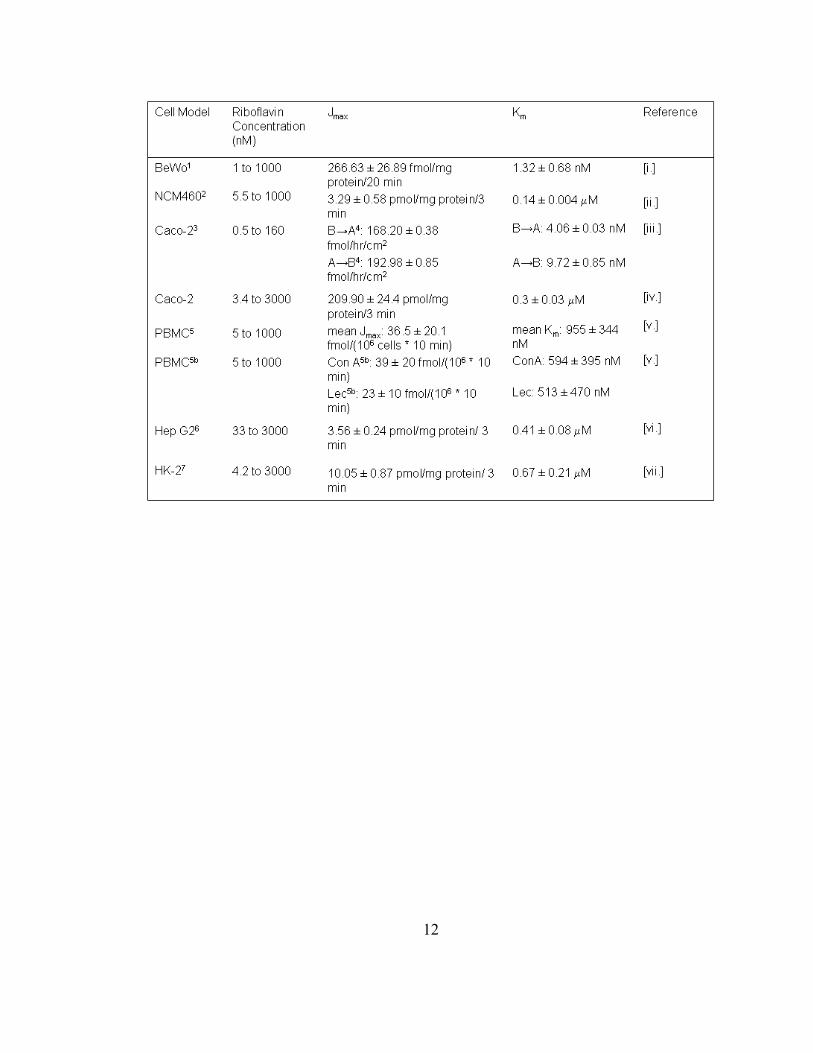
Table 1.1. Summary of riboflavin uptake kinetics across divergent cell models……….11
Table 1.2. Effects of related and un-related structural analogs on riboflavin uptake…...13
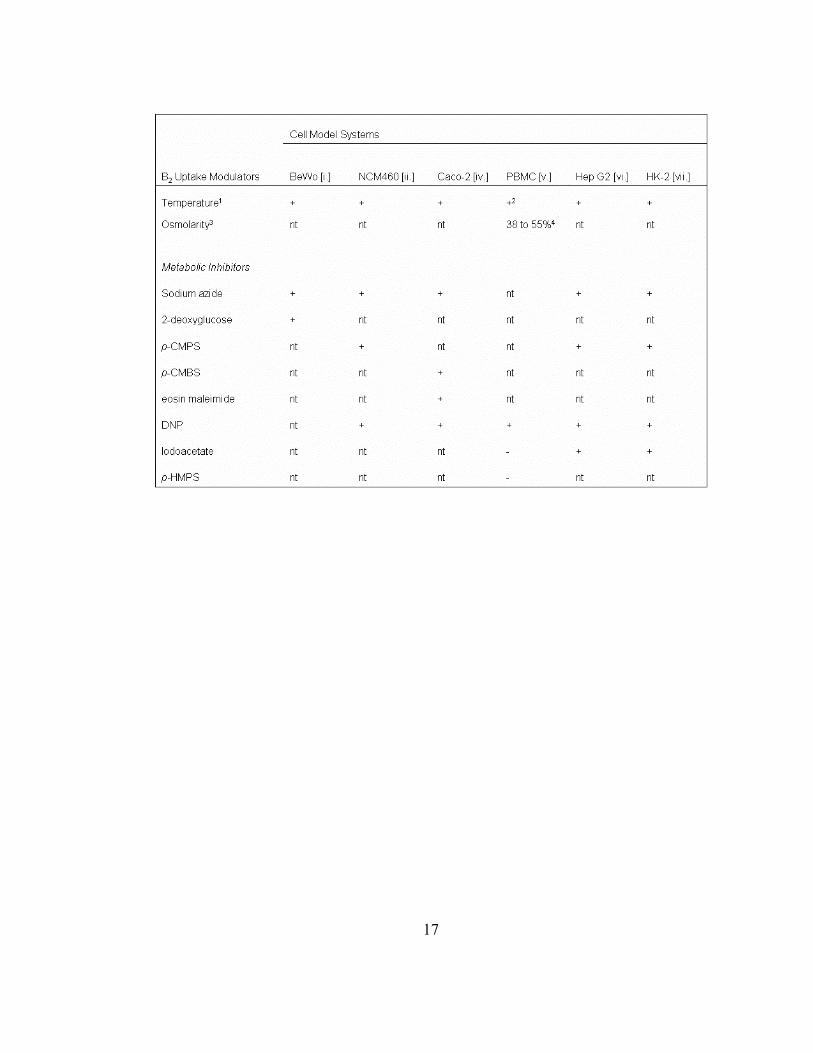
Table 1.3. Effects of metabolic inhibitors on riboflavin transport across divergent cell
models……………………………………………………………………………16
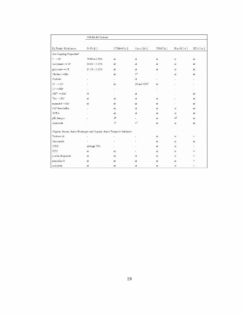
Table 1.4. Effects of ion substitutions, organic anions, anion exchangers or organic anion
transport inhibitors on B2 transport………………………………………………18
Table 1.5. Effects of various modulators targeting cAMP, protein tyrosine kinase, protein
kinase A, G-, C- and calmodulin-mediated pathways on B2 transport ………….25
Table 3.1. siRNA or plasmid and transfection reagent effects on cell viability as a
function of lactate dehydrogenase (LDH) release……………………………….97

xvi
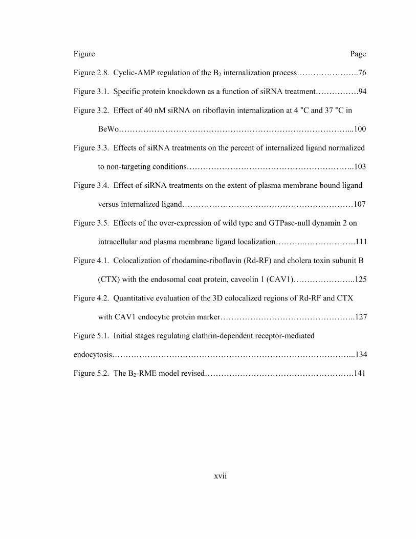
LIST OF FIGURES
Figure Page
Figure 1.1. Model illustrating B2 absorption pathways and homeostasis along human
intestinal epithelial cells…………………………………………………………...4
Figure 2.1. Comparative cell-associated profiles of [3H]-riboflavin (10 nM B2) with the
endocytic marker 125I-transferrin (10 nM TF) and an unrelated bile acid
transporter substrate, [3H]-taurocholic acid (20 nM TCA), in placental (BeWo)
and intestinal (Caco-2) cells……………………………………………………...56
Figure 2.2. Distribution of [3H]-B2 and 125I-Transferrin (TF) in the postnuclear fractions
isolated from trophoblasts and enterocytes………………………………………59
Figure 2.3. Time-dependent localization of [3H]-B2 and 125I-TF within the separated
fractions from BeWo and Caco-2 cells…………………………………………..63
Figure 2.4. Concentration-dependent trafficking profiles of [3H]-B2 after 2 hr incubation
at 37 °C in BeWo and Caco-2 cells……………………………………………...64
Figure 2.5. Endosomal and lysosomal colocalization of Rhodamine-B2 (Rd-RF) and
FITC-labeled transferrin (FITC-TF)……………………………………………..66
Figure 2.6. Quantitative evaluation of the 3D colocalized regions of ligands, B2 and TF,
with organelle protein markers in BeWo cells…………………………………...70
Figure 2.7. Localization of Rd-RF and FITC-TF to the Golgi and mitochondria in BeWo
cells………………………………………………………………………………73
xvii
Figure Page
Figure 2.8. Cyclic-AMP regulation of the B2 internalization process…………………..76
Figure 3.1. Specific protein knockdown as a function of siRNA treatment…………….94
Figure 3.2. Effect of 40 nM siRNA on riboflavin internalization at 4 °C and 37 °C in
BeWo…………………………………………………………………………...100
Figure 3.3. Effects of siRNA treatments on the percent of internalized ligand normalized
to non-targeting conditions……………………………………………………..103
Figure 3.4. Effect of siRNA treatments on the extent of plasma membrane bound ligand
versus internalized ligand………………………………………………………107
Figure 3.5. Effects of the over-expression of wild type and GTPase-null dynamin 2 on
intracellular and plasma membrane ligand localization………..……………….111
Figure 4.1. Colocalization of rhodamine-riboflavin (Rd-RF) and cholera toxin subunit B
(CTX) with the endosomal coat protein, caveolin 1 (CAV1)…………………..125
Figure 4.2. Quantitative evaluation of the 3D colocalized regions of Rd-RF and CTX
with CAV1 endocytic protein marker…………………………………………..127
Figure 5.1. Initial stages regulating clathrin-dependent receptor-mediated
endocytosis……………………………………………………………………………...134
Figure 5.2. The B2-RME model revised……………………………………………….141

xviii
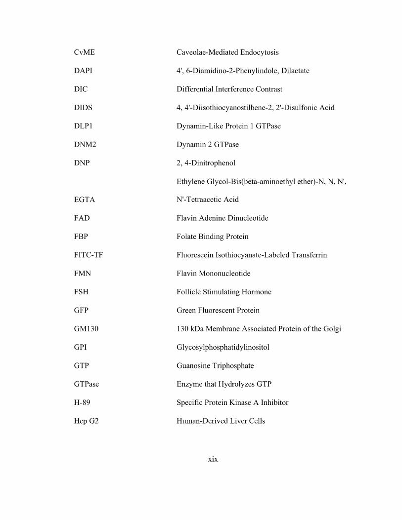
LIST OF ABBREVIATIONS
[ABC]-
Transporter ATP-Binding Cassette Transporter
AP-2 Adaptor Protein 2
ATP Adenosine Triphosphate
B2 Riboflavin
BCRP/ABCG2 Breast Cancer Resistance Protein
BeWo Human-Derived Placental Trophoblasts
Br-cAMP Membrane Permeable cAMP Analog
BSA Bovine Serum Albumin
Ca2+/CaM Ca2+/Calmodulin-Mediated Pathway
CaCo-2 Human-Derived Intestinal Epithelia
cAMP Cyclic Adenosine Monophosphate
CAV1 Caveolin 1 Protein
CCP Clathrin-Coated Pit
CME Clathrin-Mediated Endocytosis
CTMR4A Carboxytetramethylrhodamine-4-Amine
CTX Cholera Toxin Subunit B
xix
CvME Caveolae-Mediated Endocytosis
DAPI 4', 6-Diamidino-2-Phenylindole, Dilactate
DIC Differential Interference Contrast
DIDS 4, 4'-Diisothiocyanostilbene-2, 2'-Disulfonic Acid
DLP1 Dynamin-Like Protein 1 GTPase
DNM2 Dynamin 2 GTPase
DNP 2, 4-Dinitrophenol
EGTA
Ethylene Glycol-Bis(beta-aminoethyl ether)-N, N, N',
N'-Tetraacetic Acid
FAD Flavin Adenine Dinucleotide
FBP Folate Binding Protein
FITC-TF Fluorescein Isothiocyanate-Labeled Transferrin
FMN Flavin Mononucleotide
FSH Follicle Stimulating Hormone
GFP Green Fluorescent Protein
GM130 130 kDa Membrane Associated Protein of the Golgi
GPI Glycosylphosphatidylinositol
GTP Guanosine Triphosphate
GTPase Enzyme that Hydrolyzes GTP
H-89 Specific Protein Kinase A Inhibitor
Hep G2 Human-Derived Liver Cells
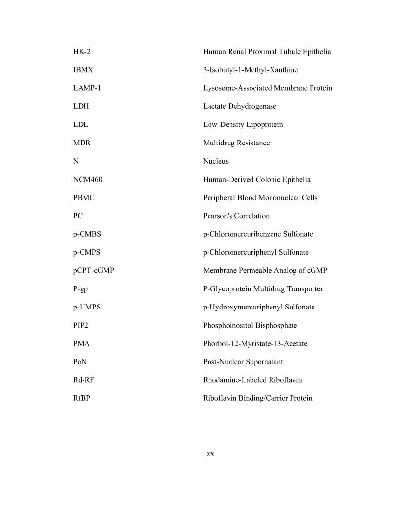
xx
HK-2 Human Renal Proximal Tubule Epithelia
IBMX 3-Isobutyl-1-Methyl-Xanthine
LAMP-1 Lysosome-Associated Membrane Protein
LDH Lactate Dehydrogenase
LDL Low-Density Lipoprotein
MDR Multidrug Resistance
N Nucleus
NCM460 Human-Derived Colonic Epithelia
PBMC Peripheral Blood Mononuclear Cells
PC Pearson's Correlation
p-CMBS p-Chloromercuribenzene Sulfonate
p-CMPS p-Chloromercuriphenyl Sulfonate
pCPT-cGMP Membrane Permeable Analog of cGMP
P-gp P-Glycoprotein Multidrug Transporter
p-HMPS p-Hydroxymercuriphenyl Sulfonate
PIP2 Phosphoinositol Bisphosphate
PMA Phorbol-12-Myristate-13-Acetate
PoN Post-Nuclear Supernatant
Rd-RF Rhodamine-Labeled Riboflavin
RfBP Riboflavin Binding/Carrier Protein

xxi
RME Receptor-Mediated Endocytosis
RNA Ribonucleic Acid
RNAi RNA Interference
SARA Smad Anchor Protein for Receptor Activation
siRNA Short-Interfering RNA
SITS
4-Acetamido-4'-Isothiocyanostilbene-2, 2'-
Disulfonic Acid
Smad7-Smurf2
Protein Complex that Leads to Receptor
Degradation
Smads Cellular Cytoplasmic Gene Transcription Factors
SNX9 Sorting Nexin 9 Protein
TCA Taurocholic Acid
TF Transferrin, a Soluble Iron-Carrier Protein
TGF-b Tumor Growth Factor Beta
TJ Tight Junctions
VLDL Very-Low-Density Lipoprotein
1
CHAPTER 1
INTRODUCTION
Riboflavin (also known as vitamin B2) is a water-soluble vitamin essential for
normal cellular functions, growth and development. In its coenzyme forms of flavin
adenine dinucleotide (FAD) and flavin mononucleotide (FMN), B2 performs key
metabolic functions as an intermediary in the transfer of electrons in biological oxidation-
reduction reactions. During periods of dietary deprivation or physiological and
pathological stress, humans are vulnerable to developing B2-deficiency. This may lead to
a variety of clinical abnormalities, including growth retardation, anemia, skin lesions and
degenerative changes in the nervous system (Cooperman, 1991). Humans cannot
synthesize B2 and, thus, must obtain the vitamin from their diet through absorption in the
small intestine. Alternatively, riboflavin is also obtained from indigenous bacteria that
colonize the large intestine, which naturally synthesize this vitamin. Although many
studies have focused on the mechanism of B2 uptake using various tissues and cell lines,
transepithelial absorption is controversial and its exact mechanism(s) remains to be
defined. Intracellular processes in B2 absorption, such as cellular homoeostasis, and B2
2
function and regulation are also poorly understood. The purpose of this chapter is to
illustrate current in vitro and in vivo data defining B2 uptake mechanisms noted in
different studies and to compare and contrast the various implicated parameters defining
this (these) absorption mechanism(s). In addition, B2 subcellular trafficking events
involving receptor-mediated endocytosis (RME) and cytoskeletal elements are discussed.
The involvement of a soluble riboflavin binding/carrier protein (RfBP) in B2 uptake and
trafficking in humans is currently elusive. Although several studies have reported its
existence (Natraj et al., 1988; Prasad et al., 1992), it has not been characterized in detail.
The majority of the work on RfBP has been focused on oviparous species and has shown
a crucial role in embryo development. Few studies have been carried out on mammalian
RfBP analogs despite its recent attention in therapeutic applications for contraception,
and as a candidate biomarker of breast- and liver-cancer progression (Rao et al., 1999;
Karande et al., 2001; Rao et al., 2006). This chapter further considers the similarities in
protein sequence and function between RfBP for oviparous and mammalian species.
Based on uptake and transport results using avian RfBP in the presence of B2, suggested
roles in B2 absorption and trafficking events are presented. Since parallels can be drawn
between the uptake and transport mechanisms of B2 and other well-studied vitamins, a
brief discussion of commonalities defining such mechanisms for folate are illustrated.
Here, the aim is to present comparisons with current knowledge on B2 uptake and
transport, and how these prior studies may guide future elucidations of B2-specific
pathways functioning in its homeostasis.
3
1.1 Clinical significance and models of riboflavin transport, storage and regulation
Flavoproteins constitute one of the largest groups of functionally related proteins
currently known, catalyzing essential oxidation/reduction steps in almost every metabolic
pathway in both prokaryotic and eukaryotic cells. Using essentially the same B2-based
cofactor, flavoproteins participate in a remarkable array of biological processes, from
simple electron transfer to complex signal transduction pathways. It is not surprising that
defects within critical electron transfer processes may manifest themselves in severe, and
sometimes fatal, metabolic diseases. Most dietary B2 is presented in the form of free
riboflavin, FAD, FMN and flavoproteins, which must first be hydrolyzed to B2 before
absorption can occur (Figure 1.1). Digestion of flavoproteins, FMN and FAD occurs by
highly specific enzymes (Kasai et al., 1990), suggesting a physiologic preference that
favors B2 as the absorptive species of dietary flavin. Though there is extensive
information on the function of mucosal and cytosolic enzymes involved in the digestion
of flavoproteins (e.g., flavokinase and FAD synthetase), it is interesting to note that none
of these enzymes have been cloned or otherwise structurally characterized.
Therapeutically, B2 has recently gained renewed interest after it was shown to
protect vital tissues from ischemia-induced oxidative injury resulting from heart attack or
stroke (Hultquist, 1993). Given the critical role that B2 appears to play in pathological
conditions with high morbidity, it is pertinent that its cellular homeostasis and the
pathway(s) by which this vitamin gains entry into the body needs to be further defined.
4
Figure 1.1. Model illustrating B2 absorption pathways and homeostasis along
human intestinal epithelial cells. Following dietary intake and digestion (gastric
acidification and proteases), B2 analogs, FMN and FAD, are generated. These cofactors
are further metabolized to the absorption active form, riboflavin, through the catalytic
activities of various phosphatases. Extracellular levels of free B2 may be bound by human
RfBP prior to cellular uptake of the vitamin. At physiological concentrations, B2 is taken
into the cell through passive diffusion (A), receptor-mediated endocytosis (B), and/or a
carrier-mediated mechanism (C). Once in the cell, B2 is phosphorylated (Flavokinase) to
FMN. FMN is either used as a prosthetic group of flavoenzymes, or converted to FAD
through the action of FAD synthetase. FAD is commonly incorporated into flavoenzymes.
Another possible fate of FMN and B2 may involve transcytosis, where the receptor-bound
B2 or FMN complex is translocated to the basolateral membrane to allow release of the
ligand in systemic circulation. Another possible role of a putative riboflavin-specific,
basolaterally localized, receptor is to bind B2 from the serum and translocate the vitamin
into the cell to be used for various cellular maintenance and growth needs. Abbreviations:
tight junctions (TJ), flavin mononucleotide (FMN), and flavin adenine dinucleotide
(FAD).

5
6
In animals, B2-deficiency results in lack of growth, failure to thrive, and eventual
death (Cooperman, 1991). Experimental riboflavin deficiency in dogs results in growth
failure, weakness, ataxia, and inability to stand. The animals collapse, become comatose,
and die (Cooperman, 1991). During the deficiency state, dermatitis develops together
with hair-loss. Other signs include corneal opacity, lenticular cataracts, hemorrhagic
adrenals, fatty degeneration of the kidney and liver, and inflammation of the mucus
membrane of the gastrointestinal tract (Horwitt et al., 1972). Post-mortem studies in
rhesus monkeys fed a B2-deficient diet revealed that about one-third the normal amount
of riboflavin was present in the liver (Foy and Mbaya, 1977), which is the main storage
organ for B2. These overt clinical signs of B2-deficiency are rarely seen among
inhabitants of developed countries. However, about 28 million Americans exhibit a
common ‘sub-clinical’ stage (Lemoine et al., 1980), characterized by a change in
biochemical indices (e.g., erythrocyte glutathione reductase, (Cooperman et al., 1973)).
Although the effects of long-term sub-clinical B2-deficiency are unknown, in children
this deficiency results in stunted growth. Subclinical B2-deficiency has also been
observed in women taking oral contraceptives (Wynn, 1975), in the elderly, in
individuals with eating disorders, and in disease states such as HIV (Beach et al., 1992),
inflammatory bowel disease (Fernandez-Banares et al., 1989), diabetes and chronic heart
disease (Cooperman, 1991). The fact that B2-deficiency does not immediately lead to
gross clinical manifestations indicates that the systemic levels of this essential vitamin are
tightly regulated.

7
1.2 Riboflavin uptake mechanism(s)
Between the early 1940s and late 1970s, numerous studies described riboflavin
pharmacokinetics and pharmacodynamics in man and other mammalian species. These
initial studies provided the foundation of data suggesting the existence of a specialized B2
absorption mechanism (Jusko, 1975). Jusko and colleagues (Jusko et al., 1970)
demonstrated in man a significant inhibition in renal clearance of B2 upon combined
treatment with probenecid, now known to be a potent inhibitor of many organic anion
transport systems. The same group reported pharmacokinetic data providing additional
support for an active uptake mechanism. Using a three compartment open model, residual
analysis of B2 renal clearance data in dog and man after rapid intravenous injections, 18.3
mg and 31 mg, respectively, revealed a saturable tubular reabsorption component (Jusko
and Levy, 1970). Since the net renal clearance exceeded the glomerular filtration rate, B2
tubular secretion was also apparent. The maximal transport capacity was 33.3 µg/min for
man, 21.9 µg/min. for dog, and the apparent Km values were 16.3 and 9.61 µg/ml,
respectively. Studies performed in the late 70s to early 1980s further suggested that
riboflavin uptake and secretion involved the cyclic organic acid transport system. Jusko
and Levy (Jusko, 1975) were the first to generate this hypothesis by showing the organic
anion transport inhibitor, probenecid, to substantially block riboflavin transport in renal
tubules. Additional support for this theory was presented by Spector (Spector, 1982), who
showed saturable and energy-dependent B2 accumulation in rabbit kidney cortex slices.
This accumulation was found to be inhibited by sulfhydryl reagents and cyclic organic
acids such as 0.1 mM probenecid, which resulted in 37% inhibition compared to controls,
8
and no inhibition was observed in the presence of weak bases. Riboflavin was shown to
be a competitive inhibitor of p-aminohippurate, a weak acid, and penicillin G uptake into
kidney slices (Spector, 1982). Lowy and Spring (Lowy and Spring, 1990) showed similar
effects of probenecid on B2 transport into renal-derived MDCK cells. Using quantitative
fluorescence video microscopy, they showed a 54% reduction in B2 uptake in the
presence of 1 mM probenecid. Increasing probenecid concentration to 10 mM resulted in
near complete inhibition of B2 transport. Combining the results of these studies illustrates
that probenecid inhibits B2 uptake in a dose-dependent manner in kidney cells and that B2
absorption is suggested to involve a saturable, active uptake mechanism consisting of the
carrier-mediated component, the organic anion transport system.
Upon oral administration of B2, the fraction of the oral dose absorbed was
approximately 50% (Jusko, 1975). Subsequent studies concentrated on the B2 transport in
isolated membrane vesicles or intact intestinal preparations (e.g., everted sacs and
perfused segments) in an attempt to focus on the existence of a carrier-mediated
component responsible for B2 transport (Daniel et al., 1983; Said et al., 1985; Middleton,
1990; Said et al., 1993a; Said et al., 1993b). However, at the time of these publications,
the cellular and molecular regulation of the intestinal uptake of B2 was not clear and, thus,
a definitive mechanistic conclusion could not be drawn. With the advent of well-
characterized in vitro intestinal cell models, B2 intestinal transport studies were better
able to define kinetic transport parameters.
9
1.2.1 Saturable (active) transport component established in in vitro studies
Through the mid 1990s to the present, a vast amount of research has concentrated
on defining the regulatory mechanisms involved in B2 transport across different cell line
models. Such studies have utilized cell systems representing colonic, intestinal, placental,
renal and pulmonary epithelial cells, along with liver hepatocytes and peripheral blood
mononuclear cells. However, kinetic parameters defining the maximal transport velocity
and receptor/or transporter affinity constant appear to vary depending on the cell line or
tissue being studied (Table 1.1). Reported Km values range from low nM to low µM, and
Jmax values range from low fmol/mg of protein/min to low pmol/mg of protein/min
(Kumar et al., 1998; Huang and Swaan, 2000; Said et al., 2000; Zempleni and Mock,
2000; Huang and Swaan, 2001). These discrepancies could be explained by the existence
of multiple and distinct B2 uptake mechanisms at varying levels of expression depending
on cell lineage, or in part to the highly mutant prone characteristics defining the inherent
nature of immortalized cell lines. Despite these differences, a trend is observed across
different studies showing the B2 uptake mechanism to be highly specific and saturable. B2
competitive analog studies indicate the highest ligand affinity is continually expressed
with unlabeled riboflavin that is in excess to basal concentrations of labeled riboflavin
(Table 1.2). Half-maximal transport constants (comparable to Michaelis-Menten
constants) range from 1.32 nM in placental trophoblast cells, BeWo (Huang and Swaan,
2001), to 0.67 µM in renal proximal tubule epithelial cells, HK-2 (Kumar et al., 1998).
Riboflavin derivatives including flavin mononucleotide (FMN), flavin adenine
dinucleotide (FAD), lumiflavin, lumichrome, iso- B2, and 8- [NH2]- B2, show significant
10
inhibition in B2 uptake as observed in competitive analog assays (Said and Ma, 1994;
Kumar et al., 1998; Said et al., 2000; Zempleni and Mock, 2000; Huang and Swaan,
2001). Furthermore, these studies demonstrate the following analogs, listed in order of
decreasing effects, exhibit marked inhibition in B2 absorption: FMN (a phosphate group
is attached to the alpha carbon of the ribityl side chain), FAD (with adenine diphosphate
attached at the ribose moiety), lumiflavin (a methyl group replaces the ribose side chain),
and lumichrome (completely lacks a ribose side chain) (Huang and Swaan, 2001).
Unrelated molecules such as biotin and mannitol, in addition to D-ribose and pterin ring-
containing substances such as folate and pantothenic acid did not have any effect on B2
uptake (Said and Ma, 1994; Kumar et al., 1998; Zempleni and Mock, 2000; Huang and
Swaan, 2001). Combined, these data suggest the B2 absorption mechanism does not
require the ribose side chain. The highest substrate affinity constants observed by these
effector analogs (i.e., those which lead to significant inhibition in B2 uptake) were
reported for the placental trophoblast cell model, BeWo (Huang and Swaan, 2001). The
distinctively higher affinity in this cell line can be explained by clinical reports stating the
crucial requirements that must be met during fetal nutrition and development (Zempleni
et al., 1995).
11
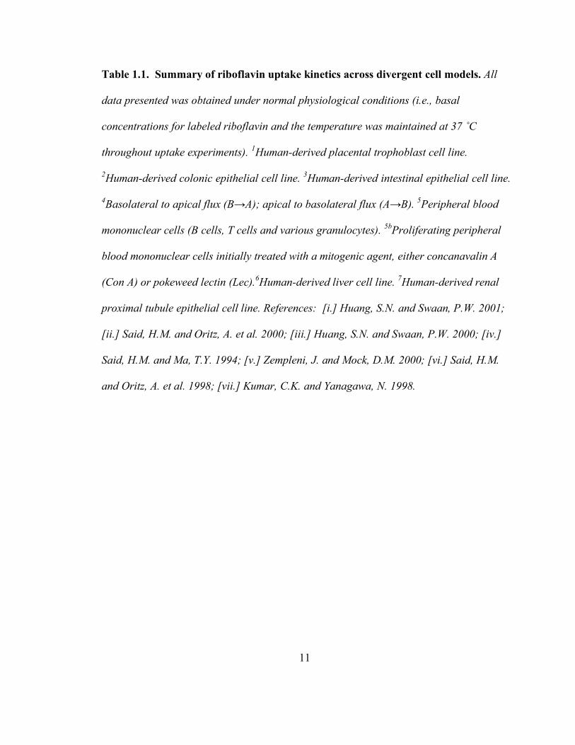
Table 1.1. Summary of riboflavin uptake kinetics across divergent cell models. All
data presented was obtained under normal physiological conditions (i.e., basal
concentrations for labeled riboflavin and the temperature was maintained at 37 ˚C
throughout uptake experiments). 1Human-derived placental trophoblast cell line.
2Human-derived colonic epithelial cell line. 3Human-derived intestinal epithelial cell line.
4Basolateral to apical flux (B→A); apical to basolateral flux (A→B). 5Peripheral blood
mononuclear cells (B cells, T cells and various granulocytes). 5bProliferating peripheral
blood mononuclear cells initially treated with a mitogenic agent, either concanavalin A
(Con A) or pokeweed lectin (Lec).6Human-derived liver cell line. 7Human-derived renal
proximal tubule epithelial cell line. References: [i.] Huang, S.N. and Swaan, P.W. 2001;
[ii.] Said, H.M. and Oritz, A. et al. 2000; [iii.] Huang, S.N. and Swaan, P.W. 2000; [iv.]
Said, H.M. and Ma, T.Y. 1994; [v.] Zempleni, J. and Mock, D.M. 2000; [vi.] Said, H.M.
and Oritz, A. et al. 1998; [vii.] Kumar, C.K. and Yanagawa, N. 1998.
12

13
Table 1.2. Effects of related and un-related structural analogs on riboflavin uptake.
All competitive uptake assays were performed with physiologic concentrations of labeled
B2 in the presence or absence of excess structural analogs. All percentages represent the
extent of labeled B2 uptake inhibition as compared to respective untreated controls.
Abbreviations are defined as follows: structural analogs were not tested in this study (nt);
significant uptake inhibition was observed as compared with controls (+); an
insignificant effect on B2 uptake was reported (-); flavin mononucleotide (FMN); flavin
adenine dinucleotide (FAD). 1Inhibitory constants were obtained using the Dixon Plot
method. 2Significant uptake inhibition was observed with individual treatments of 8-
[NH2]-B2 or iso-B2. References: [i.] Huang, S.N. and Swaan, P.W. 2001; [ii.] Said, H.M.
and Oritz, A. et al. 2000; [iv.] Said, H.M. and Ma, T.Y. 1994; [v.] Zempleni, J. and Mock,
D.M. 2000; [vi.] Said, H.M. and Oritz, A. et al. 1998; [vii.] Kumar, C.K. and Yanagawa,
N. 1998.

14

15
1.2.1.1. Common transport characteristics
Temperature-dependency of B2 transport has been reported in different cell lines
(Said and Ma, 1994; Zempleni and Mock, 2000; Huang and Swaan, 2001). In general,
physiological temperatures (~ 37 ºC) coincide with saturable uptake kinetics, whereas
low temperature (~ 4 ºC) correlates with linear absorption profiles (Said and Ma, 1994).
This temperature-dependent regulation in vitamin absorption is a classic feature defining
an active uptake mechanism. Nevertheless, this data alone does not distinguish between
carrier- or receptor-mediated components regulating ligand translocation.
Metabolic and transport modulators are commonly implemented to define energy
and membrane protein structural requirements that drive drug absorption. For the past
decade in vitro systems have aided in the elucidation of the molecular and cellular events
regulating B2 absorption. As a result, we now have a better understanding of cellular
regulatory events. For example, concerning energy-dependence, a regulatory role
involving the Ca2+/calmodulin-mediated pathway (Ca2+/CaM), and a highly specific,
saturable carrier-mediated component have been determined through competitive analog
assays (Said and Ma, 1994; Kumar et al., 1998; Said et al., 1998; Zempleni and Mock,
2000; Huang and Swaan, 2001). Furthermore, the majority of studies suggest that B2
absorption is Na+-independent.
In order to determine whether the absorption of B2 is dependent on a motive
energy force, metabolic and transport inhibitors have been employed, along with testing
possible roles of ion-coupled gradients as analyzed through ion substitution assays
(Tables 1.3 and1.4).

16
Table 1.3. Effects of metabolic inhibitors on riboflavin transport across divergent
cell models. The general effects of selected metabolic inhibitors on B2 transport are
indicated as significant in inhibiting uptake as compared to controls (+), insignificant or
no observed alterations (-) or presented as the extent of uptake inhibition as compared to
untreated controls (percentage). Abbreviations are defined as follows: metabolic
inhibitor was not tested in study (nt); p-chloromercuriphenyl sulfonate (p-CMPS); p-
chloromercuribenzene sulfonate (p-CMBS); 2, 4-dinitrophenol (DNP); and p-
hydroxymercuriphenyl sulfonate (p-HMPS). 1Significant inhibition in labeled Rf uptake
was noted at low temperatures (~ 4 ˚C). 2Uptake was performed using quiescent cells.
3The effects of changes in the osmolarity of the uptake medium on B2 uptake were tested.
4Effects of 805 mosM media were assessed using quiescent and mitogen-stimulated
PBMC, respectively, and increased B2 uptake into proliferating PBMC were correlated
with increased cell volume due to blastogenesis. References: [i.] Huang, S.N. and Swaan,
P.W. 2001; [ii.] Said, H.M. and Oritz, A. et al. 2000; [iv.] Said, H.M. and Ma, T.Y. 1994;
[v.] Zempleni, J. and Mock, D.M. 2000; [vi.] Said, H.M. and Oritz, A. et al. 1998; [vii.]
Kumar, C.K. and Yanagawa, N. 1998.
17

18
Table 1.4. Effects of ion substitutions, organic anions, anion exchangers or organic
anion transport inhibitors on B2 transport. All percentages represent the extent of B2
uptake inhibition as compared to controls unless stated otherwise. 1Ionic coupling
properties suggested for B2 transport were tested through ion substitution assays, and is
indicated as X ion isosmotically replaces Y ion (X→Y). 2Significant inhibition on B2
uptake was exclusively observed after prolonged incubation in Na+-depleted conditions.
3Effect is indicated as the percentage of inhibition in B2 uptake as compared to controls.
4An increase in B2 uptake was noted as pH changed from pH 8 to 7. 5A gradual decrease
in B2 uptake was observed as pH was changed from pH 8 to 5. 6Inhibition constant (Ki)
was determined to be 0.86 mM as determined by the Dixon method. 7Inhibition constant
(Ki) was determined to be 0.48 mM as determined by the Dixon method. Abbreviations
and symbols are defined as follows: significant inhibition in B2 uptake (+); no effect on
B2 uptake observed (-); treatment was not tested in study (nt); 4,4’-
diisothiocyanostilbene-2,2’disulfonic acid (DIDS); 4-acetamido-4’-isothiocyanostilbene-
2,2’-disulfonic acid (SITS); ethylene glycol-bis(beta-aminoethyl ether)-N,N,N’,N’-
tetraacetic acid (EGTA). References: [i.] Huang, S.N. and Swaan, P.W. 2001; [ii.] Said,
H.M. and Oritz, A. et al. 2000; [iv.] Said, H.M. and Ma, T.Y. 1994; [v.] Zempleni, J. and
Mock, D.M. 2000; [vi.] Said, H.M. and Oritz, A. et al. 1998; [vii.] Kumar, C.K. and
Yanagawa, N. 1998.
19
20
Consistent effects leading to marked inhibition in B2 uptake, though varying on the extent
of inhibition between different cell lines, are seen with sodium azide treatments (Said and
Ma, 1994; Kumar et al., 1998; Said et al., 1998; Said et al., 2000; Huang and Swaan,
2001). Sulfhydryl group modifying agents such as p-chloromercuriphenyl sulfonate (p-
CMPS), p-chloromercuribenzene sulfonate (p-CMBS), iodoacetate, and eosin maleimide,
cause significant inhibition in B2 uptake in colon, intestine, liver, and renal proximal
tubule cell systems (Said and Ma, 1994; Kumar et al., 1998; Said et al., 1998; Said et al.,
2000). These results, which are in agreement with earlier
pharmacokinetic/pharmacodynamic and ex vivo tissue studies (Jusko and Levy, 1970;
Jusko, 1975; Spector, 1982), suggest a possible role for reducing equivalents (e.g.,
NADH and FADH2) in the absorption process. Added support for this theory was
demonstrated in p-CMBS-pretreated Caco-2 cell monolayers that were subsequently
exposed to reducing agents, dithiothreitol or mercaptoethanol, which were found to
reverse the effects of the sulfhydryl agent (Said and Ma, 1994). Energy in the form of
ATP is also suggested to be involved in B2 uptake. Said and coworkers (Said and Ma,
1994) report significant (P < 0.01) inhibition in B2 (3.4 nM) uptake across Caco-2 cell
monolayers when treated with 2,4-dinitrophenol (1 mM), which is known to reduce
intracellular ATP. However, B2 transport appears to not be directly coupled to ATP
sources.
Transporters are generally fueled through the cotransport of ions (solute transport
family) or through a coupled catabolic reaction involving adenosine triphosphate (ATP)
hydrolysis (ATP binding cassette [ABC]-transport family). However, Na+ or H+
21
mediated-cotransport of organic solutes is more frequently observed in mammalian
species (Hediger et al., 1995). The role of Na+ in B2 uptake has been controversial. In
general, a sodium-independent process has been observed across inherently diverse cell
lines (Table 1.4). Yet, prior to the early 1990s, several studies utilizing intestinal tissue
preparations reported B2 absorption to be dependent on Na+ (Daniel et al., 1983;
Middleton, 1985; Said and Arianas, 1991). Said and Ma demonstrated a general Na+-
independence in B2 uptake studies using Caco-2 cell monolayers (Said and Ma, 1994).
Their studies involved replacing Na+ ions with choline, Li+, mannitol, or K+ (Table 1.4).
All replacement conditions showed no effect on B2 transport, except for K+, which led to
24% (P < 0.01) inhibition as compared to controls. In addition, with prolonged incubation
periods under Na+-depleted conditions, the K+ ion inhibiting effects markedly increased
to 42%. This reduction in uptake has been suggested to be a result of depolarizing effects
that K+ ions or Na+-depleted growth conditions can have on inherent biochemical
maintenance processes in cells and in intact tissue preparations (Said and Ma, 1994).
Additional support for Na+-independence has been shown through uptake studies
performed in the presence of ouabain, a well known Na+, K+-ATPase inhibitor. Such
studies repeatedly reveal no significant effects on B2 uptake with this agent (Kumar et al.,
1998; Said et al., 1998; Said et al., 2000; Zempleni and Mock, 2000; Huang and Swaan,
2001).
In general, B2 uptake appears independent of environmental pH (Table 1.4).
However, Said and coworkers revealed a slight increase in B2 uptake with media pH near
the physiologic range (~ pH 7), whereas, more acidic conditions led to a gradual decrease
22
in uptake (Said et al., 1998; Said et al., 2000). Such pH-coupled effects may be explained
through a putative ligand-receptor uncoupling event that is common in receptor-mediated
endocytosis. This uncoupling reaction, which is known to take place shortly after
receptor-ligand endocytosis, is accompanied by a reduction in endosomal pH (~ pH 5)
(Mukherjee et al., 1997; Swaan, 1998). However, reduced pH in the extracellular media
can theoretically mimic the intracellular effects of decoupling the ligand from its receptor.
Our laboratory reported a pH-dependent dissociation of surface bound B2 in human
intestinal cells (Caco-2) (Huang and Swaan, 2000). A further implication of a receptor-
mediated component in B2 uptake is discussed in later sections of this chapter.
A recent study by our laboratory suggests the involvement of Cl- ions in B2 uptake
as tested in placental BeWo cells (Huang and Swaan, 2001). Isoosmotic replacement of
Cl- ions with iodide, isocyanate, or gluconate significantly inhibited B2 uptake as
compared to controls (Table 1.4). Chloride ion effects have not been thoroughly studied
in other cell lines, and thus additional investigations are warranted.
As indicated in the early studies by Levy and colleagues (Jusko et al., 1970), B2
uptake was significantly reduced in the presence of the organic anion transport inhibitor,
probenecid. These results along with additional studies on renal tissue further suggested
B2 absorption was regulated through the organic anion transport system (Jusko, 1975;
Spector, 1982; Lowy and Spring, 1990). As a result several subsequent studies have
tested whether probenecid along with furosemide (Na+, K+-2Cl-- cotransporter inhibitor),
4,4’-diisothiocyanostilbene-2,2’-disulfonic acid (also known as DIDS, a membrane
impermeable anion-exchanger/chloride channel inhibitor), 4-acetamido-4’-
23
isothiocyanostilbene-2,2’-disulfonic acid (also known as SITS, an organic anion transport
inhibitor), and/or various organic anions (e.g., p-aminohippurate, penicillin G and
salicylate) generate similar inhibition effects on B2 absorption in non-renal cells (Table
1.4). Interestingly, excluding DIDS, none of these agents resulted in any significant
inhibition. However, as shown by Huang and coworkers (Huang and Swaan, 2001),
DIDS (for all tested concentrations 0.1, 0.5, and 1.0 mM) caused an average of 75%
reduction in B2 uptake in BeWo cells. Such results further corroborate the putative role of
Cl- ions in cellular riboflavin homeostasis in placental trophoblasts. These studies do not
indicate whether Cl- ion concentrations are directly or indirectly involved in the
translocation of B2 across a plasma membrane, and thus additional studies are warranted.
Nonetheless, the general inhibitory effect of organic anion modulators on B2 uptake
appears to be exclusive to renal proximal tubule cells (Kumar et al., 1998).
Cellular regulation of B2 absorption, particularly cyclic nucleotide-dependent
pathways, has been investigated using such modulators as 3-isobutyl-1-methyl-xanthine
(IBMX, a protein kinase A (PKA)-activator), H-89 (specific PKA inhibitor), pCPT-
cGMP (membrane permeable analog of cGMP), forskolin (adenylyl cyclase activator),
Br-cAMP (membrane permeable cAMP analog), cholera toxin (increases intracellular
cAMP levels), and dibutryl cAMP (also increases intracellular cAMP) (Table 1.5). Huang
and coworkers demonstrated an immediate and significant reduction in B2 uptake upon
short term incubation with IBMX using BeWo cells (Huang and Swaan, 2001). This
effect was accompanied by marked changes in Km and Jmax. In a similar fashion, the
cGMP analog, pCPT-cGMP, significantly inhibited B2 absorption (Huang and Swaan,

24
2001), which suggests a possible role for the protein kinase G (PKG) pathway. In
addition, coincubating with both modulators resulted in a potentiated uptake reduction
(reduced uptake fell to 43.7% and 15.3% as compared to individual treatments with
pCPT-cGMP or IBMX, respectively). Similar studies in Caco-2 cells with IBMX induced
significant uptake inhibition in a concentration dependent manner (Said and Ma, 1994).
Overall, data suggest a possible role for the PKA and/or PKG pathways in riboflavin
absorption.
25
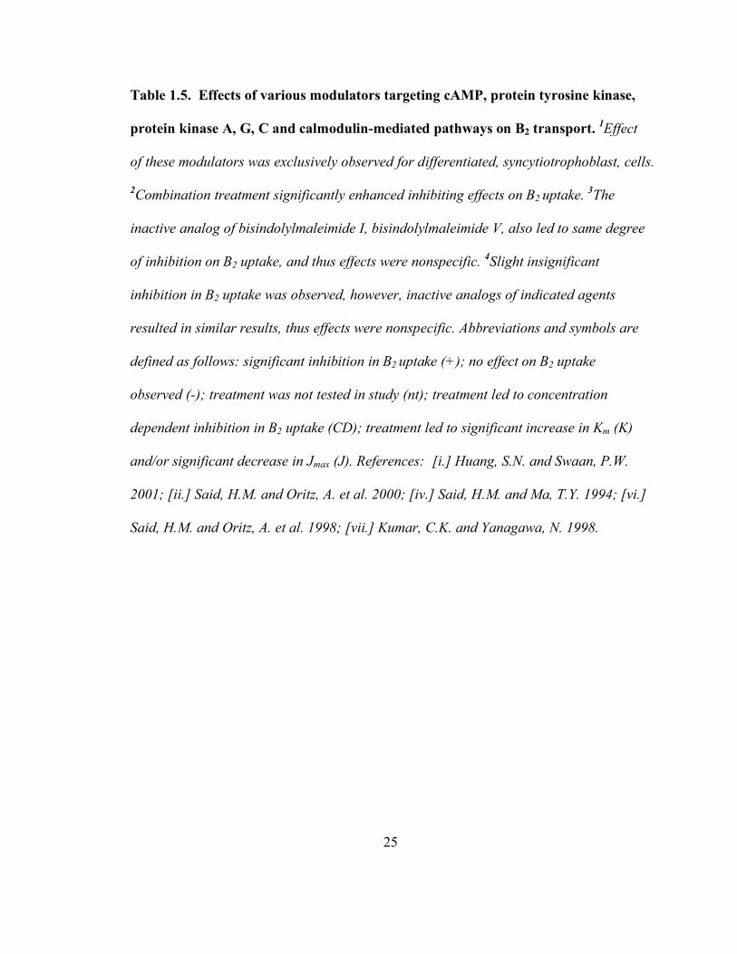
Table 1.5. Effects of various modulators targeting cAMP, protein tyrosine kinase,
protein kinase A, G, C and calmodulin-mediated pathways on B2 transport. 1Effect
of these modulators was exclusively observed for differentiated, syncytiotrophoblast, cells.
2Combination treatment significantly enhanced inhibiting effects on B2 uptake. 3The
inactive analog of bisindolylmaleimide I, bisindolylmaleimide V, also led to same degree
of inhibition on B2 uptake, and thus effects were nonspecific. 4Slight insignificant
inhibition in B2 uptake was observed, however, inactive analogs of indicated agents
resulted in similar results, thus effects were nonspecific. Abbreviations and symbols are
defined as follows: significant inhibition in B2 uptake (+); no effect on B2 uptake
observed (-); treatment was not tested in study (nt); treatment led to concentration
dependent inhibition in B2 uptake (CD); treatment led to significant increase in Km (K)
and/or significant decrease in Jmax (J). References: [i.] Huang, S.N. and Swaan, P.W.
2001; [ii.] Said, H.M. and Oritz, A. et al. 2000; [iv.] Said, H.M. and Ma, T.Y. 1994; [vi.]
Said, H.M. and Oritz, A. et al. 1998; [vii.] Kumar, C.K. and Yanagawa, N. 1998.

26

27
The influence of the protein kinase C (PKC) pathway was investigated using
phorbol-12-myristate-13-acetate (PMA, a PKC activator), chelerythrine (specific PKC
inhibitor), or staurosporin (also a PKC inhibitor). However, data clearly indicate that this
pathway is not involved in B2 uptake (Table 1.5) (Said and Ma, 1994; Kumar et al., 1998;
Said et al., 1998; Said et al., 2000; Huang and Swaan, 2001).
The involvement of Ca2+/CaM in B2 absorption has been established through the
use of various modulators that are specific to this pathway. Of these modulators,
calmidazolium, a Ca2+/CaM antagonist, has been commonly implemented. At the
molecular level, calmidazolium has been shown to activate phospholipases C and A2 that
potentiate the release of cellular Ca2+ stores and also Ca2+ entry across a variety of
mammalian cell systems (Peppiatt et al., 2004). Interestingly, several studies report a
significant inhibition in B2 uptake by calmidazolium treatment in three distinct cell
models, BeWo (human-derived placental trophoblasts), NCM460 (human-derived
colonic epithelial cell line), and Hep G2 (human-derived liver cells) (Said et al., 1998;
Said et al., 2000; Huang and Swaan, 2001). Furthermore, these reports demonstrate that
calmidazolium inhibits B2 uptake in a concentration dependent manner (Table 1.5). Said
and coworkers further demonstrated a marked reduction in the maximal transport velocity
(Jmax = 0.135 ± 0.003 and 0.084 ± 0.007 pmol/mg protein/3 min for control and 10 µM
calmidazolium, respectively (P < 0.01)) in colonic epithelial cells (Said et al., 2000). This
effect was accompanied by a significant increase in the apparent Km. Similar effects were
noted with calmidazolium treatments in Hep G2 (Said et al., 1998) and HK-2 cells
(Kumar et al., 1998). Other Ca2+/CaM modulators tested have included W-13 (a specific

28
Ca2+/CaM antagonist that is structurally different from calmidazolium), KN-93 (a
selective Ca2+/CaM-dependent kinase II inhibitor), trifluoperazine (a Ca2+/CaM inhibitor),
and KN-62 (a Ca2+/CaM inhibitor) (Kumar et al., 1998; Said et al., 1998; Said et al., 2000;
Huang and Swaan, 2001). All inhibitors, excluding KN-93, led to a significant reduction
in B2 uptake. The largely consistent effect of these Ca2+/CaM modulators on riboflavin
homeostasis as observed across divergent human cell models suggests the molecular
mechanism(s) regulating the cellular entry of this vitamin are sensitive to Ca2+ levels.
1.2.2. Passive diffusion component and an adaptive regulatory mechanism in B2
uptake
Along with indications of a B2 specific, active transport process, a passive or
simple diffusion component has been observed in cell culture under certain growth
conditions. At riboflavin concentrations higher than in human plasma (>12nM), Huang
and colleagues reported B2 absorption in intestinal epithelial cells, Caco-2, to
predominantly exhibit passive diffusion kinetics, whereas, saturation kinetics are
observed at near basal B2 concentrations (≤12 nM for humans). The conditions favoring
this passive component could be further explained by reports indicating an adaptive
regulatory absorption mechanism as evidenced in B2-deficient and over supplemented
growth conditions. This adaptive phenomenon has been observed in renal (Kumar et al.,
1998), colon (Said et al., 2000), and intestinal (Said and Ma, 1994) epithelial cells.
Intestinal cells initially grown in B2-deficient media and subsequently exposed to
physiological B2 concentrations exhibit a marked increase in riboflavin uptake compared
to controls (Said and Ma, 1994). Conversely, over supplemented B2 conditions led to a

29
marked reduction in Jmax (129 ± 13 pmol/mg protein/3 min). However, the apparent Km
values did not change in both cases (Said and Ma, 1994). Similar results were obtained in
colon epithelial cells (Said et al., 2000). Renal proximal tubule cells, HK-2 (Kumar et al.,
1998), revealed similar changes in maximal transport velocity under deficient and over
supplemented growth conditions. However, the apparent Km values were inconsistent:
Jmax increased in the B2-deficient state, whereas the corresponding apparent Km value
significantly decreased (69% of control); Jmax decreased under over supplemented
conditions, whereas the corresponding Km increased (127% of control). Overall, these
findings suggest the existence of multiple uptake mechanisms in renal cells that are
differentially regulated by varying levels of B2. Effects to the maximal transport velocity
in all three models suggest that expression or activity of the proteins involved in B2
uptake are correlated with changes in B2 concentration. Said and coworkers further
questioned whether such adaptive uptake responses were attributed to transcriptional
events (Said et al., 2000). Upon treating NCM460 cells with a transcription inhibitor,
actinomycin D, 24 hours after cells were maintained in either B2-deficient or over
supplemented conditions, B2 uptake was unaffected in the over supplemented situation,
but, significant inhibition was noted for B2-starved cells. This latter finding suggests
transcriptional events may function in up-regulating protein machinery necessary for
efficient B2 absorption during periods of vitamin deficiency. At the other extreme, i.e.,
B2-over supplemented conditions, protein machinery are suggested to be down regulated
and may explain why a passive diffusion component dominates at these higher riboflavin
concentrations.
30
1.3. Receptor-mediated and/or carrier-mediated transport?
Until recently, it was generally accepted that B2 translocation followed an active,
carrier-mediated pathway. However, the criteria used to define active transport, i.e.
energy dependency and saturation transport kinetics, do not effectively distinguish
carrier-mediated transport from receptor mediated endocytosis. Receptor-mediated events
involve endocytosis followed by microtubule driven vesicular sorting to various cellular
organelles (Mukherjee et al., 1997). In an effort to determine whether a receptor mediated
component is involved in B2 transport, our laboratory treated Caco-2 cells with either
brefeldin A (BFA), which induces misrouting of vesicles at the trans-Golgi network, or
nocodazole, a microtubule depolymerizing agent (Huang and Swaan, 2000). Our data
revealed nocodazole significantly inhibited the basolateral to apical flux of riboflavin as
well as fluorescently labeled transferrin (56.7% and 31.8% of control, respectively), an
iron transport protein that has been extensively established to be internalized by a
transferrin-specific receptor via receptor-mediated endocytosis. In addition, the apical to
basolateral transport of both riboflavin and transferrin was significantly increased (37.1%
increase compared to controls for riboflavin) upon treatment with nocodazole. BFA led to
a significant increase in transport in both directions for transferrin (13-fold higher in
apical to basolateral, and 5-fold increase in basolateral to apical). BFA caused a slight,
though not significant, increase in basolateral to apical B2 transport, and a significant
increase was observed for the apical to basolateral flux. The effects of BFA on inducing
increased concentrations of membrane receptor on the apical membrane as opposed to the
31
basolateral membrane are comparable to similar studies involving transferrin transport by
Shah and Shen (Shah and Shen, 1994).
More recently, the subcellular localization of B2 in BeWo cells was studied
(Huang et al., 2003). Rhodamine-labeled riboflavin (Rd-RF) and FITC-labeled transferrin
(FITC-Tf) were used to trace subcellular distribution patterns of both compounds upon
cellular uptake. Specifically, internalized fluorescently-labeled ligands were examined for
2D colocalized signals with indirect-immunostained RME markers for human clathrin
heavy chain and LAMP-1, a lysosome-associated membrane protein. Using fluorescence
microscopy, a distinct perinuclear punctate staining pattern was observed for rhodamine-
B2, which colocalized with clathrin and FITC-Tf. Furthermore, a membrane diffusible
probe specific for acidic organelles (e.g. late endosomes and lysosomes), LysoTracker
Blue-white DPX, did colocalize with Rhodamine- B2. Colocalization of riboflavin was
also observed with Rab5, a small GTPase that specifically resides with early endosomes,
and LAMP-1. All these findings suggest B2 transport involves clathrin dependent RME in
BeWo cells (Huang et al., 2003).
1.4. Riboflavin binding/carrier protein (RfBP)
Plasma protein binding and translocation of riboflavin in mammals are non-
specific under normal conditions. During pregnancy, this non-specific transport by
albumin and riboflavin binding immunoglobulins is not adequate to provide the
developing fetus with sufficient B2 for normal development and growth. It is believed
that under these demanding conditions specific carrier proteins are expressed that deliver
riboflavin to the developing embryo. These soluble plasma proteins are referred to as

32
“riboflavin binding proteins” (RfBP) or “riboflavin carrier protein” (RCP). It is important
to clarify that RfBP, originally discovered in chicken, is not a membrane spanning carrier
protein but a soluble protein that delivers B2 to the cell possibly by a receptor-mediated
mechanism. Pregnancy-specific RfBP has been found in rat (Muniyappa and Adiga,
1980), mouse (Natraj et al., 1987), bovine (Merrill et al., 1979), simian (Visweswariah
and Adiga, 1987b), spadefoot toad (Storey et al., 1999), turtle (Hamajima and Ono, 1995)
and human plasma (Visweswariah and Adiga, 1987a). Though the mechanism of the
transport process remains elusive, it is believed that more then one mode of transport
process may exist, which involves a receptor and/or carrier mediated transport
mechanism as reported for the transport of other micronutrients, such as folic acid.
1.4.1 Functions in oviparous species
Chicken RfBP is the first riboflavin specific carrier to be identified, and thus the
most extensively studied. It was originally isolated from egg yolk (Rhodes et al., 1959),
but later it was found that it is also present in egg white (Ostrowski et al., 1968) and
plasma of egg laying hens (Miller et al., 1982). RfBP in egg constitutes 0.8% (Rhodes et
al., 1959) of the egg white proteins present, which is relatively abundant amongst egg
white proteins. There are three known forms of RfBP that are the product of the same
gene and undergo different post-translational modifications (Hamazume et al., 1984;
Hamazume et al., 1987; Rohrer and White, 1992; Tarutani et al., 1993). The RfBP gene is
expressed both in liver and the oviduct of egg laying hens (Zheng et al., 1988). Despite
reports of RfBP in other birds and reptilian sources, such as the Japanese quail (Walker et
al., 1991), Indian python, painted turtle and American alligator (Abrams et al., 1988),

33
none of the amino acid sequences have been reported. So far, among the reptilian species
the turtle, Pelodiscus sinensis, gene sequence is exclusively known (Hamajima and Ono,
1995). In the amphibious spade foot toad, Scaphiopus couchii, RfBP is among a group of
genes that are induced or up-regulated during estivation (Storey et al., 1999).
In chicken, it is now well established that B2 is transported to the egg by the egg
laying hen with the help of RfBP. Chicken RfBP (cRfBP), a 37 kDa protein, is one of the
best characterized riboflavin binding proteins. Egg white RfBP is 35% saturated with
riboflavin as compared to fully saturated yolk RfBP. It is believed that RfBP in egg white
serves as the scavenger of free riboflavin, which may protect egg white from bacterial
attack (Tranter and Board, 1982).
In Scaphiopus toads the need for micronutrients during breeding is provided by an
increased expression of RfBP along with other micronutrient carrier proteins (Storey et
al., 1999). The developing embryo has higher nutrient demands, and most of the proteins
produced are under estrogen control. Estrogen-dependent expression of cRfBP is distinct
from that of other egg proteins such as egg white ovalbumin which is synthesized in the
oviduct, or vitellogenin which is synthesized in the liver. Chicken RfBP is synthesized in
both the liver and oviduct (Norioka et al., 1985).
1.4.1.1 Interspecies sequence homology and identity
Avian plasma RfBP is produced in the liver under estrogen control and includes a
17 amino acid signal peptide that is cleaved post-translationally. Egg white and yolk
RfBP are formed as a result of proteolytic cleavage of the last 11-13 amino acids of
plasma RfBP, and takes place when the molecule crosses the vitellogen membrane of the

34
oocyte (Norioka et al., 1985). In an estrogen induced turtle it has been reported that
unlike chicken RfBP, the turtle RfBP gene contains an open reading frame of 242 amino
acids with a signal peptide of 18 amino acids (Hamajima and Ono, 1995). Hamajima and
coworkers found overall 71.3% amino acid identity between the deduced amino acid
sequences of turtle and chicken RfBP. In a separate study, Storey and coworkers (Storey
et al., 1999) found that spade foot toad RfBP has ~ 50% sequence identity with chicken
(49.6%) and turtle (50.4%) RfBP. The area of greatest species variation between the three
proteins was within the 17-18 residue signal sequence and the final 25 residues near the
C-terminus. Furthermore, of the tissues examined, RfBP was shown to be largely liver
specific, as no RfBP mRNA was detected in brain, gut, heart or kidney.
1.4.1.2 Suggested mechanism of RfBP in riboflavin homeostasis
As mentioned earlier, RfBP has been found to be essential for the developing
embryo in chicken. Birds with a genetic defect in the RfBP gene lay fertilized eggs
normally, but their eggs fail to hatch due to a lack of riboflavin disposition (MacLachlan
et al., 1993). Chicken RfBP in oocytes binds to lipid carrier vitellogenin in a 1:1 molar
ratio, and the macromolecular complex of the two carriers recognizes a multifunctional
oocyte specific lipoprotein receptor (Mac Lachlan et al., 1994). The binding to
vitellogenin is thought to be through a highly phosphorylated region on cRfBP that
extends from amino acids 186-197, containing 8 serine-bound phosphate residues.
Removal of any phosphate residue from RfBP reduces oocyte uptake in vitro by over
60% (Miller et al., 1982). This macromolecular complex binds to an oocyte specific LDL
receptor that can also bind to low-density lipoprotein (LDL), very low-density lipoprotein

35
(VLDL) and vitellogenin independently (Sooryanarayana et al., 1998). The interaction
between vitellogenin and RfBP is thought to be through phosphate-calcium-phosphate
bridges involving phosphorylated residues in both proteins, as the interaction was
reported to be optimum in the presence of free phosphate and calcium (Mac Lachlan et al.,
1994). The authors in the study pointed out the differences in the molar concentrations of
vitellogenin (six-fold) and RfBP (nine-fold), which does not correspond with a serum 1:1
molar ratio and suggests that there may be additional mechanisms of RfBP uptake by the
oocyte. Recent studies have indicated the phospho-peptide of RfBP may play an
important role in the binding of this protein to its membrane receptor (Sooryanarayana et
al., 1998). This is supported by data that revealed vitellogenin inhibits RfBP binding to
its specific protein. Since vitellogenin binds to receptors of the low-density lipoprotein
receptor family and LDL receptors, which can interact with polyanionic ligands, it was
predicted that the family of LDL receptors and LDL related proteins are potential
candidates for such interactions. To date this relationship remains to be defined.
1.4.2 Mammalian RfBP
In rats, Sertoli cells maintain the tubular biochemical microenvironment for germ
cell proliferation and differentiation in the lumen of seminiferous tubules through
selective transport and polarized secretion. They also act as a blood testis barrier to
provide a serum free microenvironment. Nearly all major carrier proteins are produced in
Sertoli cells as there is a higher nutrient demand in the differentiating cells (Skinner and
Griswold, 1980; Skinner and Griswold, 1983; Collard et al., 1988; Davis and Ong, 1992).
Since follicle stimulating hormone (FSH) and testosterone increase the synthesis of both

36
cellular and secreted proteins in Sertoli cells, it was predicted that a riboflavin binding
protein also exists in these cells (Subramanian and Adiga, 1996a). A protein species that
was synthesized and secreted by immature rat Sertoli cells was shown to be similar to
chicken RfBP at the biochemical and immunological levels (Subramanian and Adiga,
1996b). To understand the importance of RfBP in embryonic development, estrogenic
modulation of RfBP gene expression has been tested in subhuman primates (bonnet
monkeys) through immunochemical studies (Visweswariah and Adiga, 1988). It was
observed that serum RfBP levels increased with enhanced estradiol secretion during the
menstrual cycle and early pregnancy. In humans, the existence and role of RfBP is more
elusive. Human cord serum contains proteins capable of binding riboflavin (Natraj et al.,
1988). Furthermore, reports have shown rodent-derived and non-human primate RfBP
cross-react with anti-chicken antibodies (Seshagiri and Adiga, 1987; Subramanian and
Adiga, 1996b). Hence it was suggested that the transport of riboflavin to the developing
mammalian embryo could be a carrier protein-mediated process analogous to that of
riboflavin disposition in the chicken oocyte. A recent study by our laboratory revealed a
95% reduction in riboflavin absorption in human placental trophoblasts (BeWo) that were
treated with an anti-chicken RfBP antibody (Mason et al., 2006). In addition, this same
report showed both [3H]-B2 and 125I-RfBP to be subcellularly enriched to endosomal and
lysosomal organelles isolated by sucrose-density fractionation. Similar to avian species,
these results may suggest that a B2-specific soluble carrier protein is important in
mediating this vitamin’s cellular absorption in human placental trophoblasts. Another
interpretation of this data may be that the antibody to the chicken RfBP is merely
37
expressing an epitope that shares biochemical conformational similarities to the elusive
riboflavin receptor or a binding protein expressed in humans. However, the evidence of a
soluble RfBP analog with biochemical and immunological similarities to avian RfBP
reported for several divergent mammalian species (e.g., rat, bovine, and simian) suggest
this secreted protein is evolutionarily conserved, and further strengthens the hypothesis
that such a B2-sequestering protein potentially exists in humans.
1.4.2.1 Putative function and role of RfBP in riboflavin uptake and transport
Immunohistochemical localization revealed that RfBP exists in rodent Sertoli
cells, Leydig cells, pachytene spematocytes, around spermatids and on mature spematoza
of rodents and primates (Bhat et al., 1995). A three-fold increase in RfBP levels in Sertoli
cells was reported to correspond with 25 ng/ml FSH levels, and a six-fold increase in the
presence of testosterone (10 µΜ) (Subramanian and Adiga, 1996a). These studies were
validated by using aromatase enzyme inhibitors that block the aromatization of androgens
to estrogen by aromatase in chicken liver and oviduct. This treatment reduced RfBP
secretion to 85% of control, suggesting that the hormonal stimulation is mediated through
in-situ synthesized estrogen. This became more evident when, exogenous estradiol-17-β
(10 µM) led to three-fold enhanced secretion of RfBP. Inhibition of FSH by tamoxifen
(10 µΜ) reduced RfBP secretion to 75% of control. Overall, these findings suggest that
estrogen regulates RfBP expression in hormonally stimulated Sertoli cells, presumably to
function as the carrier of riboflavin to the developing germ cells in rodents (Subramanian
and Adiga, 1996a).

38
In female Bonnett macaques, the systemic RfBP concentration appears to be
regulated by plasma estradiol levels during the menstrual cycle as well as early
pregnancy (Visweswariah and Adiga, 1988). Interestingly, antibodies raised against
chicken RfBP caused pregnancy termination in mice, most likely due to B2-deficiency in
the developing fetus. RfBPs, as mentioned earlier have been detected in plasma of male
monkeys as well as in rat testicular Leydig and Sertoli cells (Bhat et al., 1995). Similar to
their female counterparts, RfBP production in male rats and monkeys is sensitive to
estradiol-17-β and FSH levels (Subramanian and Adiga, 1996a; Subramanian and Adiga,
1996b).
Human purified RfBP has been reported to have a molecular weight of 36 kDa
and an isoelectric point of 4.1 (Natraj et al., 1988). The presence of RfBP in amniotic
fluid of pregnant women has also been revealed (Prasad et al., 1992). Detectable RfBP
plasma levels were observed in pregnant women after 4 months of gestation and
remained high up to 8 months later. In amniotic fluid, a two-fold higher concentration
was reported as compared to RfBP levels in serum at 3-4 months of gestation (Natraj et
al., 1988). Despite these reports revealing RfBP in humans, the genetic sequence of such
a protein remains to be reported on any public database.
From a translational medicine perspective, a recent study by Rao and colleagues
reported significant elevations of serum RfBP in patients with breast cancer (Rao et al.,
1999). Earlier, Vaidya and coworkers (Vaidya et al., 1998) reported a decrease in serum
B2 levels in breast cancer patients; with baseline B2 serum levels achieved after
tamoxifen treatment. Combined, these data suggest that RfBP is up-regulated during
39
breast cancer with a concomitant decrease in plasma riboflavin levels. Thus, an important
role for riboflavin and RfBP in the pathology of breast cancer has been demonstrated, but
the physiological role remains to be elucidated. Similar to its potential as a breast cancer
biomarker, elevated serum RfBP levels have recently been shown to correlate with
hepatocellular carcinoma progression (Rao et al., 2006).
In rats, as stated previously, RfBP synthesis is under the control of estrogen.
Knowing the nutritional demand for B2 during human pregnancy, it can be hypothesized
that the biosynthesis of human RfBP could be regulated by estrogen levels. This theory is
supported in studies that showed RfBP levels were significantly elevated during the first
trimester of pregnancy (Natraj et al., 1988). Furthermore, Clarke and coworkers reported
riboflavin levels in cord serum to be 3-4 times higher than in maternal serum, and may
reflect a concentrative accumulation of RfBP in response to physiological requirements
for B2 (Clarke, 1977). It is believed that human RfBP may be produced in the liver
analogous to avian and rat RfBP. It is regulated by estrogen but the origin of human
RfBP in amniotic fluid is not known. It has been proposed that the human placenta
synthesizes this RfBP and the liver harbors a RfBP specific mRNA that has the
propensity to synthesize this protein, which may also contribute to amniotic fluid RfBP.
Natraj and colleagues suggested the presence of human RfBP in amniotic fluid could be
due to the direct transfer from maternal circulation through the placenta (Natraj et al.,
1988).
Riboflavin concentration in mammalian milk is several fold higher than in
maternal circulation during lactation in spite of the presence of circumferential tight

40
junctions between mammary epithelial cells which constitute a barrier to the free passage
of blood constituents (Pitelka and Hamamoto, 1977). Consequently, it was proposed that
RfBP synthesis is up regulated by lactating mammary glands to sequester the vitamin for
secretion into milk required for neonatal nutrition (Karande et al., 2001). The mammary
gland is a known estrogen target tissue, which further strengthens this hypothesis, as
estrogen and progesterone are known modulators of RfBP biosynthesis in vivo as well as
in vitro (Karande et al., 2001). Interestingly however, a recent report by van Herwaarden
and colleagues (van Herwaarden et al., 2007) revealed the multi-drug transporter, breast
cancer resistance protein (BCRP/ABCG2, an established [ABC]-transporter), to function
in the secretion of B2 into breast milk of lactating mice. These scientists reported a 63-
fold reduction in milk secretion of riboflavin in BCRP1-/- lactating females compared to
lactating wild-type females. Furthermore, this efflux transporter was shown to be largely
instrumental in reducing the tissue distribution and plasma levels for riboflavin, as
BCRP1-/- mice revealed substantial reductions (0.8 fold less) and increases (1.8 fold
higher) in intestinal and hepatobiliary excretion profiles, respectively. However, pups that
were nurtured by standard chow fed BCRP1-/- lactating females only exhibited a modest
reduction in plasma riboflavin levels and there were no signs of B2-deficiency. It was
only when riboflavin was removed from the diet fed to the BCRP1-/- lactating females
that the pups suffered vitamin deficient states, as was evidenced by a lack of growth,
severe anemia, and fatty degeneration of the liver. Collectively, this report suggests that
BCRP functions in reducing the systemic and tissue levels of riboflavin. However, the

41
overall effects are modest and other distinct uptake mechanisms are suggested to regulate
riboflavin homeostasis to offset the extrusion capacity of this efflux system.
1.4.2.2 Utilizing the riboflavin absorption and trafficking mechanism to enhance
macromolecule delivery
Serum albumin is known to adsorb riboflavin and has been demonstrated to co-
elute with RfBP during riboflavin affinity purification. Thus, serum albumin is
considered a non-specific carrier of riboflavin under normal physiological conditions. In
addition, Low and colleagues reported facilitated entry of BSA into human lung, ovary
and pulmonary cell systems upon covalently labeling BSA with riboflavin (Wangensteen
et al., 1996; Holladay, 1999). These studies were the first to demonstrate the utility of the
B2 absorption pathway in enhancing macromolecule delivery, and suggested such B2-
specific mechanisms to involve endocytosis and transcytosis events. These results were
substantiated by experiments revealing 1) a significant reduction in BSA-B2 uptake in the
presence of transcytosis inhibitors and 2) accumulation of these conjugates to organelle
populations resembling endosomes. However, these same researchers reported cellular
accumulation of BSA-B2 was inhibited by unlabeled BSA and not by free riboflavin.
Overall, in addition to endocytosis, riboflavin absorption is suggested to be regulated by
multiple internalization mechanisms.
1.5 Comparison of B2 internalization with folate uptake and transport mechanisms
Chicken RfBP belongs to the family of folate binding proteins that are
glycosylphosphatidylinositol (GPI) anchored proteins, and found on the outer surface of
the plasma membrane. Eight of the nine pairs of cysteines involved in disulfide bonds in

42
RfBP are conserved in milk folate binding protein (FBP), as are all of the tryptophan
residues (Zheng et al., 1988). This conservation of cysteine residues indicates the
importance of secondary structure and function of cRfBP. Sequence identity between
homologous regions of these two vitamin-binding proteins is more than 30% (Zheng et
al., 1988). Furthermore, RfBP is evolutionarily conserved in terms of its physiochemical
and immunological characteristics and is involved in the embryonic vitamin nutrition
from avian to primate species. Folic acid contains a pterin ring structure, which is similar
to the B2 structure. In addition, sequence similarity of the residues in cRfBP and FBP
indicate similar folding of the ligand binding domain in the two proteins (Monaco, 1997).
Considering these commonalities in binding protein biochemistry and vitamin structural
similarities, it was theorized that both vitamins were absorbed via the same cellular entry
pathways upon binding to their respective soluble carrier proteins. However, studies
performed in our laboratory suggested otherwise, as the internalization of [3H]-B2 in the
presence of folate (1000-fold excess) did not inhibit B2 uptake in human placental
trophoblasts (Huang and Swaan, 2001). Furthermore, enzymatic studies carried out by
our laboratory involving the cleavage of GPI-linked plasma membrane receptors in this
same cell model revealed no alteration in [3H]-B2 uptake (unpublished data). Taken
together, these studies suggest the B2 and folate uptake mechanisms are distinct, at least
in human placental trophoblasts, and thus involve different plasma-membrane associated
protein machinery.

43
1.6 Summary and thesis objectives
Over the last several decades, our understanding of the molecular regulation and
cellular transport mechanism specific to B2 has significantly increased. In general,
riboflavin absorption across divergent human cell systems exhibits the following salient
properties: temperature dependence, a general Na+-independence, a saturable active
transport component that appears to dominate at near basal B2 concentrations (~ 12 nM),
and a passive diffusion component that dominates at concentrations above physiological
vitamin concentrations. In addition, the Ca+/calmodulin-, protein kinase A and G
pathways are suggested to regulate B2 uptake (Huang and Swaan, 2001). More recently,
receptor-mediated endocytosis (RME) has been shown to play a role in riboflavin
absorption across the placental barrier (Huang et al., 2003). The role of RfBP in B2
absorption and transport is still elusive and the existence of a human RfBP analog has not
been definitively demonstrated. However, in a recent report by our laboratory that
showed nearly complete inhibition in B2 uptake along human placental trophoblasts
(BeWo) treated with antibodies raised against chicken RfBP may suggest the importance
of such a carrier protein in regulating the cellular entry of riboflavin (Mason et al., 2006).
Furthermore, in light of reports revealing elevated serum levels of RfBP in human breast-
and hepatocellular carcinomas (Rao et al., 1999; Karande et al., 2001; Rao et al., 2006), a
clinical niche has been suggested for utilizing the riboflavin-specific absorption pathway
as an indicator of cancer progression and in targeted chemotherapeutic strategies.
However, all studies to date have involved indirect methods to define B2-specific
absorption and trafficking mechanisms in human cell systems. Currently, it is apparent

44
that there are multiple absorption processes that are regulating this vitamin’s cellular
entry. These modes include a passive component and an active component. Regarding its
active uptake, it appears that a receptor-mediated pathway plays a role, specifically
involving clathrin-mediated endocytosis (CME), in part in non-renal cell systems.
However, a transporter component has not been excluded as an alternate route of
internalization. Thus, some of the questions remaining to be answered regarding
riboflavin uptake and trafficking in human cells include the following: 1) to what extent
is this vitamin being internalized by RME or non-RME processes 2) what is the rate at
which B2 is initially absorbed and trafficked to cellular organelles specific to RME and 3)
what specific proteins facilitate these RME or non-RME mechanisms? These are
important questions that need to be answered to enrich the knowledge base for future
studies aimed at utilizing the B2 internalization pathway in drug targeting approaches.
However, this thesis focuses on characterizing the RME mechanisms regulating B2
internalization and trafficking in human epithelia. Specifically, the major objectives of
this thesis include:
1. Define the temporal and spatial distribution of endocytosed B2 in human
epithelial cell models BeWo and Caco-2, both of which are known to exhibit
high affinities (nM) for this vitamin,
2. Define the extent by which B2 absorption and trafficking depends on RME,
3. Elucidate whether B2–specific RME involves clathrin-independent endocytic
pathways, such as the common lipid raft/caveolae pathway.

45
CHAPTER 2
cAMP-COUPLED RIBOFLAVIN TRAFFICKING IN PLACENTAL
TROPHOBLASTS AND ENTEROCYTES: A DYNAMIC AND ORDERED
PROCESS
Abstract
Riboflavin (vitamin B2), an essential micronutrient central to cellular metabolism
through formation of flavin mononucleotide (FMN) and flavin adenine dinucleotide
(FAD) cofactors, is internalized, at least in part, via a proposed receptor-mediated
endocytic (RME) process. The purpose of this study was to delineate the cellular B2
distribution using human placental trophoblasts and enterocytes, and evaluate the
regulatory role of cAMP in this process. Subcellular fractionation and 3D confocal
microscopy analyses were carried out to define the B2 accumulation profile. Biochemical
assays evaluating the cAMP dependence of this pathway were also performed. The
present study records an intracellular B2 distribution pattern that shows dynamic

46
accumulation of the ligand predominantly to the endosomal and lysosomal
compartments, and to a lesser extent to the Golgi and mitochondria. In contrast,
transferrin (TF) colocalizes rapidly within endosomes with minimal accumulation in the
other organelles. Temporal and spatial distribution of B2 colocalized with unique markers
of the endocytic machinery provide added morphological evidence in support of the RME
process with ultimate translocation to the mitochondrial domain. Colocalized staining
with the Golgi also suggests a possible recycling or exocytic mechanism for trafficking of
this ligand. Furthermore, this study demonstrates cAMP regulation of the putative ligand-
bound B2 receptor and its association into endocytic vesicles. Delineating the dynamics
of the process governing cellular B2 homeostasis presents an untapped resource that can
be further exploited to improve our current understanding of nutritional biology and fetal
growth and development, and perhaps target the endogenous system to develop novel
therapeutic approaches.
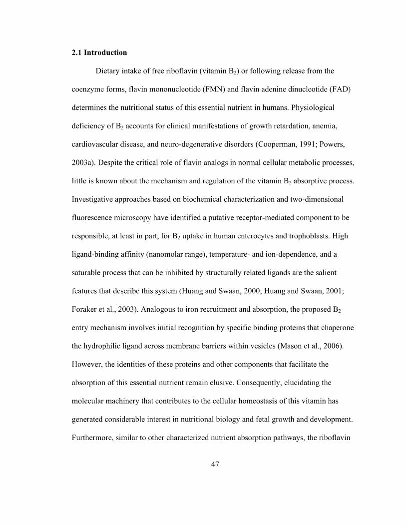
47
2.1 Introduction
Dietary intake of free riboflavin (vitamin B2) or following release from the
coenzyme forms, flavin mononucleotide (FMN) and flavin adenine dinucleotide (FAD)
determines the nutritional status of this essential nutrient in humans. Physiological
deficiency of B2 accounts for clinical manifestations of growth retardation, anemia,
cardiovascular disease, and neuro-degenerative disorders (Cooperman, 1991; Powers,
2003a). Despite the critical role of flavin analogs in normal cellular metabolic processes,
little is known about the mechanism and regulation of the vitamin B2 absorptive process.
Investigative approaches based on biochemical characterization and two-dimensional
fluorescence microscopy have identified a putative receptor-mediated component to be
responsible, at least in part, for B2 uptake in human enterocytes and trophoblasts. High
ligand-binding affinity (nanomolar range), temperature- and ion-dependence, and a
saturable process that can be inhibited by structurally related ligands are the salient
features that describe this system (Huang and Swaan, 2000; Huang and Swaan, 2001;
Foraker et al., 2003). Analogous to iron recruitment and absorption, the proposed B2
entry mechanism involves initial recognition by specific binding proteins that chaperone
the hydrophilic ligand across membrane barriers within vesicles (Mason et al., 2006).
However, the identities of these proteins and other components that facilitate the
absorption of this essential nutrient remain elusive. Consequently, elucidating the
molecular machinery that contributes to the cellular homeostasis of this vitamin has
generated considerable interest in nutritional biology and fetal growth and development.
Furthermore, similar to other characterized nutrient absorption pathways, the riboflavin
48
internalization mechanism is suggested to be instrumental in serving as a portal of
cellular entry that can be exploited to promote targeted drug design and delivery to
breast- and liver cancers (Rao et al., 1999; Rao et al., 2006).
Internalization of extracellular nutrients in eukaryotes via receptor-mediated
endocytosis employs complex cell machinery in a sequential and regulated manner. The
co-existence of abundant intracellular structural and signaling networks offers several
possibilities for receptor-associated ligands. Most water-soluble vitamins are fated to
metabolic trapping, a term that describes enzymatic processing of the molecule to forms
that are spatially restricted within the cell (Aw et al., 1983). However, alternative
translocation routes, including late endosomal or lysosomal degradation, membrane
recycling, or even transcytosis (in polarized epithelial systems) remain a distinct
possibility (Maxfield and McGraw, 2004). Such receptor-ligand interactions also occur
under extensive cellular regulation either via an intrinsic mechanism such as kinase or
phosphatase activity or via generation of signaling cascades that include second
messenger regulation of integrated pathways (Schulte and Fredholm, 2003; Barbieri et
al., 2004). Huang and colleagues have demonstrated that the B2 internalization process is
affected by modulation of the cyclic nucleotide second messengers, cAMP and cGMP, as
well as by protein kinase A, G and calcium-calmodulin pathways (Huang and Swaan,
2001).
The objective of the current study was to evaluate the fate of internalized B2 over
time by following its intracellular sorting using the human placental trophoblast and
enterocyte cell lines, BeWo and Caco-2, respectively (Pattillo and Gey, 1968; Rindler

49
and Traber, 1988; Hughson and Hopkins, 1990; Knight et al., 1995), and define the
regulatory role of cyclic-AMP in this process. The enhanced nutritional requirements of
the fetus (Dancis et al., 1985; Kirshenbaum et al., 1987; Visweswariah and Adiga, 1987a;
Dancis et al., 1988) and the presence of high affinity B2 systems in both BeWo and Caco-
2 (Huang and Swaan, 2000; Huang and Swaan, 2001) justify the use of the selected
models. Time- and concentration-dependent fractionation analyses of the localized ligand
in endosomes, lysosomes, Golgi, and mitochondria were compared with confocal laser
scanning microscopy studies examining time-lapsed 3D colocalization between
internalized rhodamine-labeled riboflavin (Rd-RF) (Huang et al., 2003; Phelps et al.,
2004) and immunolabeled organelles. Trafficking profiles for B2 were compared with
those determined for transferrin (TF), a receptor-mediated endocytic marker, (Dautry-
Varsat et al., 1983; Jin and Snider, 1993; van Dam and Stoorvogel, 2002), and for
taurocholic acid (TCA) that does not follow a vesicular pathway (Zhang et al., 2002;
Zhang et al., 2004). In addition, cyclic AMP assays were included to determine
regulation of the downstream events of cellular B2 translocation upon its binding to the
proposed B2-receptor. Overall, the study for the first time shows that B2, like TF,
accumulates mainly within endosomes but differs from TF in the extent of lysosomal
distribution. The present study also suggests a possible recycling mechanism that
contributes to B2 homeostasis and regulation via the cAMP signaling pathway in the
trophoblast model.
50
2.2 Methods and materials
2.2.1 Cell culture
BeWo and Caco-2 cells were obtained from American Type Culture Collection
(Manassas, VA) and routinely maintained in a controlled atmosphere at 37 ºC, under 5%
CO2. BeWo (passage numbers 206-221) and Caco-2 (passage numbers 30-48) cells were
routinely cultured in F-12K and DMEM medium, respectively (Invitrogen Life
Technologies, Carlsbad, CA) supplemented with 10% fetal bovine serum, 1% non-
essential amino acids, 100 U/ml penicillin, and 100 µg/ml streptomycin. Cells were
seeded in 150 mm culture dishes at a density of 3 × 10⁴ cells/cm2 and were used 5 to 7
days post-seeding for fractionation studies. All studies involved cells in the proliferating
phase, as higher nutrient demands have been commonly correlated with rapidly dividing
cells (e.g., a characteristic of tumor cells) compared to differentiated cell systems (Ross et
al., 1994; Ponka and Lok, 1999).
2.2.2 Labeling of transferrin
Holo-transferrin (Sigma, St. Louis, MO) was labeled with Na-125I (~ 5 µCi /µg;
Amersham Biosciences, Piscataway, NJ) using the IODOGEN® method (Pierce
Biotechnology Inc., Rockford, IL). Iodinated protein was desalted by gel filtration using
Micro-Bio-Spin® columns (Bio-Rad Laboratories, Hercules, CA) and 125I-incorporation
was determined by gel electrophoresis and autoradiography. The specific activity of the
125I-transferrin was ~ 400 cpm/pmol.

51
2.2.3 Ligand uptake and subcellular fractionation
Cell monolayers were dosed with 10 nM each of [3H]-riboflavin (Sigma, St.
Louis, MO) and 125I-transferrin, or 20 nM [3H]-taurocholic acid (American Radiolabeled
Chemicals, Inc., St. Louis, MO), in Hanks’ Balanced Salt Solution (HBSS, pH 7.4)
containing 25 mM glucose and 10 mM HEPES at 37 ºC for 2 hr. After incubation, cells
were washed thoroughly with ice-cold Dulbecco’s phosphate-buffered saline (DPBS)
containing Ca2+ and Mg2+ (pH 3.0) to remove plasma membrane associated ligands. Cells
were then pooled in homogenization buffer containing 0.25 M sucrose, 10 mM Tris-HCl
(pH 7.5) and protease inhibitors (Complete Mini®, Roche, USA), and allowed to swell on
ice for 15 min prior to lysis through a 25G 5/8 hypodermic needle and monitored by
phase-contrast microscopy. The cell lysate was centrifuged at 600 × g for 5 min at 4 ºC to
yield a nuclear pellet (N, represents nuclear and dense membrane fragments) and
postnuclear supernatant (PoN). Total protein content of each fraction was measured using
the Bradford assay (Bio-Rad Laboratories, Hercules, CA).
Endosomes, lysosomes, Golgi, and mitochondria were isolated by loading the
PoN fraction on a discontinuous sucrose gradient (0.8 – 2.0 M) and subjected to 205,000
× g for 2 hr at 4 ºC using a SW 55Ti rotor (Beckmann Coulter Inc., Fullerton, CA). The
gradient was top-fractionated into 350 µl aliquots. Accumulation of radiolabeled ligands
in each fraction was measured by a quench-curve corrected dual-label (i.e., [3H] and 125I)
liquid scintillation counting program (Beckmann Coulter Inc., Fullerton, CA) and
normalized to total protein content determined by the Bradford assay. Time- and
concentration-dependent enrichment of riboflavin and transferrin to these organelles were

52
assessed by varying incubation times (30 min – 2 hr) and ligand concentrations (5 – 30
nM).
2.2.4 Immunoblotting and data analysis
Cellular organelles in the collected fractions were identified by western blot
analyses using monoclonal antibodies (BD Pharmingen, San Diego, CA) directed against
organelle-specific protein markers (i.e., Rab5 GTPase and clathrin for early endosomes,
LAMP-1 for lysosomes, GM130 for Golgi, and cytochrome c for mitochondria).
Fractionated proteins were resolved on 7.5% or 18% Tris-HCl gels, transferred to PVDF
membranes (Immun-Blot™, Bio-Rad, Hercules, CA), immunoblotted using peroxidase-
conjugated secondary antibodies, and detected using the ECL plus system (Amersham
Biosciences, Piscataway, NJ). Sequentially fractionated samples that were positive for a
specific protein marker were grouped together to represent that particular organelle
population. Subcellular ligand distribution to the individual organelle populations was
expressed as a percentage of the total ligand content in the PoN fraction. Due to overlap
of marker expression between fractions, the ligand distribution exceeds 100% but serves
as a semi-quantitative approach for identifying the ligand trafficking pathway(s) upon
internalization.
2.2.5 Immunofluorescence and confocal microscopy
BeWo cells were seeded 3 - 5 days prior (5 × 10³ cells/cm2) on collagen-coated
BD Falcon™ culture slides (BD Biosciences, Bedford, MA). Following serum starvation,
pulse-chase studies with either 500 nM rhodamine-B2 (Rd-RF) (Phelps et al., 2004)or 30
nM FITC-transferrin (FITC-TF, Sigma, St. Louis, MO) were carried out as previously

53
described (Huang et al., 2003). Cells were then fixed, permeabilized, and blocked prior to
immunolabeling for 2 hr at room temperature to detect organelle markers (clathrin, Rab5
GTPase, LAMP-1, GM130, or cytochrome c). Cells were washed thoroughly and probed
with either Alexafluor546®- or Alexafluor405®-labeled sheep anti-mouse IgG
(Molecular Probes, Eugene, OR). Immunofluorescence treatments were preserved using
GelMount™ (Biomeda Corp., Foster City, CA), and visualized using a Nikon Eclipse
TE2000 E inverted confocal laser scanning microscope equipped with three fixed lasers,
488-, 405-, and 543 nm (Nikon Instruments Inc., Melville, NY). All 3D confocal images
were acquired using the following parameters on Nikon EZ-C1 acquisition software
(Gold version 2.3, Image Systems Inc., Columbia, MD): Nikon Plan Apo 60xA oil
objective (1.4 numerical aperture), 6.00 µs scan dwell, 512 × 512 pixel size, 0.25 µm z-
step, 60- or 150 µm detector pinhole, and constant attenuation of laser 488 nm using a
neutral density filter (ND8). Images were iteratively deconvolved using a calculated point
spread function for each fluorescent channel and corrected using intensity thresholds
defined by treatment with either a non-reactive rhodamine derivative or secondary
antibodies alone, and by applying a median filter. All images were then analyzed to
define 3D colocalized regions using Volocity 3.6 software (Improvision Inc., Lexington,
MA). The extent of colocalization between fluorescent ligands and AlexaFluor-labeled
organelles was determined by calculating the percent of total overlapping volume (cubic
micrometers) between the two channels compared to the corresponding total ligand
volume. All data were averaged using 3 - 4 regions of colocalized distributions and

54
statistical significance was determined using the Pearson’s Correlation (Manders et al.,
1993).
2.2.6 Determination of cyclic AMP accumulation in BeWo cells
BeWo cells were seeded for assays evaluating the cAMP response to the B2
stimulus (2.5 × 10⁵ cells/well) and cAMP inhibition assays (1.0 × 10⁵ cells/well), one day
prior to use in 24-well Biocoat™ plates (BD Biosciences, Bedford, MA). Cells were
washed with Earle’s Balanced Salt Solution (EBSS, pH 7.4) and incubated at 37 °C with
10-12 to 10-3 M stimulant (i.e., forskolin or riboflavin) for the indicated times in 250 µl of
stimulation buffer (20 mM HEPES buffered F-12K, pH 7.4, containing 30 µM Ro-20-
1724, a phosphodiesterase inhibitor and 200 µM sodium metabisulfite, an anti-oxidant).
Negative coupling assays were carried out by preincubating cells with varying B2
concentrations for 10 minutes prior to stimulating with 1 µM forskolin. The reaction was
terminated by discarding the stimulation buffer and adding 200 µl of 3% perchloric acid
per well. After incubating on ice for 30 min, 80µl of 15% KHCO3 was added to each
well, and the plates were further incubated for 10 min. The plates were then centrifuged
for 10 min at 1,300 × g. Subsequently, 50 µl of the supernatant from each well was
transferred to 250 µl of reaction mixture (150 µl of Tris-EDTA buffer, 50 µl of cAMP-
binding protein, and 50 µl of [3H]-cAMP) and incubated at 4 °C overnight. On the
following day, 250 µl of charcoal/dextran mix (1%) was added to each sample followed
by incubation at 4 °C for 15 min, and then centrifugation for 15 min at 1,300 × g.
Radioactivity in the supernatant from each tube was quantified by liquid scintillation

55
counting. Cyclic AMP concentrations were calculated using a standard curve according
to the protocol of the assay kit (Diagnostic Products Corporation, Los Angeles, CA).
2.3 Results
2.3.1 Isolation of organelle-enriched fractions
Cell homogenates following ligand exposure were centrifuged to separate the
denser membrane fragments and nuclei from the lower density organelles. Separation of
nuclei was confirmed by DAPI staining (Molecular Probes, Eugene, OR). Another
polarized epithelial system, the human intestinal Caco-2 cell line, which also exhibits
nanomolar affinity for B2 (Huang and Swaan, 2000), was used in parallel. Total
intracellular content of riboflavin that accumulated within the fractionated compartments
was compared with that of transferrin and taurocholic acid, respectively taken to be
positive and negative controls for the receptor-mediated endocytic process. As expected,
the BeWo- and Caco-2-associated profiles for TF when expressed as % total dose
revealed substantial ligand accumulation (28 – 48 %) independent of cell line. Likewise,
the riboflavin load in the fractions was ≥ 8%, but TCA showed poor accumulation, at
least in BeWo cells (Figure 2.1). Such distinct distribution profiles for B2, TF, and TCA
in cells from different tissue origins provide validation of the isolation procedure.

56
Figure 2.1. Comparative cell-associated profiles of [3H]-riboflavin (10 nM B2) with
the endocytic marker 125I-transferrin (10 nM TF) and an unrelated bile acid
transporter substrate, [3H]-taurocholic acid (20 nM TCA), in placental (BeWo) and
intestinal (Caco-2) cells. Ligand content derived from nuclear and postnuclear fractions
and expressed as the percent of total dose revealed ≥ 8% B2 accumulation (hashed bars),
while TF levels (white bars) varied from 48% in BeWo cells to 28% in Caco-2 cells. The
distribution profile of TCA (black bars) showed negligible accumulation in trophoblasts
versus enterocytes.

57
Protein content of the nuclear fractions was significantly higher (~ 5 times) than
that for postnuclear fractions that had protein yields varying between 2.30 – 4.95 mg/ml.
However, the protein load per gradient was kept constant. The identity of the fractionated
gradient was determined by western blot analyses for organelle-specific markers. Clathrin
heavy chain (180 kDa), a protein component of the endocytic vesicle coat (Nathke et al.,
1992), and Rab5 GTPase (25 kDa), a GTP-binding protein associated with early
endosomes (Bucci et al., 1992), were detected mainly in fractions 1 through 7 isolated
from BeWo cells (Figure 2.2C). Likewise, Caco-2 cells upon subcellular fraction analysis
showed a similar distribution of clathrin but Rab5 GTPase was restricted to fractions 1
and 2 (Figure 2.2D). Lysosomal-associated membrane protein (LAMP1; 110 kDa)
(Rohrer et al., 1996) indicated the presence of lysosomes in fractions 3 through 10 in
BeWo (Figure 2.2C), and 5 through 9 in Caco-2 (Figure 2.2D). Due to overlap in protein
markers between fractions, the organelles were not exclusive to any given fraction but
instead pooled fractions were considered enriched for a defined subcellular population.
2.3.2 Distribution of internalized B2 and TF
In order to evaluate the intracellular localization profile of B2 after cellular entry,
cells were loaded for 2 hrs at 37 °C with 10 nM of each [3H]-B2 and 125I-TF. The
endocytic uptake and processing of TF enabling cellular iron transfer has been well-
documented (Dautry-Varsat et al., 1983), thereby supporting its use as a marker with
which to contrast the trafficking pattern(s) of B2. Following incubation with the ligands,
subcellular fractions isolated from trophoblasts and enterocytes by density gradient
centrifugation were measured for radiolabeled B2 and TF by liquid scintillation counting.

58
Subcellular distribution patterns for [3H]-B2 and 125I-TF measured in the postnuclear
organelle fractions from both cell models resulted in localized bands of the radiolabels
along the sucrose gradients. B2 was detected mainly in the heavier fractions for both cell
lines (fractions 7 – 12 in BeWo, and fractions 8 – 12 in Caco-2), whereas TF appeared to
concentrate in the less dense fractions (fractions 2 – 7 in BeWo and 2 – 8 in Caco-2) of
the gradient (Figure 2.2A and B). Degradation of 125I-TF resulting in loss of label during
the cell homogenization and fractionation process poses a potential concern. Hence, the
integrity of the marker following processing was verified by autoradiography, which
revealed intact protein at ~ 83 kDa in all fractions containing transferrin (Figure 2.2C and
D). Consequently, the organelle isolation process did not interfere with the
compartmentalization and detection of TF as demonstrated for both cell models.

59
Figure 2.2. Distribution of [3H]-B2 and 125I-Transferrin (TF) in the postnuclear
fractions isolated from trophoblasts and enterocytes. Postnuclear fractions isolated
from BeWo (A & C) and Caco-2 (B & D) cells were resolved based on differential
organelle densities using a discontinuous sucrose gradient, fractionated, and measured
for dual radiolabel accumulation (closed squares - [3H]-B2; open circles - 125I-TF).
Organelle-enriched fractions were identified by Western blot analyses for BeWo and
Caco-2 (C & D, respectively) using antibodies directed against endosomal (clathrin,
Rab5 GTPase), lysosomal (LAMP-1), and Golgi-associated (GM130) marker proteins.
Nuclear fractions (i.e., the denser plasma membrane and nuclear fragments separated
from cytosolic or ‘PoN’ contents of cell lysates) were examined for the mitochondrial
marker (cytochrome c), as the PoN fractions were negative for the expression of this
protein. All protein markers detected in fractions were compared to their corresponding
expression in unfractionated whole cell lysate samples (WC). Intact radiolabel on TF
following homogenization and gradient fractionation was visualized at ~ 83 kDa by gel
electrophoresis and autoradiographic exposure of the isolated fractions from
trophoblasts (C) and enterocytes (D).
60
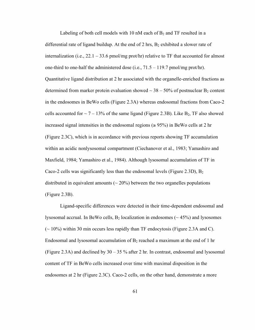
61
Labeling of both cell models with 10 nM each of B2 and TF resulted in a
differential rate of ligand buildup. At the end of 2 hrs, B2 exhibited a slower rate of
internalization (i.e., 22.1 – 33.6 pmol/mg prot/hr) relative to TF that accounted for almost
one-third to one-half the administered dose (i.e., 71.5 – 119.7 pmol/mg prot/hr).
Quantitative ligand distribution at 2 hr associated with the organelle-enriched fractions as
determined from marker protein evaluation showed ~ 38 – 50% of postnuclear B2 content
in the endosomes in BeWo cells (Figure 2.3A) whereas endosomal fractions from Caco-2
cells accounted for ~ 7 – 13% of the same ligand (Figure 2.3B). Like B2, TF also showed
increased signal intensities in the endosomal regions (≥ 95%) in BeWo cells at 2 hr
(Figure 2.3C), which is in accordance with previous reports showing TF accumulation
within an acidic nonlysosomal compartment (Ciechanover et al., 1983; Yamashiro and
Maxfield, 1984; Yamashiro et al., 1984). Although lysosomal accumulation of TF in
Caco-2 cells was significantly less than the endosomal levels (Figure 2.3D), B2
distributed in equivalent amounts (~ 20%) between the two organelles populations
(Figure 2.3B).
Ligand-specific differences were detected in their time-dependent endosomal and
lysosomal accrual. In BeWo cells, B2 localization in endosomes (~ 45%) and lysosomes
(~ 10%) within 30 min occurs less rapidly than TF endocytosis (Figure 2.3A and C).
Endosomal and lysosomal accumulation of B2 reached a maximum at the end of 1 hr
(Figure 2.3A) and declined by 30 – 35 % after 2 hr. In contrast, endosomal and lysosomal
content of TF in BeWo cells increased over time with maximal disposition in the
endosomes at 2 hr (Figure 2.3C). Caco-2 cells, on the other hand, demonstrate a more
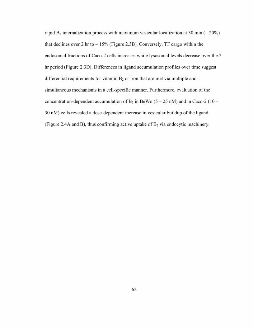
62
rapid B2 internalization process with maximum vesicular localization at 30 min (~ 20%)
that declines over 2 hr to ~ 15% (Figure 2.3B). Conversely, TF cargo within the
endosomal fractions of Caco-2 cells increases while lysosomal levels decrease over the 2
hr period (Figure 2.3D). Differences in ligand accumulation profiles over time suggest
differential requirements for vitamin B2 or iron that are met via multiple and
simultaneous mechanisms in a cell-specific manner. Furthermore, evaluation of the
concentration-dependent accumulation of B2 in BeWo (5 – 25 nM) and in Caco-2 (10 –
30 nM) cells revealed a dose-dependent increase in vesicular buildup of the ligand
(Figure 2.4A and B), thus confirming active uptake of B2 via endocytic machinery.

63
Figure 2.3. Time-dependent localization of [3H]-B2 and 125I-TF within the separated
fractions from BeWo (A & C) and Caco-2 (B & D) cells. Sucrose density gradient
fraction analysis by Western blotting of organelle-associated markers (closed squares -
clathrin; closed triangles - Rab5 GTPase; open triangles - LAMP1; open circles -
GM130; closed circles - cytochrome c) following 30 min, 60 min, and 120 min exposure
to [3H]-B2 (A & B) and 125I-TF (C & D) revealed ligand-specific distribution profiles.
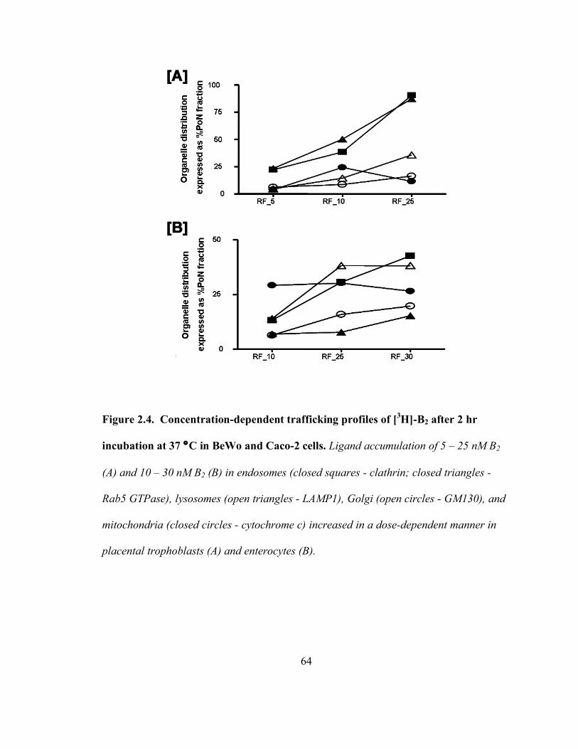
64
Figure 2.4. Concentration-dependent trafficking profiles of [3H]-B2 after 2 hr
incubation at 37 °C in BeWo and Caco-2 cells. Ligand accumulation of 5 – 25 nM B2
(A) and 10 – 30 nM B2 (B) in endosomes (closed squares - clathrin; closed triangles -
Rab5 GTPase), lysosomes (open triangles - LAMP1), Golgi (open circles - GM130), and
mitochondria (closed circles - cytochrome c) increased in a dose-dependent manner in
placental trophoblasts (A) and enterocytes (B).
65
2.3.3 Colocalization of B2 with endocytic organelles in BeWo
Cellular distribution of internalized B2 was further examined using a characterized
rhodamine-labeled riboflavin conjugate (Rd-RF) that exhibits affinity characteristics
similar to the natural vitamin (Huang et al., 2003). Although the rate of internalized B2 in
the placental trophoblasts is suggested to be slower than that shown for Caco-2, the
overall higher ligand accumulation profiles for this vitamin’s enrichment to endosomal
populations justify using the trophoblast model over the enterocyte cell system for the
remaining studies presented in this report. Since maximal accrual of B2 within endocytic
organelles in BeWo cells occurred after 60 minutes, all fluorescence-based studies were
performed using this uptake period. Internalized Rd-RF or FITC-TF were examined for
colocalized signals with AlexaFluor405®- or AlexaFluor546®-labeled proteins for
endosomes (clathrin or Rab5) and lysosomes (LAMP-1) using confocal laser scanning
microscopy. Signal intensities specific to Rd-RF were normalized to cells dosed with
equimolar amounts of a non-reactive rhodamine analog, carboxytetramethlyrhodamine-4-
amine (CTMR4A) (Phelps et al., 2004). In all cases, the signal-to-noise ratio for
CTMR4A was significantly less than that for Rd-RF, and, in contrast to Rd-RF, staining
patterns were routinely diffuse (data not shown). Like FITC-TF, internalized Rd-RF
resulted in distinct punctate, perinuclear staining and exhibited colocalization with both
clathrin and Rab5 GTPase (Figure 2.5A and B). In contrast to FITC-TF, Rd-RF also
exhibited partial colocalization with the lysosomal marker, LAMP-1 (Figure 2.5C).
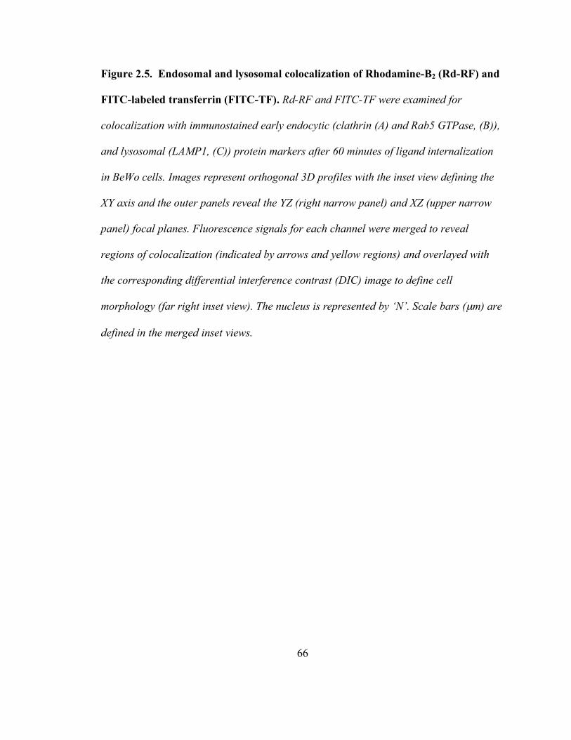
66
Figure 2.5. Endosomal and lysosomal colocalization of Rhodamine-B2 (Rd-RF) and
FITC-labeled transferrin (FITC-TF). Rd-RF and FITC-TF were examined for
colocalization with immunostained early endocytic (clathrin (A) and Rab5 GTPase, (B)),
and lysosomal (LAMP1, (C)) protein markers after 60 minutes of ligand internalization
in BeWo cells. Images represent orthogonal 3D profiles with the inset view defining the
XY axis and the outer panels reveal the YZ (right narrow panel) and XZ (upper narrow
panel) focal planes. Fluorescence signals for each channel were merged to reveal
regions of colocalization (indicated by arrows and yellow regions) and overlayed with
the corresponding differential interference contrast (DIC) image to define cell
morphology (far right inset view). The nucleus is represented by ‘N’. Scale bars (µm) are
defined in the merged inset views.

67
68
A quantitative assessment of the extent of colocalization between ligands and
endocytic markers revealed maximal signal overlap with Rab5 GTPase for both Rd-RF
(64%) and FITC-TF (45%) (Figure 2.6A and B). Compared to the 60 minute
fractionation data in figure 2.3A, the total B2 distribution to this endosome population
(~70%) is strikingly similar to that seen with Rd-RF. However, the extent of FITC-TF
colocalization with Rab5-positive endosomes was nearly 2-fold lower (Figure 2.6B)
compared to that defined in the fractionation results in figure 2.3B. This disparity may be
attributed to the presence of a heterogenous sub-population of endosomes that are Rab5
negative but TF positive; an incidence that cannot be detected by subcellular fractionation
due to the fractional overlap. Rd-RF also revealed ~ 51% colocalization with clathrin,
whereas, FITC-TF showed only 8% overlapping signal with this early endosome protein.
The Pearson’s Correlation (PC), a common statistical test for shape similarities exhibited
between colocalized fluorescence channels, was used to further define linear relationships
existing between ligands and organelle markers. Values > 0.0 were defined as having a
positive linear relationship between variables (Manders et al., 1993). Interestingly, both
ligands revealed positive correlations with all organelle-associated proteins (Figure 2.6).
Consistent for both Rd- RF and FITC-TF, the relative colocalization (14% and 6%,
respectively) with the lysosome protein, LAMP-1 were markedly less than those
determined for each ligand’s association with endosomes. Although both ligands revealed
colocalization with LAMP-1, the overall enrichment to lysosomes is suggested to be
minimal after the 60 min uptake period. Collectively these results indicate significant
ligand recruitment for both B2 and TF to early or sorting endosomes via plasma
69
membrane-associated clathrin and/or Rab5 positive vesicles (Gorvel et al., 1991; Bucci et
al., 1992; McLauchlan et al., 1998). In contrast to TF, the internalized B2 demonstrates
higher lysosomal accumulation. Riboflavin that bypasses lysosomal degradation is likely
to be sorted to recycling endosomes, similar to apo-transferrin which undergoes
exocytosis at the plasma membrane (Jackle et al., 1991).

70
Figure 2.6. Quantitative evaluation of the 3D colocalized regions of ligands, B2 and
TF, with organelle protein markers in BeWo cells. Overlapping volumes (µm3) for
either Rd-RF (A) or FITC-TF (B) with organelle channels were expressed as a
percentage of the total volume for each ligand. The Pearson’s Correlation (PC) was
chosen to define the similarity of 3D shapes between ligands and overlapping organelle
marker channels. Data are expressed as the mean ± SEM for 3 - 4 regions of interest.

71
2.3.4 Golgi and mitochondrial involvement in B2 itinerary
Intracellular enrichment of B2 to the Golgi and mitochondria in comparison with
that for TF was also examined in the isolated subcellular fractions. GM130 (130 kDa), a
structural marker of the Golgi apparatus (Nakamura et al., 1995) was identified in
postnuclear fractions 4 through 6 (Figure 2.2C) while nuclear fractions 11 and 12 tested
positive for cytochrome c (15 kDa), an integral component of the mitochondrial
respiratory chain (Gonzales and Neupert, 1990) (Figure 2.2C) in placental trophoblasts. A
similar organelle enrichment profile was noted with the enterocyte system (Figure 2.2D).
Steady-state distribution (2 hr) showed 6 – 8% of B2 (Figure 2.3A) and 30 – 37% of TF
(Figure 2.3C) distributed to the Golgi. Interestingly, mitochondrial localization of B2 in
BeWo cells peaked at 30 min and declined over 2 hrs (Figure 2.3A), while B2 confined to
the mitochondria increased over time in Caco-2 cells (Figure 2.3B). Despite this
divergent accumulation profile in the two cell models, maximal mitochondrial B2 content
was no greater than 30% of the postnuclear load, whereas TF levels associated with the
mitochondria were negligible in comparison (Figure 2.3C and D).
In order to further corroborate the Golgi and mitochondrial fate of B2, 3D
colocalization of Rd-RF and FITC-TF by confocal microscopy together with the same
organelle markers (i.e., GM130 and cytochrome c) was assessed. Both ligands were
shown to colocalize to some degree with these organelle markers (Figure 2.7A and B). As
noted with the endocytic proteins, Rd-RF and FITC-TF resulted in punctate and
perinuclear colocalization with GM130 and cytochrome c. Image analyses for both
ligands revealed a positive Pearson’s Correlation with GM130 and cytochrome c, thus
72
further suggesting the involvement of these organelles in B2 and TF trafficking. Rd-RF
exhibited a higher degree of colocalization (~ 34%) with GM130 (Figure 2.6A) that was
approximately 2-fold higher than that determined for FITC-TF (Figure 2.6B). FITC-TF
also revealed the least amount of colocalization (~8%) with the mitochondrial marker,
cytochrome c (Figure 2.6B). In contrast, Rd-RF revealed greater than 3-fold higher
overlap (~26%) with cytochrome c (Figure 2.6A). Based on the current understanding of
B2 physiology and the role of its cofactors FMN and FAD in normal metabolic
functioning of the cell, distribution to the mitochondria appears to be consistent.

73
Figure 2.7. Localization of Rd-RF and FITC-TF to the Golgi and mitochondria in
BeWo cells. Confocal 3D images of either Rd-RF or FITC-TF after 60 minutes
internalization in BeWo cells were analyzed for colocalization (arrows and yellow
regions) with Golgi (A) and mitochondria (B). Confocal images are defined by
orthogonal profiles as described in figure 2.5, and fluorescence signals for ligands and
organelle markers were merged with DIC images (far right inset views). Scale bars
represent 10 µm and nuclei (N) are defined in DIC images.
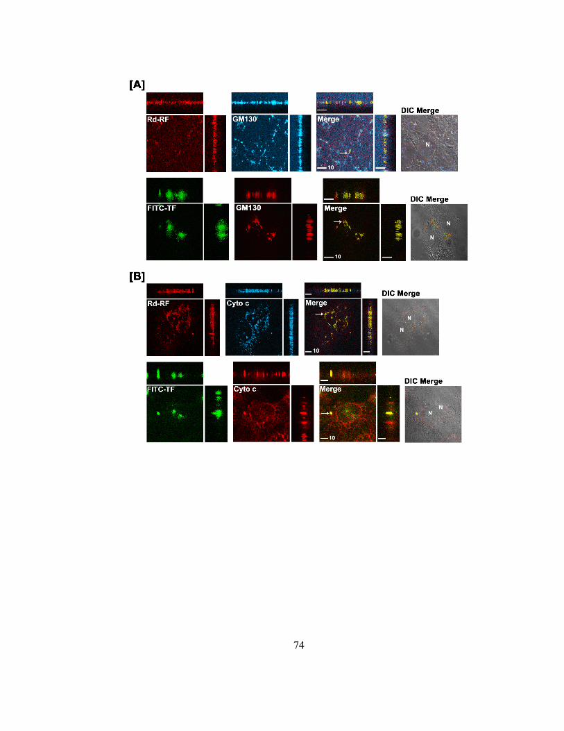
74
75
2.3.5 cAMP regulation of B2 binding and initiation of the endocytic cascade in
placental trophoblasts
Cyclic AMP is unequivocally a major short-term regulator that regulates
autologous or heterologous receptor-mediated endocytic internalization (Foti et al.,
1997). Previous reports have shown that increased levels of cAMP elicited by forskolin,
Br-cAMP, and 3-isobutyl-1-methyl-xanthine (IBMX) have resulted in a dramatic
reduction in B2 uptake in BeWo cells (Huang and Swaan, 2001; Foraker et al., 2003). In
the present study we sought to evaluate if this cAMP-sensitive endocytic trafficking of B2
occurred as a consequence to regulation of B2 binding with its specific receptors. BeWo
cells stimulated with increasing concentrations of forskolin resulted in a robust cAMP
response with an EC50 of 1.5µM [0.99-1.9] and a Bmax value of 125 ± 2.2 pmol
cAMP/250,000 cells, while treatment with riboflavin up to 10-4 M failed to stimulate any
cAMP accumulation (Figure 2.8A). Interestingly, pretreatment of BeWo cells with
increasing concentrations of B2 for merely 10 min prior to stimulation with 1µM
forskolin in the continued presence of B2 resulted in a diminished cAMP response
(Figure 2.8B). The maximum inhibition was ~ 67% of control with a reported IC50 value
of ~ 0.4 µM. This indicates that B2 bound to the putative riboflavin receptor is negatively
coupled to cAMP and suggests that stimulated cAMP production, consequently results in
decreased B2 uptake via clathrin-coated pits.
76
Figure 2.8. Cyclic-AMP regulation of the B2 internalization process. Determination
of cAMP accumulation in BeWo cells after stimulation with riboflavin or forskolin (A).
BeWo cells cultured as described in the experimental section were stimulated via
incubation with the indicated concentration of either forskolin (closed squares) for 10
minutes, or riboflavin (open circles) for 40 minutes. Data are a representative curve from
three independent experiments showing similar results. The inhibitory effects of
riboflavin on forskolin-stimulated cAMP accumulation were analyzed (B). BeWo cells
were washed and then incubated with the indicated concentrations of riboflavin dissolved
in HF12K (20 mM HEPES buffered F12K, pH 7.4, 37 °C). After 10 minutes HF12K was
removed and cells were stimulated with 1µM forskolin in the continued presence of
riboflavin. Cyclic-AMP accumulation was then determined as described in the
experimental section. The dashed line indicates 1µM forskolin stimulated accumulation
in the absence of riboflavin. Results are expressed as mean ± S.D. (n = 5).

77

78
2.4 Discussion
Our laboratory has recently demonstrated that intestinal and placental entry of
vitamin B2 into the cell occurs via a putative receptor-mediated endocytic process (Huang
and Swaan, 2000; Huang and Swaan, 2001; Huang et al., 2003). This B2-specific
endocytic pathway is still in its nascent stage of identification and has warranted
investigative pursuits to clearly define its translocation pathway within the cell. In the
present study we delineate the trafficking route of B2 upon internalization that
consequently, contributes to maintaining the balanced vitamin status in a physiological
environment. In addition, we also report second messenger regulation of the ligand
absorption into placental trophoblasts that suggests interaction of other extrinsic
pathways in response to the ligand stimulus.
The approach employed followed a conventional fractionation analysis of
intracellular compartments that are integral to the ‘classical’ clathrin-dependent endocytic
machinery. The BeWo cell line exhibited distinctive accumulations for B2 and TF within
the endocytic system but negligible levels for a non-endocytic marker TCA, which is
routinely transported by placental bile acid transporters (Dumaswala et al., 1993).
Sucrose density gradient-resolved endosomal and lysosomal fractions defined by widely
used organelle markers (clathrin, Rab5 GTPase, and LAMP1) showed extensive labeling
for B2 and TF within the endosomes. Selective enrichment of the endosomes over
lysosomes for both ligands was in accordance with previously determined trafficking
profiles for TF (Ciechanover et al., 1983; Yamashiro and Maxfield, 1984; Yamashiro et
al., 1984) and confirmed the efficiency of the fractionation method. The method also
79
provided insight into the possible fate of B2 following internalization. Biochemical
association with the mitochondrial compartment is expected since FMN and FAD play a
pivotal role in the electron transport chain (Barile et al., 2000) generating the metabolic
energy required for other cellular functions.
Both placental trophoblasts and enterocytes exhibited comparatively higher
uptake levels for TF than B2. This may be attributed to the differential nutritional needs
of these cells for iron and vitamin B2. van der Ende and colleagues (van der Ende et al.,
1987) have demonstrated that the augmented process of iron transfer and metabolism in
the developing fetus is efficiently mediated by the transplacental TF system. Likewise,
gestational needs of B2 are also increased (Powers, 2003a) and concomitant fortification
of B2 supply would be expected. Further, a time-associated increase in ligand build-up
was observed for B2 and TF within the endocytic compartments. The endocytic process
entails rapid internalization of recognized ligands; consequently evaluation of time points
shorter than 30 min are currently underway. Nevertheless, this method provides an
informative overview of the trafficking itinerary of B2 via the endocytic route, which
appears to be conserved across the two divergent cell systems applied in this study. The
kinetic distribution of 5 – 30 nM B2 expectedly showed increased accumulation within
the organelles examined with the exception of mitochondrial compartments. Clearly, the
changing distribution patterns of B2 within the mitochondria suggest a tightly-regulated
feedback mechanism that controls the trafficking route of the molecule, which in turn
maintains a homeostatic check on the cellular riboflavin content. It is very likely that the
increased B2 accumulation over time provides the impetus to rapidly convert B2 to the
80
flavin intermediates that cycle through the metabolic pathway. This dynamic change in
compartmentalized B2 presents a challenging scenario of putative vitamin sensors,
receptors, and other regulators that must interact to maintain a balanced nutritional
environment.
A limitation of any fractionation method is the potential overlap between the
organelles identified. Consequently, to further corroborate the intracellular distribution
profiles of B2, a minimally invasive labeling approach examining the colocalization of
fluorescently labeled ligands with immunostained organelle markers was utilized.
Rhodamine-conjugated riboflavin, a derivative of the natural ligand previously
characterized in our laboratory (Phelps et al., 2004), that retains affinity for the B2
transport system was compared with the staining patterns of FITC-TF. Rd-RF
convincingly localized within the endosomes, lysosomes, Golgi, and mitochondria,
whereas FITC-TF was confined predominantly to the endosomes. It is not clear at this
time what the significance of localization to the Golgi may signify, but this may reflect a
possible recycling mechanism that recovers B2 and packages it either for storage or
exocytosis. Evidence for Golgi processing of the TF-receptor is seen in myeloma cells
and supports the recycling theory of this receptor that continuously scavenges iron-bound
TF (Woods et al., 1986). In fact, prior studies reveal two types of recycling pathways,
i.e., a short pathway involving early endosomal fusion with recycling endosomes that
target ligands back to the plasma membrane, and a long pathway that translocates ligands
to the Golgi at which clathrin initiates vesicular budding from the trans-most cisternae
and eventual shuttling to the plasma membrane (Ghosh and Maxfield, 1995; Sakai et al.,

81
1998; Maxfield and McGraw, 2004). A recycling component specific to the B2 trafficking
itinerary has yet to be defined; however, it would be interesting to delineate whether one
or both of these recycling events is specific to this vitamin. It should be noted that the
possibility of equilibrative changes associated with time-resolved imaging cannot be
ruled out at this juncture and require real-time imaging approaches. However, the intent
of this study was to illustrate the involvement of the endocytic machinery in the cellular
trafficking itinerary of B2.
The presence of a receptor-mediated endocytic component in the placental and
intestinal internalization of B2 then raises several questions that need to be addressed
regarding its effectors and regulators. Literature reports have established that the
endocytic process in eukaryotes occurs through a highly ordered process and is driven by
signaling events that activate or inactivate depending upon the stimulus (Foti et al.,
1997). Cyclic AMP is clearly a pivotal candidate integral to a large number of signaling
cascades and was evaluated as a key regulator of the B2 endocytic process in the
trophoblast model. B2 alone did not generate a cAMP response but it was responsible in
down-regulating the cAMP production upon stimulation with forskolin. This indicates
that the ligand-sequestered B2-receptor lowers the intracellular levels of cAMP prior to or
shortly after its association into clathrin-coated pits, and eventually triggers the
subsequent endocytic events of sorting and translocation. Conversely, we have already
shown that increased stores of intracellular cAMP significantly inhibit the internalization
of B2 (Huang and Swaan, 2001), thereby indicating an inverse relationship between
intracellular cAMP and B2 receptor-mediated endocytosis.

82
In conclusion, fractionation data in both divergent cell systems taken together
with the morphological assessment of cellular B2 distribution provides definitive
evidence for endocytic trafficking of B2, at least in part via a clathrin-dependent pathway,
and eventual translocation to the mitochondria where the role of its metabolites are well
characterized. In addition, this cellular absorption mechanism is shown to be sensitive to
cAMP levels, further suggesting the existence of a highly regulated B2-specific-receptor
internalization process. Understanding the characteristics of the conserved B2
translocation pathway provides a novel platform that can be potentially exploited in the
future, analogous to the folate and transferrin receptors, in biomedical imaging (Berry et
al., 2004), and tumor-targeted delivery of genes or therapeutic conjugates (Li et al., 2003;
Hattori and Maitani, 2004; Yoo and Park, 2004a; Yoo and Park, 2004b).

83
CHAPTER 3
DYNAMIN 2 REGULATES THE ENDOCYTOSIS OF RIBOFLAVIN IN HUMAN
EPITHELIA
Abstract
Riboflavin (vitamin B2) is known to be indispensable in a multitude of metabolic
pathways through its conversion to coenzyme forms flavin mononucleotide (FMN) and
flavin adenine dinucleotide (FAD). Despite its physiological importance, little is known
about specific mechanisms or proteins involved in regulating its cellular entry in humans.
Recent evidence suggests riboflavin internalization and trafficking in human epithelia
occurs through a clathrin-dependent receptor-mediated endocytic (CME) process. The
purpose of this study was to define the role of the conserved endocytic protein, dynamin
2 GTPase (DNM2), in riboflavin absorption into placental trophoblasts (BeWo) using
RNA interference (RNAi) and transient transfections with wild-type or GTPase-null
dynamin 2 constructs. The extent of plasma membrane bound and internalized
84
B2 were elucidated under knocked down, functional, and non-functional dynamin 2
states. Analogous to the endocytic control ligand, transferrin, riboflavin was shown to
exhibit ~ 50% dependence on the functional expression of dynamin 2 for its active
cellular entry. Furthermore, a reduction in functional dynamin 2 levels led to ~2 fold
higher plasma membrane bound B2 over its internalization. These data are the first to
identify the functional requirement of dynamin 2 GTPase in the B2-specific RME process
in humans. Delineating the mechanism and proteins regulating B2-RME provides future
studies with a novel pathway that may be potentially exploited in targeted drug design,
delivery and biomedical imaging strategies.

85
3.1 Introduction
Riboflavin (vitamin B2) is an essential nutrient required in many metabolic
pathways critical in normal cellular growth and development. States of physiological B2-
deficiency have been correlated with various clinical manifestations including
cardiovascular disease, stunted growth, anemia, neurodegenerative disorders and various
forms of cancer (Cooperman, 1991; Powers, 2003b; Powers, 2005). Despite its clinical
importance, the specific cellular mechanism(s) regulating B2 absorption are poorly
defined. Recent reports by our laboratory suggest one portal of cellular entry involves
clathrin-mediated endocytosis (Huang and Swaan, 2000; Huang and Swaan, 2001; Huang
et al., 2003; D'Souza et al., 2006b). These studies demonstrated a microtubule
dependence and early clathrin and Rab5 positive endosomal enrichment of B2 in human
placental trophoblasts (BeWo). As both clathrin and Rab5 are conserved endocytic
markers of the clathrin-dependent endocytic (CME) pathway, this data strongly suggests
riboflavin absorption involves CME. Furthermore, analogous to the iron carrier protein,
transferrin, a riboflavin carrier protein has been proposed to sequester free extracellular
B2 and facilitate internalization and localization to endosomal organelles (Ramana Murty,
1982; Visweswariah and Adiga, 1987a; Prasad et al., 1992; Subramanian and Adiga,
1999; Mason et al., 2006). However, a limitation to this B2-specific RME model is a lack
of direct evidence identifying the functional dependence on critical proteins regulating
this absorption mechanism in humans.
Dynamin 2 (DNM2) is a ubiquitously expressed GTPase that has been thoroughly
established to regulate the invagination and constriction of endocytic vesicles at the

86
plasma membrane of mammalian endothelial and epithelial cells (Sever et al., 2000; Hill
et al., 2001; Sever, 2002; Conner and Schmid, 2003). It is required for multiple endocytic
mechanisms including those associated with classical pathways dependent on clathrin or
caveolae, and consequently it has been described as the “…master regulator of membrane
trafficking events at the cell surface.” (Conner and Schmid, 2003). Prior studies
performed in our laboratory revealed distinct subcellular distribution of radiolabeled and
fluorescently labeled riboflavin to endocytic populations positive for both clathrin and the
early endosome marker, Rab5 GTPase, in placental trophoblasts (D'Souza et al., 2006b).
Therefore, we hypothesized that riboflavin internalization requires the functional
expression of dynamin 2 GTPase. In order to test this hypothesis, we used RNA
interference (RNAi) strategies to knock down DNM2 protein expression in the BeWo cell
system, which has been established to exhibit nM affinity for riboflavin (Huang and
Swaan, 2001).
Like DNM2, dynamin-like protein 1 GTPase (DLP1) is a member of the dynamin
superfamily (Praefcke and McMahon, 2004). DLP1 shares roughly 36% sequence
homology to DNM2. Unlike DNM2, DLP1 is not involved in endocytic vesicle scission
events. However, DLP1 has been largely characterized to function in the morphological
maintenance of peroxisomes and mitochondria (McNiven et al., 2000; Yoon et al., 2003;
Pitts et al., 2004). In addition, DLP1 has been implicated as a regulator of exocytic
trafficking of vesicles from the Golgi to the plasma membrane (Yoon et al., 1998).
Duplex short interfering RNA (siRNA) targeting DLP1 was used to silence DLP1
expression in BeWo cells, and served as a control for siRNA silencing specificity in these

87
studies. Based on the current understanding of DLP1, we hypothesized this protein is not
involved in the endocytosis of riboflavin.
The purpose of this study was to define the extent by which riboflavin absorption
depends on endocytosis as a function of the expression level of the major vesicle
invagination/scission protein, dynamin 2 GTPase. RNAi combined with wild-type and
GTPase-null dynamin 2 transfections in the BeWo cell model were carried out to
illustrate the importance of functional dynamin 2 expression on the cellular entry of this
important nutrient. Results from this study are the first to directly reveal the dependence
of B2 absorption on dynamin 2 expression in human placental trophoblasts, and
demonstrates that approximately 50% of actively absorbed B2 involves an RME process.
3.2 Methods and materials
3.2.1 Cell Culture
Human placental trophoblasts (BeWo) were routinely maintained in a controlled
atmosphere at 37 ºC with 5% CO2 in F-12K culture media (Invitrogen Life Technologies,
Carlsbad, CA) supplemented with 10% fetal bovine serum, 1% non-essential amino acids,
100 U/ml penicillin and 100 µg/ml streptomycin. All transfection treatments were tested
for cytotoxic effects using the lactate dehydrogenase-based CytoTox-ONE™ assay
according to manufacturer’s instructions (Promega Corp., Madison, WI). All chemicals
used in these studies were purchased from Sigma (St. Louis, MO) unless stated otherwise.
3.2.2 Duplex siRNA transient transfection
Cells grown to ~ 50-60% confluency were seeded (4.2 x 10⁴ cells/cm2) onto 24-
well plates using antibiotic-free F-12K media. Twenty four hours later, cells were briefly
88
washed and pre-incubated in serum-free and antibiotic-free Opti-MEM I media
(Invitrogen Life Technologies, Carlsbad, CA) for ~ 2 hours at 37 °C and under 5% CO2.
Following this incubation, cells were transfected in Opti-MEM I media with 40 nM
duplex siRNA complexed with Lipofectamine™ 2000 (0.25% v/v) according to
manufacturer’s instructions (Invitrogen Life Technologies, Carlsbad, CA). Cells were
incubated with siRNA-lipid complexes at 37 °C and 5% CO2 for 6 hours, at which time
the transfection media was replaced with normal F-12K media lacking antibiotics. Cells
were used in experiments 48-72 hours post-transfection. All RNA interference studies
involved the use of experimentally validated duplex siRNA targeting the human dynamin
2 gene (DNM2 pool siGenome Smartpool, gene accession no. NM_004945, Dharmacon
RNA Technologies, Chicago, IL) and human dynamin-like protein 1 (DLP1, gene
accession no. NM_012063, Qiagen®, Cambridge, MA). Targeting siRNA effects were
normalized to cells transfected with equivalent amounts of non-targeting duplex siRNA
(Dharmacon RNA Technologies, Chicago, IL) and compared to mock (Lipofectamine™
2000 alone) and untreated cell conditions.
3.2.3 Western blotting and chemiluminescence-based densitometry
Whole cells transfected 63-68 hours prior with siRNA were harvested and lysed
on ice for 20 min in RIPA buffer (10 mM Tris-HCL, pH 7.5, 140 mM NaCl, 1% (v/v)
Triton X-100, 1% (w/v) sodium deoxycholate and 0.1% (w/v) SDS) supplemented with a
Complete Mini® protease cocktail tablet according to manufacturer’s instructions (Roche,
USA). Ten to 15 µg total protein was resolved on 12.5% Tris-HCl Criterion gels (BIO-
RAD, Hercules, CA), transferred to PVDF membranes, and was immunoblotted using
89
monoclonal antibodies specific to dynamin 2 (Calbiochem®, EMD Biosciences Inc., San
Diego, CA) or dynamin-like protein 1 (BD Pharmingen, San Diego, CA). Primary
antibodies were subsequently labeled with horse radish peroxidase-conjugated IgG and
detected using the ECL plus system (Amersham Biosciences, Piscataway, NJ). Protein
levels were quantitated using chemiluminescence-based densitometry on a chemi-doc
universal hood II system (Bio-Rad Laboratories, Hercules, CA) and normalized to the
corresponding housekeeping protein expression for either GAPDH (Ambion Inc., Austin,
TX) or β-actin.
3.2.4 GTPase-null (K44A) and wild type dynamin 2 plasmid transient transfections
and cotransfections with DNM2 pool siRNA
Characterized wild type (DNM2WT) and dominant-negative (DNM2K44A) dynamin
2 expression constructs (both in the mammalian expression vector, pCR3.1, Invitrogen
Life Technologies, Carlsbad, CA) were kindly provided by Dr. Mark A McNiven (Mayo
Clinic and Foundation, Rochester, Minnesota) (Cao et al., 1998). Prior to transfections,
both plasmids were transformed into DH5-α cells (Invitrogen Life Technologies,
Carlsbad, CA), subcultured in Luria broth with ampicillin selection, and purified using
the Plasmid Maxi Kit (Qiagen®, Cambridge, MA). Expression vector identities were
confirmed through restriction enzyme digests and sequencing using the following
established dynamin 2 primers: forward primer [5’-GAAGAGGGCCATACC-3’] and
reverse primer [5’-AGTTGCGGATGGTCTC-3’] (Cao et al., 1998). Cell seeding
conditions were maintained as in RNAi studies. Twenty four hours post-seeding, cells
were briefly washed and pre-incubated in serum-free and antibiotic-free Opti-MEM I for
90
2 hours at 37 °C and under 5% CO2. Cells were transfected using Opti-MEM I with 400
ng plasmid DNA alone or with 40 nM DNM2 pool siRNA and complexed with
Lipofectamine™ 2000 (0.25% v/v). Cells were exposed to these transfection complexes
for 6 hours at 37 °C and under 5% CO2. Subsequently, the transfection media was
replaced with complete F-12K media devoid of antibiotics. Cells were used in
experiments 48-72 hours post-transfection.
3.2.5 Radio-labeled ligand endocytosis assays
Cells were dosed with either 5 nM [3H]-riboflavin ([3H]-B2) or 10 nM 125I-
transferrin (125I-TF, iodinated using the IODOGEN® method (Pierce Biotechnology Inc.,
Rockford, IL) according to established procedures (D'Souza et al., 2006b)) in Hanks’
Balanced Salt Solution (pH 7.4) containing 25 mM D-glucose and 10 mM HEPES at 37
ºC for 4 min. Immediately, cells were placed on ice and free ligands were removed by
washing 3x with ice-cold DPBS (pH 7.4). Plasma membrane bound ligands were then
removed by washing cells 2x on ice (5 min/wash) with ice-cold DPBS (pH 3.0). Cells
were alkaline-lysed (1 N NaOH) at 4 °C for at least 2 hours prior to internalized ligand
quantitation. The extent of plasma membrane bound and internalized [3H]-B2 and 125I-TF
was determined using liquid scintillation- or gamma counting, respectively. Both, plasma
membrane bound and internalized radio-labeled ligands were normalized to total protein
content using the Bradford assay (Bio-Rad Laboratories, Hercules, CA). All [3H]-B2
uptake data generated at 37 °C was corrected for passively absorbed B2 by performing
parallel uptake assays exclusively at 4 °C. Actively (uptake at 37 °C) internalized B2 was
defined by subtracting internalized [3H]-B2 at 4 °C.

91
Acid wash samples collected after uptake assays represented plasma membrane
bound ligands and were compared to internalized samples using the following equations:
100% !+
"+
=nt
ntnt
ee
e
b
ib
ib
bbound (Eq. 1)
100% !+
"+
=nt
ntnt
ee
e
i
ib
ib
iabsorbed (Eq. 2)
The extent of bound ligand was expressed as a percent of the sum of bound (be) and
absorbed (ie) ligand under experimental conditions, and was normalized to the percent of
bound ligand under non-targeting conditions (bnt) (equation 1). Likewise, the extent of
absorbed ligand (ie) was defined as a percent of bound plus internalized ligand under
experimental conditions and then was normalized to non-targeting effects (int) (equation
2).
3.2.6 Statistics
One-way ANOVA with Dunnett’s or Neuman-Keuls multiple comparison tests
were used to define statistical significance between the effects of targeting siRNA or wild
type and dominant negative dynamin 2 construct treatments and control (i.e., non-
targeting siRNA, empty vector, mock, and untreated treatments) conditions on ligand
trafficking and targeted protein knockdown.

92
3.3 Results
3.3.1 Duplex siRNA target specificity at the protein level and RNAi method
validation
BeWo cells were transiently transfected with 20-40 nM targeting duplex siRNA
for dynamin 2 GTPase (DNM2 pool) or dynamin-like protein 1 (DLP1). Cells were
harvested 63-68 hours after initiating siRNA transfections and analyzed for target protein
knockdown of dynamin 2 (DNM2) and the dynamin homologue, dynamin-like protein 1
using Western blotting and chemiluminescence-based densitometry. Both DNM2 and
DLP1 protein levels were normalized to the housekeeping expression of GAPDH or β-
actin, respectively. Upon normalization, the extent of dynamin expression was defined as
a percentage of detected dynamin levels in cells treated exclusively with non-targeting
siRNA (NT). Parallel cell populations that were treated exclusively with the lipid
transfection reagent (Mock) or untreated cells served as additional negative controls.
Twenty and 40 nM DNM2 pool siRNA treatments substantially reduced dynamin 2 (100
kDa) (figure 3.1A) expression levels to a similar extent. Furthermore, quantitative
densitometry tests revealed 40 nM DNM2 pool siRNA treatments led to ~ 78% (21.95 ±
7.03% NT) and ~ 82% lower dynamin 2 GTPase levels compared to non-targeting siRNA
treated and untreated cells, respectively (figure 3.1C). Likewise, 20 and 40 nM DLP1
siRNA specifically knocked down DLP1 protein (83 kDa) expression in a dose-
dependent manner (Figure 3.1B). Forty nM DLP1 siRNA conditions resulted in ~ 62%
(37.65 ± 11.13% NT) and ~ 63% lower DLP1 protein levels compared to non-targeting
siRNA and untreated conditions, respectively (figure 3.1D). Neither of the dynamin

93
siRNA treatments revealed significant off-target effects at the protein level. Furthermore,
dynamin protein levels were shown to be similar between non-targeting siRNA
treatments and untreated cell conditions. This data suggests the negative control siRNA
transfection effects are not significantly altering target protein levels, and serves as a
valid negative control for targeting siRNA data normalization.

94
Figure 3.1. Specific protein knockdown as a function of siRNA treatment. Cells
transfected with duplex siRNA targeting mRNA for dynamin 2 (DNM2 pool), or dynamin-
like protein 1 (DLP1) were examined for target protein knockdown using western blotting
and quantitative chemiluminescence-based densitometry, and normalized to
housekeeping proteins, GAPDH or β-actin. Twenty and 40 nM targeting siRNA (A:
DNM2p, and B: DLP1) treatments were compared to 40 nM non-targeting (N) siRNA,
mock, and untreated (U) cells. Cells treated with 40 nM targeting or non-targeting siRNA
were assessed for the extent of DNM2 (C) or DLP1 (D) knockdown, and expressed as a
percentage of non-targeting siRNA effects. The data represents the standard deviations of
2 to 4 separate experiments. Statistical significance between targeting and non-targeting
effects on dynamin protein expression were defined by one-way ANOVA with Dunnett’s
multiple comparison test (**p<0.01).
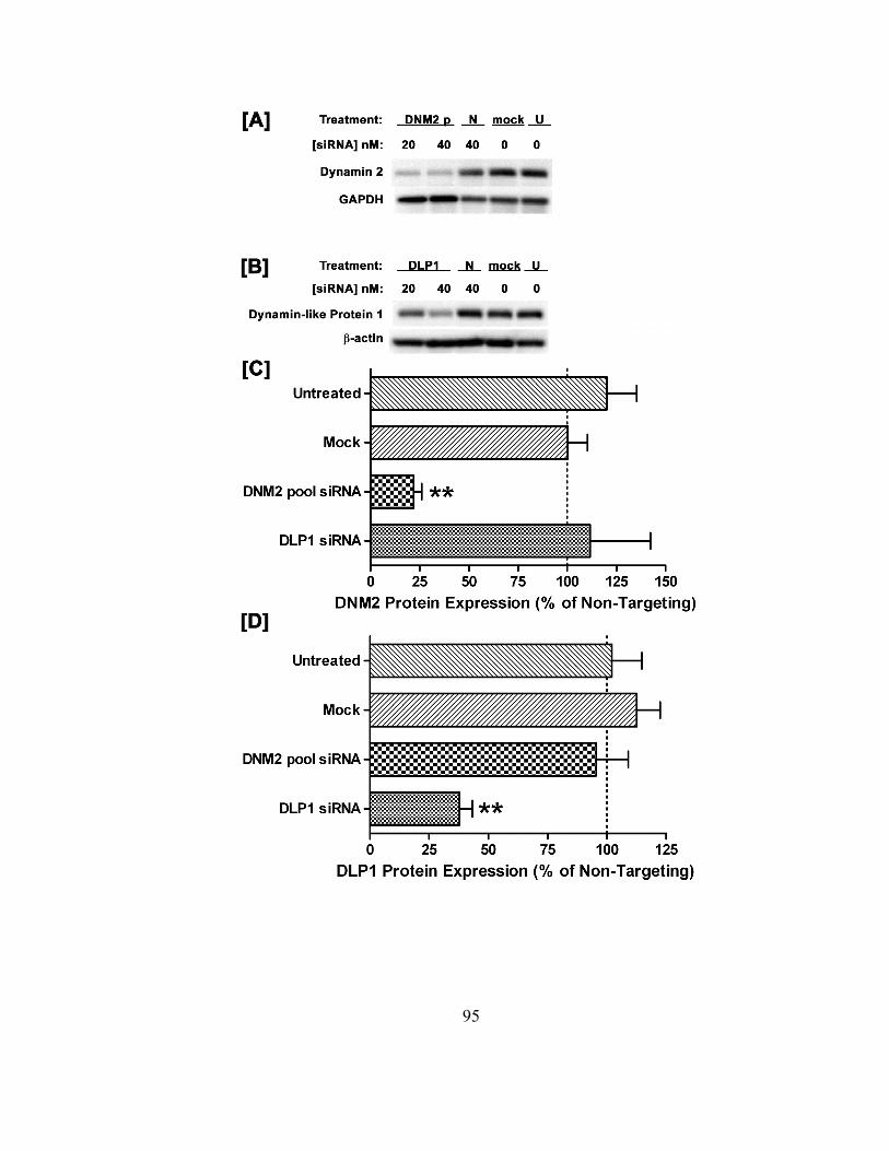
95
96
In order to evaluate whether cell physiology was compromised due to transfection
conditions, all treatments were assessed for potential cytotoxicity as a function of lactate
dehydrogenase (LDH) release. Cells treated with the various RNAi conditions, along
with plasmid transfections, mock, and untreated cell populations, were tested for LDH
release 48 hours post-transfection. As shown in Table 3.1, no significant cell death was
revealed for any of the transfection treatments, and cell viability for these conditions was
at or near those measured for untreated BeWo cells. Taken together, these data indicate
that siRNA treatments at the 40 nM dose specifically knock down target dynamin
proteins without compromising cell viability, and justify the RNA interference
methodology (Figure 3.1, Table 3.1).

97
Table 3.1. siRNA or plasmid and transfection reagent effects on cell viability as a
function of lactate dehydrogenase (LDH) release. All transfection conditions were
examined for cytotoxic effects on BeWo cells. Upon 48 hours post-transfection cell
viability was assessed using the LDH release assay according to manufacturer’s
instructions (Promega Corporation, Madison, WI). These data represent the mean
percent of untreated cell viability ± standard deviations of 2 to 4 separate experiments.
98
3.3.2 Dynamin siRNA effects combined with temperature dependence on
endocytosis of [3H]-B2 in BeWo
It is thoroughly established that riboflavin gains entry into human epithelial cells
via multiple mechanisms, including a passive diffusion component that appears to
dominate at oversupplemented riboflavin levels and an active component that has been
reported to coincide with nM B2 concentrations (Huang and Swaan, 2000; Huang and
Swaan, 2001). One of the salient features defining an active absorption mechanism is
temperature dependence. Saturable absorption kinetics have been consistently shown to
correlate with physiological temperatures (~ 37 ºC), while low temperatures (~ 4 ºC)
generate linear absorption profiles reflecting passive diffusion (Said and Ma, 1994). In
fact, B2 transport has been reported to be dependent on temperature in divergent cell
models (Said and Ma, 1994; Huang and Swaan, 2001). However, temperature
dependence alone does not discriminate between a carrier/transporter or receptor-
mediated endocytic process. Therefore, in order to differentiate between such active
uptake mechanisms, the standardized RNAi methods involving siRNA-induced silencing
of the conserved endocytic protein, dynamin 2 GTPase, in combination with the effects
temperature change on B2 absorption were investigated. To date, the human placental
trophoblast cell model (BeWo) has been shown to express the highest affinity for
riboflavin (~ 2 nM) (Huang and Swaan, 2001), and such nM affinities further suggest the
involvement of RME in this vitamin’s cellular uptake. Combined these data validate the
utility of the BeWo cell model to characterize the B2-specific RME pathway(s). BeWo
cells transfected with 40 nM targeting or non-targeting siRNA 63-72 hours prior were
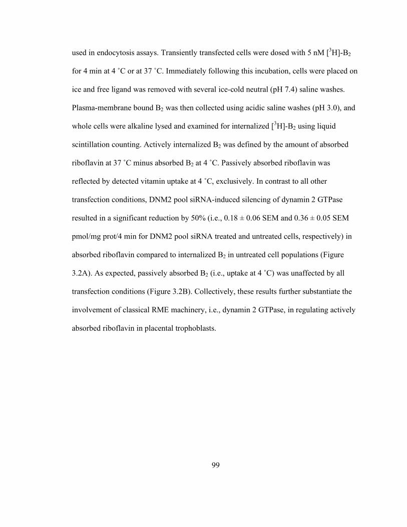
99
used in endocytosis assays. Transiently transfected cells were dosed with 5 nM [3H]-B2
for 4 min at 4 ˚C or at 37 ˚C. Immediately following this incubation, cells were placed on
ice and free ligand was removed with several ice-cold neutral (pH 7.4) saline washes.
Plasma-membrane bound B2 was then collected using acidic saline washes (pH 3.0), and
whole cells were alkaline lysed and examined for internalized [3H]-B2 using liquid
scintillation counting. Actively internalized B2 was defined by the amount of absorbed
riboflavin at 37 ˚C minus absorbed B2 at 4 ˚C. Passively absorbed riboflavin was
reflected by detected vitamin uptake at 4 ˚C, exclusively. In contrast to all other
transfection conditions, DNM2 pool siRNA-induced silencing of dynamin 2 GTPase
resulted in a significant reduction by 50% (i.e., 0.18 ± 0.06 SEM and 0.36 ± 0.05 SEM
pmol/mg prot/4 min for DNM2 pool siRNA treated and untreated cells, respectively) in
absorbed riboflavin compared to internalized B2 in untreated cell populations (Figure
3.2A). As expected, passively absorbed B2 (i.e., uptake at 4 ˚C) was unaffected by all
transfection conditions (Figure 3.2B). Collectively, these results further substantiate the
involvement of classical RME machinery, i.e., dynamin 2 GTPase, in regulating actively
absorbed riboflavin in placental trophoblasts.

100
Figure 3.2. Effect of 40 nM siRNA on riboflavin internalization at 4 °C and 37 °C in
BeWo. BeWo cells transfected 63-72 hours prior with 40 nM siRNA duplexes were dosed
with 5 nM [3H]-B2 for 4 min at 37 °C or 4 °C. Actively internalized B2 (uptake at 37 ˚C, A)
was determined by subtracting passively absorbed riboflavin (uptake at 4 ˚C, B). This
data represents the standard deviations of 3 to 5 separate experiments. Statistical
significance between targeting and non-targeting treatments was defined using one-way
ANOVA with Newman-Keuls multiple comparison test (*p < 0.05).
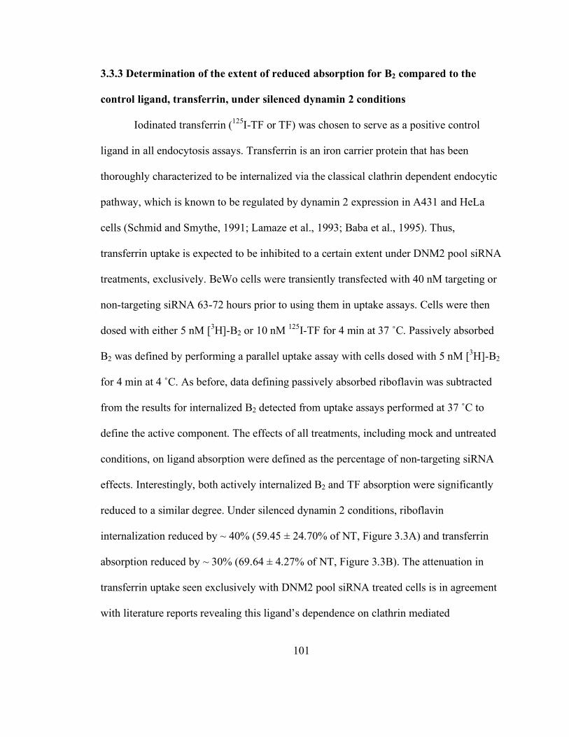
101
3.3.3 Determination of the extent of reduced absorption for B2 compared to the
control ligand, transferrin, under silenced dynamin 2 conditions
Iodinated transferrin (125I-TF or TF) was chosen to serve as a positive control
ligand in all endocytosis assays. Transferrin is an iron carrier protein that has been
thoroughly characterized to be internalized via the classical clathrin dependent endocytic
pathway, which is known to be regulated by dynamin 2 expression in A431 and HeLa
cells (Schmid and Smythe, 1991; Lamaze et al., 1993; Baba et al., 1995). Thus,
transferrin uptake is expected to be inhibited to a certain extent under DNM2 pool siRNA
treatments, exclusively. BeWo cells were transiently transfected with 40 nM targeting or
non-targeting siRNA 63-72 hours prior to using them in uptake assays. Cells were then
dosed with either 5 nM [3H]-B2 or 10 nM 125I-TF for 4 min at 37 ˚C. Passively absorbed
B2 was defined by performing a parallel uptake assay with cells dosed with 5 nM [3H]-B2
for 4 min at 4 ˚C. As before, data defining passively absorbed riboflavin was subtracted
from the results for internalized B2 detected from uptake assays performed at 37 ˚C to
define the active component. The effects of all treatments, including mock and untreated
conditions, on ligand absorption were defined as the percentage of non-targeting siRNA
effects. Interestingly, both actively internalized B2 and TF absorption were significantly
reduced to a similar degree. Under silenced dynamin 2 conditions, riboflavin
internalization reduced by ~ 40% (59.45 ± 24.70% of NT, Figure 3.3A) and transferrin
absorption reduced by ~ 30% (69.64 ± 4.27% of NT, Figure 3.3B). The attenuation in
transferrin uptake seen exclusively with DNM2 pool siRNA treated cells is in agreement
with literature reports revealing this ligand’s dependence on clathrin mediated
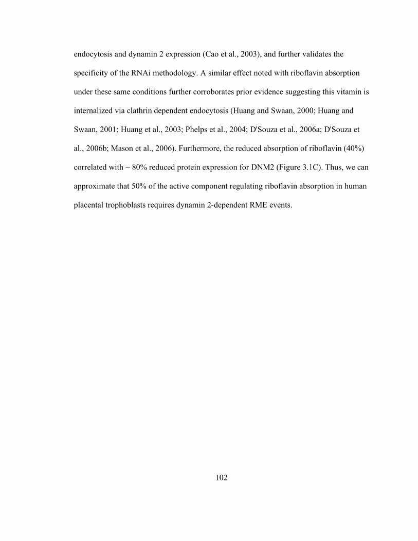
102
endocytosis and dynamin 2 expression (Cao et al., 2003), and further validates the
specificity of the RNAi methodology. A similar effect noted with riboflavin absorption
under these same conditions further corroborates prior evidence suggesting this vitamin is
internalized via clathrin dependent endocytosis (Huang and Swaan, 2000; Huang and
Swaan, 2001; Huang et al., 2003; Phelps et al., 2004; D'Souza et al., 2006a; D'Souza et
al., 2006b; Mason et al., 2006). Furthermore, the reduced absorption of riboflavin (40%)
correlated with ~ 80% reduced protein expression for DNM2 (Figure 3.1C). Thus, we can
approximate that 50% of the active component regulating riboflavin absorption in human
placental trophoblasts requires dynamin 2-dependent RME events.

103
Figure 3.3. Effects of siRNA treatments on the percent of internalized ligand
normalized to non-targeting conditions. In this analysis, the overall effect of all
treatments is expressed as a percent of non-targeting siRNA effects. BeWo cells
transfected 63-72 hours prior with 40 nM siRNA duplexes were dosed with either 5 nM
[3H]-B2 or 10 nM 125I-transferrin (TF) for 4 min at 37 °C or 4 °C. Both actively
internalized B2 (minus passively absorbed B2, A) and TF (B) are significantly reduced
exclusively under DNM2 pool siRNA treatments. The results reflect the standard
deviations of 4-6 separate experiments. Statistical significance between targeting siRNA
and non-targeting treatments was defined using one-way ANOVA with Newman-Keuls
multiple comparison test (*p < 0.05).

104
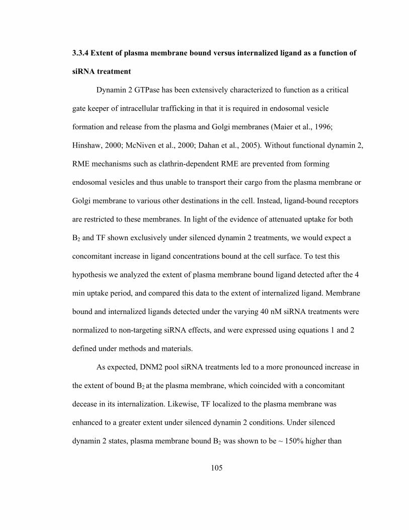
105
3.3.4 Extent of plasma membrane bound versus internalized ligand as a function of
siRNA treatment
Dynamin 2 GTPase has been extensively characterized to function as a critical
gate keeper of intracellular trafficking in that it is required in endosomal vesicle
formation and release from the plasma and Golgi membranes (Maier et al., 1996;
Hinshaw, 2000; McNiven et al., 2000; Dahan et al., 2005). Without functional dynamin 2,
RME mechanisms such as clathrin-dependent RME are prevented from forming
endosomal vesicles and thus unable to transport their cargo from the plasma membrane or
Golgi membrane to various other destinations in the cell. Instead, ligand-bound receptors
are restricted to these membranes. In light of the evidence of attenuated uptake for both
B2 and TF shown exclusively under silenced dynamin 2 treatments, we would expect a
concomitant increase in ligand concentrations bound at the cell surface. To test this
hypothesis we analyzed the extent of plasma membrane bound ligand detected after the 4
min uptake period, and compared this data to the extent of internalized ligand. Membrane
bound and internalized ligands detected under the varying 40 nM siRNA treatments were
normalized to non-targeting siRNA effects, and were expressed using equations 1 and 2
defined under methods and materials.
As expected, DNM2 pool siRNA treatments led to a more pronounced increase in
the extent of bound B2 at the plasma membrane, which coincided with a concomitant
decease in its internalization. Likewise, TF localized to the plasma membrane was
enhanced to a greater extent under silenced dynamin 2 conditions. Under silenced
dynamin 2 states, plasma membrane bound B2 was shown to be ~ 150% higher than

106
bound ligand revealed for untreated cells (Figure 3.4A). In addition, internalized
riboflavin reduced by ~ 40% compared to untreated conditions. Although to a lesser
extent than that shown for riboflavin, membrane bound TF detected for cells treated with
DNM2 pool siRNA increased ~ 110% over bound ligand detected in untreated cell
populations (Figure 3.4B). Furthermore, TF uptake reduced ~ 30% under reduced
dynamin 2 levels as compared to untreated conditions. Both B2 and TF showed
substantial localization at the plasma membrane with dynamin 2 silenced treatments
when compared to the corresponding internalized ligand data. Both ligands revealed
nearly 200% higher enrichment at the plasma membrane than within the cell. When the
extent of bound over the extent of internalized ligand was expressed as a ratio, both
riboflavin and transferrin were shown to be 3.1- and 2.0 fold higher than that defined for
untreated conditions, respectively (Figure 3.4C and D).
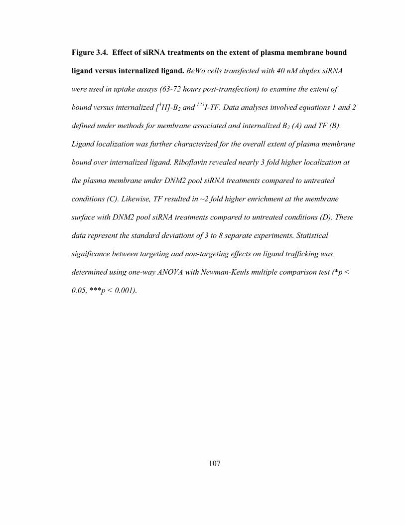
107
Figure 3.4. Effect of siRNA treatments on the extent of plasma membrane bound
ligand versus internalized ligand. BeWo cells transfected with 40 nM duplex siRNA
were used in uptake assays (63-72 hours post-transfection) to examine the extent of
bound versus internalized [3H]-B2 and 125I-TF. Data analyses involved equations 1 and 2
defined under methods for membrane associated and internalized B2 (A) and TF (B).
Ligand localization was further characterized for the overall extent of plasma membrane
bound over internalized ligand. Riboflavin revealed nearly 3 fold higher localization at
the plasma membrane under DNM2 pool siRNA treatments compared to untreated
conditions (C). Likewise, TF resulted in ~2 fold higher enrichment at the membrane
surface with DNM2 pool siRNA treatments compared to untreated conditions (D). These
data represent the standard deviations of 3 to 8 separate experiments. Statistical
significance between targeting and non-targeting effects on ligand trafficking was
determined using one-way ANOVA with Newman-Keuls multiple comparison test (*p <
0.05, ***p < 0.001).

108
109
3.3.5 Effect of transiently transfected wild-type or dominant-negative dynamin 2
(K44A) expression constructs on the extent of bound and internalized ligands
A common approach in corroborating RNAi data involves the use of wild-type
and dominant-negative expression vectors. We obtained the characterized wild-type
dynamin 2 (DNM2WT) and GTPase-null dynamin 2 (DNM2K44A) expression constructs,
the latter of which is unable to hydrolyze GTP and thus unable to pinch off nascently
formed endocytic vesicles at the plasma membrane (Cao et al., 1998). BeWo cells were
transiently transfected with 400 ng of plasmid DNA (wild-type dynamin 2 (DNM2WT),
GTPase-null dynamin 2 (DNM2K44A), or the empty expression vector) in the presence or
absence of 40 nM DNM2 pool siRNA according to the same methods used in siRNA
transfections. Cells were used in uptake assays 64-72 hours post-transfection, at which
time cells were dosed with either 5 nM [3H]-B2 or 10 nM 125I-TF for 4 min. As mentioned
earlier, all internalized B2 data generated at 37 ˚C was corrected for the passively
absorbed component by subtracting the corresponding results obtained at 4 ˚C. When
both B2 and TF results were expressed as a ratio of plasma membrane bound over
internalized, a strikingly similar trend was revealed (Figure 3.5). Both ligands were
shown to be largely localized at the cell membrane, as opposed to intracellularly, under
GTPase-null dynamin 2 alone and cotransfection conditions (i.e., DNM2K44A alone or
DNM2 siRNA + DNM2K44A, respectively). Although not significant, plasma membrane
bound B2 under GTPase-null dynamin 2 conditions was shown to increase 1.5 fold and
1.7 fold over cells transfected with wild type dynamin 2 or the empty vector alone,
respectively (figure 3.5A). The enhanced enrichment of B2 to the membrane surface was
110
shown to be more pronounced under cotransfection conditions (i.e., DNM2 siRNA +
DNM2K44A), of which led to 1.8 fold, 2 fold and 1.4 fold higher membrane localization
when compared to the effects of wild type, the empty vector, and the cotransfection
treatment involving non-targeting siRNA and the empty vector, respectively. A similar,
though more significant, trend was noted for the control ligand, transferrin. Transient
transfections involving GTPase-null dynamin 2 revealed nearly 2 fold and 2.4 fold higher
TF localization at the plasma membrane versus the effects of wild type dynamin 2 and
empty vector treatments, respectively (figure 3.5B). Similar to B2, the cotransfection
condition led to a more pronounced enrichment of TF at the membrane surface that was
1.5 fold and nearly 3 fold higher than the effects of the negative control cotransfection
treatment (i.e., NT siRNA + empty vector) and the empty vector, respectively. These
results are in agreement with data from cells transfected with DNM2 pool siRNA alone,
and provide additional evidence that actively internalized riboflavin requires the
functional expression of the mechanoenzyme, dynamin 2, for its endocytic trafficking in
human placental trophoblasts.

111
Figure 3.5. Effects of the over expression of wild type and GTPase-null dynamin 2
on intracellular and plasma membrane ligand localization. BeWo cells were
transiently transfected with 400 ng each of either wild type dynamin 2 (DNM2WT),
GTPase-null dynamin 2(DNM2K44A), or with a combination of DNM2K44A and 40 nM
DNM2 pool siRNA. Sixty three to 72 hours post-transfection, cells were dosed with 5 nM
[3H]-B2 or 10 nM 125I-TF for 4 min at 37 ˚C and examined for membrane associated and
internalized ligands. Analysis of plasma membrane bound B2 over internalized B2 (A)
revealed a trend in increased membrane bound ligand exclusively under single GTPase-
null dynamin 2 transfection (DNM2K44A) and cotransfection (DNM2 siRNA + DNM2K44A)
conditions. Similar results were exhibited by the control ligand, TF (B). The data
represents the standard deviations of 3-4 separate experiments. The effects of dynamin 2
expression vectors or GTPase-null dynamin 2 cotransfections with DNM2 siRNA on
ligand localization were defined through comparisons to single transfections with an
empty vector or empty vector cotransfections with 40 nM non-targeting siRNA (NT
siRNA), respectively. Statistical significance between experimental treatments and
control conditions were determined using one-way ANOVA with Newman-Keuls multiple
comparison test (*p < 0.05, **p < 0.01).
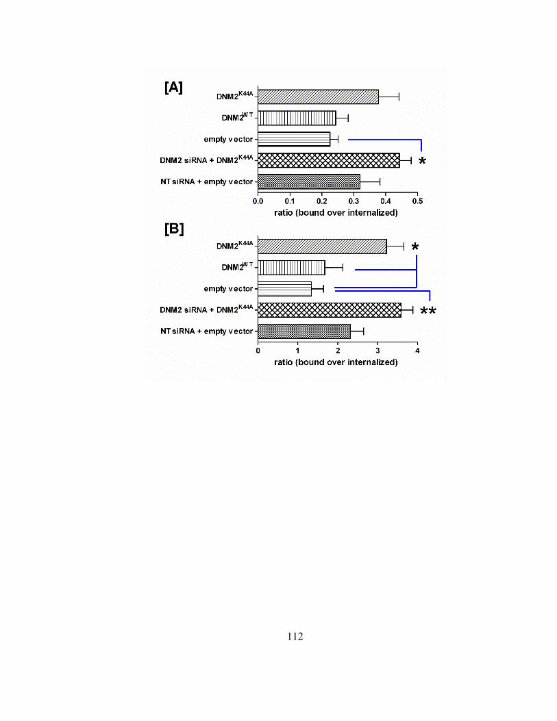
112

113
3.4 Discussion
A combinatorial approach involving RNAi and wild-type or dominant-negative
plasmid transfections was carried out to delineate the involvement of the conserved
endocytic mechanoenzyme, dynamin 2, in riboflavin absorption in BeWo cells.
Collectively the RNAi and dynamin 2 expression construct data revealed that the
endocytosis of riboflavin is regulated, in part, by the functional expression of dynamin 2
GTPase (Figures 3.1-5). In BeWo cells, the extent of dynamin 2 dependence in regulating
cellular riboflavin levels was similar to that of the clathrin-dependent RME ligand,
transferrin. Attenuated internalization of both B2 and TF (~ 60 and 70% of control
conditions, respectively) correlated with ~ 80% reduced dynamin 2 levels. In addition,
reductions in ligand uptake under these same conditions revealed a concomitant increase
in B2 and TF binding along the cell surface. Furthermore, cotransfection results involving
the GTPase-null dynamin 2 expression construct and DNM2 siRNA illustrated an
amplified effect on the predominant ligand accumulation at the plasma membrane for
both B2 and TF. Although the riboflavin receptor remains to be identified, this data
provides the most substantial evidence to date supporting the existence of such a protein.
The fact that the reduced dynamin 2 protein expression does not reduce transferrin
uptake more profoundly poses a concern. A common technique applied by other
laboratories performing RNAi studies is to carry out two consecutive transfections (~ 24
hours apart) of cells in order to achieve maximal gene silencing and target protein
knockdown. Such strategies have produced more substantial functional effects in other
cell models, with HeLa cells being a gold standard system. For instance, Huang and

114
coworkers (Huang et al., 2004) revealed ~ 70-80% inhibition in transferrin absorption in
HeLa cells that were transfected twice with siRNA targeting the clathrin heavy chain (i.e.,
critical protein subunit of the clathrin triskelion complex) or dynamin 2. In an effort to
duplicate this approach in BeWo cells, two consecutive transfections were carried out
using the DNM2 siRNA treatments. However, this strategy caused the cells to stop
growing and cell morphology appeared to be compromised (data not shown). In another
study (Soulet et al., 2005) the researchers performed a single siRNA transfection in HeLa
cells using siRNA targeting sorting nexin 9 (SNX9), which is a protein that binds and
regulates dynamin 2 activity and clathrin-mediated endocytic efficiency. They reported a
45% reduction in transferrin absorption, which is comparable to the effect we observed
for this control ligand in BeWo cells treated with DNM2 siRNA (i.e., 30% reduced
transferrin uptake). The close agreement between our results in BeWo and that of Soulet
and coworkers’ studies in HeLa suggest our RNAi strategies are specific and effective in
eliciting functional effects on the trafficking of the control ligand, transferrin.
In summary, the RNAi and dynamin 2 plasmid DNA transfection data provide
definitive and direct evidence that a riboflavin receptor-mediated endocytic mechanism
exists in human placental trophoblasts. Dynamin 2 GTPase is the first protein identified
in humans to serve as a regulator of B2 cellular entry through RME. Knowing that
dynamin 2 is required for the majority of known endocytic pathways, and in light of the
significant reduction in B2 uptake under silenced dynamin 2 levels, we can estimate that
the endocytosis of this critical vitamin accounts for up to 50% of the riboflavin-specific
active absorption mechanisms in BeWo. Analogous to the folate and transferrin receptor

115
pathways, understanding B2 endocytic internalization and trafficking provides a novel
pathway that may be potentially exploited in targeted drug therapeutics and/or biomedical
imaging (Li et al., 2003; Berry et al., 2004; Hattori and Maitani, 2004; Yoo and Park,
2004a).
116
CHAPTER 4
ELUCIDATING THE ROLE OF CAVEOLIN 1 IN THE RIBOFLAVIN
ENDOCYTIC ITINERARY IN PLACENTAL TROPHOBLASTS
Abstract
Riboflavin (vitamin B2) is an essential water soluble vitamin that is thoroughly
established to be critical to cellular growth, function, and development. In its coenzyme
forms, flavin mononucleotide (FMN) and flavin adenine dinucleotide (FAD), it is
indispensable in a myriad of metabolic pathways. Despite its biochemical and
physiological importance, the specific active absorption and trafficking mechanisms
regulating B2 cellular entry remain to be characterized. Recent reports suggest riboflavin
is absorbed into human epithelia via a clathrin and dynamin 2 dependent endocytic
process (Chapters 2 and 3). Considering dynamin 2’s indiscriminate promotion of distinct
receptor-mediated endocytic mechanisms, riboflavin absorption may occur through
alternative clathrin-independent endocytic pathways. The purpose of this study was to
delineate the involvement of the clathrin-independent caveolae-mediated endocytic
117
mechanism in regulating B2 cellular entry into human placental trophoblasts (BeWo).
Endocytosed rhodamine-riboflavin was examined for colocalization with the
immunofluorescence detected caveolae-associated protein, caveolin 1, using 3D confocal
laser scanning microscopy. Although to a lesser extent than that for the control ligand,
cholera toxin subunit B, ~ 11% of rhodamine-B2 was shown to distribute to caveolin 1-
positive endosomes upon 10 min uptake. Results from this study suggest that B2 uptake is
minimally regulated by caveolae-mediated endocytosis.
118
4.1 Introduction
Riboflavin (vitamin B2) plays a vital role in normal cellular growth and
maintenance. At the molecular level, B2 serves as the precursor molecule to the
coenzyme forms, flavin adenine dinucleotide (FAD) and flavin mononucleotide (FMN).
Both FAD and FMN have been extensively recognized for their critical function as
coenzyme prosthetic groups of flavoproteins, which are indispensable macromolecules
involved in a multitude of cellular redox reactions (Voet, 1995). Since humans are unable
to synthesize B2, they are restricted to obtaining this vitamin through their diet or through
symbiotic microorganisms that naturally synthesize this vitamin, and are known to
colonize the gut lumen. Despite its rare occurrence, B2-deficiency has been documented
in humans and is correlated with such factors as dietary deprivation or physiological
and/or pathological conditions. Common manifestations resulting from B2-deficient states
in humans include growth retardation, neurodegeneration, skin lesions, and anemia
(Cooperman, 1991). More severe cases have been documented with animals. Such studies
have indicated a general sequence of symptoms including stunted growth, muscle
weakness, and failure to thrive that leads to eventual death (Cooperman, 1991). Several
pathological states have also been correlated with mammalian B2 levels. In general,
studies have shown B2 to function as a protective factor in preventing cardiovascular
disease in humans (Hultquist, 1993), cataract formation in animals (Wintrobe, 1994;
Miyamota, 1998), and certain human cancers (Powers, 2003a). Despite extensive
evidence suggesting the physiological and pathological importance of B2 across different
species, the factors regulating its cellular absorption and trafficking are poorly understood.

119
To date, the majority of documented studies defining the factors regulating B2
absorption have been based on biochemical assays (Foraker et al., 2003). Combined,
these studies have established several common regulatory parameters that are observed
across divergent species, and include temperature dependence, Na+-independence,
general energy dependence, active transport kinetics at physiological B2 levels and
passive diffusion kinetics above basal B2 concentrations. These factors alone indicate the
existence of a transport mechanism specific to this vitamin; however, they do not
distinguish between a carrier-mediated or receptor-mediated process. More recent studies
have provided evidence of microtubule-driven vesicle trafficking events, specific to
receptor-medicated endocytosis (RME), being involved in the regulation of B2 absorption
by various human-derived cell lines (Wangensteen et al., 1996; Holladay, 1999; Huang
and Swaan, 2000; Huang and Swaan, 2001). Additional support for a B2-specific
receptor-mediated mechanism has been established through 2D fluorescence microscopy
and 3D confocal laser scanning microscopy-based assessments showing rhodamine-
labeled B2 localization to early endosomes of the clathrin-mediated endocytic (CME)
pathway (Huang et al., 2003; D'Souza et al., 2006b). Such data was further corroborated
by evidence of a time- and dose-dependent [3H]-B2 accumulation to clathrin pathway
specific endosomal populations that were isolated using sucrose density-based subcellular
fractionation (Chapter 2) (D'Souza et al., 2006b). More recent unpublished studies aimed
at elucidating the involvement of the major endocytic vesicle scission protein, dynamin 2
GTPase, have clearly demonstrated that ~ 50% of actively absorbed B2 depends on the
functional expression of this conserved protein (Chapter 3). Considering the pluripotent
120
nature of dynamin 2 on regulating the nascent formation and release of endocytic vesicles
from the plasma membrane for different RME pathways, it becomes plausible that B2-
RME may involve multiple, distinct endocytic mechanisms.
Caveolin 1 (CAV1) is thoroughly established to function in caveolar-mediated
endocytosis (CvME) in mammalian cells (Pelkmans, 2001; Pelkmans et al., 2001;
Pelkmans et al., 2002; Nabi and Le, 2003; Pelkmans et al., 2004; Pietiainen et al., 2004;
Pelkmans et al., 2005; Pelkmans and Zerial, 2005; Tagawa et al., 2005). Specifically,
CAV1 is a cytoplasmically oriented integral membrane protein known to bind cholesterol
(Uittenbogaard and Smart, 2000) and associates with cholesterol- and sphingolipid-rich
plasma membrane domains termed caveolae. CAV1 is one of three known caveolin
isoforms that has been characterized to be critical in signal transduction pathways,
membrane organization, and ligand bound receptor mediated trafficking in many
divergent cell systems (Cheng et al., 2006). Some of the classical ligands that have been
characterized to be absorbed and trafficked via this pathway include vitamin D, cholera
toxin subunit B, and folate, to name a few (Parton, 1994; Mineo and Anderson, 1996;
Norman et al., 2002; Singh et al., 2003; Huhtakangas et al., 2004; Hansen et al., 2005;
Norman, 2006). Furthermore, caveolae-mediated endocytosis has been shown to be
dependent on dynamin 2 expression (Yao et al., 2005), however it is distinct from the
classical clathrin-dependent endocytic pathway (Cheng et al., 2006).
Although riboflavin absorption and its cellular distribution have been extensively
shown to involve clathrin-mediated endocytosis (Huang and Swaan, 2000; Huang and
Swaan, 2001; Huang et al., 2003; D'Souza et al., 2006b), the involvement of other
121
endocytic pathways remains to be elucidated. Caveolae-mediated endocytosis is a
candidate B2-specific RME pathway known to be regulated by dynamin 2.
The objective of this study was to (a) evaluate whether B2-specific RME involves
the clathrin-independent caveolar-mediated internalization pathway in the human
placental trophoblast cell model, BeWo, and if so, (b) to define the extent by which this
alternative endocytic mechanism may regulate the active absorption of this vitamin.
Studies involving fluorescent ligand absorption assays combined with indirect
immunofluorescence detection of caveolin 1-positive endosomes were carried out to
define the role of caveolae-mediated endocytosis in riboflavin homeostasis.
4.2 Methods and materials
4.2.1 Cell culture
Human placental trophoblasts (BeWo) were obtained from the American Type
Culture Collection (Manassas, VA) and maintained in a controlled atmosphere at 37 ˚C,
under 5% CO2. BeWo cells (passages 204-215) were cultured in F-12K growth medium
(Invitrogen Life Technologies, Carlsbad, CA) supplemented with 10% fetal bovine serum,
1% nonessential amino acids, 100 units/mL penicillin, and 100 µg/mL streptomycin.
4.2.2 Fluorescent ligand endocytosis assay and immunofluorescence staining
Cells were seeded 3-5 days prior (5 x 10³ cells/cm2) in collagen-coated BD Falcon
culture slides (BD Biosciences, Bedford, MA). Following serum starvation, pulse-chase
assays with either 500 nM rhodamine-riboflavin (Rd-RF) (Phelps et al., 2004)or 15 nM
AlexaFluor®555-labeled cholera toxin subunit B (CTX, Invitrogen™, Molecular
Probes™, Eugene, OR) were carried out according to established methods (Huang et al.,

122
2003; D'Souza et al., 2006b). Cells were immediately fixed with 4% paraformaldehyde,
permeabilized, and blocked with bovine serum albumin prior to the immunolabeling of
caveolin 1 with rabbit-anti-CAV1 (Sigma, Saint Louis, MO) for 1 hr at room temperature.
Cells were thoroughly washed and probed with AlexaFluor®405-labeled goat-anti-rabbit
IgG (Invitrogen™, Molecular Probes™, Eugene, OR). Immunofluorescence treatments
were preserved using GelMount™ (Biomeda Corp., Foster City, CA).
4.2.3 Fluorescence image acquisition, restoration, and colocalization analysis
Internalized fluorescent ligands and immunostained caveolin 1 were imaged using
a Nikon Eclipse TE2000 E inverted confocal laser scanning microscope (Nikon
Instruments Inc., Melville, NY) outfitted with fixed lasers for 405 nm and 543 nm. Three
dimensional images were acquired using the following settings on Nikon EZ-C1 software
(Gold version 2.3, Image Systems Inc., Columbia, MD): Nikon Plan Apo 60xA oil
objective (1.4 numerical aperture), 3.6 µs scan dwell time, 512 x 512 pixel size, 0.30 µm
z-step, and a 150 detector pinhole. Raw images were iteratively deconvolved using a
calculated point spread function for individual channels and corrected for background
noise using a combination of median filtration and setting threshold levels just above
negative control treatments with either a non-reactive rhodamine derivative or the
secondary AlexFluor®405 antibody alone. Restored images were analyzed for 3D
colocalized fluorescence between ligands and caveolin 1 using Volocity, version 3.6
(Improvision Inc., Lexington, MA). The extent of colocalization between ligands and
caveolin 1 were determined by calculating the percent of total overlap volume (µm3) over

123
the corresponding total ligand volume. True colocalization between channels was defined
using the statistical test, the Pearson’s correlation (Manders et al., 1993).
4.3 Results
4.3.1 Colocalization of rhodamine- riboflavin with the conserved endocytic coat
protein, caveolin 1
The intracellular localization of B2 with respect to the caveolae-associated vesicle
protein, caveolin 1 (CAV1), was examined using the characterized rhodamine-labeled
riboflavin (Rd-RF) conjugate, which has been shown to exhibit nanomolar affinity
characteristics similar to the natural ligand (Huang et al., 2003). Ligand internalization
assays were carried out using subconfluent cells dosed with either 500 nM Rd-RF or 15
nM AlexaFluor®555-labeled cholera toxin subunit B (CTX) for 10 min prior to cell
fixation. Fluorescently-tagged CTX is thoroughly characterized to be endocytosed
through clathrin- and caveolae-dependent pathways across divergent cell models, and
thus serves as a positive control ligand in these assays (Parton, 1994; Henley et al., 1998;
Singh et al., 2003; Hansen et al., 2005; Yao et al., 2005). Internalized Rd-RF and CTX
were examined for colocalized signals with immunostained CAV1 in BeWo cells using
3D confocal laser scanning microscopy. Fluorescence signals specific to Rd-RF were
normalized to fluorescence intensities detected in parallel cells treated exclusively with a
non-reactive rhodamine analogue, caboxytetramethlrhodamine-4-amine (CTMR4A), of
which is a known by-product of the Rd-RF conjugation reaction (Phelps et al., 2004).
Furthermore, AlexaFluor®405-labeled secondary antibody specificity to the CAV1
antibody was defined by omitting the primary antibody in parallel treatments. Signal-to-

124
noise ratios for cells treated with either CTMR4A or the fluorescent-tagged secondary
antibody were substantially lower than cells treated exclusively with Rd-RF or the
combined primary and secondary antibodies, respectively (data not shown). Both Rd-RF
and CTX resulted in punctate perinuclear staining resembling endosomal organelle
localization (Figure 4.1). In contrast to CTX, Rd-RF resulted in minimal overlap with
CAV1.

125
Figure 4.1. Colocalization of rhodamine-riboflavin (Rd-RF) and cholera toxin
subunit B (CTX) with the endosomal coat protein, caveolin 1 (CAV1). Rd-RF and
AlexaFluor®555-labeled CTX were examined for colocalization with the immunostained
early endocytic protein marker, CAV1, after ligand internalization in BeWo cells for 10
min. Images represent orthogonal 3D profiles with the inset view defining the XY plane,
and the outer panels reveal the YZ (right narrow panel) and XZ (top narrow panel) focal
planes. Fluorescence signals for each channel were merged to reveal regions of
colocalization (indicated by arrows and yellow regions) and overlaid with the
corresponding differential interference contrast (DIC) image to define cell morphology
(far right inset view). Scale bars (micrometers) are defined in the merged inset panels.
126
The extent of colocalization between ligands and CAV1 were defined as the
percent of the total detected ligand volumes (cubic µm). The control ligand, CTX,
revealed ~ 3.3 fold higher (39.16 ± 2.91%) colocalization with CAV1 compared to the
signal overlap seen for Rd-RF and CAV1 (11.71 ± 0.76%) (figure 4.2). Overlapping
fluorescence intensities between ligands and CAV1 were further examined for 3D shape
similarities using the Pearson’s correlation statistical test (Manders et al., 1993).
Specifically, this test is commonly used to reveal linear relationships existing between
colocalized objects. Values > 0.0 define positive overlap between fluorescent channels.
Both ligands revealed positive fluorescence signal overlap with CAV1, and thus further
suggest ligand enrichment to caveolar endosomal populations. Collectively, these results
in combination with the morphological assessments shown in figure 4.1 indicate
significant CTX recruitment to CAV1-positive endosomes, which is in agreement with
literature results (Parton, 1994; Singh et al., 2003; Hansen et al., 2005). In striking
contrast, Rd-RF was shown to minimally localize with caveolae-associated vesicles. This
data suggests that B2 internalization through the caveolae-mediated endocytic pathway
occurs to a minor extent compared to that for CTX.
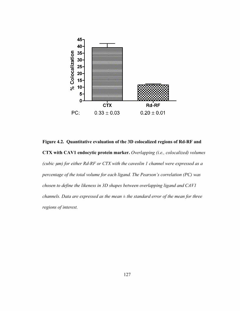
127
Figure 4.2. Quantitative evaluation of the 3D colocalized regions of Rd-RF and
CTX with CAV1 endocytic protein marker. Overlapping (i.e., colocalized) volumes
(cubic µm) for either Rd-RF or CTX with the caveolin 1 channel were expressed as a
percentage of the total volume for each ligand. The Pearson’s correlation (PC) was
chosen to define the likeness in 3D shapes between overlapping ligand and CAV1
channels. Data are expressed as the mean ± the standard error of the mean for three
regions of interest.

128
4.4 Discussion
Recent reports by our laboratory revealed a clathrin-mediated endocytic
component regulating riboflavin absorption and trafficking in divergent cell systems
(Huang and Swaan, 2000; Huang and Swaan, 2001; Huang et al., 2003; D'Souza et al.,
2006b). Furthermore, the functional expression of the vesicle scission protein, dynamin 2
GTPase, of which is well known to be critical in the early stages of endosome formation
common to the majority of characterized endocytic pathways, has been shown to play an
important role in regulating the internalization of B2 in human placental trophoblasts
(Chapter 3). Considering the pluripotent nature of dynamin 2 on regulating different
RME mechanisms, the question remains as to whether riboflavin is internalized by
multiple endocytic pathways. In this study, our data reveals minimal involvement of the
clathrin-independent caveolae-mediated endocytic pathway in regulating the cellular
entry of B2.
Established fluorescent ligand endocytosis assays involving the characterized
rhodamine-riboflavin conjugate were carried out to define the extent of colocalized signal
intensities with the immunofluorescence detected caveolar endosome marker protein,
caveolin 1. Three-dimensional confocal laser scanning microscopic analysis revealed ~
11% colocalization between Rd-RF and CAV1 after 10 min uptake in the BeWo model.
This degree of signal overlap was in dramatic contrast to that for the positive control
ligand, cholera toxin subunit B, which revealed nearly 40% localization to caveolin 1
positive endosomes. Combined with prior reports by our laboratory, the active absorption
of B2 in human epithelia is revealed to be regulated to a minor extent by the caveolar
129
mediated pathway, while the clathrin-dependent trafficking mechanism appears to be the
dominant route of cellular entry.
The concept of multiple endocytic processes regulating the cellular entry of a
single ligand is not unique to riboflavin. In fact, evidence of this phenomenon has been
demonstrated for the tumor growth factor beta (TGF-β), interferon-gamma, and cholera
toxin subunit B (Sadir et al., 2001; Di Guglielmo et al., 2003; Hansen et al., 2005).
Considering the importance of maintaining cellular riboflavin levels required for normal
growth and development, the existence of multiple active uptake mechanisms would
provide additional controls to meet such nutritional demands in states of physiological
distress. In the case for TGF-β, Di Guglielmo and colleagues demonstrated a biochemical
feedback mechanism regulating its internalization via either the clathrin-mediated or
caveolar-mediated pathways (Di Guglielmo et al., 2003). Specifically, their data revealed
the absorption of TGF-β along the CME pathway correlated with a signal transduction
response as defined by interactions with the Smad anchor for receptor activation (SARA)
protein. In contrast, caveolae-mediated endocytosis (CvME) of TGF-β was coupled with
the Smad7-Smurf2-dependent receptor degradation response and led to ubiquitin-
dependent degradation of the TGF-β receptor. In this particular instance, the CvME
mechanism appears to be involved in receptor degradation and ultimately receptor
turnover, whereas the CME pathway functions in promoting signal transduction cascades.
However, there are also reports that have shown the CME pathway to facilitate receptor
degradation through trafficking to lysosomal organelles, as is documented for the
epidermal growth factor (de Melker et al., 2001). In the case for riboflavin, the
130
involvement of two distinct RME pathways regulating its cellular entry may similarly
reflect a homeostatic seesaw of molecular cascades resulting in either B2-receptor
activation coupled with increased vitamin absorption, or receptor degradation and
reduced ligand uptake.
The involvement of multiple endocytic pathways regulating riboflavin
internalization opens up novel drug targeting mechanisms that can bypass efflux
transporters and thus may potentially improve drug bioavailability. Conceptually and
empirically, such drug formulations have been demonstrated for the folate and transferrin
RME pathways (Singh, 1999; Stephenson et al., 2003; Ghaghada et al., 2005; Hattori and
Maitani, 2005; Soni et al., 2005; Lubgan et al., 2006; Singh et al., 2006; Suresh Reddy et
al., 2006). Current studies in our laboratory are focused on defining the potential of
exploiting the B2-RME pathways for targeted chemotherapeutics using riboflavin-
conjugated polymeric drug carriers. Furthermore, recent reports (Rao et al., 1999;
Karande et al., 2001; Rao et al., 2006) revealing substantially elevated serum riboflavin
binding protein (RfBP) levels in breast and liver cancer patients compared to healthy
subjects proposes a potential targeted-chemotherapeutic niche for such B2-conjugated
formulations.
The purpose of this study was to elucidate whether alternative, clathrin-
independent, endocytic mechanisms regulate B2 cellular entry in the human placental
trophoblast cell model, BeWo. The caveolae-mediated endocytic pathway was chosen as
a candidate mechanism largely based on the extensive reported knowledge defining this
major clathrin-independent route for ligand internalization. Caveolin 1, an endosomal
131
protein marker specific to caveolae-dependent endocytosis, served as the indicator of this
pathway in 3D fluorescence colocalization assessments of the intracellular distribution of
rhodamine-riboflavin. Although to a minor extent compared to the positive control
ligand, CTX, B2 absorption was shown to involve CvME in BeWo. Ideally, such data
would be more informative compared to the extent of colocalization between B2 and
clathrin-positive endosomes occurring after the same uptake period. Such studies would
further define the overall importance of the CME and CvME pathways in regulating B2
cellular entry. Ongoing studies are addressing this concern in the effort to more clearly
define the B2-RME trafficking itinerary in placental trophoblasts.
132
CHAPTER 5
SUMMARY, CONCLUSIONS, AND FUTURE DIRECTIONS
5.1 Summary
The objective of this thesis was to characterize the receptor-mediated endocytic
mechanism(s) (RME) regulating riboflavin absorption and trafficking in human derived
placental trophoblasts and intestinal cell systems, BeWo and Caco-2, respectively.
Results from these studies record for the first time the temporal and spatial accumulation
of riboflavin to clathrin-associated early endosomal organelles that is suggested to be
negatively regulated by increasing cAMP levels (Chapter 2). The extent by which B2
internalization relies on RME in BeWo cells was further demonstrated (~ 50%) as a
function of the expression level of the master endocytic vesicle scission enzyme,
dynamin 2 GTPase (Chapter 3). Finally, preliminary studies examining whether clathrin-

133
independent RME processes are regulating the active internalization of B2 revealed a
minor involvement of the caveolae-mediated endocytosis pathway as demonstrated
through 3D fluorescence colocalization between rhodamine-riboflavin and the caveolar
coat protein, caveolin 1 (Chapter 4).
Classical clathrin-dependent receptor mediated endocytosis (CME) is
characterized by sequential orchestrated signaling cascades involving protein-protein and
protein-lipid interactions (Figure 5.1) (Merrifield et al., 2005; Perrais and Merrifield,
2005). Initial stages at the plasma membrane involve the nucleation of a clathrin-coated
pit (CCP), which is suggested to occur when the adaptor protein 2 (AP-2) binds to
phosphoinositol bisphosphate (PIP2). This interaction coincides with the recruitment of
clathrin and ligand-bound receptors through AP-2, and the consequential formation of a
CCP. The CCP is anchored at the plasma membrane by cytoskeletal microfilaments and
assessory proteins. Subsequent CCP growth and invagination events are then driven by a
consortium of proteins, including the vesicular scission enzyme, dynamin 2 GTPase.
Dynamin proteins polymerize to form a collar around the invaginated CCP neck, and
through GTP hydrolysis, the dynamin collar drives the membrane budding and release of
the vesicle from the plasma membrane into the cytosol. This nascently formed vesicle is
termed an early endosome, and shortly after its release, the clathrin coat also detaches to
repeat the cycle of CCP formation along the cytosolic face of the plasma membrane.
Endosomally associated Rab GTPases, such as Rab5, then function in trafficking early
endosome cargo to various destinations in the cell, including the Golgi, mitochondria,
and lysosomes (Rhodaman, 2000; Qualmann and Kessels, 2002; Smythe, 2002).

134
Figure 5.1. Initial stages regulating clathrin-dependent receptor-mediated
endocytosis. (i.) Ligand-bound receptors expressed along the extracellular surface of
epithelia are recruited by AP-2 and concentrated to clathrin coated pits (CCP) that
become stabilized by microfilaments and accessory proteins. (ii.) Consequently, CCP
growth and vesicle tubulation results through a consortium of protein-protein
interactions, including the formation of a dynamin 2 collar at the CCP neck. (iii.)
Through dynamin 2 dependent GTP-hydrolysis, the invaginated CCP is released from the
plasma membrane, which is characterized by the association of the endosome trafficking
protein, Rab5 GTPase, and is defined as an early endosome. (iv.) Almost immediately
upon its release, the early endosome is stripped of its clathrin coat, allowing clathrin to
return to the plasma membrane to repeat the ligand recruitment cycle of RME and the
ligand-bound receptor laden early endosome to continue to traffic along the CME
pathway.

135
In light of recent reports by our laboratory suggesting riboflavin absorption and
trafficking is governed, in part, by clathrin-mediated endocytosis (Huang and Swaan,
2000; Huang and Swaan, 2001; Huang et al., 2003; D'Souza et al., 2006a; D'Souza et al.,
2006b; Mason et al., 2006), and with the knowledge of the sequence of events regulating
ligand trafficking along this pathway, riboflavin absorption and subcellular distribution is
predicted to follow the dynamic and ordered trafficking itinerary specific to this
mechanism. To test this hypothesis, a dual approach involving density gradient
subcellular fractionation and 3D fluorescence confocal laser scanning microscopy was
carried out to delineate the intracellular trafficking route specific to riboflavin in human
intestinal (Caco-2) and placental trophoblast (BeWo) cells (Chapter 2). Analogous to the
positive endocytic protein, transferrin, absorbed riboflavin was shown to largely
accumulate to clathrin-associated early endosomes in a time- and dose-dependent manner
in both cell models. Maximal B2 accumulation to these endosomal populations was
revealed to occur at a faster rate in Caco-2 (within 30 min) as compared to BeWo (within
60 min). However, a riboflavin dose-dependent effect on endosomal enrichment was
shown to be dramatically more pronounced in the placental trophoblasts compared to the
enterocytes. Although to a lesser extent, internalized riboflavin was also revealed to
concentrate to Golgi and mitochondrial organelles in a time and ligand-dose dependent
manner. This is the first report of riboflavin enrichment to Golgi and may suggest a
possible recycling mechanism that is reminiscent of that reported for transferrin
(Yamashiro et al., 1984; Woods et al., 1986). Furthermore, in agreement with the
existence of a B2-specific receptor mediated endocytic mechanism, we revealed strict
136
homeostatic regulation of cellular riboflavin levels by cAMP. Collectively, these results,
generated in divergent cell models, demonstrate a dynamic and ordered B2 absorption and
trafficking process consistent with the clathrin-dependent RME pathway. A variation in
vitamin internalization rates and concomitant fortification to endosomal populations
appears to reflect distinctive nutritional requirements that are met in a cell lineage-
dependent manner.
Cumulative evidence has been shown to justify the clathrin-dependent B2-RME
process in human epithelia. Prior to the data presented in this thesis, no direct functional
evidence had been reported regarding critical known human protein regulators governing
riboflavin endocytosis. A strong candidate protein is Dynamin 2 GTPase. This
ubiquitously expressed enzyme has been extensively documented to regulate the
invagination and vesicle scission events occurring in classical endocytic pathways
including clathrin-dependent and independent mechanisms. Consequently, dynamin 2 has
been termed the master regulator of endocytic membrane trafficking events along the
plasma membrane (Conner and Schmid, 2003). The potential involvement of dynamin 2
in the B2-RME process was examined using the placental trophoblast model (Chapter 3).
BeWo was chosen for these studies largely based on riboflavin’s substantial enrichment
to clathrin-associated endosomes of this cell system as shown in Chapter 2, and for its
higher affinity for B2 compared to the intestinal cell model, Caco-2 (Huang and Swaan,
2001). Approaches leading to significantly reduced dynamin 2 expression (i.e., duplex
siRNA-based RNAi methods) or the over expression of nonfunctional dynamin 2 (i.e.,
GTPase null dynamin 2K44A) were carried out in BeWo cells to define the extent by which
137
actively absorbed B2 relies on RME. Similar to the positive control, transferrin, actively
internalized B2 was substantially reduced by ~ 50% exclusively under silenced dynamin 2
conditions. Furthermore, such reduced uptake correlated with a concomitant increase (~ 2
fold higher than absorbed B2) in B2 localization at the cell surface. Similar ligand
distribution profiles were shown under GTPase-null dynamin 2 conditions. In addition, a
more pronounced B2 localization at the cell surface was noted with cotransfection
treatments involving both dynamin 2 silencing siRNA and the over expression of the
GTPase-null protein, thus further suggesting the importance of this endocytic protein
regulator in maintaining cellular B2 levels. This study is the first to report the direct
functional involvement of the conserved endocytic scission protein, dynamin 2 GTPase,
in regulating riboflavin absorption in human epithelia. In addition, these studies
demonstrate the general extent (~ 50%) by which riboflavin depends on RME in human
placental trophoblasts.
Dynamin 2 GTPase is known to indiscriminately function in the vesicular
budding and release of endosomes for multiple and distinct RME pathways. Therefore, if
B2 absorption is dependent on the functional expression of dynamin 2 as demonstrated in
Chapter 3, there is a possibility that alternative clathrin-independent RME mechanisms
may be regulating the cellular uptake of this vitamin. One such candidate pathway is the
caveolae-mediated mechanism (CvME) (Nichols, 2002; Le and Nabi, 2003; Venkatesan
et al., 2003). Like CME, CvME depends on functional dynamin 2 GTPase (Henley et al.,
1998; Shajahan et al., 2004) to allow for the release of ligand-bound receptor laden
vesicles from the plasma membrane. Caveolin 1 (CAV1) is a critical coat protein
138
characteristic of caveolae endosomes and is required in caveolar-mediated endocytosis
(Nichols, 2003). In addition, CAV1 has been reported to interact directly with dynamin 2
(Yao et al., 2005). To demonstrate whether B2 uptake involves CvME, BeWo cells were
examined for colocalization between internalized rhodamine-riboflavin and fluorescently
immunostained CAV1 using 3D fluorescence confocal laser scanning microscopy.
Results from these studies revealed minimal colocalization between B2 and the caveolae-
associated endosomal marker, suggesting B2 absorption and trafficking is regulated by
CvME to a minor extent.
5.2 Conclusions: the B2-RME model revised
For the past several decades, various studies have been carried out to elucidate the
mechanisms regulating vitamin B2 absorption in humans. From these studies, a set of B2-
specific parameters regulating its uptake kinetics have been generated and shown to be
conserved across divergent mammalian cell models and tissues. These salient features
include temperature-, energy-, and pH- dependence, active uptake and passive diffusion
kinetics at low nM and above basal vitamin concentrations, respectively, and a general
involvement of Ca2+-calmodulin, protein kinase A- and G- pathways (Huang and Swaan,
2000; Huang and Swaan, 2001; Foraker et al., 2003). In addition, 2D fluorescence
microscopy studies analyzing the localization of absorbed rhodamine-labeled riboflavin
in human placental trophoblasts suggested the involvement of the clathrin-mediated RME
pathway in regulating cellular B2 levels (Huang et al., 2003). In further agreement of a B2
endocytic process, indirect biochemical-based modulator studies revealed riboflavin
absorption and trafficking to be dependent on cytoskeletal scaffolds and associated motor

139
proteins common in microtubule and actin-dependent vesicle transport dynamics
(D'Souza et al., 2006a). More recently, studies examining the effects of antibodies
specific to the secreted avian-derived riboflavin binding protein (RfBP), of which is
known to selectively bind extracellular B2 and facilitate its cellular entry in oviparous
species (Zheng et al., 1988; White et al., 1992; Wasylewski, 2004), was shown to
dramatically reduce riboflavin absorption and trafficking in an RME dependent manner
in human placental trophoblasts (Mason et al., 2006). Thus, analogous to the ligand
sequestering functions of the characterized molecules transferrin and intrinsic factor, of
which are known to regulate cellular iron and B12 levels (Birn et al., 1997; Fyfe et al.,
2004), respectively, the involvement of a soluble human RfBP protein is suggested in
regulating B2-RME. Collectively, these studies indirectly indicate riboflavin absorption
and trafficking in human epithelia is regulated, at least in part, by the classical CME
pathway.
A major weakness to this B2-RME model is the lack of direct evidence identifying
specific cellular proteins facilitating this vitamin’s uptake mechanism in humans.
Although recent reports suggest the existence of a soluble human riboflavin binding
protein, the direct identity of such a molecule remains to be established. Several
laboratories, including our own, have attempted to isolate such a protein using various
techniques including size-exclusion- and riboflavin-coupled affinity-chromatography,
and riboflavin based photo-affinity labeling reactions. However, these studies were
unsuccessful due to non-specific selection and labeling of ubiquitous flavoprotein
molecules endogenous to all studied cell models and tissues. The aims in this thesis
140
involved alternative approaches to (1) resolve the subcellular distribution of absorbed
riboflavin through subcellular fractionation and fluorescence 3D confocal laser scanning
microscopy, (2) directly identify the functional involvement of the conserved pluripotent
endocytic effector protein, dynamin 2 GTPase, using RNAi and dominant negative
protein expression constructs, and (3) to determine the involvement of the clathrin-
independent caveolae-mediated RME pathway, through 3D fluorescence colocalization
analysis between internalized rhodamine-riboflavin and the immunostained caveolae-
associated endosome marker, caveolin 1. Results from these studies provide additional
justification of the clathrin-dependent B2-RME model, and further demonstrate for the
first time the functional requirement of human dynamin 2 GTPase in this process (Figure
5.2).

141
Figure 5.2. The B2-RME model revised. To date, the cellular entry of riboflavin along
human epithelia has been definitively shown to occur through passive diffusion (A) and
receptor-mediated endocytosis (B). In the case of B2-specific RME, riboflavin is initially
suggested to be bound by a putative human analog of extracellular avian-RfBP, of which
is believed to aid in the recruitment of B2 to its plasma membrane associated receptor.
The ligand bound receptor complex is then sequestered to clathrin coated pits (CCP) (1).
Through the sequential and concerted functions resulting from various protein-protein
interactions including the vesicular scission action of dynamin 2 GTPase, this CCP
becomes invaginated and is pinched off from the plasma membrane to form an early
endosome (EE) (2). Through the functions of the associated Rab5 GTPase, the EE fuses
with other early endosomes to form a late sorting or recycling endosome (LE) that
characteristically has a lower pH than that of EE (3). At this point, the B2-bound receptor
complex may uncouple to generate free B2 within the acidic LE. Similar to the
transferrin-RME pathway, the unbound B2-receptor and RfBP would then be free to
recycle back to the apical membrane to recruit additional extracellular riboflavin (4a).
Alternative destinations for trafficking of the receptor and RfBP molecules, in the
absence or presence of B2, from the LE are suggested to include the Golgi (4b), the
basolateral membrane domain or systemic circulation through transcytosis (4c),
lysosomes (L) for degradation (4d), or the mitochondria (4e). The nucleus (N) and
endoplasmic reticulum (ER) are shown as points of reference.
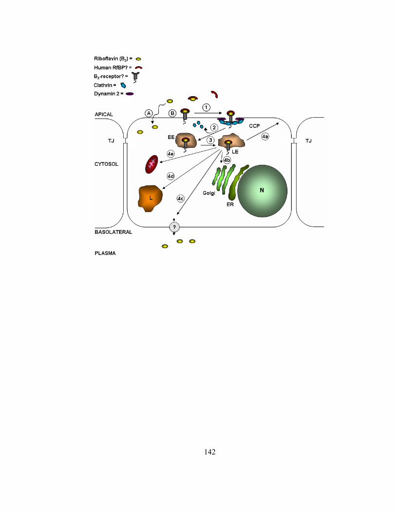
142

143
Despite the extensive evidence indicating the involvement of the CME pathway in
B2 cellular entry in human epithelia, several critical questions remain to be addressed at
the molecular level. The identity of the human analogs for a riboflavin binding protein
and the proposed B2-receptor remain to be established. In addition, earlier studies largely
suggested a riboflavin-specific transporter was responsible for this vitamin’s active
absorption in humans as reviewed by Foraker and coworkers (Foraker et al., 2003); yet,
such a protein has not been definitively isolated or identified. Data presented in this thesis
combined with earlier reports by our laboratory definitively justify the clathrin-dependent
B2-RME model; however, these results don’t exclude the possibility of a transporter also
being involved in regulating cellular B2 levels. To date, the involvement of a transporter
in B2 absorption remains to be defined in humans.
5.3 Future directions: exploring the potential application of the B2-RME
mechanism in targeted medicinal therapeutics
Some of the major obstacles in drug therapy include low permeability of
hydrophilic drugs and/or macromolecules across cellular membranes, reduced cellular
retention due to ubiquitously expressed efflux transporters, metabolic degradation by
omnipresent enzymes, low mean residence time due to the reticuloendothelial system,
and underlying genetic aberrations and/or pathological states exhibited by patients. All
these factors combined drastically reduce drug bioavailability, and in certain instances
increase drug-related toxicity. One approach to circumvent several of these issues has
involved the packaging of biotherapeutics into ligand-targeted drug delivery systems

144
(DDS). Traditionally, fabrication of these devices has involved covalently conjugating
natural biological ligands to lipid- or polymer-based nanoparticles that are loaded with
various therapeutic entities. Such drug carriers bind to specific receptors along the
cellular plasma membrane and then gain entry through endocytosis.
Ligand-targeted DDS technology has proven successful for targeting such natural
ligand-receptor-mediated endocytic (RME) pathways as that for transferrin and folate.
Kobayashi and coworkers (Kobayashi et al., 2007) demonstrated that drug efflux by P-gp
is more effectively prevented in multidrug resistance (MDR) cancer cells when
chemotherapeutics (e.g., doxorubicin) are delivered via liposomes targeted to transferrin
receptors, as compared to untargeted liposome formulations. In another study by Kim and
colleagues (Kim et al., 2006), reports revealed substantial enhancements in gene
transfection efficiencies and gene silencing of GFP (green fluorescent protein) in the
human epidermal carcinoma cell model, KB, upon transfecting cells with poly electrolyte
DDS conjugated with folate. Specifically, their results demonstrated that such ligand-
targeted gene silencing formulations’ effects could be achieved using significantly less
short-interfering RNA (siRNA). In addition, these researchers demonstrated the
entrapment of gene-silencing therapeutics in folate-tagged DDS enhanced the extent of
cellular entry, increased siRNA stability against nuclease degradation, and further
enhanced cell targeting specificity through the endogenous folate-specific RME pathway.
Based on our current understanding of the riboflavin-specific RME pathway in epithelial
cells, it would be interesting to test whether drug delivery systems targeting this pathway
reveal similar advantages to gene and chemotherapeutic formulations as those shown for
145
transferrin receptor- and folate receptor-targeted DDS. Along with establishing a
molecular map of B2 transport, metabolism and utilization in cellular growth and
development, understanding the absorption and intracellular trafficking of riboflavin in
human epithelia opens new opportunities for scientists to utilize this pathway for targeted
drug delivery.

146
BIBLIOGRAPHY
Abrams VA, McGahan TJ, Rohrer JS, Bero AS and White HB, 3rd (1988) Riboflavin-binding protein from reptiles: a comparison with avian riboflavin-binding proteins. Comp Biochem Physiol B 90:243-247.
Aw TY, Jones DP and McCormick DB (1983) Uptake of riboflavin by isolated rat liver cells. J. Nutr. 113:1249-1254.
Baba T, Damke H, Hinshaw JE, Ikeda K, Schmid SL and Warnock DE (1995) Role of dynamin in clathrin-coated vesicle formation. Cold Spring Harb Symp Quant Biol 60:235-242.
Barbieri MA, Ramkumar TP, Fernadez-Pol S, Chen PI and Stahl PD (2004) Receptor tyrosine kinase signaling and trafficking--paradigms revisited. Curr Top Microbiol Immunol 286:1-20.
Barile M, Brizio C, Valenti D, De Virgilio C and Passarella S (2000) The riboflavin/FAD cycle in rat liver mitochondria. Eur. J. Biochem. 267:4888-4900.
Beach RS, Mantero-Atienza E, Shor-Posner G, Javier JJ, Szapocznik J, Morgan R, Sauberlich HE, Cornwell PE, Eisdorfer C and Baum MK (1992) Specific nutrient abnormalities in asymptomatic HIV-1 infection. Aids 6:701-708.
Berry CC, Charles S, Wells S, Dalby MJ and Curtis AS (2004) The influence of transferrin stabilised magnetic nanoparticles on human dermal fibroblasts in culture. Int. J. Pharm. 269:211-225.
Bhat KG, Malhotra P, Karande AA and Adiga PR (1995) Immunohistochemical localization of riboflavin carrier protein in testicular cells of mammals. Indian J Exp Biol 33:12-16.
147
Birn H, Verroust PJ, Nexo E, Hager H, Jacobsen C, Christensen EI and Moestrup SK (1997) Characterization of an epithelial approximately 460-kDa protein that facilitates endocytosis of intrinsic factor-vitamin B12 and binds receptor-associated protein. J Biol Chem 272:26497-26504.
Bucci C, Parton RG, Mather IH, Stunnenberg H, Simons K, Hoflack B and Zerial M (1992) The small GTPase rab5 functions as a regulatory factor in the early endocytic pathway. Cell 70:715-728.
Cao H, Garcia F and McNiven MA (1998) Differential distribution of dynamin isoforms in mammalian cells. Mol Biol Cell 9:2595-2609.
Cao H, Orth JD, Chen J, Weller SG, Heuser JE and McNiven MA (2003) Cortactin is a component of clathrin-coated pits and participates in receptor-mediated endocytosis. Mol Cell Biol 23:2162-2170.
Cheng ZJ, Singh RD, Marks DL and Pagano RE (2006) Membrane microdomains, caveolae, and caveolar endocytosis of sphingolipids. Mol Membr Biol 23:101-110.
Ciechanover A, Schwartz AL, Dautry-Varsat A and Lodish HF (1983) Kinetics of internalization and recycling of transferrin and the transferrin receptor in a human hepatoma cell line. Effect of lysosomotropic agents. J Biol Chem 258:9681-9689.
Clarke HC (1977) Distribution of riboflavin in blood: in women and in prenates. Int J Vitam Nutr Res 47:361-363.
Collard MW, Sylvester SR, Tsuruta JK and Griswold MD (1988) Biosynthesis and molecular cloning of sulfated glycoprotein 1 secreted by rat Sertoli cells: sequence similarity with the 70-kilodalton precursor to sulfatide/GM1 activator. Biochemistry 27:4557-4564.
Conner SD and Schmid SL (2003) Regulated portals of entry into the cell. Nature 422:37-44.
Cooperman JM, and Lopez, R. (1991) Riboflavin, in: Handbook of vitamins, pp 283-310, Marcel Dekker, New York.
148
Cooperman JM, Cole HS, Gordon M and Lopez R (1973) Erythrocyte glutathione reductase as a measure of riboflavin nutritional status of pregnant women and newborns. Proc Soc Exp Biol Med 143:326-328.
D'Souza VM, Bareford LM, Ray A and Swaan PW (2006a) Cytoskeletal scaffolds regulate riboflavin endocytosis and recycling in placental trophoblasts. J Nutr Biochem 17:821-829.
D'Souza VM, Foraker AB, Free RB, Ray A, Shapiro PS and Swaan PW (2006b) cAMP-Coupled riboflavin trafficking in placental trophoblasts: a dynamic and ordered process. Biochemistry 45:6095-6104.
Dahan S, Anderson KL, Weller S, Krueger E and McNiven MA (2005) Agonist-induced vesiculation of the Golgi apparatus in pancreatic acinar cells. Gastroenterology 129:2032-2046.
Dancis J, Lehanka J and Levitz M (1985) Transfer of riboflavin by the perfused human placenta. Pediatr Res 19:1143-1146.
Dancis J, Lehanka J and Levitz M (1988) Placental transport of riboflavin: differential rates of uptake at the maternal and fetal surfaces of the perfused human placenta. Am J Obstet Gynecol 158:204-210.
Daniel H, Wille U and Rehner G (1983) In vitro kinetics of the intestinal transport of riboflavin in rats. J Nutr 113:636-643.
Dautry-Varsat A, Ciechanover A and Lodish HF (1983) pH and the recycling of transferrin during receptor-mediated endocytosis. Proc. Natl. Acad. Sci. U. S. A. 80:2258-2262.
Davis JT and Ong DE (1992) Synthesis and secretion of retinol-binding protein by cultured rat Sertoli cells. Biol Reprod 47:528-533.
de Melker AA, van der Horst G, Calafat J, Jansen H and Borst J (2001) c-Cbl ubiquitinates the EGF receptor at the plasma membrane and remains receptor associated throughout the endocytic route. J Cell Sci 114:2167-2178.

149
Di Guglielmo GM, Le Roy C, Goodfellow AF and Wrana JL (2003) Distinct endocytic pathways regulate TGF-beta receptor signalling and turnover. Nat Cell Biol 5:410-421.
Dumaswala R, Setchell KD, Moyer MS and Suchy FJ (1993) An anion exchanger mediates bile acid transport across the placental microvillous membrane. Am J Physiol 264:G1016-1023.
Fernandez-Banares F, Abad-Lacruz A, Xiol X, Gine JJ, Dolz C, Cabre E, Esteve M, Gonzalez-Huix F and Gassull MA (1989) Vitamin status in patients with inflammatory bowel disease. Am J Gastroenterol 84:744-748.
Foraker AB, Khantwal CM and Swaan PW (2003) Current perspectives on the cellular uptake and trafficking of riboflavin. Adv. Drug Deliv. Rev. 55:1467-1483.
Foti M, Carpentier JL, Aiken C, Trono D, Lew DP and Krause KH (1997) Second-messenger regulation of receptor association with clathrin-coated pits: a novel and selective mechanism in the control of CD4 endocytosis. Mol. Biol. Cell 8:1377-1389.
Foy H and Mbaya V (1977) Riboflavin. Prog Food Nutr Sci 2:357-394.
Fyfe JC, Madsen M, Hojrup P, Christensen EI, Tanner SM, de la Chapelle A, He Q and Moestrup SK (2004) The functional cobalamin (vitamin B12)-intrinsic factor receptor is a novel complex of cubilin and amnionless. Blood 103:1573-1579.
Ghaghada KB, Saul J, Natarajan JV, Bellamkonda RV and Annapragada AV (2005) Folate targeting of drug carriers: a mathematical model. J Control Release 104:113-128.
Ghosh RN and Maxfield FR (1995) Evidence for nonvectorial, retrograde transferrin trafficking in the early endosomes of HEp2 cells. J Cell Biol 128:549-561.
Gonzales DH and Neupert W (1990) Biogenesis of mitochondrial c-type cytochromes. J. Bioenerg. Biomembr. 22:753-768.
150
Gorvel JP, Chavrier P, Zerial M and Gruenberg J (1991) rab5 controls early endosome fusion in vitro. Cell 64:915-925.
Hamajima S and Ono S (1995) Sequence of a cDNA encoding turtle riboflavin-binding protein: a comparison with avian riboflavin-binding protein. Gene 164:279-282.
Hamazume Y, Mega T and Ikenaka T (1984) Characterization of hen egg white- and yolk-riboflavin binding proteins and amino acid sequence of egg white-riboflavin binding protein. J Biochem (Tokyo) 95:1633-1644.
Hamazume Y, Mega T and Ikenaka T (1987) Positions of disulfide bonds in riboflavin-binding protein of hen egg white. J Biochem (Tokyo) 101:217-223.
Hansen GH, Dalskov SM, Rasmussen CR, Immerdal L, Niels-Christiansen LL and Danielsen EM (2005) Cholera toxin entry into pig enterocytes occurs via a lipid raft- and clathrin-dependent mechanism. Biochemistry 44:873-882.
Hattori Y and Maitani Y (2004) Enhanced in vitro DNA transfection efficiency by novel folate-linked nanoparticles in human prostate cancer and oral cancer. J. Control. Release 97:173-183.
Hattori Y and Maitani Y (2005) Folate-linked lipid-based nanoparticle for targeted gene delivery. Curr Drug Deliv 2:243-252.
Hediger MA, Kanai Y, You G and Nussberger S (1995) Mammalian ion-coupled solute transporters. J Physiol 482:7S-17S.
Henley JR, Krueger EW, Oswald BJ and McNiven MA (1998) Dynamin-mediated internalization of caveolae. J Cell Biol 141:85-99.
Hill E, van der Kaay J, Downes CP and Smythe E (2001) The role of dynamin and its binding partners in coated pit invagination. J Cell Biol 152:309-323.
Hinshaw JE (2000) Dynamin and its role in membrane fission. Annu Rev Cell Dev Biol 16:483-519.

151
Holladay SR, Yang, Z., Kennedy, M. D., Leamon, C. P., Lee, R. J., Jayamani, M., Mason, T., Low, P. S. (1999) Riboflavin-mediated delivery of a macromolecule into cultured human cells. Biochim Biophys Acta 1426:195-204.
Horwitt MK, Harvey CC, Dahm CH, Jr. and Searcy MT (1972) Relationship between tocopherol and serum lipid levels for determination of nutritional adequacy. Ann N Y Acad Sci 203:223-236.
Huang F, Khvorova A, Marshall W and Sorkin A (2004) Analysis of clathrin-mediated endocytosis of epidermal growth factor receptor by RNA interference. J Biol Chem 279:16657-16661.
Huang SN, Phelps MA and Swaan PW (2003) Involvement of endocytic organelles in the subcellular trafficking and localization of riboflavin. J. Pharmacol. Exp. Ther. 306:681-687.
Huang SN and Swaan PW (2000) Involvement of a receptor-mediated component in cellular translocation of riboflavin. J. Pharmacol. Exp. Ther. 294:117-125.
Huang SN and Swaan PW (2001) Riboflavin uptake in human trophoblast-derived BeWo cell monolayers: cellular translocation and regulatory mechanisms. J. Pharmacol. Exp. Ther. 298:264-271.
Hughson EJ and Hopkins CR (1990) Endocytic pathways in polarized Caco-2 cells: identification of an endosomal compartment accessible from both apical and basolateral surfaces. J Cell Biol 110:337-348.
Huhtakangas JA, Olivera CJ, Bishop JE, Zanello LP and Norman AW (2004) The vitamin D receptor is present in caveolae-enriched plasma membranes and binds 1 alpha,25(OH)2-vitamin D3 in vivo and in vitro. Mol Endocrinol 18:2660-2671.
Hultquist DE, Xu, F., Quandt, K. S., Shlafer, M., and Mack, C. P. (1993) Evidence that NADPH-dependent methaemoglobin reductase and administered riboflavin protect tissue from oxidative injury. Am J Hematol 42:13-18.
Jackle S, Runquist EA, Miranda-Brady S and Havel RJ (1991) Trafficking of the epidermal growth factor receptor and transferrin in three hepatocytic endosomal fractions. J Biol Chem 266:1396-1402.
152
Jin M and Snider MD (1993) Role of microtubules in transferrin receptor transport from the cell surface to endosomes and the Golgi complex. J. Biol. Chem. 268:18390-18397.
Jusko WJ and Levy G (1970) Pharmacokinetic evidence for saturable renal tubular reabsorption of riboflavin. J Pharm Sci 59:765-772.
Jusko WJ, Levy G, Yaffe SJ and Gorodischer R (1970) Effect of probenecid on renal clearance of riboflavin in man. J Pharm Sci 59:473-477.
Jusko WJ, Levy, G. (1975) Absorption, Protein Binding, and Elimination of Riboflavin, in: Riboflavin (Rivlin RS ed), pp 99-152, Plenum Press, New York City.
Karande AA, Sridhar L, Gopinath KS and Adiga PR (2001) Riboflavin carrier protein: a serum and tissue marker for breast carcinoma. Int J Cancer 95:277-281.
Kasai S, Nakano H, Maeda K and Matsui K (1990) Purification, properties, and function of flavokinase from rat intestinal mucosa. J Biochem (Tokyo) 107:298-303.
Kim SH, Mok H, Jeong JH, Kim SW and Park TG (2006) Comparative evaluation of target-specific GFP gene silencing efficiencies for antisense ODN, synthetic siRNA, and siRNA plasmid complexed with PEI-PEG-FOL conjugate. Bioconjug Chem 17:241-244.
Kirshenbaum NW, Dancis J, Levitz M, Lehanka J and Young BK (1987) Riboflavin concentration in maternal and cord blood in human pregnancy. Am J Obstet Gynecol 157:748-752.
Knight A, Hughson E, Hopkins CR and Cutler DF (1995) Membrane protein trafficking through the common apical endosome compartment of polarized Caco-2 cells. Mol Biol Cell 6:597-610.
Kobayashi T, Ishida T, Okada Y, Ise S, Harashima H and Kiwada H (2007) Effect of transferrin receptor-targeted liposomal doxorubicin in P-glycoprotein-mediated drug resistant tumor cells. Int J Pharm 329:94-102.
153
Kumar CK, Yanagawa N, Ortiz A and Said HM (1998) Mechanism and regulation of riboflavin uptake by human renal proximal tubule epithelial cell line HK-2. Am J Physiol 274:F104-110.
Lamaze C, Baba T, Redelmeier TE and Schmid SL (1993) Recruitment of epidermal growth factor and transferrin receptors into coated pits in vitro: differing biochemical requirements. Mol Biol Cell 4:715-727.
Le PU and Nabi IR (2003) Distinct caveolae-mediated endocytic pathways target the Golgi apparatus and the endoplasmic reticulum. J Cell Sci 116:1059-1071.
Lemoine A, Le Devehat C, Codaccioni JL, Monges A, Bermond P and Salkeld RM (1980) Vitamin B1, B2, B6, and C status in hospital inpatients. Am J Clin Nutr 33:2595-2600.
Li Y, Ogris M, Wagner E, Pelisek J and Ruffer M (2003) Nanoparticles bearing polyethyleneglycol-coupled transferrin as gene carriers: preparation and in vitro evaluation. Int. J. Pharm. 259:93-101.
Lowy RJ and Spring KR (1990) Identification of riboflavin transport by MDCK cells using quantitative fluorescence video microscopy. J Membr Biol 117:91-99.
Lubgan D, Marczak A, Distel L and Jozwiak Z (2006) [Transferrin conjugates in the anticancer therapy]. Postepy Biochem 52:72-79.
Mac Lachlan I, Nimpf J and Schneider WJ (1994) Avian riboflavin binding protein binds to lipoprotein receptors in association with vitellogenin. J Biol Chem 269:24127-24132.
MacLachlan I, Nimpf J, White HB, 3rd and Schneider WJ (1993) Riboflavinuria in the rd chicken. 5'-splice site mutation in the gene for riboflavin-binding protein. J Biol Chem 268:23222-23226.
Maier O, Knoblich M and Westermann P (1996) Dynamin II binds to the trans-Golgi network. Biochem Biophys Res Commun 223:229-233.

154
Manders MM, Verbeek PJ and Aten JA (1993) Measurement of co-localization of objects in dual colour confocal images. J. Microscopy 169:375-382.
Mason CW, D'Souza VM, Bareford LM, Phelps MA, Ray A and Swaan PW (2006) Recognition, co-internalization, and recycling of an avian riboflavin carrier protein in human placental trophoblasts. J Pharmacol Exp Ther 317:465-472.
Maxfield FR and McGraw TE (2004) Endocytic recycling. Nat. Rev. Mol. Cell. Biol. 5:121-132.
McLauchlan H, Newell J, Morrice N, Osborne A, West M and Smythe E (1998) A novel role for Rab5-GDI in ligand sequestration into clathrin-coated pits. Curr. Biol. 8:34-45.
McNiven MA, Cao H, Pitts KR and Yoon Y (2000) The dynamin family of mechanoenzymes: pinching in new places. Trends Biochem Sci 25:115-120.
Merrifield CJ, Perrais D and Zenisek D (2005) Coupling between clathrin-coated-pit invagination, cortactin recruitment, and membrane scission observed in live cells. Cell 121:593-606.
Merrill AH, Jr., Froehlich JA and McCormick DB (1979) Purification of riboflavin-binding proteins from bovine plasma and discovery of a pregnancy-specific riboflavin-binding protein. J Biol Chem 254:9362-9364.
Middleton HM, 3rd (1985) Uptake of pyridoxine by in vivo perfused segments of rat small intestine: a possible role for intracellular vitamin metabolism. J Nutr 115:1079-1088.
Middleton HM, 3rd (1990) Uptake of riboflavin by rat intestinal mucosa in vitro. J Nutr 120:588-593.
Miller MS, Bruch RC and White HB, 3rd (1982) Carbohydrate compositional effects on tissue distribution of chicken riboflavin-binding protein. Biochim Biophys Acta 715:126-136.

155
Mineo C and Anderson RG (1996) A vacuolar-type proton ATPase mediates acidification of plasmalemmal vesicles during potocytosis. Exp Cell Res 224:237-242.
Miyamota Y, Sancar, A. (1998) Vitamin B2 based blue photoreceptors in the retinohypothalamic tract as the photoactive pigments for setting the circadian clock in mammals. Proc Natl Acad Sci U S A 95:6097-6102.
Monaco HL (1997) Crystal structure of chicken riboflavin-binding protein. Embo J 16:1475-1483.
Mukherjee S, Ghosh RN and Maxfield FR (1997) Endocytosis. Physiol Rev 77:759-803.
Muniyappa K and Adiga PR (1980) Isolation and characterization of riboflavin-binding protein from pregnant-rat serum. Biochem J 187:537-540.
Nabi IR and Le PU (2003) Caveolae/raft-dependent endocytosis. J Cell Biol 161:673-677.
Nakamura N, Rabouille C, Watson R, Nilsson T, Hui N, Slusarewicz P, Kreis TE and Warren G (1995) Characterization of a cis-Golgi matrix protein, GM130. J. Cell Biol. 131:1715-1726.
Nathke IS, Heuser J, Lupas A, Stock J, Turck CW and Brodsky FM (1992) Folding and trimerization of clathrin subunits at the triskelion hub. Cell 68:899-910.
Natraj U, George S and Kadam P (1988) Isolation and partial characterisation of human riboflavin carrier protein and the estimation of its levels during human pregnancy. J Reprod Immunol 13:1-16.
Natraj U, Kumar RA and Kadam P (1987) Termination of pregnancy in mice with antiserum to chicken riboflavin-carrier protein. Biol Reprod 36:677-685.
Nichols B (2003) Caveosomes and endocytosis of lipid rafts. J Cell Sci 116:4707-4714.
Nichols BJ (2002) A distinct class of endosome mediates clathrin-independent endocytosis to the Golgi complex. Nat Cell Biol 4:374-378.

156
Norioka N, Okada T, Hamazume Y, Mega T and Ikenaka T (1985) Comparison of the amino acid sequences of hen plasma-, yolk-, and white-riboflavin binding proteins. J Biochem (Tokyo) 97:19-28.
Norman AW (2006) Minireview: vitamin D receptor: new assignments for an already busy receptor. Endocrinology 147:5542-5548.
Norman AW, Olivera CJ, Barreto Silva FR and Bishop JE (2002) A specific binding protein/receptor for 1alpha,25-dihydroxyvitamin D(3) is present in an intestinal caveolae membrane fraction. Biochem Biophys Res Commun 298:414-419.
Ostrowski W, Zak Z and Krawczyk A (1968) Riboflavin flavoprotein from egg yolk. Analytical and biophysical data. Acta Biochim Pol 15:241-260.
Parton RG (1994) Ultrastructural localization of gangliosides; GM1 is concentrated in caveolae. J Histochem Cytochem 42:155-166.
Pattillo RA and Gey GO (1968) The establishment of a cell line of human hormone-synthesizing trophoblastic cells in vitro. Cancer Research 28:1231-1236.
Pelkmans L, Burli T, Zerial M and Helenius A (2004) Caveolin-stabilized membrane domains as multifunctional transport and sorting devices in endocytic membrane traffic. Cell 118:767-780.
Pelkmans L, Fava E, Grabner H, Hannus M, Habermann B, Krausz E and Zerial M (2005) Genome-wide analysis of human kinases in clathrin- and caveolae/raft-mediated endocytosis. Nature 436:78-86.
Pelkmans L, Kartenbeck J and Helenius A (2001) Caveolar endocytosis of simian virus 40 reveals a new two-step vesicular-transport pathway to the ER. Nat Cell Biol 3:473-483.
Pelkmans L, Kartenbeck, J., and Helenius, A. (2001) Caveolar endocytosis of simian virus 40 reveals a new two-step vesicular-transport pathway to the ER. Nature Cell Biology 3:473-483.

157
Pelkmans L, Puntener D and Helenius A (2002) Local actin polymerization and dynamin recruitment in SV40-induced internalization of caveolae. Science 296:535-539.
Pelkmans L and Zerial M (2005) Kinase-regulated quantal assemblies and kiss-and-run recycling of caveolae. Nature 436:128-133.
Peppiatt CM, Holmes AM, Seo JT, Bootman MD, Collins TJ, McDonald F and Roderick HL (2004) Calmidazolium and arachidonate activate a calcium entry pathway that is distinct from store-operated calcium influx in HeLa cells. Biochem J 381:929-939.
Perrais D and Merrifield CJ (2005) Dynamics of endocytic vesicle creation. Dev Cell 9:581-592.
Phelps MA, Foraker AB, Gao W, Dalton JT and Swaan PW (2004) A novel rhodamine-riboflavin conjugate probe exhibits distinct fluorescence resonance energy transfer that enables riboflavin trafficking and subcellular localization studies. Mol Pharm 1:257-266.
Pietiainen V, Marjomaki V, Upla P, Pelkmans L, Helenius A and Hyypia T (2004) Echovirus 1 endocytosis into caveosomes requires lipid rafts, dynamin II, and signaling events. Mol Biol Cell 15:4911-4925.
Pitelka DR and Hamamoto ST (1977) Form and function in mammary epithelium: the interpretation of ultrastructure. J Dairy Sci 60:643-654.
Pitts KR, McNiven MA and Yoon Y (2004) Mitochondria-specific function of the dynamin family protein DLP1 is mediated by its C-terminal domains. J Biol Chem 279:50286-50294.
Ponka P and Lok CN (1999) The transferrin receptor: role in health and disease. Int J Biochem Cell Biol 31:1111-1137.
Powers HJ (2003a) Riboflavin (vitamin B-2) and health. Am. J. Clin. Nutr. 77:1352-1360.
Powers HJ (2003b) Riboflavin (vitamin B-2) and health. Am J Clin Nutr 77:1352-1360.
158
Powers HJ (2005) Interaction among folate, riboflavin, genotype, and cancer, with reference to colorectal and cervical cancer. J Nutr 135:2960S-2966S.
Praefcke GJ and McMahon HT (2004) The dynamin superfamily: universal membrane tubulation and fission molecules? Nat Rev Mol Cell Biol 5:133-147.
Prasad PD, Malhotra P, Karande AA and Adiga PR (1992) Isolation and characterization of riboflavin carrier protein from human amniotic fluid. Biochem Int 27:385-395.
Qualmann B and Kessels MM (2002) Endocytosis and the cytoskeleton. Int Rev Cytol 220:93-144.
Ramana Murty CV, Adiga, P. R. (1982) Pregnancy suppression by active immunization against gestation-specific riboflavin carrier protein. Science 216:191-193.
Rao PN, Crippin J, Levine E, Hunt J, Baliga S, Balart L, Anthony L, Mulekar M and Raj MH (2006) Elevation of serum riboflavin carrier protein in hepatocellular carcinoma. Hepatol Res 35:83-87.
Rao PN, Levine E, Myers MO, Prakash V, Watson J, Stolier A, Kopicko JJ, Kissinger P, Raj SG and Raj MH (1999) Elevation of serum riboflavin carrier protein in breast cancer. Cancer Epidemiol Biomarkers Prev 8:985-990.
Rhodaman JS, and Wandinger-Ness, A. (2000) Rab GTPases coordinate endocytosis. J Cell Sci 113:183-192.
Rhodes MB, Bennett N and Feeney RE (1959) The flavoprotein-apoprotein system of egg white. J Biol Chem 234:2054-2060.
Rindler MJ and Traber MG (1988) A specific sorting signal is not required for the polarized secretion of newly synthesized proteins from cultured intestinal epithelial cells. J Cell Biol 107:471-479.
Rohrer J, Schweizer A, Russell D and Kornfeld S (1996) The targeting of Lamp1 to lysosomes is dependent on the spacing of its cytoplasmic tail tyrosine sorting motif relative to the membrane. J. Cell Biol. 132:565-576.

159
Rohrer JS and White HB, 3rd (1992) Separation and characterization of the two Asn-linked glycosylation sites of chicken serum riboflavin-binding protein. Glycosylation differences despite similarity of primary structure. Biochem J 285 ( Pt 1):275-280.
Ross JF, Chaudhuri PK and Ratnam M (1994) Differential regulation of folate receptor isoforms in normal and malignant tissues in vivo and in established cell lines. Physiologic and clinical implications. Cancer 73:2432-2443.
Sadir R, Lambert A, Lortat-Jacob H and Morel G (2001) Caveolae and clathrin-coated vesicles: two possible internalization pathways for IFN-gamma and IFN-gamma receptor. Cytokine 14:19-26.
Said HM and Arianas P (1991) Transport of riboflavin in human intestinal brush border membrane vesicles. Gastroenterology 100:82-88.
Said HM, Ghishan FK, Greene HL and Hollander D (1985) Development maturation of riboflavin intestinal transport in the rat. Pediatr Res 19:1175-1178.
Said HM, Hollander D and Mohammadkhani R (1993a) Uptake of riboflavin by intestinal basolateral membrane vesicles: a specialized carrier-mediated process. Biochim Biophys Acta 1148:263-268.
Said HM and Ma TY (1994) Mechanism of riboflavine uptake by Caco-2 human intestinal epithelial cells. Am J Physiol 266:G15-21.
Said HM, Mohammadkhani R and McCloud E (1993b) Mechanism of transport of riboflavin in rabbit intestinal brush border membrane vesicles. Proc Soc Exp Biol Med 202:428-434.
Said HM, Ortiz A, Ma TY and McCloud E (1998) Riboflavin uptake by the human-derived liver cells Hep G2: mechanism and regulation. J Cell Physiol 176:588-594.
Said HM, Ortiz A, Moyer MP and Yanagawa N (2000) Riboflavin uptake by human-derived colonic epithelial NCM460 cells. Am J Physiol Cell Physiol 278:C270-276.
160
Sakai T, Mizuno T, Miyamoto H and Kawasaki K (1998) Two distinct kinds of tubular organelles involved in the rapid recycling and slow processing of endocytosed transferrin. Biochem Biophys Res Commun 242:151-157.
Schmid SL and Smythe E (1991) Stage-specific assays for coated pit formation and coated vesicle budding in vitro. J Cell Biol 114:869-880.
Schulte G and Fredholm BB (2003) The G(s)-coupled adenosine A(2B) receptor recruits divergent pathways to regulate ERK1/2 and p38. Exp. Cell. Res. 290:168-176.
Seshagiri PB and Adiga PR (1987) Pregnancy suppression in the bonnet monkey by active immunisation with chicken riboflavin carrier protein. J Reprod Immunol 12:93-107.
Sever S (2002) Dynamin and endocytosis. Curr Opin Cell Biol 14:463-467.
Sever S, Damke H and Schmid SL (2000) Garrotes, springs, ratchets, and whips: putting dynamin models to the test. Traffic 1:385-392.
Shah D and Shen WC (1994) The establishment of polarity and enhanced transcytosis of transferrin receptors in enterocyte-like Caco-2 cells. J Drug Target 2:93-99.
Shajahan AN, Timblin BK, Sandoval R, Tiruppathi C, Malik AB and Minshall RD (2004) Role of Src-induced dynamin-2 phosphorylation in caveolae-mediated endocytosis in endothelial cells. J Biol Chem 279:20392-20400.
Singh M (1999) Transferrin As A targeting ligand for liposomes and anticancer drugs. Curr Pharm Des 5:443-451.
Singh M, Hawtrey A and Ariatti M (2006) Lipoplexes with biotinylated transferrin accessories: novel, targeted, serum-tolerant gene carriers. Int J Pharm 321:124-137.
Singh RD, Puri V, Valiyaveettil JT, Marks DL, Bittman R and Pagano RE (2003) Selective caveolin-1-dependent endocytosis of glycosphingolipids. Mol Biol Cell 14:3254-3265.
161
Skinner MK and Griswold MD (1980) Sertoli cells synthesize and secrete transferrin-like protein. J Biol Chem 255:9523-9525.
Skinner MK and Griswold MD (1983) Sertoli cells synthesize and secrete a ceruloplasmin-like protein. Biol Reprod 28:1225-1229.
Smythe E (2002) Direct interactions between rab GTPases and cargo. Mol Cell 9:205-206.
Soni V, Kohli DV and Jain SK (2005) Transferrin coupled liposomes as drug delivery carriers for brain targeting of 5-florouracil. J Drug Target 13:245-250.
Sooryanarayana, Sarkar S, Adiga PR and Visweswariah SS (1998) Identification and characterization of receptors for riboflavin carrier protein in the chicken oocyte. Role of the phosphopeptide in mediating receptor interaction. Biochim Biophys Acta 1382:230-242.
Soulet F, Yarar D, Leonard M and Schmid SL (2005) SNX9 regulates dynamin assembly and is required for efficient clathrin-mediated endocytosis. Mol Biol Cell 16:2058-2067.
Spector R (1982) Riboflavin transport by rabbit kidney slices: characterization and relation to cyclic organic acid transport. J Pharmacol Exp Ther 221:394-398.
Stephenson SM, Yang W, Stevens PJ, Tjarks W, Barth RF and Lee RJ (2003) Folate receptor-targeted liposomes as possible delivery vehicles for boron neutron capture therapy. Anticancer Res 23:3341-3345.
Storey KB, Dent ME and Storey JM (1999) Gene expression during estivation in spadefoot toads, Scaphiopus couchii: upregulation of riboflavin binding protein in liver. J Exp Zool 284:325-333.
Subramanian S and Adiga PR (1996a) Hormonal modulation of riboflavin carrier protein secretion by immature rat Sertoli cells in culture. Mol Cell Endocrinol 120:41-50.

162
Subramanian S and Adiga PR (1996b) Hormonal modulation of secretion of immunoreactive riboflavin carrier protein by adult rat Leydig cells in vitro. Indian J Biochem Biophys 33:274-280.
Subramanian S and Adiga PR (1999) Immunological relatedness of chicken and human riboflavin carrier protein. Biochem Biophys Res Commun 262:539-544.
Suresh Reddy J, Venkateswarlu V and Koning GA (2006) Radioprotective effect of transferrin targeted citicoline liposomes. J Drug Target 14:13-19.
Swaan PW (1998) Recent advances in intestinal macromolecular drug delivery via receptor-mediated transport pathways. Pharm Res 15:826-834.
Tagawa A, Mezzacasa A, Hayer A, Longatti A, Pelkmans L and Helenius A (2005) Assembly and trafficking of caveolar domains in the cell: caveolae as stable, cargo-triggered, vesicular transporters. J Cell Biol 170:769-779.
Tarutani M, Norioka N, Mega T, Hase S and Ikenaka T (1993) Structures of sugar chains of hen egg yolk riboflavin-binding protein. J Biochem (Tokyo) 113:677-682.
Tranter HS and Board RG (1982) The inhibition of vegetative cell outgrowth and division from spores of Bacillus cereus T by hen egg albumen. J Appl Bacteriol 52:67-73.
Uittenbogaard A and Smart EJ (2000) Palmitoylation of caveolin-1 is required for cholesterol binding, chaperone complex formation, and rapid transport of cholesterol to caveolae. J Biol Chem 275:25595-25599.
Vaidya SM, Kamlakar PL and Kamble SM (1998) Molybdenum, xanthine oxidase and riboflavin levels in tamoxifen treated postmenopausal women with breast cancer. Indian J Med Sci 52:244-247.
van Dam EM and Stoorvogel W (2002) Dynamin-dependent transferrin receptor recycling by endosome-derived clathrin-coated vesicles. Mol. Biol. Cell. 13:169-182.

163
van der Ende A, du Maine A, Simmons CF, Schwartz AL and Strous GJ (1987) Iron metabolism in BeWo chorion carcinoma cells. Transferrin-mediated uptake and release of iron. J. Biol. Chem. 262:8910-8916.
van Herwaarden AE, Wagenaar E, Merino G, Jonker JW, Rosing H, Beijnen JH and Schinkel AH (2007) Multidrug transporter ABCG2/breast cancer resistance protein secretes riboflavin (vitamin B2) into milk. Mol Cell Biol 27:1247-1253.
Venkatesan S, Rose JJ, Lodge R, Murphy PM and Foley JF (2003) Distinct mechanisms of agonist-induced endocytosis for human chemokine receptors CCR5 and CXCR4. Mol Biol Cell 14:3305-3324.
Visweswariah SS and Adiga PR (1987a) Isolation of riboflavin carrier proteins from pregnant human and umbilical cord serum: similarities with chicken egg riboflavin carrier protein. Biosci Rep 7:563-571.
Visweswariah SS and Adiga PR (1987b) Purification of a circulatory riboflavin carrier protein from pregnant bonnet monkey (M. radiata): comparison with chicken egg vitamin carrier. Biochim Biophys Acta 915:141-148.
Visweswariah SS and Adiga PR (1988) Estrogen modulation of riboflavin carrier protein in the bonnet monkey (Macaca radiata). J Steroid Biochem 31:91-96.
Voet D, and Voet, J. G. (1995) Metabolism. John Wiley & Sons, Inc., New York.
Walker M, Stevens L, Duncan D, Price NC and Kelly SM (1991) A comparative study of the structure of egg-white riboflavin binding protein from the domestic fowl and Japanese quail. Comp Biochem Physiol B 100:77-81.
Wangensteen OD, Bartlett MM, James JK, Yang ZF and Low PS (1996) Riboflavin-enhanced transport of serum albumin across the distal pulmonary epithelium. Pharm Res 13:1861-1864.
Wasylewski M (2004) Evaluation of riboflavin binding protein domain interaction using differential scanning calorimetry. Biochim Biophys Acta 1702:137-143.

164
White HB, 3rd, Nuwaysir EF, Komara SP, Anderson DA, Chang SJ, Sherwood TA, Alphin RL and Saylor WW (1992) Effect of riboflavin-binding protein deficiency on riboflavin metabolism in the laying hen. Arch Biochem Biophys 295:29-34.
Wintrobe MM, Buschke, W., Follis, R. H., and Humphreys, S. (1994) Riboflavin deficiency in swine with special reference to the occurrence of cataracts. Bull Johns Hopkins Hosp 75:102-144.
Woods JW, Doriaux M and Farquhar MG (1986) Transferrin receptors recycle to cis and middle as well as trans Golgi cisternae in Ig-secreting myeloma cells. J. Cell Biol. 103:277-286.
Wynn V (1975) Vitamins and oral contraceptive use. Lancet 1:561-564.
Yamashiro DJ and Maxfield FR (1984) Acidification of endocytic compartments and the intracellular pathways of ligands and receptors. J Cell Biochem 26:231-246.
Yamashiro DJ, Tycko B, Fluss SR and Maxfield FR (1984) Segregation of transferrin to a mildly acidic (pH 6.5) para-Golgi compartment in the recycling pathway. Cell 37:789-800.
Yao Q, Chen J, Cao H, Orth JD, McCaffery JM, Stan RV and McNiven MA (2005) Caveolin-1 interacts directly with dynamin-2. J Mol Biol 348:491-501.
Yoo HS and Park TG (2004a) Folate-receptor-targeted delivery of doxorubicin nano-aggregates stabilized by doxorubicin-PEG-folate conjugate. J. Control. Release 100:247-256.
Yoo HS and Park TG (2004b) Folate receptor targeted biodegradable polymeric doxorubicin micelles. J. Control. Release 96:273-283.
Yoon Y, Krueger EW, Oswald BJ and McNiven MA (2003) The mitochondrial protein hFis1 regulates mitochondrial fission in mammalian cells through an interaction with the dynamin-like protein DLP1. Mol Cell Biol 23:5409-5420.

165
Yoon Y, Pitts KR, Dahan S and McNiven MA (1998) A novel dynamin-like protein associates with cytoplasmic vesicles and tubules of the endoplasmic reticulum in mammalian cells. J Cell Biol 140:779-793.
Zempleni J, Link G and Bitsch I (1995) Intrauterine vitamin B2 uptake of preterm and full-term infants. Pediatr Res 38:585-591.
Zempleni J and Mock DM (2000) Proliferation of peripheral blood mononuclear cells increases riboflavin influx. Proc Soc Exp Biol Med 225:72-79.
Zhang EY, Knipp GT, Ekins S and Swaan PW (2002) Structural biology and function of solute transporters: implications for identifying and designing substrates. Drug Metab Rev 34:709-750.
Zhang EY, Phelps MA, Banerjee A, Khantwal CM, Chang C, Helsper F and Swaan PW (2004) Topology scanning and putative three-dimensional structure of the extracellular binding domains of the apical sodium-dependent bile acid transporter (SLC10A2). Biochemistry 43:11380-11392.
Zheng DB, Lim HM, Pene JJ and White HB, 3rd (1988) Chicken riboflavin-binding protein. cDNA sequence and homology with milk folate-binding protein. J Biol Chem 263:11126-11129.