the rho-specific gap protein dlc3 coordinates endocytic membrane trafficking
TRANSCRIPT

Jour
nal o
f Cel
l Sci
ence
RESEARCH ARTICLE
The Rho-specific GAP protein DLC3 coordinates endocyticmembrane trafficking
Anja C. Braun, Janina Hendrick, Stephan A. Eisler, Simone Schmid, Angelika Hausser and Monilola A. Olayioye*
ABSTRACT
Membrane trafficking is known to be coordinated by small GTPases,
but the identity of their regulators, the guanine nucleotide exchange
factors (GEFs) and GTPase-activating proteins (GAPs) that ensure
balanced GTPase activation at different subcellular sites is largely
elusive. Here, we show in living cells that deleted in liver cancer 3
(DLC3, also known as STARD8) is a functional Rho-specific GAP
protein, the loss of which enhances perinuclear RhoA activity. DLC3
is recruited to Rab8-positive membrane tubules and is required for
the integrity of the Rab8 and Golgi compartments. Depletion of DLC3
impairs the transport of internalized transferrin to the endocytic
recycling compartment (ERC), which is restored by the simultaneous
downregulation of RhoA and RhoB. We further demonstrate that
DLC3 loss interferes with epidermal growth factor receptor (EGFR)
degradation associated with prolonged receptor signaling. Taken
together, these findings identify DLC3 as a novel component of
the endocytic trafficking machinery, wherein it maintains organelle
integrity and regulates membrane transport through the control of
Rho activity.
KEY WORDS: Rho GTPase-activating protein, Endocytosis, Rab8,
Tumor suppressor, EGFR signaling
INTRODUCTIONSmall GTPases of the Rho family are key regulators of the actin
and microtubule cytoskeleton, thereby controlling different steps
of cell migration, adhesion and polarity, cell cycle progression
and differentiation (Jaffe and Hall, 2005). Owing to their
fundamental role in actin-myosin dynamics, Rho GTPases are
also crucially involved in intracellular trafficking, with Cdc42,
Rho and Rac studied most extensively (Chi et al., 2011;
Qualmann and Mellor, 2003; Symons and Rusk, 2003). Apart
from its function at the plasma membrane in the regulation of
endo- and exocytosis, active Cdc42 has been shown to localize to
the Golgi complex (Nalbant et al., 2004), where it regulates
polarized post-Golgi traffic and Golgi-to-ER transport (Harris and
Tepass, 2010; Luna et al., 2002; Musch et al., 2001). In the case
of the Rho subfamily members, RhoA has mainly been associated
with membrane trafficking events at the plasma membrane. For
example, activated RhoA was reported to inhibit clathrin-
dependent internalization of the transferrin receptor (Lamaze
et al., 1996). Associated with the distinct localization of RhoB to
endocytic vesicles (Adamson et al., 1992; Robertson et al., 1995),
activated RhoB induces actin assembly on endosomes, causing
reduced motility and thereby affecting endosomal vesicle
dynamics (Fernandez-Borja et al., 2005).
Small GTPases function as molecular switches cycling
between an active GTP-bound state and an inactive GDP-bound
state. The activity of small GTPases is controlled by GEFs, which
promote the exchange of bound GDP for GTP, leading to GTPase
activation, and GAPs, which accelerate the low intrinsic GTPase
activity and thereby lead to GTPase inactivation (Bos et al., 2007;
Vigil et al., 2010). Although GAPs and GEFs are crucial for the
control of Rho GTPases, within vesicle trafficking only a few
regulators are known so far. Focusing on Rho, the GAP protein
GMIP and GEF-H1 (also known as ARHGEF2) have been shown
to be important for local RhoA regulation during exocytosis
(Johnson et al., 2012; Pathak et al., 2012). In the context of
receptor-mediated endocytosis, p50RhoGAP (also known as
ARHGAP1) has been proposed to regulate crosstalk between
Rab and Rho GTPases (Sirokmany et al., 2006). Overexpressed
p50RhoGAP has been found to colocalize with Rab5- and Rab11-
positive endosomal membranes and to inhibit transferrin uptake
(Sirokmany et al., 2006).
The ‘deleted in liver cancer 1, 2 and 3 proteins’ (DLC1, DLC2
and DCL3; hereafter referred to as STARD12, STARD13 and
STARD8, respectively) are a structurally conserved subfamily of
GAP proteins that further contain a sterile a motif (SAM) and a
steroidogenic acute regulatory protein-related lipid transfer
(START) domain (Durkin et al., 2007a). Expression of the focal-
adhesion-associated DLC1 protein is frequently lost in various types
of human cancers and a tumor-suppressive function associated with
its RhoGAP activity has been established in vivo (Xue et al., 2008).
DLC3 has also been observed to be downregulated in several cancer
cell lines and primary tumors (Durkin et al., 2007b), but its cellular
function is still poorly characterized, and GAP activity for RhoA
has only been demonstrated in vitro (Holeiter et al., 2012; Kawai
et al., 2007). Our group previously reported a role for DLC3 in
adherens junction stability and cell differentiation associated with
its localization at cell–cell contacts in breast epithelial cells
(Holeiter et al., 2012). Here, we show for the first time in living
cells that DLC3 possesses Rho-inhibitory activity, and we provide
insight into the subcellular aspects of RhoA regulation by DLC3.
Importantly, our study identifies DLC3 as a novel component of the
endocytic recycling pathway, wherein it regulates Rho activity to
determine membrane trafficking routes.
RESULTSDLC3 colocalizes with Rab8 and affects ERC and Golgimorphology in a GAP-dependent mannerTo obtain a better understanding of the cellular functions of
DLC3, we first investigated the localization of DLC3 in HeLa
cells. For these studies, we transiently transfected cells with
Institute of Cell Biology and Immunology, University of Stuttgart, Allmandring 31,70569 Stuttgart, Germany.
*Author for correspondence ([email protected])
Received 26 September 2014; Accepted 2 February 2015
� 2015. Published by The Company of Biologists Ltd | Journal of Cell Science (2015) 128, 1386–1399 doi:10.1242/jcs.163857
1386

Jour
nal o
f Cel
l Sci
ence
fluorescently tagged DLC3 variants, corresponding to the full-length DLC3a isoform (Fig. 1), because neither commercially
available nor our custom-made DLC3-specific antibodies weresuited for the detection of the endogenous protein byimmunofluorescence. The ectopically expressed DLC3 wild-type (WT) protein caused severe morphological changes and a
complete loss of actin stress fibers (supplementary material Fig.S1A,B), most likely associated with its GAP function.Interestingly, in cells expressing very low DLC3 levels, the
Golgi complex appeared to be more compact (Fig. 1). However,although DLC3-WT was concentrated in the perinuclear region,little colocalization with the cis- and medial-Golgi protein giantin
(also known as GOLGB1) was observed (Fig. 1). We thereforeanalyzed the potential colocalization of DLC3 with the ERC,located in close vicinity to the Golgi complex. The small GTPase
Rab11 is the best characterized component of the ERC, but hasalso been found at the TGN. Its main function is the regulation ofthe slow recycling of endocytosed cargo from the ERC to theplasma membrane (Grant and Donaldson, 2009; Stenmark, 2009).
GFP-tagged Rab11 partially colocalized with DLC3-WT in theperinuclear region, where it accumulated in the presence ofDLC3 (Fig. 1). Interestingly, DLC3-WT strongly colocalized with
endogenous Rab8, a further constituent of the ERC involved inmembrane recycling, with additional functions in cell migration,epithelial polarization, ciliogenesis and neuronal differentiation
(Peranen, 2011). Furthermore, expression of DLC3-WT causedobvious compaction of the Rab8 compartment, which, in contrastto untransfected cells, displayed no tubular structures (Fig. 1).
To investigate the contribution of the GAP activity to DLC3localization and function, we expressed a mutant containing aninactivating amino acid substitution in the GAP domain (K725E)(Holeiter et al., 2012). Using the fluorescence resonance energy
transfer (FRET)-based RhoA biosensor pTriEx-RhoA (Pertzet al., 2006), we measured RhoA activity in cells expressingeither DLC3-WT or -K725E. Expression of DLC3-WT caused a
significant decrease in FRET efficiency, proving the inhibitory
function of DLC3 towards RhoA (supplementary material Fig.S1C). In line with the inactivation of its GAP activity, DLC3-
K725E expression had no impact on the FRET efficiency(supplementary material Fig. S1C). Consistent with this, ectopicexpression of DLC3-K725E caused no obvious changes in stressfiber formation, and the cells maintained a spread morphology
(supplementary material Fig. S1A,B), making the GAP-inactivemutant suited for more detailed localization studies of the protein.Based on the comparable RhoA activities and F-actin contents in
control and DLC3-K725E-expressing cells, GAP-inactive DLC3does not appear to act in a dominant-negative manner, at leastwith respect to Rho signaling.
Similar to DLC3-WT, only minimal overlap with the Golgimembranes was observed in cells expressing GAP-inactiveDLC3 (Fig. 2A). Interestingly, in many cells, DLC3-K725E
was observed on tubular structures, which emerged from theperinuclear region and reached to the cell periphery (Fig. 2A).These tubules displayed a remarkable colocalization with Rab8-positive tubules or were decorated by orderly aligned Rab8
puncta (Fig. 2A). The formation of Rab8 tubules is dependent onactin dynamics and the biogenesis of tubules can be induced bytreatment with cytochalasin D (CytD) (Hattula et al., 2006). To
show that DLC3 is associated with Rab8 tubules, we treatedDLC3-K725E-expressing cells with CytD. Upon treatment, thenumber of cells exhibiting prominent Rab8 tubulation increased
significantly (Fig. 2A). This was accompanied by the distributionof DLC3-K725E to a network of tubular membranes thatextensively overlapped with Rab8 (Fig. 2A,B). Quantification
revealed that ,60% of the total cellular Rab8 signal overlappedwith that of DLC3-K725E (Fig. 2B).
To examine whether DLC3-K725E associates with othertubulovesicular carriers or whether this association is restricted
to Rab8-positive structures, we co-expressed DLC3-K725E andRab11. Here, CytD treatment did not cause obvious redistributionof Rab11, which was found in small vesicular structures near the
plasma membrane and the perinuclear region, and Rab11 did not
Fig. 1. Ectopically expressed wild-type DLC3 colocalizes with Rab8and affects ERC and Golgimorphology. HeLa cells expressingGFP-tagged DLC3-WT (green) werestained for either giantin or Rab8(red). In the case of Rab11, cells wereco-transfected with vectors encodingmCherry–DLC3-WT (depicted ingreen) and GFP–Rab11-WT(depicted in red). Scale bars: 10 mm.
RESEARCH ARTICLE Journal of Cell Science (2015) 128, 1386–1399 doi:10.1242/jcs.163857
1387

Jour
nal o
f Cel
l Sci
ence
localize to DLC3-K725E-positive tubules before and after CytD
treatment (Fig. 2A). Moreover, Rab6, which localizes to Golgimembranes (Goud et al., 1990; Liu and Storrie, 2012) and alsoassociates with Rab8-positive post-Golgi carriers (Wakana et al.,
2012), failed to colocalize with DLC3-K725E-positive tubules(supplementary material Fig. S1D). Although partial overlap withthe trans-Golgi marker TGN46 (also known as TGOLN2) wasdetected at the center of emerging tubules, TGN46 was not found
on DLC3-K725E-positive tubules (supplementary material Fig.S1D). Real-time imaging further revealed the presence of DLC3 onvesicles moving towards and away from the plasma membrane
(data not shown), a subset of which were positive for endosomalRhoB (supplementary material Fig. S1D) (Adamson et al., 1992;Robertson et al., 1995). Taken together, these data suggest that the
dynamic recruitment of ectopically expressed DLC3 to the Golgi–ERC interface occurs independently of its GAP activity, whereasthe morphological and cytoskeletal changes associated with the
inactivation of RhoA signaling require a functional GAP domain.
DLC3 is required for the integrity of the Rab8 andGolgi compartmentsOwing to the localization of the ectopically expressed protein,DLC3 is a candidate GAP that, through Rho proteins, regulates the
morphology and/or function of the Rab8-positive ERC. Indeed,overexpression of constitutively active RhoA-G14V inducedvesiculation of the tubular Rab8 compartment (Fig. 3A). Inaddition, RhoA hyperactivation was shown to cause severe Golgi
fragmentation (Zilberman et al., 2011) (Fig. 3A). Considering thepartial colocalization of DLC3 with the trans-Golgi marker TGN46(supplementary material Fig. S1D) and the fact that spatial analysis
of RhoA activity revealed an inactive Golgi-localized pool ofRhoA (Pertz et al., 2006), DLC3 might be involved in the negativeregulation of Rho signaling at this subcellular site as well.
To assess the effects of DLC3 depletion on organellemorphology, we transiently transfected HeLa cells with DLC3-specific SMARTpool small interfering (si)RNAs and confirmed
efficient downregulation of endogenous DLC3 expression by
Fig. 2. Colocalization of GAP-inactive DLC3 with Rab8-positive tubules. (A) HeLa cells expressing GFP-tagged DLC3-K725E (green) were either stainedfor giantin or Rab8 (red). In the case of Rab11, cells were co-transfected with vectors encoding mCherry–DLC3-K725E (depicted in green) and GFP–Rab11-WT(depicted in red). Where indicated, cells were incubated with 0.1 mm CytD for 30 min (+ CytD). Scale bars: 10 mm. (B) Left panel, DLC3-K725E-expressingcells (green) were treated with CytD and stained for Rab8 (red). Scale bar: 5 mm. Middle panel, the fluorescence intensities of both signals along the white lineare depicted. Right panel, quantification of the fraction of Rab8 overlapping with GFP–DLC3-K725E. Data show the mean6s.e.m. (N520).
RESEARCH ARTICLE Journal of Cell Science (2015) 128, 1386–1399 doi:10.1242/jcs.163857
1388

Jour
nal o
f Cel
l Sci
ence
immunoblotting (Fig. 3D). Endogenous Rab8 staining of controlcells revealed prominent Rab8 tubules that originated from theperinuclear region (Fig. 3B). By contrast, DLC3-depleted cells
showed a vesicular distribution of Rab8 in membrane protrusionsand the perinuclear region. The number of cells displaying Rab8tubules was significantly decreased by 30% and, in contrast to
control cells, could not be enhanced by treatment with CytD(Fig. 3C). This could possibly be explained by a stabilized actincytoskeleton, which was more resistant to CytD treatment (data
not shown). Western blot analysis showed that Rab8 levels werenot altered in DLC3-depleted cells (Fig. 3D), indicating a changein cellular distribution rather than expression.
Fig. 3. See next page for legend.
RESEARCH ARTICLE Journal of Cell Science (2015) 128, 1386–1399 doi:10.1242/jcs.163857
1389

Jour
nal o
f Cel
l Sci
ence
Furthermore, the compact Golgi ribbon organization was lost inDLC3-depleted cells and single Golgi fragments were distributed
throughout the cell, as visualized by giantin staining (Fig. 3E). Thedegree of Golgi dispersion was quantified using the Golgicompaction index (Bard et al., 2003). Compared with control
cells, the value of this index dropped twofold in cells depleted ofDLC3 (Fig. 3F). Fragmentation of the Golgi complex was notspecific for either the cis or trans face and was also observed withthe Golgi markers GM130 and p230 (also known as GOLGA2 and
GOLGA4, respectively) (supplementary material Fig. S2B).Importantly, vesiculation of Rab8 recycling tubules andfragmentation of the Golgi complex were reproducible with an
independent siRNA targeting DLC3 (supplementary material Fig.S2A,B). Moreover, DLC3 depletion induced the formation of actinstress fibers. Although actin cables in depleted cells were less thick
than in control cells, they traversed the cytoplasm, forming a denseactin network (Fig. 3E). Immunostaining with the focal adhesionmarker paxillin further revealed an accumulation of numerous
focal adhesions at the tips of these actin-myosin bundles(supplementary material Fig. S3). These cytoskeletal changeswere in accordance with the function of DLC3 as a RhoGAPprotein and with increased Rho activity upon DLC3 knockdown.
Most probably owing to their short structure (Egea et al., 2013),we were unable to detect Golgi-associated actin microfilaments inboth control and DLC3-depleted cells. Therefore, to rule out that
vesiculation of the perinuclear region was a general consequence ofRhoGAP downregulation, we depleted p190RhoGAP-A (alsoknown as ARHGAP35) (Fig. 3H). This widely expressed
RhoGAP has proven GAP specificity towards Rho and Rac(Settleman et al., 1992) and localizes to membrane ruffles as wellas to adherens junctions (Arthur and Burridge, 2001; Wildenberget al., 2006). In contrast to DLC3 downregulation, cells depleted of
p190RhoGAP-A showed no obvious morphological changes in theRab8 compartment or Golgi complex (Fig. 3G). Our findings thussupport the hypothesis that vesiculation of the Rab8 and Golgi
compartments are specific to DLC3 depletion and most likely aconsequence of Rho hyperactivation.
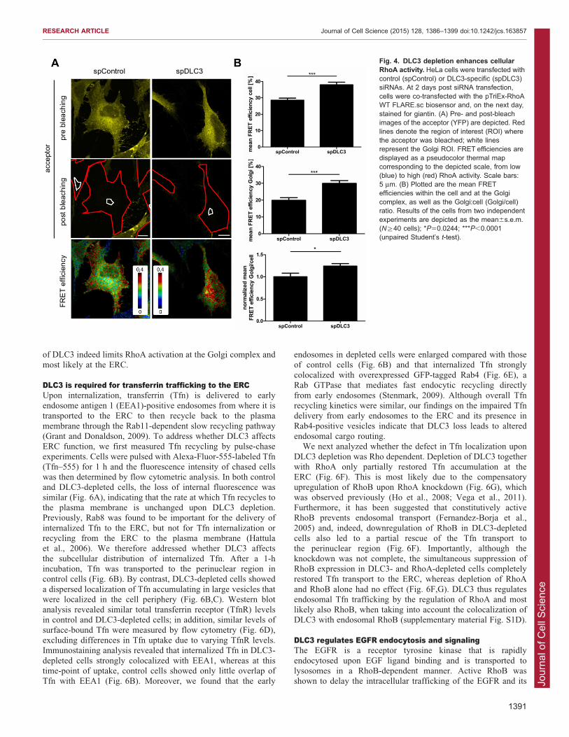
DLC3 depletion enhances perinuclear RhoA activityTo investigate the spatial aspects of RhoA GTPase regulationby endogenous DLC3, we measured RhoA activity in
DLC3-depleted HeLa cells using the RhoA biosensor, and wemeasured FRET over the whole cell by acceptor photobleaching.
The FRET efficiency was then plotted as pseudocolor imagesrepresenting high (red) and low (blue) RhoA activity (Fig. 4A). Incontrol cells, high RhoA activity was observed near the plasmamembrane (Fig. 4A) as reported previously (Pertz et al., 2006).
Upon DLC3 depletion, RhoA activity was enhanced by 30%within the cell (Fig. 4B), with especially high levels of activeRhoA in the perinuclear region (see pseudocolor image in
Fig. 4A). Considering the reports on the localization of RhoA tothe Golgi complex (Camera et al., 2003; Pertz et al., 2006) andthe observed Golgi fragmentation upon loss of DLC3, we
specifically measured RhoA activity at this compartment. TheGolgi complex was visualized by co-staining of the cis- andmedial-Golgi protein giantin using far-red-conjugated secondary
antibodies that do not interfere with the FRET fluorophores.Quantification of RhoA activation at the Golgi complex, aswell as the relative Golgi-localized RhoA activity, revealed asignificant increase in DLC3-depleted cells compared to that of
control cells (Fig. 4B). It is very likely that DLC3 loss also causesRhoA hyperactivation at the ERC; however, owing to thedispersion of the Rab8 compartment in DLC3-depleted cells,
this subcellular site cannot easily be discerned. Importantly,enhanced total and Golgi-localized RhoA activity was alsoobserved with an independent siRNA targeting DLC3
(supplementary material Fig. S2C,D), confirming the specificityof this effect.
A targeted DLC3 GAP domain rescues Golgi fragmentationAll DLC proteins possess a SAM domain, which potentiallymediates protein–protein or protein–lipid interactions. To addresswhether this domain was involved in DLC3 recruitment to the
Golgi–ERC interface, we expressed the isolated GFP-taggedDLC3a N-terminus (DLC3-SAM), which was found toaccumulate in the perinuclear region (Fig. 5A). Interestingly,
DLC3-SAM was not detected on Rab8-positive tubules, butextensively colocalized with TGN46 and giantin (Fig. 5A). Toprovide additional evidence that the SAM domain is necessary for
the subcellular localization of DLC3, we ectopically expressedGAP-inactive DLC3b, an isoform that is generated by analternative transcriptional start site and that naturally lacks theSAM domain (Durkin et al., 2007a). Indeed, DLC3b-K645E
showed a cytosolic distribution without any colocalization withthe Golgi complex or the Rab8 compartment (Fig. 5A; data notshown). The isolated SAM domain of DLC1 was fully cytosolic
(Fig. 5A), indicating that the DLC3 SAM domain specificallydetermines the localization of this particular DLC isoform.
To prove that Golgi fragmentation was a consequence of local
RhoA activation in DLC3-depleted cells, we designed a rescueexperiment in which the DLC3-SAM was used to target DLC3GAP activity to the Golgi complex. This was achieved by the
direct fusion of the DLC3 GAP domain with DLC3-SAM.Fig. 5B shows a representative image of a DLC3-depleted cellexpressing Golgi-localized, GFP-tagged SAM-GAP (Fig. 5B,marked with an asterisk). In contrast to untransfected cells,
DLC3-depleted cells expressing SAM-GAP regained a compactGolgi complex (Fig. 5B, compare * and .), which wasconfirmed by quantification of the Golgi compaction index in
these cells (Fig. 5C; see also supplementary material Fig. S2E forvalidation with an independent siRNA). No rescue of the Golgicompaction index was seen for DLC3-SAM alone (Fig. 5B,C).
These results provide strong evidence that the RhoGAP activity
Fig. 3. Both active RhoA and DLC3 depletion cause vesiculation of theRab8 recycling compartment and Golgi fragmentation. (A) HeLa cells wereeither mock transfected or transfected with a plasmid encoding GFP–RhoA-G14V (green). The next day, cells were stained for Rab8 or giantin (red) andnuclei were counterstained with DAPI (blue). Note the dispersed morphology ofthe Rab8 compartment and Golgi complex in cells expressing constitutivelyactive RhoA (*). (B–H) HeLa cells were transfected with control siRNA(spControl) or siRNAs specific for DLC3 (spDLC3) and p190RhoGAP-A(sip190RhoGAP), and analyzed 3 days later. (B) Cells were stained for Rab8(green) and nuclei were counterstained with DAPI (blue). (C) Cells were leftuntreated or treated with 0.1 mM CytD. The number of cells containing Rab8-positive tubules was counted and normalized to the total number of analyzedcells. Data show the mean6s.e.m. (n53, N§100 cells); ns, not significant;**P,0.001; ***P,0.0001 (one-way ANOVA followed by Tukey’s post-test).(D) Whole-cell lysates were analyzed by western blotting with anti-DLC3, anti-Rab8 and anti-tubulin antibodies. (E) Cells were stained for giantin (red), F-actinwas labeled with phalloidin (green) and nuclei were counterstained with DAPI(blue). (F) The degree of Golgi fragmentation was quantified using the Golgicompaction index (Bard et al., 2003); values were normalized to the control. Datashow the mean6s.e.m. (n53, N§40 cells); **P50.0053 (paired Student’s t-test).(G) Cells were stained for Rab8 and giantin (green), and nuclei werecounterstainedwith DAPI (blue). (H)Whole-cell lysateswere analyzed bywesternblotting with anti-p190RhoGAP and anti-tubulin antibodies. Scale bars: 10 mm.
RESEARCH ARTICLE Journal of Cell Science (2015) 128, 1386–1399 doi:10.1242/jcs.163857
1390
Jour
nal o
f Cel
l Sci
ence
of DLC3 indeed limits RhoA activation at the Golgi complex andmost likely at the ERC.
DLC3 is required for transferrin trafficking to the ERCUpon internalization, transferrin (Tfn) is delivered to earlyendosome antigen 1 (EEA1)-positive endosomes from where it istransported to the ERC to then recycle back to the plasma
membrane through the Rab11-dependent slow recycling pathway(Grant and Donaldson, 2009). To address whether DLC3 affectsERC function, we first measured Tfn recycling by pulse-chase
experiments. Cells were pulsed with Alexa-Fluor-555-labeled Tfn(Tfn–555) for 1 h and the fluorescence intensity of chased cellswas then determined by flow cytometric analysis. In both controland DLC3-depleted cells, the loss of internal fluorescence was
similar (Fig. 6A), indicating that the rate at which Tfn recycles tothe plasma membrane is unchanged upon DLC3 depletion.Previously, Rab8 was found to be important for the delivery of
internalized Tfn to the ERC, but not for Tfn internalization orrecycling from the ERC to the plasma membrane (Hattulaet al., 2006). We therefore addressed whether DLC3 affects
the subcellular distribution of internalized Tfn. After a 1-hincubation, Tfn was transported to the perinuclear region incontrol cells (Fig. 6B). By contrast, DLC3-depleted cells showed
a dispersed localization of Tfn accumulating in large vesicles thatwere localized in the cell periphery (Fig. 6B,C). Western blotanalysis revealed similar total transferrin receptor (TfnR) levelsin control and DLC3-depleted cells; in addition, similar levels of
surface-bound Tfn were measured by flow cytometry (Fig. 6D),excluding differences in Tfn uptake due to varying TfnR levels.Immunostaining analysis revealed that internalized Tfn in DLC3-
depleted cells strongly colocalized with EEA1, whereas at thistime-point of uptake, control cells showed only little overlap ofTfn with EEA1 (Fig. 6B). Moreover, we found that the early
endosomes in depleted cells were enlarged compared with thoseof control cells (Fig. 6B) and that internalized Tfn stronglycolocalized with overexpressed GFP-tagged Rab4 (Fig. 6E), aRab GTPase that mediates fast endocytic recycling directly
from early endosomes (Stenmark, 2009). Although overall Tfnrecycling kinetics were similar, our findings on the impaired Tfndelivery from early endosomes to the ERC and its presence in
Rab4-positive vesicles indicate that DLC3 loss leads to alteredendosomal cargo routing.
We next analyzed whether the defect in Tfn localization upon
DLC3 depletion was Rho dependent. Depletion of DLC3 togetherwith RhoA only partially restored Tfn accumulation at theERC (Fig. 6F). This is most likely due to the compensatoryupregulation of RhoB upon RhoA knockdown (Fig. 6G), which
was observed previously (Ho et al., 2008; Vega et al., 2011).Furthermore, it has been suggested that constitutively activeRhoB prevents endosomal transport (Fernandez-Borja et al.,
2005) and, indeed, downregulation of RhoB in DLC3-depletedcells also led to a partial rescue of the Tfn transport tothe perinuclear region (Fig. 6F). Importantly, although the
knockdown was not complete, the simultaneous suppression ofRhoB expression in DLC3- and RhoA-depleted cells completelyrestored Tfn transport to the ERC, whereas depletion of RhoA
and RhoB alone had no effect (Fig. 6F,G). DLC3 thus regulatesendosomal Tfn trafficking by the regulation of RhoA and mostlikely also RhoB, when taking into account the colocalization ofDLC3 with endosomal RhoB (supplementary material Fig. S1D).
DLC3 regulates EGFR endocytosis and signalingThe EGFR is a receptor tyrosine kinase that is rapidly
endocytosed upon EGF ligand binding and is transported tolysosomes in a RhoB-dependent manner. Active RhoB wasshown to delay the intracellular trafficking of the EGFR and its
Fig. 4. DLC3 depletion enhances cellularRhoA activity. HeLa cells were transfected withcontrol (spControl) or DLC3-specific (spDLC3)siRNAs. At 2 days post siRNA transfection,cells were co-transfected with the pTriEx-RhoAWT FLARE.sc biosensor and, on the next day,stained for giantin. (A) Pre- and post-bleachimages of the acceptor (YFP) are depicted. Redlines denote the region of interest (ROI) wherethe acceptor was bleached; white linesrepresent the Golgi ROI. FRET efficiencies aredisplayed as a pseudocolor thermal mapcorresponding to the depicted scale, from low(blue) to high (red) RhoA activity. Scale bars:5 mm. (B) Plotted are the mean FRETefficiencies within the cell and at the Golgicomplex, as well as the Golgi:cell (Golgi/cell)ratio. Results of the cells from two independentexperiments are depicted as the mean6s.e.m.(N§40 cells); *P50.0244; ***P,0.0001(unpaired Student’s t-test).
RESEARCH ARTICLE Journal of Cell Science (2015) 128, 1386–1399 doi:10.1242/jcs.163857
1391

Jour
nal o
f Cel
l Sci
ence
lysosomal degradation (Gampel et al., 1999). To analyze howEGFR trafficking might be affected by DLC3 downregulation andsubsequent Rho misregulation, we incubated cells with Alexa-
Fluor-555-labeled EGF (EGF–555) for different lengths of time.After 20 min, EGF-positive endosomes were primarily localizedin the perinuclear region in control cells, whereas, in
DLC3-depleted cells, EGF–555 accumulated in enlarged EEA1-positive endosomes in the cell periphery (Fig. 7A). Although thelevels of internalized EGF were comparable between control and
depleted cells, we found significantly more peripheral EGF–555localization upon DLC3 depletion (Fig. 7B). After 40 min, EGF–555 still failed to accumulate in the perinuclear region in
Fig. 5. DLC3-SAM localizes to Golgi membranes and the targeted DLC3 GAP domain rescues Golgi fragmentation in DLC3 knockdown cells. (A) HeLacells transiently expressing GFP-tagged DLC3-SAM, GFP-tagged DLC3b-K645E or mCherry-tagged DLC1-SAM (all depicted in green) were stained forRab8, TGN46 or giantin (red) as indicated. (B,C) HeLa cells were transfected with control (spControl) or DLC3-specific (spDLC3) siRNAs. At 2 days postsiRNA transfection, cells were transfected with vectors encoding the DLC3 GAP domain fused to DLC3-SAM (SAM-GAP) or DLC3-SAM alone (SAM).Cells were fixed and stained for giantin 24 h later. (B) Representative maximum projections of DLC3-depleted cells are shown. Note the morphologicaldifference of giantin (red) in untransfected cells (.) compared to SAM-GAP-expressing (green) cells (*). Nuclei were counterstained with DAPI (blue).Scale bars: 10 mm. (C) The Golgi compaction index was calculated for at least three independent experiments. Values were normalized to untransfectedcontrol cells and are depicted as the mean6s.e.m. (n53–6, N§50 cells); ns, not significant; *P,0.05; **P,0.01 (two-way ANOVA followed by Bonferronipost-test).
RESEARCH ARTICLE Journal of Cell Science (2015) 128, 1386–1399 doi:10.1242/jcs.163857
1392

Jour
nal o
f Cel
l Sci
ence
DLC3-depleted cells (Fig. 7A), indicating that EGF–555 wasnot delivered to later endosomal compartments. Indeed, after120 min, DLC3-depleted cells showed a significant pool ofremaining EGF–555, whereas the signal was strongly reduced in
control cells (Fig. 7C,D). This remaining EGF–555 pool did notcolocalize with the late endosomal and lysosomal marker LAMP1(Fig. 7C), excluding incomplete EGF–555 degradation due to
lysosomal dysfunction. To further investigate the identity of the
remaining EGF pool in DLC3-depleted cells, we performedimmunostaining analysis of different Rab recycling markers. At120 min post-internalization, EGF–555 in DLC3-depleted cellscolocalized with neither EEA1 nor Rab7 (supplementary material
Fig. S4A), but extensively overlapped with GFP-tagged Rab4(Fig. 7E). DLC3 depletion thus impairs EGFR lysosomaldegradation and, reminiscent of the TfnR, appears to cause
receptor re-routing to the Rab4-dependent recycling pathway.
Fig. 6. See next page for legend.
RESEARCH ARTICLE Journal of Cell Science (2015) 128, 1386–1399 doi:10.1242/jcs.163857
1393

Jour
nal o
f Cel
l Sci
ence
Because the receptor trafficking route determines the signalingresponse, we examined EGFR levels and activity byimmunoblotting cell lysates of EGF-stimulated HeLa cells.Basal EGFR levels of DLC3-depleted cells were comparable to
those of control cells (Fig. 8A; EGFR). Immediately after EGFstimulation, strong phosphorylation of EGFR at Tyr1068 wasdetected in both control and depleted cells (Fig. 8A; pEGFR).
After 15 min of stimulation, the level of EGFR was alreadydecreased in control cells, whereas even at late time points thereceptor levels persisted in DLC3-depleted cells (Fig. 8A, EGFR;
Fig. 8B for quantification). EGFR activation triggers downstreamsignaling, including the activation of the PI3K and MAPKpathways, resulting in the activation of AKT and ERK kinases,
respectively. Compared with control cells, the total levels of thesekinases were reduced in DLC3-depleted cells for unknownreasons (Fig. 8A; AKT and ERK). Nevertheless, correlatingwith the delayed EGFR degradation, phosphorylation and thus
activation of AKT was prolonged in DLC3-depleted cells(Fig. 8A; pAKT). In the breast cancer cell line MCF7,prolonged AKT and also ERK phosphorylation were observed,
indicating that this effect is not restricted to a particular cell line(supplementary material Fig. S4B; note that the levels of theEGFR itself are too low for detection by immunoblotting). Thus,
given its potential tumor suppressor function, the ability of DLC3to regulate EGFR degradation could be one mechanism by whichDLC3 loss contributes to cancer progression.
DISCUSSIONHere, we establish a novel role for the RhoGAP protein DLC3 inthe control of endocytic vesicle traffic. DLC3 is shown to localize
at the interface of the TGN and Rab8-positive recyclingcompartment where it regulates RhoA activity. In the absenceof DLC3, the structural integrity of the Golgi complex and
Rab8 compartment are compromised, most likely owing to theinduction of aberrant cytoskeletal rearrangements. Both the actinand microtubule cytoskeleton play important roles in the
maintenance of organelle morphology and positioning (Egea
et al., 2013; Yadav and Linstedt, 2011). Downstream of activeRhoA, the Rab8-positive ERC dispersed, which is in line with the
RhoA-mDia-induced Golgi fragmentation mediated by actinpolymerization, myosin-II-driven contractility and microtubule-dependent movement (Zilberman et al., 2011), highlighting thenecessity of tight control of Rho signaling. To the best of our
knowledge, our study identifies DLC3 as the first Rho-specificGAP protein in living cells that maintains the structure andfunction of the ERC and Golgi compartments.
The specific colocalization of DLC3 with Rab8 and theimpact of DLC3 expression on the formation of Rab8-positivetubules suggest a role for DLC3 in Rab8-dependent processes.
Rab8 and Rab11 are both found at the ERC where the twoGTPases control different steps in the recycling of Tfn. WhereasRab11 regulates Tfn recycling from the ERC to the plasma
membrane, Rab8 controls the delivery of Tfn to the perinuclearERC (Hattula et al., 2006; Roland et al., 2007; Ullrich et al.,1996). Our findings on the impaired Tfn delivery to the ERC inDLC3-depleted cells, in which Tfn accumulated in enlarged
EEA1-positive endosomes in the cell periphery, are inagreement with these differential Rab functions. Consideringthat the actin depolymerizing agent CytD enhances Rab8
tubulation, the right balance of actin polymerization anddepolymerization appears to be crucial for the biogenesis ofRab8-positive transport carriers. We therefore propose that
increased local Rho signaling in the absence of DLC3 triggersthe loss of Rab8 recycling tubules, causing a transport block atthe early endosomal stage. Because Rab8a/b, together with
Rab10 and Rab13, compose a subfamily of closely related Rabproteins with overlapping localizations and functions (Stenmark,2009), it is possible that DLC3 affects additional Rabs. Ourobservation on the compact appearance of both Rab8 and Rab11
in DLC3-WT-expressing cells might also suggest a role ofDLC3 in Rab11-dependent recycling processes.
The endosomal Tfn transport block in DLC3-depleted cells was
fully rescued by the simultaneous knockdown of RhoA andRhoB, demonstrating the involvement of Rho hyperactivation.The compensatory upregulation of RhoB in cells lacking RhoA
provides an explanation for why RhoA knockdown alone onlypartially restored Tfn transport in these cells. Considering thecolocalization of DLC3-K725E with endosomal RhoB, DLC3 islikely to possess RhoB-inhibitory activity too. Unfortunately,
biochemical pulldown assays failed to reveal the potential RhoBregulation by DLC3, making the development of a RhoBbiosensor necessary to address this question at the subcellular
level. Because retrograde and anterograde transport processes areintimately connected to ensure cellular homeostasis, any defectsat the level of endosomal recycling are likely to affect Golgi
integrity and vice versa. This is supported by our observation thatoverexpression of inactive Rab8a negatively affected Golgicompaction (data not shown). There are also hints for the direct
regulation of TGN function by Rho signaling, as myosin II andactin were found to play an important role in the fission of Rab6vesicles (Miserey-Lenkei et al., 2010). Furthermore, the actin-severing protein actin-depolymerizing factor (ADF)/cofilin and
its upstream regulator LIM kinase 1 were reported to regulatefission and cargo sorting at the TGN (von Blume et al., 2009;Salvarezza et al., 2009). Although TGN46 and Rab6 did not
accumulate on the tubular structures positive for GAP-inactiveDLC3, DLC3 might be still involved in controlling the formationof a specific subset of transport carriers from the TGN by local
Rho regulation.
Fig. 6. DLC3 depletion impairs Tfn transport to the ERC. (A–E) HeLacells were transfected with control (spControl) or DLC3-specific (spDLC3)siRNAs and analyzed 3 days later. (A) Serum-starved cells were incubatedwith Tfn–555 for 1 h (pulse) and then chased for the indicated times. Thedecrease in Tfn–555 fluorescence intensity was measured by flow cytometry,and two representative experiments are depicted as the mean6s.e.m.(B,C) Serum-starved cells were incubated with Tfn–555 (red) for 1 h andstained for EEA1 (green). Representative maximum projections are shownand imaging parameters were identical. Scale bars: 10 mm. (C) The numberof cells displaying Tfn–555 either at the perinuclear region or in the cellperiphery was counted and normalized to the total number of analyzed cells.Data show the mean6s.e.m. (n53, N§36 cells); **P,0.01; ***P,0.001(two-way ANOVA followed by Bonferroni post-test). (D) Left panel, whole-celllysates were analyzed by western blotting with anti-TfnR and anti-tubulinantibodies. Right panel, flow cytometric analysis of surface-bound Tfn.Unlabeled cells are shown filled in gray. (E) At 2 days post siRNAtransfection, cells were transfected with a vector encoding GFP–Rab4-WT(green). The next day, serum-starved cells were incubated with Tfn–555 (red)for 1 h. Scale bars: 10 mm. (F,G) HeLa cells were transfected with controlsiRNAs (spControl) or siRNAs specific for DLC3 (spDLC3), RhoA (spRhoA)and RhoB (spRhoB), and analyzed 3 days later. (F) Serum-starved cellswere incubated with Tfn–555 for 1 h. The percentage of cells displaying Tfn–555 at the perinuclear region was determined and normalized to the control.Data show the mean6s.e.m. (n53, N§50 cells); ns, not significant;*P,0.01; **P,0.001; ***P,0.0001 (one-way ANOVA followed by Tukey’spost-test). (G) Whole-cell lysates were analyzed by western blotting with anti-DLC3, anti-RhoA, anti-RhoB and anti-tubulin antibodies.
RESEARCH ARTICLE Journal of Cell Science (2015) 128, 1386–1399 doi:10.1242/jcs.163857
1394

Jour
nal o
f Cel
l Sci
ence
Targeted expression of the isolated DLC3 GAP domain wassufficient to restore Golgi compaction in DLC3-depleted cells,providing clear evidence for a GAP-dependent Rho regulatory
mechanism. Although the SAM domain of DLC1 and DLC3 arehighly conserved (Durkin et al., 2007a), the isolated SAM domainof DLC1 did not localize to the Golgi complex, but was fully
cytosolic. These differences support the idea that the DLC family
members exert isoform-specific functions associated withtheir distinct localizations, ultimately resulting in distinctspatiotemporal Rho activation patterns. Interestingly, DLC3-
SAM did not overlap with Rab8-positive structures, indicatingthat additional domains are required for the transport of DLC3itself from the Golgi membranes to the ERC. Considering the
localization at the Golgi complex, the ERC, endocytic vesicles
Fig. 7. DLC3 depletion impairsdegradation of activated EGFR.HeLa cells were transfected withcontrol (spControl) or DLC3-specific(spDLC3) siRNAs. (A,B) At 3 dayspost siRNA transfection, cells wereincubated with EGF–555 (red) for 20and 40 min, and stained for EEA1(green). Nuclei were counterstainedwith DAPI (blue). (B) The number ofcells displaying EGF–555 in the cellperiphery after a 20-min incubationwas counted and normalized to thetotal number of analyzed cells. Datashow the mean6s.e.m. (n53, N550cells); **P50.0013 (paired Student’s t-test). (C,D) At 3 days post siRNAtransfection, cells were incubated withEGF–555 (red) for 120 min andstained for Lamp1 (green). Nucleiwere counterstained with DAPI (blue).(D) Quantification of the meanfluorescence intensity of EGF–555per cell after incubation for 120 min.Data show the mean6s.e.m. (n53,N550 cells); **P50.0066 (pairedStudent’s t-test). (E) At 2 days postsiRNA transfection, cells weretransfected with a vector encodingGFP–Rab4-WT (green). The next day,cells were incubated with EGF–555(red) for 120 min. Note theoverlapping distribution of EGF–555and GFP–Rab4 (.).(A,C,E) Representative maximumprojections are shown and imagingparameters were identical. Scalebars: 10 mm.
RESEARCH ARTICLE Journal of Cell Science (2015) 128, 1386–1399 doi:10.1242/jcs.163857
1395

Jour
nal o
f Cel
l Sci
ence
and cell–cell contacts (Holeiter et al., 2012), DLC3 appears to
dynamically associate with different cellular membranes, where itmight exert GAP-dependent and perhaps also GAP-independentfunctions.
We further provide evidence that DLC3 is required for the
proper lysosomal degradation of the EGFR. In the absence ofDLC3, the EGFR accumulated in Rab4-positive vesicles.Similarly, in DLC3-depleted cells, internalized Tfn was found to
colocalize with Rab4, which is responsible for the fast recycling ofcargo. This is in line with the observation that disturbed ERCintegrity can lead to missorting of endocytosed Tfn (Hattula et al.,
2006; Horgan et al., 2007; Naslavsky et al., 2006). In the case of theEGFR, disruption of the ERC by overexpression of the clathrinadaptor protein Eps15S (a short form of Eps15) caused the
accumulation of the EGFR in early endosomes and potentiatedreceptor recycling (Chi et al., 2011). Although receptorinternalization is a mechanism by which receptor signaling isterminated, it is now widely accepted that endosomes constitute
active signaling platforms (Platta and Stenmark, 2011; Tomaset al., 2014), and, indeed, endosomal accumulation of EGF-stimulated EGFR in DLC3-depleted cells was associated with
prolonged AKT activation. The selective activation of AKT mightbe mediated by the Rab5 effector protein APPL1 that regulatesAKT activation from endosomes (Schenck et al., 2008).
It is tempting to speculate that the regulation of the turnoverof membrane receptors and membrane-proximal signalingmolecules by DLC3 might not be restricted to the EGFR. Wepreviously reported that, in MCF7 and MCF10A breast epithelial
cells, DLC3 predominantly localizes to cell–cell contacts. In theabsence of DLC3, the adherens junction proteins E-cadherin andb-catenin were mislocalized, which was associated with enhanced
migration and increased cell disaggregation (Holeiter et al.,2012). Given its novel function in membrane transport, DLC3might also be involved in the regulation of the trafficking of
adherens junction proteins. Intriguingly, Rab8 was found to
regulate E-cadherin transport by binding to the JRAB (alsoknown as MICAL-L2) effector protein (Yamamura et al., 2008).DLC3 might thus possess a more global role in membranetrafficking through the integration of Rho and Rab GTPase
signaling. Our future studies will focus on defining the molecularfactors that regulate DLC3 activity and localization. In particular,it will be interesting to identify protein and/or lipid interactors of
DLC3 that recruit the protein to the different subcellularcompartments and to delineate in more detail the downstreameffector pathways that govern its function. The increased
understanding of DLC3 biology will also shed light onto itspotential function in the suppression of the different steps ofneoplastic cell transformation.
MATERIALS AND METHODSCell cultureHeLa cells (ATCC; Manassas, VA) and MCF7 cells (kindly provided
by Cornelius Knabbe; Institute of Clinical Pharmacology, Stuttgart,
Germany) were grown and maintained in RPMI 1640 (Invitrogen;
Carlsbad, CA) supplemented with 10% FCS (PAA Laboratories; Colbe,
Germany) at 37 C and 5% CO2.
Antibodies and reagentsThe antibodies used were as follows: monoclonal mouse anti-GM130,
monoclonal mouse anti-p230, monoclonal mouse anti-paxillin and
monoclonal mouse anti-p190RhoGAP antibodies from BD
Transduction Laboratories (Heidelberg, Germany); polyclonal rabbit
anti-giantin (ab24586) antibodies from Abcam (Cambridge, UK);
monoclonal rabbit anti-Rab8 (D22D8), polyclonal rabbit anti-Rab7,
polyclonal rabbit anti-Rab6 (D37C7), polyclonal rabbit anti-EEA1,
monoclonal rabbit anti-pAKT (Thr308; C31E5E), monoclonal mouse
anti-AKT (pan; 40D4), monoclonal rabbit anti-pEGFR (Tyr1068; D7A5),
polyclonal rabbit anti-pERK1/2 (Thr202/Tyr204), monoclonal mouse
anti-ERK1/2 (3A7) and polyclonal rabbit anti-RhoB antibodies from Cell
Signaling (Cambridge, UK); monoclonal mouse anti-TfnR antibodies
Fig. 8. DLC3 depletion prolongs EGFR signaling. HeLa cells were transfected with control (spControl) or DLC3-specific (spDLC3) siRNAs. At 2 days postsiRNA transfection, cells were serum starved overnight and, prior to lysis, stimulated with 10 ng/ml EGF for the indicated times. (A) Whole-cell lysates wereanalyzed by western blotting with anti-pEGFR (Tyr1068), anti-EGFR, anti-pAKT (Thr308), anti-AKT, anti-pERK (Thr202/Tyr204), anti-ERK and anti-tubulinantibodies. (B) EGFR levels from two independent experiments including the blot shown in A were quantified and normalized to tubulin. Data showthe mean6s.e.m.
RESEARCH ARTICLE Journal of Cell Science (2015) 128, 1386–1399 doi:10.1242/jcs.163857
1396

Jour
nal o
f Cel
l Sci
ence
from Invitrogen; monoclonal mouse anti-Lamp1 antibodies from
Developmental Studies Hybridoma Bank (University of Iowa; IA);
sheep serum antibodies recognizing TGN46 (AHP500) from BioRad
Laboratories (Hercules, CA); monoclonal mouse anti-a-tubulin and
monoclonal anti-FLAG M2 antibodies from Sigma-Aldrich (St Louis,
MO); monoclonal mouse anti-DLC3 (E-2), monoclonal mouse anti-RhoA
(26C4) and polyclonal rabbit anti-EGFR (1005) antibodies from Santa
Cruz Biotechnology (Dallas, TX). Horseradish peroxidase (HRP)-labeled
secondary anti-mouse- and anti-rabbit-IgG antibodies were obtained
from GE Healthcare (Piscataway, NJ), Alexa-FluorH-labeled secondary
IgG antibodies and Alexa-FluorH-labeled phalloidin were obtained
from Invitrogen. Alexa-FluorH-labeled phalloidin was obtained from
Invitrogen. DAPI was obtained from Sigma-Aldrich. CytD was obtained
from Enzo Life Sciences (Lorrach, Germany) and EGF was from R&D
Systems (Minneapolis, MN).
DNA constructs and transfectionThe expression vectors pEGFP-C1-DLC3a-WT, pEGFP-C1-DLC3a-
K725E, pEGFP-C1-DLC3b-K645E and pmCherry-C1-DLC3a-K725E
have been described previously (Erlmann et al., 2009; Holeiter et al.,
2012). pCR.V62-Met-FLAG-DLC3a-WT and K725E were generated by
EcoRI excision of the DLC3 cDNAs from pEGFP vectors (Erlmann
et al., 2009; Holeiter et al., 2012) and cloning into the pCR.V62-Met-
FLAG vector. pEGFP-C1-DLC3-SAM (encoding the first 80 amino acids
in DLC3a) was generated by PCR amplification using pEGFP-C1-
DLC3a as a template and the following forward and reverse primers: 59-
CCGGAATTCTCCTCTGCTGGACGTTTTCTG-39 and 59-CCGGAAT-
TCTCACAGCCTCCTACACAGGGC-39. The PCR product was cloned
into the pEGFP-C1 vector by EcoRI restriction. pmCherry-C1-DLC1-
SAM was subcloned from pEGFP-C1-DLC1-SAM (Heering et al., 2009)
by BglII/BamHI and SalI/XhoI restriction into the pmCherry-C1 vector.
pEGFP-C1-DLC3-SAM-GAP was generated in two steps. First, the
sequence encoding amino acids 1–80 was amplified by PCR without
integration of a stop codon using pEGFP-C1-DLC3a as a template and
the following forward and reverse primers: 59-CCGGAATTCTCCT-
CTGCTGGACGTTTTCTG-39 and 59-GCAGTCGACCAGGCCTCCT-
ACACAGGGC-39. The PCR product was cloned into the pEGFP-C1
vector by EcoRI and SalI restriction. Next, the GAP domain was
amplified by PCR using pEGFP-C1-DLC3a as a template and the
following forward and reverse primers: 59-GCAGTCGACAAGGGCTC-
ACTGCTGCGGC-39 and 59-GCAGTCGACTCACTGACGCAGCTCA-
GCCAGG-39. This PCR product was cloned into the pEGFP-C1-DLC3-
SAM vector lacking the stop codon by SalI restriction. To obtain pEGFP-
C1-RhoA-G14V, the RhoA-WT expression cassette was subcloned from
pcDNA3.1-HA-RhoA-WT by BglII/BamHI and EcoRI restriction into the
pEGFP-C1 vector. Next, the G14V mutation was introduced by site-
directed PCR mutagenesis using the following forward primer: 59-
GTGATTGTTGGTGATGTAGCCTGTGGAAAGACA-39. All amplified
cDNAs were verified by sequencing. Oligonucleotides were purchased
from Eurofins MWG Operon (Ebersberg, Germany). The RhoA-WT
biosensor pTriEx-RhoA FLARE.sc was purchased from Addgene
(Addgene plasmid 12150; Pertz et al., 2006). The pECFP-RhoB/Endo
vector was purchased from Clontech (Clontech plasmid #6934-1). pEGFP-
Rab11 and pEGFP-Rab4 vectors were kindly provided by Lucas
Pelkman (University of Zurich, Switzerland) and Hesso Farhan
(University of Konstanz, Germany), respectively. Vector transfections
were carried out with TransIT-HeLaMONSTERH (Mirus Bio; Madison,
WI) and TurbofectH (Thermo Scientific; Rockford, IL), according to the
manufacturer’s instructions.
RNA interferenceFor RNA interference, cells were reverse transfected with siRNAs using
LipofectamineH RNAiMAX (Invitrogen) according to the manufacturer’s
instructions. As a negative control (termed spControl), ON-TARGETplusHnon-targeting control pool D-001810-10 from Dharmacon (Lafayette,
CO) was used. Two independent DLC3-specific siRNAs were used,
termed spDLC3 and siDLC3. spDLC3 corresponds to the siGENOME
SMARTpool human STARD8 M-010254 from Dharmacon. siDLC3
corresponds to a single duplex with the sense sequence: 59-UCUC-
UGAGGCGGAAGGAAA-39 (Eurofins MWG Operon). sip190RhoGAP
corresponds to a single duplex with the sense sequence 59-GGAU-
UGUGUGGAAUGUAAG-39 (Eurofins MWG Operon). spRhoA and
spRhoB correspond to the ON-TARGETplusH SMARTpool human
RhoA L-003860 and ON-TARGETplusH SMARTpool human RhoB L-
008395 from Dharmacon, respectively.
Cell lysis, SDS-PAGE and western blottingCells were washed once with PBS and lysed for 10 min with ice-cold
RIPA buffer [50 mM Tris-HCl pH 7.5, 150 mM sodium chloride, 1% (v/
v) NP-40, 0.25% (v/v) sodium deoxycholate, 0.1% (v/v) SDS, 1 mM
EDTA, 1 mM sodium orthovanadate, 10 mM sodium fluoride, 0.5 mM
PMSF and 20 mM b-glycerophosphate plus protease inhibitors]. Whole-
cell lysates were clarified by centrifugation for 10 min at 15,700 g and
4 C. Equal amounts of protein were loaded on 8% or 15%
polyacrylamide gels and transferred to polyvinylidene difluoride
membranes (Roth; Karlsruhe, Germany). For detection of RhoA and
RhoB, lysates were run on NuPageH NovexH 4–12% Bis-Tris gels
(Invitrogen) and blotted onto nitrocellulose membranes using the iBlotHdevice (Invitrogen). Membranes were blocked for 30 min with 0.5%
(v/v) blocking reagent (Roche Diagnostics; Mannheim, Germany) in
PBS containing 0.05% (v/v) Tween-20 and 0.01% (v/v) thimerosal.
Membranes were incubated with primary antibodies overnight at 4 C,
followed by 1 h incubation with HRP-conjugated secondary antibodies at
room temperature. Proteins were visualized using an enhanced
chemiluminescence detection system (Thermo Scientific; Rockford,
IL). Quantification of ECL films was performed by densitometric
analysis using ImageJ software (NIH; Bethesda, MD).
Immunofluorescence staining and confocal microscopyCells grown on glass coverslips coated with 10 mg/ml collagen R (Serva;
Heidelberg, Germany) were fixed for 15 min with 4% (v/v)
paraformaldehyde. After washes in PBS, cells were incubated for
15 min with 150 mM glycine in PBS and permeabilized for 5 min with
0.1% (v/v) Triton X-100 in PBS. Blocking was performed with 5% (v/v)
goat serum (Invitrogen) in PBS containing 0.1% (v/v) Tween-20. Fixed
cells were incubated with primary antibodies diluted in blocking buffer
for 2 h at room temperature. Following three washing steps with PBS,
cells were incubated with Alexa-FluorH-(488, 546 or 633)-labeled
secondary antibodies in blocking buffer for 1 h at room temperature.
Nuclei were counterstained with DAPI and coverslips were mounted in
Fluoromount-GH (SouthernBiotech; Birmingham, AL). All samples were
analyzed at room temperature using a confocal laser scanning microscope
(LSM 710, Carl Zeiss; Oberkochen, Germany) equipped with a Plan
Apochromat 636/1.40 DIC M27 (Carl Zeiss) oil-immersion objective.
Linear adjustments to brightness, contrast and maximum intensity
projections were made using the ZEN software (Carl Zeiss). For
quantification of mean fluorescence intensities, images were acquired
with the same confocal settings and analyzed using the ImageJ software
(NIH; Bethesda, MD). Fluorescence intensities along a line of interest
were measured using the ZEN software. For colocalization and line-scan
experiments, all images were acquired with identical dimensions in x and
y directions and the same pinhole settings (1 AU in the red channel). The
fraction of Rab8 staining overlapping with DLC3-K725E signals was
analyzed using the ImageJ plugin JACoP, measuring the Manders’ M2
coefficient (Bolte and Cordelieres, 2006).
FRET analysisFor FRET measurements, cells were transfected overnight with the
RhoA-WT biosensor pTriEx-RhoA FLARE.sc and pCR.V62-Met-FLAG,
pCR.V62-Met-FLAG-DLC3a-WT or pCR.V62-Met-FLAG-DLC3a-K725E.
The next day, cells were fixed and immunostained with anti-Flag M2 and
Alexa-FluorH-633-labeled secondary anti-mouse antibodies. In the case of
siRNA transfection, control and DLC3-depleted cells were co-transfected
with the RhoA-WT biosensor at 2 days post siRNA transfection and,
the next day, cells were fixed and immunostained for the cis- and medial-
Golgi marker giantin using an Alexa-FluorH-633-labeled secondary
RESEARCH ARTICLE Journal of Cell Science (2015) 128, 1386–1399 doi:10.1242/jcs.163857
1397

Jour
nal o
f Cel
l Sci
ence
anti-mouse-IgG antibody. The staining was performed according to the
immunofluorescence staining protocol described above and samples were
then mounted in MowiolH (Polysciences; Warrington, PA) mounting
solution. FRET efficiencies were determined using the acceptor
photobleaching method. CFP was excited with a diode UV laser at
405 nm, and emission was detected in the spectral window 454–515 nm.
YFP was excited with the 514-nm laser line of an argon laser and emission
was detected from 515–621 nm. Donor and acceptor images were acquired
pre- and post-bleaching. Whole cells were bleached for YFP with the 514-
nm argon laser line (80% intensity, 20 iterations, pixel dwell 50.4 ms). The
FRET efficiency was calculated from the increase of the donor intensity
(CFP) after acceptor bleaching using the FRET module of ZEN 2009
software (Carl Zeiss). FRET efficiency images were generated with a
MATLAB script (developed by Dr Felix Neugart, University Stuttgart,
Germany) that allows background suppression and visualization of the
FRET efficiency at the same time by using a two-dimensional look-up
table (total fluorescence intensity coded by pixel brightness, FRET
efficiency coded by color). To determine the Golgi-specific increase in
RhoA activity, the mean FRET efficiency of the Golgi region was
normalized to the mean FRET efficiency of the whole cell. Alexa-FluorH-
633-labeled giantin was excited with the 633-nm helium-neon laser line
and its emission was detected from 621–735 nm.
Tfn recycling and surface labelingFor Tfn recycling, cells were starved in serum-free medium prior to
incubation with 10 mg/ml Alexa-FluorH-555-labeled Tfn (Invitrogen) for
1 h. Cells were then rinsed for 45 s with acidic buffer (0.5% acetic acid,
0.5 M sodium chloride, pH 3.0) to remove surface-bound Tfn, followed
by washes with PBS and medium containing 10% FCS. During the chase
phase, cells were incubated in medium containing 10% FCS for the
indicated times. Next, cells were washed with ice-cold PBS and
trypsinized for 3 min at 37 C. Finally, cells were washed with ice-cold
FACS buffer (PBS+2% FCS+0.1% sodium azide] and analyzed with the
EPICS FC500 flow cytometer (Beckman Coulter; Krefeld, Germany).
Tfn–555 intensity was measured in the FL-2 channel; unlabeled cells
were used for gating. Post-acquisition data analysis was performed using
FlowJo software (Tree Star; Ashland, OR). To measure surface-bound
Tfn, cells were harvested by trypsinization and washed once with ice-
cold FACS buffer. After pre-chilling on ice for 10 min, cells were
incubated with 10 mg/ml Alexa-FluorH-555-labeled Tfn for 45 min. Cells
were then washed with ice-cold FACS buffer and processed for flow
cytometry as described above.
Tfn uptake assayMicroscopic analysis of Tfn uptake was performed as described
previously (Hattula et al., 2006). Briefly, cells were washed with
serum-free medium and starved for 1 h. Cells were then incubated with
10 mg/ml Alexa-FluorH-555-labeled Tfn for 1 h. To remove surface-
bound Tfn, cells were rinsed for 45 s with acidic buffer (0.5% acetic acid,
0.5 M sodium chloride, pH 3.0), washed three times with PBS, fixed and
processed for immunofluorescence staining.
EGFR trafficking and signaling assaysTo analyze EGFR endocytosis, cells were incubated for the indicated
times with 200 ng/ml Alexa-FluorH-555-labeled EGF (Invitrogen),
washed with PBS, fixed and processed for immunofluorescence
staining. For EGFR signaling experiments, cells were starved overnight
in medium supplemented with 0.5% FCS. Cells were then stimulated
with either 10 ng/ml EGF (HeLa) or 50 ng/ml EGF (MCF7) for the
indicated times. Next, cells were lysed and processed for western
blotting.
AcknowledgementsWe thank Prof. Dr. Klaus Pfizenmaier (University of Stuttgart, Germany) forcritically reviewing the manuscript. We gratefully acknowledge Dr. Felix Neugart(University of Stuttgart, Germany) for developing the MATLAB script. We thankDrs. Lucas Pelkman, Hesso Farhan and Cornelius Knabbe for providing plasmidsand cells.
Competing interestsThe authors declare no competing or financial interests.
Author contributionsA.C.B., J.H. and S.A.E. performed the experiments and analyzed data; S.S.assisted with cloning. A.H. participated in the experimental design and dataanalysis. M.A.O. conceived the study and wrote the manuscript together withA.C.B.
FundingThis work was funded by the Deutsche Krebshilfe [grant number 109033]. M.A.O.is funded by the Heisenberg program of the Deutsche Forschungsgemeinschaft.
Supplementary materialSupplementary material available online athttp://jcs.biologists.org/lookup/suppl/doi:10.1242/jcs.163857/-/DC1
ReferencesAdamson, P., Paterson, H. F. and Hall, A. (1992). Intracellular localization of theP21rho proteins. J. Cell Biol. 119, 617-627.
Arthur, W. T. and Burridge, K. (2001). RhoA inactivation by p190RhoGAPregulates cell spreading and migration by promoting membrane protrusion andpolarity. Mol. Biol. Cell 12, 2711-2720.
Bard, F., Mazelin, L., Pechoux-Longin, C., Malhotra, V. and Jurdic, P.(2003). Src regulates Golgi structure and KDEL receptor-dependent retrogradetransport to the endoplasmic reticulum. J. Biol. Chem. 278, 46601-46606.
Bolte, S. and Cordelieres, F. P. (2006). A guided tour into subcellularcolocalization analysis in light microscopy. J. Microsc. 224, 213-232.
Bos, J. L., Rehmann, H. and Wittinghofer, A. (2007). GEFs and GAPs: criticalelements in the control of small G proteins. Cell 129, 865-877.
Camera, P., da Silva, J. S., Griffiths, G., Giuffrida, M. G., Ferrara, L., Schubert,V., Imarisio, S., Silengo, L., Dotti, C. G. and Di Cunto, F. (2003). Citron-N is aneuronal Rho-associated protein involved in Golgi organization through actincytoskeleton regulation. Nat. Cell Biol. 5, 1071-1078.
Chi, S., Cao, H., Wang, Y. and McNiven, M. A. (2011). Recycling of the epidermalgrowth factor receptor is mediated by a novel form of the clathrin adaptor proteinEps15. J. Biol. Chem. 286, 35196-35208.
Durkin, M. E., Yuan, B.-Z., Zhou, X., Zimonjic, D. B., Lowy, D. R.,Thorgeirsson, S. S. and Popescu, N. C. (2007a). DLC-1:a Rho GTPase-activating protein and tumour suppressor. J. Cell. Mol. Med. 11, 1185-1207.
Durkin, M. E., Ullmannova, V., Guan, M. and Popescu, N. C. (2007b). Deleted inliver cancer 3 (DLC-3), a novel Rho GTPase-activating protein, isdownregulated in cancer and inhibits tumor cell growth. Oncogene 26, 4580-4589.
Egea, G., Serra-Peinado, C., Salcedo-Sicilia, L. and Gutierrez-Martınez, E.(2013). Actin acting at the Golgi. Histochem. Cell Biol. 140, 347-360.
Erlmann, P., Schmid, S., Horenkamp, F. A., Geyer, M., Pomorski, T. G. andOlayioye, M. A. (2009). DLC1 activation requires lipid interaction through apolybasic region preceding the RhoGAP domain. Mol. Biol. Cell 20, 4400-4411.
Fernandez-Borja, M., Janssen, L., Verwoerd, D., Hordijk, P. and Neefjes, J.(2005). RhoB regulates endosome transport by promoting actin assembly onendosomal membranes through Dia1. J. Cell Sci. 118, 2661-2670.
Gampel, A., Parker, P. J. and Mellor, H. (1999). Regulation of epidermal growthfactor receptor traffic by the small GTPase rhoB. Curr. Biol. 9, 955-958.
Goud, B., Zahraoui, A., Tavitian, A. and Saraste, J. (1990). Small GTP-bindingprotein associated with Golgi cisternae. Nature 345, 553-556.
Grant, B. D. and Donaldson, J. G. (2009). Pathways and mechanisms ofendocytic recycling. Nat. Rev. Mol. Cell Biol. 10, 597-608.
Harris, K. P. and Tepass, U. (2010). Cdc42 and vesicle trafficking in polarizedcells. Traffic 11, 1272-1279.
Hattula, K., Furuhjelm, J., Tikkanen, J., Tanhuanpaa, K., Laakkonen, P. andPeranen, J. (2006). Characterization of the Rab8-specific membrane trafficroute linked to protrusion formation. J. Cell Sci. 119, 4866-4877.
Heering, J., Erlmann, P. and Olayioye, M. A. (2009). Simultaneous loss of theDLC1 and PTEN tumor suppressors enhances breast cancer cell migration.Exp. Cell Res. 315, 2505-2514.
Ho, T. T. G., Merajver, S. D., Lapiere, C. M., Nusgens, B. V. and Deroanne, C. F.(2008). RhoA-GDP regulates RhoB protein stability. Potential involvement ofRhoGDIalpha. J. Biol. Chem. 283, 21588-21598.
Holeiter, G., Bischoff, A., Braun, A. C., Huck, B., Erlmann, P., Schmid, S., Herr,R., Brummer, T. and Olayioye, M. A. (2012). The RhoGAP protein Deleted inLiver Cancer 3 (DLC3) is essential for adherens junctions integrity. Oncogenesis1, e13.
Horgan, C. P., Oleksy, A., Zhdanov, A. V., Lall, P. Y., White, I. J., Khan, A. R.,Futter, C. E., McCaffrey, J. G. and McCaffrey, M. W. (2007). Rab11-FIP3 iscritical for the structural integrity of the endosomal recycling compartment.Traffic 8, 414-430.
Jaffe, A. B. and Hall, A. (2005). Rho GTPases: biochemistry and biology. Annu.Rev. Cell Dev. Biol. 21, 247-269.
Johnson, J. L., Monfregola, J., Napolitano, G., Kiosses, W. B. and Catz, S. D.(2012). Vesicular trafficking through cortical actin during exocytosis is regulated
RESEARCH ARTICLE Journal of Cell Science (2015) 128, 1386–1399 doi:10.1242/jcs.163857
1398

Jour
nal o
f Cel
l Sci
ence
by the Rab27a effector JFC1/Slp1 and the RhoA-GTPase-activating proteinGem-interacting protein. Mol. Biol. Cell 23, 1902-1916.
Kawai, K., Kiyota, M., Seike, J., Deki, Y. and Yagisawa, H. (2007). START-GAP3/DLC3 is a GAP for RhoA and Cdc42 and is localized in focal adhesionsregulating cell morphology. Biochem. Biophys. Res. Commun. 364, 783-789.
Lamaze, C., Chuang, T. H., Terlecky, L. J., Bokoch, G. M. and Schmid, S. L.(1996). Regulation of receptor-mediated endocytosis by Rho and Rac. Nature382, 177-179.
Liu, S. and Storrie, B. (2012). Are Rab proteins the link between Golgiorganization and membrane trafficking? Cell. Mol. Life Sci. 69, 4093-4106.
Luna, A., Matas, O. B., Martınez-Menarguez, J. A., Mato, E., Duran, J. M.,Ballesta, J., Way, M. and Egea, G. (2002). Regulation of protein transport fromthe Golgi complex to the endoplasmic reticulum by CDC42 and N-WASP. Mol.Biol. Cell 13, 866-879.
Miserey-Lenkei, S., Chalancon, G., Bardin, S., Formstecher, E., Goud, B. andEchard, A. (2010). Rab and actomyosin-dependent fission of transport vesiclesat the Golgi complex. Nat. Cell Biol. 12, 645-654.
Musch, A., Cohen, D., Kreitzer, G. and Rodriguez-Boulan, E. (2001). cdc42regulates the exit of apical and basolateral proteins from the trans-Golginetwork. EMBO J. 20, 2171-2179.
Nalbant, P., Hodgson, L., Kraynov, V., Toutchkine, A. and Hahn, K. M. (2004).Activation of endogenous Cdc42 visualized in living cells. Science 305, 1615-1619.
Naslavsky, N., Rahajeng, J., Sharma, M., Jovic, M. and Caplan, S. (2006).Interactions between EHD proteins and Rab11-FIP2: a role for EHD3 in earlyendosomal transport. Mol. Biol. Cell 17, 163-177.
Pathak, R., Delorme-Walker, V. D., Howell, M. C., Anselmo, A. N., White, M. A.,Bokoch, G. M. and Dermardirossian, C. (2012). The microtubule-associatedRho activating factor GEF-H1 interacts with exocyst complex to regulate vesicletraffic. Dev. Cell 23, 397-411.
Peranen, J. (2011). Rab8 GTPase as a regulator of cell shape. Cytoskeleton(Hoboken) 68, 527-539.
Pertz, O., Hodgson, L., Klemke, R. L. and Hahn, K. M. (2006). Spatiotemporaldynamics of RhoA activity in migrating cells. Nature 440, 1069-1072.
Platta, H. W. and Stenmark, H. (2011). Endocytosis and signaling. Curr. Opin.Cell Biol. 23, 393-403.
Qualmann, B. and Mellor, H. (2003). Regulation of endocytic traffic by RhoGTPases. Biochem. J. 371, 233-241.
Robertson, D., Paterson, H. F., Adamson, P., Hall, A. and Monaghan, P. (1995).Ultrastructural localization of ras-related proteins using epitope-taggedplasmids. J. Histochem. Cytochem. 43, 471-480.
Roland, J. T., Kenworthy, A. K., Peranen, J., Caplan, S. and Goldenring, J. R.(2007). Myosin Vb interacts with Rab8a on a tubular network containing EHD1and EHD3. Mol. Biol. Cell 18, 2828-2837.
Salvarezza, S. B., Deborde, S., Schreiner, R., Campagne, F., Kessels, M. M.,Qualmann, B., Caceres, A., Kreitzer, G. and Rodriguez-Boulan, E. (2009).LIM kinase 1 and cofilin regulate actin filament population required for dynamin-dependent apical carrier fission from the trans-Golgi network. Mol. Biol. Cell 20,438-451.
Schenck, A., Goto-Silva, L., Collinet, C., Rhinn, M., Giner, A., Habermann, B.,Brand, M. and Zerial, M. (2008). The endosomal protein Appl1 mediates Aktsubstrate specificity and cell survival in vertebrate development. Cell 133, 486-497.
Settleman, J., Albright, C. F., Foster, L. C. and Weinberg, R. A. (1992).Association between GTPase activators for Rho and Ras families. Nature 359,153-154.
Sirokmany, G., Szidonya, L., Kaldi, K., Gaborik, Z., Ligeti, E. and Geiszt, M.(2006). Sec14 homology domain targets p50RhoGAP to endosomes andprovides a link between Rab and Rho GTPases. J. Biol. Chem. 281, 6096-6105.
Stenmark, H. (2009). Rab GTPases as coordinators of vesicle traffic. Nat. Rev.Mol. Cell Biol. 10, 513-525.
Symons, M. and Rusk, N. (2003). Control of vesicular trafficking by RhoGTPases. Curr. Biol. 13, R409-R418.
Tomas, A., Futter, C. E. and Eden, E. R. (2014). EGF receptor trafficking:consequences for signaling and cancer. Trends Cell Biol. 24, 26-34.
Ullrich, O., Reinsch, S., Urbe, S., Zerial, M. and Parton, R. G. (1996). Rab11regulates recycling through the pericentriolar recycling endosome. J. Cell Biol.135, 913-924.
Vega, F. M., Fruhwirth, G., Ng, T. and Ridley, A. J. (2011). RhoA and RhoC havedistinct roles in migration and invasion by acting through different targets. J. CellBiol. 193, 655-665.
Vigil, D., Cherfils, J., Rossman, K. L. and Der, C. J. (2010). Ras superfamilyGEFs and GAPs: validated and tractable targets for cancer therapy? Nat. Rev.Cancer 10, 842-857.
von Blume, J., Duran, J. M., Forlanelli, E., Alleaume, A.-M., Egorov, M.,Polishchuk, R., Molina, H. and Malhotra, V. (2009). Actin remodeling by ADF/cofilin is required for cargo sorting at the trans-Golgi network. J. Cell Biol. 187,1055-1069.
Wakana, Y., van Galen, J., Meissner, F., Scarpa, M., Polishchuk, R. S., Mann,M. and Malhotra, V. (2012). A new class of carriers that transport selectivecargo from the trans Golgi network to the cell surface. EMBO J. 31, 3976-3990.
Wildenberg, G. A., Dohn, M. R., Carnahan, R. H., Davis, M. A., Lobdell, N. A.,Settleman, J. and Reynolds, A. B. (2006). p120-catenin and p190RhoGAPregulate cell-cell adhesion by coordinating antagonism between Rac and Rho.Cell 127, 1027-1039.
Xue, W., Krasnitz, A., Lucito, R., Sordella, R., Vanaelst, L., Cordon-Cardo, C.,Singer, S., Kuehnel, F., Wigler, M., Powers, S. et al. (2008). DLC1 is achromosome 8p tumor suppressor whose loss promotes hepatocellularcarcinoma. Genes Dev. 22, 1439-1444.
Yadav, S. and Linstedt, A. D. (2011). Golgi positioning. Cold Spring Harb.Perspect. Biol. 3, a005322.
Yamamura, R., Nishimura, N., Nakatsuji, H., Arase, S. and Sasaki, T. (2008).The interaction of JRAB/MICAL-L2 with Rab8 and Rab13 coordinates theassembly of tight junctions and adherens junctions. Mol. Biol. Cell 19, 971-983.
Zilberman, Y., Alieva, N. O., Miserey-Lenkei, S., Lichtenstein, A., Kam, Z.,Sabanay, H. and Bershadsky, A. (2011). Involvement of the Rho-mDia1pathway in the regulation of Golgi complex architecture and dynamics.Mol. Biol.Cell 22, 2900-2911.
RESEARCH ARTICLE Journal of Cell Science (2015) 128, 1386–1399 doi:10.1242/jcs.163857
1399