rosuvastatin enhances angiogenesis via enos-dependent mobilization of endothelial progenitor cells
TRANSCRIPT
Rosuvastatin Enhances Angiogenesis via eNOS-Dependent Mobilization of Endothelial Progenitor CellsJunlan Zhou1., Min Cheng2., Yu-Hua Liao2, Yu Hu3, Min Wu4, Qing Wang5, Bo Qin6, Hong Wang7,
Yan Zhu7, Xiu-Mei Gao7, David Goukassian8, Ting C. Zhao9, Yao-Liang Tang10, Raj Kishore1,
Gangjian Qin1*
1 Feinberg Cardiovascular Research Institute, Northwestern University Feinberg School of Medicine, Chicago, Illinois, United States of America, 2Department of
Cardiology, Northwestern University Feinberg School of Medicine, Chicago, Illinois, United States of America, 3Department of Hematology, Union Hospital, Tongji Medical
College, Huazhong University of Science and Technology, Wuhan, Hubei, P. R. China, 4Department of Plastic Surgery, Tongji Hospital, Tongji Medical College, Huazhong
University of Science and Technology, Wuhan, Hubei, P. R. China, 5 Key Laboratory of Molecular Biophysics of the Ministry of Education, College of Life Science and
Technology, Center for Human Genome Research, Cardio-X Institute, Huazhong University of Science and Technology, Wuhan, Hubei, P. R. China, 6Weinberg College of
Arts and Sciences, Northwestern, Chicago, Illinois, United States of America, 7 Tianjin State Key Laboratory of Modern Chinese Medicine, Key Laboratory of Pharmacology
of Traditional Chinese Medical Formulae, Ministry of Education, Tianjin University of Traditional Chinese Medicine, Tianjin, P. R. China, 8CardioVascular Systems Biology,
Steward St. Elizabeth’s Medical Center, Tufts University School of Medicine, Boston, Massachusetts, United States of America, 9Department of Surgery, Boston University
Medical School, Roger William Medical Center, Providence, Rhode Island, United States of America, 10Division of Cardiovascular Disease, Cardiovascular Research Center,
University of Cincinnati, Cincinnati, Ohio, United States of America
Abstract
Circulating endothelial progenitor cells (circEPCs) of bone marrow (BM) origin contribute to postnatal neovascularizationand represent a potential therapeutic target for ischemic disease. Statins are beneficial for ischemia disease and have beenimplicated to increase neovascularization via mechanisms independent of lipid lowering. However, the effect of Statins onEPC function is not completely understood. Here we sought to investigate the effects of Rosuvastatin (Ros) on EPCmobilization and EPC-mediated neovascularization during ischemic injury. In a mouse model of surgically-induced hindlimbischemia (HLI), treatment of mice with low dose (0.1 mg/kg) but not high dose (5 mg/kg) significantly increased capillarydensity and accelerated blood flow recovery, as compared to saline-treated group. When HLI was induced in mice that hadreceived Tie2/LacZ BM transplantation, Ros treatment led a significantly larger amount of endothelial cells (ECs) of BM originincorporated at ischemic sites than saline. After treatment of mice with a single low dose of Ros, circEPCs significantlyincreased from 2 h, peaked at 4 h, declined until 8 h. In a growth-factor reduced Matrigel plug-in assay, Ros treatment for 5d induced endothelial lineage differentiation in vivo. Interestingly, the enhanced circEPCs and post-HLI neovascularizationstimulated by Ros were blunted in mice deficient in endothelial nitric oxide synthase (eNOS), and Ros increased p-Akt/p-eNOS levels in EPCs in vitro, indicating these effects of Ros are dependent on eNOS activity. We conclude that Ros increasescircEPCs and promotes their de novo differentiation through eNOS pathway.
Citation: Zhou J, Cheng M, Liao Y-H, Hu Y, Wu M, et al. (2013) Rosuvastatin Enhances Angiogenesis via eNOS-Dependent Mobilization of Endothelial ProgenitorCells. PLoS ONE 8(5): e63126. doi:10.1371/journal.pone.0063126
Editor: Costanza Emanueli, University of Bristol, United Kingdom
Received December 10, 2012; Accepted March 29, 2013; Published May 21, 2013
Copyright: � 2013 Zhou et al. This is an open-access article distributed under the terms of the Creative Commons Attribution License, which permitsunrestricted use, distribution, and reproduction in any medium, provided the original author and source are credited.
Funding: This work was supported by National Institutes of Health grants (HL093439 and HL113541 to G.Q.) and American Heart Association grants (0430135Nto G.Q. and 10POST4360009 to J.Z.). The funders had no role in study design, data collection and analysis, decision to publish, or preparation of the manuscript.
Competing Interests: Drs. Gangjian Qin, Raj Kishore, and Yao-Liang Tang are currently academic editors for the PLOS ONE journal, and this does not alter theauthors’ adherence to all the PLOS ONE policies on sharing data and materials. The authors declare that they have no conflict of interest.
* E-mail: [email protected]
. These authors contributed equally to this work.
Introduction
Cardiovascular ischemic disease is the leading cause of
morbidity and mortality worldwide and constitutes a major health
burden. Recent clinical studies suggest that infusion of bone
morrow (BM)-derived endothelial progenitor cells (EPCs) aug-
ments neovascularization of ischemic tissues and improve the
therapeutic outcome [1–3]. In animal studies, EPCs have been
shown to contribute to new vessel formation and tissue recovery by
direct integration into injured vasculature [4–6], mediating
favorable cell-to-cell contact [7], secreting paracrine factors [8,9]
and microparticles [10], and activating endogenous tissue stem/
progenitor cells [11].
Statins are inhibitors of 3-hydroxyl-3-methyl coenzyme A
reductase, the rate-limiting enzyme in cholesterol biosynthesis,
and possess anti-inflammatory, anti-oxidant, anti-platelet and anti-
fibrotic properties; thus, they are widely used in the treatment of
dyslipidemia and the associated cardiovascular abnormalities [12–
14]. Interestingly, considerable benefits have been demonstrated
in statins’ clinical trials in patients with ischemic heart and
peripheral disease, irrespective of the cholesterol concentration
[15–18]. In fact, statins have been shown to stimulate angiogenesis
by upregulation of the expression and activity of endothelial nitric
oxide synthase (eNOS) [19–22]. eNOS is a key enzyme in the
generation of nitric oxide in endothelial lineage cells, which not
only contributes to angiogenesis induced by various stimuli [23]
PLOS ONE | www.plosone.org 1 May 2013 | Volume 8 | Issue 5 | e63126
but also plays an important role in the mobilization of BM EPCs
[24–26]; and studies from other laboratories suggest that statins
enhance the functions of EPCs [27–29].
In this study, we have investigated the role of Rosuvastatin
(Ros), a new and efficacious statin [30,31], in the regulation of
EPC function and ischemic angiogenesis by the use of BM
transplantation (BMT) and surgical hindlimb ischemia (HLI)
model in knockout and transgenic mice. Our results indicate that
Ros increases EPC mobilization and promotes neovasculogenesis
through an eNOS dependent mechanism.
Materials and Methods
MiceWild-type C57BL/6 and FVB/N mice and Tie2/LacZ
transgenic mice (on FVB/N background) were purchased from
The Jackson Laboratories (Bar Harbor, Maine). All animal work
presented in this report was approved by the Institutional Animal
Care and Use Committee (IACUC) of Northwestern University
and performed in the barrier facilities of the Center for
Comparative Medicine of the university.
HLI ModelHLI was induced in 10- to 12-week-old male mice by surgical
excision of the left femoral artery as described previously [5,32].
The mice were anesthetized by inhaling IsofluraneTM delivered at
2–4% throughout the surgical procedure, and were injected
subcutaneously with Metacam (1 mg/kg) as analgesic immediately
after the surgery and then daily for the next 2 to 3 days. After the
surgery, the mice were treated with Ros (AstraZeneca Pharma-
ceuticals, Cheshire, UK) and Simvastatin (Sigma-Aldrich) at serial
doses for different time lengths. The blood flow recovery was
monitored regularly with Laser Doppler perfusion imaging (LDPI)
system (Moor Instruments, Wilmington, DE, USA) and expressed
as perfusion ratio of ischemic/healthy (i.e. left/right) limbs. At
10 min before euthanasia, a 50 uL BS lectin (Vector Laboratories)
was i.v. injected to facilitate identification of vasculature in the
tissue sections. Then, the mice were euthanized by CO2 inhalation
(primary method) and cervical dislocation (secondary method).
Matrigel Plug-in ModelThe plugs were created by subcutaneous (s.c.) injection of
300 mL mixture of growth factor-reduced Matrigel (BD Biosci-
ences) with 26106 BM mononuclear cells (MNCs) at the back of
mice with a G27 needle. After 5 days, the plugs were excised,
examined microscopically, and stained with X-gal.
Tie2/LacZ BM Transplantation (BMT)Tie2/LacZ BMT was performed as described previously [4].
Briefly, donor Tie2/LacZ mouse BM MNCs were isolated with
density centrifugation. The recipients, 8 week old male FVB/N
mice, were irradiated (9 Gy), and each recipient received 26106
Tie2/LacZ BM MNCs via tail vein injection. One month later,
the recipient mice were subject to surgical HLI and daily Ros
injection. The engraftment routinely reached 80,90% for FVB
mice receiving Tie2/LacZ BM MNCs with the standardized
protocol in our lab [5].
Circulating EPC (circEPC) Culture AssayCircEPCs were evaluated with a culture assay as previously
described [5]. Briefly, peripheral blood (PB) MNCs were isolated
from 300 mL blood with Histopaque-1083 (sigma) and seeded on
0.1% gelatin and 2.5 mg/ml rat vitronectin (sigma)-precoated 4
chamber slides containing 1 ml EBM-2 complete medium (EBM-2
CM). EBM-2 CM is EBM-2 basal medium plus the cytokine
cocktail of EGM-2-MV SingleQuots (Clonetics, Inc., San Diego,
California). At day 4, a 2 mL of DiI-acLDL (Biomedical
Technologies Inc., Massachusetts) was added to incubate for
4 h. The cells were then fixed in 1% paraformaldehyde (PFA) and
counter-stained with 1% (v/v) Isolectin B4-FITC (Vector Labo-
ratories, Inc., California). The adherent cells positive for both
Isolectin B4-FITC binding and DiI-acLDL uptake were consid-
ered EPCs, which reflect the initial circulating EPCs present in the
PB MNCs. Randomly chosen 12,20 fields with 2006magnifi-
cation were counted by blinded investigators under a fluorescent
microscope.
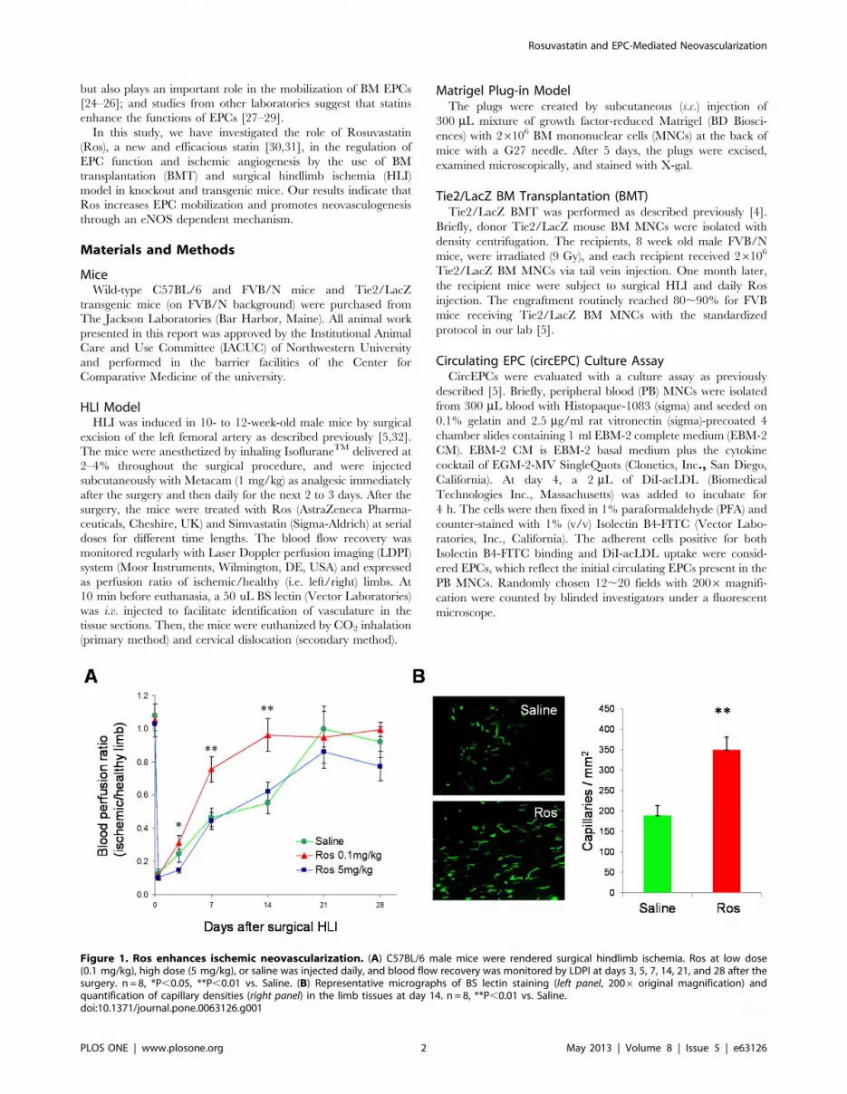
Figure 1. Ros enhances ischemic neovascularization. (A) C57BL/6 male mice were rendered surgical hindlimb ischemia. Ros at low dose(0.1 mg/kg), high dose (5 mg/kg), or saline was injected daily, and blood flow recovery was monitored by LDPI at days 3, 5, 7, 14, 21, and 28 after thesurgery. n = 8, *P,0.05, **P,0.01 vs. Saline. (B) Representative micrographs of BS lectin staining (left panel, 2006 original magnification) andquantification of capillary densities (right panel) in the limb tissues at day 14. n = 8, **P,0.01 vs. Saline.doi:10.1371/journal.pone.0063126.g001
Rosuvastatin and EPC-Mediated Neovascularization
PLOS ONE | www.plosone.org 2 May 2013 | Volume 8 | Issue 5 | e63126

X-gal Staining and ImmunohistochemistryTissues or Matrigel plugs were fixed either in 4% PFA or 100%
methanol. Enzymatic staining for X-gal and immunohistochemical
staining with antibodies for BS Lectin (Vector laboratories,
Burlingame, CA) and b-Gal (Cell Signaling Technology, Beverly,
MA) were performed as previously described [5,32,33].
Western BlottingEPCs were isolated and cultured from BM MNCs for 7 days
with a standardized protocol established in our lab [5], and then
treated with different concentrations of Ros for 30 min. Western
blotting analyses with antibodies for eNOS, phospho-eNOS
(Ser1177), Akt, and phospho-Akt (Ser473) (Cell Signaling Tech-
nology) were performed as previously described [34–36]. Band
intensities were determined densitometrically with Image J
software.
Statistical AnalysisData are presented as average 6 SEM. Unpaired Student’s t
test was used for the significance of differences. P,0.05 was
considered significant.
Results
Ros Enhances NeovascularizationTo investigate the effect of Ros on neovascularization after
ischemic injury, we induced acute ischemia in C57BL/6 mice by
surgically removing the left femoral artery. Ros at low dose
(0.1 mg/kg), high dose (5 mg/kg), or Saline control was injected
Figure 2. Ros increases BM-derived EPCs in neovascularization. BM MNCs isolated from Tie2/LacZ mice were used to transplant lethallyirradiated syngeneic FVB/NJ mice. One month later, HLI was induced in the recipient mice, and Ros (0.1 mg/kg) was s.c. injected daily. (A–C) At day 14after HLI, mice were injected with BS lectin and 10 min later, euthanized. The ischemic tissues were stained immunofluorescently with BS lectin (FITC,green) and b-gal (PE, red) to indicate vasculature and BM-derived cells, respectively. (A) Representative images of immunofluorescent doublestaining. White arrows indicate BS lectin and b-gal double positive cells. 4006 original magnification. (B) Quantification of BM-derived EPCsincorporated in the neovasculature (i.e., double positive cells) in the ischemic limb. (C) Quantification of capillary densities (BS lectin-FITC positivecells). n = 6, *P,0.05, **P,0.01 vs. Saline. (D) Blood flow was monitored with LDPI at days 3, 5, 7, 10, 14, and 16 following HLI. *P,0.05, **P,0.01 vs.Saline.doi:10.1371/journal.pone.0063126.g002
Rosuvastatin and EPC-Mediated Neovascularization
PLOS ONE | www.plosone.org 3 May 2013 | Volume 8 | Issue 5 | e63126
subcutaneously (s.c.) daily from day 0 to day 28 following the
surgery. The low-dose but not high-dose group of mice
demonstrated more rapid blood flow recovery than the control
group at days 3, 7 and 14 (Figure 1A). Low dose Ros treatment
also increased capillary density in the ischemic limb (Figure 1B).
Thus, we continued our investigations of Ros at low dose for the
following in vivo studies.
Ros Increases Incorporation of BM-derived EPCs at Sitesof Ischemic InjuryTo assess the effect of Ros on BM EPC contribution to
neovascularization, we performed BM transplantation (BMT) to
reconstitute the BM of lethally-irradiated WT FVB/N mice with
BM MNCs from background-matched Tie2/LacZ mice. One
month later, recipient mice with .90% engraftment were chosen
to receive surgical HLI and treatment with Ros or saline. Ros
treatment led to a significantly greater number of BM-derived ECs
(as shown by BS lectin and b-gal double positive cells)
incorporated at ischemic sites (Figures 2A and 2B), which were
associated with an increased capillary density and accelerated
blood flow recovery in the ischemic limb (Figures 2C and 2D),
although recovery of the mice on FVB/N background were
generally slower than those on C57BL/6 background (Figure 1A).
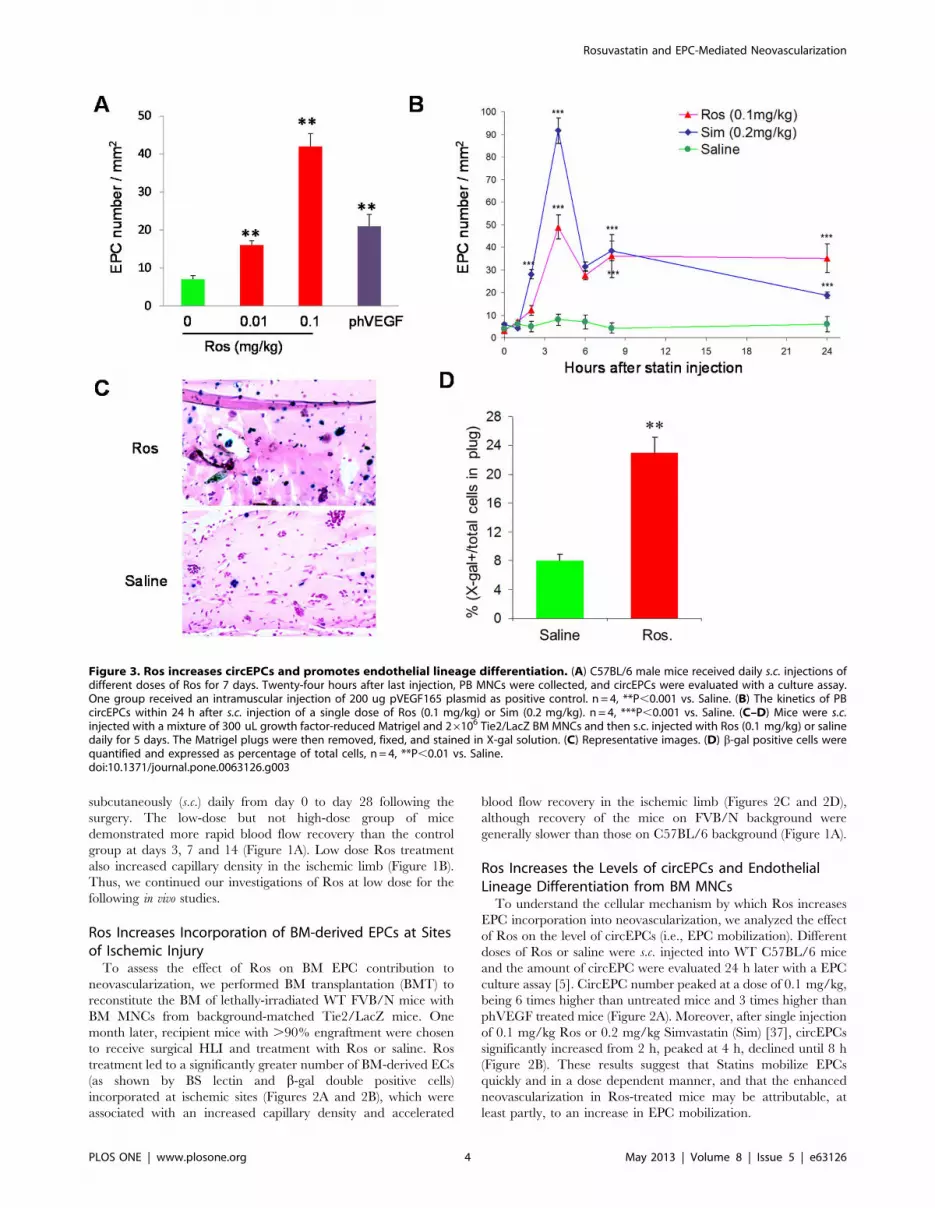
Ros Increases the Levels of circEPCs and EndothelialLineage Differentiation from BM MNCsTo understand the cellular mechanism by which Ros increases
EPC incorporation into neovascularization, we analyzed the effect
of Ros on the level of circEPCs (i.e., EPC mobilization). Different
doses of Ros or saline were s.c. injected into WT C57BL/6 mice
and the amount of circEPC were evaluated 24 h later with a EPC
culture assay [5]. CircEPC number peaked at a dose of 0.1 mg/kg,
being 6 times higher than untreated mice and 3 times higher than
phVEGF treated mice (Figure 2A). Moreover, after single injection
of 0.1 mg/kg Ros or 0.2 mg/kg Simvastatin (Sim) [37], circEPCs
significantly increased from 2 h, peaked at 4 h, declined until 8 h
(Figure 2B). These results suggest that Statins mobilize EPCs
quickly and in a dose dependent manner, and that the enhanced
neovascularization in Ros-treated mice may be attributable, at
least partly, to an increase in EPC mobilization.
Figure 3. Ros increases circEPCs and promotes endothelial lineage differentiation. (A) C57BL/6 male mice received daily s.c. injections ofdifferent doses of Ros for 7 days. Twenty-four hours after last injection, PB MNCs were collected, and circEPCs were evaluated with a culture assay.One group received an intramuscular injection of 200 ug pVEGF165 plasmid as positive control. n = 4, **P,0.001 vs. Saline. (B) The kinetics of PBcircEPCs within 24 h after s.c. injection of a single dose of Ros (0.1 mg/kg) or Sim (0.2 mg/kg). n = 4, ***P,0.001 vs. Saline. (C–D) Mice were s.c.injected with a mixture of 300 uL growth factor-reduced Matrigel and 26106 Tie2/LacZ BM MNCs and then s.c. injected with Ros (0.1 mg/kg) or salinedaily for 5 days. The Matrigel plugs were then removed, fixed, and stained in X-gal solution. (C) Representative images. (D) b-gal positive cells werequantified and expressed as percentage of total cells, n = 4, **P,0.01 vs. Saline.doi:10.1371/journal.pone.0063126.g003
Rosuvastatin and EPC-Mediated Neovascularization
PLOS ONE | www.plosone.org 4 May 2013 | Volume 8 | Issue 5 | e63126
Although the increase in new vessel formation could be the
result of this mechanism alone, we also considered the possibility
that Ros could enhance differentiation of EPCs. To test this
hypothesis we performed experiments in which ,56106 undiffer-
entiated BM MNCs isolated from Tie2/LacZ mice were mixed in
growth factor-reduced Matrigel and s.c. implanted into back-
ground-matched WT mice that receive daily injection of either
0.1 mg/kg Ros or saline for 5 days. Since growth factor-reduced
Matrigel does not provide a sufficient substrate to support the
growth of new vessels, this model provided us with the opportunity
to quantify EPC differentiation in vivo, defined by the advent of
Tie2 driven LacZ expression in the population of unselected BM
MNCs. As shown in Figures 3C and 3D, a significantly higher
ratio of X-gal (+) to total cells was found in the Matrigel plugs in
Ros-treated mice than in saline-treated mice. Since an equal
number of BM MNCs were implanted in both groups, these data
indicate that Ros enhances de novo EPC differentiation in vivo.
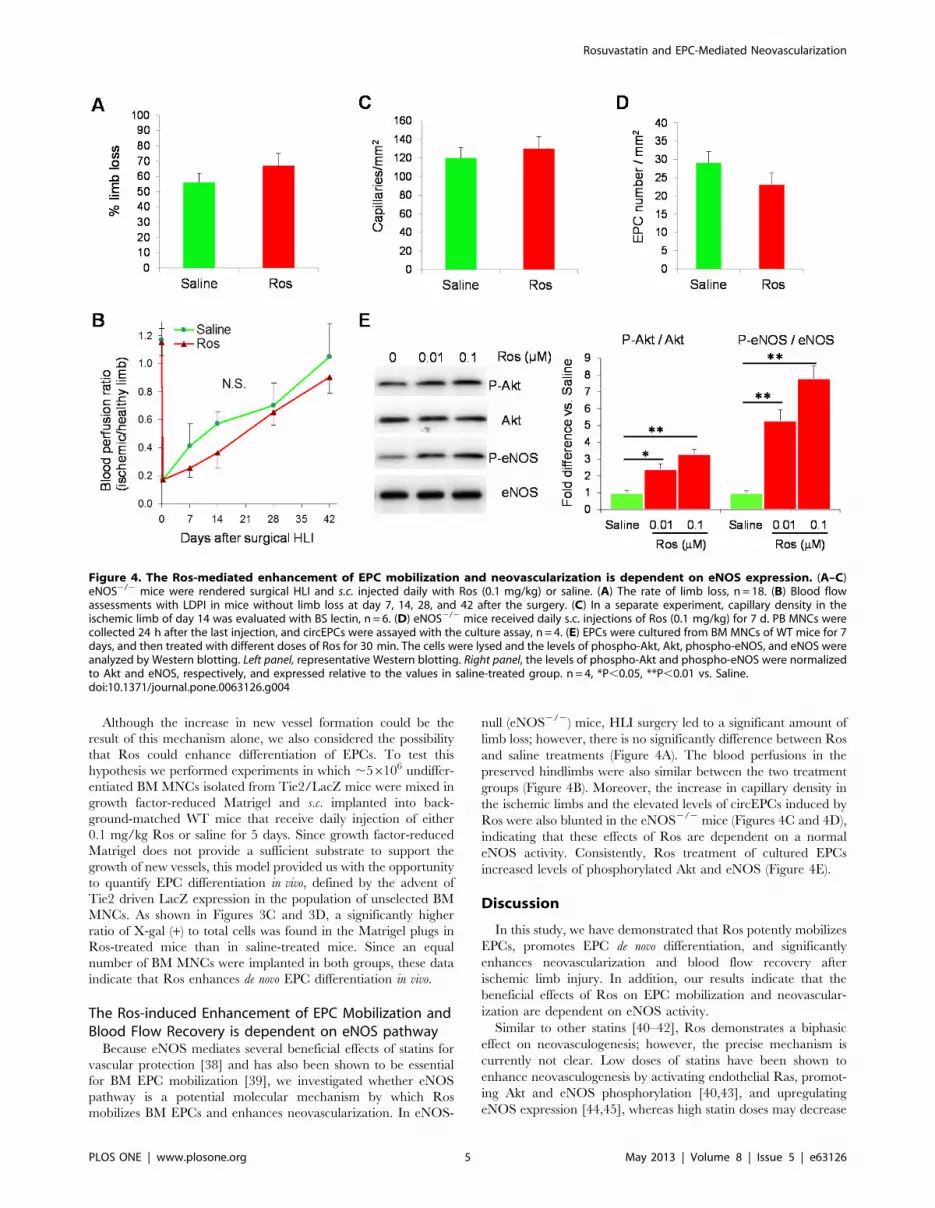
The Ros-induced Enhancement of EPC Mobilization andBlood Flow Recovery is dependent on eNOS pathwayBecause eNOS mediates several beneficial effects of statins for
vascular protection [38] and has also been shown to be essential
for BM EPC mobilization [39], we investigated whether eNOS
pathway is a potential molecular mechanism by which Ros
mobilizes BM EPCs and enhances neovascularization. In eNOS-
null (eNOS2/2) mice, HLI surgery led to a significant amount of
limb loss; however, there is no significantly difference between Ros
and saline treatments (Figure 4A). The blood perfusions in the
preserved hindlimbs were also similar between the two treatment
groups (Figure 4B). Moreover, the increase in capillary density in
the ischemic limbs and the elevated levels of circEPCs induced by
Ros were also blunted in the eNOS2/2 mice (Figures 4C and 4D),
indicating that these effects of Ros are dependent on a normal
eNOS activity. Consistently, Ros treatment of cultured EPCs
increased levels of phosphorylated Akt and eNOS (Figure 4E).
Discussion
In this study, we have demonstrated that Ros potently mobilizes
EPCs, promotes EPC de novo differentiation, and significantly
enhances neovascularization and blood flow recovery after
ischemic limb injury. In addition, our results indicate that the
beneficial effects of Ros on EPC mobilization and neovascular-
ization are dependent on eNOS activity.
Similar to other statins [40–42], Ros demonstrates a biphasic
effect on neovasculogenesis; however, the precise mechanism is
currently not clear. Low doses of statins have been shown to
enhance neovasculogenesis by activating endothelial Ras, promot-
ing Akt and eNOS phosphorylation [40,43], and upregulating
eNOS expression [44,45], whereas high statin doses may decrease
Figure 4. The Ros-mediated enhancement of EPC mobilization and neovascularization is dependent on eNOS expression. (A–C)eNOS2/2 mice were rendered surgical HLI and s.c. injected daily with Ros (0.1 mg/kg) or saline. (A) The rate of limb loss, n = 18. (B) Blood flowassessments with LDPI in mice without limb loss at day 7, 14, 28, and 42 after the surgery. (C) In a separate experiment, capillary density in theischemic limb of day 14 was evaluated with BS lectin, n = 6. (D) eNOS2/2 mice received daily s.c. injections of Ros (0.1 mg/kg) for 7 d. PB MNCs werecollected 24 h after the last injection, and circEPCs were assayed with the culture assay, n = 4. (E) EPCs were cultured from BM MNCs of WT mice for 7days, and then treated with different doses of Ros for 30 min. The cells were lysed and the levels of phospho-Akt, Akt, phospho-eNOS, and eNOS wereanalyzed by Western blotting. Left panel, representative Western blotting. Right panel, the levels of phospho-Akt and phospho-eNOS were normalizedto Akt and eNOS, respectively, and expressed relative to the values in saline-treated group. n = 4, *P,0.05, **P,0.01 vs. Saline.doi:10.1371/journal.pone.0063126.g004
Rosuvastatin and EPC-Mediated Neovascularization
PLOS ONE | www.plosone.org 5 May 2013 | Volume 8 | Issue 5 | e63126
protein prenylation in ECs, inhibit cell growth, and induce
apoptosis [40].These biphasic activities of statins on EC biology
can potentially be explained by the properties of the biosynthetic
pathways that originate from mevalonic acid [46], because in
addition to cholesterol, mevalonic acid is an essential precursor for
several cellular components including ubiquinone, isopentenylated
transfer RNAs, and prenylated proteins. Mevalonate-derived
intermediates have a higher affinity for the enzymes that catalyse
non-sterol product formation than for the cholesterol biosynthetic
enzymes. Therefore, low doses of statins may predominantly affect
cholesterol synthesis and not interfere with the biosynthesis of non-
sterol products that are required for cellular housekeeping
functions, and only at higher statin doses may a significant
inhibition of non-sterol product synthesis occur.
Ros is by far the most efficacious statin [31,47]; nevertheless, the
JUPITER study, despite reducing CVD and overall mortality,
highlighted an increase in new onset diabetes in the Ros treated
arm [47]. More recently, the increase in the incidence of diabetes
during statins trials has been confirmed by many meta-analyses of
the randomized controlled trials and appears to be associated with
a higher statin dosage [48,49]. Our study suggests that a lower
dose may be more favorable, at least in patients with diabetes or
diabetes associated risk factors.
It is exciting that low dose of Ros significantly enhances EPC
mobilization and recruitment to the site of neovascularization.
Stem cell mobilization involves complicated adhesive interactions
or cross-talks between stem cells and BM microenvironment
[34,35,50,51]. For example, G-CSF acts not directly on hemato-
poietic stem cells (HSCs) but via receptors on cells of the BM
stroma [52], while cleavage of VCAM-1 with neutrophil protease
was also involved in HSC mobilization [53]. The kinetics of HSC
mobilization with chemokines versus cytokines range from a few
minutes (with chemokines) to several days (with hematopoietic
growth factors) [51]. The kinetics of EPC mobilization, however,
varies considerably. Vascular trauma and ischemia was reported
to induce rapid but transient EPC mobilization [54]. Previous
study from our institute demonstrated that circEPCs reached a
peak at day 7 after surgical ischemia in rabbits [55]. The
mobilization of EPCs in nude mice with Ad-VEGF injection is
rapid, peaks at 2–3 days while Angiopoietin-1 exerts a delay EPC
mobilization as compared to VEGF, peaks at 2 weeks [56]. Other
researchers reported that peripheral circEPC numbers increased
gradually, reached peak with 4 weeks treatment of statins in mouse
models and in patients with coronary diseases [29,57]. Our data
further show that the kinetics of Ros-induced EPC mobilization is
more resembling to that of ‘‘chemokine-type’’. Further investiga-
tions are underway to determine how high Ros doses affect EPC
mobilization and neovasculogenesis.
We found that the enhanced circEPCs and post-HLI angio-
genesis stimulated by Ros were blunted in eNOS2/2 mice,
suggesting an essential role of eNOS. Because Ros upregulates
eNOS in both EPCs and ECs, it is difficult to dissect the extent to
which the angiogenic effect of the statin is dependent on EPCs.
Our attempt to reconstitute WT mouse BM with that of eNOS2/
2 mice was with low efficiency presumably due to the
indispensable role of eNOS for a successful BMT. Currently, it
is not completely clear how eNOS is activated (i.e., phosphory-
lated) by Ros. Since PI3K/Akt pathway has been shown essential
to EPC mobilization, migration, proliferation, and survival [37,57]
and our results indicate that Ros also mediates Akt phosphory-
lation, it is therefore likely that eNOS be a down-stream mediator
of Akt [58–60]; this however, remain to be investigated in our
future study.
In summary, our study demonstrates that Ros at a lower dose
promotes ischemic neovascularization via eNOS-dependent EPC
mobilization. Thus, optimization of Ros dose may maximize the
effect of Ros for the prevention and treatment of ischemic disease.
Author Contributions
Conceived and designed the experiments: JZ YL RK GQ. Performed the
experiments: JZ MC MW BQ HW. Analyzed the data: JZ MC MW GQ.
Contributed reagents/materials/analysis tools: YH QW YZ XG DG TCZ
YLT RK. Wrote the paper: JZ GQ.
References
1. Wollert KC, Drexler H (2010) Cell therapy for the treatment of coronary heart
disease: a critical appraisal. Nat Rev Cardiol 7: 204–215.
2. Losordo DW, Henry TD, Davidson C, Sup Lee J, Costa MA, et al. (2011)
Intramyocardial, autologous CD34+ cell therapy for refractory angina. Circ Res
109: 428–436.
3. Chavakis E, Koyanagi M, Dimmeler S (2010) Enhancing the outcome of cell
therapy for cardiac repair: progress from bench to bedside and back. Circulation
121: 325–335.
4. Asahara T, Masuda H, Takahashi T, Kalka C, Pastore C, et al. (1999) Bone
marrow origin of endothelial progenitor cells responsible for postnatal
vasculogenesis in physiological and pathological neovascularization. Circ Res
85: 221–228.
5. Qin G, Ii M, Silver M, Wecker A, Bord E, et al. (2006) Functional disruption of
alpha4 integrin mobilizes bone marrow-derived endothelial progenitors and
augments ischemic neovascularization. J Exp Med 203: 153–163.
6. Ziebart T, Yoon CH, Trepels T, Wietelmann A, Braun T, et al. (2008)
Sustained persistence of transplanted proangiogenic cells contributes to
neovascularization and cardiac function after ischemia. Circ Res 103: 1327–
1334.
7. Dimmeler S, Zeiher AM, Schneider MD (2005) Unchain my heart: the scientific
foundations of cardiac repair. J Clin Invest 115: 572–583.
8. Mirotsou M, Jayawardena TM, Schmeckpeper J, Gnecchi M, Dzau VJ (2011)
Paracrine mechanisms of stem cell reparative and regenerative actions in the
heart. J Mol Cell Cardiol 50: 280–289.
9. Rehman J, Li J, Orschell CM, March KL (2003) Peripheral blood "endothelial
progenitor cells" are derived from monocyte/macrophages and secrete
angiogenic growth factors. Circulation 107: 1164–1169.
10. Sahoo S, Klychko E, Thorne T, Misener S, Schultz KM, et al. (2011) Exosomes
from human CD34(+) stem cells mediate their proangiogenic paracrine activity.
Circ Res 109: 724–728.
11. Xiong Q, Ye L, Zhang P, Lepley M, Swingen C, et al. (2012) Bioenergetic and
functional consequences of cellular therapy: activation of endogenous cardio-
vascular progenitor cells. Circ Res 111: 455–468.
12. Pedersen TR, Kjekshus J, Berg K, Haghfelt T, Faergeman O, et al. (2004)
Randomised trial of cholesterol lowering in 4444 patients with coronary heart
disease: the Scandinavian Simvastatin Survival Study (4S). 1994. Atheroscler
Suppl 5: 81–87.
13. Levine GN, Keaney JF Jr, Vita JA (1995) Cholesterol reduction in
cardiovascular disease. Clinical benefits and possible mechanisms. N Engl J Med
332: 512–521.
14. Brautbar A, Ballantyne CM (2011) Pharmacological strategies for lowering LDL
cholesterol: statins and beyond. Nat Rev Cardiol 8: 253–265.
15. Heart Protection Study Collaborative G (2007) Randomized trial of the effects of
cholesterol-lowering with simvastatin on peripheral vascular and other major
vascular outcomes in 20,536 people with peripheral arterial disease and other
high-risk conditions. J Vasc Surg 45: 645–654; discussion 653–644.
16. Group TL-TIwPiIDLS (1998) Prevention of cardiovascular events and death
with pravastatin in patients with coronary heart disease and a broad range of
initial cholesterol levels. N Engl J Med 339: 1349–1357.
17. Colhoun HM, Betteridge DJ, Durrington PN, Hitman GA, Neil HA, et al.
(2004) Primary prevention of cardiovascular disease with atorvastatin in type 2
diabetes in the Collaborative Atorvastatin Diabetes Study (CARDS): multicentre
randomised placebo-controlled trial. Lancet 364: 685–696.
18. Koren MJ, Hunninghake DB, Investigators A (2004) Clinical outcomes in
managed-care patients with coronary heart disease treated aggressively in lipid-
lowering disease management clinics: the alliance study. J Am Coll Cardiol 44:
1772–1779.
19. Yemisci M, Ay H, Kocaefe C, Qui J, Topalkara K, et al. (2008) Statin
potentiates human platelet eNOS activity without enhancing eNOS mRNA and
protein levels. Cerebrovasc Dis 26: 190–198.
Rosuvastatin and EPC-Mediated Neovascularization
PLOS ONE | www.plosone.org 6 May 2013 | Volume 8 | Issue 5 | e63126
20. Merla R, Ye Y, Lin Y, Manickavasagam S, Huang MH, et al. (2007) The central
role of adenosine in statin-induced ERK1/2, Akt, and eNOS phosphorylation.Am J Physiol Heart Circ Physiol 293: H1918–1928.
21. Kosmidou I, Moore JP, Weber M, Searles CD (2007) Statin treatment and 3’
polyadenylation of eNOS mRNA. Arterioscler Thromb Vasc Biol 27: 2642–2649.
22. Wang CY, Liu PY, Liao JK (2008) Pleiotropic effects of statin therapy: molecularmechanisms and clinical results. Trends Mol Med 14: 37–44.
23. Bir SC, Xiong Y, Kevil CG, Luo J (2012) Emerging role of PKA/eNOS
pathway in therapeutic angiogenesis for ischaemic tissue diseases. CardiovascRes 95: 7–18.
24. Lemarie CA, Shbat L, Marchesi C, Angulo OJ, Deschenes ME, et al. (2011)Mthfr deficiency induces endothelial progenitor cell senescence via uncoupling
of eNOS and downregulation of SIRT1. Am J Physiol Heart Circ Physiol 300:H745–753.
25. Qiu FY, Song XX, Zheng H, Zhao YB, Fu GS (2009) Thymosin beta4 induces
endothelial progenitor cell migration via PI3K/Akt/eNOS signal transductionpathway. J Cardiovasc Pharmacol 53: 209–214.
26. Duda DG, Fukumura D, Jain RK (2004) Role of eNOS in neovascularization:NO for endothelial progenitor cells. Trends Mol Med 10: 143–145.
27. Suzuki G, Iyer V, Cimato T, Canty JM Jr (2009) Pravastatin improves function
in hibernating myocardium by mobilizing CD133+ and cKit+ bone marrowprogenitor cells and promoting myocytes to reenter the growth phase of the
cardiac cell cycle. Circ Res 104: 255–264, 210p following 264.28. Walter DH, Dimmeler S, Zeiher AM (2004) Effects of statins on endothelium
and endothelial progenitor cell recruitment. Semin Vasc Med 4: 385–393.29. Vasa M, Fichtlscherer S, Adler K, Aicher A, Martin H, et al. (2001) Increase in
circulating endothelial progenitor cells by statin therapy in patients with stable
coronary artery disease. Circulation 103: 2885–2890.30. Jones PH, Davidson MH, Stein EA, Bays HE, McKenney JM, et al. (2003)
Comparison of the efficacy and safety of rosuvastatin versus atorvastatin,simvastatin, and pravastatin across doses (STELLAR* Trial). Am J Cardiol 92:
152–160.
31. Clearfield MB, Amerena J, Bassand JP, Hernandez Garcia HR, Miller SS, et al.(2006) Comparison of the efficacy and safety of rosuvastatin 10 mg and
atorvastatin 20 mg in high-risk patients with hypercholesterolemia–Prospectivestudy to evaluate the Use of Low doses of the Statins Atorvastatin and
Rosuvastatin (PULSAR). Trials 7: 35.32. Qin G, Kishore R, Dolan CM, Silver M, Wecker A, et al. (2006) Cell cycle
regulator E2F1 modulates angiogenesis via p53-dependent transcriptional
control of VEGF. Proc Natl Acad Sci U S A 103: 11015–11020.33. Iwakura A, Shastry S, Luedemann C, Hamada H, Kawamoto A, et al. (2006)
Estradiol enhances recovery after myocardial infarction by augmentingincorporation of bone marrow-derived endothelial progenitor cells into sites of
ischemia-induced neovascularization via endothelial nitric oxide synthase-
mediated activation of matrix metalloproteinase-9. Circulation 113: 1605–1614.34. Cheng M, Zhou J, Wu M, Boriboun C, Thorne T, et al. (2010) CXCR4-
mediated bone marrow progenitor cell maintenance and mobilization aremodulated by c-kit activity. Circ Res 107: 1083–1093.
35. Tang YL, Zhu W, Cheng M, Chen L, Zhang J, et al. (2009) Hypoxicpreconditioning enhances the benefit of cardiac progenitor cell therapy for
treatment of myocardial infarction by inducing CXCR4 expression. Circ Res
104: 1209–1216.36. Zhou J, Zhu Y, Cheng M, Dinesh D, Thorne T, et al. (2009) Regulation of
vascular contractility and blood pressure by the E2F2 transcription factor.Circulation 120: 1213–1221.
37. Llevadot J, Murasawa S, Kureishi Y, Uchida S, Masuda H, et al. (2001) HMG-
CoA reductase inhibitor mobilizes bone marrow–derived endothelial progenitorcells. J Clin Invest 108: 399–405.
38. Balakumar P, Kathuria S, Taneja G, Kalra S, Mahadevan N (2012) Is targetingeNOS a key mechanistic insight of cardiovascular defensive potentials of statins?
J Mol Cell Cardiol 52: 83–92.
39. Aicher A, Heeschen C, Mildner-Rihm C, Urbich C, Ihling C, et al. (2003)
Essential role of endothelial nitric oxide synthase for mobilization of stem and
progenitor cells. Nat Med 9: 1370–1376.
40. Urbich C, Dernbach E, Zeiher AM, Dimmeler S (2002) Double-edged role of
statins in angiogenesis signaling. Circ Res 90: 737–744.
41. Weis M, Heeschen C, Glassford AJ, Cooke JP (2002) Statins have biphasic
effects on angiogenesis. Circulation 105: 739–745.
42. Katsumoto M, Shingu T, Kuwashima R, Nakata A, Nomura S, et al. (2005)
Biphasic effect of HMG-CoA reductase inhibitor, pitavastatin, on vascular
endothelial cells and angiogenesis. Circ J 69: 1547–1555.
43. Kureishi Y, Luo Z, Shiojima I, Bialik A, Fulton D, et al. (2000) The HMG-CoA
reductase inhibitor simvastatin activates the protein kinase Akt and promotes
angiogenesis in normocholesterolemic animals. Nat Med 6: 1004–1010.
44. Laufs U, Endres M, Stagliano N, Amin-Hanjani S, Chui DS, et al. (2000)
Neuroprotection mediated by changes in the endothelial actin cytoskeleton.
J Clin Invest 106: 15–24.
45. Laufs U, Liao JK (1998) Post-transcriptional regulation of endothelial nitric
oxide synthase mRNA stability by Rho GTPase. J Biol Chem 273: 24266–
24271.
46. Skaletz-Rorowski A, Walsh K (2003) Statin therapy and angiogenesis. Curr
Opin Lipidol 14: 599–603.
47. Ridker PM, Danielson E, Fonseca FA, Genest J, Gotto AM Jr, et al. (2008)
Rosuvastatin to prevent vascular events in men and women with elevated C-
reactive protein. N Engl J Med 359: 2195–2207.
48. Preiss D, Sattar N (2012) Pharmacotherapy: Statins and new-onset diabetes–the
important questions. Nat Rev Cardiol 9: 190–192.
49. Sattar N, Preiss D, Murray HM, Welsh P, Buckley BM, et al. (2010) Statins and
risk of incident diabetes: a collaborative meta-analysis of randomised statin trials.
Lancet 375: 735–742.
50. Cheng M, Qin G (2012) Progenitor cell mobilization and recruitment: SDF-1,
CXCR4, alpha4-integrin, and c-kit. Prog Mol Biol Transl Sci 111: 243–264.
51. Papayannopoulou T, Scadden DT (2008) Stem-cell ecology and stem cells in
motion. Blood 111: 3923–3930.
52. Liu S, Rose DM, Han J, Ginsberg MH (2000) Alpha4 integrins in cardiovascular
development and diseases. Trends Cardiovasc Med 10: 253–257.
53. Levesque JP, Takamatsu Y, Nilsson SK, Haylock DN, Simmons PJ (2001)
Vascular cell adhesion molecule-1 (CD106) is cleaved by neutrophil proteases in
the bone marrow following hematopoietic progenitor cell mobilization by
granulocyte colony-stimulating factor. Blood 98: 1289–1297.
54. Gill M, Dias S, Hattori K, Rivera ML, Hicklin D, et al. (2001) Vascular trauma
induces rapid but transient mobilization of VEGFR2(+)AC133(+) endothelialprecursor cells. Circ Res 88: 167–174.
55. Takahashi T, Kalka C, Masuda H, Chen D, Silver M, et al. (1999) Ischemia-
and cytokine-induced mobilization of bone marrow-derived endothelial
progenitor cells for neovascularization. Nat Med 5: 434–438.
56. Hattori K, Heissig B, Tashiro K, Honjo T, Tateno M, et al. (2001) Plasma
elevation of stromal cell-derived factor-1 induces mobilization of mature and
immature hematopoietic progenitor and stem cells. Blood 97: 3354–3360.
57. Dimmeler S, Aicher A, Vasa M, Mildner-Rihm C, Adler K, et al. (2001) HMG-
CoA reductase inhibitors (statins) increase endothelial progenitor cells via the PI
3-kinase/Akt pathway. J Clin Invest 108: 391–397.
58. Dimmeler S, Fleming I, Fisslthaler B, Hermann C, Busse R, et al. (1999)
Activation of nitric oxide synthase in endothelial cells by Akt-dependent
phosphorylation. Nature 399: 601–605.
59. Michell BJ, Griffiths JE, Mitchelhill KI, Rodriguez-Crespo I, Tiganis T, et al.
(1999) The Akt kinase signals directly to endothelial nitric oxide synthase. Curr
Biol 9: 845–848.
60. Fulton D, Gratton JP, McCabe TJ, Fontana J, Fujio Y, et al. (1999) Regulation
of endothelium-derived nitric oxide production by the protein kinase Akt.
Nature 399: 597–601.
Rosuvastatin and EPC-Mediated Neovascularization
PLOS ONE | www.plosone.org 7 May 2013 | Volume 8 | Issue 5 | e63126