static self-directed sample dispensing into a series of reaction wells on a microfluidic card for...
TRANSCRIPT
Static self-directed sample dispensing into a series of reaction wellson a microfluidic card for parallel genetic detectionof microbial pathogens
Robert D. Stedtfeld1& Yen-Cheng Liu1
& Tiffany M. Stedtfeld1& Tanja Kostic1,7 &
Maggie Kronlein1& Onnop Srivannavit4 & Walid T. Khalife5 & James M. Tiedje2,3 &
Erdogan Gulari4 & Mary Hughes6 & Brett Etchebarne6 & Syed A. Hashsham1,2
# The Author(s) 2015. This article is published with open access at Springerlink.com
Abstract Amicrofluidic card is described for simultaneous andrapid genetic detection of multiple microbial pathogens. Thehydrophobic surface of native acrylic and a novel microfluidicmechanism termed Bairlock^ were used to dispense sample intoa series of 64 reaction wells without the use of valves, externalpumping peripherals, multiple layers, or vacuum assistance. Thisairlock mechanism was tested with dilutions of whole humanblood, saliva, and urine, along with mock samples of varyingviscosities and surface tensions. Samples spiked with genomicDNA (gDNA) or crude lysates from clinical bacterial isolateswere tested with loop mediated isothermal amplification assays(LAMP) designed to target virulence and antibiotic resistance
genes. Reactions were monitored in real time using theGene-Z, which is a portable smartphone-driven system. Samplesloaded correctly into themicrofluidic card in 99.3%of instances.Amplification results confirmed no carryover of pre-dispensedprimer between wells during sample loading, and no observablediffusion between adjacent wells during the 60 to 90 min iso-thermal reaction. Sensitivity was comparable between LAMPreactions tested within the microfluidic card and in conventionalvials. Tests demonstrate that the airlock card works with varioussample types, manufacturing techniques, and can potentially beused in many point-of-care diagnostics applications.
Keywords Static self-directed sample dispensing . Airlockmechanism . Laser etched . Disposablemicrofluidic card .
Isothermal amplification . POC . Genetic diagnostics .
Gene-Z . LAMP
1 Introduction
Estimates of mortality caused by infectious diseases rangefrom 19 to 25 % worldwide (Fauci and Morens 2012; Lozanoet al. 2010). Point of care (POC) diagnostics that are afford-able, sensitive, specific, simple, equipment-less, rapid, androbust (Mabey et al. 2004) are expected to reduce time tointervention and influence positive outcomes. Microfluidicchips/cards with the capacity to target multiple infectious dis-eases in parallel are a promising means to rapidly diagnoseand satisfy many POC criteria (McCalla and Tripathi 2011).Innovative strategies for precise microfluidic control or load-ing and sealing samples into multiple well compartments (forparallel detection) have been described and reviewed (Junget al. 2015; Sackmann et al. 2014; Su et al. 2014) including:the manual deposition of sample in a small number of
Electronic supplementary material The online version of this article(doi:10.1007/s10544-015-9994-1) contains supplementary material,which is available to authorized users.
* Syed A. [email protected]
1 Civil and Environmental Engineering, Michigan State University,East Lansing, MI 48824, USA
2 The Center for Microbial Ecology, Michigan State University, EastLansing, MI 48824, USA
3 Department of Microbiology and Molecular Genetics, MichiganState University, East Lansing, MI 48824, USA
4 Department of Chemical Engineering, University of Michigan, AnnArbor, MI 48109, USA
5 Department of Microbiology, Sparrow Laboratories, Sparrow HealthSystem, Lansing, MI 48912, USA
6 Department of Osteopathic Medical Specialties, Sectionof Emergency Medicine, College of Osteopathic Medicine,Michigan State University, East Lansing, MI 48824, USA
7 Bioresources Unit, AITAustrian Institute of Technology GmbH,Konrad Lorenz Strasse 24, A-3430 Tulln, Austria
Biomed Microdevices (2015) 17:89 DOI 10.1007/s10544-015-9994-1
individual wells (Fang et al. 2010; Lee et al. 2008), the utili-zation of peripheral equipment to load samples with arrayersand injectors (Matsubara et al. 2004; Morrison et al. 2006;Duarte et al. 2013), pumps (Furuberg et al. 2008; Sun et al.2015), centrifuges (Lutz et al. 2010; Jung et al. 2014; Hoehlet al. 2014), development of multiple-layer cards utilizing slip(Shen et al. 2011), air, thermal, magnets, physical valve con-trol (Fang et al. 2012; Stedtfeld et al. 2012; Trung et al. 2010;Wang et al. 2011), vacuum assistance (Abe et al. 2011), orimmiscible fluids to prime cards (Gansen et al. 2012), surfacetreatment for primer immobilization and mobilization duringfilling (Morrison et al. 2006), and finally, propagation of sam-ple through a parallel network of interconnecting channelsusing an air vent for each reaction well (Fang et al. 2010;Ramalingam et al. 2010; Stedtfeld et al. 2012; Tourlousseet al. 2012). However, most microfluidic cards made of glass,silicon, and polymers require highly sophisticatedmicrofabrication (e.g. multiple layers and surfacefunctionalization), peripheral instruments, or complicated liq-uid handling that do not satisfy POC criteria (Nilghaz et al.2012; Hu et al. 2014). Without further reduction in cost oroperational simplicity, the value and adaptability ofmicrofluidic devices for POC genetic diagnostics is dimin-ished (Su et al. 2014; Volpatti and Yetisen 2014).
To better meet POC criteria, our goal was to develop dis-posable cards with 1) a single-etched layer that does not re-qu i r e f i ne a l i gnmen t , c l e an rooms , o r su r f acefunctionalization, and 2) distributes sample into a series ofreaction wells without carryover of dried pre-existing reactionconstitutes, without the need for external peripherals, extend-ed loading times, or numerous open points increasing the like-lihood for contamination and evaporation. Unique processesutilized in the development of the card include a microfluidicnetwork that traps air on one side of a bifurcated channelforming a plug termed Bairlock,^ and static self-directed sam-ple dispensing into larger volume wells due to the hydropho-bic nature of native acrylic. These mechanisms prevent carry-over of dried constitutes into subsequent wells, and completeloading of sample into reaction wells preceding further flow,respectively.
Cards were tested for proper loading with dilutions ofblood, urine, saliva, and mock samples prepared with varyingviscosities and surface tensions. The multi-well card was alsotested for parallel detection of multiple genetic markers relatedto identification, virulence, and antibiotic resistance from clin-ical pathogens. Loop mediated isothermal amplification(LAMP) reactions were performed with mixtures of genomicDNA (gDNA) and crudely lysed body fluid samples spikedwith bacterial cells. LAMP reactions were monitored in realtime using a previously described inexpensive and compactdevice termed Gene-Z (Stedtfeld et al. 2012). Endpoint im-ages of the amplification reaction within the card were alsocaptured using a CCD.
2 Material and methods
2.1 Microfluidic card fabrication
Cards were designed using Argon CAD 3D software (Ashlar-Vellum), converted to AutoCAD DXF (Drawing InterchangeFormat), and imported into InkScape (Software FreedomConservancy, Inc.). InkScape was used as an intermediatesoftware between DXF files and RetinaEngrave 3D, whichwas used to control the desktop CO2 Laser (MLE-40, FullSpectrum Laser LLC). Cards were fabricated from 1.59 mmacrylic sheets (McMaster-Carr). The cutting power and speedof the laser were programmed and controlled viaRetinaEngrave 3D. The reaction wells were fabricated by en-graving with 45 % power and 20 % speed; and the micro-channels were cut with 30 % current, 20 % speed and variouspower settings (5-65 %) to obtain desired channel depths.
Three different configurations were designed in this study:a card in which one inlet distributes samples into all 64 wells,a card with two inlets each leading to 32 wells, and a card withfour inlets each leading to 16 reaction wells (described as onelane herein). The spatial area of the 64 well card was 75×58 mm. The liquid channel was approximately 250 μmwide×250 μm deep (cut with 7 % power) and air segmentswere 200 μm wide×200 μm deep (cut with 5 % power).Channels were etched with dimensions to permit fabricationoutside of a clean room and prevent clogging with dust parti-cles (i.e. channels dimension larger than 150 μm). The oval-shaped reaction wells were 1400×2000 μm long axis ×1000 μm deep engraved with 45 % power, resulting in avolume of ~1.8 μL. The total sample volume was 160 μL.Wells and dimensions of the card were measured using a dig-ital microscope (Model #44,302-A) and a contact surfaceprofilometer (NanoMap-500LS). Inlet and outlets that fit a200 μL pipette tip (used for filling the cards) were fabricatedby laser etching 0.6 μm circles with 30 % power. The timerequired for engraving a single 64-well card was approximate-ly 10 min. With optimization of laser power and speed, thistime may be reduced to less than 5 min.
After laser micromachining, the card was rinsed with dis-tilled water, scrubbed with a paper towel, and rinsed with70 % ethanol. This cleaning procedure was performed to re-move acrylic residues, which was essential for correct flow ofsample and complete filling of reaction wells. Cleaned cardswere dried on a 70oC heater for 5 min. Once dry, compressedair was sprayed over the cards to remove dust, and depositsfrom the paper towel. For cards tested with LAMP, primers orgDNAwere dispensed into wells and dehydrated on a bench-top heater at 70 °C for 5 min. Next, engraved channels andwells were enclosed via a biocompatible optical film withpressure-sensitive silicon adhesive (MicroAmp Optical Adhe-sive Film; Applied Biosystems, Carlsbad, CA, USA). Securebonding of the film to acrylic was ensured using a press (4386;
89 Page 2 of 12 Biomed Microdevices (2015) 17:89
Carver, Wabash, IN, USA) at 3000 lb of pressure sandwichedbetween rubber sheets (McMaster Carr). It should be notedthat all other LAMP constitutes (with the exception of primerspredispensed into the card) were mixed with sample and load-ed into the card at the time of testing.
A screw cap, cut from the top of a 1.5 mL cryogenic vial(TS Scientific, Perkasie, PA, USA) was fixed on the card withUV glue (Dymax Corporation, Torrington, CT, USA) and5 min of exposure to UV light (PSD Series, Novascan). Toensure sealing, a 0.3 mL piece of polyester wax (melting pointof 37 °C, Electron Microscopy Sciences, Hatfield, PA, USA)was fixed to the top of the screw cap. When the LAMP reac-tion was performed at 63 °C, the wax melted and covered theinlet/outlet holes. Using wax to prevent contamination duringthe LAMP reaction was reported earlier by Tao and co-authors(Tao et al. 2011). The screw caps encompassed both loadingand air release ports. Cards were stored for no more than1 week prior to use.
Prior to loading, sterile needles were used to pierce the filmabove the loading port, and sample was dispensed using a200 μL micropipette (GPS-L250, Rainin). Pressure exertedby the pipette distributed sample into the channels and reac-tion wells. In this process, air inside channels was purgedthrough a single air vent placed downstream from the seriesof reaction wells.
2.2 Primer design and loop-mediated isothermalamplification with gDNA
LAMP primers targeting virulence and antibiotic resistancemarkers from bacterial pathogens were designed usingPrimerExplorer4 or retrieved from the literature (Table S1).Genomic DNA (gDNA) from Salmonella enterica (ATCC19585, 13311), Clostridium difficile (ATCC BAA-1382D)Vibrio cholerae (ATCC 39315),Campylobacter jejuni (ATCC700819), Giardia intestinalis (ATCC 30888), Staphylococcusaureus (ATCC 700699D), Streptococcus. agalactiae (ATCCBAA-1138), Enterococcus faecalis (ATCC 19433), Yersiniaenterocolitica (ATCC 55075), and Eschericia coli (ATCCBAA-460D, PTA-5184) were obtained from the AmericanType Culture Collection (Manassas, VA). The LAMP primerswere predispensed in the cards prior to chip assembly, to resultin a final concentration of 1.6 μM each of FIP and BIPprimers, 800 nM each of LF and LB primers, and 200 nMeach of F3 and B3 primers. After cards had been enclosedwiththe optical film, LAMP reaction mixtures consisting of800 mM betaine (Sigma Aldrich), 1.4 mM of each dNTP(Invitrogen), 20 mM Tris-HCl (pH 8.8), 10 mM (NH4)2SO4,10 mM KCl, 8 mM MgSO4, 8 mM Triton X-100, 0.64 units/μL of Bst polymerase (New England Biolabs), 20 μM ofSYTO 81 dye (Invitrogen), and varying amounts of templatewith gDNA, crude lysed, or lysates from bacterial isolateswere used (as described below). Results are described as
genomic copies per reaction for samples spiked with gDNAand CFU per reaction for samples spiked with crude lysates.
For the experiment testing diffusion and carryover ofgDNA predispensed in cards, template DNA from ten differ-ent organisms was used including Salmonella 13311 (Sal1)Salmonella 19585 (Sal2), E. coli PTA-5184 (EC1), E. coliBAA-460D (EC2), Y. enterocolitica (YE), C. difficile (CD),G. intestinalis (GI), V. cholera (VC), E. faecalis (EF) andC. jejuni (CJ). Two wells were no loaded with templateDNA (none).
For all amplification experiments performed in vials,primers were added into reaction mixture to have the sameconcentrations used in the microfluidic cards.
2.3 Blood, urine, saliva and LAMP reactions dispensedinto microfluidic card
Saliva, blood, and urine samples were spiked with dilutions ofbacterial isolates collected at Sparrow hospital, Department ofMicrobiology, in Lansing, Michigan. Staphylococcus aureus,Eschericia coli, Group A Streptococcus pyogenes (GAS), andGroup B Streptococcus agalactiae (GBS) were isolated usingstandard culture techniques in the Sparrow Clinical Microbi-ology Laboratory. Culture identification was performed usingSiemens Microscan, BD Phoenix, or biochemical tests. Priorto revival, isolates were stored in 15 % glycerol stocks at−80 °C. Isolates were revived by growing on TSB mediaovernight at 37 °C (no agitation) and serial diluted in 1 ×PBS. Ten microliters of each serial dilution was plated onTSA plates (in triplicate) and colony forming units werecounted following 24 h of incubation at 37 °C. GenomicDNA was also extracted from 1.5 mL of the revived stocksusing the Qiagen Blood and Tissue kit, following the protocolfor Gram-positive bacteria.
Initial experiments were performed with gDNA from theisolates to verify specificity of primer sets and ensure identityof bacterial isolates. These initial experiments were performedin the commercially available real time thermal cycler (Chro-mo4™ PCR thermal cycler, BioRad) under isothermal condi-tions. Tests included duplicate replicates for each reaction,positive controls with a type strain and no template controls.Type strains were spiked to have a total mass of 1 ng perreaction, and 2 μL was added from isolate lysates yielding472, 24, 22, and 53 ng/reaction for S. aureus, GAS, GBS,and E. coli, respectively (Table S2). Specificity of LAMPassays and concordance with culture based identificationwas tested using 23 LAMP assays against 11 re-cultured iso-lates derived from septic patients (Table S3).
Whole blood was collected from a healthy volunteer intostandard hospital purple top specimen collection EDTA tubes.Whole-blood samples were stored at 4 °C for up to 60 daysprior to use. Urine and saliva samples were collected fromhealthy volunteers in Eppendorf vials, and were stored at
Biomed Microdevices (2015) 17:89 Page 3 of 12 89
4 °C for up to 14 days prior to use. Serial dilutions of isolateswere spiked into blood, urine, and saliva samples. Bacterialcontent of dilutions was counted following 24 h of growth onTSA plates, as described above, and prepared dilutions arelisted as CFU/reaction well (Table 1). Blood sample werespiked with E. coli and S. aureus isolates, saliva sample werespiked with Group A Streptococcus (GAS), and the urinesamples were spiked with Group B Streptococcus (GBS). Af-ter spiking with isolates, saliva and urine samples were heatlysed at 95 °C for 5 min. Blood samples were tested viaLAMP amplification in the microfluidic card directly withoutlysing. It should be noted that heat lysed blood samplesclotted, and could not be handled with a micropipette. Bloodsamples were mixed at 1, 5, 10, and 40 % of the total LAMPreaction volume. Urine samples were tested by mixing inLAMP reactions to be 10, 25, and 40 % of total reactionvolume, and saliva was mixed to be 10 and 25 % of totalreaction. Cards were prepared with 16 reaction wells per sam-ple, and thus four separate samples were loaded per card. Withthis configuration, three serial dilutions of bacterial isolatesand a non-spiked (no template) control was loaded on eachcard. Cards were preloaded with primers, as described above,with each primer was dispensed into three individual reactionwells. Genomic DNA from a plant pathogen was spiked as aninternal positive control into all blood, saliva, and urine sam-ples for a final concentration of 5.7 pg/μL (~60copies/reaction) to test for inhibition in samples. A primerset targeting the positive control was also loaded into onereaction well of each 16 well lane for cards tested with blood,and four reaction wells out of each 16 well lane for cardstested with GBS and GAS.
2.4 Ethics statement
All experimentation with human samples was conducted fol-lowing the Human Research Protection Program and per-formed in accordance with institutional regulations after per-tinent review and approval by the Institutional Review Boardat Michigan State University, East Lansing, MI. MichiganState University and SparrowHospital Investigational ReviewBoard human subjects exemption with MSU IRB# 12-706;i041390 determined to be non-human subject research. Writ-ten consent was obtained from healthy individuals that donat-ed blood, urine, and saliva samples. Cultured bacterial isolatesused for spikes were determined to be non-human subjectresearch.
2.5 LAMP monitored with Gene-Z
The design and layout of the Gene-Z prototype was similar tothat described previously (Stedtfeld et al. 2012), with minormodifications including: i) embedding optical fibers in an ap-paratus placed above the card (previously, fibers were
embedded below the card), ii) cards were inserted into theGene-Z device via a port on the side of the device (insteadof a door at the top, Fig. 1). Advantages of this arrangementincluded: i) elimination of occasional misalignment of cardswith the optics, ii) reduction in cost of manufacturing thealuminium heater (i.e. drilling negative features for alignmentof the shelled card and angled holes for optical fibers in thealuminium heat sink), and iii) elimination of pressure require-ments needed by the door to obtain equal contact between thinnon-rigid cards and the heater.
After loading the card with sample, and securing the screwcap, cards were inserted into Gene-Z, and the device wasstarted using an iPod Touch as described previously (Stedtfeldet al. 2012). All reactions were run for 60 to 90 min and datawas sent from the iPod to the user’s email account for furtheranalysis. Data analysis was performed as previously describedto calculate the threshold time (Tt), akin to threshold cycle.The signal to noise ratio (SNR) was calculated as the signal ata given time point minus the median signal at the start of thereaction, divided by the standard deviation of the signal at thestart of the reaction. Results testing replication of the deviceoptics and heater temperature along the length of the heater ofthe Gene-Z device have been reported previously (Stedtfeldet al. 2012).
2.6 Endpoint images of LAMP reactions in microfluidiccard
Endpoint images of the LAMP reaction within themicrofluidic card were captured using either a cell phone cam-era (Galaxy Note II, Samsung), or a digital camera(Cybershot, Sony). SYTO intercalating dye was excited usinga 530 nm green LED (05027-PM12, LED Supply). A 572±20 nm bandpass filter (FF01-572/28-25, Semrock) was fixedto capture emission signals from wells. For experiments with40 % blood, a 0.25 megapixel monochrome CCD camera(MEADE DSI Pro, Irvine, CA, 1.0 s exposure time) was nec-essary to capture increased signal from an amplification event.
3 Results and discussion
A card, termed Bairlock^ was developed that does not requireperipheral systems, permits straightforward operation vialoading with a syringe or micropippette, and can be used witheither low cost photodetectors, CCD cameras, or the Gene-Zdevice for rapid and real-time optical readout. Cards weretested for proper loading using blood, urine, and saliva spikedwith isolates from septic infections. LAMP was performed inthe cards to ensure predispensed primers were not carried tosubsequent wells during sample filling, and no observablediffusion between wells during the reaction. Experiments also
89 Page 4 of 12 Biomed Microdevices (2015) 17:89
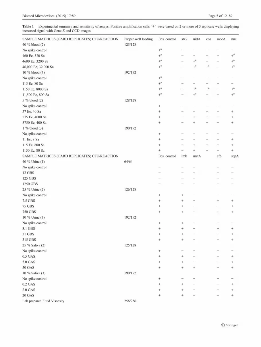
Table 1 Experimental summary and sensitivity of assays. Positive amplification calls B+^ were based on 2 or more of 3 replicate wells displayingincreased signal with Gene-Z and CCD images
SAMPLE MATRICES (CARD REPLICATES) CFU/REACTION Proper well loading Pos. control stx2 uidA coa mecA nuc
40 % blood (2) 125/128
No spike control +a − − − − −460 Ec, 320 Sa +a − − − − +#
4600 Ec, 3200 Sa +a − +a − − +a
46,000 Ec, 32,000 Sa +a − +a +a − +a
10 % blood (3) 192/192
No spike control +a − − − − −115 Ec, 80 Sa +a − − − − −1150 Ec, 8000 Sa +a − +a +a − +a
11,500 Ec, 800 Sa +a − +a − − +a
5 % blood (2) 128/128
No spike control + − − − − −57 Ec, 40 Sa + − − − − +
575 Ec, 4000 Sa + − + + − +
5750 Ec, 400 Sa + − + − − +
1 % blood (3) 190/192
No spike control + − − − − −11 Ec, 8 Sa + − − − − +
115 Ec, 800 Sa + − + + − +
1150 Ec, 80 Sa + − + − − +
SAMPLE MATRICES (CARD REPLICATES) CFU/REACTION Pos. control lmb mstA cfb scpA
40 % Urine (1) 64/64
No spike control − − − − −12 GBS − − − − −125 GBS − − − − −1250 GBS − − − − −25 % Urine (2) 126/128
No spike control + + − − −7.5 GBS + + − + +
75 GBS + + − + +
750 GBS + + − + +
10 % Urine (3) 192/192
No spike control + + − − −3.1 GBS + + − + +
31 GBS + + − + +
315 GBS + + − + +
25 % Saliva (2) 125/128
No spike control + − − − −0.5 GAS + + − − +
5.0 GAS + + − − +
50 GAS + + + − +
10 % Saliva (3) 190/192
No spike control + − − − −0.2 GAS + + − − +
2.0 GAS + + − − +
20 GAS + + − − +
Lab prepared Fluid Viscosity 256/256
Biomed Microdevices (2015) 17:89 Page 5 of 12 89
tested analytical sensitivity and replication of the non-functionalized native acrylic cards.
3.1 Principle of sample distribution in the card
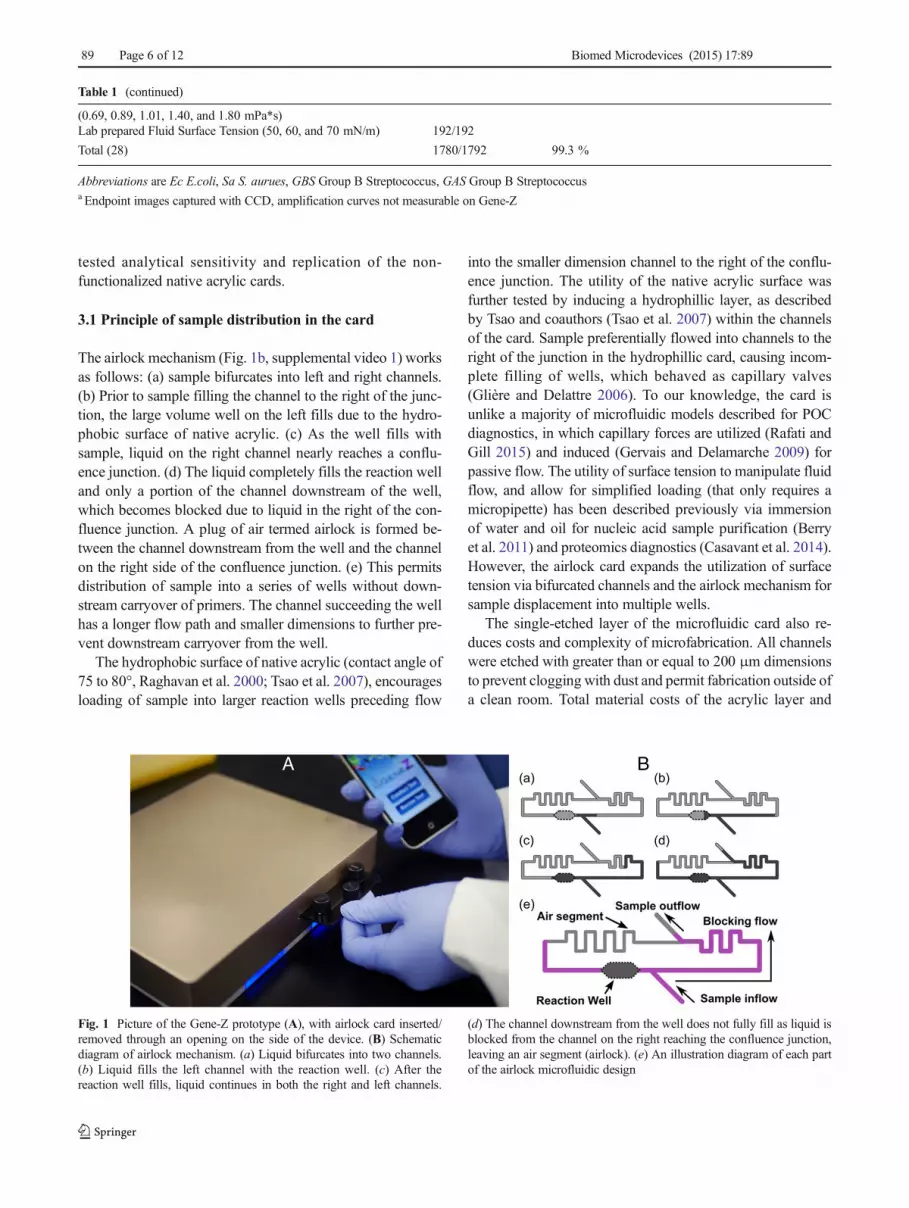
The airlock mechanism (Fig. 1b, supplemental video 1) worksas follows: (a) sample bifurcates into left and right channels.(b) Prior to sample filling the channel to the right of the junc-tion, the large volume well on the left fills due to the hydro-phobic surface of native acrylic. (c) As the well fills withsample, liquid on the right channel nearly reaches a conflu-ence junction. (d) The liquid completely fills the reaction welland only a portion of the channel downstream of the well,which becomes blocked due to liquid in the right of the con-fluence junction. A plug of air termed airlock is formed be-tween the channel downstream from the well and the channelon the right side of the confluence junction. (e) This permitsdistribution of sample into a series of wells without down-stream carryover of primers. The channel succeeding the wellhas a longer flow path and smaller dimensions to further pre-vent downstream carryover from the well.
The hydrophobic surface of native acrylic (contact angle of75 to 80°, Raghavan et al. 2000; Tsao et al. 2007), encouragesloading of sample into larger reaction wells preceding flow
into the smaller dimension channel to the right of the conflu-ence junction. The utility of the native acrylic surface wasfurther tested by inducing a hydrophillic layer, as describedby Tsao and coauthors (Tsao et al. 2007) within the channelsof the card. Sample preferentially flowed into channels to theright of the junction in the hydrophillic card, causing incom-plete filling of wells, which behaved as capillary valves(Glière and Delattre 2006). To our knowledge, the card isunlike a majority of microfluidic models described for POCdiagnostics, in which capillary forces are utilized (Rafati andGill 2015) and induced (Gervais and Delamarche 2009) forpassive flow. The utility of surface tension to manipulate fluidflow, and allow for simplified loading (that only requires amicropipette) has been described previously via immersionof water and oil for nucleic acid sample purification (Berryet al. 2011) and proteomics diagnostics (Casavant et al. 2014).However, the airlock card expands the utilization of surfacetension via bifurcated channels and the airlock mechanism forsample displacement into multiple wells.
The single-etched layer of the microfluidic card also re-duces costs and complexity of microfabrication. All channelswere etched with greater than or equal to 200 μm dimensionsto prevent clogging with dust and permit fabrication outside ofa clean room. Total material costs of the acrylic layer and
Table 1 (continued)
(0.69, 0.89, 1.01, 1.40, and 1.80 mPa*s)Lab prepared Fluid Surface Tension (50, 60, and 70 mN/m) 192/192
Total (28) 1780/1792 99.3 %
Abbreviations are Ec E.coli, Sa S. aurues, GBS Group B Streptococcus, GAS Group B Streptococcusa Endpoint images captured with CCD, amplification curves not measurable on Gene-Z
Fig. 1 Picture of the Gene-Z prototype (A), with airlock card inserted/removed through an opening on the side of the device. (B) Schematicdiagram of airlock mechanism. (a) Liquid bifurcates into two channels.(b) Liquid fills the left channel with the reaction well. (c) After thereaction well fills, liquid continues in both the right and left channels.
(d) The channel downstream from the well does not fully fill as liquid isblocked from the channel on the right reaching the confluence junction,leaving an air segment (airlock). (e) An illustration diagram of each partof the airlock microfluidic design
89 Page 6 of 12 Biomed Microdevices (2015) 17:89
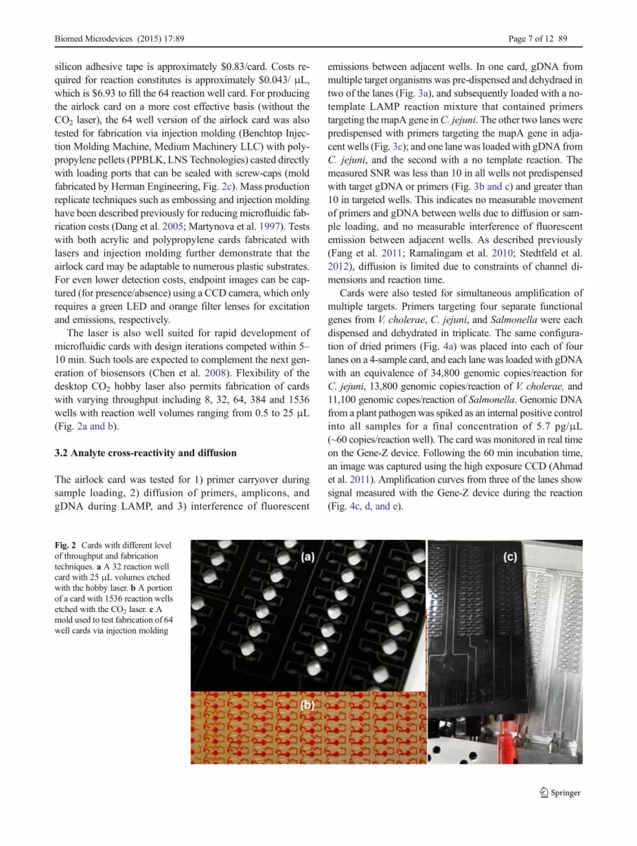
silicon adhesive tape is approximately $0.83/card. Costs re-quired for reaction constitutes is approximately $0.043/ μL,which is $6.93 to fill the 64 reaction well card. For producingthe airlock card on a more cost effective basis (without theCO2 laser), the 64 well version of the airlock card was alsotested for fabrication via injection molding (Benchtop Injec-tion Molding Machine, Medium Machinery LLC) with poly-propylene pellets (PPBLK, LNS Technologies) casted directlywith loading ports that can be sealed with screw-caps (moldfabricated by Herman Engineering, Fig. 2c). Mass productionreplicate techniques such as embossing and injection moldinghave been described previously for reducing microfluidic fab-rication costs (Dang et al. 2005; Martynova et al. 1997). Testswith both acrylic and polypropylene cards fabricated withlasers and injection molding further demonstrate that theairlock card may be adaptable to numerous plastic substrates.For even lower detection costs, endpoint images can be cap-tured (for presence/absence) using a CCD camera, which onlyrequires a green LED and orange filter lenses for excitationand emissions, respectively.
The laser is also well suited for rapid development ofmicrofluidic cards with design iterations competed within 5–10 min. Such tools are expected to complement the next gen-eration of biosensors (Chen et al. 2008). Flexibility of thedesktop CO2 hobby laser also permits fabrication of cardswith varying throughput including 8, 32, 64, 384 and 1536wells with reaction well volumes ranging from 0.5 to 25 μL(Fig. 2a and b).
3.2 Analyte cross-reactivity and diffusion
The airlock card was tested for 1) primer carryover duringsample loading, 2) diffusion of primers, amplicons, andgDNA during LAMP, and 3) interference of fluorescent
emissions between adjacent wells. In one card, gDNA frommultiple target organisms was pre-dispensed and dehydraed intwo of the lanes (Fig. 3a), and subsequently loaded with a no-template LAMP reaction mixture that contained primerstargeting the mapA gene inC. jejuni. The other two lanes werepredispensed with primers targeting the mapA gene in adja-cent wells (Fig. 3c); and one lane was loadedwith gDNA fromC. jejuni, and the second with a no template reaction. Themeasured SNR was less than 10 in all wells not predispensedwith target gDNA or primers (Fig. 3b and c) and greater than10 in targeted wells. This indicates no measurable movementof primers and gDNA between wells due to diffusion or sam-ple loading, and no measurable interference of fluorescentemission between adjacent wells. As described previously(Fang et al. 2011; Ramalingam et al. 2010; Stedtfeld et al.2012), diffusion is limited due to constraints of channel di-mensions and reaction time.
Cards were also tested for simultaneous amplification ofmultiple targets. Primers targeting four separate functionalgenes from V. cholerae, C. jejuni, and Salmonella were eachdispensed and dehydrated in triplicate. The same configura-tion of dried primers (Fig. 4a) was placed into each of fourlanes on a 4-sample card, and each lane was loaded with gDNAwith an equivalence of 34,800 genomic copies/reaction forC. jejuni, 13,800 genomic copies/reaction of V. cholerae, and11,100 genomic copes/reaction of Salmonella. Genomic DNAfrom a plant pathogen was spiked as an internal positive controlinto all samples for a final concentration of 5.7 pg/μL(~60 copies/reaction well). The card was monitored in real timeon the Gene-Z device. Following the 60 min incubation time,an image was captured using the high exposure CCD (Ahmadet al. 2011). Amplification curves from three of the lanes showsignal measured with the Gene-Z device during the reaction(Fig. 4c, d, and e).
Fig. 2 Cards with different levelof throughput and fabricationtechniques. a A 32 reaction wellcard with 25 μL volumes etchedwith the hobby laser. b A portionof a card with 1536 reaction wellsetched with the CO2 laser. c Amold used to test fabrication of 64well cards via injection molding
Biomed Microdevices (2015) 17:89 Page 7 of 12 89
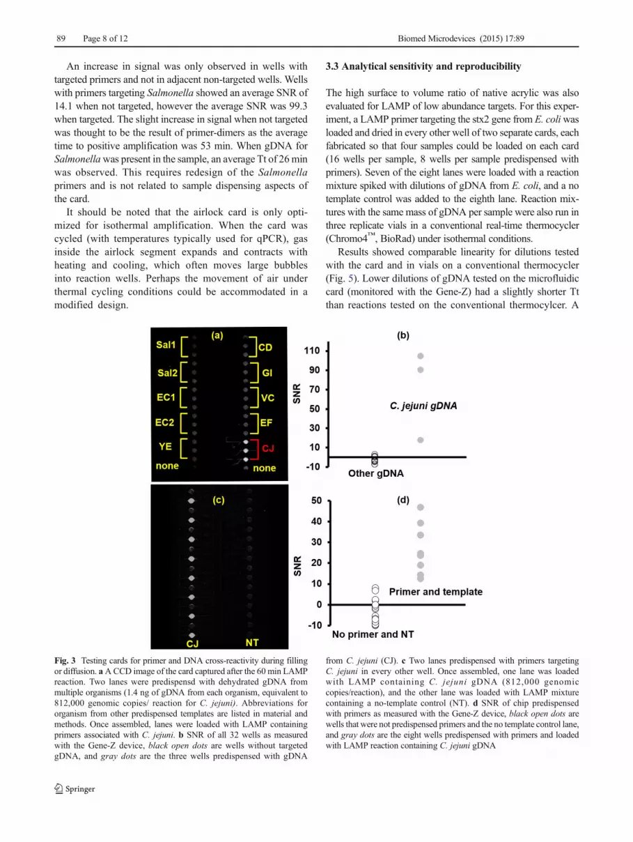
An increase in signal was only observed in wells withtargeted primers and not in adjacent non-targeted wells. Wellswith primers targeting Salmonella showed an average SNR of14.1 when not targeted, however the average SNR was 99.3when targeted. The slight increase in signal when not targetedwas thought to be the result of primer-dimers as the averagetime to positive amplification was 53 min. When gDNA forSalmonellawas present in the sample, an average Tt of 26minwas observed. This requires redesign of the Salmonellaprimers and is not related to sample dispensing aspects ofthe card.
It should be noted that the airlock card is only opti-mized for isothermal amplification. When the card wascycled (with temperatures typically used for qPCR), gasinside the airlock segment expands and contracts withheating and cooling, which often moves large bubblesinto reaction wells. Perhaps the movement of air underthermal cycling conditions could be accommodated in amodified design.
3.3 Analytical sensitivity and reproducibility
The high surface to volume ratio of native acrylic was alsoevaluated for LAMP of low abundance targets. For this exper-iment, a LAMP primer targeting the stx2 gene from E. coliwasloaded and dried in every other well of two separate cards, eachfabricated so that four samples could be loaded on each card(16 wells per sample, 8 wells per sample predispensed withprimers). Seven of the eight lanes were loaded with a reactionmixture spiked with dilutions of gDNA from E. coli, and a notemplate control was added to the eighth lane. Reaction mix-tures with the same mass of gDNA per sample were also run inthree replicate vials in a conventional real-time thermocycler(Chromo4™, BioRad) under isothermal conditions.
Results showed comparable linearity for dilutions testedwith the card and in vials on a conventional thermocycler(Fig. 5). Lower dilutions of gDNA tested on the microfluidiccard (monitored with the Gene-Z) had a slightly shorter Ttthan reactions tested on the conventional thermocylcer. A
Fig. 3 Testing cards for primer and DNA cross-reactivity during fillingor diffusion. aACCD image of the card captured after the 60 min LAMPreaction. Two lanes were predispensd with dehydrated gDNA frommultiple organisms (1.4 ng of gDNA from each organism, equivalent to812,000 genomic copies/ reaction for C. jejuni). Abbreviations fororganism from other predispensed templates are listed in material andmethods. Once assembled, lanes were loaded with LAMP containingprimers associated with C. jejuni. b SNR of all 32 wells as measuredwith the Gene-Z device, black open dots are wells without targetedgDNA, and gray dots are the three wells predispensed with gDNA
from C. jejuni (CJ). c Two lanes predispensed with primers targetingC. jejuni in every other well. Once assembled, one lane was loadedwith LAMP containing C. jejuni gDNA (812,000 genomiccopies/reaction), and the other lane was loaded with LAMP mixturecontaining a no-template control (NT). d SNR of chip predispensedwith primers as measured with the Gene-Z device, black open dots arewells that were not predispensed primers and the no template control lane,and gray dots are the eight wells predispensed with primers and loadedwith LAMP reaction containing C. jejuni gDNA
89 Page 8 of 12 Biomed Microdevices (2015) 17:89

detection limit of approximately 20 genomic copies/reactionwell was observed for the E. coli stx2 assay on the card.However, only 3 out of 8 wells showed amplification with
20 genomic copies/reaction. Tests in the Chromo4™
thermocycler resulted in positive amplification in 0 out of 3vials at 20 gDNA copies/reaction.
Fig. 4 Testing cards for LAMP based detection of multiple targets inparallel. All lanes were spiked with an internal positive control (gDNAfrom a plant pathogen) spiked into all samples plus templates of gDNAfrom CJ (C. jejuni, 34,800 genomic copies/reaction) was added to lane 2(ii), Sal (Salmonella 11,100 genomic copes/ reaction) into lane 3 (iii), and
VC (V. cholera, 13,800 genomic copies/reaction) into lane 4 (iv). a Mapshowing how primers were dried into wells, and b picture of card(captured with exposure control CCD) after the 60 min LAMP reaction.c, d, e SNR of all wells in lanes i(c), iii(d), iv(e) versus reaction time asmonitored with the Gene-Z device
Fig. 5 Analytical sensitivity and reproducibility of airlock cards testedwith LAMP reactions. a Real time LAMP profiles captured with theGene-Z from one well from each lane of the microfluidic card loadedwith serial dilutions of template gDNA. b Average time to threshold (Tt)for replicates that amplified for reactions tested with isothermal
temperatures in a conventional thermocycler (n=3) and the airlock card(n=8). Error bars represent standard error. c Mean, standard deviation,and coefficient of variation (in parenthesis) of the Tt for all reaction wellsin each of the four lanes tested in three separate cards
Biomed Microdevices (2015) 17:89 Page 9 of 12 89
While the stx2 assay lacked single copy detection limit, themicrofluidic card provides a level of sensitivity that is compa-rable to reactions tested in vials with the thermocycler. Weroutinely observe that amplification efficiency of LAMP innative polymer cards (COP and polyester) is similar to thatin tubes (Ahmad et al. 2011; Stedtfeld et al. 2012). The card isexpected to have a sensitivity dependent upon the assay,which has been previously shown to allow quantification ofsingle digit copy numbers/reaction (Thekisoe et al. 2010; Tsaiet al. 2009; Yamazaki et al. 2008).
Inter and intra assay reproducibility were also tested toensure that the thicker ~1.59 mm card did not influence tem-perature distribution along the length of the card and ultimate-ly influence threshold time and quantification. LAMP primerswere predispensed into all reaction wells on three separatemicrofluidic cards and loaded with approximately 5×10⁶ ge-nomic copies/reaction. Results show a coefficient of variationof 6.1 % between all three cards (Fig. 5c), which is compara-ble to thinner cards that were previously described with acoefficient of variation of 7.8 % (Stedtfeld et al. 2012).
3.4 Testing cards with blood, urine, saliva
The airlock card was tested for correct loading of body samples(blood, urine, and saliva) spiked with dilutions of bacterial iso-lates derived from septic infections. Ultimately, decentralizedgenetic testing must also allow for sample-in-results-out, withautomation, minimization, or elimination of sample preparation(DNA extraction/purification). LAMP and direct amplificationhave been previously demonstrated with many different targets(bacterial, viral, human, fungal), and sample types with variedconcentrations in the amplification reaction including 1 to 50%blood (Curtis et al. 2009; Ebbinghaus et al. 2012; Masaomiet al. 2003; Patterson et al. 2013; Soejima et al. 2011), 10 %stool, (Francois et al. 2011), 20 % urine samples (Hillet al. 2008; Koizumi et al. 2012), and 32 % nasal swabs(Nie et al. 2012). As such, the airlock cards were testedwith these sample types at varying sample concentrationsin the final LAMP reaction mixture.
Blood sample experiments consisted of spiking blood into1, 5, 10, and 40 % of the total amplification reaction volumeand loading cards. In total, 635 out of 640 wells loaded prop-erly with the tested blood samples (Table 1, Fig S1–S3). Itshould be noted that wells that did not load properly seemedto be due to inadequate cleaning of the card, with sample notfilling wells completely. The 40 % blood sample loadedslower than all other samples, requiring 2.5 min to completelyfill a 16 well lane. Six cards were tested with urine samplesspiked with dilutions of GBS isolates at 10, 25, and 40 %concentration of the final LAMP reaction mixture, and 382out of 384 wells loaded properly (Fig S4–S5). Five cards weretested with saliva samples spiked with dilutions of GAS iso-lates were tested as 10 and 25 % concentrations of the LAMP
reaction mixture, and 315 out of 320 wells loaded properly(Fig S6).
3.5 Sensitivity and selectivity of bacteria spiked into bodymatrices
Assays designed to target lmb, cfb, and scpA genes in GBSand GAS amplified down to single digit CFU/reaction whenspiked into urine and saliva (tested as 10 and 25 % of thereaction, Table 1). Considering a 4 μL reaction well, assuming25 % of the LAMP reaction is sample, and a detection limit ofapproximately 1 CFU/reaction; the limit of detection is ap-proximately 1000 CFU/mL. This level of sensitivity is belowthe criterion commonly used for defining significant bacteri-uria, which is 105 CFU/mL in urine (Stamm et al. 1982; Rosaet al. 2010), and 104–107 CFU/mL for conventional bacterialloads of GAS obtained via throat swabs (Dunne et al. 2013).Thus, diagnostics of urine and throat swabs could be per-formed with minimal sample preparation, potentially allowinggenetic diagnostics in a decentralized setting. While demon-strated directly with saliva, elution from swabs is expected tohave a lesser influence on inhibition.
Assays designed to target nuc and uidA amplified down toapproximatly 10–100 CFU/reaction with 5 % blood (Table 1),corresponding to a detection limit of 104 to 105 CFU/mL. Forclinically relevant blood samples with lower bacterial concen-trations (e.g., 1–100 CFU/ml in adults (Yagupsky and Nolte1990) and <10 CFU/ml in neonates (Reier-Nilsen et al.2009)), this approach still poses a challenge due to the volu-metric constraints of the cards. However, a concentration stepcould be used to capture bacteria (Mai et al. 2013; Cooperet al. 2014), or a commercially available kit such as theMolYsis or Looxster kits for concentrating cells in blood(Wiesinger-Mayr et al. 2011; McCann and Jordan 2014). An-other potential option is to centrifuge at a low speed (500g for5 min) to remove blood cells, followed by a high speed (5000gfor 10 min) to concentrate bacteria from plasma (Larsson andBergstrom 2008). While more hands-on-time may be requiredfor these concentration steps, the card may still serve as down-stream tool for parallel diagnostics.
Presence and absence of LAMP assays agreed with con-ventional culture based methods in terms of identifying iso-lates. Overall, 23 LAMP assays were tested with 11 isolatesfrom septic patients. Results of testing each assay and isolateshowed 99.6 % specificity with culture based identification(Table S3). Specificity of LAMP in cards was also comparableto presence and absence results observed using conventionalvials in the Chromo4™ (Table 1, S3). For example, only theuidA, coa, and nuc assays amplified in the blood samplesspiked with E. coli and S. aureus isolates, which was in ac-cordance with LAMP reactions on the Chromo4™ (Fig S2,S3). For the 10 and 25 % concentration of urine and saliva,assays targeting the lmb, mstA, cfb, and scpA amplified in
89 Page 10 of 12 Biomed Microdevices (2015) 17:89
accordance with the GBS and GAS isolates tested on theChromo4™ (Fig S5). However, reactions tested with urine as40 % of the LAMP reaction mixture did not amplify, whichwe suspect is due to sample inhibition caused by higher saltcontent.
3.6 Testing card with laboratory prepared mock fluids
Cards were also tested with laboratory prepared mock samplescontaining water and varying concentrations of glycerol for vis-cosities of 0.69, 0.89, 1.01, 1.40, and 1.80mPa•s and red dye forvisualization. The concentration of glycerol used for variousviscosities has been described previously (Cheng et al. 2008).Viscosities were chosen to mimic human serum and blood plas-ma, which varies from 1.27 to 1.75 mPa•s for 21 to 37 °C(Haidekker et al. 2002; Rosencranz and Bogen 2006; Rosensonet al. 1996). Each solution was loaded in a card containing asingle inlet to all 64 wells. A CCD was used to record sampleflow during loading, and to acquire an end-point image afterloading. The airlock formed uniformly with all 64 wells filledwith varying sample viscosities (Fig S7). Time required to loadthe entire card of 64 wells increased slightly with the increase inviscosity which was 37, 40 39, 50, and 60 s with viscosities of0.69, 0.89, 1.01, 1.40, and 1.80 mPa•s, respectively.
The influence of sample surface tension was also tested.Three sample mixtures of varying concentrations of Tween 20in water and red dye were prepare to have a surface tension of50, 60, and 70 N/m (Nino and Patino 1998; Patist et al. 2000).These surface tensions were chosen to mimic human serum andplasma, which varies from 52 mN/m at 37 °C to 59–60 mN/mat 20 °C (Clark 1928; Rosina et al. 2007). Solutions were load-ed into separate 64-well airlock cards (with one inlet port) andimages were taken using a CCD camera after loading and dur-ing filling (Fig S8). All tested surface tension resulted in uni-form airlock formation in all 64 wells. Time to load the cardsdid not change significantly, requiring 37, 36, and 38 s for 50,60, and 70 mN/m surface tension, respectively.
In total, 28 cards were tested with blood, urine, saliva, andlaboratory prepared mock samples. Within the tests performedhere, the airlock mechanism was stable and wells loaded cor-rectly in 99.3 % of instances. Overall, tests demonstrate thatthe airlock card can be used with various sample types,manufacturing techniques, and diagnostics applications re-quiring multiple analytes.
Acknowledgments The work was supported in part by the 21st Cen-tury Michigan Economic Development Corporation (GR-476PO085P3000517), Michigan Initiative for Innovation & Entrepreneur-ship (10-11 TC-PS 006),Michigan State University Clinical TranslationalScience Institute Seed Grant App ID 224, the Superfund Research Pro-gram (2 P42 ES004911-22A1) from the National Institute for Environ-mental Health Sciences, Environmental Protection Agency Great LakesRestoration Initiative (GL-00E01127-0), Erwin Schrodinger scholarshipof the Austrian Science Fund (FWF J3151-B11) to T. Kostic, the College
of Engineering at National Taiwan University fellowship and RichardHong Endowment Fund with College of Engineering at Michigan StateUniversity to T. Liu.
Open Access This article is distributed under the terms of theCreative Commons Attribution 4.0 International License(http://creativecommons.org/licenses/by/4.0/), which permitsunrestricted use, distribution, and reproduction in any medium,provided you give appropriate credit to the original author(s) and thesource, provide a link to the Creative Commons license, and indicate ifchanges were made.
References
T. Abe, Y. Segawa, H. Watanabe, T. Yotoriyama, S. Kai, A. Yasuda, N.Shimizuc, N. Tojod, Lab Chip 11, 1166 (2011)
F. Ahmad, G. Seyrig, D.M. Tourlousse, R.D. Stedtfeld, J.M. Tiedje, S.A.Hashsham, Biomed. Microdevices 13, 929 (2011)
S.M. Berry, E.T. Alarid, D.J. Beebe, Lab Chip 11, 1747 (2011)P.B. Casavant, L.N. Strotman, J.T. Tokar, S.M. Thiede, A.M. Traynor,
J.S. Ferguson, J.M. Lang, D.J. Beebe, Lab Chip 14, 99 (2014)C.S. Chen, D.N. Breslauer, J.I. Luna, A. Grimes,W.C. Chin, L.P. Lee, M.
Khine, Lab Chip 8, 622 (2008)L.X. Cheng, G. Ribatski, J.R. Thome, Appl. Mech. Rev. 61, 050802
(2008)E.B. Clark, Am. J. Dis. Child. 35, 18 (1928)R.M. Cooper, D.C. Leslie, K. Domansky, A. Jain, C. Yung, M. Cho, S.
Workman, M. Super, D.E. Ingber, Lab Chip 14, 182 (2014)K.A. Curtis, D.L. Rudolph, M. Owen, J. Med. Virol. 81, 966 (2009)F. Dang, O. Tabata, M. Kurokawa, A.A. Ewis, L. Zhang, Y. Yamaoka, S.
Shinohara, Y. Shinohara, M. Ishikawa, Y. Baba, Anal. Chem. 77,2140 (2005)
C. Duarte, E. Salm, B. Dorvel, B. Reddy Jr., R. Bashir, Biomed.Microdevices 15, 821 (2013)
E.M. Dunne, J.L. Marshall, C.A. Baker, J. Manning, G. Gonis, BMCInfect. Dis. 13, 312 (2013)
P. Ebbinghaus, G. von Samson-Himmelstjerna, J. Krücken, Exp.Parasitol. 131, 40 (2012)
X. Fang, L. Liu, J. Kong, X. Jiang, Anal. Chem. 82, 3002 (2010)X.E. Fang, H. Chen, S.N. Yu, X.Y. Jiang, J.L. Kong, Anal. Chem. 83, 690
(2011)X. Fang, H. Chen, X. Xu, X. Jiang, W. Wu, J. Kong, Lab Chip 12, 1495
(2012)A.S. Fauci, D.M. Morens, N. Engl. J. Med. 366, 454 (2012)P. Francois, M. Tangomo, J. Hibbs, E.J. Bonetti, C.C. Boehme, T.
Notomi, M.D. Perkins, J. Schrenzel, Fems Immunol. Med. Mic.62, 41 (2011)
L. Furuberg, M. Mielnik, A. Gulliksen, L. Solli, I.R. Johansen, J. Voitel,T. Baier, L. Riegger, F. Karlse, Microsyst. Technol. 14, 673 (2008)
A. Gansen, A.M. Herrick, I.K. Dimov, L.P. Lee, D.T. Chiu, Lab Chip 12,2247 (2012)
L. Gervais, E. Delamarche, Lab Chip 9, 3330 (2009)A. Glière, C. Delattre, Sensor. Actuat. A-Phys. 130–131, 601 (2006)M.A. Haidekker, A.G. Tsai, T. Brady, H.Y. Stevens, J.A. Frangos, E.
Theodorakis, M. Intaglitta, Am. J. Physiol. Heart Circ. Physiol.282, H1609 (2002)
J. Hill, S. Beriwal, I. Chandra, V.K. Paul, A. Kapil, T. Singh, R.M.Wadowsky, V. Singh, A. Goyal, T. Jahnukainen, J.R. Johnson, P.I.Tarr, A. Vats, J. Clin. Microbiol. 46, 2800 (2008)
Biomed Microdevices (2015) 17:89 Page 11 of 12 89
M.M. Hoehl, M. Weißert, A. Dannenberg, T. Nesch, N. Paust, F. vonStetten, R. Zengerle, A.H. Slocum, J. Steigert, Biomed.Microdevices 16, 375 (2014)
J. Hu, S. Wang, L. Wang, F. Li, B. Pingguan-Murphy, T. Jian Lu, F. Xu,Biosens. Bioelectron. 54, 585 (2014)
J.H. Jung, B.H. Park, S.J. Oh, G. Choi, T.S. Seo, Biosens. Bioelectron.68, 218 (2014)
W. Jung, J. Han, J.W. Choi, C.H. Ahna, Microelectron. Eng. 132, (2015)N. Koizumi, C. Nakajima, T. Harunari, T. Tanikawa, T. Tokiwa, E.
Uchimura, T. Furuya, C.N. Mingala, M.A. Villanueva, M.Ohnishi, Y. Suzukib, J. Clin. Microbiol. 50, 2072 (2012)
S.Y. Lee, J.G. Huang, T.L. Chuang, J.C. Sheu, Y.K. Chuang, M. Holl,D.R. Meldrum, C.N. Lee, C.W. Lin, Sens. Actuators. B Chemical133, 493 (2008)
C. Larsson, S. Bergström, Open Microbiol. J. 2, 10 (2008)R. Lozano, M. Naghavi, K. Foreman, S. Lim, K. Shibuya, V. Aboyans,
Lancet 380, 2095 (2010)S. Lutz, P. Weber, M. Focke, B. Faltin, J. Hoffmann, C. Muller, D. Mark,
G. Roth, P. Munday, N. Armes, O. Piepenburg, R. Zengerleabe, F.Stettenab, Lab Chip 10, 887 (2010)
D. Mabey, R.W. Peeling, A. Ustianowski, M.D. Perkins, Nat. Rev.Microbiol. 2, 231 (2004)
J. Mai, V.V. Abhyankar, M.E. Piccini, J.P. Olano, R. Willson, A.V. Hatch,Biosens. Bioelectron. 54, 435 (2013)
L. Martynova, L.E. Locascio, M. Gaitan, G.W. Kramer, R.G.Christensen, W.A. MacCrehan, Anal. Chem. 69, 4783 (1997)
I. Masaomi, Y. Toshihiro, O. Kimihiko, S. Wataru, N. Kentaro, H. Tetsu,T. Ke-Ita, H. Tsuneyoshi, N. Tsugunori, K. Hidetoshi, Genome Lett.2, 119 (2003)
Y. Matsubara, K. Kerman, M. Kobayashi, S. Yamamura, Y. Morita, Y.Takamura, E. Tamiya, Anal. Chem. 76, 6434 (2004)
S.E. McCalla, A. Tripathi, Annu. Rev. Biomed. Eng. 13, 321 (2011)C.D. McCann, J.A. Jordan, J. Microbiol. Methods 99, 1 (2014)T.Morrison, J. Hurley, J. Garcia, K. Yoder, A. Katz, D. Roberts, J. Cho, T.
Kanigan, S.E. Ilyin, D. Horowitz, J.M. Dixon, C.J.H. Brenan,Nucleic Acids Res. 34, e123 (2006)
K. Nie, S.-X. Qi, Y.L. Zhang, L.Y. Xie, J.-J. Yang, Y. Zhang, J. Li, H.Shen, Q. Li, X.-J. Ma, PLoS ONE 7, e52486 (2012)
A. Nilghaz, D.H. Wicaksono, D. Gustiono, F.A. Abdul Majid, E.Supriyanto, M.R. Abdul Kadir, Lab Chip 12, 209 (2012)
M.R.R. Nino, J.M.R. Patino, J. Am. Oil Chem. Soc. 75, 1241 (1998)A. Patist, S.S. Bhagwat, K.W. Penfield, P. Aikens, D.O. Shah, J.
Surfactants Deterg. 3, 53 (2000)A.S. Patterson, D.M. Heithoff, B.S. Ferguson, H.T. Soh, M.J. Mahan,
K.W. Plaxcoa, Appl. Environ. Microbiol. 79, 2302 (2013)A. Rafati, P. Gill, Microchim. Acta 182, 523 (2015)D. Raghavan, M. VanLandingham, X. Gu, T. Nguyen, Langmuir 16,
9448 (2000)
N. Ramalingam, R. Rui, H.-B. Liu, C.-C. Dai, R. Kaushik, B.Ratnaharika, H.-Q. Gong, Senss. Actuators B Chemical 145, 543(2010)
T. Reier-Nilsen, T. Farstad, B. Nakstad, V. Lauvrak, M. Steinbakk, BMCPediatr. 9, 5 (2009)
R. Rosa, S. Grosso, G. Bruschetta, M. Avolio, P. Stano, M. Modolo, A.Camporese, Clinica Chimica Acta 411, 1137 (2010)
R. Rosencranz, S.A. Bogen, Pathology Patterns Rev. 125, S78 (2006)R.S. Rosenson, A. McCormick, E.F. Uretz, Clin. Chem. 42, 1189 (1996)J. Rosina, E. Kvasnak, D. Suta, H. Kolarova, J. Malek, L. Krajci, Physiol.
Res. 56, S93 (2007)E.K. Sackmann, A.L. Fulton, D.J. Beebe, Nature 507, 181 (2014)F. Shen, E.K. Davydova, W.B. Du, J.E. Kreutz, O. Piepenburg, R.F.
Ismagilov, Anal. Chem. 83, 3533 (2011)M. Soejima, K. Egashira, H. Kawano, A. Kawaguchi, K. Sagawa, Y.
Koda, J. Mol. Diagn. 13, 334 (2011)W.E. Stamm, G.W. Counts, K.R. Running, S. Fihn, M. Turck, N. Engl. J.
Med. 307, 463 (1982)R.D. Stedtfeld, D.M. Tourlousse, G. Seyrig, T.M. Stedtfeld, M. Kronlein,
S. Price, F. Ahmad, E. Gulari, J.M. Tiedje, S.A. Hashsham, LabChip 12, 1454 (2012)
W. Su, X. Gao, L. Jianga, J. Qina, J. Chromatogr. A 1377, 13 (2014)Y. Sun, T.L. Quyen, T.Q. Hung, W.H. Chin, A. Wolff, D.D. Bang, Lab
Chip 15, 1898 (2015)Z.Y. Tao, H.Y. Zhou, H. Xia, S. Xu, H.W. Zhu, R.L. Culleton, E.T. Han,
F. Lu, Q. Fang, Y.P. Gu, Y.B. Liu, G.D. Zhu, W.M.Wang, J.L. Li, J.Cao, Q. Gao, Parasitol. Vectors 4, 115 (2011)
O.M.M. Thekisoe, N.E. Rambritch, R. Nakao, R.S. Bazie, P.Mbati, B. Namangala, I. Malele, R.A. Skilton, F. Jongejan,C. Sugimoto, S.I. Kawazu, N. Inoue, Int. J. Parasitol. 40, 55(2010)
D.M. Tourlousse, F. Ahmad, R.D. Stedtfeld, G. Seyrig, J.M. Tiedje, S.A.Hashsham, Biomed. Microdevices 14, 769 (2012)
N.B. Trung, M. Saito, H. Takabayashi, P.H. Viet, E. Tamiya, Y.Takamura, Sens. Actuators B Chemical 149, 284 (2010)
S.M. Tsai, K.W. Chan, W.L. Hsu, T.J. Chang, M.L. Wong, C.Y. Wang, J.Virol. Methods 157, 200 (2009)
C.W. Tsao, L. Hromada, J. Liu, P. Kumar, D.L. DeVoe, Lab Chip 7, 499(2007)
L.R. Volpatti, A.K. Yetisen, Trends Biotechnol. 32, 347 (2014)C.H. Wang, K.Y. Lien, T.Y. Wang, T.Y. Chen, G.B. Lee, Biosens.
Bioelectron. 26, 2045 (2011)H. Wiesinger-Mayr, E. Jordana-Lluch, E. Martró, S. Schoenthaler, C.
Noehammer, J. Microbiol. Methods 85, 206 (2011)P. Yagupsky, F. Nolte, Clin. Microbiol. Rev. 3, 269 (1990)W. Yamazaki, M. Taguchi, M. Ishibashi, M. Kitazato, M. Nukina, N.
Misawa, K. Inoue, J. Med. Microbiol. 57, 444 (2008)
89 Page 12 of 12 Biomed Microdevices (2015) 17:89