stanford synchrotron radiation laboratory
TRANSCRIPT
STANFORD SYNCHROTRON RADIATION LABORATORY
DISCLAIMER
This report was prepared as an account of work sponsored by an agency of the United States Government. Neither the United States Government nor any agency thereof, nor any of their employees, makes any warranty, express or implied, or assumes any legal liability or responsi- bility for the accuracy, completeness, or usefulness of any information, apparatus, product, or process disclosed, or represents that its use would not infringe privately owned rights. Refer- ence herein to any specific commercial product, process, or service by trade name, trademark, manufacturer, or otherwise does not necessarily constitute or imply its endorsement, recom- mendation, or favoring by the United States Government or any agency thereof. The views and opinions of authors expressed herein do not necessarily state or reflect those of the United States Government or any agency thereof.
ACTIVITY REPORT FOR 1987
ABOUT THE STANFORD SYNCHROTRON RADIATION LABORATORY
SSRL is a national facility supported primarily by the Department of Energy for the utilization of synchrotron radiation for basic and applied research in the natural sciences and engineering. It is a user-oriented facility which welcomes proposals for experiments from all researchers.
The synchrotron radiation is produced by the 4 GeV storage ring, SPEAR, and the 15 GeV storage ring, PEP, operated by the Stanford Linear Accelerator Center (SLAC). SPEAR is dedicated to the production of synchrotron radiation during 50% of its operations time or about 4 months per year. The remainder of the time synchrotron radiation may be used parasitically during colliding beam runs for high energy physics experiments. Operation on PEP is generally parasitic.
SSRL currently has 24 experimental stations on the SPEAR and PEP storage rings. There are 158 active proposals for experimental work from 114 institutions involving approximately 650 scientists. There is normally no charge for use of beam time by experimenters.
Additional information for prospective users is contained in the booklet "SSRL User Guide". Further information about the facility may be obtained by writing or telephoning Katherine Cantwell at SSRL, SLAC Bin 69, P.O. Box 4349, Stanford, CA 94309 -0210 - telephone (415) 926-3191.
This report summarizes the activity at SSRL for the period January 1,1987 to December 31,1987.
SSRL is supported by the Department of Energy, Off ice of Basic Energy Sciences: and the National Institutes of Health, Biotechnology Resource Program, Division of
Research Resources.

Portions of this document mag be illegible in electronic image products. Images are produced from the btst avaiiable original docmnent.
Stanford Synchro tron Radiation Laboratory 1987 ACTIVITY REPORT
TABLE OF CONTENTS
I
II
111
IV
V
VI
VI1
Vlll
IX
X
Laboratory Operations Operations Division Activities Beam and SPEAR Usage Tables
Accelerator Physics Program SPEAR Acce le rat0 r Physics Activities 3 GeV Injector Plans Accelerator Physics Studies on PEP
Experimental Facilities X-ray Facilities VUV Facilities Biotechnology Facilities PRT Experimental Facilities Support Facilities
Engineering Division Mechanical Engineering Electrical Engineering
Conferences and Workshops Workshop on PEP as a Synchrotron Radiation Source Fourteenth Annual SSRL Users Group Meeting First SSRL Rotation Camera Users Meeting
S S R L Organization SSRL Functional Organization SSRL Advisory Panels
Experimental Progress Reports index to Experimental Progress Reports Materials Proposals Biology Proposals VUV Proposals
Active Proposals
SSRL Experimenters and Proposals by Institution
SSRL Publications Publications Theses
Page
1
2 2
9 9 10 10
17 17 18 20 20 26
27 27 28
31 31 32 32
33 33 36
37 37 44 131 171
21 9
242
252 252 265
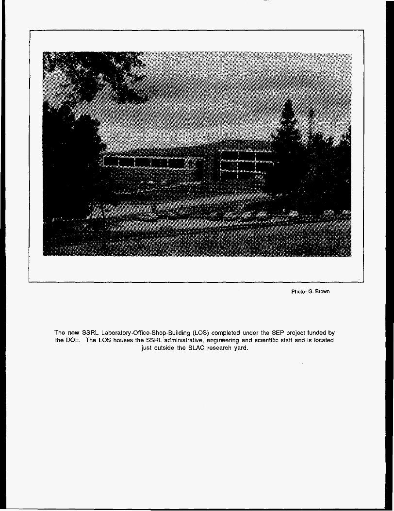
Photo- G. Brown
The new SSRL Laboratory-Office-Shop-Building (LOS) completed under the SEP project funded by the DOE. The LOS houses the SSRL administrative, engineering and scientific staff and is located
just outside the SLAC research yard.
SSRL Activity Report 1987
INTRODUCTION
During 1987, SSRL achieved many significant advances and reached several major milestones utilizing both SPEAR and PEP as synchrotron radiation sources as described in this report. Perhaps the following two are worthy of particular mention:
1. SPEAR reached an all time high of 4,190 delivered user-shifts during calendar year 1987. Highlights of the many scientific results are given below.
2. During a 12 day run in December of 1987, PEP was operated in a low emittance mode (calculated emit- tance 6.4 nanometer-radians) at 7.1 GeV with currents up to 33 mA. A second undulator beam line on PEP was commissioned during this run and used to record many spectra showing the extremely high brightness of the radiation. PEP is now by far the highest brightness synchrotron radiation source in the world.
It is gratifying to note that work done at SSRL has been recognized by an impressive array of awards during 1987 as listed at the end of this introduction.
FACILITY DEVELOPMENT
Laboratory operations are reviewed in Section I of this report. During 1987, the SLAC Linear Collider (SLC) was being commissioned, resulting in limited injection opportunities during dedicated runs on SPEAR. Typi- cally, injection was available once every 12 hours. Due to the usually long lifetimes on SPEAR this was not a major problem during runs for experiments. However, the limited injection opportunities made it difficult or impossible to pursue accelerator physics studies to improve the performance of the SPEAR ring and train graduate students in accelerator physics.
These limitations on injection from the SlAC linac will be a thing of the past once the new 3 GeV dedicated synchrotron injector to SPEAR is completed in 1990. This major construction project was started in October, 1987. (Actually, its official start was delayed until February, 1988 due to delays in the federal budget process for fiscal year 1988.) This injector will end dependence on the SLAC linac schedule and eliminate interfer- ence with SLC operation. As a full energy injector, it will improve the stability and reproducibility of the photon beams from SPEAR, as well as providing more running time at a lower cost per shift. R will also facilitate the reduction of the SPEAR emittance and will increase opportunities for accelerator physics studies to improve the performance of the SPEAR storage ring and train graduate students. The injector project is described in Section II of this report.
In October, 1987 SSRL conducted a Workshop on PEP as a Synchrotron Radiation Source attended by 125 scientists from many laboratories in the US, Europe and Japan. An accelerator physics working group provided a critical assessment of PEP’s potential as a low emittance storage ring. Nine working groups jealing with different applications discussed many experiments that could be done on PEP, particularly experiments that would make use of its unique features. [See Section V .J
Mth the major interest in PEP as a synchrotron radiation source that was evident at the PEP Workshop, and the dramatic demonstration of PEP’s capabilities in the December, 1987 tun, it became clear that PEP will be 2xtremely important to national needs in synchrotron radiation research. Even in parasitic operation during :olliding beam runs (which are scheduled to resume in August, 1988 after a two year period with no colliding 3eam operation) PEP offers x-ray beams above 10 keV with an order of magnitude higher brightness than my other source. During the 2-3 months per year available for dedicated low emittance operation, even much higher brightness can be reached.

Experiments using these higher levels of brightness and coherent power will start now, rather than waiting until the mid 1990’s when new, third generation rings designed for these performance levels are expected to start operation. Furthermore, PEP could serve as a test bed for these new rings and for the development of their insertion devices and beam line instrumentation.
In addition, PEP has characteristics (particularly its 2200 meter circumference, its 117 meter long straight sections and its 16 GeV energy) that offer the potential to achieve performance levels exceeding those of the third generation rings now in design and construction at other labs. For example, calculations indicate that, with the use of 100-200 meters of damping wigglers in the long straight sections, the emittance of PEP could be reduced to less than one nanometer-radian at 6 GeV. PEP could therefore be the prototype of a diffraction-limited x-ray source for the future. [See Section 11.1
The utilization of PEP however, will be limited by its dependence on the SLAC linac for injection, in the same way that this presently limits SPEAR. Therefore our thoughts turned to possibilities for injection to PEP independent of the SLAC linac. A plan was developed to enlarge the SPEAR 3 GeV synchrotron injector from a circumference of 100 meters to 133 meters, so that it could be later upgraded to function at 5 GeV, a level considered to be a reasonable minimum injection energy for PEP. The 5 GeV beam from this ring would be transported through a 120 meter tunnel, at the end of which it would connect to the present electron injection line of the PEP ring. After injection at 5 GeV, the energy of PEP would be ramped to the higher energy that would be needed for most synchrotron radiation experiments. Although this scheme would not provide full energy injection into PEP, it is the quickest and most cost-effective path to achieving dedicated injection. [See Section /I.]
The addition of 5 new experimental stations on SPEAR during 1987, plus the simultaneous operation of SPEAR and PEP, placed severe demands on the SSRL technical and scientific staff. Budget constraints have prevented the staff from growing at a rate commensurate with the increased number of stations and user community. In addition to the new beam line that was commissioned on PEP, two new participating research team (PRT) beam lines were commissioned on the SPEAR storage ring. These were Beam Lines Vlll and X, a joint effort of several US national laboratories in collaboration with the University of California system and SSRL. The national laboratories are Lawrence Livermore, Los Alamos and Sandia. The Lawrence Livermore Laboratory is the lead laboratory for the entire effort. This collaboration added signifi- cantly to SSRL’s experimental capabilities with two magnificent new monochromators in the VUV and soft x- ray spectral region plus a hard x-ray station operating off a very powerful 15 period, 1.4 Tesla hybrid wiggler magnet. [See Section I//.]
Although accelerator physics studies were severely limited because of the lack of the adequate opportuni- ties for frequent injection into SPEAR, some studies were carried out as described in Section II. These include studies of ramping, ion clearing, stability, wiggler characterization, filling patterns, timing mode, beam steering and operation at energies above 3 GeV.
During the 12-day low emittance run on PEP, a group of more than 20 scientists from SSRL, SLAC, Ar- gonne National Laboratory, Brookhaven National Laboratory and the Lawrence Berkeley Laboratory col- laborated in a wide range of studies to characterize the low emittance lattice, evaluate single and multi- bunch instability limits, study the beam steering and stabilizing system and determine aperture requirements for injection. In addition, many detailed studies of the characteristics of the undulator beam in low emittance operation were carried out. [See Section I/.]
Improvements to the large and increasing range of SSRL experimental facilities are described in Section 111 of this report. This includes the commissioning of new facility beam lines and the improvements to existing facility beam lines that serve both the VUV and x-ray parts of the spectrum. Significant improvements have been made in the output flux, reliability and user-friendliness of the systems. The very significant capabili- ties that SSRL offers for protein crystallography, x-ray absorption spectroscopy and small angle scattering are also described in Section 111.
During 1987, the SSRL enhancement project (SEP) was essentially completed. This very successful con- struction project provided a number of major new capabilities to the laboratory including two beam lines on the PEP storage ring, a major upgrade in the steering and stabilizing systems for SPEAR and PEP, additions to the nuclear physics injector (NPI) system to enable it to provide injection into the SPEAR and PEP storage rings and a new laboratory/off ice/shop (LOS) building. The LOS building provides new support facilities including a machine shop, a vacuum assembly area, a metrologyhnirror coating facility, a vacuum clean room, a VUV research laboratory, an x-ray laboratory, an electronics shop, a biotechnology laboratory, a design/ drafting area and a central computer facility as well as numerous staff offices, conference rooms and a library. Providing all these facilities in one centralized location is expected to increase efficiency as well as free up much needed space close to the beam lines for users.
A major improvement in computational facilities is underway at SSRL with the introduction of the new VAX 8700 computer system which will replace the VAX 1 l n 8 0 and VAX 1 ln50 systems. The beam line comput- ers are also being upgraded.
The broad range of engineering efforts at SSRL in 1987 are described in Section IV of this report. The major activities include the design of a pinhole camera system for PEP Beam Line lB, studies of beryllium window heating, thermal-structural analysis of several SSRL beam line components, the introduction of computer aided design systems, improvements to beam line control, wiggler control and beam line steering and stabili- zation systems and the development of beam line and storage ring status monitoring systems.
HIGHLIGHTS OF RESEARCH ACTIVITIES
Synchrotron radiation research at x-ray wavelengths now impacts an astonishing diversity of disciplines. At SSRL, vigorous programs are being pursued in materials sciences, chemical sciences, biology, medicine, and fundamental physics. These studies are carried out in many variants on the three basic interactions of photons with matter: absorption, elastic scattering, and inelastic scattering. Through absorption spectrosco- pies, once can decipher the local atomic and electronic structure of materials; through elastic scattering one can probe correlations from a few A to a few microns; and with inelastic scattering one can probe excitations in matter from a few meV to several hundred electron volts.
Because of this diversity of research, no overview can possibly substitute for the individual research reports presented in the Activity Report. However, we would like to call to the reader’s attention the intense work proceeding on the high transition temperature superconductors (868M, 984M, 7073M, 7079M, 7097Mp, 2023Mp). In these experiments, near-edge and extended x-ray absorption fine structures have been meas- Jred and applied to understanding the dependence of electronic and atomic structure upon preparation techniques, stoichiometry, transition temperature, and so forth.
High-resolution scattering continued to play an important role in x-ray research at SSRL. Of particular note is ihe work of Brock et al. (707 7M) on orientational ordering in tilted hexatic liquid crystal films. This program is sxploring the fascinating interplay between position order parameters and bond orientation order parameters, n very thin freely suspended liquid crystal films. The detailed phase diagrams of these systems are now Deing studied, and have already contributed greatly to the understanding of critical phenomena in 2 and 3 jimensions.
The resonant nuclear scattering program at SSRL (8003M) has achieved a major milestone, with the observa- .ion of the resonant diffraction signal from Fe-57 enriched crystals of Yttrium-Iron-Garnet. The rich time jtructure arising from the interference of the hyperfine sublevels has confirmed previous observations by )ther groups, and the time structure has been effectively modified by the interposition of resonant absorber,

which effectively “blanks out” selected lines.
Finally, the medical imaging group (7046B) has made significant progress in its quest for clinically useful images of the coronary vascular system of humans. In a series of trials on three human patients, profiles of the coronary arterial tree were obtained for various orientations of the patient, to test the ability to resolve the arteries from the ventricular background. Although analysis is not yet complete on this data, preliminary results appear to be very promising.
This has been a particularly significant year for achievements in the biotechnology area at SSRL. Perhaps most important has been the dramatic success realized through the use of multiple-wavelength phasing to solve the classic phase problem for crystallographic studies of proteins. Using data collected with the multi- wire SSRL area detector, several structures have now been fully solved. The structure of a copper-containing “blue” protein from cucumbers, which defied solution over a 1 O-year period by conventional approaches, has been phased and the structure refinement is well along (70046). Structures of a iron-containing ferredoxin and a selenium containing biotin-streptavidin complex (for which data on the later was partly collected at SSRL and partly at the Photon Factory) have also been obtained (7087Bp). Promising data is reported on a fourth structure, arnicyanin, and solution appears likely to soon be achieved (87056). This work clearly demonstrates the potential of this new approach based on multiple wavelength phasing to solution of protein structures.
The more classical approach of using several protein crystals containing heavy atom derivatives has also met with success for data collected on the rotation camera at SSRL. Six reports deal with structural work being done on this instrument. One particular highlight is the complete structure determination of an important oncogene protein isolated from human tumors (7A56). Another result with wide-ranging implications for protein crystallography using synchrotron sources is the observation of a dramatic improvement in crystal lifetimes upon cooling to temperatures of around 100 K using a new “flash freezing” technique (7A328).
X-ray absorption spectroscopy continues to find wide use in the study of the electronic and structural proper- ties of metal ions in biological systems. Several studies have resulted in a dramatic advancement of our understanding of the structure and function of manganese in the oxygen-evolving photosynthetic system (2070Bp, 7007B, 7085Bp). Nickel-containing enzymes have also been isolated relatively recently from organisms like methanogenic bacterial and XAS studies have helped in defining the nature of the active site (969 Bp). The spectral range of 2-4 keV, which contains the K absorption edges of elements like S, CI, and P and the L edges of the second row transition elements, has been opened up through the use of the Beam Line VI wiggler run at low magnetic field in undulator mode. Studies have revealed new information about the nature of sulfur in the iron-molybdenum cofactor from nitrogenase (87 76B) and appear promising for phosphorous-containing materials (70016).
VUV and Soft X-ray
During 1987 great progress was made in using EXAFS, SEXAFS, NEXAFS (Near-Edge X-Ray Absorption Fine Strucfure), PED (Photoelectron Diffraction) and ARPEFS (Angle-Resolved Photoemission Extended Fine Structure) to determine electronic and structural properties. These techniques are now being applied to problems with ever-increasing sophistication. The structure of P impurities in GaAs has been determined by EXAFS, as has the microscupic structure of sulfur doped AI,Ga,,As (7073Vp), thus extending this technique to highly dilute systems. Anisotropy in the surface vibrational amplitudes for CJ chemisorbed on Cu(lO0) has been examined for the first time with SEXAFS (947V). The surface structure of ordered S overlayers on Fe(001) and Cr(OO1) were determined by ARPEFS in a continued successful application of this technique (943Vp), and Beam Line VI was used for the first time in PED experiments, based on the Co 1s core line, to determine the surface geometry for (1x1) Co/Cu(OOl) (9900v). The electronic properties of clean and hydro-
gen terminated Si surfaces, as well as SiGe alloys, have been studied in Si K-edge absorption experiments (8104V). The same approach has been used for valency studies of Yb-compounds in this case based on the Yb L,,!,, absorption spectra (947 Vp). Major advances have been made using NEXAFS, in combination with the hlghly polarized synchrotron radiation, to determine the orientations and bonding distances for chemisorbed organic molecules on metal surfaces, e.g. methyl acetylide on Ag( 1 10) (963V) and benzen- ethiol on Mo(l10) (lUSOVp), further opening up the field of surface reactions for this kind of studies.
The electronic and structural properties of metalhemiconductor interfaces and heterostructures have continued to be the subject of both intense and successful studies. A detailed study of the bonding of arsenic and hydrogen to the Si(l11) surface has been completed (908Vp), as part of a broader reseach program on the GaAs-on-Si system. The effects of temperature and doping on the Fermi level pinning position in the bandgap of Ill-V semiconductors have been examined in the pursuit of understanding the basic mechanism behind Schottky barrier formation (935Vp). The effect of strain on the band structure of InxGa,,As, as manifested in the deformation potential of the upper valence bands, has been elucidated in detail (8704V) with important implications for understanding pseudo-morphic heterostructures in general. Excellent progress is also reported on photoelectron spectroscopic studies of metal ion sites for systems which are of interest in catalysis (7022Vp).
Electron correlation effects in narrow band materials, predominantly 4f and 5f containing lanthanides and actinides, have been studied with photoemission and resonance photoemission successfully for many years at SSRL. With the advent of the new high Tc materials these studies have intensified (7028V, 8UUOV).
The basic photoionization processes, in particular phenomena for excitation energies close to ionization thresholds, have received continued interest. These studies include valence and core level satellies for argon and lithium, respectively, photoelectron asymmetry parameter for the sulfur 2p level in SO, and autoionization resonances in He and Kr (943 Vp). Resonances in the K-edge absorption spectrum of solid Be have been reported (9907 V) and work on solid state effects in photoionization cross sections has continued (8OUUV).
The research efforts in x-ray lithography focussed on radiation damage in masks (1L09V) and on resist properties (8704V). Finally, during 1987 the first results were obtained using an imaging photoelectron microscope (8104V).
Herman Winick Acting Director (Arthur Bienenstock is on sabbatical)

1987 AWARDS BASED ON SSRL WORK
Oliver E. Buckley Condensed Matter Physics Prize of the American Physical Society to Robert J. Birgeneau (MIT) for “his use of neutron and x-ray scattering experiments to determine the phases and phase transitions of low dimen-
sional systems.”
Third Patterson Award of the American Crystallographic Association to David and Lieselotte Templeton (LBL) for “recognition of significant achievements in the accurate measurement of anomalous scattering terms at wavelengths near absorption edges using synchrotron radiation and pioneering contributions to our understanding of anomalous scattering of x-rays.”
Ernest Orlando Lawrence Memorial Award of the Department of Energy to David Moncton (EXXONIArgonne National Lab) “for the development of high resolution synchrotron x-ray scattering techniques and their applications to diverse materials systems.”
Prize for Achievement in Accelerator Physics and Technology of the US. Particle Accelerator School to Klaus Halbach (LBL) for “making high field permanent magnets practical tools for accelerator technology.”
J.D. Hanawalt Award of the International Centre for Diffraction Data to William Parrish (IBM San Jose) for “excellence in the field of powder diffraction.”
Department of Energy’s 1987 Materials Sclences Research Competition winner, Herman Winick (SSRL) in the categoty of Significant Implication for Energy Related Technology in Solid State Physics for “development of high brilliance, high flux synchrotron radiation beams with wiggler and undulator magnet insertion devices”.
The first Barrett Award of the Annual Denver Conferences on Application of X-ray Analysis to William Parrlsh (IBM San Jose) for outstanding contributions to powder diffraction.
MRS 1987 Graduate Student Award to Lane C. Wilson (Stanford University) for outstanding thesis research.

1. LABORATOR f OPERATIO
SSRL operations were at an all time high in 1987. Five new branch lines were opened to users. The highest number ever of 8-hour user shifts, 4190, were delivered. . A record number of SPEAR shifts, 396, were scheduled in three separate dedicated runs. The first run (January 2 to February 7) was a continuation of the winter 1986 run. A second dedicated run, March 18 through May 2, followed shortly. An unexpected opportunity for dedicated time arose in fall 1987, and, on short notice, the third run occurred from October 26 to December 24.
SPEAR is usually injected for SSRL running from the Nuclear Physics Injector (NPI) Gun at Sector 25 of the LINAC. If the SLAC Linear Collider (SLC) is running simultaneously with SPEAR, injection has been limited to once every 12 hours. The November 1986 to February 1987 run was the first operation in the 12 hour injection mode (see 1986 Activity Report) . In spite of this, the run was fairly successful, result- ing in average currents and lifetimes at injection of 65 mA and 15 hours respectively. The MarcWApril 1987 run, also in this mode, resulted in average beam currents of 72 mA and average lifetimes of 25 hrs. At lower currents, lifetimes approached 45 hours. This improved performance was due primar- ily to the fact that SPEAR remained under vacuum for the two months prior to the spring 1987 run. In fact, the spring run provided some of the stablest running SSRL has experienced in the past several years.
The fall 1987 run was anticipated to be very good because of the experience in the spring and because more frequent injection would be available. A number of problems, however, caused the perform- ance of SPEAR to be poor. The two major problems were primarily vacuum related. A SPEAR valve bellows, and subsequently a bellows in SPEAR itself, developed leaks. The second leak required venting part of the ring and repairing the drift tube. Because of these failures the pressure in SPEAR during the fall run was not as low as usual, resulting in shorter lifetimes and lower initial current. During this run the average lifetimes and currents at injection were 10 hours and 55 mA.
S
Since 1985 SLAC has been heavily involved in the construction and commissioning of the Linear Col- lider. This program has made planning for, and execution of, synchrotron radiation running more difficult. In particular, every attempt has been made to place the synchrotron radiation runs where they have the least impact on the SLC program. Since SLC is still in the construction/commissioning stage there have been difficulties in developing long-term schedules. SPEAR performance has also suffered since SLAC manpower is heavily committed to the SLC program. This year, for instance, only 65% of the scheduled beam was delivered and the fall run hit a low of 59%.
Neither SPEAR nor PEP has run for high energy physics since early 1986 which has meant no para- sitic operation for synchrotron radiation research. [A dedicated machine physics run was held on PEP in December which is described in the Accelerator Physics chapter.] Parasitic operation on SPEAR is used by synchrotron radiation researchers for training new users and students, testing equipment and performing research, particularly in the VUV, which can utilize the lower energy, lower currents and shorter lifetimes. SLAC’s present plans are to recommence high energy physics programs on both SPEAR and PEP in August of 1988.
One of the characteristics of the SPEAR ring is its excellent timing properties. Timing mode has four individual bunches of electrons equally spaced in the SPEAR ring and therefore separated by 190 ns. The higher current per bunch in timing mode results in a shorter lifetime and the need for more frequent injection. For this reason timing mode is not sched- uled when SSRL does not have on-demand access to the LINAC. Since the fall period was under conditions of on-demand injection, timing mode was run for the last three weeks, December 5 to 24th. Although the currents and lifetimes were still lower than in multi-bunch operation, timing mode this fall was less troubled than previously experienced.
SSRL ACTIVITY REPORT 1987 Page 1
OPERATIONS DIVISION ACTIVITIES
The Operations Division had a challenging year in 1987 with the spring and fall runs each presenting their own and different sets of demands. The spring run had limited injection and required very close coordination with SLAC operations to insure that SLC requirements did not compromise the SSRL program. Although unlimited injection was available during the fall run, a number of SPEAR related problems required close SSRL operations coordination to insure that the problems affecting SPEAR perform- ance were identified and repaired. In spite of these problems, both runs ended well.
The addition of 5 new stations plus running on PEP necessitated double staffing of duty operators. Since the Operations group has insufficient staff to provide two duty operators simultaneously, members of other SSRL groups were trained as "temporary" duty operators over the past year. This has added depth to the Operations staff and allowed it to deal with user demand during dedicated runs.
During the long May through October shutdown, the groups' activities shifted to construction and mainte- nance. During the past year, the Operations Division has participated in the construction of a number of beam and branch lines, including PEP lB, Beam Line V, and Beam Line X. In addition, the Operations group provides much of labor for beam line upgrades and routine maintenance.
During the coming year, the vacuum group and mirror coating facility will move to the new Labora- tory-Off ice-Shop Building (LOS). A precision machine shop in the LOS will be established. The machine shop in Building 120 will remain as a staff and user shop and will be upgraded. The Operations group will be working with users to insure that these changes do not affect the user support normally provided by the vacuum and operations groups.
Beam and SPEAR Usage Tables
The following tables and graphs contain statistics on SPEAR running, experimental use and characteris- tics of SSRL stations. For earlier information, consult previous Activity Reports.
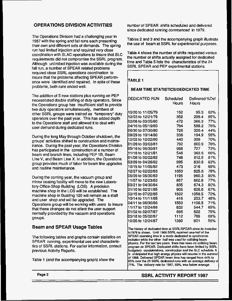
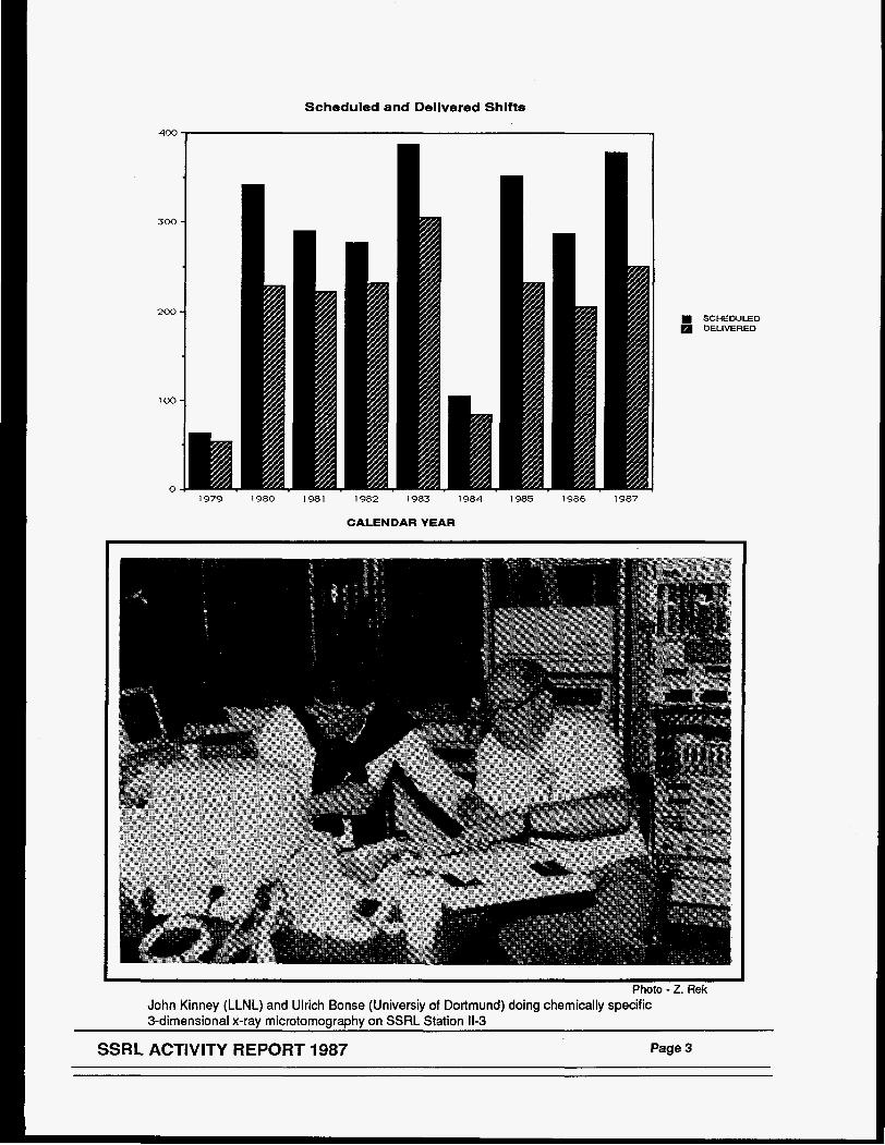
Table 1 (and the accompanying graph) show the
number of SPEAR shifts scheduled and delivered since dedicated running commenced in 1979.
Tables 2 and 3 and the accompanying graph illustrate the use of beam at SSRL for experimental purposes.
Table 4 shows the number of shifts requested versus the number of shifts actually assigned for dedicated time and Table 5 lists the characteristics of the 24 SSRL SPEAR and PEP experimental stations.
ABLE 1
BEAM TIME STATISTICS/DEDICATED TIME
DEDICATED RUN Scheduled Hours
lot20 to 1 1 /05/79 12/03 to 12/21/79 02/08 to 03/05/80 04/16 to 0511 9/80 06/30 to 07/30/80 09/29 to 1 Oil 4/80 12/02 to 12/22/80 01/26 to 03/03/81 05/1 6 to 06/30/81 11/18 to 12/21/81 01/08 to 02/22/82 03/09 to 04/26/82 10115 to 1 1/05/82 12/27 to 02/22/83 05/09 to 06/30/83 1 1/07 to 12/23/83 03/21 to 04/30/84 01/10 to 02/21/85 0311 5 to 07/22/85
04/11 to 06/30/86 1 111 7 to 12/24/86 01/02 to 02/07/87 03/18 to 05/02/87 10126 to 12/24/87
10/14to 11/11/85
152 352 472 764 726 336 440 792 988 546 748 995 473
1050 1195 857 835 905
1502 41 6
1550 832 696
1112 1360
Delivered %Del Hours
95.3 62% 299.4 85% 366.3 77% 588.2 76% 320.4 44% 194.9 58%
600.9 76% 309 70%
727 73% 363.6 66'/0 612.5 81% 830.9 83% 316 66%
960.3 80% 662.8 77% 674.3 80% 606.6 67%
825.6 78%
1056.5 70% 203.7 48%
1106.5 71% 544.7 65% 522 75% 769 69% 801 59%
IThe history of dedicated time at SSRUSPEAR since its inception in1979 is shown. Until 1986 SSRL received onshalf of the SPEAR operating time in a mode dedicated to synchrotron lradiation while the other half was used for colliding beam 'physics. For the last two years there has been no colliding beam program on SPEAR. Dedicated shifts have been limited by S S R L budgetary considerations, construction and the SLC schedule. It is anticipated that high energy physics will resume in the summei of 1988. Delivered SPEAR beam time has ranged from 44% to ~85% over the 25 SSRL dedicated runs with an average delivery o 71%. The delivery rate for 1987,65%0, was below average.
Page 2 SSRL ACTIVITY REPORT 1987
Scheduled and Delivered Shifts
400
300
200
1 00
0 1979 1980 1981 1982 1983 1984 1985 1986 1987
CALENDAR YEAR
SCHEDULED DELIVERED
t
Photo - 2. Rek John Kinney (LLNL) and Ulrich Bonse (Universiy of Dortmund) doing chemically specific 3-dimensional x-ray microtomography on SSRL Station 11-3
SSRL ACTIVITY REPORT 1987 Page 3
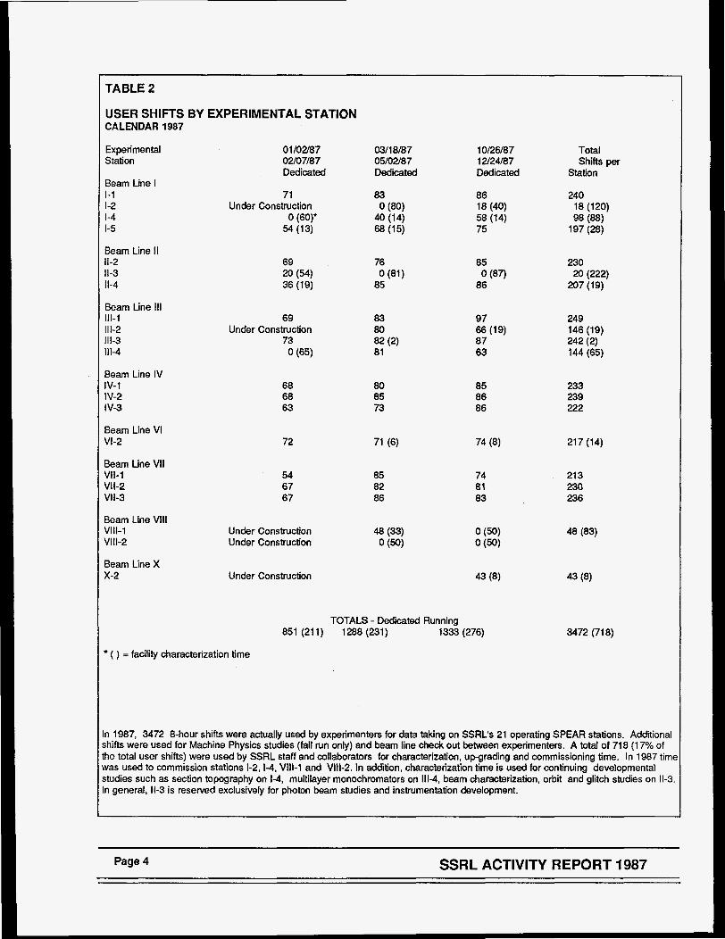
TABLE 2
USER SHIFTS BY EXPERIMENTAL STATION CALENDAR 1987
Experimental Station
01/02/87 02/07/87 Dedicated
03/18/87 05/02/87 Dedicated
10126/87 12/24/87 Dedicated
Total Shifts per
Station Beam Line I 1-1 1-2 1-4 1-5
71 Under Construction
54 (13) 0 (60)'
83
40 (14) 68 (15)
0 (80) 86 18 (40) 58 (14) 75
240 18 (120) 98 (88)
197 (28)
Beam Line I1 11-2 11-3 11-4
69 20 (54) 36 (19)
76
85 0 (81)
85
86 0 (87)
230 20 (222)
207 (19)
Beam Line 111 111-1 111-2 1113 111-4
69
73 Under Construction
0 (65)
83 80 82 (2) 81
97
87 63
66 (19) 249 146 (19) 242 (2) 144 (65)
Beam Line IV iv-1 IV-2 I v-3
68 68 63
80 85 73
85 86 86
233 239 222
Beam Line VI VI-2
Beam Line VI1 VII-1 Vll-2 Vll-3
72 217 (14)
54 67 67
85 82 86
74 81 83
21 3 230 236
Beam Line Vlll VIII-1 Vlll-2
Under Construction Under Construction
Beam Line X x-2 Under Construction
TOTALS - Dedicated Running 851 (211) 1288 (231) 1333 (276) 3472 (718)
( ) = facility characterization time
n 1987, 3472 8-hour shifts were actually used by experimenters for data taking on SSRL's 21 operating SPEAR stations. Additional ihifts were used for Machine Physics studies (fall run only) and beam line check out between experimenters. A total of 718 (1 7% of he total user shifts) were used by SSRL staff and collaborators for characterization, upgrading and commissioning time. In 1987 time vas used to commission stations 1-2,1-4, V111-1 and Vlll-2. In addition, characterization time is used for continuing developmental ;tudies such as section topography on 1-4, multilayer monochromators on 111-4, beam characterization, orbit and glitch studies on 11-3. n general, 11-3 is reserved exclusively for photon beam studies and instrumentation development.
Page 4 SSRL ACTIVITY REPORT 1987
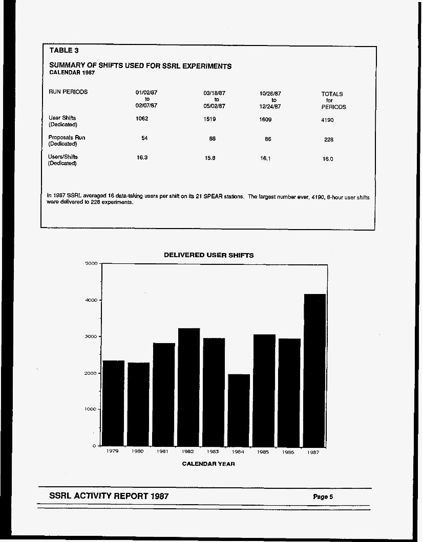
1 TABLE3
' SUMMARY OF SHIFTS USED FOR SSRL EXPERIMENTS CALENDAR 1987
RUN PERIODS
User Shifts (Dedicated)
Proposals Run (Dedicated)
UsersJShifts (Dedicated)
01/02/87 to
02/07/87
1062
54
16.3
0311 8187
05/02/87
1519
to 10126187
12/24/87 to
1609
TOTALS
PERIODS for
4190
88
15.8
86
16.1
228
16.0
In 1987 SSRL averaged 16 data-taking users per shift on its 21 SPEAR stations. The largest number ever, 4190,8-hour user shifts were delivered to 228 experiments.
DELIVERED USER SHIFTS
3 J
4000 -
3000 -
1982 1983 1984 1985 1986 1987
CALENDAR YEAR
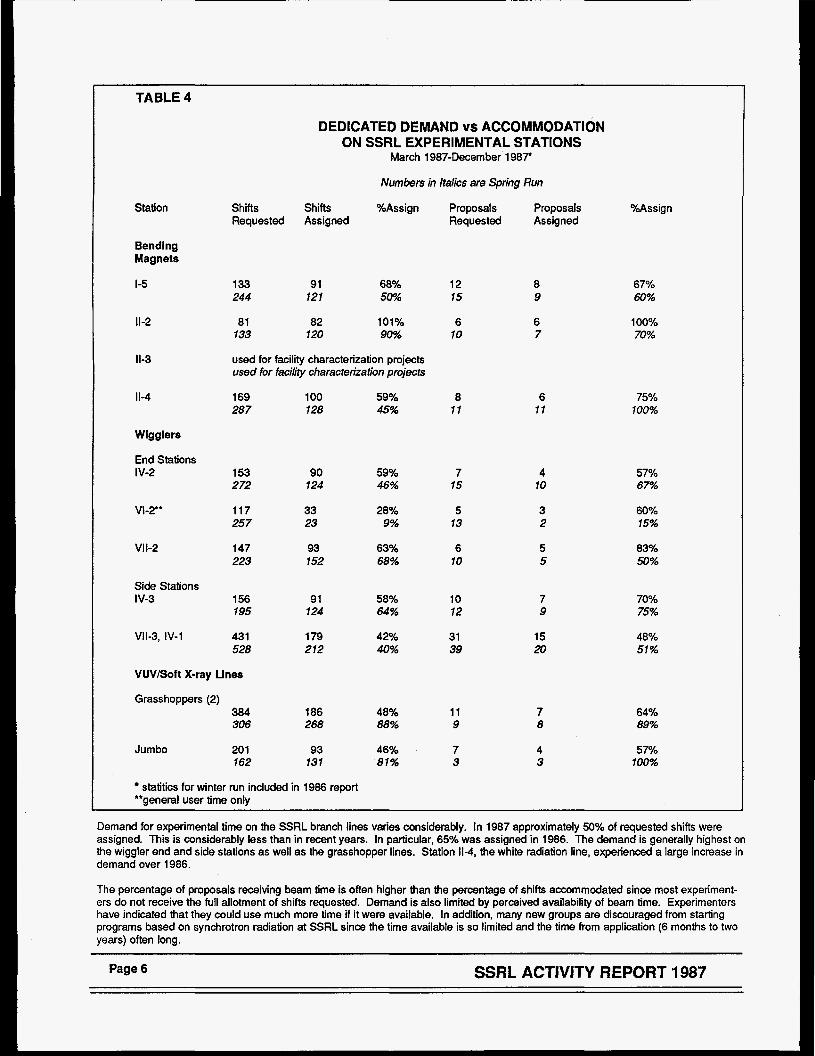
TABLE 4
Station
Bending Magnets
I -5
11-2
11-3
11-4
Wigglers
End Stations IV-2
VI-2"
VI 1-2
Side Stations I V-3
Vll-3, IV-1
DEDICATED DEMAND vs ACCOMMODATION ON SSRL EXPERIMENTAL STATIONS
March 1987-December 1987
Numbers in italics are Spring Run
Shifts Shifts %Assign Requested Assigned
133 91 68% 244 121 5077
81 82 101% 133 120 90%
used for facility characterization projects used for facility characterization projects
169 100 590/0 287 128 45%
153 90 59% 272 124 46%
117 33 28% 257 23 9%
147 93 63% 223 152 68%
156 91 58% 195 124 64%
431 1 79 &0/0 528 212 40%
VUVBoft X-ray Lines
Grasshoppers (2) 384 186 48% 306 268 88%
Jumbo 20 1 93 46% 162 131 81%
statitics for winter run included in 1986 report **general user time only
Proposals Proposals Requested Assigned
12 8 15 9
6 6 10 7
8 6 11 11
7 4 15 10
5 3 13 2
6 5 10 5
10 7 12 9
31 15 39 20
11 7 9 8
7 4 3 3
%Assign
67% 60%
100% 70%
75% 100%
57% 67%
60% 15%
83Yo 50%
70% 75%
48% 51%
64% 89%
57yo 100%
Demand for experimental time on the SSRL branch lines varies considerably. In 1987 approximately 50% of requested shifts were assigned. This is considerably less than in recent years. In particular, 65% was assigned in 1986. The demand is generally highest on the wiggler end and side stations as well as the grasshopper lines. Station 11-4, the white radiation line, experienced a large increase in demand over 1986.
The percentage of proposals receiving beam time is often higher than the percentage of shifts accommodated since most experiment- ers do not receive the full allotment of shifts requested. Demand is also limited by perceived availability of beam time. Experimenters have indicated that they could use much more time if it were available. In addition, many new groups are discouraged from starting programs based on synchrotron radiation at SSRL since the time available is so limited and the time from application (6 months to two years) often long.
Page 6 S S R L ACTIVITY REPORT 1987

TABLE 5
CHARACTERISTICS OF SSRL EXPERIMENTAL STATIONS
SSRL presently has 24 experimental stations 22 of which are located on SPEAR and two on PEP. 11 of these stalions are based on insertion devices while the remainder use bending magnet radiation.
Horizontal Mirror Monochromator Energy Angular Cut Off Range
Resolution
(ev) AtE
INSERTION DEVICES STATIONS
Amroximate Dedicated Instrumentation
LERLINFS - - XRAY Fnd Stations IV-2 (8-Pole)
VI-2 (54-Pole)
Focused 4.6 Unfocused 1.0
Focused 2.3 unfocused 1.0
Focused 4.6 Unfocused 1.0
F~cllsed 2.3 Unfocused 1.0
Vll-2 (8-Pole)
X-2 (31-Pole)
10.2
22
10.2
22
IV-1 1 .o IV-3 1 .o VII-1 1 .o VI113 1 .o
m l a t o r Lines - VUV/Soft X-Rav v-2 1.5
- - PEP 1 B FULL PEP 5B FULL 15.0
X-Bar 14 2.0
1-5 1 .o 11-2 (Focused) 4.8 11-3 1 .o 114 1 .o yUV/Soft X - R a 1-1 2.0 1-2 4.0 111-1 2.0 111-2 4.0 111-3 8-10
8.9
4.5
1114 0.6
v111-1 12
Vlll-2 5
Double Crystal Double Crystal
Double Crystal Double Crystal
Double Crystal Double Crystal
Double Crystal Double Crystal
Double Crystal Double Crystal Curved Crystal Double Crystal
Rowland Circle- Multiple Grating
Double Crystal Double Crystal
2800-1 0200 -5xl O4 280045000 -lo4
2800-21000 - 5 ~ 1 O4 2800-45000 -lo4
2800-10200 - 5 ~ 1 O4 2800-45000 -lo4
2800-21000 - 5 ~ 1 0 ~ 2800-45000 -lo4
2800-45000 4 x 1 O4 2800-45000 -lo4 6000-13000 - 8 ~ 1 0 ~ 2800-45000 -lo4
10-1 200 2 7%
12000-42000 -1 0-5 12000-20000 -1 0-5
BENDING MAGNET STATIONS
Curved Crystal 6700-10800 0.3 x lod
Double Crystal 2800-30000 -10" Double Crystal 2800-8900 -5 x 1 O4 Double Crystal 2800-30000 -5 x 1 fl None 3200-30000
2.0 x 6.0 2.0 x 20.0
2.0 x 6.0 2.0 x 20.0
2.0 x 6.0 2.0 x 20.0
2.0 x 6.0 2.0 x 20.0
2.0 x 20.0 2.0 x 20.0 0.6 x 3.0 2.0 x 20.0
Sixcircle Diffractometer
Twocircle Diff tactometer Rotation Camera
6.0 x 8.0 Angle Integrated e- Spectrometer
0.6 x 6.0 0.6 x 3.0 Sixcircle Diffractometer
0.25 x 0.5
3x20 1 x 4 3x20 3.5 x 18
Small Angle Scattering
Area DetectorKAD-4 Detector
Grasshopper 32-1000 M. = .1-.2 A 1 .O x 1 .O
Grasshopper 25-1200 M = .05-2 A 1 .O x 1 .O
UHV Double 800-4500 0.35 - 7 ev A 2.0 x 4.0
Multilayer 0-3000 White or 2 x 8
GmTGM 8-180 ~ . = . 0 6 - 3 A TBD
Seya-Namioka 5-50 AI,= .2-6A 2x7
Crystal (Jumbo)
A m = .3%
GmTGM
6fllSGM
8-1 80
50-1000
AX= .06-3 A TIM?
WAES22000 ld
Vaccum Diffractometer/ Lithography Exposure Station
Spectrometer
Spectrometer
Angle Resolved e-
Angle Resolved e-
SSRL ACTIVITY REPORT 1987 Page 7

/
\
\'\
/ /'
4 @
i I

II ACCELERATOR PHYSICS PROGRAM The Accelerator Physics group’s activities concen- trated on three major goals: to improve SPEAR operation, to implement low emittance operation of PEP and to design and construct a separate, full energy SPEAR injector.
SPEAR ACCELERATOR PHYSICS ACTIVITIES
During the spring 1987 run, most of the machine physics shifts were aimed at improving the normal operating condition of SPEAR. Specifically, the closed orbit correction scheme was cleared of various software problems and wrong corrector assignments. In addition, energy ramping was studied. The orbit control during ramping was found to be faulty and improvements were made.
Virtually no machine physics shifts were available during the fall run due to hardware failure and pressure for user shifts. A few shifts were devoted to ramping studies to reach energies above 3.0 GeV, to studies of optimum filling patterns, ion problems and dynamic aperture. Other studies on beam stability were done parasitically.
The particular activities are listed below.
PamD ina Stud ies: Studies were performed of differ- ent methods of controlling the ramping rates of the various SPEAR components from injection to final energy.
Jon Clearing: Studies of the effectiveness of biasing the position monitoring buttons in SPEAR straight section 7S8 for clearing ions from the Beam Line VI Wiggler vacuum chamber were made. No ion clearing effect was detected.
hpna Term Stab iI&: Measurements were made of the long term beam position stability of the SPEAR electron beam using a pinhole camera in Beam Line II and split ion chamber position monitors. Diurnal effects were observed, perhaps related to thermal conditions.
Peam Line X W iaaler Character i 7 W : The corrector values for compensating the effect of the residual
field integral of the Beam Line X Wiggler on the stored electron beam were measured and saved for automatic compensation by the SPEAR computer.
: Studies were performed of Alternate F i l l i ~ Patterns nonstandard filling patterns suitable for doing time resolved photo-electron spectroscopy of surfaces on a sub micro-second time scale. The desired fill patterns were achievable but, due to bunch to bunch intensity variations, a fast beam monitor may be required.
. .
IC Aoerture S m : An investigation of the effects of sextupolar fields on the dynamic aperture of SPEAR was carried out. This work was done as part of one of the first Stanford University student theses in Accelerator Physics at SSRL.
Sextupoles are used to make the chromaticity positive, but the non-linearity of the field also causes the oscillatory motions to become unstable at some amplitude. This limiting amplitude, the dynamic aperture, depends on the position of the sextupoles around the ring as well as their strengths. A large dynamic aperture is necessary in the design of future low-emittance rings which have very strong sextupoles.
In the normal operation of SPEAR the three families of sextupoles are set at low enough values such that the maximum amplitude of betatron motion is determined by the vacuum chamber size and the orbit distortion around the ring (geometric accep- tance), and not by the above instability. In this respect, SPEAR’S normal lattice is too “good” to demonstrate non-linear effects.
The goal of the measurements was to incease the sextuple strengths, to observe any subsequent reduction of dynamic aperture, and to compare results with computer tracking simulations. Prelimi- nary data reduction shows that the decrease in observed beam lifetime (an indirect measure of acceptance, under certain conditions) is due to the shrinkage of the chromatic acceptance rather than the geometric acceptance. When the sextupoles are turned up to about twice their usual strengths, an appreciable number of particles populating the tails of the energy distribution become unstable and are lost.
SSRL ACTIVITY REPORT 1987 Page 9
There is no conclusive evidence of instability due to geometric aberration, as the limiting amplitude is probably still greater than the geometric acceptance. Computer tracking simulations are not completed as of this writing.
Timina Mode OD . eratioQ: Studies were performed to determine an optimum filling pattern in timing mode in order to avoid poor lifetimes due to ion collection. Although results were varied, it seems clear that the problem is minimized if a large portion of the ring circumference (-50%) is left empty.
Hiah F n e r w a t ion: Attempts were made to ramp stored beam to higher than normal energy to test feasibility of future human angiography studies on SPEAR. It is possible to ramp beams to >3 GeV, although a tuneLup of the RF system is important before doing so.
Steerina Tests : The new steering installation for Beam Line X was tested and made operational. A prototype steering feedback servo system was tested prior to finalizing the design. (See Beam Line Steering in Chapter Wfor details.) A beam position noise monitoring technique was tried.
3 GEV INJECTOR PLANS
SSRL's preparations for the construction of a dedi- cated 3 GeV SPEAR injector synchrotron made significant progress in 1987. This project, proposed last year and included in the President's FY '88 budget proposal, was finally approved by Congress in mid-January, 1988 after passing DOE validation, construction and cost reviews and a technical review by outside committee members. The injector will be located next to the SPEAR storage ring and will be configured such that SPEAR can be operated for synchrotron radiation product ion without interference with other programs on the SLAC site. Since the approval, activities are being concentrated on building up the group, preparing designs for long lead time items and prototype activities.
In view of the successful operation of PEP in the low emittance mode, it was suggested that the 3 GeV SPEAR injector could be upgraded later to 5 GeV if the circumference of the booster was increased to 133 meters. Although this would not make'an ideal injector for PEP, it would provide injection when none is available during SLC running. In order to make this potential viable, a 400 foot tunnel has to be con-
structed from the SPEAR booster to the PEP region # I O . There the beam transport line would tie into the existing electron injection line into PEP. After beam accumulation in PEP, the energy would be ramped to the final operating energy. This option is not intended to replace a full energy injector for PEP at a later date.
A separate request for funds will have to be made to cover the additional costs for later upgrade to obtain the 5 GeV capability of the SPEAR injector.
ACCELERATOR PHYSICS STUDIES ON PEP
From December 9-21,1987 PEP was operated at 7.1 GeV in a low emittance configuration optimized for the production of high brilliance synchrotron radiation from undulators. The run was extremely successful in verifying the basic capabilities of PEP as a low emittance source of synchrotron radiation. A large amount of data was taken and is now being ana- lyzed. Technical reports are in preparation. Four abstracts have been submitted to the April 18-21, 1988 meeting of the American Physical Society in Baltimore, where there will be a special session on synchrotron radiation sources. The titles and authors of these abstracts are given below.
The main purpose of the PEP run was to study the operational characteristics of the low emittance configuration (which was tested briefly in a one day run in March, 1986), to measure the emittance achieved, to identify possible limits on stored current and stability and to measure the characteristics of the radiation using the two undulator beam lines now installed in PEP.
These studies were carried out by physicists from SSRL, SLAC, Argonne National Laboratoty, Brookhaven National Laboratory and the Lawrence Berkeley Laboratory, all of which have interest in low emittance electron rings. (See the listing of names at the end of this report.) The following is a brief synopsis of the work carried out and some tentative results, pending further analysis.
STUDIES PERFORMED
1. The low emittance lattice (called PEP 29) was implemented and characterized at 7.1 GeV. The results are in agreement with the model.
Page 10 SSRL ACTIVITY REPORT 1987
2. Prelimina? evaluation of the data taken indicate that the measured horizontal emittance is in agree- ment with the value of 6.4 nanometer-radians calcu- lated for the uncoupled horizontal emittance for this lattice at 7.1 GeV. An even lower value, consistent with a calculated horizontal emittance of 3.3 nanome- ter-radians, was measured by changing the damping partition, as described below. Most of the data was taken at low current (about 1 mA or less). There is some indication of emittance growth at higher currents.
3. Stored currents up to 33 mA were achieved in multi-bunch mode with reasonable stability at the higher current and very good stability at lower current. However, it was difficult to reproduce currents above about 15 mA.
4. The maximum beam lifetime ranged from about 2 hours at the highest current to 3 hours at low current, limited by the relatively high average pressure in the ring which had not been operated for about 20 months.
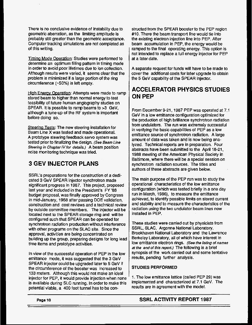
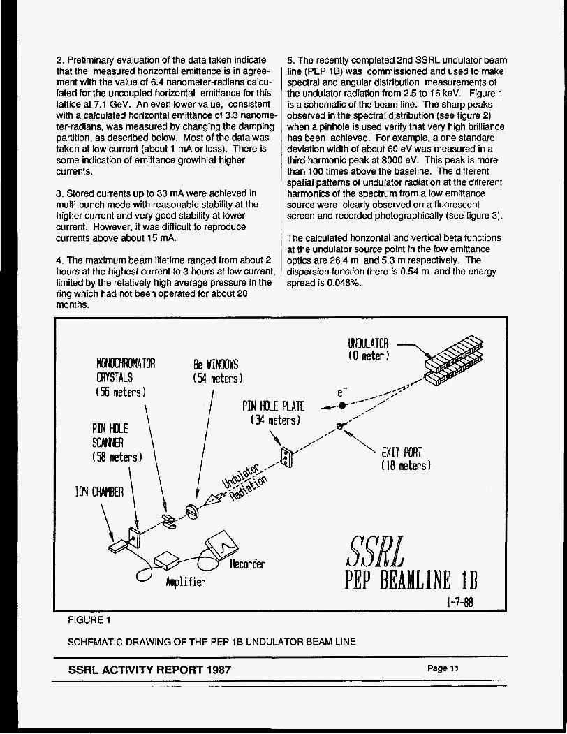
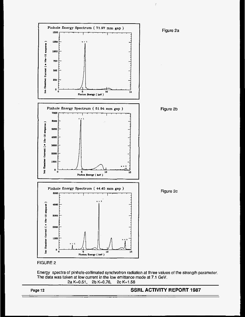
5. The recent11 completed 2nd SSRL undulator beam line (PEP 1 B) was commissioned and used to make spectral and angular distribution measurements of the undulator radiation from 2.5 to 16 keV. Figure 1 is a schematic of the beam line. The sharp peaks observed in the spectral distribution (see figure 2) when a pinhole is used verify that very high brilliance has been achieved. For example, a one standard deviation width of about 60 eV was measured in a third harmonic peak at 8000 eV. This peak is more than 100 times above the baseline. The different spatial patterns of undulator radiation at the different harmonics of the spectrum from a low emittance source were clearly observed on a fluorescent screen and recorded photographically (see figure 3).
The calculated horizontal and vertical beta functions at the undulator source point in the low emittance optics are 26.4 m and 5.3 m respectively. The dispersion function there is 0.54 m and the energy spread is 0.048%.
MOMCHROMATOR Be WINDOWS CRYSTALS (54 meters) (56 meters 1
PIN HOLE PLATE (34 meters) PIN HOLE \ I \
\ I SCANNER (58 neters 1
ION
" Amplifier
/
/* "\ EXIT PORT ( 18 meters)
PEP BEAMLPNE 1 B 1-7-88
FIGURE 1
SCHEMATIC DRAWING OF THE PEP 1B UNDULATOR BEAM LINE
SSRL ACTIVITY REPORT 1987 Page 11
Pinhole Energy Spectrum ( 72.97 mm gap ) 1500 I a I
I " " -
- 1250 - n - 1
1 I 8 looo) -
-
2
4 750
ii W L 5
t
- - - -
U
250 - -
0 5 10 15
e O A 8 * - I - I Photon Energlr ( keV )
Pinhole Energy Spectrum ( 61.94 mm gap ) 7000 , , , I I " " j
Y
I -
L 1 1 - 1
Bo00 - - m - -
4ooo- -
3OOo- -
2000 - -
lo00 - - 0 - 3
0 - I - ' I . . . A I 0 5 10 15
Photon Energy ( Lev )
Pinhole Energy Spectrum ( 44.45 mm gap )
- Photon Energy ( Lev )
FIGURE 2
Figure 2a
Figure 2b
Figure 2c
Energy spectra of pinhole-collimated synchrotron radiation at three values of the strength parameter. The data was taken at low current in the low emittance mode at 7.1 GeV.
2a K=0.51, 2b K=0.76, 2c K=l.58
Page 12 SSRL ACTIVITY REPORT 1987

FIGURE 3
Patterns of undulator radiation observed on a fluorscent screen located at 58 meters from the undulator.
~~~~ ~~
SSRL ACTIVITY REPORT 1987 Page 13
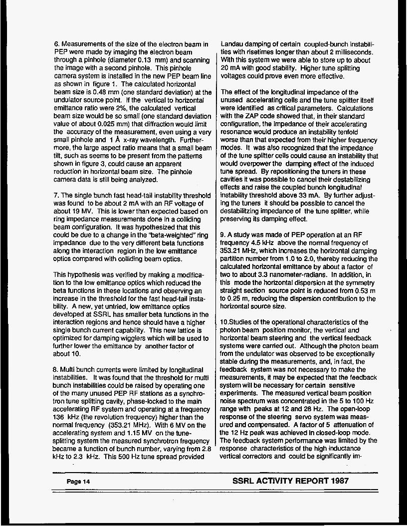
6. Measurements of the size of the electron beam in PEP were made by imaging the electron beam through a pinhole (diameter 0.13 mm) and scanning the image with a second pinhole. This pinhole camera system is installed in the new PEP beam line as shown in figure 1. The calculated horizontal beam size is 0.48 mm (one standard deviation) at the undulator source point. If the vertical to horizontal emittance ratio were 2%, the calculated vertical beam size would be so small (one standard deviation value of about 0.025 rnm) that diffraction would limit the accuracy of the measurement, even using a very small pinhole and 1 A x-ray wavelength. Further- more, the large aspect ratio means that a small beam tilt, such as seems to be present from the patterns shown in figure 3, could cause an apparent reduction in horizontal beam size. The pinhole camera data is still being analyzed.
7. The single bunch fast head-tail instability threshold was found to be about 2 mA with an RF voltage of about 19 MV. This is lower than expected based on ring impedance measurements done in a colliding beam configuration. It was hypothesized that this could be due to a change in the “beta-weighted” ring impedance due to the very different beta functions along the interaction region in the low emittance optics compared with colliding beam optics.
This hypothesis was verified by making a modifica- tion to the low emittance optics which reduced the beta functions in these locations and observing an increase in the threshold for the fast head-tail insta- bility. A new, yet untried, low emittance optics developed at SSRL has smaller beta functions in the interaction regions and hence should have a higher single bunch current capability. This new lattice is optimized for damping wigglers which will be used to further lower the emittance by another factor of about 10.
8. Multi bunch currents were limited by longitudinal instabilities. It was found that the threshold for multi bunch instabilities could be raised by operating one of the many unused PEP RF stations as a synchro- tron tune splitting cavity, phase-locked to the main accelerating RF system and operating at a frequency 136 kHz (the revolution frequency) higher than the normal frequency (353.21 MHz). With 6 MV on the accelerating system and 1.15 MV on the tune- splitting system the measured synchrotron frequency became a function of bunch number, varying from 2.8 kHz to 2.3 kHz. This 500 Hz tune spread provided
Landau damping of certain coupled-bunch instabili- ties with risetimes longer than about 2 milliseconds. Wih this system we were able to store up to about 20 mA with good stability. Higher tune splitting voltages could prove even more effective.
The effect of the longitudinal impedance of the unused accelerating cells and the tune splitter itself were identified as critical parameters. Calculations with the ZAP code showed that, in their standard configuration, the impedance of their accelerating resonance would produce an instability tenfold worse than that expected from their higher frequency modes. It was also recognized that the impedance of the tune splitter cells could cause an instability that would overpower the damping effect of the induced tune spread. By repositioning the tuners in these cavities it was possible to cancel their destabilizing effects and raise the coupled bunch longitudinal instability threshold above 33 mA. By further adjust- ing the tuners it should be possible to cancel the destabilitzing impedance of the tune splitter, while preserving its damping effect.
9. A study was made of PEP operation at an RF frequency 4.5 Wz above the normal frequency of 353.21 MHz, which increases the horizontal damping partition number from 1 .O to 2.0, thereby reducing the calculated horizontal emittance by about a factor of two to about 3.3 nanometer-radians. In addition, in this mode the horizontal dispersion at the symmetry straight section source point is reduced from 0.53 m to 0.25 m, reducing the dispersion contribution to the horizontal source size.
1O.Studies of the operational characteristics of the photon beam position monitor, the vertical and horizontal beam steering and the vertical feedback systems were carried out. Although the photon beam from the undulator was observed to be exceptionally stable during the measurements, and, in fact, the feedback system was not necessary to make the measurements, it may be expected that the feedback system will be necessary for certain sensitive experiments. The measured vertical beam position noise spectrum was concentrated in the 5 to 100 Hz range with peaks at 12 and 28 Hz. The open-loop response of the steering servo system was meas- ured and compensated. A factor of 5 attenuation of the 12 Hz peak was achieved in closed-loop mode. The feedback system performance was limited by the response characteristics of the high inductance vertical correctors and could be significantly im-
Page 14 SSRL ACTIVITY REPORT 1987

proved with new corrector magnets. The higher tune of the low emittance lattice results in poor sensitivity of the horizontal steering bumps. This could be improved by powering other, presently unused, corrector magnets. The effect of bending magnet fringe field pollution on the undulator beam position signal was also studied.
11 .Injection efficiency as a function of vertical aper- ture was studied. This data is relevant to the deter- mination of the minimum aperture that can be allowed for a fixed vacuum chamber for a small gap, short period undulator.
ABSTRACTS BASED ON THE PEP RUN SUBMITTED
CAN PHYSICAL SOCIETY TO THE APRIL 18-21, 1988 MEETING OF THE AMERI-
Operation of PEP at Low Emittance as Synchrotron Radiation Source; H. WlNlCK (SSRL) for the PEP Study Group*
Experimental Study of Collective Effects for the PEP LowEmittance Optics; M. S. ZISMAN (LBL); M.ALLEN (SLAC); M. DONALD (SLAC); J. GALAYDA (BNL); A. JACKSON (LBL); S. KRAMER (ANL); L. RlVKlN (SLAC); for the PEP Study Group'
Lattice Characterization of PEP I1 Low Emittance Configuration; M. BORLAND (SSRL) and M. DONALD (SLAC); for the PEP Study Group"
Performance of the X-Ray Undulator on PEP; W. LAVENDER (SSRL); G BROWN (SSRL); R. COISSON (SSRL, U. PARMA); T. TROXEL (SSRL); for the PEP Study Group*
* PEP Study Group: (ANL) S. Kramer, M. Yoon; (BNL) B. Craft, G. Decker, J. Galayda; (LBL) A. Jackson, M. Zisman; (SLAC) M. Allen, E. Bloom, M. Donald, J. Pater- son, J.-L. Pellegrin, B. Richter, L. Rivkin, H. Schwarz; (SSRL) M. Borland, G. Brown, R. Coisson, L. Emery, R. Hettel, W. Lavender, J. Safranek, T. Troxel, H. Wiede- mann, H. Winick, W. Xie.
SSRL ACTIVITY REPORT 1987 Page 15

Ill EXPERIMENTAL FACILITIES
Experimental facilities at SSRL are of two types: general facility stations and participating research teams (PRT's). General facility stations have been funded by various government agencies, principally the DOE, NIH and NSF and are open to the general user community on a competitive basis for 100% of their operating time.
Presently, SSRL has three operating Participating Research Team's with a fourth group having recently been formed. At SSRL all present PRT's are a three- party collaboration with SSRL as one of the parties. The two outside institutions receive 2/3rds of the available beam time while 1/3 is reserved for the SSRL general users. The PRT arrangements are for a 3 year term. Renewal is based on a review which considers scientific merit and contributions to gradu- ate education and to the SSRL user community.
The three existing PRTs are EXXON/Lawrence Berkeley Laboratory/SSRL (Beam Line VI), Univer- sity of California/National Laboratories(with LLNL as the lead lab)/SSRL (Beam Lines Vlll and X) and Xerox/Stanford/SSRL (Beam Line V). lBM/The Stanford Center for Materials ResearcWSSRL are presently developing a side station on Beam Line X in a PRT mode.
X-RAY FAClLlTlES
improvements to Existing Facility Lines
Branch I ine 1-4 - A major improvement program on Branch Line 1-4 has occurred with the installation of a new 8.5 degree asymmetrically cut silicon monochro- mator. With the new monochromator, the flux at the fundamental has increased by about 50%. Tests were performed for the first time at the third harmonic (26 KeV) and a useful spot size (1.5 x 0.5 mm) was achieved.
Line IV-3 - Branch Line IV-3 is midway through a major improvement program, with signifi- cant mechanical improvements to the two-circle diffractometer, and with construction of a focussing mirror assembly in progress. The design is complete and assembly is scheduled for the summer of 1988. This focussing mirror will provide a horizontal line focus and will cut off the higher harmonics of the x- ray beam.
ch I ine VII-7 - Branch Line Vll-2, which is used for high-resolution scattering studies on single crystal samples, has been modified to easily accommodate the transition from focussed to unfocussed operation. Some minor changes have been made to the ab- sorber system, and work is in progress to convert the beam line control software to microVAXoperation.
General X-ray Facility Improvements
Monochromator Calibrators - Using the facility Beam Line 11-3, experiments are underway to map the complete muttiple reflection spectra of perfect crystal monochromators to aid in the choice of crystal reflections for applications within a given energy range. At the same time, a theory is being developed to utilize these reflections as calibration markers, with very high accuracy. Utilizing the dynamicai theory of diffraction, it should be possible to calibrate the monochromators to an accuracy of 0.1 eV or better.
h l o n o c h r o w lntensitv I ,evellingCirca - An inten- sity levelling circuit has been developed, in conjunc- tion with Frank Bridges of the University of California at Santa Cruz. The purpose of the circuit is to provide a constant intensity to the experiment, independent of the current stored in the electron beam. This is accomplished by detuning the crystal by 50% (using the piezoelective transducer) and then continuously adjusting the transducer, via a closed- loop feedback system, to maintain a constant inten- sity. A prototype system has been built and tested, and a system is now available for the general user community.
X-ray Facllity Experimental Stations Commissioned In 1987
-- The second PEP synchrotron radiation beam line, illuminated by an undulator located in symmetry straight section 1 B, was commissioned during a dedicated PEP run in De- cember, 1987. The beam line has characteristics that are identical to the beam line located at region 58, and is optimized to produce 12 KeV radiation when PEP is operated at 14.5 GeV.
During the commissioning program, PEP was operated at 7 GeV in the normal colliding beam
SSRL ACTIVITY REPORT 1987 Page 17
configuration, as well as in a high-tune, low emittance was brought through the TGM during the spring run configuration. In these modes, the angle-integrated and on-axis energy spectra were measured, as well as the electron beam spatial and angular profiles. The measurements agreed well with the theory, and the results are summarized in the Accelerator Physics Chapter.
VUV FACILITIES
The VUV facilities at SSRL are expanding. Four new experimental stations (two PRT and two facility) were commissioned in 1987 with more to come in 1988. At the same time, many improvements have been made to existing facilii ies. Several activities are being moved to the new LOS building or off the ground floor to make room for beam lines and setup space. In conjunction with the increase in the number of VUV and soft X-ray beam lines, three new staff members, two technicians and a scientist joined this group in 1987. The Perkin Elmer chamber was overhauled and cleaned. The VG chamber was brought back on line and was used to commission Branch Line 111-2 (the 18 degree line) and for user experiments. Most of the beam line computer sys- tems are being maintained in their current configura- tion awaiting the upgrade to microVAXes.
VUV Facility Experimental Stations Commissioned In 1987
Line 111-2 (18 degree line)-- Final alignment and commissioning of Branch Line 111-2 (18 degree line) took place in January 1987. Users were sched- uled and successful experiments were performed during the April, 1987 run. At that time, it was determined that the M1 focussing mirror was not bending enough to produce a good focus at the entrance slit. During the summer shutdown the mirror was replaced with a thinner trial mirror made of float glass. This improved the focus enough to double the flux through the monochromator. In addition, the motions on the M1 mirror manipulator were motor- ized. The mirror tank hangs from the ceiling down- stairs from where the user sits, so remote control is needed to access those motions from the experimen- tal station. A further modification to the line was to shift the refocussing mirror downstream by 20 inches to produce a smaller spot size at the sample position. These improvements resulted in enhanced perform- ance during the fall run in 1987.
Branch 1-2 (TGM) - The redesigned branch on Beam Line I was commissioned in 1987. First light
and initialalignment of the line took place. . Forthis tun a substitute M1 mirror was used, since the ellipsoidal mirror designed to focus on the entrance slit of the monochromator was not delivered until the summer. With the new focussing mirror, monochro- matic scans were achieved with all three gratings during the fall run. As part of the commissioning process, users successfully collected data. Further alignment of the optical components of the line will take place during the first tun of 1988. The flux on the line is still limited by the original flat MO mirror which has been in use for 15 years.
Improvements to Existing VUV Experimental Stations
Line 1-1 and Branch Line 111-1 (GrasshoDW - A theoretical study was performed to determine the improvement possible by replacing the spherical focussing mirror on the grasshopper monochromator with an elliptical one. From ray tracing it was calcu- lated that 1.6 times the flux, or two to three times improvement in photon energy resolution would be possible (See figures 4 and 5). An elliptical mirror, holder and bender were specified and ordered for each grasshopper monochromator. They are sched- uled to arrive early in 1988.
L ine 111-3 f J u m u - No major changes were made on Jumbo during 1987. In the coming year the data acquisition and control system is to be upgraded to a microVAX work station operating with the new software designed by the SSRL computer group. This new system and software will not only eliminate many of the long standing bugs that have aggravated users, but bring on-line data analysis and rapid data transfer capabilities to the branch line. In accordance with the recommendations of the Users Organization, Jumbo will be the first of the VUV lines with a mi- croVAX system, with the other stations to follow soon after.
Line 111-4 !-I ine) - Many im- >rovements were made to Branch Line 111-4 during 1987. The monochromator chamber was modified with a larger flange to allow easier access to optics. Several other small ports were added to mount 3auges, viewports and other instrumentation. The xmputer was upgraded (and simplified) to a Macin- osh improving the interface to the motor controllers. The cabling and plumbing were streamlined. Other components were rearranged to allow easy installa- tion of user filters and to improve access in general.
Page18 . SSRL ACTIVITY REPORT 1987

NEW MIRRORS FOR THE GRASSHOPPERS
v) c .- g E
-100 -
-150 -
. - .. 9 .
8 - . -100 ;
=150 -
- 2 0 0 0 -200 m -0.5 0.0 0.5 -0.5 0.0 0.5
centimeters centimeters
FIGURE 4 FIGURE 5
Scatter plot of rays at entrance slit with spherical M1 mirror. A slit 1 cm by 30 microns is overlaid. Only 60% of the rays make it through the slit.
Scatter plot of rays at entrance slit with elliptical M I mirror. A slit 1 cm by 30 microns is overlaid. 90% of the rays make it through the slit.
SSRL ACTIVITY REPORT 1987 Page 19
BIOTECHNOLOGY FACl LIT1 ES
The research and user support in the Biotechnolog! Division at SSRL are funded jointly by the NIH Division of Research Resources and the core SSRL operations budget from the DOE. The division's responsibilities also include oversight for the com- puter activities at SSRL.
Facilities for Prote in Crvsta lloaraohv - The SSRL area detector system for protein crystallography on Beam Line 1-5 was very actively used during 1987 for collection of anomalous scattering data on proteins. The major facility upgrade from a PDP 11/34 to a microVAX-ll GPX workstation control system was fully completed, and the system operates with significantly enhanced performance and functionality. A write-once, read many times 0.4 GByte (per side) optical disk has been added for high volume, reliable data archival.
The rotation camera beam line (Station VII-1) is a station fully dedicated to the collection of protein single crystal diffraction data and it continues to be fully scheduled. The station is essentially the same as it was during 1986. Plans are being made to provide capabilities for cooling crystals to liquid nitrogen temperatures in 1988.
The CAD-4 diffractometer system for accurate measurement of diffracted intensities continues to receive use for important experiments that investi- gate anomalous scattering terms. The maintenance of the PDP-8 control computer is, however, becoming an increasing headache. Plans have been made to make a conversion to microVAX control in 1988.
iltties for X-Rav Absomtion SPectroscW- A number of new facilities are being developed for XAS measurements. The low temperature (4 K) Oxford cryostat is routinely being used for fluorescence x-ray absorption measurements of dilute biological sys- tems. This year, the troublesome vacuum pump has been replaced by a high capacity, and very reliable, turbo-pump. This greatly simplifies the process of pumping down the cryostat and maintaining the required vacuum. Additional detectors for use in XAS measurements have been purchased and tested, and are available to users by request.
...
There continues to be significant progress in the development of the new Hgl, detector systems for EXAFS and scattering studies. The project, joint with the Physics Institute of the University of Southern
California, has just produced a new prototype 5 element array solid state detector system with very good capabilities for EXAFS applications. The array detector performed very well (high count-rates, good energy resolution) in tests in November, 1987 at SSRL. In 1988, development of integrated electron- ics for the detector array and improved packaging will continue so that multiple arrays can be stacked together.
Facilities for Sma II Anale S c m ' - The Biotechnol- ogy SAXS camera has been upgraded to provide capability for three different types of experiments: 1) scattering from solutions, 2) diffraction from oriented membranes, and 3) diffraction from thin and mono layers. The camera can be placed on any end station (but is most frequently used on stations 11-2 or IV-2). The camera now consists of two sets of slits for collimation, an ionization chamber to measure intensity of the incident beam, a sample holder, a vacuum path, a gas filled one-dimensional position- sensitive detector to measure the scattered signal, and a fluorescence screen/photo diode assembly to measure the transmission through the sample. All of the units, except the ionization chamber, can be aligned by stepping or DC motors. Different sample holders are provided for the different types of experi- ments, a capillary holder for the solution scattering, a disk holder for the oriented membranes and a tilt- rotation stage for the monolayers.
Signals from the detector are discriminated and fed into a time-to-digital converter, followed by a histo- gramming memory. Data in the histogramming memory are read by a computer, currently by PDP 11/34 computer control to be upgraded to a mi- croVAX-ll workstation in 1988.
PRT EXPERIMENTAL FACILITIES
PRT Experimental Facilities Commissioned In 1987
v Of California/National Laborator ies PR T . . Ream I ines - During 1987, construction of three branch lines for a UC/National Laboratories PRT was completed and commissioning and several prelimi- nary experiments were performed. These included a 8-185 eV VUV branch and a 60-1 100 eV soft x-ray branch on bending-magnet Beam Line Vlll and a 3- 35 eV hard x-ray branch on wiggler Beam Line X. The beam lines were built under the DOE X-Rav
Page 20 SSRL ACTIVITY REPORT 1987
Calibration and Standards Facility (XCSF) project with additional funds provided by the Lawrence Livermore National Laboratory and the University of California Laboratory for Synchrotron Radiation Research. PRT members include the Livermore, Los Alamos, and Sandia National Laboratories and the University of California. LLNL is the lead laboratory for the project. These branch lines are open to the user community through SSRL's 33% share of the time.
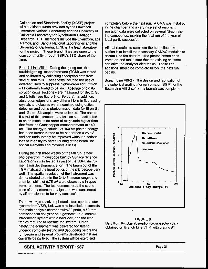
Branch Line Vlll -1 - During the spring run, the toroidal grating monochromator (TGM) was tested and calibrated by collecting absorption data from several thin foils. These tests included the use of different filters to suppress higher-order light, which was generally found to be low. Absolute photoab- sorption cross sections were measured for Be, C, Si, and U foils (see figure 6 for Be data). In addition, absorption edges of many different ions in fluorescing crystals and glasses were examined using optical detection and some photoemission data for Si-on-Ge and Ge-on-Si samples were collected. The photon flux out of this monochromator has been estimated to be as much as an order of magnitude higher than that from the Grasshopper monochromator at 140 eV. The energy resolution at 100 eV photon energy has been demonstrated to be better than 0.25 eV and can undoubtedly be improved without a serious loss of intensity by careful tuning of the beam line optical elements and movable exit slit.
During the first three weeks of the fall run, a new photoelectron microscope built by Surface Science Laboratories was tested as part of the SSRL instru- mentation development effort. The beam out of the TGM. matched the input optics of the microscope very well. The spatial resolution of the instrument was demonstrated to be in the 2- to 5-micron range, and chemical shifts of 0.75 eV were observable in spec- trometer mode. The test demonstrated the sound- ness of the instrument design, and was considered by all participants to be very successful.
The new angle-resolved photoelectron spectrometer system from VSW, Ltd. was also installed. It consists of a main analysis chamber with 30 ports, a 50-mm hemispherical analyzer on a goniometer, a sample- introduction system with a load lock, and the elec- tronics required to operate the system. Unfortu- nately, the equipment was delivered too late to undergo complete testing and debugging before the run began and several problems developed that are currently being fixed; the system will be exercised
completely before the next run. A CMA was installed in the chamber and a very nice set of resonant emission data were collected on several Ni-contain- ing compounds, making the final run of the year at least partly successful.
All that remains to complete the beam line and station is to install the necessary CAMAC modules to accumulate the data from the photoelectron spec- trometer, and make sure that the existing software can drive the analyzer electronics. These final additions should be complete before the next run begins.
Vlll-7 - The design and fabrication of the spherical grating monochromator (SGM) for the Beam Line Vlll-2 soft x-ray branch was completed
4 1 , * . . . , , . , , BL-VIJJ TGM
Beryllium (prclirinarg APECS data)
v= B - m e lplm
P 2 -
I! 0
3 if
-J 0
109 288 Ne Incident x-ray energy, eV
FIGURE 6 Beryillium K-Edge absorption cross-section data obtained on Branch Line VIII-1 with grating #1
SSRL ACTIVITY REPORT 1987 Page 21
early in the year. The SGM features three inter- changeable ion-etched gratings from Astron, Ltd., a
focussed and unfocussed modes demonstrated satisfactory performance. Using a temporary hutch,
new scanning mechanism that utilizes flexure pivots to eliminate backlash, and a laser interferometer angle monitor for increased rotational accuracy. During the spring 1987 SPEAR run, the SGM arrived from Acton Research and was installed and aligned in record time, thereby allowing three days for preliminary measurements. Commissioning and preliminary experimental work were conducted during the fall run including testing of the new software- hardware interface, alignment of a newly installed exit mirror, resolution checks and wavelength calibra- tion using a new prefilter set. A temporary Sandia UHV sample chamber and an SSRL lo section were installed and used to obtain near-edge carbon total electron yield data, to measure absolute photoemis- sion cross sections of several materials in transmis- sion, and to detect various absorption edges in the 100-1 200 eV range using optical emission from fluorescing ions in numerous crystal and glasses.
Progress was made in the design, fabrication and installation of a tandem reflectometry chamber separated from the UHV chamber by means of a thin-windowed differential pumping section. In addition to the reflectivity measurements for which it was designed, this system will be used for a variety of experiments that are not compatible with UHV. A photoemission chamber was also received from VSW, Ltd., and will be installed and tested during the spring run.
Branch Line X -3 - A 15-period hybrid wiggler with neodymium-iron-boron permanent magnets (see figure 7) built at the Lawrence Berkeley Laboratory was installed and all in-alcove work for the beam line completed during the summer of 1987. Thanks to the fine efforts of the SSRL staff and the LLNL project team the carbon filter, beryllium windows, Hower-Brown monochromator and remaining beam lines components were quickly installed in time to obtain some operational experience during the fall run. First wiggler light into the Beam Line was recorded on November 14.
The wiggler has been operated at fields up to the 1.4 Tesla design maximum with no adverse effects on SPEAR operations. Initial power measurements indicate that the flux at sample is photons/sec at 10 keV (unfocussed) with SPEAR running at 3 GeV, 50 mA and at a wiggler field of 1 Tesla. Beam profiles recorded with a CCD camera in both the
several experiments were conducted. These include XANES/EXAFS of all metal constituents in high- temperature superconductors to study valence and site geometry, optical XANESEXAFS for site- selective detection, Cr sites and valence in laser crystals, tomographic imaging of FeS, phases in coal, evanescent and standing wave measurements of thin films, and a Mossbauer nuclear scattering experiment (see Progress Reports for Proposals 9907M and 8003M for further details).
Design of a permanent experimental hutch is under- way. It will have two sections: the front section for spectroscopy and imaging experiments; the rear section for diff ractionkcattering experiments. The first section should be completed prior to the begin- ning of the next dedicated run. The addition of a side branch is also possible and is under consideration.
PRT Facilities Under Construction
I ine Vl-2 - Branch Line VI-2, the first of potentially three branch lines on the EXXONILBU SSRL PRT beam line, has been modified to accom- modate the soft x-ray branch line, VI-1 . The parity of the monochromator has been resewed to permit access when VI-1 is installed in the summer of 1988. In the spring of 1988, the monochromator housing will also be so modified.
nch Line VI-1 (1 BI /FXXON/SSf3JJ - The Beam Line VI PRT is developing a second branch line on Beam Line VI. This will be a high flux and resolution (56-1 000 eV) spherical grating monochromator accepting up to 1.1 mrad of the wiggler radiation fan. This branch line has a total horizontal deflection of 5.6 degrees from the main line and the monochroma- tor and experimental station will be located between the existing end station, Branch Line VI-2, and Beam Line V.
During the last year problems arose because of the developmental nature of the beam line components. The decision to make the MO mirror removable on a single flange lengthened the design process. In addition, the electroless nickel surface of the mirror was polished through to the copper substrate. This required replating, re-grinding and repolishing. The mirror is now ready, except for a last minute vacuum problem, for installation in its tank 8.8 meters down- stream from the wiggler. Essentially all of the
~~
Page 22 SSRL ACTIVITY REPORT 1987
FIGURE 7
The wiggler for Beam Line X, a 15-period, variable-gap, permanent magnet-steel hybrid. At 3 GeV, the wiggler at peak field is expected to radiate approximately 3 kW in a 5-rnrad horizontal fan with a 7.8-keV critical energy at 1.3 Tesla.
SSRL ACTIVITY REPORT 1987 Page 23
components up to and including M1 are finished and will be installed in 1988. The monochromator and the wavelength drive system will be assembled at LBL in the first half of the year for installation at Stanford sometime in the summer, depending on manpower availability and conflicts with the dedi- cated run.
The decision was made in 1987 to engage a com- pany that had experience in precision slides and microstepper motor technology to design and fabri- cate the slide system for the moving slits and the grating wavelength drive. LBL is working closely with Microautomation Technology of Berkeley and ex- pects to set a new standard in precise control of the gratings and slits. A laser interferometer system will be the position encoder for the wavelength, and Moire fringe glass scale readouts will encode the slit position. Real time closed loop software running in a system separate from the main beam line computer will read these encoders and maintain the accuracy of the system.
The system will be assembled initially with a fused silica grating from Ferranti Astron in the U.K. The advanced water-cooled grating substrates are nearing completion at Perkin Elmer, but the optimum means of applying the grating grooves requires development work which will be pursued by LBL in 1988 in collaboration with grating manufacturers. To facilitate this work, and to guarantee the success of the ruling or holographic process, whichever is chosen, twenty sample substrates having the same specifications for surface quality have been ordered from the same vendor. This will assure that the parameters chosen from the development process will produce good grating grooves on the water cooled substrates.
Photons through the exit slit of the monochromator are expected by in the next dedicated run.
i X- - Design has started on a soft x-ray (1 00-1 000 eV) side station for Beam Line X. This station will have a configuration similar to Branch Line VI-1 with a high resolution spherical grating monochromator. It is being built by IBM and the Center for Materials Research at Stanford University for research in the areas of semiconductor surfaces, high resolution carbon edge studies and microscopy.
previous activity report focussed on the operational success of the new multi-undulator concept. This report will focus on the status of the Locust mono- chromator. The assembly of the monochromtor has proceeded throughout 1987 and bakeout began in February, 1988. A complete review of the design of the beam line is given in [R.Z. Bachrach, R.D. Bringans, B.B. Pate, and R.G. Carr, in uProceeding.s of the Interna- tional Conference on Insertion Devices for Synchrotron Sources”, edited by R. Tatchyn and I. Lindau, 1986lwhich also cites the other pertinent papers. The last phase of the construction is due for completion of the spring of 1988 with testing and experimentation beginning with the June, 1988 run.
The specific focus for Beam Line V is the spectral range from 10-1000 eV and the design goal is to deliver the highest possible power density to the sample in the smallest feasible bandwidth. The design of the beam line is dominated by the in- creased beam power density from an undulator source which necessitated active cooling of most elements likely to be hit by the beam. In the case of optical elements, this requirement arises not only because of possible damage, but also because distortions of the optics must be minimized to maintain ultimate performance. In specifying a monochromator for this beam line, an instrument was sought which would match well to the multi-undulator and SPEAR characteristics and which would ad- vance the state of the art. The resulting Locust monochromator implements a constant deviation Vodar geometry Rowland Circle mounting and is descended from the Grasshopper Monochromator. By using closed loop computer control and configura- tional changes, the design incorporates a number of features that would not be achievable with either the Grasshopper or the Extended Range Grasshopper (ERG) configuration for these optics. As with the Grasshopper, the Locust uses principally spherical or conic section optics. The Grasshopper was the first fully UHV monochromator. The Locust achieves both variable angle of incidence optics and is the first to be fully water cooled to enable it to maintain perform- ance with the powers delivered by an undulator. The incorporation of water cooling consistent with ac- cepted vacuum practice while maintaining the required mechanical and optical tolerances proved to be a major design problem and added immensely to the scope of design work required and the complexity of the assembly. The Locust uses an elliptically figured SIC entrance mirror fabricated by TRW, Sic entrance codling slit and both spherical and cylindri- cal gratings.
Beam Line V - This multi-undulator beam line is also known as beam line WUNDER because of its use of wiggler-undulator insertion source technology. The
Page 24 SSRL ACTIVITY REPORT 1987

The refocussing system is built into the monochrorna- tor creating three work areas by providing two beams deflected horizontally at 14 degrees from mirrors 1 .O meter from the exit slit. The side stations are at 2.25 meters and the end station 3.5 meters from their respective mirrors. The two side stations have a more limited energy range than the end station, but this is offset by the participating research groups being able to maintain experimental chambers in place permanently. The end station port is for
general use and has no permanently installed chamber. Provision has been developed, however, for rapidly changing and positioning chambers available for general users.
Figure 8 shows a schematic side view of the mono- chromator system. The refocusing and exit slit chambers are on the left, the moving chamber which houses the grating and associated optics is in the center, and the entrance chamber is on the right.
n
FIGURE 8
SCHEMATIC VIEW OF THE BEAM LINE V MONOCHROMATOR SYSTEM
SSRL ACTIVITY REPORT 1987 Page 25

SUPPORT FACILITIES
ratorv/Shop/Offce u- The SSRL Labora tory-Office-Shop building was completed in February 1988 with SSRL scientific, technical and administra- tive staff moving into their new quarters on February 26.
. .
The completion of the laboratory building marks an important milestone for SSRL, as it frees up valuable user setup and experimental floor space and consoli- dates many activities that were formerly distributed throughout trailers and separate buildings. These include the electronic shop, the vacuum clean room, the machine shop, the mirror coating facility and the computer room. The former vacuum cleanroom and machine shop will be converted to a “staff clean room and machine shop facilities, and will be avail- able to the user community for their experimental programs.
The new building comprises 33,000 sq. ft. in two two- story sections. The laboratory-shop wing houses, on the first floor, a machine shop, a vacuum assembly area, a vacuum clean room, a metrology/mirror coating facility, a VUV research laboratory and an x- ray laboratory. The second floor houses the elec- tronic shop, the biotechnology laboratory, the central computer facility and scientific staff offices, as well as a conference room and a library.
The other section of the building houses the design/ drafting and engineering functions on the ground floor, and the administrative functions on the second floor. From the user perspective, the consolidation 01 these activities should streamline various userff acility interactions. It will also free up a significant amount of office space in the Bldg. 120 complex for PRT and user “quiet areas”.
fully implemented throughout SSRL and connects to SLAC via a LAN bridge. This new system will significantly enhance currently stretched computa- tional capabilities. The 8700 is more than 6 times the speed of the 11/780 and will have a very large memory and disk configuration.
Another development in the computational area is the upgrade program to implement microVAX worksta- tions as replacements for the aging PDP 11/34 branch line instrumentation control computers. Motor driver software for the interface is now almost complete and higher level applications packages (such as the EXAFS data acquisition system) are being designed. Over the next eighteen months, most of the PDP systems will be phased out and replaced by microVAXes. The new systems will also have complete data analysis software to provide for analysis in real time. All of the branch line mi- croVAXes will interconnect over the ETHERNET with the 8700 and the outside world.
SSRl Computer 1 ANs ( L o c a l o m - During this past year, with the assistance of the Electrical Engineering group, the SSRL Local Area Network (LAN) has also been expanded to include the new LOS building and, with the cooperation of SLAC networking, connections to the SSRL beam lines on PEP. The network is implemented via both coaxial network links and fiber optic interconnections between LOS, SPEAR and the engineering trailers. A new terminal server system based on DECserver 200/500 devices has replaced the aging, and less versatile, system which has existed at SSRL for the last four years. A new set of dial-in modems (3) are resident on the DECserver 500, providing convenient access to any of the systems on the SSRL LAN.
- Only a few small changes have been made in this facility which continues to receive heavy use during runs. A microfuge has been added for small volume samples. An ultracentrifuge rotor with swinging buckets is also now available for outside users.
CompuW ‘onal F a c i l b - A major change in the computational support area at SSRL is being made at the current time. The central VAX 11/780, which supports the scientific applications for the laboratory, and the VAX 1 1 n50, which supports all administra- tive computing at SSRL, are being replaced by a single new VAX 8700 which is being installed in the LOS building. This system will be the central node for the lab-wide ETHERNEnDECNET system which is
Page 26 SSRL ACTIVITY REPORT 1987
IV ENGINEERING DIVISION
MECHANICAL ENGINEERING
An appreciable portion of the effort of the Mechanical Engineering group is devoted to design of new beam lines. In 1987, work was carried out on PEP Beam Line lB, SPEAR Beam Lines Vlll and X and the Beam Line V monochromator. All of these activities are described in the preceding chapter.
Work Compl&?d in 1 9 E - Work completed includes: 1) the design of a "pin hole" device used as a diag- nostic tool in PEP Beam Line 1 B, 2) SSRL specific additions to the new SLAC Vacuum Specification, 3) the qualification of a new non-circular UHV sealing method developed at LBL, and 4) design of a fo- cussing mirror following the monochromator on Branch Line IV-3.
The PEP "pin hole" allows one to image the x-ray source, and in combination with a scanning pinhole and the monochromator in the hutch, provides a method of producing a series of planar monochro- matic maps of flux distribution.
The new LBL noncircular seal consists of a copper or aluminum seal with an '2" cross-section that is crushed into the corners of conventional O-ring grooves. Mating flanges need not be of similar materials, allowing a match of aluminum to stainless steel. The sealing system is currently in use at LBL, and will be used on a large rectangular flange on the mirror tank for Branch Line VI-1. Vacuum perform- ance is similar to the conventional conflat flange.
. . Penrllium Window and Hvbrtd Wiqaler Power Stu- - The ME group, in collaboration with the SSRL X- Ray group, is conducting experiments to measure the temperature distribution on the first beryllium window and the power output of Beam Line VI. An infra-red camera recorded the temperature field on the win- dow, and change in cooling water temperature near the window was used to determine approximately the power absorbed by the beryllium. Photon flux was also measured in the experimental hutch. Results so far indicate that the total power output of the wiggler appears to be lower than expected.
Lawrence Livermore National Laboratories produced temperature fields on sample beryllium windows with a calibrated laser welder. [Holdner, F.R., et a/. "Thermal
Damage Study of Beryllium Windows used as Vacuum Barriers in Synchrotron Radiation Beamlines: LLNL Report UCID-27 779 (September 7987}]. Temperature fields were measured with an infra-red camera and checked with thermocouples. Laser power levels were increased until the beryllium foil failed.
The ME group at SSRL, using LLNL power distribu- tions and finite element methods, reproduced the experimental temperature distributions, thus verifying the finite element models used in window thermal predictions.
In 1988, the Mechanical Engineering and X-Ray groups will design and install an accurate calorimeter to determine the beam power following the beryllium window module. This instrument may also be installed on Beam Line X to determine its total power output.
Thermal-St ructu ra I Anal= . ' - SSRL is now conduct- ing almost all of its finite element analysis on PC's. The Engineering group has two Compaq 386's with 80387 floating point processors, running a thermal- structural analysis package from Algor Interactive Systems. This package includes a model drafting package, as well as accepting output from Cadkey, the SSRL standard drafting program. The Compaq has been used to run models of 5 to 6 thousand degrees of freedom in the space of 5 to 6 hours. Larger models can be run on the VAX at SSRL using similar programs from the same company. The CompaqlAlgor combination has been used to exam- ine the temperature and stress distributions on beryllium windows, crotches, masks, and mirrors.
ComDuter Aided DesiQa- The Mechanical Engineer- ing group now has two PC's dedicated to mechanical design and drafting; a Compaq 386 and an IBM AT clone. Three designers are now able to use these machines.
The designs of the PEP pin hole device and the XAFS hutch on Beam Line X-2 were done exclusively on the Mechanical Engineering CAD stations. Future efforts, such as the Beam Line II rework and Beam Line X-1 branch line design, will also be done on the CAD systems.
SSRL ACTIVITY REPORT 1987 Page 27

ELECTRICAL ENGINEERING
Ream I ine Control Svstems - A major activity of the Electrical Engineering group is the design, construc- tion, maintenance and improvement of various control systems for SSRL beam lines. These control systems may be divided into the following functional catagories: vacuum controls, photon beam steering and stabilization controls, personnel protection interlock systems, communications (audio, video, data), insertion device controls and experimental station electronics.
In 1987, the EE group was involved in the design and implementation of control systems for the following new beam lines: PEP Beam Line 1 B, SPEAR Beam Line Vlll, and SPEAR Beam Line X. The installa- tions of the controls sytems were completed, and the various controls were tested and commissioned. The beam lines were successfully operated with beam.
Work is continuing to standardize the beam line control systems. The goals are to simplify the operation and maintenance of these systems and to reduce the training time for technical support person- nel.
rol Svstem - The Beam Line X wiggler control system, based on that of Beam Line VI, has been improved in three areas. The control panel has been simplified for easier use in routine operation. The touch panel presents all critical information, and will automatically calculate the wiggler gap for the desired field. The system will not allow fields to exceed levels that are safe as determined by the number of filters inserted. Wig- gler trim currents have been optimized to minimize effects on storage ring current. Provision has been made to allow continuous scanning of the wiggler field, controlled by the experimental station computer. with a minimum effect on the storage ring current and SPEAR control computer.
mgSy&rn - Efforts are continuing to develop a central computer facility for monitoring all SSRL beam line control systems and SPEAWPEP ring operating parameters. The purpose of the proposed computer facility is to permit centralized monitoring (and eventually, some manipulation) of the beam line control systems. The facility will also act as the interface to the SPEAWPEP VAX computers for acquisition of storage ring data. The centralized
status information will be available to SSRL operators and key personnel. Relevant information will be displayed at each beam line control station and at the main operation console in the SSRL control room. The system will be connected to the SSRL sitewide Ethernet, making the status information available to all network terminals.
The system will consist of the following major compo- nents: 1) a system supervisor computer which will also be used for software development and program maintenance, 2) a set of console computers in the main control room, 3) a set of process control com- puters (DEC microVAX-11’s for SSRL and VAX750 for SPEAR control) with individual CAMAC serial high- way drivers located central to their respective proc- ess interfaces, 4) CAMAC instrumentation crates and modules directly interfaced to the beam line control systems (or to the storage ring instrumentation, in the case of the ring control system), 5) a machine control Ethernet LAN interconnecting the system supervisor, consoles and process computers, 6) a laboratory Ethernet LAN (existing SSRL Ethernet) connecting the system supervisor computer to the terminal servers and outside world.
The supervisor computer will collect beam line statistics to investigate long term trends; for example, to identify a slow leak in the vacuum system. the Ethernet connection to the SPEAR VAX will also permit this computer to monitor SPEAR collected information, such as ring current, energy, lifetime, magnet fields and vacuum reading within the ring itself.
Beam I ine Steerina wtabi l i7at ion - The ninth beam line steering servo system for SPEAR and the system for PEP, servicing SPEAR Beam Line X and PEP Beam Line 1 b, were commissioned during the fall of 1987. Both systems utilize in-vacuum pho- toemission beam position monitors similar to those used on SPEAR Beam Line VI and the PEP Beam Line 5b (see SSRL ActMty Reports for 7984 and 7985).
While these systems can stabilize vertical beam position at the experimental station to a level of order 0.1 mm or less over the course of the delivered beam period when the beam line insertion magnets are operated in their high field configurations (>-3 kGauss for the Beam Line X 15 period hybrid wiggler, >-1 kGauss for the PEP 26 period undulator), the
Page 28 SSRL ACTIVITY REPORT 1987
stabilization level can be considerably less during low field operation because of the significant contribution to the position monitor signals from bending magnet radiation. This “pollutant“ radiation can create an asymmetric illumination of the monitor electrodes, and a shift of the illumination centroid can occur as beam decays. The steering servo system incorrectly interprets such a shift as the movement of the insertion device beam and adjusts the stored beam orbii to restore the centering of the beam centroid. This mechanism can result in missteering the desired insertion device beam by as much as a millimeter over the course of a fill.
One way to reduce the inadequacies of the steering system during low field insertion magnet operation is either to situate the position monitor far back on the beam line where the bending magnet radiation can be effectively masked. This solution is frequently impractical because of the presence of a focussing mirror at a location on the beam line that is close enough to the source point that bending magnet radiation is still a problem.
Another solution is to incorporate a position monitor that can recognize the radiation pattern of the desired beam and respond only to its displacement. For undulator radiation, the task is fairly simple since the location of the desired beam coincides with the peak of the beam’s transverse intensity profile. SSRL is assisting Britton Chance et al. (University of Pennsyl- vania) [B. Chance, D. Schkk, R. Heftel, L. Powers, Nud. Insrr. and Meth. A246 (7986) 400.1 in the development of a “vibrating reed“ monitor consisting of a thin stain- less steel wand that sweeps transversely through the beam at a high frequency (-200 Hz) and with a low duty cycle (-5%) to sample the beam’s intensity profile. A small area on the wand is coated with a phosphor to sense the limited crossection of the beam. Vertically scattered photons are detected with a photomultiplier tube; processing electronics pro- duces a signal proportional to the off-center position of the beam’s “hot spot”. A version of this monitor that operates in helium was tested on diagnostic Beam Line 11-3 and on wiggler Beam Line VI over the last year. This monitor promises 10 micron accuracy independent of beam intensity. A model tbat func- tions in vacuum is being developed by Chance.
Further development and upgrading of the steering control system was carried out in 1987 under the auspices of the SEP project. A new steering servo
controller printed circuit board was designed that incorporates the analog and digital circuitry required for computer control and monitoring of the high bandwidth analog servo components. Functions include an interlock that prevents the system from operating in closed-loop mode if the position monitor or other servo signals have improper magnitudes and disables the system totally if the corrector magnet actuators are not operating properly. Automatic gain control (AGC) has been implemented to normalize system gain over a dynamic range of -30 dB in beam intensity/ current. In addition, a gain ramping circuit has been devised that effects smooth transitions between open- and closed-loop modes (“manual” and “auto”) to avoid the transient “glitching” of the beam orbit that otherwise occurs during these transitions. The new steering controller was tested on SPEAR Beam Line Vlll and PEP Beam Line 1 B in Fall, 1987. Installation of the new units for all SSRL beam lines is scheduled for early 1988. Remote computer control and monitoring of the steering systems via the CAMAC interface is presently being implemented.
Future steering system improvement centers on the development of the 2-monitor, 4-magnet servo configuration that will stabilize both position and angle of the photon beam in the vertical plane (described in the 1985 Actjvity Report.). A key compo- nent in this system is the RF beam position monitor and signal processing module that detects and decodes the position of the stored beam near the source point where it is colinear with the photon beam. A module is being developed that produces both vertical and horizontal position sensitive signals with an estimated reproducibility error of -50 microns or better over a bandwidth of -1 kHz. This monitor, working in conjunction with a photon monitor located many meters from the source point, should be capable of stabilizing the photon beam’s position and angle to a small fraction of its transverse size and divergence parameters at the experimental station.
SSRL ACTIVITY REPORT 1987 Page 29
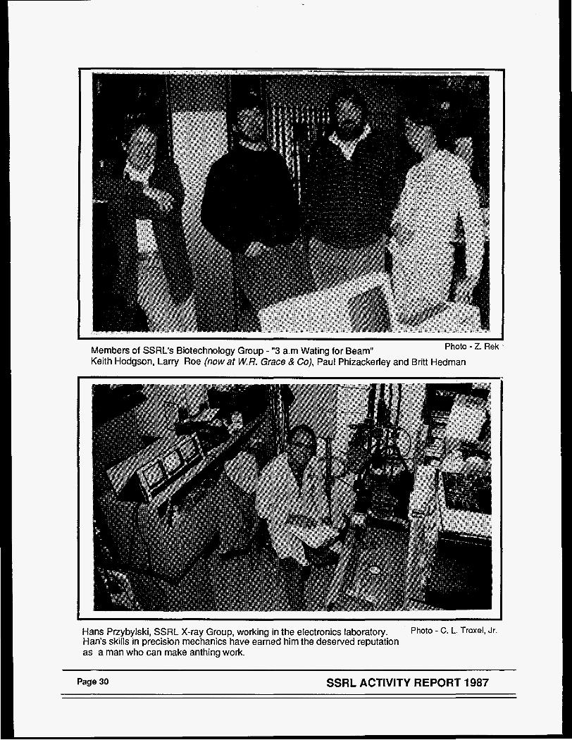
Members of SSRL’s Biotechnology Group - “3 a m Wating for Beam” Keith Hodgson, Larry Roe (now at W.R. Grace & Co), Paul Phizackerley and Britt Hedman
Photo - Z. Rek
Hans Przybylski, SSRL X-ray Group, working in the electronics laboratory. Han’s skills in precision mechanics have earned him the deserved reputation as a man who can make anthing work.
Photo - c. L. Troxell Jr.
Page 30 SSRL ACTIVITY REPORT 1987
V CONFERENCES AND WORKSHOPS
WORKSHOP ON PEP AS A SYNCHROTRON RADIATION SOURCE
A Workshop on PEP as a Synchrotron Radiation Source was held by SSRL on October 20-21,1987. PEP is a colliding-beam storage ring that has already been used for a few weeks for synchrotron radiation research parasitically during high energy physics runs.
PEPS main features as a synchrotron radiation source are high energy (up to 16 GeV with presently installed RF), long straight sections (6 are 117 meters long), large circumference (2200 meters) and low bending magnet field (0.32 Tesla at 16 GeV). It has operated in a low emittance mode with an emittance of less than 12 nanometer-radians at 8 GeV, making it the lowest emittance synchrotron radiation source. (In December 7987, after the workshop, PEP was operated at 7.7 GeV with a calculated omittance of 6.4 nanometer-radians. See Chapter 11 for details.)
At the Workshop, there were 125 registered partici- pants (77 from government labs, 23 from universi- ties, 16 from foreign institutions and 9 from US industry). The following charge was given to the Workshop by SSRL Director, Art Bienenstock.
A. Explore PEP'S potential as a low emittance storage ring. Examine the possibilities which have been proposed for achieving that very low emittance. Provide your critical analysis of those possibilities and inform us of others which may be achievable.
E. Explore the range of insertion devices which can utilize the extreme lengths and/or high energies available on this machine.
C. Consider the potential evolution of this machine from one which will function routinely with brilliances of the order of 70i6as PEP functions in a parasitic mode to the projected 7P" with the achievement of the extremely low emittances. Define new experi- ments which can be performed as this evolution proceeds.
Individual contributions to the Workshop and summa- ries and conclusions by Working Group Leaders have been assembled into a proceedings. Copies are available on request by writing to M. Steger at
SSRL, SLAC Bin #69, PO Box 4349, Stanford, CA 94309-021 0.
Some highlights from the summary talks by the leaders of the 11 Working Groups are given below:
A. Detailed analysis and past experience with PEP were reviewed by 35 scientists in the Accelerator Physics Working Group. They concluded that there was good reason to expect that PEP could run at high currents (5-8 mA per bunch, 100 mA or more total current) at 8 GeV in the low emittance mode that has already been tested at low current and that no major problems were forseen that would prevent the achievement of much lower emittances with damping wigglers, as has been proposed. A list of specific accelerator physics studies that should be carried out was made.
B. The 9 Working Groups dealing with the different applications presented many experiments that could be done on PEP. Some of these could also be performed on rings such as the APS and ESRF and PEP offered an opportunity for an early start. Others could uniquely be done at PEP because of the extreme capabilities and/or potential of PEP (bright- ness, photon energy, coherent power', short bunches). There was considerable excitement about the prospects for carrying out these experiments, but also concern about running time, staff support and the number of beam lines and support facilities that would be available.
C. The Directors of the APS and ESRF projects, and several of their staff present at the Workshop, expressed interest to participate in machine studies and use of PEP beam lines to gain experience relevant to their projects. It is also expected that interest in using very high energy rings will grow in Japan using the Tristan 26-30 GeV storage ring that recently began operation for colliding-beam studies at KEK. A talk on Japanese planning for high energy light sources was presented to the Workshop by M. Ando from the Photon Factory.
D. There was considerable interest in very high photon energies (up to several hundred KeV) for
~
SSRL ACTIVITY REPORT 1987 Page 31
applications such as studies of crystal optics, Comp- ton and magnetic scattering, and nuclear resonance spectroscopy. It was also pointed out that the high coherent power levels that PEP could produce would have application in several areas including micro- analysis and the pumping of soft x-ray lasers. Inter- est was also shown in the possibilities to place samples very close to the source, which would allow the recording of a diffraction pattern in one shot with a time resolution of picoseconds instead of millisec- onds and, conversely, to have very long beam lines to exploit the exceptional collimation of the radiation in topographic studies.
FOURTEENTHANNUALSSRLUSERS GROUP MEETING
The Users Group Meeting was held this year on October 22 and 23,1987. The SSRLUO Executive Committee is responsible for organizing the Users Group Meeting jointly with a representative of the SSRL scientific staff. Approximately 156 people were in attendance. The program for the meeting and the authors’ abstracts are available as SSRL Report Number 87/05.
Co-Chairpersons for the meeting were Joe Wong for the Users Organization and Brad Pate for the SSRL scientific staff. Reports on individual experiments were presented in poster sessions.
FIRST SSRL ROTATION CAMERA USERS MEETING
The first SSRL Rotation Camera Users Meeting, attended by about 30 people, was held at Asilomar, California on March 23, 1987 during the Eighth West Coast Protein Crystallography Workshop. This was the first such meeting since the rotation camera facility became available to users in April, 1984. It was held at this time because many of the SSRL rotation camera users were planning to attend the Crystallography Workshop.
The meeting, chaired by Paul Phizackerley of SSRL, provided the opportunity to discuss, as a group, such topics as the functioning and capabilities of the rotation camera system, the quality of results ob- tained, film scanning software used, user experi- ences with the experimental facilities, user support, hardware and software documentation, scheduling and access to the facility, and plans for future instru- mentation development.
SSRL ACTIVITY REPORT 1987 Page 32

I
VI SSRL ORGANIZATION
SSRL FUNCTIONAL ORGANIZATION
The SSRL administratiori is divided into seven functional areas each headed by a senior staff member. These are: X-ray, VUV, Biotechnology, Accelerator Physics, Operations, Engineering and Administration. In the next few months a separate organization to support the 3 GeV injector project will be developed. In addi- tion, each experimental station and piece of specialized instrumentation is the responsibility of an SSRL staff member. The charts which follow depict these responsibilities and the functional organization of SSRL as of January 1988.
BRANCH LINE
1-1 (Grasshopper)
1-4 (SAS) 1-5 (Bend Magnet X-ray) 11-2 (Bend Magnet X-ray) 114 (White Radiation) 111-1 (Grasshopper) 111-2 (SXR- 18 degree) 111-3 (SXR-Jumbo) 111-4 (Lithography) IV-1 (Wiggler) IV-2 (Wiggler) IV-3 (Wiggler) VI-2 (Wiggler)
1-2 (TGM)
VII-1 (Rotation Camera) Vll-2 (Wiggler) Vll-3 (Wiggler)
X-2 (Wiggler) PEP 1B PEP 5B
VIII-1 (SGM) Vlll-2 (TGM)
SSRL EXPERIMENTAL STATIONS RESPONSIBLE PERSONNEL
RESPONSIBLE SCIENTIST/ENGINEER
1. Lindau/A. Waldhauer 1. Lindau/M. Rowen S. BrennarVH. Tompkins G. BrowniT. Troxel G. BrowniT. Troxel Z. Rek 1. Lindau/A. Waldhauer 1. Lindau/A. Waldhauer I. Lindau/M. Rowen P. PianettdA. Waldhauer G. BrowniT. Troxel G. BrowWT. Troxel S. BrennatVH. Tompkins J. Arthur/T. Troxel D. Moncton (€XXOiV), J. Kortright(LBL) P. Phizackerley S. BrennarVH. Tompkins G. BrowniT. Troxel M. Rowen, G. Tirsell (LLNL) M. Rowen, S. Williams (UCLA) S. Brennan, J. Wong (LLNL) G. Brown/T. Troxel G. Brown/T. Troxel
SSRL ACTIVITY REPORT 1987 Page 33
INSTRUMENTATlON/FACILlTY RESPONSIBILITIES
MATERIALS DIFFRACTOM ETER: S. BrennaWH. Tompkins VACUUM SAMPLE CHAMBERS: M. Rowen AREA DETECTOR: P. Phizacker/ey COMPUTER SYSTEMS: T. Cox Vll-2 SPECTROMETER: S. Brennan/H. Tompkins BIOCHEMISTRY LABORATORY: B. HedmaWR. Mayer DARKROOMS: P. Phrickerleyfl. Rek BEAM LINE STEERING: R. Hettel EXAFS EQUIPMENT AND SOFTWARE: B. HedmaWR. Mayer SAS CAMERA (BIOLOGY): S. Wakatsuki EXAFS CONSULTANT: 6. Hedman SCATTERING CONSULTANT: S. Brennan RT-11 SOFTWARE CONSULTANT: S. Brennan
SSRL DEVELOPMENT PROJECTS -RESPONSIBLE PERSONNEL
BEAM LINE II - G. BROWN (SSRL)/T. TROXEL (SSRL)/ R. BOYCE (SSRL)
BEAM LINE V - 1. LINDAU (STANFORD)/R. BACHRACH (XEROX)/ A. WALDHAUER (SSRL)
STATION VI-1 - R. TATCHYN (SSRL), W. MCK/NN€Y (LBL) R. YOTAM (SSRL)/J. CHIN (LBL)
3 GeV SPEAR INJECTOR - H. W/€D€MANN
STATION X-1 - 1. LINDAU (STANFORD), J. STOHR (IBM), M. ROWEN (SSRL)/J. MONTGOMERY (SSRL)
Page 34 SSRL ACTIVITY REPORT 1987
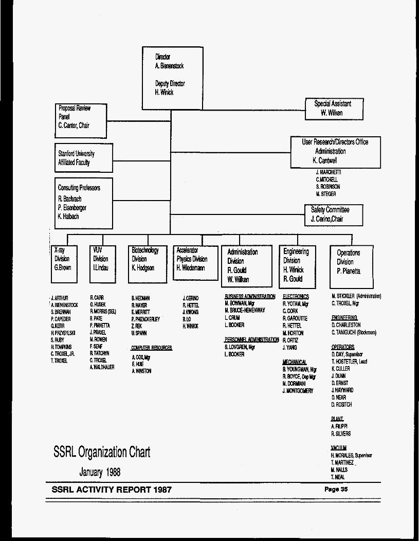
Director A. Bienenstcck
DqMy Diredor H. W&
~ Special Assistant W. Wilken Proposal Review
Panel C. Cador, Chair
S. ROBINSON M. STEGER
- User ResearcM)irectors Office
Stanford University Administration Aff ilialed FacuHy K. Cantwell
corsulting Professors R h h h P. Eisenberger safely Committee K. Halbach J. Cerino,Chair
J. MARCHETll C.MITCHEU
-
I I I I I I i Division
J. ARTHVR k BlENENMocK S. B R W P.CAPEDER GKEFIR H. PFDBMKI S. RUBY H.ToMKt6 C.lROXE.,JR. T. lf#)(B
7- M. WAN, Mg R. YOTAM, Mg
J.CEW0 RHEna J. m M. BRUCE-HEMENWAY CCORK 110 LCRUM R. GAROUTTE HWCK LBOOKER R. HETlEl
M. HORTON - RORTIZ S. LOVWN, Mgr J. YANG LBOOKH
MECHANlCM R YOUNGMAN, My R BOYCE, Dep Mg M. DORMlANl J. MONTGOMERY
Diision P. Pianetta
M. STICKLER (Adf~itWaijon) C. TROXEI, Mg
ENGlNEERlNG R CHARLESTON C. TANlGUCHl (stodvoom)
DPERATORS D. DAY, SLperviSor T. HOSlETLER, lead K CULLER J. WNN D. ERNST J.HAYWMD D. NEAR D. ROSlTCH
HAK A FIUPPl R SlLVEB
SSRL Organization Chart January 1988
JlGuM H W E S , Supervisor T. MARINE2 M. NAU 1. NEAL
SSRL ACTIVITY REPORT 1987 Page 35
SSRL ADVISORY PANELS
A main task of the Proposal Review Panel is the review and rating of scientific proposals to SSRL based largely on reports obtained from outside (non- panel) referees. The panel met on January 9 and 10, 1987 and on July 17 and 18,1987 at SSRL and rated the new proposals which had been received in September, 1986 and March, 1987 respectively.
As of December 31 , 1987 SSRL had received a total of 2048 proposals of which 158 are presently active.
The Proposal Review Panel meets twice yearly, generally in June and January. Deadlines for receipt of proposals for consideration at the next meeting are the first of September and the first of March of each year.
The panel members in 1987 were:
Pioloav Sub-panel Charles Cantor, Columbia University (Chairperson) Wayne Hendrickson, Columbia University William Orme-Johnson, MIT
nals Sub-- Boris Batterman, Cornel1 University Howard Birnbaum, University of Illinois Russell Chianelli, Exxon Denis McWhan, AT&T Bell Laboratories
vuv Sub-Danel Bernd Crasemann, University of Oregon Charles Fadley, University of Hawaii Warren Grobman, IBM Watson Research Center
Or. Crasemann’s term ended with the January 1988 meeting. Torgny Gustafsson, Rutgers University, is the new VUV subpanel member.
The Science Policy Board reviews all aspects of SSRL operation, development and plans for the future. It reports to Stanford University President Donald Kennedy. The Board met once during this reporting peroid, on May 22 and 23, 1987. The members at the time of the meeting were:
B. McDaniel, Cornell University (Chairperson) P. Chaudhari, IBM E. Ginzton, Varian Laboratories W. Kohn, University of California Wm. Orme-Johnson, MIT Y. Petroff, LURE F. Schwettman, Hewlett-Packard
Members of the Executive Committee of the SSRLUO were appointed at the Fourteenth Annual SSRL Users Group Meeting as follows:
Joe Wong, LLNL (Chairperson) Marjorie Olmstead, UC-Berkeley (Vice-Chair) Frank (Bud) Bridges, University of California Ross Bringans, Xerox Robert Greegor, Boeing Company Jeff Kortright, Lawrence Berkeley Laboratory Francesco Sette, AT&T Bell Laboratories Jo Stohr, IBM (Past-Chairperson) Louis Terminello, Lawrence Berkeley Laboratory Lane Wilson, LLNL Katherine Cantwell, (Secretary - SSRL Liaison)
The Users Organization Executive Committee meets periodically throughout the year to consider and advise on matters pertinent to user interests at the Laboratory.
~
S S R L ACTIVITY REPORT 1987
VI1 EXPERIMENTAL PROGRESS REPORTS - 1 OOMp
822Mp
868M
91 8M
940M
956Mp
956Mp
956Mp
979M
983M
984M
988M
995Mp
996M
99Mp
OOOMp
OOOMp
"X-Ray Absorption Studies of Disordered Systems" E.D. Crozier, N. Alberding, K.R. Bauchspiess, D. Jiang, A.J. Seary, R. Ingalls, J. Freund, B. Houser
"Temperature Dependence Study of Monolayers and Mukilayers of the Langmuir-Blodgett Type" W.B. Yun, M. Bloch, X. Yang, J. V i r 0
"EXAFS Studies of High To Superconductors" J.B. Boyce, F. Bridges, T. Claeson, T.H. Geballe, C.W. Chu, J.M. Tarasoon
"Investi ation of the a-Fe 4 E-Fe Transition Using Synchrotron Radiation" M.H. Manghnani, L.C. Ming, ?H. Kim, N. Nakagiri
"Anomalous X-Ray Diffraction Studies of Bimetallic Catalysts: R-Re on SiO; K.S. Liang, G.H. Hughes, J.H. Sinfelt
"Transformation of Ca and K Catalytic Species in Lignite During Pyrolysis and Gasification: An In -Situ EXAFS Investigation" F.E. Huggins, N. Shah, G.P. Huffman, F.W. Lytle, R.B. Greegor, R.G. Jenkins
"WAFS Studies of Organic Sulfur in Caal" G.P. Huffman, N. Shah, F.E. Huggins, R.B. Greegor, F.W. Lytle
"Investigation of the Kinetics of the Carbide Formation in HSLA Steels by EXAFS Spectroscopy" N. Shan, G.P. Huffman, F.E. Huggins, L.J. Cuddy, F.W. Lytle, R.B. Greegor
"3-0 X-Ray Microtomography of Catalytic Materials" J.H. Kinney, M.C. Nichols, Q.C. Johnson, R.A. Saroyan, U. Bonse, R. Nusshardt, R. Pahl
"X-Ray Absorption Study of Amorphous Germanium at High Pressure" R. Ingalls, J. Freund, E.D. Crozier
"X-Ray Absorption Studies of High T,, Superconductors" J. Whitmore, 8. Houser, Y. Ma, M. Qian, R. Ingalls, E.A. Stern, F.C. Brown, R. Kurosky, B. Thiel, J.M. Tranquada, E.D. Crozier, N. Alberding, A.J. Seary, J.P. Rice, B.G. Pazol, D.M. Ginsberg
J3m2
44
46
47
49
50
51
53
54
55
56
57
"Actinide and Fission Product Site Speciation in Nuclear Waste Glasses" EM. Larson, P.G. Eller, R.B. Greegor, F.W. Lytle, M.P. Eastman
1nterface"G.E. Brown, C.J. Chisholm. 2.F. Hayes, A.L Roe, G.A. Parks, K.O. Tdgson, J.O. Leckie
"Interface Structure of NiSiJSi(ll1)" I.K. Robinson, R.T. Tung, R. Feidenhans'l
"EXAFS Study of Rare Earth Element Environments in Quenched Silicate Melts of Geochemical Interest" C.W. Ponader, G.E. Brown Jr., W.E. Jackson
58
%-Situ X-ray Absorption Study of Pb(ll and &(ll) Sorption Complexes at the A1203 Water 59
62
64
"Characterization of &-Containing Oxides on Stainless Steel by e-Yield XANES Spectroscopy" 65 F. Lytle, R. Greegor, 2. Rek, H. Ocken
"Investigation of Pb Implanted TiO, and SrTiO, Using Total Electron Yield" R.B. Greegor, F.W. Lytle, B.C. Chakoumakos, R.C. Ewing, G.W. Arnold
66
SSRL ACTIVITY REPORT 1987 Page 37
1 OdOMp
1001 Mp
lOOlMp
lOOlMp
lOOlMp
1001Mp
1001 Mp
1OOlMp
1OllM
1021 Mp
1031M
1033M
1036Mp
1038M
1054M
1056M
1058M
1059M
1063M
1064M
"Site Selectivity In Mn Glass Ualng Optical XANES" R.B. Greeger, F.W. Lytle, D.R. Sandstrorn, M. Weber, J. Wong
"Universal Interactions in Undulating Membranes: A Series of Synchrotron X-Ray Studies of Dilute l-Dimensional Fluid Multilayers" C.R. Safinya, D. Roux, G.S. Smith, E.B. Sirota
"Dynamic 'Steady State' Microscopic Structure of Complex Fluids Studied in a Couette X-Ray Shear Cell" C.R. Safinya, R. Plano, E.B. Sirota, G.S. Smith
"SAXS Study of Colloidal Crystals" E.B. Sirota, S.K. Sinha, H.D. Ou-Yang, P.M. Chaiken, J.D. Axe, Y. Fujii
"Extended X-Ray Absorption Fine Structure (EXAFS) Studies of Platinum-Tin Catalysts" G. Meitzner, G.H. Via, F.W. Lytle, S.C. Fung, J.H. Sinfelt
"Sulfur K-Edge X-Ray Absorption Spectroscopy of Asphaltenes and Model Compounds" G.N. George, M.L. Gorbaty
"Scattering Studies of La$uO, Single Crystals: Charge Density Modulations" D.E. Moncton, K.L. DAmica, J. Bohr, J. Als-Nielsen, R.M. Fleming, J.P. Remeika, D. Vaknin
"High Resolution Powder X-Ray Diffraction" J.M. Newsam, K.S. Liang, G. Hughes
"Bond Orientational Ordering in Tilted Hexatic Liquid Crystal Films" J.D. Brock, R.J. Birgeneau, J.D. Litster, B. McClaln, D.Y. Noh, P.M. Horn
"Synchrotron X-Ray Polycrystalline Diff ractometry - IV & v" W. Parrish, M. Hart, M. Betlotto, L. Depero, C. Erickson
"XAS Studies of Synthetic Models for Hydrodesulfurization Catalysts" G.S. Waldo, 0. Barak, M.D. Curtis, J.E. Penner-Hahn
"XAS Determination of the Chemical Forms of Sulfur in Petroleums" G.S. Waldo, R.M.K. Carlson, J.E. Penner-Hahn
"Coster-Kronig Transition Probabilities in Metals" S.L. Sorensen, R. Carr, S.J. Schaphorst, S.B. Whitfield, B. Crasemann
"Deep Donors in Gallium Aluminum Arsenide Semiconductor Alloys" T.M. Hayes, D.L. Williamson, P. Gibart
"X-Ray Induced Photo-Acoustic Emission" R.D. Day, R.W. Ryon, S. Benson
"Synchrotron White Beam Topography of Striations and Interface Breakdown in Alloyed GaAs" S.R. Stock, Z.U. Rek, A.G. Elliot
"Roughening and Deconstruction of the Au(l10) Surface" G.A. Weld, J.L. Jordan-Sweet, P.M. Horn, A.Mak, R.J. Birgeneau, T. Gustafsson
"Thermal Expansivity of Mg2Si0 and y- Mg,Fe),SiO Spinels to 900°C Using Synchrotron Radiation" L.C. Ming, M . k Manghnani, 4.H. Kim, U.'Devi, D.H. Xiong, E. Wo
"In-Situ Surface EXAFS of Monolayers on Silver Electrodes" O.R. Melroy, M.F. Toney, M.G. Samant, J.G. Gordon, L. Blum II
"Static High Pressure Equation-of-State (EOS) Studies on Thorium to I00 GPa: A Preliminary Report" J. Akella, 0. Johnson, G.S. Smith, L.C. Ming
67
68
70
71
72
73
74
75A
76
77
78
79
80
82
83
85
87
89
91
92
Page 38 SSRL ACTIVITY REPORT 1987
1066Mp
1072M
1073M
1078M
1079M
1081M
1097Mp
1097Mp
2023Mp
2024Mp
8001 M
8001 M
8001 M
8003M
801 OM
8101M
81 03M
8103M
9900M
9901 M
"The Use of EXAFS Spectroelectrochemistry for the -Study of the Oxidation of a Copper(l) Complex in a Polymer Modified Electrode" R.C. Elder, W.R. Heineman
"Glancing Angle EXAFS Studies on CU-A1 Interfaces" S.M. Heald, H. Chen
"X-Ray Absorption Study of Oxide Based Superconductors" S.M. Heald, C.Y. Yang, J.M. Tranquada
"EXAFS Studies on Powders of GaAs Treated with Co Complexes" I.L. Abrahams, C.E. Caley, S.R. Lunt, G.M. Miskelly, M.J. Sailor, P.G. Santangelo, M.H. Schmidt, B.J. Tufts, N.S. Lewis
"EXAFS of YBa Cu,O and GdB Cu O,.,Superconductors" J.B. Boyce, F.C. Bridges, T. Claeson, T.H. &ebalk, R.S. Hoaand
"Microradiography of Creep Damage in Copper" J.E. Benci, D.P. Pope
"Antisite Disorder in YBa,Cu,O," F.W. Lytle, E.C. Marques, R.B. Greegor, H.G. Ahlstrom, E.M. Larson, D.E. Peterson, A.J. Panson
"Rare Earth L-Edge Spectra in Y,.xM,Ba,Cu,O," F. Lytle, R. Greegor, E. Marques, E. Larson, J. Wong, C. Violet
"Characterization of the Atomic and Electronic Structures of High T, Superconducting Materials by XAS" S.D. Conradson, G.H. Kwei, I.D. Raistrick
"XAS Studies of Quasi-One-Dimensional Halide-Bridged Mixed Valence Platinum Compounds" S.D. Conradson, B.1. Swanson, A.P. Sattelberger
"Reflectivity of Thin Carbon Films" S. Brennan, M.F. Toney
"Fluorescence EXAFS Study of Silicon in Neodymium Iron Boron Rare-Earth Magnets" S. Brennan, A. Bienenstock, J.E. Keern
"Strain Distribution in Thin Aluminum Films using X-Ray Depth Profiling" M.F. Doerner, S. Brennan
Time-Domain Effects in Resonant Nuclear Scattering of Synchrotron X-Rays" S.L. Ruby, J. Arthur, D. Brown, G. Shenoy, E. Alp
"Interferometric Study of Spherical Wave Diffraction" J. Arthur, H. Tompkins
"Structural Investigations of W-Ge Multilayers and Amorphous W-Ge Alloys" M. Rice, A. Bienenstock
"Analysis of Co Doped Iron Oxide Thin Films by X-Ray Depth Profiling" M.F. Toney, T.C. Huang, S. Brennan, Z. Rek
Quantification of the Spatial Variation of Strain Fields Surrounding Laser-Drilled Holes in Silicon using White Beam Section Topography" Y.H. Chung, Z.U. Rek, S.R. Stock
94
96
97
98
101
1 02
103
104
105
106
107
108
109
110
111
112
113
115
"Two Dimensional Compressibility of Electrochemically Adsorbed Lead on Silver (1 11)" P.N. Ross, J.B. Kortright, O.R. Melroy, M.F. Toney, G.L. Barges, M.G. Samant, L. Blum
11 7
"Valence and Anti-Site Disorder in High Temperature Superconductors" J. Wong, F.W. Lytle, R.B. Greegor, C. Violet, G. Xiao, A. Panson, E. Larson, D. Peterson, T. Collings, B. Maple
118
SSRb ACTIVITY REPORT 1987 Page 39
9901 M "X-Ray Tomography on Beam Line x" J.H. Kinney, Q.C. Johnson, R.A. Saroyan, M.C. Nichols, U. Bonse, R. Nusshardt
9901 M "Beam Line X-2 Profile Measurement with a CCD Imager" J.H. Kinney, J. Wong, T. Troxel
9901 M "EXAFS on Near Monolayer Hafnium Film" T.W. Barbee, J. Wong
119
120
121
L efters of Intent
"Bromine K-Edge Cellular Radiosensitization with Bromodeoxyuridine" D. Larson, W.J. Bodell, 122 C. Ling, T.L. Phillips, M. Schell, D. Shrieve, T. Troxel
"Radius of Gyration Measurements of Soluble Chromatin Fibers" S.P. Williams, J.P. Langmore
"Observation of Cluster Formation in an lonomer" A.F. Galambos, W.B. Stockton, J.T. Korberstein, A. Sen, R.A. Weiss
"The Microstructure of Block Copolymers Formed Via Ionic Interactions" T.P. Russell, R. Jerome, P. Charlier, M. Foucart
"intercalibration of Small-Angle X-Ray and Neutron Scattering Data" T.P. Russell, J.S. Lin, S. Spooner, G.D. Wignall
"Solution of Phase Problem for Crystallography at a Wavelength of 3.5 A" Q. Shen, R. Colella
BIOLOGY PROPOSALS
840Bp
957Bp
961 B
9698
981 Bp
991 Bp
993B
1001 B
10048
"X-Ray Absorption Spectroscopy of Mn and Fe in the Photosynthetic Apparatus" R.D. Guiles, A.E. McDermott, J.L. Cole, V.K. Yachandra, V.J. DeRose, J.-L. Zimmermann, S.L. Dexheimer, K. Sauer, M.P. Klein
"Anomalous Scattering of X-Rays" D.H. Templeton, L.K. Templeton
"Structure/Function Studies of Oxidases, Peroxidases and Catalases" B. Chance, L. Powers, S. Khalid, 1. Ayene, R. Waterland, T.E. King, G. Bunker, K. Zhang, R. Poyton, E. Davidson
123
124
126
128
130
131
133
134
"X-Ray Absorption Spectroscopic Studies of Nickel Containing Metalloenzymes" A.K. Shiemke, I35 M.K. Eidsness, R.A. Scott
"Mathematical Modeling of Multiple Wave Functions for Iron Porphyrin EXAFS Spectra" J.O. Alben, F.G. Fiamingo, A.A. Croteau, C.F. Hemann, K.A. Powell, V.M. Molleran, S. Park
137
"Technetium Diphosphonate Imaging Agents and Rhenium Therapeutic Radiopharmaceuticals" R.C. Elder, E. Deutsch
138
"A Co per XAS Study of Dopamine P-Hydroxylase" R.A. Scott, R.J. Sullivan, W.E. DeWolf, R.E. Jolle, L.I. Kruse
"Biochemistry: Use Made of Beam Line VI-2 in 1986/1987" G.N. George, R.C. Prince
"Crystal Structure Determination of Cucumber Basic Protein Using Multiple Energy Anomalous Dispersion Phasing" E.A. Merritt, R.P. Phizackerley, K.O. Hodgson, B. Hedman, H.C. Freeman, J.M. Guss
140
f 42
144
Page 40 SSRL ACTIVITY REPORT 1987
10078
10308
1035B
10408
1046B
1085Bp
1087Bp
81058
81 1 OB
8110B
81 1 OB
81168
81168
1 A328
1 A39B
1 A568
1 A66B
1A69B
1 A708
"Low Temperature Photolysis/Recombination of Hemoproteins Under Conditions of Nuclear Tunneling" B. Chance, G. Bunker, K. Zhang, K.S. Reddy, H. Oyanagi, S. Sligar, 8. Springer
"L-Edge Spectroscopy of Molybdenum Compounds and Enzymes" G.N. George, W.E. Cleland J.H. Enemark, B.E. Smith, C.A. Kpke, S. Roberts, S.P. Cramer
"XAS Investigations of Rieske-Like Iron Sulfur Centers" H.T. Tsang, J.E. Penner-Hahn, C.J. Batie, D.P. Ballou
"EXAFS Analysis of Xanthine Oxidase Complexes with Alloxanthine, Viilapterin and 6-Pteridylaldehyde" R. Hille, G.N. George, M.K. Eidsness, S.P. Cramer
"Coronary Angiography in Human Subjects" E. Rubenstein, G.S. Brown, J.C. Giacomini, H.J. Gordon, R. Hofstadter, A.C. Thompson, H.D. Zeman
"X-Ray Absorption Spectroscopic Characterization of the Mn in Biological Systems" J.E. Penner-Hahn, V.L. Pecoraro, R.M. Fronko, H.-T. Tsang, C.F. Yocum, S. Betts
"Anomalous Diffraction Measurements from Protein Crystals" W.A. Hendrickson, J.R. Horton, C.M. Ogata, W.E. Royer, W. Yang, E.A. Merritt, R.P. Phizackerley
"Crystal Structure Determination of Amicyanin Using Multiple Energy Anomalous Dispersion Phasing" E.A. Merritt, R.P. Phizackerley, L. Lim, F.S. Mathews
"Small Angle X-Ray Scattering Studies on Cation Binding Sites and Linking Regions of Bacteriorhodopsin" S. Wakatsuki, K.O. Hodgson, R.M. Stroud, S. Doniach, U. Spann
"A Study of the Electronic Structure of S203 by Polarized K-Edge X-Ray Absorption Spectroscopy" T.A. Tyson, A.L. Roe, B. Hedman, P. Frank, K. Hodgson
"A Multiple Scattering Analysis of the EXAFS Spectrum of SF," T.A. Tyson, B. Hedman, K.O. Hodgson
"XANES Spectroscopic Studies of Sulfur within Blood Cells from the Tunicate P. Frank, 8. Hedman, R.M. Carlson, K.O. Hodgson
"Sulfur K and Molybdenum L Edge XANES of the Iron-Molybdenum Cofactor from Azotobacter uelandu Nitrogenase" B. Hedman, P. Frank, S.F. Gheller, A.L. Roe, W.E. Newton, K.O. Hodgson
"Crystallography of Ribosomal Particles" A. Yonath, F. Frolow, K vonBoehlen, 1. Makowski, H. Hope, C. Kratky, K. Bartels, H.G. Wittmann
"X-Ray Crystallography of Succinyl-CoA Synthetase from Escherichia coli" W.T. Wolodko, P.M.D. Fitzgerald, M.N.G. James, W.A. Bridger
"Three-Dimensional Structure of an Oncogene Protein: Catalytic Domain of Human c-H-ras p21" A.M. DeVos, L. Tong, M.V. Milburn, P.M. Matias, J. Jancarik, K. Miura, E. Ohtsuka, S. Noguchi, S. Nishimura, S.-H. Kim
"Structural Studies of Antibodies, Antigens and Antibody-Antigen Complexes" LA. Wilson, E.A. Stura. J.A. Arevalo, G. Lambert, J.M. Rini
'The Crystal Structure of the Enzyme Phosphoribosyl Transferase" S.L. Edwards, J, Kraut, T. Halloran, S.E. Mills
"Crystallographic Studies of Transmembrane Ion Channels" T. Earnest, M. McCarthy, M. Shuster, J. Earnest, K. Perry, W. Montfort, S. Choe, R. Stroud
145
145A
146
148
149
150
152
153
154
156
158
160
162
1 64
166
167
168
169
170
SSRL ACTIVITY REPORT 1987 Page 41
908Vp
935vp
935vp
935vp
935vp
935vp
935vp
935vp
941 Vp
943vp
943vp
943vp
943vp
943vp
943vp
943vp
947v
963v
“The Bonding of Arsenic and Hydrogen to the Si(l l1) Surface” R.D. Bringans, M.A. Olmstead
“Fermi Level Pinning at Sb/GaAs(l 10) Interface: The Temperature Effects” R. Cao, K. Miyano, T. Kendelewicz, 1. Lindau, W.E. Spicer
“Low Temperature Deposition of Ag and Pd on HgCdTe” G.P. Carey, A.K. Wahi, T.T. Chiang, I. Lindau, W.E. Spicer
“Ag/GaAs Interface: Reversibiliiy of Fermi Level Pinning” T.T. Chiang, C.J. Spindt, G.P. Carey, A.K. Wahi, 1. Lindau, W.E. Spicer
“Critical Review of Valence Band Offset Measurements” R.S. List, 1. Lindau, W.E. Spicer
“Data Acquisition Software for the New pVAX Beam Line Computers” P.H. Mahowald
“A Substrate Doping Variation Study of the Pinning States at MetaBGaAs(ll0) Interfaces” K.E. Miyano, R. Cao, T. Kendelewicz, C.J. Spindt, P.H. Mahowald, I. Lindau, W.E. Spicer
“A Study of the AgRnTe. AlRnTe, and AI/CdTe MetaW11-VI(l10) Interfaces” A.K. Wahi, G.P. Carey, T. Chiang, 1. Lindau, W.E. Spicer
“Core Level Spectroscopy of Yb-Compounds” 1. Abbati, L. Braicovich, M. Sancrotti, E. Puppin, 1. Lindau, R. Eggenhoffnerr. A. Landelli, G.L. Olcese, A. Palenzona
“Observation of Many New Argon Valence Satellites Near Threshold“ U. Becker, B. Langer, H.G. Kerkhoff, M. Kupsch, D. Szostak, R. Wehlitz, P.A. Heimann, S.H. Liu, D.W. Lindle, T.A. Ferrett, D.A. Shirley
“Photoelectron Asymmetry Parameter for the S 2p Level of SO; T.A. Fenett, P.A. Heimann, H.G. Kerkhoff, U. Becker, D.W. Lindle, D.A. Shirley
“Com lete Photoemission Study T.A. f errett, P.A. Heimann, D.A.
of the He Shirley
1 s2 + 3s3p Autoionizing Resonance” D. W. Lindle,
“A Photoemission Study of Kr 3d + Np Autoionization Resonances” D.W. Lindle, P.A. Heimann, T.A. Ferrett, M.N. Piancastelli, D.A. Shirley
“Lithium 1 s Main-Line and Satellite Photoemission: Resonant and Nonresonant Behavior” T.A. Ferrett, D.W. Lindle, P.A. Heimann, W.D. Brewer, U. Becker, H.G. Kerkhoff, D.A. Shirley
“C(2x2)S/Cr(001) Surface and Near-Surface Structure Determined Using Angle-Resolved Photoemission Extended Fine Structure” L.J. Terminello, X.S. Zhang, Z.Q. Huang, S. Kim, A.E. Schach von Wittenau, D.A. Shirley
“Chemisorption Structure of c(2~2)S/Fe(OOl) Determined by ARPEFS” X.S. Zhang, L.J. Terminello, S. Kim, Z.Q. Huang, A.E. Schach von Wittenau, D.A. Shirley
“Anisotropic Vibrational Amplitudes of Surface Atoms Studied with SEXAFS“ P.H. Citrin, F. Sette, T. Hashizume, F. Comin, A.A. MacDowell
“NEXAFS Studies of %-Complex Orientation on Ag(l10)” A. Muscat, R.J. Madix, J. Stohr
171
1 73
1 75
I 76
178
179
180
181
182
183
1 84
185
186
187
188
189
190
191
Page 42 S S R L ACTIVITY REPORT 1987
101 3Vp “Sulfur Doped AI,Ga,,As: Microscopic Structure of DX Centers and Compositional Ordering” F. Sette, J.E. Rowe, J.M. Poate, S.J. Pearton
1013Vp “Structure of P Impurities in GaAs” F. Sette, J.E. Rowe, J.M. Poate, S.J. Pearton
1022Vp “Photoelectron Spectroscopic Studies of Metal Ion Sites” K.D. Butcher, S.V. Didziulis, J. Lin, E.I. Solomon
1028V “High-Photon Flux Photoemission Studies in Narrow Band Materials” J.W. Allen, J.4. Kang, Z.-X. Shen, J.J. Yeh, W. Ellis, M.B. Maple, Y.D. Dalichaouch, B.-W. Lee, M.S. Torikachvili, J.-Z Sun, 1. Lindau
108OVp “The Orientation of Ring Compounds on Ag(ll0) by NEXAFS” J.L. Solomon, R.J. Madix, J. Stohr
108OVp “NEXAFS Studies of Benzene and Benzenethiol on Mo(l10)“ A.C. Liu, J.T. Roberts, J.R. Swanson, C.M. Friend
8000V “Structural Studies of Schottky Barrier Formation by Means of Partial Photoionization Cross Section and the Asymmetry Parameter: Aglsi(ll1)” M. Ardehali, 1. Lindau
8OOOV
81 04V
“Photoemission Study of the Electronic Structure and Magnetic Interaction of High T,” Z.-X. Shen
“The Effect of Strain on the Band Structure of InxGa,-xAs” J. Hwang, P. Pianetta, G.D. Kubiak, R.H. Stulen, C.K. Shih, Y.4 . Pao, Z.X. Shen, P. Lindberg, B. Chow
8104V “First Images from Photoelectron Microscope” P. Pianetta, I. Lindau, P. King, M. Keenlyside. G. Knapp, R. Browning
81 04V “A Kinetic Model of Crosslinking in Chemically Amplified Resists” D. Seligson, P. Pianetta, P. King, L. Pan
81 04V
99OOV
“X-Ray Absorption Studies Performed at the Silicon K-Edge” J.C. Woicik, B.B. Pate, P. Pianetta
“(lxl)Co/Cu(OOl) Surface Geometry Determined from Co Is Photoelectron Diffraction”Z.Q. Huang, L.J. Terminello, L.Q. Wang, 2. Hussain, X.S. Zhang, S. Kim, L. Huckabee, D.A. Shirley, J. Woicik, P. Pianetta
9901 V “Sub-KeV Near-Threshold Resonances in Photoabsorption” N.K. Del Grande, K.G. Tirsell
9901V “Optically Detected Absorption in the Soft X-Ray Region” M.J. Weber, M.B. Schneider, K.G. Tirsell
9901 V “Resonant Photoemission Study of the NiGa Intermetallic Compound“ L. Hsu, R.S. Williams, D. Outka
1 L05V “Synchrotron Based Studies of X-Ray Induced Photoconductivity“ D.R. Kania, L. Pan, P. Bell, P. Pianetta, J. Trebes, H. Kornblum
1 LO9V “Radiation Damage in X-Ray Lithography Masks“ P.L. King, L. Pan, P. Pianetta, A. Shimkunas, P. Mauger, D. Seligson
1 93
194
195
197
201
203
204
205
206
208
21 0
21 1
21 3
21 4
21 5
21 6
21 7
21 8
S S R L ACTIVITY REPORT 1987 Page 43


Materials Proposals

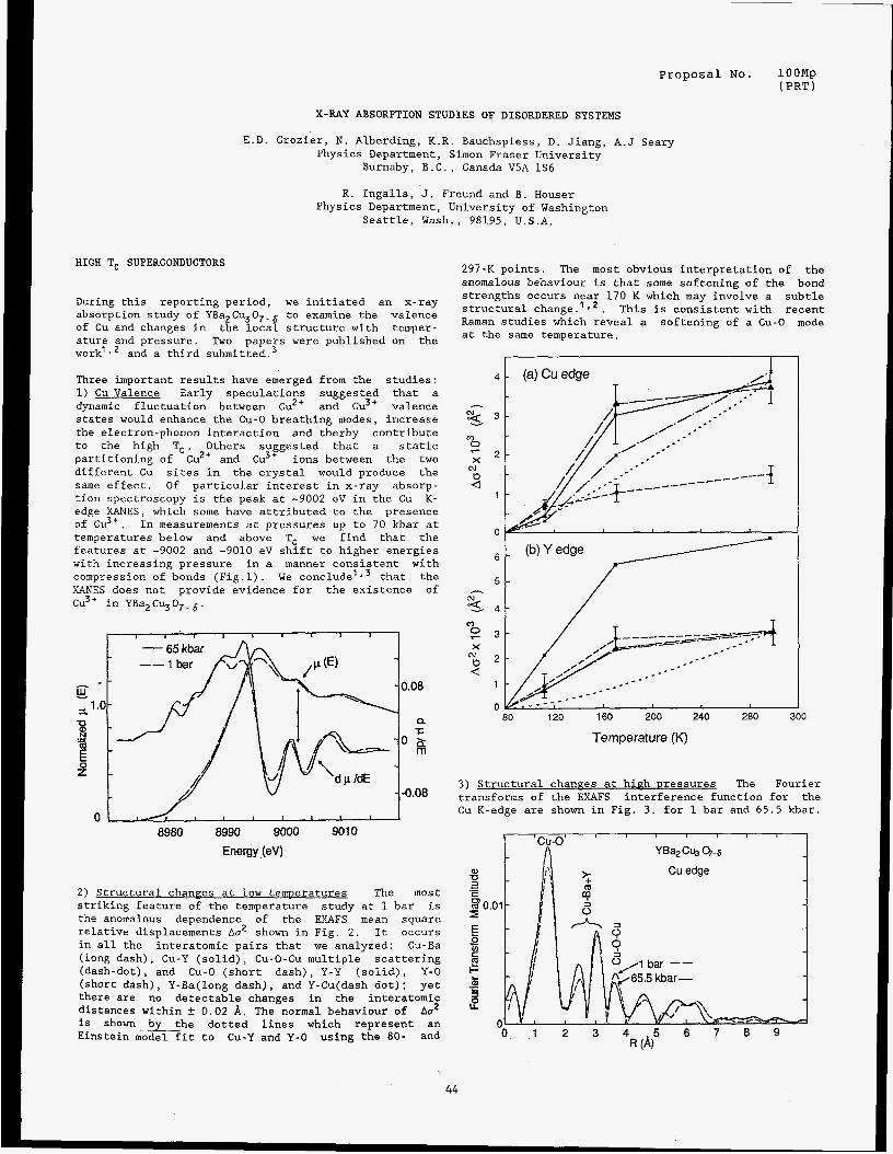
Proposal No. lOOMp ( PRT
X-RAY ABSORPTION STUDIES OF DISORDERED SYSTEMS
E.D. Crozier, N. Alberding, K.R. Bauchspiess, D. Jiang, A.J Seary Physics Department, Simon Fraser University
Burnaby, B.C., Canada VSA 1S6
R. Lngalls, J. Freund and B. Houser Physics Department, University of Washington
Seattle, Wash., 98195, U.S.A.
HIGH T, SUPERCONDUCTORS
During this reporting period, we initiated an x-ray absorption study of YBa2Cu307.6 to examine the valence of Cu and changes in the local structure with temper- ature and pressure. Two papers were published on the work'l2 and a third ~ubmitted.~
297-K points. The most obvious interpretation of the anomalous behaviour is that some softening of the bond strengths occurs near 170 K which may involve a subtle structural change.lr2. This is consistent with recent Raman studies which reveal a softening of a Cu-0 mode at the same temperature.
Three important results have emerged from the studies: 1) Cu Valence Early speculations suggested that a dynamic fluctuation between Cu2+ and Cu3' valence states would enhance the Cu-0 breathing modes, increase the electron-phonon interaction and therby contribute to the high T,. Others suggested that a static partitioning of Cu" and Cu3' ions between the two different Cu sites in the crystal would produce the same effect. Of particular interest in x-ray absorp- tion spectroscopy is the peak at -9002 eV in the Cu K- edge XANES, which some have attributed to the presence of Cu3+. In measurements at pressures up to 70 kbar at temperatures below and above Tc we find that the features at -9002 and -9010 eV shift to higher energies with increasing pressure in a manner consistent with compression of, bonds (Fig.1). We conclude''3 that the XANES does not provide evidence for the existence o f Cu3+ in YBaZ Cu3 07. & .
. 1-0.08
0 1 I I I t I I I I 8980 8990 9000 9010
Energy (eV)
2) Structural channes at low temveratures The most striking feature of the temperature study at 1 bar is the anomalous dependence of the EXAFS mean square relative displacements Ao2 shown in Fig. 2. It occurs in all the interatomic pairs that we analyzed: Cu-Ba (long dash), Cu-Y (solid), Cu-0-Cu multiple scattering (dash-dot), and Cu-0 (short dash), Y-Y (solid), Y-0 (short dash), Y-Ba(1ong clash), and Y-Cu(dash dot): yet there are no detectable changes in the interatomic distances within _+ 0.02 A . The normal behaviour of Ao2 is shown by the dotted lines which represent an Einstein mo-dy1Tit to Cu-Y and Y-0 using the 80- and
Temperature (K)
3) Structural changes at high uressures The Fourier transfcrms of the EXAFS interference function for the Cu K-edge are shown in Fig. 3 . for 1 bar and 65.5 kbar.
'cu-0' YBa2 Cu3 Q. s J
Cu edge
1 2 3 4 6 7 8 9
44
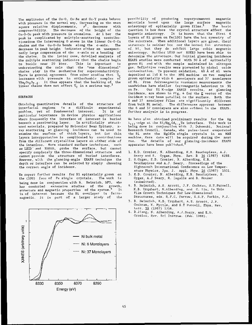
The amplitudes of the Cu-0, Cu-Ba and Cu-Y peaks behave with pressure in the normal way, increasing as the mean square relative displacements, o2 , decrease with compressibility. The decrease of the height of the cu-0-C~ peak with pressure is anomalous. At 1 bar the peak is complicated by multiple-scattering contribu- tionsfrom the intervening 0 atoms in the linear Cu-0-Cu chains and the Cu-0-Cu bonds along the c-axis. The decrease in peak height indicates either an unexpect- tedly large compression of the c-axis or a bending of the chains. In the latter case, detailed analysis of the multiple scattering indicates that the chains begin to buckle near 20 kbar. This is important to understanding the role that the "one dimensional" chains play in the mechanism for superconductivity. There is general agreement from other studies that T, increases with pressure in orthorhombic samples of YBaZCu30,.6. It thus appears that the buckling of the linear chams does not affect T, in a serious way.3
SURFACES
Obtaining quantitative details of the structure of interfacial regions is a difficult experimental problem, yet of fundamantal interest. It is of particular importance in device physics applications where frequently the interface of interest is buried beneath a passivating layer. In artificially struct- ured materials, prepared by Molecular Beam Epitaxy, x- ray scattering at glancing incidence can be used to examine the surface of thick layers, but for thin layers interpretation is complicated by contributions from the different crystalline layers on either side of the interface. More standard surface techniques, such as LEED and RHEED, probe the surface, but cannot specify completely the three-dimensional structure and cannot provide the structure of buried interfaces, However, with the glancing-angle EXAFS technique the depth or interface can be selected by simply choosing the correct angle of incidence.
We report further results for Ni epitaxially grown on the (100) face of Fe single crystals, The work is being done in conjunction with B. Heinrich, S N , who has conducted extensive studies of the growth, structure and magnetic properties of the system.4 It is of interest because the Ni overlayer is ferro- magnetic. It is part of a larger study of the
1
I I I I I I I I
- Ni bulk metal
Ni: 6 Monolayers
--- Ni: 37 Monolayers
8330 8350 8370 8390 Energy (eV)
45
possibility of producing superpermanent magnetic materials based upon the large surface magnetic anisotropy of epitaxially grown films.5 The basic question is how does the crystal structure affect the magnetic anisotropy. It is known that the first 6 layers of Ni grown on Fe(100) have the bcc symmetry of the substrate. When additional layers are grown, their structure is neither bcc nor the normal fcc structure of Ni, but they do exhibit large cubic magnetic anisotropy. Neither LEED nor RHEED have been able to determine the structure. Our initial glancing incidence EXAFS studies were conducted with 50 .& of epitaxially grown Ni and with the sample maintained in nitrogen gas. Definitive results were prevented by nickel oxide contamination. To prevent oxidation, 60 .& of A1 were deposited at 150 K in the MBE machine on two samples grown epitaxially with 6 monolayers and 37 monolayers of Ni. From ferromagnetic resonance measurements the sandwiches have similar ferromagnetic behaviour to Ni on Fe. Our Ni K-edge XANES results, at glancing incidence, are shown in Fig. 4 for the E vector of the incident x-ray beam parallel to the surface. Both the 6 and 37 monolayer films are significantly different from bulk Ni metal. The differences apparent between the 6 and 37 monolayers are also evident in the EXAFS. Further analysis is in progress.
We have also obtained preliminary results for the Hg LIrl -edge at the Si/Hg,Cd,-.Te interface. This work is being done in conjunction with J. Dubowski, National Research Council, Canada, who pulse-laser evaporated the Si onto the HgCdTe single crystals in an MBE machine. More data will be acquired during the next run. The details of our glancing-incidence EXAFS apparatus have been published.
1. E.D. Crozier, N. Alberding, K.R. Bauchspiess, A.J.
2. S.Gygax, E.D. Crozier, N. Alberding, K.R. Seary and S . Gygax, Phys. Rev. B 36 (1987) 8288.
Bauchspiess and A.J. Seary, Proceedings of the Eighteenth International Conference on Low Temper- ature Physics, Jpn. J. Appl. Phys. (1987) 1051.
Gygax, A.J Seary, R. Ingalls and B. Houser (submitted).
4 . B. Heinrich, A.S. Arrott, J.F. Cochran, S.T.Purcel1, K.B. Urquhart, N.Alberding, and C. Liu, in Thin Film Growth Techniques for Low-Dimensional Structures, eds. R.F.C. Farrow, S . S . P . Parkin, P.J.
5. B. Heinrich, K.B. Urquhart, A.S. Arrott, J.F. Cochran, K. Myrtle, and S.T Purcell, Phys. Rev. Lett. 59 (1987) 1756.
6. D.Jiang, N. Alberding, A.J.Seary, and E.D. Crozier, Rev. Sci Instrum. (Feb. 1988).
3 . E.D. Crozier, N. Alberding, K.R. Bauchspiess, S .

proposal NO. 822Mp
TEMPERATURE DEPENDENCE STUDY OF MONOLAYERS AND MULTILAYER3 OF THE LANGMUIR-BLODGETT TYPE
W. B. Yun, J. M. Bloch, X. Yang and J. Viccaro
Argonne National Laboratory
9700 5. Cars Avenue, Argoone, IL 60459
INTRODUCTION : we have &ended our previow x-ray studies of Cd-Stearate and &-Stearate monolayem and multilayers to investigate the effect of temperature variation on the in-plane structure of the layers and the stacking correlation between the layers. In this work, we have re-examined a previously reported trigonal structure for a monolayer, which did not provide a consistent picture for the in-plane orthorhombic structure observed for 2 ,3 and more layers.
RESULTS AND DISCUSSIONS :
A) A Cd-Stearate Monolayer Sample
At room temperature, two diffraction peaks A and B (se Fig.1) were observed. The A peak has been observed priv- iously by one of w and collabratom on a similar sample ( NSLS report 1986, Thin Film Gordon conference - Santa Bar- bara 1986 ). The B peak indicates that the structure is not trigonal, as was previously reported from electron diffraction experiments. However the ratio betwen the diffraction inten- sitiee of the two diffraction peaks also precludes the interpre tation of a simple orthorhombic structure. In the tempera- ture dependence study, it was found that the A peak, which corresponds with the multilayer [ll] orthorhombic peak, was practicelly temperature independent, in both peak position q a d liewidth. In contrast the B peak, which corresponds with the multilayer (021 orthorhombic peak, moved away from the [ll] peak when the temperature was lowered down to - 40 degree C. At temperatures elevated slightly above room temperature the 1021 peak position first moved toward the [ll] peak and its intensity decreased. At about 35 degrees C the peak disappeared completely. It reappeared when the temperature was lowered again. In the temperature range in- vestigated, the ratio of the B peak intensity to that of the A peak was consistantly lower than what one would expect from a regular orthorhombic structure exen at the lowest temper- ature of the experiment. In order to explain the resulk, a model of random orthorohmbic structure is proposed.
B) A 15 Layer Cd-Stearate Sample
At room temperature, results similar to those reported by us earlier (NSLS report 1986) were obtained and interpreted as an in-plane orthorhombic structure. Upon cooling, the [02OJ in-plane peak position, originating with the orthorhom- bie unit cell, moved towards larger q values, while the (1101 in-plane peak position remained unchanged. The shift of the [020] peak position toward larger q values indicates an in- crease in the orthorhombic distortion away from the trigonal rtructure at lower temperatures. The effect of temperature variation on the stacking correlation between layers was invea- tigated with scam made along [11Q2] and [OZQ,] directions with different history of thermal conditions. The results show that the stacking correlation remained u n c k g e d when the
h
1 P
Y 4 k
k t
Fig.1 Selected scans from a Cd-stearate monolayer at differnt temperatures. Cicles are experimental data and the solid l i e preaents a fit of the corresponding data with two Lorentxi-.
sample was cooled down to -40 degree C and the sample ex- hibited a 3D structure. When the sample was warmed back to room temperature, the stacking correlation in the [OZQ,] was lost. In further temperature eyelings between room tem- perature and -90 degree C, no further change in the stacking correlation was observed. This indicates that the sample had changed to a new struture after the first temperature cycling and the new structure is thermodynamcally more stable than that originally acquired by the sample at preparation. The new structure has a repeat distance larger than that of the original sample, indicating that the temperature cycling had made the chaina perhaps straighter than that in the origi- nal sample. The new structure has good stacking correlation along the [11L] direction but poor stacking correlation along the [02L] direction, and is reminiient of a "one dimensional" Smectic B material.
ACKNOWLEDGEMENTS:
This work was supported by the U. S. Department of en- ergy, BES-Material Science, under contract W-31-lOsENG 38. Part of this work was performed at SSRL. The support and services provided by SSRL staff are greatly appreciated.
46

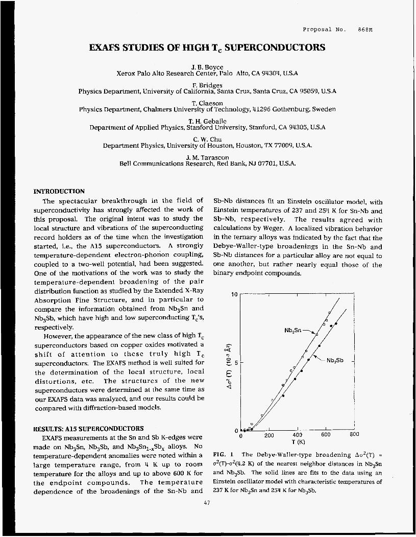
Proposal No. 868M
EXAFS STUDIES OF HIGH T, SUPERCONDUCTORS
Physics
J. B. Boyce Xerox Palo Alto Research Center, Palo Alto, CA 94304, U.S.A
Department, University of F. Bridges
California, Santa cruz, Santa Cruz, CA 95059, U.S.A
T. Claeson Physics Department, Chalmers University of Technology, 41296 Gothenburg, Sweden
T. H. Geballe Department of Applied Physics, Stanford University, Stanford, CA 94305, U.S.A
C. W. Chu Department Physics, University of Houston, Houston, TX 77009, U.S.A.
J. M. Tarascon Bell Communications Research, Red Bank, NJ 07701, U.S.A.
INTRODUCTION
The spectacular breakthrough in the field of superconductivity has strongly affected the work of this proposal. The original intent was to study the local structure and vibrations of the superconducting record holders as of the time when the investigation started, i.e., the A15 superconductors. A strongly temperature-dependent electron-phonon coupling, coupled to a two-well potential, had been suggested. One of the motivations of the work was to study the temperature-dependent broadening of the pair distribution function as studied by the Extended X-Ray Absorption Fine Structure, and in particular to compare the information obtained from Nb3Sn and Nb,Sb, which have high and low superconducting Tis, respectively.
However, the appearance of the new class of high T, superconductors based on copper oxides motivated a sh i f t of a t tent ion to these t ruly high T, superconductors. The EXAFS method is well suited for the determination of the local structure, local distortions, etc. The structures of the new superconductors were determined at the same time as our EXAFS data was analyzed, and our results could be compared with diffraction-based models.
RESULTS: A15 SUPERCONDUCTORS
EXAFS measurements at the Sn and Sb K-edges were made on Nb,Sn, Nb,Sb, and Nb,Sn,..,Sb, alloys. No
temperature-dependent anomalies were noted within a large temperature range, from 4 K up to room temperature for the alloys and up to above 600 K for the endpoint compounds. The temperature dependence of the broadenings of the Sn-Nb and
Sb-Nb distances fit an Einstein oscillator model, with Einstein temperatures of 237 and 254 K for Sn-Nb and Sb-Nb, respectively. The results agreed with calculations by Weger. A localized vibration behavior in the ternary alloys was indicated by the fact that the Debye-Waller-type broadenings in the Sn-Nb and Sb-Nb distances for a particular alloy are not equal to one another, but rather nearly equal those of the binary endpoint compounds.
10
h
4
c5
t
3 0
h
N b -4
0 I I I 200 400 600 800
T (K)
FIG. 1 The Debye-Waller-type broadening A d ( T ) =
u2(T)-u2(4.2 K) of the nearest neighbor distances in Nb,Sn and Nb,Sb. The solid lines are fits to the data using an Einstein oscillator model with characteristic temperatures of 237 K for NbJSn and 254 K for Nb,Sb.
47
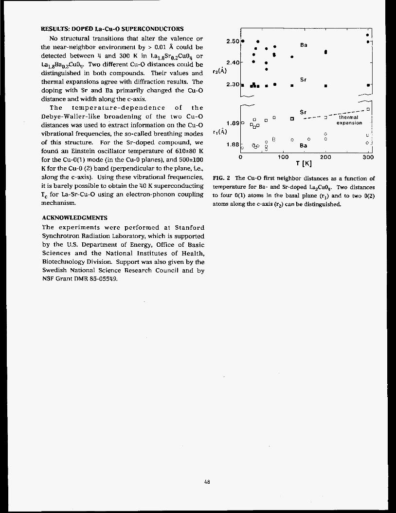
RESULTS: DOPED La-Cu-0 SUPERCONDUCTORS
No structural transitions that alter the valence or the near-neighbor environment by > 0.01 A could be detected between 4 and 300 K in La1.8Sro.2Cu0,, or Lal.8Bao.2Cu0,. Two different Cu-0 distances could be distinguished in both compounds. Their values and thermal expansions agree with diffraction results. The doping with Sr and Ba primarily changed the Cu-0 distance and width along the c-axis.
The t empera tu re -dependence of t h e Debye-Waller-like broadening of the two C u - 0 distances was used to extract information on the Cu-0 vibrational frequencies, the so-called breathing modes of this structure. For the Sr-doped compound, we found an Einstein oscillator temperature of 610280 K for the Cu-O(1) mode (in the Cu-0 planes), and 500?100 K for the Cu-0 (2) band (perpendicular to the plane, i.e., along the c-axis). Using these vibrational frequencies, it is barely possible to obtain the 40 K superconducting T, for La-Sr-Cu-0 using an electron-phonon coupling mechanism.
ACKNOWLEDGMENTS
The experiments were performed at Stanford Synchrotron Radiation Laboratory, which is supported by the U.S. Department of Energy, Office of Basic Sciences and the National Institutes of Health, Biotechnology Division. Support was also given by the Swedish National Science Research Council and by NSF Grant DMR 85-05549.
I , I 1 I
e 2.50
e e * e 8
2.40-
r*(& e
2.301 a, , G,
Ba
e
Sr m
a
8
m 1
---- --I _--- 0 Sr _--- ---- o thermal
expansion -
0 0 0 0
Ba 0.
I I I I I 1
0 100 200 300 T [Kl
FIG. 2 The Cu-0 first neighbor distances as a function of
temperature for Ba- and Sr-doped La,CuOu. Two distances to four O(1) atoms in the basal plane (rl) and to two O(2)
atoms along the c-axis (rz) can be distinguished.
48
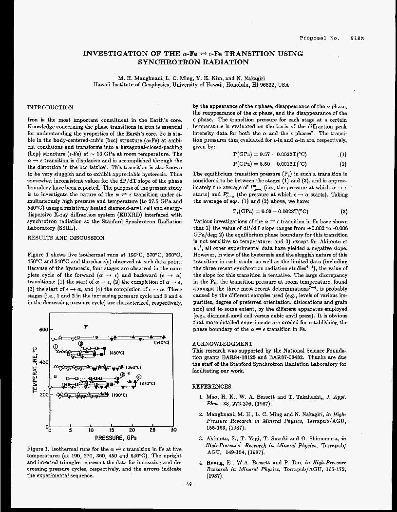
Proposal NO. 9 1 8 ~
INVESTIGATION OF THE a-Fe + e-Fe TRANSITION USING SYNCHROTRON RADIATION
M. H. Manghnani, L. C. Ming, Y. H. Kim, and N. Nakagiri Hawaii Institute of Geophysics, University of Hawaii, Honolulu, HI 96822, USA
INTRODUCTION
Iron is the most important constituent in the Earth’s core. Knowledge concerning the phase transitions in iron is essential for understanding the properties of the Earth’s core. Fe is sta- ble in the body-centered-cubic (bcc) structure (a-Fe) at ambi- ent conditions and transforms into a hexagonal-closed-packing (hcp) structure (E-Fe) at - 13 GPa at room temperature. The a + E transition is displacive and is accomplished through the the distortion in the bcc lattice’. This transition is also known to be very sluggish and to exhibit appreciable hysteresis. Thus somewhat inconsistent values for the dP/dT slope of the phase boundary have been reported. The purpose of the present study is to investigate the nature of the a * E transition under si- multaneously high pressure and temperature (to 27.5 GPa and 540OC) using a resistively heated diamond-anvil cell and energy- dispersive X-ray diffraction system (EDXRD) interfaced with synchrotron radiation at the Stanford Synchrotron Radiation Laboratory (SSRL).
RESULTS AND DISCUSSION
Figure 1 shows five isothermal runs at 190°C, 27OoC, 36OoC, 45OOC and 540°C and the phase(s) observed at each data point. Because of the hysteresis, four stages are observed in the com- plete cycle of the forward (a .+ e) and backward (e -+ a) transitions: (1) the start of a -i e, (2) the completion of a -+ e,
(3) the start of e -+ a, and (4) the completion of e -+ a. These stages (i.e., 1 and 2 in the increasing pressure cycle and 3 and 4 in the decreasing pressure cycle) are characterized, respectively,
600 1 Y
I- 200 4$eigz-. (19O’Cl t OO a 5 10 15 20 25
PRESSURE, GPO
Figure 1. Isothermal runs for the a + e transition in Fe at five temperatures (at 190, 270, 360, 450 and 540°C). The upright and inverted triangles represent the data for increasing and de- creasing pressure cycles, respectively, and the arrows indicate the experimental sequence.
by the appearance of the e phase, disappearance of the a phase, the reappearance of the a phase, and the disappearance of the e phase. The transition pressure for each stage at a certain temperature is evaluated on the basis of the diffraction peak intensity data for both the a and the e phases’. The transi- tion pressures thus evaluated for E-in and a-in are, respectively, given by:
P(GPa) = 9.57 - 0.0032T(OC) (1)
P(GPa) = 8.50 - 0.0016T(OC)
The equilibrium transition pressure (P,) in such a transition is considered to be between the stages (1) and (3), and is approx- imately the average of P:+e (i.e., the pressure at which a -+ E
starts) and P:+, (the pressure at which E -i a starts). Taking the average of eqs. (1) and (2) above, we have:
P,( GPa) = 9.03 - 0.0023T(OC) (3)
Various investigations of the a + E transition in Fe have shown that 1) the value of dP/dT slope ranges from +0.002 to -0.006 GPa/deg; 2) the equilibrium phase boundary for this transition is not sensitive to temperature; and 3) except for Akimoto et al.3, all other experimental data have yielded a negative slope. However, in view of the hysteresis and the sluggish nature of this transition in each study, as well as the limited data (including the three recent synchrotron radiation ~ t u d i e s ~ - ~ ) , the value of the slope for this transition is tentative. The large discrepancy in the PO, the transition pressure at room temperature, found amongst the three most recent deterrninati~ns’-~, is probably caused by the different samples used (e.g., levels of various im- purities, degree of preferred orientation, dislocations and grain size) and to some extent, by the different apparatus employed (e.g., diamond-anvil cell versus cubic anvil press). It is obvious that more detailed experiments are needed for establishing the phase boundary of the a + e transition in Fe.
ACKNOWLEDGMENT This research was supported by the National Science Founda- tion grants EAR84-18125 and EAR87-08482. Thanks are due the staff of the Stanford Synchrotron Radiation Laboratory for facilitating our work.
REFERENCES
1. Mao, H. K., W. A. Bassett and T. Takahashi,, J. Appl. Phys., 38, 272-276, (1967).
2. Manghnani, M. H., L. C . Ming and N. Nakagiri, in High- Pressure Research in Mineral Physics, Terrapub/AGU, 155-163, (1987).
3. Akimoto, S., T. Yagi, T. Suzuki and 0. Shimomura, in High-pressure Research in Mineral Physics, Terrapub/ AGU, 149-154, (1987).
4. Hiiang, E., W.A. Bassett and P. Tao, in High-pressure Research in Mineral Physics, Terrapub/AGU, 165-172, (1987).
49


MPOWALOUB X-RAY DIFFRACTION STUD118 OF BIWBPALLIC CATALYBPB : Pt-R. On S i 0 2
K. 6 . Liang, 0 . J. Hughee and J. H. Sinfelt Exxon Research and Engineering Co.
Route 22 E, Clinton Township Annandale, NJ 08801
Proposal NO. 940M
The formation of bimetallic clusters of immiscible binary alloys has major impacts in industrial catalysis, in particular in the production of aromatic hydrocarbons for automotive fuels. It is generally believed that these clusters are inhomogeneous due to segregation of one constituent to the surface of the small particles. However, to obtain a more precise description of the cluster structure is often a formidable task. In this work, we have employed diff- erential anomalous x-ray diffraction (DAXD) technique to study model Pt-Re bimetallic catalysts. The x-ray measurements were per- formed on the wiggler station IV-3.
The use of DAXD technique to obtain the dif- fraction patterns from supported catalytic particles has been demonstrated previously on supported Pt catalysts.1 The technique provides the capability of atom-type select- ivity, as in EXAFS, but emphasizes the low-q part of the interference function. This function contains the essential structural information and can be modeled directly with an assumed structure using the Debye equa- tion.
The catalysts used in this study contain 2.5, 5.0 and 10.0 wt.% of Pt and Re in a one-to-one weight ratio supported on silica as prepared by a usual coimpregnation pro- cedure. Diffraction patterns were collected using x-rays with energies 10 and 100 eV below the absorption maxima (white lines) at the LIII edges of Pt (-11564 eV) and Re (-10535eV). The differential diffraction patterns (DDP's)at both edges were obtained after proper normalization of the measured intensities.
Figure 1 compares the Pt-edge DDP% of two catalysts: one with 2.5 wt.% Pt on Si02 and the other with 2.5 wt.% Pt and 2.5 wt.% Re on Si02. From the widths of the Bragg peaks, one notices that the sizes of Pt particles are substantially reduced (from -150A to -20A) by the incorporation of Re, indicating the interactions between these two metals. The observed DDP can be modeled very well as small fcc particles (Fig. IC).
Knowing that in the bulk phase, Pt is fcc and Re is hcp, it is interesting to see that the Re-edge DDP*s reveal both fcc and hcp forms (Fig. 2). One plausible explanation is that in the bimetallic particles the fcc Re could be formed due to epitaxy with Pt. To our knowledge, fcc Re has not been reported.
In short, we demonstrate the use of DAXD technique to reveal novel structural features of Pt-Re bimetallic catalysts. The results are succesfully interpreted by direct structural modeling of the measured intensities.
50
REFERENCE 1. K. S. Liang, S. S. Laderman and J. H. Sinfelt, J. Chem. Phys. a, 2352 (1987).
a -
Q (A'1) F i g . 1 The differenual intensity near Pt LIII edge of (a) 2.5 wt.5 Pt on Si02, (b) 2 . 5 w t . % Pt-Re on Si02, and ( c ) calculated pattern of IsA particle of fcc Pt.
I
C ! J
I I I t # . I I I I , ] 0.0 6.
Q (A-1) Fig. 2 The differential intensity near Re LIII edge of (a) 10 wt.% Pt-Re on Si02, (b) 2.5 w t . % Pt-Re on Si02, and (c) calculated pattern of mixed fcc and,hcp Re particle.

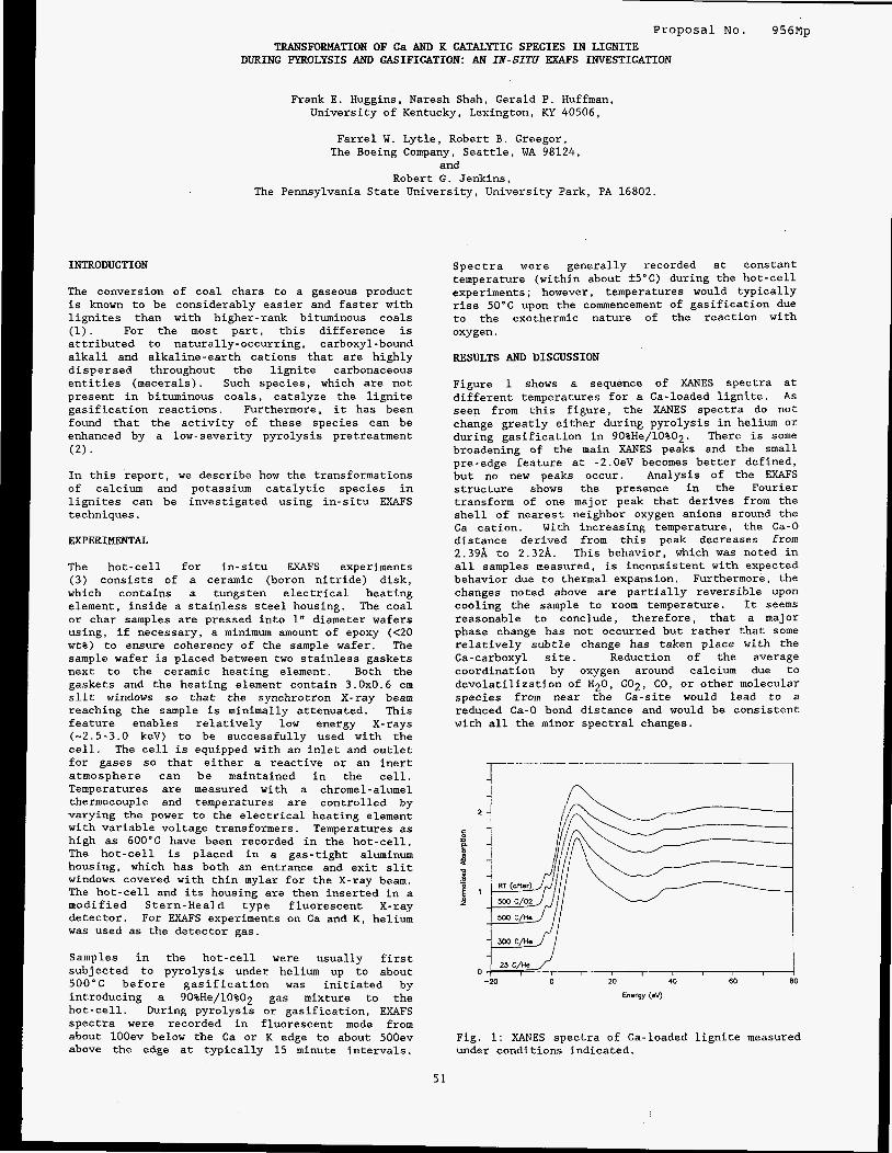
Proposal No. 956Mp TRANSFORMATION OF Ca AND K CATALYTIC SPECIES IN LIGNITE
DURING PYROLYSIS AND GASIFICATION: AN IN-SITU U S INVESTIGATION
Frank E. Huggins, Naresh Shah, Gerald P. Huffman, University of Kentucky, Lexington, KY 40506,
Farrel W. Lytle, Robert B. Greegor, The Boeing Company, Seattle, WA 98124,
and Robert G. Jenkins,
The Pennsylvania State University, University Park, PA 16802.
INTRODUCTION
The conversion of coal chars to a gaseous product is known to be considerably easier and faster with lignites than with higher-rank bituminous coals (1). For the most part, this difference is attributed to naturally-occurring, carboxyl-bound alkali and alkaline-earth cations that are highly dispersed throughout the lignite carbonaceous entities (macerals). Such species, which are not present in bituminous coals, catalyze the lignite gasification reactions. Furthermore, it has been found that the activity of these species can be enhanced by a low-severity pyrolysis pretreatment (2).
In this report, we describe how the transformations of calcium and potassium catalytic species in lignites can be investigated using in-situ EXAFS techniques.
EXPERIMENTAL
The hot-cell for in-situ EXAFS experiments (3) consists of a ceramic (boron nitride) disk, which contains a tungsten electrical heating element, inside a stainless steel housing. The coal or char samples are pressed into 1" diameter wafers using, if necessary, a minimum amount of epoxy (120 wt%) to ensure coherency of the sample wafer. The sample wafer is placed between two stainless gaskets next to the ceramic heating element. Both the gaskets and the heating element contain 3.0x0.6 cm slit windows so that the synchrotron X-ray beam reaching the sample is minimally attenuated. This feature enables relatively low energy X-rays (-2.5-3.0 keV) to be successfully used with the cell. The cell is equipped with an inlet and outlet for gases so that either a reactive or an inert atmosphere can be maintained in the cell. Temperatures are measured with a chromel-alumel thermocouple and temperatures are controlled by varying the power to the electrical heating element with variable voltage transformers. Temperatures as high as 600'C have been recorded in the hot-cell. The hot-cell is placed in a gas-tight aluminum housing, which has both an entrance and exit slit windows covered with thin mylar for the X-ray beam. The hot-cell and its housing are then inserted in a modified Stern-Heald type fluorescent X-ray detector. For EXAFS experiments on Ca and K, helium was used as the detector gas.
Samples in the hot-cell were usually first subjected to pyrolysis under helium up to about 500°C before gasification was initiated by introducing a 90%He/10%02 gas mixture to the hot-cell. During pyrolysis or gasification, EXAFS spectra were recorded in fluorescent mode from about lOOev below the Ca or K edge to about 500ev above the edge at typically 15 minute intervals.
51
Spectra were generally recorded at constant temperature (within about 55°C) during the hot-cell experiments; however, temperatures would typically rise 50°C upon the commencement of gasification due to the exothermic nature of the reaction with oxygen.
RESULTS AND DISCUSSION
Figure 1 shows a sequence of XANES spectra at different temperatures for a Ca-loaded lignite. A s seen from this figure, the XANES spectra do not change greatly either during pyrolysis in helium or during gasification in 90%He/10%02. There is some broadening of the main XANES peaks and the small pre-edge feature at -2.0eV becomes better defined, but no new peaks occur. Analysis of the EXAFS structure shows the presence in the Fourier transform of one major peak that derives from the shell of nearest neighbor oxygen anions around the Ca cation. With increasing temperature, the Ca-0 distance derived from this peak decreases from 2.398 to 2.32A. This behavior, which was noted in all samples measured, is inconsistent with expected behavior due to thermal expansion. Furthermore, the changes noted above are partially reversible upon cooling the sample to room temperature. It seems reasonable to conclude, therefore, that a major phase change has not occurred but rather that some relatively subtle change has taken place with the Ca-carboxyl site. Reduction of the average coordination by oxygen around calcium due to devolatilization of H20, Cop, CO, or other molecular species from near the Ca-site would lead to a reduced Ca-0 bond distance and would be consistent with all the minor spectral changes.
TFf 300 C/He
I
25 c/Ha
-20 0 20 40 60 80
Energy (ev)
Fig. 1: XANES spectra of Ca-loaded lignite measured under conditions indicated.
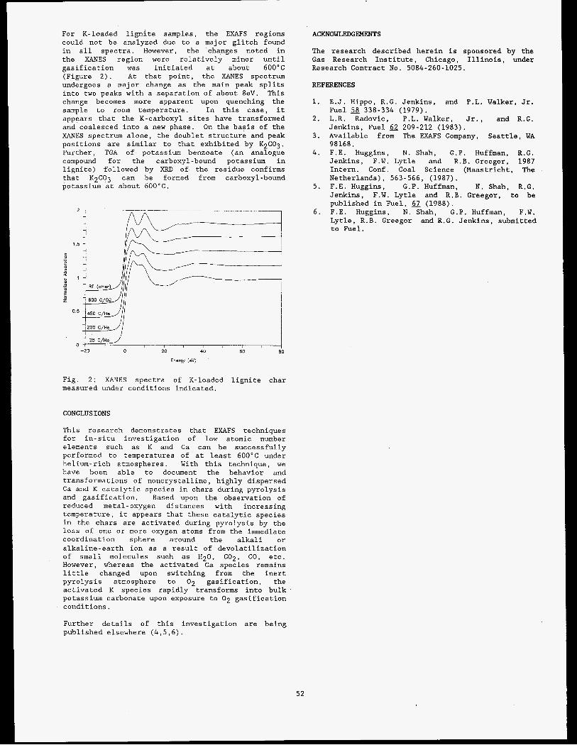
For K-loaded lignite samples, the EXAFS regions could not be analyzed due to a major glitch found in all spectra. However, the changes noted in the XANES region were relatively minor until gasification was initiated at about 600°C (Figure 2). At that point, the XANES spectrum undergoes a major change as the main peak splits into two peaks with a separation of about 8eV. This change becomes more apparent upon quenching the sample to room temperature. In this case, it appears that the K-carboxyl sites have transformed and coalesced into a new phase. On the basis of the XANES spectrum alone, the doublet structure and peak positions are similar to that exhibited by K2CO3. Further, TGA of potassium benzoate (an analogue compound for the carboxyl-bound potassium in lignite) followed by XRD of the residue confirms that K2CO3 can be formed from carboxyl-bound potassium at about 600°C.
0 , 25 C/He
-20 0 20 40 60 80
Energy (eV)
Fig. 2: XANES spectra of K-loaded lignite char measured under conditions indicated.
CONCLUSIONS
This research demonstrates that EXAFS techniques for in-situ investigation of low atomic number elements such as K and Ca can be successfully performed to temperatures of at least 600°C under helium-rich atmospheres. With this technique, we have been able to document the behavior and transformations of noncrystalline, highly dispersed Ca and K catalytic species in chars during pyrolysis and gasification. Based upon the observation of reduced metal-oxygen distances with increasing temperature, it appears that these catalytic species in the chars are activated during pyrolysis by the loss of one or more oxygen atoms from the immediate coordination sphere around the alkali or alkaline-earth ion as a result of devolatilization of small molecules such as H20, C02, CO, etc. However, whereas the activated Ca species remains little changed upon switching from the inert pyrolysis atmosphere to 02 gasification, the activated K species rapidly transforms into bulk potassium carbonate upon exposure to 02 gasification conditions.
Further details of this investigation are being published elsewhere (4,5,6).
52
ACKNOWLEDGEMENTS
The research described herein is sponsored by the Gas Research Institute, Chicago, Illinois, under Research Contract No. 5084-260-1025.
REFERENCES
1. E . J . Hippo, R.G. Jenkins, and P.L. Walker, Jr. Fuel 58 338-334 (1979).
2. L.R. Radovic, P.L. Walker, Jr., and R.G. Jenkins, Fuel 62 209-212 (1983).
3. Available from The EXAFS Company, Seattle, WA 98168.
4. F.E. Huggins, N. Shah, G.P. Huffman, R.G. Jenkins, F.W. Lytle and R.B. Greegor, 1987 Intern. Conf. Coal Science (Maastricht, The Netherlands), 563-566, (1987).
5. F.E. Huggins, G.P. Huffman, N. Shah, R.G. Jenkins, F.W. Lytle and R.B. Greegor, to be published in Fuel, 67 (1988).
6. F.E. Huggins, N. Shah, G.P. Huffman, F.W. Lytle, R.B. Greegor and R.G. Jenkins, submitted to Fuel.
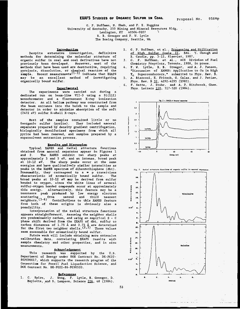
EXAFS STUDIES OF ORGANIC SULFUR IN COAL Proposal No. 956Mp
G. P. Huffman, N. Shah, and F. E. Huggins University of Kentucky, 233 Mining and Mineral Resources Bldg.
Lexington, KY 40506-0107 > R. B. Greegor and F. W. Lytle The Boeing Company, Seattle, WA
Introduction Despite extensive investigation, definitive
methods for determining the molecular structure of organic sulfur in coal and coal derivatives have not previously been developed. Moreover, most of the methods that have been used are destructive, requiring pyrolysis, dissolution, or chemical reaction of the sample. Recent mea~urements(~-~) indicate that EXAFS may be an excellent method of investigating organically bound sulfur.
berimental The experiments were carried out during a
dedicated run on beam-line VII-3 using a Si(ll1) monochromator and a fluorescent X-ray ionization detector. An all helium pathway was constructed from the beam entrance into the hutch to the sample and detector in order to minimize absorption of the soft (2472 eV) sulfur K-shell X-rays.
Most of the samples contained little or no inorganic sulfur (pyrite). They included maceral separates prepared by density gradient centrifugation, biologically desulfurized specimens from which all pyrite had been removed, and samples prepared by a supersolvent extraction process.
Results and Discussion Typical XANES and radial structure functions
obtained from maceral separates appear in Figures 1 and 2. The XANES exhibit two sharp peaks at approximately 3 and 5 eV, and an intense, broad peak at 10-12 eV. The sharp peaks occur at the same energies and have qualitatively similar intensities as peaks in the XANES spectrum of dibenzothiophene (dbt). Presumably, they correspond to s * p transitions characteristic of aromatically bound sulfur. The broad peaks at 10-12 eV may be derived from sulfur bonded to oxygen, since the white lines of several sulfur-oxygen bonded compounds occur at approximately this energy. Alternatively, this feature may be a resonance peak produced by low energy electron scattering from second and third nearest neighbors. (3-6) Contributions to this XANES feature from both of these origins is obviously also a possibility.
Interpretation of the radial structure functions appears straightforward. Assuming the neighbor shells are predominantly carbon, and using an empirical S - C phase shift derived from the EXAFS of dbt, sulfur to carbon distances of 1.75 A and 2.75 A are determined for the first two neighbor shells.(2*3) These values seem reasonable for aromatically bound sulfur.
Future work will include obtaining more extensive calibration data, correlating EXAFS results with sample chemistry and other properties, and in situ measurements.
Acknowledeement This research was supported by the U.S.
Department of Energy under DOE Contract No. DE-FC22- 86PC90017, which supports the research program of the Consortium for Fossil Fuel Liquefaction Science, and DOE Contract No. DE-FG22-86-PC90520.
References 1. C. Spiro, J. Wong, F. Lytle, R. Greegor, D.
Maylotte, .and S.. Lampson, Science 226, 48 (1984).
2. G. P. Huffman, et al., Processine and Utilization of Hieh Sulfur Coals 11, Eds. Y. Chough and R. Caudle, pp. 3-12, Elsevier, 1987.
3 . G. P. Huffman, et al., ACS Division of Fuel Chemistry Preprints, Toronto, 1988, in press.
4. F. W. Lytle, R. B. Greegor, and A . J. Panson, "Discussion of XANES: Application to Cu in High Tc Superconductors," submitted to Phys. Rev. B.
5. A. Bianconi, E. Fritsch, G. Calas, and J. Petiaw, Phys. Rev. B 2, 4292-4295 (1985).
6. F. Sette, J. Stohr, and A. P. Hitchcock, Chem. Phys. Letters 110, 517-520 (1984).
3.5
a
i 2.5
v 2 s
53

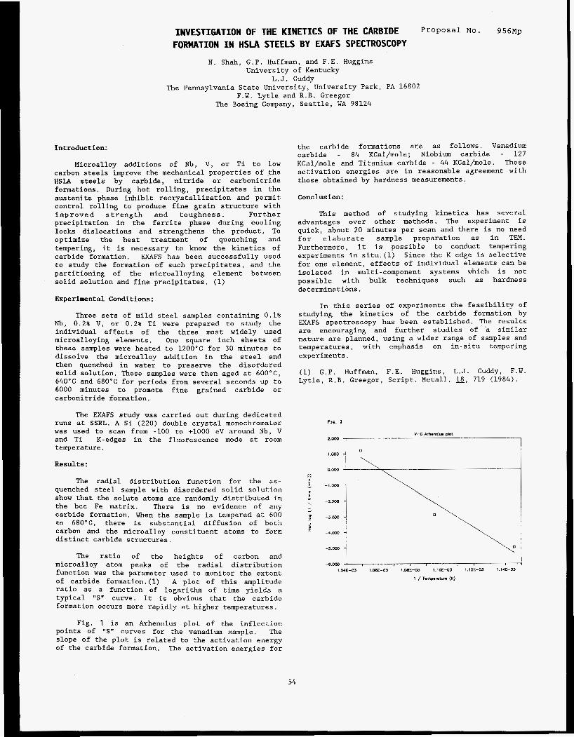
Introduction:
Microalloy additions of Nb, V, or Ti to low carbon steels improve the mechanical properties of the HSLA steels by carbide, nitride or carbonitride formations. During hot rolling, precipitates in the austenite phase inhibit recrystallization and permit control rolling to produce fine grain structure with improved strength and toughness. Further precipitation in the ferrite phase during cooling locks dislocations and strengthens the product. To optimize the heat treatment of quenching and tempering, it is necessary to know the kinetics of carbide formation. EXAFS has been successfully used to study the formation of such precipitates, and the partitioning of the microalloying element between solid solution and fine precipitates. (1)
Experimental Conditions:
Three sets of mild steel samples containing 0.1% Nb, 0.2% V, or 0.2% Ti were prepared to study the individual effects of the three most widely used microalloying elements. One square inch sheets of these samples were heated to 1200°C for 30 minutes to dissolve the microalloy addition in the steel and then quenched in water to preserve the disordered solid solution. These samples were then aged at 6OO0C, 640°C and 680°C for periods from several seconds up to 6000 minutes to promote fine grained carbide or carbonitride formation.
The EXAFS study was carried out during dedicated runs at SSRL. A Si (220) double crystal monochromator was used to scan from -100 to +lo00 eV around Nb, V and Ti K-edges in the fluorescence mode at room temperature.
Results :
The radial distribution function for the as- quenched steel sample with disordered solid solution show that the solute atoms are randomly distributed in the bcc Fe matrix. There is no evidence of any carbide formation. When the sample is tempered at 600 to 680°C, there is substantial diffusion of both carbon and the microalloy constituent atoms to form distinct carbide structures.
The ratio of the heights of carbon and microalloy atom peaks of the radial distribution function was the parameter used to monitor the extent of carbide formation.(l) A plot of this amplitude ratio as a function of logarithm of time yields a typical "S" curve. It is obvious that the carbide formation occurs more rapidly at higher temperatures.
Fig. 1 is an Arhennius plot of the inflection points of "S" curves for the vanadium sample. The slope of the plot is related to the activation energy of the carbide formation. The activation energies for
54
INVESTIGATION OF THE KINETICS OF THE CARBIDE P r o p o s a l No. 9 5 6 ~ ~
FORMATION IN HSIA STEELS BY EXAFS SPECTROSCOPY
N. Shah, G.P. Huffman, and F.E. Huggins University of Kentucky
L.J. Cuddy The Pennsylvania State University, University Park, PA 16802
F.W. Lytle and R.B. Greegor The Boeing Company, Seattle, WA 98124
the carbide formations are as follows. Vanadium carbide - 84 KCal/mole; Niobium carbide - 127 KCal/mole and Titanium carbide - 44 KCal/mole. These activation energies are in reasonable agreement with those obtained by hardness measurements.
Conclusion:
This method of studying kinetics has several advantages over other methods. The experiment is quick, about 20 minutes per scan and there is no need for elaborate sample preparation as in TEM. Furthermore, it is possible to conduct tempering experiments in situ.(l) Since the K edge is selective for one element, effects of individual elements can be isolated in multi-component systems which is not possible with bulk techniques such as hardness determinations.
In this series of experiments the feasibility of studying the kinetics of the carbide formation by EXAFS spectroscopy has been established. The results are encouraging and further studies of a similar nature are planned, using a wider range of samples and temperatures, with emphasis on in-situ tempering experiments.
(1) G.P. Huffman, F.E. Huggins, L.J. Cuddy, F.W. Lytle, R.B. Greegor, Script. Metall. 18, 719 ( 1 9 8 4 ) .
Fxs. 1
v-c AAmMl". Plot 2 . m
\
O.Oo0
-l.m
- 2 . m
-3 .m
- 5 . m
-e.Mo G J 1.W-03 l.WE-03 1.ME-03 1.1OE-03 1.12E-03 l.14E--03

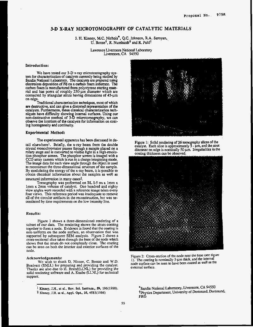
Introduction:
We have tested our 3-D x-ray microtomography sys- tem for characterization of catalysts currently being studied by Sandia National Laboratory. The catalysts are prepared using electroless deposition of Pd on a carbon foam substrate. The carbon foam is manufactured from polystyrene starting mate- rial and has pores of roughly 250-pm diameter which are connected by triangular struts having dimensions of 45-pm on edge.
Traditional characterization techniques, most of which are destructive, and can give a distorted representation of the catalysts. Furthermore, these classical characterization tech- niques have difficulty showing internal surfaces. Using our non-destructive method of 3-D microtomography, we can observe the interiors of the catalysts for information on coat- ing homogeneity and continuity.
Experimental Method:
The experimental apparatus has been discussed in de- tail elsewhere'. Briefly, the x-ray beam from the double crystal monochromator passes through a sample placed on a rotary stage and is converted to visible light in a high-resolu- tion phosphor screen. The phosphor screen is imaged with a CCD array camera which is run in a charge integrating mode. The image data for each view angle through the object is used to reconstruct the three-dimensional structure of the sample. By modulating the energy of the x-ray beam, it is possible to obtain chemical information about the samples as well as structural information in many cases?
Tomography was performed on BL 1-5 on a lmm x lmm x 2mm volume of catalyst. One hundred and eighty view angles were recorded with a reference image taken every four views. This reference period was inadequate to remove all of the circular artifacts in the reconstruction, but was ne- cessitated by time requirements on the low intensity line.
Results:
Figure 1 shows a three-dimensional rendering of a subset of our data. The rendering shows the struts coming together to form a node. Evidence is found that the coating is non-unifonn on the node surface, an observation that was supported by subsequent SEM analysis. Figure 2 shows a cross-sectional slice taken through the base of the node which shows that the struts do not completely close. The coating can be seen on both the interior and exterior surfaces of the node.
Acknowledgements: We wish to thank D. Nissen, C . Bonzo and W.D.
Bonivert (SNLL) for preparing and providing the catalyst. Thanks are also due to H. Brand(LLNL) for providing the solid rendering software and A. Knabe (LLNL) for technical support.
1 Kinney, J.H., et al., Rev. Sci. Instrum., 59, 196(1988). 2 Kinney, J.H. et al., Appl. Opt., 25, 4583(1986)
Figure 2 Cross-section of the node near the base (see Ilgurt: I). The coating is nominally 5 - p thick, and the internal node surface can be seen to have been coated as well as the external surface.
*Sandia National Laboratory, Livermore, CA 94550 ?Physics Department, University of Dortmund, Dortmund, FRG
55

Proposa l No. 9 8 3 ~ X-RAY ABSORPTION STUDY OF AMORPHOUS GERMANIUM AT HIGH PRESSURE
R. Ingalls and J. Freund
Department of Physics, University of Washington, Seattle, WA 98195
E. D. Crozier
Department of Physics, Simon Fraser University, Burnaby, B.C., V5A 1S6, CANADA
Amorphous materials are of great practical an2 theoret- ical interest. Theoretically they pose the problem of calculating their electronic and vibrational properties without resorting to the simplifications due to trans- lational symmetry. In fact, despite the lack of long range order, many properties are similar to their crystalline counterpart. from the practical standpoint. For example, ferromag- netic metallic glasses are of interest as memorydevices and transformer materials with little hysteresis loss. Amorphous semiconductors are of use for solar cells, thin film transistors, switching and memory devices and image processors.
One of the great strengths of the EXAFS technique is in the study of disordered materials, where diffraction is of limited value. One may obtain average coordination numbers, radial distribution functions and disorder terms quite readily, information crucial to understand- ing the structure. The latter is of course necessary for a microscopic understanding of the properties.
These materials are important
Amorphous germanium has proven to be an excellent system for study since it can readily be compared with its crystalline counterpart. Our colleagues, E. A. Stern and co-workers have examined, in considerable detail, the amorphous-to-crystalline transition in this material. Electrical resistance studies2 indicate that, at about 60 kbar, amorphous germanium undergoes a sharp transition in which the resistivity decreases by a factor of a million. Material taken above this
pressure transforms to a metastable tetragonal crystal structure when the pressure is remo~ed.~
We are currently analyzing room temperature data on this system taken for pressures over 100 kbar. Our sample was obtained from E. A. Stern et al., who had previously reported the zero pressure results .' The sample exhibits no large changes in this pressure range, although higher shells, not apparent at low pressure, become barely discernible. given the bond compressibility and its mean square dis- order as well as the coordination number. This is of interest in comparison with that from crystalline germanium. Moreover, any discontinuities may shed light on the above phase change, if it, in fact, exists in the sample examined.
Careful attention is being
This work was supported in part by the U.S. D.O.E. Grant DE-FG06-84ER45163 and by the Natural Sciences and Engineering Research Council of Canada.
REFERENCES
1. E. A. Stern, C . E. Bouldin, B. Von Roedern and
2. K. Tamura, J. Fukushima, H. Endo, S . Minomura, Y Azoulay, Phys. Rev. B 27, 6557 (1983).
0. Shimomura, and K. Asaumi, J. Phys. SOC. Jpn. 2 558 (1974).
3 . S . Minomura in High Pressure Science and Technology B. Vodar and Ph. Marteau, eds. (Permagon Press, NY, 1980), Vol. 1, p. 447.
56

Proposal No. 984M X-RAY ABSORPTION STUDIES OF HIGH Tc SUPERCONDUCTORS
J. Whitmore, B. Houser, Y. M a , M. Qian, R. Ingalls, E. A. Stern, F. C. Brown, R. Kurosky: and B. Thiel"
Departments of Physics and Materials Science and Engineering, * University of Washington, Seattle, WA 98195 J. M. Tranquada
Brookhaven National Laboratory, Upton, NY
E. D. Crozier, N. Alberding, and A. J. Seary
Physics Department, Simon Fraser University, Burnaby, B.C. V5A 1S6 CANADA
J. P. Rice, B. G. Pazol, and D. M. Ginsberg
Department of Physics, University of Illinois, Urbana, IL 61801
Several XAFS experiments on the new high Tc supercon- ductors were conducted during this report period on beam time originally allocated for high pressurestudies of mixed-valent materials. In the present case, some of the measurements were in fact carried out as a func- tion of pressure. In addition the valence state of the constituents, notably copper, was studied. In all cases the results are still under active analysis.
High pressure studies were performed on high quality samples of both YBa2C~307-~ (Tc LaBaCuOq (Tc .̂ 40 K). In the former case the K-edge spectra of both Cu and Y were obtained. Preliminary results are discussed under Proposal 100. case only the Cu K-edge was studied. For both studies the Llll EXAFS spectra of a gold foil was measured for pressure calibration. both cases the copper appears to remain in the divalent state. of the Cu-0-Cu bond with pressure.
In another experiment EXAFS data were obtained intrans- mission at SSRL on a small high quality single crystal of Y B ~ ~ C U ~ O ~ - ~ . The experiments were carried out as a function of angle between the x-ray beam and the normal
91 K) as well as
In the latter
Although still under study in
There is also possible evidence for a buckling
57
to the planes of the crystal. As expected, differences appear between the results for the crystal andforpoly- crystalline samples. Both the near edge and the EXAFS regions of the spectrum show an orientation dependence as the crystal is rotated relative to the x-ray elec- tric field vector.
A study of the doped superconductor YB~~(CU~-~N~,)O~-~ with x=0.02 and 0.05 was investigated. diffraction and polarized optical microscopy measure- ments indicate that these materials are more than 95% single phase orthorhombic. Magnetic measurements show sharp superconducting transition with Tc lowered by about 3 K and 8 K, respectively, from the x=O sample. X-ray absorption fine structure measurements were made on the Cu and Ni K-edges to investigate the Ni site in the two samples. Preliminary results indicate that the Ni atoms do not appear to go in the normal coppersites, Cu(1) or Cu(1I) as might have been expected. Analysis is continuing.
This work was supported in part by the U.S. D.O.E. Grants DE FG06-84ER45163, and EE-A505-80ER10742; N.S.F. Grants DMR85-01346, DMR86-12860, and DMR86-16170; and the National Sciences and Engineering Research Council of Canada.
Powder x-ray


"Actlnlde and Fission Product Slte Speciation in Nuclear Waste Glasses"
Elizabeth M. Larson and P. Gary Eller Isotope and Nuclear Chemistry Division, Los Alamos National Laboratory, Los
Alamos, NM 87544
R. B. Greegor and F.W. Lytle Boeing Corporation, Seattle, WA 98124
Michael P. Eastman Department of Chemistry, Unlvelsity of Texas, El Paso, TX 79968
lnlroducllon
disposal of high-level nuclear wastes in the United States and in Europe. Despite a considerable amount of phenomenological and engineering-related research, much remains to be learned about the behavior of actinides, fission products, and processing impurities when they are incorporated into the glass. EXAFS has become the method of choice to study site speciation and local order in glassy materials.1-3 In general, glasses are described as possessing network forming species (e.g. Si02 and Al2O3) and network modifing ones such as N q O arid CaO. Lanthanides and actinide dopants as well as transition metals will become network modifiers when introduced into the glass framework. Consequently this becomes an important study when considering the disposal of nuclear waste materials. In this report we describe our results to date with the fission porduct elements Pr, Gd and Lu.
Borosilicate glass is the leading candidate waste form for
Exper imenta l
has been analyzed and described elsewhere 4. The glass was ground to a fine powder and mixed with oxides of the respective rare earth elements in ratios such that the dopant level was 3 wt %. The samples were heated overnight in air at 1450 "c in Pt boats. Samples were withdrawn from the furnace, quenched, reground and replaced in the oven for an additional firing overnight. Powder diffraction patterns showed no residual oxide lines or crystalline materials present.
EXAFS data were collected on beam line 7-3 at SSRL using a Si(220) monochromator and 1 mm slits. Each sample was ground and enclosed in plastic and suspended in the beam. Both transmission and fluoresence data were collected for all L edges of Pr, Gd, and Lu. Each spectrum was appropriately edge shified and then background corrected. Rare earth oxides were used as model compounds for experimental phase extraction. A K 2 Fourier transform was performed on the Llll edge EXAFS in addition to a least squares fit of the data to obtain first shell distances fom the absorbing element.
Discussion
fitting proceedure of Pr. Gd, and Lu data respectively, for the rare earth-near neighbor oxygen distance in the glass. This compares well with the rare earth-oxygen first shell peaks in the Fourier transforms. A graphic representation of the results of the least squares fitting far each sample is presented as Figure 1.
The near edge and EXAFS data for Gd and Lu are similar to those of the model oxides. In addition, the first neighbor distances for these two elements correspond closely to the sequioxides. The Pr data differs in that the frit exhibits one peak in the near edge, while the oxides Pro2 and Pr6011 show double peaks.
The trends in the rare-earth oxide distances are as expected considering the change in ionic radii across the lanthanide series. The difference between the ionic radii for Gd+3 and Lu+3 in a six-wordinate site is 0.08A. 5 which corresponds well with the difference in bond lengths derived from the glass EXAFS data. The comparison between Pr+3 and Gd+3 is not as good. If PrC4 were present, since it is smaller than PrC3, the discrepancy would be even greater. If PrC3 occupies an eight coordinate site, then the bond length differences (A 0.24 A) match well with the ionic radii (AO.19 A). We plan to measure the EPR, and uv-visible spectra for these glasses in hopes of gaining more information about the valence state of the dopants.
Savannah River-Frit 21 was received from ORNL and
Values of 2.50. 2.26 and 2.21 A were obtained from the
58
Proposal No. 988M
Figure 1.Solid lines represent the data and dotted lines the fit. (a.) Pr; (b.) Gd ; (c.) Lu.
References 1. G.N. Greaves, . , Z l , (1985).
2. P.H. Gaskell, -, lS2, (1982),
203-21 7.
45-60.
3. F.W. Lytle 8 R.0. Greegor, Mat.Res.Soc.., VOl. 61, (1986), 259-271.
4. P.G. Eller, G.D. Jarvinen, J.D. Purson, R.A. Penneman, R.R. Ryan, F.W. Lytle 8 R.B. Greegor, 32.. (1985), 17-21.
5. R.D. Shannon, &Ia&y&Clvst.., (1976). 751-767.


Proposal NO. 995Mp
In-situ X-ray Absorption Study of Pb(I1) and Co(II) Sorption Complexes at the y-A12031Water Interface
1 2 Gordon E. Brown, Jr.,', Catherine J. Chisholm , Kim F. Hayes , A. Lawrence Roe 4 , George A. Parks', K. 0. Hodgson 3 , J. 0. Leckie 2
(1) School of Earth Sciences, (2) Department of Civil Engineering, (3) Department of Chemistry, Stanford University, Stanford, CA 94305
(4) W. R. Grace Company, 7379 Rte. 32, Columbia, MD 21044
Introduction
Sorption processes at solid/water interfaces are impor- tant in a variety of contexts including contaminant trimsport in groundwater systems, nuclear waste disposal, fomiation of ore deposits, electrochemical processes, corrosion control, solar energy conversion and enhanced oil recovery. Few spuctural probes are available to characterize the types of sorption complexes fomied or to constrain the mechanism of sorption reactions at solidwater interfaces. However, synchrotron-based x-ray absorption spectroscopy (XAS) is well suited for this type of investigation because it is ele- mental specific, it provides quantitative, short-range smc- nual information and it permits in situ characterization. By p p a r i n g samples in which the element of interest is local- ized at the solid/water interface, we have been able to use bulk XAS methods to obtain structural data for sorption complexes at less than monolayer coverages on several mineral surfaces in contact with water.
During the first two years of our investigation, we focused on two oxide/aqueous sorption systems: (1) Se oxy- anions sorbed to a-FeOOH (goethite) at a sorption density 0 of 1.3-3.2 pmole/m2 and (3) Pb(I1) sorbed to a-FeOOH = 1.3-19 pniole/m2). In first system, XAS indicated that selenite (SeO$) forms a strongly bound inner sphere sorption complex at pHc5.0 whereas selenate (SeO,2-> forms a weakly bound outer sphere complex in the same pH range'. Our XAS study of the aqueous Pb(II)/goethite system showed that Pb(I1) fomis inner sphere complexes that cluster at the goethite/water interface with increasing surface coverage 2*374. Evidence for this cluster- ing comes from observation of Pb-Pb scattering in the x-ray absorption near edge structure (XANES) spectra.
In the past year we have extended our work to two additional systems: aqueous Pb(II)/y-A1203 (r = 0.4-1.2 pmole/m2) and aqueous Co(II)/y-A1203 (r = 0.14-1.3 pmole/m2). These ions were sorbed at much lower surface coverages than in previous studies in order to more closely model the behavior of metal ions in natural SyStemS.
Experimental
XAS data were collected during dedicated runs in May and December 1987 on beiini line 1V-1 at a wiggler field strength of 1SkG. The sorprion miiples were centrifuged pastes in contact with water during data collection; samples were loaded into teflon cells with mylar windows to retain their moisture. Data for aqueous samples were collected in fluorescence mode using a Lytle detector with the sample Oriented at 45 degrees to the incoming X-ray beam. XAS data for all solid model compounds were collected in transmission mode.
Samples were preyued by adding a known amount of metal nitrate solution to a suspension of 100-12Og y-A1203/ lOOOg aqueous solution. The pH of the suspension was adjusted so that >95% of the metal cation was removed from solution by the solid; initial solution concentrations for our samples were 5 and 15 niM Pb(lI), and 4 and 18mh4 Co(I1). In all cases, the amount of metal ion in the supernatent after the samples were loaded was negligible. Since alumina and water do not absorb x-rays significantly at the energies of the Pb(Lm) and Co(K) edges, the sorbed sample spectra have good signal-to-noise ratios. A typical EXAFS specnum for 15mM Pb on y-A1203 is shown in Fig. 1.
I
, i
I J 4 6 8 10
K (Ae1) Fig. 1: EXAFS spectra for 15mM Pb(I1) sorbed to y A l 2 0 j and for aqueous Pb(l1). Lines above spectra denote four oscillations.
Pb/pAl,O3
The study of lead sorbed to y-A1203 was undertaken as an extension of the previous work on lead sorbed to
In their study of the solution chemistry of P b u ) sorbed to y-A1203, Hohl and Stunim5 concluded that P b a ) bonds directly to the oxide surface predominantly as a monodentate complex. This conclusion was based on the strong affinity of alumina for lead even at low pH where the surface is positively charged and on the calculated ligand number. Their study, however, provided no structural infor- mation about the sorption complex. We reproduced their experiments as closely as possible during our sample preparation with the exception of 5-15 times greater initial lead solution concentrations.
59
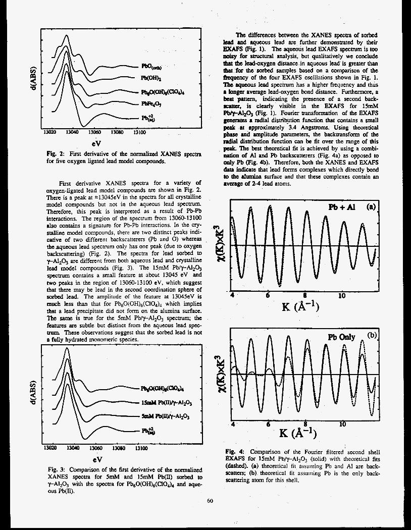
lMZ0 13040 13060 1M80 13100
eV Fig. 2: First derivative of the normalized XANES spectra for five oxygen ligated lead model compounds.
First derivative XANES spectra for a variety of oxygen-ligated lead model compounds are shown in Fig. 2. There is a peak at =13045eV in the spectra for all crystalline model compounds but not in the aqueous lead spectrum. Therefore, this peak is interpreted as a result of Pb-Pb interactions. The region of the spectrum from 13060-13100 also contains a signature for Pb-Pb interactions. In the cry- stalline model compounds, there are two distinct peaks indi- cative of two different backscatterers (Pb and 0) whereas the aqueous lead spectrum only has one peak (due to oxygen backscattering) (Fig. 2). The spectra for lead sorbed to yAI2O3 are different from bo th aqueous lead and crystalline lead model compounds (Fig. 3). The l5mM PblpA1203 spectrum contains a small feature at about 13045 eV and two peaks in the region of 13060-13100 eV, which suggest that there may be lead in the second coordination sphere of sorbed lead. The amplitude of the feature at 13045eV is much less than that for Pb60(OH)6(C104), which implies that a lead precipitate did not form on the alumina surface. The same is me for the 5n-N PblpA1203 spectrum; the features are subtle but distinct from the aqueous lead spec- trum. These observations suggest that the sorbed lead is not a fullv hvdrated monomeric sDecies.
eV Fig. 3: Comparison of the first derivative of the normalized XANES spectra for 5mM and 15mM Pb(II) sorbed to y-Al2O3 wirh the spectra for Pb60(OH)6(C104)4 and aque- ous Pb(II).
The differences between the XANES spectra of sorbed lead and aqueous lead are further demnsuated by their EXAFS (Fig. 1). The aqueous lead EXAFS spectrum is too noisy for structural analysis, but qualitatively we conclude that the lead-oxygen distance in aqueous lead is greater than ibat for the sorbed samples based on a comparison of the hquency of the four EXAFS oscillations shown in Fig. 1. The aqueous lead spectrum has a higher frequency and thus a longex average lead-oxygen bond distance. Furthermore, a beat pattun, indicating the presence of a second back- scatter, is clearly visible in the EXAFS for lSmM PWpAlz@ (Fig. 1). Fourier trmsformation of the EXAFS generates a radial disvibytion function that contains a small penk at approximately 3.4 Angstroms. Using theoretical phsse and amplitude parameters, the backtransform of the ndial distribution function can be fit over the range of this peak. The best theoretical fit is achieved by using a combi- nation of Al and Pb backscatterers (Fig. 4a) as opposed to only Pb Fig. 4b). Therefore, both the XAXES and EXAFS data indicate that lead forms complexes which directly bond to the alumina surface and that these complexes contain an a m g e of 2-4 lead atoms.
b t3 N
I
4 . 6 8 10
K (Ae’)
L I 4 . 6 8 10
Fig. 4 Comparison of the Fourier filtered second shell EXAFS for 15mM Pb/y-A1203 (solid) with theoretical fits (dashed). (a) theoretical fit assuming Pb and A1 are back- scatters; (b) theoretical fit assuming Pb is the only back- scattering atom for this shell.
GO
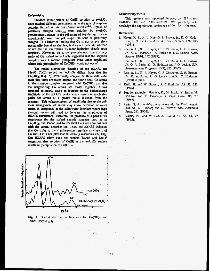
C e M 2 O 3 previous investigations of COW) sorption to yAl2O3
have reached different conclusions as to the ty of sorption complex formed at this oxiddwater interfacee Uptake of posiltivelr charged Co(lI)w from solution by Y-N2O3 pndominantly occurs in the pH range of 6-8 during titration arpaimentS6; over this pH range, the solid is positively chargtd This behavior implies that Co is not simply elm- m y bound to alumina; it does not indictate whether or not the Co ion retains its inner hydration sheath upon oorption7. However, an x-ray photoelectron spectroscopy study of Co sorbed to y-AI2O3 suggested that the sorption complwt was a surface precipitate even under conditions where bulk precipitation of Co(0H)z would not occur%.
The radial distribution function of the EXAFS for 18mM co@) sorbed to y-A1203 differs from that for
(Fig. 5). Preliminary analysis of these data indi- atto that then are fewer second and fourth shell Co atoms in the Soaption complex compared with Co(OH)2 and that the neighboring Co atoms are closer together. Atoms arranged colinearly cause an increase in the backscatted rmplitude of the EXAFS peaks which results in resolvable peaks for atoms at a greater radial distance from the 8mtmx. This enhancemment of amplitudes due to the col- inear arrangement of atoms may allow detection of outer ltwIls in complexes at the solid/water interface when high thamal motion will tend to decrease the amplitudes of EXAFS oscillations. Therefore, the presence of a peak at >5 h g s - for the sorbed sample suggests that, as in co(oH)2, the second and fourth shell Co atoms are colinear with the central absorber ion. Thus, the EXAFS indicates that Co sorbs to the alumindwater interface as clusters of CO and 0 in a complex that structurally resembles Co(OH)* our EXAFS study does not support Tewari and Lee's8 suggestion that sorption of Co(I1) at the y-A1203 surface results in precipitation of Co(OH)2.
R@) Fig. 5: Radial distributioil functions for &(OH)* and 18- Co@-AlzO~.
61
Acknowledgements
This research was supported, in part, by NSF grants EAR-85-13488 and CHE-85-12129. We gratefully ack- nowledge the experimental assistance of Dr. Britt Hedman.
References
1. Hayes, K. F., A. L. Roe, G. E. Brown, Jr., K. 0. Hodg- son, J. 0. Leckie and G. A. Parks, Science 238, 783 (1987).
3. Roe, A. L., K. F. Hayes, C. J. Chisholm, G. E. Brown, Jr., K. 0 Hodson, G. A. Parks and J. 0. Leckie. SSRL Report 87/01, 142 (1987).
3. Roe, A. L., K. F. Hayes, C. J. Chisholm, G. E. Brown, Jr., G. A. Parks, K. 0. Hodgson and J. 0. Leckie, GSA Absrracrs with Programs 19(7), 823 (1987).
4. Roe, A. L.. K. F. Hayes, C. J. Chisholm, G. E. Brown, Jr., G. A. Parks, J. 0. Leckie and I(. 0. Hodgson, (1988) in prep.
4. Hold, H. and W. Stumm, J. Colloid fn f . Sd. 55, 281 (1976).
6. See, for example: Hachiya, K., M. Sasaki, Y. Suruta, N. Mikami and T. Tasunaga, J. Piiys. Chem. 88, 23 (1984).
7. Parks, G. A., in Adsorption in the Marine Environment, 2nd ed., J. P Riberg and G. Skirrow, eds., Academic Press, 241 (1976).
8. Tewari, P.H and W. Lee, J. Colloid Int. Sci. 52, 77 (1975).


Interface Structure of NiSiz/Si(lll)
I. K. Robinson and R. T. Tang AT&T Bell Laboratories, Murray Hill, NJ 07974
R. Feidenhaas'l Rko National Lab, DK 4000 Roskilde, Denmark
Nickel silicide is very close to being lattice matched to silicon, and forms smooth pseudomorphic films by chemical reaction when Ni is annealed on a Si(ll1) surface. Because of the ease of preparation and high quality of such films, many structural studies of the interface have been performed using a variety of techniques including transmission electron microscopy' (TEM), ion scattering2 (RJ3S) and X-ray standing wave fluorescent ~ i e l d . ~ , ~ The general conclusions are in agreement with the model of Fig. 1, wit$ an inward relaxatio? of the layer spacing boy 0.06 _+O.OSA (ref. 2), 0.11 _+O.O3A (ref. 3) or 0.06 _+O.O3A (ref. 4).
On beam line VII-2, we measured the diffraction near the (111) Bragg peak of one o! these interfaces. The sample was a B-type Nisi2 film, 33A thick, grown on float zoae Si(ll1) _fl/2", then covered at room temperature by 50A a-Si to prevent oxidation in the ambient. X-ray measurements were made in air with a wavelength of 1.54A using 2 mm (horiz) X 10 mm (vert) slits in front of the detector. Very close to the Bragg peak 1 mm X 1 mm slits and an attenuator were used and the results were scaled accordingly. The diffraction pattern was found to consist of a diffuse streak extending from the origin of reciprocal space radially out through the Bragg peak and beyond. Thermal diffuse scattering was also present but could be removed completely by use of w-scans through the streak. Each data point in Fig. 2 is the integrated intensity of the streak after background subtraction in such a scan. The range of data shown is (0.75, 0.75, 0.75) to (1.3, 1.3, 1.3).
Figure 1. Ball and stick model of a NiSi2/Si(lll) interface. Shown here is a type B interface (ref. 1) which was the type used in the experiments and other work discussed; type A has the overlayer rotated 180" with respect to the substrate.
ai a a -
Proposal No. 996M
0.8 0.9 1.0 1.1 1.2 1.3 MOMENTUM TRANSFER qa,
Figure 2. Integrated intensity of the diffracted intensity near the (111) Bragg peak of a NiSi2/Si(lll) interface. The units of momentum transfer q are in inverse Si(ll1) spacings. The fit is to eqn. 3 and the individual components Fsi (eqn. 2) and F~isi2 (eqn. 1) are superimposed as dashed lines.
Both the Si(ll1) substrate and the Nisi2 film contribute to the streak. The substrate has a crystal truncation rod5 (CTR) arising from the Fourier transform of its abruptly terminated face. This has an intensity falling off symmetrically as l/q2 on either side of the Bragg peak; this is the same as the Darwin tails predicted by the dynamical theory.6 The Nisi2 film, being only eight layers thick, has broad diffraction features and subsidiary maxima on either side of the Bragg peak; it is also a symmetric function. What is seen, however, in the data is a dramatic asymmetry of the intensity around (111). This is because the two components interfere with each other. Since the relative phase is given by the interface separation, we have a sensitive method to determine that quantity.
The problem reduces to one dimension because all the layers are well ordered parallel to the interface. We can calculate the intensity as the square modulus of the sum of layerwise contributions. Adding up the Si layers and Nisi2 layers separately we obtain (see Fig. 1):
62
Rejerences
q is the momentum transfer and and al are the layer spacings of the substrate and film respectively. Finally, we introduce the interfacial distance d:
Fhtal = Fsi + eiqd F~isi2 (3)
The fit in Fig. 2 is the modulus of Fbt, for a value of d/% = 1.10 20.02. Neither Fsi nor F ~ i ~ i a alone shows any asymmetry; only when they are added together with the appropriate phase in eqn. 3, can we fit the data.
This simple technique allows accurate determination of interface separations by using the interference of the two diffraction contributions. The resulting value of d/% is less than the ideal value of 9/8 seen directly in Fig. 1 assuming no relaxation. The maJnitude of the inward relaxation measured is 0.08 f0.06A in good agreement with other determinations.
1. R. T. Tung, J. M. Gibson, J. M. Poate, Phys. Rev. Lett. 50, 429 (1983).
2. E. J. van Loenen, J. W. M. Frenken, J.F. van der Veen, S. Valeri, Phys. Rev. Lett. 54, 827 (1985).
E. Vlieg, A. E. M. J. Fischer, J. F. van der Veen, B. N. Dev, G. Materlik, Surf. Sci. 178, 36 (1986).
J. Zegenhagen, to be published.
I. K. Robinson, Phys, Rev. SS.9, 3830 (1986).
Z . G. Pinsker, “Dynamical Scattering of X-rays in Crystals”, Springer (1978).
3.
4.
5.
6.
63
P r o p o s a l N o . 999Mp
E X A F S Study of Rare E a r t h Element Environments in Quenched Silicate Melts of Geochemical In te res t
C.W. Ponader, G.E. Brown, Jr., and W.E. Jackson Department of Geology, Stanford University
Stanford, California 94305
The local geometry of the site occupied by trace amounts (0.2 wt%) of La, Gd, and Yb, with and without 1-2 wt% F or Cl, has been examined as a function of melt composition using Extended X-ray Absorption Fine Structure (EXAFS) spectroscopy. Spectra were collected in fluorescence mode on BL VII-3 and BL IV-1 during dedicated runs in April, 1986 and January, 1987. The three glass composi- tions examined are albite, sodium trisilicate, and a peralkaline composition approximately halfway be- tween the two. The results in the halogen-free glasses show that Yb is approximately 6 coordinated (<Yb-0>= 2.21 A). Gd coordination ranges from 6 in albite glass (<Gd-0>= 2.30 A) through a mixture of 6 and 7 in peralkaline glass (<Gd-0>= 2.36 A) to 7 in sodium trisilicate glass (<Gd-0>= 2.43 A). La is 7-coordinated in the peralkaline and sodium trisilicate glasses (<La-0>= 2.43 A), but has a co- ordination of 8-9 in albite glass(<La-0>= 2.59 A). The irregularity of the rare earth site (Le. , the range of distances to oxygen) appears to increase as tetra- hedral polymerization increases. This is caused, in part, by the decreasing number of non-bridging oxy- gens in the coordination sphere of the rare earth el- ement. The exception to this trend is La in albite glass. The La site in albite glass is more regular than it is in the other glasses because the size of the La ion requires it to occupy a more central position in the “cage” formed by the interconnected rings of Si and A1 tetrahedra. This leads to a more regular distribution of La-0 bond lengths. The irregularity of the rare earth site also appears to decrease as the size of the rare earth decreases. This is due to the greater ability of the smaller, higher field-strength rare earths to compete for non-bridging oxygens. As the tetrahedral polymerization decreases, the local environment of the rare earth is more strongly influ- enced by the bonding requirements of the rare earth and is less influenced by the tetrahedral network topology. Increasing the polymerization of the melt or increasing the Si/(Si+Al) ratio of the tetrahe- dral network should act to increase the crystal/liquid partition coefficients of the rare earths.
\
EXAFS studies of glasses containing fluorine demonstrate that La, Gd, and Yb form complexes with fluorine in aluminosilicate melts. Gd and Yb are coordinated by eight fluorines with no appar- ent oxygen neighbors (<Gd-F>= 2.30 A; <Yb-F>= 2.23 f i ) . La appears to form mixed 0 - F complexes consisting of eight ligands approximately evenly di- vided between fluorine and oxygen (<La-0>= 2.45 f i ; <La-F>= 2.46 A). We found no evidence of C1 complexing of REE in the chlorine-containing com- positions studied. The rare earth site in chlorine- containing melts is, however, more regular than the rare earth site in volatile-free melts. This suggests that C1 acts to depolymerize the tetrahedral net- work. Fluorine, because of complex formation, and chlorine, because of its effect on melt structure, should both act to decrease the crystal/liquid parti- tion coefficients of rare earths in silicate melts.
64


Proposal No. lOOOMp
CHARACTERIZATION OF Co-CONTAINING OXIDES ON STAINLESS STEEL BY e-YIELD XANES SPECTROSCOPY
1 1 2 3 Fa r re l Ly t l e , Robert Greegor, Zo f i a Rek and Howard Ocken
1. The Boeing Co., Seat t le , WA 98124
2. SSRL, Stanford, CA 94305
3. E P R I , Palo Al to , CA 94303
The pass i va t i ng oxide l aye rs t h a t form on the i nne r sur face o f s ta in less s t e e l water coolant pipes i n nuc lear potEr p lan ts can incorporate the energet ic Y-emi t ter Co. Measurements a t commercial p lan ts show t h a t t h i s i so tope i s responsib le f o r h 8 0 % o f t he occupat ional r a d i a t i o n exposure i ncu r red by p l a n t main- tenance personnel a l though Co i s present i n the oxide l a y e r a t on l y *0.2 w t . %. A number o f sur face t r e a t - ments have been shown i n l abo ra to ry experiments and i n l i m i t e d in-p&ijnt demonstrations t o i n h i b i t t h e i nco r - po ra t i on o f Co i n t o the ox ide l a y e r . X-ray d i f f r a c - t i o n and S I M S have been used t o character ize the major components o f these passive f i l m s ; however the actua’l environment o f t h e Co atoms i s unknown. To opt imize c o r r e c t i v e measures a b e t t e r cha rac te r i za t i on o f t he Co envi ronment i s necessary.
I n t h i s f e a s i b i l i t y experiment x- ray absorpt ion spec- t roscopy was used t o i n v e s t i g a t e t h e environment o f t h e Co atoms. The samples consisted o f sect ions o f Type 304 staineess s t e e l p ipe upon which t h e oxide l a y e r (-4000 A t h i c k ) was present. Samples were prepared by i r c u l a t i n g neu t ra l H20 con ta in ing 100 pp t and 20 p p t 5’C0 a t 280 OC f o r 500 hours. t imated t h a t t h e r e s u l t i n g su,&ace oxides contained ~ 0 . 5 - 1 . 0 and -0.1-0.2 w t . % Co, respec t i ve l y . Sect ions c u t f rom the tub ing were examined by s imultan- eous x-ray f luorescence and t o t a l e lec t ron y i e l d detect ion. Since t h e major cons t i t uen t , Fe, has a lower e x c i t a t i o n energy than Co, the Fe f luoresced i n t e n s e l y throughout the Co spectrum. f l uo rescen t measurement very no isy as shown below. The XANES spectra a re shown i n F ig . 1 along w i t h r e f - erence spect ra f rom Co304 and C O O . The CoA spectra a re from t h e h igher concentrat ion sample. concentrat ion sample gave a v i s i b l e edge bu t extensive averaging would be necessary f o r use fu l data. It i s c l e a r t h a t t h e f luorescent channel CoA, f i s t o o noisy t o be use fu l ; however, t he e -y ie ld channel CoA, e i s n o t bad and q u i t e s i m i l a r t o Co304. h ighe r i n t e n s i t y i n t h e near edge peaks o f CoA,e com- pared t o Co 0 . This i s caused by t h e thickness e f f e c t due t o s e l f 3 a k o r p t i o n i n the Co O4 data.) X-ray d i f - f r a c t i o n i nd i ca ted t h a t t h e pr imary components o f t h e ox ide l a y e r were Fe 0 7 F e 0 , Fe2Ni04, a l l o f which have the sp ine l ~ t r $ c $ u r e . ~ t o 0 i s a l s o a sp ine l , thus t h e t e n t a t i v e i d e n t i f i c a t q o 8 i s t o be expected s ince mixed sp ine l s a r e common.
It was es-
This made t h e
The low
(Disregard t h e
F ig . 1 Co K-edge XANES spect ra from the sur face ox ide CoA, e and l a y e r on Type 304 s t a i n l e s s s t e e l .
CoA, f a re t h e t o t a l e -y ie ld and f l uo rescen t data channels, respec t i ve l y . Co 0 and COO a r e normal absorpt ion mode data 3rb t h e pure compounds f o r comparison. A monochromator g l i t c h near t h e onset o f absorpt ion d i s t o r t s t h e CoA spect ra i n t h e reg ion from 0-10 eV.
The t o t a e e - y i e l d technique probes approximately t h e top 50p5A o f t h e surface. In sample CoA t h a t amounted to -10 s e n s i t i v i t y o f t h e technique. Each spectrum i n F ig . 1 i s the r e s u l t o f one scan through the Co K-edge. f u t u r e work we p lan t o ob ta in b e t t e r data by repet- i t i v e scanning s u f f i c i e n t t o analyze the EXAFS as we l l as the XANES and thus determine t h e d i s t r u b u t i o n o f Co-0 distances i n order t o more accurate ly descr ibe i t s l a t t i c e s i t e s . Sggples c u t from ac tua l power p l a n t pipes con ta in ing Co w i l l a l s o be examined. The l a c k o f s e n s i t i v i t y o f t h e e -y ie ld technique t o h igh energy Y - r a d i a t i o n w i l l be an advantage. Also, a monochrom- a t o r c r y s t a l w i t h a d i f f e r e n t g l i t c h p a t t e r n w i l l a l l ow
c! i Spect t h e near edge t o determine the r a t i o o f ~ ~ “ / C O ’ . We be l i eve t h a t t h i s more d e t a i l e d charac- t e r i z a t i o n o f t h e Co chemistry and c r y s t a l environment w i l l a l l ow a more i n t e l l i g e n t choice o f procedures t o prevent t h e e f f e c t .
Co atoms which g ives a rough est imate o f t h e
I n
n W N 0 , 9 CI
-J
iz 0
= 0,:
081
n-
I I
I I I I I I 0 40 80
ENERGY (EV)
65

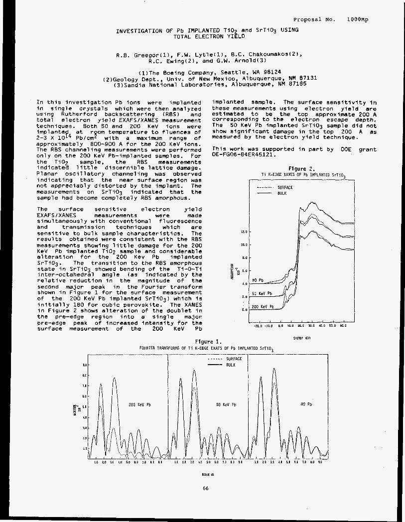
Proposal No. lOOOMp
INVESTIGATION OF Pb IMPLANTED Ti02 and SrTi03 USING TOTAL ELECTRON YIELD
R.8. Greegor(l), F.W. Lytle(l), B.C. Chakoumakos(2), R.C. Ewing(2), and G.W. Arnold(3)
(1)The Boeing Company, Seattle, WA 98124 (2)Geology Dept., Univ. of New Mexico, Albuquerque, NM 87131
(3)Sandia National Laboratories, Albuquerque, NM 87185
In this investigation Pb ions were implanted in sing’ie crysta7s which were then analyzed using Rutherford backscattering (RES) and total electron yield EXAFSIXANES measurement techniques. Both 50 and 200 KeV ions were implante room temperature to fluences of 2-3 X lop4 Fk/cm2 with a maximum range of approximately 800-900 A for the 200 KeV ions. The RBS channeling measurements were performed only on the 200 KeV Pb-implanted samples. For the Ti02 sample, the RBS measurements indicated little discernible lattice damage. Planar oscillatory channeling was observed indicating that the near surface region was not appreciably distorted by the implant. The measurements on SrTiO3 Indicated that the sample had become complete7y R E S amorphous.
The surface sensitive electron yield EXAFSIXANES measurements were made simultaneously with conventional fluorescence and transmission techniques whlch are sensitive to bulk sample characteristics. The results obtained were consistent with the RES measurements showing llttle damage for the 200 KeV Pb implanted Ti02 sample and considerable alteration for the 200 Kev Pb implanted SrTi03. The transition to the RES amorphous state in SrTiO3 showed bending of the Ti-0-Ti inter-octahedral angle (as indicated by the relative reduction in the magnitude o f the second major peak in the Fourier transform shown in Figure 1 for the surface measurement of the 200 KeV Pb Implanted SrTi03) which is initially 180 for cubic perovskite. The XANES in Figure 2 shows alteration of the doublet in the pre-edge region into a slngle major pre-edge peak of increased intensity for the surface measurement of the 200 KeV Pb
implanted sample. The surface sensitlvity in these measurements using electron yield are estimated to be the top approximate 200 A corresponding to the electron escape depth. The 50 KeV Pb implanted SrTi03 sample did not show significant damage in the top 200 A as measured by the electron yield technique.
This work was supported in part by DOE DE-FG06-84ER45121.
grant
Figure 2 . T i K-EDGE XANES OF Pb IMPLANTED SrTi03
SURFACE BULK
------- -
Figure I . FOURIER TRANSFORMS OF T i K-EDGE EXAFS OF
1 ~ ~ 1 ” ~ ’ ’ ’ -20.0-10.0 0.0 10.0 20.0 30.0 40.0 50.0 6C0
EMBCY EV)
IMPLANTED S r T i 0 3

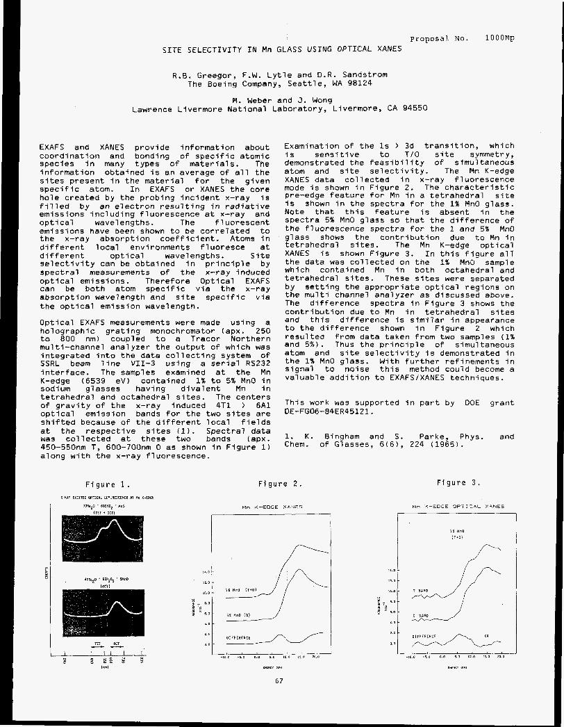
Proposal No. S I T E SELECTIVITY IN Mn GLASS USING OPTICAL XANES
lOOOMp
R.B. Greegor, F.W. Lytle and D.R. Sandstrom The Boeing Company, Seattle, WA 98124
M. Weber and J. Wong Lawrence Livermore National Laboratory, Livermore, CA 94550
EXAFS and XANES provide information about coordination and bonding of specific atomic species in many types o f materials. The information obtained is an average of all the sites present in the material for the given specific atom. In EXAFS or XANES the core hole created by the probing incident x-ray is filled by an electron resulting in radiative emissions including fluorescence at x-ray and optical wavelengths. The f 1 uorescent emissions have been shown to be correlated to the x-ray absorption coefficient. Atoms in different local environments fluoresce at different optical wavelengths. Site selectivity can be obtained in principle by spectral measurements of the x-ray induced optical emissions. Therefore Optical EXAFS can be both atom specific via the x-ray absorption wavelength and site specific via the optical emission wavelength.
Optical EXAFS measurements were made using a holographic grating monochromator (apx. 250 to 800 nm) coupled to a Tracor Northern multi-channel analyzer the output of which was integrated into the data collecting system of SSRL beam line VII-3 using a serial RSZ32 interface. The samples examined at the Mn K-edge (6539 eV) contained 1% to 5% MnO in sodium gfasses having divalent Mn in tetrahedral and octahedral sites. The centers of gravity of the x-ray induced 4T1 ) 6A1 optical emission bands for the two sites are shifted because of the different local fields at the respective sites (1). Spectral data was collected at these two bands (apx. 450-550nm T, 600-700nm 0 as shown in Figure 1) along with the x-ray fluorescence.
Figure 1. X-RAY EXCITED OPTICAL LUnIflESCENCE AT Un X-EDGE
109 0 . 63SiOp . WnO (TET + OCTI
Examination of the 1s ) 3d transition, which is sensitive to T/0 site symmetry, demonstrated the feasibility of simultaneous atom and site selectivity. The Mn K-edge XANES data collected in x-ray fluorescence mode is shown in Figure 2. The characteristic pre-edge feature for Mn in a tetrahedral site is shown in the spectra for the 1% MnO glass. Note that this feature is absent In the spectra 5% MnO glass so that the difference of the fluorescence spectra for the 1 and 5% MnO glass shows the contribution due to Mn in tetrahedral sites. The Mn K-edge optical XANES is shown Figure 3. In this figure all the data was collected on the 1% MnO sample which contained Mn in both octahedral and tetrahedral sites. These sites were separated by setting the appropriate optical regions on the mufti channel analyzer as discussed above. The difference spectra in Figure 3 shows the contribution due to Mn in tetrahedral sites and this difference is similar in appearance to the difference shown in Figure 2 which resulted from data taken from two samples (1% and 5%). Thus the principle of simultaneous atom and site selectivity i s demonstrated in the 1% MnO glass. With further refinements in signal to noise this method could become a valuable addition to EXAFSIXANES techniques.
This work was supported in part by DOE grant DE-FG06-84ER45121.
1. K. Bingham and S. Parke, Phys. and Chem. of Glasses, 6(6), 224 (1965).
Figure 2.
M n K-EOGE X A N E S
/---- 1 % uno (l+OJ $ 5 % H"O 101 5 % H"O 101 1 I . D
O I F F E R E N C E
-10.0 -5.0 0.0 5 .0 ID.0 15.0 m.0
CNEliC" IN>
67
Figure 3.
M n K-EDGE OPTICAL X A N E S
I 1% nno

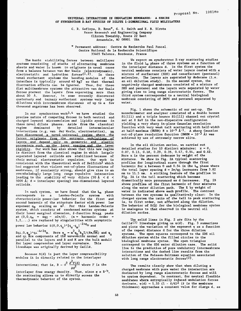
C. R. Safinya, D. Roux*, G. S . Smith and E. B. Sirota Exxon Research and Engineering Company
Clinton Township. Route 22 East Annandale, NJ 08801 USA
* Permanent address: Centre de Recherche Paul Pascal Centre National de la Recherche Scientifique
33405 Talence, Bordeaux, France
The basic stabilizing forces between multilayer systems consisting of stacks of alternating membrane sheets and water is known to originate in usual cases from a balance between van der Waals (electrodynamic), electrostatic and hydration forces(ls2). In these usual surfactant systems the bending modulus of the interface is typically around 40 kgT so that thermal fluctuation effects can be ignored. Thus, for these flat multimembrane systems the attractive van der Waals forces prevent the layers from separating more than about 30 A. However, in some recently discovered quaternary and ternary surfactant systems very large dilutions with intermembrane distances of up to a few thousand angstroms has been observed.
In our synchrotron work3$4 we have studied the precise nature of competing forces in both neutral and charged layered microemulsion and lipidic systems in these novel dilute phases. Aside from the classical regime dominated by detailed intermolecular interactions (e.g. van der Waals, electrostatics), have discovered a novel universal reeime where the forces originate from the entroDic undulations of layers and depend onlv on eeometric and elastic parameters such as the laver sDacinp and the laver rigidity. Our work has also shown that this new regime is distinct from the classical regime in which largely separated layered colloids are stabilized because of their mutual electrostatic repulsion. Our work is consistent with the theoretical work of Helfrich5 which had suggested that violent out-of-plane fluctuations of membranes, if succiently strong, should give rise to an overwhelmingly large long range repulsive interaction leading to the stability of very dilute (50 A < d < 5000 A; d = interlayer spacing) one-dimensional layered colloids.
In each system, we have found that the & phase corresponds to a Landau-Peierls system with characteristic power-law behavior for the first and second harmonic of the structure factor with power law exponent qm scaling as m2. For this Landau-Peierls system, which consists of condensed matter systems at their lower marginal dimension, 6-function Bragg peaks at (O,O,qm = mqo = m2a/d), (m = harmonic order = l,Z, ... ) are replaced by singularities with asymptotic power law behavior S(O,O,q )=lq -q and z z m
2 2 . Here r) = m qo sT/(8lrE) and q, Sq1,O,q )=ql m and q l Ere components of the wavevector normal and parallel to the layers and B and K are the bulk moduli for layer compression and layer curvature. lineshape was originally derived by CaillC.
- 4+2 qm
This
Because B(d) is just the layer compressibility
- a F(d) where F is the
modulus it is directly related to the interlayer
interactions; that is, B = d
interlayer free energy density. the scattering allows us to directly access the thermodynamic behavior of the system.
2
a d2 Thus, since r) a B-b,
We report on synchrotron X-ray scattering studies in the fluid & phase of three systems as a function of the interlayer distance d. neutral membrane consists of water layers coated with a mixture of surfactant (SDS) and cosurfactant (pentanol) molecules. The layers are separated by dodecane (i.e. an oil dilution study). In the second system, the negatively charged membranes consisted of a mixture of SDS and pentanol and the layers were separated by water giving rise to long range electrostatic forces. third system corresponded to a neutral biological membrane consisting of DMPC and pentanol separated by water.
In the first system the
The
Fig. 1 shows the schematic of our set-up. The monochromator and analyzer consisted of a double bounce Si(ll1) and a triple bounce Si(ll1) channel cut crystal set at 8 KeV in the non-dispersive configuration yielding a very sharp in-plane Gaussian resolution function with very weak tail scattering with half-width at half-maximum (HWHM) 8 x 10⁻⁵ A-1. A shar Gaussian
achieved by use of extremely narrow slits. out-of-plane resolution function (HWHM - 10- $ A) was
In the oil dilution series, we carried out detailed studies for 10 distinct mixtures: x = 0, 0.07, 0.13, 0.18, 0.29, 0.35, 0.47, 0.54, and 0.62. Here x is the percent dodecane by weight of the mixtures. We show in Fig. 2A typical scattering profiles for longitudinal scans through the first harmonic for x between 0 and 0.54 in the h phase where the total layer spacing d - 2a/qo increases from 3.82 nm to 11.5 nm. A striking feature of the profiles in Fig. 2A is the tail scattering which becomes dramatically more pronounced as d increases. Fig. 2B shows profiles of the first harmonic of four mixtures along the water dilution path. water is indicated above each profile. between the two systems is qualitatively clear: In the charged system the ratio of the tail to peak scattering is, to first order, not affected along the dilution. The behavior of S ( Q ) for the biological membrane system is analogous to that observed in the neutral oil dilution series.
The X by weight of The contrast
The solid lines in Fig. 2 are fits by the Cai114(3) lineshape giving us r)(d). and plots the variation of the exponent r) as a function of the repeat distance d for the three dilution systems. dilution system while the filled circles to the biological membrane system. The open triangles correspond to the SDS water dilution case. line is the prediction of pure undulatory intermembrane interactions and the dashed line results from the solution of the Poisson-Boltzman equation associated with long range electrostatic forces(2).
Fig. 3 summarizes
The open squares correspond to the SDS oil
The solid
The results clearly show that when diluting a charged membrane with pure water the interaction are dominated by long range electrostatic forces and will be system dependent. In contrast, for neutral flexible membranes where entropically induced undulation forces dominate, r)(d) - 1.33 (1 - 6/d)* ( 6 is the membrane thickness) approaches a constant value for dlarge d, as
68
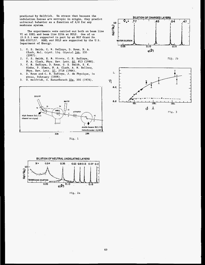
predic ted by Hel f r ich . undulat ion forces a r e en t ropic i n o r i g i n , they p r e d i c t un iversa l behavior as a func t ion of 6/d f o r any membrane system.
W e stress t h a t because t h e
The experiments were c a r r i e d out both on beam l i n e V I a t SSRL and beam l i n e XlOA a t NSLS. One of us (G.S.S.) w a s supported i n p a r t by an NSF Grant No. DMR-8307157. Department of Energy.
1. G . S. Smith, C . R. Saf inya, D . Roux, N. A .
SSRL and NSLS are supported by the U . S .
Clark, Mol. Cryst . Liq. Crystal 144, 235 (1987).
N. A. Clark, Phys. Rev. L e t t . 60, 813 (1988).
Sinha, P. Dimon, N . A. Clark, A. M . Bellocq, Phys. Rev. L e t t . 57, 2718 (1986).
p r e s s , February (1988).
2 . G. S. Smith, E . B. S i r o t a , C . R . Safinya,
3. C. R. Saf inya, D. Roux, G . S . Smith, S. K .
4 . D . Roux and C. R. Saf inya, J . de Physique, i n
5. W. Hel f r ich , Z . Naturforsch a, 305 (1978).
double bouncc Si(1,l. monochromator crysyrta
I
22M 19M
Fig. 1
DILUTION OF CHARGED LAYERS
ID
a - - \ U c
Y - TER DILUTION
0.05 0.10 0.1 5
q (8')
Fig. 2b
1.
rl 0.5,
0.0
F i g . 3 d- A
DILUTION OF NEUTRAL UNDULATING LAYERS
1 X = 0.54 0.35 023 ateo.13 0.07 0.0
Fig. 2a
69
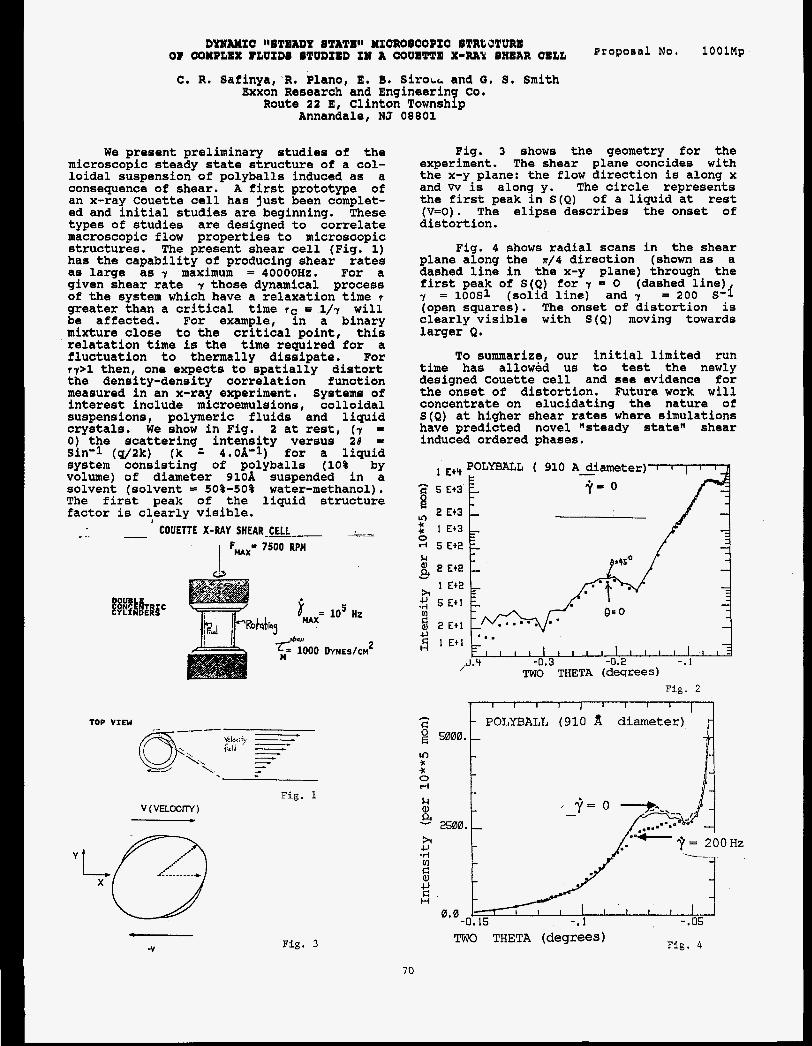
DYNAMIC lWTQADY BTATW MICROBCOOIC BTRCJTURB OF COXBLQX FbPIDB BTUDIBD I# A COUB'PTQ X-RAk BXOAR COLL
C. I?. Safinya, R. Plano, E. 8 . SiroLL and G. S. Smith Exxon Research and Engineering Co.
Route 22 E, Clinton Township Annandale, NJ 08801
We present preliminary studies of the microscopic steady state structure of a col- loidal suspension of polyballs induced as a consequence of shear. A first prototype of an x-ray Couette cell has just been complet- ed and initial studies are beginning. These types of studies are designed to correlate macroscopic flow properties to microscopic structures. The present shear cell (Fig. 1) has the capability of producing shear rates as large as 7 maximum = 40000Hz. For a given shear rate 7 those dynamical process of the system which have a relaxation time 7
greater than a critical time rC 5 l/r will be affected. For example, in a binary mixture close to the critical point, this relatation time is the time required for a fluctuation to thermally dissipate. For r ? > l then, one expects to spatially distort the density-density correlation function measured in an x-ray experiment. Systems of interest include microemulsions, colloidal suspensions, polymeric fluids and liquid crystals. We show in Fig. 2 at rest, (7 - 0) the scattering intensity versus 28 = Sin-1 (4/2k) (k 4.0A-1) for a liquid system consisting of polyballs (10% by volume) of diameter 91OA suspended in a solvent (solvent = 50%-50% water-methanol). The first peak of the liquid structure factor is clearly visible.
1
- -. __ COUETT'E X-RAY SHEAR-CELL I FMAx= 7500 RPM
V ( VELOCrrV) c
Fig. 1
!is. 3 shows the geometry for the experiment. The shear plane concides with the x-y plane: the flow direction is along x and w is along y. The circle represents the first peak in S ( Q ) of a liquid at rest (y=O). The elipse describes the onset of distortion.
Fig. 4 shows radial scans in the shear plane along the ~ / 4 direction (shown as a dashed line in the x-y plane) through the first peak of S ( Q ) for 7 = 0 (dashed line) 7 = 1OOS1 (solid line) and 7 = 200 i3-I (open squares). The onset of distortion is clearly visible with S ( Q ) moving towards larger Q.
To summarize, our initial limited run time has allowed us to test the newly designed Couette cell and see evidence for the onset of distortion. Future work will concentrate on elucidating the nature of S ( Q ) at higher shear rates where simulations have predicted novel "steady state" shear induced ordered phases.
Fig. 2
h
C 2 5000. m c c 0 l-i
- 2500. 4J
v)
h 0
H
-.05 0.0
-0.15 -. 1
HZ
Fig. 3 TWO THETA (degrees) Fig. 4

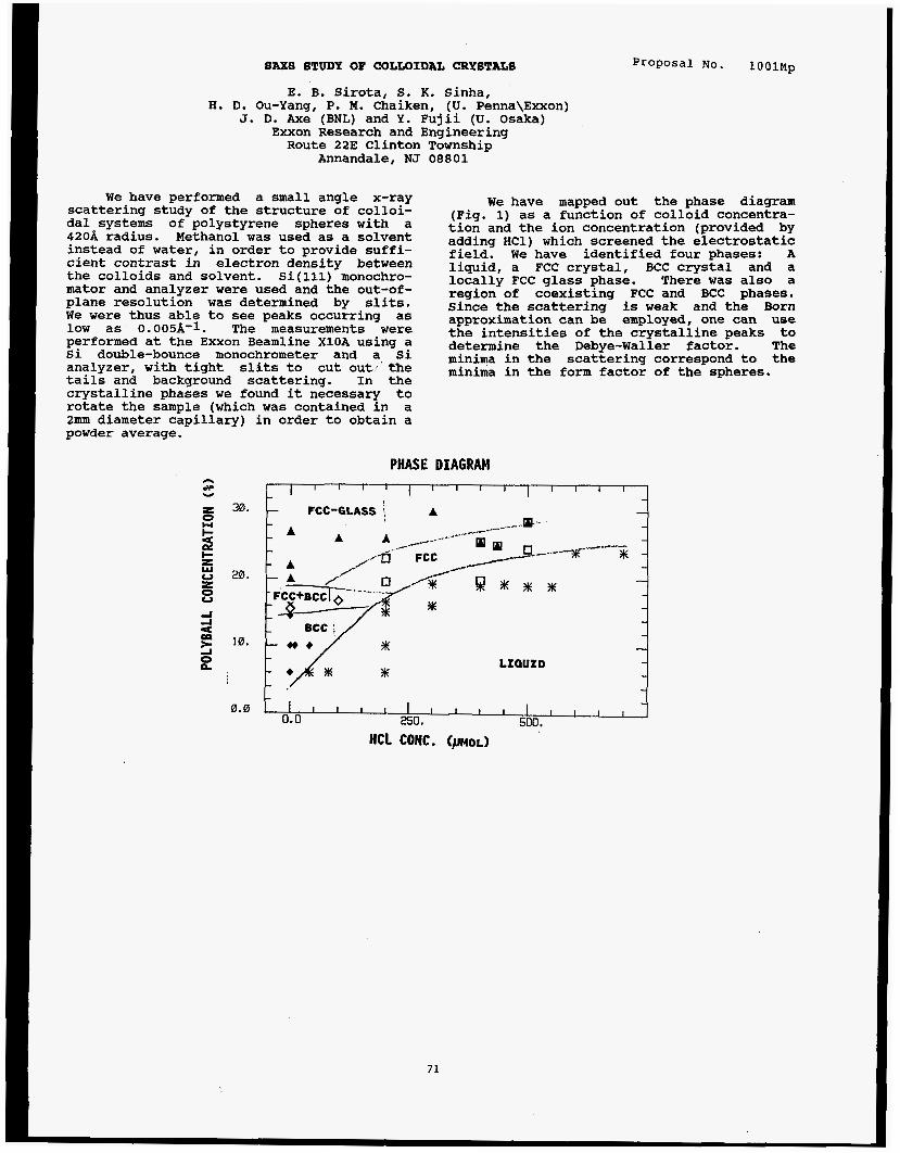
SASS STUDY OF COLLOIDAL CRYSTALS
E. B. Sirota, S. K. Sinha, H. D. Ou-Yang, P. M. Chaiken, (U. Penna\Exxon)
J. D. Axe (BNL) and Y. Fujii (U. Osaka) Exxon Research and Engineering Route 22E Clinton Township
Annandale, NJ 08801
Proposal No. lOOlMp
We have performed a small angle x-ray scattering study of the structure of colloi- dal systems of polystyrene spheres with a 420A radius. Methanol was used as a solvent instead of water, in order to provide suffi- cient contrast in electron density between the colloids and solvent. Si(ll1) monochro- mator and analyzer were used and the out-of- plane resolution was determined by slits. We were thus able to see peaks occurring as low as 0.005A-1. The measurements were performed at the Exxon Beamline XlOA using a Si double-bounce monochrometer and a Si analyzer, with tight slits to cut out, the tails and background scattering. In the crystalline phases we found it necessary to rotate the sample (which was contained in a 2mm diameter capillary) in order to obtain a powder average.
We have mapped out the phase diagram (Fig. 1) as a function of colloid concentra- tion and the ion concentration (provided by adding HC1) which screened the electrostatlc field. We have identified four phases: A liquid, a FCC crystal, BCC crystal and a locally FCC glass phase. There was also a region of coexisting FCC and BCC phases. Since the scattering is weak and the Born approximation can be employed, one can use the intensities of the crystalline peaks to determine the Debye-Waller factor. The minima in the scattering correspond to the minima in the form factor of the spheres.
PHASE DIAGRAM n dP v L ~ ’ ” ’ ” ’ ’ ~ ” ’ ’ ~
30.
z I- z W
z 0 20.
8
5
-I A = 10.
q 0.0
71

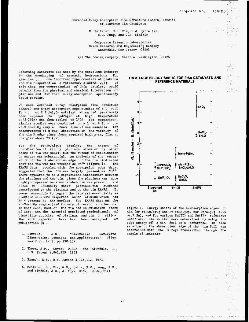
Proposal No. 1OOlMp
Extended X-ray Absorption Fine Structure (EXAFS) Studies of Platinum-Tin Catalysts
G. Meitzner, G.H. Via, F.W. Lytle (a), S.C. Fung, and J.H. Sinfelt
Corporate Research Laboratories Exxon Research and Engineering Company
Annandale, New Jersey 08801
(a) The Boeing Company, Seattle, Washington 98124
Reforming catalysts are used by the petroleum industry in the production of aromatic hydrocarbons for gasoline [l]. One important type consists of platinum and tin dispersed on a refractory alumina [2,3]. We felt that our understanding of this catalyst would benefit from the physical and chemical information on platinum and tin that x-ray absorption spectroscopy could provide.
We made extended x-ray absorption fine structure (EXAFS) and x-ray absorption edge studies of a 1 wt.% Pt - 1 wt.% Sn/A1203 catalyst which had previously been exposed to hydrogen at high temperature (775-79513) and then cooled to 140K. For comparison, similar studies were conducted on a 1 wt.% Pt - 0.6 wt.% Sn/SiOn sample. Beam line VI was essential for measurements of x-ray absorption in the vicinity of the tin K edge since these required high x-ray flux at energies above 29 keV.
For the Pt-Sn/A1203 catalyst the extent of coordination of tin by platinum atoms or by other atoms of tin was small, but the extent of coordination by oxygen was substantial. An analysis of the energy shift of the K absorption edge of the tin indicated that the tin was not present as Sn+4 (figure 1). The EXAFS data, coupled with the absorption edge data, suggested that the tin was largely present as Sn+2. There appeared to be a significant interaction between the platinum and the tin, since the platinum was more highly dispersed on alumina when tin was present, and since an unusually short platinum-tin distance contributed to the platinum and to the tin EXAFS. It seems reasonable to regard the catalyst essentially as platinum clusters dispersed on an alumina which had Sn+2 present at the surface. The EXAFS data on the Pt-Sn/Si02 sample lead to very different conclusions. In that case, most of the tin had an oxidation state of zero, and the material consisted predominantly of bimetallic entities of platinum and tin on silica. The work reported here has been accepted for publication [4].
1. Sinfelt, J.H., "Bimetallic Catalysts : Discoveries, Concepts, and Applications"; Wiley: New York, 1983, pp 130-157.
2. Thorn, J.P., Guyer, W.R.F., and Arundale, I., U.S. Patent 2,861,959, 1958.
3. Rausch, R.E., U.S. Patent 3,745,112, 1973.
4. Meitzner, G., Via, G.H., Lytle, F.W., Fung, S.C., and Sinfelt, J.H., J. Phys. Chem., OOOO(1987).
TIN K EDGE ENERGY SHIFTS FOR PtSn CATALYSTS AND REFERENCE MATERIALS
6 -
5 -
a s s2 u?
G - 2 -
e d
2 - 4 -
E
3-
I
n
i -
O t
-'t
trans-PtSq I 1 1
I I 1 I I Supported Sn (11) S n ( V
Sn
Figure 1. Energy shifts of the K-absorption edges of tin for Pt-Sn/SiOg and Pt-Sn/Al2Og, for Sn/A1203 ( 0 . 6 wt.% Sn), and for various Sn(I1) and Sn(1V) reference materials. The shifts were determined by using the edge energy of a tin foil as a reference. In each experiment, the absorption edge of the tin foil was determined with the x-rays transmitted through the sample of interest.
72


p r o p o s a l No. 1OOlMP
SULFUR K EDGE X-RAY ABSORPTION SPECTROSCOPY OF ASPHALTENES AND MODEL COMPOUNDS
G. N. George and M. L. Gorbaty
EXXON Research and Engineering Co. Annandale, NJ 08801
RESULTS AND DISCUSSION
INTRODUCTION
A major gap in our current knowledge of the chemistry of heavy hydrocarbons concerns the chemical forms and quantities of organically bound sulfur in these materials. Current knowledge is based almost entirely on the characterization of volatile prod- ucts, and none of the methods currently in common use (cf. 1) is adequate for determin- ing the forms of organically bound sulfur in native coals or in native petroleum samples. Sulfur X edge X-ray absorption spectroscopy has shown some potential to solve these problems (2).
sulfur K edge X-Ray absorption spectroscopy for the purpose of investigating the forms of organic sulfur in solids and nonvolatile liquids.
We have investigated the utility of
Figure 1.
Sulfur K edge spectra of Asphaltenes and selected model compounds. -
Asphaltenes b al U C m n L 0 10 n a U al N .A
rl
m E L 0 z
I . . .
2470 2480 E n e r g y (eV)
Figure 1 compares the sulfur K edge XANES spectra of a number of model compounds and of an asphaltene sample. The XANES can be seen to vary widely from compound to compound. As might be anticipated, the edge positions of compounds with more oxidized sulfur are notably higher in energy than for those with reduced sulfur. It is apparent that the sulfur XANES can readily be used to distinguish between these types of sulfur and that the asphaltenes do not contain appreciable quantities of oxidized sulfur. Casual inspection of figure 1 might suggest that the spectra from sulfur in different environments can be similar, notably, dibenzyl sulfide and dibenzothiophene. Closer inspection reveals that the edge of dibenzothiophene is displaced from that of dibenzyl sulfide, the first inflection point being some 0.6eV higher for the former compound. A general conclusion is that the sulfur K edge XANES can be used as a finger- print for the chemical nature of the sulfur under investigation. Thus dibenzothiophene and benzothiophene resemble one another, however dibenzothiophene and, for example, thianthrene are markedly different. The compound whose XANES bears the most resembl- ance to the asphaltene sample is dibenzo- thiophene; there are, however, some signifi- cant differences, most of which lie in the higher energy region of the spectrum.
We conclude that Sulfur K-edge X-ray absorption spectroscopy can be useful as a tool for determining the environment of sulfur in amorphous materials such as oil products, and that the sulfur in the asphaltene sample examined is mainly present in aromatic ring systems resembling dibenzothiophene, but presumably within far more extensive polynuclear aromatic ring systems.
REFERENCES
1. Van Krevelen, D.W. (1961) Coal,
2. Spiro, C.L., Wong, J., Lytle, F.W., Elsevier Amsterdam p 171
Greegor, R . B . , Mayolette, D.H. and Lamson, S.H. (1984), Science, 226, 48
73

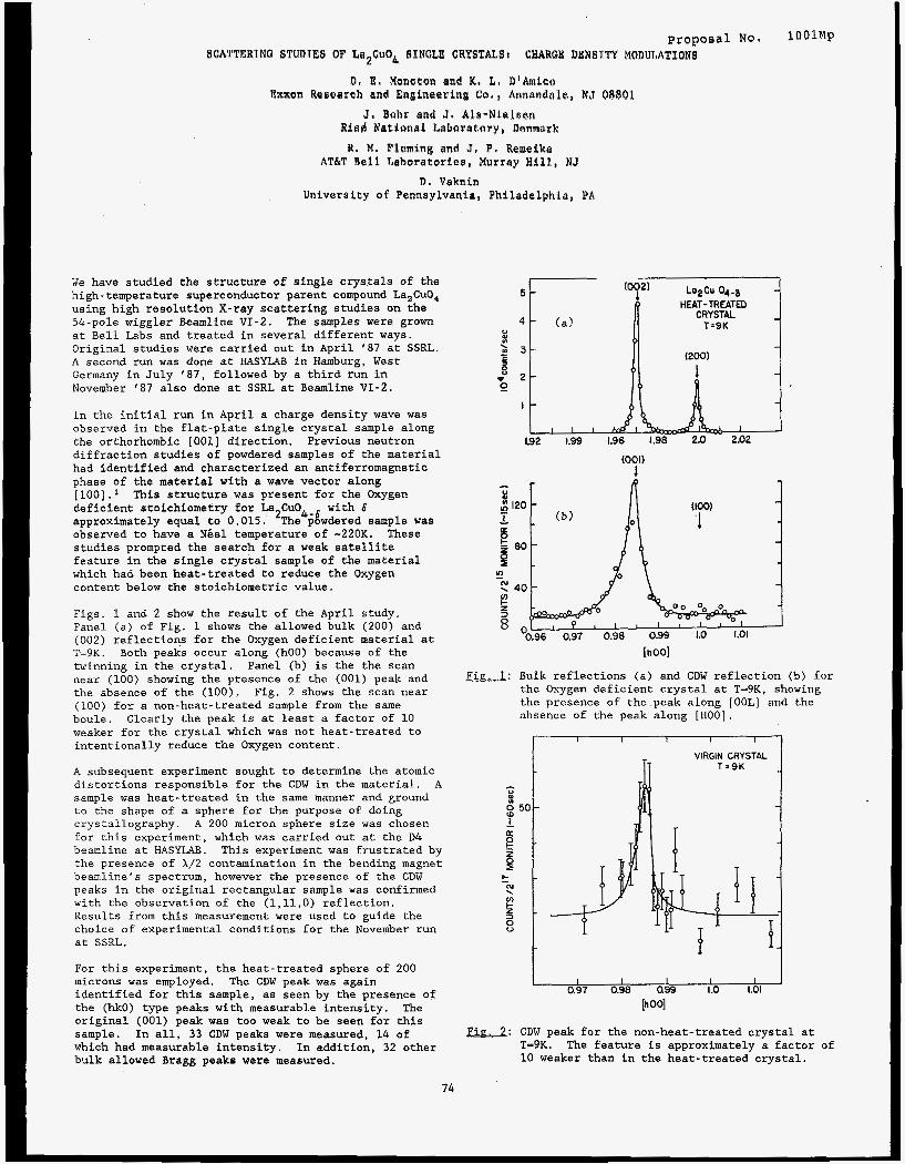
8. M. Fleming and J. P . Wemaika AT&T Boll Laboratories, Murray Hill, NJ
8 . Vaknin Univerrity of Pennsylvania, Philadelphia, PA
We have studied the structure of single crystals of the high-temperature superconductor parent compound La,CuO, using high resolution X-ray scattering studies on the 54-pole wiggler Beamline VI-2. at Bell Labs and treated in several different ways. Original studies were carried out in April '87 at SSRL. A second run was done at HASYLAB in Hamburg, West Germany in July '87, followed by a third run in November '87 also done at SSRL at Beamline VI-2.
The samples were grown
In the initial run in April a charge density wave was observed in the flat-plate single crystal sample along the orthorhombic [OOl] direction. Previous neutron diffraction studies of powdered samples of the material had identified and characterized an antiferromagnetic phase of the material with a wave vector along
deficient stoichiometry for La2Cu04-6 with 6 approximately equal to 0.015. observed to have a Niel temperature of -220K. These studies prompted the search for a weak satellite feature in the single crystal sample of the material which had been heat-treated to reduce the Oxygen content below the stoichiometric value.
Figs. 1 and 2 show the result of the April study. Panel (a) of Fig. 1 shows the allowed bulk (200) and (002) reflections for the Oxygen deficient material at T=9K. Both peaks occur along (hOO) because of the twinning in the crystal. Panel (b) is the the scan near (100) showing the presence of the (001) peak and the absence of the (100). Fig. 2 shows the scan near (100) for a non-heat-treated sample from the same boule. weaker for the crystal which was not heat-treated to intentionally reduce the Oxygen content.
A subsequent experiment sought to determine the atomic distortions responsible for the CDW in the material. A sample was heat-treated in the same manner and ground to the shape of a sphere for the purpose of doing crystallography. A 200 micron sphere size was chosen for this experiment, which was carried out at the D4 beamline at HASYLAB. This experiment was frustrated by the presence of X/2 contamination in the bending magnet beamline's spectrum, however the presence of the CDW peaks in the original rectangular sample was confirmed with the observation of the (l,ll,O) reflection. Results from this measurement were used to guide the choice of experimental conditions for the November run at SSRL.
This structure was present for the Oxygen
The powdered sample was
Clearly the peak is at least a factor of 10
For this experiment, the heat-treated sphere of 200 microns was employed. identified for this sample, as seen by the presence of the (hk0) type peaks with measurable intensity. The original (001) peak was too weak to be seen for this sample. In all, 33 CDW peaks were measured, 14 of which had measurable intensity. In addition, 32 other bulk allowed Bragg peaks were measured.
The CDW peak was again
HEAT-TREATED
1.92 1.99 1.96 1.98 2.0 2.02
(100)
'1
(001) 1
0 El
g 120 1 1
80
2 (u
u)
3
x 40
!i 8
'0.96 0.97 0.98 0.99 1.0 1.01
Fig. I: Bulk reflections (a) and CDW reflection (b) for the Oxygen deficient crystal at T=9K, showing the presence of the.peak along [OOL] and the absence of the peak along [HOO].
I I I I I I
I I I I 0.97 0.98 ass 1.0 1.01
[h 001
Fig. 2: CDW peak for the non-heat-treated crystal at T-9K. 10 weaker than in the heat-treated crystal.
The feature is approximately a factor of
74
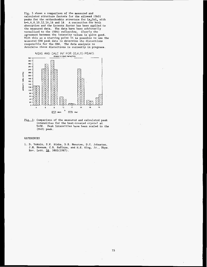
Fig. 3 shows a comparison of the measured and calculated structure factors for the allowed (OkO) peaks for the orthorhombic structure for La,CuO, with k=4,6,8,10,12,14,16 and 18. A correction for both absorption and the Lorentz factor has been applied to the measured data. The data have been arbitrarily normalized to the (004) reflection. Clearly the agreement between the intensity values is quite good. With this as a starting point it is possible to use the measured CDW peak data to determine the distortions responsible for the CDW. determine these distortions is currently in progress.
The data analysis to
MEAS AND CALC INT FOR (O,K,O) PEAKS SCUEO TO (MO) W E C B O N
4 6 8 10 12 14 16 18
YEAS 124 C W
Fig. 3 : Comparison of the measured and calculated peak intensities for the heat-treated crystal at T=9K. Peak intensities have been scaled to the (040) peak.
REFERENCES
1. D. Vaknin, S.K. Sinha, D.E. Moncton, D.C. Johnston, J.M. Newsam, C.R. Safinya, and H.E. King, Jr., Phys. Rev. Lett. 58, 5802(1987).
75
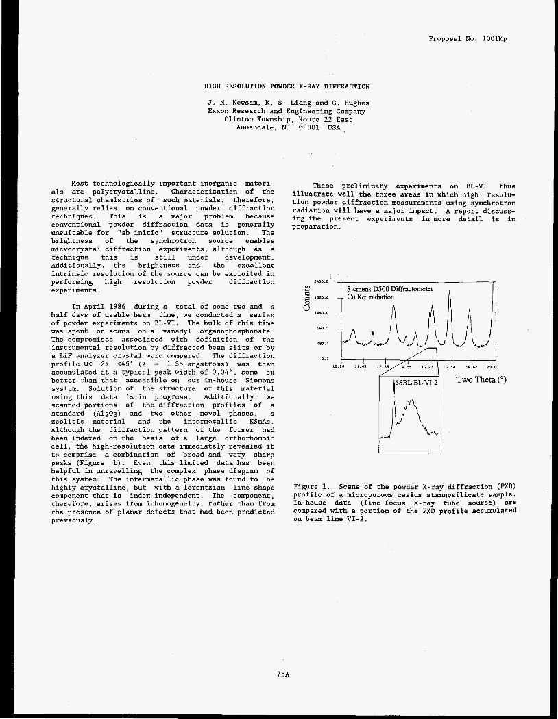
Proposal No. lOOlMp
HIGH RESOLUTION POWDER X-RAY DIFFRACTION
J. M. Newsam, K. S . Liang and G . Hughes Exxon Research and Engineering Company
Clinton Township, Route 2 2 East Annandale, NJ 08801 USA
Most technologically important inorganic materi- als are polycrystalline. Characterization of the structural chemistries of such materials, therefore, generally relies on conventional powder diffraction techniques. This is a major problem because conventional powder diffraction data is generally unsuitable for "ab initio" structure solution. The brightness of the synchrotron source enables microcrystal diffraction experiments, although as a technique this is still under development. Additionally, the brightness and the excellent intrinsic resolution of the source can be exploited in performing high resolution powder diffraction experiments.
In April 1986, during a total of some two and a half days of usable beam time, we conducted a series of powder experiments on BL-VI. The bulk of this time was spent on scans on a vanadyl organophosphonate. The compromises associated with definition of the instrumental resolution by diffracted beam slits or by a LiF analyzer crystal were compared. The diffraction profile O< 28 <45" ( A = 1.55 angstroms) was then accumulated at a typical peak width of 0 . 0 4 " , some 5x better than that accessible on our in-house Siemens system. Solution of the structure of this material using this data is in progress. Additionally, we scanned portions of the diffraction profiles of a standard ( A l 2 0 3 ) and two other novel phases, a zeolitic material and the intermetallic KSnAs. Although the diffraction pattern of the former had been indexed on the basis of a large orthorhombic cell, the high-resolution data immediately revealed it to comprise a combination of broad and very sharp peaks (Figure 1). Even this limited data has been helpful in unravelling the complex phase diagram of this system. The intermetallic phase was found to be highly crystalline, but with a lorentzian line-shape component that is index-independent. The component, therefore, arises from inhomogeneity, rather than from the presence of planar defects that had been predicted previously.
These preliminary experiments on BL-VI thus illustrate well the three areas in which high resolu- tion powder diffraction measurements using synchrotron radiation will have a major impact. A report discuss- ing the present experiments in more detail is in preparation.
2400.0
c-4 Y
9 1920.0
" 1440.0
0
960.0
480.0
0 . 0
Figure 1. Scans of the powder X-ray diffraction (PXD) profile of a microporous cesium stannosilicate sample. In-house data (fine-focus X-ray tube source) are compared with a portion of the PXD profile accumulated on beam line VI-2.

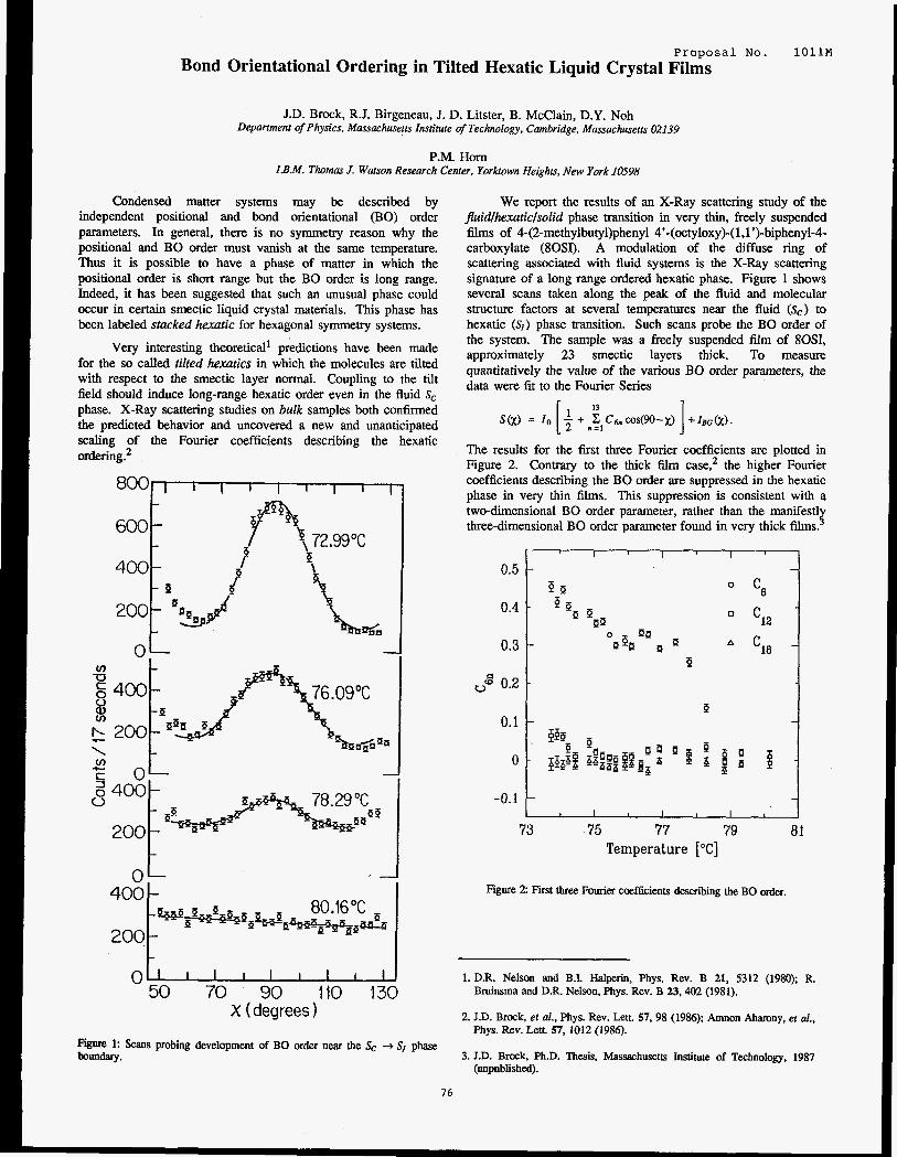
Proposal No. 1 O l l M
Bond Orientational Ordering in Tilted Hexatic Liquid Crystal Films
J.D. Brock, R.J. Birgeneau, J. D. Litster, B. McClain, D.Y. Noh Department of Physics, Massachusetts Institute of Technology, Cambridge, Massachusetts 02139
P.M. Horn I.B.M. Thomas J. Watson Research Center, Yorhown Heights, New York 10598
Condensed matter systems may be described by independent positional and bond orientational (BO) order parameters. In general, there is no symmetry reason why the positional and BO order must vanish at the same temperature. Thus it is possible to have a phase of matter in which the positional order is short range but the BO order is long range. Indeed, it has been suggested that such an unusual phase could occur in certain smectic liquid crystal materials. This phase has been labeled stacked hexuric for hexagonal symmetry systems.
Very interesting theoretical' predictions have been made for the so called tilted hexurics in which the molecules are tilted with respect to the smectic layer normal. Coupling to the tilt field should induce long-range hexatic order even in the fluid S, phase. X-Ray scattering studies on bulk samples both confirmed the predicted behavior and uncovered a new and unanticipated scaling of the Fourier coefficients describing the hexatic ordering?
c 1
0 1 50 70 90 110 130
X (degrees 1 Figure 1: Scans probing development of BO order near the SC 4 SI phase boundary.
We report the results of an X-Ray scattering study of the fluidlhexuriclsolid phase transition in very thin, freely suspended films of 4-(2-methylbutyl)phenyl 4'-(0ctyloxy)-( 1,l')-biphenyld- carboxylate (80SI). A modulation of the diffuse ring of scattering associated with fluid systems is the X-Ray scattering signature of a long range ordered hexatic phase. Figure 1 shows several scans taken along the peak of the fluid and molecular structure factors at several temperatures near the fluid (S,) to hexatic ( S I ) phase transition. Such scans probe the BO order of the system. The sample was a freely suspended film of 80S1, approximately 23 smectic layers thick. To measure quantitatively the value of the various BO order parameters, the data were fit to the Fourier Series
S W = 10 ~ + n ~ , C 6 n ~ s ( 9 0 - ~ ) + I B G ( ~ ) . [ l l3 1
The results for the first three Fourier coefficients are plotted in Figure 2. Contrary to the thick film case: the higher Fourier coefficients describing the BO order are suppressed in the hexatic phase in very thin films. This suppression is consistent with a two-dimensional BO order parameter, rather than the manifestly three-dimensional BO order parameter found in very thick films?
0.5
0.4
0.3
"g 0.2
0.1
0
-0.1
= -pa 0
a
P 1
73 75 77 79 81 Temperature ['C]
Figure 2 Fist three Fourier coefficients describing the BO order.
1. DR. Nelson and B.I. Halperin, Phys. Rev. B 21, 5312 (1980); R. Bruinsma and D.R. Nelson, Phys. Rev. B 23,402 (1981).
2. J.D. Brock, et al., Phys. Rev. Lett. 57.98 (1986); Amnon Aharony, et al., Phys. Rev. Lett. 57, 1012 (1986).
3. J.D. Brock. Ph.D. Thesis, Massachusetts Institute of Technology, 1987 (unpublished).
76

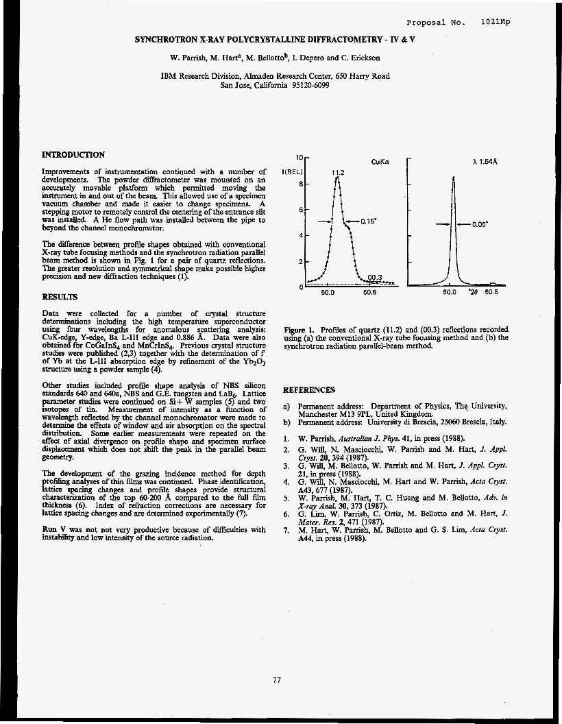
Proposal No. 1021Mp
SYNCHROTRON X-RAY POLYCRYSTALLINE DIFFRACTOMETRY - IV & V
W. Parrish, M. Harta, M. Bellottob, L Depero and C. Erickson
IBM Research Division, Almaden Research Center, 650 Harry Road San Jose, California 95120-6099
INTRODUCTION
Improvements of instrumentation continued with a number of developments. The powder diffractometer was mounted on an accurately movable platform which permitted moving the instrument in and out of the beam. This allowed use of a specimen vacuum chamber and made it easier to change specimens. A stepping motor to remotely control the centering of the entrance slit was installed. A He flow path was installed between the pipe to beyond the channel monochromator.
The difference between profde shapes obtained with conventional X-ray tube focusing methods and the synchrotron radiation parallel beam method is shown in Fig. 1 for a pair of quartz reflections. The greater resolution and symmetrical shape make possible higher precision and new dimaction techniques (1).
RESULTS
Data were collected for a number of crystal structure determinations including the high temperature superconductor using four wavelengths for anomalous scattering analysis: CuK-edge, Y-edge, Ba L-111 edge and 0.886 A. Data were also obtained for CoGaInS4 and MnCrInS4. Previous crystal structure studies were published (2,3) together with the determination o f f of Yb at the L-111 absorption edge by refrnement of the Yb@3 structure using a powder sample (4).
Other studies included profile shape analysis of NBS silicon standards 640 and M a , NBS and G.E. tungsten and LaB6 Lattice parameter studies were continued on Si+ W samples (5) and two isotopes of tin. Measurement of intensity as a function of wavelength reflected by the channel monochromator were made to determine the effects of window and air absorption on the spectral distribution. Some earlier measurements were repeated on the effect of axial divergence on profile shape and specimen surface displacement which does not shift the peak in the parallel beam geometry.
The development of the grazing incidence method for depth profiling analyses of thin films was continued. Phase identification, lattice spacing changes and profile shapes provide structural characterization of the top 60-200 A compared to the fidl film thickness (6). Index of refiaction corrections are necessary for lattice spacing changes and are determined experimentally (7).
Run V was not not very productive because of Wiculties with instability and low intensity of the source radiation.
lor X 1.54R
- 0.05"
Figure 1. Profiles of quartz (11.2) and (00.3) reflections recorded using (a) the conventional X-ray tube focusing method and (b) the synchrotron radiation parallel-beam method.
REFERENCES
a) Permanent address: Department of Physics, The University, Manchester MI3 9PL, United Kingdom.
b) Permanent address: University di Brescia, 25060 Brescia, Italy.
1. W. Parrish, Australian J. Ptys. 41, in press (1988). 2. G. Will. N. Masciocchi, W. Parrish and M. Hart, J. Appl.
21, in (1988). . G. Will. N. Masciocchi. M. Hart and W . Pamsh, Acta Cwst.
Crysf. 20, 394 (1987). G. Will. M. Bellotto. W. Parrish and M. Hart, J. Appl. Crysf. 3.
4.
5.
6.
7.
A43, 67j (1987). W. Parrish, M. Hart, T. C. Huang and M. Bellotto, Adv. in X-ray Anal. 30, 373 (1987). G. Lim, W. Parrish, C. Ortiz, M. Bellotto and M. Hart, J. Mater. Res. 2,471 (1987). M. Hart, W. Parrish, M. Bellotto and G. S . Lm, Acru Cryst. A44, in press { 1988).
77

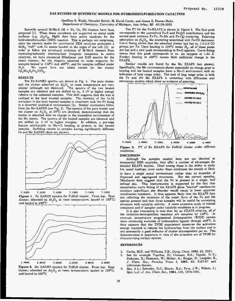
Proposal NO. 1031M XAS STUDIES OF YNTHETIC MODELS FOR HYDRODESULFURIZATION CATALYSTS 9
Geoffrey S. Waldo, Oswaldo Baralt, M. David Curtis, and James E. Penner-Hahn Department of Chemistry, University of Michigan, Ann Arbor, MI 48109-1055
Recently several M-Mo-S (M = Fe, Co, Ni) clusters have been prepared [ll. When these complexes are supported on metal oxide surfaces (e.g. A1203, MgO) they form active catalysts for the hydrodesulfurization (HDS) reaction. This is perhaps not surprising, since the current model for commercial HDS catalysts postulates a M0S2 "raft" with Co atoms bonded a t the edges of the raft [ZI. In order to follow the structural evolution of M-Mo-S clusters from crystallographically characterized inorganic complexes to active catalysts, we have measured Miissbauer and XAS spectra for the intact clusters, for the clusters adsorbed on oxide supports, for samples heated to l2OoC and 40Ooc, and for samples sulfided under H,S. We report here our initial results for the cluster CP,MO~S~F~,(CO)~.
RESULTS The Fe-XANES spectra are shown in Fig. 1. The pure cluster
and the cluster adsorbed on A1,0, a t room temperature are very similar (although not identical). The spectra of the two heated samples are identical and are shifted by ca. 5 eV to higher energy relative to the unheated samples. This shift suggests that the iron is Gxidized in the heat treated samples. The intensity of the 1s-3d transition in the heat treated samples is consistent with the Fe being in a distorted octahedral environment [31. Similar conclusions follow from the Mo-XANES (see Fig. 2). The spectra of the pure cluster and the cluster on A1,0, at 25OC are identical, demonstrating that the cluster is adsorbed with no change in the immediate environment of the Mo atoms. The spectra of the heated samples are identical and are shifted ca. 5 eV to higher energies. In addition, a pre-edge feature attributable to Mo=O bonding is present in the heated samples. Sulfiding results in samples having significantly different Fe and Mo XANES (data not shown).
I I
7 . 1 0 0 0 7 . 1 1 0 0 7 . 1 2 0 0 7 . 1 3 0 0 7 . 1 4 0 0 7 . 1 5 0 0
Figure 1. Fe XANES spectra for FeMoS cluster. From top: Neat cluster; adsorbed on A1,0, at room temperature; heated to 12@C; and heated to 4OO0C
I I I I I I
1 . 8 9 9 0 2.0010 2 . 0 0 3 0 2 . 0 0 5 0 2 . 0 0 7 0 2.0090
Figure 2. Mo XANES spectra for FeMoS cluster. From top: Neat cluster; adsorbed on Al,O, a t room temperature; heated to 1200C; and heated to 4OO0C
78
The FT for the Fe-EXAFS is shown in Figure 3. The first peak corresponds to the unresolved Fe-S and Fe-S;O contributions and the second peak contains Fe-Fe, Fe-Mo and Fe-CQ scattering. Following adsorption on A1,0,, the scattering associated with Fe-CO decreases. Curve fitting shows that the adsorbed cluster has lost ca. 1.5-2.0 CO groups per Fe. Upon heating to 12OoC under 4, all of these peaks are lost and a new peak corresponding to Fe-0 appears. Curve fitting shows that this peak corresponds to ca. six oxygens at 1.95 A. Further heating to 400°C causes little additional change in the EXAFS.
Similar results are found for the Mo EXAFS (not shown). Specifically, the Mo environment shows essentially no change prior to heating and the heated samples have a Mo-0 environment with no indication of long range order. The lack of long range order in both the Fe and the Mo EXAFS is consistent with diffraction and microscopy studies which show no evidence of sintering.
c I \
0.0000 1 . 0 0 0 0 2 .0000 3.0000 4.0000 5 .0000
Figure 3. conditions.
DISCUSSION Although the samples studied here are not identical to
commercial HDS catalysts, they offer a number of advantages for detailed EXAFS studies. Chief among these is the ability to study low metal loadings, since under these conditions the sample is likely
to have a single metal environment rather than an ensemble of dispersed and aggregated structures. For the current samples, Mossbauer data suggest that the Fe is present in a single, well defined site. This interpretation is supported by the fact that quantitative curve fitting of the EXAFS gives "normal" coordination numbers (significant site disorder would result in lower apparent coordination numbers). It thus appears likely that the EXAFS data are reflecting the structure of the major form of the Fe and Mo species present and that these samples will be useful for correlating structure with catalytic activity. A more extensive study of related complexes and of samples under catalytic conditions is in progress.
It is also interesting to note that, by an EXAFS criterion, all of the oxidation-decomposition reactions are complete by 128C. In contrast, temperature programmed decomposition (TPDE) results show continuing evolution of hydrocarbon ligands through 4000 c. It thus appears that the TPDE experimerlt measures the activation energy required to release the hydrocarbon from the surface and is not necessarily a good indicator of cluster decomposition per se. This demonstration is important in view of the extensive use of TPDE for characterizing surface species.
REFERENCES
1. 2.
FT of Fe EXAFS for FeMoS cluster under different
Curtis, M.D. a d Williams, P.D., Inorg. Chem. 1983,22, 2661. See for example TopsSoe, H.; Qausen, B.S.; Tops5e, N.-Y.; Pedersen, E.; Niemann, W.; MalIer, A.; Bmgge, H.; Lengeler, B., J. Chem. SOC., Faraday Trans I , 1987, 83, 2157-67 and references therein. Roe, A.L.; Scheider, D.J.; Mayer, R.J.; Pyrz, J.W.; Widom, J.; Que, L.Jr. J. Am. Chem. SOC., 1984, 206, 1676-1681.
3.

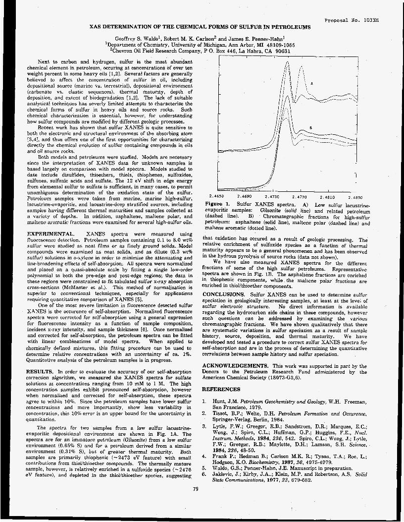
Proposal No. 103311 XAS DETERMINATION OF THE CHEMICAL FORMS OF SULFUR IN PETROLEUMS
Geoffrey S. Waldo', Robert M. K. Carlson' and James E. Penner-Hahn' 'Department of Chemistry, University of Michigan, Ann Arbor, MI 48109-1055
'Chevron Oil Field Research Company, P 0. BOX 445, La Habra, CA 90631
Next to carbon and hydrogen, sulfur is the most abundant chemical element in petroleum, occuring at concentrations of over ten weight percent in some heavy oils [1,2]. Several factors are generally believed to affect the concentration of sulfur in oil, including depositional source (marine vs. terrestrial), depositional environment (carbonate vs. clastic sequences), thermal maturity, depth of deposition, and extent of biodegradation [1,23. The lack of suitable analytical techniques has severly limited attempts to characterize the chemical forms of sulfur in heavy oils and source rocks. Such chemical characterization is essential, however, for understanding how sulfur compounds are modified by different geologic processes.
Recent work has shown that sulfur XANES is quite sensitive to both the electronic and structural environment of the absorbing atom [3,41, and thus offers one of the first opportunities for characterizing directly the chemical evolution of sulfur containing compounds in oils and oil source rocks.
Both models and petroleums were studied. Models are necessary since the interpretation of XANES data for unknown samples is based largely on comparison with model spectra. Models studied to date include disulfides, thioethers, thiols, thiophenes, sulfoxides, sulfones, sulfonic acids and sulfate. The 12 eV shift in edge energy from elemental sulfur to sulfate is sufficient, in many cases, to permit unambiguous determination of the oxidation state of the sulfur. Petroleum samples were taken from marine, marine high-sulfur, lacustrine-evaporitic, and lacustrine-deep stratified sources, including samples having different thermal maturities and samples collected at a variety of depths. In addition, asphaltene, maltene polar, and maltene aromatic fractions were examined for several high-sulfur oils.
EXPERIMENTAL. XANES spectra were measured using fluorescence detection. Petroleum samples containing 0.1 to 8.0 wt% sulfur were studied as neat films or as finely ground solids. Model compounds were examined as neat solids, and as dilute (0.3 wt% sulfur) solutions in o-xylene in order to minimize the attenuating and iine-broadening effects of self-absorption. All spectra were normalized and placed on a quasi-absolute scale by fitting a single low-order polynomial to both the pre-edge and post-edge regions; the data in these regions were constrained to fit tabulated sulfur x-ray absorption cross-sections (McMaster et al.). This method of normalization is superior to conventional techniques, especially for applications requiring quantitative comparison of XANES [51.
One of the most severe limitation in fluorescence detected sulfur XANES is the occurence of self-absorption. Normalized fluorescence spectra were corrected for self-absorption using a general expression for fluorescence intensity as a function of sample composition, incident x-ray intensity, and sample thickness [E]. Once normalized and corrected for self-absorption, the petroleum spectra can be fitted with linear combinations of model spectra. When applied to
chemically defined mixtures, this fitting procedure can be used to determine relative concentrations with an uncertainty of ca. 1%. Quantitative analysis of the petroleum samples is in progress.
RESULTS. In order to evaluate the accuracy of our self-absorption correction algorithm, we measured the XANES spectra for sulfate solutions at concentrations ranging from 10 mM to 1 M. The high concentration samples exhibit pronounced self-absorption, however when normalized and corrected for self-absorption, these spectra agree to within 10%. Since the petroleum samples have lower sulfur concentrations and inore importantly, show less variability in concentration, this 10% error is an upper bound for the uncertainty in quantitation.
The spectra for two samples from a low sulfur lacustrine- evaporitic depositional environment are shown in Fig. 1A. The spectra are for an immature petroleum (Gilsonite) from a low sulfur environment (0.65% S) and for a petroleum derived from a similar environment (0.31% S), but of greater thermal maturity. Both samples are primarily thiophenic (- 2473 eV feature) with small contributions from thiollthjoether compounds. The thermally mature sample, however, is relatively enriched in a sulfoxide species (-2476 eV feature), and depleted in the thiollthioether species, suggesting
----r-------,-- , i I I I
2 . 4 6 5 0 2 .4690 2 .4730 2 . 4 7 7 0 2 . 4 8 1 0 2 4850
Figure 1. Sulfur XANES spectra. A) Low sulfur lacustrine- evaporitic samples: Gilsonite (solid line) and related petroleum (dashed line). B) Chromatographic fractions for high-sulfur pewoleurn: asphaltene (solid line), maltene polar (dashed line) and maltene aromatic (dotted line).
that oxidation has occured as a result of geologic processing. The relative enrichment of sulfoxide species as a function of thermal maturity appears to be a general phenomenon and has been observed in the hydrous pyrolysis of source rocks (data not shown).
We have also measured XANES spectra for the different fractions of some of the high sulfur petroleums. Representatlve spectra are shown in Fig. 1B. The asphaltene fractions are enriched in thiophenic components, while the maltene polar fractions are enriched in thiolkhioether components.
CONCLUSIONS. Sulfur XANES can be used to determine sulfur speciation in geologically interesting samples, at least at the level of sulfur electronic structure. No direct information is available regarding the hydrocarbon side chains in these compounds, however such questions can be addressed by examining the various chromatographic fractions, We have shown qualitatively that there are systematic variations in sulfur speciation as a result of sample history, source, deposition, and thermal maturity. We have developed and tested a procedure to correct sulfur XANES spectra for self-absorption and are in the process of determining the quantitative correlations between sample history and sulfur speciation.
ACKNOWLEDGEMENTS. This work was supported in part by the Donors to the Petroleum Research Fund administered by the American Chemical Society (18073-G3,6).
REFERENCES
1.
2.
3.
Hunt, J.M. Petroleum Geochemistv and Geology, W.H. Freeman, San Francisco, 1979. Tissot, B.P.; Welte, D.H. Petroleum Formation and Occurence, Springer-Verlag, Berlin, 1984. Lytle, F.W.: Greegor, RB.; Sandsti-om, D.R.; Marques, E.C.; Wong, J.; Spiro, C.L.; Huffman, G.P.; Huggins, F.E., Nucl. Znstrwn. Mefhods, 1984, 226, 542. Spiro, C.L.; Wong, J.; Lytle, F.W.; Greegor, R.B.; Maylotte, D.H.; Lamson, S.H. Science, 1984, 226, 48-50. Frank P.; Hedman B.; Carlson M.K. R.; Tyson, T.A.; Roe, L.; Hodgson, K.O. Biochemistry, 1987,26, 4975-4979. Waldo, G.S.; Penner-Hahn, J.E. Manuscript in preparation. Jaklevic, J.; Kirby, J.A..; Klein, M.P. and Robertson. A S . Solid State Communications, 1977, 23, 679-682.
4.
5. 6.
79

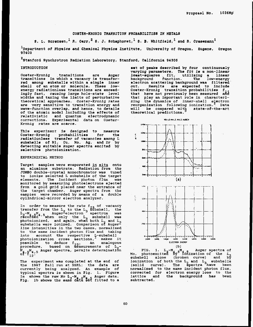
Proposal No. 1036Mp'
COITER-KRCJNPQ TRANSTTXBN BROBWBILTTTES TN METALS
S , b. lor@n81en,' R. Carrr2 5 , 3, Schapkorat,'S, B, Whitfield,' and B . Orasemann'
'Department of Physics and Chemical Physice Institute University of Oregon, Eugene, Oregon 97409
*Stanford Synchrotron Radiation Laboratory, Stanford, California 94305
INTRODUCTION
Coster-Kronig transitions ar@ Auger transitions in which a vacancy is transfer- red among subshells within a single inner shell of an atom or molecule. These low- energy radiationless transitions are exceed- ingly fast, causing large hole-state level widths and taxing the limits of perturbative theoretical approaches. Coster-Kronig rates are very sensitive to transition energy and wave-function overlap, and hence, to details of the atomic model including the effects of relativistic and quantum electrodynamic corrections. Experimental data on Coster- Kronig rates are scarce.
This experiment is designed to measure Coster-Kronig probabilities for the radiationless transfer of vacancies among L subshells of N i l Cu, Mo, Ag, and Zr by detecting suitable Auger spectra excited by selective photoionization.
EXPERIKZXTAL METHOD
Target samples were evaporated in situ onto an aluminum substrate. Radiation from the JUMBO double-crystal monochromator was tuned to ionize selected L subshells of the target elements. The incident photon flux was monitored by measuring photoelectrons ejected from a gold grid placed near the entrance of the target chamber. Auger spectra from the samples were recorded by means of a double cylindrical-mirror electron analyzer.
In order to measure the rate f23 of vacancy transfer from the L2 to the L3 subshell, the L -M Ep Auger-electron spectrum was r3co$d$b4 ' 5when only the L3 subshell was photoionized, and again, when both L and L3 subshel Is were ionized. Comparison 03 Auger- line intensities in the two cases, normalized to the same incident photon flux and taking into account the respective subshell photoionization cross sections,'- makes it possible to deduce f2'; An analogous procedure, based on measurements of La-
Auger spectra, permits determination hi of. P,, %,5 . The experiment was completed at the end of the 1987 Fall run at S S R L ; the data are currently being analyzed. An example of typical spectra is shown in Fig. 1. Figure la shows the raw Mo L3-M4 5M4 y r data. Fig. Ib shows the same data s& itted to a
sat of peaks described by four continuously varying parameters. The fit is a non-linear least-squares fit, utilizing a linear background €unction. The low-energy electron scattering background was filtered out. Results are expected to include Coster-Kronig transition probabilities that have not previously been measured :Ad that play an important role in characteri- zing the dynamics of inner-shell eJectron reorganization following ionization. Data will be compared wit$ state-of-the-art theoretical predictions.
UI L3.M.b M4.b R W Y
5:
3
ELECTRM ENEROY
(b) FIG. 1. L -M Auger spectra of
No, photoexcited &5Mf6Wization of the subshell alone (broken curve) and :$ ionization of both the L2 and L subshells (solid curve). The spectra 'have been normalized to the same incident photon flux, corrected for electron energy loss to the lattice and the background has been subtracted.
80
ACKNOWLEDGMENTS
We gratefully acknowledge the technical assistance and advice of Matt Richter, Michael Rowen, Ann Waldhauer, and Joseph Woicik. We thank W. Jitschin for help and advice in the initial stages of this work. This research is supported in part by National Science Foundation Grant PHY-85 16788 and Air Force Office of Scientific Research Grant AFOSR-87-0026. The experiment was performed at SSRL which is supported by the Department of Energy through the Office of Basic Energy Sciences and the National Institutes of Health through the Bio- technology Resources Program.
81
REFERENCES
1 - J. Scpfield, Lawrence Livermore National Laboratory Report No. UCRL- 51326 (1973) and At. Data Nucl. Data Tables L?, 121 (1974).
2. W. Bambynek, B. Crasemann, R. W. Fink, H.-U. Freund, H. Mark, C. D. Swift, R. E. Price, and P. Venugopala Rao, Rev. Mod. Phys. 44, 716 (1972).
3. M. H. Chen. in Atomic Inner-Shell Phvsics, edited by B. Crasemann (Plenum, New York, 1985), Chap. 2.
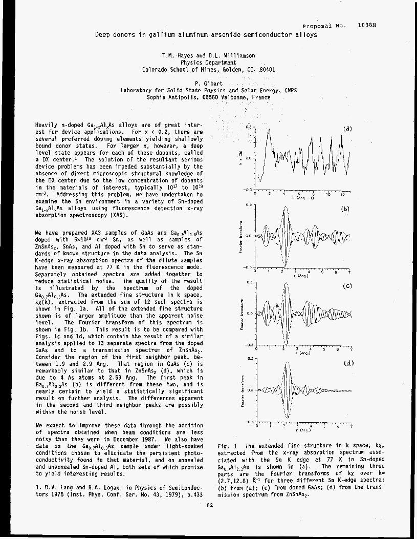
proposal NO. 1038~1
Deep donors i n g a l 1 i um a1 u m i num a r s e n i d e semi c o n d u c t o r a1 1 oys
T.M. Hayes and D.L. Williamson Physics Department
Colorado School of Mines, Golden, CO 80401
P. Gibart Laboratory f o r Solid S ta te Physics and Solar Energy, CNRS
Sophia Antipolis, 06560 Valbonne. France
Heavily n-doped Gal-,AlxAs al loys are of great i n t e r - e s t f o r device applications. For x < 0.2, there a r e several preferred doping elements yielding shallowly bound donor states. For la rger x, however, a deep level s t a t e appears f o r each of these dopants, called a DX center.1 The solution of the resul tant serious device problems has been impeded substant ia l ly by the absence of d i r e c t microscopic s t ructural knowledge of the DX center due t o the low concentration of dopants in the materials of i n t e re s t , typical ly 1017 t o 1019 ~ m - ~ . Addressing t h i s problem, we have undertaken t o examine the Sn environment in a var ie ty of Sn-doped Ga,-,Al,As a l loys using fluorescence detection x-ray absorption spectroscopy (XAS) . We have prepared XAS samples o f GaAs and Ga,~,Al,,,As doped with 5 ~ 1 0 1 ~ ~ m - ~ Sn, as well as samples of ZnSnAs,, SnAs, and A1 doped with Sn t o serve as stan- dards of known s t ructure in the data analysis. The Sn K-edge x-ray absorption spectra of the d i l u t e samples have been measured a t 77 K in the fluorescence mode. Separately obtained spectra a re added together t o reduce s t a t i s t i c a l noise. The qual i ty of the result i s i l l u s t r a t e d by the spectrum of the doped Ga,,,Al,,,As. The extended f ine s t ructure in k space, kX(k), extracted from the sum of 12 such spectra i s shown in Fig. l a . All of the extended f i n e s t ructure shown i s of la rger amplitude than the apparent noise level . The Fourier transform of t h i s spectrum i s shown i n Fig. lb . This resu l t i s t o be compared with Figs. IC and Id, which contain the r e s u l t of a s imilar analysis applied t o 13 separate spectra from the doped GaAs and t o a transmission spectrum of ZnSnAs,. Consider the region of the f i r s t neighbor peak, be- tween 1.9 and 2.9 Ang. That region in GaAs (c) i s remarkably s imilar t o t h a t i n ZnSnAs, (d), which is due t o 4 As atoms a t 2.53 Ang. The f i r s t peak in Ga,.,Al,.,As (b) is d i f fe ren t from these two, and i s nearly cer ta in t o y ie ld a s t a t i s t i c a l l y s ignif icant result on further analysis. The differences apparent i n the second and t h i r d neighbor peaks are possibly within the noise level.
We expect t o improve these data through the addition of spectra obtained when beam conditions are l e s s noisy than they were in December 1987. We a lso have data on the Ga,.,Al,.,As sample under 1 ight-soaked conditions chosen t o e lucidate the persistent photo- conductivity found in tha t material, and on annealed and unannealed Sn-doped Al, both sets of which promise t o yield interest ing resul ts .
1. D.V. Lang and R . A . Logan, in Physics of Semiconduc- tors 1978 (Inst . Phys. Conf. Ser. No. 43, 1979), p.433
-0.3,, 0 , , 2 , , , I , , 4 , , 6 , , , , , , , , 6 1 0 1 2 , ,
k (Ang -1)
0.3 1 b'i
-0.3 , , - I I I , I , , , , , , , , , , , , , , , , , ~
0 1 2 3 4 5 6 ; r ( A w )
Fig. 1 The extended f i n e structure in 'k space, k x , extracted from the x-ray absorption spectrum asso- c ia ted with the Sn K edge a t 77 K in Sn-doped Gao,,A1,.3As i s shown in (a) . The remaining three par ts are the Fourier transforms of kx over k= (2.7,12.8) 8-1 for three d i f fe ren t Sn K-edge spectra: (b) from (a ) ; (c) from doped GaAs; (d) from the t rans- mission spectrum from ZnSnAs,.
82


Proposal No. 1054M
X-Ray Induced Photo-Acoustic Emission
Robert A. Day, Richard W. Ryon, and Steven Benson
Lawrence Livermore National Laboratory 7000 East Avenue, Livermore, CA 94550
Introduction
Materials that have similar linear absorption coefficients, such as aluminum and alumina, are difficult to distinguish by normal x-ray methods. When these materials are mixed with high Z materials such as used in integrated circuits the imaging of small features is even more difficult since the absorption of the higher Z material must also be included.
X-ray induced photo-acoustics holds promise for this type of imaging because the generation of sound is a function of the coefficient of thermal expansion (CTE). For an aluminum wire embedded in an alumina matrix the theoretical sensitivity will be about four times that of x-ray absorption measurement alone. Since we are interested in thick specimens (1-5 mm), absorption edge effects and fluorescence are not useful since absorption is too high to allow penetration.
A second less obvious advantage to acoustic detection is the ability of sound to travel in all directions. This allows detection from any side of the specimen which avoids the need to peneuate high Z materials that may shadow the part of the specimen we wish to image.
Results and Discussion
Prior to doing experiments at Stanford Synchrotron Radiation Laboratoq (SSRL) we modelled the photo-acoustic generation in aluminum using a pulsed x-ray source with a 3 nanosecond width ( a Febatron ). We detected acoustic signals using an acoustic emission tranducer and amplifier. The frequency of this detector was limited to to 100-500 kilohertz region. The low repetition fresuency of the Febatron source (0.2 hertz) would require more than a day to form a useful image.
The detection system we designed for use at the synchrotron was based on this Febatron experiment (Fig. 1). A relatively narrow band transducer (€2"-SA) and integral amplifier was designed. The center frequency was 1.1 megahertz with 20% bandwidth to allow detection of the synchrotron ring frequency of 1.28 megahertz. This ffequency was chosen because prior work [l] indicated it should work well and because this frequency could allow the use of focused detectors which would allow depth sensitivity to be controlled. ~
The actual experiments with this system were not successful and it was replaced by a separate transducer bonded to a fused silica flat. The purpose of the flat was to protect the transducer from the intense beam of the synchrotron. The low frequency system was not sensitive enough to detect the higher frequency signals produced by the short x-ray pulses at SSIU..
This system detected photo-acoustic signals (Fig. 2) with an excellent signal-t +noise ratio (>2odB). Fused silica is not a good generator of photo-acoustic waves because of it's low CXE. The frequency of successful transducers was 25 and 15 megahertz. Lower frequency transducers were insensitive to the sound generated. The very short x-ray pulses (65 picoseconds) generate 15 gigahertz sound which was incapable of exciting the lower frequency detectors, contrary to our experience with the Febatron.
The a c h dekction wasperformed at 2 kilohertz using a chopper wheel in the synchrotron beam. During the period the chopper wheel is "open" about 5,000 high frequency x-ray pulses strike the material and the amplifier integrates these into a lower frequency pulse. A signal averager was used to further enhance the signal-to- noise ratio of this signal. Integration times were approximately 10 seconds.
I from chopper I Fig. 1: The experimental system used at SSRL to test x-ray
photo-acoustic generation.
The fused silica was 25 mm thick to assure that no x-rays penetrated to the transducer to generate signals through ionization effects or photo-acoustic generation in the transducer itself. Penetration of the silica is 2-4 mm and was verified by radiation darkening.
We assured that no sound was created in the experiment, by the. chopper wheel for example, by blocking the x-ray beam at the collimator. No sound was detected by the transducer without the x- ray beam verifying that we were not receiving a noise source from another part of the experiment.
Detection in aluminum, copper, graphite, acrylic plastic and alumina were unsuccessful. The samples of these materials were bonded to the silica with silicon vacuum grease in a thin layer. "he
r
83
I I I t I I I I
0 0.25 0.5 0.75 1.0 1.25 1.5 Time in ms
Fig. 2 Signal obtained on fused silica with a 15 megahertz longitudinal wave transducer and 120 dB of amplifier gain. The 25 megahertz transducer gave slightly better signal to noise ratio.
lack of results are in accord with the absorption of the high frequency acoustic pulses by these materials [2]. The very high frequency is absorbed in the f i t 20 to 30 p of all of these materials, the minimum thickness was 500 F. We did not anticipate this high frequency result both because of our Febatron experiment and work by Sachse [3]. The difference between the two x-ray pulses appears minor (65 ps vs 160 ps and different intensity). Different results may be accounted for in the detector and electronics.
will be needed to make it practical. Areas we are exploring now include the confirmation of the results of this experiment by use of lasers in transparent and absorbing materials and the use of multi- layer x-ray lens to achieve higher intensity. Optimization of the detector and electronics will be done using the laser model.
The imaging of discontinuities appears promising but much work
Acknowledgements
We would like to acknowledge the help of Jeny Haskins, Walt Kendricks, Mark Lachapell, Jim Brase, Hany Martz, Chuck Valente, Floyd Kirk, Ron Streit and Suzanne Fletcher in helping out in this experiment "beyond the call of duty".
Energy by the Lawrence Livermore National Laboratory under Contract W-7405-Eng-48.
Work performend under the auspices of the U. S. Department of
References
1. K. Y. Kim and W. Sachse, X-ray generated ultrasound, Appl. Phys. Lett., 43(12), 15 December 1983.
2. H. Wickramsinghe, Contrast and Imaging Performance in the Scanning Acoustic Microscope, J. Appl. Phys., 50(2), 1979.
84
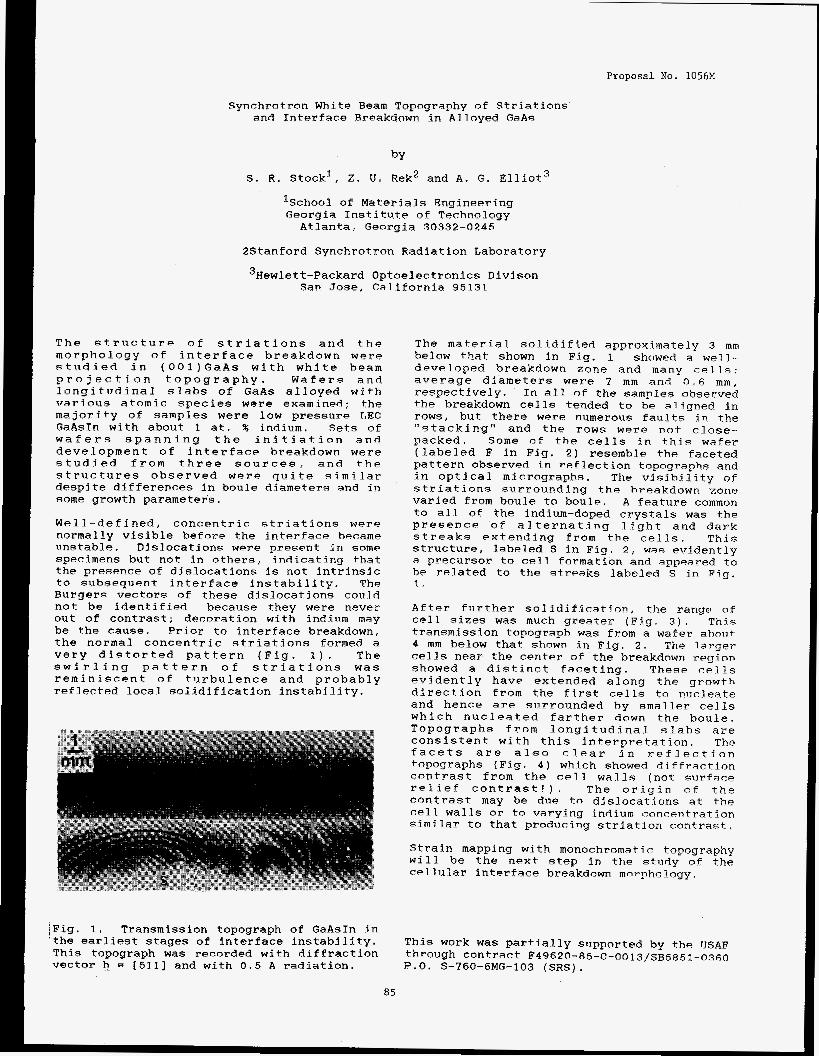
Proposal KO. 1056M
Synchrotron White Beam Topography of Striations and Interface Breakdown in Alloyed GaAs
by
S . R. Stock’, 2. CI. Rek2 and A. G. Elliot’
‘School of Materials Engineering Georgia Institute of Technology Atlanta, Georgia 30332-0245
2Stanford Synchrotron R-adiation Laboratory
’Hewlett-Packard Optoelectronics Divison San Jose, California 95131
The structure of striations and. the morphology of interface breakdown were studied in (001)GaAs with white beam projection topography. Wafers and longitudinal slabs of FaAs alloyed with various atomic species were examined; the ma-jority of samples were low pressure LEC GaAsIn with about 1 at. % indium. Sets of wafers spanning the initiation and development of interface breakdown were studied from three sources, and the structures observed were quite similar despite differences in boule diameters and in some growth parameters.
Well -defined, concentric striations were normally visible before the interface became unstable. Dislocations were present in some specimens but not in others, indicating that the presence of dislocations is not intrinsic to subsequent interface instability. The Burgers vectors of these dislocations could not be identified because they were never out of contrast; decoration with indinm may be the cause. Prior to interface breakdown, the normal concentric striations formed a very distorted pattern (Fig. 1 ) . The swirling pattern of striations was reminiscent of turbulence and probably reflected local solidification instability.
The material soli.difi.ed approximately 3 mm below that: shown in Fig. 1 showed a well- developed breakdown zone and many cells: average diameters were 7 mm and 0 . 6 mm, respectively. In all of the samples observed the breakdown cells tended to be aligned in rows, but there were numerous faults in the “stacking” and the rows were not clnse- packed. Some of the cells in this wafer (labeled F in Fig. 2) resemble the faceted pa-ttern observed in reflection topographs and in optical micrographs. The visibility of striations surrounding the breakdown zone varied from boule to boule, A feature common to all of the indium-doped crystals was the presence of alternating light and dark streaks extending from the cells. This structure, labeled S in Fig. 2, was evidently a precursor to cell formation and appeared to be related to the streaks labeled S in Fig. 1. . After further solidification, the range of cell sizes was much greater (Fig. 3 ) . This transmission topograph was from a wafer about 4 mm below that shown in Fig. 2 , Th.e l.a.rger cells neay the center of the breakdown region. showed a distinct faceting. These cells evidently have extended along the growth direction from the first cells to nucleate and hence are surrounded by smaller cells which nucleated farther down the boule. Topographs from longitudinal slabs are consistent with this interpretation. The facets are also clear in reflection topographs (Fig. 4) which showed d.iffraction contrast from the cell walls (not surface relief contrast!). The origin o f the contrast may he due to dislocations at the cell walls or to varying indium concentration similar to that prodncing stria-tion contrast,
Strain mapping with monochromatic topography will be the next step in the stiidy of the cellular interface breakdown morphology.
,Fig. 1. Transmission topograph of GaAsIn in the earliest stages of interface instability. This topograph was recorded with diffraction vector h_ = [511] and with 0.5 A radiation.
This work was partially supported by the IJSAF through contract F 4 9 6 2 0 - 8 5 - C - O 0 1 3 , / S B 5 8 ~ 1 - ~ ~ ~ 0 P.O. S-160-6MG-103 (SRS).
85
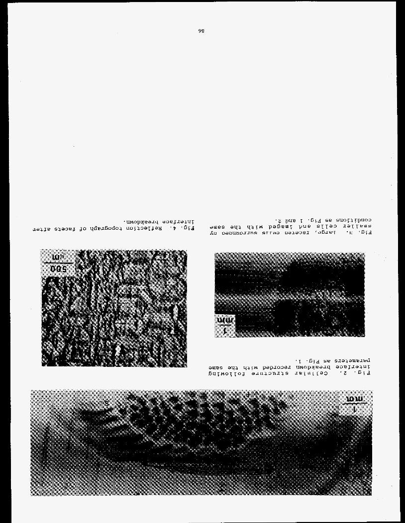
98
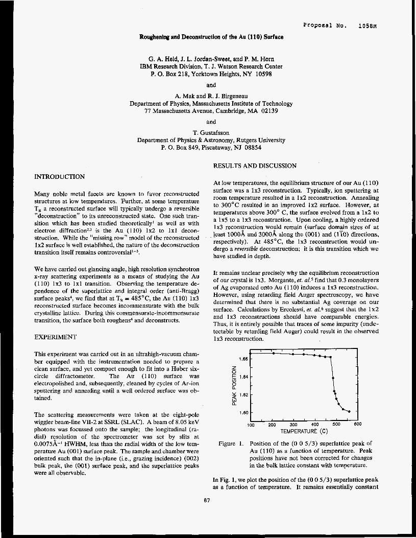
Proposal No. 1058M
Roughening and Deconstruction of the Au (1 10) Surface
G. A. Held, J. L. Jordan-Sweet, and P. M. Horn IBM Research Division, T, J. Watson Research Center
P. 0. Box 218, Yorktown Heights, NY 10598
and
A. Mak and R. J. Birgeneau Department of Physics, Massachusetts Institute of Technology
77 Massachusetts Avenue, Cambridge, MA 02139
and
T. Gustafsson Department of Physics & Astronomy, Rutgers University
P. 0. Box 849, Piscataway, NJ 08854
RESULTS AND DISCUSSION
INTRODUCTION
Many noble metal facets are known to favor reconstructed structures at low temperatures. Further, at some temperature T, a reconstructed surface will typically undergo a reversible "deconstruction" to its unreconstructed state. One such tran- sition which has been studied theoretically' as well as with electron d i f f rac t i~n~.~ is the Au (110) 1x2 to 1x1 decon- struction. While the "missing row" model of the reconstructed 1x2 surface is well established, the nature of the deconstruction transition itself remains contr~versiall-~.
We have carried out glancing angle, high resolution synchrotron x-ray scattering experiments as a means of studying the Au (110) 1x3 to 1x1 transition. Observing the temperature de- pendence of the superlattice and integral order (anti-Bragg) surface peaks4, we find that at T, = 485'C, the Au (110) 1x3 reconstructed surface becomes incommensurate with the bulk crystalline lattice. During this commensurate-incommensurate transition, the surface both roughens4 and deconstructs.
EXPERIMENT
This experiment was carried out in an ultrahigh-vacuum cham- ber equipped with the instrumentation needed to prepare a clean surface, and yet compact enough to fit into a Huber six- circle diffractometer. The Au (110) surface was electropolished and, subsequently, cleaned by cycles of Ar-ion sputtering and annealing until a well ordered surface was ob- tained.
The scattering measurements were taken at the eight-pole wiggler beam-line VII-2 at SSRL (SLAC). A beam of 8.05 keV photons was focussed onto the sample; the longitudinal (ra- dial) rtsolution of the spectrometer was set by slits at 0.0075A-' HWHM, less than the radial width of the low tem- perature Au (001) surface peak. The sample and chamber were oriented such that the in-plane (i.e., grazing incidence) (002) bulk peak, the (001) surface peak, and the superlattice peaks were all observable.
87
At low temperatures, the equilibrium structure of our Au (1 10) surface was a 1x3 reconstruction. Typically, ion sputtering a t room temperature resulted in a 1x2 reconstruction. Annealing to 300°C resulted in an improved 1x2 surface. However, a t temperatures above 300° C, the surface evolved from a 1x2 t o a 1x5 to a 1x3 reconstruction. Upon cooling, a highly ordered 1x3 reconspuction woyld remain (surface domain sizes of a t least l00OA and 3000A along the (001) and (1x0) directions, respectively). At 485OC, the 1x3 reconstruction would un- dergo a reversible deconstruction; it is this transition which we have studied in depth.
It remains unclear precisely why the equilibrium reconstruction of our crystal is 1x3. Morgante, et. al.5 find that 0.3 monolayers of Ag evaporated onto Au (1 10) induces a 1x3 reconstruction. However, using retarding field Auger spectroscopy, we have determined that there is no substantial Ag coverage on our surface. Calculations by Ercolessi, et. a1.6 suggest that the 1x2 and 1x3 reconstructions should have comparable energies. Thus, it is entirely possible that traces of some impurity (unde- tectable by retarding field Auger) could result in the observed 1 x3 reconstruction.
1.66 l-===Tl z k 1.64 rn 0
12 3 l A 2 a
1.60
io0 200 300 400 500 600 TEMPEWTURE (C)
Figure 1. Position of the (0 0 5/3) superlattice peak of Au (110) as a function of temperature. Peak positions have not been corrected for changes in the bulk lattice constant with temperature.
In Fig. 1, we plot the position of the (0 0 5/3) superlattice peak as a function of temperature. It remains essentially constant
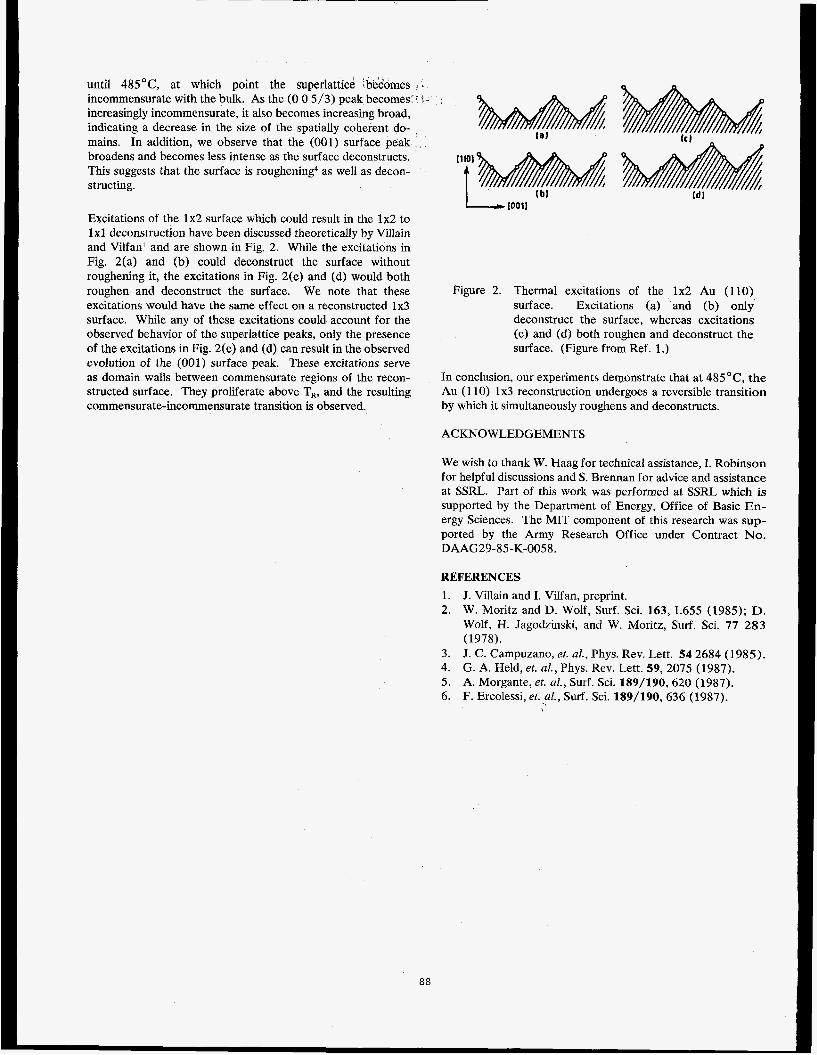
until 485OC, at which point the superlattice !bedomes - incommensurate with the bulk. As the (0 0 5/3) peak becomes 3 - increasingly incommensurate, it also becomes increasing broad, indicating a decrease in the size of the spatially coherent do- mains. In addition, we observe that the (001) surface peak broadens and becomes less intense as the surface deconstructs. This suggests that the surface is roughening4 as well as decon- structing.
Excitations of the 1x2 surface which could result in the 1x2 to 1x1 deconstruction have been discussed theoretically by Villain and Vilfan' and are shown in Fig. 2. While the excitations in Fig. 2(a) and (b) could deconstruct the surface without roughening it, the excitations in Fig. 2(c) and (d) would both roughen and deconstruct the surface. We note that these excitations would have the same effect on a reconstructed 1x3 surface. While any of these excitations could account for the observed behavior of the superlattice peaks, only the presence of the excitations in Fig. 2(c) and (d) can result in the observed evolution of the (001) surface peak. These excitations serve as domain walls between commensurate regions of the recon- structed surface. They proliferate above Tx, and the resulting commensurate-incommensurate transition is observed.
ACKNOWLEDGEMENTS
It101
t L 10011
Figure 2. Thermal excitations of the 1x2 Au (110) surface. Excitations (a) and (b) only- deconstruct the surface, whereas excitations (c) and (d) both roughen and deconstruct the surface. (Figure from Ref. 1.)
In conclusion, our experiments demonstrate that at 485OC, the Au (1 10) 1x3 reconstruction undergoes a reversible transition by which it simultaneously roughens and deconstructs.
We wish to thank W. Haag for technical assistance, I. Robinson for helpful discussions and S. Brennan for advice and assistance at SSRL. Part of this work was performed at SSRL which is supported by the Department of Energy, Office of Basic En- ergy Sciences. The MIT component of this research was sup- ported by the Army Research Office under Contract No. DAAG29-85-K-0058.
REFERENCES
1. J. Villain and I. Vilfan, preprint. 2. W. Moritz and D. Wolf, Surf. Sci. 163, L655 (1985); D.
Wolf, H. Jagodzinski, and W. Moritz, Surf. Sci. 77 283 (1978).
3. J. C. Campuzano, et. al., Phys. Rev. Lett. 54 2684 (1985). 4. G. A. Held, et. al., Phys. Rev. Lett. 59, 2075 (1987). 5. A. Morgante, et. el., Surf. Sci. 189/190, 620 (1987). 6. F. Ercolessi, et. al., Surf. Sci. 189/190, 636 (1987).
9
88
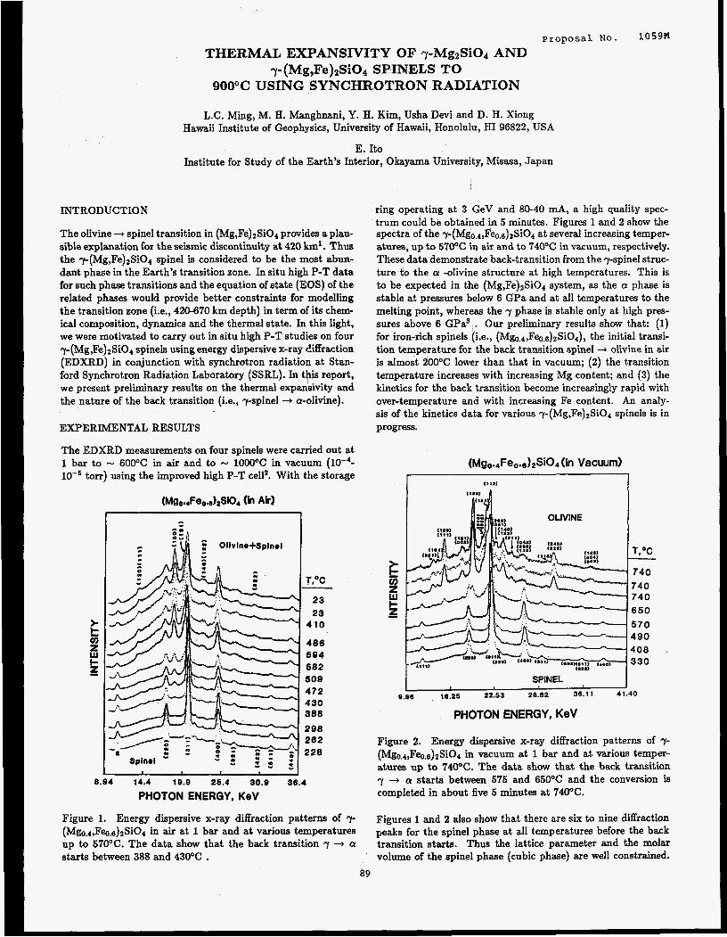
proposal NO. 1059N THERMAL EXPANSIVITY OF 7-MgzSiOl AND
y-(Mg,Fe)zSiO* SPINELS TO 900°C USING SYNCHROTRON RADIATION
L.C. Ming, M. H. Manghnani, Y. H. Kim, Usha Devi and D. H. Xiong Hawaii Institute of Geophysics, University of Hawaii, Honolulu, HI 96822, USA
E. It0 Institute for Study of the Earth’s Interior, Okayama University, Misasa, Japan
INTRODUCTION
The olivine + spinel transition in (Mg,FeIaSiO4 provides aplau- sible explanation for the seismic discontinuity at 420 km’ . Thus the y-(Mg,Fe)ZSiO4 spinel is considered to be the most abun- dant phase in the Earth’s transition zone. In situ high P-T data for such phase transitions and the equation of state (EOS) of the related phases would provide better constraints for modelling the transition zone (Le., 420-670 km depth) in term of its chem- ical composition, dynamics and the thermal state. In this light, we were motivated to carry out in situ high P-T studies on four q-(Mg,Fe)$3i04 spinels using energy dispersive x-ray Ciffraction (EDXRD) in conjunction with synchrotron radiation at Stan- ford Synchrotron Radiation Laboratory (SSRL). In this report, we present preliminary results on the thermal expansivity and the nature of the back transition (i.e., yspinel -+ a-olivine).
EXPERIMENTAL RESULTS
The EDXRD measurements on four spinels were carried out at 1 bar to - 6OOOC in air and to - 1000°C in vacuum
torr) using the improved high P-T cella. With the storage
(Mgo..Feo.drSi04 (h Air)
).
z W c
5 z
8.94 14.4 10.9 26.4 30.9 38.4
PHOTON ENERGY, KeV
Figure 1. Energy dispersive x-ray diffraction patterns of y (Mgo.4,F~.&Si04 in air at 1 bar and at various temperatures up to 57OOC. The data show that the back transition 7 + a starts between 388 and 43OOC .
T,OC - 23 23
410
486 694 582 so9 472 430 388
298 262 228
ring operating at 3 GeV and 80-40 mA, a high quality spec- trum could be obtained in 5 minutes. Figures 1 and 2 show the spectra of the ~-(Mg~.4,Feo,~)2siO4 at several increasing temper- atures, up to 570% in air and to 74OOC in vacuum, respectively. These data demonstrate back-transition from the 7-spinel struc- ture to the a -olivine structure at high temperatures. This is to be expected in the (Mg,Fe)zSi04 system, as the CY phase is stable at pressures below 6 GPa and at all temperatures to the melting point, whereas the 7 phase is stable only at high pres- sures above 6 GPa3 . Our preliminary results show that: (1) for iron-rich spinels (Le., ( M ~ O . ~ , F ~ ~ . ~ ) ~ S ~ O ~ ) , the initial transi- tion temperature for the back transition spinel + olivine in air is almost 2OOOC lower than that in vacuum; (2) the transition temperature increases with increasing Mg content; and (3) the kinetics for the back transition become increasingly rapid with over-temperature and with increasing Fe content. An analy- sis of the kinetics data for various ~-(Mg,Fe)~Si04 spinels is in progress.
(in Vacuum)
740 650
570
I SPINEL I I
9.98 , 16.25 22.53 28.82 35.11 41.40
PHOTON ENERGY, KeV
Figure 2. Energy dispersive x-ray diffraction patterns of 7- ( M ~ o . ~ , F ~ o . ~ ) ~ S ~ O ~ in vacuum at 1 bar and at various temper- atures up to ‘140°C. The data show that the back transition 7 -+ a starts between 575 and 65OOC and the conversion is completed in about five 5 minutes at 74OOC.
Figures 1 and 2 also show that there are six to nine diffraction peaks for the spinel phase at all temperatures before the back transition starts. Thus the lattice parameter and the molar volume of the spinel phase (cubic phase) are well constrained.
89
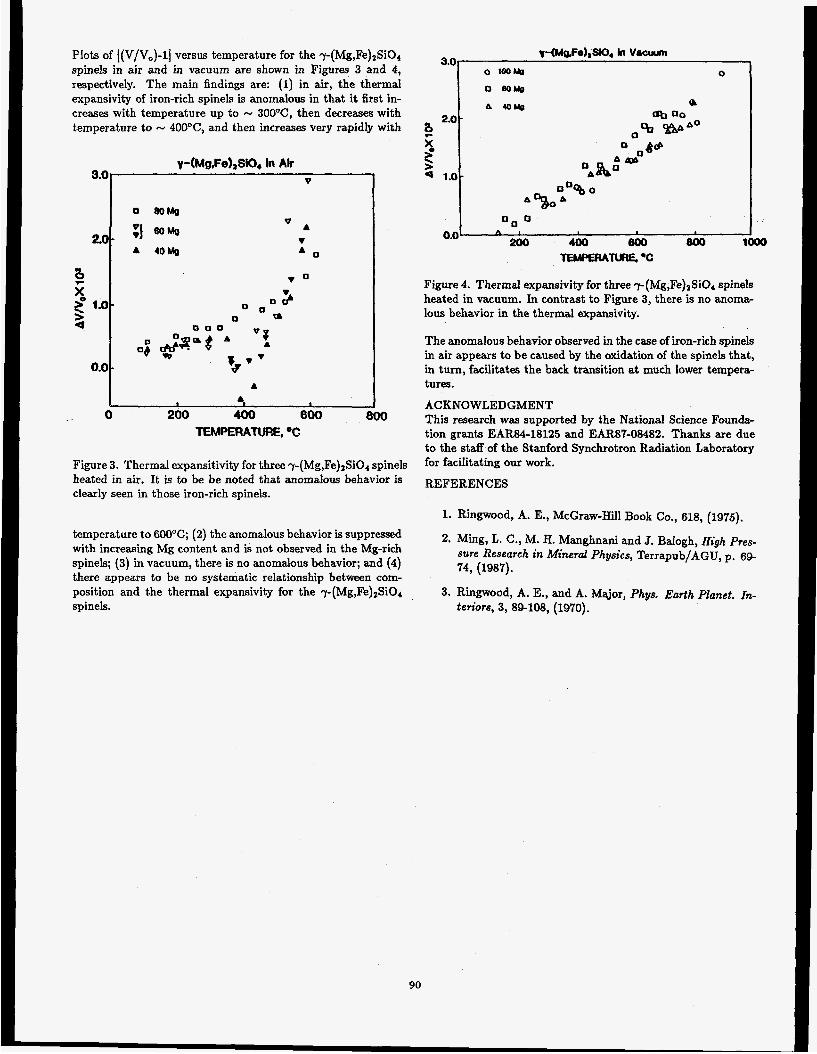
Plots of [(V/Vo)-l] versus temperature for the ~-(Mg,Fe)~Si04 spinels in air and in vacuum are shown in Figures 3 and 4, respectively. The main findings are: (1) in air, the thermal expansivity of iron-rich spinels is anomalous in that it first in- creases with temperature up to - 3OO0C, then decreases with temperature to - 400°C, and then increases very rapidly with
0
Figure 3. Thermal expansitivity for three -p(Mg,Fe)tSiO4 spinels heated in air. It is to be be noted that anomalous behavior is clearly seen in those iron-rich spinels.
temperature to 600OC; (2) the anomalous behavior is suppressed with increasing Mg content and is not observed in the Mg-rich spinels; (3) in vacuum, there is no anomalous behavior; and (4) there appears to be no systematic relationship between com- position and the thermal expansivity for the y(Mg,Fe),SiO4 spinels.
A
0 0 O
200 400 800 800 1 0.0
TEMIWUTURE..C
Figure 4. Thermal expansivity for three -y-(Mg,Fe)* Si04 spinels heated in vacuum. In contrast to Figure 3, there is no anoma- lous behavior in the thermal expansivity.
The anomalous behavior observed in the case of iron-rich spinels in air appears to be caused by the oxidation of the spinels that, in turn, facilitates the back transition at much lower tempera- tures.
ACKNOWLEDGMENT This research was supported by the National Science F o u n h tion grants EAR84-18125 and EARS?-08482. Thanks are due to the staff of the Stanford Synchrotron Radiation Laboratory for facilitating our work.
REFERENCES
1. Ringwood, A. E., McGraw-Hill Book Co., 618, (1975).
2. Ming, L. C., M. H. Manghnani and J. Balogh, High Pres- sure Research in Mineral Physics, Terrapub/AGU, p . 69- 74, (1987).
3. Ringwood, A. E., and A. Major, Phys. Earth Planet. In- teriors, 3 , 89-108, (1970).
90
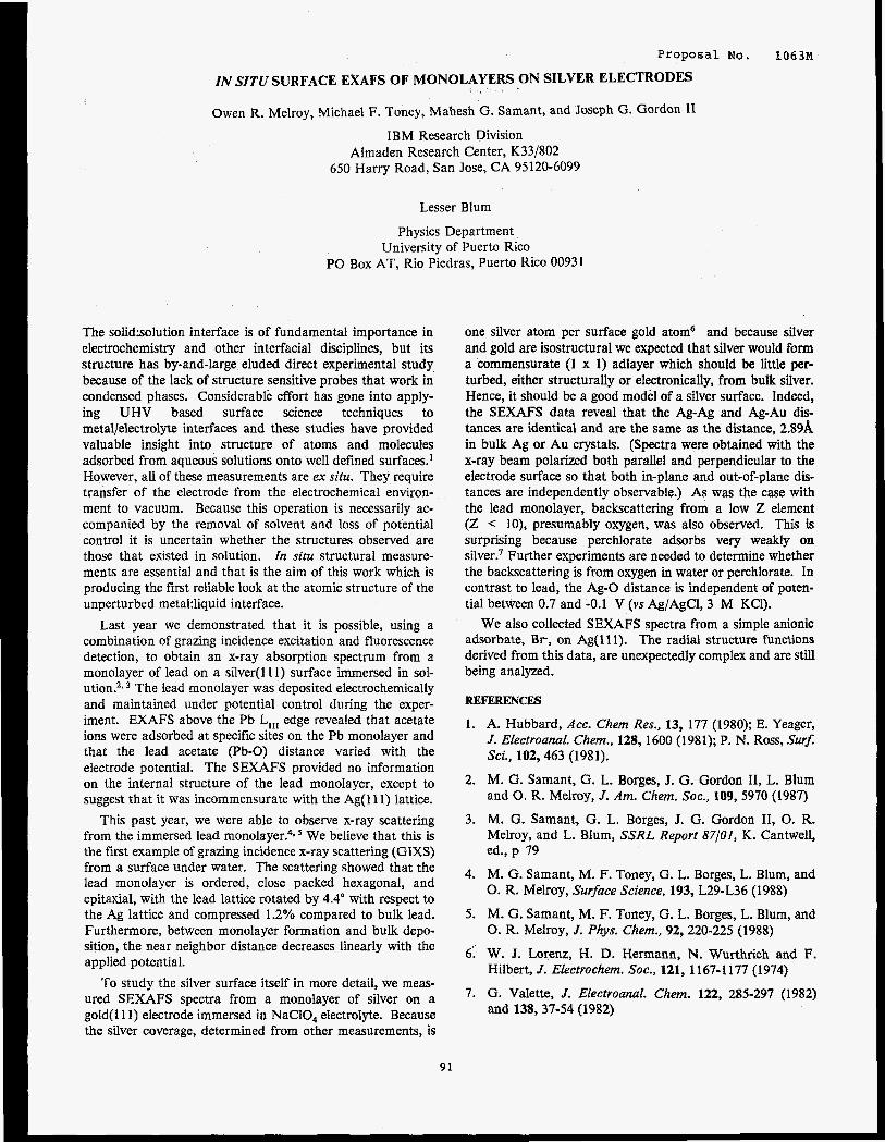
Proposal No. 1 0 6 3 ~
ZN SITU SURFACE EXAFS OF MONOLAYERS ON SILVER ELECTRODES
Owen R. Melroy, Michael F. Toney, Mahesh G. Samant, and Joseph G. Gordon 11
IBM Research Division Almaden Research Center, K33/802
650 Harry Road, San Jose, CA 95120-6099
Lesser Blum
Physics Department University of Puerto Rico
PO Box AT, Rio Piedras, Puerto Rico 00931
The solid:solution interface is of fundamental importance in electrochemistry and other interfacial disciplines, but its structure has by-and-large eluded direct experimental study because of the lack of structure sensitive probes that work in condensed phases. Considerable effort has gone into apply- ing UHV based surface science techniques to metal/electrolyte interfaces and these studies have provided valuable insight into structure of atoms and molecules adsorbed from aqueous solutions onto well defined surfaces.' However, all of these measurements are ex situ. They require transfer of the electrode from the electrochemical environ- ment to vacuum. Because this operation is necessarily ac- companied by the removal of solvent and loss of potential control it is uncertain whether the structures observed are those that existed in solution. In situ structural measure- ments are essential and that is the aim of this work which is producing the first reliable look at the atomic structure of the unperturbed meta1:liquid interface.
Last year we demonstrated that it is possible, using a combination of grazing incidence excitation and fluorescence detection, to obtain an x-ray absorption spectrum from a monolayer of lead on a silver( 1 11) surface immersed in sol- ution?, The lead monolayer was deposited electrochemically and maintained under potential control during the exper- iment. EXAFS above the Pb L,,, edge revealed that acetate ions were adsorbed at specific sites on the Pb monolayer and that the lead acetate (Pb-0) distance vaned with the electrode potential. The SEXAFS provided no information on the internal structure of the lead monolayer, except to suggest that it was incommensurate with the Ag(ll1) lattice.
This past year, we were able to observe x-ray scattering from the immersed lead monolayer?. We believe that this is the first example of grazing incidence x-ray scattering (GIXS) from a surface under water. The scattering showed that the lead monolayer is ordered, close packed hexagonal, and epitaxial, with the lead lattice rotated by 4.4" with respect to the Ag lattice and compressed 1.2% compared to bulk lead. Furthermore, between monolayer formation and bulk depo- sition, the near neighbor distance decreases linearly with the applied potential.
To study the silver surface itself in more detail, we meas- ured SEXAFS spectra from a monolayer of silver on a gold(l11) electrode immersed in NaCIO, electrolyte. Because the silver coverage, determined from other measurements, is
91
one silver atom per surface gold atom6 and because silver and gold are isostructural we expected that silver would form a commensurate (1 x 1) adlayer which should be little per- turbed, either structurally or electronically, from bulk silver. Hence, it should be a good model of a silver surface. Indeed, the SEXAFS data reveal that the Ag-Ag and Ag-Au dis- tances are identical and are the same as the distance, 2.89A in bulk Ag or Au crystals. (Spectra were obtained with the x-ray beam polarized both parallel and perpendicular to the electrode surface so that both in-plane and out-of-plane dis- tances are independently observable.) As was the case with the lead monolayer, backscattering from a low 2 element (2 lo), presumably oxygen, was also observed. This is surprising because perchlorate adsorbs very weakly on silver.' Further experiments are needed to determine whether the backscattering is from oxygen in water or perchlorate. In contrast to lead, the Ag-0 distance is independent of poten- tial between 0.7 and -0.1 V (vs Ag/AgCl, 3 M KC1).
We also collected SEXAFS spectra from a simple anionic adsorbate, B r , on Ag(ll1). The radial structure functions derived from this data, are unexpectedly complex and are still being analyzed.
REFERENCES
1. A. Hubbard, Acc. Chem Res., 13, 177 (1980); E. Yeager, J. Electroanal. Chem., 128, 1600 (1981); P. N. Ross, SurJ Sci., 102,463 (1981).
2. M. G. Samant, G. L. Borges, J. G. Gordon 11, L. Blum and 0. R. Melroy, J. Am. Chem. SOC., 109, 5970 (1987)
3. M. G. Samant, G. L. Borges, J. G. Gordon 11, 0. R. Melroy, and L. Blum, SSRL Report 87/01, K. Cantwell, ed.,p 79
4. M. G. Samant, M. F. Toney, G. L. Borges, L. Blum, and 0. R. Melroy, Surface Science, 193, L29-L36 (1988)
5. M. G . Samant, M. F. Toney, G. L. Borges, L. Blum, and 0. R. Melroy, J. Phys. Chem., 92, 220-225 (1988)
6: W. J. Lorenz, H. D. Hermann, N. Wurthrich and F. Hilbert, J . Electrochem. Soc., 121 , 1 167- 1 177 (1 974)
7. G. Valette, J . Electroanal. Chem. 122, 285-297 (1982) and 138,37-54 (1982)

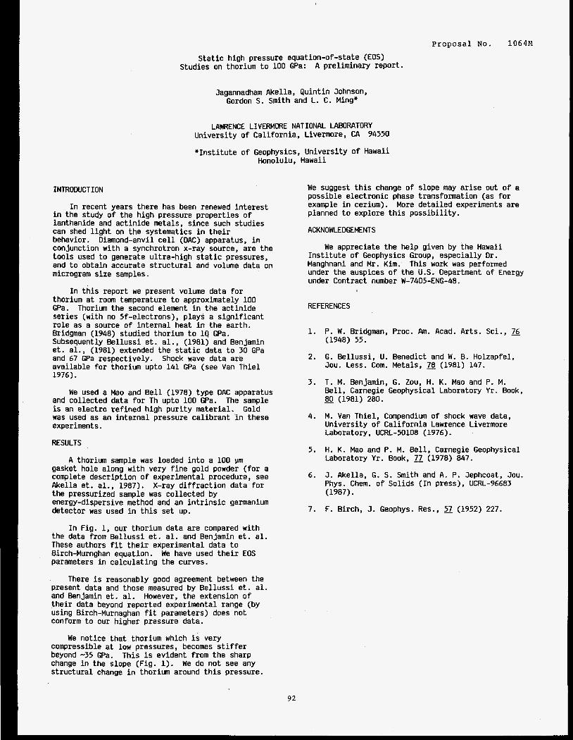
P r o p o s a l NO. 1064M
S t a t i c h igh pressure equation-of-state (EOS) Studies on thorium t o 100 GPa: A p re l im inary repor t .
Jagannadham Akella, Q u i n t i n Johnson, Gordon S. S m i t h and L. C. Ming"
LAWRENCE LIVERMORE NATIONAL LABORATORY Univers i ty o f Ca l i fo rn ia , Livermore, CA 94550
" I n s t i t u t e o f Geophysics, Un ivers i ty o f Hawaii Honolulu, Hawaii
INTRODUCTION
I n recent years there has been renewed i n t e r e s t i n the study o f t he h igh pressure proper t ies o f lanthanide and ac t i n ide metals, since such studies can shed l i g h t on the systematics i n t h e i r behavior. Diamond-anvil c e l l (DAC) apparatus, i n conjunction w i th a synchrotron x-ray source, are the too l s used t o generate u l t ra -h igh s t a t i c pressures, and t o obtain accurate s t r u c t u r a l and volume data on microgram s ize samples.
I n t h i s repor t we present volume data f o r thorium a t room temperature t o approximately 100 GPa. Thorium the second element i n the ac t i n ide ser ies (with no 5f-electrons), p lays a s i g n i f i c a n t r o l e as a source o f i n t e r n a l heat i n the earth. Bridgman (1948) studied thorium t o 19 GPa. Subsequently Be l l uss i e t . a l . , (1981) and Benjamin e t . al. , (1981) extended the s t a t i c data t o 30 GPa and 67 GPa respect ively. ava i lab le f o r thorium upto 141 GPa (see Van T h i e l 1976).
Shock wave data are
We used a Mao and B e l l (1978) type DAC apparatus and co l lec ted data f o r Th upto 100 GPa. The sample i s an e lec t ro re f i ned h igh p u r i t y mater ia l . was used as an i n t e r n a l pressure ca l i b ran t 'in these experiments.
Gold
RESULTS
A thorium sample was loaded i n t o a 100 gasket hole along with very f i n e gold powder ( f o r a complete descr ip t ion o f experimental procedure, see Akel la et . a l . , 1987). X-ray d i f f r a c t i o n data f o r the pressurized sample was co l l ec ted by energy-dispersive method and an i n t r i n s i c germanium detector was used i n t h i s set up.
I n Fig. 1, our thorium data are compared w i th the data from B e l l u s s i e t . a l . and Benjamin e t . a l . These authors f i t t h e i r experimental data t o Birch-Murnghan equation. Parameters i n ca l cu la t i ng the curves.
Present data and those measured by B e l l u s s i e t . a l . and Benjamin e t . a l . However, t he extension o f t h e i r data beyond reported experimental range (by using Birch-Murnaghan f i t parameters) does no t conform t o our higher pressure data.
compressible a t low pressures, becomes s t i f f e r beyond -35 GPa. This i s evident from the sharp change i n the slope (Fig. 1). We do no t see any s t r u c t u r a l change i n thor ium around t h i s pressure.
We have used t h e i r EOS
There i s reasonably good agreement between the
We not ice t h a t thorium which i s very
We suggest t h i s change o f slope may a r i se out o f a possible e lec t ron ic phase transformation (as f o r example i n cerium). More de ta i l ed experiments are planned t o explore t h i s p o s s i b i l i t y .
ACKNOWLEDGEMENTS
We appreciate the help given by the Hawaii I n s t i t u t e o f Geophysics Group, espec ia l l y Dr . Manghnani and Mr . Kim. under the auspices o f t h e U.S. Department o f Energy under Contract number W-7405-ENG-48.
This work was performed
REFERENCES
1.
2.
3.
4.
5.
6.
7.
P. W. Bridgman, Proc. Am. Acad. Arts. Sci., 76 (1948) 55.
G. Be l luss i , U. Benedict and W. B. Holzapfel, Jou. Less. Com. Metals, 2 (1981) 147.
T. M. Senjamin, G. Zou, H. K. Mao and P. M. Be l l , Carnegie Geophysical Laboratory Y r . Book, - 80 (1981) 280.
M. Van Thiel , Compendium o f shock wave data, Un ivers i ty o f C a l i f o r n i a Lawrence Livermore Laboratory, UCRL-50108 (1976).
H. K. Mao and P. M. Be l l , Carnegie Geophysical Laboratory Y r . Book, 77 (1978) 847.
J. Akel la, G. S. S m i t h and A. P. Jephcoat, Jou. Phys. Chem. o f So l ids ( In press), UCRL-96683 (1987).
F. Birch, J . Geophys. Res., 7 (1952) 227.
92
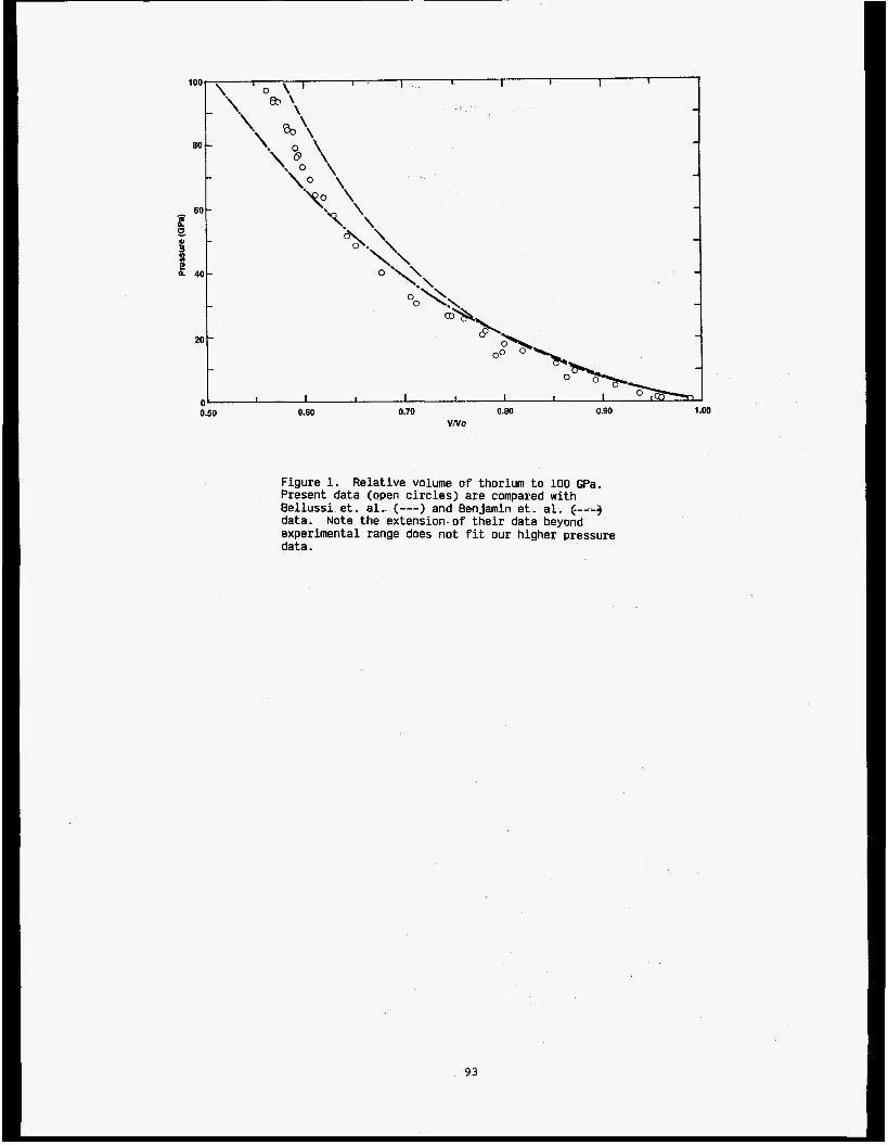
0 5 0 0.60 0.70 0.80
VNo 0.90 1 .w
Figure 1. Relative volume o f thorium to 100 GPa. Present data (open circles) are compared with Bellussi e t . a l . (---) and Benjamin e t . a l . (.---3 data. experimental range does not f i t our higher pressure data.
Note the extension o f their data beyond
93
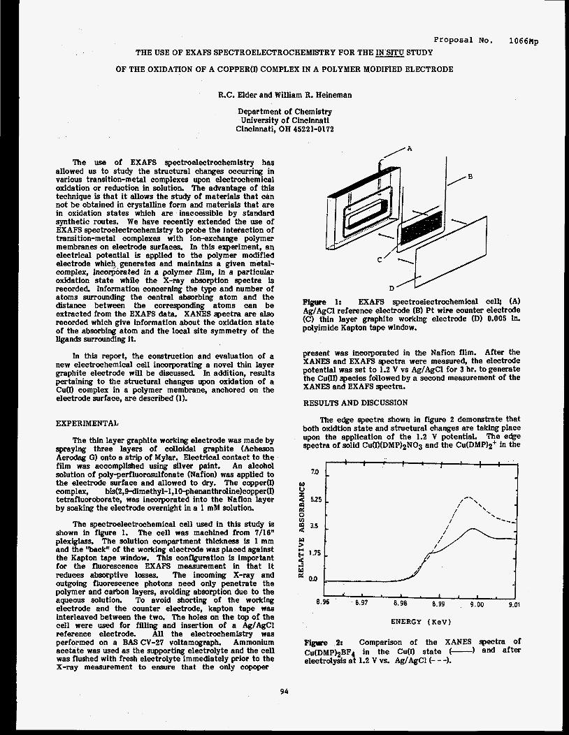
P r o p o s a l NO. 1066Np THE USE OF EXAFS SPECTROELECTROCHEMISTRY FOR THE IN SITU STUDY
OF THE OXIDATION OF A COPPER@ COMPLEX IN A POLYMER MODIFIED ELECTRODE
R.C. Elder and William R. Heineman
Department of Chemistry University of Cincinnati
Cincinnati, OH 45221-0172
The use of EXAFS spectroelectrochemistry has allowed us to study the structural changes occurring in various transition-metal complexes upon electrochemical oxidation or reduction in solution. The advantage of this technique is that i t allows the study of materials that can not be obtained in crystalline form and materials that are in oxidation states which are inaccessible by standard synthetic routes. We have recently extended the use of EXAFS spectroelectrochemistry to probe the interaction of transition-metal complexes with ion-exchange polymer membranes on electrode surfaces. In this experiment, an electrical potential is applied to the polymer modified electrode which, generates and maintains a given metal- complex, incowrated in a polymer film, in a particular oxidation state while the X-ray absorption spectra is recorded. Information concerning the type and number of atoms surrounding the central absorbing atom and the distance between the corresponding atoms can be extracted from the EXAFS data. XANES spectra are also recorded which give information about the oxidation state of the absorbing atom and the local site symmetry of the ligands surrounding it.
In this report, the construction and evaluation of a new electrochemical cell incorporating a novel thin layer graphite electrode will be discussed In addition, results pertaining to the structural changes upon oxidation of a Cum complex in a polymer membrane, anchored on the electrode surface, are described (1).
EXPERIMENTAL
The thin layer graphite working electrode was made by spraying three layers of colloidal graphite (Acheson Aerodag G) onto a strip of Mylar. Electrical contact to the film was accomplished using silver paint. An alcohol solution of poly-perfluorosulfonate (Nafion) was applied to the electrode surface and allowed to dry. The copper(I) complex, bis(2,9-dimethyl-l,lO-phenanthroline)copper(I) tetrafluoroborate, was incorporated into the Nafion layer by soaking the electrode overnight in a 1 mM solution.
The spectroelectrochemical cell used in this study is shown in figure 1. The cell was machined from 7/16" plexiglass. The solution compartment thickness is 1 mm and the %abackw of the working electrode was placed against the Kapton tape window. This configuration is important for the fluorescence EXAFS measurement in that i t reduces absorptive losses. The incoming X-ray and outgoing fluorescence photons need only penetrate the polymer and carbon layers, avoiding absorption due to the aqueous solution. To avoid shorting of the working electrode and the counter electrode, kapton tape was interleaved between the two. The holes on the top of the cell were used for filling and insertion of a Ag/AgCl reference electrode. All the electrochemistry was performed on a BAS CV-27 voltamograph. Ammonium acetate was used as the supporting electrolyte and the cell was flushed with fresh electrolyte immediately prior to the X-ray measurement to ensure that the only copoper
Figure 1: EXAFS spectroelectrochemical cell; (A) Ag/AgCI reference electrode (B) Pt wire counter electrode (C) thin layer graphite working electrode (D) 0.005 in. polyimide Kapton tape window.
present was incorporated in the Nafion film. After the XANES and EXAFS spectra were measured, the electrode potential was set to 1.2 V vs Ag/AgCl for 3 hr. to generate the Guan species followed by a second measurement of the XANES and EXAFS spectra.
RESULTS AND DISCUSSION
The edge spectra shown in figure 2 demonstrate that both oxidtion state and structural changes are taking place upon the application of the 1.2 V potential. The eee spectra of solid Cu(I)(DMP)2N03 and the Cu(DMP)2+ in the
I I I , t
I I
7.0 1
I d , 1 I I I
8.97 8.98 8.99 9.00 9.01 8.96 '
ENERGY (KeV)
pisure 2: Comparison of the XANES spectra Of
CU(DMP)~BF~ in the CU(I) state (-1 and after electrolysis a t 1.2 V vs. Ag/AgCl(- - -1.
94
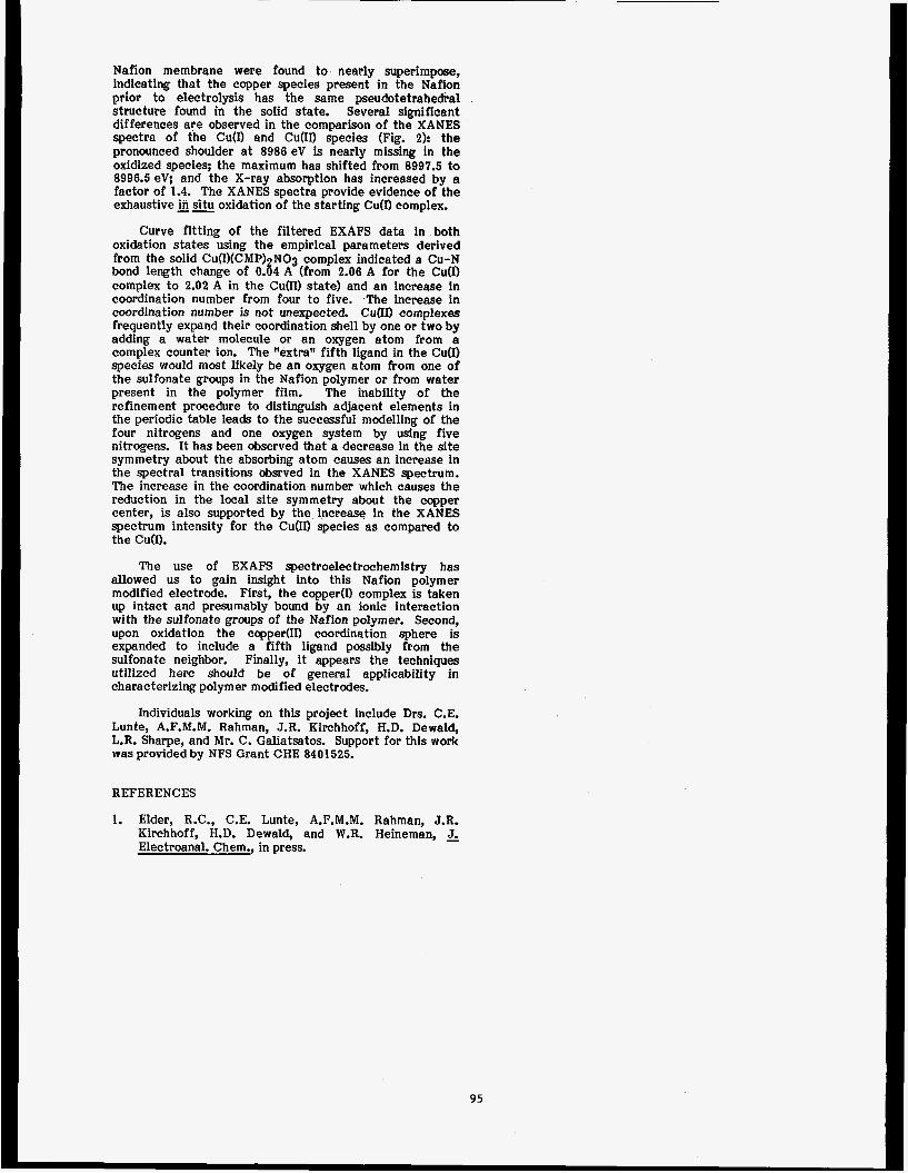
Nafion membrane were found to nearly superimpose, indicating that the copper species present in the Nafion prior to electrolysis has the same pseudotetrahedral structure found in the solid state. Several significant differences are observed in the comparison of the XANES spectra of the CdI) and CuQD species (Fig. 2): the pronounced shoulder at 8986 eV is nearly missing in the oxidized species; the maximum has shifted from 8997.5 to 8996.5 eV; and the X-ray absorption has increased by a factor of 1.4. The XANES spectra provide evidence of the exhaustive in situ oxidation of the starting Cu(D complex.
Curve fitting of the filtered EXAFS data in both oxidation states using the empirical parameters derived from the solid Cu(I)(CMP) NO3 complex indicated a Cu-N bond length change of 0.84 A (from 2.06 A for the Cu(1) complex to 2.02 A in the Cu(I1) state) and an increase in coordination number from four to five. The increase in coordination number is not unexpected. C u O complexes frequently expand their coordination shell by one or two by adding a water molecule or an oxygen atom from a complex counter ion. The ‘Iextra” fifth ligand in the Cu(1) species would most likely be an oxygen atom from one of the sulfonate groups in the Nafion polymer or from water present in the polymer film. The inability of the refinement procedure to distinguish adjacent elements in the periodic table leads to the successful modelling of the four nitrogens and one oxygen system by using five nitrogens. It has been observed that a decrease in the site symmetry about the absorbing atom causes an increase in the spectral transitions obsrved in the XANES spectrum. The increase in the coordination number which causes the reduction in the local site symmetry about the copper center, is also supported by the increase in the XANES spectrum intensity for the CuOI) species as compared to the Cu(I).
The use of EXAFS spectroelectrochemistry has allowed us to gain insight into this Nafion polymer modified electrode. First, the copper(1) complex is taken up intact and presumably bound by an ionic interaction with the sulfonate groups of the Nafion polymer. Second, upon oxidation the copper(1I) coordination sphere is expanded to include a fifth ligand possibly from the sulfonate neighbor. Finally, i t appears the techniques utilized here should be of general applicability in characterizing polymer modified electrodes.
Individuals working on this project include Drs. C.E. Lunte, A.F.M.M. Rahman, J.R. Hirchhoff, H.D. Dewald, L.R. Sharpe, and Mr. C. Galiatsatos. Support for this work was provided by NFS Grant CHE 8401525.
REFERENCES
1. Elder, R.C., C.E. Lunte, A.F.M.M. Rahman, J.R. Kirchhoff, H.D. Dewald, and W.R. Heineman, J- Electroanal. Chem., in press.
95

GLANCING ANGLE EXAFS STUDIES ON Cu-A1 INTERFACES
S. M. Heald, and H. Chen
Proposal No. 1072M
Division of Mater ia ls Science, Department of Applied Science, Brookhaven National Laboratory Upton, NY 11973
INTRODUCTION
Cu-A1 th in f i lm in te r faces have been s tudied by many techniques. CuA12 is observed a s the f i r s t compound to form a t the in te r face by x-ray d i f f r a c t i o n and RBS, although the reso lu t ion was n o t much b e t t e r than 100 A.1 Photoemission measurements observed intermixing a t monolayer coverages, bu t could n o t determine composi t ions. Theref ore , the de tec t ion of i n i t i a l i n t e r f a c e reac t ions requi res a depth s e n s i t i v e t o o l which can resolve loca l atomic struc- tures. Such a detect ion is made possible without the need of an UHV environment, v i a the a v a i l a b i l i t y of synchrotron x-rays. Fluorescence EXAFS from an i n t e r f a c e region alone can be obtained by using glancing angle inc ident x-ray^.^ To demonstrate the power of the glancing angle EXAFS technique, Cu-A1 th in f i lms prepared under var ious condi t ions were studied and compared.
RESULTS AND DISCUSSIONS
Thin f i lms of A1(-600 A)/Cu(-1000 A) were evaporated on g l a s s subs t ra tes a t near room temperatures (RT) and low temperatures (LT). During the beam time received i n December 87 i n SSRL, the LT samples were measured and were compared to the RT samples previ- ously measured a t CHESS. EXAFS data show t h a t the extent of the i n i t i a l reac t ion is about equal f o r LT and RT. The temperature of the subs t ra te might a f f e c t the gra in s ize of f i lms , and hence the r e a c t i v i t y of the in te r face . Electron microscopy s tudies a r e planned to determine the gra in s izes . The conclusion one can draw is t h a t the microscopic s t r u c t u r e s a t the i n i t i a l reacted region f o r the f i lms prepared a t d i f f e r e n t temperatures a r e the same t o the s e n s i t i v i t y of t h i s technique.
Further s t u d i e s including oxygen exposures and annealing were performed a t CHESS Later. More data ana lys i s is required to c l a r i f y the r e s u l t s .
ACKNOWLEDGEMENTS
This research w a s performed under the auspices of the U.S. Department of Energy, Division of Mater ia ls Sciences, Off ice of Basic Energy Sciences, under Contract No. DE-AC02-76CHOOOl6
REFERENCES
1. R. A. Hamm and 3 . M. Vandenberg, J. Appl. Phys.
2. V. D i . Castro and G. Po lzonet t i , Surf. Sc i . E, 3. S. M. Heald, e t a l . , to be published.
56, No. 2 , 293 (1984).
383 (1987).
-
96

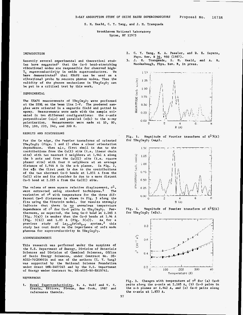
X-RAY ABSORPTION STUDY OF OXIDE BASED SUPERCONDUCTORS
S. M. Heald, C. Y. Yang, and J. M. Tranquada
Brookhaven National Laboratory Upton, NY 11973
INTRODUCTION
Recently several experimental and theoretical stud- i e s have suggested1 that the Cu-0 bond-stretching vibrational modes a re responsible fo r observed high- Tc superconductivity in oxide superconductors. We have demonstrated2 that EXAFS can be used as a vibrat ional probe to measure phonon modes. Thus the va l id i ty of the phonon mechanisms in YBa2Cu307 can be put to a c r i t i c a l t e s t by th i s work.
EXPERIMENTAL
The EXAFS measurements of YBa2Cu307 were performed a t the SSRL on the beam l i n e I-V. The powdered sam- p l e s were oriented in a magnetic f i e ld and potted i n epoxy. Measurements were made with the sample ori- ented in two different configurations: the c-axis perpendicular (eLc) and para l le l (ellc) to the x-ray polarization. Measurements were made a t 10, 80, 150, 180, 220, 240, and 300 K.
RESULTS AND DISCUSSIONS
For the Cu edge, the Fourier transforms of oriented YBa2Cu307 (Figs. 1 and 2) show a clear orientation dependence. When e l c , f i r s t she l l i s due to the contributions from the Cu(1) s i t e ( i .e. l inear chain s i t e ) with two nearest 0 neighbors a t 1.941 A along the b axis and from the CU(II) s i te (i .e. square planar s i t e ) with four 0 neighbors a t an average distance of 1.944 A i n the a-b planes. In Fig. 2 , for e l c the f i r s t peak is due to the contributions of the two shortest Cu-0 bonds a t 1.851 A from the Cu(1) s i te and i t s shoulder i s due to a more d is tan t Cu-0 bond a t 2.285 A from the Cu(I1) s i t e .
The values of mean square re la t ive displacement, u 2 , were extracted using standard techniques. The var ia t ion of u2 with temperature for the three dif- ferent Cu-0 distances is shown i n Fig. 3 along the f i t s using the Einstein model. Our resu l t s strongly indicate that there is anomalous temperature dependence of u2 for Cu-0 pairs i n YBa2Cu307. Fur- thermore, as expected, the long Cu-0 bond a t 2.285 A (Fig. 3(a)) is weaker than the Cu-0 bonds a t 1.94 A (Fig. 3(b)) and 1.85 A (Fig. 3(c)) . A s f o r a previous study of La2-xSrCu04,y system ,3 th i s study has cas t doubt on the importance of s o f t mode phonons fo r superconductivity in YBa2Cu307.
ACKNOWLEDGEMENTS
This research was performed under the auspices of the U.S. Department of Energy, Division of Materials Sciences and Division of Chemical Sciences, Office of Basic Energy Sciences, under Contract No. DE- AC02-76CHOOOl6 and one of the authors (C. Y. Yang) w a s supported by the National Science Foundation under Grant DMR-8407265 and by the U.S. Department of Energy under Contract No. DE-AC05-80-ER10742.
REFERENCES
1. Novel Superconductivity, S. A. Wolf and V. 2. Kresin, Editors, Plenum, New -York, 1987 and references therein.
Proposal No. 1073M
2. C. Y. Yang, M. A. Paesler, and D. E. Sayers,
3. J. M. Tranquada, S. M. Heald, and A. R. Phys. Rev. B 36, 980 (1987).
Moodenbaugh, Phys. Rev. B, i n press.
9 1'5
2 w 0 75
- r d
100
I
P 3
3
5 0.50
0 2 5
0 00 0 3 4 6 a
R ( A )
Fig. 1. Magnitude of Fourier transform of K2%(K) for YBa2Cu307 (elc).
1 5 0 L / " " / ~ " '
R ( A )
Fig. 2. Magnitude of Fourier transform of K2x(K) fo r YBa2Cu307 (ellc).
0 8
0 6
0.4
1- i 0 2 1 ' ' r I m z ' l I * (
J
0 100 200 300 40
Temperature (K)
Fig. 3. Changes with temperature of a2 f o r (a) Cu-0 p a i r s along the c-axis a t 2.285 A, (b) Cu-0 p a i r s i n the a-b planes a t 1.942 A, and (c) Cu-0 pairs along the c-axis a t 1.853 A.
97

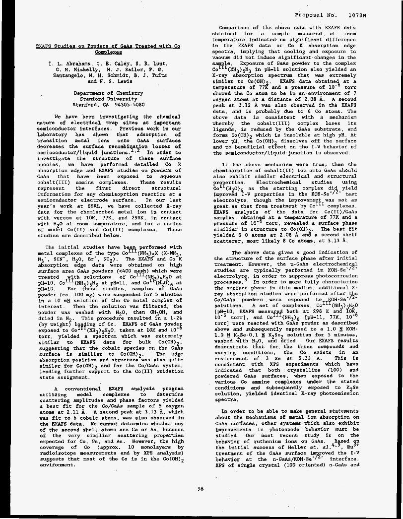
SQ w th C ComDlexes
I. L. Abrahams, C. E. Caley, S. R. Lunt, G. M. Miskelly, M. J. Sailor, P. G. Santangelo, M. H. Schmidt, B. J. Tufts
and N. S. Lewis
Department of Chemistry Stanford University
Stanford, CA 94305-5080
We have been investigating the chemical nature of electrical trap sites at important semiconductor interfaces. Previous work in our laboratory has shown that adsorption of transition metal ions onto GaAs surfaces decreases the surface recombi ation losses of semiconductor/liquid junctions. '12 In order to investigate the structure of these surface species, we have performed detailed Co K absorption edge and EXAFS studies on powders of GaAs that have been exposed to aqueous cobalt(II1) ammine complexes. These results represent the first direct structural information for any chemisoprtion reaction at a semiconductor electrode surface. In our last year's work at SSRL, we have collected X-ray data for the chemisorbed metal ion in contact with vacuum at 10K, 7JK, and 298K, in contact with H20 at room temperature, and for a series of model Co(I1) and Co(II1) complexes. These studies are described below.
The initial studies have bee performed with metal complexes of the type cO1InI(NH3)5x (X-NH3, N3-, SCN-, H20, Br-, NO2). The EXAFS and Co K absorption edge data were obtained on high surface area GaAs powders (<400 mesh) which were treated with solutions of ColI1 ) H 0 at pH-10, Co111(NH3)5N3 at pH-11, and :0'~(2O36 at pH-10. For these studies, samples 0% GaAs powder (ca. 100 mg) were suspended for 5 minutes in a 10 solution of the Co metal complex of interest. Then the solution was filtered, the powder was washed with H20, then CH30H, and dried in N2. This procedure resulted in a 1-2% (by weight) loading of Co. EXAFS of GaAs powde exposed to C O ~ ~ ~ ( N H ~ ) ~ H ~ O , taken at 10K and 10- torr, yielded a spectrum which was extremely similar to EXAFS data for bulk Co(OH)*, suggesting that the cobalt species on the GaAs surface is similiar to CO(OH)~. The edge absorption position and structure was also quite similar for Co(OH)* and for the Co/GaAs system, lending further support to the Co(I1) oxidation state assignment.
A conventional EXAFS analysis program utilizing model complexes to determine scattering amplitudes and phase factors yielded a best fit for the Co/GaAs sample of 5 oxygen atoms at 2.11 A. A second peak at 3.13 A, which was fit to 6 cobalt atoms, was also observed in the EXAFS data. We cannot determine whether any of the second shell atoms are Ga or As, because of the very similiar scattering properties expected for Co, Ga, and As. However, the high coverage of Co (approx. 10 monolayers by radioisotope measurements and by XPS analysis) suggests that most of the Co is in the CO(OH)~ environment.
98
Proposal No. 1078M
Comparison of the above data with EXAFS data obtained for a sample measured at room temperature indicated no significant difference in the EXAFS data or Co K absorption edge spectra, implying that cooling and exposure to vacuum did not induce significant changes in the Sam le. Exposure of GaAs powder to the complex Co1'I(NH3)5N3 in PH-11 solution also yielded an X-ray absorption spectrum that was extremely similar to Co(OH)2. EXAFS data obtained at a temperature of 77K and a pressure of torr showed the Co atom to be in an environment of 7 oxygen atoms at a distance of 2.08 A. A second peak at 3.12 A was also observed in the EXAFS data, and is probably due to 6 Co atoms. The above data is consistent with a mechanism whereby the cobalt(II1) complex loses its ligands, is reduced by the GaAs substrate, and forms CO(OH)~ which is insoluble at high pH. At lower pH, the Co(0H) dissolves off the surface and no beneficial effect on the I-V behavior of the semiconductor/liquid junction is observed.
If the above mechanism were true, then the chemisorption of cobalt(I1) ion onto GaAs should also exhibit similar electrical and structural pro erties. Electrochemical studies using
improved I-V properties in the KOH-Se-/2- test electrolyte, though the improvement was not as great as that from treatment by ColI1 complexes. EXAFS analysis of the data for Co(II)/GaAs samples, obtain d at a temperature of 77K and a pressure of torr, revealed a surface phase similiar in structure to CO(OH)~. The best fit yielded 6 0 atoms at 2.08 A and a second shell scatterer, most likely 8 co atoms, at 3.13 A.
Co I P (H20)6 as the starting complex did yield
The above data gives a good indication of the structure of the surface phase after initial treatment. However, the n-GaAs electrochemic 1 studies are typically performed in KOH-Se-/'- electrolyt in order to suppress photocorrosion processes ." In order to more fully characterize the surface phase in this medium, additional X- ray absorption studies were performed after t e Co/GaAs powders were exposed to KOH-Se-P- solutions. A set of complexes, c~III(NH H o [pH-10, EXAFS measured both at 298 K anc?'?Og
torr], and Co1I1(NH3)6 [pH-11, 77K, lo-6 torr] were reacted with GaAs powder as described above and subsequently exposed to a 1.0 KOH- 1.0 K2Se-0.1 K2Se2 solution for 5 minutes, washed with H 0, and dried. Our EXAFS results demonstrate tiat for the three compounds and varying conditions, the Co exists in an environment of 3 Se at 2.33 A. This is consistent with XPS experiments which have indicated that both crystalline (100) and powdered GaAs surfaces, when exposed to the various Co ammine complexes under the stated conditions and subsequently exposed to K2Se solution, yielded identical X-ray photoemission spectra.
In order to be able to make general statements about the mechanisms of metal ion absorption on GaAs surfaces, other systems which also exhibit Improvements in photoanode behavior must be studied. Our most recent study is on the behavior of ruthenium ions on GaAs. B sed n the initial success of Heller et. a1.4.4, Ru treatment of the GaAs surface im oved the I-V behavior at the n-GaAs/KOH-Se- F- interface. XPS of single crystal (100 oriented) n-GaAs and
5+
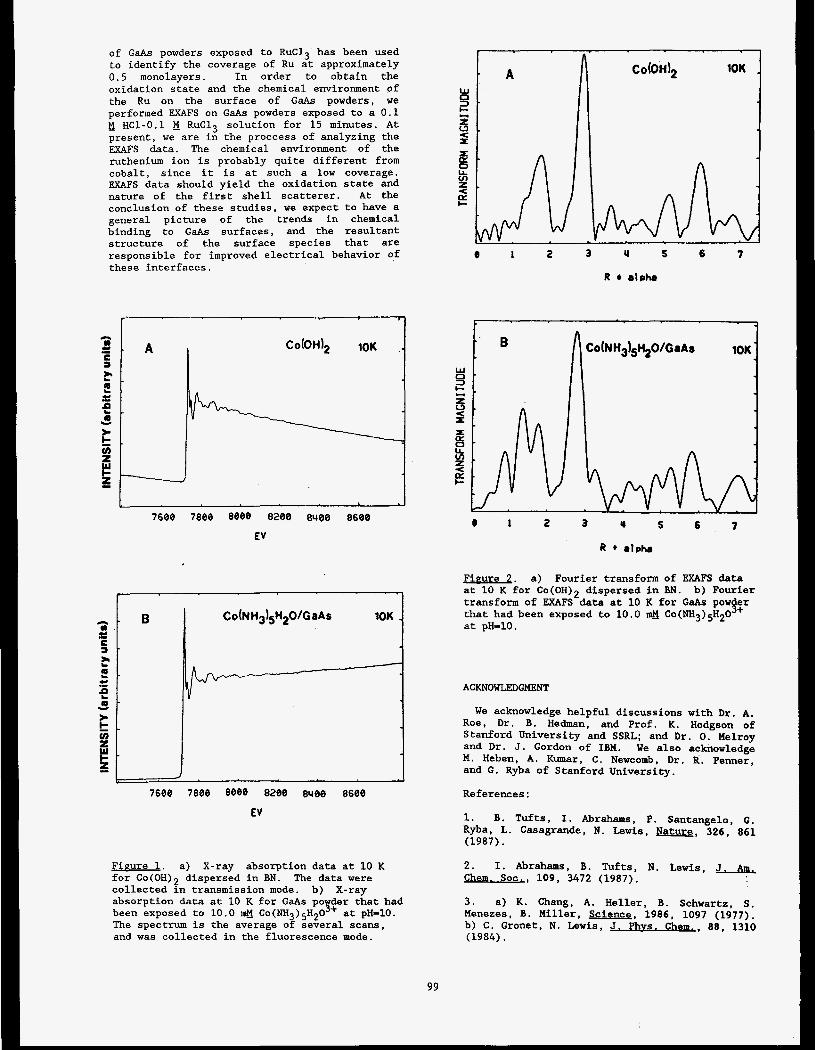
of GaAs powders exposed to RuC13 has been used to identify the coverage of Ru at approximately 0.5 monolayers. In order to obtain the oxidation state and the chemical environment of the Ru on the surface of GaAs powders, we performed FXAFS on GaAs powders exposed to a 0.1 E HC1-0.1 E RuC13 solution for 15 minutes. At present, we are in the proccess of analyzing the FXAFS data. The chemical environment of the ruthenium ion is probably quite different from cobalt, since it is at such a low coverage. EXAFS data should yield the oxidation state and nature of the first shell scatterer. At the conclusion of these studies, we expect to have a general picture of the trends in chemical binding to GaAs surfaces, and the resultant structure of the surface species that are responsible for improved electrical behavior of these interfaces.
- ii A
3 t *.
v)
c 1
EV
7668 7866 8088 8288 8400 8668
EV
Figure 1. a) X-ray absorption data at 10 K for CO(OH)~ dispersed in BN. collected in transmission mode. b) X-ray absorption data at 10 K for GaAs PO der that had been exposed to 10.0 The spectrum is the average of several scans, and was collected in the fluorescence mode.
The data were
CO(NH~)~H~O'+ at PH-10.
e 1 2 3 Y 5
R d ~ h e
6 7
c A
2 3 Y S 6 7
Firrure 2. a) Fourier transform of EXAFS data at 10 K for CO(OH)~ dispersed in BN. transform of EXAFS data at 10 K for GaAs pow that had been exposed to 10.0 a Co(NH3)gH2O at pH-10.
b) Fourier
!F-
ACKNOWLEDGMENT
We acknowledge helpful discussions with Dr. A. Roe, Dr. B. Hedman, and Prof. K. Hodgson of Stanford University and SSRL; and Dr. 0. Melroy and Dr. J. Gordon of IBM. We also acknowledge M. Heben, A. Kumar, C. Newcomb, Dr. R. Penner, and G. Ryba of Stanford University.
References:
1. B. Tufts, I. Abrahams, P. Santangelo, G. Ryba, L. Casagrande, N. Lewis, Pature, 326, 861 (1987).
2. Chem. SOC,, 109, 3472 (1987).
I. Abrahams, B. Tufts, N. Lewis, ,I. +
3 . a) K. Chang, A. Heller, B. Schwartz, S. Menezes, B. Miller, Science, 1986, 1097 (1977). b) C. Gronet, N. Lewis, J. Phvs. Chem,, 88, 1310 (1984).
99

4. a) W. Johnston, H. Leamy, B. Parkinson, A . Heller, B. Miller, J. Electrochem. SOC,, 127, 90 (1980). b) A. Heller, B. Miller, S . Chu, Y. Lee, Proc. IEEE Photovolt. Soec. Conf., 14, 1366 (1980).
5 . a) B. Parkinson, A. Heller, B. Miller, Appl. Phys. Lett., 33, 521 (1978). b) B. Parkinson, A. Heller, B. Miller, J. Electrochem. SOC., 126, 954 (1979). c) R. Nelson, J. Williams, H. Leamy, B. Miller, B. Parkinson, A . Heller, Ap~l. Phvs. Lett,, 36, 76 (1980).
100


Proposal NO. 1 0 7 9 ~
EXAFS OF YBa2CU307-8 AND GdBa2CU307-8 SUPERCONDUCTORS
J. B. Boyce Xerox Palo Alto Research Center, Palo Alto, CA 94304, U.SA
F. C. Brid es Physics Department, University of California,!anta Cruz, Santa Cruz, CA 95059, U3.A
T.,Claeson Physics Department, Chalmers University of Technology, 41296 Gothenburg, Sweden
T. H. Geballe and R. S. Howland Department of Applied Physics, Stanford University, Stanford, CA 94305, U.S.A
The local structure and the near-neighbor pair vibrations in two new ceramic superconductors of the 1:23 type have been studied. X-ray absorption measurements showed essentially no change in the near-edge and extended fine structure (EXAFS) over a temperature range of 4.2 to 688 K for YBaZCu307-(i and GdBaZCu30,;l_s. The results indicate that the two compounds are very similar to each other in local bonding and electronic structure, and they remain unchanged over the large temperature range, from far below to well above T, The EXAFS structural results agree well with the accepted structure modeled from neutron diffraction data. Alternate structures which have been proposed recently are not consistent with the data.
The absorption edge of Cu in a superconductor is broad, covering an energy range between the edges of Cu in CuO (with a formal valence of 11) and KCuOZ (with a formal valence of 111). An extra peak appears in the near-edge spectrum as oxygen is pumped out from the superconductor. It coincides with a peak in the spectrum for Cu,O and is associated with the breaking up of 1-d Cu-0 chains in the 1:23 compound
TABLE 1. Number and distances of near neighbors in Y-Ba-Cu-0 as determined from fits to EXAFS data for three samples. [The number was kept fiied for Cu-0, as indicated by (0.3 The characteristic Einstein temperatures are obtained from the temperature broadening of the distances.
cu-0 213 ( f ) 1.8820.02 10/3 ( f ) 1.95-cO.01 540+50 2/3 ( f ) 2.3220.05
Y-0 8-cl 2.3920.01 380250
cu-CWY 260+50
Ba-0 310250
From the temperature dependence of the Debye-Waller-like broadening of near-neighbor distances, it is possible to extract information on bond vibrations. The characteristic vibration frequencies of the near-neighbor atoms show that the 2-d Cu-0 planes and the 1-d Cu-0 chains form the most rigid parts of the structure. These oscillator frequencies are consistent with those for some modes seen by inelastic neutron diffraction as well as by infrared and Raman spectroscopy. The characteristic Einstein temperatures for the Cu-0 modes fall in the range of 500-600 K, a value which is lower than the desired value of the breathing modes to describe the superconductivity with a classical electron-phonon coupling.
No evidence of phonon softening is apparent in any of the phonon modes studied over a large temperature interval.
0.02
h
3 p 0.01
c 4
0
I I I
0 200 400 600 800
1 (U)
Fig. 1. The change in the square of the width of the pair distribution functions for Cu-0, Y-0, and Ba-0 in superconducting Y-Ba-Cu-0 as a function of temperature. The solid lines are fits of an Einstein oscillator model to the data. No phonon softening is observed.
10 I

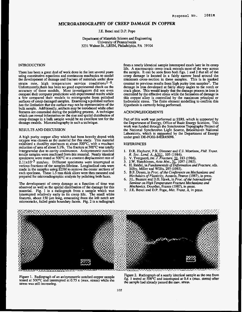
Proposal No. 108lM
MICRORADIOGRAPHY OF CREEP DAMAGE IN COPPER
J.E. Benci and D.P. Pope
Department of Materials Science and Engineering University of Pennsylvania
3231 Walnut St., LRSM, Philadelphia, PA 19104
INTRODUCTION
There has been a great deal of work done in the last several years using constitutive equations and continuum mechanics to model the development of damage and fracture of materials under slow strain rate, high temperature service conditionsl-6. Unfortunately,there has been no good experimental check on the accuracy of these models. Most investigators did not even compare their computer predictions with experimental results while a few compared their models to micrographs from polished surfaces of creep damaged samples, Examining a polished surface has the limitation that the surface may not be representative of the bulk sample. Additionally, artifacts may be introduced while other features are concealed during the polishing process. A technique which can reveal information on the size and spatial distribution of creep damage in a bulk sample would be an excellent test for the damage models. Microradiography is such a technique.
RESULTS AND DISCUSSION
A high purity copper alloy which had been heavily doped with oxygen was chosen as the material for this study. This material exhibited a ductility minimum at about 500"C, with a resultant reduction of area of about 3.1%. The fracture at 500°C was totally intergranular due to cavity coalescence. Axisymmetric notched tensile samples were machined from this material. Nearly identical specimens were tested at 500°C at a constant displacement rate of 2.1 1x10⁻⁵ mm/sec. Different specimens were interrupted at various fractions of the samples liietime. Longitudinal cuts were made in the samples using EDM to remove the center sections of each specimen. These 1.5 mm thick slices were then mounted and prepared for microradiographic analysis by polishing both faces.
The development of creep damage as a function of time was observed as well as the spatial distribution of the damage for this material. Fig. 1 is a radiograph from a sample which was interrupted relatively early in its creep life. The small white features, about 150 pm long, emanating from the left notch are microcracks, failed grain boundary facets. Fig. 2 is a radiograph
from a nearly identical sample intempted much later in its creep life. A macroscopic creep crack extends most of the way across the sample. It can be seen from both figs. 1 and 2 that all of the creep damage is located in a fairly narrow band around the minimum cross-section in these samples. This is in marked contrast to previous results from-fhgh purity iron samples7. The damage in irbn developed at fairly sharp angles to the notch or crack plane. This would imply that the damage process in iron is controlled by the effective stress while the formation of damage in this copper alloy is controlled by the maximum principal or hydrostatic stress. The finite element modelling to confum this hypothesis is currently being performed.
ACKNOWLWEMENTS
Part of this work was performed at SSRL which is supported by the Department of Energy, Office of Basic Energy Sciences. This work was funded through the Synchrotron Topography Project of the National Synchrotron Light Source, Bmkhaven National Laboratory, which is supported by the Department of Energy under grant DE-FGO2-84ER45098.
REFERENCES
1. D.R. Hayhurst, P.R. Dimmer and C.J. Morrison, Phil. Trans. R . SOC. Lond. A, A2.l. 103 (1984).
2. V. Tvergaard, Znt. J. Fracture, 3, 183 (1986). 3. J.W. Hutchinson, Acta Met., a, 1097 (1983). 4. H. Reidel, in Fundamentals of Deformation and Fracture, eds.
Bilby, Miller and Willis, 293 (1985). 5. B.F. Dyson, in Proc. of the Conference on Mechanisms and
Mechanics of Plasticity, Aussois, France (1987), in press. 6. T.L. Bassani and D.E. Hawk, in Proc. ofrhe International
Seminur on High Temperature Fructure Mechanisms and Mechanics, Dourdan, France (1987), in press.
7. J.E. Benci and D.P. Pope, Met. Trans. A, in press.
Figure 1. Radiograph of an axisymmetric notched copper sample tested at 500°C and interrupted at 0.75 x (max. stress) while the stress was still increasing.
Figure 2. Radiograph of a nearly identical sample as the one from fig. 1 tested at 500°C and intempted at 0.4 x (max. stress) after the sample had already passed the max. stress.
102

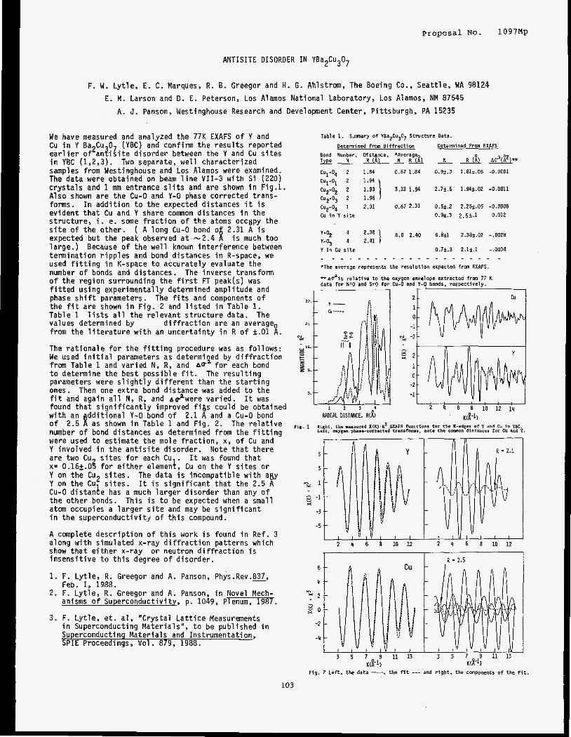
Proposal No. 1097Mp
ANTISITE DISORDER I N YBa2Cu307
F. W. L y t l e , E. C. Marques, R . 6. Greegor and H. 6. Ahlstrom, The Boeing Co., Sea t t l e , WA 98124
A. J . Panson, Westinghouse Research and Development Center, P i t tsburgh, PA 15235 E. M. Larson and D. E. Peterson, Los Alamos Nat ional Laboratory, Los Alamos, NM 87545
We have measured and analyzed the 77K EXAFS o f Y and Cu i n Y Ba Cu 0 (YBC) and conf i rm t h e r e s u l t s repor ted e a r l i e r of2an?i;ite d i so rde r between the Y and Cu s i t e s i n YBC (1,2,3). Two separate, w e l l character ized samples from Westinghouse and Los Alamos were examined. The data were obtained on beam l i n e V I I -3 w i t h S i (220) c r y s t a l s and 1 mm entrance s l i t s and are shown i n Fig.1. Also shown a re t h e Cu-0 and Y-0 phase co r rec ted t rans- forms. I n a d d i t i o n t o t h e expected distances i t i s ev ident t h a t Cu and Y share common distances i n the s t ruc tu re , i. e. some f r a c t i o n o f t h e atoms occgpy t h e s i t e o f t he o the r . ( A l ong Cu-0 bond o 2.31 A i s expected bu t the peak observed a t -2.4 i s much too large. ) Because o f t h e w e l l known in te r fe rence between te rm ina t ion r i p p l e s and bond distances i n R-space, we used f i t t i n g i n K-space t o accurate ly evaluate the number o f bonds and d is tances. The inverse transform o f t h e reg ion surrounding t h e f i r s t FT peak(s) was f i t t e d us ing exper imenta l ly determined ampli tude and phase s h i f t parameters. The f i t s and components o f t h e fit a re shown i n F ig . 2 and l i s t e d i n Table 1. Table 1 l i s t s a l l t he re levan t s t r u c t u r e data. The values determined by from t h e l i t e r a t u r e w i t h an unce r ta in t y i n R o f f.O1 A.
The r a t i o n a l e f o r t h e f i t t i n g procedure was as fo l l ows : We used i n i t i a l parameters as determined by d i f f r a c t i o n from Table 1 and va r ied N, R, and A@' f o r each bond t o determine t h e best poss ib le f it. The r e s u l t i n g parameters were s l i g h t l y d i f f e r e n t than t h e s t a r t i n g ones. Then one e x t r a bond d is tance was added t o t h e f i t and again a l l N, R, and aF'were var ied. I t was found t h a t s i g n i f i c a n t l y improved f i & s could be obtained w i t h an d d i t i o n a l Y-0 bond o f 2.1 A and a Cu-0 bond o f 2.5 f as shown i n Table 1 and F ig . 2. The r e l a t i v e number o f bond distances as determined from the f i t t i n g were used t o estimate the mole f r a c t i o n , x, o f Cu and Y invo lved i n t h e a n t i s i t e d isorder . Note t h a t t h e r e are two Cu s i t e s f o r each Cu . It was found t h a t x= 0.165.08 f o r e i t h e r elemenk, Cu on the Y s i t e s o r Y on the Cu2 s i t e s . The data i s incompatible w i t h ap,y Y on t h e Cu s i t e s . I t i s s i g n i f i c a n t t h a t the 2.5 A Cu-0 d is tange has a much l a r g e r d i so rde r than any o f t he other bonds. This i s t o be expected when a small atom occupies a l a r g e r s i t e and may be s i g n i f i c a n t i n the superconduct iv i ty o f t h i s compound.
d i f f r a c t i o n are an average,
A complete d e s c r i p t i o n o f t h i s work i s found i n Ref. 3 along w i t h simulated x-ray d i f f r a c t i o n pat terns which show t h a t e i t h e r x-ray o r neutron d i f f r a c t i o n i s i n s e n s i t i v e t o t h i s degree o f d i so rde r .
1. F. L y t l e , R . Greegor and A. Panson, Phys.Rev.K,
2. F. L y t l e , R. Greegor and A. Panson, i n Novel Mech-
3. F. L y t l e , e t . a l , "Crysta l L a t t i c e Measurements
Feb. 1, 1988.
anisms o f Superconductivi ty, p. 1049, Plenum, 1987.
i n Superconductinq Ma te r ia l s " , t o be Dublished i n Superconducting Ma te r ia l s and- Instrumentation, S P I E Proceedings, Vol . 879, 1988.
Table 1. Sumnary of YBa2Cu307 Structure Data.
Detennined Frm Diffraction Determined Frm EXAFS
Bond Number, Distgnce, *Averageo Type N R ( A ) N R no'(;')**
0.67 1.84 0 .9 t .3 1.812.05 -0.0001 Cul-O4 2 1.84
Cul-0l 2 1.94
Cu2-02 2 1.93
CuZ-03 2 1.96
Cu2-04 1 2.31
Cu i n Y s i t e
Y-O2 4 2.38
Y-Oj 4 2.41
Y i n Cu s i t e
3.33 1.94 2 . 7 i . 5 1.94t.02 -0.0011
0.67 2.31 0.5k.2 2.25t.05 -0.0006
0 . 9 ~ 3 2 . 5 i . 1 0.012
8 .0 2.40 6.8i1 2.38t.02 -.OOZE
0.7i .3 2 . 1 i . l -.0034
_ _ _ _ _ _ _ _ - - _ _ _ _ _ _ _ - _ _ _ *The average represents the resolutlon expected f r a EXAFS.
* a d l s relative to the oxygen envelope extracted from 77 K data for NiO and SrO for Cu-0 and Y-0 bonds, respectively.
r :-
RADIAL DISTANCE, R d )
g 2 1 0
-2
~~ -1
cu
2 4 6 8 10 12 19
K(R-1)
5
3
-22 1
8 -1 x
-3
-5
2 4 6 8 1 0 1 2 2 4 6 8 1 0 1 2
1 R = 2.5
Fig. 2 Left. the data -, the f it --- and right, the canponents of the f i t .
103


RARE EARTH L-EDGE SPECTRA I N Y1,xMxBa2Cu3Q7
1 1 1 2 3 3 F, Lytle, R , Greegor, E. Marques, E, barson, J . Wong and C , V i o l e t 1. The Boeing Co., Seat t le , WA 98124 2. LANL, Los Alamos, NM 87545 3 . LLNL, Livermore, CA 94550
It has been observed t h a t s u b s t i t u t i o n o f t r i v a l e n t r a r e ear th elements f o r Y i n YBa Cu 0 has l i t t l e e f fec t whereas Ce, Pr and Tb, d i c 8 iave an accessible t e t r a v a l e n t s t a t e lower Tc o r destroy the superconduc- t i v i t y . Is the e f f e c t due t o valence? Here we present x-ray absorption L-edge spectra f o r the fo l low ing compounds i n an attempt t o determine valence (1): PrxYl-xBa2Cu307 where x= 0.2, 0.4 and 0.6; GdxYl-xBa2 Cu307 where x= 0.5 and 1 ; CeBa2Cu307 and HoBa2Cu307. The Gd and Ho compounds were-90 K superconductors, the Ce compound d i d n o t superconduct and the Tc o f the P r compounds wasw80 K , ~ 4 0 K and non-superconducting as x= 0.2, 0.4 and 0.6, respect ive ly . The spectra o f the superconducting compounds i n Fig. 1 were obtained using e-y ie ld detect ion w i t h a S i (220) monochromator, i. e. reso lu t ion b e t t e r than 1 ev. The spectra o f some o f the reference compounds were measured i n the normal absorption mode and thereby s u f f e r from a t h i c k - ness e f f e c t reduct ion o f the i n t e n s i t y o f the L-edge resonances; hence, the r e l a t i v e i n t e n s i t i e s are no t accurate. However, the r e l a t i v e energy pos i t ions f o r each element are accurate t o 0.1 eV. The P r L -edges are shown because o f a near overlap o f the Ba ?1 and P r L 3 .
The spectra o f Ce02 and Pro2 have double peaks s p l i t by N 8eV which had been a t t r i b u t e d t o mixed valence; however, there i s now a consensus(2) t h a t the e f f e c t i s due t o a s p l i t t i n g o f the hybr id ized 4 f band. I n e f f e c t the band s p l i t s i n t o low and h igh energy par ts wh i le the t o t a l densi ty o f states remains approximately constant. The r e l a t i v e i n t e n s i t y and energy pos i t ion o f the doublet i s a measure o f 4 f hybr id iza t ion ra ther than a d i r e c t i n d i c a t i o n o f valence. The valence i n - formation i s present i n the -3 eV s h i f $ o f tQe f i r s t resonance as the valence changes from 3 t o 4 . The s p l i t t i n g e f f e c t f o r Ce and P r i s very sens i t i ve t o chemical pressure i n t h a t the two elements i n approx- imate ly t h e same environment show a d i f f e r e n t degree o f the e f f e c t . t o the 1 s t i n Ce02)Ce2037Ce i n s i l i c a t e glass, CeP04* and Pro2) Pr6OI1, Pr203*, P r P O j , P r i n s i l i c a t e glass.* * ind icates compounds w i t h no s p l i t t i n g . I n Fig. 1 it i s evident t h a t i n the P r superconductors the valence i s 3+, bu t there i s evidence o f a l i t t l e s p l i t t i n g as marked by the arrow. where there i s a d i p instead o f a bump. This i s the only obvious d i f fe rence i n the P r data (data f o r a l l the compounds were i d e n t i c a l ) . The L edge i n the Ce compound i s s h i f t e d r e l a t i v e t o t h 2 Ce 3+ model com- pounds i n d i c a t i n g a 4+ valence and i s broadened and s p l i t but d i f f e r e n t from Ce02 o r o ther model compounds which were examined. The L spectra o f the Gd compounds was i d e n t i c a l and very s i m i l a r t o Ho (not shown) and obviously present as 3+. Corre la t ion o f these spec&a w i t h superconducting propesgies i s n o t obvious. Ce would presumabl3,quench Cu ; however, P r i s present p r i m a r i l y as P r . The common spect ra l feature f o r the two elements i s a h igh energy component o f the edge resonance which has been gg t r ibu tqd t o an u n f i l l e d p a r t o f the 4 f band, a l b e i t P r << Ce .
The other edges are a l l L 3 .
For example, the r a t i o o f the 2nd peak
Compare w i t h the Gd ( o r Ho) data
1. We thank Br ian Maple's group, UCSD, f o r the P r and Ce superconductors , Ted Coll ings, Bat te l le , f o r the Gd and Dean Peterson, Los Alamos, f o r the Gd superconductors. V io le t , Livermore.
2 . G. K r i l l , J . de Physique, Colloque C8 47, 907 (1986)
The Ho compound was from Chuck
2.4-
1.9.
1.4,
0.9,
0.4.
5 w
n w N
-1 H
2 0 z
r I ! I -.-.- I : t
Y.4Pr .gBa2'"3O7
Pro2
Pr203
--- -___
- 1 I
-CeBa Cu 0 2 3 7
j \ ! \ i \. /-?. \>:==.-.-. --_-- \-e-"/ i LA.
I / - //>
1 1 1
-10 10 30 50 ENERGY (eV)
1
- - - - - - -GdBa Cu 0 2 3 7
Gd203 -._.-
Fig. 1 The P r Le, Ce L3, and Gd L3 absorption edges vs energy. Relat ive energy pos i t ions f o r each element are accurate t o 0.1 eV; Relat ive i n t e n s i t i e s are not accurate, the ord inate f o r the Gd data has been compressed 17%.
104
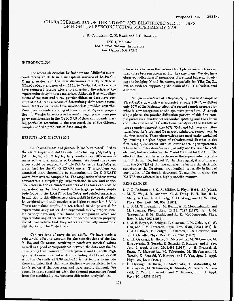
Proposal No. 2023Mp
CHARACTERIZATION OF THE ATOMIC AND ELECTRONIC STRUCTURES OF HIGH T, SUPERCONDUCTING MATERIALS BY XAS
S. D. Conradson, G. H. Kwei, and I. D. Raistrick
INC-4, MS C345 Los Alamos National Laboratory
Los Alamos, NM 87545
INTRODUCTION
The recent observation by Bednorz and Miiller'of super- ,conductivity at 30 K in a multiphase mixture of La-Ba-Cu- 0 metal oxides, and the later discoveries of a T, of 93K in YBa2Cu307-6'and now of ca. 115K in Ca-Sr-Bi-Cu-0 systems have prompted intense efforts to understand the origin of the superconductivity in these materials. Although Rietveld refine- ments of neutron and x-ray powder diffration data have pre-, empted EXAFS as a means of determining their atomic struc- tures, XAS experiments have nevertheless provided contribu- tions towards understanding of their unusual physical proper- ties3-'. We also have observed several intriguing spectrum:pro- perty relationships in the Cu K XAS of these compounds, pay- ing particular attention to the characteristics of the different samples and the problems of data analysis.
RESULTS AND DISCUSSION
Cu-0 amplitudes and phases. It has been that the use of CuzO and CuO as standards for Laz-zMzCu04-% (M = Ba, Sr) and YBazCu307-6 results in ea. 50% overesti- mates of the total number of 0 atoms. We found that these errors could be reduced to 5 1&15% by using LazCu04 as a standard for the Cu-0 EXAFS waves. This problem was examined more thoroughly by comparing the Cu-0 EXAFS waves from several compounds. The amplitudes of these waves demonstrate a surprisingly large variation in size and shape. The errors in the calculated numbers of 0 atoms can now be understood as the direct result of the larger per-atom ampli- tude found in the EXAFS of LazCu04 and related materials. In addition to this difference in size, a shift in the peak of their k3 weighted amplitude envelopes to higher to near k = 8 A-'. These anomalous amplitudes are related to the potential for superconductivity rather than superconductivity proper, inso- far as they have only been found for compounds which are superconducting either as studied or become so when properly doped. We believe that they reflect an unusually anisotropic distribution of the 0 electrons.
Contributions of more distant shells. We have made a substantial effort to account for the contributions of the La, Y, Ba, and Cu atoms, resulting in consistent metrical values as well as a good correspondence between the data and the fit. This is only true, however, for interplane 0 and Cu shells; high quality fits were obtained without including the 0 shell at 2.30 A or the Cu shells at 3.39 and 4.15 A . Attempts to include these indicated that their contributions were restricted to the low k region of the spectrum and were rapidly damped. We conclude that, consistent with the thermal parameters found from the combined x-ray/neutron diffraction analysis7, the
105
interactions between the various Cu-0 planes are much weaker than those between atoms within the same plane. We also have observed indications of anomalous vibrational behavior involv- ing the bridging Y and Ba atoms, especially for YBazCuaOs, but no evidence supporting the claim of Cu-Y substitutional disorder.
Sample dependence of YB02 Cu3 0,- 6 . Our first sample of Y B ~ ~ C U & - ~ , which was annealed at only 900°C, exhibited only 50% of the Meissner effect of a second sample prepared by what is now recognized as the optimum procedure. Although single phase, the powder diffraction pattern of this first sam- ple possesses a smaller orthorhombic splitting and the almost complete absence of [OOl] reflections. Analysis of the EXAFS of these samples demonstrates 10%,30%, and 5% lower contribu- tions from the Y, Ba, and Cu nearest neighbors, respectively, in the first sample. These observations are most easily explained by invoking a higher degree of substitutional disorder in the first sample, consistent with its lower annealing temperature. The extent of this disorder is apparently not the same for each element, but is greater for the Y and Ba than for the Cu. The effect of this disorder is to decrease the superconducting por- tion of the sample, but not T,. In this regard, it is of interest that the XANES of the two samples, reflecting the electronic structure at the Cu atoms, are identical, especially in light of our studies of Zn-doped, depressed T, samples in which the XANES was affected in a highly specific manner.
REFERENCES
1. J. G. Bednorz and K. A. Miiller, Z. Phys. B 64, 189 (1986). 2. M. K , Wu, J. R. Ashburn, C. J. Torng, P. H. Hor, R. L.
Meng, L. Gao, Z. J. Huang, Y. 0. Wang, and C. W. Chu, Phys. Rev. Lett. 58, 908 (1987).
3. a. J. M. Tranquada, S. M. Heald, A. R. Moodenbaugh, and M. Suenaga, Phys. Rev. B 35, 7187 (1987). b. J. M. Tranquada, S. M. Heald, and A. R. Moddenbaugh, Phys. Rev. B 36,5263 (1987).
4. a. J. B. Boyce, F. Bridges, T. Claeson, T. H. Geballe, C. W. Chu, and J. M. Tarascon, Phys. Rev. B 35, 7203 (1987). b. a. J. B. Boyce, F. Bridges, T. Claeson, R. S. Howland, and T. H. Geballe, Phys. Rev. B 36, 5251 (1987).
5. a. H. Oyanagi, H. Ihara, T. Matsushita, M. Tokumoto, M. Hirabayashi, N. Terada, K. Senzaki, Y. Kimura, and T. Yao, Jpn. J. Appl. Phys. 26, L488 (1987). b. H. Oyanagi, H. Ihara, T. Matsushita, M. Tokumoto, M. Hirabayashi, N. Terada, K. Senzaki, Y. Kimura, and T. Yao, Jpn. J. Appl. Phys. 26, L638 (1987).
6. H. Oyanagi, H. Ihara, T. Matsubara, T. Matsushita, M. Hirabayashi, M. Tokumoto, K. Murata, N. Terada, K. Sen- zaki, T. Ym, H. Iwasaki, and Y. Kimura, Jpn. J. Appl. Phys 26, L1233 (1987).
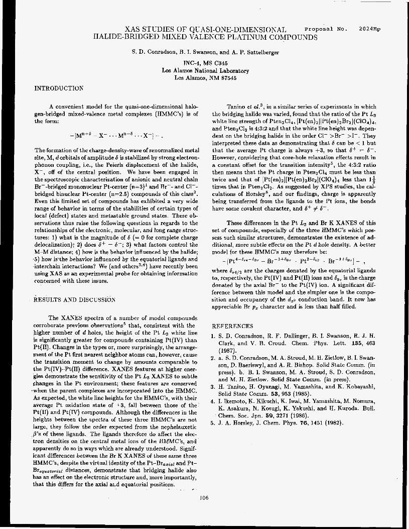
XAS STUDIES OF QUASI-ONE-DIMENSIONAL P r o p o s a l No. 2024Mp HALIDE-BRIDGED MIXED VALENCE PLATINUM COMPOUNDS
S. D. Conradson, B. I. Swanson, and A. P. Sattelberger
Los Alamos National Laboratory Los Alamos, NM 87545
INC-4, MS C345
INTRODUCTION
A convenient model for the quasi-one-dimensional halo- gen-bridged mixed-valence metal complexes (HMMC’s) is of the form:
The formation of the charge-density-wave of renormalized metal site, M, dorbitals of amplitude 6 is stabilized by strong electron- phonon coupling, Le., the Peierls displacement of the halide, X-, off of the central position. We have been engaged in the spectroscopic characterization of anionic and neutral chain Br--bridged mononuclear Pt-center (n=3)’ and Br-- and C1-- bridged binuclear Pt-center (n=2.5) compounds of this class2. Even this limited set of compounds has exhibited a very wide range of behavior in terms of the stabilities of certain types of local (defect) states and metastable ground states. These ob- servations thus raise the following questions in regards to the relationships of the electronic, molecular, and long range struc- tures: 1) what is the magnitude of 6 (= 0 for complete charge delocalization); 2) does 6+ = 6-; 3) what factors control the M-M distance; 4) how is the behavior influenced by the halide; .5) how is%he behavior influenced by the equatorial ligands and interchain interactions? We (and others3i4) have recently been using XAS as an experimental probe for obtaining information concerned with these issues.
RESULTS AND DISCUSSION
The XANES spectra of a number of model compounds corroborate previous observations5 that, consistent with the higher number of d holes, the height of the Pt L3 white line is significantly greater for compounds containing Pt (IV) than Pt(I1). Changes in the types or, more surprisingly, the arrange- ment of the Pt first nearest neighbor atoms can, however, cause the transition moment to change by amounts comparable to the Pt(IV)-Pt(I1) difference. XANES features at higher ener- gies demonstrate the sensitivity of the Pt Ls XANES to subtle changes in the Pt environment; these features are conserved
*when the parent complexes are incorporated into the HMMC. As expected, the white line heights for the HMMC’s, with their average Pt oxidation state of $3, fall between those of the’ Pt(I1) and Pt(IV) compounds. Although the differences in the heights between the spectra of these three HMMC’s are not large, they follow the order expected from the nephelauxetic p’s of these ligands. The ligands therefore do affect the eiec- ‘tron densities on the central metal ions of the HMMC’s, and apparently do so in ways which are already understood. Signif- icant differences between the Br K XANES of these same three HMMC’s, despite the virtual identity of the Pt-Br,,;,r and Pt- Brequatorlat distances, demonstrate that bridging halide also has an effect on the electronic structure and, more importantly, that this differs for the axial a1.d equatorial positions. . _ -
Tanino et aL3, in a similar series of experiments in which the bridging halide was varied, found that the ratio of the P t L3 white line strength of PtenzCl4, [Pt(en) 21 [ Pt(en) ~ B r z ] (C104) 4,
and Pten2Clz is 4:3:2 and that the white line height was depen- dent on the bridging halide in the order C1- >Br- >I-. They interpreted these data as demonstrating that 6 can be < 1 but that the average Pt charge is always +3, so that 6+ = 6-. However, considering that core-hole relaxation effects result in a constant offset for the transition intensity5, the 4:3:2 ratio then means that the Pt charge in PtenzC14 must be less than twice and that of [Pt(en)2][Pt(en)2Br2](C104)4 less than If times that in PtenzCl~. As suggested by XPS studies, the cal- culations of Horsley5, and our findings, charge is apparently being transferred from the ligands to the Pt ions, the bonds have some covalent character, and 6+ # 6- .
These differences in the Pt L3 and Br K XANES of this set of compounds, especially of the three HMMC’s which pos- sess such similar structures, demonstrates the existence of ad- ditional, more subtle effects on the P t d hole density. A better model for these HMMC’s may therefore be:
I - ? -[pt4-6,4-JBr - Br-l+&r . pt2-6e3 . Br-1+6Br
where 6e4/2 are the charges donated by the equatorial ligands to, respectively, the Pt(1V) and Pt(I1) ions and 6,, is the charge donated by the axial Br- to the Pt(1V) ion. A significant-dif- ference between this model and the simpler one is the compo- sition and occupancy of the d,2 conduction band. It now has appreciable Br p , character and is less than half filled.
REFERENCES
1. S. D. Conradson, R. I?. Dallinger, B. I. Swanson, R. J. H. Clark, and V. B. Croud. Chem. Phys. Lett. 135, 463 (198 7).
2. a. S. D. Conradson, M. A. Stroud, M. H. Zietlow, B. I. Swan- son, D. Baeriswyl, and A. R. Bishop. Solid State Comm. (in press). b. B. I. Swanson, M. A. Stroud, S. D. Conradson, and M. H. Zietlow. Solid State Comm. (in press).
3. H. Tanino, H. Oyanagi, M. Yamashita, and K. Kobayashi, Solid State Comm. 53, 953 (1985).
4. I. Ikemoto, K. Kikuchi, K. Iwai, M. Yamashita, M. Nomura, K. Asakura, N. Kosugi, K. Yakushi, and H. Kuroda. Bull. Chem. SOC. Jpn. 59, 3271 (1986).
5. J. A. Horsley, J. Chem. Phys. 76, 1451 (1982).
106
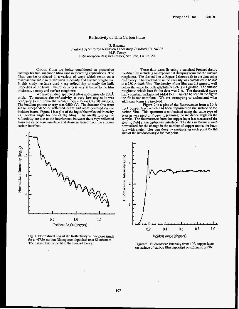
Propo'sal No.
Reflectivity of Thin Carbon Films
S. Brennan Stanford Synchrotron Radiation Laboratory, Stanford, Ca. 94305.
M.F. Tonev IBM Almaden Research Center: San Jose, Ca. 95120.
Carbon films are being considered as protective coatings for thin magnetic f i lms used in recording applications. The films can be produced in a variety of ways which result on a macroscopic scale in differences in density and surface roughness. In this study we have used x-ray reflectivity to study the bulk properties of the films. The reflectivity is very sensitive to the film thickness, density and surface roughness.
We have studied sputtered films approximately 280A thick. To measure the reflectivity at very low angles it was necessary to sIit down the incident beam to roughly 50 microns. The incident photon energy was 9500 eV. The detector slits were set to accept >0.5" of reflected beam and were centered on the incident beam. Figure 1 is a plot of the log of the reflected intensity vs. incident angle for one of the films. The oscillations in the reflectivity are due to the interference between the x-rays reflected from the carbon-air interface and those reflected from the silicon- carbon interface.
n 0 .
e o " .
E 8 - 2 - .r(
v) c 4
8 T - 4 -
n"
3 d
-6
t....l....l....l... 0.5 1.0 1.5
Incident Angle (degrees)
Fig. 1 Normalized Log of the Reflectivity vs. Incident Angle for a -275A carbon fiim sputter deposited on a Si substrate. The dashed line is the fit to the Fresnel theory.
These data were fit using a standard Fresnel theory modified by including an exponential damping term for the surface roughness. The dashed line in Figure 1 shows a fit to the data using . The modulation in the intensity was calculated to be due that to a 280 thick film. The density of the film was 1.8 gmdcc, well below the value for bulk graphite, which is 2.1 gmdcc. The surface roughness which best fit the data was 7 A. The theoretical curve had a constant background added to it. As can be seen in the figure the fit is not complete. We are attempting to understand what additional terms are involved.
Figure 2 is a plot of the fluorescence from a 10 A thick copper layer which had been deposited on the surface of the carbon film. This spectrum was obtained using the same type of scan as was used in Figure 1, scanning the incidence angle on the sample. The fluorescence from the copper layer is a measure of the electric field at the carbon-air interface. The data in Figure 2 were normalized for the change in the number of copper atoms the beam hits with angle. This was done by multiplying each point by the sine of the incidence angle for that point.
0.2 0.4 0.6 0.8 1 .o Incident Angle (degrees)
Figure 2. Fluorescence Intensity from lOA copper layer on surface of carbon fiim deposited on siliwn substrate.
107

Proposal No. 8 0 0 1 ~
Fluorescence EXAFS Study of Silicon in Neodymium Iron Boron Rare-Earth Magnets
S. Brennan and A. Bienenstock Stanford Synchrotron Radiation Laboratory, Stanford Ca. 94305
J.E. Keem Ovonic Synthetic Materials Co., Troy, Mi. 48084
Recently, a material has been described which consists predominantly of a phase similar in structure to Nd2Fe@ but with the addition of approximately 1 at.% Si. Rapidly solidified ribbon samples of this new material have been fabricated using a melt-
determined by powder diffractometry. To determine the environment of the Si atoms in this structure we have performed -g fluorescence Extended X-ray Absorption Fine Structure (EXAFS) - 4 measurements on the flakes.1 The advantages of fluorescence 8 EXAFS in this case are its significantly greater sampling depth *g which avoids the surface sensitivity of electron yield techniques and 6~ 3 its better signal-to-background ratio. Previous fluorescence EXAFS $ studies of low-Z elements have used a proportional counter, For this experiment a Si(Li) solid state detector was used because of its $ 2 improved energy resolution. The greater energy resolution (-150 e v means that the fluorescent signal can be discriminated from the elastically scattered signal, resulting in an improved signal- 1 to-background ratio compared to a proportional counter. This is especially important for polycrystalline samples where elastically scattered diffriicted beams can enter the detector.
spinning process. The resulting ribbons are polycrystalline, as 5
t Si K- a Fluorescence
Li
C a
1800 1900 2000 2100 Photon Energy (eV)
0 2 4 6 8 10 pha~e-shifted Distance, (A)
The peak centered at 2.1A was isolated and back- transformed. This back-transformation isolates the near-neighbor contribution to the EXAFS. Using hase shifts and backscattering
Si, Si-Nd and Si-0 near-neighbors. The only acceptable fit to the data is for a Si-Fe near neighbor, with a distance of 2.45 A M.05b;. Fitting this shell using calculated back-scattering amplitudes gave the number of near-neighbors as 3.3. After compensating for the contribution of the oxidized silicons to the total signal, the number of near-neighbors was determined to be 4.1. The peak at 1.2A in Fig 2 was fit using an amplitude and phaseshift derived from a sample of Si02. The derived distance is 1.65M.1 A with 1.1 near neighbors. The angular dependence of the near edge structure suggest that the Si-0 bonds are due to a surface oxide and are not a significant feature of the bulk structure. The EXAFS results indicate that the environment of the Si atom is well characterized by a single shell of Fe atoms. By comparing the environment of the Si atom with possible interstitial and substitutional sites in the Nd7FeiaB structure it is clear that there are no interstitial sites with
amplitudes tabulated by Teo and Lee P the result was fit to Si-Fe, Si-
Figure 1 shows the sum of 8 scans obtained with a total of 32 seconds per point, with 2 eV between points. Note that the Count rate in the region below the absorption edge is less than 5% of the Signal above the edge, indicating the near-total removal of the elastic signal. Figure 2 shows the results of both a ko and a k2 transform. Because the 1p transform results in better separation of the two dominant features, it was used for the analysis of the distances in the sample.
en&ghroom. The only substitutional site which has the right set of distances and number of near neighbors is the “C” site in the stand& strucmal mde1.2
References 1.
2.
S. Brennan, A. Bienenstock and J.E. Keem, submitted to J. Appl. Phys. J.F. Herbst, J.J. Croat and W.B. Yelon, J. Appl. Phys. 57 4086 (1985).
108
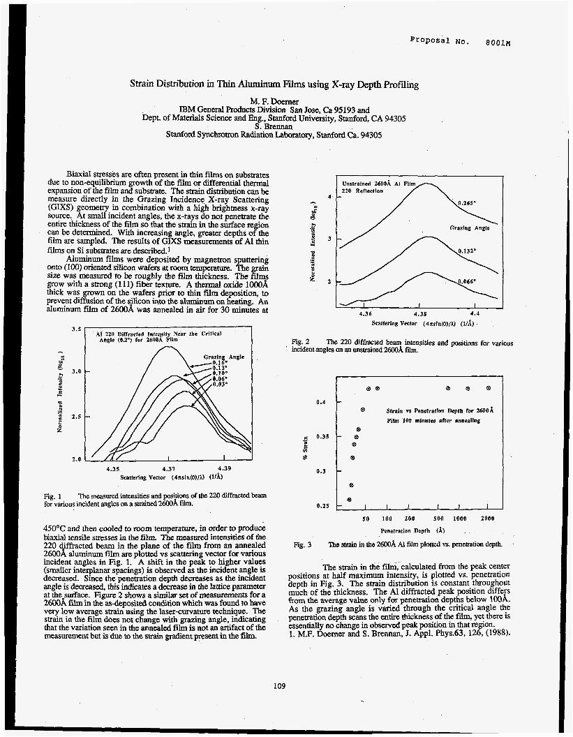
Proposal No. 8001M
Strain Distribution in Thin Aluminum Films using X-ray Depth Profiling
M. F. Doerner IBM General Products Division San Jose, Ca 95193 and
Dept. of Materials Science and En%. Stanford Universitv. Stanford. CA 94305 . I S: Brennan
Stanford Synchrotron Radiation Laboratory, Stanford Ca. 94305
Biaxial stresses are often present in thin fdms on substrates due to non-equilibrium growth of the film or differential thermal expansion of the film and substrate. The strain distribution can be measure directly in the Grazing Incidence X-ray Scattering (GIXS) geometry in combination with a high brighmess x-ray source. At small incident angles, the x-rays do not penetrate the entire thickness of the film so that the strain in the surface region can be determined. With increasing angle, greater depths of the film are sampled. The results of GIXS measurements of A1 thin Nms on Si substrates are described.1
Aluminum films were deposited by magnetron sputtering onto (100) oriented silicon wafers at room temperam. The grain size was measured to be roughly the film thickness. The films grow with a strong (1 11) fiber texture. A thermal oxide lOOOA thick was grown on the wafers prior to thin film deposition, to prevent diffusion of the silicon into the aluminum on heating. An aluminum f i i of 26OOA was annealed in air for 30 minutes at
AI 220 1)ifTracted Inteqsity Near the Critical Angle (0.2") for 2600A Film
4.35 4.37 4.39
Scattering Vector (4nsin(o)/a Wdi)
Fig. 1 for various incident angles on a strained 2600A film.
The measured intensities and positions of the 220 diffracted beam
45OOC and then cooled to room temperature, in order to produce biaxial tensile stresses in the film. The measured intensities of the 220 diffracted beam in the plane of the film from an annealed 2600A aluminum fiim are plotted vs scattering vector for various incident angles in Fig. 1. A shift in the peak to higher values (smaller interplanar spacings) is observed as the incident angle is decreased. Since the penetration depth decreases as the incident angle is decreased, this indicates a decrease in the lattice parameter at the surface. Figure 2 shows a similar set of measurements for a 2600A film in the as-deposited condition which was found to have very low average strain using the laser-curvature technique. The strain in the film does not change with grazing angle, indicating that the variation seen in the annealed film is not an artifact of the measurement but is due to the strain gradient present in the Nm.
4.36 4 .38 4 . 4
Scattering Vector (4nsin(e)/a (I/&
Fig. 2 The 220 diffracted beam intensities and positions for various ' incident angles on an unsrrained 2600A fii.
0.4
0.35
f c
0.3
0.25
Strain vs Penetration Depth for 2600A
Film '100 minutes after annealing
e ' @
e
e
e
e I I I I 1 I
5 0 i o n zoo 500 inno zoo0
Penetration Depth (A) . Fig. 3 The strain in the 2600A Al film plotted vs. penetration depth.
The strain in the fdm, calculated from the peak center positions at half maximum intensity, is plotted vs. penetration depth in Fig. 3. The strain distribution is constant throughout much of the thickness. The A1 diffracted peak position differs from the average value only for penetration depths below lOOA. As the grazing angle is varied through the critical angle the penetration depth scans the entire thickness of the fidm, yet there is essentially no change in observed peak position in that region. 1. M.F. Doerner and S. Brennan, 3. Appl. Phys.63, 126, (1988).
109
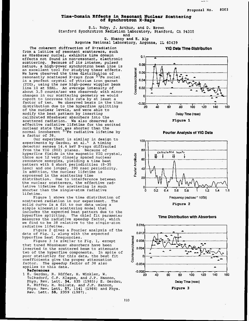
Proposal No. 8003
T b s - D m m l n Ef%ecter in Resonant Nuclear Scattering e l lynchrotton X-Ray8
Stanford Synchro tmn Radiation Laboratory, Stanford, ea 84385 and
Argonne National Laboratory, Argonne, IL 60439
%,L* RUlsyp J s Arthur, and D. Brown
G. Zjhenoy and E. Alp \
The coherent diffraction of X-radiation from a lattice of resonant scatterers, such as Mossbauer nuclei, exhibits time domain effects not found in non-resonant, electronic scattering. Because of its intense, pulsed nature, a high-power synchrotron beam line is an excellent tool for studying these effects. We have observed the time distribution of resonantly scattered X-rays from 57Fe nuclei in a perfect crystal of yttrium iron garnet (YIG), using the new high-power wiggler beam line 10 at SSRL. An average intensity of about 3.5 counts/sec was observed; with minor changes in our scattering geometry we would expect to increase this rate by at least a factor of ten. We observed beats in the time distribution due to the hyperfine splitting of the nuclear levels, and were able to modify the beat pattern by inserting calibrated Mbssbauer absorbers into the scattered radiation. We also observed an effective radiative lifetime for the excited nuclear state that was shorter than the normal incoherent 57Fe radiative lifetime by a factor of 38.
experiments by Gerdau, et a1.l A timing detector senses 14.4 keV X-rays diffracted from the YIG (002) planes. Because of hyperfine fields in the magnetic YIG crystal, there are 12 very closely spaced nuclear resonance energies, yielding a time beat pattern with 6 short periodicities (8-35 nsec) and one longer, 390 nsec periodicity. In addition, the nuclear lifetime is expressed in the scattering time distribution. Due to interference between the nuclear scatterers, the effective rad- iative lifetime for scattering is much shorter than the single-atom radiative lifetime.
scattered radiation in our experiment. The solid curve is a fit to our data using a simple kinematic scattering model that includes the expected beat pattern due to the hyperfine splitting. The chief fit parameter measures the radiative speedup factor, which we find to be 38 relative to the single-atom radiative lifetime.
data of Fig. 1, along with the expected hyperfine beat frequencies.
that tuned Mossbauer absorbers have been inserted in the scattered beam to attenuate two of the hyperfine components. In spite of poor statistics for this data, the best fit coefficients give the proper attenuation factor. The speedup factor of 38 also applies to this data.
1 E. Gerdau, R. Ruffer, H. Winkler, W.
Our experiment is similar in design to
Figure 1 shows the time distribution of
Figure 2 gives a Fourier analysis of the
Figure 3 is similar to Fig. 1, except
References
Tolksdorf, C.P. Klages, and J .P. Hannon, Phys, Rev. Lett. 54, 835 (1985); E. Gerdau, R. Riiffer, R. Hollatz, and J.P. Hannon, Phys. Rev. Lett. 57, 1141 (1986) and Phys. Rev. Lett. 58 , 2359 (1987).
110
YIG Data Time Distribution
0.11 ~ 1
* -0.02 I I I I I I I
20 40 60 80 100 120 140 160
Delay Time (nsec)
F i g u r e 1
Fourier Analysis of YIG Data
Frequency (rad/sec' 1OE9)
F i g u r e 2
l i m e Dlstribution with Absorbers
0.0161
0

Proposal No. 8010M Interferometric Study of Spherical Wave Diffraction
J. Arthur and H. Tompkins Stanford Synchrotron Radiation Laboratory, Stanford, CA 94305
We have used a monolithic two ear silicon interferometer to observe some effects of the angular coherence present in X-ray waves that have passed through a 5 micrometer wide slit. Diffraction of this radiation in the ears of the interferometer leads to a spatial distribution in the beam leaving the second ear, which is much wider than the 5 micron incident beam. The distribution can be changed by the insertion of phase shifting elements between the interferometer ears. Careful measurements of these distribution patterns provide a test of the fine points of dynamical diffraction theory.
The interferometer used in this experiment consists of two identical thin parallel plates, or ears, which have been cut from but remain attached at the base to a single perfect crystal of silicon. The connecting base keeps the ears parallel to within atomic dimensions. The ear thicknesses are identical to within a few microns; each approximately 300 microns. The ears are separated by 40 mm, and oriented for symmetric Laue diffraction from the crystal (220) or (440) planes.
of waves produced by a very small source (such as a 5 micron slit) within a crystal slab has been extensively studied1 and has been applied to the two crystal case.2 An important result of these studies is the significance of the Pendellosung (or extinction) length, which describes the flow of diffracting energy within the crystal. This length, rather than the X-ray wavelength, is the relevant length for many coherence effects in the interferometer. The Pendellosung length is typically 50 - 100 microns; relative to such lengths a 5 micron source can be considered a point source. The radiation emanating from such a source is for diffraction purposes spherical -- it carries no angular information. (An equivalent viewpoint is that the angular acceptance range for diffraction in a perfect crystal is so small that the single slit diffraction of angstrom-wavelength radiation from a 5 micron slit is enough to completely fill it.)
With such a slit source, the distribution of intensity in the diffracted beam behind the first interferometer ear is a Pendellosung pattern (essentially a Bessel function), spread out over dimensions comparable to the ear thickness. Because of the spherical nature of the source radiation, there is coherence across this widened beam. Diffraction of this beam in the second ear leads to further beam widening and interference, giving a distribution of intensity in the beam leaving the second ear that is a convolution of Pendellosung patterns. This distribution was observed in our experiment.
The experiment was performed at a bending magnet beam line at SSRL. A bent silicon premonochromator was used to focus 26 keV X-rays on the 5 micron entrance slit.
The dynamical theory of the diffraction
This slit consisted of two precision steel roller bearings separated by pieces of 5 micron nickel foil. The interferometer was mounted on a single axis diffractometer, and the exit beam inteiisity distribution was measured by scanning another 5 micron slit across it, with a NaI detector behind. Changes in the exit beam intensity distribution were induced by placing various acrylic plastic wedges in the beam between the two interferometer ears. Such a wedge introduces a linear phase shift across the beam, changing the interference pattern produced in the second ear. Figure 1 shows two of the intensity patterns that were observed. Although detailed analysis has not been completed, it appears that these patterns are consistent with the predictions of dynamical diffraction theory.
We thank the Quantum Metrology Group of the U. S . National Bureau of Standards, and Dr. Albert Hennins in particular, for hospitality and help in constructing the two ear interferometer.
Intensity Distribution in BB Beam
26 keV, (440) reflection, 5 urn slits
"r
I I I I I 0.8 0.9 1 . I 1.2 1.3 1.4 1
Exit slit position (mm)
Figure 1 Exit beam intensity distributions with
a) a narrow wedge inserted and b) a wide wedge inserted in the interferometer. There is a broad, flat peak due to incoherent, thermal diffuse scattering underneath the interference pattern in each case.
References 1 See, for example: N. Kato, J. Appl. Phys. 39, 2225 (1968); L. Azaroff, ed., X-Ray Diffraction (McGraw-Hill, N.Y., 1979).
2 V.L. Indenbom, I.Sh. Slobodetskii, and G.I. Truni, Zh. Eksp. Teor. Fiz. 66, 1110 (1974) [Sov. Phys. JETP 39, 542 (1974)l; U. Bonse and R. Teworte, Z. Naturforsch. 31a, 427 (1982); J. Arthur, C.G. Shull, and A. Zeilinger, Phys. Rev. B32, 5753 (1985).
111
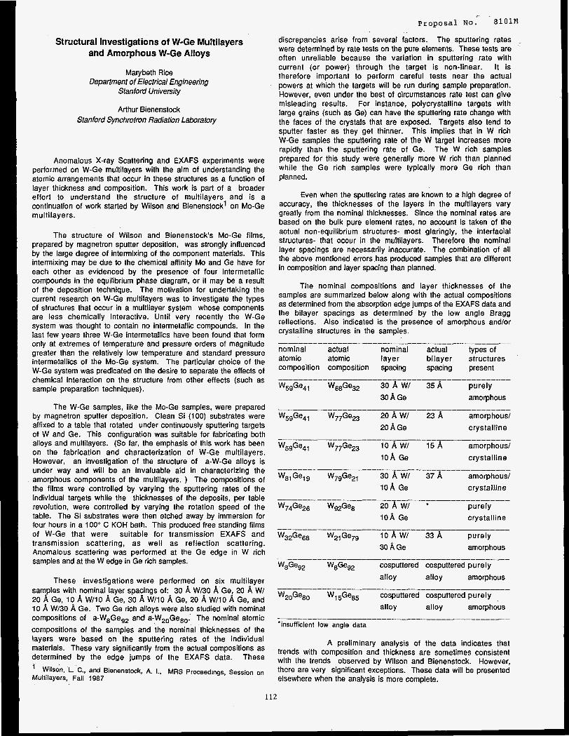
Structural Investigations of W-Ge Multilayers and Amorphous W-Ge Alloys
Marybeth Rice Department of Electrical Engineering
Stanford University
Arthur Bienenstock Stanford Synchrotron Radiation Laboratory
Anomalous X-ray Scattering and EXAFS experiments were performed on W-Ge multilayers with the aim of understanding the atomic arrangements that occur in these structures as a function of layer thickness and composition. This work is part of a broader effort to understand the structure of multilayers and is a continuation of work started by Wilson and Bienenstockl on Mo-Ge multilayers.
The structure of Wilson and Bienenstock’s Mo-Ge films, prepared by magnetron sputter deposition, was strongly influenced by the large degree of intermixing of the component materials. This intermixing may be due to the chemical affinity Mo and Ge have for each other as evidenced by the presence of four intermetallic compounds in the equilibrium phase diagram, or it may be a result of the deposition technique. The motivation for undertaking the current research on W-Ge multilayers was to investigate the types of structures that occur in a multilayer system whose components are less chemically interactive. Until very recently the W-Ge system was thought to contain no intermetallic compounds. In the last few years three W-Ge intermetallics have been found that form only at extremes of temperature and pressure orders of magnitude greater than the relatively low temperature and standard pressure intermetallics of the Mo-Ge system. The particular choice of the W-Ge system was predicated on the desire to separate the effects of chemical interaction on the structure from other effects (such as sample preparation techniques).
The W-Ge samples, like the Mo-Ge samples, were prepared by magnetron sputter deposition. Clean Si (100) substrates were affixed to a table that rotated under continuously sputtering targets of W and Ge. This configuration was suitable for fabricating both alloys and multilayers. (So far, the emphasis of this work has been on the fabrication and characterization of W-Ge multilayers. However, an investigation of the structure of a-W-Ge alloys is under way and will be an invaluable aid in characterizing the amorphous components of the multilayers. ) The cornpositions of the films were controlled by varying the sputtering rates of the individual targets while the thicknesses of the deposits, per table revolution, were controlled by varying the rotation speed of the table. The Si substrates were then etched away by immersion for four hours in a 100” C KOH bath. This produced free standing films of W-Ge that were suitable for transmission EXAFS and transmission scattering, as well as reflection scattering. Anomalous scattering was performed at the Ge edge in W rich samples and at the Wedge in Ge rich samples.
These investigations were performed on six multilayer samples with nominal layer spacings of: 30 A W/30 8, Ge, 20 8, W/ 20 A Ge, 10 8, W/10 A Ge, 30 A W/10 8, Ge, 20 8, W/10 8, Ge, and 10 8, W/30 8, Ge. Two Ge rich alloys were also studied with nominal compositions of a-W8Geg2 and a-W2,Geso. The nominal atomic ccmpositions of the samples and the nominal thicknesses of the layers were based on the sputtering rates of the individual materials. These vary significantly from the actual compositions as determined by the edge jumps of the EXAFS data. These ’ Wilson, L. c., and Bienenstock, A. I., MRS Proceedings, Session on Multilayers, Fall 1987
112
, - Proposal No. 8101M
discrepancies arise from several factors. The sputtering rates were determined by rate tests on the pure elements. These tests are often unreliable because the variation in sputtering rate with current (or power) through the target is non-linear. It is therefore important to perform careful tests near the actual powers at which the targets will be run during sample preparation. However, even under the best of circumstances rate test can give misleading results. For instance, polycrystalline targets with large grains (such as Ge) can have the sputtering rate change with the faces of the crystals that are exposed. Targets also tend to sputter faster as they get thinner. This implies that in W rich W-Ge samples the sputtering rate of the W target increases more rapidly than the sputtering rate of Ge. The W rich samples prepared for this study were generally more W rich than planned while the Ge rich samples were typically more Ge rich than planned.
Even when the sputtering rates are known to a high degree of accuracy, the thicknesses of the layers in the multilayers vary greatly from the nominal thicknesses. Since the nominal rates are based on the bulk pure element rates, no account is taken of the actual non-equilibrium structures- most glaringly, the interfacial structures- that occur in the multilayers. Therefore the nominal layer spacings are necessarily inaccurate. The combination of all the above mentioned errors has produced samples that are different in composition and layer spacing than planned.
The nominal compositions and layer thicknesses of the samples are summarized below along with the actual compositions as determined from the absorption edge jumps of the EXAFS data and the bilayer spacings as determined by the low angle Bragg reflections. Also indicated is the presence of amorphous and/or crystalline structures in the samples.
nominal actual nominal actual types of atomic atomic layer bilayer structures composition composition spacing spacing present
W5SGe41 W68Ge32 30 A w/ 35 A purely
WS9Ge4’ W77Ge23 20 A WI 23 A amorphous/
30 8, Ge amorphous
20 8, Ge crystalline
W59Ge41 W77Ge23 10 A W/ 15 8, amorphous/ 108, Ge crystalline
W8,Gel9 W79Ge21 30 8, W/ 37 8, amorphous/ 108, Ge crystalline
W74Ge26 Wg2Ge8 20 A w/ * purely l O A Ge crystalline
WS2Gess W21Ge79 10 A Wl 33 8, purely 30 A Ge amorphous
W8Ge92 W8Ge92 cosputtered cosputtered purely alloy alloy amorphous
W2*Ge8, W15GeS5 cosputtered cosputtered purely alloy alloy amorphous
‘insufficient low angle data
A preliminary analysis of the data indicates that trends with composition and thickness are sometimes consistent with the trends observed by Wilson and Bienenstock. However, there are very significant exceptions. These data will be presented elsewhere when the analysis is more complete.
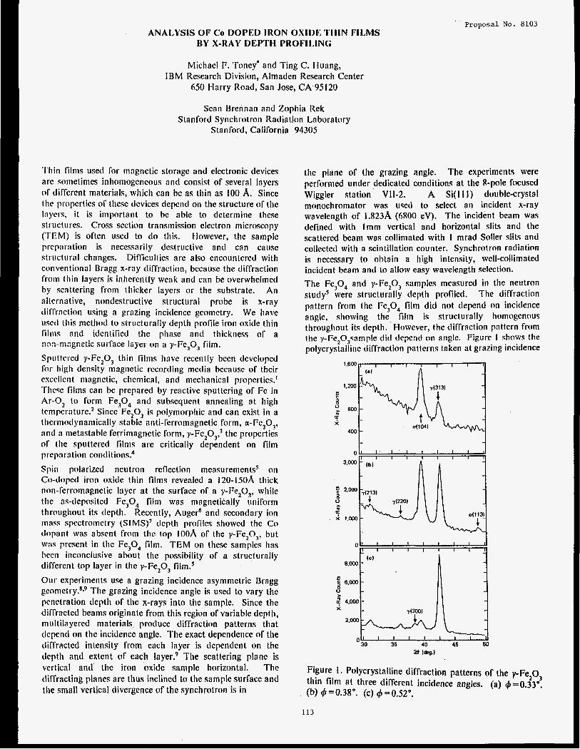
Proposal No. 8103 ANALYSIS OF Co DOPED IRON OXIDE TIIIN FILMS
BY X-RAY DEPTH PROFILING
Michael F. Toney' and Ting C. Huang, IBM Research Division, Almaden Research Center
650 Harry Road, San Jose, CA 95 I20
Sean Brennan and Zophia Rek Stanford Synchrotron Radiation Laboratory
Stanford, California 94305
Thin films used for magnetic storage antl electronic devices are sometimes inhomogcncous and consist of several layers of tliffcrent materials, which can be as thin as 100 A. Sincc thc propcrties of these devices dcpcntl on the structure o f the layers, it is important to be able to determine these structures. Cross section transmission electron microscopy (TEM) is often used to do this. However, the sample preparation is necessarily destructive and can cause structural changes. Difficulties are also encountered with conventional Bragg x-ray diffraction, because the diffraction from [h in layers is inherently weak and can be overwhelinetl by scattering from thicker layers or the substrate. An altcrnative, nondestructive structural probe is x-ray diffraction using a grazing incidence gcomctry. We have used this method to structurally depth profile iron oxide thin films and idcntificd the phase and thickness of a non-magnetic surfzce layer o n a y-Fe,O, film.
Sputtcrctl y-Fe,O, thin films have recently been developed for high density magnctic recording media because of thcir esccllcnt magnetic, chemical, and mechanical properties.' Thcse films can be prepared by reactive sputtering of Fe in Ar-0, to form Fe,O, and subsequent annealing at high temperature? Since Fe,O, is polymorphic and can exist i n a thcrtnodynamically stable anti-ferromagnetic form, a-Fe,O,, antl a metastable ferrimagnetic form, y-Fe,O,? the properties of the sputtered films are critically dependent on film preparation conditions."
Spin polarized neutron reflection measurementss oil
Co-tlnpcd iron oxide t h i n films revealed a 120-150A thick non-ferromagnetic layer a t the surface of a y-Fe201, while the as-deposited Fe,O, film was magnetically uniform throughout its depth. Recently, Auger6 and secondary ion mass spectrometry (SIMS)' depth profilcs showed the Co dc3pant was absent from thc top IOOh of the y-Fe203, but w a ~ present in tlic Fe,04 film. TEM on these samples has been inconclusive about the possibility of a structurally different top layer in the y-Fe,O, film.s
Our experiments use a grazing incidence asymmetric Bragg g c o n i ~ t r y . ~ * ~ The grazing incidence angle is used to vary the penetration depth of the x-rays into the sample. Since the diffracted beams originate from this region of variable depth, niultilayered materials produce diffraction patterns that tlcpend on thc incidence angle. The exact dcpendciice of the diffracted intensity from each layer is dcpendent on the tlcpth and extent of each layer? The scattering plane is vertical and thc iron oxide sample horizontal. The diffracting planes are thus inclined to the sample surface and the small vertical divergence of the synchrotron is in
the planc of the grazing angle. The experiments were performed under dedicated conditions a t the %pole focused Wiggler station Vll-2. A Si( I 1 1) double-crystal monochromator was used to select an incident x-ray wavelength of 1.823A (6800 eV). The incident beam was defined with Imm vertical and horizontal slits and the scattered bcam was collimated with I mrad Soller slits and collected with a scintillation counter. Synchrotron radiation is necessary to obtain a high intensity, well-collimated incident beam and to allow easy wavelength selection.
The Fe,O, and y-Fe,O, samples measured in the neutron study' were structurally depth profiled. The diffraction pattern from the Fe,04 film did not depend o n incidence angle, showing the film is structurally homogenour throughout its depth. However, the diffraction pattern from the y-Fe,O,sample did depend on angle. Figure I sl io~ci the polycrystalline diffraction patterns taken at grazing incidence
-I
Figure I . Polycrystalline diffraction patterns of the y-Fe,O, thin film a t three different incidence angles. (a) $=0.33". (b) q5=0.38". (c) (b=0.52".
113
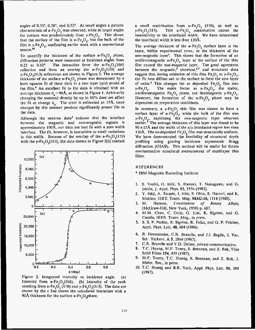
nnplcs of 0.33O, 0.38O, and 0.52O. At small angles a pattern characteristic of a-Fe,O, was observed, while a t largcr angles the pattern was predominately from y-Fe,O,. This shows that the surface of the film is a-Fe,O,, but the bulk of the film is y-Fe,O,, confirming earlier work with a conventional source.'O
To quantify the thickness of the surface a-Fe,O, phase, dirfraction patterns were measurcd at incidence angles from 0.22 to 0.62". The intensities from the a-Fe2O,(l04) reflection and from an overlap the a-Fe,O,(llO) and y-FcZ0,(313) reflcctions are shown in Figure 2. The avcrage thickness of the surface a-Fe,O, phase was determined by a lcast squares fit of these data to a two layer (a/y) model of the film? An excellent fit to the data is obtained with an nvcl-agc thickness <la=90A, as shown in Figure I . Arbittarily changing the assumcd density by up to 10?/0 does not affect the fit or change (1,. The error is estimated as ISA, since changes by this amount produce significantly poorer fits to the data.
Although the ncutron data' indicate that the interface hctwren the magnetic and non-magnetic rcgions is approximately 100A, our data are best fit with a zero width intcrface. The fit, however, is insensitive to small variations i n this width. Because of the overlap of the a-Fe,O,(l IO) with the y-FeZ0,(313), the data shown in Figure 2(b) contain
10,000 (a '
i.
c E m
C 4,000
2,000 -1 0 I I I I I I I I I
I I I I n ! I i -
( b ) 0 -
25.000 - - z
c m c c
.5 20,000
P - 15,000 c rn
5 10,000 t- C -
5,000 i 0 I I I I I I
0.2 0.3 0.4 0.5 0.6
6 (deg.1
Figure 2. lntegratcd intcnsity vs incidence angle. (a) Intensity from a-Fe20,?(l04). (b) Intensity of thc peak resulting from a-Fe,O, (1 IO) and y-Fez0,(3I3). The data are shown by the c line shows the calculated intensities with a 90A thickness for the surface a-Fe,O,phase.
114
a small contribution from a-Fe,O, (110), as well a$ y-Fe20,(313). This a-Fe,O, contribution causes the insensitivity to the interfacial width. We have determined the interfacial width is less than 120A.
The average thickness of the a-Fe,O, surface layer is the same, within experimental error, as the thickness of the non-magnetic layer5. This shows that the formation of an antiferromagnetic a-Fe,O, layer a t the surface of the thin film caused the non-magnetic layer. The good agreement between the m a g n e t i ~ , ~ chernical,6,' and structural data suggest that during oxidation of thin film Fe,O, to y-Fe,O,, the Fe ions diffuse out to the surface to form the new layer of oxides6 This changes the as deposited Fe,O, film into y-Fe,O,. The oxide forms as a-Fe,O,, the stable, antiferromagnetic Fe,O, phase, not ferrimagnetic y-Fe,O,. However, the formation of the a-Fe,O, phase may be dependent on preparation conditions.
In summary, a y-Fe,O, th in film was shown to have a surface layer o f a-Fe,O,, while the bulk of the film was y-Fe 0 explaining the non-magnetic layer observed
90f 15A and the width of the a/y interfacial region less than 120A. The as-deposited Fe,O, film was structurally uniform. We have demonstrated the feasibility of structural depth profiling using grazing incidence asymmetric Brags diffraction (GIAB). This method will be useful for future nondestructive structural measurement of multilayer thin films.
earlier. 2' The average thickness of this layer was found to be
REFERENCES
* lBhl Magnetic Recording Institute
1. S. Yoshii, 0. Ishii, S. Hattori, T. Nakagawa, and G. Ishida, J. Appl. Phys. 53, 2.556 (1982).
2. Y. I-jhii, A. Terada, I. Ishii, S. Ohta, S. Hattori, and K. Makino, IEEE. Trans. Mag. MAG-16, I I14 (1980).
3. M. Hansen, Constitution of Binary Alloys, (McGraw-Hill, New York, 1958) p. 687.
4. M.M. Chen, C. Ortiz, G. Lim, R. Sigsbee, and G. Castilo, IEEE. Trans. Mag., in press.
5. S. S. P. Parkin, R. Sigsbce, R. Felici, and G. P. Felcher, Appl. Phys. Lett. 48, 604 (1986).
6. B. Hermsmeier, C.R. Brundle, and J.J. Baglin, J. Vac. Sci. Technol. A 5, 2864 (1987).
7. C.R. Brundle and V.D. Deline, private communication. 8. T.C. Huang, M.F. Toney, S. Brennan, and Z. Rek, Thin
Solid Films 154, 439 ( I 987). 9. M.F. Toney, T.C. Huang, S. Brennan, and Z. Rek, J.
Mater. Res., in press. 10. T.C. Huang and B.R. York, Appl. Phys. Lett. 50, 3x9
(1987).
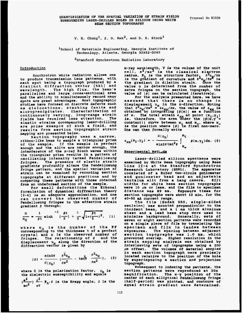
QUANTIFICATION OF THE SPATIAL VARIATION OF STRAIN FIELDS SURROUNDING LASER-DRILLED HOLES IN SILICON USING WHITE
BEAM SECTION TOPOGRAPHY Proposal No 8103M
Y , H. Chung', 2. U. Rek2, and S. R. Stock'
'School of Materials Engineering, Georgia Institute of Technology, Atlanta, Georgia 30332-0245
lstanford Synchrotron Radiation Laboratory
Introduction
Synchrotron white radiation allows one to produce transmission Laue patterns, wlth each spot being a togograph produced by a distinct diffraction vector [hkll and wavelength. The high flux, the beam's parallelism and large cross-sectional area and the ability to simultaneously record many spots art great advantages. Most topographic studiaa have focused on discrete defects such as dislocations, stacking faults and microprecipitates. Characterization of continuously varying, long-range strain fields has received less attention. The elastic strains accompanying laser-drilling are prime example of such a field, and results from section topographic *train mapping are presented below,
Section topography uses a narrow, rlbbon-like beam to sample a triangular prism of the sample. If the sample is perfect enough and the slits ore narrow enough, the interference of the x-ray Bloch waves within the triangular prism rtsults in 8; pattern of oscillating intensity termed Pcndsllosung fringes. The presence of clastic strain gradient6 profoundly alters the Pendtlfosung fringe pattern, and tho spatial variation of mtrain can be examined by recording section topographs at different positions and by comparing these pattern8 with those obtained from an undeformed section of the sampls.
Bor small deformations the Eikonal formulation of dynamical diffraction theory [2-41 is m adequate approximation. and one c a n c o n v e r t the abaerveb number of Pendellosung fringes to the effective strain gradient 8 through:
w h e r e n is t h e n u m b t r of the PF corrcspondPng to the thickness t of a perftct crystal and n is the observed number of fringes. The relationship of 6 and the displacement u along the dirtction o t the diffraction vscror is given by
- 2 sin28 a u,
where C la the polarization factor, x is tho dioloctric susceptibility and equats e* a
(-1 1 Bh.8 is the Bragg angle, a is the ulc2 xv
x-ray wavelen th, V 15 the volumt of the unit cell, e2/mc' is the classlcal e 1 ctron
I radius. Fh is the structure factor is the gradient of curvature and 3' /ax is tht gradiont in dilation strain. value n is determined from the numbtr of extra fringe8 on the section topograph, the value of 1st can be calculated iteratively.
For the analyaia presented here, wo have a s a u m a d that there is no change in displacemen ux in the 2-direction. Noting that a2,/axi = a6, /ax, the value of a is obtained by Sntegrahnp IB(x)l as a funsion of x. The total strain at point (xi,yi) iu, therefore, the area under the IB (+ ,y = constant) I curve between x and x-. where x, la the position where lfil is first non-zero. One can then formally write
a ?2 u /az
us nce the
Exner imental Met L d s
Laser-drilled silicon specimrns wose examined by White Beam topography using Beam L i n e 11-4 at the Stanford Synchrotron Remearch Laboratory (SSRL). The apparatue consisted of a Huber two-circle goniometer and goniometer head and an ad justablt tantalum slit from a Lang camera. Slit widths for white beam section topographs were 10 ,urn or lees, and the film to specimen distance was 89 mm. Exposure times tor section topographs were about 10 min, for the 40-50 mA current range,
The film (Kodak SR6, @ingle-sided emulsion) was mounted perpendicular to the incident beam, and a 1 mm thick aluminum sheet and a lead beam stop were used to minimize background. Qenormlly, sets of aevon or eight section patterns were recorded on a single piece of film by trarmlating the specimen and f i l m in tandem between exposures. The spacing between adjacent eection topographs was 1.0 am, which prevented overlap. Higher resolution in the strain mapping analyeis was obtained by interleaving sets of topographs using a 200 p i affsot. The volumea of material sampled in each sectlon topograph were precieely located relative to the position of the hole by superimposing a section and projection topograph.
Subsequent to indexing the patterns, the section patterns were reproduced at 26x magnification. The x-y position ot the center of tach olliptical Pendellosung tringe (half-period) was plotted, and contoura of epual strain gcadiont ware determined.
115
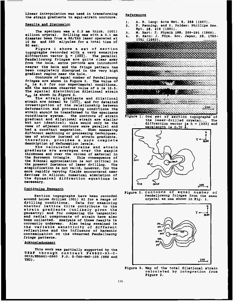
Linear interpolation was used in trensforming the strain gradientgl to equi-strain contours,
Results an8 Discussion
The specimen wa6 a 0.6 mm thick, (001) silicon crystal. Drilling was nith a 0.1 nun dianeter beam from a Nd/YAG laser operated at 10 nz and 260 mJ/pulse for a total t ime of
Figure 1 shows a siet of section topographs recorded with a very sensitive difiractlon vector h [3?S]. The parallel Pendellosung fringes are quite clear away from the hole, extra periods are introduced nearer tho hole and the Fringe pattern has been completely disrupted in the very high gradient region near the hole.
Contours of equal number o i Pendellosung fringes are shown in Figure 2. The value of no I s 9 . 6 for our experimental conditions. and tho maximum obs8rved value of n is 12.5. The spatial distribution dilational strain em i. shown in Figure 3.
The strain gradients and dilational strain are normal to t lTZ], and for detailed investigation of the relationship between deformation and processing conditions the *train should be, tranmtormed to the apecimene coordinate system. The contours of atrain gradient and dilational strain are similar b#t not identical; thia would only be the case if adjacent contours were parallel and had a conatant separation. When assessing different machining or processing techniques, w e of strains instead of atrain gradients. therefore, provides a m o r e complete deacription of deformition levels.
The calculated strains and strain gradient* are averages over the sample thickness and ever the volume of material in the Borrmann triangle. Thi8 consequence of ths Bfkonal approximation is not critical in the present instance of laser drilling. The aimpllfication is3 not valid, however, t o r the more rapidly varying fields encountered neer devices in silicon; nuaerical simulation of the dynamical diffraction equations ia necemsary.
Continuing Research
60 6CC.
Section topographs have been recorded around holes drilled (001) Si for a range of drilling conditions, Data for examining whether lattice tilts contribute to the strain gradient8 (unlikely given the geometry) and for comparing the tangential and radial componentm o f strain have also been collected. Analysis of these results is currently underway. Also being examined is the variable sensitivity of different reflections and the influence of hatmonic contamination on the observed Pendellosung fringe patterns. Acknowledoement
This work nae partially supported by the U S A % t h r o u g h c o n t r a c t 8 4 8 6 2 0 - 8 5 - C - 0013/8B6861-036Q P.0. S-760-6MQ-103 (SRS and YHC).
References
1. A . R. Lang: Acta Met. 8 , 358 (1961). 2. P, Penning, and D. Polder: Phillips Res,
3. M. Hart: 2. Physlk 190, 269-291 (1966). 1, N. Kato: 3. Phye. SOC. Japan, 10, 1785-
Rpt. 16, 419 (1961).
the laser-drilled crystal.,, ~ h a diffraction vector i s h 5 [a331 and wavelength is 0 70 i.
f X
Figure 2. Contours of equal number O f Pendellosung fringts from the same cryetal as wae ehown in Fig. 1.
y w i
Figure 3. Map of the total dilational strain calculated by Integration from Bipurt 2 .
116

Proposal No. 9900M
TWO DIMENSIONAL COMPRESSIBILITY
OF ELBcI1ROcHEnICALI.Y ADSORBED LEAD ON SILVER (111)
P.N. Ross and J.B. Kortright Lawrence Berkeley Laboratory University of California Berkeley, California 94720
O.R. Melroy, M.F. Toney, G.L. Borges, and M.G. Samant IBM Almaden Research Center
650 Harry Road San Jose, California 95120
L. Blum Physics Department
College of Natural Sciences P.O. Box AT
Rio Piedras, Puerto Rico 99831
We have applied the grazing-incidence scattering geometry to study electrochemical deposition of lead monolayers on silver (111). Measurements were made in situ (in contact with solution). For deposition potentials between underpotential monolayer formation and bulk deposition, the near-neighbor distance of the lead monolayer decreases linearly with applied potential, which is proportional to the chemical potential. This variation is shown in the figure. Since the lead monolayer is in equilibrium with lead ions in solution, the isothermal compressibility of the monolayer can be calculated from the potential dzpendence of the near-neighbor distance and is 0.98 A /eV. This potential-dependent strain in the monolayer has implications on the wetting behavior of lead.
The research effort at the Lawrence Berkeley Laboratory was supported by the Office of the Director, Program Development Funds, through an agreement with the U . S . Department of Energy under contract DE-AC03-76SF00098. The research at IBM and the University of Puerto Rico was supported by the Office of Naval Research.
deposition
€ I I I I 1 I ' I I I
-0.4 -0.6 -0.8 -1 .o Potential (VI
XBL 87125761
117
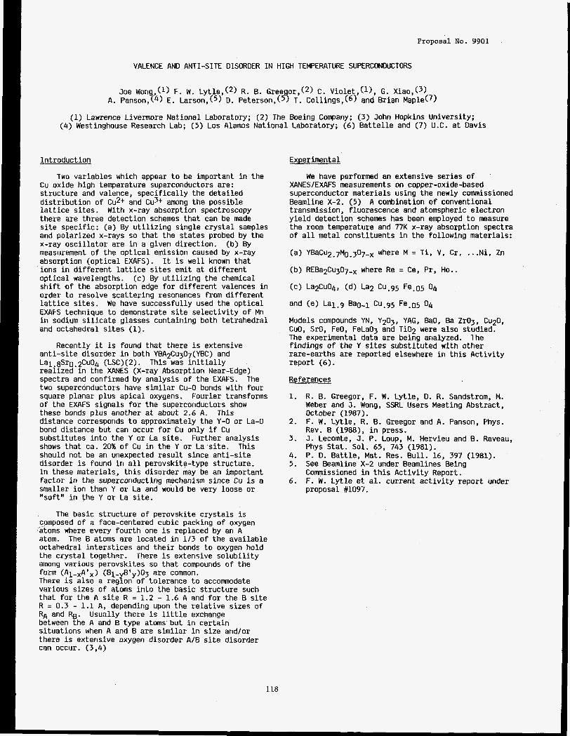
Proposal No. 9901
VALENCE AND ANTI-SITE DISORDER I N HIGH TEMPERATURE SUPERCONDUCTORS
Joe Wong,(l) F. W. Ly t le , (2) R. B. Gree or,(2) C. V i o l e t , ( l ) , G. X i a ~ , ( ~ ) A. Panson,(4) E. Larson,(5) D. Peterson,(5 3 T. Col l ings, (6) and B r i a n Maple(7)
(1) Lawrence Livermore Nat iona l Laboratory; (2) The Boeing Company; (3) John Hopkins Un ivers i ty ; (4) Westinghouse Research Lab; (5) Los Alamos N a t i o n a l Laboratory; (6) B a t t e l l e and (7) U.C. a t Davis
I n t r o d u c t i o n
Two var iab les which appear t o be impor tant i n the Cu ox ide h i g h temperature superconductors are: s t r u c t u r e and valence, s p e c i f i c a l l y the d e t a i l e d d i s t r i b u t i o n o f Cu2+ and Cu3+ among the poss ib le l a t t i c e s i t e s . With x-ray absorpt ion spectroscopy there are th ree de tec t ion schemes t h a t can be made s i t e spec i f i c : (a) By u t i l i z i n g s i n g l e c r y s t a l samples and p o l a r i z e d x-rays so t h a t the s ta tes probed by t h e x-ray o s c i l l a t o r are i n a g iven d i r e c t i o n . measurement o f the o p t i c a l emission caused by x-ray absorpt ion ( o p t i c a l EXAFS). ions i n d i f f e r e n t l a t t i c e s i t e s emit a t d i f f e r e n t o p t i c a l wavelengths. (c) By u t i l i z i n g the chemical s h i f t o f the absorpt ion edge f o r d i f f e r e n t valences i n order t o reso lve s c a t t e r i n g resonances from d i f f e r e n t l a t t i c e s i t e s . We have success fu l l y used the o p t i c a l EXAFS technique t o demonstrate s i t e s e l e c t i v i t y o f Mn i n sodium s i l i c a t e glasses conta in ing both t e t r a h e d r a l and octahedra l s i t e s (1).
a n t i - s i t e d isorder i n both YBA2Cu307(YBC) and La1.8Sr0.2CuO4 (LSC)(2). Th is was i n i t i a l l y r e a l i z e d i n t h e XANES (X-ray Absorption Near-Edge) spectra and confirmed by ana lys is o f t h e EXAFS. two superconductors have s i m i l a r Cu-0 bonds with fou r square p lanar p lus a p i c a l oxygens. o f the EXAFS s igna ls f o r the superconductors show these bonds p l u s another a t about 2.6 A. d is tance corresponds t o approximately the Y-0 or La-0 bond d is tance bu t can occur f o r Cu on ly i f Cu s u b s t i t u t e s i n t o t h e Y or La s i t e . shows t h a t ca. 20% o f Cu i n the Y or La s i t e . Th is should no t be an unexpected r e s u l t s ince a n t i - s i t e d isorder i s found i n a l l perovsk i te- type s t ruc tu re . I n these mater ia ls , t h i s d isorder may be an impor tant f ac to r i n the superconducting mechanism s ince Cu i s a smaller i o n than Y or La and would be very loose or " so f t " i n the Y or La s i t e .
(b) By
I t i s w e l l known t h a t
Recently i t i s found t h a t there i s extens ive
The
Four ie r transforms
This
Fur ther ana lys is
The bas ic s t r u c t u r e o f perovsk i te c r y s t a l s i s composed o f a face-centered cubic packing o f oxygen dtoms where every f o u r t h one i s replaced by an A atom. The B atoms are located i n 1 /3 o f t h e a v a i l a b l e octahedra l i n t e r s t i c e s and t h e i r bonds t o oxygen h o l d the c r y s t a l together . among var ious perovsk i tes so t h a t compounds o f the form (A1-xAtx) (B1-yB'y)03 are comnon. There i s also a reg ion o f to le rance t o accomnodate var ious s izes o f atoms i n t o the bas ic s t r u c t u r e such t h a t f o r the A s i t e R = 1.2 - 1.6 A and f o r the B s i t e R = 0.3 - 1.1 A, depending upon t h e r e l a t i v e s izes o f RA and R5. between the A and B type atoms bu t i n c e r t a i n s i t u a t i o n s when A and B are s i m i l a r i n s i ze and/or there i s extens ive oxygen d isorder A/B s i t e d isorder can occur. (3,4)
There i s extens ive s o l u b i l i t y
Usua l ly there i s l i t t l e exchange
Experimental
We have performed an extens ive ser ies o f XANES/EXAFS measurements on copper-oxide-based superconductor m a t e r i a l s us ing the newly commissioned Beamline X-2. (5) A combination o f convent ional transmission, f luorescence and atomspheric e l e c t r o n y i e l d de tec t ion schemes has been employed t o measure the room temperature and 77K x-ray absorpt ion spect ra o f a l l meta l cons t i tuents i n the f o l l o w i n g mater ia ls :
(a) Y B ~ C U ~ . ~ M O . ~ O ~ - ~ where M = T i , V, C r , ... Ni , 2n
(b) REBa2Cu307_, where Re = Ce, Pr, Ho..
and (e) L q . 9 Ba0-l Cu.95 Fe.05 04
Models compounds YN, Y2O3, YAG, BaO, Ba ZrO3, C U ~ , CuO, SrO, FeO, FeLa03 and T i02 were a l s o studied. The experimental data are being analyzed. l h e f i n d i n g s o f the Y s i t e s s u b s t i t u t e d with o ther rare-ear ths are repor ted elsewhere i n t h i s A c t i v i t y r e p o r t (6).
References
1. R. B. Greegor, F. W. Ly t l e , D. R. Sandstrom, M. Weber and J. Wong, SSRL Users Meeting Abst ract , October (1987).
2. F. W. L y t l e , R. B. Greegor and A. Panson, Phys. Rev. B (19881, i n press.
3. J . Lecomte, J. P. Loup, M. Hervieu and B. Raveau, Phys S ta t . Sol. 65, 743 (1981).
4. P. D. Bat t l e , Mat. Res. B u l l . 16, 397 (1981). 5. See Beamline X-2 under Beamlines Being
Commissioned i n t h i s A c t i v i t y Report. 6. F. W. L y t l e e t a l . cur ren t a c t i v i t y r e p o r t under
proposal %1097.
118

Proposa l No. 9901M
X-RAY TOMOGRAPHY ON BEAMLINE X
J. H. Kinney, Q.C. Johnson, R.Allyn Saroyan, M.C. Nichols*, U.Bonset and R. Nusshardtf Lawrence Livermore National Laboratroy, Livermore, CA 94550
Introduction:
We have tested a high resolution solid state CCD ar- ray detector for x-ray imaging on the BL-X wiggler. The principal applications of this imaging detector are microra- diography and three-dimensional microtomography. Appli- cations to topography can be anticipated.
The intense x-ray flux from the 15 period wiggler makes BL-X an ideal source for CCD imaging methods. In this report, we will describe preliminary results from our use of the CCD array for beam characterization and for three-di- mensional microtomography. Future modifications to the existing hardware that will take advantage of the unique properties of BGX will also be discussed.
Experimental Apparatus:
The experimental apparatus has been discussed in detail elsewhere'. Briefly, the x-ray beam from the double crystal monochromator passes through a sample placed on a rotary stage and is converted to visible light in a high- resolution phosphor screen. The phosphor screen is imaged with a CCD array camera which is run in a charge integrating mode. The image data for each view angle through the ob- ject is used to reconstruct the three-dimensional structure of the sample. By modulating the energy of the x-ray beam, it is possible to obtain chemical information about the samples as well as structural information in many cases2.
Applications:
In a preliminary study on the new BL-X wiggler, we used the CCD camera to characterize the x-ray beam and to perform tomography on samples of coal. Figure 1 shows the surface contour plots from a three-second exposure on BL-X operating at l4kGauss. The x-ray energy is IO-keV. From exposure data, we estimate that the x-ray intensity is roughly 20 times that of the bending magnet line BL-1-5 (accurate flux measurements cannot be made from these
1 Kinney, J.H., et al., Rev. Sci. Instrum., 59, 196(1988). 2KinneyJJH., et al., Appl. Opt., 25, 4583(1986).
measurements, since both lines have not been characterized with the same camera parameters). From these data it is possible to discern a curvature in the intensity profile radiat- ing outward from the center of the beam. We believe that this curvature is due to heat loading of the first crystal3. For digital radiography and tomography, this curvature does not pose a problem.
Figure 2 shows a tomographic reconstruction of a slice through a sample of coal. The beam energy was 7300 eV, which was on the high-absorption side of the k-edge of iron. The bright features in the reconstruction ( which are not reproduced well in the black and white screen-print prepared for this publication) are associated with iron rich precipitates that we have subsequently identified as iron sulfide. Pore structures smaller than 10 microns can also be observed. The ring structures are artifacts caused by insufficient detec- tor normalization, and can be corrected by taking a greater number of reference images. The halo surrounding the sample is caused by x-ray scatter, which we believe can be removed by introducing Bragg reflecting optics behind sample.
Future Directions:
Fogging due to x-ray scatter, and grain structure in the phosphor, are the principal limits to the spatial resolution in our CCD imaging system. The high intensity afforded by the wiggler on BL-X allows us to take a new approach to overcome these limitations. We intend to introduce a Bragg reflecting optic between the sample and the phosphor screen. This will eliminate scattered radiation outside of the Bragg angle. By using an asymmetric cut, it is also possible to in- troduce magnification in one dimension which could increase the spatial resolution in the slice plane.
Acknowledgements:
We would like to thank A1 Knabe (LLNL) for his technical support in all stages of this work. Also, we appre- ciate the support of R. Taylor (LLNL) who supplied us with
Figure 1: Surface plot of x-ray intensity from B L X wiggler operating unfocussed at ICkGauss. Scale is 5 - p .
Figure 2 Coal chemistry and structure determined from to- mography. Chemical tomography may play an important role in studying low temperature burning of coal.
Kinney, J.H., Wong, J. and Troxel, T. (accompanying
Sandia National Laboratory, Livermore, CA. 94550. Department of Physics, Dortmund University, Dortmund,
rir article in this reoort)
West Germany.
119

Proposal NO. 9 9 0 1 ~
BEAM LINE X-2 PROFILE MEASUREMENT WITH A CCD IMAGER
J. H. Kinney, J. Wong and T. Troxel*
L a d n c e Livermore National Laboratory Livermore, CA 94550
Introduction:
As part of the characterization and commissioning of Beamline X-2, we have measured a series of initial beam profiles delivered at the sample plane with a high-resolution CCD array camera to map the intensity of the monochromatic beam in two dimensions under various conditions of photon energy, wiggler field strength and focussing. The CCD camera, used principally for high-resolution radiography and tomography, has been described in detail elsewhere'. Briefly, the x-rays emerging from the double crystal monochromator are converted to visible light on a high-resolution phosphor screen. The phosphor image is projected onto the CCD array with a bi-convex lens operating in a magnifying geometry. The camera images a 2-mm by 2-mm area of the beam with roughly 5-pm spatial resolution.
Results:
Figure 1 shows the surface plot of the intensity profile of beamline X-2 operating at 14 kGauss. The crystals were tuned to 50% of the peak intensity of the rocking curve at 10keV. We call this the detuned position. The image spans approximately I-mm on either side of the horizontal center of the beam. The x and y axis units are 5-pm. The intensity units are in CCD pixel units, and are directly proportional to the incident x-ray fluence.
The surface contours indicate a saddle shaped depression in the center of the beam. We believe that this depression is due to crystal heating effects from the high heat load during operation at 14-kGauss. This depression is also present in the tuned beam at the same wiggler field strength.
Figure 2 shows the beam profile for the detuned beam under the same conditions as in figure 1, but in the focussed mode. The tuning compensates for the loss of reflectivity due to thermal loading. Because the exposure times for the two images were different, it is impossible to use this data to accurately determine the increase in intensity from focussing, although an increase is observed.
Figure 1: Surface plot of the intensity profile from beamline X-2 operating with a wiggler field strength of 14 kGauss in the detuned mode. The scale is 5 -pm, and the image spans approximately Imm to either side of the horizontal center. We believe the dip in the intensity profile is indicative of thermal effects on the f i t crystal.
Figure 2 Surface plot of the intensity profile from beamline X-2 operating und7 identical conditions as in figure 1 with the exception of being focussed with the mirror. Focussing compensates for the dip observed in figure 1.
The depression in the beam was not observed at a wiggler field strength of 8 kGauss, although a direct comparison of the data has yet to be completed. This is a further indication that thermal effects are being seen on the first crystaL Future work will characterize the entire profile of the beam under a variety of wiggler field strengths in order to improve our understanding of the beamline performance.
* Stanford Synchrotron Radiation Laboratory, Stanford, CA
1 Kinney, J.H., et al., Rev. Sci. Instrum., 59, 196(1988).
120
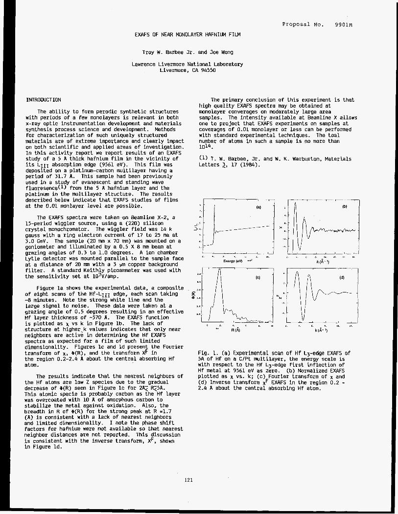
Proposal No. 9901M
EXAFS OF NEAR MONOLAYER HAFNIUM FILM
Troy W . Barbee Jr. and Joe Wong
Lawrence Livermore National Laboratory Livermore, CA 94550
INTRODUCTION
The a b i l i t y t o form perodic synthetic structures with periods of a few monolayers is relevant i n both x-ray opt ic instrumentation development and materials synthesis process science and development. fo r characterization of such uniquely structured materials are of extreme importance and clear ly impact on both s c i e n t i f i c and applied areas of investigation. In t h i s ac t iv i ty report we report results of an EXAFS study of a 5 A thick hafnium film in the v ic in i ty of its LIII absorption edge (9561 eV). This fi lm was deposited on a platinum-carbon multilayer having a period of 31.7 A. This sample had been previously used i n a study of evanescent and standing wave fluoresence(1) from the 5 A hafnium layer and the platinum i n the multi layer structure. The resu l t s described below indicate t h a t EXAFS studies of fi lms a t the 0.01 monlayer leve l are possible.
15-period wiggler source, using a (220) s i l icon crys ta l monochromator. The wiggler f i e l d was 14 k gauss with a r ing electron current of 17 t o 25 ma at 3.0 GeV. The sample (20 m x 70 mm) was mounted on a goniometer and illuminated by a 0.5 X 8 mm beam a t grazing angles of 0.3 t o 1.0 degrees. A ion chamber Lytle detector was mounted para l le l t o t h e sample face a t a distance of 20 mm with a 3 urn copper background f i l t e r . t h e sens i t iv i ty set a t 107V/amp.
Figure l a shows the experimental data, a composite of e ight scans of t h e Hf-LIII edge, each scan taking -8 minutes. Note the strong white l i n e and the large s ignal t o noise. grazing angle of 0.5 degrees resul t ing i n an e f fec t ive Hf layer thickness of -570 A - is plot ted as x vs k in Figure lb . s t ructure a t higher-k values indicates t ha t only near neighbors a re active in determining the Hf EXAFS spectra as expected fo r a f i l m of such limited dimensionality. transform of X, cP(R), and the transform XF i n t h e region 0.2-2.4 A about the central absorbing Hf atom.
Methods
The EXAFS spectra were taken on Beamline X-2, a
A standard Keithly picoammeter was used with
These data were taken a t a
The EXAFS function The lack of
Figures IC and Id present the Fourier
The results indicate t h a t the nearest neighbors of t h e Hf atoms are low 2 species due t o the gradual decrease of @(R) seen i n Figure IC fo r 2x2 R25A. This atomic specie is probably carbon as the Hf layer was overcoated with 10 A of amorphous carbon t o s t a b i l i z e the metal against oxidation. Also, t h e breadth in R of ,(R) for t h e strong peak a t R =1.7 (A) is consistent with a lack of nearest neighbors and l imited dimensionality. I note the phase s h i f t factors fo r hafnium were not avai lable so tha t nearest neighbor distances are not reported. is consistent with the inverse transform, XF, shown in Figure Id.
This discussion
The primary conclusion of t h i s experiment is that high qual i ty EXAFS spectra may be obtained a t monolayer converages on moderately large area samples. one t o project t h a t EXAFS experiments on samples at coverages of 0.01 monolayer or less can be performed with standard experimental techniques. The t o a l number of atoms in such a sample is no more than
(1) T. W . Barbee, Jr. and W . K. Warburton, Materials Letters 3, 17 (1984).
The intensi ty available a t Beamline X allows
1014.
Fig. 1. (a) Experimental scan of Hf L3-edge EXAFS of 5 A of Hf on a C/Pt multilayer, the energy scale is with respect t o the Hf L3-edge f i r s t inf lect ion of Hf metal a t 9561 eV as zero. (b) Normalized EXAFS plot ted as x vs. k; (c) Fourier transform of x and (d) Inverse transform XF EXAFS i n t h e region 0.2 - 2.4 A about the cent ra l absorbing Hf atom.
121
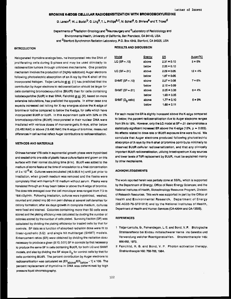
Letter of fefmf
BROMINE K-BDQB CELLULAR WADIOSENBITIZATION WITH BROMODEOXYURIDINE
D. braen', W, J. Bodellb, C. Llnga, T. L, Phlllipsafc, M. Schella, D. Shrleve'and T. Proxeld
1NTRODUC7lON
Halogenated thymidine analogs becL ne incorporated into the DNA of proliferating cells during S-phase and may be used clinically to radiosensitize tumors through unknown mechanisms. One possible mechanism involves the production of (highly radiotoxic) Auger electrons following photoelectric absorption of an X-ray by the K-shell of tho incorporated halogen. Tisljar-Lentulis gt g. [ I ] has predicted that the contribution by Auger electrons to radiosensitization should be larger f3r cells containing bromodeoxyuridine (BUdR) than for cells containing iododeoxyuridine (IUdR) in their DNA; Fairchild gal. [2], based on more extensive calculations, has predicted the opposite. In either case one expects increased cell killing for X-ray energies above the K-edge of bromine or iodine compared to below the K-edge, for cells which have Incorporated BUdR or IUdR. In this experiment cells with 32% or 0% bromodeoxyuridine (BUdR) incorporated in their nuclear DNA were irradiated with various doses of monoenergetlc X-rays, either below (13.450 KeV) or above (13.490 KeV) the K-edge of bromine; measured differences in cell survival reflect Auger contributions to radiosensitization.
METHODS AND MATERIALS
Chinese hamster V79 cells in exponential growth phase were trypsinized and seeded onto one side of plastic tissue culture flasks and grown on this surface with their normal doubling time (9 hr). BUdR was added to the medium of some ftasks at the time of innoculation to a final concentration of 3 x M. Cultures were incubated (46.5-55.5 hr) until just prior to irradiation, when growth medium was removed and the flasks were completely filled with Ham's F-12 medium without serum. Flasks were translated through an X-ray beam below or above the K-edge of bromine. The dose rate averaged over the cell monolayer area ranged from 7.3 to 18.6 cGy/min. Following irradiation, cultures were trypsinized, washed, counted and plated into 60 mm petri dishes at several cell densities for colony formation; after six days growth in complete medium, cultures were fixed and stained. Colonies containing more than 50 cells were scored and the plating efficiency was calculated by dividing the number 0: colonies scored by the number of cells plated. Surviving fraction (SF) was calculated by dividing the plating efficiency for treated cells by that for controls. SF data as a function of absorbed radiation dose were fit to linear-quadratic (LQ) and single hit multitarget (SHMT) models. Enhancement ratios (ER) were obtained by dividing the radiation dosa necessary to produce a given (0.10,O.Ol) SF in controls by that necessary to produce the same SF in cells containing BUdR, for both LQ and SHMT models, and also by dividing the SF slope Do for control cells by that for cells containing BUdR. The percent contribution by Auger electrons to radiosensitization was calculated as (ERabov$ERbelow - 1) x 100. The percent replacement of thymidine in DNA was determined by high
Departments of aRadlatlon Oncology and bNeurosurgery and 'Laboratory of Radloblology and Environmental Health, Unlverslty of Californla, San Francisco, CA 94143, USA
and dStanford Synchrotron Radiation Laboratory, P.O. Box 4349, Stanford, CA 94305, USA
RESULTS AND DISCUSSION
Model LQ (SF=.lO) above
below LQ (SF=.OI) above
below SHMT (SF=.lO) above
below SHMT (SF=.OI) above
below SHMT (Do ratio) above
below
- ER 2.31 0.10 2.25 * 0.10 2.09 0.05 1.87 * 0.05 2.27 * 0.08 2.12 * 0.09 2.05 0.06 1.93 * 0.05
1.77 0.10 1.68 * 0.1 1
- Auger(%) 3*6%
12*4%
7*6%
6*4%
5*9%
For each model the ER Is slightly increased above the K-edge compared to below; the percent radiosensitlzatlon due to Auger electrons ranges from 3% to 12%. However, only the LQ model at SF=.Ol demonstrates a statistically significant increased ER above the K-edge (12%, p = 0.003). 'No effects related to dose rate or BUdR exposure time were found. We conclude that Auger electrons produced following photoelectrlc absorption of X-rays by the K-shell of bromine contribute minimally to observed BUdR cellular radiosensitization, and that any clinically important BUdR radiosensitization, utilizing broad-spectrum X-ray sources and lower levels of TdR replacement by BUdR, must be explained mainly by other mechanisms.
ACKNOWLEDGEMENTS
The work reported herein was partially done at SSRL, which is supported by the Department of Energy, Office of Basic Energy Sciences; and the National Institutes of Health, Biotechnology Resource Program, Division of Research Resources. This work was supported in part by the Office of Health and Environmental Research, Department of Energy (DE-AC03-76-SF01012) and by the National Institutes of Health, Department of Health and Human Services (CA-42044 and CA-13525).
REFERENCES
1 Tisljar-Lentulis, G., Feinendegen, L. E. and Bond, V. P. Biologische Strahleneffekte bei Einbau mittelschwerer Kerne ins Gewebe und Verwendung weicher Roentgenstrahlen. Strahlentherapie 145: 656-662,1973.
2 Fairchild, R. G. and Bond, V. P. Photon activation therapy. Strahlentherapie 160: 758-763, 1984.
pressure liquid chromatography.
122
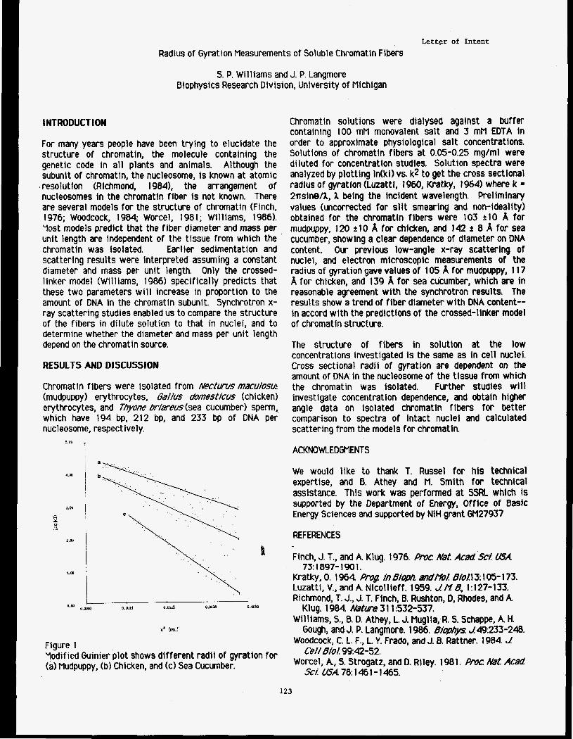
Letter of Intent
Radius of Gyration Measurements of Soluble Chromatin Fibers
S. P. Williams and J. P. Langmore Biophysics Research Division, University of Michigan
INTRODUCTION
For many years people have been trying to elucidate the structure of chromatin, the molecule containing the genetic code in all plants and animals. Although the subunit of chromatin, the nucleosome, i s known at atomic resolution (Richmond, 19841, the arrangement of nucleosomes in the chromatin fiber i s not known. There are several models for the structure of chromatln (Finch, I 976; Woodcock, 1984; Worcel, I 98 I ; Wi 11 lams, 1 986). Yost models predict that the fiber diameter and mass per unit length are independent of the tissue from which the chromatin was isolated. Earlier sedimentation and scattering results were interpreted assuming a constant diameter and mass per unit length. Only the crossed- linker model (Williams, 1986) specifically predicts that these two parameters w i l l increase in proportion to the amount of DNA in the chromatin subunit. Synchrotron x- ray scattering studies enabled us to compare the structure of the fibers in dilute solution t o that in nuclei, and t o determine whether the diameter and mass per unit length depend on the chromatin source.
RESULTS AND DISCUSSION
Chromatin fibers were isolated from Nectums mxu/osu~ (mudpuppy) erythrocytes, Ga//us domesticus (chic ken) erythrocytes, and Thyone brimus (sea cucumber) sperm, which have 194 bp, 212 bp, and 233 bp of DNA per nucleosome, respectively.
Chromatin solutions were dialysed against a buffer containing 100 mM monovalent salt and 3 mM EDTA In order to approximate physiological salt concentrations. Solutions of chromatin fibers at 0.05-0.25 mg/ml were diluted for concentration studies. Solution spectra were analyzed by plotting In(kl) vs. k2 to get the cross sectional radius of gyration (Luzatt I, 1960, Kratky, 1964) where k = 2nsine/1, 1 being the incident wavelength. Preliminary values (uncorrected for s l i t smearing and non-ideality) obtained for the chromatin fibers were 103 t10 A for mudpuppy, 120 t10 A for chlcken, and 142 2 8 A for sea cucumber, showing a clear dependence of diameter on DNA content. Our previous low-angle x-ray scattering of nuclei, and electron microscopic measurements of the radius of gyration gave values of 105 A for mudpuppy, 1 17 A for chicken, and 139 A for sea cucumber, which are in reasonable agreement with the synchrotron results. The results show a trend of fiber diameter with DNA content-- in accord with the predictions of the crossed-linker model of chromatin structure.
The structure of fibers in solution at the low concentrations investigated is the same as in cell nuclei. Cross sectional radii of gyration are dependent on the amount of DNA in the nucleosome of the tissue from which the chromatin was isolated. Further studies w i l l investigate concentration dependence, and obtain higher angle data on lsolated chromatin fibers for better comparison to spectra of intact nuclei and calculated scattering from the models for chromatin.
5.W
4.w
J.W
2.M
1.M
Figure 1 Yodified Guinier plot shows different radii of gyration for (a) Mudpuppy, (b) Chicken, and (c) Sea Cucumber.
ACKNOWLEDGMENTS
We would like to thank T. Russel for his technical expertise, and B. Athey and M. Smith for technical assfstance. This work was performed at SSRL which is supported by the Department of Energy, Office of Basic Energy Sciences and supported by NIH grant GM27937
REFERENCES
Finch, J. T., and A Klug. 1976. Rmc #at Acad ScL USA
Kratky, 0. I 964. P ! fn 6&1h mdPtolB/oll 3: 105- 173. Luzatti, V., and A Nicollieff. 1959. JHB, 1:127-133. Richmond, T. J., J. T. Finch, B. Rushton, D, Rhodes, and A
Williams, S., B. D. Athey, L. J. Muglla, R. S. Schappe, A H. Gough, and J. P. Langmore. 1986. BiopnyS: Je.233-240.
Woodcock, C. L. F., L. Y. Frado, and J. 0. Rattner. 1984. J Ce// 6iol99:42-52.
Worcel, A., S. Strogatz, and D. Riley. 1981. p/ulcr AM Acad
73: 1897- 1 9 0 1 .
Klug. 1984. Mtm 3 1 1 :532-537.
SCY USA 78 1461- 1465.
123
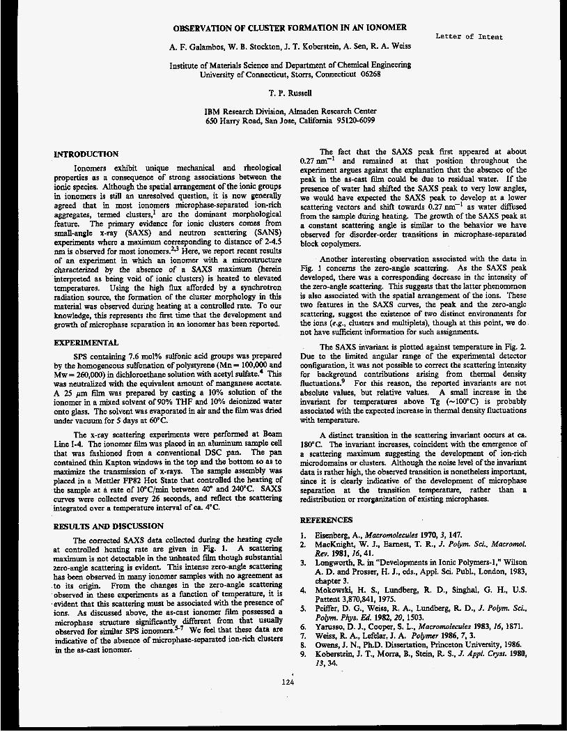
OBSERVATION OF CLUSTER FORMATION IN AN IONOMER Letter of Intent
A. F. Galambos, W. B. Stockton, J. T. Koberstein, A. Sen, R. A. Weiss
Institute of Materials Science and Department of Chemical Engineering University of Connecticut, Storrs, Connecticut 06268
T. P. Russell
IBM Research Division, Almaden Research Center 650 Harry Road, San Jose, California 95120-6099
INTRODUCTION
Ionomers exhibit unique mechanical and rheological properties as a consequence of strong associations between the ionic species. Although the spatial arrangement of the ionic groups in ionomers is still an unresolved question, it is now generally agreed that in most ionomers microphase-separated ion-rich aggregates, termed clusters,' are the dominant morphological feature. The primary evidence for ionic clusters comes from small-angle x-ray (SAXS) and neutron scattering (SANS) experiments where a maximum corresponding to distance of 2-4.5 nm is observed for most ionomers?f Here, we report recent results of an experiment in which an ionomer with a microstructure characterized by the absence of a SAXS maximum (herein interpreted as being void of ionic clusters) is heated to elevated temperatures. Using the high flux afTorded by a synchrotron radiation source, the formation of the cluster morphology in th is material was observed during heating at a controlled rate. To our knowledge, this represents the fvst time that the development and growth of microphase separation in an ionomer has been reported.
EXPERIMENTAL
SPS containing 7.6 mol% sulfonic acid groups was prepared by the homogeneous sulfonation of polystyrene (Mn = 100,000 and Mw = 260,000) in dichloroethane solution with acetyl ~ul fa te .~ T h i s was neutralized with the equivalent amount of manganese acetate. A 25 pm film was prepared by casting a 10% solution of the ionomer in a mixed solvent of 90% THF and 10% deionized water onto glass. The solvent was evaporated in air and the film was dried under vacuum for 5 days at 60°C.
The x-ray scattering experiments were performed at Beam Line 1-4. The ionomer ftlm was placed in an aluminum sample cell that was fashioned from a conventional DSC pan. The pan contained thin Kapton windows in the top and the bottom so as to maximize the transmission of x-rays. The sample assembly was placed in a Mettler FP82 Hot State that controlled the heating of the sample at a rate of 10"C/min between 40" and 240°C. SAXS curves were collected every 26 seconds, and reflect the scattering integrated over a temperature interval of ca. 4°C.
RESULTS AND DISCUSSION
The corrected SAXS data collected during the heating cycle at controlled heating rate are given in Fig. 1. A scattering maximum is not detectable in the unheated film though substantial zero-angle scattering is evident. This intense zero-angle scattering has been observed in many ionomer samples with no agreement as to its origin, From the changes in the zero-angle scattering observed in these experiments as a function of temperature, it is evident that this scattering must be associated with the presence of ions. As discussed above, the as-cast ionomer film possessed a microphase structure significantly different fiom that U S a y observed for similar SPS ion~mers.~-' We feel that these data are indicative of the absence of microphase-separated ion-rich clusters in the as-cast ionomer.
The fact that the SAXS peak first appeared at about 0.27M1' and remained at that position throughout the experiment argues against the explanation that the absence of the peak in the as-cast film could be due to residual water. If the presence of water had shifted the S A X S peak to very low angles, we would have expected the SAXS peak to develop at a lower scattering vectors and shiR towards 0.27 nm-' as water diffused from the sample during heating. The growth of the SAXS peak at a constant scattering angle is similar to the behavior we have observed for disorder-order transitions in microphase-separated block copolymers.
Another interesting observation associated with the data in Fig. 1 concerns the zero-angle scattering. As the SAXS peak developed, there was a corresponding decrease in the intensity of the zero-angle scattering. This suggests that the latter phenomenon is also associated with the spatial arrangement of the ions. These two features in the SAXS cwes , the peak and the zero-angle scattering, suggest the existence of two distinct environments for the ions (e.g., clusters and multiplets), though at t h i s point, we do not have suffcient information for such assignments.
The SAXS invariant is plotted against temperature in Fig. 2. Due to the limited angular range of the experimental detector configuration, it was not possible to correct the scattering intensity for background contributions arising fiom thermal density fluctuations? For this reason, the reported invariants are not absolute values, but relative values. A small increase in the invariant for temperatures above Tg (-100°C) is probably associated with the expected increase in thermal density fluctuations with temperature.
A distinct transition in the scattering invariant occurs at ca. 180°C. The invariant increases, coincident with the emergence of a scattering maximum suggesting the development of ion-rich microdomains or clusters. Although the noise level of the invariant data is rather high, the observed transition is nonetheless important, since it is clearly indicative of the development of microphase separation at the transition temperature, rather than a redistribution or reorganization of existing microphases.
REFERENCES
1. 2.
3.
Eisenberg, A., Macromolecules 1970,3, 147. MacKnight, W. J., Earnest, T. R., J. Polym. Sei.. Macromol. Rev. 1981, J6,41. Longworth, R in "Developments in Ionic Polymers-1,'' Wilson A. D. and Prosser, H. J., eds., Appl. Sci. Publ., London, 1983, chapter 3. Mokowslri, H. S., Lundberg, R. D., Singhal, G. H., U.S. Pattent 3,870,841, 1975. PeSer, D. G., Weiss, R. A., Lundberg, R. D., J. Polym. Sci., Polym. Phys. &i. 1982,20, 1503. Yarusso, D. J., Cooper, S . L., Macromolecules 1983, 16, 1871. Weiss, R A., Lefelar, J. A. Polymer 1986, 7, 3. Owens, J. N., Ph.D. Dissertation, Princeton University, 1986. Koberstein, J. T., Morra, B., Stein, R. S., J. Appl. Crysr. 1980, 13, 34.
4.
5.
6. 7. 8. 9.
124
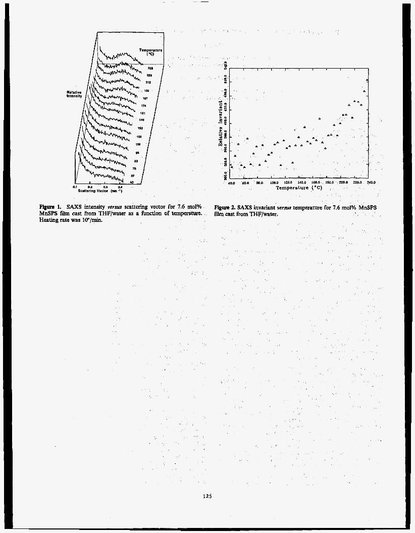
a1 0.2 0.3 01 Sca~tertng vec~or (nm
A a n A A A A A i 3; A n A A n A
A
I I I I I I I
40.0 00.0 00.0 100.0 120.0 140.0 100.0 100.0 200.0 220.0 240.0
Temperature ("C) .
Eignre 1. SAXS intensity versus scattering Vector for 7.6 mol% MnSPS film cast from THFlwater as a hction of tempram. Heating rate was lo"/min.
FigUte 2. SAXS invariant versus temperature for 7.6 molY0 MnSPS film cast From THF/water.
125
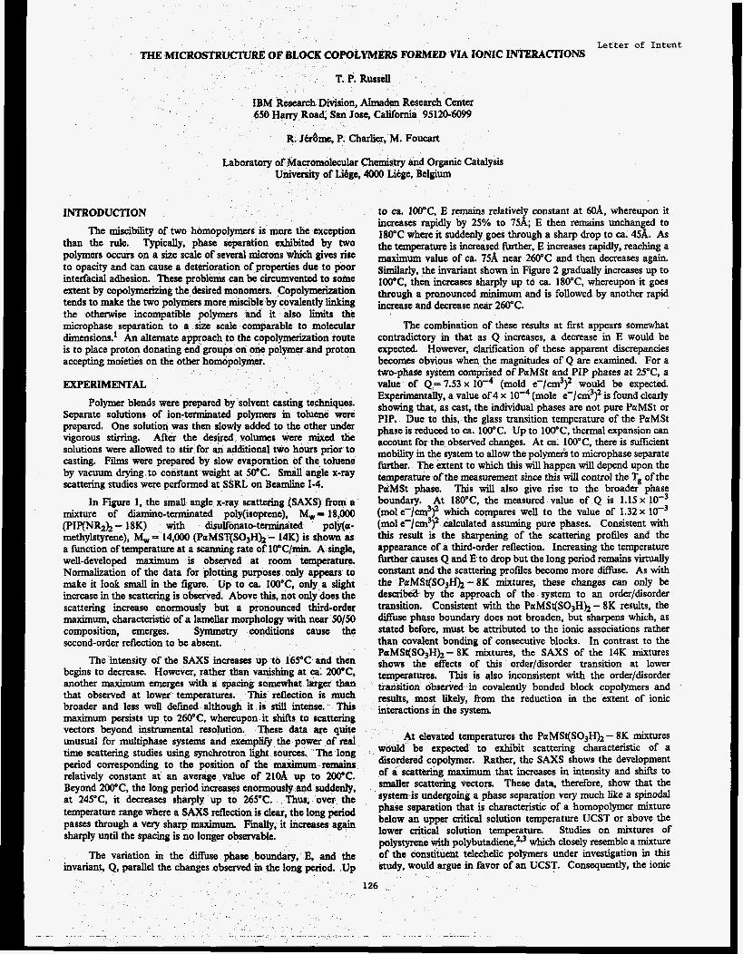
Letter of Intent THE MICROGTRUCTURE OF BLOCK COPOLYMERS MlBMED VIA IONIC INTERACTIONS
T. P. Russell
IBM Research Division, AImaden Research Center oad, San Jose, California 95120-6099
&me, P. Charlier, M. Foucart
Laboratory of Macromolecular Chemistry and Organic Catalysis University of Libge, 4OOO Litge, Belgium
INTRODUCTION
The miscibility of two homopolymers is more the exception than the rule. Typically, phase separation exhibited by two polymers occurs on a size scale of several microns which gives rise to opacity and can cause a deterioration of properties due to poor interfacial adhesion. These problems can be circumvented to some extent by copolymerizing the desired monomers. Copolymerization tends to make the two polymers more miscible by covalently linking the otherwise incompatible polymers and it also limits the microphase separation to a size scale comparable to molecular dimensions.' An alternate approach to the copolymerization route is to place proton donating end groups on one polymer and proton accepting moieties on the other homopolymer.
EXPERIMENTAL
Polymer blends were prepared by solvent casting techniques. Separate solutions of ion-terminated polymers in toluene were prepared. One solution was then slowly added to the other under vigorous stking. After the desired volumes were mixed the solutions were allowed to stir for an additional two hours prior to casting. Films were prepared by slow evaporation of the toluene by vacuum drying to consxant weight at 50°C. Small angle x-ray scattering studies were performed at SSRL on Beamline 1-4.
In Figure 1, the small angle x-ray scattering (SAXS) from a mixture of diarnino-terminated poly(isoprene), MW= 18,000 (PIP(NR2)2 - 18K) with disulfonato-tenninated poly(0r- methylstyrene), M, = 14,000 (PaMST(S03Hh - 14K) is shown as a function of temperature at a scanning rate of 10"C/min. A s u e , well-developed maximum is observed at room temperature. Normalization of the data for plotting purposes only appears to make it look small in the figure. Up to ca. tOO"C, only a slight increase in the scattering is observed. Above this, not only does the scattering increase enormously but a pronounced third-order maximum, characteristic of a lamellar morphology with near 50/50 composition, emerges. Symmetry conditions cause the second-order reflection to be absent.
The intensity of the SAXS increases up to 16SC and then begins to decrease. However, rather than vanishing at ca. 200°C. another maximum emerges with a spacing somewhat k g e r than that observed at lower temperatures. This reflection is much broader and less well defined although it is still intense. This maximum persists up to 260"C, whereupon it shifts to scattering vectors beyond instrumental resolution. These data are quite unusual for multiphase systems and exempw the power of real time scattering studies using synchrotron light sources. The long period corresponding to the position of the maximum remains relatively constant at an average value of 21OA up to 2oo'C. Beyond 2WC, the long period increases enormously and suddenly, at 245°C. it decreases sharply up to 265°C. Thus, over the temperature range where a SAXS reflection is clear9 the long period passes through a very sharp maximum. Finally, it increases again sharply until the spacing is no longer observable.
The variation in the diffuse phase boundary, E, and the invariant, Q, parallel the changes observed in the long period. Up
to ca. 1OO'C, E remains relatively constant at MA, whereupon it increases rapidly by 25% to 75A; E then remains unchanged to 180°C where it suddenly goes through a sharp drop to ca. 45A. As the temperature is increased further, E increases rapidly, reaching a maximum value of ca. 75A near 260°C and then decreases again. S i r l y , the invariant shown in Figure 2 gradually increases up to IWC, then increases sharply up td ca. 180°C, whereupon it goes through a pranounced minimum and is followed by another rapid increase and decrease near 260°C.
The combination of these results at first appears sommhat contradictory in that as Q increases, a decrease in E would be expected. However, clarification of these apparent discrepancies becomes obvious when the magnitudes of Q are examined. For a two-phase system comprised of PaMSt and PIP phases at Z"C, a value of Q = 7.53 x (mold e-/cm3p would be expected. Experimentally, a value of 4 x (mole e-/cm3y is found clearly showing that, as cast, the individual phases are not pure PctMSt or PIP. Due to this, the glass transition temperature of the PaMSt phase is reduced to ca. 100°C. Up to l W C , thermal expansion can account for the observed changes. At ca. 100"C, there is sWicient mobfity in the system to allow the polymerk to microphase separate further, The extent to which this will happen will depend upon the temperature of the measurement since t h i s will control the Tg of the PdMSt phase. This will also give rise to the broader phase boundary. At 180"C, the measured value of Q is 1 . 1 5 ~ loe3 (mol e-/cm3y which compares well to the value of 1.32 x lo-' (mol e-/cm3y calculated assuming pure phases. Consistent with this result is the sharpening of the scattering profiles and the appearance of a third-order reflection. Increasing the temperature M e r causes Q and E to drop but the long period remains virtually constant and the scattering profiles become more difhe. As with the PaMS@03H)z-8K mixtures, these changes can only be describeit by the approach of the system to an order/disorder transition. Consistent with the PaMSt(SO,H)2 - 8K results, the diffuse phase boundary does not broaden, but sharpens which, as stated before, must be attributed to the ionic associations rather than covalent bonding of consecutive blocks. In contrast to the PaMSt(S03Hh- 8K mixtures, the S A X S of the 14K mixtures shows the effects of this order/disorder transition at lower temperatures. This is also inconsistent with the order/disorder transition observed in covalently bonded block copolymers and results, most likely, from the reduction in the extent of ionic interactions in the system.
At elevated temperatures the PaMSt(SO3H)z - 8K mixtures would be expected to exhibit scattering characteristic of a disordered copolymer. Rather, the S A X S shows the development of a scattering maximum that increases in intensity and shifts to d e r scattering vectors. These data, therefore, show that the systemis undergoing a phase separation very much like a spinodd phase separation that is characteristic of a homopolymer mixture below an upper critical solution temperature UCST or above the lower critical solution temperature. Studies on mixtures of polystyrene with polybutadienelX3 which closely resemble a mixture of the constituent telechelic polymers under investigation in this study, would argue in favor of an UCST. Consequently, the ionic
. . . ... .. . ..-. ,.. . . . _ _ - -. .

asrfioeiationr of he - SO; and - N H R ~ are net ~trong enough eat there temperatures to prevent the Bystem Born rradergoinp a eoarie phare reparation, It ihould be noted that the behavior of the PrrMSt(SO$=€)2- 8K mixtures is Merent in that a coane phaie separation was not observed. Thus, the order/disorder transition must occur at temperatures either above the USCT, Le., the UCST is reduced by the reduction in the molecular weight or below the temperature at which disruption of the ionic aggregates occurs.
Consequently, a unified description of the phase diagram of the telechelic copolymers arises from these studies. It is apparent that four different features have to be considered to understand the observed SAXS results. These include the mesophase separation
74
40.00 - - '5
-1- 20.00 W C
0.00 0.04 0.08 0.12
Figure 1. The temperature dependence of the SAXS from a mixture of PIP(NR2h - 18K with PaMSt(S03Hh - 8K at a scanning rate of lO"C/min.
Figure 3. Proposed phase diagram for the PIP(NR2)2- 18KPaMSt(S03H)2 8K telechelic mixtures where UCST is the upper critical solution temperature of the constituent telechelic polymers, MST is the microphase separation temperature of the ionic copolymers, Tg is the glass transition and Ti is the temperature at which the ionic associations forming the block copolymers are disrupted. The left to right diagonally hatched area is where the mixtures exhibit a microphase separated morphology similar to that seen in covalently bonded block copolymers. The right to left diagonally hatched area is where the mixtures phase separate in a manner analogous to simple homopolymer mixtures. The stipled area is where the mixtures exist as a disordered copolymer phase and the clear area is where the mixture is a homogeneous mixture of the constituent telechelics. The dashed lines are continuations of the phase diagrams which due to ionic aggregation or disruption of the endgroups are not possible to observe.
temperature of the copolymrrrr f o m d by ionic aiaocfatiom, tb UCST of the eonsrituent hornopolymer8, the temperature, Ti, at which ionic aiseeiatiene are aisrupted rrnd the glase tranilion temperature T , The relative position of these will dictate the morphology. fhree such examples are shown in Figures 3 and 4.
REFERENCES 1. Leibler, L. Macromolecules 1982, 19, 2621. 2.
3. Russell, T. P.; Ronca, G. Macromolecules 1985, 18, 665.
Russell, T. P.; Hadziioannou, Warburton, W. K. Macromolecules 1985, 18, 78.
12.00 I I I I
0.00 30.00 1 10.00 190.00 270.00 350.00
T("C)
Figure 2. temperature for the scattering profiles shown in Figure 11.
The total integrated scattering as a function of
Figure 4. Schematic phase diagram of the PIP(NR2)2- 18K/PcrMSt(S03H)2- 14K telechelic mixtures. The labeling is the same as in Figure 15.
127

INTERCALIBRATION OF SMALL-ANGLE X-RAY AND NEUTRON SCATTERING DATA
Letter of Intent T. P. Russell
IBM Research Division, Almaden Research Center 650 Harry Road, San Jose, California 95120-6099
J. S. Lin, S. Spooner, G. D. Wignall
National Center for Small-Angle Scattering Research Oak Ridge National Laboratory, Oak Ridge, Tennessee 37831
INTRODUCTION
In view of the maturity of small-angle scattering (SAS) techniques, it is surprising that a significant fraction of data are still published in arbitrary units which are functions of the time scale of the experiment, scattering geometry and the sample dimension, e.g., thickness. Data are generally presented in the form of scattered intensity (counts per scattering elements, counts per unit time, etc.) versus Q, the scattering vector which is given by (44A)sinO where A is the wavelength of the incident x-rays or neutrons and 28 is the scattering angle.
Conversion of the data to an absolute merentid scattering cross section dc(Q)/dn, typically in units of cm-', may be accomplished by multiplying by a calibration constant. Use of absolute scattering is not necessary for the measurement of the interference function. However, placing the data on an absolute scale is necessary for the determination of molecular weights, the number density of particles, the scattering length densities of phases in multiphased systems and to restrict the parameters of a given model to the set which reproduces the observed intensity. It is also a useful means for the detection of artifacts in SAS experiments.
Despite the obvious advantages of placing data on an absolute scale, the use of suitable calibration techniques is by no means universal in the SAS field. This situation is due in part to the fact that many of the primary calibration techniques require special equipment and can be difficult and time consuming (Pate1 and Schmidt, 1971). In view of the limited time scale of many SAS experiments of steady state reactors, pulsed neutron and synchrotron sources, users are naturally reluctant to devote a sigsicant fraction of beam time to calibration experiments. One solution to this problem is to develop pre-calibrated strongly scattering samples that may be measured rapidly in a given geometry, not detracting signifcantly from the available beam time.
RESULTS AND INTERCOMPARISON OF STANDARDS
The Debye-Bueche formula is given by'
dc dc 1 (1 + Q2a2)2
(Q)= (0)
where a is a correlation length. Thus, a plot of dc-'/~Q)/dSz versus Q2 should be linear with slope a2. Figures 1-3 show such plots for small-angle neutron (SANS) and x-ray (SAXS) scattering data of vitreous carbon taken at the Oak Ridge National Laboratory and SSRL. The SSRL SAXS data were calibrated with respect to silica sol standards2 and the ORNL SAXS data were calibrated with respected to water as described previously.
Because of slight departures from lineari the data are fitted
lengths from both SAXS and SANS experiments lie in the range a=4.75+0.3k Based on a neutron cross section dc(O)/dn = 5.2 f 0.4 cm-', the x-ray cross section can be calculated by multiplying by the ratio (6 x 2.82 x 10-'3/0.665 x 10-12)2 where b, = 0.665 x cm is the coherent scattering
only for Q values between 0.045 < Q < 0.1 111- Y and the correlation
length for carbon and the scattering length of an electron is 2.28 x This leads to a predicted SAXS cross section dc(O)/dQ = 33.3 + 3.2 an-', in good agreement with the two independent determination of 31.5 f 3 cm-' (SSRL) and 38 f 4 cm-' (ORNL). Thus, this particular glassy carbon sample yields good quantitative agreement between the techniques.
cm.
Measurements on two different polyethylene standards are shown in Fig. 4. The first is the Lupolen (Kratky) standard, whereas the second is a high density polyethylene (Eltex A1050). The fust feature to be observed from these data is that despite the fact that both samples are polyethylene, the scattering profiles are profoundly different. This is not surprising since the branching content and the thermal treatment of the samples are different. Clearly, the long period, absolute intensity and the width of the reflection can vary dramatically. Concentrating first on the Lupolen standard, it is seen that a borad weak reflection with a maximum near 0.0311-' is seen. Separate determinatins of the [dc(Q,,jdSz] were found to be 5.3 0.5 cm-' (ORNL IO-m SAXS) 6.0 k 0.5 cm-' (Schelten and Hendricks4 and 5.0+ 0.3 cm-' (SSRL)), thereby yielding an average value of 5.4k0.5 cm-'. The Eltex A1050, on the other hand, shows a pronounced scattering maximum at ca. 0.027511-'. Vestiges of a third-order reflection are seen near 0.06A-'. The lack of higher-order reflections is due to distribuitons of crystal thickness, diffuse phase boundary between the crystalline and amorphous phases and symmetry since this polyethylene has a degree of crystallinity near 50%. The [C(Q,,)/dn] for Eltex A1050 has been measured by Schelten et al. as 22.4f 1.5 cm-' and more recently at SSRL as 24.0 2.0 cm-'.
Neither of these samples has a finite coherent cross section for neutrons and comparisons between SAXS and SANS are not possible. For deuterated polyethylene, however, the coherent cross sections should be proportional and Fig. 5 shows SAXS and SANS data for a deuteropolythylene where the SAXS data were calibrated via Ludox suspensions. The data can clearly be superimposed to a good approximation by a multiplicative factor after the smal l incoherent background has been subtracted. Deuteropolyethylene may be regarded as a three-phase system (crystal, amorphous and voids) and level of deuteration is 95%. Because the electron density and scattering length density of the voids are zero, the SAXS and SANS patterns can be scaled as for two-phase systems. The theoretical ratio of SAXS and SANS intensities for a 95% deuterated material is 1.41 compared to the observed ratio over the two scattering patterns (Fig. 5) of 1.31 f 0.12. Deuteropolyethylene (PED) thus seems to be suitable intercalibration standards for SAXS and SANS data with suitable samle thickness 1.2 mm for each technique.
REFERENCES
1. P. Debye, A. Bueche, J. Appl. Phys. 20,518 (1949). 2. T. P. Russell, J. Appl. Crysr. 16,473 (1983). 3. G. D. Wignall, F. S . Buks, J. Appl. Ctysr. 20,28 (1987). 4. J. Schelten, D. G. H. Ballard, G. D. Wignall, G. W. Longman,
W. Schmatz, Polymer 17, 751 (1976).
128
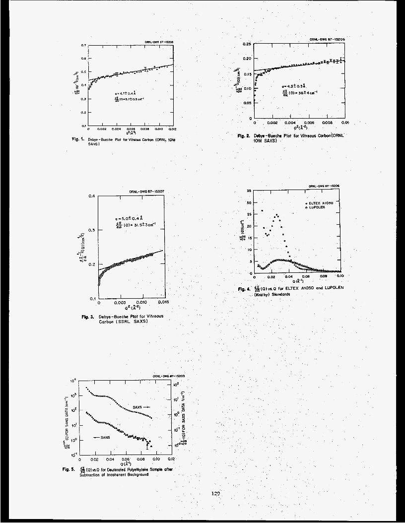
0.2
o 0.002 0.004 0.006 0.008 0.010 a o e O W
Fig. 1. Debye-Bueche Plot for Vitreous Corbcn (ORNL 10M SANS I
0.1
ORNL-OWG 87-15207 0.4
0.3 -G " - - 0
.z w e
- I -1- 0.2
0.1 0 0.005 0.010 0.015
Q ' P )
Fig. 3. Debye-Bueche Plot for Vitreous
Carbon (SSRL SAXSl
ORNL-DVO 87-15209 to4 I I I I I
IO2
IO
5
0 o 0.02 0.04 0.06 a o e 0.10
0 &'I
Pig. 4. %(Q)VLO for ELTEX A i 0 5 0 and LUPOLEN
(Kratky) Standards
Fig. 5. ( 0 ) s O for Deuterated Polyethylene Sample after Subtraction of Incoherent Background
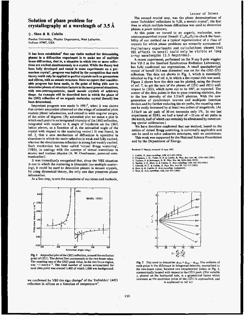
Solution of phase problem for crystallography at a wavelength of 3.5
Q . Shen & R. Colella Purdue University, Physics Department, West Larayette, Indiana 47907. USA
It has been established’ that one viable method for determining phases in a diffraction experiment is to make use of multiple beam-diffraction, that is, a situation in which two or more reflec- tions are excited simultaneously in a crystal. While the theory had been fully developed and tested in some experiments on a ger- manium crystal’, progress was halted by the recognition that such theory could only be applied to perfect crystals such as germanium and siliconj with no mosaic structure. Here we report that consider- able progress has been made, to the point of being able now to determine phases of structure factors in the most general situations, with non-centrosymmetric, small mosaic crystals of arbitrary shape. An example will be described here in which the phase of the (202) reflection of an organic molecular crystal (benzil) has been determined.
Important progress was made in 19812, when it was shown that certain anomalies observed on the wings of azimuthal plots contain phase information, and extend to wide angular ranges, of the order of degrees. (By azimuthal plot we mean a plot in which each point is an integrated intensity of the ( h k l ) reflection, integrated with respect to 0, angle of incidence on the ( k k l ) lattice planes, as a function of 4, the azimuthal angle of the crystal with respect to the scattering vector.) It was found, in ref. 2, that a new mechanism of diffraction is operative in situations in which the main reflection is weak and fully excited, whereas the simultaneous reflection is strong but weakly excited. Such mechanism has been called ‘virtual Bragg scattering’, (VRS), in analogy with the concept of virtual transitions in atomic and nuclear physics (A. W. Overhauser. personal com- munication).
It was immediately recognized that, since the VBS situation is one in which the scattering is kinematic (no multiple scatter- ing), it could be used to determine phases in mosaic crystals, by using dynamical theory, the only one that preserves phase information.
As a first step, to test the soundness of our ideas and methods,
I I I I ~ 4 0 5 (06 107 Kw) 109
Azimulhai angle Ideg.1
Fi. 1 Ari_muthal plot ofthe (202) reflection, around the excitation point or(221). The dotted line corresponds to the two beam value. The counting rate of the (202) peak value, in the two bcsin region, was -1 counts-’. The total number of counts accumulated for each data point was around 1,400 of which 1,OOO was background.
we confirmed by VBS the sign change3 of the ‘forbidden’ (442) reflection in silicon as a function of temperature4.
Le t te r of Intent The second crucial step, was the phase determinations of
some ‘forbidden’ reflections in V,Si, a mosaic crystai’, the first time in which multiple-beam difiraction was used to determine phases a priori unknown.
At this point we turned to an organic, molecular, non- centrosyminetrical crystal (benzil: C1.,HlOO2) to check the feasi- bility of our method on a typical representative of a class of crystals for which phase problems are normally encountered. Preliminary experiments and calculations showed tha t VBS effects i n benzil could only be vis ible a t long x-ray wavelengths (3.5 Angstroms).
A recent experiment, performed on the X-ray 8-pole wiggler line VII-2 at the Stanford Synchrotron Radiation Laboratory, has fully confirmed our expectations. A small quasispherkal crystal of benzil (0.3-mm diameter) was set for the (202-221) reflection. The data are shown in Fig. 1, which is essentially identical to Fig. 4 of ref. 6, in which a Rat crystal slab was used. Figure 2 shows how the data can be plotted, using the theory of ref. 7, to get the sum of the phases of (221) and (021) with respect to (202). which turns out to be 180°, as expected. The scatter of the data points is due to poor counting statistics, due to the low intensity of the 3.5 keV photons. With the new generation of synchrotron sources and multipole insertion devices and by further reducing the air paths, the counting rates can be easily increased by at least two orders of magnitude. (At 3.5 keV an air path of 30cm transmits only 1%. In our last experiment at SSRL we had a total of -21 cm of air paths in the hutch, half of which can certainly be eliminated by construct- ing special collimators.)
We have therefore confirmed that our method, based on the notion of virtual Bragg scattering, is universally applicable and can be used to solve unknown structures, with no restrictions.
This work was supported by the National Science Foundation and by the Department of Energy.
Received 17 March: accepted 16 June 19x7.
1. Colella. R. Arm cryslaffogr. AM, 413-423 (1974). 2. Chapman. L D.. Yoder. D. R. & Colclla, R phys. Rev. Lcff. a, 157X-ISR1 (1981) 3. TNcano. P. & Batterman, B. W. phys. Reu. 86, 3659-3666 (1972). 4. Tis&lcr. 1. 2.. Shcn. Q. B Colella, R. ACID crys1aNo.w A41.451-453 (19x5). 5. Schrnidl, M. C. & Colclla, R. Rev. Lerl. 55. 715-717 (19RS). 6. Shen. Q. &. Coiclla. R. Aclu crysfullogr. (suhmittcd). 7. Shcn. Q. A m cryrlallogr. A42, 525-533 11986).
Fig. 2 Plot used to determine &zl + $,,2, - $’2,,2. The ordinate of each point is the diflerence in integrated intensity, normalized to the two-beam value, between two experinFnta1 points in Fig. 1, symmetrically located with respect to the (221) peak. (The variable 2, plotted on the horizontal axis, is a gecmetrical factor which increases as the excitation point of the (221) is approached, and
is explained in ref. 6.)
130


Biology Proposals

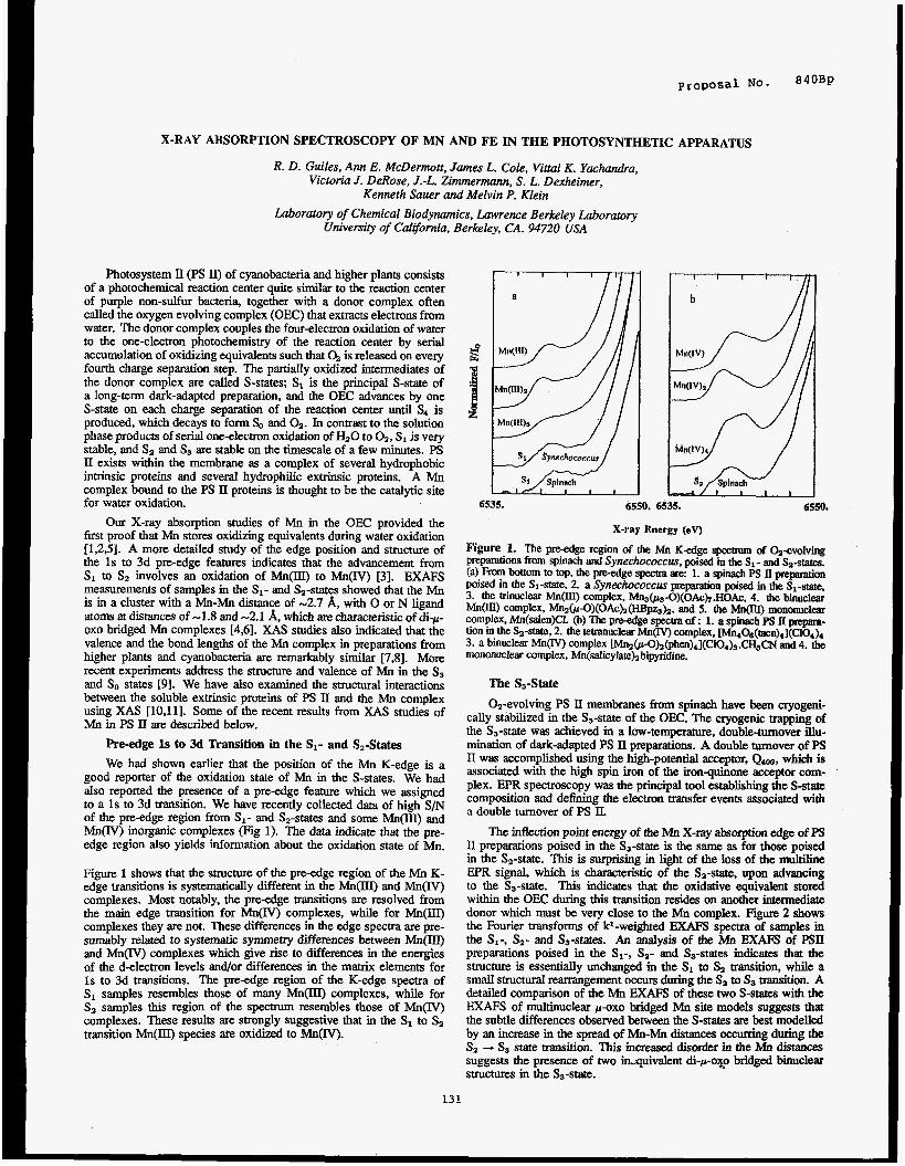
X-RAY ABSORPTION SPECTROSCOPY OF MN AND FE IN THE PHOTOSYNTHETIC APPARATUS
R. D. Guiles, Ann E. McDermott, James L. Cole, Vitta1 K. Yachandra, Victoria J. DeRose, J.-L. Zimmermann, S. L. Dexheimer,
Kenneth Sauer and Melvin P. Klein Laboratory of Chemical Biodynamics, Lawrence Berkeley Laboratory
University of Carifornia. Berkeley, CA. 94720 USA
Photosystem I1 (PS II) of cyanobacteria and higher plants consists of a photochemical reaction center quite similar to the reaction center of purple non-sulfur bacteria, together with a donor complex often called the oxygen evolving complex (OEC) that extracts electrons from water. The donor complex couples the four-electron oxidation of water to the one-electron photochemistry of the reaction center by serial accumulation of oxidizing equivalents such that a is released on every fourth charge separation step. The partidy oxidized intermediates of the donor complex are called S-states; S1 is the principal S-state of a long-term dark-adapted preparation, and the OEC advances by one S-state on each charge separation of the reaction center until S4 is produced, which decays to form f& and Oa. In contrast to the solution phase products of serial one-electron oxidation of HaO to Oa, S1 is very stable, and Sa and Ss are stable on the timescale of a few minutes. PS 11 exists within the membrane as a complex of several hydrophobic intrinsic proteins and several hydrophilic extrinsic proteins. A Mn complex bound to the PS 11 proteins is thought to be the catalytic site for water oxidation.
Our X-ray absorption studies of Mn in the OEC provided the first proof that Mn stores oxidizing equivalents during water oxidation [1,2,5]. A more detailed study of the edge position and structure of the 1s to 3d pre-edge features indicates that the advancement from SI to Sa involves an oxidation of Mn(III) to Mn(IV) [3]. E M S measurements of samples in the SI- and Sa-states showed that the Mn is in a cluster with a Mn-Mn distance of -2.7 A, with 0 or N ligand atoms at distances of -1.8 and -2.1 A, which are characteristic of di-F- oxo bridged Mn complexes [4,6]. XAS studies also indicated that the valence and the bond lengths of the Mn complex in preparations from higher plants and cyanobacteria are remarkably similar [7,8]. More recent experiments address the structure and valence of Mn in the Ss and So states 191. We have also examined the structural interactions between the soluble extrinsic proteins of PS II and the Mn complex using XAS [10,11]. Some of the recent results from XAS studies of Mn in PS II are described below,
Re-edge 1s to 3d Transition in the S1- and S,-States
We had shown earlier that the position of the Mn K-edge is a good reporter of the oxidation state of Mn in the S-states. We had also reported the presence of a pre-edge feature which we assigned to a 1s to 3d transition. We have recently collected data of high S / N of the pre-edge region from S1- and S2-states and some Mn(III) and Mn(N) inorganic complexes (Fig 1). The data indicate that the pre- edge region also yields information about the oxidation state of a.
Figure 1 shows that the struchue of the pre-edge region of the Mn K- edge transitions is systematically different in the Mn(m and Mn(IV) complexes. Most notably, the pre-edge transitions are resolved from the main edge transition for Mn(IY) complexes, while for Mn(m) complexes they are not. These differences in the edge spectra are pre- sumably related to systematic symmetry differences between Mn(IIl) and Mn(IV) complexes which give rise to differences in the energies of the d-electron levels and/or differences in the matrix elements for 1s to 3d transitions. The pre-edge region of the K-edge spectra of s1 samples resembles those of many M n O complexes, while for Sa samples this region of the spectrum resembles those of Mn(IV) complexes. These results are strongly suggestive that in the S1 to Sa transition Mn(III) species are oxidized to Mn(IV).
131
6535. 6550. 6535. 6550'
X-ray Energy (ev)
Figure 1. The preedge region of the Mn K-edge spectrum of %-evolving preparations from spinach and Synechococcus, poised in the SI- and S2-states. (a) From bottom to top, the pre-edge spectra are: 1. a spinach PS II preparation poised in the &-state, 2. a Synechococcus preparation poised in the S1-state, 3. the trinuclear M@Q complex, Mn3(ps-O)(OAc)r.HOAc, 4. the binuclear M n O complex, M n 2 0 1 - 0 ) ( 0 A c ~ ~ p s ) 2 . and 5. the M n o mononucku COmPlex, Mn(denK1. (3) The pre-edge spectra of: 1. a spinach PS iI p p a - tion in the Sa-state, 2. the tetranuclear M n o complex, ~ O , & ~ m ) ~ ] ( c l O , ) ~ 3. a binuclear M n O complex ~ a o l - O ) a ( p h e n ) 4 1 ( C 1 0 4 ) ~ . ~ c N and 4. the mononuclear complex, Mn(salicylateb bipyridim.
The S3-State
Oa-evolving PS II membranes from spinach have been cryogeni- cally stabilized in the Ss-state of the OEC. The cryogenic trapping of the Ss-state was achieved in a low-temperature, double-turnover illu- mination of dark-adapted PS II preparations. A double turnover of PS II was accomplished using the high-potential acceptor, Qm, which is associated with the high spin iron of the ironquinone acceptor com- plex. EPR spectroscopy was the principal tool establishing the S-state composition and defining the electron transfer events associated with a double turnover of PS IL
The inflection point energy of the Mn X-ray absorption edge of PS II preparations poised in the Ss-state is the same as for those poised in the Sz-state. This is surprising in light of the loss of the multiline EPR signal, which is Characteristic of the S,-state, upon advancing to the &state. This indicates that the oxidative equivalent stored within the OEC during this transition resides on another intermediate donor which must be very close to the Mn complex. Figure 2 shows the Fourier transforms of kl-weighted EXAFS spectra of samples in the Sx-, &- and S3-states. An analysis of the Mn EXAFS of PSII preparations poised in the S1-, Sa- and &-states indicates that the structure is essentially unchanged in the S1 to S2 transition, while a small structural rearrangement occurs during the Sa to Ss transition. A detailed comparison of the Mn EXAFS of these two S-states with the EXAFS of multinuclear p-oxo bridged Mn site models suggests that the subtle differences observed between the S-states are best modelled by an increase in the spread of Mn-Mn distances occurring during the Sz -+ Ss state transition. ms increased disorder in the Mn distances suggests the presence of two in.quivalent di-p-0~0 bridged binuclea~ structures in the Ss-state.
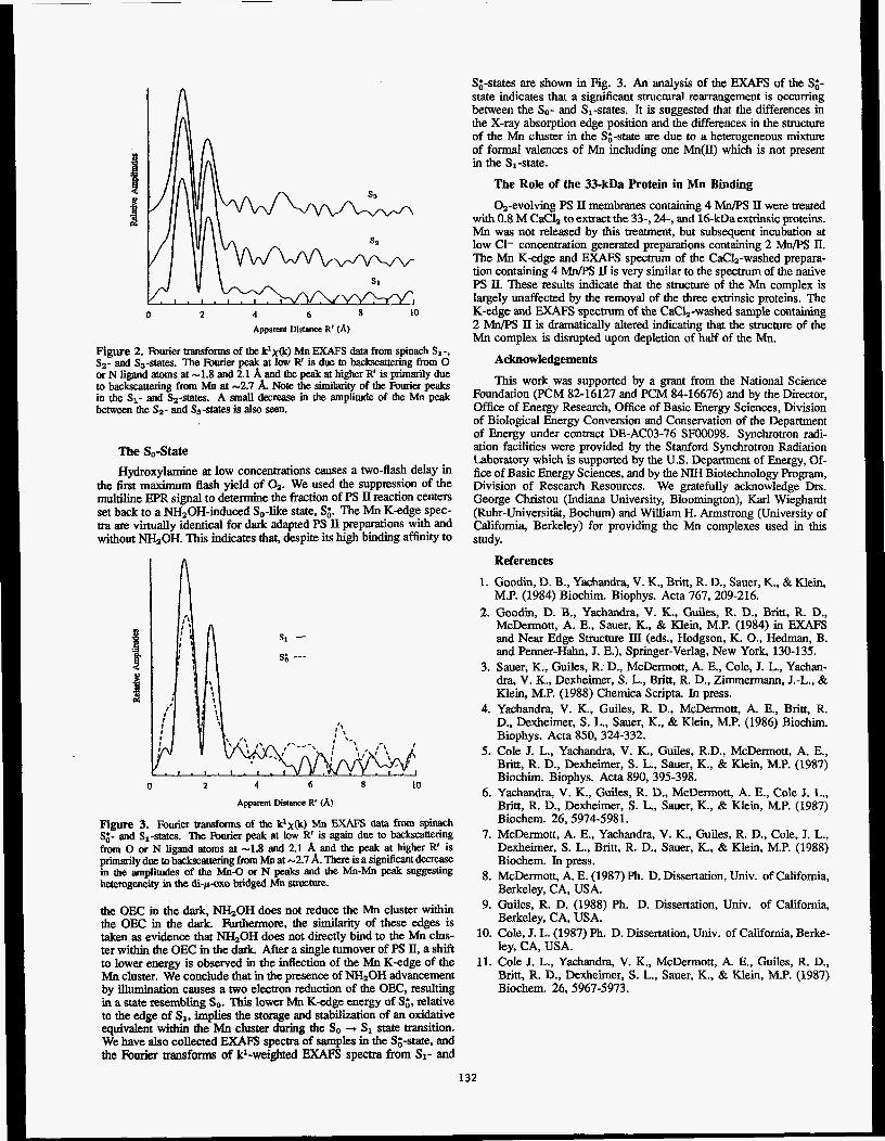
~ SI
1 . 1 . 1 . 1 . 1 I 0 2 4 6 8 10
Apparenl Distance R' (A)
Figure 2. Fourier transforms of the klx&) Mn EXAFS data from spinach SI-, Sa- and Ss-states. The Rmier peak at low R' is due to backscauering from 0 or N ligand atoms at -1.8 and 2.1 A and the peak at higher R' is primarily due to backscattering from Mn at -2.7 k Note the similarity of the Rurier peaks in the SI- and S2-state.s. A small decrease in the amplitude of the Mn peak between the Sa- and S3-states is also seen.
The So-State
Hydroxylamine at low concentratiom causes a two-flash delay in the first maximum flash yield of 4. We used the suppression of the multiline EPR signal to determine the fraction of PS II reaction centers set back to a W20H-induced So-like state, S;. The Mn K-edge spec- tra are. virtually identical for dark adapted PS II preparations with and without NH,OH. This indicates that, despite its high binding affinity to
A
si - s; _--
0 2 4 6 8 to
Apparml Distance R' (A)
Figure 3. Fourier transforms of the klx(k) Mn EXAFS data h m spinach S;- and S1-states. The fiuriex peak at low R' is again due to backscattering from 0 or N ligand atoms at -1.8 and 2.1 A and the peak at higher R' is primarily due to backscattering from Mn at -2.7 A. There is a significant decrease in the amplitudes of the Mn-0 or N peaks and the Mn-Mn peak suggesting heterogeneity in the di-p-oxo bridged Mn structure.
the OEC m the dark, NHzOH does not reduce the Mn cluster within the OEC in the dark. Furthermore, the similarity of these edges is taken as evidence that W O H does not directly bind to the Mn clus- ter within the OEC in the dark. After a single turnover of PS II, a shift to lower energy is observed in the inffection of the Mn K-edge of the Mn cluster. We conclude that in the presence of NH20H advancement by illumination causes a two electron reduction of the OEC, resulting in a state resembling So. This lower Mn K-edge energy of S;, relative to the edge of SI, implies the storage and stabilization of an oxidative equivalent within the Mn cluster during the So -.* SI state transition. We have also collected E M spectra of samples in the St;-state, and the Fourier transforms of kl-weighted EXAFS spectra from SI- and
S;-states are shown in Fig. 3. An analysis of the EXAFS of the g- state indicates that a significant structural rearrangement is occurring between the So- and Sl-states. It is suggested that the differences in the X-ray absorption edge position and the differences in the structure of the Mn cluster in the Sg-state are due to a heterogeneous mixture of formal valences of Mn including one Mn(II) which is not present in the Sl-state.
The Role of the 33-kDa Protein in Mn Binding
0,-evolving PS II membranes containing 4 Mn/PS II were treated with 0.8 M CaClz to extract the 33-, 24-, and 16-kDa extiinsic proteins. Mn was not released by this treatment, but subsequent incubation at low C1- concentration generated preparations containing 2 Mn/PS II. The Mn K-edge and EXAFS specaum of the CaC1,-washed prepara- tion containing 4 Mn/PS II is very similar to the spectrum of the native PS II. These results indicate that the s t r u c ~ e of the Mn complex is largely unaffected by the removal of the three extrinsic proteins. The K-edge and EXAFS spectrum of the CaC12-washed sample containing 2 Mn/PS II is dramatically altered indicating that the structure of the Mn complex is disrupted upon depletion of half of the h4n.
Acknowledgements
This work was supported by a grant from the National Science Foundation (PCM 82-16127 and PCM 84-16676) and by the Director, Office of Energy Research, Office of Basic Energy Sciences, Division of Biological Energy Conversion and Conservation of the Department of Energy under contract DE-ACO3-76 SF00098. Synchronon radi- ation facilities were provided by the Stanford Synchrotron Radiation Laboratory which is supported by the U.S. Department of Energy, Of- fice of Basic Energy Sciences, and by the NIH Biotechnology Program, Division of Research Resources. We gratefully acknowledge Drs. George Christou (Indiana University, Bloomington), Karl Wieghardt (Ruhr-Universitiit. Bochum) and William H. Armstrong (University of California, Berkeley) for providing the Mn complexes used in this study.
References
1. Goodin, D. B., Yachandra, V. K., Britt, R D., Sauer, K., & Klein, M.P. (1984) Biochim. Biophys. Acta 767, 209-216.
2. Goodin, D. B., Yachandra, V. K., Guiles, R. D., Britt, R. D., McDermott, A. E., Sauer, K., & Klein, M.P. (1984) in EXAFS and Near Edge Structure ID (eds., Hodgson, K. O., Hedman, B. and Penner-Hahn, J. E.), Springer-Verlag, New York, 130-135.
3. Sauer, K., Guiles, R;D., McDermott, A. E., Cole, J. L., Yachan- &a, V. K., Dexheimer, S. L., Britt, R. D., Zmermann, J.-L., & Klein, M.P. (1988) Chemica Scripta, In press.
4. Yachandra, V. K., Guiles, R D., McDermott, A. E., Britt, R. D., Dexheimer, S. L., Sauer, K., & Klein, M.P. (1986) Biochim. Biophys. Acta 850,324-332.
5. Cole J. L., Yachandra, V. K., Guiles, R.D., McDermott, A. E., Britt, R. D., Dexheimer, S. L., Sauer, K., & Klein, M.P. (1987) Biochim. Biophys. Acta 890,395-398.
6. Yachandra, V. K., Guiles, R. D., McDermott, A. E., Cole J. L., Britt, R. D., Dexheimer, S. L., Sauer, K., & Klein, M.P. (1987) Biochem. 26,5974-5981.
7. McDermott, A. E., Yachandra, V. K., Guiles, R. D., Cole, J. L., Dexheimer, S. L., Britt, R. D., Sauer, K., & Klein, M.P. (1988) Biochem. In press.
8. McDermott, A. E. (1987) Ph. D. Dissertation, Univ. of California, Berkeley, CA, USA.
9. Guiles, R. D. (1988) Ph. D. Dissertation, Univ. of California, Berkeley, CA, USA.
10. Cole, J. L. (1987) Ph. D. Dissertation, Univ. of California, Berke- ley, CA, USA.
11. Cole J. L., Yachandra, V. K., McDermott, A. E., Guiles, R. D., Britt, R D., Dexheimer, S. L., Sauer, K., & Klein, M.P. (1987) Biochem. 26,5967-5973.
132
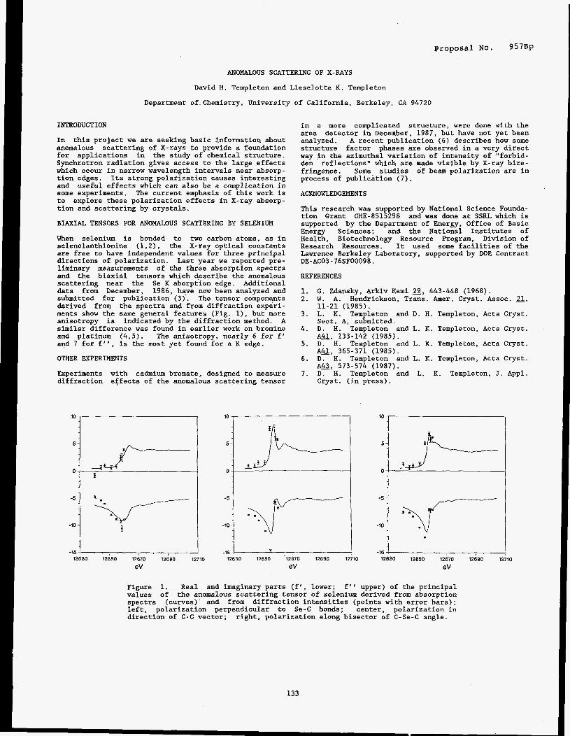
Proposal No. 9 5 7 0 ~
ANOMALOUS SCATTERING OF X-RAYS
David H. Templeton and Lieselotte K. Templeton
Department of Chemistry, University of California, Berkeley, CA 94720
INTRODUCTION
In this project we are seeking basic information about anomalous scattering of X-rays to provide a foundation for applications in the study of chemical structure. Synchrotron radiation gives access to the large effects which occur in narrow wavelength intervals near absorp- tion edges. Its strong polarization causes interesting and useful effects which can also be a complication in some experiments. The current emphasis of this work is to explore these polarization effects in X-ray absorp- tion and scattering by crystals.
BIAXIAL TENSORS FOR ANOMALOUS SCATTERING BY SELENIUM
When selenium is bonded to two carbon atoms, as in selenolanthionine (1,2), the X-ray optical tonstants are free to have independent values for three principal directions of polarization. Last year we reported pre- liminary measurements of the three absorption spectra and the biaxial tensors which describe the anomalous scattering near the Se K aborption edge. Additional data from December, 1986, have now been analyzed and submitted for publication (3). The tensor components derived from the spectra and from diffraction experi- ments show the same general features (Fig. l), but more anisotropy is indicated by the diffraction method. A similar difference was found in earlier work on bromine and platinum (4,5). The anisotropy, nearly 6 for f' and 7 for f", is the most yet found for a K edge.
OTHER EXPERIMENTS
Experiments with cadmium bromate, designed to measure diffraction effects of the anomalous scattering tensor
in a more complicated structure, were done with the area detector in December, 1987, but have not yet been analyzed. A recent publication (6) describes how some structure factor phases are observed in a very direct way in the azimuthal variation of intensity of "forbid- den reflections" which are made visible by X-ray bire- fringence. Some studies of beam polarization are in process of publication (7).
ACKNOWLEDGEMENTS
This research was supported by National Science Founda- tion Grant CHE-8515298 and was done at SSRL which is supported by the Department of Energy, Office of Basic Energy Sciences; and the National Institutes of Health, Biotechnology Resource Program, Division of Research Resources. It used some facilities of the Lawrence Berkeley Laboratory, supported by DOE Contract DE-AC03-76SF00098.
REFERENCES
1. G. Zdansky, Arkiv Kemi 9, 443-448 (1968). 2. W. A. Hendrickson, Trans. her. Cryst. Assoc. 21,
3. L. K. Templeton and D. H. Templeton, Acta Cryst.
4. D. H. Templeton and L. K. Templeton, Acta Cryst.
5. D. H. Templeton and L. K. Templeton, Acta Cryst.
6 . D. H. Templeton and L. K. Templeton, Acta Cryst.
7. D. H. Templeton and L. K. Templeton, J. Appl.
11-21 (1985).
Sect. A, submitted.
A a , 133-142 (1985).
A Q , 365-371 (1985).
A&, 573-574 (1987).
Cryst. (in press).
- 1 5 , . . , , , , . , , 12630 12650 12670 12690 1:
eV
10
-15 3 12630 12650 12670 12690 1
eV
-15 12630 12650 12670 12690 1:
eV 10
Figure 1. Real and imaginary parts (f', lower; f" upper) of the principal values the anomalous scattering tensor of selenium derived from absorption spectra (curves) and from diffraction intensities (points with error bars); left, polarization perpendicular to Se-C bonds; center, polarization in direction of C-C vector;
of
right, polarization along bisector of C-Se-C angle.
133


Propos?l No. 961B
STRUCTURE/FUNCTION STUDIES OF OXIDASES, PEROXIDASES AND C A T e S E S
B. Chance+*, L. Powers@, S. Khalidtl I. Ayene*, R. Waterland", T.E. King+, G . Bunker+, K . Zhang+,R. POytOn*, E. Davidson*
+University City Science Center, Institute for Structural & Functional Studies, Philadelphia, PA *University of Pennsylvania School of Medicine, Department of Biochemistry & Biophysics, Philadelphia, PA
@AT&T Bell Laboratories, Murray Hill, NJ #University of Colorado, Department of Molecular, Cellular & Developmental Biology, Boulder, CO
Cytochrome oxidase studies have now been focused largely upon "altered forms of cytochrome oxidase" as obtained by mutation of cytochrome oxidase through the nuclear genes rather than the mitochondrial genes. This affords a variety of altered cytochrome oxidases in which different numbers of subunits are present and in which different activities are observable and, according to our EXAFS study, different structures Of the active site will be involved.
The synthesis of altered forms of cytochrome oxidase in genetically altered yeast cells is often inefficient; assembly of cytochrome oxidase may be faulty. Thus, one special goal of the beam time allotments made ,&vailable has been an increase of the detector sensitivity and reliability to the point that not only do we measure the 100 pM copper of cytochrome oxidase of beef heart membrane-bound preparations as already published (l), but we have, on occasion, obtained edge data at half this concentration. In order to decrease the averaging time and to increase the reliability Of the results, a 13-element Germanium detector has been obtained and will be used in a wide-band energy referencing circuit that we believe will decrease the time for accumulation of a satisfactory data set by a factor of two, at least. At the same time, EXAFS studies of these dilute membrane preparations will be undertaken. R.O. Povton, our collaborator in these studies, together with T.E. King and ourselves Will make all efforts to increase the yield of purified cytochrome oxidase from the mutant cells.
Already accomplished are studies of three important aspects of cytochrome structure and function, copper depletion, mercury modification and the structure of the zinc site. Wharton has vastly improved the copper depletion methods for cytochrome oxidase so that an approximation to a single site is achieved in the enzyme. Under these conditions, the algorithms by which we have deconvoluted the separated contributions of the copper atoms in the native enzyme have been checked, and, furthermore, the structureffunction of the copper deficient enzyme has been studied in detail (213).
The mercuration of cytochrome oxidase was one of the first evidences that an EPR silent copper was involved, and Bickar has recently produced a well-defined derivative on which we have been able to do EXAFS studies (4). The finding is of importance, it is apparent that the mercury ligates the sulfur atom proximal to the as-copper binding site. The possibility of ligating a sulfur atom within back scatter distance of the copper further confirms Our identification of sulfur as a bridging atom in the resting structure.
Zinc has been identified as a component of the multi- subunit oxidase in the laboratory of Caughey (5). The subunit location and the ligands of the zinc have been identified (6) and found to be consistent with the amino acids sequence and folding properties of subunit VI of cytochrome oxidase; Caughey has shown that several other subunits are not involved and the "three subunit" oxidases is found not to contain zinc. Thus, EXAFS has proved to be most valuable in identifying the nature of the zinc binding site.
e St&
Our vibrating reed beam position detector has now been used to evaluate Beam Lines 11-3, 11-2 and VI. A beam tracking circuit has been used on 11-2 with a significant decrease (>2x) of noise in EXAFS Recording (7).
The great sensitivity of dilute cytochrome oxidase to redox perturbations by hydrated electrons has caused the reactivation of research on this topic, particularly at room temperature where eventual kinetics studies are planned. Typically 50% reduction of cytochrome oxidase occurs in 2.5 hours on Beam Line 11-2. Use of traps for hydrated electrons is in progress.
munLuam
The improved detector systems suggests feasibility of EXAFS study of the copper of several yeast mutants of cytochrome oxidase. At the same time, intensified studies on purification and concentration of the preparation to afford a cytochrome oxidase preparation free of other iron components than aa3 will make possible iron EXAFS studies as well. Theae data coupled with our detailed analysis of kinetic differences of these oxidases should lead to a better understanding of structure/function relationships in this multinuclear-oxygen reducing enzyme.
REFEREUC E S
1. powers, L., Chance, B., Chlng, Y.-C., Lee, C.-P. (1987) 262:3160-3164.
2. Powers, L., Ching, Y., Chance, B., Wharton, D., Bickar, D. (1987) E&-EzQL 46:2174a.
3. Powers, L., Bickar, D., Wharton, D., Ching, Y., Chance, B. (1988) Ligand environment of the active site copper in cytochrome oxidase, Bi6chem. (in preparation).
;L 51:298a. 4 . Bickar, D., Powers, L., Chance, B. (1987) BbQhyL.
5 . Yewey, G . Z . , Caughey, W.S. (1987) Biochem.
6. Naqui, A., Powers, L., Lundeen, M., Constantineacu, A., Chance, B. (1988) On the environment of zinc in beef heart cytochrome p oxidase: Am EXAFS Study. (submitted).
Arthur, J., Hettel, R. (1987) The vibrating reed as a probe to study the monochromatic and white x-ray beam fluctuations coming from the bending magnet, wiggler and undulator at SSRL. Bev. & ~nstr. (in preparation).
148:1520-1526.
7 . Khalid, S . , Chance, B., Zhang, M . - Z . , Sorge, J.,
ACKNOWLEDGEMENT
Support: NIH Grants GM-33165,HL-18708,HL-31909, CA-41787,DMB-841452
134

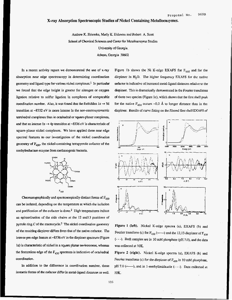
proposal NO. 969B
X-ray Absorption Spectroscopic Studies of Nickel Containing Metalloenzymes.
Andrew K. Shiemke, Marly K. Eidsness and Robert A. Scott
School of Chemical Sciences and Center for Metalloenzyme Studies
University of Georgia
Athens, Georgia 30602
In a recent activity report we demonstrated the use of x-ray
absorption near edge spectroscopy in determining coordination
geometry and ligand type for various nickel complexes.' In particular
we found that the'edge height is greater for nitrogen or oxygen
ligation relative to sulfur ligation in complexes of comparable
coordination number. Also, it was found that the forbidden 1s + 3d
transition at -8332 eV is more intense in the non-centrosymmetric
tetrahedral complexes than in octahedral or square-planar complexes,
and that an intense 1s + 4p transition at -8336 eV is characteristic of
square-planar nickel complexes. We have applied these near edge
spectral features to our investigation of the nickel coordination
geometry of F430, the nickel-containing tetrapyrrole cofactor of the
methylreductase enzyme from methanogenic bacteria.
coon
F430
Chromato~phically and S P ~ C ~ ~ ~ S C O P ~ C ~ ~ Y distinct forms Of F430
can be isolated, depending on the temperature at which the isolation
and purification of the cofactor is done? High temperatures induce
an epimerization of the side chains at the 12 andl3 positions of
pyrrole ring C of the macrocycle? The nickel coordination geometry
of the resulting diepimer differs from that of the native cofactor. The
intense pre-edge feature at -8336 eV in the diepimer spectrum (Figure
la) is characteristic of nickel in a square planar environment, whereas
the featureless edge of the F430 spectrum is indicative of octahedral
coordination.
In addition to the difference in coordination number, these
isomeric forms of the cofactor differ in metal-ligand distances as well.
Figure l b shows the Ni K-edge EXAFS for F430 and for the
diepimer in H20. The higher frequency EXAFS for the native
cofactor is indicative of increased metal-ligand distances relative to the
diepimer. This is dramatically demonstrated in the Fourier transforms
of these two species (Figure IC), which shows that the first shell peak
for the native F430 occurs -0.2 8, to longer distance than in the
diepimer. Results of curve fitting on the filtered first shell EXAFS of
11, . , , , ' I 18,
Figure 1 (left). Nickel K-edge spectra (a), EXAFS (b) and
Fourier transform (c) for F430 (-) and the 12,13 diepimer of F430
(- - -). Both samples are in 10 mM phosphate (pH 7.0), and the data
was collected at 10K.
Figure 2 (right). Nickel K-edge spectra (a), EXAFS (b) and
Fourier transform (c) for the diepimer of F430 in 10 mM phosphate,
pH 7.0 (-), and in 1-methylimidazole (- - -). Data collected at
1OK.
135

these two samples show that the nickel in F430 has 6 nitrogen (or
oxygen) ligands at an average distance of 2.1 A, whereas the
diepimer has 4 N (or 0) ligands at 1.9 A. We can therefore conclude
that the heat-induced epimerization of the cofactor is accompanied by
the loss of axial ligands with a concomitant contraction of the
macrocycle core. It is likely that the core contraction of the diepimer
macrocycle occurs through an S, ruffling of the macrocycle, similar
c I
.* E 1
N .- -
z to that observed in the closely related Ni-corphin complexes? 0
The epimerization at the periphery of the F430 macrocycle does 8300 8320 8340 8360 8380 8400 Energy (eV) not render the cenaal metal incapable of binding axial ligands. In the
presence of a large excess of a strongly coordinating species the
central nickel will bind axial ligands to become 6-coordinate. Figure
2a shows a comparison of the near edge spectra of the diepimer in
water and in 1-methylimidazole. The spectrum of the sample in
1-methylimidazole contains no trace of the 1 s + 4p pre-edge feature
seen in 30. Chnparison of the EXAFS and Fourier transforms of
the &epimer in H20 and in 1-methylimidazole (Figure 2b,c) also
Figure 3. Nickel K-edge absorption spectra of isolated F430
(-), and the methylreductase holoenzyme (- - -). Both samples
are in 10 mM phosphate @H 7.0) and thedata was collected at IOK.
slightly lower energy in the methylreductase spectrum. These
differences may indicate that the axial ligands in the protein-bound
indicate axial ligation by 1-methybidazole. The higher frequency of
the EXAFS and the shift of the first shell peak to longer distance in
the coordinating solvent are very similar to differences between the
native cofactor and diepimer in water (Figure 1). This is confirmed
cofactor are more polarizable than those of the isolated F430 (e.g.
sulfur or anionic oxygen ligation in the protein vs. water ligation in
isolated F43d. Though we cannot yet unequivically identify the axid
ligands of either isolated or protein-bound F430, we are continuing to
by curve fitting of the filtered f is t shell EXAFS of the diepimer in
1-methylimidazole which shows that the nickel coordination sphere
consists of 6 nitrogen ligands at 2.1 A. Thus, the axial ligation of the
pursue EXAFS studies of both in an attempt to do so.
diepimer is accompanied by a core expansion to yield a nickel
coordination sphere very similar to the cofactor in its native
REFERENCES
1. Scott, R. A. and Eidsness, M. K. SSRL Activify Report for
configuration. 1986, proposal 969, p. 129.
Comparison of the near-edge XAS of isolated F430 to that of the Shiemke, A. K.; Hamilton, C. L.; Scott, R. A. J. Biol. Chem.
protein-bound cofactor also reveals differences in the ~ nickel 1986, in press.
coordination sphere (Figure 3). Although the central nickel is PEaltz, A.; Livingston, D. A.; Jaun, B.; Diekert, G.; Thauer,
6-coordinate in both cases, the edge height of the R. K.; Eschenmoser, A. Helv. Chim. Acta 1985, 68,
2.
3.
methylreductase-bound cofactor is reduced relative to that of the 1338-1358.
isolated F430. In addition the inflection point of the edge is shifted to 4. Kratky, C.; Flssler, A.; Haltz, A.; Krtiutler, B.; Jaun, B.;
Eschenmoser, A. J . Chem. SOC. Chem. Comm. 1984,
1368-1371.
136
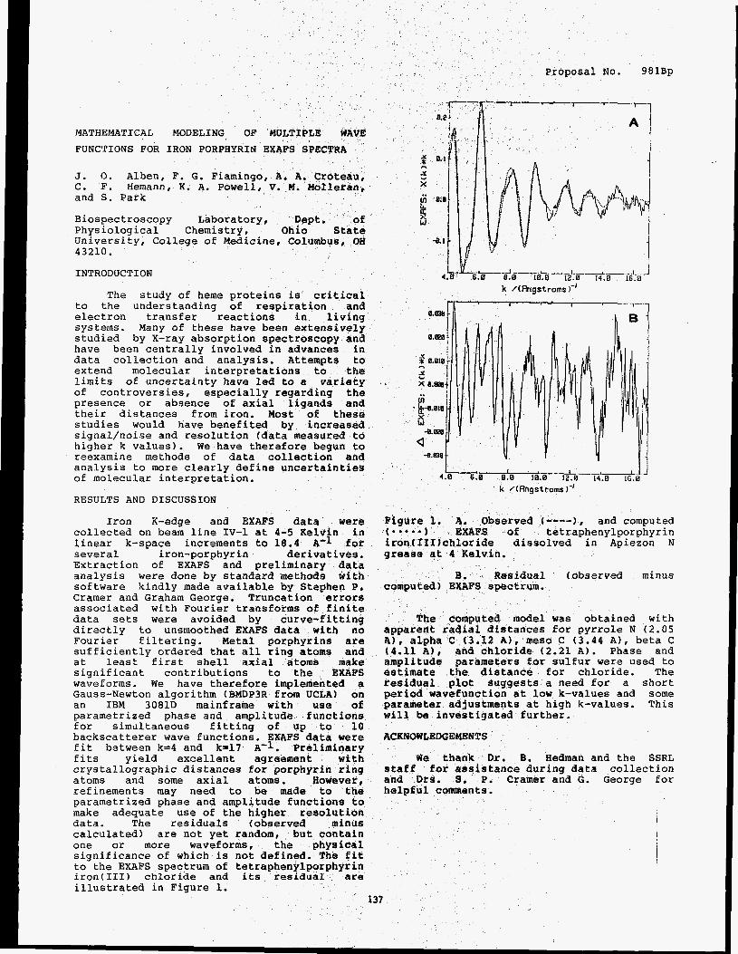
MATHEMATICAL MODELING OF MULTIPLE WAVE
FUNCTIONS FOR IRON PORPHYRIN EXAFS SPECTRA
J. 0. Alben, F. G. Fiamingo, A. A. C. F. Hemann, K. A. Powell, V. M. and S. Park
Biospectroscopy Laboratory, De Physiological Chemistry, Ohio State University, College of Medicine, Columbus, OH 43210.
INTRODUCTION
The study of heme proteins is critical to the understanding of respiration and electron transfer reactions in living systems. Many of these have been extensively studied by X-ray absorption spectroscopy and have been centrally involved in advances in data collection and analysis. Attempts to extend molecular interpretations to the limits of uncertainty have led to a variety of controversies, especially regarding the presence or absence of axial ligands and their distances from iron. Most of these studies would have benefited by increased signal/noise and resolution (data measured to higher k values). We have therefore begun to reexamine methods of data collection and analysis to more clearly define uncertainties of molecular interpretation.
RESULTS AND DISCUSSION
Iron R-edge and EXAFS data were collected on beam line IV-1 at 4-5 Kelvin in linear k-space increments to 18.4 l4-1 for several iron-porphyrin derivatives. Extraction of EXAFS and preliminary data analysis were done by standard methods with software kindly made available by Stephen P, Cramer and Graham George. Truncation errors associated with Fourier transforms of finite data sets were avoided by curve-fitting directly to unsmoothed EXAFS data with no Fourier filtering. Metal porphyrins are sufficiently ordered that all ring atoms and at least first shell axial atoms make significant contributions to the EXAFS waveforms. We have therefore implemented a Gauss-Newton algorithm (BMDP3R from UCLA) on an IBM 3081D mainframe with use of parametrized phase and amplitude functions for simultaneous fitting of up to 10 backscatterer wave functions. EXAFS data were fit between k=4 and k=17 A-1. Preliminary fits yield excellent agreement with crystallographic distances for porphyrin ring atoms and some axial atoms. However, refinements may need to be made to the parametrized phase and amplitude functions to make adequate use of the higher resolution data. The residuals (observed minus calculated) are not yet random, but contain one or more waveforms, the physical significance of which is not defined. T t to the EXAFS spectrum of tetraphenylpo n iron(II1) chloride and its residua e illustrated in Figure 1.
No. 981Bp
8.W
8.828
1 * 0.010
x 0.838
h
5
. g+.010
W -8.m Q
-8.838
rved t----), and computed of tetraphenylporphyrin dissolved in Apiezon N
Residual (observed minus EXAFS spectrum.
model was obtained with tances for pyrrole N (2.05
A ) , alpha C (3.12 A), meso C (3.44 A), beta C (4.11 A ) , and chloride (2.21 A). Phase and amplitude parameters for sulfur were used to estimate the distance for chloride. The residual plot suggests a need for a short period wavefunction at l o r k-values and some parameter adjustments at high k-values. This will be investigated further.
ACKNOWLEDGEMENTS
We thank Dr, B. Hedman and the SSRL staf€ for assistance during data collection and Ors. 8 . P. Cramer and G. George for helpful comments.

I
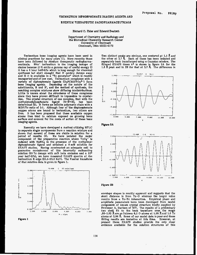
Proporal No, 091ep TECHNETIUM DIPHOBPHONATE IMAGING AUENTS AND
RHENIUM THERAPEUTIC RADIOPHARMACEUTICALS
Richard C. Elder and Edward Deutsch
Department of Chemistry and Radiology and the Biomedical Chemistry Research Center
University of Cincinnati Cincinnati, Ohio 45221-0172
Technetium bone imaging agents have been used in clinical practices for many years (1). More recently these have been followed by rhenium therapeutic radiopharma- ceuticals. 99m- technetium is the imaging isotope of choice because 1) it emits a gamma ray of useful energy 2) it has a 6 hour half-life which is long enough for chemical syntheses but short enought that it quickly decays away and 3) it is available in a "Tc generator" which is readily transported and of low cost. Technetium complexes with a variety of diphosphonate ligands (03PCRRtP0&) form bone imaging agents. Depending on the nature of the substituents, R and R', and the method of synthesis, the resulting complex mixtures show differing biodistributions. Little is known about the structures of these complexes since they have proven difficult to imparsible to crystal- lize. The crystal structure of one complex, that with the methylenediphosphonate ligand (R=R'=H), has been determined (2). It forms an infinite polymeric chain with a MDP/Tc ratio of 1:l. Although four of the disphosphonate oxygen atoms are bound to technetium, two others are free. It has been proposed that these available oxygen atoms then bind to calcium exposed on growing bone surface and account for the mode of action of these bone imaging agents.
Recently we have developed a method utilizing HPLC to separate single components from a reaction mixture and shown that several of these are stable in solution for a period of months (3). We have isolated the major component of the preparative reaction where TcO4- is reduced with NaBH4 in the presence of the methylene- diphosphonate ligand and obtained a 8 mM solution for EXAFS studies. Having constructed an adequate cell to guarantee containment of this inherently radioactive solution (99 Tc decays with soft beta emission and a 105 year half-life), we have 'measured EXAFS spectra a t the technetium K edge (20.5-22.0 KeV). The Fourier transform of that solution data is given in figure 1. , ~ ,
T:-HOP , C , F I H:GNIIUPE , , , 2.200
f T
H a
4
El0
, " t
d
1.760-
1.320-
0.880- il 0 . 4 4 0 -
0. 000 0.000 1.500 3.000 4 . 5 0 0 6.000 7 . 5 0 0
R + alpha
Figure 1
Two distinct peaks are obvious, one centered a t 1.5 d and the other at 2.7 h Each of these has been isolated and separately back transformed using a Gaussian window. The filtered EXAFS times k3 is shown in figure 2A for the 1.5 8 peak and in 28 for that a t 2.7 h The difference in
IC -MOP c TC.0 1,000
0.600-
E 1 F 0.200- I f n
-0.200- L O
-0.600- -
-1,000 3.000 5 .000 7.000 9.000 11.000 13.000
K
Figure 2A
TC-HDP C-2 TC-TC 3'0"0r-- ' ' ,: ' ' ' ' ' I
-3.000' ' ' ' ' ' ' ' ' ' I 3.000 5 . 0 0 0 7.000 9.000 11.000 13.000
K
Figure 2B
envelope shapes is readily apparent and suggests that the short distance is from Tc-0 whereas the longer value results from a Tc-Tc interaction. Empirical phase and amplitude parameters have been developed from model compounds of known crystal structure kindly supplied by Professor A. Davison of MIT. The results of a preliminary two shell fit to the back transform over the range .88-2.92 d are as follows: 6.0 0 atoms a t 1.98 8 and 1.5 Tc atoms a t 2.94 Some of our model data is poor and these fitting results are tentative at this time. However, at present these EXAFS studies provide the only clear evidence available for the solution structures of this
138
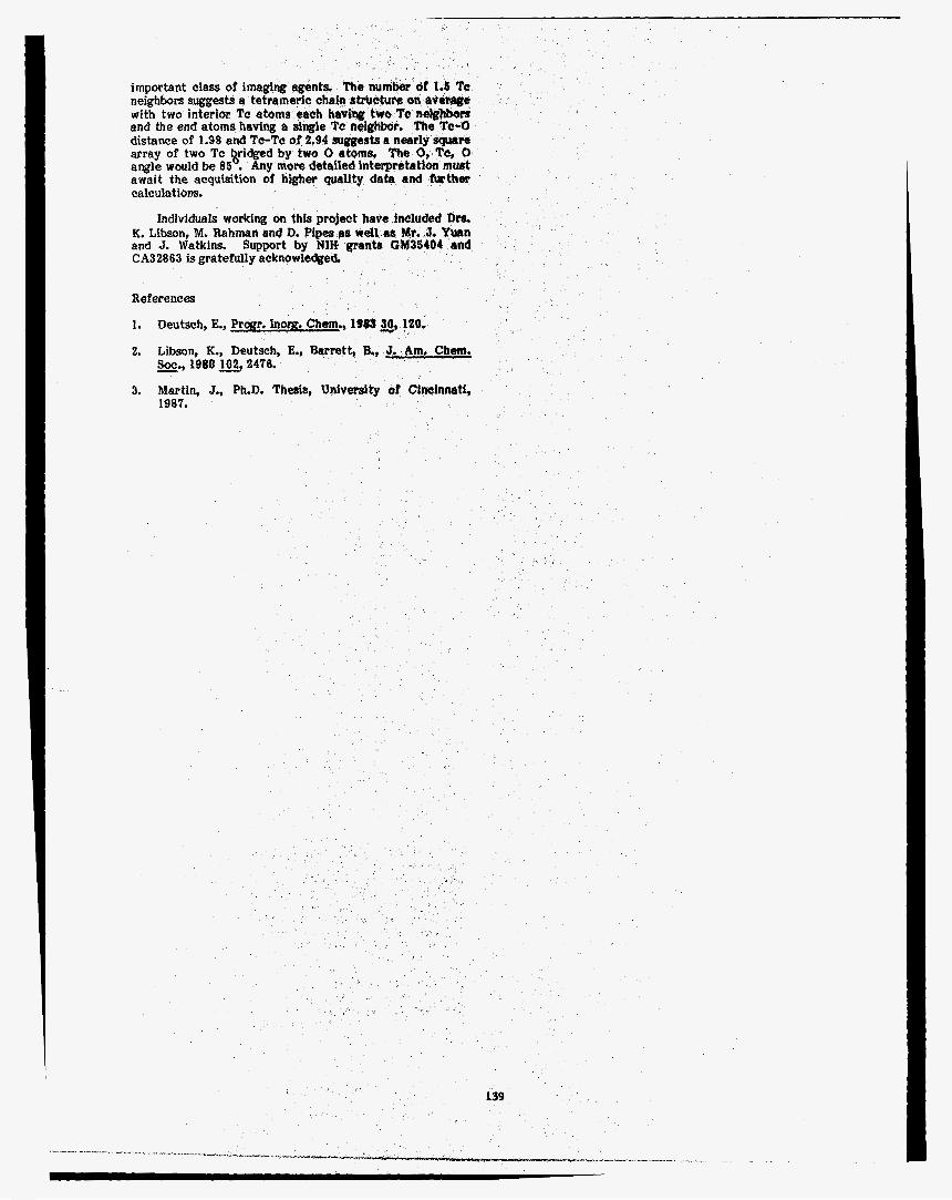
important class of imaging age neighbors suggests a tetrameric chain strrrcture with two interior Tc atoms each havihg two T and the end atoms having a single Tc neighbor. The Tc-0 distance of 1.98 and To-Tc of 2.94 suggests a nearly square array of two Tc kridged by two 0 atoms. The 0, To, 0 angle would be 85 . Any mote detailed interpretation must await the acquisition of higher quality data and further calculations.
Individuals working on this K. Libson, M. Rahman end D. Pi and J. Watkins. Support by CA32863 is gratefully acknowledged.
References
1. Deutsch, E., Prom. I n w . Chem., 1983 SO, 120.
2. Libson, K., Deutsch, E., Barrett, B,, -*) Soe 1980102,2476.
3. Martin, J., PLD. Thesis, Univeraltg of Cincinnatf, 1987.
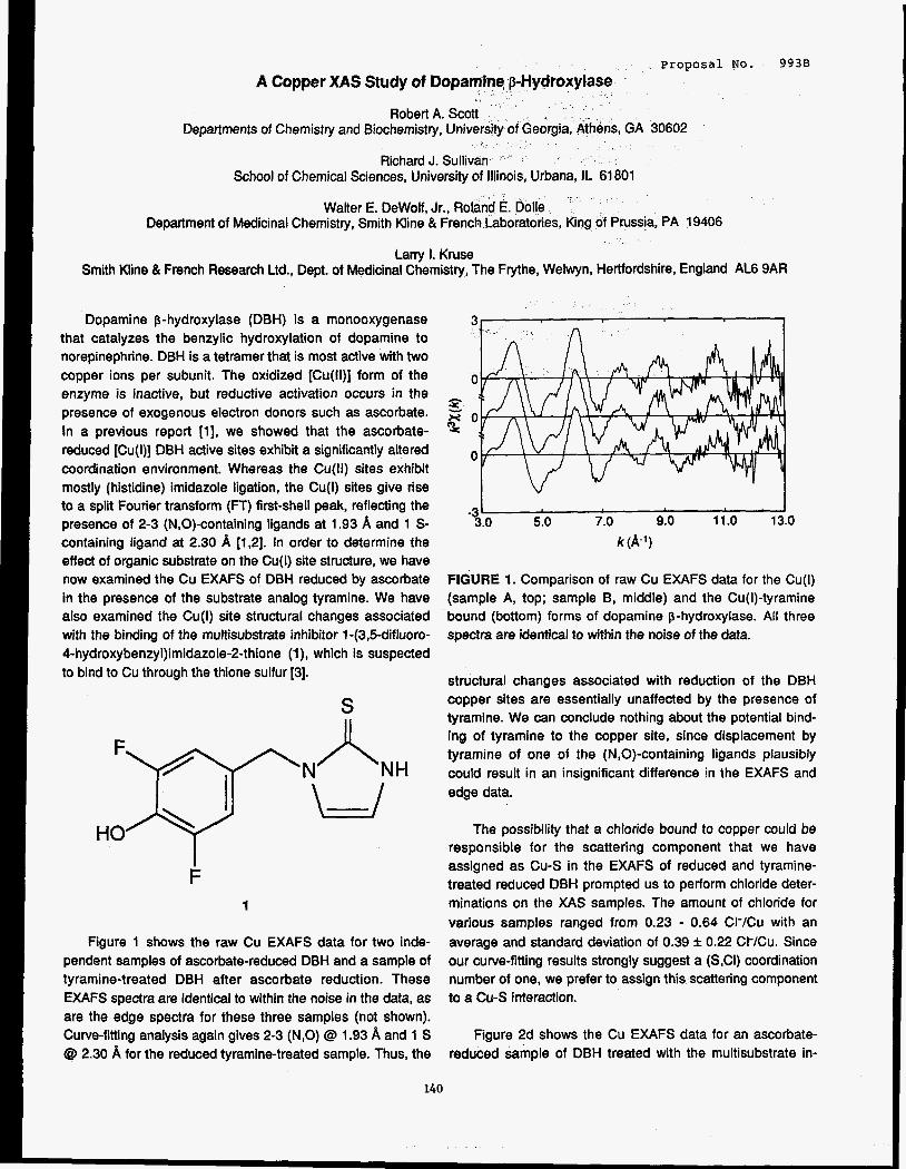
proposal No. 993B A Copper XAS Study of D
Departments of Chemistry and Biochemistry,
School of Chemical Sciences, University
Walter E. DeWolf, Jr., Rol Department of Medicinal Chemistry, Smith Kline & French Laboratories, King of Prussia, PA 19406
Larry 1. Kruse Smith Kline 81 French Research Ltd., Dept. of Medicinal Chemistry, The Frythe, Welwyn, Hertfordshire, England AL6 9AR
Urbana, IL 61801
Dopamine p-hydroxylase (DBH) is a monooxygenase that catalyzes the benzylic hydroxylation of dopamine to norepinephrine. DBH is a tetramer that is most active with two copper ions per subunit. The oxidized [Cu(ll)] form of the enzyme is inactive, but reductive activation occurs in the presence of exogenous electron donors such as ascorbate. In a previous report [l], we showed that the ascorbate- reduced [Cu(l)] DBH active sites exhibit a significantly altered coordination environment. Whereas the Cu(1l) sites exhibit mostly (histidine) imidazole ligation, the Cu(l) sites give rise to a split Fourier transform (FT) first-shell peak, reflecting the presence of 2-3 (N,O)-containing ligands at 1.93 A and 1 S- containing ligand at 2.30 A [1,2]. In order to determine the effect of organic substrate on the Cu(l) site structure, we have now examined the Cu EXAFS of DBH reduced by ascorbate in the presence of the substrate analog tyramine. We have also examined the Cu(l) site structural changes associated with the binding of the multisubstrate inhibitor 1 -(3,5-difluoro- 4-hydroxybenzyl)imidazole-2-thione (I), which is suspected to bind to Cu through the thione sulfur [3].
S
F 1
Figure 1 shows the raw Cu EXAFS data for two inde- pendent samples of ascorbate-reduced DBH and a sample of tyramine-treated DBH after ascorbate reduction. These EXAFS spectra are identical to within the noise in the data, as are the edge spectra for these three samples (not shown). Curve-fitting analysis again gives 2-3 (N,O) @ 1.93 A and 1 S Q 2.30 A for the reduced tyramine-treated sample. Thus, the
0
s g o
0
I 3.0 5.0 7.0 9.0 11.0 13.0
k (A-l)
-31
FIGURE 1. Comparison of raw Cu EXAFS data for the Cu(l) (sample A, top; sample 8, middle) and the Cu(l)-tyramine bound (bottom) forms of dopamine p-hydroxylase. All three spectra are identical to within the noise of the data.
structural changes associated with reduction of the DBH copper sites are essentially unaffected by the presence of tyramine. We can conclude nothing about the potential bind- ing of tyramine to the copper site, since displacement by tyramine of one of the (N,O)-containing ligands plausibly could result in an insignificant difference in the EXAFS and edge data.
The possibility that a chloride bound to copper could be responsible for the scattering component that we have assigned as Cu-S in the EXAFS of reduced and tyramine- treated reduced DBH prompted us to perform chloride deter- minations on the XAS samples. The amount of chloride for various samples ranged from 0.23 - 0.64 CHCu with an average and standard deviation of 0.39 f 0.22 CI-/Cu. Since our curve-fitting results strongly suggest a (SCI) coordination number of one, we prefer to assign this scattering component to a Cu-S interaction.
Figure 2d shows the Cu EXAFS data for an ascorbate- reduced sample of DBH treated with the multisubstrate in-
140
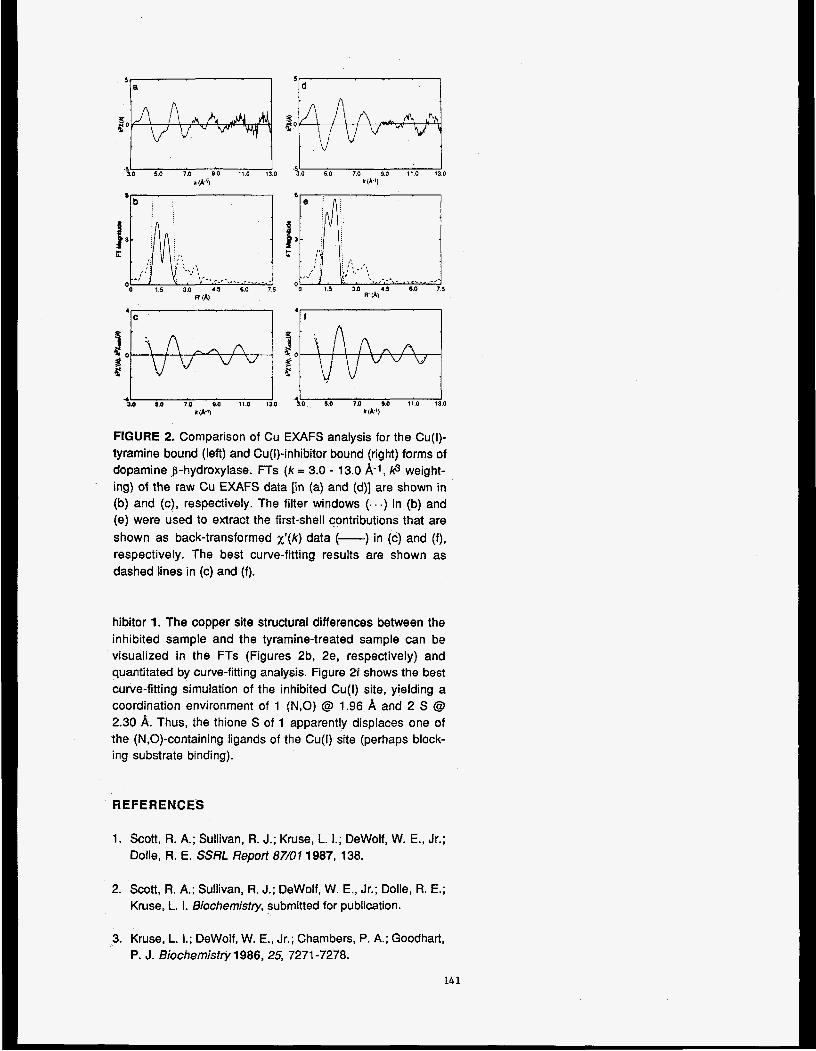
5 5 a Id
- P O 5:O 7.0 9.0 1;O 1!0 5 0 7.0 9.0 11.0 110 I ( h $ k ( A ' )
4L SO $0 7.0 0.0 11.0 13.0 4; 3.0, 3.0 70 9.0 11.0 13.0
k(k9 k ( A 9
FIGURE 2. Comparison of Cu EXAFS analysis for the Cu(l)- tyramine bound (left) and Cu(l)-inhibitor bound (right) forms of dopamine p-hydroxylase. FTs (k = 3.0 - 13.0 A-1, /c3 weight- ing) of the raw Cu EXAFS data [in (a) and (d)] are shown in (b) and (c), respectively. The filter windows (. . .) in (b) and (e) were used to extract the first-shell contributions that are shown as back-transformed ~ ' ( k ) data (-) in (c) and (f), respectively. The best curve-fitting results are shown as dashed lines in (c) and (f).
hibitor 1. The copper site structural differences between the inhibited sample and the tyramine-treated sample can be visualized in the FTs (Figures 2b, 2e, respectively) and quantitated by curve-fitting analysis. Figure 2f shows the best curve-fitting simulation of the inhibited Cu(l) site, yielding a coordination environment of 1 (N,O) @ 1.96 &, and 2 S @ 2.30 A. Thus, the thione S of 1 apparently displaces one of the (N,O)-containing ligands of the Cu(l) site (perhaps block- ing substrate binding).
REFERENCES
1. Scott, R. A.; Sullivan, R. J.; Kruse, L. I . ; DeWolf, W. E., Jr.; Dolle, R. E. SSRL Report 87/01 1987, 138.
2. Scott, R. A.; Sullivan, R. J.; DeWolf, W. E., Jr.; Dolle, R. E.; Kruse, L. 1. Biocbemistry, submitted for publication.
3. Kruse, L. 1.; DeWolf, W. E., Jr.; Chambers, P. A.; Goodhart, P. J. Biochemistry 1986,25, 7271 -7278.
14 1
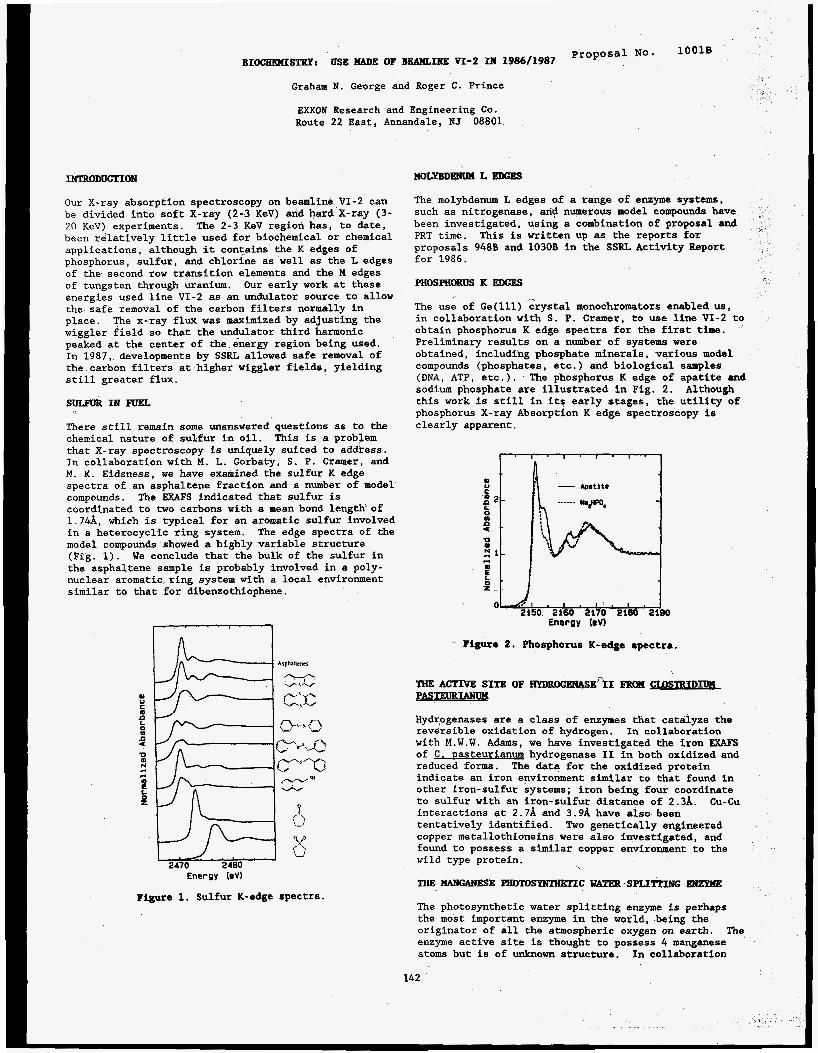
lOOlS BIOCEEKISTBYI USE W E OF BEAMLINE V I - 2 II 1986/1987 No'
Graham N. George and Roger C. Prince
EXXON Research and Engineering Co. Route 22 East, Annandale, NJ 08801
IFJTRODUCTION
Our X-ray absorption spectroscopy on beamline VI-2 can be divided into soft X-ray (2-3 KeV) and hard X-ray ( 3 - 20 KeV) experiments. The 2-3 KeV region has, to date, been relatively little used for biochemical or chemical applications, although it contains the K edges of phosphorus, sulfur, and chlorine as well as the L edges of the second row transition elements and the M edges o f tungsten through uranium. Our early work at these energies used line VI-2 as an undulator source to allow the safe removal of the carbon filters normally in place. The x-ray flux was maximized by adjusting the wiggler field so that the undulator third harmonic peaked at the center of the energy region being used. In 1987, developments by SSRL allowed safe removal of the carbon filters at higher wiggler fields, yielding still greater flux.
SULEVRINEWEL
There still remain some unanswered questions as to the chemical nature of sulfur in oil. that X-ray spectroscopy is uniquely suited to address. In collaboration with M. L. Gorbaty, S. P. Cramer, and M. K. Eidsness, we have examined the sulfur K edge spectra of an asphaltene fraction and a number of model compounds. coordinated to two carbons with a mean bond length'of 1.74A, which is typical for an aromatic sulfur involved in a heterocyclic ring system. The edge spectra of the model compounds showed a highly variable structure (Fig. 1). We conclude that the bulk of the sulfur in the asphaltene sample is probably involved in a poly- nuclear aromatic ring system with a local environment similar to that for dibenzothiophene.
This is a probJem
The EXAFS indicated that sulfur is
m u
m
0
n
9 0
N
6
L
z
4 c(
2470 2480 Energy (eV)
Figure 1. Sulfur K-edge spectra.
HOL~DENUH L EDGES
The molybdenum L edges of a range of enzyme systems, such as nitrogenase, any numerous model compounds have been investigated, using a combination of proposal and PRT time. proposals 948B and 1030B in the SSRL Activity Report for 1986.
This is written up as the reports for
pHoSpHoBuS K EDGES
The us; of Ge(ll1) Zrystal monochromators enabled us, in collaboration with S. P. Cramer, to use line VI-2 to obtain phosphorus K edge spectra for the first time. Preliminary results on a number of systems were obtained, including phosphate minerals, various model compounds (phosphates, etc.) and biological samples (DNA, ATP, etc.). The phosphorus K edge of apatite and sodium phosphate are illustrated in Fig. 2. this work is still in its early stages, the utility of phosphorus X-ray Absorption K edge spectroscopy is clearly apparent.
Although
. Figure 2. Phosphorus K-edge spectra.
Hydrogenases are a class of enzymes that catalyze the reversible oxidation of hydrogen. with M.W.W. Adams, we have investigated the iron WAFS of hydrogenase I1 in both oxidized and reduced forms. indicate an iron environment similar to that found in other iron-sulfur systems; iron being four coordinate to sulfur with an iron-sulfur distance of 2.3A. interactions at 2.7A and 3.9A have also been tentatively identified. Two genetically engineered copper metallothioneins were also investigated, and found to possess a similar copper environment to the wild type protein.
In collaboration
The data for the oxidized protein
Cu-Cu
The photosynthetic water splitting enzyme is perhaps the most important enzyme in the world, being the originator of all the atmospheric oxygen on earth. enzyme active site is thought to possess 4 manganese atoms but is of unknown structure. In collaboration
The
142

with S. P. Cramer, we have performed polarized X-ray absorption spectroscopy on the water splitting enzyme prepared in oriented membranous multilayers, which are ordered in one dimension. The Mn FXAFS Fourier transforms €or three different orientations are shown in Fig. 3 . The data indicate that the manganese is
ow6 6-07 Figure 3. Driented CnlOropleat membranes Hn EXAFS Fourier Transforms
coordinated by oxygen or nitrogen at a mean distance of 1.9A from the metal. can be discerned; 2-3 at 2.7A and 0.5-1 at 3.3A. orientation dependence of the latter indicates that the 3.3A Mn-Mn vector lies approximately along the membrane normal, while for the former a mean angle between the membrane normal and the 2.7A Mn-Mn vector of about 55' is indicated. contains a tetranuclear manganese cluster. possible geometry for this is illustrated in Fig. 4.
Two different Mn-Mn interactions The
The results suggest that the active site One
Figure 4. Proposed Mn geometry in the water splitting enzyme.
143
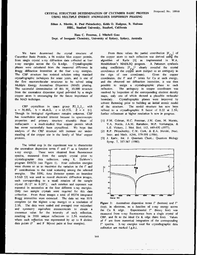
Proposal No. 1004B CRYSTAL STRUCTURE DETERMINATION OF CUCUMBER BASIC PROTEIN
USING MULTIPLE ENERGY ANOMALOUS DISPERSION PHASING
Ethan A. Memtt, R. Paul Phizackerley, Keith 0. Hodgson, B. Hedman SSRL,, Stanford University, Stanford, California
Hans C. Freeman, J. Mitchell Guss Dept. of Inorganic Chemistry, University of Sydney, Sydney, Australia
We have determined the crystal structure of Cucumber Basic Protein, a 96 residue blue copper protein, from single crystal x-ray diffraction data collected at four x-ray energies across the Cu K-edge. Crystallographic phases were calculated from the measured difference in Bragg diffraction intensities at the four x-ray energies. The CBP structure has resisted solution using standard crystallographic techniques for some years, and is one of the first macromolecular structures to be solved using Multiple Energy Anomalous Dispersion (MAD) phasing. The successful determination of this M 10,000 structure from the anomalous dispersion signal provided by a single copper atom is encouraging for the future importance of the MAD technique.
r
CBP crystallizes in space group P2,2 ,2 , . with a = 30.88& b = 46.41A, c = 65.57& Z = 4 [ I ] . Though its biological significance remains unknown, CBP has nonetheless attracted interest because its spectroscopic properties and primary structure resemble those of stellacyanin - a much-studied blue copper protein which has never successfully been crystallized. We hope that analysis of the CBP structure will increase our under- standing of the copper site in the family of 'blue' copper proteins.
The initial step in the experiment was to characterize the anomalous dispersion terms f' and f" as a function of x-ray energy. These were obtained from fluorescence spectra. measured from the sample crystal prior to crystallographic data collection, using K. Eichhorn's program DISCO (see Figure I ) . Four collection energies were chosen so as to maximize the variation in the f' and f" contributions to the total scattering among the selected energies. The SSRL Area Detector system on beamline 1-5AD [2] was used to record electronic diffraction images, each corresponding to a small rotation of the sample crystal (0.12" to 0.20'); each rotation and exposure was repeated in succession at the four different x-ray energies. Only two sample crystals were required for this data collection. From these images a total of 85374 integrated Bragg intensitites were extracted. The data set was 95% complete (at the highest x-ray energy) to a resolution of 2 . 5 k The data were scaled and averaged over redundant and symmetry equivalent measurements to obtain a consensus value for the intensity of each reflection, resulting in 3550 unique reflections to 2.5A resolution. where each re ection was represented by up to 8 distinct data points (F
-t! and F- Bijvoet pairs at four energies).
From these values the partial contribution IF I of the copper atom to each reflection was derived using the algorithm of Karle 13) as implemented in W.A. Hendrickson's MADLSQ program. A Patterson synthesis using coefficients IFCu(* clearly revealed the crystal coordinates of the copper atom (subject to an ambiguity in the sign of one coordinate). Given the copper coordinates, the f' and f" terns for Cu at each energy, and the observed net diffraction intensities, it was then possible to assign a crystallographic phase to each reflection. The ambiguity in copper coordinates was resolved by inspection of the corresponding electron density maps, only one of which showed a plausible molecular boundary. Crystallographic phases were improved by solvent flattening prior to building an initial atomic model of the structure. The model structure has now been refined to a crystallographic R factor of 0.22 at 2.5A; further refinement at higher resolution is now in progress.
11 I P.M. Colman, H.C. Freeman, J.M. Guss, M. Murata, V.A. Norris, J.A.M. Ramshaw, M.P. Venkatappa, & L.E. Vickery, J. Mol. Biol. 112, 649-650 (1977).
[2] R.P. Phizackerley, C.W. Cork, & E.A. Merrilt, Nucl. Instr. and Meth. A246, 579-595 (1986).
131 J. Karle, Int. J. Quantum Chem.: Quantum Biology Symp. 7. 357-367 (1980).
.cu
Figure 1: Anomalous dispersion terms f' (bottom) and f" (top), in electrons, as a function of x-ray energy across the Cu K edge. Experimental f" (heavy line) was measured from x-ray fluorescence from a single crystal of CBP, and fit to the ideal Cu K edge (faint line). Values of f are from numerical integration of the corresponding f" spectra. X-ray energies used for crystallographic data collection are marked f,g,h,i.
144

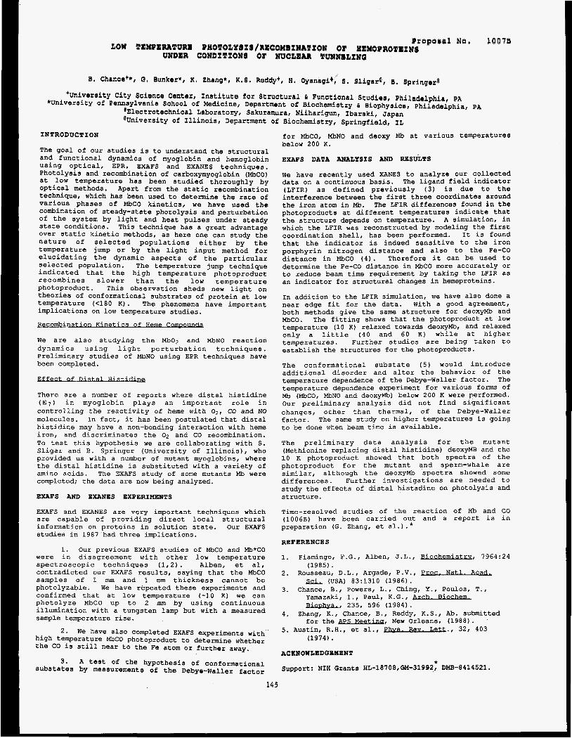
Ogopaoal No. lOO7f3 LOW TEXPERAPURE PHOPOLYBIB/RECOXlINA~I~N OF HEXOPROTEI~8
UNDER CONDITIONS OF NUCLEAR QUWNELINQ
B. Chance**, G. Bunker*, K . Zhang*, K . 6 . ReddySI H, Oyanagi), 6 . Sligare, 8 . Springera
'University City Science Center, Institute for Structural & Functional Studies, Philadelphia, PA *University of Pennsylvania School of Medicine, Department of Biochemiatry & Biophysics, Philadelphia, PA
#Electrotechnical Laboratory, Sakuramura, Niiharigun, Ibaraki, Japan @University of Illinois, Department of Biochemistry, Springfield, IL
INTRODUCTION
The goal of our studies is to understand the structural and functional dynamics of myoglobin and hemoglobin using optical, EPR, EXAFS and EXANES techniques. Photolysis and recombination of carboxymyoglobin (MbCO) at low temperature has been studied thoroughly by optical methods. Apart from the static recombination technique, which has been used to determine the rate of various phases of MbCO kinetics, we have used the combination of steady-state photolysis and perturbation of the system by light and heat pulses under steady state conditions. This technique has a great advantage over static kinetic methods, as here one can study the nature of selected populations either by the temperature jump o r by the light input method for elucidating the dynamic aspects of the particular selected population. The temperature jump technique indicated that the high temperature photoproduct recombines slower than the low temperature photoproduct. This observation sheds new light on theories of conformational substrates of protein at low temperature (<180 K). The phenomena have important implications on low temperature studies.
Recomb ination Kinetics of Heme COmDOUndS
We are also studying the Mb02 and MbNO reaction dynamics using light perturbation techniques. Preliminary studies of MbNO using EPR techniques have been completed.
t of D- . . .
There are a number of reports where distal histidine (E,) in myoglobin plays an important role in controlling the reactivity of heme with 02, CO and NO molecules. In fact, it has been postulated that distal histidine may have a non-bonding interaction with heme iron, and discriminates the 02 and CO recombination. To test this hypothesis we are collaborating with S. Sligar and B. Springer (University of Illinois), who provided us with a number of mutant myoglobins, where the distal histidine is substituted with a variety of amino acids. The EXAFS study of some mutants Mb were completed; the data are now being analyzed.
EXAFS AND EXANES EXPERIMENTS
EXAFS and EXANES are very important techniques which are capable of providing direct local structural information on proteins in solution state. Our EXAFS studies in 1987 had three implications.
1. Our previous EXAFS studies of MbCO and Mb*CO were in disagreement with other low temperature spectroscopic techniques (1,2). Alben, et al, contradicted our EXAFS results, saying that the MbCO samples of 1 mm and 1 mm thickness cannot be photolyzable. We have repeated these experiments and confirmed that at low temperature (-10 K) we can photolyze MbCO up to 2 mm by using continuous illumination with a tungsten lamp but with a measured sample temperature rise.
we have also completed EXAFS experiments with high temperature MbCO photoproduct to determine whether the CO is still near to the Fe atom or further away.
3 . A test of the hypothesis of conformational substates by measurements of the Debye-Waller factor
2.
145
for MbCO, MbNO and deoxy Mb at various temperatures below 200 K.
EXAFS DATA AE~ALYSIS AND RESULTS
We have recently used XANES to analyze our collected data on a continuous basis. The ligand field indicator (LFIR) as defined previously (3) is due to the interference between the first three coordinates around the iron atom in Mb. The LFIR differences found in the photoproducts at different temperatures indicate that the structure depends on temperature. A simulation, in which the LFIR was reconstructed by modeling the first coordination shell, has been performed. It is found that the indicator is indeed sensitive to the iron porphyrin nitrogen distance and also to the Fe-CO distance in MbCO (4). Therefore it can be used to determine the Fe-CO distance in MbCO more accurately or to reduce beam time requirement by taking the LFIR as an indicator for structural changes in hemeproteins.
In addition to the LFIR simulation, we have also done a near edge fit for the data. With a good agreement, both methods give the same structure for deoxyMb and MbCO. The fitting shows that the photoproduct at low temperature (10 K) relaxed towards deoxyMb, and relaxed only a little (40 and 60 K) while at higher temperatures. Further studies are being taken to establish the structures for the photoproducts.
The conformational substate (5) would introduce additional disorder and alter the behavior of the temperature dependence of the Debye-Waller factor. The temperature dependence experiment for various forms of Mb (MbCO, MbNO and deoxyMb) below 200 K were performed. Our preliminary analysis did not find significant changes, other than thermal, of the Debye-Waller factor. The same study on higher temperatures is going to be done when beam time is available.
The preliminary data analysis for the mutant (Methionine replacing distal histidine) deoxyMB and the 10 K photoproduct showed that both spectra of the photoproduct for the mutant and sperm-whale are similar, although the deoxyMb spectra showed some differences. Further investigations are needed to study the effects of distal histadine on photolysis and structure.
Time-resolved studies of the reaction of Hb and CO (1006B) have been carried out and a report is in preparation (G. Zhang, et al.) . * REFERENCES
1. Fiamingo, F.G., Alben, J.L., Biochemistrv , 7964:24 2. Rousseau, D.L., Argade, P.V., proc. Natl. Ac&
3. Chance, B., Powers, L., Ching, Y., Poulos, T.,
(1985).
S d , . (USA) 83:1310 (1986).
Yamazaki, I., Paul, K.G., Bioohvs., 235, 596 (1984).
4. Zhang, K., Chance, B., Reddy, K.S., Ab. submitted for the &S-&&UQ ' , New Orleans, (1988).
5. Austin, R.H., et al. , nbys. Rev. Leu., 32, 403 (1974).
ACKNOWZEDGEYENT
Support: NIH Grants HL-18708,GM-31992, DMB-8414521. *

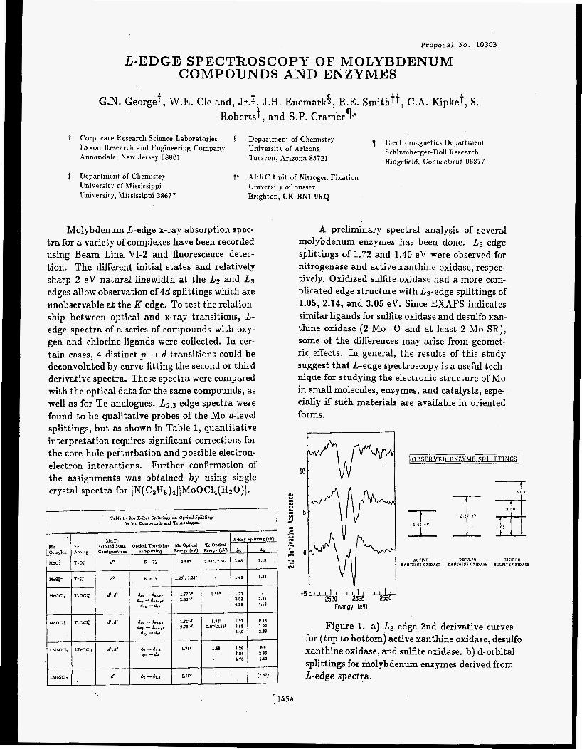
Proposal No. 1030B
LEDGE SPECTROSCOPY OF MOLYBDENUM COMPOUNDS AND ENZYMES
G.N. Georget, W.E. Cleland, Jr.t, J.H. Enemark§, B.E. Smithit, C.A. Kipket, S. Roberts+, and S.P. Crarneryj*
t Corporate Research Science Laboratories Department of Chemistry 1 Electromagnetics Department Exxon Research and Engineering Company University of Arizona Schlumberger-Doll Research Annandale, New Jersey 08801 'Tucsron, Arizona 85721 Ridgefield, Connecticut 06877
4 Department of Chemistry t t AFRC Unit of Kitrogen Fixation University of Vfississippi Lniversit y, Jlississippi 38677
University of Sussex Brighton, UK BN1 9RQ
Molybdenum L-edge x-ray absorption spec- tra for a variety of complexes have been recorded using Beam Line, VI-2 and fluorescence detec- tion. The different initial states and relatively sharp 2 eV natural linewidth at the L2 and L3
edges allow observation of 4d splittings which are unobservable at the K edge. To test the relation- ship between optical and x-ray transitions, L- edge spectra of a series of compounds with oxy- gen and chlorine ligands were collected. In cer- tain cases, 4 distinct p --i d transitions could be deconvoluted by curve-fitting the second or third derivative spectra. These spectra were compared with the optical data for the same compounds, as well as for Tc analogues. L2,3 edge spectra were found to be qualitative probes of the Mo d-level splittings, but as shown in Table 1, quantitative interpretation requires significant corrections for the core-hole perturbation and possible electron- electron interactions. Further confirmation of the assignments was obtained by using single crystal spectra for [ N ( C ~ H , ) ~ ] [ M O O C ~ ~ ( H ~ O ) ] .
Table 1 . Mo X-R.7 Sphttrng. m Ophcd Spltttlngs Ior Mo Cornponds and Tc AndoKUO
Mo.Tc x-Ra, Spllthng (ev:
Ilo Tc Ground State O p t x d Tola%tmton :ampla Andog Conlgarittons or Sphttng EncrKY (eV) Energy (ev) Lr
Ho0:- TcO;
f40.S:- TcS; do E-T* 125*, 121. ~ 141 132
Mo Optlcd Tc Optrcd Lr
P E - TI 181. 23(1*, 2311 2111 119
A preliminary spectral analysis of several molybdenum enzymes has been done. L3-edge splittings of 1.72 and 1.40 eV were observed for nitrogenase and active xanthine oxidase, respec- tively. Oxidized sulfite oxidase had a more com- plicated edge structure with L3-edge splittings of 1.05, 2.14, and 3.05 eV. Since EXAFS indicates similar ligands for sulfite oxidase and desulfo xan- thine oxidase (2 Mo=O and at least 2 Mo-SR), some of the differences may arise from geomet- ric effects. In general, the results of this study suggest that L-edge spectroscopy is a useful tech- nique for studying the electronic structure of Mo in small molecules, enzymes, and catalysts, espe- cially if such materials are available in oriented forms.
7.-
OBSERVED ENZYME SPLlTTlNGS
ACTIVE DESULFO HIGH PH XANTHINE OKIDASE XANTHINE OXIDASE SULFITE OKIDASE
Figure 1. a) L3-edge 2nd derivative curves for (top to bottom) active xanthine oxidase, desulfo xanthine oxidase, and sulfite oxidase. b) d-orbital splittings for molybdenum enzymes derived from Ledge spectra.
, 145A

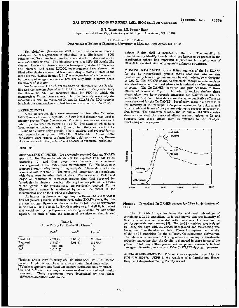
Proposal NO. 10358 XAS INVESTIGATIONS OF RIESKE-LIKE IRON SULFUR CENTERS
H.T. Tsang a n d J.E. Penner-Hahn Department of Chemistry, University of Michigan, Ann Arbor, MI 48109
C.J. Batie and D.P. Ballou Departmect of Biological Chemistry, University of Michigan, Ann Arbor, MI 48109
The phthalate dioxygenase (PDOI from Pseudomonas cepacia catalyzes the dioxygenation of phthalate to a dihydrodiol. PDO contains two Fe atoms in a binuclear site and a third, dissociable iron in a mononuclear site. The binuclear site is a [2Fe-2S] Rieske-like cluster. Rieske-like clusters are spectroscopically distinct from other FelS clusters. and recent ENDOR measurements have shown that Rieske-like clusters contain at least two nitrogen atoms in place of the more normal thiolate ligands [l]. The mononuclear site is believed to be the site of oxygen activation, however very little is known about the nature of this site.
We have used EXAFS spectroscopy to characterize the Rieske- like and the mononuclear sites in PDO. In order to study selectively the Rieske-like site, we measured data for PDO in which the mononuclear Fe had been removed. In order to study selectively the mononuclear site, we measured Zn and Co EXAFS for PDO samples in which the mononuclear site had been reconstituted with Zn or Co.
EXPERIMENTAL X-ray absorption data were measured on beam-line 7-3 using
Si(220) monochromator crystals. A Stern-Heald detector was used ta monitor proteln X-ray fluorescence. Protein concentrations were ea. 4 mM. The samples which have been examined include: native (3Fe) protein (fully reduced); 2 Fe (Rieske-like cluster only) protein in both oxidized and reduced forms; and reconstituted protein (2Fe+M, M=Zn,Co). Mixed metal derivatives were studied in forms having oxidized or reduced Rieske- like clusters and in the presence and absence of substrate (phthalate).
RESULTS
RIESKE-LIKE CLUSTER We previously reported that the EXAFS spectra for the Rieske-like site showed the expected Fe-S and Fe-Fe scattering [2] and that these data indicated a structural rearrangement of the Fe/S cluster cn reduction [31. We have now completed quantitative curve fitting analysis of these data with the results shown in Table I. The structural parameters are consistent with those seen for other FelS clusters. The increase in Fe-S bond length on reduction is somewhat greater than that observed for ferredoxin-like clusters, possibly reflecting the less polarizable nature of the ligands in the present case. As previously reported 131, the Rieske-like structure is unaffected by either the metal in the mononuclear site or the binding of substrate.
An interesting observation regarding the Rieske-like site is that it has not proven possible to demonstrate, using EXAFS alone, that the are any nitrogen ligands coordinated to the Fe [41. The improvement in fit quality for a 2 shell fit (S+N) relative to a 1 shell fit is modest and would not by itself provide convincing evidence for non-sulfur ligation. In spite of this, the position of the nitrogen shell is well
Spectra were measured at 4-10 K.
Table I. Curve Fitting For Rieske-like Clustea
Fe-Sb Fe-Nb Fe-Feb
Oxidized 2.20(2) 2.01(5) 2.68(4) Reduced 2.24(2) 2.08(5) 2.67(4) ARC 0.031(10) 0 A02 0.012(5) 0
aIsolated shells were fit using 3S+1N (first shell) or 1 Fe (second shell). Amplitude and phase parameters determined empirically.
bTabulated numbers are fitted parameters (estimated uncertainties). ‘AR and Ao2 are the change between oxidized and reduced Rieske
clusters. These parameters were determined by the phase dfference/amplitude ratio method.
defined if this shell is included in the fit. The inability to unambiguously identify ligands which are known to be present in the coordination sphere has important implications for applications of EXAFS to the elucidation of completely unknown structures.
MONONUCLEAR SITE. Curve fitting analysis of the Zn EXAFS for the Zn reconstituted protein shows that this site contains predominamtly N or 0 ligation and can be well modeled by 6 nitrogens at 2.01 A. The EXAFS shows no detectable change in mononuclear- site structure when the Rieske-like site is reduced or when substrate is bound. The Zn-XANES, however, are quite sensitive to these effects, as shown in Fig. 1. In order to explore further these observations, we have recently measured Co XANES for the Co substituted enzyme. These data show the same qualitative effects as were observed for the Zn XANES. Specifically, there is a decrease in the intensity of the principal absorption maximum for oxidized and substrate-bound forms of the enzyme relative to reduced or substrate- free forms. The similarity between the Co and Zn X A N E S spectra demonstrates that the observed effects are not unique to Zn and suggests that these effects may be relevant to the catalytic functioning of the enzyme.
1 I I I
,+ phthalate
I I I
9650 9660 9670 9680 9690 9700
eV
Figure 1. Normalized Zn XANES spectra for ZFe+Zn derivatives of PDO.
The Co XANES spectra have the additional advantage of containing a 1s-3d transition. It is well known that the intensity of this transition can be correlated with distortions of a site from a centrosymmetric environment [SI. The 1s-3d transition was isolated by fitting the edge with an arctan backgromd and subtracting this background from the observed data. Figure 2 compares the intensity of the 1s-3d transition for the different Co substituted derivatives. The iiitensity is increased following substrate binding or Rieske-site reduction indicating that the Co site is distorted in these forms of the protein. This may reflect protein rearrangement necesspry to bind dioxygen. Quantitative analysis of the Co EXAFS data is in progress.
ACKNOWLEDGEMENTS. This work was supported in part by the NIH (GM-38047). JEPH is the recipient of a Camiile and Henry Dreyfus Distinguished Young Faculty Award.
146
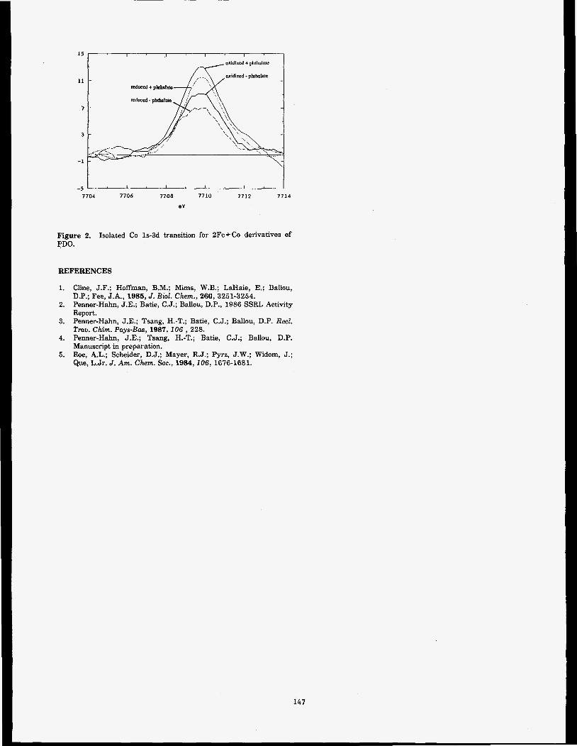
1 5
11
.?
3
-1
I I I I
oxidized + pl~ihalatc
-5 7704 7706 7708 7710 7712 7714
ev
Figure 2. PDO.
Isolated Co 1s-3d transition for 2Fe+Co derivatives of
REFERENCES
1. Cline, J.F.; Hoffman, B.M.; Mims, W.B.; LaHaie, E.; Ballou, D.P.; Fee, J.A., 1985, J. Biol. Chem., 260,3251-3254.
2. Penner-Hahn, J.E.; Batie, C.J.; Ballou, D.P., 1986 SSRL Activity
3. Penner-Hahn, J.E.; Tsang, H.-T.; Batie, C.J.; Ballou, D.P. Red. Tmu. Chim. Pays-Bas, 1987, 106 , 228.
4. Penner-Hahn, J.E.; Tsang, H.-T.; Batie, C.J.; Ballou, D.P. Manuscript in preparation.
5. Roe, A.L.; Scheider, D.J.; Mayer, R.J.; Pyrz, J.W.; Widom, J.; Que, L.Jr. J. Am. Chem. SOC., 1984, 106, 1676-1681.
Report.
147
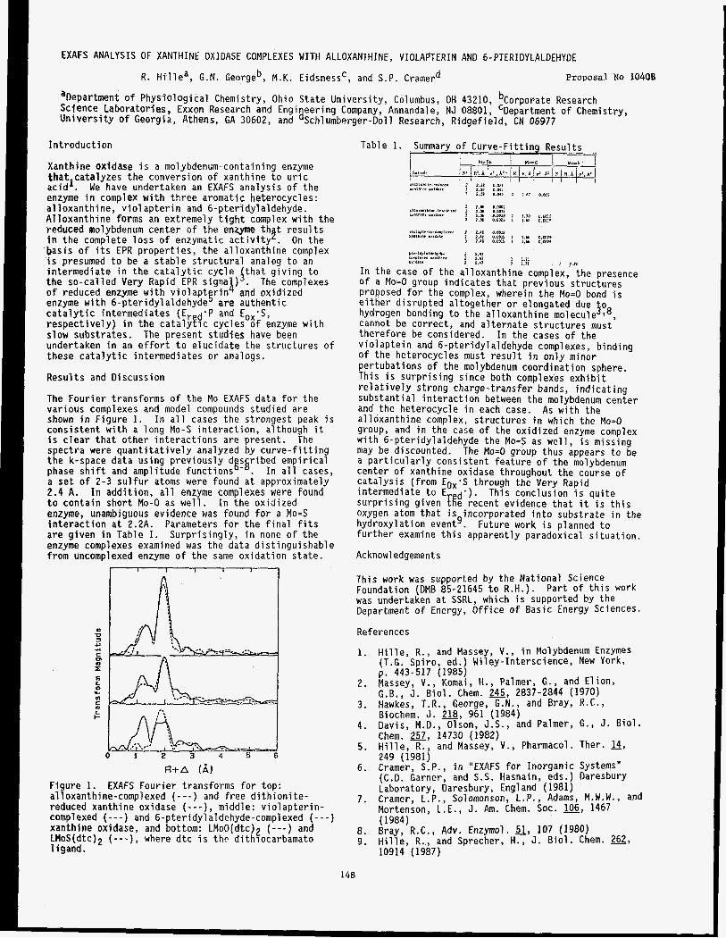
EXAFS ANALYSIS OF XANTHINE OXIDASE COMPLEXES WITH ALLOXANTHINE, VIOLAPTERIN AND 6-PTERIDYLALDEHYDE
R. Hi1
aDepartment o f Physio Scfence Laboratories, University o f Georgia
ea, G.N. Georgeb, M.K. Eidsnessc, and S.P. Cramerd Proposal NO 1040B
ogical Chemistry, Ohio State University, Columbus, OH 43210, bCorporate Research Exxon Research and Engi eering Company, Annandale, NJ 08801, 'Department of Chemistry, Athens, GA 30602, and a Schl umberger-Doll Research, Ridgefield, CN 06977
Introduction
Xanthine oxidase is a molybdenum-containing enzyme thatlcatalyzes the conversion of xanthine to uric acid . We have undertaken an EXAFS analysis of the enzyme in complex with three aromattc heterocycles: a1 loxanthi ne, viol apterin and 6-pteridylaldehyde. Alloxanthine forms an extremely tight complex with the reduced molybdenum center of the enzyme th t results
basis o f its EPR properties, the alloxanthine complex is presumed to be a stable structural analog to an intermediate in the catalytic cycle ithat giving to the so-called Very Rapid EPR signa ) . The complexes
enzyme with 6-pteridylaldehyde are authentic catalytic intermediates (Er d'p and Eox-S, respectively) in the catalytic cycles o f enzyme with slow substrates. undertaken in an effort to elucidate the structures o f these catalytic intermediates or analogs.
in the complete loss of enzymatic activity h . On the
of reduced enzyme with violaptgrin i and oxidized
The present studies have been
Results and Discussion
The Fourier transforms of the Mo EXAFS data for the various complexes and model compounds studied are shown in Figure 1 . In all cases the strongest peak is consistent with a long Mo-S interaction, although it is clear that other interactions are present. spectra were quantitatively analyzed by curve-fitting the k-space data using previously d s ribed empirical phase shift and amplitude functions'-'. In all cases, a set of 2-3 sulfur atoms were found at approximately 2.4 A. In addition, all enzyme complexes were found to contain short Mo-0 as well. In the oxidized enzyme, unambiguous evidence was found for a Mo=S interaction at 2.2A. Parameters for the final fits are given in Table 1. Surprisingly, in none of the enzyme complexes examined was the data distinguishable from uncomplexed enzyme of the same oxidation state.
The
I I I I I I 1
E L
In 1c
a L :: t- -
0 1 2 3 4 5 6
U+A (A ) Figure 1. EXAFS Fourier transforms for top: alloxanthine-complexed ( - - - ) and free dithionite- reduced xanthine oxidase ( - - - ) , middle: violapterin- compl exed (-- - ) and 6-pteridyl a1 de hyde-compl exed ( - - - ) xanthine oxidase, and bottom: LMoO(dtc)? ( - - - ) and LMoS(dtc)Z ( - - - ) , where dtc is thc dithiocarbamato 1 igand.
P,erl*,l,l&?h,*. 2 ?.I, cPPlr>.r Iznthlw 2 2.4,
1 120 1 1.71
I I.., > J 71 *.,*I.
In the case of the alloxanthine complex, the presence of a Mo=O group indicates that previous structures proposed for the complex, wherein the Mo=O bond is either disrupted altogether or elongated due o hydrogen bonding to the alloxanthine molecul>,8, cannot be correct, and alternate structures must therefore be considered. In the cases of the viol aptein and 6-pteridyl aldehyde complexes, binding of the heterocycles must result in only minor pertubations of the molybdenum coordination sphere. This is surprising since both complexes exhibit re1 atively strong charge-transfer bands, indicating substantial interaction between the molybdenum center and the heterocycle in each case. alloxanthine complex, structures in which the Mo=O group, and in the case of the oxidized enzyme complex with 6-pteridylaldehyde the Mo=S as well, is missing may be discounted. The Mo=O group thus appears to be a particularly consistent feature of the molybdenum center of xanthine oxidase throughout the course of catalysis (from Eox'S through the Very Rapid intermediate to E d'). This conclusion is quite surprising given [[e recent evidence that it is this oxygen atom that is incorporated into substrate in the hydroxylation event'. further examine this apparently paradoxical situation.
As with the
Future work is planned to
Acknowledgements
This work was supported by the National Science Foundation (DMB 85-21645 to R.H.). Part o f this work was undertaken at SSRL, which is supported by the Department o f Energy, Office of Basic Energy Sciences.
References
1. Hille, R., and Massey, V., in Molybdenum Enzymes (T.G. Spiro, ed.) Wiley-Interscience, New York,
Massey, V., Komai, H., Palmer, G . , and Elion, G.B., J. Biol. Chem. 245, 2837-2844 (1970) Hawkes, T.R., George, G.N., and Bray, R.C., Biochem. J. 218, 961 (1984) Davis, M.D. , Olson, J.S., and Palmer, G., J. Biol. Chem. 257, 14730 (1982) Hille, R., and Massey, V., Pharmacol. Ther. 14,
p. 443-517 (1985) 2.
3.
4.
5. 249 (1981) Cramer, S.P., in "EXAFS for Inorganic Systems" IC.D. Garner, and S.S. Hasnain, eds.) Daresbury
6.
Laboratory, Daresbury, England (1981) Cramer, L . P . , Solomonson, L.P., Adams, M.W.W., and Mortenson, L.E., J. Am. Chem. SOC. 106, 1467 (1984) Bray, R.C., Adv. Enzymol. a, 107 (1980) Hille, R., and Sprecher, H., J. Biol. Chem. 262, 10914 (1987)
7.
8 . 9.
148


Proposal No. 10468
CORONARY ANGIOGRAPHY IN HUMAN SUBJECTS
E. Rubenstein*, G. S . Brown**, J. C. Giacomini*, H . R. Hofstadter+, A. C. Thompson ++, H. D. Zeman***
J. Gordon*,
* Department of Medicine, School of Medicine, Stanford University: ** Stanford Synchrotron Radiation Laboratory: + Hansen Laboratories of Physics and Department of Physics, Stanford University, Stanford, CA 94305; ++ Lawrence Berkeley Laboratory, University of California, Berkeley, CA 94720; ***National Synchrotron Light Source, Brookhaven National Laboratory, Long Island, NY 1197
Previous studies have demonstrated the principles of transvenous coronary angiography in humans using synchrotron radiation (1). Investigations have been continued during 1987, in an attempt to define the modifications of the imaging system needed to provide angiograms of clinical usefulness. Three subjects were studied, one of whom had undergone transvenous synchrotron angiography 11 months earlier.
The first patient in the current group of examinations was a 63-year-old man with atypical chest pain and left ventricular hypertrophy. Conventional coronary angiogram had been interpreted as showing normal vessels. Coronary spasm was suspected but not demonstrated. Transvenous coronary angiograms revealed normal appearing vessels. The left anterior descending coronary artery, a diagonal branch, and the obtuse marginal branch of the circumflex artery were visualized (Figure 1). The image was recorded in the right anterior oblique position (-10'). The X-ray exposure for the frame was 0.14 rad.
The second patient was a 64-year-old man with angina pectoris related to narrowing of the middle segment of the left anterior descending coronary artery, which had not responded adequately to three prior transvenous balloon angioplastic procedures. A transvenous synchrotron scan, recorded in the left lateral position, confirmed the persistence of the stenosis. An image recorded with the patient in the left anterior oblique (30O) position at a time when the bolus of contrast agent had largely cleared the right side of the circulation showed a normal-appearing right coronary artery clearly visible through the thinly opacified right ventricle (Figure 2). The X-ray dose for each of these frames was 0.2 rad.
The third patient was a 42-year-old man who had been examined by the synchrotron radiation transvenous angiographic method in May, 1986. During the interval he had undergone revision of an internal mammary artery and a saphenous vein bypass graft, and had suffered a mild myocardial infarction involving the left lateral wall. The second transvenous study revealed improved perfusion of the left anterior descending coronary artery, presumably a consequence of the revision
of the internal mammary artery graft, but loss of one of the original vein bypass grafts, believed to have supplied an obtuse marginal branch.
These studies confirm the feasibility of the synchrotron radiation approach, and also confirm that greater X-ray fluence is needed to provide images of clinical quality.
References: (l)Rubenstein, E., Hofstadter, R., Zeman, H . D . , Thompson, A.C., Otis, J.N., Brown, G.S., Giacomini, J.C., Gordon, H.J., Kernoff, R.S., Harrison, D.C., Thomlinson, W. Transvenous coronary angiography in humans using synchrotron radiation. Proceedings of the National Academy of Sciences USA, 83:9724, 1986.
(Fig.
(Fig.
149


J. E. Penner-Hahn, V. L..Pecoraro, R. M. Fronko and H.-T. Tsang. Department of Chemistry, University of Michigan, Ann Arbor, MI 48109-1055.
C. F. Yocum and S. Betts. Department of Biology, University of Michigan, Ann Arbor, MI 48109-1048.
Manganoenzymes play an important role in mediating the chem- istry of 0 2 and the various reduced forms of 0 in the biosphere. In particular, the oxygen evolving complex (OE& of Photosystem I1 (PSII) catalyzes the water oxidation reaction while the manganese containing catalase from L. pZunfururn functions in the disproportion- ation of H202 tn H20 and 02. The nuclearity, structure and oxida- tion state(s) of the manganese cluster associated with the OEC have attracted much interest and effort in recent years. The Mn-catalase, which has only recently been purified, is believed to contain two Mn atoms per subunit but is otherwise essentially uncharacterieed.
It is well known that the energy of an x-ray absorption edge is correlated with the oxidation state of the absorbing atom. The current interpretation of x-ray .absorption near edge structure CXANES) assumes that the observed edge energy for a mixed valent Mn cluster will be independent of Mn-Mn separation and Mn-Mn geometry, and thus that edge energy may be used to determine the average oxidation state of a multinuclear cluster. In order tc evaluate this hypothesis, we have measured the XANES spectra for a series oi trinuclear Mn(III/IWII) complexes. The ti-inuclear complexes have a Mn-Mn separation that vanes from 3.50 to 3.27 and a Mn-Mn-Mn angIe that varies from 138.5 to 180 degrees. In addition, the XANES spectra of a series of mono- and dinuclear Mn(I1) and Mn(II1) complexes were measured. All of the models are crystallographically characterized and all have oxygeidnitrogen ligation (this is believed to be the ligation for Mn in the OEC Ill).
EXPERIMENTAL. OEC samples were prepared according to Yocum, et al. [21. A multi-element intrinsic Ge detector (courtesy of S. Cramer) was used to detect the Mn fluorescence for the OEC and Mn-catalase samples. The Mn concentration for the OEC was 1.5 mM and the Mn-catalase was 0.150 mM. All measurements were made at 12 K. Energy calibration was referenced to the KhZn04 pre- edge feature (assigned as 6543.3 eV). The edge energies in Table I are defined as the energy of the first inflection point.
Table I Comolex Oxid. m e a r i t v Edne (eV)
Mnhnidazole) + 2 11, mononuclear 6547.2 Mn(AcO)2(H26)2 11, mononuclear 6548.6 Mn,(SALPS) 11, dinuclear 6547.1 [Mn(DALAPH)!c 11, dinuclear 6549.1
+ 111, mononuclear 6551.5 Mn(acac)3 111, mononuclear 6552.9 Mn(SALPN)tAcO) 111, mononuclear 6550.4 mn[(2-OH)5-C1-SALPN]}2 III, dinuclear 6551.2 {MnSALAHP), 111, dinuclear 6552.2
MVLP MVBM MVLH MVLW
IIyII,'III, trinuclear 6549.6 IIUTIIIII, trinuclear 6549.6 IIIIIIAII, trinuclear 6550.7 III/II/III, trinuclear 6549.6
RESULTS
BIOLOGICAL STUDIES. The edge energies for several of the complexes are listed in Table I. The increase in edge energy with increasing oxidation state described by Klein, et al. [31, is readily apparent. The XANES for resting state Mn-catalase closely resembles that for Mn(II) complexes, however the low concentration of this sample precludes definitive characterization at this time.
In Figure 1 we compare the XANES spectra for two different OEC samples (solid lines) with those for Mn (11) models (dashed lines) and Mn(II1) models (dotted lines). The edge energy of the OEC is consistent with a mixture of Mn(II) and Mn(1II) since the OEC edge lies between those of authentic Mn(I1) and Mn(II1) complexes (see also Table 1). However, the large (ca. 2 eV) spread in edge energies for a
8 . 0 0 0 0
6 . 3 9 8 8
4.7976
3 . 1 9 6 4
1 . 5 9 5 2
I I I I 1
-0.0060
6 .5350 6.5460 6 . 5 5 7 0 6 . 5 6 8 0 6.5790 6.5900
Xln-catalase unknown 6547.9 OEC, dark adapted unknown, sample 1 6550.1 o-de2 65-
llIVLH = Mn3[(2-OH)S.4LPNJ2(A~0)4; MVLW = Mn,(SALADHP), (AcO),(H20), 2MeOH; MVLF=XIa (SALADHPd (AcOJ ( p ~ O q H,O; m B M = Mn3(SALADHP)2(Ac0)4(MeOH)2 (<Mn-hIn-Mn = 158.5O). SALAHP. SALPS, DALAHP, SALPN are Schiff base ligands condensed from salicylic acid and a secondav amine.
150
Figure 1. XANES spectra for: the OEC (solid line); Mn(1I) model compounds (dashed line); and Mn(D1) model compounds (dotted line). All of the model compounds have oxygen/nitrogen ligation.
given oxidation state suggests that it will be difficult to unambigu- ously assign oxidation state based solely on the energy of the absorption edge. This concern is particularly important given the breadth of the OEC edge in comparison with the edges for the model compounds (see Fig. 1). This breadth suggests that the Mn in the OEC is present in a number of distinct sites.
In addition to XANES data, EXAFS data were also measured for the OEC. The combination of highly concentrated samples and solid state fluorescence detection allowed the measurement of EXAFS data having an excellent signallnoise ratio (see Fig. 2). Analysis of these data is currently in progress.
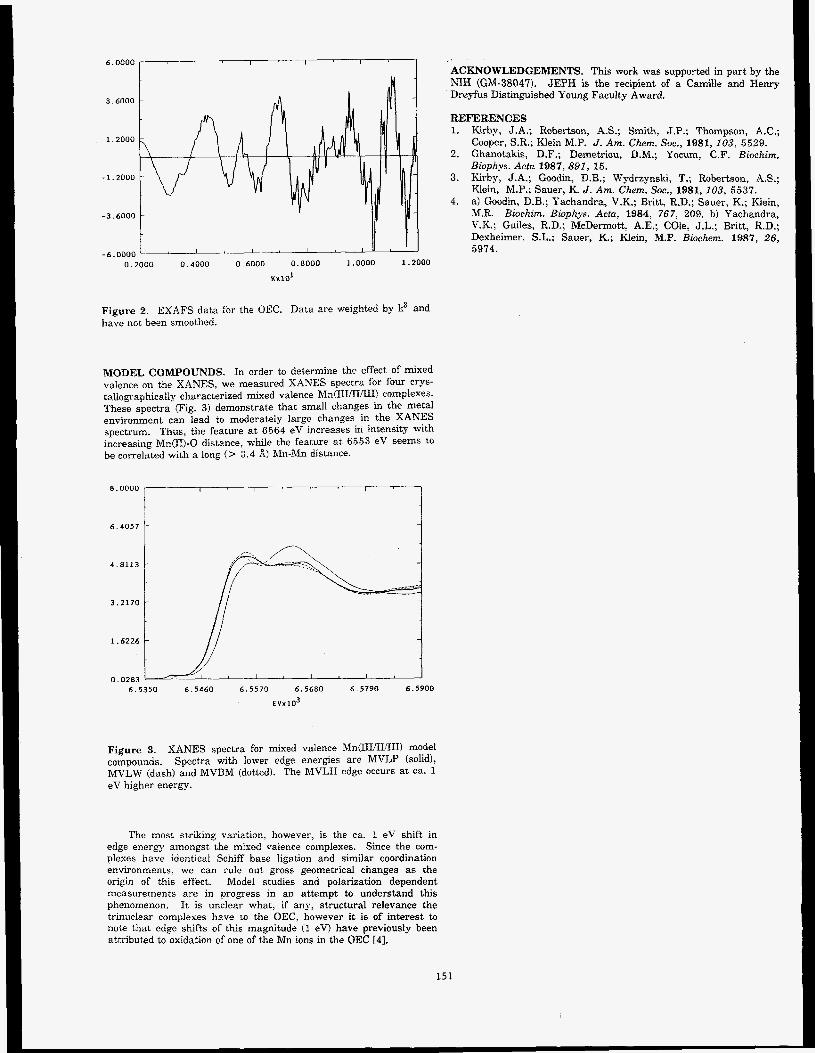
KxlOl
Figure 2. EXAFS data for the OEC. Data are weighted by k3 and have not. been smoothed.
MODEL COMPOUNDS. In order to determine the effect of mixed valence on the XANES, we measured XANES spectra for four crgs- tallographically characterized mixed valence hln(IIIIIIflII! complexes. These spectra (Fig. 3) demonstrate that small changes in the metal environment can lead to moderately large changes in the XAMES spectrum. Thus, the feature at 6564 eV increases in intensity with increasing Mn(II)-0 distance, while the feature a t 6653 eV seems to be correlated with a long (> 3.4 A) Mn-hfn distance.
8.0000 I I I I
I 6.4057
t
1 4.8113
I 3.2170
1.6226
1 1
ACKNOWLEDGEMENTS. This work was supported in part by the NIH (GM-38047). JEPH is the recipient of a Camille and Henry Dreyfus Distinguished Young Faculty Award.
0.0283 6.5350 6.5460 6.5570 6 , 5 6 8 0 6.5790 6.5900
€vx103
Figure 3. XANES spectra for mixed valence hfn(IIIIII/fII! model compounds. Spectra with lower edge energies are MVLP (solid), MVLW (dash) and MVBM (dotted). The MVLH edge occu-s at ca. 1 eV higher energy.
The most striking variation, however, is the ca. 1 eV shift in edge energy amongst the mised valence complexes. Since the com- plexes have identical Schiff base ligation and similar coordination environments, we can rule out gross geometrical changes as the origin of this effect. Model studies and polarization dependent measurements are in progress in an attempt to understand this phenomenon. It is unclear what, if any, structural relevance the trinuclear complexes have to the OEC, however it is of interest to note that edge shifts of this magnitude (1 eV) have previously been attributed to oxidation of one of the Mn ions in the OEC [4].
15 I
REFERENCES 1. Kirby, J.A.; Robertson, A S ; Smith, J.P.; Thompson, A.C.;
Cooper, S.R.; Klein M.P. J. Am. Chem. SOC., 1981, 103, 5529. 2. Ghanotakis, D.F.; Demetriou, D.M.; Yocum, C.F. Biochirn.
Biophy. Acta 1987, 891, 15. 3. Kirby, J.A.; Goodin, D.B.; Wydrzynski, T.; Robertson, AS.;
Klein, M.P.; Sauer, K. J . Am. Chem. Soc., 1981, 103, 5637. 4. a) Goodin, D.B.; Tachandra, V.K.; Britt. R.D.; Sauer, K.; Klein,
M.R. Biochin. Biophys. Acta, 1984, 767, 209. b) Yachcndra, V.K.; Guiles, R.D.; McDermott, A.E.; Cole, J.L.; Britt, R.D.; Dexheimer. S.L.: Sauer, K.; Klein, M.P. Bioclzem. 1987, 26, 5974.
ANOMALOUS DIFFRACTION MEASUREMENTS FROM PROTEIN CRYSTALS
Wayne A. Hendrickson, John R. Horton, Craig M. Ogata, William E. Royer Jr., and Wei Yang
Howard Hughes Medical Institute Department of Biochemistry and Molecular Biophysics
Columbia University, New York, M 10032 '
and
Ethan A. Merritt and R. Paul Phizackerley
Stanford Synchrotron Radiation Laboratory Stanford, CA 94305
INTRODUCTION
Methods have been developed for the measurement and analysis of diffraction measurements made at a few select wavelengths associated with the absorption edges of appropriate anomalous scatterers in macromolecular crystals. our procedures for analyzing multiwavelength anomalous diffraction (MAD) measurement made earlier at SSRL and at the Photon Factory in Japan. This work has led to a phase evaluation for lamprey hemoglobin and to three-dimensional structure for a bacterial ferredoxin and for streptavidin. made during 1987 on two other crystalline proteins. progress.
During the past year, we have applied
New measurements have also been
The analysis of these data is still in
URECHIS HEMOGLOBIN
We had previously measured a set of data complete to 5.5A spacing from the hemoglobin of Urechis caupa, an echiuran known as the innkeeper worm. C222 , with a=104.8, b=54.9 and c=110.6A. These crysials were grown and provided by Dr. Marvin L. Hackert and his collaborators at the University of Texas. Unfortunately, these data proved to be too weak and inaccurate to be useful even for the location of iron center. The present work was made possible by the availability of larger crystals.
This hemoglobin crystallizes in space group
During January of 1987 we measured data at four wavelengths (1.5000, 1.7382, 1.7400 and 1.8000A) related to the iron K edge. Due to beam conditions, there was inadequate time to complete a f u l l 3A data set. However, with a full quadrant of coverage in rotation about b (100') and a fill in of 34' about a from one crystal together with additional partial data from rotation about c, a complete 5.5A set was obtained. been processed and are markedly superior to those obtained previously. been pursued, however, as our collaborators meanwhile solved the structure.
The MAD data have
The phasing analysis has not
'MIIOREDOXIN
In collaboration with Dr. David LeMastar of Yale University, we have produced E. coli thioredoxin in which the methionine residues have been replaced by selenomethionines. This was done by expression of recombinant DNA in plasmids that had been introduced into an E. coli strain made defective in methionine synthesis. This protein has been crystallized under conditions similar to those used earlier by Holmgren et al. (PNAS 72, 2305, 1975), but ones sufficiently changed that unit cell dimensions differ appreciably. crystals are in space group C2 with a389.2, b349.6, c=57.8A and =100.2° whereas the earlier crystals had lattice dimensions of a=89.7, b=51.1, ~ 6 0 . 3 and 3113.5'.
Our
During December 1987 we measured diffraction data at five wavelengths (0.9000, 0.9791, 0.9794, 1.3776 and 1.3787A) associated with absorption edges of the copper and selenium present in these crystals. The crystal had dimensions of 0.3 x 0.3 x 0.6 mm. Data were measured with the detector at 400 mm from the crystal. A full 180' rotation was made about the b-axis, and these data were supplemented by omega-axis fill-ins with 26' of rotation about a* and some additional measurement about c*. Preliminary data reduction results indicate that counting statistics will not be limiting in the analysis.
152

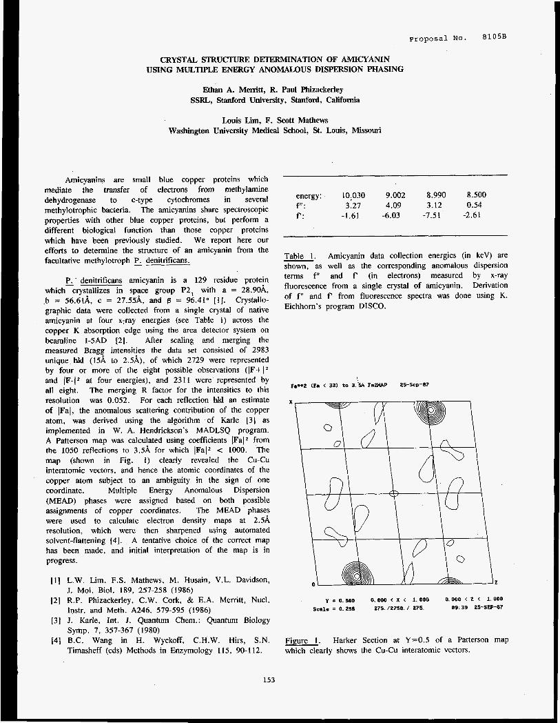
Proposal N o . 8105B
CRYSTAL STRUCTURE DETERMINATION OF AMICYANIN USING MULTIPLE ENERGY ANOMALOUS DISPERSION PHASING
Ethan A. Memtt, R. Paul Phizackei-ley SSRL, Stanford University, Stanford, California
Louis Lim, F. Scott Mathews Washington University Medical School, St. Louis, Missouri
Amicyanins are small blue copper proteins which mediate the transfer of electrons from methylamine dehydrogenase to C-type cytochromes in several methylotrophic bacteria. The amicyanins share spectroscopic properties with other blue copper proteins, but perform a different biological function than those copper proteins which have been previously studied. We report here our efforts to determine the structure of an amicyanin from the facultative methylotroph 5 denitrificans.
5 denitrificans amicyanin is a 129 residue protein which crystallizes in space group P2, with a = 28.90k b = 56.61A, c = 27.55A. and 8 = 96.41” [ I ] . Crystallo- graphic data were collected from a single crystal of native amicyanin at four x-ray energies (see Table 1) across the copper K absorption edge using the area detector system on beamline I-5AD (21. After scaling and merging the measured Bragg intensities the data set consisted of 2983 unique hkl (I5A to 2.5,&), of which 2729 were represented by four or more of the eight possible observations (IF+(* and IF-12 at four energies), and 23 1 1 were represented by all eight. The merging R factor for the intensities to this resolution was 0.052. For each reflection hkl an estimate of IFal, the anomalous scattering contribution of the copper atom, was derived using the algorithm of Karle 131 as implemented in W. A. Hendrickson’s MADLSQ program. A Patterson map was calculated using coefficients IFaI2 from the 1050 reflections to 3.5A for which lFalZ < 1000. The map (shown in Fig. 1) clearly revealed the Cu-Cu interatomic vectors, and hence the atomic coordinates of the copper atom subject to an ambiguity in the sign of one coordinate. Multiple Energy Anomalous Dispersion (MEAD) phases were assigned based on both possible assignments of copper coordinates. The MEAD phases were used to calculate electron density maps at 2.5A resolution, which were then sharpened using automated solvent-flattening [41. A tentative choice of the correct map has been made, and initial interpretation of the map is in progress.
111 L.W. Lim, F.S. Mathews, M. Husain, V.L. Davidson,
121 R.P. Phizackerley, C.W. Cork, & E.A. Merritt, Nucl.
131 J. Karle, Int. J. Quantum Chem.: Quantum Biology
141 B.C. Wang in H. Wyckoff, C.H.W. Hirs, S.N.
J. Mol. Biol. 189, 257-258 (1986)
Instr. and Meth. A246, 579-595 (1986)
Symp. 7. 357-367 (1980)
Timasheff (eds) Methods in Enzymology 115, 90-1 12.
energy: 10:030 f,r: 3.27 f‘: -1.61
9.002 4.09
-6.03
8.990 8.500 3.12 0.54
-7.5 1 -2.61
Table 1. Amicyanin data collection energies (in keV) are shown, as well as the corresponding anomalous dispersion terms f” and f‘ (in electrons) measured by x-ray fluorescence from a single crystal of amicyanin. Derivation of f ” and f‘ from fluorescence spectra was done using K. Eichhorn’s program DISCO.
P89.2 (Fa < 33) to 3.‘5A F a W P 25-Sep-87
Y = 0.500 0.000 < x < 1.000 0.000 < 2 < 1.000 Scale = 0.255 275. / 2 7 5 0 . / 275. OS: 39 25-SEP-87
Figure 1. Harker Section at Y=0.5 of a Patterson map which clearly shows the Cu-Cu interatomic vectors.
153


SH?iLL AElGLE X-RAY SCATTEKtNG STUDIES ON CATION BINDING SITES AND LINKING REGIchls OF BMYERIOI(I1ODOPSIN
proposal No. --- - ~ _ _ _ -
8llOB
1 'S. Wakatsuki, 'K.0. Hodgson, 3R.M. Stroud, 2S. Doniach & U. Spann
Departments of Chemistry and Applied Physics, Stanford University 3Dep. of Biochemistry and Biophysics, Univ. of California, San Francisco
1 2
The retinal group of bacterioRhodopsin (bR) absorbs light energy and pumps protons through the purple membrane. Each bR molecule consists of 7 a-helices that align normal to the membrane plane, a& form a2$D crystal with P3 symmetry. The binding of Mg and Ca cations to bR molecules is believed to have an important function in photocycle. Anomalous diffraction data from Tl?' ions bound to bR allow location of the cation binding sites in the 3D electron density map of the bR molecules. In electron microscopic studies the diffraction data are restricted to an area in reciprocal space outside of a conical region, called the missing cone. Small angle x-ray diffraction studies in principle can give intensity profiles in the missing cone and allow reconstruction of a complete 3D density map of the bR molecules. The phases can be calculated on the basis of electron microscopic data. mowledge of the density distribution in the linking regions of the 7 a-helices should help both to define their linking order and to map the primary amino acid sequence onto the helices.
CATION BINDING SITE STUDY ---- Anomalous diffraction studies were done on
pelleted bR samples. Ultracentrifugation of bR suspensions at 50000 rpm for 10 hours resulted in partially oriented bR samples. The membrane layers were randomly rotated about the axis perpendicular to the membrane plane. For data collection the beam lines 11-2 and IV-2 at SSRL were used. X-ray energies for anomalous %ffraction were chosen around the L(III) edge of Tb which is at 7515 eV. By measuring the meridional intensity profile the electron density distribution along the direction normal to the membrane plane was obtained. A finite number of combinations of native phases were determined using Shannon's sampling theorem [l]. The effect of anomalous diffraction is shown best by the difference between data sets co3)ected at energies around the absorption edge of Tb (Fig. 1). The anomalous signals follow precisely hfr(E). The locations of the cation binding sites were calculated by non-linear least squares refinement using the anomalous signals and the native phases (Fig. 2). The R factors of the 2 peak solutions (R-0.05) were substantially smaller than those of the 1 peak solution (Rc0.35). The addition of a third binding site did not improve the R factor and the resulting positions were further out of the membrane. The results of the refinement calculations showed that the bR molecules have 2 cation binding sites. They are located close to the lipid head groups, one on each side of the membrane. These results are consistent with FTIR-studies done by Gewert et al. [21. Due to the smearing effect caused by the misorientation of the membranes it is still difficult to obtain the distribution of cation binding sites in the membrane plane.
0.0007
0.0006
0.0005
0.000'4
0.0003
0.0002
0.0001
0.0000
Combined refinement o f natrva and heavr ston r l th one peak
Experf mental A 8.0175
0.8150
0.8125
0.01E0
0.8075
8.0050
8.0825
@.E088
-8.8025
@.@E 0.82 0.0'4 8.06 0.88 8.18
Fig. 1 Difference of amplitudes between a taken below and at the L absorption edge of a'' in the meridional directioh': Experimental curve: amplitude difference of the (0,o) rod, calculated curve: result of a non-linear least squaresrefinement with 2 peaks.
Natlve proille I s dlvldcd br 10
-100 -58 0 58 180
Fig. 2 Electron density profiles along the direction perpendicular to the bR membranes ( 2 ) . Upper curve: electron density of the native bR sample, lower curve: distribution of Tb atom after non-linear least squares refinement.
154
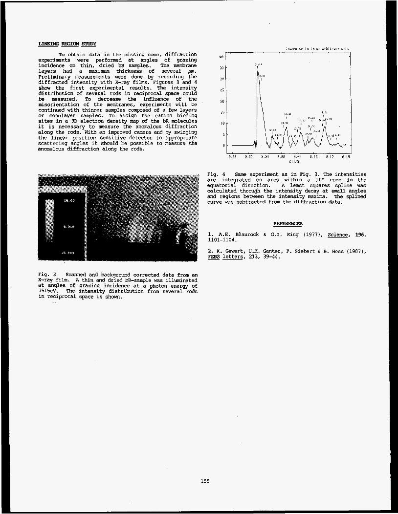
LINKING REGION STUDY ~ - - TO obtain data in the missing cone, diffraction
experiments were performed at angles of grazing incidence on thin, dried bR samples. The membrane layers had a maximum thickness of several pm. preliminary measurements were done by recording the diffracted intensity with X-ray films. Figures 3 and 4 show the first experimental results. The intensity distribution of several rods in reciprocal space could be measured. To decrease the influence of the misorientation of the membranes, experiments will be continued with thinner samples composed of a few layers or monolayer samples. To assign the cation binding sites in a 3D electron density map of the bR molecules it is necessary to measure the anomalous diffraction along the rods. With an improved camera and by swinging the linear position sensitive detector to appropriate scattering angles it should be possible to measure the anomalous diffraction along the rods.
Intensitr l e i n an arbitrary unit ! I I
4% I 35
30
25
2%
15
10
5
0
Fia. 4
0.08 0.02 0.04 8.06 0.08 0.10 0.12 0.14 SIllRl
Same exmriment as in Fis. 3. The intensities are integrated kn arcs within -a l o o cone in the equatorial direction. A least squares spline was calculated through the intensity decay at small angles and regions between the intensity maxima. The splined curve was subtracted from the diffraction data.
1. A.E. Blaurock & G.I. King (19771, Science, 196, 1101-1104.
2. K. Gewert, U.M. Ganter, F. Siebert & B. Hess (1987), FIBS letters, 213, 39-44. -~
Fig. 3 Scanned and background corrected data from an X-ray film. A thin and dried bR-sample was illuminated at angles of grazing incidence at a photon energy of 7515ev. The intensity distribution from several rods in reciprocal space is shown.
155
Proposal No. 8110B
A Study of the Electronic Structure of S20b2- by Polarized K-edge X-ray Absorption Spectroscopy
Trevor A. Tysonl , A. Lawrence Roe1 , Britt Hedman1.2 , Patrick Frank1 and Keith 0. Hodgsonl
1 Department of Chemistry, Stanford University, Stanford, Ca. 94305 SSRL, SLAC, Bin 69, P. 0. Box 4349, Stanford, Ca. 94305
1. Introduction
Since X-ray photoabsorption by a molecule invokes the promotion of a core electrons to a bound or continuum state, the absorption process reveals information about the bonding between the absorbing atom and its ligands. Moreover by performing polarized X-ray absorption (PXA) measurements information about the symmetry of empty molecular orbitals (bound states) and available continuum states can be directly obtained (1).
Here we present the results of a PXA study of the sulfur K-edge PXA excitation spectra of the sulfur oxyanion S,O,z- (dithionate).
measurements were performed and features in the spectra were labeled.
In concert with these experimental results, MS-Xu calculations using the extended continuum (EC-MS-Xu) scheme (2) were carried out. These calculations served as a cross-check on the ordering of final states determined by our direct experimental measurements and to elucidate properties not made evident by our experimental procedure.
II. Qualitative Physics
Within the single particle approximation, the probability per unit time that an electron in an initial state, I@,>. makes a transition to a final state, I@,>, due to a perturbation, V, is given by
Wf, = 2 d b I< @>I V I @ f >126(E,- E,) 1
where E, is the total energy of the initial configuration and E, is the total energy of the final configuration.
For transitions due to the absorption of electromagnetic radiation the above expression, in the dipole approximation, leads to an expression for the photoabsorption cross section :
3000 eV) care had to be taken to insure that the entire beam path was in helium and a goniometer was designed for this purpose.
IV. Results and Interpretations
K,S,O, forms crystals which have three molecules in each unit cell. The unit cell is hexagonal and the space group is P321 (4). The arrangement of the S20,2- (dithionate) anions is ideal for polarized absorption measurements (with the symmetry axes, defined by the sulfur-sulfur bond, of all anions parallel to the unit cell three-fold axis (see fig. la)). We were therefore able to obtain sulfur K-edge absorption data for two orientations. In the first orientation (fig l b thin line) the the symmetry axis (12,) of the anion was aligned parallel to the electric field vector, e. In the second configuration (fig l b thick line) the crystal was oriented so that the electric field was perpendicular to the symmetry axes.
The vacant orbital structure and available continuum states of dithionate are complex because of the presence of two sulfur atoms in the molecule. completely indistinguishable when the molecule is in its ground state. charge on the absorbing sulfur atom (which has a core hole) reduces the molecular symmetry from D,, to C,, . 2 reduces to
Fortunately, both sulfur atoms are
However on the absorption of a photon the redistribution of
Hence equation
o(fiw) = 4+aha, { BfI c @is 1 e,.XpI @ e> + -4 e$ @al > 12 } 3 N( E, + f iw - E,)
where (in configuration space) ep and Xp are the vector components of e and X. respectively, perpendicular to the C3 axis of the molecule, @I* is the initial state wave function @al and @ e are the possible final state wave functions and the function N is the density of final states. Only transitions to final states of a1 (e) symmetry are possible in the first (second) orientation.
o(bm) = 4n2ahw Z,Ic@,l e-rl @,>I2 6( E, + ha, - E,) 2 Even more can be learned by combining observations with the results of the complete calculation of the photoabsorption cross section of the dithionate anion obtained by the EC-MS-Xa method. Features which appear below the computed zero of energy can be attributed to transitions to bound final states while those above can be attributed to transitions to continuum final states.
where a is the finestructure constant, ha, is the energy of the absorbed photon, and e'r is the scalar product of the electric field polarization vector (e) with the electron position operator (r) (3).
111. Measurements In figure 1c we show the computed spectra. Based on the positions of the features A, A', B we argue that these features correspond to transitions to molecular orbitals of al, e and a1 symmetry, respectively. Features B, C', D, E and E' are seen to be transition to continuum states of e, e, e, e and a1 symmetry, respectively. The feature labeled D in the measured spectrum is believed to be residual from the e symmetry feature D . The origin of the feature C is at this point not clear.
The polarized absorption measurements were performed on the 54-pole wiggler beam line VI-2 which was operated in undulator mode under dedicated conditions (3 GeV, 40-80 mA. focussed, Si(il1) double-crystal monochromator). Only with this setup is there adequate flux to produce high quality data from crystals the size used (0.5 x 0.5 x0.5 mm3). At the energies of interest (2400 -
156
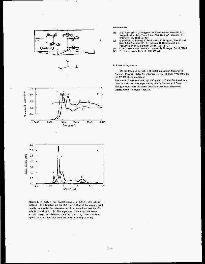
References
[ l ] J. E. Hahn and K 0. Hodgson, "ACS Symposium Series No.211, Inorganic Chemistry:Toward the 21st Century", Malcom H. Chisholm, ed, 1983 .p. 431 S. Doniach, M. Berding, T. Smith and K. 0. Hodgson, 'EXAFS and Near Edge Structure 111". K. Hodgson, B. Hedman and J. E. Penner-Hahn eds., Springer Verlag 1984, p. 33 C. R. Natoli and M. Benfatto, Journal de Physique, C8 11 (1985) E. Stanley, Acta Cryst. 9, 897 (1956)
[2]
[3] [4]
C 0 .- c a Lo 9 4
'L)
N m .- L
E z z
a
2.5 I I I A A' I
2.0
1.5
1 .o
0.5
0.0 2460 2470 2480 2490 2500 2510
Energy (eV)
C
I I I
3.5
3.0
6 2.5 E ._ 6 2.0 c
m a 1.5 Lo
g 1.0
0.5
0.0
C
-20 -1 0 0 10 20 30 Energy (eV)
Figure 1. K2S206 : (a) Crystal structure of K,S,O, with unit cell outlined. parallel to e while for orientation #2 it is rotated so that the C3 axis is normal to e. (b) The exper'mental data for orientation #1 (thin line) and orientation #2 (thick line). (c) The calculated spectra in which the lines have the same meaning as in (b).
In orientation #1 the S-S vector (C,) of the anion is held
Acknowledegments
We are indebted to Prof. C R. Natoli (Laboratori Nazionali Di Frascati, Frascati, Italy) for allowing us use of their VAX-8600 for the EC-MS-Xu computations. This research was supported by NSF grant CHE 85-12129 and was done at SSRL which is supported by the DOE'S Office of Basic Energy Science and the NIH's Division of Research Resources, Biotechnology Resource Program.
157
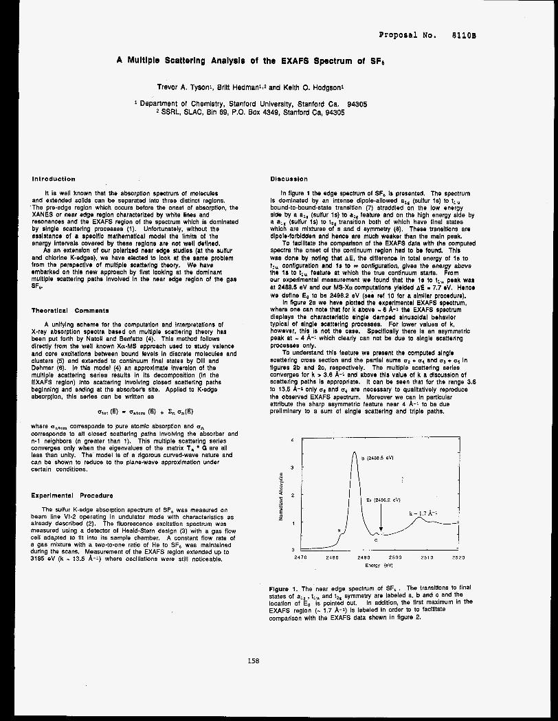
Progoeal No. 81103
A Multlple Scatterlng Analysls of the EXAFS Spectrum of SFC
ln t roduct la
Trevor A. Tysonl, Brltt HedmanlJ and Keith 0. Hodgsonl
Department of Chemistry, Stanford University, Stanford Ca. 94305 2 SSRL, SLAC, Bin 69, P.O. Box 4349, Stanford Ca, 94305
It is well known that the absorption spectrum of molecules and extended solids can be separated into three distinct regions.
'The pre-edge region which occurs before the onset of absorption, the XANES or near edge region characterized by white lines and resonances and the EXAFS region of the spectrum whlch is domlnated by single scattering processes (1). Unfortunately, wlthout the asslstance of a speclflc mathematlcal model the limits of the energy Intervals covered by these regions are not well deflned.
As an extension of our polarized near edge studies (at the sulfur and chlorlne K-edges), we have elected to look at the same problem from the perspectlve of multiple scatterlng theory. We have embarked on this new approach by first looking at the domlnant multlpie scattering paths Involved In the near edge reglon of the gas SF6.
Theoretical Comment*
A unifylng scheme for the computation and interpretations of X-ray absorption spectra based on multiple scattering theory has been put forth by Natoli and Benfatto (4). This method follows dlrectly from the well known Xu-MS approach used to study valence and core excitations between bound levels In dlscrete molecules and clusters (5) and extended to continuum flnal states by Dlil and Dehmer (6). in this model (4) an approximate lnverslon of the multiple scattering series results in its decomposltion (In the EXAFS region) into scattering Involving closed scattering paths beginning and ending at the absorber's site. Applied to K-edge absorpjion, this series can be written as
Otat (E) oatom (E) + Zn an(E)
where oatom corresponds to pure atomic absorption and a,, corresponds to all closed scattering paths involving the absorber and n-1 neighbors (n greater than 1). This multiple scattering series converges only when the eigenvalues of the matrix 1, 0 are all less than unity. The model is of a rigorous curved-wave nature and can be shown to reduce to the plane-wave approximation under certain conditions.
Experlmental Procedure
The sulfur K-edge absorption spectrum of SF6 was measured on beam line VI-2 operating in undulator mode with characteristics as already described (2). The fluorescence excitation spectrum was measured using a detector of Heald-Stern design (3) with a gas flow cell adapted to fit into its sample chamber. A constant flow rate of a gas mixture with a two-to-one ratio of He to SF, was maintained during the scans. Measurement of the EXAFS region extended up to 3185 eV (k - 13.5 A-1) where oscillations were still noticeable.
Discussion
In flgure 1 the edge spectrum of SF6 is presented. The spectrum Is dominated by an intense dipole-allowed alg (sulfur Is) to tl, bound-to-bound-state transition (7) straddled on the low energy side by a all (sulfur 1s) to al, feature and on the high energy side by a a,, (sulfur Is) to t Z g transition both of which have flnal states which are mixtures of s and d symmetry (8). These transitions are dipole-forbldden and hence are much weaker than the maln peak.
spectra the onset of the continuum reglon had to be found. This was done by noting that AE, the difference In total energy of I s to t i , configuration and I s to m configuration, gives the energy above the I s to tlu feature at whlch the true continuum starts. From our experlmental measurement we found that the I s to tl, peak was at 2488.5 eV and our MS-Xa computations ylelded A€ - 7.7 eV. Hence we deflne Eo to be 2496.2 eV (see ref 10 for a similar procedure).
In flgure 2a we have plotted the experlmental EXAFS spectrum, where one can note that for k above - 6 A-1 the EXAFS spectrum dlsplaye the characteristic single damped slnusoidai behavlor typical of slngle scattering processes. For lower values of k, however, this Is not the case. Specifically there Is an asymmetrlc peak at - 4 A-1 which clearly can not be due to slngle scatterlng processes only.
To understand this fsature we present the computed slngle scatterlng cross section and the partlal sums u2 + u, and + u5 in flgures 2b and 2c, respectlvely. The multiple scatterlng series converges for k > 3.6 A- l and above thls value of k a discussion of scattering paths is appropriate. it can be seen that for the range 3.6 to 13.5 A-1 only o2 and u4 are necessary to qualitatively reproduce the observed EXAFS spectrum. Moreover we can in particular attribute the sharp asymmetric feature near 4 A-1 to be due preliminary to a sum of slngle scattering and triple paths.
To facilitate the comparison of the EXAFS data wtth the computed
3 -
p EL 8
g 1
- : z - - 5
1 -
c
0 2 4 7 0 2480 2490 2 5 0 0 2510 2520
Energy (ev)
Flgure 1. The near edge spectrum of SF6 . The transitions to final states of a,, , t,, and t2g symmetry are labeled a, b and c and the location of Eo is pointed out. In addition, the first maximum in the EXAFS region (- 1.7 A-1) is labeled in order to to facilitate comparison with the EXAFS data shown in figure 2.
158
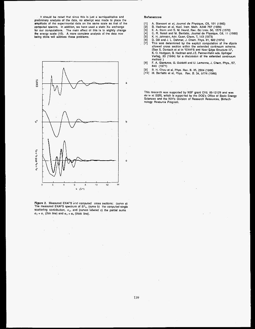
It should be noted that since this is just a semiqualitative and References
[ l ] [2] [3] [4] [5] [6] [7]
preliminary analysis of the data, no attempt was made to place the amplitude of the experimental data on the same scale as that of the computed spectra. In addition, we have used a static Xa exchange for our computations. The main effect of this is to slightly change the energy scale (10). A more complete analysis of the data now being done will address these problems.
A. Bianconi et al, Journal de Physique, C9, 101 (1985) €3. Hedman et al, Nucl. Instr. Meth. A246 797 (1986) E. A. Stern and S. M. Heald, Rev. Sci Instr. 50, 1579 (1979) C. R. Natoli and M. Benfatto, Journal de Physique, C8, 11 (1985) K. H. Johnson, Adv. Quan. Chem. 7, 143 (1973) D. Dill and J. L. Dehmer, J. Chem. Phys. 61, 692 (1974) This was determined by the explicit computation of the dipole allowed cross section within the extended continuum scheme. (See S. Doniach et al in "EXAFS and Near Edge Structure Ill", K. 0. Hodgson, B. Hedman and J.E. Penner-Hahn eds. Springer Verlag, 33 (1984) for a discussion of the extended continuum method )
[8] F. A. Gianturco, G. Guidotti and U. Lemanna, J. Chem. Phys., 57,
[91 S. H. Chou et al, Phys. Rev. B. 35, 2604 (1986) [IO] M. Benfatto et al. Phys. Rev. B. 34, 5774 (1986)
840 (1971)
a
This research was supported by NSF grant CHE 85-12129 and was done at SSRL which is supported by the DOE'S Office of Basic Energy Sciences and the NIH's Division of Research Resources, Biotech- nology Resource Program.
b
C
0 2 4 6 8 10 1 2 14
k
Figure 2. Measured EXAFS and computed cross sections: (curve a) The measured EXAFS spectrum of SF6, (curve b) the computed single scattering contribution, a2, and (curves labeled c) the partial sums oz + o4 (thin line) and os + c5 (thick line).
159
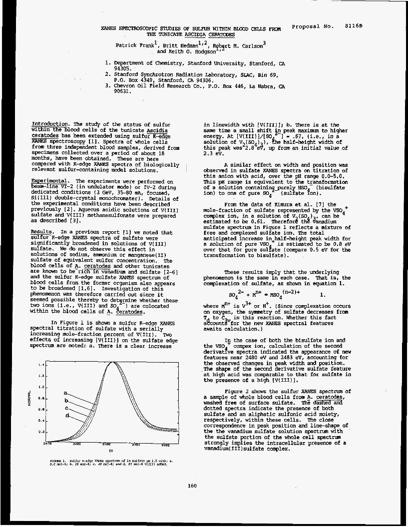
XANES SPEC'l'ROSCOPIC STUDIES OF SULFUR WITHIN BLOOD CEUS FROM THE TUNICATE ASCIDIA CERATODES
Proposa1 No* 8116B
Patrick Frank', Britt Hedmanlf2, Rf3rt M. Carlson 3 and Keith 0. Hcdgson
1. Department of Chemistry, Stanford University, Stanford, CA
2. Stanford Synchrotron Radiation Laboratory, SLAC, Bin 69,
3. Chevron oil Field Research Co., P.O. Box 446, La Habra, CA
94305.
P.O. Box 4349, Stanford, CA 94306.
90631.
Introduction. The study of the status of sulfur within the blood cells of the tunicate Ascidia ceratodes has been extended using s u l f u m g e XANES spectroscopy i l l . Spectra of whole cells from three independent blood samples, derived from specimens collected over a period of about 18 months, have been obtained. These are here compared with K-edge XANES spectra of biologically relevant sulfur-containing model solutions.
Ex rimental. The experiments were performed on b - 2 (in undulator mode) or Iv-2 during dedicated conditions (3 GeV, 35-80 m ~ , focused, Si(ll1) double-crystal monochromater) . Details of the experimental conditions have been described previously [21. Aqueous acidic solutions of v(I11) sulfate and V(II1) methanesulfonate were prepared as described [31.
Results. In a previous report [l] we noted that sulfurK-edge XANES spectra of sulfate were significantly broadened in solutions of v(111) sulfate. We do not observe this effect in solutions of sodium, ammonium or manganese(I1) sulfate of equivalent sulfur concentration. blood cells of A. ceratodes and other tunicates are known to be-rich in vanadium and sulfate [2-61 and the sulfur K-edge sulfate XANES spectrum of blood cells from the former organism also appears to be broadened [1,6]. Investigation of this phenomenon was therefore carried out since it seemed possible thereby to detcjfmine whether these two ions (i.e., V(II1) and SO within the blood cells of 5. geratodes.
spectral titration of sulfate with a serially increasing mole-fraction percent of v(II1). ?tnro effects of increasing [V(III)I on the sulfate edge spectrum are noted: a. There is a clear increase
The
) are colocated
In Figure 1 is shown a sulfur K-edge XANES
1.2 - i . 0 -
EV
in linewidth with [V(IrI)]; b. There is at the same time a small shift in peak maximum to higher energy. At [V(III)I/[SO -1 = .67, (i.e., in a solution of V (SO ) ), &e half-height width of this peak wa~~2.8~e4, up from an initial value of 2.3 eV.
A similar effect on width and position was observed in sulfate XANES spectra on titration of this anion with acid, over the pH range 0.0-5.0. This pH range is equivalent to the transformation of a solution containincpurely HSO (bisulfate ion) to one of pure so4
mole-fraction of sulfate represented by the VSO~+ complex ion, in a solution of V (SO ) estimated to be 0.61. sulfate spectrum in Figure 1 reflects a mixture of free and complexed sulfate ion. The total anticipated increase in+half-height peak width for a solution of pure VSO is estimated to be 0.8 eV over that for pure sulPate (compare 0 . 5 ev for the transformation to bisulfate).
(sulfate eon).
From the data of Kimura et al. (71 the
Therefor3 thd ainadium can be
These results imply that the underlying phenomenon is the same in each case. That is, the complexation of sulfate, as shown in equation 1.
=4 2- + #+ mo4(n-2)+ 1.
here #+ is v3+ or H+. (Since complexation occurs on oxygen, the symmetry of sulfate decreases from T to C in this reaction. Whether this fact adcountPfor the new XANES spectral features awaits calculation.)
the VSo compex ion, calculation of the second derivatfve spectra indicated the appearance of new features near 2480 eV and 2483 eV, accounting for the observed changes in peak width and position. .The shape of the second derivative sulfate feature at high acid was comparable to that for sulfate in the presence of a high [V(III)].
Figure 2 shows the sulfur XANES spectrum of a sample of whole blood cells from A. ceratodes, washed free of surface sulfate. ThG dashed and dotted spectra indicate the presence of both sulfate and an aliphatic sulfonic acid moiety, respectively, within these cells. The close correspondence in peak position and line-shape of the the vanadium sulfate solution spectrum with the sulfate portion of the whole cell spectrum strongly implies the intracellular presence of a vanadium(II1)sulfate complex.
Io the case of both the bisulfate ion and
FIGTmE 1. Sulfur K-edge XRNES S p C t N l O Of u1 Sulfate pH 1.5 with: a. 0.0 mol-%; b. 18 m1-a; C. 40 ml-%; and d. 67 ml-% v ( rm added.
160
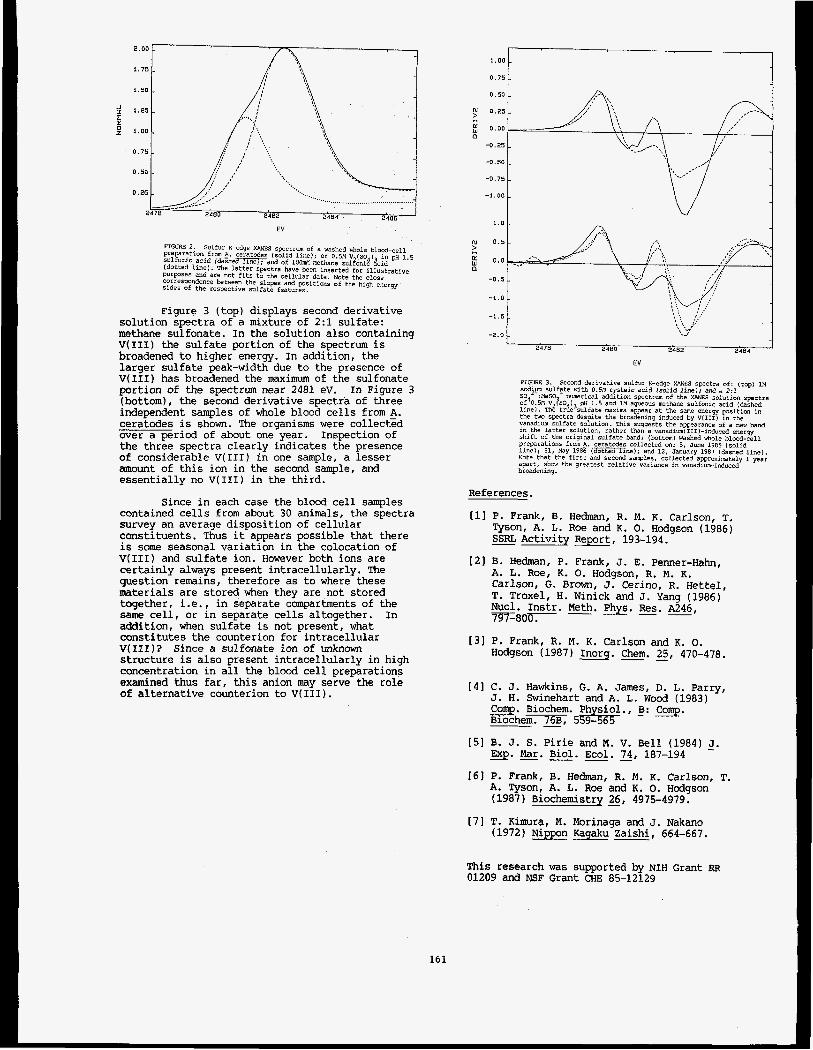
1 .50 t ,751 8 1 . 0 0
I '. 0 . 7 5 -
.'.. ......................................... - .'...,
0 . 5 0 -
0.25 -
2484 2406 2480 2402 2478
EV
FIGURE 2. preparation from A. ceratcdes ( so l id l i n e ) ; of 0.514 V.(so.i. i n OH 1.5
sulfur K-edge XANES spectram of a washed xfiole b l d - c e l l
sulfuric ac id (aashed line); and of lOomM loethane sulfoni8 &id e - - - - (dotted l i n e ) . The la t t er spectra have been inserted for i l lus tra t ive Purpases and are not fits t o the cellular data. NDte the clone correspondence t ietween the slaps and positions of the
-?tillD c*.,.c-." C^_L
Figure 3 (top) displays second derivative solution spectra of a mixture of 2:l sulfate: methane sulfonate. In the solution also containing V(II1) the sulfate portion of the spectrum is broadened to higher energy. In addition, the larger sulfate peak-width due to the presence of V(II1) has broadened the maximum of the sulfonate portion of the spectrum near 2481 eV. (bottom), the second derivative spectra of three independent samples of whole blood cells from A. ceratodes is shown. The organisms were collect% over a period of about one year. the three spectra clearly indicates the presence of considerable V(II1) in one sample, a lesser amount of this ion in the second sample, and essentially no V(II1) in the third.
In Figure 3
Inspection of
Since in each case the blood cell samples contained cells from about 30 animals, the spectra survey an average disposition of cellular constituents. Thus it appears possible that there is some seasonal variation in the colocation of V(II1) and sulfate ion. However both ions are certainly always present intracellularly. The question remains, therefore as to where these materials are stored when they are not stored together, i.e., in separate compartments of the same cell, or in separate cells altogether. In addition, when sulfate is not present, what constitutes the counterion for intracellular V(III)? Since a sulfonate ion of unknown structure is also present intracellularly in high concentration in all the blood cell preparations examined thus far, this anion may serve the role of alternative counterion to V(II1).
161
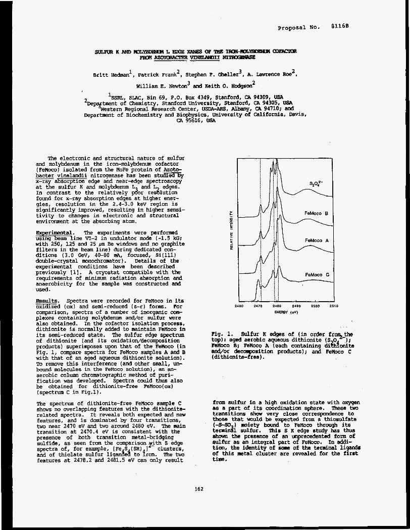
Proposal No. 81168
2 Britt Hedmanl, Patrick Frank2, Stephen F. Gheller3, A. Lawrence Roe I
3 2 William E. Newton and Keith 0. Hcdgson
‘SSRL, SLAC, Bin 69, P.O. Box 4349, Stanford, CA 94309, USA
CA 95616, USA
The electronic and structural nature of sulfur and molybdenum in the iron-molybdenum cofactor (FeMoco) isolated from the MoFe protein of Azoto- bacter vinelandii nitrogenase has been s t & x 6 x-ray absorption edge and near-edge spectroscopy at the sulfur K and molybdenum L and L edges. In contrast to the relatively &r reshution found for x-ray absorption edges at higher ener- gies, resolution in the 2.4-3.0 kev region is significantly improved, resulting in higher sensi- tivity to changes in electronic and structural environment at the absorbing atom.
The experiments were performed m i n e VI-2 in undulator mode (-1.5 kG; with 250, 125 and 25 ,m Be windows and no graphite filters in the beam line) during dedicated con- ditions (3.0 GeV, 40-80 mA, focused, Si(ll1) double-crystal monochromator). Details of the experimental conditions have been described previously [ 11. A cryostat compatible with the requirements of m i n i m radiation absorption and anaerobicity for the sample was constructed and used.
Bsults. Spectra were recorded for FeMoco in its -ed (ox) and semi-reduced (s-r) fonns. For comparison, spectra of a number of inorganic com- plexes containing molybdenum and/or sulfur were also obtained. In the cofactor isolation process, dithionite is normally added to maintain FeMoco in its semi-reduced state. The sulfur edge spectrum of dithionite (and its oxidation/decomposition products) superimposes upon that of the Felrloco (in Fig. 1, compare spectra for FeMoco samples A and B with that of an aged aqueous dithionite solution). To remove this interference (and other small, un- bound molecules in the FeMoco solution), an an- aerobic colunm chromatographic method of puri- fication was developed. Spectra could thus also be obtained for dithionite-free FeMoco(ox) (spectrum C in Fig.1).
The spectrum of dithionite-free FeMoco sample C shows no overlapping features with the dithionite- related spectra. It reveals both expected and new features, and is dominated by four transitions, two near 2470 eV and two around 2480 ev. The main transition at 2470.4 eV is consistent with the presence of both transition metal-bridging sulfide, as seen from the comparison yjth S edge spectra of, for example, [Fe S (SR) 1 clusters, and of thiolate sulfur iiganAe8 to fron. The two features at 2478.2 and 2481.5 ev can only result
2460 2470 2480 2490 2500 2510
ENEROY (eV)
Fig. 1. Sulfur R edges of (in order frT-the top): aged aerobic aqueous dithionite (S 0 1; Femco B; FeMoco A (each containing dikbnite and/or decomposition products); and FeMoco C (dithionite-free).
from sulfur in a high oxidation state with oxygen as a part of its coordination sphere. &se two transitions show very close correspondence to those that would be expected from a thiosulfate (-S-SO ) moiety bound to FeMoco through its terrmidl sulfur. This s K edge study has thus shown the presence of an unprecedented form of sulfur as an integral part of FeMoco. In addi- tion, the identity of some of the terminal ligands of this =tal cluster are revealed for the first time.
162
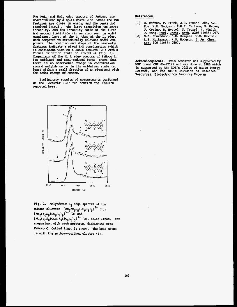
The M L and MoL edge spectra of FeMoco, are charactdrized by 3 split white-line, here the two features are close in energy and the peaks not resolved (Fig.2). The first transition has lower intensity, and the intensity ratio of the first and second transition is, as also seen in model complexes, lower at the L than at the L edge. when campared to structurally relevant mock1 com- pounds, the position and shape of the near-edge features indicate a mixed S/O coordination (which is consistent with Mo K EXAFS results [2]) with a formal oxidation state of around +4 (Fig. 2). Comparison of the Mo L edge spectra of FeMoco in its oxidized and semi-reduced forms, shows that there is no observable change in coordination around molybdenum or in its oxidation state (at least within a small fraction of an electron) with the redox change of FeMoco.
Preliminary results of measurements performed in the December 1987 run confirm the results reported here.
References.
[I] B. Hedn\an, P. Frank, J.E. Penner-Hahn, A.L. Roe, K.O. Hodgson, R.M.K. Carlson, G. Brown, J. Cerino, R. Hettel, T. Troxel, H. Winick, J. Yang, Nucl. Instr. Meth. A246 (1986) 797. S.D. C o n r E o n , m Burgess, W.E. Newton, L.E. Mortenson, K.O. Hodgson, J. Am. &em. SOC. 109 (1987) 7507.
[2]
Acknowledgements. This research was supported by NSF grant CHE 85-12129 and was done at SSRL which is supported by the DOE’S Office of Basic Energy Science, and the NIH’s Division of Research Resources, Biotechnology Resource Program.
2510 2520 2530 2540 2550
ENERGY (ev)
Fig. 2. Molybdemnn L3 edge spectra of the cubane-clusters I M O ~ F ~ ~ S ~ ( S C ~ H ~ ) ~ I ~ - (1),
[r”2Fe6S8(OCH3)s(~6HCsH5)613- (31, solid lines. For conparison with each spectrum, dithionite-free
[MozFe6s8 ( sc&j g 1 ( 2) and
Femco C, dotted line, is shown. The best match is with the methoxy-bridged cluster (3).
163
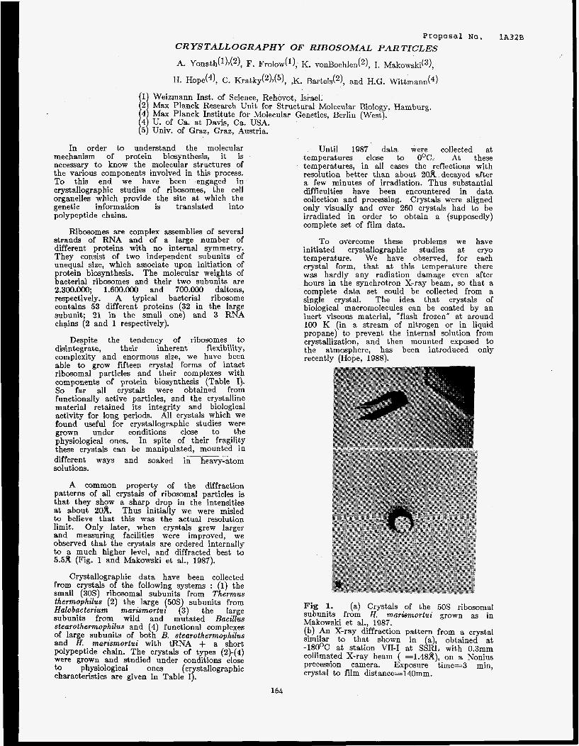
Proposal No. 1 ~ 3 2 ~ CRYSTALLOGRAPHY OF RIBOSOMAL PARTICLES
A. Yonath(l)d2), F. Frolow('), K. vonBoehlen(2), I. M a k o ~ s k i ( ~ ) ,
H. Hope(4), C. K r a t k ~ ( ~ ) , ( ~ ) , ,K. Barted2), and H.G. Wittmann(4)
Weizmann Inst. of Science, Rehovot, Israel. Max Planck Research Unit for Structural Molecular Biology, Hamburg. Max Planck Institute for Molecular Genetics, Berlin (West). U. of Ca. at Davis, Ca. USA. Univ. of Graz, Graz, Austria.
In order to understand the molecular mechanism of protein biosynthesis, it is necessary to know the molecular structures of the various components involved in this process. To this end we have been engaged in crystallographic studies of ribosomes, the cell organelles which provide the site a t which the genetic information is translated into polypeptide chains.
Ribosomes are complex assemblies of several strands of RNA and of a large number of different proteins with no internal symmetry. They consist of two independent subunits of unequal size, which associate upon initiation of protein biosynthesis. The molecular weights of bacterial ribosomes and their two subunits are 2.300.000; 1.600.000 and 700.000 daltons, respectively. A typical bacterial ribosome contains 53 different proteins (32 in the large subunit; 21 in the small one) and 3 RNA chains (2 and 1 respectively).
Despite the tendency of ribosomes to disintegrate, their inherent flexibility, complexity and enormous size, we have been able to grow fifteen crystal forms of intact ribosomal particles and their complexes with components of protein biosynthesis (Table I). So far all crystals were obtained from functionally active particles, and the crystalline material retained its integrity and biological activity for long periods. All crystals which we found useful for crystallographic studies were grown under conditions close to the physiological ones. In spite of their fragility these crystals can be manipulated, mounted in different ways and soaked in heavy-atom solutions.
A common property of the diffraction patterns of all crystals of ribosomal particles is that they show a sharp drop in the intensities at about 20A. Thus initially we were misled to believe that this was the actual resolution limit. Only later, when crystals grew larger and measuring facilities were improved, we observed that the crystals are ordered internally to a much higher level, and diffracted best to 5.5A (Fig. 1 and Makowski et al., 1987).
..___
Crystallographic data have been collected from crystals of the following systems : small (30s) ribosomal subunits from T ermus thermophilus (2) the large (50s subunits from Halobacterium marismortui 23) the large subunits from wild and mutated Bacillus stearothermophilus and (4) functional complexes of large subunits of both B. stearothermophilus and H. marismortui with tRNA + a short polypeptide chain. The crystals of types (2)-(4) were grown and studied under conditions close to physiological ones (crystallographic characteristics are given in Table I).
P the
-
Until 1987 data were collected at temperatures close to O°C. At these temperatures, in all cases the reflections with resolution better than about 20A decayed after a few minutes of irradiation. Thus substantial difficulties have been encountered in data collection and processing. Crystals were aligned only visually and over 260 crystals had to be irradiated in order to obtain a (supposedly) complete set of film data.
To overcome these problems we have initiated crystallographic studies at cry0 temperature. We have observed, for each crystal form, that at this temperature there was hardly any radiation damage even after hours in the synchrotron X-ray beam, so that a complete data set could be collected from a single crystal. The idea that crystals of biological macromolecules can be coated by an inert viscous material, "flash frozen" at around 100 K (in a stream of nitrogen or in liquid propane) to prevent the internal solution from crystallization, and then mounted exposed to the atmosphere, has been introduced only recently (Hope, 1988).
I
i
(a) Crystals of the 50s ribosomal Fig 1. subunits from H.- markmortui grown as in Makowski et al., 1987. (b) An X-ray diffraction pattern from a crystal similar to that shown in (a), obtained at -180°C at station VII-I at SSRL with 0.3mm collimated X-ray beam ( =1.4819), on a Nonius precession camera. Exposure time=3 min, crystal to film distance=140mm.
164

Due to the fikrpe of the cr teh, whlch grow a art plate@ (pig. €1 or E n g needire, attempts to ufie 01 rods for wountlng, resulted in bending of the crystals around the rods. This, in turn, caused a s\gnfficant loss of resolution in the diffraction patterns. We therefore developed a special device for mounting the crystals. Tiny spatulas were constructed from a very thin glws film glued to a thin glass rod, previously soldered to a brass pin which could be mounted on the goniometer head. Several types of spatulas were constructed, differing in the angle between the glass film and the rod, in order to permit mounting of the crystals in any desired orientation. The crystals were extracted from the solution and attached to these "spatulas" by surface tension. A more sophisticated version: the "sandwich" type , was developed later. These spatulas are made of double layers of extremely thin glass films glued to the glass rod. The crystals were placed between the two layers which provide extra protection from evaporation.
Because in all crystal forms the shortest crystal edge coincides with the largest unit cell dimension, the geometry of the spatulas dictates the permissible rotation angle. The natural tendency of the crystals was to lie flat on the glass layer. Because most cr stals exhibit a
severe problem with overlap, in particular when the long 1.e 694 R) axis was in the direction of the &ray beam. Therefore, when using "flat" spatulas, there were no fully recorded reflections. In contrast, crystals mounted on perpendicular spatulas could be positioned so that their long axis coincided with the spindle axis, permitting a relatively large rotations (3O-5O) at medium resolution. These data can be used as a "scaling set" for data collected at different orientations at higher resolution.
high mosaic spread (up to 3 B ) there was a
viscous materials we used either a hydrophobic hydrocarbon or materials such as methyl pentane diol (MPD), which was used for the growth of the crystals of the 30s subunits, or polyethylene glycol (PEG), which was used for the growth of crystals of large (50s) ribosomal subunits from Bacillus stearothermophilus. The latter ribosomal particles are very interesting because they are well characterized chemically and since we are able to split from the ribosomes several proteins in a controlled way, using biochemical and genetic methods (Yonath et al., 1986b; Gewitz et al., 1987). Initially crystals from this source were grown at 4O, using low molecular weight alcohols as precipitants (Yonath et al., 1984; 1986). Since these crystals could not be handled or soaked we have developed procedures ___ for growing crystals using non-
volatile materials. Currently we can obtain well shaped crystals from very low concentrations of pobethylene glycol in the presence of low concentrations of salts, conditions which resemble the natural environment of these particles.
We wed our B e r n - t h e perlodfi for eetablishlng the rocedumsl far oryo temperature crystallography, for examining the newly grown crystal f ~ r ~ l p ~ and for collecting date from some of them (marked D in Table I). Data have been collected aleo from several derivatives using crystals soaked in heavy-atom clusters marked H in Table I and from complexes of
stearothermophilus with tRNA and a short (18-20) segment of the nascent polypeptide chain.
50s t 'subunits from A. mariemortui and B.
References
H.S. Gewitz, J. Piefke, A. Yonath and H.G. Wittmann, Biochimie, in press (1987)
H. Hope, h e r . Cryst. Assoc. Abstract series 2,13 (1985).
I. Makowski, F. Frolow, M.A. Saper, H.G. Wittmann and A. Yonath, J. Mol. Bio1.,193, 819, (1987)
A. Yonath H.D. Bartunik, K.S. Bartels, and H.G. Wittmann, J. Mol. Biol. 177, 201, (1984)
A. Yonath, M.A. Saper, I. Makowski, J. Miissig, J. Pieflce, H.D. Bartunik, K.S. Bartels, and H.G. Wittmann J. Mol. Biol., 187, 633, (1986)
165
X-ray Crystallography of Succinyl-CoA Synthetase from Escherichia coli ~~~~~~~l N ~ . 1A39B
W.T. Wolodko, P.M.D. Fitzgerald*, M.N.G. James and W.A. Bridger
Department of Biochemistry, University of Alberta, 'Edmonton, Alberta, Canada T6G 2H7
Introduction
Succinyl-CoA synthetase is an enzyme of the tricarboxylic
acid cycle, catalyzing the 'substrate-level' phosphorylation step: it
performs a vital function in aerobic metabolism. The enzyme, puri- fied from Escherichia coli, is a tetramer with a molecular weight of
1.4 x 10s. It consists of two types of subunits and is thought to be
assembled as a dimer of ap-dimers (1). Several lines of evidence
suggest that although the complete catalytic site is formed by regions
in contact from both subunits in the ap-dimer, these ap-dimers do
not function independently within the active, tetrameric enzyme
(2,3). One way to reconcile the catalytic properties of this enzyme
with its quaternary structure is to have a clear representation of the
conformation of the tetrameric enzyme in three dimensions. Such a
determination is in progress. Crystals of succinyl-CoA synthetase
suitable for X-ray diffraction study undergo severe radiation damage
when subjected to a beam of X-rays. Their lifetime and utility for
data collection to high resolution with conventional X-ray sources is
limited (4). Fortunately, this problem is solvable using synchrotron
radiation.
Experimental
Succinyl-CoA synthetase was purified from Escherichia coli
and crystallized as described in a preliminary paper (4). Tetragonal
crystals were grown by micro-diffusion up to a size of 0.5 x 0.5 x 1.0 mm. The space group is P4122 with unit cell dimensions of 99 x
99 x 399 8, and one tetramer per asymmetric unit. Potential heavy-
atom derivatives were prepared by soaking native crystals appropri-
ately with solutions of platinum ammonium cyanide, uranyl acetate,
and auric chloride. Diffraction data were collected at SSRL (Beam
Line VII-1) using a wavelength of 1.54 A. Crystals were mounted
with c* aligned parallel the spindle axis (except for the blind-cone
data). Oscillation ranges of 1.7-2.0' (45' total range) and expo-
sures, in dose mode, of 1.5 x 10⁷ countsldegree oscillation were
taken. To minimize the effects of radiation damage, diffraction was
carried out at 4'C, only a single setting-still (exposure 1 s) was
taken with Polaroid film for preliminary crystal alignment, and only
two film packs were exposed per crystal with translation along the
crystal between the film packs. Furthermore, the spindle angle was
adjusted for an overlap of 0.5' between sequential crystals. The
diffraction patterns were recorded on Kodak DEF-5 film, in 3-film
packs with aluminum foil attenuation between films, developed by
standard procedures using fresh solutions. Films were scanned and
digitized at 50 micron raster on a P-1000 Optronics drum scanner
linked to a Kennedy 31 10 tape drive. Initial processing, merging,
scaling and post-refinement of the data have been carried out on an
Amdahl 5870 main-frame computer using programs provided by
P.M.D.F.
*Present address: Merck Sharp and Dohme Research Laboratory,
Rahway, New Jersey 07065, USA
Results and Discussion
Four full data sets (approximately 100 films each) were
collected, a native set and three potential heavy-atom derivatives (Pt,
U and Au), to a measurable resolution of 2.9 A (the limit of resolution with standard flat cassettes at a crystal-to-film distance of
100 mm). Preliminary processing of the data sets has been com-
pleted and difference Patterson maps are presently being calculated
to assess the quality of the derivatives. The merged and scaled
intensity data are of good quality producing R O factors in the order
of 8-10% with a mean multipicity slightly better than 3 reflections.
From the initial processing of these films, two observations
are important with respect to the data collection. Firstly, in
approximately 5% of the two film packs from a single crystal,
refinement of crystal orientation indicated that the correction of the
spindle value between film packs had shifted slightly (usually less
than a degree) while corrections along the other two axes were
unchanged. Either the crystals had slipped unidirectionally (thus,
notice to take extra care in drying mounted crystals in future data
collection) or the action of translating the X-ray beam to a fresh part
of the crystal, which involved the unlocking and retightening of the
restraining ring around the dial, resulted in the shift of the spindle
value (again, caution for future collection). Secondly, approxi-
mately 10% of the data were salvaged following incorporation (by
P.M.D.F.) of film tilt and pivot point calculations in the processing
programs. It seems that a film cassette and/or a position on the
carousel of the rotation camera did not lock orthogonally to the X-
ray beam resulting in a forward tilt of approximately one degree.
In summary, good data have been collected from weakly
diffracting crystals by taking advantage of synchrotron radiation.
The data are in the middle stages of processing. Heavy-atom
derivatives are being used to solve the phase problem and, thus, the
initial structure of succinyl-CoA synthetase.
Acknowledgements
This work was supported by a grant to the Medical Research
Council Group in Protein Structure and Function and by Grant
MT-2805, both from the Medical Research Council of Canada.
Provision of beam time and assistance by personnel at SSRL has
been greatly appreciated.
References
1. Wolodko, W.T., Kay, C.M. and Bridger, W.A. (1986)
2. Nishimura, J.S. (1986) Adv. Enzymol. 58, 141-172.
3. Bridger, W.A., Wolodko, W.T. and Williams, S.P. (1987)
Enzyme Dynamics and Regulation, (Chock, P.B. and Huang, C., eds.), Springer-Verlag Publishers, Chapter 2, pp. 6- 16.
4. Wolodko, W.T., James, M.N.G. and Bridger, W.A. (1984)
J. Bjol. Chem. 259, 5316-5320.
Biochemistry 25,5420-5425.
166


Three-Dimensional Structure of an Oncogene Protein: Catalytic Domain of Human c-H-ras p21
proposal No. 1A56B
Abraham M. de Vos*, Liang Ton;, Michael V. Milburn*, Pedro M. Matias*,Jarmila Jancarik*, Kazunobu Miura-, Eiko Ohtsuka., Shigeru Noguchi3,Susumu Nishimuras, and Sung-Hou Kim*
Introduction
The most commonly found oncogenes isolated from human tumors or transformed human cell lines belong to the ras gene family W. In the human genome there are three distinct cellular ras genes: c-H-ras, c-K-ras, and N-ras , the former two having sequence similarity @-a to the transforming principles of the Harvey and Kirsten strains of rat sarcoma viruses @,a, respectively. All proteins encoded by these genes are called p21, have a molecular weight of 21,000 with 188 or 189 amino acid residues, and have practically identical amino acid sequences, with the exception of the COOH-terminal 25 residues 0. The difference between the normal ras proto-oncogenes and the activated (transform- ing) ras oncogenes is usually a single base change at one of a few critical positions, resulting in a single amino acid substitution in p21 (S-U.
Knowledge of the threedimensional structure of this ras protein family should be most useful to provide a basis for understanding the known biochemical functions of both normal and activated proteins.
Results.
We have recently crystallized the catalytic domains of normal as well as activated human ras p21 proteins. The space group of the "catalytic domain" crystals is p6522 with cell parameters a = b = 83.2 A and c = 105.1 I%. There is one p21 "catalytic domain" per asymmetric unit and 46 percent of the unit cell volume is protein.We collected diraction data for the native as well as five heavy-atom derivatives on x-ray films using an Enraf-Nonius rotation camera installed on the eight-pole wiggler branch line (Stanford Synchrotron Radiation Laboratory, Palo Alto, California . and the crystal-to-film distance was 60 or 85 mm. All the data were collected at 40 C. The films were digitized with the use of a drum scanner by Optronix Cop., and the data were processed with the use of comput- er programs originally written by Rossmann uz). Reflection data from different films were merged together with the use of the PROTEIN pro- gram package w. The native data set contained about 46,000 obsew- ations of 10,400 unique reflections, with an R,rge(l) of 8% and a maxi- mum resolution of 2.25 A. About 300 reflections, missing from this data set because of intensity overload and positional overlap on the films, were later measured using a Nicolet P3/F four-circle diractometer and added to the native data set.
Five heavy-atom derivatives were used for phasing for phasing. An overall view of the three-dimensional backbone structure of p21 is shown below. The crystal structure at 2.7 A resoluiion of the normal human c-H-ras oncogene protein lacking a flexible cafimxyl-terminal 18 residues reveals that the protein consists of a six-stranded p-sheet, four a-helices and nine connecting loops. Four loops are involved in inter- actions with bound guanosine diphosphate (GDP): one with the phos- phates, another with the ribose and two with the guanine base. The majority of transforming in vivo and in vitro mutants have single amino acid substitutions at one of a few key positions in three of these four bops plus one additional loop. The biological functions of the remaining five loops and other exposed regions are at present unknown. How- ever, one loop corresponds to the binding site for a neutralizing mono- clonal antibody and another to a putative "effector region", mutations in which do not alter guanine nucleotide binding or guanosine triphos- phatase activity but reduce the transforming activity of activated proteins. The data provides a Structural basis for understanding the known bio- chemical properties of normal as well as activated ras oncogene proteins and reveals additional regions in the molecule that may possibly part'& pate in other cellular functions.
The x-ray wavelength for the data collection was either 1.541 8 or 1.08 A ,
Acknowledgements
We thank Dr. P. Phizackerley, Stanford Synchrotron Radiation Labora- ory, Palo Alto, CA for his help in data collection. Supported in part by grants from NIH (GM 29287, CA 45593). and DOE to S.H.K., a gift from Merck, Sharp, and Dohm to the University of California, Berkeley, Grants-in-aid from the Japanese Ministry of Health and Welfare for a Comprehensive 10-Year Strategy for Cancer Control to Su.N., a Re- search Res ident Fellowship from the Foundation for Promotion of Can- cer Research (Tokyo) (Su.N.) and a Damon Runyon-Walter Winchell Cancer Fellowship (P.M.M).
References
*Department of Chemistry and Lawrence Berkeley Laboratory, Univer-
*Faculty of Pharmaceutical Sciences, Hokkaido University, Kita-ku,
$Biology Division, National Cancer Center Research Institute,Chuo-ku,
1. S. Nishimura and T. Sekya, Bi0chem.J. 243,313 (1987). 2. C. Shih, L. C. Padhy, M. Murray, and R. A. Weinberg, Nature
3. T. G. Krontiris and G. M. Cooper, Proc.Nat/.Acad.Sci. U.S.A. 78,
4. M. Perucho, eta/. Cell 27,467 (1981). 5. S. Pulciani, et a/. Proc. Natl.Acad.Sci. U.S.A. 79, 2845 (1 982). 6. J. J. Harvey, Nature (London) 204,1104 (1964). 7. W. H. Kirsten and L. A. Mayer, J.Nat/.Cancer Inst. 39,311 (1967). 8. M. Barbacid, Ann. Rev. Bbchem. 56,779 (1987). 9. C. J. Tabin, et a/. Nature (London) 300, 143 (1982). 10. E.P. Reddy, R. K. Reynolds, E. Santos, and M. Barbacid, ibid., p.
11. E. Taparowsky, et a/. , &id, p. 762 (1 982). 12. M. G. Rossmann, J. Awl. Cryst. 12,225 (1 979). 13. W. Steigemann, PROTEIN :A package of crystallographic programs
for analysis of proteins. Max Planck Institute for Biochemistry, Martinsried, West Germany (1982).
sity of California, Berkeley, CA 94720 USA
Sapporo, Japan;
Tokyo, Japan.
(London) 290, 261 (1981).
1181 (1981).
149 (1982).
167

STRUCTURAL STUDIES OF ANTIBODIES, ANTIGENS AND ANTIBODY-ANTIGEN COMPLEXES Proposal No. 18663
I.A. Wilson, E.A. Stura, J.H. Arevalo, 6. Lambert and J.M. R in i
Department o f Molecular Biology Research I n s t i t u t e o f Scripps C l i n i c
10666 North Torrey Pines Road La Jo l la , Ca l i f o rn ia 92037
INTRODUCTION
I n order t o understand how the immune system in te rac ts and neutral izes fore ign microorganisms such as viruses v i a surface glycoproteins, i t i s neccessary t o under- stand the deta i led chemistry o f antibody-antigen union. In the l a s t decade four antibody Fab' fragments derived from mu l t i p le myelomas have been determined t o high reso lu t ion (1,2). With the advent o f the hybridoma technology, spec i f i c monoclonal antibodies can now be made against antigens o f in te res t . These advances have aided i n the recent successes i n the c rys ta l structures of antibody-protein complexes w i th lysozyme (3,4) and neuraminidase (5) used as imnunogens. These studies have provided valuable i ns igh ts i n t o the deta i led in teract ions o f antibodies w i th proteins and has revealed no major changes i n the antigen s t ructure on antibody binding. However, we would l i k e t o know the s t r u c t u r e s o f t h e f r e e ant ibody, f r e e an t i gen and antibody-antigen complex f o r the same system t o evaluate changes no t only i n the antigen bu t also i n the antibody. I n addi t ion, d i f f e r e n t immunogens may produce antibodies which vary i n t h e i r conformational f l e x i b i l i t y such as may be observed when ant ipept ide a n t i b o d i e s combine w i t h an i n t a c t p r o t e i n . I n ou r laboratory, we have decided t o study several antibody systems t o advance our knowledge o f the deta i led in teract ions i n antibody-antigen union. I n a l l o f the systems we have no t only complex c rys ta l s bu t na t ive Fab c rys ta l s as we1 1.
RESULTS AND DISCUSSION
Anti-progesterone Fab' and Fab'-steroid complexes
Crystals have been grown o f an anti-progesterone antibody which causes i n f e r t i l i t y i n mice up t o 36h a f t e r mating (6,7). We have co l lected several data s e t s by o s c i l l a t i o n photography a: SSRL (beam l i n e VII.2) on the anti-progesterone Fab and steroid-Fab' complexes w i th progesterone 11-a-OH-progesterone, 11-B-OH-progesterone and aetiocholanolone. I n addi- t ion, three heavy atoms have been collected, phenyl mercury acetate, amnonium osmium ch lo r i de and an iodinated steroid, 11-iodo-benzoyl-progesterone f o r use i n mu l t i p le isomorphous replaceme t methods Data have been previously c o l l e ted t o 2 - 1 1 a t 1.54 fl wavelength and recent ly t o 2.4 a t 1.08 wavelength. S ix data sets have been processed w i th merging R-factors o f 10% on in tens i t i es . Di f ference Patterson maps have been ca lcu lated and possible heavy atom posi t ions i d e n t i f i e d f o r the three der ivat ives. The s t ructure determination by mu l t i p le isomorphous replacement i s we l l underway. Data from the synchrotron source has been found t o be o f higher reso lu t ion and qua l i t y than can be co l lected i n our laboratory on a r o t a t i n g anode f o r these hexagonal c r y s t a l s. Resu l t s f o r t h i s s tudy should enable us t o understand the f i n e s p e c i f i c i t y o f the antibody in te rac t ions as wel l as how even s ing le proton changes i n the s te ro id can completely abol ish binding.
Anti-peptide Fab and Fab-peptide complexes
Great i n t e r e s t has been developed recent ly i n the use o f synthet ic peptides as vaccines. I n order t o understand how an antibody generated against a short peptide can cross-react w i t h i t s cognate sequence i n
168
t h e pa ren t p r o t e i n we have made a n t i b o d i e s a g a i n s t residues 75-100 o f the 1975 inf luenza v i r u s haemagglutinin. We have shown the determinant t o be residues 98-106 o f the peptide (8). Crystals have been grown o f bo th t h e f r e e an t i body and t h e an t i body - peptide complex (9). Survey o f the c rys ta l s a t SSRL has shown the na t ive c rys ta l s t o d i f f r a c t t o be t te r than 1.9 8 and the cocrystals t o 2.1 8. The nat ive data can conveniently be co l lected i n our laboratory. However, the synchrotron i s necessary t o achieve high reso lu t ion data on the complex c rys ta l s as these are long, th in , p la te - l i ke rods.
Cata ly t ic antibody Fab and inhibitor-complex c rys ta l s
Crystals o f a es te ro l y t i c antibody Fab ra ised against a t r a n s i t i o n s ta te ester (10) have been c rys ta l l i zed . Surveys a t t h e SSRL hav shown t h e smal l c r y s t a l s d i f f r a c t t o a t l e a s t 3.5 f resolut ion bu t are s l i g h t l y d isordered. Work i s i n progress t o o b t a i n b e t t e r qual i ty crys ta ls .
Influenza v i r u s HA, PR/8/M5/34
Crystals o f the major surface antigen o f the 1934 PR/8 inf luenza virus, the haemagglutinin (HA), have been grown i n co l laborat ion w i t h D r . Walter Gerhard, Wistar I n s t i t u t e . These c r y s t a l s were tested a t the synchrotron and found t o d i f f r a c t s i g n i f i c a n t l y be t te r than i n our l a b o r a t o r y . However, a t p resen t they d i f f r a c t o n l y t o 3.5 8 maximum i n t o t h e face and 4.5-5.0 8 i n t o the edge o f the p la te - l i ke crystals. Comparison o f t h i s s t ructure w i t h the previously solved 1968 Hong Kong HA (11) w i l l show the extent t o which t h e v i r u s su r face can change and be a n t i g e n i c a l l y d i s t i n c t bu t s t i l l funct ional f o r receptor binding and v i r a l fusion.
REFERENCES
1.
2.
3.
4.
5.
6.
7.
8.
9.
10.
11.
Amzel, L.M. and Po l j ak , R.J. (1979) Ann. Rev. B i ochem. 48: 961-967. Davies, D.R. and Metzger, H. (1983) Ann. Rev. Immunol. 1:87-117. Amit, A.G, Mariuzza, R.A., Ph i l l i ps , S.E.V. and Poljak, R.J. (1986) Science 233:747-753. Sher i f f , S., Si lverton, E.W., Padlan, E.A., Cohen, G.H., Smith-Gill, S.J., Finzel, .B.C. and Dbvies, D.R. (1987) Proc. Nat l . Acad. Sci. USA
Colman, P.M., Laver, W.G., Varghese, J.N., Baker, A.T., Tulloch, P.A., A i r , G.M. and Webster, R.G. (1987) Nature 326:358-363. Stura, E.A., Feinstein, A. and Wilson, I.A. (1987) J. Mol. B io l . 193:229-231. Stura, E.A., Arevalo, J.H., Feinstein, A., Heap, R.B., Taussig, M.J. and Wilson, 1.A. (1987) Immunology 62:511-521. Wilson, I.A., Niman, H.L., Houghten, R.A., Cherenson, A.R., Connolly, M.L. and Lerner, R.A. (1984) Cel l 37:767-778. Schulze-Gahmen, . U . , Rini , J.M., Arevalo, J.H., Stura, E.A. and Wilson, I.A. (1988) submitted. Tramontano, A., Janda, K.D. and Lerner, R.A. (1986) Science 234:1566-1570. Wilson, I.A., Skehel, J.J., and Wiley, D.C. (1981) Nature 289:366-373.
84~8075-8079.


The Crystal Structure of the Enzyme Phosphoribosyl Transferase
SL. Edwards, J. Kraut, T. Halloran and S.E. Mills
Departments of Biology and Chemistry University of California, San Diego La Jolla, CA 92093
X- ray diffraction data were collected from crystals of the
enzyme phosphoribosyl transferase (PRT). These crystals which are
moderately unstable and poorly diffracting using standard equipment
diffracted quite well using the wiggler beam at port VII-1. With the
beam adjusted to a monochromatic wavelength of 1.54 Angstroms the
crystals diffracted to a resolution of 3.0 Angstoms, a significant
improvement over previous attempts at data collection. These crystals
grow in the high symmetry cubic space group, 1432. Consequently all
of the unique data are represented in a very small wedge of reciprocal
space, 1/64 of a sphere. Although our initial intent was merely to
investigate the possible resolution enhancement due to the synchrotron
source, the actual measurements we recorded on film probably consti-
tute the majority of the data required for a complete data set. These
films have yet to be scanned with a densitometer and processed.
This enzyme is important for many reasons. First, it is one of five
enzymes that make up the pathway for tryptophan synthesis. The cry-
stal structures of three of these enzymes have been recently solved. If
the PRT structure and the one other remaining enzyme structure,
anthranilate synthase, could be solved it would represent a major
advance in biochemistry: the first complete biosynthetic pathway of an
amino acid to be elucidated with crystal structures. The final enzyme
Proposa l No. 1A69B
of this pathway, tryptophan synthase, was the target used by Dr.
Charles Yanofsky of Stanford to prove colinearity between genetic
code triplets and amino acids. Of medical interest, PRT is related to
the enzyme hypoxanthine guanine phosphoribosyl transferase which is
a key enzyme in the mechanism that leads to gout and a particular
genetic defect in this enzyme has been identified as the cause of
always fatal LeshNyhan’s disease. We hope that our continued work
will lead to the solution of the crystal structure of this enzyme, a
molecule of great biochemical and medical interest.
169

I
Proposal No. 1A70B CrvstalloaraDhi 1 s
Thomas Earnest, Michael McCarthy, Michael Shuster, Julie Earnest, Kathy Perry, William Montfort, Seunghyon Choe, Robert Stroud
Department of Biochemistry and Biophysics, University of California-San Francisco
Transmembrane ion channels play an important role
in various cellular processes such as the signalling
between cells and sensory transduction. Until recently
it has not been possible to crystallize membrane prot-
eins and obtain a three dimensional structure. The
success of Michel, et al., in crystallizing the
photosynthetic reaction center, along with the subse-
quent solution of its structure, has led to an increase
in the efforts of many protein crystallographers to
crystallize other membrane proteins. Due to the import-
ance of these proteins, the ability to crystallize
and solve the three dimensional structure of these
systems would represent a significant advance. We
are using the rotation camera facility of beam line
VII-1 at SSRL to investigate the structure of two
transmembrane ion channels which we have recently
been able to crystallize: colicin Ia from Escherichia
- coli and the nicotinic acetylcholine receptor from
Torpedo californica. We are also developing low
temperature methods (80-130 K) in order to decrease
beam damage.
I. Colicin Ia
Colicin Ia is a 70,000 dalton protein consisting
of 629 amino acids which can insert into cell membranes
to form a voltage dependent ion channel. We have
obtained crystals of the soluble form with unit cell
dimensions of a=66.3, b=176.6, ~ ~ 2 8 9 . 8 in the space
group C2221. CD studies suggest that alpha helices
are the predominant secondary structural motif. We
previously collected data to 3.5 angstrom resolution
for the native and four putative derivatives at
SSRL. These data sets have been scanned and scaled and
one derivative solved. This one was unfortunately
located on a symmetry axis necessitating more deriva-
tives for phasing.
170
11. Nicotinic acetylcholine receptor
The nicotinic acetylcholine receptor is the best
understood of all of the neurotransmitter receptors.
It can be obtained in high concentrations from the
electroplax organ of the electric fish Torpedo
californica, and consists of a 250,000 dalton multi-
subunit polypeptide. We have obtained small crystals
of detergent solublized AChR in our laboratory
which are suitable for x-ray diffraction studies.
In our laboratory we have observed reflections out to
15 angstroms. The crystals appear to have a cubic
space group with a unit cell length of 257 angstroms.
We are continuing to study parameters important for
the growth of larger, well ordered crystals. At SSRL
we obtained a rotation photograph with reflections
out to 19 angstroms,
111. Low temperature x-ray diffraction
Low temperature methods have been used to
decrease beam damage to protein crystals for a number
of years. Until recently this usually required ex-
change into a cryo-protecting agent, limiting the
general applicability of the method. Recent success
in extending the lifetime of ribosome subunit crystals
(Yonath and Hope, personal communication) by quick
freezing has been quite dramatic. At SSRL and also in
our laboratory we have shown that this method could
be used to extend the lifetime of our test crystals,
lysozyme and thymidylate synthase, although we were
unsuccessful, at that time, infreezing our crystals
of colicin and AChR. We are presently continuing to
develop low temperature methods in our laboratory
for use with our crystals of colicin and AChR.


VUV Proposals


The Bonding of Arsenic and Hydrogen to the S i ( l l 1 ) Surface. Proposal No. 908vp
R. D. Bringans
Xerox Palo Alto Research Center, 3333 Coyote Hill Rd., Palo Alto, CA 94304 and
M. A. Olmstead Department of Physics, University of California, Berkeley, CA 94720
INTRODUCTION
The (100) and (111) surfaces of Si and Ge can be passivated very effectively by a single monolayer of As1-4. In GaAs-on4 heteroepitaxy this leads, in part, to island formation rather than two- dimensional growth. This effect has been quantified for GaAs-on- Si(ll1) in an earlier papers where it was found that the degree of island formation was dependent on the arrival rate of Ga and As atoms and on which species arrived first at the substrate. The details of how the As monolayer on Si surfaces is bonded are thus very important. The stability of Si(l1l):As against oxygen and air was found to be very great4 (a 5 minute air exposure only reduced the surface state intensity in UPS spectra by 25%). Interaction of Si(l1l):As with atomic hydrogen, however, was found to change the surface state density in a reversible way4. In the present work we examine the reverse situation. A Si(ll1) surface which has been terminated with hydrogen atoms was exposed to the type of As4 fluxes used in molecular beam epitaxy (MBE) of GaAs on Si. The results cast light on the nature of the bonding between Si and As and are also important for understanding how some substrate preparation methods, which leave a .hydrogen terminated surface, will interact at moderate temperatures with As.
RESULTS AND DISCUSSION
Photoemission measurements were carried out both on the valence band and on the Si 2p and As 3d core levels. Hydrogen exposures were made with the substrate held at room temperature or a t 300oC. A hot filament was placed within 5cm of the surface in order to create H atoms from the H2 pressure in the chamber. Arsenic exposures were made at As4 partial pressures in the 10-7 t o 10-6 torr range with the substrate facing away from an effusion cell containing As.
Valence Band
Energy Below EF (eV)
Figurel. S i 2 ~ 3 ~ core level and valence band spectra, taken sequentially on the same surfaces with 130 eV synchrotron radiation and a 21.2 eV He discharge lamp respectively, for (a) the clean Si(ll1) 7x7 surface and for the same surface (b) after hydrogen exposure plus a 300% anneal. This hydrogen covered surface was then exposed t o As4 before spectra (c) and subsequently annealed at a number of temperatures with results for 500oC and 650OC being shown in (d) and (e), respectively. Intensities are shown on an arbitrary scale and the plots are aligned in energyat the Fermi energy.
Experiments t o study the interaction of arsenic with Si(l1l):H were carried out under a variety of conditions. The substrate temperature was varied for the H exposure and for the As4 exposure and annealing studies were made for Si(l1l):H and for Si(l1f):H +As. Spectra for one of these surface treatments are shown in detail in Fig. 1. The data correspond to a lOOOL hydrogen exposure at room temperature followed by a 6 minute anneal at 300OC to form Si(ll1):H. This surface was then exposed to As4 with the substrate held at 300oC for part of the exposure and at room temperature as the As4 pressure in the MBE chamber was increased and then reduced. The change in the valence band upon hydrogen termination of Si(ll1) 7x7 can be seen clearly. The three surface states on the Si(ll1) 7x7 surface (indicated by arrows) disappear and a new peak arises at around 5.OeV below the Fermi energy (shown by the arrow. This latter peak corresponds to the bond between H and Si. There is also a qualitative change in the Si 2 ~ 3 , ~ spectrum after hydrogen exposure. The surface-related shoulder on the low binding energy side of the main peak disappears and the overall spectrum narrows. Both of these results can be understood as the removal of the complicated Si(ll1) 7x7 structure, which has many inequivalent Si sites in its unit cell, and its replacement by a 1x1 structure which has onlyone type of interface S i atom site in addition to the bulk Si site.
Addition of As4 to the Si(ll1):H surface increases the photoemission intensity in the region near the top of the valence band, consistent with the increase seen in this region in the earlier angle resolved photoemission experimentsa. The peak associated with the hydrogen-silicon bond remains visible, suggesting that the As atoms reside on top of the Si(l1l):H surface. After the 650OC anneal, the valence band and core level spectra aresimilar to those for Si(l1l):As. The arrow in (e) shows the peak corresponding to the As lone pair state4. The (As 3d I Si 2p) core level intensity ratio decreased by 7% after the 350OC anneal and then remained constant during anneals at successively higher temperatures until the arsenic begins to desorb as the surface was annealed above 750OC.
A measure of the As coverage of the surfaces was determined from the ratio of areas under the As 3d and Si 2p core levels This ratio was found to be significantly lower for As4 deposition onto previously hydrogenated surfaces than it was for deposition onto the clean surface. This could be the result of loss of As via AsH3 desorption or due t o the inability of As atoms to find their optimum sites on the Si surfaces when H atoms are present.
The Si 2p core level spectrum for the ideally terminated Si(l1l):As surface has a bulk contribution and a single, well-defined chemical shift of 0.75eV to higher binding energy3-5. This shift identifies the Si atoms at the interface which are each bonded to three As atoms. The Si 2~312 spectrum in Fig 1 for the Si(ll1): H + As surface after a 650OC anneal has a large shifted component at 0.75eV. but has in addition a small component on the low binding energy side of the bulk peak and significant spectral weight between the bulk and 0.75eV components. This qualitative result can be interpreted to show that the As coverage is not complete after the H atoms have been removed.
It was shown by Uhrberg et al4 that atomic H interacts in a reversible manner with Si(ll1):As. The results of the present study show that the inverse reaction is not reversible. We have found that arsenic bonds strongly to the Si(ll1) surface and displaces hydrogen atoms on theSi(ll1): H surface. Taken together, these results indicate that if H and As atoms are competing to bond to the Si(ll1) surface, the As atoms will win. We have also shown that a hydrogen terminated surface is more resistant to bonding t o As atoms that is a clean Si(ll1) 7x7 surface.
ACKNOWLEDGEMENTS
We are grateful for many disc Biegelsen and for the skillful as and R D. Yingling. Part of this work was performed at SSRL which i s supported by the DOE Office of Basic Energy Scienc partially supported by NSF Grant OMR8657623.
'REFERENCES
1. R. D. Bringans, R. 1. G. Uhrberg, R. 2. Bachrach and Phys. Rev. Lett. 55- 533, (1985).
2. R. 1. G. Uhrberg, R. D. Bringans, R. 2. Bachrach, and 1. E. Northrup, Phys. Rev. Lett. 56- 520 (1 986) .
3. M. A. Olmstead, R. D. Bringans, R. I . G. Uhrberg and R. 2. Bachrach, Phys. Rev. B,34,6401,(1986).
4. R. 1. G. Uhrberg, R. D. Bringans, M. A. Olmstead, R. Z. Bachrach and J. E. Northrup, Phys. Rev. B, 35,3945, (1987).
5. R D. Bringans, M. A. Olmstead, R. 1. G. Uhrberg and R. Z. Bachrach, Appl. Phys. Lett., 5J, 523. (1987); Phys. Rev. B 36- 9569, (1 987).
172
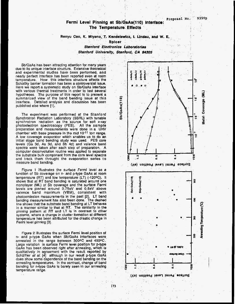
proposal NO. 9 Fermi Level Pinning at Sb/aaAs(llO) Interface:
The Temperature EffeCtS
Renyu Cao, K. Mi . Kendelewicz, I. Lindau, and W. E.
Stanford Ufiiversity, Sta
Sb/GaAs has been attracting attention for many years due to its unique interface structure. Extensive theoretical and experimental studies have been performed, and nearly perfect interface has been reported even at room temperature. How this interface structure affects the Schottky barrier formation has been a controversial issue. Here we report a systematic study on Sb/GaAs interface with various thermal treatments in order to test several hypotheses. The purpose of this report is to present a summarized view of the band bending issue at this interface. Detailed analysis and discussion has been published else where [I].
0 0 8 0
The experiment was performed at the Stanford Synchrotron Radiation Laboratory (SSRL) with tunable synchrotron radiation as the source for soft x-ray photoelectron spectroscopy (PES). All the sample preparation and measurements were done in a UHV chamber with base pressure in the mid 10-11 torr range. A low coverage evaporator which enables us to do an initial stage band bending study was used. PES core levels (Ga 3d, As 3d, and Sb 4d) and valence band spectra were taken after each step of preparation. A computer deconvolution routine was applied to separate the substrate bulk component from the core level spectra and track them throught the evaporation series t o measure band bending.
Figure 1 illustrates the surface Fermi level as a function of Sb coverage on n- and p-type GaAs at room
s I 0 2-
? 0
(he) UOla!SOd pAa’( !WJad W e 4 m
f g temperature (RT) and low temperature (LT) (-120OC). It shows that at RT band bending is saturated around one monolayer (ML) of Sb coverage and the surface Fermi
- 0 0 5 3 8 1 $ 3
v)
levels are pinned around 0.75eV and 0.5eV above valence band maximum (VBM), consistent with photoemission measurements in the past [2]. LT band # e bending measurement has also been done. The dashed line shows that the substrate band bending at LT behaves in a manner similar to that at RT. The similarity in the pinning pattern at RT and LT is in contrast to other systems, where a change in cluster formation at different
Fermi level pinning [3].
e
* 0 - 3
temperature has been attributed for the drastic change in B 0 - 3
B
Figure 2 illustrates the surface Fermi level position of n- and p-type GaAs when SbIGaAs interfaces were annealed in the range between 300% and 450OC. Large variation in surface Fermi level position for p-type GaAs has been observed right after annealing, which is qualitatively in agreement with the result reported by Schaffler et a/. [4] although in our result p-type GaAs does show some dependence of the band bending on the annealing temperatures. In the contrast, change of band bending for n-type GaAs is barely seen in our annealing temperature range.
The nature of Schottky barrier formation at the SbIGaAs interface has been argued for a long time. Theoretical calcutions [5,6] based on a zig-zag chain model suggests that the Sb-derived states lie inside the valence band and conduction band or very close to conduction band minimum, and thus those states can not be responsible for the Fermi level pinning around mid band gap. In another words, any band bending has to be explained by other interface states. The photoemission data show that Sb depostion does induce the band bending for both n- and p-type GaAs. The positions of the surface Fermi levels are 0.75eV for n-type GaAs and 0.5eV for p-type GaAs abave the VBM, same as those in other metal or oxide-GaAs systems. The coincidence of the pinning position suggests that even at an interface such that strong chemical bonds are formed, defects [7] may still play a dominant role in the Schottky barrier formation. The Fermi level pinning at LT can also be understood. Detailed analysis has shown that the interface morphological and chemical structure does not change much upon the substrate temperature. Therefore the pinning mechanism is, conceivably, the same at both RT and LT.
Schaffler et a/ [4] argued that room temperature band bending is due to a non perfect Sb/GaAs interface and after annealing the interface, a higher degree of perfection is achieved, resulting in a decrease of band bending. The so-called Sb-derived states have been attributed to the residual 0.28eV band bending. Furthermore same kind of behavior was predicted for Sbln-GaAs. Clearly it is not the case on the basis of our study. On the other hand, under the framework of defect mode one has to include the change of the relative density of donor states versus acceptor states upon annealing to explain the different pinning behavior of n- and p-type GaAs upon annealing, although it seems plausible to explain the Fermi level pinning at Sb/GaAs interface prepared at RT and LT.
This work was supported by ONR under contract number NO001 4-85-0736. The experiment was performed at SSRL, which is supported by Department of Energy, Office of Basic Science Research and National Science Fundation, Division of Materials Research.
References
1. R. Cao, K. Miyano, T. Kendelewicz, I. Lindau, and W. E. Spicer, J. Vac. Sci. Technol. A 6(3), in press. P. Skeath, C. Y . Su, I. Lindau, and W. E. Spicer, J. Vac. Sci. Technol, 17, 874 (1980). R. Cao, K. Miyano, T. Kendelewicz, K. K. Chin, I. Lindau, and W. E. Spicer, J. Vac. Sci. Technol. B 5(4), 998 (1987). F. Schaffler, R. Ludeke, A. Taleb-lbranimi, G. Hughes, and D. Rieger, J. Vac. Sci. Technol. B 5(4), 1048 (1987); Phy. Rev. B 36, 1328 (1987). C. M. Bertoni, C. Calandra, F. Manghi, and E. Molinari, Phy. Rev. B 27, 1251 (1983). C. Maihiot, C. B. Duke, and D. J. Chadi, Phy. Rev. Lett. 53, 2114 (1984); Phy. Rev. B 31, 2213 (1 985). W. E. Spicer, I . Lindau, P. Skeath, C. Y. Su, and P. Chye, Phy. Rev. Lett. 44, 420 (1980).
2.
3.
4.
5.
6.
7.
Figure Captions
Figure 1 Band bending diagrams as a function of Sb coverage. pinnied at 0.75eV and 0.5eV above VBM for n- and p-type GaAs respectively.
Figure 2 Band bending diagram as a function of annealing temperature. Large variation in band bending for p-type GaAs (from 0.5eV to 0.2eV) has been observed but not for n-type GaAs.
The surface Fermi levels are
174
Many studies have shown that metal/H&dTe interfaces formed at room temperature generally exhibit profound intermixing of the semiconductor and overlayer metal, to 'an extent far greater than for interfaces formed on Si or GaAs. The usual disruption at the interface upon overlayer formation and the concurrent loss of Hg in the near surface cause the interface to be very complex, both morphologically and electrically. Since metal interdiffusion, chemical ,reactions and Hg loss should be inhibited in interfaces formed at lower temperatures, leading to a simplification of the interface, an experimental approach is followed where Ag and Pd are deposited in ultrahigh-vacuum (UHV) (p < 10-lo torr) onto atomically clean (110) Hg0.77Cd0.23Te surfaces cooled to 100 K. These metals have similar telluride heats of formationl: AHf(Ag2Te) = -8.6 kcal/mole and AHfldTe) = -9.0 kcallmole, but Pd also has predicted high heats of solution2 with the cations: AHsol(Cd;Pd) = -32.4 kcallmole and AHs01(Cd;Pd) = -26.2 kcal/mole. This high of solution for the Pd case results in vastly different interface morphology between these two interfaces formed at room temperature (RT), and the RT interfaces formed between them and HgCdTe have been previously investigated3~4. Incremental overlayer coverages ranging from sub-monolayer (ML) at low coverages to several ML at higher coverages were employed in this study. The evolution of both of these low temperature (LT) interfaces are studied using the surface sensitive techniques of photoemission spectroscopy (PES) employing synchrotron radiation and low energy electron spectroscopy @ED), and compared to the corresponding room temperature (RT) interfaces.3~4 The HgCdTe crystals used in this study are bulk p-type. Preliminary analysis of the data reveals that the LT interfaces are more abrupt than the RT cases, but more in depth analysis of the data is necessary before any concrete conclusions can be drawn from this study.
,
References *This work is supported by DAFPA contract # N00014-86-K-0854 1K. C. Mills, Thermodynamic Data for Inorganic Sulphides, Selenides, and Tellurides (Butterworths, London, 1974).
2A. R. Miedema, P. F. de Ch2te1, and F. R. de Boer, Physica B 100, 1 (1980). 3D. J. Friedman, G. P. Carey, C. K. Shih, I. Lindau, W. E. Spicer, and J. A. Wilson, J. Vac. Sci. Technol. A 4, 1977(1986). 4G. P. Carey, D. J. Friedman, and W. E. Spicer, to be published.
Not
175
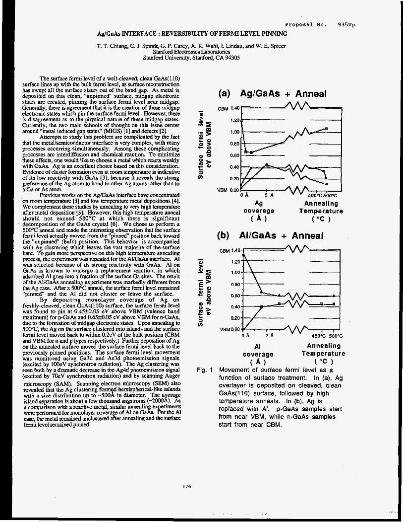
Ag/GaAs INTERFACE : REVERSIBILITY OF FERMI L
T. T. Chiang, C. J. Spindt, G. P. Carey, A. K. Wahi, I. Lindau, Stanford Electronics Laboratories
Stanford University, Stanford, CA 94305
The surface f e d level of a well-cleaved, clean GaAs( 1 10) surface lines up with the bulk fermi level, as surface reconstruction has swept all the surface states out of the band gap. As metal is deposited on this clean, "unpinned" surface, midgap electronic states are created, pinning the surface fermi level near midgap. Generally, there is agreement that it is the creation of these midgap electronic states which pin the surface fermi level. However, there is disagreement as to the physical nature of these midgap states. Currently, the two main schools of thought on this issue center around "metal induced gap states" (MIGS) [13 and defects 121.
Attempts to study this problem are complicated by the fact that the metavsemiconductor interface is very complex, with many processes occurring simultaneously. Among these complicating processes are interdiffusion and chemical reaction. To minimize these effects, one would like to choose a metal which reacts weakly with GaAs. Ag is an excellent choice based on this consideration. Evidence of cluster formation even at mom temperature is indicative of its low reactivity with GaAs [3], because it reveals the strong preference of the Ag atom to bond to other Ag atoms rather than to a Ga or As atom.
Previous works on the Ag/GaAs interface have concentrated on room temperature [3] and low temperature metal depositions [4]. We complement these studies by annealing to v q high temperature after metal deposition [5]. However, this high temperature anneal should not exceed 58OOC at which there is significant decomposition of the GaAs crystal [6]. We chose to perform a 500OC anneal and made the interesting observation that the surface f d level actually moved from the "pinned" position back toward the ''unpinned'' (bulk) position. This behavior is accompanied with Ag clustering which leaves the vast majority of the surface bare. To gain more perspective on this high temperature annealing process, the experiment was repeated for the AYGaAs interface. A1 was selected because of its strong reactivity with GaAs. A1 on GaAs is known to undergo a replacement reaction, in which adsorbed A1 goes into a fraction of the surface Ga sites. The result of the Al/GaAs annealing experiment was markedly different from the Ag case. After a 50O0C anneal, the surface fermi level remained "pinned and the A1 did not cluster or leave the surface.
By depositing monolayer coverage of Ag on freshly-cleaved, clean GaAs(l10) surface, the surface fermi level was found to pin at 0.45k0.05 eV above VBM (valence band maximum) for p-GaAs and 0.65M.05 eV above VBM for n-GaAs, due to the formation of midgap electronic states. Upon annealing to 500"C, the Ag on the surface clustered into islands and the surface fermi level moved back to within 0.2eV of the bulk position (CBM and VBM for n and p types respectively.) Further deposition of Ag on the annealed surface moved the surface fermi level back to the previously pinned positions. The surface fermi level movement was monitored using Ga3d and AsSd photoemission signals (excited by lOOeV synchrotron radiation). The Ag clustering was seen both by a dramatic decrease in the Ag4d photoemission signal (excited by 7OeV synchrotron radiation) and by scanning Auger microscopy (SAM). Scanning electron microscopy (SEW also reveded that the Ag clustering formed hemispherical-like islands with a size distribution up to -5OOk in diameter. The average island separation is about a few thousand angstrom (-2OO0k). As a comparison with a reactive metal, similar annealing experiments were performed for monolayer coverage of At on GaAs. For the Al case, the metal remained unclustered after annealing and the surface fermi level remained pinned.
176
PT
,EVEL PINNING
, and W. E. Spicer
sal No.
(a) Ag/GaAs + Anneal
9 3 5vp
c m 1.40
1.20 b--.-------- ______( 1 .oo
0.80
0.60 - 0.40
0.20
VBM 0.00
Ag Annealing Tem pe rat u re coverage
( A ) ( " C )
(b) AI/GaAs + Anneal
CBM 1.40
1.20
1 .oo
0.80
0.60
0.40
0.20
VBM 0.00 450°C 500°C
AI Annealing coverage Temperature
( A ) ( " C 1 Movement of surface fermi level as a function of surface treatment. In (a), Ag overlayer is deposited on cleaved, clean GaAs(ll0) surface, followed by high temperature anneals. In (b), Ag is replaced with AI. p-GaAs samples start from near VBM, while n-GaAs samples start from near CBM.
In summary, our experimental findings indicate that monolayer metal deposition on GaAs pins the surface fermi level near midgap. In the case of Ag, annealing this Ag-covered GaAs surface around 5OOOC causes the Ag on the surface to cluster and moves the surface fermi level back to within 0.2eV of the bulk position. For the AI case, upon annealing, there is no evidence of clustering and the surface fermi level remains pinned. This result can be adequately explained by both the "metal induced gap state" (MIGS) model and the defect model of Schottky barrier formation [5]. However, even though both models can adequately explain the gross features of these experimental findings, they do have their problems in accounting for particular details. Clearly, more experimental and theoretical work is needed to establish a complete understanding of these results.
This work was supported by the U.S. Defense Advanced Research Projects Agency (DARPA) and the U.S. Office of Naval Research (ONR) under Contract No. NOOO14-83-K-0073, The SEM (scanning electron microscopy) and SAM (scanning Auger microscopy) experiments were performed at the Center for Integrated Systems (Stanford University) by Dr. R. Browning.
REFERENCES
[ 13 J. Tersoff, Phys. Rev. Lett. 52,465 (1984). [2] W. E. Spicer, I. Lindau, P.Skeath, C.Y. Su, and P. Chye,
Phys. Rev. Lett. 44,420 (1980). [3] R. Ludeke, T.-C. Chiang, and T. Miller, J. Vac. Sci. Technol.
B 1(3), 581 (1983). [4] R. Cao, K. Miyano, T. Kendelewicz, K.K. Chin, I. Lindau,
and W. E. Spicer, J. Vac. Sci. Technol. B 5(4), 998 (1987). 1.51 For details, see T. T. Chiang, C . J. Spindt, W. E. Spicer, I.
Lindau, and R. Browning, submitted for publication in the Proceedings of 15th PCSI conference, to appeat in J. Vac. Sci. Technol..
Solids 34, 1693 (1973). [6] C. T. Foxon, J. A. Harvey, and B. A. Joyce, J. Phys. Chem.
177

Proposal No. 935vp Critical Review of Valence Band Offset Measurements
R. S. List, I. Lindau and W. E. Spicer
Stanford Electronics Laboratories Stanford University, Stanford, CA 94305
The progress in understanding heterojunction band lineups
has increasingly become dependent upon the availability of accurate
and reliable measurements. The reported values of the band offset
for the Si / GaAs system measured by photoemission range from
0.05 eV to 0.70 eV. Such a large scatter in experimentally
determined discontinuities is common for other systems as well and
not only makes the evaluation of different theories difficult, but
raises the question as to whether such variations are real and
representative of technological variations in the offset. Clearly the
understanding and control of these variations is essential before any
device applications can be realized.
We attempted to study the source of these variations in the Si
/ GaAs (1 10) heterojunction. We altered the growth conditions to
effect modifications in the overlayer crystalline order, strain and
chemical reactivity. The collective consideration of the above factors
gives us a coherent overview of the problems involved in
discontinuity measurements. To study and characterize the above
factors we relied heavily upon photoemission, near edge structure
and surface extended x-ray absorption fine structure measurements
performed at the Stanford Synchrotron Radiation Laboratory. The
Jumbo monochromater beamline at SSRL has unique capabitities of
studying absorption across the Si K-edge (1840 eV). In our studies
we found the offset to be 0.23 f 0.0.5 eV for both amorphous and
crystalline overlayers, either strained or unstrained. We did,
however, observe discontinuities as large as 0.7 eV for room
temperature growths. We show that such large discontinuities are due to dangling bond type states which extend into the bandgap and
change the apparent valence band maximum energy. These states
can be removed by annealing the sample without crystallization.
Offsets as small as 0.05 eV may be inferred from our data if we fail
to account for the contributions of rather subtle chemical effects at
the interface. The analysis technique employed in determining the
valence band maximum has also been examined and found to yield
large absolute variations. Theoretical valence band maxima fitting
has been used to remove such errors. Overall we found the
discontinuity to be more dependent on the interpretation of the data
than on technological variations. If we discern and properly account
for extrinsic effects and unambiguously define the discontinuity,
photoemission can yield accurate and single valued measurements of
the valence band discontinuity.
Direct Measurement of Pinning States in the Band Gap ? With the recently acquired access to new beamlines at SSRL yith
greatly enhanced photon flux and resolution, and the anticipation of
even brighter sources in the near future., we are attempting to push
the sensitivity limits of our photoemission experiments to new
limits. A namal system of interest to us is the pinned GaAs surface
which is thought to have roughly 0.01 of a monolayer of defect
states in the GaAs band gap near midgap. While the importance of
the high flux and surface sensitivity of synchrotron radiation is obvious to such an experiment, the high resolution and tunability of
synchrotron radiation experiments allow us to resolve these states
from the valence band maximum and also sweep the photon energy
across the Ga, As and overlayer absorption edges to look for a
resonance of the defect states, thereby giving us insight into their
nature and origin. A preliminary experiment has been performed
during the first run of the new TGM monochromater which
demonstrated that there is sufficient flux and resolution for the
experiment. We are anticipating the installation of filters in the
beamline to greatly reduce the background from second order light
and zssembling a new high resolution, high throughput
hemispherical electron analyzer which combmed should allow for
the successful completion of this experiment.
Papers published
R. S. List, T. Kendelewicz, M. D. Williams, C. E. Mccants, I. Lmdau and W. E. Spicer, "Chemical Reactions at the Si / GaAs (110) and Si / InP (110) interfaces: Efects on Valence Band
Discontiuity Measurements," J. Vac Sci. Technol. A, proceedings
of the National Symposium of the American Vacuum Society,
Anaheim, CA 1987, (in press).
Papers submitted R. S. List and W. E. Spicer, "Can Photoemission Measure Valence
Band Discontinities ?" J. Vac. Sci. Technol. B, Promdm . gs of the Conference on the Physics and Chemistry of Semiconductor
Interfaces, Asilomar, CA 1988.
178

proposal NO. 9 3 5 W Data Acquisition Software for the New p V A X Beam-line Computers
Peter H. Mahowald
Stanford Electronics Laboratories Stanford University, Stanford, CA 94305
The data acquisition and data analysis computer programs "Exp" and "Auto",which have been used by Stanford and other groups on the VUV beam lines, now operate on the new p V a x beam line computers. Work is in progress to polish the final versions.
These programs take advantage of the features of the new p- Vax computers. For instance, they use the multiple-window capability of the large pVax operators console, and they use the sophisticated CAMAC wntrol routine called VAXMOTORS.
The main advantages of this software over other existing systems are threefold. One, it is faster to use, both in terms of fast computer response and saving the operator unnecessary interaction with the computer. Second, by its internal design it is less prone to bugs which interrupt data-acquisition. Third, it has a great depth of wmmands (including wmmands which can be programmed at the beam line by non-experts). These programs are command oriented (with menus when requested) and the documentation is available from SSRL.
/
B.
,/
179

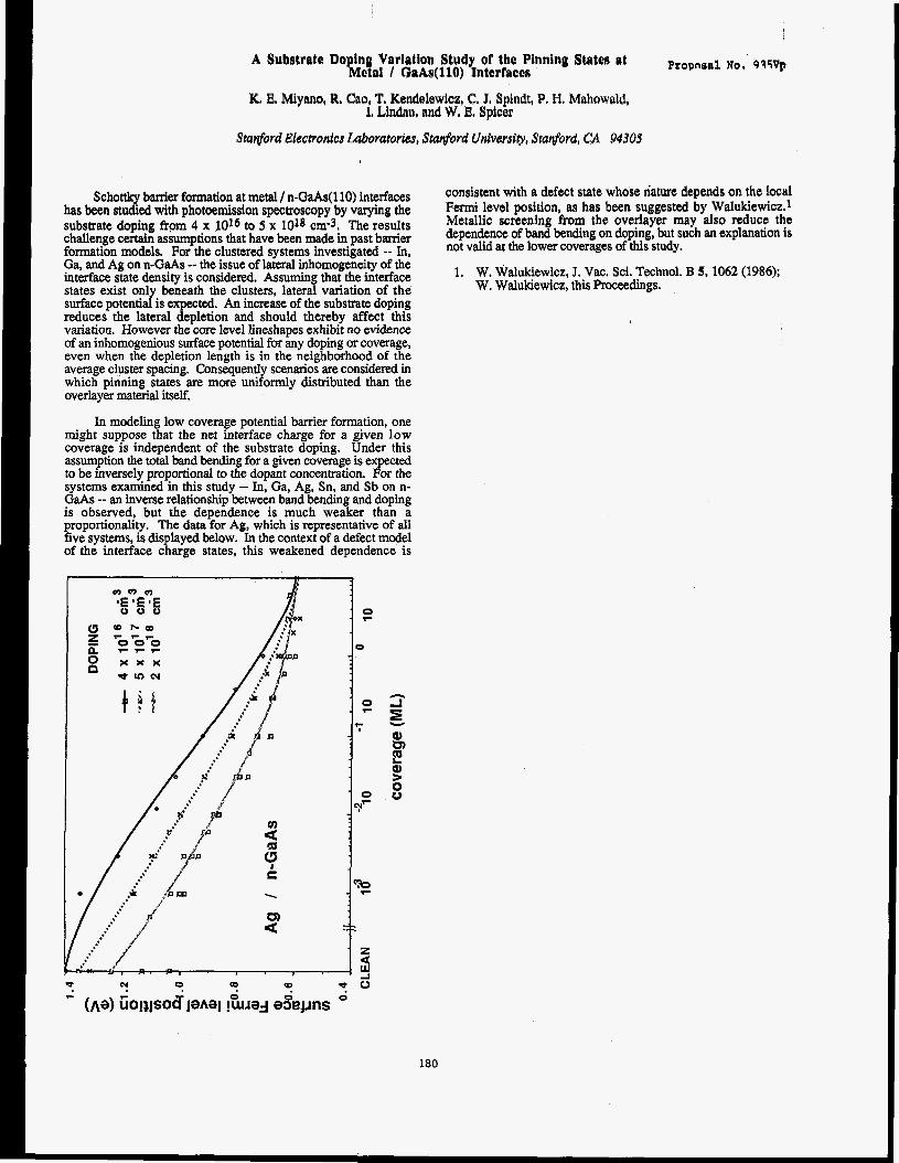
A Substrate Bo fng Variation Study of the Pinning States at beta1 / GaAr(118) Interface$ Proporal
Schottky baniex formation at metal / n-GaAs(l10) interfaces has been studied with photoemission spectroscopy by varying the substrate doping from 4 x 10¹⁶ to 5 x 1018 cm-3. The results challenge certain assumptions that have been made in past barrier formation models. For the clustered systems investigated -- In, Ga, and Ag on n-GaAs -- the issue of lateral inhomogeneity of the interface state density is considered. Assuming that the interface states exist only beneath the clusters, lateral variation of the surface potential is expected. An increase of the substrate doping reduces the lateral depletion and should thereby affect this variation. However the core level lineshapes exhibit no evidence of an inhomogenious surface potential for any doping or coverage, even when the depletion length is in the neighborhood of the average cluster spacing. Consequently scenarios are considered in which pinning states are more uniformly distributed than the overlayer material itself.
In modeling low coverage potential barrier formation, one might suppose that the net interface charge for a given low coverage is independent of the substrate doping. Under this assumption the total band bending for a given coverage is expected to be inversely proportional to the dopant concentration. For the systems examined in this study -- In, Ga, Ag, Sn, and Sb on n- GaAs -- an inverse relationship between band bending and doping is observed, but the dependence is much weaker than a proportionality. The data for Ag, which is representative of all five systems, is displayed below. In the context of a defect model of the interface charge states, this weakened dependence is
consistent with a defect state whose nature depends on the local Fermi level position, as has been suggested by Walukiewicz.1 Metallic screening from the overlayer may also reduce the dependence of band bending on doping, but such an explanation is not valid at the lower coverages of this study.
1. W, Walukiewicz, J. Vac. Sci. Technol. B 5, 1062 (1986); W. Walukiewicz, this Proceedings.
180

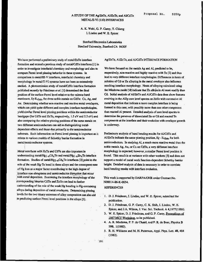
Proposal NO. 93517~ A STUDY OF THE AgLikTe, AUZnTe, and W d T e
METAL/II-VI (1 10) INTERFACES
A. K. Wahi, G. P. Carey, T. Chiang
I. Lindau and W. E. Spicer
Stanford Electronics Laboratories Stanford University, Stanford CA 94305
We have performed a preliminary study of m e W T e interface
formation and extend a previous study of meWCdTe interfaces 113 in
order to investigate interfacial chemism and morphology and also to compare Fermi level pinning behavior in these systems. In comparison to metal/III-V interfaces, interfacial chemistry and morphology in metal/II-VI systems have not been as extensively
studied. A photoemission study of meWCdTe interface formation
published recently by Friedman et ul. 111 determined the final
position of the surface Fermi level relative to the valence band maximum, Ef-Evbm, for three noble metals on CdTe: Cu, Ag, and
Au. Dpminin g whether non-reactive and reactive metal overlayers, which can yield quite different and complex interface morphologies,
yield similar Fermi level pinning positions within the semiconductor
bandgaps (for CdTe and ZnTe, respectively, 1.5 eV and 2.2 eV) and
also comparing the relative pinning positions of the same metals on
two different semiconductors can aid in distinguishing metal-
dependent effects and those due primarily to the semiconductor
substrate. Such information on Fermi level pinning is important as it relates to various models of Schottky banier fonnation in metalkemiconductor systems.
Metal interfaces with ZnTe and CdTe are also important in understanding metaVHgi-xCdxTe and metal/Hgi-xZn,Te interface
formation. Studies of metal/Hgi-xCdxTe interfaces 121 point to the
role of the weak Hg-Te bond in these alloys and the consequent ease
of Hg loss as a major factor contributing to the high degree of
interface non-abruptness and semiconductor disruption that occur with metal deposition. Examining the interface morphology of the
[corresponding binaries CdTe and ZnTe can lead to further understanding of the role of the weak Hg bonding in Hgtontaining
alloys during deposition of metal overlayers. D e t e a g pinning levels for the two binary extremes of alloy composition c ~ n also aid in predicting surface Fermi level positions in the alloys [3].
_. ._
AglZnTe, AvznTe, and Al/CdTe INTERFACE FORMATION
We have focused on the metals Ag and Al, predicted to be, respectively, non-reactive and highly reactive with Te [2] and thus
lead to very different interface morphologies. Differences in heats of
solution of Cd or Zn alloying in the metal overlayer also influence
resulting interface morphology. Heats of alloying calculated using
the Miedema model [4] indicate that Zn a l l o b Al more readily than Cd. Initial analysis of AUZnTe and Al/CdTe data does show f e a m s
evolving in the Al2p core level spectra on ZnTe with successive Al
metal deposition that indicate a more complex interface is being
formed in this case, with possibly more than one other component
than reacted Al present. Detailed analysis of core level spectra to determine the presence of dissociated Zn or Cd and reacted Te
components at the interface and their evolution with overlayer growth is underway.
Preliminary analysis of band bending results for AYCdTe and AVZnTe indicate the same pinning position, Ef - EVbm for both
semiconductors. In studying Al, a much more reactive metal than the
noble metals Ag, Au, or Cu on CdTe, a very diffemt interface
morphology is expecred, however, a similar Fermi level position is
found. This result is at variance with other workers [SI and does not
support a model of metal work functiondependent Schottky barrier
height. Detailed analysis of data is necessary in order to comlate band bending results with interface evolution.
This work is supported by DAFWNONR under Contract No. N00014-86-K-0854.
REFER€NCES
1. D. J. Friedman, I. Lindau, and W. E. Spicer, submitted for
publication.
2. D. J. Friedman, G. P. Carey, C. K. Shih, I. Lindau, W. E.
Spicer, and J.A. Wilson, J. Vac. Sci. Technol. A 4,1977(1986).
3. W. E. Spicer, D. J. Friedman, and G. P. Carey, Proceedings of l987 McT Worbshpp, to be published.
4. A. R. Miedema, P. F. de Chatel, and F. R. de Boer, Physica B
100, 1( 1980).
5 . R. H. Williams and M. H. Patterson, Appl. Phys. Lett. 40,484
(1982).
18 I


-- CORE LEVEL SPECTROSCOPY Yb-COMPOUNDS Proposa l No. 941Vp
I.?&bati, L.Braicovich, M-Sancrotti Istituto di Fisica - Politecnico di Milano
3 2 Piazza Leonard0 da Vinci - 20133 Milano (Italy) E.Puppin, 1.Lindau
Stanford Electronics Laboratories - Stanford University Stanford - CA 94305 USA
R.Eggenhoffnerr Dipartimento di Fisica - Universita' di Genova
Via Dodecaneso - 16100 Genova (Italy) Istituto di Chimica-Fisica - Universita' di Genova A.Iandelli, G.L.Olcese and A.Palenzona
30 Corso Europa - 16132 Genova (Italy)
INTRODUCTION
In the past years, core level spectroscopies have been widely applied to Rare-Earth (RE) compounds in order to investigate the relationship between their peculiar properties, such as Kondo effect and valence fluctuations, and their electron-states distribution. In the field of light RE-compounds, it is at present well known that valence and core level spectra usually exhibit a rich variety of features which are strictly related to the high-energy nature of the photon excitation and are very indirectly related to the ground-state properties of the samples. Typical examples are shake-up and shake- down satellites and final-state configuration cross-over. On the contrary, core level spectra from heavy RE-compounds, especially Yb-compounds, have been so far interpreted as useful tools capableto give direct informations on the ground-state properties.
Here we report strong final-state effects in the Yb-LI1 and -LIII X-ray
...................-.-*
me tal I
Fig. 1
absorption spectra of YbIn3 and YbzAg7. While magnetic susceptibility measurements and shallow core level spectroscopy (XPS,UPS) suggest a divalent configuration for the Yb-ions of these compounds, our XAS spectra reveal strong contributions related to the trivalent configuration.
RESULTS AND DISCUSSION
Clean surfaces were prepared by scraping the samples with a diamond file in a Ultra-High Vacuum chamber. The XAS spectra were collected in the total-yield mode.
Fig 1 shows the XAS spectra, excited at the Yb-LII edge, from YbIn3, Yb A and Yb-metal ffor reference). Strong %hite- linesll ,corresponding to the dipole allowed 2p---5d transitions, are clearly evident in the near edge region of all these spectra. Whereas in the pure metal case only a single white line is observed, which correspond to the divalent configuration, a second peak is present in the other spectra. The two components of this latter double-peak feature can be assignefa respectively, to ions in the 4f (divalent) configuration and ions in the 4f l3 (trivalent) configuration. On the other hand, these Yb-compounds are unambigously recognised as purely divalent samples through an analysis of their magnetic properties and their 4f- photoemission spectra. Thus, the trivalent components in the XAS spectra turn out to De a final-state erfect (shake-up), strictly related to the creation of the deep Yb-2p core hole. Photoemission investigation of accessible core levels in YbIn3 and Yb2Ag7 is in progress.
REFERENCES
1) F.U.Hillebrecht and J.C.Fuggle, Phys.Rev. B25, 3550 (1982) 2) E.V.Sampathkumaran, G.Kaind1, W.Krone, B.Perscheid and R.Vijayaraghavan, Phys.Rev.Lett. 54, 1067 (1985) 3 ) G.K.Wertheim, E.V.Sampathkumaran, C.Laubschat and G.Kaind1, Phys.Rev. B a , 6836 (1985)
132

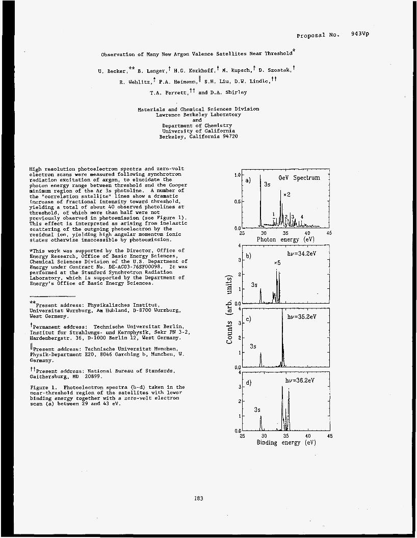
Proposa l No. 943Vp
Observation of Many New Argon Valence Satellites Near Threshold*
** U. Becker, B. Langer,t H.G. Kerkhoff, M. Kupsch,+ D. Szostak,
R. Wehlitz, P.A. Heimann, 11 S.H. Liu, D.W. Lindle, T.A. Ferrett,tt and D.A. Shirley
Materials and Chemical Sciences Division Lawrence Berkeley Laboratory
and Department of Chemistry University of California Berkeley, California 94720
High resolution photoelectron spectra and zero-volt electron scans were measured following synchrotron radiation excitation of argon, to elucidate the photon energy range between threshold and the Cooper minimum region of the Ar 3s photoline. the "correlation satellite" lines show a dramatic increase of fractional intensity toward threshold, yielding a total of about 40 observed photolines at threshold, of which more than half were not previously observed in photoemission (see Figure 1). This effect is interpreted as arising from inelastic scattering of the outgoing photoelectron by the residual ion, yielding high angular momentum ionic states otherwise inaccessible by photoemission.
*This work was supported by the Director, Office of Energy Research, Office of Basic Energy Sciences, Chemical Sciences Division of the U.S. Department of Energy under Contract No. DE-AC03-76SF00098. It was performed at the Stanford Synchrotron Radiation Laboratory, which is supported by the Department of Energy's Office of Basic Energy Sciences.
A number of
-kat
Universitat Wurzburg, Am Hubland, D-8700 Wurzburg, West Germany.
tPermanent address: Technische Universitat Berlin, Institut fur Strahlungs- und Kernphysik, Sekr PN 3-2, Hardenbergstr. 36, D-1000 Berlin 12, West Germany.
IlPresent address : Technische Universitat Munchen, Physik-Department E20, 8046 Garching b, Munchen, W. Germany.
Gaithersburg, MD 20899.
Figure 1. Photoelectron spectra (b-d) taken in the near-threshold region of the satellites with lower binding energy together with a zero-volt electron scan (a) between 29 and 43 eV.
Present address: Physikalisches Institut,
Present address : National Bureau of Standards,
183
OeV Spectrum
25 30 35 40 45 Photon energy (eV)
25 30 35 40 45 Binding energy (eV)

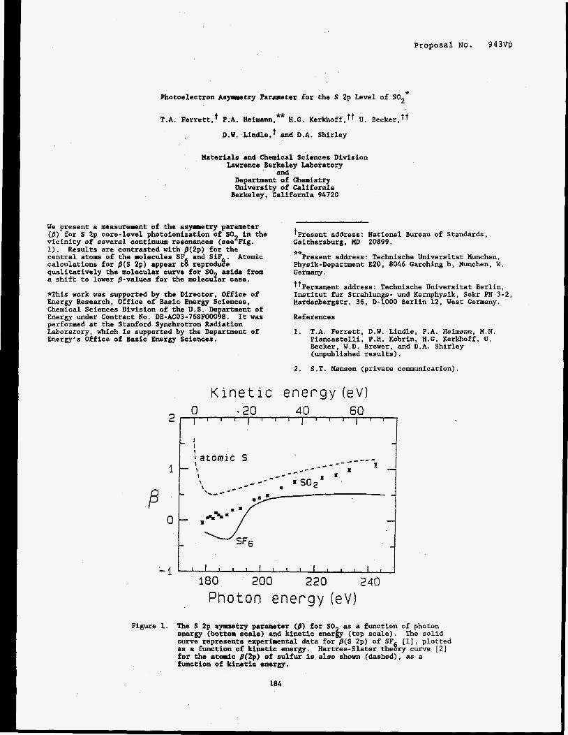
Proposal No. 9 4 3vp
* Photoelectron Asymmetry Parameter for the S 2p Level of SO2
T.A. Ferrett,t P.A. Heimann, ht H.G. Kerkhoff, t t U. Becker,ff
D.W. Lindle,t and D.A. Shirley
Naterials and Chemical Sciences Division Lawrence Berkeley Laboratory
and Department of Chemistry University of California Berkeley, California 94720
We present a measurement of the asymmetry parameter ( f l ) for S 2p core-level photoionization of SO2 in the vicinity of several continuum resonances (see Fig. 1). Results are contrasted with /?(2p) for the central atoms of the molecules SF and SiF . Atomic calculations for /?(S 2p) appear tg reprodde qualitatively the molecular curve for SO aside from a shift to lower /?-values for the molecular case.
*This work was supported by the Director, Office of Energy Research, Office of Basic Energy Sciences, Chemical Sciences Division of the U.S. Department of Energy under Contract No. DE-AC03-76SF00098. It was performed at the Stanford Synchrotron Radiation Laboratory, which is supported by the Department of Energy's Office of Basic Energy Sciences.
K i n e t i c
+Present address: National Bureau of Standards, Gaithersburg, MD 20899. ** Physik-Department 820, 8046 Garching b, Munchen, W. Germany.
tPermanent address: Technische Universitat Berlin, Institut fur Strahlungs- und Kernphysik, Sekr PN 3-2, Hardenbergstr. 36, D-1000 Berlin 12, West Germany.
Present address: Technische Universitat Munchen,
References
1. T.A. Ferrett, D.W. Lindle, P.A. Heimann, M.N. Piancastelli, P.H. Kobrin, H.G. Kerkhoff, U. Becker, W.D. Brewer, and D.A. Shirley (unpublished results).
2. S.T. Manson (private communication).
e n e r g y ( e v )
P
0 - 2 0 4 0 60 2 1 " ' 1 ' " 1 ' " 1 "
- 1 1
0
-1 : 180 200 220 240
Photon e n e r g y (eV)
I
Figure 1. The S 2p symmetry parameter (/?) for SO energy (bottom scale) and kinetic enerb (top scale). curve represents experimental data for p(S 2p) of SF [l], plotted as a function of kinetic energy, for the atomic p(2p) of sulfur is also shown (dashed), as a function of kinetic energy.
as a function of photon The solid
Hartree-Slater the8ry curve [2]
184

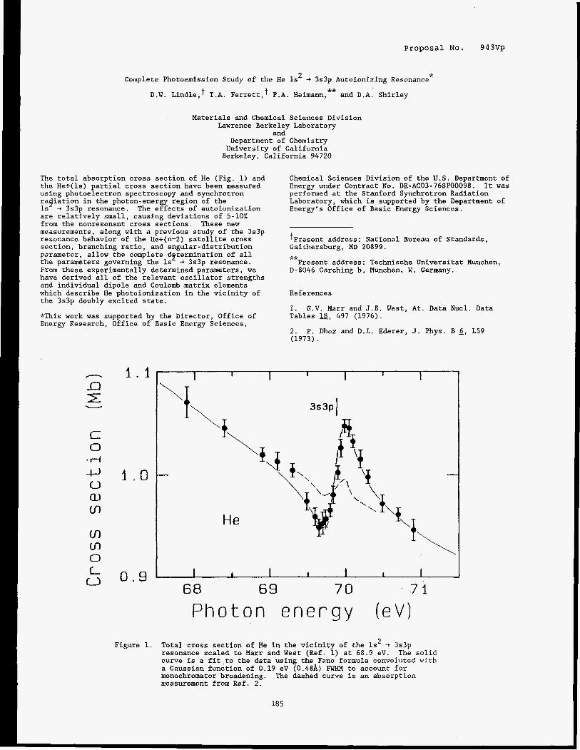
Proposal No. 943Vp
Complete Photoemission Study of the He ls2 + 3s3p Autoionizing Resonance*
D.W. Lindle, T.A. Ferrett,' P.A. Heimann, and D.A. Shirley **
Materials and Chemical Sciences Division Lawrence Berkeley Laboratory
and Department of Chemistry University of California Berkeley, California 94720
The total absorption cross section of He (Fig. 1) and the He+(ls) partial cross section have been measured using photoelectron spectroscopy and synchrotron ragiation in the photon-energy region of the Is -+ 3s3p resonance. The effects of autoionization are relatively small, causing deviations of 5-10% from the nonresonant cross sections. These new measurements, along with a previous study of the 3s3p resonance behavior of the He+(n=2) satellite cross section, branching ratio, and angular-distribution parameter, allow the complete determination of all the parameters governing the Is + 3s3p resonance. From these experimentally determined parameters, we have derived all of the relevant oscillator strengths and individual dipole and Coulomb matrix elements which describe He photoionization in the vicinity of the 3s3p doubly excited state.
*This work was supported by the Director, Office of Energy Research, Office of Basic Energy Sciences,
1.1
1 . 0
0 . 9
Figure 1.
Chemical Sciences Division of the U.S. Department of Energy under Contract No. DE-AC03-76SF00098. It was performed at the Stanford Synchrotron Radiation Laboratory, which is supported by the Department of Energy's Office of Basic Energy Sciences.
Present address : National Bureau of Standards, Gaithersburg, MD 20899. ** Present address: Technische Universitat Munchen,
D-8046 Garching b, Munchen, W. Germany.
References
1. G.V. Marr and J.B. West, At. Data Nucl. Data Tables 18, 497 (1976).
2. P. Dhez and D.L. Ederer, J. Phys. B 6, L59 (1973).
I I I I I I 1
He
I 1 I I I I I
68 69 7 0 7 1
P h o t o n e n e r g y ( e V ) Total cross section of He in the vicinity of the ls2 -t 3s3p resonance scaled to Marr and West (Ref. 1) at 68.9 eV. The solid curve is a fit,to the data using the Fano formula convoluted with a Gaussian function of 0.19 eV (0.48A) FWHM to account for monochromator broadening. The dashed curve is an absorption measurement from Ref. 2.
185

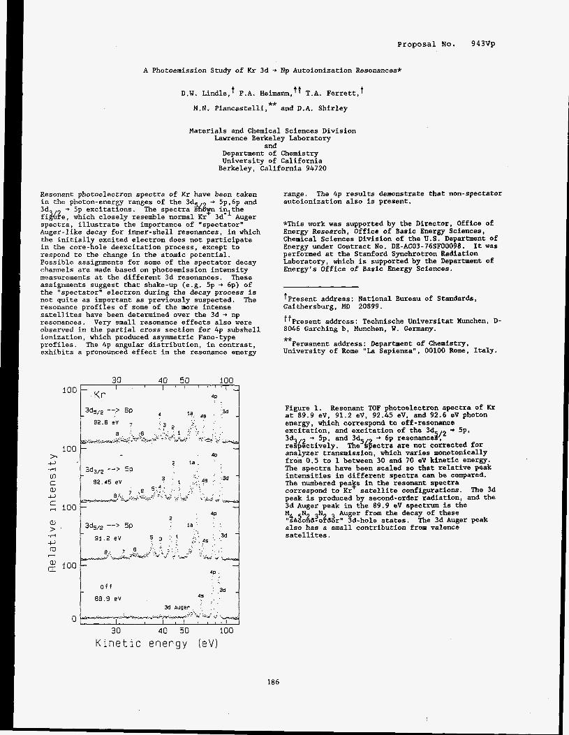
Proposal No. 943Vp
A Photoemission Study of Kr 3d + Np Autoionization Resonances*
D.W. Lindle,t P.A. Heimann,tt T.A. Ferrett,t ** M.N. Piancastelli, and D.A. Shirley
Materials and Chemical Sciences Division Lawrence Berkeley Laboratory
and Department of Chemistry University of California Berkeley, California 94720
Resonant photoelectron spectra of Kr have been taken in the photon-energy ranges of the 3d + 5p,6p and 3d + 5p excitations. The spectra %$ inlthe fi@e, which closely resemble normal Kr 3d- Auger spectra, illustrate the importance of "spectator' Auger-like decay for inner-shell resonances, in which the initially excited electron does not participate in the core-hole deexcitation process, except to respond to the change in the atomic potential. Possible assignments for some of the spectator decay channels are made based on photoemission intensity measurements at the different 3d resonances. These assignments suggest that shake-up (e.g. 5p + 6p) of the "spectator" electron during the decay process is not quite as important as previously suspected. The resonance profiles of some of the more intense satellites have been determined over the 3d + np resonances. observed in the partial cross section for 4p subshell ionization, which produced asymmetric Fano-type profiles. The 4p angular distribution, in contrast, exhibits a pronounced effect in the resonance energy
Very small resonance effects also were
30 40 50 100 I ' I ' " I 1
4P < .
loo , K r I
range. autoionization also is present.
The 4p results demonstrate that non-spectator
*This work was supported by the Director, Office of Energy Research, Office of Basic Energy Sciences, Chemical Sciences Division of the U . S . Department of Energy under Contract No. DE-AC03-76SF00098. It was performed at the Stanford Synchrotron Radiation Laboratory, which is supported by the Department of Energy's Office of Basic Energy Sciences.
. -. :: .3d
4' . . . . .
K i n e t i c e n e r g y (eV)
186
tPresent address: National Bureau of Standards, Gaithersburg, MD 20899.
ttPresent address: Technische Universitat Munchen, D- 8046 Garching b, Munchen, W. Germany. ** Permanent address: Department of Chemistry,
University of Rome "La Sapienza", 00100 Rome, Italy.
Figure 1. at 89.9 eV, 91.2 eV, 92.45 eV, and 92.6 eV photon energy, which correspond to off-resonance excitation, and excitation of the 3d + Sp, 3d3 + 5p, and 3d + 6p resonancea(2 resbectively . analyzer transmission, which varies monotonically from 0.5 to 1 between 30 and 70 eV kinetic energy. The spectra have been scaled so that relative peak intensities in different spectra can be compared. The numbered pealp in the resonant spectra correspond to Kr satellite configurations. The 3d peak is produced by second-order radiation, and the 3d Auger peak in the 89.9 eV spectrum is the :4 5 3 3N4 3 Auger from the decay of these seco d-o der" 3d-hole states. The 3d Auger peak also has a small contribution from valence satellites.
Resonant TOF photoelectron spectra of Kr
The'dgectra are not corrected for

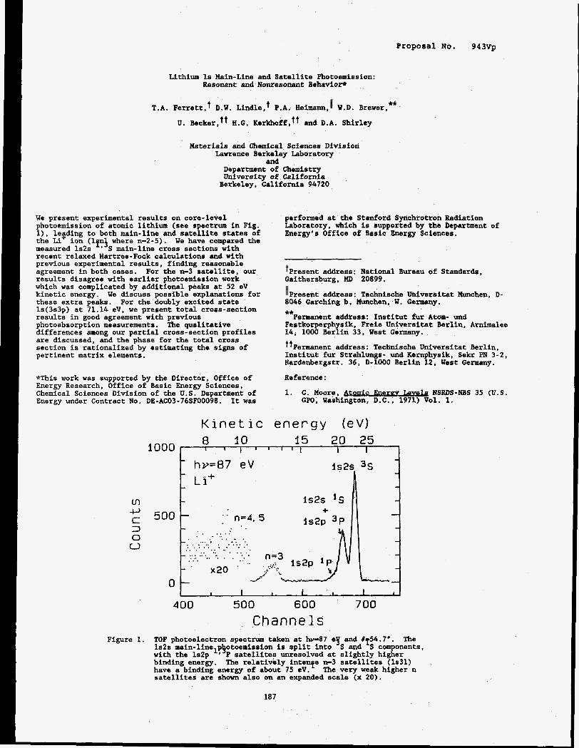
Proposal No. 943Vp
Lithium 1s Main-Line and Satellite Photoemission: Resonant and Nonresonant Behavioe
** T.A. Ferrett, D.W. Lindle, P.A. Heimann,I W.D. Brewer,
U. H.G. KerkhoffStt and D.A. Shirley
Katerials and Chemical Sciences Division Lawrence Berkeley Laboratory
and Department of Chemistry University of California Berkeley, California 94720
We present experimental results on core-level photoemission of atomic lithium (see spectrum in Fig. l), leading to both main-line and satellite states of the Li ion (lfnf where n-2-5). We have compared the measured ls2s ' S main-line cross sections with recent relaxed Hartree-Fock calculations and with previous experimental results, finding reasonable agreement in both cases. results disagree with earlier photoemission work which was complicated by additional peaks at 52 eV kinetic energy. these extra peaks. For the doubly excited state ls(3s3p) at 71.14 eV, we present total cross-section results in good agreement with previous photoabsorption measurements. The qualitative differences among our partial cross-section profiles are discussed, and the phase for the total cross section is rationalized by estimating the signs of pertinent matrix elements.
For the n-3 satellite, our
We discuss possible explanations for
*This work was supported by the Director, Office of Energy Research, Office of Basic Energy Sciences, Chemical Sciences Division of the U.S. Department of Energy under Contract No. DE-AC03-76SF00098. It was
performed at the Stanford Synchrotron Radiation Laboratory, which is supported by the Department of Energy's Office of Basic Energy Sciences.
tPresent address: National Bureau of Standards, Gaithersburg, KD 20899.
((Present address : Technische Uidversitat Munchen, D- 8046 Garching b. Munchen, W. Germany.
Festkorperphysik, Freie Universitat Berlin, Arnimalee 14, 1000 Berlin 33, West Germany.
tPermanent address: Technische Univarsitat Berlin, Institut fur Strahlungs- W d Kernphysik, Sekr PN 3-2, Hardenbergstr. 36, D-1000 Berlin 12, West Germany.
Reference:
1. C. Moore, Levek NSRDS-NBS 35 (U.S.
** Permanent address: Institut fur Atom- und
GPO, Washiagton, D.C., 1971) Vol. 1.
Ln c, c 3
1000
500
0 c,
0
Kinetic energy (eV) 8 10 15 20 25
- hv=87 eV l s2s 3s
- L i +
- l s2s 1s -
1 I I I I I I I I I c
- -
+ - l s 2 p 3 P " n=4. 5 -
- . . - . . . . . . - * . . . _ . - .... . . . - . . . . . . . . . . . . . . . . . . . . . . . . . I . . . . . . . . - . . . . . . . . . . . . . . . . . . n=3 - .. . - . x20 . - - -
400 500 600 700 Channels
Figure 1. TOF photoelectron spectrum taken at hv-87 eY and 8 54.7'. 1s2s main-line p otoemission is split into S and 'S components, with the ls2p '*'P satellites unresolved at slightly higher binding energy. have a binding energy of about 75 eV. The very weak higher n satellites are shown also on an expanded scale (x 20).
The
The relatively intenfe n-3 satellites (1931)
187


proposal No. 943VP
C(2x2)S/Cr(001) Surface and Near-Surface Structure Determined Using Angle-Resolved Photoemission Extended Fine Structure*
L.J. Terminello, X.S. Zhang,' Z.Q. Huang, S. Kim, A.E. Schach von Wittenau, and D.A. Shirley
Materials and Chemical Sciences Division Lawrence Berkeley Laboratory
and Department of Chemistry University of California Berkeley, California 94720
We report the surface and near-surface structure of c(2x2)S/Cr(001) determined from angle-resolved photoemission extended fine structure (ARPEFS). The sulfur Is photoemission partial cross section was measured in the [OOl] and [Oll] emission directions which yielded fine structure amplitudes up to 50-70%. Qualitative results are obtained from Fourier transforms of the ARPEFS revealing geometrical information beyond 10 A path-length difference from the photoemitter. Structural parameters are determined with greater precision by comparing the Fourier filtered data to multiple-scattering spherical-wave (MSSW) calculations giving statistically tractable results (as shown in Figure 1). The directional sensitivity of ARPEFS is demonstrated through the analysis of normal [OOl] and off-normal [Oll] emission direction data. We show that sulfur adsorbs in the four fold hollow site 1.17 A above the first chromium layer and that the first to second layer chromium distance contracts to 1.31 A, 8% less than the 1.44 A bulk value. of the near-surface contribution to ARPEFS showed that deeper layers essentially retain the bulk separation.
*This work was supported by the Director, Office of Energy Research, Office of Basic Energy Sciences, Chemical Sciences Division of the U.S. Department of Energy under Contract No. DE-AC03-76SF00098. It was performed at the Stanford Synchrotron Radiation Laboratory, which is supported by the Department of Energy's Office of Basic Energy Sciences.
Analysis
G eom-etry D eterrnination:
[ O ! I] - 20-1 cui-off
MSSW Best Fit - L I
1
tPermanent address: Department of Physics, Zhej tang University, Hangzhou, Zhejiang, China.
Figure 1. experimental curves. Each experimental curve (solid line) has been Fourier filtered and is compared to the MSSW calculated data (dashed line) that gives an absolute r-factor minimum.
Best-€it MSSW calculations compared to
[ O O l ] - 134 cut-ofY -
4 - 6 5 ! O
Wavevector k (-%-I)
188

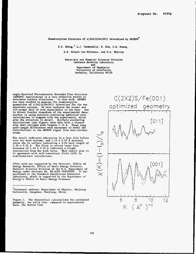
Proposal No. 943Vp
Chemisorption Structure of c(2x2)S/Fe(001) Determined by ARPEFS*
X.S. Zhang,t L.J. Tenninello, S, Kim, 2.4. Huang,
A.E. Schach von Wittenau, and D.A. Shirley
Materials and Chemical Sciences Division Lawrence Berkeley Laboratory
and Department of Chemistry University of California Berkeley, California 94720
Angle-Resolved Photoemission Extended Fine Structure (ARPEFS) spectroscopy is a very effective method to determine surface structures. In this work, ARPEFS has been studied to measure the chemisorption geometries of c(ZxZ)S/Fe(OOl) directions for the two adsorbate systems. We have analyzed the normal and off-normal data of both experiments in two ways. One is direct Fourier transform of the experimental data; another is using multiple-scattering spherical-wave calculations to compare with the experiments, which give the structure in detail. Multiple-scattering calculations (see figure) were done with a cluster size that included path lengths > 15 A. These long path-length differences were necessary to model the contributions to the ARPEFS signal from near-surface atoms.
The result indicates adsorption in a four fold hollow site for both systems, and 1.10 & 0.03 A distance above the Fe surface indicating a S-Fe bond length of 2.30 f 0.01 A . distance of 1.40 -+ 0.02 A indicates a slight contraction from the bulk value. This result also is in agreement with self-consistent field (SCF) Xor scattered-wave calculations.
The first to second layer iron
*This work was supported by the Director, Office of Energy Research, Office of Basic Energy Sciences, Chemical Sciences Division of the U.S. Department of Energy under Contract No. DE-AC03-76SF00098. It was performed at the Stanford Synchrotron Radiation Laboratory, which is supported by the Department of Energy's Office of Basic Energy Sciences.
+Permanent address: Department of Physics, Zhej iang University, Hangzhou, Zhejiang, China.
Figure 1. geometry, the solid line, compared to experimental data, the dashed line.
The theoretical calculations for optimized
0 w
\
I
I I 1c
x
/? 0
H
H W
n
W
189
C(2XZ)S/Fe(OOI) o p t i rn k e d CJ eo rn etry
A
6 8 13 12

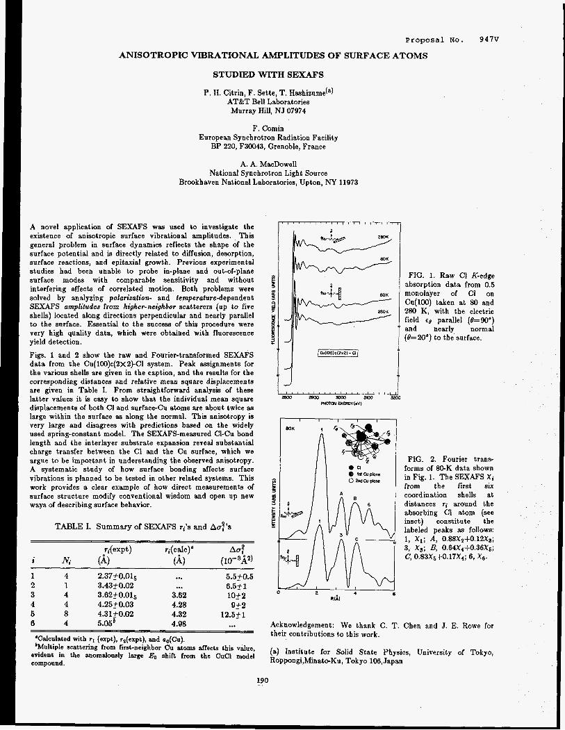
Proposal No. 947V
ANISOTROPIC VIBRATIONAL AMPLITUDES OF SURFACE ATOMS
STUDIED WITH SEXAFS
P. H. Citrin, F. Sette, T. Hashizurne(') AT&T Bell Laboratories Murray Hill, NJ 07974
F. Comin European Synchrotron Radiation Facility
BP 220, F30043, Grenoble, France
A. A. MacDowell National Synchrotron Light Source
Brookhaven National Laboratories, Upton, NY 11973
A novel application of SEXAFS was used to investigate the existence of anisotropic surface vibrational amplitudes. This general problem in surface dynamics reflects the shape of the surface potential and is directly related to diffusion, desorption, surface reactions, and epitaxial growth. Previous experimental studies had been unable to probe in-plane and out-of-plane surface modes with comparable sensitivity and without interfering effects of correlated motion. Both problems were solved by analyzing polarization- and temperature-dependent SEXAFS amplitudes from higher-neighbor scatterers (up to five shells) located along directions perpendicular and nearly parallel to the surface. Essential to the success of this procedure were very high quality data, which were obtained with fluorescence yield detection.
Figs. 1 and 2 show the raw and Fourier-transformed SEXAFS data from the Cu(lOO)c(2~2)-Cl system. Peak assignments for the various shells are given in the caption, and the results for the corresponding distances and relative mean square displacements are given in Table I. From straightforward analysis of these latter values it is easy to show that the individual mean square displacements of both C1 and surface-Cu atoms are about twice as large within the surface as along the normal. This anisotropy is very large and disagrees with predictions based on the widely used spring-constant model. The SEXAFS-measured C1-Cu bond length and the interlayer substrate expansion reveal substantial charge transfer between the C1 and the Cu surface, which we argue to be important in understanding the observed anisotropy. A systematic study of how surface bonding affects surface vibrations is planned to be tested in other related systems. This work provides a clear example of how direct measurements of surface structure modify conventional wisdom and open up new ways of describing surface behavior.
TABLE I. Summary of S E W S ti's and Aaf's
ri(exPt) ri(ca1c)' Aop 2 Ni (4 (A, (10- 3A2)
~-
1 4 2.37f0.015 ... 5.5.0.5 2 1 3.4350.02 ... 6.5f 1
4 4 4.25f0.03 4.28 9f2 5 8 4.31k0.02 4.32 12.551
3 4 3.62k0.015 3.82 1052
6 4 5.05' 4.98 ... ~
'Calculated with r, (expt), rs(expt), and oo(Cu). 'Multiple scattering from fiit-neighbor Cu atoms affects this value,
evident in the anomalously large EO shift from the CuCl model compound.
BOK
FIG. 1. Raw C1 K-edge absorption data from 0.5 monolayer of C1 on Cu(100) taken at 80 and 280 K, with the electric field €6 parallel (6=90°) and nearly normal (0=20°) to the surface.
FIG. 2. Fourier trans- forms of 80-K data shown in Fig. 1. The SEXAFS X i from the first six coordination shells at distances ri around the absorbing C1 atom (see inset) constitute the labeled peaks as follows: 1, X i ; A, 0.88X2+0.12X3; 3, X s ; B, 0.64X4+0.36X~; c, 0.83X~1-0.17X~; 6, 2 6 .
Acknowledgement: We thank C. T. Chen and J. E. Rowe for their contributions to this work.
(a) Institute for Solid State Physics, University of Tokyo, Roppongi,Minat+Ku, Tokyo 106, Japan
190 ..

1
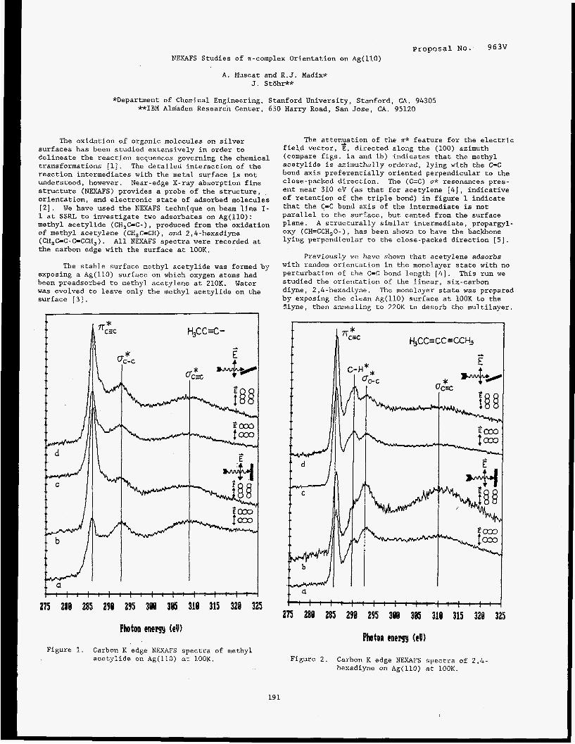
Proposal No. 963V NEXAFS Studies of ?r-complex Orientation on Ag(ll0)
A . Muscat and R.J. Madix* J. Sttlhr**
*Department of Chemical Engineering, Stanford University, Stanford, CA. 94305 **IBM Almaden Research Center, 650 Harry Road, San Jose, CA. 95120
The oxidation of organic molecules on silver surfaces has been studied extensively in order to delineate the reaction sequences governing the chemical transformations [l]. The detailed interaction of the reaction intermediates with the metal surface is not understood, however. Near-edge X-ray absorption fine structure (NEXAFS) provides a probe of the structure, orientation, and electronic state of adsorbed molecules [ 2 ] . We have used the NEXAFS technique on beam line I- 1 at SSRL to investigate two adsorbates on Ag(ll0): methyl acetylide (CH,C=C-), produced from the oxidation of methyl acetylene (CH,C-CH), and 2,4-hexadiyne (CH,C=C-CsCCH,). All NEXAFS spectra were recorded at the carbon edge with the surface at 100K.
The stable surface methyl acetylide was formed by exposing a Ag(ll0) surface on which oxygen atoms had been preadsorbed to methyl acetylene at 210K. Water was evolved to leave only the methyl acetylide on the surface [ 3 ] .
1
Photon enem (e!)
Figure 1. Carbon K edge NEXAFS spectra of methyl acetylide on Ag(ll0) at 100K.
The attenzation of the n* feature for the electric field vector, E, directed along the (100) azimuth (compare figs. la and lb) indicates that the methyl acetylide is azimuthally ordered, lying with the C=C bond axis preferentially oriented perpendicular to the close-packed direction. The (C-C) u* resonances pres- ent near 310 eV (as that for acetylene [ 4 ] , indicative of retention of the triple bond) in figure 1 indicate that the C=C bond axis of the intermediate is not parallel to the surface, but canted from the surface plane. A structurally similar intermediate, propargyl- oxy (CH=CCH,O-), has been shown to have the backbone lying perpendicular to the close-packed direction [5].
Previously we have shown that acetylene adsorbs with random orientation in the monolayer state with no perturbation of the C=C bond length [4]. This run we studied the orientation of the linear, six-carbon diyne, 2,4-hexadiyne. The monolayer state was prepared by exposing the clean Ag(ll0) surface at lOOK to the Biyne, then aniiealing to 220K to desorb the multilayer.
Photon cnelrgy (eV1
Figure 2. Carbon K edge NEXAFS spectra of 2,4- hexadiyne on Ag(ll0) at 100K.
19 1
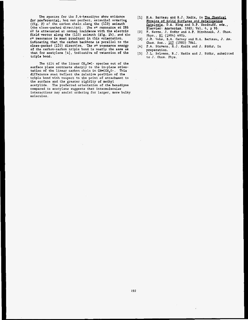
The spectra f o r the 2,4-hexadLyne show evidence for prcfarsatial, but no t perfect, azimuthal ordering ( f ig . 2) of the eeebon okein along the (110) asimutk
sV i r ettonuated a t normal inaidenee with the eleetrio f i e l d veeter along tho (110) azimuth (fig, 4b) , grid the u* rrmnanee is mogt prominant in this orientation, indicating t ha t the carbon backbone f a parallel 6a the cloee-packed (110) direstion. %e Q* remotlance energy of the carbon-carbon triple bond is nearly the same as that for acetylene [4], indicative of ratantion of the triple bond.
('GhQ CbO%@=pa'2kQd diPCi'ZtfOn). 'PhQ If* PeSORi%R;le@ 286
The tilt of the liqear CH,C=C- species out of the surface plane contrasts sharply to the in-plane orien- tation of the linear carbon chain in CH=CCH,O-. difference must reflect the relative position of the triple bond with respect to the point of attachment to the surface and the greater rigidity of methyl acetylide. The preferred orientation of the hexadiyne compared to acetylene suggests that intermolecular interactions may assist ordering for larger, more bulky molecules.
This
ellys, I 2 (1984) 4905,
Shem. s e e . , w (1985) 9841. J . M . Vaka, B , A . Carney and M.A. Barteau, J , Am.
P . A . Stevens, R.J. Madlx and J. Stbhr, in prepsratian. J.L. Solomon, R . J . Madix and J. Stbhr, submitced to J. Chem. Phys.
192
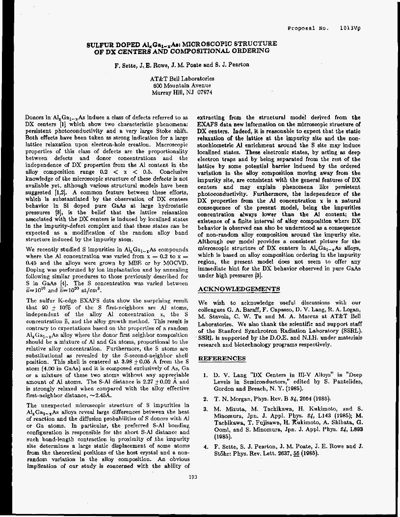
Proposal No. 1013Vg
SULFUR DOPED Al,Ga~,,Asc MICROSCOPIC STRUCTURE OF DX CENTERS AND COMPOSYTIONAL ORDERING
F. Sette, J. E. Rowe, J. M. Poate and S. J. Pearton
AT&T Bell Laboratories 000 Mountain Avenue
Murray Hill, NJ 07974
Donors in Al,Gal-,As induce a class of defects referred to as DX centers [l] which show two characteristic phenomena: persistent photoconductivity and a very large Stoke shift. Both effects have been taken as strong indication for a large lattice relaxation upon electron-hole creation. Macroscopic properties of this class of defects are the proportionality between defects and donor concentrations and the independence of DX properties from the A1 content in the alloy composition range 0.2 < x < 0.5. Conclusive knowledge of the microscopic structure of these defects is not available yet, although various structural models have been suggested [1,2]. A common feature between these efforts, which is substantiated by the observation of DX centers behavior in Si doped pure GaAs at large hydrostatic pressures [3], is the belief that the lattice relaxation associated with the DX centers is induced by localized states in the impurity-defect complex and that these states can be expected as a modification of the random alloy band structure induced by the impurity atom.
We recently studied S impurities in Al,Gal-,As compounds where the AI concentration was varied from x = 0.2 to x = 0.45 and the alloys were grown by MBE or by MOCVD. Doping was performed by ion implantation and by annealing following similar proedures to those previously described for S in GaAs [4]. The S concentration was varied between ii= 10'' and ii= IO2' at/cm3.
The sulfur K-edge EXAFS data show the surprising result that 90 _+ 10% of the S first-neighbors are A1 atoms, independent of the alloy Al concentration x, the S concentration Z, and the alloy growth method. This result is contrary to expectations based on the properties of a random A1,Gal-,As alloy where the donor first neighbor composition should be a mixture of Al and Ga atoms, proportional to the relative alloy concentration. Furthermore, the S atoms are substitutional as revealed by the S-second-neighbor shell position. This shell is centered at 3.98 f 0.05 A from the S atom (4.00 in GaAs) and it is composed exclusively of As, Ga or a mixture of these two atoms without any apprefiable amount of AI atoms. The S-Al distance is 2.27 fO.02 A and is strongly relaxed when compared with the alloy effective first-neighbor distance, - 2.45A.
The unexpected microscopic structure of S impurities in A1,Gal-,As alloys reveal large differences between the heat of reaction and the diffusion probabilities of S donors with A1 or Ga atoms. In particular, the preferred S-A1 bonding configuration is responsible for the short S-AI distance and such bond-length contraction in proximity of .the impurity site determines a large static displacement of some atoms from the theoretical positions of the host crystal and a non- random variation in the alloy composition. An obvious implication of our study is concerned with the ability of
extracting from the structural model derived from the EXAFS data new information on the microscopic structure of DX centers. Indeed, it is reasonable to expect that the static relaxation of the lattice at the impurity site and the non- stochiometric Al enrichment around the S site may induce localized states. These electronic states, by acting as deep electron traps and by being separated from the rest of the lattice by some potential barrier induced by the ordered variation in the alloy composition moving away from the impurity site, are consistent with the general features of DX centers and may explain phenomena like persistent photoconductivity. Furthermore, the independence of the DX properties from the Al concentration x is a natural consequence of the present model, being the impurities concentration always lower than the AI content; the existence of a finite interval of alloy compwition where DX behavior is observed can also be understood as a consequence of non-random alloy composition around the impurity site. Although our model provides a consistent picture for the microscopic structure of DX centers in Al,Gal-,As alloys, which is based on alloy composition ordering in the impurity region, the present model does not seem to offer any immediate hint for the DX behavior observed in pure GaAs under high pressures [3].
ACKNOWLEDGEMENTS
We wish to acknowledge useful discussions with our colleagues G. A. Baraff, F. Capasso, D. V. Lang, R. A. Logan, M. Stavola, .C. W. Tu and M. A. Marcus at AT&T Bell Laboratories. We also thank the scientific and support staff of the Stanford Synchrotron Radiation Laboratory (SSRL). SSRL is supported by the D.O.E. and N.I.H. under materials research and biotechnology programs respectively.
REFERENCES
1. D. V. Lang "DX Centers in IIP-V Alloys" in "Deep Levels in Semiconductors," edited by S. Pantelides, Gordon and Breach, N. Y. (1985).
2. T. N. Morgan, Phys. Rev. B 34,2664 (1986).
3. M. Mizuta, M. Tachikawa, H. Kukimoto, and S. Minomura, Jpn. J. Appl. Phys. 24, L143 (1985); M. Tachikawa, T. Fujisawa, H. Kukimoto, A. Shibata, G. Oomi, and S. Minomura, Jpn. J. Appl. Phys. 24, L893 (1985).
F. Sette, S. J. Pearton, J. M. Poate, J. E. Rowe and J. Stijhr: Phys. Rev. Lett. 2637 ,s (1986).
4.
193

Proposal No. 1013Vp
STRUCTURE OF P IMPURITIES IN Ga&
F. Sette, J. E. Rowe, J. M. Poate and S. J. Pearton
AT&T Bell Laboratories 600 Mountain Avenue
Murray Hill, NJ 07974
We measured the K-edge photoabsorption spectra of impurities in GaAs at 10"- lo2' P at/cm3 concentrations. Samples were prepared by ion implantation and subsequent annealing at 900" C. Channeling measurements showed no noticeable residual damage. In Fig. 1 we report the Fourier transform magnitude of the EXAFS data for phosphorus. This function is directly related to the radial distribution function and shows the' first, second and third neighbor shells of the P atoms. The second and third shells results reveal that the P atoms are substitutional on the As sublattice. The radial di!tances of these two shells, 4.00_+0.02 d; and 4.689.03A respectively, agree extrzmely well Yith the analogous values of the GaAs lattice, 4.00A and 4.68A. However, the bond-distance betwe$n the P atom an9 its 4 Ga first neighbors is R=2.?8+0.01A, which is 0.07A shorter than in GaAs, R12.45A. This contracted first neighbor distance and the unrelaxed higher shells reveal a large local contraction of the four first-shell Ga atoms toward the P site with distortion of the 2nd shell GaAs bond angles and lengths. This homogenous breathing relaxation of the Ga atoms is due to a trigonal distortion taking place on such Ga sites. The shorter P-Ga bond is consistent with tetrahedrally coordinated fir$t-shell Ga atoms and with the P-Ga bond distance of 2.36A in bulk GaP; although, it is inter:sting to observe that the measured distance is still 0.02A longer than in GaP. This is due to a balance of forces on each first-shell Ga atom provided by the remaining three seco!d-shell As-Ga bonds which are stretched to a value of 2.47A as required by the first and second shell distances given above.
BO -
60 - - . c C . - "401
20-
c
Our results show that local chemical interactions on an isolated isoelectronic impurity in the zincblende lattice are dominant with respect to the crystal structure geometrical constraints. This property arises from weak bond-bending forces which, with small variations of bonding angles, allow large variations in the bond distances of those Ga atoms bound to both As and P atoms. Specifically, the Ga atoqs bound to As and P have distances of 2.47A and 2.3&4, respectively, with only 1% variation in the bond angles.
Summarizing results already pointed out [1,2] for AXB1-,C III-V alloys, our results show that even in the limit x+O, the A-C bond distance is much closer to the bond distance in the A-C compound then to the average bond distance, which in this concentration limit is the B-C bond distance. However, we also experimentally demonstrate that the lattice relaxation is absorbed within the first two shells of the isoelectronic impurity and the average bond distance of the displaced Ga atoms is -
d = (~RG, h+ Rca-p) /4=2.4& (1)
which is identical to the Ga-As bond in GaAs. The previous relation (1) also confirms the theoretical treatment of local lattice distortion for isoelectronic impurities presented by M. Scheffler (21.
ACKNOWLEDGEMENTS
We wish to acknowledge useful discussions with our colleagues G. A. Baraff, F. Capasso, D. V. Lang, R. A. Logan, M. Stavola, C. W. Tu and M. A. Marcus at A T k T Bell Laboratories. We also thank the scientific and support staff of the Stanford Synchrotron Radiation Laboratory (SSRL). SSRL is supported by the D . O E and N.I.H. under materials research and biotechnology programs respectively.
REFERENCES
Rp * 4.00& 1. J. C. Mikkelsen and J. C. Boyce, Phys. Rev. Lett., 49,
1412 (1982).
2. M. Scheffler, Proceedings of the 2nd Int'l. Conf. on Shallow Impurity Centers (Trieste, July 1986, ed. Baldereschi, Elsevier) to be published and private comm.
Fig. 1 Fourier transform magnitude IF(r)l of the EXAFS signal obtained from a P doped GaAs sample ( I i 3 5 ~ 1 0 ~ ~ at/cm3). The sample was doped by ion implantation and annealed at 900°C. The distances of the first three P atomic shells are indicated after phase-shift corrections.
194

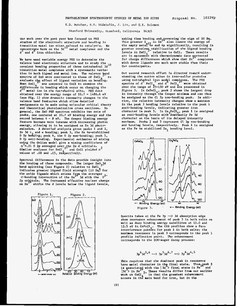
PHOTOELECTRON SPECTROSCOPIC STUDIES OF METAL ION SITES
K.D. Butcher, S.V. Didziulis, J. Lin, and E.I. Solomon
Stanford University, Stanford, California 94305
proposal NO. 1022vp
Our work over the past year has focused on PES studies of the electronic structure and bonding in transition metal ion sites16elated to catalysis. We c ncentr te here on the 3d metal complexes and the d and d iron chlorides.
We have used variable energy PES to determine the valence band electronic structure and to study the covalent bonding properties of three tetrahedral 3d transition metal complexes with a systematic varia- tion in both ligand and metal ion. spectra of ZnO were contrasted to those of ZnCl to evaluate t$g effect of ligand variation on bondfng. Next ZnC14 was compared to CuCl to examine the
erences in bonding which occur on changing the metal ion in the tetrahedral site. PES data
obtained over the energy range of 16.7 - 1486‘6 eV (see Fig. 1) show dramatic intensity changes in the valence band feataures which allow detailed assignments to be made using molecular orbital theory and theoretical photoionization cross sections. In general, the ZnO PES spectrum exhibits two sets of peaks, one centered at 10.7 eV binding energy and the second between 4 - 8 eV. The deeper binding energy feature becomes more intense with increasing photon energy, allowing it to be assigned as Zn 3d photo- emission. A detailed analysis gives peaks 1 and 2, Zn 3d t and e bonding; peak 3, the Zn 4s-stabilized 0 2p bonding; peak 4, the 0 2p non-bonding; peak 5, 0 2p anti-bonding. Experimental estimates of mixing us$ng the Gelius model give a mixing coefficient of
Similar analyses for ZnC14’- and CuCl yielded a values of .08 and .15, respectively.
Spectral differences in the data provide insight into the bonding of these compounds. band splitting (see Figure 2) relative to ZnCl indicates greater ligand field strength (10 Dqj for
2- the oxide ligands which arises fs$m the stronger u-bonding interaction of the Zn 3d with the 0 2p or$$tals. The increased effective nuclear charge on Zn shifts the d levels below the ligand levels,
9 8
10
The valence &nd
2
2 a =.21 0 2p averaged over the Zn d orbitals.
The larger Zn022d
Figure 1. 7
making them bonding and2$eversing’the sign of 10 Dq. This greater Z on Zn also lowers the energy of the empty metaPffs and 4p significantly, resulting in greater covalens-stabilization of the ligand bonding levels in ZnC14 relative to CuCl. These results are in agreement with thermodynamic data 52rrected for charge differences which show that Zn with donor ligands are much more stable than their Cu+ counterparts.
Our second research effort is directed toward under- standing the active sites in iron-sulfur proteins using tetrah dral iron mod 1 compiexes. The PES spectra of d3 FeCl and d FeC14 were obtained over the range of $5-150 eV and are presented in Figure 3. In CsFeC14, peak 2 shows the largest drop in intensity through the Cooper minimum and can thus be assigned as the C1 3p non-bonding peak. In addi- tion, the relative intensity changes show a maximum in the peak 3 bonding levels relative t o the peak 1 anti-bonding levels, indicating greater iron character in peak 3. as anti-bonding levels with dominantly Fe 3d character on the basis of its delayed intensity maximum. and bonding levels, respectively. Peak 4 is assigned as the Fe 4s stabilized 2al bonding level.
complexes
%
In Cs3FeC15, peak 1 is assigned
Peaks 2 and 3 represent C1 3p non-bonding
2 1
Figure 2. sr-c-L-c--e-e--r
- t Bindicg Energy (ev) Figure 3 .
Spectra taken at the Fe 3p --> 3d absorption edge show resonance enhancement of peak 3 in both salts as well as deep binding energy satellites at 11.0 and 12.5 eV in CsFeC14. interference pattern for peak 1 in both salts; the maximum resonance in peak 3 corresponds to the peak 1 profile inflection point. The enhancement corresponds to the SCK-auger decay process:
The CIS profiles show a Fano
3p63d5’6 --> 3p53dbB7 --> 3p 6 3d 4,5
This requires that the dominant peak in resonance have metal character in the final state. is gssociatfg with the 13d > final state in Fe 13d > in Fe work on CuC14 occurs in the main band for iron, but in the
Thus3qeak 3
2- These results differ from our earlier and
in that the greatest enhancement
195
satellite for copper. The deep binding energy Fe satellite shows very little intensity out of resonance, but it does show some enhancement at the absorption edge. The lack of off resonance intensity in the deep binding energy satellite indicates little relax tion upon ionization, again in contrast to our CuCl results. Spin-restricted and unrestricted SCF-h-SW calculations for both initial and final states have been performed using spheres adjusted to reproduce ligand to metal charge transfer energies. The calculations support these experimental results, indicating that the anti-bonding levels have mostly ligand character, and showing that there is limited relaxation (Q33X) upon ionization for all levels. This has important implications with respect to bonding interactions at iron active sites.
REFERENCES
9-
1. S.V. Didziulis, S.L. Cohen, K.D. Butcher, E.I. Solomon, Inorg. Chem., in press.
2. S.V. Didziulis, S.L. Cohen, A.A. Gewirth, E.I. Solomon, J. Am. Chem. SOC. 9, 250 (1988).
196

Proposal NO. 1026V
HIGH-PHOTON FLUX PHOTOEMISSION STUDIES IN NARROW BAND MATERIALS
(l1J.W. Allen, ('s2)J.-S. Kang, (3)Z.-X. Shen, (3)J. J. Yeh, (4)W. Ellis, (2)M.B. Maple, (2)Y.D. Dalichaouch, (')B.-W. Lee, (5)M.S. Torikachvili, (3) J.-Z. Sun, (3)I. Lindau
(l) Randall Laboratory, University of Michigan, Ann Arbor, ML 48109-1120
(2)Department of Physics, University of California at San Diego, La Jolla, CA. 92093
(3)Stanford Electronic Laboratory, Stanford University, Stanford, CA. 94305
C4)Los Alamos National Laboratory, Los Alamos, NM. 87545
(SPhysics Dept., San Diego State University, San Diego, CA.
Electronic structure of copper oxlde superconductors
An understanding of the origin of the high superconducting transition temperatures Tc of the superconducting copper oxides will require an understanding of their electronic structure. Coulomb interaction effects are usually described by a Hubbard or Anderson model, and it is important to determine at least roughly the magnitude of the model parameters. Therefore we have measured and analyzed valence band resonant photoemission (RESPES) and core level x- ray photoemission spectroscopy (XPS) data using the Anderson Hamiltonian in the impurity cluster approximation.'
All the data in Figs. 1-4 were taken at room temperature and the h resolution of the data is 0.45 eV , unless otherwise indicated.
PHOTON ENERGY 75 EV
/I&
\\ I
/
I \ - y1 Ba2cu307 ( BAND THEORY )
-- I I
1 -15 -10 -5 0 5 ENERGY RELATIVE TO FERMI LEVEL (ev)
Fig. 1
197
.--- Fig. 1 compares the experimental valence band spectra to band the- oretical spectra2 with the appropriate photoemission cross-sections taken approximately into account using tabulated atomic cross- sections3. The photon energy used maximizes the Cu 3d emission via a cross-section resonance at the Cu 3p edge. Effects due to sec- ond order light have been removed from the YBazCu3O74 spectra.
There are obviod differences between the experimental spectra and the theory. For the large peaks at low binding energy, for all the samples, (a) the experimental centroids are shifted to higher energy than in the theory, (b) the shapes and the widths of the spectra from theory and experiment are quite different, and (c) the experimental density of states at the Fermi energy EF is much smaller than in the theory. Another difference is the f e a m at -12.5 eV. discussed next.
Fig. 2 shows the valence band EDC's for photon energies in the Cu 3d resonant photoemission (RESPES) region for Lal.gSr.2CuO4. The inset shows the detail of the Fermi level region at a photon energy of 70 eV. Peaks A,B and D display resonant behavior$ implying that they involve Cu 3d states. In our cluster model, peak D gives the Cu ds energy, pushed far from the main band by the d-d Coulomb interaction U. Peak C is attributed to oxygen 2p states, consistent with 0 2p RESPES data reported by other workers and confirmed recently by us.
I
-20 -15 -1 0 -5 EF ENERGY RELATIVE TO FERMI LEVEL (ev)
-. Fig. 2
t

I I I ~
72 76 80 84
PHOTON ENERGY (eV)
Fig. 3
Fig. 3 shows constant initial state (CIS) spectra in the Cu 3d RESPES region for various binding energies Ei of the valence band spectra of Fig. 2. The spectra are the difference between raw data and a smooth spline fit to the data. The main band emission shows a resonance dip and the satellie emission shows a resonance peak as in CuO and CuzO. The vertical scale is aribaary but the same for all the curves. The CIS taken at h4.5 eV just above EF also shows resonance behavior, which means that models for the superconductivity should include the Cu 3d character at EF. Fig. 4 shows CIS spectra for photon energies at the La 4d edge for various binding energies Ei of the valence band spectra of Fig. 2. For YBazCu3074, CIS spectra around the Ba 4d edge for various Ei are similar to those shown in Fig. 4.
We also measured the Cu 2p XPS spectra for our superconducting samples and for Cu and CuO. These show spin-orbit-split 2~112 and 2 p 3 ~ components, each of which has a main line and a satellite. The separation and intensity ratios for the two 2p3n lines were analyzed using the Anderson model. The separation 8.7 eV is approximately equal to Ucd, the Coulomb interaction between the Cu 2p core hole and the 3d electrons. The chemical shift from Cu to CuO agrees with that of a previous measurement and it is significant that the chemical
shifts of the superconducting samples are intermediate, indicating that the oxidation of Cu in these samples is less than that of CuO, which provides evidence that Cu3+ does not occur. This conclusion is consistent with the large energy of the Cu d8 state observed in the valence band spectra.
From an analysis of our photoemission data, using the Anderson Hamiltonian, we obtained experimental values of the parameters of the model, the copper-oxygen charge transfer energy A 4 4 eV, the d-d Coulomb interaction UN6 eV, and the ligand-3d hybridization T-2.4 eV. Using the parameters, we evaluated the linear Cu-0-Cu superexchange interaction J and found it is dominated by the charge fluctuations. For A<<U and T>>A the charge transfer degrees of freedom and the lattice aspects of the Anderson lattice Hamiltonian should not be neglected in constructing models for the high Tc superconductivity.
Recently we have also obtained data on the states above EF by Bremstrahllung isochromat spectroscopy (BIS), which provides a much more direct measure of the charge transfer energy A. From the new data, it is apparent that the A-values should be considerably larger ( > 2 eV) than those we deduced using photoemission data alone, where A is determined mostly by fitting relative lime intensities, rather than from line positions. We are now repeating the analysis to take this new data into account.
-1 8.5
too 110 120 130 PHOTON ENERGY (eV)
Fig. 4
198

Electronic structure of Y1-xPrxBa2C~307-6
The superconductivity of Y1-xPrxBa2C~307~ is quenched as x is increased4. In order to correlate changes in the electronic structure and superconductivity properties of these alloys, we have measured their RESPES, core level XPS and BIS spectra.
Fig. 5 compares the Pr 4f specta obtained by RESPES for several values of x. The spectra are scaled to have the same magnitude of the main peak. Each is obtained by suitably scaling5 and subtracting the Fano minimum spectrum (photon energy hv=ll5 eV) from that of the Fano maximum spectrum (hv=124 eV), both taken with resolution 0.35 eV. Fig. 6 shows CIS spectra for E i 4 . 8 eV for four values of x.The CIS spectra for other Ei values and x-values are also very similar, suggesting no valence change as a function of x.
From the measured BIS spectra (not shown here) the Pr 4f spectral weight above EF was extracted by subtracting the spectrum for x=O from that for each x, using the Ba 4P-i4f1 peak for normalization. There are 4f peaks at 2eV and 8eV abover EF. As x increases, the relative intensity of the 2 eV peak decreases. Interpretation of the Cu 2p core level XPS spectra (not shown here) is complicated by overlapping Pr 3d emission just under the Cu 2p main peaks. Analysis is underway to correct for this, and to find a unified interpretation of all the data.
-10 -8 -6 -4 -2 0 2 ENERGY RELATIVE TO EF( eV)
Fig. 5
L I I I I I 1 . 1 I I I I I I I I , I 110 120 130 140
PHOTON ENERGY (eV)
Fig. 6 i Spectral Weight of Y1,UxPd3.
Cerium and uranium materials have in common that they display a diversity of ground state properties6. The f-electrons of these materials are generally regarded as being responsible. For Ce, the major features of the 4f spectra can be understood using the impurity Anderson Hamiltonian, which also provides a description of ground state properties not explicitly related to translational symmetry7. The U 5f spectra usually have very much weight around EF, with a rather bandlike appearance, contrasting sharply to the Ce-4f spectra. There exists no unified picture for the U 5f spectral weight.
-10 -8 -6 -4 -2 0 2 ENERGY RELATIVE TO E,(eV)
Fig. 7
199
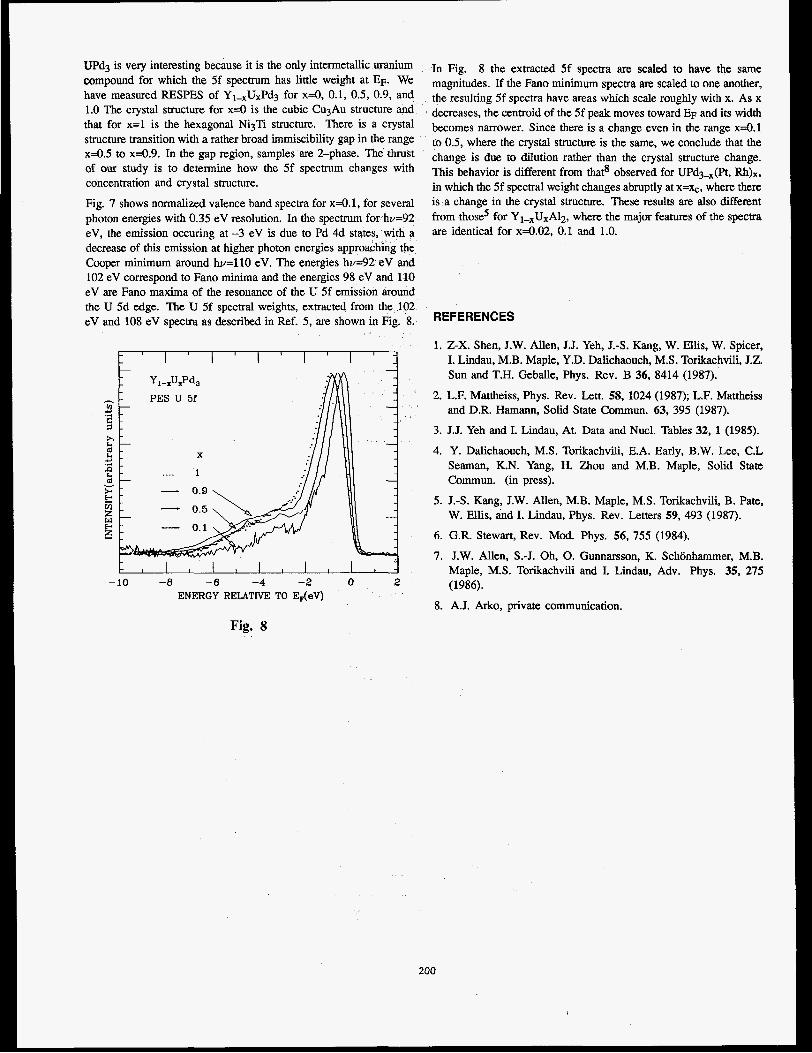
UPd3 is very interesting because it is the only intermetallic uranium compound for which the 5f spectrum has little weight at EF. We have measured RESPES of Y1-xUxPd3 for x 4 , 0.1, 0.5, 0.9, and 1.0 The crystal structure for x 4 is the cubic CqAu structure and that for x=l is the hexagonal Ni3Ti structure. There is a crystal structure transition with a rather broad immiscibility gap in the range x a . 5 to x4.9. In the gap region, samples are 2-phase. The thrust of our study is to determine how the 5f spectrum changes with concentration and crystal structure.
Fig. 7 shows normalized valence band spectra for x4.1, for several photon energies with 0.35 eV resolution. In the spectrum for hv=92 eV, the emission occuring at -3 eV is due to Pd 4d states, with a decrease of this emission at higher photon energies approaching the Cooper minimum around hv=llO eV. The energies hv=92 eV and 102 eV correspond to Fano minima and the energies 98 eV and 110 eV are Fano maxima of the resonance of the U 5f emission around the U 5d edge. The U 5f spectral weights, extracted from the 102 eV and 108 eV spectra as described in Ref. 5, are shown in Fig. 8.
-10 -8 -6 -4 -2 0 2 ENERGY RELATIVE TO E,(eV)
In Fig. 8 the extracted 5f spectra are scaled to have the same magnitudes. If the Fano minimum spectra are scaled to one another, the resulting 5f spectra have areas which scale roughly with x. As x
decreases, the centroid of the 5f peak moves toward EF and its width becomes narrower. Since there is a change even in the range x 4 . 1 to 0.5, where the crystal structm is the same, we conclude that the change is due to dilution rather than the crystal structure change. This behavior is different from thats observed for UPd3,(Pt, Rh)x, in which the 5f spectral weight changes abruptly at X=Q, where there is a change in the crystal structure. These results are also different from those5 for Y I,U,A~~, where the major features of the spectra are identical for x4.02, 0.1 and 1.0.
REFERENCES
1. Z-X. Shen, J.W. Allen, J.J. Yeh, J.-S. Kang, W. Ellis, W. Spicer, I. Lindau, M.B. Maple, Y.D. Dalichaouch, M.S. Torikachvili, J.Z. Sun and T.H. Geballe, Phys. Rev. B 36, 8414 (1987).
2. L.F. Mattheiss, Phys. Rev. Lett. 58, 1024 (1987); L.F. Mattheiss and D.R. Hamann, Solid State Commun. 63, 395 (1987).
3. J.J. Yeh and I. Lindau, At. Data and Nucl. Tables 32, 1 (1985).
4. Y. Dalichaouch, M.S. Torikachvili, E.A. Early, B.W. Lee, C.L Seaman, K.N. Yang, H. Zhou and M.B. Maple, Solid State Commun. (in press).
5. J.-S. Kang, J.W. Allen, M.B. Maple, M.S. Torikachvili, B. Pate, W. Ellis, and I. Lindau, Phys. Rev. Letters 59, 493 (1987).
6. G.R. Stewart, Rev. Mod. Phys. 56, 755 (1984).
7. J.W. Allen, S.-J. Oh, 0. Gunnarsson, K. Schonhammer, M.B. Maple, M.S. Torikachvili and I. Lindau, Adv. Phys. 35, 275 (1986).
8. A.J. Arko, private communication.
Fig. 8
200
,

The Orientation of Ring Compounds on Ag(ll0) by NEXAFS Proposa1 No. lo8OVp
J.L. Solomon and R.J. Madix* J. StOhr**
*Department of Chemical Engineering, Stanford University, Stanford, CA 94305 **IBM Almaden Research Center, 650 Harry Road, San Jose, CA 95120
Near-edge x-ray absorption fine structure (NEXAFS) is a powerful technique to determine orientation and bonding information of adsorbed molecules 111. We have used NFXAFS to determine the orientation of benzene (C,H,), aniline (C,H,NH,), phenol (C,H,OH), furan (C,H,O), 2,5-dihyrofuran (C,H,O), and maleic anhydride (C,H,O,) on Ag(ll0). Multilayers were formed by dosing each compound at 100 K on the clean Ag(ll0) surface. Monolayer coverages were then obtained by annealing the crystal to desorb the multilayers. spectra were taken on beam line 1-1 at Stanford Synch- rotron Radiation Laboratory with the surface held at 100 K. Final NEXAFS spectra were obtained by dividing the adsorbate covered spectra by spectra taken of the clean surface.
Carbon K-edge
215 28fl 285 298 2% 388 385 318 315 328 325
Photon enem (eU)
Figure 1 contains spectra for benzene, phenol, and aniline. A clear polar dependance is observed for all three of the molecules as evidenced by dominance of the x* resonance at glancing incidence ( 9 = 20"), and the dominance of the u* resonances at normal incidence ( 0 - 9 0 ' ) . Clear similarities in the spectxa are also appar- ent as one would expect irom the similar structure of the molecules (i.e. each contains a six member aromatic ring). The K* resonance at glancing incidence is broadened for aniline and split for phenol, as expected due to the differing C(ls) binding energies for the ring carbons bound to nitrogen and oxygen, respec- tively.
Monolayer spectra for furan and 2,5-dihydrofuran (DHF) are shown in Figure 2. Again a clear polar dependance i s observed. resonances indicate that both of these five member rings lie parallel to the plane of the surface.
The m* resonance and the o*
Furan i ?*
20 1
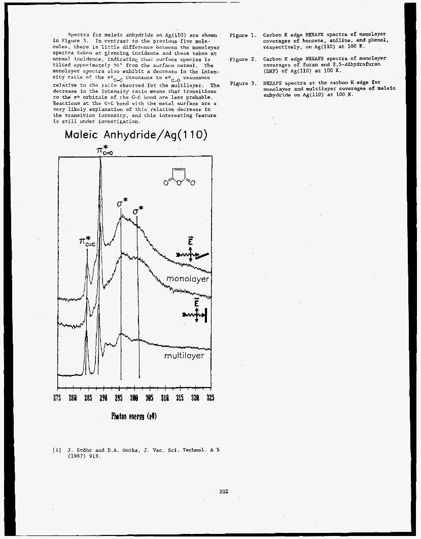
Spectra for maleic anhydride on Ag(ll0) are shown in Figure 3 . cules, there is little difference between the monolayer spectra taken at glancing incidence and those taken at normal incidence, indicating that surface species is tilted appoximately 54" from the surface normal. The monolayer spectra also exhibit a decrease in the inten- sity ratio of the I I * ~ - ~ resonance to ? I * ~ - ~ resonance relative to the ratio observed for the multilayer. The decrease in the intensity ratio means that transitions to the A* orbitals of the C-C bond are less probable. Reactions at the C=C bond with the metal surface are a very likely explanation of this relative decrease in the transition intensity, and this interesting feature is still under investigation.
In contrast to the previous five mole-
Maleic Anhydride/Ag( 1 10)
275 288 285 294 295 388 385 314 315 328 325
Photon energy (eU)
Figure 1. Carbon K edge NEXAFS spectra of monolayer coverages of benzene, aniline, and phenol, respectively, on Ag(ll0) at 100 K.
-Figure 2. Carbon K edge NEXAFS spectra of monolayer coverages of furan and 2,5-dihydrofuran (DHF) o f Ag(ll0) at 100 K.
NEXAFS spectra at the carbon K edge for monolayer and multilayer coverages of maleic anhydride o n Ag(ll0) at 100 K.
Figure 3 .
[l] J. Stt)hr and D.A. Outka, J. Vac. Sci. Technol. A 5 ' (1987) 919.
202
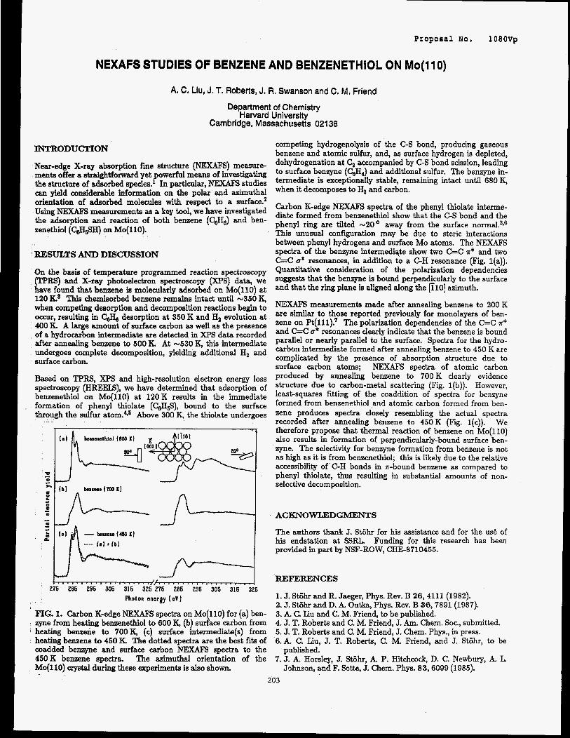
Proposal NO. 1080vp
NEXAFS STUDIES OF BENZENE AND BENZENETHIOL ON Mo(1 I O )
A. C. Llu, J. T. Roberts, J. R. Swanson and C. M. Friend
Department of Chemistry Harvard University
Cambridge, Massachusetts 021 38
INTRODUCTION
Near-edge X-ray absorption fine structure (NEXAFS) measure- ments offer a straightforward yet powerful means of investigating the structure of adsorbed species.' In particular, NEXAFS studies can yield considerable information on the polar and azimuthal orientation of adsorbed molecules with respect to a surface.' Using NEXAFS measurements as a key tool, we have investigated the adsorption and reaction of both benzene (C6&) and ben- zenethiol (C&SH) on Mo(ll0).
' EESULTS AND DISCUSSION
On the basis of temperature programmed reaction spectroscopy (TPRS) and X-ray photoelectron spectroscopy (XPS) data, we have found that benzene is molecularly adsorbed on Mo(ll0) at 120 K3 This chemisorbed benzene remains intact until ~ 3 5 0 K, when competing desorption and decomposition reactions begin to occur, resulting in CSH, desorption at 350 K and H2 evolution at 400 K. A large amount of surface carbon as well as the presence of a hydrocarbon intermediate are detected in XPS data recorded after annealing benzene to 500 K. At ~ 5 3 0 K, thii intermediate undergoes complete decomposition, yielding additional H, and surface carbon.
Based on TPRS, XPS and high-resolution electron energy loss spectroscopy (HREEIS), we have determined that adsorption of benzenethiol on Mo(ll0) at 120K results in the immediate formation of phenyl thiolate (C6H5S), bound to the surface through the sulfur at0m.4.~ Above 300 K, the thiolate undergoes
FIG. 1. Carbon K-edge NEXAFS spectra on Mo(ll0) for (a) ben- zyne from heating benzenethiol to 600 K, (b) surface carbon from heating benzene to 700K, (c) surface intermediate(s) from heating benzene to 450 K. The dotted spectra are the best fits of madded benzyne and surface carbon NEXAFS spectra to the 450K benzene spectra. The azimuthal orientation of the Mo(ll0) crystal during these experiments is also shown.
competing hydrogenolysis of the C-S bond, producing gaseous benzene and atomic sulfur, and, as surface hydrogen is depleted, dehydrogenation at C, accompanied by C-S bond scission, leading to surface benzyne (CSH,) and additional sulfur. The benzyne in- termediate is exceptionally stable, remaining intact until 680 K, when it decomposes to Ha and carbon.
Carbon K-edge NEXAlB spectra of the phenyl thiolate interme- diate formed from benzenethiol show that the C-S bond and the phenyl ring are tilted ~ 2 0 ' away from the surface norrna1.'y6 This unusual configuration may be due to steric interactions between phenyl hydrogens and surface Mo atoms. The NEXAFS spectra of the benzyne intermediqte show two C=C x* and two C=C o* resonances, in addition to a C H resonance (Fig. l(a)). Quantitative consideration of the polarization dependencies suggests that the benzyne is bound perpendicularly to the surface and that the ring plane is aligned along the FlO] azimuth.
NEXAFS measurements made after annealing benzene to 200 K are similar to those reported previously for monolayers of ben- zene on Pt(111)! The polarization dependencies of the C=C ?r* and C=C o* resonances clearly indicate that the benzene is bound parallel or nearly parallel to the surface. Spectra for the hydro- carbon intermediate formed after annealing benzene to 450 K are complicated by the presence of absorption structure due to surface carbon atoms; NEXAFS spectra of atomic carbon produced by annealing benzene to 700 K clearly evidence structure due to carbon-metal scattering (Fig. l(b)). However, least-squares fitting of the coaddition of spectra for benzyne formed from benzenethiol and atomic carbon formed from ben- zene produces spectra closely resembling the actual spectra recorded after annealing benzene to 450K (Fig. l(c)). We therefore propose that thermal reaction of benzene on Mo(ll0) also results in formation of perpendicularly-bound surface ben- zyne. The selectivity for benzyne formation from benzene is not as high as it is from benzenethiol; this is likely due to the relative accessibility of C-H bonds in n-bound benzene as compared to phenyl thiolate, thus resulting in substantial amounts of non- selective decomposition.
ACKNOWLEDGMENTS
The authors thank J. Stijhr for his assistance and for the us6 of his endstation at SSRL. Funding for this research has been provided in part by NSF-ROW, CHE-8710455.
REFERENCES
1. J. Stijhr and R. Jaeger, Phys. Rev. B 26,4111 (1982). 2. J. Stiihr and D. A. Outka, Phys. Rev. B 36,7891 (1987). 3. k C. Liu and C. M. Friend, to be published. 4. J. T. Roberts and C. M. Friend, J. Am. Chem. SOL, submitted. 5. J. T. Roberts and C. M. Friend, J. Chem. Phys., in press. 6.k C. Liu, J. T. Roberts, C. M. Friend, and J. Stijhr, to be
7. J. A. Horsley, J. Stiihr, A. P. Hitchcock, D. C. Newbury, A. L published.
Johnson, and F. Sette, J. Chem. Phys. 83,6099 (1985).
203


Proposal No. 8000V
Structural Studies of Schottky Barrier Formation by Means of Partial Photoionization Cross Section and the Asymmetry Parameter: Ag/Si(lll)
Stanford Electronics Laboratories Stanfod University, Stanford, CA 94305
The goal of this work is to assess the possibility of using the energy dependence of the cross section (0) and the asymmetry parameter1 (E) as a surface sensitive tool to probe the degree of hybridization between a transition metal d-states with a semiconductor sp valence band during the initial stages of metal deposition onto a clean semiconductor surface. In general, very different interface smctures may be formed. In one extreme, the metal atoms may be present as isolated adatoms, with no hybridization with the semiconductor sp-states so that the metal wave function at the interface is atomic like. In the other extreme, the metal atoms may nucleate into large clusters and/or form chemical bonding with each other or with the semiconductor sp-states so that the metal wave function at the interface is severely distorted with respect to the free atom. It is thus apparent that very complex interface structures may be formed and a structural technique is required to distinguish between the different interface complexes and to yield information about the degree of distortion of the metal wave function at the intedacc.
We measured the parrial photoionization cross section and the asymmetry parameter of Ag atoms deposited at room temperature and at submonolayer coverage onto a vacuum cleaved Si (1 11) in the energy region near the Cooper minimum (CM).* The Ag 4d partial photoionization cross section Fig. 1 in the photon energy range hv = 75-200 eV. It exhibits a dramatic energy dependence and drops by almost 2 orders of magnitude around the CM. Also shown in Fig. 1 are the experimental Q for atomic Ag, the calculated Q for atomic Ag in Herman-Skillman (HS) model, and the calculated d from the neighboring atom Pd in Relativistic Random Phase Approximation (RRPA). As seen in Fig. 1, the energy dependence of these curves are very similar, all showing a pronounced Cooper effect (since we measturd relative cross section, our experimental data are normalized to the theoretical curves at h-80 eV). Since there is a strong correlation betweem the behavior of d near the CM and the nature of the initial states? the remarkable agreement between our experimental data and and those for atomic Ag thus suggests that Ag 4d wave function at Aglsi interface is atomic like.
Measurements of the subshell asymmetry parameter in the condensed phase is experimentally more difficult than in the gas phase (interpretation of the data is also considerably more difficult). In the condensed phase, there are at least three vector quantities associated with any photoemission event, (1) the polarization of the incoming photon E (for non-polarized light, photon direction), (2) the direction of the outgoing photoelectron p, and (3) the coordinate system characterizing the system which for a disordered system- such as a disordered Ag overlayer prepared by evaporating Ag atoms at room temperature and at submonolayer coverages onto Si (1 11)- consists of a single vector: the sample normal n. To measure the asymmetry parameter in the condensed phase, it is essential to take photoemission spectra at several ejection angles by varying the angle between the polarization vector and the photoelectron momentum 6, while keeping the angle between the sample normal and the photoelectron momentum (Ln.p) and the angle between the
polarization vector and the sample normal (&.E) constant. This can be achieved by rotating the electron analyzer in a cone around the sample normal, themby achieving 6 variability.
The asymmeay parameter of Ag 4d at its interface with Si is plotted in Fig. 2 as a function of photon energy in the energy region around the CM. Also shown in Fig. 2 are the experimental data and the theoretical calculations in Hennan-Skillman approximation for the asymmetry parameter of atomic Ag and the theoretical calculations in RRPA for theasymmetry parameter of the neighboring atom Pd. The energy dependence of E from AglSi interface, in the vicinity of the CM. is very close to the atomic results, all showing a pronounced Cooper effect.
The strong oscillation of 8 near the CM. and its favorable agreement with the experimental and the theoretical results. indicates that Ag 4d states do not hybridize with Si sp valence band since any hybridization would attenuate the oscillation of 8 4
References
1. 2. 3.
C. N. Yang, Phys. Rev. j3.764 (1948). J. W. Cooper, Phys. Rev. 1281 681 (1962). T. A. Carlson, M. 0. Krause, F. A. Grimm, P. Keller, and J. W. Taylor, J. Chem. Phys. Iz 5340 (1982). _ - .~
4. T. A. C&lson, M. 0. Krause, W. A. Svensson, P. Gerard, F. A. Grimm, T. A. Whitely, B. P. Pullen, Z. Phys. D. 2 309 (1986).
atomic Pd (RRPA velocity form) atomic Ag (HS)
D--O Ag on Si (experiment)
- --- 3 atomic Ag (experiment)
-~ ~ ~~~ Photon Energy, hv (eV) - _ ~
Fig. 1 . - ___
- atomic Pd (RRPA veloclty form) ...__. atomic W (RRPA length form)
atomic Ag (HS) D- - -0 Ag on SI (experiment)
atomic Ag (experiment)
i ;.- I , , , , , , , , , , , , ,
1 80 100 120 140 160 180 200
Photon Energy, hv (ev)
Fig. 2.
204

Proposal NO. 8000v Photoemission Study of the Electronic Structure and Magnetic
Interaction of High Tc
Z.-X. Shen
Stanford Electronics Laboratories Stanford University, Stanford, CA 94305
The discovery of the high Tc superconductors have draw many attentions because of their important impacts in understanding of the fundamental physics. The unusually high critical temperature raises the question of whether the traditional theory of superconductivity is sufficient to describe the mechanism of superconductivity. To data, many schools of new theories have been proposed trying to explain the superconductivity at very high temperature, but There are not enough experimental data to distinguish the correct one from others.
In order to understand the properties of the new superconductors, it is necessary to understand their electronic structures. However, the understanding of the electronic structures of the high Tc superconductors remains very controversial. At present, there are three different kind of views upon the electronic structure of the superconductors held by different theories of superconductivity. The First group of theories believe that the electronic structure of the high Tc superconductors can be well described by one electron band theory, and the superconductivity is mediated by more or less conventional way in the form of electron phonon coupling. The second group of theories believe that the one electron band theory breaks down due to the strong coulomb interactions among the 3d electrons of Cu. They describe the electronic structure by a localized Hubbad Model which only take into account the copper 3d states. The third groups of theories believe that the one electron band theory breaks down and a localize picture should be used. However, the Hubbard Model is over simplified. They believe that the charge transfer degree of freedom between oxygen 2p states and Cu 3d states should not be neglected. These theories like to describe the electronic structure of the high Tc superconductors by Anderson Model which reconcile both the strong coulomb interaction U among the localized electrons and the charge transfer between the copper 3d states and oxygen 2p states.
As a very powerful tool of understanding the electronic structure, photoemission has been widely used to study the electronic structure of many materials. Given the controversy among the understanding of the electronic structure of the superconductors, it seems well motivated to perform photoemission study on the electronic structure of the superconductors.
The electronic structure of the high Tc superconductors was studied by resonance photoemission experiment performed at SSRL on Bean line 111-4. The result suggested that the Anderson Model gave the most realistic description of the electronic structures of high Tc superconductors.
Another important observation which clearly signaled the breakdown of the one electron band theory is A d8 satellite in the valence band 12.5 ev below the fermi level[l]. The satellite clearly showed a resonance behavior as the photon energy was tuned through the copper 3p to 3d absorption edge around 75 eV. This kind of resonance behavior which was not predicted by one electron band theory but often observed in transition metal compounds. We interpreted its unusually high binding energy position in the valence band as due to the strong coulomb interaction U among two d holes in d* configuration. The separation between this satellite and the main valence band gave a measure of U about 6 ev. We then fitted all our experimental data by the Anderson Hamiltonian applied to an configuration interaction cluster model. We obtained the experimental data for coulomb interaction U to be 6 ev, and the charge transfer energy D to be 0.5 ek. We found that the charge transfer degree of freedom dominated the low energy degree of excitation freedom. The lattice aspect of the Anderson Model must be taken into account. We then used the obtained experimental values to estimate the superexchange among 0-Cu-0 Chain. Again the result showd the importance of the charge transfer between the oxygen and copper. In summary, Our conclusion from the experiment was that the the superconductors are highly correlated, namely there were strong coulomb interactions among the electrons. On the other hand, the charge transfer degree of freedom also had to be considered.
The behavior of the high Tc superconductors below their superconducting transition temperature (93K) was also studied, no significant change in the electronic structure was observed. [2]
References
1.
2.
Z.-X. Shen et al, Phys. Rev. B 36, 8414 (1987).
Z.-X. Shen et al, to be published in the proceeding of the Fall Meeting of Material Research Society, Boston, DecemberJ987.
The EDC's obtained in the experiment were very different from what one electron band theory has predicted [ 11. In Comparison with the prediction by band theory, the observed experimental density of states at fermi level was much lower, the centroid of the valence band shifted to higher binding energy, and the valence band EDC lineshape was also very different.
205

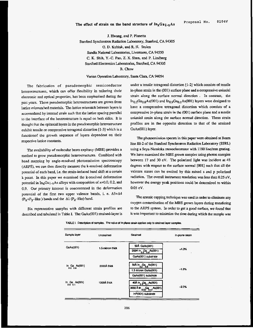
Proposal No. The effect of strain on the band structure of In,Gal.,As
8104V
J. Hwang, and P. Pianetta
Stanford Synchrotron Radiation Laboratory, Stanford, CA 94305
G. D. Kubiak, and R. H. Stulen
Sandia National Laboratories, Livermore, CA 94550
C. K. Shih, Y.-C. Pao, Z. X. Shen, and P. Lindberg
Stanford Electronics Laboratories, Stanford, CA 94305
B. Chow
Varian Operation Laboratory, Santa Clara, CA 94054
The fabrication of pseudomorphic semiconductor
heterostructures, which can offer flexibility in tailoring their
electronic and optical properties, has been emphasized during the
past years. These pseudomorphic heterostructures are grown from
lattice mismatched materials. The lattice mismatch between layers is accomodatd by internal strain such that the lattice spacing parallel
to the interface of the heterostructure is equal on both sides. It is
thought that the epitaxial layers in the pseudomorphic heterostructure
exhibit tensile or compressive tetragonal distortion [l-31 which is a
functionof the growth sequence of layers dependent on their
respective lattice constants.
The availability of molecular beam expitaxy (MBE) provides a
method to grow pseudomorphic heterostructures. Combined with
band mapping by angle-resolved photoemission spectroscopy
(ARPS), we can then directly measure the k-resolved deformation
potential of each band, i.e. the strain-induced band shift at a certain
k point. In this paper we examined the k-resolved deformation potential in InxGal-xAs alloys with composition of x=0.0,0.2, and
0.9. Our primary interest is concentrated in the deformation
potential of the first two upper valence bands, i. e. A3+A4 CpX+PTlike ) bands and the A1 (Pzlikej band.
Six representive samples with different strain profiles are described and tabulated in Table I. The GaAs(OO1) strained-layer is
under a tensile tetragonal distortion [ 1-21 which consists of tensile
in-plane strain in the (001) surface plane and a compressive uniaxial strain along the surface normal direction . In contrast, the In0.2GaO.gAs(001) and Iq,gGa,-,lAs(OO1) layers were designed to
have a compressive tetragonal distortion which consists of a
compressive in-plane strain in the (001) surface plane and a tensile
uniaxial strain along the surface normal direction. These strain
profiles are in the opposite direction to that of the strained
GaAs(001) layer.
The photoemission spectra in this paper were obtained at Beam line III-2 of the Stanford Synchrotron Radiation Laboratory (SSRL)
using a Seya-Namioka monochromator with 1180 line/= grating.
We have examined the MBE grown samples using photon energies
between 17 and 30 eV. The polarized light was incident at 45
degrees with respect to the surface normal [001] such that all the
valence states can be excited by this mixed s and p polarized
radiation. The overall instrument resolution was less than 0.25 eV,
however the energy peak positions could be determined to within
0.05 eV.
The arsenic capping technique was used in order to eliminate any
oxygen contamination of the MBE grown layers during transfering
to the ARPS system. In order to get a good surface, we found that
it was important to minimize the time during which the sample was
TABLE 1. Descciption of samples. The value 01 in-plane strain applies only to strained Iayef samples.
Sample layer Unstrained Strained In-plane strain
In da ~ ~ ( 0 0 1 ) 0.2 0.8
2oooA thii I .3%
1.5 micron thick +I% GaAs(OO1)
GaAs(001) substrate
In Ga As(OO1) 0.9 0.1
1 OOoA lhiik
-2.3%
lnP(001) substrate
206
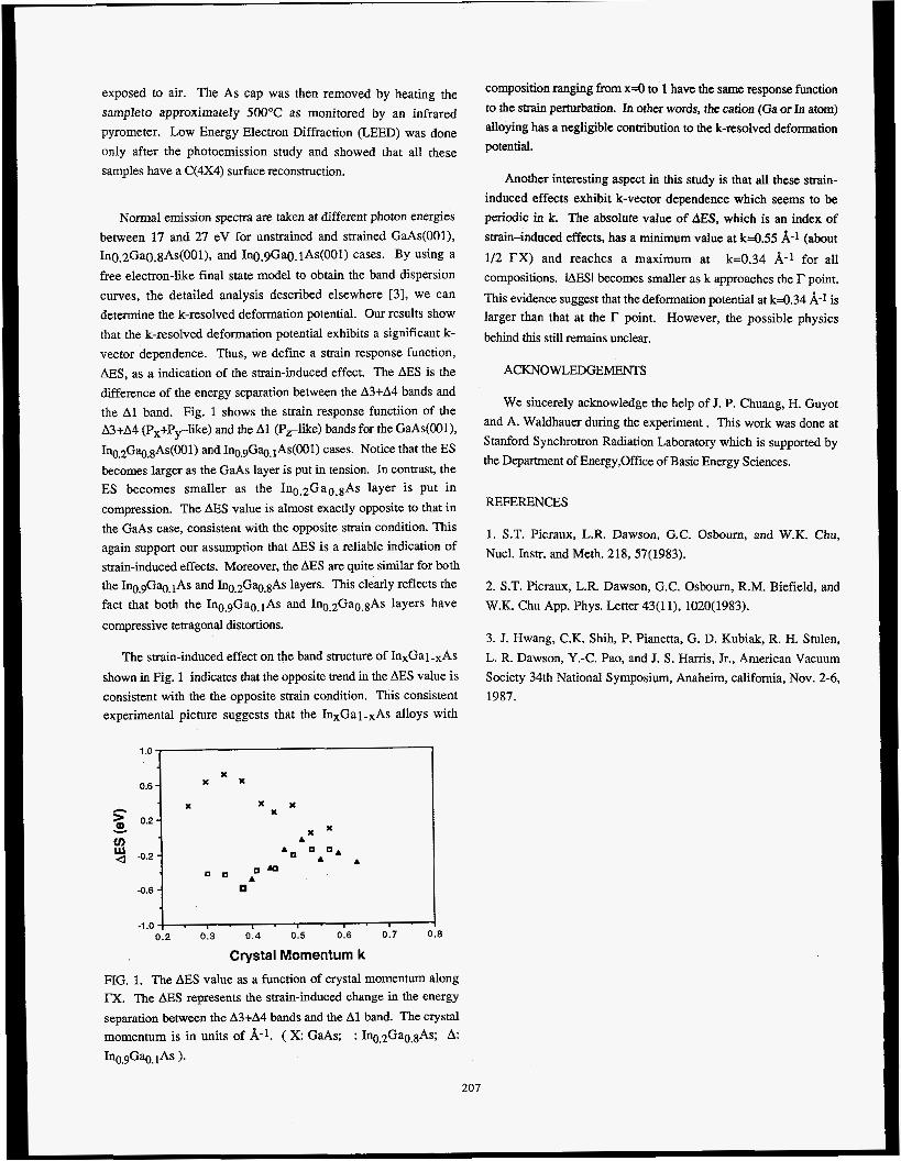
exposed to air. The As cap was then removed by heating the
sampleto approximately 5OOOC as monitored by an infrared
pyrometer. Low Energy Electron Diffraction (LEED) was done
only after the photoemission study and showed that all these
samples have a C(4X4) surface reconstruction.
Normal emission spectra are taken at different photon energies
between 17 and 27 eV for unstrained and strained GaAs(001), In0~2Ga0.gAs(001), and Ino.9Gao. iAs(001) cases. By using a
free electron-like final state model to obtain the band dispersion
curves, the detailed analysis described elsewhere [3], we can
determine the k-resolved deformation potential. Our results show
that the k-resolved deformation potential exhibits a significant k- vector dependence. Thus, we define a strain response function,
AES, as a indication of the strain-induced effect. The AES is the
difference of the energy separation between the A3+A4 bands and
the A1 band. Fig. 1 shows the strain response functiion of the A3+A4 (Px+Prlike) and the A1 (PJike) bands for the GaAs(001),
In0.2Gq.8As(001) and In0.9Gq.1As(001) cases. Notice that the ES
becomes larger as the GaAs layer is put in tension. In contrast, the ES becomes smaller as the In0.2Ga0.8As layer is put in
compression. The AES value is almost exactly opposite to that in
the GaAs case, consistent with the opposite strain condition. This
again support our assumption that AES is a reliable indication of
strain-induced effects. Moreover, the AES are quite similar for both the InO.gGa0.1As and In0.2Gq.8As layers. This clearly reflects the
fact that both the InO.gGa0.1As and In0.2Ga0.8As layers have
compressive tetragonal distortions.
The strain-induced effect on the band structure of InxGal-xAs
shown in Fig. 1 indicates that the opposite trend in the AES value is
consistent with the the opposite strain condition. This consistent experimental picture suggests that the InxGal-xAs alloys with
0.6 1 x x x
x x X 5 0.2 1
v
x x
x x
n . c I A
-0.6 -0.6 1 -1 .o
0.2 0.3 0 .4 0.5 0.6 0.7 (
Crystal Momentum k
I
composition ranging from x=O to 1 have the same response function
to the strain perturbation. In other words, the cation (Ga or In atom)
alloying has a negligible contribution to the k-resolved deformation
potential.
Another interesting aspect in this study is that all these strain-
induced effects exhibit k-vector dependence which seems to be
periodic in k. The absolute value of AES, which is an index of
strain-induced effects, has a minimum value at k4.55 A-1 (about
1/2 r X ) and reaches a maximum at k=0.34 A-1 for all
compositions. IAESI becomes smaller as k approaches the r point.
This evidence suggest that the deformation potential at k=0.34 A-1 is larger than that at the r point. However, the possible physics
behind this still remains unclear.
ACKNOWLEDGEMENTS
We sincefely acknowledge the help of J. P. Chuang, H. Guyot
and A. Waldhauer during the experiment. This work was done at
Stanford Synchrotron Radiation Laboratory which is supported by
the Department of Energy,Office of Basic Energy Sciences.
REFERENCES
1. S.T. Picraux, L.R. Dawson, G.C. Osbourn, and W.K. Chu,
Nucl. Instr. and Meth. 218, 57(1983).
2. S.T. Picraux, L.R. Dawson, G.C. Osbourn, R.M. Biefield, and
W.K. Chu App. Phys. Letter 43(11), 1020(1983).
3. J. Hwang, C.K. Shih, P. Pianetta, G. D. Kubiak, R. H. Stulen,
L. R. Dawson, Y.-C. Pao, and J. S . Harris, Jr., American Vacuum
Society 34th National Symposium, Anaheim, California, Nov. 2-6,
1987.
FIG. 1. The AES value as a function of crystal momentum along TX. The AES represents the strain-induced change in the energy
separation between the A3+A4 bands and the A1 band. The crystal
momentum is in units of W-l. ( x: GaAs; : In0.2Ga0.8As; h:
In0.9Ga0.1As 1.
207
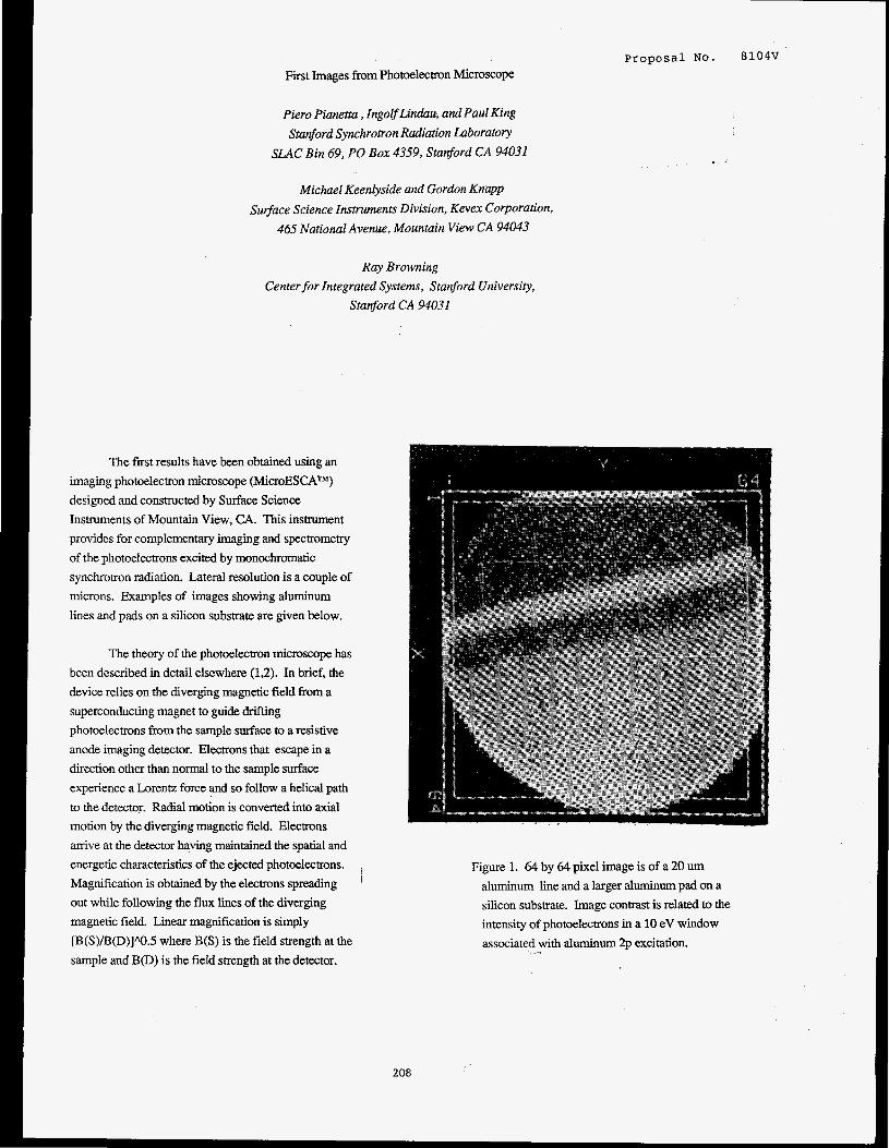
Proposal NO. 8104V
First Images from Photoelectron Microscope
Pier0 Pianetta , IngoEfLindau, and Paul King
Stanford Synchronon Radiation Laboratory
SLAC Bin 69, PO Box 4359, Stanford CA 94031
Michael Keenlyside and Gordon Knapp
Surjface Science Instruments Division, Kevex Corporation,
465 National Avenue, Mountain View CA 94043
Ray Browning
Center for Integrated Systems, Stanford University, Stanford CA 94031
The first results have been obtained using an
imaging photoelectron micmscope (MicroESCATM)
designed and constructed by Surface Science
Instruments of Mountain View, CA. This instrument
provides for complementary imaging and spectrometry
of the photoelectrons excited by monochromatic
synchrotron radiation. Lateral resolution is a couple of
microns. Examples of images showing aluminum
l i e s and pads on a silicon substrate are given below.
The theory of the photoelectron microscope has
been described in detail elsewhere (1,2). In brief, the
device relies on the diverging magnetic field from a
superconducting magnet to guide drifting
photoelectrons from the sample surface to a resistive
anode imaging detector. Electrons that escape in a
direction other than normal to the sample surface
experience a Lorentz force and so follow a helical path
to the detector. Radial motion is converted into axial
motion by the diverging magnetic field. Electrons
arrive at the detector having maintained the spatial and
energetic characteristics of the ejected photoelectrons.
Magnification is obtained by the electrons spreading
out while following the flux lines of the diverging
magnetic field. Linear magnification is simply
[B(S)/B(D)]/\O.S where B(S) is the field strength at the
sample and B@) is the field strength at the detector.
, '
208
Figure 1. 64 by 64 pixel image is of a 20 um
aluminum line and a larger aluminum pad on a
silicon substrate. Image contrast is related to the
intensity of photoelectrons in a 10 eV window
associated with aluminum 2p excitation.

Typical working values are B(S) equal to 7T and B@)
equal to 1 SmT resulting in a linear magnification near
70. Spectrometry is carried out by controlling a
retarding voltage at the detector. This allows for the
generation of images from electrons in any particular
energy window and thus provides a method for
elemental and chemical mapping of surfaces.
Figure 1 is an image of the photoelectron
intensity of a sample consisting of a 20 um wide
aluminum line and a larger aluminum pad on a silicon
substrate. Figure 1 represents all photoelectrons with
energies associated with the aluminum 2p excitation.
Incident photon energy here is 170eV. Figure 2 shows
the spectrum of photoelectrons collected from the
aluminum line and from the aluminum pad.
Surprisingly, the photoelectron peak was found to shift
between these two areas. In this instance the
possibility of local charging has not yet been excluded.
Analysis of this and other image data is still underway.
References
1. G. Beamson, H.Q. Porter, and D.W. Turner,
Nature 290, 556 (1981)
2. I.R. Plummer, H.Q. Porter, D.W. Turner, A.J.
Dixon, K. Gehring, and M. Keenlyside, Nature
303, 599 (1983)
I I I I I I I I I
93 91 89 87 a Photoelectron energy [eV]
Figure 2. Spectrum of photoelectrons collected from
the aluminum line and aluminum pad regions shown
in Figurel. An apparent peak shift of the aluminum
line to higher binding energies is observed.
209
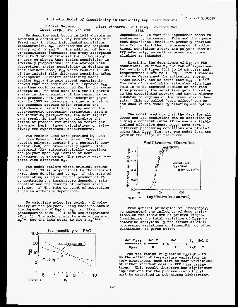
Proposal No.8104V A Kinetic Model of Crosslinking in Chemically Amplified Resists
Daniel Seligson Piero Pianetta, Paul Kinu. Lawrence Pan Intel C o r p . , 408-765-2381 *.
SSRL We describe work begun in 1986 wherein we
examined a series of x-ray resists which dif- fered only in their halogenated sensitizer concentration, nr. Photoresists are composed mainly of C, N and 0. The addition of Br- or C1-sensitizers increases the x-ray absorption for 1 to 3 keV of interest for lithography. In 1986 we showed that resist sensitivity is inversely proportional to the average mass absorption. (Note: sensitivity is defined as that incident dose, DsQ which results in 50% of the initial film thickness remaining after development. Greater sensitivity means smaller D50.) The more recent experiments showed that the addition of C1 improved dS0 more than could be accounted for by the x-ray absorption. We concluded that the C1 partic- ipates in the chemistry of the resist expo- sure process, not just the absorption phys- ics. In 1987 we developed a kinetic model of the exposure process which predicts the dependence of sensitivity on nr and on the post-exposure processing parameters. From a manufacturing perspective, the most signifi- cant result is that we can calculate the effect of process variations on resist per- formance, basing those calculations on rela- tively few experimental measurements.
and Haas Research Laboratories. They are novolak polymers containing a photoacid gen- erator (PAG) and crosslinking agent. The photoacid (PA) autocatalytically crosslinks the polymer upon application of heat subsequent to exposure. The resists were pre- pared with different nr.
tions. 1) PA is proportional to the absorbed x-ray dose density and to nr. 2) The rate of crosslinking is equal to the product of PA concentration, a temperature dependent rate constant and the density of uncrosslinked polymer. 3 ) The rate constant of assumption 2 has an Arrhenius dependence.
The resists used were provided by Rohm
The model employs three critical assump-
We calculate molecular weight and solu- bility of the polymer, using these to obtain the dependence of Ds0 on n,, for fixed postexposure bake (PEB) time and temperature (Fiq. 1). The model predicts a dependence of nr- , but the data seems to fit a
Intrinsic sensitivity v s . PAG 100
1' .5 1 2 3 1
FIGURE 1 nr
dependence. I n ract the dependence seems to weaken as nr increases. This and the square root dependence itself are probably attribut- able to the fact that the presence of addi- tional sensitizer alters the polymer chemis- try adversely, as well as promoting cros- slinking as intended.
Examining the dependence of D50 on PEB conditions, we fixed nr and ran an experimen- tal matrix of times (0.5 to 16 minutes) and temperatures (9OoC to 130OC). From Arrhenius plots we determined the activation energ -9200 Kelvin, and we found that D50 - t'Ij3. The rate of crosslinking saturates with time. This is to be expected because as the reac- tion proceeds, the sensitizer gets locked up in the crosslinked network and cannot migrate outwards to regions of low crosslinking den- sity. This so called 'cage effect' can be included in the model by altering assumption 2.
The model predicts that the data for all doses and PEB conditions can be described by a single contrast curve if we use a suitably defined effective dose, DQrg. Data from 9 different processing conditions are plotted using this De€€ (Fig. 2). The model does not predict the shape of the curve.
Final Thickness vs . Effective Dose
0.4 u .-
z 0.0 L , I . I , I I I I ,
10 100 io00
FIGURE Log Effective Dose (mJ/cmP)
p4G
Contrast - 2.6 D ~ - S S I I I J
8 120 68c. ll0OC
From general principles of lithography, we understand the influence of dose varia- tions on the linewidth of printed images. Considering the total variation of Deff we determine analytically the effect of small processing variations on linewidth, or other quantities, as given below.
del Deff del D del t E, del T + --- ----e + n ----- -------- = ---e-
Deff D t kgT T
For the resist in question Ea/kpT - 25, so the effect of temperature variations is very pronounced, much more so than variations of either incident dose or PEB time varia- tions. This result therefore has significant implications for the process control that must be exercised in sub-micron lithography.
2 10

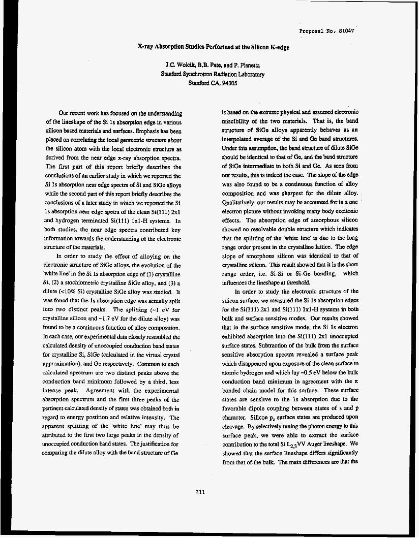
Proposal NO, 8104V
X-ray Absorption Studia Performed at the Silicon Kmedge
J.C. Woic&, B.B. Pate, and P. Pianetta
Stanford Synchrotron Radiation Laboratory
Stanford CA, 94305
Our recent work has focused on the understanding
of the lineshape of the Si 1s absorption edge in various
silicon based materials and surfaces. Emphasis has been
placed on correlating the local geometric structure about
the silicon atom with the local electronic structure as derived from the near edge x-ray absorption spectra.
The fiist part of this report briefly describes the
conclusions of an earlier study in which we reported the
Si 1s absorption near edge spectra of Si and SiGe alloys
while the second part of this report briefly describes the
conclusions of a later study in which we reported the Si
Is absorption near edge spetra of the clean Si( 11 1) 2x1
and hydrogen terminated Si(ll1) 1x1-H systems. In both studies, the near edge spectra contributed key
information towards the understanding of the electronic
structure of the materials.
In order to study the effect of alloying on the
electronic structure of SiGe alloys, the evolution of the
'white line' in the Si 1s absorption edge of (1) crystalline
Si, (2) a stochiometric crystalline SiGe alloy, and (3) a
dilute ( 4 0 % Si) crystalline SiGe alloy was studied. It
was found that the 1s absorption edge was actually split
into two distinct peaks. The splitting (-1 eV for crystalline silicon and -1.7 eV for the dilute alloy) was
found to be a continuous function of alloy composition.
In each case, our experimental data closely resembled the
calculated density of unoccupied conduction band states
for crystalline Si, SiGe (calculated in the virtual crystal
approximation), and Ge respectively. Common to each
calculated spectrum are two distinct peaks above the
conduction band minimum followed by a third, less
intense peak. Agreement with the experimental
absorption spectrum and the first three peaks of the
pertinent calculated density of states was obtained both in
regard to energy postition and relative intensity. The apparent splitting of the 'white line' may thus be
attributed to the first two large peaks in the density of unoccupied conduction band states. The justification for
comparing the diIute alloy with the band structure of Ge
is based on the extreme physical and assumed electronic
miscibility of the two materials, That is, the band
structure of SiGe alloys apparently behaves as an
interpolated average of the Si and Ge band structures.
Under this assumption, the band structure of dilute SiGe
should be identical to that of Ge, and the band structllre
of SiGe intermediate to both Si and Ge. As seen from our results, this is indeed the case. The slope of the edge
was also found to be a continuous function of alloy
composition and was sharpest for the dilute alloy.
Qualitatively, our results may be accounted for in a one '
electron picture without invoking many body excitonic
effects. The absorption edge of amorphous silicon
showed no resolvable double structure which indicates
that the splitting of the 'white line' is due to the long
range order present in the crystalline lattice. The edge
slope of amorphous silicon was identical to that of
crystalline silicon. This result showed that it is the short range order, i.e. Si-Si or Si-Ge bonding, which
influences the lineshape at threshold
In order to study the electronic structure of the
silicon surface, we measured the Si 1s absorption edges
for the Si(ll1) 2x1 and Si(l11) 1x1-H systems in both
bulk and surface sensitive modes. Our results showed
that in the surface sensitive mode, the Si 1s electron
exhibited absorption into the Si(ll1) 2x1 unoccupied
surface states. Subtraction of the bulk from the surface
sensitive absorption spectra revealed a surface peak
which disappeared upon exposure of the clean surface to
atomic hydrogen and which lay -0.5 eV below the bulk
conduction band minimum in agreement with the
bonded chain model for this surface. These surface
states are sensitve to the 1s absorption due to the
favorable dipole coupling between states of s and p
character. Silicon pz surface states are produced upon
cleavage. By selectively tuning the photon enern to this
surface peak, we were able to extract the surface
contribution to the total Si h3W Auger lineshape. We
showed that the surface lineshape differs sisnificantly
from that of the bulk. The main differences are that the
211,
bulk plasmon loss at -70 eV kinetic energy is suppressed
at the 2x1 surface, an effect which may be explained by
classical arguments, and that the contribution of the
surface filledaangling-bonds shifts the main Auger peak
-1 eV towards higher kinetic energy. The bulk plasmon
loss at -70 eV is greatly intensified at the Si( 11 1) 1x1-H
surface due to the passivation of the surface upon
hydrogen adsorption and the reordering of the surface
from a sp2 to sp3 configuration. The high energy
%,,W Auger satellite peak at -107 eV kinetic energy
was shown to be sensitive to the photon energy within
the Vicinity of the Si K-edge. This phenomenon was
explained by the double ionization of the Si L-shell due
to a KL,L decay for photon energies above the silicon
K-edge. We deduced that -15 eV is the energy
difference between a singly and doubly ionized Si
Gshell and is due to the presence of the extra 'spectator'
core hole during the L W decay. Unlike the plasmon
loss, this feature was not sensitive to the surface
geometry since it is an atomic rather than geometric
effect.
212

Proposal No. 99OoV
(lxl)Co/Cu(OOl) Surface Geometry Determined From Co 1s Photoelectron Diffraction
Z.Q. Huang, L.J. Terminello, L.Q. Wang, 2. Hussain,+ X.S. Zhang,++ S. Kim, L. Huckabee, D.A. Shirley
Materials and Chemical Sciences Division Lawrence Berkeley Laboratory
and Department of Chemistry University of California Berkeley, California 94720
J. Woicik, P. Pianetta Stanford Synchrotron Radiation Laboratory Stanford University, California 94305
Co 1s angle-resolved photoemission was measured over a kinetic energy range of 80-400 eV from a (lxl)Co/Cu(OOl) adsorbate system. The photoemission measurements were made on Beamline VI and exhibit an intensity modulation arising from photoelectron diffraction. photoelectron diffraction, recently termed angle- resolved photoemission extended fine structure (ARPEFS). exhibits large oscillations (see Fig. 1) that permit Fourier analysis and subsequent extraction of structural information. The (lxl)Co/Cu(OOl) system was prepared by evaporating Co from high purity wire onto a clean (1x1) Cu(OO1) substrate. Evaporation rates were monitored by a quartz micro-balance and were correlated to effective coverages determined from separate Auger electron intensity analysis. The results of our (lxl)Co/Cu(OOl) ARPEFS study were covpared with the epitaxial structure proposed by LXED and reveal additional physical information. There is evigence that the Generalized Ramsauer-Townsend effect, ar'isbs from particular scattering conditions, is presient in our results.
This energy dependent form of
that
p( l x1)co/cu(oo1)
0 na
>\ .-li 0.5 - cn c (u
c
-
- 1 2
U 0.0 - -
(u > 0 Q) -0.5 - K
.- #
- -
Acknowledeement
This work was supported by the Director, Office of Energy Research, Office of Basic Energy Sciences, Chemical Sciences Division of the U.S. Department of Energy under Contract No. DE-AC03-76SF00098. It was performed at the Stanford Synchrotron Radiation Laboratory, which is supported by the Department of Energy's Office of Basic Energy Sciences.
tPermanent address: Department of Physics, University of Petroleum & Minerals, Dhahran, Saudi Arabia
tPermanent address: Department of Physics, Zhej iang University, Hangzhou, Zhejiang, China
References I
1. J.J. Barton, C.C. Bahr, Z. Hussain, S.W. Robey, J.G. Tobin, L.E. Klebanoff, and D.A. Shirley, Phys. Rev. Lett. 2. 272 (1983).
2. A. Clarke, G. Jennings, R.F. Willis, P.J. Rous, and J.B. Pendry, Surf. Sci 187, 327 (1987).
3. J.J. Barton, Z. Hussain, and D.A. Shirley, Phys. Rev. B z, 933 (1987).
Figure 1. x(k) curve for emission direction normal to the Cu(OO1) surface. The circles represent unfiltered data. is the same X(k) filtered at path-length differences between l.5A and 20A.
The solid line
I< (A- ' ) 2 13

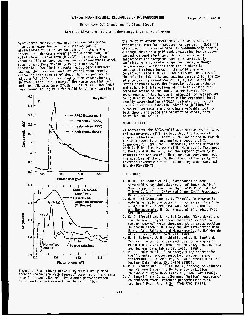
SU B-keV NEAR-THRESHOLD RESONANCES I N PHOTOABSORPTI ON
Nancy Kerr Del Grande and K. Glenn T i r s e l l
Lawrence Livermore National Laboratory, L ivermre , CA 94550
Proposal No. 9901V
Synchrotron rad ia t ion was used f o r absolute photo- absorption experimental cross sec t i n (APECS) measurements taken i n t r a n s m i ~ s i o n . ~ - ~ Among the in te res t ing phenomena observed f o r a broad range o f s o l i d elements (Z=4 through Z=92) a t energies from about 50-1500 eV were t h e resonances/enhancements which seem t o accompany v i r t u a l l y every inner s h e l l threshold. and amorphous carbon) have s t ruc tu ra l enhancements extending some tens o f eV above t h e i r respective K- edges which d i f f e r s i g n i f i c a t l y from r e l a t i v i s t i c Hartree Slater (RHS) theory,' the Henke compilation5 and the LLNL data base (CSLOW). The BL-VI11 TGM APECS measurement i n Figure 1 f o r s o l i d Be c losely p a r a l l e l s
Two l i g h t elements (e.g., bery l l ium metal
4.0
3.5
3.0
2.5
s G 2.0 4: g 1.5
1 .o
u) u)
0
0.5
0.0
Berylllum
- APECS experiment --- Data base (CSLOW)
-.-.- Henke tables (1982)
RHS atdmlc theory
100 140 180 220
Photon energy, eV
4.0 I 1 1 I l l
-.-.- Gaseous Be,
-
-
0.0 ' I I 1 1 1 1 1 , l
1 20 126 132 138
Photon energy (eV)
Figure 1. Prel iminary APECS mea urement o f B metal
base i n l a and wi th r e l a t i v e atomic photoio izaton showing comparison w i t h theory,' compilat ion 5 and data
cross section measurement f o r Be gas i n l b . 8
the r e l a t i v e atomic photoionization cross sgction measurement from Auger spectra f o r the gas. Note the s t ruc tu re f o r the s o l i d metal is predominantly atomic, although there i s s i g n i f i c a n t broadening due t o outer conduction band electrons. enhancement f o r amorphous carbon i s t e n t a t i v e l y explained as a molecular shape resonance, although auto ion iz ing t rans i t ions from the 1s s t a t e t o unoccupie valence bands i n the s o l i d are a lso possible.p Recent BL-VI11 SGM APECS measurements o f the r e l a t i v e in tens i ty and spacing versus Z f o r the 2p- 3d autoionizing resonances o f Ti, V, C r y Fe and N i reveal features about the in te rp lay between exchange and sp in o r b i t in teract ions which help expla in the coupling scheme o f the ions. measurements o f the 5d g ian t resonance f o r uranium are being used t o t e s t r e l a t i v i s t i c time-dependent l oca l densi ty approximation (RTDLDA) ca lcu lat ions fog $he uranium atom i n a Spherical "drop" o f je l l ium. 9
APECS measurements are prov id ing a valuable t o o l t o t e s t theory and probe the behavior o f atoms, ions, molecules and sol ids.
A broad s t r u c t u r a l
Other BL-VI11 TGM
ACKNOWLEDGMENTS
We appreciate the APECS mul t i layer sample design ideas and measurements o f T. Barbee, Jr.; the technica l support e f f o r t s o f J. Batteux, M. Fowler and R. Mazuch; the data acqu is i t ion and analysis support o f M. Schneider, E. Carr, and P. Mahowald; the co l laborat ion w i t h B. Pate; the UHV work o f H. Morales, T. Martinez, T. Neal, and P. Golceff; and the support given by P. Pianetta and h i s s t a f f . This work was performed under the auspices o f the U. S. Department o f Energy by the Lawrence Livermore National Laboratory under Contract NO. W-7405-ENG-48.
REFERENCES
1. N. K. Del Grande e t al., "Resonances i n near- threshold x-ray photoabsorpt i on o f inner shells," Spec. suppl . t o Journ. de Phys. w i th Proc. o f 14th In ternat . Conf. on X-Ray and Inner S h m , P a r i s * France (1988).
obtain r e l i a b l e photoabsorption cross sections,'' i n X-Ray and WV In te rac t ion Data Bases, Calculations, and Measurements, N. Del Grande e t al., Eds., Proc. 'SPIE 911 (1 988).
3. K. G. T i r s e l l and N. K. Del Grande, "Considerations f o r the use o f synchrotron r a d i a t i o n sources t o measure sub-keV x-ray photoabsorption cross sections i n transmission," i n X-Ray and VUV In te rac t ion Data Bases, Calculat ions, and Measurements, N. Del Grande e t al., Eds., Proc. SPIE 911 (1988 . "X-ray attenuation cross sections f o r energies 100 eV t o 100 keV and elements Z=1 t o Z=92," Atomic Data and Nuclear Data Tables 38, 1-145 (1988).
5. 6. L. Henke e t al., "Low energy x-ray in te rac t ion coef f i c ien ts : photoabsorption, sca t te r ing and re f lec t ion , E=100-2000 eV, Z=1-94," Atomic Data and Nuclear Data Tables 27, 1-144 (1982).
6. M. 0. Krause and C. 0 . Caldwell, "Strong cor re la t ion and alignment near the Be 1s photo ion izat ion threshold," Phys. Rev. Le t t . 59, 2736-2739 (1987).
7. A. Zangwill and 0 . A. L ibermax "Optical response o f an embedded atom: uranium," Phys. Rev. B 3, 6705-6707 (1987).
2. N. K. Del Grande and K. 6. T i r s e l l , "A program t o
4. E. B. Saloman, J. H. H u b b n , and 3. H. Scofield,
Resonant photoemission from
214


OPTICALLY DETECTED ABSORPTION IN THE SOFT X-RAY REGION Proposal No. 9901V
M. J . Weber, M. B. Schneider and K. G. T i r s e l l
Lawrence Livermore Nat ional Laboratory P.O. Box 808, Livemore, Ca l i f o rn ia 94550
INTRODUCTION:
Detect ion o f x-ray absorption edges and f i n e s t ruc tu re by monitoring o p t i c a l luminescence has been reported f o r both the hard1 and soft293 x-ray regions. This technique involves r e l a t i v e l y simple sample preparation and experimental apparatus, can be used f o r spectroscopy o f low-Z elements or high- ly ing core leve ls having small x-ray fluorescence yields, and i n ce r ta in cases can provide s i t e and valence spec i f i c information. We have demonstrated the fu r the r v e r s a t i l i t y o f t h i s method f o r the s o f t x-ray region by examining several c r y s t a l l i n e and amorphous mater ia ls having e i the r i n t r i n s i c luminescence or luminescence from t r a n s i t i o n metals and organic dyes present as dopants. Measurements were made i n the 10-200 eV region using beamline V I I I - 1 (TGM) and i n the 100-1200 eV region using beamline V I I I -2 (SGM). Fluorescence was observed from the f ron t surface o f the sample a t 45O t o the d i rec t i on o f the x-ray beam w i th a photomult ip l ier tube and photon counting electronics. Since the samples were o p t i c a l l y t h i c k w i th respect t o x-rays, the edge jumps are n e g a t i ~ e . ~
RESULTS:
Rare Earth Materials: Several ra re earths f luoresce intensely, even a t high concentrations, and therefore are use fu l act ivators. The M4,5 and N4 5 edges corresponding t o allowed 3 h 4 f and 4 h 4 t t rans i t i ons o f CeF3, CeF3-CaF2 s o l i d solut ions, PrF3, TbF3, LiTbFq, and KTb3F10 were eas i l y detectable. The complex spectrum observed f o r CeF3 was i n excel lent agreement w i th t h a t obtained by transmission through t h i n films.5 As an example, the luminescence exci ta- t i o n spectrum f o r a Tb(PO3)3 glass showing both the Tb N4,5 edges and the P L3,2,1 edges i s given i n Fig. 1.
m:Tb(O. l%) : Although the terbium concentrat ion i n t h i s sample i s small, i t s luminescence can be used t o detect the F K edge and EXAFS and the La M4,5 edges as shown i n Fig. 2.
w: The exc i ta t i on spectrum o f the i n t r i n s i c luminescence shows Ba and F absorption edges s im i la r t o those shown i n Fig. 2, bu t d i f f e r e n t f i n e s t ruc tu re .
Photon : .?rgy (eV)
Figure 1. f o r Tb(P03)3 glass a t room temperature.
Terbium luminescence exc i ta t i on spectrum
1 1 I I 1 I
600 700 800 8CO Photon Energy (eV)
Figure 2. f o r LaF3:Tb(O.l%) a t room temperature.
Terbium luminescence exc i ta t i on spectrum
E&&QL~: there was no c lear evidence o f other B i or Ge edges in the 100-1300 eV region ( the B i L edges and Ge K edges have, however, been detected op t i ca l l y ) .
Europium Aluminoborate Glass: t he B and 0 K edges were observed by monitoring the Eu luminescence; the l a t t e r absorptions were weak.
The 0 K edge and EXAFS were observed but
The Eu N4 5 edges and
Sodium Phosphate Glass (1.0% Mnl: Mnzt luminescence, the 0 and Na K edges and EXAFS and a weak Mn L3 edge were observed.
Tin-Lead-Fluorophosphate Glass (Rhodamine 66): The Sn &,5 edge and the 0 and F K edges were detected by monitoring the dye fluorescence i n t h i s low temperature glass.6
By monitoring the
ACKNOWLEDGEMNTS :
We thank R. Stulen (SNLL) f o r the loan o f the UHV chamber used f o r the experiments and 0. Blackburn (NBS) and P. Tick (Corning) f o r glass samples. was performed under the auspices o f the U.S. Department o f Energy by the Lawrence Livermore Nat ional Laboratory under contract W-7405-ENG-48.
REFERENCES:
1. J. Goulon e t a l . i n EXAFS and Near Edae Structure
This work
- 111, ed. K. Hodgson, B. Hedman, and J.E. Penner-Hahn (Springer-Verlag, Ber l in , 1984), 490 and references c i t e d therein.
2. B.X. Yang, J. K i r z and S.Xu, Nucl. I n s t r . and Meth. m, 141 (1987).
3. D.B.M. Klaassen, C.M.G. van Leuken and K.M.H. Maessen, J. Lumin. a/4l, 696 (1988).
4. J. Goulon, P. Tola, M. Lemonnier and J. Dexpert- Ghys, Chem. Phys. 2, 347 (1983).
5. C. G. Olson and 0. W. Lynch, J. Opt. SOC. Am. 72, 88 (1982)
6. W. R. Tompkin, R. W. Boyd, D. W. H a l l and P. Tick, J . Opt. SOC. Am. B 4, 1030 (1987).

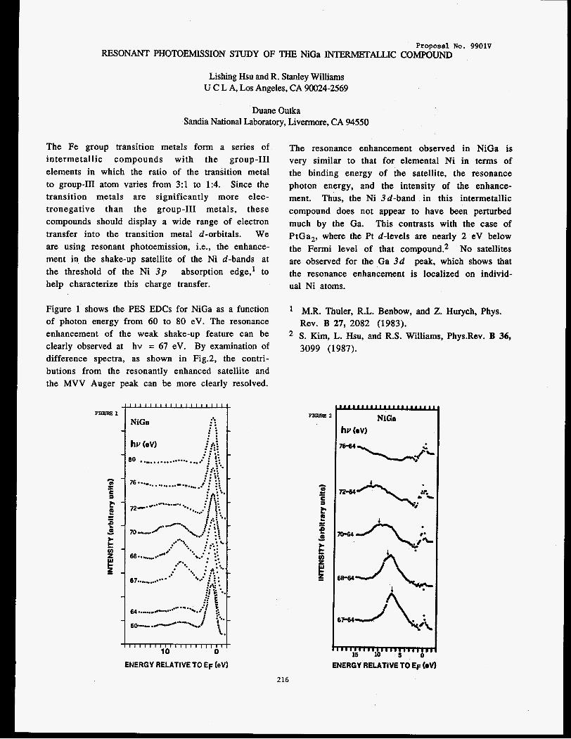
Proposal No. 9901V RESONANT PHOTOEMISSION STUDY OF THE NiGa INTERMETALLIC COMPOUND
Lishing Hsu and R. Stanley Williams U C L A, Los Angeles, CA 90024-2569
Duane Outka Sandia National Laboratory, Livermore, CA 94550
The Fe group transition metals form a series of
i n t e rme ta l l i c compounds with the group-I11
elements in which the ratio of the transition metal
to group-I11 atom varies from 3:l to 1:4. Since the
transit ion metals a re significantly more elec-
t ronegat ive than the group-I11 metals, these
compounds should display a wide range of electron
transfer into the transition metal d-orbitals. We
are using resonant photoemission, i.e., the enhance-
ment in the shake-up satellite of the Ni d-bands at
the threshold of the Ni 3p absorption edge,' to
help characterize this charge transfer.
Figure 1 shows the PES EDCs for NiGa as a function
of photon energy from 60 to 80 eV. The resonance
enhancement of the weak shake-up feature can be
clearly observed at hv = 67 eV. By examination of
difference spectra, as shown in Fig.2, the contri-
butions from the resonantly enhanced satellite and
the MVV Auger peak can be more clearly resolved.
The resonance enhancement observed in NiGa is very similar to that for elemental Ni in terms of
the binding energy of the satellite, the resonance
photon energy, and the intensity of the enhance-
ment. Thus, the Ni 3d-band in this intermetallic
compound does not appear to have been perturbed
much by the Ga. This contrasts with the case of
PtGa,, where the Pt d-levels are nearly 2 eV below
the Fermi level of that compound.2 No satellites
are observed for the Ga 3d peak, which shows that
the resonance enhancement is localized on individ-
ual Ni atoms.
M.R. Thuler, R.L. Benbow, and 2. Hurych, Phys.
Rev. B 27, 2082 (1983). S. Kim, L. Hsu, and R.S. Williams, Phys.Rev. B 36, 3099 (1987).
1111111111111111111oI NiGa
hv (OV)
..
ENERGY RELATIVE TO EF bv)
216

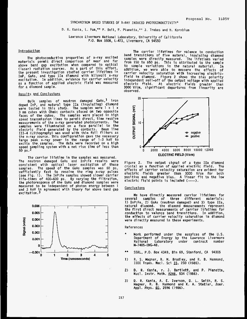
Proposal No. SYNCHROTRON BASED STUDIES OF X-RAY INDUCED PHOTOCONDUCTIVITY*
1L05V
D. R. Kania, L. Pan,** P. Bel l , P. Pianetta,** J . Trebes and H. Kornblum
Lawrence Livermore National Laboratory, Un ive rs i t y o f C a l i f o r n i a P.O. Box 5508, L-473, Livermore, CA 94550
I n t r o d u c t i o n
The photoconductive p roper t i es o f x-ray exc i ted ma te r ia l s pe rm i t d i r e c t comparison o f near and f a r above band gap e x c i t a t i o n when compared t o o p t i c a l ( l ase r ) r a d i a t i o n sources. As a p a r t o f t h i s e f f o r t , the present i n v e s t i g a t i o n studied c a r r i e r l i f e t i m e s i n InP, GaAs, and type I I a diamond w i t h k i l o v o l t x-ray e x c i t a t i o n . I n add i t i on . evidence f o r c a r r i e r v e l o c i t y as a func t i on o f appl ied e l e c t r i c f i e l d was measured f o r a diamond sample.
Results and Conclusions
Bulk samples o f neutron damaged GaAs,l i r o n doped InP, and na tu ra l type I I a ( i n s u l a t i n g ) diamond were tested i n t h i s study. The samples w e r e 1 x 1 x 3 mm cubes w i t h Ohmic contacts placed on two opposite faces o f the cubes. The samples were placed i n h igh speed transmission l i n e s t o permi t d i r e c t , t i m e resolve measurements of the x-ray generated photocurrents. The samples were i l l u m i n a t e d on a face p a r a l l e l t o the e l e c t r i c f i e l d generated by the contacts. Beam l i n e 111-4 (Lithography) was used w i t h t h i n f o i l f i l t e r s as the x-ray source. This con f igu ra t i on gave the necessary l a rge peak x-ray power i n the range o f 1-3 keV t o e x c i t e the. samples. The data w e r e recorded on a h igh speed sampling system w i t h a net r i s e t i m e o f l e s s than 50 P S . ~
The neutron damaged GaAs and 1nP:Fe r e s u l t s were consis tent w i t h o p t i c a l l ase r e x c i t a t i o n o f these samples. The speed o f the GaAs samples was 60 ps, s u f f i c i e n t l y f a s t t o resolve the r i n g x-ray pulses (see Fig. 1). The 1nP:Fe samples showed slower c a r r i e r l i fe- t imes o f 400-600 ps. By vary ing the f i l t r a t i o n . the photoresponse o f the GaAs and diamond samples w e r e measured t o be independent o f photon energy between 1 and 3 keV i n agreement w i t h theory f o r above band gap e x c i t a t i o n . 3
The c a r r i e r l i f e t i m e i n the samples was measured.
Time (nanoseconds)
The c a r r i e r l i f e t i m e s f o r valence t o conduction band t r a n s i t i o n s o f f i v e natura l , i n s u l a t i n g diamond samples were d i r e c t l y measured. The l i f e t i m e s va r ied from 150 t o 650 ps. This i s a t t r i b u t e d t o the sample t o sample va r ia t i ons i n the na tu ra l ma te r ia l . I n addi t ion, we were able t o measure the e f fec ts o f c a r r i e r v e l o c i t y sa tu ra t i on w i t h increas ing e l e c t r i c - f i e l d i n diamond. Figure 2 shows the b ias p o l a r i t y independent r o l l - o f f o f the output vo l tage w i t h appl ied e l e c t r i c f i e l d . A t e l e c t r i c f i e l d s greater than 3000 V/cm, s i g n i f i c a n t departures from l i n e a r i t y are observed.
20
F E Y
-I
2 g 10
5 n + 3 + positive 0
0 2000 4000 6000 8000 10000 1 2
ELECTRIC FIELD (V/Cm)
0
/
0
Figure 2. The output s ignal o f a type I I a diamond c r y s t a l as a func t i on o f appl ied e l e c t r i c f i e l d . The e f f e c t s o f c a r r i e r v e l o c i t y sa tu ra t i on are evident f o r e l e c t r i c f i e l d s g rea te r than 3000 V/cm f o r both p o s i t i v e and negative b ias. A l i n e a r f i t t o the low e l e c t r i c f i e l d po in ts i s inc luded (-1.
Concl us i ons
We have d i r e c t l y measured c a r r i e r l i f e t i m e s f o r several samples o f three d i f f e r e n t mater ia ls : 1) InP:Fe, 2) GaAs (neutron damaged) and 3) type I I a , na tu ra l diamond. the diamond measurements represent the f i r s t d i r e c t measurements o f c a r r i e r l i f e t i m e s f o r conduction t o valence band t r a n s i t i o n s . I n addi t ion, t he e f f e c t s o f c a r r i e r v e l o c i t y sa tu ra t i on i n diamond were d i r e c t l y measured i n these experiments.
References
* Work performed under the auspices o f the U.S. Department o f Energy by the Lawrence Livermore National Laboratory under con t rac t number W-7405-ENG-48.
** SSRL, P.O. Box 4349, B in 69, Stanford, CA 94305
1) R. S. Wagner, R. M. Bradley, and R. B. Hammond, I E E E Trans. Nucl. Sci 3, 250 (1986).
2) D. R. Kania, r. J . B a r t l e t t , awd P. Pianetta, Nucl. I n s t r . Meth. w, 534 (1986).
3) D. R. Kania, A. E. Iverson, D. L. Smith, R. S. Wagner, R. B. Hammond and K. A. S t e t l a r , Jour. Appl. Phys. dn, 2596 (1986).
217

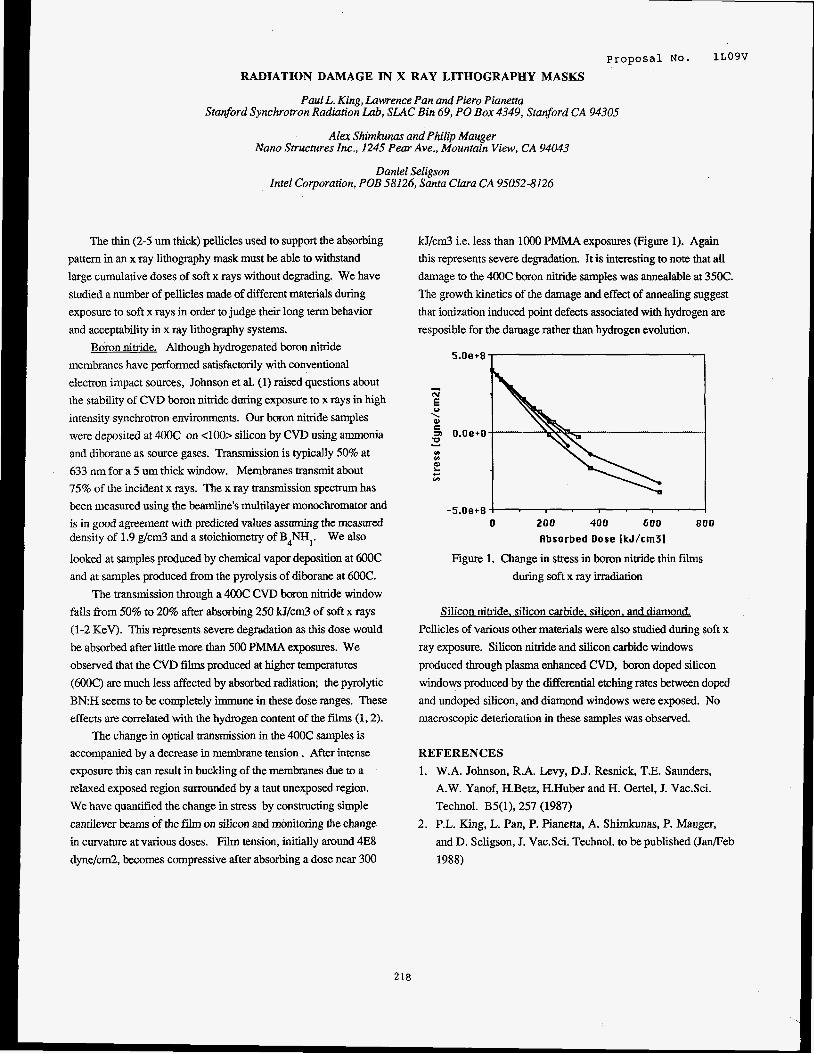
p r o p o s a l NO. 1LOgv
RADIATION DAMAGE IN X RAY LITHOGRAPHY MASKS
Paul L. King, Lawrence Pan and Pier0 Pianetta Stanford Synchrotron Radiation Lab, SLAC Bin 69, PO Box 4349, Stanford CA 94305
Alex Shimkunas and Philip Mauger %no Structures Inc., 1245 Pear Ave., Mountain View, CA 94043
Daniel Seligson Intel Corporation, POB 58126, Santa Clara CA 95052-8126
The thin (2-5 um thick) pellicles used to support the absorbing
pattern in an x ray lithography mask must be able to withstand
large cumulative doses of soft x rays without degrading. We have
studied a number of pellicles made of different materials during
exposure to soft x rays in order to judge their long term behavior
and acceptability in x ray lithography systems.
membranes have performed satisfactorily with conventional
electron impact sources, Johnson et al. (1) raised questions about
the stability of CVD boron nitride during exposure to x rays in high
intensity synchrotron environments. Our boron nitride samples
were deposited at 400C on <loo> silicon by CVD using ammonia
and diborane as source gases. Transmission is typically 50% at
633 nm for a 5 um thick window. Membranes transmit about
75% of the incident x rays. The x ray transmission spectrum has
been measured using the beamline's multilayer monochromator and
is in good agreement with predicted values assuming the measured density of 1.9 g/cm3 and a stoichiometry of B4NH1. We also
looked at samples produced by chemical v a p r deposition at 6OOC
and at samples produced from the pyrolysis of diborane at 6OOC.
The transmission through a 400C CVD boron nitride window
falls from 50% to 20% after absorbing 250 kJ/cm3 of soft x rays
(1-2 KeV). This represents severe degradation as this dose would
be absorbed after little more than 500 PMMA exposures. We
observed that the CVD films produced at higher temperatures
(6OOC) are much less affected by absorbed radiation; the pyrolytic
B N H seems to be completely immune in these dose ranges. These
effects are correlated with the hydrogen content of the films (1,2).
The change in optical transmission in the 400C samples is
accompanied by a decrease in membrane tension . After intense
exposure this can result in buckling of the membranes due to a
relaxed exposed region surrounded by a taut unexposed region.
We have quantifkd the change in stress by constructing simple
cantilever beams of the fii on silicon and monitoring the change
in curvature at various doses. Film tension, initially around 4E8
dyne/cm2, becomes compressive after absorbing a dose near 300
Boron nitride. Although hydrogenated boron nitride
kJ/cm3 i.e. less than lo00 PMh4A exposures (Figure 1). Again
this represents severe degradation. It is interesting to note that all damage to the 400C boron nitride samples was annealable at 35OC. The growth kinetics of the damage and effect of annealing suggest
that ionization induced point defects associated with hydrogen are resposible for the damage rather than hydrogen evolution.
-5.0e+8 ! 0 200 400 600 800
flbsorbed Dose [kJ/cm31
Figure 1. Change in stress in boron nitride thin films during soft x ray irradiation
Silicon nitride, silicon carbide. silicon. and diamond.
Pellicles of various other materials were also studied during soft x
ray exposure. Silicon nitride and silicon carbide windows
produced through plasma enhanced CVD, boron doped silicon
windows produced by the differential etching rates between doped
and undoped silicon, and diamond windows were exposed. No
macroscopic deterioration in these samples was observed.
REFERENCES
1. W.A. Johnson, R.A. Levy, D.J. Resnick, T.E. Saunders,
A.W. Yanof, H.Betz, RHuber and H. Oertel, J. Vac.Sci.
Technol. B5(1), 257 (1987)
and D. Seligson, J. Vac.Sci. Technol. to be published ( J M e b
1988)
2. P.L. King, L. Pan, P. Pianetta, A. Shimkunas, P. Mauger,
2 18


Vlll ACTIVE PROPOSALS
As of December 31 , 1987 there were 158 active SSRL proposals. Proposals remain active for two years after their initial rating by the Proposal Review Panel. Since October 1974 SSRL has received a total of 2048 proposals. The Spokesperson for each proposal is underlined and their institution shown in parentheses. The letter suffix appended to the proposal number indicates the sub-panel of the Proposal Review Panel to which the proposal is assigned: Materials (M), Biology (B) or Vacuum Ultra-Violet (v). The small (p) indicates a program proposal.
1 OOMp (PRV
882Mp
89Np
898Mp
906Bp
908Vp
9/1/83
3 1 I84
4/3/84
5/3/84
a1291a4
8/31/84
lamuszw X-ray Absorption Studies of Disordered ANDREW J SEARY systems FARREL W LYTLE ROBERT B GREEGOR ROBERT L INGALLS P VIRAN J E WHITMORE NEIL ALBERDING K R BAUCHSPIESS (SIMON FRASER UNIVERSITY)
MATlBLOCH Structural Studies of Organic Thin
interface
Spin Polarized Photoelectron Studles of Magnetism in Sollds
PETER M EISENBERGER
(ARGONNE NATIONAL LABORATORY)
Films on the Solid Gas and Liquid Gas
E E U U U B X m MARTIN L DENBOER INGOLF LINDAU (SPEXAFS) (STEVENS INSTITUTE OF TECHNOLOGY)
Lmwa Magnetic X-Ray Scattering from Ho KEVIN L D'AMICO JOHN D AXE JAKOB BOHR DAVID MONCTON (BROOKHAVEN NATDNAL LABOR4 TORY) - ELIZABETH C THEIL
X-Ray Absorption Studies of the Protein-Iron interaction in Ferritin
PAULINE HARRISON E A STERN KENNETH RAYMOND (NORTH CAROLINA STA lE UNIVERSITY) - ROBERT 2 BACHRACH ROGER I G UHRBERG Surfaces
Influence of Surface Reconstruction on Electronic Properties of Semiconductor
J E NORMRUP (XEROX)
SSRL ACTIVITY REPORT 1987 Page 219
91 OMp
922Bp
928Mp
935vp
941 Vp
9/6/84
v M I BELL
Structural Studles of ion Implants in Ill-V Semlconductors wlth Reflectance
R A FORMAN EXAFS (NATIONAL BUREAU OF STANDARDS)
P Gold-Based Antiarthritis and Anticancer KATHERINE TEPPERMAN Drugs and Metabolltes: XAS, WAXS, DAS (UNIVERSITY OF CINCINNATI)
PFTFR MJgISFNBFRGFq Grazing-Incidence X-ray Scattering KENG S LIANG Program (EXXON RESEARCH 6 ENGINEERING) - TOMAS F KENDELEWICZ WILLIAM E SPICER JOEL SILBERMAN M D WILLIAMS JOE WOlClK C BOWMAN J J YEH S J EGLASH K K CHIN K SHIH S LIST C MCCANTS KRISTINE BERTNESS T CHIANG CARLORCARBONE PETER MAHOWALD DANIEL FRIEDMAN JUN NOGAMI N NEWMAN EZlO PUPPIN (SSRL) - 0 BlSl IVAN0 ABBATI STEFAN0 NANNARONE CARLO CALANDRA Z SHEN EZlO PUPPIN MASSIMO SANCROTTI UMBERTO DEL PENNINO WILLIAM E SPICER INGOLF LINDAU (POLITECNICO DI MILANO)
Electronic Structure, Chemlstry, and the Fermi Level at Semlconductor Surfaces and Interfaces
Study of the Formatlon of Rare Earth/ Elemental Semiconductor (Ge, Si) Interfaces and Compounds
Page 220 SSRL ACTIVITY REPORT 1987
943vp
947v
956Mp
957Bp
959vp
963vp
969Bp
9/6/84
1 011 5/84
2/27/85
2/28/85
311 I85
3/6/85
311 4/85
- DENNIS W LINDLE -------- TRlClA FERRETT ALLEN L JOHNSON JOHN J BARTON ERNESTOPAPARAUO BAOHUA NIU TONG LEUNG SEHUN KIM ALEXIS SCHACH V WITTENAU
SHIHONG LIU STEVEN W ROBEY PHILIP A HEIMANN LAlSHlNG WANG LOUIS J TERMINELLO MARIA NOVELLA PlANCASTELLl
LAURA MEDHURST CHARLES C BAHR (LAWRENCE BERKELEY LA BORA TORY)
ZHENG-QING HUANG
XUN-SHENG ZHANG
I3!uumw FABIO COMlN Substrate Surfaces (AT& T BELL LABORATORIES)
Proposal to Perform SEXAFS from
E€2MBaN ROBERT B GREEGOR
Appllcatlons of EXAFS Spectroscopy in the Steel and Coal Industries
FARREL W LYTLE F E HUGGINS NARESHSHAH R G JENKINS (UNIVERSITY OF KENTUCKY) - Anomalous Scattering of X-rays L K TEMPLETON (LAWRENCE BERKELEY LABORATORY)
PO6ERT 7 BACHRACM ROSS D BRINGANS
Core Level and Near Edge Structure Study of Interface Formation
(XEROX) - NEXAFS 8nd SEXAFS Studies by Means of X-ray Fluorescence Detectlon: Local Structure Around Low-Z Atoms
(ISM RESEARCH LABORATORY) - X-ray Absorption Spectroscopic STEPHEN P CRAMER Studies of Nickel-Containing
Metalloenzymes (UNIVERSITY OF GEORGIA)
SSRL ACTIVITY REPORT 1987 Page 221

973M
981 Bp
991 Bp
994Mp
995Mp
997Mp
998M
999Mp
411 2/85
9/3/85
9/4/85
9/5/85
9/6/85
911 0185
911 0185
911 0185
v M I BELL
EXAFS Study of Damage and Annealing of Ion Implanted Semiconductors
R A FORMAN G K HUBLER E D DONOVAN (NATIONAL BUREAU OF STANDARDS) - STEPHEN P CRAMER
High Resolution EXAFS Spectroscopy of Iron and Copper Proteins
ALLAN A CROTEAU CRAIG F HEMANN KIMBERLY A POWELL FRANK G FlAMlNGO (OHIO STATE UNIVERSITY)
Technetium and Rhenium Imaging Agents and Therapeutic Radiopharmaceuticais EDWARD A DEUTSCH
(UNIVERSITY OF CINCINNATI)
GLFNN A W A Y C H U M GORDON E BROWN, JR (STANFORD UNIVERSITY)
High Temperature EXAFS Study of Fe and Ca in Silicate Crystals and Meits
GORDON F BROWN. JR
GEORGE A PARKS GLENN A WAYCHUNAS JAMES 0 LECKIE KIM F HAYES (STANFORD UNIVERSIW)
XAS Study Of Cation Adsorption at KEITH 0 HODGSON Aqueous/Oxide Interface
lJmmEa9 P J ESTRUP W(100)
The Surface Phase Transition of Clean
M ALTMAN ROBERT J BIRGENEAU (AT&T BELL LABORATORIES)
lLKtw Order-Disorder Transltion of Adsorbates KENG S LIANG on Nickel G J HUGHES PETER M EISENBERGER (Brookhaven National Laboratory)
GORDON E BROW- W A JACKSON C W PONADER GLENN A WAYCHUNAS S ROTHFUS (STANFORD UNIVERSIW)
EXAFS Study of Trace Element Environments In Quenched Silicate Meits of Geochemical interest
Page 222 SSRL ACTIVITY REPORT 1987
1001Mp 111186 EXXON PRT Proposal Beam Llne VI (PRT) S K SINHA
C R SAFINYA KENG S LlANG GRAYSON H VIA JOHN H SINFELT J M NEWSAM DAVID MONCTON (EXXON RESEARCH & ENGINEERING)
1008Mp 10/4/85 )VI1 I IAM K WARBURTOY Binary Liquld Structures Determined by KARL F LUDWIG ' Anomalous X-ray Scatterlng ARTHUR BIENENSTOCK (ARACOR)
101 OM 1011 4185 E R SAFlNYq Synchrotron Studles of Freely Suspended BILL VARADY N A CLARK Strands
Dlscotlc and Ordered Microemulsion
(EXXON RESEARCH & ENGINEERING)
101 2Bp 11/4/85 BQBFRT H FAIRCLOUGY Membrane X-Ray Diffraction Studies Of STEVEN R HUBBARD the Acetylcholine Receptor KEITH 0 HODGSON SEBASTIAN DONIACH D RICHMAN (UNIVERSITY OF CHICAGO}
1013Vp 11/18/85 m J C F S C 0 SF- NEXAFS and EXAFS Studies of Shallow J E ROWE JOHN M POATE X-ray Fluorescence
lnpuritles In Semiconductors by Soft
(AT& T BELL LABORATORIES)
1015Vp 2/28/86 JOHN I GI Am The Structure and Reactlvlty of
FRANCISCO ZAERA SEXAFS (EXXON RESEARCH & ENGINEERING)
DANIEL A FISCHER Adsorbed Intermediates Using NEXAFS and
1017M 3/3/86
1018Mp 3/3/86
- J C MIKKELSON FRANK G BRIDGES (Xf ROX)
Local Structure of Icosahedral Materials
uEEQuaw 6 E C DAVIS JEFF J HOYT B CLARK M KRAITCHMAN (UNIVERSITY OF CALIFORNIA)
Diffuslon In Ternary Multilayer Thin Films
SSRL ACTIVITY REPORT 1987 Page 223
101 9B
1021 Mp
1022vp
1025M
1026M
1027Bp
1028Vp
1029B
1030Bp
3 m 6
3/3/86
3/3/86
3/4/86
3/4/86
3/5/86
3/5/86
3/5/86
311 7/86
- LOUIS LIM R PAUL PHIZACKERLEY Radiation ETHAN A MERRllT (WASHINGTON UNIVERSITY)
Crystallographic Study of Amicyanin by Anomalous Scattering Using Synchrotron - Synchrotron X-ray Polycrystalline
TlNG C HUANG Diff ractometry GEORGE WILL MAURlZlO BELLOTTO CURT ERICKSON MICHAEL HART (ISM RESEARCH LABORATORY)
EDWARDlSOLOMON STEVE V DlDZlULiS JIAN-YI LIN and Sulfides
UPS Studies of the Coordination Chemistry of Metal Oxides, Chlorides
KRISTINE BUTCHER (STANFORD UNIVERSIW)
U X E A Y m ANTHONY D COX M CHEN LEI P ZIA J V ACRIVOS (CALIFORNIA STATE UNIVERSITY)
Structure of Multiple Valence Compounds such as Prussian Blue vs Temperature
DONALD R SANoSTROM FARREL W LYTLE Gas Phase
Transition Metal ion Complexes In the
(BOEING COMPANY)
STFVE D CONRAnSOr\l WILLIAM WOODRUFF
Dispersive XAS Development: Time Resolved and Diamond Anvil Applications
BASIL SWANSON (LOS ALA MOS NA TIONAL LA BORA TORY)
!Jlwum High-Photon Flux Photoemission Studies INGOLF LINDAU of Narrow Band Materials (UNIVERSITY OF MICHIGAN)
l!mwLwA SRIRAM SUBRAMANIAM (AT& T BELL LABORATORIES)
X-ray Diffraction by Molecular Films on Solid Substrates in Aqueous Environment -
6 E SMITH M WW ADAMS E I STEIFEL V MINAK L S SOLMONSON J ENEMARK W CLELAND GRAHAM N GEORGE (SCHL UMBERGER DOLL RESEARCH)
Soft X-ray Spectrocopy of Molybdenum Enzymes, Cofactors and Model Compounds
Page 224 SSRL ACTIVITY REPORT 1987
1031M 3/18/86
1032M 311 9/86
1 0 3 3 ~ 31191a6
1034Mp 3/4/86
10358 4/14/86
1036M 4/14/86
1037M 511 9/86
1038M 5/19/86
XAS of Synthetic Models for M DAVID CURTIS Hydrodesuifurizatlon Catalysts OSWALDO BARALT LEVI THOMPSON (UNIVERSITY OF MICHIGAN)
v P BOOLCHAND
EXAFS Study of Local Chemical Coordination of Cation and Anion Sites in Chalcogenide Glass
(NATIONAL BUREAU OF STANDARDS)
WJFS F PFNNFR-HAW ROBERT MK CARLSON GEOFFREY WALDO (UNIVERSITY OF MICHIGAN)
XAS Determination of the Chemical Forms of Sulfur in Petroleums
Em&Pmwu ERIC SIROTA SUZANNE AMADOR Films L B SORENSEN (HARVARD UNIVERSITY)
Structure and Phase Transltions of Hexatic Phases in Thin Liquid Crystal
J N F S F PENNFR-HAW DAVID P BAUOU Sulfur Centers CHRISTOPHER J BATIE
JAMES A FEE (UNIVERSITY OF MICHIGAN)
XAS investigations of RiesksLike iron
HIM-TAI TSANG - Coster-Kronig Transition Probabilities GEORGE S BROWN G BRADLEY ARMEN W JITSCHIN S L SORENSEN U WERNER S B WHITFIELO ROGER CARR (UNIVERSITY OF OREGON)
v Grain Boundary Segregation of D L WILLIAMSON Impurities In iron and Steel G KRAUSS (COLORADO SCHOOL OF MINES) - D L WILLIAMSON
Deep Donors in Gallium Aluminum Arsenide Semiconductor Alloys
P G IBART (COLORADO SCHOOL OF MINES)
SSRL ACTIVITY REPORT 1987 Page 225
1039B 6/3/86 STFPHFN P C R M Polarized XANES and EXAFS of TERRENCE G FREY ROGER C PRINCE GRAHAM N GEORGE ROBERT A SCOTT (SCHLUMBERGER DOLL RESEARCH)
Cytochrome Oxidase in Hydrated, Oriented Mitochondrial Muitllayers
10408 6/3/86 X-Ray Absorption Spectroscopy of STEPHEN P CRAMER Xanthine Oxldase
1041 B 9/3/86 J?OBFRT A S C O U Measurement of Metal-Metal Distances
(OHIO STATE UNIVERSITY)
MARLY EIDSNESS in Proteins by X-ray Anomalous Scattering
(UNIVERSITY OF GEORGIA)
1042B 9/3/86 BRITTON CHANCF Studies of Intermediates in the BARTON HOLMQUIST Mechanism of Carboxypeptidase-A DAVID AULD MARK CHANCE JAMES RIORDAN LINDA S POWERS B VALLEE (UNIVERSITY CITY SCIENCE CENTER)
10438 9/3/86 BRITTON CHANCF X-Ray Absorption Spectroscopy of GRANT BUNKER iron-iron Interactions: Ribonucleotide PETER REICHARD Reductase S M KHALID
ALI NAQUI GUANG ZHANG (UNIVERSITY CITY SCIENCE CENTER)
BRITT-MARIE SJOBERG
1044M 9/3/86 MATTHFW A MARCUS Structural Effects of Ultrahigh Stralns CHING-LONG TSAl and Dislocation Densities J BEVK (AT& T BELL LABORATORIES)
1045M 9/3/86 m W A MARCUS Multiple and Unusual Slting In Dilute CHING-LONG TSAl Solid Solutions (AT&T BELL LABORATORIES)
Page 226 SSRL ACTIVITY REPORT 1987
10468 9/3/86 FDWARD RUBFNSTFIN iodine Dlchromography with W THOMLINSON ALBERT C THOMPSON Angiography
Monochromatic X-ray Beams for
GEORGE S BROWN ROBERT HOFSTADTER DONALD C HARRISON JOHN OTIS ROBERT KERNOFF HELEN GORDON JOHN GlACOMlNl HERBERT D ZEMAN (STANFORD UNIVERSITY)
1047M 9/3/86 BQBFRT I INGAI I S High Pressure Transition Mechanlsms J FREUD via EXAFS DARYL CROZIER (UNIVERSITY OF WASHINGTON)
1048Mp 911 1/86 W L I N F KII S O U W High Resolution Compton Profiles of iron SEANBRENNAN and Silicon ARTHUR BIENENSTOCK (STANFORD UNIVERSITY)
1049Mp 911 1/86 POBFRT B G W X-ray Spectroscopic Investigation of R C EWING FARREL W LYTLE B C CHAKOUMAKOS (BOEING COMPANY)
Radiation Damaged Materials
1050Mp 911 1/86 61 FNN A WAYCHUNM X-ray Anomalous Scattering and EXAFS
JAMES E PENNER-HAHN Aqueous Solutions MICHAEL J APTED (STANFORD UNIVERSITY)
GREG J EXARHOS Study of Heavy Metal Complexes in
1051 M 911 1/86 W N N A WAYCHUNqS Crystal and Aqueous Chemistry Of LAWRENCE R BERNSTEIN Germanium in Geochemically Important
Systems (STANFORD UNIVERSITY)
1052M 9/11/86 JVAN SFI I IN Measurement of Recoil Energies of cso S B ELSTON Molecular Fragments D A CHURCH J LEVIN H CEDERQUIST (OAK RIDGE NATIONAL LABORATORY)
Multiply Charged Recoil Ions and
SSRL ACTIVITY REPORT 1987 Page 227

10538
1054M
10558
1056Mp
1058M
1059M
060Mp
061 Bp
1062Vp
911 5/86
9/ 1 5/86
9/15/88
911 6/86
91 1 6/86
911 6/86
911 6/86
911 6/86
1 011 I86
EQwLumn DAVID M DOOLEY
Copper EXAFS Spectroscopy of Non-Blue Copper Protelns. Amlne Oxldases
(UNIVERSITY OF GEORGIA} - X=Ray Induced Photoacoustle Emlsslon HARRY E MARTZ SUZANNE FLETCHER ROBERT A DAY JOHN H KINNEY (LAWRENCE LIVERMORE NATIONAL LABORATORY) - STEPHEN F GHELLER FRANK A SCHULTZ Control B R l l l HEDMAN A LAWRENCE ROE W E NEWTON (SSRL)
XAS of Iron-Molybdenum Cofactor of Nitrogenase under Electrochemlcal
m Section Topographic Strain Mapping B COULMAN Using Synchrotron WhRe Radlation Y H CHUNG ZOPHIA U REK (GEORGIA INSTITUTE OF TECHNOLOGY)
E4uuaxm Roughenlng Transition of Metal Surfaces GLENN HELD (IBM RESEARCH LABORATORY) - L C MlNG
In SWu High P-T Equation-of-State, Phase Transformations and Kinetics Studies on Mantle Mineral Phases using Synchrotron Radiation
(UNIVERSITY OF HAWAII) - Structural Investigations of Layered ARTHUR BIENENSTOCK Synthetic Microstructures and Binary
(STANFORD UNIVERSITY) Amorphous Alloys -
ALLAN A CROTEAU Analysis CRAIG F HEMANN KIMBERLY A POWELL FRANK G FlAMlNGO (OHIO STATE UNIVERSIW)
Iron Porphyrin Models for EXAFS
- R L SHARPLESS Gases
Spectroscopy and Kinetics of Excited
(SRI INTERNATIONAL)
Page 228 SSRL ACTIVITY REPORT 1987
1064M 3llt07
1066Mp 311 I87
1 0 6 7 ~ ~ 311 187
1 0 6 8 ~ p 311 187
1069M 311187
1071M 3/1/87
1072M 3/1/87
1073M 3/1/87
ILBI(ELLB H K MA0 L C MlNQ MURLI H MANGHNANI Blamond=Anvll Apparatus
Statls Hlgh Pressure and Hlgh Temperature X=Ray Dlffractlon Studies of Some RareEarth Element8 Uslng
0 S SMITH QUINTIN C JOHNSON (LAWRENCE LIVERMORE NATIONAL LABORATORY)
MlCHELBOUDART Characterization of Supported Pd/Au R J DAVIS Alloys by X-Ray Absorptlon
Spectroscopy (STANFORD UNIVERSITY)
u X-Ray Spectroscopy of Electrochemically WILLIAM R HEINEMAN Generated Species (UNIVERSITY OF CINCINNATI) - SUNNEY I CHAN P MARK LI (UNIVERSITY OF GEORGIA)
Active Site Structures of Cytochrome c Oxldase
8HELKU E A STERN
Structure of Transients In the Catalytic Cycle of D lox y g enases
(GEORGIA INSTITUTE OF TECHNOLOGY) - Characterization of Vanadium, MAX W HILL FARREL W LYTLE Particulate Matter
Chromium, Nickel and Arsenic In
(ERIGHAM YOUNG UNIVERSITY)
3 l u w u E M Anchoring Chemistry of Transition KARIN MOLLER Metal Complexes and Clusters to
Intrazeoiite Surfaces (UNIVERSITY OF NEW MEXICO)
MlCHELBOUDART Characterlzatlon of Clusters of G P VALENCA PtN-Zeolite and rhl -A1 0 by X-Ray
Absorption Spectroscopy (STANFORD UNIVERSIP/) - HEATHERCHEN Aluminum-Metal Interfaces
Glancing Angle EXAFS and Reflectivity of
(BROOKHAVEN NATIONAL LABORATORY) - JMTRANQUADA Superconductors
X-Ray Absorption Study of La CuO Based
M SUENAGA (BROOKHAVEN NATIONAL LABORATORY)
SSRL ACTIVITY REPORT 1987 Page 229

1074M
1076B
1077M
1078M
1079M
1080Vp
1081M
1082B
- X-Ray Absorption and Reflectivity DALE E SAYERS Studies of Interfaces in Multilayer
ERIC ZIEGLER (BROOKHAVEN NATIONAL LABORATORY)
JIM VICCARO optics - S WAKATSUKI Bacteriorhodopsin using Time-Resolved ROBERTMSTROUD Small Angle X-Ray Diffraction KEITH 0 HODGSON (STANFORD UNIVERSITY)
Study of Conformational Changes in
KRISHNA M CHOUDHARY Photoemission EXAFS Studies of Surfaces and interfaces in Compound Semiconductors
(UNIVERSITY OF NOTRE DAME)
IllAENS KEITH 0 HODGSON BRUCE J TUFTS (STANFORD UNI VERSIp/) - T CLAESON THEODORE H GEBALLE FRANK G BRIDGES (XEROX) - C M FRIEND J T ROBERTS B FACTOR A C LIU J SOLOMON PAUL STEVENS (STANFORD UNIVERSITY..
PAVlD P POPF JOHN E BENCl
X-Ray Absorption Studies of Metal ion Absorption at Semiconductor Surfaces
Local Atomic Structure of High-T, Superconductors
Adsorption Structure of Heteroatom- Containing Molecules on Transition Metal Surfaces
A Study of Creep Damage Using Microradiography
(UNIVERSITY OF PENNSYLVANIA)
DAVID A I ARSON TERESA TROXEL
Contribution of Auger Electrons to Radiation Sensitization with iUdR
MICHAEL SCHELL CLIFF LING THEODORE L PHILLIPS DENNIS SHRIEVE (UNIVERSITY OF CALIFORNIA)
Page 230 ~ ~
S S R L ACTIVITY REPORT 1987
10838
1084Bp
1085Bp
1086V
1087Bp
1088M
1089M
311187
3/1/87
3ma7
311 187
31 1 187
3/1/87
X-Ray Absorption spectroscopy of Myeloperoxidase and Lactoperoxidase ROGER C PRINCE
GRAHAM N GEORGE (UNIVERSITY OF PENNSYL VANIA) - STEPHEN P CRAMER ROGER C PRINCE Complex (EXXON RESEARCH & ENGINEERING)
Polarized X-Ray Spectroscopy of the Oriented Cholroplast Water-Oxidizing
4AMFS F PFNNFtWAHN
V L PECORARO W D FRASCH R FRANK0 C F YOCUM M STERN J T GROVES D GHANOTAKIS (UNIVERSITY OF MICHIGAN)
HIM-TAI TSANG X-Ray Absorption Spectroscoplc Characterlzatlon of the Manganese In Biologlcal Systems
Polarization In Molecular X-Ray Flourescence
- PAUL L COWAN SEANBRENNAN DENNIS W LINDLE (NATIONAL BUREAU OF STANDARDS)
JVAYNF A HFNDRICKSON R PAUL PHIZACKERLEY Crystal Structures ETHAN A MERRllT STEVENRHUBBARD JOHN HORTON WE1 YANG WlLlAM E ROYER ARNO PAHLER MARIANNE CUFF HM KRISHNA MURTHY CRAIG OGATA (COLUMBIA UNIVERSITY)
Anomalous-scattering Studies of Protein
FS F PENNFR-HAM GEOFFREY WALDO SHENGKE WANG RICHARD FRONKO GLENN A WAYCHUNAS (UNIVERSITY OF MICHIGAN)
Polarized X-Ray Absorption Near Edge Structure
X-Ray Absorption of Iridium in K-T Boundary Samples and Meteorites
- WILLIAM MURPHY JULIA PECK STEPHEN P CRAMER GRAHAM N GEORGE (SCHLUMBERGER DOLL RESEARCH)
SSRL ACTIVITY REPORT 1987 Page 231
Electrolyte-Metal interfaces 1090MP 6/9/87 T F FURTAY TIMOTHY M HAYES (COLORADO SCHOOL OF MINES)
1091Mp 6/9/87 QLluuma Characterlzatlen of Ion-Implanted TIMOTHY M HAYES Aluminum and Titanium F M KUSTAS (COLORADO SCHOOL OF MINES)
1092M Structural lnvestigatlon of lnterlayer Potassium and Sodium in Phyllosilicates
6/19/87 & M S H O W U N WADA S WHllTlNGHAM D. R. HINES STEPHEN P CRAMER (SCHLUMBERGER DOLL RESEARCH)
1093Mp 8/7/87 - K CHAR J MOLL B OH N MISSERT J Z SUN A D KENT
THEODORE H GEBALLE STEPHEN LADERMAN (STANFORD UNIVERSIW)
ALICE FISCHER-COLBRIE
Structure of Superconductlng Thin Films
1094M X-Rays Studies of Rare Gas Inclusions in Single Crystal Aluminium
8/20/87 J A K O R W I K ROBINSON E JOHNSON
L F GRAABAEK H H ANDERSEN A JOHANSEN (BROOKHAVEN NATIONAL LABORATORY)
L SARHOLT-KRIST
1095Mp 8/7/87 JsJuw&s LAURA NORTON
EXAFS and NEXAFS Investigation of OMPVE Growth of ti-Vi Compounds
D W KISKER (AT&T BELL LABORATORIES)
1096Mp 8120187 PAUl H FUOSS In-Situ X-Ray Scattering Studies of OMVPE Growth of Il-VI Compounds D W KISKER
LAURA NORTON (AT& T BELL LABORATORIES)
1097Mp 8120187 FARRFl W I YTL F ROBERT 6 GREEGOR (BOEING COMPANY)
Antl-Site Disorder In High T, Superconductors
Page 232 SSRL ACTIVITY REPORT 1987
1098M
1099M 9/1 5/87
2 0 0 0 ~ gii 5/87
2001 M 9/15/87
2002Mp 9/15/87
2003M 9/15/87
2005M 9/15/87
- In-Situ EXAFS Transition Metal PHILIP N. ROSS Macrocycles Absorbed on Electrode
Surfaces (CASE WESTERN RESERVE UNIVERSIP/)
Study of Organic Metals sLYmmQs M CHEN LEI S S P PARKIN R ITHNIN D HELMOLDT J SATCHER S ARNOLD C BUSTILLO (CALIFORNIA STATE UNIVERSITY)
huAmMQs MELVIN P KLEIN S S P PARKIN M CHEN LEI
Study of Catlon, Anion X-Ray Edges in S Intercalated Perovskltes
R ITHNIN S ARNOLD J SATCHER D HELMOLDT C BUSTILLO (CALIFORNIA STATE UNIVERSITY) - Extended X-Ray Absorption Fine PAUL D FLEISCHAUER
(THE AEROSPACE CORPORATION)
StWctUre Of Transition Metal Dlschalcogenide Materials
AlJ2msK J D LITSTER ROBERT J BIRGENEAU (WJ
Bond Orientational Order In Tllted Hexatlc Liquld Crystal Films
J!tumuz W H SMYRL OWEN MELROY Crystal TI
In-Situ X-Ray Scattering Study of the Stnrcture of Passive Oxides on Slngie
(IBM RESEARCH LABORATORY)
-Q JAN LllTON Scintillators
Search for Ultra-Fast Heavy Atom
WILLIAM W MOSES (LA WRENCE BERKELEY LABORATORY)
EQwmJmu DARYL CROZIER B HOUSER E A STERN J M TRANQUADA (UNIVERSITY OF WASHINGTON)
X-Ray Absorption Studies of Perovskltes as a Function of Pressure
SSRL ACTIVITY REPORT 1987 Page 233

2006Mp
20078
2008B
2009Vp
201 OBp
2011v
911 5/87
91 1 5/87
911 7/87
911 7/87
91 1 7/87
201 2M
2013M
9/17/87
911 7/87
luEmuUE JEFF J HOYT M KRAITCHMAN S SPOONER M WlEDENMElR (UNIVERSIN OF CALIFORNIA)
Kinetics of Phase Separation in Ai-LI
- Protein Folding Studied by Synchrotron X-Ray Scattering
(BROOKHAVEN NATIONAL LABORATORY) - GREGG A MARG
EXAFS of Active-Site Metals in Glucose Isomerase and Methane Monooxygenase
R MARK GUlNN (UNIVERSITY OF CALIFORNIA) - THOMAS P RUSSELL
Structural Studies of Thin Polymer Films on Surfaces
W JARK (IBM RESEARCH LABORATORY)
X-Ray Spectroscopy of Manganese and iron in Chloroplasts R GUILES
ANN MCDERMOTT SUSAN DEXHEIMER J L ZIMMERMANN VllTAL K YACHANDRA VlCKlE DEROSE KENNETHSAUER (LAWRENCE BERKELEY LABORATORY)
J2suMU3 PIER0 PIANETTA induced Photoconducthrity
Synchrotron Based Studies of X-Ray
J TREBES (LAWRENCE LIVERMORE NATIONAL LABORATORY)
E E E U L SODERHOLM GOPAL K SHENOY Spectroscopy
Electronic Structure of High T, Oxide Superconductors by X-Ray Absorption
PEDRO A MONTAN0 (ARGONNE NATIONAL LABORATORY)
Photoelectron Spectrometry of Correlation Satellites in KR
l & E u B K HAMISH ROBERTSON DAVE KNAPP WALTER J. TRELA RICH MARTIN TOM BOWLES JOHN WILKERSON JOHN HENDERSON BERNDCRASEMANN GEORGE S BROWN ROGER J BARTLETT S B WHITFIELD S L SORENSEN (LOS ALAMOS NATIONAL LABORATORY)
Page 234 SSRL ACTIVITY REPORT 1987

2014M
201 5Mp
201 6M
201 7Mp
201 8M
9/17/87
911 7/87
911 7/87
9/21/87
- J D SHINDLER Thermodynamics R F HAINSEY (CARNEGIE MELLON UNIVERSITY)
Melting of XeRiS,: Structure and
&5sExx In-Situ Synchrotron Microradiography G WEBB S D ANTOLOVICH Composites
of Delamination in Graphite/Epoxy
ZOPHIA U REK (GEORGIA INSTITUTE OF TECHNOLOGY)
Anomalous X-Ray Powder Diffraction Studies of the Structure of Materials
GEsBxMm ANDREW C LAWSON ROBERT B VON DREELE JOYCE A GOLDSTONE ART WILLIAMS (LOS ALAMOS NATIONAL LABORATORY) - DAVID M ZEHNER S G J MOCHRIE
Structure and Stability of Metal Surfaces: A Proposal for X-Ray Scattering Experiments
L D GIBBS (BROOKHAVEN NATIONAL LABORATORY)
JEFEBFY B K0RTRlGI-U ROSS D BRINGANS
Structural Study of Hetero-Epitaxial CaF, on Si(111) Using Standing Wave
JONATHAN DENLINGER Fluorescence MARJORIE OLMSTEAD (LAWRENCE BERKELEY LABORATORY)
2019V
2020Mp
2021 M
2022B
9/21/87
9/21 187
9/2 1 I87
- Heteroepitaxiai interface Formation JONATHAN DENLINGER ROSS D BRINGANS (UNIVERSITY OF CALIFORNIA)
Between Insulators and Semiconductors
ailmu T GROY
X-Ray Diffraction of Solid State Reactions and Phase Transitlons
J R SCHOONOVER (ARIZONA STATE UNIVERSIW
Structural Features of Low Temperature Recombination of Hemoproteins, Carbon Monoxide and Oxygen
Structures of Silver(1)-Crown Complexes in Liquid and Solid Phases
- MAX W HILL ROBERT B GREEGOR FARREL W LYTLE (BRIGHAM YOUNG UNIVERSITY)
v HlROWKl OYANAGI GRANT BUNKER KE ZHANG S G SLIGAR (UNIVERSITY CITY SCIENCE CENTER)
SSRL ACTIVITY REPORT I987 Page 235
2023Mp 9/21/87 8IEYE.U- Hlgh Tc Suporconductlng Materlale: IAN D RAlSTRlCK ZACHARY FISK Characterlratlon by XAS
Tholr Structural and Electronlc
ANTONIO REDONDO (LOS ALAMOS NA TiONAL LABORATORY)
2024Mp 9/21/87 STFVF D CON- XAS Studles of Quasi-OnsDlmenslonal HalldsBrldged Mlxed Valence Piatlnum BASIL SWANSON
ALFRED SATTLEBERGER Compounds (LOS AlAMOS NATlONAL LABORATORY)
20256 Altered Structures of Cytochrome Oxidase
9/21/87 BITTON CHANGE GRANT BUNKER TSOO E. KING KE ZHANG ROBERT POYTON EDGAR DAVIDSON (UNIVERSITY CITY SCIENCE CENTER)
2026Mp 9/21/87 PIFRO PlANFlTA JENNCHANG HWANG GLENN KUBIAK JEFF NELSON RICHARD H STULEN (SSRL)
The Bonding Behaviour in Strained Semlconductor Heterostructures
2027M
2028M
9/22/87 SEANUBEJJ Structural Studies of --Sputtered ARTHUR I BIENENSTOCK ROLAND TlMSlT B FACTOR
Amorphous AlTa Thin Fllms
(SSRL)
9/29/87 J-IOWARD OCKFN ZOPHIA U REK FARREL W LYTLE (EPRI)
Characterization of Cobalt-Bearlng Passlve Oxides on Stainless Steel by EXAFS
2029Bp 9/29/87 E l T H 0 HODGSON Mo and Se XAS Studles of Small-Molecule BRITT HEDMAN Binding to Nftrogenase BARBARA K BURGESS STEVEN A VAUGHN (SSRL)
2030Bp 9/29/87 HEITH 0 HODGSON STEPHEN J LIPPARD BRl lT HEDMAN ROBERT H BEER JAMES G BENTSEN (SSRL)
XAS Structural Characterization of the Binuclear Iron-Center in Methane Monooxygenase
Page 236 SSRL ACTIVITY REPORT 1987
2031 Bp 9/29/87 - LUNG-SHAN KAU BRITT HEDMAN GRACE TAN EDWARD I SOLOMON (SSRL)
X-Ray Absorption Studies of Coupled Binuclear Copper Sites in Proteins
2032Bp 9/29/87
2033Mp 9/29/87
2034M 9/29/87
2035B 9/29/87
2036M 9/29/87
2038 M 1 011 187
- BRllT HEDMAN MASANORI SON0 JOHN H DAWSON (SSRL)
XAS Studies of Heme-iron Enzymes: Cytochrome P-450 and Peroxidases -
BRITT HEDMAN Photographic Sensitizing Centers Sulfur K Edge XAS Studies of
KEITH 0 HODGSON (EASTMAN KODAK RESEARCH LABORATORY) - BRlIT HEDMAN MATTHEW B ZlSK HlTOSHl ASAHINA JAMES P COLLMAN (SSRL)
XAS Studies of Liquid Crystalline Ruthenium Phthalocyanlnes and Porphyrlns -
S WAKATS U KI ROBERTMSTROUD U W SPANN KEITH 0 HODGSON (STANFORD UNIVERSITY)
l3&BxMm D B MITZI J MOLL THEODORE H GEBALLE STEPHEN LADERMAN MARTIN P SCOTT
AHARON KAPITULNIK (STANFORD UNIVERSITY)
ALICE FISCHER-COLBRIE - DANIEL SELIGSON PAUL KING LAWRENCE PAN (SSRL)
X-Ray Diffraction Studies of Helix Linking Regions of Bacteriorhodopsin
Structural Study of L”3P3+xCuPiO,, + 0
X-Ray Lithography Process Development
J2ailua E M LARSON FARREL W LMLE J D PURSON ROBERT B GREEGOR (LOS ALAMOS NATIONAL LABORATORY)
EXAFS Study of Actinide and Fission Product Speciation in Nuclear Waste Storage Media
SSRL ACTIVITY REPORT 1987 Page 237
2039Vp
2040M
2041 B
2042M
2043V
2044Mp
2045M
2046M
- Superconductor-Semiconductor SANDY LANGSJOEN Heterostructure Research TSU-WIE NEE CAREY SCHWARTZ WAYNE WIEMER PHILLIP LAROE VICTOR REHN AROLD K GREEN (NAVAL WEAPONS CENTER) - JAMES B BOYCE
EXAFS Study of Off-Center Behavior and Aggregation in Dilute lmmissible Systems
(UNIVERSITY OF CALIFORNIA)
JOHNPLANGMORE Synchrotron Studies of Ordered SHAWN WILLIAMS BRIAN D ATHEY MICHAEL F SMITH (UNIVERSITY OF MICHIGAN)
Chrornatln Fibers for Model Calculations
E€lEuaM GREG S SMITH Membrane Systems
Synchrotron Scattering Studies of Dilute
N A CLARK C R SAFINYA (EXXON RESEARCH & ENGINEERING) - Schottky Barrier Formation in GaP GlORGlO MARGARITONDO Single Crystals L J BRILLSON (ISITIUTIO DI STRUTTURA DELL MATERIA) - SEANBRENNAN Premelting
Direct Scatterlng Studies of Surface
LAURA NORTON ARTHUR I BIENENSTOCK J KAHN (AT& T BELL LABORATORIES) - Grazing Incidence X-Ray Scattering PAUL H FUOSS Study of Ge Epitaxy on Mo ARTHUR I BIENENSTOCK LANE C WILSON (STANFORD UNIVERSITY}
B22QELJA Q ZHAO
X-Ray Study of Charge Density Waves (CDW’s) in Layer Compounds
(PURDUE UNIVERSITY)
Page 238 SSRL ACTIVITY REPORT 1987
2047B 11/16/87 &WFS E PFNNFR-HAHN X-Ray Absorption Spectroscopy of the HIM-TAI TSANG MER-R Gene Product THOMAS V O'HALLORAN JEFF WRIGHT DIANA RALSTON I
RICHARD FRONKO (UNIVERSITY OF MICHIGAN)
20486 11/16/87 JAMFS E PFNNFR-HAW X-Ray Absorption Spectroscopy of RICHARD FRONKO Galactose Oxidase ROBERTKERTAYASA JAMES D WHllTAKER (UNIVERSITY OF MICHIGAN}
ROTATION CAMERA PROPOSALS
Access to the rotation camera facility for protein crystallography is through the submittal of a brief application which is reviewed by the Biology Sub-panel of the PRP on a short turnaround basis. The 15 rotation camera proposals active in 1987 are listed below:
1A57 2/1/87 BRIAN W MAlTHFWS Structural Studies of Luciferase and other STEVE RODERICK Proteins LARRY WEAVER (UNIVERSITY OF OREGON}
1A62 7/1/87
1 ~ 6 3 718187
i2LmaK S A COLLEIT Ferredoxin
High Resolution Data Collection on
ARTHUR ROBBINS (RESEARCH INSTITUTE OF SCRIPPS}
JANUSZLM SOWADSU Catalytic Subunit of Proteln Klnase (CAMP SUSAN TAYLOR Dependent) GORDON GILL (UNIVERSITY OF CALIFORNIA)
lAbumaX JAR0 AREVALO Antibody-Antigen Complexe
The Structure of Antibodies, Antigens and
GUY LAMBERT JAMES RlNl ENRICO STURA (RESEARCH INSTITUTE OF SCRIPPS)
SSRL ACTIVITY REPORT 1987 Page 239
1 A68
1 A71
1 A72
1 A73
1 A74
1 A75
1 A76
1 A78
7/5/87
9/23/87
9/23/87
9/23/07
9/23/07
9/23/87
1 1/5/07
12/17/07
Virus Structure S KRISHNASWAMY PHlLlPPE DUMAS KLAUS PIONTEK THOMAS J SMITH JOHN BADGER SANGSOO KIM (PURDUE UNIVERSIT!
MARCOS H HAT- Interleukin 2 BRADFORD GRAVES JULANN MILLER (HOFFMANN-LA ROCHE INC.)
MARCOS H HATBaa BRADFORD GRAVES JULANN MILLER
Interleukin 2: interleukin 2 Receptor
(HOFFMANN-LA ROCHE INC.)
MARCOS H HATADA Interleukin 1 A BRADFORD GRAVES JULANN MILLER (HOFFMANN-LA ROCHE INC.)
m High Resolution Data Collection on Aconitase ARTHUR ROBBINS S A COLLETT (RESEARCH INSTITUTE OF SCRIPPS)
-W Crystal Structure of Collcln la V RAMALINGAM THOMAS EARNEST MICHAEL SHUSTER CYNTHIA WOLBERGlER MICHAEL MCCARTHY KATHY KANTAR DJ I EFF (UNIVERSITY OF CALIFORNIA)
lyamMwu Crystallographic Studies of Myosin Subf ragment-1
(UNIVERSITY OF ARIZONA)
elwdmKw Crystallographic Studles on Ribosomal FELIX FROLOW Particles ILONA MAKOWSKI KLAUS BARTELS KLAUS V BOEHLEN HAKON HOPE (WElZMANN INSTITUTE)
Page 240 SSRL ACTIVITY REPORT 1987

1 A79
1 A80
1 A81
Redesignal Sweet Protein 12/28/87 SUNG-HOU KIM MIKE MILBURN BART DE VOS LIANG TONG CHULHEE KANG (UNIVERSITY OF CALIFORNIA)
1 I1 I80
1 I1 100
THOMAS FARNFS'I: JULIE EARNEST MICHAEL MCCARTHY Acetylcholine Receptor
Crystal Struciure of Transmembrane Ion Channels: Colicin la and the Nicotinic
MICHAEL SHUSTER CYNTHIA WOLBERGER ROBERT M STROUD (UNIVERSITY OF CALIFORNIA)
DOUGLAS-& Structures of Electron-Transfer Protelns (UNIVERSITY OF CALIFORNIA)
SSRL ACTIVITY REPORT 1987 Page 241


IX SSRL EXPERIMENTERS AND PROPOSALS BY I NSTITUTIONS
As of December 31,1987 there were 535 experimenters from 114 Institutions officlally involved with active proposals at SSRL. In addltlon, over a 100 others (graduate students, etc.) participated In work at the laboratory in collaboration with these scientists. The 89 Unlted States institutlons included 49 Universities, 27 private corporations and 13 government laboratories.
JJ.S CORPORATIONS: ARACOR, AT&T BELL LABORATORIES, BECTON DICKINSON & COMPANY RESEARCH CENTER, BOEING COMPANY, CHEVRON OIL FIELD RESEARCH COMPANY, CHEVRON RESEARCH COMPANY, EASTMAN KODAK RESEARCH LABORATORY, EPRI, EXXON RESEARCH & ENGINEERING, HEWLElT PACKARD LABORATORIES, HOFFMANN-LA ROCHE INC., IBM RESEARCH LABORATORY, INTEL CORPORATION, KMS FUSION, LOCKHEED RESEARCH LABORATORY, 3M CENTRAL RESEARCH LABORATORY, MACROATOM, INC, MARTIN MARIETTA, MONSANTO COM- PANY, PAL0 ALTO VETERANS HOSPITAL, SCHLUMBERGER-DOLL RESEARCH, SlGNETlCS CORPO- RATION, SRI INTERNATIONAL, THE AEROSPACE CORPORATION, UNIVERSITY CITY SCIENCE CENTER, W.R. GRACE & COMPANY, XEROX
Y.S. I ~ABORATOW: ARGONNE NATIONAL LABORATORY, BROOKHAVEN NATIONAL LABORA- TORY, LAWRENCE BERKELEY LABORATORY, LAWRENCE LIVERMORE NATIONAL LABORATORY, LOS ALAMOS NATIONAL LABORATORY, NATIONAL BUREAU OF STANDARDS, NAVAL RESEARCH
WEST LABORATORY, SANDIA NATIONAL LABORATORY, STANFORD SYNCHROTRON RADIATION LABORATORY, WESTERN REGIONAL RESEARCH CENTER
LABORATORY, NAVAL WEAPONS CENTER, OAK RIDGE NATIONAL LABORATORY, PACIFIC NORTH-
US- UN lVF,RSlT IES; AMHERST COLLEGE, ARIZONA STATE UNIVERSITY, BRIGHAM YOUNG UNI- VERSITY, BROWN UNIVERSITY, CALIFORNIA INSTITUTE OF TECHNOLOGY: CALIFORNIA STATE UNIVERSITY, CARNEGIE INSTITUTE, CARNEGIE MELLON UNIVERSITY, CASE WESTERN RESERVE UNIVERSITY, CITY UNIVERSITY OF NEW YORK, COLORADO SCHOOL OF MINES, COLUMBIA UNI- VERSITY, FLORIDA-ATLANTIC UNIVERSITY, GEORGIA INSTITUTE OF TECHNOLOGY, HARVARD MEDICAL SCHOOL, HARVARD UNIVERSITY, HUNTER COLLEGE CUNY, MASSACHUSETTS INSTI- TUTE OF TECHNOLOGY, NORTH CAROLINA STATE UNIVERSITY, NORTHWESTERN UNIVERSITY, OHIO STATE UNIVERSITY, PENNSYLVANIA STATE UNIVERSITY, PRINCETON UNIVERSITY, PURDUE UNIVERSITY, RESEARCH INSTITUTE OF SCRIPPS CLINIC, STANFORD UNIVERSITY, STEVENS INSTITUTE OF TECHNOLOGY, TEXAS A&M UNIVERSITY, UNIVERSITY OF ARIZONA, UNIVERSITY OF CALIFORNIA, UNIVERSITY OF CHICAGO, UNIVERSITY OF CINCINNATI, UNIVERSITY OF COLORADO, UNIVERSITY OF GEORGIA, UNIVERSITY OF HAWAII, UNlVERSlTY OF ILLINOIS, UNlVERSlTY OF KENTUCKY, UNIVERSITY OF MICHIGAN, UNIVERSITY OF MINNESOTA, UNIVERSITY OF MISSISSIPPI, UNIVERSITY OF NEW MEXICO, UNIVERSITY OF NOTRE DAME, UNIVERSITY OF OREGON, UNIVER- SITY OF PENNSYLVANIA, UNIVERSITY OF SOUTH CAROLINA, UNIVERSITY OF SOUTH FLORIDA, UNIVERSITY OF WASHINGTON, UNIVERSITY OF WISCONSIN, WASHINGTON UNIVERSITY
m: ALCAN INTERNATIONAL LIMITED, CHALMERS INSTITUTE OF TECHNOLOGY, CNRS, ELECTROTECHNICAL LABORATORY, EUROPEAN SYNCHROTRON RADIATION FACILITY, HC OR- STED INSTITUTE, ISTITUTO DI FlSlCA DEL POLITECNICO, ISTITUTO DI STRUTTURA DELLA MATE- RIA, KAROLINSKA INSTITUTE, KING’S COLLEGE, LINKOPING UNIVERSITY, MAX-PLANCK INSTI- TUTE FUR MED FORSCH, MEDICAL NOBEL INSTITUTE, MPG HAMBURG, POLITECNICO DI MILANO, SIMON FRASER UNIVERSITY, UNlVERSlTA DI BRESCIA, UNlVERSlTA DI MODENA, UNIVERSITA DI ROMA, UNIVERSITAT BIELEFELD, UNIVERSITAT BONN, UNIVERSITY OF SHEFFIELD, UNIVERSITY OF SUSSEX, WEIZMANN INSTITUTE, ZHEJIANG UNIVERSITY
SSRL ACTIVITY REPORT 1987 Page 242
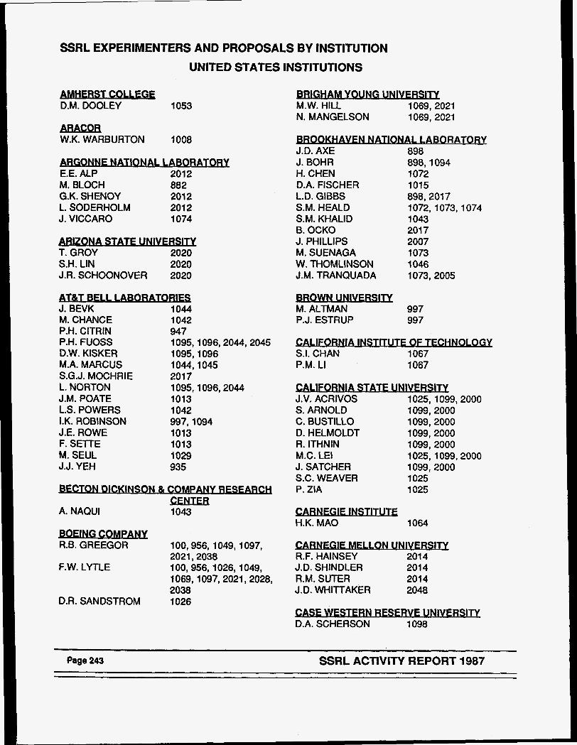
SSRL EXPERIMENTERS AND PROPOSALS BY INSTITUTION
UNITED STATES INSTITUTIONS - D.M. DOOLEY
ARACOR W.K. WARBURTON
1053
1008
GONNE NATlONAl , WORATORY E.E. ALP 201 2 M. BLOCH 882 G.K. SHENOY 201 2 L. SODERHOLM 2012 J. VICCARO 1074
3 T. GROY 2020 S.H. LIN 2020 J.R. SCHOONOVER 2020 - J. BEVK 1044 M. CHANCE 1042 P.H. ClTRlN 947 P.H. FUOSS 1095,1096,2044,2045 D.W. KISKER 1095,1096 M.A. MARCUS 1044,1045 S.G.J. MOCHRIE 201 7 L. NORTON 1095,1096,2044 J.M. POATE 1013 L.S. POWERS 1 042 I.K. ROBINSON 997,1094 J.E. ROWE 1013 F. SETTE 1013 M. SEUL 1029 J.J. YEH 935
BFCTON DICKINSON & C W A N Y R E S F A W
A. NAQUl 1 043 CENTER
100,956,1049,1097, R.B. GREEGOR 2021,2038
F.W. LYTLE 100,956,1026,1049, 1069,1097,2021,2028, 2038 1026
F
D.R. SANDSTROM
A M.W. HILL 1069,2021 N. MANGELSON 1069, 2021
00- NATIONAL I ARORATORY J.D. AXE 898 J. BOHR 898,1094 H. CHEN 1072 D.A. FISCHER 1015 L.D. GIBBS 898,2017 S.M. HEALD 1072, 1073,1074 S.M. KHALID 1 043 B. OCKO 201 7 J. PHILLIPS 2007 M. SUENAGA 1073 W. THOMLINSON 1046 J.M. TRANQUADA 1073,2005 - M. ALTMAN P.J. ESTRUP
997 997
CALIFORNIA INSTITUTF OF TF,CHNOLOGY S.I. CHAN 1067 P.M. LI 1067
ORNlA STATF UNIVERSITY J.V. ACRIVOS 1025,1099,2000 S. ARNOLD 1099,2000 C. BUSTILLO 1099,2000 D. HELMOLDT 1099,2000 R. ITHNIN 1099,2000 M.C. LEI 1025,1099,2000 J. SATCHER 1099,2000 S.C. WEAVER 1025 P. ZIA 1025 - H.K. MA0 1064
C A W E G E M U O N UWERSITY R.F. HAINSEY 201 4 J.D. SHINDLER 201 4 R.M. SUTER 2014 J.D. WHITTAKER 2048
SF WESTERN RESFRVE UNIVFRSITY D.A. SCHERSON 1 098
Page 243 SSRL ACTIVITY REPORT 1987
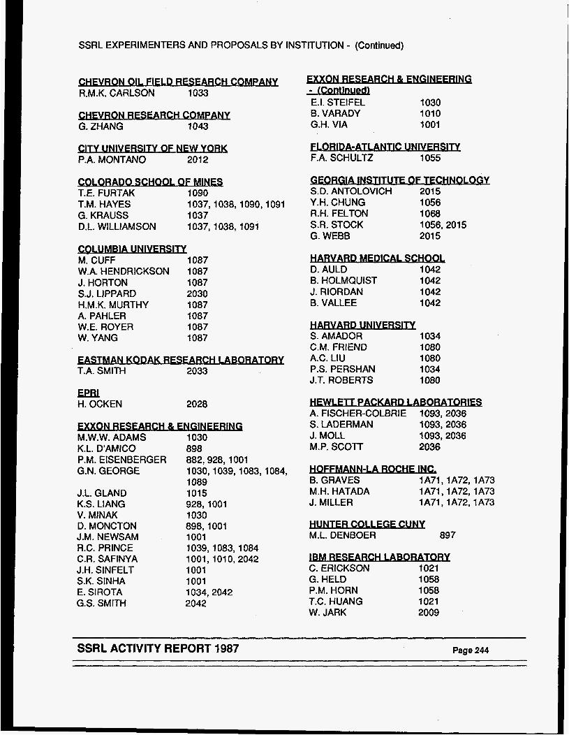
SSRL EXPERIMENTERS AND PROPOSALS BY INSTITUTION - (Continued)
CHF,VRON Qll F-CH COndpANY R.M.K. CARLSON 1033
VRON m C H CQMPANY G. ZHANG 1 043
P.A. MONTAN0 2012
4 T.E. FURTAK 1090 T.M. HAYES 1037,1038,1090,1091 G. KRAUSS 1037 D.L. WILLIAMSON i 037,1038,1091 - M. CUFF 1087 W.A. HENDRICKSON 1887 J. HORTON 1087 S.J. LIPPARD 2030 H.M.K. MURTHY 1087 A. PAHLER 1087 W.E. ROYER 1087 W. YANG 1087
STMAN KODAK RESFARCH LABORATORY T.A. SMITH 2033
E m H. OCKEN 2028
M.W.W. ADAMS K.L. D'AMICO P.M. EISENBERGER G.N. GEORGE
J.L. GLAND K.S. LIANG V. MINAK D. MONCTON J.M. NEWSAM R.C. PRINCE C.R. SAFINYA J.H. SINFELT S.K. SINHA E. SIROTA G.S. SMITH
XON RESFARCH 81 ENGlNEERlNG 1030
882,928, 1001 1030,1039,1083,1084, 1089 1015 928,1001 1030 898,1001 1001 1039,1083,1084 1001,101 0,2042 1001 1001 1034,2042 2042
898
E.I. STEIFEL 1030 B. VARADY 1010 G.H. VIA 1001
FLORIDA-ATI ANTIC UNlVFRSlTY F.A. SCHULTZ 1055
ORGlA INSTITUTF OF TECHNQLOGY S.D. ANTOLOVICH 201 5 Y.H. CHUNG 1056 R.H. FELTON 1068 S.R. STOCK 1056,2015 G. WEBB 201 5
1 D. AULD 1042 B. HOLMQUIST 1042 J. RIORDAN 1042 B. VALLEE 1042 - S. AMADOR 1 034 C.M. FRIEND 1080 A.C. LIU 1080 P.S. PERSHAN 1034 J.T. ROBERTS 1080
A. FISCHER-COLBRIE 1093,2036 S. LADERMAN 1093,2036 J. MOLL 1093,2036 M.P. SCOTT 2036
OCHE INC, B. GRAVES 1A71,1A72, 1A73 M.H. HATADA 1A71 , 1A72,l A73 J. MILLER lA71,1A72,1A73 - M.L. DENBOER 897
lBM RESEARCH LABORATORY C. ERICKSON 1021 G. HELD 1058 P.M. HORN 1058 T.C. HUANG 1021 W. JARK 2009
- SSRL ACTIVITY REPORT 1987 Page 244
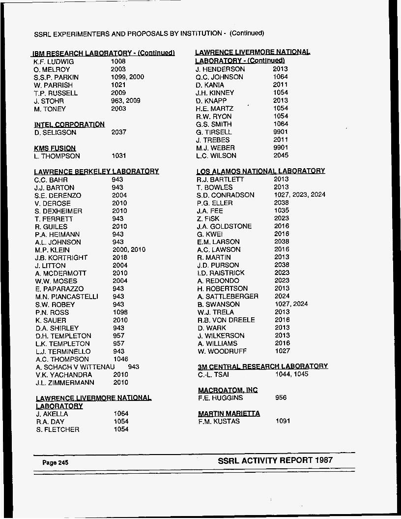
SSRL EXPERIMENTERS AND PROPOSALS BY INSTITUTION - (Continued)
IBM RESFARCHJABORATORY - [Cont[nued) K.F. LUDWIG 1008 0. MELROY 2003 S.S.P. PARKIN 1099,2000 W. PARRISH 1021 T.P. RUSSELL 2009 J. STOHR 963,2009 M. TONEY 2003
2037 - D. SELIGSON
1031 lsMwJsw L. THOMPSON
C.C. BAHR J.J. BARTON S.E. DERENZO V. DEROSE S. DEXHEIMER T. FERRETT R. GUILES P.A. HEIMANN A.L. JOHNSON M.P. KLEIN J.B. KORTRIGHT J. LITTON A. MCDERMOTT W.W. MOSES E. PAPARAZZO M.N. PIANCASTELLI S.W. ROBEY P.N. ROSS K. SAUER D.A. SHIRLEY D.H. TEMPLETON L.K. TEMPLETON L.J. TERMINELLO A.C. THOMPSON
943 943 2004 2010 201 0 943 2010 943 943 2000,201 0 2018 2004 201 0 2004 943 943 943 1098 201 0 943 957 957 943 1046
A. SCHACH V WITTENAU 943 V.K. YACHANDRA 2010 J.L. ZIMMERMANN 2010
LABORATORY J. AKELLA 1064 R.A. DAY 1054 S. FLETCHER 1054
E LIVERMORE NATIONAL ORATORY - IContlnueLU
J. HENDERSON 201 3 Q.C. JOHNSON 1064 D. KANIA 201 1 J.H. KINNEY 1054
201 3 1054
D. KNAPP H.E. MART2 R.W. RYON 1054 G.S. SMITH 1064 G. TIRSELL 990 1 J. TREBES 201 1 M.J. WEBER 990 1 L.C. WILSON 2045
R.J. BARTLETT T. BOWLES S.D. CONRADSON P.G. ELLER J.A. FEE Z. FISK J.A. GOLDSTONE G. KWEl E.M. LARSON A.C. LAWSON R. MARTIN J.D. PURSON 1.0. RAlSTRlCK A. REDONDO H. ROBERTSON A. SATTLEBERGER B. SWANSON W.J. TRELA R.B. VON DREELE D. WARK J. WILKERSON A. WILLIAMS W. WOODRUFF
201 3 201 3 1027,2023,2024 2038 1035 2023 201 6 201 6 2038 201 6 201 3 2038 2023 2023 201 3 2024 1027,2024 201 3 201 6 201 3 201 3 201 6 1027
LABORATORY C.-L. TSAl 1044,1045
956 - F.E. HUGGINS
1091 - F.M. KUSTAS
Page 245 SSRL ACTIVITY REPORT 1987
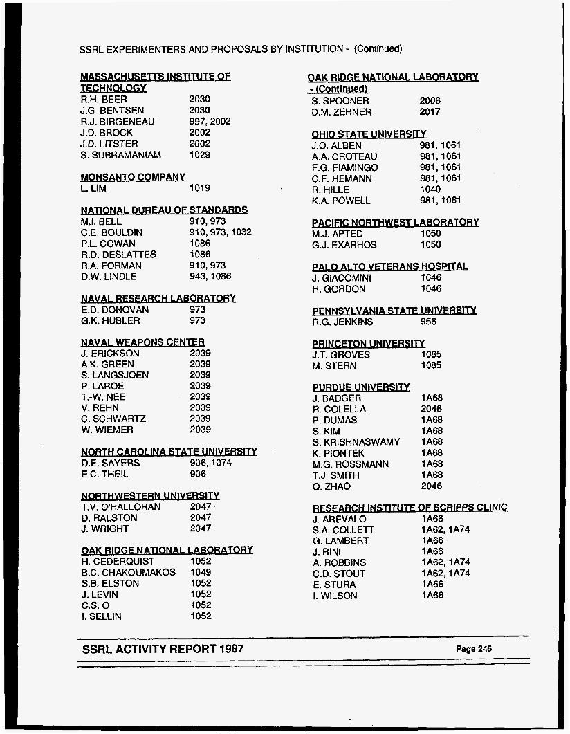
SSRL EXPERIMENTERS AND PROPOSALS BY INSTITUTION - (Continued)
TECHNoLoSY R.H. BEER 2030 J.G. BENTSEN 2030 R.J. BIRGENEAU 997,2002 J.D. BROCK 2002 J.D. LITSTER 2002 S. SUBRAMANIAM 1029 - L. LIM 1019
ONAl BUREAU OF STANDARDS M.I. BELL 91 0,973 C.E. BOULDIN 910,973,1032 P.L. COWAN 1086 R.D. DESLATTES 1086 R.A. FORMAN 910,973 D.W. LINDLE 943,1086
SEARCH LABORATORY E.D. DONOVAN 973 G.K. HUBLER 973 - J. ERICKSON 2039 A.K. GREEN 2039 S. LANGSJOEN 2039 P. LAROE 2039 T.-W. NEE 2039 V. REHN 2039 C. SCHWARTZ 2039 W. WIEMER 2039
3 T.V. OHALLORAN 2047 D. RALSTON 2047 J. WRIGHT 2047
PAK RIDGE NATlONAl 1kWORATORY H. CEDERQUIST 1052 B.C. CHAKOUMAKOS 1049 S.B. ELSTON 1052 J. LEVlN 1052 C.S. 0 1052 1. SELLIN 1052
QAK RIDGE NATIONAL LABORATORY - (Continuedl S. SPOONER 2006 D.M. ZEHNER 201 7 - J.O. ALBEN 981,1061 A.A. CROTEAU 981,1061 F.G. FlAMlNGO 981 , 1061 C.F. HEMANN 981, 1061 R. HILLE 1 040 K.A. POWELL 981,1061
- J.T. GROVES 1085 M. STERN 1085
1 A68 J. BADGER R. COLELLA 2046 P. DUMAS 1 A68 S. KIM 1 A68 S. KRISHNASWAMY lA68 K. PIONTEK 1 A68 M.G. ROSSMANN 1 A68 T.J. SMITH 1 A68 Q. ZHAO 2046
- FARCH INSTITUTF OF SCRIPPS CLINIC
J. AREVALO 1 A66 S.A. COLLETT 1 A62,l A74 G. LAMBERT 1 A66 J. RlNl 1 A66 A. ROBBINS 1A62,1A74 C.D. STOUT 1A62,1A74 E. STURA 1 A66 I. WILSON 1 A66
~~
SSRL ACTIVITY REPORT 1987 Page 246
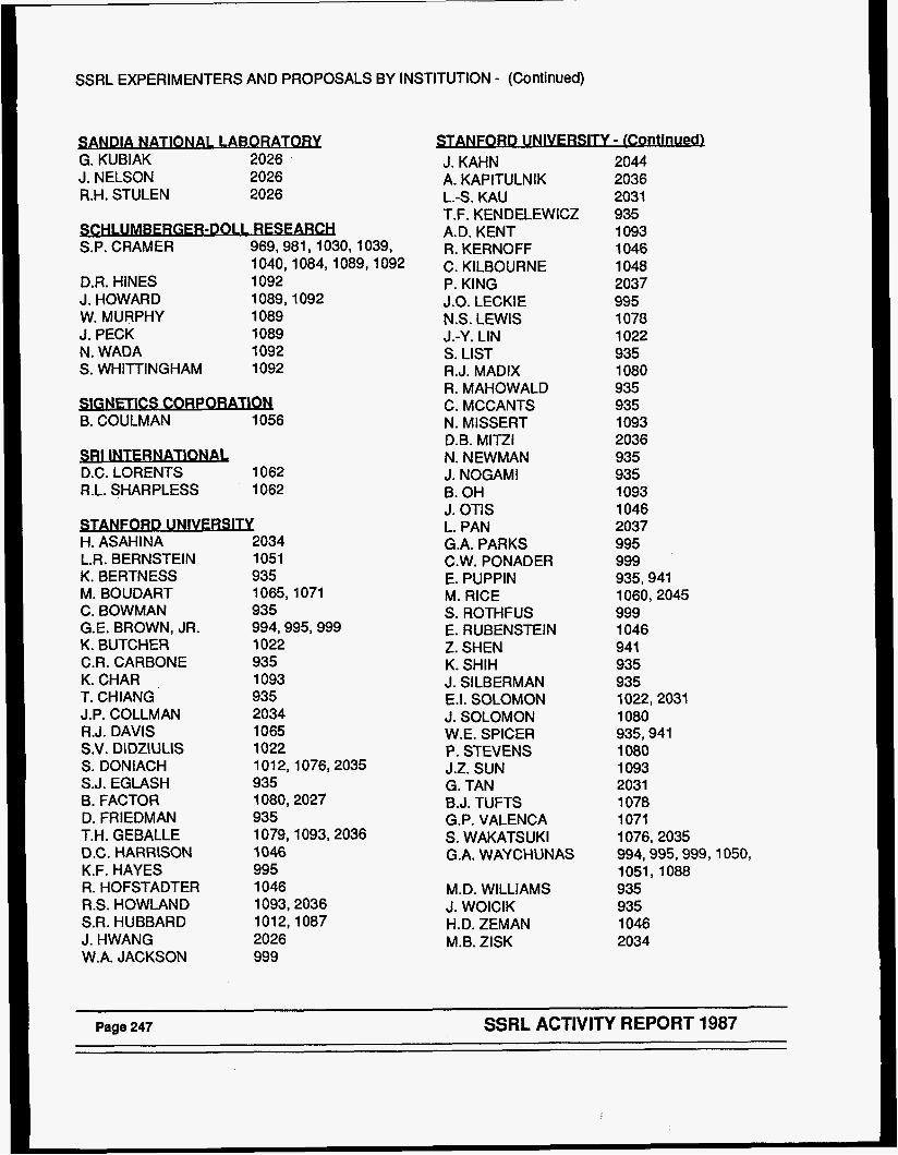
SSRL EXPERIMENTERS AND PROPOSALS BY INSTITUTION - (Continued)
SANDIA NATIONAL LABORATORY G. KUBIAK 2026 J. NELSON 2026 R.H. STULEN 2026
R-DOLL RESEAKH S.P. CRAMER 969,981,1030,1039,
D.R. HINES 1092 J. HOWARD 1089,1092 W. MURPHY 1089 J. PECK 1089 N. WADA 1092 S. WHITTINGHAM 1092
1040,1084,1089,1092
- B. COULMAN 1056
1062 D.C. LORENTS R.L. SHARPLESS 1062
- - H. ASAHINA 2034 L.R. BERNSTEIN 1051 K. BERTNESS 935 M. BOUDART 1065,1071 C. BOWMAN 935 G.E. BROWN, JR. 994,995,999 K. BUTCHER 1022 C.R. CARBONE 935 K.CHAR , 1093 T. CHIANG 935 J.P. COLLMAN 2034 R.J. DAVIS 1065 S.V. DlDZlULlS 1022 S. DONIACH 1012,1076,2035 S.J. EGLASH 935 B. FACTOR 1080,2027 D. FRIEDMAN 935 T.H. GEBALLE 1079,1093,2036 O.C. HARRISON 1046 K.F. HAYES 995 R. HOFSTADTER 1046 R.S. HOWLAND 1093,2036 S.R. HUBBARD 1012,1087 J. HWANG 2026 W.A. JACKSON 999
NFORD UNIVFRSITY - (Continuedl J. KAHN A. KAPITULNIK L.-S. KAU T.F. KENDELEWICZ A.D. KENT R. KERNOFF C. KILBOURNE P. KING J.O. LECKIE N.S. LEWIS
S. LIST R.J. MADIX R. MAHOWALD C. MCCANTS N. MISSERT D.B. MITZI N. NEWMAN J. NOGAMI €3. OH J. OTIS L. PAN G.A. PARKS C.W. PONADER E. PUPPIN M. RICE S. ROTHFUS E. RUBENSTEIN Z. SHEN K. SHIH J. SILBERMAN E.I. SOLOMON J. SOLOMON W.E. SPICER P. STEVENS J.Z. SUN G. TAN 6.J. TUFTS G.P. VALENCA S . WAKATS UK I G.A. WAYCHUNAS
J.-Y. LIN
M. D. WILLIAMS J. WOICIK H.D. ZEMAN M.B. ZlSK
2044 2036 2031 935 1 093 1046 1048 2037 995 1078 1022 935 1080 935 935 1093 2036 935 935 1093 1046 2037 995 999 935,941 1060,2045 999 1046 941 935 935 1022,2031 1080 935,941 1080 1 093 2031 1 078 1071 1076,2035 994,995,999,1050, 1051,1088 935 935 1046 2034
SSRL ACTIVITY REPORT 1987
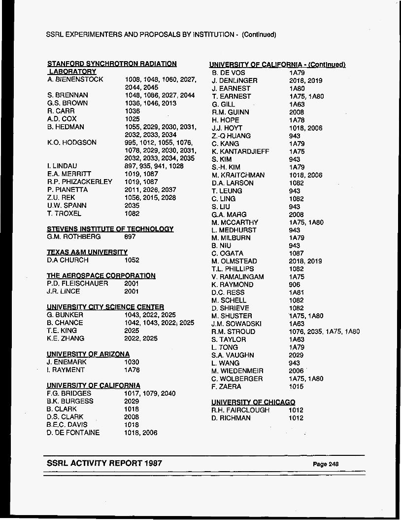
SSRL EXPERIMENTERS AND PROPOSALS BY INSTITUTION - (Continued)
3 LABORATORY
A. BIENENSTOCK 1008,1048,1060,2027,
S. BRENNAN 1048,1086,2027,2044 G.S. BROWN 1036,1046,201 3 R. CARR 1036 A.D. COX 1025 B. HEDMAN 1055,2029,2030,2031,
2032,2033,2034 K.O. HODGSON 995,101 2,1055,1076,
1078,2029,2030,2031, 2032,2033,2034,2035
2044,2045
I. LINDAU 897,935,941,1028 E.A. MERRITT 1019,1087 R.P. PHIZACKERLEY 1019,1087 P. PIANETTA 201 1,2026,2037 Z.U. REK 1056,201 5,2028 U.W. SPANN 2035 T. TROXEL 1082
STEVENS INSTITUTE OF TECHNOLOGY G.M. ROTHBERG 897
TEXAS A&M UNIVERSITY D.A CHURCH 1052
THE AEROSPACE CORPORATION P.D. FLEISCHAUER 2001 J.R. LINCE 2001
UNIVERSITY CITY SCIENCE CENTER G. BUNKER 1043,2022,2025 6. CHANCE 1042,1043,2022; 2025 T.E. KING 2025 K.E. ZHANG 2022,2025
UNIVERSITY OF ARIZONA J. ENEMARK 1030 I . RAYMENT 1 A76
UNIVERSITY OF CALlFORNlA F.G. BRIDGES 101 7,1079,2040 B.K. BURGESS 2029 B. CLARK 1018 D.S. CLARK 2008 B.E.C. DAVIS 1018 D. DE FONTAINE 1018,2006
6. DE VOS J. DENLINGER J. EARNEST T. EARNEST G. GILL R.M. GUINN H. HOPE J.J. HOYT
C. KANG K. KANTARDJIEFF S. KIM
M. KRAITCHMAN D.A. LARSON T. LEUNG C. LING s. LIU G.A. MARG M. MCCARTHY L. MEDHURST M. MILBURN B. NIU C. OGATA M. OLMSTEAD T.L. PHILLIPS V. RAMALINGAM K. RAYMOND D.C. RESS M. SCHELL D. SHRIEVE M. SHUSTER J.M. SOWADSKI R.M. STROUD S. TAYLOR L. TONG S.A. VAUGHN L. WANG M. WlEDENMElR C. WOLBERGER F. ZAERA
Z.-Q HUANG
S.-H. KIM
1 A79 2018,2019 1 A80 1A75,1A80 1 A63 2008 1 A78 1018,2006 943 1 A79 1 A75 943 1 A79 101 8,2006 1082 943 1082 943 2008 1 A75 1 A80 943 1 A79 943 1087 2018,2019 1082 1 A75 906 1 A81 1082 1082 '1 A75,l A80 1 A63 l076,2035,1A75,1A80 1 A63 1 A79 2029 943 2006 1 A75,l A80 1015 -
R.H. FAIRCLOUGH 1012 D. RICHMAN 1012
SSRL ACTIVITY REPORT 1987 Page 248
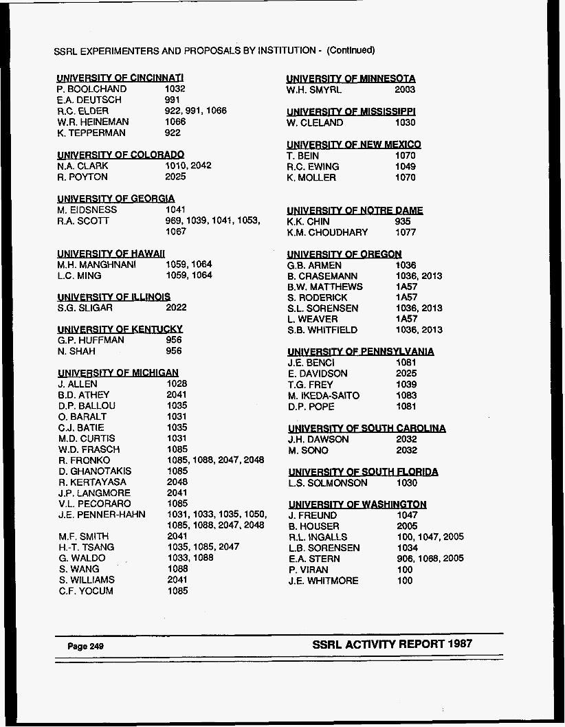
SSRL EXPERIMENTERS AND PROPOSALS BY INSTITUTION - (Continued)
P P. BOOLCHAND 1032 €.A. DEUTSCH 991 R.C. ELDER 922,991,1066 W.R. HEINEMAN 1066 K. TEPPERMAN 922 - N.A. CLARK 1010,2042 R. POYTON 2025 - M. EIDSNESS 1041 R.A. SCOTT 969,1039,1041,1053,
1067
1059,1064 M.H. MANGHNANI L.C. MlNG 1059,1064
- - S.G. SLIGAR 2022 - G.P. HUFFMAN 956 N. SHAH 956
J. ALLEN B.D. ATHEY D.P. BALLOU 0. BARALT C.J. BATIE M.D. CURTIS W.D. FRASCH R. FRONKO D. GHANOTAKIS R. KERTAYASA J.P. LANGMORE V.L. PECORARO J .E. PEN N ER-HAHN
M.F. SMITH
G.WALD0 ~
S. WANG S. WILLIAMS C.F. YOCUM
H.-T. TSANG
1028 2041 1035 1031 1035 1031 1085 1085,1088,2047,2048 1085 2048 2041 1085 1031,1033,1035,1050, 1085,1088,2047,2048 2041 1035,1085,2047 1033,1088 1088 2041 1085
- W.H. SMYRL 2003
P W. CLELAND 1030
b T. BElN 1070 R.C. EWING 1049 K. MOLLER 1070
3 K.K. CHIN 935 K.M. CHOUDHARY 1077 - G.B. ARMEN 1036 B. CRASEMANN 1036,201 3 B.W. MATTHEWS 1 A57 S. RODERICK 1 A57 S.L. SORENSEN 1036,2013 L. WEAVER 1A57 S.B. WHITFIELD 1036,2013
NSYLVANIA J.E. BENCI IO81 E. DAVIDSON 2025 T.G. FREY 1039 M . IKEDA-SAITO 1083 D.P. POPE 1081
N OF SOUTH C- J.H. DAWSON 2032 M. SON0 2032
IlY OF SOUTH FLORlDA L.S. SOLMONSON 1030
A J. FREUND 1047 B. HOUSER 2005 R.L. INGALLS 100,1047,2005 L.B. SORENSEN 1 034 E.A. STERN 906,1068,2005 P. VlRAN 100 J.E. WHITMORE 100
Page 249 SSRL ACTIVITY REPORT 1987
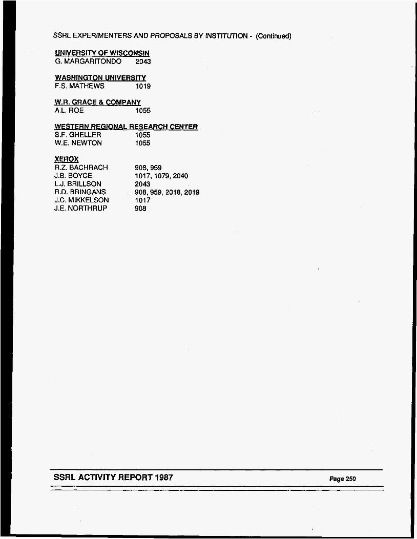
SSRL EXPERIMENTERS AND PROPOSALS BY INSTITUTION - (Continued)
UNIVERSITY OF WISCONSIN G. MARGARITONDO 2043
F.S. MATHEWS 1019
W.R. GRACE & CO MPANY A.L. ROE 1055
WESTERN REGIONAL RESEARCH CENTEQ S.F. GHELLER 1055 W.E. NEWTON 1055
XEROX R.Z. BACHRACH 908,959 J.B. BOYCE 101 7,1079,2040 L.J. BRILLSON 2043 R.D. BRINGANS . 908,959,2018,2019 J.C. MIKKELSON 1017 J.E. NORTHRUP 908
SSRL ACTIVITY REPORT 1987 Page 250
SSRL EXPERIMENTERS AND PROPOSALS BY INSTITUTIONS FOREIGN INSTITUTIONS
ALCAN INTERNATIONAI I (Canada) R. TlMSlT 2027
S INSTITUTE OF TECHNQLOGY (Sweden)
T. CLAESON 1079
CNRS (France) P. GIBART 1038
CTROTFCHNICAI LABORATORY (Japan) H. OYANAGI 2022
J-IC ORSTFD INSTITUTE (Denmark) H.H. ANDERSEN 1094 L.F. GRAABAEK 1094 A. JOHANSEN 1094 E. JOHNSON 1094 L. SARHOLT-KRIST 1094
LSTITUTO DI FlSlCA DEL POI ITFCNIa (Ita&) 1. ABBATI 94 1 M. S. ANCROlTl 941
J S T I T U T O ? W MATERlA (Italy)
P. CHlARADlA 2043 F. COMIN 947
KAROWSKA INSTITUTE (Sweden) B.-M. SJOBERG 1043
(England) M. HART 1021
-SlTY (Sweden) R.I.G. UHRBERG 908
X-PLANCK INSTITUTE FUR FORSCH (Germany)
K. BARTELS 1 A78
MEDI(=AL NOBEL INSTITUTE (Sweden) P. REICHARD 1043
MPG HAMBURG (Germany) K.V. BOEHLEN 1 A78 - (Ita&) L. BRAlCOVlCH 94 1
(Canada) N. ALBERDING 100 K.R. BAUCHSPIESS 100 D. CROZIER 100,1047,2005 A.J. SEARY 100 - (rtalv) M. BELLOTTO 1021 - (Italy) 0. BlSl 941 C. CALANDRA 941 U. DEL PENNINO 941
(W) S. NANNARONE 941
UNIVERSITAT B W (Germany) W. JlTSCHlN 1036 U. WERNER 1036
UNIVERSITAT BONN (Germany) 6. WILL 1021
UNIVERSITY OF S H F F F W (England) P. HARRISON 906
W l T Y OF SUSSEX (England) B.E. SMITH 1030
-ANN INSTITUE (Israel) F. FROLOW 1 A78 1. MAKOWSKI 1 A78 A. YONATH 1 A78 - (PW X.-S ZHANG 943
Page 251 SSRL ACTIVITY REPORT 1987
X SSRL PUBLICATIONS
The following Is a partlal list of papers published or submitted In 1987 based on work at SSRL. The list represents 183 publlcatlons, A list of 1987 theses follows,
Abbati, I., Braicovich, L., Carbone, C., Nogami, J., Lindau, I., Iandelli, I., Olcese, G., Palenzona, A., Photoemission Studies of Mixed Valence in Yb Si Ybsi, and Yb5Si3: Equivalent Versus Inequivalent yb Sites: Solid State Cod 62,' 35 (1987)
Allen, J.W., Kang, J.-S., Lassailly, Y., Maple, M.B., Torikachvilli, M.S., Ellis, W., Pate, B., Lindau, I., Electron Spectroscopy Study of the Heavy Fermion Compound URu2S12: Solid State Corn 61, 183 (1987)
Armen, G.B., Tulkki, J., e r g , T., Crasemann, B., Quantum Theory of Post Collision Interaction in IMer-Shell Photoionization: Final-State Interaction Between !Iho Continuum Electrons: Phys Rev A 36, 5606 (1987)
Amen, G.B., Sorensen, S.L., Whitfield, S.B., Ice, G.E., Levin, J.C., Crasmann, B., Vanishing Post-Collision Interaction During Photon-Excited Coster-Kronig Decay: Phys Rev A 35, 3966 (1987)
Bachrach, R.Z., Bringans, R.D., Olmstead, M.A., Uhrberg, R.I.G., Synchrotron Radiation Studies of MBE Formed Semiconductor Interfaces: Si- and GaAs-Si: Modern Phys Lett B 1, 97 (1987)
Bachrach, R.Z., Bringans, R.D., Olmstead, M.A., Uhrberg, R.I.G., Surface Structure and Interface Formation of Si on GaAs(100): J Vac Sci Techno1 B 5, 1135 (1987)
Bachrach, R.Z., Bringans, R.D., Swartz, L., Lindau, I., Pate, B.B., Carr, R.G., Hower, N., Youngman, B., Morales, H., Pianetta, P., Multi-Wdulator Beam Line V at SSRL: A Progress Report: accepted Nucl Instrum & Methods
Barbee, T.W., Pianetta, P., Tatchyn, R., Redaelli, R., Barbee, T.W. 111, Molybdenum-Silicon Hultilayer Monochromator for the Extreme Ultraviolet: App Phys Lett 50, 644 (1987)
Barton, J.J., Hussain, Z., Shirley, D.A., Genetalized Ramsauer-Townsend Resonance in Angle-Resolved Photoemission Extended Fine-Structure Oscillations: Phys Rev B 35, 933 (1987)
Becker, U., Langer, B., Kerkhoff, H.G., Kupsch, M., Szostak, D., Wehlitz, R., Heimann, P.A., Liu, S.H., Lindle, D.W., Ferrett, T.A., Shirley, D.A., Observation of Many New Argon Valence Satellites Near Threshold: submitted Phys Rev Lett
Bertness, K.A., McCants, C.E., Chiang, T.T., Mahowald, P.H., Wahi, A.K., Kendelewicz, T., Lindau, I., Spicer, W.E., Camparison of Low Intensity Laser Enhancemnt of Oxygen Chemisorption on GaAs Using O2 and N20: Mat Res SOC Sym Proc 75, 575 (1987)
SSRL ACTIVITY REPORT 1987 Page 252
Bertness, K.A., Chiang, T.T., McCants, C.E., Mahowald, P.H., Wahi, A.K., Kendelewicz, T., Lindau, I., Spicer, W.E., Cqrative Uptake Kinetics on N20 and O2 Chemisorption on GaAs(ll0): Surf Sci 185, 544 (1987)
Bienenstock, A., SSRL Development - Recent and Planned: accepted Nucl Instrum & Methods
Bouldin, C.E., Forman, R.A., Bell, M.I., Silicon Photo-Diode Detector for Fluorescence EXWS: Rev Sci Instrum 58, 1891 (1987)
Bouldin, C.E., Forman, R.A., Bell, M.I., Conversion Electron EXAFS Measurements of Ion-Damage GaAs: Phys Rev B35, 1429 (1987)
Bouldin, C.E., Forman, R.A., Bell, M.I., Donovan, E.P., Structural Relaxation in Ion-Damaged Amorphous Ge: submitted Phys Rev B
Boyce, J.B., Bridges, F., Claeson, T., Howland, R.S., Geballe, T.H., X-Ray Absorption Studies of yBa2Cu307-u and GdBa2Cu307-u Superconductors: Phys Rev B 36, 5251 (1987)
Boyce, J.B., Bridges, F., Claeson, T., Geballe, T.H., Chu, C.W., Tarascon, J.M., x-ray Absorption Studies of the High-Tc Superconductors Lal *SrO aCuo4 and Lal.8Bao~2Cu04: Phys Rev B 35, 7203 (1987)
Bringans, R.D., Olmstead, M.A., Uhrberg, R.I.G., Bachrach, R.Z., Sumnary Abstract: Core Level Spectroscopy of the GaAs-on-Si Interface: J Vac Sci Technol A5, 2141 (1987)
Bringans, R.D., Uhrberg, R.I.G., Olmstead, M.A., Bachrach, R.Z., Northrup, J.E., Model Semhxmductor Surfaces: Arsenic Tendnation of the Ge(lll), Si(lll), and Si(100) Surfaces: Physica Scripta T17, 7 (1987)
Bringans, R.D., Olmstead, M.A., Uhrberg, R.I.G., Bachrach, R.Z. , Formation of the Interface Bebeen GaAs and Si: Implications for GaAs-cm-Si Heteroepitaxy: Appl Phys Lett 51, 523 (1987)
Bringans, R.D., Olmstead, M.A., Uhrberg, - R.I.G., Bachrach, R.Z., GaAs-on-Si Epitaxy: Results for Coverage of 1 Monolayer: Mat Res SOC Symp Proc 94, 201 (1987 )
Brown, G.S., Cerino, J., Hofmann, A., Liu, R., Troxel, T., Wang, P., Wiedemann, H., Winick, H., Berndt, M., Brown, R., et al., Operation of PEP in a Low Emittance W e : accepted IEEE
Cao, R., Miyano, K., Kendelewicz, T., Chin, K.K., Lindau, I., Spicer, W.E., Kinetics Study of Initial Stage Barxi Bending at Metal GaAs ( 110) Interfaces: J Vac Sci Techno1 B5, 998 (1987)
Carey, G.P., Friedman, D.J., Lindau, I., Spicer, W.E., Photoemission Studies of the mom Temperature Si/Hg1-cd,Te, Si/HgTe and Si/CdTe Interfaces: J Vac Sci Technol A5, 3198 (1987)
Chin, K.K., Cao, R., Kendelewicz, T., Miyano, K., Yeh, J.J., Doniach, S., Lindau, I., Spicer, W.E., Photoemission Study of the Physical Nature of the InP Near-Surface Defect states, in Interface, Superlattices, and Thin Films: Mat Res SOC Sym Proc 77, 429 (1987)
Page 253 SSRL ACTIVITY REPORT 1987

Chin, K.K., Cao, R., Kendelewicz, T., Miyano, K., Williams, M.D., Doniach, S., Lindau, I., Spicer, W.E., Transition from Schottky L M t to Bardeen Limit in the Schottky Barrier Formation of AI. on n- and p-GaAs(ll0) Interfaces: Mat Res SOC Symp Proc 77, 297 (1987)
Chin, K.K., Miyano, K., Cao, R., Kendelewicz, T., Yeh, J., Lindau, I., Spicer, w.E., Chemical Reaction at the In on GaAs (110) Interface: A Synchrotron Radiation PhotOemission Study: J VaC Sci Techno1 B5, 1080 (1987)
Cole, J., Yachandra, V.K., Guiles, R.D., McDermott, A.E., Britt, R.D., Dexheimer, S.L., Sauer, K., Klein, M.P., Assignment of the 9-4.1 EPR Signal to Manganese in the S2 State of the Photosynthetic OxYgen-Evolving Complex: An X-Ray Absorption Edge Spectroscopy Study: Biophysical Acta 890, 395 (1987)
Cole, J., Yachandra, U.K., McDermott, A.E., Guiles, R.D., Britt, R.D., Dexheimer, S.L., Sauer, K., Klein, M.P., Structure of the Mn Cuiuplex of Photosystem I1 Upon Remval of the 33 Kilodalton Extrinsic Protein: An X-Ray Absorption &&e Spectroscopy Study: Biochemistry 26, 5967 (1987)
Comelli, G., Stohr, J., Pate, B., Extended X-ray Absorption Fine Structure Studies of Diamond and Graphite: submitted Phys Rev B
Conradson, S.D., Burgess, B.K., Hodgson, K.O., Newton, W.E., Mortenson, L.E., Structural Studies of the Holybdenum Site in the M F e Protein and its FeHo-Cofactor by EXAFS: J Am Chem SOC 109, 7507 (1987)
Cox, L.E., Ellis, W.P., Cowan, R.D., Allen, J.W., Oh, S.-J., Lindau, I . , Pate, B.B., Arko, A.J., Valence43and Jhtoemission in u02(111) Near the 5d Resonant Photon Energy: Phys Rev B 35, 5161 (1987)
Cramer, S.P., Eidsness, M.K., Pan, W.-H., Morton, T.A., Ragsdale, S.W., DerVartanian, D.V., Ljungdahl, L.G., Scott, R.A., X-Ray Absorption Spectroscopic Evidence for a Unique Nickel Site in Clostridium Thennoaceticum Carbon Monoxide Dehydrogenase: Inorg Chem 26, 2477 (1987)
Crozier, E.D., Alberding, N., Bauchspiess, K.R., Seary, A.J., Gygax, S., Structure of YBa2Cu307,6 vs Temperature by X-ray Absorption Spectroscopy: Phys Rev B
Doniach, S., Chin, K.K., Lindau, I., Spicer, W.E., Microscopic Metal Cluster and Schottky Barrier Formation: Phys Rev Lett 58, 591 (1987)
Eberhardt, W,, Plummer, E.W., Lyo, I.W., Carr, R., Ford, W.K., Auger Electron Ion Coincidence Studies of Soft X-Ray Induced Fragmentation of Nz: submitted Phys Rev Lett
Eberhardt, W., Core Electron Excitation and Decay in Molecules: submitted Phys Scripta
Elder, R.C., Eidsness, M.K., Synchrotron X-ray Studies of Metal-Based Drugs and Metabolites: Chem Rev 87, 1027 (1987)
Ellis, W.P., Albers, R.C., Allen, J.W., Lassailly, Y., Kang, J.-S., Pate, B.B., Lindau, I., Valence-Eland Densities of State in NiAs: Solid State Corn 62, 591 ( 1987 )
SSRL ACTIVITY REPORT 1987 Page 254
Ericson, A., Hedman, B., Hodgson, K.O., Green, J., Dalton, H., Bentsen, J.G., Beer, R.H., Lippard, S.J . , Structural Characterization by EXAFS Spectroscw of the Binuclear Iron Center in component A of Hethane Honooxygenase fram Methylococcus capsulatus (Bath): in press J Amer Chem SOC
Feldman, R.D., Austin, R.F., Fuoss, P.H., Dayem, A.H., Westerwick, E.H., Nakahara, S., Boone, T., Menendez, J., Pinczuk, A., Valladares, J.P., Brennan, S., Phase Separation in Cdl-xZnxTe Grown by Molecular-Beam bpitaxy: J Vac Sci Techno1 B 5, 690 (1987)
Ferrett, T.A., Heimann, P.A., Kerkhoff, H.G., Becker, U., Lindle, D.W., Shirley, D.A., Photoelectron Asymnetry Parameter for the S 2p Level of SO2: Chem Phys Letts 138, 607 (1987)
Ferrett, T.A., Piancastelli, M.N., Lindle, D.W., Heimann, P.A., Medhurst, L.J., Liu, S.H., Shirley, D.A., Resonance Effects on the Snner-Valence Levels of SF6 in the Photon-Energy Range 52-72 eV: Chem Phys Lett 134, 146 (1987)
Ferrett, T.A., Lindle, D.W., Heimann, P.A., Brewer, W.D., Becker, U., Kerkhoff, H.G., Shirley, D.A., Lithium 1s Main-Line and Satellite Photoemission: Resonant and Nonresonant Behavior: Phys Rev A 36, 3172 (1987)
Frank, P., Hedman, B., Carlson, R.M.K., Tyson, T.A., Roe, A.L., Hodgson, K.O., A Large Reservoir of Sulfate and Sulfonate Resides within Plasma Cells from Ascidia ceratodes, Revealed by X-ray Absorption Near-Edge Structure Spectroscopy: Biochem 26, 4975 (1987)
Friedman, D.J., Carey, G.P., Lindau, I., Spicer, W.E., Wilson, J.A., Overlayer-Cation Reaction at the Pt/tfgl - xCdxTe Interface: Phys Rev B 35, 1188 ( 1987 )
Friedman, D.J., Carey, G.P., Lindau, I., Spicer, W.E., Systematics of Metal Contacts to Hgl - xCdxTe: J Vac Sci Technol A!5, 3190 (1987)
Friend, C.M., Stevens, P.A., Serafin, J.G., Baldwin, E.K., Madix, R.J., Bonding and Absorption Structure of CO on W(100)-(5xl)-C: submitted J Chem Phys
Fuller, A . , Tatchyn, R., Csonka, P., Lindau, I., Studies of Interfacial Structure in WC Multilayers: SPIE 733, 286 (1987)
Giauque, R.D., Jaklevic, J.M., Thompson, A.C., Biological Trace-Element Measurements Using Synchrotron Radiation: Biological Trace Element Reseach 12, 185 (1987)
Guiles, R.D., Yachandra, V.K., McDermott, A.E., Dexheimer, S.L., Sauer, K., Klein, M.P., EXAFS and XANES Studies of Manganese Complexes: Models for the Photosynthetic Oxygen-Evolving Complex: submitted
Guiles, R.D., Cole, J., McDermott, A.E., Yachandra, V.K., Britt, R.D., Dexheimer, S.L., Sauer, K., Klein, M.P., Structure of Manganese in the SO State of Photosynthetic Oxygen-Evolving Complex: submitted
Guiles, R.D., Yachandra, V.K., Cole, J., McDermott, A.E., Dexheimer, S.L., Sauer, K., Klein, M.P., Structure of the S3 State of the Photosynthetic wen Evolving Complex : submitted
Page 255 SSRL ACTIVITY REPORT 1987
Gygax, S., Crozier, E.D., Alberding, N., Bauchspiess, K.R., Seary, A.J., Temperature-Dependent EXAFS Spectra of yBa2Cu307-,,: Jpn J Appl Phys 26, 1051 (1987)
Hahn, S., Arst, M., Rek, Z.U., Stojanoff, V., Bulla, D.A., Castro, W.E., Jr., Tiller, W.A., Effects of 450 C Thermal Annealing upon Oxygen Precipitation in Heavily B- and SMoped Czochralski Si: Appl Phys Lett 50, 401 (1987)
Hart, M., Parrish, W., Bellotto, M., Lim, G.S., The Refractive Index Correction in Powder Diffraction: submitted Acta Crystallogr
Hayes, K.F., Roe, A.L., Brown, G.E., Hodgson, K.O., Leckie, J.O., Parks, G.A., In-Situ X-Ray Absorption Study of Surface Complexes: Selenium Oxyanions on Alpha-FeoH: Science 238, 783 (1987)
Hedman, B., Frank, P., Gheller, S.F., Roe, A.L., Newton, W.E., Hodgson, K.O., New Structural Insights into the Iron-Holybdernmn Cofactor from Azotobacter Vinelandii Nitrogenase through Sulfur K and Molybdenum L X-ray Absorption Edge Studies: in press J Amer Chem SOC
HeimaM, P.A., Lindle, D.W., Ferrett, T.A., Liu, S.H., Medhurst, L.J., Piancatelli, M.N., Shirley, D.A., Becker, U., Kerkhoff, H.G., Langer, B., et al., Shake-off on Inner-Shell Resonances of AT, Kr and Xe: J Phys B: At Mol Phys' 20, 5005 ( 1987)
Hernandez, P., Lamelas, F., Clarke, R., Dimon, P., Sirota, E.B., Sinha, S.K., Time-Resolved X-Ray Scattering Study of Ordering Kinetics in SbC15-Intercalated Graphite: Phys Rev Lett 59, 1220 (1987)
Hitchcock, A.P., Stohr, J., K-Shell Shape Resonances and Intramolecular Bond Lengths Camoents on "The Relationship Between Shape Resonances and Bond Lengths": J Chem Phys 87, 3253 (1987)
Horsley, J.A., Wachs, I.E., Brown, J.M., Via, G.H., Hardcastle, F.D., Structure of Surface Tungsten Oxide Species in the MI x-ray Absorption Near-Edge Spectroscopy and 4014 (1987)
Supported Oxide System from J Phys Chem 91,
Huang, T.C., Toney, M.F., Brennan, S., Rek, Z., Analysis of C d p e d Iron Oxide Thin Films by Synchrotron Radiation: Thin Solid Films 154, 439 (1987)
Huggins, F.E., Huffman, G.P., Shah, N., Jenkins, R.G., Lytle, F.W., Greegor, R.B., Further EXWS E%dnation of the State of Calcium in Pyrolyzed Char: in press Fuel
Huggins, F.E., Shah, N., Huffman, G.P., Lytle, F.W., Greegor, R.B., Jenkins, R.G., In-Situ EXAE'S Investigation of Ca and K Catalytic Species During Pyrolysis and Gasification of Lignite Chars: in press Fuel
Hwang, J., Shih, C.K., Pianetta, P., Spicer, W.E., Pao, Y.-C., Harris, J.S., Determination of the Natural Valence Band Offset in the InxGal - System: Appl Phys Lett 51, 1632 (1987)
Ingalls, R., Crozier, E.D., Alberding, N., Seary, A.J., Bauchspiess, K.R., Houser, B., Freund, J., X-ray Absorption Spectroscopy at High Pressure: Naukuva Dumka
SSRL ACTIVITY REPORT 1987 Page 256
Jaklevic, J.M., Bouret, E., Scott, M.P., Laderman, S.S., Application of -le Monochromatic Synchrotron Radiation to Topographic Studies of Dislocations in GaAs: in preparation
Joensen, P., Crozier, E.D., Alberding, N., Frindt, R.F., A Study of Single-layer and Restacked Nos2: J Phys C 20, 4043 (1987)
Kalkowski, G., Kaindl, G., Brewer, W.D., Krone, W., Near Mge X-ray Absorption Fine Structure in Uranium Cuqounds: Phys Rev B 35, 6 (1987)
Kang, J.S., Allen, J.W., Maple, M.B., Torikachvili, M.S., Pate, B., Ellis, W., Lindau, I., Spectral Evidence for the Importance of Single-Site Effects in Heavy-Fermion Uranium Materials: Phys Rev Lett 59, 493 (1987)
Kendelewicz, T., Mahowald, P.H., McCants, C.E., Bertness, K.A., Lindau, I., Spicer, W.E., Chemical Reaction and Schottky Barrier Formation at the Ti/InP (110) and Sn/InP (110) Interfaces: Reactive Versus Nonreactive Case: J Vac Sci Techno1 B 5, 1033 (1987)
Kendelewicz, T., Mahowald, P.H., Bertness, K.A., McCants, C.E., Lindau, I., Spicer, W.E., Surface Shifts in the In 4d and 2p Core Level Spectra of InP(110): Phys Rev B 36, 6543 (1987)
King, P., Pan, L., Pianetta, P., Shimkunas, A., Mauger, P., Seligson, D., X-ray Induced Damage in Boron Nitride, Silicon, and Silicon Nitride Lithography Masks: SPIE 773, 126 (1987)
Kortright, J.B., Fischer-Colbrie, A., Standing Wave Enhanced Scattering in Multilayer Structures: J Appl Phys 61, 1130 (1987)
Kortright, J.B., Denlinger, J., Study of Atomic Arrangements in Rnrgsten-Carbon Multilayers of X-ray Optical Interest: in preparation
Kortright, J.B., Bienenstock, A., X-ray Structural Study of Amorphaus Ho-Ge Films: accepted Phys Rev B
Kortright, J.B., Laderman, S.S., Scott, M.P., Fischer-Colbrie, A., Study of Defects in GaAs on Si (100) by X-Ray Diffraction Techniques: submitted
Laderman, S.S., Kortright, J., Scott, M., Fischer-Colbrie, A., Grazing Incidence X-Ray Scattering Study of Defect Structures in M5E -/Si Using Synchrotron Radiation: Mat Res SOC Sym Proc 91, 143 (1987)
Lassailly, Y., Allen, J.W., Ellis, W., Cox, L., Pate, B., Fisk, Z., Lindau, I., Electron Spectroscopy Study of the Heavy Fermion Campnmd Uz%7: J Magn & Magn Mat 63 & 64, 512 (1987)
Li, P.M., Gelles, J., Chan, S . I . , Sullivan, R.J., Scott, R.A., JBtended X-Ray Absorption Fine Structure of Copper in Cu(A)-Depleted pHNJ3-Modified and Native Cytochrome c Oxidases: Biochem 26, 2091 (1987)
Liang, K.S., Laderman, S.S., Sinfelt, J.H., Structure Study of Small Catalytic Particles Using Differential &omalous X-ray Scattering: J Chem Phys 86, 2352 ( 1987)
Liang, K.S., Eisenberger, P., Grazing Incidence X-ray Scattering Study of Surface Defects: Mat Res SOC Symp Proc 82, 493 (1987)
Page 257 SSRL ACTIVITY REPORT 1987
Lim, G., Parrish, W., Ortiz, C., Bellotto, M., Hart, M., Grazing Incidence Synchrotron X-Ray Diffraction Method for Analyzing Thin Films: J Mater Res 2, 471 (1987)
Lindau, I., Interfacial Properties of Metal Overlayers on 111-V C-s: Vacuum 37, 385 (1987)
Lindle, D.W., Heimann, P.A., Ferrett, T.A., Piancastelli, M.N., Shirley, D.A., Photoemission Study of Kr 3r np Autoionization Resonances: Phys Rev A 35, 4605 ( 1987 )
Lindle, D.W., Heimann, P.A., Ferrett, T.A., Shirley, D.A., Helium Photoelectron Satellites: Low Energy Behavior of the n-3-5 Lines: Phys Rev A 35, 1128 (1987)
Lindle, D.W., Ferrett, T.A., Heimann, P.A., Shirley, D.A., Complete Photoemission Study of the He Is2 3s3p Autoionizing Resonance: Phys Rev A 36, 2112 (1987)
Lindle, D.W., Ferrett, T.A., Heimann, P.A., Shirley, D.A., Photoemission fram Xe in the Vicinity of the 4d Cooper Minimum: submitted Phys Rev A
List, R.S., Woicik, J., Mahowald, P.H., Lindau, I., Spicer, W.E., The Si- (110) Heterojunction Discontinuity: Amorphous Versus CryStalline Overlayers: J Vac Sci Technol A5, 1459 (1987)
List, R . S . , Woicik, J.C., Lindau, I., Spicer, W.E., The Si- (110) Heterojunction: Strain, Disorder, and Valence-Band Discontinuity: J Vac Sci Technol B 5, 1279 (1987)
Ludwig, K.F., Warburton, W.K., Wilson, L., Bienenstock, A.I., Liquid GeBr . 1.- A Test of the Anomalous X-ray Scattering Method as Applied to Disofdered Materials: J Chem Phys 87, 604 (1987)
Ludwig, K.F., Wilson, L., Warburton, W.K., Bienenstock, A.I., Liquid GeBr4. 11. - Molecular Packing and Liquid Structures: J Chem Phys 87, 613 (1987) Lytle, F.W., Greegor, R.B., Panson, A. J., Discussion of X-Ray Absorption Near mge Structure: Application to Cu in the High T(c) Superconductors Lal,8Sro2Cu04 and YBa2Cu307: submitted Phys Rev B
McCants, C.E., Kendelewicz, T., Bertness, K.A., Mahowald, P.H., Williams, M.D., List, R.S., Lindau, I., Spicer, W.E., Chemical and Electronic Properties of the Pt/GaAs (110) Interface: J Vac Sci Technol B 5, 1083 (1987)
Meitzner, G., Via, G.H., Lytle, F.W., Sinfelt, J.H., Structure of Bimetallic Clusters Extended X-ray Absorption Fine Structure (EMLFS) of Pt-Re and Fd-Re Clusters: J Chem Phys 87, 6354 (1987)
Meitzner, G., Via, G.H., Sinfelt, J.H., Lytle, F.W. , Extended - X-ray Absorption Fine Structure Spectroscopic (EXAFS) Studies of Model Reforming Catalysts: Petroleum Chem 32, 733 (1987)
Moon, D.W., Cameron, S., Zaera, F., Eberhardt, W., Carr, R., Bernasek, S.L., Dwyer, D.J., A Titled Precursor for CO Dissociation on the Fe(100) Surface: Surf Sci 180, L123 (1987)
SSRL ACTIVITY REPORT 1987 Page 258

Nogami, J., Friedman, D.J., KendePew%czr, %! BE, W.E., Bidling Energy Shifts from Alloying at I W x l f l S - W m tor Xnterfacesr: J Vac S C i Techno1 A5, 591 (1987)
Olmstead, M.A. , Uhrberg, R.I.G, I ~ ~ i ~ g ~ ~ ~ ~ R.D, I ~~~~~~~~~ R.Z,, Phtmdss3.m Study of Bonding at the CaF2+Si(Ifl): Phys R@v El 35, 4526 (1987)
Olmstead, M.A., Bringans, R.D.t Uhrberg, R.I.G.# Bachrach, R.Z., Bonding at the CaF2-on-Si(111) Interface: Mat Res SOC Symp Proc 94, 195 (1987)
Outka, D.A., Stohr, J., Jark, W., Stevens, P., Solomon, J., Madix, R.J., Orientation and Bond Length of Molecular n on Ag(110) and Pt(1ll): A NEKAFS Study: Phys Rev B 35, 4119 (1987)
Outka, D.A., Stohr, J., Rotermund, H.H., Madix, R.J., Solomon, J., Hermsmeier, B., NEXAFS Studies of Complex Alcohols $Ifd Carboxylic Acids on the Si(111)(7x7) Surface: Surf Sci 185, 53 (1987)
Outka, D.A., Stohr, J., Rabe, J., Swalen, J.D., Rotermd, H.H., Orientation of Arachidate Chains in Langmir-Bldgett Monolayers on Si(ll1): Phys Rev Lett 59, 1321 (1987)
Outka, D.A., Stohr, J., Madix, R.J. Rotermund, H.H. Hermm&er, Solomon, J. , NEJW’S Studies of unsaturated Carboxylic Acids cohols Msorbed on the Si(111) Surface: Mat Res SOC Synp Proc 90, (1987)
Parrrish, W., Hart, M., Advantages of Diffractamtry: submitted 2 KristaElogr
Parrish, W., Advances in Synchrotron PoLycrystaPliae Diffraction: submitted Aust J Phys
Parrish, W., Hart, M., Hua T.C., Be%lotto, M., ttice Paranreter Determination using synchrotron r mta: accepted pldv X-
Parrish, W., Hart, M., Accurate Measurement of E Diffraction Intensities using Synchrotron Radiation: submitted Aust J Phys
Pearton, S.J., Poate, J.M., Sette, F., Gibson, J.M., Jacobson, D.C., Ion Implantation in GaAs: Nucl Instrum (E Methds Phys Res 19, 369 (1987)
Penner-Hahn, J.E., Murata, M., Freeman, H.C., Hodgson, K.O., Low “eqerature X-ray Absorption Spectroscopy of Plast in: submitted J Am Chern SOC
Persson, I., Penner-Hahn, J.E., Hodgs orption Spectroscopy of Mercury(I1) Halide Ccnnplexes in DM50 itted Pnerg Chem
Piestrup, M.A., Moran, M.J., Berman, B.L., Piarnetta, P., Seligson, D., Transition Radiation as an X-ray Source f O K Ei raphp: SPIE 733, 37 (1987)
Puppin, E., Nogami, J., Carbone, C., Shen, Z.X., Eindau, I., Pate, B,, Abbati, I., Braicovich, L., A Photoemission Study of the S i (Ill)/Gd Interface: A Cornparison w i t h Bulk Silicides: J Vac Sci Techno1 €3 5, 1083 (1987)
Robey, S.W., Barton, J.J., Bahr, C.C., L i u , G., Shirley, D.A., An kagle-Resolved plaotoemission Extendled FineStmeture ctrsscopy Investigation of c(zXZ)S/Ni(Oll): Phys Rev B 35, 1108 (1987)
Page 259 L A~~~~~~~ REPORT 1987

Robey, S.W., Bahr, C.C., Hussain, Z . , Leung, K.T., Lou, J., Schach von Witenau, A.E., Barton, J.J., Surface Structure of (2x2)S/Ge(lll) Determined by Angle-Resolved Photoemission Pine Structure: Phys Rev B 35, 5657 (1987)
Robinson, C.J., Samant, M.G., Stohr, J., Speriosu, V.S., Guarnieri, C.R., Cuomo, J.J., Structural Studies of Hydrogenated Aumrphaw; Carbon Infrared Coatings: Mat Res SOC Sym Proc, 90 (1987)
Robinson, I.K., Symetry of the Si(111)7x7 at an a-Si Interface: Phys Rev B 35, 3910 (1987)
Rotermund, H.H., Hermsmeier, B., Solomon, J., NEILAFS Studies of Complex rrlcohols and Carboxylic Acids on the Si(ll1) Surface: Surf Sci 185, 53 (1987)
Rubenstein, E., Synchrotron Radiation for Angiography: Ann Rev Biophy Biophy Chem 16, 161 (1987)
Rubenstein, E., Brown, G.S., Giacomini, J.C., Gordon, H.J., Hofstadter, R., Kernoff, R.S., Otis, J.N., Thomlinson, W., Thompson, A.C., Zeman, H.D, Angiograhy using Synchrotron Radiation: in press Phys Scripta
Samant, M.G., Borges, G.L., Gordon, J.G., Melroy, O.R., Blum, M., In-Situ Surface Extended X-Ray Absorption Fine Structure Spectroscopy of a Lead -layer at a Silver(P11) Electrode/Electrolyte Interface: Amer Chem Soc 109, 5970 (1987)
Savborg, O., Schoonover, J.R., Lin, S.H., wring, L., Time-Resolved X-ray Diffraction by Synchrotron Radiation: The T h e m 1 Decomposition of a(-) Powders: J Solid State Chem 68, 214 (1987)
Sette, F., Pearton, S.J . , Poate, J.M., Rowe, J.E., Extended X-Ray Absorption Fine Structure Studies by Soft X-Ray Fluorescence Detection: Physica Scripta T17, 209 (1987)
Sette, F., Pearton, S . J . , Poate, J.M., Rowe, J.E., Local Structure of S Ilpplrities in Implanted GaAs: Nucl Instrum & Methods Phys Res 19, 408 (1987)
Shen, Z.-X., Allen, J.W., Yeh, J.J., Kang, J.-S., Ellis, W., Spicer, W.E., Lindau, I., Maple, M.B., Dalichaouch, Y.D., Torikachvili, M.S., et al, Anderson Hamiltonian Description of the Experimental Electronic Structure and Magnetic Interactions of Copper Oxide Superconductors: Phys Rev B 36, 8414 (1987)
Shih, C.K., Silberman, J.A., Wahi, A.K., Carey, G.P., Lindau, I., Spicer, W.E., Berding, M.A., Sher, A., Angle Resolved Photoemission Study of the Alloy Scattering Effect in Hgl - xCdxTe: J Vac Sci Techno1 A5, 3026 (1987)
Short, R.T., 0, C.-S, Levin, J.C., Sellin, I.A., Johnson, B.M., Meron, M., Jones, K.W., Church, D.A., Synchrotron Radiation Inner-Shell Photoionization of Atomic and Molecular Gases: Nucl Instrum Methods B24/25, 417 (1987)
Siedle, A.R., Markell, C.G., Lyon, P.A., Hodgson, K.O., Roe, A.L., Bifunctional Rhodimcm&alate Catalysts: Inorg Chem 26, 219 (1987)
Sirota, E.B., Pershan, P.S., Amador, S., Sorensen, L.B., Synchrotron X-ray Observation of Surface Smectic-I Hexatic Layers on Smectic4 Liquid Crystal Films: Phys Rev A 35, 5 (1987)
SSRL ACTIVITY REPORT 1987 Page 260
Sirota, E.B., Pershan, P.S., Sorensen, L.B., Collett, J., X-Ray and Optical Studies of the mickness Dependence of the Phase Diagram of Liquid-crystal Films: Phys Rev A 36, 2890 (1987)
Stohr, J., Outka, D.A., Madix, R.J., Near mge X-ray Absorption Fine Structure Studies of Molecules and Molecular Chains Bonded to Surface: J Vac Sci Techno1 A5, 919 (1987)
Stohr, J., Outka, D.A., Determination of Molecular Orientations on Surfaces from the Angular Dependence of NEXWS Resonances: Phys Rev B36 7891 (1987)
Stohr, J., Outka, D.A., Baberschke, K., Arvanitis, D., Horsley, J.A., Identification of C-H Resonances in the K-Shell Excitation Spectra of Gas Phase, Chemisorbed and Polyaneric Hydrocarbons: Phys Rev B 36, 2967 (1987)
Stura, E.A., Feinstein, A., Wilson, I.A., Crystallization and Preliminary Crystallographic Data for an Antiprogesterone lrIonoclonal Antibody Fab' and Steroid-Fab' Copppzlexes: J M o l Biol 193, 229 (1987)
Stura, E.A., Arevalo, J.H., Feinstein, A., Heap, R.B., Taussig, M.J., Wilson, I.A., Analysis of an Anti-Progesterone Antibody: Variable Crystal lvbrphology of the Fab' and Steroid-Fab' Camplexes: Immunology 62, 511 (1987)
Tatchyn, R., Lindau, I., Off-Axis Radial Properties of m a t o r Light: SPIE 733, 115 (1987)
Templeton, D.H., Templeton, L.K., X-ray Birefringence, Forbidden Reflections, and Direct Observation of Structure-Factor Phases: Acta Cryst A43, 573 (1987)
Thompson, A.C., WU, Y., Underwood, J.H., Barbee, T.W., Focusing of Synchrotron Radiation X-Ray Beams Using Synthetic Hdtilayer Mirrors: Nucl Instrum Methods A255, 603 (1987)
Thompson, A.C., Zeman, H.D., Rubenstein, E., Otis, J.N., Hofstadter, R., Brown, G.S., Harrison, D.C., Kernoff, R.S., Giacomini, J.C., Gordon, H.J., Thomlinson, w., Transvenom Coronary Angiography in Dogs Using Synchrotron Radiation: J Cardiac Imaging, in press
Thompson, A.C., Hofstadter, R., Otis, J.N., Zeman, H.D., Kernoff, R.S., Rubenstein, E., Giacomini, J.C., Gordon, H.D., Brown, G.S., Thomlinson, W., Transvenous Coronary Angiography using Synchrotron Radiation: in press Nucl Instrum & Methods
Tobin, J.G., Robey, S.W., Klebanoff, L.E., Shirley, D.A., Developnent of a Three-Dimensional valence-Band Structure in Ag Overlayers on Cu(OO1): Phy Rev B 35, 9056(1987)
Toney, M.F., Huang, T., Brennan, S., Rek, Z., Sunaoary Abstract: Analysis of Co Doped Iron Oxide Thin Films by Grazing Incidence X-ray Diffraction: accepted J vac Sci
Toney, M.F., Huang, T.C., Brennan, S., Rek, Z., X-ray Depth Profiling of Iron Oxide Films: submitted J Mat Res
ntlkki, J., Armen, G.B., Aberg, T., Crasemann, B., Chen, M-H, Quantum Theory of Post-Collision Interaction in Inner-Shell Photoionization: Z Physik D5, 241 ( 1987 )
-~
Page 261 SSRL ACTIVITY REPORT 1987
Uhrberg, R.I.G., Bringans, R.D., Olmstead, M.A., Bachrach, R.Z., Electronic Structure, Atomic Structure and the Passivated Nature of the Arsenic Terminated Si(ll1) Surface: Phys Rev B 35, 3945 (1987)
Vijay-Kumar, S., Senadhi, S.E., Ealick, S.E., Nagabhushan, T.L., Trotta, P.P., Kosecki, R., Reichert, P., Bugg, C.E., Crystallization and Preliminary X-Ray Investigation of a Recombinant Form of Human Ganma Interferon: J Biol Chem 262, 4804 (1987)
Warburton, W.K., Ludwig, K.F., Wilson, L., Bienenstock, A., Differential Anomalous X-ray Scattering Techniques for Determination of Liquid and Amorphous Structures: Mat Res SOC Symp 57, 211 (1987)
Watkins, J.W., Greene, B., Darnall, D.W., Elder, R.C., Determination of Gold Binding in an Algal Biomass using EXAFS and XANES Spectroscopies: Inorg Chem 26, 1147 (1987)
Wenzel, L., Arvanitis, D., Dam, W., Rotermund, H.H., Stohr, J., Baberschke, K., Ibach, H., Structural Determination of an Adsorbate Induced Surface Reconstruction: c(2x2)N Versus ~(2x210 on Ni(100): Phys Rev B 36, 7689 (1987)
Wilson, L., Bienenstock, A., Atomic Arrangements in Short Period Mo-Ge Multilayers Determined by Anomalous Scattering and EXAFS: submitted Mat Res SOC Proc
Winick, H., Wiggler Magnets as X-ray Sources for Angiography Studies: to be published
Winick, H., Synchrotron Radiation: Sci Am 255, 88 (1987)
Winick, H., Status of Synchrotron Radiation Facilities Outside the USSR: Nucl Instrum & Methods A261, 9 (1987)
Yachandra, V.K., Guiles, R.D., McDermott, A.E., Cole, J., Britt, R.D., Dexheimer, S.L., Sauer, K., Klein, M.P., Comparison of the Structure of the Manganese Complex in the S1 and S2 States of the Photosynthetic oxygen Evolving Complex: An X-Ray Absorption Spectroscopy Study: Biochemistry 26, 5974 (1987)
Yeh, J.J., Bertness, K.A., Cao, R., Hwang, J., Lindau, I., Ekonding Characteristics of the Ag/Si Interface Identified by the Ebergy Dependence of the Photoemission Cross Section: Phys Rev B 35, 3024 (1987)
Book Articles - and Conference Proceedings
Bienenstock, A., Brennan, S., Fischer-Colbrie, A., FUoss, P.H., Ludwig, K.F., Jr., Lorentz, R.D., Warburton, W.K., Wilson, L., X-ray Synchrotron Radiation Structural Studies of Amorphous Materials in Proceedings Second Intl -- Conf on Effects of Modes of Formation -- on the Structure -- of Glass ( N m l e l e M , June 1987)---
Bienenstock, A., Brennan, S., Fischer-Colbrie, A., Fuoss, P.H., Ludwig, K.F., Jr., LOrentz, R.D., Warburton, W.K., Wilson, L., Synchrotron Radiation as a Tool for the Determination of Atomic Arranqements in Amorphaus Materials in A. K. Bhatnagar (editor) Metallic and Semicon&ctor Glasses - I: (Trans Tech Pub, Switzerland, 1987) pp. 59-70
SSRL ACTIVITY REPORT 1987 Page 262

Boyce, J.B., Mikkelsen, J.C., Jr., Local Structure of Ternary Alloys: MAFS Studies in S.K. Deb, A. Zanger (editors) Ternary and Multinary Compounds: (Mat Res SOC, Boston, 1987) pp. 359-364
Bringans, R.D., Uhrberg, R.I.G., Olmstead, M.A., Bachrach, R.Z., Core Level Study of Bonding at the GaAs - on Si Interface in Proceedings 18th Intl Conf on - the Physics - of Semiconductors (1987)
Chin, K.K., Cao, R., Miyano, K., McCants, C.E., Lindau, I., Spicer, W.E., An Investigation of the Cause of the Initial Band Bending of a Cleaved Clean n-GaAs (110) Surface in R.J. Nemanich, et al. Thin Films-Interfaces and Phenomena (Materials Research Society, 1987)
Crozier, E.D., Rehr, J.J., Ingalls, R., Amorphous and Liquid Systems in Prins,
----
R., Konigsberger, D. (editors) in Techni es of M A F S , SEXAFS and --- *- Eidness, M.K., Sullivan, R.J., Cramer, S.P., Scott, R.A., aectronic and Molecular Structure of Biological Nickel as Studied by X-ray Absorption Spectroscopy in J.R. Lancaster (editor) Bioinorganic ?hemiscry - of Nickel: (VCH, Deerfield Beach) accepted for publication
Friedman, D.J., Lindau, I., Spicer, W.E., Surfaces of and Interfaces With
George, G.N., Cramer, S.P., Frey, T.G. , Prince, R.C., X-ray Absorption Studies of Oriented Cytochrame Oxidase in Kim, C.H., et al. (editors) Advances - in Membrane Biochemistry - and Biophysics: (Plenum Press, 1987) p. 4 2 9 - 4 3 7
Gygax, S., Crozier, E.D., Alberding, N., Bauchspiess, K.R., Seary, A.J., Temperature-dependent EXAFS Spectra of YBa2Cu307-o in Proceedings -- Intl Conf -- on Low Temperature (Tokyo, 1987)
Huffman, G.P., Huggins, F.E., Shah, N., Bhattacharyya, D., Pugmire, R.J., Davis, B., Lytle, F.W., Greegor, R.B., Investigation of the Atomic and Physical Structure of Orcranic and Inorqanic Sulfur in Coal in Y. P. Chush, R. - D. Caudle (editors) of High Sulfur Co'als 11: - --- (Elsevier Science Pub
Huggins, F.E., Shah, N., Huffman, G.P., Jenkins, R.G., Lytle, F.W., Greegor, R.B., Structures of Alkali and Alkaline-earth Gasification Catalysts frcnn HCAFS Spectroscopy in J. A. Moulijn, et al. (editors) 1987 Intl Conf on Coal Science: (Elsevier Science Publishers, 1987) pp. 563-566
Kortright, J.B., Denlinger, J.D., Tungsten-carbon Multilayer System Studied
Properties - and Nonelectronic Applications: (Mat Res SOC, Boston with X-ray Scattering in T. W. Barbee, et a l . (editors)
McDermott, A., Yachandra, V. K., Guiles, R.D., Britt, R.D., Dexheimer, S.L., Sauer, K., Klein, M.P., Iron X-ray Absorption Spectra of Acceptors in PS I in J. Biggins (editor) Progress - in Photosynthesis Research: (Martinus Nijhoff Pub, 1987) pp. 249-252
Page 263 SSRL ACTIVITY REPORT 1987
McDermott, A., Yachandra, V.K., Guiles, R.D., Cole, J., Britt, R.D., Dexheimer, S.L., Sauer, K., Klein, M.P., The State of Iron and Manganese in the Photosynthetic Apparatus Determined by X-ray Spectroscopy in A. Bianconi (editor) Proceedings Intl Conf on Bio h sics and Synchrotron Radiation (Frascati, 1987) ( SpringFVer-, Hxde* - Noqami, J., Lindau, I., Rare Earth Metal/Semiconductor Interface Formation in O.-Engstrom (editor) 18th Intl Conf on the Physics - of Semiconductors (World Scientific , Singapore38Tpp3334F Olmstead, M.A., Uhrberg, R.I.G., Bringans, R.D., Bachrach, R.Z., The Interface Between a Covalent Semiconductor and an Ionic Insulator: CaF, on Si(ll1) in Proceedings 18th ---- Intl Conf on the Physics - of SemiconductorsL(World’ Scientific, Singapore, 198f)
Parrish, W., Hart, M., Huang, T.C., Bellotto, M., Lattice Parameter Determination using Synchrotron Powder Data in C.S. Barrett, et al. (editors) Advances - in X-ray Analysis, Vol., 30 (Plenum Press, NY, 1987) pp. 373-382
Penner-Hahn, J.E., Hodgson, K.O., X-ray Absorption Spectroscopy of Iron Porphyrins in A.B.P. Lever, H.B. Gray (editors) Physical Bioinorganic Chemistry (in press 1987)
Plunnner, E.W., Gustafsson, T., Hamann, D.R., Lindau, I., Mills, D.L., Quate, C.F., Shen, Y.R., Newways of Looking at Surfaces in P.A. Psaras, H.D. Langford (editors) Advancing Materials Research (National Academy Press, 1987) pp. 283-306
Rubenstein, E., Zeman, H.D., Thompson, A.C., Thomlinson, W., Otis, J.N., Kernoff, R.S., Hofstadter, R., Gordon, H.J., Giacomini, J.C., Brown, G.S., Recent Results in SYfMR at the Stanford Synchrotron Radiation Laboratory (Societe Italiana di Fisica, in press, 1987)
Rubenstein, E., Brown, G.S., Giacomini, J.C., Gordon, H.J., Hofstadter, R., Kernoff, R.S., Otis, J.N., Thomlinson, W., Thompson, A.C., Zeman, H.D., Progress Report on Synchrotron Radiation Angiography on Human Subjects in Proceedin s 14th Intl Conf on X-ray - and Inner-shell Processes (Les Editions de
Uhrberg, R.I.G., Bringans, R.D., Olmstead, M.A., Bachrach, R.Z., Arsenic Passivation of the Si(ll1) Surface in proceedings ----- 18th Intl Conf on the Physics - of Semiconductors (1987)
Warburton, W.K., Ludwig, K.F., Wilson, L., Bienenstock, A., Differential ~omalous X-ray Scattering Techniques for Determination of Liquid and Amorphous Structures in G.S. Cargill 111, et al. (editors) Phase Transitions in Condensed Systems-Experiment - and Theory (Mat Res SOC, P i t t s m , 1987) p. 21T
Yachandra, V.K., Guiles, R.D., McDermott, A . , Cole, J., B r i t t , R.D., Dexheimer, S.L., Sauer, K., Klein, M.P., The State of Manganese in the Photosynthetic Apparatus: An X-ray Absorption Study in J. Biggins (editor) Pro ress in Photosynthesis Research - - Vol. 1 (Martinus Nijhoff Pub, 1987) pp.
--- F&Z@iE39W
*- Zaera, F., Fischer, D., Carr, R., Kollin, E., Gland, J., Study of Kinetics of Ethylidyne Formation on Pt(1ll) using NEXAFS in ACS S Phenomena - at Electrode Surfaces (New Orleans, 198;iT d$!h%d -
osium on Molecular
SSRL ACTIVITY REPORT 1987 Page 264
THESES BASED ON RESEARCH AT SSRL
Thirteen theses based on work at SSRL were completed in 1987 bringing the total number of completed theses to 127 from 20 different Universities.
R.T. Short
S.R. Hubbard
L.-S. Kau
J.L. Cole
A.E. McDermott
R.E. Guiles
J.J. Yeh
D. Friedman
A. Viescas
P. Jupiter
K.A. Bertness
J.R. Schoonver
J.N. Otis
University of Tennessee
Stanford University
Stanford University
UC-Berkeley
UC-Berkeley
UC-Berkeley
Stanford University
Stanford University
Stanford University
Stanford University
Stanford University
Arizona State
Stanford University
For the complete list consullt earlier Activity Reports.
“Recoil-Energy Measurements of Slow Highly-Charged Ions Produced by Synchrotron Radiation and by Swift-Ion Impact” (Advisor - LA. Sellin)
“Small-Angle X-Ray Scattering Studies of Calcium- Binding Proteins in Solutions” (Advisor - K.O. Hodgson)
“Chemical and Spectroscopic Studies on Metal Active Sites in Metalloproteins and Heterogeneous Catalysts” (Advisor - K.O. Hodgson)
“Photosynthetic Oxygen Evolution Studied by Electron Paramagnetic Resonance and X-Ray Absorption Spectroscopy“ (Advisor - M.P. Klein)
“Structural Studies of Iron and Manganese in Photosynthetic Reaction Centers” (Advisor - M.P. Klein)
“X-Ray Absorption and EPR Studies of the Role of Mn in the Photosynthetic Oxygen Evolving System” (Advisor - M.P. Klein)
“MetaUSilicon Interfaces and Their Oxidation Behavior - A Photoemission Spectroscopy Analysis” (Advisor - I. Lindau)
“Metal Interfaces with Mercury Cadmium Telluride” (Advisor - I. Lindau)
“Photoemission Studies of Core Level Energy Shifts in Clean and Adsorbate Covered Tungsten and Platinum Single Crystal Surfaces” (Advisor - I. Lindau)
“Correlation Between the Electronic Structure and Surface Chemistry of Tungsten and Tungsten Carbide 4“ (Advisor - W.E. Spicer)
“Photoemission Studies of Oxygen Chemisorption on Gallium Arsenide and Indium Phosphide Surfaces” (Advisor - W.E. Spicer)
“Time Resolved X-ray Diffraction using Synchrotron Radiation” (Advisor -S.H. Lin)
“Minimally Invasive Coronary Angiography with Monochromatic X-Rays: Developmental Studies Utilizing Synchrotron Radia tion” (Advisor - R. Hofstadter)
Page 265 SSRL ACTIVITY REPORT 1987
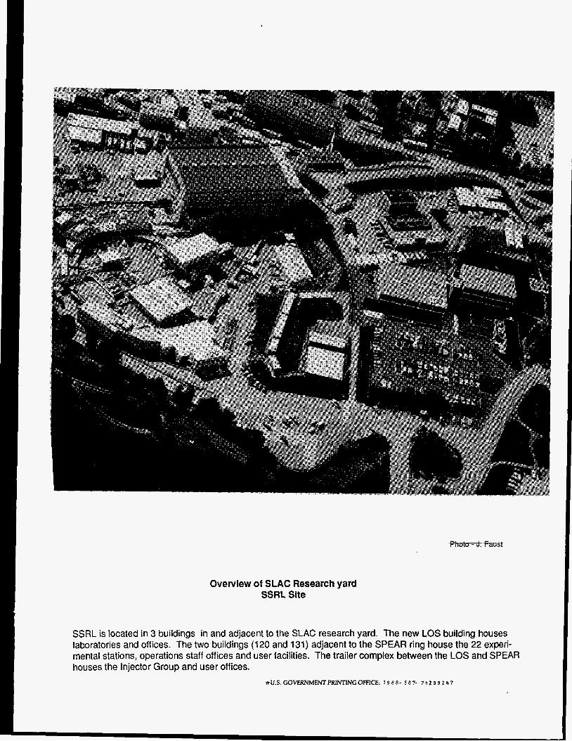
Overview of SLAC Research yard SSRL Site
SSRL is located in 3 buildings in and adjacent to the SLAC research yard. The new LOS building houses laboratories and offices. The two buildings (120 and 131) adjacent to the SPEAR ring house the 22 experi- mental stations, operations staff offices and user facilities. The trailer complex between the LOS and SPEAR houses the Injector Group and user offices.
*U.S. GOVERNMENTPRWTINGOFFICE: 1 9 8 8- 5 8 7- 7 4 1 9 9 1 4 7