standardized pathology report for breast cancer
TRANSCRIPT

Journal of Pathology and
Translational Medicine
Journal of Pathologyand Translational Medicine
January 2021Vol. 55 / No.1jpatholtm.orgpISSN: 2383-7837eISSN: 2383-7845
Standardized Pathology
Report for Breast Cancer
Journal of P
athology and Translational M
edicineVol. 55, N
o.1, pp 1-82, January 2021
J Pathol Transl MedpISSN: 2383-7837eISSN: 2383-7845
Volume 55 • Number 1 • January 2021 (bimonthly) Published since 1967Printed on 11 January 2021 Published on 15 January 2021
Aims & ScopeThe Journal of Pathology and Translational Medicine is an open venue for the rapid publication of major achievements in various fields of pathology, cytopathology, and biomedical and translational research. The Journal aims to share new insights into the molecular and cellular mechanisms of human diseases and to report major advances in both experimental and clinical medicine, with a particular emphasis on translational research. The investigations of human cells and tissues using high-dimensional biology techniques such as genomics and proteomics will be given a high priority. Articles on stem cell biology are also welcome. The categories of manuscript include original articles, review and perspective articles, case studies, brief case reports, and letters to the editor.
Subscription InformationTo subscribe to this journal, please contact the Korean Society of Pathologists/the Korean Society for Cytopathology. Full text PDF files are also available at the official website (https://jpatholtm.org). Journal of Pathology and Translational Medicine is indexed by Emerging Sources Citation Index (ESCI), PubMed, PubMed Central, Scopus, KoreaMed, KoMCI, WPRIM, Directory of Open Access Journals (DOAJ), and CrossRef. Circulation number per issue is 50.
Contact the Korean Society of Pathologists/the Korean Society for Cytopathology
Publishers: Cho, Nam Hoon, MD, Gong, Gyungyub, MDEditors-in-Chief: Jung, Chan Kwon, MD, Park, So Yeon, MDPublished by the Korean Society of Pathologists/the Korean Society for Cytopathology
Front cover image: Extensive intraductal component and skin involvement in invasive breast carcinoma (p6, p7).
© Copyright 2021 by the Korean Society of Pathologists/the Korean Society for Cytopathology Journal of Pathology and Translational Medicine is an Open Access journal under the terms of the Creative Commons Attribution Non-Commercial License (https://creativecommons.org/licenses/by-nc/4.0). This paper meets the requirements of KS X ISO 9706, ISO 9706-1994 and ANSI/NISO Z.39.48-1992 (Permanence of Paper).
Editorial OfficeRoom 1209 Gwanghwamun Officia, 92 Saemunan-ro, Jongno-gu, Seoul 03186, Korea Tel: +82-2-795-3094 Fax: +82-2-790-6635 E-mail: [email protected]
#1508 Renaissancetower, 14 Mallijae-ro, Mapo-gu, Seoul 04195, KoreaTel: +82-2-593-6943 Fax: +82-2-593-6944 E-mail: [email protected]
Printed by iMiS Company Co., Ltd. (JMC)Jungang Bldg. 18-8 Wonhyo-ro 89-gil, Yongsan-gu, Seoul 04314, KoreaTel: +82-2-717-5511 Fax: +82-2-717-5515 E-mail: [email protected]
Manuscript Editing by InfoLumi Co.210-202, 421 Pangyo-ro, Bundang-gu, Seongnam 13522, KoreaTel: +82-70-8839-8800 E-mail: [email protected]
Editorial Board
Avila-Casado, Maria del Carmen, MD (University of Toronto, Toronto General Hospital UHN, Canada)Bae, Young Kyung, MD (Yeungnam University, Korea)Bongiovanni, Massimo, MD (Lausanne University Hospital, Switzerland)Bychkov, Andrey, MD (Chulalongkorn University, Thailand Kameda Medical Center, Japan Nagasaki University Hospital , Japan)Choi, Yeong-Jin, MD (The Catholic University of Korea, Korea)Chong, Yo Sep, MD (The Catholic University of Korea, Korea)Chung, Jin-Haeng, MD (Seoul National University, Korea)Fadda, Guido, MD (Catholic University of Rome-Foundation Agostino Gemelli University Hospital, Italy)Gong, Gyungyub, MD (University of Ulsan, Korea)Ha, Seung Yeon, MD (Gachon University, Korea)Han, Jee Young, MD (Inha University, Korea)Jain, Deepali, MD (All India Institute of Medical Sciences, India)Jang, Se Jin, MD (University of Ulsan, Korea)Jeong, Jin Sook, MD (Dong-A University, Korea)Jun, Sun-Young, MD (The Catholic University of Korea, Korea)Kang, Gyeong Hoon, MD (Seoul National University, Korea)Kim, Aeree, MD (Korea University, Korea)Kim, Jang-Hee, MD (Ajou University, Korea)Kim, Jung Ho, MD (Seoul National University, Korea)Kim, Kyu Rae, MD (University of Ulsan, Korea)
Kim, Se Hoon, MD (Yonsei University, Korea)Kim, Woo Ho, MD (Seoul National University, Korea)Ko, Young Hyeh, MD (Sungkyunkwan University, Korea)Koo, Ja Seung, MD (Yonsei University, Korea)Lai, Chiung-Ru, MD (Taipei Veterans General Hospital, Taiwan)Lee, C. Soon, MD (University of Western Sydney, Australia)Lee, Hye Seung, MD (Seoul National University, Korea)Liu, Zhiyan, MD (Shandong University, China)Lkhagvadorj, Sayamaa, MD (Mongolian National University of Medical Sciences, Mongolia)Moon, Woo Sung, MD (Jeonbuk University, Korea)Paik, Jin Ho, MD (Seoul National University, Korea)Park, Chan-Sik, MD (University of Ulsan, Korea)Park, Young Nyun, MD (Yonsei University, Korea)Shahid, Pervez, MD (Aga Khan University, Pakistan)Song, Joon Seon, MD (University of Ulsan, Korea)Sung, Chang Ohk, MD (University of Ulsan, Korea)Tan, Puay Hoon, MD (National University of Singapore, Singapore)Than, Nandor Gabor, MD (Semmelweis University, Hungary)Tse, Gary M., MD (The Chinese University of Hong Kong, Hong Kong)Yatabe, Yasushi, MD (Aichi Cancer Center, Japan)Yoon, Sun Och, MD (Yonsei University, Korea)Zhu, Yun, MD (Jiangsu Institution of Nuclear Medicine, China)
Consulting EditorsHuh, Sun, MD (Hallym University, Korea)Kakudo, Kennichi, MD (Kindai University, Japan)Ro, Jae Y., MD (Cornell University, The Methodist Hospital, U.S.A.)
Ethic EditorChoi, In-Hong, MD (Yonsei University, Korea)
Statistics EditorsKim, Dong Wook (National Health Insurance Service Ilsan Hospital, Korea)Lee, Hye Sun (Yonsei University, Korea)
Manuscript EditorChang, Soo-Hee (InfoLumi Co., Korea)
Layout EditorKim, Haeja (iMiS Company Co., Ltd., Korea)
Website and JATS XML File ProducersCho, Yoonsang (M2Community Co., Korea)Im, Jeonghee (M2Community Co., Korea)
Administrative AssistantsJung, Ji Young (The Korean Society of Pathologists)Jeon, Ãnnmi (The Korean Society for Cytopathology)
Journal of Pathology and Translational Medicine
Editors–in-Chief
Senior Editors
Associate Editors
Jung, Chan Kwon, MD (The Catholic University of Korea, Korea) https://orcid.org/0000-0001-6843-3708Park, So Yeon, MD (Seoul National University, Korea) https://orcid.org/0000-0002-0299-7268
Hong, Soon Won, MD (Yonsei University, Korea) https://orcid.org/0000-0002-0324-2414Kim, Chong Jai, MD (University of Ulsan, Korea) https://orcid.org/0000-0002-2844-9446
Shin, Eunah, MD (Yongin Severance Hospital, Yonsei University, Korea) https://orcid.org/0000-0001-5961-3563Kim, Haeryoung, MD (Seoul National University, Korea) https://orcid.org/0000-0002-4205-9081

Journal of Pathology and Translational MedicineJ Pathol Transl Med
pISSN: 2383-7837eISSN: 2383-7845
Volume 55, Number 1, January 2021
CONTENTS
REVIEWS
1 Standardized pathology report for breast cancer Soo Youn Cho, So Yeon Park, Young Kyung Bae, Jee Yeon Kim, Eun Kyung Kim, Woo Gyeong Kim, Youngmee Kwon, Ahwon Lee, Hee Jin Lee, Ji Shin Lee,
Jee Young Park, Gyungyub Gong, Hye Kyoung Yoon
16 Imaging features of breast cancer molecular subtypes: state of the art Nariya Cho
26 DNA-protein biomarkers for immunotherapy in the era of precision oncology Binnari Kim, So Young Kang, Kyoung-Mee Kim
ORIGINAL ARTICLES
33 Automated immunohistochemical assessment ability to evaluate estrogen and progesterone receptor status compared with quantitative reverse transcription-polymerase chain reaction in breast carcinoma patients
Taesung Jeon, Aeree Kim, Chungyeul Kim
43 Interobserver diagnostic reproducibility in advanced-stage endometrial carcinoma Ho Jin Jung, Soo Yeon Lee, Jin Hwa Hong, Yi Kyeong Chun
53 A comparative prognostic performance of definitions of Crohn-like lymphoid reaction in colorectal carcinoma Younghoon Kim, Jeong Mo Bae, Jung Ho Kim, Nam-Yun Cho, Gyeong Hoon Kang
60 Causes of necrotic features in fine-needle aspirates from cervical lymph nodes Young Jin Seo, Hyeongchan Shin, Hye Won Lee, Hye Ra Jung
CASE REPORTS
68 A case of cribriform carcinoma of the skin: a newly described rare condition Hyun Lee, Chong-Hyun Won, Chan-Sik Park
CONTENTS CONTINUED© 2021 The Korean Society of Pathologists/The Korean Society for Cytopathology
© 2021 The Korean Society of Pathologists/The Korean Society for Cytopathology
75 Fibrocartilaginous mesenchymoma with an unusual location in the rib Sun-Ju Oh
79 EGFR-mutated pulmonary adenocarcinoma with concurrent PIK3CA mutation, and with acquired RET fusion and EGFR T790M mutation after afatinib therapy
Minhye Kim, Ji Min Na, Gyeong-Won Lee, Seung Jun Lee, Jong Duk Kim, Jung Wook Yang
Instructions for Authors for Journal of Pathology and Translational Medicine are available at http://jpatholtm.org/authors/authors.php

1
© 2021 The Korean Society of Pathologists/The Korean Society for CytopathologyThis is an Open Access article distributed under the terms of the Creative Commons Attribution Non-Commercial License (https://creativecommons.org/licenses/ by-nc/4.0) which permits unrestricted non-commercial use, distribution, and reproduction in any medium, provided the original work is properly cited.
pISSN 2383-7837eISSN 2383-7845
In Korea, the incidence of breast cancer has steadily increased partly due to an increase in screening mammography and to changes in lifestyle [1]. Breast cancer has become the most common cancer in women in Korea [1]. Thus, pathologists are encountering more breast cancer specimens in daily practice. Furthermore, as our understanding of breast cancer biology deepens and treatment strategies for breast cancer rapidly prog-ress, including advances in neoadjuvant therapy, targeted therapy, and immunotherapy, the role of pathologists in evaluation of breast specimens is changing [2]. Therefore, it would be useful to provide pathologists with a standard reporting format for ref-erence and recent updates in the field of breast cancer diagnosis.
A committee for standardization of breast cancer reporting
was formed in the Breast Pathology Study Group of the Korean Society of Pathologists. The ‘Standardized Pathology Report for Breast Cancer’ was developed after several committee meetings. The report form refers to the College of American Pathologists (CAP) Cancer Protocols [3], the American Joint Committee on Cancer (AJCC) 8th edition [4], and the World Health Organi-zation (WHO) Classification of Breast Tumors, 5th edition [5], and was modified by the Breast Pathology Study Group of the Korean Society of Pathologists.
The purpose of this report form is to enable standardized pathologic diagnosis of breast cancer and to improve communi-cation between clinicians and pathologists, as well as between pathologists inter-institutionally. The basic pathologic features
Standardized pathology report for breast cancer
Soo Youn Cho1, So Yeon Park2, Young Kyung Bae3, Jee Yeon Kim4, Eun Kyung Kim5, Woo Gyeong Kim6, Youngmee Kwon7, Ahwon Lee8, Hee Jin Lee9, Ji Shin Lee10, Jee Young Park11, Gyungyub Gong9, Hye Kyoung Yoon12
1Department of Pathology and Translational Genomics, Samsung Medical Center, Sungkyunkwan University School of Medicine, Seoul; 2Department of Pathology, Seoul National University Bundang Hospital, Seoul National University College of Medicine, Seongnam;
3Department of Pathology, Yeungnam University College of Medicine, Daegu; 4Department of Pathology, Pusan National University Yangsan Hospital, Pusan National University College of Medicine, Yangsan;
5Department of Pathology, Eulji University College of Medicine, Seoul; 6Department of Pathology, Inje University Haeundae Paik Hospital, Busan;
7Department of Pathology, National Cancer Center, Goyang; 8Department of Hospital Pathology, Seoul St. Mary’s Hospital, College of Medicine, The Catholic University of Korea, Seoul;
9Department of Pathology, Asan Medical Center, University of Ulsan College of Medicine, Seoul; 10Department of Pathology, Chonnam National University Medical School, Gwangju;
11Department of Pathology, School of Medicine, Kyungpook National University, Kyungpook National University Chilgok Hospital, Daegu; 12Department of Pathology, Busan Paik Hospital, Inje University, Busan, Korea
Given the recent advances in management and understanding of breast cancer, a standardized pathology report reflecting these chang-es is critical. To meet this need, the Breast Pathology Study Group of the Korean Society of Pathologists has developed a standardized pathology reporting format for breast cancer, consisting of ‘standard data elements,’ ‘conditional data elements,’ and a biomarker report form. The ‘standard data elements’ consist of the basic pathologic features used for prognostication, while other factors related to prog-nosis or diagnosis are described in the ‘conditional data elements.’ In addition to standard data elements, all recommended issues are also presented. We expect that this standardized pathology report for breast cancer will improve diagnostic concordance and commu-nication between pathologists and clinicians, as well as between pathologists inter-institutionally.
Key Words: Breast neoplasms; Diagnosis; Pathology report
Received: October 23, 2020 Revised: November 19, 2020 Accepted: November 20, 2020Corresponding Author: Hye Kyoung Yoon, MD, PhD, Department of Pathology, Busan Paik Hospital, Inje University, 75 Bokji-ro, Busanjin-gu, Busan 47392, KoreaTel: +82-51-890-6628, Fax: +82-51-890-5825, E-mail: [email protected]
This article has been published jointly, with consent, in both Journal of Pathology and Translational Medicine and Journal of Breast Cancer.
REVIEWJournal of Pathology and Translational Medicine 2021; 55: 1-15https://doi.org/10.4132/jptm.2020.11.20

https://jpatholtm.org/ https://doi.org/10.4132/jptm.2020.11.20
2 • Cho SY et al.
for prognostication are described in the “standard data ele-ments” section, and other factors related to prognosis or diag-nosis are described in the ‘conditional data elements’ section. Finally, descriptions on biomarkers essential for breast cancer diagnosis and treatment are included.
STANDARD DATA ELEMENTS
All essential standard data elements for the report form are sum-marized in Table 1. In addition, all data elements including recom-mended issues as well as standard data elements can be found in Supplementary Table S1.
Breast specimen types
Breast specimen types can be roughly divided into wide exci-
sion and total mastectomy. Wide excision is defined as removal of only part of the breast tissue, with or without axillary con-tents, and includes specimens designated as excisional biopsy, segmental or partial mastectomy, lumpectomy, or quadrantec-tomy. Total mastectomy refers to removal of all breast tissue, which may include skin, nipple, or areola, with or without axil-lary contents, and includes simple mastectomy (total mastectomy without axillary node removal), skin-sparing mastectomy (total mastectomy with removal of the nipple and a narrow surrounding rim of skin), nipple-sparing mastectomy (total mastectomy with-out removal of skin or nipple), modified radical mastectomy (total mastectomy with axillary node dissection and with occa-sional removal of a small portion of the pectoralis muscle), and radical mastectomy (total mastectomy with pectoralis muscle removal and axillary node dissection) [6].
Table 1. Standard data element
Breast specimen type□ Wide excision (specify)□ Total mastectomy (specify)
Specimen laterality□ Right□ Left□ Unspecified
Tumor location□ UOQ□ LOQ□ UIQ□ LIQ□ Central□ Unspecified
Histologic type□ Invasive breast carcinoma of no special type (specify for special morphological patterns)□ Invasive lobular carcinoma (specify for subtype)□ Tubular carcinoma□ Cribriform carcinoma□ Mucinous carcinoma□ Invasive micropapillary carcinoma□ Carcinoma with apocrine differentiation□ Metaplastic carcinoma (specify for subtype)□ Other rare subtype (specify)
Tumor focality□ Unifocal□ Multifocal
Tumor size____ × ____ × ____ cm
Histologic grade□ Grade I (Low) □ Grade II (Intermediate) □ Grade III (High)
Ductal carcinoma in situ (DCIS)□ Not identified□ Present (□ EIC-positive, □ EIC-negative)
(Continued to the next page)

https://jpatholtm.org/https://doi.org/10.4132/jptm.2020.11.20
Standardized pathology report: breast • 3
Histological type
The histopathologic classification of breast tumors in this pa-per is based on the WHO Classification of Breast Tumors, 5th edition [5] (Supplementary Table S2). The term “invasive breast carcinoma (IBC) of no special type (NST)” defines a large and heterogeneous group of IBCs that cannot be classified morpho-logically as any of the special histological types. The terms “in-vasive breast carcinoma, not otherwise specified (NOS),” “invasive ductal carcinoma,” or “infiltrating ductal carcinoma” are also ac-ceptable; however, invasive mammary carcinoma of NST is not
recommended. IBC-NST encompasses a wide spectrum of histological pat-
terns, including some special morphological patterns. Oncocytic, lipid-rich, glycogen-rich, clear cell, and sebaceous carcinomas; carcinoma with medullary pattern; invasive carcinoma with neuroendocrine differentiation; and carcinomas with pleomor-phic and choriocarcinomatous patterns are considered wide morphological patterns of IBC-NST regardless of the extent of differentiation or pattern. Breast carcinomas showing a specialized subtype in ≥ 90% of the tumor are designated as a pure special
Table 1. Continued
Nuclear grade □ Grade I (Low)□ Grade II (Intermediate)□ Grade III (High)
Necrosis□ Not identified□ Present (□ focal, □ central)
Extent of DCIS (for EIC-positive case)Estimated size: × cm No. of blocks with DCIS/No. of blocks examined: /
Lobular carcinoma in situ (in case of invasive lobular carcinoma)□ Not identified□ Present (□ classic type, □ pleomorphic type)
Tumor extensionSkin □ Not present□ Present (□ uninvolved, □ involved, without skin ulceration/ with skin ulceration/with satellite skin nodule)
Skeletal muscle□ Not present□ Present (□ uninvolved, □ involved in pectoralis muscle, □ involved in pectoralis muscle and chest wall)
Resection margin □ Cannot be assessed □ Positive for carcinoma Location (specify): invasive carcinoma/DCIS/invasive carcinoma and DCIS (unifocal, multifocal, extensive) □ Uninvolved by invasive carcinoma and/or DCIS Distance from closest margin : _____ mm from __________ margin
Regional lymph node metastasisTotal number of lymph nodes examined: Number of lymph nodes involved with metastases: (sentinel node: / , nonsentinel node: / )Size of largest metastasis: mmExtranodal extension: Not identified/Present
Lymphovascular invasion □ Not identified□ Present
Pathologic stage classification (pTNM, AJCC 8th edition)TNM descriptors: □ m □ r □ y Primary tumor (pT): Regional lymph nodes (pN):
UOQ, upper outer quadrant; LOQ, lower outer quadrant; UIQ, upper inner quadrant; LIQ, lower inner quadrant; EIC, extensive intraductal component; AJCC, American Joint Committee on Cancer.

https://jpatholtm.org/ https://doi.org/10.4132/jptm.2020.11.20
4 • Cho SY et al.
Fig. 1. Measurement of invasive tumor size. (A) Pathologic tumor (pT) category is based on the largest diameter of invasive carcinoma. Duc-tal carcinoma in situ, which is present on the upper right side of the invasive carcinoma, is not included in this measurement. (B) In post-treatment samples, the pT category (ypT) is based on the diameter of the largest contiguous focus (bar) of residual invasive carcinoma.
tumor type, such as mucinous cystadenocarcinoma or lobular, tubular, cribriform, mucinous, micropapillary, apocrine, or meta-plastic carcinoma.
The phrase “mixed IBC-NST and special subtype carcinoma” can be used when the special subtype comprises 10% to 90% of the carcinoma. For mixed tumors, overall percentage of the spe-cial subtype, grade and biomarkers status of both IBC-NST and special type carcinoma components should be reported. Carci-nomas in which the special subtype comprises < 10% should be classified as IBC-NST, with the optional comment of focal spe-cialized subtype. Tumors lacking such specific features are des-ignated IBC-NST, which accounts for the majority of IBC cases. Currently, estrogen receptor (ER) and human epidermal growth factor receptor type 2 (HER2) biomarker status are used for man-agement purposes instead of histological subtype or pattern.
The traditionally used classifications of medullary carcinoma, atypical medullary carcinoma, and carcinoma with medullary features found in the 4th edition of the WHO Breast Tumor Classification were removed in the 5th edition. Carcinomas for-merly classified as these subtypes are now categorized as “IBC-NST with medullary pattern,” representing one end of the spec-trum of tumor-infiltrating lymphocyte (TIL)-rich IBC-NSTs, rather than a distinct morphological subtype. IBC-NST with medullary pattern belongs to triple negative breast carcinomas, characterized by high expression of immune-related genes.
Metaplastic carcinoma is a heterogeneous group of IBCs char-acterized by differentiation of the neoplastic epithelium toward squamous cells and/or mesenchymal-looking elements, includ-ing but not restricted to spindle, chondroid, and osseous cells. The type of metaplastic elements present may be recorded using
a descriptive classification system.
Tumor focality
If multiple invasive carcinomas are present, tumor focality should be recorded. Multifocal tumors are associated with increased risk of lymph node involvement compared to similar unifocal disease [7-9], which reflects increased tumor load [10]. Counting the number of invasive foci is not essential but is recommended. When there is difficulty in determining whether two tumors are separate or not, microscopic examination of the tissue between the two masses should be performed. There are several occasions when multiple foci of invasion are present: extensive carcinoma in situ with multifocal invasion, invasive carcinoma with satel-lite foci, extensive lymphovascular invasion (LVI), multiple sep-arate invasive carcinomas, invasive carcinomas after neoadjuvant chemotherapy, and transection of a single carcinoma [3].
Except for cases presenting multiple separate invasive carci-nomas, most multifocal tumors have similar appearance and immunophenotype to the largest tumor. When multifocal tu-mors have similar histology, only the largest tumor is tested for ER, progesterone receptor (PR), and HER2. If multifocal tumors have different histological subtypes and grades, it is recom-mended to evaluate ER, PR, and HER2 status of each compo-nent, separately [3,11].
Tumor size
The single greatest dimension of the largest invasive tumor is used to ascertain the pathologic tumor (pT) category, regardless of extent of accompanying in situ carcinoma [4] (Fig. 1A). Three-dimensional measurement of tumor size is essential. In cases in
A B

https://jpatholtm.org/https://doi.org/10.4132/jptm.2020.11.20
Standardized pathology report: breast • 5
which it is difficult to determine the tumor size, information from imaging, gross findings, and microscopic evaluation should be used. For multifocal tumors, measurement of each tumor is rec-ommended.
The post-treatment pT category (ypT) is based on the largest contiguous focus of residual invasive carcinoma. Treatment-associated fibrosis adjacent to residual tumor or between foci of residual invasive carcinoma is not included in the ypT category (Fig. 1B).
Histological grade
Histological grading should be performed according to the Elston-Ellis modification of Bloom-Richardson grading [12]. Histological grading of IBCs is determined by three compo-nents: tubule formation, nuclear pleomorphism, and mitotic count (Supplementary Table S3). Tubule formation is assessed under low-power magnification. Scoring is performed according to the proportion of tumor cells forming tubules: more than 75% (score 1), 10%–75% (score 2), and less than 10% (score 3). Nu-clear pleomorphism should be assessed in the area showing the highest degree of pleomorphism. A score of 1 is given to small (less than 1.5 times the size of benign epithelial cell nuclei) and uniform nuclei with finely dispersed chromatin. A score of 3 is given to large (more than two times the size of benign epithelial cell nuclei), vesicular, and pleomorphic nuclei with prominent nucleoli and irregular chromatin. A score of 2 is given to nuclei with characteristics that lie between those two categories. Mitotic count is the number of mitotic figures present in 10 high-power fields (HPFs). Counting should be performed in the hot spot (area with the most frequent mitotic figures), which is usually at the peripheral, leading edge of the tumor. Care should be taken not to count hyperchromatic and apoptotic nuclei. The
cutoff points for mitotic count scores differ according to the field diameter of the 40× objective lens. The 5th edition WHO Breast Tumor Classification system recommends the use of number of mitoses per mm2 instead of number of mitoses per 10 HPFs for standardization [5].
These three scores are summed, and the total score of 3–9 is used for overall tumor grade: score 3–5 = grade 1, well differen-tiated; score 6 – 7 = grade 2, moderately differentiated; score 8 –
9 = grade 3, poorly differentiated (Fig. 2). The histological grade of IBC shows a strong correlation with prognosis [12,13].
Ductal carcinoma in situ
Ductal carcinoma in situ (DCIS) is concomitantly present in as many as 80% of IBC cases and is associated with increased risk of local recurrence after breast-conserving surgery [5].
Extensive intraductal component (EIC)-positive carcinoma is present when (1) DCIS is a major component (≥ 25%) of the area of invasive carcinoma and also is present outside the area of invasive carcinoma (Fig. 3A) or (2) there is extensive DCIS asso-ciated with a small (≤ 10 mm) invasive carcinoma (Fig. 3B) [3].
The histological features of DCIS associated with increased risk of recurrence are large lesion size, high nuclear grade, certain architectural patterns, central necrosis, and positive surgical mar-gin [5]. It is essential to report the features of DCIS, including nuclear grade, presence of necrosis, and extent of DCIS, in cases of EIC-positive carcinoma [3].
Nuclear grade is determined according to pleomorphism, nuclear size, chromatin, nucleoli, mitoses, and orientation (Sup-plementary Table S4) and is predictive of clinical outcome (re-currence) [14]. Central (comedo) necrosis is easily detected at low magnification within the central portion of ducts affected by DCIS. Focal necrosis means necrosis in small foci or single-cell
Fig. 2. Histological grades of invasive breast carcinoma of no special type: (A) grade 1, (B) grade 2, and (C) grade 3.
A AB C

https://jpatholtm.org/ https://doi.org/10.4132/jptm.2020.11.20
6 • Cho SY et al.
necrosis and is indistinct at low magnification. Reporting the architectural pattern of DCIS is not essential
but is recommended. Comedo DCIS is characterized by high nuclear grade associated with central necrosis, often with calci-fication. Solid DCIS shows compact proliferation of tumor cells with low-to-intermediate nuclear grade that fills the entire duct. Small necrotic foci may be present. Cribriform DCIS is charac-terized by intraductal proliferation with a sieve-like or fenestrated pattern. The secondary lumens are round, rigid, and surrounded by low-to-intermediate grade nuclei or occasional high-grade nuclei. Micropapillary DCIS has papillary fronds that lack fibro-vascular cores and that protrude into the ductal lumen in a reg-ular distribution. Micropapillary DCIS tends to be extensive in distribution (multifocal and multicentric). Papillary DCIS con-tains arborizing papillae with thin fibrovascular cores. Although it may be seen only microscopically, papillary DCIS more com-monly presents as a large mass [14]. Encapsulated papillary car-cinoma without invasion and solid papillary carcinoma without invasion are unusual patterns of DCIS.
Reporting the extent of DCIS is essential in cases of EIC-pos-itive carcinoma. However, a precise measurement of the extent of DCIS may be difficult or, at times, impossible. There are sev-eral methods for estimating the extent of DCIS. If DCIS is con-fined to a single tissue block, it is possible to estimate the extent of DCIS by direct measurement of the histological slides. If the entire specimen is blocked sequentially, the extent of DCIS can be calculated by multiplying the number of slices involved by average slice thickness. If the specimen is sampled, rather than sequentially blocked in its entirety, the extent of DCIS can be estimated by counting the number of blocks with DCIS [3,15].
Tumor extension
Satellite tumor nodules in the skin are separate from the pri-mary tumor and macroscopically identifiable. Skin and dermal satellite nodules identified only on microscopic examination and skin involvement without epidermal ulceration or skin edema (clini-cally peau d’orange) do not qualify as pT4b category (Fig. 4A, B). Such tumors should be categorized based on tumor size. Inflam-matory carcinoma is categorized only when there are clinical symp-toms of erythema and edema in more than one-third of the entire breast skin and not by the pathologic findings of tumor emboli in the dermal lymphatics.
The chest wall includes ribs, intercostal muscles, and serratus anterior muscle but not the pectoralis muscles. Therefore, in-volvement of the pectoralis muscles in the absence of invasion of these chest wall elements does not constitute chest wall inva-sion, and cancers with such involvement are categorized based on tumor size.
Resection margin
Whenever possible, specimens should be oriented to identify specific margins for the pathologist. All identifiable margins should be evaluated for carcinoma involvement both grossly and microscopically [16].
Orientation may be conducted using sutures or clips placed on the specimen surface or by other means of communication between the surgeon and pathologist and should be documented in the pathology report. Margins can be identified in several ways, including using multiple colored inks, submitting the margins in specific cassettes, or submitting each margin as a separately excised specimen.
Margin status is listed as “positive” if there is ink on the cancer
Fig. 3. Extensive intraductal component-positive invasive carcinoma. (A) Ductal carcinoma in situ (DCIS) constitutes ≥ 25% of the area of in-vasive carcinoma and also is present outside the area of invasive carcinoma. (B) A small invasive carcinoma is present in background of ex-tensive DCIS.
A B

https://jpatholtm.org/https://doi.org/10.4132/jptm.2020.11.20
Standardized pathology report: breast • 7
cells during pathologic margin evaluation. If the specimen is ori-ented, the specific site(s) of involvement should be reported. The approximate extent of margin involvement can be reported as follows: unifocal, 1 focal area of carcinoma (< 4 mm) at the margin; multifocal, 2 or more carcinoma foci at the margin; exten-sive, carcinoma present at the margin over a broad front (> 5 mm).
In lobular carcinoma in situ (LCIS), assessment of resection margin is optional. However, for pleomorphic type LCIS, evalu-ation of resection margin is recommended.
Regional lymph node metastasis
Most patients with invasive carcinoma will have lymph nodes sampled for pathologic regional lymph node (pN) categoriza-tion [4]. All lymph nodes must be examined histologically [4]. The nodes commonly examined include sentinel nodes, nonsen-tinel nodes, nodes from axillary dissections, and intramammary nodes. When the total number of sentinel and nonsentinel nodes removed is less than 6, the AJCC “sn” modifier is used.
Metastases are classified into three groups based on size: iso-lated tumor cells (ITCs), micrometastases, and macrometastases [4]. ITCs are defined as single cells, small clusters of cells no larger than 0.2 mm, or no more than 200 cells in a single cross section. The AJCC states that a cluster is a group of cells in contact with each other (confluent or contiguous). Cells that are not touching each other should be considered independent and measured independently. In cases of multiple clusters of tumor cells within a lymph node, only the largest should be considered when determining N category (Fig. 5A). The AJCC states that the size of the tumor should include both the tumor cells and the surrounding desmoplastic reaction. Some carcinomas, particularly lobular carcinomas, may metastasize as individual single cells
and not as clusters and present as a dispersed pattern of nodal metastases (Fig. 5B). In such cases, single cells are measured sep-arately. If fewer than 200 tumor cells are present in a node cross section, then classification of ITCs is recommended (Fig. 5C). Nodes containing only ITCs are not included in the total num-ber of positive nodes when determining N category, so cases with only ITCs are classified as node negative (pN0 (i+)). Microme-tastases measure greater than 0.2 mm but not greater than 2 mm and/or comprise more than 200 cells in a single cross section. If only micrometastases are present on lymph node examination, the N category is pN1mi. If at least 1 macrometastasis is pres-ent, nodes with micrometastases are included in the total number of positive nodes. Any lesion where the largest cluster is greater than 2 mm represents a macrometastasis (Fig. 5D).
Extranodal extension is defined as the presence of full-thickness (i.e., into and through) lymph node capsular invasion, as seen with metastatic tumor invasion of extranodal fat with or with-out an associated desmoplastic stromal response (Fig. 5D). The area of extranodal extension is included when measuring the overall size of the lymph node metastasis. Extranodal extension is a marker of poor prognosis in breast cancer patients, and the status of extranodal extension should be reported [17]. Reporting of extranodal extension size based on a 2-mm cutoff is incorpo-rated into the CAP reporting guidelines, but more evidence is needed for this practice to become widely accepted [3,17].
When cancerous nodules that are not associated with residual lymph node tissue are present in the axillary fat, the AJCC states that these nodules should be classified as positive lymph nodes [4]. However, if there is surrounding normal breast parenchyma or DCIS, then cancerous nodules in the axillary fat should be clas-sified as invasive carcinoma and not as a nodal metastasis.
Fig. 4. Skin involvement in invasive breast carcinoma. (A) Tumor cells infiltrate into the upper dermis in the absence of ulceration. These cas-es should not be classified as pT4b category. (B) There is an ulceration of overlying epidermis accompanied by tumor extension, correspond-ing to the pT4b category.
A B

https://jpatholtm.org/ https://doi.org/10.4132/jptm.2020.11.20
8 • Cho SY et al.
The post-treatment pN (ypN) classification system is the same as that for pre-treatment lymph nodes. Only the largest contigu-ous focus of residual tumor in the node evaluation is used for determining N category; any treatment-associated fibrosis is not included [3,4].
Lymphovascular invasion
LVI is associated with local recurrence and reduced survival [18]. Strict criteria or immunohistochemistry (IHC) stains have been proposed to differentiate LVI from DCIS and retraction ar-tifacts [3]. If a limited area is involved in LVI, a measurement in millimeters can be given. Alternatively, LVI can be quantified as focal or extensive, with ‘extensive’ defined as one or more foci in more than one block [19].
The presence of pure LVI without stromal invasion after neo-adjuvant therapy may be called ypTX and should not be classi-fied as pathologic complete response (pCR) [20].
Pathologic stage classification [4]Pathologic stage classification according to the AJCC 8th
edition should be reported as a standard data element [4]. Clas-sification of primary tumor (T), regional lymph nodes (N), and distant metastasis (M) by pathologic examination is denoted by the prefix “p” (pT, pN, and pM). The descriptor “m” is used when invasive cancer is observed in multiple foci, and the prefix “r” is used for recurrent cancer. If the patient has undergone neo-adjuvant chemotherapy, hormonal therapy, immunotherapy, or radiation therapy before surgery, the prefix “y” is used. pM0 is not a valid category. When distant metastases cannot be confirmed by pathologic examination, staging can be performed by com-bining pT, pN, and the clinical evaluation of metastases (cM).
pT category
Criteria for each pT category are summarized in Supplemen-tary Table S5. For multiple invasive cancers, use the tumor with
Fig. 5. Classification of lymph node metastases. (A) Multiple clusters of tumor cells. N category is based on the size of the largest contigu-ous cluster of tumor cells. (B) Dispersed pattern of metastasis. Some lobular carcinomas may metastasize as single cells and may not form cohesive clusters. If more than 200 tumor cells are present in a node cross section, then the category of micrometastasis is recommended. (C) Isolated tumor cells. A dispersed pattern of lobular carcinoma with fewer than 200 cells is detected by cytokeratin immunohistochemistry. (D) Macrometastasis with extranodal extension. This metastasis is classified as a macrometastasis based on the size of cluster (> 2 mm). Ex-tranodal extension, an area of invasion outside the lymph node capsule (arrow), is noted.
A
C
B
D

https://jpatholtm.org/https://doi.org/10.4132/jptm.2020.11.20
Standardized pathology report: breast • 9
the highest T category for classification and staging, and use the descriptor “m” or number of invasive cancers in parentheses (e.g., T2(m) or T2(3)). For simultaneous bilateral breast cancers, staging should be conducted separately because they are consid-ered independent tumors in different organs.
pN category
Criteria for each pN category are summarized in Supplemen-tary Table S6. If no lymph nodes were submitted for evaluation, record pNX. It is not the pathologist’s obligation to record the pN status by integrating the previous pathologic results. pN1a, pN2a, and pN3a refer to metastases in 1 to 3, 4 to 9, and 10 or more axillary lymph nodes, respectively, with at least one mac-rometastasis. If the specimen contains internal mammary lymph nodes, infraclavicular lymph nodes, or supraclavicular lymph nodes with metastases, or if clinically metastatic internal mam-mary lymph node(s) are identified, refer to the AJCC staging manual for accurate lymph node categorization. A regional lymph node with direct extension of the primary tumor or a tumor nodule in a regional lymph node area should be considered as a positive node.
When nodal metastasis is confirmed by fine-needle aspiration cytology or core needle biopsy without further resection of nodes, use the “f” modifier (e.g., pN(f)).
pM category
The pM category is assigned only if metastasis larger than 0.2 mm (pM1) is histologically confirmed. When staging after neo-adjuvant therapy, the classification should remain M1 regardless of responsiveness to therapy, if the case was confirmed to be M1 prior to therapy.
CONDITIONAL DATA ELEMENTS
All conditional data elements for this report form are sum-marized in Table 2.
Perineural invasion
Perineural invasion (PNI) is infrequently observed in IBC, occurring in approximately 1% of cases, perhaps in part because nerves of notable size are not numerous in mammary tissues [21].
PNI may occur more frequently in IBC-NST than in the other histological subtypes. It tends to occur in high-grade tumors, where it is frequently associated with LVI, but it has not been proven to have independent prognostic significance [21-24].
PNI can also be observed in some benign lesions, such as scle-rosing adenosis, as well as in DCIS.
Tumor border
The tumor margins of IBC can be grossly described as ill-de-marcated, well-demarcated (circumscribed), or mixed [21].
Approximately one-third of tumors have grossly circum-scribed margins. However, some carcinomas that appear to have circumscribed margins grossly exhibit an invasive growth pat-tern microscopically [21]. Grossly ill-demarcated tumors tend to be larger, and they are more likely to have axillary metastases than those with circumscribed margins [21].
Microcalcification
DCIS/invasive carcinoma found in biopsies performed for microcalcifications will almost always be at the site of the micro-calcifications or in close proximity [25,26]. The presence of tar-geted microcalcifications in the specimen can be confirmed by radiography.
The pathologist needs to confirm that the specimen has been sampled from the lesion responsible for the microcalcifications. Microcalcifications are commonly present in secretions and/or in necrotic materials [5]. The radiological and pathologic corre-lation of all microcalcifications, including information about the presence and site of microcalcifications (e.g., invasive carci-noma, DCIS, benign lesion, or mixed), should be indicated [5]. Information about the microcalcifications can be an important
Table 2. Conditional data element
Perineural invasion□ Not identified□ Present
Tumor border□ Not applicable □ Well-demarcated/Circumscribed □ Ill-demarcated □ Mixed
Microcalcification□ Not identified □ Present in invasive carcinoma □ Present in DCIS □ Present in non-neoplastic tissue
Tumor-infiltrating lymphocytes (TILs)___________ %
Treatment effect (RCB class) □ RCB class 0 □ RCB class I □ RCB class II □ RCB class III RCB index:
Additional pathologic findings
DCIS, ductal carcinoma in situ; RCB, Residual Cancer Burden.

https://jpatholtm.org/ https://doi.org/10.4132/jptm.2020.11.20
10 • Cho SY et al.
consideration when correlating imaging findings with the pathologic diagnosis, when guiding further management of the disease, and when identifying recurrent carcinoma in the breast or metastatic diseases [21].
Tumor-infiltrating lymphocytes
The prognostic and predictive value of TILs in breast cancer has been studied extensively [27,28]. TILs are lymphocytes present in the stroma of a tumor or inside tumor cell nests. As-sessment of TIL level in hematoxylin and eosin (H&E) sections can be easily performed. The International Immuno-Oncology Biomarker Working Group on Breast Cancer published guide-lines for evaluation of TIL level in H&E sections of invasive breast cancer in 2014, and they later extended this method to DCIS, metastatic tumor deposits, and specimens obtained after neoad-juvant chemotherapy [7,29,30]. In brief, TIL level is determined by measuring the percentage of the total stromal area, excluding tumor necrosis and crush artifacts, occupied by mononuclear inflammatory cells, including plasma cells, within the borders of the invasive carcinoma. TILs are usually evaluated in incre-ments of 10% (e.g., < 10%, 10%–19%, 20%–29%) (Fig. 6). Since distribution of TILs is usually not even throughout the tu-mor tissue, assessment of the average number of TILs, without focusing on hot spots, is recommended. The International Im-muno-Oncology Biomarker Working Group showed that proper training of pathologists could achieve more consistent results with regard to evaluation of TILs in ring studies and suggested poten-tial pitfalls in assessment of TILs [31,32].
Effect of treatment
Many classifications have been proposed to evaluate the
pathologic status of breast cancer after treatment, including those of Chevallier [33], Sataloff [34], the National Surgical Adjuvant Breast and Bowel Project (NSABP) B-18 [35], Miller-Payne [36], the Residual Cancer Burden (RCB) system [37], the Clinical-Pathologic Stage-Estrogen/Grade (CPS-EG) system [38], the Residual Disease in Breast and Nodes (RDBN) system [39], and the AJCC [4]. Among these classifications, the AJCC and the RCB calculator are the most widely used systems to measure residual disease [40].
The RCB index is calculated from the following five variables [37]: (1) primary tumor bed area (mm2), (2) overall cancer cel-lularity (%), (3) percentage of carcinoma in the tumor bed that is in situ disease (%), (4) number of positive lymph nodes, and (5) diameter of the largest lymph node metastasis (mm).
Primary tumor bed area is the two largest dimensions between invasive tumor cells, even if these are widely scattered and sepa-rated by treatment-induced fibrosis. Overall cancer cellularity is the overall percentage of the residual tumor bed area that is occupied by carcinoma (invasive and in situ). It is assessed in each slide, and the average is calculated using all fields that fall within the perimeter of the largest cross-sectional area of resid-ual tumor bed, even those with very low cellularity or no disease [37]. The same method can be used for the in situ component to assess the percentage of cancer that is in situ disease [37]. Unlike the AJCC ypN category, the number of positive lymph nodes includes the number of lymph nodes with ITCs. The di-ameter of the largest lymph node metastasis used in the RCB system may be different from that used for AJCC staging be-cause the former includes intervening treatment-related fibrosis [40,41].
A mathematical formula combines these variables into a con-
Fig. 6. Different levels of tumor-infiltrating lymphocyte (TIL) infiltration in invasive breast carcinoma: (A) TIL < 10%, (B) TIL 10%–50%, and (C) TIL > 50%.
A B C
https://jpatholtm.org/https://doi.org/10.4132/jptm.2020.11.20
Standardized pathology report: breast • 11
tinuous index to define four RCB classes: RCB-0 for pCR and RCB 1 to 3, representing progressively greater extent of residual cancer [37]. A web-based calculator and detailed instructions for calculating RCB indices are publicly available (http://www3.mdanderson.org/app/medcalc/index.cfm?pagename=jsconvert3).
As the presence of positive lymph nodes after treatment rep-resents a worse prognosis even when there is no residual inva-sive carcinoma in the breast [42,43], several classification systems and the CAP cancer protocol have recommended that effect of treatment be evaluated in both the breast and lymph nodes [3,4,34,37,39,44,45].
BIOMARKERS
Determination of biomarker status, including ER, PR, and HER2 status, is essential for newly diagnosed IBC. ER, PR, and HER2 should be evaluated according to the current American Society of Clinical Oncology (ASCO)/CAP guidelines [46,47]. Currently, there are no established guidelines regarding re-eval-uation of biomarkers in post-treatment specimens. However, it is recommended that ER, PR, and HER2 testing be repeated on post-treatment invasive carcinomas, such as when there was insufficient invasive tumor tissue or negative or equivocal re-sults on pre-treatment core biopsy; when biopsies were performed and biomarkers assessed at other institutions; or when post-treat-ment tumors display heterogeneous morphology or no response to therapy.
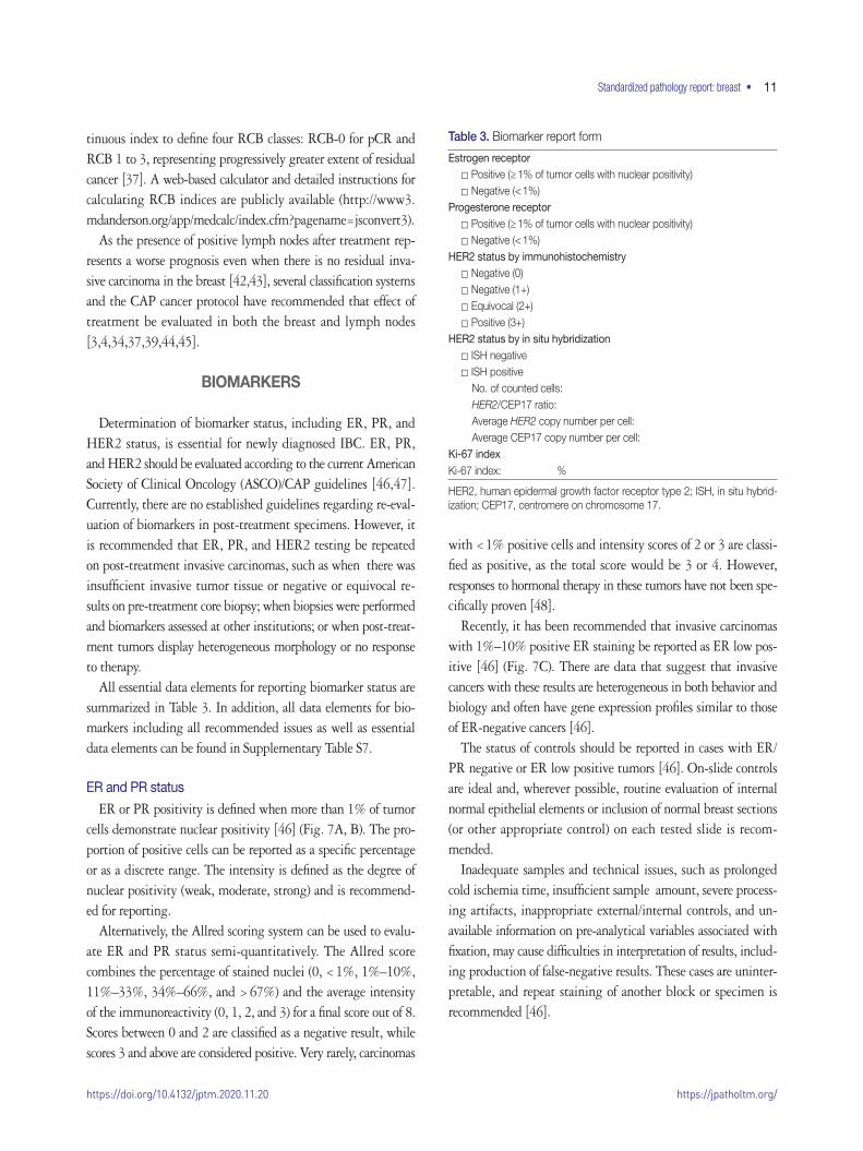
All essential data elements for reporting biomarker status are summarized in Table 3. In addition, all data elements for bio-markers including all recommended issues as well as essential data elements can be found in Supplementary Table S7.
ER and PR status
ER or PR positivity is defined when more than 1% of tumor cells demonstrate nuclear positivity [46] (Fig. 7A, B). The pro-portion of positive cells can be reported as a specific percentage or as a discrete range. The intensity is defined as the degree of nuclear positivity (weak, moderate, strong) and is recommend-ed for reporting.
Alternatively, the Allred scoring system can be used to evalu-ate ER and PR status semi-quantitatively. The Allred score combines the percentage of stained nuclei (0, < 1%, 1%–10%, 11%–33%, 34%–66%, and > 67%) and the average intensity of the immunoreactivity (0, 1, 2, and 3) for a final score out of 8. Scores between 0 and 2 are classified as a negative result, while scores 3 and above are considered positive. Very rarely, carcinomas
with < 1% positive cells and intensity scores of 2 or 3 are classi-fied as positive, as the total score would be 3 or 4. However, responses to hormonal therapy in these tumors have not been spe-cifically proven [48].
Recently, it has been recommended that invasive carcinomas with 1%–10% positive ER staining be reported as ER low pos-itive [46] (Fig. 7C). There are data that suggest that invasive cancers with these results are heterogeneous in both behavior and biology and often have gene expression profiles similar to those of ER-negative cancers [46].
The status of controls should be reported in cases with ER/PR negative or ER low positive tumors [46]. On-slide controls are ideal and, wherever possible, routine evaluation of internal normal epithelial elements or inclusion of normal breast sections (or other appropriate control) on each tested slide is recom-mended.
Inadequate samples and technical issues, such as prolonged cold ischemia time, insufficient sample amount, severe process-ing artifacts, inappropriate external/internal controls, and un-available information on pre-analytical variables associated with fixation, may cause difficulties in interpretation of results, includ-ing production of false-negative results. These cases are uninter-pretable, and repeat staining of another block or specimen is recommended [46].
Table 3. Biomarker report form
Estrogen receptor
□ Positive (≥ 1% of tumor cells with nuclear positivity)
□ Negative (< 1%) Progesterone receptor
□ Positive (≥ 1% of tumor cells with nuclear positivity)
□ Negative (< 1%) HER2 status by immunohistochemistry
□ Negative (0)
□ Negative (1+)
□ Equivocal (2+)
□ Positive (3+)HER2 status by in situ hybridization
□ ISH negative
□ ISH positive No. of counted cells: HER2/CEP17 ratio: Average HER2 copy number per cell: Average CEP17 copy number per cell:
Ki-67 indexKi-67 index: %
HER2, human epidermal growth factor receptor type 2; ISH, in situ hybrid-ization; CEP17, centromere on chromosome 17.

https://jpatholtm.org/ https://doi.org/10.4132/jptm.2020.11.20
12 • Cho SY et al.
HER2 status
IHC and in situ hybridization (ISH) are regarded as standard methods to assess HER2 status in breast cancer. The ASCO/CAP have jointly released guidelines and recommendations on HER2 testing in breast cancer since 2007, and, recently, have
updated these guidelines to provide clear instructions for HER2 testing and accurate determination of HER2 status in breast can-cer [49].
Currently, HER2 status determined by IHC and ISH should be interpreted based on the 2018 updated ASCO/CAP guide-
Fig. 7. Representative examples of estrogen receptor (ER) expression assessed by immunohistochemistry in invasive breast carcinoma: (A) ER negative, (B) ER positive, and (C) ER low positive.
Fig. 8. Representative examples of human epidermal growth factor receptor 2 (HER2) immunohistochemistry (IHC) scores in invasive breast carcinoma: (A) HER2 IHC score 0, (B) HER2 IHC score 1+, (C) HER2 IHC score 2+, and (D) HER2 IHC score 3+.
A B C
A
C
B
D

https://jpatholtm.org/https://doi.org/10.4132/jptm.2020.11.20
Standardized pathology report: breast • 13
lines [47]. HER2 IHC status should be assessed using a semi-quantitative scoring system (Fig. 8A–D). For HER2 IHC equiv-ocal (2+) cases, HER2 status should be confirmed by ISH.
Interpretation of HER2 ISH is performed by counting at least 20 cells in the invasive tumor area. Scanning of entire slides or use of IHC slides prior to counting is mandatory to define the areas of potential HER2 amplification [50]. Please refer to a pre-vious article for interpretation of HER2 heterogeneity [49].
For a diagnostic approach using HER2 ISH, concomitant IHC review for dual-probe ISH groups 2 to 4 is required in the updated guidelines [47]. In laboratories using single-probe ISH assays, concomitant IHC review is included as part of the interpre-tation of all ISH assay results [47]. By this approach, the HER2 ISH status is designated as positive or negative, with no equivocal category. Determining HER2 ISH status using dual-probe ISH is summarized in Supplementary Table S8. In reporting the results of HER2 ISH, final HER2 ISH status (negative or positive), number of counted cells, HER2/centromere on chromosome 17 (CEP17) ratio, average HER2 copy number per cell, average CEP17 copy number per cell, and designation of ISH group (optional) should be included.
Ki-67 index
The Ki-67 index is defined by the percentage of tumor cells with positive Ki-67 nuclear staining out of all tumor cells counted in a given field. A high Ki-67 index is regarded as a prognostic marker associated with high risk of recurrence and as a predictive marker for treatment response in breast cancer [51-54]. How-ever, there remain controversies in its use as a standard prognostic or predictive biomarker owing to high inter-observer variability and lack of a standardized measurement method. Currently, as-sessment of Ki-67 can be performed applying recommenda-tions from the International Ki-67 in Breast Cancer Working Group [55] in clinical practice. Briefly, at least three high-power (40 × objective) fields should be selected to represent the spec-trum of staining on initial overview of the whole section. If there are clear hot spots, data from these should be included in the overall average score. Scoring should involve the counting of at least 500 malignant invasive cells (and preferably at least 1,000 cells). Currently, computational imaging analysis meth-ods are widely used for Ki-67 quantification, but their superiority over the direct counting method is unclear. When morphological analysis is used, the number of tumor cells counted and the num-ber of tumor cells with positive Ki-67 nuclear staining should be included in the report form.
CONCLUSION
In accordance with recent advances in collective understanding of breast cancer biology and in treatment of breast cancer patients, a committee of the Breast Pathology Study Group of the Korean Society of Pathologists presents in this publication a ‘Standard-ized Pathology Report for Breast Cancer.’ This report form is com-posed of ‘standard data elements,’ ‘conditional data elements,’ and a biomarker report form to guide diagnosis, prognosis, and treat-ment of breast cancer patients. It is our hope that this report will lead to standardization of the pathologic diagnosis of breast cancer and to improvement in communication between clini-cians and pathologists, as well as between pathologists.
Supplementary Information The Data Supplement is available with this article at https://doi.org/10.4132/jptm.2020.11.20.
Ethics StatementNot applicable.
ORCID Soo Youn Cho https://orcid.org/0000-0001-9714-7575So Yeon Park https://orcid.org/0000-0002-0299-7268Young Kyung Bae https://orcid.org/0000-0002-6689-9413Jee Yeon Kim https://orcid.org/0000-0002-0503-984XEun Kyung Kim https://orcid.org/0000-0001-6701-6988Woo Gyeong Kim https://orcid.org/0000-0003-1694-6602Youngmee Kwon https://orcid.org/0000-0002-1370-2496Ahwon Lee https://orcid.org/0000-0002-2523-9531Hee Jin Lee https://orcid.org/0000-0002-4963-6603Ji Shin Lee https://orcid.org/0000-0003-4634-2228Jee Young Park https://orcid.org/0000-0002-1857-813XGyungyub Gong https://orcid.org/0000-0001-5743-0712Hye Kyoung Yoon https://orcid.org/0000-0003-0714-8537
Author Contributions Conceptualization: HKY. Project administration: SYP, YKB. Supervision: HKY. Writing—original draft: SYC, SYP, YKB, JYK, EKK, WGK, YK, AL, HJL, JSL, JYP. Writing—review & editing: SYC, SYP, YKB, GG, HKY. Ap-proval of final manuscript: all authors.
Conflicts of InterestSYP, the editor-in-chief and YKB and GG, contributing editors of the Jour-nal of Pathology and Translational Medicine, were not involved in the edi-torial evaluation or decision to publish this article. All remaining authors have declared no conflicts of interest.
Funding StatementNo funding to declare.
References1. Kang SY, Kim YS, Kim Z, et al. Breast cancer statistics in Korea in
2017: data from a breast cancer registry. J Breast Cancer 2020; 23: 115-28.

https://jpatholtm.org/ https://doi.org/10.4132/jptm.2020.11.20
14 • Cho SY et al.
2. Li X, Oprea-Ilies GM, Krishnamurti U. New developments in breast cancer and their impact on daily practice in pathology. Arch Pathol Lab Med 2017; 141: 490-8.
3. Fitzgibbons PL, Connolly JL, Bose S, et al. Protocol for the examina-tion of resection specimens from patients with invasive carcinoma of the breast [Internet]. Northfield: College of American Patholo-gists, c2020 [cited 2020 Sep 10]. Available from: https://documents.cap.org/protocols/cp-breast-invasive-resection-20-4400.pdf.
4. Hortobagyi GN, Connolly JL, D'Orsi CJ, et al. Breast. In: Amin MB, Edge S, Greene F, et al., eds. AJCC cancer staging manual. 8th ed. New York: Springer, 2017; 589-636.
5. WHO Classification of Tumours Editorial Board. WHO classifica-tion of tumours: breast tumours. 5th ed. Lyon: International Agency for Research on Cancer, 2019.
6. Lester SC, Bose S, Chen YY, et al. Protocol for the examination of specimens from patients with invasive carcinoma of the breast. Arch Pathol Lab Med 2009; 133: 1515-38.
7. Salgado R, Aftimos P, Sotiriou C, Desmedt C. Evolving paradigms in multifocal breast cancer. Semin Cancer Biol 2015; 31: 111-8.
8. Andea AA, Wallis T, Newman LA, Bouwman D, Dey J, Visscher DW. Pathologic analysis of tumor size and lymph node status in multifo-cal/multicentric breast carcinoma. Cancer 2002; 94: 1383-90.
9. Weissenbacher TM, Zschage M, Janni W, et al. Multicentric and multifocal versus unifocal breast cancer: is the tumor-node-metasta-sis classification justified? Breast Cancer Res Treat 2010; 122: 27-34.
10. Coombs NJ, Boyages J. Multifocal and multicentric breast cancer: does each focus matter? J Clin Oncol 2005; 23: 7497-502.
11. Choi Y, Kim EJ, Seol H, et al. The hormone receptor, human epider-mal growth factor receptor 2, and molecular subtype status of indi-vidual tumor foci in multifocal/multicentric invasive ductal carci-noma of breast. Hum Pathol 2012; 43: 48-55.
12. Elston CW, Ellis IO. Pathological prognostic factors in breast cancer. I. The value of histological grade in breast cancer: experience from a large study with long-term follow-up. Histopathology 1991; 19: 403-10.
13. Rakha EA, El-Sayed ME, Lee AH, et al. Prognostic significance of Nottingham histologic grade in invasive breast carcinoma. J Clin Oncol 2008; 26: 3153-8.
14. Jaffer S, Bleiweiss IJ. Histologic classification of ductal carcinoma in situ. Microsc Res Tech 2002; 59: 92-101.
15. Grin A, Horne G, Ennis M, O'Malley FP. Measuring extent of duc-tal carcinoma in situ in breast excision specimens: a comparison of 4 methods. Arch Pathol Lab Med 2009; 133: 31-7.
16. Morrow M, Van Zee KJ, Solin LJ, et al. Society of Surgical Oncolo-gy-American Society for Radiation Oncology-American Society of Clinical Oncology consensus guideline on margins for breast-con-serving surgery with whole-breast irradiation in ductal carcinoma in situ. Pract Radiat Oncol 2016; 6: 287-95.
17. Choi AH, Blount S, Perez MN, et al. Size of extranodal extension on sentinel lymph node dissection in the American College of Surgeons Oncology Group Z0011 trial era. JAMA Surg 2015; 150: 1141-8.
18. Mohammed RA, Martin SG, Mahmmod AM, et al. Objective as-sessment of lymphatic and blood vascular invasion in lymph node-negative breast carcinoma: findings from a large case series with long-term follow-up. J Pathol 2011; 223: 358-65.
19. Provenzano E, Bossuyt V, Viale G, et al. Standardization of patho-logic evaluation and reporting of postneoadjuvant specimens in clinical trials of breast cancer: recommendations from an interna-
tional working group. Mod Pathol 2015; 28: 1185-201.20. Baker GM, King TA, Schnitt SJ. Evaluation of breast and axillary
lymph node specimens in breast cancer patients treated with neoad-juvant systemic therapy. Adv Anat Pathol 2019; 26: 221-34.
21. Hoda SA, Brogi E, Koerner FC, Rosen PP. Rosen's breast pathology. Philadelphia: Wolters Kluwer, 2014.
22. Duraker N, Caynak ZC, Turkoz K. Perineural invasion has no prog-nostic value in patients with invasive breast carcinoma. Breast 2006; 15: 629-34.
23. McCready DR, Chapman JA, Hanna WM, et al. Factors affecting distant disease-free survival for primary invasive breast cancer: use of a log-normal survival model. Ann Surg Oncol 2000; 7: 416-26.
24. McCready DR, Chapman JA, Hanna WM, et al. Factors associated with local breast cancer recurrence after lumpectomy alone: post-menopausal patients. Ann Surg Oncol 2000; 7: 562-7.
25. Owings DV, Hann L, Schnitt SJ. How thoroughly should needle lo-calization breast biopsies be sampled for microscopic examination? A prospective mammographic/pathologic correlative study. Am J Surg Pathol 1990; 14: 578-83.
26. Silverstein MJ, Lagios MD, Recht A, et al. Image-detected breast can-cer: state of the art diagnosis and treatment. J Am Coll Surg 2005; 201: 586-97.
27. Aaltomaa S, Lipponen P, Eskelinen M, et al. Lymphocyte infiltrates as a prognostic variable in female breast cancer. Eur J Cancer 1992; 28a: 859-64.
28. Wein L, Savas P, Luen SJ, Virassamy B, Salgado R, Loi S. Clinical va-lidity and utility of tumor-infiltrating lymphocytes in routine clini-cal practice for breast cancer patients: current and future directions. Front Oncol 2017; 7: 156.
29. Hendry S, Salgado R, Gevaert T, et al. Assessing tumor-infiltrating lymphocytes in solid tumors: a practical review for pathologists and proposal for a standardized method from the International Immu-no-Oncology Biomarkers Working Group: part 2: TILs in melano-ma, gastrointestinal tract carcinomas, non-small cell lung carcinoma and mesothelioma, endometrial and ovarian carcinomas, squamous cell carcinoma of the head and neck, genitourinary carcinomas, and primary brain tumors. Adv Anat Pathol 2017; 24: 311-35.
30. Dieci MV, Radosevic-Robin N, Fineberg S, et al. Update on tumor-infiltrating lymphocytes (TILs) in breast cancer, including recom-mendations to assess TILs in residual disease after neoadjuvant therapy and in carcinoma in situ: a report of the International Im-muno-Oncology Biomarker Working Group on Breast Cancer. Se-min Cancer Biol 2018; 52: 16-25.
31. Denkert C, Wienert S, Poterie A, et al. Standardized evaluation of tumor-infiltrating lymphocytes in breast cancer: results of the ring studies of the international immuno-oncology biomarker working group. Mod Pathol 2016; 29: 1155-64.
32. Kos Z, Roblin E, Kim RS, et al. Pitfalls in assessing stromal tumor infiltrating lymphocytes (sTILs) in breast cancer. NPJ Breast Can-cer 2020; 6: 17.
33. Chevallier B, Roche H, Olivier JP, Chollet P, Hurteloup P. Inflam-matory breast cancer. Pilot study of intensive induction chemo-therapy (FEC-HD) results in a high histologic response rate. Am J Clin Oncol 1993; 16: 223-8.
34. Sataloff DM, Mason BA, Prestipino AJ, Seinige UL, Lieber CP, Baloch Z. Pathologic response to induction chemotherapy in local-ly advanced carcinoma of the breast: a determinant of outcome. J Am Coll Surg 1995; 180: 297-306.

https://jpatholtm.org/https://doi.org/10.4132/jptm.2020.11.20
Standardized pathology report: breast • 15
35. Fisher ER, Wang J, Bryant J, Fisher B, Mamounas E, Wolmark N. Pathobiology of preoperative chemotherapy: findings from the Na-tional Surgical Adjuvant Breast and Bowel (NSABP) protocol B-18. Cancer 2002; 95: 681-95.
36. Ogston KN, Miller ID, Payne S, et al. A new histological grading system to assess response of breast cancers to primary chemothera-py: prognostic significance and survival. Breast 2003; 12: 320-7.
37. Symmans WF, Peintinger F, Hatzis C, et al. Measurement of residu-al breast cancer burden to predict survival after neoadjuvant che-motherapy. J Clin Oncol 2007; 25: 4414-22.
38. Mittendorf EA, Jeruss JS, Tucker SL, et al. Validation of a novel staging system for disease-specific survival in patients with breast cancer treated with neoadjuvant chemotherapy. J Clin Oncol 2011; 29: 1956-62.
39. Chollet P, Abrial C, Durando X, et al. A new prognostic classifica-tion after primary chemotherapy for breast cancer: residual disease in breast and nodes (RDBN). Cancer J 2008; 14: 128-32.
40. Mrkonjic M, Berman HK, Done SJ, Youngson B, Mulligan AM. Breast specimen handling and reporting in the post-neoadjuvant setting: challenges and advances. J Clin Pathol 2019; 72: 120-32.
41. Bossuyt V. Processing and reporting of breast specimens in the neo-adjuvant setting. Surg Pathol Clin 2018; 11: 213-30.
42. Hennessy BT, Hortobagyi GN, Rouzier R, et al. Outcome after pathologic complete eradication of cytologically proven breast can-cer axillary node metastases following primary chemotherapy. J Clin Oncol 2005; 23: 9304-11.
43. von Minckwitz G, Untch M, Blohmer JU, et al. Definition and im-pact of pathologic complete response on prognosis after neoadjuvant chemotherapy in various intrinsic breast cancer subtypes. J Clin On-col 2012; 30: 1796-804.
44. Pinder SE, Provenzano E, Earl H, Ellis IO. Laboratory handling and histology reporting of breast specimens from patients who have re-ceived neoadjuvant chemotherapy. Histopathology 2007; 50: 409-17.
45. Smith IC, Heys SD, Hutcheon AW, et al. Neoadjuvant chemothera-py in breast cancer: significantly enhanced response with docetaxel. J Clin Oncol 2002; 20: 1456-66.
46. Allison KH, Hammond ME, Dowsett M, et al. Estrogen and pro-
gesterone receptor testing in breast cancer: ASCO/CAP guideline update. J Clin Oncol 2020; 38: 1346-66.
47. Wolff AC, Hammond MEH, Allison KH, et al. Human epidermal growth factor receptor 2 testing in breast cancer: American Society of Clinical Oncology/College of American Pathologists clinical practice guideline focused update. J Clin Oncol 2018; 36: 2105-22.
48. Harvey JM, Clark GM, Osborne CK, Allred DC. Estrogen receptor status by immunohistochemistry is superior to the ligand-binding assay for predicting response to adjuvant endocrine therapy in breast cancer. J Clin Oncol 1999; 17: 1474-81.
49. Ahn S, Woo JW, Lee K, Park SY. HER2 status in breast cancer: chang-es in guidelines and complicating factors for interpretation. J Pathol Transl Med 2020; 54: 34-44.
50. Wolff AC, Hammond ME, Hicks DG, et al. Recommendations for human epidermal growth factor receptor 2 testing in breast cancer: American Society of Clinical Oncology/College of American Pa-thologists clinical practice guideline update. J Clin Oncol 2013; 31: 3997-4013.
51. Luporsi E, Andre F, Spyratos F, et al. Ki-67: level of evidence and methodological considerations for its role in the clinical manage-ment of breast cancer: analytical and critical review. Breast Cancer Res Treat 2012; 132: 895-915.
52. Yerushalmi R, Woods R, Ravdin PM, Hayes MM, Gelmon KA. Ki67 in breast cancer: prognostic and predictive potential. Lancet Oncol 2010; 11: 174-83.
53. Viale G, Giobbie-Hurder A, Regan MM, et al. Prognostic and pre-dictive value of centrally reviewed Ki-67 labeling index in postmeno-pausal women with endocrine-responsive breast cancer: results from Breast International Group Trial 1-98 comparing adjuvant tamoxi-fen with letrozole. J Clin Oncol 2008; 26: 5569-75.
54. Penault-Llorca F, André F, Sagan C, et al. Ki67 expression and docetaxel efficacy in patients with estrogen receptor-positive breast cancer. J Clin Oncol 2009; 27: 2809-15.
55. Dowsett M, Nielsen TO, A'Hern R, et al. Assessment of Ki67 in breast cancer: recommendations from the International Ki67 in Breast Cancer working group. J Natl Cancer Inst 2011; 103: 1656-64.

Supplementary Table S1. Standard data element including all recommended issues
Breast specimen type
□ Wide excision (specify)
□ Total mastectomy (specify) Specimen laterality
□ Right
□ Left
□ Unspecified Tumor location
□ UOQ
□ LOQ
□ UIQ
□ LIQ
□ Central
□ Unspecified Histologic type
□ Invasive breast carcinoma of no special type (specify for special morphological patterns)
□ Invasive lobular carcinoma (specify for subtype)
□ Tubular carcinoma
□ Cribriform carcinoma
□ Mucinous carcinoma
□ Invasive micropapillary carcinoma
□ Carcinoma with apocrine differentiation
□ Metaplastic carcinoma (specify for subtype)
□ Other rare subtype (specify) Tumor focality
□ Unifocal
□ Multifocal (× ) Tumor size __ × __ × __ cm (For multiple tumors, describe all tumor sizes)
Histologic grade
□ Grade I (Low) □ Grade II (Intermediate) □ Grade III (High)
Tubule formation: score
Nuclear pleomorphism: score
Mitotic count: score ( /10HPF or /mm2)
Ductal carcinoma in situ (DCIS)
□ Not identified
□ Present (□ EIC-positive, □ EIC-negative)

Nuclear grade □ Grade I (Low)
□ Grade II (Intermediate)
□ Grade III (High) Necrosis
□ Not identified
□ Present (□ focal, □ central) Architectural pattern □ Comedo □ Solid □ Cribriform
□ Micropapillary □ Papillary
□ Paget disease (DCIS involving nipple epidermis)
□ Other (specify): Extent of DCIS (for EIC-positive case)
Estimated size: × cm No. of blocks with DCIS/No. of blocks examined: /
Lobular carcinoma in situ (LCIS) (in case of invasive lobular carcinoma)
□ Not identified
□ Present (□ classic type, □ pleomorphic type) Tumor extension
Skin □ Not present
□ Present (□ uninvolved, □ involved, without skin ulceration/with skin ulceration/with satellite skin nodule) Skeletal muscle
□ Not present
□ Present (□ uninvolved, □ involved in pectoralis muscle, □ involved in pectoralis muscle and chest wall)
Resection margin
□ Cannot be assessed
□ Positive for carcinoma
Location (specify): invasive carcinoma/DCIS/invasive carcinoma and DCIS (unifocal, multifocal, extensive)/pleomorphic LCIS
□ Uninvolved by invasive carcinoma and/or DCIS
Distance from closest margin : ___ mm from ________ margin
Distance from other margins
Anterior: ___ mm Posterior: ___ mm Superior: ___ mm Inferior: ___ mm Medial: ___ mm

Lateral: ___ mm Regional lymph node metastasis
Total number of lymph nodes examined:
Number of lymph nodes involved with metastases:
(sentinel node: / , non-sentinel node: / )
Number of lymph nodes involved with macrometastases (>2 mm):
Number of lymph nodes involved with micrometastases (>0.2 mm to 2 mm and/or >200 cells): Size of largest metastasis: mm
Extranodal extension: Not identified/Present
Lymphovascular invasion
□ Not identified
□ Present (□ focal, □ extensive)
Pathologic stage classification (pTNM, AJCC 8th Edition)
TNM descriptors: □ m □ r □ y
Primary tumor (pT):
Regional lymph nodes (pN): UOQ, upper outer quadrant; LOQ, lower outer quadrant; UIQ, upper inner quadrant; LIQ, lower inner quadrant; HPF, high-power field; EIC, extensive intraductal component; AJCC, American Joint Committee on Cancer.

Supplementary Table S2. Histologic types of breast cancer [1]
INVASIVE BREAST CARCINOMA
Invasive breast carcinoma of no special type Invasive lobular carcinoma Tubular carcinoma Cribriform carcinoma Mucinous carcinoma Mucinous cystadenocarcinoma Invasive micropapillary carcinoma Carcinoma with apocrine differentiation Metaplastic carcinoma
EPITHELIAL-MYOEPITHELIAL TUMORS
Adenomyoepithelioma with carcinoma
Epithelial-myoepithelial carcinoma
PAPILLARY NEOPLASMS
Encapsulated papillary carcinoma with invasion
Solid papillary carcinoma with invasion
Intraductal papillary adenocarcinoma with invasion
RARE and SALIVARY GLAND-TYPE TUMORS
Acinar cell carcinoma Adenoid cystic carcinoma classic/solid-basaloid/adenoid cystic carcinoma with high-grade transformation Secretory carcinoma Mucoepidermoid carcinoma Polymorphous adenocarcinoma Tall cell carcinoma with reversed polarity
NEUROENDOCRINE NEOPLASMS
Neuroendocrine tumor NOS Neuroendocrine tumor, grade 1 Neuroendocrine tumor, grade 2 Neuroendocrine carcinoma NOS Neuroendocrine carcinoma, small cell Neuroendocrine carcinoma, large cell
NOS, not otherwise specified.

Supplementary Table S3. Semiquantitative method for assessing histological grade in invasive breast carcinomas
Component Score
Tubule formation Majority of tumor: >75% Moderate degree: 10%‒75% Little or none: <10%
1 2 3
Nuclear pleomorphism Small, regular, uniform cells Moderate increase in size and variability, etc. Marked variation in size and shape, etc.
1 2 3
Mitotic count Dependent on microscope field area 1‒3 Total sum of scores in each component Histologic grade 3‒5 Grade 1 6 or 7 Grade 2 8 or 9 Grade 3

Supplementary Table S4. Nuclear grade of ductal carcinoma in situ
Feature Grade I Grade II Grade III
Pleomorphism Monotonous Intermediate Markedly pleomorphic
Nuclear size 1.5‒2× of RBC or ductal
epithelial cell nucleus
Intermediate >2.5× of RBC or ductal
epithelial cell nucleus
Chromatin Finely dispersed Intermediate Coarse or vesicular
Nucleoli Inconspicuous or absent Intermediate Prominent, often multiple
Mitoses Rare Intermediate Frequent
Orientation Polarized toward luminal
spaces
Intermediate Usually not polarized
toward luminal spaces
RBC, red blood cell.

Supplementary Table S5. Pathological classification of primary tumor (pT) category [2]
pT category Criteria
pT1 Tumor ≤20 mm in greatest dimension
pT1mi Tumor ≤1 mm in greatest dimension
pT1a Tumor >1 mm but ≤5 mm in greatest dimension (round any measurement >1.0−1.9 mm to 2 mm)
pT1b Tumor >5 mm but ≤10 mm in greatest dimension
pT1c Tumor >10 mm but ≤20 mm in greatest dimension
pT2 Tumor >20 mm but ≤50 mm in greatest dimension
pT3 Tumor >50 mm in greatest dimension
pT4 Tumor of any size with direct extension to the chest wall and/or to the skin
pT4a Extension to the chest wall; invasion or adherence to pectoralis muscle in the absence of invasion of chest wall structures does not qualify as pT4
pT4b Ulceration and/or ipsilateral macroscopic satellite nodules and/or edema (including peau d’orange) of the skin that does not meet the criteria for inflammatory carcinoma
pT4c Both T4a and T4b are present
pT4d Inflammatory carcinoma (erythema and edema involving ≥one-third of the skin of the breast)

Supplementary Table S6. Pathological classification of regional lymph nodes (pN) category [2]
ITC, isolated tumor cell.
pN category Criteria
pNX Regional lymph nodes cannot be assessed
pN0 No regional lymph node metastasis identified or ITCs only
pN0(i+) ITCs only (malignant cell clusters no larger than 0.2 mm) in regional lymph node(s)
pN1mi Micrometastases (approximately 200 cells, larger than 0.2 mm, but none larger than 2.0 mm)
pN1a Metastases in 1 to 3 axillary lymph nodes, at least 1 metastasis larger than 2.0 mm
pN1b Metastases in ipsilateral internal mammary sentinel nodes, excluding ITCs
pN1c pN1a and pN1b combined
pN2a Metastases in 4 to 9 axillary lymph nodes (at least 1 tumor deposit larger than 2.0 mm)
pN2b Metastases in clinically detected internal mammary lymph nodes with or without microscopic confirmation; with pathologically negative axillary nodes
pN3a Metastases in 10 or more axillary lymph nodes (at least 1 tumor deposit larger than 2.0 mm) or metastases to the infraclavicular (Level III axillary lymph) nodes
pN3b pN1a or pN2a in the presence of cN2b (positive internal mammary nodes by imaging); or pN2a in the presence of pN1b
pN3c Metastases in ipsilateral supraclavicular lymph nodes

Supplementary Table S7. Biomarker report form including all recommended issues
HER2, human epidermal growth factor receptor type 2; ISH, in situ hybridization; CEP17, centromere on chromosome 17.
Estrogen receptor
□ Positive (≥1% of tumor cells with nuclear positivity)
Percentage of cells with nuclear positivity: %
Average intensity of staining: □ Weak □ Moderate □ Strong
□ Negative (<1%)
Allred score:
Progesterone receptor
□ Positive (≥1% of tumor cells with nuclear positivity)
Percentage of cells with nuclear positivity: %
Average intensity of staining: □ Weak □ Moderate □ Strong
□ Negative (<1%)
Allred score:
HER2 status by immunohistochemistry
□ Negative (0)
□ Negative (1+)
□ Equivocal (2+)
□ Positive (3+)
HER2 status by in situ hybridization
□ ISH negative
□ ISH positive
No. of counted cells:
HER2/CEP17 ratio:
Average HER2 copy number per cell:
Average CEP17 copy number per cell:
ISH group:
Ki-67 index
Ki-67 index: %
When morphometric analysis was performed
No. of tumor cells counted:
No. of tumor cells with positive nuclear staining:

Supplementary Table S8. HER2 status by dual-probe in situ hybridization based on the 2018 ASCO/CAP guidelines [3]
HER2 ISH status Definition ISH positive HER2/CEP17 ratio ≥2.0 and average HER2 copy number ≥4.0 (ISH group
1)
HER2/CEP17 ratio ≥2.0 and average HER2 copy number <4.0 (ISH group
2) with concurrent IHC 3+
HER2/CEP17 ratio <2.0 and average HER2 copy number ≥6.0 (ISH group
3) with concurrent IHC 2+a or 3+
HER2/CEP17 ratio <2.0 with average HER2 copy number ≥4.0 and <6.0
(ISH group 4) with concurrent IHC 3+
ISH negative HER2/CEP17 ratio <2.0 with average HER2 copy number <4.0 (ISH group 5)
HER2/CEP17 ratio ≥2.0 and average HER2 copy number <4.0 (ISH group
2) with concurrent IHC 2+b
HER2/CEP17 ratio <2.0 with average HER2 copy number ≥4.0 and <6.0
(ISH group 4) with concurrent IHC 2+b ISH group 2, 3 and 4 with concurrent IHC 0 or 1+
HER2, human epidermal growth factor receptor type 2; ASCO, American Society of Clinical Oncology; CAP, College of American Pathologists; ISH, in situ hybridization; CEP17, centromere on chromosome 17; IHC, immunohistochemistry. aAn additional observer blinded to previous result recounts ISH. If the repeated ISH result is categorized to the same group, it is finally regarded as HER2 positive; bAn additional observer blinded to previous result recounts ISH. If the repeated ISH result is designated to same ISH group, it is finally regarded as HER2 negative.
References
1. WHO Classification of Tumours Editorial Board. WHO classification of tumours: breast tumours. 5th ed. Lyon: International Agency for Research on Cancer, 2019.
2. Hortobagyi GN, Connolly JL, D'Orsi CJ, et al. Breast. In: Amin MB, Edge S, Greene F, et al., eds. AJCC cancer staging manual. 8th ed. New York: Springer, 2017; 589-636.
3. Wolff AC, Hammond MEH, Allison KH, et al. Human epidermal growth factor receptor 2 testing in breast cancer: American Society of Clinical Oncology/College of American Pathologists clinical practice guideline focused update. J Clin Oncol 2018; 36: 2105-22.
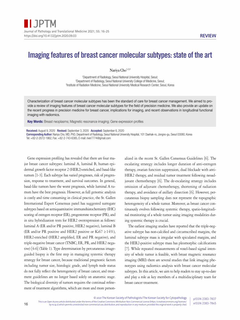
16
pISSN 2383-7837eISSN 2383-7845
© 2021 The Korean Society of Pathologists/The Korean Society for CytopathologyThis is an Open Access article distributed under the terms of the Creative Commons Attribution Non-Commercial License (https://creativecommons.org/licenses/
by-nc/4.0) which permits unrestricted non-commercial use, distribution, and reproduction in any medium, provided the original work is properly cited.
Gene expression profiling has revealed that there are four ma-jor breast cancer subtypes: luminal A, luminal B, human epi-dermal growth factor receptor 2 (HER2)-enriched, and basal-like tumors [1-3]. Each subtype has varied prognoses, risk of progres-sion, response to treatment, and survival outcomes. In general, basal-like tumors have the worst prognosis, while luminal A tu-mors have the best prognosis. However, as full genomic analysis is costly and time consuming in clinical practice, the St. Gallen International Expert Consensus panel has suggested surrogate subtypes based on semiquantitative immunohistochemistry (IHC) scoring of estrogen receptor (ER), progesterone receptor (PR), and in situ hybridization tests for HER2 overexpression as follows: luminal A (ER and/or PR positive, HER2 negative), luminal B (ER and/or PR positive and HER2 positive or Ki67 ≥ 14%), HER2-enriched (HER2 amplified, ER and PR negative), and triple-negative breast cancer (TNBC; ER, PR, and HER2 nega-tive) [4-6] (Table 1). Type determination by percutaneous image-guided biopsy is the first step in managing systemic therapy strategy for breast cancer, because traditional prognostic factors including tumor size, histologic grade, and lymph node status do not fully reflect the heterogeneity of breast cancer, and treat-ment guidelines are no longer based solely on anatomic stage. The biological diversity of tumors requires the continual refine-ment of treatment algorithms, which are more and more person-
alized in the recent St. Gallen Consensus Guidelines [6]. The escalating strategy includes longer duration of anti-estrogen therapy, ovarian function suppression, dual blockade with anti-HER2 therapy, and residual tumor treatment following neoad-juvant chemotherapy [6]. The de-escalating strategy includes omission of adjuvant chemotherapy, shortening of radiation therapy, and avoidance of axillary dissection [6]. However, per-cutaneous biopsy sampling does not represent the topographic heterogeneity of a whole tumor. Moreover, as breast cancer con-tinuously evolves following systemic therapy, spatio-longitudi-nal monitoring of a whole tumor using imaging modalities dur-ing systemic therapy is crucial.
The earliest imaging studies have reported that the triple-neg-ative subtype has non-calcified and circumscribed margins, the luminal subtype mass is irregular with spiculated margins, and the HER2-positive subtype mass has pleomorphic calcifications [7]. While repeated measurements of voxel-based signal inten-sity of whole tumor is feasible, with breast magnetic resonance imaging (MRI) there are several studies that link imaging phe-notypes using radiomics analysis with breast cancer molecular subtypes. In this article, we aim to help readers to stay up-to-date and play a role as key members of a multidisciplinary team for breast cancer treatment.
Imaging features of breast cancer molecular subtypes: state of the art
Nariya Cho1,2,3
1Department of Radiology, Seoul National University Hospital, Seoul; 2Department of Radiology, Seoul National University College of Medicine, Seoul;
3Institute of Radiation Medicine, Seoul National University Medical Research Center, Seoul, Korea
Characterization of breast cancer molecular subtypes has been the standard of care for breast cancer management. We aimed to pro-vide a review of imaging features of breast cancer molecular subtypes for the field of precision medicine. We also provide an update on the recent progress in precision medicine for breast cancer, implications for imaging, and recent observations in longitudinal functional imaging with radiomics.
Key Words: Breast neoplasms; Magnetic resonance imaging; Gene expression profiles
Received: August 9, 2020 Revised: September 3, 2020 Accepted: September 6, 2020Corresponding Author: Nariya Cho, MD, PhD, Department of Radiology, Seoul National University Hospital, 101 Daehak-ro, Jongno-gu, Seoul 03080, KoreaTel: +82-2-2072-1862, Fax: +82-2-743-6385, E-mail: [email protected]
REVIEWJournal of Pathology and Translational Medicine 2021; 55: 16-25https://doi.org/10.4132/jptm.2020.09.03

https://jpatholtm.org/https://doi.org/10.4132/jptm.2020.09.03
Imaging of breast molecular subtypes • 17
RADIOMICS IN BREAST CANCER
Radiomics is a technique to extract and select the quantitative features of radiologic images, to create a high-dimensional data set, and to draw hypotheses, which will lead to better clinical decisions [8]. In breast radiomics analysis, determination of be-nign or malignant lesion, correlation with prognostic factors, prediction of the response to systemic chemotherapy or lymph node metastasis have been studied. Radiomics features are math-ematically defined and classified into morphology, histogram, texture, or transformed features [8]. Morphology features are compactness, roundness, or convexity. Histogram features are median, entropy, uniformity, skewness, or kurtosis, in which spa-tial information is not included. Conversely, texture features in-clude the spatial information. Gray-level co-occurrence matrix (GLCM)-based features are the most commonly used method for textural analysis [8]. The relationship between voxels and their neighborhoods are characterized in GLCM analysis. Entropy, con-trast, and homogeneity, which reflect the uniformity or hetero-geneity of the voxel signal intensities, are the main parameters of GLCM models. Transform-based features such as Laplacian of Gaussian and wavelet are commonly used; these transform the original image, creating a new image from which the features
can be quantified. Application of radiomics in distinguishing molecular subtypes
is one of the most intensely studied areas (Table 2) [9-20]. Leithner et al. [9] reported accuracies of 81%–89% in distin-guishing luminal A from luminal B, luminal B from triple-negative, luminal B from all others, and HER2-enriched from all others. In their study, the region of interest of a tumor was drawn, and gray-level normalization was performed to minimize the effect of contrast variations. Then, first-order histogram, GLCM, and transform-based features were calculated. Feature selection to reduce the dimensionality of texture features, to se-lect the minimal numbers of features explaining the phenome-non, were performed. K-nearest neighbor classification with leave-one-out cross validation was performed for classification [9]. More recent studies using radiomic analysis reported an area under the receiver operating characteristic curve (AUC) of 0.844 in the prediction of disease-free survival of TNBC [10] and an AUC of 0.890 in the differentiation of HER2-positive tumors vs. -negative tumors [11]. The radiomics approach is based on the premise that microstructural variations between molecular subtypes would cause various gray-level textures on contrast enhanced MRI. The radiomics approach has the poten-tial to provide prognostic information of spatio-longitudinal
Table 1. Treatment-oriented classification of subgroups of breast cancer from St. Gallen consensus guidelines
Clinical grouping Notes Type of therapy
Triple-negative Negative ER, PR, and HER2 Cytotoxic chemotherapy including anthracycline and taxaneConsideration of cabecitabine for residual tumor after neoadjuvant chemotherapy
ER negative and HER2 positive American Society of Clinical Oncology (ASCO)/College of American Pathologists (CAP) guideline 2018a
Stage 1: paclitaxel+trastuzumab as adjuvant therapyStage 2 or 3: Neoadjuvant anthracycline, alkylator-, and taxane-based chemotherapy + trastuzumab- and pertuzumab-based treatment (dual anti-HER2 therapy)
Trastuzumab ematansine therapy for residual tumor after neoadjuvant chemotherapy in adjuvant setting
ER positive and HER2 positive ASCO/CAP guidelines 2018a As above + endocrine therapy appropriate to menopausal status
ER positive and HER2 negative ER and/or PR-positive ≥ 1%b
Luminal A-like (high receptor, low proliferation, low grade)
Multi-parameter molecular marker ‘good’ if availablec
High ER/PR and clearly low Ki-67 or gradeEndocrine therapy alone according to menopausal status
Intermediate Multi-parameter molecular marker ‘intermediate’ if availableUncertainty persists about degree of risk and responsiveness to endocrine and cytotoxic therapies
Endocrine therapy + adjuvant chemotherapy
Luminal B-like (low receptor, high proliferation, high grade)
Multi-parameter molecular marker ‘bad’ if available. Lower ER/PR with clearly high Ki-67, high histological grade 3
Endocrine therapy + adjuvant chemotherapy
Modified from Coates AS et al. Ann Oncol 2015;26:1533-46 [4] and Burstein HJ et al. Ann Oncol 2019;30:1542-57 [6], according to the Creative Commons license.ER, estrogen receptor; PR, progesterone receptor; HER2, human epidermal growth factor receptor 2; TNBC, triple-negative breast cancer. aAn immunohistochemistry of c-erbB-2 staining 3+ score was defined as HER2-positive, while a 0 or 1+ score was negative. For tumors with 2+ score, HER2 gene copies to the centromeric region of chromosome 17 ratios < 2.0 by fluorescence in situ hybridization must be interpreted as negative due to the lack of evidence for any benefit from HER2 targeted therapy; bIf ER values fall between 1% and 9%, the term equivocal should not be used, suggesting response to endocrine therapy even in low ER (1%–9%). Low HR expression is associated with higher Ki-67, higher grade, and loss PR positivity, as well as higher recur-rence score and higher chemo-sensitivity. Chemotherapy should be given following guidelines for TNBC. Endocrine therapy should be recommended despite the likely extremely small benefit; cNo role for gene testing in clinical pathologic low risk cases (pT1a, pT1b, G1, ER high, pN0).

https://jpatholtm.org/ https://doi.org/10.4132/jptm.2020.09.03
18 • Cho NTa
ble
2. P
ublic
atio
ns re
gard
ing
radi
omic
s fe
atur
es o
f bre
ast c
ance
r acc
ordi
ng to
mol
ecul
ar s
ubty
pe
Stud
yN
o.
Imag
ing
Para
met
ers
Out
com
esR
esul
ts
Leith
ner e
t al.
(201
9) [9
]14
3 Fi
rst o
rder
hist
ogra
m,
co-
occu
rrenc
e m
atrix
Lum
inal
A, l
umin
al B
, TN
BC
, HER
2-
enr
iche
d
Accu
racy
: lum
inal
B v
s. lu
min
al A
, 84.
2%; l
umin
al B
vs.
TN
BC
, 83.
9%; l
umin
al B
vs.
all o
ther
s, 8
9%;
HER
2-en
riche
d vs
. all o
ther
s, 8
1.3%
Kim
et a
l. (2
020)
[10]
228
Rad
iom
ics
scor
e fro
m 5
MR
I
fea
ture
s
Dise
ase
free
surv
ival o
utco
me
of
TN
BC
Com
bine
d cl
inic
opat
holo
gica
l and
radi
omic
s fe
atur
e m
odel
sho
wed
hig
hest
AU
C (0
.844
) in
pred
ictio
n
of D
FS
Song
et a
l. (2
020)
[11]
92
GLC
M fe
atur
es
HER
2-po
sitive
vs.
-neg
ative
tum
ors
det
erm
ined
by
FISH
test
Amon
g 3
mac
hine
lear
ning
met
hods
, LR
A, Q
DA,
and
SVM
, AU
C o
f SVM
was
the
best
(0.8
90)
Maz
urow
ski e
t al.
(201
4) [1
2]48
Sem
iaut
omat
ical
ly ex
tract
ed
MR
I fea
ture
s
TCG
A da
taba
se c
onta
ins
full
gen
omic
seq
uenc
ing
Hig
her r
atio
of l
esio
n en
hanc
emen
t rat
e to
bac
kgro
und
pare
nchy
mal
enh
ance
men
t was
mor
e lik
ely
to
be lu
min
al B
sub
type
Grim
m e
t al.
(201
5) [1
3]
275
56 F
eatu
res
(mor
phol
ogic
,
tex
ture
, and
dyn
amic
feat
ures
)
lum
inal
A, l
umin
al B
, HER
2, b
asal
Lum
inal
A a
nd lu
min
al B
mol
ecul
ar s
ubty
pe w
ere
asso
ciat
ed w
ith s
emia
utom
atic
ally
extra
cted
imag
ing
fea
ture
s
Bae
et a
l. (2
015)
[14]
280
Tum
or ro
undn
ess
scor
eER
, PR
, Ki-6
7ER
sco
re a
nd K
i-67
inde
x w
ere
inde
pend
ent f
acto
rs d
eter
min
ing
tum
or ro
undn
ess.
TN
BC
sho
wed
the
hig
hest
mea
n ro
undn
ess
scor
es c
ompa
red
with
the
othe
r sub
type
s
Sutto
n et
al.
(201
5) [1
5]95
Mor
phol
ogy,
hist
ogra
m
fea
ture
s, G
LCM
feat
ures
Onc
otyp
e D
X re
curre
nce
scor
e of
Lum
inal
A tu
mor
An in
crea
sed
kurto
sis w
as fo
und
to b
e a
stat
istic
ally
signi
fican
t fac
tor c
orre
latin
g w
ith O
ncot
ype
DX
rec
urre
nce
scor
e
Wau
gh e
t al.
(201
6) [1
6]
221
Text
ure
feat
ures
H
orm
one
rece
ptor
–pos
itive
and
–ne
gativ
e ca
ncer
s de
mon
stra
ted
signi
fican
tly d
iffere
nt e
ntro
py fe
atur
es
Text
ural
diffe
renc
es o
n co
ntra
st-e
nhan
ced
MR
imag
es m
ight
refle
ct u
nder
lying
lesio
n su
btyp
es
Li e
t al.
(201
6) [1
7]
91Te
xtur
e fe
atur
esLu
min
al A
, lum
inal
B, H
ER2-
enric
hed,
and
bas
al-li
ke s
ubty
pe
Enha
ncem
ent t
extu
re (e
ntro
py) a
nd m
olec
ular
sub
type
s w
ere
rela
ted.
AU
C o
f ER
+ vs
. ER
–: 0
.89,
PR
+
vs. P
R–:
0.6
9, H
ER2+
vs.
HER
2–: 0
.65,
TN
BC
vs.
oth
ers:
0.6
7
Agne
r et a
l. (2
014)
[18]
76
Mor
phol
ogy,
kine
tic in
tens
ity,
hist
ogra
m fe
atur
es, G
LCM
feat
ures
TNB
C, H
ER2,
ER
-pos
itive
tum
ors
AUC
of t
extu
re fe
atur
es (m
ore
hete
roge
neity
) for
TN
BC
vs.
oth
er s
ubty
pes:
0.7
3 to
0.7
4
Cha
mm
ing’
s et
al.
(201
8) [1
9]85
Fine
, med
ium
, and
coa
rse
tex
ture
for m
ean,
sta
ndar
d
devia
tion,
mea
n pr
opor
tion
of
posit
ive p
ixels,
ent
ropy
,
skew
ness
, and
kur
tosis
pCR
of T
NB
Cku
rtosis
app
ears
to b
e as
soci
ated
with
pC
R to
neo
adju
vant
che
mot
hera
py in
non
–TN
BC
and
may
be
a pr
omisi
ng b
iom
arke
r for
the
iden
tifica
tion
of T
NB
C
Bra
man
et a
l. (2
017)
[20]
11
7In
tratu
mor
al a
nd p
eritu
mor
al
tex
ture
feat
ures
Pred
ictio
n of
pC
RC
ombi
ned
intra
tum
oral
and
per
itum
oral
radi
omic
s fo
r pre
dict
ion
of p
CR
yie
lded
AU
C 0
.83
for H
R+
HER
2–. N
on-p
CR
was
cha
ract
erize
d by
ele
vate
d pe
ritum
oral
het
erog
enei
ty d
urin
g in
itial p
hase
. For
TNB
C/H
ER+
tum
ors
wer
e be
st c
hara
cter
ized
by a
per
itum
oral
spe
ckle
d en
hanc
emen
t.
TNB
C, t
riple
-neg
ative
bre
ast c
ance
r; H
ER2,
hum
an e
pide
rmal
gro
wth
fact
or re
cept
or 2
; MR
I, m
agne
tic re
sona
nce
imag
ing;
AU
C, a
rea
unde
r the
rece
iver o
pera
ting
char
acte
ristic
cur
ve; D
FS, d
iseas
e-fre
e su
rviva
l; G
LCM
, gra
y-le
vel c
o-oc
curre
nce
mat
rix; F
ISH
, fluo
resc
ence
in s
itu h
ybrid
izatio
n; L
RA,
logi
stic
regr
essio
n an
alys
is; Q
DA,
qua
drat
ic d
iscrim
inan
t ana
lysis;
SVM
, sup
port
vect
or m
achi
ne; T
CG
A, T
he C
ance
r Gen
ome
Atla
s; E
R, e
stro
gen
rece
ptor
; PR
, pro
gest
eron
e re
cept
or; p
CR
, pat
holo
gica
l com
plet
e re
spon
se.

https://jpatholtm.org/https://doi.org/10.4132/jptm.2020.09.03
Imaging of breast molecular subtypes • 19
biology changes of the peritumoral parenchyma as well as of the tumor itself. Moreover, deep learning methods have increasing-ly been applied in recent radiomics studies [8,11]. The deep learning approach, which is data driven and capable of learning relevant features from the data themselves, has shown superior performance in various tasks in radiology [8,11]. In the near fu-ture, radiomics parameters based on a deep learning algorithm would be useful surrogate markers for precision medicine in breast cancer treatment.
LUMINAL SUBTYPE
About 70% of breast cancers are ER positive and show a more favorable prognosis than ER-negative cancers. Within ER-posi-tive/HER2-negative breast cancer, 90%–95% are luminal A or B subtypes [3]. The luminal B subtype shows higher prolifera-tion gene expression [2] and worse recurrence-free survival out-comes compared with the luminal A subtype, although the lu-minal B subtype shows higher pathological complete response
(pCR) rate following neoadjuvant chemotherapy [3]. Thus, dif-ferentiation between luminal A and B tumors is important for deciding the duration of endocrine therapy or to predict resistance to endocrine therapy [3]. There is a 30 to 44% discordance rate between the gene expression profiling and surrogate IHC clas-sifications [3,21]. Within ER-positive/HER2-negative breast cancers, 5%–10% of tumors are non-luminal subtypes (HER2 enriched and basal-like tumors) by gene expression profiling [3]. Non-luminal (ER positive/HER2 negative) breast cancers show worse outcomes compared with the luminal A subtype when they were treated with 5 years of adjuvant tamoxifen-only [22]. One study reported that 80% of tumors showing low expres-sion ER positive (1%–9%) were non-luminal subtypes [23].
For patients with ER-positive tumors, prognostic signatures including 70-gene MammaPrint microarray assay (Agendia, Amsterdam, The Netherlands), the 50-gene PAM50 assay (Pro-signa, Nanostring Technologies, Seattle, WA, USA), and the 21-gene Oncotype DX assay (Genomic Health, Redwood City, CA, USA) are commercially available [24]. These signatures al-
A B
Fig. 1. A 56-year-old woman with a luminal A-like breast cancer. (A) Mammography shows a spiculated mass with calcifications (arrow). (B) Enhanced T1-weighted magnetic resonance imaging shows an irregular, spiculated mass (arrow). Histopathology revealed a 1.5-cm invasive ductal carcinoma with low histologic grade. American Joint Committee on Cancer (AJCC) anatomic stage was T1N0M0. Immunohistochem-istry analysis revealed that estrogen receptor 90% positive, progesterone receptor 1% positive, human epidermal growth factor receptor 2–negative, and Ki-67, 1% positive. Multigene assay recurrence score was 10 and low risk. The 9-year distant recurrence risk was estimated as 3%. She did not receive adjuvant chemotherapy, but received aromatase inhibitor.

https://jpatholtm.org/ https://doi.org/10.4132/jptm.2020.09.03
20 • Cho N
low us to distinguish prognosis of patients according to the pro-liferation-associated genes expression levels [24]. However, the signatures do not apply to patients with ER-negative tumors, because more than 95% of them already have high expression of proliferation-associated genes [24,25]. The oncotype DX as-say analyzes a panel of 21 genes to decide a recurrence score (RS) representing the possibility of recurrent cancer within 10 years. Adjuvant chemotherapy provides greater benefits for patients with high-RS tumors and does very little for patients with low-RS tumors (Fig. 1) [26].
In morphologic analyses for mammography, ER-positive tu-mor tends to show a not-circumscribed margin (Fig. 1), which is in contrast to ER-negative tumors [12,13,27]. Tumor round-ness score, quantifying the relative similarity to a perfect circle, has been shown to have an inverse correlation with the ER ex-pression (%) and a positive correlation with the Ki-67 index [14]. By ultrasonography, parallel orientation (odds ratio [OR], 5.53; p = .02) and tumor roundness (OR, 1.70 per 10 increase in the roundness value; p = .01) were independent features associated
with high RS on Oncotype DX [28]. The high-risk group was also associated with the presence of calcifications, similar to a previ-ous study in which a mass with pleomorphic microcalcifications might be associated with an intermediate to high RS in ER-posi-tive, HER2-negative early breast cancer at mammography [29].
A previous report on MRI results demonstrated that patients with luminal B subtype tended to have multifocal or multicentric cancer 2.8 times more often than patients with luminal A sub-type [30]. Sutton et al. [15] reported that an increased kurtosis was associated with high RS on Oncotype DX for ER-positive/HER2-negative tumors (Fig. 2). Kurtosis is a second order pa-rameter quantifying the amount of histogram deviating from a Gaussian shape. High kurtosis might reflect the amount of het-erogeneity in a complex way, and these tumors are believed to be more biologically aggressive [19]. This result is in line with a study reporting that both ER-positive and ER-negative tumors showed statistically different entropy levels [16]. The entropy reflecting spatial distribution pattern of grayness of voxel is also believed to be an important biomarker at textural analysis of medical image.
A B
Fig. 2. A 44-year-old woman with a luminal A-like breast cancer. (A) Mammography shows an oval non-calcified mass (arrow). (B) Enhanced T1-weighted magnetic resonance imaging shows an irregular mass with internal rim-enhancement (arrow). Histopathology revealed a 1.7-cm invasive ductal carcinoma with intermediate histologic grade. American Joint Committee on Cancer (AJCC) anatomic stage was T1N0M0. Immunohistochemistry analysis revealed that estrogen receptor 90% positive, progesterone receptor 5% positive, human epidermal growth factor receptor 2 negative, and Ki-67, 4% positive. Multigene assay recurrence score was 23. The 10-year distant recurrence risk was esti-mated as 12% and high risk. She received adjuvant chemotherapy and tamoxifen.

https://jpatholtm.org/https://doi.org/10.4132/jptm.2020.09.03
Imaging of breast molecular subtypes • 21
Fig. 3. A 67-year-old woman with a human epidermal growth factor receptor 2 (HER2)–positive breast cancer. (A) Mammography shows ill-defined asymmetry with pleomorphic microcalcifications (arrows). (B) Enhanced T1-weighted magnetic resonance imaging (MRI) shows an 8.2-cm ill-defined, diffuse irregular mass with internal heterogeneous enhancement. Needle biopsy revealed an invasive ductal carcinoma with high histologic grade. Immunohistochemistry analysis revealed that estrogen receptor and progesterone receptor negative, and HER2 positive. (C) Following combined docetaxel, carboplatin and dual HER2 blockade, there is no residual mass and but subtle enhancements in the breast on MRI (arrows). (D) Mammography shows two hookwires around the residual calcifications (arrows). Surgical histopathology re-vealed pathological complete response in the breast and axilla.
A
C
B
D

https://jpatholtm.org/ https://doi.org/10.4132/jptm.2020.09.03
22 • Cho N
HUMAN EPIDERMAL GROWTH FACTOR RECEPTOR 2–ENRICHED SUBTYPE
HER2 overexpression is found in approximately 20% of in-vasive breast cancers. It is associated with worse prognosis but good response to HER2-targeted therapies [31] and is reported to increase cell proliferation, survival, mobility, and invasiveness, as well as neo-angiogenesis at the cellular level [32]. The Cancer Genome Atlas (TCGA) and clinical trials have suggested that HER2-positive tumors are a heterogeneous group of cancers [3]. Compared with ER-positive/HER2-positive tumors, patients with ER-negative/HER2-positive tumors show a higher risk of death within 5 years of diagnosis; the first recurrence in brain was higher and in bone was lower, and the response rate to neoadju-vant chemotherapy was higher [33,34]. HER2-targeted agents combined with chemotherapy are recommended for ER-nega-tive/HER2-positive tumors, and HER2-targeted agents with endocrine therapy are recommended for ER-positive/HER2-positive tumors [4]. A higher pCR rate was observed in patients with ER-negative/HER2-positive tumors than in patients with ER-positive/HER2-positive tumors [35]. The pCR rate was over 70% using the dual HER2 blockade, either with trastuzumab with lapatinib or trastuzumab with pertuzumab in combination with chemotherapy [6].
HER2 overexpression was also associated with the presence of calcifications, branching or fine linear shape calcifications, high
level of suspicion on mammography, and a washout or fast early enhancement kinetic curve pattern on MRI (Fig. 3) [36]. MRI showed that patients with the HER2 subtype tended to have 4.1 times more multifocal or multicentric cancers than patients with the luminal A subtype [30]. Fine pleomorphic/fine linear or linear branching calcification morphology on mammography (OR, 7.23), PR negativity (OR, 6.76), and a high TILs (tumor in-filtrating lymphocytes) level (OR, 5.92) were independent fac-tors associated with pCR in patients receiving neoadjuvant che-motherapy with dual HER2 blockade (Fig. 3) [37]. Low tumor peak enhancement at MRI indicating less aggressiveness was significantly associated with high TILs (OR, 1.01; p = .020) [38]. These results are consistent with previous studies showing that TILs observed in breast cancers were associated with higher rate of pCR or improved overall survival outcomes [5,6]. More-over, increasing TILs during systemic therapy was reported to be correlated with pCR [39], which suggests that MRI could pro-vide valuable information regarding response during treatment.
BASAL-LIKE SUBTYPES
TNBC comprises 10%–20% of all breast cancers. The term of TNBC and basal-like tumors are interchangeably used be-cause 86% of TNBC are the basal-like subtype [3]. However, each of the intrinsic subtypes exist within a TNBC [40], and TNBC is a very heterogeneous group of tumors based on ge-
A
C
B
Fig. 4. A 35-year-old woman with a triple-negative breast cancer. (A) En-hanced T1-weighted magnetic resonance imaging (MRI) shows a 3.3 cm round mass with an internal rim enhancement and peritumoral het-erogeneous enhancement (arrow) (B) T2-weighted MRI shows a central cystic necrosis and peritumoral edema (arrow). Needle biopsy revealed an invasive ductal carcinoma with high histologic grade. Immunohisto-chemistry analysis revealed that estrogen receptor, progesterone recep-tor, and human epidermal growth factor receptor 2 negative. (C) Follow-ing chemotherapy, enhanced T1-weighted MRI shows a 3.4 cm round mass without response to chemotherapy.

https://jpatholtm.org/https://doi.org/10.4132/jptm.2020.09.03
Imaging of breast molecular subtypes • 23
netic profiling [41]. Rates of pCR after receiving anthracycline/taxane regimen are 25%–35% and patients with pCR show a better outcome in patients with TNBC [25]. Recent St. Gallen Consensus Conference Guidelines recommended that TILs should be routinely characterized for TNBC in view of their prognostic value [6]. Tumor programmed death-ligand 1 and immune-cell programmed death-1 expression are considered as markers to predict benefit from immunotherapy for advanced TNBC [6]. Also, in TNBC with residual disease following neoadjuvant che-motherapy, post-neoadjuvant treatment with capecitabine showed survival benefits [42].
Unifocal, circumscribed margin, round shape, and no associ-ated calcifications are signatures of TNBC [43]. Circumscribed margin and round shape are more commonly found in high-grade tumors and spiculation is more frequently found in low-grade tumors [44]. TNBC has been shown to have a higher tu-mor roundness score compared with the other subtypes, reflecting a more biologically aggressive tumor type. Absence of calcifica-tions is also considered representative of rapid malignant trans-formation of TNBC with bypassing of the in-situ stage [43]. TNBC shows a round mass with rim enhancement on MRI and frequently has internal high signal intensity on T2-weighted MRI [45-47] (Fig. 4). Recent studies using radiomics analysis have reported that TNBC masses tend to be larger, have a more heterogeneous enhancement texture, are more irregularly shaped, and have a rapid enhancement rate compared with other sub-types [17,18]. Notably, the heterogeneous enhancement texture, quantified at the first post-contrast enhanced MRI, has emerged as a discriminatory indicator for tumor subtype, regardless of tumor size [17,18]. Waugh et al. [16] also reported that TNBC and HER2 subtypes showed increased entropy values compared with luminal A and luminal B subtypes.
In the neoadjuvant chemotherapy setting, intratumoral necro-sis was associated with non-response to chemotherapy [48] and peritumoral edema was associated with worse recurrence-free-survival outcome of TNBC (Fig. 4) [49]. With textural analysis, increased kurtosis of non-TNBC on T2 weighted image was independently associated with pCR; however, the association between increased kurtosis and pCR was not found in TNBC [19]. In another study predicting pCR by using radiomics, Bra-man et al. reported that combined intratumoral and peritumoral radiomic features yielded a maximum AUC of 0.83 for the ER- positive/HER2-negative group and 0.93 for TNBC or HER2-positive group [20]. In Braman et al’s study [20], elevated peri-tumoral heterogeneity was associated with non-pCR in ER-positive/HER2-negative tumors, and peritumoral speckled en-
hancement pattern was associated with non-pCR in TNBC or HER2-positive tumors (Fig. 4A) [20]. These results are in line with peritumoral lymphatics or vascular invasion, and peritu-moral immune response as predictors of survival. In addition, TILs, known as a favorable prognostic factor in TNBC, could be quantified by textural analysis [20]. Thus, peritumoral ra-diomics features on MRI could be valuable predictors of pCR in TNBC and HER2-positive tumors.
CONCLUSION
Breast cancer consists of heterogeneous subtypes and evolves continuously after systemic therapy. Earlier studies linking im-aging features and molecular subtypes have reported presence of calcifications, margin or shape features, and enhancement fea-tures on dynamic contrast enhanced MRI according to each subtype. Recent studies using radiomics parameters, which are indiscernible by the human eye, have shown high accuracy in distinguishing molecular subtypes, prediction of responses to chemotherapy, and prediction of survival outcomes. Imaging biomarkers could be helpful in realizing better precision medi-cine due to the feasibility of repeated measurements for whole tumors and the applicability of deep-learning based algorithms.
Ethics StatementNot applicable.
ORCID Nariya Cho https://orcid.org/0000-0003-4290-2777
Conflicts of InterestThe authors declare that they have no potential conflicts of interest.
Funding StatementNo funding to declare.
References1. Cancer Genome Atlas Network. Comprehensive molecular por-
traits of human breast tumours. Nature 2012; 490: 61-70.2. Sorlie T, Perou CM, Tibshirani R, et al. Gene expression patterns of
breast carcinomas distinguish tumor subclasses with clinical impli-cations. Proc Natl Acad Sci U S A 2001; 98: 10869-74.
3. Prat A, Pineda E, Adamo B, et al. Clinical implications of the in-trinsic molecular subtypes of breast cancer. Breast 2015; 24 Suppl 2: S26-35.
4. Coates AS, Winer EP, Goldhirsch A, et al. Tailoring therapies--im-proving the management of early breast cancer: St Gallen Interna-tional Expert Consensus on the Primary Therapy of Early Breast Cancer 2015. Ann Oncol 2015; 26: 1533-46.
5. Curigliano G, Burstein HJ, Winer EP, et al. De-escalating and esca-lating treatments for early-stage breast cancer: the St. Gallen Inter-

https://jpatholtm.org/ https://doi.org/10.4132/jptm.2020.09.03
24 • Cho N
national Expert Consensus Conference on the Primary Therapy of Early Breast Cancer 2017. Ann Oncol 2017; 28: 1700-12.
6. Burstein HJ, Curigliano G, Loibl S, et al. Estimating the benefits of therapy for early-stage breast cancer: the St. Gallen International Consensus Guidelines for the primary therapy of early breast can-cer 2019. Ann Oncol 2019; 30: 1541-57.
7. Cho N. Molecular subtypes and imaging phenotypes of breast can-cer. Ultrasonography 2016; 35: 281-8.
8. Lee SH, Park H, Ko ES. Radiomics in breast imaging from tech-niques to clinical applications: a review. Korean J Radiol 2020; 21: 779-92.
9. Leithner D, Horvat JV, Marino MA, et al. Radiomic signatures with contrast-enhanced magnetic resonance imaging for the assessment of breast cancer receptor status and molecular subtypes: initial re-sults. Breast Cancer Res 2019; 21: 106.
10. Kim S, Kim MJ, Kim EK, Yoon JH, Park VY. MRI radiomic fea-tures: association with disease-free survival in patients with triple-negative breast cancer. Sci Rep 2020; 10: 3750.
11. Song L, Lu H, Yin J. Preliminary study on discriminating HER2 2+ amplification status of breast cancers based on texture features semi-automatically derived from pre-, post-contrast, and subtrac-tion images of DCE-MRI. PLoS One 2020; 15: e0234800.
12. Mazurowski MA, Zhang J, Grimm LJ, Yoon SC, Silber JI. Radioge-nomic analysis of breast cancer: luminal B molecular subtype is as-sociated with enhancement dynamics at MR imaging. Radiology 2014; 273: 365-72.
13. Grimm LJ, Zhang J, Mazurowski MA. Computational approach to radiogenomics of breast cancer: Luminal A and luminal B molecu-lar subtypes are associated with imaging features on routine breast MRI extracted using computer vision algorithms. J Magn Reson Imaging 2015; 42: 902-7.
14. Bae MS, Seo M, Kim KG, Park IA, Moon WK. Quantitative MRI morphology of invasive breast cancer: correlation with immunohis-tochemical biomarkers and subtypes. Acta Radiol 2015; 56: 269-75.
15. Sutton EJ, Oh JH, Dashevsky BZ, et al. Breast cancer subtype inter-tumor heterogeneity: MRI-based features predict results of a ge-nomic assay. J Magn Reson Imaging 2015; 42: 1398-406.
16. Waugh SA, Purdie CA, Jordan LB, et al. Magnetic resonance imag-ing texture analysis classification of primary breast cancer. Eur Ra-diol 2016; 26: 322-30.
17. Li H, Zhu Y, Burnside ES, et al. Quantitative MRI radiomics in the prediction of molecular classifications of breast cancer subtypes in the TCGA/TCIA data set. NPJ Breast Cancer 2016; 2: 16012.
18. Agner SC, Rosen MA, Englander S, et al. Computerized image analysis for identifying triple-negative breast cancers and differen-tiating them from other molecular subtypes of breast cancer on dy-namic contrast-enhanced MR images: a feasibility study. Radiology 2014; 272: 91-9.
19. Chamming’s F, Ueno Y, Ferre R, et al. Features from computerized texture analysis of breast cancers at pretreatment MR imaging are associated with response to neoadjuvant chemotherapy. Radiology 2018; 286: 412-20.
20. Braman NM, Etesami M, Prasanna P, et al. Intratumoral and peri-tumoral radiomics for the pretreatment prediction of pathological complete response to neoadjuvant chemotherapy based on breast DCE-MRI. Breast Cancer Res 2017; 19: 57.
21. Chia SK, Bramwell VH, Tu D, et al. A 50-gene intrinsic subtype clas-sifier for prognosis and prediction of benefit from adjuvant tamoxi-
fen. Clin Cancer Res 2012; 18: 4465-72.22. Prat A, Parker JS, Fan C, et al. Concordance among gene expres-
sion-based predictors for ER-positive breast cancer treated with ad-juvant tamoxifen. Ann Oncol 2012; 23: 2866-73.
23. Iwamoto T, Booser D, Valero V, et al. Estrogen receptor (ER) mRNA and ER-related gene expression in breast cancers that are 1% to 10% ER-positive by immunohistochemistry. J Clin Oncol 2012; 30: 729-34.
24. Ng CK, Schultheis AM, Bidard FC, Weigelt B, Reis-Filho JS. Breast cancer genomics from microarrays to massively parallel sequencing: paradigms and new insights. J Natl Cancer Inst 2015; 107:djv015.
25. Liedtke C, Mazouni C, Hess KR, et al. Response to neoadjuvant therapy and long-term survival in patients with triple-negative breast cancer. J Clin Oncol 2008; 26: 1275-81.
26. Paik S, Tang G, Shak S, et al. Gene expression and benefit of che-motherapy in women with node-negative, estrogen receptor-posi-tive breast cancer. J Clin Oncol 2006; 24: 3726-34.
27. Shin HJ, Kim HH, Huh MO, et al. Correlation between mammo-graphic and sonographic findings and prognostic factors in patients with node-negative invasive breast cancer. Br J Radiol 2011; 84: 19-30.
28. Chae EY, Moon WK, Kim HH, et al. Association between ultra-sound features and the 21-gene recurrence score cssays in patients with oestrogen receptor-positive, HER2-negative, invasive breast cancer. PLoS One 2016; 11: e0158461.
29. Yepes MM, Romilly AP, Collado-Mesa F, et al. Can mammograph-ic and sonographic imaging features predict the Oncotype DX recur-rence score in T1 and T2, hormone receptor positive, HER2 negative and axillary lymph node negative breast cancers? Breast Cancer Res Treat 2014; 148: 117-23.
30. Grimm LJ, Johnson KS, Marcom PK, Baker JA, Soo MS. Can breast cancer molecular subtype help to select patients for preoperative MR imaging? Radiology 2015; 274: 352-8.
31. Arteaga CL, Sliwkowski MX, Osborne CK, Perez EA, Puglisi F, Gi-anni L. Treatment of HER2-positive breast cancer: current status and future perspectives. Nat Rev Clin Oncol 2011; 9: 16-32.
32. Zhou BP, Hung MC. Dysregulation of cellular signaling by HER2/neu in breast cancer. Semin Oncol 2003; 30: 38-48.
33. Perez EA, Romond EH, Suman VJ, et al. Four-year follow-up of trastuzumab plus adjuvant chemotherapy for operable human epi-dermal growth factor receptor 2-positive breast cancer: joint analysis of data from NCCTG N9831 and NSABP B-31. J Clin Oncol 2011; 29: 3366-73.
34. Vaz-Luis I, Ottesen RA, Hughes ME, et al. Impact of hormone re-ceptor status on patterns of recurrence and clinical outcomes among patients with human epidermal growth factor-2-positive breast can-cer in the National Comprehensive Cancer Network: a prospective cohort study. Breast Cancer Res 2012; 14: R129.
35. Gianni L, Eiermann W, Semiglazov V, et al. Neoadjuvant and adju-vant trastuzumab in patients with HER2-positive locally advanced breast cancer (NOAH): follow-up of a randomised controlled su-periority trial with a parallel HER2-negative cohort. Lancet Oncol 2014; 15: 640-7.
36. Elias SG, Adams A, Wisner DJ, et al. Imaging features of HER2 overexpression in breast cancer: a systematic review and meta-analysis. Cancer Epidemiol Biomarkers Prev 2014; 23: 1464-83.
37. Yoon GY, Chae EY, Cha JH, et al. Imaging and clinicopathologic features associated with pathologic complete response in HER2-positive breast cancer receiving neoadjuvant chemotherapy with

https://jpatholtm.org/https://doi.org/10.4132/jptm.2020.09.03
Imaging of breast molecular subtypes • 25
dual HER2 blockade. Clin Breast Cancer 2020; 20: 25-32.38. Choi WJ, Kim Y, Cha JH, et al. Correlation between magnetic reso-
nance imaging and the level of tumor-infiltrating lymphocytes in patients with estrogen receptor-negative HER2-positive breast can-cer. Acta Radiol 2020; 61: 3-10.
39. Nuciforo P, Pascual T, Cortes J, et al. A predictive model of patho-logic response based on tumor cellularity and tumor-infiltrating lymphocytes (CelTIL) in HER2-positive breast cancer treated with chemo-free dual HER2 blockade. Ann Oncol 2018; 29: 170-7.
40. Prat A, Adamo B, Cheang MC, Anders CK, Carey LA, Perou CM. Molecular characterization of basal-like and non-basal-like triple-negative breast cancer. Oncologist 2013; 18: 123-33.
41. Lehmann BD, Bauer JA, Chen X, et al. Identification of human tri-ple-negative breast cancer subtypes and preclinical models for se-lection of targeted therapies. J Clin Invest 2011; 121: 2750-67.
42. Masuda N, Lee SJ, Ohtani S, et al. Adjuvant capecitabine for breast cancer after preoperative chemotherapy. N Engl J Med 2017; 376: 2147-59.
43. Dogan BE, Turnbull LW. Imaging of triple-negative breast cancer. Ann Oncol 2012; 23 Suppl 6: vi23-9.
44. Lamb PM, Perry NM, Vinnicombe SJ, Wells CA. Correlation be-tween ultrasound characteristics, mammographic findings and his-
tological grade in patients with invasive ductal carcinoma of the breast. Clin Radiol 2000; 55: 40-4.
45. Uematsu T, Kasami M, Yuen S. Triple-negative breast cancer: cor-relation between MR imaging and pathologic findings. Radiology 2009; 250: 638-47.
46. Youk JH, Son EJ, Chung J, Kim JA, Kim EK. Triple-negative inva-sive breast cancer on dynamic contrast-enhanced and diffusion-weighted MR imaging: comparison with other breast cancer sub-types. Eur Radiol 2012; 22: 1724-34.
47. Kim GR, Ku YJ, Cho SG, Kim SJ, Min BS. Associations between gene expression profiles of invasive breast cancer and Breast Imag-ing Reporting and Data System MRI lexicon. Ann Surg Treat Res 2017; 93: 18-26.
48. Kawashima H, Inokuchi M, Furukawa H, Kitamura S. Triple-nega-tive breast cancer: are the imaging findings different between re-sponders and nonresponders to neoadjuvant chemotherapy? Acad Radiol 2011; 18: 963-9.
49. Bae MS, Shin SU, Ryu HS, et al. Pretreatment MR imaging features of triple-negative breast cancer: association with response to neoad-juvant chemotherapy and recurrence-free survival. Radiology 2016; 281: 392-400.

26
pISSN 2383-7837eISSN 2383-7845
© 2021 The Korean Society of Pathologists/The Korean Society for CytopathologyThis is an Open Access article distributed under the terms of the Creative Commons Attribution Non-Commercial License (https://creativecommons.org/licenses/
by-nc/4.0) which permits unrestricted non-commercial use, distribution, and reproduction in any medium, provided the original work is properly cited.
Translational research is a two-way process in which new findings from basic research are applied in clinical trials and de-velopment of new drugs or treatments. Samples of DNA, RNA, proteins, the metabolome, etc. collected from cancer tissues and blood or urine are analyzed in clinical trials to search for valuable and relevant biomarkers that are crucial for drug development. Next-generation sequencing (NGS) has helped us understand and characterize many cancers, identify new cancer subtypes, develop biomarkers, and discover new treatment targets. NGS also has allowed us to learn about the mutational landscape of several cancers and to develop technologies and drugs targeting “driver” molecular abnormalities. Thus, the oncological commu-nity considers NGS as a tool to improve the effectiveness of cancer treatment. Comprehensive, integrated molecular analyses iden-tify molecular relationships across a diverse set of human cancers, suggesting future directions to explore clinical actionability in cancer treatments [1]. Early successes in targeting and identify-ing individual oncogenic drivers and increased feasibility of tumor genome sequencing have made possible genome-driven oncology
care [2]. We are now using NGS to capture genetic algorithms and register them for clinical trials.
Immunotherapy, especially use of immune checkpoint inhib-itors (ICI), has led to dramatic changes in the treatment of sev-eral types of cancer in recent years [3]. Given that a small num-ber of patients experience a long-lasting response, development of biomarkers to predict responsiveness to immunotherapy has become important. The most widely investigated biomarkers for immunotherapy are programmed death-ligand 1 (PD-L1), microsatellite instability/defective mismatch repair (MSI/dMMR), and tumor mutational burden (TMB). Although MSI/dMMR has been used for immunotherapy regardless of tumor type, PD-L1 is being used in specific cancer types [3]. In this article, we concisely review the applications of NGS and immunohistochem-istry (IHC)-based protein biomarkers, especially PD-L1, in pre-cision oncology and clinical trials.
DNA-protein biomarkers for immunotherapy in the era of precision oncology
Binnari Kim1,2, So Young Kang1, Kyoung-Mee Kim1,3
1Department of Pathology and Translational Genomics, Samsung Medical Center, Sungkyunkwan University School of Medicine, Seoul; 2Department of Pathology, Ulsan University Hospital, Ulsan;
3Center of Clinical Genomics, Samsung Medical Center, Seoul, Korea
The use of biomarkers to guide patient and therapy selection has gained much attention to increase the scope and complexity of tar-geted therapy options and immunotherapy. Clinical trials provide a basis for discovery of biomarkers, which can then aid in develop-ment of new drugs. To that end, samples from cancer patients, including DNA, RNA, protein, and the metabolome isolated from cancer tissues and blood or urine, are analyzed in various ways to identify relevant biomarkers. In conjunction with nucleotide-based, high-throughput, next-generation sequencing techniques, therapy-guided biomarker assays relying on protein-based immunohistochemistry play a pivotal role in cancer care. In this review, we discuss the current knowledge regarding DNA and protein biomarkers for cancer im-munotherapy.
Key Words: Biomarker; Clinical trial; Next-generation sequencing; Immunohistochemistry
Received: June 12, 2020 Revised: September 7, 2020 Accepted: September 23, 2020Corresponding Author: Kyoung-Mee Kim, MD, Department of Pathology and Translational Genomics, Samsung Medical Center, Sungkyunkwan University School of Medicine, 81 Irwon-ro, Gangnam-gu, Seoul 06351, KoreaTel: +82-2-3410-2807, Fax: +82-2-3410-6396, E-mail: [email protected]
REVIEWJournal of Pathology and Translational Medicine 2021; 55: 26-32https://doi.org/10.4132/jptm.2020.09.23

https://jpatholtm.org/https://doi.org/10.4132/jptm.2020.09.23
DNA-protein biomarker analysis • 27
TUMOR MUTATIONAL BURDEN
Clinical significance and cutoff points
Although not yet approved for clinical use, TMB has been shown to predict the response to several forms of immunotherapy across multiple cancer types. Specifically, cancer patients with a high neoantigen load or high TMB are more likely to have a good clinical response to ICI [4].
While not all mutations result in immunogenic neoantigens and determining which mutations are likely to induce immuno-genic neoantigens remains a challenge, TMB represents a quan-tifiable measure of the number of mutations in a tumor that can be used for treatment selection [5]. TMB has been traditionally determined using whole-exome sequencing (WES); however, the high cost and long work time limit its widespread use in clinical settings. Therefore, current precision oncology platforms generally use NGS of targeted gene panels [6]. A recent study analyzing clinical data from 7,033 ICI- and non-ICI–treated advanced-stage cancer patients and genomic data from cancers sequenced with targeted NGS showed that higher somatic TMB (highest 20% in each histology subtype) was associated with better overall survival in all patients [7]. However, the TMB cutoff points asso-ciated with improved survival varied markedly between cancer types, suggesting that a universal definition of high TMB may not be possible. Similar findings in patients with gastric cancer were observed when the following cutoff points were applied: 11% for the higher mutation group in 330 non-ICI–treated pa-tients [8] and 14.31 mt/mb in 63 ICI-treated patients [6].
Despite efforts to standardize TMB from multiple genomic profiling cancer panels [4], the cutoff value for TMB remains
inconsistent. TMB is generally defined as the number of non-synonymous somatic mutations per megabase of genome exam-ined, and a detailed description of TMB definitions in recently published papers using targeted sequencing is summarized in Table 1 [6-18].
Factors affecting tumor mutation burden
Measurement of mutation load using WES can be difficult due to its high cost and extensive analysis and data management requirements. To be applicable in a clinical setting, the follow-ing requirements need to be met: the test must be suitable for clinical samples even with a limited amount of DNA; and the test results should be delivered within a limited time, be accu-rate, help clinical decision-making, and must be affordable. Thus, targeted sequencing with comprehensive gene panels is desir-able because of the lower sequencing costs, lower DNA input amounts, and shorter turnaround time [19]. However, the follow-ing factors affect TMB calculation: (1) Contents of tumor cell and coverage of sequencing, as targeted cancer panels enable deeper sequencing compared with WES. (2) Presence of sequence artifacts that can be caused by formalin fixation as formalin can cause various crosslinks and is a well-known source of sequencing artifacts due to fragmentation of DNA, denaturation, and de-amination of cytosine bases. Specifically, using formalin-fixed paraffin-embedded tissues for NGS causes an increase in DNA sequence artifacts (C:G > T:A) [20].
Pre-analytical factors also affect TMB measurement [19]. Among the most important factors are the size and number of genes included within the targeted cancer panel. The most widely used panels are the MSK-IMPACT panel, which in its latest ver-
Table 1. Tumor mutation burden measured by targeted sequencing in various cancer types
Cancer typeNo. of
patientsMethods Name of panel
No. of genes
Cutoff (mt/Mb)
Cutoff (percentile)
ICI responses
Study
Pan-cancer 1,638 Targeted sequencing FoundationOne ~315 20 90 Yes Goodman et al. [9]Pan-cancer 2,189 Targeted sequencing Custom Panel 592 17 92.3 Yes Vanderwalde et al. [10]Pan-cancer 1,662 Targeted sequencing MSK-IMPACT v3 468 8.8 80 Yes Samstein et al. [7]SCLC 134 Targeted sequencing DFCI OncoPanel 447 9.68 50 Yes Ricciuti et al. [11]Colorectal 6,004 Targeted sequencing Comprehensive Genomic Profiling (CGP) 315 11.7 NA No Fabrizio et al. [12]NSCLC 1,649 Targeted sequencing FoundationOne 324 10 50 Yes Hellmann et al. [13]NSCLC 98 Targeted sequencing FoundationOne 324 10 50 Yes Ready et al. [14]Urothelial 316 Targeted sequencing FoundationOne 315 16 75 Yes Rosenberg et al. [15]Urothelial 931 Targeted sequencing FoundationOne NA 9.65 50 Yes Powles et al. [16]Gastric 330 Targeted sequencing CancerScan 404 10.5 89 No Cho et al. [8]Gastric 581 Targeted sequencing Custom Panel 592 17 93.1 No Weinberg et al. [17]Gastric 80 Targeted sequencing Oncomine Comprehensive Assay v3 161 10 41 Yes Mishima et al. [18]Gastric 63 Targeted sequencing Oncomine Tumor Mutation Load Assay 409 10.6 80 Yes Kim et al. [6]
ICI, immune checkpoint inhibitors; SCLC, small cell lung cancer; NA, not available; NSCLC, non-small cell lung cancer.

https://jpatholtm.org/ https://doi.org/10.4132/jptm.2020.09.23
28 • Kim B et al.
sion targets 468 genes (1.22 Mb of the genome), and the Foun-dation Medicine Panel, which targets 315 genes (1.2 Mb). Recent-ly, two commercially available panels have been developed: the Oncomine Tumor Mutation Load Assay (Life Technologies; 409 genes, 1.7 Mb) and the TruSight Oncology 500 (Illumina; 523 genes, 1.94 Mb). As the size of panels decreases, the zone of un-certainty associated with TMB measurement rapidly increases. Moreover, uncertainty rapidly increases when the size of the pan-els is less than 1 Mb. Therefore, a minimum panel size of 300 genes or 1 Mb has been suggested for TMB determination [5,21]. The final factor is the bioinformatic pipeline. For tumor-only se-quencing in a clinical setting, germline false-positive variants can be filtered out using large, publicly available germline variant data sets. Use of germline databases is a critical step in measure-ment of TMB. These germline databases need to provide a suffi-ciently broad representation of all populations and patients with ethnic backgrounds whose underrepresentation would result in elevated rates of germline false-positive mutations [19]. The factors influencing TMB measurements and cutoff values are summarized in Table 2.
MICROSATELLITE INSTABILITY
Clinical significance
Microsatellite instability–high (MSI-H) is characterized by accumulation of mutations, such as insertion or deletion of a small number of nucleotides, in microsatellites (repeated se-quences of 1–9 nucleotides) [22]. The MSI phenotype has been extensively studied in colorectal cancer and is caused by deficiency in the DNA mismatch repair (MMR) system [23-25]. MSI has recently been shown to occur in 6%–20% of colorectal cancer, 9%–20% of gastric cancer, and 17%–31% of endometrial can-cer patients, with an incidence < 5% in other cancer types [26]. In addition, MSI correlates positively with survival outcome and predicts the response to ICI therapy [27]. For MSI, different
microsatellites and microsatellite panels have been proposed, including the Bethesda/NCI panel, which is the gold standard microsatellite panel for MSI detection. Continuous develop-ment of NGS has resulted in the emergence of new computa-tional algorithms allowing detection of MSI and changes in the standard of MSI detection in cancer [27].
Diagnosis of MSI with NGS
To diagnose MSI, conventional IHC or polymerase chain reac-tion (PCR) methods are widely used. For an IHC test to deter-mine MSI status, antibodies for the four MMR proteins (MLH1, PMS2, MSH2, and MSH6) are used, and additional analysis for MLH1 methylation or BRAF V600E mutation might be neces-sary depending on the expression of the MMR proteins [28]. MLH1, PMS2, MSH2, and MSH6 form heterodimers by pair-ing two proteins (MSH2/MSH6 and MLH1/PMS2), so that one defective protein shows loss of expression in one or two proteins [29]. The IHC results are interpreted as intact antibody when unequivocal nuclear staining in viable tumor cells appears in the presence of an internal positive control. Using PCR-ampli-fied microsatellite loci with fluorescently labeled primers, the labeled PCR products can be analyzed by capillary electrophore-sis to separate the amplicons by size. If there is allelic size varia-tion in two or more microsatellite markers, it is considered MSI-H; otherwise, it is categorized microsatellite-stable (MSS) [8].
Since the development of NGS, a larger number of microsat-ellites can be analyzed for MSI detection. Ideally, MSI in cancer can be detected with a limit of detection at 1% in an MSS back-ground and will further improve MSI detection in cancer.
After the first study [30] describing an MSI detection approach using WES and whole-genome sequencing data on colorectal and endometrial cancers from The Cancer Genome Atlas (TCGA), several NGS-based computational methods have been developed. These methods were based on length differences of selected microsatellites obtained from the read count of all al-leles. Using Kolmogorov-Smirnov statistics for TCGA MSI analyses, several MSI detection programs were developed, in-cluding MSIsensor [31], mSINGs [30], and MANTIS [32], which present higher overall specificity and sensitivity compared with prior methods [27]. To detect MSI in cancer, two critical parameters should be taken into consideration. First, given that microsatellite marker changes markedly differ between cancer types, selection of microsatellite markers should be carefully conducted to ensure high sensitivity and specificity for MSI de-tection. Second, the analytical method should be highly resolute to allow discrimination of MSI and mutant allele genotype recog-
Table 2. Factors influencing measurement and cutoff values of tu-mor mutation burden by next-generation sequencing
Factor
Type of tumor (organ)Indications including types of drugPre-analytic factors (input DNA amount, tumor cell percentages, quality and quantity of DNA)Method (type of panel sequencing including size and number of genes, read depth and coverage)Bioinformatics (limit of detection, threshold for allele frequency and definition of mutation, filter settings for germline variants and deamination artifacts)

https://jpatholtm.org/https://doi.org/10.4132/jptm.2020.09.23
DNA-protein biomarker analysis • 29
nition and should present the lowest possible limit of detection for employment in samples with low mutant allele frequency [27].
RNA sequencing data have demonstrated that MSIseq is the only method to detect MSI based on the proportion of insertions and deletions in mono- to hexa-nucleotide repeat microsatellites among all insertions and deletions found in RNA transcripts [33]. Since MSI has been discovered in many cancer types by NGS and is a major predictive biomarker to understand the re-sponses to ICI therapy in solid tumors, it is critical to develop and use new sensitive tools for MSI diagnosis in clinical applications.
PROTEIN BIOMARKERS BY IMMUNOHISTOCHEMISTRY
IHC as an important biomarker assay
IHC studies the localization of proteins or antigens in tissue sections through antigen-antibody interactions using labeled antibodies as specific reagents. This method is widely used in diagnosis and biomarker discovery because of its easy accessibili-ty, relatively lower cost compared with other methods, and high effectivity if the target (biomarker) is a protein. IHC plays a piv-otal role in cancer care, providing information about the expres-sion status of a protein target. However, over the past decade, IHC use as a platform for biomarkers has been challenged by development of more sensitive quantitative molecular assays, which provide reference standards but lack morphological con-text. For IHC to be considered a ‘‘top-tier’’ biomarker assay, it must provide quantitative data, digitization of images, and auto-matic image analysis [34]. Unlike manual interpretation of IHC, which is subjective, time consuming, and presents inherent inter-observer and intra-observer variability, digital image analysis offers rapid and uniform interpretation [35]. Recently, a study on tumor classification and mutation prediction in non-small cell lung cancer using hematoxylin and eosin imaging and deep learning
found that digital image analysis offered a significant benefit of providing important prognostic information based on initial diagnosis [36]. Automatic quantification of biomarkers such as tumor-infiltrating lymphocytes (TIL) and PD-L1 is one of the most studied topics in imageomics, digital image analysis.
Factors affecting IHC results
The use of biomarkers to guide therapy selection is gaining unprecedented support as a targeted therapy option to increase scope and complexity [37]. To be applicable for therapy in a clin-ical setting, several conditions must be met: adequate sampling, fast fixation with proper fixatives and proper fixation time, de-velopment of assays with positive and negative controls, accu-rate interpretation, and quality control and assurance. Standard-ized, commercially available IHC assays are preferred over in house assays to ensure reliability and reproducibility [38]. During interpretation, the test should be rejected when following factors are present: (1) preanalytical parameters, especially fixation, are not in accordance with validated procedures, (2) analytical param-eters are not as expected due to artifacts, (3) unsatisfactory results in the controls, or (4) lack or very low percentage (< 10%) of tumor cells in the stained section. Consistent quality control and assur-ance will help ensure reliable and consistent results. All labora-tories should comply with the best practice guidelines to improve the accuracy and reliability of the test. The advantages, shortcom-ings, and methods regarding IHC are described in Table 3.
PD-L1 as a biomarker
PD-L1 is a well-known and broadly used biomarkers for im-munotherapy. Until now, it has been standard to perform IHC to evaluate PD-L1 expression. It is important for pathologists to pay attention to the reproducibility and accuracy in evaluating PD-L1 expression. Although the criteria differ depending on tumor type, both the tumor proportion score (TPS) and com-bined positive score (CPS) are widely used. For TPS, the repre-
Table 3. Advantages, shortcomings, and methods as options for immunohistochemistry
Advantages Shortcomings Solutions for shortcomings
Simple Issues with reproducibility and false positive/negative results
Strict interpretation criteria, validation, quality control, and accuracy
Inexpensive Suffer from inter-observer variation or subjective interpretation
Digital microscopy and precision image analysis technologies
Processed slides can be stored for years and reassessed
Fixation can affect results Standardization of analytical and pre-analytical variables
Cell morphology can be viewed in parallel
Staining quality affects results Assay optimization with best-in-class primary antibody selection
Usually only 1–2 proteins can be analyzed per a section
Multiplex immunohistochemistry

https://jpatholtm.org/ https://doi.org/10.4132/jptm.2020.09.23
30 • Kim B et al.
sentative cancer type is lung cancer (https://www.agilent.com/cs/library/usermanuals/public/29158_pd-l1-ihc-22C3-pharm-dx-nsclc-interpretation-manual.pdf). TPS is the percentage of viable tumor cells showing partial or complete membrane stain-ing relative to all viable tumor cells (Fig. 1). For CPS, the repre-sentative tumors are urothelial carcinoma (https://www.agilent.com/cs/library/usermanuals /public/29276_22C3_pharmdx_uc_interpretation_manual_us.pdf) and gastric cancer (https://www.agilent.com/cs/library/usermanuals/public/29219_pd-l1-
Fig. 2. Programmed death-ligand 1 staining of tumor cells and tumor-associated mononuclear inflammatory cells in gastric cancer, exhibit-ing two distinct staining patterns: lattice (A) and interface (B).
ihc-22C3-pharmdx-gastric-interpretation-manual_us.pdf). CPS is identified as the number of PD-L1–stained cells including tumor cells, lymphocytes, and macrophages divided by the to-tal number of viable tumor cells, multiplied by 100. In gastric cancer, PD-L1–stained tumor cells and tumor-associated mono-nuclear inflammatory cells in gastric cancer exhibit distinct stain-ing patterns (Fig. 2).
PD-L1 is a cell surface protein encoded by the CD274 gene. Tumor cells up-regulate the expression of PD-L1 after exposure
Fig. 1. High programmed death-ligand 1 (≥ 50%) staining in partial or complete cell membrane (≥ 1+) in ≥ 50% of viable tumor cells in non-small cell lung cancers. (A) Lower magnification. (B) Higher magnification.
A
A
B
B

https://jpatholtm.org/https://doi.org/10.4132/jptm.2020.09.23
DNA-protein biomarker analysis • 31
to interferon-γ and other cytokines [39]. Moreover, some im-mune cells in the tumor microenvironment (TME) such as antigen presenting cells, dendritic cells, macrophages, and T cells also show increased PD-L1 expression [40]. Evaluation using IHC has demonstrated that pre-treatment positive PD-L1 expression on tumor or immune cells may be used as a biomarker to predict favorable prognosis of ICI therapy in various cancer types [41]. Recently, there was an attempt to classify tumors into four types of TME based on PD-L1 expression status and TIL [42,43]: type I (PD-L1+/TIL+, adaptive immune resistance; 38%), type II (PD-L1–/TIL–, immune ignorance; 41%), type III (PD-L1+/TIL–, intrinsic induction of PD-L1; 1%), and type IV (PD-L1–/TIL+, tolerance; 20%) [44]. Patients with TME subtype I are the most likely to respond to programmed death-1/PD-L1 blockade, and the proportion of this TME type in various kinds of cancer can differ depending on genetic alterations, oncogene drivers of the cancer, and tissue type [42]. Since TME is heterogeneous between tumor types and between patients [45], in silico insights on TME are critical for successful immunotherapy [22]. Recently, it was shown that PD-L1 mRNA expression examined by RNA-seq [46] or Nanostring [47] correlates well with PD-L1 protein expression by IHC. Development of additional platforms will allow prediction of cancer progression and increase the length and quality of life of cancer patients.
In conclusion, despite the many obstacles, gene-targeted clini-cal trials may be very successful, and combined biomarkers will allow us to select optimal individual treatment strategies.
Ethics StatementNot applicable.
ORCID Binnari Kim https://orcid.org/0000-0001-7707-0857So Young Kang https://orcid.org/0000-0003-2142-2803Kyoung-Mee Kim https://orcid.org/0000-0002-1162-9205
Author Contributions Writing—original draft: BK, SYK. Writing—review & editing: KMK. Ap-proval of final manuscript: all authors.
Conflicts of InterestThe authors declare that they have no potential conflicts of interest.
Funding StatementThis work was supported by the Basic Science Research Program through the National Research Foundation of Korea (NRF) funded by the Ministry of Science and ICT (NRF-2017R1E1A1A01075005 and NRF-2017R1A2B 4012436) and by a grant from the Korea Health Technology R&D Project through the Korea Health Industry Development Institute (KHIDI) fund-ed by the Ministry of Health & Welfare, Republic of Korea (HR20C0025).
References1. Hoadley KA, Yau C, Hinoue T, et al. Cell-of-origin patterns domi-
nate the molecular classification of 10,000 tumors from 33 types of cancer. Cell 2018; 173: 291-304.
2. Hyman DM, Taylor BS, Baselga J. Implementing genome-driven oncology. Cell 2017; 168: 584-99.
3. Duffy MJ, Crown J. Biomarkers for predicting response to immuno-therapy with immune checkpoint inhibitors in cancer patients. Clin Chem 2019; 65: 1228-38.
4. Stenzinger A, Allen JD, Maas J, et al. Tumor mutational burden standardization initiatives: Recommendations for consistent tumor mutational burden assessment in clinical samples to guide immu-notherapy treatment decisions. Genes Chromosomes Cancer 2019; 58: 578-88.
5. Chalmers ZR, Connelly CF, Fabrizio D, et al. Analysis of 100,000 human cancer genomes reveals the landscape of tumor mutational burden. Genome Med 2017; 9: 34.
6. Kim J, Kim B, Kang SY, et al. Tumor mutational burden determined by panel sequencing predicts survival after immunotherapy in pa-tients with advanced gastric cancer. Front Oncol 2020; 10: 314.
7. Samstein RM, Lee CH, Shoushtari AN, et al. Tumor mutational load predicts survival after immunotherapy across multiple cancer types. Nat Genet 2019; 51: 202-6.
8. Cho J, Ahn S, Son DS, et al. Bridging genomics and phenomics of gastric carcinoma. Int J Cancer 2019; 145: 2407-17.
9. Goodman AM, Kato S, Bazhenova L, et al. Tumor mutational bur-den as an independent predictor of response to immunotherapy in dverse cancers. Mol Cancer Ther 2017; 16: 2598-608.
10. Vanderwalde A, Spetzler D, Xiao N, Gatalica Z, Marshall J. Micro-satellite instability status determined by next-generation sequenc-ing and compared with PD-L1 and tumor mutational burden in 11,348 patients. Cancer Med 2018; 7: 746-56.
11. Ricciuti B, Kravets S, Dahlberg SE, et al. Use of targeted next gener-ation sequencing to characterize tumor mutational burden and ef-ficacy of immune checkpoint inhibition in small cell lung cancer. J Immunother Cancer 2019; 7: 87.
12. Fabrizio DA, George TJ Jr, Dunne RF, et al. Beyond microsatellite testing: assessment of tumor mutational burden identifies subsets of colorectal cancer who may respond to immune checkpoint inhi-bition. J Gastrointest Oncol 2018; 9: 610-7.
13. Hellmann MD, Ciuleanu TE, Pluzanski A, et al. Nivolumab plus ipilimumab in lung cancer with a high tumor mutational burden. N Engl J Med 2018; 378: 2093-104.
14. Ready N, Hellmann MD, Awad MM, et al. First-line nivolumab plus ipilimumab in advanced non-small-cell lung cancer (Check-Mate 568): outcomes by programmed death ligand 1 and tumor mutational burden as biomarkers. J Clin Oncol 2019; 37: 992-1000.
15. Rosenberg JE, Hoffman-Censits J, Powles T, et al. Atezolizumab in patients with locally advanced and metastatic urothelial carcinoma who have progressed following treatment with platinum-based che-motherapy: a single-arm, multicentre, phase 2 trial. Lancet 2016; 387: 1909-20.
16. Powles T, Duran I, van der Heijden MS, et al. Atezolizumab versus chemotherapy in patients with platinum-treated locally advanced or metastatic urothelial carcinoma (IMvigor211): a multicentre, open-label, phase 3 randomised controlled trial. Lancet 2018; 391: 748-57.
17. Weinberg BA, Xiu J, Hwang JJ, Shields AF, Salem ME, Marshall JL.

https://jpatholtm.org/ https://doi.org/10.4132/jptm.2020.09.23
32 • Kim B et al.
Immuno-oncology biomarkers for gastric and gastroesophageal junction adenocarcinoma: why PD-L1 testing may not be enough. Oncologist 2018; 23: 1171-7.
18. Mishima S, Kawazoe A, Nakamura Y, et al. Clinicopathological and molecular features of responders to nivolumab for patients with advanced gastric cancer. J Immunother Cancer 2019; 7: 24.
19. Melendez B, Van Campenhout C, Rorive S, Remmelink M, Salmon I, D'Haene N. Methods of measurement for tumor mutational bur-den in tumor tissue. Transl Lung Cancer Res 2018; 7: 661-7.
20. Kim S, Park C, Ji Y, et al. Deamination effects in formalin-fixed, paraffin-embedded tissue samples in the era of precision medicine. J Mol Diagn 2017; 19: 137-46.
21. Garofalo A, Sholl L, Reardon B, et al. The impact of tumor profiling approaches and genomic data strategies for cancer precision medi-cine. Genome Med 2016; 8: 79.
22. Cho J, Chang YH, Heo YJ, et al. Four distinct immune microenvi-ronment subtypes in gastric adenocarcinoma with special reference to microsatellite instability. ESMO Open 2018; 3: e000326.
23. Ionov Y, Peinado MA, Malkhosyan S, Shibata D, Perucho M. Ubiq-uitous somatic mutations in simple repeated sequences reveal a new mechanism for colonic carcinogenesis. Nature 1993; 363: 558-61.
24. Peltomaki P, Lothe RA, Aaltonen LA, et al. Microsatellite instability is associated with tumors that characterize the hereditary non-polypo-sis colorectal carcinoma syndrome. Cancer Res 1993; 53: 5853-5.
25. Thibodeau SN, Bren G, Schaid D. Microsatellite instability in can-cer of the proximal colon. Science 1993; 260: 816-9.
26. Zhao P, Li L, Jiang X, Li Q. Mismatch repair deficiency/microsatel-lite instability-high as a predictor for anti-PD-1/PD-L1 immuno-therapy efficacy. J Hematol Oncol 2019; 12: 54.
27. Baudrin LG, Deleuze JF, How-Kit A. Molecular and computational methods for the detection of microsatellite instability in cancer. Front Oncol 2018; 8: 621.
28. Chen W, Swanson BJ, Frankel WL. Molecular genetics of microsat-ellite-unstable colorectal cancer for pathologists. Diagn Pathol 2017; 12: 24.
29. Richman S. Deficient mismatch repair: read all about it (review). Int J Oncol 2015; 47: 1189-202.
30. Salipante SJ, Scroggins SM, Hampel HL, Turner EH, Pritchard CC. Microsatellite instability detection by next generation sequencing. Clin Chem 2014; 60: 1192-9.
31. Niu B, Ye K, Zhang Q, et al. MSIsensor: microsatellite instability detection using paired tumor-normal sequence data. Bioinformat-ics 2014; 30: 1015-6.
32. Kautto EA, Bonneville R, Miya J, et al. Performance evaluation for
rapid detection of pan-cancer microsatellite instability with MAN-TIS. Oncotarget 2017; 8: 7452-63.
33. Lu Y, Soong TD, Elemento O. A novel approach for characterizing microsatellite instability in cancer cells. PLoS One 2013; 8: e63056.
34. Dunstan RW, Wharton KA Jr, Quigley C, Lowe A. The use of im-munohistochemistry for biomarker assessment: can it compete with other technologies? Toxicol Pathol 2011; 39: 988-1002.
35. Jain KK. The handbook of biomarkers. New York: Springer, 2010.36. Coudray N, Ocampo PS, Sakellaropoulos T, et al. Classification and
mutation prediction from non-small cell lung cancer histopatholo-gy images using deep learning. Nat Med 2018; 24: 1559-67.
37. Khoury JD, Wang WL, Prieto VG, et al. Validation of immunohisto-chemical assays for integral biomarkers in the NCI-MATCH EAY131 clinical trial. Clin Cancer Res 2018; 24: 521-31.
38. Kim KM, Bilous M, Chu KM, et al. Human epidermal growth factor receptor 2 testing in gastric cancer: recommendations of an Asia-Pa-cific task force. Asia Pac J Clin Oncol 2014; 10: 297-307.
39. Garcia-Diaz A, Shin DS, Moreno BH, et al. Interferon receptor sig-naling pathways regulating PD-L1 and PD-L2 expression. Cell Rep 2017; 19: 1189-201.
40. Zou W, Wolchok JD, Chen L. PD-L1 (B7-H1) and PD-1 pathway blockade for cancer therapy: Mechanisms, response biomarkers, and combinations. Sci Transl Med 2016; 8: 328rv4.
41. Yang Q, Xu Z, Zheng L, Zhang L, You Q, Sun J. Multimodal detec-tion of PD-L1: reasonable biomarkers for immune checkpoint in-hibitor. Am J Cancer Res 2018; 8: 1689-96.
42. Teng MW, Ngiow SF, Ribas A, Smyth MJ. Classifying cancers based on T-cell infiltration and PD-L1. Cancer Res 2015; 75: 2139-45.
43. Taube JM, Anders RA, Young GD, et al. Colocalization of inflam-matory response with B7-h1 expression in human melanocytic le-sions supports an adaptive resistance mechanism of immune es-cape. Sci Transl Med 2012; 4: 127ra37.
44. Lee J, Kim KM. Biomarkers for gastric cancer: molecular classifica-tion revisited. Precis Future Med 2017; 1: 59-68.
45. Fridman WH, Pages F, Sautes-Fridman C, Galon J. The immune contexture in human tumours: impact on clinical outcome. Nat Rev Cancer 2012; 12: 298-306.
46. Conroy JM, Pabla S, Nesline MK, et al. Next generation sequencing of PD-L1 for predicting response to immune checkpoint inhibitors. J Immunother Cancer 2019; 7: 18.
47. Park C, Cho J, Lee J, et al. Host immune response index in gastric cancer identified by comprehensive analyses of tumor immunity. Oncoimmunology 2017; 6: e1356150.

33
© 2021 The Korean Society of Pathologists/The Korean Society for CytopathologyThis is an Open Access article distributed under the terms of the Creative Commons Attribution Non-Commercial License (https://creativecommons.org/licenses/ by-nc/4.0) which permits unrestricted non-commercial use, distribution, and reproduction in any medium, provided the original work is properly cited.
pISSN 2383-7837eISSN 2383-7845
Multimodality therapy has resulted in improved survival rates for breast cancer patients. Hormonal receptor status is especially important when considering therapeutic options and categoriz-ing prognostically significant molecular subgroups. Routinely conducted immunohistochemistry plays a role in determining whether a patient needs anti-hormone therapy or not by measur-ing the expression of protein levels. To measure hormonal receptor status, a few scoring systems have been used, including the Allred score, histochemical scores (H scores), and quick score. The Allred and quick scores are semi-quantitative scores based on the sum of the percentage (PS) and intensity scores (IS). The Allred scoring system is a well-known, successfully clinically-validated scoring system [1]. An Allred score above 2, which corresponds to a
weak staining intensity of greater than 1% of tumor cells, is the best cutoff for both disease-free survival and overall survival [2].
It is well established that multigene panels can accurately predict disease recurrence. Among them, Oncotype Dx has been widely used to determine high-risk groups for chemother-apy treatment since it was introduced [3]. The Oncotype Dx Recurrence Score (RS) is derived from quantitative measure-ment of mRNA expression that includes estrogen receptor (ER) and progesterone receptor (PR) and uses the quantitative reverse transcription–polymerase chain reaction (RT-qPCR) method. RS can predict anti-hormone therapy sensitivity in patients with ER-positive, node-negative breast cancer [4]. Previous stud-ies have shown a high correlation between immunohistochemi-
Automated immunohistochemical assessment ability to evaluate estrogen and progesterone receptor status compared with quantitative reverse transcription-polymerase chain reaction
in breast carcinoma patients
Taesung Jeon, Aeree Kim, Chungyeul Kim
Department of Pathology, Korea University Guro Hospital, Korea University College of Medicine, Seoul, Korea
Background: This study aimed to investigate the capability of an automated immunohistochemical (IHC) evaluation of hormonal recep-tor status in breast cancer patients compared to a well-validated quantitative reverse transcription–polymerase chain reaction (RT-qP-CR) method. Methods: This study included 93 invasive breast carcinoma cases that had both standard IHC assay and Oncotype Dx as-say results. The same paraffin blocks on which Oncotype Dx assay had been performed were selected. Estrogen receptor (ER) and progesterone receptor (PR) receptor status were evaluated through IHC stains using SP1 monoclonal antibody for ER, and 1E2 mono-clonal antibody for PR. All ER and PR immunostained slides were scanned, and invasive tumor areas were marked. Using the Quant-Center image analyzer provided by 3DHISTECH, IHC staining of hormone receptors was measured and converted to histochemical scores (H scores). Pearson correlation coefficients were calculated between Oncotype Dx hormone receptor scores and H scores, and between Oncotype Dx scores and Allred scores. Results: H scores measured by an automated imaging system showed high concor-dance with RT-qPCR scores. ER concordance was 98.9% (92/93), and PR concordance was 91.4% (85/93). The correlation magnitude between automated H scores and RT-qPCR scores was high and comparable to those of Allred scores (for ER, 0.51 vs. 0.37 [p = .121], for PR, 0.70 vs. 0.72 [p = .39]). Conclusions: Automated H scores showed a high concordance with quantitative mRNA expression levels measured by RT-qPCR.
Key Words: Breast neoplasm; Hormone; RNA, messenger; Analysis
Received: June 23, 2020 Revised: August 25, 2020 Accepted: August 29, 2020Corresponding Author: Chungyeul Kim, MD, PhD, Department of Pathology, Korea University Guro Hospital, Korea University College of Medicine, 148 Gurodong-ro, Guro-gu, Seoul 08308, KoreaTel: +82-2-2626-1472, Fax: +82-2-2626-1486, E-mail: [email protected]
ORIGINAL ARTICLEJournal of Pathology and Translational Medicine 2021; 55: 33-42https://doi.org/10.4132/jptm.2020.09.29

https://jpatholtm.org/ https://doi.org/10.4132/jptm.2020.09.29
34 • Jeon T et al.
cal scores and Oncotype Dx receptor scores [5-11]. Low levels of ER and PR are associated with high RS.
ER status is used as a dichotomous rather than a continuous variable when assessing patient suitability for anti-hormone ther-apy, and the degree of ER positivity has no impact on recom-mendations for the use of anti-hormonal therapy [12,13]. In a study conducted by Qureshi and Pervez [13], most tumors were either unequivocally ER-positive or ER-negative while weakly ER-positive tumors were rare [13]. Badve et al. [6] also stated that ER and PR by central immunohistochemical (IHC) were bimodal. However, some authors stated that ER expression is not bimodal in breast cancer [14].
The degree of nuclear expression measured by semi-quantita-tive scoring systems is dichotomous and skewed to a high score. In contrast, RT-qPCR methods can provide linear quantitative mRNA expression values that enable more precise decisions for clinicians and patients. However, not all patients can afford the high cost of these methods. If quantitative IHC scores show a good correlation with RT-qPCR results, they would accurately predict hormone receptor status and response to anti-hormone therapy.
The H score (histochemical score) is calculated by the sum of the proportion of tumor cells multiplied by the staining reac-tivity [1]. The score ranges from 0 to 300. A score of < 50 is considered negative and scores of 50–100, 101–200, and 201–300 are considered weakly positive (1+), moderately positive (2+), and strongly positive (3+), respectively [15].
We obtained the H score using a computer-aided image anal-ysis program to secure faster and reproducible results. Compu-tational approaches can play a role in better quantitative charac-terization of diseases and quantitative histomorphometry [16]. Current American Society of Clinical Oncology/College of Ameri-can Pathologists (ASCO/CAP) recommendations encourage the use of quantitative image analysis techniques to improve the con-sistency of clinical interpretation [17]. ER and PR status assess-ment by image analysis presented an excellent agreement with visual histoscores and were predictive of recurrence-free survival and cancer-specific survival [18].
In our study, we compared the hormone scores of Oncotype Dx and the results of immunohistochemical expression scores—Allred score and computer-aided H score—and tested their agreements.
MATERIALS AND METHODS
Patient selection and data collection
Among those who had undergone surgery due to invasive breast carcinoma from 2014 to 2019 at Korea University Guro Hospital, 98-patient cases who had Oncotypes Dx test (Onco-type DX, Genomic Health, CA, USA) results were included. Five cases that were missing paraffin blocks were excluded. Eighty of the remaining 93 cases for which immunohistochemistry had been performed in the biopsy sample alone, were stained again in the paraffin block where Oncotype Dx was implemented.
Information such as patient age at diagnosis, tumor size, tu-mor grade, Ki-67 labeling index, and mitotic count was collected from pathologic review. ER score, PR score, and RS score data were collected from the Oncotype Dx report. To improve com-parability, we stained the same paraffin block where the Onco-type Dx assay had been implemented. We also analyzed the whole invasive tumor area of the same section by obtaining the Allred score and computer-aided H score.
The clinical and pathologic characteristics of the final 93 cases are summarized in Table 1.
Table 1. Patient characteristics
Characteristic Value
Age at diagnosis (yr) 53.47 (30–79)Tumor size (cm) 1.29 (0.5–2.9)Ki-67 labeling index (%) 12.93 (0.5–74.7)Histologic type
Invasive NST 81 (87.1) Invasive lobular 8 (8.6)Pleomorphic lobular 2 (2.2)Mucinous 2 (2.2)
Histologic grade1 55 (59.1)2 37 (39.8)3 1 (1.1)
Tubule formation1 19 (20.4)2 37 (39.8)3 37 (39.8)
Nuclear grade1 17 (18.3)2 71 (76.3)3 5 (5.4)
Mitosis1 82 (88.2)2 10 (10.8)3 1 (1.1)
Values are presented as mean (range) or number (%).NST, no special type.

https://jpatholtm.org/https://doi.org/10.4132/jptm.2020.09.29
Assessment capability of automated H scores • 35
Immunohistochemical stain
The same paraffin blocks on which the Oncotype Dx assay was performed were selected. ER and PR receptor status were evaluated through immunohistochemical stains using the SP1 monoclonal antibody for ER, and the 1E2 monoclonal antibody for PR (Ventana Medical Systems, Tucson, AZ, USA). Forma-lin-fixed paraffin-embedded tissue samples were sliced with a microtome at 4 μm and placed on slides. The slides containing tissue sections were deparaffinized at 75°C, and cell conditioning was done with EDTA solution at 100°C for 4 minutes. Primary antibodies were applied for 20 minutes. A Ventana Benchmark Ultra instrument automatically stained the slides.
Allred score
The stained slides were reviewed and Allred scores for ER and PR were given by two skilled pathologists. Allred score was de-rived from the sum of PS (range, 0 to 5) and IS (range, 0 to 3).
Slide scanning and calculating IHC scores by image analysis
All ER and PR immunostained slides were scanned, and whole invasive tumor areas were marked by a pathologist. Using a QuantCenter image analyzer provided by 3DHISTECH (Bu-dapest, Hungary), the results of the immunohistochemical staining of hormone receptors were measured and converted to H scores. The image analyzing system also provided automati-cally calculated Allred score results.
We set “score intensity” cutoff values in the QuantCenter pro-gram at 200, 160, and 100 to define negative, weakly positive, moderately positive, and strong positive staining intensity that corresponded to the reactivity of staining (0, 1, 2, 3, respectively) (255–200, 0; 200–160, 1; 160–100, 2; 100–0, 3).
Statistics
Pearson correlation coefficients were calculated between On-cotype Dx hormone receptor scores and H scores, and between Oncotype Dx scores and Allred scores. The RS score was also compared with IHC scores. Further, the automatically calculated Allred scores were compared with RT-qPCR scores and RS scores as well. Fisher’s z transformation was used to compare each corre-lation coefficient. Statistical analyses were performed with Graph-Pad Prism ver. 8.3 software (GraphPad Software Inc., San Diego, CA, USA).
The patients were subcategorized into a high score group (≥ 200), an intermediate score group (≥ 100 and < 200), and a low score group (< 100) to identify which subgroup was more correlated with the RT-qPCR score. The high score group (≥ 200)
was subcategorized into < 250 and ≥ 250 groups.
RESULTS
Immunohistochemical and RT-qPCR score results for ER and PR are summarized in Table 2. ER and PR concordance rate be-tween the H score and the RT-qPCR assay was 98.9% (92/93) and 91.4% (85/93), respectively. The correlation coefficient be-tween ER H score and ER RT-qPCR score was 0.51, and that between ER Allred score and ER RT-qPCR score was 0.37 (Ta-ble 3). The correlation coefficient between PR H score and PR RT-qPCR score was 0.70, and that between the PR Allred score and PR RT-qPCR score was 0.72. The correlation coefficients were higher for PR compared to ER (0.70 vs. 0.51 [p = .021] and 0.72 vs. 0.37 [p < .01]). The correlation coefficients for auto-matically calculated Allred scores were similar to those for the manual Allred score (Table 3). Fig. 1 demonstrates the correla-tion status between scores. Among all three measuring methods, the RT-qPCR score was closest to the normal distribution (Fig. 2). In general, the PR IHC stain showed a more heterogeneous staining intensity compared to ER IHC (Fig. 3). When we exam-ined the cases of Allred score 8, computer-aided H score results showed a significant portion of moderately positive (intensity 2) nuclei as well as strong positive nuclei (intensity 3) (Fig. 4).
Table 2. Hormone receptor status
Mean (range)
ER H score 250.6 (128.86–296.23)PR H score 196.51 (1.37–293.17)ER Allred score 7.88 (4–8)PR Allred score 6.73 (0–8)ER RT-qPCR score 9.86 (4.3–12.5)PR RT-qPCR score 7.44 (3.2–10)RS score 15.16 (0–68)
ER, estrogen receptor; H, histochemical; PR, progesterone receptor; RT-qPCR, quantitative reverse transcription–polymerase chain reaction; RS, recurrence score.
Table 3. The correlation coefficient (R) between IHC scores and RT-qPCR scores
Correlation coefficient (R) H score Allred score Automated allred score
ERRT-qPCR score 0.51 0.37 0.35RS 0.28 0.42 0.30
PRRT-qPCR score 0.70 0.72 0.72RS 0.43 0.50 0.50
IHC, immunohistochemical; RT-qPCR, quantitative reverse transcription-polymerase chain reaction; H, histochemical; ER, estrogen receptor; RS, recurrence score; PR, progesterone receptor.

https://jpatholtm.org/ https://doi.org/10.4132/jptm.2020.09.29
36 • Jeon T et al.
Fig. 1. Correlation between IHC scores and RT-qPCR scores. The number in each graph indicates correlation coefficient (R). ER, estrogen receptor; PR, progesterone receptor; H, histochemical; RT-qPCR, quantitative reverse transcription-polymerase chain reaction; IHC, immu-nohistochemical. (Continued on the next page)
We inspected correlation magnitudes for each subgroup cate-gorized by H score values. The intermediate H score group (range, 100 to 200) and low H score group (< 100) demonstrated the lowest correlation (Table 4). As the high H score group (range, 200 to 300) comprised a significant portion of all subjects, this group was further subcategorized into a 200–250 group and a 250–300 group. Compared to the 250–300 group, the 200–250 group showed a higher correlation with both ER and PR, although not statistically significant (0.59 vs. 0.52 for ER; 0.44 vs. 0.29 for PR) (p = .35 and p = .27) (Table 4).
There were eight discordant cases for PR, while there was one discordant case for ER (Table 5). For ER, one case had IHC-positive and polymerase chain reaction (PCR)–negative results (ER score, 4.3; H score, 128.86). For PR, six cases had negative results across all measuring systems (mean PR score, 4.06; mean H, score 20.8). Five cases were only PCR-negative (mean PR score, 4.82; mean H score, 144.95). Two cases were only H score–neg-
ative (mean PR score, 6.25; mean H score, 38.49). One case was IHC-negative and PCR-positive (PR score, 5.8; H score, 12.24). Our results were consistent with previous studies in that more IHC-positive and RT-qPCR–negative cases were observed com-pared to the opposite [5,7,9,10]. In concordance with earlier stud-ies, no PR-positive, ER-negative case was found.
We reviewed the discordant cases between IHC scores and RT-qPCR scores. Regarding the IHC-positive and RT-qPCR–neg-ative cases (one for ER and five for PR), the immunostained slides showed positivity for both the Allred score and H score (Table 5). One PR IHC-negative, RT-qPCR–positive case demonstrated a strong PR-positive ductal carcinoma in situ component within the area of hormone-negative invasive carcinoma (Fig. 5A). Un-der secondary review, the automatically recognized staining in-tensities and subsequently calculated H scores seemed accurate (Fig. 5B).
15
10
5
0
15
10
5
0
15
10
5
0
15
10
5
0
PR R
T-qP
CR
sco
reER
sco
re
PR R
T-qP
CR
sco
reER
RT-
qPC
R s
core
PR H score
ER IHC (H score)
PR Allred score
ER Allred score
PR H score & PR RT-qPCR score
ER H score & ER RT-qPCR score
PR Allred score & PR RT-qPCR score
ER Allred score & ER RT-qPCR score
0 100 200 300 400
0 100 200 300 400
0 2 4 6
0 2 4 6 8 10
0.70
0.51
0.72
0.37

https://jpatholtm.org/https://doi.org/10.4132/jptm.2020.09.29
Assessment capability of automated H scores • 37
DISCUSSION
RT-qPCR methods enable quantitative and consistent mea-surement of clinically significant gene expression levels. In con-trast, currently used manual immunohistochemical assessment systems may demonstrate a lack of reproducibility. Scoring sys-
Fig. 1. (Continued from the previous page).
tems that use image analyzers are expected to overcome this weak-ness. In this study, we found high correlations between automat-ically calculated immunohistochemical scores and RT-qPCR hormone expression levels.
Immunohistochemical evaluation of hormonal receptor ex-pression status captured the tendency to oversaturation—skewed
14
12
10
8
6
12
11
10
9
8
7
6
15
10
5
0
15
10
5
0
14
12
10
8
6
11
10
9
8
7
6
ER R
T-qP
CR
sco
reER
RT-
qPC
R s
core
PR R
T-qP
CR
sco
re
ER R
T-qP
CR
sco
reER
RT-
qPC
R s
core
PR R
T-qP
CR
sco
re
ER H score
ER H score
PR H score
ER H score
ER H score
PR H score
ER 200–300 (n = 80)
ER 200–250 (n = 22)
PR 200–250 (n = 27)
ER 100–200 (n = 13)
ER 250–300 (n = 58)
PR 250–300 (n = 29)
150 200 250 300 350
180 200 220 240 260
180 200 220 240 260
120 140 160 180 200 220
240 260 280 300
240 250 260 270 280 290 300
0.60
0.59
0.44
0.23
0.52
0.29

https://jpatholtm.org/ https://doi.org/10.4132/jptm.2020.09.29
38 • Jeon T et al.
to the high expression side—, especially in the ER and a high score groups. Among all three measuring methods, the RT-qPCR score was closest to the normal distribution. Despite that the number of discordant cases for PR was higher, the correlation was higher
for PR than for ER. This is because the distribution was more right-shifted in ER, resulting in a non-linear correlation with the RT-qPCR score. As the IHC score was somewhat shifted to the right, it would lose linearity as it approached a high score.
15
10
5
0
100
80
60
40
20
0
30
20
10
0
20
15
10
5
0
50
40
30
20
10
0
30
20
10
0
No.
No.
No.
No.
No.
No.
Score
Score
Score
Score
Score
Score
ER H score
ER Allred score
ER RT-qPCR score
PR H score
PR Allred score
PR RT-qPCR score
0 20 40 60 80 100 120 140 160 180 200 220 240 260 280 300
0 1 2 3 4 5 6 7 8
0 1 2 3 4 5 6 7 8 9 10 11 12 13
0 20 40 60 80 100 120 140 160 180 200 220 240 260 280 300
0 1 2 3 4 5 6 7 8
0 1 2 3 4 5 6 7 8 9 10
Fig. 2. Distribution of H scores, Allred scores, and RT-qPCR scores. ER, estrogen receptor; PR, progesterone receptor; H, histochemical; RT-qPCR, quantitative reverse transcription-polymerase chain reaction; IHC, immunohistochemical.

https://jpatholtm.org/https://doi.org/10.4132/jptm.2020.09.29
Assessment capability of automated H scores • 39
Fig. 3. PR-IHC stain showed more heterogeneous staining tendency (B) than ER-IHC stain (A) in the same tumor section. ER, estrogen re-ceptor; PR, progesterone receptor; IHC, immunohistochemical.
Fig. 4. (A) ER-stained slide with Allred score 8. (B) However, a significant portion of moderate nuclear staining (2+) was present as well as strong nuclear staining (3+) by image analysis. ER, estrogen receptor.
Measurements obtained with the Allred scoring system were even more right-shifted than H scores for both ER and PR [10]. More than 90% of cases had an ER Allred score of 8, and more than 50% of cases had a PR Allred score of 8. No single case had an ER Allred score less than 4. Compared to the Allred score, the H score system demonstrated a linear quantitative measurement for receptor status.
The correlation magnitude between H scores and RT-qPCR scores was not significantly different than that between Allred scores and RT-qPCR scores. ER H score showed a higher corre-lation coefficient compared to the Allred score in this study 0.51 vs. 0.37 (p = .121). In contrast, the PR Allred score showed a
higher correlation coefficient than the H score 0.70 vs. 0.72 (p =
.39). Additionally, we compared the correlation magnitude when the image analysis system calculated both the H scores and Allred scores. After excluding confounding factors, the results showed a similar tendency to the manual Allred score. Compared to the H score, the Allred score was more correlated with the RS score in both ER (0.42 vs. 0.28) (p = .14) and PR (0.50 vs. 0.43) (p = .27). When we looked into the cases of Allred score 8, com-puter-aided H score results showed a significant portion of mod-erately positive (intensity 2) nuclei as well as strong positive nu-clei (intensity 3). Sometimes, moderately positive nuclei were observed more often than strong positive nuclei. The computer-
A B
A B

https://jpatholtm.org/ https://doi.org/10.4132/jptm.2020.09.29
40 • Jeon T et al.
A B
recognized variation in staining intensity may have resulted in a lower correlation than the Allred score due to its complexity [19].
We reviewed the discordant cases between IHC scores and RT-qPCR scores. Regarding the IHC-positive and RT-qPCR–nega-tive cases (one for ER and five for PR), the immunostained slides showed positivity for both the Allred score and H score.
From this result, we speculated that the RT-qPCR method may have lower sensitivity compared to IHC methods in certain sit-uations. One PR IHC-negative, RT-qPCR–positive case dem-onstrated a strong PR-positive ductal carcinoma in situ compo-nent within the area of hormone-negative invasive carcinoma. This intraductal component may have caused false-positive RT-qPCR results. While immunohistochemical methods detect and count only invasive tumor areas, the RT-qPCR method may incor-porate intraductal components and non-tumor areas as well.
Two cases had a negative PR H score and positive Allred score and RT-qPCR score. They both had positive Allred scores under secondary review. The automatically recognized staining intensi-ties and subsequently calculated H scores seemed accurate. These two cases had a mean H score of 38.49. We set the “score in-tensity” cutoff points in the image analyzing system to define nuclear staining intensities, and the cutoff points could be finely adjusted to obtain more precise results. The H score cutoff value itself (which was set at 50 in this study) can be adjusted to reduce false-negative H score results.
In our study, PR had more intermediate H score cases than ER (13/93 [13.97%] vs. 25/93 [26.88%]). As mentioned above, the intermediate group showed the lowest correlation. The inter-mediate group may have had more intratumoral heterogeneity and stromal influence. Intratumoral heterogeneity of PR and contaminated non-tumor areas could have caused lower RT-qP-CR sensitivity compared to IHC.
Although only weak staining of more than 1% of tumor cells is a well-known cutoff value for predicting anti-hormone therapy
Table 5. Discordant cases
Discordant results
No. of cases
ResultsMean RT-qPCR
scoreMean H score
ER 1 IHC (+), PCR (–) 4.3 128.86PR 5 Allred (+), H (+), PCR (–) 4.82 144.95
2 Allred (+), H (–), PCR (+) 6.25 38.491 Allred (–), H (–), PCR (+) 5.8 12.24
ER RT-qPCR score was considered positive when ≥ 6.5, PR RT-qPCR score ≥ 5.5; H score was considered positive when ≥ 50, and negative when < 50.RT-qPCR, quantitative reverse transcription-polymerase chain reaction; H score, histochemical score; ER, estrogen receptor; IHC, immunohisto-chemical; PCR, polymerase chain reaction; PR, progesterone receptor.
Table 4. The correlation coefficient (R) in each subgroup
Subgroup ER PR
No. R No. R
≥ 200 80 0.60 55 0.47200–250 22 0.59 27 0.44250–300 58 0.52 30 0.29
100–200 13 0.23 25 0.22< 100 0 N/A 13 0.69
ER, estrogen receptor; PR, progesterone receptor; N/A, not available.
Fig. 5. (A) Strong PR-positive intraductal component within PR-negative invasive cancer area. (B) During a secondary review for the case which had false-negative H score, nuclear staining intensity and H score recognized by an image analyzer seemed accurate. PR, progester-one receptor; H, histochemical.

https://jpatholtm.org/https://doi.org/10.4132/jptm.2020.09.29
Assessment capability of automated H scores • 41
response, the value is quite left-shifted on a percentile scale. Allred score has been assessed visually, and an inherent problem could occur because the 1% cutoff value can be arbitrary by visual assess-ment [20]. The therapeutic benefit of anti-hormone therapy in low ER and PR receptor groups (positivity ranges from 1% to 10%) has not yet been established. True low ER and PR groups are rare, according to some the previous reports [13,21]. More recently, some portions of this low hormonal receptor group had characteristics more like basal-like and triple-negative groups than hormone receptor-positive groups [19,22]. No low ER tu-mor was found in our study, while one low PR tumor case was present. The low PR (Allred score 3 [1 + 2]) case had a positive H score value (201.22), negative RT-qPCR score (4.2), and positive ER result (Allred score, 8; H score, 278.64). More careful assess-ment is required for these low ER and PR groups. Various meth-ods including the RT-qPCR method and computer-aided quan-tification will be helpful.
In conclusion, the correlation magnitude between automated H scores and RT-qPCR scores was high and comparable to those of Allred scores. Automated H scores may become more predic-tive when further large-scale studies with refined methods are conducted.
The antibodies used in this study (SP1 for ER and 1E2 for PR) are well known for being more sensitive than other ER and PR antibodies, thus can reduce false-negative results [23,24]. These widely used antibodies have shown a good correlation with patient outcomes [23,24]. With the use of these antibod-ies, the distribution of IHC scores could be more skewed to high scores.
The Oncotype Dx test targets ER-positive, node-negative breast cancer patients only. Thus, this study was conducted only with ER-positive and high score patients, which could have led to an incomplete interpretation of the results. Further study de-signs that include all hormonal receptor statuses, especially low ER, PR groups (1%–10% positive cells), would be informative.
Ethics StatementThe Institutional Review Board (IRB) of Korea University Guro Hospital (IRB No. 2019GR0410) approved this study. The requirement for informed consent was waived by the IRB. Investigations were conducted as per the rules of the Declaration of Helsinki of 1975, revised in 2013.
ORCID Taesung Jeon https://orcid.org/0000-0002-9852-0084Aeree Kim https://orcid.org/0000-0001-9645-2156Chungyeul Kim https://orcid.org/0000-0002-9636-5228
Author Contributions Conceptualization: CK. Data curation: TJ. Formal analysis: TJ. Funding ac-
quisition: CK, AK. Investigation: TJ. Methodology: TJ, CK. Project admin-istration: CK. Resources: TJ, CK, AK. Supervision: CK, AK. Validation: TJ, CK. Visualization: TJ. Writing—original draft: TJ. Writing—review & edit-ing: TJ, CK, AK. Approval of final manuscript: all authors.
Conflicts of InterestAK, a contributing editor of the Journal of Pathology and Translational Medicine, was not involved in the editorial evaluation or decision to publish this article. All remaining authors have declared no conflicts of interest.
Funding StatementNo funding to declare.
AcknowledgmentsThe biospecimens and data used for this study were provided by the Bio-bank of Korea University Guro Hospital, a member of the Korea Biobank Network. This research was supported by a grant (HI14C3396) by the Ko-rea Health Technology R&D Project through the Korea Health Industry Development Institute (KHIDI) funded by the Ministry of Health & Wel-fare (MOHW), Republic of Korea.
References1. Umemura S, Kurosumi M, Moriya T, et al. Immunohistochemical
evaluation for hormone receptors in breast cancer: a practically useful evaluation system and handling protocol. Breast Cancer 2006; 13: 232.
2. Hammond ME, Hayes DF, Dowsett M, et al. American Society of Clinical Oncology/College of American Pathologists guideline rec-ommendations for immunohistochemical testing of estrogen and progesterone receptors in breast cancer (unabridged version). Arch Pathol Lab Med 2010; 134: e48-72.
3. Paik S, Shak S, Tang G, et al. A multigene assay to predict recur-rence of tamoxifen-treated, node-negative breast cancer. N Engl J Med 2004; 351: 2817-26.
4. Xin L, Liu YH, Martin TA, Jiang WG. The era of multigene panels comes? The clinical utility of Oncotype DX and MammaPrint. World J Oncol 2017; 8: 34-40.
5. O’Connor SM, Beriwal S, Dabbs DJ, Bhargava R. Concordance be-tween semiquantitative immunohistochemical assay and oncotype DX RT-PCR assay for estrogen and progesterone receptors. Appl Immunohistochem Mol Morphol 2010; 18: 268-72.
6. Badve SS, Baehner FL, Gray RP, et al. Estrogen- and progesterone-receptor status in ECOG 2197: comparison of immunohistochem-istry by local and central laboratories and quantitative reverse tran-scription polymerase chain reaction by central laboratory. J Clin Oncol 2008; 26: 2473-81.
7. Kraus JA, Dabbs DJ, Beriwal S, Bhargava R. Semi-quantitative im-munohistochemical assay versus oncotype DX((R)) qRT-PCR as-say for estrogen and progesterone receptors: an independent quali-ty assurance study. Mod Pathol 2012; 25: 869-76.
8. Bradshaw SH, Pidutti D, Gravel DH, Song X, Marginean EC, Rob-ertson SJ. Predicting OncoDx recurrence scores with immunohis-tochemical markers. Appl Immunohistochem Mol Morphol 2013; 21: 490-6.
9. Park MM, Ebel JJ, Zhao W, Zynger DL. ER and PR immunohisto-chemistry and HER2 FISH versus oncotype DX: implications for breast cancer treatment. Breast J 2014; 20: 37-45.
10. Khoury T, Yan L, Liu S, Bshara W. Oncotype DX RT-qPCR assay

https://jpatholtm.org/ https://doi.org/10.4132/jptm.2020.09.29
42 • Jeon T et al.
for ER and PR correlation with IHC: a study of 3 different clones. Appl Immunohistochem Mol Morphol 2015; 23: 178-87.
11. Hanna MG, Bleiweiss IJ, Nayak A, Jaffer S. Correlation of Oncotype DX recurrence score with histomorphology and immunohistochem-istry in over 500 patients. Int J Breast Cancer 2017; 2017: 1257078.
12. Collins LC, Botero ML, Schnitt SJ. Bimodal frequency distribution of estrogen receptor immunohistochemical staining results in breast cancer: an analysis of 825 cases. Am J Clin Pathol 2005; 123: 16-20.
13. Qureshi A, Pervez S. Allred scoring for ER reporting and it’s impact in clearly distinguishing ER negative from ER positive breast can-cers. J Pak Med Assoc 2010; 60: 350-3.
14. Allred DC, Mohsin SK. ER expression is not bimodal in breast can-cer. Am J Clin Pathol 2005; 124: 474-5.
15. Shousha S. Oestrogen receptor status of breast carcinoma: Allred/H score conversion table. Histopathology 2008; 53: 346-7.
16. Madabhushi A, Lee G. Image analysis and machine learning in digital pathology: challenges and opportunities. Med Image Anal 2016; 33: 170-5.
17. Veta M, Pluim JP, van Diest PJ, Viergever MA. Breast cancer histo-pathology image analysis: a review. IEEE Trans Biomed Eng 2014; 61: 1400-11.
18. Mohammed ZM, Edwards J, Orange C, et al. Breast cancer out-comes by steroid hormone receptor status assessed visually and by computer image analysis. Histopathology 2012; 61: 283-92.
19. Reisenbichler ES, Lester SC, Richardson AL, Dillon DA, Ly A, Brock JE. Interobserver concordance in implementing the 2010 ASCO/CAP recommendations for reporting ER in breast carcino-mas: a demonstration of the difficulties of consistently reporting low levels of ER expression by manual quantification. Am J Clin Pathol 2013; 140: 487-94.
20. Nassar A, Cohen C, Agersborg SS, et al. A new immunohisto-chemical ER/PR image analysis system: a multisite performance study. Appl Immunohistochem Mol Morphol 2011; 19: 195-202.
21. Nadji M, Gomez-Fernandez C, Ganjei-Azar P, Morales AR. Immu-nohistochemistry of estrogen and progesterone receptors reconsid-ered: experience with 5,993 breast cancers. Am J Clin Pathol 2005; 123: 21-7.
22. Iwamoto T, Booser D, Valero V, et al. Estrogen receptor (ER) mRNA and ER-related gene expression in breast cancers that are 1% to 10% ER-positive by immunohistochemistry. J Clin Oncol 2012; 30: 729-34.
23. Troxell ML, Long T, Hornick JL, Ambaye AB, Jensen KC. Compar-ison of estrogen and progesterone receptor antibody reagents using proficiency testing data. Arch Pathol Lab Med 2017; 141: 1402-12.
24. Zhang H, Han M, Varma KR, Clark BZ, Bhargava R, Dabbs DJ. High fidelity of breast biomarker metrics: a 10-year experience in a single, large academic institution. Appl Immunohistochem Mol Morphol 2018; 26: 697-700.

43
© 2021 The Korean Society of Pathologists/The Korean Society for CytopathologyThis is an Open Access article distributed under the terms of the Creative Commons Attribution Non-Commercial License (https://creativecommons.org/licenses/ by-nc/4.0) which permits unrestricted non-commercial use, distribution, and reproduction in any medium, provided the original work is properly cited.
pISSN 2383-7837eISSN 2383-7845
Endometrial carcinoma is the most common gynecological cancer in the Western world [1]. The annual incidence of endo-metrial cancer in South Korea has increased from 619 in 1999 to 2,263 in 2015 [2].
According to the 2014 WHO Classification of Tumors of Fe-male Reproductive Organs [3], various histological subtypes of endometrial carcinoma include endometrioid carcinoma, serous carcinoma, clear cell carcinoma, carcinosarcoma, and undifferen-tiated/dedifferentiated carcinoma, among others. All subtypes except low-grade endometrioid carcinoma are high-grade ma-lignancies, and are clinically more aggressive and exhibit poor prognoses [4,5]. Precise histological diagnosis of high-grade en-dometrial carcinoma can be challenging if histopathologic and immunohistochemical (IHC) characteristics are overlapping or atypical [6,7].
Several researchers have reported the utility of biomarker pan-
els and molecular profiling in endometrial carcinoma diagnosis. Han et al. [8] demonstrated that a seven-marker immunostain-ing panel (consisting of estrogen receptor [ER], progesterone receptor [PR], p16, p53, vimentin, PTEN, and IGF2BP3) could differentiate high-grade endometrioid carcinoma and serous car-cinoma with 100% concordance. Nastic et al. [9] reported an increase in interobserver agreement rates after application of ER, PR, and p53 immunostaining and DNA ploidy studies. Molec-ular profiling can also aid in the differential diagnosis of endo-metrial carcinoma. For example, TP53 mutations and CCNE1 locus amplification are common in serous carcinoma, but rare in endometrioid carcinoma [10]. In contrast, most endometrioid carcinomas harbor ARID1A mutations, whereas serous carcino-mas do not [10,11].
This study was designed to assess interobserver reproducibil-ity in the subtype diagnosis of cases previously diagnosed as ad-
Interobserver diagnostic reproducibility in advanced-stage endometrial carcinoma
Ho Jin Jung1, Soo Yeon Lee1, Jin Hwa Hong2, Yi Kyeong Chun1
Departments of 1Pathology and 2Obstetrics and Gynecology, Korea University Guro Hospital, Korea University College of Medicine, Seoul, Korea
Background: The accurate pathologic diagnosis and subtyping of high-grade endometrial carcinoma are often problematic, due to its atypical and overlapping histopathological features. Methods: Three pathologists reviewed 21 surgically resected cases of advanced-stage endometrial carcinoma. The primary diagnosis was based only on hematoxylin and eosin stained slides. When a discrepancy arose, a secondary diagnosis was made by additional review of immunohistochemical (IHC) stains. Finally, three pathologists discussed all cases and rendered a consensus diagnosis. Results: The primary diagnoses were identical in 13/21 cases (62%). The secondary di-agnosis based on the addition of IHC results was concordant in four of eight discrepant cases. Among four cases with discrepancies occurring in this step, two cases subsequently reached a consensus diagnosis after a thorough discussion between three reviewers. Next-generation sequencing (NGS) study was performed in two cases in which it was difficult to distinguish between serous carcinoma and endometrioid carcinoma. Based on the sequencing results, a final diagnosis of serous carcinoma was rendered. The overall kappa for concordance between the original and consensus diagnosis was 0.566 (moderate agreement). Conclusions: We investigated step-wise changes in interobserver diagnostic reproducibility in advanced-stage endometrial carcinoma. We demonstrated the utility of IHC and NGS study results in the histopathological diagnosis of advanced-stage endometrial carcinoma.
Key Words: Endometrial neoplasms; Uterine neoplasm; Observer variation
Received: August 10, 2020 Revised: September 16, 2020 Accepted: October 4, 2020Corresponding Author: Yi Kyeong Chun, MD, Department of Pathology, Korea University Guro Hospital, Korea University College of Medicine, 148 Gurodong-ro, Guro-gu, Seoul 08308, KoreaTel: +82-2-2626-1472, Fax: +82-2-2626-1486, E-mail: [email protected]
ORIGINAL ARTICLEJournal of Pathology and Translational Medicine 2021; 55: 43-52https://doi.org/10.4132/jptm.2020.10.04

https://jpatholtm.org/ https://doi.org/10.4132/jptm.2020.10.04
44 • Jung HJ et al.
vanced-stage endometrial carcinoma in routine surgical patholo-gy practice, under the assumption that there are more high-grade than low-grade carcinomas in cases diagnosed as advanced-stage endometrial carcinoma. Three pathologists with variable diag-nostic experience reviewed the hematoxylin and eosin (H&E)-staining and IHC-stained slides. Through this study, we aimed to assess the diverse distribution of histological subtypes in ad-vanced-stage endometrial carcinomas and evaluate the repro-ducibility of histological diagnosis among pathologists. We also investigated the utility and limitations of IHC staining and next-generation sequencing (NGS) analysis in the differential diag-nosis of a few discordant cases and attempted to identify histo-logical and IHC staining features that are useful in daily pathology practice.
MATERIALS AND METHODS
Patients and cases
Twenty-one patients with high International Federation of Gy-necology and Obstetrics (FIGO) stage (stage III or IV) endome-trial carcinoma who had undergone total hysterectomy in Korea University Guro Hospital (KUGH) between 2008 and 2017 were included in this study. A number of general pathologists performed the original diagnosis. Three pathologists (Y.K.C., S.Y.L., and H.J.J.) reviewed the cases; Y.K.C. is a gynecology specialty pathologist, S.Y.L. is a board-certified general patholo-gist, and H.J.J. is a fourth-year anatomic pathology resident.
Three-step assessment of interobserver reproducibility
The diagnostic process proceeded as follows. First, three re-viewers reached a primary diagnosis based on the H&E-stained slides. Each case was diagnosed based on the 2014 WHO Clas-sification of Tumors of Female Reproductive Organs [3], and was categorized into one of the following entities: endometrioid carcinoma, serous carcinoma, mucinous carcinoma, clear cell car-cinoma, undifferentiated/dedifferentiated carcinoma, carcinosar-coma, and mixed carcinoma. Cases diagnosed as endometrioid carcinoma were classified as low-grade (FIGO grade 1 and 2) or high-grade (FIGO grade 3).
In cases with discrepant original and primary diagnoses, re-viewers reached a secondary diagnosis based on the H&E-stained and IHC-stained slides. Finally, all three pathologists conducted a discussion and reached a consensus diagnosis. If the discussion did not lead to a consensus diagnosis, NGS analysis was performed.
Concordance between the original and consensus diagnoses was calculated using the kappa statistics. A kappa value of 0.4
indicates poor agreement, 0.4–0.6 indicates moderate agree-ment, 0.6–0.8 indicates substantial/good agreement, and 0.8–1.0 indicates near perfect/ excellent agreement. The Windows version of IBM SPSS ver. 24.0 (IBM Corp., Armonk, NY, USA) was used for statistical analysis.
IHC staining and interpretation
IHC staining was performed on whole slide sections using two automated staining systems, including a Bond-III autostainer (Leica Biosystems, Wetzlar, Germany) and a Bench-Mark UL-TRA system (Ventana Medical Systems, Tucson, AZ, USA). In brief, 4-µm-thick paraffin-embedded tissue sections were depa-raffinized and rehydrated across a graded series of ethyl alcohol concentrations. Heat-induced antigen retrieval was carried out in citrate buffer. Sections were incubated with the primary anti-body in an automated immunostainer. Counterstaining with he-matoxylin was performed. The antibodies used were as follows: anti-p53 (1:200, DO-7, Novocastra, Newcastle, UK), anti-ER (Ready-To-Use [RTU], SP1, Roche, Basel, Switzerland), anti-PR (RTU, 1E2, Roche), anti-p16 (RTU, E6H4, Roche), anti-MLH1 (1:100, ES05, Novocastra), anti-MSH2 (1:400, G219-1129, Novocastra), anti-PMS2 (1:100, MRQ-28, Cell Marque, Rocklin, CA, USA), anti-MSH6 (1:200, 44, Cell Marque), anti–hepato-cyte nuclear factor-1β (HNF-1β; 1:200, polyclonal, Sigma-Al-drich, St. Louis, MO, USA), anti–napsin A (1:100, polyclonal, Cell Marque), anti-CD56 (1:200, 123 C3, DAKO, Glostrup, Denmark), anti-synaptophysin (1:200, polyclonal, Cell Marque,), anti-chromogranin (1:500, Dak-A3, DAKO), anti-GATA3 (1:50, L50-823, Cell Marque), anti-CD10 (RTU, CC1, Roche), anti–thyroid transcription factor-1 (TTF-1; 1:200, 8G7G3/1, DAKO), anti-WT1 (1:200, 6F-H2, Cell Marque), anti-cytoker-atin (CK; 1:200, AE1/AE3, DAKO), anti-vimentin (1:1000, Vim 384, DAKO), anti-Ki67 (1:100, MIB-1, DAKO), anti-PTEN (1:400, 6H2-1, DAKO), and anti–c-KIT (1:200, polyclonal, Novocastra).
Each reviewer independently assessed the results of IHC stain-ing. When interpretation-associated discrepancies arose, all three reviewers conducted a discussion until a consensus was achieved. p53 immunostaining was interpreted as mutation type if the tumor exhibited: (1) diffuse strong nuclear positivity involving at least 80% of the tumor cells (aberrant pattern), (2) complete absence of staining with the presence of positive internal con-trol staining of non-neoplastic cells such as lymphocytes (null pattern), or (3) unequivocal cytoplasmic staining accompanied by variable nuclear staining (cytoplasmic staining pattern). Cas-es were considered wild-type if any degree of non-diffuse nucle-

https://jpatholtm.org/https://doi.org/10.4132/jptm.2020.10.04
Interobserver reproducibility in EMCA • 45
ar staining (< 80%) of the tumor cells was present [12]. Immu-nostaining for ER and PR was scored based on the percentage of tumor cells exhibiting moderate to intense nuclear staining. p16 staining was interpreted as positive if ≥ 90% of tumor cells were stained. PTEN immunostaining was considered negative if there was a complete loss of expression in tumor cells. Immu-nostaining for MLH1, MSH2, MSH6, and PMS2 was considered negative (loss of expression pattern) if there was a complete ab-sence of nuclear staining. Immunostaining for napsinA and WT1 was interpreted as positive if ≥ 1% of the tumor cells were stained, regardless of the staining intensity. HNF-1β immunos-taining was interpreted as positive if there was positive staining in ≥ 50% of tumor cells. Immunostaining for neuroendocrine markers (CD56, synaptophysin, and chromogranin) was inter-preted as positive if there was positive staining in ≥ 10% of tu-mor cells. Immunostaining for CK and vimentin was consid-ered positive if there was diffuse staining in ≥ 50% of tumor cells. Immunostaining for CD10, GATA3, and TTF-1 was interpret-ed as positive if there was more than one area of focal staining. c-KIT immunostaining was considered positive if there was cy-toplasmic staining in ≥ 1% of tumor cells. The Ki67 labeling in-dex was measured by recording the tumor cell percentages that exhibited moderate to intense nuclear staining.
Next-generation sequencing
Up to five 10-µm-thick sections of formalin-fixed, paraffin-embedded tissue blocks were used for DNA and RNA sequenc-ing analysis. Representative tumor areas were manually micro-dissected. DNA and RNA were extracted using the Invitrogen RecoverAll Total Nucleic Acid Isolation Kit (Invitrogen, Carls-bad, CA, USA). Massively parallel sequencing of cancer-related gene panels (Oncomine Comprehensive Assay v3, ThermoFisher Scientific Waltham, MA, USA) was performed using the Ion Chef and Ion Torrent sequencing platforms. Sequence data were aligned against the human reference genome (hg19 build). Bio-informatics data analysis was performed using Ion Reporter soft-ware (v.5.10.1.0) with the Oncomine Variants filter chain (v.5.6).
RESULTS
Interobserver diagnostic reproducibility
The original diagnoses in 21 cases were endometrioid carcino-ma (n = 15), serous carcinoma (n = 5), and clear cell carcinoma (n
= 1). Among the 15 cases of endometrioid carcinoma, five were low grade (G1, 2 cases; G2, three cases), and 10 cases were high grade (G3). The primary diagnoses made by the three reviewers
were identical in 13/21 cases (62%). These cases included endo-metrioid carcinoma (n = 9), serous carcinoma (n = 3), and clear cell carcinoma (n = 1). Table 1 showed a summary of the prima-ry diagnosis.
Secondary diagnosis due to disagreement in primary diagno-sis or diagnostic difficulty was performed in 8/21 cases (38%). Table 2 summarized the IHC staining result in eight discordant cases. The original diagnosis of eight cases with discrepancies was endometrioid carcinoma in six cases (one case of G1 and five cases of G3) and serous carcinoma in two cases. Secondary diag-nosis based on H&E and IHC staining was concordant in four of eight discrepant cases. The secondary diagnosis of four concor-dant cases was mesonephric-like adenocarcinoma (case 1), carci-nosarcoma (case 2), clear cell carcinoma (case 3), and large cell neuroendocrine carcinoma (LCNEC) (case 4). The four discordant cases were as follows: mixed carcinoma vs. LCNEC (case 5), G3 endometrioid vs. dedifferentiated carcinoma (case 6), and G3 en-dometrioid vs. serous carcinoma (cases 7 and 8).
Among four discordant cases, two cases reached a consensus diagnosis through review of all slides and discussion between three
Table 1. Original diagnosis and primary diagnosis of all cases
Case No.
Original diagnosis
Primary diagnosis
Reviewer 1 Reviewer 2 Reviewer 3
1 G1 EC MesL Low-grade EC Low-grade EC 2 SC SC vs. CS SC SC 3 G3 EC G3 EC CC G3 EC 4 G3 EC LCNEC G3 EC LCNEC 5 G3 EC Mixed (G3 EC +
CC + LCNEC)G3 EC SC
6 G3 EC DD DD SC 7 G3 EC G3 EC vs. SC SC G3 EC 8 SC G3 EC vs. SC SC SC 9 G1 EC Low-grade EC Low-grade EC Low-grade EC10 G2 EC Low-grade EC Low-grade EC Low-grade EC11 G2 EC Low-grade EC Low-grade EC Low-grade EC12 G2 EC Low-grade EC Low-grade EC Low-grade EC13 G3 EC G3 EC G3 EC G3 EC14 G3 EC G3 EC G3 EC G3 EC15 G3 EC G3 EC G3 EC G3 EC16 G3 EC G3 EC G3 EC G3 EC17 G3 EC G3 EC G3 EC G3 EC18 SC SC SC SC19 SC SC SC SC20 SC SC SC SC21 CC CC CC CC
G1, grade 1; G2, grade 2; G3, grade 3; EC, endometrioid carcinoma; MesL, mesonephric-like adenocarcinoma; SC, serous carcinoma; CS, car-cinosarcoma; CC, clear cell carcinoma; LCNEC, large cell neuroendocrine carcinoma; Mixed, mixed cell adenocarcinoma; DD, dedifferentiated carci-noma.

https://jpatholtm.org/ https://doi.org/10.4132/jptm.2020.10.04
46 • Jung HJ et al.
pathologists. The consensus diagnosis was LCNEC (case 5) and dedifferentiated carcinoma (case 6). In two cases of G3 endome-trioid vs. serous carcinoma (cases 7 and 8), NGS was performed because a consensus was not achieved even after discussion. Table 3 was a sequential diagnosis summary of eight discordant cases.
Through the above diagnostic process, three reviewers made a consensus diagnoses for 21 advanced-stage endometrial carci-noma cases. The final diagnoses included four cases of low-grade endometrioid carcinoma, five cases of high-grade (G3) endome-trioid carcinoma, five cases of serous carcinoma, two cases of clear cell carcinoma, two cases of neuroendocrine carcinoma, one case of mesonephric-like adenocarcinoma, one case of carcinosarcoma, and one case of dedifferentiated carcinoma (Table 4). The overall kappa for concordance between the original diagnosis and con-sensus diagnosis was 0.566 (moderate agreement).
Cases with a consensus diagnosis from IHC staining
The original diagnosis of case 1 was G1 endometrioid carci-noma. Three pathologists’ primary diagnosis were discordant, as
one pathologist diagnosed this case as mesonephric-like adeno-carcinoma and two others diagnosed it as low-grade endometri-oid carcinoma. The tumor exhibited diverse histological mor-phology such as papillary and ductal/glandular patterns with a low nuclear grade. Nuclear features of the cells included nuclear overlapping, nuclear grooves, and open, vesicular chromatin, simi-lar to those of papillary thyroid carcinoma. Although the histo-pathological characteristics were consistent with those of meso-nephric adenocarcinoma, mesonephric remnants and uterine cervical involvement were not identified. IHC staining showed positivity in GATA3 and TTF-1, negativity in CD10 and ER, and focal positivity in PR. Based on pathologic findings and IHC results, the consensus diagnosis was mesonephric-like adenocar-cinoma (Fig. 1A, B).
In case 2, the original diagnosis was serous carcinoma. This primary diagnosis was discordant as two pathologists diagnosed this as serous carcinoma, while the remaining pathologist could not decide between serous carcinoma and carcinosarcoma. Mi-croscopic findings showed serous and mesonephric carcinoma
Table 2. Immunohistochemical staining results of eight discordant cases
Case No. Original diagnosis p53 ER PR Others
1 G1 EC Wild type (–) (+), 30% GATA3 (+), TTF-1 (+), CD10 (–)2 SC Aberrant (+) ,10% (+), 5% p16 (+, diffuse in carcinoma)
CK (+, diffuse in carcinoma, focal in sarcoma)Vimentin (+ in sarcoma)WT1 (+ in carcinoma)
3 G3 EC Wild type (–) (–) Napsin A (+), HNF-1β (–) 4 G3 EC Wild type (+), 30% (–) CD56 (+), chromogranin (+), synaptophysin (–)5 G3 EC Wild type (+), 20% (+), 15% CD56 (+), synaptophysin (+), chromogranin (+, focal)
PTEN (+), WT1 (–)6 G3 EC Wild type (+), 10% (+), 5% pMMR 7 G3 EC Null (–) (–) p16 (+, diffuse), pMMR, WT1 (+), PTEN (+)8 SC Aberrant (–) (–) p16 (+, diffuse), pMMR, WT1 (+, focal)
ER; estrogen receptor; PR, progesterone receptor; EC, endometrioid carcinoma; G1, grade 1; G3, grade 3; TTF-1, thyroid transcription factor-1; SC, serous carcinoma; CK, cytokeratin; pMMR, proficient mismatch repair.
Table 3. Sequential diagnosis of discordant cases
Case No.Original
diagnosisPrimary diagnosis 2nd diagnosis Consensus
diagnosisReviewer 1 Reviewer 2 Reviewer 3 Reviewer 1 Reviewer 2 Reviewer 3
1 G1 EC MesL Low-grade EC Low-grade EC MesL MesL MesL MesL2 SC SC vs. CS SC SC CS CS CS CS3 G3 EC G3 EC CC G3 EC CC CC CC CC4 G3 EC LCNEC G3 EC LCNEC LCNEC LCNEC LCNEC LCNEC
5 G3 ECMixed (G3 EC + CC + LCNEC)
G3 EC SC LCNEC LCNECMixed (low-grade EC + LCNEC)
LCNEC
6 G3 EC DD DD SC DD DD G3 EC DD7 G3 EC G3 EC vs. SC SC G3 EC G3 EC vs. SC SC SC SCa
8 SC SC vs. G3 EC SC SC G3 EC SC G3 EC SCa
G1, grade 1; G2, grade 2; G3, grade 3; EC, endometrioid carcinoma; MesL, mesonephric-like adenocarcinoma; SC, serous carcinoma; CS, carcinosarcoma; LCNEC, large cell neuroendocrine carcinoma; Mixed, mixed cell adenocarcinoma; CC, clear cell carcinoma; DD, dedifferentiated carcinoma.aConsensus diagnosis was made after next-generation sequencing analysis.

https://jpatholtm.org/https://doi.org/10.4132/jptm.2020.10.04
Interobserver reproducibility in EMCA • 47
components. Two small foci of atypical spindle and pleomorphic cell proliferation suggested the presence of a sarcomatous com-ponent. The spindle cell component exhibited strong vimentin positivity and aberrant p53 expression, while CK expression was focal and weak. After discussion, the three pathologists agreed upon the presence of a distinct sarcomatous component, and a consensus diagnosis of carcinosarcoma was made (Fig. 1C, D).
Case 3 showed solid or glandular proliferation patterns, and the tumor cells displayed severe nuclear atypia and abundant clear or eosinophilic cytoplasm. The primary diagnosis was dis-cordant, with one diagnosis of clear cell carcinoma and two di-agnoses of endometrioid carcinoma. On IHC stains, the tumor cells were p53 wild-type and negative for ER, PR, and HNF-1β with granular positivity for napsin A. The final consensus diag-nosis was clear cell carcinoma (Fig. 1E, F).
In case 4, large polygonal cells with prominent nucleoli and abundant cytoplasm formed well-demarcated nests, which dis-played peripheral palisading. Frequent mitotic figures were ob-served, and geographic tumor necrosis was identified. The prima-ry diagnosis was discordant, with two diagnoses of neuroendocrine carcinoma and one diagnosis of G3 endometrioid carcinoma. The tumor cells revealed diffuse positivity to CD56 and chromogranin.
A consensus diagnosis of LCNEC was rendered (Fig. 1G, H).
Cases confirmed by consensus discussion
For case 5, the primary diagnoses were mixed carcinoma (en-dometrioid carcinoma, clear cell carcinoma, and neuroendocrine carcinoma) vs. G3 endometrioid carcinoma vs. serous carcinoma. Histopathologic analysis showed that the tumor was composed of endometrioid carcinoma (50%), clear cell carcinoma (30%), and LCNEC (20%). The IHC staining revealed an LCNEC com-ponent showing diffuse positivity for CD56 and synaptophysin and focal positivity for chromogranin. Although the tumor con-sisted of various components, according to the criteria of the WHO Classification of Tumors of Female Reproductive Organs [3], case 5 was diagnosed as LCNEC (Fig. 2A, B).
In case 6, the primary diagnosis was divided into two diag-noses of dedifferentiated carcinoma and one diagnosis of serous carcinoma. The secondary diagnosis was also discordant, as two pathologists diagnosed the case as dedifferentiated carcinoma and the remaining pathologist’s diagnosis was G3 endometrioid carcinoma. Microscopically, the tumor was composed of G1 en-dometrioid carcinoma and a poorly differentiated solid portion showing non-cohesive cells without readily visible gland forma-tion. The three reviewers agreed that the solid portion was an undifferentiated carcinoma, not a high-grade endometrioid car-cinoma component. A final consensus diagnosis of dedifferenti-ated carcinoma was reached (Fig. 2C, D).
Cases that reached consensus diagnosis with NGS
The microscopic features of case 7 displayed columnar tumor cells with nuclear pseudostratification and pleomorphism. While the tumor’s dominant pattern was solid proliferation, several foci showed complex papillary or glandular patterns. IHC staining revealed a complete loss of p53 staining, negativity for ER and PR, and positivity for p16, WT1, MLH1, PMS2, MSH2, and MSH6. NGS analysis was performed as histopathological fea-tures (suggestive of endometrioid carcinoma) and IHC features (suggestive of serous carcinoma) exhibited discrepancies (Fig. 2E, F). NGS revealed mutations in TP53, PIK3CA, and NF1, and copy number variations (CNVs) in MYCL, FGFR3, CDK2, CDK4, ERBB2, and CCNE1. As the molecular profile corre-sponded to a copy-number high group, the final diagnosis was rendered as serous carcinoma.
Case 8 presented with high-grade tumor cells with severe nu-clear pleomorphism. The tumor showed complex glandular and solid patterns. In the solid portion, nuclear pleomorphism was less severe than that of typical serous carcinoma. The primary
Table 4. Original diagnosis and consensus diagnosis of all reviewed cases
Case No. Original diagnosis Consensus diagnosis
1 G1 EC MesL 2 SC CS 3 G3 EC CC 4 G3 EC LCNEC 5 G3 EC LCNEC 6 G3 EC DD 7 G3 EC SC 8 SC SC 9 G1 EC Low-grade EC10 G2 EC Low-grade EC11 G2 EC Low-grade EC12 G2 EC Low-grade EC13 G3 EC G3 EC14 G3 EC G3 EC15 G3 EC G3 EC16 G3 EC G3 EC17 G3 EC G3 EC18 SC SC19 SC SC20 SC SC21 CC CC
G1, grade 1; G2, grade 2; G3, grade 3; EC, endometrioid carcinoma; MesL, mesonephric-like adenocarcinoma; SC, serous carcinoma; CS, car-cinosarcoma; CC, clear cell carcinoma; LCNEC, large cell neuroendocrine carcinoma; DD, dedifferentiated carcinoma.

https://jpatholtm.org/ https://doi.org/10.4132/jptm.2020.10.04
48 • Jung HJ et al.
Fig. 1. Representative microscopic features of cases 1–4. (A) In case 1 (mesonephric-like adenocarcinoma), the tumor showed ductal/glan-dular and papillary patterns. (B) Nuclear features of tumor cells were similar to those of papillary thyroid carcinoma, showing nuclear overlap-ping and openness of vesicular chromatin. Immunohistochemical (IHC) staining showed positivity in GATA3 (inset). (C) In case 2 (carcinosar-coma), the tumor predominantly showed complex papillary components and small foci of hypercellular stromal lesions. (D) On higher magnification, this lesion consisted of atypical pleomorphic spindle cells with frequent mitosis. (E) In case 3 (clear cell carcinoma), the tumor mainly showed a solid pattern. The tumor cells had hyperchromatic nuclei with vesicular chromatin and eosinophilic or clear cytoplasm. (F) A portion of the tumor showed a glandular pattern with high-grade nuclear atypia. On IHC stains, the tumor cells exhibited granular positivity for napsin A (inset). (G) In case 4 (large cell neuroendocrine carcinoma), the tumor showed well-defined nests with peripheral palisading. (H) Geographic tumor necrosis and diffuse immunoreactivity to CD56 was seen (inset).
A
C
E
G
B
D
F
H

https://jpatholtm.org/https://doi.org/10.4132/jptm.2020.10.04
Interobserver reproducibility in EMCA • 49
Fig. 2. Representative microscopic features of cases 5–8. (A) In case 5 (large cell neuroendocrine carcinoma), a portion of the tumor was composed of well-defined nests with peripheral palisading and geographic necrosis. (B) The tumor also partly showed complex glandular and papillary pattern. The Immunohistochemical (IHC) staining of tumor cells showed diffuse positivity for synaptophysin (inset). (C) In case 6 (dedifferentiated carcinoma), the tumor showed complex glandular architecture consisting of columnar cells. (D) The undifferentiated carcino-ma component was composed of solid sheets of monotonous dyscohesive cells. (E) In case 7 (serous carcinoma), the tumor showed a fo-cal complex glandular and papillary pattern. (F) It was mostly comprised of solid architecture with high-grade nuclei and diffuse positivity to WT1 (inset). (G) In case 8 (serous carcinoma), some areas of the tumor showed papillary and micropapillary architecture composed of co-lumnar cells with prominent nucleoli and nuclear pleomorphism. (H) Most of the tumor showed a solid pattern with focal glandular differentia-tion with tumor cells showing mild nuclear atypia. On immunostaining, the tumor cells exhibited aberrant p53 expression (inset).
A
C
E
G
B
D
F
H

https://jpatholtm.org/ https://doi.org/10.4132/jptm.2020.10.04
50 • Jung HJ et al.
diagnoses were divided into G3 endometrioid carcinoma versus serous carcinoma. On immunostaining, the tumor cells exhibit-ed aberrant p53 expression, negativity for PR, diffuse positivity for p16, MLH1, PMS2, MSH2, and MSH6, and focal positivity for WT1 and ER. While the immunoprofile suggested serous carcinoma, the diagnosis of G3 endometrioid carcinoma could not be entirely excluded for the histomorphological findings (Fig. 2G, H). NGS analysis revealed mutations in TP53 and poly-merase E (POLE) and CNVs involving ALK, ERBB2, CCNE1, AKT2, and AXL. Although POLE mutation was present, the total tumor mutation burden was not as high as that seen in the typical POLE-ultramutated type. Since a significant number of CNVs were detected, the case was assigned to the copy-number high group, and a final diagnosis of serous carcinoma was made.
DISCUSSION
Diagnosis of high-grade endometrial carcinoma has been shown to be unreproducible, even among gynecologic pathology special-ists. In several studies involving high-grade endometrial carci-noma, the consensus rate of cancer histotypes among the partic-ipating pathologists varied from 39% to 72% [7,8,13]. According to Gilks et al. [7], a diagnostic consensus among three reviewers about the exclusive or major subtype of high-grade endometrial carcinoma was reached in only 62.5% of cases. We studied diag-nostic agreement in high-grade endometrial carcinoma cases to evaluate interobserver reproducibility, based on the assumption that advanced-stage endometrial carcinoma would predominantly consist of high-grade endometrial carcinomas.
Of the advanced-stage endometrial carcinomas included in this study, the original diagnosis and the primary diagnosis by three reviewers were identical in 13/21 cases (62%). These cases had typical histopathological features, and the diagnoses includ-ed nine cases of endometrioid carcinoma, three cases of serous carcinoma, and one case of clear cell carcinoma. Among the eight cases involving discordant primary diagnoses, six cases were ini-tially diagnosed as endometrioid carcinoma and two cases was diagnosed as serous carcinoma. We attained diagnostic agree-ment for six of the eight cases based on characteristic histopatho-logical features and IHC staining results. The cases in which meticulous histologic examination alone led to an agreed diag-nosis were carcinosarcoma and dedifferentiated carcinoma. The diagnosis of LCNEC, mesonephric-like adenocarcinoma, and clear cell carcinoma were achieved by panel of IHC staining. The overall kappa for concordance between the original and consen-sus diagnosis was 0.566 (moderate agreement). Other studies
about interobserver reproducibility also show similar kappa val-ues [8,13].
In cases 7 and 8, it was difficult to differentiate between serous carcinoma and G3 endometrioid carcinoma. Both cases exhibited papillary growth and marked nuclear atypia, and immunostain-ing findings suggested a diagnosis of serous carcinoma. However, the possibility of G3 endometrioid carcinoma could not be wholly excluded. Molecular profiling by NGS was performed to reach a final diagnosis. In a comprehensive genomic analysis of endo-metrioid and serous carcinoma based on the The Cancer Ge-nome Atlas database, endometrioid and serous carcinomas were divided into four molecular subgroups [11], including POLE ultramutated, microsatellite instability-hypermutated, copy-number low, and copy-number high; serous carcinoma belongs only to the copy-number high group. Endometrioid carcinoma belongs to all four groups [14]. According to this molecular clas-sification scheme, cases 7 and 8 were designated as copy-number high group and finally diagnosed as serous carcinoma.
In daily clinical practice settings, where molecular studies can-not be performed routinely, histopathological examination and IHC panel staining help to differentiate G3 endometrioid carci-noma from serous carcinoma. If the tumor shows papillary growth, cellular tufts, and diffuse high-grade nuclear atypia, a diagnosis of serous carcinoma is favored. When defining endometrioid fea-tures such as squamous or mucinous differentiation on a back-ground of endometrial hyperplasia present, a diagnosis of endo-metrioid carcinoma can be rendered. The use of a panel of IHC stains can help to establish a confirmatory diagnosis when histo-morphological findings are unclear or overlapping [6,9,15]. Bio-markers commonly used to distinguish between endometrioid and serous carcinomas are immunostaining for p53, p16, ER, PR, PTEN, and DNA mismatch repair proteins [6,9,12,16,17]. Tumors lacking aberrant p53 expression are unlikely to be serous carcinoma [17].
The final diagnosis of case 1 was mesonephric-like adenocar-cinoma, whereas the original diagnosis was G1 endometrioid carcinoma. Mesonephric carcinoma exhibits diverse morphology, such as tubular, glandular, papillary, retiform, and glomeruloid patterns, and nuclei resembling those of papillary thyroid carci-noma, with dense focal eosinophilic intraluminal secretions [17,18]. Mesonephric-like adenocarcinoma can be differentiated from low-grade endometrioid carcinoma by immunostaining for ER and PR, as mesonephric-like adenocarcinoma rarely shows immunoreactivity to these antigens. As mesonephric-like adeno-carcinoma shows aggressive clinical behavior a with preponder-ance for pulmonary metastasis [18], accurate diagnosis of this

https://jpatholtm.org/https://doi.org/10.4132/jptm.2020.10.04
Interobserver reproducibility in EMCA • 51
tumor is of critical importance.Endometrial neuroendocrine carcinoma is a rare, highly aggres-
sive tumor, and has a propensity for systemic spread and poor prognosis [19]. In this study, two LCNEC cases were originally diagnosed as G3 endometrioid carcinoma. Neuroendocrine car-cinoma usually presents as pure neuroendocrine carcinoma or a mixture of other epithelial neoplasias, most commonly endome-trioid carcinoma. The diagnosis of endometrial neuroendocrine carcinomas is challenging, due to its frequent association with low-grade endometrioid carcinoma. Awareness of these facts may help to avoid overdiagnosis or misdiagnosis of neuroendocrine carcinoma, especially in quantitatively limited samples [19].
The dedifferentiated carcinoma identified in this study (case 6) was composed of low-grade endometrioid carcinoma and undif-ferentiated carcinoma. Undifferentiated carcinoma is defined as a malignant epithelial neoplasm composed of small to intermedi-ate-sized cells arranged in sheets, without any apparent glandular differentiation; it often exhibits a characteristically ‘dyscohesive’ pattern [6,17]. When combined with low-grade endometrioid carcinoma, it is often misinterpreted as a solid growth of endo-metrioid carcinoma. The original diagnosis of case 6 was G3 endometrioid carcinoma. While it is easy to overlook due to its rarity, diagnosis of undifferentiated/dedifferentiated carcinoma is essential, because it has a poorer prognosis than high-grade endometrioid carcinoma [20].
This study has a few limitations. First, the number of cases studied was small (a total of 21 cases) and the three reviewers produced diverse diagnoses, so there was no proper statistical method to calculate interobserver reproducibility. Second, only one of the three reviewers was an expert in gynecologic pathology. Since the two general pathologists were not familiar with the difficult histologic diagnosis of high-grade endometrial carcino-ma, the interobserver discrepancy rate might have been measured higher than expected.
We investigated sequential changes in interobserver reproduc-ibility in advanced-stage and high-grade endometrial carcinoma, with the stepwise addition of IHC results and molecular data. Accurate histological diagnosis of endometrial carcinoma sub-types is critical for patient management since several rare sub-types show unfavorable prognosis and require different treatment decisions. In conclusion, we demonstrated the utility of selected IHC markers and NGS molecular profiling in the diagnosis of advanced-stage endometrial carcinoma.
Ethics StatementThis study was approved by the Institutional Review Board of KUGH (ac-
cession No. 2020GR0348). All the glass slides, paraffin blocks, and clinico-pathological information were provided by the KUGH Biobank, which had collected patient samples and information with their informed consent.
ORCID Ho Jin Jung https://orcid.org/0000-0002-5032-6178Soo Yeon Lee https://orcid.org/0000-0003-0142-377XJin Hwa Hong https://orcid.org/0000-0002-6905-5363Yi Kyeong Chun https://orcid.org/0000-0002-4235-9690
Author Contributions Conceptualization: HJJ, SYL, JHH, YKC. Data curation: HJJ, SYL, YKC. Formal analysis: HJJ, SYL, YKC. Investigation: HJJ, SYL, YKC. Methodolo-gy: HJJ, JHH, YKC. Supervision: YKC. Validation: HJJ, SYL, JHH, YKC. Writing—original draft: HJJ, YKC. Writing—review & editing: HJJ, SYL, YKC. Approval of final manuscript: all authors.
Conflicts of InterestThe authors declare that they have no potential conflicts of interest.
Funding StatementNo funding to declare.
References1. Ferlay J, Soerjomataram I, Dikshit R, et al. Cancer incidence and
mortality worldwide: sources, methods and major patterns in GLO-BOCAN 2012. Int J Cancer 2015; 136: E359-86.
2. Lim MC, Won YJ, Ko MJ, et al. Incidence of cervical, endometrial, and ovarian cancer in Korea during 1999-2015. J Gynecol Oncol 2019; 30: e38.
3. Kurman RJ, Carcangiu ML, Herrington CS, Young RH. WHO clas-sification of tumours of female reproductive organs. 4th ed. Lyon: IARC Press, 2014.
4. Bokhman JV. Two pathogenetic types of endometrial carcinoma. Gynecol Oncol 1983; 15: 10-7.
5. Wang J, Jia N, Li Q, et al. Analysis of recurrence and survival rates in grade 3 endometrioid endometrial carcinoma. Oncol Lett 2016; 12: 2860-7.
6. Murali R, Davidson B, Fadare O, et al. High-grade endometrial car-cinomas: morphologic and immunohistochemical features, diag-nostic challenges and recommendations. Int J Gynecol Pathol 2019; 38 Suppl 1: S40-63.
7. Gilks CB, Oliva E, Soslow RA. Poor interobserver reproducibility in the diagnosis of high-grade endometrial carcinoma. Am J Surg Pathol 2013; 37: 874-81.
8. Han G, Sidhu D, Duggan MA, et al. Reproducibility of histological cell type in high-grade endometrial carcinoma. Mod Pathol 2013; 26: 1594-604.
9. Nastic D, Shanwell E, Wallin KL, et al. A selective biomarker panel increases the reproducibility and the accuracy in endometrial biop-sy diagnosis. Int J Gynecol Pathol 2017; 36: 339-47.
10. Yen TT, Wang TL, Fader AN, Shih IM, Gaillard S. Molecular classi-fication and emerging targeted therapy in endometrial cancer. Int J Gynecol Pathol 2020; 39: 26-35.
11. Hussein YR, Soslow RA. Molecular insights into the classification of high-grade endometrial carcinoma. Pathology 2018; 50: 151-61.
12. Kobel M, Ronnett BM, Singh N, Soslow RA, Gilks CB, McClug-gage WG. Interpretation of P53 immunohistochemistry in endo-

https://jpatholtm.org/ https://doi.org/10.4132/jptm.2020.10.04
52 • Jung HJ et al.
metrial carcinomas: toward increased reproducibility. Int J Gynecol Pathol 2019; 38 Suppl 1: S123-31.
13. Hoang LN, Kinloch MA, Leo JM, et al. Interobserver agreement in endometrial carcinoma histotype diagnosis varies depending on The Cancer Genome Atlas (TCGA)-based molecular subgroup. Am J Surg Pathol 2017; 41: 245-52.
14. Soslow RA, Tornos C, Park KJ, et al. Endometrial carcinoma diag-nosis: use of FIGO grading and genomic subcategories in clinical practice: recommendations of the International Society of Gyneco-logical Pathologists. Int J Gynecol Pathol 2019; 38 Suppl 1: S64-74.
15. Hoang LN, McConechy MK, Kobel M, et al. Histotype-genotype correlation in 36 high-grade endometrial carcinomas. Am J Surg Pathol 2013; 37: 1421-32.
16. Chen W, Husain A, Nelson GS, et al. Immunohistochemical profil-ing of endometrial serous carcinoma. Int J Gynecol Pathol 2017;
36: 128-39.17. Carlson JW, Nastic D. High-grade endometrial carcinomas: classi-
fication with molecular insights. Surg Pathol Clin 2019; 12: 343-62.18. Horn LC, Hohn AK, Krucken I, Stiller M, Obeck U, Brambs CE.
Mesonephric-like adenocarcinomas of the uterine corpus: report of a case series and review of the literature indicating poor prognosis for this subtype of endometrial adenocarcinoma. J Cancer Res Clin Oncol 2020; 146: 971-83.
19. Pocrnich CE, Ramalingam P, Euscher ED, Malpica A. Neuroendo-crine carcinoma of the endometrium: a clinicopathologic study of 25 cases. Am J Surg Pathol 2016; 40: 577-86.
20. Yokomizo R, Yamada K, Iida Y, et al. Dedifferentiated endometrial carcinoma: a report of three cases and review of the literature. Mol Clin Oncol 2017; 7: 1008-12.
53
© 2021 The Korean Society of Pathologists/The Korean Society for CytopathologyThis is an Open Access article distributed under the terms of the Creative Commons Attribution Non-Commercial License (https://creativecommons.org/licenses/ by-nc/4.0) which permits unrestricted non-commercial use, distribution, and reproduction in any medium, provided the original work is properly cited.
pISSN 2383-7837eISSN 2383-7845
Colorectal cancer (CRC) is the fourth most common cause of cancer death worldwide, and its incidence is increasing in young patients [1]. Although CRC is a heterogeneous cancer with var-ious subtypes, a lymphoid reaction is known to represent the immune response to the tumor [2-4]. Among these reactions, the Crohn-like lymphoid reaction (CLR) consists of lymphoid aggregates (LAs), with or without germinal centers that are dis-tributed within the bowel wall, that surpass the invasion front (IF) of the tumor and are mostly localized in the muscularis pro-pria and the pericolic adipose tissue [5]. Like other lymphoid reactions within CRC, CLR has not only been described as a means of antitumor immune response but also as a prognostic factor in several individual studies [4,6-8].
In CRC, conditions that involve dense lymphocytic reactions, namely CLR, peritumoral reaction, intratumoral periglandular
reaction, and tumor-infiltrating lymphocytes, are strongly asso-ciated with molecular subtypes microsatellite instability (MSI) and the CpG island methylator phenotype (CIMP) [9-13]. In-flammatory bowel diseases (IBD) including ulcerative colitis and Crohn’s disease could also be the reason for CLR in patients with CRC. Since certain molecular subtypes or preceding IBD could be associated with the prognosis, it would be possible to assume that these factors may act as confounding factors between CLR and patient prognosis [14-16]. However, previous studies have found that CLR is a prognostic indicator even within MSI-high or colitis-associated CRC populations [6,17]. Therefore, it has been acknowledged that a quantifying immune reaction could benefit in providing additional survival information in overall CRC patients [18].
Several different histological assessment systems have been
A comparative prognostic performance of definitions of Crohn-like lymphoid reaction in colorectal carcinoma
Younghoon Kim1,2,3, Jeong Mo Bae1,3, Jung Ho Kim1,3, Nam-Yun Cho1, Gyeong Hoon Kang1,2
1Laboratory of Epigenetics, Cancer Research Institute, Seoul National University College of Medicine, Seoul; 2Department of Pathology, Seoul National University College of Medicine, Seoul;
3Department of Pathology, Seoul National University Hospital, Seoul, Korea
Background: The prognostic potential of Crohn-like lymphoid reaction (CLR) in colorectal carcinoma (CRC) has been investigated through the assessment of different criteria. Methods: The prognostic impact of CLR was investigated in 636 CRC patients to compare methods from previously published articles. These methods included CLR measured by number of lymphoid aggregates (LAs) (CLR count), LA size greater than or equal to 1 mm (CLR size), CLR density with a cutoff value of 0.38, and subjective criteria as defined by intense CLR. Results: In univariate survival analysis, CLR-positive CRC as defined by the four aforementioned methods was associated with better overall survival (OS) (hazard ratio [HR], 0.463; 95% confidence interval [CI], 0.305 to 0.702; p < .001; HR, 0.656; 95% CI, 0.411 to 1.046; p = .077; HR, 0.363; 95% CI, 0.197 to 0.669; p = .001; and HR, 0.433; 95% CI, 0.271 to 0.690; p < .001, respectively) and disease-free survival (DFS) (HR, 0.411; 95% CI, 0.304 to 0.639; p < .001; HR, 0.528; 95% CI, 0.340 to 0.821; p = .004; HR, 0.382; 95% CI, 0.226 to 0.645, p = .004; and HR, 0.501; 95% CI, 0.339 to 0.741; p < .001, respectively) than CLR-negative CRC, regardless of criteria with the exception of OS for CLR density. In multivariate analysis, two objective criteria (CLR count and CLR density) and one subjective criterion (intense CLR) for defining CLR were considered independent prognostic factors of OS and DFS in CRC patients. Conclusions: CLR has similar traits regardless of criteria, but CLR-positivity should be defined by objective criteria for better reproducibility and prog-nostic value.
Key Words: Colorectal neoplasms; Crohn-like lymphoid reaction; Histology; Prognosis
Received: August 24, 2020 Revised: September 21, 2020 Accepted: October 6, 2020Corresponding Author: Gyeong Hoon Kang, MD, PhD, Department of Pathology, Seoul National University College of Medicine, 103 Daehak-ro, Jongro-gu, Seoul 03080, KoreaTel: +82-2-740-8273, Fax: +82-2-765-5600, E-mail: [email protected]
ORIGINAL ARTICLEJournal of Pathology and Translational Medicine 2021; 55: 53-59https://doi.org/10.4132/jptm.2020.10.06

https://jpatholtm.org/ https://doi.org/10.4132/jptm.2020.10.06
54 • Kim Y et al.
developed to determine the correlation between CLR and prog-nosis. Some of the criteria for identifying positive CLR have been categorized and applied to a single MSI-high CRC patient cohort [4-7]. However, a comparative analysis that assesses alternative criteria in a single CRC population regardless of molecular sub-type or preceding disease conditions has been unavailable up to this point. In this study, we reviewed previous studies to inves-tigate the methods that were used to measure CLR in CRC and categorized the criteria being used. Furthermore, we compared each criterion in a single CRC patient set to characterize different aspects of various CLR criteria including association with prog-nosis, age, sex, molecular subtype, and other histological charac-teristics. We attempted to clarify the prognostic value of CLR in CRC according to various assessment systems.
MATERIALS AND METHODS
Patients and specimens
We retrospectively analyzed 767 CRC patients who underwent tumor resection between January 2004 and December 2006. Among them, 636 patients whose whole section slide samples were available from the pathology archive at Seoul National Uni-versity Hospital were selected. The available tissue samples were formalin-fixed, paraffin-embedded tissues that were obtained from CRC specimens resected during curative surgery. Patients exposed to chemotherapy or radiotherapy before resection were excluded. Patient information including age, sex, and other clin-ical or pathological data were collected from the electronic medi-cal records.
Analysis of molecular subtypes and KRAS and BRAF mutation status
MSI status was determined using five microsatellite markers (BAT25, BAT26, D2S123, D5S345, and D17S250). MSI-high was defined when instability was present in ≥ 40% of the mark-
ers. MSI-low and microsatellite stable (MSS) were defined as in-stability in one marker and none of the markers, respectively. CIMP status was determined by quantification of the DNA meth-ylation levels in eight markers (CACNA1G, CDKN2A, CRABP1, IGF2, MLH1, NEUROG1, RUNX3, and SOCS1) via bisulfate DNA modification and MethyLight as described previously [19]. CIMP was considered positive when more than four markers were methylated but negative otherwise. To evaluate mutations in KRAS and BRAF, microdissected tissues were manually col-lected and incubated in a mixture of lysis buffer and proteinase K at 55°C for 2 days. KRAS mutations were identified by direct sequencing of codons 12 and 13. BRAF mutations were identi-fied by allele-specific polymerase chain reaction in codon 600 as described previously [20].
Pathological assessment of CLR
Two pathologists (YK and JMB) assessed the CLR status of 636 CRC patient slides. Criteria used for assessments were ob-tained from the review of previous studies (Table 1) [4,6,7,9, 17,21-23]. These criteria include (1) counting the number of three or more LAs, from Buckowitz et al. (Fig. 1A, CLR count), (2) size-based assessment of LA ≥ 1 mm, from Ueno et al. (Fig. 1B, CLR size), (3) criteria from the Väyrynen-Mäkinen group, which considers a density of CLR (the number of LAs divided by the length of the IF) greater than or equal to 0.38 to be high (Fig. 1C, CLR density), and (4) intense CLR (Fig. 1D), which represents less objective criteria such as transmural and intense CLRs [4,7,21].
Statistical analysis
R (ver. 3.3.1, R Foundation for Statistical Computing, Vien-na, Austria) was used to analyze the CRC patient samples. To compare categorical variables, Fisher exact test was performed. For the univariate analysis, Kaplan-Meier analysis with a log-rank test was used to investigate the association between clinicopath-
Table 1. Characteristics of studies of the correlation between CLR and survival
Study Stage No. Positive (%) Subtype Outcome CLR criteria
Buckowitz et al. (2005) [21] I–IV 118 35.6 - OS ≥ 3 LAsOgino et al. (2009) [9] I–IV 843 27.3 - CSS, OS Transmural CLRKim et al. (2015) [6] I–IV 212 21.5–65.1 MSI-H DFS Intense CLR, LA ≥ 1 mm, LA/IF ≥ 0.38Rozek et al. (2016) [22] I–IV 1484 47.1 - CSS, OS ≥ 3 LAsKakar et al. (2004) [23] I–IV 248 41.1 Mucinous OS Intense CLRLewis et al. (2013) [17] I–IV 89 56.2 Colitis-associated PFS, OS Transmural CLRUeno et al. (2013) [4] I–IV 1354 73.3 - DFS LA ≥ 1 mmVayrynen et al. (2014) [7] I–IV 567 - - CSS LA/IF ≥ 0.38
CLR, Crohn-like lymphoid reaction; OS, overall survival; LA, lymphoid aggregate; CSS, cancer-specific survival; MSI-H, microsatellite instability–high; DFS, dis-ease-free survival; IF, invasion front; PFS, progression-free survival.

https://jpatholtm.org/https://doi.org/10.4132/jptm.2020.10.06
Prognosis of CLR in CRC • 55
ological parameters including CLR status and survival. Multi-variate survival analysis, which was performed when the p-value was less than .05 in the univariate analysis for a given parameter, was conducted using the Cox proportional hazards regression model.
RESULTS
Association between clinicopathological parameters and CLR in CRC patients
The clinicopathological features of CRCs with CLR-positivity according to different CLR criteria are summarized in Table 2. CLR-positive CRCs, regardless of the CLR criteria used, were significantly associated with a lower pTNM stage. Lymphatic invasion demonstrated a significant inverse correlation with CLR-positive CRCs as defined by CLR count, CLR size, and intense
CLR (p = .002, p = .005, and p = .021, respectively). CLR-posi-tivity was positively correlated with MSI-high and demonstrated preference for the colon over the rectum and for the right-sided colon over the left-sided colon as the site of origin, regardless of criteria.
Prognostic value of CLR according to different criteria in CRC patients
Univariate survival analysis revealed that CLR-positive CRC is associated with significantly better overall survival (OS) (hazard ratio [HR], 0.463; 95% confidence interval [CI], 0.305 to 0.702; p < .001; HR, 0.363; 95% CI, 0.197 to 0.669; p = .001; and HR, 0.433; 95% CI, 0.271 to 0.690; p < .001, respectively) and disease-free survival (DFS) (HR, 0.411; 95% CI, 0.304 to 0.639; p < .001; HR, 0.382; 95% CI, 0.226 to 0.645; p = .004; and HR, 0.501; 95% CI, 0.339 to 0.741; p < .001, respectively) than
A
C
B
D
Fig. 1. Examples of Crohn-like lymphoid reaction (CLR)–positive cases according to each criterion. (A) At least three lymphoid aggregates (LAs) (arrow), positive according to CLR count. (B) A single LA, which is greater than or equal to 1 mm in diameter, positive by CLR size. (C) The number of LAs divided by the length of the invasion front (bold line) is greater than or equal to 0.38 (positive according to CLR density). (D) An intense LA in the proper muscle and subserosa.

https://jpatholtm.org/ https://doi.org/10.4132/jptm.2020.10.06
56 • Kim Y et al.
CLR-negative CRC, when the criteria of CLR count, CLR density, and intense CLR were applied (Fig. 2). When CLR was assessed with the size criteria, it was significantly correlated only with improved DFS (HR, 0.528; 95% CI, 0.340 to 0.821; p = .004), but not with better OS (HR, 0.656; 95% CI, 0.411 to 1.046; p =
.077). The correlation between survival and CLR showed iden-tical significance when evaluating only MSI-low and MSS CRC patients (Supplementary Table S1). In the multivariate survival analysis, CLR-positivity based on CLR count, CLR density, and intense CLR were determined to be an independent prognostic factors after adjustment for lymphatic invasion, perineural inva-sion, venous invasion, and pTNM stage (Table 3). When ex-
cluding MSI-high CRC, CLR count and intense CLR were sig-nificantly associated with survival (Supplementary Table S2).
DISCUSSION
Strong lymphocytic reactions have been suspected to predict favorable prognoses in CRC. Among the four entities of CLR, peritumoral reaction, intratumoral periglandular reaction, and tumor-infiltrating lymphocyte (TIL), TIL has been the target of most interest and the front-runner when evaluating the lym-phatic reaction of CRC [13,24-28]. Therefore, the correlation between TIL and survival has been established in multiple large-
Table 2. Clinicopathological parameters and CLR
Variable CLR count (%) p-value CLR size (%) p-value CLR density (%) p-value Intense CLR (%) p-value
Total 636 169 (26.6) 104 (16.4) 94 (14.8) 140 (22.0)Age .470 .236 .736 .847 ≥ 65 358 91 (25.4) 53 (14.8) 51 (14.2) 80 (22.3) < 65 278 78 (28.1) 51 (18.4) 43 (15.5) 60 (21.6)Sex .583 .827 .670 .626 Male 383 105 (27.4) 64 (16.7) 65 (17.0) 87 (22.7) Female 253 64 (25.3) 40 (15.9) 29 (11.5) 53 (20.9)pTNM stage .001 .041 .002 .005 I or II 306 100 (32.7) 60 (19.7) 59 (19.3) 82 (26.8) III or IV 330 69 (20.9) 44 (13.4) 35 (10.6) 58 (17.6)Lymphatic invasion .002 .131 .005 .021 Absent 353 111 (31.4) 65 (18.5) 65 (18.4) 90 (25.5) Present 283 58 (20.5) 39 (13.8) 29 (10.2) 50 (17.7)Vascular invasion .118 .214 .072 .486 Absent 549 152 (27.7) 94 (17.2) 87 (15.8) 124 (22.6) Present 87 17 (19.5) 10 (11.5) 7 (8.0) 16 (18.4)Perineural invasion .141 .102 .065 .178 Absent 485 136 (28.0) 86 (17.8) 79 (16.3) 113 (23.3) Present 151 33 (21.9) 18 (11.9) 15 (9.9) 27 (17.9)MSI status .002 .002 .001 .006 Low/stable 581 144 (24.8) 86 (14.8) 77 (13.3) 119 (20.5) High 55 25 (45.5) 18 (33.3) 17 (30.9) 21 (38.2)CIMP .274 .510 .365 .438 Low 595 155 (26.1) 96 (16.2) 86 (14.5) 129 (21.7) High 41 14 (34.1) 8 (20.0) 8 (19.5) 11 (26.8)KRAS mutation .839 .466 .704 > .99 Absent 470 124 (26.4) 74 (15.8) 68 (14.5) 104 (22.1) Present 166 45 (27.1) 30 (18.2) 26 (15.7) 36 (21.7)BRAF mutation .429 .813 .619 .290 Absent 602 158 (26.2) 98 (16.3) 88 (14.6) 130 (21.6) Present 34 11 (32.4) 6 (17.6) 6 (17.6) 10 (29.4)Tumor location .003 .001 .005 .005 Colon 451 135 (29.9) 87 (19.4) 78 (17.3) 78 (17.3) Rectum 185 34 (18.4) 17 (9.2) 16 (8.6) 16 (8.6)Tumor side .005 .009 .002 .045 Left 461 60 (9.4) 39 (6.1) 38 (6.0) 47 (7.4) Right 175 109 (17.1) 65 (10.2) 56 (8.8) 93 (14.6)
CLR, Crohn-like lymphoid reaction; MSI, microsatellite instability; CIMP, CpG island methylator phenotype.

https://jpatholtm.org/https://doi.org/10.4132/jptm.2020.10.06
Prognosis of CLR in CRC • 57
Fig. 2. Correlations between each Crohn-like lymphoid reaction (CLR) criterion and survival (overall survival and disease-free survival). (A) Ka-plan-Meier curves for CLR count (at least three lymphoid aggregates [LAs]). (B) Kaplan-Meier curves for CLR size (LA ≥ 1 mm in diameter). (C) Kaplan-Meier curves for CLR density (the number of LAs divided by the length of the invasion front ≥ 0.38). (D) Kaplan-Meier curves for in-tense CLR.
1.0
0.9
0.8
0.7
0.6
0.5
1.0
0.9
0.8
0.7
0.6
0.5
1.0
0.9
0.8
0.7
0.6
0.5
1.0
0.9
0.8
0.7
0.6
0.5
1.0
0.9
0.8
0.7
0.6
0.5
1.0
0.9
0.8
0.7
0.6
0.5
1.0
0.9
0.8
0.7
0.6
0.5
1.0
0.9
0.8
0.7
0.6
0.5
Ove
rall
surv
ival
Ove
rall
surv
ival
Ove
rall
surv
ival
Ove
rall
surv
ival
Dis
ease
-free
sur
viva
lD
isea
se-fr
ee s
urvi
val
Dis
ease
-free
sur
viva
lD
isea
se-fr
ee s
urvi
val
Time (mo)
Time (mo)
Time (mo)
Time (mo)
Time (mo)
Time (mo)
Time (mo)
Time (mo)
CLR count (≥ 3 LAs)
CLR density (LA/IF ≥ 0.38)
CLR size (LA ≥ 1 mm)
Intense CLR
CLR count (≥ 3 LAs)
CLR density (LA/IF ≥ 0.38)
CLR size (LA ≥ 1 mm)
Intense CLR
p < .001
p = .001
p = .077
p < .001
p < .001
p = .004
p = .004
p < .001
CLR-positiveCLR-negative
CLR-positiveCLR-negative
CLR-positiveCLR-negative
CLR-positiveCLR-negative
CLR-positiveCLR-negative
CLR-positiveCLR-negative
CLR-positiveCLR-negative
CLR-positiveCLR-negative
0 20 40 60 80
0 20 40 60 80
0 20 40 60 80
0 20 40 60 80
0 20 40 60 80
0 20 40 60 80
0 20 40 60 80
0 20 40 60 80
A
C
B
D

https://jpatholtm.org/ https://doi.org/10.4132/jptm.2020.10.06
58 • Kim Y et al.
scale studies [8,29,30]. Studies suggest that TIL should be con-sidered as a superior predicting prognostic factor to other molec-ular markers, including MSI and the KRAS and BRAF mutations, and to the consensus molecular subtype from the CRC subtype consortium [8]. However, TIL has disadvantages when clinical applications are considered. Since a standardized area of interest has not been established, studies range from counting TILs in high power fields and single tissue microarray cores to whole slide sections [31,32]. Moreover, intratumoral epithelial and stromal regions, as well as subtypes of TILs ac-cording to immunohistochemical data, could also have differen-tial or opposing influence on survival [33]. Unlike TIL, CLR has been uniformly addressed with whole section slides and could be considered a more reliable marker for evaluating the prognostic potential of the lymphatic reaction in CRC.
The role of CLR in CRC can be regarded as a tertiary lym-phoid structure that is associated with the host immune response against tumor cells [4]. While some studies have reported CLR as a prognostic factor, the results are not always in agreement [6,9]. Furthermore, various methods used to evaluate CLR-pos-itivity have been developed, but the effects of different assessment methods have not been completely elucidated and need to be re-viewed in detail. In this study, we observed different aspects of CLR and survival by applying multiple criteria to a single cohort of CRC patients for the comparison of each criterion. According to univariate analysis, CLR was prognostic with any of the four criteria, but not all criteria could demonstrate CLR as an inde-pendent prognostic factor in multivariate analysis. However, we have previously reported that interobserver reproducibility is higher with objective criteria than with subjective criteria [6]. Therefore, in the present study, we defined CLR as positive when at least 3 LAs were present and when LA/IF greater than or equal to 0.38 was observed.
In this study, we limited our focus to criteria that dichotomized the CRC patients according to CLR, and therefore, semiquanti-tative three-tier grading by Graham and Appelman [5] was not included. Nevertheless, the findings from the previous studies
provided the basis of the criteria for this comparative analysis.We clarified assessment methods for CLRs according to those
presented in previous studies, analyzed 636 CRC cases for CLR according to four different criteria, and correlated CLR with clin-icopathological features and prognoses. Findings of our study suggest that CLR should be considered as an independent prog-nostic marker, and for clinical practice, CLR-positivity should be defined with objective criteria such as at least 3 LAs or LA/IF greater than or equal to 0.38.
Supplementary InformationThe Data Supplement is available with this article at https://doi.org/10.4132/jptm.2020.10.06.
Ethics StatementThis study was approved by the Ethics Committee of the Seoul National University Hospital, which waived the requirement to obtain informed con-sent (H-1313-050-542).
ORCID Younghoon Kim https://orcid.org/0000-0003-4656-0188Jeong Mo Bae https://orcid.org/0000-0003-0462-3072Jung Ho Kim https://orcid.org/0000-0002-6031-3629 Nam-Yun Cho https://orcid.org/0000-0002-5736-2956Gyeong Hoon Kang https://orcid.org/0000-0003-2380-6675
Author Contributions Conceptualization: GHK. Data curation: YK, JMB. Formal analysis: YK, NYC. Funding acquisition: GHK. Investigation: YK, NYC. Methodology: YK. Resources: JMB. Software: YK. Supervision: GHK. Validation: GHK. Visualization: YK. Writing—original draft: YK. Writing—review & edit-ing: JHK, GHK. Approval of final manuscript: all authors.
Conflicts of InterestGHK and JHK, contributing editors of the Journal of Pathology and Trans-lational Medicine, were not involved in the editorial evaluation or decision to publish this article. All remaining authors have declared no conflicts of interest.
Funding StatementThis work was supported by grants from the National Research Founda-tion (NRF) funded by the Korean Ministry of Science, ICT and Future Planning (2011-0030049 and 2016M3A9B6026921), a grant from the Ko-rea Health Technology R&D Project through the Korea Health Industry
Table 3. Multivariate analysis of CLR according to Cox proportional hazards regression model
CriteriaOverall survival Disease-free survival
HR 95% CI p-value HR 95% CI p-value
CLR count 0.595 0.390–0.907 .016 0.559 0.384–0.813 .002CLR size 0.687 0.440–1.072 .098CLR density 0.519 0.280–0.962 .037 0.558 0.328–0.949 .031Intense CLR 0.610 0.378–0.984 .043 0.610 0.395–0.941 .025
Each variable was adjusted for lymphatic, perineural, and venous invasion and pTNM stage. One of the CLR criteria was included for each model.CLR, Crohn-like lymphoid reaction; HR, hazard ratio; CI, confidence interval.

https://jpatholtm.org/https://doi.org/10.4132/jptm.2020.10.06
Prognosis of CLR in CRC • 59
Development Institute funded by the Korean Ministry of Health and Wel-fare (HI14C1277), and a grant from the SNUH Research Fund (2320160090 (2016-1999)).
References1. Siegel RL, Miller KD, Jemal A. Cancer statistics, 2017. CA Cancer J
Clin 2017; 67: 7-30.2. Donehower LA, Creighton CJ, Schultz N, et al. MLH1-silenced and
non-silenced subgroups of hypermutated colorectal carcinomas have distinct mutational landscapes. J Pathol 2013; 229: 99-110.
3. Guinney J, Dienstmann R, Wang X, et al. The consensus molecular subtypes of colorectal cancer. Nat Med 2015; 21: 1350-6.
4. Ueno H, Hashiguchi Y, Shimazaki H, et al. Objective criteria for crohn-like lymphoid reaction in colorectal cancer. Am J Clin Pathol 2013; 139: 434-41.
5. Graham DM, Appelman HD. Crohn’s-like lymphoid reaction and colorectal carcinoma: a potential histologic prognosticator. Mod Pathol 1990; 3: 332-5.
6. Kim JH, Kim KJ, Bae JM, et al. Comparative validation of assess-ment criteria for Crohn-like lymphoid reaction in colorectal carci-noma. J Clin Pathol 2015; 68: 22-8.
7. Vayrynen JP, Sajanti SA, Klintrup K, et al. Characteristics and sig-nificance of colorectal cancer associated lymphoid reaction. Int J Cancer 2014; 134: 2126-35.
8. Williams DS, Mouradov D, Jorissen RN, et al. Lymphocytic re-sponse to tumour and deficient DNA mismatch repair identify sub-types of stage II/III colorectal cancer associated with patient out-comes. Gut 2019; 68: 465-74.
9. Ogino S, Nosho K, Irahara N, et al. Lymphocytic reaction to colorec-tal cancer is associated with longer survival, independent of lymph node count, microsatellite instability, and CpG island methylator phenotype. Clin Cancer Res 2009; 15: 6412-20.
10. Jass JR. HNPCC and sporadic MSI-H colorectal cancer: a review of the morphological similarities and differences. Fam Cancer 2004; 3: 93-100.
11. Alexander J, Watanabe T, Wu TT, Rashid A, Li S, Hamilton SR. Histopathological identification of colon cancer with microsatellite instability. Am J Pathol 2001; 158: 527-35.
12. Jass JR. Classification of colorectal cancer based on correlation of clinical, morphological and molecular features. Histopathology 2007; 50: 113-30.
13. Galon J, Costes A, Sanchez-Cabo F, et al. Type, density, and loca-tion of immune cells within human colorectal tumors predict clini-cal outcome. Science 2006; 313: 1960-4.
14. Juo YY, Johnston FM, Zhang DY, et al. Prognostic value of CpG is-land methylator phenotype among colorectal cancer patients: a sys-tematic review and meta-analysis. Ann Oncol 2014; 25: 2314-27.
15. Ording AG, Horvath-Puho E, Erichsen R, et al. Five-year mortality in colorectal cancer patients with ulcerative colitis or Crohn’s dis-ease: a nationwide population-based cohort study. Inflamm Bowel Dis 2013; 19: 800-5.
16. Guastadisegni C, Colafranceschi M, Ottini L, Dogliotti E. Micro-satellite instability as a marker of prognosis and response to thera-py: a meta-analysis of colorectal cancer survival data. Eur J Cancer 2010; 46: 2788-98.
17. Lewis B, Lin J, Wu X, et al. Crohn’s disease-like reaction predicts fa-
vorable prognosis in colitis-associated colorectal cancer. Inflamm Bowel Dis 2013; 19: 2190-8.
18. Ferris RL, Galon J. Additional support for the introduction of im-mune cell quantification in colorectal cancer classification. J Natl Cancer Inst 2016; 108: djw033.
19. Bae JM, Kim JH, Rhee YY, Cho NY, Kim TY, Kang GH. Annexin A10 expression in colorectal cancers with emphasis on the serrated neoplasia pathway. World J Gastroenterol 2015; 21: 9749-57.
20. Lee DW, Kim KJ, Han SW, et al. KRAS mutation is associated with worse prognosis in stage III or high-risk stage II colon cancer pa-tients treated with adjuvant FOLFOX. Ann Surg Oncol 2015; 22: 187-94.
21. Buckowitz A, Knaebel HP, Benner A, et al. Microsatellite instability in colorectal cancer is associated with local lymphocyte infiltration and low frequency of distant metastases. Br J Cancer 2005; 92: 1746-53.
22. Rozek LS, Schmit SL, Greenson JK, et al. Tumor-infiltrating lym-phocytes, Crohn’s-like lymphoid reaction, and survival from colorec-tal cancer. J Natl Cancer Inst 2016; 108: djw027.
23. Kakar S, Aksoy S, Burgart LJ, Smyrk TC. Mucinous carcinoma of the colon: correlation of loss of mismatch repair enzymes with clin-icopathologic features and survival. Mod Pathol 2004; 17: 696-700.
24. Titu LV, Monson JR, Greenman J. The role of CD8(+) T cells in im-mune responses to colorectal cancer. Cancer Immunol Immunoth-er 2002; 51: 235-47.
25. Ling A, Edin S, Wikberg ML, Oberg A, Palmqvist R. The intratu-moural subsite and relation of CD8(+) and FOXP3(+) T lympho-cytes in colorectal cancer provide important prognostic clues. Br J Cancer 2014; 110: 2551-9.
26. Pages F, Berger A, Camus M, et al. Effector memory T cells, early metastasis, and survival in colorectal cancer. N Engl J Med 2005; 353: 2654-66.
27. Mei Z, Liu Y, Liu C, et al. Tumour-infiltrating inflammation and prognosis in colorectal cancer: systematic review and meta-analy-sis. Br J Cancer 2014; 110: 1595-605.
28. Kim Y, Bae JM, Li G, Cho NY, Kang GH. Image analyzer-based as-sessment of tumor-infiltrating T cell subsets and their prognostic values in colorectal carcinomas. PLoS One 2015; 10: e0122183.
29. Nosho K, Baba Y, Tanaka N, et al. Tumour-infiltrating T-cell sub-sets, molecular changes in colorectal cancer, and prognosis: cohort study and literature review. J Pathol 2010; 222: 350-66.
30. Frey DM, Droeser RA, Viehl CT, et al. High frequency of tumor-infiltrating FOXP3(+) regulatory T cells predicts improved survival in mismatch repair-proficient colorectal cancer patients. Int J Can-cer 2010; 126: 2635-43.
31. Turksma AW, Coupe VM, Shamier MC, et al. Extent and location of tumor-infiltrating lymphocytes in microsatellite-stable colon cancer predict outcome to adjuvant active specific immunotherapy. Clin Cancer Res 2016; 22: 346-56.
32. Li Y, Liang L, Dai W, et al. Prognostic impact of programed cell death-1 (PD-1) and PD-ligand 1 (PD-L1) expression in cancer cells and tumor infiltrating lymphocytes in colorectal cancer. Mol Can-cer 2016; 15: 55.
33. Jakubowska K, Kisielewski W, Kanczuga-Koda L, Koda M, Famul-ski W. Stromal and intraepithelial tumor-infiltrating lymphocytes in colorectal carcinoma. Oncol Lett 2017; 14: 6421-32.

Supplementary Table S1. Univariate analysis of CLR in MSI-low and MSS CRC
Criteria Overall survival Disease-free survival
HR 95% CI p-value HR 95% CI p-value
CLR count 0.523 0.341–0.801 0.003b 0.486 0.332–0.713 <0.001
CLR size 0.787 0.493–1.257 0.316 0.608 0.387–0.955 0.031
CLR density 0.445 0.241–0.821 0.004b 0.435 0.253–0.749 0.003
Intense CLR 0.479 0.297–0.774 0.003b 0.517 0.342–0.780 0.001
CLR, Crohn-like lymphoid reaction; MSI, microsatellite instability; MSS, microsatellite stable; CRC, colorectal carcinoma; HR,
hazard ratio; CI, confidence interval.

Supplementary Table S2. Multivariate analysis of CLR in MSI-low and MSS CRC
Criteria Overall survival Disease-free survival
HR 95% CI p-value HR 95% CI p-value
CLR count 0.634 0.413–0.973 0.037 0.602 0.410–0.885 0.010
CLR size 0.726 0.461–1.142 0.166
CLR density 0.613 0.331–1.137 0.121 0.591 0.342–1.022 0.060
Intense CLR 0.559 0.346–0.903 0.017 0.638 0.422–0.965 0.033
Each variable was adjusted for lymphatic, perineural, and venous invasion and pTNM stage.
CLR, Crohn-like lymphoid reaction; MSI, microsatellite instability; MSS, microsatellite stable; CRC, colorectal carcinoma; HR,
hazard ratio; CI, confidence interval.

60
pISSN 2383-7837eISSN 2383-7845
© 2021 The Korean Society of Pathologists/The Korean Society for CytopathologyThis is an Open Access article distributed under the terms of the Creative Commons Attribution Non-Commercial License (https://creativecommons.org/licenses/
by-nc/4.0) which permits unrestricted non-commercial use, distribution, and reproduction in any medium, provided the original work is properly cited.
Fine-needle aspiration (FNA) is an excellent, minimally invasive diagnostic technique for evaluating a mass or lesion. In particular, FNA is commonly used for evaluating enlarged lymph nodes (LNs). In many cases, the aspirate can provide conclusive evidence for a diagnosis; sometimes, the cytology reveals only necrosis.
Necrosis of LNs is found in various diseases. Malignant neo-plasms (including lymphomas and metastatic carcinomas) must first be excluded. In addition, numerous benign conditions, such as tuberculosis (TB) and Kikuchi disease, also cause LN necrosis. When necrosis is identified in an LN FNA cytology sample, pathologists should consider various entities and at-tempt to find clues that lead to a final diagnosis. However, LN FNA cytology sometimes reveals necrosis alone, and few studies have investigated this situation.
The aim of this study was to evaluate the causes of necrosis in FNA of enlarged LNs.
MATERIALS AND METHODS
We searched the electronic medical record system of the Keimy-ung University Dongsan Hospital from 2003–2017 to find pa-tients who underwent FNA on cervical LNs. We selected cases with a description of necrotic features on the pathology report. In addition, we also collected the clinical parameters of these cases, including patient age, sex, biopsy findings, other ancillary tests for Mycobacterium tuberculosis (MTB; including acid-fast bacilli staining), and molecular studies carried out using conventional polymerase chain reaction (PCR) or real-time PCR. We classi-fied these cases into five categories: granulomatous inflamma-tion, Kikuchi disease, suppurative inflammation, malignant neoplasm, and necrosis only. When granuloma was included in the microscopic description (e.g., granulomatous inflammation with caseous necrosis, granulomatous inflammation with a necrotic background, or necrosis with a vague granuloma), it was classi-
Causes of necrotic features in fine-needle aspirates from cervical lymph nodes
Young Jin Seo1, Hyeongchan Shin1,2, Hye Won Lee1,2, Hye Ra Jung1,2
1Department of Pathology, Keimyung University Dongsan Hospital, Daegu; 2Department of Pathology, Keimyung University School of Medicine, Daegu, Korea
Background: Lymph node fine-needle aspiration (LN FNA) cytology indicates necrosis in various diseases. Dominant necrotic features make the diagnosis of underlying conditions very difficult. Methods: We retrospectively reviewed 460 patients who underwent cervical LN aspiration cytology that revealed necrotic findings at Keimyung University Dongsan Hospital in Daegu, Korea, from 2003–2017. Each specimen was evaluated and analyzed in association with the clinical findings, biopsy findings, and/or other ancillary tests, including acid-fast bacilli staining and molecular testing for Mycobacterium tuberculosis. Results: When necrotic features were noted upon cervi-cal LN FNA cytology, the most common pathologic LN FNA category was necrosis alone (31.5%). The second most common category was granulomatous inflammation (31.3%), followed by Kikuchi disease (20.0%) and malignant neoplasm (8.7%). In cases where the cervical LN FNA revealed necrosis alone, the most common final diagnosis was tuberculosis. In young patients, Kikuchi disease should be considered as one cervical LN FNA category, while metastatic carcinoma should be suspected in older patients. Conclusions: Even when necrosis alone is observed in LN FNA cytology, it is important to determine the cause through further evaluation.
Key Words: Fine-needle aspiration; Lymph node; Necrosis; Tuberculosis; Kikuchi disease
Received: July 20, 2020 Revised: September 16, 2020 Accepted: September 28, 2020Corresponding Author: Hye Ra Jung, MD, PhD, Department of Pathology, Keimyung University Dongsan Hospital, Department of Pathology, Keimyung University School of Medicine, 1095 Dalgubeol-daero, Dalseo-gu, Daegu 42601, KoreaTel: +82-53-258-7395, Fax: +82-53-258-7382, E-mail: [email protected]
ORIGINAL ARTICLEJournal of Pathology and Translational Medicine 2021; 55: 60-67https://doi.org/10.4132/jptm.2020.09.28

https://jpatholtm.org/https://doi.org/10.4132/jptm.2020.09.28
Cause of necrosis in LN FNA • 61
fied as granulomatous inflammation. When the LN FNA result contained content that indicated histiocytic necrotizing inflam-mation, we placed the patient into the Kikuchi disease category. Cases reported as suspicious for Kikuchi disease with various microscopic descriptions (e.g., necrotizing lymphadenitis, poly-morphous lymphoid cells, and macrophages on a necrotic back-ground) were classified as Kikuchi disease. Malignant neoplasms (e.g., metastatic carcinoma, metastatic sarcoma, metastatic mel-anoma, and malignant lymphoma) with a necrotic background were determined to be malignancies. Cases with suppurative inflammation on a necrotic background, acute inflammatory cells on a necrotic background, aspirated pus, or neutrophilic infiltration with necrotic material were classified as suppurative inflammation.
RESULTS
Clinical characteristics of the patients
We retrieved the electronic medical reports of 460 patients who underwent LN aspiration cytology at our hospital that revealed
necrotic features. The mean age of the patients was 42.5 years (range, 2 to 86 years; median, 40 years). The male-to-female ratio (M:F ratio) was 0.62. Except for cases of pure necrosis, the most common FNA category was granulomatous inflammation (31.3%), followed by Kikuchi lymphadenitis (20.0%), malig-nancy (8.7%), and suppurative lymphadenitis (6.5%) (Table 1).
Granulomatous inflammation
Of the 460 patients, 144 were classified with granulomatous inflammation from among the LN FNA categories. The M:F ratio was 0.38. This type of inflammation was mainly distrib-uted in people from 20–39 years of age (36.8%). Of the 144 patients with granulomatous inflammation, 117 underwent an-cillary tests for MTB; 82 (70.0%) of these were diagnosed with TB. In addition, MTB testing was performed in 364 of the 460 total cases who underwent cervical LN FNA. Among these pa-tients, 148 cases were MTB-positive, which represented 32% of all cervical LN FNA cases (Table 2).
Table 1. The age and sex distribution of cervical LN FNA categories according to the FNA diagnosis
FNA category Mean age (yr) Age range (yr) Men (person) Women (person) Total (%)
Granulomatous inflammation (31.3%) 46.0 0–19 4 4 820–39 14 39 5340–59 13 32 45< 60 9 29 38Total 40 104 144
Kikuchi disease (20.0%) 26.4 0–19 15 16 3120–39 14 37 5140–59 1 8 9< 60 0 1 1Total 30 62 92
Suppurative inflammation (6.5%) 40.8 0–19 0 5 520–39 5 4 940–59 4 7 11< 60 3 2 5Total 12 18 30
Malignant neoplasm (8.7%) 61.9 0–19 0 0 020–39 1 0 140–59 8 6 14< 60 24 1 25Total 33 7 40
Necrosis (31.5%) 44.0 0–19 5 6 1120–39 22 33 5540–59 13 36 49< 60 15 15 30Total 55 90 145
Other (2.0%) 46.0 Total 5 4 9Total 42.5 175 285 460
LN, lymph node; FNA, fine-needle aspiration.

https://jpatholtm.org/ https://doi.org/10.4132/jptm.2020.09.28
62 • Seo YJ et al.
Kikuchi disease
Ninety-two patients who underwent LN FNA were deter-mined to have Kikuchi disease. Of these 92 people, 89.1% were
young (< 40 years). The M:F ratio was approximately 1:2. Thir-teen of these patients classified as having Kikuchi disease by LN FNA underwent ancillary tests for MTB. Among them, 11
Table 2. The results of ancillary tests for Mycobacterium tuberculosis in the granulomatous inflammation, Kikuchi disease, suppurative lymphadenitis, malignant neoplasm, and necrosis categories of cervical LN FNA findings
FNA category Related Bx MTB Biopsy Dx No. of cases
Granulomatous inflammation Present Positive ND 3Negative Granulomatous inflammation 7
Kikuchi disease 2Lymphoma 2
ND Metastatic squamous cell carcinoma 1Absent Positive ND 79
Negative ND 24ND ND 26
Kikuchi disease Present Positive ND 0Negative Preauricular pit 1ND Kikuchi disease 1
Absent Positive ND 2Negative ND 10ND ND 78
Supprative lymphadenitis Present Negative Granulomatous inflammation 1Metastatic carcinoma 1Warthin’s tumor 1
ND Metastatic carcinoma 1Absent Positive ND 11
Negative ND 7ND ND 8
Malignant neoplasm Present Negative Adenocarcinoma 1ND Adenocarcinoma 4
Diffuse large B-cell lymphoma 2Malignant tumor, unclassified 1Other metastatic carcinoma 2Small cell carcinoma 7Squamous cell carcinoma 23
Necrosis Present Positive ND 4Negative Granulomatous inflammation 4
Infarct 1Kikuchi disease 3Lymphoma 2Other mimickers 5
ND Granulomatous inflammation 1infarct 1Kikuchi 1Lymphoma 2Other mimickers 3
Absent Positive ND 49Negative ND 28ND ND 41
Others Present ND Reactive hyperplasia 1Granulomatous inflammation 1
Absent Negative ND 1ND ND 6
LN, lymph node; FNA, fine-needle aspiration; Related Bx, presence or absence of biopsy on related site of fine needle biopsy; MTB, result of ancillary tests in-cluding acid-fast bacillus stain, polymerase chain reaction or reverse transcription polymerase chain reaction for Mycobacterium tuberculosis; Biopsy Dx, di-agnosis by related site biopsy; ND, not done.
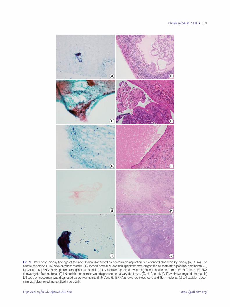
https://jpatholtm.org/https://doi.org/10.4132/jptm.2020.09.28
Cause of necrosis in LN FNA • 63
Fig. 1. Smear and biopsy findings of the neck lesion diagnosed as necrosis on aspiration but changed diagnosis by biopsy (A, B). (A) Fine needle aspiration (FNA) shows colloid material. (B) Lymph node (LN) excision specimen was diagnosed as metastatic papillary carcinoma. (C, D) Case 2. (C) FNA shows pinkish amorphous material. (D) LN excision specimen was diagnosed as Warthin tumor. (E, F) Case 3. (E) FNA shows cystic fluid material. (F) LN excision specimen was diagnosed as salivary duct cyst. (G, H) Case 4. (G) FNA shows myxoid stroma. (H) LN excision specimen was diagnosed as schwannoma. (I, J) Case 5. (I) FNA shows red blood cells and fibrin material. (J) LN excision speci-men was diagnosed as reactive hyperplasia.
A
C
E
G
I
B
D
F
H
J

https://jpatholtm.org/ https://doi.org/10.4132/jptm.2020.09.28
64 • Seo YJ et al.
patients were negative, but the remaining two patients had posi-tive results. Therefore, the final diagnosis of 11 patients was TB.
Suppurative lymphadenitis
Of the 460 total patients, 30 (6.7%) were classified with sup-purative lymphadenitis. Twenty-five patients (83.3%) were <60 years of age. The M:F ratio was 0.67. Among these 30 patients, 21 underwent ancillary tests for MTB. Eleven patients received a final diagnosis of TB. Four of the 10 patients who were nega-tive for MTB underwent LN biopsy and were finally diagnosed with granulomatous inflammation, Warthin tumor, and meta-static carcinoma (Table 2).
Malignant neoplasm
In 40 patients, malignant neoplasms were classified into dis-tinct LN FNA categories. The average patient age was 61.9 years. The M:F ratio was 4:7. All 40 patients underwent LN biopsies in the same region. Metastatic carcinoma accounted for 92.5% of cases. Among these cases, the incidence of squamous cell carci-noma was 62.2%. Two cases of malignant lymphoma were dif-fuse large B-cell lymphomas. In young patients, many cases were diagnosed as TB, while some older patients had metastatic carci-noma.
Necrosis alone
Of the 460 cases, 145 were classified as having necrosis alone; 94 cases underwent ancillary testing for MTB, and 53 patients (56.4%) were diagnosed with TB. In addition, 27 patients re-ceived an LN biopsy. These remaining cases were finally diag-nosed with granuloma, Kikuchi disease, necrosis, lymphomas, and other disorders (Table 2). Seven other mimickers included reactive hyperplasia (n = 2), metastatic papillary carcinoma (n =
1), Warthin tumor (n = 1), salivary duct cyst (n = 1), schwanno-ma (n = 1), and spindle cell tumor (n = 1) (Fig. 1).
DISCUSSION
FNA is frequently performed as a minimally invasive proce-dure in patients with mass lesions of superficial organs. It is also a very useful test for clinicians to use when deciding on a treat-ment. However, the FNA cytology slides may offer no diagnostic clues. In some cases, the cellularity is too low and reveals only necrotic material. In these situations, the pathologist faces diffi-culties in arriving at a diagnosis. Necrosis is common in benign inflammatory lesions as well as in metastatic malignancies. Ex-tensive necrosis is known to accompany not only metastatic ma-
lignancy, but also acute inflammation or granulomatous inflam-mation. In addition, focal necrosis is seen in LNs associated with systemic lupus erythematosus, infectious mononucleosis, and brucellosis [1].
We reviewed the literature on LN FNAs that reveal necrotic features. The most common reports were on TB [2-11], fol-lowed by studies on lymphoma [12-19] and metastatic lesions [20-23] (Table 3). However, most of these studies were case re-ports. There have been very few systematic reports of cases where necrosis is observed or certain diseases must be considered to be more important based on the patient age.
In this study, we analyzed 460 patients who underwent cervi-cal LN FNA cytology that revealed necrotic features. The most common causative disease was TB. Of the 460 patients, 247 underwent ancillary testing for MTB, and 148 (59.9%) were fi-nally diagnosed with TB. Typically, the most characteristic cy-tologic findings of TB are nodular collections of epithelioid his-tiocytes with Langhans giant cells and caseous necrosis. However, either end of the cytologic spectrum may be seen, especially ne-crosis without granulomas [24]. Kumar et al. [5] reported that the presence of acute lymphadenitis does not completely exclude TB. They clinically suspected TB in 263 cases with a cytologic picture that demonstrated acute suppurative inflammation. LN FNA should be repeated in all FNA cases that show acute sup-purative inflammation without granulomas. It has been reported that a repeat LN FNA helps to detect more than 55% of addi-tional TB cases [25]. In this study, among the patients classified with suppurative lymphadenitis according to FNA cytology, 36.7% were finally diagnosed with TB following ancillary tests. Therefore, it is important to rule out TB when necrotic features are noted on LN FNA cytology.
Necrotic features may also be characteristic of the Kikuchi disease category in cervical LN FNA. The FNA findings in Ki-kuchi disease typically reveal a polymorphous lymphoid popu-lation, crescentic histiocytes, extensive apoptotic bodies, and necrosis. The combination of crescentic histiocytes and karyor-rhectic debris suggests the diagnosis of Kikuchi disease. However, some cases show only karyorrhexis and necrosis, which is not specific for Kikuchi disease. For example, infectious mononucle-osis may rarely demonstrate extensive necrosis. Infectious mono-nucleosis is a self-limited viral disease that frequently involves the cervical LNs of young patients who present with fever and pharyngitis. In infectious mononucleosis, a polymorphous infil-trate with atypical large lymphoid cells is usually noted. The pa-tient’s clinical history is helpful in arriving at a differential diag-nosis in these cases [26].

https://jpatholtm.org/https://doi.org/10.4132/jptm.2020.09.28
Cause of necrosis in LN FNA • 65
Another important cause of necrotic features in cervical LN FNA cytology is malignant neoplasm. Among our 460 cases, 49 were finally confirmed as malignancies (including metastatic car-cinoma) by tissue biopsy. The mean age of these patients was 62 years. Forty cases were metastatic carcinoma. Among these cases, the most common malignancy was squamous cell carcinoma (62.5%). This trend was consistent with the findings of previous reports. In squamous cell carcinoma, metastatic LNs of the head and neck are often the first signs of malignancy in the inconspic-uous organs of this region [1]. Additionally, cystic changes are often observed in metastatic LNs in squamous cell carcinoma [27]. Necrotic material may also be identified in the cyst spaces; therefore, pathologists should be mindful during their micro-scopic examinations. In our study, most cases were diagnosed as
malignant neoplasms by FNA alone; the remaining nine cases were diagnosed as suppurative lymphadenitis (n = 1), granuloma (n = 3), and necrosis (n = 5) of the LNs. Therefore, when necrotic features are seen in the cervical LN FNA cytology in older pa-tients, pathologists should consider the possibility of malignancy and carefully correlate their findings with the clinical history of the patient to ensure the proper evaluation is recommended to clinicians, even though there may be a paucity of cells on the smeared slides.
In this study, we identified misdiagnosed cases of cervical LN necrosis on FNA. Following an excision biopsy, these cases were diagnosed as reactive hyperplasia, salivary duct cysts, Warthin tumors, schwannomas, and spindle cell tumors. As demonstrated by these cases, fibrinoid material that is present due to excessive
Table 3. Previously published reports with the keywords “FNA,” “necrosis,” and “lymph node”
Category Diagnosis Journal First authorPublication
Year
Infection (n = 17) Cat scratch disease (n = 1) Am J Clin Pathol Choi AH [28] 2015Histoplasmosis (n = 1) Cancer Cytopathol Gailey MP [29] 2013Infection (n = 1) J Thorac Oncol Aerts JG [30] 2008Tuberculosis (n = 11) Diagn Cytopathol Das DK [3] 1992
Am J Clin Pathol Lapuerta P [31] 1997Cytopathology Ersoz C [4] 1998Cytopathology Kumar N [5] 1998World J Gastroenterol Xia F [7] 2003Cytopathology Aljafari AS [2] 2004Southeast Asian J Trop Med Public Health Chantranuwat C [6] 2006J Gastroenterol Itaba S [9] 2007Int J Tuberc Lung Dis Berzosa M [8] 2010Diagn Cytopathol Mittal P [10] 2011Ann Thorac Surg Sun J [11] 2013
Tularemia (n = 3) Infection Ulu-Kilic A [32] 2013APMIS Tuncer E [33] 2014Acta Cytol Markoc F [34] 2014
Kikuchi disease (n = 2) Kikuchi disease (n = 2) Diagn Cytopathol Hsueh EJ [35] 1993Diagn Cytopathol Viguer JM [36] 2001
Lymphoma (n = 8) Hodgkin lymphoma (n = 2) Diagn Cytopathol Florentine BD [12] 2014J Clin Exp Hematop Okuni M [13] 2018
Langerhans cell histiocytosis (n = 1) Cytopathology Phulware RH [14] 2019Non-Hodgkin lymphoma (n = 5) Acta Cytol Tani E [16] 1989
Acta Cytol Sneige N [15] 1990Leuk Lymphoma Dao TH [17] 1991Diagn Cytopathol Dunphy CH [18] 1997Indian J Pathol Microbiol Handa U [19] 2005
Metastatic lesion (n = 4) Squamous cell carcinoma (n = 4) Diagn Cytopathol Allison DB [22] 2016Cytojournal Lastra RR [21] 2013Diagn Cytopathol Banet N [20] 2016Cytopathology Rollo F [23] 2018
Other (n = 3) Paraganglioma (n = 1) Diagn Cytopathol Fite JJ [37] 2018Sarcoidosis (n = 2) Eur Respir J Annema JT [38] 2005
Coll Antropol Vrbica Z [39] 2010

https://jpatholtm.org/ https://doi.org/10.4132/jptm.2020.09.28
66 • Seo YJ et al.
hemorrhaging, amorphous material of tumor components, or hypocellular myxoid stroma could be confused with a necrotic background.
An accurate diagnosis on cervical LN FNA cytology is impor-tant to determine the most appropriate treatment and to prevent unnecessary surgery. This study was conducted in the Republic of Korea, and we found that the most common cause of necrosis in cervical LN FNA cytology was TB. When there are necrotizing features in the FNA of the cervical lymph nodes in young patients, Kikuchi disease should be considered first. Metastatic carcinoma should always be suspected in older patients, even if they do not have any previous history of malignancy. In addition, when nec-rotizing features are noted in the FNA of the cervical lymph nodes of patients in any age group, MTB testing should be performed in parallel to exclude TB, which has a high prevalence in the Republic of Korea.
Ethics StatementThis study was approved by the Institutional Review Board of the Keimyung University Dongsan Hospital with a waiver of informed consent (DSMC 2020-03-069-002) and performed in accordance with the principles of the Declaration of Helsinki.
ORCID Young Jin Seo https://orcid.org/0000-0002-9048-0903Hyeongchan Shin https://orcid.org/0000-0002-3716-7678Hye Won Lee https://orcid.org/0000-0001-8540-524XHye Ra Jung https://orcid.org/0000-0002-1477-6606
Author Contributions Conceptualization: HRJ. Data curation: YJS, HS. Formal analysis: YJS, HRJ. Methodology: YJS, HRJ. Supervision: HRJ. Visualization: YJS, HWL. Writing—original draft: YJS, HRJ. Writing—review & editing: YJS, HS, HWL, HRJ. Approval of final manuscript: all authors.
Conflicts of InterestThe authors declare that they have no potential conflicts of interest.
Funding StatementNo funding to declare.
References1. van Schaik JE, Muller Kobold AC, van der Laan B, van der Vegt B,
van Hemel BM, Plaat BEC. Squamous cell carcinoma antigen con-centration in fine needle aspiration samples: a new method to de-tect cervical lymph node metastases of head and neck squamous cell carcinoma. Head Neck 2019; 41: 2561-5.
2. Aljafari AS, Khalil EA, Elsiddig KE, et al. Diagnosis of tuberculous lymphadenitis by FNAC, microbiological methods and PCR: a comparative study. Cytopathology 2004; 15: 44-8.
3. Das DK, Bhambhani S, Pant JN, et al. Superficial and deep-seated tuberculous lesions: fine-needle aspiration cytology diagnosis of 574 cases. Diagn Cytopathol 1992; 8: 211-5.
4. Ersoz C, Polat A, Serin MS, Soylu L, Demircan O. Fine needle aspira-tion (FNA) cytology in tuberculous lymphadenitis. Cytopathology 1998; 9: 201-7.
5. Kumar N, Tiwari MC, Verma K. AFB staining in cytodiagnosis of tuberculosis without classical features: a comparison of Ziehl-Neelsen and fluorescent methods. Cytopathology 1998; 9: 208-14.
6. Chantranuwat C, Assanasen T, Shuangshoti S, Sampatanukul P. Polymerase chain reaction for detection of Mycobacterium tuber-culosis in papanicolaou-stained fine needle aspirated smears for di-agnosis of cervical tuberculous lymphadenitis. Southeast Asian J Trop Med Public Health 2006; 37: 940-7.
7. Xia F, Poon RT, Wang SG, Bie P, Huang XQ, Dong JH. Tuberculosis of pancreas and peripancreatic lymph nodes in immunocompetent patients: experience from China. World J Gastroenterol 2003; 9: 1361-4.
8. Berzosa M, Tsukayama DT, Davies SF, et al. Endoscopic ultra-sound-guided fine-needle aspiration for the diagnosis of extra-pul-monary tuberculosis. Int J Tuberc Lung Dis 2010; 14: 578-84.
9. Itaba S, Yoshinaga S, Nakamura K, et al. Endoscopic ultrasound-guided fine-needle aspiration for the diagnosis of peripancreatic tuberculous lymphadenitis. J Gastroenterol 2007; 42: 83-6.
10. Mittal P, Handa U, Mohan H, Gupta V. Comparative evaluation of fine needle aspiration cytology, culture, and PCR in diagnosis of tuberculous lymphadenitis. Diagn Cytopathol 2011; 39: 822-6.
11. Sun J, Teng J, Yang H, et al. Endobronchial ultrasound-guided transbronchial needle aspiration in diagnosing intrathoracic tuber-culosis. Ann Thorac Surg 2013; 96: 2021-7.
12. Florentine BD, Cohen AN. Nodular sclerosing classical Hodgkin lymphoma masquerading as acute suppurative-necrotizing lymph-adenitis. Diagn Cytopathol 2014; 42: 238-41.
13. Okuni M, Yakushijin K, Sakai Y, et al. A case of classical Hodgkin lymphoma with total lymph node infarction. J Clin Exp Hematop 2018; 58: 24-6.
14. Phulware RH, Guleria P, Iyer VK, et al. Cytological diagnosis of Langerhans cell histiocytosis: a series of 47 cases. Cytopathology 2019; 30: 413-8.
15. Sneige N, Dekmezian RH, Katz RL, et al. Morphologic and immu-nocytochemical evaluation of 220 fine needle aspirates of malignant lymphoma and lymphoid hyperplasia. Acta Cytol 1990; 34: 311-22.
16. Tani E, Liliemark J, Svedmyr E, Mellstedt H, Biberfeld P, Skoog L. Cytomorphology and immunocytochemistry of fine needle aspi-rates from blastic non-Hodgkin’s lymphomas. Acta Cytol 1989; 33: 363-71.
17. Dao TH, Fleury-Feith J, Haioun C, et al. Percutaneous fine needle aspiration cytology and biopsy in the diagnosis and classification of lymphoma: clinical evaluation. Leuk Lymphoma 1991; 5: 237-42.
18. Dunphy CH, Ramos R. Combining fine-needle aspiration and flow cytometric immunophenotyping in evaluation of nodal and extra-nodal sites for possible lymphoma: a retrospective review. Diagn Cytopathol 1997; 16: 200-6.
19. Handa U, Mohan H, Punia RS, Nada R. FNAC in a case of NHL presenting initially as nodal infarction. Indian J Pathol Microbiol 2005; 48: 510-2.
20. Banet N, Rooper LM, Maleki Z. Metastatic HPV-related head and neck squamous cell carcinoma to the lung and mediastinal lymph nodes in aspirated cytology material: a diagnostic pitfall. Diagn Cy-topathol 2016; 44: 206-14.
21. Lastra RR, Pramick MR, Nakashima MO, et al. Adequacy of fine-

https://jpatholtm.org/https://doi.org/10.4132/jptm.2020.09.28
Cause of necrosis in LN FNA • 67
needle aspiration specimens for human papillomavirus infection molecular testing in head and neck squamous cell carcinoma. Cy-tojournal 2013; 10: 21.
22. Allison DB, Bishop JA, Ali SZ. Cytopathologic characteristics of SMARCB1 (INI-1) deficient sinonasal carcinoma: a potential diag-nostic pitfall. Diagn Cytopathol 2016; 44: 700-3.
23. Rollo F, Dona MG, Pellini R, et al. Cytology and direct human pap-illomavirus testing on fine needle aspirates from cervical lymph node metastases of patients with oropharyngeal squamous cell car-cinoma or occult primary. Cytopathology 2018; 29: 449-54.
24. Mac DeMay R. The art and science of cytopathology. Chicago: Amer-ican Society for Clinical Pathology, 2012.
25. Kumar N, Jain S, Murthy NS. Utility of repeat fine needle aspira-tion in acute suppurative lesions: follow-up of 263 cases. Acta Cytol 2004; 48: 337-40.
26. Tong TR, Chan OW, Lee KC. Diagnosing Kikuchi disease on fine needle aspiration biopsy: a retrospective study of 44 cases diagnosed by cytology and 8 by histopathology. Acta Cytol 2001; 45: 953-7.
27. Mokhtari S. Mechanisms of cyst formation in metastatic lymph nodes of head and neck squamous cell carcinoma. Diagn Pathol 2012; 7: 6.
28. Choi AH, Bolaris M, Nguyen DK, Panosyan EH, Lasky JL 3rd, Duane GB. Clinicocytopathologic correlation in an atypical pre-sentation of lymphadenopathy with review of literature. Am J Clin Pathol 2015; 143: 749-54.
29. Gailey MP, Klutts JS, Jensen CS. Fine-needle aspiration of histoplas-mosis in the era of endoscopic ultrasound and endobronchial ultra-sound: cytomorphologic features and correlation with clinical labo-ratory testing. Cancer Cytopathol 2013; 121: 508-17.
30. Aerts JG, Kloover J, Los J, van der Heijden O, Janssens A, Tournoy
KG. EUS-FNA of enlarged necrotic lymph nodes may cause infec-tious mediastinitis. J Thorac Oncol 2008; 3: 1191-3.
31. Lapuerta P, Martin SE, Ellison E. Fine-needle aspiration of periph-eral lymph nodes in patients with tuberculosis and HIV. Am J Clin Pathol 1997; 107: 317-20.
32. Ulu-Kilic A, Gulen G, Sezen F, Kilic S, Sencan I. Tularemia in cen-tral Anatolia. Infection 2013; 41: 391-9.
33. Tuncer E, Onal B, Simsek G, et al. Tularemia: potential role of cyto-pathology in differential diagnosis of cervical lymphadenitis: multi-center experience in 53 cases and literature review. APMIS 2014; 122: 236-42.
34. Markoc F, Koseoglu RD, Koc S, Gurbuzler L. Tularemia in differen-tial diagnosis of cervical lymphadenopathy: cytologic features of tularemia lymphadenitis. Acta Cytol 2014; 58: 23-8.
35. Hsueh EJ, Ko WS, Hwang WS, Yam LT. Fine-needle aspiration of histiocytic necrotizing lymphadenitis (Kikuchi’s disease). Diagn Cytopathol 1993; 9: 448-52.
36. Viguer JM, Jimenez-Heffernan JA, Perez P, Lopez-Ferrer P, Gonza-lez-Peramato P, Vicandi B. Fine-needle aspiration cytology of Kiku-chi’s lymphadenitis: a report of ten cases. Diagn Cytopathol 2001; 25: 220-4.
37. Fite JJ, Maleki Z. Paraganglioma: cytomorphologic features, radio-logic and clinical findings in 12 cases. Diagn Cytopathol 2018; 46: 473-81.
38. Annema JT, Veselic M, Rabe KF. Endoscopic ultrasound-guided fine-needle aspiration for the diagnosis of sarcoidosis. Eur Respir J 2005; 25: 405-9.
39. Vrbica Z, Boras Z, Rakusic N, et al. Necrotising sarcoid granuloma-tosis of the spinal cord: case report. Coll Antropol 2010; 34: 713-7.

68
pISSN 2383-7837eISSN 2383-7845
© 2021 The Korean Society of Pathologists/The Korean Society for CytopathologyThis is an Open Access article distributed under the terms of the Creative Commons Attribution Non-Commercial License (https://creativecommons.org/licenses/
by-nc/4.0) which permits unrestricted non-commercial use, distribution, and reproduction in any medium, provided the original work is properly cited.
Cribriform carcinoma of the skin (primary cutaneous cribri-form carcinoma [PCCC]) is an indolent, rare, adnexal tumor with a presumed apocrine lineage. The first global case was described in 1998 [1], and the most recent (4th) World Health Organiza-tion (WHO) classification for skin now includes this condition. Currently, only 42 cases have been reported in the English liter-ature (Table 1) [1-7]. The female-to-male ratio is 2:1, with a me-dian age of 47 years (range, 20 to 77 years). Most cases involve the extremities (85%). Although its malignant potential remains uncertain, no recurrence or metastasis has been reported.
Herein, we report the first Korean case of primary cribriform carcinoma of the skin.
CASE REPORT
A 33-year-old man presented with a solitary, erythematous, subcutaneous nodule on the right knee (Fig. 1A), which had developed a few months prior. The clinical impression was epider-mal cyst. His past medical history comprised intracranial hem-orrhage due to arteriovenous malformation 10 years previous.
After the patient underwent systemic evaluation, including positron emission tomography–computed tomography (PET-CT) and gastric endoscopy, the mass was confirmed as a primary skin tumor, and resection was performed. The resected tumor was a well-circumscribed, yellowish-white, fibrotic, firm, 2.0 × 1.2 ×
0.7-cm mass (Fig. 1B).Histologically, the tumor was a well-circumscribed mass of
the dermis and subcutis. The tumor was composed of (Fig. 2A) a predominantly solid component (90%) and a predominantly cribriform component (10%). The solid component showed multiple solid nests of epithelial cells. Individual cells had large, oval-to-round, hyperchromatic, pleomorphic nuclei with a nuclear groove (Fig. 2B). The cytoplasm was eosinophilic and scant. The cribriform component showed similar neoplastic cells with many prominent lumina, giving rise to a cribriform pattern with a thin, thread-like, intraluminal bridge (Fig. 2C). Some lumina had an eosinophilic substance that exhibited a positive periodic acid–Schiff reaction (Fig. 2D). At the periphery of the tumor, mul-tifocal lymphoid aggregates (Fig. 2E), desmoplastic reaction, and some infiltrative tumor cell clusters were present (Fig. 2F).
A case of cribriform carcinoma of the skin: a newly described rare condition
Hyun Lee1, Chong-Hyun Won2, Chan-Sik Park1
Departments of 1Pathology and 2Dermatology, Asan Medical Center, University of Ulsan College of Medicine, Seoul, Korea
Primary cribriform carcinoma of the skin is an indolent, rare, adnexal tumor. Although its malignant potential remains uncertain, no recur-rence or metastasis has been reported. A 33-year-old man presented with a solitary, erythematous, subcutaneous nodule on the right knee. The clinical impression was epidermal cyst, and the resected tumor demonstrated a well-circumscribed mass in the dermis and subcutis. The tumor was composed of two regions: a solid component and a cribriform component. The solid component (90%) showed multiple solid nests of epithelial cells. Individual cells had large, oval-to-round, hyperchromatic, pleomorphic nuclei with a nu-clear groove. The cribriform component (10%) showed similar neoplastic cells with many prominent lumina. Some lumina had an eo-sinophilic substance that exhibited a positive periodic acid-Schiff reaction. No recurrence or metastasis was observed within a follow-up period of eight months after excision. In conclusion, we report the first case of primary cribriform carcinoma of the skin in Korea.
Key Words: Cribriform carcinoma; Apocrine tumor; Skin tumor
Received: August 10, 2020 Revised: September 13, 2020 Accepted: October 5, 2020Corresponding Author: Chan-Sik Park, MD, PhD, Department of Pathology, Asan Medical Center, University of Ulsan College of Medicine, 88 Olympic-ro 43-gil, Songpa-gu, Seoul 05505, KoreaTel: +82-2-3010-5838, Fax: +82-2-472-7898, E-mail: [email protected]
CASE STUDYJournal of Pathology and Translational Medicine 2021; 55: 68-74https://doi.org/10.4132/jptm.2020.10.05

https://jpatholtm.org/https://doi.org/10.4132/jptm.2020.10.05
Primary cutaneous cribriform carcinoma • 69Ta
ble
1. S
umm
ary
of re
porte
d ca
ses
Cas
e N
o.St
udy
Year
Age
(yr)
Sex
Site
Clin
ical d
iagno
sisSi
ze (c
m)
Trea
tmen
tF/
U
perio
dPr
ogno
sisPo
sitive
IHC
Neg
ative
IHC
Com
men
ts
1C
urre
nt c
ase
2020
33M
Low
er e
xtre
mity
(R
t. kn
ee)
Epid
erm
al c
yst
2.0
Exci
sion
8 m
oN
RO
MD
S100
(diffu
se, l
umin
al c
ompo
nent
; pa
tchy
, sol
id c
ompo
nent
), EM
A (p
atch
y, lu
min
al c
ompo
nent
; di
ffuse
, sol
id c
ompo
nent
), C
K7,
CK5
/6 (p
atch
y), C
D11
7 (p
atch
y),
EpC
AM (p
atch
y), C
EA (i
ntra
-lu
min
al),
p63
(rare
)
CK2
0, G
CD
FP-1
5, E
R, c
alpo
nin,
SM
AH
istor
y of
intra
cran
ial h
emor
rhag
e du
e to
arte
riove
nous
m
alfo
rmat
ion
No
evid
ence
of o
ther
m
alig
nanc
ies
2R
eque
na
et a
l. [1
] 19
98N
AN
ALo
wer
ext
rem
ityN
AN
AEx
cisio
nN
AN
AN
AN
AFi
rst d
escr
iptio
nR
epor
ted
as m
ainl
y w
omen
with
a
mea
n ag
e of
44
year
s (ra
nge,
20
to 5
5)Tw
o ca
ses
of re
curre
nt tu
mor
af
ter i
ncom
plet
e ex
cisio
n
3N
AN
ALo
wer
ext
rem
ityN
AN
AEx
cisio
nN
AN
AN
AN
A
4N
AN
ALo
wer
ext
rem
ityN
AN
AEx
cisio
nN
AN
AN
AN
A
5N
AN
AU
pper
ext
rem
ityN
AN
AEx
cisio
nN
AN
AN
AN
A
6N
AN
APu
bis
NA
NA
Exci
sion
NA
NA
NA
NA
7Ad
amsk
i et
al.
[5]
2005
37M
Low
er e
xtre
mity
(L
t. po
plite
al
foss
a)
NA
2.0
Exci
sion
2 yr
NR
OM
DC
K7C
K20,
GC
DFP
-15,
S10
0 -
8Fe
rnan
dez-
Flor
es
et a
l. [4
]20
0762
FLo
wer
ext
rem
ity
(Lt.
popl
iteal
fo
ssa)
NA
0.8
Exci
sion
NA
NR
OM
DC
K AE
1/AE
3, C
AM 5
.2, C
K7, E
MA,
ER
(2+,
1%
–5%
cel
ls), c
-erb
B-2
(2
+, 5
0%–7
5% c
ells)
, p53
(1
+, 1
%–5
% c
ells)
, S10
0 (2
+, 5
0%–7
5% c
ells)
CK2
0, C
EA, P
R, G
CD
FP-1
5,
CD
15, S
MA
-
9R
utte
n et
al.
[2]
2009
48M
Low
er e
xtre
mity
(Rt.
thig
h)D
erm
atofi
brom
a(h
istio
cyto
ma)
-Ex
cisio
n18
yr
NR
OM
DC
K M
NF1
16, C
K AE
1/AE
3, C
AM
5.2,
CK7
, CEA
(mor
e pr
omin
ent i
n th
e du
ctal
stru
ctur
es),
EMA
(mor
e pr
omin
ent i
n th
e du
ctal
stru
ctur
es)
CK2
0, G
CD
FP-1
5, S
100,
α-S
MA,
M
SA, c
alpo
nin,
CD
68, v
imen
tin-
1051
FU
pper
ext
rem
ity
(Rt.
fore
arm
)C
yst
-Ex
cisio
nN
AN
A-
1144
FB
ack
Der
mat
ofibr
oma
(hist
iocy
tom
a)-
Exci
sion
13 y
rN
RO
MD
-
1277
FN
eck
NA
-Ex
cisio
nN
AN
AR
ecur
rent
tum
or a
fter i
ncom
plet
e ex
cisio
n
1332
FLo
wer
ext
rem
ity
(Rt.
thig
h)D
erm
atofi
brom
a (h
istio
cyto
ma)
-Ex
cisio
n9
yrN
RO
MD
-
1442
FLo
wer
ext
rem
ity
(Rt.
thig
h)D
erm
atofi
brom
a (h
istio
cyto
ma)
-Ex
cisio
nN
AN
A-
1534
FU
pper
ext
rem
ity
(Rt.
arm
)D
erm
atofi
brom
a (h
istio
cyto
ma)
-Ex
cisio
n11
yr
NR
OM
D-
1640
MU
pper
ext
rem
ity
(Lt.
fore
arm
)D
erm
atofi
brom
a (h
istio
cyto
ma)
-Ex
cisio
nN
AN
A-
1723
MLo
wer
ext
rem
ity
(Lt.
calf)
NA
-Ex
cisio
nN
AN
A-
1820
FR
t. bu
ttock
Cys
t-
Exci
sion
NA
NA
No
evid
ence
of o
ther
m
alig
nanc
ies
1967
FLt
. pre
auric
ular
ar
eaB
CC
vs.
ad
nexa
l tum
or-
Exci
sion
8 yr
NR
OM
D-
2060
MLo
wer
ext
rem
ity
(Rt.
foot
do
rsum
)
Der
mat
ofibr
oma
(hist
iocy
tom
a)-
Exci
sion
4 yr
NR
OM
D-
2154
FU
pper
ext
rem
ity
(acr
al)
NA
-Ex
cisio
nN
AN
A-
(Con
tinue
d to
the
next
pag
e)

https://jpatholtm.org/ https://doi.org/10.4132/jptm.2020.10.05
70 • Lee H et al.Ta
ble
1. C
ontin
ued
Cas
e N
o.St
udy
Year
Age
(yr)
Sex
Site
Clin
ical d
iagno
sisSi
ze (c
m)
Trea
tmen
tF/
U
perio
dPr
ogno
sisPo
sitive
IHC
Neg
ative
IHC
Com
men
ts
2249
FLo
wer
ext
rem
ity
(Lt.
thig
h)D
erm
atofi
brom
a (h
istio
cyto
ma)
-Ex
cisio
n4
yrN
RO
MD
-
2359
FLt
. sho
ulde
rD
erm
atofi
brom
a (h
istio
cyto
ma)
-Ex
cisio
nN
AN
A-
2428
MLo
wer
ext
rem
ity
(Rt.
low
er le
g)N
A-
Exci
sion
NA
NA
-
2554
MU
pper
ext
rem
ity
(Rt.
hand
)N
A-
Exci
sion
NA
NA
-
2650
FU
pper
ext
rem
ity
(Rt.
hand
)N
A-
Exci
sion
NA
NA
-
2770
MR
t. tru
nkLo
ng-s
tand
ing
lesio
n-
Exci
sion
6 yr
NR
OM
D-
2864
FN
AD
erm
atofi
brom
a (h
istio
cyto
ma)
-Ex
cisio
n5
yrN
RO
MD
-
2940
FLo
wer
ext
rem
ity
(Low
er a
rm)
NA
-Ex
cisio
n5
yrN
RO
MD
-
3064
MU
pper
bac
kD
erm
atofi
brom
a (h
istio
cyto
ma)
or
cyst
-Ex
cisio
n3
yrN
RO
MD
-
3154
MLo
wer
ext
rem
ity
(Low
er a
rm)
NA
-Ex
cisio
n3
yrN
RO
MD
-
3244
FU
pper
ext
rem
ity
(Lt.
thum
b)N
A-
Exci
sion
2 yr
NR
OM
D-
3336
FLo
wer
ext
rem
ity
(Lt.
ante
rior
thig
h)
Der
mat
ofibr
oma
(hist
iocy
tom
a)-
Exci
sion
NA
NA
-
3426
FLo
wer
ext
rem
ity
(Lt.
post
erio
r leg
)Fi
brom
a-
Exci
sion
NA
NA
-
35Ar
ps e
t al.
[3]
2015
41F
Low
er e
xtre
mity
(le
g)Ep
ider
mal
in
clus
ion
cyst
0.6
Exci
sion
NA
NR
OM
DS1
00 (d
iffuse
in th
ree
case
s, p
atch
y in
one
cas
e), C
D11
7 (d
iffuse
in
two
case
s, p
atch
y in
one
cas
e),
CK5
/6, C
K7, E
pCAM
, CEA
(lu
min
al),
EMA
(lum
inal
), p6
3 (ra
re)
CK2
0, G
CD
FP-1
5, E
R, P
R,
calp
onin
, SM
A-
3632
FU
pper
ext
rem
ity
(elb
ow)
NA
0.5
Exci
sion
NA
NA
-
3735
MLo
wer
ext
rem
ity
(leg)
NA
0.5
Exci
sion
NA
NR
OM
D-
3859
FU
pper
ext
rem
ity
(arm
)N
A0.
4Ex
cisio
nN
AN
A-
3961
MU
pper
ext
rem
ity
(arm
)M
obile
nod
ule
0.7
Exci
sion
NA
NA
-
4031
MLo
wer
ext
rem
ity
(leg)
Der
mat
ofibr
oma
1.2
Exci
sion
NA
NA
-
41Yo
kota
et a
l. [7
]20
1739
FU
pper
ext
rem
ity
(Rt.
fore
arm
)N
A0.
5 (clin
ical
)Ex
cisio
n15
mo
NR
OM
DC
K5/6
, CK7
, CA1
5-3,
CA1
25,
CD
117,
S10
0 (p
artia
lly),
p53
(par
tially
), p6
3 (p
artia
lly)
CK2
0, c
alpo
nin,
GC
DFP
-15,
m
amm
aglo
bulin
, MU
C1,
ER
, AR
, D
2-40
Stab
le in
size
for m
ore
than
10
year
s, n
o ev
iden
ce o
f oth
er
mal
igna
ncie
s42
Bog
ner e
t al.
[6]
2018
65M
Lt. l
ater
al n
eck
NA
0.6 (c
linic
al)
Exci
sion
3 m
oN
RO
MD
CK7
(stro
ng),
EMA
(stro
ng),
CAM
5.2
(s
trong
), Ep
CAM
(les
ser d
egre
e),
CEA
(in
som
e of
the
lum
ina)
, p63
, p4
0
CK2
0, D
2-40
, TTF
-1, C
DX-
2,
hepa
tocy
te a
ntig
en, P
SA, P
SAP,
ca
lpon
in, S
100
No
evid
ence
of o
ther
m
alig
nanc
ies
F/U
, fol
low
up;
IHC
, im
mun
ohist
oche
mic
al s
tain
ing;
Rt.,
righ
t; Lt
., le
ft; N
RO
MD
, no
recu
rrenc
e or
met
asta
tic d
iseas
e; E
MA,
epi
thel
ial m
embr
ane
antig
en; C
K, c
ytok
erat
in; E
pCAM
, epi
thel
ial c
ell a
dhes
ion
mol
ecul
e; C
EA, c
arci
noem
bryo
nic
anti-
gen;
GC
DFP
-15,
gro
ss c
ystic
dise
ase
fluid
pro
tein
-15;
ER
, est
roge
n re
cept
or; S
MA,
sm
ooth
mus
cle
actin
; MSA
, mus
cle
spec
ific a
ntig
en; N
A, d
ata
not a
vaila
ble;
S10
0, S
-100
pro
tein
; PR
, pro
gest
eron
e re
cept
or; B
CC
, bas
al c
ell c
arci
nom
a;
MU
C1,
muc
in1;
AR
, and
roge
n re
cept
or; T
TF-1
, thy
roid
tran
scrip
tion
fact
or 1
; PSA
, pro
stat
e-sp
ecific
ant
igen
; PSA
P, p
rost
atic
aci
d ph
osph
atas
e; +
, >50
% o
f tum
or c
ells
are
posit
ive; p
atch
y, 25
%–5
0% o
f tum
or c
ells
are
posit
ive; r
are,
< 1
% o
f tu
mor
cel
ls ar
e po
sitive
.

https://jpatholtm.org/https://doi.org/10.4132/jptm.2020.10.05
Primary cutaneous cribriform carcinoma • 71
Fig. 1. Gross appearance of the mass. (A) A solitary, erythematous, subcutaneous nodule on the right knee. (B) The cut surface of the mass is well-circumscribed, yellowish-white, fibrotic, and firm.
The epidermis was neither involved with nor connected to the tumor.
The two components of the tumor displayed distinctive im-munohistochemical staining patterns for epithelial membrane antigen (EMA) (Fig. 3A) and S-100 protein (Fig. 3B). The pre-dominantly solid component exhibited diffuse immunopositivity for EMA and focal immunopositivity for S100 protein, while the predominantly luminal component exhibited focal immu-nopositivity for EMA and diffuse immunopositivity for S-100 protein.
Given its histopathologic features, the mass was diagnosed as PCCC. No recurrence or metastasis was observed within a fol-low-up period of 8 months after excision.
DISCUSSION
PCCC is a rare, newly described, unique, adnexal neoplasm with an indolent clinical course. Currently, no recurrence or me-tastasis has been reported. In few cases, remnants have been report-ed after incomplete excision [1,3]. Accurate diagnosis and exclu-sion of metastasis are important for avoiding over-treatment.
In cases of primary skin neoplasms, the differential diagnosis should include tumors that can show a cribriform pattern: ade-noid cystic carcinoma, secretory carcinoma, and tubular adeno-ma (eccrine papillary adenoma). The histopathologic features for differential diagnoses are listed in Table 2. Adenoid cystic car-cinoma can be distinguished by the presence of basaloid epithe-lial cells with more uniform nuclei surrounding the pseudolu-mina. The presence of frequent perineural invasion and small true ducts with myoepithelial cell differentiation are points of
differential diagnosis. Secretory carcinoma exhibits tubules and microcysts with conspicuous intraluminal secretions, but back-to-back proliferation and cuboidal neoplastic cells are character-istic compared with PCCC. Tubular adenoma can show dilated cystic spaces with attenuated epithelium, micro-papillae, and fo-cal intraluminal bridging, mimicking PCCC. However, this con-dition lacks cytologic atypia and mitotic activity and involves accumulation of basal/myoepithelial cells.
Histopathologically, metastatic tumors that show a cribri-form pattern shold be excluded. A cribriform pattern can be seen in cancers of various organs, including the breast (adenoid cystic carcinoma and cribriform adenocarcinoma), prostate (ductal carcinoma and acinar carcinoma), stomach, colon, lung, thyroid (cribriform-morular variant of papillary thyroid carcinoma), uter-ine endometrium, and salivary gland [8-10]. To exclude metas-tasis, imaging studies, such as PET-CT and CT, and immuno-histochemical staining are required.
Immunohistochemical staining results are listed in Table 1. Although decapitation secretion in the luminal border supports apocrine differentiation, gross cystic disease fluid protein-15, a marker for the apocrine gland, was negative in previous reports [1-7] and in our case. The S-100 protein, a marker for the ec-crine gland, demonstrated variable results (diffuse positive, focal positive, and negative) in previous reports [1-7]. Our case showed more prominent S-100 protein in the luminal component. EMA was positive in previous reports [1-7]. Rutten et al. [2] reported more prominent EMA in the luminal structures, while our case showed more prominent EMA in the solid component.
The relatively mutually exclusive immunohistochemical stain-ing patterns of EMA and S-100 protein may be associated with
A B

https://jpatholtm.org/ https://doi.org/10.4132/jptm.2020.10.05
72 • Lee H et al.
A
C
E
B
D
F
S
L
Fig. 2. Histopathologic features of the mass. (A) The mass exhibited a predominantly solid component (90%) and a predominantly cribriform component (10%, inside the green line). (B) The predominantly solid component revealed pleomorphic nuclei. (C) The predominantly cribri-form component had similar cytologic features to those of the solid component and demonstrated many prominent small lumina with a thin thread-like intraluminal bridge (cribriform pattern) along with an occasional eosinophilic substance. (D) The intraluminal eosinophilic sub-stance with periodic acid–Schiff reaction. (E) Multifocal lymphoid aggregates at the periphery. (F) Infiltrative tumor clusters at the periphery. S, predominantly solid component; L, predominantly luminal component.

https://jpatholtm.org/https://doi.org/10.4132/jptm.2020.10.05
Primary cutaneous cribriform carcinoma • 73
A B
L
L
SS
architectural differentiation, and further research is needed.In conclusion, we report the first case of cribriform carcinoma
of the skin in Korea. Pathologists should be aware of cribriform carcinoma of the skin to avoid over-treatment.
Fig. 3. Results of immunohistochemical (IHC) staining. (A) IHC for epithelial membrane antigen: diffuse immunopositivity for the predominant-ly solid component (on the right) and focal immunopositivity for the predominantly luminal component (on the left). (B) IHC for S-100 protein: diffuse immunopositivity for the predominantly luminal component (on the left) and focal immunopositivity for the predominantly solid compo-nent (on the right). S, predominantly solid component; L, predominantly luminal component.
Table 2. Histopathologic characteristics of cribriform carcinoma and similar tumors
Cribriform carcinoma Adenoid cystic carcinoma Secretory carcinoma Tubular adenoma
Architecture Usually well-circumscribed Poorly circumscribed Intradermal, circumscribed Well circumscribedMixed variable portion of solid
and cribriformComposed of lobules, islands,
and cords of basaloid cells with numerous cystic and ductular spaces
Back-to-back proliferation of tubules and microcysts
Variable sized tubules with attenuated epithelium
No back-to-back appearance Cuboidal cells Micro-papillae, and focal intraluminal bridging
No cuboidal cells Sclerotic stroma Paucicellular fibrous stroma
Desmoplastic stromaRecognition of myoepithelial
layerIntra-(pseudo) luminal
substanceEosinophilic substance with
PAS reactionMucin or basement membrane
material that stains with mucicarmine, Alcian blue, and colloidal iron
Conspicuous intraluminal secretions
Eosinophilic proteinaceous material
Nuclei Pleomorphic Uniform Mildly pleomorphic UniformMitosis Rare Rare Rare to few AbsentPerineural invasion Absent Present, frequent Absent AbsentImmunohistochemical
stainingVariable CK (MNF116, AE1/
AE3, CAM5.2, and CK7) EMA and monoclonal CEA S100 protein, mammaglobin
and STAT5AHMFG-1 and GCDFP-15
EpCAM S100, p63, GFAP, SMA, MSA and calponin: often stain peripheral cells (myoepithelial differentiation)
NTRK3: variable EMA and CEA: luminal cells
CD117, S100, and p63: variable
S100 and SMA: myoepithelial cells
CEA, EMA: highlight ductal component
Reference [11,12] [12,13] [12,14] [3,12,13]
PAS, periodic acid-Schiff; CK, cytokeratin; EMA, epithelial membrane antigen; CEA, carcinoembryonic antigen; S100, S-100 protein; GCDFP-15, gross cys-tic disease fluid protein-15; EpCAM, epithelial cell adhesion molecule; GFAP, glial fibrillary acidic protein; SMA, smooth muscle actin; MSA, muscle specific antigen.

https://jpatholtm.org/ https://doi.org/10.4132/jptm.2020.10.05
74 • Lee H et al.
Ethics StatementThis study was approved by the Institutional Review Board of Asan Medi-cal Center (IRB 2020-0364). Formal written informed consent was not re-quired, with a waiver from the appropriate Institutional Review Board.
ORCID Hyun Lee https://orcid.org/0000-0001-7005-8142Chong-Hyun Won https://orcid.org/0000-0003-1997-2240Chan-Sik Park https://orcid.org/0000-0001-9783-4498
Author Contributions Conceptualization: CSP. Data curation: HL. Formal analysis: HL. Funding acquisition: CSP. Investigation: CSP. Methodology: HL. Resources: WCH. Supervision: CSP. Validation: CSP, HL. Writing—original draft: HL. Writ-ing—review & editing: CSP. Approval of final manuscript: all authors.
Conflicts of InterestCSP, a contributing editor of the Journal of Pathology and Translational Medicine, was not involved in the editorial evaluation or decision to publish this article. All remaining authors have declared no conflicts of interest.
Funding StatementNo funding to declare.
References1. Requena L, Kiryu H, Ackerman AB. Neoplasms with apocrine dif-
ferentiation. Philadelphia: Lippincott-Raven, 1988; 879-905.2. Rutten A, Kutzner H, Mentzel T, et al. Primary cutaneous cribri-
form apocrine carcinoma: a clinicopathologic and immunohisto-chemical study of 26 cases of an under-recognized cutaneous ad-nexal neoplasm. J Am Acad Dermatol 2009; 61: 644-51.
3. Arps DP, Chan MP, Patel RM, Andea AA. Primary cutaneous crib-
riform carcinoma: report of six cases with clinicopathologic data and immunohistochemical profile. J Cutan Pathol 2015; 42: 379-87.
4. Fernandez-Flores A, Pol A, Juanes F, Crespo LG. Immunohisto-chemical phenotype of cutaneous cribriform carcinoma with a panel of 15 antibodies. Med Mol Morphol 2007; 40: 212-7.
5. Adamski H, Le Lan J, Chevrier S, Cribier B, Watier E, Chevrant-Breton J. Primary cutaneous cribriform carcinoma: a rare apocrine tumour. J Cutan Pathol 2005; 32: 577-80.
6. Bogner R, Brown T, Fearneyhough P, Gataky G. Primary cutane-ous cribriform carcinoma treated with mohs micrographic surgery. Dermatol Surg 2018; 44: 583-5.
7. Yokota K, Kono M, Mori S, Shimizu K, Matsumoto T, Akiyama M. A solid variant of primary cutaneous cribriform carcinoma: a small, stable, long-term lesion. Eur J Dermatol 2017; 27: 419-21.
8. Branca G, Ieni A, Barresi V, Tuccari G, Caruso RA. An updated re-view of cribriform carcinomas with emphasis on histopathological diagnosis and prognostic significance. Oncol Rev 2017; 11: 317.
9. Page DL, Dixon JM, Anderson TJ, Lee D, Stewart HJ. Invasive crib-riform carcinoma of the breast. Histopathology 1983; 7: 525-36.
10. McNeal JE, Reese JH, Redwine EA, Freiha FS, Stamey TA. Cribri-form adenocarcinoma of the prostate. Cancer 1986; 58: 1714-9.
11. Requena L, Sangueza O. Cribriform carcinoma. In: Requena L, Sangueza O, eds. Cutaneous adnexal neoplasms. Cham: Springer, 2017; 313-20.
12. Elder DE, Massi D, Scolyer RA, Willemze R. WHO classification of skin tumours. Lyon: IARC Press, 2018.
13. Cassario D. Diagnostic pathology: neoplastic dermatopathology. Salt Lake City: Amirsys Publishing Inc., 2012.
14. Llamas-Velasco M, Mentzel T, Rutten A. Primary cutaneous secre-tory carcinoma: a previously overlooked low-grade sweat gland car-cinoma. J Cutan Pathol 2018; 45: 240-5.

75
© 2021 The Korean Society of Pathologists/The Korean Society for CytopathologyThis is an Open Access article distributed under the terms of the Creative Commons Attribution Non-Commercial License (https://creativecommons.org/licenses/ by-nc/4.0) which permits unrestricted non-commercial use, distribution, and reproduction in any medium, provided the original work is properly cited.
pISSN 2383-7837eISSN 2383-7845
Fibrocartilaginous mesenchymoma (FM) is a rare, locally ag-gressive bone tumor, with fewer than 35 cases reported in the literature since 1984 when it was first reported by Dahlin et al. [1-3]. FM predominantly affects young patients with a median age of 13 years. This tumor principally occurs in long tubular bones and, infrequently, in the axial skeleton such as the iliac–pubic bones and vertebrae. Rib is an unusual location for FM, with only one published report [4]. Herein, we present a second case of FM arising in the rare anatomic site of the rib.
CASE REPORT
A 17-year-old boy was referred to the thoracic surgery depart-ment due to a mass located on the right side of the back. The patient had experienced intermittent pain and discomfort for several months at the time of admission, and localized swelling was noted on his back during physical examination. A chest ra-diograph showed a radiolucent lesion with cortical disruption in the posterolateral aspect of the right 5th rib. A computed tomog-raphy scan performed at the local clinic revealed an osteolytic mass with a juxtacortical extension (Fig. 1). Focal calcification was identified, but no periosteal reaction was observed. Overall
radiologic findings suggested a benign tumor, including fibrous dysplasia (FD). En bloc resection was performed without a prior biopsy.
The surgical specimen revealed a 3.1-cm-sized, ill-defined, eccentrically located, hard mass. The resected tumor was con-fined to the rib without expansion to the surrounding soft tissue, although the cortex had been partly destroyed. The cut surface of the tumor showed spotty areas of cartilaginous tissue (Fig. 2A). Histologically, the tumor was predominantly composed of hya-line cartilage nodules juxtaposed with hypocellular fibrous tissue (Fig. 2B). The cartilage islands throughout the tumor revealed low to intermediate cellularity but did not have any cytologic features of malignancy, such as nuclear atypia, multinucleation, mitosis, or myxoid changes (Fig. 2C). The hypocellular fibrotic area measured 1 cm in size, accounted for about 20% of the le-sion, and consisted of benign spindle-to-stellate cells in a collag-enous stroma without nuclear atypia or mitotic figures, suggest-ing low-grade sarcoma (Fig. 2D). The tumor was characterized by enchondral ossification at the periphery of the cartilage de-posits, reminiscent of the epiphyseal growth plate or the carti-lage cap of an osteochondroma (Fig. 2C). Newly formed bone was present and appeared as well-formed long and short trabec-
Fibrocartilaginous mesenchymoma with an unusual location in the rib
Sun-Ju Oh
Department of Pathology, Kosin University Gospel Hospital, Busan, Korea
Fibrocartilaginous mesenchymoma is a rare bone tumor, with fewer than 35 cases reported in the literature since 1984. This tumor usu-ally occurs in the long bones of children and adolescents. In the current case, the tumor affected a rib. A 17-year-old boy presented with a mass in the right fifth rib. Radiologic findings revealed an osteolytic mass with cortical destruction and calcification; en bloc resection was performed. The tumor showed three distinct histologic features: bland spindle cell proliferation, benign cartilage nodules, and epiphyseal plate-like enchondral ossification. The pathologic diagnosis was fibrocartilaginous mesenchymoma. The patient remains free of disease 1 year after the surgery. Pathological diagnosis of fibrocartilaginous mesenchymoma can be challenging, especially when the tumor occurs in an unusual site. When any fibro-osseous lesion with a cartilaginous component is encountered, the possibility of fibro-cartilaginous mesenchymoma should be considered because of its locally aggressive behavior.
Key Words: Fibrocartilaginous mesenchymoma; Fibrocartilaginous dysplasia; Enchondral ossification
Received: August 10, 2020 Revised: September 15, 2020 Accepted: October 8, 2020Corresponding Author: Sun-Ju Oh, MD, Department of Pathology, Kosin University Gospel Hospital, 262 Gamcheon-ro, Seo-gu, Busan 49267, KoreaTel: +82-51-990-6744, Fax: +82-51-990-3080, E-mail: [email protected]
CASE STUDYJournal of Pathology and Translational Medicine 2021; 55: 75-78https://doi.org/10.4132/jptm.2020.10.08

https://jpatholtm.org/ https://doi.org/10.4132/jptm.2020.10.08
76 • Oh SJ
ulae with prominent osteoblastic rimming. Multinucleated giant cells were not observed. Because the tumor was characterized as a fibro-osseous lesion with abundant cartilage nodules, the pos-sibility of low-grade osteosarcoma, chondrosarcoma, and fibro-cartilaginous dysplasia (FCD), a variant of FD, were consid-ered. Immunohistochemical stains for murine double minute 2 (MDM2) and cyclin-dependent kinase 4 (CDK4) were negative in the spindle cells, excluding the possibility of low-grade osteo-sarcoma. Chondrosarcoma was unlikely because the cartilage islands were composed of benign chondrocytes without atypia, as described earlier. In addition, the lesion showed well-formed bone trabeculae and did not feature the characteristic, irregular, curvilinear trabeculae of woven bone without the osteoblastic rimming of FD or FCD. Epiphyseal growth plate-like enchon-dral ossification was another distinguishing feature, distinguish-ing it from FCD. A final diagnosis of FM was rendered based on the overall histologic findings. The patient was regularly followed every 3 months and is doing well 1 year after resection.
DISCUSSION
A rare neoplasm, FM was initially described as “a fibrocarti-laginous mesenchymoma with low-grade malignancy” in 1984 by Dahlin et al. [1], who found unique lesions originally diag-nosed as FCD but with different clinical, radiographic, and morphologic features. Since then, there have been debates in the literature regarding FM as a separate neoplasm or one that coincides with FCD [5-7]. However, several studies, particular-
ly molecular analyses, have demonstrated that FM is not geneti-cally related to FCD, which has a characteristic GNAS (guanine nucleotide-binding protein α-stimulating polypeptide) mutation [2]. Accordingly, the 5th edition of the World Health Organiza-tion classification of bone tumors newly listed FM as an indepen-dent tumor entity [8].
FM predominantly affects long bones, including the femur, tibia, and humerus; a few cases have been reported in rare loca-tions, such as metatarsus, iliac–pubic bones, vertebrae, and ribs [2]. FM tends to affect young patients aged 3 months to 27 years (median age of 13 years), with a slight predominance of male patients. Radiologically, FM appears as an expansile osteo-lytic lesion with cartilaginous calcification and cortical destruc-tion, and extension to soft tissue is not uncommon.
Histologically, FM is characterized by spindle cell prolifera-tion in association with bland cartilage nodules and epiphyseal growth plate-like enchondral ossification. The morphology of spindle cells varies with respect to cellularity, nuclear pleomor-phism, and mitotic figures, but it usually mimics low-grade spin-dle cell sarcoma. The occasional presence of multinucleated giant cells can be observed. Pathologic diagnosis can be challenging, particularly in small biopsies in which various fibro-osseous lesions with new bone formation must be considered.
The differential diagnoses include FCD, low-grade osteosar-coma, dedifferentiated chondrosarcoma, desmoplastic fibroma, and chondromesenchymal hamartoma of the chest wall when the tumor occurs in the ribs, as in the current case. FCD is a variant of FD and contains nodules of benign hyaline cartilage. Rare cases of FCD demonstrate enchondral ossification as in FM; however, FCD principally consists of bland spindle cells and char-acteristic, irregular, curvilinear trabeculae of woven bone, lack-ing osteoblastic rimming. Furthermore, GNAS mutations are detected in many cases of FCD, whereas no specific molecular aberrations have been found in FM [2]. Although the GNAS mutation was not tested in the present case, differentiation from FCD was not difficult due to the tumor’s overall histological char-acteristics. Finally, in contrast to FCD, FM is characterized clin-ically by a high propensity for local recurrence. The presence of epiphyseal plate-like cartilage is used to differentiate FM from desmoplastic fibroma and low-grade osteosarcoma. A molecular test for MDM2 amplification can be helpful to differentiate low-grade osteosarcoma from FM. Dedifferentiated chondrosarcoma is characterized by a low-grade cartilaginous tumor juxtaposed with a high-grade sarcoma. Cartilaginous nodules in dediffer-entiated chondrosarcoma do not feature the epiphyseal growth plate-like appearance characteristic of FM. Moreover, dediffer-
Fig. 1. Computed tomography shows an osteolytic lesion (arrow) with calcification and juxtacortical extension at the posterolateral side of the right 5th rib.

https://jpatholtm.org/https://doi.org/10.4132/jptm.2020.10.08
Fibrocartilaginous mesenchymoma • 77
entiated chondrosarcoma always shows high-grade sarcoma, whereas the spindle cells in FM commonly have a benign appear-ance. Chondromesenchymal hamartoma of the chest wall is his-tologically similar to FM in terms of a varying admixture of spindle cells, cartilage, and foci of epiphyseal growth plate-like enchondral ossification. However, chondromesenchymal hamar-toma of the chest wall is a tumor of early infancy and occurs only in the rib; moreover, it has a prominent, aneurysmal, bone cyst-like appearance that is not a feature of FM.
FM is a locally aggressive tumor with no distant metastasis reported thus far. Local recurrence occurs only in cases of incom-
plete removal, such as curettage or intralesional excision. Com-plete surgical excision with adequate margins is the treatment of choice for FM.
FM is a rare bone tumor that affects young patients and has a propensity for long tubular bones. We report a case of FM with an unusual location in the rib, which was successfully treated by en bloc resection. In cases of bone tumors that show characteristic histologic features of bland spindle cells, benign cartilage nod-ules, and epiphyseal plate-like enchondral ossification, physicians should consider the possibility of FM. Finally, FM is locally ag-gressive, so marginal resection or close follow-up for incomplete
A
C
B
D
Fig. 2. Histologic findings. (A) Gross examination reveals an ill-defined, eccentrically-located, hard mass with multifocal glistening bluish grey nodules corresponding to cartilage tissue. The cortex had been partially destroyed. (B) The tumor consists of multiple cartilage islands with a fibrocollagenous area (asterisk) and well-formed new bone trabeculae (arrows). (C) Epiphyseal plate-like enchondral ossification is observed at the periphery of the cytologically bland cartilage nodules. (D) A hypocellular fibrocollagenous lesion consisting of spindle and stellate cells without nuclear pleomorphism or mitosis is noted. Reactive woven bone formation with osteoblastic rimming is observed.

https://jpatholtm.org/ https://doi.org/10.4132/jptm.2020.10.08
78 • Oh SJ
removal is required.
Ethics StatementThis study was approved by the institutional review board (IRB) of Kosin University Gospel Hospital with a waiver of informed consent (IRB No. 2019-07-037).
ORCID Sun-Ju Oh https://orcid.org/0000-0001-6013-8579
Conflicts of InterestThe authors declare that they have no potential conflicts of interest.
Funding StatementNo funding to declare.
References1. Dahlin DC, Bertoni F, Beabout JW, Campanacci M. Fibrocartilagi-
nous mesenchymoma with low-grade malignancy. Skeletal Radiol
1984; 12: 263-9.2. Gambarotti M, Righi A, Vanel D, et al. Fibrocartilaginous mesen-
chymoma of bone: a single-institution experience with molecular investigations and a review of the literature. Histopathology 2017; 71: 134-42.
3. Saito T, Motoi T, Suehara Y, et al. Fibrocartilaginous mesenchymo-ma of the tibia with predominant microcystic features: a case report and literature review. Hum Pathol Case Rep 2019;16:100288.
4. Bulychova IV, Unni KK, Bertoni F, Beabout JW. Fibrocartilagenous mesenchymoma of bone. Am J Surg Pathol 1993; 17: 830-6.
5. Czerniak B. Dorfman and Czerniak’s bone tumors. 2nd ed. Phila-delphia: Elsevier Saunders, 2016; 606-14.
6. Bhaduri A, Deshpande RB. Fibrocartilagenous mesenchymoma versus fibrocartilagenous dysplasia: are these a single entity? Am J Surg Pathol 1995; 19: 1447-8.
7. Lin J, Shulman SC, Steelman CK, et al. Fibrocartilaginous mesen-chymoma, a unique osseous lesion: case report with review of the literature. Skeletal Radiol 2011; 40: 1495-9.
8. Fletcher CD. WHO classification of tumours of soft tissue and bone tumours. 5th ed. Lyon: IARC Press, 2020; 470-1.

79
© 2021 The Korean Society of Pathologists/The Korean Society for CytopathologyThis is an Open Access article distributed under the terms of the Creative Commons Attribution Non-Commercial License (https://creativecommons.org/licenses/ by-nc/4.0) which permits unrestricted non-commercial use, distribution, and reproduction in any medium, provided the original work is properly cited.
pISSN 2383-7837eISSN 2383-7845
Generally, receptor tyrosine kinase (RTK) fusions are mutual-ly exclusive of epidermal growth factor receptor (EGFR) muta-tions in non-small cell lung cancer (NSCLC). However, RTK fusions have recently emerged as mechanisms of actionable resis-tance to EGFR–tyrosine kinase inhibitors (TKIs) in EGFR-mu-tated NSCLC [1]. More than half of the acquired RET fusions following EGFR-TKI therapy occur in response to osimertinib (third-generation) treatment. To the best of our knowledge, only one case of NSCLC with acquired RET fusion following afatinib (second-generation) therapy has been reported in the English literature. Phosphatidylinositol-4,5-bisphosphate 3-kinase cata-lytic subunit alpha (PIK3CA) mutations are found infrequently (approximately 7%) in pulmonary adenocarcinoma [2]. Although other oncogenic driver mutations in NSCLC are mutually exclu-sive, PIK3CA mutations frequently coexist with other mutations [2,3]. Here, we report a rare case of EGFR-mutated pulmonary adenocarcinoma with concurrent PIK3CA mutation, displaying acquired RET fusion and EGFR T790M mutation following afatinib therapy.
CASE REPORT
A 64-year-old man without a known underlying disease was
transferred with dyspnea and pleural effusion. He was a smoker with a 30 pack-year history. The clinical course is shown in Fig. 1. The cytological examination of the pleural fluid confirmed the diagnosis as metastatic adenocarcinoma of the lung (Fig. 1A). Chest computed tomography (CT) (Fig. 2A) suggested lung malignancy with pleural and lymph node metastases. Positron emission tomography showed a 3-cm-sized hypermetabolic mass in the right upper lobe of the lung. CT-guided needle biopsy was performed on the mass in the right upper lobe, and adeno-carcinoma was diagnosed (Fig. 1B). The EGFR mutation status was tested using real-time polymerase chain reaction clamping method on the pleural fluid and our results revealed exon 19 de-letion. The patient received afatinib treatment, and as a result, the size of the lung mass and amount of pleural fluid were found to be decreased on chest CT after 3 months (Fig. 2B). How-ever, the disease was stable for 6 months, and next-generation sequencing (NGS) was performed on the previous needle biopsy specimen of the lung on an Ion Torrent S5 sequencer (Thermo Fisher Scientific, Waltham, MA, USA) using a commercially available targeted gene panel (Oncomine Comprehensive Assay v3, Thermo Fisher Scientific). The NGS results showed exon 19 deletion of EGFR and PIK3CA G118D mutation. The disease progressed in 7.5 months (Fig. 2C) and afatinib had to be dis-continued. Video-assisted thoracoscopic wedge resection was performed on the right upper lobe of the lung, and the status of the EGFR mutation was re-evaluated. Microscopic examination of the specimen showed tumor heterogeneity with solid and cribriform components (Fig. 1C), and acinar and papillary com-ponents (Fig. 1D). A second EGFR mutation test showed T790M
EGFR-mutated pulmonary adenocarcinoma with concurrent PIK3CA mutation, and with acquired RET fusion and
EGFR T790M mutation after afatinib therapy
Minhye Kim1, Ji Min Na1, Gyeong-Won Lee2, Seung Jun Lee3, Jong Duk Kim4, Jung Wook Yang5
1Department of Pathology, Gyeonsang National University Hospital, Jinju; 2Division of Hematology-Oncology, Department of Internal Medicine,
3Division of Pulmonology and Allergy, Department of Internal Medicine, Gyeonsang National University Hospital, Gyeongsang National University College of Medicine, Jinju; 4Department of Cardiothoracic Surgery, Gyeonsang National University Hospital, Jinju;
5Department of Pathology, Gyeonsang National University Hospital, Gyeongsang National University College of Medicine, Jinju, Korea
Received: September 1, 2020 Revised: October 21, 2020Accepted: November 2, 2020
Corresponding Author: Jung Wook Yang, MD, PhDDepartment of Pathology, Gyeongsang National University Hospital, Gyeongsang National University College of Medicine, 79 Gangnam-ro, Jinju 52727, KoreaTel: +82-55-772-8065, Fax: +82-55-759-7952, E-mail: [email protected]
BRIEF CASE REPORTJournal of Pathology and Translational Medicine 2021; 55: 79-82https://doi.org/10.4132/jptm.2020.11.02

https://jpatholtm.org/ https://doi.org/10.4132/jptm.2020.11.02
80 • Kim M et al.
mutation and exon 19 deletion. The patient was prescribed with osimertinib, but the disease progressed in 3 months (Fig. 2D). Needle biopsy of the right axillary lymph node was per-formed for further molecular evaluation of the cancer. Metastatic adenocarcinoma with a solid and cribriform pattern was observed in the lymph node (Fig. 1E). The third EGFR mutation test showed exon 19 deletion but not T790M mutation. The second NGS test on axillary lymph node specimens revealed a new CCDC6-RET fusion in addition to exon 19 deletion of EGFR and PIK3CA G118D mutation. Additionally, a third NGS test was performed on the wedge resection specimen (second biopsy), which showed CCDC6-RET fusion with T790M mutation and exon 19 deletion of EGFR and PIK3CA G118D.
DISCUSSION
Viola et al. analyzed 86 cases of RTK fusions as acquired re-sistance in EGFR-mutated NSCLC [1]. The acquired RTK fu-sions occurred most frequently (57%) after the third generation EGFR-TKI therapy (first, 24%; second, 12%). The most fre-
quently reported acquired RTK fusion in EGFR-TKI–resistant NSCLC was RET fusion, whereas CCDC6-RET fusion was the most common variant. Combined EGFR and RET inhibition with osimertinib and BLU-667 may be an effective therapeutic strategy in EGFR-TKI–resistant NSCLC with acquired RTK fusion [4]. The concurrent PIK3CA mutation is a known poor prognostic and predictive marker for EGFR-TKI therapy in pul-monary adenocarcinomas [2]. CCDC6-RET fusion and PIK3CA G118D mutation are not the most common variants in NSCLC with a single oncogenic driver mutation [1,5,6].
In this case, considering the third NGS result, the CCDC6-RET fusion probably occurred with EGFR T790M mutation after afatinib treatment. In lung wedge specimens resected after afatinib treatment, we observed tumor heterogeneity (Fig. 1C, D). We could not confirm which tumor population had CCDC6-RET fusion or EGFR T790M mutation or both. Considering morphological features (Fig. 1C, E), we assumed that tumor popu-lation with solid and cribriform patterns (Fig. 1C) had CCDC6-RET fusion.
In conclusion, we report a rare case of EGFR-mutated pul-
Diagnosis- EGFR exon 19 deletion
(pleural fluid, RT-PCR)- EGFR exon 19 deletion &
PIK3CA G118D (lung needle biopsy, 1st NGS)
Progression- EGFR T790M and exon 19 deletion
(lung wedge resection, RT-PCR)
Afatinib Osimertinib
7.5 months 3 months
Progression- EGFR exon 19 deletion
(axillary LN needle biopsy, RT-PCR) - CCDC6-RET fusion, EGFR exon 19
deletion & PIK3CA G118D (axillary LN needle biopsy, 2nd NGS)
- CCDC6-RET fusion, EGFR T790M and exon 19 deletion, and PIK3CA
G118D (lung wedge resection, 3rd NGS)
Fig. 1. Clinical course and pathological diagnosis of the patient. Adenocarcinoma was diagnosed using pleural fluid (A), and lung needle bi-opsy (B). Lung wedge resection specimen showed tumor heterogeneity; solid and cribriform components (C), and acinar and papillary compo-nents (D). (E) Metastatic adenocarcinoma was observed in the axillary LN. EGFR, epidermal growth factor receptor; PIK3CA, phosphatidylino-sitol-4,5-bisphosphate 3-kinase catalytic subunit alpha; NGS, next-generation sequencing; RT-PCR, reverse tranascription; LN, lymph node.
A
B
C
D
E

https://jpatholtm.org/https://doi.org/10.4132/jptm.2020.11.02
Acquired RET fusion after EGFR-TKI therapy • 81
A
C
B
D
monary adenocarcinoma with concurrent PIK3CA mutation, and acquired RET fusion and EGFR T790M mutation after EGFR-TKI therapy. Since NSCLC has a number of well-known oncogenic driver mutations, we believe that NGS is currently one of the best methods to determine the treatment of NSCLC, especially adenocarcinoma.
Ethics StatementThis study was approved by the Institutional Review Board of Gyeongsang National University Hospital, and informed consent was waived (IRB No. GNUH 2020-08-005).
ORCID Minhye Kim https://orcid.org/0000-0002-8631-5104Ji Min Na https://orcid.org/0000-0003-4330-6598Gyeong-Won Lee https://orcid.org/0000-0002-1781-2262Seung Jun Lee https://orcid.org/0000-0002-1849-5086Jong Duk Kim https://orcid.org/0000-0003-0268-1674Jung Wook Yang https://orcid.org/0000-0002-9698-3667
Author Contributions Conceptualization: JWY. Data Curation: MK, JWY. Investigation: MK, JMN, JWY. Resources: GWL, SJL, JDK. Supervision: JWY. Visualization: MK, JWY. Writing—original draft preparation: MK, JWY. Writing—re-view and editing: MK, JWY. Approval of final manuscript: all authors.
Conflicts of InterestThe authors declare that they have no potential conflicts of interest.
Funding StatementNo funding to declare.
References1. Zhu VW, Klempner SJ, Ou SI. Receptor tyrosine kinase fusions as
an actionable resistance mechanism to EGFR TKIs in EGFR-mu-tant non-small-cell lung cancer. Trends Cancer 2019; 5: 677-92.
2. Eng J, Woo KM, Sima CS, et al. Impact of concurrent PIK3CA mu-tations on response to EGFR tyrosine kinase inhibition in EGFR-mutant lung cancers and on prognosis in oncogene-driven lung adenocarcinomas. J Thorac Oncol 2015; 10: 1713-9.
3. Wang L, Hu H, Pan Y, et al. PIK3CA mutations frequently coexist with EGFR/KRAS mutations in non-small cell lung cancer and sug-
Fig. 2. Chest computed tomography. (A) Lung mass (arrow) in right upper lobe with pleural fluid (arrowhead) at diagnosis. (B) Decreased size of lung mass (arrow) and amount of pleural fluid (arrowhead) after 3 months of afatinib therapy. (C) Increased amount of pleural fluid (arrow) after 7.5 months with afatinib therapy. (D) Increased size of axillary (arrow) and mediastinal (arrowhead) lymph nodes after 3 months with osimertinib therapy.

https://jpatholtm.org/ https://doi.org/10.4132/jptm.2020.11.02
82 • Kim M et al.
gest poor prognosis in EGFR/KRAS wildtype subgroup. PLoS One 2014; 9: e88291.
4. Piotrowska Z, Isozaki H, Lennerz JK, et al. Landscape of acquired resistance to osimertinib in EGFR-mutant NSCLC and clinical vali-dation of combined EGFR and RET inhibition with osimertinib and BLU-667 for acquired RET fusion. Cancer Discov 2018; 8: 1529-39.
5. Scheffler M, Bos M, Gardizi M, et al. PIK3CA mutations in non-
small cell lung cancer (NSCLC): genetic heterogeneity, prognostic impact and incidence of prior malignancies. Oncotarget 2015; 6: 1315-26.
6. Ahn JW, Kim HS, Yoon JK, et al. Identification of somatic muta-tions in EGFR/KRAS/ALK-negative lung adenocarcinoma in nev-er-smokers. Genome Med 2014; 6: 18.