pathology of the breast
TRANSCRIPT

1
Pathology of the Breast
Rex Bentley, M.DDepartment of Pathology

2
Today’s Goals:By the end of the lecture you will be able to:
1. Describe the clinical presentation of common breast pathologies
2. Explain what "fibrocystic change" means and discuss several of the most common benign lesions of the breast
3. Recognize and describe the pathology associated with the common types of breast cancer
4. List and discuss the major prognostic factors in breast cancer
5. Explain why testing for expression of estrogen receptor and Her2/neu is an important part of breast cancer analysis
3
Structure of Lecture1. Review anatomy/histology2. Clinical presentations of breast disease3. Benign breast diseases4. In-situ neoplasms5. Malignancies
a. Classificationb. Prognosis/treatment
6. Additional topics (not covered in lecture)a. Special presentations of breast cancerb. Male breast
43
Embryology of the Breast• Modified sweat gland• Mammary ridge in embryo --multiple
potential breasts• Only one on each side develops
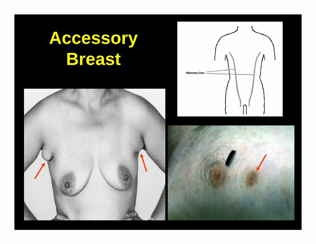
normally• Accessory breasts 1% of population,
male or female, anywhere on mammary ridge from axilla to groin
5
Accessory Breast
6
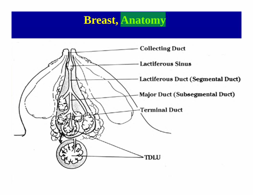
Breast, Anatomy
7
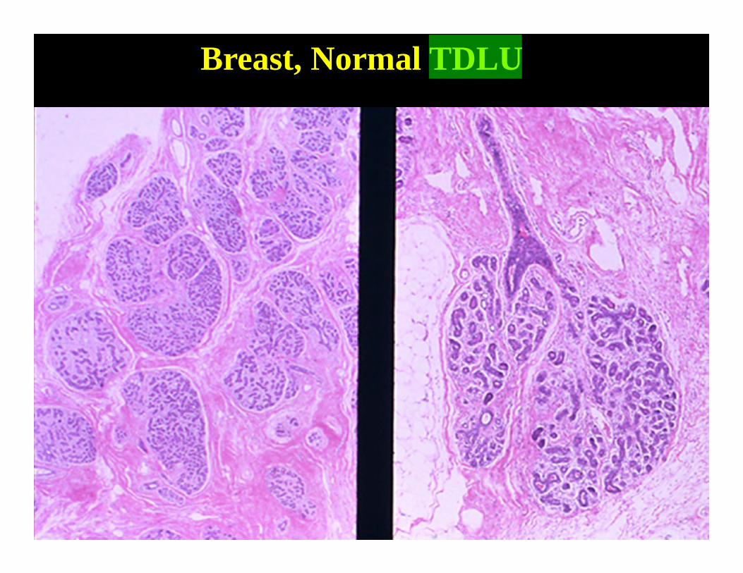
Breast, Normal TDLU
85
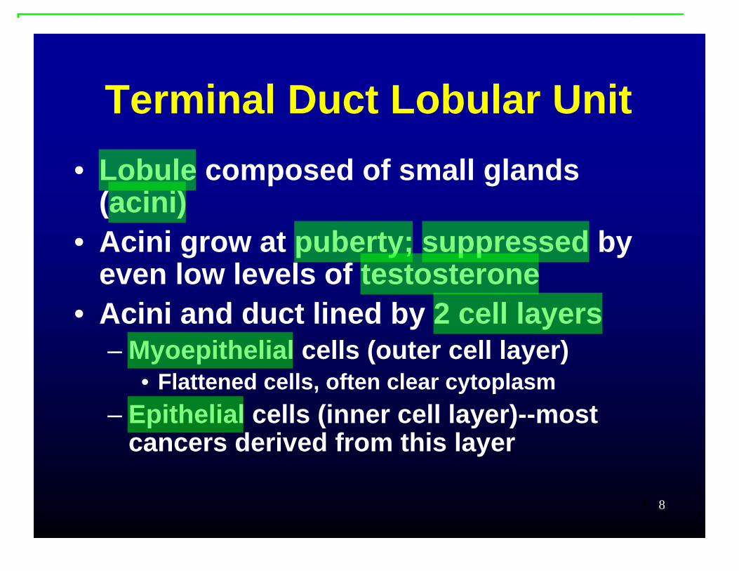
Terminal Duct Lobular Unit• Lobule composed of small glands
(acini)• Acini grow at puberty; suppressed by
even low levels of testosterone• Acini and duct lined by 2 cell layers
– Myoepithelial cells (outer cell layer)• Flattened cells, often clear cytoplasm
– Epithelial cells (inner cell layer)--most cancers derived from this layer
94
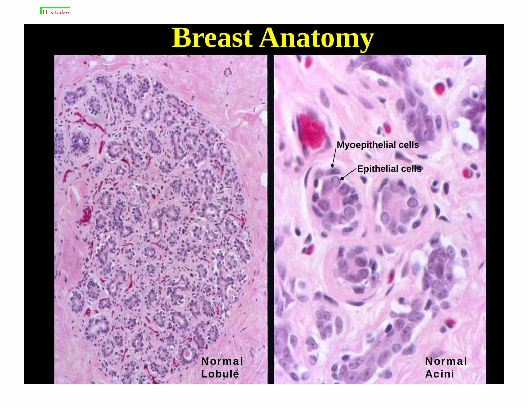
Breast Anatomy
Myoepithelial cells
Epithelial cells
Normal Lobule
Normal Acini
106
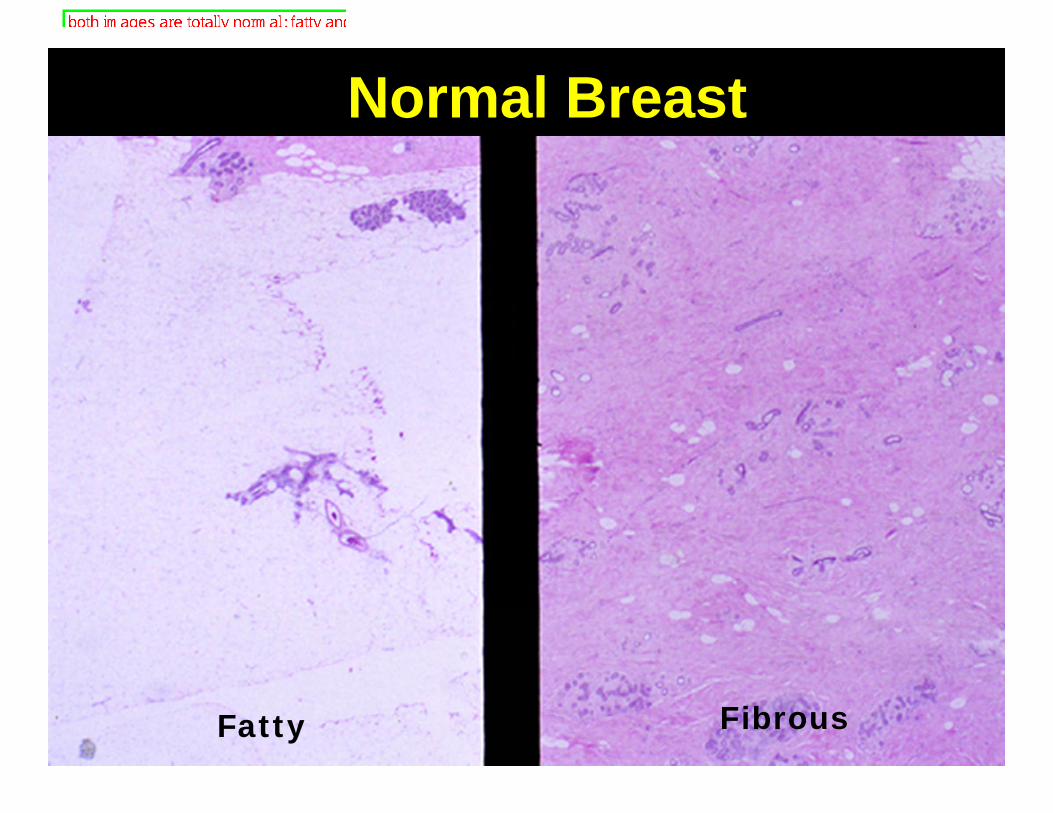
Stroma
• Large variation in amount of fibrous stroma
• Varies between individuals, and with menopause/hormone status within individual over time.
11
Normal Breast
FibrousFatty
12
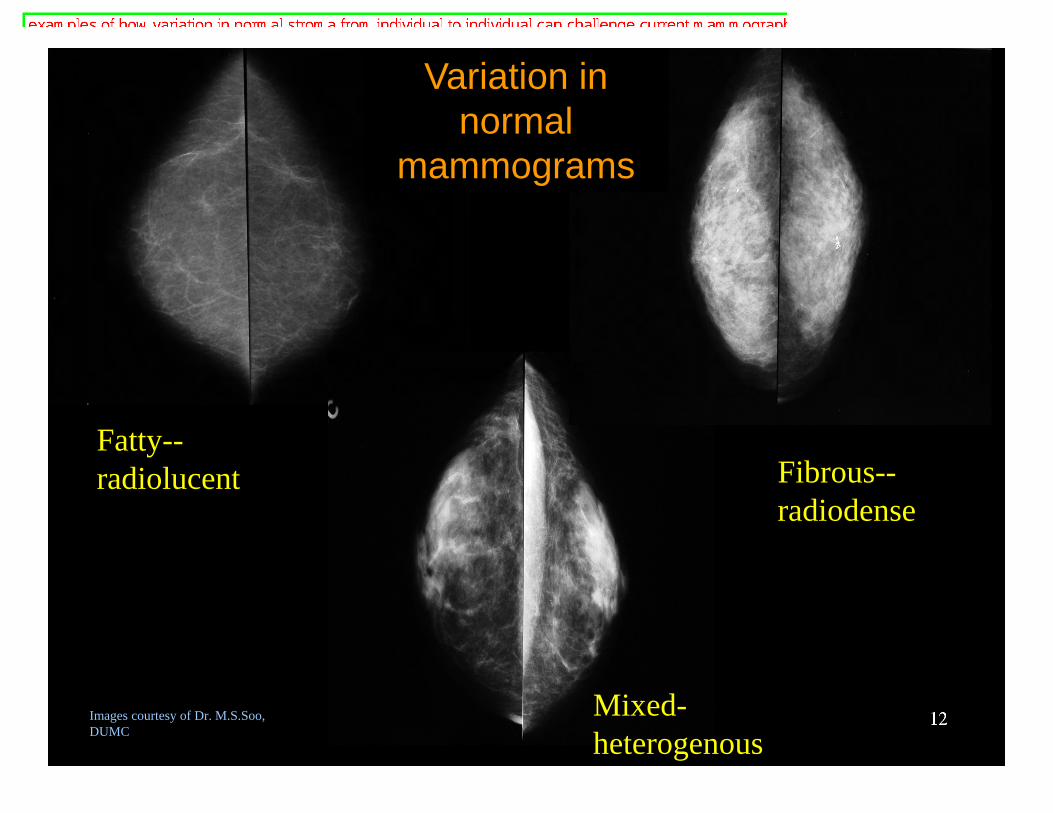
Fatty--radiolucent Fibrous--
radiodense
Images courtesy of Dr. M.S.Soo, DUMC
Variation in normal
mammograms
Mixed-heterogenous
137
Benign Breast Lesions
• Non-neoplastic– Inflammatory – Fibrocystic changes– Proliferative breast
changes– Proliferative breast
disease with atypia• Benign neoplasms

14
• Many can mimic malignancy:– Lumps on physical exam– Microcalcifications or masses on
mammograms– Bloody nipple discharge
• Some are risk factors for developing future breast cancer
• Benign lesions are much more commonthan cancers.
Benign Breast LesionsWhy do we care?

15
Breast Disease: A common reason to see the Doctor!
• 16% of women in large group practice sought medical attention for breast symptoms over 10 year period
• Only 4% of visits for breast symptoms resulted in dx of cancer
16
• Pain– Rarely is sole sign/symptom of CA
• Palpable mass (“lump”)• Bloody nipple discharge• Mammographic Abnormalities
– Density (mass)– Microcalcifications.
Breast Disease: Most common clinical presentations

17
Unlike medical school professors, most breast lesions are benign
Pathologic findings in women with “lump”
W.B. Saunders Company items and derived items Copyright (c) 1999 by W.B. Saunders Company

18
• Acute mastitis: Bacterial infection, usually while beginning nursing– Red, hot, swollen, painful breast– Can develop abscess, extensive tissue
destruction• Plasma cell mastitis: Non-bacterial,
chronic irritation from secretory products– Usually in multiparous woman, nursing
Inflammatory ConditionsMastitis

19
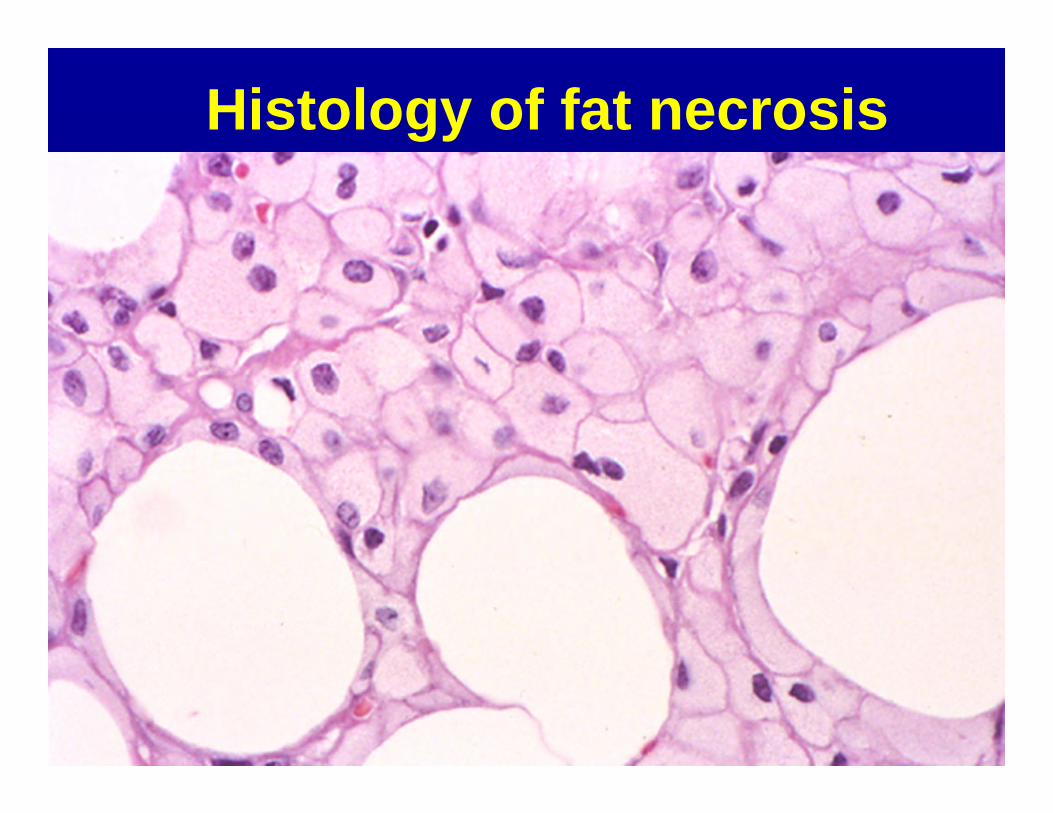
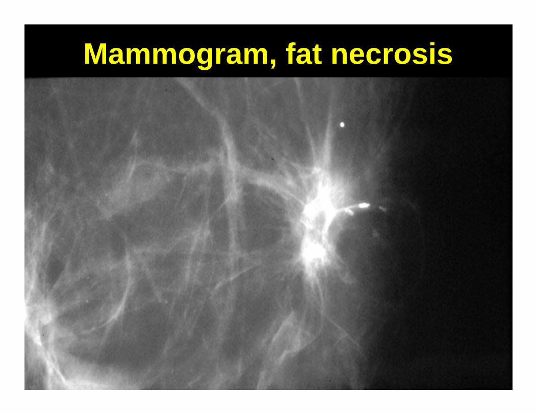
• Trauma to fat, release of fatty acids with marked inflammatory response
• Heals by scarring• Excellent cancer mimic
– Rock hard, spiculated mass– Microcalcifications on mammogram
Inflammatory ConditionsFat Necrosis
20
Histology of fat necrosis
2110
Mammogram, fat necrosis

22
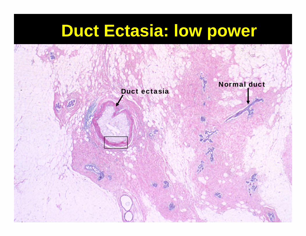
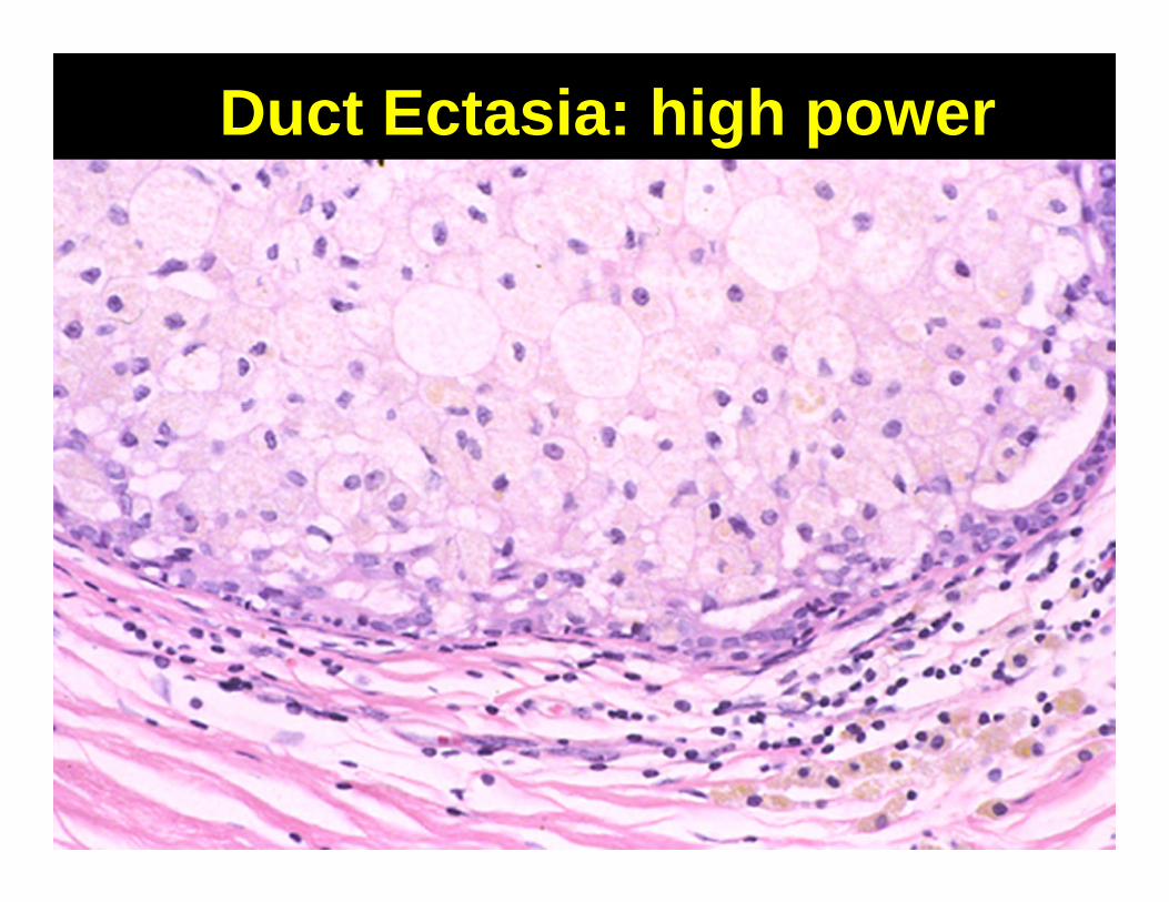
• Inflammation destroys duct wall• Common cause of nipple discharge• Microcalcifications can mimic cancer
Inflammatory ConditionsDuct Ectasia
2312
Duct Ectasia: low power
Normal ductDuct ectasia
24
Duct Ectasia: high power
2513
Benign Epithelial Lesions• Nonproliferative changes
– Fibrocystic change– Fibroadenoma
• Proliferative breast disease– Epithelial hyperplasia– Sclerosing Adenosis– Radial Scar– Intraductal papilloma

2614
Fibrocystic Change• Not a disease!• A group of processes which are related
only by the fact that they tend to occur together.
• Represents exaggerated response to hormonal stimulation
• Present in most women (>80%)• No increased risk for cancer
27
Fibrocystic Changeaka Non-Proliferative Breast Changes
• Fibrosis• Cysts• Metaplasia
– Apocrine– Columnar
2815
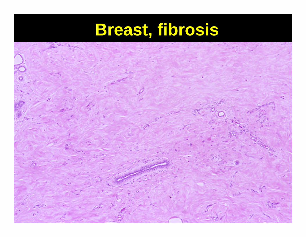
Fibrocystic Change“Fibrosis”
• Localized areas of fibrous tissue is common cause of lump
• This gets called “fibrosis” implying an increase over normal…but it is just normal breast tissue.
• Fibrous tissue in breast is NORMAL
29
Breast, fibrosis

30
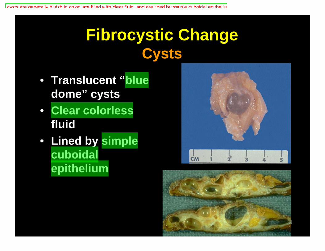
Fibrocystic ChangeCysts
• Cysts are extremely common• Multiple, bilateral• Fluctuate over time• Disappear with fluid aspiration
31
• Translucent “blue dome” cysts
• Clear colorless fluid
• Lined by simple cuboidal epithelium
Fibrocystic ChangeCysts
32
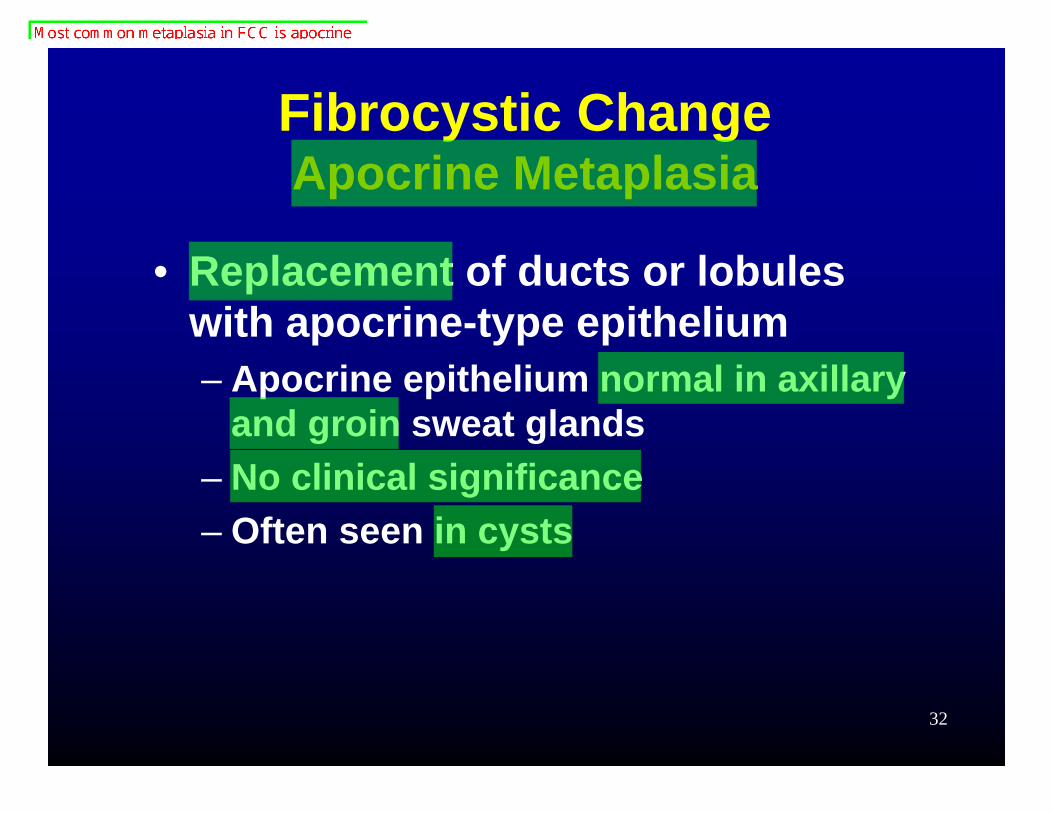
• Replacement of ducts or lobules with apocrine-type epithelium– Apocrine epithelium normal in axillary
and groin sweat glands– No clinical significance– Often seen in cysts
Fibrocystic ChangeApocrine Metaplasia
33
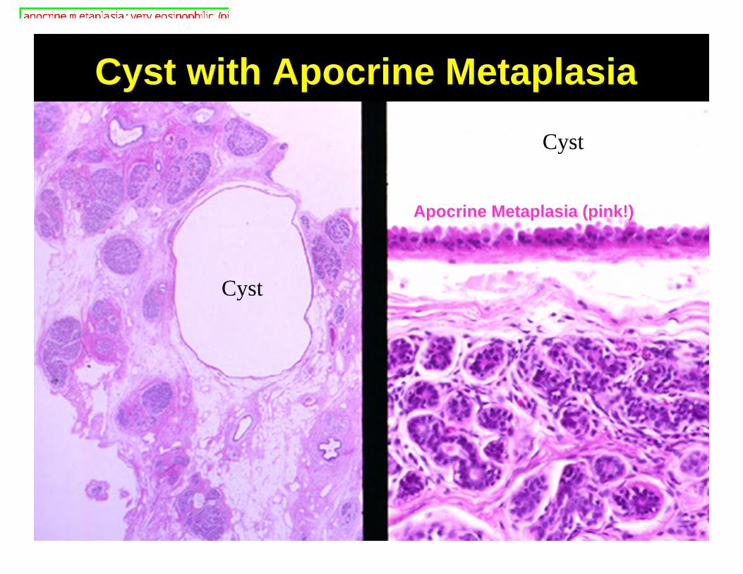
Cyst with Apocrine MetaplasiaCyst
Cyst
Apocrine Metaplasia (pink!)

3423
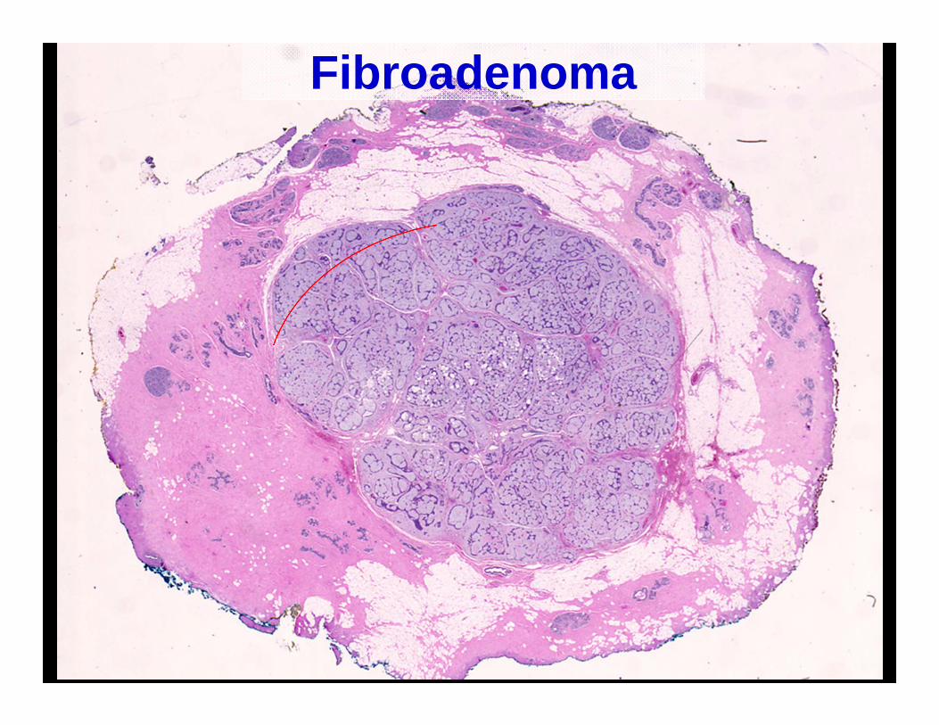
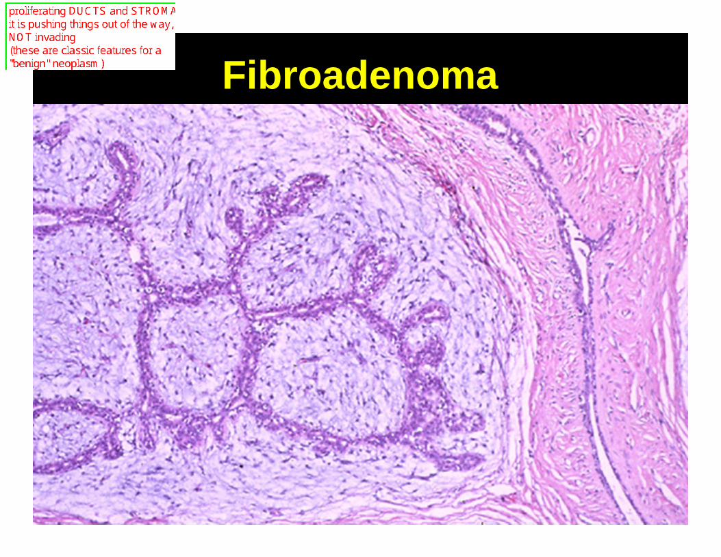
Fibroadenoma• Most common benign neoplasm of the
breast• Most common in teens and twenties;
second peak around menopause• Proliferation of ducts AND stroma --
“biphasic neoplasm”• Hard, round, well circumscribed
nodule; can mimic cancer• Often diagnosed clinically, not biopsied
35
Fibroadenoma
36
Fibroadenoma
37
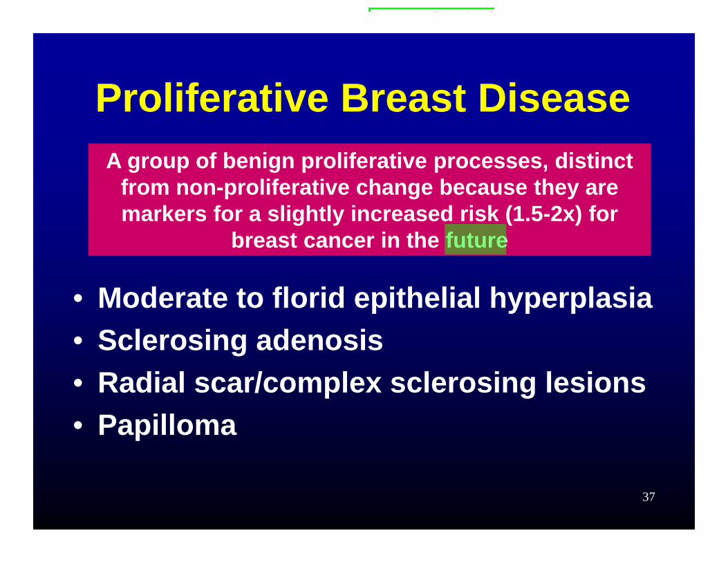
Proliferative Breast Disease
• Moderate to florid epithelial hyperplasia• Sclerosing adenosis• Radial scar/complex sclerosing lesions• Papilloma
A group of benign proliferative processes, distinct from non-proliferative change because they are markers for a slightly increased risk (1.5-2x) for
breast cancer in the future

3818
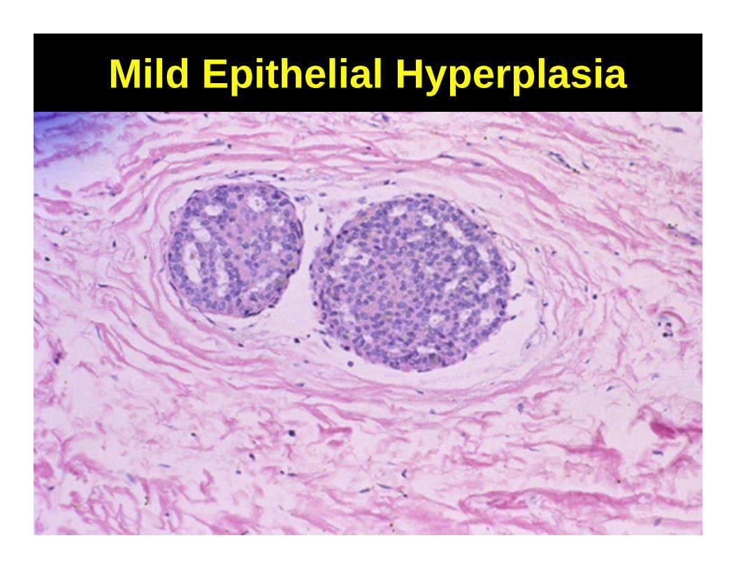
• Proliferation of epithelial cells within ducts and acini
• Classified as ductal (usual type) or lobular
Proliferative Breast DiseaseEpithelial Hyperplasia

39
• Always an incidental finding• Does not make lump or
microcalcifications• Graded from mild to severe (florid)• Important mostly as risk factor
– Patients with moderate/florid hyperplasia have 1.5-2.0 relative risk for developing breast cancer over 20 year f/u.
Proliferative Breast DiseaseEpithelial Hyperplasia
4019
Mild Epithelial Hyperplasia

41
Florid Epithelial Hyperplasia
4216
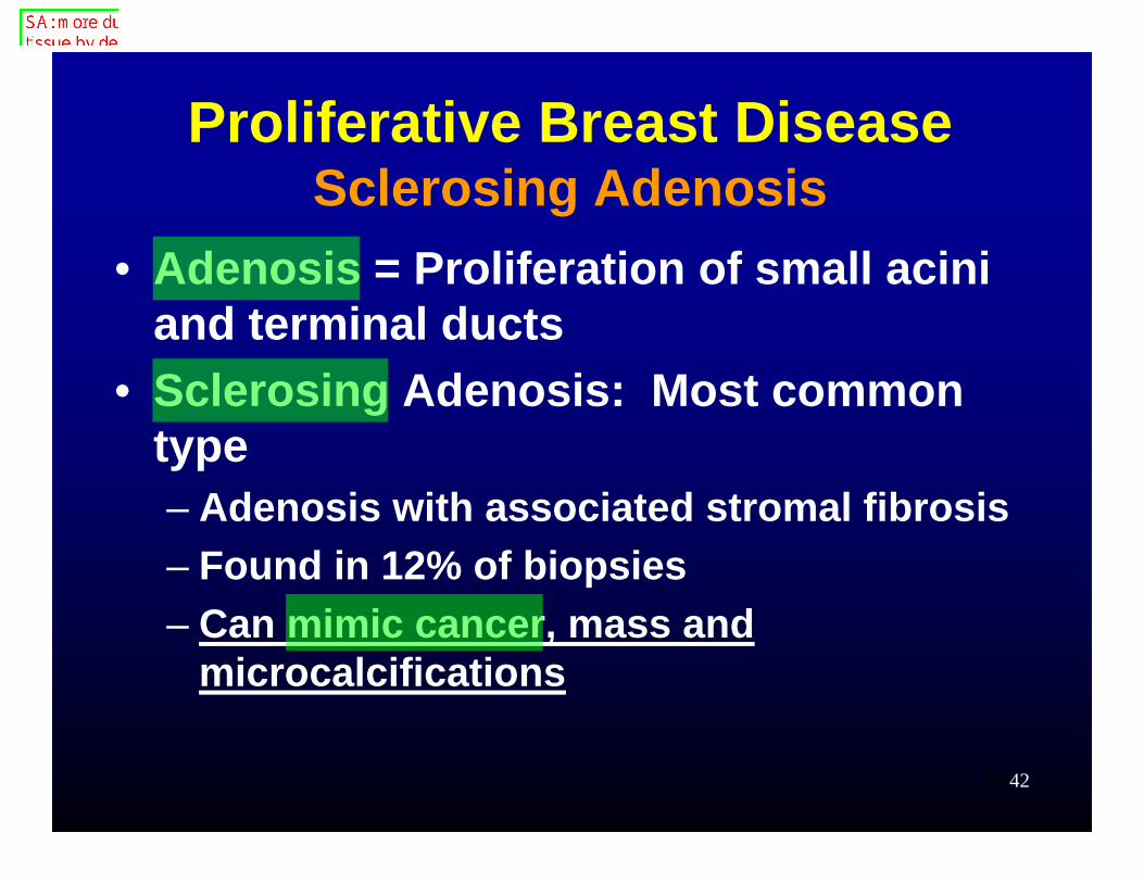
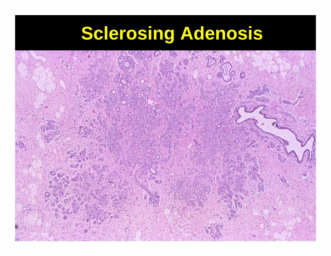
Proliferative Breast DiseaseSclerosing Adenosis
• Adenosis = Proliferation of small acini and terminal ducts
• Sclerosing Adenosis: Most common type– Adenosis with associated stromal fibrosis– Found in 12% of biopsies– Can mimic cancer, mass and
microcalcifications
43
Sclerosing Adenosis
4417
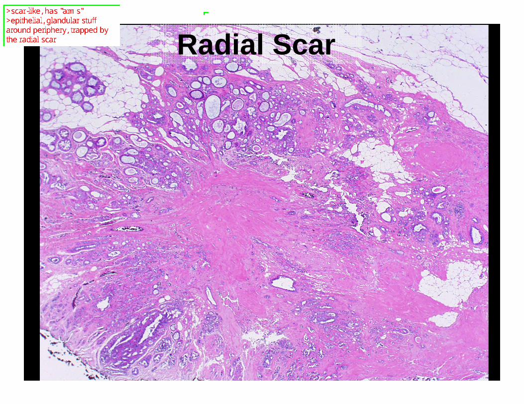
Proliferative Breast DiseaseRadial Scar
• Misnomer, not related to trauma• Stellate proliferation of ducts and acini
around a central scar-like area of fibrous and elastic tissue.
• Often mimics cancer mammographically (spiculated mass with microcalcifications)
45
Radial Scar

4622
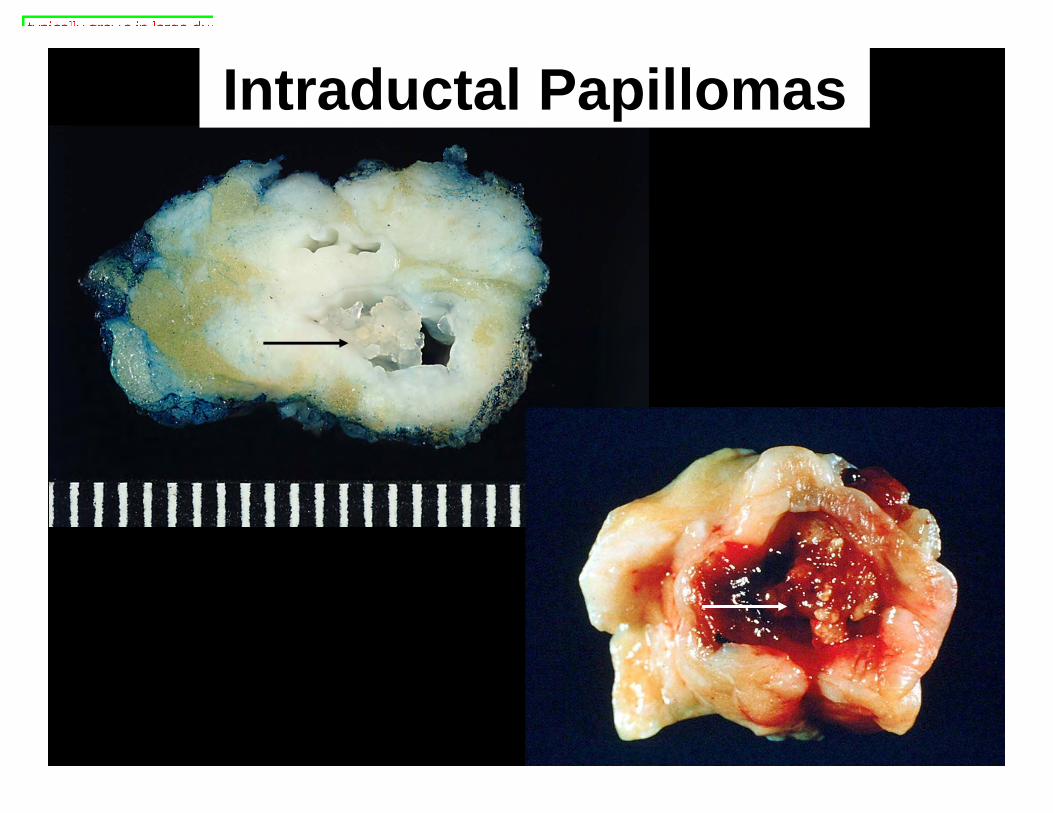
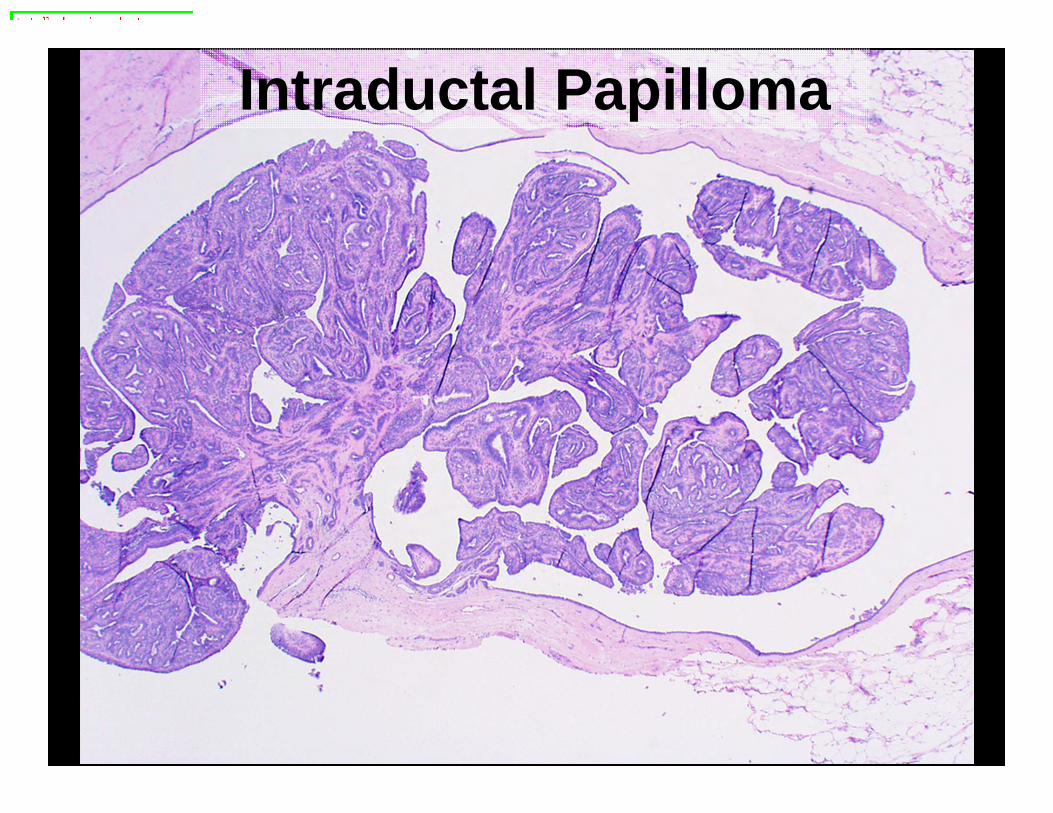
Intraductal Papilloma• Proliferation of papillary fronds within
dilated duct• Large ducts beneath nipple• Most common cause of bloody nipple
discharge
47
Intraductal Papillomas
48
Intraductal Papilloma
49
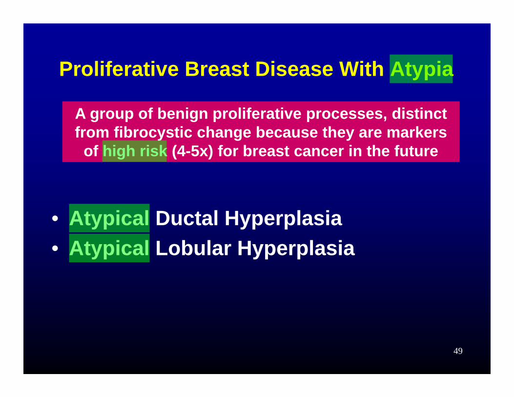
Proliferative Breast Disease With Atypia
• Atypical Ductal Hyperplasia• Atypical Lobular Hyperplasia
A group of benign proliferative processes, distinct from fibrocystic change because they are markers
of high risk (4-5x) for breast cancer in the future

5020
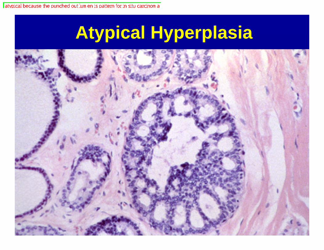
Proliferative Breast Disease With AtypiaAtypical Ductal Hyperplasia
• Has some but not all features of in-situ carcinoma– Probably precursor to in-situ carcinoma,
like dysplasia in cervix• Usually detected because of Ca++• Approximately 5% of biopsies• Moderate increase in risk for cancer
51
Atypical Hyperplasia

52

5324
Malignant Neoplasmsof the Breast

5426
Breast Cancer• Subject of intense scientific
investigation• Major advances in breast-conserving
therapy and reconstruction• Major focus of cancer screening
(mammography, self-exam)• Only recent years have seen a modest
impact on mortality rate

55
Malignant Neoplasms• Basic epidemiology• In-situ carcinoma
– Ductal carcinoma in-situ– Lobular carcinoma in-situ
• Invasive carcinoma– Ductal
• Special ductal subtypes– Lobular
• Prognostic and treatment factors– ER/PR, Her2/neu, genomic
• Special presentations of breast cancer
5625
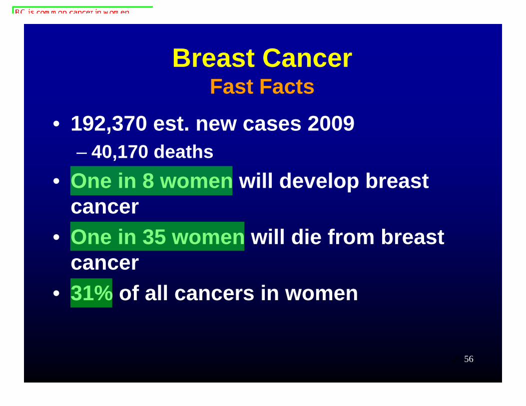
Breast CancerFast Facts
• 192,370 est. new cases 2009– 40,170 deaths
• One in 8 women will develop breast cancer
• One in 35 women will die from breast cancer
• 31% of all cancers in women
57
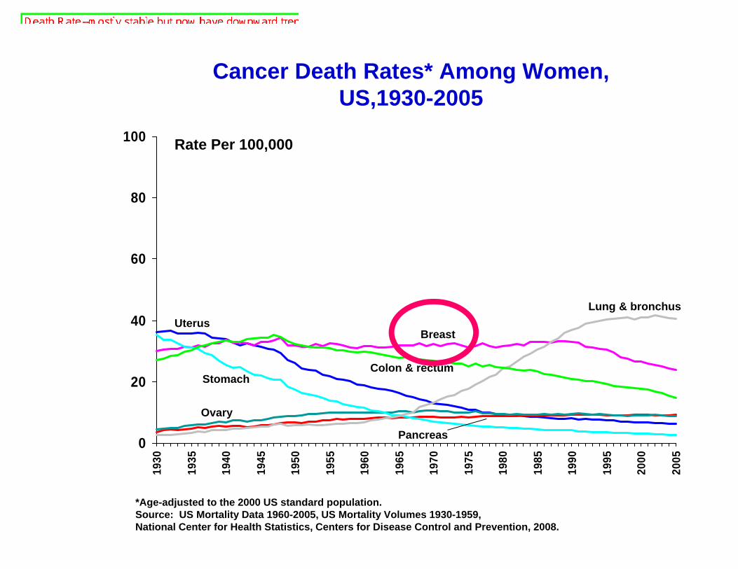
Cancer Death Rates* Among Women, US,1930-2005
*Age-adjusted to the 2000 US standard population.Source: US Mortality Data 1960-2005, US Mortality Volumes 1930-1959,National Center for Health Statistics, Centers for Disease Control and Prevention, 2008.
0
20
40
60
80
10019
30
1935
1940
1945
1950
1955
1960
1965
1970
1975
1980
1985
1990
1995
2000
2005
Lung & bronchus
Colon & rectum
Uterus
Stomach
Breast
Ovary
Pancreas
Rate Per 100,000

58
Cancer Incidence Rates* Among Women, US, 1975-2005
*Age-adjusted to the 2000 US standard population and adjusted for delays in reporting.Source: Surveillance, Epidemiology, and End Results Program, Delay-adjusted Incidence database: SEER Incidence Delay-adjusted Rates, 9 Registries, 1975-2005, National Cancer Institute, 2008.
0
50
100
150
200
250
1975 1978 1981 1984 1987 1990 1993 1996 1999 2002 2005
Colon and rectum
Rate Per 100,000
Breast
Lung & bronchus
Uterine CorpusOvary
Non-Hodgkin lymphoma

5944
Risk Factors for Breast Cancer
• Age• Family history• Specific gene mutations: BRCA1,
BRCA2, p53– very high risks (50-80%) for affected
families, but uncommon causes of breast cancer overall
– also have increased risk of other cancers (ovary, other)
60
Risk Factors...• Prolonged estrogen exposure
– early menarche, late menopause, birth control pills
• Late or no pregnancy• High risk findings in previous breast
biopsy• Radiation, esp. as teenager or young
adult• Breast feeding is protective
6127
Pre-invasive Malignancy
• Ductal Carcinoma in Situ (DCIS)– Synonym: Intraductal Carcinoma– Direct precursor to invasive carcinoma.– Malignant cells proliferating within duct,
no invasion through basement membrane (no metastatic potential)
– Spread within duct system; can involve very large area
– Microcalcifiations on mammogram
62
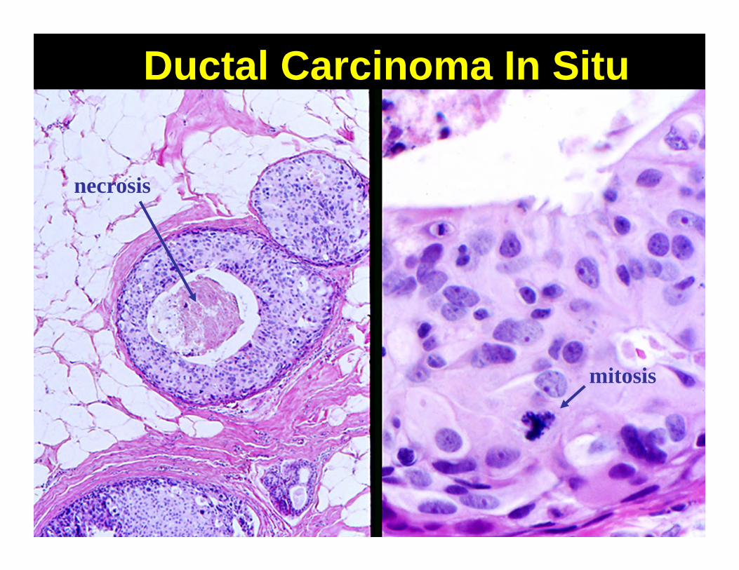
Ductal Carcinoma In Situ
mitosis
necrosis

63
Mammogram, DCIS
Microcalcifications
6429
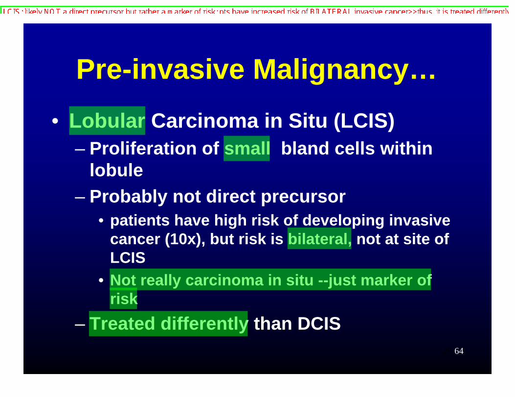
Pre-invasive Malignancy…• Lobular Carcinoma in Situ (LCIS)
– Proliferation of small bland cells within lobule
– Probably not direct precursor• patients have high risk of developing invasive
cancer (10x), but risk is bilateral, not at site of LCIS
• Not really carcinoma in situ --just marker of risk
– Treated differently than DCIS

6531
Invasive Neoplasms

66
Invasive AdenocarcinomaClassification
• Invasive Ductal adenocarcinoma– No special type (NST, NOS)– Special subtypes -- better prognosis
MedullaryMucinousTubular
• Invasive Lobular adenocarcinoma
6733
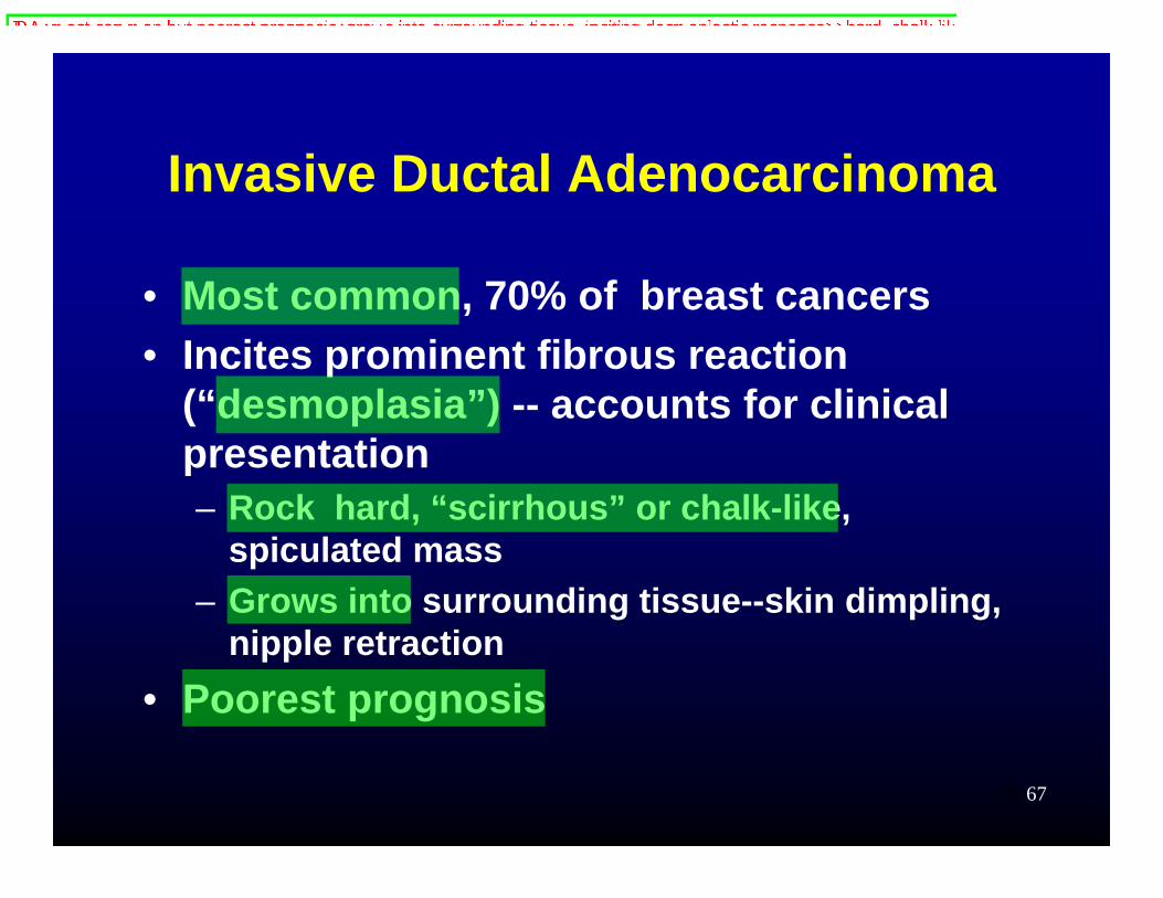
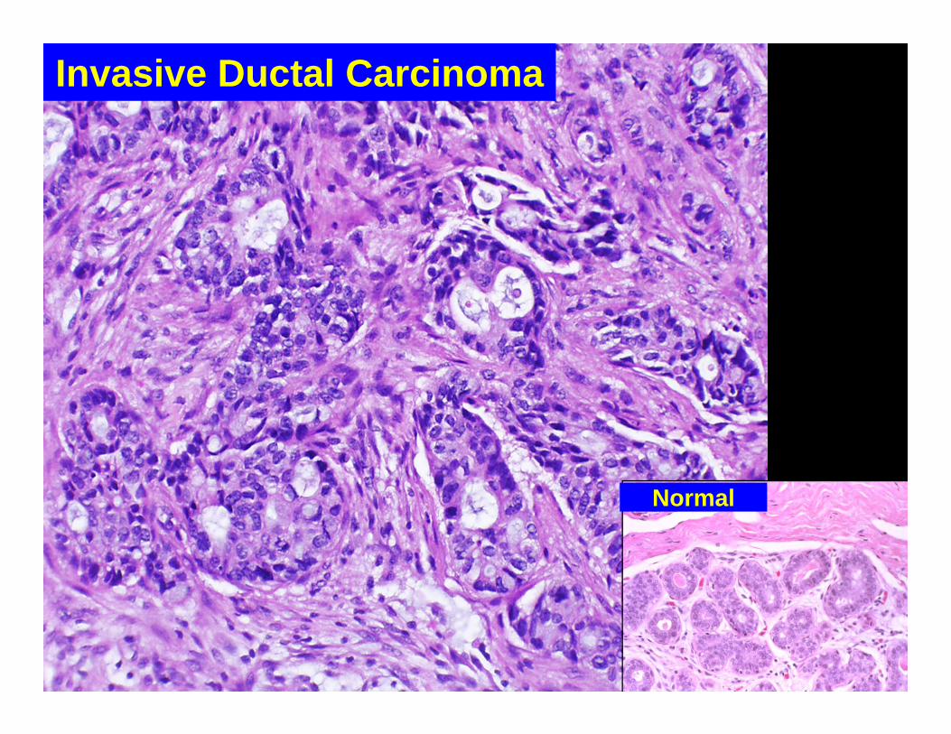
Invasive Ductal Adenocarcinoma
• Most common, 70% of breast cancers• Incites prominent fibrous reaction
(“desmoplasia”) -- accounts for clinical presentation– Rock hard, “scirrhous” or chalk-like,
spiculated mass– Grows into surrounding tissue--skin dimpling,
nipple retraction• Poorest prognosis

68
Invasive Ductal AdenocarcinomaSpiculated Mass
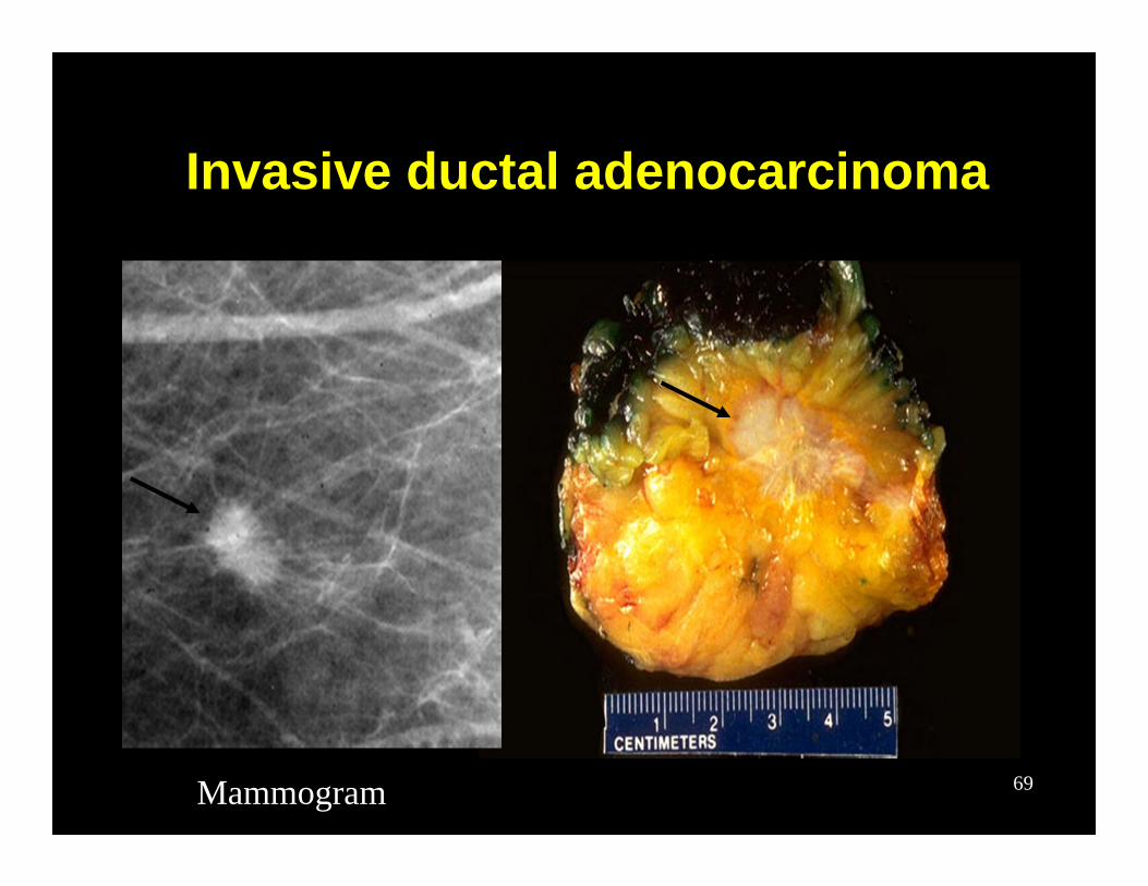
69
Invasive ductal adenocarcinoma
Mammogram
7034
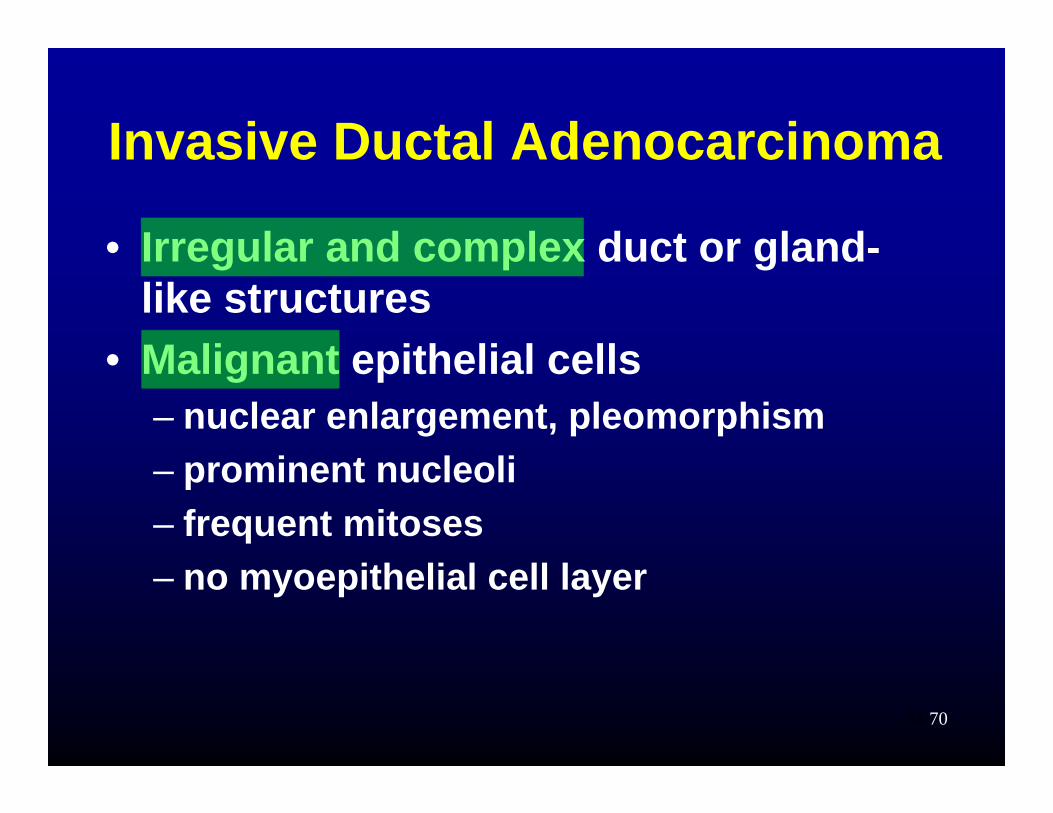
Invasive Ductal Adenocarcinoma
• Irregular and complex duct or gland-like structures
• Malignant epithelial cells– nuclear enlargement, pleomorphism– prominent nucleoli– frequent mitoses– no myoepithelial cell layer
71
Invasive Ductal Carcinoma
Normal

7235
Special Types...• Medullary carcinoma
– Well circumscribed, soft– Prominent lymphoid infiltrate– Paradoxically, despite relatively good
prognosis, most anaplastic tumor cells of any type.
• No ducts or glands• High grade nuclei
– Rare, less than 1% of breast cancers

73
Medullary Carcinoma
Lymphoid infiltrate at tumor edge

7436
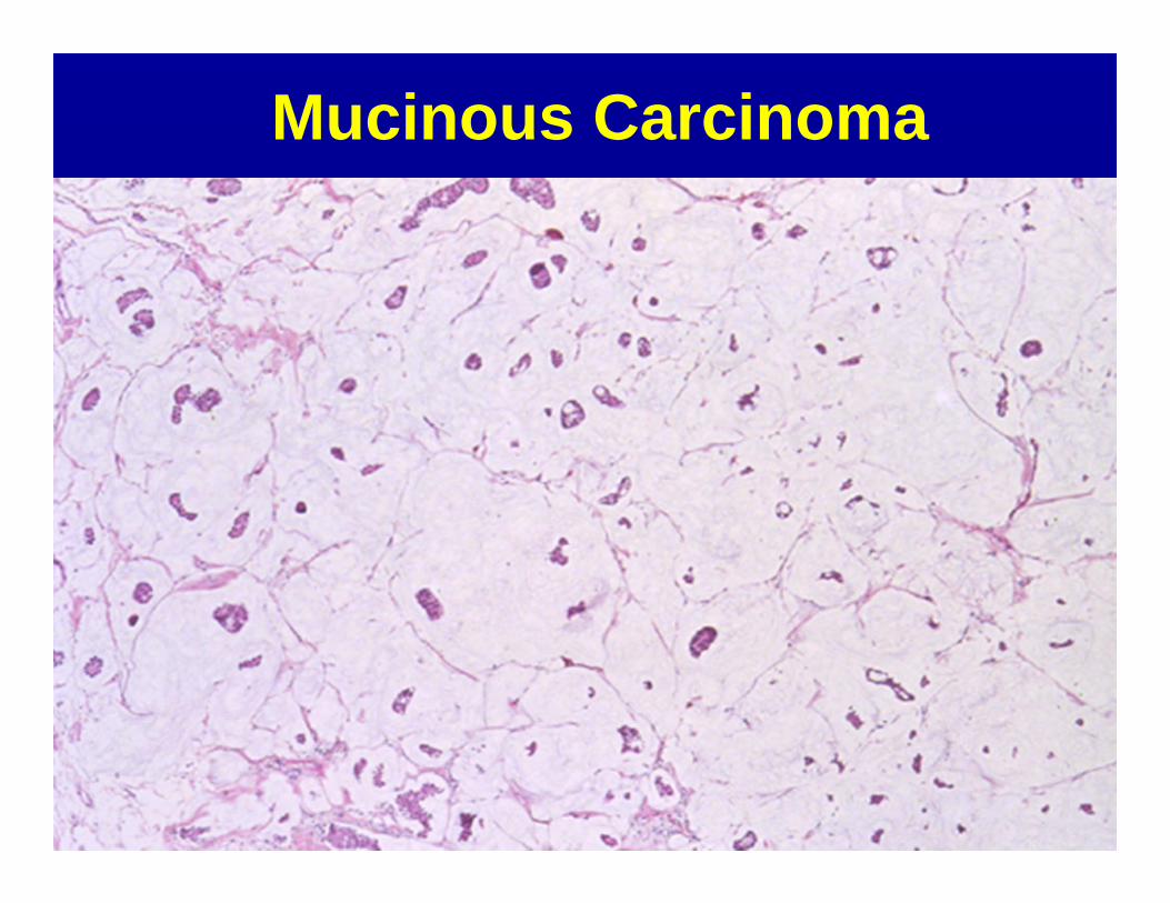
Special Types...• Mucinous Carcinoma
– Synonym: Colloid carcinoma– Well circumscribed, mucinous consistency– “Islands of tumor floating in a sea of
mucin”– Approximately 1-5% of breast cancers
75
Mucinous Carcinoma

7637
Special Types…
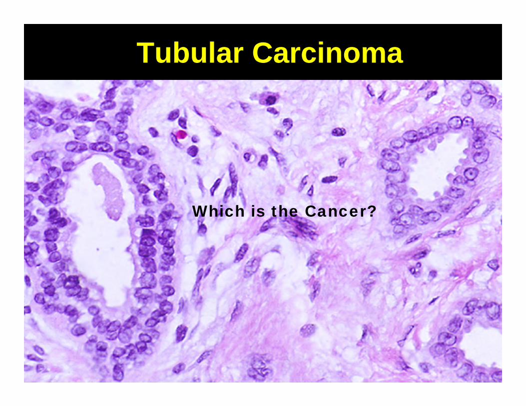
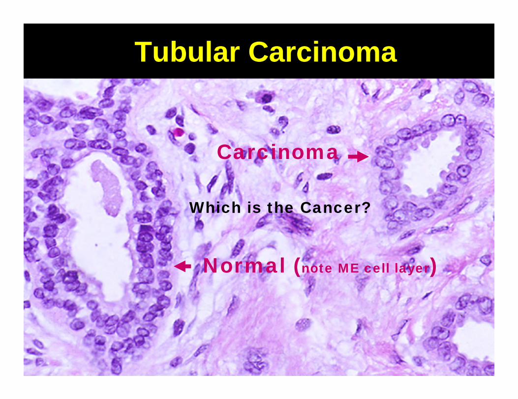
• Tubular Carcinoma– Extremely well differentiated ductal carcinoma– Composed entirely of simple tubules lined by
single layer of cells– Can be confused with benign lesions (radial scar)
• No myoepithelial cell layer– Extremely good prognosis; no deaths reported
when <1cm– 5% of breast cancers
77
Tubular Carcinoma
Which is the Cancer?
78
Tubular Carcinoma
Carcinoma
Normal (note ME cell layer)
Which is the Cancer?
7938
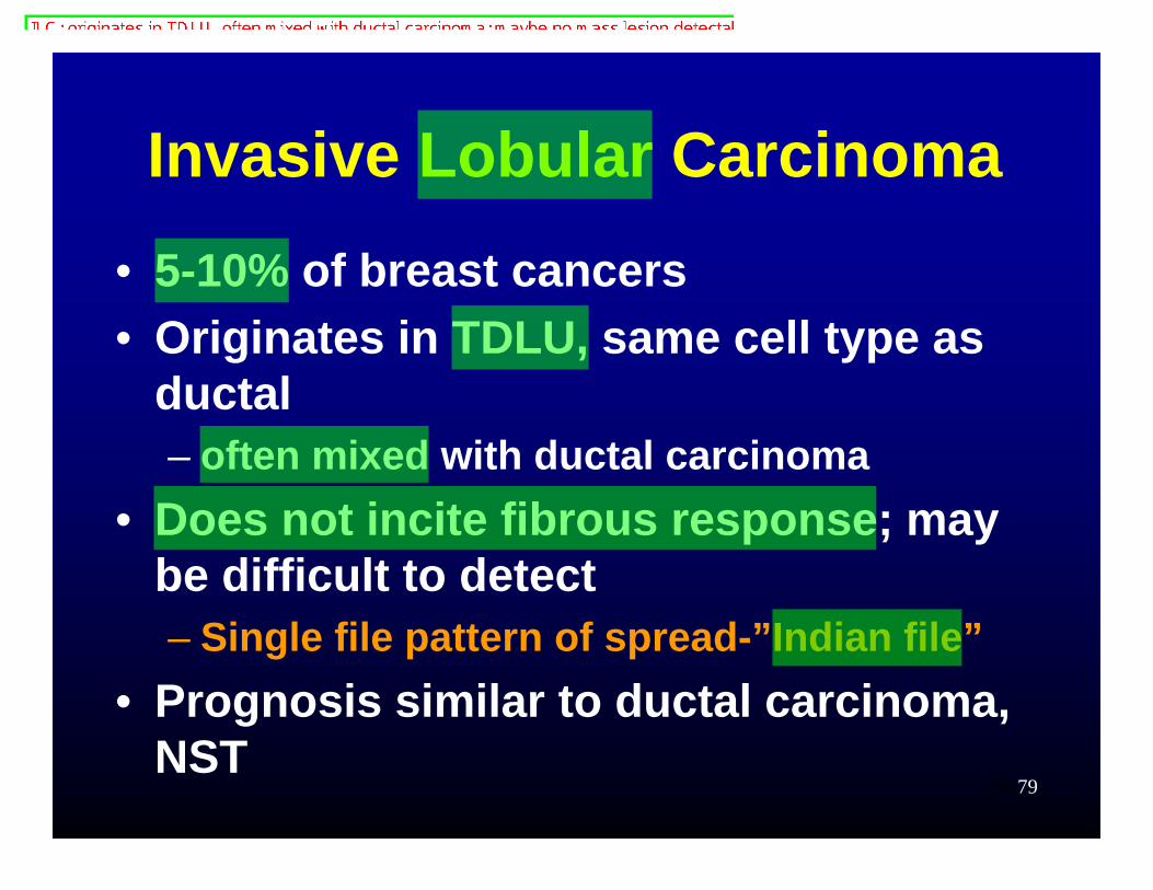
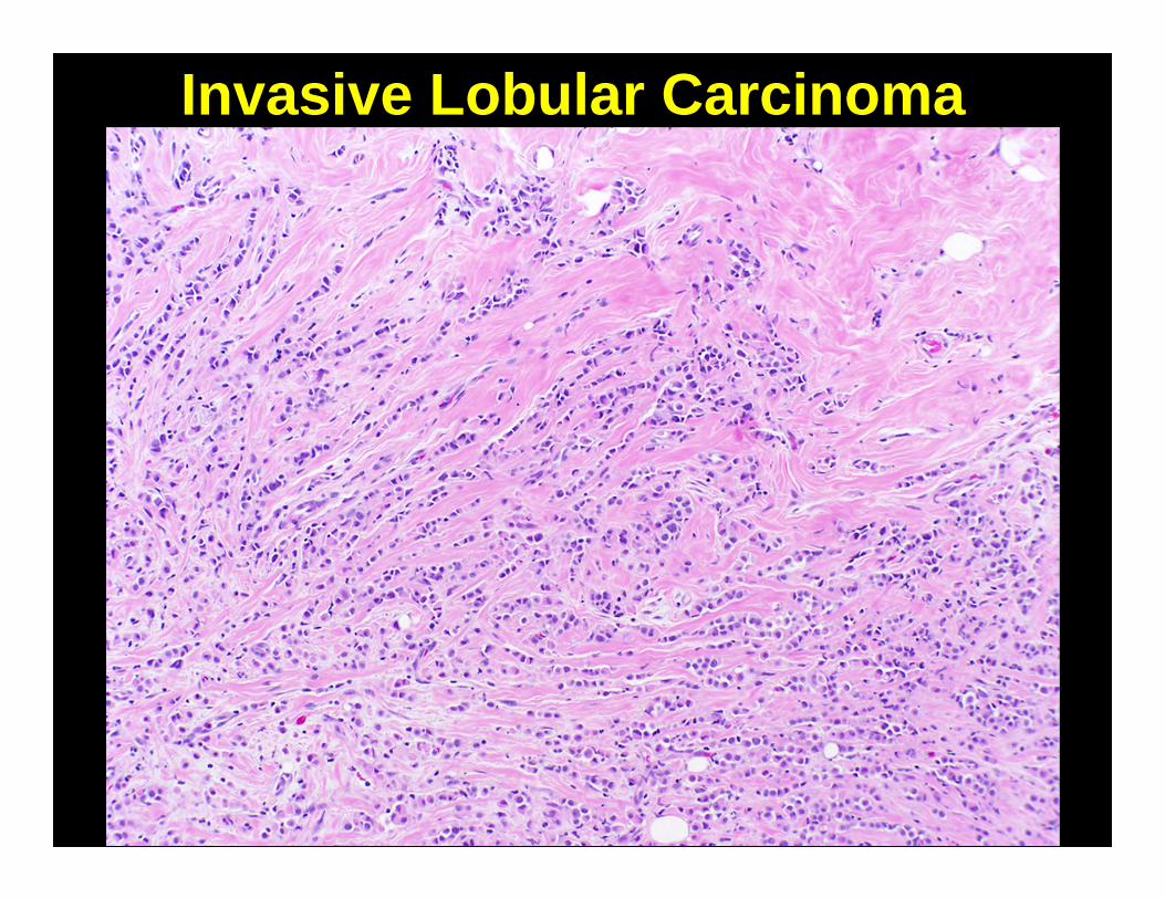
Invasive Lobular Carcinoma• 5-10% of breast cancers• Originates in TDLU, same cell type as
ductal– often mixed with ductal carcinoma
• Does not incite fibrous response; may be difficult to detect– Single file pattern of spread-”Indian file”
• Prognosis similar to ductal carcinoma, NST
8039
Invasive Lobular Carcinoma
81
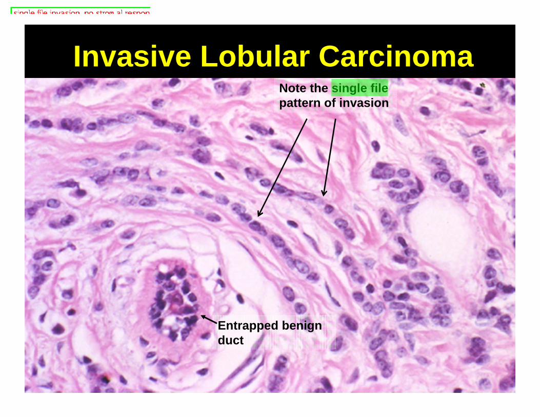
Invasive Lobular CarcinomaNote the single file pattern of invasion
Entrapped benign duct
8242
Behavior of Breast Carcinoma• Local recurrence
– Can ulcerate through skin, invade chest wall
• Lymphatic/hematogenous metastases– local metastases to axillary nodes (most
common); internal mammary nodes, supraclavicular nodes less common
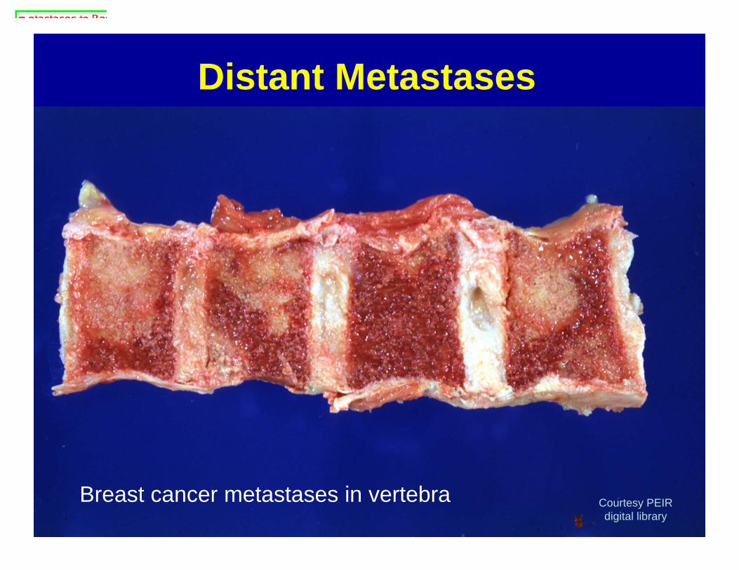
– distant metastases to lung, liver, bone, brain common sites

83
Breast Cancer-Local recurrence
Courtesy PEIR digital libraryBreast cancer ulcerating through skin
84
Distant Metastases
Breast cancer metastases in vertebra Courtesy PEIR digital library

85

8643
Key Prognostic Factors• Stage of disease
– Tumor size– Axillary node status -- single most
important prognostic feature, predicts distant metastases
• Tumor grade: well differentiated vs. poorly differentiated
• Margins of resection: local recurrence likely if tumor in margins
87
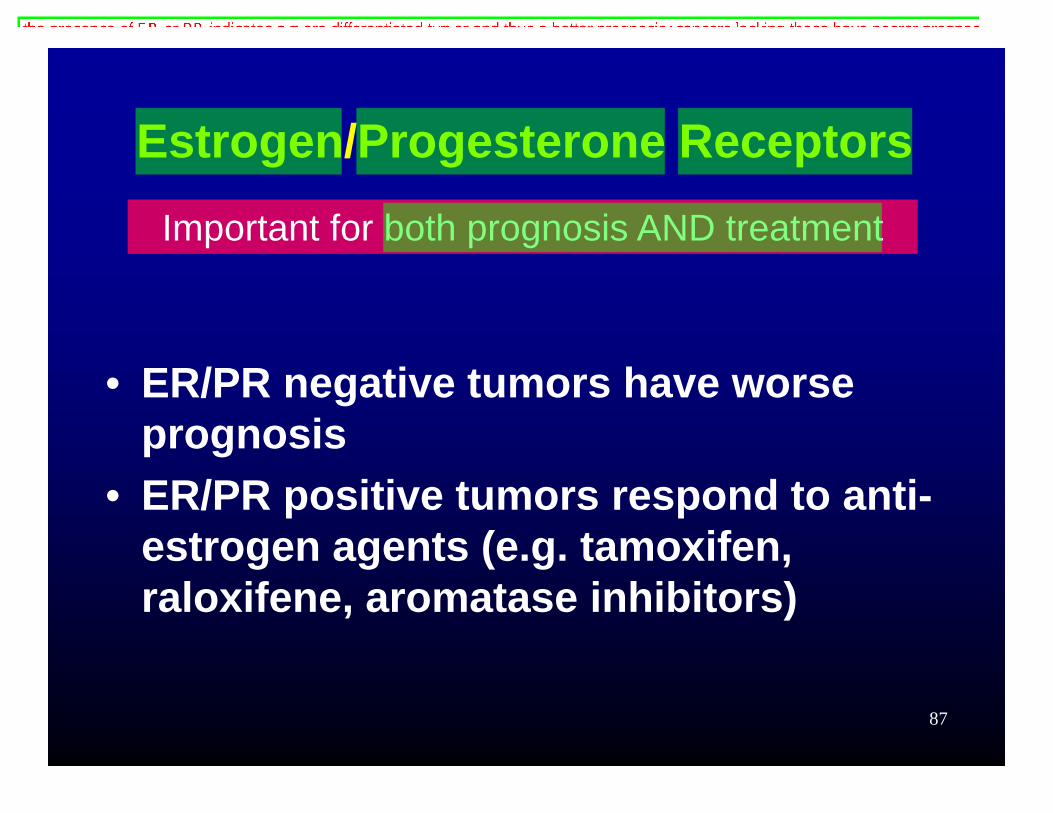
Estrogen/Progesterone Receptors
• ER/PR negative tumors have worse prognosis
• ER/PR positive tumors respond to anti-estrogen agents (e.g. tamoxifen, raloxifene, aromatase inhibitors)
Important for both prognosis AND treatment

Estrogen Receptor Immunostain(Brown nuclear staining)
89
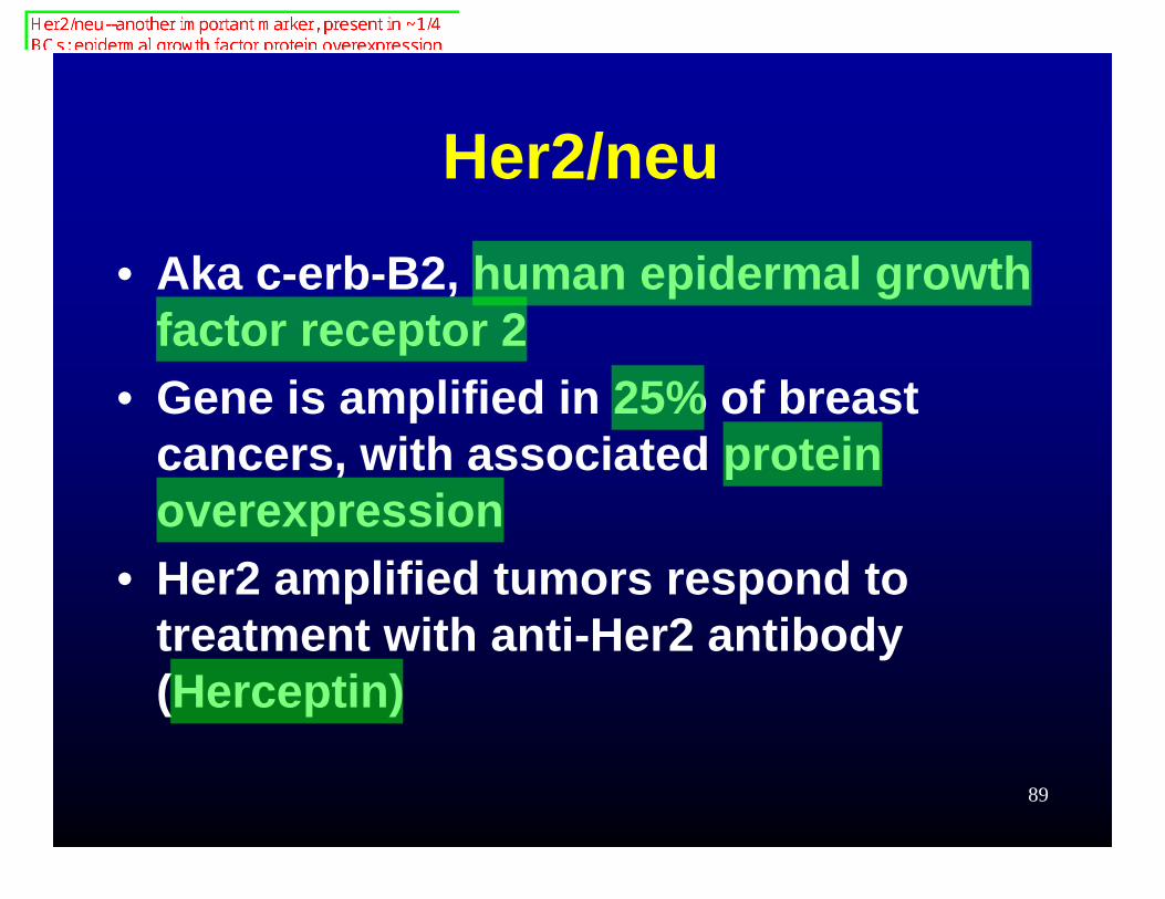
Her2/neu• Aka c-erb-B2, human epidermal growth
factor receptor 2• Gene is amplified in 25% of breast
cancers, with associated protein overexpression
• Her2 amplified tumors respond to treatment with anti-Her2 antibody (Herceptin)
90
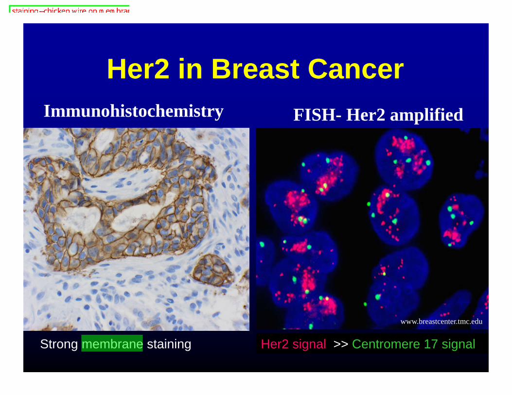
Her2 in Breast Cancer
www.breastcenter.tmc.edu
Her2 signal >> Centromere 17 signal
FISH- Her2 amplifiedImmunohistochemistry
Strong membrane staining
91
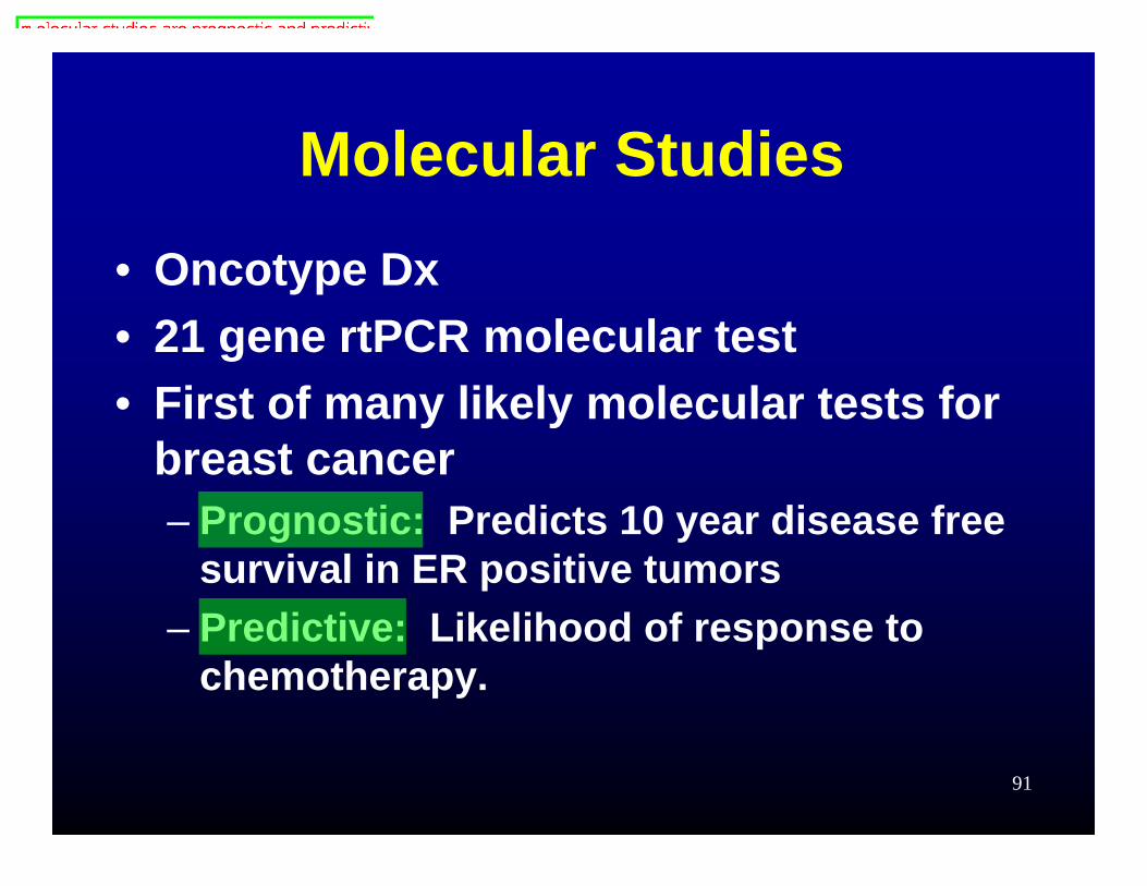
Molecular Studies• Oncotype Dx• 21 gene rtPCR molecular test• First of many likely molecular tests for
breast cancer– Prognostic: Predicts 10 year disease free
survival in ER positive tumors– Predictive: Likelihood of response to
chemotherapy.
92
Summary• Described the clinical presentation of common breast
pathologies• Explained what "fibrocystic change" means and
described several of the most common benign breast lesions.
• Described the common types of breast cancer• Discussed major prognostic factors in breast cancer• Explained why testing for expression of estrogen
receptor and Her2/neu is an important part of breast cancer analysis

93
Contact information:
Rex BentleyM216A Duke South Green Zone
For more details Pathology 448C—Practical Surgical Pathology (4th year elective)

94
Additional slides for those who have an unquenchable thirst for knowledge

95
***Filler slide***Unique Manifestations of Breast Cancer
Or two things to know about that will help you avoid unpleasant encounters with malpractice lawyers!
96
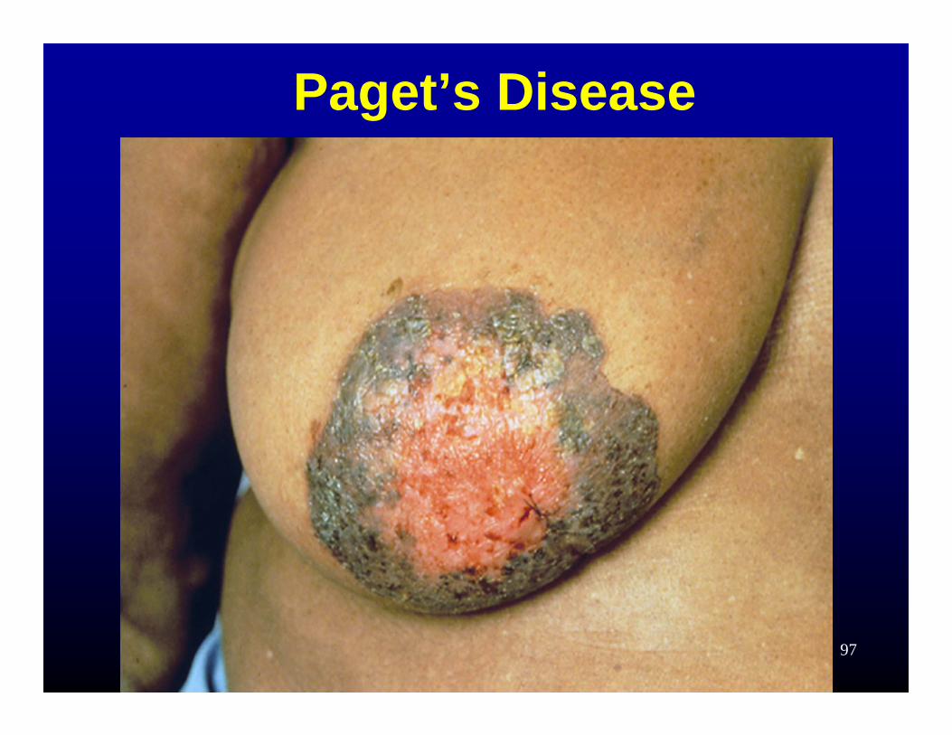
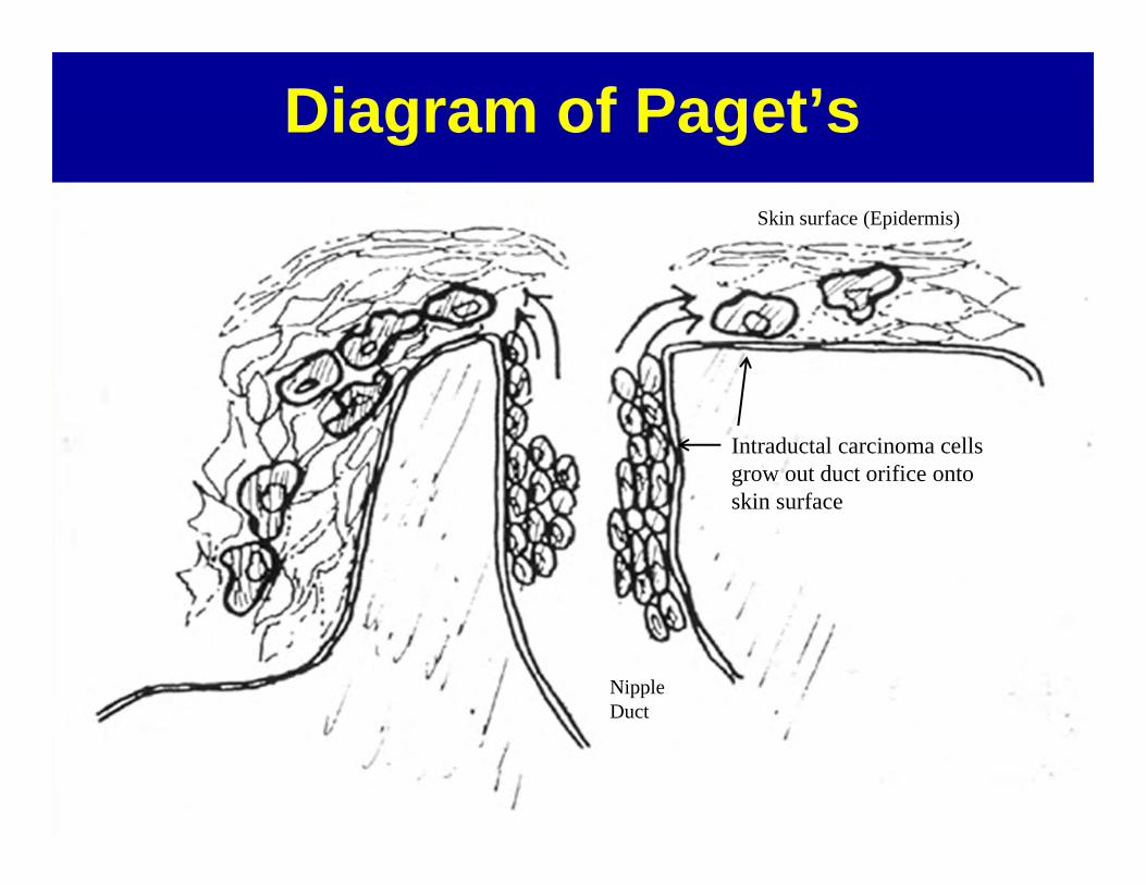
Paget’s Disease• Eczematous, scaly, red rash around
nipple• Represents ductal carcinoma in-situ
invading epidermis of nipple• Frequently not recognized clinically—
diagnosis of breast cancer delayed
97
Paget’s Disease
98
Diagram of Paget’s
Nipple Duct
Skin surface (Epidermis)
Intraductal carcinoma cells grow out duct orifice onto skin surface

99
Paget’s Disease of the Nipple
Tumor cellsEpidermis
100
Remember…
1. Rashes around the nipple can represent breast cancer.
2. When your patient discovers that you’ve been treating her breast cancer with topical steroids, she will not be pleased!
101
Inflammatory Carcinoma• Diffusely red, swollen, hot breast• Associated with very poor prognosis
(considered T4 disease)• Skin biopsies show plugging of dermal
lymphatics by tumor cells • Closely mimics infection (cellulitis) but
does not respond to antibiotics; often not recognized clinically—diagnosis delayed

Inflammatory Carcinoma
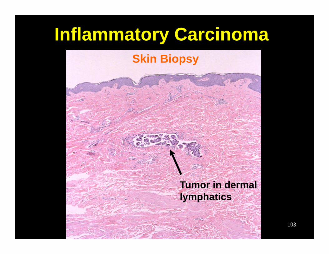
103
Tumor in dermal lymphatics
Inflammatory CarcinomaSkin Biopsy
104
Remember…
1. “Cellulitis” in the breast can represent breast cancer
2. Six weeks of antibiotics will not cure breast cancer!
10545
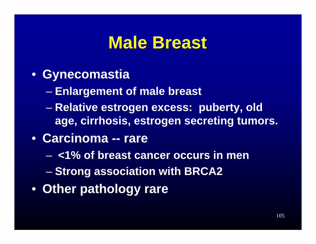
Male Breast• Gynecomastia
– Enlargement of male breast– Relative estrogen excess: puberty, old
age, cirrhosis, estrogen secreting tumors.• Carcinoma -- rare
– <1% of breast cancer occurs in men– Strong association with BRCA2
• Other pathology rare

106
Gynecomastia
Note absence of lobules!

107
The End (Really!)