essentials of pathology
TRANSCRIPT
Ya. Bodnar, A. Romanyuk, V. Voloshyn, V. Gargin
Ya. Bodnar, A. Romanyuk, V. Voloshyn, V. Gargin
Essentials of pathology
Kharkiv - 2020

Essentials of pathology
UDK 616-091(075.8)
Authors: Bodnar Yaroslav, professor, head of the Department of Pathological Anatomy with Dissection Course and Forensic Medicine, Morphology Institute I.Ya. Horbachevsky Ternopil State Medical University Romanyuk Anatolii, professor, head of the Department of Pathomorphology with Dissection Course and Forensic Medicine, Medical Institute Sumy State University Voloshyn Volodymyr, ass.-professor of the Department of Pathological Anatomy with Dissection Course and Forensic Medicine, Morphology Institute I.Ya. Horbachevsky Ternopil State Medical University Gargin Vitaliy, professor of the Department of Pathological Anatomy, Kharkiv National Medical University
Reviewers: head of the Department of Morhpology, Medical Institute Sumy State University, prof., V.I. Bumeister; head of the Department of Pathological Anatomy with Autopsy Course, Ukrainian Medical Stomatological Academy, prof., I.I. Starchenko. Essentials of pathology : textbook / Ya. Bodnar, A. Romanyuk, V. Voloshyn, V. Gargin – Kharkiv, «Planeta-Print» Ltd, 2020, 219 . ISBN 978-617-7751-83-9 This manual was written in conformance with training program in Pathomorphology for higher educational establishments and based on European credit-transfer system principles. Its first part covers one module - general pathologic processes and tumors growth, second part covers systematic and infectious pathology. Notional modules include theoretical knowledge of pathologic processes macroscopic and microscopic manifestations. The aim of the book is clearly and easily assist student to acquire habits of synthetic generalization of pathologic processes demonstration and their interpretation in cause-effect correlations. The book is intended for English medium medical Students and is recommended for publication by the Academic Council of Sumy State University (Ukraine).

Ya. Bodnar, A. Romanyuk, V. Voloshyn, V. Gargin
3
Essentials of pathology
Topic. Subject and tasks of pathologic morphology. Methods of pathologic morphology investigations. Main stages of pathologic morphology development.
Pathologic anatomy being fundamental medical-biology science is at the meeting point of medical theory and practice. Main assignment of pathology anatomy service is lifetime and posthumous diagnostic of diseases, study of etiology, pathogenesis and tanatogenesis of the most widespread diseases, control of clinical diagnostic quality and therapeutic process effectiveness as well as physicians’ professional advanced training. Pathologic anatomy is a science dealing with structural basis of the disease. There are no functional changes which are not connected with structural ones. There are no purely functional diseases that are diseases which are not accompanied by changes in the cell structure.
Pathologic anatomy is not limited to study of morphological changes which occur in the organism of a sick person. It uses the information about morphology to reveal etiology, pathogenesis of the disease, other aspects.
Pathologic anatomy began to develop as a separate branch of science in the 16th century. The first scientific pathologic work (published in 1761) was a work of an Italian anatomist Morgagni "On site and causes of diseases revealed by anatomist". The author had studied 700 corpses.
The first textbook on Anatomy written by Bailey was published in the middle of the 18th century. In the 19th century pathology departments were founded in Berlin, Paris, Wien, Kharkiv. Such prominent scientists as Schleiden (1804-1881), Schwann (1810-1882), Rokitansky (1804-1878), Virchov (1821-1902) (the two latter being pathologists), Lambl (1824-1895) contributed much to pathology development.
Basic methods of diseases posthumous and lifetime diagnostic
Pathologist is specialist which takes participation in about 70% of all medical diagnosis by using different methods. Basic methods of diseases posthumous diagnostic are macroscopic (autopsy) and microscopic (necropsy), lifetime diagnostic are microscopic (biopsy, cytology) and experiment. Accessory methods are as follows: biological (bacteriologic, virologic, serologic, hematological, tissue culture method), chemical (histochemical, immunohystochemical, atomic absorptiometry, quantitative analysis, qualitative analysis, biochemical), physical (hystoautoradiography, roentgenography, roentenostructural analysis, ultrasonic diagnostics), molecular-genetic. The objects of pathological study are corpses, surgical material and specially obtained (biopsied) material as well as experimental material.
Autopsy (from Greek – to see somebody, to see in own eyes). Function: - scientific-cognitive process development. During autopsy not only last terminal
stage of disease is fixed, but also morpho-fuctional changes dynamics is clarified. For example, stages of cardiac (nutmeg, portal, small nodular) liver cirrhosis or secondary tuberculosis. Based on acquired knowledge new classifications of diseases are developed and old ones are updated;

Essentials of pathology
4
- control of treatment-prophylactic facility work quality. It determines non-conformity or conformity of clinical and postmortem diagnosis, cause of death. Due to study of latter the efforts of medical personnel can be concentrated to eliminate them further on. For example, it was revealed that pulmonary edema is often registered in cardio section as direct cause of death. By the way of analysis the cause of incorrect diagnosis can be found. It could be poor qualification of physician, insufficient reanimation measures or ungrounded utilization of medicines, etc. Autopsy is used to analyze new diagnostic procedures, medicines, surgery methods of treatment effectiveness determination;
- contagious diseases detection and prophylaxis, especially those subject to quarantine;
- students and practicing physicians training. Not in vain on the gamble of Sorbonne (Paris) prosectorium in XIV century it was written Latin phrases: HIC LOCUS EST UBI MORS GAUDET SUCCURRERE VITAE (This is the place where death delights in helping life). That is a motto of many morgues or wards of pathological anatomy till now. It’s analysis of diagnostic, treatment faults should be mandatory for every physician. M. Pyrogov mentioned "Medical mis-actions is a science of special importance". Definition of medical error was given by I.Davydovskyi "This is honest mistake of physician and, in case this mistake happened, there is no other way to improve except by own mistakes investigation”. Besides above said, there is no better science than to see changes of organs and systems gross observed. That’s owing to autopsy excellent anatomic atlases of Leonardo da Vinci, Rembrandt, M.Pyrogov appeared;
- finding new diseases, their aetiology and path morphogenesis, for example, presentation of familial hypertrophic cardiomyopthy, a number of hereditary and congenital diseases, prion diseases, B type chronic gastritis, etc.
Necropsy (from Greek - dead and - to look) are done to confirm or deny revealed gross manifestation of pathologic processes on cellular and subcellular levels.
Biopsy (from Greek - life and – to look) is microscopic examination of alive human beings’ tissues. So, biopsy includes research of the material taken from a living organism. This term was introduced in 1879 by . Besnier. The first biopsy investigation was done in 1864 by Dushen d Boulogne for diagnosis of pseudohypertrophic mastopathy.
Pathohistology had acquired the same significance, as well as macroscopic anatomy after acceptance of the theory of cellular pathology by R. Virchow. Pathological anatomy received the reliable assistant. It was the beginning of the development for new direction - research of the organs and tissue removed for diagnostic purposes, i. e. biopsy method. For the first time, biopsy was used by R. Virchow in Germany, H. Schroder attended for this problem, dermatologist J. . White worked with biopsy in the USA. Thus, pathologist became to accept direct and active participation in destiny of the patient.
Biopsy is rather complicated and responsible part of the clinical pathologist work (investigation of a surgical and diagnostic material). The responsibility of the pathologist is, the quality of his work, and the results of his research, whether it is

Ya. Bodnar, A. Romanyuk, V. Voloshyn, V. Gargin
5
diagnostic or surgical material. It depends not only on the choice of methods of treatment, but also destiny of the patient. Mistakes are possible at any stage beginning from the taking of the material and to reading the pathologist conclusion.
Today, a pathologist spends about 3/4 of his working day to study the operational and diagnostic biopsy material. The boundaries of such separation are indistinct, as quite often ordinary operational material (i. e. organs removed with the medical purpose with "beforehand known" diagnosis), opens new facts updating or even changing the diagnosis. As a matter of fact this material appears diagnostic. And, on the contrary, the excision of tissues conducted with the diagnostic purpose, appears sufficient for medical effect. Therefore it is more correct to call biopsy any research of the tissue from alive organism, all measures beginning from excision of the tissue and finishing their histological investigation and answer by the pathologist. It is the physician's concern that every specimen is examined histologically. This procedure should never be omitted regardless of how convincing the gross specimen may be.
Biopsy could be urgent (tissues examination during surgery), as well as planned to clarify diagnosis or under preventive examinations. To carry out urgent examination the method of frozen sections or replicas is used. Last one is used for cytological examinations. Main purpose of biopsies is to make out accurate intravital diagnosis. Material for biopsy is tissues extracted in surgical way and for cytology – secrets (urine, sputum, blenna, mammary secretions, etc.), replicas from tissues and swabs as well as cells acquired by aspiration from mammary glands, liver, lymph glands, lungs, pancreatic glans, etc. Utilizing auxiliary research methods pathomorphological changes on subcellular and molecular levels are determined. Thus, with electron microscopic methods histogenesis of a number of tumors is revealed, with immunehistochemical methods – hormones, receptors, immunoglobulins, antigenic proteins, ferments, karyogens and with histochemical methods – various classes of proteins, fats, carbohydrates, metals and ferments.
Biopsy of tissues can be performed by several techniques and material processed for histology.
Excision biopsy – total dissection of the injured tissue or organ with subsequent study.
Incision biopsy – taking of a part of the injured tissue for studying. Open (operative) biopsy – taking biopsy after surgical opening of the injured
focus. Needle (aspiration) biopsy – taking of the specimen by drawing it off through a
needle or trochar. Endoscopic biopsy – taking of the specimen by instrument through the
endoscope or by needle under endoscopic control. Puncture biopsy – taking of the small cylindrical specimen through puncture or
small incision. Brush biopsy – taking of the biopsy material with help of the brush catheter with
subsequent study of the attached specimen. Shave biopsy – taking the material with the help of the razor or surgical edge (is
used for biopsy of the tissue which is prominent above the skin or upper layers of the derma).

Essentials of pathology
6
Trepanobiopsy, curettage, smear, smear-imprint, forceps biopsy, biopsy by wash-out of operative wound and ulcerative defect, casual biopsy are also used.
Samples can also be assessed by electron microscopy.
Characteristic of the most widespread techniques for obtaining tissue samples by biopsy
Method Description Size of sample Demerit Application
Needle biopsy
Uses cutting needle to
sample tissue (tumour)
Sample is a core of tissue 1-2 mm wide and 2 cm long
Small size can make
histological interpretation
difficult
Can be applied to any lesion,
including those in brain
Endoscopic biopsy
Uses small forceps to
sample lesions seen at
endoscopy
Samples are fragments 2-3
mm in size
Small size can make
histological interpretation
difficult
Applied to lesions in GI, respiratory, genital and
urinary tracts
Incision biopsy
Scalpel is used to remove a
sample of the lesion
Sample is of variable size depending on
nature of lesion
Applied to surgically accessible
lesions only
Applied to surgically accessible
lesions only
Excision biopsy
Whole abnormal lesion is removed surgically
Sample is of variable size depending on
nature of lesion
Applied to surgically accessible
lesions only
Applied to surgically accessible
lesions only
Experiment is quite rarely used in practical pathoanatomy but extremely
important for performing scientific research, creation of new methods of treatment and medicine. It is known that some illnesses existence can be proved utilizing research model. En example could be guinea-pigs infection with urine of kidneys ill with tuberculosis.
Nowadays situ hybridization is used more and more widely. The essence of hybridization technology is based on the fact that nucleic acid bases are complementary to each other in one chain. Utilization of marked test makes it possible to find complementary nucleic acids in the cells. Latter could be a portion of native dezoxyribonucleic acid (DNA) cell, a portion of ribonucleic acid, bearing information from certain genes or a portion of virus genome. In such a way, it could be found morphologically in a cell where target is localized or if target is absent. Utilizing above named method it is possible to determine presence of papilloma virus, cytomegalovirus, virus of herpes.
Practical activity of pathologist on modern stage
On the modern stage of medicine development considerable changes in illnesses
Ya. Bodnar, A. Romanyuk, V. Voloshyn, V. Gargin
7
clinical pictures, morphology and consequences, namely pathomorphism, are seen caused by wide introduction of new hormonal medicines, antibiotics, drug-mediators into physicians’ practice as well as by environment contamination with xenia-biotics. It is also caused by new reanimation measures, artificial blood circulation, mechanical ventilation and organs transplantation introduction into medical practice. More often doctor sees combination of several severe illnesses. He/she also is faced with “therapy pathology” problem, meaning disease states caused by medical interventions.
Students and young physicians readily forget that the pathologist assumes an important role in the prevention and treatment of diseases. Clinically this role becomes obvious through the examination of biopsy specimens, where the pathologic diagnosis may make the difference between life and death for the patient. By observing a few simple and essentially technical guidelines, the clinician can contribute materially to the establishment of an accurate histologic diagnosis. Pathologists each day perform early diagnosis of the pathological process, verification of the tumour, ascertainment of the histogenesis and anaplasia degree for tumours, determination of efficacy for operative procedures and prognosis for diseases, determination of characteristics of non neoplastic processes.
Above named changes in medical practice complicated anatomist to define various processes pathogenesis, especially – genesis. To solve these problems the practice of mutual with hospital physicians discussion found morphologic facts is widely introduced at the moment. Besides that subspecialty of pathologists are widely spread. Thus anatomists working in Oncology Dispensaries, Tuberculosis Dispensaries, Cardiac Dispensaries, Infection hospitals, etc. become narrowly focused specialists. Quite often their work in these establishments narrows down to small range of diseases interpretation. This relates to scientific-research institutes and laboratories in which these specialists carry out ancillary work ordered, formally describe found morphological changes and give these descriptions to the others to analyze and interpret. It often occurs in laboratories where experiments are carried out, for example, to study new medicines effect. In these circumstances anatomist becomes morphologist, specialist with narrow range of cogitation, restricted by his/her methods data and clinical field of medical establishment he works at.
As practice shows the most part of his/her working hours anatomist spends for life-time diagnostics of diseases. However, utilizing such forms of biopsy as puncture, aspiration, trepanobiopsy, etc. as well as cytology, chemical and physical methods, anatomist controls the course of curative process and disease dynamics in general. In our days his/her services are asked by surgeons, oncologists, gastroenterologists, renal pathologists, cardiologists. It’s just study of kidneys, liver, skin biopsy made it possible to extend imagination of glomerulopathies, viral hepatitis, rheumatic diseases pathogenesis, to define their clinicopathologic forms.
In our days anatomist is not limited with pathologic process affirmation only, more and more often he/she gives definition of its stages, prognosis. Whereas earlier it was enough to diagnose only presence of cancer or sarcoma, this diagnosis is not complete now. Anatomist is required to differentiate accurately histological accessory of tumor, tumor maturity stage as the character of medical intervention
Essentials of pathology
8
depends on that. It is this to cause wide introduction of histochemical, electron microscope, morphometric, immunological morphological investigation.
Clinical-laboratory data is more often used for biopsy interpretation as it would be incorrect to use widely ancillary methods but evaluate pathologic process only by morphology data. As it is known, sometimes biopsy is taken in non-standard location, so morphologic diagnosis can differ from clinical one. In such a case the results are discussed by clinicians who are interested and have equal rights participants of diagnosis process.
Finally, histologic examination of all tissues and cadavers is essential for medical quality assurance. Anyone who discards excised organs, e.g., tonsils, gall bladders, or appendices with the comment, "Everybody can see that this is normal" does not contribute to quality control. The increasing number of malpractice suits should serve as a warning.
Topic. Elements of cell ultrastructural pathology. Cell-matrix interactions. Cellular
and extracellular mechanisms of trophism regulation. Alteration: intracellular pathology
Pathologic process is natural organism response in reply to injury factor. Various in its origin the latter is able to act directly or indirectly (through humoral or reflex influence) on cells and tissues, they reply this influence with stereotyped reactions: alteration, blood supply disturbance, compensation and adaptation, inflammation, give tumors growth. In some cases these are superficial and reversible changes and in the other cases they are deep and irreversible. Any of them could be a constituent of general pathologic process. It is established that in most cases organism reacts injury with adaptive, defense and compensatory reactions. In case of their insufficiency diseases is developed quite often. For example, inflammation as defense-adaptive reaction occurs as a reply to alteration, cause by mechanical trauma, temperature, chemical agents, infection agents and other injury factors. Simultaneously alteration and blood supply disturbance are constituent elements of inflammation, which often is main manifestation of disease and disease often develops in case of their insufficiency.
Alteration (from Lat. alteratio – change) or injury is modification of cells structure, intercellular substances, tissues and organs expressed in their disfunctions. The causes of alteration are various. Factors can influence cellular and tissue structures directly (trauma mechanical, thermal, electrical, barometric, toxins of endogenous and exogenous origin) as well as indirectly through humoral (thyrotoxicosis, allergy), or reflex (vasospasms causing hypoxia) influences. Character and degree of alteration depends on pathogenic factor strength and nature as well as on functional features of the organ and tissue. Injury mostly occurs in functionally active parenchymatous structures (heart, cerebrum, liver, kidneys) or on histion level. In some cases superficial and reversible changes of intracellular ultrastructures occur and in the other cases they are deep and irreversible and can end with specific cells, whole organs dying off or even whole organism death. Alteration

Ya. Bodnar, A. Romanyuk, V. Voloshyn, V. Gargin
9
includes dystrophy and necrosis which as a rule are consequent stages of injury and can develop on ultrastructure and cell levels.
Alteration could be presented by two main processes: degeneration (dystrophy) and necrosis.
Ultrastructural pathology Ultrastructural pathology is manifested with injury of plasmolemma, nucleus,
mitochondrio, granular and granular endoplasmic reticulum, Golgi apparatus, lysosomes, microfilaments, cytoplasm, etc.
Plasmolemma pathology causes active membrane transport disturbance, water-electrolytic metabolism imbalance, cells swelling and edema. In certain cases under plasma membrane injury some substances delve into cytoplasm and various types of cellular degenerations occurs. Complete injury of Plasmolemma causes cell necrosis.
Membrane injuries conditionally can be distributed into: Transport, functional-metabolic, structural Plasma membranes pathology variations: local lysis of plasmolemma is often observed under ionizing radiation
influence, chemical agents, antigens action; excessive generation of vesicles with further small vesicles merger into big
blisters and cavities. Plasmolemma surface increase could be observed owing to micropinocytic vesicles which is a sign of tissue sharp swelling. Under substantial swelling membrane integrity is broken and cell ruins;
microplasma outgrowths development- occurs under hypoxia; folds, cytoplasma outgrowths, invaginations, blisters forming by cell
membranes occurs under various injury factors, hypoxia influence; membranes thickening is the result of ferments activity suppression and
phospholipin number decrease. plasma membrane local injuries, its lysis, which is observed under ionizing
radiation influence, antigens, chemical agents action, intoxication, hypoxia; olive-like structures creation occurs under intensive lipids peroxidation with
radiation, chemical and other injuries influence.
Nucleus pathology variations: nucleus capsule external membrane protrusion occurs under influence of
ionizing radiation, hypoxia, starvation, viral infections, tumor growth; nucleus shape change with deep invaginations development in nucleus capsule
under toxic substances action, hypoxia, cell hyper function; perinuclear space enlargement occurs under hypoxia, ionizing radiation
influence, starvation; internal membrane protrusion as well as distortion occurs under neoplastic
(tumor) processes; pores size decrease in nucleus capsule is developed under ionizing radiation
action, viral infections, etc.; nucleus pores number decrease is developed under ionizing radiation, cell
ageing;

Essentials of pathology
10
nucleus pores number increase is observed under intoxications, tumor growth, regeneration disturbance;
nucleus capsule to endoplasmic reticulum communication disturbance occurs under intoxications, protein insufficiency, neoplastic process;
nucleoplasm clarification and its edema occurs in conditions of hypoxia, under ionizing radiation action;
chromatin margination into small or big randomly situated aggregates under ionizing radiation influence, chemical regents action and various mutagens;
lipid, viral, protein, glycogen inclusions occurence in nucleus due to infections, intoxications, diabetes mellitus, etc.;
mitosis pathology is observed under the influence of ionizing radiation, chemical agents;
nucleus pyknosis (nucleus corrugation into homogenous hyperchromic aggregation), karyorrhexis (nucleus disintegration into separate fragments), karyolysis (complete dilution of nucleus)
Mitochondrion pathology variations: swelling, vacuolization, matrix clarification occurs under ionization radiation,
chemical agents influence; cristas shape change, their deformation and destruction, their fragmentation
occurs under hypoxia, neoplastic (tumor) growth; mitochondrion matrix hardening under intoxications; shape change and scalloped mitochondrion formation due to hypoxia, at water-
salt metabolism imbalance; local or complete injury of external membrane under hypoxia, intoxication,
radiation; mitochondrion myelinic (mucoid) degeneration under ionization radiation,
excessive peroxidation of lipids; calcium osmic granules accumulation in matrix under hypoxia, intoxications.
Granular and agranular endoplasmic reticulum pathology variations: fragmentation, swelling, partial or full loss of ribosomes under influence of
hypoxia, hypovitaminosis or neoplasic growth; shape or size change under hypoxia, intoxications; tubular dilation with osmo structures appearance under intoxications, burns,
acute functional cell overload; structure simplification; ribosomes and polysomes disaggregation; irregular ribosomal-lamellar vomplexes creation; endoplasmic reticulum atrophy under proteinic starvation, liver diseases, intoxications.

Ya. Bodnar, A. Romanyuk, V. Voloshyn, V. Gargin
11
Golgi apparatus pathology variations: cisterns swelling under hypoxias; dictyosomes number increase at the cost of its membranes, secretory granules,
vesicles and vacuoles hyperplasia under increased functional activity; apparatus size decrease or apparatus structural components collapse under viral
infections.
Lysosomes pathology variations: primary lysosomes decrease under influence of hypoxia, chemical factors,
ionization radiation influence; primary lysosomes increase under hypertrophic processes; cellular elements accumulation in secondary lysosomes under immune injuries,
intoxications, hypoxia; lysosomes membrane penetrability increase under hypoxia, toxic substances
action, radiation, infection diseases.
Microfilaments injury Is manifested with their number increase under neoplastic growth, wounds
incarnation, liver diseases, alcoholism, cholestasis, cardiomyopathies, etc.
Topic. Morphology of cells and tissues reversible and irreversible injury. Intracellular and extracellular accumulation (uptake) of proteins, hydrocarbons
and lipids. (Parenchimatous and mesenchimal dystrophies) So, alteration is the pathological transformation of cellular structure, extracellular
matrix, tissue and organs which are accompanied by violation of their vital functions. The cellular morphologic changes induced by various stimuli can be divided into:
Patterns of acute cell injury — reversible and irreversible cell injury leading to necrosis or apoptosis
Subcellular alterations that occur largely as a response to more chronic or persistent injurious stimuli
Intracellular accumulations of a number of substances - lipids, carbohydrates, proteins - as a result of derangements in cell metabolism or excessive storage. Cells and tissue reversible changes occurs in the result of tissue or cell
metabolism disturbance and are accompanied with these substances (proteins, fats, hydrocarbons) which exists as norm intracellular or tissue uptakes and appearance of those pathological which do not exist in the norm. These changes are named metabolic products pathologic uptakes or dystrophies (from Lat. dys – disturbance, trophe – nutrition). Degeneration (dystrophy) is a pathological process due to disturbance of either cellular or tissue metabolism which causes changes in the structure of cells, tissues etc.
Intracellular uptake of substances causes parenhymatous degenerations development. Parenhymatous degenerations occurs mostly in highly specialized cells of parenhymatous organs (kidneys, liver, heart, cerebrum, etc.). Acquired or congenital enzymopathies underlie parenhymatous degenerations development.
Essentials of pathology
12
These enzymopathies make a big group of storage diseases or thesaurismoses. Latter contain a big group of storage diseases or thesaurismoses.
Causes of metabolism products abnormal uptake 1 Cell pathology. Cells are not able to utilize substances as energy or plastic
material or release them. This is caused mostly by cells structure injury with various factors, sometimes by congenital or acquired ferments pathology, which participate in metabolism (enzymopathies).
2 Function disturbance of transport systems, providing both substances supply to tissues and cells and metabolism products excretion. It is often observed under cardiovascular collapse and pulmonary insufficiency.
3 Endocrine (and/or) nervous (and/or) immune regulation of trophism disorders. Mechanisms of metabolism products abnormal uptake (morphogenetic
mechanisms of degenerations) could be presented: Infiltration is excessive penetration of metabolism products from blood into
cells and intercellular substance with their subsequent uptake due to ferment system, providing their metabolism, insufficiency. Substances metabolism products abnormal uptake by way of infiltration is observed in liver, kidneys, aorta wall.
Decomposition (phanerosis) occurs under cell and intercellular substance ultrastructures destruction due to intoxication, hypoxia or other reasons. Ultrastructures membranes are made of proteins, fats and hydrocarbons, so under their destruction these substances are accumulated and stored in cells.
Disturbed synthesis is synthesis of those substances in cells and tissues which are not observed in them as a norm. As an example, it’s glycogen synthesis in nephron tubules epithelium under diabetes mellitus, alcohol hyaline synthesis in hepatocytes.
Transformation is the creation of one kind of metabolism products from intermediate disintegration products, which should be utilized for proteins, fats and hydrocarbons synthesis. For example, it’s fats and hydrocarbons components transformation into proteins under starvation, fats and hydrocarbons components transformation into glycogen under diabetes mellitus.
Metabolism products abnormal uptake classification
By pathologic process localization: a) parenchymatous, which are intracellular (modifications in the organs
parenchymatous cells - cardiomyocytes, hepatocytes, ganglionic cells of cerebrum, etc.);
b) stromal-vascular (mesenchimal), which are extracellular (modifications in organs stroma);
c) mixed (changes in parenchyma and stroma). Classification by the type of metabolism disturbance prevail: a) protein, b) fat, c) hydrocarbon, d) mineral Depending on genetic factors influence:

Ya. Bodnar, A. Romanyuk, V. Voloshyn, V. Gargin
13
a) congenital, b) acquired. By process spread: a) general, b) local.
Morphology of proteins abnormal uptake (proteinosis) Occurs under proteins metabolism disturbance. Tissues proteins form cells as
plastic materials (capsule, nucleus, cytoplasm, intracellular organelles) as well as intercellular stroma – collagen, elastic, reticulin fibers, basic intercellular substance, vessels, nerves. By proteins metabolism disturbance development location proteinosises are divided into parenchymatous, stromal-vascular and mixed. .
Under parenchymatous proteinosis physical-chemical features of intracellular proteins are violated. At the beginning grain effect occurs in cytoplasm at the cost of protein inclusions, which is manifestation of cell ultrastructures overstrain (hyper function). This process is reversible. Quite often proteins dismetabolism is combined with Na-K-pump operation faults, which is accompanied with natrium ions uptake and cells hydration. In case intoxication, hypoxia, inflammation or other reasons of proteinosis increase this cause cells destructive changes intensification.
The essence of parenchymal dysproteinosis is to change the physicochemical and morphological properties of cell proteins, they are: 1) or subjected to coagulation, ie coagulation with increasing number of chemical bonds (for example, SS bridges between polypeptide chains) and resulted in hyaline-drop types; 2) or, conversely, subjected to collicification (from the word liquor - liquid), ie the breakdown of polypeptide chains into fragments, leading to hydration cytoplasm and resulted in hydropic types.
The following kinds of parenchymatous proteinaceous degenerations (proteinosis) are recognized: hyaline-drop, hydropic (vacuolar), keratinization (horny degeneration).
At hyaline-drop proteinosis proteins compacts and become similar to hyaline cartilage. Big hyalinoid drops of protein occur in cells cytoplasm. Sometimes coagulation necrosis develops and cells die, organ function decreases, but macroscopic changes are not found. This kind dystrophy is often observed in hepatocytes under alcoholic hepatitis (Mellori bodies), in renal tubules epithelium under nephrotic syndrome, etc.
Hydropic or dropsy proteinosis is characterized by intracellular fluid increase, in which cytoplasm proteins are dissolved due to hydrolytic pigments action. Vacuoles full of cytoplasm fluid occur in cells. Further on cells cytoplasm transforms into blisters full with fluid, intracellular organelles destroy, cell dies off and coliquation necrosis develops. Organs also don’t change macroscopically. Hydropic proteinosis often develops in liver under viral hepatitis, in kidneys under glomerulonephritis, etc.
Keratinization proteinosis (horny degeneration) is characterized with excessive keratin generation on the surface of plane keratinized epithelium – hyperkeratosis, ichthyosis. The causes of keratinization development is chronic inflammation, avitaminosis, skin development abnormalities. Leukoplakia which is mucous tunics epithelium pathologic keratinization, also belongs to this process and can become a source of malignant growth (precancerous process).
Essentials of pathology
14
Ichtyosis is a family of genetic skin disorders characterized by dry, thickened, scaly skin due to incresead keratin producing. Ichthyosis is a genetically and phenotypically heterogeneous disease that can be isolated and restricted to the skin manifestations or associated with extracutaneous symptoms.
Extracellular uptakes Extracellular uptakes occur in the result of metabolism disturbance in organs
stroma or in vessels walls, so they are named stromal-vascular or mesenchymal proteinosis. Important attention is paid to proteinosis developing in the result of proteins metabolism in conjunctive tissue and are found in stroma and vessels walls. Primary pathologic changes are developed on histion level, consisting of microcirculation channel: basic substance, fibers (collagen, reticulum, elastic), cells (fibroblasts, fibrocytes, lymphocytes, labrocytes, histiocytes), nerves. Basic substance is connecting, cementing, fiber and cells are situated in it. By chemical composition it is polymer of composite protein-hydrocarbon molecules – mucopolysaccharides (glycosamineglycanes). The following relates to stromal-vascular proteinosis: mucoid swelling, fibrinoid swelling (fibrinoid), hyalinosis, which are considered to be consequent stages of conjunctive tissue destruction.
Mucoid swelling – is primary superficial reversible disorganization of conjunctive tissue. Causes: hypoxia, allergy, endocrine pathologies. It often occurs under rheumatic and infection diseases, atherosclerosis, it is found in artery walls, cardiac valves, endocardium, heart, immunopathological and autoimmune states, hypoxia, infections. Frequently this types of connective tissue disorganization are observed in hypertension, rheumatism and other diseases of the connective tissue accompanied by immune disturbances as well as in allergic diseases etc. Basic substance depolymerization underlies its development. As a consequence it becomes hydrophilic, attracts liquid, vessel wall penetrability increases. Basic substance hydration, collagen fibers swelling occurs. With vascular-tissue penetrability growth conjunctive tissue saturates with blood plasm proteins, in first turn with albumines and globulins. Macroscopically organ or tissue mostly doesn’t change. Microscopically phenomenon of metachromasia is observed: glycosamineglycanes are painted with toluidine blue in red color. Described changes in conjunctive tissue provided that the reason was eliminated are reversible and tissue structure is rehabilitated.
Fibrinoid swelling is following irreversible stage of conjunctive tissue disorganization. Under substantial growth of vascular-tissue penetrability fibrinogen sweats in stroma from vessels clearance, which rather quickly precipitates in strings of fibrin, collagen fibers are destroyed (broken, fragment), conjunctive tissue basic substance chemical composition is changed. Under fibrinoid swelling deep and irreversible disorganization of conjunctive tissue is observed, which is accompanied with basic substance and fibers destruction against the background of considerable increase of balls vascular permeability. Macroscopically organ doesn’t change, microscopically collagen fibers become homogenous, eosinophilic, becomes yellow when painted with picrofuchsin, pyroninophil and argyrophil. Fibrinoid swelling may be generalized (in systemic diseases of the connective tissue) and localized (in chronic inflammations, e.g. in the bed of chronic ulcer).

Ya. Bodnar, A. Romanyuk, V. Voloshyn, V. Gargin
15
Consequence fibrinoid necrosis is developed in the final of the process. Significance – organ function disturbance under heart disease formation, joints immobility, luminal narrowing and vessel wall elasticity decrease, organ function termination under renal insufficiency at malignant hypertension, when fibrinoid changes as well as arterioles and cappilars necrosis develops.
Hyalinosis is the final stage of tissue disorganization and is characterized with uptake of collagen destruction products, plasm proteins, polysaccharides, which merge into homogenous mass which consolidates as time passes, becomes semi-transparent similar to hyaline cartilage, so it is called hyaline. This is complex fibrillar protein. Hyalinosis occurs as a consequence of fibrinoid swelling, plasmorrhagia, sclerosis, necrosis. It develops as the result of peculiar completion of sclerosis in scarring, cardiac valves under rheumatism (local conjunctive tissue hyalinosis). There are 3 types of vascular hyalin depending on the pathogenetic peculiarities of its formation: 1) simple, 2) lipohyalin, 3) compound hyalin. Connective tissue hyalinosis is usually localized; it develops in the scars, adhesions, in the areas of chronic inflammation (e.g. "glazed spleen").
Macroscopically fibrous conjunctive tissue becomes dense, cartilaginous, whitish, semi-transparent. Microscopically collagen fibers loss fibrillarity and merge into homogenous dense cartilaginous mass, cells squeeze and atrophy.
Heart in such cases is enlarged, ventricular cavities are dilated, mitral valve flappers are dense, whitish color, conjoint in between each other, considerably deformed. This kind of hyalinosis is peculiarly expressed in rough vicious cicatrix after burns (keloid). Consequences are unfavorable because of considerable deterioration of organ or injured tissue function.
Systemic hyalinosis develops in vessels walls under hypertension disease, diabetes mellitus (vascular hyalinosis) and is mostly expressed in kidneys, cerebrum, eye retina, pancreas. Considering occurrence pathogenesis three kinds of vascular hyaline are recognized: simple is observed under hypertension disease, atherosclerosis; lipohyaline is developed under diabetes mellitus; complex hyaline occurs in the result of immunopathologic disturbances and vessel wall fibrinoid disorganization at collagenosis.
Morphology of lipids pathological uptake (lipidosis) Occurs as the result of fats metabolism disturbance. Cellular cytoplasm is mainly
formed of lipids, which, together with proteins form lipoprotein complexes (cellular membranes). Besides, there is neutral fat, it is localized in the fat depots, i.e. subcutaneous fat, meseriteiy, subepicardial fat etc.
Lipidosis are divided into parenchymatous and stromal-vascular (mesenchymal) fatty (adipose) degenerations. Usually to reveal fats frozen sections are stained with Sudan III (red), Sudan IV (black). Nile blau sulfate stains fatty acids blue and neutral fats red.
Disturbance of fat metabolism may manifest as: appearance in the place where it does not appear under normal conditions (e.g.
in the myocardium), appearance of fat of unusual composition;

Essentials of pathology
16
increase of fat amount in the places where it is present under normal conditions (e.g. in the fat depots).
The main cause of fatty degeneration is hypoxia which may be due to: disturbances in transportation systems (e.g. in patient with chronic cardiovascular and chronic pulmonary insufficiency); chronic intoxications (e.g. alcoholism); cachexia, avitaminosis; infections (e.g. diphtheria, tuberculosis).
Parenchymatous lipidosis could be formed due to: high levels of fatty acids in plasma - alcoholism, diabetes mellitus, general
obesity; when exposed to toxic substances - ethanol, carbon tetrachloride,
phosphorus, etc.; in case of malnutrition due to a lack of protein in food (alipotropic obesity of
the liver) or diseases of the gastrointestinal tract; with genetic defects of enzymes involved in fat metabolism - hereditary
lipidoses Parenchymatous lipidosis are manifested with neutral lipids (triglycerides) drops
uptake in cells cytoplasm and are the results of cytoplasm fats metabolism disturbance. Mostly they are found in myocardium, lever, kidneys.
Myocardium lipidosis is characterized with lipoproteids drops uptake in cardiac hystiocytes. As a rule it is observed under intoxications (diphtherial, alcohol, with phosphoric compounds, arsenic, diseases of liver, kidneys, thyrotoxicosis, etc.), long time general hypoxia (anemia, chronic pulmonary and cardiovascular insufficiency), Under oxygen deficiency process of oxidative phosphorylation and ATP synthesis in cardiomyocytes decreases, fatty acids beta-oxidation violates. So fats coming into cell are not completely utilized as plastic and power material and they accumulate in cytoplasm. Besides that under hypoxia membrane lipoprotein complexes destruction is observed (decomposition or phanerosis). Macroscopically heart at this process enlarges in size, its chambers stretch, myocardium becomes flaccid, of clay-yellow color, retraction ability of cardial muscle decreases. From myocardium side especially on papillary muscles surface, trabeculas, it is observed yellow-grey striation– “tiger’s heart”, which is caused by dystrophy. Microscopically fat uptakes in muscular cells groups, situated downstream capillaries venous elbow and small veins where hypoxia factor is mostly expressed.
Liver lipidosis is characterized with fat content increase in hepatocytes. The liver also has impressive appearance. It is called "goose's liver". Quite often it is the result of imbalance between increased fats supply under hyper lipidemia (alcoholism, diabetes mellitus, general obesity), their decreased assimilation (fatty acids oxidation in mitochondrions under hypoxia or toxic influences) and lipids excretion decrease by liver cells under apoprotein production decrease which transports fats in the form of lipoproteins. This is observed in case protein insufficiency in food or under gastrointestinal disturbances, poisoning with ethanol, phosphor, etc., congenital defects of ferments metabolizing fats. Microscopically first occurs saw type, then small drop and large drop degeneration. Three stages of liver lipidosis are distinguished:

Ya. Bodnar, A. Romanyuk, V. Voloshyn, V. Gargin
17
1- fat uptake in hepatocytes, 2- fat uptake with mesenchymal reaction development, 3- fat uptake with liver fibrosis and cirrhosis development. Fat fills all cytoplasm and gradually pushes nucleus aside to periphery and modified hepatocytes become similar to adipocytes. Fatty degeneration prevalence in peripheral portions of liver part confirms infiltration mechanism of its development, which is observed under hyperlipidemia. Fatty degeneration development prevalence in central portions of liver part is connected with decompensation mechanism and is observed under hypoxia or intoxication. Macroscopically liver is enlarged, loose (of pastry consistency), yellow or clay color.
Kidneys lipidosis is often observed under nephrotic syndrome, chronic renal insufficiency when hyperlipidemia and lipiduria occur. Fat excess is excreted from organism with kidneys and constipates them. Microscopically fat occurs in proximal, distal or convoluted renal tubules epithelium in cells basal portions. Nephrocytes lipidosis often joins hyaline-drop degeneration and hydropic proteinosis. Macroscopically kidney is enlarged, flaccid, cortical layer is dilated with signs of swelling, of grey color with yellow specks.
Congenital lipid metabolism disturbances are manifested with systemic lipidosis and pertain to enzymopathies (diseases of storage or uptake). The following diseases are marked out: cerebrosine lipidosis (Gaucher's disease), sphingomyelin lipidosis (Niemann-Pick disease), generalized gangliosidosis (Tay-Sachs disease), generalized gangliosidosis (Norman-Landig disease), which are accompanied with liver, spleen, marrow, nervous system and other organs and tissues damage.
Stromal-vascular lipidosis include neutral fat metabolism disturbance in adipose tissue and adipose depot as well as cholesterol and its ethers in arteries walls under atherosclerosis.
General disturbance of neutral fats metabolism is manifested with neutral fat stocks increase or decrease in hypodermic fat tissue, mesentery, pericardium, marrow, etc. General uptake of neutral fat in fat depots is called obesity. The following is recognized: primary or idiopathic obesity the cause of which is unknown and secondary obesity which occurs under endocrine, cerebral and hereditary diseases. By external signs obesity kinds are as follows: upper, mid, lower and universal symmetric. By morphologic signs hyper plastic type is marked out characterized with fat cells (adipocytes) quantity increase in organism as well as hypertrophic (malignant) type the basis of which is adipose cells size increase several times and triglycerides content increase in cytoplasm several times.
Under general obesity the important clinical attention is paid to heart injury. In this case adipose tissue grows under pericardium, surrounding organ like case. Lipocytes uptake in myocardium stroma between cardiac hystiocytes, squeezing the latter ones which causes their atrophy. Right portion of the heart is the most injured one. Sometimes the whole thickness of right ventricle myocardium is changed with adipose tissue, that can cause cardiac rupture or accelerate decompensation process.
Neutral fat local uptake is observed under Madelung's syndrome, Dercum's disease and Weber-Krischen’s desease, as well as vacant obesity when organ atrophied portion is substituted. The essence of Dercum's disease is in painful lipomas occurrence in subcutaneous adipose tissue of extremities and trunk. Weber-Krischen’s
Essentials of pathology
18
disease is characterized with recurrent nonpurulent cellulites with productive granulomatous inflammation development around sphacelous adipose tissue.
General decrease of adipose tissue occurs under emaciation (cachexia). Tissue becomes loose, flabby, is saturated with liquid, sliming.
Cholesterol and its ethers’ metabolism imbalance is a basis of atherosclerosis development. Uptake of cholesterol fractions, lipoproteins of various density, proteins in arteries’ walls causes formation of fat detritus (atheroma) and conjunctive tissue enlargement (sclerosis). Hereditary cholesterol metabolism disturbance is observed under family hypercholesterolemic xanthomatosis, manifested with xanthalasms formation (cholesterol deposition in skin, big vessels’ walls, heart valves and other organs).
Carbohydrates pathologic uptake (glycogenosis) morphology The most valuable in carbohydrates metabolism disturbance is glycogen,
glycosamineglycanes and glycoproteins. The most important in this pathology is glycogen metabolism disturbance occurring under diabetes mellitus. In case insulin deficiency in blood the tissues utilize sugar insufficiently causing sugar level increase in blood (hyperglycemia), and glycogen quantity in tissues decreases. Kidneys remove sugar excess with urine (glucosuria). In the result of glucose polymerization under its resorption from plasma ultrafiltrate glycogen is accumulated in tubules epithelium, mesangium and membranes of glomerule vessels. The most of it is in epithelial cells and in Henle’s loop lumens (narrow segment). Epithelium in these sections of nephron becomes high, with light and foamy cytoplasm. Changes in kidneys under diabetes mellitus are finalized with sclerosis development called diabetic glomerulosclerosis.
Hereditary (glycogenosis) occurs under deficiency of enzyme which splits glycogen and the latter accumulates in cells (enzymopathy). These includes hepatorenal glygenosis, Pompe disease, McArdle and Gerce’s under which glycogen structure is not damaged, as well as Forbes-Cori (type 3 glycogenosis) and Anderson’s disease (type 4 glycogenosis) under which this structure is changed.
Under glycoproteins metabolism disturbance (mucins and mucoids which are the base of mucus) mucus degeneration develops. The typical manifestation of it is mucoviscidosis which is systemic disease, charactristic of which is high viscosity of mucus, causing development of retention cysts and sclerosis in pancreas, bronchi, digestive and other glands. Besides that this degeneration is often observed under catarrhal inflammation of nose mucous tunic (rhinitis), mucous tunic of larynx (laryngitis), bronchi (bronchitis), stomach (gastritis), etc. Macroscopically excess of mucus is seen on mucous tunic, and this mucous trickles down from the surface. Microscopically wine glass like cells appears in mucous tunic and release mucus. Also desquamation or cells necrosis is observed, glands’ excretory ducts are clogged with mucus which is accompanied with cysts formation.
Glycoproteins and glycosamineglycanes uptake in organs’ stroma is accompanied with collagen fiber as well as cartilage and adipose tissue substitute with mucus-like mass. Damaged tissues cells have star-like shape. This process is called tissue sliming and it is observed under cachexias and myxedema. Carbohydrates uptake consequence can be reversible and under process progress they become semi-transparent, looks like mucus, colliquative necrosis develops.

Ya. Bodnar, A. Romanyuk, V. Voloshyn, V. Gargin
19
Topic. Metabolic disease. Morphology of pathologic accumulation of endogenous and exogenous pigments. Morphology of mineral metabolism disease.
Pathologic accumulation of endogenous pigments rather often is represented in metabolic disease of complex proteins – chromoproteins, nucleoproteins, glucoproteins and lipoproteins. Chromoproteins, or colored proteins, are endogenous pigments, to which hematogenous, proteinogenous and lipidogenous pigments are referred. Metabolic disease of complex proteins is observed in parenchyma, as well as in stroma of tissues and organs.
Iron metabolic disease and metabolic disorder of hematogenous pigments Ferritin, hemosiderin, bilirubin are referred to hematogenous pigments. There are
pigments which may be accumulated in organism at physiological conditions and at some diseases; hematoidin, hematin, porphyrin are pigments which are formed only at pathologic processes. They are generated from hemoglobin at destruction (hemolysis) of erythrocytes.
Ferritin is generated from hemoglobin at intensive intravascular hemolysis of erythrocytes – catabolic form. Anabolic form is generated from iron absorbed in bowels. At conditions of hypoxia ferritin is restored into an active form (SH-ferritin) which is an adrenalin antagonist, that’s why it acts vasoparesically, i.e. as vasodilator. An active ferritin is accumulated at incompatible blood transfusion and collapse of vessels is observed, then a syncope takes place.
Hemosiderin is generated from hemoglobin only in macrophages (intracellularily). It appears outside the cell only after cell destruction. It looks like small brown seeds; tissue acquires brown coloration at evident hemosiderosis. One can distinguish general and local hemosiderosis. General hemosiderosis is developed at intensive intravascular hemolysis of erythrocytes (incompatible blood transfusion, hemolytic poisoning). Unconjugated hemoglobin is captured by macrophages of unitary mononuclear phagocyte system of liver, spleen, lymph nodes, bone marrow, thymus gland in which hemoglobin turns into hemosiderin. Listed organs acquire brown coloring.
Local hemosiderosis arises at areas of extravasation (develops at extravascular hemolysis of erythrocytes). Erythrocytes are absorbed outside the vessels by macrophages, in which hemoglobin turns into hemosiderin. An example of local hemosiderosis is pulmonary hemosiderosis which is developed at venous plethora of lungs accompanied by diapedetic extravasations. in the patients with rheumatic heart defects, cardiosclerosis, i.e. in chronic cardiac insufficiency.
Chronic venous congestion in the lungs causes hypoxia which results in increase of vascular permeability, development of diapedetic hemorrhages, erythrocytes occur in the interalveolar septa, alveoles, where they are destroyed and turn into hemosiderin. The erythrocytes are partially phagocytized by the alveolar marcophages. In this case, hemosiderin is formed in them. These cells are called sideroblasts. More often macrophages phagocytize ready hemosiderin, in this case they are called siderophages. Connective tissue begins to grow around the hemosiderin deposits. The lung is dense, rusty-brown.

Essentials of pathology
20
Hemochromatosis is a peculiar disease closely related to common hemosiderosis. There could be primary and secondary one. Primary (hereditary) hemochromatosis is referred to storage diseases, caused by a hereditary defect of small intestine ferments. A secondary one is conditioned by acquired enzymatic deficiency of systems providing food iron metabolism. In hemochromatosis, iron absorption increases, the iron is deposited in the form of hemosiderin in the liver, pancreas, endocrine glands, heart, eye retina, the mucous membrane of the intestine. Ferritin and melanin amount increases simultaneously. Therefore, the main features of the disease are bronze skin, bronzed diabetes (diabetes mellitus), pigment cirrhosis of the liver, pigment cardiopathy with cardiac insufficiency.
Bilirubin is a bile pigment generated at destruction of hemoglobin and detachment of haem in reticulum- endothelial (mononuclear) system. Increased bilirubin (bilirubinhemia) is evidence of jaundice. One can distinguish hemolytic jaundice, hepatocellular jaundice and obstructive (mechanical) jaundice. Hemolytic jaundice arises at infectious diseases, intoxications, isoimmune and autoimmune conflicts, massive hemorrhage, as well as erythrocytopathy and hemoglobinopathy.
Hepatocellular jaundice arises at liver diseases of various aetiology, in case defective hepatocytes are not able to capture bilirubin, its conjugation to glucuronic acid and excretion are disturbed (acute and chronic hepatitis, liver cirrhosis, autointoxications in gestosis). Obstructive (mechanical) jaundice arises at retention of bile outflow from liver (cholelithiasis, cancer of bile ducts, etc.).
Hematoidin is a pigment which doesn’t contain iron. It is accumulated in central areas of hemorrhage in the distance of living tissues.
Hematin – is an oxidized form of haem. The following pigments are referred to: malarial pigment which is generated from hemoglobin under influence of malarial plasmodia, muriatic hematin which is generated at hemoglobin interaction with intestinal juice ferments and hydrochloric acid (it colours erosions and bottom of bleeding ulcer into black and brown), as well as formalin pigment which occurs in histologic specimen fixed by acid formalin.
Hemomelanin (malaria pigment) is produced from hemoglobin due to the plasmodium vital activity. While circulating in the blood, it is phagocytized by macrophages of the spleen, liver, bone marrow, lymphatic nodes, brain and causes hemomelanosis. The organs became bright gray.
Hydrochloride hematin is found in the erosions and ulcers of the stomach, it is brown-black. It is formed from hemosiderin in the presence of HCl.
Hematoporphyrin is a pigment which is melanin antagonist. Its small quantity is contained in blood, urine and stool, it heightens light sensibility of skin. Excess accumulation of this pigment is called porphyria. It could be caused by congenital defect of porphyrin metabolism or acquired one: lead or barbiturate poisoning, avitaminosis PP, etc. Such patients are UV hypersensitive which causes burns, ulcers, skin atrophy and depigmentation. Bones and teeth are coloured into brown.
Ya. Bodnar, A. Romanyuk, V. Voloshyn, V. Gargin
21
Metabolic disorder of proteinogenous pigments. Melanin chromogenesis disorder.
Melanin, as well as adrenochrome and pigment of enterochromaffin cell granules are referred to proteinogenous (tyrosinogenous) pigments which are tyrosine and tryptophan metabolic derivatives.
Melanin is a brown-black pigment which determines color of skin, hair and eyes. Melanin chromogenesis disorder could appear in increase or decrease of this pigment in skin. There could be local or extensive process. There could be congenital or acquired pathology. Extensive hypopigmentation or hypomelanosis (albinism) appears as a result of hereditary deficiency of tyrosinase. Local hypomelanosis (vitiligo, leukoderma) appears as a result of disorder of neuroendocrine control of melanogenesis at leprosy, diabetes mellitus, hyperparathyroidism, Hashimoto's thyroiditis, syphilitic skin affection. Extensive acquired hypermelanosis declares itself in excessive accumulation of melanin in skin (melanoderma) and is observed at emaciation, Addison's disease, endocrine disorders, pellagra, scurvy. Extensive congenital hypermelanosis declares itself in spotted skin pigmentation, hyperkeratosis and edema – pigmentary xeroderma. Local congenital hypermelanosis is represented by birthmarks or nevus, acquired one is observed at pregnancy, pituitary adenoma, lentigo, melanosis coli at constipation.
Adrenochrome is an adrenalin oxidation product. It occurs in the form of granules in cells of medullary substance of adrenal glands.
Pigment of enterochromaffin cell granules occurs in cells of diffuse endocrine system: enterochromaffin cells of stomach, bowels, B and C cells of thyroid gland, cells of juxtaglomerular apparatus of kidney, cells of Langans’s islands of pancreas. It is considered to be a serotonin analog. Carcinoids or tumors made of above mentioned cells possess a significant serotonin activity. In such cases patients get carcinoid syndrome.
Metabolic disorder of lipidogenous pigments Lipofuscin and lipochromes are referred to lipidogenous pigments. Lipofuscin is a pigment of goldish colour. Its perinuclear location is an evidence
of active metabolic processes. Its accumulation (lipofuscinosis) at the periphery of a cell is an evidence of activity decrease of respiratory ferments in a cell. Lipofuscinosis is occurred at aging, cachexy. The organs are colored into brown – brown atrophy of myocardium, liver.
Lipochrome colours lipocytes, adrenal gland cortex, blood serum, yellow body of ovary into yellow. At pathologic conditions the quantity of lipochromes is increased in fatty tissue at diabetes mellitus, lipidic-vitaminous metabolic disorder, drastic emaciation.
Metabolic disorder of nucleoproteids could be often observed at excessive formation of uric acid and its salts which determines development of podagra, urolithiasis, uric acid infarct. At most cases pathology is determined by congenital purine metabolic disorder. Over-use of animal proteins, kidney diseases are of a significant importance for disease pathogenesis. Uric acid sodium deposits in joints (synovial membrane, articular cartilages of hands and feet), synovial membranes of tendon with necrosis areas developed, granulomatosis giant-cell reaction, painful
Essentials of pathology
22
arthroliths, deformation of joints are typical for podagra and gouty arthritis. Podagric nephropathy – uric acid salt deposits in ducts and gathering tubes with obstruction of their lumens and inflammatory, sclerotic and atrophic changes – arises as complication.
Copper metabolic disorder could be most often observed at hereditary hepatolenticular degeneration or Wilson's disease (hepatocerebral degeneration). Copper accumulation is observed in liver, brain, kidneys, pancreas. It is a heritable disease in which liver ceruloplasmin production decreases. Ceruloplasmin is alpha2-globulin and can bind copper in the blood. As a result, copper becomes free from unstable bonds with plasma proteins and sediments in the tissue. Copper accumulates in the liver, brain, kidneys, cornea, in the pancreas, testes, etc. Green-brown ring on the margin of the cornea cornea – typical green-brown (Kaiser-Fleischer ring) at the periphery of cornea is extremely important diagnostic sign.
The state is characterized by development of liver cirrhosis, degenerative symmetrical changes in the brain in the area of lens nuclei, caudal body, pale globe, cortex. Copper blood plasma amount is decreased but is increased in the urine. Dystrophic and sclerotic changes are the result of copper accumulation in organs.
There are 3 forms of the disease: hepatic, lenticular, hepatolenticular. The outcome is unfavorable.
Potassium metabolic disorder could declare itself in increase of potassium in blood and tissues which is observed at Addison’s disease as result of affection of adrenal glands. Decrease of potassium causes periodic paralysis – fit of weakness and motor paralysis development.
Calcium metabolic disorder Most part of calcium is located in the bones (calcium depot) where it is connected with organic base of the bone tissue. It is stable in the compact bone substance and labial in the spongy substance of epiphyses and metaphyses. Calcium metabolism is regulated neurohumorally. The most important for it are parathormone of parathyroid gland and calcitonine of thyroid gland. Parathormone stimulates washing out calcium from the bone. Calcitonine vice verse contributes the transition of calcium from the blood to the bone. In parathyroid gland hypofunction and thyroid hyperfunction, blood calcium amount decreases, in parathyroid hyperfunction and thyroid hypofunction, calcium is washed out from the bones.
Calcium washing out may be of two types: lacunar and sinusal. Lacunar one takes place with the help of osteoclasts when large cavities in the bone tissue are formed. In sinusal resorption, the bones are dissolved without the participation of the cells, in this case so-called "liquid bone" (small-cell structures) is formed.
Complications: spontaneous (unexpected) bone fractures. Bone calcium is revealed with Kossa's silvering technique. It could declare itself in increase or decrease of calcium concentration in blood
(hypocalcemia and hypercalcemia). Calcium metabolic disorder results in development of calcifications (calcinosis) – calcium salts deposits in intercellular substance or cells, that’s why calcifications are divided into intercellular and extracellular ones. According to development mechanism there are metastatic, dystrophic, metabolic calcifications. Calcifications also could be systemic or local.

Ya. Bodnar, A. Romanyuk, V. Voloshyn, V. Gargin
23
Metastatic calcifications are more often systemic and appear at hypercalcemia caused by the following:
- disorder of endocrine control of calcium metabolism (hyperproduction of parathyroid hormone, calcitonin deficiency), excessive vitamin D content;
- intensive calcium excretion from bones (multiple fractures, myelomatosis, tumor deposits of bones, osteomalacia, hyperparathyroidic osteodystrophy);
- disorder of calcium excretion from organism (colonic involvement, chronic dysentery, mercuric chloride poisoning, kidney diseases: polycystic renal disease, chronic nephritis).
It may be connected with the reduction of calcium excretion from the organism. That is why calcium metastases develop in multiple fractures of the bones, multiple myeloma, osteomalacia, lesions of the large intestine (the place of Ca excretion), vitamin D abundance.
Calcium salts precipitate in different organs, more frequently in the lungs, gastric mucosa, kidneys, myocardium, arterial walls. Sedimentation in the lungs and stomach is due to acid products, in myocardium and arteries because of they are washed with poor with carbon dioxide arterial blood.
Dystrophic calcifications or petrifications are of local character and result in calcium salts deposits formation in necrosis areas or areas of severe dystrophic changes of tissues (atherosclerosis of vessel wall, mitral valve at endocarditis, dead parasites, caseous foci in tuberculosis, in syphilitic gummas, infarction places, tumors, foci of chronic inflammation as well in the scars, cartilages, dead parasites (echinococcus), dead fetus (lythopedion)).
Change of physicochemical composition of tissues and local increase in phosphatase activity determine their development, there is no hypercalcemia observed at the same time.
Metabolic calcifications (calcium gout) develops in instability of buffer systems (pH and protein colloid) when calcium is not retained in the blood and tissue fluid even at its low concentration as well as in calcergia, i.e. increased sensitivity of the tissues to calcium. Metabolic calcinosis may be systemic and local. In interstitial systemic calcinosis, calcium is accumulated in the skin, subcutaneous fat, along the sinews, fasciae, aponeuroses, in the muscles, nerves, vessels, in local calcinosis - in the skin of the fingers and toes in the form of plates.
Consequences of calcifications are unfavorable in most cases. Calcium does not resolve.
Iron metabolism disturbances are observed in disturbances of hemoglobinogenic pigments metabolism.
Stones, or concrements (from Latin "splice") are dense formations freely lying in the cavities of the organs or in the ducts. Stone formation is appearance of solid concrements in cavital organs or excretory ducts of glands. Stones appear in biliary and urinary tracts, excretory ducts of pancreas and salivary glands, bronchi and bronchiectasis, as well as in vessels and bowels. Their appearance depends on the organs in which they are formed: round in the urinary bladder, facet in the gallbladder (their faces are lapped to each other), branching in the kidneys. Their surface may be either smooth or rough. The color depends on their chemical composition: white

Essentials of pathology
24
(phosphates), yellow (urates), dark brown or green (pigment). On saw cut they may be crystalloid (radial structure), colloid (stratified structure) and colloid-crystalloid (radial-stratified). Their chemical composition is different: biliary concrements may be cholesterol, pigment, calcium and combined, urinary - urates, phosphates, oxalates (calcium oxalate), cystin, xantin. Bronchial concrements consist of mucus inlayed with calcium. Most frequently the concrements are formed in the bile ducts and urinary tract in cholelythiasis, urolythiasis, in the excretory ducts of the pancreas, salivary glands, bronchi, cryptas of the tonsils, veins (phlebolyth), intestine (coprolyth).
Both general and local factors are important for pathogenesis of concrement formation. General factors are the main ones, they are acquired or hereditary disturbances of metabolism. Local factors are secrete congestion, inflammation of an organ. The immediate mechanism of concrement formation consists of two processes: formation of organic matrix and salt crystallization. Each of these processes may be primary.
Depending on localization and form of organ in which stones appear there are solitary, multiple, round, oval stones, stones with processes, cylindrical, smooth and shaggy stones.
Cholelithic disease and urolithiasis, pressure bedsore, perforation of organs, fistulas, inflammation of walls of caval organs, jaundice, hydronephrosis are the consequences of stone formation. Compression with a concrement may result in necroses in renal pelves, gallbladder, etc., bedsores, perforations, inflammation (pyelocystitis, cholecystitis, cholangitis, etc.).
Topic. Cells and tissues damage and death. Necrosis and apoptosis. Pathologic anatomy of organ deficiency. Fundamentals of thanatology. Death, definition,
signs of death. Critical alteration of specialized cells is manifested with their death being the
final result of their damage. The most often cell’s death is caused by acute hypoxia or ischemia; physical factors (mechanical trauma, burns, frostbites, radiation, electric shock); chemical substances and medicines; infections, intoxications, immune reactions and other conditions.
Mechanisms of cells damage
Mechanisms of cells damage are extremely various. Under ischemia damage develops in the result of oxygen scarcity in tissues and its free radicals creation causing lipids peroxidation and cellular breakdown. Critical damage can develop under calcium homeostasis disturbance. Under cytolemma hyperpermeability free calcium ions concentration grows causing activation of numerous ferments’ damaging cell: phospholipase, protease, ATPase, endonuclease. ATP content decrease causes cytolemmas damage and induces cell death.
Types of specialized cells death. Three basic types of specialized cells death in organism are recognized: ischemic
or hypoxic, toxic and damage with oxygen free radicals. Hypoxic and ischemic damage occurs in the result of arterial flow cessation. Herewith oxidative

Ya. Bodnar, A. Romanyuk, V. Voloshyn, V. Gargin
25
phosphorilation is ceased and ATP formation is terminated, anaerobic glycolysis enhances, lactic acid, inorganic phosphate accumulates, intracellular pH decreases, chromatin consenses, cell becomes dropsical, membrane structures destruct. Cell damage by free radicals is caused by membranes lipids peroxidation, autocatalytic reactions development, oxic proteopepsis, DNA damage. Toxic damage occurs under chemical substances action on cell membrane or intracellular organelles.
Two types of local death exists: necrosis and apoptosis. Necrosis (from Greek nekros – dead) which is local death, death is characterized with cells death in living body. Specific cells, a group of cells, the portion of the organ, organ in full can be subject to death.
Cells necrosis
Cell necrosis is cell death under the influence of extreme negative exogenic and endogenic factors and it is manifested with considerable cells edema or cellular breakdown, cytoplasmic proteins denaturation and coagulation, cell organelles breakdown.
Two essential changes bring about irreversible cell injury in necrosis - cell digestion by denaturation of proteins and lytic enzymes: coagulative necrosis develops (during denaturation of proteins ). liquefactive necrosis is a progressive catalysis of cell structures (during enzymic
digestion). Liquefactive necrosis is typical of organs in which the tissues have a lot of lipid (such as brain) or when there is an abscess with lots of acute inflammatory cells whose release of proteolytic
Both of these processes require hours to develop
Three stages are differentiated in necrosis development: pre-necrotic, necrotic and post necrotic. Pre-necrotic stage is characterized with severe degenerative changed which are ended with necrosis. At necrosis stage the following is broken-down and decomposed (kariorrhexis, kariolysis), cellular cytoplasm (plasmorrhexis, plasmolysis) and intercellular substance – fibrinoid necrosis. In the post necrotic stage necrosis products are subject to autolysis, meaning dilation or dispersion or organization.
Other types of stages are distinguished in necrosis morphogenesis:1) paranecrosis - reversible changes; 2) necrobiosis - irreversible degenerative changes; 3) cell death; 4) cell autolysis - decomposition of a dead substratum with hydrolytic enzymes.
Macroscopically necrosis region differs from surrounding living tissues. Its of dirty black color in skin and bowels and whitish yellow in the other organs (myocardium, liver, kidneys, spleen).
By etio-pathogenetic principle the following direct necrosis is differentiated: traumatic, toxic and the following indirect ones: traumatic (caused by chemical or physical factors); toxic (toxins of bacteria and chemical substances); trophoneurotic (in disturbances of nervous trophism) e.g. bedsore; allergic (develops in the sensibilized organism as hypersensitivity reaction of immediate type; vascular (infarction). According to mechanism of its development it may be: direct; indirect.
Essentials of pathology
26
Microscopic signs of necrosis: Cell nucleus change: karyopyknosis, karyorrhexis, kariolysis. Cell cytoplasm chang: plasma coagulation, plasmorrhesis, plasmolysis. Intracellular substance change: mucoid swelling, fibrinoid swelling, fibers
disintegration. As result dissaprearence of nuclei is most important histological sign of necrosis
for practical detection. Necrosis classification by etiology: trophoneurotic, toxic, traumatic, vascular,
allergic. Trophoneurotic necrosis occurs under central nervous system and peripheral
nerves injury. Traumatic necrosis occurs in the result of physical, electrical, chemical, thermal trauma direct action. Toxic necrosis occurs in the result of toxins, mostly of bacterial origin influence on tissues. Allergic necrosis develops on condition of tissues hypersensitivity (sensibilization). Vascular (ischemic) necrosis occurs in the result of tissues blood supply significant decrease or termination.
Clinicopathologic classification of the main types of organs’ and tissues’ necrosis
The following types of necrosis are differentiated: coagulation, colliquative, infarction, gangrene, decubitus, sequester.
Coagulation (dry) necrosis is characterized with sphacelus portion deaquation
and induration. It includes cheesy (caseation) necrosis under tuberculosis, lues, lymphogranulomatosis as well as cereous myonecrosis under abdominal and flea-borne typhus, cholera, fibrinoid necrosis under allergic and lymphocytic diseases, malignant hypertension as well as adiponecrosis which is distributed into ferment, which occurs under pancreatitis and non-ferment caused by trauma.
Colliquative (wet) necrosis is characterized with necrotic tissue rarefication and fusion in the result of hydrolytic processes activation. It is developed in tissues rich with moisture, for example in cerebrum.
Infarction (originates from Latin "stuff, fill") is necrosis caused by blood supply deficiency. Occurs in the result of thrombosis, embolism, long term arteriostenosis and long term, functional overexertion of organ in hypoxia conditions. By its shape infarction could be wedge-like (spleen, lung, kidneys) and irregular shape (heart, cerebrum). By its appearance it is distributed into white (ischemic), which the most often is found in cerebrum, spleen; red (hemorrhagic) which occurs in lungs, bowel, amphiblestrodes; white with hemorrhagic crown – in heart, kidneys. Infarction form and appearance depends on the features of organ’s vascular system, types of vessels branching, anastomosis development, structural-functional features of the organ (for detail see the Topic of circulatory disturbances). The color of infarct depends on the peculiarities of the blood supply of the organ. When an organ is supplied through the main vessel (spleen), infarct is white. If under the background of the supply through the main vessel, microcirculatory system is well developed, white with hemorrhagic crown (kidney). In the lungs, infarct is red as the lungs are supplied through the system of two arteries (pulmonary and bronchial). The causes of infarction are prolonged stasis, thrombosis, embolism, spasm.

Ya. Bodnar, A. Romanyuk, V. Voloshyn, V. Gargin
27
Gangrene is death of tissues contacting with air (bowel, extremities). Under the influence of air ferric sulphide is formed from hemoglobin, and this ferric sulphide colors necrotic portion in black. Dry and wet gangrenes are differentiated. Dry occurs mostly in the result of insufficient arterial blood supply. Necrotic portion dries up, densifies, mummifies. Wet gangrene occurs in the cases when lymph and black blood outflow is disrupted or when necrosis portion is subject to putrefactive mycronychia action. Necrotic portion is hydropic, diluted, of dirty black color with very unpleasant smell. Anaerobic gangrene development is based also on blood outflow disrupted. It is caused by a group of anaerobic activators. During that gases squeeze microvasculature structures.
Decubitus is a kind of gangrene. It is caused by blood supply and nervous trophism disturbance of subiculum in the place of squeezing (sacral bone, bladebones, calx) under seriously ill patient long term decubitus, for example, cerebrovascular accident.
Sequestrum is sphacelus which is not subject for autolysis for a long time. As a rule sequestration is observed in bones under osteomyelitis.
Demarcation line of red color with a tinge of yellow occurs surrounding necrotic portion. This is reactive inflammation characterized with vascular distention in living tissue, edema, leukocytic infiltration, macrophages incipiency. Lytic ferments of heterophilic leukocytes expedite dead zymolyte maceration and resolution similar to the one observed under wet necrosis, for example in cerebrum with cisterns formation and cyst buildup or rejection (autoamputation) of external necrotic body parts. In favorable cases mesenhymal origin cells proliferation starts around necrotic portion, spacelous aggregate either grow with conjunctive tissue (organization) or encrust with it (encapsulation) or are subject to calcification (petrification). Sometimes necrotic portion purulence is observed with abscess formation.
Outcome of necrosis may be either favorable or unfavorable. Favorable outcome: organization, replacement by connective tissue with formation of a scar or a capsule; petrifaction; ossification, formation of bone; aseptic autolysis. Unfavorable outcome: saprogenic fusion of necrosis focus followed by sepsis.
Apoptosis
Apoptosis is genetically programmed death of unnecessary or defective cells in living body and the following causes these cells destruction in the process of embryogenesis and physiologic involution: cutaneous epithelium, white and red corpuscles extinction. Herewith chromatin condensation and fragmentation in cells is observed. In case apoptosis decrease neoplastic process is developed and in case apoptosis increase – atrophy. Apoptosis differs from necrosis in:
- absence of inflammation, - only several cells or their groups are involved in the process, - cell membrane is saved, - cellular breakdown is done not by activated hydrolytic ferments, but in
participation of special calcium-magnesium dependent endonucleases which cut nucleus into numerous fragments,
Essentials of pathology
28
- formed cells fragments (apoptosis corpuscles) phagocytized by parenchymatous or stromal cells which are situated nearby.
Apoptosis morphogenesis develops in several stages: - chromatin condensation and margination, nucleus becomes fragmented, - intracellular organelles condensation and cells shrinkage, - apoptosis corpuscles formation, - apoptosis corpuscles phagocytosis with parenchymatous cells or macrophages
. Under histological investigation apoptosis cells are round or oval particles with
intensively colored cytoplasm and dark fragments of nucleus chromatin. In contrast to necrosis, which is a form of traumatic cell death that results from
acute cellular injury, apoptosis is a highly regulated and controlled process that confers advantages during an organism's life cycle. For example, the separation of fingers and toes in a developing human embryo occurs because cells between the digits undergo apoptosis.
Fundamentals of thanatology Thanatology is doctrine of organism dying starting from initial signs up to full
corruption of the body. In the course of dying organism stays in terminal (critical) condition and is capable for reversible development occur prior to death coming. Herewith progressive functions decrement of various organism’s systems is observed, first of all respiratory depression as well as blood flow organs depression occurs, organism’s homeostatic systems incoordination has place: pulmonary edema, arrhythmia, paroxysm, respiration disturbance, constrictors paralyses, etc. Hypoxia and blood circulation disturbance cause pathologic changes in organs and tissues, which are called moribund state. Blood circulation directed to support functions of cerebrum causes microcirculation disturbance on periphery resulting in parenhymal organs structure and functions failure. Energy metabolism switches to anaerobic glycolysis causing lactic acid accumulation, acidosis, hypoxia intensifies. Biologically active substances come into blood causing microcirculation channel paresis and paralysis, increase of vascular permeability, blood clotting, stasis occurrence, clots formation. Terminal condition development and signs depend on pathological process caused death agony. In case dying is going on, terminal condition can be divided into several stages: pre-agony, terminal pause, agony, apparent death, natural death. During pre-agony stage arterial tension gradually decreases, inhibition of sensorium and electric activity of cerebrum. Tachycardia passes into bradycardia, trunkal reflex disturbance occur. In terminal phase temporary breath holding is observed, and periodic asystolia changes bradycardia. Agony is characterized with sudden activation of bulbar centers on the background of cerebral cortex full shutdown. Such disintegration of vegetal centers is accompanied with temporary and short time arterial tension increase, sinus automatism initiation and respiratory movements intensification. Apparent death is characterized with the deepest inhibition of central nervous system which expands also on spinal bulb with blood circulation termination and apnea.

Ya. Bodnar, A. Romanyuk, V. Voloshyn, V. Gargin
29
Death, types, signs, postmortem changes Depending on the causes the following types of death are recognized: natural
(physiologic) death from age and organism depreciation, violent death from trauma or other negative influence on organism which ends with death and from diseases. Depending on reversible or irreversible changes in organism apparent death and natural death are specified. Apparent death is characterized with apnea, blood circulation termination and lasts for 5-6 minutes until cerebral cells death. Apparent death is reversible process of dying. Reversibility depends on the stage of hypoxic changes in cerebrum. Natural death is manifested with irreversible changes development and autolytic processes beginning in all the organs. It has characteristic signs and postmortem changes in tissues: dead body cooling, postmortem rigidity, mummification, blood relocation, postmortem lividity, cadaveric disintegration. In case death process in fast, it is observed liquid blood in the heart and vessels caused by fibrinolysis, postmortem face lividity, ecchymosis in conjunctiva, intensive and wide spread cadaveric lividity, urine, fecal matter discharge as well as red mucus presence in respiratory passages, considerable venous plethora of internal organs, hemicardia engorgement, punctuate hemorrhage on heart, lungs surface.
In case agony comes prior to death dense blood clots are observed in the heart and vessels – red in case of short term agony and yellowish-white or white under long term agony. Following basic vital functions of organism termination, early and late signs of natural death gradually develop in organism. Early signs are as follows: cadaveric lividity (occur in 30 –60 minutes post mortem), cadaveric rigidity (occurs in 2-4 hours), cooling (every hour of death gives 1 degree dead body temperature decrease, desiccation of specific parts of skin and mucous coats (the most clearly it can be seen on opened eye sclera – Lyarshe spots) and autolysis. Late signs of natural death occur on 2-3 day port mortem. They are ruining (putrefaction, dead body damage by plants, animals) and preserving (grave wax, mummification, turfy tannage, etc.). Putrefaction occurs with microorganisms participation and is characterized with dead body organic substances destruction. This is accompanied with gases formation, tissues mollities and dilution. First signs of putrefaction occur in large bowel in 24-36 hours, abdominal wall derma turns green because of sulfgemoglobin accumulation.
Dead body stays in the ward for two hours after the fact of natural death is established by in-patient hospital’s physician. Surname, name, father’s name, date and time of death, department are to be written on the hip with brilliant green. Usually rubber-coated label on which above mentioned passport data is written is fixed to the arm. The latter method is better to use in those medical and preventive treatment facilities in which sporadic death cases occur.
Under body lift and its further examination it’s necessary to keep all moral-ethical and professional requirements. Ethical requirements include medical secrecy keeping regarding everything revealed at autopsy (thanatopsy). It’s also should be taken in mind that dead body serving for science has relatives and family. For example, Professor V.Gruberg required from students and those working in autopsy room to take off hats, as "hats wearing does not correspond the credit of the room". It’s advised to warn junior health professionals of the fact that cadaveric hypostasis can disfeature the face in case body stays dorsum upwards. It should be kept in mind
Essentials of pathology
30
that after natural death fact is etsbalished it’s necessary to close eyes, fasten up lower jaw, to cover the body with clean linen, etc. Simultaneously with diseased body completely filled-in medical records should be submitted to mortuary.
Prior to deceased body autopsy anatomist studies all the data regarding patient’s life, disease and death which can be found in medical card of hospital patient, asks attending doctor missed facts relating to course of disease and dying. Sometimes it’s useful to clarify some data from relatives, especially in case patient’s short term stay in the hospital. The following should be carefully investigated: laboratory, tolls and other methods of investigations, methods of treatment, medicines potions taken by patient, diagnosis written on title page of medical records as well as all working diagnosis written in log books. All this circumstances study pursues one more important aim – to exclude or to find out medicolegal aspect.
It’s desirable that anatomist examining all necessary data independently formulated diagnosis which can differ of attending doctor diagnosis. Doing this, as P.Kalitiyevskyi mentions, anatomist in a certain manner puts her/himself in the position of attending doctor, which is really important for mutual understanding between anatomist and clinician.
There is certain algorithm in autopsy fulfillment: 1 To carry out autopsy in day light as artificial lighting changes color transfer. 2 To put on gown and rubberized apron and oversleeves. It’s advisable to use
anatomical gloves. This will ensure contagious diseases prevention, as well as cadaveric alkaloid penetration through possible defects of skin.
3 External examination of diseased body. The following should be established: sex, body-type, nutrition, state of integumentum, existence of death signs, eruptions, hematomas, wounds, ulcerations, edema, etc. It’s desirable that attending doctor could confirm passport data of diseased.
4 Main incision. It’s necessary to watch to prevent it coming through after surgical sections, cicatrix and other defects.
5 Detailed examination of cavities establishing the position and interlocation of organs, presence of joints, exudates, transudate, foreign objects, etc.
6 Organs’ withdrawal from the cavities and their investigations (size, weight, color, consistency, shape, etc.) with simultaneous necropsy taking and, depending on tasks set for anatomist, material for bacteriologic, serologic, biochemical and virology investigations. Sometimes X-ray examination of bones is done.
7 Short summary incorporating paragnosis, the cause of death, possible discrepancies between clinical diagnosis and paragnosis, accessory matters clarification which are of interest for clinicians.
8 Cadaver toilette. 9 Autopsy records keeping.
First autopsy methods were described in details by R.Virhov. Later on it was improved by Kiary, L’Etule, O.Abrykosov, G.Shore. Methods of two last ones are the most widely used in anatomists’ practice.
O.Abrikosov offers to investigate organs by cavities. First organs of cervix and thoracic cavity are removed in totality. Then separately intestinal tract, liver, stomach and dodecadactylon in one set, urinary tracts and genital organs in totality.
G.Shore suggested organs full evisceration method, which means removal of

Ya. Bodnar, A. Romanyuk, V. Voloshyn, V. Gargin
31
cervix, thoracic cavity, abdominal cavity and small pelvis as single total complex. This method is rather convenient to be used under investigation of those deceased bodies which died of after surgery complications. In these cases it’s reasonable to search in details field of operation area, namely state of surgical sutures, vessels, exudates presence and character, correctness of surgery fulfillment.
Autopsy recording Autopsy recoding should be done in autopsy document – records of post mortem
examination (autopsy). It consists of the following parts: passport, descriptive, paragnosis and clinical autopsy epicrisis. Passport portion includes data regarding deceased’ surname, name and father’s name, his/her age, address, number of in-patent’s observation records, profession and specialty, the date of admission to the hospital and date of death, diagnosis. Autopsy records should contain also brief extract from observation records regarding features of etiology, clinical implications, tools and laboratory results, methods of treatment. Take into consideration that it’s advisable to indicate specialty instead of writing “retired”, as well as characteristic features of disease which made it possible to make diagnosis mentioned in clinics.
There are various procedures to fill-in descriptive part. At present there is a tendency to simplify it, to go apart from classical form of presentation. It’s unacceptable to use general terms, for example "atherosclerosis", "adenoma", "pneumosclerosis", etc. instead of pathologic signs or to compare the size of pathologic changes with such objects as English walnut, pea, egg instead of accurate statement of dimensions. It should be remembered that autopsy records is legal document in which minor changes, which, to the opinion of anatomist, are not critical could be of first priority under further examination. Moreover it’s not feasible to use autopsy records in which the character of pathologic changes is only emphasized. This way often causes mistakes, which are hard to correct. Making pictures and audio tape recording are also considered to be ancillary methods of recording. The basic requirement imposed to descriptive part of records is sufficient completeness and distinctness combined in case possible with briefness of presentation.
The following forms of pathologicoanatomic changes registration are widely used in autopsy practice:
by anatomic systems of organism; by the way of autopsy fulfillment; by preliminary defined place of system injury in conformance with
peculiarities of the case, and further on - by the way of other systems examination. It’s always recommended to start descriptive part from body appearance
description, registration of nutrition, status of skin integuments, mucus tunic, eyes, hair, nails, character of edema, etc. These features are sometimes sufficient to assume this or that pathology presence. It’s advisable to make records immediately following autopsy and do not defer that on the next day, it’s better to make records at dictation by stages of autopsy carrying out or using voice recorder.
Pathologoanatomic diagnosis formulation follows descriptive part of records, based on macroscopic diagnostics and in case necessary using express-methods. Diagnosis formulation is advised to be done in attending doctors presence prior to the body toilette.
Essentials of pathology
32
Pathologicoanatomic diagnosis structure and composition Diagnosis is medical conclusion regarding pathologic state of health of the
person under examination, presence of disease (trauma) or the cause of death expressed in terms, provided by International classification of diseases, traumas and causes of death. Making diagnosis is the final stage of the data of anamnesis, clinics, laboratory-tools investigations, macro- and microscopic morphology examination results analysis.
The following variants of diagnostic process are differentiated depending to its stages:
diagnosis under long term health condition observation by territory or family physicians, and prophylaxis observations
diagnosis at admission to medical-diagnostic establishment; clinical diagnosis by which treatment is carried out; This is final clinical diagnosis which is to be made by attending doctor at patient’s release from the hospital or in case of death;
pathologoanatomical (legal) diagnosis made by anatomist (medical examiner) based on sectional and biopsy material examination .
Up-to-date clinical and pathologoanatomic diagnosis should represent nosology, etiology, pathogenesis, morphofunctional manifestations and prognosis of disease. That is to say pathologoanatomic diagnosis should include all the stages of cognitive process: observation, morphofunctional characteristic of pathologic changes, disease nosology attribute definition (formal diagnosis), describe etiology, interrelationship and sequence of morphologic manifestations occurrence taking into consideration data of anamnesis, clinical signs and complex of laboratory-tools and morphologic intravital analysis results (clinical diagnosis of this patient or deceased), as well as prognosis in case diagnosis making based on biopsy examination.
It should be kept in mind that each nosology unit contains the reason as well as probable consequence which realize in certain conditions only. Cause and effect are interconnected with possibility and reality, contingency and probability. At this connection between the cause and contingency incorporates consequence variability on the same cause and possibility of cause transfer into effect is defined by probability.
Under pathologoanatomical diagnosis making it’s necessary to take into consideration as follows:
one reason can cause one consequence; one reason can cause a number of consequences; one consequence can be caused by a number of reasons; patient’s death can be caused by reason and consequence (consequences); reason and consequence (consequences) can change disease manifestations (pathomorphism).
It’s often that attending doctors and anatomists interpret and understand the same phenomena in a different way, as well as their place among the other processes found at patient from the point of view of cause and effect, their significance in the course of disease, as well as of diagnostic positions. Clinicians often establish as basic nosology unit manifestation of disease or complication on which their curative or reanimation actions were directed. This is the ground to understand that without unified principles of pathologic anatomy processes interpretation and registration
Ya. Bodnar, A. Romanyuk, V. Voloshyn, V. Gargin
33
collaboration of attending doctors and prosectors will be inefficient and will not be useful for clinical practice and doctor’s skills improvement which should be its result.
Final diagnosis is the result of complicated process of numerous facts comparison and apprehension, collected by doctor in the process of treatment which is based on formal and dialectical logic’s laws. Diagnosis defining is not formal stage, but the conclusion of doctor’s mentation expressed in written form. In such a way there should be accurate principles of its expression understandable for attending doctor, prosector as well as comprehensible under statistic analysis of population death rate.
Clinical analysis and paragnosis consist of divisions 1 Principal diseases. 2 Principal disease complications. 3 Concurrent diseases.
Principal disease should be nosologic form which by itself or through pathogenically connected complications caused functional diseases lead to patient’s clinical picture and afflicted death. For example, peptic ulcer diseases, lung cancer, croupous pneumonia, rheumatism, etc. Herewith it’s not feasible to list symptoms and syndromes to substitute nosologic unit.
Clinical-pathology anatomical epicrisis is the most complicated autopsy records division to be formulated. This is synthesis of the clinical course of disease and the data found under morphologic examination, determination of etiology, morphogenesis and mechanism of death. Prosector states in it his/her view on the features of this specific case.
Clinical-pathology anatomical epicrisis should cover the following matters: 1 Substantiation of diagnosis: principal disease, complications, concurrent
disease. 2 Clarification of thanatogenesis links and primary and immediate causes of
death establishment; 3 Pathomorphism manifestations analysis (medical actions influence on disease
clinical-morphological manifestations); 4 Diagnosis comparison by headings (principal disease, its complications and
concurrent diseases) mentioning the cause of diagnosis discrepancy; 5 Clarification of diagnostics and patient’s admission expediency evaluating this
factor influence on curative process and disease consequence. There is not any distinct scheme of clinical-pathological anatomy epicrisis which
is caused by the fact that specific approach is possible for every specific case. In the other words, this is subjective prosector’s view on disease with morphological analysis utilization. However, taking into consideration that major part of it content is devoted to clinical picture and treatment analysis, possibilities of early pre-hospital and hospital diagnosis, necessary diagnostic measures use, timely patient’s admission, diagnostic process dynamics, surgery feasibility, characteristic of therapy, reanimation measures, these principal matters are advisable to be peer reviewed, under attending doctors active participation, during medical session, clinical-pathology anatomical conference. Only in such a way it’s possible to express medical cogitation errors and failures of treatment-prophylaxis work in every specific case.

Essentials of pathology
34
Topic. Water-electrolytic balance disorders and blood circulation disturbances. Hemostasis disorders. Thrombosis. Disseminated intravascular coagulation
syndrome.
Blood circulation disturbance causes tissue (cell) metabolism decrease, causing tissue (cell) structure damage in the form of degeneration or necrosis, as well as activates fibroblasts causing sclerosis development. The factors which cause the failure in these mechanisms as well as the disturbances of blood and lymph circulation and those in the tissue fluid are numerous.
Ion-osmotic and water balance disturbance It is manifested with tissue fluid content and ions concentration in cell or extra cellular change. Tissue liquid content decrease causes dehydration or exicosis. At that organs and tissues grow down, atrophies, capsule shrinks, Serous and mucus tunics surface becomes dry, blood thickens and darkens. Tissue liquid content increase causes edema. At that transudate accumulates in tissues, which is a liquid containing max. 2% of protein. Depending on reasons edemas are differentiated as congestive, cardiac, renal, degenerative, marantic (cachectic), inflammatory, allergic, toxic, neuropathic, traumatic edemas. Congestive edemas occur under disorders of venous outflow (trombophlebitis, phlebothrombosis, veins compression), lymphostasis. Cardiac edemas develop under cardiac activity decompensation. Renal edemas develop under renal diseases, but their development pathogenetic mechanisms are various: under nephrotic syndrome it is hypoproteinemic edemas, under glomerulonephritis edemas are caused by sodium holdup. Degenerative and marantic edemas are connected with blood oncotic tension decline. Inflammatory, allergic, toxic, neuropathic, traumatic edemas are caused by vascular membranes hyperpermeability. In case liquid uptake in subcutaneous fat anasarca develops, in cardiac pouch cavity – hydro pericardium, in pleural cavity – hydrothorax, in abdominal cavity – ascites. The most dangerous for organism is cerebral and pulmonary edema, which often cause patients’ death. Edema consequences could be favorable – liquid resorption or unfavorable - parenchymatous cells degeneration and atrophy followed by sclerosis.The following kinds of blood circulation disorders are differentiated: plethora (arterial and venous), ischemia (ischemia), infarction, stasis, thrombosis, embolism, hemorrhages, shock, disseminated intravascular coagulation syndrome, plasmorrhagia. Some of them are of general and some of them are of local character.
Arterial plethora
Arterial plethora is organ or tissue intensified blood filling caused by excessive arterial flow. It could be acute or chronic, physiological or pathological, general or local. General plethora develops under circulating blood volume increase (plethora) or number of erythrocytes increase in blood (erythroemia, Vaquez's disease). Skin, visible mucus tunics redness (plethora), blood tension increase is observed at that.
Local arterial plethora. Local arterial plethora could be physiological and occur under shame, heavy manual labor, organs hyperfunction (work plethora) and pathological. The following kinds of local pathological arterial plethora are differentiated:

Ya. Bodnar, A. Romanyuk, V. Voloshyn, V. Gargin
35
Angioneurotic (neuroparalytic plethora) is observed under vasodilatating nerves irritation or vasoconstrictor nerves paralysis. Skin, mucus tunic becomes red, slightly swollen, warm or hot by touch. This plethora could occur on certain body portions under innervation’s failure, sympathetic nervous system nodes failure. For example same side face skin redness is observed under croupous pneumonia. As a rule this plethora passes without trace.
Collateral plethora occurs because of blood flow hindrance in main artery lumen of which is closed with thrombus, embolus or artery is squeezed with tumor. Blood comes to bloodless portion by collateral vessels, lumen of which is reflex dilated. In case collaterals insufficient development tissue and ischemia or even necrosis develops.
Plethora after ischemia (post ischemic) occurs in cases when the cause of artery squeezing (tumor, ligature, liquid accumulation in cavity) is eliminated rather quickly. Under these circumstances vascular lumen of former bloodless tissue is sharply dilated and overfilled with blood which can cause its rupture and hemorrhage. Besides that ischemia occurs in the other organs because of blood redistribution, for example cerebrum ischemia can occur with vertigo. So ascetic liquid should be slowly released from abdominal cavity. In case vertigo caused by cerebrum ischemia occurred in the result of blood redistribution it’s necessary to place patent’s body in such a way to provide low position of the head.
Vacant plethora is caused by atmospheric pressure decline. General vacant plethora occurred with divers and pilots under fast lift from high pressure into low pressure area. In such cases it is combined with gas embolism. An example of local vacant plethora is redness in the place of gallipots.
Inflammatory plethora is caused by action of biologically active substances – inflammation mediators, for example, histamine, serotonin. At this in the place of injury arterioles are dilated after short time reflex spasm of them. Most of all it relates to postcapillares and venules lumens, local redness and temperature rise. Plethora facilitates metabolism intensification in inflammable zone tissue, neutrophilic leucocytes (microphages) migration in tissues, microorganisms elimination, that is of defensive character.
Plethora based on arteriovenous fistula occurs in those cases when, for example under gunshot wound or tumor injury joint between artery and vein is formed and arterial blood overfills venous vessels because of tension difference.
The significance of arterial hyperemia is different. In some cases it is protective reaction or protection adaptation reaction whereas vacant hyperemia is an important factor in caisson disease morphogenesis, postanemic hyperemia may cause death.
Venous plethora Venous plethora is organ or tissue blood filling increase caused by slow
(hindered) blood outflow, blood flow at that is not changed or decreased. Venous (passive) congestion causes dilation of veins, venules, capillaries, blood flow slowing down in them causing development of hypoxia, capillaries wall penetrability increase, edema and tissue trophism disorder. Venous plethora could be general or local.

Essentials of pathology
36
General venous plethora General venous plethora occurs under cardiac pathology causing heart failure.
Under acute cardiac insufficiency (myocardium infarction, acute cardiac decompensation) plasm extravasation (plasmorrhagia), edema, punctuated diapedesis bleeding occurs, degenerative and necrotic changes in parenchymatous elements, for example in lungs under left ventricle infarction. Chronic venous plethora occurs under chronic cardiac (cardiovascular) insufficiency, which develops under congenital and acquired cardiac malformations, myocarditis, cardiosclerosis. At that chronic hypoxia occurs causing not only plasmorrhagia, edema and punctuated bleeding but also tissues and organs atrophy and sclerosis.
Sclerotic changes are caused by the fact that hypoxia stimulates collagen synthesis by fibroblasts; simultaneously parenchymatous elements atrophy occurs. In such a way parenchyma is substituted with conjunctive tissue, organs and tissues thicken – their epiduration occurs.
Skin, especially legs’ skin under general venous plethora becomes cold and of bluish color (cyanosis). Blue color is caused by the reduced hemoglobin (without oxygen) which is of bluish color. Veins and cutis lymphatic vessels are dilated, overfilled with blood, derma and subcutaneous fat are edematous. Conjunctive tissue enlargement is manifested with skin induration. Inflammation pyogenic abscesses and trophic ulcers occur in skin quite often which are long lasting.
Liver under general venous plethora is enlarged, dense. Section surface is striped – dark red spots are seen on grey-yellow background, looking similar to nutmeg section - nutmeg liver. Nutmeg liver development morphogenesis is rather complicated. Under general venous plethora blood outflow from the liver is hindered, hepatic veins are dilated. Central veins of the parts and central sections of sinusoids supplying blood to the central veins also dilates. Dilated central veins and central sections of sinusoids create “bloody lakes” in the center of the parts causing dark-red spots. In case plethora intensification hemorrhages occur in the center of the parts. Hepatocytes situated in the center of the lobules (centroclinal) atrophy because of dilated vessels’ compression, degenerative changes and necrosis develops in them. At this parts periphery hepatocytes compensatory hypertrophy. In the result of hypoxia adipose degeneration occurs in hepatocytes, causing grayish-yellow color of liver. Hypoxia facilitates conjunctive tissue excrescence, due to that sinusoids walls thicken causing hepatocytes hypoxia extension. Venous plethora intensification causes hepatic sclerosis (fibrosis) progress which is finalized with congestive (nutmeg) hepatic cirrhosis. In such a way as time passes hepatic insufficiency joins cardiac insufficiency.
Under chronic venous plethora brown hardening (induration) develops in lungs. Pulmonary venous blood congestion occurs on condition that right ventricle of heart pumps blood into lungs and left ventricle can not provide this blood pumping from the lungs into aorta. It is caused by mitral or aortic valves failure or left ventricle cardiomiocytes injury. Blood accumulates in pulmonary artery pond, hypertension occurs in lesser (pulmonary) circulation. As a result of hypertension microcirculation channel vessels dilate and capillary walls permeability increases. Besides that capillary walls permeability is caused be intensifying hypoxia. Blood liquid phase
Ya. Bodnar, A. Romanyuk, V. Voloshyn, V. Gargin
37
sweats from capillaries accumulating in alveoli’s lumen, pulmonary edema develops. As hypoxia and hypertension intensify in lesser circulation capillary walls permeability becomes more expressed - numerous diapedetic hemorrhages occur, meaning erythrocytes’ sweating from vessels lumen into surrounding tissues. Out of vessels they are treated by tissues as foreign and are absorbed by macrophages. Hemoglobin transforms in them into hemosiderin (ferrum containing pigment). Further on macrophages are destroyed and hemosiderin under insufficient lymph flow deposits in stromal tissues. Lungs obtain brown color. Macrophages in which hemosiderin forms are called siderophages. Alveolocytes also have macrophage function and those of them which are found in patients’ with cardiac decompensation sputum are called cardiac failures’ cells. Thus, rusty-brown color of lungs under chronic venous plethora is caused by hemosiderin which situates in macrophages as well as in interalveolar partitions, alveolar lumens, bronchi’ walls and lumens, lymphatic vessels and lymph nodes.
Lungs thickening (induration) under chronic venous plethora is caused by conjunctive tissue’ increased effuse in lungs. Three factors contribute that:
1 Tissue hypoxia activates fibroblasts, latter actively fissure, synthesize collagen fiber and intracellular substance causing conjunctive tissue growth leading lungs’ thickening.
2 Under lungs’ venous plethora lymphatic system’s absorption and dynamic insufficiency causing congestion of fluid in tissues and tissues proteins accumulation takes place. Tissue fluid accumulation enhances hypoxia, that in its turn leads to sclerosing.
3 Free hemosiderin also contributes tissues sclerosing. In such a way lungs become large, thick, of rusty-brown color on surface and in
section. Thus lungs insufficiency joins cardiac decompensation. Kidneys under chronic general venous plethora become large and cyanotic
(cyanotic induration), the most plethoric are cerebral layer veins and intermediate area veins. Cyanotic color is caused by organ’s overfilling with venous blood. Enhancing hypoxia causes parenchymatous elements degeneration and conjunctive tissue excrescence, leading to organ’s hardening. Similar changes develop in spleen, cerebrum and other organs. Skin, especially legs’ skin, becomes cyanotic, cold to touch, hard.
Local venous plethora develops in case hindrance of blood outflow from specific organs or parts of the body, caused by vein lumen obstruction with clot, embolus or vein contraction by tumor, enlarged neighbour organ. For example, acute venous plethora of gastrointestinal tract occurs under portal vein thrombosis. Under hepatic veins’ thrombosis or in case their obliteration caused by thrombophlebitis nutmeg liver disease (Budd-Chiari syndrome) develops. Kidneys’ venous plethora can develop under thrombosis of their veins. Under local venous plethora venous blood outflow partially goes through collaterals.
Sometimes collateral veins are so much overfilled with blood that their varicose develops. Such varicose nodes (knots) can burst because of their wall atrophy, causing hemorrhage, sometimes fatal. For example, under portal vein blood congestion at hepatocirrhosis port-canal anastomosis develops causing varicose of

Essentials of pathology
38
low one-third of esophagus veins. Varicose node burst causes significant hemorrhage, sometimes fatal.
Ischemia Exsanguination or ischemia (from Lat. ischo – block ) is organ, tissue or part of
the body blood filling’ reduction caused by insufficient blood inflow. Complete exsanguinations is possible. Ischemic tissue becomes pale, flaccid, organ decreases in size, its capsule shrinks.
Under ischemia tissue’ oxygen shortage (hypoxia) occurs, metabolism slows down, reductive-oxidative ferments activity decreases, mitochondrion destroy, glycogen disappears, degenerative and necrobiotic changes develop, in first turn of parechymatous elements. Tempo of described changes depends on ischemia development (acute and chronic ischemia). Under complete blood supply cessation ischemised portion necrosis occurs (infarction). Under chronic ischemia parenchymatous elements degeneration and atrophy develops as well as conjunctive tissue enhanced excrescence (sclerosis). Depending on courses and conditions of origination the following types of ischemia are differentiated:
1 Spastic (reflex) – arteriospasm under painful stimulation, negative emotions. 2 Obstruction – partial or complete obstruction of artery with thrombus, embolus,
spalled atherosclerotic plaque, conjunctive tissue grew after arterial wall inflammation (obliterating endarteritis).
3 Compressive – artery contraction with tumor, exudates, ligature, tourniquet. 4 Ischemia caused by blood redistribution. Under ascitic fluid drain blood
outflows to abdominal cavity and brain ischemia develops. Blood outflows in lower situated portions of the body in cases person tries to stand up quickly, brain ischemia occurs with giddiness, orthostatic shock develops, that is loss of consciousness.
Stasis Stasis (from Latin stasis – arrest) – blood circulation arrest in microcirculation
channel vessels, mainly in capillaries. Blood circulation arrest is preceeded by blood circulation slowing down which is
pre-stasis condition or pre-stasis. In stasis development mechanism changes of blood flow characteristics expressed with enhanced erythrocytes’ intracapillary aggregation are of main importance. It leads blood capillary flow hindrance, slowing down and arrest. Under stasis hemolysis and blood coagulation doesn’t occur. Erythrocytes aggregation is called slage-phenomenon. Erythrocytes stick together forming so called coin columns causing blood viscosity increase. The causes are as follows: blood clotting under capillary walls increased permeability, occuring under plethora, hypoxia, vasculitis, high and low temperature’s action, allergic diseases. Stasis is reversable phenomenon. Condition after its release is called post-stasis. Irreversable condition leads to distrophy and tissue and organ cells’ necrosis.
Plasmorrhagia Plasmorrhagia is plasma going out blood circulatory channel, causing plasma
leakage of vessel wall and degenerative changes development in it up to fibrinoid necrosis.
Epithelium edema and hardening takes place, choroids fissure dilates, basal membrane integrity is crippled. The causes are as follows: nerve-vascular failures
Ya. Bodnar, A. Romanyuk, V. Voloshyn, V. Gargin
39
(spasm) – hypertension disease, tissue hypoxia – decompansated cardiac diseases, immunopathologic reactions – autoimmune reactions, vasoactive substances (serotonin, histamine) amount increase in blood - infection, infection-allergic diseases, coarsely dispersed proteins, lipoproteins – atherosclerosis. Plasmorrhagia consequence is transcapillary metabolism failure and fibrinoid necrosis development or vessels’ hyalinosis.
Hemorrhage Hemorrhage (haemorrhagia) is blood outcome from vessels lumen or cardiac
cavity into environment (external) or into body cavities (internal). External hemorrhages are lung (hemoptysis) – haemo toe, nose – epistaxis, blood
vomiting– haematemesis, blood in excrements – maelena, from uterus – metrorhagia. Internal hemorrhages are as follows: blood accumulation in heart cavity hemopericardium, pleura – hemothorax, abdominal cavity – hemoperitoneum.
Extravasations are accumulation of blood run out from vessels in tissues.Kinds of extravasations: hematoma, fruise, petechia, echymosis, hemorrhagic
infiltration. Hematoma is clotted blood accumulation in previously damaged tissue. They are the most dangerous in cerebrum, adrenal glands. Fruise (hemorrhage) – flat hemorrhages in skin and mucous tunics. Petechias, echymosis are small spot hemorrhages. Massive infiltration of tissue without basic and structural components destruction is called hemorrhagic infiltration.
The causes of blood outgo from blood circulatory system are as follows: break (haemorrhagia per rhexin), erosion (haemorrhagia per diabrosin), vascular walls’ permeability increase (haemorrhagia per diapedesis).
Hemorrhages caused by vascular’ wall or heart rupture (haemorrhagia per rexin). Could be of traumatic (mechanical) or pathological origin. The latter is mostly caused by necrosis, inflammation or tumor. For example, under myocardial infarction, rupture of aorta’s outgoing portion (over valve), under hypertension disease, necrosis of mid layer of aorta wall (medionecrosis), syphilitic mesaortitis. Sometimes rupture of cardiac aneurysm or aorta or other organs is observed caused by considerable increase and overdistension of their capsule (enlarged spleen rupture under leucosis). Such ruptures occur even with minor trauma, for example, rough palpation.
Hemorrhages caused by vascular walls erosion (haemorrhagia per diabrosin) occurs under inflammation, malignant tumors, necrosis. For example, proteolytic ferments action under inflammation, gastric juice, chorion villous growing-in under chorioepithelioma.
Hemorrhages caused by vascular walls increased permeability (haemorrhagia per diapedesis). Mostly shows up under arterioles, capillaries, venules injury. The causes of microcirculatory channel vessels’ walls increased permeability are as follows: hypoxia (cardiac, pulmonary insufficiency, ischemia; vascular walls inflammation (vasculitis) under flu, measles, epidemic typhus, meningococcosis, secondary syphilis, sepsis, scarlatina , avitaminosis – deficiency of vitamin – scorbutus. Diapedetic hemorrhages are also observed under blood flow features and blood coagulability characteristics change, haematogenic organs failure (thrombocytopenia or Werlhof's disease, hemophilia, leucosis, ischemia). Diapedetic
Essentials of pathology
40
hemorrhages taken systemic character it’s called hemorrhagic syndrome. Multiple spots hemorrhages are called hemorrhagic purpure or hemorrhagic diathesis.
Consequences of bleeding, hemorrhages – blood resolves more often, sometimes cysts are formed (cerebrum). Their content and walls are of chocolate color (chocolate cysts), the color is caused by hematogenous pigments. Sometimes blood coagulates and grows with conjunctive tissue – organization.
Hemorrhages significance. In case aorta wall rupture death comes fast of heart ventricles filling deficiency caused by intracardial pressure sharp drop, even under minor blood loss. The condition of cardiac systole is sufficient intracardial pressure, as it is not made, heart stops in diastole. Autopsy shows in blood sags in endocardium (Minakov’s spots), which occurs because of adhere by suction heart action (like after cupping glasses). In cases cardiac rupture its pressurization with blood comes - cardiac tamponade. Under considerable hemorrhage up to half mass of blood (2-2,5l) death comes from loss of blood. Long term hemorrhages repeating periodically under gastric ulcer disease, ulcerative colitis, menstrual period’s failures, etc. lead to chronic ischemia, posthemorrhagic ischemia. The most dangerous is cerebral haemorrhage, and pulmonary hemorrhage at which death comes because of asphyxia as lumens of bronchi and trachea are obturated with blood.
Thrombosis Thrombosis is antemortem blood coagulation in lumens of vessels or heart in
living organism. Formed grume is called thromb. Intravascular grume of lympha is also called thrombus. Thrombosis is the main factor in morphogenesis of disseminated intravascular coagulation (DIC syndrome) and is the basis of thromboembolic syndrome.
Local factors of thrombus formation are as follows: endothelium damage, blood flow laminarity slowing down and abnormality. To general one: imbalance between coagulative and anticoagulative blood systems and change of its composition. The following processes underlie the process of thrombs formation: thrombocytes agglutination, fibrin formation, erythrocytes agglutination, blood plasma proteins’ precipitation. Thrombocytes agglutination and their coagulation close to the wall is one of important stages of thrombs formation. Under thrombocytes denaturation thromboplastic substances are segregated: active thromboplastin or thromboplastin which in the presence of calcium ions activate prothrombin which transforms into trombin. Futher on agglutinated thrombocytes degranulation takes place. Fibrin formation goes on caused by coagulation or protein (fibrinogen) coagulation. Thrombin influences fibrinogen and fibrin-polimer forms. The process of blood coagulation proceeds in the form of cascade reactions.
Thrombus morphology. Thrombus consists of head, body and tail. With its head it is fixed to vascular wall in the place of its damage, exactly where the process of thrombus formation started. Thrombus is thick unlike postmortem grume, its surface is stripped (Tsan’s transverse lines) because of thrombocytes and fibrin rhythmic precipitation. Postmortem grume’s surface is smooth, shining.
Depending on regular elements of blood domination, white, red, mixed and hyaline thrombus are differentiated. In white – dominate leucocytes, thrombocytes and fibrin, which form slowly under fast blood flow in arteries. Red, apart from
Ya. Bodnar, A. Romanyuk, V. Voloshyn, V. Gargin
41
white, contains bigger amount of erythrocytes, forms fast under blood slow flow, more often in veins. Mixed thrombus, in which leucocytes are alternated with erythrocytes and fibrin layer-by-layer occur in heart cavities, aneurisms, varicose veins. Hyaline thrombus does not contain fibrin, consists of destroyed erythrocytes, leucocytes, blood plasma proteins, forms in microcirculation channel vessels. In respect to vessels lumen thrombus could be mural and obturating.
Thrombi can form in arteries, veins, cardiac cavities, in heart’s and vessels’ aneurisms. The most important practical meaning has thrombus appearance in cardiac cavities and venous network. The causes of thrombus formation in veins are progressive cardiac insufficiency, immovability after complicated surgeries, severe oncology pathology, serious infections, veins inflammation (phlebitis), veins catheterization. Thrombus formation in cardiac cavities occurs more often in atriums, in atrial auricle portion, in chronic aneurisms, on cardiac valves. The cause is: cardiac insufficiency and cavities dilation, myocardial infarction with endocardium damage, valves injury under endocarditis. Thrombi formation in arteries are observed under atherosclerosis plaques’ ulceration, arterial aneurisms, vasculitis. Thrombi growth goes on by thrombosis masses stratification in the direction of blood flow or against blood flow direction. Thrombi which grows fast is called progressive. There is also a concept of “migrating thrombosis”, when many thrombi in various places of human body form in case blood ability to coagulate is increased. Thrombi in aneurism are called dilative. Thrombi formed under blood flow general slowing down, under cardiac insufficiency are called marantic or congestive. Thrombi formed in the place of the vessels branching are called thrombus-riders.
Thrombosis consequences are favorable and unfavorable. Aseptic autolysis and organization belong to the first ones. Thrombi dissolve owing to blood anticoagulative system activation and leucocytes’ proteolytic ferments which are destroyed in the thrombi. Thrombi disappear without a trace. Big thrombi are rare to dissolve, more often they grow with conjunctive tissue, that is called organization. Conjunctive tissue growing in starts from the head. Cracks (channels) form in it in which blood circulation can recommence - recanalization of vessels. Surface of such channels is paved with endothelium. Later on they convert into vessels containing blood – “thrombus vascularization”. Besides that vessels can grow in from intima side. Sometimes thrombi could carbonize (phlebolits).
Unfavorable consequence is septic autolysis under pyogenic infection influence. In such cases thrombi disintegration into parts is observed, these parts are carried with the blood in various organs and tissues causing inflammation generalization and sepsis development.
Thrombosis significance. The defensive one is determined by hemorrhage stop from damaged vessel. Unfavorable – development of necrosis, thromboembolism, thrombophlebitis.
Embolism Embolism is circulation in blood or lymph particles which are not met there as
normal. Emboli mostly move in blood or lymph flow direction (orthograde), sometimes – rethrograde (against the flow), for example in case veins (lymphatic vessels) valves insufficiency under their lumen dilation (venous stagnation,

Essentials of pathology
42
lymphostasis). Sometimes paradoxical embolism is possible when under defects presence in interatrial septum or interventricular septum, embolus, passing lungs, comes from left half of heart to the right one.
Depending on emboli nature the following kinds of embolism are differentiated: thromboembolism, fat, air, gas, tissue (cellular), microbial embolism, embolism by foreign objects.
1 Thromboembolism – is the most often kind of embolism. The most often thrombi of greater circulation veins’ become emboli or those formed on valves under endocarditis. From greater circulation veins they come into small branches of pulmonary artery. Under that, as a rule, hemorrhagic infarction of lungs occurs. Under thromboembolism of pulmonary artery large branches sudden death comes caused by pulmocoronary shock development. The essence of shock lays in the fact that as the result of pulmonary artery intima irritation by embolus, which is rich with nerve receptors, especially in the place of its branching, sudden spasm of bronchi, pulmonary artery branches and cardiac coronary vessels occurs. Thromboemboli from lungs, mitral and aortal valves comes to aorta and through its branches – into various organs, where they obstruct vessels and contribute infarctions development. Thromboemboli from intestines veins migrate in liver portal vein system. Under migrating thrombosis thromboembolism is diversified, in such cases we speak of thromboembolism syndrome.
2 Fat embolism – emboli are fat drops. It develops under traumatic injury of subcutaneous fatty tissue, tubular bones fracture, massive fermentative necrosis of fatty tissue (pancreonecrosis), mistaken injection in vessels oily medicines. Oil, as a rule, comes into veins and pulmonary artery branches. Death comes in case two third of its branches are obstructed, from acute pulmonary-cardiac insufficiency. In case less amount of vessels are obstructed, fat emulsifies, lathers and resolves with lypophagues, sometimes pneumonias’ development is observed.
3 Air embolism occurs in case neck veins injury in which negative pressure exists in case uterus veins are not diminished in its postnatal atony, pneumothorax, accidental air injection in vein together with medicines. Massive air embolism of lesser circulation vessels causes sudden death. At that air accumulates in right heart cavities. With the aim of its preliminary diagnosis right heart is subject to sticked submerged in the water. First pericardium should be dissected and filled with water, after right ventricle of the heart sticking air bubbles are coming out. Blood in right heart cavities is foamy.
4 Gas embolism occurs mostly under fast change of high atmospheric pressure to the low one (fast depressurization of airplane cabin, space vehicle, pneumatic work). Under fast decompression nitrogen dissolved in blood could not be taken out by lungs and its bubbles occur in blood - “blood boils”. Gas emboli appears in arterial blood, obstruct capillaries of all organs and tissues, especially in capillary vascular network structure. The most affected are cerebrum and spinal cord,, kidneys, knee joints, eye retina. The portions of ischemia and necrosis appear in organs with further multiple spot hemorrhages and microthrombi, which is characteristic for decompression (caisson) sickness

Ya. Bodnar, A. Romanyuk, V. Voloshyn, V. Gargin
43
5 Tissue (cell) embolism occurs under tissue damage with trauma or pathologic process causing a piece of tissue (cells) coming into blood circulation. It’s mostly apply to malignant tumors, cells of which penetrate into lumens of blood (veins) and lymph vessels causing metastatic disease, pieces of heart ventricles under ulcerative endocarditis, aorta walls under atherosclerotic plaques ulceration, cerebrum tissues under head trauma, as well as (neonates) under birth craniocerebral trauma. Embolism with amniotic water in parturient women also refers here.
6 Microbial embolism occurs when pathogens’ colonies obstruct vessels lumens (capillaries). It could be fungus, protozoa, zooparasites. Quite often microbial emboli forms under thrombi’ suppurative melting. In obstruction place metastatic pyogenic abscesses form.
7 Embolism with foreign objects occurs in case fragments of bullets, mines and other objects come into vessels lumen. Heavy foreign objects move close, sometimes against blood flow – rethrograde embolism. Here relates also embolism with a pieces of petrificates, atherosclerotic plaques’ cholesterol crystals.
Thromboembolic syndrome is abruption of a thrombus or a part of it and circulation of these particles in the blood of general system with obstruction of lumen of different arteries accompanied by multiple infractions. Most frequently it is caused by thrombi in aortic or mitral valves, intratrabecular thrombi of the left ventricle and auricle of the left atrium, the thrombi of aorta and large arteries which turn into thromboemboli. Everything mentioned above develops in rheumatic and bacterial endocarditis, atherosclerosis, cardiac aneurysm, heart defects.
In thromboembolic syndrome, infarcts frequently develop in the kidneys (white with hemorrhagic crown), spleen (white), brain (white and red), heart (white with hemorrhagic crown), intestine (red), gangrene of extremities.
A type of thromboembolic syndrome is pulmonary thromboembolism. Thromboemboli are formed in the veins of the general system and in the right heart and enter the pulmonary artery.
Thromboemboli may enter small branches of the pulmonary artery causing hemorrhagic lung infarction. If the embolus enters a large branch of the artery, the patients die suddenly because of pulmonocoronary reflex. The condition is characterized by cardiac arrest, spasm of bronchial tree, branches of pulmonary artery and coronary arteries.
Thromboembolic syndrome may complicate infectious, cardiovascular, oncological diseases, it may occur after different operations.
Significance of embolisms: infarctions development, metastatic diseases of tumors, pyogenic abscesses metastaic diseases with sepsis, thromboembolism syndrome development, sudden death of pulmocoronary shock.
Infarction Infarction is a fire of necrosis, caused by blood supply stop, in other words,
ischemia. It belongs to vascular and ischemic kind of necrosis. Infarctions occurs of wedging shape in organs with mainline type of arteries branching (spleen, lungs, kidneys) and of irregular shape in organs with scatter type of arteries branching (cerebrum, heart). White infarction (spleen), which infarction with red shell (myocardium, kidneys) and red infarction (lungs, bowel) are differentiated.

Essentials of pathology
44
White infarction is well separated from surrounding tissue necrosis portion of white-yellowish color. Occurs mostly in organs with collaterals insufficient development (spleen).
White infarction with hemorrhagic dressing is a portion of white-yellowish color necrosis separated from surrounding tissue with dilated collateral vessels and diapedetic hemorrhages. The shell is the result of spasm conversion into paretic dilation of vessels and increase of vessels permeability.
Hemorrhagic infarction is a portion of necrosis soaked with blood. Its development is caused by organ’s angioarchitecture – dual type blood supply with anastomosis presence. For example, lungs obtain venous blood through pulmonary artery system and arterial – from bronchial artery system. In conditions of pulmonary artery branch lumen obstruction which is often facilitated with thrombi formation on venous stagnation basis, blood through anastomosis comes to necrosis portion from bronchial artery, burst capillaries and accumulates in alveoli.
Morphology of infarctions
Organ Type of infarction Type of necrosis
Heart
Lungs
Kidneys
Cerebrum
Spleen
Bowel
White with hemorrhagic dressing
Red
White with hemorrhagic dressing
White and red
White
Red
Coagulation with secondary colliquation
Coagulation
Coagulation
Colliquation
Coagulation
Colliquation
Three consequential stages are differentiated in infarction morphogenesis – pre-necrotic (ischemic), necrotic and post-necrotic (infarction healing, cicatrization). Pre-necrotic stage is characterized with growing degenerative changes. Tissue structure yet conserved. Glycogen disappears in ischemic portion, breath ferments activity decreases, intracellular organelles swell and destroy. Necrosis stage is clearly manifested in 18-24 hours from the beginning of the process development. It is characterized with tissue decay (nucleus disappear, cytoplasm dissolve) and its melting (autolysis). In the place of infarction with time passing by conjunctive tissue cicatrix is formed. Petrification and cyst formation (cerebrum) also relate to favourable consequences. Dangerous one is suppurative melting which is often found under bacterial embolism.

Ya. Bodnar, A. Romanyuk, V. Voloshyn, V. Gargin
45
Shock Shock is generalized acute failure of hemodynamics caused by super strong
irritation of organism with cardiac-vascular system neurohumoral regulation disorder manifested with acute decrease of blood supply into tissues, their hypoxia and vitally important functions of organism depression.
Shock pathogenesis In shock development pathogenesis erectile and torpid phases are differentiated.
In the first phase generalized excitation of nervous system is observed, metabolism intensification, sympathoadrenal system activation, catecholamines’ amount increase in blood, endocrine glands function increase, generalized spasm of the vessels, arterial-venous anastomosis opening, blood re-distribution in venous channel past capillaries, venous pressure increase, failure of blood outflow from capillaries, blood depositing in internals, hypovolemia, blood portion exclusion from general circulation, blood minute volume decrease, circulation speed decrease, hypodynamia development, energy metabolism change on anaerobic way. In the second phase considerable slowing-down of central nervous system functions is observed as well as cardiovascular system function failure, respiratory compromise and hypoxia development.
Etiopathogenetic classification of shock By etiology the following types of shock are differentiated – from exogenous
factors action: traumatic, burn, from electric trauma; from endogenous factors action under internal diseases: abdominal, cardiogenic, nephrogenic; - caused by humoral failures: anaphylactic, hemotransfusion, hemolytic, endocrine, toxic (bacterial, infection-toxic). By endopathogenous principle shock is divided into septic, cardiogenic, anaphylactic, hypovolemic, neurogenic.
Shock morphology Shock morphology: fluid condition of blood in vessels, disseminated
intravascular blood coagulation, hemorrhagic syndrome, blood depositing in microcirculatory channel, blood circulation bridging, glycogen mobilization in tissues’ depots, degenerative changes in parenchymatous organs. Fluid condition of blood occurs under instantaneous death and is caused by postmortem fibrinolysis as the result of consumption coagulopathy under DIC-syndrome which the most often occurs under bacterial shock. Blood depositing macroscopically is manifested with the features of hypovolemia: there is no blood in the heart, small amount of blood is in big venous vessels. Blood circulation bridging is manifested with kidneys cortex ischemia, juxteglomerular zone and renal pyramids plethora, interstitial edema of lungs. Fast glycogen mobilization from depot is manifested with light (shock) hepatocytes presence: first glycogen disappears, then fatty (adipose) degeneration develops. Hemodynamic changes at shock are as follows: venous hyperemia, sludge-syndrome, stasis, thrombosis, diapedetic hemorrhages, pulmonary edema. Certain morphologic features of changes in internals depending on shock type were found.
Septic or bacterial (endotoxic) shock occurs under bacterial toxins accumulation in organism and cytokines level increase in blood. Basic manifestation of this shock is increased vascular permeability and enhanced intravascular blood coagulation. The following develop at that: thrombosis of kidneys’ microvessels, DIC-syndrome,

Essentials of pathology
46
adrenal glands, adenohypophysis with corresponding necrotic changes in these organs with their insufficiency development.
Cardiogenic shock occurs in the result of considerable sudden cardiac activity depression, observed under myocardial infarction, acute myocarditis, arrhythmias, cardiac valves perforation, massive pulmonary thromboembolism, pericardial tamponade. Morphologic manifestation is even venous plethora of capillaries and venules, or untimely death features: venous plethora of internals, big venous vessels overfilling with fluid blood and merged hemorrhages on serous tunics, pulmonary edema.
Disseminated intravascular clotting (DIC) syndrome Disseminated intravascular blood coagulation syndrome or DIC-syndrome or
thrombohemorrhagic syndrome or consumption coagulopathy is grave terminal condition characterized with fine thrombi (fibrin, erythrocyte, hyaline) widespread formation in microcirculatory channel with simultaneous non-coagulation of blood causing multiple hemorrhages.
The most often thrombi are observed in microvessels of lungs, kidneys, liver, adrenal glands, hypophysis, cerebrum, etc. Simultaneously multiple hemorrhages develop in these organs, degenerative and necrotic changes, and thrombocytopenia in blood causing pathologic bleeding disease. Owing to such changes multisystem insufficiency develops and patients’ death. The cause of syndrome development is unknown. The most often DIC-syndrome develops under endotoxic shock caused by massive injury of endothelium with bacteria, virus, rickettsia, immune complexes or cytokines, under premature detachment of placenta and embolism with amniotic fluid and intrauterine death of fetus; under snake bites, under promyelocytic leukemia, etc. Syndrome is grounded on blood coagulative and anticoagulative systems function failure.
Lymph flow disorders Lymph flow disorders are manifested with mechanic, dynamic and resorption
insufficiency. Mechanic insufficiency develops when lymph flow hindrance exists (squeezing, lymphatic vessels congestion, lymph nodes’ block by malignant cells, lymphatic vessels’ or thoracic duct’s surgical ablation, lymphatic vessels valves insufficiency). Dynamic insufficiency occurs under capillaries’ enhanced filtration. Resorptive insufficiency is observed under decreased permeability of lymphatic capillaries. Morphologic manifestations are as follows: lymph flow slows down and lymph vessels dilation, lymph congestion, collateral lymph flow development, lymphatic vessels reconstruction, lymphangiectasias appearance, lymphedema (local or general) development, development of chylous ascites, chylothorax, lymph stasis, elephantiasis, sclerotic changes in tissues.
Topic. Inflammation Inflammation is a typical pathologic process which appears as an answer to
action of damaging agent and shows in three interrelated reactions - alteration, microcirculation disorder together with exudation, emigration and proliferation, so it could be defined as the local response of living tissues to injury due to any agent. It is body defense reaction in order to eliminate or limit the spread of injurious agent.

Ya. Bodnar, A. Romanyuk, V. Voloshyn, V. Gargin
47
This universal vascular-mesenchymal reaction has been developed during the process of phylogenesis and has a protective-adaptable significance. It is aimed at elimination or deactivation of pathogenic agent and restoration of damaged tissue. That is its biological sense. Celse and Halen were the first who described clinical presentations of inflammation: swelling, pain, reddening, temperature increase, function disorder and you can easy memorise 5 main clinico- morphological signs of inflammations as: rubor (redness); tumor (swelling); calor (heat), dolor (pain) and functio laesa (loss of function).
Virkhov showed the significance of cellular reaction in development of inflammatory reaction in parenchyma and stroma of organs. I.I. Mechnikov discovered the phenomenon of phagocytosis in the process of inflammation. D.F. Kongame showed that the vascular reaction is of great importance in the development of inflammation, as well as vascular penetration increase, outlet of plasma and cellular elements from the vessels which determines swelling. The word "inflammation" means burning. This nomenclature has its origin in old times but now we know that burning is only one of the signs of inflammation. The condition develops on the histion.
Ethiology and pathogenesis of inflammation, mediators of inflammation Inflammation is the organism's answer to influence of numerous agents of external and internal surroundings. Among external (exogenous) causes the biological agents come first - viruses, bacteria, rickettsia, fungi, protozoa, and helminthes. Among physical causes there are traumas, radiant energy, high and low temperatures which are the most important, among chemical ones - acids and alkalis. Internal (endogenous) factors are the structures of proper tissues and cells, as well as metabolic products which changed their properties as a result of necrosis, tumor decay, hemorrhage, thrombosis, salt deposits. Immune complexes also belong hereto. The agents causing inflammation may be following:
Physical agents (heat, cold, radiation, mechanical injury). Chemical agents (organic and inorganic poisons). Infective agents (bacteria, viruses, parasites). Immunological agents (cell-mediated and antigen-antibody reactions). Kinetics of inflammatory response. Cellular and molecular processes at
inflammation Inflammation is developed in histion. This term determines the morphofunctional
unit of connective tissue which includes cellular elements, fibers, ground substance, nerves and nerve endings, hemomicrocircular channel and lymphatic viae.
Traditionally, inflammation has three stages which cannot be clearly demarcated - stage of alteration, stage of blood circulation disturbance with exudation and emigration of cellular elements and stage of proliferation.
One can distinguish the following clinical presentations of inflammation: fever, reddening, swelling, pain, function disorder and morphologic presentations: alteration, exudation, proliferation. According to prevailing one of these phases, inflammation is classified into 2 groups. We known exudative and proliferative inflammations. Depending upon the defense capacity of the host and duration of the response, inflammation can be classified as acute and chronic. Exudative

Essentials of pathology
48
inflammation usually develops like acute inflammation, proliferative inflammation develops like chronic one. Acute inflammation is of short duration and represents the early body reaction and is usually followed by repair. Chronic inflammation is of longer duration and occurs either after the causative agent of acute inflammation persists for a long time, or the stimulus is such that it induces chronic inflammation from the beginning.
Morphological manifestations of inflammation depend upon a number of factors and processes. They are factors of the organisms and the host, type of exudation, cellular, proliferation.
Factors involving the organisms: Type of injury and infection. For example, skin reacts to herpes simplex infection
by formation of a vesicle and to streptococcal infection by formation of a boil; lung reacts to pneumococci by occurrence of lobar pneumonia while to tubercle bacilli it reacts by granulomatous inflammation.
Virulence. Many species and strains of organisms may have varying virulence e.g. the three strains of C. diphtheriae (gravis, intermedius and mitis) produce the same diphtherial exotoxin but in different amount.
Dose. The concentration of organism in small doses produces usually local lesions while a larger dose results in more severe spreading infections.
Portal of entry. Some organisms are infective only if administered by particular route, e.g. Vibrio cholerae is not pathogenic if injected subscutaneously but causes cholera if swallowed.
Product of organisms. Some organisms produce enzymes that help in spread of infections, e.g. hyaluronidase by Cl. welchii, streptokinase by Streptococci, staphylokinase and coagulase by Staphylococci.
Factors involving the host General health of host. For example, starvation, hemorrhagic shock, chronic
debilitating diseases like diabetes mellitus, alcoholism, etc. render the host more susceptible to infections.
Immune state of host. Immunodeficiency helps in spread of infections rapidly, e.g. in AIDS.
Leukopenia. Patients with low WBC count with neutropenia or agranulocytosis develop spreading infection.
Site or type of tissue involved. For example, the lung has loose texture as compared to bone and thus both tissues react differently to acute inflammation.
Local host factors. For instance, ischemia, presence of foreign bodies and chemicals cause necrosis and are thus harmful.
Alteration Alteration, that is damage of tissue - is an initial stage of inflammatory
process. Its essence is in local metabolic disorder and dystrophic changes of parenchyma and stroma to the very necrosis.
One can distinguish primary and secondary alteration. Primary alteration is caused by the most hazardous agent; secondary alteration is caused by biologically active substances, which are released in the process of inflammation. Even if the direct action of inflammatory agent was a short-term, alteration does not end after its

Ya. Bodnar, A. Romanyuk, V. Voloshyn, V. Gargin
49
elimination. Biologically active substances (mediators of inflammation) which are accumulated in the damaged area and determine further kinetics of inflammatory process (secondary alteration) support the alteration.
Mediators of inflammation are of double origin - cellular and plasmic. Vasoactive amines belong to the first group: histamine contained in tissue basophiles (labrocytes) in complex with heparin dilates the vessels and causes the vascular penetration increase; serotonin is synthesized in platelets with a similar mechanism of action; metabolites of arachidonic acid (prostaglandins, leukotrienes); activation factor of platelets, tumor necrosis factors, interleukins, interferon. Heparin prevents the formation of fibrin deposits on internal surface of capillaries. Lysosome ferments of granulocytes, monocytes, tissue macrophages and basophiles - protease, esterase, collagenase, elastase - play a key role in mechanisms of secondary alteration.
Exudation The essence of the secondary stage of inflammation is in disturbance of blood
and lymph circulation in microcirculation channel - arterioles, capillaries, venules. At first a short-term reflex spasm of arterioles appears. It is changed into the arterial hyperemia, which is developed as a result of accumulation of mediators of inflammation, as well as hydrogen ions and potassium ions in the nidus of inflammation. The following stage of vascular changes is venous hyperemia. Accumulation of exudation in extracellular space results in compression of veins and slowing down the blood outflow. Then pre-stasis comes, which is characterized with pendular and jerky movements of blood and eventually there is an entire circulatory arrest (stasis).
Blood circulation disturbance is accompanied by exudation and emigration of cellular elements.
Exudation - is an outflow of liquid phase of blood (water, proteins, and electrolytes) out of the bounds of bloodstream.
It is tightly connected with emigration, which is the outlet of platelets. Exudation is determined by three causes: a) increased intravascular pressure at arterial or venous hyperemia; b) increased vascular wall penetration under influence of mediators of
inflammation, hydrogen ions and potassium ions, adenosine triphosphate, lactic acid and others;
c) increased oncotic pressure outside the vessels in consequence of decomposition of molecules of proteins and outlet of albumins.
For a long time the mechanism of plasma and form elements' migration through endothelial cover and basic vascular membrane was unknown. According to electron microscope tests it is clear now that endotheliocytes are adjacent to each other, just in some areas connected through desmosomes. Owing to their location above membrane colloid mass these cells are able to contract, to change form, to migrate. As a result of such migration the fissures are generated between endotheliocytes. At the initial stages of inflammation water, molecules of proteins and electrolytes penetrate mainly by means of pinocytosis, not so often they penetrate through fissures between endotheliocytes. First of all water penetrates with soluble salts and small quantity of low molecular weight proteins (to 2 %) - albumins. At further increase of penetration
Essentials of pathology
50
content of proteins 3-5 % runs up due to outlet of globulins and fibrinogen. This inflammatory liquid is called exudation. Depending on quantitative predominance of its components one can differ serous exudation, purulent effluent, fibrinous exudate, hemorrhagic exudate, mixed exudation. Macroscopic examination shows that tissues swell, and their color depends upon stage of inflammation and type of exudation.
Emigration of leucocytes occurs in venules in parallel with exudation. Their outlet out of the vessel includes three periods - marginal placement,
vascular wall penetration, motion in tissue. The period of marginal placement is represented by stratification of form
elements of blood. Erythrocytes move in the middle of vascular lumens (axon), and leucocytes move to the layer of plasma, closer to vascular wall.
Internal surface of vessels is covered by bordering layer, which consists of glycosamineglycanes, glycoproteins, fibrin and other components. First of all polymorphonuclear leukocytes (neutrophils, eosinophils) adhere to this border, later - monocytes and lymphocytes. They migrate to the inflammatory nidus in the same order.
Among the mechanisms of marginal placement the formation of bands of fibrous tissue in vascular lumens and reduction of electric charge of leucocytes and endothelial cells are also important.
In order to migrate out of the vessel the leukocyte should surmount two obstacles - monolayer of endothelium and basic membrane.
The mechanisms of this transition are known. When two adjacent endotheliocytes contract, the fissure is generated between them, where the leukocyte pseudopod penetrates.
By means of it the leukocyte rapidly pours cytoplasm through the fissure under endotheliocyte which exfoliates from the basic membrane. The opening is closed. This way of emigration is called interendothelial. It is peculiar to neutrophils, eosinophils. Monocytes and lymphocytes are able to penetrate directly through endothelial cell (transendothelial emigration).
The next obstacle - basic membrane - the leukocyte overcomes due to phenomenon of thixotropy that is transfer of membrane gel into sol at its contact with ferments - elastase, collagenase, hyaluronidase. The leukocyte easy penetrates the sol and appears in tissue outside the microvessel, and membrane is restored into the dense gel.
After penetration of venule wall, the leukocyte continues its motion to the centre of inflammation due to chemotaxis with a promotion of its negative charge because positively charged H+- and K+-ions are accumulated in inflammatory tissues.
The monocytes leave bloodstream the same way as the neutrophils do. They turn into macrophages outside the vessel. Eosinophils as neutrophils are
accumulated in connective tissues of intestine, lungs, skin, and external genitals at local allergic reactions. They are slow- moving and have low phagocytic activity concerning bacteria.
Eosinophilic chemotaxis factor excreted by T- lymphocytes, basophiles, and mastocytes determines their motion to the area of allergic inflammation. Eosinophils

Ya. Bodnar, A. Romanyuk, V. Voloshyn, V. Gargin
51
are also accumulated in areas of histamine placement digesting granules released by labrocytes.
Moreover, they are able to excrete on the surface of parasite their lysosomal enzymes.
That is why there is a diagnostic significance of eosinophilia. Basophiles are also accumulated in areas of inflammation and take part in allergic reactions. They contain more than a half of blood histamine. Its release has a systemic character and may cause a circulatory collapse and death
T-lymphocytes and plasmocytes which penetrated inflammatory tissue function for immune protection.
Infiltrate is an accumulation of cellular elements of hematogenic and local origin, liquid phase of blood and chemical substances in the area of inflammation. "Inflammatory edema" is a term for tissue soaking just with a blood plasma without mixture of cellular elements.
Depending upon cell composition there are infiltrates of polymorphonuclear leukocytes, round-cell, macrophage, eosinophilic, hemorrhage infiltrates. Their characteristic features are increase of tissue volume, increased tissue density, pain, as well as change of color. Polymorphonuclear leukocytes determine gray-green color, lymphocytes - light gray, erythrocytes - red.
Erythrodiapedesis is a migration of erythrocytes outside the vessel. Cellular composition depends upon character of pathogen, area of inflammation, duration of process, physical-chemical changes of tissue medium, reactivity of the organism.
Polymorphonuclear infiltrate predominates at bacterial infection, eosonophilic and limphocytic infiltrate - at allergic and chronic inflammations. Basic functions of infiltrate cells are phagocytic, barrier, and enzymatic ones, which are tightly connected.
Phagocytosis - is an ability of some cells of the organism to absorb and digest various particles of biotic and abiotic environment.
All phagocytes are divided into two groups - microphages (neutrophils, eosinophils) and macrophages. Microphages absorb mainly pathogens of acute infection, macrophages - dead cells and their rests.
Four stages can be distinguished at the process of phagocytosis - approach, adhesion, absorption, and digestion.
Approach of phagocyte to the object is connected with positive chemotaxis. It is created by chemotaxis factors of T- lymphocytes, labrocytes, basophiles, components C3 and C. of complement system, products of vital activity of microorganisms and tissue destruction. The object of phagocytosis adheres to the leukocyte in that area where surface tension of its coat decreases and the cytoplasm protrudes. If the leukocyte and the phagocytic particle have unlike charges, it contributes to adhesion. Adhered particle can be absorbed by two ways - its retraction inside of the phagocyte (invagination), or encapsulation by pseudopods from all sides.
In both cases it turns to be in a closed space circled by the membrane of phagocyte (phagosome).
Digestion is performed by means of hydrolytic enzymes of lysosomes, which circle phagosome and merge into a united food vacuole (phagolysosome).

Essentials of pathology
52
Total destruction of the absorbed particle is a characteristic feature of a complete phagocytosis. Though, there are conditions when the phagocyte does not contain a sufficient number of enzymes or antibacterial cationic proteins, then the absorbed object isn't digested. There is an uncompleted phagocytosis. Sometimes phagocytic bacteria may find favourable conditions for their intracellular development and reproduction (endocytobiosis). As a result the phagocyte dies and microorganisms are distributed with stream of lymph.
There could be the following causes of uncompleted phagocytosis - hereditary disturbances of generation and maturing of phagocytes: hereditary hypogranulocytosis, at which reproduction of neutrophils is blocked; Chediak-Higashi syndrome, based upon defect of generation of lysosomes; Alder syndrome, at which the metabolism of polysaccharides in leukocytes is blocked; NADF oxidase enzyme deficiency which catalyses generation of hydrogen peroxide. The phagocytosis becomes incompleted at persons with radiation sickness, after long glucocorticoid treatment, at proteinic starvation, at aged persons.
Proliferation Proliferation is the third final stage of the process of inflammation, at which
there is a cessation of damages and there is a renovation of damaged tissues. At this period the concentration of active substances grows which slow down
destructive processes. The cellular composition of the infiltrate is changed. At the nidus of inflammation the processes of reproduction of cells start to prevail, both local cells (cells-residents) and cells- emigrants which came from blood and adjacent tissues.
There is an increase in quantity of auxesis. It is generated in platelets (platelet-derived growth factor), monocytes (interleukine 1), hepatocytes (somatomedin), and other cells.
Mesenchymal (cambial), adventitional cells, endotheliocytes, lymphocytes and monocytes are propagating themselves. Cambial cells of mesenchyma differentiate into fibroblasts and then into fibrocytes. The hypoxia developed as a result of thrombosis and stasis is significant for their proliferation. Monocytes are able to be transformed into tissue macrofages, which are able to differentiate into epithelioid cells and giant cells. B- lymphocytes generate plasmocytes, T- lymphocytes, evidently, are not able to differentiate. Fibroblasts and endotheliocytes are of the most importance in the proliferative processes.
Fibroblasts synthesize collagen and glycosaminoglycans, and endotheliocytes provide the appearance of new blood and lymphatic vessels.
Consequences of inflammation Consequences of inflammation defend upon ethiology, anamnesis, structure of
the organ, in which it appeared. Typical consequences are as follows: a) enzymatic decomposition, phagocytic resorption and resolution of
decomposition products with a complete renovation of structure and function of the organ;
b) renovation of structure of the organ by means of substitution (cicatrization); c) conversion to chronic form; d) death of vitally important organs and the organism.

Ya. Bodnar, A. Romanyuk, V. Voloshyn, V. Gargin
53
Terms of inflammation In most cases name of inflammation is formed by means of addition of ending
"itis" (pleuritis, appendicitis, and conjunctivitis) to the Latin or Greek name of organ or tissue. Sometimes the special term is used (angina, pneumonia).
At classification of inflammation ethiology, anamnesis, character of tissue reaction, predomination of one of the phases are taken into consideration.
According to the ethiology the inflammation could be classified for ordinary inflammation, which is caused by physical, chemical and biological factors and specific one (tuberculosis, syphilis, leprosy, glanders, rhinoscleroma).
According to anamnesis there are fulminant, acute, subacute and chronic inflammations. Both ordinary and specific inflammations have two morphological forms: exudative and productive.
Exudative inflammation Exudative inflammation - the type of inflammation, in which exudation prevails
over alteration and proliferation. By the type of exudation there could be serous, fibrinous, suppurative, putrid,
hemorrhage, catarrhal, mixed inflammations. The nature of exudation depends upon stage of penetration of vascular wall. First
of all proteins, salts and water penetrate (serous inflammation), then fibrinogen comes (fibrinous inflammation), later - leukocytes (suppurative inflammation) and at the highest degree of penetration the erythrocytes come (hemorrhage inflammation). The last form of exudative inflammation is the hardest one. Putrid, catarrhal, mixed inflammations are not considered to be independent forms.
Serous inflammation has an acute form. It is developed at action of thermal, chemical and biological agents (microbacteria of tuberculosis, diplococci of Franckel, meningococci, shigels), autointoxications (thyrotoxicosis, uremia). The exudation contains about 2% of proteins. It is accumulated in serous cavities, between leaves of soft brain tunic, in perisinusoid and perivascular spaces, in the intersticium of the organs, Shumlyansky - Bowman's capsule, in the epidermis and under, generating vesicles, in alveoles' lumens. It causes pressure upon the organs and tissues, disturbs their functions. Most of all there is a favourable consequence of serous inflammation (resolution), the sclerosis appears not so often (e.g., cardiosclerosis, hepatocirrhosis at thyrotoxicosis).
Fibrinous inflammation is also characterized by acute course. The exudation is rich in fibrin which is generated from fibrinogen of blood plasma. The tissue alteration with releasing of thromboplastin promotes thereto. It appears at uremia, mercuric chloride poisoning, as well as a result of action of biological agents (diplococcus of Franckel, streptococcus, staphylococcus, microbacteria of tuberculosis, pathogens of diphtheria, dysentery, and influenza). It is developed on mucous and serous membranes, as an exception in the organ (croupous pneumonia).
There are two subtypes of the inflammation - croupous inflammation and diphtheritic inflammation.
Morphologically they are identified by the stage of easiness of fibrinous membrane removal. If it is easy to remove the membrane so it is croupous inflammation, if it is difficult -so it is diphtheritic inflammation. Close contact of

Essentials of pathology
54
fibrinous membrane depends upon depth of necrosis. The deeper and bigger the area of necrosis of mucous or serous membranes is, the more tissue thromboplastin is excreted and more fibrin threads are accumulated. At exfoliation of the membrane the ulcers, hemorrhage, bleeding appear. Diphtheritic inflammation always appears on mucous membranes covered by multilayer pavement epithelium (tonsils, esophagus, groin, neck of uterus), as well as on skin (do not mix with diphtheria inflammation, which determines ethiology but not the morphological characteristic of inflammation).
It is determined by the fact, that multilayer pavement epithelium unlike the single-layer prismatic epithelium is closely adjacent to underlying connective tissue. At the same time the fibrin threads penetrate between epithelial cells, and it is difficult to remove the membrane. At macroexamination the mucous or serous membranes are dark, shaggy, as if they are covered with hair coat. It is clearly demonstrated at presence of fibrinous pericarditis (hairy heart), fibrinous pleurisy.
Clinically it determines noise of friction of pericardium or pleura. Fibrinous inflammation causes intoxication by products of tissue dissociation or toxins of microorganisms accumulated under membrane.
Under influence of neutrophils the membrane could be dissolved or turn off. At diphtheria it could cause aspiration and asphyxia. After tearing off the granulation tissue is generated at areas of ulcers, then scars, especially at diphtheritic inflammation.
Often the fibrinous membranes could be organized by means of invasion into granulation tissue which brings to commissures generation or cavities obliteration (obliterating pleurisy or pericarditis), organs' deformation (stenosis of bowel). At deposits of chloride of lime "the stone heart" (pericardium petrification) could be developed.
Suppurative inflammation has an acute or a chronic course. The exudation of a green tint contains dead neutrophils (suppurative corpuscles), lysed tissues and cells with mixture of lymphocytes, macrophages and erythrocytes. It is developed mostly in response of action of pyogenic microorganisms - staphylococcus, streptococcus, gonococcus, meningococcus. Not so often diplococci of Franckel, typhoid fever bacteria, microbacteria of tuberculosis, fungi could cause the suppurative inflammation.
Sometimes it appears at action of chemical substances (aseptic inflammation). There are two morphological types of suppurative inflammation - phlegmon and
abscess. Moreover, there are such special forms as empyema and edema. Suppurative
inflammation starts with a local infiltration by exudation without generation of a cavity. For example, the inflammation of hair follicle and oil gland (furuncle) appears this way. Carbuncle is the fusion of several furuncles. The perifocal suppurative inflammation is developed around the foreign body, fungi, parasites, colony of microorganisms, necrotic tissue. At this stage the process may be ended or changed into phlegmon or abscess.
Phlegmon - is a vast suppurative infiltration, through which the exudation is distributed diffusely between tissue structures dividing them into layers.
Ya. Bodnar, A. Romanyuk, V. Voloshyn, V. Gargin
55
In some cases the tissues are fused under influence of proteolytic enzymes (soft phlegmon), in other cases they come under influence merely of necrosis (hard phlegmon).
Necrotic tissue is rejected and changes into sequestrum. Cellulites (suppurative inflammation of fibro-fatty tissue) is distinguished as a separate form of phlegmon.
The transformation of local suppurative infiltration into a phlegmon is observed in the organs of layered structure which consist of layers of fatty tissue, fasciae, vascular and nerve trunks.
Their mobility (peristalsis, tractions of skeletal muscles) is of a special importance. As a practical matter the fact is important that the exudation is able to be distributed from nidus of primary local infiltration to remote areas and be accumulated at clusters of soft tissue. The edema appears in such a way with corresponding clinical presentations. For example, after postinjection suppurative infiltration of a buttock the edema appears in popliteal space or around the Achilles' tendon.
The second consequence of suppurative infiltration is an abcess - a local inflammation generating cavity filled with pus.
As a rule, it could be developed in organs with lack of soft layers (brain, liver, kidney, lungs). The abscess is developed as follows: under influence of proteolytic enzymes the leukocytes of tissue in the area of local suppurative infiltration are lysed and separated from neighbor structures by means of granulation bank of granulation tissue which creates the pseudocoat. Its internal surface is rich in capillaries and produces suppurative corpuscles (pyogenic membrane). Gradually the granulation tissue of external surface becomes mature and passes on to the membrane of connective tissue (encapsulation).
The abscess takes its chronic course. The suppurative inflammation of hollow organs or serous cavities with pus accumulation is called empyema.
The most favourable consequences of suppurative inflammation are resolution and scar formation. Often they determine generalized intoxication with dystrophic processes in other organs, especially and organism emaciation, especially at chronic course.
After dissolution of the capsule the pyogenic abscesses may burst open outside or to adjacent cavities. The fistulas are generated, the inflammation is continued as pleurisy or pericarditis. As a result of contact perifocal expansion of the process the reactive inflammation is observed, e.g. pleurisy or pericarditis. The pus may be distributed by the vessels (even to the development of sepsis) at patients with suppurative lymphangitis, phlebitis, phlebothrombosis. The chronic suppurative inflammation causes amyloidosis of internal organs.
Furuncle is an acute inflammation via hair follicles in the dermal tissues. Carbuncle is seen in untreated diabetics and occurs as a located abscess in the
dermis and soft tissues of the neck. Cellulitis. It is a diffuse inflammation of soft tissues resulting from spreading
effects of substances like hyaluronidase released by some bacteria. Bacterial infections of the blood. This includes the following 3 conditions:
bacteremia, septicemia, pyemia.

Essentials of pathology
56
Bacteremia is defined as presence of small number of bacteria in the blood which don't multiply significantly. They are commonly not detected by direct microscopy. Blood culture is done for their detection, e.g. infection with Salmonella typhi, Escherichia coli, Streptococcus viridans.
Septicemia means presence of rapidly multiplying, highly pathogenic bacteria in the blood, e.g. pyogenic cocci, bacilli of plague, etc. Septicemia is generally accompanied by systemic effects like toxemia, multiple small hemorrhage, neutrophilic leucocytosis and disseminated intravascular coagulation (DIC).
Pyemia is the dissemination of small septic thrombi in the blood which cause their effects at the site where they are lodged. This can result in pyemic abscesses or septic infarcts. Pyemic abscesses are multiple small abscesses in various organs such as in cerebral cortex, myocardium, lungs and renal cortex, resulting from very small emboli fragmented from septic thrombus. Microscopy of pyemic abscess shows a central zone of necrosis containing numerous bacteria, surrounded by a zone of suppuration and an outer zone of acute inflammatory cells. Septic infarcts result from lodgment of larger fragments of septic thrombi in the arteries with relatively larger foci of necrosis, suppuration and acute inflammation, e.g. septic infarcts of the lungs, liver, brain, and kidneys from septic thrombi of leg veins or from acute bacterial endocarditis.
Hemorrhagic inflammation is mainly acute. It is developed at special danger infectious diseases (plague, anthrax, smallpox) and viral infections which are accompanied with significant increase of vascular penetration. The exudation contains erythrocyte that is why it has a rusty tint.
Putrid inflammation is accompanied with tissue destruction and excretion of gases with objectionable odor. It is caused by putrefactive bacteria. The exudation looks like ichor.
Catarrhal inflammation is developed on mucous membranes. The exudation consists of mucus, cast-off epithelium and blood elements. Depending upon its constituents prevalence there are serous (thin), mucous (thick, viscous) exudations, purulent effluent (of green tint), putrid (with objectionable odor, e.g. at ozena), hemorrhage (rusty, e.g. at influenza) exudations.
The inflammation has an acute or a chronic form. At first one there is a predominance of hypertrophy of mucous membrane (hypertrophic catarrh), at second one - atrophy and sclerosis (atrophic catarrh). Most often there are the following causes: infectious agents, thermal and chemical agents, autointoxication, allergy.
Mixed inflammation is observed at action of various agents, particularly mixed infections, when one exudation (serous-suppurative or serous-fibrinous) supplements another one, especially often it is observed at changed reactivity of the organism.
The exudative inflammatory process can culminate in one of the following outcomes: resolution, healing by scarring, progression to suppuration, progression to chronic inflammation.
Resolution. This means complete return to normal tissue following acute inflammation. It occurs when tissue changes are slight and the cellular changes are reversible, e.g. resolution in lobar pneumonia.

Ya. Bodnar, A. Romanyuk, V. Voloshyn, V. Gargin
57
Healing by scarring. This takes place when the tissue destruction in acute inflammation is extensive so that there is no tissue regeneration but actually there is healing by fibrosis.
Progression to suppuration. When the pyogenic bacteria causing acute inflammation result in severe tissue necrosis, the process progresses to suppuration. Initially, there is intense neutrophilic infiltration. Subsequently, mixture of neutrophils, bacteria, fragments of necrotic tissue, cell debris and fibrin comprise pus which is contained in a cavity to form an abscess. The abscess, if not drained, may get organized by dense fibrous tissue, and in time, get calcified.
Progression to chronic inflammation. Acute inflammation may progress to chronic one in which the processes of inflammation and healing proceed side by side.
Topic. Proliferative inflammation. specific inflammation. granulematosis
Productive (proliferative) inflammation, at which predominance of proliferation of cells with formation of focal or diffuse infiltrates takes place in the area of damage.
They can be polymorphocellular, roundcellular (limphocytic- monocitic), macrophagal, epithelioid or plasmocellular. There are found in all tissues.
Three types of it are distinguished: interstitial, with formation of polypuses and pointed condiloms and granulematous.
Interstitial inflammation is characterized by formation of cellular infiltrates in stroma of organ (interstitial myocarditis, interstitial pneumonia, interstitial nephrite).
Progress of it can be acute (rheumatism, glomerulonephritis) or chronic. Chronic progress is ended in development of focal or diffuse sclerosis (cardiosclerosis). New growth connective tissue sometimes is undergone dystrophy (hyalinosis). If it is gone with structural alteration of organ (regenerative nodes, bronchoectasises) and its deformation, then they say about sclerosis.
This is characterized by nonspecific inflammatory cell infiltration, e.g. chronic osteomyelitis, lung abscess. A variant of this type of chronic inflammatory response is chronic suppurative inflammation in which infiltration by polymorphs and abscess formation are additional features, e.g. actinomycosis. The inflammatory cell infiltration consists of lymphocytes, monocytes, plasmocytes, eosinophils and other cells.
Productive inflammation with formation of polypuses and pointed condyloms is characterized by simultaneous drawing stroma and epithelium into inflammatory process.
Polypuses grow in the places, where glandular epithelium (stomach, intestine) is situated. Stratified flat epithelium which is placed near prismatic (anus, genitals) in reply to the permanent irritation at a gonorrhoea or syphilis proliferates, forming together with stroma protuberances which are named condyloms.
Polyps are the end-result of prolonged chronic irritation. Nasal, cervical, colorectal polyps are common. Macroscopically they are gelatinous masses with smooth and shining surface. Microscopically they are composed of loose edematous

Essentials of pathology
58
connective tissue containing some mucous glands and varying number of inflammatory cells (lymphocytes, plasmocytes, eosinophils).
Condyloma is commonly located on the coronal sulcus on the penis or the perineal area.
Granulematous inflammation is the special form of productive inflammation, which develops in reply to persistent irritant of organic or inorganic, often immune nature and is morphologically expressed in forming cellular accumulations (granulomas) of macrophages and their derivative the most frequent granulematous inflammation has chronic progress and very rarely is acute, for example, at spotter fever, rabies (hydrophobia).
There phases of granulomatous formation are marked out: 1. Accumulation in foci of damage of young mononuclear cells. 2. Their transformation into macrophages. 3. Forming of mature granuloma.
Depending on reactivity of organism three types of tissue reaction in granulomas are observed productive, exudative and alterative productive reaction with formation of resistance of organism. Such granulomas more frequently are completed by scarring. Alterative- productive and exudative-productive reactions prevail when granuloma is formed in weakened organism.
Connected with intensive exudative and emigrant processes it is pierced by plasma proteins polymorphonuclear leucocytes and is undergone necrosis.
Macroscopically granulomas have sizes from barely perceptible by eye nodes to
tumular formations (syphilis, tuberculosis). At presence of necrosis they are yellow, at its absence - grey. Granulomas are formed around vessels or alongside them. The damaged vascular wall and mesenchimal cells are the basic components of node.
Granuloma is defined as a circumscribed, tiny lesion, about 1 mm in diameter, composed predominantly of collection of modified macrophages called epithelioid cells, and rimmed at the periphery by lymphoid cells. The word 'granuloma' is composed of granule meaning circumscribed granule-like lesion, and -oma which is a suffix commonly used for true tumours but here indicates inflammatory mass or collection of macrophages. The epithelioid cells, so called because of their epithelial cell-like appearance, are modified macrophages which are somewhat elongated, having pale-staining abundant cytoplasm, lightly-staining nucleus and the cell membrane of adjacent epithelioid cells is closely apposed. Besides the presence of epithelioid cells and lymphoid cells, granulomas may have giant cells, necrosis and fibrosis. The giant cells are formed by fusion of adjacent epithelioid cells or by
Granulematous inflammation Non-specific Specific
Acute Chronic Tuberculosis Enteric fever (typhoid) Rheumatism Syphilis
Spotted Brucellosis Leprosy Rabies (hydrophobia) Tularemia Rhinoscleroma
Sarcoidosis Glanders
Ya. Bodnar, A. Romanyuk, V. Voloshyn, V. Gargin
59
internal nucleate division without cytoplasmic division and may have 50-100 nuclei. These nuclei may be arranged at the periphery like horse-shoe or ring or clustered at the two poles (Langhans' giant cells), or they may be present centrally (foreign body giant cells). The former are commonly seen in tuberculosis while the latter are common in foreign body tissue reactions.
Necrosis may be a feature of some granulomatous conditions, e.g. central caseous necrosis of tuberculosis, so called because of cheese-like appearance and consistency of necrosis.
Fibrosis is due to proliferation of fibroblasts at the periphery of granuloma. The following two factors favour the formation of granulomas: Presence of poorly digestible irritant which may be organisms like
Mycobacterium tuberculosis, particles of talc, etc. Presence of cell-mediated immunity to the irritant, implying thereby the role of
hypersensitivity in granulomatous inflammation. A fully-developed tubercle is about 1 mm in diameter with central area of
caseous necrosis, surrounded by epithelioid cells and one to several multinucleated giant cells (commonly Langhans's type), surrounded at the periphery by lymphocytes and bounded by fibroblasts and fibrous tissue.
Granulomatous inflammation is typical of reaction to poorly digestible agents elicited by tuberculosis, leprosy, fungal infections, schistosomiasis, foreign particles, etc.
At a number of diseases (tuberculosis, syphilis, scleroma, leprosy, glanders) granulomas assume specific structural cellular features. In such cases after the whole complex of specific morphologic features it is possible with a certain extent of authenticity to define etiology of disease. Such granulomas are named specific.
Morphological signs of specific granulomas at tuberculosis: presence of epithelioid cells, lymphocytes, single plasmocytes, giant Pirogov-Langhans' cells, necrosis in a center.
Morphological signs of specific granulomas at syphilis: presence of epithelioid cells, lymphocytes, big number of plasmocytes, giant Pirogov-Langhans' cells, vasculitises, necrosis in a center.
Morphological signs of specific granulomas at leprosy: presence of epithelioid cells, lymphocytes, big number of plasmocytes, giant Virhovs' cells, fibroblasts.
Morphological signs of specific granulomas at glanders: presence of epithelioid cells, neutrophiles, microabscesses, necrosis with kariorrhexis, granulative tissue.
Morphological signs of specific granulomas at rhinoscleroma: presence of epithelioid cells, lymphocytes, big number of plasmocytes, giant Mikulichs' cells, hyaline spheres.
In granulomas at rhinoscleroma light, with foamy cytoplasm and presence of pathogene Mikulichs' cells are founded. In leprosy granuloma there are Virhovs' cells, in which clepsiella Gansen are founded.
Accumulations of polymorphonuclear leucocytes with the phenomena of kariorrhexis are the specific signs of glanders granuloma, are from histiocytes and epithelioid cells is formed around glanders granulomas. Structure of tubercular and syphilitic granulomas is very similar: necrosis, bank of epithelioid cells,
Essentials of pathology
60
accumulation of lymphocytes and Pirogov-Langhans' cells. But in syphilitic granulomas plasmocytes prevail and necrosis always develops around vessels.
The outcomes of proliferative inflammation depend on the type of inflammation, morphofunctional characteristic of the definite organ or tissue, where inflammation develops. Frequently sclerosis and hyalinosis may develop.
Topic. Immune system pathomorphology. Hypersensitivity reactions and mechanisms
In 1880 Louis Pasteur studied chicken's cholera which is not dangerous to the man. The microorganism which lived in the test-tubes in the laboratory infected the experimental animals without any problem. The death occurred in 1 - 2 days. During the vocations the work was stopped. The tubes with the microorganisms were stored in the laboratory at free access of fresh air. Three weeks later these microorganisms were used to infect the hens, they became ill but survived. The investigators decided to repeat the experiment and in some days the animals were infected with new microorganisms. The birds did not even catch the disease.
This unsuccessful experiment suggested Pasteur an idea. He checked everything he noted and came to the conclusion: if the toxicity (virulence) of macroorganisms is decreased as well as their capability to cause the disease, they turn into a preparation protecting from the disease.
According to this idea Pasteur worked out a vaccine against anthrax which is dangerous both for animals and for people. Thus, immunology was founded.
Later immunology won the victory over smallpox, polyomyelitis, diphtheria etc. But recently immunology turned from special subject and demonstrated new, uninvestigated problems.
Biological incompatibility at transplanting appeared to be the result of the immune system activity which rejects foreign tissues. It has been discovered that the organism protection from the tumors is also realized by the immune system. It was found out that the whole group of autoimmunological diseases is connected with the defects in the immune system. The mechanisms of protection of vital forces of the organism are subjected to overstrain due to environment pollution. The human immune system is resistive enough against usual harmful effects. But it can be easily damaged even by weak but evolutionally unexpected factors. Thus, immunology which achieved definite success in the struggle against infections faces new problems. First place is occupied by the problem of AIDS, covid-19, than the problem of tumors, ecological effects, autoimmune diseases.
Immunopathological processes are pathological states which are connected with disturbances of function of lymphoid tissue.
Structural-functional organization of immune system, cellular grounds of immune response Immune system provides organism protection of infection agents and biologic substances with antigenic features. It includes the following peripheral organs: lymph nodes, pharyngeal tonsils, lymph follicles in intestine wall, lymphocytes in peripheral gland, spleen and central organs – thymus, marrow. Immune protection is done by lymphocytes (immunocytes) forming in the marrow from lymphoid embryo. Two types of immune response are differentiated: cellular

Ya. Bodnar, A. Romanyuk, V. Voloshyn, V. Gargin
61
and humoral. Cellular immunity is provided by -lymphocytes ( -killers, -suppressors, -helpers). They are formed in thymus. Significant role in cellular immunity realization belongs to cytotoxic cells ( -killers) carrying out direct injury of cells by their lysis. Besides that -cells synthesize lymphokines (cytokines): interleukins, interferon and others which regulate macrophages and other lymphocytes function. Important role in this process is given to -helpers (CD4) and
-suppressors (CD8). Humoral immunity is carried out by - lymphocytes, which transform into plasmacytes and synthesize immunoglobulin (antibodies). Immunoglobulin has antigenic specificity and differs from each other by amino acid composition. Several classes of antibodies are differentiated: IgA, IgG, IgM, IgD, IgE. Immunoglobulin molecules consist of light and heavy chains. Each chain has permanent and temporary chains comprising corresponding receptors to antigens providing their contact and annihilation. Immune response to antigen could be primary and secondary. Primary response occurs in case immune system first contact with antigen. It is realized in several days while -lymphocytes transform in plasma cells and start to synthesize IgM. Secondary response occurs after immune system repeated contact with antigen and develops fast (in 2-3 days) with IgG assistance.
Thymus disease
The most often thymus disease shows itself with inherited pathology: aplasia, hypo- and dysplasia, atrophy, thymomegalia as well as accidental involution, hyperplasia from lymphoid elements or neoplastic processes. Under aplasia, hypo- and dysplasia of thymus, as well as under it senile accidental involution or atrophy cellular or combined immune deficiency develops quite often. Thymomegalia (inherited or acquired) is also accompanied with immunodeficiency state progress causing severity of infection diseases course and sometimes even fatal consequences of them. Thymus hyperplasia from lymphoid elements is characteristic for autoimmune diseases.
Immune response of the organism for antigen action
Immune response of organism for antigen action is done by organism’s lymphoid system and is characterized with specificity (action is directed on specific antigen), potentiation (action enhancement under repeated introduction of antigen) and immunological memory (recognize antigen in considerable time period between its penetration into organism). Phases of immune response: lymphocytate antigen recognition, T- and B- lymphocytes transformation and proliferation, Types of immune response are as follows: primary and secondary. Primary immune response occurs under the first time meeting with specific antigen. At it IgM is produced, further on IgG appear. Secondary immune response occurs under repeated antigen getting into organism and is accompanied with IgG accumulation. Immune tolerance means immune system’s insusceptibility to own tissues which are antigens, this is natural tolerance developing in fetal life.
Immunological hypersensitivity Immunological hypersensitivity is one of the evidences of dysimmunity, occurring in sensitized organism and is connected with humoral and cellular immunity. Immediate and delayed type hypersensitivity are

Essentials of pathology
62
differentiated which are morphologically shown with acute or chronic immune inflammation. Reactions of hypersensitivity could progress by four types of scenarios.
Hypersensitivity of the Ist (immediate) type develops at participation of tissue basophils and blood basophils which produce Ig in case antigen (allergen) getting into organism. This reaction takes place at eczemas, dermatitis, allergic rhinitis and gastroenteritis, atopic asthma – local manifestations, anaphylactic reactions and shock - systemic manifestations. Immediate type hypersensitivity reaction progresses very fast, at it alterative and vascular-excudative changes prevail: plasma escape, mucoid and firbrinoid swelling, fibrinoid necrosis, accumulation of coarsely dispersed proteins, fibrin, immune complexes, cellular elements – erythrocytes, neutrophils, eosinophils. These are so called reagin reactions in which allergic antibodies or reagins participate, fixing on tissue basophiles membrane and blood basophiles. In case repeated antigen coming these activated cells separate vasoactive substances – histamine and various ferments, which starts bloodstream exudative reaction. In the place of this reaction development intensive eosinophilic infiltration is found which is able to reduce allergic response.
Hypersensitivity of the IInd type (antibody-mediated hypersensitivity) develops under antibody (IgG or IgM) interaction with antigen on cells surface, with their further damage by lysis, phagocytosis by microphages, T-lymphocytes cellular cytotoxicity, cells’ function change. An example of these reactions could be reactions with erythrocytes destruction after hemotransfusion, hemolytic disease of neonates, reactions with neutrophils’, thrombocytes’, etc. destruction.
Hypersensitivity of the IIIrd type (immune complex hypersensitivity) develops in the result of immune complexes formation after antibody and antigen interaction, causing complement activation and acute inflammation and necrosis progress. Immune complex hypersensitivity could be systemic - serum sickness, erythematosus or local – Arthus phenomenon after repeated antigen introduction at vaccination.
Hypersensitivity of the IVth type (delayed-type hypersensitivity) is realized under participation of cells - sensitized lymphocytes and macrophages, which could behave cytotoxically directly ( -killers) or secret lymphoquins. This reaction develops in 24-72 hours after antigen introduction in sensitized organism and is characterized with granulomatous inflammation with caseous necrosis. . Clinicopathologic manifestations of delayed-type hypersensitivity include tuberculine-type reaction in skin for antigen introduction, contact dermatitis, autoimmune diseases, immunity under viral, fungal and some bacterial infections (tuberculosis. brucellosis).
Thymus as the organ regulating the whole immune system could be characterized by significant morphological transphormation. At immunogenesis disturbances we usually see the following pathology.
Accidental thymus transformation (involution), that is reduction in the size and mass due to thymocyte migration to the peripheral immune organs and blood as well as due to their partial decomposition and absorption by macrophages. According to T. Ivanovskaya (1976), accidental involution consists of 5 stages.
Stage 1 - "holey clearing" - accumulation of lymphocytes around the macrophages. It occurs in the cortex.

Ya. Bodnar, A. Romanyuk, V. Voloshyn, V. Gargin
63
Stage 2 - transition of the lymphocytes from the cortex to the medullar substance. The boundary between the layers is either poorly seen or not seen at all.
Stage 3 - "layer inversion". Stage 4 - Reduction in the lymphocyte amount in the both layers, reticular stroma
growth, appearance of Hassall's corpuscles in the cortical substance. Stage 5 - sclerosis, lobe atrophy. Accidental transformation more often occurs in
the newborn suffering from stress factors. The more powerful is the stimulus, the more pronounced is the degree of involution.
Accidental involution occurs in infections, intoxications, in the children born from sick mothers. The process is reversible. Elimination of pathological agent results in thymus normalization.
Thymus hyperplasia (thymolymphatic state, thymomegaly). The weight and the size of thymus are considerably increased. Microscopic examination reveals a large number of immature lobules (zones are not distinct). The density of the thymocytes is high. The condition is accompanied by hypoplasia of lymphoid tissue, adrenal and sexual glands. Obesity, narrow aorta and arteries may also be observed. Sudden death syndrome may occur in thymomegaly, it results from insufficiency of both T-lymphocytes of the cortex and medullar substance of the adrenal glands.
Thymus hypoplasia is characterized by absence of lobule division into cortical and medullar substance, poor development of reticuloepithelial component, responsible for hormonal function, as well as lymphocyte component. As a rule thymus hypoplasia is typical for congenital immune deficiency.
Changes of lymphoid tissue at antigen stimulation in the thymus, different stages of accidental transformation are observed. The reaction in peripheral lymphoid organs is similar. First, T-zones become inhibited and B-zone hyperplasia occurs. Macrophages and plasmatic cells appear as well as their blasts producing immunoglobulins. Vascular endothelium is swollen, there are
lymphocytes in the lumen. After that both T and B-zones become empty. T-zone is characterized by "holey" appearance. In B-zone density of the cells decreases. The lymphocytes either die or circulate in the blood. Reticuloepithelium hyperplasia and lympho-plasmocyte infiltration occur in the interstitium of the kidneys, pancreas, intestines, liver, muscles.
Autoimmune diseases Autoimmune diseases occur in case disorder of immune system natural tolerance to
own antigens, which is formed in embrional period. Autoimmunization is formed, in the other words autoantibodies, circulating immune complexes aggression, which contain autoantibodies to antigens of the own cells of organism. In autoimmune diseases development significant role is assigned chronic viral infections, radiation, genetic abnormalities. At it a number of cells damage mechanisms are differentiated occurring under humoral or cellular hypersensitivity (types , and V) immune system dysfunction – T-lymphocytes and antiidiotype antibodies suppressive activity decrease. Autoimmune diseases could be organ non-specific (Hashimoto's thyroiditis, inculineresistant diabetes, disseminated sclerosis, encephalomyelitis, polyneuritis, aspermatogenesis, etc.) and organ specific or systemic diseases (systemic lupus

Essentials of pathology
64
erythematosus, rheumatoid arthritis, dermatomyositis and others). Organ specific autoimmune diseases develop in connection with immunologic separated organs immune barriers damage (thyroid gland, cerebrum, nerves, testicles, adrenal glands, eyes). Antibodies and sensibilized lymphocytes are formed for unchanged antigens of these organs, morphologic changes develop, characteristic for delayed-type hypersensitivity reaction: tissue is subject to infiltration with lymphocytes, parenchyma dies, conjunctive tissue expands. Lymphoid system failure to control immune homeostasis of organism is characteristic for autoimmune diseases. Most of autoimmune diseases have family inclination (systemic lupus erythematosus, Hashimoto's thyroiditis and others) or are connected with specific HLA antibodies.
Other third group of autoimmune diseases occurs when new foreign antigens appear in the organism. Antigenic properties of the tissues change, which causes immune reaction development. It is observed in glomerulonephritis, hepatitis, chronic gastritis, burn disease. Only the diseases of group 1 and 2 are true autoimmune ones. Autoimmune diseases of group 3 begin not so much as a result of autoimmunization (which is secondary) but under the influence of other exogenic factors. Therefore, at present these diseases are called diseases with autoimmune disturbances. Autoimmunization is responsible not for the beginning but the progress of the disease as autoimmune antibodies appear during the disease (burns, rheumatism, glomerulonephritis, liver cirrhosis).
Autoimmune diseases of intermediate type are also differentiated: myasthenia gravis, diabetes mellitus of the 1st type, thyrotoxicosis, Goodpasture's syndrome, Sjogren's sicca syndrome, etc. Besides that the following diseases with autoimmune disorders are differentiated: autoantigens appearance at them occurs as the result of tissues and organs antigen features change, tissue proteins denaturation: burns, irradiation, traumas, chronic inflammations, viral infections.
Immunologic deficiency is manifested with immunodeficiency state progress, which could be primary in the result of underdevelopment (hypoplasia, aplasia) of central or peripheral organs of immunogenesis – congenital or heritable immunodeficiencies and secondary (acquired) – occur under sicknesses and other exogenous influences. Primary (congenital) immune deficiencies are manifested with cells humoral immunity deficiency or combined immunodeficiency. The most investigated are the following types of congenital immune deficiencies: severe combined immunodeficiency, hypoplasia of thymus (DayJorge syndrome), congenital agamoglobulinemia (Brutton’s disease), isolated IgA deficit, complement deficit, Nezelof-type thymic dysplasia, immune deficiencies connected with heritable diseases (Wiskott-Aldrich syndrome, ataxy- telangiectasia Lui-Barre), etc. Clinicopathological manifestations of primary immune deficiencies often are presence of thymus congenital anomalies, spleen, lymphatic nodes underdevelopment. Aplasia, hypoplasia of thymus is accompanied with cellular immunity deficiency or combined immunodeficiency. At aplasia (agenesia) thymus is absent completely, at hypoplasia it is of smaller size, division into cortex and medullary substance is abnormal, lymphocytes quantity is sharply reduced. In spleen follicles size is noticeably reduced, light centers and plasma cells are absent. In lymphatic nodes follicles and cortex layer ( -dependent zones) are absent, only pericortex layer ( -dependent zone) is kept. The

Ya. Bodnar, A. Romanyuk, V. Voloshyn, V. Gargin
65
course of patients’ death is infection diseases (purulent infections, tuberculosis, sepsis, etc.) progress and organism inability to struggle against microorganisms.
Secondary (acquired)immunodeficiences are met rather often at various diseases or drug therapy. Acquired immunodeficineces progress could be caused by infection diseases, leucosis, malignant lymphomas (lymphogranulomatosis), thymomas, sarcoidosis. Yatrogenic immune deficiencies often occur after radiation therapy, administration of corticosteroids, immunosuppressants, antilymphocytic serum, thymectomy, thoracic duct, drainage. At various organs and tissues transplantation graft-versus-host reaction often develops. At that graft antigens induce specific antibodies creation and sensibilized erythrocytes production, infiltrating graft and causing its destruction and rejection by the way of direct cytotoxic action or by the way of lymphoquins secretion. Graft immunity manifestations are similar to delayed-type hypersensitivity reaction. In these cases immunosuppressive agents ought to be used. An example of yatrogenic immune reactions could be reactions of “graft-versus-host”. These statuses occur in case introduction into recipient’s suffering from immunodeficiency body big amount of HLA-incompatible and viable lymphocytes, for example at bone marrow transplantation or intestine transplantation, or at lymphocytes transfusion together with blood. Disease is manifested with skin rash, diarrhea, liver impairment, anemia, neutropenia.
Acquired immune deficiency syndrome (AIDS)
Among secondary immune deficiencies the most important one in all the countries at the moment is acquired immune deficiency syndrome (AIDS). This is chronic, rarely – acute disease with prevailing injury of immunogenesis organs and blood cells, the final stage of which is complete oppression of immune system. Etiology - - lymphotropic virus of human immunodeficiency (HIV). In the recent years this virus was defined as HIV - 2 (African AIDS virus), in Japan HIV-3 was also revealed. Because of infinite inclination to mutation, there are various viral strains. Virus contains two RNA molecules – virus genome and reversible transcriptase. On the capsule surface there are two glycoproteins providing virus binding with cells which on their surface carry 4+ antigen. These cells include as follows: - 4+ lymphocytes (helpers), -lymphocytes, which have 4+ receptors, monocytes, macrophages, microglia, dendritic cells, endotheliocytes.
Epidemiology. AIDS expansion is of pandemic character. Approximately every 8-10 months amount of those ill with AIDS doubles, half of them die in 3 years period. Most of them are found in USA, West European countries, Africa. In certain regions of Central Africa up to 60 % of adults are infected. In Ukraine by 01.04.1998 thirty six thousand of HIV-infected were registered. The source of infection is sick person - virus carrier. The highest concentration of virus is found in blood, sperm, cerebrospinal fluid, it is lower in saliva, tears, in cervical and vaginal secretions of sick people. Three ways of infecting were proved: sexual, parenteral (by the way of virus introduction with blood preparations or with contaminated instruments utilization), transplacental and with mother’s milk. According to the data of American Center of Sickness Rate Control risk of medical employees infection in case contaminated syringe needle prick or in case cut equals to 4,7:1000.

Essentials of pathology
66
Pathogenesis. In human blood virus hitches cells with 4+, penetrates inside with receptor and builds in cell’s genetic code. By the way of reversible transcriptase virus codes production of particles similar to it until cell dies. Than it occupies new cells with 4+ receptors. In 4+ lymphocytes-helpers HIV could stay in latent state for indefinitely long time. Cells with immunodeficiency virus on their surface stimulate immune response by the way of HIV-antibodies and cytotoxical lymphocytes production which cause both damaged and undamaged -lymphocytes-helpers’ cytolysis. All that cause cellular and humoral immunity decrease which in the final of disease ends with complete loss of delayed-type hypersensitivity for various antigens.
In AIDS clinical course four periods are differentiated: incubation period (asymptomatic carrier), lymphadenopathyc syndrome (LAS), pre-AIDS (syndrome, associated with AIDS), acquired immune deficiency syndrome (AIDS).
Incubation period could last from 6 months up to 12 years and longer. As a rule there are no symptoms manifested at this stage. Anti-HIV – antibodies are found in blood. Various factors reducing organism resistance could provoke clinical symptoms. Approximately in 20 % cases acute signs of primary AIDS infection appear in 3-6 weeks from the moment of contamination. Major signs of disease beginning is high and long-term fever (38-39 ) with lymphatic nodes injury, more often it is neck lymphatic nodes enlargement, skin rash appearance and mononucleosis syndrome. Signs frequency: fever 92%, myalgia – 83 %, skin rash – 50 %, mononucleosis and plasmacytosis in blood formula – 70 %.
Period of persistent generalized lymphadenopathy is characterized with persistent enlargement of various groups of lymphatic nodes. Morphologically lymphatic nodes follicles increase is revealed. Period duration is 3-5 years.
Pre-AIDS (syndrome associated with AIDS) progresses on the ground of moderate immunodeficiency and is characterized with body weight decrease up to 20 %, development of fever, diarrhea, progressive lymphadenopathy, recurring acute viral respiratory infections.
Period of acquired immune deficiency syndrome (AIDS) is accompanied with considerable loss of body weight, up to cachexia, sharp immunity depression causing opportunistic infections and malignant tumors (lymphoma, Kaposi's sarcoma) progress. AIDS manifestations are really various but they are grouped in three main syndromes – lymphatic nodes injury, lesions caused by opportunistic infections, malignant tumors progress.
Changes in lymphatic nodes schematically are manifested in three stages. Stage of follicular hyperplasia is characterized with follicles size increase with
large light centers. Peripheral lymphocytic crown surrounding follicles is narrow or completely absent, medullary tension bars are hard to determine. Stage of diffuse hyperplasia similar to angioimmuneblast lymphadenopathy is characterized with lymphatic nodes usual structure loss. Histologically vessels prevail in lymphatic node, the amount of cells is small, their composition is polymorphous: round of irregular shape lymphocytes, plasmacytes, immunoblasts, eosinophils, tissue basophils. Follicles atrophied, little. Sometimes follicle centers’ hyalinosis is found. Stage of lymphoid emaciation. Lymphatic nodes are represented with stroma only. Sinuses are
Ya. Bodnar, A. Romanyuk, V. Voloshyn, V. Gargin
67
dilates, filled with mononucleate cells. Lymphatic nodes and diminished, sclerosed, amount of lymphoid elements is not big, plasmacytes and immunoblasts are found. Similar changes are observed in spleen, thymus gland, lymphoid apparatus of bowel.
Injuries caused by opportunistic infections are various in their localization and nature: bacterial, fungi, parasitogenic, viral. Opportunistic are called infections caused by conditionally-pathogenic causative agents contamination with which healthy people does not accompanied with pathologic changes. At AIDS opportunistic infections are characterized with recurrent course, process generalization. Treatment is ineffective. Interstitial pneumonia, esophagitis, gastroenterocolitis, encephalitis, meningitis, abscess, sepsis.
Malignant tumors at AIDS are mostly of two types: Kaposi's sarcoma, malignant lymphomas among elderly people. At AIDS there are often early manifestation of disease. Besides cutis mucus tunics, lymphatic nodes are subject to injury, sometimes multiple visceral lesions are observed. Microscopically Kaposi's sarcoma is represented with numerous neoplasms, thin walled vessels localized in random way. Malignant lymphomas injure central nervous system, lymphatic nodes, digestive tract, upper air passages, bone marrow.
AIDS always ends mortally caused by purulent infections, sepsis, tuberculosis or malignant growth progress.
Amyloidosis Amyloidosis is characterized with abnormal fibrillar protein (F-component)
accumulation in tissues which is connected with blood plasma glucoproteins ( – component) with characteristic physics-chemical features. This composite substance is called amyloid-glycoprotein, that is protein with carbohydrates admixture and subject to iodine and sulphuric acid is colored in blue (Virhov’s reaction). Amyloid consists of albumines, fibrin, complement, blood plasma globulins, lipids, lipoproteins, calcium salts, acid glycosamineglycanes of main substance - chondroitin sulfate and heparitin sulfate. Fibrillar and globular proteins of amyloid are closely connected with polysaccharides.
Amyloidosis morphogenesis, in accordance with V.V.Serov, goes through a number of stages:
1-stage transformation of reticuloendothelial system cells, plasmacytes and lymphocytes into amyloidoblasts,
2-stage amyloidoblasts’ synthesis of amiloid’s fibrillar component, 3-stage fibrils aggregation with amyloid framework formation, 4-stage amyloid fibrils combination with plasma components (proteins,
glucoproteins, lipids, immune complexes, etc.) and glycosamineglycanes of main substance.
By biochemical structure the following is differentiated: - – amyloidosis (protein is not associated with immunoglobulins) – is
observed at secondary amyloidosis and certain forms of hereditary (Maccle-Wells’ disease);
- L - amyloidosis (protein associated with immunoglobulins) – is observed at primary (idiopathic) amyloidosis and secondary one, connected with multiple (plasma cell) myeloma and other monoclonal -cellular malignant lymphomas
Essentials of pathology
68
(Valdenstrem’s disease, heavy-chain Franklin's disease), this form is of generalized character and is accompanied with heart, vessels, lungs injuries;
- F – amyloidosis (prealbumin prevails in protein formation) is of hereditary origin and is observed at family amyloidosis with nervous tissue injury;
- ASC – amyloidosis (prealbumin is precursor) is observed among elderly people and is of generalized character.
By its spread amyloidosis could be: Generalized: primary, secondary, hereditary, senile; - localized amyloidosis includes tumor like, separate forms of hereditary
amyloidosis, cardial, insular, cerebral amyloidosis of elderly people, APUD-amyloidosis, etc. Localized amyloidosis is characterized with nodular shape amyloid masses appearance, which are seen microscopically in one organ: lungs, larynx, skin, urinal bladder, tongue. Lymphocytic or plasmacytic infiltration is often observed surrounding amyloid masses being a provement of their immune origin. Endocryne amyloidosis is characterized with amiloid masses appearance in endocrine tumors: medullary carcinoma, pancreatic islets’ tumors, pheochromocytoma, poorly differentiated gastric carcinoma; in islet of Langerhans at nd type diabetes mellitus. Senile amyloid is manifested in two variants: - amyloid depositing in the heart (in ventricles or auricle) and lungs, spleen, pancreatic gland of elderly people; - senile cerebral amyloidosis, when amyloid deposits in blood vessel walls and plaques of cerebral cells at Alzheimer's dementia.
By etiology: - primary (idiopathic); - secondary (acquired, reactive); - hereditary (genetic); - senile.
The most often secondary (acquired) amyloidosis is observed. It occurs as complication of sicknesses accompanied by tissues decay: chronic abscesses, osteomyelitis, pulmonary tuberculosis, extensive burns, multiple bronchiectasis, chronic pneumonias, tumors disintegration. Tissues decay products are absorbed in blood, hyper- and disproteinemia develops. During this process first of all discharge organs (kidneys) are littered, second – organs depositing blood (spleen, liver) and third turn – other organs (heart, skeleton muscles, adrenal glands, etc.). This causes intoxication and autoimmunization. In kidney amyloid accumulates in mesangium, capillary walls. Macroscopically kidneys enlarge, harden. Organ is pale on section, looks like wax or lard -
“lardaceous kidney”. In spleen amyloid appears first as homogenous mass around vessels - “sago” spleen, later on in all the pulp – “lardaceous spleen”. In heart, skeleton muscles amyloid deposits mostly downstream vessels. Microscopically in case hematoxylin and eosin coloration amyloid is represented with amorphous eosinophilic masses, and in case colored with Congo-red (specific coloration of amyloid) amyloid is colored in brick-red color. Consequence is unfavorable, the process is irreversible, function of tissue or organ sharply decreases or completely stops, for example, renal insufficiency at renal amyloidosis.
Regeneration, adaptation and compensation processes In the vital activity process organism continuously adapts to changing living
conditions. We differentiate physiologic adaptation – cells respond to normal

Ya. Bodnar, A. Romanyuk, V. Voloshyn, V. Gargin
69
stimulation with hormones or other endogenous biologically active substances and pathologic adaptation – adaptation of cells or tissues to external or internal environment pathogen components influence. Adaptation is manifested with hyperplasia, hypertrophy, organization, atrophy, metaplasia, dysplasia.
Hyperplasia Hyperplasia is organ or tissue size increase due to cells amount increase.
Hyperplasia could be physiologic and pathologic. Besides that reactive or defensive, neurohumoral or hormonal hyperplasia and substitutive compensatory hyperplasia in case blood loss are differentiated. Reactive or defensive hyperplasia often takes place in immunocompetent organs: thymus, spleen, lymph system, red bone marrow, tonsils under antigen stimulation, septic conditions, anemias, etc. Hormonal hyperplasia could be physiologic (mammary gland hyperplasia during lactation) as well as pathologic: hyperplasia of prostate gland, endometrium, fibrocystic mastopathy, thyroid gland hyperplasia under hormonal disorders in organism. Pathologic hyperplasia occurs under the influence of viral infection – epithelium hyperplasia in verruga, etc.
Hypertrophy
Hypertrophy (from Latin hyper – excessive, trophe – nutrition) is cell, tissue or organ volume increase on account of cells reproduction or increase of their quantity and intracellular ultrastructures size. True and pseudohypertrophy is differentiated. First one is characterized with volume increase on the account of functional (parenchymatous) structures, the other one – on the account of support tissues – conjunctive or adipose. Hypertrophy is integrally connected with hyperplasia ( from Latin plaseo – create), which is manifested in cells reproduction by the way of mitosis (cell hyperplasia), tissues excrescence (tissue hyperplasia) and ultrastrauctures excrescence (intracellular hyperplasia). Adaptive processes include neurohumoral hypertrophy (hyperplasia) and hypertrophy excrescences, compensatory – compensatory hypertrophy.
Neurohumoral hypertrophy (hyperplasia) occurs on the background of endocrine glands dysfunction. Its physiologic type is uterus hypertrophy and macromastia under pregnancy. In pathologic conditions it is observed endometrium glands hyperplasia, mastopathy under ovarian dysfunction, mammary gland excretory ducts hyperplasia in males (gynecomastia) under testicles atrophy, enlargement of organs and prominent parts of skeleton (acromegalia) under chromophobe adenoma in adults.
Role of hypetrophic excrescences in adaptive processes is insignificant. They are observed under chronic inflammations of mucus tunics with polyps formation, under lymph flow disorders in low extremities and lymphostasis causing conjunctive tissue excrescence (elephantiasis). Adipose and conjunctive tissues can fill the space occupied by organ or tissue causing their atrophy. An example could be cranial bones thickening under cerebral atrophy, adipose tissue excrescence in atrophied kidney hilus area. This type of hypertrophy is called vacant.
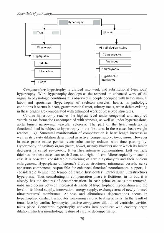
Essentials of pathology
70
Compensatory hypertrophy is divided into work and substitutional (vicarious) hypertrophy. Work hypertrophy develops as the respond on enhanced work of the organ. In physiologic conditions it is observed in people occupied with heavy manual labor and sportsmen (hypertrophy of skeleton muscles, heart). In pathologic conditions it occurs in heart, gastrointestinal tract, urinary tracts, when defect existing in these organs are compensated with enhanced work of preserved structures.
Cardiac hypertrophy reaches the highest level under congenital and acquired ventricles malformations accompanied with stenosis, as well as under hypertensions, aorta lumen narrowing, vascular sclerosis. The part of the heart undertaking functional load is subject to hypertrophy in the first turn. In these cases heart weight reaches 1 kg. Structural manifestation of compensation is heart length increase as well as its cavity dilation determined as active, compensatory, tonogenous. However in case prime cause persists ventricular cavity reduces with time passing by. Hypertrophy of cavitary organ (heart, bowel, urinary bladder) under which its lumen decreases is called concentric. It testifies intensive compensation. Left ventricle thickness in these cases can reach 2 cm, and right – 1 cm. Microscopically in such a case it is observed considerable thickening of cardic hystiocytes and their nucleus enlargement. Hyperplasia of stroma’s fibrous structures, intramural vessels, nerve apparatus components responsible for enhanced function’ neurohumoral support, is considerable behind the tempo of cardic hystiocytes’ intracellular ultrastructures hyperplasia. Thus contributing in compensation phase is fictitious, in its bud it is already has the features of decompensation. In case prime cause is not removed, unbalance occurs between increased demands of hypertrophied myocardium and the level of its blood supply, innervation, energy supply, exchange area of newly formed ultrastructures’ membranes. Adipose and albuminous degenerations occurs in hypertrophied cardiac hystiocytes weakening cardiac beating activity. In the result of tonus lose by cardiac hystiocytes passive myogenous dilation of ventricles cavities takes place. Concentric hypertrophy converts into eccentric with cavitary organ dilation, which is morphologic feature of cardiac decompensation.

Ya. Bodnar, A. Romanyuk, V. Voloshyn, V. Gargin
71
Gastric or bowel muscle layer hypertrophy occurs, naturally, upward stenosis which impedes evacuation. This can take place under ulcers healing, tumors presence. Urinal bladder hypertrophy is observed under prostate gland adenoma, narrowing urethra, as well as in connection with the other impediments of bladder emptying. Functional insufficiency of above named organs occurs under leiomyocytes degeneration and manifests in their cavities dilation.
Vicarious (substitutional) hypertrophy compensates the function of one of the dead or surgically removed paired organs (lungs, kidneys, adrenal glands). By its pathological essence it is close to regenerative hypertrophy. Significant role in its occurrence plays the complex of reflex and hymoral influences, the same with compensatory hypertrophy.
Atrophy
Atrophy is lifetime change of organs’, tissues’ and cells’ volume, accompanied with their functions weakening or their functions termination. Physiologic and pathologic atrophies are differentiated.
Physiologic atrophy is observed during the whole lifetime of human being. Upon the birth umbilicial arteries, arterial (Botallo's) duct atrophy and obliterate, aged people face with genital glands atrophy, old people – with bones and intervertebral cartilages atrophy.
Pathologic atrophy is observed in any age and can be caused by various reasons - insufficient feeding, endocrine glands dysfunction, central and peripheral nervous system lesions, intoxications. Pathologic atrophy is reversible process. In case the cause is removed under condition that atrophy didn’t reach high level, organ structure and function can be completely rehabilitated. Pathologic atrophy can be general and local.
Cachexia or emaciation is divided into the following types: alimentary cachexia emaciation under cancerous cachexia, emaciation under cerebral cachexia, emaciation under other diseases. Concept of “emaciation” and “cachexia” are not identical. Cachexia in primary stages can be free from emaciation and be manifested with progressive degenerative changes of the organs, for example, with osteoporosis.
Alimentary emaciation occurs during starvation. Gradually fat stock decreases, skeleton muscles atrophy, Atrophied adipose (fatty) tissue becomes ochre-yellow color due to lipochrome pigment accumulation. Fatty tissue of atrium and fatty marrow impregnate with serous fluid and become dropsical (serouse atrophy of fatty tissue). Pigment melanin accumulates in the skin of starving, so it colors in grey-brown color. Heart, liver and other organs decrease in size. Pigment lipofuscin, (wear-and-tear pigment) accumulates in cardic hystiocytes, hepatocytes and myocytes of skeleton muscles, as the resuly of which organs become of brown color (brown atrophy of organs).
Emaciation under cancerous cachexia is characteristic for cancerous growth of any localization. The most fast it develops in patients ill with cancer of esophagus, gastric carcinoma or intestine cancer caused by digestion disorders.

Essentials of pathology
72
Emaciation under cerebral and hypophysial (Simmonds' and Schigens’ diseases) cachexia occurs due to hypothalamus’ or hypophysis’ injury with inflammatory process or tumor.
Emaciation under the other diseases takes place in case long term chronic infections (tuberculosis, dysentery, chronic sepsis). It is caused by severe disorder of metabolism.
Under general emaciation subcutaneous fatty tissue is absent, eyes are hollow, skin is dry, abdomen is scaphoid. Starvation edemas sometimes take place.
Local atrophy occurs by various reasons. The following types of it are differentiated: dysfunctional, caused by inadequate blood supply, compression,
trophoneurotic, caused by physical and chemical agents influence. Dysfunctional atrophy or atrophy caused by inactivity occurs because of organ
function decrease: muscles atrophy under bones fracture, optic nerve atrophy after eye ectomy. Atrophy development in patients with ruptures could be slow down in case massage and physical exercises are applied.
Atrophy caused by inadequate blood supply occurs caused by narrowing of arteries feeding organ. Exsanguination leads to hypoxia in the result of which parenchimatous elements’ functions fall and cells size reduces Hypoxia stimulates fibroblasts proliferation (reproduction), so sclerosis develops under inadequate blood supply. Patients with atherosclerosis suffer from this process in myocardium, kidneys, cerebrum, legs.
Atrophy from compression occurs in organs subject to compression by tumor or aneurysm (local evagination of aorta). Even the bones of spinal column and breast bone atrophy because of their compression by aneurysm. Under urinary tracts obstruction with calculus urine stretches renal pelvis and cups (hydronephrosis) causing kidney parenchyma atrophy. In case liquor outflow hindrance ventricles of brain dilate (hydrocephalus) and cerebrum atrophy.
Trophoneurotic atrophy is caused by failure of organ connection with central nervous system under peripheral nerves traumatic, tumor or inflammatory injury. Skeleton muscles’ atrophy often develops by this scenario.
Atrophy caused by physical and chemical agents influence occurs, for example, in marrow and genital glands under radiation influence. Radioiodine causes thyroid gland atrophy. After long term treatment with adrenocorticotropic hormone or glucocorticoids adrenal glands cortex’ atrophy develops.
Organs reduce in size under atrophy. Their surface in most cases is smooth (smooth atrophy), in kidneys – granular (granular atrophy). Under hydronephrosis and hydrocephalus organs are enlarged due to liquid accumulating in them and their parenchyma is atrophied.

Ya. Bodnar, A. Romanyuk, V. Voloshyn, V. Gargin
73
Metaplasia Metaplasia is adaptive pathologic process characterized with substitution of one
differentiated tissue with the other in the limits of one histiotype: mesenchymal or epithelial.
This phenomenon does not take place in muscular and nervous tissue. The most wide spread example of metaplasia is one layer prismatic epithelium substitution with multilayer flat epithelium, observed under bronchi mucous tunic inflammation, gastric epithelium substitution with intestinal epithelium – intestinal metaplasia, or gastric mucus tunic enterolization. A-hypovitaminosis and others could be the causes of metaplasia. Conjunctive tissue metaplasia is observed with cartilage or bone formation in cicatrix, aorta wall under atherosclerosis. Metaplasia occurs in connection with previous non-differentiated tissues’ proliferation – indirect metaplasia. Metaplsia is grounded on the change of genetic program of differentiation on column cells level. Metaplasia could be the background for malignant growth development.
Dysplasia
Dysplasia is major failures of proliferation and epithelium differentiation with cellular atypia development and histoarchitectonics change: loss of polarity, loss of epithelium histo- and organo- specificity.
Basic membrane is not injured under dysplasia. The most often dysplasia develops under inflammatory and regenerative processes. Depending on proliferation stage and condition of cellular and tissue atypia three stage of displasia are differentiated: – minor (small), – moderate (middle), – severe (major). Minor and moderate dysplasia are of reversible character. Cellular and tissue changes under severe dysplasia are rare subject to reversible process and are treated as precancerous process. Sometimes locally they are hard to be differentiated from carcinoma.
Regeneration
Regeneration (from Latin regeneratio – restoration) is the process of living matter self-recovery in injured area.
Regeneration takes place on molecular, subcellular, cellular, tissue and organ levels and reflects the principle of living functions autoregulation. It is grounded on cellular and intracellular hyperplastic processes. Cellular reproduction is characteristic for cellular form of regeneration, ultrastructures and their components quantity increase (hyperplasia) and their enlargement (hyperplasia) are characteristic for intracellular form. The last form is peculiar for all organs’ cells and is universal.
Two phases are differentiated in regeneration morphogenesis – proliferation and differentiation. In the term of the first phase reproduction of non-differentiated (cambial, column) cells or pre-cells are observed. During the second phase young cells mature and specialize.
Regenerative process is regulated with humoral, immune, nervous and functional mechanisms. Humoral mechanisms are realized in cells and tissues at intracellular and tissue regulators participation, and out of them – at participation of hormones, poetines, mediators, growth factors as well as keylones (substances

Essentials of pathology
74
depressing cells division). Immune mechanisms are connected with “regenerative information” transfer by leukocytes, nervous – with trophic function of nervous system, and functional – with adequate demands of organs and tissues.
Three types of regeneration are differentiated: physiologic, reparative and pathologic.
Physiologic regeneration Physiologic regeneration is done in the course of the whole life and reflects
endless process of substances’ disintegration and synthesis. It is characterized with intracellular renewal of molecules and ultrastructures as well as entire cells, fiber structures and major substance of conjunctive tissue. Intracellular regeneration is the only form of content and function renewal of central nervous system’s cardiac hystiocytes and neurocytes. Combination of intracellular renewal with cells mitosis is observed in liver, kidneys, pancreas. Continuous change of epidermis, digestive tract mucus tunic epithelium, synovial membranes, marrow, blood elements are done on the account of cells division.

Ya. Bodnar, A. Romanyuk, V. Voloshyn, V. Gargin
75
Reparative regeneration Reparative regeneration is organ defect substitution under various pathologic
processes. It is grounded on the same mechanisms which refer to physiologic regeneration, moreover injury reparation in each organ is going on the same way as in conditions of physiologic recovery, but more intensive. Intracellular regeneration becomes major form of degenerative changed tissues cells’ structure rehabilitation, as well as cellular and intracellular – under their necrosis.
Final result of reparative regeneration is expressed in restitution or substitution. Restitution (complete regeneration) is characterized with tissue defect substitution with tissue identical to dead one. It is attributable to those organs and tissues where regeneration is going on exceptionally in cellular form (marrow, epidermis, mucus tunics epithelium).
Substitution (incomplete regeneration) is characteristic for the organs healing of which goes on mostly or exceptionally by intracellular reparation (heart, central nervous system). For example, in myocardium necrosis focuses are substituted with conjunctive tissue, in cerebrum dead neurocytes – with glial cicatrix. Function renewal is provided with nucleus and cytoplasm ultrastructures enlargement in preserved cells which hypertrophy. Incomplete regeneration variation is “distance regeneration”. As an example of it could be named qualitative reconstruction various portions of gastrointestinal tract, compensating exocrinous, function of pancreas head or uninjured cerebral hemisphere reconstruction in case the other hemisphere injury.
Pathologic regeneration
Pathologic regeneration is the type of reparative regeneration going on in conditions of local and general regulatory mechanisms failure, and is characterized with regenerative process distortion, violation of proliferation phase change into differentiation phase. Deficiency of proteins or vitamins, nervous regulation failure, hormonal disorders, immune system depression could seriously influence healing speed and quality. In that way long term nonhealing crus ulcers in patients with chronic cardiac insufficiency could be explained as well as persistent wounds under diabetes mellitus. An example of pathologic regeneration can be conjunctive tissue hyperproduction with keloid formation under radiation or thermal trauma.
Regeneration of separate organs and tissues
Blood can regenerate by physiologic, reparative and pathologic regeneration type. An example of blood reparative regeneration under anemia can serve extramedullary hematosis. Pathologic blood regeneration is observed under radiation, leucosis. Small size vessels regenerates satisfactory and big vessels regenerate by substitution type – cicatrix formation on the place of mid and external layer portions injury. Conjunctive tissue regeneration starts from young mesenchymal cells proliferation and vascularization with granulation tissue formation, that is young conjunctive tissue reach with cells: non-differentiated lymphocytine cells of conjunctive tissue, leukocytes, plasmocytes, labrocytes, fibroblasts; loop-like thin wall vessels. Granulation tissue maturing is ended with rough fibered cicatrix tissue formation, sometimes even keloid. Osteous tissue regeneration after uncomplicated rupture of

Essentials of pathology
76
bone goes by the way of primary bony union, which have the following stages: primary conjunctive tissue callus, primary bony callus, final bony callus. Under regenerative process failure secondary bony union occurs in bone through prior osteocartilaginous callus. Cartilage tissue regeneration goes as incomplete regeneration with scar tissue growth. Muscle tissue regeneration depends on its type. Unstriped muscles regenerate completely under minor defects. Transversely striated muscles regenerate only in case sarcolemma is preserved. Cardiac muscle regeneration goes by the way of cicatrix formation. Epithelium regenerates by the way of new cells reproduction, in other words by restitution type. Nervous tissue regeneration goes ambiguous. Cerebrum and spinal marrow cells regenerate by substitution, that is glia growth and cicatrix formation.
Organization
Organization is protective-adaptive process directed to separate and substitute with granulation tissue focus of necrosis, hemorrhage or exudates as well as thrombi, foreign objects and parasites.
Its essence comes to conjunctive tissue formation under defects healing in wounds and ulcers, substitution with conjunctive tissue areas of necrosis or thrombotic masses (properly organization) and their encapsulation.
By I.V.Davydovsky the following forms of wounds healing are differentiated: epithelium defect immediate closing, healing under eschar, primary intention of wound, secondary intention or healing by granulation.
Epithelium defect immediate closing provides cells growth on wound sides and
its skinning over with cells layer without mitotic cells division. Such simple form of healing is peculiar to surface injuries of cornea, mucus tunics, vessels intima.
Healing under eschar is also characteristic for minor injuries of epidermis. For example, under surface excoriation lymph and blood excude fast drying and converting into crust (eschar). Epidermis regenerate under crust which in the result of rejection process drops away on the 3rd -7th day.
Healing of wounds involved not only skin but lower situated tissues goes by the way of primary or secondary intention. Principal difference in them is in the manner of wound cleansing. Primary cleansing is characteristic for healing with primary intention. Under phagocytes’ proteolytic ferments influence partial lysis of grumes and tissue detritus takes place and wound content is removed in the very first date after injury together with exudate, On the 2nd-3rd day granulation tissue appears which

Ya. Bodnar, A. Romanyuk, V. Voloshyn, V. Gargin
77
ripens on the 10th-15th day. In the clinic the sides of big wounds connect with sutures and support with dressings. In case distance between the sides equals even 10 mm, in few days this distance will diminish zero due to tissue edema and fibrin clot reduction which sticks wound edges. In those cases when wound sides separated due to suppurative inflammation, primary intention is impossible and healing is done through secondary intention. It is characterized with wound release of detritus and foreign objects by “outsuppuration”. On the boarder with necrotic tissue fast appears the features of demarcation suppurulent inflammation, and its melting comes. Necrotic masses rejection takes place during the first 5-6 days (secondary cleansing of the wound) and granulation tissue starts to develop on wounds edges. Under wounds healing by primary or secondary intention granulation tissue maturing is accompanied with epithelium regeneration. However under secondary intention healing on the place of wound cicatrix forms anyway.
Inflammatory process always precedes ulcers healing. Granulation tissue grows into necrosis area which matures into rough fiber and often subjects to hyalinosis. The latter causes cavitary organ deformation and stenosis. Epithelial layer stratifies conjunctive tissue.
Really organization of necrotic masses starts from reactive exudates inflammation in surrounding tissues and necrosis areas lysis. Exudative reaction transfers into productive with mesenchymal cells proliferation. Granularion tissue ingrows from periphery and gradually transforms into cicatrix. This type of organization is peculiar to myocardial infarction healing, as well as kidneys and spleen. Thrombus organization starts from 2nd-3rd day of its origination, goes parallel with aseptic autolysis and is finalized with thrombotic masses substitution with conjunctive tissue, canals formation and vascularization. Organization of hemorrhage or exudates in intermediate tissue also ends with cicatrization and in serous cavities – with their obliteration or joints formation. Fibrinogenous exudates organization in alveoli under croupous pneumonia results in carnification.
Topic. Tumors, general data. Malignant cell features. Molecular fundamentals of
carcinogenesis. Antitumor immunity. Non-malignant (benign) and malignant growth. Tumors morphogenesis and hystogenesis.
Tumors etiology. Carcinogen agents and their interaction with cells. It is ascertained fact that tumors can be caused by physical, chemical and biological agents which are called cancirogens
• Neoplasms: persistent, abnormal and relatively autonomous proliferation of cells occurring as a result of permanent cellular defect that is passed to the progeny. Usually develops due to a factor(s), but once developed, becomes independent of them.
Cancerous diseases accounted for 23 % of all deaths; environmental carcinogens and heredity factors are important in appearance of such pathology. Environmental carcinogens could be formed drugs (antineoplastic, immune suppressing etc..), organic chemicals (insecticides, herbicides, aromatic hydrocarbons, etc..), cigarette smoke, ethanol, heavy metals, sexually transmitted viruses (HTLV-I, Herpes simplex,

Essentials of pathology
78
Human papilloma virus), radiation, ultraviolet light. Influence of heredity factors could be presented by hereditary predisposition, clustering of environmentally induced cancers in families; close relatives of cancer patients have three times greater risk of developing the same neoplasm; close relatives of patients with breast, colon, or endocrine cancers have greater than three times risk for developing the same neoplasm; increased cancer risk with inherited mutations of cancer suppressor genes such as Rb and p53.
But over 75% of human beings’ cancerous diseases are caused by environmental factors, and in first turn – by chemical compounds. First experimental proofs of chemical compounds’ carcinogenicity, were Yamagiva’s and Ishikava’s researches (1915). They induced rabbit’s ear skin cancer by applying there coal-tar pitch for the period of 15 months.
Chemical cancirogens are wide-spreaded in environment and the majority of them are of antropogenous origin. Same time we shouldn’t exaggerate their role in human being pathology as only about 100 compounds and manufacturing processes are acknowledged as carcinogenic for human beings.
By their chemical structure carcinogens are divided into several groups. The most important of them are as follows: a) polycyclic aromatic hydrocarbons; b) aromatic amine and amides; c) nitrosoamines and nitrosoamides.
First group consists of over than 200 substances with three and more benzene rings. Only one of them, namely 3,4-benzpyrene considered to be the one able to cause cancerous diseases of human being. The others cause tumors only in experimental animals. The biggest amount of this group of carcinogens is in tobacco fume, exhaust gases of automobiles, blast furnaces smoke, asphalt, waste of chemical plants, dried and overdone food.
Substance of polycyclic structure shows mostly local carcinogenic influence. In case during experiment they are applied on skin cancer occurs, in case they are applied under skin – sarcoma occurs. Polycyclic aromatic hydrocarbons are excreted by various organs of organism, so tumors of these organs occur – kidneys, skin, mammary glands.
The second group of carcinogens are mostly azo dyes, for which two or more azo groups presence is characteristic (mono-azobenzene, 2- naphthylamine, benzidine). These substances are used to color natural and synthetic fibers, in printing industry, cosmetics, color photography, to synthesize medicines, insecticides. Carcinogenic arcinogenic action of amines and amides becomes apparent when they are introduced in digestive tract, subcutaneous or in case they are applied on skin. Tumors appears in organs far from the place of application, the most often in liver, urinal bladder, bowels, kidneys.
Nitrocompounds (nitrosoamines and nitrosoamides) are characterized with alkyl radical presence. They are utilized as antioxidants, pesticides, paints solvents, semi-products under paints, medicines and polymers synthesis. Their cancirogenity for human being is nor proved but experimental data causes oncologic alertness. Possibility of nitrocompounds synthesis of nitrites, nitrates, nitric oxide in human being’s intestinal tract is proved. Nitrites are widely used as conserved agents for foodstuff.

Ya. Bodnar, A. Romanyuk, V. Voloshyn, V. Gargin
79
Practically all chemical substances are not carcinogenic as they are. They acquire these features after coming into organism and are subject to metabolic transformations. Here is the origin of idea of final carcinogens which are able to interact with cells macromolecules – DNA, RNA, proteins. Taking into consideration role of DNA in heredity information transfer the most attention is focused to carcinogens’ binding exactly with this acid. A number of products were found which made possible to decode fine mechanisms of final carcinogens interaction with DNA. They mostly methylate guanine and affect purine bases complementary character – instead of normal combination guanine - cytosine paramethylated guanine – hymine is created. So, carcinogens cause point mutations in certain DNA positions. In case these mutations refer to transforming genes, i.e. oncogenes a chain of events starts causing malignization.
Radiation carcinogenesis. Physical carcinogens includes ionizing radiation and to a lesser extend – ultraviolet rays. Ionizing radiation acts indirectly, through highly active free radicals distorting DNA structure. Ultraviolet rays prevent its reparation.
Viral carcinogenesis. There are various biologic agents able to cause malignant growth. The biggest group consists of viruses. Indisputable proofs were acquired regarding viral origin of many animal tumors – hens’ Rous sarcoma, rabbits’ Shope fibroma and papilloma, mice mammary glands cancer (virus is transferred through milk). The quantity of human beings’ tumors which are indoubtfully caused by viruses is not big – Burkitt's lymphoma, rhinopharyngitis cancer, carcinoma of uterine cervix.
Viruses causing tumors are called oncogeneous. They are divided into two groups depending of genome’s molecular structure - RNA-containing and DNA-containing. Major group consists of RNA oncogenous viruses, forming the group of retroviruses. Their mutual characteristics is the fact that their genom is of one chain RNA, and that they have ferment RNA-dependent DNA-polymerase (invertible transcriptase, revertase). The essence of virus inducted carcinogenesis adds up to the fact that oncogenous viruses introduce their own genome in infected cell, this genome contains transforming gene – viral oncogene. Its activity product (oncoprotein) starts cell transformation and keeps it in transformed condition.
Retroviruses are the major cause of human’s malignant growths, however they point the way to understand basic mechanism underlies this diseases. They became model system by means of which the most modern data was received of fine molecular distortions occurring under cellular transformations.
All above said allows to make major conclusion: tumor starts from DNA damage. This mechanism is obligatory for all tumors irrespectively what carcinogens caused them – chemical, physical or biological. All of them are carcinogens exactly because of the fact that they are able to cause genetic apparatus failures. Chemical agents cause mostly point mutations, ionizing radiation – mostly chromosome mutations and retroviruses introduce to DNA molecule additional genes and oncogenes are among them. In such a way DNA damages could be treated as molecular grounds of all further processes transforming normal cell into transformed cell. In the other words DNA damage is common denominator to which the action of all known carcinogens is reduced.
Essentials of pathology
80
Pathogenesis of tumors. Molecular grounds of cancerogenesis. The question arises: what kind of DNA damage is realized into tumor? The answer to that is not at all simple. Based on modern knowledge scientific theory was formulated which is known as oncogene consept. It combines all forms of carcinogenesis (chemical, physical and viral) into one universal mechanism. There are really many causes of cancer, but all of them similar to water through watering-can should pass through one critical channel – DNA and leave trace in it, meaning damage. This damage is specific. It will lead to normal cell transformation into malignant cell (tranformation phenomenon) only in case it localizes not at any random DNA portion, but exactly in the portion where genes controlling cells growth and differentiation are situated. These genes are called cellular oncogenes or proto-oncogenes. They are usual components of cellular genome and are absolutely necessary for cell’s vital activity. Cellular proliferation would be impossible without proto-oncogenes. Oncogenes are normal cellular genes that are involved in growth control. Cancer results when these genes become dysregulated such that they are inappropriately activated.
It is considered that under minor damages normal function of cellular oncogenes as auxesis could be kept in principle, but it stops to subordinate controlling influences of the cell itself. Normal controlled process of growth and maturing is lost and is interchanged with an endless process of cellular divisions under which cells do not have time to differentiate meaning to mature to condition when they are able to fulfill appropriate specialized physiologic functions. It comes out that cell from its creation beginning hides the sprouts of its own death in the form of cellular oncogenes.
Different insults continuously act on cells leading to transformative alterations in (epi) genetics, chromosomal numbers and arrangements, and heterotypic interactions which, along the path towards malignancy, undergo cycles of evolutionary clonal selection leading to the acquisition of cancer-competent traits, the hallmarks of cancer.
Right now nobody denies that normal cellular oncogenes under specific conditions could activate and cause malignant growth. Several ways of their activation are differentiated. One of them is viral transduction, in other words cellular oncogenes passage through viral genome. It is proved that retroviruses damage DNA by the way of introduction to it so called viral oncogenes. It was found that they are of cellular origin. They are proto-oncogenes which on the certain stage of evolution were deported from infected cell nucleus by viruses and included into self-genome. Starting from that moment they became viral oncogenes. Right now over 20 of them are known. All of them have cellular counteracts in various chromosomes.
Viral oncogenes coming into cell for the second time behaves uncontrolled. The point is that they structurally differs of their cellular ancestry. Retroviruses, as a rule, capture cellular gene incomplete, without repressor genes, so similar viral oncogene keeps ability to stimulate cells growth and differentiation but loses regulator (operator) genes and becomes uncontrolled. It causes unlimited non-corresponding organism’s needs cells division. Cellular oncogene itself also is subject to structural changes at its capture by retrovirus. This makes difficult regulative influences on it by repressor genes as well as by epigenome cellular regulators. Thus, viral transduction deprives cellular oncogenes their primary positive function of growth

Ya. Bodnar, A. Romanyuk, V. Voloshyn, V. Gargin
81
stimulators and simultaneously releases their hidden transformation abilities. Growth and proliferation genes starts to function as cancer genes.
Cellular oncogenes activation can occur in the result of chromosomal translocations. It was noted that under certain forms of tumors chromosome discontinuities take place exectly in those portions where cellular oncogenes are situated.
It was clarified that certain tumors, for example, Burkitt's lymphoma occurs when any foreign (viral) genetic material inserts into DNA molecule close to proto-oncogene, even if this material doesn’t include oncogene. Viral DNA built-in close to cellular oncogene activates it up to cancer level of expression. This mechanism is called insertion.
As a rule, cellular oncogenes are represented in DNA in one copy but it was proved that copies quantity can increase in the result of DNA replication abnormality. This phenomenon is called amplification (augmenting). Cellular oncogenes copies amount increase causes enhanced division of cells. This mechanism acts in human neuroblastoma and carcinoma of large intestine creation.
Anyway, point mutations independently of their cause are considered to be major mechanism of proto-oncogene transformation into active cancer oncogene. It is proved that that‘s enough to change in human urine bladder cancer only one base – guanine for the other one - thymine as inactive proto-oncogene becomes transfomating. Totality of scientific ideas of mutations’ decisive force in tumor etiology forms the grounds of mutation concept of cancerogenesis.
Epigenome concept adds up to the fact that the grounds of normal cell transformation into malignant one are not genetic apparatus’ structures changes, but persistent failures in genous activity regulation. The genes which should be repressed are dysinhibited and those which should be active are clocked. Cell loses its specificity, becomes insensitive to regulative influences of the whole organism.
Stages of cancerogenesis. Tumors occurrence and progress is multistage process. There are three main stages – trasnformation (initiation), promotion and progression. Proto-oncogene activation finishes first stage – stage of initiation. Main feature acquired by the cell in the result of proto-oncogene transformation into oncogene is immortalization, meaning its potential ability to endless division, to immortality. However active oncogene presence is only potential possibilitity for expression. Cell with active oncogene could stay for years in latent (delitescence) state, doesn’t expressing itself in any way. Immortalized cell needs additional influences taking it out of latent state and give a stimulus to endless division.
Tumor growth risk factors. These provocative factors could be additional doses of chemical or physical cancerogenes, retroviral superinfection as well as various agents which do not cause tumors as they are, but are able to take immortalized cells out of latent state. Here starts old idea of super multicauses of tumor growth however in reality absolute majority of the factors attributed etiologic role should be considered among promotional conditions causing expression of latent, potentially cancerous, cells. Factors activating pre-cancerous cells are called promoters. Under their influence trasnformed cells go into new stage of development – promotion stage for which cellular oncogenes expresssion is charactristic.

Essentials of pathology
82
Provided that the fact of oncogenes participation in oncogenesis is not under the doubt at the moment, mechanism of their action is still a mystery. It was ascertained that oncogenes code specific proteins (oncoproteins), most of them having tyrosinase activity. Further on it was found that oncoproteins which cause uncontrolled growth of malignant cells are similar to usual growth factors – thrombocyte growth factor, epidermal growth factor, insulin-like growth factors. Under the normal conditions growth factors comes into cell from outside providing cell dependability from organism. Malignant cells differs with the fact that they produce growth factors by themselves. A part of them is aimed to support their own proliferation (autocrine secretion), and the other one – for other type cells (paracrine secreation).
Progression is the final phase of tumor progress. Under this term persistent, irreversible qualitative changes of tumor to malignization are understood. For example hormone-dependent neoplasms became hormone-dependent, tumor reacted medicines stopped to react them. Progression is the last and the most long lasting stage of tumor progress lasting up to organism death.
The most important clinicopathologic implications of tumor growth. Interrelations between tumor and organism. Tumor negative influence on organism depends on its type (non-malignant or malignant), localization, speed of growth and directions of metastasis. Tumor directly injures organ in which it progresses disturbing its structure and functions. Surrounding organs are subject to atrophy and deformation, lumens of cavity organs narrows. Due to chronic intoxication with decay products and insufficient feeding cachesia develops. Hematosis depression, excessive hemolysis and chronic hemorrhage cause anemia.
In case tumor consists of hormone-active cells diseases occur connected with corresponding hormone hyperproduction or paraneoplastic syndromes of endocrinopathy, neurological aspects (dementia, neuropathy), skin implications, hematologic implications (hyper coagulability of blood, anemia, thrombocytopenia, polycythemia). Pheocromacytoma (cancer of adrenal glands cerebral layer, producing adrenalin) causes arterial hypertention progress, insulinoma (tumor of islet of Langerhans -cells) causes hypoglycemia, gastrinoma (pancreatic tumor producing gastrin - gastric secretion stimulator) causes stomach ulcer.
Tumors structure. There are various tumors by their macro- and microscopic structure. Their appearance can remind mushroom, cauliflower, node or intumescence. In section tumors are mostly of white, grey and red color. The following is often found in them: hemorrhages, necrosis and cysts cavity of which is filled with mucus or bloody mass. Some tumors are of brown color, for example, melanoma.
Tumor size depends mostly of its origin, location and growth period. In some cases they can reach giant sizes (fibroid tumors) in the other cases they can be seen only through magnifying glass or microscope (microcarcinomas). Tumors localized close to vitally important centers as a rule are of rather small size.
Tumor consistency is defined first of all by the type of outgoing tissue and ratio between stroma and parenchyma. Tumors of bone (osseous) tissue, cartilage tissue and fiber conjunctive tissue are of dense consistence. Malignant growth of epithelium

Ya. Bodnar, A. Romanyuk, V. Voloshyn, V. Gargin
83
in which stroma is underdeveloped are flaccid and by their consistence they are similar to new-born child’s brain (cancer-brainer).
Stroma and parenchyma are seen microscopically in each tumor. Parenchyma is its specific part which is represented by malignant cells and determines tumor place in hystologic classification. Even in tumors originating from mesenchyma cells producing intercellular substances (collagen fibers, basic substance of cartilage or bone tissue) are also should be treated as parenchyma. Stroma is mechanical-trophic framework including conjunctive tissue, blood and lymph vessels and nerves.
Most of tumors look like organ by their structure, i.e. have parenchyma and completely represented stroma. Such tumors are called organoid. In undifferentiated tumors parenchyma prevails and stroma is underdeveloped. They are called histioid. Blood circulation insufficiency causing necrosis easily occurs in them. At the same time there are tumors poor with parenchymatous elements and rich with stromal, for example gastric fibrocarcinoma or sccirrhous. These tumors cause complications due to stroma’s corrugation. They deform organ or narrow its lumen.
Tumor corresponding structure of the organ it is localized in is called homologous, and the one which structure differs from this organ structure is defined as heterologous. In case tumor is developed from the cells of organ in which it occurred – this is homotopy tumor. In cases it occurs from the cells of embryonal displacement (heterotopia), it is called heterotopic, for example tumor of bone marrow in uterus.
Tumor (new growth, neoplasm, blastoma) is typical pathologic process in the form of excrescence of tissue subject to genetic apparatus change, characterized with potential infinity of its uncontrolled growth as well as structural elements’ atypicity.
Biology of tumor growth. Universal and mandatory feature of all the tumors – both non-malignant and malignant – is their ability to endless growth. This is fundamental feature of any tumor. Uncontrolled excessive proliferation of malignant cells doesn’t mean at all that they divide faster than homologous cells of healthy tissue. Vice versa, certain healthy tissues grow much more faster than the most malignant growth, for example, embryonal cells, regenerating liver. In such a way, malignant cells proliferation differs from normal cells proliferation not with cells division and growth speed, but in the character of division and growth.
Infinity of malignant cells growth is based on the fact that they are unable to exhaust division resource. It is found that genetic program limiting its divisions quantity is integrated into each cell. As a result of genetic somatic mutation malignant cell losses this restrictive program and starts to divide “endless”, escaping aging up to the death of host organism. In case these cells are carried from living organism to the other one of the same species, they will settle down and again will divide up to the death of recipient organism. In case these cells are carried to nutrient medium, there they will also divide endless times, in the other words they become independent of Heiflic’s rule. This ability of malignant cells to endless division is dominantly propagated to further cells generation.
Malignant cells life could be kept artificially. There are two methods to provide that: transplantation – tumor inoculation from one animal to the other one of the same species and explantation – malignant cells cultivation on nutrient medium. Tumor

Essentials of pathology
84
kept for a long time with transplantation or explantation method is called tumor strain. First transplantation strain was made in 1905 (Ehrlich's carcinoma in mice), first explantation - in 1950 (Hela’s cells – carcinoma of uterine cervix).
Malignant cell has one more feature – uncontrolled growth. On the level of the whole organism tumor growth is controlled with nervous and endocrine systems, and on the local level – with mitogens and keylones. Malignant cell gets out of this hand, that is shows autonomy, independence of growth. It’s clear that this autonomy is not absolute but in this or that way is characteristic for all tumors. In case tumor partially keeps ability to come under control influence of hormones, it is called hormone-dependent tumor and in case it completely loses this ability - hormone-independent tumor. Autonomy doesn’t mean that tumor lost any connection with organism. This connection changed. They can be characterized as relations between host organism and parasite tissue.
Third peculiar feature of malignant cells is anaplasia, which means their persistent dedifferentiation, loss of ability to form specific tissue structures or produce specific substances characteristic for normal cells. In the other words its return to embrional state, structural-chemical organisation simplification.
Tumor occurs from single parent cell subject to genous mutation. Malignant cells differs in several parameters from their common normal ancestor. This difference relates to cell’s and its organoids’ structure, metabolism, specific features and functions. Therefore morphologic, biochemical, physical-chemical, immunologic and functional anaplasia is differentiated.
The essence of morphological anaplasia comes to tissue, cellular and subcellular atypicity occurrence. Polymorphism is inherent to malignant cells – they acquire smaller as well as bigger size and shape which is not peculiar for normal cells. Interrelation between nucleus and cytoplasm is shifted in favor of nucleus due to its enlargement. Multinuclear cells, nucleus hyperchromatosis are observed caused by nucleic acids accumulation in them, nucleolus amount increase and their migration into cytoplasm, of subcellular structures mitochondrions are subject to most prominent changes. Their quantity and size are decreased, membranes became thinner, cristas also become thinner and disappear. At tissue level structures’ created by malignant cells size and shape changes are observed. This referes for example to glandular follicles in adenocarcinomas and focuses of ossification in osteosarcomas. Sometimes tumor completely losses morphologic features indicating its origin from the certain differentiated tissue.
Biochemical anaplasia is peculiar of malignant cells’ metabolism caused by theirs genetic apparatus change. Carcinogens are able not only to distort mitosis process and start endless division mechanisms, but also to supress or unbrake the other genes. As the result of that malignant cells enzymatic range changes. Intracellular enzyme insufficiency occurs - some enzymes are inhibited but the other ones activate or start to synthesize absolutely new substances which didn’t exist in normal cells.
It is found that all tumors, subject to progression start to look like each other by their enzymes set independently of what cells they come from. Unification of tumors

Ya. Bodnar, A. Romanyuk, V. Voloshyn, V. Gargin
85
izoenzymal range independently of their histogenesis is very characteristic manifestation of malignization.
It is known that every tissue synthesize enzymes specific for it, where every enzyme is represented with strictly specific set of isoenzymes. This specific feature is lost in tumors. So called monotonization or isoenzymic simplification is developed – amount of isoenzymes reduces and their set becomes approximately the same for tumor of any origin. Isoenzyme reconstruction goes in the direction of those enzymes increase which are peculiar for embrional tissues.
The most peculiar biochemical features of malignant cells relate to proteins and carbohydrates metabolism. Proteins synthesis prevails their decomposition. To build own proteins tumor captures aminoacides of the other organs (“tumor - trap for nitrogen”).
Carbohydrates metabolism and power of malignant cells significantly differ from the norm. In aerobic conditions normal cell provides itself with energy mostly at the expense of more advantageous glucose aplittance in Crabbs’ cycle, and in anaerobic conditions – it is forced to change to glycolysis. In case amount of oxygen is sufficient, glycolysis is oppressed with breathing (Paster’s effect).
Malignant cell also provides its demands in energy on account of glycolysis and breathing, but correlative meaning of these processes is different. Peculiarities of tumors power supply are as follows: a) activation of anaerobic glycolysis and enzymes providing it - pyruvatekinase, hexokinase, fructokinase; b) presence of aerobic glycolysis for which normal cells are not able (exceptions – leukocytes, spermatozoon, eye retina cells); c) breath oppression with glycolysis (Crabtree effect), to say exact – with powerful system of glycolytic enzymes, which intercept substrates – inorganic phosphorus, coenzymes.
Among physical-chemical features of malignant cells the following should be emphasized: acidosis in the result of lactic acid accumulation, intracellular aquation, potassium ions accumulation, electroconductivity increase, colloids density reduction, membrane negative change increase, their surface tension decrease.
Antitumor immunity. Under immune anaplasia changes of malignant cell’s antigene features is understood. These changes is the result of protein metabolism rebuilding. It is known that each tissue synthesize a set of antigenes specific for it. This set is changed in tumor. Tumor antigenes. Antigene simplification and antigene complication are differentiated. Antigene simplification is characterized with antigenes synthesized by malignant cell numerous times decrease.
Antigene complication is manifested with antigene divergence and antigene reversion. Antigene divergence means that malignant cells start to synthesize antigenes which are not characteristic for healthy cells, but these antigenes are usually synthesized by the other cells. For example hepatic tumor can synthesize antigenes of spleen or kidneys. Tumor’s synthesis of embrional antigenes is called antigene reversion. Renal carcinoma of human being synthesizes - fetoprotein, which serves as the test for its diagnosis. In the course of tumor’s malignization it strats to synthesize antigenes characteristic for moire and more earlier stages of intrauterine evolution.

Essentials of pathology
86
Organism is not defenseless towards carcinogenes and transformed (mutant) cells. It has strong defensive mechanisms providing prevention of tumors occurrence or slow down their progress. Here relates a system of carcinogenic compounds neutralization and their evacuation through kidneys, digestive tract and skin. Organism clears of mutant cells due to immune surveillance function, peculiar to -lymphocytes. System of endonucleases exists providing damages oncogenes renewal and stopping synthesis of oncoproteins coded by them. Tumor growth is also influenced with hormones – insulin, adrenalin, tropic hormones of hypophysis, gormones of thyroid gland and sexual glands. This influence is ambiguous and depends on its combination with the other mechanisms of antineoplastic defense.
Functional anaplasia is manifested with loss or distortion of function fulfilled by cell. In thyroid gland malignant cells’ thyroid hormones synthesis can reduce or increase up to myxedema or thyrotoxicosis occurrence. Bilirubin conjugation is stopped in hepatoma (liver cell carcinoma). In some cases tumors start to synthesize the products not peculiar to them. For exmpale pulmonary and bronchi tumors can synthesis hormonoform substances.
Secondary changes in tumor. Secondary metabolism disorders can develop in tumors, like sliming, hyalinosis, adiposity, calcification. Blood circulation functional insufficiency is characteristic for malignant growth as parenchyma always grows faster than stroma. Besides that, blood vessels are often thrombosed causing progress of necrosis on background of which ulcers, hemorrhages, perforations occur.
There are acquired preneoplastic syndromes (precancerous conditions) which could be obligatory and facultative. Quite often cancer is formed in area of persistent regenerative cell replication (chronic skin fistula squamous cell carcinoma, cirrhosis hepar cancer), in area of hyperplastic and dysplastic proliferations (atypical endometrial hyperplasia endometrial cancer, dysplastic bronchial mucosa
lung cancer), in area of chronic inflammation (chronic atrophic gastritis gastric cancer, chronic ulcerative colitis colon cancer), in disturbed tissual area (leukoplakia squamous cell carcinoma).
Non-malignant growth and malignant growth. Tumors are not equivalent from the clinical point of view. Depending on the stage of differentiation, speed and character of growth, inclination to metastasis and recurrence, secondary changes in tumors, their influence on organism, they are distributed into non-malignant, malignant and the ones with local destructive growth.
Non-malignant (benign) or mature tumors are built of cells from structure of which it is always could be determined from what tissue they grow. In case they do not locate near vital important centers they are manifested with local changes and their influence on organism is minor. But these tumors can transform into malignant ones – malignizate.
Malignant (immature) tumors are built of low-differentiated or nondifferentiated cells which lose structural similarity to cells they originate from. Apart from non-malignant tumors they give metastasis, recur, manifest themselves with local changes and influence on the whole organism non-transforming into differentiated forms.

Ya. Bodnar, A. Romanyuk, V. Voloshyn, V. Gargin
87
Tumors with local destructive growth occupy intermediate position between non-malignant and malignant. They have the features of infiltrating growth, but do not metastasis. These are hemangioma, desmoid tumor.
Basic differential features of non-malignant and malignant growth Characteristic of non-malignant and malignant growth
Non-malignant growth Malignant growth Consist of differentiated (mature) cells
Consist of poorly differentiated and undifferentiated (immature) cells
Tissue atypism, which is a property which distinguishes cells, tissues from their normal condition, have minor deviations from parent tissue
Characterized by both tissue and cellular (biochemical, histochemical and antigenic atypism as well as that of the ultrastructure)
Expansive growth Infiltrative growth Grow slowly Grow fast Reach big size Rear rich big size Clear boundary Unclear boundary Rare are subject to ulceration
Often are subject to ulceration
Do not give metastasis Metastasis The relapses are rare Often relapse Local influence as rule, minor influence on patient’s general condition
Local and general effects (local - squeezing and destruction, general - metabolic disturbances, cachexia, metastases)
Rare secondary changes Often secondary changes
Benign tumors are composed of well-differentiated cells. Malignant tumors are characterized by a wide range of cellular differentiation. Anaplasia (cellular pleomorphism, hyperchromatic nuclei, high N:C ratio, giant cells, bizarre nuclei) is a feature of malignant tumors.
Well-differentiated tumors contain cells that resemble the normal cells of origin. Poorly-differentiated or undifferentiated tumors contain cells that do not resemble their normal counterparts (ancillary studies may be needed to determine the cell of origin). Anaplasia is process in cells with lose the morphological characteristics of mature cells. The term also refers to a group of morphological changes in a cell (nuclear pleomorphism, altered nuclear:cytoplasmic ratio, presence of nucleoli, high proliferation index). Cataplasia is reversion of cells or tissues to a more embryonic

Essentials of pathology
88
condition (degenerative reversion of cells or tissue to a less developed or more primitive form).
Dysplasia denotes a loss of architectural organization and a loss of cell uniformity in epithelium usually. Pleomorphism and mitoses are more prominent than in the normal. Uusually graded: mild, moderate, severe, and carcinoma-in-situ. Mild to moderate dysplasia is potentially reversible. Dysplasia is a non-neoplastic proliferation. Dysplasia may or may not progress to cancer.
In general, benign and well-differentiated malignant tumors have a slower rate of growth than moderately-differentiated and poorly-differentiated malignant tumors. There are exceptions. Blood supply, site, and hormonal stimulation are factors that can affect the growth rate of tumors.
Tumor development and growth connected with transformation, growth of transformed cells, invasion of tumor cells into the surrounding tissues, metastasis of tumor cells to distant sites.
By the time a tumor is clinically detectable ( 1gr. or 10x9 cells, it has completed a major portion o f its life cycle. Growth fraction: By the time of detection, 10-30% of tumor cells are in the replicative pool. Rate of growth determined by growth fraction, and excess of cell production, over cell loss. Transformation of one cell in one gram of cells needs 30 cell cycles (theoretically 90 days ) but actually it needs much longer time, months or even years.
Tumors’ growth and spread in organism. Depending on differentiation level the
following forms of tumor growth are differentiated: expansive, opposition and infiltrative (invasive). First form is peculiar for non-malignant growth, and second and third – for malignant ones.
Tumor which grows expansively increases as a node, moving aside surrounding tissues. Cells surrounding it atrophy and stroma is subject to collapse causing pseudocapsule formation and sharpness of tumor boarder.
Opposition growth is intermediate between expansive and infiltrative. Tumor grows from multiple spots of growth – focal proliferates forming “tumor field”. Tumor transformation (malignization) is done consequentially from the center to peripheria and is finished with malignization focuses fusion into single node.
Infiltrative growth is characterized with tumor elements spreading into the least resistance directions and ingrown surrounding tissues destructing them. Tumor boarder in this case is indistinct, worn down.
In respect to organ’s cavity endophytic and exophytic growth are differentiated. Pre-invasive or intraepithelial neoplasia is observed as specific form. Hystologically epithelium displasia of epithelium, atypism are found, its normal distribution into layers disappears, but basal membrane is not injured.
Tumors which grow expansively do not spread out of organ’s boarder. In case infiltrative growth tumor spreads not only inside the organ but also out of it. Continuous contact tumor spread and metastasis are differentiated.
Continuous spread is tumor ingrowth into neighbour tissues. Under infiltrative growth malignant cells can reach serous tunic where reactive inflammation occurs and excudate organization is ended with commissure formation with neighbour

Ya. Bodnar, A. Romanyuk, V. Voloshyn, V. Gargin
89
organs. Through commissures tumor ingrow these organs (for example gastric carcinoma grows into liver or pancreas). In case cavity organs coalescence, fistulas formation is possible due to continuous spread and necrosis. Coloenteric fistula, for example, is observed in case gallbladder carcinoma.
Metastasis (dissimination) is malignant cells transfer from primary focus into distant parts with their further settle down and secondary focuses creation. Several ways of tumor dissemination exists: hematogenic, lymphogenic, perineural, implant, mixed.
Hematogenic metastases occur when malignant growth’s cells come into blood circulation system and moves by venous or arterial blood stream. Spreading through veins is the most often way of metastasis. In this case two possible directions exist: first is through vena cava system when malignant cells from primary focus (uterum, kidney, skeleton bones) are transferred into lungs, and the second one - through portal vein, when gastric, intestine carcinoma, tumor of pancreas metastasis in liver. Sometimes paradoxical and retrograde metastases are possible. Arterial way of metastasis relates, in the first turn, primary focus localized in lungs. At it metastasis into cerebrum, bone marrow, liver and other organs are possible. Hematogenic way of metastasis is most peculiar to sarcomas.
Lymphogenic metastasis is malignant cells transfer into regional, and further on – into distant lymph nodes. Later on malignant cells come into blood circulation system through thoracal lymphatic vessel.
Perineural metastases could be better characterized as an example of endless spread. Cells are disseminated through perineurium fissures.
Implantation metastasis is called tumor extension through serous cavities or natural channels. When serous tunic is invaded with malignant cells, they can come off and disseminate in serous cavity. In case conditions are favorable, they settle down and new focuses occur – implantation metastases. Macroscopically these metastases look like white plaques or humps. At that hemorrhagic inflammation occurs. Implantation metastases should be differentiated from lymphogenous metastases (carcinoma of pleura, peritoneum) when similar humps are formed downstream lymphatic vessels. Quite rare intracanalicular extension occurs. For example, malignant cells of bronchi, esophagus, pharynx oimplant into mucus tunic of little bronchi, ventricle, bowels and cause new tumors occurrence. Implantation metastases also include subinoculated metastasis (malignant cells transfer with surgeon’s hands and surgical tools) and contact metastasis (transfer from one organ to the other one, for example from labrum to labium).
Metastase cells have parent tumor structure and function. Intensity of metastasis depends on the stage of tumor differentiation and immunologic reactivity of organism. There is no correlation between tumor size and metastasis intensity. Malignant growth is able to metastasis from the moment of its occurrence. Metastases size often exceed parent tumor’s size. Most of cells die when transferred to the other place, so metastases could stay latent for a long time.
Recurrent tumor is repeated occurrence of the same tumor by its features in the place of removed or treated tumor. Both non-malignant and malignant tumors recur, the latter - more often.
Essentials of pathology
90
In clinical picture the following is differentiated: pretumor conditions (diseases at which the risk of tumor progress is increased) and precursors of cancer (histologic ‘;abnormalities” of tissues). They are classified in the following types: a) pathologic regeneration an example of which can be chronic bronchitis with epithelium metaplasia, mucus tunics’ leukoplakia, chronic atrophic gastritis, chronic stomach ulcer, subacute skin ulcer; b) chronic proliferative inflammation, first of all polyps of ventricle and large intestine; c) dishormonal diseases – proliferative mastopathy, glandular hyperplasia of endometrium, endocervicitis, prostatic hypertrophy; d) tissues development abnormalities – teratomas, nevus pigmentosis and birthmarks.
Pretumor processes shouldn’t be connected with etiology. Pretumor changes presence do not mean at all that tumor will occur on their ground. So by cancer threat level they are distributed into optional (under which cancer develops rarely) and obligatory (under which cancer develops rather often).
At practical work it is necessary to know from what tissue tumor originates, in other words to make clear its histogenesis. In case tumor is built of differentiated cells keeping similarity to the parent one, its relatively easy to be done. In case undifferentiated cells prevail, histogenesis understanding faces with difficulties, sometimes it even becomes impossible.
Tumors classification. Terminology. Modern classification is built by histogenetic principle taking into consideration morphologic structure, localization, structure features in certain organs (organo-specificity), non-malignancy or malignancy. Tumor name ends with ‘oma” (mioma, fibroma). Malignant epithelium growth are called “cancer”, mesenchymal – “sarcoma”, tumors of embrional tissues – “blastoma”, of several embryonic leafs - “teratomas”. Some tumors are called with the name of the author described them – Kaposi's sarcoma (angiosarcoma), Wilms' tumor (nephroblastoma). International TNM system is used in respect to tumor process extention, where (tumor) – tumor characteristic, N(nodus) – presence of metastases in lymph nodes, M(metastasis) – presence of distant hematogenous metastases. Seven groups of tumors were differentiated combining over 200 names according to histogenetic principle:
a) epithelial tumors without specific localization (organo-nonspecific); b) organospecific epithelial tumors; c) mesenchymal tumors; d) tumors of melanin creating tissue; e) tumors of nervous system and cerebral membranes; f) tumors of hematopoietic and lymphoid tissue; g) teratomas. Morphological features of tumors from tissues which take place from a
mesenchyma. Mesenchymal tumors are tumors growing from tissues derivative mesenchyma:
conjunctive, adipose, muscular, vascular, osteous, cartilage tissues, synovial membranes and serous tunics. These tumors do not have organ specificity and are found not as often as epithelial tumors.

Ya. Bodnar, A. Romanyuk, V. Voloshyn, V. Gargin
91
Non-malignant (benign) tumors of conjunctive tissue: fibroma (hard, soft) – is found in skin, ovaries, extremities, grow slowly, expansively; fibrous histiocytoma or dermatofibroma – is found in skin, subcutaneous fat; fibromatosises (desmoid), which have local-destructive infiltrative growth, but do not metastasis, occurs downstream fascias, angioneurosises. Non-malignant growths of adipose tissue: lipoma (fibrolipoma, angiolipoma, myelolipoma), hibernoma – tumor of brown fat. Non-malignant growths of muscles: leiomyoma – tumor of smooth muscles, the most often occurs in uterus; rhabdomyoma – tumor of transversal striated muscles, occurs mostly among children; granular cell tumor or Abrikosov's tumor localizes in tongue, skin, esophagus.. Non-malignant tumors of vessels: hemangiomas, including capillary angioma, cavernous angioma, glomal angioma (Barre-Masson tumor) – occurs on toes and fingers, non-malignant hemangiopericytoma, lymphangiomas. Tumors of synovial membrane are represented with synoviomas, which most of the authors attribute to malignant independently of morphologic structure. Among mesothelial tissue tumors the most often fibrous mesothelioma is seen. Osteous tumors include osteoma spongiosum and compact osteoma. Cartilage tissue tumors – chondroma – could be of two types: ecchondromas and enchondromas, as well as non-malignant chondroblastomas. Mesenchymal origin tumors include also giant-cell tumor.
Malignant growths of mesenchymal origin are called sarcomas from Greek word sarcos – meat and are found rarely. On the section tumors are of whitish-grey color, look like fish meat, these tumors metastasis mostly in hematogenous way. Fibrosarcoma occurs of conjunctive tissue, which depending on cataplasia level could be differentiated and poorly differentiated, as well as malignant histiocytoma. Malignant tumors of adipose tissue – liposarcomas and malignant hibernomas grow rather slowly and do not metastasis for a long time. Among liposarcomas the following are recognized: high differentiated, myxoid, round cell polymorphonuclear sarcoma. From muscles malignant leiomyoma, malignant granular cell tumor and malignant rhabdomyoma occurs. Malignant growths from vessels – angiosarcomas develop from endothelium and pericytes – malignant hemangioendotelioma hemangiopericytoma, lymphangioendotelioma, Kaposi's sarcoma. In joints malignant synoviomas are found, in peritoneum, pleura, pericardium – malignant mesothelioma. In bones osteogenic and osteolytic sarcomas develop as well as Ewing's sarcoma, and in cartilage tissue - chondrosarcomas.
Topic. Nomenclature and morphological features of nervous tissue tumors.
Features of CNS tumors. Nomenclature of tumors derived from melanin-producing tissue. Morphological features of tumors derived from melanin-producing tissue.
Tumors of neural tissue. Neural tissue tumors have a number of clinical peculiarities: referring to their course practically all of them are malignant independent of their morphological characteristic as they press neighbour portions of cerebrum, their extension goes on in the limits of neural tissue without distant hematogenous metastases.
The annual incidence of tumors of the CNS ranges from: 10 to 17 per 100,000 persons for intracranial tumors
Essentials of pathology
92
1 to 2 per 100,000 persons for intraspinal tumors About half to three-quarters are primary tumors, and the rest are metastatic.
Tumors of the CNS are a larger proportion of cancers of childhood, accounting for as many of 20% of all tumors. CNS tumors in childhood differ from those in adults both in histologic subtype and location. In childhood, tumors are likely to arise in the posterior fossa, while in adults they are mostly supratentorial.
There are peculiarities of brain tumors. The term "benign" is not suitable in this case as they are located in the brain and indeed are always have pressure on surrounding tissue with general influence on all organism, so they are clinically malignant as even slow growth affects vitally important centers and causes their dysfunction. Many tumors of CNS dysontogenetic, i.e. develop from the cells which are known as precursors of mature CNS elements. Therefore, it may be difficult to determine their histological type. More often their cellular composition corresponds to definite stages of development of neuronal and glial elements. Brain tumors produce metastases within the skull, that is with the help of liquor. Their microscopic appearance is characterized by fascicular structures, prolonged, lying either in wave-like or curl-like manner.
Nervous system tumors are distributed into neuroectodermal and meningovascular.
Neuroectodermal are divided into astrocytic, oligodendroglial, ependymal tumors of choroid epithelium, neuronal, poorly differentiated and embrional. Astrocytic tumors could be non-malignant (benign) – astrocytoma (fibrillar, protoplasmatic, fibrillar-protoplasmatic) and malignant – astroblastoma, and occur in any part of cerebrum. Oligodendroglial tumors are represented with oligodendrogliomas and oligodendroglioblastomas. Ependymal tumors include ependymomas, ependymoblastomas, chorioidpapillomas and chorioidcarcinomas. Among neuronal tumors the following is differentiated: ganglioneuroma or gangliocytoma, ganglioneuroblastoma, neuroblastoma. Poorly differentiated and embrional tumors include medullary blastoma (the most often is found in cerebellum and among children) and glioblastoma (occurs among adults in white substance, second by frequency, grows fast and causes death).
Meningovascular tumors develop from cerebral membranes and are represented with meningiomas and meningial sarcomas. Meningiomas could be arachnoidendotelial and fibrous.
Arachnoidendothelioma (meningioma) is the most frequent type of meningovascular tumors. They mainly occur in adults over 30. In children, they are rare. They are characterized by slow expansive growth. Arachnoendothelioma is usually localized in 1) longitudinal sinus and Paccionian bodies, 2) convex, 3) falciform process, 4) olfactory region, 5) wings and body of main bone, 6) tubercle of the saddle, 7) the region of semilunar node of trigeminal nerve, 8) tentorium cerebelli, 9) vascular plexi, 10) meninges of spinal cord. Macroscopically arachnoidendotheliomas look like well-limited solitary (in rare cases, multiple) nodes, their consistency is dense, elastic. On incision they are grayish-pink with light bands.
Microscopically they are characterized by large endothelium-like cells. The cells are usually form groups (plate-like, curl-like, band-like), so-callled endotheliomatous

Ya. Bodnar, A. Romanyuk, V. Voloshyn, V. Gargin
93
structures. In these tumors, there are secondary changes (calcifications, psammoma bodies, paste). Its second name is psammoma. Types of arachnoidendotheliomas: 1) endotheliomatous; 2) fibrous arachnoidendothelioma - plenty of connective tissue fibers; 3) meningotheliomatous - microcircular structures; 4) alveolar; 5) xantomatous. Malignant type is meningeal sarcoma. Histologically it resembles fibrosarcoma, polymorphocellular sarcoma, diffuse sarcomatosis of the meninges.
Meningial sarcoma by its histological picture looks like fibrosarcoma. The 2016 World Health Organization Classification of Tumors of the Central
Nervous System is both a conceptual and practical advance over its previous predecessor. For the first time, the WHO classification of CNS tumors uses molecular parameters in addition to histology to define many tumor entities, thus formulating a concept for how CNS tumor diagnoses should be structured in the molecular era. As such, the 2016 CNS WHO presents major restructuring of the diffuse gliomas, medulloblastomas and other embryonal tumors, and incorporates new entities that are defined by both histology and molecular features, including glioblastoma, IDH-wildtype and glioblastoma, IDH-mutant; diffuse midline glioma, H3 K27M-mutant; RELA fusion-positive ependymoma; medulloblastoma, WNT-activated and medulloblastoma, SHH-activated; and embryonal tumour with multilayered rosettes, C19MC-altered. The 2016 edition has added newly recognized neoplasms, and has deleted some entities, variants and patterns that no longer have diagnostic and/or biological relevance. Other notable changes include the addition of brain invasion as a criterion for atypical meningioma and the introduction of a soft tissue-type grading system for the now combined entity of solitary fibrous tumor / hemangiopericytoma - a departure from the manner by which other CNS tumors are graded. Overall, it is hoped that the 2016 CNS WHO will facilitate clinical, experimental and epidemiological studies that will lead to improvements in the lives of patients with brain tumors.
Thus, morphogenetic variety of CNS tumors, difficult diagnosis and differential diagnosis as well as their localization allow including them into a separate group. Special attention should be paid to development of secondary signs which appear due to the influence on the craniobasal and distal regions of the brain. Secondary syndromes are dislocation syndromes which are dangerous for the life of the patient; entrance of the temporal lobe to the tentorial foramen with strangulation of the midbrain; vasomotor vascular crises, heart failure; wedging of cerebellum tonsil to the great foramen; regional foci of circulation disturbance (insults and hemorrhages); epileptiform attacks.
Tumors of peripheral nervous system, which develop in most cases from nerve sheathes are separated. They include neurinomas (Schwannomas), neurofibromas, neurofibromatosis (von Recklinghausen's disease) and neurogenic sarcomas.
Schwannoma is formed of spinder-like cells with rod-shaped nuclei. The cells and fibers form rhythmical structures. Neurofibroma is a tumor connected with nerve membrane. It consists of connective tissue with nervous cells, bodies and fibers. Neurofibromatosis is systemic disorder characterized by development of multipleneurofibromas associated with different development defects. Forms:

Essentials of pathology
94
peripheral and central. Malignant neurilemma is neurogenic sarcoma. Pilymorphocellular atypism, polynuclear symplasts, garden-like structure.
Tumors of melanin-producing tissue develop from cells of neuroectodermal origin – melanocytes, which are located in basal layer of epidermis, hair follicles, soft cerebrum membranes, eye retina and cornea. Melanocytes could be a source of tumor-like lesions – nevuses and malignant growthes – melanomas. Nevuses are found in skin of face, extremeties and other parts of the body in the form of dark protruding lesions. They could be of several types: epidermic-dermic (junction) nevus, intradermal nevus, complex (mixed) nevus, epithelioid or spindle-cell (juvenile), blue.
Junction nevus. Nests of nevus cells are found on the border of epidermis and dermis. The nests are round or oval. Their cytoplasm is homogeneous, slightly granular. Nevus cells are localized in the area of reticular layer apices.
Compound nevus. Together with the nevus cells located on the border of dermis and epidermis, there are nests of nevus cells in derma itself.
Intradermal nevus. Nevus cells are located only in derma. Some of them can be found on the border between derma and epidermis. They resemble nests.
The nevus cells look like compact mass. Nevus cells in mature nevi may be polynuclear.
Macroscopically they have papillomatous appearance and may contain hairs. Epithelioid nevus can often appear on the face, especially in children. It looks
like flat or ball-like node. The surface of the skin is smooth, sometimes papillomatous changes are observed.
Microscopically it looks like compound nevus with borderline changes. Sometimes marked acanthosis is present. The amount of melanin is small, it may also be absent. The cells have light basophilic cytoplasm and hyperchromic nuclei. Epithelioid cells
with large foamy light cytoplasm may be present. Mitoses are not numerous. Uni- or polynuclear cells resemble Touton's cells. There are a lot of vessels.
Blue nevus. Macroscopically it looks like bluish or bluish-brown or bluish-gray sport, its shape is round or oval, it does not elevate over the surface of the skin.
Melanomas (melanoblastomas) mostly occur among females and are found on skin, pigment choroid, cerebral layer of adrenal glands, cerebral membranes. They grow in the form of a node or with surface extention. Melanoma, as a rule, looks like brown spot with red or black impregnations, bluish-black soft node or plaque. In cells cytoplasm melanin of yellow-brown color is found often, nevertheless sometimes pigmentless melanomas are found. Melanoma gives hematogenous and lymphogenous metastases early. Melanomas development is often connected with high solar irradiation. Sometimes melanomas occur in the place of pigment formations, Lentigo maligna, dysontogenetic nevus, congenital giant nevus.
At the tumor decomposition, a great amount of melanin and chromelanin enter the bloodstream, which is accompanied by melaninemia and melaninuria.
They are localized in tissue where melanocytes are usual, so, on the skin, pigment membrane of the eye, meninges, medullar layer of adrenal glands, in rare cases mucous membranes.

Ya. Bodnar, A. Romanyuk, V. Voloshyn, V. Gargin
95
Topic. Nomenclature and morphological features of tumors derived from epithelium
Epithelial tumors. First, it is necessary to emphasize that epithelial tumors are the most frequent ones in the man, they involve mainly people of middle and old age. Depending on histogenesis we differentiate tumors of covering epithelium (multilayer, flat and transitional) and glandular epithelium. By their course and differentiation epithelial tumors could be non-malignant (benign) and malignant. Depending on organ specificity epithelial tumors are divided into organ specific tumors and tumors without specific localization. Non-malignant tumors without characteristic localization of covering epithelium - papillomas are found in skin, larynx, urinal bladder, etc., of glandular epithelium – adenomas are found in all glandular organs.
Papilloma is a tumor originating from the skin of mucous membranes, it looks like a process, a bush of branching papillas. It is a good example of an exophytic tumor. The base of the tumor consists of connective tissue containing blood vessels. It is a continuation of subepithelial connective tissue covered with epithelium like with a glove. Depending on the place of localization, epithelial integument may be of multilayer squamous, cylindrical, ciliated or transitional epithelium, the number of the layers may be more than 1 cell. Depending on the stage of the development and the character of stroma, papillomas may be either hard of soft. The former are benign: they grow slowly, seldom become ulcerative and seldom bleed. They appear on the skin and mucous membranes covered with multilayer squamous epithelium (mouth, larynx, pharynx).
Soft papillomas are tender, their stroma is loose, swollen, consists of thin fibers with thin-walled vessels. They are covered with cylindrical transition or ciliated epithelium. Their thin branching papillas can be easily injured and bleed. These papillomas more often occur on the mucous membranes (nose, uterus, gastrointestinal tract, fallopian tubes) and are associated with chronic irritation of the mucous membrane. The most dangerous are papillomas of the urinary bladder. They grow quickly, relapse, may be the cause of bleeding resulting in general anemia, they often become malignant turning into cancer. They mainly localize in the neck of the urinary bladder and in the region of triangle.
It is necessary to mention that in papilloma both epithelium and stroma are subjected to tumor growth, they are characterized by anaplasia therefore, papillomas are considered fibroepithelial tumors.
Tumors of epithelial cells Cell Type Benign Malignant Squamous epithelium Papilloma Squamous cell
carcinoma
Transitional Transitional cell papilloma
Transitional cell carcinoma
Glandular/ ductal Adenoma Adenocarcinoma
Essentials of pathology
96
Adenoma - benign epithelial tumor from the epithelium of the glands and glandular organs. More often they can be found in the breast, thyroid gland, liver, ovaries, prostatic gland, gastrointestinal tract. In the stroma made of connective tissue with vessels there are glandular cavities resembling tubes (tubular adenoma) or bubbles (alveolar adenoma) bedded with cylindrical or cubic epithelium. In this cases, the epithelium is separated from the surrounding tissue by its own membrane. Adenomas from compact organs (liver, adrenal gland) can be made of groups of respective cells separated from each other by thin layer of stroma. Thus, the structure of adenomas is similar to that of the original organ which is the cause of their functional similarity (ability of adenoma cells to produce respective secretes (adenomas of mucous membranes - mucus, adenomas of eosinophilic cell of the anterior lobe of pituitary - somatotropic hormone, medullar layer of adrenal gland - norepinephrine, -cells of pancreas - insulin, etc. This peculiarity must be always taken into account by a physician as it may contribute timely diagnosis of these tumors and correct treatment tactics.
But, alone with similarity, adenomas (being tumors) have atypical structure which manifests in absence of ducts, variety of shape, size and location, parenchyma/stroma ratio (fibroadenoma, adenofibroma) in the glandular tubules and vesicles. Sometimes pappiloid growth of epithelium, bedding glandular cavities, is observed.
In some adenomas glandular cavities are widened and form large cavities, cysts filled with serous fluid or mucus. These cyst-like adenomas are called cystoadenomas. Sometimes epithelial integument of glandular cavities begin to grow in the cyst-like manner. Papillas fill cyst-like cavities with masses resembling cauliflower. Sometimes epithelial growth is so intensive that the papillas invade the walls of the cyst, involve the peritoneum, produce metastases, relapse, cause cachexia and may cause sever consequences. These adenomas are called papillary adenocystomas. They occur in ovaries, thyroid gland. They can be large and frequently torsion of the limb may occur, which requires urgent surgery. Adenocystomas may become malignant more frequent than the other adenomas.
Malignant epithelial tumors are called cancer or carcinoma. The term “cancer” came to us from the time of Hyppocrates and Galen. The popularity of this term can be explained by the increase of the cancer incidence in the 20th century when compared with previous centuries. This fact can be explained by increase of the mean life span by 20 years, that is the group of people of "cancer age" enlarged (due to increased possibility to be exposed to carcinogenic factors, accumulation of the total number of precarcinogenic processes and increased chance to develop latent cancer of long duration). Besides, increase of the number of tumors can be connected with improvement of diagnosis. But the above does not exclude objective causes of cancer development especially in the population of the developed countries due to increase of the number of industrial tumors (cancer of lungs, skin, urinary bladder) associated with exposure to chemical carcinogens (at present there are about 300 of them, mainly polycyclic aromatic hydrocarbon, azo- or aminocompounds). As the main feature of immature tumor is tissue and cellular atypism, ana- and cataplasia, it is

Ya. Bodnar, A. Romanyuk, V. Voloshyn, V. Gargin
97
easy to imagine the variety of histological types of cancers. But morphologists succeeded in their classification. It is based on differentiation of the tumor cells.
The following forms of carcinoma without specific localization are differentiated: epidermoid cancer, developing from multilayer flat epithelium and is found in corresponding tissues or in mucus tunics where squamous cell metaplasia occurred. Carcinomas could be high-, moderate and poorly differentiated. Cancerous keratin perls presence is characteristic for high differentiated cancer of squamous epithelium. Carcinoma in situ – carcinoma which does not penetrate through basal membranne and does not invade tissue depth is marked out separately. Carcinoma from glandular epithelium is called glandular neoplasm or adenocarcinoma. It occurs in organ with corresponding epithelium and also could be of three stages of differentiation. Peculiar for poorly differentiated adenocarcinoma is sccirrhous carcinoma, containing big quantity of fibrous stroma squeezing tumor parenchyma. Undifferentiated forms of epithelial malignant growths are represented with small cell carcinoma, carcinoma, signet ring cell carcinoma and meddulary carcinoma. Malignant organ-specific epithelial growths include chorioncarcinoma and trophoblastic tumor, clear-cell carcinoma of kidney, etc.
It is very important to know clinicomorphological peculiarities of different cancers due to the degree of differentiation, or anaplasia of their cellular elements: intensity and character of the primary tumor growth, secondary changes, sensitivity to radiotherapy which in higher in undifferentiated, character, rate and terms of metastases appearance (metastasis of the tumor is its autotransplant). Squamous-cell cancer of skin, bronchi, i.e. highly differentiated cancers do not produce metastases for a prolonged period of time. In contrast, undifferentiated cancers, e.g. medullar, small-cell cancer of bronchi, even small in size, give early and abundant metastases. This may be accounted by the location of the cellular complexes in medullar cancers forming pure cultures of free cells easily penetrating lymphatic and blood vessels. It is necessary to remember about the association of the type and character of the metastases with the age of the patient.
Thus, the size of the primary tumor does not influence metastases appearance. Its histological structure and the degree of anaplasia are more important.
As to metastases, it is important to know that invasion of the tumor cells to the veins is difficult because they become narrowed in the rapidly growing tumor and due to increase of intravenous pressure. Blood vessels in the tumors look differently.
Usually they have the structure of capillaries. As a rule, vessels in tumors are new structures but they are connected with general circulation. The tumors may be connected with the sources of nutrition in different ways. The more directly they contact, the more intensive is the growth of the tumor, the more rapidly it produces metastases (e.g., chorionepithelioma, seminoma, hypernephroid cancer).
Together with tissue and cellular atypism malignant tumors are characterized by infiltrating tumor growth. Different types of infiltrating growth are due to activity of hyaluronidase of the cancer cells, presence of hyaluronic acid in the tumor and infiltrated tissue.
Clinicoanatomical practice suggests that tumor, as a rule, does not appear at once, it is preceded by different processes characterized by 1) prolonged chronic course, 2)

Essentials of pathology
98
association with cell multiplying, 3) failure of conservative treatment. These processes or states are called precancerous. There are a large number of them: defects of development, including lost embryonic germs, chronic inflammatory diseases, unhealing ulcers, disturbed tissue regeneration (abundant granulation, metaplasia), hormonal hyperplasias, polyposis of mucous membrane, leukoplakias of the mucous membrane.
The problem of the terms of transition of pre-cancerous states into cancers is disputable. It is thought that the period of malignization (latent period) may last 15 - 20 years (gastric cancer).
Clinical observations show that some precancerous states turn into cancers more often than the other. The former are called obligatory precancers (polyposis of the mucous membrane of the stomach, intestine, uterus, chronic gastric ulcer, cystic mastopathy, erosion of the uterine cervix), the latter are optional.
There exists an idea about "precancerous field of appearance" which includes multicenter character of growth of some tumors (e.g., hepatic cancer against the background of cirrhosis) as a result of simultaneous appearance of a group of tumor germs. Another example - breast cancer against the background of fibrous cystic mastopathy in 15% of cases has multicenter growth.
Topic. Features of childhood neoplasia. Dysontogenetic tumors. Teratomas and teratoblastomas. Tumors from cambial embryonic tissues. Tumors of childhood,
which develop on as the tumors of adults. Tumors in infants. Peculiarities: they often develop from embryonal tissues as
the result of their development and formation disorder – these are dysembryomas or teratoid tumors (teratomas); - benign tumors (angiomas, nevi) occurs more often then malignant, - sarcomas (lymphosarcomas, osteosarcomas) are found more often than cancers which occur mostly in internal organs, endocrine glands; - malignant tumors (embryonal carcinosarcomas, hepatoma) in infants keep expansive growth for quite a long time, don’t metastasis for long and even are able to reverse – to transfer into benign tumor – neuroblastoma into ganglioneuroma; - malignant tumors in infants most often are found in children of 3-5 years, which confirms significance of antenatal cancerigenic influences; - certain benign tumors are inclined to infiltrative growth – angiomas.
Classification: - the first type are dysembryomas, teratoid tumors or teratomas. They could be histoid, organoid, organizmoid and embryonal, which could be homologous – teratomas and heterologous – teratoblastomas. Histoid teratomas are also called hamartomas (angiomas, nevi, embryonal tumors of internal organs ) or hamartoblastomas;
- the second type are tumors with embryonal cambial tissues in nervous tissue, sympathetic ganglia, adrenal glands (medulloblastomas, retinoblastomas, neuroblastomas). They are also could be referred to as hamartoblastomas,
- the third type are tumors developing like adults’ tumors - these are tumors of mesenchymal origin: haemoblastomas, osteogenetic and tumors of soft tissues.

Ya. Bodnar, A. Romanyuk, V. Voloshyn, V. Gargin
99
Dysembryomas: - hamartomas and hamartoblastomas of vascular origin, among which capillary and cavernous hemangiomas on skin (in the form of red-bluish node) are found most often as well as in liver and other organs. Capillary hemangiomas have ability for infiltrative growth, so they can recur after oncotomy. Angiosarcomas and lymphangiomas are found rarely, they can reach big size on the neck with endothelium and capillaries proliferation and infiltrative growth;
- hamartomas and hamartoblastomas of cross-striped muscles – rhabdomyomas, which are found in heart, extremetes muscles as a 10-15 cm node of grey-brown color, rhabdomyoblastomas or embrional rhabdomyosarcoma – malignant tumor which is found in small pelvis organs;
- hamartomas of internal organs: Wilms tumor or embryonal carcinosarcoma (Wilms tumor, adenosarcoma) grow expansively in capsule for long, can reach giant size, reddish-white color with hemorrhages. Histologically in tumor among kidney tissue structures elements of mesenchymal origin are found; hepato blastoma or embryonal hepatoma – malignant tumor of hepar, on section it looks like numerous white-yellowish nodes of solid fields of embryonal hepatic tissue and structures of mesenchymal origin. Metastasize, complicates with internal hemorrhages.
Teratomas and teratoblastomas: organismoid and organoid teratomas – tumors derivated from three germ layers are found in testis, ovaries, mediastinal, extraperitoneal, base of brain. In girls’ ovaries malignant teratoblastomas develop more often and benign teratomas – in testis, throat teratomas grow as polyps, are of benign course, intracranial teratomas more often are of malignant course, they often are hormonally active.
Tumors of cambial embryonal tissues: medulloblastoma is malignant tumor in tentorium, retinoblastoma is malignant tumor from embryonal poorly differentiated cells of retina, neuroblastoma is malignant tumor in sympathetic ganglia, adrenal meddula, fast metastasizes, discharge catecholamine.
Tumors developing like adults’ tumors are tumors of nervous system: astrocytomas, hematopoietic system: leucosis, malignant lymphomas; bones’ tumors: osteomas, chondromas, osteosarcomas, Ewing’s sarcomas.
Diseases of Blood System Topic. Anaemias. Thrombocytopenias and Thrombocytopathies. Coagulopathies.
Hemoblastosis. The diseases of Lymphoreticular System organs. Anaemia is a blood disease of erythrocytes quantity or their hemoglobin
saturation per unit blood volume. So, anemia literary means "without blood, bloodless". But indeed this term denotes a complicated symptom-complex which is characterized by changes in the number of erythrocytes and reduction of hemoglobin amount in a unit of blood volume. At the same time in the circulating blood there can appear erythrocytes of different sizes (poikilocytosis, poikilocythemia), different shapes (anisocytosis), different levels of colouring (hyperchromatism and hypochromatism), erythrocytes with inclusions (Jolly’s corpuscles, Kabo’s rings), nuclear erythrocytes (erythroblasts, normoblasts, megaloblasts).

Essentials of pathology
100
True anemia should be differentiated from hemodilution (hydremia), i.e. liquefaction of blood due to abundant amount of interstitial fluid (e.g. when edema becomes less).
True anemia may be obscured with blood thickening (in abundant vomiting, profuse diarrhea). Due to reduction in the amount of plasma, the number of erythrocytes in a unite of blood may become normal or even elevated. In the majority of cases, quantitative values of hemoglobin and erythrocytes are sufficient to diagnose anemia.
Blood mass in anemia may be normal, increased or decreased. These conditions are called normovolemia, hypervolemia and hypovolemia, respectively. It is known that erythrocytes and hemoglobin are necessary to transport oxygen to the tissues.
Thus, decrease in the number of erythrocytes may cause oxygen deficiency in the tissues, i.e. hypoxia development.
Not only the degree of anemia but also the rate of its development as well as the degree and quickness of the organism adaptation are important. Physicians often observe discrepancy between the severity of anemia and active condition of the patient, which can be explained by compensation mechanisms, providing physiological need of the tissues in oxygen. Only in cases of severe anemia or at high rate of adaptation, hypoxia may develop.
Numerous neurohumoral factors participate in compensation of anemic state. They stimulate blood and hemopoietic systems. Hypoxia causes appearance of incompletely oxygenated metabolic products. They affect central regulation of blood system as well as neuromuscular apparatus of the heart causing increase in the heart rate and acceleration of the blood flow. As a result, minute blood volume discharged by the left ventricle increases twice (up to 8 liters instead of 4).
Besides, spasm of peripheral vessels develops in anemia and blood reserve from the tissue depot (mainly from subcutaneous tissue) enter the blood circulation.
In mild cases, compensation occurs due to: 1) increase of capillary wall permeability for blood gases; 2) increase of physiological activity of erythrocytes by changes in their lipoid
membrane and intensification of enzyme reactions. Iron-containing enzymes (cytochromoxydase, catalase, peroxidase) are also
important for compensation as they are potential carriers of oxygen. Important role in control of anemia and restoration of normal blood composition
belongs to the bone marrow and its erythropoietic function. To define the peculiarities of anaemia, morphogenesis and other blood
diseases, biopsy of the sternal bone marrow puncture is widely used. In breast bone (sternum) punctate it is possible to diagnose the bone marrow regeneration level in anaemia as well as the type of erythropoiesis (erythroblastic, normoblastic, megaloblastic).
Classification of anaemias: According to the etiology and pathogenesis, there are three groups of anaemias: posthemorrhagic anaemia (as a result of blood loss), anaemia as a result of erythropoiesis disturbance, and hemolytic anaemia (as a result of increased haemolysis). According to the clinical course anaemia can be acute and chronic.

Ya. Bodnar, A. Romanyuk, V. Voloshyn, V. Gargin
101
Posthemorrhagic anaemia develops as a result of massive hemorrhage of the stomach or intestinal vessels due to ulcer or tumor effects, uterine tube rupture in extrauterine pregnancy, rupture of the aorta, pulmonary vessels disturbance in tuberculosis, etc. Because of the bleeding of large vessels the acute posthemorrhagic anaemia occurs and death occurs faster than morphologic manifestations of anaemia. Because of the prolonged bleeding of small vessels the chronic posthemorrhagic anaemia develops and its manifestation can be pallor of the skin, mucous tunics, and viscera. Hyperplasia of the red bone marrow of flat bones and epiphysial plates turning intense and succulent. Metaplasia of yellow bone marrow occurs, turning red, the centres of extramedullary erythropoiesis in the spleen, thymus, lymph nodes and other tissues occurs. As a result of hypoxia (oxygen starvation) dystrophic changes occurs in the viscera, small hemorrhages in mucous and serous tunics may develop.
Depending on the size of the injured vessel and the rate of the blood loss anaemia may be acute or chronic. Acute anemia could be observed as massive hemorrhage of the vessels of the stomach and intestines in ulcer of the stomach and duodenum, from the ulcers in typhoid fever, in ectopic pregnancy, pulmonary hemorrhage in tuberculosis, rupture of aortic aneurysm. The large is the vessel, the closer it to the heart, the more dangerous is the hemorrhage.
In rupture of the aortic arch, loss of less than 1 liter of blood causes death due to sudden drop of arterial pressure. The death occurs before exsanguination of the organism, therefore anemia in the organs is not marked.
In hemorrhages from small vessels, death occurs when half of the blood is lost. The patients develop the signs of hemorrhage: pale skin, oligohemia of organs, collapse signs. Cadaveric hypostasis is poorly marked. The changes in the blood develop some time after the blood loss (1-2 days) which is accounted by the character of compensation in acute anemias.
After the blood loss, the indices of the red blood do not change during the first day (obscured anemia) due to reflex reduction of the general vascular bed and compensatory entrance of the blood from the depot.
This is so-called reflex vascular phase of compensation. A physician should remember that anemia caused by blood loss is not revealed at once, but in 1 or 2 days after hydremic phase of compensation which consists in abundant entrance of interstitial fluid to the blood. As a result, the vascular bed restores its initial volume. Reduction of the amount of erythrocytes and hemoglobin without changes in color index is observed.
Thus, anemia has normochromic character. 4 - 5 days after the blood loss a large number of reticulocytes occur in the blood. Reticulocytosis, neutrophil leucosis with nuclear shift to megamyelocytes and myelocytes as well as moderate thrombocytosis develop. This is bone-marrow phase of compensation which begins due to increase in erythrocytopoietic activity of the bone marrow under the influence of plasma erythropoietin, the amount of which increases due to the blood loss.
After single acute blood loss, reduction of plasma iron level (sideropenia) occurs and the picture of hypochromic iron-deficiency anemia develops. If the hemorrhage is not fatal, the blood loss is compensated due to regeneration processes, taking place

Essentials of pathology
102
in the tissue of the bone marrow. The bone marrow of the flat bones proliferates and becomes bright. The yellow bone marrow is replaced by red (hemopoietic) one.
In repeated hemorrhages, extramedullary hemorrhage may take place in the spleen, liver, lymphatic nodes and other organs. In the majority of cases, the diagnosis of acute posthemorrhagic anemia is not difficult. Difficulties may appear at sudden internal hemorrhages (rupture of the fallopian tube). In such cases, syndrome of acute anemia confirmed by the findings of blood analysis will aid the physician to make the correct diagnosis.
The prognosis of posthemorrhagic anemia depends on the rate of blood flow. Rapid blood loss of 1/4 of the total blood volume may cause shock, loss of 1/2 of the total blood volume is incompatible with the life. Loss of 3/4 of the total circulating blood does not cause death if it occurs slowly during several days.
In healthy persons, even at considerable blood loss, its composition restores in 4 - 5 weeks, in weak ones it restores for a long period of time.
Chronic posthemorrhagic anemia develops frequently after long, repeated slow blood loss. In the majority of cases at hemorrhages from gastrointestinal tract (ulcer, cancer, hemorrhoids), uterine bleedings, in hemophilia, hemorrhagic diathesis, in ankylospondylosis. Clinical sign of anemia is pale skin and visceral organs. In some cases, the source of hemorrhage is inconsiderable, it is very difficult to reveal it. Severe iron-deficient anemia develops.
Anaemia as a result of erythropoiesis disturbance develops due to the deficiency of iron, vitamin B-12 and folic acid. Examples of this are hypoplastic and aplastic anaemiae. Asiderotic (iron-deficiency) anaemia is always hypochromic and develops as a result of low intake of iron into the organism with food. Such anaemiae are common among children, and also under intense need of iron during pregnancy, female maturation (from puberty to about 30 years) or chlorosis. This anaemia can appear in stomach and intestinal diseases, especially after their resection. Vitamin B12 and folic acid deficiency anaemias (megaloblastic hyperchromatism, pernicious (Biermer's, Biermer-Ehrlich) anaemia) are characterized by erythropoiesis disturbance and appear in disturbance of tha absorbtion of exogenous vitamin B-12 in the stomach, in diseases of the stomach, with decreased secretion of gastromucoprotein. Such changes can be of hereditary origin or autoimmune genesis.At lymphogranulomatosis, polyposis, syphilis, corrosive (necrotic, (toxico)chemical) gastritis, malignant growths of stomach, after the ulcer of the stomach, intestinal resections pernicious anaemia can appear. The cause of such anaemia can be deficiency of exogenous vitamin B-12 or folic acid of children fed with goat's milk. As a result of this the erythropoiesis is realized by the megaloblastic type and the hemolysis exceeds the erythropoiesis. The pathomorphologic manifestations of this anaemia are as follows: liver, spleen, kidney hemosiderosis, fatty degeneration of parenchymatous organs, general obesity, and bleach lemon-tinged skin, small hemorrhages in mucous and serous tunics and in the skin. In the gastrointestinal tract, there are atrophic and sclerotic changes, the bone marrow turns raspberry-red with the predominance of erythroblasts, normoblasts, and megakaryoblasts. In lateral and posterior (dorsal) columns of spinal cord there is funicular myelosis and in the brain there are the centres of encephalomalacia and

Ya. Bodnar, A. Romanyuk, V. Voloshyn, V. Gargin
103
ischemia. Hypoplastic and aplastic anaemias can be endogenous or inherited (familial aplastic anaemia of Fanconi and Ehrlich’s hypoplastic anaemia), and exogenous or acquired (radiation, toxic, medicamentosis anaemias).
There are different types of iron deficiency anemia, their etiology is various, but the main pathogenetic factor is iron deficiency in the organism (sideropenia, hyposiderosis). The causes of hyposiderosis can be both exogenic (alimentary insufficiency of iron) and endogenic. An example of exogenic iron deficiency is anemia in premature infants, alimentary iron deficiency anemia in children aged 6 - 18 months which are fed with unvaried milk food made from cow or goat milk.
Congenital deficiency of iron in mothers is the factor which contributes to the development of iron deficient anemia in children. Sometimes exogenic insufficiency of iron is observed in adults at insufficient nutrition (long milk diet with limited amount of iron). The cases of endogenic iron deficiency are more frequent. The main cause of endogenic hemosiderosis is either increased consumption and insufficient assimilation of iron.
Increase iron consumption may be observed in repeated, long and considerable blood losses. Physiological blood loss in women at the presence of additional factors (diarrheas, achlorhydria which disturb iron consumption, may cause iron deficiency anemia. The examples of increased consumption of iron at physiological conditions are at growth, at lactation and pregnancy in women. Iron deficiency may occur at increased perspiration, heat when the amount of alimentary iron is low.
Chronic infections (tuberculosis), intoxications (azotemia), hypovitaminosis (vitamin C), hypothyriodism, malignant growths may cause iron deficiency anemia.
Sideropenia (low serum iron level) and erythrocyte hypochromia (low color index) are observed in iron deficiency.
All types of iron deficiency anemia are hypochromic, that is color index is lower that 1. All types of iron deficiency anemia may be divided into the following clinicoanatomical forms:
1) iron deficiency anemia of early age 2) early and late chlorosis 3) symptomatic chloranemia which develops at different pathological conditions
of gastrointestinal tract (achylic, agastric, anenteral etc.) in infections (tuberculosis) 4) hypochromic anemia of pregnancy 5) posthemorrhagic anemia which indeed is iron deficiency anemia 6) chlorosis, called so because of pale greenish color of skin in this disease There are two types of chlorosis: early and late. Early chlorosis occurs in women
at the age of 15 - 20, that is at the period of sexual maturation. The etiology of the disease is not completely understood. Deficiency is connected with the increased demand for iron at this period. Ovarian disturbances due to poor development of the ovaries and uterus at normal breast development are important. Blood analysis reveals hypochromia, sharp reduction of erythrocyte hemoglobin amount at normal or slight reduction of the number of erythrocytes therefore color index is 0.4 - 0.5 and even lower. Early chlorosis is a rare condition. Late chlorosis is observed in women aged 35 - 45, sometimes before climax. Its pathogenesis is difficult. Iron consumption
Essentials of pathology
104
may be due to pregnancy, lactation. Characteristic symptom is taste perversion: the patients eat chalk, clay.
Symptomatic chloranemias develop against the background of a definite etiological factor (gastroenterogenic, most frequently) due to stomach resection, achylia, enterocolitis, which cause disturbance of ionization and iron absorption.
Symptomatic iron deficiency anemia may be observed in infections (tuberculosis). Anemia of pregnancy should be distinguished from anemia in pregnant. Anemia of pregnancy is connected with pregnancy. Anemias in pregnant develop due to different exogenic causes.
Iron deficiency anemias are iron-insaturated (sideroachrestic) anemias in which erythrocytes contain small amount of iron due to the fact that iron is not used by the bone marrow for hemoglobin synthesis.
Pathogenetic mechanisms of B12 deficient anemia development are different, that is why there are different forms:
1) pernicious anemia (Addison-Biermer) due to deficiency of gastromucoprotein in the gastric juice. Vitamin B12 consists of two co-enzymes methyl cobalamin and desoxyadenosine cobalamin. The former is necessary for normal hemopoiesis, it takes part in DNA synthesis. One of the stages of DNA formation is transition of uridine monophosphate (UMP) to thymidine monophosphate (TMP). Folic acid (its derivatives, folates) plays an active part in it. When methyl cobalamin is absent, cycle of folic acid transformation disturbs. DNA, containing thymidine is not produced, division and maturation of red cells is disturbed, they grow but do not lose their nucleus. So megaloblasts appear, they do not turn into megalocyte, they are easily hemolyzed in the bone marrow.
The other co-enzyme, desoxyadenosine cobalamin, participates in fatty acids matabolism. When its amount is insufficient, methylmalonic acid does not turn into succinic acid. Methylmalonic acid is toxic for the nervous system, it caused degeneration of posterolateral column causing funicular myelosis.
2) pernicious anemia after stomach resection for cancer, polyposis. 3) pernicious anemia in diseases of small intestine due to disturbed absorption of
vitamin B12 activity. 4) helminthic pernicious anemia. 5) pernicious anemia of pregnancy due to fetal growth and increased
consumption of vitamin B12 and folic acid. 6) B12 achrestic anemia due to disturbances of B12 utilization in the bone
marrow. Classical form of B12 deficiency anemia is Addison-Biermer malignant or
pernicious anemia first described by Addison in 1855. Autopsy of patients with Addison-Biermer disease demonstrates pale skin and mucous membrane, poorly marked cadaveric spots, oligohemia of inner organs, at good nutrition there is fatty degeneration of myocardium (tiger's heart), kidneys and liver. Hemosiderosis of liver, kidneys, lymphatic nodes, spleen and bone marrow is observed. The alimentary organs have characteristic changes. Papillae of the tongue are atrophic, the tongue is smooth, looks like varnished. Its tip and edges are inflamed (Hunter's glossitis). Similar changes are observed in the pharynx and esophagus. Atrophy is observed in

Ya. Bodnar, A. Romanyuk, V. Voloshyn, V. Gargin
105
the mucous membrane of the stomach and intestines (anadenia). The mucous membrane of the stomach is thin, smooth, without folds. Microscopic study demonstrates degeneration of glands, main and lining cells are replaced by mucous ones. Metaplasia of epithelium resembling mucous membrane of intestines is noted. Lymphoid follicles are atrophic. Later sclerosis of gastric mucosa develops. Similar changes are observed in the intestine. In the central nervous system, mainly in the posterior and lateral columns of the spinal cord funicular myelosis is present (swelling and destruction of myelin and axons. In rare cases ischemic foci with necrotic softening of nervous tissue are present in the spinal cord. Similar changes are sometimes observed in the brain cortex.
Typical signs of pernicious anemia are crimson juicy bone marrow, not only in the flat but also in tubular bones where it looks like raspberry jelly. Foci of extramedullar hemopoiesis are seen (accumulations of erythroblasts and megaloblasts) in the spleen, liver, lymphatic nodes. The most characteristic are megaloblasts, which participate in normal erythropoiesis only at an early embryonic period, in the bone marrow and peripheral blood. Biological peculiarity of megaloblasts is loss of capability to turn into normal erythrocyte due to disturbed processes of hemoglobin formation. Megaloblastic way of blood formation is close to embryonic type. It suggests serious changes in erythropoiesis due to vitamin B12 insufficiency. Thus, increased hemo- and erythropoiesis take place, but the latter is not complete, erythrocytes are not of full value, they are destroyed by macrophages (erythrophagia) of the bone marrow, spleen, liver, lymphatic glands which results in hemosiderosis.
Hypoplastic or aplastic anemias are total or partial inhibition of hemopoietic processes that is panmyelopathies with disturbances of proliferation of the ancestor elements hemohistio- and hemocystoblasts. Peripheral blood pancystopenia is the manifestation of panmyelopathy. Any hypo- and aplastic anemia is accompanied by leukoand thrombocytopenia. When speaking about anemia we only emphasize the main syndrome (anemic) which determines clinical manifestations.
There are congenital and developed anemias. According to their course they are
divided into acute, subacute, chronic hypo- and aplastic anemias. The etiology is different. The factors causing it may be exogenic and endogenic.
Endogenic: 1) endocrine (hypothyroidism, thymus tumors; 2) genuine (Ehrlich's aplastic anemia); 3) osteomyelosclerosis. Exogenic: radiation lesions (x-rays, radium radiation,
atomic energy); chemical (benzene, cytostatic preparations, etc.); 4) toxicoallergic: a - medicinal (pyramidon, barbiturates, sulfanilamides), b –
antibiotics (chloromycetin); 5) infectious. Congenital hypo- and aplastic anemias include: 1. Family anemia. It develops in childhood and occurs against the background of
clearly marked endocrine insufficiency (dwarfism, infantilism, undeveloped thumb phalanges, testis atrophy). Pathology: Clearly marked oligemia of all organs, bone

Essentials of pathology
106
marrow aplasia, atrophy of testes, thyroid and pituitary glands. The etiology is unknown.
2. Ehrlich's aplastic anemia. It is a rare condition, mainly occurring in the young people. It is characterized by progressive anemia, hemorrhages, necrotic phenomena and sepsis. The disease has either acute or subacute course. The etiology is unknown. Quantitative changes are observed in the blood. They are accompanied by reduction in the amount of hemopoietic tissue and general changes of erythrocyte mass. Pathologic changes are very peculiar. There is abundant accumulation of fat in the fat depot due to sharp inhibition of oxidation processes because of absolute reduction of erythrocyte mass. Point hemorrhages and necroses are seen on the mucous membranes. The inner organs are anemic with fatty degeneration. Red bone marrow may be completely absent (panmyelophthisis). Hemopoietic cells are absent or single.
3. Osteosclerotic anemia. There are two forms: a) marble disease which develops in childhood and is accompanied by obliteration of the bone marrow cavity. The bone looks like a solid mass resembling marble. The etiology and pathogenesis are unknown but is considered that the course is parathyroid gland dysfunction.
4. Osteomyelosclerosis is observed mainly in elderly people, it is chronic subleukemic myelosis. Anemia develops due to substitution of bone-marrow spaces by osseous and osteoid tissues, i.e. due to osteosclerosis. Aplastic and hypoplastic anemias can occur at destruction of the bone marrow by cancer metastases. Developed hypo- and aplastic anemias are caused by definite etiological factors (antibiotics such as chloromycetin, levomycetin) mast frequently due to overdosage of drugs or increased sensitivity of the patient to usual doses. Benzene and its derivates are classical causative agents of aplastic anemia. It develops in poisoning with ethylized benzine, at exposure to ionizing radiation. Consequences of atomic bombardment in Hiroshima and Nagasaki are well known. Many residents of these towns developed aplastic anemia. The disease is characterized by hemorrhages, necroses and suppurations. Fatty degeneration and hemosiderosis areobserved in all inner organs. Fatty bone marrow is found both in tubular and flat bones.
Hemolytic anaemia is characterized by the increased haemolysis which can be intravascular and extravascular. Intravascular anaemia appears when hemolytic poisons get into the organism, in bad burns (toxic anaemia), in malaria, sepsis and other infections (infectious anaemia), blood transfusion of incompatible blood group or Rhesus factor (posttransfusion anaemia), at immune pathologic processes (immune, isoimmune and autoimmune anaemias (hemolytic disease of newborns, chronic lympholeukemia, bone marrow carcinomatosis, systemic lupus erythematosus, medicamentosis immune hemolysis, thermal hemoglobinuria and other). Extravascular (intracellular) anaemia is mostly of inherited origin and is divided into erythrocytopathy, erythrocyte-enzymopathy and hemoglobinopathy. Diseases such as microspherocytosis, inherited ovalocytosis, etc result in hemolytic anaemia due to their deviation from normal structures of the erythrocytes’ membrane. Erythrocyte-enzymopathic hemolytic anaemia appears due to deficiency of enzymes of pentose-phosphate cycle - glucose 6-phosphate dehydrogenase and pyruvate kinase. This anaemia grows progressively worse in viral infections, usage of some medicaments. Hemoglobinopathic hemolytic anaemia develops in disturbance of
Ya. Bodnar, A. Romanyuk, V. Voloshyn, V. Gargin
107
haemoglobin synthesis – a and b-thalassemia or in appearance of anomalous haemoglobin – S, C, D, E. Falciform cellular anaemia can include hemoglobinopathies.
Hemolytic anemias due to intravascular hemolysis can be chronic and acute. Acute anemia develops in poisoning with hemolytic poisons (those of snakes and mushrooms, phosphorus, etc.), in burns, sepsis, malaria, transfusion of incompatible blood, fetal erythroblastosis. The latter occurs due to rhesus incompatibility of the mother's and fetus's blood. Fetal erythroblastosis is a reaction of the bone marrow to the blood decay caused by maternal anti-rhesus agglutinins. In fetus, there is jaundice, enlargement of liver, spleen and hemorrhagic diathesis. The blood is characterized by anemia with great amount of erythroblasts (up to 50%), leukocytosis. There are 3 types of hemolytic disease of newborn 1) edematous, 2) with jaundice, 3) without jaundice.
Favism is acute hemolytic anemia caused by eating beans (Vicia fava) or inhalation of their pollen. It may be observed in Italy. Hemosiderosis in favism is connected with congenital deficiency of enzymic system of erythrocytes with deficiency of glucoso-6-phosphate. Recently, autoaggressive hemolytic anemia has been described. Under the influence of a number of medicines, bacteria, viruses, autoantibodies with specific agglutinating properties develop in the organism.
Morphologic manifestations of hemolytic anaemias are very specific: general hemosiderosis, hemolytic jaundice in serious cases with hemoglobinuric nephrosis, splenomegaly in inherited hemolytic anaemias, the presence of centres of extramedullar erythropoiesis.
Thrombocyte diseases. Diseases which manifest themselves in reduced quantity of platelets in circulating blood as a result of their increased destruction or decreased production are called thrombocytopenias. They can be inherited or acquired. Inherited thrombocytopenias are divided into immune and non-immune. Immune thrombocytopenia appears in incompatibility of blood in any system, in the disturbance of antigenic thrombocytes structure (heteroimmune), in production of antybodies against their own thrombocytes (autoimmune). Non-immune thrombocytopenia appears in case of mechanic injuries of thrombocytes, impaired proliferation of bone marrow cells because of toxic agents, radiation, metastases of malignant growths, hemoblastosis, vitamin B-12 or folic acid deficiency, disseminated intravascular coagulation (DIC), etc. Morphologic manifestation of thrombocytopenia is the presence of hemorrhagic syndrome on the skin, mucous tunics, and parenchyma of internal organs.
Thrombocytopathies are diseases in which morphologic, functional, biochemical thrombocytes impairments are observed, which causes the hemorrhagic syndrome development in the vessels of microcirculatory channels. Thrombocytopathies can be congenital or acquired. They are characterized by the disturbance of the formation of hemostatic thrombocyte plug including adhesion, secretion, and aggregation. Inherited variants of pathology mostly accompany other inherited defects. In their essence there is autosomal recessive disturbance of membrane glycoprotein synthesis and thrombocytes secretion. As an example we can observe Glanzmann–Negeli (Glanzmann’s thrombasthenia) disease with lack of thrombocytes aggregation, the disturbance of binding with fibrinogen and prolonged bleedings. The other example

Essentials of pathology
108
is Bernard-Soulier syndrome with large thrombocytes and reduction of their adhesion. Acquired thrombocytopathies appear in many diseases: hemoblastosis, vitamin B-12 deficiency anaemia, cirrhosis, tumour diseases of the liver, uraemia, radiation sickness, scorbutus (scurvy), massive hemotransfusion, DIC syndrome, hormonal disturbance, medicamentosis and toxic infections of the organism, etc. Thrombocytopathies can occur with more or less apparent thrombocytopenia.
Coagulopathies is a group of diseases connected with the disturbance of blood coagulation system. Prolonged deficiency of any coagulation factor causes hemorrhagic syndrome in organism: prolonged bleeding, spontaneous petechia, large posttraumatic haematomas, hemorrhages into gastroiintestinal tract, joints, etc.
Coagulation disturbances can be congenital and acquired. Acquired coagulopathies appear under K vitamin deficiency, when the factors of coagulation: II, VII, IX, X and C protein are oppressed. Such conditions are common in liver diseases since almost all coagulation factors are synthesized in the liver; and at DIC syndrome. DIC syndrome is a coagulopathy with the activation of coagulation which leads to the formation of microthrombs in the microcircular canal. As a result of thrombophilia, the deficiency of thrombocytes, the coagulation factors and the secondary activation fibrinolysis mechanisms appears, which increases the hemorrhagic diathesis.
Inherited coagulopathies appear as a deficiency of one coagulation factor. They are often met in family marriages (rulers dynasties in Europe, Russia). Examples are haemophilia-A at factor VIII deficiency, and haemophilia-B in factor IX deficiency. For the most coagulopathies autosomal transfer is typical. Hemostasic disturbance is expressed through such coagulation changes as: prolonged bleeding, prolonged prothrombin time (duration in seconds of formation of blood plasma clot with the presence of thromboplastin and calcareous salt), and prolonged thromboplastin time (formation period of thromboplastin- factor III of thrombocytes which helps to transform prothrombin into thrombin).
Hemoblastoses Hemoblastoses (tumors of hemopoietic system) are divided into 2 groups: 1)
Leukemia (leukoses) - systemic tumorous diseases of hemopoietic tissue; 2) Lymphomas - regional tumorous diseases of hemopoietic and/or lymphatic tissue.
Leukemia • Malignancy of hematopoietic cells • Starts in bone marrow, can spread to blood, nodes • Myeloid or lymphoid • Acute or chronic
Lymphoma • Malignancy of hematopoietic cells • Starts in lymph nodes (usually) , can spread to blood, marrow • Lymphoid only • Hodgkin or non-Hodgkin
Leukemia is a malignant neoplasm of hemopoietic tissues (blood-forming
tissues) which are characterized by the progressive overgrowth of tumour cells-leukemia cells. First tumour cells increase in hematopoietic organs (bone marrow,

Ya. Bodnar, A. Romanyuk, V. Voloshyn, V. Gargin
109
lymph nodes, spleen) and then hematogenously spread in the whole organism with the infiltration of some organs; and also appear in circulating blood. Progressive overgrowth of leukemia cells leads to anaemia, hemorrhagic syndrome, dystrophic changes in parenchymal (parenchymatous) organs, immunity oppression, ulcero-necrotic and septic complications. Leukemia etiology can not be always identified because it is a polyethiologic disease. The cause can be genetic and inherited factors, chromosomal anomaly, and all factors which can cause cellular mutation in the hematopoietic system. These factors are: viruses (retrovirus HTLV-I, II, Epstein-Barr DNA-virus), ionizing radiation, chemical compounds (benzpyrene, pesticides, herbicides, benzene ring compounds, etc). Classification of leukemia is based on the morphologic and cytochemical peculiarities of bone marrow tumour cells. • The division into chronic and acute leukosis is based on the presence of blasts
(immature) or cytic (mature) cells. If blasts (pre-blast) are revealed, acute leukosis is diagnosed, if mature cells are found, the disease is chronic. The type of acute and chronic leukosis is established on the basis of cytochemical peculiarities of tumor cells.
The acute and chronic leukemia are divided according to the level of differentiation of the tumour blood cells and their development. Acute leukemia is characterized by the proliferation of non-differentiated or differentiated, blastic cells with malignant development. Chronic leukemia is characterized by the proliferation of differentiated leukemic cells with relative non-malignant development. As to the quantity of leucocytes and leukemic cells there are the following variants of leukemia: leukemic (dozens and hundreds of thousands of cells per 1μ l (microliter) of blood), subleukemic (not more than 15-25 thousands cells), leukopenic (lowering of leucocytes quantity but with their presence), aleukemic (no leukemi cells in circulating blood).
Acute leukemia • Sudden onset • Can occur in either adults or children • Rapidly fatal without treatment • Composed of immature cells (blasts)
Chronic leukemia • Slow onset • Occurs only in adults • Longer course • Composed of mature cells
Acute and chronic leukoses are characterized by the following pathomorphological syndromes:
1) Pyoid bone marrow due to proliferation of the tumor cells (mature or immature, respectively) in the bone marrow with displacement of the red sprout. Macroscopically, bone marrow is grayish-whitish.
Essentials of pathology
110
2) Leukosis infiltration of hemopoietic organs (bone marrow, spleen, lymphatic glands) at first, then of the other organs (mucous membranes, myocardium, kidneys, brain, etc., vessels).
3) The displacement of the red sprout of the bone marrow causes anemia. 4) Severe hemorrhagic syndrome in combination with anemia and destruction of
the vascular walls with leukosis infiltration develop as a manifestation of thrombocyte formation in the bone marrow.
5) Necrotic tonsillitis, gingivitis develop due to leukosis infiltration of the oral mucosa and tonsils against the background of immunogenesis inhibition.
6) Secondary infection often accompanies the process, sepsis may develop. 7) Foci of extramedullary hemopoiesis develop in the liver, spleen, kidneys,
lymphatic glands as compensatory adaptation reaction directed to restoration of the red sprout.
Distinctive features of acute and chronic leukosis are: 1) Bone marrow and blood picture, 2) Leukemic failure (hiatus leucemicus) characterizes acute leukosis. It is sharp
increase of blast count and single mature elements while transitional forms are absent.
3) Sharp enlargement of the spleen, liver, kidneys and lymphatic glands characterizes chronic leukosis while in chronic leukosis it is less marked. The spleen can weigh 6 - 8 kg, the liver 5 - 6 kg.
Abreviature is used for detection of main leukemic forms with detection of acute or chronis, lymphoid or myeloid types.
Acute Chronic Lymphoid ALL CLL Myeloid AML CML
Acute leukemia with respect to morphologic and cytochemical peculiarities of
leucocytes is divided into lymphoblastic and myeloblastic leukemia or lymphoblastic and non-lymphoblastic. As to contemporary knowledge of erythropoiesis among the acute leukemia there are non-differentiated, myeloblastic with blasts maturation, promyelocytic, myelomonocytic, monocytic, monoblastic, erythroleukemia, megakaryoblastic variants which develop from spinal cell or cell precursories of class II-IV. Among the lymphoblastic leukemia according to immunal and cytogenetic characteristics 3 morphologic forms: are distinguished L1, L2, L3.
Clinicopathologic characteristic. The first manifestation of the acute leukemia is the presence of blastic cells in the bone marrow of breast bone as a result of which it changes its colour and consistence (red, succulent, sometimes with grey shade under non-differentiated form; pyoid in myeloblastic form; raspberry-red in lymphoblastic leukemia). In the circulating blood the leukemic (leucemicus) hiatus develops. It is a great number of blastic cells, too little of mature, and the total absence transferring cell forms. There is a substitution of bone marrow with the new blastic leukemic cells. Gradually leukemic infiltration appears in the spleen, liver, lymph nodes, kidneys, meninges (brain tunic) (neuroleukemia in lymphoblastic leukemia), mucous

Ya. Bodnar, A. Romanyuk, V. Voloshyn, V. Gargin
111
tunics of gastrointestinal tract, lungs (leukemic pneumonitis in myeloblastic leukemia) and other organs. There develops anaemia, thrombocytopenia, and hemorrhagic syndrome on skin, mucous tunics, serous tunic, internal organs, cerebrum, necrotic tonsillitis (angina), septic complications, and dystrophic changes in parenchymatous organs.
Children have acute leukemia more often; it can be an inherited form of the disease. There are nodular infiltrations in different organs. The most common is T-dependent lymphoblastic leukemia, the less common is myeloblastic leukemia.
Causes of death: septic complications (especially at un-differentiated forms), ulcero-necrotic complications, hemorrhages (especially dangerous to the cerebrum which are in promyelocytic leukemia, progressive disease).
Medical pathomorphism: under the influence of therapy in leukemia the hemorrhagic diathesises, necrotic changes in mucous membrane of the mouth (oral) cavity; more often the ulcero-necrotic changes are met in tunics of gastroiintestinal tract; leukemic pneumonics, leukemic meningitis.
Chronic leukemia is divided into leukemia of myelocytic origin, leukemia of lymphocytic origin, and leukemia of monocytic origin (myelomonocytic leukemia and histiocytosis).
Chronic leukemia of myelocytic origin or myeloproliferative syndrome are represented generally by chronic myelosis or chronic myeloid leukemia, chronic erythromyelosis, polycythemia, erythromia, myelofibrosis. Chronic myeloid leukemia has two stages: monoclonal non-malignant and polyclonal malignant. The first stage lasts for several years and is characterized by the progressive increase of neutrophils with change to myelocytes. At the later stage in 3-6 months there develops polyclonism, blastic cell form appear (myeloblasts, erythroblasts, monoblasts and other), blast crisis appears, the quantity of erythrocytes in blood increases to several millions per 1μ l, all manifestations of acute leukemia develop.
Morphology: The bone marrow is reddish grey, succulent and pyoid; the blood is greyish red; internal organs are anaemic; the spleen weight is abruptly increased to 6-8 kg (13,22-17,64 lbs), of grey with brown colour, atrophied follicles, sclerosis and hemosiderosis of the pulp, leukemic infiltrates, leukemic thrombi in vessels; the liver weight is increased to 5-6 kg (11,02-13,22 lbs), of grey with brown colour, leukemic infiltration along the sinusoid, fatty dystrophy of hepatocytes, hemosiderosis; lymph nodes are diffusely very increased, soft, of greyish red colour.
Myelofibrosis is characterized by the presence of myeloid leukemia manifestations and the change of bone marrow to connective or bone (osseous) tissue. Thus the disease has a prolonged non-malignant course.
Erythromia is encountered in elderly people and is characterized by an increase in the mass of erythrocytes, thrombocytes, granulocytes in circulating blood, increased blood (arterial) pressure, inclination to thrombosis, splenomegaly.
Chronic leukemia of lymphocytic origin are represented by chronic lympholeukemia, skin lymphomatosis (Caesary’s disease), and paraproteinemic leukemia. Chronic lympholeukemia develops in elderly people, appears from B-lymphocytes, but with abrupt lowering of immunoglobulin formation, the development of autoimmune reactions, the increased quantity of leucocytes in

Essentials of pathology
112
circulating blood to 100 thousands per 1 μ l, leukemic infiltrates are present in all organs.
Morphology: the bone marrow is red; the spleen is increased to 1 kg (2,2 lbs), of red colour, follicles are increased due to leukemic infiltrations; the liver is increased, of grayish brown colour, leukemic infiltration along the portal tract, fatty dystrophy of hepatocytes; lymph nodes are abruptly increased, thick, in the form of bags, can squeeze the neighbouring organs, of grey with pink colour; kidneys are greatly increased, leukemic infiltration abruptly disturbs parenchymal structure. Infectious complication and hemolytic states are typical.
Tumours of plasmatic cells or paraproteinemic leukemia develop from B-lymphocytic system, the precursors of plasmatic cells. These cells synthesize the pathologic proteins, paraproteins. This type of leukemia includes: myeloma (myelomatosis, plasmocytoma, Kahler's disease), Waldenström’s macroglobulinemia, Franklin’s disease of heavy chains. Myeloma is characterized by the spread of tumour cells of lymphoplasmocytic line – myelomic cells in bone marrow with bones destruction. The abnormal proteins (paraproteins) accumulate in circulating blood, which segregates into urine through the kidneys (Bence-Jones protein).Depending on the character of myelomic infiltrates in the bone marrow, diffusive, diffusive-nodal and multiple forms of disease are distinguished. The most affected are the flat bones (skull and ribs), vertebras, more seldom tubular with the development of bone tissue destruction. In the bones osteolysis and osteoporosis develop. Myelomic infiltration is also observed in the internal organs: spleen, liver, kidneys, lungs, lymph nodes. Complications: paraproteinemic nephrosis, “myelomicly wrinkled kidneys”, renal amyloidosis (amyloid nephrosis), inflammatory changes as pneumonia, pyelonephritis. The other forms of paraproteinemic leukemia are seldom accompanied with bones destructions.
Tumour diseases of lymph nodes or lymphomas. To this group belong: lymphosarcoma, mycosis fungoides, Caesary’s disease, reticulosarcoma, Hodgkin's disease (lymphogranulomatosis). There are Hodgkin’s and non-Hodgkin’s lymphomas. They can be B- and T-cellular. Lymphomas or lymphocytomas are ectomarrow tumours which consist of different lymphocytes or of lymphocytes and prolymphocytes. They appear in lymph nodes or lymphoid tissue of the other internal organs. They are characterized by the local growth and non-malignant course. The first manifestation of lymphomas are increased peripheral lymph nodes, they become thicker, mobile, non-painful. Later there appear the manifestations of intoxication, general weakness, weight loss, night sweat, which is the manifestation of the tumour process. Transformation into lymphosarcoma is rarely met and after the long time.
Lymphosarcoma is a malignant lymphoma of mediastinal, extraperitoneal, inguinal lymph nodes, and lymph tissue of gastroiintestinal tract. The nodes increase with the necrotic and hemorrhagic areas. Process generalization courses lymphaticly and hematogenously. To this group belong: Burkitt's lymphoma (Burkitt's tumor) - endemic disease of African children when facial skeleton bones are damaged. The cause is the herpetiformis virus.
Mycosis fungoides is a non-malignant T-cellular skin lymphoma.

Ya. Bodnar, A. Romanyuk, V. Voloshyn, V. Gargin
113
Hodgkin's disease (lymphogranulomatosis) is a chronic recurrent lymphoma with the affection of cervical, mediastinal, extraperitoneal, inguinal lymph nodes. There are isolated (local) and spread (generalized) forms. The spleen is often affected (necrosis nidi of white with yellow colour, sclerosis, lymphocytic infiltration), that’s why it turns to variegated and porphyric look. In lymph nodes there appear prolypheration of leucocytes, histiocytes, reticular cells, eosinophils, plasmatic cells, neutrophilic leucocytes, necrosis and sclerosis nidi, atypical mononuclear small and big Hodgkin’s cells, polynuclear giant Rid-Berezovsky-Stemberg’s cells. There are four clinicopathologic forms of disease: predominance of lymph tissue (lymphohistiocytic) variant – I-II stages of disease, its localized form, nodular sclerosis is met in non-malignant course of disease, mixed-cellular variant appears in disease spread and corresponds to the II-III stages, the oppression of lymph tissue variant is typical for the generalized form and has a malignant course, sometimes called Hodgkin’s sarcoma. The causes of death and complications: renal amyloidosis followed by contracted kidney and uremia, intoxication, septic complications
Diseases of the cardiovascular system
Topic. Atherosclerosis and arteriosclerosis. Ischemic heart diseases. Hypertension. The system of vasculitis: unspecific aortoarteritis, periarteritis nodosa, Wegener
granulomatosis, thromboangitis obliterans. The Löffler’s endocarditis, idiopathic myocarditis, is innate and the defects of heart are acquired.
Atherosclerosis - chronic condition arising as result of lipoproteins metabolism disturbance, described by injury of arteries of elastic and muscle-elastic types with focal deposit of lipoproteins in the intrinsic layer and reactive growth of connective tissue. On determination of WHO, atherosclerosis is “various combinations of changes of internal membrane of arteries, which shows up as a focus laying of lipids, difficult connections of carbohydrates, elements of blood and circulatory in it matters, the formation of the connecting tissue and laying of calcium”. Atherosclerosis damages vessels of elastic and elastic-muscular types. According to prevalence, it occupies the first place in cardio-vascular pathology. Recent epideMyological data reveals a high occurrence in highly developed countries. It occurs mainly in people of mature age - after 30-35.
Etiology. It is a polyetiologic disease. There are a number of risk factors which are instrumental in the increase of the level of atherogenic lipoproteins in blood and their penetration into the walls of vessels: arterial hypertension, diabetes mellitus, obesity, hypodynamia, smoking, hyperlipidemia and dyslipoproteinemia, inherited inclination, age, sex (more frequently occurs in men), psychoemotional overstrain, etc.
There are some theories of the development of atherosclerosis : the infiltrative theory of Anichkov, the nervous metabolic theory of Myasnikov, the immunological theory of Klimov and Nagornev, the viral theory, the gerontology theory of Davidovskiy, the thrombogenic theory of Rokitansky.

Essentials of pathology
114
Pathogenesis. The pathogenetic essence of atherosclerosis consists of the formation of lesions of atherogenic lipoproteins in the intimae of arteries of in response to the damage of the endothelium.
Lipoproteins are spherical particles which consist of a core and an external membrane. In the complement of core are triglycerides and esters of cholesterol, in the complement of external membrane are protein (apoproteins), phospholipids and unesterified cholesterol. Four classes of lipoproteins circulate in blood, which differ in sizes and maintenance of cholesterol and albumens - chylomicrons, lipoproteins of very low and high density. Atherogenic are considered to be lipoproteins of very low and low density, which contain the large supply of cholesterol (to 45%) and little apoprotein. Lipoproteins of high density in contrast, have much apoprotein (55%) and comparatively little cholesterol (16%). They execute an antiatherogenic function, that prevents the development of atherosclerosis.
Main parts of pathogenesis of atherosclerosis look as: atherogenous lipoproteinemia, increase of permeability of the vascular wall, injury of endothelium of arteries, accumulation of LDLP and VLDLP in intima, noncontrollable endocytosis of atherogenous lipoproteins by intima cells, proliferation of smooth muscle cells and macrophages with transformation them in “foamy” cells, formation of atherosclerotic plaque.
Pathological anatomy. In the development of atherosclerosis four stages are distinguished –the prelipid stage, the stage of lipid spots, the stage of fibrous plaques(atheroma) and the stage of the complicated defeats (ulceration, calcinosis, thrombosis).
The prelipid stage is characterized by such processes, as the loss of glycocalix - protective polysaccharide layer of endotheliocytes, the expansion of intraendothelial cracks, the activation of endocytosis in endothelial cells. Intima swells up. In the subendothelial space, lipoproteins begin to penetrate from plasma of blood in increasing amounts.
The main transport form of cholesterol is lipoproteins of low density. They transport cholesterol from liver to the cells of organism. The mechanism by which cholesterol is transported into the cell, which is receptor-mediated, is called endocytosis. Parenchymatous cells and connective tissue types (fibroblasts, fibres of smooth muscles of arteries) capable of binding lipoproteins of low density have specific receptorson their surfaces(apo-v, Å-receptors). This co-operation takes place in the area of the special diaphragm structures, adopted by the coated pits. After co-operating with the particles of the lipoprotein the coated pits invaginate and fuse, forming bordered endocytic vesicle. They contact with lysosomes and fuse. The released cholesterol is utilized for the necessities of the cell, for example for the synthesis of membranes and hormones. Receptor-mediated endocytosis is regulated by the mechanism of feed-back. At the increase of cholesterol the quantity of Apo-v diminishes in a cell, Å-receptors on its membrane, and fusion of lipoproteins is limited. That is why there is no transport of cholesterol by receptormediated way to its accumulation in the cytoplasm.
It has been lately proved that in the genesis of atherosclerosis a leading role is played not by native lipoproteins of low density, but by their modified variants. Name

Ya. Bodnar, A. Romanyuk, V. Voloshyn, V. Gargin
115
such change of structure of Lipoprotein particle modification, when it stops to be recognized Apo-v, by the Å-receptors of fibroblasts and other cells and is not taken in by them. The modification of lipoproteins takes place in blood and vascular wall. To the major modified forms belong:
à) glycosylated lipoproteins , to which glucose was added; b) peroxide-modified lipoproteins, which appeared under action of free radicals
and products of peroxide oxidization of lipids; c) autoimmune complexes of lipoproteins antibodies; d) lipoproteins , that were partially degraded bt the action of proteolytic enzymes. Modified lipoproteins, which entered the subendothelial space from blood or
appeared in a vascular wall, carry with them macrophages. On the surface of these cells, next to typical Apo-B and E-receptors, are located receptors to the other type, adopted phagocytes -receptors. phagocytosis - absorption of modified lipoproteins greatly differs from endocytosis of native lipoproteins , mediated through Apo-B and E-receptors. This mechanism is not regulated by the principle of feed-back regulation that is why a high amount of lipoproteins of low density rich in cholesterol penetrates the Macrophages uncontrollably. The activity of lyzosomal enzymes becomes insufficient for the breaking up of the esters, and gradually the cytoplasm of macrophages becomes overfilled with lipid vacuoles with the accumulated esters of cholesterol. Under a microscope it looks like dots, that is why such cells are called foamy cells. The transformation of macrophaes to foamy cells is the irreversible stage of atherosclerotic process.
High density lipoproteins counteract the convertion of macrophages into foamy cells. They easily penetrate through the intimae, saturated cholesterol and likewise easily go back the into blood. Macrophages have on their surfaces, specific receptors for high density lipoproteins. The particles of lipoproteins after binding to the receptors are taken in, but are not broken by the enzymes of lysosomes. Enriched in cholesterol, they leave the macrophages by the mechanism of exocytosis and migrate to a blood-stream. Removal of cholesterol by this mechanism is important for those cells which take in modified lipoproteins through apo-B,E-receptors, that are uncontrolled. Purging them of surplus cholesterol, high density lipoproteinsslow development of atherosclerosis by such method.
Another characteristic morphological feature of atherogenesis is the proliferation of the cells of smooth muscles in the intimae of vessels. Myocytes migrate here from the middle layer of arteries (media) under action of factors of chemotaxis, and their reproduction depends on the growth factors - thrombocytic, fibroblastic, endothelial. The myocytes which migrated to the intima and began to propagate themselves transform from retractive cells into metabolically active ones. Without regard to the absence of scavenger -receptors, they acquire the property to take in modified lipoproteins and accumulate esters of cholesterol. Foamy cells also appear from them.
Lipid spots (strips) appear in different parts of the arterial system, but firstly, in the aorta. From cellular elements, foamy cells, T-lymphocytes and fibres of smooth muscles, prevail in them. In this stage, esters of cholesterol are mainly in cells. Around, there is insignificant excrescence of the connective tissue. Lipid spots do not hinder blood stream.
Essentials of pathology
116
Foamy cells, overloaded with cholesterol, collapse in the course of time, and cholesterol is poured into the extracellular space. It irritates the surrounding tissues as an extraneous body and causes brief cellular proliferation at first, and afterwards - progresses to fibrosis. The accumulations of foamy cells and extra cellular lipids, embedded between elastic fibres, make light intima. Glycosaminglycans are replaced in it by -globulin and fibrin.
The fibres of smooth muscles which migrated into the intima from the media grow into secretory cells. They begin to increase the production of connective tissue proteins - elastin, collagen. Fibrotic tissue which surrounds lipid corpuscles like a capsule is formed from them. This structure is called the fibrous plaque. It is dense macroscopically, oval, white or whitish yellow color, and rises above the surface of the intimae. That part which bulges into the lumen of the vessel, is denser and that is why it obstructs the blood stream.
Fibrous plaques consist of amorphous mass, which tailings of elastic and collagen fibres, cholesterol, enter in the complement foamy cells are not blasted. If the processes of disintegration of plaques prevail over the formation of necrotic masses, then such plaques are called atheromatos. Foamy cells, lymphocytes, plasmocytes, newly formed vessels, accumulate on the periphery of the plaque. The lumen of the vessel is narrowed by the connective tissue (overlay of plaque). Complications begin on this stage.
Parietal blood clots often appear in the area of fibrous plaques. Their appearance is study theed by the ruptures of fibrous capsule of plaques, and also by the demage of the endothelium under them.
Ulceration of plaques - is also a frequent phenomenon. An ulcer has unequal edges, its bottom is formed by muscular layer or advevtitia. The defects of plaques are often covered by blood clots. If atheromatous masses get into the blood stream, they become the cause of brain embolism and embolism of other organs.
Another complication of fibrous plaques –is calcification (atherocalcinosis). This process is complete with atherosclerosis. Salts of lime are put aside in atheromatous masses, the fibrous tissue and intermediate matter between elastic fibres. Plaques attain stony consistency. The focus of calcinosis is localized mainly in abdominal aorta, coronary arteries and arteries of pelvis and thighs.
Depending on the localization of atherosclerosis, the following clinical-morphological forms can be distinguished: atherosclerosis of the aorta, atherosclerosis of the coronary vessels, atherosclerosis of arteries of the cerebrum, atherosclerosis of arteries of the kidneys, atherosclerosis of arteries of the intestine, atherosclerosis of arteries of the lower limbs.
Hypertensive disease (HI) or essential hypertension is a chronic disease with increase of arterial pressure. Symptomatic (Secondary) hypertension occurs at diseases of the nervous system, kidneys and vessels. Types of secondary hypertension also distinguished are: kidney (nephrogenic, renal vasculitis), endocrine (disease/syndrome of Icenko-Cushing, primary aldosteronism, to pheochromocytoma), neurogenic (trauma, tumor, abscess, hemorrhage in a cerebrum, defeat of hypothalamus and barrel of brain), vascular (coarctation of aorta, other anomalies of vessels, polycytemia).

Ya. Bodnar, A. Romanyuk, V. Voloshyn, V. Gargin
117
Etiology and pathogenesis of hypertensive disease is not fully known. That factor which needs to be considered as the starting one, as a result of whose action arterial pressure begins to exceed critical border - 140 and 90 mm Hg is not set. It is possible, that there are a lot of causes, and only when unfavorable for an organism are they able to inhibit the mechanisms of correction (regulation) of arterial pressure and to increase it over the norm (critical) border. The main place in the origin of the disease is due to disorders of adjusting of vascular tone (Lung, Myasnikov) as a result of unreacting emotions, and also surplus use of kitchen salt in meals, which is combined with genetic disposition to hypertension. It is possible to select among the mechanisms of development of hypertensive disease: nervous, reflex, hormonal, kidney and inherited.
All factors which are able to increase the cardiac output or peripheral resistance or both simultaneously can be considered as the etiologic factors of hypertensive disease. To the majority of them belong: the increase of the volume of plasma, the increase of the cardiac output, the hyperactivity of the sympathetic nervous system, the breach of kidney functions.
Sympathetic hyperactivity -is one of the strongest factors of the development of the essential hypertension. This state affects the functions of some organs which can be considered as the targets of the sympathetic influencing. Except for the heart, arterioles, veins and kidneys belong here.
The pathological anatomy of hypertensive disease depends on its course which can be of high quality and malignant. In the first case there are three clinical-morphological stages – the preclinical or transient stage, the stage of widespread changes of arteries, or organic stage, and the stage of the second changes, or organ stage.
The transient (functional) stage clinically occurs as a periodic brief increase of arterial pressure, and morphologically - by the hypertrophy of muscular layer and hyperplasia of elastic structures of arteriole, the spasm of arteriole and moderate compensative hypertrophy of the left ventricle of heart.
The stage of widespread changes of arteries is characterized by constantly increased arterial pressure. Walls of small arteries and arterioles are in a state of proof reduction and hypoxia. Their permeability increases. Plasma enters the structures of vascular walls (plasmorrhagia), and the latter are destroyed. The elements of destruction, and also protein and lipids of plasma are removed through the wall by resorbtion, but as a rule, is incompleted, which results in the development of hyalinosis and arteriolosclerosis. The vascular wall becomes thickened, and the lumen of arteriole becomes narrower.
In large arteries, unlike the changes of arteriole mentioned above, elastofibrosis develops and atherosclerosis. Elastofibrosis is a compensative process for hypertension as hyperplasia and the ruptureing up of the internal elastic membrane of vascular wall. The development of atherosclerosis is related to the destruction of the vascular wall, accumulation of cholesterol and increased arterial pressure.
The typical clinical-morphological display of this stage is hypertrophy of the left ventricle of heart, and also dystrophy and necrosis of cardiomyocytes.
Essentials of pathology
118
The stage of the secondary or organ changes is characterized by the destructive, atrophic and sclerotic changes of internal organs. There is diffuse smallfocus cardiosclerosis in the hypertrophied heart, in kidneys arterial sclerotic nephrosclerosis develops or initially wrinkled kidneys which are symmetrically diminished and have a dense consistency, with small tuberositas on the surface and the thickening of the cortical layer on section. Microscopically, the bulge of the afferent arteriole appears which expresses hyalinosis, sclerous and hyalinous, glomerular tubules are obsolete and the stroma is scleroused.
For the malignant clinical course of hypertensive disease such characteristic as frequent hypertensive crisis is present (it is a acute increase of arterial pressure, which occurs as a result of the spasm of arteriole). The morphological sign of crisis is goffering and the destruction of basal membrane, location in endothelium of paling, plasmarrhagia, fibrinoid necrosis of walls of arteriole and thrombosis. Myocardiac infarctions and hemorrhages develop in internal organs.
Depending on the predominance of structural alteration of vessels in a certain pool and relation to its clinical-morphological changes, there are kidneys, cerebral and cardiac clinical-morphological forms of hypertensive disease.
The kidney form of hypertensive disease is characterized by acute and chronic displays. Before acute displays, which removes the main malignant character of the disease, occurs myocardiac infarction, arterionecrosis and capillarnecrosis of the glomerules of kidneys. The latter can cause acute kidney insufficiency. Sometimes arteriole- and capillarnecrosis are transitory (malignant is chronic).
Chronic displays are expressed by the development of the initially wrinkled kidney. Thus the majority of nephrons due to insufficient blood supply become atrophied and scleroused, those are the small areas of microcavities macroscopically. Other nephrons are compensately hypertrophied and appear above the surface of kidneys as grey-red granules. Kidneys become dense, their surface is fine-grained, the cortical layer is thin, and its capsule is taken off with difficulty.
The cerebral form of hypertensive disease forms the basis of cerebro-vascular diseases, and cardiac - togester with the cardiac form of atherosclerosis - ischemic heart diseases.
The causes of death of hypertensive disease can be hemorrhages in the cerebrum, myocardiac infarctions, malignant nephrosclerosis and excavation of aorta.
Ischemic heart diseases name the breach of the heart functions, as a result of absolute or relative insufficiency of coronary blood supply. In connection with large social meaningfulness of this pathology it is selected IHO in independent nosology unit. Ischemic heart disease is revealed in arrhythmias, ischemic dystrophy of myocardium, myocardial infarction, cardiosclerosis. It occurs mostly in men above 50 years and occupies the first place in invalidisation and death rate of patients with cardio-vascular pathology. Ischemic heart disease pathogenetically is related to atherosclerosis and hypertensive disease and is basically the cardiac form of these diseases having the same risk factors. Other defects of the coronary arteries, in particular at rheumatism, periarteritis nodosa can lead to ischemic heart disease.
Etiology and pathogenesis, risk factors. Direct causes of ischemia of heart are more frequently spasm, thrombosis or embolism of coronal arteries, and also

Ya. Bodnar, A. Romanyuk, V. Voloshyn, V. Gargin
119
functional overload of the myocardium in the conditions of sclerotic occlusions of these vessels. But these are only local factors of ischemia and necrosis of cardiac muscle. In the origin of ischemic disease as a cardiac form of atherosclerosis and hypertensive disease an important role is played by the following conditions hyperlipidemia, arterial hypertension, obesity,hypodynamia, smoking, diabetes mellitus and gout, chronic emotional overstrain, inherited inclination. At combination for the same person during 10 of such factors, as hyperlipidemia, arterial hypertension, smoking and surplus mass, there will be ischemic heart diseases in the half of cases.
Ischemic heart diseases has undulating motion. In the background there is chronic (relative) insufficiency of coronal blood circulation, there are flashes of acute (absolute) insufficiency. That is why we distinguish the acute and chronic forms of ischemic heart diseases. The acute form shows up ischemic dystrophy of myocardium of -stenocardia (Angina Pectoris) and myocardiac infarction (by necrosis) of myocardium, chronic - cardiosclerosis. The latter is diffuse small- and largfocus and postattack largfocus. Sometimes cardiosclerosis is complicated with chronic aneurysm of heart.
It is known that providing of myocardium blood for a healthy man is carried out the system functionally eventual arteries. The diameter of anastomoses between right, middle and left coronal arteries does not exceed 40 mkm, collaterals are not developed. At the time of physical training blood supply of myocardium is provided due to hyperemia of intraorganic branches of coronal vessels. Hyperemia is caused by metabolits that appear during activating of tissue exchange. In addition, metabolic expansion of coronal arteries is combined with the oppression of sensitiveness of a-adrenoreceptors to the vasoconstrictors influencing. Due to these mechanisms the increase of volume speed of coronal blood flowing always answers the growings requirements of myocardium in oxygen.
Patients with the stenous sclerosis of coronal arteries have the continuous piling up of vasoactive metabolits in the focus of ischemia that exists permanent dilatation vessels of microcirculatory river-bed, which diminishes their functional reserve. These vessels are not able to provide the increase of volume speed of coronal blood flowing the physical training. For patients with atherosclerosis even in the conditions of rest there is a deficit of blood supply of myocardium. Morphologically it reminds a mosaic, built of normal cardiomyocytes and cardiomyocytes with changed structure and function (dystrophy and necrosis - in one, hyperplasia - in other places). Clinically it shows up such characteristic as pains and electrocardiography changes, however enzymemia (increase of activity of transaminase, lactatdehydrogenase and other enzymes in blood), which testifies to the presence of myocardiac infarction is absent. This state is called stenocardia (Angina Pectoris). We distinguish its unstable and stable forms.
Morphologically stenocardia (Angina Pectoris) is characterized by ischemic dystrophy of myocardium. It is flabby, in the focus of ischemia, pale and filling out. Histologically we find out paresis of vessels, sometimes fresh blood clots, interstitial is swollen, red corpuscles stasis, the disappearance of transversal banding cardiomyocytes, diapedesis hemorrhages. Electronic - microscopic and histochemical

Essentials of pathology
120
changes are taken to diminish the amount of granules of glycogen, the swelling and destruction of chondriosome and tubules of sarcoplasmatic net. These changes are conditioned by the breach of the tissue breathing, the strengthening of anaerobic glycolysis, breaking up of breathing and oxidizing phosphorilation. In development of destructive changes of cellular organelles an important role is played by disengaged catecholamines and to the changed water-electrolyte exchange (loss of magnesium, potassium and phosphorus but piling up of sodium, calcium and water).
Long durated coronal spasm, thrombosis or occlusion of coronal vessels are causes of the transition of ischemic dystrophy of myocardium at the time of myocardiac infarction. Myocardial infarction is circulatory ischemic necrosis of cardiac muscle that is why, except for the changes of electrocardiogram, enzymemiya is typical for it. Morphologically it is ischemic myocardiac infarction with hemorrhagic crownom. It is classified by the time of origin, by localization, distribution and motion.
Complete necrosis of cardiomyocytes is formed within a day. At first myocardium in the pool of the damaged artery is flabby, unevenly vascularity. Histologically the accumulations of leucocytes appear in capillaries, emigration of them, diapedesis hemorrhages, relaxation of cardiomyocytes, the disappearance in the latter of glycogen and oxide restoration enzymes. During the following hours the outlines of fillings out cardiomyocytes become wrong, transversal banding disappears.
Macroscopically the area of a myocardiac infarction expressly appears only through 18-24 hours after the origin of disease. A necrotic area acquires grey-red color, it is limited by the ribbon of hemorrhage and something comes forward above the surface of section as a result of edema. The phenomenon of edema disappears in subsequent days, necrotic tissue falls back, becomes dense, yellow grey. On periphery a demarcation billow which consists of leucocytes is formed, fibroblasts and Macrophages. The latter take part in resorbtion of dead masses, lipids and tissue detritus accumulate in their cytoplasm. Fibroblasts take part in fibrinogenesis. The process of organization of myocardiac infarction lasts for 7-8 weeks. The connecting tissue germinates from the area of demarcation from the round of the stored tissue in the area of necrosis. Newformed connecting tissue at first is magnificent, as granulation, afterwards passes in rough fibrose. In it and round it islands of hypertrophied cardiomyocytes appear. Investigation of this process is the formation of a dense scar – the morphological basis of postattack largefocus cardiosclerosis.
The acute myocardial infarction has the most frequent complication as cardiogenic shock, fibrillation of ventricles, asystole, acute cardiac insufficiency, Myomalation, acute aneurysm and rupture of heart, parietal thrombosis and pericarditis.
There is melting of myocardium in the cases of predominance of autolisis of dead tissue - Myomalation. Myocardium in these cases is helpless to counteract interventricle pressure of blood. Wall of heart is thickeningand knobs outside, that results in formation of additional cavity - aneurysm of heart. Compensately parietaly blood clot appears in it. It covers the tears of endocardium and strengthens durability of wall. At insufficient thromboformation blood penetrates under endocardium and
Ya. Bodnar, A. Romanyuk, V. Voloshyn, V. Gargin
121
necrotic tissue what conduces hearts to the rupture. Blood is outpoured in the cavity of cardiac shirt (hemopericardium). Parietal blood clots arise up mainly at transmural and subendocardial myocardiac infarctions. They can be the source of embolism, for example, of kidney vessels.
At subepicardial and transmural myocardiac infarctions there is the reactive exudative inflammation - fibrinous pericarditis often enough
Cardiosclerosis makes the structural basis of chronic ischemic heart diseases. It can be atherosclerotic diffuse smallfocus or can be developed at hypertensive disease, and also postattack largfocus. The first form is related to hypoxia of myocardium. The connective tissue replaces the places of dystrophy, atrophy and dead cardiomyocytes, and also overgrows in perivascular spaces. Macroscopically such cardiosclerosis is presented as white perivascular layers and narrow ribbons in all layers of heart muscle.
The organization of myocardiac infarctions is completed by largefocus cardiosclerosis. Sometimes it is the vast fields of connecting tissue, which take all layer of wall of heart. In such cases it is thinned and knobs under pressure of blood - an aneurismatic sack appears.
At the time of chronic ischemic heart diseases constantly there are terms for development of the repeated myocardial infarction with all characteristic complications.
Cardiogenic shock, fibrillation of ventricles, asystole, acute cardiac insufficiency, come forward in the early period of myocardiac infarction direct causes of death. In the course of time the first place will be taken up by the rupture of heart and thromboembolia of vessels of cerebrum. At the time of chronic ischemic heart diseases death is caused by cardiac insufficiency, thromboembolic complications and rupture of wall of aneurysm.
Cardiomyopathy is an disease with the insufficient retractive function of cardiomyocytes as a result of dystrophic changes of myocardium, which are unconnected with coronal blood circulation or rheumatic defeats.
Classification. Cardiomyopathy is divided into primary (idiopathic): dillatation (congestive), hypertrophy (constrictive, obstructive), restrictive; but the second: intoxication (alcohol, salts of heavy metals, uremia)infectious, exchange inherited (amyloidosis, glycogenosis) and acquired (thireotoxicosis, gout, hyperparathireosis, avitaminosis), alimentary (malabsorption, cirrhosis of liver).
Dillatation cardiomyopathy is characterized by the considerable expansion of cavities of heart, hypertrophy and dystrophy of myocardium and decline of his retractive function. Often occurs after the carried viral infection (Koxaki), drinking of alcohol.
Hypertrophy cardiomyopathy is characterized by the expressed hypertrophy of myocardium as a result of the increased sensitiveness to catecholamines with the disorganization of Myofibrils and the diminishing of volume of cavities of heart.
Restrictive cardiomyopathy is characterized by the rigidity of walls of the ventricles of heart, which develops as a result of endoMyocardial fibrosis, fibroelastosis, fibroplastic eozinofil endocarditis. The cavities of ventricles can even be diminished, and the cavities of atriums broaden.

Essentials of pathology
122
The second cardiomyopathy is characterized by the development of dystrophic changes in cardiomyocytes as a result of action of that or other etiologic factors that is why their displays can differ.
Complications of cardiomyopathies can be: sudden death, thromboembolic syndrome, chronic cardiac insufficiency.
Vasculitis is an inflammatory disease of vessels which is often accompanied by destructive changes in a wall.
Classification. We can distinguish local and system vasculitis. Depending on localization there are aortitis, arteritisis, arteriolitis, capillaritis, phlebitis. In addition, vasculitis can be endo-, mezo-, peri-, panvasculitis. Also, the infectious and immunodefensive vasculitis are distinguished.
Systemic vasculitis is revealed in different types of inflammatory reactions: alterative-exudative, productive, necrotic, destructively productive and granulematous. In the pathogenesis of the development of the morphological changes the basic process is the immune reactions of hypersensitivity.
Types: primary vasculitis: - the vasculitis of aorta and its large branches by granulematous gigantcellular
reaction (unspecific aortoarteritis or Takayasu arteritis, temporal arteritis or Horton’s arteritis);
- the vasculitis of the middle and small arteries with destructive productive reaction (periarteritis nodosa, allergic granulomatosis, systemic necrotic vasculitis, Wegener granulomatosis );
- the vasculitis of arteries of small caliber, capillaries, veins (thromboangitis obliterans or disease of Buerger);
secondary vasculitis: - infectious (syphilis, tuberculosis, ricket, sepsis); - the systemic diseases of connective tissue; - the vasculitis of hypersensitiveness (serum sickness, malignant new
formations). Unspecific aortoarteritis (arteritis of Takayasu) or arteritis of young women
appears as the productive granulematous inflammation in the wall of aorta, the cause of which can be different factors. The bulge of wall, the formation of aneurysm, parietal blood clots, the deformation of aorta is developed.
Periarteritis nodosa (disease of Kussmaulya-Mayera) is characterized by the development of necrotic imunnocomplex vasculitis in the arteries of middle and small sizes of every localization, but more frequent in kidneys, heart, the digestive system, the nervous system and skeletal muscles. The necrosis of media and intima with infiltration of the walls by lymphocytes, plasmatic cells and eosinophils are typical. Aneurysm of vessels, hemorrhages, thrombus and blood clots develop. In kidneys, immunocomplex arteriolitis and glomerulonephritis develop. Kidney failure and arterial hypertension are often the causes of death at periarteritis nodosa. The damage of coronary arteries predetermines the development of ischemic damage of myocardium. In the organs of the digestive system there are ischemic damages of guts at periarteritis nodosa, gangrene can also develop sometimes. There are myalgias in the skeletal muscles, artralgias and arthritis. Aneurysm which can be ruptured and

Ya. Bodnar, A. Romanyuk, V. Voloshyn, V. Gargin
123
can cause the fatal bleeding or insults of cerebrum develop in the vessels of cerebrum.
Wegener granulomatosis is the necrotizing of vessels of mainly the upper respiratory tracts, kidneys and other organs with the development of alterative, exudative and productive (granulematosis) inflammatory changes. Complication is hyalinosis, sclerosis, the formation of aneurysm in the wall of vessels and sclerosis and the deformation of organ. Mesangiocapillar glomerulonephritis often develops.
Thromboangitis obliterans (disease of Winiwarter -Buerger) is a productive inflammation of mainly small arteries and veins of lower limbs with the development of blood clots, the obliteration of vessels and gangrene of extremity. Microabscesses can develop with necrotic changes in tissues. More frequently it occurs in men who smoke.
Defects of heart are proof rejections in its structure and predetermine the breach of function. We distinguish the acquired and born defects.
The acquired defects develop as a complication of rheumatism, atherosclerosis, syphilis, bacterial endocarditis. The eventual link of pathogenesis of the acquired defects of heart is the sclerotic deformation of valves in connection with the chronic inflammation and the disorganization of connecting tissue. Hereupon there is the insufficiency of valve (it is not closed up fully) or its stenosis, more frequent there is the combined defect – the combination of stenosis and the insufficiency of that valve. After localization we distinguish the defects of mitral, aortic, three-leaved valves and valves of pulmonary artery. The acquired defects can be compensated and decompensated. The signs of general venous plethora develop at decompesated defects, that is morphological picture of chronic cardiac insufficiency which is often the cause of death of such patients.
Born defects of heart depending on the degree of hypoxia can be cyanotic and white. At dark blue defects circulation of blood is carried out by anomalous ways from right to left (general arterial barrel, complete transposition of pulmonary artery and aorta, stenosis and atresia of pulmonary artery or aorta, combined defects of Fallo). Blood flows around the small circle of blood supply or passes through it only partly. At the time of white defects hypoxia is absent. Blood circulation of is carried out from left to right. Depending on the breach of morphogenesis of heart all of defects are divided into three groups:
- Defects with the breach of the division of cavities of heart: partial or complete defect of interventricles partition, isolated defect of interatrial partition (wide oval opening). These are white defects; the three-chambered heart is often formed;
- Defects with the breach of the division of general arterial barrel: complete absence of division, transposition of pulmonary artery and aorta: aorta flows away from the right ventricle, and pulmonary artery from the left ventricle behind aorta;
- combined defects: triad (defect of interventricle membrane, stenosis of pulmonary artery and hypertrophy of right ventricle), (defect of interventricle membrane, stenosis of pulmonary artery, dextraposition of aorta and hypertrophy of right ventricle), (defect of interventricle membrane, stenosis of pulmonary artery, dextrapoition of aorta and hypertrophy of right ventricle, defect of interatrial membrane) of Fallo.

Essentials of pathology
124
Myocarditis is a group of diseases which is characterized by the inflammation of cardiac muscle. According to etiology we select primary and secondary myocarditis. More frequent are secondary myocarditis: - infectious (viral, bacterial, mycotic and), infectiously allergic (at rheumatic diseases, gigantcellular arteriitis, Wegener granulomatosis, generalized sarkoidosis), toxic (uremia, diphtherial toxin, substances of phosphorus), medical.
Gigantcellular idiopathic myocarditis of Abramov- Fiedler is shown up by the focus of necrosis of cardiomyocytes, by diffuse inflammatory infiltration of myocardium with lymphocytes, eozinofils, plasmatic cells, giant cells and ends with cardiosclerosis.
Topic. Diseases of the nervous system. Cerebrovascular disease. Cerebrovascular disease is a disease of the cerebrum, which occurs due to
disorder/ disturbance of blood circulation. Due to their high frequency of morbidity and death rate, they are ranked among the independent group of diseases with the proper code in the international classification of diseases. Their predisposing conditions are atherosclerosis and hypertensive disease, inherited anomalies of the development of the vessels of the brain, arteritis, and hemorrhagic diathesis. Risk factors can be diabetes mellitus, atherosclerosis of coronary arteries, cardiac insufficiency, obesity, smoking of cigarettes and alcoholism
Etiology and pathogenesis. Direct reasons: spasm, thrombosis, tromboembolism of cerebral and precerebral arteries. A considerable cause is psychoemotional overstrain.
Classification. Distinguished are: transient ischemic encephalopathy, selective necrosis of neurons, ischemic and hemorrhagic stroke.
Morphology. In transient ischemia of the cerebrum in edema, dystrophic changes are marked in nervous cells, single shallow hemorrhage, laying of hemosiderin at chronic motion.
Selective necrosis of neurons can have diffuse or focal character – after the attacks of hypotension. Diffuse selective necrosis of neurons, which is observed in cardiac arrest leads to death of patients in a few days. Thus, signs of cardiac arrest can not be found in the cerebrum. Only on microscopic level does widespread necrosis of neurons appears especially in hippocampus, , V, V layers of the cerebral cortex. Focal selective necrosis of neurons occurs after of hypotensive shocks and often occurs in areas between the arterial pools of brain and the cerebellum. The most remote parts of the cerbrum from the arterial vessels suffer the most.
An ischemic stroke occurs as a result of stopping of arterial blood supply of the cerebrum due to thrombosis of the atherosclerotic changed vessels of the brain. The morphological display of ischemic stroke can be ischemic, hemorrhagic and a heart arrest is mixed. In the mixed heart arrest it is possible to find the areas of both ischemic and hemorrhagic heart arrest. It more frequently occurs in the grey matter of the brain. At ischemic stroke, circulatory ischemic necrosis which looks like softening of grey cerebral matter develops in the brain;
Ya. Bodnar, A. Romanyuk, V. Voloshyn, V. Gargin
125
A hemorrhagic stroke appears: by an intracranial haematoma, hemorrhagic impregnation of the cerebral matter, subarachnoid hemorrhage. Spontaneous intracranial hemorrhage often occurs at hypertension (intracerebral hemorrhage) and rupture of aneuritic arteries of aneurysm of arteries (subarachnoid hemorrhage). The cause of spontaneous intracranial hemorrhages can be bleeding at acute leukemia, hemorrhage due to tumor of primary or metastasic origin. Intracerebral hemorrhage (hemorrhagic stroke, cerebral apoplexy) develops at the rupture of microaneurysm of artery which is often formed in patients with arterial hypertension. At a hemorrhagic stroke, saturating with blood of the damaged area of the brain is marked additionally with development of haematoma of brain. In the place of hemorrhage tissue of brain collapses and is softened – red softening of the brain.
The primary (uninfectious or stagnant or marantic) thrombosis of veins of the brain and venous sinus of brain-tunic develops more frequently in the exhausted or dehydrated children who suffer from a heavy infectious disease, rarely – in adults with persistent cardiac insufficiency, at hematological diseases, at complications of pregnancy or in post-natal period. Investigation is development of venous heart arrests of cerebrum. The second or septic thrombosis of veins and venous sinus occurs at malicious infectious diseases, infections of the middle ear and opened fractures of bones of the skull.
Infectious diseases of the nervous system. Purulent infections in the cerebrum cause meningitis- inflammation of
subarachnoid space or encephalitis – inflammation of matter of cerebrum. After localization meningitis can be: pachymeningitis is inflammation of hard brain-tunic, leptomeningitis is inflammation of vascular and arachnoid.
Leptomeningitis occurs after penetration of infection (meningococcus, pneumococcus, is into the subarachnoid space. It is characterised by hematogenic and aspiration methods of infection. At morphological research pus is found in the subarachnoid intracranial and spinal spaces, on the surfaces of the hemispheres and on the base of the brain. In the ventricles, a turbid cerebrospinal liquid, fibrin and pus, appears on walls and vascular interlacement and hydrocephaly.
Pachymeningitis – acute inflammation of the dura mater occurs more frequently at the spread of purulent inflammatory process from the bones of the skull at otitis, fractures of bones of skull.
Abscess of cerebrum in non-purulent infections in the cerebrum occurs often with secondary tuberculosis and syphilis. Tubercular meningitis and tuberculoma often develop in cerebral tuberculosis.
Tuberculous meningitis occurs at hematogenic spread of the stimulant. Macroscopically exudate has a cheese-like appearance (caseation) kind and appears in the cisterns of the base of the brain and around the spinal cord.. Difficulty of the flow of cerebrospinal fluid and hydrocephaly develops. Fibrous-caseous exudate, infiltration by lympho- and plasmocytes, macrophagocytes also develops. Tuberculoma is an encapsulated caseation necrosis in the large hemispheres of the brain or in the cerebellum in children.
The affection of the cerebrum at syphilis can be revealed as tertiary and parenchymatous neurosyphilis.

Essentials of pathology
126
Tertiary neurosyphilis shows subacute meningitis by the infiltration of subarachoid space by lymphocytes and plasmocytes. Typically, periarteritis and obliterated endarteritis are characteristic signs of meningovascular syphilis. Investigation can be ischemic defeats of cerebrum, and also counterfoils cranial and spinal nerves. There are rubbers with necrosis of tissues of the brain.
Parenchymatous neurosyphilis occurs as subacute encephalitis with progressive paralysis, psychical disorders, progressive dementia and atrophy of the cerebrum. Macroscopically the narrow appear and rounded bend, wide sulci, extended ventricles, sometimes granulomatous epedidimitis. Microscopically, lymphoplasmocytic perivascular infiltrates are seen in the meninges of the brain and the subarachoid space.
Mycosis infections of the nervous system are always the secondary affections of mycosis. Among neuromycosis are cryptococcosis, mucormycosis, candidosis etc. Cryptococcosis occurs as subacute meningitis; in exudates, masses of encapsulated Cryptococcus are found. In the superficial layers of the brain cortex are cysts which are filled with Cryptococcus. Often, opportunist mycosis accompanies formation of abscesses in the brain. Mucormycosis occurs in patients with diabetes mellitus and is characterized by the affection of the frontal lobes of the cerebrum.
The viral infections of the cerebrum are characterized as the development of aseptic meningitis and encephalitis. Viruses can get into the brain by hematogenic or perineural routes. Aseptic meningitis often develops in children, caused by enterovirus or virus of epidemic parotitis. Morphological displays are insignificant.
Acute viral encephalitis is characterized by the presence of lymphocytes and plasmocytes in the subarachoid space, lymphoplasmocytes perivascular [muffs] and mononuclear inflammatory infiltrate, which consists of lymphocytes, plasmatic cells and macrophagocytes; diffuse hyperplasia of microglia and oligodendria with formation of rod-shaped and amoeba like cells, astrocytosis, areas of destruction of cerebrum matter, intranuclear and intracellular chromatolysis including, necrosis of neurons. Encephalitis which it is characterized by is caused by Herpes Simplex Virus in addition to development of areas of cerebral infarcts with the affection of white and grey matters. Hydrophobia is caused by rhabdovirus
Changes of the central nervous system are at senescence, degenerative processes and dementia.
At senescence beyond 65 years often occurs atrophic change in the hemispheres of the cerebrum, inflammation of spaces, extended sulci especially in frontal and temporal lobes. Some changes of cortex of the large hemispheres, reducing the amount of grey matter and expansion of the system of ventricles is observed on sectioning. At a microscopy, the insignificant loss of neurons is served; there are senile plaques in the grey matter and sometimes grainy and vacuole degeneration of nervous fibres.
Dementia appears in disorders of higher nervous activity and leads to the development of areas of destruction or disorganization in the menix of the cerebrum, white matter and subcortical nuclei.
Causes of dementia: 1-primary dementia: Alzheimer’s disease, Huntington's disease and Parkinson’s disease; 2- secondary dementia: vascular pathology (cereblal

Ya. Bodnar, A. Romanyuk, V. Voloshyn, V. Gargin
127
infarcts, systemic lupus erythematosus), cranial-cerebral trauma (posttraumatic encephalopathy, subdural haematoma), infections (neurosyphilis with proggressive paralysis and by psychical disorders, Creutzfeldt-Jakob disease, AIDS), hydrocephaly at normal intracranial pressure, heavy intoxications and metabolic disorders.
Alzheimer’s disease’s or presenile and senile imbecility, which reveals proggressive degenerative changes in the nervous system, which begins to develop after 40-65years and is accompanied by laying of pathological albumen, – senile amyloid and by neurofibrilar changes, atrophy of brain and hydrocephaly.
Amyotrophic lateral sclerosis (charcot’s disease) is a proggressive disease of the nervous system with the loss of motor neurons of anterior and lateral horns of the spinal cord and peripheral nerves. Spastic paresthesia of muscles is developed; hands with atrophy of muscles and increase of tendon and periosteal reflexes. Causes of the disease can be a chronic viral infection, immunological and metabolic disturbance. At morphological research atrophy of the anterior motor funiculus of spinal cord, compression of lateral corticospinal tracts, atrophy of precerebral bunch of brain and atrophy of skeletal muscles are observed. At microscopic examination, the dystrophic and destructive changes of nervous cells appear in the anterior horns of spinal cord, their demyelination with inflammation, disintegration and death of axons. Sometimes demyelination spreads to peripheral nerves with the loss of pyramid ways on all of layers. There is lymphoid infiltration in tissues.
The demyelinating disease is characterized by the development of demyelination of cells of the white substance of the cerebrum and spinal cord with next excrescence of glia and sclerosis. Clinically, the disease begins in young people and presents as shaking, nystagmus, scanned language, increase of tendon reflexes, spastic paralysis and disorders of sight. Considering a viral infection, the reason of the disease is the development of processes of autoimunisation. Morphologically, in tissues of the cerebrum are found the areas of plaque. Visual nerves, chiasma and visual ways are often damaged. At the microscopy of site of the demyelination can be found round vessels (perivenous demyelination) from lymphocytes and by mononuclear infiltration. As the disease progresses, the perivascular cells of demyelinating meet and typical plaques are formed with blasted olygodendrocytes.
Sharp disseminated encephalomyelitis develops early in life after viral infection (epidemic parotitis, measles, windy pox, german measles) and accompanied by the diffuse defeat of head and spinal brain as sites of perivenous demyelinating, inflammatory edema, neutrophil, and later lymphomacrophagic infiltrations. In development of disease a considerable place is taken by immune reactions.
Sharp hemorrhagic leucoencephalitis develops after viral infections, septic shock, medicinal therapy and others like that. Morphologically the edema of cerebrum is found, numerous point hemorrhages in a white matter, sites of necrosis, wall, vessels, perivascular area of demyelinating with neutrophil, and later by lymphoplasmocytes infiltration. In development of disease a considerable place is taken by immunopatological processes.

Essentials of pathology
128
Topic. Autoimmune Systemic Diseases of Connective Tissue. Rheumatism. Rheumatoid Arthritis. Systemic Lupus Erythematosus. Scleroderma.
Dermatomyositis. Bechterew’s (Strumpell’s) disease. Rheumatic diseases are a group of chronic diseases characterized by systemic
lesion of connective tissue and blood vessels. In their etiology a significant role is played by a clinically apparent or latent streptococcic infection and the pathogenetic mechanisms mainly consist of allergic reactions of delayed and immediate type. There develops a progressive disorganization of connective tissue – a mucous edema, a fibrinoid edema and necrosis, cellular reaction (granulomatosis) and sclerosis.
As connective tissue involvement is the main link in the morphogenesis of the diseases, the term "collagen diseases", introduced by G. Klemperer in 1942 was replaced by "systemic diseases of connective tissue with immune disturbances" or "rheumatic diseases" which is also frequently used.
At present the following commonsigns of rheumatic diseases could be distinguished.
1) early systemic changes of microcirculation; 2) systemic and progressive disorganization of the connective tissue consisting of
4 phases: a) mucoid swelling, b) fibrous changes, c) cellular reactions, d) sclerosis; 3) combination of different phases of connective tissue disorganization which
indicated the chronic character of the disease; 4) marked disturbance of immune homeostasis with immune organs hyperplasia
and dysproteinosis; 5) involvement of synovial membranes (arthralgias); 6) visceral disturbances. Peculiarities of each nosological form: 1) involvement of
the connective tissue of a particular organ: heart in rheumatism, joints in rheumatic arthritis, joints and ligaments of the spinal column in Bekhterev's disease, skin in scleroderma, vessels in nodular periarteritis, skin, vessels and kidney in lupus erythematosus, striated muscles in dermatomyositis; 2) genetic and environmental factors are important for development of theses diseases. Thus, rheumatic arthritis has less severe course in the residents of Africa than in those of Europe. Lupus erythematosus in more frequent in CIS countries, the USA than in Great Britain.
Although the pathogenesis of rheumatic diseases is of a single-type, every nosologic form has its characteristic peculiarities. In rheumatism, for instance, the sensitizing factor is the antibodies against the -hemolytic streptococcus of A-type that have affinity to antigens of cardiac connective tissue. That is why rheumatism usually affects the patient’s heart.
Rheumatoid arthritis mainly affects the connective tissue of articular capsules. The immune complexes where the antibodies are immunoglobulins of various types (Ig M, Ig G, Ig A) are important for the pathogenesis of the disease.
In systemic lupus erythematosus the DNA metabolism is disturbed and antibodies are produced against the components of the nucleus and the cytoplasm – DNA, RNA and nucleoproteins. This causes polymorphic changes in many organs and tissues but mainly in skin, vessels, kidneys and heart.

Ya. Bodnar, A. Romanyuk, V. Voloshyn, V. Gargin
129
Visceral Manifestations of Rheumatic Diseases
Rheumatism Arteritis, arteriolitis, capillaritis, endocarditis, myocarditis, pericarditis, serofibrinous polyarthritis, glomerulonephritis, erythema nodosum of skin, polyserositis, chorea minor, pneumonia, hypodermic nodes
Rheumatoid arthritis
Arteritis, arteriolitis, progressive destructive polyarthritis, fibrous and bony anchylosis, osteoporosis, polyserositis, glomerulonephritis, pyelonephritis, renal amyloidosis, cardiosclerosis
Systemic lupus erythematosus
Arteriolitis, capillaritis, vasculitis, intermediate inflammation of internal organs followed by sclerosis, periarterial bulbous spleen sclerosis, hyperproduction of immunoglobulins, DNA loss, appearance of lupous cells, erythema of skin (butterfly circuit), Libman-Sacks endocarditis, glomerulonephritis, polyarthritis without articular deformations
Scleroderma Arteriolitis, capillaritis, sclerosis, hyalinosis, skin atrophy (parchment-skin), sclerodermic heart (macrofocal cardiosclerosis), sclerodermic kidney (cortical necrosis), basal pneumofibrosis
Periarteritis nodes
Vasculitis (destructive, destructive-and-productive, productive), infarcts and postinfarction sclerosis of internal organs, haemorrhage, glomerulonephritis
Systemic scleroderma is characterized by sclerotic and atrophic changes of skin.
Deranged synthesis of collagen is considered to be the decisive factor for scleroderma development.
Periarteritis nodosa is defined by a complex immune mechanism of arterial lesion of small and medium calibre which leads to secondary transformations of internal organs. It is considered that the fibrinoid necrosis of middle layer of blood vessels causes the development of proliferative reaction of cells in the external layer, which is followed by sclerosis and formation of nodes.
The group of rheumatic diseases is constantly growing owing to new nosologic forms included into it, whose pathogenesis is connected with systemic disorganization of connective tissue and blood vessels. Bechterew’s disease and dermatomyositis fall into this category. Bechterew’s disease is a chronic rheumatic disease consisting of lesion of articular-and-ligamentous apparatus of the spine that leads to bony anchylosis. Dermatomyositis is a rheumatic disease that manifests itself mainly in systemic lesion of transversely striated muscles and less in that of non-striated muscles.
Rheumatism (Sokolsky-Bouillaud disease) is a chronic disease with prevailing lesion of heart and blood vessels. Its progression is undulating, periods of exacerbation alternating with remissions. Its development is associated with -hemolytic streptococcus of A-type. However, rheumatism cannot be regarded as a simple streptococcic infection. Penetrating the body through the tonsils, streptococci releases toxins and causes cell destruction and inflammation in the places of invasion that usually manifests as tonsillitis. The toxins and cell destruction products are the

Essentials of pathology
130
antigens against which antibodies are produced. Recurrent exacerbation of tonsillitis serves as a starting point of the development of the disease.
It has been proven that some streptococcic products break up glucosamine-and-protein complexes in the connective tissue. As a result of immune response to streptococcic components and tissue destruction products, a wide range of antibodies and immune complexes appears in the blood which creates preconditions for autoimmune processes.
Four stages of connective tissue disorganization are observed in the development of rheumatism – mucous edema, fibrinoid changes, granulomatosis and sclerosis. Mucous edema is a surface and reverse disorganization of connective tissue characterized by intensified metachromatic reaction to glucosaminoglycanes and hydration of basal substance. For a clinician it is important to know that this phase is reversible. Early diagnosis and beginning of treatment may bring about complete recovery.
Fibrinoid changes (swelling and necrosis) are irreversible. They are characterized by homogenization of collagen fibres that get filled with plasma proteins, including fibrin.
The stage of granulomatosis manifests itself morphologically in inflammatory reaction of cells. It was first described in the form of nodular masses in heart stroma by Aschoff (1904) and in 1930 V.Talalayev singled out three phases in the development of rheumatic granuloma – alterative-exudative, proliferative and sclerotic. Correlating them to clinical data he showed that the whole cycle of granuloma development lasts for 4-6 months.
The alterative-exudative phase is characterized by accumulation of macrophages around the fibrinoid necrosis focus, which transform into large cells with a hyperchromic nucleus. Such granuloma is called (“floriferous”). It indicates an acute process going on.
During the proliferative phase the cells become elongated, fibroblasts appear and the quantity of fibrinoid masses decreases. The “fading granuloma” develops. This indicates the attenuation of the process.
In the phase of sclerosis the fibroblasts substitute the fibrinoid necrosis zone, and reticular connective tissue fibres and collagen fibres are synthesized. The granuloma assumes the properties of a scar. This indicates the remission of the disease.
In typical progression of rheumatism the heart is affected first and foremost. Endocarditis, myocarditis, and less often – pericarditis develop there. Sometimes one can observe acute polyarthritis characterized by swelling of big joints, quick passage from one joint to another, and restoration of their functions during remission. Chorea, erythema annulare, formation of hypodermic nodes that used to be typical of rheumatism, is relatively rare nowadays.
According to localization, endocarditis can be valvular (valvulitis), chordal and parietal. In most cases the rheumatic process affects the mitral and the aortic valves. Depending upon the prevailing alterative or regenerative process, one can distinguish four types of rheumatic valvular endocarditis:
a) diffuse endocarditis characterized by diffuse mucous edema of connective tissue without endothelium lesion;

Ya. Bodnar, A. Romanyuk, V. Voloshyn, V. Gargin
131
b) acute verrucous endocarditis defined by fibrinoid transformation of connective tissue and endothelium desquamation with accumulation of thrombotic masses in the form of warts in the places of lesion;
c) fibroplastic endocarditis that develops as a result of the above mentioned forms and is characterized by excrescence of the newly formed connective tissue, emboli of blood vessels and regeneration of epithelium; the valve is thickened and transformed by scars which causes its deficiency (acquired valvular disease);
d) recurrent verrucous endocarditis characterized by recurrent disorganization of the newly formed connective tissue, endothelium lesion and fibrin deposition due to sclerosis and hyalinosis of the valve; this process indicates a recurrent rheumatism attack.
Myocarditis is a constant manifestation of rheumatism. Three forms of it are singled out:
a) granulomatous, characterized by the presence of “floriferous”, “fading” and sclerotic rheumatic granulomas in perivascular connective tissue;
b) diffuse exudative interstitial myocarditis characterized by edema, hyperaemia and considerable infiltration of interstitium with lymphocytes, histiocytes, neutrophils and eosinophils, and solitary Aschoff-Talalayev granulomas;
c) focal exudative interstitial myocarditis that manifests itself in slight focal infiltration of interstitium with lymphocytes, histiocytes and neutrophils. Under favourable conditions myocardite develops into cardiosclerosis.
Pericarditis is a sort of serous, serofibrinous or fibrinous exudative inflammation. It often ends with the formation of adhesions. Obliteration of pericardial cavity and calcification of the formed connective tissue may also occur (stone heart).
The combination of endo- and myocarditis is referred to as rheumatic carditis, and that of endo-, myo- and pericarditis – as rheumatic pancarditis.
Vasculitis in of rheumatism is of systemic nature and is observed in all organs and tissues. Capillary permeability increases drastically, clinically manifests itself as nodular erythema. Often skin capillaries are wrapped in pericyte muffs and endothelium is in the state of proliferation. Eventually sclerosis develops around capillaries with the formation of rheumatic nodes.
Polyarthritis is usually of serofibrinous type in rheumatism. Articular cartilage is not damaged so there is articular deformation.
Chorea minor is a cerebral form of rheumatism. It occurs more often in children. Because of vasculitis dystrophic changes of nerve cells develop in the brain as well as destruction foci and haemorrhages that are the morphologic basis of clinical presentations.
Rheumatism complications are connected in most cases with heart lesions;valvular defects and embolisms in verrucous endocarditis, internal infarcts, encephalomalacia, limb gangrene, commissures and obliteration of pericardial cavity.
The most frequent cause of death of rheumatism is decompensated valvular defect and thromboembolic complications.
Rheumatoid arthritis is a chronic disease based on progressive disorganization of connective tissue of synovial membranes and articular cartilages. Its characteristic feature is the development of nonsuppurative proliferative synovitis followed by

Essentials of pathology
132
articular deformations. It often causes damage of the skin, blood vessels, heart, lungs, muscles and other organs and tissues. It affects mainly women. The disease is of unknown etiology, but there is genetic susceptibility to autoimmune reactions to collagen of Type 2. For that matter T-lymphocytes therefore release inflammatory mediators and lytic cytokines that destroy joints. Microbial infection, especially viruses, is often the starting point for the disease. The body produces antibodies to its own Ig G, which is the rheumatoid factor.
Morphologic changes mainly manifest themselves in the lesion of musculoskeletal system. Synovitis of three stages develops. The first stage is characterized by edema of the synovial membrane and villi with the development of disorganization of connective tissue: mucoid and fibrinoid intumescence, fibrinoid necrosis. The villi necrotize and there develops “rice body”. There are signs of inflammatory reaction of cells in tissues. The second stage manifests itself in the growth of villi and proliferation of synoviocytes, inflammatory cellular infiltration, formation of granulation tissue on the joint surface, erosions in articular cartilage, exposure of bone and epiphyses, and in osteoporosis. The granulation tissue narrows the joint space, decreases articular mobility and causes dislocations and subdislocations. The third stage manifests itself in fibrous and bony anchylosis and develops after long progression of the disease. It is defined by complete articular immobility, the formation of rheumatoid nodes around joints with signs of destructive changes in connective tissue.
The main visceral manifestations of rheumatoid arthritis are polyserositis, vasculitis in the lungs and heart with disorganization of connective tissue and inflammatory cellular infiltration with lymphocytes, plasmocytes and histiocytes. The heart may be affected by endocarditis with the development of valvular disease and the lungs – by pneumosclerosis.
One of the complications is renal amyloidosis with the development of uraemia which is often the cause of patient’s death.
Bechterew’s (Strumpell’s) disease (poker back) or spondylitis anchylosis, rheumatoid spondylitis is a chronic rheumatic disease characterized by the lesion of articular-and-ligamentous apparatus of spine that ends with its immobility. In its etiology and pathogenesis the main role is played by infectious and allergic factors, spinal trauma and heredity. More often it affects men. The pathologic anatomy is characterized by the development of destructive-inflammatory changes in the tissues of small spinal joints with the destruction of articular cartilage and the development of bony anchylosis. Similar transformations develop in intervertebral disks. The spine becomes completely immobile. It also damages internal organs: aorta, heart, lungs. Renal amyloidosis also develops, which is often the cause of death.
Systemic lupus erythematosus (SLE) (Libman-Sacks disease) is a systemic disease marked by autoimmunization that has acute or chronic progression and is characterized by the lesion of skin, vessels and kidneys. More often it affects young women. The cause of the disease is unknown. A nonspecific provoking factor is ultraviolet radiation and pregnancy. The disease may also develop after a viral infection. Hereditary factors also play an important role. In its pathogenesis a significant role is played by the imbalance of the function of T-suppressors and T-
Ya. Bodnar, A. Romanyuk, V. Voloshyn, V. Gargin
133
helpers with the formation of multiple organ antibodies (lupous factor – antinuclear antibodies). The pathologic anatomy is characterized by the development of fibrinoid changes in the walls of microcirculation vessels with the formation of vasculitis that ends with secondary ischemic changes in organs in the form of dystrophy and necrosis. The skin is affected by cheek erythema – “red butterfly” – due to proliferative-destructive vasculitis in the derma; edema and focal perivascular lymphohistiocytic infiltration. Kidneys are affected by lupous glomerulonephritis or mesangial proliferative glomerulonephritis. A characteristic peculiarity is the deposition of immune complexes and capillary thickening in the form of “wire loops”, fibrinoid necrosis foci, hematoxylin bodies, hyaline thrombi. Glomerulonephritis results in contraction of kidneys and the development of renal insufficiency which is often the cause of patient’s death. The patient’s heart is affected by nonbacterial verrucous Libman-Sacks endocarditis where hematoxylin bodies can be found in the necrosis foci. In contrast to rheumatism no mucoid or fibrinoid intumescence can be observed. In spleen one can find periarterial “bulbous sclerosis”. Among complications of SLE and causes of death are lupous nephritis and the development of renal insufficiency.
Systemic scleroderma (systemic sclerosis) is defined by the development of diffuse sclerosis and hyalinosis of connective tissue in various organs and tissues. The etiology and pathogenesis is unknown. Important for the disease development are viral infections and hereditary factors with autoimmunization. Pathologic anatomy. Major changes develop in the heart, kidneys, gastrointestinal tract, blood vessels and skin. In the heart there is sclerosis and contraction of mitral valve cusps, subendocardial cardiosclerosis with the development of cardiovascular collapse – “sclerodermic heart”. In coronary vessels one can often find concentric sclerosis and hyalinosis. Around vessels there is inflammatory infiltration with lymphocytes, macrophages and plasmatic cells. The skin is affected by diffuse or focal epidermal atrophy, sclerotic transformations and hyalinosis of connective tissue. In dermal vessels one can observe vasculitis and later reduction of bloodstream. Due to insufficient vascularization there appears necrosis and exulceration foci in the skin. The latter becomes dense, with foci of hyperpigmentation and hemangiectasia. The face becomes masklike. In the kidneys there develops progressive vasculitis, concentric thickening and thrombosis of interlobular arteries, cortical necroses and infarcts, parenchyma sclerosis with the development of renal insufficiency. In the lungs one can observe carnification due to diffuse fibrosis, thickening of alveolar septa, arteriolosclerosis. In the gastrointestinal tract one can observe sclerotic transformations of submucous and muscular layer, swallowing and absorption disturbance, slowing-down of motility and development of cachexy.
Dermatomyositis is characterized by the lesion of transversely striated muscles and less by that of non-striated muscles. More often it affects skeletal, pharyngeal, laryngeal, ocular and diaphragmatic muscles. Muscles undergo atrophic and dystrophic changes, lose their striation, their fermentative activity and glycogen supplies decrease and sometimes coagulation necrosis occurs. Muscles are gradually substituted by connective tissue and fat masses. In the heart one can observe dystrophy of cardiomyocytes, intermediate myocarditis with productive vasculitis,

Essentials of pathology
134
edema of intercellular substance, and infiltration with lymphocytes, macrophages and plasmatic cells. The process ends with diffuse cardiosclerosis and atrophy of cardiomyocytes. In the lungs, alveolar septa are thickened. In the gastrointestinal tract one can observe atrophic and dystrophic transformations of muscular cells, perivascular lymphomacrophage infiltrations, sclerosis of mucous and submucous layer. Other organs undergo inflammatory and sclerotic changes.
Topic. The diseases of respiratory organs. Pneumonia is a disease, which by etiology, pathogenesis and morphological
description unites the large group of various diseases of inflammations of respiratory compartment of the lungs. There are three ways of entrance of stimulants of pneumonia into the lungs - bronchogenic, hematogenic and lymphogenic. The first of them has a leading value. At first an inflammatory process occurs in the bronchiole, and then spreads to the parenchyma of the lungs (bronchopneumonia). If inflammation has mostly productively exudative character, it passes on to the interalveolar septa, known as interstitial pneumonia. Additionally, there is an independent infectious disease, which shows up in that among a complete health sharply catches a fire fibrinous inflammation of parenchyma of lungs is parenchymatos (lobar pneumonia) pneumonia.
Lobar (crupous) pneumonia – in 95 % of cases is caused by s Fraenkel’s pneumococcus, or rarer by Friedländer’s diplobacillus, by streptococcus, staphylococcus, and Pfeiffer’s bacillus. A cold which decreases the immunobiological reactivity acts as a provoking factor. Disease often arises in persons with alcoholism, avitaminosis, cardiac insufficiency, and chronic overstrain. Morphological changes in lobar pneumonia occur in a certain sequence, which distinguishes a few stages of the process ( .Rokitansky) - stage of congestion (from 12 hours to 3 days), stage of red hepatization (1-3 days), stage of grey hepatization (2-6 days), and stage of resolution.
Pneumonia begins with a small of inflammation in the posterior or postero-lateral segments of the lungs round the colonies of pneumococcus. Inflammation spreads by contact and quickly absorbs one or few pulmonary substance. In the stage of congestion, the lung is megascopic in volume, with exudates in its tissue and is sanguineous. In the stage of red hepatization, the exudate is enriched with fibrin and red corpuscles. The lungs, under close view look the liver, and is crimson coloured on section. The color of the phlegm is rusty. On the 4-6th day, the composition of the exudate changes – red corpuscles disappear, but the number of neutrophils which phagocyte the pneumococcus increases. The surface of the lungs is grey color on section (stage of grey grained detritus it is possible to find remains of fibrins hepatization). During the period of convalescence exudate resolves.
Complications of lobar pneumonia are divided into pulmonary and extrapulmonary. The first group consists of carnification, empyema of the pleura, abscess formation, and gangrene. Extrapulmonary complications are pneumococcal inflammatory processes in different organs (lymphadenitis, meningitis, peritonitis, arthritis, etc.).

Ya. Bodnar, A. Romanyuk, V. Voloshyn, V. Gargin
135
Bronchopneumonia. The term “ bronchopneumonia” unites different primary inflammations of the lungs with localization of primary process in bronchial tubes. From here inflammation spreads to the pulmonary tissue and can be limited to the acini, by a particle, segment or particle. Bronchopneumonia occurs more frequently, than lobar. As children and people have an independent disease for years. Bronchopneumonia is complicated by acute respiratory and viral diseases (flu, measles). It can occur at insufficiency of circulation of blood, especially on a background of the stagnant pneumonia in lungs (stagnant pneumonia), at the protracted confinement to bed for heavy and weakened patients (hypostatic pneumonia), and in postoperation period.
In most cases, the cause of bronchopneumonia is aerogenic infection, but hematogenic and lymphogenic ways of transmission are also possible. The process begins in the bronchiole and spreads to the alveolar sacs. Bronchitis can be accompanied by peribronchitis. From peribronchial tissue infection spreads to the nearby alveolar tree (peribronchial pneumonia). Inflammation of alveolar tissue quite often is preceded by the collapse of alveolar passages. It could be a consequence of compression from outside or obstruction of the bronchial tube with exudates followed by suction of air from the alveolar ways which have lost connection with the respiratory passages.
Atelectasis is an active slump of the pulmonary tissue, which can occur due to shortage of surfactant, while collapse is a passive slump under pressure of exudate, air or tumor. The exception of part of alveolar ways from a respiratory function causes the development of vicarious (compensate) emphysema. Exudate at bronchopneumonia is composed of serous liquid with the admixture of leucocytes, desquamated cells of the alveolar epithelium, red corpuscles, and at times fibrin. That is why serous, purulent, desquamation, hemorrhagic and fibrinous pneumonia are distinguished.
Macroscopically, there are inflammatory focuses which correspond to the collapsed bronchial tubes or particles which appear in lungs. They burst above the surface of cut, have yellow grey, grey or red color, are dense by touch, and sink in water. A turbid liquid which does not contain the blisters of air flows down during their squeezing. From the bronchiole, mucus-purulent exudate is pressed out.
Bronchopneumonia mostly ends with convalescence, but complications – lung abscess, bronchiectasis, gangrene of lungs and carnification are possible.
Interstitial (intermediate) pneumonia is a type in which the mesh-like walls of the alveoli become inflamed; it spreads mainly on intermediate tissue, in lumen of alveolar ways. Interstitial pneumonia belongs to the atypical forms. It occurs in viral infections and lobar pneumonia.
In 1935, Hamman and Rich first reported autopsy cases of initially healthy individuals who developed a rapidly progressive and fatal type of interstitial lung disease, which differed from other interstitial pneumonia clinically and pathologically.
Interstitial pneumonia refers to a morphologic entity defined by a combination of patchy interstitial fibrosis with alternating areas of normal lung, temporal heterogeneity of fibrosis characterized by scattered fibroblastic foci in the
Essentials of pathology
136
background of dense acellular collagen, and architectural alteration due to chronic scarring or honeycomb change
The process begins with bronchitis and spreads by lymphatic ways (lymphangitis) or hematogenously (system red lupus). Productive inflammation prevails at times (measles). Frequently is purulent lymphangitis. Distinguished types are peribronchial, interlobular and interalveolar pneumonia. Macroscopically, yellow ribbons which separate particles from induration are seen. Sometimes at purulent inflammation, the areas of honeycomb and particles become separated. Such pneumonia supports the development of interstitial emphysema. Complications are abscess formation, empyema and mediastinitis.
Pneumonia of children has some features: a) Inflammatory process develops mainly in the respiratory parts of lungs; b) Infection occurs intrauterine or through aspiration of amniotic waters; c) Hyaline membranes appear as a result of increased permeability of blood
vessels; d) infection is more frequent than in adults and spreads outside lungs – to
kidneys, liver, cerebrum. Bronchitis is divided into acute and chronic bronchitis (bronchitis acuta,
bronchitis chronica). Among the etiologic factors of the acute inflammation of bronchial tubes, of important are viruses and bacteria which cause respiratory diseases. Among physical factors are the pathogenic action of dry or cold air, dust; and chemical factors are inhalation of tobacco smoke, steams of chlorine, oxides of nitrogen etc. The inherited impairment of barrier mechanisms of mucus, insufficiency of cellular and humoral (IGA) protective factors of local importance supports the development of bronchitis. In reply to the pathogenic influence on the gland and goblet cells of mucus membrane of bronchial tubes, production of mucus increases. It results in shedding of ciliary the prismatic epithelium, baring of mucus and penetration of infection through the wall of bronchial tube.
Acute bronchitis can be of independent nosology or the secondary sign of a series of other diseases (lobar pneumonia, uremia and so on). In the mucus membrane of the bronchial tubes almost all forms of catarrhal inflammation are developed – serous, purulent, fibrinous, fibrinous-hemorrhagic, and mucus. Destruction of the mucus membrane is sometimes possible with the development of ulcers. In such cases it is known as destructively ulcerous bronchitis. Predominance of this or other forms of catarrh depends on the pathogenic factor and resistance of the organism. Inflammation begins from the mucus membrane (endobronchitis), then spreads to the muscular layer (endomesobronchitis) and in the terminal phase affects all the layers (panbronchitis). Certainly, an inflammatory process can be stopped at the development on a certain layer.
Existing of acute bronchitis can be complicated with bronchopneumonia or peribronchial by intermediate pneumonia. Bronchopneumonia is mostly as a result of aspiration of infected mucus in the respiratory compartment of the lungs. Peribronchial intermediate pneumonia occurs as a result of transition of inflammation not only on peribronchial but also on interstitial tissue.

Ya. Bodnar, A. Romanyuk, V. Voloshyn, V. Gargin
137
Serous and mucus catarrh quickly ends with convalescence. Purulent, fibrinous and fibrinous-hemorrhagic catarrh, and also an ulcerous-destructive bronchitis have the protracted course and often progresses to the chronic form or pneumonia.
Chronic inflammation of bronchial tubes is revealed in the following forms: ) Chronic mucus or purulent catarrh with atrophy of mucus membrane, by the
cystous regeneration of glands and metaplasia of prismatic epithelium into stratified squamous epithelium;
b) Chronic productive inflammation is with formation of polyposis from granulation tissue (polyposis chronic bronchitis);
c) deformation of bronchial tube at maturation of granulation tissue, outgrowth of connective tissue in a muscular layer, sclerosis and atrophy of mucus (deforming chronic bronchitis).
Chronic bronchitis with the protracted course, except sclerotic changes, is accompanied by dystrophy of elastic, muscular and cartilaginous frameworks. That is why during cough, when intrabronchial pressure increases sharply, in the areas of the least resistance the wall of bronchial tube broadens and bursts. So, saccular bronchiectasis appear. At diffuse expansion of the bronchial tubes, they have a cylindrical form. Chronic bronchitis is always accompanied by the impairment of drainage function of bronchial tubes, which causes an increase in the period of timemucus spends in the lower parts, closing of airways of bronchiole and the development of bronchiolung complications (obstructive emphysema, chronic pneumonia, pneumofibrosis).
Bronchiectasis is inherited and acquired expansions of bronchial tubes in cylindrical or saccular forms. Inherited bronchiectasis occurs in connection with impairment of formation of the bronchial tree. They are marked with the chaotic location of structures of walls of bronchial tubes. Sometimes bronchioles are closed blindly in the parenchyma of lungs and cysts appear. In such cases, it known as cystous lung. Bronchiectasis is acquired with relation to the acute bronchitis, pneumonia, and collapse of lungs.
According to the form of expansion of bronchial tubes, saccade bronchiectasis (local thrusting out of wall) and cylinder bronchiectasis (diffuse expansion of airways of bronchial tube) are distinguished. Expansions of shallow bronchial tubes are known as bronchioloectasis. Lungs in such cases have a cellulous kind (pulmo isticus).
At bronchiectasis there are the phenomena of chronic inflammation in the wall of bronchial tubes, metaplasia of prismatic epithelium into stratified squamous, dystrophic changes of elastic fibres, cartilaginous tissue and leiomyocyte and sclerosis. In the cavities of bronchiectasis mucus and purulent exudates accumulate. Based on this, abscesses, perifocal purulent pneumonia, perifocal fibrous, obstructive emphysema occur. Sclerosis develops in vessels in the presence of plural bronchiectasis and emphysema results in the development of hypertension in the lesser blood circulation and hypertrophy of the right ventricle of the heart. The symptoms of hypoxia appear with the disorder of trophism of tissues that follows. A very typical sign is the bulging of the distal phalanges of fingers and toes as “drumsticks”.
Essentials of pathology
138
A combination of changes in the lungs and complications (pulmonary heart, general amyloidosis, hypoxic signs, sclerosis, etc.) at presence of bronchiectasis examined as new nosology is bronchiectatic disease.
Emphysema of lungs is the pathological state of the pulmonary tissue, characterized by the increased presence of air in it. Vesicular, diffuse obstructive, chronic, focus, compensating, primary panacinar, senile and interstitial types of emphysemas are distinguished. Development of vesicular emphysema is related to chronic bronchitis, bronchiolitis and by their consequences – plural bronchiectasis. It has been discovered, that there is a deficit of inhibitors of protease - elastase, collagenase in these diseases. Insufficiency of the important inhibitor; )1-antitrypsin can be genetically conditioned. Activation of elastase and collagenase causes the destruction of interalveolar septa leading to bigger cavities.
Diffuse obstructive emphysema (emphysema pulmonum obstructium diffusum chronicum) occurs at chronic diffuse bronchitis. Its development is by valvular mechanism. It happens because air accumulates in the alveolar ways during inhalation and remains even after exhalation due to the presence of mucus clots in shallow bronchial tubes and bronchioles. Air is accumulated in the acinus, which becomes broadened as a result of the deficiency of elastic and collagen fibres. Huge dilation of the respiratory bronchiole and acinus result in centriacinar emphysema, while dilation of the large bronchial tubes and acinus result in panacinar emphysema. Stretching of walls of acinus results in thinning of interalveolar septa, expansion of interalveolar sac and formation of vesicular blisters. The capillary net is empty. Thus, there is the considerable diminishing of the area of gaseous exchange and a ventilatory function of lungs is impaired. The damage of the capillary network of alveolar ways together with the sclerosis of interalveolar capillaries leads to the development of pulmonary hypertension and hypertrophy of the right ventricle of heart (pulmonary heart).
Chronic focus emphysema (emphysema pulmonum focale chronicum) occurs as a result of the expansion of acini and respiratory bronchiole round the old sites of tuberculous inflammation or post atack scars. Confluence of a few bullae results in bullous emphysema. Bullae (sub pleural blebs), which are located under the pleura, can break through the pleura cavity and cause spontaneous pneumothorax. This type of emphysema is not accompanied by pulmonary hypertension, as a capillary network is damaged in a limited area of lungs.
Compensating emphysema (emphysema pulmonum vicarum s. compensatorium) is also called vicarious emphysema. It occurs after the surgical removal of part lungs or one of lungs. This type of emphysema is accompanied by compensatory hypertrophy and hyperplasia of the remaining structures of the lungs. The cause of primary (idiopathic) emphysema is unknown. It has such typical signs a atrophy of walls of alveolar ways, reduction of capillary wall, and pulmonary
Development of senile emphysema, more precisely are emphysemas in old men, related to age-old involution of lungs.
Interstitial emphysema (emphysema pulmonum interstetiale) is characterized by the penetration of air into the interstitial tissue. The cause of such phenomenon is the destruction of alveolar ways at strong coughing motions. Through the cells of the root

Ya. Bodnar, A. Romanyuk, V. Voloshyn, V. Gargin
139
of the lungs, air gets into the intercellular spaces of the mediastinum (pneumomediastinum), subcutaneous cells of the neck, thorax and head (hypodermic emphysema). At pressure on the skin of the areas of increased air, a characteristic crunch (crepitation) can be heard.
Bronchial asthma is a chronic disease of allergic nature, which is characterized by the attacks of expiratory dyspnoea. There are two main forms of bronchial asthma– atopic and infectiously allergic. Atopic form occurs at influence of allergens of uninfectious origin on the respiratory tracts. . In the half of the cases, the disease is predefined by a dusty room in the complement of which high-allergic carbohydrates - products of disintegration of cellulose enter from a cotton plant. In addition to a dusty room the special type of allergin which causes the bronchial asthma in childhood is found. From among other allergens such, as vegetable pollens, epidermis and wool of animals, medications (acetylsalicylic acid, morphine), domestic chemicals (detergents, varnishes) are important. The infectiously allergic form of bronchial asthma develops in patients with broncho-pulmonary pathology, caused by infectious agents – viruses, bacteria and mushrooms. Pathogenesis of both forms of bronchial asthma is similar. Immunological, pathochemical and patophysiological stages are selected. In the atopic form the immunological stage is characterized by the hyperproduction and accumulation of g . These antibodies are adsorbed in the cells of bronchiole and at the repeated introduction of antigen in respiratory tracts, interact with it by the mechanism of anaphylaxis. The reaction of immediate type is formed; the attack of dyspnoea occurs in a few minutes after the action of the antigen. At infectiously allergic bronchial asthma the immunological stage is of the mechanism of hypersensitiveness of slow type, where the leading role is played not by antibodies, but by sensibilised lymphocytes. The dyspnoea appears in 12-36 hours after a contact with the allergen.
During the pathochemical stage under the action of an antigen-antibody complex active substances- histamine acetylcholine, prostaglandin, leukotriene are released. They disturb the function of target cells in the walls of the bronchiole, – leyomiocytes, goblet and other cells. It results in bronchiospasm, hypersecretions of mucus and edema of bronchiole. Eventually ventilation functions are strongly limited affecting exhalation mainly, when due to the additional tension of respiratory muscles high intrapulmonary pressure is created. Bronchiole adhere together, and exhalation is affected or generally becomes impossible. Disorder of respiration in patients with bronchial asthma is revealed as repeated attacks of dyspnoea. During an attack there is infiltration of walls of bronchiole by eosinophiles, neutrophiles, labrocytes, and T-lymphocytes. There is an edema of mucus and submucus layers, obturation of bronchiole by mucus in which eosinophiles appear and epithelium shedding . In pulmonary tissue acute obstructive emphysema develops with the focus of atelectasis. Respiratory insufficiency which can lead to death of the patient during an attack, comes as a result. Before the chronic signs of bronchial asthma are the phenomena of diffuse chronic bronchitis, inflammation and hyalinosis of the basal membrane of bronchiole, sclerosis of intraalveolar partitions, chronic obstructive emphysema of lungs, pulmonary hypertension, hypertrophy of right ventricle of heart.
Essentials of pathology
140
Interstitial diseases of lungs are characterized by the primary inflammatory process in intraalveolar connective tissue (pneumonitis), also called fibrosing alveolitis. They end up with the development of diffuse pneumofibrosis.
Three nosology forms of fibrous alveolitis are distinguished: 1) idiopathic pulmonary fibrosis/chronic fibrosing alveolitis; 2) extrinsic allergic alveolitis (lung „farmer”, „poultry farmer”, „cattle-breeder”,
„textile worker”, „pharmaceutist”; 3) toxic fibrous alveolitis. Causes: 1) viral, bacterial, mycosis infection; 2) dust with the antigens of animal and vegetable origin; 3) medical preparations:, immunosuppressors, antitumor antibiotics, antidiabetic
preparations and so on. In pathogenesis the basic role is played by the immunocomplex damages of
capillaries between alveolar partitions and stroma of lungs followed by cellular immune cytolysis.
Pathological anatomy is presented by three stages: 1) diffuse or granulomatous alveolitis with infiltration neutrophiles, lymphocytes,
plasmatic cells; 2) disorganization of alveolar structures and pneumofibrosis; 3) forming of cellular lungs with the development of the alveolar-capillary block,
panacinar emphysema, bronchiolectasis, pulmonary hypertension, hypertrophy of the right ventricle.
The syndrome of Hamman-Rich is an acute form of fibrous alveolitis, that occurs at systemic diseases of connective tissue and active viral hepatitis.
Pneumofibrosis is a chronic process in lungs, which develops after the previous diseases of pulmonary tissue or interstitia. It is characterized by outgrowth of connective tissue, deep alteration of microcirculations, the development of pulmonary hypertension followed by hypertrophy of the right ventricle and pulmonary heart, hypoxia of pulmonary tissue, its alteration and deformation.
Professional diseases of lungs Silicosis and anthracosis belong to the group of pneumoconiosis – professional
diseases which are caused by the action of industrial dust. The cause of silicosis is protracted inhalation of dust which contains the dioxide
of silicon - SiO2. The crystalline oxide of silicon in a tissue fluid slowly dissolves and develops into colloid solution of silicic acid. The latter damages the tissue of lungs and initiates a fibrous process.
The same role in the pathogenesis of silicosis is played by the damage of the wall of lysosomes by the particles of quartz, as a result of which hydrolytic enzymes are emptied in the cytoplasm of macrophages. The products of autolysis of macrophages stimulate the proliferative activity of fibroblasts.
The course of silicosis is mostly chronic. In mucus and submucus membranes of the nose, larynx, trachea, interstitial of lungs and lymphatic nodes the phenomena of atrophy, sclerosis and formation of silicotic nodules appear. They have a round or polygonal form with grey or grey black color. In some cases silicotic nodules are

Ya. Bodnar, A. Romanyuk, V. Voloshyn, V. Gargin
141
built from concentric located hyaline of connective tissue cells, in other - from the irregular directed collagenase bunches. In both cases a free dust or dust appears in macrophages. They are called as dustcontaining cells– coniophagocytes.
Three forms of silicosis are distinguished. At a miliary form shallow nodes prevail by a size from millet corn. At a tumor form silicotic nodules are large, resemble a tumor and occupy the greater part of pulmonary fate or and all of fate. The diffusely sclerotic form is characterized by the negligible quantity of miliary nodes and the predominance of diffuse outgrowth of connective tissue after motion of bronchial tubes, vessels and intraalveolar partitions.
During all forms of silicosis the development of chronic bronchitis, pneumosclerosis, pulmonary hypertension, hypertrophy of right ventricle of heart are observed. Sometimes silicotic nodules can be disintegration with the formation of silicate cavity. In the formation of cavities of importance is the instability of newly formed connective tissue. In particular, it is less steady to collagenosis. Tuberculosis often accompanies silicosis. In such cases the disease is called silicotuberculosis.
Anthracosis occurs at the protracted inhalation of coal dust. The disease is characterized by the development of connective tissue in the areas of the deposition of the coal dust – in intraalveolar partitions, bronchial tubes and vessels. Connective tissue overgrows round the accumulations of dust, not shown out coniophagocytes through a bronchial tree or lymphatic vessels. Such nodes are called anthracotic.
At the infiltration of lymphatic nodes by coal dust and their sclerosis there is the stagnation of lymph, hypoxia and acidosis of the stroma of lungs. This leads to black induration of lungs is developed.
Anthracosis is accompanied by chronic bronchitis, emphysema, pulmonary hypertension and bronchopneumonia. As a result of the disorders of blood circulation and direct influencing of coal dust sometimes there is necrosis and softening of pulmonary tissue with the formation of cavities. This form of anthracosis is accompanied by haemoptysis and, resembles secondary tuberculosis, through called black consumption.
The cancer of lungs occupies the first position among malignant tumors in men and the second – in women. The death rate for it is 26 %.
The cancer of bronchial tubes occurs mainly in smokers (90%). Of important role are the carcinogenic substances which penetrate blood and lymph.
To the precancer states belong chronic bronchitis, bronchiectasis,, and to the precancer changes – hyperplasia, displasia and metaplasia of the epithelium.
As a rule, the cancer of lungs develops from the epithelium of bronchial tubes (bronchogenic, central cancer), rarely – from the epithelium of bronchiole and alveolar epithelium (pneumogenic, peripheral cancer). Pathogenesis of central cancer is related to such precancer changes, as basal-cellular hyperplasia, dysplasia and squamous cellular metaplasia of the epithelium of bronchial tubes. For the morphogenesis of peripheral cancer, the main characteristic is a wider spectrum of pre-tumor changes. Foremost, they are related to the development of pneumosclerosis after inflammation, to heart attack and so on. Substances which are instrumental in malignant transformation are created in the scar, namely the deposition of carcinogens, local immunosuppression, disorder of intercellular connections.

Essentials of pathology
142
According to . .Strukov classification of cancer of lungs foresees a division after localization, character of growth, macroscopic form and microscopic kind.
According to localization the following forms are selected: 1) periapical (central) cancer which developes from an epithelium a barrel lobular
and initial part of bronchial tube, grows as a node or polypus of white color and dense consistency;
2) peripheral cancer which developes from the peripheral part of bronchial tube and its branches, and also from alveolar epithelium, exophiticaly grows for a long time and often developes in the area of scar;
3) the mixed (massive) cancer reveals itself as soft tissue of white color, which can occupy part or all of lung.
According to character of growth endophitic (endobronchial) and exophitic (exobronchial and peribronchial) cancers are distinguished.
According to macroscopic form a cancer is plague-like, polypus, endobronchial diffuse, ramified and nodal ramified cancer.
According to microscopic structure squarmous cellular (epidermoid) cancer, undifferentiated, anaplastic caner (finecellular, largecellular, oastmealcellular), golden-flatcellular cancer, of bronchial glands – adenoidno-cystous and mucoepidermoid are distinguished.
Metastasis outside an organ is a characteristic of a central cancer. At endophitic growth it spreads to the tissue of mediastinum, pericardium and pleura. Peripheral and mixed cancers spread within the limits of the organ, germinating tissue of the bronchial tubes and pleura. The cancer of lungs metastases by lymphogenic and hematogenic ways. Lymphogenic metastases occur in the peribronchial, bifurcational, neck and other lymphatic nodes, hematogenic – to the cerebrum, bones (mainly vertebrae), and adrenal glands. For central cancer, lymphogenic metastases are typical, for peripheral – hematogenic. First clinical sign of peripheral cancer, which developes in the area of scar and has a small sizes (microcarcinoma), related to the plural hematogenic metastases.
Permanent complication of cancer of lungs, especially central, is the development of atelectasis. Pneumonias, abscesses, bronchiectasis, bleeding which mask the course of cancer, develops as a result of the disorder of the drainage function. The distribution on the pleura causes the development of serous-hemorrhagic and hemorrhagic pleuritis, and also to carcinom of the pleura. Cachexia during the cancer of lungs develops later than during the cancer of the stomach.
The pleuritis is the inflammation of pleura which frequently occurs as the complication of some of visceral pathologies. Often occurs at diseases of the lungs: pneumonias, ischemic heart disease, cancer, tuberculosis etc., at rheumatism and other system diseases of connective tissue (allergic pleuritis), and also diseases of kidneys (pleuritis of uremia). According to the character of inflammations pleuritis serous, fibrinous, sero-fibrinous, purulent, hemorrhagic types are distinguished.

Ya. Bodnar, A. Romanyuk, V. Voloshyn, V. Gargin
143
Topic. Diseases of the Esophagus, Stomach and Intestine. Tonsillitis (as a side effect causes digestive function disorder) or angina is an
infectious disease with evident inflammatory changes of the lymphoid tissue of the pharynx and tonsils. Tonsillitis is caused by streptococci, staphylococci, adenoviruses, etc. The inflammation is usually caused by general or local hypothermia. Important to the disease etiology is the sensitization of the body. More often, the disease occurs in teenagers and adults up to 40 years of age, rarely – in babies and elderly people. It can be explained by age-related peculiarities of pharyngeal lymphoid apparatus development and the body’s reactivity.
Tonsillitis progression may be acute or chronic. Acute tonsillitis is divided according to the character of inflammation, into catarrhal, fibrinous, suppurative, lacunar, follicular, necrotic tonsillitis and angina gangrenosa.
In catarrhal tonsillitis the mucous tunic of the tonsils and palatal arches is drastically plethoric, cyanotic, swollen and covered with serous-mucous (catarrhal) exudation.
Fibrinous tonsillitis usually occurs in diphtheria and is present in diphtheritic inflammation. The mucous tunic of the tonsils is covered with white-yellow coat which is difficult to remove.
Suppurative tonsillitis is characterized by the enlargement of the tonsils due to their swelling and neutrophil infiltration. According to the character of the suppurative inflammation, this type is subdivided into quinsy (angina) and abscess tonsillitis.
Lacunar tonsillitis is characterized by the accumulation of serous, mucous or suppurative exudation in the depth of lacunas. It can be seen on the surface of the swollen tonsils in the form of yellow coats which are easy to remove.
In follicular tonsillitis tonsils are large and hyperaemic, follicles are considerably bigger in size, with central suppurative fluidizing.
Necrotic tonsillitis and angina gangrenosa occurs in people affected with scarlet fever and leukemia. In necrotic tonsillitis one observes superficial or deep necrosis of the mucous tunic of tonsils with haemorrhage. It may develop into angina gangrenosa characterized by tissue destruction.
Chronic tonsillitis develops as a result of numerous recurrences of the acute form. It is characterized by hyperplasia and sclerosis of the lymphoid tissue of the tonsils and their capsules, dilation of lacunas, occasionally by superficial ulcers.
Local complications of tonsillitis are connected with the inflammation spreading to the surrounding tissues and the development of paratonsillar or retropharyngeal abscess, Ludwig's angina and thrombophlebitis. Generalization may lead to sepsis. Tonsillitis recurrences facilitate the development of rheumatism, glomerulonephritis and other infectious-allergic diseases.
Barrett's esophagus is a condition in which there is an abnormal (metaplastic) change in the mucosal cells lining the lower portion of the esophagus, from normal stratified squamous epithelium to simple columnar epithelium with interspersed goblet cells that are normally present only in the small intestine, and large intestine. This change is considered to be a premalignant condition because it is associated with

Essentials of pathology
144
a high incidence (30-100 times increases) of further transition to esophageal adenocarcinoma, an often-deadly cancer. The main cause of Barrett's esophagus is thought to be an adaptation to chronic acid exposure from reflux esophagitis.
Gastritis Gastritis is the inflammation of the mucous tunic of the stomach – the prevailing
pathology of the digestive tract. The progression of the disease may be acute or chronic. It should be noted that acute and chronic gastritis are caused by different factors.
Acute gastritis (gastritis acuta) is caused by physical and chemical stimuli (overeating, too cold or too hot food, alkalis, acids), medicines (salicylates, sulfanilamides, corticosteroids), microorganisms, mushrooms, exo- and endotoxins, for example in uraemia. The inflammation of the mucous tunic of the stomach may be diffuse or focal (focal gastritis). The latter is divided into fundic, antral, pyloroantral and pyloroduodenal. According to the exudation character, catarrhal (simple), fibrinous, suppurative (phlegmonous) and necrotic (corrosive) gastritis are distinguished.
In catarrhal (simple) gastritis (gastritis cataralis s. simplex) the mucous tunic is thickened, swollen and hyperaemic, its surface is covered with a lot of mucus.
Tonsillitis forms and complications
acute
catarrhal
fibrinous
gangrenosa
lacunar
follicular
suppurative
necrotic
chronic
complications
rheumatism
glomerulonephritis
complications
retropharyngeal abscess
Ludwig's angina
thrombophlebitis
sepsis

Ya. Bodnar, A. Romanyuk, V. Voloshyn, V. Gargin
145
Histologically one finds dystrophy and desquamation of the superficial epithelium, with the formation of erosions. When they are numerous, it is called erosive gastritis.
Fibrinous gastritis (gastritis fibrinosa) can be manifested in the form of catarrhal or diphtheritic inflammation. In this case the mucous tunic is covered with a fibrinous coat of grey or yellow-brown colour.
Suppurative (phlegmonous) gastritis (gastritis phlegmonosa) is a serious disease occurring due to stomach trauma, stomach ulcer and ulcerative gastric carcinoma. The mucous tunic is drastically thickened, folds are thick with haemorrhages and fibrinous-suppurative deposition. Leucocytic infiltration penetrates all stomach layers and the surrounding peritoneum, leading to the development of perigastritis and peritonitis.
Necrotic (corrosive) gastritis (gastritis necrotica s. corrosiva) is the result of acid and alkali influence on the mucous tunic of stomach, when they coagulate and destroy it. The necrotic process may lead to the development of phlegmon and even perforation.
Catarrhal gastritis treated in time ends with recovery but it may sometimes be recurrent and develop into the chronic form. Necrotic and phlegmonous gastritis end with sclerotic deformation of the organ – gastric cirrhosis.
Chronic gastritis (gastritis chronica) is a different disease with its own etiology and pathogenesis, rarely connected with acute gastritis. Chronic gastritis is characterized by chronic dystrophic and necrobiotic changes of the mucous tunic epithelium in combination with regeneration disorder and structural change of the mucous tunic. The process ends with atrophy and sclerosis. The factors that can disturb the regenerative process are important for the etiology of chronic gastritis. First and foremost, these are exogenous factors – eating pattern disorder, alcohol abuse, the effect of thermal, chemical and mechanical stimuli. Among the endogenous factors the greatest attention is paid to autoinfection, in particular Helicobacter pylori, to chronic autointoxication, endocrine and cardiovascular diseases, allergic reactions and duodenogastric reflux. Regeneration disorders mainly influence the slowing-down of differentiation of the parietal cells. Immature cells that perish early before the differentiation is completed appear. So, chronic gastritis is not an inflammatory process but a manifestation of regeneration disorder and dystrophy.
According to etiology and pathogenesis, gastritis A, B and C are distinguished. The prevailing one is gastritis B – nonimmune gastritis. It is caused by Helicobacter pylori, intoxications, alcohol abuse and malnutrition. According to Houston classification there are 3 types of chronic gastritis: nonatrophic, atrophic and specific forms. Gastritis A is an autoimmune gastritis caused by the appearance of antibodies to parietal cells and is characterized by the lesion of the fundic part. It often occurs with other autoimmune diseases and is accompanied by the decrease of hydrochloric acid secretion and the development of pernicious anaemia. Gastritis C is reflux gastritis caused by duodenogastric reflux and characterized by lesion of the antral part. It often occurs after stomach resection. According to topography, chronic gastritis is divided into fundic, antral and pangastritis.
Sydney System for the classification of chronic gastritis emphasized the importance of combining topographical, morphological, and etiological information

Essentials of pathology
146
into a schema that would help to generate reproducible and clinically useful diagnoses.
Sydney system of grading Feature De nition Grading Guidelines
Chronic in ammation
Increased lymphocytes and plasma cells in the lamina propria
Mild, moderate, or severe increase in density
Activity Neutrophilic in ltrates of the lamina propria, pits, or surface epithelium
Less than one third of pits and surface in ltrated = mild; one third to two thirds = moderate; more than two thirds = severe
Atrophy Loss of specialized glands from either antrum or corpus
Mild, moderate, or severe loss
Intestinal metaplasia
Intestinal metaplasia of the epithelium
Less than one third of mucosa involved = mild; one third to two thirds = moderate; more than two thirds = severe
Helicobacter pylori
H. pylori density Scattered organisms covering less than one third of the surface = mild colonization; large clusters or a continuous layer over two thirds of surface = severe; intermediate numbers = moderate colonization
As can be seen from the table, almost all the criteria for chronic gastritis are the result of a biopsy investigation, so the diagnosis of "chronic gastritis” is valid only in the presence of the conclusion of a pathologist. Full and accurate clinicopathologic correlation in gastritis is consistently achievable only when the pathologist is aware of biopsy locations and of relevant endoscopic and clinical observations. In addition to hematoxylin and eosin, many laboratories routinely undertake a special stain for H. pylori. The choice of stain, for example, modified Giemsa, Warthin-Starry, or the new Genta stain
Chronic atrophic gastritis is characterized by a qualitatively new peculiarity – glands atrophy that precedes the development of sclerosis. From the endoscopic point of view, stomach mucosa is either smoothed or looks like villi and resembles polyps. They are covered with epitheliocytes with mucus and goblet cells (intestinal metaplasia of epithelium). Glands atrophy and mucoid degeneration of the epithelium cause the disorder of pepsin and hydrochloric acid secretion. It manifests itself clinically with the increased gastrin level in blood and decreased acidity of gastric juice. This gastritis is connected with autoimmune processes, pernicious anaemia and

Ya. Bodnar, A. Romanyuk, V. Voloshyn, V. Gargin
147
gastric carcinoma. It occurs in patients’ close relatives, in combination with thyroiditis and diffuse toxic goiter.
In nonatrophic chronic gastritis there is no gastrinaemia, the hydrochloric acid secretion is in the normal range, slightly decreased or increased. This gastritis is connected with Helicobacter pylori and the development of peptic ulcer.
Morphologically, one distinguishes between superficial and atrophic gastritis. Superficial gastritis is characterized by disturbed regeneration and dystrophy of the superficial epithelium. Some areas of mucous tunic become similar to the cuboidal tunic and is characterized by hyposecretion. In other areas the epithelium resembles high prismatic form and hypersecretion. Later the dystrophic changes affect the glands. The mucous tunic layer proper gets densely infiltrated with lymphocytes, plasmocytes and solitary neutrophils.
Ménétrier's disease (giant hypertrophic gastritis) is a specific form of chronic gastritis in which the mucous tunic is greatly thickened and looks like convolutions of the brain. The morphologic basis of the disease is the proliferation of glandular epithelium cells, hyperplasia of glands, infiltration of mucous tunic with lymphocytes, plasmocytes, epithelioid and giant cells, and the formation of cysts.
The exacerbation of chronic gastritis manifests itself in stroma edema, hyperaemia, considerable cellular infiltration with the increase of neutrophile level, occasionally microabscesses and erosions may occur. At remissions there are no such manifestations.
With the most evident processes of disturbed regeneration and structural formation which lead to cellular atypism (dysplasia), chronic gastritis is often the basis for the development of gastric carcinoma.
Peptic Ulcer
Peptic ulcer is a general chronic recurrent disease that affects the stomach or duodenum. The ulcer has a polycyclic progression and is characterized by seasonal exacerbations.
According to contemporary view, the main role in the disease’s etiology is played by psycho-emotional and physical overstress. Under stress conditions the system’s ‘hypothalamus – adenohypophysis – adrenal gland cortex’ gets activated and glucocorticoid production eventually increases. This hormone stimulates gastric secretion and increases the acidity of gastric contents. At the same time it decreases mucus secretion, hinders protein synthesis and cell reproduction in the mucous tunic of the stomach. The ulcerogenic action of glucocorticoids also reveals itself when they are introduced for medical purposes.
Direct damaging influences on the stomach are also important– constant eating of too hot, too coarse or too spicy food, eating pattern disorder, alcohol abuse and smoking. In recent years a significant role has been allotted to Helicobacter pylori that destroys the mucous barrier of the stomach and makes its mucous tunic vulnerable to the digestive action of gastric juices.
The etiologic role of hereditary factor has also been proved. Peptic ulcer is associated with blood group 0 (I) and the presence of Rh-antigen. The prevailing tonus of the parasympathetic part of the vegetative nervous system over the

Essentials of pathology
148
sympathetic part lies in the basis of the hereditary susceptibility. The vagotonia stimulates gastric secretion and creates favourable conditions for the development of ulcer.
The pathogenesis of peptic ulcer may be imagined as an imbalance between the factors that damage and protect the mucous tunic. Among the damaging factors are acidic gastric juices and various physical and chemical stimulations; among the protective ones – the mucous barrier, adequate blood supply, high regenerative capacity of the mucous tunic, alkalinity of saliva and pancreatic juice. All influences that cause the predominance of the damaging factors over the protective ones play a certain role in the etiology and pathogenesis of ulcer.
The morphogenesis of chronic recurrent gastric or duodenal ulcer includes the following stages: erosion, acute ulcer and chronic ulcer.
Erosion (erosio) is a superficial defect of mucous tunic that does not go deeper than its muscular laminar. Such defects are usually acute and rarely chronic. They occur as a result of necrosis of an area of mucous tunic with subsequent haemorrhage and rejection of the dead tissue. In the depths of such defects one finds muriatic haematin of black colour and at its edges – a leucocytic infiltration.
In the course of ulcer development, erosions, especially on the lesser curvature of stomach, do not close up. Under the influence of gastric juices, the layers of the stomach wall necrotize deeper and deeper and the erosion develops into an acute peptic ulcer (ulcus acutum pepticum) of round or oval form. The lesser curvature is known to be a ‘food pathway’, so it is easily traumatized. Its glands secrete very active digestive juice. The lesser curvature is rich in receptors and extremely reactive but its folds are rigid, and at the contraction of the muscular layer they cannot cover the defect. This causes improper closing up of lesser curvature injuries and the development of the acute ulcer into a chronic one (ulcus chronicum). That’s why chronic ulcers are usually located on the lesser curvature, in the antral and pyloric part. Ulcers in the cardial part and on the greater curvature of stomach are rare.
Chronic ulcer may penetrate into the serous tunic. Its edges look like cushions, they are dense, sometimes callous (callous ulcer) and its bottom is smooth or rough. The edge of ulcer, extends towards the esophagus, is undermined and the mucous tunic hangs over the defect. This results in the formation of a pocket in which gastric contents accumulate. The edge of the ulcer, turned towards the hilus, is sloppy. Microscopically the bottom of such ulcer is represented by connective tissue, and in the mucous tunic one finds chronic inflammation at the edges of the defect.
The indications of the exacerbation of peptic ulcer are the appearance of fibrinoid necrosis isolated with a leucocytic layer and granulation tissue, and fibrinoid changes of vessel walls in the depth of the ulcer. Upon healing,, one can observe connective tissue with obliterated vessels, the epithelization of the mucous tunic defect in the depth of ulcer.
The morphogenesis of duodenal ulcer is identical. According to the localization, one distinguishes between bulbar ulcer (on the front or the back wall of the bulb), postbulbar (below the bulb) and “kissing” ulcers (located opposite each other on the front and the back wall of the bulb).

Ya. Bodnar, A. Romanyuk, V. Voloshyn, V. Gargin
149
All complications of peptic ulcer are divided, according to V.Samsonov, into the following groups: ulcerative-destructive – haemorrhage, perforation, penetration (into pancreas, large intestine wall, liver, etc.); inflammatory – gastritis, duodenitis, perigastritis, periduodenitis; ulcerocicatricial – narrowing of the upper and the lower (outlet) part of stomach, deformation of stomach, narrowing of the duodenal lumen, deformation of the duodenal bulb; ulcer malignization – the development of cancer; combined complications.
Haemorrhages occur in the period of exacerbation due to the fibrinoid necrosis of vessel walls (erosive haemorrhage). An affected person vomits with “coffee grounds”, the colour is determined by muriatic haematin. Fecal masses have the colour and the consistency of tar. Such fecal masses are called melaena.
Perforation (perforatio) usually occurs with ulcers on the front wall of the duodenal bulb. The gastric content goes into the abdominal cavity, the retroperitoneal space and the lesser omentum. Perforation occurs in the period of exacerbation and leads to diffuse peritonitis – suppurative-fibrinous inflammation of peritoneum.
Penetration (penetratio) is spreading of the ulcer beyond the stomach or duodenum when other organs’ tissues become the bottom of the ulcer – pancreas, lesser omentum, transverse colon, gallbladder, liver. Penetration causes the digestion of the neighbouring organ’s tissue by gastric juices and the inflammation of the organ.
Inflammatory complications lead to the formation of commissures. Occasionally, ulcers may be complicated by phlegmon.
Upon healing, ulcer leaves a thick scar that often causes pyloric stenosis. Food masses are retained in the stomach and an affected person often vomits. This causes loss of water, salts and hydrochloric acid, and the development of chlorohydropenic uraemia (gastric tetany).
Combined complications are the simultaneous occurrence of the above mentioned variants.
Stomach Cancer (Gastric Carcinoma) More often it occurs in men older than 50. Among the etiologic factors are endogenous nitrosoamines, exogenous nitrates
and Helicobacter pylori. The precancerous conditions are adenomatous polyp, chronic atrophic gastritis, chronic stomach ulcer, gastric stump, pernicious anaemia, high epithelial dysplasia of the mucous tunic of the stomach.
According to the localization, gastric carcinoma usually occurs in the pyloric part and on the lesser curvature.
According to the character of growth, the following clinicoanatomic (macroscopic) forms of gastric carcinoma can be distinguished: exophytic expansive growth (plaque-forming, polypous, fungous, ulcerative); endophytic infiltrating growth; exoendophytic growth. One can distinguish early gastric carcinoma that grows not deeper than the submucous layer.
According to the histologic structure, one distinguishes between adenocarcinoma, undifferentiated carcinoma, squamous cell carcinoma, squamous cell adenocarcinoma, unclassified carcinoma.
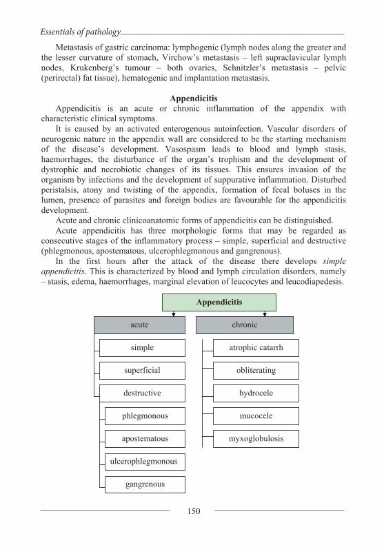
Essentials of pathology
150
Metastasis of gastric carcinoma: lymphogenic (lymph nodes along the greater and the lesser curvature of stomach, Virchow’s metastasis – left supraclavicular lymph nodes, Krukenberg’s tumour – both ovaries, Schnitzler’s metastasis – pelvic (perirectal) fat tissue), hematogenic and implantation metastasis.
Appendicitis
Appendicitis is an acute or chronic inflammation of the appendix with characteristic clinical symptoms.
It is caused by an activated enterogenous autoinfection. Vascular disorders of neurogenic nature in the appendix wall are considered to be the starting mechanism of the disease’s development. Vasospasm leads to blood and lymph stasis, haemorrhages, the disturbance of the organ’s trophism and the development of dystrophic and necrobiotic changes of its tissues. This ensures invasion of the organism by infections and the development of suppurative inflammation. Disturbed peristalsis, atony and twisting of the appendix, formation of fecal boluses in the lumen, presence of parasites and foreign bodies are favourable for the appendicitis development.
Acute and chronic clinicoanatomic forms of appendicitis can be distinguished. Acute appendicitis has three morphologic forms that may be regarded as
consecutive stages of the inflammatory process – simple, superficial and destructive (phlegmonous, apostematous, ulcerophlegmonous and gangrenous).
In the first hours after the attack of the disease there develops simple appendicitis. This is characterized by blood and lymph circulation disorders, namely – stasis, edema, haemorrhages, marginal elevation of leucocytes and leucodiapedesis.
Appendicitis
acute chronic
simple
superficial
destructive
phlegmonous
apostematous
ulcerophlegmonous
gangrenous
atrophic catarrh
obliterating
hydrocele
mucocele
myxoglobulosis
Ya. Bodnar, A. Romanyuk, V. Voloshyn, V. Gargin
151
Superficial appendicitis manifests itself morphologically in the primary site of inflammation. This term denotes a focus of suppurative inflammation with mucous tunic erosion on the basis of circulation disorders. The appendix grows thick and the serous tunic becomes plethoric and of dirty-grey colour.
From the primary site of inflammation, localized usually in the distal part of the appendix, the suppurative inflammation spreads over the whole organ – there develops phlegmonous appendicitis. The appendix is thick, its serous tunic is of dirty-grey colour and covered with fibrinous exudation. From the lumen there comes pus. The mesentery is swollen and hyperaemic. If the suppurative process is limited around the primary site of inflammation with the formation of small abscesses, it is called apostematous appendicitis. On the basis of phlegmonous inflammation there often occur mucous tunic ulcers. This is typical of ulcerophlegmonous appendicitis.
Diffuse suppurative inflammation is not self limited to the appendix but spreads to the surrounding tissues and mesentery with the development of periappendicitis and mesenteritis. At mesentery lesion there often occurs appendicular artery thrombosis which causes gangrene of the appendix. That is how the secondary gangrenous appendicitis appears. It is called secondary because the thrombosis is the result of previous suppurative inflammation of the appendix. That is the difference between it and appendix gangrene (primary gangrenous appendicitis) that develops on the basis of primary thrombosis or thromboembolism of its artery.
Acute appendicitis complications are connected with the destruction of the appendix wall and the spreading of the suppurative inflammation to the surrounding tissues. In ulcerophlegmonous appendicitis there often occurs perforation with subsequent development of peritonitis. In cases of lumen blockage in the proximal part of the appendix, pus accumulates in the distal part. The organ looks like a sack with pus (appendix empyema). The spread of the inflammation to the surrounding tissues is called periappendicitis, to the blind gut – perityphlitis and to the mesentery – mesenteritis. The latter may end with suppurative thrombophlebitis of the vessels of the mesentery. Further spread of the inflamation leads to the inflammation of the liver veins (pylephlebitis), thromboembolism of portal vein branches and the formation of pylephlebitic abscesses in the liver.
Chronic appendicitis develops after persistent acute one and is characterized by atrophic and sclerotic changes. In cases of self-recovery, acute inflammation ends with the development of granulation tissue in the zone of primary inflammation. There have been cases when the appendix lumen was completely filled with granulation and fibrous tissue (obliteration of appendix). Sometimes scar tissue obliterates only the proximal part and serous fluid accumulates in the distal part. The appendix turns into a cyst (hydrocele). If glands intensely produce mucus, it fills the cyst contents (mucocele). Rarely, the mucus turns into mucous globules (myxoglobulosis). Occasionally, due to cyst rupture, the mucus flows out into the abdominal cavity and the mucus-producing cells settle down and become the source of pseudomyxoma peritonei.

Essentials of pathology
152
Regional Enteritis (Crohn’s Disease) Regional enteritis is a chronic inflammatory lesion of the intestinal wall, usually
of the terminal part of the ileum, though it may affect all parts of the digestive tract. There develops a nonspecific granulomatous inflammation without necrosis (it looks like sarcoidosis) and with submucous tunic fibrosis and narrowing of the intestinal lumen. Typical of the disease is the alternation of the affected and unaffected areas. In the mucous tunic one finds deep transverse and longitudinal ulcers, edema of the submucous tunic. Macroscopically the mucous tunic resembles a cobblestone pavement. Among the complications are diarrhoea, malabsorption syndrome, intestinal obstruction, fistulas and degeneration into cancer.
Ulcerative colitis (UC) is a long-term condition that results in inflammation and ulcers of the colon and rectum. The primary symptoms of active disease are abdominal pain and diarrhea mixed with blood. Weight loss, fever, and anemia may also occur. Often, symptoms come on slowly and can range from mild to severe. Symptoms typically occur intermittently with periods of no symptoms between flares. Complications may include megacolon, inflammation of the eye, joints, or liver, and colon cancer.
The cause of UC is unknown. Theories involve immune system dysfunction, genetics, changes in the normal gut bacteria, and environmental factors. Rates tend to be higher in the developed world with some proposing this to be the result of less exposure to intestinal infections, or to a Western diet and lifestyle. The removal of the appendix at an early age may be protective. Diagnosis is typically by colonoscopy with tissue biopsies. It is a kind of inflammatory bowel disease (IBD) along with Crohn's disease and microscopic colitis.
Inflammatory Bowel Disease Crohn Disease Ulcerative Colitis
• Anywhere • Patchy • Transmural • Poor response to surgery • Increased risk of cancer
May have non-necrotizing non-peri-intestinal crypt granulomas
• Colon only • Continuous • Superficial • Good response to surgery • Increased risk of cancer • Non-peri-intestinal crypt granu-
lomas not seen
Topic. Diseases of the Liver, Gallbladder and Pancreas.
Massive hepatic necrosis (acute hepatic injury) is characterized by the progressive necrosis of the liver parenchyma. It is usually caused by exogenous (mushroom poison, chemical compounds) and endogenous (pregnancy, thyrotoxicosis) factors. In the progression of massive hepatic necrosis the stages of yellow atrophy, red atrophy and recovery are distinguished. The duration of the disease is about three weeks.
During the first days one can observe fatty degeneration of hepatocytes in the centre of the heptic lobule. It is quite soon followed by necrosis and autolytic

Ya. Bodnar, A. Romanyuk, V. Voloshyn, V. Gargin
153
destruction. The liver becomes smaller, flaccid and turns yellow. That’s why it is called yellow atrophy.
The detritus yields to resorption by macrophages. The stroma becomes “uncovered” and sinusoids, finding no resistance of hepatocytes, become overfilled with blood. The liver turns yellow with red spots (the stage of red atrophy). At this stage hepatic failure often develops.
Hepatic failure has general clinical representations of tissue turgor decline, xeroderma, icteric skin and sclera, vessel “stars” and skin haemorrhages, enlargement or reduction of liver, and there often occurs splenomegaly, ascites and edemas. The pathologic process progression provokes a complex of hepatic, mental and neurologic disorders. The affected person has fetor hepaticus, the liver aches at palpation and he/she suffers from fever and leucocytosis.
The gravity of hepatic failure is usually estimated according to how deep the nervous and mental disorders develop. Three stages of hepatic failure are singled out. The stage of psycho-emotional disorders is characterized by emotional instability: swift change of humour, depression or euphoria, insomnia at night and sleepiness in daytime, headache, overexcitement and memory weakening. The stage of neurologic disorders and impairment of consciousness manifests itself in sudden excitation which is followed by inhibition, tremor of hands, lips and eyelids. Progressive hepatic failure ends with coma (the third stage).
Hepatitis
Hepatitis is an acute or chronic liver disease characterized by dystrophic and necrobiotic changes of the parenchyma combined with the inflammatory stroma infiltration. Hepatitis may be a separate nosologic unit (primary) or a manifestation of other diseases (secondary).
Primary hepatitis develops under the influence of hepatotropic viruses (viral hepatitis), alcohol (alcoholic hepatitis), medicines (medicamentous hepatitis), cholestasis (cholestatic hepatitis). Viral and alcoholic hepatitis are the most common forms of hepatitis.
Secondary hepatitis accompanies a wide range of diseases. They are infectious diseases (typhoid fever, dysentery, cytomegaly, yellow fever, malaria, tuberculosis and sepsis), thyrotoxicosis, rheumatic diseases, digestive tract pathology and intoxications.
Acute hepatitis can be exudative or productive. Exudative hepatitis is in turn divided into serous and suppurative.
Chronic hepatitis is characterized by parenchyma destruction, cellular infiltration of stroma, sclerosis and changed regeneration. Three types are distinguished – aggressive, where hepatocytes dystrophy and necrosis prevails; persistent, where cellular infiltration of portal areas and intraparticular stroma prevails; and cholestatic characterized by cholestasis, cholangitis and cholangiolitis.
Light cases of hepatitis end with full recovery but massive liver lesion may lead to the development of cirrhosis.
Viral hepatitis is caused by hepatotropic viruses. Liver cells are damaged either by the allergic reaction of cytolytic type or by the hypersensibility of delayed type.

Essentials of pathology
154
Autoimmunisation is connected with a specific liver lipoprotein that forms as a result of virus replication in hepatocytes and acts as an auto-antigen. After the recovery the disease leaves type-specific immunity that’s why the person may be affected by a different type of viral hepatitis.
The following clinicopathologic forms of viral hepatitis are distinguished: acute cyclic (icteric), anicteric, necrotic (malignant), cholestatic and chronic.
At its peak the cyclic (icteric) form is characterized by the ballooning degeneration(diffuse swelling), focal and coagulation necrosis of hepatocytes. Groups of hepatocytes that have undergone coagulation necrosis form round homogenous eosinophilic structures which are forced out into perisinusoid spaces – Councilman’s corpuscles (body). Cholestasis and necrosis of hepatocytes results in hepatocellular jaundice. At the same time there occurs lympho- and macrophage infiltration of portal tracts and sinusoids. Macroscopically, the liver is larger in size, the capsule is tense, dense and red (large red liver).
In the course of recovery the liver returns to normal size and hyperaemia decreases. The capsule is somewhat thickened and dingy; adhesions appear between the capsule and the peritoneum. Reparative processes prevail over the destructive ones, lympho- and macrophage infiltration becomes focal. The process ends with liver sclerosis that may develop into cirrhosis.
The anicteric form of viral hepatitis, compared to the icteric one, is characterized by less evident morphologic changes although at laparoscopy one finds the picture of large red liver. Ballooning degeneration and Councilman’s corpuscles are rarely found in this form. But one can clearly observe proliferation of reticuloendotheliocytes. Lympho- and macrophage infiltrations do not destroy the terminal plate and there is no cholestasis.
The necrotic form is first and foremost marked by the progressive necrosis of parenchyma. The liver rapidly reduces in volume and becomes contracted and grey-brown in section. Microscopically one can observe necroses of hepatocytes, accumulation of reticuloendotheliocytes, Councilman’s corpuscles, “uncovered” stroma as a result of resorption of necrotic masses, haemorrhages and cholestasis in capillaries. If the affected person does not die of hepatic coma, postnecrotic liver cirrhosis develops.
The cholestatic form is manifested with prevailing cholestasis with the development of cholangitis and cholangiolitis on the basis of hepatocytes destruction, and lympho-, macrophage and neutrophil infiltration of stroma. One often finds Councilman’s corpuscles.
The chronic form of viral hepatitis is represented by active or persistent hepatitis. Active hepatitis develops on the basis of sclerotic liver changes. It is characterized by ballooning degeneration, necrosis of hepatocytes and inflammatory stroma infiltration. Liver regeneration is incomplete which leads to the development of cirrhosis. The persistent form is characterized by prevailing infiltration of sclerosed portal areas with lymphocytes, histiocytes and plasmatic cells. Dystrophic hepatocyte changes are low-grade. Chronic persistent hepatitis rarely develops into cirrhosis.
In viral hepatitis death occurs due to acute or chronic hepatic failure.

Ya. Bodnar, A. Romanyuk, V. Voloshyn, V. Gargin
155
Alcoholic hepatitis is an acute or chronic liver disease caused by alcoholic intoxication. Ethanol and acetaldehyde are hepatotropic poisons. Ethanol is neutralized by liver enzyme - alcohol dehydrogenase. Its synthesis in the liver is genetically predetermined and quantitatively specific for each individual. After a long period of alcohol abuse the alcohol dehydrogenase’s protective effect is not sufficient to safeguard the liver from destruction and at a certain alcohol concentration there occurs hepatocyte necrosis. The cytotoxic effect of alcohol, even in small doses, occurs in the liver which has been previously affected by such diseases as chronic hepatitis, fatty liver and cirrhosis. Cessation of alcohol consumption leads the process into a benign course. But if alcohol consumption continues chronic hepatitis progresses and ends with liver cirrhosis as ethanol drastically suppresses the regenerative potential of the organ.
In acute alcoholic hepatitis the liver is larger in volume and density and light brown areas alternate with brown-red ones. Microscopically one can observe necrosis of centrolobular hepatocytes. The so called alcoholic hyaline (Mellori’s corpuscles) can be found in their cytoplasm which is an important diagnostic sign. Peripheral hepatocytes are in the state of fatty degeneration. Necrotic areas and portal tracts are infiltrated with neutrophils. Occasionally, especially in a previously affected liver, massive hepatic necrosis occurs. In most cases after the cessation of alcohol consumption the liver structure regenerates.
Chronic alcoholic hepatitis does not differ morphologically from active and persistent viral hepatitis. It is identified by the presence of Mellori’s corpuscles in the cytoplasm of hepatocytes and beyond the cells. Alcoholic hyaline is a fibrillar protein which is synthesized by hepatocytes under the influence of ethanol and causes their destruction. Chronic alcoholic hepatitis ends with the development of cirrhosis.
Liver Cirrhosis Liver cirrhosis is a chronic disease characterized by sclerosis, structural change
and the deformation of the liver. The pathomorphology of cirrhosis includes the following liver changes: hepatocytes dystrophy and necrosis, deranged regeneration, diffuse sclerosis, structural change and deformation of the organ. In cirrhosis the liver is dense and gibbous, its volume usually reduces but in rare cases it may increase.
The cirrhosis development is based on hepatocytes dystrophy and necrosis. Their destruction leads to intense regeneration of preserved parenchyma. It results in the formation of nodular regenerates and false particles which are wrapped in connective tissue. The false particles are characterized by deranged angioarchitecture. They often lack the central vein or it is located in peripheral areas and connective tissue membrane develops in sinusoids. All this causes blood circulation disturbance in the liver. Increasing hypoxia leads to dystrophy and destruction of hepatocytes in nodular regenerates and to intense excrescence of connective tissue, which further disturbs the microcirculation. The process develops like a chain reaction with constant intensification of sclerotic changes.
The classification of cirrhosis is based on etiologic, morphologic, morphogenetic and clinicofunctional criteria.

Essentials of pathology
156
Postnecrotic cirrhosis develops after massive necrotic liver changes, for example after massive hepatic necrosis, viral or alcoholic hepatitis. The necrotized tissue resolves, the stroma and central veins collapse, triads become close to each other. Vast fields of connective tissue develop in these areas and from the organ’s surface they look hollow. Big nodular regenerates appear. According to its morphology, it is usually a macronodular form of cirrhosis and rarely – a mixed one.
Portal cirrhosis is a micronodular form. It develops as a result of the circulation deficiency, chronic alcoholic hepatitis, malnutrition and metabolic disorders. The connective tissue expands in the directions of portal tracts and penetrates into liver parenchyma in the form of processes dividing the parenchyma into smaller false ones. Moderate cellular infiltration of stroma remains as a manifestation of previous hepatitis.
Biliary cirrhosis can be primary and secondary. Primary cirrhosis is the result of nonsuppurative destructive (necrotic) cholangitis and cholangiolitis. In response to destruction, there occurs proliferation and cicatrisation of bile ducts, infiltration and sclerosis of the periportal areas, the destruction of peripheral hepatocytes and the formation of septa and false particles as in portal cirrhosis. The liver is enlarged, grey-green in section, and its surface is smooth or fine-grained.
Secondary biliary cirrhosis is caused by cholestasis (cholangiostatic cirrhosis) as a result of extrahepatic obstruction of bile duct (stone, tumour) or by bile duct infection with the development of bacterial, usually suppurative, cholangitis and cholangiolitis (cholangiolitic cirrhosis).
Classification of Liver Cirrhosis
According to
etiology According to morphology
According to morphogenesis
According to clinicofunctional criteria
Infectious (viral hepatitis, parasitic liver diseases) Toxic and toxicoallergic (alcohol, hepatotropic poisons, medicines, allergens)
Micronodular Macronodular
Postnecrotic Portal Biliary Mixed
According to the extent of hepatocellular deficiency (cholemia, hypoalbuminemia, hypothrombinemia, hypo-onchia, haemorrhages, coma) According to the extent of portal hypertension (ascites, oesophagogastric haemorrhage)
Biliary (cholangitis, cholestasis)
According to the process activity (active, moderately active, inactive)

Ya. Bodnar, A. Romanyuk, V. Voloshyn, V. Gargin
157
Metabolic-alimentary (insufficiency of proteins, vitamins and lipotropic factors, accumulation diseases)
Circulatory (chronic venous stasis)
According to progression (progressive, stable, regressive)
Cryptogenic (of unidentified etiology)
Morphologic symptoms of cirrhosis are dilatation and rupture of bile capillaries,
which causes peripheral hepatocytes necrosis. Connective tissue expands according to the morphogenesis of portal cirrhosis. In secondary biliary cirrhosis the liver is enlarged, dense and green due to bile impregnation, in section one can see dilated ducts filled with bile.
Mixed cirrhosis appears as a result of portal cirrhosis supplemented at a certain stage by necrotic liver changes.
Liver cirrhosis causes typical extrahepatic disorders: jaundice and haemorrhagic syndrome as a sign of hepatocellular deficiency, cholestasis and cholemia; exhaustion as a result of digestion disorders caused by stasis and atrophy of gastrointestinal tract in portal hypertension, splenomegaly as a result of reticuloendothelium hyperplasia and sclerosis. This leads to the development of extrahepatic porta-caval shunts due to which some blood bypasses the liver and discharges the portal vein. Affected people have dilated veins in the esophagus, hemorrhoidal plexus and dilated hypodermic veins in the thorax and the abdominal wall. The latter are called “Medusa heads”. The varicosity of the above mentioned veins goes together with the thinning of their walls which is often the cause of profuse esophageal, gastric or hemorrhoidal haemorrhage. As a result of portal hypertension and the lesion of liver parenchyma where the degradation of antidiuretic hormone occurs, the transudate infiltrates into the abdominal cavity, sometimes up to a volume of 10 litres. This phenomenon is called ascites. The ascitic fluid, accumulated in the abdominal cavity, compresses blood vessels and internal organs distorting the blood flow. In the kidneys, one finds signs of acute renal insufficiency (tubular epithelium necrosis) and, occasionally, hepatic immune complex glomerulonephritis, which cause the development of hepatorenal syndrome. In most cases people affected by cirrhosis die of chronic hepatic failure. Besides, cirrhosis may be the basis for the development of liver cancer.

Essentials of pathology
158
Cholecystitis Among the pathologic processes in the gallbladder, acute and chronic
inflammation (cholecystitis) and gallstones are the most common. In acute cholecystitis the inflammation can be catarrhal, fibrinous and
suppurative (phlegmonous). It is caused by ascending and descending infection on the basis of biliary dyskinesia and cholestasis. Important to its development are gallstones which traumatise the mucous tunic often causing pressure sores. Acute cholecystitis is complicated by the destruction of gallbladder wall with the development of bile peritonitis. In cases of gallbladder duct obstruction and pus accumulation in the cavity, gallbladder empyema develops. The spreading of the suppurative process beyond the organ is complicated by suppurative cholangitis, cholangiolitis and pericholecystitis with the formation of adhesions.
Chronic cholecystitis is as a result of the acute form. Morphologically it is manifested as mucous tunic atrophy and sclerosis with lymphohistiocytic infiltration. Occasionally the petrification of the gallbladder wall and adenomatous excrescence of the mucous tunic occur.
Gallstones are often the cause of calculous cholecystitis. Such cases manifest themselves in the chronic inflammation with periodic exacerbations. The gallbladder wall may be destroyed by the stone causing the development of bile peritonitis. When the stone enters the general bile duct and causes its occlusion, obstructive jaundice develops.
Cholelithiasis
Cholelithiasis is a disease defined by the formation and presence of concrements in hepatic and extrahepatic bile ducts. Its main difference from calculous cholecystitis lies in the fact that in cholelithiasis the stones are in intrahepatic ducts. The disease is polyetiologic. The interaction of such factors as genetic susceptibility, malnutrition, metabolic disorders, bile duct infections and cholestasis creates conditions under which bile tends to form stones. What is considered to be important to the change of normal bile into lithogenic one is the decrease of the cholato-cholesterol index – the ratio between the contents of biliary acids and cholesterol in the bile. When the quantity of biliary acids is insufficient, cholesterol turns into sediment and stimulates the formation of stones. But their formation also requires favourable local conditions – bile duct inflammation, mucus discharge, absorption disorders in gallbladder and local irritation. According to I.V.Davydovskyi, the main morphologic signs of cholelithiasis are the presence of Luschka’s ducts, excrescence of non-striated muscles and glandular hyperplasia of gallbladder mucous tunic. Luschka’s ducts are the channels lined with prismatic epithelium and extend to the muscular and subserous tunic of the organ. It is in them that bile accumulates, which facilitates the formation of stones. The second sign of cholelithiasis is productive granulomatous inflammation. Granulomas appear as a result of ulcero-necrotic lesion of bile ducts and the gallbladder with bile penetration. As a result of regeneration its components get immured in connective tissue. Cholesterol crystallizes and turns into sediment. It is resorbed by gigantic cells of “foreign bodies” which form a granuloma.
Ya. Bodnar, A. Romanyuk, V. Voloshyn, V. Gargin
159
Cholelithiasis may be complicated by choledochitis, cholangitis, cholangiolitis, pressure sores in the general bile duct and the gallbladder, bile peritonitis, obstructive jaundice, secondary biliary liver cirrhosis, reactive hepatitis and cholangiocarcinoma of the liver.
Liver Cancer
Primary liver cancer is the eighth important on the list of cancers of other localization.
According to the macroscopic picture, nodular – one or several green nodes – and diffuse cancer are distinguihed, according to its growth pattern – infiltrative, expansive and mixed cancer. According to the histogenesis, hepatocellular and cholangiocellular cancer are distinguished.
Hepatocellular cancer is the most common one. In 60-80% of cases it develops on the basis of liver cirrhosis. One often finds HbsAg in cancer cells.
According to the macroscopic picture, liver cancer may have trabecular, solid or trabecular-solid construction with cellular atypism, invasion into venous vessels and subsequent hematogenic metastasis.
Cholangiocarcinoma is more common among people older than 60. It grows out of bile duct epithelium and is not connected with cirrhosis. In the macroscopic picture it resembles a dense node of white colour. According to the microscopic structure, it is more often an adenocarcinoma, sometimes tumour cells secrete mucus. It usually spreads in by lymphogenous way.
More often one can find secondary metastatic malignant tumours in the liver which metastasize from the gastrointestinal tract, lungs, kidneys or mammary gland.
The malignant process in the liver may result in hepatic failure which is often the cause of death.
Pancreas diseases
Pancreatitis. One distinguishes acute and chronic pancreatitis. Acute pancreatitis is connected to 80 % of cases with cholelithiasis or with
alcoholism. Important to the pathogenesis of the disease development is the ischemic lesion of the organ’s parenchyma due to arterial thrombosis; medication damage, etc. With the disease progression there appear white or yellow-white areas of fat necrosis in the surrounding tissues (steatonecrosis). The gland is swollen, sometimes one can observe haemorrhagic imbibition of parenchyma. In such cases the tissue turns black-brown with the areas of necrosis.
Chronic pancreatitis often occurs after a long period of alcohol consumption. Fibrosis, cicatricial narrowing of ducts, acinar tissue atrophy develops in the tissue. The gland is dense and grey; in some places cysts with calcareous content can be seen.
Pancreas tumours are divided into benign (adenoma) and malignant (carcinoma). The head of pancreas is affected in 60% of cases, the body – in 20%, the tail – in 5%. Head carcinomas obstruct the outlet of general bile duct and cause obstructive jaundice.

Essentials of pathology
160
Topic. Diseases of the endocrine system. The diseases of the hypophysis are connected with the affection the front part
(adenohypophysis) and the rare part (neurohypophysis) and are displayed with the changes of the proper hormones secretion. Besides, the affection of the adenohypophysis is accompanied by the local changes: the growth of the size of the ephippium, which can be detected by means of the X-rays or computer examination. It is also displayed by malfunction of the sight because of the optic (II cranial) nerve pinching, the increase of the intracranial pressure which is accompanied by headaches, nausea, vomiting etc.
The malfunctions of the hypophysis appear in the case of tumorous, autoimmune, inflammatory and necrotic affections or affections of the hypothalamus or nervous system.
The most wide-spread diseases of the hypophysis are hyperpituitarism and hipopituitarism. The hyperpituitarism often appears in the case of adenomas of adenohypophysis, which are almost always hormone-active and secrete one or another hormone. The somatotrophic adenoma or adenocarcinoma produces the progress of acromegalia for adults and giantism for children. Typical displays of tumours are eosinophilic cells: extra growth of the tissues of mesenchymal origin (connective, cartilaginous, bone, stroma of internal organs); evident growth of the size of ears, lips, tongues (macroglossia), noses, bones of extremities especially hands, feet, lower jaws, frontal bones. The dyschondrosteosis osteogenesis is restored in the bones. In the case of acromegalia of the goitre; the hyperplasia of the thymicis gland, the epiphysis, the cortex of the adrenals; atrophy of the sexual glands and pancreatic islets are discovered in the endocrine organs. The prolactinomic adenoma which develops on the basis of chromophoric cells is found very often and is displayed in the form of the giantism, the loss of libido, the sterility ((barrenness) among women), the galactorrhea, the amenorrhea. The corticotroph adenoma which develops on the basis of basophil cells is accompanied by hyper secretion of the adrenocorticotropic hormone (ACTH) which activates the cortex substance of the adrenals provokes the progress of the pituitary (Cushing's) basophilism (Cushing's syndrome). It is found more often among women. It is displayed by the progressive adiposity of the upper type, the arterial hypertension, the secondary steroid pancreatic (insular) diabetes, the dysfunction of ovaries, the hirsutism, the osteoporosis, the nephrolithiasis, the chronical pyelonephritis. The gonadotrop(h)ic adenoma if found very seldom, it is accompanied by the increase of the maintenance of the follicle-stimulating hormone (FSH) in the blood and displayed by the hypogonadism among men.
The hypopituitarism appears in the case of craniopharyngiomas and gliomas. The patients of prepubertal period have the pituitary nanism and the delay of pubescence. The disease appears in the case of the hypoplasia of the hypophysis or its destruction during the period of childhood by the inflammatory process or the necrotic one. It is displayed by the general hypoplasia of the organism. In this case adults have the hypogonadism, the absence of the secondary sexual characters. Women have

Ya. Bodnar, A. Romanyuk, V. Voloshyn, V. Gargin
161
amenorrhea, atrophy of the external genitals, sterility, decrease (degradation) of the activity of the thyroid glands and the adrenals.
The cerebrohypophysial cachexia is determined by the progress of the dystrophic and necrotic changes in the hypophysis, which is observed in the case of its tuberculous, atherosclerotic, thromboembolitic, syphilitic and tumorous affection of the hypophysis vessels. Sometimes young women have this disease after the child birth which is accompanied by the considerable uterine bleeding, the DIC syndrome or the embolism by the amniotic fluid. It is accompanied by the progressive cachexy.
The adiposogenital dystrophy appears after the neuroinfection, the tumourous affection of the hypophysis or the hypothalamus. It is displayed by the adiposity, the hypoplasia of the genitals and the decrease of sexual gland function, sometimes by the hypothyroidism, the decrease of the cortex adrenals function, the diabetes insipidus.
The syndrome of the empty ephippium is found very seldom, it is connected with the ephippium membrane defect. The constant pressure of the liquor provokes atrophy of the hypophysis. Such state can appear in the cases of the Shikhan syndrome, exposure to radiation, the hypophysis infarction and so on.
Suprasellar tumours of the hypothalamus are represented more often by gliomas and craniopharyngiomas. They can provoke hipo- and hyperfunction of the adenohypophysis. The craniopharyngioma appears on the basis of the Rathke's pouch among children and teenagers. Its diameter can reach 3-4 cm. The tumour tissue contains bones, calcificates, its structure looks like the adamantinoma or ameloblastoma.
The syndrome of the rare part of the hypophysis is displayed by the diabetes insipidus. The disease appears in the case of the decrease of the antidiuretic hormone secretion. It determines the disability of kidneys to concentrate urine, its great loss and profound violation of the water-electrolytic balance.
Diseases of the endocrine part of the pancreas. The diseases can be displayed by the increase of decrease of the islet cells
function. The decrease of the -cells function is observed more often, that determines the progress of the pancreatic (insular) diabetes. When the tumour (adenoma) develops the antihyperglycemic syndrome appears in the case of the - insulinoma. The Zollinger-Ellison syndrome (multiple endocrine neoplasms) with plural ulcers on the mucous tunic of the stomach in the case of insulinoma or gastrinoma.
The pancreatic (insular) diabetes is a cronical disease which appears because of insulin insufficiency and it is accompanied by the dysmetabolic dysfunction with the affection of vessels and internal organs. There are the following forms of the pancreatic (insular) diabetes: spontaneous, secondary, the diabetes of pregnant women, cryptic. Among the spontaneous forms there are the following ones:
- The pancreatic (insular) diabetes of the I type (insulin dependent) which appears because of the destruction of -cells of autoimmune or idiopathic origin;
- The pancreatic (insular) diabetes of the II type (insulin independent) which is accompanied by the relative insulin insufficiency.

Essentials of pathology
162
The secondary pancreatic (insular) diabetes develops in the cases of the pancreatitis, the diseases of endocrine system (the acromegalia, the Itsenko-Cushing syndrome, the pheochromocytoma), the genetic syndromes, in the case of the use of some kinds of medicaments – medical diabetes.
The spontaneous diabetes is observed as an independent (self-dependent) disease. The diabetes of the I and II type is mostly is genetically determined. The pancreatic (insular) diabetes of the I type often appears during the early years (juvenile diabetes) after the virus infections, the auto immunization to -cells. In the process of disease progress the immune insulin with the presence in the inflammatory infiltration numerous T and B - lymphocytes, macrophages. The pancreatic (insular) diabetes of the II type develops among the adults. The base of the disease is the insufficiency of the -cells function and the insulin resistance of tissues. Insular insufficiency provokes dysfunction of the glycogen synthesis, the increase of sugar content in blood (the hyperglycemia), the advent of sugar in urine (the glycosuria), the progress of the hyperlipidemia, the acetonemia, the ketonemia, the acidosis. Macro- and microangiopathy develops in vessels, the insular apparatus of the pancreas, kidneys and the liver become damaged.
The morphological displays. The pancreas: atrophy, lipomatosis, hyalinosis, sclerosis. The liver: fatty atrophy, decrease of the glycogen content in hepatocytes. The vessels: macro- and microangiopathies which are determined by the
circulatory immune complexes and the products of disbalanced metabolism. The diabetic macroangiopathy is characterized by the affection of the elastic and muscular- elastic types arteries. It is displayed by the progressive atherosclerosis with the progress of the vessel necrosis and lower extremities gangrene. The diabetic microangiopathy is characterized by the system affection of the arterioles and capillaries of different organs and tissues: the plasmatic dripping and affection of the basal membrane, the lipohyalinosis, the dystrophic changes, the proliferation of the endothelium and perithelium, the atrophic changes of the cells, the inflammatory lymphohistiocytic seepage of the side (wall).
The retina of an eye: the seepage of the side (wall) develops because of the diabetic retinal microangiopathy. It is displayed by the hyaline degeneration and the capillaries silting, the vein microaneurism, the perivascular edema, the extravasation (hemorrhage), dystrophic and atrophic changes of the optic (II cranial) nerve. There are two types of the retinopathy: nonproliferative one (or simple diabetic) which provokes separate microaneurisms (and dotty extravasations) and proliferative one which provokes the capillaries neoplasm, considerable extravasations, the retina sclerosis, the sclerosis the optic (II cranial) nerve papilla with the progress of glaucoma, the retina exfoliation and loss of sight. Sudden extravasations into the vitreous body are possible with the following progress of one eye blindness.
The nervous system: the symmetric affection of the peripheral nerves especially in lower extremities with the following progress of the paresthesia, affection of temperature, vibratory and pain sensitivity. Sometimes the motor nerves are affected too. The progress of the segmental demyelinization, edema, axon dystrophy are observed too.
Ya. Bodnar, A. Romanyuk, V. Voloshyn, V. Gargin
163
The kidneys: the diabetic intracapillary glomerulonephritis and glomerulosclerosis. There is the proliferation of the mesangium cells in the glomerules because of the soiling of the mesangium by the metabolic products and immune complexes, the hyaline degeneration of the mesangium develops gradually and death (destruction) of the glomerules. “The fibrin hats” appear on the capillary loops of the glomerules; glucogenic infiltration, fatty dystrophy and hydropic (vacuolar) degeneration are observed in the epithelium of the narrow segment of the nephron; epithelium becomes high with the light opaque cytoplasm. High proteinuria, edemata, arterial hypertension are found clinically. The kidneys decrease in their size symmetrically, the have the small-grained surface and hard consistence. The glomerulosclerosis can be nodular (follicular), diffusive and mixed diabetic.
Among the complications and causes of death one should mark out the gangrene of the upper extremities with the following progress of the septicopyemia, the myocardial (cardiac) infarction, the uremia and some infectious complications (the pyoderma, the furunculosis, the septicaemia).
The tumours of the endocrine part of the pancreas (insulinomas) can be benign (non-malignant) (the adenomas) and malignant (the adenocarcinomas, low-grade differentiated adenocarcinoma which provide metastasis into the regional glands and liver). The sclerosis, the hyaline degeneration, the amyloidosis of the stroma with the microcalcifications are often found in the tumours.
The insulinoma is a tumour (from 0,5 to 2 cm) which consists of -cells. It is located in the body or in the tail of the gland, it is displayed by the state of coma accompanied by the hypoglycemia, high level of the immune-reactive insulin, serious neuro-mental disorders. The fits are over when the intravenous injection of glucose is made.
The alpha-cell tumour can reach the size of 10 cm, it is located in the body or in the tail of the gland, it is characterized by the increased level of glucose synthesis. It is displayed by dermatitis, pancreatic (insular) diabetes, anemia and weight loss. The most obvious changes are seen on the skin: necrotic migratory erythema with the numerous papulae, vesicles, erosions and parts of hyperpigmentation. The patient emaciation (exhaustion) develops because of generalized catabolitic changes in the organism.
The gastrinoma develops on the base of G-cells. It is displayed by the Zollinger-Ellison syndrome which appears as a result of gastrin hypersecretion. Numerous ulcers and erosions can appear on the mucous tunic of the stomach and duodenum. They can be followed by such complications as perforations, penetrations, bleedings (haemorrhages), stenosises, relapses. The tumour is found more often among young men, it reaches 4 cm in diameter and has the structure of the parenchymatous adenoma. It can also degenerate into the cancerous growth.
The carcinoid (tumor) of the pancreas is displayed by the increased secretion biogenic amins: the serotonin and the histamine. Tachycardia, diarrhea, bronchial asthma fits are observed in this case clinically.
The vipoma is a tumour of a considerable size, it develops on the base of D-cells of the body or the tail of the gland, which produce vasoactive interstitial peptide.

Essentials of pathology
164
Clinically the disease is displayed by considerable water diarrhea, hypokaliemia, hypochlorhydria, acidosis, dehydration.
The diseases of the adrenals are divided into the affections of the cortex substances and the cerebral ones and accompanied by hyper- and hipofunctions of the proper hormones.
The hyperfunction of the cortex substance (hyperadrenalism) is often found on the basis of the pituitary (Cushing's) basophilism (Cushing's syndrome), the hyperaldosteronemia, the adrenogenital syndrome. The hyperadrenocorticism: the pituitary (Cushing's) basophilism (Cushing's syndrome) appears on the basis of increased secretion of the adrenocorticotropic hormone (ACTH) – adenoma of the hypophysis, adenoma or hyperplasia of the cortex substance of the adrenals because of the prolonged glucocorticoid therapy. The (hyper)aldosteronism appears on the basis of adenomas (the aldosteroma), the idiopathic hyperplasia of the adrenals. It can be primary (initial) and secondary. The disease is accompanied by the hypokaliemia, the hypernatremia, the arterial hypertensia. It is displayed by the muscle asthenia, the impaired cardial function (the cardiac decompensation, cardiac insufficiency) because of the hypokaliemia myopathy and the myocardiodystrophy, the paresthesia, the convulsions. The adrenogenital syndromes (congenital hyperplasia of the adrenals) is displayed by the malfunction of the steroid hormones synthesis and the accumulation of the androgenic hormones, which provoke the progress of the viricidism.
Hipofunction of the cortex substance (the hypo(adreno) corticism, the hypoadrenalism) can be primary (initial) or secondary (in the case of the adrenocorticotropic hormone (ACTH) shortage, acute (adrenal crisis) and chronic ((chronic) adrenocortical insufficiency, bronzed disease, bronzed skin, melasma, suprarenale, Addison's disease). The primary acute hipofunction of the cortex substance develops on the basis of stresses, the chronic hypoadrenalism, the sudden (quick) stoppage taking steroidal agents, considerable extravasation (hemorrhage) into the adrenal during the period of the bacterial infection (the meningococcosis, the diphtheria, the septicaemia) accompanied by the progress of the Waterhouse-Friderichsen syndrome among little children.
The primary chronic cortex substance insufficiency ((chronic) adrenocortical insufficiency, bronzed disease, bronzed skin, melasma, suprarenale, Addison's disease) develops in the case of the autoimmune adrenalitis, the tuberculous and metastatic affection of the adrenals. The patients have general asthenia, fatiguability, loss of weight, hypotonia, hyperpigmentation of skin (the melanoderma) and mucous tunics, the atrophy of the myocardium.
The secondary chronic cortex substance insufficiency develops in the case of the adrenocorticotropic hormone (ACTH) shortage. It is often found on the basis of tumours, the inflammation, the infarction, the extravasation (hemorrhage), exposure to radiation of the hypothalamus or the hypophysis, prolonged glucocorticoid therapy. The atrophy of the cortex (substance) takes place but the cerebral substance almost doesn’t change. The pheochromocytoma is found among the diseases of the cortex (substance) more often. It is accompanied by the increased synthesis of the catecholamines and high blood pressure. The tumour is found more often among women. It is one-side, its colour is red-grey or brown. It’s built of polymorphous cells

Ya. Bodnar, A. Romanyuk, V. Voloshyn, V. Gargin
165
with light cytoplasm. The malignant variant of the tumour might exist, it provides metastases into the lymph nodes, the liver, the lungs and bones.
The diseases of the thyroid gland are divided into goitres, thyroiditises and tumours. They can be accompanied by the hyperthyroidism (the thyrotoxicosis) or the hypothyroidism (the myxedema). The goitre (the struma) is the growth of the thyroid gland. The base of this process is the hyperplasia. In this case the function of the gland can be increased, decreased or not changed at all. According to the morphological signs goitres are divided into diffusive, nodular, diffusive-nodular (mixed) ones. According to the histological structure they are divided into colloid goitres and parenchymatous ones. The colloid goitre looks like a solid (dense) nodule which is built of the follicles of different sizes. Usually they are filled with the colloid. If the follicles are big, cyst-like with the flattened out epithelium it is the macrofollicular colloid goitre. If the follicles are small it is the microfollicular goitre. If the follicles have the growth of the epithelium in the shape of papillas it is the proliferative colloid goitre. If there are follicles of different size at the same time it is micro-macrofollicular goitre. The parenchymatous goitre looks like a fleshy tissue. It’s colour is grey-pink. It is characterized by the proliferation of the follicles epithelium in the form of solid structures. The colloid in the follicles is almost absent.
The diffusive non-toxic (simple) goitre can be endemic and sporadic. During the first hyperplastic stage of the disease the hyperplasia of the gland develops. Its weight increases to 100-150 gr. During the histological analysis little follicles are found which are covered with the prismatic epithelium. They contain very little colloid. After the progress of the euthyroid state the proliferation of the follicles epithelium stops. After that the stage of the colloid involution develops. Gradually the follicles grow in their size and the epithelium atrophies. The weight of the gland grows rapidly. The gland becomes dense, gelatine-like in the incision. It is the colloid goitre which can compress (squeeze) internal organs and even provoke the asphyxia.
The endemic goitre is found in the regions, where there is lack of iodine that determines the decrease of the synthesis of thyroid gland hormones. Because of that the compensatory hypertrophy of the gland. Gradually the hypothyroidism develops. Adults have the myxedema, the children have the endemic cretinism which is displayed by the physical and mental gap.
The sporadic goitre is found very seldom. It appears among young women and is accompanied by the euthyroid state or the hypothyroidism. The fact that causes this disease is unknown.
The diffusive toxic goitre (Graves' disease) appears among young women and is accompanied by the hyperthyroidism. One of the causes of the disease is autoimmunization: the appearance of the antibodies which stimulate the cellular receptors of the thyroticis. There is a genetic inclination for this disease. Among clinical displays of the disease there are the growth of the thyroid gland, tachycardia, loss of weight, increased nervous excitability. In the case of the Graves' disease the typical visceral and local changes develop in the thyroid gland. The morphological changes in the gland are the following: the organ grows diffusively; it has the soft consistence; the prismatic epithelium of the follicles transforms into the cylindrical one; there is the proliferation of the epithelium; the pseudo papillas appear; there is

Essentials of pathology
166
the vacuolization and rarefaction of the colloid; there is the lymphoplasmocytic seepage of the stroma; the lymphatic follicles form. Among the visceral changes the most important is the affection of the liver, the heart, the brain (cerebrum). The thyrotoxic heart develops in the heart: the hypertrophy and fatty (adipose) degeneration of the cardiac hystiocytes; the serous oedema and the myocarditis which results in the diffusive focal localized myocardiosclerosis. The edema, the fatty (adipose) degeneration, the interstitial inflammation with the progress of the fibrosis and the cirrhosis in the end are found in the liver too. The dystrophic changes develop in the nerve cell of the brain, the perivascular infiltration develop in the medulla. The dystrophy of the cortex of the adrenals is observed too. Patients die because of the cardiovascular collapse, the acute adrenal gland insufficiency during operations.
The hypothyroidism appears in the case of the thyroiditis which can be of the following types: the Hashimoto's thyroiditis, the acute granulomatous one, the acute lymphocytic one, fibrous one, the purulent one. The Hashimoto's thyroiditis (autoimmune, lymphatic stroma) more often appears among women who have genetic tendency for this disease. In the pathogenesis the most important role is played by the autoimmune affection of the organ parenchyma. The diffusive lymphoplasmocytic seepage of the gland with the forming of the lymphoid follicles develops. Because of the influence of the immune cells parenchyma dies. It is replaced with the conjunctive tissue. The hypothyroidism develops afterwards.
The acute granulomatous thyroiditis appears among women after the viral infection. The gland grows asymmetrically; grey-yellow infiltrations are found in it. The leukocytic infiltrations with the gradual purulent fusion of the gland are observed too. The macrophage granulomas with the admixture of giant cells and fibrosis progress appear later.
The acute lymphocytic thyroiditis very often does not have symptoms. Usually it is found during histological examination of the operating materials. The lymphoplasmocytic infiltration are located subcapsularily. The cause of this disease is still unknown.
The fibrous thyroiditis (the struma) (ligneous (Riedel's) thyroiditis)) is displayed by the hypothyroidism which appears because of the follicle atrophy and the growth of the fibrous tissue. The organ becomes very dense. The cause of this disease is still unknown.
The purulent (infectious) thyroiditis appears after the secondary hematogenic traumatic infection of the gland. The purulent inflammation and the insignificant decrease of the gland function are also found in this case.
The tumours of the thyroid gland can be non-malignant and malignant. Among the non-malignant tumours the follicular adenomas are found more often. They develop on the basis of A and B-cells. They are represented by the follicles of different size. The solid adenomas composed of C-cells which produce calcitonin are found too. The tumour is represented by the big light cells with the light cytoplasm. Also there are papillary adenomas. They have needle papillary accretions (excrescences) among the cystous neoplasms. The presence of the papillary accretions (excrescences) is a bad sign because the is possibility of the malignization. The secondary changes can be found in the tumours: little extravasations
Ya. Bodnar, A. Romanyuk, V. Voloshyn, V. Gargin
167
(hemorrhages); the areas of the necrosis, sclerosis and calcareous degeneration. There are atypical adenomas made of fusiform cells. There are also dermoid cysts, lipomas, hemangiomas, teratoblastomas. The malignant tumours are often found among women. They are represented by the cancer which can be papillary, follicular, anaplastic or medullary carcinoma. The papillary carcinoma is found more often. Clinically it is displayed by the thyrotoxicosis. Macroscopically it is a dense tumour. Its of grey colour. It is the tumour with the petrifacts and cysts. Its diameter is 7-10 cm. It is built of papillas with the growth of the cuboidal epithelium. Histologically the hypochromic empty nucleuses without nucleoluses with the eosinophilic intussusceptions of the cytoplasm. The psammous corpuscles are located inside the papillas. There are capsulated and follicular variants of the tumour. The tumour provokes metastases into the jugular (cervical) glands. The follicular carcinoma are found among women. It is represented by the grey or brown-pink node with several cm in diameter. It has the tendency for the marked infiltrating growth. It grows through the vessels and provokes metastases into the lungs, the liver and bones. The anaplastic carcinoma among the people of the elderly age who live in the regions with the lack of the iodine. It has the marked infiltrating growth and quick metastases. Its treatment is usually ineffective. The medullary carcinoma appears on the basis of the parafollicular cells C-cells. It is accompanied by the increased synthesis of the calcitonin. The tumour is represented by the grey-yellow node with the dense consistence and amyloid fragments. The squamous cell carcinoma and fibrosarcoma are found very seldom.
The diseases of the parathyroid glands are displayed by the hyper- and hypoparathyroidism.
The primary hyperparathyroidism appears in the case of the affection of the gland by the tumorous process (the adenoma, the the adenocarcinoma) or in the case of its hyperplasia or the autoimmune process. Commonly the adenomas are the same. They grow as the node on the basis of the acidophilic or transitional cells. They are accompanied by the extravasations (hemorrhages), the hemosiderosis, the necrosis, the fibrosis. The disease is also accompanied by the hypercalcinemia, the hypophosphatemia, the osteoporosis, the generalized fibrous-cystous osteit is, the parathyroid osteodystrophy with the overgrowth of the osteoid tissue, the metastatic tumor, the urolithiasis, the peptic stomach ulcers, the neurologic changes (the convulsions, the worsening of the memory) and the ophthalmologic ones (the cataract, the calcification of the cornea).
The secondary hyperparathyroidism appears after the erroneous ablation of the gland during the operation; in the case of the innate absence of the glands, autoimmune or hereditary diseases. The patients have the hypocalcemia, the increased neuromuscular excitability, the tetany, the laryngospasm, the increase of the calcium concentration in the bones, the cataract, the hypoplasia of the teeth.

Essentials of pathology
168
Topic. Diseases of kidneys (glomerulonephritis, nephrotic syndrome, pyelonephritis). Acute and chronic renal insufficiency. Urolithiasis. Neoplasia of
kidneys. Cancer of urinary bladder. Characteristics of diseases of kidneys in general. Wide application of biopsy of
kidneys, immune morphology, radioisotope and biochemical methods of research found classification of renal diseases on topograph-morphological principle nature of injury (inflamation, disorder of methabolism, tumors) and its consequences.
Glomerular diseases
Glomerular diseases is group of diseases of immune genesis. In case of primary and main pathomorphological changes develop in membrane structures of glomerules, it causes disorders of filtration and formation of the primary urine. Glomerulopathy are divided into malformed and acquired. Nephritis of deafs (Alport’s syndrome), malform nephrotic syndrome, systematic nephritis (periodical disease) belong malform diseases; glomerulonephritis, nephrotic syndrome, diabetic glomerulosclerosis, amyloidosis of kidneys belong to acquired diseases.
Malformation glomerulopathy. Alport’s syndrome is characterized with early development of renal deficiency, with deficincy of ear and sight. Morphologically it is shown with haemorrhagic type of glomerulonephritis and infiltration of intersticium with lipides. Process ends with development of productive extra- and intracapillar glomerulonephritis and interstitial syndrome.
In case of malformation nephrotic and renal antibodies are determined in mother’s and child’s body. It connects with anomaly of renal development - polycystosis. It is determined with electrone microscopy, that morphological reason of disease is absence of little shoots of podocytes and intracapillar productive glomerulonephritis.
System nephritis with amyloidosis (periodical disease) is shown recidive polyserositis and development of general amyloidosis.
Acquired glomerulopathy. Glomerulonephritis is mostly infectious-allergic
disease, which is morphologically shown with injury of membrane structures of glomerule and is clinically shown with olygouria, haematouria, proteineuria, artherial hypertension and oedemas. In 80% of accidents reason of disease is -haemolythycal streptococcus (bacterial glomerulonephritis). Disease is mostly clear and typycally shown after quincy, scarlet feverand and other infections, that sensibilate organism.
Non-bacterial glomerulonephritis begins in patients with diffuse injuring of connective tissue (periarteritis nodosa), after vaccination and serotherapy, colding, effect of chemical substances (for example medicines).
There is dividing immune complex and nephrotoxical glomerulonephritis (antibodies) glomerulonephritis by the mechanism of development. Reason of immune complex glomerulonephritis is fixation of immune complexes, that form and circulate in blood, on basal membranes of glomerule. They can be heterological, if they consist of antigens of bacterias and autological, if they consist of own tissues antigens. Imune complex form is 80% cases disease. Antibodies variant is found

Ya. Bodnar, A. Romanyuk, V. Voloshyn, V. Gargin
169
more rarely. It is caused by formation of antibodies to glycoprotein of basal membrane of glomerule.
Immune complexes, that injure basal membrane can be deposited subendothelially, subepithelially or mesangially. Subepithelial deposits are determined submicroscopically in form of solitary granules on external surface of basal membrane. Subendothelial deposits are like dawn. There is a think that it is complexes antigen-antibody. Mesangial deposits are mainly globulines. They are situted near mesangial cells that absorb it. Mesangial cells produce fibrinogen that penetrate into space between endothelial cells and basal membrane, except phagocyte function.
Reaction of capilary glomerules with heterological immune compelexes is provided by the mechanisms of immediate hypersensitivity. It characterizes acute and subacute glomerulonephritis. Injuring of membrane with autological complexes is provided with mechanism of hypersensivity of delayed type, which characterizes chronic formes of disease. In case of autoimmunization (antibodies glomerulonephritis) field of inflamation is glomerule capsule, and disease is acute glomerulonephritis begins from intracapillar exudative changes, then extracapillar exudative changes join to it, and at last productive changes join. It gives chance to distinguish such morphological forms of glomerulonephritis: intracapillar (pathological process develops in capillars and mesangium) and extracapillar (morphological changes takes place in capsule of glomerule). They can be exudative and productive by the character of inflamation.
Exudative intracapillar glomerulonephritis begins as reactive process to subendothelial deposits of immune complexes. Plasmorrhagy and leucodyapedesis are caused by injuring of membranes, they cause oedema of mesangium and infiltration of it by leucocytes.
Exudative extracapillar glomerulonephritis is characterized with accumulation of exudate ( serouse, fibrinouse, haemorhagic) in the cavity of capsule.
Prolyferative intracapillar glomerulonephritis is characterized with reproduction of endothelial and mesangial cells.
Kidneys become dropsical in case of acute glomerulonephritis . Pyramides are dark red, cortex is grey brown with red points on the surface. But it is almost not differs at the beginning of disease. Diagnosing is possible only during hystological research in such cases.
Subacute glomerulonephritis is also called “rapidly progressive” or malignant, it is connected with fast development (in 0.5-2 years) of chronic renal deficiency. Growing of endothelial cells of capsule is hystological symptom of it. They fill in cavity, straining opposite of its gate that seems like a crescentic. Productive extracapillar glomerulonephritis develops.
Macroscopically kidneys are enlarged and pale with petechial hemorrhages on the cortical surfaces. Cortical layer is wide, dropsical, yellow-grey, well restricted from dark red medullary substance of kidney.
Chronic glomerulonephritis is not final of acute. More often it is distinctive disease which is latent with recidives and it takes its course during many years and ends with renal deficiency. It is distinguished 4 forms of disease: latent, hypertensive,

Essentials of pathology
170
nephrotic and mixed. Its names pay our attension to main syndrome and level of its manifestation.
Hystological picture of disease is multicolored. It is represented with different hystological types – prolypferative intra- and extracapillar, membranous, mesangial and fibroplastic.
Membranous glomerulonephritis morphologically manifestated with thickening and splitting of basal membrane of capillar. It is not connected with prolyferation of cells. Immune complexes of same size are revealed subepithelialy. There is no explanation why exactly immune compelexes are situated on subepithelial side of membrane.
Mesangial glomerulonephritis is characterized with prolyferation of mesangial and epithelial cells. Immune complexes are revealed at mesangium, subendothelially and subepithelially. It is found out that mesangial cells can product tropocolagen, that’s why mesangium is enlarged nd sclerosed. Depending on level expressiveness changes of mesangium this form of glomerulonephritis can be mesangio-prolyferative, mesangio-capillar and lobular. In first case prolyferation of mesangiocytes without essential changes in capillars prevails in second – prolyferation mesangiocytes compared with diffuse enlargment and splittening of capillar membranes, in third - because of prolyfration of mesangiocytes in the center of glomerule capillars are removed into periphery, where are squeezed and undergo hyalinosis.
Development of mentioned forms of glomerulonephritis, ends with sclerosis and hyalinosis of capillar glomerules, forming of lesions in cavity of capsule, which is morphological symptom of fibroplastic glomerulonephritis. Kidneys become little, dry, anaemic, wrinkled. Renal surface become granular becaus nephrones undergo atrophy and sclerosis, but safe nephrones hypertrophy. Secondary (nephrotic) wrinkling of kidney develops.
Nephrotic syndrome is observed 65-75% at children and 8-30% at adults. It
appears very fast without prodromal heralds. Its symptoms are massive oedemas, proteinuria, hypoproteinaemia, hypercholesterinaemia. Arterial pressure is decreased or normal.
By the pathogenesis nephrotic syndrome is divided into primary and secondary. Primary nephrotic syndrome has no tie with previous diseases of kidneys. It appears because of genetic disorders of methabolism, or injuring of kidneys of fetal by mother’s antibodies. Pathognomical morphlogical symptom of primary nephrotic syndrome is connecting of podocytes shoots in hole cytoplasmatic mass, which is sprawled on basal membrane. In such case capillars are not changed. This injure of basal membrane is base for including of nephrotic syndrome into group of glomerulopathyas. Disease is also known as “glomerulopathy of minimal injures”or “minimal disease”. These titles reflect not clinical symptoms, but minimal hystological changes in the glomerule.
Secondary nephrotic syndrome is found mainly in adults and is symptom of glomerulonephritis and amyloidosis of kidney. Pathology is caused by depositing of subepithelial immune complexes and connective of membrane podocytes into

Ya. Bodnar, A. Romanyuk, V. Voloshyn, V. Gargin
171
one structure. In case of reumatic diseases glomerulonephritis have number of symptoms: brastly capillars of glomerule and arteriols are injured; arises fibrinoid dystrophy of glomerules and arteriols; injures of glomerules have segmentary character, it causes discrepancy between expressed morphological changes and minimal clinical symptoms.
Diabetic glomerulopathy is caused by local immune conflict. Hystologically distinguish 3 types of changes: knotty, diffuse and exudative. Knotty changes are specific for diabetes melitus. Diffuse changes are manifestated as thikening and hyalinosis of capilar membrane. Exudative changes are not specific, they are manifestated as “fibrinoid “ on periphery of glomerule and capsule drops on inner surface of Bowman’s capsule. Process ends as chronic renal deficiency.
Amyloidosis of kidney appears as a result of chronic diseases, which decompensated by destruction of tissues: fibrous cavernous tuberculosis, bronchoectatic disease, chronic abscesses, osteomyelitis etc.
Distinguish latent, proteinuretic, nephrotic nitroaemic stages in development of amyloid glomerulopathy. Amyloid is revealed in moderate quality along basal mambrane in latent stage. Kidneys are close, like fat (big greasy kidney). Amyloid is determined in glomerules, macroscopically it looks like glass balls . In nephrotical stage kidneys are enlarged, close, yellow-grey, in section with waken shine (big white amyloid kidney). The presence of amyloid fat dystrophy of canal epithelium is determined.This stage is characterized with proteinuria, hypoproteinemia, hypercholesterinaemia, oedemas. In nitroaemic stage sclerosis, destruction of most nephrons and its atrophy. Kidneys are diminished, close , with scar drawn in (amyloid wrinkled kidney).
Tubulopathyas. With this term marked a group of renal diseases in which injuring of renal canals
is main link of pathogenesis, it is manifestated as disorders of such funcions as concentration, reabsorbtion, secretion.
Primary (malform) tubulopathyas. Rickets-like tubulopathyas is group of diseases, caused by disorders of enzyme systems and enshoritment and narowing of canal space of proximal section of nephron . It causes disorders of reabsorbtion of glucose, aminoacids, phosphor, bicarbonates. Loss of aminoacids cause loss of weight and delay of growth, loss of phosphor- disorders of bones, mineralization and arise of osteoporosis (distortion, fractures), loss of bicarbonates- acidosis and hypokaliaemia, it causes muscle hypotony, arterial hypotensionand colapse. Main clinical symptoms are polyuria and nephrolithiasis. Secondary infections (otitis, pneumonia) often are joint.
Polyurical tubulepathyas are caused by disorders of enzyme systems of distal part of nephrone, it causes reabsorbtion of water and glucose disorder. Diseases are accompanied with polyuria, polydipsia, vommiting, acidosis, acetonaemia, glucosuria, loss of weight.
Nephrolithiase tubulepathyas have genetic base and transduce through autosome-recessive type. In case of first type of disease we can see aminoaciduria, caused by influence of indols to epitheliocytes of nephrone, that absorbe in intestines. Urine has

Essentials of pathology
172
blue colour. In case of second type we can see hyperoxalaturia. Crystales of calcium oxalate, that deposite in kidneys, become reason of interstitial nephritis and nephrosclerosis.
Secondary (acquired) tubulepathyas. Acute renal deficiency is clinical morphological syndrome with different
ethiology, which is characterized with sudden decreasing of glomerulus filtration, which causes loss of ability of kidneys to regulate water-sault gomeostasis of the body.
Reasons of deficiency are divided into 3 categories: prerenal, renal and postrenal. Prerenal reasons are reasons that decrease volume of circulating blood (traumatical shock, loss of blood, vomiting, dyarhea). Renal reasons are kidney injure, that can appear after influence of nephrotoxins and drugs (heavy metals, organic solvents, antibiotics, x-ray contrast substances) in case of haemovascular haemolysis of erythrocytes (DVC-syndrome), trombosis and emboly of artery. Postrenal reasons are obstrucrtion of urinary tracts with lithium, tumor, blood coagulate, hypertrophied prostate.
Necrosis of nephrothelium of proximal canals of nephrone is morphlogical symptom of acute renal deficiency. Kidney is enlarged, cortical layer is oedemated, grey, inner layer is hyperemic. Namely we can see syndrome of «robbing» of blood bed of glomerulus.
In development of renal deficiency four phases are distinguish. Initial (shock) phase is period from injuring of kidneys till development of olyguria, it lasts from some hours to week. Olygurical phase is characterized with acute decreasing of speed of glomerulus filtration, its duration from few days to few weeks. Patients die in just this period. During polyurical phase volume of urine gradually increases and during of period of recovering renal functions fully recovers.
Acute renal deficiency is accompanied with high mortality rate. Its index the highest in case of and traumatical forms – 50-70%, in case of other forms it is competed 10-35%.
Myelome kidney and gouty kidney are acquired tubulepathyas. Myelome kidney develops in case of myelome disease. Symptom of the last one
is tumor of plasmatic cells that secrete immuneglobulines. Plasmoblasts begin to secrete abnormal protein (paraprotein) into blood and surround tissues. Light chains of this protein compare amyloid fibrils. That’s why injure of kidneys in this case resemble amyloid glomerulopathy, but it has other pathogenic base. It is paraproteinosis with blockade of canals with protein conglomerates. It is called Benz-Jones protein. It denaturates in temperature of 40-500C, then it dissolves again. Sometimes giant cells of alien solids and deposits of lime salts are determined around cylinders.
Gouty kidney is observed at patients with disorders of purine metabolism. Purines (adenine, guanine) compose nucleoproteins.
Interstitial diseases Interstitial nephritis is an inflammation of mostly interstitial tissue of kidney with
next injure of nephrones.

Ya. Bodnar, A. Romanyuk, V. Voloshyn, V. Gargin
173
Tubule-interstitial nephritis is disease of immune-inflammative nature with injuring if intersticium and canals.
Causes: influence of toxins, drugs (antibiotics, sulfanilamides, analgesics, heavy metal salts) sensibilization, immune and angiogenic influences, endogen intoxication and influence of metabolits, genetic reason, oncogenic influence (leucosis, tymphome).
There are primary (proper disease) and secondary (in case of system diseases of connective tissue, Kidneyspacher’s syndrome) forms of disease. By the development of disease there are acute and chronic forms of disease.
Acute tubule-interstitial nephritis is characterized with oedema, infiltration of intersticium with lymphocytes, macrophages, plasmocytes, eosinophylic cells, epithelioid cells, dystrophic changes in nephrocytes, immune complexes on basal membranes.
In case of chronic tubule-interstitial nephritis could be found atrophy of canals, proliferation of connective tissue, that causes nephrosclerosis.
Pyelonephritis is non-specific inflammation of renal basin, its cups, parenchyma
of kidney with most localization in intersticium. That’s why pyelonephritis is called interstitial nephritis. By the character of inflammation there is pyogenic nephritis, which can have acute or chronic development. Women are mostly injured, it is caused by structure of urethra on hormonal status. It is known that estrogens cause hulling and metaplasy of epithelium, weakening of basin. Urine of pregnants changes by aminoacid composition, it causes development of bacterias. And secretion fluid of prostate has antibacterial factors, provides defense of urogenital system. Dyskinesia of urogenital system helps dissemination of infection (e.coli, enterococcus, streptococcus, staphylococcus, proteus). Infection can enter the kidney by lymphogenic (enteric fever, colitis, endometritis, enteritis) and haematogenic (sepsis, pneumonia, angina) ways. Development of pyelonephritis needs not only entering of infection but also determined by reactivity of organism and local factors, that cause disorders of urine excretion and urinary stasis.
In case of acute pyelonephritis interstitial tissue of all layers are oedemed and infiltrated by neutrophyles. Microabscesses and haemorrhage are often arise.
Association of sclerotic and exudative-necrotical changes characterize chronic pyelonephritits. Canals are distrophically changed and sclerozed. Spaces of canals are wided and filled in with colloid like fluid, epithelium is flattened. This kidney looks like thyreoid gland (thyreoid kidney).
Acute process is complicated with formation of carbuncles, connecting of pyogenic cavities (pyonephrosis), transition of inflammation into fibrous capsule (perinephritis) and paranephral cellular tissue (paranephritis), papilonecrosis. Chronic pyelonephritis is complicated nephrogen arterial hypertension and chronic renal deficiency.
Urolithiasis is chronic disease, it arises when in renal cavities, basin and urethers form lithiums which differ by growth, structure, chemical structure (phosphate, urate, oxalate, carbonate). The causes of formation of stones are general (congenital and acquired disorders of mineral metabolism, character of food, mineral structure of

Essentials of pathology
174
water, avitaminosis A) and local (inflammation, urinary stasis, trophic and water disorders of urogenital system function).
Lithiums, that enclose ways of urine excretion cause widening, atrophy, inflammation and sclerosis of parts of urinary system that are situated higher than obstacle. For example lithium of basin cause pyeloectasy and hydronephrosis, lithiums of urether – hydrouretheronecrosis, lithiums of cup – hydrokaliosis. Infection causes urethritis, pyelitis, pyelo- and paranephritis. Process can be confirmed with urogenic sepsis and chronic renal deficiency.
Polycystic kidney is congenital disease with both-side injuring of renal parenchyma, canals and tubules. It is often connects with cystous of other organs – liver, spleen, lungs. Polycystic renal disease that injures adults transducts dominantly, children’s polycystic renal disease – recessively. Development of polycystic renal disease is caused by disorders of embriogenesis on first weeks of fetus development. Defect is abscence of metanephral canals and collective canals of predecessor of urethra. It causes disorders of excretion and glomerular, tubular and excretive cysts form. Glomerular cysts don’t connect with renal canals, tubular cysts are formed from canals, excretive – from tubules. Cysts enlarge, and islands of parenchyma undergo atrophy from pressure. Disease develops 10-12 years. If it arises earlier, it has harder development. Patients die mostly from chronic renal deficiency.
Nephrosclerosis is induration and wrinkling of kidneys as a result of proliferation of connective tissue.
Arteriolosclerotical nephrosclerosis, or primary wrinkled kidney arises in case of hypertonic disease. Arteriols are primary injured with gyalinosis. Block of blood circulation in glomerulus develops. Some glomerulus undergo atrophy and sclerosis, some – hypertrophy.
Sclerosis and wrinkling of kidney develops not only primary in the case of the sclerosis of kidney vases, but also secondary as a result of inflammation (glomerulonephritis, pyelonephritis) or dystrophy. This kidney is called secondary wrinkled. Its surface is grained.
Arteriosclerotically wrinkled kidney develops in case of arteriosclerosis. Perpetual ischemy of organ is accompanied with atrophy of parenchyma, proliferation of strome. Connective tissue proliferates as scars in places of regeneration after infarcts. That’s why surface is highly hilly.
There are two phases in morphogenesis of nefrosclerosis: nosological and syndromic. Character of injuring of kidney during first phase is determined by peculiarities of morphogenesis of main disease. Then all renal structures undergo sclerosis and it becomes hard to determine reason of it. On this phase nephrosclerosis is syndrome. Nephrosclerosis ends as chronic renal deficiency.
Chronic renal deficiency. Symptoms of renal deficiency arise when speed of glomerular filtration decrease to 30ml/minute (25% from norm).
Morphological base of chronic renal deficiency is nephrosclerosis. Disorders of kidneys arise when decreasing of number of nephrones in force or decreasing of speed of glomerular filtration without decreasing of its number. Primary symptoms of renal deficiency appear in case of decreasing of number of nephrones to 30-50% of its initial number. Evident symtoms develop in case of decreasing of nephrones in

Ya. Bodnar, A. Romanyuk, V. Voloshyn, V. Gargin
175
force to 10-30%, more decreasing cause uraemia. Man can live in case of presence of 40000 nephrones (2% from norm).
Chronic renal deficiency is characterized with system injuring of organism. Among haematological symptoms anaemia is most characterized. Main factor which causes its development is erythropoetin deficit, which is produced by kidneys. But also in case of normal quantity of erythropoetin marrow can’t fully react to its influence. Erythropoesis increases little and new erythrocytes has high inclination to haemolysis, that shortens its life. Also patients with chronic renal deficiency frequently have stomach and intestinal haemorrhage.
Disorder of blood coagulation is manifestated as extension of haemorhagy time. This symptom is explained as violation of trombocytes quantity. Its function is injured by guanidinyantar and oxyphenolacetic acids, which circulate in blood. Among cardiovascular system injures the most important is hypertension. There are a lot of mechanisms of its formation: increased production of rennin, decreased production of vasodilatative prostaglandines , decreasing of natrium excretion, increasing of extracellular fluid volume.
Osteodystrophy characterizes chronic renal deficiency. Level of ionized calcium decreases after decreasing of weight of nephrones. Secondary hyperparathyreosis appears. Resorbtion of bones arises, its density is lost. Decreasing of absorption of calcium in digestive system causes osteodystrophycal changes, because in case of injury of kidney formation of active form of vitamin D decreases.
Terminal phase of chronic renal deficiency is called uraemia. Symptoms of uraemia become expressed when speed of glomerular filtration decreases to 10 ml/min. Main cause of pathogenesis in this syndrome have uraemic toxins, which are waste products of nitrogen metabolism. They are derivatives of guanidine (creatinine, creatine, guaninediyantar and guanidineacetic acids), aromatic substances (phenol, indol, aromatic amines), conjugated aminoacids, peptides. Acidosis and disorder of electrolyte metabolism are important.
Uraemia is confirmed with deficit of some substances, which stop producing in such conditions – erythropoetine and active form of vitamin D (1,25-dioxycholecalciferol).
In case of uraemia toxical substances excreting through extrarenal excretory systems: skin, lungs, mucous layer of intestines, serous membranes. In these organs penetrate ability of vassels grows sharp, oedema and reactive inflammation develops, often – fibrinous-haemorhagical. During autopsy it smells like urine.
Tumors of kidney Benign and malignant tumors are found in kidney. Benign tumors at kidney can
be epithelial nature: adenoma (dark-, lightcellular, acidophylic). More often cortical adenoma diameter 2sm lookes like nodles of yellow-grey colour. Microscopically we can see cysts with proliferation of nipple; structures, cells can form canals, glands. Mesenchimal tumors are represented by fibroma – tumor from interstitial cells, tumors from vessels, muscular tissue.

Essentials of pathology
176
Topic. Diseases of urogenital system. Diseases of female (endocervicosis, glandular hyperplasia of endometrium, endometriosis) and male (benign hyperplasia of
prostate) urogenital system. Tumors of cervix uteri and body of womb, ovarian tumors. Cancer of prostate. Fybrocystic disease of lactiferous gland. Breast cancer.
Classification of diseases of genital organs. Diseases of genital organs and lactiferous gland are divided into disharmonious, inflammatory and tumorous. Disharmonious diseases include adenoma of prostate and gynecomastia (of men), hyperplasia of mucous tunic of uterus (endometrium), endocervicosis (erosion of cervix uteri), non-malignant dysplasia of lactiferous gland (mastopathy) of women. To inflammatory diseases belong male prostatitis, orchitis, etc., female mastitis, adnexitis, endometritis, etc. Among tumorous processes of men the most often are seminoma, prostate cancer, orchis cancer, etc., as well as female carcinoma or uterus, cervix uteri, lactiferous gland, chorionepithelioma, tumors of external genitals, etc.
Diseases of prostate are divided into inflammatory processes — prostatitis, non-malignant nodular hyperplasia and tumors. Inflammatory diseases become apparent in the form of prostatitis which can be acute, chronic bacterial and chronic nonbacterial. Acute prostatitis is mostly caused by coccobacilluses and can be catarrhal, follicular, parenchymatous. Focal or diffuse inflammation with cellular infiltration appears, sometimes with abscesses, sites of necroses.
Hypertrophy of prostate is observed mainly among men older than 50 because of the weakening of genitals function. The glade becomes much larger in size, the surface is often uneven and its consistency is elastic. The middle part especially enlarges and presses into urinary bladder that causes urinary difficulties.
According to histological characteristics hypertrophy can be glandular (adenomatous), musculorfibrous and mixed. As the result of urinary tract squeeze there appears urinary retention. Secondary infection is added, there develop cystitis, pyelitis and pyelonephritis, moreover urosepsis can emerge. All this can lead to cancer.
Prostate cancer takes the second place among oncological pathologies of men and is observed in old age. At the time of cancer pathogenesis hormonal factor — problems with androgen secretion — plays the crucial role. According to histological characteristics it is most often adenocarcinoma and rarely undefined cancer. It rapidly attacks urinary bladder and rectum, metastasizes through lymphogenous and hematogenous ways into internal organs and bones.
Diseases of body of uterus and endometrium. Dysfunctional uterus hemorrhages appear in the form of menorrhagias — extra uterine bleeding during menstruation — or in the form of metrorrhagias — uterine bleedings which are not connected with menstruation. They appear because of the breeches in hormonal balance of pituitary gland and ovaries. These diseases can be met with anovulatory cycle, yellow body deficiency, while using oral contraceptives, in the period of menopause, with fibromyomas, endometrium polyps, malignant tumors of uterus.
Glandular hyperplasia of mucous tunic of uterus (endometrium) is a rather wide-spread disease; it develops because of the problems with hormonal balance. The disease is typical mostly for women of mature and old age, often in climacteric,

Ya. Bodnar, A. Romanyuk, V. Voloshyn, V. Gargin
177
accompanied with metrorrhagias. As a rule, hormonal dysfunction of ovaries is also observed. In such a situation endometrium is considerably thickened, sometimes with polyps. Microscopic glands are elongated, winding, often cysticly widened. There can be found atypical hyperplasia of endometrium or adenomatous hyperplasia with atypia which belongs to pre-cancer state of uterus. Under these circumstances epithelium of mucous tunic has some symptoms of atypia, nucleus hyperchromatism, nuclear-cytoplasmic index increases, figures of mitosis appear.
Adenomyosis or internal endometriosis means the state when nidi of endometrial glands and stroma appear in myometrium. The disease clinically manifests in the form of menorrhagias and pains.
Endometriosis is emerging of endometrium parts outside uterus in ovaries, abdominal cavity, internal organs. Meanwhile in ovaries there can appear endometrial cyst.
Endometrium polyp is a tumor on a wide stalk which comes into cavity of uterus and can cause uterus bleedings.
Carcinoma or uterus resembles cauliflower or a polyp on a wide stalk. It grows exophiticly, rapidly submits to ulceration and destruction. From histological point of view it is as a rule adenocarcinoma which can be highly, fairly or low differentiated. Non-differentiated cancer occurs seldom. Carcinoma or uterus metastasizes mostly through lymphogenous ways, later hematogenous metastases in lungs and other organs develop.
Among mesenchymal tumors in uterus the most often are leiomyomas and fibromyomas that can be intramural, subserosal, submucous. In tumors there can be observed secondary changes such as necrosis, hyalinosis, calcification. Rarely leiomysarcomas can also be found.
Diseases of cervix uteri. Cervix uteri has two parts: vaginal and cervical canal. The vaginal part of uterus is covered with multilayered flat uncornificated epithelium. Mechanical traumas, inflammations can sometimes cause nidal peeling of flat epithelium (ectocervix), there appears its fiery defect — genuine erosion of cervix uteri. Very often one can observe inflammation of cervix uteri — cervicitis. According to its course the disease can be acute or chronic. Besides inflammatory infiltration it can be accompanied by epithelium injuries, dystrophic changes, desquamation and erosion. Endocervicosis (pseudoerosion, ectropion). Genesis of pseudoerosion is caused by the fact that hormonal dysfunction leads to metaplasia of flat epithelium of cervix uteri into cylindrical one as well as gland enlargement, sometimes with appearing of nipples. During colposcopy one can see through one-layered cylindrical epithelium capillaries of inner tissue that is why in microscope a rosy spot is visible which looks like some defect (erosion). In reality there is no genuine erosion or defect of epithelium in such a case but in a certain area ectocervix is replaced by endocervix. Its spreading in the form of glands with their growing into inner tissue is called proliferous endocervicosis. With rupture of cervix uteri and its healing with cicatrix the mucous coat of cervical canal seems to turn into ectocervix that from histological point of view resembles endocervicosis and is defined as ectropion. The disease belongs to obligate precancers. One distinguishes simple
Essentials of pathology
178
(without appearing of new glands), proliferous (with the process of appearing of new glandular structures) and healing (epidermizating) endocervicosis.
Besides there are adenomatoses in cervix uteri (enlarging of glandular structures under covering epithelium of vaginal part of cervix uteri) and polyps which appear in the canal walls. Endocervicosis, adenomatosis, polyps of cervix uteri are considered to be precancer processes.
Cancer of cervix uteri originates both from epithelium of its vaginal part and from cervical canal. The tumor grows mostly in an exophytic way and submits to ulceration on the early stages. According to histological structure there can be flat cellular, glandular and mixed type of cancer, according to the degree of invasion one distinguishes carcinoma in situ, microcarcinoma and invasive cancer. Preinvasive form (cancer in situ) is characterized by atypical epithelium without spreading of base membrane. Microcarcinoma is characterized by destruction of base membrane and spreading of atypical epithelium into stroma in one or several areas. Proper deep spreading of tumorous cells into stroma is peculiar to invasive cancer. In the case of cervix uteri cancer metastases appear early and are spread through lymphatic ways. Late metastases are spread through hematogenous ways. Often the tumor grows into environmental cellular tissue, urinary bladder and rectus, and their destruction leads to appearing of holes.
Diseases of ovaries are represented by non-tumorous cysts, tumors, inflammation which is always accompanied by inflammation of uterine tubes. Non-tumorous cysts include cysts of yellow body, follicular cysts, polycystic ovaries. Ovaries tumors develop from the surface epithelium, embryo cells, stromas of ovaries. Tumors from surface epithelium can be serous, mucinosious and endometriosious. There distinguish non-malignant and malignant variants of these tumors. Tumors from embryo cells or germinal tumors are represented by teratomas (mature and immature), carcinoids, dysgerminomas. Tumors of ovaries stroma include granular cell neoplasms, thecomas and fibromas. Among metastatic tumors there is Krukenberg’s tumor, i.e. metastasis of stomach cancer and metastases of uterus cancer, the second ovary, organs of alimentary canal.
Diseases of testicles and epididymes include congenital changes, inflammation, tumors. Inflammatory processes consist of unspecific epididymitis and orchitis, granularmatosous autoimmune orchitis as well as specific injuries caused by gonorrhea, epidemic parotiditis, tuberculosis, syphilis. Inflammation of testicles and appendages often leads to sterility. Testicles tumors can grow from genital or embryo cells (germinal tumors) and from stroma of genital cord (nongerminal tumors). Among germinal tumors the most often is seminoma, embryo cancer of testicles, choriocarcinoma, teratoma. The majority of germinal tumors have extremely malignant course and early metastases. Cryptorchism, testicles dysgenesis and genetic factors belong to the factors which cause testicles cancer. The most frequently occuring tumor out of them is seminoma which according to its histological characteristics can be typical (it grows in the form of a grayish-white nodus), anaplastic (cells with high degree of polymorphisms, atypia, numerous mitoses), spermatocidal (it grows slowly and does not metastasizes). Embryo cancer of testicles appears at the age of 20-30 years and has an extremely aggressive character with lots

Ya. Bodnar, A. Romanyuk, V. Voloshyn, V. Gargin
179
of bleedings and necrosis. Choriocarcinoma grows in the form of a nodus of cyto- and syncytiotrophoblast with areas of necrosis and bleedings, rapidly affects blood vessels. Teratomas according to their morphologic characteristics can be mature, immature, with malignant transformation. Nongerminal tumors grow from glandulocytis — Leidig’s cells and Sertoli’s cells. Characteristic of them are enlarging of testicles and heightened hormonal activity.
Tumors of appendages, seminal crest, testicles cover can be epithelial and non-epithelial. They are mesothelioma, appendage cancer, sarcoma.
Diseases of mammary (lactiferous glands). To inflammatory and necrotic diseases belong acute mastitis, ectasia of lactiferous ducts, adipose necrosis. Acute mastitis develops in the period of feeding. Coccal infection comes through a chap in a teat and in the gland suppurative inflammation with possible abscesses develops.
Ectasia of lactiferous ducts appears because of the hardening of lactiferous glands secretion accompanied with inflammation and tension of ducts.
Adipose necrosis in the tissue of lactiferous gland appears after traumas and is often accompanied by added inflammatory reaction.
Fibrotic and cystic diseases of lactiferous glands are represented by fibroadenomas, non-malignant dysplasias, mastopathy. There distinguish simple fibrotic and cystic changes and gigantic cysts, duct and lobe epithelial hyperplasia, sclerosing adenosis. Development of mastopathy is connected with misbalance of estrogens.
Cysts and fibrosis of lactiferous gland (simple fibrotic and cystic changes) become clear in the form of growing number of fibrotic stroma, widening of ducts with formation of cysts of various size. Often in the tissue of the gland there is inflammatory lymphocytic, plasmocytic or macrophagic infiltration.
Epithelial hyperplasia (lobe hyperplasia, ducts hyperplasia, cystadenopapilloma) belongs to optional precancers and is characterized by epithelial structures of solid character, sometimes with sign of cellular dysplasia.
Sclerosing (fibrosing) adenosis is characterized by nodi of various density with proliferation of epithelium of small ducts and alveoli accompanied with spreading of glandular structures. One can distinguish proliferated mastopathy (growing of epithelium and myoepithelium) and non-proliferated mastopathy (enlarging of connective tissue with areas of hyalinosis where there are atrophic particles and cysticly widened ducts). It is possible to find simultaneous spreading of glandular structures and connective tissue, in this case excretory ducts, as a rule, are cysticly widened. This is fibrotic and cystic mastopathy. Among histological characteristics of mastopathy there are apocrinisation of epithelium and hyalinosis. At the background of non-malignant dysplasia cancer of lactiferous gland can often develop.
Tumors of lactiferous glands are represented by non-malignant and malignant neoplasms. Among non-malignant tumors the most often are met fibroadenoma, phyllode (leaf-like) tumor, inner-duct papilloma. Fibroadenoma grows in the form of a nodus of a round shape with the size of 2-5 cm. One distinguishes pericanalicular and intracanalicular variants.
Phyllode (leaf-like) tumor has lobate structure with crack-like and cystic cavities. It is based on proliferation of stroma cells together with glandular structures.
Essentials of pathology
180
Inner-duct papilloma is a tumor from ducts epithelium. It can be singular or plural. The latter is more malignant and tends to malignation.
Cancer of breast has one of the first places among cancerous diseases among women. It is more often met at the age after 40 years though it also can be found among younger women. The disease manifests itself both with the deformation of the organ and without it. Skin above tumor is less moving and wrinkled. The teat can elongate and have some excretions as well as ulceration.
Colour of the tumor is pinkish-white or grey with yellow grains, in the case of scirrhomas it is of soli density and white fibrotic cords. It is possible to observe acute inflammation of skin. In the case of Paget’s cancer injury of teat and nearby areas resembles eczema.
According to the microscopic characteristics one can distinguish the following forms of lactiferous gland cancer: nodous, diffusive, cancer of teat and nearby area (Paget’s cancer). According to histological features there are preinvasive cancer (cancer of inner ducts or carcinoma in situ and inner-lobe cancer with Paget’s cancer), invasive cancer (ducts one, ducts cancer with Paget’s cancer, lobe, medullar, colloid, tubular).
According to morphology lactiferous gland cancer is divided into cancer in situ, Paget cancer, lobe cancer and ducts cancer (infiltrative and non-infiltrative).
Cancer in situ originates both from usual epithelium and dyshormonal proliferate. Adenocarcinoma and tubular cancer originate from acinuses and ducts, alveolar cancer stems from alveolar epithelium. Inner-ducts forms of cancer are characterized by enlarging of atypical epithelium along widened lactiferous ducts of medium and large size. Sometimes there appear papillary spreadings. A variant of glandular cancer is crybrosic cancer in the case of which glandular gaps are formed in inner-ducts solid structures. Papillary cancer more often develops in large ducts. In the case of obvious secretion of mucus the tumor id qualified as mucous cancer. To less differentiated forms belong solid cancer (when the tumor is formed from the cords of large atypical epithelial cells) and brain-like cancer (when cells from in the shape of large unformed fields). Diffusive cancer is distinguished by its sharp anaplasia and obvious cellular invasion. In the case of scirrhoma at the background of enlargement of connective tissue some cells of atypical epithelium appear.
Paget’s cancer is flat cellular cancer of a teat and nearby area. Its origin is epithelium of basal layer and epithelium of large lactiferous ducts. A characteristic feature is a great number of mitoses and presence of light vacuolized cells (Paget’s cells) in the basal layer. Lymphogenous metatasization occurs in regional (armpits and clavicle areas) and remote lymphoglandulas. Hematogenous metastases go into lungs, liver and bones.
Tumors from mesenchyma in lactiferous gland develop quite seldom. These are malignant and non-malignant tumors from connective, adipose, nervous and vascular tissues. Sometimes there can develop adenosarcoma.

Ya. Bodnar, A. Romanyuk, V. Voloshyn, V. Gargin
181
Topic. Pathology of Pregnancy, Perinatal Life and Placenta. Separate conditions which appear during the Perinatal life (hemolytic and hemorrhagic diseases of the
neonates, asphyxia, pneumopathies). Neurohumoral changes during pregnancy can determine anomalies in embryonal
development and pregnancy course. The following pathologies refer to the pregnancy period: early and late gestosis, extrauterine pregnancy, spontaneous abortion, preterm delivery, hydatidiform mole, chorioepithelioma, placental polyp, puerperal infection.
Pregnancy toxicoses (gestosis). Among the early gestoses, the most frequently occurs the following ones: sickness of pregnancy, excessive sickness of pregnancy, allergic reactions, hyperpituitarism and others. Early gestosis occur on the 1st-3d month of pregnancy and determined by excessive irritation of the nerve centers and cerebral cortex depression or jump of estrogen and progesterone concentration in the blood.
To the late gestosis are referred edema of pregnant, nephropathy, preeclampsia and eclampsia. They occur and declare themselves more frequently from the 32th-34th week of pregnancy. In studies, they also called “EPH-gestosis” – edema, proteinuria, hypertension or preeclampsia.
Eclampsia is one of the pregnancy toxicosis develops in the second half of pregnancy, childbirth and puerperal period. Clinically eclampsia determined by the renal and liver insufficiency, major epilepsy with syncope.
Etiology and pathogenesis. Autointoxication with products which are secreted by fetal tissues and excrements is considered to be the cause of eclampsia. Eclampsia appears on the background of renal insufficiency, endocrine balance disturbance (hypophysis hyperfunction, adrenal and in-thyroid glands insufficiency). Allergic eclampsia theory is worth mentioning according to that the pregnant woman is sensitized by the fetus’ and excremental albuminous products.
Anatomical pathology. At the time of the partition jaundice, edemas, full-blown changes in liver and kidneys. Liver is enlarged, striped in looks – on the yellow background (fatty degeneration) there are numerous flat subcapsular hemorrhages. The surface of the incision is pale, clayey, with numerous hemorrhages. With the help of the microscope hemorrhages, necroses in the peripheric sites of particles, fibrinogenous small venous thrombosis, albuminous and fatty degeneration of the hepatocytes.
Kidneys are enlarged, slack, crust layer is pale, gummy, a little thickened, cerebral one is sharply plethoric.
With the help of histology dystrophy and tubule epithelium necrosis, excrement cells embolism of the vas capillares glomerulares, fibrinoid necrosis of the capillary walls, stroma hemorrhages, sometimes a picture of mesangium glomerulonephritis with laying the immune complexes on the basic membrane and mesangium cells proliferation are detected. In the serious cases necrotic nephrosis with acute renal insufficiency develops.
Numerous hemorrhages combined with small venous thrombosis as well as necrotic and dystrophic changes apart from liver and kidneys are detected in the celebral, lungs, heart tissue, serous membranes. In the placenta, changes are found

Essentials of pathology
182
which are the consequence of its ischemia: intensive deciduocellular nodi formation, thickening of the basic trophoblast membrane, cytotrophoblast hyperplasia, infarctions. Premature placenta exfoliation is often observed. Infants are born premature with the features of hypoxia and intrauterine fetal hypotrophy, sometimes intrauterine fetal death occurs. Parturient women die of liver-renal insufficiency and hemorrhages to the vital organs.
Extrauterine pregnancy: characterized by the fetus development outside the uterine cavity – in the tube (tubal pregnancy), in the ovary (ovarian pregnancy) or in the abdominal cavity (abdominal pregnancy). Development of the extrauterine pregnancy is determined by abnormality of the uterine tubes permeability (chronic inflammation, congenital luminal narrowing, tumors) which make fertilized ovum translocation from ampullar tube part to the uterine cavity difficult.
Tubal pregnancy can be ampullar, which develops in the abdominal tube part, interstitial – in the part of the tube located in the uterus wall depth, and isthmic – in the place of the anatomic tube constriction. If the tube breaks along the lower rib in the consequence of the fetal egg growth then the latter falls between the uterine ligament folia and interligament pregnancy develops.
During the tubal pregnancy in the mucous tube, on the place of the fetal egg attachment and in the endometrium decidual reaction appears – appearance of big cells rich in glycogen. The fetus is attached to the endometrium with chorion. As the wall of the tube is thin, the choria grow through the endometrium, muscular layer reaches the serous membrane. The wall becomes friable and the fetus is torn away (2nd-3d pregnancy months) – it is called maldeveloped tubal pregnancy. The tube rapture is accompanied with the hemorrhage into abdominal cavity which can result in the woman’s death. Sometimes a thrombus tampons the rapture hole (masked rapture); if the latter drops out, recurring hemorrhages are possible.
If the torn-away egg is left in the tube lumen – incomplete tubal abortion. In the cases it dies and its membranes are impregnated with blood – it’s a blood mole, and if the fetus falls into the abdominal cavity through the ampullar tube part, a complete tubal abortion comes. Reimplantation is possible in this case, the development of the secondary abdominal pregnancy. The fetus dies more often, embalm (papyraceous fetus) and limes (lithopedion), or resolves. When the histologic study of the tube extracted by means of the operation, pregnancy features are displayed – chorionic villi, decidual cells. Decidual reaction in the endometrium takes place, the uterus enlarges a little.
Miscarriage (spontaneous abortion) is a spontaneous fetus wastage and fetus excretion out of the endometrium before the 28th week from the conception moment. Abortion before 14th week considered to be early, from 14th to 28th weeks – the late one. Miscarriage between the 28th and the 38th week is called “Preterm delivery”. At the time of the fetus wastage the whole fetal egg (fetus and membranes) is excreted out of the endometrium. The latter can be intact or torn. During the preterm delivery the fetus is born first and then the membranes and placenta (afterbirth). Histologically chorionic villi, decidual cells and fetuc membranes are detected among the grumes.
The abortion very often comes after the fetus has died as a result of incomplete immersion of the fetus egg into the endometrium as a result of its incompetence. The

Ya. Bodnar, A. Romanyuk, V. Voloshyn, V. Gargin
183
latter was mainly determined by the atrophy resulted from previous abortions, inflammation. Separation and extrusion of the fetus egg is often caused by the early fetal death when the mother has different diseases (syphilis, serious infections, intoxications, and avitaminosis). Miscarriage also develops along with congenital maldevelopment, which are incompatible with life. It remains a mystery how the mother’s organism detects the deformities of the embryo.
According to the data of the embryologists Svyetlov P.G., Dyban A.P., the fetal death more frequently comes during a certain period of gestation. For the human embryo such periods of special sensitivity to the pathogenic agents are implantation which coincide with the 15th day of gestation and placentation – every 3th-6th week. These periods of the most fetus sensitivity to the influence of the disturbing agents are called the first and the second critical periods. But the embryo death in most cases doesn’t come right after the damage but after some time for the first critical period – the 4th week of gestation, for the second one – the 8th-11th week of gestation.
Artificial abortion is carried out according to the medical indication or the undesirable pregnancy. If such abortion is either carried out outside the medical establishments in insanitation which can lead to sepsis and criminal investigation or is not registered as surgical operation and it is called criminal. It is proved that embryo is very troubled before the artificial abortion, his heart beating speeding up; it contracts as if trying to become less, unnoticed, hides in the most remote corner. He reacts to the abortion as to death which is coming closer. The abortion complications are: sterility, hemorrhages, sepsis.
Trophoblastic tumor includes hydatidiform mole, invasive hydatidiform mole, chorioncarcinoma, trophoblastoma of the placental site. The source of the disease is placental tissues. It is more often found among the pregnant in the age before 16 or after 35 years.
Hydatidiform mole (mola hydatiosa) is a hydropic and cystic degeneration of the placental chorionic villi during gestation. The number of villi increases they become large in the shape of moles filled with transparent liquid and resemble a bunch of grapes. The disease manifests itself through vaginal hemorrhages, sometimes with elimination of the hydatidiform villi during the first term. The uterus is enlarged and extremely high level of chorionic gonadotrophin is displayed. The fetus dies. The trophoblast proliferates, lytic activity rises which leads to the growing of the villi into the deep layers of the uterus (moimetrium), sometimes to the serous membrane (chorioadenoma destruens). In such cases urinal hemorrhages are observed a few weeks after the ablation of hydatidiform mole. Along with it lung, vagina metastases are found which can disappear especially after the chemotherapy course. If the villi have grown into the veins, tissue placental pulmonary embolism occurs. Hydatidiform mole can be complicated with chorioepithelioma. The cause of hydatidiform mole is unknown, it is often connected with follicular ovary cyst and perhaps appears on the background of the harmonic dysfunction of the rest. Cystic transformation of the placental villi with formation of hydatidiform mole can be also determined by domination of the father’s chromosomes in the embryonic karyotype.
Chorioepithelioma (chorioncarcinoma) is a malignant tumor from the trophoblast epithelium. It develops of the remnants of placenta after abortions (25%),

Essentials of pathology
184
deliveries complicated by the hydatidiform mole (50%), clinically normal delivery (22%), ectopic pregnancy, especially with chorioadenoma destruens (invasive mole). Chorioncarcinoma can develop in the lungs as a result of placental embolism, in the ovary with teratogen, urinary bladder, partial septum of testis, testicles. Typical clynical symptom is the appearance of the metrorrhagias. The tumor is hormonally active, extremely malignant, accompanied with the uterus enlargement with evident decidual reaction in the endometrium.
Some time ago this tumor was called deciduoma, it was considered to originate from decidual tissue of the gravid uterus. In 1886 Nikiforov M.N. and Marshan proved that it develops of the chorionic villi epithelium. It looks like a variegated fluffy nodus in the myometrium, the vessels in the form of cavities. There are no stroma and own vessels. It feeds from blood which flows out the tissues, destroyed by it. It is of a dark-brown colour due to hematogenous pigments. It consists of the cyto- and syncytiotrophoblast cells – light epithelium Langhans cells, among which there are many gigantic cells with numerous mitoses; polymorphous dark syncytium cells are located at the periphery. It gives metastases to the lungs in the early period.
Placental pathology is classified due to localization and character of the pathologic process. Pathologic process can nestle on the basic membrane (deciduas basalis), intervillous lacuna, fetus part of placenta (villi, chorionic plate), umbilical cord, outplacental fetus membranes. Inflammatory processes and blood-circulation disorders are the most often found in the placenta. Disturbance of the villiferous tree are often found which lead to the placental hypoplasia, insufficient vascularization of the villi.
Infectional processes in the placenta appear as a result of penetration of the microorganisms (viruses, bacteria, protozoas, etc.) into the placenta. They distinguish: – ascending way of infectioning – through the uterus and the cervix uteri which takes place along with early moving of waters and long-lasting anhydrous period; – hematogenic from the maternal blood-circulation; – descending through the uterus tubes. The inflammation can nestle in the decidual membranes, in the villi, in the intervillous lacuna, chorionic and amniotic membranes, in the umbilical cord. Depending on the causative agent inflammatory cellular infiltration is presented by leukocytes, lymphocytes, plasma and gigantic cells, histiocytes, etc. Inflammatory processes in placenta can cause fetus, uterus infection, preterm delivery, anomalies of the following pregnancies.
Placental blastodisk anomalies manifest themselves through the change of form, appearance of the elevation or limbus which surround placenta. In such cases hemorrhages in placenta, preterm delivery or stillbirth are observed. According to the changes in the localization of the placenta attachment the following variants of anomalies are distinguished: marginal or central placental presentation relative to the cervix uteri internal fauces. Such anomalies can cause hemorrhages and lead to the fetal and mother’s death. Anomalies of exfoliation manifest themselves through adherences or early exfoliations which lead to the metrorrhagias.
Blood-circulation disorders in placenta manifest themselves through diffuse ischemia, diffuse hyperemia, hemorrhages, edema, perivilliferous fibrin deposition, thromboses, infarctions. Diffuse ischemia of placenta is observed with hemolytic

Ya. Bodnar, A. Romanyuk, V. Voloshyn, V. Gargin
185
anemia, posthemorrhagic conditions, intrauterine fetal death. Placental ischemia can cause anemia as well as the fetal death. Diffuse hyperemia of placenta takes place accompanied with mother’s hypoxic conditions, blood outflow derangements through the umbilical vein as a result of nodi formation in the umbilical cord. Hemorrhages from the placenta occur with early exfoliation, placental presentation. Placental edema develops At the time of hemolytic disease, nephropathies, infectious diseases. Thromboses develop during gestoses and cause infarctions formation. Perivilliferous fibrin deposition is observed at the periphery of placenta in the form of close daffodil bonfires with fibrosis and vessels obliteration.
Umbilical cord pathology manifests itself through the change of length (a short one – less than 40 cm, a long one – over 70 cm), places of attachment to placenta (central, eccentric, marginal, membranous), vessels hypoplasia, persistence with formation of the umbilical-intestinal fistula, persistence of the urachus with formation of the umbilical-urinary fistula.
Anomalies of the amnion development manifest themselves through enlargement (over 2 l) or diminution (less than 500 ml) of the amount of waters by the amniotic adhesions or amnionic constrictions.
The twins’ placentas distinguish depending on the kind of ovum fertilization: dizygotic twins have a dichorionic diamniotic placenta, monoovular twins have a monochorionic placenta. Anastomoses between the twins’ vessels is formed in the placenta. In case of unilateral direction of such anastomoses placental transfusion syndrome develops: one of the twins becomes a donor, another – a recipient. With this syndrome a twin-donor death rate is rather high.
Placental polyp develops in the endometrium from the remnants of placenta pieces after deliveries or abortions. Histologically it is made of villi, decidual tissue, fibrin clots which become organized. In the place where the polyp is attached connective tissue site is formed. Placental polyp slows down postnatal involution of uterus, contributes to development of endometritis, is accompanied by metrorrhagias.
Afterbirth infection of the uterus is the most often determined with streptococcosis, staphylococcosis, colon bacillus. Purulent endometritis (endometritis pyrylenta) occurs. Endometritis can develop before delivery (endometritis sub partum), during delivery (endometritis intra partum), and after delivery (endometritis post partum). Afterbirth infection more frequently occurs exogenously (nonobservance of the aseptics rules) or endogenously (antenatal endometritis). Endometritis very often causes uterine sepsis. Septical endometritis is of purulent, diphtheritic or suppurative character; endometrium surface is covered with taupe incrustation. Lymphangitis, phlebitis, thrombophlebitis develop. Metritis, perimetritis, pelvic peritonitis often develop.
Prenatal pathology
Prenantal (antenatal) pathology includes pathologic processes of the human embryo, beginning with fertilization and ending with delivery. Prenatal period lasts 280 days, or 40 weeks. The whole development from fertilization to delivery is called kinetogenesis, which is preceded by progenesis – period of male and female sex cells (gametes) ripening before the fertilization. Kinetogenesis period divided into three

Essentials of pathology
186
periods – blastogenesis, lasts from fertilization to the 15th day of pregnancy, when the fertilized ovum division takes place and it ends up with embryo- and trophoblast elimination; embryogenesis – from the 16th to the 75th day of pregnancy when the main organogenesis takes place amnion and chorion are formed; fetogenesis – lasts from the 76th to the 280th day of pregnancy when differentiation and ripening of the fetal tissue take place, placenta is formed, ends up with delivery. Fetogenesis period can be divided into early fetal (from the 76th to the 180th day), at the end of this period fetus becomes viable, and late fetal (from the 181st to the 280th day), when the fetus becomes mature. Pathology which occurs during the kinetogenesis period is called kinetopathy and it is correspondingly divided into blastosis, embryopathy, early and late fetopathy.
The reasons for kinetopathy according to the latest data: 20% deformities (main kinetogenesis period pathology) are connected with gene mutations, 10% – with chromosome aberration, 10% – with the exogenous factors influence, 60% – of ambiguous etiology. German measles, rubeola, chickenpox, mononucleosis, parotitis, hepatitis, influenza, poliomyelitis, pale treponema, toxoplasmosis, tuberculosis microbacterium belong to the exogenous factors.
Apart from infection agents kinetopathies can be caused by radiation energy, some pharmaceutical preparations (tolidomide, cytostatic drugs), hormones, vitamins, alcohol, drugs, hypoxia.
Gametopathies. During protogenesis pathology of gametes may occur – gametopathies. They are manifested through nuclear substance and sex cells cytoplasm pathology. Nucleus changes are characterized by hereditary apparatus of gamete pathology. Gene, chromosome and genomic mutations are distinguished that are the cause of congenital maldevelopment (deformity). Deformities are not viable and end up with spontaneous abortion. Gamete cytoplasm pathology as a rule results in sterility (infertility).
Blastopathies. They are the most frequently caused by chromosome aberrations accompanied with environmental influence (mother’s endocrine diseases, hypoxia, intoxications, etc.). To blastopathy belong: blastocyte implantation disturbance (extrauterine pregnancy), twin deformities, solitary deformities, placenta and umbilical cord formation deformities. Twin deformities are connected with appearance of two or more independent growing centres during division. If centres of growth are in close location and have common intermediate zone, than two conjoined twins develop. If the conjoined twins are identical, symmetrically developed, they are called diplopagus (from Gr. diplos – double, pagus – to connect), if the twins are asymmetrically developed, it is heteropagus. The twin lesser in size is called teratic parasite. Sometimes such twin is found in the body of the bigger one – “fetus in fetu”. In 1995 in the medical press was announced that a 43-year-old man had suddenly died in Nizhni Novgorod, in his thorax a dead fetus was found, its weight was 6,1 kg and sizes 32×26×18 cm. The body of the fetus was of ligneous density, yellow-red colour. Lungs and heart of the dead man were deformed, underdeveloped. The medical workers were surprised how this man had lived to the age of 43 with such deformation of thorax organs. A few years before it was reported

Ya. Bodnar, A. Romanyuk, V. Voloshyn, V. Gargin
187
that in China a fetus, extracted from the thorax of a 46-year-old man during the tumor ablation, began to grow. The doctors treated it as growth of the thorax tumor.
The degree of twins conjugation can be different – from minor conjoined superficial tissues to such degree when only heads and limbs are separated. To determine the localization of twins conjugation a word pagus was added to the anatomic name of the site of conjugation – craniopagus, thoracopagus, ischiopagus, etc.
Embryopathy is an embryonic period pathology from the 16th to the 75th day of pregnancy, during which the main organogenesis is completed. To the embryopathies belong mostly congenital maldevelopment – deformities. With the embryo’s development the ability to react to different pathogenic influences with disturbance of morphogenesis is gradually developed. This ability is called dysontogenesis. It was found out that different teratogenic agents can cause the same deformity. Along with the same teratogenic agent can cause different deformities of development influencing during different periods of embryogenesis. There is a certain period of time for each organ during which under the influence of a teratogenic agent hypoplasia of this organ occurs. This period of time is called teratogenic termination period (from Lat. teratos – deformity and terminus – boundary). So, it is a certain period of time during which anlage and formation of different organs is performed and the influence of teratogenic factors in this period causes the disturbance of this process which results in deformities. Morphogenesis of different organs is carried out during different periods of embryogenesis and during this time the organs are the most susceptible to the teratogenic agent effect. Teratogenic agents are the viruses – bacteria, toxins, alcohol, medicine, hormones, vitamins, penetrating radiation, etc.
Congenital maldevelopment is persistent morphologic changes of the organs which appeared as a result of the region’s or organism’s morphogenesis disturbance and they are beyond the measures of normal variations. To the congenital maldevelopment belong:
1) absence of any organ or region – agenesia, aplasia; 2) underdevelopment of an organ – hypoplasia; 3) excessive development – hyperplasia; 4) change in forms: conjugated organs, arthrodesia or stenosis of apertures or
canals, nonclosure of embryonic fissures – persistence, eversion – ectropion; 5) change in organs’ location – ectopia; 6) persistence of embryonic (provisional) organs, more frequently of branchial
arches or their remnants. Apart from the pathology of organs their can be congenital maldevelopment with
disturbance in differentiation of separate tissues of: - skeletal muscles – congenital Oppenheim’s myopathy; - connective tissue – Marphan’s disease; - skin – ichthyosis congenita; - bones of cartilage genesis – congenital chondrodysplasia. Congenital maldevelopment can be simple – when one organ is involved,
complicated – a few organs of one system and numerous – organs of few systems.

Essentials of pathology
188
Fetopathies are pathologies of the fetal period, from the 76th to the 280th day of pregnancy, during which the basic tissue differentiation of the organs is carried out. Two types of manifestations are typical: blood-circulation disorders, dystrophy and necroses, mutated immune reactions and compensatory and time-serving processes. Disturbances of tissue morphogenesis are typical of early fetopathies, reactive reactions – of the late ones. Fetus infection takes place in ascending way through genital organs and placenta and in a descending hematogenic way, mainly with either salpingitis or ovaritis. Morphologically infectional fetopathies manifest through generalization of the inflammatory process with numerous foci of reactive necroses, granuloma formation, hemorrhagic syndrome resulted from vasculitis, hemolytic jaundice, retention of foci of extramedullar hematosis, accidental involution (athrophy) of the retrosternal gland (thymus), general hypothrophy, prematurity. As a rule, such infants die during their first months of life. If they survive, persistent changes in the organs remain that cause disability.
Noninfectious fetopathies can be early or late. To the early ones belong: hypertrophic pylorostenosis, megacolon, megaurethra, agenesis, hypoplasia or hyperplasia of bile ductules, polycystic lung disease, polycystic renal disease, etc., to the late ones – hemolytic disease of the infants, fetal mucoviscidosis, endocardial fibroelastosis, diabetic and alcoholic fetopathy, etc.
Alcoholic fetopathy is characterized by small fetal weight, microcranium, microgyria, polygyria. Sizes of head and brain are diminished, gyri are narrow, numerous, sulci are deep. These changes are often combined with other congenital maldevelopment – maldevelopment of brain, cardiovascular system, and urinary system. Such children are slow in their mental and physical development.
Pathologies of the infants Perinatal period (period “around delivery”) lasts from the 196th day of the
intrauterine life of fetus (28 weeks of pregnancy) to the first week of the extrauterine independent life. Infant is a neonate which has begun to breathe by itself. Stillborn is a fetus which doesn’t breathe at the moment of birth and it could not be stimulated artificially although the heart beating can be observed during some time. Stillbirth and death of infants during the first seven days after delivery are called perinatal mortality. Perinatal period and corresponding pathology and mortality can be divided into antenatal (prenatal), intranatal (during the delivery), postnatal (postpartum) or neonatal.
Features of prematurity and intrauterine growth retardation: gestation duration is less than 38 weeks, the weight of fetus is less than 2500g, height – less than 45 cm, long, lanugo hair on the face, shoulders, back, soft auricles, underdeveloped nails, the boys’ testicles are not dropped into the gates and the girls’ pudendal fissure gapes because of the maldevelopment of vulvar lips, cranial bones are soft, foci of bones in the long cortical bones are absent. Causes could be grouped as fetal (chromosomal disorders, congenital malformations), placental (placenta previa, placental abruption, placental infarction), maternal (preeclampsia, chronic hypertension, drugs, smoking).
Features of postmaturity: gestation duration is over 41 weeks, dryness, desquamation and partial maceration of skin, general hypothrophy, anemia, water,
Ya. Bodnar, A. Romanyuk, V. Voloshyn, V. Gargin
189
umbilical cord and membranes are imbued with meconium into bottle green because of hypoxia.
To the pathology of infants belong asphyxia, pneumopathies, birth trauma, hemorrhagic and hemolytic disease of infants.
Most often causes of death during first year: intrauterine growth retardation/low birth weight, respiratory distress syndrome, intrauterine hypoxia/birth asphyxia, birth trauma, congenital anomalies, sudden infant death syndrome (SIDS), pneumonia, GIT disorders.
Respiratory distress syndrome (RDS) is characterized by hyaline membrane disease with inability of immature lungs to synthesize surfactant. Till 20% children are suffer with predominant by boys. In pathogenesis greater inspiration effort lead to alveoli collapse with damage of hypoxia atelectasis open hyaline epithelium and endothelium membranes. Growthly lungs are airless, heavy, mottled color. Microsopically congestion, atelectasis, hyaline membranes have been observed. Complication of RDS is formation of bronchopulmonary dysplasia with hyperplasia/metaplasia of bronchial epithelium and peribronchial/interstitial fibrosis.
Necrotizing enterocolitis could be formed as result of intestinal ischemia, bacterial colonization, changes of feed formulas. Most often localization are terminal ileum, cecum, right colon with distended, friable, congested segment. Complication of necrotizing enterocolitis is formation of perforation, peritonitis. Microsopically mucosal or transmural necrosis is observed. Unfovorable oucome is formation of post-NEC fibrosis.
Birth trauma is a mechanic injury of tissues and organs of the fetus during delivery. Causes that determines births’ trauma divided into three groups. The first group is those laid in the condition of fetus itself: fragility of tissues at prematurity or postmaturity, congenital maldevelopments, which are accompanied by venous hyperemia, hemorrhagic syndrome, fetopathies, hypoxia. The second group is determined by pathologies in the mother’s maternal passages: rigidity of the maternal passages tissues, inclination of pelvis, contracted pelvis, tumors, olygoamnios, early pouring-out of the waters. The third group is those laid in the course of delivery – accelerated and prolonged labor.
Morphology of the birth traumas. Cephalic tumor appears in the part of head that adjoins the pelvic outlet. It is
determined by disorders of blood-circulation and lymphokinesis. The tissues of the latter become dropsy, swollen, can suppurate.
Cephalohematoma is a hemorrhage under the cranial bones, it is always restricted to the one bone site. External hematoma is the most frequently found. It resolves slowly, undergoes organization and petrification. If there is a purulence, meningitis can develop.
Hemorrhage into the meninges and brain. Epidural, subarachnoid and intracerebral hemorrhages are distinguished. Epidural hemorrhages (internal cephalohematoma) are always massive. They can take place when there are traumas of cranial bones and dura mater of brain. Subdural hemorrhages most frequently occur along with laceration of tentorium of cerebellum, of crescent, they are as a rule massive and located on the surface of celebrum. Subarachnoid hemorrhages are

Essentials of pathology
190
mostly determined by rupture of small veins. Unlike asphyctic, traumatic subarachnoid hemorrhages are always massive.
Intracerbral hemorrhages caused by rupture of terminal veins, can lead to development of hematomas. Intraventricular hemorrhages most frequently observed among the premature infants.
Spinal cord trauma is a result of injury of the spine mostly on the level of the IV cervical vertebra and is accompany by development of descending subdural hemorrhages.
Among the skeleton bones clavicle is most frequently injured (fracture of clavicle). Paralyses of arms and diaphragm of the infants are determines by trauma of root of cervical plexus and brachial plexus. Rapture and hemorrhage into the nodding muscle results in torticollis. Among the internals liver and adrenal glands are most frequently injured.
Hemolytic disease of the infants develops with blood incompatibility between the mother and the fetus (mother is Rh-negative and child is Rh-positive). From the mother’s blood anti-Rh antibodies penetrate to the child’s blood and attack red blood cells. It develops during the second and the following gestations because immunization of the network (antibody titer) grows with gestation. Thee main types of disease are distinguished – edematous, anemic and jaundice diseases. Manifestation of their certain forms depends on the period and amount of penetration of the mother’s antibodies into the blood of fetus.
When early massive penetration of the antibodies takes place, in some cases early fetopathy develops and antenatal death of the 5-7-month-old fetus, in the others – chronic fetopathy in the form of heavy edematous form of the hemolytic disease with maldevelopment of the tissue ripening. Pathologoanatomic changes with the intrauterine fetal death manifest through maceration and autolysis. Maceration (from the lat. maceratio – maceration) is softening of the tissues by the water. Along with it edema of face and peeling of the epidermic tissue in big layers. Autolysis (from the gr. autolis – by oneself, lisis – dissolution) is an autodigestion, disintegration of tissues of the organism, which takes place under aseptic conditions and effect of their own enzymes. Organs and tissues disintefrate till the formation of the uniform mass of the murrey colour. If it is chronic edematous form, the skin of the infant is pale, half-transparent, glossy, partly macerated, with solitary petechial hemorrhages. Hypodermic cellulose, cerebral tissue and cerebral membranes are sharply dropsical, in the body cavity is transsudate (hydrops factus universalis). Liver and spleen are greatly enlarged, retrosternal gland is atrophied. The heart is enlarged due to myocardium hyperplasia, lungs are diminished. With the help of the microscope foci of extramedullar hematosis with the dominance of erythroblasts (erythroblastosis) in the liver, spleen, lymph nodi, kidneys and petechial hemorrhages, dystrophic and necrobiotic changes in the internals.
During the later and moderate penetration of the mother’s antibodies into the blood of fetus anemic form of the hemolytic disease of the infants develops, it is more frequent observed with the premature infants. Paleness, slight pitting edemas are observed. There is no icteritiousness. The internals are anemic. Liver and spleen are slightly enlarged, they contain microscopic displays of marked erythroblastosis.

Ya. Bodnar, A. Romanyuk, V. Voloshyn, V. Gargin
191
Icteritous form seldom develops itrauterinally, because placenta is able to remove bilirubin from the organism of fetus. During the massive penetration of the antibodies at delivery time a heavy postnatal icterous form of the hemolytic disease of the infants develops (icterus neonatorum gravis). Jaundice appears at the end of the first or on the second day after delivery and grows quickly. Penetration of the indirect toxic bilirubin into the brain causes the damage of ganglionic cells till their very necrosis – bilirubinic encephalopathy. The changes develop mostly in the subcortical sections – hypostones, nuclei of the bottom of the diamond-shaped fossa, inferior olives, pale nucleus and nuclei of the cerebellum. Hypostones, nuclei of the bottom of the diamond-shaped fossa, inferior olives, pale nucleus and nuclei of the cerebellum are intensively coloured yellow – nuclear icterus. Erythroblastosis, hemosiderosis, biliarystases and thrombi, sometimes even gallstones; in the kidneys – bilirubinic infarctions are observes in the liver. Spleen is enlarged, dense. Microscopically hemosiderosis and erythroblastosis found in it.
The children who overcame hemolytic disease can have considerable defects in the development of the central nervous system (CNS) that results in mental deficiency.
Sudden Infant Death Syndrome is characterized by sudden death of infant less 1 year old and complete autopsy does not reveal other cause of death. Usual age about 2-4 months. Very often it’s crib death. Big thymus and petechiae (mark of breathlessness) are observed in autopsy.
Congenital anomalies could be formed as: Malformations: primary morphogenesis errors – multifactorial Disruptions: destruction of normally developted organ – amniotic bands Deformations: compression of fetus – malformed uterus, leiomyoma, multiple
fetuses Congenital anomalies could be presented as agenesis (complete absence of
organ), hypoplasia (incomplete development of organ), atresia (absence of opening, e.g. GIT, bile ducts).
Etiology of congenital malformations are genetic and environmental. Last one could be presented as infections (rubella, toxoplasmosis, syphilis, CMV), maternal diseases (diabetes mellitus), drugs (thalidomide, warfarin), alcohol, smoking, irradiation.
Perinatal infections could be formed by transplacentally or transcervically. Transplacentally incoming could be realized by viruses, parasites, bacteria, TORCH-infestions. "TORCH" is an acronym for (T)oxoplasmosis, (O)ther Agents, (R)ubella, (C)ytomegalovirus, and (H)erpes Simplex. TORCH syndrome is a cluster of symptoms caused by congenital infection with toxoplasmosis, rubella, cytomegalovirus, herpes simplex, and other organisms including syphilis, parvovirus, and Varicella zoster. Zika virus is considered the most recent member of TORCH infections. Though caused by different infections, the signs and symptoms of TORCH syndrome are consistent. They include hepatosplenomegaly, fever, lethargy, difficulty feeding, anemia, petechiae, purpurae, jaundice, and chorioretinitis. The specific infection may cause additional symptoms. TORCH syndrome may develop before birth, causing stillbirth, in the neonatal period, or later in life.
Essentials of pathology
192
Transcervically passing could be observed during pregnancy (infected amniotic fluid) or delivery with spreading Streptococcus agalactiae, HSV with first manifestation by chorioamnionitis and funisitis.
Genetic diseases take till 20% pediatric patients. There are hereditary (derived from one´s parent), familial (transmitted through generations), congenital (present at birth) manifestation.
Pathomorphology of infectious process
Topic. Infectious and parasitic diseases. The description of infectious process. Intestinal infectious diseases: typhoid, salmonellosis, dysentery, yersiniosis,
staphylococcal intestinal infection, intestinal coli-infection. Typhoid (abdominal typhus) is an acute infectious disease from the group of
intestinal, which is characterized by cyclic course, bacteriemia , intoxication, typhoid maculopapular eruption on a skin, granulose inflammation of lymphoid formations of intestine, their ulcerogenesis.
Etiology. An agent is bacillus of typhus which belongs to the group of salmonella (Salmonella of typhi).
Pathogenesis. Infecting takes place through a digestive channel. Latent period is from three days to three weeks. Salmonellas reproduce in lymphatic formations of small intestine, secreting endotoxin, cause lymphadenitis. Overcoming a lymphatic barrier, an agent gets into blood, it is bacteriemia. It is possible to select bacillus of typhus from blood on the first week of illness. Bacteria settle in different organs, that draws the development of pneumonia, meningitis, etc. At the same time, approximately from the beginning of the second week, begins cleaning of organism from salmonellas with bile, milk, sweat, urine, excrements. Immunity develops in reply to circular microbes and toxins.
Pathomorphology. Depending on the overwhelming primary defeat of lymphatic formations of intestine and lymphatic nodes we distinguish iliotyphoid, colotyphoid and iliocolotyphoid. Five stages are selected in morphogenesis of pathological changes, each them lasts approximately week. At first as a responce to the action of endotoxin in Peyer's patches, solitary follicles and granulomatous inflammation develops in regional lymphatic nodes. Peyer's patches swell, their surface reminds the brain of child. From here there is name - the stage of the medullary swelling. Histological in lymphatic nodes considerable hyperplasia of monocytes, hyst ocytes is marked and reticulocytes, which oust lymphocytes. Majority of them is executed phagocytic function and sintestineow up bacillus of typhus. Such macrophages are called typhus cells, and their accumulation - typhus granuloma.
The stage of necrosis of typhus granuloma develops on the second week. It is predefined by hyperergic reaction. If there is sensitizing and becoming of allergy in the first stage of illness, since bacteria increased propagated oneself in a gall-bladder, they enter intestine with a bile again and repeatedly contact with peyer's patches. In reply to it there is necrosis of superficial layer of groups and solitar follicles, which deepens gradually, sometimes achieves peritoneum. Round necrosis there is demarcation inflammation. Dead tissue saturates with bile which adopts the green

Ya. Bodnar, A. Romanyuk, V. Voloshyn, V. Gargin
193
colouring. Dystrophic changes are marked in intramural ganglions and nervous fibres.
The third stage is characterized by the formation of ulcers as a result of sequestration and seizure of necrotic masses. They are dangerous by the development of the inside-intestine bleeding and perforation of intestine.
The fourth stage is called the stage of clean ulcers which are amenable to the complete cleaning from necrotic masses. It is characterized by the fact, that they take place longitudinally to small intestine, mainly in its lower segment. Ulcers are dangerous by the development of perforations.
The terminal stage of typhus defeat of intestine is cicatrization: tender scars appear; Peyer's patches partly recommence and become something pigmented.
In the lymphatic nodes of mesentery there is the also certain stage of morphological changes: proliferation of monocytic macrophages, formation of typhus granuloma, necrosis, organization and petrifaction of necrotic masses.
To general morphological and clinical displays of typhoid it is necessary to refer: eruption, hyperplastic processes of lymphoid formation, dystrophic changes of internal organs. Eruption appears on 7-11 day of illness, has maculopapular character, localized mainly on the skin of stomach. Proliferation of monocytic and hystiocytic macrophages, and also the formation of typhus granuloma is shown in a spleen, lymphatic nodes, marrow, lungs, gall-bladder, kidneys. Sometimes at the insignificantly expressed changes in intesine, inflammation develops in lungs or kidneys, bilious ways, which prevails in the clinical picture of illness. Thus from the hearth of defeat a typhus stick is sown. In such cases we select the followings clinic-morphological forms of typhoid: pneumotyphus, cholangotyphus.
Complications of typhoid can be divide into intraenteric and extraenteric. The intraenteric are bleeding and perforation. The latter, as well as necrosis of lymphatic nodes of mesentery, spleen can entail the development of peritonitis. Among extraenteric complications the most common are: pneumonia, festering perichondritis of larynx, ceraceous necrosises of direct muscles of abdomen, osteomyelitis, intramuscular abscesses.
Death of typhoid patients mainly comes from hemorrhage, peritonitis, pneumonia, sepsis.
Salmonellosis is intestinal infections which is caused by salmonella; belong to anthropozoonosis and meet in a human as well as in the rows of animals.
Etiology. An infection is transmitted by food way. Among salmonella Salmonella typhimurium have most value, Salmonella of enteritidis, Salmonella of cholerae suis.
Pathogenesis. Endotoxin is excreted at disintegration of salmonella in intesine. In one cases it causes acute vascular disorders, collapse and acute gastroenteritis; in others - changes are similar to typhoid. Salmonellosis overburden flowing of dysentery and relapsing fever joins often.
Pathological anatomy. Distinguish intestinal (toxic), septic and typhus forms of salmonellosis.

Essentials of pathology
194
Intestinal a form arises up at the food poisoning. It is characterized by acute gastroenteritis which results in dehydration of organism. Illness reminds cholera that is why it is called a “home cholera” (cholera nostras).
A septic form is characterized by the presence of festering metastatic focuses in many organs at the insignificant phenomena of inflammation in a small intestine and regional lymphatic nodes from hematogenic generalization of agent.
Typhus form after localization reminds the poorly expressed changes in intestine, what is similar to typhus. Intestinal complications, unlike typhoid, occur rarely.
Complications: dehydration of organism and festerings metastases which at untimely and low-quality treatment can cause patient`s death.
Dysentery (Greece. dys is disorder, and enteritidis - intestine) is a acute infectious disease with the overwhelming defeat of colon.
Etiology. The agent of illness is bacteria from the group of shigella. Way of infection is enteral.
Pathogenesis. For bacterial dysentery endocytobiosis is typical. Shigella propagates itself in the epithelium of mucus tunic of colon. The way of distribution of them is ascending, from a rectum to sigmoid colon, etc. Cytocidal action of bacteria explains the development of catarrhal colitis on the first days of illness. Enterotoxin, which frees itself at death of epithelium draws the damage of intramural ganglion, promoted vascular penetrating and paralysis of vessels. The destruction of epithelium and the vascular penetration is promoted by the determine replacement of catarrh and the development of ulcers fibrinous inflammation at tearing away of fibrinous tapes.
Pathomorphology. Dysentery is an acute illness, but chronic motion is possible. The illness is characterized, as by the local displays so of commons. Local changes are expressed by colitis. The degree of it displays relaxes in direction from rectum and sigmoid colon to the cecum. In morphogenesis of colitis we distinguish four stages:
catarrhal colitis, fibrinous colitis, formation of ulcers (ulcerous colitis), cicatrization of ulcers.
Catarrhal dysentery lasts for to 2-3 days and is characterized by serous or serous-festering catarrh of mucus tunic. Microscopically here appear hyperemia, plethora, serous-festering impregnation, exfoliation of rich of shigella epithelium, necrosis, and hemorrhages. Sometimes illness is limited to these changes and does not pass to the next stage. It is the so-called abortive form of dysentery.
In the stage of fibrinous colitis eruption-like stratification of fibrin of brown-green color appears on the surface of mucus tunic. The festering-gangrenous areas of the thickened mucus tunic spread and deepen, by an impregnation of fibrin, and on periphery infiltration and hemorrhage is marked by neutrophils. In Meissner and Auerbach nervous interlacements there are dystrophy and necrosis of nervous cells, and also nervous fibres with the proliferation of lymphomocytes. Diphtheritic colitis lasts for 5-10 days.

Ya. Bodnar, A. Romanyuk, V. Voloshyn, V. Gargin
195
Ulcerous colitis develops on the 10-12 day of illness. Ulcers arise up as a result of tearing away of fibrinous tapes and necrotic masses, quickly make progress due to the continuation of suppuration and alteration. It has rough outlines and different depth. This stage of motion of dysentery is dangerous by such complications as bleeding and perforations of intestine.
The stage of cicatrization of ulcers is characterized by the processes of regeneration and lasts for 3-4 weeks. The speed of cicatrization depends on the depth and distribution of defeat. At the considerably expressed ulcerous colitis cicatrization can be completed by rough cicatrization changes with the deformation of rod clearance of intestine. At the languid flowing of reparation, and also pathological regeneration with formation of polypuses, dysentery adopts chronic motion.
Dysentery occurs in children. Often they have hyperplasia of solitary follicles (follicle colitis), or their necrosis and festering melting with formation of ulcers (follicle-ulcerous colitis), and also joining of anaerobic infection and gangrene of intestine (gangrenous colitis).
General pathological changes are characterized by: hyperplastic processes in spleen, by small focuses necrosises in heart, liver, kidneys; by violation of calcium exchange with formation macro- and micro l tes
Complications at dysentery are distributed after the mechanism of origin on intestinal and extraenteric. To the first it follows to deliver a perforation with the development of periproctitis or peritonitis, phlegmon of intestine, gangrene of intestine, bleeding, stenosis. Among extraenteric complications most ponderable are bronchopneumonia, pyelitis and pyelonephritis, toxic (serous) arthritises, pielophlebitic abscesses of liver, amyloidosis, exhaustion, violation of water-mineral exchange.
The death of patients suffering from dysentery comes from intestinal and extraenteric complications.
Yersiniosis - acute infectious disease which is characterized by local
inflammation of mainly terminal department of small intestine and appendix, and also regional lymphatic nodes with the inclination to generalization.
Etiology. The agent of illness is Yersinia enterocolitica. Mechanism of transmission - alimentary through muddy food stuffs.
Pathogenesis. An agent penetrates through the mucus tunic of small intestine, causing its inflammation - enteritis, then gets in to mesenterial lymph nodes, where propagates itself and accumulates - with the development of mesenterial lymph adenitis. Generalization of infection can be observed, when an agent gets into blood with the defeat of internal organs, by development of intoxication.
Pathomorphology. We select three clinical-morphological forms of illness: abdominal (gastroenterocolitis), appendiceal, septical. An abdominal form is characterized by the development of catarrhal or catarrhal-
ulcerous enteritis. On the background inflammation ulcers on the day of which find yersinia and polymorphonuclear leucocytes appear in hyperplastic lymphoid

Essentials of pathology
196
follicles. In a process cecum can be pulled in with the development of pseudomembranous colitis. The infiltration of all layers of intestine by neutrophils, mononuclear cells, eosinophils, plasmatic cells is typical. Mesenteric lymphatic nodes are enlarged in size, their tissues are infiltrated polymorphic-nuclear leucocytes, eosinophils, hyst ocytes, epithelioid cell granuloma appear with the presence of single cells as Pirogov-Langkhans. In liver there are dystrophic changes of hepatocytes, in spleen - hyperplas a, in vessels - vasculitis. Trombovasculitis, fibrinoid necrosis, there can be eruption on a skin, with remains of scarlatina eruptions.
Appendiceal form shows up clinical and morphological form of acute appendicitis in combination with terminal ileitis and mesenteric lymphadenitis. In the intestine of appendix there is inflammatory infiltration, sometimes yersinia granuloma with suppuration.
A septic form runs across on the type of septicemia and often ends with death. Complications has infectiously-allergic character. In an early period - phlegmon
and gangrene develop on a background of catarrhal inflammation of intestine, perforation of ulcers with the development of peritonitis, pneumonia, hepatitis, in the late period of illness - polyarthritis, nodes erythema, myocarditis, transition of illness in a chronic form.
Death is observed at septic form. Intestinal coli-infection - coli enterocolitis, acute intestinal infection of new-
born and children of early age. Etiology. The agents of illness are enteropathical cultures of intestine stick of
E.coli O111, O55, O119 and others. Pathogenesis. An agent must have ability to produce coctostabile and coctolabile
toxins which predetermine the development of diarrhea. In addition a stick has enteroinvasive operate as a result there is defeat of mucus tunic of gastrointestinal tract. The source of infection is patients, and infections take place through food (milk).
Pathomorphology. Local changes develop in the mucus tunic of stomach, small and large intestines and presented as catarrhal inflammation, edema with dystrophic changes in the epithelium, lymphoplasmocytic infiltration with the admixtures of neutrophils, eosinophils. In mucus tunic of small intestine desquamation of fringe of epithelium can be found. Macroscopically stomach and small intestine on the first day of illness are stretched watery maintenance with the presence of greyish and greenish scale. There can be hemorrhage in the colon with ulceration. The changes of commons are predefined by the dehydration of organism, there is fatty dystrophy in internal organs, in spleen, lymphatic nodes - hyperplasia of reticulocytes, plethora, swollening.
Complication is focal pneumonia in the case of joining of bacterial flora, sometimes illness can run across on the type of shigellosis and complications can be the same, as at dysentery.
The death of patients is caused by dehydration.

Ya. Bodnar, A. Romanyuk, V. Voloshyn, V. Gargin
197
Topic. Viral respiratory infections: influenza, parainfluenza, respiratory-syncytial infection, adenoviral infection. HIV- infection. Rabies. Epidemic and sporadic
typhuses. Relapsing fever. Rickettsiosis. Prionic infection. Acute viral respiratory infections Among ARVI most often we observe influenza, parainfluenza, adenovirus and
respiratory-syncytial infection. Influenza (grippe, French. - to grab) is an acute viral disease of respiratory ways
with spreading on the respiratory area of lungs, characterized by their catarrhal inflammation, primary and secondry virusemia, oppressing the protective systems of organism and expressed intoxication.
Etiology. An agent is pneumotropic RNA-containing viri of three conditioned serologic kinds of antigens A, (A1, A2), B, C. Antigenic tunic of virus is apt at changeability which causes development of the repeated epidemics.
Pathogenesis. With the drops of mucus of sick man, approximately on the 2nd-3rd day of illness, at the time of cough and sneeze, virus gets on the epithelium of upper respiratory tracts and due to the presence of specific receptors of lipoglycoproteid tunic (capsid) is adsorbed by these cells. Such antigen of capsid, as neuraminidase dissolves the tunic of prismatic epithelium and an agent gets to the middle of cell of owner, and RNA - polymerase activates reproduction of virus. Reproduction of it takes place and in endothelium of capillaries, which draws primary virusemia. Characteristically is that virus, which submerged in an epithelium not only propagates oneself but also draws the cytolytic influence, causing necrosis and desquamation. Virus frees oneself, and populates all cell areas of respiratory ways, causing catarrhal inflammation. A characteristic feature is desquamation of epithelium by layers, and also presence in their cytoplasm of basophilous (microcolony of virus) and oxyphyle (focal destruction) of organelles. Violation of integrity of epithelium barrier of bronchial tubes, alveoli determines possibility of development of secondry virusemia. At this time such negative possibilities of virus, as a angiopathyc action (plethora, spasm, plasm- and haemorrhage) and oppressing protective forces of organism (phagocytosis of neutrophil, oppressing chemotaxis and phagocytosis of monocytes, development of allergy) show up most brightly. These properties of agent determine possibility of joining of the second infection, character of local and commons displays of illness.
Pathological anatomy. In motion illnesses, middle weight and heavy forms of influenza are easily possible.
Easy (ambulatory) form of influenza. It lasts for 5-6 days. It is characterized by catarrhal inflammation of mucus tunic of nose, pharynx, and larynges. It shows up hyperemia, increased formation of eyewater, and also by dystrophy, necrosis and exfoliation of epithelium.
Influenza of middle weight. Heavily flows at pectoral children, people of old age and patients with cardio-vascular pathology. It is characterized by distribution of catarrh on trachea, bronchioles and alveoli, often with the origin of focal necrosis of mucus tunic. Bronchopneumonia which can pass to protracted or chronic forms which develop in lungs. Sometimes cardiac insufficiency causes death.
Essentials of pathology
198
Heavy form of influenza. Two variants are distinguished in its motion: 1 - with predominance of intoxication, 2 - with predominance of pulmonary complications. The heavy form of influenza with predominance of intoxication has malignant
fleeting character (patients perish in 4-6 days). On a section find out hemorrhagic tracheobronchitis and acinous bronchopneumonia, petechial hemorrhages in internal organs and cerebrum.
The heavy form of influenza with predominance of pulmonary complications also has malignant motion. On a background of expressed intoxication in respiratory tracts fibrinogenous- hemorrhagic inflammation develops with passing to mucus tunic of trachea and bronchial tubes with subsequent development of the necrotic phenomenon, and also focuses of abscess formation, hemorrhages in organ parenchyma. Lungs are enlarge in size, have the pied colouring on a cut (“large pied lung”).
Complications and causes of death. Patients die mainly from complications predefined by intoxication, damage of vascular bloodstream and joining of the secondary infection. Yes, intoxication causes dystrophy of cardiomyocytes, and dystrophy and necrobiosis of intramural nervous ganglions of heart can cause its stop. Stasis, hyaline blood clots are causes of cerebral edema with wedging of cerebellum tonsils into the large cervical opening, and also hemorrhages. Joining of bacterial infection which is predefined oppressing the immune system assists development of pneumonia complicated by an abscess, sometimes abscesses of cerebrum and festering meningoencephalitis.
Parainfluenza (para, grets. - near) is influenza -like illness which is caused by
the virus of parainfluenza, characterized by the catarrh of respiratory tracts, moderate general intoxication and inflammation of conjunctiva and lymph nodes.
Etiology. Agent of parainfluenza is pneumotropic RNA-containing virus of I-IV types, family of Paramyxovirus.
Pathogenesis is similar to such at the time of influenza, but intoxication is expressed insignificantly. It is proved that the virus of parainfluenza has ability to reproduce itself not only in the epithelium of respiratory ways and endothelium of capillaries but also in the cells of ependyma of vascular interlacements of cerebrum. Like, virus of parainfluenza, as well as the one of influenza, is capable of repressing protective forces of organism.
Pathological anatomy. Illness which is caused by the virus of parainfluenza of I or II type morphologically corresponds to the clinic- morphological displays of easy form of influenza, but often there is an unreal croup, especially children have it, as a result of edema of larynx and pharynx. Virus of parainfluenza of III type damages bronchioles and alveoli with development of peribronchial pneumonia, and virus of IV type causes intoxication which is less expressed, than at the time of influenza. The feature of morphological changes of trachea, bronchial tubes and alveoli is proliferation of epithelium, with appearance of polymorphic cells which contain a few pyknotic nuclei.

Ya. Bodnar, A. Romanyuk, V. Voloshyn, V. Gargin
199
Complications of parainfluenza are observed as a result of joining of the secondary bacterial infection. Bronchopneumonia, quinsy, sinusitis, otitis develop most often, eustachitis.
Death can be caused by asphyxia at the time of unreal croup or pulmonary complications.
Adenoviral infection is an acute respiratory infection, caused by adenov ri and
characterized by the damage of respiratory ways, conjunctiva, lymphoid tissue of throat and pharynx, sometimes - intestines and lymph nodes of abdominal area.
Etiology. Adenov ri – is a group of DNA - containing viri. Pathogenesis. Infection is passed by a respiratory way. Virus gets into the
epithelium of the respiratory way, viral DNA is transformed in nuclei, where its reproduction is realized. The viral intranuclear includings draw the lytic action on a cell. The exit of agent from the lost cell predetermines intoxication. a generalization of process on other organs and tissues, and also joining of the secondary infection is possible.
Pathological anatomy. Morphological displays depend on weight of illness. Easy form of adenoviral infection is characterized by acute catarrhal
inflammation of upper respiratory tracts, conjunctiva and regional lymphadenitis. Adenoviral pneumonia often develops at children. Diagnostic signs are: presence of adenoviral cells (polynuclear), presence of the fuchsin-free including in the cytoplasm, nuclei are enlarged through the presence of including adenoviruses.
Heavy form can be conditioned by predominance of generalization of virus or predominance of the secondary bacterial infection. At the time of the generalization of infection there is reproduction of virus in epithelial cells of intestines, liver, kidneys, pancreas, ganglionic nerve cells of cerebrum. Adenoviral cells appear thus. At the time of predominance of the secondary bacterial infection, on a background a generalization of virus, suppuration and necrosis appear morphologically.
Complications are mainly caused by the secondary bacterial infection with development of otitis, sinusitis, quinsies, pneumonia.
Death is caused by suppurative processes in lungs, and also adenoviral pneumonias and defeats of cerebrum at the time of generalization of infection.
Respiratory syncytial infection is an acute infectious disease which is caused by
respiratory syncytial virus. Etiology. It is caused by the RNA containing virus of family of Paramyxoviridae,
which is able to form in a culture of giant cells and syncytium. Pathogenesis. It is similar to such at the time of parainfluenza and influenza. At
the children of junior age the process begins from a defeat of lungs, and then passes to the bronchial tubes. At the children of senior age and adults it is restricted by upper respiratory tracts. Generalization of infection is possible.
Pathological anatomy. Morphologically illness shows up by laryngotracheobronchitis, by a bronchitis and bronchopneumonia. Histological proliferation of epithelium appears as papillae and layers which draw the obstruction of bronchial tubes with development of acute emphysema and atelectasis. In the time
Essentials of pathology
200
of inflammatory exudation there are a lot of large cells which form symplasts, often immunological alteration of organism takes place. In easy cases changes show up the serous catarrh of mucus tunic of upper respiratory tracts. A festering or festering-ulcerous catarrh develops rarer. At the time of generalization of infection cellular inflammatory infiltration and papillary excrescences of epithelium appear in intestines, pancreas, kidneys, in ependyma of cerebral ventricles.
Complications are mainly pulmonary as a result of joining of the secondary infection. In serious cases death is caused by pneumonia, generalization of infection.
Prion illnesses are caused by the modified proteins which do not have nucleic acids. The followings diseases belong to this group of illnesses take: kuru, which is associated with cannibalism; illness of Creytsfeldt - Yakob, which is related with transplantation of cornea; bovine porous encephalopathy which is so-called illness of cow rabies; atypical illness of Creytsfeldt - Yakob, which is passed to humanbeings with food products from animals, which are ill incow rabies.
Pathogenesis - the protracted latent period, permanent making progress motion, neurotropy, high lethality. M crocystous regeneration of grey matter of cerebrum with surplus of hypertrophied astrocytusis is typical for prion illnesses and making progress death of neurons.
Topic. The infections of children’s age mainly: measles, scarlatina fever,
diphtheria, meningococcal infection, poliomyelitis, infectious mononucleosis, epidemic parotids.
Diphtheria (diphtheria is a rind, tape) is an acute infectious disease which is characterized by fibrinous inflammation of tissues in the hearth of the primary fixing of agent and general intoxication with the toxic defeat of the cardio-vascular and nervous systems, adrenal glands.
Etiology. An agent is a stick of diphtheria which belongs to the family of coryneforms. Mechanism of transmission is respiratory from carrier of bacterium, rarer from patients.
Pathogenesis. A diphtheria stick propagates itself in the area of gate of entrances: the mucous tunic of pharynx, pharyngeal tonsils, overhead respiratory tracts, sometimes private parts in girls, wounds. In the process of vital functions a stick excrete exotoxin which has an ability to repress the biosynthesis of enzymes of respiratory cycle, that is why it paralyses the tissue breathing; to change cholinergic processes; to violate the synthesis of catecholamines with the accumulation of them in tissues. Locally it draws necrosis of epithelium and the development of fibrinous inflammation, sucked into blood, damages heart, nervous system, adrenal glands, causes paresis and destruction of microcirculatory bloodstreams, and his excretion with urine is caused by the damage of nephrothelium of nephron canaliculars.
Pathomorphology. In connection with that pharynx, skin, tonsils, mycoses privy parts deported a multi-tuniced epithelium diphtheritic inflammation develops at them. They are covered with fibrinous tape which tissues necrotized under, saturated with fibrin and leucocytes. Tape long time torn away does not that create terms for suction of toxins, but consequently the origin of toxemia Regional gland begin to

Ya. Bodnar, A. Romanyuk, V. Voloshyn, V. Gargin
201
necrotize. Toxic (alterative, parenchimatous) and interstitial serous myocarditis develops in heart. Alterative myocarditis is characterized by fatty dystrophy of cardiac hystiocyte which is reason of heart cavity dilatation. If myocarditis on the 2-nd week of disease is drawn by death from acute cardiac insufficiency, in such cases, we speak about an early heart failure at diphtheria. The carried out myocarditis stimulates the development of cardiosclerosis. Late heart, diaphragm, soft palate failures are conditioned by parenchimatous neuritis of glossopharyngeal, vagus, sympathetic nerve and diaphragm nerves, and also by dystrophic changes up to cytolysis III neck sympathetic nerve and nodose ganglion of vagus nerve mainly on the 1,5 month from the beginning of illness. In the medulla of adrenals we find out hemorrhages, dystrophy and necrosis of cells, in cortical tunic there is necrosis and disappearance of fat. A serious toxemia causes the development of necrotic nephrosis in kidneys.
The separate form of diphtheria is considered diphtheria of respiratory tracts. The mucous tunic of aeriferous ways below vocal cords are mainly affected by croupous inflammation, although they can be possibly affected by diphtheritic. One everything depends on the depth of necrotic process through expression of action of exotoxin. The mucous tunic secrets much mucus, and consequently, fibrinous tape is quickly torn away, and toxemia at this form of diphtheria does not achieve high degree. However, tearing away of tape, edema of the mucous tunic can close the clearance of trachea and draw an asphyxia. Croupous inflammation of larynx at diphtheria got the name of a real croup, unlike the edema of mucus, which is observed at ARVI. Croupous inflammation can spread from trachea and bronchial tubes on bronchioles (descending croup), which can be accompanied by the development of focal pneumonia.
Complications at diphtheria of respiratory tracts are often linked with the tracheotomy and with the introduction of tracheotomy tube and presented by bedsores, septic perichondritis of cartilages of trachea, phlegmons , mediastenitis.
Death at diphtheria is mainly caused by untimely introduction of antitoxic whey from an early heart failure at myocarditis, late heart and diaphragm, acute adrenals insufficiency, asphyxia, acute kidney insufficiency, septic complications, chronic cardiac insufficiency failures from the development of cardiosclerosis.
Scarlatina fever (scarlatina fever ital. - red) is an acute streptococcus infectious disease which manifests itself in tonsillitis, typical eruption (exanthema) general intoxication.
Etiology. An agent is a beta- hemolytic streptococcus of group A and, that contains an erythrolysin toxin and allergen. An infection is transmitted by a patient with scarlatina fever, reconvalescentor, and also patients with other infections (tonsillitis, erysipeloid, pneumonia, and others like that) of streptococci by a respiratory way, rarer through objects and food (milk).
Pathogenesis of scarlatina fever is difficult and conditioned toxic, allergic and septic mutual relations of micro- and to macro organisms. In the place of the primary fixing of streptococcus, more frequent in tonsils, to the skin, there is a primary focus of inflammation (primary scarlatina fever affect) which spreads through of the circulatory system and lymphatic ways with the involvement in the process of

Essentials of pathology
202
regional gland. A primary affect, vasculitis and lymphadenitis, make a primary scarlatina fever complex. Localization of affect out of the tonsils is called an extrabuccal scarlatina fever. Exactly in this period (I period) the toxic phenomena of commons, which causes the defeat of the nervous, endocrine and cardio-vascular systems, eruption, manifestation due to the formation of antitoxic antibodies. It lasts for 7-9 days. On the 2-3 week of motion illnesses calms down to infectiously-allergic displays it and on the first plan the symptoms of allergic reaction come forward from the side of skin, joints, bloodstreams, kidneys, heart (II period). Allergic changes through activating of penetrating of barriers of tissues and vascular bloodstream, the invasions of streptococcus can promote in organs with the development of sepsis.
Pathomorphology. Every period of scarlatina fever has the characteristic manifestation. The degree of their expression determines the easy, middle and severe form of illness. The first period (allergization) manifests itself by catarrhal quinsy with the acute plethoric of tonsil and pharynx (blazing pharynx), which can often change into necrotic or even ulcerous. On a background hyperemic skin bright point purple-red eruption appears with the exfoliation of epithelium on the surfaces of bends of limbs, in exception of nasolabial triangle, which pale and expressly well-defined on the general red background of skin. Vasculitis of bloodstreams of skin lies in the basis of eruption. The manifestation of dystrophy is present in an epidermis, by the edema, and also necrosis of epidermis. Severe dystrophic changes develop in parenchymatous organs, inter-incompatible lympho- histiocytic infiltrates, disorders of blood circulation, hyperplasia of spleen is expressed. At the severe form of scarlatina they can entail death on the 2nd-3rd day of disease. Septic complications mainly arise on the second week of illness and show activation of septic-necrotic process in a primary complex, to development of retropharyngeal abscess with erosion of bloodstreams of bloods and mortal bleeding, metastasis of pus into different organs.
The second period of scarlatina is not obligatory. It can develop on the 3rd-5th week of illness in the presence of provoking factor - super cooling and begins from an easy catarrhal tonsillitis. The basic threatening manifestation of this period is a sub acute glomerulonephritis; there can be warty endocarditis, arthritics, vasculitis of skin, and consequently, an urticaria.
Complications: lymphadenitis, otitis, otogenic abscesses of cerebrum, endocarditis, glomerulonephritis, arthritis, defects of heart, cardiosclerosis.
Death is mainly caused by toxemia, festering-septic complications, kidney and cardiac insufficiency.
Meningococcal infection. An acute infectious disease more frequent in child's
age with epidemic flashes, which is manifested in three forms: meningococcal nasopharyngitis, festering meningitis, meningococcal sepsis.
Etiology and pathogenesis. The agent is a meningococcus. An infection is transported by a respiratory way from a patient or the transmitter of infection. The penetration of agent in the mucous tunic of nasopharynx causes the development of meningococcal nasopharyng tis, and at hematogenic spreading and the penetration of it over the hematoencephalic barrier predetermines the development of festering
Ya. Bodnar, A. Romanyuk, V. Voloshyn, V. Gargin
203
meningitis. Meningococcal sepsis which is manifested by bacterial shock and is connected with the accumulation of endotoxin can develop at the violation of immunological reactivity. Thus paresis of shallow bloodstreams, stasis, thromboses, hemorrhages, necrosis, take place in internal organs.
Pathomorphology. Meningococcal nasopharyng tis manifests itself by the catarrhal inflammation
of mucous tunics, with their hyperemia and hyperplasia of lymphatic follicles. This form is dangerous in an epidemiology aspect.
Meningococcal meningitis begins from a basal surface by the serose inflammation on the first days, and then passes on a hemisphere and in 2-3 days festering inflammation develops as rather yellow-green cap, the fibrinous inflammation joins in 5-6 days. Septic ependyma and pyocephalus, meningoencephalitis can develop. Making progress hydrocephaly and atrophy of brain can develop during the organization of fibrin.
Meningococcemia is characterized by the generalized defeat of microcirculatory bloodstreams, hemorrhagic eruption on skin, mucous and serous tunics, in internal organs. Eruption on skin is mainly localized on buttocks, lower limbs, eyelids, sometimes on sclera there are cells of necrosis in the center of vesicles. Focuses of necrotizes and hemorrhages develop at adrenal glands, that predetermines the development of the acute adrenal insufficiency – the syndrome of Woterkhaus-Frideriksen, in kidneys –the necrosis of epithelium of nephron canaliculars is necrotic nephrosis, in bloodstreams there are vasculites, extravasations, necrotizes of wall.
Death is caused by the edema of brain in the first stage of illness or from a cerebral cachexy in the late terms of illness at the time of meningitis or from bacterial shock, acute adrenal insufficiency - at the time of meningococcal sepsis.
A whooping-cough is an acute infectious disease of children with the defeat of
breathing organs and the development of typical fits of the spasmic coughing. Etiology and pathogenesis. The agent - the stick of whooping-cough is
transported to the mucous tunics of the upper respiratory tracts by a respiratory way from a patient. At the destruction of agent endotoxin causes the irritation of the nervous receptors of larynx which predetermines the fit of the spasmic coughing through difficult mechanisms; infant children have attacks of apnoea with the loss of consciousness and asphyxia.
Pathomorphology. The mucous tunic of respiratory tracts is plethoric, covered with mucus, lympho-plasmocytic infiltration, in lungs there are areas of atelectasis, hyperemia, interstitial emphysema, and spontaneous pneumothorax can develop. Piece-meal pneumonia develops at the infant children. In brain there are edemas, plethora and small extravasations. Breaks and ulcers on the bridle of tongue are typical.
Complications are predefined by the joining of the second infection and the development of panbronchitis, peribronchial pneumonia.
Death occurs rarely and mainly at infant children from asphyxia, pneumonia, spontaneous pneumothorax.

Essentials of pathology
204
Measles is an acute highly contagious infectious disease which manifests itself by the catarrhal inflammation of overhead mycoses lay of respiratory tracts, conjunctiva, maculo-papulous eruption on skin.
Etiology and pathogenesis .The agent is a DNA-virus which is passed by respiratory way from a patient into mucous tunic of the upper respiratory tracts, eyes conjunctive of a healthy man, then gets to blood with the development of virusemia. Virus has an ability to reduce the barrier function of epithelium, phagocytic activity, represses the immune system.
Pathomorphology. In the mucous tunic edema, plethora, the secretion of mucus, lymphohistiocytic infiltration is promoted, sometimes in an epithelium there are vacuolar dystrophy, methaplasy, exfoliation and necrotic changes. Mucus becomes dingy, of grey-yellow color. Edema and necrosis of the mucous tunic of larynx brought to the development of unreal croup. In the consequence of virusemia thre is the appearance of enantem and exanthema. Enanthema is white spots of Bilshovskiy-Filatov-Coplic on mucous tunic of cheeks near small lower cheek-teeth. Exanthema is the large maculo-papulous eruption on the skin on face, neck, trunk, etc. At microscopic researches of the eruption we find edema, hyperemia, perivasculitis lymphohistiocytic infiltration in a papillary tunic, to the vacuolisation epidermis, sometimes parakeratosis. In the immune system there is the prolypheration with plasmatisation of B-dependent areas and multiplying of the centers of the reproduction of follicles. There are giant denuclearized macrophags. The lungs between alveolar partitions are infiltrated by lymphocytes, hyst ocytes, plasmatic cells. The development of interstitial giant cells measles pneumonia is possible. The development of measles encephalitis in a cerebrum is possible.
Complications. The defeats of bronchial tubes and lungs are accompanied by the secondary bacterial infection by the development of endobronchitis, mesobronchitis, peribronchitis, sometimes of the necrotic or septic-necrotical panbronchitis, which can be the source of bronchiectasis, abscesses of lungs, festering pleurisy.
Death is caused by pulmonary complications, with asphyxia at the time of unreal croup.
Epidemic parotitis is an acute infectious disease with the overwhelming defeat
of parotid salivary glands. Etiology and pathogenesis. An agent, a DNA-virus, is transported from the
patient by a respiratory way on the mycoses tunic of overhead respiratory tracts with the following fixing of virus in salivary and other glands.
Pathomorphology. Salivary glands are plethoric, edematous, there is the lymphoid infiltration around the channels and acinuses, in the clearance of channels - a secret is thickened. Testis can be damaged – it is orbits, ovaries – it is an ophoritis, and pancreas - it is a pancreatitis.
Complications. They are sclerosis and the atrophy of parenchyma of testicles, which results in asperm a and sterility; serose meningitis and meningoencephalitis.
Infectious mononucleosis (illness of Filatov) is an acute infectious disease with
the overwhelming damage of the lymphogistiocitar system.

Ya. Bodnar, A. Romanyuk, V. Voloshyn, V. Gargin
205
Etiology and pathogenesis. An agent can be herpes-like virus of Epshtain-Bar. An infection spreads from a patient or virus carrier by a respiratory way, alimentary, contact, by transplacental ways. There is an inflammation of mucous tunic, and later viral-bacterial quinsy. The virus spreads by lymph and blood. In regional lymph nodes, liver, spleen, red marrow there is hystiomono ytar, lymphoid prolypheration, atypical lymphocytes and mononuclear cells appear in peripheral blood. Clinically illness can have typical and atypical motion of different degree of weight with the development of hepatosplenomegaly, hypogranulocytosis, thrombocytopenia, tonsillopharyngitis, and obstructive changes in respiratory tracts.
Pathomorphology. On the mycoses tunic of fauces, overhead respiratory tracts there are catarrhal changes, sometimes ulcer process. The glands of pharyngeal ring are enlarged, puffed up, plethoric; their tissues are of rather yellow, grey-red color with the grey-yellow cells of necrosis. Structure of the picture is fully effaced due to macrophagal, mononuclear, giant-cells infiltrations. Spleen is multiplied, a capsule is tense, and there is parenchyma of crimson color on a section. A liver is enlarged, in parenchyma there is infiltration by lymphoid, plasmatic, mononuclear cells. Soft brain-tunics are puffed up, plethoric, they are infiltrated by hyst ocytes, mononuclear cells, meningoencephalitis can develop, poliomyelitis with the development of dystrophic changes in gangliose cells, perivascular hemorrhages. Mononuclear infiltrates are observed in lungs, in endo-, pericardium, interstitium, myocardium, kidneys, pancreas, mycoses tunic of the digestive system, endocrine glands.
Death is caused by the break of spleen, peripheral paralysis of breathing, secondary infection.
Poliomyelitis is the illness of Geyne-Med na,it is child's spinal paralysis. Etiology and pathogenesis. Illness is caused by the virus which gets into
nasopharynx, lymphoid ring of Pirogov-Valdeer and further spreads by lymph and blood all over organism. Catarrhal inflammation develops in the gate of entrances, the temperature of the body rises.
Pathomorphology. For poliomyelitis characteristic is a defeat of grey matter of spinal brain of the front horns. Here are signs of inflammation: plethora, edema, inflammatory cellular infiltration mainly by lymphocytes and dystrophic changes in the nervous cells of the front horns with their destruction, grey softening which conduces in future to the sclerosis and paralysis of muscles of lower or overhead limbs with dystrophic changes and the disappearance of motive nerves. There is also a defeat of motive centers of cerebrum, kernels of medulla with the development of bulbar or cerebral forms of illness. In the motion of the illnesses we select the followings stages: pre- paralytic, paralytic, restoration with the remaining changes. In addition we distinguish spiral, bulbar, encephalitical, pontic forms of illness. At the same time hyperplasia takes place in lymphoid organs, in lungs there are numerous atelectasis, in heart there is an interstitial myocarditis, dystrophic changes in card omyocytes. Productive vasculitis, in skeletal muscles there is neurotrophical atrophy.
Essentials of pathology
206
Death is caused by the respiratory insufficiency at the paralysis of proper muscles, at the forms of bulbar - from the defeat of centers of breathing or cardio-vascular activity.
Topic. Tuberculosis.
Tuberculosis is a chronic infectious disease which affects all organs of a human, but most frequently is localized in lungs.
Etiology: acidproof mycobacteria of tuberculosis, which was opened by R. Coch in 1882 year. For a human pathogenic it is M.tuberculosis (human variety), what is mainly passed by respiratory way and M.bovis (bovine type), that is passed through milk products to patients, causing the damage of tonsils and intestine. Mycobacteria is obligate aerobes, does not form spores, immobile, and have a waxen capsule. Expressed changeability, formation of L-forms, and proof to chemopreparations is typical for mycobacteris.
Pathogenesis: beginnings, clinical course and the consequences of illness depend on reactivity of organism. A value is important in pathogenesis belongs to support of virulence of agent in an organism, intercommunication between a hypersensitiveness and antituberculous immunity, specific defeat of tissues and development of caseouse (cheesy) necrosis. At the beginning of illness inflammation does not have characteristic signs, but in 2-3 weeks adopts specific granulomatous character. The permanent change of immunological reactions (hyperergy-immunity-hyperergy) lies in basis of undulating clinical course. Distinguish primary, hematogenic (after primary) and second clinic-morphological forms of tuberculosis.
Primary tuberculosis It develops at the first hit of mycobacterium in an organism (child's or youth,
rarer adult age). Thus, as a rule, the reaction of hypersensitiveness of immediate type develops with predominance of exudative-necrotic changes and inclination to generalization of infectious process.
Morphological expression of primary tuberculosis is a primary tubercular complex which consists of primary tubercular affect, lymphangitis and specific lymphadenitis.
A primary tubercular affect is a focus of specific inflammation which arises up in the place of primary accumulation of mycobacteris of tuberculosis. At the aerogenic way of infection a process appears subpleural, mainly in III, VIII, IX or the X segments more frequent of right lungs.
Macroscopically it is the focus of caseous necrosis of primrose of dense consistency by the sizes of hazel-nut, fibrinous inflammation develops on pleura.
Microscopically - at first acinous exudative pneumonia develops, later is focus of caseous pneumonia, which is limited a serous edema and lymphocytic infiltration with the next forming of tubercular granuloma. Exudate is quickly added necrosis. At an alimentary way of infection a primary affect is formed in lymphoid formations of lower department of jejunum or caecum with development of ulcer. A primary tubercular affect can also appear in tonsils (quinsy) or on the skin (ulcer of skin).

Ya. Bodnar, A. Romanyuk, V. Voloshyn, V. Gargin
207
Specific tubercular lymphangitis is inflammation of divert lymphatic vessels from a primary affect to the regional lymphatic node, which is characterized lymphocytic infiltration of wall with formation of tubercular granuloma.
Tubercular lymphadenitis is specific granulomatosis inflammation of regional (bronchopulmonary, bronchial, bifurcative) lymphatic nodes with quickly growth of caseous necrosis.
Three variants of clinical course of primary tubercular complex are possible: 1) cicatrization; 2) progress; 3) chronic clinical course. Cicatrization of primary complex regardless of it localization begins from
resorption of perifocal inflammation. Exudation inflammation changes productive, a bank from epithelial cells appears, and in subsequent connective tissue membrane. Caseous necrotic masses are dehydrated and a lime is put aside in them, than fossilization became. From the giant cells of resorption of necrotic masses the plates of bones appear the way of metaplasia with red marrow. Such fossilizated and ossified focuses of primary affect are healed obtained the name of focus of Gona. Parallel there is a sclerosis after motion of lymphangitis, and also sclerosis and fossilization of the initially staggered lymphatic nodes. In place of tubercular ulcer in a bowel a scar appears also. In the focus of Gona mycobacteris are saved for ten years which predetermines unsterile immunity.
Progress of primary tuberculosis can have four varieties: growth of primary affect, hematogenic, lymphogenic and a mixed form.
Growth of primary affect is the heaviest form of progress of primary tuberculosis. Essence consists in that arises up round primary caseous pneumonia, as it is known, not productive inflammation, but exudation. The fresh areas of exudative inflammation are quickly added necrosis and meet between it self - partial caseous pneumonia develops (fleeting pulmonary consumptions). Necrotic masses can dissolve in addition, and a primary pulmonary cavity takes place in them.
Hematogenic is a form of progress that arises up at the hit of mycobacteris from a primary affect or from caseous lymphadenitis in the circulatory system bloodstream, later they settle in preliminary sensitized tissues of organs with development of humps by sizes from miliary (miliary tuberculosis) to large, size from pea (macrofocal form of hematogenic spreading). In cases of favorable motion such focus are in bones, bodies of vertebrae, privy parts, kidneys encapsulated and others like that, in that number on top of lungs (focus of Simon). Development of tubercular eptomeningitis is dangerous.
Lymphogenic is the form of progress of primary tuberculosis that is characterized by gradual involvement in the process of all new lymphatic nodes: bronchial, bifurcational, paratracheal, submaxillary and others like that, with development in them of caseous necrosis. Especially dangerous is tubercular bronchadenitis, when lymphatic nodes squeeze clearance of bronchial tubes, or necrotic process passes to the tissue of mediastinum, sometimes with formation offistulas. At primary intestinal tuberculosis development of tubercular mesenteric lymphadenitis is possible.
The mixed form of progress of primary tuberculosis most often develops at the persons impaired by infections, operations, by starvation and others like that.

Essentials of pathology
208
Death at the time of progress of primary tuberculosis mainly comes from tubercular meningitis, peritonitis or generalizated defeat of internal organs. At timely treatment focuses are encapsulated, but they can be the source of development of hematogenic tuberculosis.
Chronic motion of primary tuberculosis is observed in such cases: - a primary affect heals over, and in lymphostasis complex processes of cicatrization are changing with Acuteening yet; - at formation of primary pulmonary cavity and development of primary pulmonary consumptions. It causes sensitizing of organism. As a reply to it there are paraspecific displays in internal organs: diffuse or node proliferation of lymphocytes and macrophages, hyperplasia of organs of hemogenic, fibrinoid change of connecting tissues, arterioles, disproteinosis sometimes amyloidosis. Paraspecific reaction in joints at the cours of clinic of primary tuberculosis is known under the name of rheumatism of Ponse.
Hematogenic tuberculosis arises up at persons, who clinically got better from primary tuberculosis, but at them an infection is saved in the not fully healed focuses, there are focuses of the hematogenic sifting out and the stored is promoted sensitiveness to the tuberculin on a background the produced immunity to mycobacteria. At unfavorable terms (trauma, inflammation, avitaminosis, stress and others like that) an infection from the focus of inflammation in place screening, or latently running across lymphadenitis gets to the circulatory system bloodstream. The features of this form of tuberculosis are: predominance of productive reactions of tissues (formation of granuloma); - inclination is expressed to hematogenic generalization; it is a defeat of different organs and tissues. Select three varieties of hematogenic tuberculosis: 1) generalization, 2) hematogenic with the overwhelming damage of lungs, 3) hematogenic, from mainly by extrapulmonary damages.
Generalized hematogenic tuberculosis is the heaviest form. It arises out of focuses of screening, which arose up in different organs in the period of progress of primary tuberculosis and did not prove long time. Inflammation shows up development of plural humps in the internal organs with predominance of necrosis above exudation and prolypheration (quick as fulminant tubercular sepsis), or miliary humps with predominance of productive reaction (acute general miliary tuberculosis). They are often completed development of meningitis. Sometimes there is macrofocal general tuberculosis which is characterized by formation of large focuses of specific inflammation in different organs.
Hematogenic tuberculosis with the overwhelming defeat of lungs arises up as a result of their infecting from the focuses of screening, which mainly take place in privy parts or lymphatic nodes. Because mycobacteria act with the flow of blood the defeat of lungs is always bilateral, reflect. Acute and chronic forms distinguish. At the time of presence of little humps it we speak about miliary tuberculosis, at the time of presence of large - macrofocal. Chronic macrofocal or hematogenic-disseminated tuberculosis occurres at adults and characterized by followings signs: - mainly corticoplevural localization of focuses in both lungs; it is predominance of productive reaction; it is development of reticulated pneumofibrosis and emphysema of lungs; it is presence of the pressed cavities; it is hypertrophy of right ventricle of heart; it is presence of extrapulmonary tubercular focus. At chronic motion often there is

Ya. Bodnar, A. Romanyuk, V. Voloshyn, V. Gargin
209
scarring of humps, development of emphysema, cavity and, as a result, hypertension of small circle of circulation of blood with development of pulmonary heart.
Hematogenic tuberculosis form mainly develops a extrapulmonary defeat from focuses - screening, by bringing in the agent into one or other organ of hematogenic way in a period of the primary infecting. It can be acute and chronic. Distinguish the followings forms: osteoarticular, with the defeat of cerebrum, urogenital system, skin. A osteoarticular form is presented by tubercular spondilosis, coxitis, gonitis. Scoliosis, kyphoscoliosis, lordoscoliosis often develops. In a cerebrum tubercular leptomeningitis or tuberculomas develop in large hemispheres or cerebellum. Tuberculosis of the urogenital system shows up interstitial tubercular nephrite, inflammation of testicle and his additions, prostatitis, vesiculitis at men and endometritis and adnexitis at women. Tuberculosis of privy parts often ends with sterility.
Secondary tuberculosis Tuberculosis, which comes after carried out primary, on a background of certain,
although unstable immunity. It is caused by repeated superinfection, or the revivification process in place of focal screening in lungs after primary tuberculosis. It is often drawn by the decline of resistance of organism. Features of the secondary tuberculosis: - localized only in lungs, - has intracapillary spreading from an apex to basis, - there is unspecific inflammation in lymphatic nodes, - a change of clinic-morphologic phases is the display of his clinic-morphologic forms.
Distinguish the followings clinic-morphologic forms of the secondary tuberculosis:
1) Acute focal; 2) fibrous-focal; 3) infiltrative 4) tuberculoma; 5) caseous pneumonia; 6) Acute cavernous; 7) fibrous-cavernous; 8) cirrotic.
Acute focal secondary tuberculosis begins with inflammation of bronchioles in the focuses of screening of primary tuberculosis, which take place in I and II segments, mainly right lungs. Bronchitis quickly passes to panbronchitis with spreading of specific inflammation on peribronchial pulmonary tissue. In the peribronchial develops caseous pneumonia which is limited epithelioid and limphoid cells, there are cells of Pirogova-Langhansa. Such morphological complex (Bronchitis, panbronchitis, caseous pneumonia) is one-sided which will not outgoing of I-II segments of lungs it is adopted the focus of reinfection of Abricosov, or by acute focal tuberculosis. At cicatrization of such focuses of caseous bronchopneumonia appear petrifications or focuses Ashoff-Pul.
Fibrous-focal tuberculosis is the phase of course of acute focal tuberculosis, which combines in it self, both manifestations of cicatrization (encapsulation, fossilization) and of alteration of acinous and nodose focuses of caseous pneumonia. Arises up in place of focuses of Ashoff-Pul, which feed a large weakness to

Essentials of pathology
210
acuteening. Thus, morphologically at the time of fibrous-focal tuberculosis there are focuses of Simon (encapsulated and petrificated focuses of screening of primary tuberculosis), focuses of Ashoff-Pul and cells of caseous pneumonia. The feature of focuses of Simon consists in that they always are petrificated and encapsulated (but do not contain ossificates, as a focus of Gon), take place symmetric in the apexes of lungs, considerably more little and there are partly petrificated focuses of Ashoff-Pul are encapsulated.
Infiltration tuberculosis arises up at progress of acute focal or acuteening of fibrous-focal. Unspecific perifocal inflammation occupies the considerable areas of pulmonary tissue round the insignificant focuses of caseous necrosis and goes out outside a particle and even segment, goes down below the projection of clavicle on a lung. Such roentgenologic picture was described by Asman and Redeker, and a focus was named a focal infiltration of Asman-Redeker. Unspecific inflammation can resolve and then the focus of defect adopts character of fibrous-focal tuberculosis. However, it is known, three forms of evolution of nfiltrative tuberculosis: it is a transition in tuberculoma, - caseous pneumonia, it is cavernous tuberculosis.
Roentgenologically tuberculoma reminds a tumour. Morphologically the focus of caseous necrosis is by sizes up to 5 cm, which is restricted by a fibrous capsule. More frequent localized in I or II segments of upper particle of right lungs. Essence consists in the citrization of nfiltrative tuberculosis unspecific perifocal inflammation resolves, and the focus of caseous necrosis became restricted by a capsule.
Caseous pneumonia arises up in cases of progress of nfiltrative tuberculosis, when caseous changes begin to prevail above perifocal unspecific inflammation, quite often spreading on all particles of lungs. Mainly it develops at persons with low resistance of organism.
Acute cavernous tuberculosis is the result of the festering melting and dissolution of caseous masses in the focus of nfiltration of Asmana-Redekera or tuberculoma. Necrotic masses are excreted with a mucous, and a cavity which has a round, oval or wrong form and connected with clearance of segmental bronchial tube appears in them place. Its internal wall is presented by caseous masses, and external - by packed through inflammation pulmonary tissue. This form of the secondary tuberculosis is dangerous by the bronchogenic semination of lungs, and also excretion of mycobacteria with mucous on outwardly.
Fibrous-cavernous tuberculosis arises up as a result of sclerose of external layer of sclerosed of cavity wall. It has chronic course and is called chronic pulmonary consumptions too. The wall of cavity is dense, morphologically distinguish three layers in it: 1) necrotic (pyogenous), rich in leucocytes; 2) tubercular granulation tissue; 3) connective tissue. Its internal surface is rough with crossings beams which show by themselves sclerosed vessels and bronchial tubes, and external - with the focuses of inflammation (depending on a tissue reaction) and bronchiectases. By bronchogenic way with mucous a process spreads on neighbouring areas or even on the second lung .
Cirrotic tuberculosis is the final phase of the secondary tuberculosis. It shows up in considerable development of connecting tissue, by the presence of chronic cavities,

Ya. Bodnar, A. Romanyuk, V. Voloshyn, V. Gargin
211
scars, emphysema, bronchiectases, sclerosis of vessels, accretions of pleurae, deformation of lungs.
Complications of tuberculosis are numerous: meningitis, pleurisy, pericarditis, abscesses, fistulas, perifocal inflammations, can develop at the time of primary tuberculosis; at the time of secondary tuberculosis - bleeding, pneumatothorax, empyema of pleura, amyloidosis of internal organs, development of pulmonary heart develop.
Death mainly is caused by the indicated complications, chronic insufficiency of pulmonary heart, uremia.
Topic. Sepsis. Quarantines infections. Syphilis. A sepsis is the special form of infectious disease which often has hard
development and is characterized by high lethality. Its polyethiologity (very much many microorganisms can be reason of illness), special reaction of the immune system on an infection, clinical (there is no recurrence in development, not depending on an exciter manifestation of sepsis of the same types), epidemiologys (not contagious disease), pathomorphological features, is characterized (the local and general changes do not have the specific manifestation).
In pathogeny of sepsis an important place is taken to bacteriaemia. Development of sepsis is predefined by the special reaction of macroorganism :often it is hyperergic reaction, absence of immunoreaction, acyclicity of development, advantage of general reaction on the hit of microorganisms.
Pathomorphology of sepsis is presented by the local and general manifestation. The local changes develop in the hearth of penetration of microorganisms (septic hearth) or on the way of their distribution (lymphangitis, lymphadenitis, lymphotrombosis, phlebitis, thrombophlebitis). A septic hearth more frequent shows up festering inflammation. The general changes are presented by dystrophic, inflammatory, hyperplastical processes in different organs. The dystrophic and inflammatory (intermediate or interstitial inflammation) changes develop in parenchimatous organs and vessels, that predetermines the increase vascular - tissue penetrating and development of hemorragic syndrome and hemlytic icterus. Hyperplastic processes mainly develop in the lymphoid and hemopoetic system: a generalized lymphadenopathy is the increase of lymphatic knots; septic spleen - acutely megascopic, rose, loose, gives large scrape of pulpa; hyperplasia of marrow and his metaplasia; leukocytosis with development even leukemoid reaction.
Classification of sepsis is based on etiology, entrances gate clinical - morphological manifestation.
After etiology a sepsis can be related to the different microorganisms (by bacteria, fungi, and others like that). Today more frequent meets staphylococcus and pseudomonas aeruginosa sepsis.
An entrance gate of sepsis could be: surgical, therapeutic, wound, umbilical, fallopian, otogenic, odontogenic, tonsilogenic, urology, criptogene (an entrance gate is not known). Lately has been the selection of paratherapeutic sepsis, when an infection is brought in in an organism during implementation of medical

Essentials of pathology
212
manipulations: incubation (an entrances gate is lungs), cannulation, imposition of vascular shunts, and others like that.
After clinical-morphological features distinguish the following forms of sepsis: septicaemia, septicopyemia, septic (bacterial) endocarditis, chroniosepsis.
Septicaemia is characterized to the fasts (a few days), sometimes by fleeting development, expressed intoxication (high temperature disorder of consciousness) enhanceable reactivity of organism (hyperergy), sometimes by absence of septic abscess, by predominance of general changes in an organism: dystrophy and intermediate inflammation of parenchimatous organs (septic spleen, and others like that), vasculites, DIC syndrome, hyperplasia of the lymphoid and hemopoetical systems. Development of Septicaemia is often related to streptococcus. On a skin, mucuses membranes there is the expressed hemorragic syndrome icterus. Patients die from endotoxical shock, hemorrhages in suprarenal glands with development of acute suprarenal deficiency.
A septicopyemia is characterized by predominance of festering processes in a gate and distributing them to all organism due to development of bacterial embolies by staphylococcuss, pseudomonas aeruginosa in lungs, liver, kidneys, marrow, synovial membranes, on the valves of heart, membranes and tissue of cerebrum, by the protracted development - a few weeks hyperplastical processes, intermediate inflammation expressed insignificantly. Among complications select the empyema of pleura, peritonitis, phlegmons of skin.
A septic (bacterial) endocarditis develops as a result of septic damage of valves of heart with the hyperergic manifestation as a result of circulation of toxic immune complexes.
Etiology - more frequent aureus and albe staphylococcus green streptococcus, rarer is enterococcus.
Classification. 1. By character of development : acute is duration about 2 weeks, subacute - 3
months, chronic are months and years. 2. Presence or absence of base-line disease: primary septic endocarditis or illness
of Chornoguzov - develops on the unchanged valves (20-30%), second septic endocarditis - develops on a background the defect of heart (rheumatic, atherosclerotic, syphilitic, borning), on protez valves.
A pathoanatomy is presented by the local and general changes. A polypous-ulcerous endocarditis which more frequent develops on an aortic valve belongs to the local manifestation, rarer - on mitral, and at drug addicts - on tricuspidal. Macroscopically find the considerable areas of necrosises and ulcering with destruction of valve, formation at them of defects, sometimes with fenestration, tearing off of particle of valve and development of tissue embolism. Trombotical masses which spread on an endocardium and wall of aorta deposite often in ulcers. If a septic endocarditis develops on the damaged valves, here the phenomena of sclerosis, hyalinosis early calcification leaves of valves, hypertrophy of myocardium, take place. The microscopic changes are presented by polymorphic-nuclear leukocyte, lympho-macrophage infiltration of wall of valve, presence of colonies of microorganisms, considerable deposites of salts of calcium, in trombotical masses.

Ya. Bodnar, A. Romanyuk, V. Voloshyn, V. Gargin
213
The general manifestations of endocarditis are: 1- septic spleen (megascopic in sizes, tense capsule, gives considerable scrape, often there are infarcts, at chronic development is sclerosis and compression); 2- generalized alterative-productive vasculitis especially in the vessels of microcirculation with development of plural petechial hemorrhages on a skin, mucuses and serosal membranes conjunctiva (a lower eyelid near an internal edge are Lukin-Libman spots - pathognomical sign); 3- immune complex diffuse glomerulonephritis; 4 -artritis; 5 are tromboembolical complications with development of infarcts in spleen, kidneys, cerebrum, gangrene. Before the peripheral manifestation take also the knots bulges on the hands of brush are knots of Osler, bulges of nail flanks («drumsticks»), cells of necrosis in a fatty hypoderm, dermatorrhagias and hypoderm (spots of Jeynuey), icterus.
Chroniosepsis form of sepsis, which has the following signs: - long-term development -decreasing of reactivity of organism -presence septic hearth which lasted does not heal (carious tooth, chronic
tonsillitis wound, with suppuration) -chronic intoxication with exhaustion (pyoresorptive fever) - brown atrophy of organs (hearts, livers, and others like that) - atrophy and hemosiderosis of spleen - lardaceous of internalss.
Quarantines illnesses are the group of infectious diseases which are
characterized by high contagiosity and often end with death of patient. Cholera is an acute infectious disease from the group of diarrheatical, which is
characterized by the overwhelming defect of stomach and small intestines and shows common grave condition and dehydration of organism. Cholera, as well as plague, behaves especially as dangerous diseases or quarantinable infections and are extraordinarily contagious. Distribution of cholera carries character of epidemics and pandemics.
Etiology - Vibrio cholerae (choleraic vibrio), which is selected by R. Koch in 1884 and is a gram-negative stick as a comma, yet name him vibrio of the Asiatic cholera. Reason of the last pandemic was a vibrio El-tor.
Pathogeny. A sick man or vibrio is the source of infection . Way of infection is sullage-oral. A latent period more frequent lasts 2-3 days. At the hit in a bowel a vibrio produces exotoxineis cholergen, which activates the adenilatcyclase system of enterocytes, that predetermines the increased secretion in space of bowel of ions of sodium, to the chlorine, water. Profuse diarrhea, which predetermines strong dehydration, hypovolemic shock, metabolic acidosis tissue hypoxy, develops here upon.
Clinical-morphological stages of cholera: choleraic enteritis, choleraic gastroenteritis choleraic algidis.
Choleraic enteritis shows up heavy diarrhea, in mucus small intestines the serosal edema of fibres and enterocytes, infiltration by lymphocytes and negligible quantity of neutrophyles, develops,because choleraic toxin represses the chemotaxis of neutrtophyles and phagocytosis.

Essentials of pathology
214
In case of development of choleraic gastroenteritis clinically takes place vomit, strengthening of dehydratation.
At choleraic algidis the acute picture of exicosis, decline of arteriotony, clotting of blood is marked (the coloured index is more unit, leukocytosis). A patient has a characteristic kind: “hand of laundress”, “for a gladiator”, “face of Hyppocrates”. In a small intestines find a plethora, vacuolization and desquamation of epithelium. In space of bowel a liquid looks like a rice-water. A spleen is diminished, dense consistency. The considerable dystrophic changes appear in internalss.
Before specifical complications of cholera take: choleraic typhoid, which develops on a background sensibilization to the vibrio. Thus a dyphteritical colitis develops in a colon, in kidneys - intracapilar productive glomerulonephritis, in a spleen is hyperplasia of pulpa; chlorhydropenical uremia ( necrotizing nephrosis with cortical necrosises). Heterospecific complications are related to joining of the second infection and show up milliar pneumonias, by abscesses by phlegmons, sepsis.
Pathomorphosis of cholera is characterized by easy development, complications, low lethality, develop rarely, often there is vibriocarriage.
Syphilis is the chronic infectious venereal disease, which shows up the defeat of
skin, mucuses membranes, internalss, bones, nervous system. Etiology and pathogeny is pale treponema which gets to the organism through
the damaged epidermis or epithelium of mucuses. The infection takes place sexual or unsexual (domestic) and transplacental way.
A pathoanatomy depends on the period of illness. In the first period in the place of penetration of exciter (private parts, mucus company fingers of hands, at physicians) a primary syphilitic affect develops is hard chancre or hard ulcer which has a round form with the smooth lacquered bottom and even chondroid consistency by edges. The defeat of transplacental lymphatic knots and lymphatic vessels of similar character results in formation of primary syphilitic complex inflammatory infiltration of plasmocytes prevails in which, by lymphocytes with the admixtures of neutrophilic leukocyte and epithelioid cells. Often shallow vessels are taken in a process.
For the second period (6-10 week of illnesses) appearance of syphilides is characteristic on a skin and mucuses, which are presented by roseolas, papulae, pustules. In syphilides find plenty of treponema with the signs of intensive inflammation and necrobiotic changes in tissues and vessels.
A tertiary period develops in 3-6 years and is characterized by chronic diffuse interstitial inflammation in internals (liver, lungs, wall of aorta, and others like that) and formation of rubbers (syphilitic productively-necrotizing inflammation) and syphilitic cirrhosis. In this period there is the defect of internals, which shows up the picture of visceral syphilis. In a heart the picture of gumous and chronic intermediate myocarditis develops with transition in cardiosclerosis; in vessels is productive arteriitis, in an aorta is syphilitic mesaortitis with the defect of ascending part and arc of aorta. Vasa vasorum is often taken in a process. Macroscopically intima aortas reminds a shagreen skin. As a result of destruction of elastic fibres often syphilitic
Ya. Bodnar, A. Romanyuk, V. Voloshyn, V. Gargin
215
aneurysm develop aortas which can cause usuras breastbone and ribs. Sometimes a process can pass to the aortic valves with development of syphilitic aortic defect.
The syphilitic defeat of the nervous system (neurosyphilis) shows up inflammatory lympho- plasmocytic infiltrations of tissue of brain and his membranes (simple form), gumous changes vascular violations (obliteriing endarteriitis, endophlebitis), with development of softening brain). Such changes result in development of progressive paralysis and spinal consumptions, when in a spinal cord head and development of spinal dystrophic, atrophy, scleroticas, area of demyelinization, violation of architectonics of cerebral matter
Malform syphilis is divided into syphilis of stillborn prematures garden-stuffs (macerated fruit), early born syphilis of babies, late born syphilis of children. Early born syphilis shows up interstitial syphilitic inflammation of kidney, liver (made of flint liver), lungs (white pneumonia), bones nervous system, in which find miliary rubbers. Late born syphilis shows up deformation of teeth (teeth of Hatchinson), parenchimatous keratitis deafness is triad of Hatchinson, by the abscesses of Dubua in thymus.
At syphilis a placenta is megascopic almost in three four times, dense consistency yellow-grey color, with the signs of edema, cellular infiltration.

Essentials of pathology
216
Information sources:
Anderson's Pathology // Edited by Jonh M. Kissane. The C.V. Mosby Company.
- Toronto - Philadelphia, 1990. - 2196 p.
Emanuel Rubin, John L. Farber. Pathology. - Philadelphia, 1994. - 1200 p.
Harsh Mohan. Textbook of Pathology. — Jaypee Brothers, Medical publishers
(P) LTD., New Delhi, 1995. — 1980 p.
Ramzi S. Kotran, Vinay Kumar, Stanley S. Robbins. Robbins Pathologic Basis of
Disease, W.B. Saunders Company, USA, 1994. - 1400 p.
Robbins and Contran Pathologic Basis of Disease /Eighth edition. - Saunders,
Elsevier, 2010. - 1450 p.
Sorokina I.V., Yakovtsova A.F. Lectures in Pathological anatomy. – Kharkiv:
Tornado, 2000. – 254 p.
Thomas C. Histopathology. - B.C. Decker Inc,. - Toronto - Philadelphia, - 386 p.
Thomas C. Macropathology. - B.C. Decker Inc. - Toronto - Philadelphia, - 355 p.
Zagoruyko A.K. Short lectures on pathology (pathological anatomy). —
Simferopol: 2 ed. CSMU, 2002 – 196 p.
B.M., . ., . . . . -
.: " ", 1997.- 507 .
. ., A.M. . - : , 2009. -
496 .
A.I., . . : / . . 4-
., . - X.: , 2004. - 864 ..
. . . - ., 1999. – 493 .
Ya. Bodnar, A. Romanyuk, V. Voloshyn, V. GarginTABLE OF CONTENTS
Topic. Subject and tasks of pathologic morphology. Methods of pathologic mor-phology investigations. Main stages of pathologic morphology development....3Topic. Elements of cell ultrastructural pathology. Cell-matrix inter-actions. Cellular and extracellular mechanisms of trophism regula-tion. Alteration: intracellular pathology.....................................................8Topic. Morphology of cells and tissues reversible and irreversible injury. In-tracellular and extracellular accumulation (uptake) of proteins, hydrocar-bons and lipids. (Parenchimatous and mesenchimal dystrophies)......11Topic. Metabolic disease. Morphology of pathologic accumulation of endoge-nous and exogenous pigments. Morphology of mineral metabolism disease....19Topic. Cells and tissues damage and death. Necrosis and apopto-sis. Pathologic anatomy of organ defi ciency. Fundamentals of thanatol-ogy. Death, defi nition, signs of death......................................................24Topic. Water-electrolytic balance disorders and blood circulation distur-bances. Hemostasis disorders. Th rombosis. Disseminated intravascular co-agulation syndrome.........................................................................................34Topic. Immune system pathomorphology. Hypersensitivity reactions and mechanisms...............................................................................................60Topic. Tumors, general data. Malignant cell features. Molecular fundamen-tals of carcinogenesis. Antitumor immunity. Non-malignant (benign) and ma-lignant growth. Tumors morphogenesis and hystogenesis........................77Topic. Nomenclature and morphological features of nervous tissue tumors. Features of CNS tumors. Nomenclature of tumors derived from melanin-producing tissue. Morphological features of tumors derived from melanin-producing tissue.......91Topic. Nomenclature and morphological features of tumors derived from epithelium...........................................................................................95Topic. Features of childhood neoplasia. Dysontogenetic tumors. Terato-mas and teratoblastomas. Tumors from cambial embryonic tissues. Tu-mors of childhood, which develop on as the tumors of adults..........98
Diseases of Blood SystemTopic. Anaemias. Th rombocytopenias and Th rombocytopathies. Coagulopathies. Hemoblastosis. Th e diseases of Lymphoreticular System organs............................99

Essentials of pathology
Diseases of the cardiovascular systemTopic. Atherosclerosis and arteriosclerosis. Ischemic heart diseases. Hyperten-sion. Th e system of vasculitis: unspecifi c aortoarteritis, periarteritis nodosa, Wegener granulomatosis, thromboangitis obliterans. Th e Löffl er’s endocarditis, idiopathic myocarditis, is innate and the defects of heart are acquired........113Topic. Diseases of the nervous system. Cerebrovascular disease............124Topic. Autoimmune Systemic Diseases of Connective Tissue. Rheumatism. Rheu-matoid Arthritis. Systemic Lupus Erythematosus. Scleroderma. Dermatomy-ositis. Bechterew’s (Strumpell’s) disease.............................................................128Topic. Th e diseases of respiratory organs..............................................................134Topic. Diseases of the Esophagus, Stomach and Intestine................................143Topic. Diseases of the endocrine system.......................................................160Topic. Diseases of kidneys (glomerulonephritis, nephrotic syndrome, py-elonephritis). Acute and chronic renal insuffi ciency. Urolithiasis. Neopla-sia of kidneys. Cancer of urinary bladder...........................................168Topic. Diseases of urogenital system. Diseases of female (endocervicosis, glandular hyperplasia of endometrium, endometriosis) and male (benign hyperplasia of pros-tate) urogenital system. Tumors of cervix uteri and body of womb, ovarian tumors. Cancer of prostate. Fybrocystic disease of lactiferous gland. Breast cancer............176Topic. Pathology of Pregnancy, Perinatal Life and Placenta. Separate con-ditions which appear during the Perinatal life (hemolytic and hemorrhag-ic diseases of the neonates, asphyxia, pneumopathies)........................181
Pathomorphology of infectious processTopic. Infectious and parasitic diseases. Th e description of infectious pro-cess. Intestinal infectious diseases: typhoid, salmonellosis, dysentery, yers-iniosis, staphylococcal intestinal infection, intestinal coli-infection............192Topic. Viral respiratory infections: infl uenza, parainfl uenza, respiratory-syn-cytial infection, adenoviral infection. HIV-infection. Rabies. Epidemic and sporadic typhuses. Relapsing fever. Rickettsiosis. Prionic infection..........197Topic. Th e infections of children’s age mainly: measles, scarlati-na fever, diphtheria, meningococcal infection, poliomyelitis, infec-tious mononucleosis, epidemic parotids.......................................200Topic. Tuberculosis................................................................................................206Topic. Sepsis. Quarantines infections. Syphilis.......................................................211Information sources..............................................................................................216

Ya. Bodnar, A. Romanyuk, V. Voloshyn, V. Gargin

Essentials of pathology
Ya. Bodnar, A. Romanyuk, V. Voloshyn, V. Gargin
Essentials of pathology
« - » 61002, . , . , 16 ’ :
4568 17.06.2013 .
. 12.08.2020 . 60 84 1/16. . . . . 9.17 300 . . 12/082020
. ., . , . , 16