standard of care: burn icd 10 codes: - brigham and
TRANSCRIPT
Standard of Care: ____PT________________
Copyright © 2018 The Brigham and Women's Hospital, Inc., Department of Rehabilitation Services. All rights reserved
1
Department of Rehabilitation Services
Physical Therapy
Standard of Care: Burn
ICD 10 Codes: T21.00XA Burn of trunk
T22.0 Burn of upper limb, except wrist and hand
T23.09 Burn of wrist(s) and hands(s)
T24.00 Burn of lower limb(s)
T25.09 Burn of foot and ankle
T31 Burns classified according to extent of body surface involved
T69.9XXA Effects of reduced temperature (i.e. frostbite)
L51.3 Erythema multiforme, Toxic epidermal nectolysis (TEN)
Others may also apply (e.g. various extensive wound diagnoses)
Case Type / Diagnosis: (diagnosis specific, impairment/ dysfunction specific/ ICD 10
codes)
This standard of care applies to patients who are admitted to the Brigham and Women’s
Hospital (BWH) for the management of burn injuries, as well as burn similar conditions (i.e.
Stephen Johnson Syndrome/toxic epidermal necrosis). A burn injury can be sustained through a
variety of sources including thermal/heat (flame, flash, scald, and steam), chemicals, radiation,
sunlight, or electricity. Burn-like injuries can also occur due to reduced temperature [frostbite 15]
and as a reaction to medication and from an autoimmune reaction as in Graft vs Host Disease
(GVHD) of the skin. Care of the patient with burns extends along a lengthy continuum,
spanning months to years. Patients often require prolonged initial acute hospitalizations,
extensive rehabilitation, and readmissions to acute hospitals for additional debridements,
contracture releases, reconstructive, and cosmetic procedures.
According to the American Burn Association (ABA), in 2016 there were 486,000 burn
injuries occurred and of these 40,000 required hospitalizations. During the years 2005 until 2014
the most common mechanisms of injury were 43% fire/flame, 34% scald, 9% contact, 4%
electrical, 3% chemical, 7% other. There has been a focus on community prevention, safety, and
education/training community hospitals which has significantly reduced burn morbidity and
mortality in recent years.1
Initial burn care originates at the accident scene with emergency medical services
performing an initial survey, assessment and the beginning of the resuscitative efforts. One
decision made by EMS personnel is to determine whether an individual requires care at a
regional burn center. Regional burn centers achieve specialized certification from both the
American Burn Association and the American College of Surgeons. Each center must
demonstrate the ability to provide optimal care for patients with burns. The American Burn
Standard of Care: ____PT________________
Copyright © 2018 The Brigham and Women's Hospital, Inc., Department of Rehabilitation Services. All rights reserved
2
Association has developed criteria to assist EMS and hospitals determine which patients require
transfer to a burn center. These criteria are listed below1.
▪ Patients who sustain partial thickness burns greater than 10% of total body surface
area (TBSA) require more intensive medical monitoring and intervention due to
effects of significant edema. They are more likely to have mobility and
movement issues and will require early physical therapy/occupational therapy
intervention.
▪ Patients who sustain burns of the neck and face are at higher risk for significant
edema that can cause respiratory distress. They may need to be intubated for an
extended period.
▪ Patients who sustain burns involving the hands, feet, genitalia, perineum, or major
joints are at higher risk for decreased healing, hypertrophic scarring and
contractures. These parts of the body are crucial for normal function and require
specialized intervention for best recovery.
▪ Patients who sustain full-thickness (i.e. third degree) burns of any size are at
significantly higher risk for decreased healing, hypertrophic scarring and
contractures. They almost always need complex wound care and surgical
intervention. These patients also require intensive nutritional support and
hemodynamic monitoring. They require more specialized, intensive PT and OT
intervention for optimal progress.
▪ Patients with electrical burns, including lightning injury are at risk for cardiac
symptoms such as arrhythmias due to the electrical current. In addition, the path
of an electrical current can cause deeper, less obvious injuries that can affect vital
organs and deep muscles. Frequent surgical debridement as well as hemodynamic
monitoring is essential.
▪ Patients who sustain chemical burns require more intensive management. The
chemical can be absorbed into the skin and cause damage for an extended period
of time. These patients often require specialized cleansing procedures and close
monitoring.
▪ Patients with inhalation injuries often require ventilation and intensive pulmonary
hygiene.
▪ Patients who sustain a burn and have pre-existing medical disorders require more
intensive management and frequently have slower progress. Their medical status
can complicate management and prolong recovery.
▪ Patients with burns and concomitant trauma (such as fractures) in which the burn
poses the greatest risk of morbidity or mortality require higher intensity of care.
▪ Patients with burn injuries who will require special social, emotional, or long-
term rehabilitative intervention.
Standard of Care: ____PT________________
Copyright © 2018 The Brigham and Women's Hospital, Inc., Department of Rehabilitation Services. All rights reserved
3
Phases of Burn Care: Burn management can be divided into three phases. An interdisciplinary approach including
PT and OT involvement is essential in all three phases 18:
Emergent or Resuscitative Phase: The primary goal of this phase is performing the primary
survey (airway, circulation and breathing- ABCs) a process completed for any trauma patient,
continuing or beginning fluid resuscitation, initial wound care, and nutritional support.
Medical Assessment
• Assess and obtain a patent airway. Many patients who sustain a burn injury are
intubated early, often in the field, due to concern for inhalation injuries. The frequency of
these injuries vary but they do occur in approximately 7% to 20% of burn cases which
require admission to a hospital.5 The risk for inhalation injury increases with the extent of
the burn and has been found to be present in two-thirds of patients with burns greater than
70% TBSA.14 Inhalation injuries are caused by either direct trauma to the lung tissue
from smoke or other chemicals present during a fire or by secondary injury following the
activation of the systemic inflammatory response10. This inflammatory response from
either direct or indirect causes can create rapid upper airway edema; therefore, intubation
should not be delayed. Some common signs of airway injury include persistent cough,
stridor, wheezing, hoarseness, deep facial or circumferential neck burns, nares with
singed hair, carbonaceous sputum, hypoxia, and elevated carbon monoxide and/or
cyanide levels14. The presence of an inhalation injury can significantly increase mortality,
(3% vs 16% per one study) as well as length of stay3. If an inhalation injury is suspected
patients often undergo bronchoscopy, and can be given brochodilators when
bronchospasm is present. Corticosteroids and prophylactic antibiotics have been
associated with increased risk of bacterial infection and therefore are not used. 11,14
• Establish hemodynamic stability using fluid resuscitation (see below) and other
medications. After a burn injury, the systemic microcirculation loses vessel wall integrity.
Protein is then lost to the interstitium thus changing the osmotic gradient. As a result,
large amounts of fluid, electrolytes, and proteins leave the intravascular space.11
Clinically this results in massive edema, decreased urine output, hypotension, and
tachycardia. High volume fluid resuscitation is gold standard in initial burn management,
and a large percentage of facilities use the Parkland Formula to calculate fluid needs The
Parkland formula gives IV lactated ringer’s solution at the rate of 4mL/kg x %TBSA
involved. This amount is given over 24 hours, with half being given within the first 8
hours and the remaining half is given over the next 16 hours11. Brigham and Women’s
Hospital uses a variation of the Parkland Formula.
Standard of Care: ____PT________________
Copyright © 2018 The Brigham and Women's Hospital, Inc., Department of Rehabilitation Services. All rights reserved
4
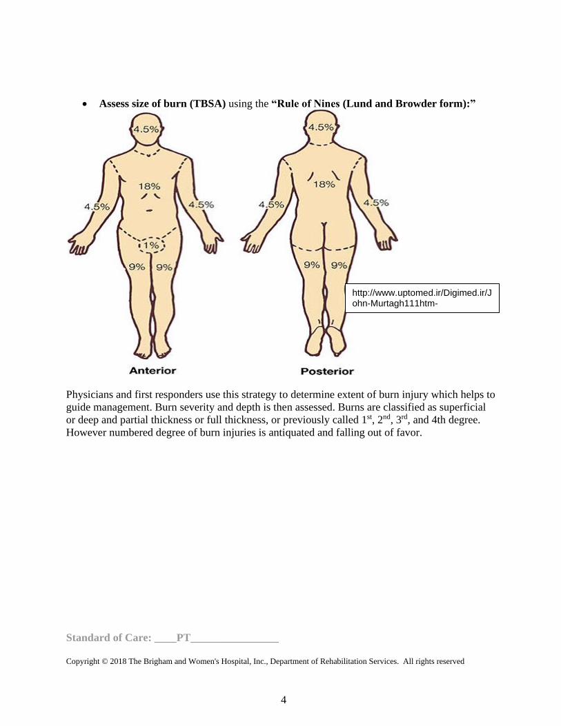
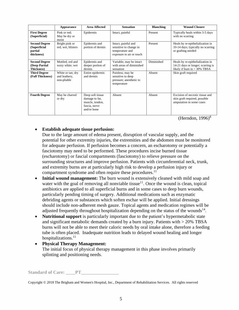
• Assess size of burn (TBSA) using the “Rule of Nines (Lund and Browder form):”
Physicians and first responders use this strategy to determine extent of burn injury which helps to
guide management. Burn severity and depth is then assessed. Burns are classified as superficial
or deep and partial thickness or full thickness, or previously called 1st, 2nd, 3rd, and 4th degree.
However numbered degree of burn injuries is antiquated and falling out of favor.
http://www.uptomed.ir/Digimed.ir/John-Murtagh111htm-Practice/John_Murtagh_Practice/index/part-8/chapter-
Standard of Care: ____PT________________
Copyright © 2018 The Brigham and Women's Hospital, Inc., Department of Rehabilitation Services. All rights reserved
5
Appearance Area Affected Sensation Blanching Wound Closure
First Degree
(Superficial)
Pink or red;
May be dry or moist
Epidermis Intact, painful Present Typically heals within 3-5 days
with no scarring
Second Degree
(Superficial
partial
thickness)
Bright pink or
red, wet, blisters
Epidermis and
portion of dermis
Intact, painful and
sensitive to change in temperature and
exposure to air or touch
Present Heals by re-epithelialization in
10-14 days; typically no scarring or grafting needed
Second Degree
(Deep Partial
Thickness)
Mottled, red and waxy white; wet
Epidermis and deeper portion of
dermis
Variable; may be intact with areas of diminished
sensation
Diminished Heals by re-epithelialization in 14-21 days or longer; scarring is
likely if burn in > 30% TBSA
Third Degree
(Full Thickness)
White or tan; dry and leathery,
non-pliable
Entire epidermis and dermis
Painless; may be sensitive to deep
pressure; anesthetic to
temperature
Absent Skin graft required
Fourth Degree May be charred
or dry
Deep soft tissue
damage to fat,
muscle, tendon, fascia, nerve
and/or bone
Absent Absent Excision of necrotic tissue and
skin graft required, possible
amputation in some cases
(Herndon, 1996)8
• Establish adequate tissue perfusion:
Due to the large amount of edema present, disruption of vascular supply, and the
potential for other extremity injuries, the extremities and the abdomen must be monitored
for adequate perfusion. If perfusion becomes a concern, an escharotomy or potentially a
fasciotomy may need to be performed. These procedures incise burned tissue
(escharotomy) or fascial compartments (fasciotomy) to relieve pressure on the
surrounding structures and improve perfusion. Patients with circumferential neck, trunk,
and extremity burns are at particularly high risk to develop a perfusion injury or
compartment syndrome and often require these procedures.11
• Initial wound management: The burn wound is extensively cleaned with mild soap and
water with the goal of removing all nonviable tissue11. Once the wound is clean, topical
antibiotics are applied to all superficial burns and in some cases to deep burn wounds,
particularly pending timing of surgery. Additional medications such as enzymatic
debriding agents or substances which soften eschar will be applied. Initial dressings
should include non-adherent mesh gauze. Topical agents and medication regimes will be
adjusted frequently throughout hospitalization depending on the status of the wounds14.
• Nutritional support is particularly important due to the patient’s hypermetabolic state
and significant metabolic demands created by a burn injury. Patients with > 20% TBSA
burns will not be able to meet their caloric needs by oral intake alone, therefore a feeding
tube is often placed. Inadequate nutrition leads to delayed wound healing and longer
hospitalizations.11
• Physical Therapy Management:
The initial focus of physical therapy management in this phase involves primarily
splinting and positioning needs.
Standard of Care: ____PT________________
Copyright © 2018 The Brigham and Women's Hospital, Inc., Department of Rehabilitation Services. All rights reserved
6
Acute Phase: (after emergent phase and until wounds are closed)
Medical Management
• Ongoing wound debridement, assessment for evolution of burn wound depth. Burn
wounds can often progress or worsen with time as the retained heat from the injury
continues to damage tissue. Therefore, it is necessary to continue to monitor the wound
for conversion of the burn to a deeper level. It is not uncommon for second degree burns
to convert to third degree, for example.
• Skin grafting is initiated once burn wounds are cleaned and debrided either at bedside
or in the operating room (OR). Autologous grafting or split thickness skin grafting
(STSG) involves using the patient’s own skin to close an integumentary defect created
after removing burned, non-viable tissue. The donor skin is usually meshed to increase
its surface area and thus allow a smaller amount of donor skin to cover a larger recipient
area. For areas such as the face and hands sheet (or unmeshed) grafts are used for
improved cosmetic appearance. Various biologic dressings can also be used as
temporary wound coverage to provide a protective barrier, giving time for donor sites to
heal for future split thickness skin grafts. Allografts are human cadaver skin, while
xenografts are skin from a different species, usually a pig as porcine skin closely
resembles human skin16.
• Cultured Epidermal Autografts (CEA) are used primarily for patients who have
sustained large TBSA burns (> 50 %) and therefore may not have enough donor skin
available to provide complete wound coverage following primary excision16. A section
of the patient’s own skin is sampled, and grown in a laboratory. It can take several
weeks to become ready for use. This technique is costly, and once the new skin is ready
for use, it must be applied immediately, regardless of the status of the recipient wound
beds. CEA also requires up to two weeks of immobility following application,
including cessation of all OT/PT range of motion activities, exercises and functional
mobility. Even turns and positioning with nursing staff is minimalized.
• Vacuum assisted closure (VAC) are often used frequently following operative
management of burn injuries. VACs are placed over donor sites as well as newly grafted
integument. For patients who have sustained a large TBSA burn, a large number VACs
may be used, as there will multiple operative wounds. Physicians/Physician
assistants/nurse practioners apply and remove VACs, this can be done bedside, or in the
OR and this depends on the size of the wound and patient tolerance.
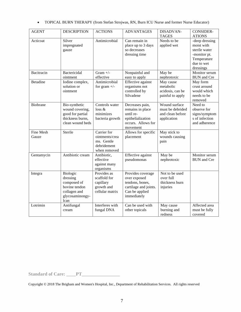
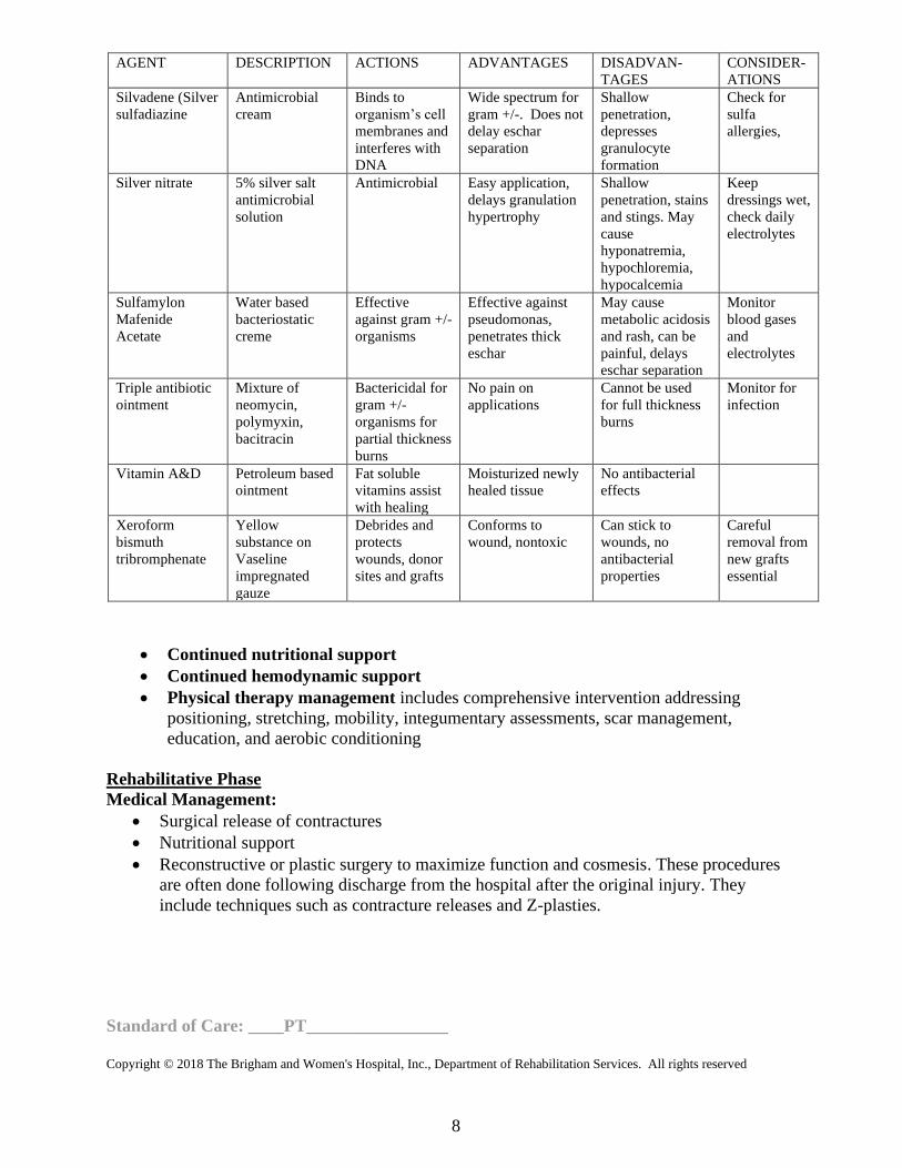
• Wound care is ongoing throughout a patient’s hospital stay and can change as often as
daily depending on the needs of the wound. Below are the common topical agents and
dressings used at BWH.
Standard of Care: ____PT________________
Copyright © 2018 The Brigham and Women's Hospital, Inc., Department of Rehabilitation Services. All rights reserved
7
• TOPICAL BURN THERAPY (from Stefan Strojwas, RN, Burn ICU Nurse and former Nurse Educator)
AGENT DESCRIPTION ACTIONS ADVANTAGES DISADVAN-
TAGES
CONSIDER-
ATIONS
Acticoat Silver
impregnated
gauze
Antimicrobial Can remain in
place up to 3 days
so decreases
dressing time
Needs to be
applied wet
-deep dressing
moist with
sterile water
-monitor pt.
Temperature
due to wet
dressings
Bacitracin Bactericidal
ointment
Gram +/-
effective
Nonpainful and
easy to apply
May be
nephrotoxic
Monitor serum
BUN and Cre
Betadine Iodine complex,
solution or
ointment
Antimicrobial
for gram +/-
Effective against
organisms not
controlled by
Silvadene
May cause
metabolic
acidosis, can be
painful to apply
May form
crust around
would which
needs to be
removed
Biobrane Bio-synthetic
wound covering,
good for partial
thickness burns,
clean wound beds
Controls water
loss &
minimizes
bacteria growth
Decreases pain,
remains in place
until re-
epithelialization
occurs. Allows for
movement
Wound surface
must be debrided
and clean before
application
Need to
observe for
signs/symptom
s of infection
and adherence
Fine Mesh
Gauze
Sterile Carrier for
ointments/crea
ms. Gentle
debridement
when removed
Allows for specific
placement
May stick to
wounds causing
pain
Gentamycin Antibiotic cream Antibiotic,
effective
against many
organisms
Effective against
pseudomonas
May be
nephrotoxic
Monitor serum
BUN and Cre
Integra Biologic
dressing
composed of
bovine tendon
collagen and
glycosaminongy-
lcan
Provides as
scaffold for
capillary
growth and
cellular matrix
Provides coverage
over exposed
tendons, bones,
cartilage and joints.
Can be applied
immediately
Not to be used
over full
thickness burn
injuries
Lotrimin Antifungal
cream
Interferes with
fungal DNA
Can be used with
other topicals
May cause
burning and
redness
Affected area
must be fully
covered
Standard of Care: ____PT________________
Copyright © 2018 The Brigham and Women's Hospital, Inc., Department of Rehabilitation Services. All rights reserved
8
AGENT DESCRIPTION ACTIONS ADVANTAGES DISADVAN-
TAGES
CONSIDER-
ATIONS
Silvadene (Silver
sulfadiazine
Antimicrobial
cream
Binds to
organism’s cell
membranes and
interferes with
DNA
Wide spectrum for
gram +/-. Does not
delay eschar
separation
Shallow
penetration,
depresses
granulocyte
formation
Check for
sulfa
allergies,
Silver nitrate 5% silver salt
antimicrobial
solution
Antimicrobial Easy application,
delays granulation
hypertrophy
Shallow
penetration, stains
and stings. May
cause
hyponatremia,
hypochloremia,
hypocalcemia
Keep
dressings wet,
check daily
electrolytes
Sulfamylon
Mafenide
Acetate
Water based
bacteriostatic
creme
Effective
against gram +/-
organisms
Effective against
pseudomonas,
penetrates thick
eschar
May cause
metabolic acidosis
and rash, can be
painful, delays
eschar separation
Monitor
blood gases
and
electrolytes
Triple antibiotic
ointment
Mixture of
neomycin,
polymyxin,
bacitracin
Bactericidal for
gram +/-
organisms for
partial thickness
burns
No pain on
applications
Cannot be used
for full thickness
burns
Monitor for
infection
Vitamin A&D Petroleum based
ointment
Fat soluble
vitamins assist
with healing
Moisturized newly
healed tissue
No antibacterial
effects
Xeroform
bismuth
tribromphenate
Yellow
substance on
Vaseline
impregnated
gauze
Debrides and
protects
wounds, donor
sites and grafts
Conforms to
wound, nontoxic
Can stick to
wounds, no
antibacterial
properties
Careful
removal from
new grafts
essential
• Continued nutritional support
• Continued hemodynamic support
• Physical therapy management includes comprehensive intervention addressing
positioning, stretching, mobility, integumentary assessments, scar management,
education, and aerobic conditioning
Rehabilitative Phase
Medical Management:
• Surgical release of contractures
• Nutritional support
• Reconstructive or plastic surgery to maximize function and cosmesis. These procedures
are often done following discharge from the hospital after the original injury. They
include techniques such as contracture releases and Z-plasties.
Standard of Care: ____PT________________
Copyright © 2018 The Brigham and Women's Hospital, Inc., Department of Rehabilitation Services. All rights reserved
9
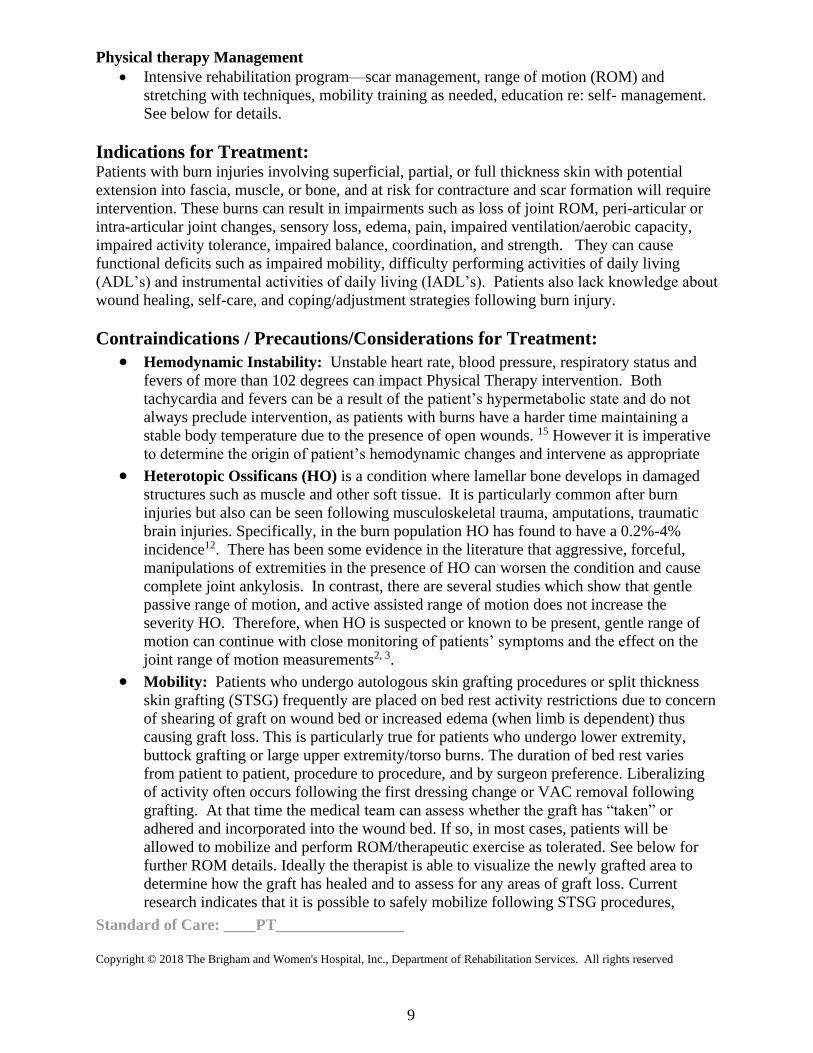
Physical therapy Management
• Intensive rehabilitation program—scar management, range of motion (ROM) and
stretching with techniques, mobility training as needed, education re: self- management.
See below for details.
Indications for Treatment: Patients with burn injuries involving superficial, partial, or full thickness skin with potential
extension into fascia, muscle, or bone, and at risk for contracture and scar formation will require
intervention. These burns can result in impairments such as loss of joint ROM, peri-articular or
intra-articular joint changes, sensory loss, edema, pain, impaired ventilation/aerobic capacity,
impaired activity tolerance, impaired balance, coordination, and strength. They can cause
functional deficits such as impaired mobility, difficulty performing activities of daily living
(ADL’s) and instrumental activities of daily living (IADL’s). Patients also lack knowledge about
wound healing, self-care, and coping/adjustment strategies following burn injury.
Contraindications / Precautions/Considerations for Treatment:
• Hemodynamic Instability: Unstable heart rate, blood pressure, respiratory status and
fevers of more than 102 degrees can impact Physical Therapy intervention. Both
tachycardia and fevers can be a result of the patient’s hypermetabolic state and do not
always preclude intervention, as patients with burns have a harder time maintaining a
stable body temperature due to the presence of open wounds. 15 However it is imperative
to determine the origin of patient’s hemodynamic changes and intervene as appropriate
• Heterotopic Ossificans (HO) is a condition where lamellar bone develops in damaged
structures such as muscle and other soft tissue. It is particularly common after burn
injuries but also can be seen following musculoskeletal trauma, amputations, traumatic
brain injuries. Specifically, in the burn population HO has found to have a 0.2%-4%
incidence12. There has been some evidence in the literature that aggressive, forceful,
manipulations of extremities in the presence of HO can worsen the condition and cause
complete joint ankylosis. In contrast, there are several studies which show that gentle
passive range of motion, and active assisted range of motion does not increase the
severity HO. Therefore, when HO is suspected or known to be present, gentle range of
motion can continue with close monitoring of patients’ symptoms and the effect on the
joint range of motion measurements2, 3.
• Mobility: Patients who undergo autologous skin grafting procedures or split thickness
skin grafting (STSG) frequently are placed on bed rest activity restrictions due to concern
of shearing of graft on wound bed or increased edema (when limb is dependent) thus
causing graft loss. This is particularly true for patients who undergo lower extremity,
buttock grafting or large upper extremity/torso burns. The duration of bed rest varies
from patient to patient, procedure to procedure, and by surgeon preference. Liberalizing
of activity often occurs following the first dressing change or VAC removal following
grafting. At that time the medical team can assess whether the graft has “taken” or
adhered and incorporated into the wound bed. If so, in most cases, patients will be
allowed to mobilize and perform ROM/therapeutic exercise as tolerated. See below for
further ROM details. Ideally the therapist is able to visualize the newly grafted area to
determine how the graft has healed and to assess for any areas of graft loss. Current
research indicates that it is possible to safely mobilize following STSG procedures,
Standard of Care: ____PT________________
Copyright © 2018 The Brigham and Women's Hospital, Inc., Department of Rehabilitation Services. All rights reserved
10
provided certain precautions are taken, as early as post op day zero. These precautions
include immobilization of the grafted area with ambulatory aides (air cast boots, knee
immobilizers) and compression provided by elastic bandages.13 However, a discussion
with the medical staff remains necessary to confirm if a patient’s mobility can be
progressed and to determine if there are any ROM or positioning restrictions.
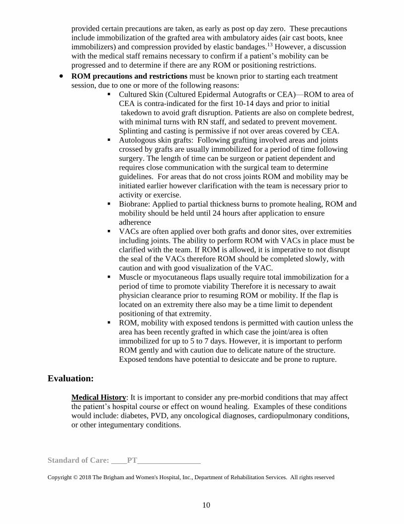
• ROM precautions and restrictions must be known prior to starting each treatment
session, due to one or more of the following reasons: ▪ Cultured Skin (Cultured Epidermal Autografts or CEA)—ROM to area of
CEA is contra-indicated for the first 10-14 days and prior to initial
takedown to avoid graft disruption. Patients are also on complete bedrest,
with minimal turns with RN staff, and sedated to prevent movement.
Splinting and casting is permissive if not over areas covered by CEA.
▪ Autologous skin grafts: Following grafting involved areas and joints
crossed by grafts are usually immobilized for a period of time following
surgery. The length of time can be surgeon or patient dependent and
requires close communication with the surgical team to determine
guidelines. For areas that do not cross joints ROM and mobility may be
initiated earlier however clarification with the team is necessary prior to
activity or exercise.
▪ Biobrane: Applied to partial thickness burns to promote healing, ROM and
mobility should be held until 24 hours after application to ensure
adherence
▪ VACs are often applied over both grafts and donor sites, over extremities
including joints. The ability to perform ROM with VACs in place must be
clarified with the team. If ROM is allowed, it is imperative to not disrupt
the seal of the VACs therefore ROM should be completed slowly, with
caution and with good visualization of the VAC.
▪ Muscle or myocutaneous flaps usually require total immobilization for a
period of time to promote viability Therefore it is necessary to await
physician clearance prior to resuming ROM or mobility. If the flap is
located on an extremity there also may be a time limit to dependent
positioning of that extremity.
▪ ROM, mobility with exposed tendons is permitted with caution unless the
area has been recently grafted in which case the joint/area is often
immobilized for up to 5 to 7 days. However, it is important to perform
ROM gently and with caution due to delicate nature of the structure.
Exposed tendons have potential to desiccate and be prone to rupture.
Evaluation:
Medical History: It is important to consider any pre-morbid conditions that may affect
the patient’s hospital course or effect on wound healing. Examples of these conditions
would include: diabetes, PVD, any oncological diagnoses, cardiopulmonary conditions,
or other integumentary conditions.
Standard of Care: ____PT________________
Copyright © 2018 The Brigham and Women's Hospital, Inc., Department of Rehabilitation Services. All rights reserved
11
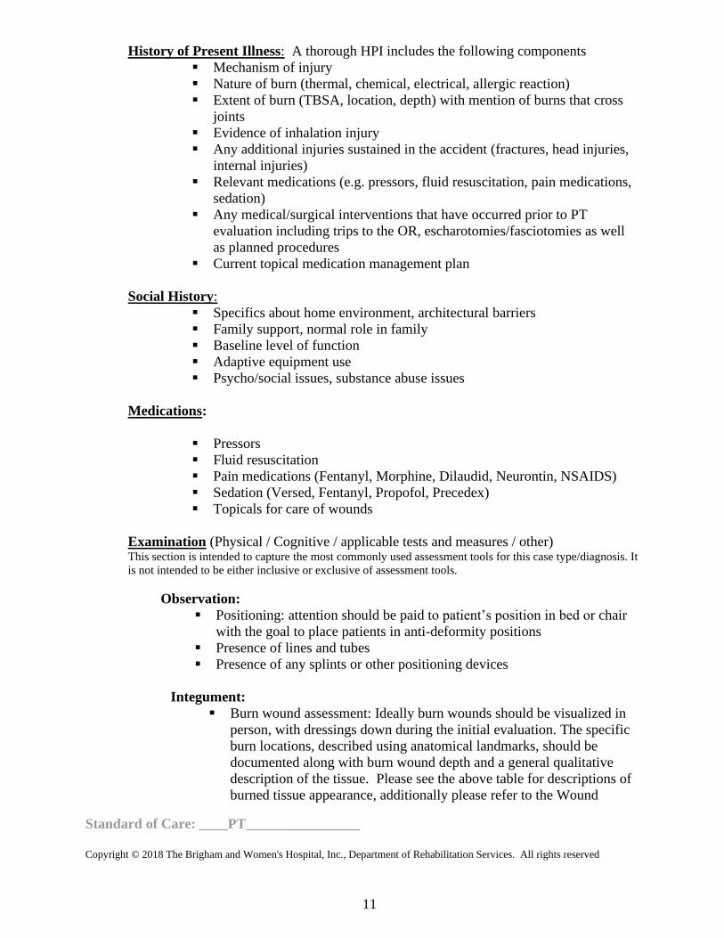
History of Present Illness: A thorough HPI includes the following components
▪ Mechanism of injury
▪ Nature of burn (thermal, chemical, electrical, allergic reaction)
▪ Extent of burn (TBSA, location, depth) with mention of burns that cross
joints
▪ Evidence of inhalation injury
▪ Any additional injuries sustained in the accident (fractures, head injuries,
internal injuries)
▪ Relevant medications (e.g. pressors, fluid resuscitation, pain medications,
sedation)
▪ Any medical/surgical interventions that have occurred prior to PT
evaluation including trips to the OR, escharotomies/fasciotomies as well
as planned procedures
▪ Current topical medication management plan
Social History:
▪ Specifics about home environment, architectural barriers
▪ Family support, normal role in family
▪ Baseline level of function
▪ Adaptive equipment use
▪ Psycho/social issues, substance abuse issues
Medications:
▪ Pressors
▪ Fluid resuscitation
▪ Pain medications (Fentanyl, Morphine, Dilaudid, Neurontin, NSAIDS)
▪ Sedation (Versed, Fentanyl, Propofol, Precedex)
▪ Topicals for care of wounds
Examination (Physical / Cognitive / applicable tests and measures / other) This section is intended to capture the most commonly used assessment tools for this case type/diagnosis. It
is not intended to be either inclusive or exclusive of assessment tools.
Observation:
▪ Positioning: attention should be paid to patient’s position in bed or chair
with the goal to place patients in anti-deformity positions
▪ Presence of lines and tubes
▪ Presence of any splints or other positioning devices
Integument:
▪ Burn wound assessment: Ideally burn wounds should be visualized in
person, with dressings down during the initial evaluation. The specific
burn locations, described using anatomical landmarks, should be
documented along with burn wound depth and a general qualitative
description of the tissue. Please see the above table for descriptions of
burned tissue appearance, additionally please refer to the Wound
Standard of Care: ____PT________________
Copyright © 2018 The Brigham and Women's Hospital, Inc., Department of Rehabilitation Services. All rights reserved
12
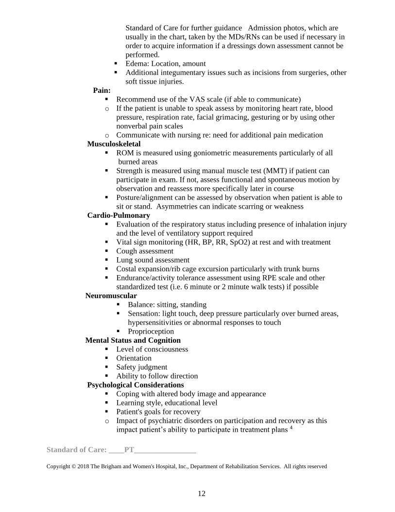
Standard of Care for further guidance Admission photos, which are
usually in the chart, taken by the MDs/RNs can be used if necessary in
order to acquire information if a dressings down assessment cannot be
performed.
▪ Edema: Location, amount
▪ Additional integumentary issues such as incisions from surgeries, other
soft tissue injuries.
Pain:
▪ Recommend use of the VAS scale (if able to communicate)
o If the patient is unable to speak assess by monitoring heart rate, blood
pressure, respiration rate, facial grimacing, gesturing or by using other
nonverbal pain scales
o Communicate with nursing re: need for additional pain medication
Musculoskeletal
▪ ROM is measured using goniometric measurements particularly of all
burned areas
▪ Strength is measured using manual muscle test (MMT) if patient can
participate in exam. If not, assess functional and spontaneous motion by
observation and reassess more specifically later in course
▪ Posture/alignment can be assessed by observation when patient is able to
sit or stand. Asymmetries can indicate scarring or weakness
Cardio-Pulmonary
▪ Evaluation of the respiratory status including presence of inhalation injury
and the level of ventilatory support required
▪ Vital sign monitoring (HR, BP, RR, SpO2) at rest and with treatment
▪ Cough assessment
▪ Lung sound assessment
▪ Costal expansion/rib cage excursion particularly with trunk burns
▪ Endurance/activity tolerance assessment using RPE scale and other
standardized test (i.e. 6 minute or 2 minute walk tests) if possible
Neuromuscular
▪ Balance: sitting, standing
▪ Sensation: light touch, deep pressure particularly over burned areas,
hypersensitivities or abnormal responses to touch
▪ Proprioception
Mental Status and Cognition
▪ Level of consciousness
▪ Orientation
▪ Safety judgment
▪ Ability to follow direction
Psychological Considerations
▪ Coping with altered body image and appearance
▪ Learning style, educational level
▪ Patient's goals for recovery
o Impact of psychiatric disorders on participation and recovery as this
impact patient’s ability to participate in treatment plans 4
Standard of Care: ____PT________________
Copyright © 2018 The Brigham and Women's Hospital, Inc., Department of Rehabilitation Services. All rights reserved
13
Functional Mobility: Assess bed mobility, transfers, ambulation and stairs
appropriate
Assessment: Patients who sustain burn injuries are at high risk for scarring, contractures, and impaired
functional mobility. Also, particularly with large TBSA burns and/or inhalation injuries, there is
usually a long period of relative immobility as patients undergo multiple grafting procedures, and
require prolonged intubation and sedation. These patients are at high risk of developing aerobic
capacity/endurance impairments, balance derangements as well cognitive dysfunction. Given all
of this, patients who sustain burn injuries often require intensive and frequent intervention. It is
important that a therapist be well versed in the specific intervention strategies that are unique to
the burn population and have demonstrated competency in these areas.
Problem List
Health Condition: Burn
Body Structure and Functions:
▪ Impaired integument
▪ Impaired ROM
▪ Impaired motor performance
▪ Impaired balance
▪ Impaired aerobic conditioning
▪ Impaired airway clearance
▪ Impaired ventilation
▪ Impaired balance
▪ Edema
▪ Impaired cognition
▪ Impaired communication
Activity Limitations:
▪ Decreased bed mobility
▪ Decreased transfers
▪ Decreased ambulation
▪ Decreased stairs
Participation:
▪ Decreased community mobility
▪ Impaired vocation
▪ Impaired role in family
▪ Decreased home management
Prognosis: Overall, there has been a significant decline in the incidence of
burn injuries over the decades. In the 1950’s burns occurred approximately 10 per 1000
people as compared with 4.2 per 1000 in the 1990’s. This decline has been associated
with increased injury prevention in the workplace and at home along with the
implementation of other safety measures. Along with a decline in overall incidence,
mortality has also significantly declined due to the development of comprehensive burn
centers, advancement in treatment such as improved resuscitation, topical agents, new
Standard of Care: ____PT________________
Copyright © 2018 The Brigham and Women's Hospital, Inc., Department of Rehabilitation Services. All rights reserved
14
antibiotics, early excision and grafting, and the use of artificial skin substitutes.6
However, prognosis can be highly variable. Some considerations that impact prognosis
are depth of burn, surface area involved, type of burn (chemical and electrical injuries
may increase length of stay), presence of an inhalation injury, significant psychiatric or
substance abuse issues and co-morbidities such as history of smoking, diabetes. “Risk
factors most strongly associated with death are increasing total body surface area
(TBSA), inhalation injuries and increasing age”.5 In terms of long term outcomes for
survivors, one study found that their health was poorer than matched non-injured controls
on all eight subscales of the SF-36 Health Assessment Tool. Additionally, greater than
20% of burn survivors have been to found to have elevated levels of psychological
distress, as compared with 12% in a control group. Therefore, while an increased number
of patients are surviving a burn injury, there is evidence of continued medical and
psychological needs which require ongoing treatment.6
Goals (Measurable parameters and specific timelines to be included on evaluation)
1.) Prevent loss of ROM with appropriate positioning program and exercise program
decreasing risk for contracture
2.) Patient will demonstrate independent functional mobility, with least restrictive
assistive devices
3.) Minimize hypertrophic scarring, especially with at risk patient populations
4.) Patient will be independent with appropriate home exercise program
5.) Patient will rate all activity as less than or equal 3/10 on Modified Borg Scale
6.) Patient will demonstrate good posture without asymmetries in supine, sitting and
standing
7.) Patient will demonstrate good understanding and need of all splinting devices and
activity progression
Treatment Planning / Interventions
Established Pathway ___ Yes, see attached. _X_ No
Established Protocol ___ Yes, see attached. _X_ No
Interventions most commonly used for this case type/diagnosis. This section is intended to capture the most commonly used interventions for this case type/diagnosis. It is
not intended to be either inclusive or exclusive of appropriate interventions.
• Pain management can be a difficult hurdle for patients who have sustained a burn
injury. An appropriate pain regime is paramount for successful rehab interventions and
requires a multi-disciplinary approach. Treatment sessions should be coordinated around
a patient’s pain medication schedule and often additional medications can be given
during the therapy session if needed. It is important to update the nurse and the team if
pain management is not appropriate as this can significantly impact a patient’s overall
prognosis and recovery. Involving the patient as much as possible is also helpful to
Standard of Care: ____PT________________
Copyright © 2018 The Brigham and Women's Hospital, Inc., Department of Rehabilitation Services. All rights reserved
15
reduce anxiety and pain during a session. Completing ROM with techniques such as
AAROM and contract relax can be very beneficial. Creating a daily schedule, with set
times for PT and OT, with the assistance of the primary RNs and the involvement of the
patient (if possible) helps coordinate pain medication schedules, gives patients both self-
efficacy and structure to their day. Use of cognitive behavioral strategies such as
relaxation techniques, and guided imagery are also helpful.
• ROM/Therapeutic exercise. A cornerstone in burn injury management is the use of
effective range of motion exercises. Due to nature of burn injury, patients are high risk
for scarring and thus early and frequent range of motion to prevent long standing wound
contractures is necessary. Burn stretches should be held, at end range, for a prolonged
period of time. One study found that holding each individual stretch at end range (as
evidenced by blanching of the soft tissue) for 3 minutes for a total of 60 minutes per day
(divided between both OT and PT) resulted in an improvement in ROM by 8.2 degrees
per week7. Once awake and participatory, involving patients in self stretching AAROM
and using techniques such as contract relax or hold relax can help with gaining additional
range of motion as well decreasing patient anxiety.
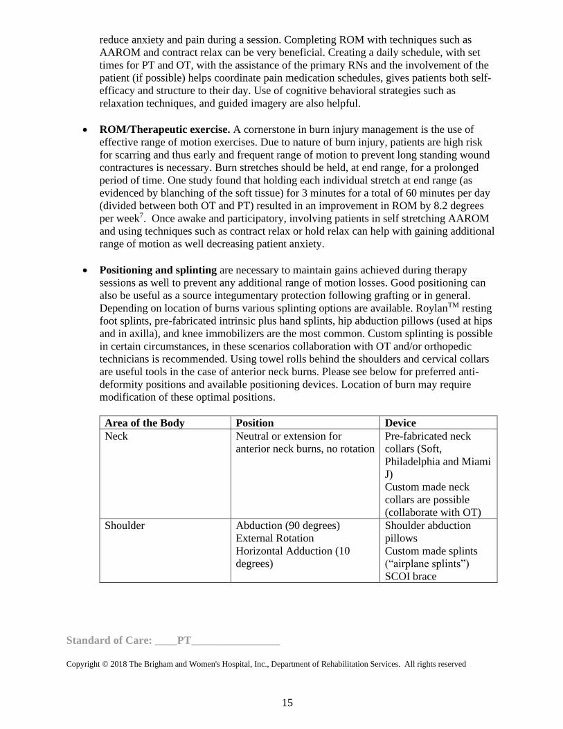
• Positioning and splinting are necessary to maintain gains achieved during therapy
sessions as well to prevent any additional range of motion losses. Good positioning can
also be useful as a source integumentary protection following grafting or in general.
Depending on location of burns various splinting options are available. RoylanTM resting
foot splints, pre-fabricated intrinsic plus hand splints, hip abduction pillows (used at hips
and in axilla), and knee immobilizers are the most common. Custom splinting is possible
in certain circumstances, in these scenarios collaboration with OT and/or orthopedic
technicians is recommended. Using towel rolls behind the shoulders and cervical collars
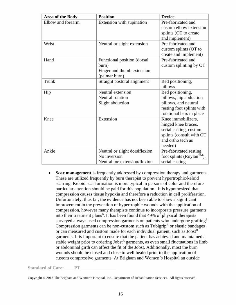
are useful tools in the case of anterior neck burns. Please see below for preferred anti-
deformity positions and available positioning devices. Location of burn may require
modification of these optimal positions.
Area of the Body Position Device
Neck Neutral or extension for
anterior neck burns, no rotation
Pre-fabricated neck
collars (Soft,
Philadelphia and Miami
J)
Custom made neck
collars are possible
(collaborate with OT)
Shoulder Abduction (90 degrees)
External Rotation
Horizontal Adduction (10
degrees)
Shoulder abduction
pillows
Custom made splints
(“airplane splints”)
SCOI brace
Standard of Care: ____PT________________
Copyright © 2018 The Brigham and Women's Hospital, Inc., Department of Rehabilitation Services. All rights reserved
16
Area of the Body Position Device
Elbow and forearm Extension with supination Pre-fabricated and
custom elbow extension
splints (OT to create
and implement)
Wrist Neutral or slight extension Pre-fabricated and
custom splints (OT to
create and implement)
Hand Functional position (dorsal
burn)
Finger and thumb extension
(palmar burn)
Pre-fabricated and
custom splinting by OT
Trunk Straight postural alignment Bed positioning,
pillows
Hip Neutral extension
Neutral rotation
Slight abduction
Bed positioning,
pillows, hip abduction
pillows, and neutral
resting foot splints with
rotational bars in place
Knee Extension Knee immobilizers,
hinged knee braces,
serial casting, custom
splints (consult with OT
and ortho tech as
needed)
Ankle Neutral or slight dorsiflexion
No inversion
Neutral toe extension/flexion
Pre-fabricated resting
foot splints (RoylanTM),
serial casting
• Scar management is frequently addressed by compression therapy and garments.
These are utilized frequently by burn therapist to prevent hypertrophic/keloid
scarring. Keloid scar formation is more typical in persons of color and therefore
particular attention should be paid for this population. It is hypothesized that
compression causes tissue hypoxia and therefore a reduction in cell proliferation.
Unfortunately, thus far, the evidence has not been able to show a significant
improvement in the prevention of hypertrophic wounds with the application of
compression, however many therapists continue to incorporate pressure garments
into their treatment plans6. It has been found that 49% of physical therapists
surveyed always used compression garments on patients who undergone grafting9
Compression garments can be non-custom such as TubigripR or elastic bandages
or can measured and custom made for each individual patient, such as JobstR
garments. It is important to ensure that the patient has achieved and maintained a
stable weight prior to ordering JobstR garments, as even small fluctuations in limb
or abdominal girth can affect the fit of the Jobst. Additionally, most the burn
wounds should be closed and close to well healed prior to the application of
custom compressive garments. At Brigham and Women’s Hospital an outside
Standard of Care: ____PT________________
Copyright © 2018 The Brigham and Women's Hospital, Inc., Department of Rehabilitation Services. All rights reserved
17
vendor, who can measure both inpatients as well as outpatients in burn clinic,
provides custom compressive garments. The burn program manager/burn
department can facilitate acquiring compressive garments, and should be
contacted by the therapist when a patient is deemed ready.
• Functional mobility training
• Ventilatory training such as costal expansion, diaphragmatic breathing
particularly for patients who have sustained truncal burns.
• Endurance/aerobic conditioning
Frequency & Duration: Patients who are admitted to Brigham and Women’s Hospital
for management of a burn injury are frequently seen 5-7 times weekly, based on clinical
need. The severity and location of burn wounds and the need for aggressive ROM and
mobility are the primary factors determining frequency. Length of stay is dependent on
extent and severity of burns and need for intensive acute care intervention. Hospital stay
can vary from 2-3 days for a localized burn (such as partial thickness burn to hand or
foot) to many weeks to months for a high percentage, full thickness burns that require
multiple surgical procedures and prolonged intubation.
Patient / family education
▪ Discussion with patient and family re: Physical Therapy involvement with patient
and expected progression
▪ Discussion with patient and family re: optimizing patient’s independent mobility
and self-care and providing the appropriate level of assistance to the patient
▪ Instruction of patient and family in appropriate exercises and activities with written
exercise program and exercise/activity log
▪ Discussion of longer term issues common following a burn injuries
1. phases of burn healing, estimated time line, risk of scarring
2. ways to minimize scarring and contracture
3. proper management of pressure garments, DME
4. proper skin care and protection
Recommendations and referrals to other providers.
▪ Occupational Therapy
▪ Speech Therapy
▪ Social Work
▪ Care Coordination
▪ Psychiatry
▪ Orthopedic Technician
▪ Medical interpreters
▪ Outside resources for the measurement and fit of compression garments
▪ Outside Resources such as support groups (e.g. the Phoenix Society). Visits by
known burn survivors who can talk with patient and family can be arranged by the
social worker
Standard of Care: ____PT________________
Copyright © 2018 The Brigham and Women's Hospital, Inc., Department of Rehabilitation Services. All rights reserved
18
Re-evaluation Re-evaluations should be performed every 10 days while patient is admitted to the
hospital. However, re-evaluations may need to be performed more frequently in the
setting of changes to medical status, new procedures or a significant change in function.
Discharge Planning
Commonly expected outcomes at discharge: Patients who sustain large TBSA burns or
smaller burns with complications, often require rehab placement to address
continued impairments from a physical therapy standpoint as well as for continued
wound management. Brigham and Women’s Hospital has an established relationship
with Spaulding Rehab in Boston and large majority of patients who have burn injuries are
discharged to this facility. Sub-acute/SNF level rehab centers are an option for less
involved burns if needed. Smaller burns can often be managed in the community usually
with family assistance and home or outpatient physical therapy involvement. These
patients are seen frequently in the burn clinic at BWH as well have daily visits by visiting
nurses for dressing changes.
Transfer of Care When a patient leaves Brigham and Women’s a thorough physical
therapy discharge summary is necessary to assist with transition of care. The referral
should include an updated hospital course particularly all grafting procedures, current
functional status, an updated and complete integumentary assessment, status of
compression garments, and current ROM measurements.
Patient’s discharge instructions Patients who are discharged home are often given a
written independent therapeutic exercise program, and with the involvement
of their home physical therapists. Patients are also educated on the need for eventual
compression garments and the mechanisms for obtaining these garments.
Authors: Alisa G. Finkel PT Reviewed by: Barbara Odaka PT
October 2008 Melanie Parker PT
Merideth Donlan PT
Revised:
Author: Meredith Detwiller, PT Reviewed by: Alisa Finkel, PT
May 2018 Barbara Odaka, PT
Standard of Care: ____PT________________
Copyright © 2018 The Brigham and Women's Hospital, Inc., Department of Rehabilitation Services. All rights reserved
19
REFERENCES 1.) American Burn Association. Burn Incidence and Treatment in the United States:
2016 Fact Sheet. Available at: http://www.ameriburn.org/resources_factsheet.php.
Accessibility verified: April 16th, 2018.
2.) Casavanat AM, Hastings H. Heterotopic Ossification about the Elbow: A Therapist’s
Guide to Evaluation and Management. Journal of Hand Therapy. 2006; 19: 255-267.
3.) Crawford CM, Varghese G, Mani M, Neff JR. Heterotopic Ossification: Are Range of
Motion Exercises Contraindicated? Journal of Burn Care and Research. 1986; 7;
323-327.
4.) Druery M, Brown Tim La H, Muller, M. Longterm Functional Outcomes and Quality
of Life Following Severe Burn Injury. Burns. September 2005; 31 (6):692-5
5.) Edelman DA, White MT, Tyburski JG, Wilson RF. Factors Affecting Prognosis of
Inhalation Injury. Journal of Burn Care and Research. 2006; 27: 848-853.
6.) Esselman PC, Thombs BD, Magyar-Russell G, Fauerbach JA. Burn Rehabilitation:
State of the Science. American Journal Physical Medicine Rehabilitation. 2006; 85:
383-413.
7.) Godeleski M, Oeffling A, Bruflat AK, Craig E, Weitzenkamp D, Lindberg G.
Treating Burn-Associated Joint Contracture: Results of an Inpatient Rehabilitation
Stretching Protocol. Journal of Burn Care and Research. 2013; 34: 420-426.
8.) Herndon, D. (Ed.). (1996). Total burn care. London: W. B. Saunders Company Ltd
9.) Holavanahalli RK, Helm PA, Parry IS, Dolezal CA, Greenhalgh DG. Select Practices
in Management and Rehabiliation of Burns: A Survey Report. Journal of Burn Care
and Research. 2011; 32: 210-223.
10.) Ipatchi K, Arbabi S. Advances in Burn Critical Care. Critical Care Medicine.
2006; 34: S239-S244
11.) Latenser BA. Critical Care of the Burn Patient: The First 48 Hours. Critical Care
Medicine. 2009; 37: 2819-2826.
12.) Medina A, Shankowsky H, Savaryn B, Shukalak B, Tredget EE. Characterization
of Heterotopic Ossification in Burn Patients. Journal of Burn Care and Research.
2013; XX: 1-6.
13.) Nedelec, B., Serghiou, M et.al Early Ambulation of Burn Survivors after Lower
Extremity Grafts. Journal of Burn Care and Research. 2012; May/June: 319-329.
14.) Orgill DP, Rice PL. Emergency Care of Moderate and Severe Thermal Burns in
Adults. In: UpToDate, Rose, BD (Ed), UpToDate, Waltham, MA, 2012.
15.) Richard RL and Staley, M., Burn Care and Rehabilitation: Principles and
Practice, Philadelphia, PA: F. A. Davis Company; 1994: chapter 2/25.
16.) Richard RL and Staley, M., Burn Care and Rehabilitation: Principles and
Practice, Philadelphia, PA: F. A. Davis Company; 1994: chapter 22/622-40
17.) Taylor S, Manning S, Quaries J. A Multidisciplinary Approach to Early
Mobilization of Patients with Burns. Critical Care Nursing Quarterly. 2013; 36: 56-
62.
18.) Trombly, CA, Radomski MV, eds. Occupational Therapy for Physical
Dysfunction. 5th ed. Philadephia, PA: Lippincott Williams & Wikins; 2002; 1025-
1030.
19.) Whitehead C, Serghiou M A 12 Year Comparison of Common Therapeutic
Interventions in the Burn Unit. Journal of Burn Care and Research. 2009; 30: 281-
287.