self-cross-linking biopolymers as injectable in situ forming biodegradable scaffolds
TRANSCRIPT
ARTICLE IN PRESS
0142-9612/$ - se
doi:10.1016/j.bi
�Correspond
E-mail addr
Biomaterials 26 (2005) 3941–3951
www.elsevier.com/locate/biomaterials
Self-cross-linking biopolymers as injectable in situ formingbiodegradable scaffolds
Biji Balakrishnan, A. Jayakrishnan�
Polymer Chemistry Division, Biomedical Technology Wing, Sree Chitra Tirunal Institute for Medical Sciences and Technology,
Satelmond Palace Campus, Poojapura, Trivandrum, Kerala 695012, India
Received 12 June 2004; accepted 20 October 2004
Available online 2 December 2004
Abstract
The injectable polymer scaffolds which are biocompatible and biodegradable are important biomaterials for tissue engineering
and drug delivery. Hydrogels derived from natural proteins and polysaccharides are ideal scaffolds for tissue engineering since they
resemble the extracellular matrices of the tissue comprised of various amino acids and sugar-based macromolecules. Here, we report
a new class of hydrogels derived from oxidized alginate and gelatin. We show that periodate-oxidized sodium alginate having
appropriate molecular weight and degree of oxidation rapidly cross-links proteins such as gelatin in the presence of small
concentrations of sodium tetraborate (borax) to give injectable systems for tissue engineering, drug delivery and other medical
applications. The rapid gelation in the presence of borax is attributed to the slightly alkaline pH of the medium as well as the ability
of borax to complex with hydroxyl groups of polysaccharides. The effect of degree of oxidation and concentration of alginate
dialdehyde, gelatin and borax on the speed of gelation was examined. As a general rule, the gelling time decreased with increase in
concentration of oxidized alginate, gelatin and borax and increase in the degree of oxidation of alginate. Cross-linking parameters of
the gel matrix were studied by swelling measurements and trinitrobenzene sulphonic acid (TNBS) assay. In general, the degree of
cross-linking was found to increase with increase in the degree of oxidation of alginate, whereas the swelling ratio and the degree of
swelling decreased. The gel was found to be biocompatible and biodegradable. The potential of the system as an injectable drug
delivery vehicle and as a tissue-engineering scaffold is demonstrated by using primaquine as a model drug and by encapsulation of
hepatocytes inside the gel matrix, respectively.
r 2004 Elsevier Ltd. All rights reserved.
Keywords: Hydrogel; Alginate; Gelatin; Oxidation; Drug delivery; Cell encapsulation
1. Introduction
Biocompatible, biodegradable polymer scaffolds areimportant biomaterials for tissue engineering and drugdelivery [1–3]. Molecular design of these materials hasimportant role in determining their suitability in suchapplications. The simplest and the most convenientapproach in these applications will be to inject thepolymer-cell/drug entity into the body. Injectablesystems offer specific advantages over preformed scaf-
e front matter r 2004 Elsevier Ltd. All rights reserved.
omaterials.2004.10.005
ing author. Fax: +91 471 2341814.
ess: [email protected] (A. Jayakrishnan).
folds, which include ease of application, confineddelivery for a site-specific action and improved patientcompliance and comfort [4]. Various methods have beenemployed for the preparation of injectable hydrogelsystems. Water-soluble, thermosensitive and pH sensi-tive polymers exhibiting reversible sol–gel transition andphoto-polymerizable hydrogels have been tailor-madeas injectables [5–8].
Hydrogels derived from naturally occurring polymersmimic many features of extracellular matrix and thushave the potential to direct the migration, growth andorganization of cells during tissue regeneration andwound healing and for stabilization of encapsulated and
ARTICLE IN PRESSB. Balakrishnan, A. Jayakrishnan / Biomaterials 26 (2005) 3941–39513942
transplanted cells. Many of them also demonstrateadequate biocompatibility and biodegradability. In situgelling formulations from biopolymers are achieved byphoto-polymerization of their custom-made monomers[9], enzymatic cross-linking [10,11] chemical cross-linking with metal ions [12] or by cross-linking agentssuch as glutaraldehyde, carbodiimide, adipic dihydra-zide etc [13]. However, photo-polymerization oftenrequires a photo-sensitizer and prolonged irradiationlimiting their use. Cross-linking with metal ions is oftenreversible in the body and exerts cytotoxic effects [3].Agents that are incorporated into the polymer matrixsuch as glutaraldehyde, polyepoxides and isocyanatesare highly toxic and are prone to leach out into the bodyon matrix biodegradation [14]. Agents that cross-linkwithout incorporation by activating the carboxylic acidresidues in biopolymers such as acyl azides andcarbodiimides are considered less toxic. The toxicity ofcross-linking agents is the major obstacle in the use ofthese polymers as injectable, in situ-forming polymerscaffolds, since their seepage into body fluids even at lowconcentrations can be catastrophic [15]. Potentially lesstoxic reagents such as adipic acid dihydrazide [16,17]and oxidized mono, di and polysaccharides have beeninvestigated as cross-linking agents [18,19]. The gelationreaction leading to the three-dimensional network israther slow with many of these reagents to be ofpractical use as injectable systems and modification ofthe biopolymer has been attempted to reduce the gellingtime [20].
Alginates, derived from brown sea weed are anioniclinear polysaccharides composed of 1,4-linked b-D-man-nuronate (M) and 1,4-linked a-L-guluronate (G) residuesin varying proportions. An important feature of alginate isits gelation in the presence of divalent cations, such ascalcium. The G-blocks are responsible for the ‘‘egg box’’formation with calcium ions or other alkaline earth metalions [21]. This gentle property has led to their wide use ascell transplantation vehicles to grow new tissues and aswound dressings. However, alginate hydrogels used inthese applications have uncontrollable degradation ki-netics and gels dissolve in an uncontrollable mannerfollowing the loss of divalent cations releasing high andlow molecular weight alginate units [22]. There are reportsshowing degradability of low molecular weight(o80 KDa) alginates [23]. However, when used as wounddressings, there was no evidence of degradation orbreakdown of the alginate fibres [24]. Also, high calciumconcentration is reported to inhibit the growth of cells inculture [25]. Attempts have been made to covalently cross-link sodium alginate with gelatin and ethylenediamineusing water-soluble carbodiimide [26,27]. Both ethylene-diamine and carbodiimide are however, toxic reagents.Recently it is reported that although higher molecularweight alginate is non-biodegradable, its di-aldehydederivative is biodegradable [28].
Here we show that an injectable, in situ-forming, non-toxic, biodegradable polymer scaffold can be preparedby self-cross-linking of oxidized alginate and gelatin, inthe presence of small concentrations of borax withoutemploying any extraneous cross-linking agents. Gelatinhas a long history of medical use as a plasma expanderas a wound dressing material and as adhesive andabsorbent pads, etc. [14]. Borax also has a long historyof medical use and the mean lethal dose in man exceeds700 mg/kg [29]. Toxicity or discomfort has not beenreported in humans receiving equivalent to 100 mg ofboron intravenously [30]. Boron is reported to preventosteoporosis at a dose between 3 and 6 mg/day and inpersons with arthritis a dose of 3 mg of boron per dayfor 2–4 months is indicated [31].
The objective of the present study is to demonstratean injectable system from oxidized alginate and gelatinfor tissue engineering and drug delivery applications. Byemploying the commonly used double-syringe fibringlue applicator wherein one syringe is filled with thegelatin solution containing the cell/drug entity and theother with the oxidized alginate in the presence of borax,injectable hydrogel matrices for cell encapsulation anddrug delivery can be easily fabricated. The mixing of thepolymer solutions inside the hypodermic needle of theapplicator leads to gelation and cross-linking in a fewseconds allowing the placement of an insoluble hydro-gel-containing cell or drug enitity inside the body. Byvarying the concentrations of the reactants, the gelationtime can be varied from a few seconds to less than aminute allowing the system to be used in a number ofapplications.
2. Materials and methods
2.1. Materials
Sodium alginate (Medium Viscosity Grade, viscosityof 2% solution 3500 cps at 25 1C), gelatin (Bloom 300,Type A, MW 100,000), sodium metaperiodate, borax(sodium tetraborate decahydrate), TNBS, primaquinephosphate, 3-(4,5-dimethylthiazol-2yl)-2,5, diphenyl tet-razolium bromide (MTT), and Dulbecco’s MinimumEssential Medium were obtained from Sigma ChemicalCo., St. Louis, MO, USA. Dialysis tubing (Spectra/Pors, M.W.C.O 3500) was from Spectrum LaboratoriesInc., CA, USA. Dextran (Mw/Mno1.2) standards ofvarious molecular weights and glucose were fromPolymer Laboratories (Amherst, MA, USA). Neutralred for measuring the viability of hepatocytes wasprocured from Hi Media Laboratories, Mumbai, India.Bromocresol-green reagent kit for albumin estimationwas purchased from NPIL Reagents, Mumbai, India.Phosphate buffered saline (PBS, pH 7.4, 0.1 M) wasprepared by dissolving 17.97 g of disodium hydrogen
ARTICLE IN PRESSB. Balakrishnan, A. Jayakrishnan / Biomaterials 26 (2005) 3941–3951 3943
phosphate, 5.73 g of monosodium hydrogen phosphateand 9 g of sodium chloride in 1 L distilled water. Allother reagents were of analytical or equivalent grade.Double distilled water was employed throughout.
2.2. Methods
2.2.1. Periodate oxidation of sodium alginate
Into 20 g sodium alginate dispersed in 100 mLethanol, different amounts of sodium metaperiodate in100 mL distilled water was added and stirred in the darkmagnetically at 25 1C for 6 h to obtain alginatedialdehyde (ADA) of different degree of oxidation.The degree of oxidation was followed by determiningthe concentration of periodate left unconsumed byiodometry after 6 h [32]. A 5 mL aliquot of the reactionmixture was neutralized with 10 mL of 10% sodiumbicarbonate solution. Iodine was liberated by theaddition of 20% potassium iodide solution (2 mL). Thiswas kept under dark for 15 min and liberated iodine wasthen titrated with standardized sodium thiosulphatesolution using starch as the indicator. Values reportedfor degree of oxidation are average of a minimum ofthree oxidation experiments. After reaction, solutionswere dialysed against distilled water (2.5 L) for 48 h withseveral changes of water till the dialyzate was periodatefree. The absence of periodate was checked by adding a0.5 mL aliquot of the dialyzate to 0.5 mL of a 1%solution of silver nitrate and ensuring the absence of anyprecipitate. The dialyzate was then freeze dried. Typicalyield of the oxidized products ranged from 50% to 60%.
2.2.2. Molecular weight (MW) measurements
Polymer MWs were determined by using HPLC-GEC[33]. A 30 cm� 0.75 cm TSK-G4000 PW column (ToyoSoda, Tokyo, Japan) preceded by a 2-mm filter(Rheodyne, CA) was equipped with a Hitachi pump(Model L-6000), a precision injection valve (Rheodyne,50 mL sample loading) and a differential refractometer(R401 Waters Associates, France) connected to acomputer for sample detection. Dextran standards andglucose were injected at a concentration of 1 mg/mL toestablish the selectivity curve of the column. Injectionvolume was 50 mL for all analyses. The mobile phase was100 mM NaNO3 aqueous solution (pH 7) at a flow rateof 1 mL/min. A plot of log (Mw) versus Kd was used forthe determination of the MW of the alginate derivatives.
2.2.3. Gelling time determination
One mL of ADA in 0.1 M borax (pH 9.4) and in PBSwas reacted with 1 mL aqueous solution of gelatin inglass vials of 15 mL capacity (diameter 20 mm) undermagnetic stirring using a teflon-coated stir bar (diameter5 mm, length 10 mm) at 37 1C. Gelling time was noted asthe time required for the stir bar to stop according toMo et al. [20]. Values reported are average of 4–5
determinations 7S.D. Gelling time was studied byvarying the concentration of ADA, gelatin and borax.
2.2.4. Swelling measurements
Swelling studies were conducted on hydrogels pre-pared from ADAs of different degree of oxidation andgelatin, in 0.1 M borax. Concisely, 0.5 mL each of a 20%solution of 87 and 57% oxidized ADA, or a 10%solution of 27% oxidized ADA dissolved in borax and0.5 mL of a 15% aqueous solution of gelatin were mixedto obtain different gels. After 10 min, 5 mL PBS wasadded to the vial containing hydrogels and incubated at37 1C for 24 h. The excess medium was then removedand the gels blotted gently with a filter paper andweighed. All experiments were done in triplicate. Degreeof swelling (Q) was defined as the reciprocal of thevolume fraction of the polymer in the hydrogel ðu2Þ:
Q ¼ n�12 ¼ ½ð1=rpÞ½ðQm=rsÞ þ ð1=rpÞ�
�1��1;
where rp is the polymer density (0.825 g/cm3), rs is thedensity of water (0.9971 g/cm3 at 25 1C) and Qm is theswelling ratio, defined as the mass ratio of absorbedwater and the dried gel [16].
2.2.5. Degree of cross-linking
Degree of cross-linking of the gels was determined byTNBS assay [16,34]. Briefly, gels were frozen within aminute after solutions of ADA and gelatin were mixedand lyophilized. About 5 mg of lyophilized gel wastreated with a mixture of 1 mL of 0.5% solution ofTNBS and 1 mL of 4% sodium bicarbonate at 60 1C for4 h. The unreacted gelatin in the hydrogel reacts withTNBS and forms a soluble complex. One milli liter ofthis solution was further treated with 3 mL of 6 N HCl at40 1C for 1.5 h and its absorbance was determined at334 nm after dilution spectrophotometrically (Spectro-nic, Genesis 2, NY, USA). A standard curve was plottedfor non-cross-linked gelatin by treating various concen-trations of gelatin by TNBS in a similar manner. All theexperiments were done in triplicate.
Degree of cross-linking ð%Þ
¼ 1 �Absorbance of cross-linked gel
Absorbance of non-cross-linked gel
� �� 100:
2.2.6. Cytotoxicity studies
Cytotoxicity evaluation of ADA-cross-linked gelatingel was carried out by the direct contact assay and teston extracts with a monolayer of L929 mouse fibroblastcells according to ISO standards [35]. Gels wereprepared from a 15% solution of gelatin and 20%solution of 57% oxidized ADA in 0.1 M borax. Briefly,L929 cells were subcultured from stock culture (Na-tional Centre for Cell Sciences, Pune, India) by
ARTICLE IN PRESSB. Balakrishnan, A. Jayakrishnan / Biomaterials 26 (2005) 3941–39513944
trypsinization and seeded onto multi-well tissue cultureplates (Nunc, Denmark). Cells were fed with Dulbecco’sminimum essential medium supplemented with bovineserum and incubated at 37 1C in 5% carbon dioxideatmosphere. When the cells attained a monolayer, thegel (0.75 cm2) was kept in contact with the cells intriplicate for direct contact assay. After incubation at3771 1C for 24 h, cell culture was examined micro-scopically using a phase contrast inverted microscope(Leica, WILD MPS32, Germany) for cellular response.The morphology of the cells was assessed in comparisonwith negative (high-density polyethylene) and positivecontrol (copper wire). Cellular responses were scored as0, 1, 2 and 3 according to non-cytotoxic, slightlycytotoxic, moderately cytotoxic and severely cytotoxic.
For test on extract, the extract was prepared byincubating the gel with media containing serum at anextraction ratio of 0.75 cm2/mL for 24 h at 3771 1C.One hundred micro liter of the extracts of the gel,negative control (high-density polyethylene), and posi-tive control (diluted phenol) in triplicate were placed onsubconfluent monolayer of cells. After incubation at3771 1C for 24 h, cellular responses were microscopi-cally examined and scored as before. Experiments weredone in triplicate.
Cytotoxicity of gels were quantitatively assessedfurther by MTT assay [36] which measures the meta-bolic reduction of 3-(4,5-dimethylthiazol-2yl)-2,5, di-phenyl tetrazolium bromide to a coloured formazan byviable cells. Toxicity was evaluated on the extract of thematerial (0.75 cm2/mL) in medium containing serum asbefore. MTT dissolved at a concentration of 5 mg/mL insterile PBS, filtered through a 0.22 mm filter to removeany formazan crystals and stored at 20 1C was used asworking solution. Cells were cultured as before in multi-well tissue culture plates and when monolayer wasattained, culture medium was removed, rinsed with PBSand 100 mL each of extracts of gel and negative control(high-density polyethylene) and 100 mL of dilutedphenol (positive control ) were added to different pre-labelled wells containing cells. Cells with medium aloneserved as control. Culture medium (100 mL) was used asreagent blank. Plates were incubated for 24 h at 37 1C in5% carbon dioxide atmosphere. After 24 h, the extracts/medium were removed and 200 mL of MTT workingsolution was introduced using a multi-channel pipetteinto each well. Plates were wrapped with aluminium foiland incubated at 95% humidified atmosphere at 37 1Cfor 8 h. After removing the reagent solution and rinsingwith PBS, 200 mL of isopropanol was added to each welland incubated for 20 min at 37 1C in a shaker incubator(Labline Instruments, Melrose Park, USA). The absor-bance of the resulting solution in each well was recordedimmediately at 570 nm using automated micro platereader (Bio–Tek Instruments, Vermont, USA). Resultswere expressed as O.D after blank (i.e; medium only)
subtraction. Reported values are mean of three repli-cates.
2.2.7. Morphology
Morphology of ADA/gelatin gels was examined byscanning electron microscopy (SEM). Lyophilized gelswere placed on double-sided tape, sputter coated withgold and examined in the microscope (Hitachi, Model S-2400, Japan). SEM images were analysed using an imageanalysis software (OptimasTM 6.1, West Ford, MA,USA).
2.2.8. In vitro drug release
In vitro release experiments were performed in asimilar manner as reported by Jeong et al. [5] with minormodifications. One half milli liter of a 20% solution ofADA having a degree of oxidation of 87% and 57% or a10% solution of ADA having degree of oxidation 27%in 0.1 M borax and an equal volume of a 15% solution ofgelatin in water containing primaquine to give a drugpayload of 5% by weight in the final gel were introducedinto screw-capped test tubes of 10 mL capacity using adouble syringe fibrin glue applicator fitted with a 20 Gneedle. Gelation occurred within seconds after themixture was extruded out of the needle. After 10 min,5 mL PBS was introduced and the tubes were incubatedat 37 1C. At regular intervals, 1 mL aliquots werewithdrawn and replenished with 1 mL fresh PBS.Absorbance of the released primaquine was read at355 nm in a spectrophotometer (Spectronic, Genesys 2,NY, USA). All experiments were done in triplicate.
2.2.9. Hepatocytes encapsulation
Hepatocytes were isolated from adult, male Wistarrats, 7–8 weeks old, weighing 200–300 g as per modifiedSeglen’s method [37] and cultured in Iscove’s ModifiedDulbecco’s Medium supplemented with 10% foetal calfserum and antibiotics. Gelatin and ADA were sterilizedusing ethylene oxide using standard protocols andsolutions were prepared in sterile media. Fifty microliter of a 20% solution of 57% oxidized ADA in 0.1 M
borax mixed with 30 mL of single cell suspension(containing 2.65� 105 cells/mL) was introduced intoeach well of a 96 well tissue culture plate followed by50 mL of 15% gelatin in distilled water. Transparent gelsentrapped with the cells developed slight brown colourwith time. Gels without cells were also prepared in asimilar manner. Into each well, 150 mL of the mediumcontaining serum was then introduced and the plateswere incubated at 37 1C. Culture medium from the wellswas collected at different time intervals for albuminestimation using bromocresol-green reagent at 578 nmusing the kit according to manufacturer’s instructions.Values reported are average of eight experiments.
Viability of cells was assessed using neutral red assay[38]. Neutral red stock solution was made at a
ARTICLE IN PRESS
Table 1
Summary of results of periodate oxidation of sodium alginate and its
gelling properties with gelatin
Periodate
equivalent
(%)
Degree of
oxidation
(%)
MW (Da) Gelling
time (s)
Degree of
crosslinking
(%)
30.0 27.470.4 28,79071440 5375a 42.7711
50.0 48.070.4 31,24071560 3274b 44.678
65.0 57.570.2 30,24571510 2672b 50.672
95.0 87.070.3 11,3407570 2272b 77.672
MW of the starting alginate was 4,89,000750,000.aGelling time for 10% solutions of alginate dialdehyde with 15%
solution of gelatin in 0.1 M borax.bGelling time for 20% solutions of alginate dialdehyde with 15%
solution of gelatin in 0.1 M borax.
B. Balakrishnan, A. Jayakrishnan / Biomaterials 26 (2005) 3941–3951 3945
concentration of 1 mg/mL in deionized water at roomtemperature. The stock solution was diluted (1:1) with1.8% NaCl before use. Gels with cells and without cells(blank) were washed by adding 100 mL of PBS to eachwell and centrifuging at 1500 rpm for 3 min. PBS wasthen removed and 100 mL of working solution of neutralred was then added to each well and kept for 5 h at37 1C. After the removal of the dye solution by invertingthe plates, the gels were rapidly washed twice with PBS.The dye was then extracted from the cells by theaddition of 100 mL of 0.05 M NaH2PO4 in 50% isopropylalcohol. Optical density was recorded at 630 nm using amicro plate reader (Bio-Tek Instruments, Vermont,USA). The extract from gels without cells was used asblank. Values reported are average of eight experiments.
2.2.10. Degradation
Gels were prepared as in the case of in vitro releasebut without primaquine using 0.5 mL each of a 20%solution ADA with degree of oxidation 57% in 0.1 M
borax and 15% solution of gelatin in distilled water andincubated with 5 mL PBS at 37 1C ðn ¼ 3Þ: Degradationof gels was followed every week by aspirating themedium followed by freeze-drying the gel to dryness andnormalising the weight obtained to their initial values[16].
2.2.11. Statistical analysis
Statistical analysis of data was performed by one-wayanalysis of variance (ANOVA), assuming confidencelevel of 95% (po0.05) for statistical significance. Alldata were expressed as mean7standard deviation (S.D).
3. Results and discussion
Periodate oxidation specifically cleaves the vicinalglycols in polysaccharides to form their dialdehydederivatives. This reaction is generally used for theelucidation of polysaccharide structure. Each p-glycolgroup consumes one molecule of periodate, and undergiven conditions, the rate of the reaction is dependentprincipally on the stereochemistry of the p-glycolgroup. Usually, the reaction is carried out in aqueousmedium. Alginates form very viscous solutions even atlow concentrations and are difficult to handle. There-fore, we examined the reaction in heterogeneousmedium (1:1 ethanol: water) as a dispersion. By doingthe reaction in this medium, solvent quantity needed wassmall even for preparing large quantity of the oxidizedproduct, which made the reaction handier. The oxida-tion proceeded smoothly giving rise to alginates ofdifferent degrees of oxidation depending on the quantityof periodate employed (Table 1). Determination of theMW of the products obtained showed significantdepolymerization of the alginate.
Depolymerization of alginates is a free radical-mediated reaction due to oxidation of phenolic impu-rities present, and aliphatic alcohols, usually isopropa-nol, act as a hydroxyl radical scavenger to preventdepolymerization giving rise to higher MW [39]. Weexpected the presence of ethanol would prevent depo-lymerization and give rise to oxidized alginate of higherMW. On the contrary, we observed extensive degrada-tion, which we believe, was due to the generation ofreactive 1-hydroxyethyl radicals during oxidation (asopposed to the more stable and less reactive 2-hydroxypropyl radicals) along with hydroxyl radicalscleaving the glycosidic bonds in alginate. Except for veryhigh periodate equivalents, the MW of the product wasfound to remain the same due to the presence of excessethanol in the reaction medium predominantly influen-cing the depolymerization of the alginate. At very highperiodate equivalent, the effect is believed to be synergic,both alcohol and the periodate influencing the cleavageof the alginate chains (Table 1).
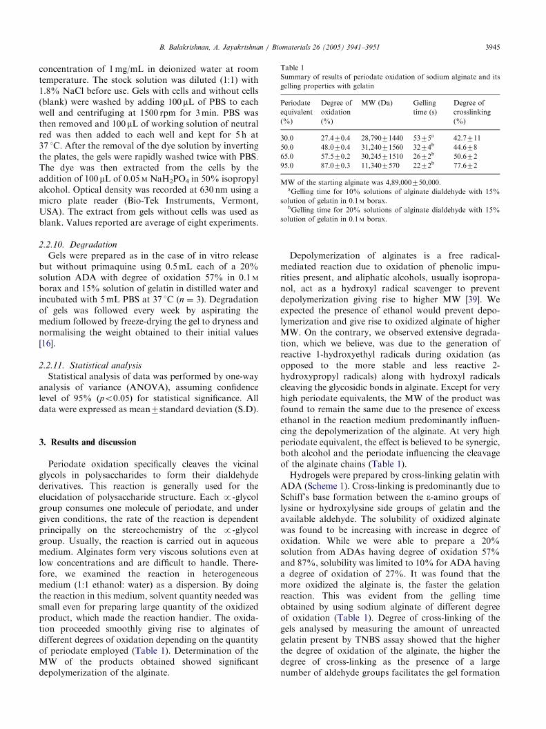
Hydrogels were prepared by cross-linking gelatin withADA (Scheme 1). Cross-linking is predominantly due toSchiff’s base formation between the e-amino groups oflysine or hydroxylysine side groups of gelatin and theavailable aldehyde. The solubility of oxidized alginatewas found to be increasing with increase in degree ofoxidation. While we were able to prepare a 20%solution from ADAs having degree of oxidation 57%and 87%, solubility was limited to 10% for ADA havinga degree of oxidation of 27%. It was found that themore oxidized the alginate is, the faster the gelationreaction. This was evident from the gelling timeobtained by using sodium alginate of different degreeof oxidation (Table 1). Degree of cross-linking of thegels analysed by measuring the amount of unreactedgelatin present by TNBS assay showed that the higherthe degree of oxidation of the alginate, the higher thedegree of cross-linking as the presence of a largenumber of aldehyde groups facilitates the gel formation
ARTICLE IN PRESS
Scheme 1. Gelatin cross-linking with ADA in the presence of borax.
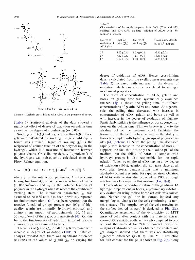
Table 2
Characteristics of hydrogels prepared from 20% (57% and 87%
oxidized) and 10% (27% oxidized) solution of ADAs with 15%
solution of gelatin
Degree of
oxidation of
ADA (%)
Swelling
ratio (Qm)
Degree of
swelling (Q)
Crosslinking density
ðue � 105Þmol=cm3
27 8.0270.45 8.2370.22 32.4572.01
57 7.1270.63 7.5370.31 38.6974.05
87 5.8270.52 6.1870.28 57.3976.50
B. Balakrishnan, A. Jayakrishnan / Biomaterials 26 (2005) 3941–39513946
(Table 1). Statistical analysis of the data showed asignificant effect of degree of oxidation on gelling timeas well as the degree of crosslinking ðpo0:05Þ:
Swelling ratio (Qm) and degree of swelling (Q) of thesegels were calculated by swelling the gels until equili-brium was attained. Degree of swelling (Q) is thereciprocal of volume fraction of the polymer ðu2Þ in thehydrogel, which is a measure of interaction betweenpolymer chains. Cross-linking density ðue; mol=cm3
Þ ofthe hydrogels was subsequently calculated from theFlory–Rehner equation,
ue ¼ �½ln ð1 � u2Þ þ u2 þ w1u22� ½V1ðu
1=32 � 2u2=f ��1;
where w1 is the interaction parameter, f is the cross-linking functionality, V1 is the molar volume of water(18.062 cm3/mol) and u2 is the volume fraction ofpolymer in the hydrogel when its reaches the equilibriumswelling state. The interaction parameter, w1 wasassumed to be 0.35 as it has been previously reportedfor similar interaction [16]. It has been reported that thereactive functional groups present per 100 g of highquality gelatin are primarily, hydroxyl, carboxyl andamino at an amount of approximately 100, 75 and50 meq of each of these groups, respectively [40]. On thisbasis, the functionality of gelatin in terms of reactiveamino groups was assumed to be 50.
The values of Q and Qm for all the gels decreased withincrease in degree of oxidation (Table 2). Statisticalanalysis revealed that there was significant difference(po0.05) in the values of Q and Qm on varying the
degree of oxidation of ADA. Hence, cross-linkingdensity calculated from the swelling measurements (seeTable 2) increased with increase in the degree ofoxidation which can also be correlated to strongermechanical properties.
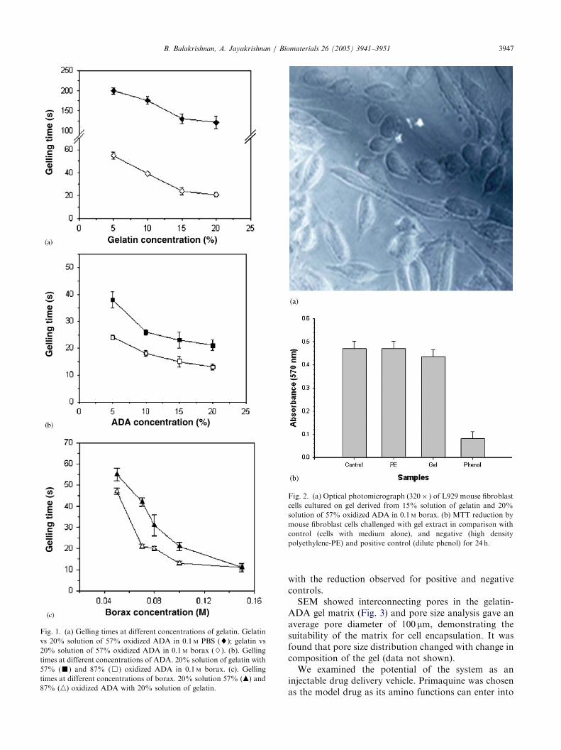
The effect of concentration of ADA, gelatin andborax on gelling time was systematically examinedfurther. Fig. 1 shows the gelling time at differentconcentrations of gelatin, ADA and borax. As a generalrule, the gelling time decreased with increase inconcentration of ADA, gelatin and borax as well aswith increase in the degree of oxidation of alginate.Particularly striking is the influence of borax concentra-tion on the gelling time. This we believe is due to thealkaline pH of the medium which facilitates theformation of the Schiff’s base as well as the ability ofborax to complex with hydroxyl groups of polysacchar-ides [41] (Scheme 1). Since the gelling time decreasedrapidly with increase in the concentration of borax, itsupports the fact that not only the alkaline pH of themedium, but the ability of borax to complex withhydroxyl groups is also responsible for the rapidgelation. When we employed ADA having a low degreeof oxidation (10%), gelation did not take place at alleven after hours, demonstrating that a minimumaldehyde content is essential for rapid gelation. Gelationof ADA with gelatin also occurred in PBS, althoughreaction was less rapid in this medium (Fig. 1(a)).
To reconfirm the non-toxic nature of the gelatin-ADAhydrogel preparations in borax, a preliminary cytotoxi-city evaluation using mouse fibroblast cells was carriedout. Neither the gel nor its extract induced anymorphological changes to the cells confirming its non-toxic nature. The morphology of the cells growing onthe surface (scored as zero) is depicted in Fig. 2(a).Quantitative assessment of the cytotoxicity by MTTassay of cells after contact with the material extractshowed 93% metabolically active cells compared to cellswithout the material for 24 h of contact. Statisticalanalysis of absorbance values obtained for control andgel samples showed that there was no statisticallysignificant difference (p40.07). The MTT reductionfor 24 h contact for the gel is shown in Fig. 2(b) along
ARTICLE IN PRESS
Fig. 1. (a) Gelling times at different concentrations of gelatin. Gelatin
vs 20% solution of 57% oxidized ADA in 0.1 M PBS (~); gelatin vs
20% solution of 57% oxidized ADA in 0.1 M borax (}). (b). Gelling
times at different concentrations of ADA. 20% solution of gelatin with
57% (’) and 87% (&) oxidized ADA in 0.1 M borax. (c). Gelling
times at different concentrations of borax. 20% solution 57% (m) and
87% (n) oxidized ADA with 20% solution of gelatin.
Fig. 2. (a) Optical photomicrograph (320� ) of L929 mouse fibroblast
cells cultured on gel derived from 15% solution of gelatin and 20%
solution of 57% oxidized ADA in 0.1 M borax. (b) MTT reduction by
mouse fibroblast cells challenged with gel extract in comparison with
control (cells with medium alone), and negative (high density
polyethylene-PE) and positive control (dilute phenol) for 24 h.
B. Balakrishnan, A. Jayakrishnan / Biomaterials 26 (2005) 3941–3951 3947
with the reduction observed for positive and negativecontrols.
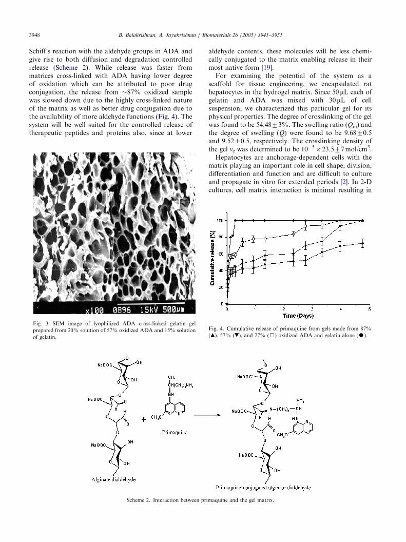
SEM showed interconnecting pores in the gelatin-ADA gel matrix (Fig. 3) and pore size analysis gave anaverage pore diameter of 100 mm, demonstrating thesuitability of the matrix for cell encapsulation. It wasfound that pore size distribution changed with change incomposition of the gel (data not shown).
We examined the potential of the system as aninjectable drug delivery vehicle. Primaquine was chosenas the model drug as its amino functions can enter into
ARTICLE IN PRESSB. Balakrishnan, A. Jayakrishnan / Biomaterials 26 (2005) 3941–39513948
Schiff’s reaction with the aldehyde groups in ADA andgive rise to both diffusion and degradation controlledrelease (Scheme 2). While release was faster frommatrices cross-linked with ADA having lower degreeof oxidation which can be attributed to poor drugconjugation, the release from 87% oxidized samplewas slowed down due to the highly cross-linked natureof the matrix as well as better drug conjugation due tothe availability of more aldehyde functions (Fig. 4). Thesystem will be well suited for the controlled release oftherapeutic peptides and proteins also, since at lower
Fig. 3. SEM image of lyophilized ADA cross-linked gelatin gel
prepared from 20% solution of 57% oxidized ADA and 15% solution
of gelatin.
Scheme 2. Interaction between pri
aldehyde contents, these molecules will be less chemi-cally conjugated to the matrix enabling release in theirmost native form [19].
For examining the potential of the system as ascaffold for tissue engineering, we encapsulated rathepatocytes in the hydrogel matrix. Since 50 mL each ofgelatin and ADA was mixed with 30 mL of cellsuspension, we characterized this particular gel for itsphysical properties. The degree of crosslinking of the gelwas found to be 54.4873%. The swelling ratio (Qm) andthe degree of swelling (Q) were found to be 9.6870.5and 9.5270.5, respectively. The crosslinking density ofthe gel ne was determined to be 10�5
� 23.577 mol/cm3.Hepatocytes are anchorage-dependent cells with the
matrix playing an important role in cell shape, division,differentiation and function and are difficult to cultureand propagate in vitro for extended periods [2]. In 2-Dcultures, cell matrix interaction is minimal resulting in
maquine and the gel matrix.
Fig. 4. Cumulative release of primaquine from gels made from 87%
(m), 57% (.), and 27% (&) oxidized ADA and gelatin alone (K).
ARTICLE IN PRESSB. Balakrishnan, A. Jayakrishnan / Biomaterials 26 (2005) 3941–3951 3949
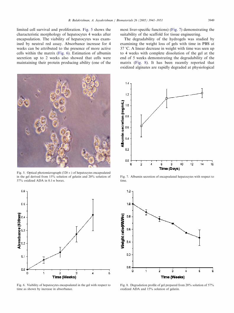
limited cell survival and proliferation. Fig. 5 shows thecharacteristic morphology of hepatocytes 4 weeks afterencapsulation. The viability of hepatocytes was exam-ined by neutral red assay. Absorbance increase for 4weeks can be attributed to the presence of more activecells within the matrix (Fig. 6). Estimation of albuminsecretion up to 2 weeks also showed that cells weremaintaining their protein producing ability (one of the
Fig. 5. Optical photomicrograph (320� ) of hepatocytes encapsulated
in the gel derived from 15% solution of gelatin and 20% solution of
57% oxidized ADA in 0.1 M borax.
Fig. 6. Viability of hepatocytes encapsulated in the gel with respect to
time as shown by increase in absorbance.
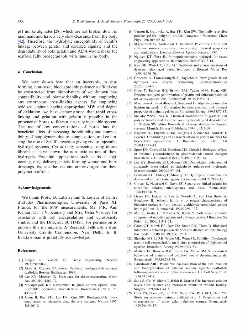
most liver-specific functions) (Fig. 7) demonstrating thesuitability of the scaffold for tissue engineering.
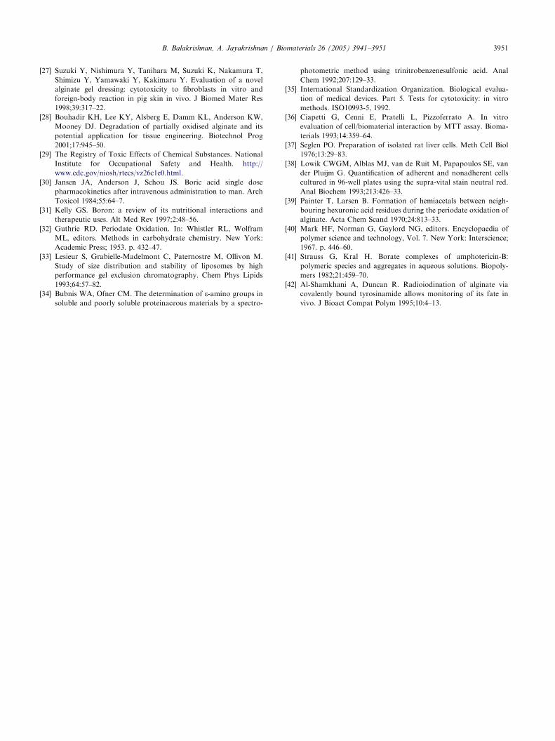
The degradability of the hydrogels was studied byexamining the weight loss of gels with time in PBS at37 1C. A linear decrease in weight with time was seen upto 4 weeks with complete dissolution of the gel at theend of 5 weeks demonstrating the degradability of thematrix (Fig. 8). It has been recently reported thatoxidized alginates are rapidly degraded at physiological
Fig. 8. Degradation profile of gel prepared from 20% solution of 57%
oxidized ADA and 15% solution of gelatin.
Fig. 7. Albumin secretion of encapsulated hepatocytes with respect to
time.
ARTICLE IN PRESSB. Balakrishnan, A. Jayakrishnan / Biomaterials 26 (2005) 3941–39513950
pH unlike alginates [28], which are not broken down inmammals and have a very slow clearance from the body[42]. Therefore, the hydrolytic susceptibility of Schiff’slinkage between gelatin and oxidized alginate and thedegradability of both gelatin and ADA would make thescaffold fully biodegradable with time in the body.
4. Conclusion
We have shown here that an injectable, in situ-forming, non-toxic, biodegradable polymer scaffold canbe constructed from biopolymers of well-known bio-compatibility and bioresorbability without employingany extraneous cross-linking agents. By employingoxidized alginate having appropriate MW and degreeof oxidation, we have demonstrated that rapid cross-linking and gelation with gelatin is possible in thepresence of borax to fabricate a truly injectable system.The use of low concentrations of borax has thebeneficial effect of increasing the solubility and compat-ibility of biopolymers due to complexation, and enhan-cing the rate of Schiff’s reaction giving rise to injectablehydrogel systems. Cytotoxicity screening using mousefibroblasts have shown the non-toxic nature of thesehydrogels. Potential applications such as tissue engi-neering, drug delivery, in situ-forming wound and burndressings, tissue adhesives etc. are envisaged for thesepolymer scaffolds.
Acknowledgements
We thank Profs. D. Labarre and S. Lesieur of Centred’Etudes Pharmaceutiques, University of Paris XI,France, for the MW measurements, Mr. P.R. AnilKumar, Dr. T.V. Kumary and Mrs. Usha Vasudev forassistance with cell encapsulation and cytotoxicitystudies and the Director, SCTIMST, for permission topublish this manuscript. A Research Fellowship fromUniversity Grants Commission, New Delhi, to B.Balakrishnan is gratefully acknowledged.
References
[1] Langer R, Vacanti JP. Tissue engineering. Science
1993;260:920–6.
[2] Atala A, Mooney DJ, editors. Synthetic biodegradable polymer
scaffolds. Boston: Birkhauser; 1997.
[3] Lee KY, Mooney DJ. Hydrogels for tissue engineering. Chem
Rev 2001;101:1869–79.
[4] Mallapragada KS, Narasimhan B, guest, editors. Special issue.
Injectable polymeric biomaterials. Biomaterials 2002; 23:
4305–33.
[5] Jeong B, Bae YH, Lee DS, Kim SW. Biodegradable block
copolymers as injectable drug delivery systems. Nature 1997;
388:860–2.
[6] Vernon B, Gutowska A, Bae YH, Kim SW. Thermally reversible
polymer gel for biohybrid artificial pancreas. J Macromol Chem
Phys 1996;109:155–67.
[7] Skjak-Braek G, Anthonsen T, Sandford P, editors. Chitin and
chitosan: sources, chemistry, biochemistry, physical properties
and applications. London: Elsevier Applied Science; 1992.
[8] Nguyen KT, West JL. Photopolymerizable hydrogels for tissue
engineering applications. Biomaterials 2002;23:4307–14.
[9] Kim SH, Won CY, Chu CC. Synthesis and characterisation of
dextran–maleic acid based hydrogel. J Biomed Mater Res
1999;46:160–70.
[10] Crescenzi V, Francescangeli A, Taglienti A. New gelatin based
hydrogels via enzyme networking. Biomacromolecules
2002;3:1384–91.
[11] Chen T, Embree HD, Brown EM, Taylor MM, Payne GF.
Enzyme-catalyzed gel formation of gelatin and chitosan: potential
for in situ applications. Biomaterials 2003;24:2831–41.
[12] Martinsen A, Skjak-Braek G, Smidsrod O. Alginate as immobi-
lization material: 1. Correlation between chemical and physical
properties of alginate gel beads. Biotechnol Bioeng 1989;33:79–87.
[13] Shalaby WSW, Park K. Chemical modification of proteins and
polysaccharides and its effect on enzyme-catalyzed degradation.
In: Shalaby SW, editor. Biomedical polymers: designed to degrade
systems. Munich: Hanser Publishers; 1994. p. 213–58.
[14] Kuijpers AJ, Engbers GHM, Krijgsveld J, Zaat SA, Dankert J,
Feijen J. Crosslinking and characterisation of gelatin matrices for
biomedical applications. J Biomater Sci Polym Ed
2000;11:225–43.
[15] Speer DP, Chavapil M, Eskelson CD, Ulreich J. Biological effects
of residual glutaraldehyde in glutaraldehyde-tanned collagen
biomaterials. J Biomed Mater Res 1980;14:753–64.
[16] Lee KY, Bouhadir KH, Mooney DJ. Degradation behaviour of
covalently cross-linked poly(aldehyde guluronate) hydrogels.
Macromolecules 2000;33:97–101.
[17] Bouhadir KH, Alsberg E, Mooney DJ. Hydrogels for combination
delivery of antineoplastic agents. Biomaterials 2001;22:2625–33.
[18] Cortesi R, Nastruzzi C, Davis SS. Sugar cross-linked gelatin for
controlled release: microspheres and disks. Biomaterials
1998;19:1641–9.
[19] Draye J-P, Delaey B, Van de Voorde A, Van Den Bulke A,
Bogdanov B, Schacht E. In vitro release characteristics of
bioactive molecules from dextran dialdehyde crosslinked gelatin
hydrogel films. Biomaterials 1998;19:99–107.
[20] Mo X, Iwata H, Matsuda S, Ikada Y. Soft tissue adhesive
composed of modified gelatin and polysaccharides. J Biomater Sci
Polym Ed 2000;11:341–51.
[21] Grant GT, Morris ER, Rees DA, Smith PJC, Thom D. Biological
interactions between polysaccharides and divalent cations: the egg
box model. FEBS lett 1973;32:195–8.
[22] Shoichet MS, Li RH, White ML, Winn SR. Stability of hydrogels
used in cell encapsulation: an in vitro comparison of alginate and
agarose. Biotechnol Bioeng 1996;50:374–81.
[23] Mathew IR, Brownie RM, Frame JW, Millar BM. Subperiosteal
behaviour of alginate and cellulose wound dressing materials.
Biomaterials 1995;16:265–74.
[24] Lansdown ABG, Payne MJ. An evaluation of the local reaction
and biodegradation of calcium sodium alginate (kaltostat)
following subcutaneous implantation in rat. J R Coll Surg Edinb
1994;39:284–8.
[25] Sank A, Chi M, Shima T, Reich R, Martin GR. Increased calcium
levels after cellular and molecular events in wound healing.
Surgery 1989;106:1141–8.
[26] Choi YS, Hong SR, Lee YM, Song KW, Park MH, Nam YS.
Study on gelatin-containing artificial skin: 1. Preparation and
characteristics of novel gelatin-alginate sponge. Biomaterials
1999;20:409–17.
ARTICLE IN PRESSB. Balakrishnan, A. Jayakrishnan / Biomaterials 26 (2005) 3941–3951 3951
[27] Suzuki Y, Nishimura Y, Tanihara M, Suzuki K, Nakamura T,
Shimizu Y, Yamawaki Y, Kakimaru Y. Evaluation of a novel
alginate gel dressing: cytotoxicity to fibroblasts in vitro and
foreign-body reaction in pig skin in vivo. J Biomed Mater Res
1998;39:317–22.
[28] Bouhadir KH, Lee KY, Alsberg E, Damm KL, Anderson KW,
Mooney DJ. Degradation of partially oxidised alginate and its
potential application for tissue engineering. Biotechnol Prog
2001;17:945–50.
[29] The Registry of Toxic Effects of Chemical Substances. National
Institute for Occupational Safety and Health. http://
www.cdc.gov/niosh/rtecs/vz26c1e0.html.
[30] Jansen JA, Anderson J, Schou JS. Boric acid single dose
pharmacokinetics after intravenous administration to man. Arch
Toxicol 1984;55:64–7.
[31] Kelly GS. Boron: a review of its nutritional interactions and
therapeutic uses. Alt Med Rev 1997;2:48–56.
[32] Guthrie RD. Periodate Oxidation. In: Whistler RL, Wolfram
ML, editors. Methods in carbohydrate chemistry. New York:
Academic Press; 1953. p. 432–47.
[33] Lesieur S, Grabielle-Madelmont C, Paternostre M, Ollivon M.
Study of size distribution and stability of liposomes by high
performance gel exclusion chromatography. Chem Phys Lipids
1993;64:57–82.
[34] Bubnis WA, Ofner CM. The determination of e-amino groups in
soluble and poorly soluble proteinaceous materials by a spectro-
photometric method using trinitrobenzenesulfonic acid. Anal
Chem 1992;207:129–33.
[35] International Standardization Organization. Biological evalua-
tion of medical devices. Part 5. Tests for cytotoxicity: in vitro
methods. ISO10993-5, 1992.
[36] Ciapetti G, Cenni E, Pratelli L, Pizzoferrato A. In vitro
evaluation of cell/biomaterial interaction by MTT assay. Bioma-
terials 1993;14:359–64.
[37] Seglen PO. Preparation of isolated rat liver cells. Meth Cell Biol
1976;13:29–83.
[38] Lowik CWGM, Alblas MJ, van de Ruit M, Papapoulos SE, van
der Pluijm G. Quantification of adherent and nonadherent cells
cultured in 96-well plates using the supra-vital stain neutral red.
Anal Biochem 1993;213:426–33.
[39] Painter T, Larsen B. Formation of hemiacetals between neigh-
bouring hexuronic acid residues during the periodate oxidation of
alginate. Acta Chem Scand 1970;24:813–33.
[40] Mark HF, Norman G, Gaylord NG, editors. Encyclopaedia of
polymer science and technology, Vol. 7. New York: Interscience;
1967. p. 446–60.
[41] Strauss G, Kral H. Borate complexes of amphotericin-B:
polymeric species and aggregates in aqueous solutions. Biopoly-
mers 1982;21:459–70.
[42] Al-Shamkhani A, Duncan R. Radioiodination of alginate via
covalently bound tyrosinamide allows monitoring of its fate in
vivo. J Bioact Compat Polym 1995;10:4–13.