injectable biodegradable hydrogels
TRANSCRIPT

Feature Article
Injectable Biodegradable Hydrogels
Minh Khanh Nguyen, Doo Sung Lee*
Injectable biodegradable copolymer hydrogels, which exhibit a sol–gel phase transition inresponse to external stimuli, such as temperature changes or both pH and temperature (pH/temperature) alterations, have found a number of uses in biomedical and pharmaceuticalapplications, such as drug delivery, cell growth, and tissue engineering. These hydrogels can beused in simple pharmaceutical formulations that can be prepared by mixing the hydrogel withdrugs, proteins, or cells. Such formulations are administered in a straightforward manner,through site-specific control of release behavior,and the hydrogels are compatible with biologicalsystems. This review will provide a summary ofrecent progress in biodegradable temperature-sensitive polymers including polyesters, polypho-sphazenes, polypeptides, and chitosan, and pH/temperature-sensitive polymers such as sulfa-methazine-, poly(b-amino ester)-, poly(aminourethane)-, and poly(amidoamine)-based polymers.The advantages of pH/temperature-sensitivepolymers over simple temperature-sensitive poly-mers are also discussed. A perspective on thefuture of injectable biodegradable hydrogels isoffered.
Introduction
Hydrogels are three-dimensional hydrophilic polymeric
networks that can absorb and retain a considerable amount
of water with maintenance of shape.[1,2] Injectable
biodegradable hydrogels have been widely used in
biomedical applications, such as drug/cell delivery and
tissue engineering, because of their highly hydrophilic
characteristics. Such hydrogels are of particular interest
because drugs, proteins, and cells can be easily incorporated
into polymer solutions prior to administration. Importantly,
D. S. Lee, M. K. NguyenDepartment of Polymer Science and Engineering, SungkyunkwanUniversity, Suwon, Gyeonggi 440-476, KoreaFax: þ82-31-292-8790; E-mail: [email protected]
Macromol. Biosci. 2010, 10, 563–579
� 2010 WILEY-VCH Verlag GmbH & Co. KGaA, Weinheim
no surgical procedures are required for the insertion of gels
into the body; the gels are administered by simple
injection.[3–5]
Injectable hydrogels can be formed in situ by either
chemical or physical crosslinking methods. Chemically
crosslinked hydrogels, prepared through photopolymeriza-
tion,[6] disulfide bond formation,[7] or reaction between
thiols and acrylate or sulfones,[7] undergo significant
volume changes during the phase transition. In contrast
to chemical hydrogels, physically crosslinked hydrogels,
formed by the self-assembly of polymers in response to
environmental stimuli (for example, temperature, pH, or
both) display sol–gel transitions without marked volume
changes. The sol–gel transition systems are relatively low-
viscosity aqueous solutions (sol state) prior to injection, but
rapidly convert into a gel under physiological conditions
DOI: 10.1002/mabi.200900402 563
M. K. Nguyen, D. S. Lee
Minh Khanh Nguyen graduated from theDepartment of Chemical Engineering fromHoChiMinh City University of Technology (Viet-Nam) in 2003 and is currently pursuing his Ph.D.under the guidance of Professor Doo Sung Lee atSungkyunkwan University (Korea). His mainresearch is focused on the development of func-tionalized and biodegradable injectable poly-meric hydrogels for controlled drug andprotein delivery.
Doo Sung Lee studied chemical engineering atthe Seoul National University. He completed hisM.Sc. and Ph.D. at Korea Advanced Institute ofScience and Technology in 1984 working in thefield of interpenetrating polymer networks. Hejoined Sungkyunkwan University as AssistantProfessor in the Department of Textile Engineer-ing. He was one of the founders of the Depart-ment of Polymer Science and Engineering andwas appointed Professor of this department in1993. He served as a Dean of the College ofEngineering at Sungkyunkwan University in2005–2007. He started to study biomaterials in
564
post-injection.[1–3] Physical hydrogels have several advan-
tages over chemical hydrogels, because they do not require
photo irradiation, use of organic solvents or crosslinking
agents, and do not release heat during polymerization at the
gelation site, which may denature incorporated proteins
and damage embedded cells and surrounding tissues. Thus,
the physical systems have recently attracted increased
attention. For use in drug/cell delivery and tissue engineer-
ing, hydrogels should be low-viscosity solutions (free-
flowing) prior to subcutaneous injection, and should
rapidly gel in the human body, where ultimate degradation
of the hydrogels is desired.
This article provides insights into recent advances in
synthesis and biomedical applications of injectable, biode-
gradable, polymeric hydrogels that exhibit sol–gel transi-
tions in response to temperature and pH/temperature
changes. The discussion covers poly(ethylene glycol) (PEG)/
polyester block copolymers, polyphosphazenes, polypep-
tides, chitosan, polymers based on sulfamethazine, poly(b-
amino ester), poly(amino urethane), poly(amidoamine),
and others.
1995 when he was in the Department of Phar-maceutics and Pharmaceutical Chemistry at theUniversity of Utah as a visiting Professor. Heserved as an Editor-in-chief of Polymer Scienceand Technology (2001) and on the Editorial boardof Macromolecular Research for four years(2002–2005). He was elected as a member ofKorean Academy of Engineering in 2007. He iscurrently a vice president and an editorial boardmember of Biomaterials Research of the KoreanSociety of Biomaterals (2006–present). He hasauthored and co-authored about 300 papers,including about 120 in peer-reviewed journals,and seven book chapters, and filed 26 patents.His main research interest is functionalized andbiodegradable injectable hydrogels and micellesfor the controlled drug and protein delivery andmolecular imaging.
Injectable Biodegradable Block CopolymerHydrogels
Thermosensitive Block Copolymer Hydrogels
Hydrogels that are sensitive to temperature are useful for
both in vitro and in vivo applications, because temperature
control is generally easy. Temperature-sensitive hydrogels
undergo a sol–gel phase transition when the temperature is
increased from room temperature to physiological tem-
peratures.
Poly(ethylene glycol) (PEG)/Polyester
Copolymers of hydrophilic biocompatible PEG with biode-
gradable biocompatible aliphatic polyesters, for example
polylactide (PLA), polyglycolide (PGA), poly(e-caprolactone)
(PCL), or poly[(R)-3-hydroxybutyrate] (PHB), have received
increasing attention as promising biomaterials.
The first reported biodegradable thermosensitive
hydrogel was PEG-poly(L-lactide)-PEG (PEG-PLLA-PEG)
(Scheme 1).[9] PEG-PLLA-PEG was synthesized by the ring-
opening polymerization (ROP) of L-lactide using mono-
methoxy PEG (MPEG) of molecular weight (MW) 5 000 as
macroinitiator, to form MPEG-PLLA diblock copolymers.
Triblock copolymers (PEG-PLLA-PEG), with PLLA blocks in
Scheme 1. Structure of PEG-PLLA-PEG.
Macromol. Biosci. 2010, 10, 563–579
� 2010 WILEY-VCH Verlag GmbH & Co. KGaA, Weinheim
the 2 000–5 000 Da molecular weight range were next
produced by coupling the resulting diblock copolymers
using hexamethylene diisocyanate (HMDI). The concen-
trated copolymers (10–30 wt.-%) dissolved in water
demonstrated a gel-to-sol transition with increases in
temperature. The gel-to-sol transition was precisely con-
trolled by the biodegradable PLLA block length when a PEG
block was fixed at both ends. The gel-to-sol properties of the
PEG/polyester diblock and triblock copolymers in water
depended on the hydrophobic/hydrophilic balance, block
length, hydrophobicity, and stereoregularity of the hydro-
phobic block.[10,11]
PEG-(D,L-lactide)-PEG (PEG-PLA-PEG) triblock copolymers
(MW of MPEG¼ 2 000–5 000) were recently produced by
DOI: 10.1002/mabi.200900402

Injectable Biodegradable Hydrogels
Figure 1. Phase diagram of PEG-PLGA-PEG triblock copolymeraqueous solutions. Sol (flow) to gel (no flow) transition tempera-ture was measured by the test tube inverting method increasing2 8C/step. Reprinted with permission from ref.[16]. Copyright 1999Elsevier.
coupling MPEG-PLA diblock copolymers using adipoyl
chloride.[12] Rheological measurements showed a gel-to-
sol transition in concentrated polymer solutions, with an
increase in temperature. Gelation at lower temperatures
was attributed to hydrogen bonding between PEG blocks,
and the hydrogen bonds were broken at elevated tempera-
tures, leading to the gel-to-sol transition. A series of
biodegradable star-shaped PLLA-PEG block copolymers
were obtained by the pairing of two components: star
PLLA and monocarboxy-MPEG, using dicyclohexylcarbodii-
mide (DCC).[13,14] The gel-to-sol transitions, induced by
increases in temperature, were observed at concentrations
beyond the critical gelation concentration (CGC). With the
same PEG block length, use of longer PLLA blocks led to a
decrease in the CGC, thus widening the gelation window.
Gelation was attributed to micellar packing and the
breaking of micellar packing structures caused by the
partial hydration of the PEG block, which resulted in the gel-
to-sol transition at higher temperatures.
However, the abovementioned gels underwent gel-to-sol
transitions, reducing their suitability for encapsulation of
some drugs or proteins. In addition, injection at tempera-
tures that are elevated with respect to body temperature
is uncomfortable for patients. PEG-poly(D,L-lactide-co-
glycolide)-PEG (PEG-PLGA-PEG) triblock copolymers with
short PEG blocks (MW� 750) were soluble in water at low
temperatures and converted into a gel at elevated
temperatures (Scheme 2).[15] The triblock copolymers were
shown to be biocompatible with blood.[16–18] The gelation
window spanned the physiological temperature range
(Figure 1). 13C NMR and dynamic light scattering (DLS)
studies revealed that the gelation of PEG-PLGA-PEG was
enhanced by micellar growth and close packing of micelles
(Figure 2). The upper gel-to-sol transition at higher
temperatures was driven by the breakage of the micellar
structure caused by the partial dehydration of PEG and
PLGA blocks. The sol–gel transition could be adjusted by
variation in PLGA and PEG block lengths, PLGA composition,
and the use of particular additives. In other studies,[19,20] the
gelation mechanism of PEG-PLGA-PEG (550-2810-550) was
investigated by rheology, DLS, differential scanning calori-
metry (DSC), and small-angle neutron scattering (SANS).
The results indicated that the macroscopic liquid–liquid
phase separation induced gelation of the triblock copoly-
mer. A transparent gel was formed in situ after injection of a
33 wt.-% PEG-PLGA-PEG (550-2810-550) aqueous solution
into rats, and gel integrity persisted for one month.[21]
Interestingly, thermosensitive hydrogels based on PLGA-
PEG-PLGA (BAB-type) (Scheme 3) showed a sol-to-gel
Scheme 2. Structure of PEG-PLGA-PEG.
Macromol. Biosci. 2010, 10, 563–579
� 2010 WILEY-VCH Verlag GmbH & Co. KGaA, Weinheim
transition similar to that of PEG-PLGA-PEG (ABA-type)
copolymer hydrogels.[22–24] However, the synthetic proce-
dure for BAB-type hydrogels was simpler than that for the
ABA-type, using HMDI as a coupling agent. The gelation
mechanism for BAB-type hydrogels was different from that
of ABA-type hydrogels because of the presence of two PLGA
end blocks. The former formed micelles with intermicellar
bridges, whereas the latter formed regular micelles with a
PLGA block core and a PEG block shell, in aqueous solution.
At temperatures below the critical gelation temperature
(CGT), some bridging micelles of PLGA-PEG-PLGA copoly-
mers formed, but they were not stable because of the low
hydrophobicity of PLGA. With increasing temperatures to
the CGT, a bridged micelle network was formed because of
an increase in the hydrophobicity of the PLGA segment,
leading to gelation (Figure 3). In vitro and in vivo
degradation of the PLGA-PEG-PLGA (1500-1000-1500)
copolymer (ReGel) was studied.[23] A 23 wt.-% ReGel was
found to degrade completely in vitro after 6–8 weeks at
37 8C, whereas no hydrogels were observed at the end of the
fourth week after subcutaneous injection of a 23 wt.-%
ReGel solution into rats.
To overcome the molecular weight constraints of the
PLGA-PEG-PLGA and PEG-PLGA-PEG triblock copolymers,
and to control the time of persistence of gels, graft
copolymers of PEG-g-PLGA and PLGA-g-PEG were investi-
gated (Scheme 4).[25–27] The former copolymer was
synthesized by the ROP of D,L-lactide (LA) and glycolide
(GA) using a hydroxy-pendant PEG as a macroinitiator. The
PEG-g-PLGA copolymer in water showed a sol–gel transi-
tion at concentrations above 16 wt.-%. Gel integrity
persisted for one week under physiological conditions.
The latter copolymer was prepared by the one-step ROP of
LA, GA, and epoxy-terminated PEG.[25,26] Aqueous solutions
www.mbs-journal.de 565

M. K. Nguyen, D. S. Lee
Figure 2. Schematic diagram of the sol-gel transition of PEG-PLGA-PEG aqueous solutionin response to temperature.
Scheme 5. PCL-PEG-PCL.
Scheme 3. Structure of PLGA-PEG-PLGA.
Figure 3. Schematic diagram of the sol–gel transition of PLGA-PEG-PLGA aqueous solution in response to temperature.
Scheme 4. Chemical structures of a) PEG-g-PLGA and b) PLGA-g-PEG.
566
of PLGA-g-PEG copolymers also exhibited a sol-to-gel
transition upon heating. After injection of the copolymer
solution (29 wt.-%) into rats, the gel persisted for more than
two months, a significant improvement over the one week
persistence time observed for the PEG-g-PLGA copolymer
hydrogel. The gelation mechanism of the PLGA-g-PEG
copolymer was examined by 13C NMR spectroscopy, SANS,
rheology, and infrared (IR) spectroscopy.[28] Partial dehy-
dration of PEG caused micellar aggregation, leading to
gelation, and significant dehydration of PEG led to
macroscopic separation at the gel-to-sol transition
Macromol. Biosci. 2010, 10, 563–579
� 2010 WILEY-VCH Verlag GmbH & Co. KGaA, Weinheim
temperature. The sol-to-gel transition
temperature was tuned from 15 to
45 8C by tailoring the polymer composi-
tion, the number of PEG grafts present,
and the PEG molecular weight. By mixing
these copolymers at different composi-
tion ratios, the sol-to-gel transition tem-
perature could be adjusted, and the gel
duration could be varied from one week
to three months. Both copolymers
yielded rather soft gels with storage
moduli (G0 values) of 100 Pa.[26]
Poly(e-caprolactone) (PCL) is a hydro-
phobic crystalline polymer that is both
biodegradable and biocompatible. PCL copolymers have a
powdery morphology, making them easier to handle than
are PLGA and PLLA copolymers, which have a sticky paste
morphology. Aqueous solutions of MPEG-PCL diblock
copolymers (MPEG MW� 2 000) underwent a gel-to-sol
transition on temperature increase. The gelation window
was strongly influenced by PEG and PCL block length. In
vivo gelation of MPEG-PCL (MW 2 000–2 300) was also
studied. When a 23 wt.-% copolymer solution at 42 8C was
injected into a rat, the gel formed immediately and
persisted for one month with only a small amount of
inflammation at the injection site.[29] MPEG-PCL diblock
copolymers with a lower molecular weight MPEG
(MW¼ 750) were subsequently reported.[30] Interestingly,
synthesized diblock copolymers with PCL block lengths of
1 400–3 000 were soluble in water and underwent a sol-to-
gel-to-sol transition as a result of temperature changes. The
sol–gel phase diagram depended on PCL block length.
Recently, PEG-PCL-PEG and PCL-PEG-PCL (Scheme 5) triblock
copolymers have been introduced, and aqueous solutions
thereof showed a clear sol-to-gel-to-turbid sol transition
with an increase in temperature.[31,32] DLS and 13C NMR
studies indicated that the clear sol-to-gel transition arose
from micellar aggregation, whereas the gel-to-turbid sol
transition was driven by the breakage of the core–shell
structure. Because of differences in structural topology, PCL-
PEG-PCL had a higher G0 (10 000 Pa) (Figure 4) than did
PEG-PCL-PEG (100 Pa) (Figure 5). However, because of
crystallization of the PCL block, a clear aqueous solution
of PCL-PEG-PCL (20 wt.-%) turned turbid within 1 h at 20 8C,
which may be problematic for injectability. The crystal-
lization problem was, however, solved by using poly-
(caprolactone-co-trimethylene carbonate)-PEG-poly(capro-
lactone-co-trimethylene carbonate) (PCTC-PEG-PCTC).[33] A
DOI: 10.1002/mabi.200900402

Injectable Biodegradable Hydrogels
Figure 4. Dynamic mechanical analysis of PCL-PEG-PCL triblockcopolymer aqueous solutions (20wt.-%) as a function of tempera-ture. The thermogram was obtained with a heating rate of0.2 8C �min�1. Reprinted with permission from ref.[31]. Copyright2005 American Chemical Society.
Figure 5. Dynamic mechanical analysis of the PEG-PCL-PEG tri-block copolymer aqueous solutions as a function of temperatureand concentration. Reprinted with permission from ref.[30]. Copy-right 2005 American Chemical Society.
PCTC-PEG-PCTC triblock copolymer in water exhibited a sol-
to-gel-to-syneresis transition upon heating. However, a
very soft gel with a G0 of 1 Pa was obtained. This gel was
stable with respect to hydrolysis in phosphate buffers at
37 8C for 50 d, but degraded markedly in rats. In addition, the
crystallizability of PCL decreased as the molecular weight of
PCL rose. Therefore, a multiblock copolymer of (PCL-PEG-
PCL)n was prepared by coupling PCL-PEG-PCL triblock
copolymers (1 000-1 000-1 000), using terephthaloyl chlor-
ide. A 20 wt.-% aqueous solution of the multiblock
copolymer underwent a sol-to-gel-to-sol transition that
did not turn turbid at room temperature, indicating that the
material may be convenient for practical applications, such
as drug formulation and injection. The multiblock showed a
lower G0 (100 Pa) than did the triblock (10 000 Pa). The
gelation mechanism was attributed to crystallization of the
multiblock copolymer.[34]
The multiblock topology affected the gelation behavior
of other copolymer hydrogels. A series of PEG/PLLA
alternating multiblock copolymers was synthesized by
coupling PEG (MW¼ 600) to PLLA (MW¼ 1 100–1 500) using
succinic anhydride.[35] The multiblock PEG/PLLA aqueous
Macromol. Biosci. 2010, 10, 563–579
� 2010 WILEY-VCH Verlag GmbH & Co. KGaA, Weinheim
solution underwent a sol-to-gel-to-sol transition with
increasing temperature. The transition temperature and
gel modulus could be controlled by varying the PLLA block
length, PEG molecular weight, and PEG/PLLA ratio. The
gelation mechanism was considered to be micelle aggrega-
tion. Stereochemistry also affected gelation.[36] The PEG/
poly(D,L-lactide) (PDLLA) and PEG/PLLA multiblock copoly-
mers with identical block lengths and total molecular
weight were prepared. Relative to amorphous PEG/PDLLA,
the stereoregular PEG/PLLA multiblock copolymer had a
reduced CGC, a lower sol-to-gel transition temperature, a
broader gel area, and a larger maximal gel modulus.13C NMR, X-ray diffraction (XRD), and UV-visible spectro-
scopy studies indicated that the different gelation proper-
ties could be attributed to the slower dynamic molecular
motion of the methyl groups in PLLA. The isotactic
arrangement in PLLA induced strong aggregation of the
PEG/PLLA multiblock. In contrast with the sol-to-gel
transition behavior of the PCL-PEG-PCL (1 000-1 000-1 000)
multiblock copolymer,[34] the PCL-PEG-PCL multiblock
copolymer, with a higher molecular weight of both PEG
and PCL, exhibited a gel-to-sol transition upon heating.[37]
In addition, multiblock copolymers of PEG-sebacate (PEG-
SA) were synthesized by simple condensation polymeriza-
tion.[38] A soft gel formed when a 25 wt.-% aqueous solution
of the PEG-SA multiblock was heated to 37 8C, and gel
integrity persisted for more than three weeks in phosphate
buffer, pH 7.4, at 37 8C.
In addition, it was found that a stereocomplex of the
enantiomeric triblock copolymers (10 wt.-%), PLLA-PEG-
PLLA (1 300-4 600-1 300), and poly(D-lactide)-PEG-poly-
(D-lactide) (PDLA-PEG-PDLA) (1 100-4 600-1 100), in water
could induce temperature-dependent gelation, although
the individual enantiomeric copolymers did not show
thermal gelation. Stereocomplex formation during gelation
was confirmed by wide-angle X-ray scattering (WAXS).[39] A
mixture of PEG-PLLA-PEG (2 000-2 000-2 000) and PEG-
PDLA-PEG (2 000-2 000-2 000) (35 wt.-%) exhibited a gel-
to-sol transition with an increase in temperature. WAXS
results showed that gelation was not attributable to
complexation of PLLA and PDLA, but rather to interdigita-
tion of the helical PEG chains induced by the complemen-
tary arrangement of PDLA and PLLA helices.[40]
Enantiomeric PEG12500-(PLA)2 and PEG21800-(PLA)8 aqu-
eous solutions showed gel-to-sol transitions with increas-
ing temperatures. Although neither enantiomeric polymer
solution formed a gel, a stereocomplex with a PLA block
could form a gel at the same concentration. Rheology
studies revealed that PEG-(PLA)8 showed a higher gel
modulus than PEG-(PLA)2, because of the higher cross-
linking density of the former. The PLA block length, PEG
content, polymer topology, and polymer concentration
influenced the gel–sol transition, gel modulus, and kinetics
of gelation.[41,42] Subsequently, the PEG-PLLA and PEG-PDLA
www.mbs-journal.de 567
M. K. Nguyen, D. S. Lee
568
multiblock copolymers were developed by coupling the
corresponding triblock copolymers using diisocyanatobu-
tane.[43] The multiblock copolymers showed a much lower
CGC, faster gelation, and a higher gel storage modulus,
compared with the parent triblock copolymers, because of
an increase in the crosslinking density of the multiblock
copolymers.
The end groups of the thermosensitive copolymers also
influenced gelation.[44,45] A series of PLGA-PEG-PLGA tri-
block copolymers with several different end caps (hydroxy,
acetyl, propionyl, and butanoyl groups) was synthesized
and characterized. Triblock copolymers containing acetate
and propionate groups exhibited a sol-to-gel transition as a
function of temperature, whereas the copolymer contain-
ing the butyrate group precipitated in water. An increase in
the hydrophobicity of the copolymer lowered the transition
temperature, and the end cap caused a significant change
in the position of the gelation window. Cholesterol end-
capped star PEG-PLLA copolymers (above 3 wt.-%) in water
also exhibited thermal gelation, but PEG-PLLA itself did
not.[46] Gelation was induced by the strong hydrophobic
association of the cholesterol group.
Poly[(R)-3-hydroxybutyrate] (PHB) is a natural biode-
gradable polyester produced by bacteria. The crystallinity
and hydrophobicity were higher than those of other
synthetic polymers, such as PLA and PCL. A thermosensitive
and amphiphilic poly(ether ester urethane) multiblock
copolymer consisting of PHB, PEG, and poly(propylene
glycol) (PPG) (PEG/PPG/PHB) was synthesized.[47,48] This
copolymer, in aqueous solutions and at very low concen-
trations (2–5 wt.-%). showed a sol-to-gel transition as a
function of temperature change. The gelation of the
copolymer solution was associated with micellar packing.
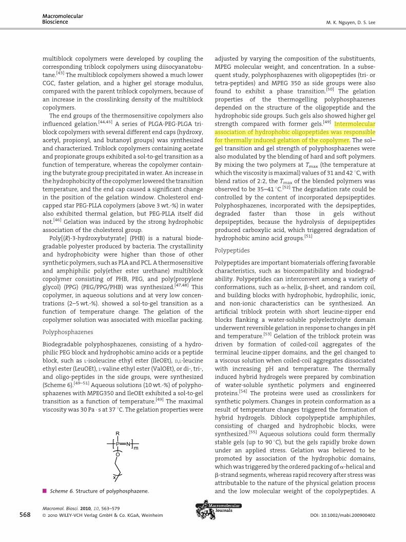
Polyphosphazenes
Biodegradable polyphosphazenes, consisting of a hydro-
philic PEG block and hydrophobic amino acids or a peptide
block, such as L-isoleucine ethyl ester (IleOEt), D,L-leucine
ethyl ester (LeuOEt), L-valine ethyl ester (ValOEt), or di-, tri-,
and oligo-peptides in the side groups, were synthesized
(Scheme 6).[49–51] Aqueous solutions (10 wt.-%) of polypho-
sphazenes with MPEG350 and IleOEt exhibited a sol-to-gel
transition as a function of temperature.[49] The maximal
viscosity was 30 Pa � s at 37 8C. The gelation properties were
Scheme 6. Structure of polyphosphazene.
Macromol. Biosci. 2010, 10, 563–579
� 2010 WILEY-VCH Verlag GmbH & Co. KGaA, Weinheim
adjusted by varying the composition of the substituents,
MPEG molecular weight, and concentration. In a subse-
quent study, polyphosphazenes with oligopeptides (tri- or
tetra-peptides) and MPEG 350 as side groups were also
found to exhibit a phase transition.[50] The gelation
properties of the thermogelling polyphosphazenes
depended on the structure of the oligopeptide and the
hydrophobic side groups. Such gels also showed higher gel
strength compared with former gels.[49] Intermolecular
association of hydrophobic oligopeptides was responsible
for thermally induced gelation of the copolymer. The sol–
gel transition and gel strength of polyphosphazenes were
also modulated by the blending of hard and soft polymers.
By mixing the two polymers at Tmax (the temperature at
which the viscosity is maximal) values of 31 and 42 8C, with
blend ratios of 2:2, the Tmax of the blended polymers was
observed to be 35–41 8C.[52] The degradation rate could be
controlled by the content of incorporated depsipeptides.
Polyphosphazenes, incorporated with the depsipeptides,
degraded faster than those in gels without
depsipeptides, because the hydrolysis of depsipeptides
produced carboxylic acid, which triggered degradation of
hydrophobic amino acid groups.[51]
Polypeptides
Polypeptides are important biomaterials offering favorable
characteristics, such as biocompatibility and biodegrad-
ability. Polypeptides can interconvert among a variety of
conformations, such as a-helix, b-sheet, and random coil,
and building blocks with hydrophobic, hydrophilic, ionic,
and non-ionic characteristics can be synthesized. An
artificial triblock protein with short leucine-zipper end
blocks flanking a water-soluble polyelectrolyte domain
underwent reversible gelation in response to changes in pH
and temperature.[53] Gelation of the triblock protein was
driven by formation of coiled-coil aggregates of the
terminal leucine-zipper domains, and the gel changed to
a viscous solution when coiled-coil aggregates dissociated
with increasing pH and temperature. The thermally
induced hybrid hydrogels were prepared by combination
of water-soluble synthetic polymers and engineered
proteins.[54] The proteins were used as crosslinkers for
synthetic polymers. Changes in protein conformation as a
result of temperature changes triggered the formation of
hybrid hydrogels. Diblock copolypeptide amphiphiles,
consisting of charged and hydrophobic blocks, were
synthesized.[55] Aqueous solutions could form thermally
stable gels (up to 90 8C), but the gels rapidly broke down
under an applied stress. Gelation was believed to be
promoted by association of the hydrophobic domains,
which was triggered by the ordered packing ofa-helical and
b-strand segments, whereas rapid recovery after stress was
attributable to the nature of the physical gelation process
and the low molecular weight of the copolypeptides. A
DOI: 10.1002/mabi.200900402

Injectable Biodegradable Hydrogels
Scheme 8. Chitosan structure.
10 wt.-% b-lactoglobulin aqueous solution showed a sol-to-
gel transition at 85 8C.[56] b-lactoglobulin is a component of
milk whey, with two disulfide bonds and one free
sulfhydryl group. Gelation was attributed to the formation
of hydrophobically linked aggregates, followed by forma-
tion of disulfide-bonded aggregates. A synthetic polypep-
tide of poly(ferrocenylsilane)-poly(g-benzyl-L-glutamate)
(PFS-PBLG) was soluble in hot toluene, but formed a
transparent gel at room temperature.[57] The gelation
process arose from formation of an a-helix and random-
coil structure in the diblock copolymer in toluene.
VPGVG (V¼ valine, P¼proline, and G¼ glycine) is a
prominent amino acid sequence in elastin. A hexahistidine
metal-binding motif was incorporated into an elastin-like
polypeptide.[58] A 6 wt.-% aqueous solution of the polymer
was a clear at 4 8C, but changed to a gel at 25 8C. This
material may be useful in heavy metal removal applica-
tions. Copolymers that contained a collagen peptide and an
elastin peptide formed a thermally induced gel in water.[59]
Polypeptides with 82–86 mol-% VPGVG composition
showed a sol-to-gel transition when the temperature was
increased. The collagen acted as a hydrate unit and the
elastin peptide acted as a thermosensitive crosslinking
point. A de novo-designed peptide showed a thermosensi-
tive gelation transition.[60] A 2 wt.-% aqueous solution of
the MAX3 peptide was a rigid gel at 75 8C withG0 of 1 100 Pa.
A transition from a random coil to a b-hairpin produced a
hydrogel network as the temperature increased. A sol-to-gel
transition was also evident in aqueous solutions (above
3 wt.-%) of amphiphilic poly(N-substituted a/b-aspara-
gines) (Scheme 7).[61] The phase diagram was strongly
influenced by hydrophilic blocks (amino alcohols) and
polymer concentration. Recently, poly(alanine)-poloxamer-
poly(alanine) (PA-PLX-PA) was synthesized as a thermo-
sensitive hydrogel.[62] Aqueous solutions of PA-PLX-PA
underwent a sol-to-gel transition as the temperature
increased. The sol-to-gel transition temperature was
influenced by the molecular weight of each block and by
the composition of PA. Based on FT-IR, DLS, 13C NMR, circular
dichroism (CD), transmission electron microscopy (TEM),
and fluorescence spectroscopy studies, the PA transition
from random coil to b-sheet and the decrease in molecular
motion of PLX resulted in gelation. The hydrogels were
stable in phosphate buffer but degraded quickly in the
Scheme 7. Structure of poly(N-substituted a/b-asparagine).
Macromol. Biosci. 2010, 10, 563–579
� 2010 WILEY-VCH Verlag GmbH & Co. KGaA, Weinheim
presence of enzymes. A new thermogelling poly(N-vinyl
pyrrolidone)-PA (PVP-PA) has been reported.[63] Aqueous
solutions of the polymers showed sol-to-gel transitions. Gel
formation was attributed to hydrophobic association and
formation of a b-sheet structure.
Chitosan
Chitosan, a polysaccharide derived from the partial
deacetylation of chitin from crustacean shells, has been
widely used as a biomaterial because of biodegradability,
biocompatibility, non-toxicity, and bioadhesive properties
(Scheme 8). Chitosan was approved by the US Food and Drug
Administration and has been used in drug delivery, tissue
engineering, and cosmetics.[64,65] An injectable thermogel-
ling hydrogel was prepared by the combination of chitosan
andb-glycerol phosphate (C/GP).[66] Chitosan was dissolved
in hydrochloric acid, and a GP solution was then slowly
added to obtain a clear solution. At pH 7.15, the C/GP
aqueous solution remained in a clear liquid state, but gelled
rapidly in the vicinity of 37 8C when heated. The gelation
temperature increased as the degree of deacetylation
decreased, but was not affected by the molecular weight
of the chitosan. Gelation was driven by hydrophobic
association of the neutral chitosan molecules, promoted
by the influence of GP on water at elevated temperatures.
When subcutaneously injected into rats, the C/GP solution
rapidly gelled. PEG-g-chitosan aqueous solutions exhibited
thermal gelation.[67] A 3 wt.-% solution of chitosan grafted
onto 55 wt.-% PEG showed an increase in viscosity at 37 8C,
from < 1 Pa � s to > 6 Pa � s within 1 250 s, whereas the
chitosan solution alone did not show any change in
viscosity, even after 3 500 s. A network gel resulted from
the association of chitosan molecules and reduction in the
mobility of the PEG segments at high temperatures.
Pluronic was grafted onto chitosan (C-g-P), and the resulting
C-g-P in aqueous solution displayed a sol-to-gel transition
as the temperature increased.[68] The gelation temperature
was controlled by chitosan content, and gelation did not
occur when the chitosan content was >17 wt.-%. More
recently, hydrophobic N-palmitoyl moieties were grafted
onto chitosan (NPCS) to produce a pH-triggered hydrogel
within the pH range of 6.5–7.0.[69] The G0 of the NPCS
aqueous solution at pH 6.5 was about 100 Pa (Figure 6) and
was strongly dependent on the shear rate, indicating that
www.mbs-journal.de 569

M. K. Nguyen, D. S. Lee
Figure 6. Dynamic temperature sweeps of aqueous NPCS(1% w/v, pH 6.5) at an oscillatory strain amplitude of 1% and afrequency of 0.1 Hz. Reprinted with permission from ref.[68].Copyright 2009 Elsevier.
570
the NPCS aqueous solution might be able to squeeze
through the needle during injection. The G0 of the hydrogel
at pH 7.0 was higher than that at pH 6.5. This material was
non-toxic in vitro, but a large degree of inflammation was
observed at the interface between the tissue and the
hydrogel after two weeks of implantation. No chronic
inflammation was observed after six weeks of implanta-
tion. A balance between charge repulsion and hydrophobic
interactions in NPCS aqueous solutions in the pH range 6.5–
7.0 was involved in the gelation process.
Other Thermosensitive Block Copolymers
Poly(trimethylene carbonate) (PTMC) is biodegradable,
biocompatible, and has soft mechanical properties. A
PEG-PTMC diblock copolymer was synthesized by the
ROP of trimethylene carbonate (TMC) onto MPEG, using
stannous octoate as a catalyst.[70] The diblock copolymer
solution (�25 wt.-%) underwent a sol-to-gel transition on
an increase in temperature. The phase diagram was
mapped by varying the concentration, molecular weight,
and composition of the diblock copolymer. On the basis of13C NMR, DLS, and TEM studies, micellar aggregation,
through dehydration of PEG, was attributed to the gelation
process. The hydrogel was stable in vitro for up to 90 days,
but a 15% weight loss occurred over 20 days in vivo. The
degradation in vivo, which is different from the degrada-
tion in vitro, may be due to the influence of body fluid. The
degradation of PTMC in vivo produced alcohol and carbon
dioxide, which did not reduce the pH at the interface
between the hydrogel and the tissue.[71] ABA-type block
copolymers consisting of poly(propylene fumarate) (PPF)
and MPEG were synthesized by a simple transesterification
method.[72] The triblock copolymer with MPEG molecular
weights of 570 and 800 in aqueous solution exhibited a
thermosensitive gelation process in the concentration
Macromol. Biosci. 2010, 10, 563–579
� 2010 WILEY-VCH Verlag GmbH & Co. KGaA, Weinheim
range of 5–25 wt.-%. The sol–gel transition was influenced
by salt concentration and MPEG molecular weight. Highly
unsaturated double bonds in PPF could form in situ
crosslinks. Poly(propylene phosphate) (PPP) has been used
in biomaterial applications, such as drug delivery, tissue
engineering, and gene delivery, because of its biodegrad-
ability and biocompatibility.[73] PPP aqueous solutions did
not exhibit a thermally induced gelation transition, but
underwent a sol-to-gel transition in the presence of calcium
ions. Polyacetal grafted with MPEG was prepared, to make a
thermogelling hydrogel.[74] When a 15% graft ratio of MPEG
and a 5% graft ratio of poly(orthoester) were introduced
onto the polyacetal backbone, the resulting polymer in
aqueous solutions (25 wt.-%) underwent a sol-to-gel transi-
tion at 34 8C. PEG-poly(ethyl-2-cyanoacrylate) (PEG-PEC)
was synthesized by addition polymerization.[75] A PEG-PEC
aqueous solution (750-450) showed a unique closed-loop
phase transition in the concentration range of 4–15 wt.-%.
In contrast with the systems described above, such as PCL
and PLGA, the sol-to-gel transition temperature of PEG-PEC
increased when the polymer concentration increased.
Closed-loop gelation resulted from a balance between
aggregation and stabilization of micelles as a function of
gelation temperature and concentration.
pH/Temperature-Sensitive Block CopolymerHydrogels
The thermosensitive block copolymer hydrogels have
potential applications as biomaterials. However, they
suffer from some limitations that restrict the range of
applications in which they may be utilized. First, when a
thermosensitive polymer solution is injected into the body
using a syringe, the increase in temperature to the
physiological temperature (37 8C) during injection causes
gelation inside the needle, creating a blockage. This makes it
difficult to inject thermosensitive polymer solutions into
the body. Second, a lack of functional groups limits
applications of these materials with respect to delivery of
ionic peptides/proteins. Third, it takes a long time to
dissolve the thermosensitive polymers in water, thus the
polymer should be stored prior to use. The biodegradable
polymer could be degraded during storage and circulation
for commercial use. Therefore, the reconstitution problem
of the polymer solution is of concern. Fourth, the degrada-
tion of polyester generates acidic products sometimes that
change the local pH. The resulting low pH damages
incorporated proteins or cells. Thus, it is important to
maintain neutral pH during the degradation.
pH/temperature-sensitive copolymer hydrogels were
prepared by combining a pH-sensitive moiety with a
temperature-sensitive block to solve the abovementioned
drawbacks. Acidic sulfamethazine oligomers (OSMs) were
DOI: 10.1002/mabi.200900402

Injectable Biodegradable Hydrogels
Scheme 9. Structure of OSM-PCLA-PEG-PCLA-OSM.
Figure 7. Phase diagram of block copolymers in buffer solution.Mn of PEG¼ 1 750; concentration, 15wt.-%; PEG/PCLA weightratio, 1/1.89 (&), 1/2.08 (~). a) PCLA-PEG-PCLA solutionb) OSM-PCLA-PEG-PCLA-OSM solution. A) pH 7.4, 37 8C; B) pH8.0, 37 8C; C) pH 7.4, 15 8C; D) pH 8.0, 15 8C. Reprinted with per-mission from ref.[75]. Copyright 2005 American Chemical Society.
coupled with thermosensitive poly(e-CL-co-LA)-PEG-poly-
(e-CL-co-LA) triblock copolymers to produce pH/tempera-
ture-sensitive hydrogels (OSM-PCLA-PEG-PCLA-OSM)
(Scheme 9).[76] These copolymer hydrogels were synthe-
sized in two steps: First, a carboxylic acid-terminated OSM
was obtained by conventional radical polymerization in the
presence of a chain transfer agent (3-mercaptopropionic
acid), and a PCLA-PEG-PCLA triblock copolymer was
produced by the ROP of CL and LA using PEG as a
macroinitiator. Second, the carboxylic group in OSM was
coupled to the hydroxy groups at both ends of PCLA-PEG-
PCLA using 4-(dimethylamino) pyridine (DMAP) as a
catalyst. The parent PCLA-PEG-PCLA triblock copolymer
aqueous solutions (15 wt.-%) showed a sol-to-gel transition
in response to changes in temperature but not in pH
(Figure 7a). In contrast, the 15 wt.-% OSM-PCLA-PEG-PCLA-
OSM solution exhibited a sol-to-gel transition as a function
of both pH and temperature (Figure 7b). The gel window
became wider with increasing pH and/or PCLA/PEG ratio.
The sol–gel transition could be controlled by the polymer
concentration, hydrophobic/hydrophilic balance, PEG block
length, and OSM molecular weight.[77] An association of
bridged micelles was suggested as a mechanism for
gelation of the pentablock copolymer. A schematic gelation
mechanism is illustrated in Figure 8. At pH 8.0 and in the
temperature range of 10–70 8C, the polymer solution
existed as a sol state because the OSM was ionized. At
pH 7.4 and 15 8C, the OSM deionized and became more
hydrophobic, but the pentablock copolymer still exhibited a
sol state because of weak interactions with the hydrophilic
PCLA block at low temperatures. In contrast, at pH 7.4 and
37 8C, the PCLA blocks became hydrophobic, inducing a
strong hydrophobic interaction between PCLA-OSM blocks
and leading to a micellar interconnecting gelation process.
At pH 8.0, the polymer solution did not form a gel in the
temperature range of 10–70 8C, suggesting that the solution
was easily injectable using a syringe. A strong gel formed
quickly when the polymer solution was injected into a
pH 7.4 phosphate buffered saline (PBS) solution, whereas
dispersion of the polymer in a pH 8.0 PBS solution was
observed. The pentablock copolymer hydrogel maintained
its integrity for more than two weeks in pH 7.4 PBS at 37 8Cand showed a slower degradation than did the parent PCLA-
PEG-PCLA polymer.[78] After one month, the molecular
Macromol. Biosci. 2010, 10, 563–579
� 2010 WILEY-VCH Verlag GmbH & Co. KGaA, Weinheim
weight of the pentablock copoly-
mer decreased from 6 550 to 4 830.
The pH drop as a result of degrada-
tion of the parent PCLA-PEG-PCLA
triblock copolymer was significant,
from pH 7.4 to 2.2, whereas the pH
drop resulting from OSM-PCLA-
PEG-PCLA-OSM degradation was
from only pH 7.4 to 5.5 after one
month. The buffering effect of OSM
moieties minimized the effects of acidic degradation
products. A hydrogel formed rapidly after injection of the
pentablock copolymer solution (20 wt.-%, pH 8.0) into rats.
Good cytotoxicity against HeLa cells was observed in vitro
for concentrations up to 10 mg �mL�1 of the pentablock
copolymer. A histology study revealed that acute inflam-
mation was found during the first two weeks, but decreased
notably after six weeks. Well-defined OSM-PCLA-PEG-PCLA-
OSM pentablock copolymers were synthesized by atom
transfer radial polymerization (ATRP).[79] The Br-PCLA-PEG-
PCLA-Br was synthesized by conjugating 2-bromoisobu-
tyryl bromide with PCLA-PEG-PCLA. The pentablock
www.mbs-journal.de 571

M. K. Nguyen, D. S. Lee
Figure 8. Schematic diagram of the sol–gel mechanism of the pH and temperature sensitiveblock copolymer solution. A) pH 7.4, 37 8C; B) pH 8.0, 37 8C; C) pH 7.4, 15 8C; D) pH 8.0, 15 8C.Reprinted with permission from ref.[75]. Copyright 2005 American Chemical Society.
Scheme 10. Structure of PAE-PCL-PEG-PCL-PAE.
572
copolymer was then prepared by polymerization of
sulfamethazine methacrylate monomers using Br-PCLA-
PEG-PCLA-Br as an ATRP macroinitiator. The molecular
weight distribution of the polymer was narrow relative to
that obtained from conventional radical polymerization.
Subsequently, OSM-PCGA-PEG-PCGA-OSM copolymers
were synthesized.[80] The OSM-PCGA-PEG-PCGA-OSM
hydrogel degraded at a faster rate than did the OSM-
PCLA-PEG-PCLA-OSM copolymer hydrogel.
Recently, copolymer hydrogels based on a basic poly(b-
amino ester) (PAE) were prepared.[81,82] PAE is known to be a
pH-sensitive, non-cytotoxic, biodegradable polymer, and
the positive charge of PAE facilitated an electrostatic
linkage with plasmid DNA (pDNA) at pH 7.2.[83] MPEG-PCL-
PAE block copolymers were synthesized by the Michael
addition polymerization of piperazine, hexan-1,6-diol
diacrylate (HDA), and MPEG acrylate.[81] Aqueous solutions
of the resulting copolymers exhibited a gel-to-sol transition
at pH values above 6.0, when the temperature was
Macromol. Biosci. 2010, 10, 563–579
� 2010 WILEY-VCH Verlag GmbH & Co. KGaA, Weinheim
increased. The phase diagram
could be tailored by varying the
MPEG molecular weight and PCL
block length. Gelation was related
to packing of the micelles upon
heating. Subsequently, a PAE-PCL-
PEG-PCL-PAE pentablock copoly-
mer was prepared by Michael
addition polymerization of 4,4-
trimethylene dipiperidine (TMDP),
PCL-PEG-PCL diacrylate, and
butane-1,4-diol diacrylate (BDA)
(Scheme 10).[82] The parent PCL-
PEG-PCL aqueous solution (20 wt.-%)
showed a sol–gel transition as a
function of temperature but
not pH. In contrast, the PAE-PCL-
PEG-PCL-PAE aqueous solution
at pH values above 6.0 underwent
a sol-to-gel transition in response
to both temperature and pH
changes (Figure 9). When the pen-
tablock copolymer was mixed with
insulin, the sol-to-gel transition
temperature was lowered because
of an ionic complex formed
between the polymer and insulin.
The PAE-PCL-PEG-PCL-PAE copoly-
mer hydrogel degraded in two
steps: first, fast degradation of
PAE was noted, followed by slow
degradation of the PCL-PEG-PCL
triblock copolymer. The PAE in
the hydrogel degraded completely
within 12 d, whereas 18 d were
required for degradation when the hydrogel was mixed
with insulin (a complex gel) in a pH 7.4 PBS solution. The
sol–gel transition of the polymer/insulin solution shifted
relative to the transition of the polymer solution alone.[82]
Thus, it was important to control this shift near physio-
logical conditions for practical applications.[84] The gel
window could be tailored by varying the PEG molecular
weight, PAE block length, PCL/PEG ratio, and concentration.
In addition, the degradation of pentablock copolymers
could be controlled by substituting PCLA for the PCL
block.[85] The PAE of the PAE-PCLA-PEG-PCLA-PAE hydrogel
degraded within 10 days compared to the 12 days for the
DOI: 10.1002/mabi.200900402

Injectable Biodegradable Hydrogels
Figure 9. Sol–gel phase diagram of triblock and pentablock copo-lymer solutions at 20wt.-%. Reproduced with permission fromref.[81]. Copyright 2008 Elsevier.
Scheme 12. Structure of PAA-PEG-PAA.
PAE-PCL-PEG-PCL-PAE hydrogel, because of faster degrada-
tion of the PCLA block compared to the PCL block.
Subsequently, a series of pH/temperature-sensitive
multiblock copolymers based on poly(amino urethane)
(PAU) were reported.[86] The multiblock copolymers were
synthesized by polyaddition of HO-PCL-PEG-PCL-OH, bis-
1,4-(hydroxyethyl)piperazine (HEP), and 1,6-diisocynato
hexamethylene (HDI) (PCL-PEG-PCL-PAU)n (Scheme 11).
At pH values below 7.0, the polymer solution (20 wt.-%)
existed as a sol state over a temperature range of 0–80 8C. In
contrast, at pH 7.0 and above, the polymer solution
exhibited a sol-to-gel-to-aggregation transition upon heat-
ing. The sol–gel phase diagram could be controlled by
varying the hydrophobic/hydrophilic balance and block
length. A hydrogel formed quickly when the polymer
solution (20 wt.-%) was subcutaneously injected into rats.
The (PCL-PEG-PCL-PAU)n copolymers containing the hydro-
phobic PCL blocks showed incomplete solubility in water,
even at a relatively low pH. A double hydrophilic polymer
that did not contain a hydrophobic block dissolved easily in
water at low pH. Therefore, a series of pH/temperature-
sensitive multiblock copolymers based on the double
hydrophilic polymer were synthesized by polyaddition of
HO-PEG-OH, HEP, and HDI (PEG-PAU)m.[87] A 20 wt.-%
aqueous solution of (PEG-PAU)m showed a sol-to-gel-to-
sol transition in the pH range of 6.8–7.4 with increasing
temperature. Because of complete dissolution in water at
Scheme 11. Structure of [PCL-PEG-PCL-PAU]n.
Macromol. Biosci. 2010, 10, 563–579
� 2010 WILEY-VCH Verlag GmbH & Co. KGaA, Weinheim
low pH, the double hydrophilic multiblock copolymers may
be easily mixed with drugs or proteins, suggesting that
these copolymers are promising candidates for biomaterial
applications.
Unfortunately, it is difficult to control the molecular
weight and composition of PAU-related copolymers,
because of the presence of bifunctional monomers and
the high reactivity of HDI, resulting in multiblock
copolymers. A poly(amidoamine)-PEG-poly(amidoamine)
(PAA-PEG-PAA) triblock copolymer was synthesized by
Michael addition polymerization of PEG, TMDP, and 1,10-
decylene diacrylamide (DDA) (Scheme 12).[88] The composi-
tion of this copolymer was easily controlled, resulting in a
triblock copolymer. The PAA block formed a hydrophilic
block at relatively low pH (such as pH 3.0), but became
hydrophobic at higher pH (such as pH 7.4). The decrease
in pKa of the PAA-PEG-PAA polymer with increasing
temperature indicated that the PAA blocks became more
hydrophobic at higher temperatures. PAA-PEG-PAA aqu-
eous solutions underwent a sol-to-gel-to-condensed gel
transition with a very high viscosity of 43.6 kPa � s at 37 8Cand pH 7.4. At the condensed gel temperature, water was
squeezed from the gel matrix. The gelation process was
attributed to the self-assembly, hydrogen bonding, and
hydrophobic interactions of PAA blocks. The sol–gel
window was tailored by varying the polymer composition
and concentration.[89] The PAA-PEG-PAA copolymer
showed bioadhesive properties, suggesting potential for
applications involving mucosal surfaces. The mucoadhe-
sive properties were attributed to hydrogen bond interac-
tions between amide groups and the mucin, and the
interactions of positively charged PAA blocks with the
negatively charged sialic acid of mucin. In contrast to PAA-
PEG-PAA copolymers, which displayed a sol-to-gel transi-
tion with increasing temperature, concentrated PAE-PEG-
PAE triblock copolymers (30 wt.-%) in water showed a gel-
to-sol transition at higher temperatures and at pH> 6.4.[90]
PAE-PEG-PAE was synthesized by Michael addition poly-
merization of PEG diacrylate, TMDP, and HDA. The resulting
polymer could be easily dissolved in water at a
relatively low pH, because of its
doubly hydrophilic character. The
sol–gel transition point was
adjusted by changing the composi-
tion. This polymer also
showed potential for bioadhesive
applications.
www.mbs-journal.de 573

M. K. Nguyen, D. S. Lee
574
Biomedical Applications of InjectableBiodegradable Hydrogels
Biodegradable polymeric hydrogels that are responsive to
temperature or pH/temperature changes have been widely
Table 1. Various biodegradable injectable hydrogel systems and thei
Hydrogel types Polymer
Temperature-sensitive PEG-PLLA-PEG
PEG-PLGA-PEG
PLGA-PEG-PLGA
(ReGel1)
PLGA-g-PEG/PEG-g-
PLGA
MPEG-PCL
Cholesterol end-capped
star PEG-PLLA
PEG/PPG/PHB
Polyphosphazenes
C/GP
pH/temperature-sensitive OSM-PCLA-PEG-PCLA-OSM
PAE-PCL-PEG-PCL-PAE
(PCL-PEG-PCL-PAU)n
(PEG-PAU)m
PAA-PEG-PAA
PAE-PEG-PAE
Macromol. Biosci. 2010, 10, 563–579
� 2010 WILEY-VCH Verlag GmbH & Co. KGaA, Weinheim
used for biomedical applications, such as drug/protein
delivery and tissue engineering. Aqueous polymer solu-
tions may be loaded with drugs, proteins, or cells at a
specific temperature before injection into particular sites in
the body. Once formed, the hydrogels act as drug delivery
r applications.
Delivery/applications References
FITC-labeled dextran [9]
Spironolactone, ketoprofen [91]
Plasmid DNA [92]
Plasmid TGF-b1 [93]
FITC [94]
Insulin [95]
GLP-1 [96]
Testosterone [97]
Indomethacin, 5-fluorouracil [98]
Ganciclovir [99]
Levonorgestrel, interleukin-2,
ceftazidime
[100–102]
Paclitaxel [23]
Insulin [103]
Chondrocyte
rBMSC, dexamethasone [104]
FITC-BSA, BSA [105,106]
L929 cells [46]
BSA [48]
FITC-dextran, HSA [107]
Doxorubicin, paclitaxel [108,112]
FITC-albumin [109]
Pancreatic islets, hepatocytes [110,111]
Camptothecin, insulin [113,114]
Bovine chondrocytes [115]
Paclitaxel [116]
hMSCs [117]
Insulin [82,118]
Paclitaxel [86]
Chlorambucil [87]
Flurbiprofen [90]
Lidocaine [89]
DOI: 10.1002/mabi.200900402

Injectable Biodegradable Hydrogels
matrices or cell growth depots. Table 1 summarizes the
biomedical applications of the biodegradable injectable
hydrogels.
Temperature-Sensitive Block Copolymer Hydrogels
Thermogelling PEG-PLLA-PEG hydrogels were investigated
for the sustained release of fluorescein isothiocyanate
(FITC)-labeled dextran, which was incorporated into poly-
mer solutions at 45 8C, and the drug-loaded polymer
solutions were changed to gels by cooling to body
temperature. The release rate of dextran was influenced
by polymer concentration. With a 35 wt.-% polymer gel,
dextran was released at a constant rate over the course of
12 days, without a burst release, in contrast with the burst
effect observed for 23 wt.-% polymer gels.[9]
To study the influence of hydrophobicity and hydro-
philicity on sustained release using a PEG-PLGA-PEG
triblock copolymer hydrogel, spironolactone and ketopro-
fen were employed as drug models.[91] The hydrophilic
ketoprofen was released over two weeks with a first-order
release profile, whereas hydrophobic spironolactone
required two months for release and yielded an S-shaped
release trace. A diffusion mechanism was proposed for
release of the hydrophilic drug, whereas diffusion at the
first stage, followed by degradation at later stages, was
the mechanism envisaged to describe the release of the
hydrophobic drug. pDNA was released from a PEG-PLGA-
PEG matrix at an approximately constant rate (zero order)
over two weeks, and the drug half life was five days.[92]
When a hydrogel mixed with luciferase pDNA was applied
to skin wounds of CD-1 mice, the expression of luciferase
reached a maximum at 24 h and then dropped to 94% at
72 h. The supercoiled structure of the released pDNA was
preserved, although a small quantity of linear pDNA was
observed. A plasmid TGF-b1-loaded PEG-PLGA-PEG hydro-
gel was used to accelerate wound healing in diabetic
mice.[93] Reepithelialization was complete at day 9 using
hydrogels containing TGF-b1, at day 11 employing a
commercial wound dressing (Humatrix) that contained
TGF-b1, and at day 14 in the absence of treatment.
Fibroblast proliferation and collagen organization were
significantly enhanced by hydrogel treatment relative to
treatment with TGF-b1-loaded Humatrix. These results
suggest that PEG-PLGA-PEG hydrogels could be used as a
gene delivery system for treatment of skin disorders and
wound healing. A hydrogel mixed with FITC was found to
prolong drug exposure in the bladder of rats.[94] The FITC-
loaded hydrogel showed sustained release for up to 24 h
after instillation, whereas all administered free FITC was
observed in the urine of rats after 8 h.
The in vitro release of human insulin from PLGA-PEG-
PLGA (ReGel) systems proceeded at a constant rate over two
weeks, without an initial burst effect.[95] The presence of
Macromol. Biosci. 2010, 10, 563–579
� 2010 WILEY-VCH Verlag GmbH & Co. KGaA, Weinheim
0.2% (w/v) zinc could enhance the insulin release rate, and
almost 90% of the insulin was released over the course of
two weeks. An in vivo study revealed that a steady amount
of insulin was secreted from the ReGel/0.2 wt.-% Zn-insulin
depot within a period of two weeks. A related study showed
that the incretin hormone glucagon-like peptide-1 (GLP-1)
was released from ReGel in vitro and in vivo.[96] The in vitro
study of GLP-1/ReGel showed a release profile over five
days, whereas the release profile of zinc-complexed GLP-1/
ReGel (ZnGLP-1/ReGel) exhibited a zero-order release profile
over two weeks, without an initial burst. After a single
injection of ZnGLP-1/ReGel into Zucker diabetic fatty (ZDF)
rats, plasma GLP-1 was maintained at levels that were
significantly higher than the control group over the course
of two weeks. In addition, plasma insulin levels increased
and the blood glucose levels fell.
Testosterone was released over the course of three
months from PLGA-PEG-PLGA systems.[97] The drug release
rates were affected by drug concentration, solvent compo-
sition, and composition of the copolymer. The release of
indomethacin and 5-fluorouracil from PLGA-PEG-PLGA
systems was also reported.[98] Hydrophilic 5-fluorouracil
and hydrophobic indomethacin were secreted from the
hydrogel over five days and one month, respectively, and
the release rate depended on copolymer composition.
Ganciclovir-loaded PLGA microspheres were dispersed in a
PLGA-PEG-PLGA gel.[99] The microspheres/hydrogel showed
a slower release rate compared to that from PLGA micro-
spheres alone. The release of levonorgestrel, interleukin-2,
and ceftazidime from a PLGA-PEG-PLGA hydrogel was
reported.[100–102] ReGel was used to release a hydrophobic
drug, paclitaxel (ReGel/paclitaxel: OncoGel),[23] over the
course of 50 d. The in vitro release study showed diffusion-
controlled release within the first two weeks, followed by
combined diffusion/polymer degradation.
A PLGA-g-PEG/PEG-g-PLGA hydrogel was used for the
sustained release of insulin.[103] After a single injection of
insulin-loaded hydrogel, blood glucose levels could be
adjusted from 5 to 16 days in diabetic rats, depending on
polymer composition. Also, a chondrocyte-loaded PLGA-g-
PEG gel was used to repair an articular cartilage defect.
Poly(N-isopropyl acrylamide)-co-acrylic acid/hydroxyapa-
tite collagen sponges containing chondrocytes were used as
a control. The cartilage defect was completely repaired
using the PLGA-g-PEG hydrogel, in contrast with persis-
tence of control defects. The superior efficacy of cartilage
defect repair was attributed to the biodegradability of the
PLGA-g-PEG.
The in vivo osteogenic differentiation of rat bone marrow
stromal cells (rBMSC) was studied.[104] Histological analysis
demonstrated that in situ formed MPEG-PCL hydrogels
containing rBMSC and dexamethasone were biocompatible
and enhanced bone formation. The in vitro release of FITC-
labeled bovine serum albumin (FITC-BSA) from the MPEG-
www.mbs-journal.de 575

M. K. Nguyen, D. S. Lee
576
PCL hydrogel showed a sustained release profile for more
than 20 days.[105] Although in vivo release of FITC-BSA was
sustained for 30 days, an initial burst was observed. In
addition, MPEG-PCL wafers could be prepared as an
implantable material using a direct compression
method.[106] BSA was released from the wafers over the
course of 30 days with an initial burst release.
L929 cells encapsulated in cholesterol end-capped star
PEG-PLLA hydrogels (10 and 20 wt.-%) were viable and
proliferated in three dimensions within the hydrogels,[46]
indicating that the polymer could be used as an injectable
cellular scaffold. The PEG/PPG/PHB hydrogel is a promising
candidate for the controlled release of protein.[48] The in
vitro release of BSA was controllable over 70 d. Diffusion
control was proposed as the release mechanism during the
first stage of release, and the mechanism at the later stages
was proposed to be erosion control.
Polyphosphazenes were used for the sustained release of
FITC-dextran and human serum albumin (HSA) over the
course of two weeks.[107] The release of FITC-dextran was
dependent on polymer concentration. The solubility of
doxorubicin (DOX) was enhanced in polyphosphazene
hydrogels.[108] DOX was released at a controlled rate from
the polyphosphazene hydrogel over 20 d and the release
rate was affected by gel strength. The antitumor activity of
DOX in the mouse lymphoblast cell line P388D1 was
maintained over the course of one month. The release of
FITC-albumin from polyphosphazene hydrogels was con-
trolled using chitosan,[109] and was sustained over two
months without an observed initial burst in the presence of
chitosan, in contrast with the observed release over one
month without chitosan. The extension of release time was
attributable to formation of an ionic complex between
chitosan and FITC-albumin. The polyphosphazene hydrogel
has also been used to entrap pancreatic islets.[110] In
comparison with both rat islets entrapped in other
hydrogels, and free islets, rat islets in the polyphosphazene
hydrogel retained higher cell viability and insulin produc-
tion over a 28-day culture period. In subsequent work,
polyphosphazene hydrogels were used to encapsulate
hepatocytes as spheroids or single cells.[111] Over a
28-day culture period, the spheroid hepatocytes main-
tained a higher viability and produced albumin and urea,
whereas single hepatocytes reduced the level of albumin
secretion from the hydrogel. These results suggest that the
hydrogels could be used as a three-dimensional cell culture
system. More recently, paclitaxel within covalently con-
jugated polyphosphazene gels showed sustained release
over one month at pH 7.4, and three days at pH 6.8.[112] The
in vitro evaluation of antitumor activities against several
cancer cell lines indicated that paclitaxel was released from
the hydrogel without inhibiting tumor growth. However,
the paclitaxel-conjugated polyphosphazene hydrogel
showed a higher level of antitumor activity over one
Macromol. Biosci. 2010, 10, 563–579
� 2010 WILEY-VCH Verlag GmbH & Co. KGaA, Weinheim
month compared with the control, after a single intratu-
moral injection of the hydrogel into HSC-45M2 human
gastric cancer cell-containing nude mice.
C/GP was used as a hydrogel for the sustained release of
camptothecin.[113] The release profile showed zero-order
release kinetics within the first four weeks. In contrast with
the blank C/CP, tumor growth was significantly delayed
after injection of C/GP-containing camptothecin. Insulin
was mixed with G/GP and released over 350 h. At a fixed
concentration of C (2.5 v/w), the higher GP content resulted
in a faster release of insulin, due to the higher mobility of
the protein in the gel. The diffusion controlled process was
attributed to the release mechanism.[114] A chitosan-g-
pluronic (CP) system was used for injectable cell delivery to
generate cartilage.[115] When bovine chondrocytes were
encapsulated in a CP hydrogel, cell viability and synthesized
glycosaminoglycan content increased after 28 days of cell
culture.
pH/Temperature-Sensitive Block CopolymerHydrogels
OSM-PCLA-PEG-PCLA-OSM hydrogels with good biocom-
patibility were used for the controlled release of pacli-
taxel.[116] An in vitro release (pH 7.4, 37 8C) study showed
sustained release at a constant rate over 20 d. An in vivo
study was carried out on C57BL/6 male mice. Good anti-
tumor activity was observed over the course of two weeks
after subcutaneous injection of the paclitaxel-loaded
copolymer hydrogel into tumor-bearing mice. After two
weeks of treatment, the tumor volume of saline-treated
mice was about 17 cm3, whereas that of paclitaxel/
hydrogel-treated mice smaller than 7 cm3. The anti-tumor
effect depended on the concentration of paclitaxel. At the
paclitaxel concentration of 1 mg �mL�1, the body weight
was constantly maintained for two weeks while the body
weight decreased during the first six days and slightly
increased. The release of paclitaxel from OSM-based
hydrogels was controlled using PLGA-PEG-PLGA instead
of PCLA-PEG-PCLA.[77] In comparison with the release profile
of paclitaxel from the OSM-PCLA-PEG-PCLA-OSM hydrogel,
the release of paclitaxel from the OSM-PCGA-PEG-PCGA-
OSM hydrogel was faster, because of more rapid degrada-
tion of the PCGA block. A OSM-PCLA-PEG-PCLA-OSM
solution containing human mesenchymal stem cells
(hMSCs) and recombinant human bone morphogenetic
protein-2 (rhBMP-2) was injected into the backs of mice.[117]
After seven weeks, mineralized tissue with high levels of
alkaline phosphate activity had formed, indicating that this
material could be used as an injectable scaffold for bone
tissue engineering.
pH/temperature-sensitive PAE-PCL-PEG-PCL-PAE hydro-
gels were employed for the controlled release of insulin.[82]
The polymer exhibited as a solution at pH< 6, thus, it was
DOI: 10.1002/mabi.200900402

Injectable Biodegradable Hydrogels
Figure 10. Insulin release experiment in vivo. In the insulin-onlygroup, a 200mL insulin solution of 0.25mg �mL�1 (in PBS buffer(pH 7.4) is administered by subcutaneous injection (0.05mg ofinsulin for each rat)). In the insulin PCL-PEG-PCL gel group, 200mLof a solution (5mg �mL�1 of insulin in PCL-PEG-PCL solutions of25wt.-%) at pH 7.0 and 10 8C is subcutaneously injected (1mg ofinsulin for each rat). In the complex gel group, 200mL of thecomplexed insulin solution (5mg �mL�1 in PAE-PCL-PEG-PCL-PAEsolutions of 25wt.-%) at pH 7.0 and 10 8C is subcutaneouslyinjected (1mg of insulin for each rat). (Male SD rats, error barsrepresent the standard deviation (n¼ 5). Reproduced with per-mission from ref.[81]. Copyright 2008 Elsevier.
easy to mix the polymer and insulin at pH 3.0–4.0 at 2 8C. An
in vitro study carried out at pH 7.4 and 37 8C showed a
constant release rate profile for up to 20 days. After a single
injection of the PAE-PCL-PEG-PCL-PAE/insulin solution into
SD rats, the serum insulin concentration was sustained for
one month and plasma insulin levels were maintained at a
constant level over 15 days, without an initial burst
(Figure 10). In contrast, a 1-day duration of plasma insulin
level and a marked initial burst were observed when
the PCL-PEG-PCL/insulin mixture was employed. The
physiological effects of pentablock copolymer/insulin
injection were investigated in diabetic rats.[118] After a
single injection into a diabetic rat, steady blood glucose
levels were maintained for more than one week, without a
decrease in body weight. The blood glucose levels and body
weight showed a dose dependence of insulin loaded into
the polymer. The sustained release of insulin was related to
the ionic interactions between partial positive charges in
PAE blocks and negative charges in insulin at
physiological pH. The sustained release of insulin was
attributed to degradation of the PAE blocks, suggesting that
a degradation-controlled process was the major mechan-
ism of insulin release.
The (PCL-PEG-PCL-PAU)n multiblock hydrogel was used
for sustained release of paclitaxel over the course of one
Macromol. Biosci. 2010, 10, 563–579
� 2010 WILEY-VCH Verlag GmbH & Co. KGaA, Weinheim
month, and chlorambucil was released from the (PEG-
PAU)m multiblock hydrogel for 10 days under physiological
conditions. For these polymers, drug encapsulation was
easily performed at pH 5.0–6.0 and 2 8C because of their
solubility character.[86,87] PAE-PEG-PAE and PAA-PEG-PAA
hydrogels may have potential for drug delivery at mucosal
surfaces.[89,90] Lidocaine and flurbiprofen were released
from PAE-PEG-PAE and PAA-PEG-PAA hydrogels over the
course of one day, indicating that these hydrogels could be
used as drug carriers.
Conclusions and Perspectives
Significant progress has been achieved in the development
of injectable biodegradable polymeric hydrogels, and each
hydrogel system has special intrinsic properties (including
gel strength, pH after degradation, and degradation rate)
that may be appropriate for a particular application. In this
review, the characteristics, sol–gel mechanisms, and
biomedical applications (such as drug/cell delivery and
tissue engineering) of temperature- and pH/temperature-
sensitive hydrogels are summarized. Some challenges
remain for improving the applicability of hydrogels.
The first challenge in the development of practical
injectable applications of thermosensitive block copolymer
hydrogels is administration. The risk of the syringe clogging
upon injection can be addressed by modulating the gelation
temperature and lowering the polymer concentration.
Slowly degrading polymers can be used for long-term
applications in drug/cell delivery and tissue engineering,
thus, the lifetime of the gel prior to degradation must
be considered. For controlled drug release, an initial burst
release is a limiting factor, especially for low-molecular-
weight drugs, hydrophilic drugs, and proteins. The require-
ment of storage in solution for commercial use may cause
degradation, thus, the reconstitution problem should
be considered. The products generated from the degrada-
tion of some polymers, such as PLLA and PLGA, may lower
the pH of the surrounding environment and should,
therefore, be considered. Such degradation products may
cause inflammation at the injection site or damage
incorporated proteins/cells.
pH/temperature-sensitive block copolymer hydrogels
show several advantages over thermosensitive block
copolymer hydrogels, such as the absence of clogging
during injection, which allows facile injection into deep
sites in the body, avoidance of the local low pH environ-
ment caused by degradation, which protects proteins/cells
from damage, and the ease of handling and storage. In
particular, cationic block copolymer hydrogels can form
ionic linkages with the anionic proteins or pDNA at
physiological conditions, which result in sustained release
without an initial burst. The sustained release of the low-
www.mbs-journal.de 577

M. K. Nguyen, D. S. Lee
578
molecular-weight and hydrophilic drugs may be obtained
by ionically bonding with the polymers. The pentablock
copolymers possess complicated structures that may make
FDA approval difficult, thus, well-defined pH/temperature-
sensitive copolymers should be considered. In addition,
the degradation rate of such well-defined copolymers
should be optimal for the sustained release of proteins over
one month.
For both kinds of the above mentioned block copolymer
hydrogels, appropriate time for clearance of the hydrogels
from the body is quite important. The degradation depends
on polymer composition, crystallinity of the polymer, and
topological structure. This understanding may help to
design hydrogels with fine tuning of the degradation rate,
which results in the corresponding release rate. The
changes of the gel phase and gel strength after mixing
with drugs, cells, or proteins should be a challenge. The
integration of drugs may be considered for potential
applications. Biodistribution, elimination routes and the
effect of degradation products on organs, are also of
concern. For tissue engineering and cell growth, cell
adhesion to the gels, molecular-level characteristics of
the tissue/polymer interface, effects of the degradation
products on cells, and the effect of hydrogels on histogen-
esis, should be further investigated.
Acknowledgements: The authors thank Dr GuangJin Im for hiscomments on this paper. This research was financially supportedby the MEST and NRF (20090093631)
Received: November 2, 2009; Revised: December 15, 2009;Published online: March 1, 2010; DOI: 10.1002/mabi.200900402
Keywords: biodegradable; block copolymers; hydrogels; inject-able; pH/temperature-sensitive; temperature-sensitive
[1] A. S. Hoffman, Adv. Drug Delivery Rev. 2002, 54, 3.[2] B. Jeong, S. W. Kim, Y. H. Bae, Adv. Drug Delivery Rev. 2002,
54, 37.[3] C. He, S. W. Kim, D. S. Lee, J. Controlled Release 2008, 127, 189.[4] E. Ruel-Gariepy, J. Leroux, Eur. J. Pharm. Biopharm. 2004, 58,
409.[5] L. Yu, J. Ding, Chem. Soc. Rev. 2008, 37, 1473.[6] C. R. Nuttelman, S. M. Henry, K. S. Anseth, Biomaterials 2002,
23, 3617.[7] A. Goessl, N. Tirelli, J. A. Hubbell, J. Biomater. Sci., Polym. Ed.
2004, 15, 895.[8] A. B. Pratt, F. E. Weber, H. G. Schmoekel, R. Muller, J. A.
Hubbell, Biotechnol. Bioeng. 2004, 86, 27.[9] B. Jeong, Y. H. Bae, D. S. Lee, S. W. Kim, Nature 1997, 388, 860.
[10] B. Jeong, Y. H. Bae, J. Shon, Y. H. Bae, S. W. Kim, J. Polym. Sci.,Part A: Polym. Chem. 1999, 37, 751.
[11] S. W. Choi, S. Y. Choi, B. Jeong, S. W. Kim, D. S. Lee, J. Polym.Sci., Part A: Polym. Chem. 1999, 37, 2207.
Macromol. Biosci. 2010, 10, 563–579
� 2010 WILEY-VCH Verlag GmbH & Co. KGaA, Weinheim
[12] F. Li, S. Li, A. E. Ghzaoui, H. Nouailhas, R. Zhuo, Langmuir2007, 23, 2778.
[13] S. Y. Park, D. K. Han, S. C. Kim, Macromolecules 2001, 34,8821.
[14] S. Y. Park, B. R. Han, K. M. Na, D. K. Han, C. S. Kim, Macro-molecules 2003, 36, 4115.
[15] B. Jeong, Y. H. Bae, S. W. Kim, Macromolecules 1999, 32, 7064.[16] Y. Duan, Y. Nie, T. Gong, Q. Wang, Z. Zhang, J. Appl. Polym. Sci.
2006, 100, 1019.[17] B. Jeong, Y. K. Choi, Y. H. Bae, G. Zentner, S. W. Kim,
J. Controlled Release 1999, 62, 109.[18] B. Jeong, Y. H. Bae, S. W. Kim, Colloids Surf. B 1999, 16, 185.[19] M. J. Park, K. Char, Langmuir 2004, 20, 2456.[20] K. Kwon, M. J. Park, Y. H. Bae, H. D. Kim, K. Char, Polymer
2002, 43, 3353.[21] B. Jeong, Y. H. Bae, S. W. Kim, J. Biomed. Mater. Res. 2000, 50,
171.[22] D. S. Lee, M. S. Shim, S. W. Kim, H. Lee, I. Park, T. Chang,
Macromol. Rapid Commun. 2001, 22, 587.[23] G. M. Zentner, R. Rathi, C. Shih, J. C. McRea, M. Seo, H. Oh, B. G.
Rhee, J. Mestecky, Z. Moldoveanu, M. Morgan, S. Weitman,J. Controlled Release 2001, 72, 203.
[24] M. S. Shim, H. T. Lee, W. S. Shim, I. Park, H. Lee, T. Chang, S. W.Kim, D. S. Lee, J. Biomed. Mater. Res. 2002, 61, 188.
[25] B. Jeong, L. Wang, A. Gutowska, Chem. Commun. 2001, 1516.[26] Y. Chung, K. L. Simmons, A. Gutowska, B. Jeong, Biomacro-
molecules 2002, 3, 511.[27] B. Jeong, M. R. Kibbey, J. C. Birnbaum, Y. Won, A. Gutowska,
Macromolecules 2000, 33, 8317.[28] B. Jeong, C. F. Windisch, M. J. Park, Y. S. Sohn, A. Gutowska, K.
Char, J. Phys. Chem. B 2003, 107, 10032.[29] M. S. Kim, K. S. Seo, G. Khang, C. H. Cho, H. B. Lee, J. Biomed.
Mater. Res. 2004, 70A, 154.[30] M. S. Kim, H. Hyun, K. S. Seo, Y. H. Cho, J. W. Lee, C. R. Lee, G.
Khang, H. B. Lee, J. Polym. Sci., Part A: Polym. Chem. 2006, 44,5413.
[31] M. J. Hwang, J. M. Suh, Y. H. Bae, S. W. Kim, B. Jeong,Biomacromolecules 2005, 6, 885.
[32] S. J. Bae, J. M. Suh, Y. S. Sohn, Y. H. Bae, S. W. Kim, B. Jeong,Macromolecules 2005, 38, 5260.
[33] S. H. Park, B. G. Choi, M. K. Joo, D. K. Han, Y. S. Sohn, B. Jeong,Macromolecules 2008, 41, 6486.
[34] S. J. Bae, M. K. Joo, Y. Jeong, S. W. Kim, W. Lee, Y. S. Sohn, B.Jeong, Macromolecules 2006, 39, 4873.
[35] J. Lee, Y. H. Bae, Y. S. Sohn, B. Jeong, Biomacromolecules 2006,7, 1729.
[36] M. K. Joo, Y. S. Sohn, B. Jeong, Macromolecules 2007, 40, 5111.[37] J. W. Lee, F. Hua, D. S. Lee, J. Controlled Release 2001, 73, 315.[38] J. Lee, M. K. Joo, H. Oh, Y. S. Sohn, B. Jeong, Polymer 2006, 47,
3760.[39] T. Fujiwara, T. Mukose, T. Yamaoka, H. Yamane, S. Sakurai, Y.
Kimura, Macromol. Biosci. 2001, 1, 204.[40] T. Mukose, T. Fujiwara, J. Nakano, I. Taniguchi, M. Miyamoto,
Y. Kimura, I. Teraoka, C. W. Lee, Macromol. Biosci. 2004, 4,361.
[41] C. Hiemstra, Z. Zhong, P. J. Dijkstra, J. Feijen, Macromol.Symp. 2005, 224, 119.
[42] C. Hiemstra, Z. Zhong, L. Li, P. J. Dijkstra, J. Feijen, Bioma-cromolecules 2006, 7, 2790.
[43] C. Hiemstra, Z. Y. Zhong, X. Jiang, W. E. Hennink, P. J. Dijkstra,J. Feijen, J. Controlled Release 2006, 116, e17
[44] L. Yu, H. Zhang, J. Ding, Angew. Chem., Int. Ed. 2006, 47, 3760.[45] L. Yu, G. Chang, H. Zhang, J. Ding, J. Polym. Sci., Part A: Polym.
Chem. 2007, 45, 1122.
DOI: 10.1002/mabi.200900402

Injectable Biodegradable Hydrogels
[46] K. Nagahama, T. Ouchi, Y. Ohya, Adv. Fuct. Mater. 2008, 18,1220.
[47] X. J. Loh, S. H. Goh, J. Li, Biomacromolecules 2007, 8, 585.[48] X. J. Loh, S. H. Goh, J. Li, Biomaterials 2007, 28, 4113.[49] B. H. Lee, Y. M. Lee, Y. S. Sohn, S. Song, Macromolecules 2002,
35, 3876.[50] J. Seong, Y. J. Jun, B. Jeong, Y. S. Sohn, Polymer 2005, 46, 5075.[51] B. H. Lee, S. Song, Macromolecules 2004, 37, 4533.[52] G. D. Kang, J. Heo, S. B. Jung, S. Song, Macromol. Rapid.
Commun. 2005, 26, 1615.[53] W. A. Petka, J. L. Harden, K. P. McGrath, D. Wirtz, D. A. Tirrel,
Science 1998, 281, 389.[54] C. Wang, R. J. Steward, J. Kopecek, Nature 1999, 397, 417.[55] A. P. Nowak, V. Breedveld, L. Pakstis, B. Ozbas, D. J. Pine, D.
Pochan, T. J. Deming, Nature 2002, 417, 424.[56] J. Gezimati, H. Singh, L. K. Creamer, J. Agric. Food Chem. 1996,
44, 804.[57] K. T. Kim, C. Park, G. W. M. Vandermeulen, D. A. Rider, C. Kim,
M. A. Winnik, I. Manners, Angew. Chem. Int. Ed. 2005, 44,7964.
[58] U. L. Lao, M. Sun, M. Matsumoto, A. Mulchandani, W. Chen,Biomacromolecules 2007, 8, 3736.
[59] Y. Morihara, S. Ogata, M. Kamitakahara, C. Ohtsuki, M.Tanihara, J. Polym. Sci., Part A: Polym. Chem. 2005, 43, 6048.
[60] D. J. Pochan, J. P. Schneider, J. Kretsinger, B. Ozbas, K.Rajagopal, L. Haines, J. Am. Chem. Soc. 2003, 125, 11802.
[61] Y. Takeuchi, H. Uyama, N. Tomoshige, E. Watanabe, Y.Tachibana, J. Polym. Sci., Part A: Polym. Chem. 2006, 44, 671.
[62] H. J. Oh, M. K. Joo, Y. S. Sohn, B. Yeong, Macromolecules 2008,41, 8204.
[63] J. O. Han, M. K. Joo, J. H. Jang, M. H. Park, B. Jeong, Macro-molecules 2009, 42, 6710.
[64] N. V. Majeti, R. Kuma, React. Funct. Polym. 2000, 46, 1.[65] J. Kristl, J. Smid-Korbar, E. Struc, M. Schara, H. Rupprecht, Int.
J. Pharm. 1993, 99, 13.[66] A. Chenite, C. Chaput, D. Wang, C. Combes, M. D.
Buschmann, C. D. Hoemann, J. C. Leroux, B. L. Atkinson, F.Binette, A. Selmani, Biomaterials 2000, 21, 2155.
[67] N. Bhattarai, H. R. Ramay, J. Gunn, F. A. Matsen, M. Zhang,J. Controlled Release 2005, 103, 609.
[68] H. J. Chung, D. H. Go, J. W. Bae, I. K. Jung, J. W. Lee, K. D. Park,Curr. Appl. Phys. 2005, 5, 485.
[69] Y. Chiu, S. Chen, C. Su, C. Hsiao, Y. Chen, H. Chen, H. Sung,Biomaterials 2009, 30, 4877.
[70] S. Y. Kim, H. J. Kim, K. E. Lee, S. S. Han, Y. S. Sohn, B. Jeong,Macromolecules 2007, 40, 5519.
[71] R. Chapanian, M. Y. Tse, S. C. Pang, B. G. Amsden, Biomater-ials 2009, 30, 295.
[72] E. Behravest, A. K. Shung, S. Jo, A. G. Mikos, Biomacromole-cules 2002, 3, 153.
[73] J. Wang, D. D. N. Sun, Y. Shin-ya, K. W. Leong, Macromol-ecules 2004, 37, 670.
[74] E. Schacht, V. Toncheva, K. Vandertaelen, J. Heller,J. Controlled Release 2006, 116, 219.
[75] B. G. Choi, Y. S. Sohn, B. Jeong, J. Phys. Chem. B. 2007, 111,7715.
[76] W. S. Shim, J. S. Yoo, Y. H. Bae, D. S. Lee, Biomacromolecules2005, 6, 2930.
[77] W. S. Shim, S. W. Kim, D. S. Lee, Biomacromolecules 2006, 7,1935.
[78] W. S. Shim, J. Kim, H. Park, K. Kim, I. C. K, D. S. Lee,Biomaterials 2006, 27, 5178.
[79] K. Dayananda, B. S. Pi, B. S. Kim, T. W. Park, D. S. Lee, Polymer2007, 48, 758.
Macromol. Biosci. 2010, 10, 563–579
� 2010 WILEY-VCH Verlag GmbH & Co. KGaA, Weinheim
[80] D. P. Huynh, W. S. Shim, J. H. Kim, D. S. Lee, Polymer 2006, 47,7918.
[81] J. S. Joo, M. S. Kim, D. S. Lee, Macromol. Res. 2006, 14, 117.[82] D. P. Huynh, M. K. Nguyen, B. S. Pi, M. S. Kim, S. Y. Chae, K. C.
Lee, B. S. Kim, S. W. Kim, D. S. Lee, Biomaterials 2008, 29, 2527.[83] D. M. Lynn, R. Langer, J. Am. Chem. Soc. 2000, 122, 10761.[84] D. P. Huynh, M. K. Nguyen, D. S. Lee, Polymer 2009, 50, 2565.[85] D. P. Huynh, M. K. Nguyen, D. S. Lee, Macromol. Res. 2010,
DOI 10.1007/s13233-009-0182-0.[86] K. Dayananda, C. He, D. S. Lee, Polymer 2008, 49, 4620.[87] K. Dayananda, C. He, D. S. Lee, Polymer 2008, 49, 4968.[88] M. K. Nguyen, D. K. Park, D. S. Lee, Biomacromolecules 2009,
10, 728.[89] M. K. Nguyen, D. S. Lee, Macromol. Res. 2010, DOI 10.1007/
s13233-009-0196-7.[90] M. K. Nguyen, C. T. Huynh, D. S. Lee, Polymer 2009, 50,
5205.[91] B. Jeong, Y. H. Bae, S. W. Kim, J. Controlled Release 2000, 63,
155.[92] Z. Li, W. Ning, J. Wang, A. Choi, P. Lee, P. Tyagi, L. Huang,
Pharm. Res. 2003, 20, 884.[93] P. Lee, Z. Li, L. Huang, Pharm. Res. 2003, 20, 1995.[94] P. Tiagy, Z. Li, M. Chancellor, W. C. D. Groat, N. Yoshimura, L.
Huang, Pharm. Res. 2004, 24, 832.[95] Y. J. Kim, S. Choi, J. J. Koh, M. Lee, K. S. Ko, S. W. Kim, Pharm.
Res. 2001, 18, 548.[96] S. Choi, M. Baudys, S. W. Kim, Pharm. Res. 2004, 21, 827.[97] S. Chen, J. Singh, Int. J. Pharm. 2005, 295, 183.[98] M. Qiao, D. Chen, X. Ma, Y. Liu, Int. J. Pharm. 2005, 294, 103.[99] S. Duvvuri, K. G. Janoria, A. K. Mitra, J. Controlled Release
2005, 108, 282.[100] S. Chen, J. Singh, Pharm. Dev. Technol. 2005, 10, 319.[101] W. E. Samlowski, J. R. McGregor, M. Jurek, M. Baudys, G. M.
Zentner, K. D. Fowers, J. Immunother. 2006, 29, 524.[102] H. Lin, H. Y. Tian, J. R. Sun, X. L. Zhuang, X. S. Chen, Y. S. Li, X. B.
Jing, Chem. J. Chin. Univ. 2006, 27, 1385.[103] B. Jeong, K. M. Lee, A. Gutowska, Y. H. An, Biomacromolecules
2002, 3, 865.[104] M. S. Kim, S. K. Kim, S. H. Kim, H. Hyun, G. Khang, H. B. Lee,
Tissue Eng. 2006, 12, 2863.[105] H. Hyun, Y. H. Kim, I. B. Song, J. W. Lee, M. S. Kim, G. Khang, K.
Park, H. B. Lee, Biomacromolecules 2007, 8, 1093.[106] M. S. Kim, K. S. Seo, H. Hyun, G. Khang, S. H. Cho, H. B. Lee,
J. Appl. Polym. Sci. 2006, 102, 1561.[107] G. D. Kang, S. H. Cheon, G. Khang, S. C. Song, Eur, J. Pharm.
Biopharm. 2006, 63, 340.[108] G. D. Kang, S. H. cheon, S. C. Song, Int. J. Pharm. 2006, 319, 29.[109] G. D. Kang, S. Song, Int. J. Pharm. 2008, 349, 188.[110] K. Park, S. Song, J. Biomater. Sci. Polym. Edn. 2005, 16, 1421.[111] K. Park, S. Song, J. Biosci. Bioeng. 2006, 101, 238.[112] C. Chun, S. M. Lee, S. Y. Kim, H. K. Yang, S. Song, Biomaterials
2009, 30, 2349.[113] M. Berrada, A. Serreqi, F. Dabbarh, A. Owusu, A. Gupta, S.
Lehnert, Biomaterials 2005, 26, 2115.[114] S. Kempe, H. Metz, M. Bastrop, A. Hvilsom, R. V. Contri, K.
Mader, Eur. J. Pharm. Biopharm. 2008, 68, 26.[115] K. M. Park, S. Y. Lee, Y. K. Young, J. S. Na, M. C. Lee, K. D. Park,
Acta Biomater. 2009, 5, 1956.[116] W. S. Shim, J. Kim, K. Kim, Y. Kim, R. Park, I. Kim, I. C. Kwon,
D. S. Lee, Int. J. Pharm. 2007, 331, 11.[117] H. K. Kim, W. S. Shim, S. E. Kim, K. Lee, E. Kang, J. Kim, K. Kim,
I. C. Kwon, D. S. Lee, Tissue Eng. 2009, 15, 923.[118] D. P. Huynh, G. J. Im, S. Y. Chae, K. C. Lee, D. S. Lee, J. Controlled
Release 2009, 137, 20.
www.mbs-journal.de 579