cell-laden hydrogels for tissue engineering
TRANSCRIPT

REVIEW
Copyright © 2014 American Scientific PublishersAll rights reservedPrinted in the United States of America
Journal ofBiomaterials and Tissue Engineering
Vol. 4, 507–535, 2014
Cell-Laden Hydrogels for Tissue Engineering
Deepti Rana1� †, T. S. Sampath Kumar2, and Murugan Ramalingam3�4�∗1Amity Institute of Nanotechnology, Amity University, Noida 201313, India
2Medical Materials Laboratory, Department of Metallurgical and Materials Engineering,Indian Institute of Technology Madras (IITM), Chennai 600036, India
3Centre for Stem Cell Research (CSCR), A Unit of Institute for Stem Cell Biology and RegenerativeMedicine-Bengaluru, Christian Medical College Campus, Vellore 632002, India
4WPI-Advanced Institute for Materials Research (WPI-AIMR), Tohoku University, Sendai 980-8577, Japan
This article reviews cell-laden hydrogels focusing on their impact and recent trends in tissue engi-neering. Tissue engineering aims to develop functionalized tissues and organs for repair and regen-eration of defective body parts with help of cells and engineered matrices called scaffold. Scaffoldplays a key role in tissue engineering as a supporting system to accommodate cell attachment,proliferation, migration and differentiation into a specific tissue. Scaffolds in the form of hydrogelsare widely used as a support system for engineering tissues owing to their functional propertiessuch as biocompatibility, matching physical, mechanical and chemical properties to the native niche,providing microenvironment for cells to grow and infiltrate into three-dimensional (3D) space andfor providing adequate nutrient and oxygen supplies. To optimize the scaffold properties and itscompatibility with native niche, cell-laden hydrogel is an appealing option that helps engineeringpotential tissue constructs with biomimetic structure and function. In this article, therefore, we reviewcell-laden hydrogels and their applications in tissue engineering with special emphasis on differenttypes of gel scaffolds and their functional properties. Recent trends in hydrogel-based scaffoldingsystems, especially stem cell-laden hydrogels, gradient hydrogels, and their potential in engineer-ing cells and tissues are also discussed. The review is expected to be useful for readers to gainan in-sight on the cell-laden hydrogel as a promising scaffolding system for tissue engineeringapplications such as bone, cartilage, cardiac and neural.
Keywords: Scaffolds, Hydrogels, Microenvironment, Gradients, Cell Encapsulation, Stem Cells,Tissue Engineering.
CONTENTS1. Introduction . . . . . . . . . . . . . . . . . . . . . . . . . . . . . . . . . 5072. Hydrogel Scaffolds . . . . . . . . . . . . . . . . . . . . . . . . . . . . 510
2.1. Properties of Hydrogels . . . . . . . . . . . . . . . . . . . . . . 5102.2. Types of Hydrogels . . . . . . . . . . . . . . . . . . . . . . . . . 5122.3. Fabrication of Hydrogels . . . . . . . . . . . . . . . . . . . . . 518
3. Cell-Laden Hydrogel Scaffolds . . . . . . . . . . . . . . . . . . . . . 5203.1. Photopatterning . . . . . . . . . . . . . . . . . . . . . . . . . . . 5203.2. Directed Assembly Technique . . . . . . . . . . . . . . . . . . 5203.3. Microfluidics . . . . . . . . . . . . . . . . . . . . . . . . . . . . . 5213.4. Bioprinting . . . . . . . . . . . . . . . . . . . . . . . . . . . . . . 522
4. Porous Cell-Laden Hydrogels . . . . . . . . . . . . . . . . . . . . . . 5225. Stimuli Responsive Cell-Laden Hydrogels . . . . . . . . . . . . . . 5236. Stem Cell-Laden Hydrogels . . . . . . . . . . . . . . . . . . . . . . . 5247. Applications of Cell-Laden Hydrogel Scaffolds . . . . . . . . . . 525
∗Author to whom correspondence should be addressed.†Present address: Short-Term Project Student at Centre for Stem Cell
Research (CSCR), A Unit of Institute for Stem Cell Biology and Regen-erative Medicine-Bengaluru, Christian Medical College Campus, Vellore632002, India.
7.1. Bone Tissue Engineering . . . . . . . . . . . . . . . . . . . . . 5257.2. Cartilage Tissue Engineering . . . . . . . . . . . . . . . . . . . 5257.3. Cardiac Tissue Engineering . . . . . . . . . . . . . . . . . . . . 5267.4. Neural Tissue Engineering . . . . . . . . . . . . . . . . . . . . 527
8. Recent Trends in Hydrogel Scaffolds: Gradient Hydrogels . . . 5289. Concluding Remarks . . . . . . . . . . . . . . . . . . . . . . . . . . . 530
Acknowledgments . . . . . . . . . . . . . . . . . . . . . . . . . . . . . 531References and Notes . . . . . . . . . . . . . . . . . . . . . . . . . . . 531
1. INTRODUCTIONTissue engineering is an emerging area of regenerativemedicine with a goal of repairing or regenerating the func-tions of damaged tissues and organs, which fails to healspontaneously by themselves with the help of cells andengineered matrices called scaffold.1–3 Tissue engineeringis a multidisciplinary subject that integrates the principlesand concepts of biomaterial sciences, biological sciencesand bioengineering, and drives the progress in diagnos-tics, monitoring, discovering implantable materials/devices
J. Biomater. Tissue Eng. 2014, Vol. 4, No. 7 2157-9083/2014/4/507/029 doi:10.1166/jbt.2014.1206 507
REVIEW
Cell-Laden Hydrogels for Tissue Engineering Rana et al.
Deepti Rana is a M.Tech (Nanotechnology) student from Amity University, Noida, India.She is currently doing her project work at the Centre for Stem Cell Research, ChristianMedical College Campus, Vellore, India. Her research interests include the development ofmulti-scale (nano to micro to macro) biomaterials for translational stem cell research.
T. S. Sampath Kumar is a Professor of Metallurgical and Materials Engineering and headof Medical Materials Laboratory at the Indian Institute of Technology Madras, India. Hereceived his Ph.D. in materials engineering from Indian Institute of Science, Bangalore in1986. His current research areas are nanostructured biomaterials, antimicrobial ceramicsand delivery systems, injectable bone cements and value added biomaterials from naturalwastes. Professor Sampath Kumar is Fellow, Biomaterials and Artificial Organs (FBAO)and Fellow of the International Medical Sciences Academy (FIMSA). He was the Presidentof the Society for tissue engineering and regenerative medicine (India) for 2011–2014 andVice-President of the Society for Biomaterials and Artificial Organs India (SBAOI) from2008–2014. He received the 1994 University Grants Commission Career Award. ProfessorSampath Kumar co-authored a book “Biomaterials: A Nano Approach” published by CRC
Press and has recently written a chapter on “Physical and Chemical Characterization of Biomaterials” in ‘Characterizationof Biomaterials’ published by Elsevier Science and Technology Books. He has published more than 100 papers in peerreviewed journals with many papers having well over 100 citations and edited 3 conference proceedings in journals. Hehas presented about 150 inaugural address, plenary and keynote talks and invited lectures. He has 3 Indian patents to hiscredit.
Murugan Ramalingam is Associate Professor at the Centre for Stem Cell Research (a unitof the Institute for Stem Cell Biology and Regenerative Medicine-Bengaluru), ChristianMedical College Campus, India. Concurrently he is Adjunct Associate Professor at theTohoku University, Japan. Prior to joining the CSCR, he was Associate Professor of Bio-materials and Tissue Engineering at the Institut National de la Santé et de la RechercheMédicale, Faculté de Chirurgie Dentaire, Université de Strasbourg, France. He has worked atthe WPI Advanced Institute for Materials Research, Japan, as an Assistant Professor. He hasalso worked at the National Institute of Standards and Technology (NIST) and the NationalInstitutes of Health (NIH), under the U.S. National Academies Associateship program. Hereceived his Ph.D. in Biomaterials from the University of Madras. He has also undergonetraining in Ethical and Policy issues on Stem Cells from Harvard University, USA, and
in Operations Management from the University of Illinois–Chicago. His current research interests are focused on thedevelopment of multiphase biomedical materials, through conventional to nanotechnology to biomimetic approaches,microfabrication, cell patterning, stem cell differentiation, tissue engineering and drug delivery. He is the author of over190 publications, including peer-reviewed journal papers, conference proceedings, book chapters, authored books, editedbooks, and patents relevant to biomaterials, stem cells, and tissue engineering. His current h-index is 22 with over 3800citations. He has organized several international conferences and chaired Biomaterials, Nanobiotechnology, Stem Cellsand Tissue Engineering sessions. He also serves as a board member of several international scientific and research com-mittees in various public and private bodies and grant reviewer of various international funding agencies. He serves onthe editorial boards of multiple biomaterials and tissue engineering-related journals, including as the Editor-in-Chief ofthe Journal of Biomaterials and Tissue Engineering, the Journal of Bionanoscience and the American Journal of StemCell Research. He is a recipient of several prestigious fellowships and awards, including CSIR Fellowship (India), SMFFellowship (Singapore), NRC National Academies Fellowship (USA), Nationale Professeur des Universités (France),Fellow of Institute of Nanotechnology (UK) and Fellow of Royal Society of Chemistry (UK).
508 J. Biomater. Tissue Eng. 4, 507–535, 2014

REVIEW
Rana et al. Cell-Laden Hydrogels for Tissue Engineering
and engineered tissue grafts for the benefit of humanhealth care. Though conventional tissue or organ transplan-tations save millions of lives, they have their own limi-tations too, which include compromised biocompatibility,limited biofunctionality, shortage of donated organs, donorsite morbidity and immune rejection. As per the reportof United States’ Organ Procurement and transplanta-tion network (OPTN), reported in the year 2014, thereare about 123,000 patients currently in need of organtransplantation.4 It is also stated that 18 of these patientsdie every day while waiting for the donors. In January2014, 2,401 transplants were done from 1,209 organdonors, which are considerably far from the number ofpeople who are waiting for the transplantation procedure.5
The inadequate supply of transplants triggers the devel-opment of artificial tissue-engineered grafts with efficienttissue regenerative properties and reduced immunosup-pressive effects. Tissue engineering approaches have beenintroduced for overcoming these limitations by employingrecent advancements and development of patient-specifictissue grafts to mimic the functional properties of nativetissue that could be transplanted back to the patient witha minimal surgical intervention and maximum host tissueintegrity.The concept of tissue engineering, in particular scaffold-
based tissue engineering, involves culturing of isolatedcells from the patient or donor into a scaffolding sys-tem that support the growth and function of the isolatedcells into a specific tissue which could be grafted backto the defective site of the patient where tissue regen-eration is required (see Fig. 1).6 The key components,which determine the success of tissue engineering, include
Fig. 1. Schematic representation of concept of scaffold-based tissueengineering. NF, BM and GF denote nano-fillers, bioactive molecules andgrowth factors, respectively.
cells, engineered matrices (scaffolds) and cell-matrix inter-actions. Scaffold plays a key role in tissue engineering byproviding a structural support for the cells to accommo-date and guide their growth in the 3D space into a specifictissue or organ. In the view of biology, cells in the humanbody live in a complex mixture of pores, ridges and com-ponents of micro and nano-featured gelly kind of extracel-lular matrix (ECM) environment, which are all play a vitalrole in facilitating cell-matrix interactions and cell–cellcommunications upon implantation of the graft.7–9 There-fore, in order to mimic the native environmental con-ditions of a defective tissue, scaffolds with ability tofacilitate cell-matrix interactions and cell–cell communi-cation signals have been emerged. Currently, many mate-rials are being tested as a tissue scaffold. Scaffolds in theform of hydrogels could mimic the native tissue environ-ment as an encouraging niche for regulating cell attach-ment, proliferation, synthesis of ECM proteins, cell-matrixinteractions, cell–cell communications and correspondingfunctions.10–13 Hydrogels are cross-linked form of polymernetworks with hydrophilic characteristics. Hydrogels havereceived much attention for tissue engineering applicationsbecause of their dynamic functional properties and the bestmimicking abilities of native ECM. To further increase theefficiency, incorporation of various growth factors, bioac-tive molecules and nano-fillers into these matrices as bio-logical signals could be introduced to promote the desireddifferentiation lineage of cells14�15 as shown in Figure 2.Hydrogel scaffolds encapsulating or entrapping cells
within their cross-linked polymer network structure is anapproach to mimic the native tissue-like structure andfunction, which is called cell-laden hydrogel. Among thehydrogel-based scaffolding systems, cell-laden hydrogelsare the most recent and popular choice as carriers forsite-specific cell-delivery within the body. Cells encap-sulating hydrogels could be used for the generation of3D tissue engineering structures. These cell-laden hydro-gel systems can address several challenges associated withconventional scaffolds, such as inability to control thecomplex cellular interactions in the scaffolds and the lack
Fig. 2. Schematic illustrations for major components of tissue con-struct. The left panel highlights cell-material interactions with nano-fillers, bioactive molecules and growth factors, and the right panel showsthe bulk cell-laden 3D hydrogel graft.
J. Biomater. Tissue Eng. 4, 507–535, 2014 509

REVIEW
Cell-Laden Hydrogels for Tissue Engineering Rana et al.
of vascularization. Studies have suggested that the cell-laden hydrogels provides a viable solution for cell seed-ing, oxygen delivery and mass transfer in large 3D celland tissue engineering. For example, Chen et al. demon-strated a stable even coverage of cells on the surface ofthe hydrogel filaments as a preliminary microvasculaturenetwork.16 Commonly used methods of 3D cell-cultureare cell spheroids, microspheres, in situ forming hydro-gels and preformed porous scaffolds.17–19 Among them,spheroids and microspheres lack integral structures andhave difficulty in providing sufficient perfusion and masstransfer with increasing size. Similarly, preformed porousscaffolds, for large constructs show inefficiency in cellseeding and distribution.20 Cell’s ability to adhere to thesurface of the scaffold is prime factor for retention ofcells in preformed porous scaffolds; therefore less adhe-sive cells may be lost during perfusion. Additionally, cellsare exposed to non-physiological shear forces if perfu-sion is not well controlled,21�22 but they can be protectedfrom hydrodynamic forces if perfused over the surfaces.To further standardize the conditions, cell-laden hydro-gels has been developed with many modifications such asporous cell-laden hydrogels, stimuli-responsive cell-ladenhydrogels, stem cell-laden hydrogels etc., to make thesesystems more efficient with native ECM mimicking prop-erties and in providing right environmental cues. Thesesystems have been discussed in detail with experimentalexamples in preceding sections. Due to the several mer-its associated with cell-laden hydrogels in terms of struc-ture and functions that mimics the native ECM, thesekinds of hydrogels have applications in different areassuch as tissue engineering, drug delivery, gene delivery,immunoisolation microcapsule systems and scalable biore-actors. Although cell-laden hydrogels are well establishedand proven to be useful, there are still a few challengesto build tissue constructs with biomimetic architecture andfunction. In addition, cellular rearrangement within cell-laden scaffolds often does not resemble the biomimeticstructure of the native tissue, which hinders proper cell-microenvironment interactions, cell phenotype preserva-tion and cell differentiation.23
Considering the aforementioned impact of hydrogel-based systems, in this review, the authors have focusedtheir attention on cell-laden hydrogels as a potential tissuegraft and analyzed their merits and demerits in the con-text of tissue engineering. Synthesis, properties and appli-cations of the hydrogels have been discussed along withcurrent trends in hydrogel systems such as stem cell-ladenand gradient hydrogel systems, which could be used ininterface tissue engineering (ITE), a new subset of tissueregenerative medicine. For the benefit of readers, basics ofscaffold-based tissue engineering, different types of hydro-gels, and key mechanisms involved in the cell-laden hydro-gels are also briefly discussed. The authors do not suggestthat this is the only material of promise for scaffold-based
tissue engineering, but the key intention is to stimulateresearch on cell-laden hydrogels and to formulate them aspromising synthetic ECM to modulate cellular growth andfunctions for tissue engineering applications.
2. HYDROGEL SCAFFOLDSHydrogels represent a class of biomaterials that are widelyused in tissue engineering and regenerative medicine asa scaffold due to their structural and physicochemicalfunctional properties. Hydrogels are cross-linked form ofpolymer networks with hydrophilic characteristics. Theyexhibit a high degree of swelling in aqueous environmentsdue to their insoluble 3D networks.9 Hydrogels are oftenused in cell culture for tissue engineering and drug dis-covery applications. With hydrogels, the cells are culturedgenerally in two ways: (i) on the gels or (ii) in the gels.In ‘on the gel’ condition, hydrogel provides a substratumfor the attachment and proliferation of the cultured cellsand cells grow in a 2D fashion, whereas ‘in the gel’ condi-tion of cell culture leads to encapsulation of cells inside the3D gel network in order to mimic natural tissue microen-vironment. The highly swollen state of the hydrogels facil-itates transport of nutrients into and cellular waste out ofthe gel. They provide a temporary support for the cellsto attach, grow, proliferate, migrate, and differentiate intoa specific tissue, facilitating its retention and distributionwithin the region of desired tissue growth for supportingvascularization, neo-tissue formation, and remodeling ofniches with efficient mass transport.
2.1. Properties of HydrogelsHydrogels that are used for cell culture and tissue engi-neering should have some basic properties in order to usethem as a scaffolding material.24 The properties of thehydrogels can be tuned according to the application andrequirement of the scaffolds. Some of the important prop-erties of hydrogel scaffolds are highlighted in Table I.Biocompatibility is one of the important properties
of hydrogels regarding tissue regenerative applications.25
It means that the gel should not provoke any adversereaction, rejection or immune response upon implantation.It should be biologically compatible to the host tissues.Degradability is another important feature of hydrogelscaffolds. Degradation process of hydrogels should notproduce toxic or non-degradable products i.e., componentsformed should be metabolized into harmless products orthey should be able to excrete from the body. Degradationrates of these hydrogel scaffolds should be directly pro-portional to the rate of regeneration of a new tissue withbiomechanics quite similar to the replaced tissue. Somehydrogels shows dissolution of their hydrophilic polymerchains in an aqueous phase that can be avoided by incor-porating physical or chemical crosslinks into the struc-ture to improve gelation.26 Sometimes, unstable bonds are
510 J. Biomater. Tissue Eng. 4, 507–535, 2014

REVIEW
Rana et al. Cell-Laden Hydrogels for Tissue Engineering
Table I. Basic properties of hydrogels for tissue engineeringapplication.
S. no. Properties Description
1. Mechanicalstrength
Mechanical property of gels can betuned using polymer concentration,mesh size, porosity and crosslinkingdensity
2. Swellingbehavior
Shows swelling property which can beused for releasing biomolecules, cellsor drugs
3. Tissue-likemicroenvi-ronment
Mimicking the dynamic nature of nativeECM
Biochemical and biophysical cues couldbe incorporated to modulate cellbehavior
4. Mass trans-portation
Supports continuous exchange ofnutrients, proteins, gases and wasteproducts into, out of and within thehydrogel
5. Degradability Rate of degradation should be matchwith rate of tissue regeneration
Biodegraded product should not betoxic
Controllable degradation kinetics6. Biocompatibility Should be biologically compatible with
host and surrounding tissues Shouldnot provoke any rejection or immuneresponse
Should supports host tissue integration7. Cell-
compatiblecrosslinking
Effect of crosslinking reaction on cellviability or protein bioefficacy can bemodulated
8. Surface modi-fication
Incorporation of chemical functionalgroups and biological ligands arefeasible
Surface properties can be altered thatcontribute to better cell-matrixinteractions
9. Stimuli-responsiveness
Crosslinking could be controlled byexternal stimulus such as light, pH,ionic strength etc.
10. pHadjustability
pH can be tuned by changing reactionconditions and fabrication methods
pH can be tuned to physiological pHsuitable for in vivo studies.
intentionally introduced in the gels to develop biodegrad-able hydrogels for different applications,25 whose degra-dation behavior can be regulated either by enzymaticallyor chemically via hydrolysis.27 Degradation of hydrogelscan be tuned by either chemical or physical crosslink-ing or sometimes both to create 3D structures of polymernetworks in aqueous environments. In physical crosslink-ing, the physical interactions between polymer chainsprevent dissociation of the hydrogel, while in chemi-cal crosslinking covalent bonds between polymer chainscreate stable hydrogels. Physically-crosslinked hydrogelsemploy mild reaction conditions and changes physicalor environmental conditions such as pH, temperature,ionic interactions, hydrogen bonding, and protein interac-tions. Chemically-crosslinked gels have been obtained by
radical polymerization, chemical reactions, energy irradi-ation, and enzymatic crosslinking. Chemical crosslinkerscreate unwanted free radicals and toxic ions within the sys-tem that can degrade the embed proteins or bioactive fac-tors thus compromising on cell compatibility and viability.Hence exclusion of chemical crosslinkers is the main bio-compatibility factor in these types of hydrogels.28 Variousinjectable hydrogels based on alginate, collagen, agarose,hyaluronic acid (HA) and chitosan have been synthesizedby using physical crosslinking approaches for engineeringdifferent tissues.29 These gels can be confined in damagedsite thus eliminating the need of invasive surgery. How-ever, low mechanical properties of physically-crosslinkedhydrogels may limit their tissue engineering applications,particularly in the regeneration of load bearing tissues.Whereas, chemically-crosslinked gels have higher mechan-ical properties as compared to their physically-crosslinkedcounterparts but they exhibit cytotoxicity due to residualchemical crosslinkers, organic solvents and photoinitiators.Some examples of chemically crosslinked gels for tis-sue engineering applications include poly(2-hydroxyethylmethacrylate) (PHEMA), polyacrylamide, glutaraldehyde(GA) crosslinked polyvinyl alcohol (PVA), elastin, chi-tosan, UV crosslinked methacrylated gelatin and elastin,and transglutaminases crosslinked fibrinogen hydrogels.30
Porosity is another highlight of hydrogels that help in effi-cient mass transfer within the scaffold and facilitates cel-lular infiltration in 3D space. Surface properties that canenable cell attachment, growth, proliferation, and differ-entiation as well as ECM deposition, optimum structuralproperties in terms of pore size, porosity, pore interconnec-tivity, and processing compatible to fabricate 3D complexshapes in a well-controlled and reproducible manner aresome of the other requisite properties for the optimizationof hydrogel scaffolds. These properties make hydrogels apotential scaffolding system for tissue engineering.On the other hand, hydrogels have their own limitations
to serve as an ideal scaffolding material for tissue engi-neering due to their poor mechanical properties, whichcould be attributed to random alignment of polymer chainsand high water content within the structure. However, themechanics of hydrogels could be modulated. Interestingly,cells are reported to improve mechanical strength of thehydrogel construct through the reorganization of polymerchains, production of ECM products and the applicationof intrinsic strains.31 Hydrogels have been used exten-sively to prevent adhesions due to their relative lack ofcell adhesiveness.32 Consequently, cell adhesion proteinsare needed to incorporate into hydrogels to promote celladhesion.33 Degradation of hydrogels generally occurs byhydrolysis; however, enzymatically degradable hydrogelshave also been reported.34 Though the use of hydrogelshas successful records but they are also associated withclear limitations including donor site morbidity, shortagesin supply of nutrients and waste removal, immunologic
J. Biomater. Tissue Eng. 4, 507–535, 2014 511

REVIEW
Cell-Laden Hydrogels for Tissue Engineering Rana et al.
reactions and poor integration.35 Overall, however, hydro-gels are a good choice for culturing and delivering cellsand their physical, chemical and biological properties canbe tuned to specific cell type in order to engineer func-tional tissues and organs
2.2. Types of HydrogelsHydrogels are different types, depending upon their struc-ture, property and function. The use of different typesof hydrogels in tissue engineering purely depends onits application of interest. This is because each cell-and tissue-type is unique in its functional properties andthus their cell-material interactions are significantly var-ied upon implantation. Therefore, choice of hydrogel scaf-folds for a particular cell or tissue engineering applicationrequires defined physical, mechanical, chemical and bio-logical properties. In the following section, various typesof hydrogel scaffolds (see Fig. 3) have been discussedalong with their major advancements, properties, researchstatus and area of application in tissue engineering.
2.2.1. Elastomeric HydrogelsHydrogels have water content and mechanical propertiescomparable to soft tissues. Significant efforts have beenmade to mimic elastic properties of soft tissues by engi-neering elastomeric biomaterials that can extend understress conditions. Due to the high strechability of nativetissues, thermoplastic polymers with elongation break ofless than 3% fail to replicate the innate tissue elasticity byundergoing plastic deformation under variable loading.36
This observation shows inability of elastomeric systems to
Fig. 3. Different types of hydrogels applicable for tissue engineering.
mimic non-uniform elasticity of native tissue. For exam-ple, most of the native tissues display strain stiffeningand are responsive to applied strain, which cannot be eas-ily obtained by elastomeric systems.37 To overcome thislimitation, many groups are trying to develop responsivehydrogels for biomedical applications.38 As a solution tothis problem, several elastin-based hydrogels have beensynthesized from solubilized elastin for engineering dif-ferent types of tissues such as skin,39 cartilage,40 andblood vessels.41 For example, �-elastin hydrogels havebeen fabricated through chemical crosslinking approachesusing various types of crosslinking agents.39–41 Highlyporous and elastic hydrogels were also engineered bycrosslinking �-elastin with GA42 and hexamethylenedi-isocyanate (HMDI)39 under high pressure carbon dioxide(CO2). The fabricated hydrogels facilitated the attach-ment, infiltration and growth of 3T3 fibroblasts withinthe 3D structure of the hydrogels.42 Additionally, thecombination of �-elastin with PCL promoted chondro-cyte adhesion and proliferation.40 Regeneration of carti-lage tissue has also been achieved by using compositehydrogels containing K-elastin, alginate, and collagen.43
In a study, chondrocytes isolated from porcine and humanwere embedded inside the hydrogel composite and sub-sequently implanted into nude mice. After 12 weeks ofimplantation, cartilage-specific components including pro-teoglycans, collagen, and elastin fibers were formed withinthe engineered tissues which closely mimicked the nativearticular cartilage.43 Despite its extensive use in tissueengineering, animal-derived soluble elastin have certainlimitations related to its heterogeneous mixture of pep-tides which are partially crosslinked and provideinade-quate cell binding sites.44 In addition, the clinical use ofanimal-derived proteins is often restricted due to the riskof pathogen transfer and immunological rejection.45
2.2.2. Photosensitive HydrogelsPhotosensitive hydrogels are defined as the hydrogelsexhibiting a light-induced reversible change of chemicalstructures and physical properties i.e., it can be gener-ated or degraded by light exposure. Ionic interactions,pH stimulation and light exposure are the commonlyused approaches for the crosslinking and degradation ofhydrogels.46 Photosensitive hydrogels have been exten-sively used for a wide range of tissue engineering appli-cations which can be prepared by mixing a photocurablehydrogel precursor with a photoinitiator and then exposedto light that initiates the crosslinking reaction.47 Althougha range of light wavelengths can be used, UV light is mostcommonly used to induce the photoinitiator to generatefree radicals. The activated functional groups then formcovalent bonds with free radicals to create crosslinkednetworks.48 Subsequently, unreacted polymer is washedout upon completion of the crosslinking process. Photosen-sitive hydrogels offer a number of advantages over other
512 J. Biomater. Tissue Eng. 4, 507–535, 2014

REVIEW
Rana et al. Cell-Laden Hydrogels for Tissue Engineering
Fig. 4. Sequential crosslinking process of MeHA and effect of in situ stiffening caused by polymerization on cell spreading (Confocal and SEMimages). Reprinted with permission from [64], M. Guvendiren, et al., Stiffening hydrogels to probe short-and long-term cellular responses to dynamicmechanics. Nat. Commun. 3, 792 (2012). © 2012, Nature Publishing Group.
types of crosslinking schemes. For example, they enablecontrolled spatial crosslinking of the hydrogel to controlthe microarchitecture of the resulting material,49 which canbe used to modulate cellular behavior such as adhesion,proliferation, migration, and differentiation.50 In addition,photocrosslinking is a simple, rapid, and cost effectivetechnique.47 Despite of their attractive features, photosen-sitive hydrogels also entangle some drawbacks such as theformation of free radicals upon UV exposure that couldlead to DNA damage and impaired cellular function.47
Additionally, in vivo gelation of photocrosslinkable hydro-gels is challenging due to the limited light penetrationthrough the tissues.Materials with both synthetic and natural origins
have been modified with photocrosslinkable functionalgroups.51 For instance, PEG52 and PHEMA53 were chem-ically modified by methacrylate groups to synthesizephotocrosslinkable hydrogels. Similarly, naturally-derivedmaterials, such as alginate,54 dextran,55 agarose, heparin,56
hyaluronan, chitosan,57 collagen,58 and gelatin59 weremethacrylated to yield photocurable gels. These pho-tocrosslinkable hydrogels were used as robust 3D envi-ronments to engineer biomimetic cell-laden hydrogels fordifferent tissue engineering applications. For instance,macrophages,52 human umbilical vein endothelial cells(HUVECs)51 and hepatocytes60 were tested for their cel-lular response within photo-crosslinked gels based onPEG, gelatin, and HA. Results of the study showed thatencapsulated mammalian cells (fibroblasts, hepatocytes,and macrophage) responded positively to RGD modi-fied PEG hydrogels with enhanced spreading of encap-sulated fibroblasts over a 24-h period in culture and all
encapsulated cells remained viable in hydrogel microstruc-tures for a period in excess of 1 week in culture.52 Interest-ing results were obtained from the cellular response studyon HA-gelatin hybrid hydrogels showing enhanced cellspreading within hybrid structures on addition of GelMAinto methacrylated hyaluronic acid (HAMA),51 conclud-ing that cellular responses can be optimized within thescaffold by integrating GelMA and HAMA in a hybridphoto-crosslinked hydrogel. In an interesting study, gelwas crosslinked by a Michael-type addition reaction withdithiothreitol (DTT) and then its mechanical stiffness wastuned by additional UV crosslinking which resulted insubstrate stiffness that can affect differentiation of MSCsseeded onto hydrogel surface (see Fig. 4).61
In addition to conventional and mechanically tunablesystems, inter-penetrating networks (IPNs) can also besynthesized through photo-crosslinking. For example,Hago et al. showed a new route to develop photo-crosslinked alginate IPN macromeres and studied theirbiodegradation rates, biocompatibility and mechanicalproperties.62 The results showed no cytotoxicity and excel-lent cyto-compatibility for fibroblasts L929 cells holdingpromise for biomedical purpose.63 Advanced techniquessuch as micro-patterning have also been studied for cel-lular behavior modulation with photo-crosslinked gels.It is reported that micro-patterned gelatin-based hydrogelsenable guidance and alignment of different cell types,such as 3T3 fibroblasts, C2C12 skeletal muscle cells, car-diac side population (CSP) cells, and HUVECs.62 Similarto photocrosslinkable functional groups, photodegradablehydrogels from synthetic sources can be fabricated byincorporating photodegradable functional groups such
J. Biomater. Tissue Eng. 4, 507–535, 2014 513

REVIEW
Cell-Laden Hydrogels for Tissue Engineering Rana et al.
Fig. 5. Effect of silica in photocrosslinked nanocomposite hydrogels from PEG and silica for cell adhesion, spreading and proliferation. Addition ofsmall amounts of silica enhances cell adhesion in comparison with control PEG hydrogels. TCPS stands for tissue culture polystyrene. Reprinted withpermission from [71], A. K. Gaharwar, et al., Photocrosslinked nanocomposite hydrogels from PEG and silica nanospheres: Structural, mechanical andcell adhesion characteristics. Mater. Sci. Eng. C 33, 1800 (2013). © 2013, Elsevier.
as nitrobenzylether,64 poly(t-butyl acrylate),65 4-[4-(1-Hydroxyethyl)-2-methoxy-5-nitrophenoxyl butanoic acid66
and bis(4-(dimethylamino)phenyl)(4-vinylphenyl)methylleluco cyanide.67
2.2.3. Reinforced Composite HydrogelsReinforced composite hydrogels can be defined as hydro-gels mixed with other materials within their matrix todevelop inhomogeneity and enhance their chemical orphysical properties that modulates biological functionsof the encapsulated cells. It involves incorporation ofdifferent entities to create composite hydrogel matriceswith improved properties. Since single polymer crosslink-ing cannot hold all the required physical, mechanicaland biological properties, reinforced composite hydrogelsemerged as a possible solution. These strategies includeincorporation of secondary polymers as well as vari-ous nanostructures into the core hydrogel (see Fig. 5).68
Polymer composite hydrogels such as alginate-compositehydrogels prepared via physical blending are commonlyused in various bioengineering applications.69 In thisalginate-composite system, researchers have incorporatedECM proteins such as collagen orfibronectin or any cell-responsive synthetic polymers (e.g., polylysine) to regulatetheir cell-interactive properties.70 In addition, mechanicalproperties of the alginate hydrogels were enhanced byincorporating other natural or synthetic polymers (e.g., chi-tosan, PVA and poly(acrylic acid)) into the system.71 Thereare other types of polymer composite hydrogels, whichutilize more elaborate strategies for incorporating a sec-ondary polymeric network, such as hybrid networks, IPNs,and semi-IPNs.Nanocomposite hydrogels are another type of reinforced
hydrogels that have gained much attention with the rapiddevelopment of nanotechnology. Nanoparticles (NPs) canbe engineered from a variety of sources (e.g., polymers,
minerals, metals, and semiconductors) and into differentshapes (e.g., spheres, rods, shells, wires, and tubes).72
In addition, chemical modification strategies are availableto further modulate the properties of NPs.73 For exam-ple, Hou et al. reported synthesis of thermo respon-sive nanocomposite hydrogels comprised of a PNIPAAmhydrogel matrix and polysiloxane colloidal NPs via in situphotopolymerization method. They mentioned that due toincorporation of NPs, an increase in modulus as well as inthe rate of deswelling was observed along with effectivedetachment of mouse smooth precursor cells (10T1/2)from nanocomposite hydrogel surface due to swellingeffect.74 Due to diverse array of NPs with distinct physicaland chemical properties, research efforts are being made toincorporate various types of NPs into hydrogel systems tocreate reinforced nanocomposite hydrogels. Various typesof NPs have been employed including mineral, polymeric,metallic, magnetic and carbon-based NPs. Bionanocom-posite hydrogels are also a popular choice for modulatingphysical and chemical behavior of scaffolds. For example,Jia et al. reported synthesis of bionanocomposite hydro-gels from bacterial cellulose (BC)/chitosan and evaluatedtheir biocompatibility for tissue engineering applications.75
The results of this study suggested BC/chitosan scaffoldpromotes the growth and proliferation of fibroblast andkeratinocyte cells.75
Besides NPs, recently many researchers have reportedthe use of nanofibers as the filler material for rein-forced hydrogel scaffolds due to their high surface areato volume ratio and other functional properties such astunable mechanical strength and cellular compatibility.Incorporation of hydrogel with nanofibers can reinforcethe strength of the hydrogels and the presence of nano-fibers may potentially improve or influence cell activ-ity in the resultant composite.76 There are a bunch ofmethods reported for fabricating this type of advanced
514 J. Biomater. Tissue Eng. 4, 507–535, 2014

REVIEW
Rana et al. Cell-Laden Hydrogels for Tissue Engineering
hydrogels, as Sakai et al. reported the synthesis ofnanofiber membranes compatible with solution state ofhydrogels.73 These membranes can be unraveled (by man-ually tearing of the membrane) and mixed with the solu-tion to make nanofiber reinforced composite hydrogel.73
Another innovative method reported is to incorporate lay-ers of nanofibers in hydrogel in a layer-by-layer assem-bly. Ekaputra et al. constructed a hybrid nanofiber andhydrogel 3D structure by having electrospinning and elec-trospraying of hydrogel simultaneously.76 In a study byKai et al. a nanofiber reinforced composite hydrogel wasfabricated by incorporating electrospunPCL/gelatin ‘blend’or ‘coaxial’ nanofibers into gelatin hydrogels. Morpho-logical, mechanical, swelling and biodegradable propertiesof nanocomposite hydrogels were studied and concludedthat the moduli and compressive strengths are higher inthese composites than pure gelatin hydrogels. Bone mar-row mesenchymal stem cells (BM-MSCs) were used forbiocompatibility evaluation of nanofiber reinforced hydro-gels by studying cell proliferation and immunostainning.The results showed that the nanocomposite hydrogelswith PCL/gelatin ‘blend’ nanofiber (PGB25) resulted intoenhanced cell proliferation, indicating that the ‘nanocom-posite hydrogels’ could provide necessary mechanicalsupport and could be used as cell delivery system for tis-sue regeneration applications.77 In another notable study,a combination of three approaches for scaffold designwas investigated.76 That is, selective leaching of water-soluble fiber phase (PEO or gelatin), the use of micron-sized fibers (mPCL/Col) as the scaffold, and a combinationof micron-sized fibers with co-deposition of hyaluronicacid-derivative hydrogel, Heprasil. All the three scaffoldssupported attachment and proliferation of human fetalosteoblasts. The results of the study were encouraging ina way that better cell penetration results obtained withmPCL/Col microfibers and effect was more pronouncedwhen Heprasil regions were present, therefore furtheremphasizing on the better cyto-compatibility and materialproperties of nanocomposite reinforced systems than con-ventional hydrogels.76
2.2.4. Shape Memory HydrogelsShape memory hydrogels (SMHs) are a class of smarthydrogels that are capable of varying their shapes whenexposed to an external stimulus such as temperature orpH. Thermal stimulation is the most studied variable tomodify polymers and hydrogels that can cause largest con-formational and structural responses. The type of bond-ing dominating in SMHs is supramolecular bonding thatutilizes hydrogen bonds, van der Waals interaction, �–�interactions, or metal complexes to provide conforma-tional reversibility to the SMHs systems. These interac-tions serve to build up network chains from non-covalentinteractions between monomers and polymer chains.78 Oneof the advantage of SMHs is that one can control the
polymer variables like wettability,79 swelling capability,80
permeability,81 and sol–gel transition properties25 by pro-viding different combinations of non-covalent bonds to theSMHs system. While completing polymerization action,SMHs merge transient, reversible and non-covalent physi-cal bonds with stable chemical bonds.When SMHs are subjected to heat upto a critical tem-
perature (TT ), the physical crosslinks starts to dissociateand show network deformation and thus leaving behindcovalent crosslinks that are solely responsible for an elas-tic response.82 This deformation of physical crosslinks isreversible and shows association of crosslinks once thesystem temperature starts to decrease below TT leadingto hydrogel deformation and further locking the system.Once the association is completed, again if the temper-ature goes above TT system would result in dissociationof physical crosslinks owing to their reversible behavior.The supramolecular interactions allow these networks toassociate and dissociate in response to a thermal simula-tion by releasing stored elastic energy of the permanentcrosslinks, and restoring the hydrogel back to its origi-nal shape.83 Since SMHs can be controlled by an exter-nal stimulus, temperature-responsive SMHs can be usedas delivery vehicles of multiple bioactive molecules orgrowth factors due to their unique self-healing properties.84
Once the SMH is stimulated, it can modify its shapeand selectively attract or release a pre-determined set ofbiomolecules. For example, Ozadin-Ince et al. developeda coaxial nanofilm with a hydrogel core and a p(tert-butyl acrylate-co-diethylene glycol divinyl ether) shapememory shell to form temperature activated nanotubesusing initiated chemical vapor deposition.85 The tempera-ture response of the coaxial nanofilm was studied throughthe release time measure of encapsulated and adsorbedfluorescent dye from the hydrogel layer. These systemsshowed burst release profile of the fluorescent dye due tostress applied by shape memory outer layer polymer whenactivated with increased temperature.85
Temperature-responsive SMHs can be used as smarthydrogels for cells and growth factor encapsulation. Forexample, Wang et al. developed a biodegradable, partiallycrosslinked alginate hydrogel with shape-memory prop-erties at body temperature for minimally invasive surgi-cal applications.86 90% of recombinant insulin-like growthfactor-1 (IGF-1) that was encapsulated in these hydrogelswas released over several days in vitro, allowing skeletalmuscle cell survival, proliferation, and migration withinthe scaffold over a 28-day period. Recently, SMHs withtunable mechanical characteristics have shown promise asinjectable hydrogels. For example, Bencherif et al. devel-oped injectable macroporous alginate scaffolds with well-defined shape-memory properties (see Fig. 6).87 Theseinjectable hydrogels were highly compressible and couldwithstand reversible deformations up to 90% strain uponin vivo injection by using a conventional needle–syringe
J. Biomater. Tissue Eng. 4, 507–535, 2014 515

REVIEW
Cell-Laden Hydrogels for Tissue Engineering Rana et al.
Fig. 6. Shape-memory alginate hydrogels fabricated using cryogelation process; (A) overview of process of cryogelation; (B) images of cryogel in asyringe and shape recovery after injection; (C) fluorescent images of rhodamine labeled cryogel exhibiting geometric restoration; (D) different shapesof hydrogels prepared by cryogelation. Reprinted with permission from [90], S. A. Bencherif, et al., Injectable preformed scaffolds with shape-memoryproperties. Proc. Natl. Acad. Sci. USA 109, 19590 (2012). © 2012, National Academy of Sciences, USA.
technique. They also demonstrated long-term release ofbiomolecules such as BSA in vivo as a carrier and resultedenhanced survival, higher local retention of bioluminescentreporter cells and extended engraftment of transplantedcells at the injection site compared with a standard injec-tion technique, thus promising a future for cell therapy.87
SMHs are also being used in drug delivery. A near-infrared light responsive polymer-nanorod composite witha TT in the range of body temperature was employedfor the controlled release of anti-cancer drugs such asdoxorubicin.88 In vitro studies on these composite micro-spheres demonstrated a ∼90% reduction in the activityof cancerous T6–17 cells when the release of doxorubicinwas triggered from microspheres exposed to near-infraredlight. Due to their high surface area, the microspheresfacilitated cumulative release of drug.88 Based on theexperimental examples discussed in this section, and otherreported literatures, SMHs are good choice for tissue engi-neering applications.
2.2.5. Self-Assembled HydrogelsSelf-assembled hydrogel are composed of amphiphilicmolecules with hydrophobic groups to promote aggre-gation and hydrophilic groups to support solubility ascompetent hydrogelators. Functional group on gelatormolecule assists physical gelation of a solvent that formssupramolecular gels by maintaining a balance betweencrystallization and solubilization. The whole structureof a self-assembled gel is controlled by the propertiesof these amphiphilic molecules. Therefore, an intensiveresearch is going on in designing amphiphiles specifically
to control self-assembly and gelation (formation of entan-gled fibril network) by environmental stimuli such astemperature, pH, ionic strength or an additive (enzymesor multivalent cations) to form a functional hydrogelsystem. In a study reported by Kimizuka et al. light-harvesting supramolecular self-assembled receptor hydro-gel from cationic L-glutamate derivatives was developedthat can bind to anionic fluorophores by moderate elec-trostatic and van der waals interactions resulting into aspontaneous reaction between polymer chains.89 Enzyme-triggered molecular self-assembly have also been exploredfor the formation of supramolecular hydrogels.90�91 Forexample, Xu et al. reported hydrogels with high drugdelivering capacities, enzyme detection activity and cellfate control. They used phosphatase and �-lactamase tocontrol the dephosphorylation, hydrolysis and molecularself-assembly resulting into nanofibers and supramolecularhydrogels.92�93
Current interest in supramolecular hydrogels includesdesigning of amphiphiles, artificial proteins and carbohy-drates for exploring self-assembling property of hydro-gels. Pochan et al. reported de novo a peptide with 20amino acid-long sequence (without a hydrophobic tail)that can fold into a �-hairpin upon heating due to self-assembly and further can assemble into fibril structuresand show hydrogelation.94 In contrast to macro-hydrogelstructures, Zhang et al. reported aligned monodomainhydrogels by molecular self-assembly of peptide-basedsmall molecules on cooling.95 After heat treatment watermolecules formed lamellar plaques in filamentous tex-tures that can be employed as spontaneous template for
516 J. Biomater. Tissue Eng. 4, 507–535, 2014
REVIEW
Rana et al. Cell-Laden Hydrogels for Tissue Engineering
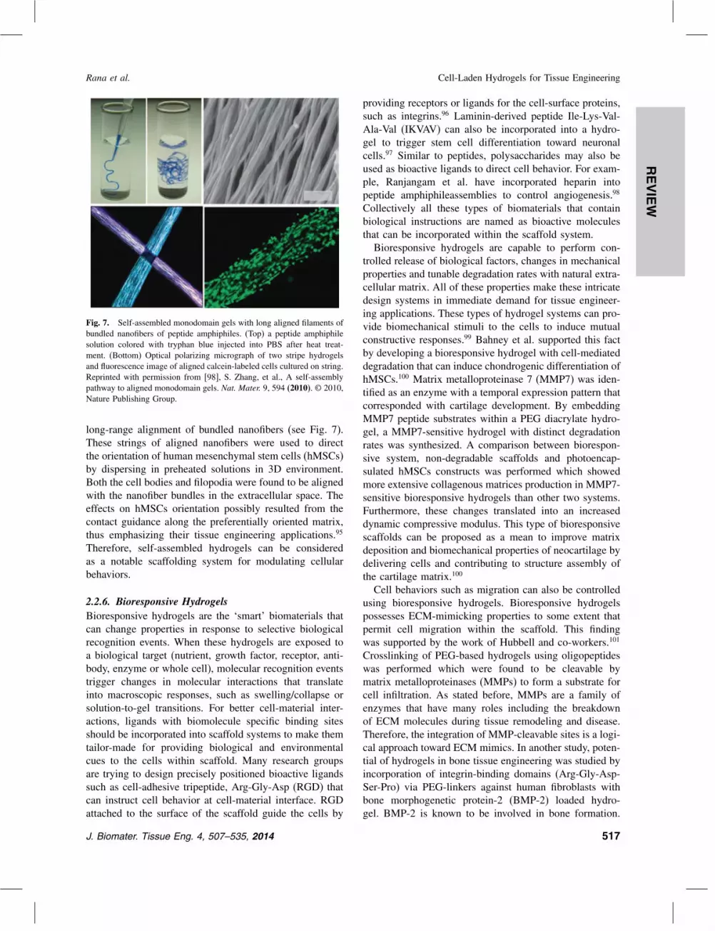
Fig. 7. Self-assembled monodomain gels with long aligned filaments ofbundled nanofibers of peptide amphiphiles. (Top) a peptide amphiphilesolution colored with tryphan blue injected into PBS after heat treat-ment. (Bottom) Optical polarizing micrograph of two stripe hydrogelsand fluorescence image of aligned calcein-labeled cells cultured on string.Reprinted with permission from [98], S. Zhang, et al., A self-assemblypathway to aligned monodomain gels. Nat. Mater. 9, 594 (2010). © 2010,Nature Publishing Group.
long-range alignment of bundled nanofibers (see Fig. 7).These strings of aligned nanofibers were used to directthe orientation of human mesenchymal stem cells (hMSCs)by dispersing in preheated solutions in 3D environment.Both the cell bodies and filopodia were found to be alignedwith the nanofiber bundles in the extracellular space. Theeffects on hMSCs orientation possibly resulted from thecontact guidance along the preferentially oriented matrix,thus emphasizing their tissue engineering applications.95
Therefore, self-assembled hydrogels can be consideredas a notable scaffolding system for modulating cellularbehaviors.
2.2.6. Bioresponsive HydrogelsBioresponsive hydrogels are the ‘smart’ biomaterials thatcan change properties in response to selective biologicalrecognition events. When these hydrogels are exposed toa biological target (nutrient, growth factor, receptor, anti-body, enzyme or whole cell), molecular recognition eventstrigger changes in molecular interactions that translateinto macroscopic responses, such as swelling/collapse orsolution-to-gel transitions. For better cell-material inter-actions, ligands with biomolecule specific binding sitesshould be incorporated into scaffold systems to make themtailor-made for providing biological and environmentalcues to the cells within scaffold. Many research groupsare trying to design precisely positioned bioactive ligandssuch as cell-adhesive tripeptide, Arg-Gly-Asp (RGD) thatcan instruct cell behavior at cell-material interface. RGDattached to the surface of the scaffold guide the cells by
providing receptors or ligands for the cell-surface proteins,such as integrins.96 Laminin-derived peptide Ile-Lys-Val-Ala-Val (IKVAV) can also be incorporated into a hydro-gel to trigger stem cell differentiation toward neuronalcells.97 Similar to peptides, polysaccharides may also beused as bioactive ligands to direct cell behavior. For exam-ple, Ranjangam et al. have incorporated heparin intopeptide amphiphileassemblies to control angiogenesis.98
Collectively all these types of biomaterials that containbiological instructions are named as bioactive moleculesthat can be incorporated within the scaffold system.Bioresponsive hydrogels are capable to perform con-
trolled release of biological factors, changes in mechanicalproperties and tunable degradation rates with natural extra-cellular matrix. All of these properties make these intricatedesign systems in immediate demand for tissue engineer-ing applications. These types of hydrogel systems can pro-vide biomechanical stimuli to the cells to induce mutualconstructive responses.99 Bahney et al. supported this factby developing a bioresponsive hydrogel with cell-mediateddegradation that can induce chondrogenic differentiation ofhMSCs.100 Matrix metalloproteinase 7 (MMP7) was iden-tified as an enzyme with a temporal expression pattern thatcorresponded with cartilage development. By embeddingMMP7 peptide substrates within a PEG diacrylate hydro-gel, a MMP7-sensitive hydrogel with distinct degradationrates was synthesized. A comparison between biorespon-sive system, non-degradable scaffolds and photoencap-sulated hMSCs constructs was performed which showedmore extensive collagenous matrices production in MMP7-sensitive bioresponsive hydrogels than other two systems.Furthermore, these changes translated into an increaseddynamic compressive modulus. This type of bioresponsivescaffolds can be proposed as a mean to improve matrixdeposition and biomechanical properties of neocartilage bydelivering cells and contributing to structure assembly ofthe cartilage matrix.100
Cell behaviors such as migration can also be controlledusing bioresponsive hydrogels. Bioresponsive hydrogelspossesses ECM-mimicking properties to some extent thatpermit cell migration within the scaffold. This findingwas supported by the work of Hubbell and co-workers.101
Crosslinking of PEG-based hydrogels using oligopeptideswas performed which were found to be cleavable bymatrix metalloproteinases (MMPs) to form a substrate forcell infiltration. As stated before, MMPs are a family ofenzymes that have many roles including the breakdownof ECM molecules during tissue remodeling and disease.Therefore, the integration of MMP-cleavable sites is a logi-cal approach toward ECM mimics. In another study, poten-tial of hydrogels in bone tissue engineering was studied byincorporation of integrin-binding domains (Arg-Gly-Asp-Ser-Pro) via PEG-linkers against human fibroblasts withbone morphogenetic protein-2 (BMP-2) loaded hydro-gel. BMP-2 is known to be involved in bone formation.
J. Biomater. Tissue Eng. 4, 507–535, 2014 517

REVIEW
Cell-Laden Hydrogels for Tissue Engineering Rana et al.
Fig. 8. Bioresponsive MMP sensitive PEG hydrogels allow spindle-shaped cell morphologies similar to natural fibrin (FIB) and collagen (COL) gels,whereas plasma sensitive PEG hydrogels (P-PEG) inhibits cell spreading. Reprinted with permission from [105], G. P. Raeber, et al., Molecularlyengineered PEG hydrogels: A novel model system for proteolytically mediated cell migration. Biophys. J. 89, 1374 (2005). © 2005, Elsevier.
The fibroblasts were observed to cause a local breakdownof hydrogel crosslinks via secreted MMPs. An assessmentof the degradation behavior of MMPs and the cell invasionof provisional matrices revealed that the healing responsein vivo depends on the enzymatic sensitivity of the matrix.Raeber et al. subsequently tested the suitability of twoproteolytically degradable PEG hydrogels with cell migra-tion in 3-D to mimic natural ECM.102 The results indicatethat migration in M-PEG (PEG hydrogel crosslinked withMMP-sensitive sequence) gels is highly sensitive to MMPmodulation (see Fig. 8). The ability of a gel to respondto a single class of enzyme provides base for an effectivecommunication between cells and the matrices.Bioresponsive hydrogels are also employed for growth
factor delivery. For example, Hall et al. designed a 3D fib-rin hydrogel scaffold that act as a depot/release system forgrowth factors to aid in angiogenesis.103 The fibrin matri-ces are modified by covalently adding receptor-bindingsites of integrin �v�3. In vivo analysis of angiogenesisreveals that a denser capillary network was formed inreceptor-stimulated matrices than native fibrin networks.On comparing these results with additional growth fac-tors for co-stimulation, similar results that are in coher-ence with either receptor or growth factor embedded wereobtained. To combine the advantages of synthetic and nat-ural systems, peptide amphiphiles (PAs) can be considered.Jun et al. described a strategy to regenerate dental tissuesusing PA molecules that form rigid, cell-responsive fibrousnanostructures and incorporate biological epitopes as thepeptide part of the molecule.104 Along with other biore-sponsive molecules, proteases are found to play an essen-tial role in a large number of biological processes suchas wound healing and cell differentiation making them
critical for incorporation into scaffold systems for the tis-sue regeneration.All these hydrogel types provide better opportunities
to increase cell-material interaction and ECM remodeling.To further enhance the biocompatibility and niche mim-icking properties of these hydrogels, cell-laden hydrogelscaffolds have been introduced to yield significant dif-ferences in cellular response to exogenous cues as com-pared to monolayer culture. As embedding cells within3D hydrogel can allow for their immobilization for bet-ter replication of spatiotemporal presentation of cells toeach other throughout the developmental process. Theseadvanced scaffolding systems are being discussed in thepreceding sections with their different types, properties andadvantages over the conventional hydrogel systems in thecontext of tissue engineering.
2.3. Fabrication of HydrogelsThe method of fabrication of hydrogel scaffolds plays avital role in imparting ECM-mimicking functional proper-ties and tissue-like environment. Hydrogel scaffolds thatcan be fabricated with optimum mechanical propertiesgenerally gives negative results for cell encapsulationand proliferation, while constructs with high porosity anduniform cell distribution results into being mechanicallyweak.105 Thus, a need to combine structural stability withhigh cell density while maintaining tissue-like environmentfor accelerated tissue formation has aroused. Over the pastdecades, many research groups have published an amalga-mation of various engineering techniques for the fabrica-tion of hydrogel scaffolds with controlled architecture.106
In this section, various fabrication methods for the better-ment of geometrical features as well as the distribution of
518 J. Biomater. Tissue Eng. 4, 507–535, 2014
REVIEW
Rana et al. Cell-Laden Hydrogels for Tissue Engineering
cells and biomolecules within the hydrogel scaffolds havebeen discussed
2.3.1. Micro-Fabrication TechniquesMicro-fabricated hydrogels can be developed by employ-ing photolithography or photo patterning approaches.107
In photolithography, a mask is specifically designed toimplement patterns over the hydrogel constructs. Alter-native transparent and opaque areas are introduced inthe mask according to the pattern for controlling thecrosslinking of hydrogels. Upon light irradiation, trans-parent mask areas exposed to UV light for crosslinkingto form micro patterns whereas the remaining unexposedparts are shielded from crosslinking and subsequentlywashed out in the washing step.108 Visible or UV light canreact with certain light-sensitive compounds called pho-toinitiators to form crosslinked hydrogels in vitro, in vivoor in situ. Photopolymerization allows spatial as well astemporal control over polymerization by controlling theircuring rates from less than a second to a few minutesat room or physiological temperatures. Photopolymerizedhydrogels exhibit minimal heat production, as well as theability to form complex shapes that adhere and conformto the defect site which makes them exceptionally impor-tant for the field.109 Although biological systems put con-straints on the use of photopolymerization in vivo, owingto the limits of acceptable temperatures, pH, as well astoxicity of most monomers and organic solvents, but canbe overcome by the use of mild polymerization conditions(i.e., low light intensity and organic solvent levels, shortirradiation time, and physiological temperature).109
Along with photocrosslinkable systems, other meth-ods have been also developed including enzymatic110 andthermo sensitive systems to avoid the use of potentiallycytotoxic UV light and free radicals. For example, Ferrutiet al. found amphoteric poly(amidoamine) (PAA)-basedhydrogels containing carboxyl and amino groups in theirrepeating units as a potential scaffold material becauseof its cyto-compatibility with fibroblasts as well as non-cytotoxic degradation products, but their mechanical prop-erties needed further improvement.111 The group furthermodified the PAA hydrogels by introducing side guani-dine groups to improve cell adhesion and proliferationand found improved mechanical properties when a sec-ond PAA carrying primary amino group was used as acrosslinking agent, suggesting that mechanical propertiescan be tuned for PAA or acrylate-based photocrosslink-able hydrogels by incorporating crosslinking agents withconsiderable cyto-compatibility for cell growth. Someexamples of these hydrogels includes PEG diacrylate(PEGDA), PEG-dimethacrylates (PEGDM), methacrylatedgelatin (GelMA), methacrylated HA and methacrylatedtropoelastin(MeTro).106 Similar to chemical and physicalcrosslinks, hydrogels are quite often modified with celladhesion peptidesin order to enhance the cell attachment
and growth.112 For example, Sannino et al. combinedthe photocrosslinking reaction with a foaming processto induce an interconnected porosity within PEG-basedhydrogels that had been modified with peptide sequencesfor enhancing cell adhesion.113 Though this method isremarkably successful, it also includes some limitationssuch as uncontrollable crosslinking depth over hydrogellayer, resolution of the features depends on the quality ofphotomask and aspect ratio.Soft lithography and molding are other popular tech-
niques for the synthesis of micro-fabricated hydrogel scaf-folds. Micro-fabricated hydrogel scaffolds can be moldedlike conventional molding techniques but with a replace-ment of elastomeric molds such as polydimethylsiloxane(PDMS) due to their biocompatibility and hydrophobicsurface properties. To further facilitate the detachmentof crosslinked hydrogels from the mold, a temperatureresponsive hydrogel poly(N -isopropylacrylamide) (PNI-PAAm) coating over mold surface have been introduced.114
Both physically and chemically cross-linkable hydrogelscan be fabricated using this method.Rapid-prototyping is a recently developed additive-
based fabrication technique with sequential delivery ofmaterial or energy to form a scaffold. It is an auto-mated system where scaffolds are designed using a CADsoftware and then converted to sliced models which canbe fabricated using laser-based systems through an addi-tive process. Stereolithography (SLA), digital light pro-jection (DLP) and two photon polymerization techniquesare the other popular laser-based systems currently beingemployed for the micro-fabrication of hydrogel scaffolds.
2.3.2. Wetspinning and Microfluidic SpinningFibrous structure play an important role in mimickingnative ECM for regeneration of tissues emphasizing on theimportance of fibrous constructs. In fact, the native ECMis by itself made of micro and nano-featured fibers andporous structure in the network of gelly-like environment.Hydrogel fibers with high porosity and high surface areato volume ratio can be fabricated using wetspinning andmicrofluidic spinning methods. Microfluidic fiber spinninginvolves arrangement of two or more parallel streams ofpolymer solutions and a sheath flow in a microchannelwhich will further lead to hydrogel formation down thestream by chemical, optical or thermal crosslinking. Pre-cise control on single or multiple fibers shape, size, celldistribution and chemical composition is feasible throughthis approach. Alginate, gelatin/hydroxyphenylpropionicacid (Gtn-HPA) and NIPAm are the commonly usedmaterials for the fabrication of fibers in a microfluidicplatform.115 But these polymers lack components of nativeECM thus limiting the cell–cell interactions. Similar tomicrofluidic spinning, wetspinning includes injection ofa pre-polymer solution into one or multiple coagulationbaths through a syringe pump or by using pressurized air.
J. Biomater. Tissue Eng. 4, 507–535, 2014 519
REVIEW
Cell-Laden Hydrogels for Tissue Engineering Rana et al.
Various biocompatible materials have used for hydrogelfiber synthesis through this method such as alginate, colla-gen/alginate composite, collagen, chitosan and starch/PCLcomposite suitable for tissue engineering applications.106
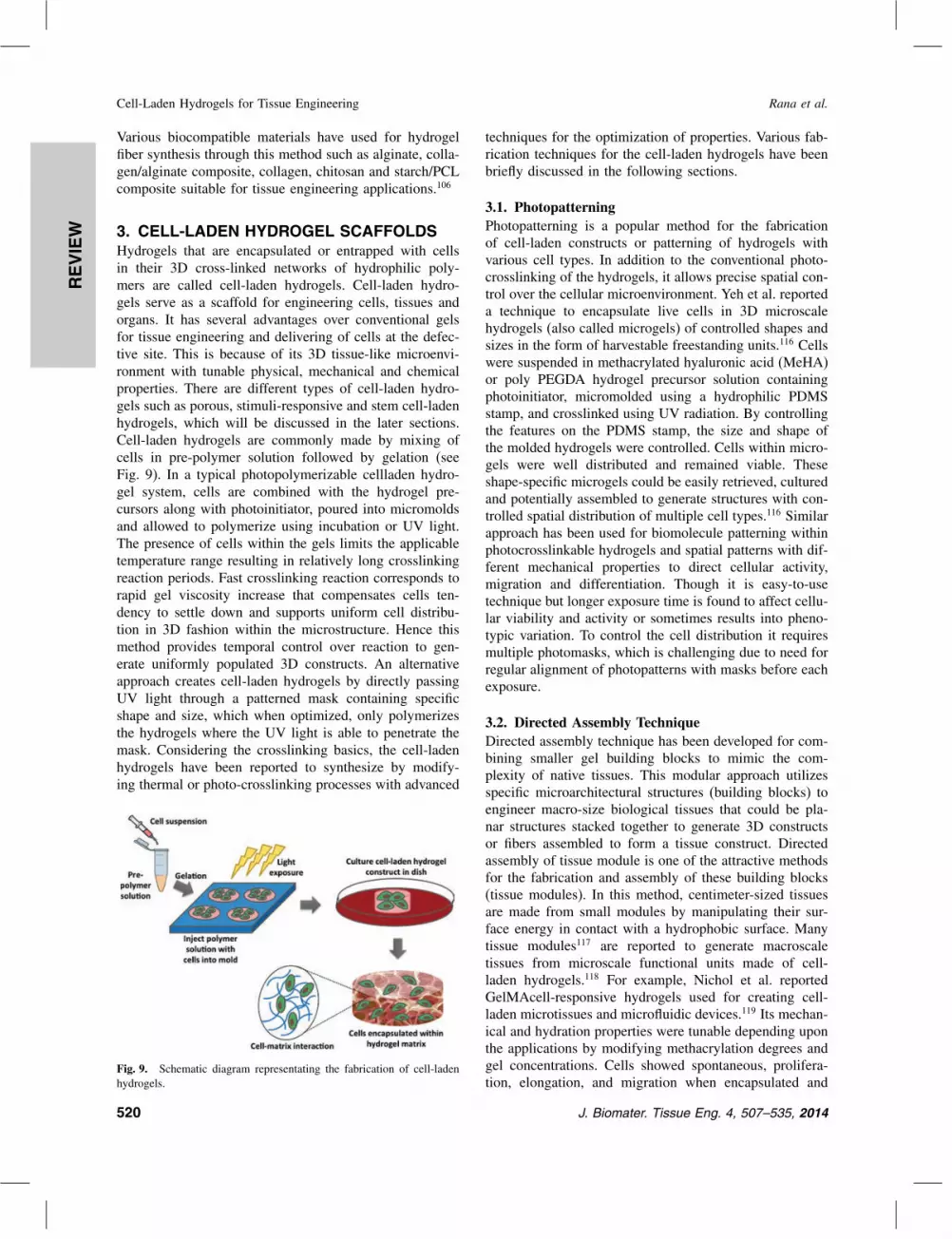
3. CELL-LADEN HYDROGEL SCAFFOLDSHydrogels that are encapsulated or entrapped with cellsin their 3D cross-linked networks of hydrophilic poly-mers are called cell-laden hydrogels. Cell-laden hydro-gels serve as a scaffold for engineering cells, tissues andorgans. It has several advantages over conventional gelsfor tissue engineering and delivering of cells at the defec-tive site. This is because of its 3D tissue-like microenvi-ronment with tunable physical, mechanical and chemicalproperties. There are different types of cell-laden hydro-gels such as porous, stimuli-responsive and stem cell-ladenhydrogels, which will be discussed in the later sections.Cell-laden hydrogels are commonly made by mixing ofcells in pre-polymer solution followed by gelation (seeFig. 9). In a typical photopolymerizable cellladen hydro-gel system, cells are combined with the hydrogel pre-cursors along with photoinitiator, poured into micromoldsand allowed to polymerize using incubation or UV light.The presence of cells within the gels limits the applicabletemperature range resulting in relatively long crosslinkingreaction periods. Fast crosslinking reaction corresponds torapid gel viscosity increase that compensates cells ten-dency to settle down and supports uniform cell distribu-tion in 3D fashion within the microstructure. Hence thismethod provides temporal control over reaction to gen-erate uniformly populated 3D constructs. An alternativeapproach creates cell-laden hydrogels by directly passingUV light through a patterned mask containing specificshape and size, which when optimized, only polymerizesthe hydrogels where the UV light is able to penetrate themask. Considering the crosslinking basics, the cell-ladenhydrogels have been reported to synthesize by modify-ing thermal or photo-crosslinking processes with advanced
Fig. 9. Schematic diagram representating the fabrication of cell-ladenhydrogels.
techniques for the optimization of properties. Various fab-rication techniques for the cell-laden hydrogels have beenbriefly discussed in the following sections.
3.1. PhotopatterningPhotopatterning is a popular method for the fabricationof cell-laden constructs or patterning of hydrogels withvarious cell types. In addition to the conventional photo-crosslinking of the hydrogels, it allows precise spatial con-trol over the cellular microenvironment. Yeh et al. reporteda technique to encapsulate live cells in 3D microscalehydrogels (also called microgels) of controlled shapes andsizes in the form of harvestable freestanding units.116 Cellswere suspended in methacrylated hyaluronic acid (MeHA)or poly PEGDA hydrogel precursor solution containingphotoinitiator, micromolded using a hydrophilic PDMSstamp, and crosslinked using UV radiation. By controllingthe features on the PDMS stamp, the size and shape ofthe molded hydrogels were controlled. Cells within micro-gels were well distributed and remained viable. Theseshape-specific microgels could be easily retrieved, culturedand potentially assembled to generate structures with con-trolled spatial distribution of multiple cell types.116 Similarapproach has been used for biomolecule patterning withinphotocrosslinkable hydrogels and spatial patterns with dif-ferent mechanical properties to direct cellular activity,migration and differentiation. Though it is easy-to-usetechnique but longer exposure time is found to affect cellu-lar viability and activity or sometimes results into pheno-typic variation. To control the cell distribution it requiresmultiple photomasks, which is challenging due to need forregular alignment of photopatterns with masks before eachexposure.
3.2. Directed Assembly TechniqueDirected assembly technique has been developed for com-bining smaller gel building blocks to mimic the com-plexity of native tissues. This modular approach utilizesspecific microarchitectural structures (building blocks) toengineer macro-size biological tissues that could be pla-nar structures stacked together to generate 3D constructsor fibers assembled to form a tissue construct. Directedassembly of tissue module is one of the attractive methodsfor the fabrication and assembly of these building blocks(tissue modules). In this method, centimeter-sized tissuesare made from small modules by manipulating their sur-face energy in contact with a hydrophobic surface. Manytissue modules117 are reported to generate macroscaletissues from microscale functional units made of cell-laden hydrogels.118 For example, Nichol et al. reportedGelMAcell-responsive hydrogels used for creating cell-laden microtissues and microfluidic devices.119 Its mechan-ical and hydration properties were tunable depending uponthe applications by modifying methacrylation degrees andgel concentrations. Cells showed spontaneous, prolifera-tion, elongation, and migration when encapsulated and
520 J. Biomater. Tissue Eng. 4, 507–535, 2014

REVIEW
Rana et al. Cell-Laden Hydrogels for Tissue Engineering
Fig. 10. Fluorescence images of HUVEC cells (cells on the gel) adhered to GelMA of all macromere concentrations exhibiting cell adhesion,proliferation and migration, but did not adhere to PEG 4000 as demonstrated by endogenous GFP (A) and rhodamine-labeled phalloidin/DAPIstaining for F-actin/cell nuclei (B) on day 5 of culture (scale bar = 200 �m). Reprinted with permission from [111], J. W. Nichol, et al., Cell-ladenmicroengineered gelatin methacrylate hydrogels. Biomaterials 31, 5536 (2010). © 2010, Elsevier.
seeded in hydrogels (see Fig. 10).119 Figure 11 showsfluorescent images of cells seeded within these micro-gel channels having potential for perfusable microvascu-lature. In general, modular approaches allow a precisecontrol over the population and distribution of differentcell types within the construct. The lack of scalability andlow mechanical properties of the fabricated constructs isthe few challenges that need to be resolved.As an alternate to modular approaches, Shin et al.
reported a double-network (DN) strategy that could beused to engineer strong hydrogels encapsulating cells.120
They synthesized DN hydrogels by a two-step pho-tocrosslinking using gellan gum methacrylate (GGMA) forrigid and brittle first network, and GelMA for the soft andductile second network. The resulting DN hydrogels exhib-ited the compressive failure stress of up to 6.9 MPa, whichis comparable to the strength of cartilage. DN hydrogels
Fig. 11. Fluoresence images of cell seeded within the cell-laden GelMAmicrofluidic channels that were 300 �m in diameter. (A) and (B) havePKH67 labeled 3T3 fibroblasts, and (C) and (D) have GFP-HUVEC cellsdemonstrating attachment within endothelial-lined perfusable microvas-culature microgels. Reprinted wth permission from [111], J. W. Nicholet al., Cell-laden microengineered gelatin methacrylate hydrogels. Bio-materials 31, 5536 (2010). © 2010, Elsevier.
with a higher mass ratio of GelMA to GGMA exhibitedhigher strength, which shows potential in developing evenstronger DN hydrogels in the future. 3D encapsulation ofNIH-3T3 fibroblasts showed cell compatibility of DN for-mation process, which provides an insight for regenerationof load-bearing tissues.120 However, the DN strategy needsto be modified according to the different microscale hydro-gel tissue modules assembly methods for the regenerationof tissues and organs.
3.3. MicrofluidicsAlong with the hydrogel and microscale technologies,microfluidic device can also potentially be used to facil-itate the exchange of nutrients and soluble factors in3D tissue constructs.121 Microfluidic device requires min-imal reagent consumption, allow for the laminar flowof fluids, and may be used for high-throughput analy-sis. PDMS molds are usually used as they are non-toxicto cells along with poly(DL-lactic-co-glycolide) (PLGA)and poly(glycerol sebacate) (PGS) to engineer microvas-culature within synthetic scaffolds.122 In certain works,endothelial and hepatocyte cells were seeded within thecomplex microfluidic channels togenerate patterns forblood vessels and liver constructs for tissue engineering.123
Recently, calcium alginate121 and gelatin124 hydrogelshave been used to fabricate microfluidic devices withcells seeded on the surface of microchannels. Ling et al.reported a fabrication technique for microfluidic channelsfrom cell-laden agarose hydrogels.125 Molten of agarosewas molded against SU-8 patterned silicon wafer as perstandard soft lithography protocols to fabricate sealed andwater-tight microfluidic channels. For cell-laden microflu-idic hydrogel channels, 6% Agarose molten solution (auto-claved/sterilized to dissolve in PBS) cooled to 70 �Cand mixed with equal volume of cell suspension withdefined cell density to yield a 3% agarose mixture loadedwith cells. This master solution of molten agarose andcells was then poured onto silicon master and allowedfor gelation for 2 hr at 25 �C in sterilized conditions.
J. Biomater. Tissue Eng. 4, 507–535, 2014 521
REVIEW
Cell-Laden Hydrogels for Tissue Engineering Rana et al.
For microchannel base, a thin flat slab of agarose wasdeveloped. Agarose molds were gently peeled from thesilicone masters and trimmed to a suitable shape. In addi-tion, a metal feeder wire of upto 2 cm length was usedas a guide for insertion of the flexible polyethylene tubingto generate holes for inlets and outlets, which was pressedagainst the feeder wire to force the wire out of the hole andplace tubing correctly. Finally, molded agarose surfaceswere heated at 71 �C for 3 s and pressed against anothersurface-heated agarose slab to form sealed microfluidicchannels. Channels of different dimensions were gener-ated and it was shown that agarose, though highly porous,is a suitable material for microfluidic channels. Cellsembedded within the molds were well distributed andmedia pumped through the channels allowing exchangeof nutrients and waste products. They demonstrated theimportance of a perfused network of microchannels fordelivering nutrients and oxygen to maintain cell viabil-ity in large hydrogels.126 Du et al. reported a sequen-tial assembly of cell-laden hydrogel constructs to engineervascular-like microchannels.127 They sequentially assem-bled microengineered hydrogels (microgels) into hydrogelconstructs with an embedded network of microchannels.Photolithography were used to fabricate arrays of micro-gels with predefined internal microchannels and assembledinto 3D tubular construct with multi-level interconnectedlumens. This technique holds promise as a biofabricationmethod, which does not influence cell viability within themicrogels. Endothelial cells and smooth muscle cells wereincorporated into an assembled construct with a concentricmicrogel design.127 Demonstrating a completely innova-tive approach, Chiang et al. reported an integrated plat-form combining digital microfluidic and photo-patterningtechniques to manipulate and fabricate biomimic cell-ladenhydrogels.128 They combine electrowetting-on-dielectric(EWOD) and photo-patterning techniques on a single plat-form. Different hydrogels cured from individually-drivenpolymer droplets provide 3D microenvironment for in vitrocell culture and actively drive polymer droplets, assemblethe hydrogels and arrange the cells inside the hydrogel.128
Currently, flow lithography and microfluidic fiber spin-ning techniques are the newly emerging fabrication meth-ods for cell-laden hydrogel constructs. As discussedin Section 2.3.2, microfluidic fiber spinning techniqueaccompanies drawbacks such as lack of natural ECM com-ponents. Core–shell fibers came up as a solution to thisproblem with ECM proteins in the core and alginate hydro-gel in the shell. It employs a double co-axial laminar flowmicrofluidic device system where a stream of ECM proteinwas generated in a core flow surrounded by sodium algi-nate prepolymer and a sheath calcium chloride flow. Thediffusion of ECM proteins was reported to be minimizedby the calcium alginate shell during their gelation and thusreconstitute intrinsic morphologies and functions of livingtissues. Moreover, this technique allows the incorporation
of cells and chemicals in single-and multi-layer fibers dur-ing the manufacturing process but limiting its use due tolow mechanical properties of the fabricated fibers.
3.4. BioprintingBioprinting of hydrogels at precise 3D architecturalarrangements represents an innovative approach for engi-neering biomimetic tissue constructs. Bioprinting includessequential deposition of solid layers for the precise devel-opment of complex structures. In bioprinted tissues, cellscan be either embedded within biologically relevant hydro-gels or printed free of scaffold support with high cell via-bility. Hasan et al. has reported an innovative and uniqueplatform that prints a 3D smooth muscle cells (SMCs)patch consisting of multiple cell-laden hydrogel layers.129
The developed bioprinting platform allows high through-put patterning of SMCs encapsulated in collagen hydro-gel droplets, microscale spatiotemporal droplet placementcontrol, printing of 3D cell-laden hydrogel structures andcell seeding uniformity. This layer-by-layer 3D tissue epi-taxy is a powerful approach to treat diverse diseasessuch as cancer, loss of tissue function or organ failure.130
The major drawback of this approach is poor or inadequatematerial-printing device and substrate combinations, aswell as the relatively small size of the printed construct.Campos et al. hypothesized that cell-laden hydrogelscan be printed when submerged in perfluorotributylamine(C12F27N), a hydrophobic high-density fluid.131 Humanmesenchymal stem cells and MG-63 cells were encapsu-lated into agarose hydrogels, and subsequently printed inhigh aspect ratio in three dimensional structures that weresupported in high density fluorocarbon. The results showedthat the cells encapsulated within 3D hydrogel constructsremain viable while printing 3D structures with variousshapes and sizes were manufactured and remained sta-ble for more than six months. The compressive strengthvalues of the printed gels consequently increased dur-ing the two weeks in culture.131 Along with these cell-laden scaffolds in tissue engineering, a number of newapproaches have been utilized by other groups serve greatimportance to regenerative medicine field. All the fab-rication methods discussed above allow the cell-ladenhydrogel constructs to optimize their structural propertiesand functions. To further explore different properties ofcell-laden hydrogels, in the following sections, we haveincluded some innovative and current trends used in cell-laden scaffolds with their research potential and promisingproperties.
4. POROUS CELL-LADEN HYDROGELSPorosity is an essential characteristic of tissue scaffold asit allows cells to infiltrate in 3D space. Porous structuresin hydrogels are potentially useful for mimicking nativetissues by improving protein transport and diffusion ingels. To synthesize the porous scaffolds, several methods
522 J. Biomater. Tissue Eng. 4, 507–535, 2014

REVIEW
Rana et al. Cell-Laden Hydrogels for Tissue Engineering
have been developed. For example, Cordell et al. stated amethod in which a colloidal suspension was used to cre-ate pores within hydroxyapatite scaffolds.132 The mechan-ical bending and compression analysis of this scaffoldconfirmed about the strength of the bulk microporousscaffold with smaller micropore sizes was higher thanscaffold with larger micropore sizes. These results werefound to be in agreement with reported results for otherporous materials.133 Poly(methyl methacrylate) (PMMA)beads were also reported as porogento generate microp-orous structures within fibrin scaffolds.134 However, thedrawback of this method is the use of toxic chemical pro-cesses while removing the PMMA beads from the scaf-fold, which ultimately results into micropores.135 Porogenand its leaching method is thus an important parameterfor microporous cell-laden hydrogels. Sucrose holds goodcharacteristics for this and has been used to create parti-cles and pores within PLGA sponges during a gas foamingprocess.136 In addition to sucrose crystal leaching method,salt crystals have also been used to create interconnectedpores within polymeric scaffolds.137 The method of fab-rication employed for these porous PLGA scaffolds aresolvent casting/particulate leaching and gas foaming usinga suitable porogen (usually salt). Fusion of porogen in thesolvent casting process found to pronounce pore intercon-nectivity and mechanical properties of polymeric scaffoldsand thus poresize could be controlled by using porogenmi-croparticles (see Fig. 12). Overriding the inability of thesescaffolds to directly encapsulate cells within the materi-als and avoiding use of toxic chemicals becomes one ofthe major requirements for their development emphasizingon biocompatible modifications in conventional scaffolds.In a study by Park et al. the authors introduced cell-ladenagarose gel system containing engineered constructs with amicrovascular structure and micropores that are created bydissolving sucrose crystals without any organic solvents.138
They developed a porous cell-laden agarose fluidic deviceand characterized the physical and mechanical propertiesof agarose gels with various micropores. Hepatic cellswere encapsulated within gels studied for their correspond-ing diffusion profile in microchannels. Cell viability inhydrogels with 100 wt% sucrose formulation was foundto be higher than that in hydrogels with 0 wt% sucroseformulation at all distances from the medium perfusionchannel.138 Synthetic polymer scaffolds of PEGDA can beformed under physiological conditions through the processof photopolymerization which allows uniform encapsula-tion of cells.139 Photopolymerization utilizes light energyto dissociate initiator molecules into free radicals that canreact with monomers functionalized with double or triplechemical bonds of acrylate groups in PEGDA that will leadto propagation of radical chain polymerization. Thoughthis method is the most common and easiest to performit causes unintended formation of free radicals that candamage cell membranes, proteins and DNA.
Fig. 12. Images of microporosity: Phase contrast images of sucrosecrystals (0–200 wt%, (a)–(c)), sucrose crystals dissolved within agarosegels (d)–(f), and cross-section images of agarose gels containing micro-pores (g)–(i). Confocal microscope images (j)–(l) and SEM images(m)–(o) of microporosity within agarose gels. Reprinted with permis-sion from [130], J. H. Park, et al., Microporous cell-laden hydrogels forengineered tissue constructs. BiotechnolBioeng 106, 138 (2010). © 2010,John Wiley and Sons.
5. STIMULI RESPONSIVE CELL-LADENHYDROGELS
Considerable effort has been devoted for controlling therelease of cells and degradation rate of the scaffold sys-tem. Scaffolds made up of stimuli-responsive materialshave been developed to release their encapsulated cellsand bioactive molecules in their host tissue microenvi-ronement. The in situ formation of cell/scaffold constructallows the delivery of encapsulated cells, nutrients andgrowth factors at the defective site using minimally inva-sive techniques. The method involves mixing of stimuli-responsive polymer with cells at room temperature andthen pre-gel solution is injected into the body. Uponinjection, due to the external stimuli, the polymer formsa physical gel leading to cell encapsulation within 3Dmatrix. In a study by Ratner et al. they used templatingmethods to produce crosslinked PNIPAAm hydrogels witha defined pore size.140 They found that switching ofpore size with temperature can modulate encapsulationof cells within the gel.140 In another interesting study,Han et al. reported the formation of a dynamic hydrogelas a tissue-engineering scaffold with tunable macroporos-ity using stimuli-responsive porogens of gelatin, alginate
J. Biomater. Tissue Eng. 4, 507–535, 2014 523

REVIEW
Cell-Laden Hydrogels for Tissue Engineering Rana et al.
Fig. 13. SEM images of stimuli-responsive tri-porogen-based scaf-folds upon sequential porogen removal: (A) Illustration; (B) TypeA gelatin porogen removed by temperature stimulus (37 �C incuba-tion); (C) Alginate-calcium porogen removed by chelation stimulus(EDTA/disodium citrate), and (D) Hyaluronic acid porogen removed byenzymatic stimulus (hyaluronidase). Scale bars: 200 �m. Reprinted withpermission from [133], L. H. Han et al., Dynamic tissue engineeringscaffolds with stimuli-responsive macroporosity formation. Biomaterials34, 4251 (2013). © 2013, Elsevier.
and hyaluronic acid (see Fig. 13).141 These porogens wereembedded within 3D hydrogels made up of type-B gelatinthat encapsulate bovine chondrocytes. They reported thedegradation of porogen can be controlled by specific stim-uli including temperature, chelating and enzymatic diges-tion which lead to sequential formation of macroporeswithin hydrogels to enhance cell proliferation and ECMproduction over time. The results of this study showedeffective cell release from alginate porogen with high cellviability, cell proliferation and spreading throughout the3D hydrogel. In addition, dynamic pore formation was alsofound to contribute for increased type II and X collagenproduction by chondrocytes and thus provide a platformfor a tool to create stimuli-responsive scaffolds for widespectral tissue engineering application and to study cellresponses for dynamic niche properties.141
6. STEM CELL-LADEN HYDROGELSStem cells can be used as anexcellent cell source for tis-sue engineering and regenerative medicine applications.142
Unlike other types of cells in the body, stem cells areunspecialized cells that are capable of self-renewal forlonger period yet maintain their capacity to differenti-ate into multiple specialized cell types when exposedto specific induction cues. Techniques for culturing andregulating human stem cells could lead to unprecedentedregenerative treatments. Many tissues are comprisedof cells that typically do not undergo extensive self-replication, such as cardiomyocytes in cardiac tissue thusthe ability to control and dictate stem cell behavior is cru-cial for the future success of engineered tissues. Stem cellscan reside in niches that are unique to the tissues and
organs containing highly ordered microarchitectures, cel-lular compartmentalization and arrangement.Employing stem cell-laden hydrogels as an investiga-
tive model for determining and controlling cell behavioris a beneficial approach for driving stem cell differenti-ation and to function down specific lineages that can beadvantageous for use in a variety of tissue engineeringand regenerative medicine applications. Stem cells withhydrogels can be used as micro-tissue constructs with spe-cific mechanical properties and geometries. On combiningthese scaffold constructs with microfluidic patterning andmicromoulding techniques, cells can be arranged, shaped,layered and made to increase permeation for oxygen, nutri-ents and other water-soluble metabolites to exactly mimicthe natural organization of cells in native tissues. Bahneyet al. reported about the non-toxic conditions for photoen-capsulation of human mesenchymal stem cells (hMSCs)in PEGDA scaffolds using a visible light photoinitiatorsystem composed of eosin Y, triethanolamine and 1-vinyl-2-pyrrolidinone. This system showed increased viabilityof encapsulated hMSCs and a more tightly crosslinkednetwork in one-third the time of UV polymerization withIrgacure 2959 (See Fig. 14).143
In contrast to natural proteins, many groups have usedsynthetic peptide sequences designed to self-assembleinto a hydrogel under certain environmental conditions.144
One strategy was based on an amphiphilic peptide contain-ing positively charged lysine residues.145 When the peptidesolution was mixed with cell culture medium containinga high concentration of electrolytes, the lysine residuesbecome shielded, causing the hydrophobic regions of thepeptide to rapidly assemble and form a crosslinked hydro-gel. Under shear stress, the crosslinked gel undergoesshear thinning resulting in a viscous gel and recoversfully when unloaded. They demonstrated a cell-laden gelencapsulated with MSCs to be delivered in situ through asyringe.145 Similar reports were obtained with elastin-likepeptide (ELP). Human adipose-derived adult stem cellswere encapsulated in ELP hydrogels and it was found thatELP environment promoted chondrogenic differentiationwithout the addition of chondrogenic medium which infersthat hydrogel matrix provide some cues to induce stem celldifferentiation.146
Stem cell-laden hydrogels have many advantages overconventional scaffolds that can be employed in differ-ent tissue regeneration application. In an interesting study,Fedorovich et al. have studied the effect of calcium phos-phate (CaP) particles on osteoinductive potential of cell-laden hydrogel-CaP composite matrices.147 They includedapatitic nanoparticles in Matrigel constructs and stud-ied viability of embedded multipotent stromal progenitorcells in vitro. Furthermore, they investigated the in vivoosteoinductive potential of cell-laden Matrigel containingapatitic nanoparticles after subcutaneous implantation inimmunodeficient mice and compared with composites
524 J. Biomater. Tissue Eng. 4, 507–535, 2014

REVIEW
Rana et al. Cell-Laden Hydrogels for Tissue Engineering
Fig. 14. Cytotoxicity of visible light initiation compared to UV light initiation by live-dead staining of hMSCs 48 h after photoencapsulation in a10% PEGDA scaffold using (A) I2959, (B) 0.75% TEA and 0.1 mM eosin Y and (C) 0.1% TEA and 0.01 mM eosin Y. Reprinted with permissionfrom [135], C. S. Bahney, et al., Visible light photoinitiation of mesenchymal stem cell-laden bioresponsive hydrogels. Eur. Cell. Mater. 22, 43 (2011).© 2011, AO Research Institute Davos.
containing osteoinductive biphasic calcium phosphate(BCP) microparticles. Finally, they concluded that apatiticnanoparticles were osteoinductive and induced osteoclastactivation without bone formation whereas BCP parti-cles were found to be more effective in inducing boneformation at ectopic location.147 Similar to bone appli-cation, Chung et al. studied the influence of 3D HAmicroenvironments on chondrogenic potential of MSCsfor cartilage applications.148 As HA is a native com-ponent of cartilage, MSCs may interact with HA viacell surface receptors that influence stem cell differ-entiation. In vitro and in vivo cultures of MSC-ladenHA hydrogels supports chondrogenesis. To assess theinfluence of scaffold chemistry on chondrogenesis, HAhydrogels were compared with inert PEG hydrogelsand showed enhanced expression of cartilage-specificmarkers. Differences between HA and PEG hydrogelsin vivo were most noticeable for MSCs and polymeralone, indicating that hydrogel chemistry influences thecommitment of MSCs to undergo chondrogenesis (e.g.,∼43-fold up-regulation of type II collagen of MSCs inHA over PEG hydrogels). Although this study investi-gated only early markers of tissue regeneration, theseresults emphasize the importance of material cues in MSCdifferentiation.148 Therefore, based on the experimentalexamples discussed in this section, and other reporteddata, stem cell-laden hydrogels may be considered as apotential candidate for scaffold-based tissue regenerativeapplications.
7. APPLICATIONS OF CELL-LADENHYDROGEL SCAFFOLDS
Cell-laden hydrogels are widely used in engineering cells,tissues and organs. This is because, they offer numerousattractive features such as hydrated tissue-like microenvi-ronment for cell and tissue growth and ease of cell trans-plantation. Table II summarizes applications of differenttypes of cell-laden hydrogel systems with their uniqueproperties. In the preceding sections, different tissue engi-neering applications of cell-laden hydrogels are brieflydiscussed.
7.1. Bone Tissue EngineeringConventional hydrogels that can be used as injectableor in organ printing are reported to lack appropriatestimuli to direct osteogenic differentiation in encapsu-lated multipotent stromal cells causing inefficient boneformation within the matrix. To overcome this problem,Fedorovich et al. studied the effect of CaP nanoparticlesfor the osteoinductive potential of cell-laden hydrogel-CaP composite matrices in comparison with compos-ites containing osteoinductive biphasic calcium phosphate(BCP) microparticles.152 They concluded that apatiticnanoparticles were found to be osteoinductive and inducedosteoclast activation without bone formation whereas BCPparticles were found to be more effective in inducing boneformation at ectopic location.152 To further resolve theproblems, the group tried to fabricate a suitable osteochon-dral implant with optimum tissue formation and integrationproperties. They characterized the use of a 3D fiber depo-sition (3DF) technique for the fabrication of cell-laden,heterogeneous hydrogel construct as osteochondral grafts.Fluorescently labeled human chondrocytes and osteogenicprogenitor cells were encapsulated and printed in alginatehydrogels yielding scaffolds with both cell types located indifferent parts. The results of this study confirmed that thescaffolds of varying porosity and elastic modulus can beobtained with high cell viability, by changing fiber spacingor angle of fiber deposition, while printing that supportdistinctive tissue formation both in vivo and in vitro atdifferent locations within one construct.153
7.2. Cartilage Tissue EngineeringSimilar to bone applications, many research groupsare exploiting cell-laden hydrogels for chondrogenicapplications. As mechanical forces play an important rolein regulating cartilage development from MSCs, theseforces can be employed to develop engineered constructswith enhanced mechanical properties. Huang et al. reportedthe effect of long-term dynamic compression on MSC-seeded constructs and observed the influence and pheno-typic transitions by varying pre-culture duration, loadingregimes and inclusion of TGF-�3 during loading�149
They found that loading initiated before chondrogenesis
J. Biomater. Tissue Eng. 4, 507–535, 2014 525
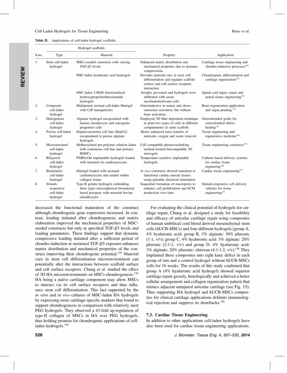
REVIEW
Cell-Laden Hydrogels for Tissue Engineering Rana et al.
Table II. Applications of cell-laden hydrogel scaffolds.
Hydrogel scaffolds
S.no. Type Material Property Application
1. Stem cell-ladenhydrogel
MSCs-seeded constructs with varyingTGF-�3 levels
Enhanced matrix distribution andmechanical properties due to dynamiccompressions
Cartilage tissue engineering andchondro-inductive processes149
MSC-laden hyaluronic acid hydrogels Provides material cues in stem celldifferentiation and regulate scaffoldsurface and cell surface receptorsinteraction
Chondrogenic differentiation andcartilage regeneration150
MSC-laden 2-RGD functionalizedhydroxypropylmethacrylamidehydrogels
Atrophy prevented and hydrogels wereinfiltrated with axonsmyelinatedschwann cells
Spinal cord injury repair andneural tisuue engineering151
2. Compositecell-ladenhydrogel
Multipotent stromal cell-laden Matrigelwith CaP nanoparticles
Osteoinductive in nature and showsosteoclast activation, but withoutbone activation.
Bone regeneration applicationand organ printing.152
3. Hetrogenouscell-ladenhydrogel
Alginate hydrogel encapsulated withhuman chondrocyte and osteogenicprogenitor cells
Employed 3D fiber deposition techniqueto print two types of cells in differentcompartments of same scaffold
Osteochondral grafts forosteochondral defecthealing153
4. Porous cell-ladenhydrogel
Hepatocarcinoma cell line (HepG2)encapsulated in porous alginatehydrogels
Shows enhanced mass transfer ofnutrients, oxygen and waste removal
Tissue engineering andregenerative medicine154
5. Microstructuralcell-ladenhydrogel
Methacrylated pre-polymer solution ladenwith continuous cell-line and primaryBMSCs
Cell compatible photocrosslinkingmethod formed biocompatible 3Dmicrogels
Tissue engineering constructs155
6. Bilayeredcell-ladenhydrogel
PNIPAAM implantable hydrogels loadedwith neonatal rat cardiomyocytes
Temperature-sensitive implantablehydrogels
Catheter-based delivery systemsfor cardiac tissueengineeing156
7. Biomimeticcell-ladenhydrogel
Matrigel loaded with neonatalcardiomyocytes and seeded withincollagen foams
In vivo constructs showed transition tofunctional cardiac muscle tissuesusing pulsatile electrical stimulation
Cardiac tissue engineering157
8. Stimuli-responsivecell-ladenhydrogel
Type-B gelatin hydrogels embeddingthree types microspherical biomaterialbased porogens with neonatal bovinechondrocytes
Sequential formation of macropores toenhance cell proliferation and ECMproduction over time
Stimuli-responsive cell deliveryvehicles for tissueengineering141
decreased the functional maturation of the constructalthough chondrogenic gene expression increased. In con-trast, loading initiated after chondrogenesis and matrixelaboration improved the mechanical properties of MSC-seeded constructs but only at specified TGF-�3 levels andloading parameters. These findings support that dynamiccompressive loading initiated after a sufficient period ofchondro-induction at sustained TGF-�3 exposure enhancesmatrix distribution and mechanical properties of the con-struct improving their chondrogenic potential.149 Materialcues in stem cell differentiation microenvironment canpotentially alter the interactions between scaffold surfaceand cell surface receptors. Chung et al. studied the effectof 3D HA microenvironments on MSCs chondrogenesis.150
HA being a native cartilage component may allow MSCsto interact via its cell surface receptors and thus influ-ence stem cell differentiation. This fact supported by thein vitro and in vivo cultures of MSC-laden HA hydrogelsby expressing more cartilage-specific markers that found tosupport chondrogenesis in comparison with relatively inertPEG hydrogels. They observed a 43-fold up-regulation oftype-II collagen of MSCs in HA over PEG hydrogels,thus holding promise for chondrogenic applications of cell-laden hydrogels.150
For evaluating the clinical potential of hydrogels for car-tilage repair, Chung et al. designed a study for feasibilityand efficacy of articular cartilage repair using compositesof human umbilical cord blood derived mesenchymal stemcells (hUCB-MSCs) and four different hydrogels (group A,4% hyaluronic acid; group B, 3% alginate: 30% pluronic(1:1, v/v); group C, 4% hyaluronic acid: 3% alginate: 20%pluronic (2:1:1, v/v) and group D, 4% hyaluronic acid:3% alginate: 20% pluronic: chitosan (4:1:1:2, v/v).158 Theyimplanted these composites into right knee defect in eachgroup of rats and a control hydrogel without hUCB-MSCscells for 16 weeks. The results of this study confirmed thatgroup A (4% hyaluronic acid hydrogel) showed superiorcartilage repair grossly, histologically and achieved a bettercellular arrangement and collagen organization pattern thatmimics adjacent uninjured articular cartilage (see Fig. 15).Thus supporting HA hydrogel and hUCB-MSCs compos-ites for clinical cartilage applications delimits immunolog-ical rejection and suppress its drawbacks.158
7.3. Cardiac Tissue EngineeringIn addition to other applications cell-laden hydrogels havealso been used for cardiac tissue engineering applications.
526 J. Biomater. Tissue Eng. 4, 507–535, 2014

REVIEW
Rana et al. Cell-Laden Hydrogels for Tissue Engineering
Fig. 15. Articular cartilage defects in a rat model and gross appearanceand scoring result at 16 weeks post-transplantation. (A) Gross photos ofarticular cartilage defects in a rat model transplanted with composites ofhUCB-MSCs and different hydrogels; (B) Gross findings of repair tissueat articular cartilage defects in a rat model after 16 weeks of implantationshowing transplanted knees repaired to almost normal level and borderregions between repair and normal tissue were less distinct than controlknees; (C) Gross appearance scores of experimental and control kneesin four different hydrogel groups. Reprinted with permission from [151],J. Y. Chung, et al., Comparison of articular cartilage repair with differ-ent hydrogel-human umbilical cord blood-derived mesenchymal stem cellcomposites in a rat model. Stem. Cell. Res. Ther. 5, 39 (2014). © 2014,BioMed Central.
A bilayered hydrogel from a temperature-responsive PNI-PAAM and non-responsive polymer was generated toinvestigate its potential use as an implantable cell-loadeddelivery system. Neonatal rat cardiomyocytes were cul-tured on the hydrogels until they reached confluence.Subsequently, temperature was lowered, and the hydrogelsheets were curled/rolled into tubes owing to the differencein swelling properties of the bilayered polymers, resultingin the formation of cell-laden hydrogels for catheter-baseddelivery systems.156
Decellularized cardiac tissues have also attractedsignificant attention for cardiac regeneration. In an inter-esting study, pig myocardial ECM was used to synthe-size hydrogels.156 The ECM material was isolated byprocessing decellularized porcine heart muscle. After char-acterization of the structure and composition of the ECMhydrogels, their potential to promote vascularization wasinvestigated using endothelial cells (ECs) and smoothmuscle cells. In addition, the ECM hydrogels supportedhigh cardiomyocytes viability. Decellularized ECM-basedhydrogels have strong potential for in vivo studies, par-ticularly for the delivery of cell-laden gels into themyocardium via syringe or catheter-based approaches.In a report by Zhang et al. they mentioned fabricationof tissue engineered cardiac patch for the maturationof human embryonic stem cell–derived cardiomyocytes(hESC–CMs) (see Fig. 16).159 The results of this studyconfirmed the uniform alignment of hESC-CMs withinthe patch by locally controlling the direction of passivetension. Higher conduction velocities (CVs), longer sar-comeres and enhanced expression of genes involved incardiac contractile functions were observed in hESC-CMs3D patches. The CVs in cardiac patches increased withcardiomyocyte purity, reaching 25.1 cm/s in patches con-structed with 90% hESC-CMs. Maximum contractile forceamplitudes and active stresses of cardiac patches averagedto 3�0±1�1 mN and 11�8±4�5 mN/mm2, respectively withcontractile force per input cardiomyocyte averaged upto5�7±1�1 nN/cell resulting into a negative correlation withhESC-CM purity. This study demonstrated advanced lev-els of hESC-CM maturation after 2 weeks of 3D cardiacpatch culture for future cell therapy studies.
7.4. Neural Tissue EngineeringNeural tissue engineering approaches also require develop-ment of engineered 3D constructs to provide guidance con-duits to cell-based therapies. Schwann cells play a key rolein peripheral nerve regeneration by forming oriented pathsfor axonal regeneration. Suri et al. encapsulated schwanncells within engineered collagen and HA IPN hydro-gels with and without laminin as a 3D culture system.160
The results of this study exposed that encapsulation ofcells in 3D hydrogel constructs did not affect the cellviability and underwent spreading and proliferation withincreased cell numbers. Cells were also observed to align
J. Biomater. Tissue Eng. 4, 507–535, 2014 527
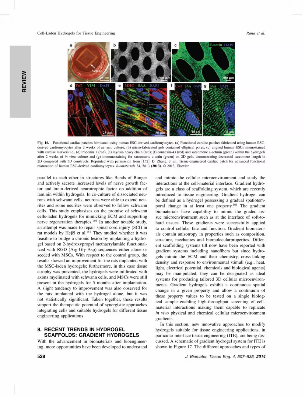
REVIEW
Cell-Laden Hydrogels for Tissue Engineering Rana et al.
Fig. 16. Functional cardiac patches fabricated using human ESC-derived cardiomyocytes. (a) Functional cardiac patches fabricated using human ESC-derived cardiomyocytes after 2 weeks of in vitro culture; (b) micro-fabricated gels contained elliptical pores; (c) aligned human ESCs imunostainedwith cardiac markers i.e., (d) troponin T (red); (e) myosin heavy chain (red); (f) connexin-43 (red) and sarcomeric a-actinin (green) within the hydrogelsafter 2 weeks of in vitro culture and (g) immunostaining for sarcomeric a-actin (green) on 2D gels, demonstrating decreased sarcomere length in2D compared with 3D constructs. Reprinted with permission from [152], D. Zhang, et al., Tissue-engineered cardiac patch for advanced functionalmaturation of human ESC-derived cardiomyocytes. Biomaterials 34, 5813 (2013). © 2013, Elsevier.
parallel to each other in structures like Bands of Bungerand actively secrete increased levels of nerve growth fac-tor and brain-derived neurotrophic factor on addition oflaminin within hydrogels. In co-culture of dissociated neu-rons with schwann cells, neurons were able to extend neu-rites and some neurites were observed to follow schwanncells. This study emphasizes on the promise of schwanncells-laden hydrogels for mimicking ECM and supportingnerve regeneration therapies.160 In another notable study,an attempt was made to repair spinal cord injury (SCI) inrat models by Hejcl et al.151 They studied whether it wasfeasible to bridge a chronic lesion by implanting a hydro-gel based on 2-hydroxypropyl methacrylamide functional-ized with RGD (Arg-Gly-Asp) sequences either alone orseeded with MSCs. With respect to the control group, theresults showed an improvement for the rats implanted withthe MSC-laden hydrogels; furthermore, in this case tissueatrophy was prevented, the hydrogels were infiltrated withaxons myelinated with schwann cells, and MSCs were stillpresent in the hydrogels for 5 months after implantation.A slight tendency to improvement was also observed forthe rats implanted with the hydrogel alone, but it wasnot statistically significant. Taken together, these resultssupport the therapeutic potential of synergistic approachesintegrating cells and suitable hydrogels for different tissueengineering applications
8. RECENT TRENDS IN HYDROGELSCAFFOLDS: GRADIENT HYDROGELS
With the advancement in biomaterials and bioengineer-ing, more opportunities have been developed to understand
and mimic the cellular microenvironment and study theinteractions at the cell-material interface. Gradient hydro-gels are a class of scaffolding system, which are recentlyintroduced to tissue engineering. Gradient hydrogel canbe defined as a hydrogel possessing a gradual spatiotem-poral change in at least one property.161 The gradientbiomaterials have capability to mimic the graded tis-sue microenvironment such as at the interface of soft-to-hard tissues. These gradients were successfully appliedto control cellular fate and function. Gradient biomateri-als contain anisotropy in properties such as composition,structure, mechanics and biomolecularproperties. Differ-ent scaffolding systems till now have been reported withgradient systems including nanofibers but since hydro-gels mimic the ECM and their chemistry, cross-linkingdensity and response to environmental stimuli (e.g., heat,light, electrical potential, chemicals and biological agents)may be manipulated, they can be designated as idealsystems for producing tailored 3D cellular microenviron-ments. Gradient hydrogels exhibit a continuous spatialchange in a given property and allow a continuum ofthese property values to be tested on a single biolog-ical sample enabling high-throughput screening of cell-material interactions making them capable to replicatein vivo physical and chemical cellular microenvironmentgradients.In this section, new innovative approaches to modify
hydrogels suitable for tissue engineering applications, inparticular interface tissue engineering (ITE), are being dis-cussed. A schematic of gradient hydrogel system for ITE isshown in Figure 17. The different approaches and types of
528 J. Biomater. Tissue Eng. 4, 507–535, 2014

REVIEW
Rana et al. Cell-Laden Hydrogels for Tissue Engineering
Fig. 17. (A) Schematic representation of cell-laden gradient hydrogelscaffolds for interface tissue engineering applications. (B) Fluorescentmicrographs of MC3T3 cells cultured on PEGDA hydrogels with variousconcentrations of toxin (OA). Negative control i.e., without OA, positivecontrol i.e., with 2 �M OA and with an OA concentration gradient for24 hours. Dead cells are marked in red by ethidium bromide and livecells marked in green by calcein-AM.162 Reprinted with permission from[154], S. Ostrovidov, et al., Controlled release of drugs from gradienthydrogels for high-throughput analysis of cell-drug interactions. Anal.Chem. 84, 1302 (2012). © 2012, American Chemical Society.
hydrogels discussed are inspired from fundamental biologyof the cellular microenvironment to resemble or mimicECM by controlling the various bioactive cues such asadhesion molecules and growth factors.
Fig. 18. Vascular sprout invasion within PEGDA hydrogels with gradient of PBFP. (A) and (B) confocal images of fluorescently labelled endothelialcells (red) and smooth muscle cells (green) within matrix, 3D vascular sprout invasion in bulk control gels (C) and PBFP gradient hydrogels (D),flattened 3D mosaic renderings of co-culture aggregates in (E) bulk control gels, (F) top, (G) middle and (H) bottom regions of PFBP gradienthydrogels. Reprinted with permission from [164], M. V. Turturro, et al., MMP-Sensitive peg diacrylate hydrogels with spatial variations in matrixproperties stimulate directional vascular sprout formation. PLoS One 8 (2013). © 2013, PLOS.
Molecular concentration gradients of growth factors,chemokines and cytokines are proved to play an importantrole in many biological phenomena such as chemotaxis,163
morphogenesis and wound healing.164 The ECM’s mechan-ical properties also signal cells to induce a structural rear-rangement of the cytoskeletal and immobilized proteins togenerate a mechanotransduction response.165 In addition,gradient’s properties provide a way to mimic the propertiesof the ECM surrounding cell which are mechanically con-nected to different concentrations of different cells such asbone-cartilage interfaces and dentino-enamel junctions.166
Various technologies have been developed to create spa-tiotemporal gradients and complex biomaterials to rapidlyscreen cell-biomaterial interaction167 and to study cellularprocesses such as migration and angiogenesis in vitro168
which can be employed in drug delivery169 and tissueengineering170 applications.
Peptide gradient gels are investigated for use in con-trolling cellular functions. For example, Guarnieri et al.studied the effect of covalently immobilized RGD peptidegradients on PEGDA hydrogels with their cell behavior.171
The results of this study suggest that cells recognize theRGD gradient and adhere onto it assuming a stretchableshape. Moreover, cells tend to migrate in the directionof the gradient. The speed of migration of cells on thehydrogel with uniform RGD distribution was found tobe higher. Speed of migration increases with increasingRGD gradient steepness supporting the augmentation ofbias speed component of the mean squared speed i.e., thedrift of the cell population migrating on the anisotropicsurface provided by the RGD gradient.171 In another inter-esting study, Turturro et al. engineered PEGDA hydro-gel scaffolds with gradients of mechanical properties andimmobilized biofunctionality using perfusion-based frontal
J. Biomater. Tissue Eng. 4, 507–535, 2014 529
REVIEW
Cell-Laden Hydrogels for Tissue Engineering Rana et al.
photopolymerization (PBFP).172 The controlled delivery ofa buoyant photoinitiator, eosin Y, through a glass frit fil-ter results in the formation and subsequent propagationof a polymer reaction front that is self-sustained and ableto propagate through the monomeric mixture. Propaga-tion of this front results in monomer depletion, leadingto variations in cross-linking, as well as spatial gradientsof elastic modulus and immobilized concentrations of theYRGDS cell adhesion ligand within PEGDA hydrogels.Furthermore, the magnitudes of the resulting gradients arecontrolled through alterations in polymerization conditions(see Fig. 18).172
The effects of VEGF and matrix composition on 3Dendothelial cell invasion have been investigated in natu-ral scaffolds such as collagen173 and semi-interpenetratednetworks of collagen and HA.174 Directed endothelial cellinvasion within porous collagen scaffolds has been foundto occur due to gradients of immobilized VEGF, whileendothelial cell sprout formation has been shown to beguided towards regions of decreased HA concentration incollagen-HA interpenetrating networks with observed 2-fold increases in sprout length as compared to that occur-ring in isotropic matrix controls.174 These studies stronglysuggest that gradients of matrix cues lead to enhanced anddirected endothelial cell behavior and play a critical role ininducing vascular sprout formation. Based on the experi-mental studies discussed in this section, and other reporteddata, advanced capabilities of gradient hydrogel systems inrecapitulating native tissue environment are proved to besupportive by providing a gradient of properties for het-erotypic tissues regeneration.
9. CONCLUDING REMARKSHydrogels are being considered as a promising scaffold-ing system that supports cell adhesion, proliferation anddifferentiation by providing requisite physical, mechanicaland chemicals cues for the growth of cells and their sub-sequent development into tissues and organs for regenera-tive medicine applications. Hydrogel design and synthesisapproaches play a significant role to impart the microenvi-ronment of native tissue, which is essential to achieve theultimate goal of development of biological substitutes fordamaged tissues and organs. Only certain aspects of tis-sue properties such as physiochemical properties are takenas the main focus while designing conventional hydrogelsystems for tissue engineering applications, which resultsinto unsuccessful replicas of native environment’s com-plexities. Thus, the development of cell-laden hydrogelswith high biocompatibility, biodegradability, tunable pat-terning properties and efficient mass transfer propertiesare of great importance. Cell-laden hydrogels encapsu-late cells in a spatially and temporally controlled mannerwith suitable cell sources and bioactive molecules in orderto mimic native tissue microenvironment, which paves away towards engineering functional tissues and organs.
Limitations such as biocompatibility, tuned mechanicalproperties, porosity and permeability content to permitingress of cells and nutrients, appropriate surface struc-ture and cell-attachment sites on the scaffolds, could beovercome with cell-laden hydrogel-based approaches andcells could be efficiently delivered at the defective site oftissues too. Stem cells are a recent addition to hydrogelsystems and its being extensively investigated in tissueengineering because of its self-renewal and differentia-tion potential. The use of stem cells within the hydro-gels would employ the regeneration potency of stem cellswithin the in vivo microenvironment or in vitro. And thuscell-laden hydrogels loaded with stem cells can be provedas a potential scaffold material for the development oftissue engineering and regenerative medicinal tools andtechniques. Though the positive impact of these bioma-terial scaffolds are proved by numerous research groupsbut still the requirement of hydrogels has not yet opti-mized which require much attention in future. The majorchallenges concerned with cell-laden hydrogel scaffoldsare the homogeneous cell encapsulation and optimizationof porous structure within the gel system. These limita-tions can be resolved with more sophisticated fabricationtechniques and material modification processes. So far, asper literature survey, cell-laden hydrogels are often usedto repair homotypic tissues. In the view of regenerationof heterotypic tissues, cell-laden hydrogels lack the het-erogeneity within their structures to mimic the native tis-sue microenvironment or niche. Though each tissue has itsown structure and functional properties, they are all inter-connected and inter-dependent with each other to main-tain physiological and metabolic events within their niches.This cross-talk protrudes anisotropy within the niches ofeach tissue. This anisotropy within the diverse tissues andorgans proved to be the main hurdle in optimization ofscaffold system conditions and parameters. As a solutionto this, the field ITE has emerged to regenerate func-tional tissues at the interface of different types of tis-sues, such as soft-to-hard tissues. ITE deals with tissueinterfaces, which are heterogeneous structure and prop-erties in a gradient manner. It helps in the developmentof complex biomaterials with biomimetic gradient prop-erties in terms of composition, structure, mechanical andother functional properties. In this regard, gradient systemsare proved to beoptimal scaffold systems for engineeringgraded tissue interfaces for multiple types of cell growthand subsequent tissue development. Cell-laden hydrogelgradient system could then be exploited as complex bio-materials with biomimetic gradient properties in terms ofphysical, mechanical, chemical and biological cues to pro-mote heterotypic cellular growth, differentiation and inter-action. This is an exciting time to be involved in cell-ladenhydrogel scaffold engineering in order to formulate themas a clinically promising synthetic ECM for tissue repairand reconstruction, with great challenges and also greatexpectations ahead.
530 J. Biomater. Tissue Eng. 4, 507–535, 2014
REVIEW
Rana et al. Cell-Laden Hydrogels for Tissue Engineering
Acknowledgments: This work was supported byCSCR. The author, Deepti Rana, would like to thankCSCR for the award of short term project.
References and Notes1. R. Langer and J. P. Vacanti, Tissue engineering. Science 80, 920
(1993).2. R. Murugan and S. Ramakrishna, Design strategies of tissue engi-
neering scaffolds with controlled fiber orientation. Tissue Eng. 13,1845 (2007).
3. R. Murugan and S. Ramakrishna, Nano-featured scaffolds for tissueengineering: A review of spinning methodologies. Tissue Eng. 12,435 (2006).
4. HRSA. OPTN: Organ Procurement and Transplantation Net-work. US Dep. Heal. Hum. Serv. (2014), (REF: http://optn.transplant.hrsa.gov/data/).
5. Statistics: Donate Life America. (REF: http://donatelife.net/statistics/).
6. Y. Sharma, A. Tiwari, S. Hattori, D. Terada, A. K. Sharma,M. Ramalingam, and H. Kobayashi, Fabrication of conducting elec-trospun nanofibers scaffold for three-dimensional cells culture. Int.J. Biol. Macromol. 51, 627 (2012).
7. S. Ramakrishna and M. Ramalingam, Nanostructured biomaterials.Encycl. Nanosci. Nanotechnol. 7, 595 (2004).
8. R. Murugan, Z. M. Huang, F. Yang, and S. Ramakrishna, Nanofi-brous scaffold engineering using electrospinning. J. Nanosci. Nan-otechnol. 7, 4595 (2007).
9. R. Murugan and K. P. Rao, Grafting of glycidyl methacrylate uponcoralline hydroxyapatite in conjugation with demineralized bonematrix using redox initiating system. Macromol. Res. 11, 14 (2003).
10. N. A. Peppas and R. Langer, New challenges in biomaterials. Sci-ence 263, 1715 (1994).
11. J. A. Hubbell, Biomaterials in tissue engineering. Biotechnology (NY) 13, 565 (1995).
12. R. Langer and D. A. Tirrell, Designing materials for biology andmedicine. Nature 428, 487 (2004).
13. M. P. Lutolf and J. A. Hubbell, Synthetic biomaterials as instruc-tive extracellular microenvironments for morphogenesis in tissueengineering. Nat. Biotechnol. 23, 47 (2005).
14. J. A. Hubbell, Bioactive biomaterials. Curr. Opin. Biotechnol. 10,123 (1999).
15. K. E. Healy, A. Rezania, and R. A. Stile, Designing biomaterialsto direct biological responses. Ann. N Y Acad. Sci. 875, 24 (1999).
16. P. Y. Chen, K. C. Yang, C. C. Wu, J. H. Yu, F. H. Lin, and J. S. Sun,Fabrication of large perfusable macroporous cell-laden hydrogelscaffolds using microbial transglutaminases. Acta Biomater. 10, 912(2014).
17. J. W. Haycock, 3D cell culture: A review of current approachesand techniques. Methods Mol. Biol. 695, 1 (2011).
18. F. Pampaloni, E. G. Reynaud, and E. H. K. Stelzer, The thirddimension bridges the gap between cell culture and live tissue. Nat.Rev. Mol. Cell. Biol. 8, 839 (2007).
19. T.-M. Achilli, J. Meyer, and J. R. Morgan, Advances in the for-mation, use and understanding of multi-cellular spheroids. ExpertOpin. Biol. Ther. 12, 1347 (2012).
20. L. Tan, Y. Ren, and R. Kuijer, A 1-min method for homogenouscell seeding in porous scaffolds. J. Biomater. Appl. 26, 877 (2012).
21. M. Radisic, L. Yang, J. Boublik, R. J. Cohen, R. Langer, L. E.Freed, and G. Vunjak-Novakovic, Medium perfusion enables engi-neering of compact and contractile cardiac tissue. Am. J. Physiol.Heart Circ. Physiol. 286, H507 (2004).
22. H. Kamaraju, K. Wetzel, and W. J. Kelly, Modeling shear-inducedCHO cell damage in a rotary positive displacement pump. Biotech-nol. Prog. 26, 1606 (2010).
23. N. N. Kachouie, Y. Du, H Bae, M. Khabiry, A. F. Ahari,B. Zamanian, and J. F. A. Khademhosseini, Directed assembly ofcell-laden hydrogels for engineering functional tissues. Organogen-esis 6, 234 (2010).
24. C. M. Agrawal and R. B. Ray, Biodegradable polymeric scaffoldsfor musculoskeletal tissue engineering. J. Biomed. Mater. Res. 55,141 (2001).
25. B. Jeong, S. W. Kim, and Y. H. Bae, Thermosensitive sol–gelreversible hydrogels. Adv. Drug. Deliv. Rev. 64, 154 (2012).
26. J. L. Drury and D. J. Mooney, Hydrogels for tissue engineering:Scaffold design variables and applications. Biomaterials 24, 4337(2003).
27. W. R. Gombotz and D. K. Pettit, Biodegradable polymers for pro-tein and peptide drug delivery. Bioconjug. Chem. 6, 332 (1995).
28. W. E. Hennink and C. F. van Nostrum, Novel crosslinking methodsto design hydrogels. Adv. Drug. Deliv. Rev. 64, 223 (2012).
29. D. Macaya and M. Spector, Injectable hydrogel materials for spinalcord regeneration: A review, Biomed. Mater. 7, 012001 (2012).
30. E. Westhaus and P. B. Messersmith, Triggered release of calciumfrom lipid vesicles: A bioinspired strategy for rapid gelation ofpolysaccharide and protein hydrogels. Biomaterials 22, 453 (2001).
31. M. Ahearne, Y. Yang, and K. Liu, Mechanical characterisationof hydrogels for tissue engineering applications. Tissue Eng. 4, 1(2008).
32. T. Sawada, K. Tsukada, K. Haseqawa, Y. Ohashi, Y. Udaqawa,and V. Gomel, Cross-linked hyaluronate hydrogel prevents adhe-sion formation and reformation in mouse uterine horn model.Hum. Reprod. 16, 353 (2001).
33. D. L. Hern and J. A. Hubbell, Incorporation of adhesion peptidesinto nonadhesive hydrogels useful for tissue resurfacing. J. Biomed.Mater. Res. 39, 266 (1998).
34. X. He and E. Jabbari, Material properties and cytocompatibility ofinjectable MMP degradable poly(lactide ethylene oxide fumarate)hydrogel as a carrier for marrow stromal cells. Biomacromolecules8, 780 (2007).
35. F. Yang, W. L. Neeley, M. J. Moore, J. M. Karp, A. Shukla,and R. S. Langer, Tissue engineering: The therapeutic strategy ofthe twenty-first century, Nanotechnology and Tissue Engineering,edited by C. T. Laurencin and L. S. Nair, CRC Press (2008),pp. 3–32.
36. D. Garlotta, A literature review of Poly(Lactic Acid). J. Polym.Environ. 9, 63 (2001).
37. J. P. Winer, S. Oake, and P. A. Janmey, Non-linear elasticity ofextracellular matrices enables contractile cells to communicate localposition and orientation. PLoS ONE 4, e6382 (2009).
38. Q. Chen, S. Liang, and G. A. Thouas, Elastomeric biomaterials fortissue engineering. Prog. Polym. Sci. 38, 584 (2013).
39. N. Annabi, S. M. Mithieux, E. A. Boughton, A. J. Ruys, A. S.Weiss, and F. Dehghani, Synthesis of highly porous crosslinkedelastin hydrogels and their interaction with fibroblasts in vitro. Bio-materials 30, 4550 (2009).
40. N. Annabi, A. Fathi, S. M. Mithieux, A. S. Weiss, and F. Dehghani,Fabrication of porous PCL/elastin composite scaffolds for tissueengineering applications. J. Supercrit. Fluids 59, 157 (2011).
41. D. Lamprou, P. Zhdan, F. Labeed, and C. Lekakou, Gelatine andgelatine/elastin nanocomposites for vascular grafts: Processing andcharacterization. J. Biomater. Appl. 26, 209 (2011).
42. N. Annabi, S. M. Mithieux, A. S. Weiss, and F. Dehghani, The fab-rication of elastin-based hydrogels using high pressure CO2. Bio-materials 30, 1 (2009).
43. T. de Chalain, J. H. Phillips, and A. Hinek, Bioengineeringof elastic cartilage with aggregated porcine and human auricu-lar chondrocytes and hydrogels containing alginate, collagen, andkappa-elastin. J. Biomed. Mater. Res. 44, 280 (1999).
J. Biomater. Tissue Eng. 4, 507–535, 2014 531

REVIEW
Cell-Laden Hydrogels for Tissue Engineering Rana et al.
44. T. J. Broekelmann, C. H. Ciliberto, A. Shifren, and R. P.Mecham, Modification and functional inactivation of the tropoe-lastin carboxy-terminal domain in cross-linked elastin. Matrix Biol.27, 631 (2008).
45. S. F. Badylak and T. W. Gilbert, Immune response to biologic scaf-fold materials. Semin. Immunol. 20, 109 (2008).
46. G. Camci-Unal, P. Zorlutuna, and A. Khademhosseini, Fabricationof microscale hydrogels for tissue engineering applications, Bio-fabrication, edited by G. Forgacs and W. Sun, Elsevier Publishing(2012), pp. 59–80.
47. B. V Slaughter, S. S. Khurshid, and O. Z. Fisher, Hydrogels inRegenerative Medicine-Slaughter-2009-Advanced Materials-WileyOnline Library. Adv. (2009).
48. T. Corrales, F. Catalina, C. Peinado, and N. S. Allen, Free radi-cal macrophotoinitiators: An overview on recent advances. J. Pho-tochem. Photobiol. A Chem. 159, 103 (2003).
49. C. Rivest, D. W. G. Morrison, B. Ni, J. Rubin V. Yadav,A. Mahdavi, J. M. Karp, and A. Khademhosseini, Microscalehydrogels for medicine and biology: Synthesis, characteristics andapplications. J. Mech. Mater. Struct. 2, 1103 (2007).
50. S. Gerecht, J. A. Burdick, L. S. Ferreira, S. A. Townsend,R. Langer, and G. Vunjak-Novakovic, Hyaluronic acid hydrogelfor controlled self-renewal and differentiation of human embryonicstem cells. Proc. Natl. Acad. Sci. USA 104, 11298 (2007).
51. G. Camci-Unal, D. Cuttica, N. Annabi, D. Demarchi, andA. Khademhosseini, Synthesis and characterization of hybridhyaluronic acid-gelatin hydrogels. Biomacromolecules 14, 1085(2013).
52. W.-G. Koh, L. J. Itle, and M. V Pishko, Molding of hydrogelmicrostructures to create multiphenotype cell microarrays. Anal.Chem. 75, 5783 (2003).
53. S. J. Bryant, J. L. Cuy, K. D. Hauch, and B. D. Ratner, Photo-patterning of porous hydrogels for tissue engineering. Biomaterials28, 2978 (2007).
54. A. I. Chou, S. O. Akintoye, and S. B. Nicoll, Photo-crosslinkedalginate hydrogels support enhanced matrix accumulation bynucleus pulposus cells in vivo. Osteoarthr. Cartil. 17, 1377 (2009).
55. T. Meyvis, S. De Smedt, B. Stubbe, W. Hennink, and J. Demeester,On the release of proteins from degrading dextran methacrylatehydrogels and the correlation with the rheologic properties of thehydrogels. Pharm. Res. 18, 1593 (2001).
56. A. Nilasaroya, L. A. Poole-Warren, J. M. Whitelock, and P. JoMartens, Structural and functional characterisation of poly(vinylalcohol) and heparin hydrogels. Biomaterials 29, 4658 (2008).
57. Y. Zhou, G. Ma, S. Shi, D. Yang, and J. Nie, Photopolymerizedwater-soluble chitosan-based hydrogel as potential use in tissueengineering. Int. J. Biol. Macromol. 48, 408 (2011).
58. I. D. Gaudet and D. I. Shreiber, Characterization of methacrylatedtype-I collagen as a dynamic, photoactive hydrogel. Biointerphases7, 25 (2012).
59. M. J. Hancock, F. Piraino, G. Camci-Unal, M. Rasponi, andA. Khademhosseini, Anisotropic material synthesis by capillaryflow in a fluid stripe. Biomaterials 32, 6493 (2011).
60. S. Nemir, H. N. Hayenga, and J. L. West, PEGDA hydrogels withpatterned elasticity: Novel tools for the study of cell response tosubstrate rigidity. Biotechnol. Bioeng. 105, 636 (2010).
61. M. Guvendiren and J. A. Burdick, Stiffening hydrogels to probeshort-and long-term cellular responses to dynamic mechanics. Nat.Commun. 3, 792 (2012).
62. H. Aubin, J. W. Nichol, C. B. Hutson, H. Bae, A. L. Sieminski,D. M. Cropek, P. Akhyari, and A. Khademhosseini, Directed 3Dcell alignment and elongation in microengineered hydrogels. Bio-materials 31, 6941 (2010).
63. E. E. Hago, L. Xinsong, and J. J. Kai, A new route towardIPN photocrosslinked alginate hydrogels with biodegradation rates,excellent biocompatibility and mechanical properties. Journal ofBiomater. Tissue Eng. 4, 89 (2014).
64. A. M. Kloxin, A. M. Kasko, C. N. Salinas, and K. S. Anseth, Pho-todegradable hydrogels for dynamic tuning of physical and chemi-cal properties. Science 324, 59 (2009).
65. D. Y. Wong, D. R. Griffin, J. Reed, and A. M. Kasko, Photodegrad-able hydrogels to generate positive and negative features over mul-tiple length scales. Macromolecules 43, 2824 (2010).
66. A. M. Kloxin, M. W. Tibbitt, and K. S. Anseth, Synthesis of pho-todegradable hydrogels as dynamically tunable cell culture plat-forms. Nat. Protoc. 5, 1867 (2010).
67. M. I. A. Mamada, T. Tanaka, and D. Kungwatchakun, Photoinducedphase transition of gels. Macromolecules 23, 1517 (1990).
68. A. K. Gaharwar, C. Rivera, C. J. Wu, B. K. Chan, and G. Schmidt,Photocrosslinked nanocomposite hydrogels from PEG and silicananospheres: Structural, mechanical and cell adhesion characteris-tics. Mater. Sci. Eng. C 33, 1800 (2013).
69. A. D. Augst, H. J. Kong, and D. J. Mooney, Alginate hydrogels asbiomaterials. Macromol. Biosci. 6, 623 (2006).
70. M. S. Hahn, B. A. Teply, M. M. Stevens, S. M. Zeitels, andR. Langer, Collagen composite hydrogels for vocal fold laminapropria restoration. Biomaterials 27, 1104 (2006).
71. L. Baruch and M. Machluf, Alginate-chitosan complex coacerva-tion for cell encapsulation: Effect on mechanical properties and onlong-term viability. Biopolymers 82, 570 (2006).
72. Y. Yang, I. Wimpenny, and M. Ahearne, Portable nanofiber meshesdictate cell orientation throughout three-dimensional hydrogels.Nanomedicine Nanotechnology, Biol. Med. 7, 131 (2011).
73. S. Sakai, Y. Takagi, Y. Yamada, T. Yamaguchi, and K. Kawakami,Reinforcement of porous alginate scaffolds by incorporating elec-trospun fibres. Biomed. Mater. 3, 034102 (2008).
74. M. A. Hou, Y. Fei, R. Burkes, J. C. Lee, S. D. Munoz-Pinto,D. Hahn, and S. M. Grunlan, Thermoresponsive nanocompositehydrogels: Transparency, rapid deswelling and cell release. J. Bio-mater. Tissue. Eng. 1, 93 (2011).
75. Y. Jia, Y. Wei, Z. Fu, W. Huo, M. Li, F. Zhong, C. Jia, and S. Zhou,Biocompatibility evaluation on a bio-hydrogel composed of bacte-rial cellulose and chitosan. J. Biomater. Tissue Eng. 4, 118 (2014).
76. A. K. Ekaputra, G. D. Prestwich, S. M. Cool, and D. W. Hutmacher,Combining electrospun scaffolds with electrosprayed hydrogelsleads to three-dimensional cellularization of hybrid constructs.Biomacromolecules 9, 2097 (2008).
77. D. Kai, M. P. Prabhakaran, B. Stahl, M. Eblenkamp,E. Wintermantel, and S. Ramakrishna, Behavior of nanofiber–hydrogel composites for tissue engineering applications. Nanotech-nology 23, 095705 (2012).
78. B. Rybtchinski, Adaptive supramolecular nanomaterials based onstrong noncovalent interactions. ACS Nano 5, 6791 (2011).
79. J. Gu, F. Xia, Y. Wu, X. Qu, Z. Yang, and L. Jiang, Programmabledelivery of hydrophilic drug using dually responsive hydrogelcages. J. Control. Release 117, 396 (2007).
80. J. Yang, Hydrogels as smart biomaterials. Polym. Int. 1098, 1078(2007).
81. T. Peng and Y. L. Cheng, Temperature-responsive permeability ofporous PNIPAAm-g-PE membranes. J. Appl. Polym. Sci. 70, 2133(1998).
82. R. Ravichandran, S. Sundarrajan, J. R. Venugopal, S. Mukherjee,and S. Ramakrishna, Advances in polymeric systems for tissueengineering and biomedical applications. Macromol. Biosci. 12,286 (2012).
83. M. Behl and A. Lendlein, Shape-memory polymers. Mater. Today10, 20 (2007).
84. R. Censi, T. Vermonden, M. J. Van Steenbergen, H. Deschout,K. Braeckmans, S. C. De Smedt, C. F. Van Nostrum, P. Di Martino,and W. E. Hennink, Photopolymerized thermosensitive hydro-gels for tailorable diffusion-controlled protein delivery. J. Control.Release 140, 230 (2009).
532 J. Biomater. Tissue Eng. 4, 507–535, 2014

REVIEW
Rana et al. Cell-Laden Hydrogels for Tissue Engineering
85. G. Ozaydin-Ince, K. K. Gleason, and M. C. Demirel, A stimuli-responsive coaxial nanofilm for burst release. Soft Matter. 7, 638(2011).
86. L. Wang, J. Shansky, C. Borselli, D. Mooney, and H. Vandenburgh,Design and fabrication of a biodegradable, covalently crosslinkedshape-memory alginate scaffold for cell and growth factor delivery.Tissue Eng. Part A 18, 2000 (2012).
87. S. A. Bencherif, R. W. Sands, D. Bhatta, P. Arany, C. S. Verbeke,D. A. Edwards, and D. J. Mooney, Injectable preformed scaf-folds with shape memory properties. Proc. Natl. Acad. Sci. USA109, 19590 (2012).
88. M. J. Glassman, J. Chan, and B. D. Olsen, Reinforcement of ShearThinning Protein Hydrogels by Responsive Block Copolymer Self-Assembly. Adv. Funct. Mater. 23, 1182 (2013).
89. Z. L. Wu and J. P. Gong, Hydrogels with self-assembling orderedstructures and their functions. NPG Asia Mater 3, 57 (2011).
90. Z. Yang, G. Liang, and B. Xu, Enzymatic hydrogelation of smallmolecules. Acc. Chem. Res. 41, 315 (2008).
91. Y. Gao, F. Zhao, Q. Wang, Y. Zhang, and B. Xu, Small peptidenanofibers as the matrices of molecular hydrogels for mimickingenzymes and enhancing the activity of enzymes. Chem. Soc. Rev.39, 3425 (2010).
92. Y. Gao, F. Zhao, Q. Wang, Y. Zhang, and B. Xu, Small peptidenanofibers as the matrices of molecular hydrogels for mimickingenzymes and enhancing the activity of enzymes. Chem. Soc. Rev.39, 3425 (2010).
93. Z. Yang, G. Liang, and B. Xu, Enzymatic hydrogelation of smallmolecules. Acc. Chem. Res. 41, 315 (2008).
94. B. Ozbas, K. Rajagopal, J. P. Schneider, and D. J. Pochan, Semi-flexible chain networks formed via self-assembly of beta-hairpinmolecules. Phys. Rev. Lett. 93, 268106 (2004).
95. S. Zhang, M. A. Greenfield, A. Mata, L. C. Palmer, R. Bitton, J. R.Mantei, C. Aparicio, M. O. de la Cruz, and S. I. Stupp, A self-assembly pathway to aligned monodomain gels. Nat. Mater. 9, 594(2010).
96. U. Hersel, C. Dahmen, and H. Kessler, RGD modified polymers:Biomaterials for stimulated cell adhesion and beyond. Biomaterials24, 4385 (2003).
97. G. A. Silva, C. Czeisler, K. L. Niece, E. Beniash, D. A. Harrington,J. A. Kessler, and S. I. Stupp, Selective differentiation of neural pro-genitor cells by high-epitope density nanofibers. Science 303, 1352(2004).
98. K. Rajangam, H. A. Behanna, M. J. Hui, X. Han, J. F. Hulvat,J. W. Lomasney, and S. I. Stupp, Heparin binding nanostructuresto promote growth of blood vessels. Nano Lett. 6, 2086 (2006).
99. T. T. Lau and D.-A. Wang, Bioresponsive hydrogel scaffolding sys-tems for 3D constructions in tissue engineering and regenerativemedicine. Nanomedicine (Lond) 8, 655 (2013).
100. C. S. Bahney, C.-W. Hsu, J. U. Yoo, J. L. West, and B. Johnstone,A bioresponsive hydrogel tuned to chondrogenesis of human mes-enchymal stem cells. FASEB J. 25, 1486 (2011).
101. G. P. Raeber, M. P. Lutolf, and J. A. Hubbell, Molecularly engi-neered PEG hydrogels: A novel model system for proteolyticallymediated cell migration. Biophys. J. 89, 1374 (2005).
102. S. Kim, E. H. Chung, M. Gilbert, and K. E. Healy, SyntheticMMP-13 degradable ECMs based on poly(N -isopropylacrylamide-co-acrylic acid) semi-interpenetrating polymer networks. I. Degra-dation and cell migration. J. Biomed. Mater. Res. A 75, 73 (2005).
103. H. Hall and J. A. Hubbell, Modified fibrin hydrogels stimulateangiogenesis in vivo: Potential application to increase perfusion ofischemic tissues. Materwiss Werksttech 36, 768 (2005).
104. H. W. Jun, V. Yuwono, S. E. Paramonov, and J. D. Hartgerink,Enzyme-mediated degradation of peptide-amphiphile nanofiber net-works. Adv. Mater. 17, 2612 (2005).
105. V. L. Tsang and S. N. Bhatia, Fabrication of three-dimensionaltissues. Adv. Biochem. Eng. Biotechnol. 103, 189 (2007).
106. N. Annabi, A. Tamayol, J. A. Uquillas, M. Akbari, G. E.Bertassoni, C. Cha, G. Camci-Unal, M. R. Dokmeci, N. A. Peppas,and A. Khademhosseini, 25th anniversary article: Rational designand applications of hydrogels in regenerative medicine. Adv. Mater.26, 85 (2014).
107. P. Zorlutuna, N. Annabi, G. Camci-Unal, M. Nikkhah, J. M.Cha, J. W. Nichol, A. Manbachi, H. Bae, S. Chen, andA. Khademhosseini, Microfabricated biomaterials for engineering3D tissues. Adv. Mater. 24, 178 (2012).
108. Š. Selimovic, J. Oh, H. Bae, M. Dokmeci, and A. Khademhosseini,microscale strategies for generating cell-encapsulating hydrogels.Polymers �Basel� 4, 1554 (2012).
109. K. T. Nguyen and J. L. West, Photopolymerizable hydrogels fortissue engineering applications. Biomaterials 23, 4307 (2002).
110. S. H. Um, J. B. Lee, N. Park, S. Y. Kwon, C. C. Umbach, andD. Luo, Enzyme-catalysed assembly of DNA hydrogel. Nat. Mater.5, 797 (2006).
111. P. Ferruti, S. Bianchi, E. Ranucci, F. Chiellini, and V. Caruso,Novel poly(amido-amine)-based hydrogels as scaffolds for tissueengineering. Macromol. Biosci. 5, 613 (2005).
112. B. K. Mann, A. S. Gobin, A. T. Tsai, R. H. Schmedlen, and J. L.West, Smooth muscle cell growth in photopolymerized hydrogelswith cell adhesive and proteolytically degradable domains: Syn-thetic ECM analogs for tissue engineering. Biomaterials 22, 3045(2001).
113. A. Sannino, P. A. Netti, M. Madaghiele, V. Coccoli, A. Luciani,A. Maffezzoli, and L. Nicholais, Synthesis and characterization ofmacroporous poly(ethylene glycol)-based hydrogels for tissue engi-neering application. J. Biomed. Mater. Res. A 79, 229 (2006).
114. J. Wang, L. Chen, Y. Zhao, G. Guo, and R. Zhang, Cell adhesionand accelerated detachment on the surface of temperature-sensitivechitosan and poly(N -isopropylacrylamide) hydrogels. J. Mater. Sci.Mater. Med. 20, 583 (2009).
115. M. Hu, R. Deng, K. M. Schumacher, M. Kurisawa, H. Ye,K. Purnamawati, and J. Y. Ying, Hydrodynamic spinning of hydro-gel fibers. Biomaterials 31, 863 (2010).
116. J. Yeh, Y. Ling, J. M. Karp, J. Gantz, A. Chandawarkar, G. Eng,J. Blumling 3rd, R. Langer, and A. Khademhosseini, Micromoldingof shape-controlled, harvestable cellladen hydrogels. Biomaterials27, 5391 (2006).
117. J. W. Nichol and A. Khademhosseini, Modular tissue engineer-ing: Engineering biological tissues from the bottom up. Soft Matter5, 1312 (2009).
118. V. L. Tsang, A. A. Chen, L. M. Cho, K. D. Jadin, R. L. Sah,S. DeLong, J. L. West, and S. N. Bhatia, Fabrication of 3D hepatictissues by additive photopatterning of cellular hydrogels. FASEB J.21, 790 (2007).
119. J. W. Nichol, S. T. Koshy, H. Bae, C. M. Hwang, S. Yamanlar, andA. Khademhosseini, Cell-laden microengineered gelatin methacry-late hydrogels. Biomaterials 31, 5536 (2010).
120. H. Shin, B. D. Olsen, and A. Khademhosseini, The mechanicalproperties and cytotoxicity of cell-laden double-network hydro-gels based on photocrosslinkable gelatin and gellan gum biomacro-molecules. Biomaterials 33, 3143 (2012).
121. M. Cambodi, N. W. Choi, J. P. Gleghorn, C. S. Lee, L. J. Bonassar,and A. D. Stroock, A microfluidic biomaterial. J. Am. Chem. Soc.127, 13788 (2005).
122. K. R. King, C. C. J. Wang, M. R. Kaazempur-Mofrad, J. P. Vacanti,and J. T. Borenstein, Biodegradable microfluidics. Adv. Mater. 16,2007 (2004).
123. J. T. Borenstein, H. Terai, K. R. King, E. J. Weinberg, M. R.Kaazempur-Mofrad, and J. P. Vacanti, Microfabrication technologyfor vascularized tissue engineering. Biomed. Microdevices 4, 167(2002).
124. A. Paguirigan and D. J. Beebe, Gelatin based microfluidic devicesfor cell culture. Lab Chip 6, 407 (2006).
J. Biomater. Tissue Eng. 4, 507–535, 2014 533

REVIEW
Cell-Laden Hydrogels for Tissue Engineering Rana et al.
125. Y. Ling, J. Rubin, Y. Deng, C. Huang, U. Demirci, J. M. Karp, andA. Khademhosseini, A cell-laden microfluidic hydrogel. Lab Chip7, 756 (2007).
126. Y. Ling, et al., A cell-laden microfluidic hydrogel. Lab Chip 7, 756(2007).
127. Y. Du, M. Ghodousi, H. Qi, N. Haas, W. Xiao, andA. Khademhosseini, Sequential assembly of cell-laden hydrogelconstructs to engineer vascular-like microchannels. Biotechnol. Bio-eng. 108, 1693 (2011).
128. M.-Y. Chiang and S.-K. Fan, Generation and assembly of cell-laden hydrogels on a digital microfluidic platform, 16th Interna-tional Conference on Miniaturized Systems for Chemistry and LifeSciences, Okinawa, Japan, October–November (2012).
129. S. J. Moon, S. K. Hasan, Y. S. Song, H. O. Keles, F. Manzur,S. Mikkilineni, J. W. Hong, J. Nagatomi, E. Haeggstrom,A. Khademhosseini, and U. Demirci, Layer by layer 3D tissue epi-taxy by cell laden hydrogel droplets, 35th Annual Northeast Bio-engineering Conference, IEEE Publication, Boston, MA (2009).
130. S. J. Moon, S. K. Hasan, Y. S. Song, F. Xu, H. O. Keles, F. Manzur,S. Mikkilineni, J. W. Hong, J. Nagatomi, E. Haeggstrom,A. Khademhosseini, and U. Demirci, Layer by layer 3D tissue epi-taxy by cell laden hydrogel droplets. Tissue Eng. Part C Methods16, 157 (2009).
131. D. F. D. Campos, A. Blaeser, M. Weber, J. Jakel, S. Neuss,W. Jahnen-Dechent, and H. Fischer, Three-dimensional printing ofstem cell-laden hydrogels submerged in a hydrophobic high-densityfluid. Biofabrication 5, 015003 (2013).
132. J. M. Cordell, M. L. Vogl, and A. J. W. Johnson, The influence ofmicropore size on the mechanical properties of bulk hydroxyapatiteand hydroxyapatite scaffolds. J. Mech. Behav. Biomed. Mater. 2,560 (2009).
133. A. Bignon, J. Chouteau, J. Chevalier, G. Fantozzi, J.-P. Carret,P. Chavassieux, G. Boivin, M. Melin, and D. Hartmann, Effect ofmicro- and macroporosity of bone substitutes on their mechanicalproperties and cellular response. J. Mater. Sci. Mater. Med. 14, 1089(2003).
134. M. P. Linnes, B. D. Ratner, and C. M. Giachelli, A fibrinogen-basedprecision microporous scaffold for tissue engineering. Biomaterials28, 5298 (2007).
135. M. P. Linnes, B. D. Ratner, and C. M. Giachelli, A fibrinogen-basedprecision microporous scaffold for tissue engineering. Biomaterials28, 5298 (2007).
136. Y.-C. Huang, M. Connell, Y. Park, D. J. Mooney, and K. G. Rice,Fabrication and in vitro testing of polymeric delivery system forcondensed DNA. J. Biomed. Mater. Res. A 67, 1384 (2003).
137. W. L. Murphy, R. G. Dennis, J. L. Kileny, and D. J. Mooney, Saltfusion: An approach to improve pore interconnectivity within tissueengineering scaffolds. Tissue Eng. 8, 43 (2002).
138. J. H. Park, B. G. Chung, W. G. Lee, J. Kim, M. D. Brigham,J. Shim, S. Lee, C. M. Hwang, N. G. Durmus, U. Demirci, andA. Khademhosseini, Microporous cell-laden hydrogels for engi-neered tissue constructs. Biotechnol. Bioeng. 106, 138 (2010).
139. N. B. Graham, Hydrogels: Their future, Part II. Med. Device Tech-nol. 9, 22 (1998).
140. A. Galperin, T. J. Long, and B. D. Ratner, Degradable,thermo-sensitive poly(N -isopropyl acrylamide)-based scaffoldswith controlled porosity for tissue engineering applications.Biomacromolecules 11, 2583 (2010).
141. L. H. Han, J. H. Lai, S. Yu, and F. Yang, Dynamic tissue engi-neering scaffolds with stimuli-responsive macroporosity formation.Biomaterials 34, 4251 (2013).
142. S. P. Bruder, D. J. Fink, and A. I. Caplan, Mesenchymal stem cellsin bone development, bone repair, and skeletal regeneration therapy.J. Cell. Biochem. 56, 283 (1994).
143. C. S. Bahney, T. J. Lujan, C. W. Hsu, M. Bottlang, J. L. West, andB. Johnstone, Visible light photoinitiation of mesenchymal stemcell-laden bioresponsive hydrogels. Eur. Cell. Mater. 22, 43 (2011).
144. C. E. Semino, J. R. Merok, G. G. Crane, G. Panagiotakos, andS. Zhang, Functional differentiation of hepatocyte-like spheroidstructures from putative liver progenitor cells in three-dimensionalpeptide scaffolds. Differentiation 71, 262 (2003).
145. L. Haines-Butterick, K. Rajagopal, M. Branco, D. Salick,R. Rughani, M. Pilarz, M. S. Lamm, D. J. Pochan, and J. P.Schneider, Controlling hydrogelation kinetics by peptide design forthree-dimensional encapsulation and injectable delivery of cells.Proc. Natl. Acad. Sci. USA 104, 7791 (2007).
146. H. Betre, S. R. Ong, F. Guilak, A. Chilkoti, B. Fermor, and L. A.Setton, Chondrocytic differentiation of human adipose derivedadult stem cells in elastin-like polypeptide. Biomaterials 27, 91(2006).
147. N. E. Fedorovich, S. C. Leeuwenburgh, Y. J. M. van der Helm,J. Alblas, and W. J. A. Dhert, The osteoinductive potential of print-able, cell-laden hydrogel-ceramic composites. J. Biomed. Mater.Res. A 100, 2412 (2012).
148. C. Chung and J. A. Burdick, Influence of three-dimensionalhyaluronic acid microenvironments on mesenchymal stem cellchondrogenesis. Tissue Eng. Part A 15, 243 (2009).
149. A. H. Huang, M. J. Farrell, M. Kim, and R. L. Mauck, Long-termdynamic loading improves the mechanical properties of chondro-genic mesenchymal stem cell-laden hydrogel. Eur. Cell. Mater. 19,72 (2010).
150. C. Chung and J. A. Burdick, Influence of three-dimensionalhyaluronic acid microenvironments on mesenchymal stem cellchondrogenesis. Tissue Eng. Part A 15, 243 (2009).
151. A. Hejcl, J. Sedy, M. Kapcalova, D. A. Toro, T. Amemori,P. Lesny, K. Likavcanova-Masinova, E. Krumbholcova, M. Pradny,J. Michalek, M. Burian, M. Hajek, P. Jendelova, and E. Sykova,HPMA-RGD hydrogels seeded with mesenchymal stem cellsimprove functional outcome in chronic spinal cord injury.Stem. Cells Dev. 19, 1535 (2010).
152. N. E. Fedorovich, S. C. Leeuwenburgh, Y. J. M. Van Der Helm,J. Alblas, and W. J. A. Dhert, The osteoinductive potential of print-able, cell-laden hydrogel-ceramic composites. J. Biomed. Mater.Res.-Part A 100 A, 2412 (2012).
153. N. E. Fedorovich, W. Schuurman, H. M. Wijnberg, H. J. Prins, P. R.Van Weeren, J. Malda, J. Alblas, and W. J. Dhert, Biofabrication ofosteochondral tissue equivalents by printing topologically defined,cell-laden hydrogel scaffolds. Tissue Eng. Part C Methods 18, 33(2012).
154. C. M. Hwang, S. Sant, M. Masaeli, N. N. Kachouie, B. Zamanian,S. H. Lee, and A. Khademhosseini, Fabrication of three-dimensional porous cell-laden hydrogel for tissue engineering. Bio-fabrication 2, 035003 (2010).
155. P. Occhetta, N. Sadr, F. Piraino, A. Redaelli, M. Moretti, andM. Rasponi, Fabrication of 3D cell-laden hydrogel microstruc-tures through photo-mold patterning. Biofabrication 5, 035002(2013).
156. G. Camci-Unal, N. Annabi, M. R. Dokmeci, R. Liao, andA. Khademhosseini, Hydrogels for cardiac tissue engineering. NPGAsia Materials 6, e99 (2014).
157. M. Radisic, H. Park, H. Shing, T. Consi, F. J. Schoen, R. Langer,L. E. Freed, and G. Vunjak-Novakovic, Functional assemblyof engineered myocardium by electrical stimulation of cardiacmyocytes cultured on scaffolds. Proc. Natl. Acad. Sci. USA101, 18129 (2004).
158. J. Y. Chung, et al., Comparison of articular cartilage repair withdifferent hydrogel-human umbilical cord blood-derived mesenchy-mal stem cell composites in a rat model. Stem. Cell. Res. Ther. 5,39 (2014).
159. D. Zhang, I. Y. Shadrin, J. Lam, H. Q. Xian, H. R. Snodgrass, andN. Bursac, Tissue-engineered cardiac patch for advanced functionalmaturation of human ESC-derived cardiomyocytes. Biomaterials34, 5813 (2013).
534 J. Biomater. Tissue Eng. 4, 507–535, 2014

REVIEW
Rana et al. Cell-Laden Hydrogels for Tissue Engineering
160. S. Suri and C. E. Schmidt, Cell-laden hydrogel constructs ofhyaluronic acid, collagen, and laminin for neural tissue engineering.Tissue Eng. Part A 16, 1703 (2010).
161. M. Singh, C. Berkland, and M. S. Detamore, Strategies and appli-cations for incorporating physical and chemical signal gradients intissue engineering. Tissue Eng. Part B Rev. 14, 341 (2008).
162. S. Ostrovidov, N. Annabi, A. Seidi, M. Ramalingam, F. Dehghani,H. Kaji, and A. Khademhosseini, Controlled release of drugs fromgradient hydrogels for high-throughput analysis of cell-drug inter-actions. Anal. Chem. 84, 1302 (2012).
163. D. O. Freytes, L. Q. Wan, and G. Vunjak-Novakovic, Geometry andforce control of cell function. J. Cell. Biochem. 108, 1047 (2009).
164. N. Li Jeon, H. Baskaran, S. K. Dertinger, G. M. Whitesides, L. Vande Water, and M. Toner, Neutrophil chemotaxis in linear and com-plex gradients of interleukin-8 formed in a microfabricated device.Nat. Biotechnol. 20, 826 (2002).
165. B. G. Chung, L. A. Flanagan, S. W. Rhee, P. H. Schwartz, A. P.Lee, E. S. Monuki, and N. L. Jeon, Human neural stem cell growthand differentiation in a gradient-generating microfluidic device. LabChip 5, 401 (2005).
166. V. Vogel and M. Sheetz, Local force and geometry sensing regulatecell functions. Nat. Rev. Mol. Cell. Biol. 7, 265 (2006).
167. A. Miserez, T. Schneberk, C. Sun, F. W. Zok, and J. H. Waite, Thetransition from stiff to compliant materials in squid beaks. Science319, 1816 (2008).
168. C. G. Simon, Y. Yang, V. Thomas, S. M. Dorsey, and A. W.Morgan, Cell interactions with biomaterials gradients and arrays.Comb. Chem. High Throughput Screen 12, 544 (2009).
169. S. Chung, R. Sudo, V. Vickerman, I. K. Zervantonakis, and R. D.Kamm, Microfluidic platforms for studies of angiogenesis, cellmigration, and cell–cell interactions. Ann. Biomed. Eng. 38, 1164(2010).
170. N. A. Peppas and A. R. Khare, Preparation, structure and diffu-sional behavior of hydrogels in controlled release. Adv. Drug Deliv.Rev. 11, 1 (1993).
171. D. Guarnieri, A. De Capua, M. Ventre, A. Borzacchiello,C. Pedone, D. Marasco, M. Ruvo, and P. A. Netti, Covalentlyimmobilized RGD gradient on PEG hydrogel scaffold influencescell migration parameters. Acta Biomater. 6, 2532 (2010).
172. M. V. Turturro, M. C. Christenson, J. C. Larson, D. A. Young, E. M.Brey, and G. Papavasiliou, MMP-sensitive PEG diacrylate hydro-gels with spatial variations in matrix properties stimulate directionalvascular sprout formation. PLoS ONE 8, e58897 (2013).
173. D. Odedra, L. L. Y. Chiu, M. Shoichet, and M. Radisic, Endothe-lial cells guided by immobilized gradients of vascular endothelialgrowth factor on porous collagen scaffolds. Acta Biomater. 7, 3027(2011).
174. C. Borselli, O. Oliviero, S. Battista, L. Ambrosio, and P. A. Netti,Induction of directional sprouting angiogenesis by matrix gradients.J. Biomed. Mater. Res. A 80, 297 (2007).
Received: 15 May 2014. Accepted: 1 July 2014.
J. Biomater. Tissue Eng. 4, 507–535, 2014 535