injectable lipid-based depot formulations
TRANSCRIPT
pharmaceutics
Review
Injectable Lipid-Based Depot Formulations:
Where Do We Stand?
Lisa Rahnfeld and Paola Luciani *
Pharmaceutical Technology Research Group, Department of Chemistry and Biochemistry, University of Bern,Freiestrasse 3, 3012 Bern, Switzerland; [email protected]* Correspondence: [email protected]
Received: 27 May 2020; Accepted: 15 June 2020; Published: 19 June 2020!"#!$%&'(!!"#$%&'
Abstract: The remarkable number of new molecular entities approved per year as parenteral drugs,such as biologics and complex active pharmaceutical ingredients, calls for innovative and tunabledrug delivery systems. Besides making these classes of drugs available in the body, injectable depotformulations o↵er the unique advantage in the parenteral world of reducing the number of requiredinjections, thus increasing e↵ectiveness as well as patient compliance. To date, a plethora of excipientshas been proposed to formulate depot systems, and among those, lipids stand out due to their uniquebiocompatibility properties and safety profile. Looking at the several long-acting drug deliverysystems based on lipids designed so far, a legitimate question may arise: How far away are we froman ideal depot formulation? Here, we review sustained release lipid-based platforms developed in thelast 5 years, namely oil-based solutions, liposomal systems, in situ forming systems, solid particles,and implants, and we critically discuss the requirements for an ideal depot formulation with respectto the used excipients, biocompatibility, and the challenges presented by the manufacturing process.Finally, we delve into lights and shadows originating from the current setups of in vitro release assaysdeveloped with the aim of assessing the translational potential of depot injectables.
Keywords: injectable lipid depot; long-acting drug delivery system; oil-based solutions; liposomes;in situ forming systems; implants; solid particles
1. Introduction
Medication non-compliance is a dreadful bottleneck for successful treatment outcomes in amultitude of diseases. Among the factors threatening patient adherence, a high number of daily doses,the duration of the condition (acute versus chronic), and the transition to chronification as well asadverse side e↵ects pose severe challenges [1]. The World Health Organization (WHO) has reportedthat in countries in the Global North the concordance to long-term therapies stands at about 50% [2].The administration of properly designed long-acting formulations reduces the frequency of requireddoses needed to achieve and maintain therapeutic e�cacy, improving patient compliance and overallreducing unwanted side e↵ects. Furthermore, depot formulations could be particularly beneficialfor classes of patients that are unable to adhere to treatment regimens, such as those su↵ering frompsychiatric disorders [3].
The remarkable number of yearly approved new parenteral molecular entities, including antibodies,proteins, and peptides, but also small molecules characterized either by instability in the gastrointestinaltract or high first-pass metabolism, encourages the design of more versatile drug delivery technologies.This review aims at providing a synopsis of the most recently developed platforms (over the last 5years) with lipids as a primary excipient, emphasizing systems with high translational potential ando↵ering a critical perspective on non-standardized in vitro release assays.
Pharmaceutics 2020, 12, 0567; doi:10.3390/pharmaceutics12060567 www.mdpi.com/journal/pharmaceutics
Pharmaceutics 2020, 12, 0567 2 of 28
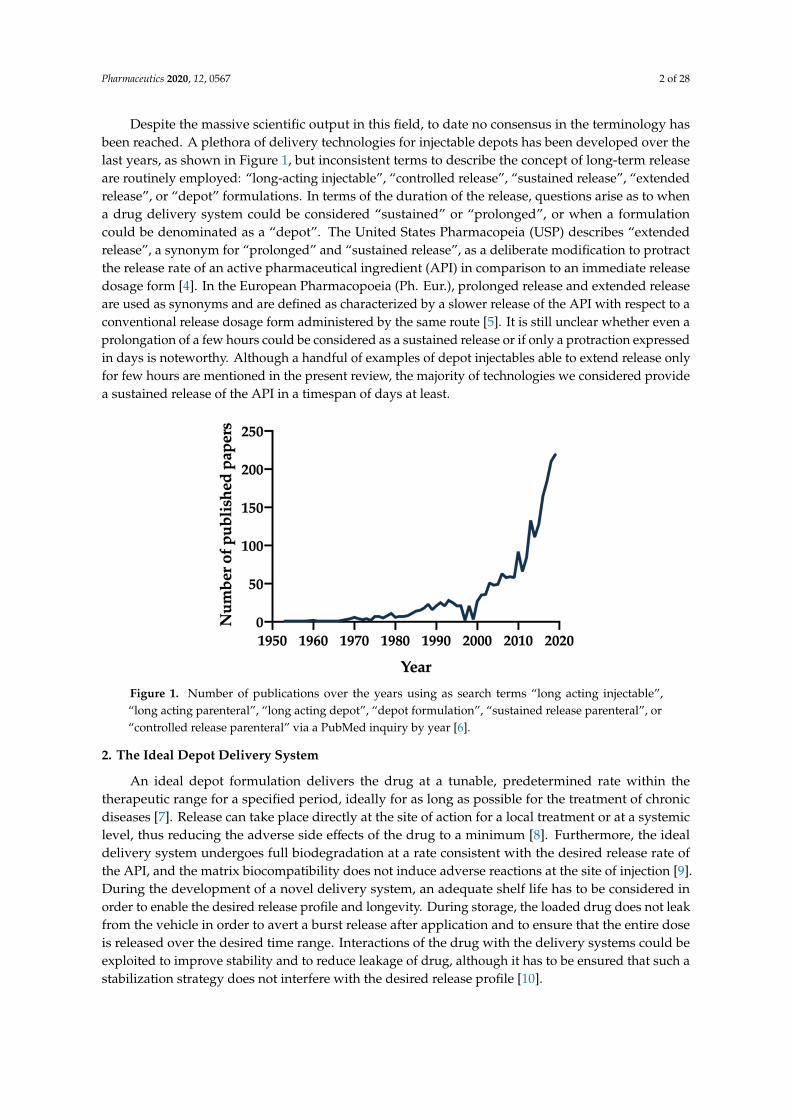
Despite the massive scientific output in this field, to date no consensus in the terminology hasbeen reached. A plethora of delivery technologies for injectable depots has been developed over thelast years, as shown in Figure 1, but inconsistent terms to describe the concept of long-term releaseare routinely employed: “long-acting injectable”, “controlled release”, “sustained release”, “extendedrelease”, or “depot” formulations. In terms of the duration of the release, questions arise as to whena drug delivery system could be considered “sustained” or “prolonged”, or when a formulationcould be denominated as a “depot”. The United States Pharmacopeia (USP) describes “extendedrelease”, a synonym for “prolonged” and “sustained release”, as a deliberate modification to protractthe release rate of an active pharmaceutical ingredient (API) in comparison to an immediate releasedosage form [4]. In the European Pharmacopoeia (Ph. Eur.), prolonged release and extended releaseare used as synonyms and are defined as characterized by a slower release of the API with respect to aconventional release dosage form administered by the same route [5]. It is still unclear whether even aprolongation of a few hours could be considered as a sustained release or if only a protraction expressedin days is noteworthy. Although a handful of examples of depot injectables able to extend release onlyfor few hours are mentioned in the present review, the majority of technologies we considered providea sustained release of the API in a timespan of days at least.
Pharmaceutics 2020, 12, x 2 of 30
Despite the massive scientific output in this field, to date no consensus in the terminology has been reached. A plethora of delivery technologies for injectable depots has been developed over the last years, as shown in Figure 1, but inconsistent terms to describe the concept of long-term release are routinely employed: “long-acting injectable”, “controlled release”, “sustained release”, “extended release”, or “depot” formulations. In terms of the duration of the release, questions arise as to when a drug delivery system could be considered “sustained” or “prolonged”, or when a formulation could be denominated as a “depot”. The United States Pharmacopeia (USP) describes “extended release”, a synonym for “prolonged” and “sustained release”, as a deliberate modification to protract the release rate of an active pharmaceutical ingredient (API) in comparison to an immediate release dosage form [4]. In the European Pharmacopoeia (Ph. Eur.), prolonged release and extended release are used as synonyms and are defined as characterized by a slower release of the API with respect to a conventional release dosage form administered by the same route [5]. It is still unclear whether even a prolongation of a few hours could be considered as a sustained release or if only a protraction expressed in days is noteworthy. Although a handful of examples of depot injectables able to extend release only for few hours are mentioned in the present review, the majority of technologies we considered provide a sustained release of the API in a timespan of days at least.
Figure 1. Number of publications over the years using as search terms “long acting injectable”, “long acting parenteral”, “long acting depot”, “depot formulation”, “sustained release parenteral”, or “controlled release parenteral” via a PubMed inquiry by year [6].
2. The Ideal Depot Delivery System
An ideal depot formulation delivers the drug at a tunable, predetermined rate within the therapeutic range for a specified period, ideally for as long as possible for the treatment of chronic diseases [7]. Release can take place directly at the site of action for a local treatment or at a systemic level, thus reducing the adverse side effects of the drug to a minimum [8]. Furthermore, the ideal delivery system undergoes full biodegradation at a rate consistent with the desired release rate of the API, and the matrix biocompatibility does not induce adverse reactions at the site of injection [9]. During the development of a novel delivery system, an adequate shelf life has to be considered in order to enable the desired release profile and longevity. During storage, the loaded drug does not leak from the vehicle in order to avert a burst release after application and to ensure that the entire dose is released over the desired time range. Interactions of the drug with the delivery systems could be exploited to improve stability and to reduce leakage of drug, although it has to be ensured that such a stabilization strategy does not interfere with the desired release profile [10].
In practice, not all requirements could be fulfilled in a universal delivery platform; nevertheless, existing lipid-based systems are continuously being improved to meet as many conditions as possible, as highlighted in the following sections.
Figure 1. Number of publications over the years using as search terms “long acting injectable”,“long acting parenteral”, “long acting depot”, “depot formulation”, “sustained release parenteral”, or“controlled release parenteral” via a PubMed inquiry by year [6].
2. The Ideal Depot Delivery System
An ideal depot formulation delivers the drug at a tunable, predetermined rate within thetherapeutic range for a specified period, ideally for as long as possible for the treatment of chronicdiseases [7]. Release can take place directly at the site of action for a local treatment or at a systemiclevel, thus reducing the adverse side e↵ects of the drug to a minimum [8]. Furthermore, the idealdelivery system undergoes full biodegradation at a rate consistent with the desired release rate ofthe API, and the matrix biocompatibility does not induce adverse reactions at the site of injection [9].During the development of a novel delivery system, an adequate shelf life has to be considered inorder to enable the desired release profile and longevity. During storage, the loaded drug does not leakfrom the vehicle in order to avert a burst release after application and to ensure that the entire doseis released over the desired time range. Interactions of the drug with the delivery systems could beexploited to improve stability and to reduce leakage of drug, although it has to be ensured that such astabilization strategy does not interfere with the desired release profile [10].
Pharmaceutics 2020, 12, 0567 3 of 28
In practice, not all requirements could be fulfilled in a universal delivery platform; nevertheless,existing lipid-based systems are continuously being improved to meet as many conditions as possible,as highlighted in the following sections.
2.1. Excipients for Formulating Depots
Two major classes of chemical compounds are used to create drug delivery systems: polymersand lipids [11]. Despite the fact that both serve the same purpose—encapsulating drugs fordepot formulations—they di↵er in their fundamental properties like chemical structure, solubility,or biodegradability and biocompatibility. The focus of this review is exclusively on depot formulationsbased on lipids, although hybrid systems combining the benefits of lipid and polymeric excipients willalso be discussed. For more details about polymeric drug delivery systems we refer the readers todedicated comprehensive recent reviews [12,13].
Lipids are defined as hydrophobic or amphiphilic small molecules originating entirely or in partby carbanion-based condensations of thioesters and/or by carbocation-based condensations, and areclassified by LIPID MAPS® into eight categories shown in Table 1 [14,15].
Table 1. Classification of lipids according LIPID MAPS with examples of subgroups [15,16].
Category Subgroup Examples
Fatty acyls Fatty esters, fatty amides, fatty acyl glycosidesGlycerolipids Mono-, di-, triradylglycerolsGlycerophospholipids Glycerophosphocholines, -serines, -glycerols, -inositolsSphingolipids Sphingoid bases, ceramides, phosphosphingolipidsSterol Lipids Sterols, steroids, secosteroids, bile acids, and derivativesPrenol Lipids Isoprenoids, quinones and hydroquinones, polyprenolsSaccharolipids Acylaminosugars, acylaminosugar glycans, acyltrehalosesPolyketides Flavonoids, macrolides, linear or aromatic polyketides
2.2. Parenteral Administration Routes for Depot Injectables
Administering depot formulations parenterally is a recommended choice for drugs which undergosubstantial first-pass metabolism or that are characterized by low oral bioavailability. Typically,the subcutaneous (s.c.) and intramuscular (i.m.) routes are preferred to achieve a systemic e↵ect.When local treatment is sought, intraarticular or intraocular injections are also considered viable optionsdepending on the condition to address. The absorption, distribution, metabolism, and elimination of adrug strongly rely on the characteristics of the biological environment. Opting for a specific site ofadministration thus represents a strategy to modulate the drug release kinetics. As the blood perfusionin the s.c. tissue is lower than in the muscles, a s.c. injection will result in a delayed absorption of thedrug with respect to an i.m. injection [17]. Targeted, localized drug delivery could be beneficial fordiseases a↵ecting only a small, distinct area of the body, resulting in a high local drug concentrationbut a low systemic concentration, thus reducing systemic side e↵ects and overall toxicity. Moreover,di↵usion barriers and drug metabolism could be circumvented.
S.c. injections are the most-often used parenteral administration route due the possibility ofself-injection by the patients, but the administered volume is limited. The administration of volumeslarger than 1.5 to 2 mL usually requires multiple injections to minimize the risk of considerably highpain upon injection. For example, it has been shown that incrementing the injected volume from 0.8 to2.25 mL resulted in an increase of the perceived pain about 1.7-fold [18,19]. The choice of the mostsuitable drug delivery system has to be carefully considered; the potency of the drug and the dose onthe one hand and the encapsulation capacity of the delivery system on the other play a decisive role inthis selection. Although it has been reported that the used bu↵ers (pH, osmolarity) in the formulationscould influence the pain upon injection [18], no correlation between di↵erent lipids and the inductionof pain has been investigated so far.
Pharmaceutics 2020, 12, 0567 4 of 28
Sterilization of the final product is mandatory for this route of application; however,(phospho)lipids are sensitive to high temperatures, and sterilization processes such as steam sterilizationor dry heat sterilization may cause oxidation and hydrolysis of the (phospho)lipids, resulting in adestabilization of the delivery system. Sterilization of lipid-based products might thus be achievedvia gamma-irradiation, sterile filtration (only for colloidal systems with a size below 200 nm), or viaproduction under aseptic conditions [20].
2.3. Biocompatibility
Host response to the depot formulations, including foreign body reactions or fibrous encapsulation,plays a pivotal role in the choice of materials for long-acting formulations. The extent is dependent onthe size, shape, and composition of the drug delivery system on the one hand and on the targeted tissueor organ on the other [21]. An initial acute inflammatory response is triggered after every injection dueto the minimal injury of the connective tissue. Released leachables or degradation products from thedelivery system could promote this inflammatory response and could turn the acute inflammationinto chronic inflammation if the stimuli are persistent [22]. Ultimately, a collagenous fibrous capsuleof typically 50–200 µm thickness can be built as reaction of the immune system to the foreign object,especially for solid implants or in situ forming systems. This creates an additional di↵usion barrierinfluencing drug release [23–25].
Being able to predict biocompatibility is of paramount interest for a successful delivery system:early toxicity screenings could be performed using in vitro tests but for the assessment of themultiple tissue reactions, in vivo studies combining pharmacokinetic and pharmacodynamic e↵ectsare also required. Besides quantifying the plasma concentrations of inflammatory cytokines and themacro- and microscopical inspection of the surrounded tissue, a cage implant system could be used.This system enables the characterization of degradation products of the drug delivery system as wellthe inflammatory exudates in the surrounded tissue. Regrettably, the inflammatory reactions from thecage itself could represent an insidious Trojan horse [26–28].
Lipids are characterized by an outstanding biocompatibility and safety profile due to theiroccurrence throughout the living world and the biodegradability in the human body induced bylipases [29,30]. For liposomes, the lipids used for the production of multivesicular liposomes did notinduce any foreign body reaction [31,32].
3. Oil-Based Solutions
Oily solutions containing a dissolved or suspended drug were first introduced in the 1950s and arehence the oldest approach proposed for injectable depots. They were traditionally used for antipsychoticdrugs or hormones, and several di↵erent products became available over the decades, including HaldolDecanoate, Depo-Testosterone, Invega Sustenna, or Depo-Provera, to name a few that are still on themarket [33,34]. With the aim of providing a more pronounced lipophilicity to extend drug release,highly hydrophobic esters were usually conjugated to first-generation antipsychotics. The drugsare dissolved in pharmaceutical oils like sesame seed oil or middle-chain triglycerides. Upon i.m.injection, drug transfer from the oil phase into the tissue fluid represents the main release-limitingfactor. Spreading of the oil solution along the muscle fibers can occur, increasing the surface area andtherefore accelerating drug release [35,36]. Although this depot system has a favorable long-termstability and can be prepared in a fast and uncomplicated manner with sterilization of the final product,only lipophilic drugs can be included. Hydrophilic drugs have to be modified prior to insertion,which limits the use of the system. In recent years almost no new approaches using dissolved drugsin oily solutions have been reported, and the system is still traditionally used for antipsychotics(Table 2). The concept of oil-based solutions has been further elaborated, and, in most cases, the drugis encapsulated in a carrier and then dissolved in an oil phase (Figure 2).
Pharmaceutics 2020, 12, 0567 5 of 28
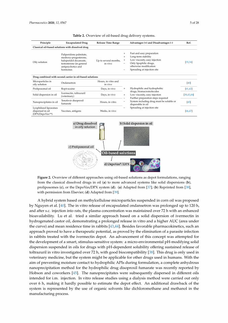
Table 2. Overview of oil-based drug delivery systems.
Principle Encapsulated Drug Release Time Range Advantages (+) and Disadvantages (–) Ref.
Classical oil-based solutions with dissolved drug
Oily solution
Paliperidone palmitate,medroxy-progesterone,haloperidol decanoate,testosterone (in generalantipsychotics andhormones
Up to several months,in vivo
+ Fast and easy preparation+ Long-term stability+ Low viscosity, easy injection– Only lipophilic drugs,
otherwise modification– Spreading at injection site
[33,34]
Drug combined with second carrier in oil-based solutions
Microparticles inoily solution Ondansetron Hours, in vitro and
in vivo
+ Hydrophilic and hydrophobicdrugs, biomacromolecules
+ Low viscosity, easy injection– Further preparation steps required– System including drug must be soluble or
dispersible in oil– Spreading at injection site
[40]
Proliposomal oil Ropivacaine Days, in vivo [41,42]
Solid dispersion in oil Ivermectin, toltrazuril(veterinary) Days, in vivo [38,43,44]
Nanoprecipitates in oil Tenofovir disoproxilfumarate Hours, in vitro [45]
Lyophilized liposomesdispersed in oil(DPX/DepoVax™)
Vaccines, antigens Weeks, in vivo [46,47]Pharmaceutics 2020, 12, x 5 of 30
Figure 2. Overview of different approaches using oil-based solutions as depot formulations, ranging from the classical dissolved drugs in oil (a) to more advanced systems like solid dispersions (b), proliposomes (c), or the DepoVax/DPX system (d). (a) Adapted from [37]; (b) Reprinted from [38], with permission from Elsevier; (d) Adapted from [39].
Table 2. Overview of oil-based drug delivery systems.
Principle Encapsulated Drug Release Time Range
Advantages (+) and Disadvantages (–) Ref.
Classical oil-based solutions with dissolved drug
Oily solution
Paliperidone palmitate, medroxy-progesterone, haloperidol decanoate, testosterone (in general antipsychotics and hormones
Up to several months, in vivo
� Fast and easy preparation � Long-term stability � Low viscosity, easy injection – Only lipophilic drugs,
otherwise modification – Spreading at injection site
[33,34]
Drug combined with second carrier in oil-based solutions Microparticles in oily solution Ondansetron
Hours, in vitro and in vivo � Hydrophilic and
hydrophobic drugs, biomacromolecules
� Low viscosity, easy injection – Further preparation steps
required – System including drug must
be soluble or dispersible in oil – Spreading at injection site
[40]
Proliposomal oil Ropivacaine Days, in vivo [41,42] Solid dispersion in oil
Ivermectin, toltrazuril (veterinary)
Days, in vivo [38,43,44]
Nanoprecipitates in oil
Tenofovir disoproxil fumarate
Hours, in vitro [45]
Lyophilized liposomes dispersed in oil (DPX/DepoVax™)
Vaccines, antigens Weeks, in vivo [46,47]
A hybrid system based on methylcellulose microparticles suspended in corn oil was proposed by Nguyen et al. [40]. The in vitro release of encapsulated ondansetron was prolonged up to 120 h, and after s.c. injection into rats, the plasma concentration was maintained over 72 h with an enhanced bioavailability. Lu et al. tried a similar approach based on a solid dispersion of ivermectin in hydrogenated castor oil, demonstrating a prolonged release in vitro and a higher AUC (area under the curve) and mean residence time in rabbits [43,44]. Besides favorable pharmacokinetics, such an approach proved to have a therapeutic potential, as proved by the elimination of a parasite infection
Figure 2. Overview of di↵erent approaches using oil-based solutions as depot formulations, rangingfrom the classical dissolved drugs in oil (a) to more advanced systems like solid dispersions (b),proliposomes (c), or the DepoVax/DPX system (d). (a) Adapted from [37]; (b) Reprinted from [38],with permission from Elsevier; (d) Adapted from [39].
A hybrid system based on methylcellulose microparticles suspended in corn oil was proposedby Nguyen et al. [40]. The in vitro release of encapsulated ondansetron was prolonged up to 120 h,and after s.c. injection into rats, the plasma concentration was maintained over 72 h with an enhancedbioavailability. Lu et al. tried a similar approach based on a solid dispersion of ivermectin inhydrogenated castor oil, demonstrating a prolonged release in vitro and a higher AUC (area underthe curve) and mean residence time in rabbits [43,44]. Besides favorable pharmacokinetics, such anapproach proved to have a therapeutic potential, as proved by the elimination of a parasite infectionin rabbits treated with the ivermectin depot. An advancement of this concept was attempted forthe development of a smart, stimulus-sensitive system: a micro-environmental pH-modifying soliddispersion suspended in oils for drugs with pH-dependent solubility o↵ering sustained release oftoltrazuril in vitro investigated over 72 h, with good biocompatibility [38]. This drug is only used inveterinary medicine, but the system might be applicable for other drugs used in humans. With theaim of preventing moisture contact to hydrophilic APIs during formulation, a complete anhydrousnanoprecipitation method for the hydrophilic drug disoproxil fumarate was recently reported byHobson and coworkers [45]. The nanoprecipitates were subsequently dispersed in di↵erent oilsintended for i.m. injection. In vitro release studies using a dialysis method were carried out onlyover 6 h, making it hardly possible to estimate the depot e↵ect. An additional drawback of thesystem is represented by the use of organic solvents like dichloromethane and methanol in themanufacturing process.
Pharmaceutics 2020, 12, 0567 6 of 28
The above-mentioned approach was revisited by the company PainReform Ltd. (Herzeliya, Israel):phospholipids, co-dissolved in an oily solution with a drug of choice, created a proliposomal oil,thus transforming a classical drug delivery system in a pro-formulation system [41,42]. Upon injectionand contact with body fluids the amphiphilic phospholipid molecules self-assemble into drug-loadedmultilamellar liposomes in the micron range. Following s.c. injection in pigs, a proliposomal oilformulated with ropivacaine showed a 5-fold increase in half-life and local anesthesia e↵ect, while inhuman volunteers the formulation of ropivacaine in a proliposomal oil resulted in a 2- to 3-fold increasein anesthetic e↵ect over plain ropivacaine. Combining the advantages of liposomes with an oil-basedsolution represented the core idea of a vaccine delivery platform with sustained immunological activity.The system, called DPX (formerly DepoVax™), is based on lyophilized liposomes encapsulatingantigens or nucleic acids which are dispersed in oil diluents [48]. In comparison to regularly usedemulsions, the system does not require the use of emulsifiers, does not support a passive release in vitro,and retains the antigens at the site of injection in vivo. DPX facilitated in vivo the development andpersistence of T cells and provided a better control of tumor growth [46]. Recently, Weir and coworkersused DPX for an anthrax vaccine formulation to reduce the current multidose vaccination plan (fiveinjections over 12 months combined with an annual booster) to a single injection, obtaining 100%protection against anthrax in rabbits and non-human primates [47]. Di↵erent clinical trials in early orlate phase 2 are ongoing for the treatment of ovarian cancer and lymphoma using immunotherapy [49].
Although the preparation of oily solutions might be easy and fast, these depot systems have thedisadvantage of spreading at the site of injection because of their low viscosity. Consequently, semisolidformulations with a higher viscosity or liquid in situ solidifying systems were developed (see Section 5,Semi-Solid Preparations and in Situ Forming systems). The latter approach especially combines theadvantages of easy administration and the formation of a physical depot in vivo (vide infra).
4. Liposomal Systems
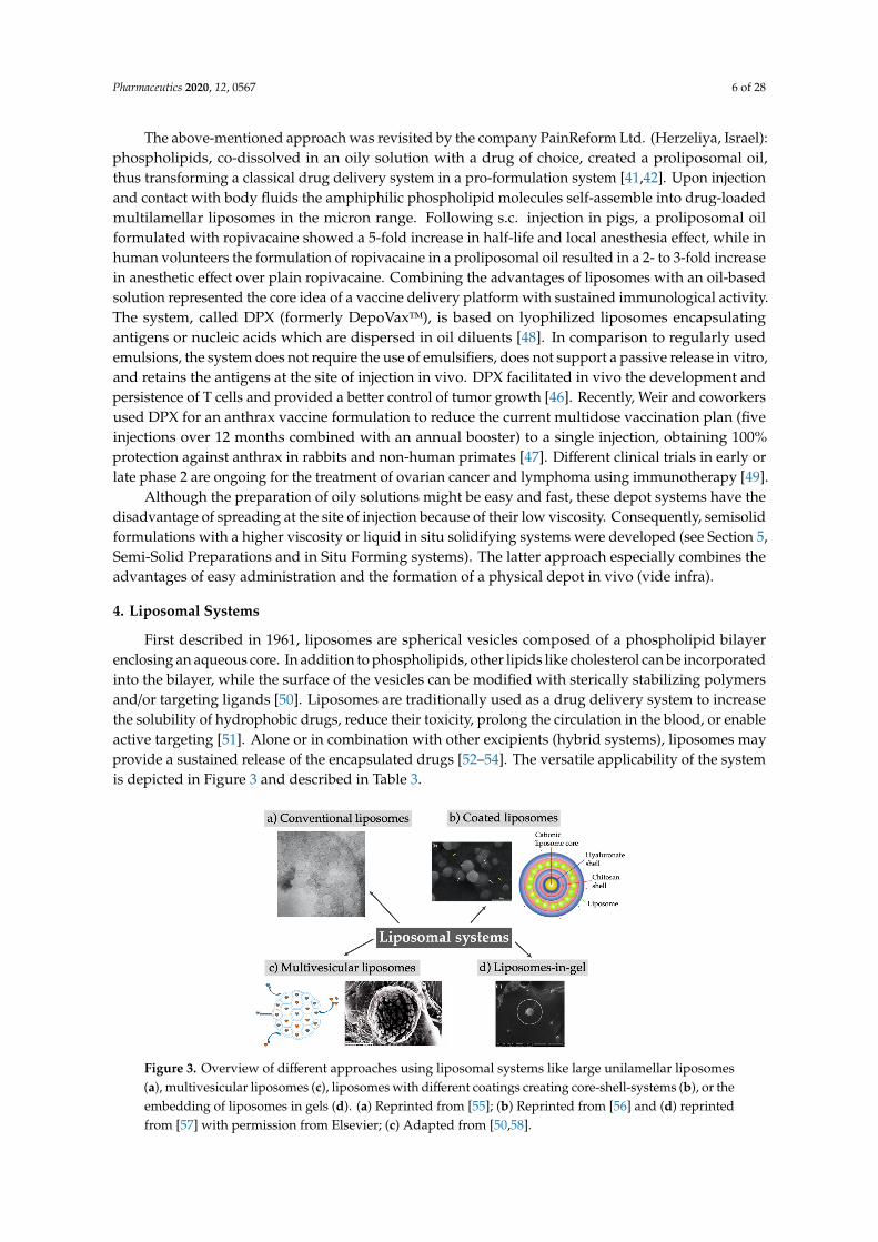
First described in 1961, liposomes are spherical vesicles composed of a phospholipid bilayerenclosing an aqueous core. In addition to phospholipids, other lipids like cholesterol can be incorporatedinto the bilayer, while the surface of the vesicles can be modified with sterically stabilizing polymersand/or targeting ligands [50]. Liposomes are traditionally used as a drug delivery system to increasethe solubility of hydrophobic drugs, reduce their toxicity, prolong the circulation in the blood, or enableactive targeting [51]. Alone or in combination with other excipients (hybrid systems), liposomes mayprovide a sustained release of the encapsulated drugs [52–54]. The versatile applicability of the systemis depicted in Figure 3 and described in Table 3.Pharmaceutics 2020, 12, x 7 of 30
Figure 3. Overview of different approaches using liposomal systems like large unilamellar liposomes (a), multivesicular liposomes (c), liposomes with different coatings creating core-shell-systems (b), or the embedding of liposomes in gels (d). (a) Reprinted from [55]; (b) Reprinted from [56] and (d) reprinted from [57] with permission from Elsevier; (c) Adapted from [50,58].
Table 3. Overview of sustained release systems based on liposomes.
Principle (Used Phospholipids *)
Encapsulated Drug/Marker
Release Time Range
Advantages (+) and Disadvantages (–) Ref.
Conventional liposomes Aggregation of negatively charged liposomes (DPPC, EPC, HPC, DSPG, DPPG, DOPA, DPPA)
Flurbiprofen, Calcein, FITC-dextran-4000
Days, in vitro and in vivo
� Hydrophilic and lipophilic drugs, biomacromolecules
� Reduce toxicity, targeting possible besides drug delivery
� Large-scale production obtainable with simple sterilization methods
– Limited amount of encapsulated drug
– Small size could lead to rapid release
– Fast clearance or lymphatic uptake possible
[55,59]
PEGylated liposomes (DPPC, DSPE-PEG2000)
Adrenaline Hours, in vitro [60]
Combination of fast and slow release liposomes (DSPC, DLPC, DMPC, DSPC)
Vaccine Not specified [61]
Small unilamellar liposomes (DSPC)
D-glucosamine sulphate, lubrication effect of phospholipids
Weeks, in vitro, days in vivo
[62]
Conventional and stealth liposomes (DPPG, DPPE-PEG2000)
Bevacizumab Months in vitro, weeks in vivo
[63]
Multivesicular liposomes MVLs, DepoFoam®
(EPC, SPC, HSPC, DEPC, DOPC, DPPG)
Bupivacaine (Exparel®), ropivacaine, oleanolic acid,
Days, in vitro and in vivo
� Hydrophilic and lipophilic drugs (but mostly hydrophilic drugs), biomacromolecules
[58,64–72]
Figure 3. Overview of di↵erent approaches using liposomal systems like large unilamellar liposomes(a), multivesicular liposomes (c), liposomes with di↵erent coatings creating core-shell-systems (b), or theembedding of liposomes in gels (d). (a) Reprinted from [55]; (b) Reprinted from [56] and (d) reprintedfrom [57] with permission from Elsevier; (c) Adapted from [50,58].

Pharmaceutics
20
20,12,0567
7of28
Ta
ble
3.O
verviewofsustained
releasesystem
sbased
onliposom
es.
Prin
cip
le(U
sed
Ph
osp
ho
lipid
s*)
En
cap
su
late
dD
ru
g/M
ark
er
Rele
ase
Tim
eR
an
ge
Ad
van
tag
es
(+)
an
dD
isad
van
tag
es
(–)
Ref.
Co
nv
en
tion
al
lipo
so
mes
Aggregation
ofnegativelycharged
liposomes
(DPPC
,EPC,H
PC,D
SPG,
DPPG
,DO
PA,D
PPA)
Flurbiprofen,Calcein,FITC
-dextran-4000D
ays,invitro
andin
vivo+
Hydrophilic
andlipophilic
drugs,biomacrom
olecules+
Reduce
toxicity,targetingpossible
besidesdrug
delivery+
Large-scaleproduction
obtainablew
ithsim
plesterilization
methods
–Lim
itedam
ountofencapsulateddrug
–Sm
allsizecould
leadto
rapidreleas
–Fastclearance
orlym
phaticuptake
possible
[55,59]
PEGylated
liposomes
(DPPC
,DSPE-PEG
2000)A
drenalineH
ours,invitro
[60]
Com
binationoffastand
slowrelease
liposomes
(DSPC
,DLPC
,DM
PC,
DSPC
)Vaccine
Notspecified
[61]
Smallunilam
ellarliposom
es(D
SPC)
D-glucosam
inesulphate,lubrication
e↵ectofphospholipidsW
eeks,invitro,days
invivo
[62]
Conventionaland
stealthliposom
es(D
PPG,D
PPE-PEG2000)
Bevacizumab
Months
invitro,w
eeksin
vivo[63]
Mu
ltivesic
ula
rlip
oso
mes
MV
Ls,DepoFoam
®(EPC
,SPC,H
SPC,D
EPC,D
OPC
,DPPG
)Bupivacaine
(Exparel ®),ropivacaine,
oleanolicacid,fluocinolone
acetonidein
cyclodextrinliraglutide,bevacizum
abD
ays,invitro
andin
vivo
+H
ydrophilicand
lipophilicdrugs
(butmostly
hydrophilicdrugs),biom
acromolecule
+M
ultipleaqueous
compartm
entsallow
highencapsulation
e�ciency
forhydrophilic
drugsand
slowrelease,avoiding
aburste↵ec
+Size
inm
icronrange
preventsfastlym
phaticuptak
–Expensive
productionprocess
includingseveralsteps,
organicsolvents
andaseptic
condition–
Encapsulationoflipophilic
drugsm
ightalterm
ulticompartm
entstructureand
notintensively
investigated
[58,64–72]
Lip
oso
mes
ing
el,
co
ate
dlip
oso
mes
Liposomes
inPluronic ®
F127hydrogel(H
PC,D
PPG,D
SPE-PEG2000)
Insulin,flurbiprofen,doxorubicin,paclitaxel
Days
tow
eeks,invivo
+H
ydrophilicand
lipophilicdrug
+Em
beddingin
matrix
retainssystem
atsiteofinjection
andcould
reducelym
phaticuptake
+Surface
coatingcould
o↵er
more
possibilitiesfor
targetingand
prolongdrug
releas–
Furtherpreparation
stepsrequire
–A
dditionofm
oreexcipients
with
possiblelow
erbiocom
patibilitythan
phospholipid–
Increaseofviscosity
couldlead
tow
orseinjectability
[73–75]
Liposomes
inPLG
A-PEG
-PLGA
hydrogel(SPC)
Isoniazid,doxorubicinD
ays,invitro
andin
vivo[76,77]
Liposomes
inchitosan
hydrogel(SPC,D
PPC)
Doxorubicin,C
arboxy-fluoresceinD
ays,invitro
andin
vivoor
notspecified[57,78]
Hybrid
liposomes
with
chitosan(S75)
Curcum
inD
ays,invitro
[79]
Hyaluronic
acid-coatedliposom
es(D
PPC,D
OTA
P)Paclitaxel
Hours,in
vitro[80]
Ternarydrug-cyclodextrin-liposom
escom
plex(EPC
)R
opivacaineH
oursin
vitroand
invivo
[81]
Capsosom
es(EPC
,DO
TAP)
None
Notspecified
[56]
*A
bbreviations:EPC
:L-↵-phosphatidylcholine(egg),D
EPC:1,2-dierucoyl-sn-glycero-3-phosphocholine,D
LPC:1,2-dilauroyl-sn-glycero-3-phosphocholine,D
MPC
:1,2-dimyristoyl-sn-glycero-
3-phosphocholine,D
OPA
:1,2-dioleoyl-sn-glycero-3-phosphate,
DO
PC:
1,2-dioleoyl-sn-glycero-3-phosphocholine,D
OTA
P:1,2-
dioleoyl-3-trimethylam
monium
-propan,D
PPA:
1,2-dipalmitoyl-sn-glycero-3-phosphate,
DPPC
:1,2-dipalm
itoyl-sn-glycero-3-phosphocholine,D
PPE-PEG2000:
1,2-dipalmitoyl-sn-
glycero-3-phosphoethanolamine-N
-[methoxy(polyethylene
glycol)-2000]
(amm
oniumsalt),
DPPG
:1,2-dipalm
itoyl-sn-gly-cero-3-phospho-(1 0-rac-glycerol)
(sodiumsalt),
DSPC
:1,2-distearoyl-sn-glycero-3-phosphocholine,
DSPE-PEG
2000:1,2-dis-
tearoyl-sn-glycerol-3-phosphoethanolamine-N
-[methoxy(poly-ethyleneglycol)-2000],D
SPG:1,2-distearoyl-sn-glycero-3-phospho-(1 0-rac-glycerol),H
PC:L-↵-phosphatidylcholine,hydrogenated,H
SPC:
hydrogenatedsoy
phosphatidylcholineSPC
:L-↵-phosphatidylcholine(soy),S75:soybean
lecithinat71%
ofphosphatidylcholine.

Pharmaceutics 2020, 12, 0567 8 of 28
The release of drugs encapsulated in liposomes, and with it the possibility to achieve adepot e↵ect, is mainly dependent on the di↵usion of the drugs across the bilayer. By generatingliposomal aggregates, the di↵usion of the drug into the surrounding can be mitigated,as recently proposed by our group [55]. Long-acting drug delivery systems could indeedbe generated in situ, controlling the aggregation of liposomes formulated with negativelycharged phospholipids (NCPs) through the interaction with divalent cations. Various NCPswere screened as function of their head groups and acyl chains for their suitability as sca↵oldfor depot formulations and the system extensively characterized from a physico-chemicalviewpoint. Mixing liposomes formulated with either 1,2-dipalmitoyl-sn-glycero-3-phosphate (DPPA)or 1,2-distearoyl-sn-glycero-3-phospho-(10-rac-glycerol) (DSPG) with physiologically compatibleconcentrations of calcium and magnesium resulted in a rapid fusion-free aggregation, confirming thepharmaceutical potential of intact liposomes as single composing units of the depot. Bupivacaine-loadedliposomal aggregates are presently under evaluation in our laboratories as an analgesic platformalternative to the current strategies for pain management.
Negatively charged phospholipids could be also used to form so-called cochleates, definedas supramolecular assemblies of phospholipid bilayers which form spiral structures with divalentcations [82]. Despite the thorough characterization conducted by Fahr and coworkers so far [83,84],their clinical potential regarding their use as drug delivery system still needs to be investigated.Drawing from this concept, Blazaki et al. induced aggregation of negatively charged liposomeswith protamine and encapsulated two di↵erent fluorescence dyes or the anti-inflammatory drugflurbiprofen [59]. The in vitro release of the fluorescent dyes was sustained for more than 17 days,whereas flurbiprofen showed a sustained release over only 120 h. Despite the increase in retention offlurbiprofen in vivo in the posterior eye segment after intravitreal injection achieved with the aggregatedliposomes, the di↵erences with respect to the non-aggregated system were rather small. Seeking astrategy aimed at avoiding repeated injections during emergency treatments such as cardiopulmonaryresuscitation, Schlich et al. described the use of adrenaline-loaded PEGylated liposomes [60]. One ofthe reported challenges during the production process of this formulation was to prevent the oxidationof adrenaline, which could ultimately be ensured by addition of an antioxidant while maintaining theloading e�ciency of the active loading method. The in vitro release of the API was investigated foronly 8 h (cumulative release of around 10%) in two di↵erent release setups, with neither reflecting thecomplexity after intravenous injection. The authors did not address how this liposomal system couldbe included in the treatment protocol for emergency situations in clinical practice.
Liposomes could not only act as a sustained drug delivery system, but the phospholipidsthemselves could exert local, mechanical beneficial e↵ects in the management of osteoarthritis [62].Distearoyl phosphatidylcholine (DSPC) liposomes encapsulating D-glucosamine sulphate combinedthe anti-inflammatory e↵ects of the drug and the lubrication ability of the liposomal system in thejoint. A prolonged release over several days in vitro was achieved and the friction between a silica andpolystyrene surface in an atomic force microscopy setup reduced. The lubrication e↵ect of the liposomesis attributed to a hydration layer around the phospholipid headgroups creating a ball-bearing-likesystem. It is questionable whether the lubrication enhancing is a clinically relevant e↵ect, since, so far,this has only been tested in instrumental setups or ex vivo using human cartilage [85,86].
A combination of fast and prolonged releasing liposomes was used by Christensen et al. as aparenteral vaccine strategy able to generate a long-lasting intestinal immune response to prevententeric infections [61]. The fast releasing system comprised retinoic acid in PEGylated liposomesto pre-condition the vaccine-draining lymph nodes. Cationic liposomes based on the surfactantdimethyldioctadecylammonium bromide (DDA) and stabilized with the synthetic immunostimulatortrehalose 6,60-dibehenate (TDB) formed a depot at the site of injection and delivered over a prolongedperiod of time the antigen. In vivo results confirmed this hypothesis and the combination of bothspecies induced an antigen-specific intestinal immunoglobulin A response after s.c. injection.
Pharmaceutics 2020, 12, 0567 9 of 28
Looking at the list of the liposomal drugs approved by the US Food and Drug Administration(FDA), only three can be strictly considered sustained drug delivery systems: Exparel® (bupivacaine),DepoDur® (morphine), and DepoCyte® (cytarabine). These three formulations share an importantaspect, that is, the same multivesicular liposomes technology, DepoFoam® [87,88]. Multivesicularliposomes (MVLs), first described by Kim et al. 1983, are micron-sized vesicles containing numerousinternal aqueous compartments divided by non-concentric lipid bilayers, as shown in Figure 3c [89,90].The production follows a two-step, water-in-oil-in-water double-emulsification process under asepticconditions [91]. In contrast to conventional liposomes, the use of a neutral lipid besides phospholipidsis mandatory to form this unique structure [89]. Several mechanisms that contribute to the release ofencapsulated drugs, like di↵usion, erosion, and reorganization of the compartments as well the usedphospholipids and neutral lipids have a major influence on the release profile [89,92]. While DepoDurand DepoCyt were discontinued from the market in the United States or Europe, Exparel wasrecently approved (2011) [87,88,93]. The research approaches aiming at exploring this technologyfor the delivery of bupivacaine in new indications are proliferating [71,72,94]. Besides bupivacaine,ropivacaine was considered as cargo for MVLs for prolonged-duration local anesthesia [65,66].After successfully encapsulating small molecules, attention was drawn to the use of this deliverysystem for biomacromolecules. MVLs loaded with bevacizumab were used for the treatment ofchoroidal neovascularization, resulting in a prolonged intravitreal retention time of bevacizumaband consequently in a reduced number of intraocular injections [58]. Becavizumab–MVLs turnedout to be superior to plain bevacizumab solution with respect to the e↵ective inhibition of thethickness of the choroidal neovascularization lesion at 28 days after treatment. The MVLs showeda prolonged release for 10 up to 13 days in vitro, mostly sustained for the formulation containing1,2-dioleoyl-sn-glycero-3-phosphocholine (DOPC). Interestingly, a more extended depot e↵ect usingconventional liposomes was reported by Karumanchi et al. [63]. Bevacizumab was released in vitroover 70 to 100 days out of conventional liposomes and over an even much longer time period usingstealth liposomes (150 to 200 days). Besides di↵erences in the in vitro drug release setup, the in vivoexperiments were designed di↵erently, and thus the results could not be compared directly. Karumanchiet al. injected the conventional liposomes into the anterior segment of rabbit eyes and observedtherapeutic concentrations of bevacizumab over 22 weeks, as determined by non-invasive fluorescenceimaging after fluorescence tagging of the antibody. Bevacizumab–MVLs injected into rabbit eyesshowed therapeutic concentrations over the full 56 days of study duration after direct measurementof the antibody concentration in vitreous and aqueous humor. The comparison of these two studiesunderlines how critical the choice of the in vitro release setup is to compare results obtained fromdi↵erent groups.
Although mostly hydrophilic drugs were encapsulated very e�ciently owing to the large aqueouscompartments in the MVLs, various attempts were made to include hydrophobic drugs into thissystem. Luo et al. encapsulated oleanolic acid, a pentacyclic triterpenoid, into the bilayer of theMVLs and in vitro and in vivo showed a prolonged release and plasma concentrations superior tofree oleanolic acid, respectively [68,69]. A di↵erent approach to include a hydrophobic agent intothe MVL was pursued by Vafaei and coworkers using cyclodextrins [70]. The lipophilic modeldrug fluocinolone acetonide was encapsulated into di↵erent cyclodextrin inclusion complexes andsubsequently incorporated into MVLs by reverse-phase evaporation. The in vitro drug release wassustained up to 180 h depending on the used cyclodextrin.
To summarize, MVLs are an all-round platform for prolonged drug delivery. A high encapsulatione�ciency for hydrophilic drugs can be achieved due to their relatively high water content (aqueous:lipid ratio 95:5). The nonconcentric bilayer geometry increases the stability of the system and reduces aburst release, while the depot e↵ect itself relies on reduced clearance, an aftermath of the micron-rangesize of the vesicles [87,91]. Despite the superb clinical success, the drawbacks of this system, mainly withrespect to the manufacturing procedure, should not be overlooked. The double-emulsification processrequires the use of organic solvents and needs to be performed entirely under aseptic conditions [89,90].
Pharmaceutics 2020, 12, 0567 10 of 28
At the other end of the scale, conventional small and large unilamellar liposomes are still beingintensively investigated and show tremendous potential for depot systems, especially in a hybridmodality, in combination with other biocompatible materials like gel matrices to form liposomesin gel, which are favorable for local drug release due to the increased viscosity of the system.The biodegradable hydrogel poly(lactic-co-glycolic acid)–polyethylene glycol–poly(lactic-co-glycolicacid) (PLGA–PEG–PLGA) was proposed as matrix to embed liposomal doxorubicin, thus enabling thetaming of the burst release typical of PLGA systems in favor of a smoother, prolonged release [76].For the first 12 h the hybrid composite depot outperformed the pristine doxorubicin in hydrogel,while the in vitro sustained profile of the liposomal gel was maintained for over 11 days; furthermore,tumor growth was significantly reduced in a breast cancer model in mice, and the known cardiotoxicityof doxorubicin was reduced. The same approach of liposomes in gel but using a Pluronic® (poloxamer)hydrogel was tried by Fu and colleagues, who encapsulated doxorubicin and paclitaxel for the treatmentof hepatocellular carcinoma [73]. After local injection the system inhibited the tumor growth in vivosignificantly and reduced the toxicity of the drugs.
Coating the liposomal surface with polymers could represent an alternative strategy toextend drug release over time, as proposed by Ravar and coworkers [80]. Hyaluronic acidwas used produce hybrid polymer-lipid vesicles via electrostatic coating upon titration of 1,2-dioleoyl-3-trimethylammonium-propan (DOTAP)-based large unilamellar liposomes encapsulatingpaclitaxel. Although the in vitro release of paclitaxel followed over 40 h was not substantially prolongedin comparison to hyaluronic acid-free plain liposomes, the tumor growth in vivo in 4T1-tumor bearingmice intravenously injected with the depot system was reduced by about 10-fold with respect to theones treated with a commercial paclitaxel solution, and the overall survival of the mice increased by60% over 20 days. The addition of more than one polymer can lead to the formation of layer-by-layerparticles, also called capsosomes, arising from electrostatic interactions [56]. The capsosomes wereformed by covering a cationic liposomal core layer-by-layer with hyaluronic acid and chitosan, toppedwith small sized liposomes and hyaluronic acid. Although it seems a promising approach to sustainthe drug release using several layers as a di↵usion barrier, investigations about the encapsulation ofdrugs and their release are missing. A completely di↵erent strategy was adopted by Nascimento Vieiraet al., who encapsulated a drug into cycodextrins and included this complex into liposomes to improvethe encapsulation e�cacy, prolong the release, and enhance the biological activity [81]. The ternarydrug delivery system containing ropivacaine sustained the release in vitro up to 8 h while the celltoxicity of ropivacaine was reduced. Furthermore, the in vivo sensory block of the local anestheticwas prolonged 1.7 times in comparison to the drug solution. These examples demonstrate the varietyof liposomal hybrid systems acting as an alternative to MVLs, and their clinical relevance should befurther investigated.
5. Semi-Solid Preparations and In Situ Forming Systems
Injectable in situ forming gels are an attractive way to deliver drugs due to their easy handlingprior to and during administration. The depot can be conveniently generated upon contact withbody fluids or when the physiological temperature is reached upon injection. A brief overview of thereported technologies is given in Figure 4 and summarized in Table 4.
Pharmaceutics 2020, 12, 0567 11 of 28Pharmaceutics 2020, 12, x 12 of 30
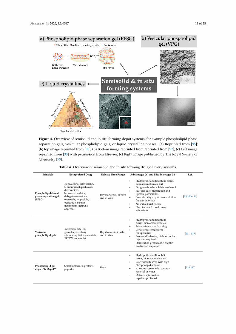
Figure 4. Overview of semisolid and in situ forming depot systems, for example phospholipid phase separation gels, vesicular phospholipid gels, or liquid crystalline phases. (a) Reprinted from [95]; (b) top image reprinted from [96]; (b) Bottom image reprinted from reprinted from [97]; (c) Left image reprinted from [98] with permission from Elsevier; (c) Right image published by The Royal Society of Chemistry [99].
Table 4. Overview of semisolid and in situ forming drug delivery systems.
Principle Encapsulated Drug Release Time Range
Advantages (+) and Disadvantages (–) Ref.
Phospholipid-based phase separation gel (PPSG)
Ropivacaine, pitavastatin, 5-fluorouracil, paclitaxel, doxorubicin, bromo- tetrandrine, dabigatran etexilate, exenatide, leuprolide, octreotide, insulin, incomplete Freund’s adjuvant
Days to weeks, in vitro and in vivo
� Hydrophilic and lipophilic drugs, biomacromolecules, but
– Drug needs to be soluble in ethanol
� Fast and easy preparation and upscale possibilities
� Low viscosity of precursor solution for easy injection
� No initial burst release – Use of ethanol could cause side
effects
[95,100–110]
Vesicular phospholipid gels
Interferon-beta-1b, granulocyte-colony stimulating factor, exenatide, FKBP51 antagonist
Days to weeks in vitro and in vivo
� Hydrophilic and lipophilic drugs, biomacromolecules
� Solvent-free manufacturing � Long-term storage form for
liposomes – Semisolid behavior, high forces
for injection required – Sterilization problematic, aseptic
production required
[111–115]
Phospholipid gel depo (PG DepotTM)
Small molecules, proteins, peptides Days
� Hydrophilic and lipophilic drugs, biomacromolecules
� Low viscosity even with high phospholipid amount
[116,117]
Figure 4. Overview of semisolid and in situ forming depot systems, for example phospholipid phaseseparation gels, vesicular phospholipid gels, or liquid crystalline phases. (a) Reprinted from [95];(b) top image reprinted from [96]; (b) Bottom image reprinted from reprinted from [97]; (c) Left imagereprinted from [98] with permission from Elsevier; (c) Right image published by The Royal Society ofChemistry [99].
Table 4. Overview of semisolid and in situ forming drug delivery systems.
Principle Encapsulated Drug Release Time Range Advantages (+) and Disadvantages (–) Ref.
Phospholipid-based
phase separation gel
(PPSG)
Ropivacaine, pitavastatin,5-fluorouracil, paclitaxel,doxorubicin,bromo-tetrandrine,dabigatran etexilate,exenatide, leuprolide,octreotide, insulin,incomplete Freund’sadjuvant
Days to weeks, in vitroand in vivo
+ Hydrophilic and lipophilic drugs,biomacromolecules, but
– Drug needs to be soluble in ethanol+ Fast and easy preparation and
upscale possibilities+ Low viscosity of precursor solution
for easy injection+ No initial burst release– Use of ethanol could cause
side e↵ects
[95,100–110]
Vesicular
phospholipid gels
Interferon-beta-1b,granulocyte-colonystimulating factor, exenatide,FKBP51 antagonist
Days to weeks in vitroand in vivo
+ Hydrophilic and lipophilicdrugs, biomacromolecules
+ Solvent-free manufacturing+ Long-term storage form
for liposomes– Semisolid behavior, high forces for
injection required– Sterilization problematic, aseptic
production required
[111–115]
Phospholipid gel
depo (PG Depot™)
Small molecules, proteins,peptides Days
+ Hydrophilic and lipophilicdrugs, biomacromolecules
+ Low viscosity even with highphospholipid amount
+ Aqueous system with optionalremoval of water
– Detailed informationis patent-protected
[116,117]
Pharmaceutics 2020, 12, 0567 12 of 28
Table 4. Cont.
Principle Encapsulated Drug Release Time Range Advantages (+) and Disadvantages (–) Ref.
Liquid crystalline
phases (cubosomes,
hexosomes),
FluidCrystal®
,
hybrid systems
Bufalin, bupivacaine,minocycline, indomethacin,finasteride, doxorubicin,buprenorphine (Buvidal®,Brixadi®), sinomenine,docetaxel, leuprolide,octreotide, setmelanotide,Huperzine A, VEGF siRNA(combined withpolyethylene-imine),
Days to weeks in vitroand in vivo
+ Hydrophilic and lipophilic drugs,biomacromolecules, but
– Size of water channels need to bealtered for releaseof macromolecules
– High water content could induceburst release of hydrophilic drugs
– High viscosity of cubic andhexagonal phase requires highinjection forces, but
+ Dilution to cubosomes andhexosomes or use of water-freeprecursor solutionsdecrease viscosity
+ Choice of (phospho)lipids cancontrol final properties
– Contact with body fluids couldchange crystal structure, but
+ Precursor solutions use thetransformation to liquid crystalsystems upon contact withbody fluids
– Small changes in production canlead to di↵erent crystal structures
– Highly controlled productionprocess required
[98,118–132]
Nanoemulsion Bupivacaine Hours, in vitro andin vivo
+ Hydrophilic and lipophilic drugs+ Fast and easy preparation– Addition of polymeric surfactants
to stabilize the system couldinduce toxicity
– Depot e↵ect rather small
[133]
OrganogelCandesartan cilexetil,risperidone
Days in vitro andin vivo
– Only lipophilic drugs possible+ Easy preparation– High viscosity complicates injection– Use of organogelators could
increase toxicity
[134,135]
Phospholipid-based phase separation gels (PPSG) combine simple manufacturing with the use ofexcipients known for their high biocompatibility and of solvents with low toxicity such as ethanol,soybean oil, and medium-chain triglycerides. By dissolving the phospholipids in ethanol and neutraloil mixtures and subsequently adding the drug of choice, a straightforward upscale can be envisaged,while a sterilization by sterile filtration can be performed to ensure the production of a product suitablefor parenteral administration [100,104]. After s.c. injection and contact with the body fluids, a sol–geltransformation occurs due to the di↵usion and exchange of ethanol out and tissue fluids into the gel,initiating a phase transition of the phospholipids and resulting in an increase in the viscosity in a rangefrom 10- to 106-fold [95,103,104,110]. The remarkably higher phospholipid concentrations (up to 70%) ofPPSGs with respect to organogels or vesicular phospholipid gels is responsible for the low viscosity andnegligible initial burst release after administration [104]. The existence of the gel over a long time periodwas proven by fluorescence imaging or harvesting of the depot [95,100,103,105]. Encapsulating smallmolecules could prolong the release in vitro for several days, as shown by Li et al., when incorporatingropivacaine into PPSG [95]. The initial burst observed for the local anesthetic solution after s.c. injectioninto rats was reduced using the depot system and the nerve blockade in guinea pigs lasted threetimes longer. The in vivo drug release or the therapeutic e↵ect for biomacromolecules was observedfor up to weeks, as in the case of exenatide released over 3 weeks [106], and leuprolide acetate andoctreotide acetate both released for over 4 weeks [107,108]. The administration of an antigen using thisdelivery system led to a persistent immune response and immunologic memory, showing the potentialof PPSG as new vaccine adjuvant [105]. In general, foreign body reactions and tissue responses arerelated to the ethanol content and are reduced by increasing the phospholipid content, while the

Pharmaceutics 2020, 12, 0567 13 of 28
inflammatory responses are low and disappear with depot degradation [100,103,105]. Using this depotsystem, the toxicity and side e↵ects of the drug itself are reduced while showing the same or evenbetter therapeutic e↵ect [101,102,105]. PPSGs are fully biodegradable but only limited information isavailable about the detailed degradation pathway. The formation of small vesicles at the edges andtheir release from the main gel are assumed [100].
In comparison to the PPSG, vesicular phospholipid gels (VPGs) comprise phospholipids not insolution, but rather as tightly packed vesicles and lamellar structures. This minimizes the surroundingaqueous phase and leads to the typical semisolid behavior mediated by the steric interactions of thevesicles without the need for additional viscosity enhancers. Dilution of the system after injection resultsin the formation of small unilamellar vesicles, as recently reviewed [136]. The inner structure of thedensely packed vesicles causes a relatively high viscosity and it might be di�cult or even impossible toinject them. A promising needle-free injection approach for VPGs to overcome this hurdle was proposedby Breitsamer et al., but a clinical validation is to date still missing [137]. VPGS have been intensivelyinvestigated for the delivery of proteins and peptides [111–113]; for example, exenatide–VPGs showeda release in vivo over 11 days, and the hypoglycemic e↵ect in diabetic rats lasted for 10 days uponinjection [114].
Latitude Pharmaceuticals Inc. o↵ers a phospholipid gel depot (PG Depot) that contains between20% and 80% phospholipids nano-dispersed in water and can be easily injected through a 25 G needle.A variety of drugs can be loaded in this system, from small molecules to proteins and peptides. In afirst step a phospholipid nanodispersion with a high-water content is produced and a gel phase issubsequently obtained by removing water in a second step. The depot e↵ect could be adjusted from 1to 7 days [116,117,138].
Lyotropic liquid phases are characterized by a self-assembly of amphiphilic lipids to di↵erentlamellar, hexagonal, or cubic phases or a reversed micellar cubic phase. The relatively high viscosityof the systems is an advantage for topical administrations but is not beneficial for their injectability.Consequently, the cubic or hexagonal phase can be dispersed in aqueous solutions forming cubic(“cubosomes”) or hexagonal nanoparticles (“hexosomes”) with an intact inner liquid crystallinestructure and larger surface area but a lower viscosity [139]. An alternative strategy to improvetheir injectability is based on the use of a lower amount of viscous precursor (with a low amount ofwater): body fluids are taken up in vivo upon injection, resulting in the formation of a viscous depotsystem in situ [118,123,125]. A comprehensive overview about the detailed inner structure, preparationmethods, and applications has been given in two recent reviews [140,141]. Narrow-sized waterchannels in the liquid crystal structure cause the sustained release behavior of low-molecular-weightdrugs but might inhibit the release of molecules with higher molecular weight and therefore demandthe addition of hydration-modulating agents [142]. The in vitro duration of action of the depot e↵ectranges from several days for bufalin [125], to 10 days for minocycline [126], or up to one month forleuprolide acetate [98], and can be easily tuned by adjusting the ratios of the used compounds [121].A novel non-viral gene delivery system was designed by Borgheti-Cardoso et al., combining liquidcrystals with polyethylenimine (PEI) to complex siRNA (small interfering RNA) [129]. The release ofsiRNA was maintained over 7 days in vitro, while in vivo the in situ formed gels were degraded over30 days, inducing only a mild inflammatory response. The siRNA was released in a complex withPEI, which protects the siRNA against degradation, but experimental data about the gene silencinge�ciencies of the released siRNA are missing to date. Overall, the groups developing liquid crystallinephases as injectable depots have focused mainly on the characterization of the systems using methodslike small angle X-ray scattering, electron or polarized light microscopy, rheology, and in vitro drugrelease studies [98,124,143]. Besides measuring drug plasma concentrations, little attention has beenpaid to additional in vivo characterization like the fate of the depot after injection, possible local sidee↵ects, and biocompatibility or the pharmacological e↵ect.
The only liquid crystalline system that has reached clinical studies is based on the FluidCrystal®
technology marketed by Camurus AB. Recently, Buvidal® (buprenorphine weekly or monthly) was
Pharmaceutics 2020, 12, 0567 14 of 28
approved in the European Union and Australia and tentatively approved in the United States (Brixadi™)for the treatment of opioid use disorder [130,131]. The depot system is based on a phosphatidylcholineand glyceroldioleate precursor solution which forms in situ a reversed non-lamellar liquid crystallinestructure with a tunable release profile [144,145]. Other products in di↵erent phases of clinical studies,such as octreotide for the treatment of acromegaly and neuroendocrine tumors [119,132], leuprolidefor the treatment of prostate cancer, or setmelanotide for treating genetic obesity [120], demonstratethe great potential of liquid crystalline phases as depot system.
Although the aqueous-based nanoemulsion developed by Rachmawati et al. does not show asemi-solid behavior, it is discussed in this chapter since it relies on the same principle of dispersedlipids in aqueous solutions [133]. Bupivacaine is distributed in castor oil droplets stabilized bypolymeric surfactants, as proven by cryo-transmission electron microscopy. Di↵erent biodegradableoils and polymeric surfactants were evaluated for their potential as depot formulations but onlythe nanoemulsion prepared with castor oil showed su�cient stability over 3 months. The releaseof bupivacaine was extended in vitro up to 24 h. Evaluating the in vivo plasma profile, the initialburst release using the nanoemulsion was lowered in comparison to the bupivacaine solution, but thenanoemulsion containing the same amount of bupivacaine as the control solution showed a loweranalgesic e↵ect than the bupivacaine solution (as explained by the slow release), and did not achievean e↵ective concentration. After increasing the concentration of drug in the nanoemulsion three times,the analgesic e↵ect was then prolonged up to 24 h.
Classical organogels merely based on immobilized organic phase in a three-dimensionalnetwork-building gelling agent undergo sol–gel transition in situ due to temperature increase orbecause of partial desolvation [146]. Although in recent years major scientific e↵orts have been madeto expand the current portfolio of in situ forming systems, considered very attractive for their superiorclinical translational properties, some attempts to synthesize novel organogelators were successfullycarried out. For example, Li and coworkers synthesized a novel low-molecular-weight bis-amideorganogelator able to prolong the release of candesartan cilexetil out of a soybean organogel for up to10 days in vivo with reasonably moderate inflammation at the injection site [134]. Newly synthesizedorganogelators based on amino acid derivates induced a prolonged release in vitro of risperidoneout of soybean oil [135]. The depot e↵ect was confirmed in vivo, showing an almost linear releasebehavior after s.c. injection over 8 days and suggesting a possible relevant translational potential, but acomprehensive toxicity and biocompatibility study is missing.
6. Solid Particles and Implants
Solid drug delivery systems could be produced according to di↵erent technologies, each resultingin products varying in size and structure.
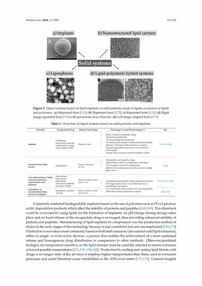
Solid lipid implants are monolithic, typically cylindrical-shaped systems with diameter and lengthin the millimeter range, whereas solid lipid nano- or microparticles are characterized by a size in thenano- or micron-range, respectively, and both systems contain lipids or mixtures of them in a solidstate (Figure 5). To overcome the physical instabilities and the short shelf-life often associated withsolid lipid nanoparticles, nanostructured lipid carriers were introduced and are characterized by a lessordered crystal structure due to the addition of liquid lipids to the solid lipids [147,148]. Surroundingclassical lipid cores with a phospholipid bilayer, lipospheres were created which were designed toreduce the production costs of liposomes and improve their stability. The absence of an aqueous corecompared to liposomes limits the encapsulation of drugs to only lipophilic substances [149]. Despitethe fact that most often preparation requires the use of organic solvents, a method averting the use oftoxic organic solvents has been recently reported [150]. Furthermore, the solid lipid particles could bemerged with polymers to improve stability and encapsulation e�ciency, and prolong the release rate.An overview of reviewed solid lipid systems investigated in the last years is given in Table 5.
Pharmaceutics 2020, 12, 0567 15 of 28
Pharmaceutics 2020, 12, x 16 of 30
aqueous core compared to liposomes limits the encapsulation of drugs to only lipophilic substances [149]. Despite the fact that most often preparation requires the use of organic solvents, a method averting the use of toxic organic solvents has been recently reported [150]. Furthermore, the solid lipid particles could be merged with polymers to improve stability and encapsulation efficiency, and prolong the release rate. An overview of reviewed solid lipid systems investigated in the last years is given in Table 5.
Figure 5. Depot systems based on lipid implants or solid particles made of lipids or mixture of lipids and polymers. (a) Reprinted from [151]; (b) Reprinted from [152]; (c) Reprinted from [153]; (d) Right image reprinted from [154] with permission from Elsevier; (d) Left image adapted from [155].
Table 5. Overview of depot systems based on solid particles and implants.
Principle Encapsulated Drug Release Time Range
Advantages (+) and Disadvantages (–) Ref.
Implants
Ovalbumin, monoclonal antibody, ranibizumab, insulin, tumor peptide TRP2
Weeks in vitro
� More versatile for lipophilic drugs and biomacromolecules
� No drug leakage during storage
� No induced pH change for enhanced protein stability
– Injection with large needle diameter or surgery
– Non-biodegradable systems have to be removed with surgery
– Foreign body reactions around monolithic system
[151,156–160]
Nanostructured lipid carriers
Ondansetron Days in vitro and in vivo
� Hydrophilic and lipophilic drugs
� High stability with no crystallization of the lipid
– Use of organic solvents for preparation
– Relatively fast release due to small size and resulting high surface
[161]
Core-shell systems of lipid nano-
Ropivacaine, bupivacaine,
Days to weeks in
� Hydrophilic and lipophilic drugs, biomacromolecules
[154,155,162–165]
Figure 5. Depot systems based on lipid implants or solid particles made of lipids or mixture of lipidsand polymers. (a) Reprinted from [151]; (b) Reprinted from [152]; (c) Reprinted from [153]; (d) Rightimage reprinted from [154] with permission from Elsevier; (d) Left image adapted from [155].
Table 5. Overview of depot systems based on solid particles and implants.
Principle Encapsulated Drug Release Time Range Advantages (+) and Disadvantages (–) Ref.
Implants
Ovalbumin,monoclonal antibody,ranibizumab, insulin,tumor peptide TRP2
Weeks in vitro
+ More versatile for lipophilic drugsand biomacromolecules
+ No drug leakage during storage+ No induced pH change for enhanced protein stability– Injection with large needle diameter or surgery– Non-biodegradable systems have to be removed
with surgery– Foreign body reactions around monolithic system
[151,156–160]
Nanostructured lipid
carriersOndansetron Days in vitro and
in vivo
+ Hydrophilic and lipophilic drugs+ High stability with no crystallization of the lipid– Use of organic solvents for preparation– Relatively fast release due to small size and resulting
high surface
[161]
Core-shell systems of lipid
nano-/microparticles,
combinations with
polymers
Ropivacaine,bupivacaine,risperidone,monoclonal antibody,lysozyme, insulin
Days to weeks in vitroand in vivo + Hydrophilic and lipophilic drugs, biomacromolecules
+ Prolonged release due to core–shell structure orembedding in gel matrix
– Use of polymers with possible lower biocompatibility– More steps during preparation required
[154,155,162–165]
Lipospheres or
nanostructured lipid
carriers in hydrogel
Quetiapine, estradiolvalerate
Hours to days in vitroand in vivo [166,167]
Commonly marketed biodegradable implants based on the use of polymers such as PLGA produceacidic degradation products which a↵ect the stability of proteins and peptides [168,169]. This drawbackcould be overcome by using lipids for the formation of implants: no pH change during storage takesplace and no burst release of the encapsulate drug is envisaged, thus providing enhanced stability ofproteins and peptides. Manufacturing of lipid implants by compression was the production method ofchoice in the early stages of this technology because it was considered fast and uncomplicated [159,170].Production is nowadays most commonly based on holt melt extrusion (also named solid lipid extrusion),either in single- or twin-screw devices, a process that enables the achievement of a more sustainedrelease and homogenous drug distribution in comparison to other methods. Often-encapsulatedbiologics are temperature-sensitive, so the lipid mixture must be carefully selected to ensure extrusionat lowest possible temperatures [151,156–158,160]. Production by melting and casting lipid blends withdrugs is no longer state of the art since it employs higher temperatures than those used in extrusionprocesses and could therefore cause instabilities in the APIs even faster [171,172]. General insights
Pharmaceutics 2020, 12, 0567 16 of 28
into the e↵ect of lipid composition and di↵erent preparation parameters on the physico-chemicalcharacteristics and the in vitro release behavior (up to 28 weeks) were obtained using albumin ormonoclonal antibodies as model proteins [156–158,160]. Lipid implants including a monoclonalantibody from the IgG1 class and the fab-fragment ranibizumab showed a sustained release in vitro ofaround 120 days with a good stability of the proteins, which were stabilized by cyclodextrines [151].Overall, recent studies mainly focus on the preparation and characterization of the implants and do notfocus on the implementation in vivo and the drug release, pharmacokinetics, or pharmacodynamics.An exception is represented by Even and coworkers, who showed in vivo results using the tumorpeptide TRP2 as an antigen in lipid implants [158]. The mice receiving this implant had a delayedtumor growth of three days compared to control groups without TRP2 treatment. They explained thislow e↵ect with the slow release out of the implant and reported that this type of therapy might be notappropriate for fast-growing tumors.
Solid lipid nano-/microparticles or nanostructured lipid carriers are not commonly used forparenteral depot injectables owing to a relatively fast release caused by erosion of the particles,a phenomenon that makes them ideal candidates predominantly for topical or oral administrationof drugs [161]. Nevertheless, they have been investigated as parenteral depot formulations.Nanostructured lipid carriers encapsulating ondansetron were shown to enable an in vitro sustainedrelease up to 96 h, a result confirmed in vivo without the appearance of a burst release [161].
To prolong the release rate, hybrid systems of polymers and lipids were explored. Mergingpoly-"-caprolactone nanoparticle cores with encapsulated ropivacaine and a lipid shell prolonged therelease of ropivacaine in vitro to 96 h [162]. The median duration of analgesia in vivo was maintainedover 36 h for the nanoparticles compared to 0.5 h for the plain ropivacaine solution. The same ideawas proposed by Ma and coworkers encapsulating bupivacaine into PLGA nanoparticles which weresurrounded by a lipid shell [165]. The hybrid system prolonged the in vitro release up to 96 h and,in comparison to pure polymeric nanoparticles, the initial burst release was reduced. This observationwas confirmed in vivo with a 5-h extended analgesic e↵ect of the polymer–lipid hybrids over thepolymeric nanoparticles. The influence of di↵erent lipid types on core–shell particle systems wasdemonstrated using mannitol microparticle cores with embedded monoclonal antibody IgG1 [163].Glyceryl stearate as a coating showed an incomplete in vitro release of IgG1, whereas the release profilewas prolonged up to 6 weeks with increasing amounts of hard fat as the lipid shell and the initialburst decreased.
Besides being used to create core–shell systems, lipids could also be included into the matrix ofpolymeric systems with the aim of modulating the release profile of co-loaded drugs. For example,encapsulating risperidone in PLGA microcapsules formulated with tuned middle-chain triglyceridesresulted in an almost ideal zero-order in vitro release kinetic over 60 days without the lag phasetypically observed for pure polymeric microparticles [155]. Imaging techniques suggest a multicoreinner structure of the microcapsules, with a core of dispersed lipid phase in which the drug couldbe dissolved. To date, in vivo results to prove the concept are still lacking. The type of triglyceridesused to form hybrid particles has a major influence on the release behavior, as demonstrated by Wuand coworkers [154]. The length of the acyl chain and the amount of lipid employed were correlatedwith the initial burst release of lysozyme as the model protein; the longer the chain length the greaterthe initial burst, and, as shown for one lipid, the burst release was increased by increasing the lipidproportion from 33% to 67%. The amount of lipid influenced the encapsulation e�ciency as well;depending on the type of lipid the best results were achieved for lipid proportions between 5% and20%, without a clear trend.
Analogous to the liposome-in gel-approach, lipid nanoparticles can be embedded in hydrogelmatrices to prolong the release of the encapsulated APIs. With the aim of creating a bio-shielding in situforming gel, quetiapine-loaded lipospheres were incorporated into a thermoresponsive gel composedof Poloxamer® 407 [166]. Almost similar release profiles with a sustained release over three dayswere achieved either with the lipospheres themselves or with the thermoresponsive gel containing

Pharmaceutics 2020, 12, 0567 17 of 28
lipospheres. However, in vivo studies presented almost no di↵erences in the AUC between thelipospheres and the drug solution, but the presence of the thermoresponsive gel was crucial, protectingthe lipospheres against degradation by lipases and increasing the AUC threefold, demonstrating thebeneficial e↵ects of the combination of gel and lipospheres. Embedding nanostructured lipid carrierscontaining estradiol valerate into a thermo-reversible hydrogel prolonged the release in vitro, withonly 20% of the drug released after 56 h [167]. The in vivo evaluation revealed a rapid initial release,although the bioavailability was increased 17-fold and the tmax was delayed for 2 h in comparison to acommercial drug suspension, showing the necessity for improvement to manipulate the release profile.
In summary, approaches proposed for solid long-acting formulations are very diverse andrange from nanoparticles to implants in the macro scale, demonstrating once again the versatility oflipid-based technologies.
7. From Design to Application: Considerations about In Vitro Release Testing
In most of the new depot formulations discussed above, the in vitro release behavior of the drugwas tested as a first step to predict the in vivo performance and estimate the potential for clinicalapplications. Medicine agencies worldwide have not issued any specific requirements for testing therelease from parenteral depot formulations yet, with only the standard dissolution apparatus from theUSP or the Ph. Eur. being mentioned. A team from the FDA recently addressed the variations in therelease behavior among the standard methods, and even for a marketed formulation like Exparel aconsensus on standard in vitro release test is missing [173]. Completely di↵erent release profiles forbupivacaine-loaded multivesicular liposomes were obtained using a sample and separation methodor a reverse dialysis method, and temperature and agitation speed modulated the release profile.Presumably similar results would be obtained by testing more drug delivery systems, demonstratingthat the choice of in vitro release setup as well the used media have a great influence on the results andshould be carefully chosen.
To date it is unclear if a release study in a bulk fluid dissolution set up under sink conditions couldreflect and predict in vivo behavior, since the injection sites in s.c. or i.m. administrations have di↵erenthydrodynamics, generally low volumes of body fluids, and various possibilities for interaction with thedepot delivery system. Choosing an appropriate in vitro release method mimicking the physiologicalconditions of the injection site could improve the correlation to the in vivo release. We believe thatthere is a lack in transferring standard release methods specified by USP or Ph. Eur. to ones which arecloser to physiological conditions.
For mimicking the extracellular matrix (a major component of the subcutaneous tissue), employinghydrogels appears to be the most straightforward and simple approach to simulating in vivo conditions.Agarose is usually used due to its similar porosity and water content to soft tissue [174,175], as wellgelatin [137,176]. This approach could be combined with UV–VIS imaging techniques or laser-inducedbreakdown spectroscopy to detect the real-time release of drugs and to give provide an exhaustiveinsight in the release behavior [159,177,178]. The company Pion claims to have developed a devicesimulating the stress conditions and environmental transitions that a↵ect a drug after s.c. injection,proposing thus a more complex approach with respect to the basic use of a hydrogel matrix [179,180].The Subcutaneous Injection Site Simulator (Scissor) is composed of a modified dialysis chamber filledwith hyaluronic acid to mimic the extracellular matrix, which is immersed in a carbonate bu↵er. Severalparameters are tracked during the in vitro release to detect interactions with the matrix. Although thissystem is intended for biopharmaceuticals it could be also useful for other long-acting injectables anddepot formulations. More complex in vitro release setups including adipose tissue could be achievedby using skin for release tests. Especially for lipid-based depot formulations, possible interactionswith the fat tissue are conceivable. The company Genoskin developed a method to keep human skinbiopsies alive for seven days [181]. HypoSkin® contains all three layers of the skin and is intendedfor s.c. injections, aiming to get a better understanding of the release mechanism, degradation rate,and in vivo behavior.

Pharmaceutics 2020, 12, 0567 18 of 28
Although some of the developed methods have been described only for polymeric systems,it is envisaged that these release assays could be adapted to any kind of depot system. The use ofphysiological setups for in vitro release studies could give a better prediction of in vivo behavior,and may open up possible optimizations of drug delivery systems before proceeding into in vivostudies, thus reducing, replacing, and refining the animal studies according to the 3R concept ofRussell and Burch [182]. Ideally, not only the physiological conditions at the injection site should bemimicked, but also possible side e↵ects, like an inflammatory response leading to an increase in theblood circulation and a change in the pH value. In the long-term, the building of a fibrotic capsuleshould be taken into account.
8. Conclusions
Depot formulations are an attractive way to administer drugs, with a reduced dosing frequencyand a simultaneous improvement of therapeutic e�cacy and compliance of patients. Starting withthe oldest approach of oily solution for i.m. injection in the 1950s, the continuous and successfuldevelopment of novel concepts has demonstrated that lipid-based long-acting injectables representa versatile technology. The whole range of drugs, from small molecules to biomacromolecules likeproteins, peptides, and antibodies can be encapsulated thanks to the unique properties of lipids.The increasing number of approved biologics has called for the development of new parenteral drugdelivery systems. In particular, in situ forming systems like liquid crystalline depots or phospholipidphase separation gels, liposomes, and implants are used for this purpose. Overall, the two mostsuccessful approaches for lipid-based depot formulations are multivesicular liposomes (DepoFoamtechnology) and liquid crystalline phases (FluidCrystal technology). Products such as Exparel andBuvidal, respectively, are remarkable commercial successes stemming from those depot systems.The two platforms have generated an enthusiastic consensus in the drug delivery community, and anincreasing number of research groups are attempting to bring the two concepts a step further, extendingthe recommended applications or loading novel APIs. A plethora of other long-acting systems haveundergone constant further development in recent years, proving their suitability in several in vivostudies, and these will likely be tested in clinical studies in the future. Thanks to new and improvedanalytical methods it is nowadays possible to visualize the release of drugs and the degradation of thedelivery system in vitro and in vivo, enabling the collection of useful information about the systemsfor further development. Using optimized in vitro release setups mimicking physiological conditions,a better prediction about the in vivo behavior could be obtained.
A universal depot system able to encapsulate any drug, with a tunable release, no toxicity,an easy and inexpensive manufacturing process, and long-term stability remains a Holy Grail indrug delivery. Lipids stand out from other excipients formulating long-acting systems due to theirunbeatable biocompatibility and safety profile, especially phospholipids which lack toxicity thanks totheir physiological presence. Besides representing an ideal building block for optimal sustained-releaseplatforms of APIs, lipids succeed in enhancing the solubility of poorly water-soluble drugs, in increasingtheir stability, and in protecting them from degradation, improving their bioavailability overall.From the multitude of lipid-based technologies nowadays available, the most appropriate deliverysystem for the API to be delivered to treat a specific medical condition can always be identified.The versatility of the lipid excipients can duly ensure further customization, ultimately resulting in asafe injectable depot with an optimal long-acting drug release over a duration of days to weeks.
Author Contributions: L.R. and P.L.: conceptualized, organized and revised this review; L.R.: wrote the initialdraft of the manuscript; P.L.: reviewed and edited the manuscript, and acquired the financial support for thisresearch. All authors have read and agreed to the published version of the manuscript.
Funding: This research was in part funded by the Phospholipid Research Center.
Conflicts of Interest: The authors declare no conflict of interest. The funder had no influence on the conception ofthe review, interpretation of literature data, conclusions drawn, or in the decision to publish this review.
Pharmaceutics 2020, 12, 0567 19 of 28
References
1. Jin, J.; Sklar, G.E.; Min Sen Oh, V.; Li, S.C. Factors a↵ecting therapeutic compliance: A review from thepatient’s perspective. Ther. Clin. Risk Manag. 2008, 4, 269–286. [CrossRef] [PubMed]
2. Sabaté, E.; De Geest, S. Adherence to Long-Term Therapies: Evidence for Action; World Health Organization:Geneva, Switzerland, 2003; Volume 2, ISBN 9241545992.
3. Lee, W.Y.; Asadujjaman, M.; Jee, J.-P. Long acting injectable formulations: The state of the arts and challengesof poly(lactic-co-glycolic acid) microsphere, hydrogel, organogel and liquid crystal. J. Pharm. Investig. 2019.[CrossRef]
4. h1151i Pharmaceutical Dosage Forms; United States Pharmacopeia: Rockville, MD, USA, 2019.5. Glossary. In European Pharmacopoeia 9.2, 1502; European Pharmacopoeia: Strasbourg, France, 2017.6. Pubmed Search. Available online: https://www.ncbi.nlm.nih.gov/pubmed?term=
((((((%22longactinginjectable%22)OR%22longactingparental%22)OR%22longactingdepot%22)OR%22depotformulation%22))OR%22sustainedreleaseparental%22)OR%22controlledreleaseparental%22(accessed on 25 March 2020).
7. Bassyouni, F.; ElHalwany, N.; Abdel Rehim, M.; Neyfeh, M. Advances and new technologies applied incontrolled drug delivery system. Res. Chem. Intermed. 2015, 41, 2165–2200. [CrossRef]
8. Vhora, I.; Bardoliwala, D.; Ranamalla, S.R.; Javia, A. Parenteral Controlled and Prolonged Drug DeliverySystems: Therapeutic Needs and Formulation Strategies. In Novel Drug Delivery Technologies; Springer:Singapore, 2019; pp. 183–260.
9. Kohane, D.S.; Langer, R.; Kinney, R.C.; Lipp, M.; Lotan, N.; Louis, D.N. Biocompatibility of lipid-protein-sugarparticles containing bupivacaine in the epineurium. J. Biomed. Mater. Res. 2002, 59, 450–459. [CrossRef]
10. Capen, R.; Christopher, D.; Forenzo, P.; Ireland, C.; Liu, O.; Lyapustina, S.; O’Neill, J.; Patterson, N.;Quinlan, M.; Sandell, D.; et al. On the shelf life of pharmaceutical products. AAPS PharmSciTech 2012, 13,911–918. [CrossRef] [PubMed]
11. Siepmann, J.; Faham, A.; Clas, S.D.; Boyd, B.J.; Jannin, V.; Bernkop-Schnürch, A.; Zhao, H.; Lecommandoux, S.;Evans, J.C.; Allen, C.; et al. Lipids and polymers in pharmaceutical technology: Lifelong companions.Int. J. Pharm. 2019, 558, 128–142. [CrossRef]
12. Paolini, M.S.; Fenton, O.S.; Bhattacharya, C.; Andresen, J.L.; Langer, R. Polymers for extended-releaseadministration. Biomed. Microdevices 2019, 21. [CrossRef]
13. George, A.; Shah, P.A.; Shrivastav, P.S. Natural biodegradable polymers based nano-formulations for drugdelivery: A review. Int. J. Pharm. 2019, 561, 244–264. [CrossRef]
14. Fahy, E.; Subramaniam, S.; Brown, H.A.; Glass, C.K.; Merrill, A.H.; Murphy, R.C.; Raetz, C.R.H.; Russell, D.W.;Seyama, Y.; Shaw, W.; et al. A comprehensive classification system for lipids. Eur. J. Lipid Sci. Technol. 2005,107, 337–364. [CrossRef]
15. Fahy, E.; Subramaniam, S.; Murphy, R.C.; Nishijima, M.; Raetz, C.R.H.; Shimizu, T.; Spener, F.; Van Meer, G.;Wakelam, M.J.O.; Dennis, E.A. Update of the LIPID MAPS comprehensive classification system for lipids 1.J. Lipid Res. 2009, 50, S9–S14. [CrossRef]
16. LIPID MAPS Structure Database (LMSD). Available online: http://www.lipidmaps.org/data/classification/LM_classification_exp.php (accessed on 8 July 2019).
17. Ruiz, M.E.; Scioli Montoto, S. Routes of Drug Administration. In ADME Processes in Pharmaceutical Sciences:Dosage, Design, and Pharmacotherapy Success; Talevi, A., Quiroga, P.A.M., Eds.; Springer InternationalPublishing: Cham, Switzerland, 2018; pp. 97–133. ISBN 978-3-319-99593-9.
18. Usach, I.; Martinez, R.; Festini, T.; Peris, J.E. Subcutaneous Injection of Drugs: Literature Review of FactorsInfluencing Pain Sensation at the Injection Site. Adv. Ther. 2019, 36, 2986–2996. [CrossRef] [PubMed]
19. Zijlstra, E.; Jahnke, J.; Fischer, A.; Kapitza, C.; Forst, T. Impact of Injection Speed, Volume, and Site on PainSensation. J. Diabetes Sci. Technol. 2018, 12, 163–168. [CrossRef] [PubMed]
20. Toh, M.-R.; Chiu, G.N.C. Liposomes as sterile preparations and limitations of sterilisation techniques inliposomal manufacturing. Asian J. Pharm. Sci. 2013, 8, 88–95. [CrossRef]
21. Morais, J.M.; Papadimitrakopoulos, F.; Burgess, D.J. Biomaterials/Tissue Interactions: Possible Solutions toOvercome Foreign Body Response. AAPS J. 2010, 12, 188–196. [CrossRef] [PubMed]
22. Anderson, J.M.; McNally, A.K. Biocompatibility of implants: Lymphocyte/macrophage interactions.Semin. Immunopathol. 2011, 33, 221–233. [CrossRef] [PubMed]

Pharmaceutics 2020, 12, 0567 20 of 28
23. Onuki, Y.; Bhardwaj, U.; Papadimitrakopoulos, F.; Burgess, D.J. A review of the biocompatibility ofimplantable devices: Current challenges to overcome foreign body response. J. Diabetes Sci. Technol. 2008, 2,1003–1015. [CrossRef]
24. Anderson, J.M. Host Response to Long Acting Injections and Implants. In Long Acting Injections and Implants;Wright, J.C., Burgess, D.J., Eds.; Springer: Boston, MA, USA, 2012; pp. 25–55. ISBN 978-1-4614-0554-2.
25. Chen, W.; Yung, B.C.; Qian, Z.; Chen, X. Improving long-term subcutaneous drug delivery by regulatingmaterial-bioenvironment interaction. Adv. Drug Deliv. Rev. 2018, 127, 20–34. [CrossRef]
26. Doty, A.C.; Hirota, K.; Olsen, K.F.; Sakamoto, N.; Ackermann, R.; Feng, M.R.; Wang, Y.; Choi, S.; Qu, W.;Schwendeman, A.; et al. Validation of a cage implant system for assessing in vivo performance of long-actingrelease microspheres. Biomaterials 2016, 109, 88–96. [CrossRef]
27. Kim, J.; Dadsetan, M.; Ameenuddin, S.; Windebank, A.J.; Yaszemski, M.J.; Lu, L. In vivo biodegradation andbiocompatibility of PEG/sebacic acid-based hydrogels using a cage implant system. J. Biomed. Mater. Res.Part A 2010, 95, 191–197. [CrossRef]
28. Marchant, R.; Hiltner, A.; Hamlin, C.; Rabinovitch, A.; Slobodkin, R.; Anderson, J.M. In vivo biocompatibilitystudies. I. The cage implant system and a biodegradable hydrogel. J. Biomed. Mater. Res. 1983, 17, 301–325.[CrossRef]
29. Rawat, M.; Singh, D.; Saraf, S.; Saraf, S. Lipid Carriers: A Versatile Delivery Vehicle for Proteins and Peptides.YAKUGAKU ZASSHI 2008, 128, 269–280. [CrossRef] [PubMed]
30. Deshpande, A.; Mohamed, M.; Daftardar, S.B.; Patel, M.; Boddu, S.H.S.; Nesamony, J. Solid LipidNanoparticles in Drug Delivery: Opportunities and Challenges. In Emerging Nanotechnologies for Diagnostics,Drug Delivery and Medical Devices; Elsevier: Amsterdam, The Netherlands, 2017; pp. 291–330. ISBN9780323429979.
31. Katre, N.V.; Asherman, J.; Schaefer, H.; Hora, M. Multivesicular Liposome (DepoFoam) Technology for theSustained Delivery of Insulin-Like Growth Factor-I (IGF-I). J. Pharm. Sci. 1998, 87, 1341–1346. [CrossRef]
32. Roehrborn, A.A.; Hansbrough, J.F.; Gualdoni, B.; Kim, S. Lipid-based slow-release formulation of amikacinsulfate reduces foreign body-associated infections in mice. Antimicrob. Agents Chemother. 1995, 39, 1752–1755.[CrossRef]
33. Rhee, Y.-S.; Park, C.-W.; DeLuca, P.P.; Mansour, H.M. Sustained-Release Injectable Drug Delivery.Pharm. Technol. 2010, 34, 6–13.
34. Altamura, A.C.; Sassella, F.; Santini, A.; Montresor, C.; Fumagalli, S.; Mundo, E. Intramuscular Preparationsof Antipsychotics. Drugs 2003, 63, 493–512. [CrossRef]
35. Weng Larsen, S.; Larsen, C. Critical Factors Influencing the In Vivo Performance of Long-acting LipophilicSolutions—Impact on In Vitro Release Method Design. AAPS J. 2009, 11, 762–770. [CrossRef]
36. Park, E.J.; Amatya, S.; Kim, M.S.; Park, J.H.; Seol, E.; Lee, H.; Shin, Y.H.; Na, D.H. Long-acting injectableformulations of antipsychotic drugs for the treatment of schizophrenia. Arch. Pharm. Res. 2013, 36, 651–659.[CrossRef] [PubMed]
37. Set of Di↵erent Medical Ampoules on White Background-Download Free Vectors, Clipart Graphics & VectorArt. Available online: https://www.vecteezy.com/vector-art/141828-set-of-di↵erent-medical-ampoules-on-white-background (accessed on 12 May 2020).
38. Zhang, S.; Wan, Q.; Xu, X.; Xing, Y.; Ding, J.; Yang, S.; Sun, W.; Lu, M.; Pan, B. A novel oil-based suspensionof a micro-environmental, pH-modifying solid dispersion for parenteral delivery: Formulation and stabilityevaluation. Colloids Surf. B Biointerfaces 2019, 179, 382–392. [CrossRef] [PubMed]
39. Platform | IMV. Available online: https://imv-inc.com/platform/ (accessed on 25 March 2020).40. Nguyen, T.T.L.; Duong, V.A.; Maeng, H.J.; Chi, S.C. Preparation of an oil suspension containing ondansetron
hydrochloride as a sustained release parenteral formulation. Drug Deliv. Transl. Res. 2020, 10, 282–295.[CrossRef]
41. Davidson, E.M.; Haroutounian, S.; Kagan, L.; Naveh, M.; Aharon, A.; Ginosar, Y. A Novel ProliposomalRopivacaine Oil. Anesth. Analg. 2016, 122, 1663–1672. [CrossRef]
42. Ginosar, Y.; Haroutounian, S.; Kagan, L.; Naveh, M.; Aharon, A.; Davidson, E.M. Proliposomal RopivacaineOil. Anesth. Analg. 2016, 122, 1673–1680. [CrossRef] [PubMed]
43. Lu, M.; Xiong, D.; Sun, W.; Yu, T.; Hu, Z.; Ding, J.; Cai, Y.; Yang, S.; Pan, B. Sustained release ivermectin-loadedsolid lipid dispersion for subcutaneous delivery: In vitro and in vivo evaluation. Drug Deliv. 2017, 24,622–631. [CrossRef] [PubMed]
Pharmaceutics 2020, 12, 0567 21 of 28
44. Lu, M.; Cai, Y.; Yang, S.; Wan, Q.; Pan, B. A single subcutaneous administration of a sustained-releaseivermectin suspension eliminates Psoroptes cuniculi infection in a rabbit farm. Drug Dev. Ind. Pharm. 2018,44, 2000–2004. [CrossRef] [PubMed]
45. Hobson, J.J.; Curley, P.; Savage, A.C.; Al-Khouja, A.; Siccardi, M.; Flexner, C.; Meyers, C.F.; Owen, A.;Rannard, S.P. Anhydrous nanoprecipitation for the preparation of nanodispersions of tenofovir disoproxilfumarate in oils as candidate long-acting injectable depot formulations. Nanoscale Adv. 2019, 1, 4301–4307.[CrossRef]
46. Brewer, K.D.; Weir, G.M.; Dude, I.; Davis, C.; Parsons, C.; Penwell, A.; Rajagopalan, R.; Sammatur, L.;Bowen, C.V.; Stanford, M.M. Unique depot formed by an oil based vaccine facilitates active antigen uptakeand provides e↵ective tumour control. J. Biomed. Sci. 2018, 25, 1–12. [CrossRef]
47. Weir, G.M.; MacDonald, L.D.; Rajagopalan, R.; Sivko, G.S.; Valderas, M.W.; Rayner, J.; Berger, B.J.;Sammatur, L.; Stanford, M.M. Single dose of DPX-rPA, an enhanced-delivery anthrax vaccine formulation,protects against a lethal Bacillus anthracis spore inhalation challenge. NPJ Vaccines 2019, 4, 1–9. [CrossRef]
48. Karkada, M.; Weir, G.M.; Quinton, T.; Fuentes-Ortega, A.; Mansour, M. A liposome-based platform,VacciMax®, and its modified water-free platform DepoVax™ enhance e�cacy of in vivo nucleic acid delivery.Vaccine 2010, 28, 6176–6182. [CrossRef]
49. Clinical Trials | IMV. Available online: https://imv-inc.com/clinical-trials/ongoing-trials/ (accessed on18 March 2020).
50. Bulbake, U.; Doppalapudi, S.; Kommineni, N.; Khan, W. Liposomal Formulations in Clinical Use: An UpdatedReview. Pharmaceutics 2017, 9, 12. [CrossRef]
51. Bozzuto, G.; Molinari, A. Liposomes as nanomedical devices. Int. J. Nanomed. 2015, 10, 975–999. [CrossRef]52. Liu, J.; Wang, Z.; Li, F.; Gao, J.; Wang, L.; Huang, G. Liposomes for systematic delivery of vancomycin
hydrochloride to decrease nephrotoxicity: Characterization and evaluation. Asian J. Pharm. Sci. 2015, 10,212–222. [CrossRef]
53. Couto, V.M.; Prieto, M.J.; Igartúa, D.E.; Feas, D.A.; Ribeiro, L.N.M.; Silva, C.M.G.; Castro, S.R.; Guilherme, V.A.;Dantzger, D.D.; Machado, D.; et al. Dibucaine in Ionic-Gradient Liposomes: Biophysical, Toxicological, andActivity Characterization. J. Pharm. Sci. 2018, 107, 2411–2419. [CrossRef] [PubMed]
54. Rwei, A.Y.; Sherburne, R.T.; Zurakowski, D.; Wang, B.; Kohane, D.S. Prolonged duration local anesthesiausing liposomal bupivacaine combined with liposomal dexamethasone and dexmedetomidine. Anesth. Analg.2018, 126, 1170–1175. [CrossRef] [PubMed]
55. Rahnfeld, L.; Thamm, J.; Steiniger, F.; Van Hoogevest, P.; Luciani, P. Study on the in situ aggregation ofliposomes with negatively charged phospholipids for use as injectable depot formulation. Colloids Surf.B Biointerfaces 2018, 168, 10–17. [CrossRef] [PubMed]
56. Yoo, C.Y.; Seong, J.S.; Park, S.N. Preparation of novel capsosome with liposomal core by layer-by-Layerself-assembly of sodium hyaluronate and chitosan. Colloids Surf. B Biointerfaces 2016, 144, 99–107. [CrossRef]
57. Billard, A.; Pourchet, L.; Malaise, S.; Alcou↵e, P.; Montembault, A.; Ladavière, C. Liposome-loaded chitosanphysical hydrogel: Toward a promising delayed-release biosystem. Carbohydr. Polym. 2015, 115, 651–657.[CrossRef]
58. Mu, H.; Wang, Y.; Chu, Y.; Jiang, Y.; Hua, H.; Chu, L.; Wang, K.; Wang, A.; Liu, W.; Li, Y.; et al. Multivesicularliposomes for sustained release of bevacizumab in treating laser-induced choroidal neovascularization.Drug Deliv. 2018, 25, 1372–1383. [CrossRef]
59. Blazaki, S.; Pachis, K.; Tzatzarakis, M.; Tsilimbaris, M.; Antimisiaris, S.G. Novel Liposome AggregatePlatform (LAP) system for sustained retention of drugs in the posterior ocular segment following intravitrealinjection. Int. J. Pharm. 2020, 576, 118987. [CrossRef]
60. Schlich, M.; Sinico, C.; Valenti, D.; Gulati, A.; Joshi, M.D.; Meli, V.; Murgia, S.; Xanthos, T. Towards long-actingadrenaline for cardiopulmonary resuscitation: Production and characterization of a liposomal formulation.Int. J. Pharm. 2019, 557, 105–111. [CrossRef]
61. Christensen, D.; Bøllehuus Hansen, L.; Leboux, R.; Jiskoot, W.; Christensen, J.P.; Andersen, P.; Dietrich, J.A Liposome-Based Adjuvant Containing Two Delivery Systems with the Ability to Induce MucosalImmunoglobulin A Following a Parenteral Immunization. ACS Nano 2019, 13, 1116–1126. [CrossRef]
62. Ji, X.; Yan, Y.; Sun, T.; Zhang, Q.; Wang, Y.; Zhang, M.; Zhang, H.; Zhao, X. Glucosamine sulphate-loadeddistearoyl phosphocholine liposomes for osteoarthritis treatment: Combination of sustained drug releaseand improved lubrication. Biomater. Sci. 2019, 7, 2716–2728. [CrossRef]
Pharmaceutics 2020, 12, 0567 22 of 28
63. Karumanchi, D.K.; Skrypai, Y.; Thomas, A.; Gaillard, E.R. Rational design of liposomes for sustained releasedrug delivery of bevacizumab to treat ocular angiogenesis. J. Drug Deliv. Sci. Technol. 2018, 47, 275–282.[CrossRef]
64. Vyas, S.P.; Rawat, M.; Rawat, A.; Mahor, S.; Gupta, P.N. Pegylated Protein Encapsulated MultivesicularLiposomes: A Novel Approach for Sustained Release of Interferon ↵. Drug Dev. Ind. Pharm. 2006, 32,699–707. [CrossRef] [PubMed]
65. Da Silva, C.M.G.; Franz-Montan, M.; Limia, C.E.G.; Ribeiro, L.N.d.M.; Braga, M.A.; Guilherme, V.A.;Da Silva, C.B.; Casadei, B.R.; Cereda, C.M.S.; De Paula, E. Encapsulation of ropivacaine in a combined(donor-acceptor, ionic-gradient) liposomal system promotes extended anesthesia time. PLoS ONE 2017,12, e0185828. [CrossRef] [PubMed]
66. Da Silva, C.M.G.; Fraceto, L.F.; Franz-Montan, M.; Couto, V.M.; Casadei, B.R.; Cereda, C.M.S.; De Paula, E.Development of egg PC/cholesterol/↵-tocopherol liposomes with ionic gradients to deliver ropivacaine.J. Liposome Res. 2016, 26, 1–10. [CrossRef] [PubMed]
67. Zhang, L.; Ding, L.; Tang, C.; Li, Y.; Yang, L. Liraglutide-loaded multivesicular liposome as a sustained-deliveryreduces blood glucose in SD rats with diabetes. Drug Deliv. 2016, 23, 3358–3363. [CrossRef]
68. Luo, Y.; Liu, Z.; Zhang, X.; Huang, J.; Yu, X.; Li, J.; Xiong, D.; Sun, X.; Zhong, Z. E↵ect of a controlled-releasedrug delivery system made of oleanolic acid formulated into multivesicular liposomes on hepatocellularcarcinoma in vitro and in vivo. Int. J. Nanomed. 2016, 11, 3111–3129. [CrossRef]
69. Wang, Y.; Luo, Y.; Li, C.; Zhang, X.; Pi, C.; Yu, L.; Wang, S.; Zhong, Z. Optimized formulation of multivesicularliposomes loaded with oleanolic acid enhanced anticancer e↵ect in vitro. Drug Des. Dev. Ther. 2017, 11,955–968. [CrossRef]
70. Vafaei, S.Y.; Dinarvand, R.; Esmaeili, M.; Mahjub, R.; Toliyat, T. Controlled-release drug delivery systembased on fluocinolone acetonide–cyclodextrin inclusion complex incorporated in multivesicular liposomes.Pharm. Dev. Technol. 2015, 20, 775–781. [CrossRef]
71. Lee, C.Y.; Robinson, D.A.; Johnson, C.A.; Zhang, Y.; Wong, J.; Joshi, D.J.; Wu, T.T.; Knight, P.A. A RandomizedControlled Trial of Liposomal Bupivacaine Parasternal Intercostal Block for Sternotomy. Ann. Thorac. Surg.2019, 107, 128–134. [CrossRef]
72. Yalmanchili, H.M.; Buchanan, S.N.; Chambers, L.W.; Thorns, J.D.; McKenzie, N.A.; Reiss, A.D.; Page, M.P.;Dizon, V.V.; Brooks, S.E.; Sha↵er, L.E.; et al. Postlaparotomy pain management: Comparison ofpatient-controlled analgesia pump alone, with subcutaneous bupivacaine infusion, or with injection ofliposomal bupivacaine suspension. J. Opioid Manag. 2019, 15, 169. [CrossRef]
73. Fu, H.; Huang, L.; Xu, C.; Zhang, J.; Li, D.; Ding, L.; Liu, L.; Dong, Y.; Wang, W.; Duan, Y. Highly biocompatiblethermosensitive nanocomposite gel for combined therapy of hepatocellular carcinoma via the enhancementof mitochondria related apoptosis. Nanomed. Nanotechnol. Biol. Med. 2019, 21, 102062. [CrossRef] [PubMed]
74. Chen, X.; Wong, B.C.K.; Chen, H.; Zhang, S.; Bian, Z.; Zhang, G.; Lin, C.; Riaz, M.K.; Tyagi, D.; Lu, A.; et al.Long-lasting Insulin Treatment Via a Single Subcutaneous Administration of Liposomes in ThermoreversiblePluronic® F127 Based Hydrogel. Curr. Pharm. Des. 2017, 23, 6079–6085. [CrossRef] [PubMed]
75. Pachis, K.; Blazaki, S.; Tzatzarakis, M.; Klepetsanis, P.; Naoumidi, E.; Tsilimbaris, M.; Antimisiaris, S.G.Sustained release of intravitreal flurbiprofen from a novel drug-in-liposome-in-hydrogel formulation. Eur. J.Pharm. Sci. 2017, 109, 324–333. [CrossRef]
76. Cao, D.; Zhang, X.; Akabar, M.D.; Luo, Y.; Wu, H.; Ke, X.; Ci, T. Liposomal doxorubicin loadedPLGA-PEG-PLGA based thermogel for sustained local drug delivery for the treatment of breast cancer.Artif. Cells Nanomed. Biotechnol. 2019, 47, 181–191. [CrossRef] [PubMed]
77. Liu, P.; Guo, B.; Wang, S.; Ding, J.; Zhou, W. A thermo-responsive and self-healing liposome-in-hydrogelsystem as an antitubercular drug carrier for localized bone tuberculosis therapy. Int. J. Pharm. 2019, 558,101–109. [CrossRef] [PubMed]
78. Ren, S.; Dai, Y.; Li, C.; Qiu, Z.; Wang, X.; Tian, F.; Zhou, S.; Liu, Q.; Xing, H.; Lu, Y.; et al. Pharmacokineticsand pharmacodynamics evaluation of a thermosensitive chitosan based hydrogel containing liposomaldoxorubicin. Eur. J. Pharm. Sci. 2016, 92, 137–145. [CrossRef]
79. Peng, S.; Zou, L.; Liu, W.; Li, Z.; Liu, W.; Hu, X.; Chen, X.; Liu, C. Hybrid liposomes composed ofamphiphilic chitosan and phospholipid: Preparation, stability and bioavailability as a carrier for curcumin.Carbohydr. Polym. 2017, 156, 322–332. [CrossRef]

Pharmaceutics 2020, 12, 0567 23 of 28
80. Ravar, F.; Saadat, E.; Gholami, M.; Dehghankelishadi, P.; Mahdavi, M.; Azami, S.; Dorkoosh, F.A. Hyaluronicacid-coated liposomes for targeted delivery of paclitaxel, in-vitro characterization and in-vivo evaluation.J. Control. Release 2016, 229, 10–22. [CrossRef]
81. Nascimento Vieira, A.L.; Franz-Montan, M.; Cabeça, L.F.; De Paula, E. Anaesthetic benefits of a ternarydrug delivery system (Ropivacaine-in-Cyclodextrin-in-Liposomes): In-Vitro and In-Vivo evaluation.J. Pharm. Pharmacol. 2020, 72, 396–408. [CrossRef]
82. Nagarsekar, K.; Ashtikar, M.; Steiniger, F.; Thamm, J.; Schacher, F.; Fahr, A. Understanding cochleateformation: Insights into structural development. Soft Matter 2016, 12, 3797–3809. [CrossRef]
83. Nagarsekar, K.; Ashtikar, M.; Steiniger, F.; Thamm, J.; Schacher, F.H.; Fahr, A. Micro-spherical cochleatecomposites: Method development for monodispersed cochleate system. J. Liposome Res. 2017, 27, 32–40.[CrossRef] [PubMed]
84. Nagarsekar, K.; Ashtikar, M.; Thamm, J.; Steiniger, F.; Schacher, F.; Fahr, A.; May, S. Electron Microscopy andTheoretical Modeling of Cochleates. Langmuir 2014, 30, 13143–13151. [CrossRef]
85. Duan, Y.; Liu, Y.; Zhang, C.; Chen, Z.; Wen, S. Insight into the Tribological Behavior of Liposomes in ArtificialJoints. Langmuir ACS J. Surf. Colloids 2016, 32, 10957–10966. [CrossRef] [PubMed]
86. Sivan, S.; Schroeder, A.; Verberne, G.; Merkher, Y.; Diminsky, D.; Priev, A.; Maroudas, A.; Halperin, G.;Nitzan, D.; Etsion, I.; et al. Liposomes act as e↵ective biolubricants for friction reduction in human synovialjoints. Langmuir 2010, 26, 1107–1116. [CrossRef] [PubMed]
87. Angst, M.S.; Drover, D.R. Pharmacology of Drugs Formulated with DepoFoam™: A Sustained ReleaseDrug Delivery System for Parenteral Administration Using Multivesicular Liposome Technology.Clin. Pharmacokinet. 2006, 45, 1153–1176. [CrossRef]
88. Crommelin, D.J.A.; Van Hoogevest, P.; Storm, G. The role of liposomes in clinical nanomedicine development.What now? Now what? J. Control. Release 2020, 318, 256–263. [CrossRef]
89. Mantripragada, S. A lipid based depot (DepoFoam® technology) for sustained release drug delivery.Prog. Lipid Res. 2002, 41, 392–406. [CrossRef]
90. Kim, S.; Turker, M.S.; Chi, E.Y.; Sela, S.; Martin, G.M. Preparation of multivesicular liposomes.Biochim. Biophys. Acta 1983, 728, 339–348. [CrossRef]
91. Katre, N.V. Liposome-based depot injection technologies: How versatile are they? Am. J. Drug Deliv. 2004, 2,213–227. [CrossRef]
92. Shen, Y.; Ji, Y.; Xu, S.; Chen, D.Q.; Tu, J. Multivesicular liposome formulations for the sustained deliveryof ropivacaine hydrochloride: Preparation, characterization, and pharmacokinetics. Drug Deliv. 2011, 18,361–366. [CrossRef]
93. Public Statement EMA. Available online: https://www.ema.europa.eu/en/documents/public-statement/public-statement-depocyte-withdrawal-marketing-authorisation-european-union_en.pdf (accessed on12 May 2020).
94. Burnett, A.; Faley, B.; Nyirenda, T.; Bamboat, Z.M. Liposomal bupivacaine reduces narcotic use and timeto flatus in a retrospective cohort of patients who underwent laparotomy. Int. J. Surg. 2018, 59, 55–60.[CrossRef] [PubMed]
95. Li, H.; Liu, T.; Zhu, Y.; Fu, Q.; Wu, W.; Deng, J.; Lan, L.; Shi, S. An in situ-forming phospholipid-based phasetransition gel prolongs the duration of local anesthesia for ropivacaine with minimal toxicity. Acta Biomater.2017, 58, 136–145. [CrossRef] [PubMed]
96. Tardi, C.; Drechsler, M.; Bauer, K.H.; Brandl, M. Steam sterilisation of vesicular phospholipid gels.Int. J. Pharm. 2001, 217, 161–172. [CrossRef]
97. Brandl, M.; Drechsler, M.; Bachmann, D.; Tardi, C.; Schmidtgen, M.; Bauer, K.-H. Preparation andcharacterization of semi-solid phospholipid dispersions and dilutions thereof. Int. J. Pharm. 1998, 170,187–199. [CrossRef]
98. Báez-Santos, Y.M.; Otte, A.; Mun, E.A.; Soh, B.K.; Song, C.G.; Lee, Y.-N.; Park, K. Formulation andcharacterization of a liquid crystalline hexagonal mesophase region of phosphatidylcholine, sorbitanmonooleate, and tocopherol acetate for sustained delivery of leuprolide acetate. Int. J. Pharm. 2016, 514,314–321. [CrossRef]
99. Angelov, B.; Angelova, A.; Drechsler, M.; Garamus, V.M.; Mutafchieva, R.; Lesieur, S. Identification of largechannels in cationic PEGylated cubosome nanoparticles by synchrotron radiation SAXS and Cryo-TEMimaging. Soft Matter 2015, 11, 3686–3692. [CrossRef]
Pharmaceutics 2020, 12, 0567 24 of 28
100. Zhang, T.; Luo, J.; Peng, Q.; Dong, J.; Wang, Y.; Gong, T.; Zhang, Z. Injectable and biodegradablephospholipid-based phase separation gel for sustained delivery of insulin. Colloids Surf. B Biointerfaces 2019,176, 194–201. [CrossRef]
101. Chen, T.; Gong, T.; Zhao, T.; Liu, X.; Fu, Y.; Zhang, Z.; Gong, T. Paclitaxel loaded phospholipid-based gel as adrug delivery system for local treatment of glioma. Int. J. Pharm. 2017, 528, 127–132. [CrossRef]
102. Luo, J.W.; Zhang, T.; Zhang, Q.; Cao, X.; Zeng, X.; Fu, Y.; Zhang, Z.R.; Gong, T. A novel injectable phospholipidgel co-loaded with doxorubicin and bromotetrandrine for resistant breast cancer treatment by intratumoralinjection. Colloids Surf. B Biointerfaces 2016, 140, 538–547. [CrossRef]
103. Xiang, N.; Zhou, X.; He, X.; Zhang, Y.; Zhang, J.; Zhang, Z.R.; Sun, X.; Gong, T.; Fu, Y. An InjectableGel Platform for the Prolonged Therapeutic E↵ect of Pitavastatin in the Management of Hyperlipidemia.J. Pharm. Sci. 2016, 105, 1148–1155. [CrossRef]
104. Zhang, T.; Peng, Q.; San, F.Y.; Luo, J.W.; Wang, M.X.; Wu, W.Q.; Gong, T.; Zhang, Z.R. A high-e�ciency,low-toxicity, phospholipids-based phase separation gel for long-term delivery of peptides. Biomaterials 2015,45, 1–9. [CrossRef] [PubMed]
105. Han, L.; Xue, J.; Wang, L.; Peng, K.; Zhang, Z.; Gong, T.; Sun, X. An injectable, low-toxicity phospholipid-basedphase separation gel that induces strong and persistent immune responses in mice. Biomaterials 2016, 105,185–194. [CrossRef] [PubMed]
106. Hu, M.; Zhang, Y.; Xiang, N.; Zhong, Y.; Gong, T.; Zhang, Z.R.; Fu, Y. Long-Acting Phospholipid Gel ofExenatide for Long-Term Therapy of Type II Diabetes. Pharm. Res. 2016, 33, 1318–1326. [CrossRef] [PubMed]
107. Long, D.; Gong, T.; Zhang, Z.; Ding, R.; Fu, Y. Preparation and evaluation of a phospholipid-based injectablegel for the long term delivery of leuprolide acetate. Acta Pharm. Sin. B 2016, 6, 329–335. [CrossRef]
108. Wang, M.; Shan, F.; Zou, Y.; Sun, X.; Zhang, Z.R.; Fu, Y.; Gong, T. Pharmacokinetic and pharmacodynamic studyof a phospholipid-based phase separation gel for once a month administration of octreotide. J. Control. Release2016, 230, 45–56. [CrossRef]
109. Yang, L.; Song, X.; Gong, T.; Wang, L.; Zhao, T.; Li, L.; Zhou, C.; Sun, X.; Zhang, Z.; Gong, T.Enhanced anti-tumor and anti-metastasis e�cacy against breast cancer with an intratumoral injectablephospholipids-based phase separation gel co-loaded with 5-fluotouracil and magnesium oxide by neutralizingacidic microenvironment. Int. J. Pharm. 2018, 547, 181–189. [CrossRef]
110. Zhang, X.; Hu, M.; Wei, G.; Jia, M.; Gong, T.; Liu, J. An injectable in situ lipid phase transition system forsustained delivery of dabigatran etexilate with low burst release. RSC Adv. 2017, 7, 56594–56601. [CrossRef]
111. Neuhofer, C.T.K.-H. Development of Lipid Based Depot Formulations Using Interferon-Beta-1b as a ModelProtein. Ph.D. Thesis, Ludwig-Maximilians-Universität München, Munich, Germany, 2015. Available online:https://edoc.ub.uni-muenchen.de/18948/1/Neuhofer_Christian.pdf (accessed on 18 May 2020).
112. Balsevich, G.; Häusl, A.S.; Meyer, C.W.; Karamihalev, S.; Feng, X.; Pöhlmann, M.L.; Dournes, C.;Uribe-Marino, A.; Santarelli, S.; Labermaier, C.; et al. Stress-responsive FKBP51 regulates AKT2-AS160signaling and metabolic function. Nat. Commun. 2017, 8. [CrossRef]
113. Pöhlmann, M.L.; Häusl, A.S.; Harbich, D.; Balsevich, G.; Engelhardt, C.; Feng, X.; Breitsamer, M.; Hausch, F.;Winter, G.; Schmidt, M.V. Pharmacological Modulation of the Psychiatric Risk Factor FKBP51 Alters E�ciencyof Common Antidepressant Drugs. Front. Behav. Neurosci. 2018, 12, 1–8. [CrossRef]
114. Zhang, Y.; Zhong, Y.; Hu, M.; Xiang, N.; Fu, Y.; Gong, T.; Zhang, Z. In vitro and in vivo sustained release ofexenatide from vesicular phospholipid gels for type II diabetes. Drug Dev. Ind. Pharm. 2016, 42, 1042–1049.[CrossRef]
115. Breitsamer, M.; Stulz, A.; Heerklotz, H.H.; Winter, G. Do interactions between protein and phospholipidsinfluence the release behavior from lipid-based exenatide depot systems? Eur. J. Pharm. Biopharm. 2019, 142,61–69. [CrossRef] [PubMed]
116. Chen, A.X.; Chen, H. Phospholipid Depot. U.S. Patent 2017/0294648 A1, 6 July 2017.117. Chen, A.X.; Chen, H. Phospholipid Depot. U.S. Patent 9.517.202 B2, 13 December 2016.118. Xia, M.; Liu, L.; Tian, C.; Li, Q.; Hu, R.; Gui, S.; Chu, X. Pharmacokinetics of sinomenine hydrochloride cubic
liquid crystal injection based on microdialysis technology. J. Drug Deliv. Sci. Technol. 2019, 52, 553–558.[CrossRef]
119. Pavel, M.; Borson-Chazot, F.; Cailleux, A.; Hörsch, D.; Lahner, H.; Pivonello, R.; Tauchmanova, L.; Darstein, C.;Olsson, H.; Tiberg, F.; et al. Octreotide SC depot in patients with acromegaly and functioning neuroendocrinetumors: A phase 2, multicenter study. Cancer Chemother. Pharmacol. 2019, 83, 375–385. [CrossRef]
Pharmaceutics 2020, 12, 0567 25 of 28
120. Schoubben, A.; Ricci, M.; Giovagnoli, S. Meeting the unmet: From traditional to cutting-edge techniques forpoly lactide and poly lactide-co-glycolide microparticle manufacturing. J. Pharm. Investig. 2019. [CrossRef]
121. Wang, X.; Huang, L.; Zhang, Y.; Meng, F.; Donoso, M.; Haskell, R.; Luo, L. Tunable Two-CompartmentOn-Demand Sustained Drug Release Based on Lipid Gels. J. Pharm. Sci. 2020, 109, 1059–1067. [CrossRef][PubMed]
122. Mei, L.; Xie, Y.; Huang, Y.; Wang, B.; Chen, J.; Quan, G.; Pan, X.; Liu, H.; Wang, L.; Liu, X.; et al. Injectable insitu forming gel based on lyotropic liquid crystal for persistent postoperative analgesia. Acta Biomater. 2018,67, 99–110. [CrossRef]
123. Chen, Y.; Liang, X.; Ma, P.; Tao, Y.; Wu, X.; Wu, X.; Chu, X.; Gui, S. Phytantriol-Based In Situ Liquid Crystalswith Long-Term Release for Intra-articular Administration. AAPS PharmSciTech 2015, 16, 846–854. [CrossRef]
124. Otte, A.; Báez-Santos, Y.M.; Mun, E.A.; Soh, B.K.; Lee, Y.-n.; Park, K. The in vivo transformation andpharmacokinetic properties of a liquid crystalline drug delivery system. Int. J. Pharm. 2017, 532, 345–351.[CrossRef]
125. Li, Y.; Angelova, A.; Liu, J.; Garamus, V.M.; Li, N.; Drechsler, M.; Gong, Y.; Zou, A. In situ phase transition ofmicroemulsions for parenteral injection yielding lyotropic liquid crystalline carriers of the antitumor drugbufalin. Colloids Surf. B Biointerfaces 2019, 173, 217–225. [CrossRef]
126. Jiang, X.; Liang, X.; Wang, S.; Guo, J.; Tao, Y.; Gui, S. An injectable in situ hexagonal mesophase system forlocal delivery of minocycline hydrochloride: Preparation and pharmacodynamics in rats. Pharmazie 2017, 72,249–256. [CrossRef]
127. Wang, B.; Huang, Y.; Huang, Z.; Wang, H.; Chen, J.; Pan, X.; Wu, C. Self-assembling in situ gel based onlyotropic liquid crystals containing VEGF for tissue regeneration. Acta Biomater. 2019, 99, 84–99. [CrossRef]
128. Rarokar, N.R.; Saoji, S.D.; Raut, N.A.; Taksande, J.B.; Khedekar, P.B.; Dave, V.S. Nanostructured Cubosomesin a Thermoresponsive Depot System: An Alternative Approach for the Controlled Delivery of Docetaxel.AAPS PharmSciTech 2016, 17, 436–445. [CrossRef] [PubMed]
129. Borgheti-Cardoso, L.N.; Depieri, L.V.; Kooijmans, S.A.A.; Diniz, H.; Calzzani, R.A.J.; De CarvalhoVicentini, F.T.M.; Van Der Meel, R.; De Abreu Fantini, M.C.; Iyomasa, M.M.; Schi↵elers, R.M.; et al.An in situ gelling liquid crystalline system based on monoglycerides and polyethylenimine for local deliveryof siRNAs. Eur. J. Pharm. Sci. 2015, 74, 103–117. [CrossRef]
130. Frost, M.; Bailey, G.L.; Lintzeris, N.; Strang, J.; Dunlop, A.; Nunes, E.V.; Jansen, J.B.; Frey, L.C.; Weber, B.;Haber, P.; et al. Long-term safety of a weekly and monthly subcutaneous buprenorphine depot (CAM2038)in the treatment of adult outpatients with opioid use disorder. Addiction 2019, 114, 1416–1426. [CrossRef]
131. Lofwall, M.R.; Walsh, S.L.; Nunes, E.V.; Bailey, G.L.; Sigmon, S.C.; Kampman, K.M.; Frost, M.; Tiberg, F.;Linden, M.; Sheldon, B.; et al. Weekly and Monthly Subcutaneous Buprenorphine Depot Formulations vsDaily Sublingual Buprenorphine With Naloxone for Treatment of Opioid Use Disorder: A RandomizedClinical Trial. JAMA Intern. Med. 2018, 178, 764–773. [CrossRef] [PubMed]
132. Tiberg, F.; Roberts, J.; Cervin, C.; Johnsson, M.; Sarp, S.; Tripathi, A.P.; Linden, M. Octreotide s.c. depotprovides sustained octreotide bioavailability and similar IGF-1 suppression to octreotide LAR in healthyvolunteers. Br. J. Clin. Pharmacol. 2015, 80, 460–472. [CrossRef] [PubMed]
133. Rachmawati, H.; Arvin, Y.A.; Asyarie, S.; Anggadiredja, K.; Tjandrawinata, R.R.; Storm, G. Local sustaineddelivery of bupivacaine HCl from a new castor oil-based nanoemulsion system. Drug Deliv. Transl. Res.2018, 8, 515–524. [CrossRef]
134. Li, Z.; Cao, J.; Li, H.; Liu, H.; Han, F.; Liu, Z.; Tong, C.; Li, S. Self-assembled drug delivery system based onlow-molecular-weight bis-amide organogelator: Synthesis, properties and in vivo evaluation. Drug Deliv.2016, 23, 3168–3178. [CrossRef] [PubMed]
135. Hu, B.; Yan, H.; Sun, Y.; Chen, X.; Sun, Y.; Li, S.; Jing, Y.; Li, H. Organogels based on amino acid derivativesand their optimization for drug release using response surface methodology. Artif. Cells Nanomed. Biotechnol.2020, 48, 266–275. [CrossRef]
136. Breitsamer, M.; Winter, G. Vesicular phospholipid gels as drug delivery systems for small molecular weightdrugs, peptides and proteins: State of the art review. Int. J. Pharm. 2019, 557, 1–8. [CrossRef]
137. Breitsamer, M.; Winter, G. Needle-Free Injection of Vesicular Phospholipid Gels—A Novel Approach toOvercome an Administration Hurdle for Semisolid Depot Systems. J. Pharm. Sci. 2017, 106, 968–972.[CrossRef] [PubMed]

Pharmaceutics 2020, 12, 0567 26 of 28
138. Chen, A.X.; Chen, H. One-Phase Gel Composition Comprising Phospholipids. WO 2011/075623 A1,23 June 2011.
139. Singhvi, G.; Banerjee, S.; Khosa, A. Lyotropic liquid crystal nanoparticles. In Organic Materials as SmartNanocarriers for Drug Delivery; Elsevier: Amsterdam, The Netherlands, 2018; pp. 471–517. ISBN 9780128136638.
140. Otte, A.; Soh, B.K.; Yoon, G.; Park, K. Liquid crystalline drug delivery vehicles for oral and IV/subcutaneousadministration of poorly soluble (and soluble) drugs. Int. J. Pharm. 2018, 539, 175–183. [CrossRef]
141. Madheswaran, T.; Kandasamy, M.; Bose, R.J.C.; Karuppagounder, V. Current potential and challenges in theadvances of liquid crystalline nanoparticles as drug delivery systems. Drug Discov. Today 2019, 24, 1405–1412.[CrossRef] [PubMed]
142. Bisset, N.B.; Boyd, B.J.; Dong, Y. Da Tailoring liquid crystalline lipid nanomaterials for controlled release ofmacromolecules. Int. J. Pharm. 2015, 495, 241–248. [CrossRef] [PubMed]
143. Kim, D.H.; Lim, S.; Shim, J.; Song, J.E.; Chang, J.S.; Jin, K.S.; Cho, E.C. A simple evaporation method forlarge-scale production of liquid crystalline lipid nanoparticles with various internal structures. ACS Appl.Mater. Interfaces 2015, 7, 20438–20446. [CrossRef]
144. Tiberg, F.; Harwigsson, I.; Johnsson, M. High Bioavailability Opioid Formulations. U.S. Patent 2013/0190341 A1,25 July 2013.
145. Camurus Technologies | Camurus. Available online: https://www.camurus.com/technologies/#injectiondepot(accessed on 20 June 2017).
146. Esposito, C.L.; Kirilov, P.; Roullin, V.G. Organogels, promising drug delivery systems: An update ofstate-of-the-art and recent applications. J. Control. Release 2018, 271, 1–20. [CrossRef] [PubMed]
147. Gordillo-Galeano, A.; Mora-Huertas, C.E. Solid lipid nanoparticles and nanostructured lipid carriers:A review emphasizing on particle structure and drug release. Eur. J. Pharm. Biopharm. 2018, 133, 285–308.[CrossRef] [PubMed]
148. Rosiaux, Y.; Jannin, V.; Hughes, S.; Marchaud, D. Solid lipid excipients-matrix agents for sustained drugdelivery. J. Control. Release 2014, 188, 18–30. [CrossRef]
149. Santamaria, C.M.; Woodru↵, A.; Yang, R.; Kohane, D.S. Drug delivery systems for prolonged duration localanesthesia. Mater. Today 2017, 20, 22–31. [CrossRef]
150. Makoni, P.A.; Kasongo, K.W.; Walker, R.B. Short term stability testing of efavirenz-loaded solid lipidnanoparticle (SLN) and nanostructured lipid carrier (NLC) dispersions. Pharmaceutics 2019, 11, 397.[CrossRef]
151. Vollrath, M.; Engert, J.; Winter, G. Long-term release and stability of pharmaceutical proteins delivered fromsolid lipid implants. Eur. J. Pharm. Biopharm. 2017, 117, 244–255. [CrossRef] [PubMed]
152. Zhao, X.; Tang, D.; Yang, T.; Wang, C. Facile preparation of biocompatible nanostructured lipid carrier withultra-small size as a tumor-penetration delivery system. Colloids Surf. B Biointerfaces 2018, 170, 355–363.[CrossRef] [PubMed]
153. Jain, A.; Pooladanda, V.; Bulbake, U.; Doppalapudi, S.; Rafeeqi, T.A.; Godugu, C.; Khan, W. Lipospheremediated topical delivery of thymoquinone in the treatment of psoriasis. Nanomed. Nanotechnol. Biol. Med.2017, 13, 2251–2262. [CrossRef] [PubMed]
154. Wu, C.; Baldursdottir, S.; Yang, M.; Mu, H. Lipid and PLGA hybrid microparticles as carriers for proteindelivery. J. Drug Deliv. Sci. Technol. 2018, 43, 65–72. [CrossRef]
155. Janich, C.; Friedmann, A.; Martins de Souza e Silva, J.; Santos de Oliveira, C.; Souza, L.E.d.; Rujescu, D.;Hildebrandt, C.; Beck-Broichsitter, M.; Schmelzer, C.E.H.; Mäder, K. Risperidone-Loaded PLGA–LipidParticles with Improved Release Kinetics: Manufacturing and Detailed Characterization by ElectronMicroscopy and Nano-CT. Pharmaceutics 2019, 11, 665. [CrossRef]
156. Vollrath, M.; Engert, J.; Winter, G. New insights into process understanding of solid lipid extrusion (SLE) ofextruded lipid implants for sustained protein delivery. Eur. J. Pharm. Biopharm. 2018, 130, 11–21. [CrossRef]
157. Duque, L.; Körber, M.; Bodmeier, R. Impact of change of matrix crystallinity and polymorphism on ovalbuminrelease from lipid-based implants. Eur. J. Pharm. Sci. 2018, 117, 128–137. [CrossRef]
158. Even, M.P.; Bobbala, S.; Gibson, B.; Hook, S.; Winter, G.; Engert, J. Twin-screw extruded lipid implantscontaining TRP2 peptide for tumour therapy. Eur. J. Pharm. Biopharm. 2017, 114, 79–87. [CrossRef]
159. Jensen, S.S.; Jensen, H.; Møller, E.H.; Cornett, C.; Siepmann, F.; Siepmann, J.; Østergaard, J. In vitro releasestudies of insulin from lipid implants in solution and in a hydrogel matrix mimicking the subcutis. Eur. J.Pharm. Sci. 2016, 81, 103–112. [CrossRef]

Pharmaceutics 2020, 12, 0567 27 of 28
160. Even, M.P.; Bobbala, S.; Kooi, K.L.; Hook, S.; Winter, G.; Engert, J. Impact of implant composition oftwin-screw extruded lipid implants on the release behavior. Int. J. Pharm. 2015, 493, 102–110. [CrossRef]
161. Duong, V.A.; Nguyen, T.T.L.; Maeng, H.J.; Chi, S.C. Preparation of Ondansetron Hydrochloride-LoadedNanostructured Lipid Carriers Using Solvent Injection Method for Enhancement of PharmacokineticProperties. Pharm. Res. 2019, 36, 138. [CrossRef] [PubMed]
162. Li, A.; Yang, F.; Xin, J.; Bai, X. An e�cient and long-acting local anesthetic: Ropivacaine-loaded lipid-polymerhybrid nanoparticles for the control of pain. Int. J. Nanomed. 2019, 14, 913–920. [CrossRef]
163. Wang, B.; Friess, W. Lipid-coated mannitol core microparticles for sustained release of protein. Eur. J.Pharm. Biopharm. 2018, 128, 91–97. [CrossRef] [PubMed]
164. Wu, C.; Van de Weert, M.; Baldursdottir, S.G.; Yang, M.; Mu, H. E↵ect of excipients on encapsulation andrelease of insulin from spray-dried solid lipid microparticles. Int. J. Pharm. 2018, 550, 439–446. [CrossRef][PubMed]
165. Ma, P.; Li, T.; Xing, H.; Wang, S.; Sun, Y.; Sheng, X.; Wang, K. Local anesthetic e↵ects of bupivacaine loadedlipid-polymer hybrid nanoparticles: In vitro and in vivo evaluation. Biomed. Pharmacother. 2017, 89, 689–695.[CrossRef] [PubMed]
166. Abo Elela, M.M.; ElKasabgy, N.A.; Basalious, E.B. Bio-shielding In Situ Forming Gels (BSIFG) Loaded withLipospheres for Depot Injection of Quetiapine Fumarate: In Vitro and In Vivo Evaluation. AAPS PharmSciTech2017, 18, 1–12. [CrossRef]
167. Pineda-Hernández, M.T.; Pérez-Urizar, J.T.; Ganem-Rondero, A. Thermo-reversible in situ forming implantwith nanostructured lipid carriers (NLC) as a delivery system for the administration of estradiol valerate.Drug Deliv. Transl. Res. 2020, 1–10. [CrossRef]
168. Estey, T.; Kang, J.; Schwendeman, S.P.; Carpenter, J.F. BSA degradation under acidic conditions: A model forprotein instability during release from PLGA delivery systems. J. Pharm. Sci. 2006, 95, 1626–1639. [CrossRef]
169. Duque, L.; Körber, M.; Bodmeier, R. Improving release completeness from PLGA-based implants for theacid-labile model protein ovalbumin. Int. J. Pharm. 2018, 538, 139–146. [CrossRef]
170. Kreye, F.; Siepmann, F.; Siepmann, J. Drug release mechanisms of compressed lipid implants. Int. J. Pharm.2011, 404, 27–35. [CrossRef]
171. Kreye, F.; Siepmann, F.; Willart, J.F.; Descamps, M.; Siepmann, J. Drug release mechanisms of cast lipidimplants. Eur. J. Pharm. Biopharm. 2011, 78, 394–400. [CrossRef] [PubMed]
172. Kreye, F.; Siepmann, F.; Zimmer, A.; Willart, J.F.; Descamps, M.; Siepmann, J. Cast lipid implants for controlleddrug delivery: Importance of the tempering conditions. J. Pharm. Sci. 2011, 100, 3471–3481. [CrossRef][PubMed]
173. Manna, S.; Wu, Y.; Wang, Y.; Koo, B.; Chen, L.; Petrochenko, P.; Dong, Y.; Choi, S.; Kozak, D.; Oktem, B.; et al.Probing the mechanism of bupivacaine drug release from multivesicular liposomes. J. Control. Release 2019,294, 279–287. [CrossRef] [PubMed]
174. Leung, D.H.; Kapoor, Y.; Alleyne, C.; Walsh, E.; Leithead, A.; Habulihaz, B.; Salituro, G.M.; Bak, A.; Rhodes, T.Development of a Convenient In Vitro Gel Di↵usion Model for Predicting the In Vivo Performance ofSubcutaneous Parenteral Formulations of Large and Small Molecules. AAPS PharmSciTech 2017, 18, 2203–2213.[CrossRef]
175. Ye, F.; Larsen, S.W.; Yaghmur, A.; Jensen, H.; Larsen, C.; Østergaard, J. Real-time UV imaging of piroxicamdi↵usion and distribution from oil solutions into gels mimicking the subcutaneous matrix. Eur. J. Pharm. Sci.2012, 46, 72–78. [CrossRef]
176. Moronkeji, K.; Todd, S.; Dawidowska, I.; Barrett, S.D.; Akhtar, R. The role of subcutaneous tissue sti↵ness onmicroneedle performance in a representative in vitro model of skin. J. Control. Release 2017, 265, 102–112.[CrossRef]
177. Sun, Y.; Jensen, H.; Petersen, N.J.; Larsen, S.W.; Østergaard, J. Concomitant monitoring of implant formationand drug release of in situ forming poly (lactide-co-glycolide acid) implants in a hydrogel matrix mimickingthe subcutis using UV–vis imaging. J. Pharm. Biomed. Anal. 2018, 150, 95–106. [CrossRef]
178. Zou, L.; Stenslik, M.J.; Giles, M.B.; Ormes, J.D.; Marsales, M.; Santos, C.; Kassim, B.; Smith, J.P.; Gonzalez, J.J.;Bu, X. Direct visualization of drug release in injectable implant by laser induced breakdown spectroscopy(LIBS). J. Anal. At. Spectrom. 2019, 1351–1354. [CrossRef]

Pharmaceutics 2020, 12, 0567 28 of 28
179. Kinnunen, H.M.; Sharma, V.; Contreras-Rojas, L.R.; Yu, Y.; Alleman, C.; Sreedhara, A.; Fischer, S.; Khawli, L.;Yohe, S.T.; Bumbaca, D.; et al. A novel in vitro method to model the fate of subcutaneously administeredbiopharmaceuticals and associated formulation components. J. Control. Release 2015, 214, 94–102. [CrossRef]
180. Application Note 501/15: A Novel In Vitro Model for Subcutaneous Injection, Pion Scissor. Available online:https://pion-inc.com/resource/226/download (accessed on 17 June 2020).
181. HypoSkin®Human Skin Model for Subcutaneous Injections. Available online: https://www.genoskin.com/en/tissue-samples/skin-model-subcutaneous-injections/ (accessed on 9 March 2020).
182. Russell, W.; Burch, R. The Principles of Humane Experimental Technique; Methuen: London, UK, 1959;ISBN 9780900767784.
© 2020 by the authors. Licensee MDPI, Basel, Switzerland. This article is an open accessarticle distributed under the terms and conditions of the Creative Commons Attribution(CC BY) license (http://creativecommons.org/licenses/by/4.0/).