seizures produced by pilocarpine in mice: a behavioral, electroencephalographic and morphological...
TRANSCRIPT

Brain Research, 321 (1984) 237-253 237 Elsevier
BRE 10373
Seizures Produced by Pilocarpine in Mice: A Behavioral, Electroencephalographic and Morphological Analysis
WALDEMAR A. TURSKI 1,* , ESPER A. CAVALHEIRO 2, ZUNER A. BORTOLOTFO 2, LUIZ M. MELLO 2, MICHAEL SCHWARZ 1,** and LECHOSI_AW TURSKI 1
1Department of Pharmacology, Institute of Clinical Pathology, Medical School, Jaczewskiego 8, PL 20-090 Lublin (Poland) and 2Department of Neurology and Neurosurgery, Laboratory of Experimental Neurology, Escola Paulista de Medicina,
R. Napoleao de Barros 827, BR 04023 Sao Paulo, SP (Brazil)
(Accepted March 12th, 1984)
Key words: pilocarpine - - seizures - - brain damage - - temporal lobe epilepsy - - scopolamine - - diazepam - - mouse
Increasing doses of pilocarpine, 100-400 mg/kg, were given intraperitoneally to mice and the resulting behavioral, electroencepha- lographic and neuropathological alterations were studied. No behavioral phenomena were observed in mice treated with the lowest dose of pilocarpine. Occasional tremor and myoclonus of hindlimbs were found in animals which received pilocarpine in a dose of 200 mg/kg. At doses of 300, 325 and 350 mg/kg, pilocarpine produced a sequence of behavioral alterations including staring spells, limbic gustatory automatisms and motor limbic seizures that developed over 15-30 min and built up progressively into a limbic status epilepticus lasting for several hours. The highest dose of pilocarpine, 400 mg/kg, was generally lethal to mice.
Pilocarpine produced both interictal and ictal epileptiform activity in the electroencephalogram (EEG). The earliest EEG altera- tions appeared in the hippocampus and then spread to cortical areas. EEG seizures started 10-15 min after injection of large doses of pilocarpine, 300-350 mg/kg. Ictal periods lasted for 1-2 min, recurred every 5-10 min and were followed by periods of depression of the EEG activity. By 30-45 min paroxysmal activity resulted in a status epilepticus.
Examination of frontal forebrain sections with light microscopy revealed a widespread damage to several brain regions including the hippocampus, amygdala, thalamus, olfactory cortex, neocortex and substantia nigra. Scopolamine, 10 mg/kg, and diazepam, 10 mg/kg, prevented the development of convulsive activity and brain damage produced by pilocarpine.
The results emphasize that excessive and sustained stimulation of cholinergic receptors can lead to seizures and seizure-related brain damage in mice. It is proposed that systemic pilocarpine in mice provides a useful animal model for studying mechanisms of and therapeutic approaches to temporal lobe epilepsy.
INTRODUCTION
D e b a t e on the i n v o l v e m e n t of ace ty lchol ine ( A C h )
and cent ra l chol inerg ic m e c h a n i s m s in neuro log ica l
and psychiatr ic diseases in h u m a n s was d o m i n a t e d
for some years by a lack o f consensus on the specific
con t r ibu t ion of chol inerg ic m e c h a n i s m s to these dis-
orders16, 31. O n the o t h e r hand , da ta have b e e n pre-
sen ted d e m o n s t r a t i n g that chol inerg ic mechan i sms
are invo lved in A l z h e i m e r ' s s8 and H u n t i n g t o n ' s dis-
ease 31, and may have i m p o r t a n t ef fects on m e m o r y 4s.
F u r t h e r m o r e , chol inerg ic m e c h a n i s m s have s o m e in-
f luence on m o o d 44 and s e e m to be e n g a g e d in cer ta in
forms of h u m a n epilepsy31, 47 as well as in expe r imen-
tally induced seizures16,17,28,37,55,56. The possible rela-
t ionship o f A C h to ep i lepsy was ra ised when it was re-
alized (i) that A C h or its analogues, acetylcholinester-
ase inhibi tors and A C h precursors , when adminis-
t e red into the bra in of e x p e r i m e n t a l animals resu l ted
in p r o f o u n d seizure activityl,17,2s; (ii) that a t rop ine
de layed the p rogress ion of e lectr ical ly k ind led sei-
zures in rats40; and (iii) that the concen t r a t i on of A C h
in the h u m a n C S F was e l eva t ed af ter se izures 47.
M c C a n n et al. 29 were the first to p ropose that e leva-
ted bra in A C h concen t r a t i on e n h a n c e d the ra te of
kindl ing in rats. T h e p roposa l of A C h i n v o l v e m e n t in
* Present address: Wander Research Institute, Sandoz Research Unit, Monbijoustr. 116, P.O. Box 2747, CH-3001 Berne, Switzer- land.
Correspondence: L. Turski (present address) Department of Biochemical Pharmacology, Max-Planck-lnstitute for Experimental Medicine, Hermann-Rein-Str. 3, D-3400 G6ttingen, F.R.G.
0006-8993/84/$03.00 (~ 1984 Elsevier Science Publishers B .V.
Author's personal copy

238
the initiation and spread of seizures has been put foreward on the basis of kindling experiments in rats demonstrating that stimulation of muscarinic cholin- ergic receptors within the amygdala was an efficient means to induce a kindled state characterized by both evoked and spontaneous seizures s6. New interest in the relation of cholinergic mechanisms to epilepsy was generated by the finding that intraamygda- 10id37,55, intrahippocampa152.55 or systemic 51,55 injec-
tions of large doses of muscarinic cholinergic agonists in rats produce EEG and behavioral occurrence of limbic seizures accompanied by a widespread dam- age to forebrain structures resembling that frequent- ly observed in autopsied brains of human epileptics 12. These findings have been confirmed and extended by the direct demonstration of the potential importance of the cholinergic pathways in the development of an epileptic cell loss within the rat brain 32,59. McGeer et al. 32 and Wong et al. 59 demonstrated that application
of folic acid into the substantia innominata damaged GABAergic neurons in several areas of the forebrain distal to the injection site, the majority of the latter being cholinoceptive. Consequently, it has been pro- posed that both systemic and intraamygdaloid appli- cation of cholinomimetics in rats may provide valu- able animal models for studying mechanisms of tem- poral lobe epilepsy ~9,37,51,55.
Basic research on the mechanisms and the treat- ment of epilepsy has been performed in the classical mouse models of convulsions, including pentylenete- trazol-, bicuculline-, picrotoxin- and electroshock seizure models43, 47 as well as in genetic models of re- flex epilepsy 43. Convulsions in animal models pro- duced by means of chemical and electrical methods could be divided into myoclonic and generalized clonic-tonic, and it seems possible to differentiate be- tween antiepileptic drugs preferentially active against either type 43. A valuable and useful mouse
model for studying mechanisms of and therapeutic approaches to temporal lobe epilepsy has not been developed so far. In fact, the results of neuroanatom- ical studies have established that systemic adminis- tration of kainic acid in mice may result in convul- sions and damage to forebrain structures39; however, the practical efficacy of this preparation appeared hampered by relatively high doses of the drug re- quired and by a narrow margin of safety 39. The con- vulsive properties of intracerebral kainic acid in mice
have not been studied in detail, and no animal model of temporal lobe epilepsy has been created in this
species, although a rough description of limbic sei- zures and hippocampal pathology following intra- cerebroventricular kainic acid in mice has already appeared13,14,21.
The main objective of the present study on mice was to characterize pharmacologically and electroen- cephalographically the effects of systemically applied pilocarpine, a potent muscarinic cholinergic agonist, in order to determine whether a disturbed function of the central cholinergic mechanisms may cause either epileptiform activity in the limbic structures, behav- ioral manifestations of limbic seizures or seizure-re- lated brain damage. Furthermore, it will be shown that diazepam, a potent anticonvulsant 18, was capa- ble to affect convulsive properties of systemically ap- plied pilocarpine in mice.
MATERIALS AND METHODS
Adult male, albino Swiss mice 25-30 g body weight were used. For at least one week prior to the experiments, animals were housed in groups of 5-10 on a standard light-dark cycle with ad libitum access to food (mice chow pellets) and tap water. Mice were assigned to experimental groups by means of a ran- domized method and singly allocated to new cages not less than 30 min prior to experiments. To deter- mine the subsequent time of behavioral and EEG testing for different treatment groups, a randomized schedule was employed. All behavioral assessments took place between 08.30 and 18.00 h. Experimental groups consisted of 8-16 animals.
Behavior
Behavioral assessments were carried out in com- partments (40 x 25 x 17 cm) built of plexiglass. Prior
to administration of the drug solutions each animal was habituated for 30-45 min. After the habituation mice were removed, injected s.c. or i.p. with the ap- propriate drug and rapidly returned to the experi- mental cage. A total of 144 mice was studied with dif- ferent doses of pilocarpine. Eighty-one mice were used to determine the dose-response relationship and time-course of behavioral alterations after pilo- carpine. Thirty-six animals were used to determine the effect of scopolamine or diazepam on convulsive
Author's personal copy
action of pilocarpine. Twenty-seven mice were used for EEG recordings. Twelve additional mice served as controls.
Surgery and electrophysiological procedures Under sodium pentobarbital (Nembutal; Ceva,
Neuilly-sur-Seine, France) anesthesia (50 mg/kg, i.p.), bipolar twisted wire electrodes (tip diameter 100 ktm, interelectrode distance 500/~m) were placed stereotaxically in the dorsal hippocampus (AP 2.5; L 2.0; V 3.536) and fixed to the skull with dental acrylic. Surface recordings were made from jeweller screws placed bilaterally over the sensory motor cor- tex. An additional screw placed over the frontal sinus served as a reference (indifferent) electrode. Signals under investigation were amplified by a Beckman model RM polygraph (time constant 0.03 s, high cut- off filter 15). EEG recordings began 5-7 days after surgery. Recordings and visual observations of the animal's behavior were carried out in a plexiglass compartment (30 x 30 x 45 cm). Before EEG re- cording session animals were singly placed in the re- cording compartment, connected to the recording plug, and allowed for at least 30 min for habituation to the experimental setup. Following the period of habituation, baseline EEG recordings were made for 15-30 min. EEG recordings were made continuously and behaviour noted for periods ranging from 6 to 8 h following pilocarpine or saline injection. Addi- tional recordings were made between 10 and 12 h and 24 and 27 h following the injection. The correct location of the implanted deep electrodes was histo- logically controlled in cresyl violet-stained serial sec-
tions.
Drugs Pilocarpine hydrochloride was obtained from Sig-
ma (St. Louis, MO, U.S.A.), freshly dissolved in sa- line and administered i.p. in doses of 100,200, 300, 325,350 and 400 mg/kg. Methyl scopolamine nitrate (Sigma), 1 mg/kg, was injected s.c. 30 min prior to all dosages of pilocarpine in order to minimize peripher- al cholinergic effects 51. Scopolamine hydrochloride (Sigma), 10 mg/kg, was dissolved in saline and ad- ministered s.c. Diazepam (Polfa, Poznafi, Poland) was suspended in a 3% solution of Tween 81 (Loba Chemie, Wien, Austria) and given i,p. in the dose of 10 mg/kg. Scopolamine and diazepam were adminis-
239
tered either 30 min prior to, or 1 h after, the injection of pilocarpine, 325 mg/kg.
Histology For examination by light microscopy, brains were
histological processed 48-72 h after administration of pilocarpine. Mice were deeply anesthetized with an overdose of sodium pentobarbital and perfused through the heart with saline followed by 10% forma- lin solution. The brains were removed, stored in for- malin, embedded in paraffin and coronally sectioned at 10ktm. Every 10th section was preserved and stained with cresyl violet. Extensive histological changes after application of pilocarpine were identi- fied by the presence of widespread degeneration of neurons and an extensive disruption of the surround- ing neuropil. A moderate neuronal degeneration was identified by the presence of a majority of shrunken and dark staining cells, while surrounding neuropil appeared relatively unaffected. In areas less severely damaged, cellular degeneration was largely restrict- ed, neurons were shrunken and pyknotic compared with control tissue, and the cell nuclei often displaced to the periphery. Consequently, three types of histo- logical changes were noted, a mild form with a partial neuronal loss and gliosis, severe form where entire nuclear groups or areas underwent necrosis, and moderate form where intensity of changes was limit- ed and less severe. Mild, moderate and severe changes could occur in the same animal.
RESULTS
Behavior Injection of 100 mg/kg (n = 8) of pilocarpine did
not result in any abnormal behavioral phenomena with the exception of mild tremor displayed by 2 out of 8 mice. With the higher dose, 200 mg/kg (n = 14), all animals tested were motionless (14/14), displayed a body tremor (6/14) which started 10 min after treat- ment or earlier, and occasionally showed a myoclo- nus of the hindlimbs (3/14). This activity persisted up to 45-60 min after pilocarpine, 200 mg/kg. With the two higher doses (300 and 325 mg/kg) pilocarpine produced a sequence of behavioral alterations in- cluding an initial akinesia (16/16 for both doses), a tremor of the whole body (9/16 and 12/16), ataxic lurching (7/16 and 14/16) and incomplete limbic gus-
Author's personal copy
240
A
B HPC
C
D . . . . . . . . . . . . . . 1 . . . . . . . . . . . . . . . ~ . . . . . . . . . . . . . . . r ] . . . . . . . . . , . . . . . . . . . . . . . .
HPC . . . . . . . . . . . . . . . . . . . . . . . . J . . t ._ _ l . . . . . . . . . . . . , ~ _ . . . . . . . . . . . . . . . . . . . . . r - ] . . . . . . . . . . r • 1 r ~ " " : : ' : ~ ' - ' - ; ; e l : " i ' ~ : : : ' : ' - - - : - . . . . . .
E H P C ~ , ~ , , w r , r ~ , , i v ~ r . . . . . r . . . . ' " " ' r " r ..... ,-.. - , . . - ~ ~ • 5 0 p v
m
l s
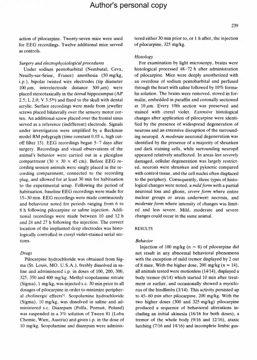
Fig. 1. Electroencephalographic recordings illustrating the sequence of alterations produced by intraperitoneal injection of pilocar- pine in mice. A: pre-drug control recordings. B: electroencephalographic correlates 2-5 min after injection of pilocarpine in the dose of 325 mg/kg demonstrating low-voltage fast activity in cortical recordings and a significant hippocampal theta rhythm. C and D: 10-15 min after injection of pilocarpine. High-voltage fast activity (C) and spikes superpose (D) over the hippocampal theta rhythm. Isolated low-voltage spikes are initially registered in hippocampal leads (D), while cortical recordings display no or minor changes. E: high-voltage spiking similar to that shown in panel D spreads to cortical leads 15-30 min after pilocarpine.
tatory automatisms (12/16 and 16/16) with salivation
(6/16 and 8/16). This type of preconvulsive behavior persisted for up to 5-10 min after injection and built up progressively into motor limbic seizures with up-
per extremity clonus, rearing, falling and intense sali- vation. During the period of motor limbic seizures
mice exhibited additionally stereotyped behaviors like repeated head twitches, grooming and rearing,
and occasionally jumping fits. Motor limbic seizures
commenced after 10-18 min (mean 17.89 + 7.64 min, n = 16, for 300 mg/kg; and mean 11.67 _ 3.26 min, n = 16, for 325 mg/kg), recurred every 5-10 min and reached a maximal occurrence of 6 -8 per 30 min. The period of limbic convulsive behavior lasted for up to 30-45 min after injection and rapidly devel- oped into a status epilepticus (mean 27.63 + 12.7, n = 12, for 325 mg/kg) which sometimes led to the death of the animal. With the dose of 325 mg/kg myoclonic seizures could rarely be observed; how-
ever, clonic-tonic seizures with death of the animal were observed in 4 out of 16 mice. With the dose of 350 mg/kg of pilocarpine the development of convul-
sive activity was rapid (mean 6.2 + 2.3 min, n -- 12) and the lethal toxicity in mice increased over 50% (7/12). With this dose, a short-lasting period of motor
limbic seizures rapidly developed into the status epi-
lepticus with generalized clonic-tonic convulsions which ended in death of the animals. At the dose of
400 mg/kg (n = 15) pilocarpine was generally lethal to mice (14/15). Pretreatment of mice with scopola- mine, 10 mg/kg (n = 10) or diazepam, 10 mg/kg (n -- 10) prevented the development of convulsive altera- tions produced by pilocarpine, 325 mg/kg. Animals which were sedated and stuporous after injection of diazepam, began to walk and explore following the injection of pilocarpine, but no typical behavioral al- terations produced by pilocarpine in drug-naive mice could be observed. Diazepam, 10 mg/kg (n = 8) re-
Author's personal copy

241
duced the convulsive activity when administered 1 h after pilocarpine, 325 mg/kg, while scopolamine, 10 mg/kg (n = 8) failed to disrupt pilocarpine-in- duced seizure activity and did not lessen the lethal toxicity of the drug (2/8).
Electroencephalography The nature of EEG alterations occurring after i.p.
application of pilocarpine in mice is illustrated in Figs. 1-3. Alterations produced by i.p. injections of pilocarpine in mice were dose- and time-dependent. Pilocarpine caused both interictal and ictal epilepti- form activity in the EEG. Typical EEG changes con- sisted of (i) a fast activity of low or high voltage, (ii) slow or fast high voltage spiking and (iii) fully devel- oped ictai activity. The sequence of behavioral alter- ations evoked by pilocarpine correlated well with the EEG changes produced by the drug. Immediately (2-5 min) after injection of pilocarpine in doses of
100-300 mg/kg, the normal background activity was replaced with low-voltage fast activity in the cortex (Fig. 1B), while significant theta rhythm (6-7 Hz) appeared in the hippocampus (Fig. 1B). By 5-10 min, high-voltage fast activity superposed over the hippocampal theta rhythm and isolated low-voltage spikes were registered initially in hippocampal leads, while cortical recordings displayed no or only minor changes (Fig. 1C, D). At this time a striking feature was that activity changes originated in the hippocam- pus and then rapidly spread to the cortex (Fig. 1D, E). In animals receiving low (100 mg/kg) and inter- mediate (200 mg/kg) doses of pilocarpine, low- or high-voltage fast activity and spiking, which ap- peared at variable frequencies and were initially re- stricted to the hippocampus, prevailed for at least 1-2 h after injection. Fully developed electrographic seizures did not appear with these doses. At the time of spiking activity with low and intermediate doses of
A H P C ~ - ; . " : . . . . . . . . . J . . . . . . . . t _ , . . ~ . . . . . . . . . . . . t . . . . . I ~ [ , . A . . . . . d, . . . . . . . . . . . . . .
I
B . , e . . . . . . . . . . . . . , . . . . . . i , i . . . . . . . . . . . . . . . . . . . . . . . .
I I ' ~ l l ~ l l h J / t u . , l . . . . . . . . . . , ....... L,..~ . . . . . ] , l j . t a . . . . . .
1 i s m ~ - - ~ - . . . . . . : . . . . . . . . . . . . . ~ . ' t 7 - : . z - 3
c I
1 !' I Iii II t 1"11 t lJ ~1
! , ! i 't "',I It l l!M I, ,I ,I '! , ,~ t¥ ' .Pc ~ i ~ . . . v
m I|
Fig. 2, Electrographic correlates of behavioral seizures produced by pilocarpine in mice. A: continuous recording demonstrating elec- trographic seizure registered in hippocampal leads 25 min after intraperitoneal injection of 325 mg~g of pilocarpine. The fast activity develops into high-voltage spiking resulting rapidly in an electrical seizure registered in the hippocampal lead (A), while cortical re- cording displays minor changes restricted exclusively to the appearance of the fast activity and isolated high-voltage spikes. During the time in which an electrical seizure occurs in the hippocampus, the animal remained frozen in a motionless staring spell. B: electrographic seizure observed 33 min after injection of pilocarpine. The waxing and waning of electrical seizure activity is highly synchronized in both hippocampal and cortical leads. The fast activity and isolated high-voltage spikes registered in both recordings preceded the de- velopment of a seizure. C: electrographic recordings illustrating the development of a seizure 40 min after injection of pilocarpine. The fast activity develops into prominent high-voltage spiking, which built up progressively into a seizure activity. Note a rapid evolu- tion as well as prolonged waning of the seizure activity in the hippocampal recording. D: electrographic recordings to illustrate clonic- tonic seizures observed 2-4 h after injection of 350 mg/kg pilocarpine.
Author's personal copy

242
C X
A H P C
t i i ,
B , i,, Ih ilh t!,l II=t tll! h, ii Ill, t bll,ll~ I l i l l t l l~ l I tl,Jtlt~l qdt~l~tN ! I, l i l t II1!t lOll!Ill,! 1,1! l l ' L= ,IJ' " ' ' ~ ~ ' i ' ; ' ' " ' ( , ~ ' " : ' , ' , ! : ~ , ,
l s
C i I ! , I I ~ I I I H P C L ~ I
C X ;]2LJ:; . . . . / . . . . . . I , l _ l l . . . . . - . . . . . . ; : . ~ 7 , . . . . . ; ~ _ ~ . . . . ~ . Z . , . l l ~
D H P C I , • flOlaV
Fig. 3. Electrographic recordings illustrating the sequence of alterations observed at longer delays after intraperitoneal injection of pi- locarpine in mice. A: electrographie correlates of interictal activity observed 2-4 h after injection of 325 mg/kg pilocarpine. Note fast activity and highly synchronized high-voltage spiking that occurs in all recordings. B: electrographic recordings to illustrate the electri- cal alterations observed 2-6 h after injection of 325 mg/kg pilocarpine during a status epilepticus. C: electrographic correlates 8 h af- ter injection of pilocarpine. High-voltage fast activity and prominent spiking still occurs in hippocampal lead, while cortical recording displays less pronounced changes. D: 24 h post-injection the EEG recordings are indistinguishable from pre-injection control activity, although isolated spikes are infrequently registered in hippocampal leads.
pilocarpine, animals remained motionless and often displayed mild limbic automatisms. By 24 h after pi- locarpine, 100 and 200 mg/kg, EEG recordings were indistinguishable from the pre-drug background ac- tivity. Doses of 300 mg/kg of pilocarpine or greater, reproducibly elicited a sequence of E E G alterations and concomitant seizure-like behaviors described with low and intermediate doses; however, the laten- cy period decreased and clonic or clonic-tonic motor convulsions (Fig. 2D) frequently appeared on in- creasing the dose. With higher doses of pilocarpine, 300, 325 and 350 mg/kg, typically, fully developed EEG seizures occurred, which started 10-15 min af- ter injection. A high-voltage fast activity and promi- nent high-voltage spiking usually preceded the devel- opment of a seizure (Fig. 2A-C) . Characteristically, these activity changes originated in the hippocampus
and built up progressively into an electrographic sei- zure registered in the hippocampal recordings (Fig. 2A), while cortical recordings displayed minor changes restricted to the appearance of the fast activ- ity and isolated spikes (Fig. 2A). Later, the electro- graphic seizures became highly synchronized in the hippocampus and cortex (Fig. 2B, C); however, fas- ter buildup, as well as a prolonged waning of the sei- zure activity in the hippocampal recordings, was re- producibly registered (Fig. 2C). The ictal periods lasted 1-2 min, recurred every 5-10 min and were followed by periods of depression of the electro- graphic activity. By 30-45 min paroxysmal activity, which progressively grew in duration and complexi- ty, resulted in a status epilepticus (Fig. 3B). This pat- tern of EEG changes lasted for 4 -6 h and was fol- lowed by a progressive normalization of the EEG ac-
Author's personal copy

tivity. By 8 h after the injection of pilocarpine,
300-350 mg/kg, high-voltage fast activity and promi-
nent spiking was often seen in hippocampal record- ings (Fig. 3C), while cortical recordings displayed
less pronounced changes. By 24 h after injection of
higher doses of pilocarpine, E E G recordings were in- distinguishable from pre-drug background activity,
although isolated high-voltage spikes were occa- sionally registered in the hippocampal leads (Fig. 3D).
The development of electrographic seizure activity
usually preceded the buildup of clinical seizures.
243
First electrographic seizures were usually restricted
to the hippocampus (Fig. 2A) and freezing of ani-
mals in a motionless staring spell coincided with this type of activity. Motor limbic seizures appeared
when electrical seizure activity became synchronized in the cortex and hippocampus (Fig. 2B, C). Head
twitches occurred infrequently after electrical sei- zures, and never preceded them. When seizure activ-
ity built up progressively and had became well devel- oped (Fig. 2C), the motor manifestation of l imbic
convulsions became more severe. With doses greater than 350 mg/kg seizures were accompanied by in-
A HPC .; , ;r~
B H~C
C H P C
D HPC
E
C X , ~ , * ' ~ , ~ f ' ~ ' .~: ~ . ,~7 . % ~ : ~ r ~ " 7 . ~ ~ , '~.:.~...~;~.- ~: v~-; -~ '~- - ' : . ' , :~ t ; : -L ." -~ '~ ' ' - ' . : -~- ' :b~
F HPC ~ = 5 0 p V
Fig. 4. Electrographic recordings illustrating the effect of scopolamine on the convulsant activity of pilocarpine in mice. Scopolamine in the dose of 10 mg/kg was injected subcutaneously 30 min prior to injection of pilocarpine (325 mg/kg, i.p.). A: pre-drug control re- cording. B: electrographic correlates 5-10 min after injection of scopolamine. A normal background activity is replaced with the high- voltage slow activity. No theta activity could be registered in hippocampal leads. C: 20 min after injection of pilocarpine in scopola- mine-pretreated mouse. Pilocarpine progressively decreases the voltage of the slow activity produced by scopolamine (B) resulting in the appearance of low-voltage activity in both leads (C), which is slower than that registered during control recordings (A) but faster than that observed after scopolamine alone (B). D and E: electrographic correlates 1-4 h after intraperitoneal injection of pilocarpine in scopolamine-pretreated mice. By 1 h after injection of pilocarpine, high-voltage slow activity alternates with and then superposes over low-voltage fast activity induced by the injection of the drug and prevails in hippocampal recordings up to 4 h post-injection (E). F: by 4-8 h after injection of pilocarpine the voltage of the slow activity gradually decreases (E) and is progressively replaced with fas- ter activity (F). By 24 h after injection of pilocarpine in scopolamine-pretreated mice all recordings are indistinguishable from pre- drug background activity (A).
Author's personal copy
244
A H PC ~ w . ~ , . ~ ' , w e , ~ ' ~ . ~ . . . ' ~ ' . ' ~ . ' . . ~ ' ~ 4 .
B HPC
C HPC
D HPC
E
e x
F HPC ~ . ~ ' , m , , • 5 0 p V
m l s
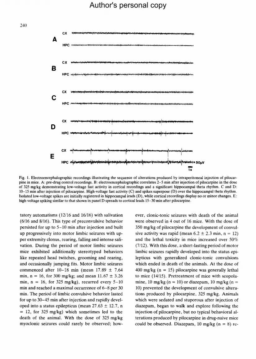
Fig. 5. Electrographic recordings illustrating the effect of diazepam on the convulsant activity of pilocarpine in mice. Diazepam in the dose of 10 mg/kg was injected intraperitoneally 30 min prior to systemic injection of 325 mg/kg of pilocarpine. A: pre-drug control re- cording. B: electrographic correlates 5-10 min after injection of diazepam. A normal background activity is replaced with the low-vol- tage fast activity which prevails in both leads. No theta activity could be registered in hippocampal recordings. C-E: electrographic correlates 15 min to 1 h after injection of pilocarpine in diazepam-pretreated mice. Injection of pilocarpine results in a short-lasting re- appearance of the theta activity in the hippocampal recordings (15 min after application of pilocarpine) (C). Low-voltage fast activity rapidly superposes over or alternates with the hippocampal theta activity within 30-60 min after pilocarpine and then predominates in hippocampal leads (D). F: by 8 h after injection of pilocarpine in diazepam-pretreated mice hippocampal and cortical recordings are indistinguishable from pre-drug background activity (A).
tense salivation, prominent cyanosis, conspicuous respiratory disturbances and death. Therefore elec- trophysiological analysis of effects produced by these doses of pilocarpine was not justified.
Pretreatment of mice with scopolamine, 10 mg/kg s.c., completely prevented the occurrence of convul- sive activity in the E E G produced by 325 mg/kg of pi- locarpine (Fig. 4). After injection of 10 mg/kg of sco- polamine the background activity was replaced with the high-voltage slow activity, while the hippocampal theta rhythm was instantaneously blocked (Fig. 4B). Injection of pilocarpine, 325 mg/kg, led to a decrease of the voltage of the slow activity resulting in the ap- pearance of low-voltage activity in both hippocampal
and cortical recordings (Fig. 4C). Characteristically, this type of low-voltage activity was slower than that registered during the pre-drug recordings (Fig. 4B). By 1 h after injection of pilocarpine low-voltage fast activity produced by the drug alternated with high- voltage slow activity and finally superseded the latter (Fig. 4C). This pattern of activity prevailed in hippo- campal recordings up to 4 h post-injection (Fig. 4D). The voltage of the slow activity gradually decreased by 4-8 h after 325 mg/kg pilocarpine and faster ac- tivity progressively replaced it (Fig. 4E). By 24 h af- ter injection of pilocarpine in scopolamine-pre- treated mice the E E G in all recordings returned to the pre-drug pattern (Fig. 4F). After the injection of
Author's personal copy

diazepam, 10 mg/kg, the background activity (Fig. 5A) was replaced with a low-voltage fast activ- ity which prevailed in both recordings (Fig. 5B). Fif- teen minutes after application of pilocarpine, 325 mg/kg, a short-lasting reappearance (up to 30-45 min) of the theta activity in the hippocampal recordings occurred (Fig. 5C). Low-voltage fast ac- tivity alternated with the former pattern of EEG and then rapidly replaced it (Fig. 5D, E). Eight hours af- ter application of pilocarpine in diazepam pretreated mice, no consistent qualitative differences between electrographic activity registered from both hippo- campus and cortex of these and drug-naive animals were found (Fig. 5F).
Neuropathology Examination of frontal forebrain sections with
light microscopy revealed widespread brain damage in mice treated systemically with pilocarpine. The de- velopment of neuropathological alterations pro- duced by pilocarpine in mice was related to the dose of the drug. Intraperitoneal application of the low dose of pilocarpine, 100 mg/kg, which elicited slight behavioral and EEG alterations did not result in de- tectable pathological changes within the brain. With intermediate dose of pilocarpine, 200 mg/kg, mild neuropathological alterations, largely confined to pi- riform cortex and olfactory nucleus, could be en- countered. When the three highest doses of pilocar- pine, 300, 325 and 350 mg/kg, which reproducibly elicited consistent and long-lasting behavioral and electrographic changes including motor limbic sei- zures and limbic status epilepticus, were used, exten- sive neuropathological alterations were found within the olfactory cortex, thalamus, amygdaloid complex, hippocampal formation, neocortex and substantia ni- gra. The anatomical terminology used in the present paper conforms to that used by Kovac and Denk 23 and Montemurro and Dukelow 36. With pilocarpine, 325 mg/kg, extensive degeneration of neurons and complete disruption of the surrounding neuropil were found within the olfactory system, i.e. piriform cortex, anterior olfactory nucleus and periamygda- loid cortex. The piriform cortex appeared severely swollen and edematous. The majority of cells within the piriform cortex were dark and shrunken (Fig. 6F) and the surrounding neuropil appeared frequently fragmented. Severely shrunken and darkened neu-
245
rons could be observed in the anterior olfactory nu-
cleus and the entorhinal cortex (Fig. 6G), as well. The olfactory tubercle was less sensitive to pilocar- pine and neuronal degeneration has not been found in this area. The amygdaloid nuclear complex ap- peared to be particularly sensitive to pilocarpine (Fig. 6I). Degeneration and disapparance of virtual- ly all neurons with prominent edema, fragmentation and disruption of the neuropil were observed within this area (Fig. 6I). Within the hippocampal forma- tion prominent neuronal degeneration was observed in the regio superior of the dorsal hippocampus (Fig. 6C). Destruction of almost all pyramidal cells was identified in the regio inferior (Fig. 6B) as well as in the area dentata. Within the area dentata de- generated neurons were usually observed in the hilus and at the granule cell-hilar border. Ventral hippo- campus displayed extensive damage as well, includ- ing degeneration of pyramidal neurons within areas in continuity with the olfactory cortices. The ventral part of the subiculum and lateral septal nucleus were also consistently affected (Table I). Within the thala- mus, most consistently affected were anterior, later- al, dorsomedial, reuniens, rhomboideus, paraven- tricular and parathenial nuclei; however, the extent of damage in the thalamus varied among mice treat- ed with 325 mg/kg of pilocarpine (Table I). Exten- sive neuronal degeneration with almost complete dis- ruption of the neuropil usually occurred in the lateral (Fig. 6D) and dorsomedial thalamic nuclei. Ventral, ventromedial and posterior thalamic nuclei were less severely damaged. The lateral geniculate nucleus was extensively damaged as well (Table I). Within the neocortex, severe damage was found in the outer molecular layers, but also deep layers could be af- fected. The areas most consistently affected included areas dorsal to the rhinal sulcus, cingulate cortex (Fig. 6H), portions of the perirhinal areas and soma- tomotor cortices. Shrunken and dark staining cells were found within the substantia nigra pars ventralis and dorsalis (Table I). Neuropathological altera- tions were never observed in the dorsal part of the striatum, while the ventral part was consistently af- fected. Little neuronal degeneration was encoun- tered within the hypothalamus. The typical pattern and distribution of the brain damage produced by pi- locarpine, 325 mg/kg, is illustrated in Figs. 6 and 7. The animals pretreated with either scopolamine,
Author's personal copy

246
A
C
~ j ~!'hl
- ~ ~!~i~ "
Author's personal copy

247
10 mg/kg, or diazepam, 10 mg/kg, 30 min before the
application of 325 mg/kg pilocarpine, displayed no
epileptiform activity, and neuropathological altera- tions were not detected in their brains.
DISCUSSION
Three major conclusions can be drawn from the
experiments presented here: (i) systemically applied pilocarpine in mice produced profound convulsive
activity preferentially confined to limbic structures;
(ii) a major component of this activity is muscarinic; and (iii) the seizures induced by the drug may result
in a widespread damage to forebrain structures re-
sembling that frequently observed in brains of human epileptics 12.
Electroencephalographic observations of the epi- leptic response to pilocarpine in mice provide evi-
dence for the involvement of the hippocampus in the
buildup of epileptiform activity following the drug
administration and emphasize that hippocampus has a particularly low threshold for pilocarpine-induced
convulsions. This finding is related to the hypothesis
that there exists a powerful positive-feedback mech-
anism within discrete regions of the hippocampus and septum, and the assumption that its activation allows
the development of long-term changes in excitability within the hippocampal subfields 2,8,9,24. Further-
more, anatomical studies emphasize the high degree
of interconnections between hippocampus, entorhin- al cortex, amygdala and other parts of the limbic
forebrain, which may facilitate the rapid spread of
seizures originating in one part of this system to all otherslt,57.
From the results of neuropathological analysis of brains from mice treated with pilocarpine it is appar- ent that pyramidal cells of the hippocampal forma-
tion, as well as of the piriform cortex and the amyg- dala, are particularly sensitive to pilocarpine neuro-
toxicity. The topography of the hippocampal damage in mice resembles that described after systemic appli- cation of either pilocarpine19,51, 55, arecoline 19 or
kainic acidT, 26,45 in rats, and that reported in mice
treated intracerebroventricularly with the latter drug t334. This finding may have interesting implica-
tions for the elucidation of the causative mechanism of pathological aspects of seizures produced by pilo- carpine in mice. Firstly, the conformity of hippocam-
pal pathology in mice to that reported in other mod- els of temporal lobe epilepsy 5,7,26,35,37,45,49,50,51,55 es-
t ab l i shed in different animal species (rat, cat, mon-
key) implicates similar, and very likely seizure-re- lated, mechanisms of neuronal damage. Indeed,
from the studies with pilocarpine in rats, it is appar-
ent that an ischemic factor does not seem to be of ma-
jor importance for the hippocampal damage ob- served in these experiments. Specifically in the rat,
the regio superior of the dorsal hippocampus, a re- gion which typically undergoes extensive neuronal degeneration following experimental ischemia, is
considerably less sensitive to pilocarpine neurotoxic- ity 51.
Secondly, the topography of brain damage follow-
ing pilocarpine in mice resembled that observed after
Fig. 6. Neuropathological alterations in the mouse brain produced by systemic administration of pilocarpine. Cresyl violet stain. Sur- vival time: 48-72 h. A: photomicrograph of the regio inferior of the dorsal hippocampus of a drug-naive control mouse. B: representa- tive section illustrating the destruction of the regio inferior of the hippocampus of the mouse treated i.p. with 300 mg/kg of pilocar- pine. Note the nearly total disruption of the cytoarchitecture of the pyramidal layer with extensive cellular degeneration and glial infil- tration. Shrunken and dark staining cells are visible throughout the pyramidal layer. Some changed cells contain narrowings which lead to fragmentation and disintegration. Dark staining and severely shrunken cells are interspersed among normal appearing pyrami- dal neurons. C: high-power photomicrograph demonstrating the type of tissue reaction observed within the regio superior of the dorsal hippocampus of the mouse treated i.p. with 325 mg/kg of pilocarpine. Severely shrunken and darkened pyknotic pyramidal neurons are prominent in the pyramidal layer. D: photomicrograph illustrating the destruction of the lateral thalamic nucleus after i.p. injec- tion of 300 mg/kg of pilocarpine. Note the nearly total disruption of the cytoarchitecture of the lateral thalamus. Neurons stain darkly and appear shrunken. E: photomicrograph of a portion of the piriform cortex of a drug-naive control mouse. F: photomicrograph illus- trating the destruction of the posterior piriform cortex of the mouse injected i.p. with 325 mg/kg of pilocarpine. Shrunken and dark- ened neuronal somata are widely interspersed among normal appearing cells and are present throughout all layers of the piriform cor- tex. G: entorhinal cortex in the mouse injected systemically with 300 mg/kg of pilocarpine. The cells become dark and severely shrun- ken. H: cingulate cortex in the mouse injected with 325 mg/kg of pilocarpine. Shrunken and darkened pyknotic neurons are present in the outer layers of the cingulate cortex. Dark staining cells are present within the deep layers as well. I: photomicrograph of a portion of the amygdaloid complex of the mouse injected with 325 mg/kg of pilocarpine. Note the nearly total destruction of the neuronal pop- ulation within the amygdala accompanied by edema and disruption of the neuropil. A-I: × 196. (Publisher's reduction factor: 0.75.)
Author's personal copy

248
ua
"~ ~r~ ~ ¢~
E
~ +~ +
• p_. @
+ + + + ÷ + + + + + + + + + ÷ ÷ + +
+ + + I I + + I + I + +
÷ + +
+ +
+ + ÷ +
+ + +
"~ 4- +
+ ~ + +
4- 4-
+ + + + + + + + + + + 4 - + + + + + + +
+ + + 4- + q- q- + + + + + + +
+ -t- + + 4- + + 4- + + p + p + +
+ + + + + + + + + + + + + + + + + + + + + + + + + + + ÷
+ + + + + + + + ~ + + + + + + + + + + + ÷ + + + + + + + +
+ + + + + + + +
~ + + + + + + I + +
+ + + + + + + + + ~ + + + + + + + + + + + + + + + +
L~
I I I J ~ l l l l l l l
+ + + + + + + + + +
+ + + + + + + + + + + +
+ + + + + + + + + + + + + + + + + + + + + + + + + + + + + + + +
I + + + +
+ + + + + + + + + + + + +
+ + + + + + + + + + + ÷ + + + + ~ + + ÷ + ÷
+ + + + + + + + + + + + + + + + + + + + + + + + +
+ + - I - + + 4-
+ + + + + 4 - 3 - + + + + +
+ + + + + + ~ + + + + + + + + + +
+ + + + + + + + + + + +
+ + + + • + + + + + + + + + + + + + + + + + + +
i
Author's personal copy

249
tpv tpv
A 3 . 3 v ) m
~ 1 I I'~_.~'1~1"~ m ~ g m
A1.5 cpf Fig. 7. Schematic reconstruction of the distribution of neuropathological alterations in the mouse brain after intraperitoneal adminis- tration of 325 mg/kg pilocarpine relying on atlases of Kovac and Denk e3 and Montemurro and Dukelow 36. The lesions were estimated in cresyl violet-stained serial sections of the entire brain. Survival time: 48-72 h. Dark areas represent nearly total destruction of neu- rons accompanied by edema and disruption of the neuropil. Hatched areas represent moderate cellular destruction with partial neuro- nal loss apparent as numerous shrunken and dark staining neurons. Dotted areas represent mild cellular destruction with more isola- ted degenerating neurons which were found in several scattered locations. Abbreviations for anatomical structures: am, amygdaloid nuclear complex; ce, entorhinal cortex; cpf, piriform cortex; gm, medial geniculate nucleus; s, subiculum; sn, substantia nigra; tan, an- terior thalamic nucleus; tl, lateral thalamic nucleus; tmd, dorsomedial thalamic nucleus; tpo, posterior thalamic nucleus; tpt, pare nte- nial thalamic nucleus; tpv, paraventricular thalamic nucleus; trh, rhomboideus thalamic nucleus; tv, ventral thalamic nucleus.
either intraamygdaloid37, 55 or intrahippocampa152,55
injections of cholinomimetics in rats which induced behavioral and electrographic occurrence of limbic seizures and no symptoms of respiratory distress. The destruction of the regio superior of the dorsal hippocampus is rather characteristic for the hippo- campal pathology found after these treatment37,55.
Lemercier et al. 2s very recently formulated a ten- tative hypothesis that both seizure-related and is- chemic factors may contribute to the neuropatholog- ical effects of acute and chronic poisoning of rats with soman, an organophosphorous compound with irre- versible anticholinesterase activity. These authors favored a predominant role for an ischemic factor, related to prolonged nicotinic cholinergic stimula- tion, as a tentative cause of the brain damage pro- duced by soman 25. In contrast, McDonough et al. 30, on the basis of the metabolic activation of forebrain
structures in rats submitted to sustained seizures pro- duced by soman, suggest that the brain damage in- duced by the organophosphate is linked to the pres-
ence of seizure activity per se. Whatever the underly- ing mechanism of brain damage produced by soman intoxication may be, the EEG monitoring and behav- ioral observation of mice and rats 5a after pilocarpine
treatment suggests that a major link between pro- longed seizure activity within the areas undergoing extensive neuropathological changes and distribu- tion of the brain damage exists.
The topography of neuropathological alterations detected in brains of mice treated with pilocarpine re- sembled the pattern of metabolic activation of fore- brain structures following prolonged hippocampal seizures 57. Experimental evidence provided by Wat- son et al. 57 indicates that the generation of the dorsal hippocampal seizures with the prolonged electrical
Author's personal copy

250
stimulation of the granule cells of the dentate gyrus in the rat results in the metabolic activation bilaterally within the amygdala, piriform and entorhinal cortex, .claustrum, olfactory tubercle, cingulate and prefron- tal cortex, mediodorsal, reuniens and ventral thalam- ic nuclei as well as within the substantia nigra and substantia innominata. The topography of metabolic activation resulting from the prolonged dorsal hippo- campal seizures may be understood in terms of a pro- gressive development of secondary epileptic loci, the ventral subiculum being regarded to serve as a crit- ical region in the propagation of afterdischarges from, the hippocampus to the amygdala, lateral septum and entorhinal cortex 22. A consideration of the com- mitment of multiple (secondary) seizure loci in the propagation of afterdischarges within the limbic cir- cuitry suggests a relevant role for gradual recruit- ment of sensitive regions as a function of the duration of seizures and its severity in the development of electrographic and pathological aspects of temporal lobe epilepsy 11. The importance of anatomical path- ways in the propagation of abnormal electrical activ- ity is demonstrated not only by the similarities in the topographical distribution of a brain damage linked to different mechanisms which interact in the initia- tion of limbic seizure activity 5~7,10,11,26,35,37,38,45,46,
50.51,54,55, but in the limitation of the metabolic activa-
tion to the first synapse, when the stimulation of the specific area did not elicit seizures57, as well. This ob-
servation does not rule out the role of cholinergic pathways in the pathogenesis of the epileptic brain damage, but it indicates that sustained stimulation of muscarinic cholinergic sensitive regions elsewhere within the limbic system, e.g. within the projection fields of the septum, nucleus of diagonal band and substantia innominatalS, may result in the recruit- ment of multiple epileptic loci, the situation being re- flected in a progressive buildup of behavioral and electrographic seizures finally resulting in a status ep- ilepticus. This hypothesis indicates that early stages of the development of limbic seizure activity pro- duced by pilocarpine in mice are relatively specific and dependent on the stimulation of cholinoceptive parts of the limbic system, but it does not necessarily mean that this is the only mechanism responsible for the final consequences of cholinergic stimulation, particularly those observed during advanced stages of the convulsive activity. Such a hypothesis predicts
the role for other excitatory mechanisms in the pathogenesis of limbic seizures produced by pilocar- pine, regardless of where those mechanisms func- tion. In fact, it is well known that ACh mediates long- term increases in excitability within the hippocampus and neocortex: 4.8.9.24 and may account for increases
in the effectiveness of other excitatory inputs to these structuresS,4~. It seems likely that this mechanism may be responsible for either lowering the seizure threshold or facilitating triggering seizure dis- charges, and could develop a higher susceptibility to recurrent seizuresS.
The pivotal role for cholinergic pathways in deli- miting the extent and topography of the epileptic brain damage is demonstrated by both the localiza- tion of destructions within the projection fields of the cholinergic forebrain system, prevention of seizure activity and brain damage with scopolamine, and by the finding that chemical stimulation of cholinergic structures of the basal forebrain produced sustained epileptiform activity along their efferent pathways 32 and epileptic brain damage which parallels the densi- ty of cholinergic innervation from these nuclei32.59. Evidence from electrophysiological experiments rein-
forces the assumption that ACh may be involved in certain neurodegenerative processes within the brain. Muscarinic cholinergic excitation within the mammalian CNS occurs as a result of a reduced vol- tage-dependent and Ca-dependent K + conductance, and is mediated by voltage-dependent Ca 2÷ and Na + conductances 2,3. Such an action favors inward move- ments of Ca2+ and Na +, an effect which is primarily responsible for a prolonged membrane depolariza- tion and might be relevant for the epileptic cell death 33,34. In fact, prolonged seizure activity shifted the balance between inward and outward Ca 2+ cur- rents, resulting in an excess accumulation of Ca 2+
within the ce1127,42; the nature of calcium-dependent
cell death is based on the failure of the cell to extrude or sequester Ca 2+ in the mitochondria 33,34.
Thus, two hypotheses may be offered for the neu- rotoxicity of pilocarpine and other muscarinic cholin- ergic agonists: either that the drug directly destroys neurons by the changes in membrane conductance resulting in a tonic depolarization or by inducing in- tense epileptic discharges. The results of our experi- ments are consistent with both of these hypotheses, but do not conclusively prove any specific mechanism
Author's personal copy

for pilocarpine-induced brain damage. Although the distribution of the brain damage in mice reflects well the areas within the projection fields of the basal forebrain cholinergic system 15, it does not allow us to specify the cholinergic mechanisms as a factor indica- tive of and responsible for the pathogenesis of the neuronal degeneration.
The data on the abortive effect of scopolamine against pilocarpine-induced seizures reveal a neuro- transmitter specificity in the generation of the epilep- tic response to pilocarpine. However, if the choliner- gic overstimulation was a sufficient condition for the neurotoxicity of pilocarpine, then the muscarinic cholinergic antagonist scopolamine should be able to prevent the development of pilocarpine-induced sei- zure activity as well as to stop seizures after they have emerged and progressed into the status epilepticus. From the observations in rats, it is evident that scopo- lamine prevented the buildup of seizures, but failed to disrupt pilocarpine-induced status epilepticus 53. In mice, scopolamine given 1 h after injection of 325 mg/kg of pilocarpine, when the status epilepticus has been well developed, failed to disrupt the sei- zures and reduced neither the severity of them nor the lethal toxicity of pilocarpine. Whatever the basic mechanism of the brain damage produced by pilocar- pine may be, these experiments show that cholinergic circuits are very important for the generation of sus- tained seizure activity, and emphasize the relevance of seizure activity in pathological sequelae of pilocar- pine action.
From the results of the intraamygdaloid injec- tions of cholinomimetics37. 55, kainic acid 5, folates 38, GABA antagonists 50 and morphine 54 in rats, which revealed that initiation of abnormal electrographic activity within the limbic circuits linked to divergent receptor mechanisms resulted in very similar brain pathology, it seems evident (i) that epileptic brain damage in experimental animals involves the same mechanism regarding the development, and (ii) that one neurotransmitter system cannot be considered pivotal in the genesis of the seizures and their patho- logical sequelae.
Diazepam specifically prevented the seizure activ- ity and brain pathology reproducibly observed in ani- mal models of limbic seizures 5-7,52. Diazepam pre- vented the development of a sequence of seizure-re- lated behavioral and EEG alterations, and wide-
251
spread damage to forebrain structures produced by pilocarpine in mice, also. The preferential effect of
diazepam on the limbic type of seizure activity pro- duced by pilocarpine fits well with the potency of the drug in inhibiting the limbic seizure activity and brain damage produced by both intraamygdaloid and sys- temic injections of kainic acid in rats 5,6. The mecha- nism of action of benzodiazepines within the CNS is currently interpreted in terms of an enhancement of the GABA-mediated synaptic inhibition TM. Autora- diographic studies have shown that hippocampus, amygdala and cerebral cortex are particularly rich in GABA and benzodiazepine receptors 18. Further- more, there is evidence indicating that the anticon- vulsant activity of benzodiazepines is specifically confined to the limbic system and dependent on the intactness of both the hippocampus 13,14 and the amygdala 20. Accordingly, one would expect that di- azepam potentiates the effects of endogenous GABA in affected brain areas which counteracts the neurotoxic activity of pilocarpine. The finding of Mc- Geer et al. 32 and Wong et al. 59 on the loss in both GAD activity and [3H]quinuclidynylbenzilate bind- ing within the amygdala, piriform and frontal cortex following the chemical stimulation of the substantia innominata with folic acid makes this hypothesis like- ly.
In summary, the pharmacological demonstration of a potent convulsant and brain damaging action of pilocarpine in mice supports and extends the propo- sal of usefulness of this treatment as a model for iden- tifying drugs preferentially active in the treatment of temporal lobe epilepsy. The relevance of this model is stressed by a broad margin of safety between sei- zures and mortality. Further evaluation of this hy- pothesis will depend on the results of studies specif- ically designed to find out whether drugs such as car- bamazepine, phenytoin, valproate and clonazepam affect the development of behavioral, electroen- cephalographic and neuropathological alterations produced by pilocarpine in mice.
ACKNOWLEDGEMENTS
The authors wish to express their gratitude to Dr. John W. Olney for critically reading the manuscript. This work was supported by grants from FAPESP and CNPq of Brazil to E.A.C., Z.A.B. and L.M.M.
Author's personal copy

252
REFERENCES
1 Belluzzi, J. D. and Grossman. S. P., Avoidance learning: long-lasting deficits after temporal lobe seizure, Science, 166 (1969) 1435-1437.
2 Benardo, L. S. and Prince, D. A., Cholinergic excitation of mammalian hippocampal pyramidal cells, Brain Research, 249 (1982) 315-331.
3 Benardo. L. S. and Prince, D. A., Ionic mechanisms of cho- linergic excitation in mammalian hippocampal pyramidal cells, Brain Research, 249 {1982) 333-344.
4 Ben-Aft, Y., Krnjevi6, K., Reinhardt, W. and Ropert, N., lntracellular observations on the disinhibitory action of acetylcholine in the hippocampus, Neuroscience, 6 (1981) 2475-2484.
5 Ben-Aft, Y., Tremblay, E. and Ottersen, O. P., Injections of kainic acid into the amygdaloid complex of the rat: an electrographic, clinical and histological study in relation to the pathology of epilepsy, Neuroscience, 5 (1980) 515-528.
6 Ben-Aft, Y., Tremblay, E., Ottersen, O. P. and Meldrum, B. S., The role of epileptic activity in hippocampal and ~re- mote' cerebral lesions induced by kainic acid, Brain Re- search, 191 (1980) 79-97.
7 Ben-Aft, Y., Tremblay, E., Riche, D., Ghilini, G. and Na- quet, R., Electrographic, clinical and pathological altera- tions following systemic administration of kainic acid, bicu- culline or pentetrazole: metabolic mapping using the de- oxyglucose method with special reference to the pathology of epilepsy, Neuroscience, 6 ( 1981 ) 1361 - 1391.
8 Burchfiel, J. L., Duchowny, M. S. and Duffy, F. H., Neu- ronal supersensitivity to acetylcholine induced by kindling in the rat hippocampus, Science, 204 (1979) 1096-1098.
9 Cole, A. E. and Nicoll, R. A., Acetylcholine mediates a slow synaptic potential in hippocampal pyramidal cells, Sci- ence, 221 (1983) 1299-1301.
10 Collins, R. C., Lothman, E. W. and Olney, J. W.. Status epilepticus in the limbic system: biochemical and patholog- ical changes, Advanc. Neurol., 34 (1983) 277-288.
11 Collins. R. C., Tearse, R. G. and Lothman, E. W., Func- tional anatomy of limbic seizures: focal discharges from me- dial entorhinal cortex in rat, Brain Research, 280 (1983) 25-40.
12 Corsellis, J. A. N. and Meldrum, B. S., Epilepsy. In W. Blackwood and J. A. N. Corsellis (Eds.), Greenfield's Neu- ropathology, Arnold, London, 1976, pp. 771-793.
13 Czuczwar, S. J., Turski, L. and Kleinrok, Z., Anticonvul- sant action of phenobarbital, diazepam, carbamazepine, and diphenylhydantoin in the electroshock test in mice after lesion of hippocampal pyramidal cells with intracerebro- vcntricular kainic acid, Epilepsia, 23 (1982) 377-382.
14 Czuczwar, S. J., Turski, L., Turski, W. and Kleinrok, Z,, Effects of some antiepileptic drugs in pentetrazol-induced convulsions in mice lesioned with kainic acid, Epilepsia, 22 (1981) 407-414.
15 Fibiger, H. C., The organization and some projections of cholinergic neurons of the mammalian forebrain, Brain Res. Rev., 4 (1982) 327-388.
16 Girgis, M., Neostygmine activated epileptiform discharge in the amygdala: electrographic-behavioural correlations, Epilepsia, 19 (1978) 521-530.
17 Grossman, S. P., Chemically induced epileptiform seizures in the cat, Science, 142 (1963) 409-411.
18 Haefely, W., Pieri, L., Polc, P. and Schaffner, R., General pharmacology and neuropharmacology of benzodiazepine
dcrivatives. In F. Hoffmeistcr and G. Stille (Eds.), Psycho- tropic Agents. Handbook of Experimental Pharmacology Vol. 55/I1, Springer, Berlin, Heidelberg, New York, 1981, pp. 13-262.
19 Honchar. M. P., Olney, J. W. and Sherman, W. R., Sys- temic cholinergic agents induce seizures and brain damage in lithium-treated rats, Science, 220 (1983) 323-325.
20 Kish, S. J., Sperk, G. and Hornykiewicz, O., Alterations in benzodiazepine and GABA receptor binding in rat brain following systemic injection of kainic acid, Neuropharma- cology, 22 (1983) 1303-1309.
21 Kleinrok, Z., Czuczwar, S. J. and L. Turski, Prevention of kainic acid-induced seizure-like activity by antiepileptic drugs. Pol. J. Pharmacol. Pharm., 32 (1980) 261-264.
22 Kliot, M. and Poletti, C. E.. Hippocampal afterdischarges: differential spread of activity shown by the taC-deoxyglu- cose technique, Science, 204 (1979) 641-643.
23 Kovac, W. and Denk, H., Der Hirnstamm der Maus. Topo- graphie, Cytoarchitektonik und C,vtologie, Springer, Wien, New York, 1968.
24 Krnjevi6, K. and Ropert, N., Electrophysiological and pharmacological characteristics of facilitation of hippocam- pal population spikes by stimulation of the medial septum, Neuroscience, 7 (1982) 2165-2183.
25 Lemercier. G., Carpentier, P., Sentenac-Roumanou, H. and Morelis, P., Histological and histochemical changes in the central nervous system of the rat poisoned by an irre- versible anticholinesterase organophosphorous compound, Acta Neuropathol. (Berl.), 61 (I983) 123-129.
26 Lothman, E. W. and Collins, R. C., Kainic acid induced limbic seizures: metabolic, behavioral, electroencephalo- graphic and neuropathological correlates, Brain Research, 218 (1981) 299-318.
27 Lux, H. D. and Heinemann, U., Ionic changes during ex- perimentally induced seizure activity. In W. A. Cobb and H. Van Duijn (Eds.), Contemporary Clinical Neurophysi- ology, EEG Suppl. 34, Elsevier, Amsterdam, 1978, pp. 289-297.
28 MacLean, P. D. and Delgado, J. M. R., Electrical and chemical stimulation of frontotemporal portion of limbic system in the waking animal, Electroenceph. clin. Neuro- physiol., 5 (1953) 91-100.
29 McCann, K., Cain, D. and Philbrick, D., Facilitation of kindled seizures in rats fed choline-supplemented diets, Soc. Neurosci. Abstr., 7 (1983) 586.
30 McDonough, J. H., Hackley, B. E., Cross, R., Samson, F. and Nelson, S., Brain regional glucose use during soman- induced seizures, Neurotoxicology, 4 (1983) 203-210.
31 McGeer, P. L. and McGeer, E. G., Cholinergic mecha- nisms in central disorders. In G. C. Palmer (Ed.), Neuro- pharmacology of Central Nervous System and Behavioral Disorders, Academic Press, New York, 1981, pp. 253-289.
32 McGeer, P. L., McGeer, E. G. and Nagai, T., GABAergic and cholinergic indices in various regions of rat brain after intracerebral injections of folic acid, Brain Research, 260 (1983) 107-116.
33 Meldrum, B. S., Metabolic factors during prolonged sei- zures and their relation to nerve cell death, Advanc. Neu- rol., 34 (1983) 261-275.
34 Meldrum, B. S., Griffiths, T. and Evans, M., Hypoxia and neuronal hyperexcitability: a clue to mechanisms of brain protection. In A. Wauquier, M. Borgers and W. K. Amery (Eds.), Protection of Tissues Against Hypoxia, Janssen Re- s'earch Foundation Series, Vol. 7. Elsevier, Amsterdam,
Author's personal copy

1982, pp. 275-286. 35 M6nini, C., Meldrum, B. S., Riche, D., Silva-Comte, C.
and Stutzmann, J. M., Sustained limbic seizures induced by intra-amygdaloid kainic acid in the baboon: symptomatolo- gy and neuropathological consequences, Ann. Neurol., 8 (1980) 501-509.
36 Montemurro, D. G. and Dukelow, R. H., A Stereotaxic At- las of the Diencephalon and Related Structures of the Mouse, Futura, Mount Kisco, NY, 1972.
37 Olney, J. W., deGubareff, T. and Labruyere, J., Seizure- related brain damage induced by cholinergic agents, Nature (Lond.), 301 (1983) 520-522.
38 Olney, J. W., Fuller, T. A. and deGubareff, T., Kainate- like neurotoxicity of folates, Nature (Lond.), 292 (1981) 165-167.
39 Olney, J. W., Rhee, V. and Ho, O. L., Kainic acid: a pow- erful neurotoxic analogue of glutamate, Brain Research, 77 (1974) 507-512.
40 Paterson, S. L. and Albertson, T. E., Neurotransmitter and neuromodulator function in the kindled seizure and state, Prog. Neurobiol., 19 (1982) 237-270.
41 Prince, D. A., Connors, B. W. and Benardo, L. S., Mecha- nisms underlying interictal-ictal transitions, Advanc. Neu- rol., 34 (1983) 177-187.
42 Pumain, R., Kurcewicz, I. and Louvel, J., Fast extracellu- lar calcium transients: involvement in epileptic precesses, Science, 222 (1983) 177-179.
43 Purpura, D. P., Penry, J. K., Tower, D., Woodbury, D. M. and Walter, R., Experimental Models of Epilepsy, Raven Press, New York, 1972.
44 Risch, S. C., Cohen, R. M., Janowsky, D. S., Kalin, N. H. and Murphy, D. L., Mood and behavioral effects of physo- stigmine on humans are accompanied by elevations in plas- ma fl-endorphin and cortisol, Science, 209 (1980) 1545-1546.
45 Schwob, J. E., Fuller, T., Price, J. L. and Olney, J. W., Widespread patterns of neuronal damage following system- ic or intracerebral injections of kainic acid: a histological study, Neuroscience, 5 (1980) 991-1014.
46 Siesj6, B. K., Cell damage in the brain: a speculative syn- thesis, J. cereb. Blood Flow Metab., 1 (1981) 155-185.
47 Snead, O. C., On the sacred disease: the neurochemistry of epilepsy, Int. Rev. Neurobiol., 24 (1983) 93-180.
48 Squire, L. R. and Davis, H. P., The pharmacology of mem- ory: a neurobiological perspective, Ann. Rev. Pharmacol., 21 (1981) 323-356.
253
49' Tanaka, T., Kaijama, M., Daita, G., Ohgami, S., Yonema- su, Y. and Riche, D., Electroclinical features of kainic acid- induced status epilepticus in freely moving cats. Microin- jection into the dorsal hippocampus, Electroenceph. clin. Neurophysiol., 54 (1982) 288-300.
50 Turski, W. A., Bortolotto, Z. A., Mello, L. M. and Caval- heiro, E. A., Seizure-related brain damage induced by intraamygdaloid GABA antagonists in rats, Naunyn- Schmiedeberg's Arch. Pharmacol., 325 (1984) 77R.
51 Turski, W. A., Cavalheiro, E. A., Schwarz, M., Czuczwar, S. J., Kleinrok, Z. and Turski, L., Limbic seizures pro- duced by pilocarpine in rats: behavioural, electroencepha- lographic and neuropathological study, Behav. Brain Res., 9 (1983) 315-336.
52 Turski, W. A., Cavalheiro, E. A., Turski, L. and Kleinrok, Z., Intrahippocampal bethanechol in rats: behavioural, electroencephalographic and neuropathological correlates, Behav. Brain Res., 7 (1983) 361-370.
53 Turski, W. A., Czuczwar, S. J., Cavalheiro, E. A., Turski, L. and Kleinrok, Z., Acute and long-term effects of system- ic pilocarpine in rats: spontaneous recurrent seizures as a possible model of temporal lobe epilepsy, Naunyn-Sch- miedeberg's Arch. Pharmacol., 324 (1983) 25R.
54 Turski, W. A., Czuczwar, S. J., Kleinrok, Z., Schwarz, M. and Turski, L., Intraamygdaloid morphine produces sei- zures and brain damage in rats, Life Sci., 33 Suppl. 1 (1983) 615-618.
55 Turski, W. A., Czuczwar, S. J., Kleinrok, Z. and Turski, L., Cholinomimetics produce seizures and brain damage in rats, Experientia, 39 (1983) 1408-1411.
56 Wasterlain, C. G., Morin, A. M. and Jonec, V., Interac- tions between chemical and electrical kindling of the rat amygdala, Brain Research, 247 (1982) 341-346.
57 Watson, R. E., Edinger, H. M. and Siegel, A., A [14C]2-de- oxyglucose analysis of the functional neural pathways of the limbic forebrain in the rat. III. The hippocampal formation, Brain Res. Rev., 5 (1983) 133-176.
58 Whitehouse, P. J., Price, D. L., Struble, R. Gz., Clark, A. W., Coyle, J. T. and DeLong, M. R., Alzheimer's disease and senile dementia: loss of neurons in the basal forebrain, Science, 215 (1982) 1237-1239.
59 Wong, P. T.-H., McGeer, E. G., Singh, K. and McGeer, P. L., Changes in muscarinic binding in distant brain regions showing neuronal losses after folic acid injections into the substantia innominata, J. Neurochem., 40 (1983) 1754-1757.
Author's personal copy