prolonged febrile seizures impair synaptic plasticity and alter
TRANSCRIPT
Int. J. Mol. Sci. 2022, 23, 12224. https://doi.org/10.3390/ijms232012224 www.mdpi.com/journal/ijms
Article
Prolonged Febrile Seizures Impair Synaptic Plasticity and Alter
Developmental Pattern of Glial Fibrillary Acidic Protein
(GFAP)-Immunoreactive Astrocytes in the Hippocampus of
Young Rats
Alexandra V. Griflyuk, Tatyana Y. Postnikova and Aleksey V. Zaitsev *
Sechenov Institute of Evolutionary Physiology and Biochemistry of RAS, 44, Toreza Prospekt,
194223 Saint Petersburg, Russia
* Correspondence: [email protected]
Abstract: Prolonged neonatal febrile seizures (FSs) often lead to cognitive decline and increased risk
of psychopathology in adulthood. However, the neurobiological mechanisms underlying the long-
term adverse effects of FSs remain unclear. In this study, we exposed rat pups to hyperthermia and
induced FSs lasting at least 15 min. We investigated the short-term (one day) and delayed (11–13
and 41–45 days) effects of FSs on some parameters of morphological and functional maturation in
the hippocampus. We noticed that FSs altered the developmental pattern of glial fibrillary acidic
protein (GFAP) immunoreactivity. In rats aged 21–23 days, GFAP-positive astrocytes covered a
smaller area, and their morphological characteristics resembled those of rats at 11 days of age. In
post-FS rats, the magnitude of long-term synaptic potentiation was reduced compared to control
animals of the same age. Applying the gliotransmitter D-serine, an agonist of the glycine site of
NMDA receptors, restored LTP to control values. A decrease in LTP amplitude was correlated with
impaired spatial learning and memory in the Barnes maze task in post-FS rats. Our data suggest
that impaired neuron–glia interactions may be an essential mechanism of the adverse effects of FS
on the developing brain.
Keywords: febrile seizures; hyperthermia; long-term potentiation; astrocyte; hippocampus
1. Introduction
Early age is the most important critical developmental period. The brain is being
shaped during this period, and early-life adversity may disrupt normal brain develop-
ment [1,2]. Such adversity can be trauma [3], malnutrition [4], infections, febrile seizures
(FSs) [5–8], and others, which lead to cognitive decline and an increased risk of psycho-
pathology in adulthood. In addition, stress experienced at an early age leads to a change
in the regulation of the hypothalamic–pituitary–adrenal axis, which in adulthood is man-
ifested by an increased response to stressful stimuli [9–11] and may promote vulnerability
to epileptogenesis [12].
FSs are among the most common neurological disorders in children aged between 3
months and 5 years, with a peak incidence in the second year of life, and are typically
caused by infectious diseases with high fever [13,14]. FSs are classified as simple and com-
plex (recurrent or prolonged) depending on the duration and the presence of recurrent
episodes. Simple FSs last less than 15 min, and the attacks occur no more than once in 24
h. Complex FSs include seizures lasting more than 15 min or recurring episodes during
the day [15,16]. Usually, simple FSs do not have long-term adverse effects, while pro-
longed FSs more often lead to various psychopathologies, including learning and memory
deficits [17–21].
Citation: Griflyuk, A.V.; Postnikova,
T.Y.; Zaitsev, A.V. Prolonged Febrile
Seizures Impair Synaptic Plasticity
and Alter Developmental Pattern of
Glial Fibrillary Acidic Protein
(GFAP)-Immunoreactive Astrocytes
in the Hippocampus of Young Rats.
Int. J. Mol. Sci. 2022, 23, 12224.
https://doi.org/10.3390/ijms232012224
Academic Editors: Jacopo Meldolesi
and Guillaume Lucas
Received: 31 August 2022
Accepted: 11 October 2022
Published: 13 October 2022
Publisher’s Note: MDPI stays neu-
tral with regard to jurisdictional
claims in published maps and institu-
tional affiliations.
Copyright: © 2022 by the authors. Li-
censee MDPI, Basel, Switzerland.
This article is an open access article
distributed under the terms and con-
ditions of the Creative Commons At-
tribution (CC BY) license (https://cre-
ativecommons.org/licenses/by/4.0/).

Int. J. Mol. Sci. 2022, 23, 12224 2 of 17
Despite numerous studies, the neurobiological mechanisms underlying the patho-
physiology of prolonged FSs remain unclear. Epileptic seizures can impair brain activity
due to synaptic dysfunction: altering the probability of mediator release, the composition
of postsynaptic receptor subunits, and the properties of astrocytes and microglia [22]. Re-
search on this topic is difficult in humans because of many limitations, so animal models,
especially rat models, have been used to study the mechanisms underlying the long-term
adverse effects of FSs [5,23,24]. In these models, febrile seizures are most often induced in
9–11-day-old (P (postnatal) 9–P11) rat pups with a heated stream of air [5,23–25]. Among
various brain structures, the hippocampus has been extensively studied because of its key
role in cognitive functions [26,27].
According to current concepts, synaptic transmission and plasticity are determined
not only by neurons but also by the surrounding glial cells [28]. The properties of long-
term synaptic plasticity, which is a cellular mechanism of memory, may be an indicator
of hippocampal maturation and functional activity. The magnitude of long-term synaptic
potentiation (LTP) increases after birth, becoming similar to the LTP of adult animals by
the end of the second week of life [29].
During the first two weeks of postnatal development, cell networks undergo mor-
phological and functional maturation in the rodent brain. This relates not only to neuronal
maturation, which includes the dramatic growth of dendrites and the formation of syn-
apses, but also includes the differentiation and maturation of astrocytes and the astrocytic
network [30]. Most astrocytes are generated during the second postnatal week [31,32]; as-
trocytes at P11 appeared to have large cell bodies and short branches in CA1 and CA3 of
the hippocampus and the dentate gyrus. In older rats, astrocytes appeared to have a
smaller cell bodies with longer processes with thin and ramified branches [31]. Astrocyte
proliferation decreases sharply after two postnatal weeks, although a moderate increase
in the number of astrocytes is observed until 1 month of age [31].
In our previous study, we found that febrile seizures in P10 cause a significant atten-
uation of LTP induction in the hippocampus in rats at P21 [25]. We also found that the
addition of D-serine, an NMDA receptor glycine site agonist, restored LTP, which may
indicate impaired neuron–glia interactions. In this work, we aimed to analyze how the
morphological maturation of astrocytes as well as the properties of synaptic plasticity in
the hippocampus in rats at P12, P21–23, and P51–55 after FSs at P10 are affected by FSs.
2. Results
2.1. Febrile Seizures Impair Long-Term Synaptic Plasticity
Experimental FSs were induced in rat pups at P10. The pups were heated with a
warm airflow (46 °C) for 30 min. The development and course of FSs in most animals were
stereotypic: during the first 10 min, body temperature rose to 39 °C, facial automatisms
were observed, often accompanied by unilateral body flexion. Then, there were myoclonic
twitches of the hind limbs, followed by clonic convulsions. Body temperature (rectal) was
measured every two minutes during seizures. If the body temperature was above 41 °C,
the rats were transferred to a cool surface until the body temperature returned to 39 °C or
less. The rats were then transferred back to the chamber. Only animals with FSs that lasted
at least 15 min were included in the study (N = 53).
We examined LTP at CA3-CA1 synapses in the hippocampus of rats of different ages
(P12, P21, and P55) in the control group and rats exposed to FSs at P10 (Figure 1). LTP was
induced with a high-frequency stimulation (HFS) protocol. A two-way ANOVA revealed
significant effects of age and FS on LTP magnitude (factor 1 “Age”: F2,52 = 6.9, p < 0.01 and
factor 2 “FS”: F1,52 = 17.6, p < 0.001). However, there was no interaction of the factors (Age
× FS: F2,52 = 1.27, p = 0.29).

Int. J. Mol. Sci. 2022, 23, 12224 3 of 17
Figure 1. Febrile seizures impair long-term synaptic plasticity. (a) Schema showing the positions of
the electrodes in the hippocampus. CA1, CA2, CA3—hippocampal areas, SC—Schaffer collaterals.
(b) Representative examples of field excitatory postsynaptic potential (fEPSP) before induction (1)
and 50–60 min after high-frequency stimulation (HFS) (2). (c) Diagram showing changes in the value
of the normalized slope of fEPSP in control (CTRL) and experimental (FS) animals of different ages
(P12, P21, and P55) after LTP induction (arrow indicates the time of high-frequency stimulation
(HFS). (d) Diagram illustrating the differences in LTP between the control (CTRL) and experimental
(FS) animals. Each diamond represents a value obtained for each slice. All data are presented as
means ± standard error of the mean. Asterisks indicate significant differences between groups ac-
cording to Tukey’s post hoc tests: * p < 0.05.
We noticed age-related dynamics of changes in the LTP values in control animals.
The LTP values in P12 animals were lower (1.23 ± 0.06, N = 7 rats, n = 10 slices) compared
to P21–23 rats (1.61 ± 0.09, N = 7, n = 9, p < 0.05) and P51–55 animals (1.61 ± 0.11, N = 6, n =
9, p < 0.05). We also observed differences in the time course of LTP in control animals: in
P12 animals, potentiation persisted for approximately 30 min after HFS then gradually
decreased, whereas in P21 and P55 animals, potentiation persisted at a steadily high level
for one hour after HFS. These results are consistent with those of other researchers [29].
The effect of FS on LTP magnitude was distinct only in the two older age groups. In
P12 rats subjected to FSs, we observed an almost complete absence of synaptic potentia-
tion after 1 h (1.09 ± 0.08, N = 5, n = 9), although this value did not differ statistically from
that of control P12 rats (p = 0.87). It should also be noted that in the beginning the time
course of LTP development practically coincided in both groups of P12 rats (Figure 1c),
but later LTP stabilized at a relatively low level in healthy animals, and the FS group
showed a tendency to further decrease synaptic potentiation.
The LTP values were significantly lower in P21 rats after FSs (1.27 ± 0.07, N = 6, n =
12, p < 0.05) and at P55 (1.22 ± 0.09, N = 6, n = 9, p < 0.05). Interestingly, in P21 animals after
FS, the time course of LTP development was similar to that observed in younger animals.
We can assume that FSs slowed down the functional maturation of the hippocampus.

Int. J. Mol. Sci. 2022, 23, 12224 4 of 17
2.2. The Co-Agonist of NMDARs, D-Serine, Restored Long-Term Synaptic Potentiation in Rats
after Febrile Seizures
LTP induction in hippocampal CA3-CA1 synapses requires the activation of NMDA
receptors [33]. Therefore, to determine whether the NMDA-receptor-dependent mecha-
nism of LTP induction changed after FS, we induced LTP in the presence of the NMDA
receptor channel blocker MK-801 (10 μM).
In the control group, the application of MK-801 prevented the induction of LTP (Fig-
ure 2; P12: 0.86 ± 0.03, N = 6, n = 7, p < 0.01; P21: 1.07 ± 0.06, N = 8, n = 9, p < 0.001; P55: 1.07
± 0.11, N = 6, n = 6, p < 0.01). In the FS group, the use of MK-801 also prevented LTP induc-
tion (P12: 0.91 ± 0.09, N = 6, n = 7; P21: 1.12 ± 0.08, N = 9, n = 10; P55: 1.07 ± 0.07, N = 5, n =
8). Our results confirm that in FS-exposed rats the mechanism of LTP induction remains
an NMDA-receptor-dependent process.
Figure 2. LTP induction at CA3-CA1 synapses in the hippocampus of rats of different ages after FSs
was NMDAR-dependent, and a co-agonist of NMDARs, D-serine, restored LTP in rats at P21 and
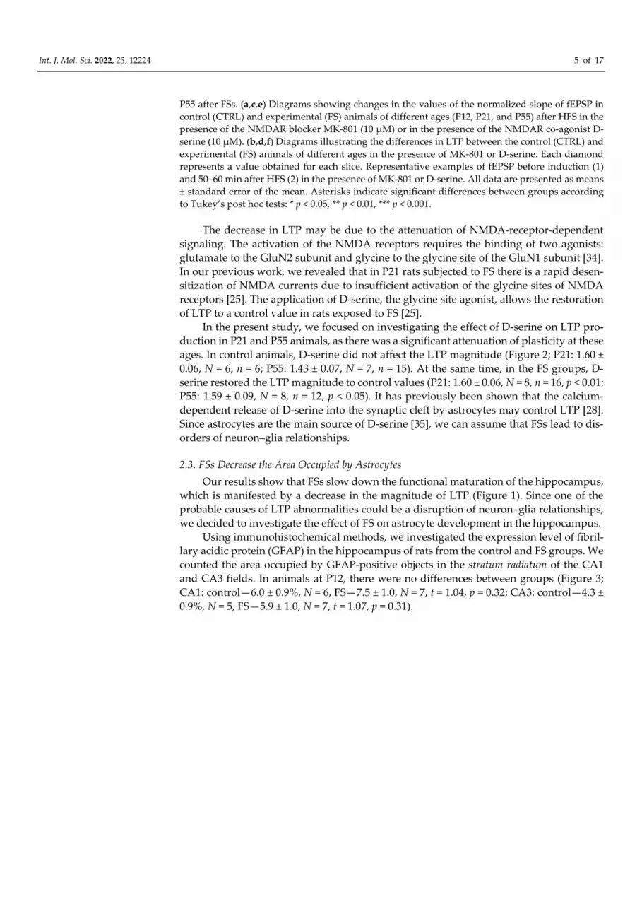
Int. J. Mol. Sci. 2022, 23, 12224 5 of 17
P55 after FSs. (a,c,e) Diagrams showing changes in the values of the normalized slope of fEPSP in
control (CTRL) and experimental (FS) animals of different ages (P12, P21, and P55) after HFS in the
presence of the NMDAR blocker MK-801 (10 μM) or in the presence of the NMDAR co-agonist D-
serine (10 μM). (b,d,f) Diagrams illustrating the differences in LTP between the control (CTRL) and
experimental (FS) animals of different ages in the presence of MK-801 or D-serine. Each diamond
represents a value obtained for each slice. Representative examples of fEPSP before induction (1)
and 50–60 min after HFS (2) in the presence of MK-801 or D-serine. All data are presented as means
± standard error of the mean. Asterisks indicate significant differences between groups according
to Tukey’s post hoc tests: * p < 0.05, ** p < 0.01, *** p < 0.001.
The decrease in LTP may be due to the attenuation of NMDA-receptor-dependent
signaling. The activation of the NMDA receptors requires the binding of two agonists:
glutamate to the GluN2 subunit and glycine to the glycine site of the GluN1 subunit [34].
In our previous work, we revealed that in P21 rats subjected to FS there is a rapid desen-
sitization of NMDA currents due to insufficient activation of the glycine sites of NMDA
receptors [25]. The application of D-serine, the glycine site agonist, allows the restoration
of LTP to a control value in rats exposed to FS [25].
In the present study, we focused on investigating the effect of D-serine on LTP pro-
duction in P21 and P55 animals, as there was a significant attenuation of plasticity at these
ages. In control animals, D-serine did not affect the LTP magnitude (Figure 2; P21: 1.60 ±
0.06, N = 6, n = 6; P55: 1.43 ± 0.07, N = 7, n = 15). At the same time, in the FS groups, D-
serine restored the LTP magnitude to control values (P21: 1.60 ± 0.06, N = 8, n = 16, p < 0.01;
P55: 1.59 ± 0.09, N = 8, n = 12, p < 0.05). It has previously been shown that the calcium-
dependent release of D-serine into the synaptic cleft by astrocytes may control LTP [28].
Since astrocytes are the main source of D-serine [35], we can assume that FSs lead to dis-
orders of neuron–glia relationships.
2.3. FSs Decrease the Area Occupied by Astrocytes
Our results show that FSs slow down the functional maturation of the hippocampus,
which is manifested by a decrease in the magnitude of LTP (Figure 1). Since one of the
probable causes of LTP abnormalities could be a disruption of neuron–glia relationships,
we decided to investigate the effect of FS on astrocyte development in the hippocampus.
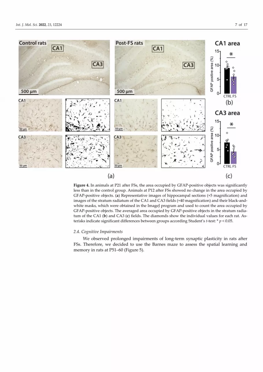
Using immunohistochemical methods, we investigated the expression level of fibril-
lary acidic protein (GFAP) in the hippocampus of rats from the control and FS groups. We
counted the area occupied by GFAP-positive objects in the stratum radiatum of the CA1
and CA3 fields. In animals at P12, there were no differences between groups (Figure 3;
CA1: control—6.0 ± 0.9%, N = 6, FS—7.5 ± 1.0, N = 7, t = 1.04, p = 0.32; CA3: control—4.3 ±
0.9%, N = 5, FS—5.9 ± 1.0, N = 7, t = 1.07, p = 0.31).

Int. J. Mol. Sci. 2022, 23, 12224 6 of 17
Figure 3. Animals at P12 after FSs showed no change in the area occupied by GFAP-positive objects.
(a) Representative images of hippocampal sections (×5 magnification) and images of the stratum
radiatum of the CA1 and CA3 fields (×40 magnification) and their black-and-white masks, which
were obtained in the ImageJ program and used to count the area occupied by GFAP-positive objects.
The averaged area occupied by GFAP-positive objects in the stratum radiatum of the CA1 (b) and
CA3 (c) fields. The diamonds show the individual values for each rat.
However, in P21 animals exposed to FSs, the area occupied by GFAP-positive objects
was significantly less than in the control group (Figure 4; CA1: control—9.0 ± 0.9%, N = 6,
FS—6.0 ± 0.8%, N = 7, t = 2.52, p < 0.05; CA3: control—7.5 ± 0.9%, N = 6, FS—4.3 ± 0.6%, N
= 7, t = 2.98, p < 0.05). Moreover, GFAP-positive astrocytes had rather short branches, and
their morphological characteristics resembled those of rats at 11 days of age.
Int. J. Mol. Sci. 2022, 23, 12224 7 of 17
Figure 4. In animals at P21 after FSs, the area occupied by GFAP-positive objects was significantly
less than in the control group. Animals at P12 after FSs showed no change in the area occupied by
GFAP-positive objects. (a) Representative images of hippocampal sections (×5 magnification) and
images of the stratum radiatum of the CA1 and CA3 fields (×40 magnification) and their black-and-
white masks, which were obtained in the ImageJ program and used to count the area occupied by
GFAP-positive objects. The averaged area occupied by GFAP-positive objects in the stratum radia-
tum of the CA1 (b) and CA3 (c) fields. The diamonds show the individual values for each rat. As-
terisks indicate significant differences between groups according Student’s t-test: * p < 0.05.
2.4. Cognitive Impairments
We observed prolonged impairments of long-term synaptic plasticity in rats after
FSs. Therefore, we decided to use the Barnes maze to assess the spatial learning and
memory in rats at P51–60 (Figure 5).

Int. J. Mol. Sci. 2022, 23, 12224 8 of 17
Figure 5. Training session (days 1 to 4) in the Barnes maze. (a) The percentage of control rats (CTRL)
and rats after FSs (FS) that successfully completed each attempt. (b) Time spent searching for the
escape box by control (CTRL) and experimental (FS) animals. Results of two-way ANOVA reported
in the text. Asterisks indicate significant differences between groups according to Tukey’s post hoc
tests: ** p < 0.01. Representative examples of the pathways of a control rat (c) and an FS rat (d) in the
Barnes maze on day 4 (second attempt).
During the training sessions (days 1 to 4), the location of the escape box did not
change. The rats were given two attempts for five minutes per day to find an escape box.
If during this time the rat did not find the escape box, it was gently pushed and forced to
go down into the box. In this case, we considered that the rat failed the attempt. Figure 5a
shows the percentage of control rats and rats after FSs that successfully completed each
attempt during training (N = 12 rats in each group). Rats in both groups learned to find
the escape box, but more rats successfully completed the task in the control group than in
the FS group at the beginning of the training. We also compared the average time the rats
spent on both attempts per day (Figure 5b). A two-way ANOVA revealed a significant
effect of learning (factor 1 “Day of training session”: F3,66 = 22; p < 0.001) and the role of the
FSs at the tendency level (factor 2 “FS”: F1,66 = 3.8; p = 0.06). The interaction of the factors
was not statistically significant (“FS” × “Day of training session”: F3,66 = 1.54; p = 0.21). We
found a significant difference in the escape time only for the first day (Figure 5b). How-
ever, FS rats (Figure 5d) also tended to spend more time on the last day of training than
control animals (Figure 5c).
On the test day, the escape box was moved to the opposite side of the field (Figure
6). First, we assessed the time spent searching for the escape box and the distance traveled
during this time. There were no differences in these parameters (time: control—119 ± 32 s,
FS group—103 ± 28 s, p = 0.72; distance: control—5.1 ± 1.4 m, FS group—3.2 ± 0.7 m, p =
0.21). Next, we counted the percentage of time each animal spent in the target area (the
sector of the field where the escape box was located during training) while searching for
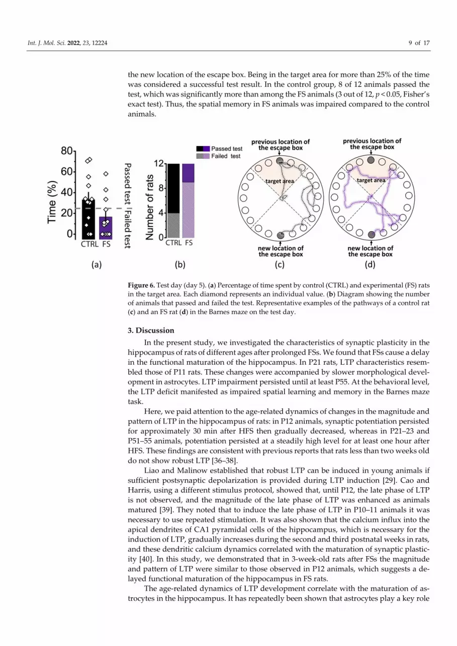
Int. J. Mol. Sci. 2022, 23, 12224 9 of 17
the new location of the escape box. Being in the target area for more than 25% of the time
was considered a successful test result. In the control group, 8 of 12 animals passed the
test, which was significantly more than among the FS animals (3 out of 12, p < 0.05, Fisher’s
exact test). Thus, the spatial memory in FS animals was impaired compared to the control
animals.
Figure 6. Test day (day 5). (a) Percentage of time spent by control (CTRL) and experimental (FS) rats
in the target area. Each diamond represents an individual value. (b) Diagram showing the number
of animals that passed and failed the test. Representative examples of the pathways of a control rat
(c) and an FS rat (d) in the Barnes maze on the test day.
3. Discussion
In the present study, we investigated the characteristics of synaptic plasticity in the
hippocampus of rats of different ages after prolonged FSs. We found that FSs cause a delay
in the functional maturation of the hippocampus. In P21 rats, LTP characteristics resem-
bled those of P11 rats. These changes were accompanied by slower morphological devel-
opment in astrocytes. LTP impairment persisted until at least P55. At the behavioral level,
the LTP deficit manifested as impaired spatial learning and memory in the Barnes maze
task.
Here, we paid attention to the age-related dynamics of changes in the magnitude and
pattern of LTP in the hippocampus of rats: in P12 animals, synaptic potentiation persisted
for approximately 30 min after HFS then gradually decreased, whereas in P21–23 and
P51–55 animals, potentiation persisted at a steadily high level for at least one hour after
HFS. These findings are consistent with previous reports that rats less than two weeks old
do not show robust LTP [36–38].
Liao and Malinow established that robust LTP can be induced in young animals if
sufficient postsynaptic depolarization is provided during LTP induction [29]. Cao and
Harris, using a different stimulus protocol, showed that, until P12, the late phase of LTP
is not observed, and the magnitude of the late phase of LTP was enhanced as animals
matured [39]. They noted that to induce the late phase of LTP in P10–11 animals it was
necessary to use repeated stimulation. It was also shown that the calcium influx into the
apical dendrites of CA1 pyramidal cells of the hippocampus, which is necessary for the
induction of LTP, gradually increases during the second and third postnatal weeks in rats,
and these dendritic calcium dynamics correlated with the maturation of synaptic plastic-
ity [40]. In this study, we demonstrated that in 3-week-old rats after FSs the magnitude
and pattern of LTP were similar to those observed in P12 animals, which suggests a de-
layed functional maturation of the hippocampus in FS rats.
The age-related dynamics of LTP development correlate with the maturation of as-
trocytes in the hippocampus. It has repeatedly been shown that astrocytes play a key role

Int. J. Mol. Sci. 2022, 23, 12224 10 of 17
in the modulation of neuronal synaptic transmission, so the morphological and electro-
physiological maturation of astrocytes affects their function. Our results are consistent
with previous studies that showed that the GFAP-positive area increases significantly
from the second to the third week of postnatal development [31]. We observed an increase
in the size of GFAP-positive astrocytes and the branching level in the control group but
not in the FS rat group. Thus, this observation also supports the idea of impaired func-
tional maturation of the hippocampus. It can be assumed that the activation of astrocytes,
in this case, could have a beneficial effect. An astrocyte-kinetic drug could be used for this
purpose; for example, l-deprenyl, an irreversible inhibitor of monoamine oxidase type B,
potentiates an astrocyte reaction associated with the increased secretion of trophic factors
[41].
However, the obtained data should be interpreted with some caution since GFAP
labeling only allows the visualization of primary, secondary, and occasionally tertiary
branches of the astrocyte [42]. It should be also noted that during this period the electro-
physiological maturation of astrocytes occurs as well, resulting in reduced voltage-gated
ion conductances [43].
Periods of rapid development of any system are critical for their function formation
and are most vulnerable to the action of injuring factors. Therefore, the consequences of
febrile seizures can be diverse. It has been shown that FSs do not cause significant neu-
ronal death in the hippocampus [25,27]. Moreover, experimental FSs in P10 rats increase
the survival rate and structural integration of newborn dentate granule cells. Their den-
drites develop faster, have more spines, and display increased spontaneous excitatory in-
put [44,45]. FSs may cause long-term alterations in hippocampal functions. FSs trigger
multiple changes in the expression of genes associated with heat, immune, and inflamma-
tion responses, the glutamate–glutamine cycle, myelination, and structural reorganization
[46]. It is assumed that many signaling systems affecting synaptic transmission can be
disrupted by FSs. For example, FSs may cause CB1 receptor upregulation, which in turn
increases the depolarization-induced suppression of inhibition and leads to persistent lim-
bic hyperexcitability [47]. The critical role of TRPV1 signaling was reported in a mouse
model of FS, which led to the increased expression of proinflammatory cytokines (IL-1ß,
IL-6, TNF-α, and HMGB1) [48]. It is well-established that these proinflammatory cyto-
kines modulate synaptic transmission and plasticity [49–51].
There are relatively few experimental studies that have examined the effects of FSs
on long-term synaptic plasticity. The data obtained in different laboratories are contradic-
tory [25,52,53]. One study demonstrated that repeated FSs resulted in LTP attenuation and
LTD facilitation in the CA1 hippocampal area [53]. In contrast, another study demon-
strated an increase in LTP and a decrease in LTD in P44 rats that were exposed to FSs at
P10 [52]. This experimental work unequivocally indicates an attenuation of LTP in P21
and P55 rats exposed to FSs. Discrepancies in the experimental results may be because FSs
lead to the activation of several signaling systems affecting synaptic plasticity differently,
which can lead to diverse outcomes. It should be noted that various early-onset seizure
models in rodents usually result in long-term impairments of synaptic plasticity and cog-
nitive function [54–60]. In a hypoxia model of seizures in P10 rats, increased LTP was
detected instantly after the seizure [61]. However, reduced LTP was found 48–72 h after
the seizures and in adult rats (P60) [62].
In our previous study, we found that LTP attenuation in FS rats is due to a more
pronounced desensitization of NMDA receptors. The application of D-serine, a co-agonist
of the glycine site of NMDA receptors, allowed us to restore synaptic plasticity to the con-
trol value [25]. In this work, we obtained a similar result; in addition, we found that in P55
rats subjected to FSs D-serine was also efficient. It is known that astrocytes provide the
local D-serine supply, thus changing NMDA-receptor-dependent signaling, and can en-
hance LTP [28]. The application of D-serine was shown to improve LTP in rats in the lith-
ium–pilocarpine model. In the latent phase of the model, D-serine fully restored LTP in

Int. J. Mol. Sci. 2022, 23, 12224 11 of 17
hippocampal slices [63], while in animals in the chronic phase, the effect was less pro-
nounced and was only evident in the initial phase of LTP [64].
Due to the fact that FSs occur in early postnatal ontogenesis, when the processes of
the maturation of neurons and astrocytes and the formation of synapses are active, some
studies have raised the question of whether FSs lead to impairments in important pro-
cesses such as learning and memory. Cognitive impairments after prolonged FSs have
been described in both pediatric clinical studies [65] and several animal studies. In partic-
ular, adult rats subjected to FSs were found to have deficits in working and reference
memory in the Morris water maze test and in long-term memory in a contextual fear con-
ditioning test [45,53,66]. We also note that after prolonged FSs animals performed worse
when we changed the escape box locations after several days of training in the Barnes
maze. This result is similar to that previously described by Dube et al. using the Morris
maze [1].
It should also be noted that the hippocampus is part of the limbic system and has
connections with emotion-related brain regions, for instance, the amygdala and the pre-
frontal cortex. An impairment of synaptic plasticity in these areas of the CNS, resulting in
an abnormal structural remodeling of these areas, may contribute to the pathophysiology
of depression [67]. During early postnatal development, the hippocampus is highly sus-
ceptible to stress and other negative stimuli [68] and smaller hippocampal volumes have
been reported in children and adults exposed to early-life adversity [69,70]. Although the
nature of the associations among early-life adversity, hippocampal volume deficit,
changes in neural plasticity, and depressive disorder are not well-understood, antidepres-
sant treatment has been shown to protect against hippocampal volume loss [71,72] and
affect the synaptic plasticity processes, in particular by increasing the expression of plas-
ticity-related protein [73], and can relieve stress-induced inhibition LTP [74] in the hippo-
campus. Our data show that FSs lead to long-term LTP disorders in the hippocampus,
and it was previously demonstrated that adult rats subjected to hyperthermia-induced
seizures during the neonatal period demonstrate depressive-like behavior [75], which al-
lows the consideration of antidepressant treatment as a potential method of correcting not
only depressive behavior but also cognitive impairments that develop after FSs.
4. Materials and Methods
4.1. Animals
Wistar rats were used in this study. Animals were kept under standard conditions at
room temperature with free access to water and food. All experiments were approved by
the Sechenov Institute of Evolutionary Physiology and Biochemistry Ethics Committee
and were carried out following local guidelines on the treatment of laboratory animals.
These guidelines fully comply with Russian and international standards for animal stud-
ies.
4.2. FS Model
The nests were formed in such a way that each cage contained a female with 10 pups.
This was to ensure that the animals from each litter had approximately the same body
weight at P10.
Experimental FSs were induced at P10. The pups were placed on the bottom of a glass
chamber for 30 min. At a height of 40 cm, warm airflow was created in such a way that a
temperature of 46 °C was maintained at the bottom of the chamber, where the animal was
located. Body temperature (rectal) was measured at baseline (31.1 ± 0.1 °C), at the onset of
seizures (39.8 ± 0.1 °C), and every two minutes during seizures. If the body temperature
was above 41 °C, the rats were transferred to a cool surface until the body temperature
returned to 39 °C or less. The rats were then transferred back to the chamber. Under such
conditions, the development and course of FSs in most animals were stereotypic: during
the first 10 min, body temperature rose to 39 °C, and facial automatisms were observed,
Int. J. Mol. Sci. 2022, 23, 12224 12 of 17
often accompanied by unilateral body flexion. Then, there were myoclonic twitches of the
hind limbs, followed by clonic convulsions. Only animals with FSs that lasted at least 15
min were included in the study (N = 53). After hyperthermia, the animals were placed on
a cold surface until the core temperature returned to the normal range. Then, they were
returned to the home cage.
After hyperthermia, the pups’ weights changed insignificantly (<3% change in body
weight), indicating only slight dehydration symptoms. The mortality rate during hyper-
thermia and the following 30 min was less than 1%.
Littermates used as controls were taken from the cage for the same time but were
maintained at room temperature (N = 50).
4.3. Brain Slice Preparation
At P12, P21–23, and P51–55, the rats were decapitated, and brains were quickly re-
moved. Horizontal brain slices (400 μm) were cut using an HM 650 V vibratome (Microm,
Walldorf, Germany) in chilled artificial cerebrospinal fluid (ACSF; t = 0 °C; containing (in
mM): 126 NaCl, 24 NaHCO3, 2.5 KCl, 2 CaCl2, 1.25 NaH2PO4, 1 MgSO4, and 10 glucose),
which was aerated with carbogen (95% O2 and 5% CO2). Afterward, slices were allowed
to recover at 35 °C in oxygenated ACSF for 1 h.
4.4. Field Potential Recordings
Extracellular field excitatory postsynaptic potentials (fEPSPs) were recorded from the
CA1 stratum radiatum of the hippocampus with glass microelectrodes (0.2–1.0 MΩ). Syn-
aptic responses were evoked by the stimulation of the Schaffer collaterals using a bipolar
nichrome electrode as previously described [25]. Stimuli were delivered every 20 s via an
A365 stimulus isolator (World Precision Instruments, Sarasota, FL, USA). Responses were
amplified by a Model 1800 amplifier (A-M Systems, Carlsborg, WA, USA) then digitized
with an ADC/DAC NI USB-6211 (National Instruments, Austin, TX, USA) using WinWCP
v5.x.x software (University of Strathclyde, Glasgow, UK).
LTP was induced using high-frequency stimulation (HFS, three trains consisting of
100 pulses at 100 Hz applied every 20 s). A 20 min baseline period preceded LTP induc-
tion. Potentiated fEPSPs were recorded for 60 min following stimulation. LTP was quan-
tified by calculating the ratio of the average slope of the potentiated fEPSPs (50–60 min
after stimulation) and the baseline ones (10 min before stimulation). The recordings were
analyzed using Clampfit 10.2 software (Axon Instruments, San Jose, CA, USA).
Dizocilpine (MK-801, 10 μM), a noncompetitive NMDA receptor antagonist, and D-
serine, a co-agonist of NMDARs, were obtained from Sigma (St. Louis, MO, USA). These
drugs were diluted in distilled water and bath-applied.
4.5. Immunohistochemistry
At P12 and P21–23, rats were deeply anesthetized with a mixture of Zoletil (3 mg per
100 g) and Xylazine (50 μL per 100 g) diluted in a saline solution and perfused transcardi-
ally with phosphate-buffered saline (PBS, pH 7.4, 0.01 M) followed by 4% paraformalde-
hyde in PBS. Next, decapitation, the extraction of the brain, and its fixation in 4% PFA at
4 °C for at least 24 h were performed. After fixation, brains were cryoprotected with 30%
sucrose, frozen in cooled (<−50 °C) isopentane (78-78-4, Isopentane Solution, Sigma-Al-
drich, St. Louis, MO, USA), and stored at −80 °C. The 20 μm-thick frontal serial sections
(from −2.6 to −3.6 mm to the bregma) were cut on a Bright OTF5000 cryostat (Bright In-
strument Co Ltd., Huntingdon, UK) and mounted on slides with the adhesive coating
Super Frost Plus (J1800AMNZ, Fisher Scientific UK Ltd., Loughborough, UK).
The distribution of GFAP was analyzed using indirect immunofluorescence analysis.
For blocking the endogenous peroxidase activities in the sections, 3% hydrogen peroxide
was used for 30 min. After a rinse in PBS, sections were incubated in phosphate-buffered
saline with 0.2% Triton X-100 (PBS-T) for 30 min then incubated in blocking serum (2%
Int. J. Mol. Sci. 2022, 23, 12224 13 of 17
normal goat serum and 3% bovine serum albumin in PBS-T) for 1 h. Then, sections were
incubated in blocking serum with the primary mouse antibody against GFAP (1:1000; cat
# NBP1-05197, Bio-Techne Ltd., Abingdon, OX14 3NB, UK) for 48 h at 4 °C. Next, sections
were incubated with a biotinylated goat antimouse secondary antibody (1:500 in PBS, cat
# BA-9200-1.5, Vector Laboratories Inc, Burlingame, CA, USA) for 1 h and with streptavi-
din (1:1000 in PBS, cat # S2438-250UG, Sigma-Aldrich, St. Louis, MO, USA) for 1 h at room
temperature. Then, sections were stained with 3,3’-diaminobenzidine (DAB), dehydrated,
cleared, and permanently mounted.
At least 5–6 brain sections, taken at 100 μm intervals, were analyzed using the Leica
AF 7000 Microscope (Leica Microsystems, Wetzlar, Germany) under ×400 magnification.
The areas to be analyzed in each section were selected as shown in Figure 3a. The area (in
%) occupied by GFAP-positive objects was assessed using ImageJ (U. S. National Insti-
tutes of Health, Bethesda, MD, USA). First, the images were converted to an 8-bit format.
Next, we obtained the resulting image histograms representing the distribution of pixels
from min–max (0–255) display values. The threshold option allowed us to choose a thresh-
old value: pixels less than a certain value were recognized as one class (black), and pixels
greater than the value were recognized as another class (white). The threshold value was
determined individually for each image using the following protocol: (1) We found a
value that included 99.5% of the pixels and multiplied it by 0.75. This value was consid-
ered the threshold value. (2) The GFAP-positive area was determined using the Analyze
Particles tool (ImageJ). Objects with an area of less than 500 pixels were cut out of the final
result using the Size function. Thus, for each image, the percentage of the area (%) occu-
pied by GFAP-positive objects and a visual black-and-white mask were obtained.
4.6. Barnes Maze Task
The Barnes maze task was performed on P51–60 rats. The Barnes maze is a behavioral
paradigm for assessing spatial learning and memory [76,77]. The apparatus used in the
Barnes maze task included a circular white field (100 cm in diameter) with 20 circular
holes (8 cm in diameter) evenly spaced around the periphery, an escape box (25 × 17 × 12.5
cm) for the animal to hide in installed under one of the holes, and a nontransparent box
(diameter was 28 cm, and depth was 30 cm) for rat transportation. The field was located
80 cm above the floor, light sources brightly illuminated the field (illumination on the
center of the field was 520 Lx), and a recording camera was placed above the field. The
recordings were analyzed using the program “Pole Krest” designed at the Institute of Ex-
perimental Medicine for the analysis of animal behavior.
The experiment contained tree stages:
(1) Habituation: To familiarize the animal with the maze, habituation was performed 72
h before the beginning of the training. For this purpose, the rat was first placed in the
escape box for 3 min. Then, this box was placed under the hole of a fully illuminated
field. After that, the rat was placed in the center of the field. The escape box was
under one of the holes, and the rat had to find it within 3 min. If during this time the
rat did not find the escape box, it was gently pushed and forced to go down into the
box, where it was left for 3 min. Then, the rat was placed in an opaque box, and the
box was placed in the center of the field for 1 min. Then, the opaque box was re-
moved, and the rat searched for the escape box again. The last step was repeated two
times.
(2) Training: The location of the escape box was changed from the time of habituation
and fixed during training. Training was performed over four consecutive days. The
rat had two attempts per day. The rat was placed in the nontransparent box, and this
box was placed in the center of the field for 1 min. After removing the nontransparent
box, the recording began, and the rat had 5 min to find the escape box. If the rat did
not find the escape box during this time, it was gently pushed to it and forced to go

Int. J. Mol. Sci. 2022, 23, 12224 14 of 17
down into the box. After that, the rat remained in the escape box for 1 min. The sec-
ond attempt began five minutes after the first attempt. Between each attempt, the
field and box hole were cleaned with an alcohol–peroxide solution (0.3% peroxide
and 30% alcohol). To assess progress in learning, we measured the time spent search-
ing for the escape box.
(3) Testing (day 5): The day after the training was completed, the escape box was moved
to another location in the opposite sector. On this day, the rat had one attempt to find
the escape box. We measured the time spent in the target sector, where the box was
located, during training.
4.7. Statistical Analysis
The statistical analysis was performed with OriginPro 8 (OriginLab Corporation,
Northampton, MA, USA) and Statistica 8.0 (Systat Software Inc., Palo Alto, CA, USA).
Dixon’s Q test (at a 90% confidence level) was used to identify and reject outliers. The
normality of the sample data was evaluated using the Kolmogorov–Smirnov test. Statisti-
cal significance was assessed using Student’s t-test and one-way or two-way ANOVAs as
stated in the text. All data are presented as means ± standard error of the mean. p < 0.05
was considered statistically significant.
Author Contributions: Formal analysis, T.Y.P. and A.V.G.; Funding acquisition, A.V.Z.; Investiga-
tion, T.Y.P. and A.V.G.; Methodology, T.Y.P.; Supervision, T.Y.P. and A.V.Z.; Validation, T.Y.P. and
A.V.Z.; Writing—original draft, T.Y.P., A.V.G., and A.V.Z.; Writing—review and editing, T.Y.P.,
A.V.G., and A.V.Z. All authors have read and agreed to the published version of the manuscript.
Funding: This research was funded by the Russian Science Foundation, grant number 21-15-00430.
Institutional Review Board Statement: The study was conducted according to EU Directive
2010/63/EU for animal experiments and was approved by the Ethics Committee of the Sechenov
Institute of Evolutionary Physiology and Biochemistry of the Russian Academy of Sciences (ethical
permit number 13-k-a, 15 February 2018).
Informed Consent Statement: Not applicable.
Data Availability Statement: The data presented in this study are available on request from the
corresponding author.
Acknowledgments: Immunohistochemistry experiments were performed using the facilities of the
Research Resource Centre for Physiological, Biochemical, and Molecular-Biological Research of the
Sechenov Institute of Evolutionary Physiology and Biochemistry of the Russian Academy of Sci-
ences.
Conflicts of Interest: The authors declare no conflict of interest.
References
1. Dubé, C.M.; Zhou, J.-L.; Hamamura, M.; Zhao, Q.; Ring, A.; Abrahams, J.; McIntyre, K.; Nalcioglu, O.; Shatskih, T.; Baram, T.Z.;
et al. Cognitive dysfunction after experimental febrile seizures. Exp. Neurol. 2009, 215, 167–177.
https://doi.org/10.1016/j.expneurol.2008.10.003.
2. Baram, T.Z.; Donato, F.; Holmes, G.L. Construction and disruption of spatial memory networks during development. Learn.
Mem. 2019, 26, 206–218. https://doi.org/10.1101/lm.049239.118.
3. Saleh, A.; Potter, G.G.; McQuoid, D.R.; Boyd, B.; Turner, R.; MacFall, J.R.; Taylor, W.D. Effects of early life stress on depression,
cognitive performance and brain morphology. Psychol. Med. 2017, 47, 171–181. https://doi.org/10.1017/S0033291716002403.
4. de Rooij, S.R.; Wouters, H.; Yonker, J.E.; Painter, R.C.; Roseboom, T.J. Prenatal undernutrition and cognitive function in late
adulthood. Proc. Natl. Acad. Sci. USA 2010, 107, 16881–16886. https://doi.org/10.1073/pnas.1009459107.
5. Dubé, C.M.; Ravizza, T.; Hamamura, M.; Zha, Q.; Keebaugh, A.; Fok, K.; Andres, A.L.; Nalcioglu, O.; Obenaus, A.; Vezzani, A.;
et al. Epileptogenesis provoked by prolonged experimental febrile seizures: Mechanisms and biomarkers. J. Neurosci. 2010, 30,
7484–7494. https://doi.org/10.1523/JNEUROSCI.0551-10.2010.
6. Lewis, D.V.; Shinnar, S.; Hesdorffer, D.C.; Bagiella, E.; Bello, J.A.; Chan, S.; Xu, Y.; MacFall, J.; Gomes, W.A.; Moshé, S.L.; et al.
Hippocampal sclerosis after febrile status epilepticus: The FEBSTAT study. Ann. Neurol. 2014, 75, 178–185.
https://doi.org/10.1002/ana.24081.
7. Cendes, F. Febrile seizures and mesial temporal sclerosis. Curr. Opin. Neurol. 2004, 17, 161–164. https://doi.org/10.1097/00019052-
Int. J. Mol. Sci. 2022, 23, 12224 15 of 17
200404000-00013.
8. Francis, J.R.; Richmond, P.; Robins, C.; Lindsay, K.; Levy, A.; Effler, P.V.; Borland, M.; Blyth, C.C. An observational study of
febrile seizures: The importance of viral infection and immunization. BMC Pediatr. 2016, 16, 202. https://doi.org/10.1186/s12887-
016-0740-5.
9. Biagini, G.; Pich, E.M.; Carani, C.; Marrama, P.; Agnati, L.F. Postnatal maternal separation during the stress hyporesponsive
period enhances the adrenocortical response to novelty in adult rats by affecting feedback regulation in the CA1 hippocampal
field. Int. J. Dev. Neurosci. 1998, 16, 187–197. https://doi.org/10.1016/S0736-5748(98)00019-7.
10. Stratilov, V.A.; Tyulkova, E.I.; Vetrovoy, O.V. Prenatal Stress as a Factor of the Development of Addictive States. J. Evol. Biochem.
Physiol. 2020, 56, 471–490. https://doi.org/10.1134/S0022093020060010.
11. Gulyaeva, N.V. Glucocorticoid Regulation of the Glutamatergic Synapse: Mechanisms of Stress-Dependent Neuroplasticity. J.
Evol. Biochem. Physiol. 2021, 57, 564–576. https://doi.org/10.1134/S0022093021030091.
12. Koe, A.S.; Salzberg, M.R.; Morris, M.J.; O’Brien, T.J.; Jones, N.C. Early life maternal separation stress augmentation of limbic
epileptogenesis: The role of corticosterone and HPA axis programming. Psychoneuroendocrinology 2014, 42, 124–133.
https://doi.org/10.1016/j.psyneuen.2014.01.009.
13. Feng, B.; Chen, Z. Generation of Febrile Seizures and Subsequent Epileptogenesis. Neurosci. Bull. 2016, 32, 481–492.
https://doi.org/10.1007/s12264-016-0054-5.
14. Leung, A.K.; Hon, K.L.; Leung, T.N.H. Febrile seizures: An overview. Drugs Context 2018, 7, 212536.
https://doi.org/10.7573/dic.212536.
15. Millichap, J.G.J.; Millichap, J.G.J. Role of viral infections in the etiology of febrile seizures. Pediatr. Neurol. 2006, 35, 165–172.
https://doi.org/10.1016/j.pediatrneurol.2006.06.004.
16. Mewasingh, L.D.; Chin, R.F.M.; Scott, R.C. Current understanding of febrile seizures and their long-term outcomes. Dev. Med.
Child Neurol. 2020, 62, 1245–1249. https://doi.org/10.1111/dmcn.14642.
17. Avishai-Eliner, S.; Brunson, K.L.; Sandman, C.A.; Baram, T.Z. Stressed-out, or in (utero)? Trends Neurosci. 2002, 25, 518–524.
https://doi.org/10.1016/s0166-2236(02)02241-5.
18. Kipp, K.H.; Opitz, B.; Becker, M.; Hofmann, J.; Krick, C.; Gortner, L.; Mecklinger, A. Neural correlates of recognition memory
in children with febrile seizures: Evidence from functional magnetic resonance imaging. Front. Hum. Neurosci. 2012, 6, 17.
https://doi.org/10.3389/fnhum.2012.00017.
19. Kipp, K.H.; Mecklinger, A.; Becker, M.; Reith, W.; Gortner, L. Infant febrile seizures: Changes in declarative memory as revealed
by event-related potentials. Clin. Neurophysiol. Off. J. Int. Fed. Clin. Neurophysiol. 2010, 121, 2007–2016.
https://doi.org/10.1016/j.clinph.2010.05.011.
20. Martinos, M.M.; Pujar, S.; O’Reilly, H.; de Haan, M.; Neville, B.G.R.; Scott, R.C.; Chin, R.F.M. Intelligence and memory outcomes
within 10 years of childhood convulsive status epilepticus. Epilepsy Behav. 2019, 95, 18–25.
https://doi.org/10.1016/j.yebeh.2019.03.039.
21. Martinos, M.M.; Yoong, M.; Patil, S.; Chong, W.K.; Mardari, R.; Chin, R.F.M.; Neville, B.G.R.; de Haan, M.; Scott, R.C. Early
developmental outcomes in children following convulsive status epilepticus: A longitudinal study. Epilepsia 2013, 54, 1012–1019.
https://doi.org/10.1111/epi.12136.
22. Zaitsev, А. V.; Amakhin, D.V.; Dyomina, A.V.; Zakharova, M.V.; Ergina, J.L.; Postnikova, T.Y.; Diespirov, G.P.; Magazanik, L.G.
Synaptic Dysfunction in Epilepsy. J. Evol. Biochem. Physiol. 2021, 57, 542–563. https://doi.org/10.1134/S002209302103008X.
23. Baram, T.Z.; Gerth, A.; Schultz, L. Febrile seizures: An appropriate-aged model suitable for long-term studies. Brain Res. Dev.
Brain Res. 1997, 98, 265–270. https://doi.org/10.1016/s0165-3806(96)00190-3.
24. Bender, R.A.; Baram, T.Z. Epileptogenesis in the developing brain: What can we learn from animal models? Epilepsia 2007, 48
(Suppl. 5), 2–6. https://doi.org/10.1111/j.1528-1167.2007.01281.x.
25. Postnikova, T.Y.; Griflyuk, A.V.; Amakhin, D.V.; Kovalenko, A.A.; Soboleva, E.B.; Zubareva, O.E.; Zaitsev, A.V. Early Life
Febrile Seizures Impair Hippocampal Synaptic Plasticity in Young Rats. Int. J. Mol. Sci. 2021, 22, 8218.
https://doi.org/10.3390/ijms22158218.
26. Toth, Z.; Yan, X.-X.X.; Haftoglou, S.; Ribak, C.E.; Baram, T.Z. Seizure-induced neuronal injury: Vulnerability to febrile seizures
in an immature rat model. J. Neurosci. 1998, 18, 4285–4294. https://doi.org/10.1523/JNEUROSCI.18-11-04285.1998.
27. Bender, R.A.; Dubé, C.; Gonzalez-Vega, R.; Mina, E.W.; Baram, T.Z. Mossy fiber plasticity and enhanced hippocampal
excitability, without hippocampal cell loss or altered neurogenesis, in an animal model of prolonged febrile seizures.
Hippocampus 2003, 13, 399–412. https://doi.org/10.1002/hipo.10089.
28. Henneberger, C.; Papouin, T.; Oliet, S.H.R.; Rusakov, D.A. Long-term potentiation depends on release of d-serine from
astrocytes. Nature 2010, 463, 232–236. https://doi.org/10.1038/nature08673.
29. Liao, D.; Malinow, R. Deficiency in induction but not expression of LTP in hippocampal slices from young rats. Learn. Mem.
1996, 3, 138–149,. https://doi.org/10.1101/lm.3.2-3.138.
30. Felix, L.; Stephan, J.; Rose, C.R. Astrocytes of the early postnatal brain. Eur. J. Neurosci. 2021, 54, 5649–5672.
https://doi.org/10.1111/ejn.14780.
31. Catalani, A.; Sabbatini, M.; Consoli, C.; Cinque, C.; Tomassoni, D.; Azmitia, E.; Angelucci, L.; Amenta, F. Glial fibrillary acidic
protein immunoreactive astrocytes in developing rat hippocampus. Mech. Ageing Dev. 2002, 123, 481–490.
https://doi.org/10.1016/S0047-6374(01)00356-6.
32. Abbink, M.R.; van Deijk, A.L.F.; Heine, V.M.; Verheijen, M.H.; Korosi, A. The involvement of astrocytes in early-life adversity

Int. J. Mol. Sci. 2022, 23, 12224 16 of 17
induced programming of the brain. Glia 2019, 67, 1637–1653. https://doi.org/10.1002/GLIA.23625.
33. Bliss, T.V.P.; Collingridge, G.L. A synaptic model of memory: Long-term potentiation in the hippocampus. Nature 1993, 361,
31–39. https://doi.org/10.1038/361031a0.
34. Traynelis, S.F.; Wollmuth, L.P.; McBain, C.J.; Menniti, F.S.; Vance, K.M.; Ogden, K.K.; Hansen, K.B.; Yuan, H.; Myers, S.J.;
Dingledine, R. Glutamate Receptor Ion Channels: Structure, Regulation, and Function. Pharmacol. Rev. 2010, 62, 405–496.
https://doi.org/10.1124/pr.109.002451.
35. Kang, N.; Peng, H.; Yu, Y.; Stanton, P.K.; Guilarte, T.R.; Kang, J. Astrocytes release d-serine by a large vesicle. Neuroscience 2013,
240, 243–257. https://doi.org/10.1016/j.neuroscience.2013.02.029.
36. Harris, K.M.; Teyler, T.J. Developmental onset of long-term potentiation in area CA1 of the rat hippocampus. J. Physiol. 1984,
346, 27–48. https://doi.org/10.1113/jphysiol.1984.sp015005.
37. Jackson, P.S.; Suppes, T.; Harris, K.M. Stereotypical changes in the pattern and duration of long-term potentiation expressed at
postnatal days 11 and 15 in the rat hippocampus. J. Neurophysiol. 1993, 70, 1412–1419. https://doi.org/10.1152/jn.1993.70.4.1412.
38. Muller, D.; Oliver, M.; Lynch, G. Developmental changes in synaptic properties in hippocampus of neonatal rats. Dev. Brain Res.
1989, 49, 105–114. https://doi.org/10.1016/0165-3806(89)90063-1.
39. Cao, G.; Harris, K.M. Developmental regulation of the late phase of long-term potentiation (L-LTP) and metaplasticity in
hippocampal area CA1 of the rat. J. Neurophysiol. 2012, 107, 902–912. https://doi.org/10.1152/jn.00780.2011.
40. Isomura, Y.; Kato, N. Action Potential–Induced Dendritic Calcium Dynamics Correlated With Synaptic Plasticity in Developing
Hippocampal Pyramidal Cells. J. Neurophysiol. 1999, 82, 1993–1999. https://doi.org/10.1152/jn.1999.82.4.1993.
41. Biagini, G.; Frasoldati, A.; Fuxe, K.; Agnati, L.F. The concept of astrocyte-kinetic drug in the treatment of neurodegenerative
diseases: Evidence for l-deprenyl-induced activation of reactive astrocytes. Neurochem. Int. 1994, 25, 17–22.
https://doi.org/10.1016/0197-0186(94)90047-7.
42. Bushong, E.A.; Martone, M.E.; Jones, Y.Z.; Ellisman, M.H. Protoplasmic Astrocytes in CA1 Stratum Radiatum Occupy Separate
Anatomical Domains. J. Neurosci. 2002, 22, 183–192. https://doi.org/10.1523/JNEUROSCI.22-01-00183.2002.
43. Zhou, M.; Schools, G.P.; Kimelberg, H.K. Development of GLAST(+) Astrocytes and NG2(+) Glia in Rat Hippocampus CA1:
Mature Astrocytes Are Electrophysiologically Passive. J. Neurophysiol. 2006, 95, 134–143. https://doi.org/10.1152/jn.00570.2005.
44. Raijmakers, M.; Clynen, E.; Smisdom, N.; Nelissen, S.; Brône, B.; Rigo, J.M.; Hoogland, G.; Swijsen, A. Experimental febrile
seizures increase dendritic complexity of newborn dentate granule cells. Epilepsia 2016, 57, 717–726.
https://doi.org/10.1111/EPI.13357.
45. Hoogland, G.; Raijmakers, M.; Clynen, E.; Brône, B.; Rigo, J.-M.; Swijsen, A. Experimental early-life febrile seizures cause a
sustained increase in excitatory neurotransmission in newborn dentate granule cells. Brain Behav. 2022, 12, e2505.
https://doi.org/10.1002/brb3.2505.
46. Jongbloets, B.C.; van Gassen, K.L.I.; Kan, A.A.; Olde Engberink, A.H.O.; de Wit, M.; Wolterink-Donselaar, I.G.; Groot Koerkamp,
M.J.A.; van Nieuwenhuizen, O.; Holstege, F.C.P.; de Graan, P.N.E. Expression Profiling after Prolonged Experimental Febrile
Seizures in Mice Suggests Structural Remodeling in the Hippocampus. PLoS ONE 2015, 10, e0145247.
https://doi.org/10.1371/journal.pone.0145247.
47. Chen, K.; Neu, A.; Howard, A.L.L.; Foldy, C.; Echegoyen, J.; Hilgenberg, L.; Smith, M.; Mackie, K.; Soltesz, I. Prevention of
Plasticity of Endocannabinoid Signaling Inhibits Persistent Limbic Hyperexcitability Caused by Developmental Seizures. J.
Neurosci. 2007, 27, 46–58. https://doi.org/10.1523/JNEUROSCI.3966-06.2007.
48. Huang, W.-X.; Yu, F.; Sanchez, R.M.; Liu, Y.-Q.; Min, J.-W.; Hu, J.-J.; Bsoul, N.B.; Han, S.; Yin, J.; Liu, W.-H.; et al. TRPV1
promotes repetitive febrile seizures by pro-inflammatory cytokines in immature brain. Brain. Behav. Immun. 2015, 48, 68–77.
https://doi.org/10.1016/j.bbi.2015.01.017.
49. Costello, D.A.; Watson, M.B.; Cowley, T.R.; Murphy, N.; Murphy Royal, C.; Garlanda, C.; Lynch, M.A. Interleukin-1 and
HMGB1 Mediate Hippocampal Dysfunction in SIGIRR-Deficient Mice. J. Neurosci. 2011, 31, 3871–3879.
https://doi.org/10.1523/JNEUROSCI.6676-10.2011.
50. Stellwagen, D.; Malenka, R.C. Synaptic scaling mediated by glial TNF-α. Nature 2006, 440, 1054–1059.
https://doi.org/10.1038/nature04671.
51. Avital, A.; Goshen, I.; Kamsler, A.; Segal, M.; Iverfeldt, K.; Richter-Levin, G.; Yirmiya, R. Impaired interleukin-1 signaling is
associated with deficits in hippocampal memory processes and neural plasticity. Hippocampus 2003, 13, 826–834.
https://doi.org/10.1002/hipo.10135.
52. Notenboom, R.G.E.; Ramakers, G.M.J.J.; Kamal, A.; Spruijt, B.M.; De Graan, P.N.E. Long-lasting modulation of synaptic
plasticity in rat hippocampus after early-life complex febrile seizures. Eur. J. Neurosci. 2010, 32, 749–758.
https://doi.org/10.1111/j.1460-9568.2010.07321.x.
53. Chang, Y.-C.; Kuo, Y.-M.; Huang, A.-M.; Huang, C.-C. Repetitive febrile seizures in rat pups cause long-lasting deficits in
synaptic plasticity and NR2A tyrosine phosphorylation. Neurobiol. Dis. 2005, 18, 466–475.
https://doi.org/10.1016/j.nbd.2004.12.012.
54. Karnam, H.B.; Zhao, Q.; Shatskikh, T.; Holmes, G.L. Effect of age on cognitive sequelae following early life seizures in rats.
Epilepsy Res. 2009, 85, 221–230. https://doi.org/10.1016/j.eplepsyres.2009.03.008.
55. Mikati, M.A.; Zeinieh, M.P.; Kurdi, R.M.; Harb, S.A.; El Hokayem, J.A.; Daderian, R.H.; Shamseddine, A.; Obeid, M.; Bitar, F.F.;
El Sabban, M. Long-term effects of acute and of chronic hypoxia on behavior and on hippocampal histology in the developing
brain. Brain Res. Dev. Brain Res. 2005, 157, 98–102. https://doi.org/10.1016/j.devbrainres.2005.03.007.
Int. J. Mol. Sci. 2022, 23, 12224 17 of 17
56. Huang, L.-T.; Yang, S.N.; Liou, C.-W.; Hung, P.-L.; Lai, M.-C.; Wang, C.-L.; Wang, T.-J. Pentylenetetrazol-induced recurrent
seizures in rat pups: Time course on spatial learning and long-term effects. Epilepsia 2002, 43, 567–573.
https://doi.org/10.1046/j.1528-1157.2002.29101.x.
57. Holmes, G.L.; Ben-Ari, Y. A single episode of neonatal seizures permanently alters glutamatergic synapses. Ann. Neurol. 2007,
61, 379–381,. https://doi.org/10.1002/ana.21136.
58. Yang, S.-N.; Huang, C.-B.; Yang, C.-H.; Lai, M.-C.; Chen, W.-F.; Wang, C.-L.; Wu, C.-L.; Huang, L.-T. Impaired SynGAP
expression and long-term spatial learning and memory in hippocampal CA1 area from rats previously exposed to perinatal
hypoxia-induced insults: Beneficial effects of A68930. Neurosci. Lett. 2004, 371, 73–78. https://doi.org/10.1016/j.neulet.2004.08.044.
59. Lippman-Bell, J.J.; Zhou, C.; Sun, H.; Feske, J.S.; Jensen, F.E. Early-life seizures alter synaptic calcium-permeable AMPA receptor
function and plasticity. Mol. Cell. Neurosci. 2016, 76, 11–20. https://doi.org/10.1016/j.mcn.2016.08.002.
60. Cornejo, B.J.; Mesches, M.H.; Benke, T.A. A single early-life seizure impairs short-term memory but does not alter spatial
learning, recognition memory, or anxiety. Epilepsy Behav. 2008, 13, 585–592. https://doi.org/10.1016/j.yebeh.2008.07.002.
61. Jensen, F.E.; Wang, C.; Stafstrom, C.E.; Liu, Z.; Geary, C.; Stevens, M.C. Acute and chronic increases in excitability in rat
hippocampal slices after perinatal hypoxia In vivo. J. Neurophysiol. 1998, 79, 73–81. https://doi.org/10.1152/jn.1998.79.1.73.
62. Zhou, C.; Bell, J.J.L.; Sun, H.; Jensen, F.E. Hypoxia-induced neonatal seizures diminish silent synapses and long-term
potentiation in hippocampal CA1 neurons. J. Neurosci. 2011, 31, 18211–18222. https://doi.org/10.1523/JNEUROSCI.4838-11.2011.
63. Postnikova, T.Y.; Diespirov, G.P.; Amakhin, D.V.; Vylekzhanina, E.N.; Soboleva, E.B.; Zaitsev, A.V. Impairments of Long-Term
Synaptic Plasticity in the Hippocampus of Young Rats during the Latent Phase of the Lithium-Pilocarpine Model of Temporal
Lobe Epilepsy. Int. J. Mol. Sci. 2021, 22, 13355. https://doi.org/10.3390/ijms222413355.
64. Plata, A.; Lebedeva, A.; Denisov, P.; Nosova, O.; Postnikova, T.Y.; Pimashkin, A.; Brazhe, A.; Zaitsev, A.V.; Rusakov, D.A.;
Semyanov, A. Astrocytic Atrophy Following Status Epilepticus Parallels Reduced Ca2+ Activity and Impaired Synaptic
Plasticity in the Rat Hippocampus. Front. Mol. Neurosci. 2018, 11, 215. https://doi.org/10.3389/fnmol.2018.00215.
65. Martinos, M.M.; Yoong, M.; Patil, S.; Chin, R.F.M.; Neville, B.G.; Scott, R.C.; de Haan, M. Recognition memory is impaired in
children after prolonged febrile seizures. Brain 2012, 135, 3153–3164. https://doi.org/10.1093/brain/aws213.
66. Dai, Y.; Wu, D.; Feng, B.; Chen, B.; Tang, Y.; Jin, M.; Zhao, H.; Dai, H.; Wang, Y.; Chen, Z. Prolonged febrile seizures induce
inheritable memory deficits in rats through DNA methylation. CNS Neurosci. Ther. 2019, 25, 601–611.
https://doi.org/10.1111/cns.13088.
67. Masi, G.; Brovedani, P. The Hippocampus, Neurotrophic Factors and Depression. CNS Drugs 2011, 25, 913–931.
https://doi.org/10.2165/11595900-000000000-00000.
68. Teicher, M.H.; Andersen, S.L.; Polcari, A.; Anderson, C.M.; Navalta, C.P.; Kim, D.M. The neurobiological consequences of early
stress and childhood maltreatment. Neurosci. Biobehav. Rev. 2003, 27, 33–44. https://doi.org/10.1016/S0149-7634(03)00007-1.
69. Rao, U.; Chen, L.-A.; Bidesi, A.S.; Shad, M.U.; Thomas, M.A.; Hammen, C.L. Hippocampal Changes Associated with Early-Life
Adversity and Vulnerability to Depression. Biol. Psychiatry 2010, 67, 357–364. https://doi.org/10.1016/j.biopsych.2009.10.017.
70. Woon, F.L.; Hedges, D.W. Hippocampal and amygdala volumes in children and adults with childhood maltreatment-related
posttraumatic stress disorder: A meta-analysis. Hippocampus 2008, 18, 729–736. https://doi.org/10.1002/hipo.20437.
71. Czéh, B.; Michaelis, T.; Watanabe, T.; Frahm, J.; de Biurrun, G.; van Kampen, M.; Bartolomucci, A.; Fuchs, E. Stress-induced
changes in cerebral metabolites, hippocampal volume, and cell proliferation are prevented by antidepressant treatment with
tianeptine. Proc. Natl. Acad. Sci. USA 2001, 98, 12796–12801. https://doi.org/10.1073/pnas.211427898.
72. Sheline, Y.I.; Gado, M.H.; Kraemer, H.C. Untreated Depression and Hippocampal Volume Loss. Am. J. Psychiatry 2003, 160,
1516–1518. https://doi.org/10.1176/appi.ajp.160.8.1516.
73. Sairanen, M.; O’Leary, O.F.; Knuuttila, J.E.; Castrén, E. Chronic antidepressant treatment selectively increases expression of
plasticity-related proteins in the hippocampus and medial prefrontal cortex of the rat. Neuroscience 2007, 144, 368–374.
https://doi.org/10.1016/j.neuroscience.2006.08.069.
74. Shakesby, A.C.; Anwyl, R.; Rowan, M.J. Overcoming the Effects of Stress on Synaptic Plasticity in the Intact Hippocampus:
Rapid Actions of Serotonergic and Antidepressant Agents. J. Neurosci. 2002, 22, 3638–3644.
https://doi.org/10.1523/JNEUROSCI.22-09-03638.2002.
75. Crespo, M.; León-Navarro, D.A.; Martín, M. Early-life hyperthermic seizures upregulate adenosine A2A receptors in the cortex
and promote depressive-like behavior in adult rats. Epilepsy Behav. 2018, 86, 173–178. https://doi.org/10.1016/j.yebeh.2018.06.048.
76. Barnes, C.A. Memory deficits associated with senescence: A neurophysiological and behavioral study in the rat. J. Comp. Physiol.
Psychol. 1979, 93, 74–104. https://doi.org/10.1037/h0077579.
77. Gawel, K.; Gibula, E.; Marszalek-Grabska, M.; Filarowska, J.; Kotlinska, J.H. Assessment of spatial learning and memory in the
Barnes maze task in rodents—methodological consideration. Naunyn-Schmiedeberg's Arch. Pharmacol. 2019, 392, 1–18.
https://doi.org/10.1007/s00210-018-1589-y.