role of 2-integrins for homing and neovascularization capacity of endothelial progenitor cells
TRANSCRIPT

The
Journ
al o
f Exp
erim
enta
l M
edic
ine
JEM © The Rockefeller University Press $8.00Vol. 201, No. 1, January 3, 2005 63–72 www.jem.org/cgi/doi/10.1084/jem.20041402
ARTICLE
63
Role of
�
2-integrins for homing and neovascularization capacity of endothelial progenitor cells
Emmanouil Chavakis,
1
Alexandra Aicher,
1
Christopher Heeschen,
1
Ken-ichiro Sasaki,
1
Ralf Kaiser,
1
Naual El Makhfi,
1
Carmen Urbich,
1
Thorsten Peters,
2
Karin Scharffetter-Kochanek,
2
Andreas M. Zeiher,
1
Triantafyllos Chavakis,
3
and Stefanie Dimmeler
1
1
Molecular Cardiology, Dept. of Medicine III, University of Frankfurt, 60950 Frankfurt, Germany
2
Department of Dermatology and Allergology, University of Ulm, Ulm, 89069 Germany
3
Department of Internal Medicine I, University Heidelberg, 69117 Heidelberg, Germany
The mechanisms of homing of endothelial progenitor cells (EPCs) to sites of ischemia are unclear. Here, we demonstrate that ex vivo–expanded EPCs as well as murine hematopoietic Sca-1
�
/Lin
�
progenitor cells express
�
2-integrins, which mediate the adhesion of EPCs to endothelial cell monolayers and their chemokine-induced transendothelial migration in vitro. In a murine model of hind limb ischemia, Sca-1
�
/Lin
�
hematopoietic progenitor cells from
�
2-integrin–deficient mice are less capable of homing to sites of ischemia and of improving neovascularization. Preactivation of the
�
2-integrins expressed on EPCs by activating antibodies augments the EPC-induced neovascularization in vivo. These results provide evidence for a novel function of
�
2-integrins in postnatal vasculogenesis.
The term vasculogenesis was originally intro-duced to describe the de novo formation ofnew vessels from angioblasts during embryonicdevelopment (1). Accumulating evidence sug-gests that vasculogenesis, mediated by circulat-ing bone marrow–derived endothelial progenitoror hematopoietic stem cells, plays an importantrole in postnatal neovascularization of adultischemic tissues (2–7). Human endothelial pro-genitor cells (EPCs) were initially characterizedby the expression of the VEGF receptor 2(VEGF R2; Flk-1) and a hematopoietic markersuch as CD133 (6). EPCs are mobilized fromthe bone marrow during ischemia (8, 9) or ex-ogenously by stimulation with cytokines suchas VEGF and contribute to neovascularizationof ischemic tissues (4, 8, 10) or tumors (11).Infusion of EPCs or isolated hematopoieticprogenitor cells (e.g., murine Sca-1
�
/Lin
�
cells) augmented neovascularization of ischemicmyocardium and limbs and improved leftventricular function after myocardial ischemia(12–15). EPCs are preferentially recruited tosites of ischemia and incorporated into vascularstructures (2, 4, 8, 12, 16). The mechanisms of
EPC homing to sites of ischemia are still unclear.Because integrins are mediating the homing oftransplanted hematopoietic stem cells to thebone marrow (17) as well as the recruitment ofinflammatory cells to sites of inflammation,we investigated the contribution of integrinsand especially of
�
2-integrins for homing andneovascularization capacity of EPCs and he-matopoietic stem cells to areas of ischemia.
Recruitment of inflammatory cells requiresa coordinated sequence of multistep adhesiveand signaling events, including selectin-mediatedrolling, leukocyte activation by chemokines,integrin-mediated firm adhesion and diapedesis(18–22). During firm adhesion of leukocytesto the endothelium, members of the
�
2-integrinfamily, LFA-1 (
�
L
�
2, CD11a/CD18), Mac-1(
�
M
�
2, CD11b/CD18), and p150,95 (
�
X
�
2,CD11c/CD18), as well as
�
1-integrins onleukocytes interact with endothelial counterli-gands such as ICAM-1, VCAM-1, and sur-face-associated fibrinogen. Mac-1 also regulatesleukocyte adhesion to provisional matrix sub-strates including fibrinogen, which is depositedat sites of inflammation and injury upon in-creased vascular permeability and damage (19,20, 23). Because
�
2-integrins are stronglyexpressed on EPCs, we studied the role of
CORRESPONDENCEStefanie Dimmeler: [email protected]
Abbreviations used:
�
2
���
,
�
2-integrin–deficient; EPC, endo-thelial progenitor cell; HUVEC, human umbilical vein endothelial cell; MNC, mononuclear cell; vWF, von Willebrand factor.
T. Chavakis’ present address is Experimental ImmunologyBranch, National Cancer Institute/National Institutes ofHealth, Bethesda, MD 20892.
PROGENITOR CELL HOMING AND NEOVASCULARIZATION | Chavakis et al.
64
the
�
2-integrins for homing and neovascularization capacityof peripheral blood–derived cultivated human EPCs, bonemarrow–derived murine hematopoietic Sca-1
�
/Lin
�
as wellas VEGF R2
�
/Lin
�
progenitor cells. Our results show that
�
2-integrins mediate the adhesive interactions of EPCs tomature endothelial cells and to extracellular matrix proteinsand are critical for chemokine-induced transendothelial mi-gration of EPCs in vitro. In a mouse model of hind limbischemia, using murine Sca-1
�
/Lin
�
hematopoietic progen-itor cells from
�
2-integrin–deficient (
�
2
���
) mice, we dem-onstrate that
�
2-integrins are involved in the homing of he-matopoietic progenitor cells to sites of ischemia and arecritical for their neovascularization capacity. Alternately, pre-activation of the
�
2-integrins on EPCs by activating anti-bodies significantly augments the in vivo neovascularizationcapacity of EPCs, indicating a new therapeutic approach topromote homing of EPCs.
RESULTSEPCs express active
�
2-integrins
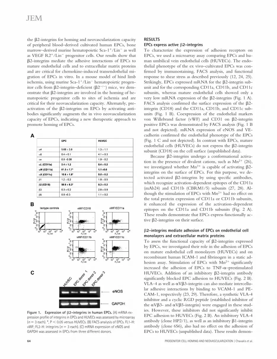
To characterize the expression of adhesion receptors onEPCs, we used a microarray assay comparing EPCs and hu-man umbilical vein endothelial cells (HUVECs). The endo-thelial phenotype of the ex vivo–cultivated EPCs was con-firmed by immunostaining, FACS analysis, and functionalresponse to shear stress as described previously (12, 24, 25).Strikingly, EPCs expressed mRNA for the
�
2-integrin sub-unit and for the corresponding CD11a, CD11b, and CD11csubunits, whereas mature endothelial cells showed only avery low mRNA expression of the
�
2-integrins (Fig. 1 A).FACS analysis confirmed the surface expression of the
�
2-integrin (CD18) and the CD11a, CD11b, and CD11c sub-units (Fig. 1 B). Coexpression of the endothelial markersvon Willebrand factor (vWF) and CD31 on
�
2-integrinpositive EPCs was demonstrated by FACS analysis (Fig. 1 Band not depicted). mRNA expression of eNOS and VE-cadherin confirmed the endothelial phenotype of the EPCs(Fig. 1 C and not depicted). In contrast with EPCs, matureendothelial cells (HUVECs) do not express the
�
2-integrinsubunit (CD18) on the cell surface (unpublished data).
Because
�
2-integrins undergo a conformational activa-tion in the presence of divalent cations, such as Mn
2
�
(26),we investigated whether Mn
2
�
is capable of activating
�
2-integrins on the surface of EPCs. For this purpose, we de-tected activated
�
2-integrins by using specific antibodies,which recognize activation-dependent epitopes of the CD11a(mAb24) and CD11b (CBRM1/5) subunits (27, 28). Al-though the stimulation of EPCs with Mn
2
�
had no effect onthe total protein expression of CD11a or CD11b subunits,it enhanced the expression of the activation-dependentepitopes on the CD11a and CD11b subunits (Fig. 2 A).These results demonstrate that EPCs express functionally ac-tive
�
2-integrins on their surface.
�
2-integrins mediate adhesion of EPCs on endothelial cell monolayers and extracellular matrix proteins
To assess the functional capacity of
�
2-integrins expressedby EPCs, we investigated their role in the adhesion of EPCson mature endothelial cell monolayers (HUVECs) and onrecombinant human ICAM-1 and fibrinogen in a static ad-hesion assay. Stimulation of EPCs with Mn
2
�
significantlyincreased the adhesion of EPCs to TNF-
�
–prestimulatedHUVECs. Addition of an inhibitory
�
2-integrin antibodysignificantly blocked EPC adhesion to HUVECs (Fig. 2 B).VLA–4 as well as
�
V
�
3-integrin can also mediate intercellu-lar adhesive interactions by binding to VCAM-1 and PE-CAM-1, respectively (23, 29). Therefore, a synthetic VLA-4inhibitor and a cyclic RGD peptide (established inhibitor ofthe
�
V
�
3- and
�
V
�
5-integrin) were engaged in these stud-ies. However, these inhibitors did not significantly inhibitEPC adhesion to HUVECs (Fig. 2 B). An inhibitory VLA-4antibody (clone HP2/1), as well as an inhibitory
�
1-integrinantibody (clone 6S6), also had no effect on the adhesion ofEPCs to HUVECs (unpublished data). These results demon-
Figure 1. Expression of �2-integrins in human EPCs. (A) mRNA ex-pression profile of integrins in EPCs and HUVECs was assessed by microarray (n � 3 each). *, P � 0.05 versus HUVECs. (B) FACS analysis of EPCs. FL1-H: vWF; FL2-H: integrins (n � 3 each). (C) mRNA expression of eNOS and GAPDH was assessed in EPCs from three different donors.

JEM VOL. 201, January 3, 2005
65
ARTICLE
strate that EPC adhesion to endothelial cells is predomi-nantly mediated by
�
2-integrins expressed on EPCs.Endothelial ICAM-1 and extracellular matrix-associated
fibrinogen are established ligands for the
�
2-integrins (30–33). Therefore, we investigated whether EPCs are capable ofbinding to immobilized recombinant human ICAM-1 andfibrinogen via
�
2-integrins. Indeed, stimulation with eitherMn
2
�
or an activating
�
2-integrin antibody (KIM185) in-duced adhesion of EPCs to immobilized human ICAM-1and fibrinogen (Fig. 2, C and D). Adhesion induced by bothstimuli was completely abolished in the presence of an inhib-itory
�
2-integrin antibody (Fig. 2, C and D). In contrast, aninhibitory
�
1-integrin antibody had no effect on the adhe-sion of EPCs to human ICAM-1 (unpublished data). Theseresults demonstrate that EPCs bind to fibrinogen and endo-thelial ICAM-1 in a
�
2-integrin–dependent manner.
Role of
�
2-integrins for transmigration of EPCs
We investigated the involvement of
�
2-integrins in the transen-dothelial migration of EPCs in a transwell transmigration assay.Chemoattraction of EPCs by MCP-1, SDF-1
�
, and VEGF sig-nificantly increased the transmigration rate of EPCs through
HUVEC monolayers (Fig. 3). Addition of an inhibitory
�
2-integrin antibody (anti-CD18) significantly reduced EPC trans-migration, whereas an inhibitory
�
1-integrin antibody (anti-CD29) and RGD peptides had no effect (Fig. 3). Moreover, aninhibitory VLA-4 antibody did not affect chemokine-inducedtransendothelial migration of EPCs (unpublished data). Thus,
�
2-integrins, but not
�
1-integrins, mediate chemokine andVEGF-induced transendothelial migration of EPCs (Fig. 3).
Role of
�
2-integrins for neovascularization after ischemia
Next, we determined whether
�
2
���
mice have an impairedneovascularization capacity. Hind limb ischemia was inducedin WT and
�
2
���
mice. Perfusion was assessed 14 d after in-duction of ischemia by laser Doppler imaging. �2��� miceshowed a significantly impaired recovery in limb perfusion ascompared with WT controls (Fig. 4, A and B), suggesting that�2-integrins contribute to neovascularization after ischemia.
Role of �2-integrins for the in vivo homing and neovascularization capacity of Sca-1�/Lin� bone marrow cellsTo assess the in vivo relevance of �2-integrins for progeni-tor cell homing to sites of ischemia and progenitor cell–
Figure 2. Integrins and EPC adhesion. (A) EPCs were activated with 2 mM MnCl2 for 30 min. Activation of integrins was determined by CBRM1/5 and mAb24 antibodies (left) and expression of CD11a and CD11b was con-trolled by FACS analysis (right; n � 3). (B) Adhesion of EPCs to 10 ng/ml TNF-�–prestimulated HUVECs was stimulated where indicated with MnCl2. 105 EPCs/well (in 100 �l adhesion buffer) were added to the HUVEC monolayers in the absence or presence of 30 �g/ml of blocking monoclonal anti–�2-integrin antibodies (clone IB4 or mAb 60.3), 30 �g/ml of murine isotype control antibodies, 50 �M of the inhibitory agents cyclic RGD pep-
tide or 50 �M of VLA-4-inhibitor. The total number of EPCs added to the well is set as 100%. *, P � 0.01 versus MnCl2 � isotype antibody; #, P � 0.01 versus isotype control. (C and D) EPCs were treated with 2 mM MnCl2 or 10 �g/ml �2-integrin–activating antibody. Adhesion to fibrinogen- or ICAM-1–coated plates was detected after 20 min in the presence or absence of �2-integrin–inhibitory antibody or isotype control antibodies (n � 4). *, P � 0.05 versus MnCl2 � isotype IgG; #, P � 0.05 versus activating anti-�2 � isotype IgG.

PROGENITOR CELL HOMING AND NEOVASCULARIZATION | Chavakis et al.66
induced neovascularization, we used bone marrow–derivedstem/progenitor cells in a mouse model of hind limb ische-mia. First, we detected the expression of CD18 on periph-eral blood Sca-1�/c-kit� cells. The �2-integrin subunitCD18 was expressed on 95% of the Sca-1�/c-kit� cells(Fig. 5 A). Peripheral blood cells from �2��� mice wereused as negative control. As expected, no CD18 stainingwas detectable (Fig. 5 A). Second, we isolated Sca-1�/Lin�
progenitor cells from the bone marrow of wild-type miceby magnetic beads and detected the expression of �2-inte-grin by FACS analysis. The majority of isolated Sca-1�/Lin� cells (98.5 0.2%) expressed the CD18 surface anti-gen. Moreover, adhesion of murine bone marrow Lin�
progenitor cells to TNF-�–pretreated mature endothelialcells was significantly blocked by �2-blocking antibodies(unpublished data).
The functional activity of the stem/progenitor cells toimprove neovascularization was assessed by intravenous infu-
sion of Sca-1�/Lin� bone marrow cells derived from eitherwild-type or �2��� mice into athymic mice 24 h after induc-tion of limb ischemia. After 14 d, transplantation of Sca-1�/Lin� bone marrow cells from wild-type mice significantlyenhanced the recovery of blood flow of the ischemic hindlimbs of athymic mice as compared with ischemic hind limbsfrom untreated athymic mice (Fig. 5 B). In contrast, �2���
Sca-1�/Lin� bone marrow cells were significantly less effec-tive for improving recovery of limb perfusion as comparedwith wild-type Sca-1�/Lin� bone marrow cells (Fig. 5 B).
Moreover, histological evaluation of ischemic hind limbsof athymic mice 14 d after cell infusion revealed a signifi-cantly lower capillary density in mice receiving �2��� Sca-1�/Lin� bone marrow cells compared with mice receivingwild-type cells (Fig. 5 C). Furthermore, the number of in-corporated male Sca-1�/Lin� cells in female recipients wasdetermined by fluorescence in situ hybridization for the mu-rine Y-chromosome of the infused male cells. The numberof incorporated Y-chromosome positive cells was signifi-
Figure 3. �2-Integrins mediate the chemokine-induced transendothe-lial migration of EPCs. Transendothelial migration of EPCs was stimu-lated where indicated with 50 ng/ml MCP-1, 100 ng/ml SDF-1, or 50 ng/ml VEGF for 18 h. EPCs transmigrated in the presence or absence of indicated blocking antibodies or isotype control antibodies (30 �g/ml each) or 10 �g/ml RGD peptides. Data of a typical experiment (triplicates) are presented. Similar results were obtained in at least three separate experiments. *, P � 0.05 versus MCP-1, SDF-1, or VEGF � isotype control IgG.
Figure 4. Neovascularization of WT versus CD18��� mice after hind limb ischemia. 14 d after induction of hind limb ischemia, perfusion of the ischemic limb was determined by laser Doppler imaging. Quantification (n � 6 per group) (A) and representative images (B). Arrows indicate isch-emic linbs.

JEM VOL. 201, January 3, 2005 67
ARTICLE
cantly lower for the infusion of �2��� cells as compared withthe infusion of wild-type cells (Fig. 5, D and E).
Similar results were obtained when using isolated VEGFR2�/Lin� bone marrow–derived cells. The majority ofVEGF R2�/Lin� cells expressed CD18 (89 9.6%).Moreover, VEGF R2�/Lin� cells derived from CD18���
mice cells showed a significantly reduced capacity to aug-ment blood flow after ischemia as compared with WT cells(WT 180 15% of untreated control mice; CD18��� cells:125 7% of untreated control mice). These results indicatethat the �2-integrins are involved in the homing of pro-genitor cells to ischemic tissues and their neovascularizationcapacity.
Activation of the �2-integrins improves in vivo homing and neovascularization capacity of EPCsBecause �2-integrins are involved in the homing of EPCs, weinvestigated whether preactivation of �2-integrins may im-prove homing and neovascularization capacity of humanEPCs in the mouse model of hind limb ischemia. Ex vivo–expanded human EPCs isolated from peripheral blood werepretreated with the �2-integrin–activating antibody (KIM185), which was shown before to enhance �2-integrin–dependent adhesion of EPCs to endothelial ICAM-1 or fi-brinogen (Fig. 2), and were subsequently infused into athymicmice. To be able to detect an increase in progenitor cell–medi-ated neovascularization, we used a reduced number of EPCs
Figure 5. Role of �2-integrins for in vivo homing of Sca-1�/Lin� stem/progenitor cells and neovascularization capacity. (A) Peripheral blood was gated on the lymphocyte/monocyte fraction (R1) and stained for Sca-1–FITC (FL1) and c-Kit–allophycocyanin (APC; FL4). Gate R2 defines cells, which are Sca-1�c-Kit�. Cells detected in both R1 and R2 are ana-lyzed for expression of CD18-PE (FL2). Expression of CD18 in Sca-1�/c-Kit� cells was deficient in CD18��� mice. A representative FACS analysis is shown. (B and C) A total of 105 male Sca-1�/Lin� cells were isolated from bone marrow and infused intravenously into female nude mice 24 h after
induction of hind limb ischemia. Laser Doppler-derived blood flow (arrows indicate ischemic limbs) (B) and capillary density (C) was determined after 2 wk (n � 5 per group). (D and E) Incorporation of Y-chromosome positive injected murine Sca-1�/Lin� cells isolated from male wild-type or CD18��� mice in female recipients after hind limb ischemia (n � 4 per group; n � 10 images per mouse were analyzed). Representative images (E) stained for the panendothelial marker MECA-32 (FITC, green), nuclei (Sytox; blue), and Y-chromosome (Cy3-labeled murine probe, red). Arrows indicateY-chromosome positive cells.

PROGENITOR CELL HOMING AND NEOVASCULARIZATION | Chavakis et al.68
(105 cells), which is lower than previously published numbers(5 � 105 EPCs; reference 25), to yield a 50% improvement ofneovascularization as compared with untreated mice.
Preincubation of the EPCs with the activating �2-inte-grin antibody resulted in a significantly enhanced neovascu-larization capacity of infused EPCs in comparison withcontrol antibody-treated EPCs as assessed by laser Dopplerimaging (Fig. 6 A). Incorporation of human EPCs was de-
tected by confocal microscopy using antibodies directedagainst human HLA and the endothelial marker proteinvWF (Fig. 6, B–D). Incorporation of �2-activating anti-body-treated EPCs into the ischemic muscle was increasedin comparison with control antibody-treated EPCs (Fig. 6, Band C). Moreover, the numbers of capillaries and small arte-rioles (20–50 �m) were significantly augmented in micetreated with preactivated EPCs (Fig. 6, E and F; capillary
Figure 6. Activation of �2-integrins improves in vivo homing and neovascularization capacity of EPCs. (A) EPCs were pretreated with �2-integrin–activating antibody or isotype control antibody (20 �g/ml each) for 30 min, and 105 cells were injected intravenously in nude mice 24 h after induction of hind limb ischemia. Perfusion was determined by laser Doppler imaging after 2 wk (n � 6 per group). (B–D) Incorporation of human EPCs (stained for HLA-ABC; Alexa 555, red) into vessels (endothelial marker CD31-FITC, green) after pretreatment with isotype control antibody or ac-
tivating �2-integrin antibody. Double positive cells were quantified (B) and representative overviews (C) and high power images (D) are shown from 10 sections provided by five mice per group. (E and F) Conductance vessels were identified by staining for smooth muscle �-actin using a Cy3-labeled mouse monoclonal antibody (red). The number of small (20–50 �m), medium (50–100 �m), and large vessels (100 �m) was counted separately. Data are mean SEM (n � 5). (A, B, and E) *, P � 0.05 versus no cells; **, P � 0.05 versus isotype Ab-treated EPCs.

JEM VOL. 201, January 3, 2005 69
ARTICLE
density: EPC � control mAb: 0.77 0.10 capillaries permyocyte; EPC � activating mAb: 1.32 0.09; P � 0.003).Thus, an external activation of the �2-integrins by an acti-vating antibody before infusion is capable of improving theneovascularization capacity of EPCs.
DISCUSSIONThe data of the present paper underscore the importance of�2-integrins for the proangiogenic activity of EPCs andbone marrow–derived progenitor cells. Specifically, our in-vestigations revealed the following: (a) EPCs as well as he-matopoietic stem/progenitor cells express �2-integrins; (b)�2-integrins expressed on EPCs can be activated by Mn2�
and can mediate the adhesion of EPCs to mature endothelialcells, to recombinant human ICAM-1, and to fibrinogenand the chemokine-induced transendothelial migration ofEPCs; (c) �2��� animals display a neovascularization defectin the model of hind limb ischemia; (d) �2-integrins are in-volved in the in vivo homing of progenitor cells to sites ofischemia and their vascular integration and significantly con-tributed to the neovascularization capacity of infused bonemarrow Sca-1�/Lin� or VEGF R2�/Lin� progenitor cellsin the mouse model of hind limb ischemia as demonstratedusing the �2��� progenitor cell populations; (e) stimulationof ex vivo–expanded human EPCs by preincubation with anactivating �2-integrin antibody significantly enhanced thehoming of EPCs to sites of ischemia and EPC-inducedneovascularization. Therefore, the present paper unravels anovel function of the �2-integrin subunit CD18 for neovas-cularization exceeding its well-known function in innate andadaptive immune responses.
Increasing evidence suggests that �2-integrins are notonly expressed on differentiated leukocytes but also on he-matopoietic stem/progenitor cells (34, 35). Our in vitro datasuggest that �2-integrins expressed on EPCs mediate hom-ing functions such as endothelial adhesion and transmi-gration. Moreover, �2-integrins contribute to the in vivohoming of bone marrow–derived progenitor cells to ische-mic tissue. In line with these data, it has been reported pre-viously that �2-integrins mediate adhesion and transmigra-tion of hematopoietic stem/progenitor cells (36–38). In arecent paper assessing in vivo homing of embryonic EPCsderived from cord blood, the circulating cells arrested withintumor microvessels extravasated into the interstitium and in-corporated into neovessels, suggesting that adhesion andtransmigration are involved in the recruitment of EPCs tosites of tumor angiogenesis (39). Thus, it is conceivable tospeculate that ex vivo–expanded adult EPCs and hemato-poietic stem/progenitor cells may engage similar pathwaysfor recruitment to sites of ischemia and incorporation intonewly forming vessels.
Our in vivo data provide the first evidence for a directparticipation of �2-integrins in neovascularization processesand particularly in stem/progenitor cell–mediated, ischemia-induced vasculogenesis. Adamis and colleagues previouslyhighlighted the role of �2-integrins for corneal and choroi-
dal angiogenesis induced by injury (40, 41). In both studies,�2��� mice displayed a reduced inflammation-associated an-giogenic response after injury and these effects were associ-ated with reduced inflammatory cell infiltrates (40, 41). Yet,no incorporation of leukocytes into new vessels was reportedin these studies. Moreover, the same group demonstratedthat, in the case of retinal ischemia, leukocyte–endothelialcell interactions contribute to the development of ischemiaby inducing vascular obliteration via Fas ligand–mediatedendothelial cell apoptosis (42). In contrast with these find-ings, our data provide evidence that, during hind limb ische-mia, intravenous infusion of bone marrow hematopoieticprogenitor cells leads to incorporation of the transplantedcells in newly formed vessels and to improvement of neovas-cularization in an at least partially �2-integrin–dependentmanner. As opposed to EPCs, infusion of inflammatory cells,such as monocytes/macrophages, had only a slight if any ef-fect on the neovascularization of ischemic limbs in themodel of hind limb ischemia in athymic mice (25). Thus,our results support a novel direct function of �2-integrinsin progenitor cell–induced vasculogenesis during ischemia,which is distinct from the indirect role of �2-integrins in theinflammation-associated angiogenesis described by Adamisand colleagues (40, 41). Because the �2��� mice display nodefect in the mobilization of progenitor cells (43), the neo-vascularization defect in the �2��� mice in the model ofhind limb ischemia is most conceivably mediated by a hom-ing defect of progenitor cells into ischemic tissue.
Interestingly, our present data indicate that the recruit-ment of hematopoietic progenitor cells to sites of ischemia ismediated at least in part by different mechanisms comparedwith the homing of infused cells into the bone marrow of le-thally irradiated recipient mice, which is predominantly me-diated via �4�1 (17, 43). In this context, �2-integrins onlyact in a synergistic manner together with the �4�1-integrin.Our finding that �2-integrin deficiency does not completelyblock homing and neovascularization improvement after in-fusion of Sca-1�/Lin� bone marrow cells suggests that othermechanisms may additionally be involved in these processes.We cannot exclude that �4�1-integrin partially compensatesfor the lack of �2-integrin during in vivo homing of Sca-1�/Lin� bone marrow cells. Interestingly, the homing of inflam-matory cells during pneumonia or myocardial ischemia in�2��� mice is mediated by the �4�1-integrin (44, 45).Moreover, the initial cell arrest of embryonic progenitor cellhoming during tumor angiogenesis was suggested to bemediated by E- and P-selectin and P-selectin glycoproteinligand-1 (39). Yet, it is important to underscore that thiswork was performed with embryonic EPCs, whereas weused adult EPCs and bone marrow stem/progenitor cells. It islikely that different cell types may use distinct mechanisms forhoming to sites of ischemia. In addition, it is well establishedthat interactions of selectins with selectin–ligands mediate therolling of cells on the surface of endothelial cells as the initialstep of homing (21). Further studies are needed to elucidate apotentially synergistic role of other adhesion molecules and

PROGENITOR CELL HOMING AND NEOVASCULARIZATION | Chavakis et al.70
their counterligands for the multistep recruitment process ofadult endothelial progenitor and stem cells to ischemic tissue.
Regardless of potentially additive mechanisms involvedin the recruitment of stem/progenitor cells to areas of ische-mia, our data clearly demonstrate that preincubation ofEPCs with a �2-integrin–activating antibody markedly en-hanced the incorporation of transplanted EPCs in vesselsand the neovascularization of ischemic limbs. The peripheralblood–derived EPCs used in the present paper are alreadyused in clinical trials to improve neovascularization in pa-tients with ischemic heart diseases (15). Thus, our resultscould have important clinical implications as they disclose amechanism to enhance homing of EPCs and, thereby, im-prove neovascularization capacity of infused EPCs.
In summary, the present paper demonstrates for the firsttime a critical role of �2-integrins in vitro and in vivo forhoming and neovascularization capacity of endothelial pro-genitor and hematopoietic progenitor cells. Moreover, ourresults show that activation of �2-integrins appears to be afeasible and promising tool to improve the efficacy of EPC-induced neovascularization. A better understanding of thehoming mechanisms of EPCs may lead to the developmentof new therapeutic strategies for improvement of vasculo-genesis in patients with ischemic diseases.
MATERIALS AND METHODSCell cultureMononuclear cells (MNCs) were isolated by density–gradient centrifuga-tion with Ficoll from peripheral blood of healthy human volunteers as de-scribed previously (46). Immediately after isolation, total MNCs (8 � 106
cells/ml medium; cell density 2.5 � 106 cells/cm2) were plated on culturedishes coated with 10 �g/ml human fibronectin (Sigma-Aldrich) andmaintained in endothelial basal medium (Cambrex) supplemented with 1�g/ml hydrocortisone, 12 �g/ml bovine brain extract, 50 �g/ml genta-mycin, 50 ng/ml amphotericin B, 10 ng/ml epidermal growth factor, and20% FCS. After 3 d, nonadherent cells were removed, and adherent cellswere incubated in medium for another 24 h before initiation of the exper-iments. EPCs were characterized by dual staining for 1,1 –dioctadecyl–3,3 ,3 –tetramethylindo-carbocyanine–labeled acetyl low-density lipopro-tein, lectin, and expression of endothelial markers KDR, VE-cadherin, andvWF (25).
Sca-1�/Lin� cells were purified from BM MNCs from wild-type andCD18��� mice by negative selection using a cocktail of biotinylated anti-bodies to lineage markers (Lineage cell depletion kit, mouse; Miltenyi Bio-tec) for 10 min at 4�C followed by antibiotin microbeads for 15 min (Mil-tenyi Biotec). The Lin� BM cells were incubated with anti–Sca-1 microbeads(Miltenyi Biotec) for 15 min and Sca-1�/Lin� BM cells were collected (7).To obtain VEGFR2� Lin� cells, Lin� cells were incubated with biotinyl-ated Flk-1 antibodies (DSB-X biotin protein labeling kit; Molecular Probes;antibody was obtained from BD Biosciences) for 30 min at 4�C followed byantibiotin microbeads for 15 min.
HUVECs were purchased from Cambrex and cultured in endothelialbasal medium supplemented with 1 �g/ml hydrocortisone, 12 �g/ml bo-vine brain extract, 50 �g/ml gentamycin, 50 ng/ml amphotericin-B, 10ng/ml epidermal growth factor, and 10% FCS until the third passage. Afterdetachment with trypsin, cells (4 � 105 cells) were grown in 6-cm cell cul-ture dishes or 96-well plates for at least 18 h as described previously (47).
Oligonucleotide microarrays, FACS10 �g of total RNA was hybridized to the HG-U95Av2 microarray (9670human genes; Affymetrix, Inc.). The standard protocol used for sample
preparation and microarray processing is available from Affymetrix, Inc. Ex-pression data were analyzed using Microarray Suite version 5.0 (Affymetrix,Inc.) and GeneSpring version 4.2 (Silicon Genetics). 3 � 105 human EPCs,peripheral blood, or isolated Sca-1�/Lin� cells were incubated for 30 min at4�C with FITC- or PE-labeled antibodies (anti-CD11a, -CD11b, -CD11c,-CD18, –Sca-1, and –c-kit; BD Biosciences; anti-vWF was obtained fromAcris) or CBRM1/5-antibody (BD Biosciences) for 30 min at 37�C. ThemAb24 antibody (provided by N. Hogg, Cancer Research UK LondonResearch Institute, London, England, UK) was incubated for 30 min at 4�Cand detected with a secondary FITC-labeled goat anti–mouse antibody(DakoCytomation). Surface expression was quantified using a FACS Cali-bur (BD Biosciences).
Adhesion, transmigration experimentsCell–cell adhesion. Cell–cell adhesion was performed as described previ-ously (48, 49). Confluent HUVEC monolayers were stimulated with TNF-�
(Sigma-Aldrich) for 24 h. Ex vivo–expanded EPCs were stained withCell Tracker green-CMFDA (Molecular Probes) and were resolved in ad-hesion buffer (150 mM NaCl, 20 mM Hepes, 2 mM MgCl2, 0.05% BSA,pH 7.4). A total of 105 EPCs/well (in 100 �l adhesion buffer) was added tothe HUVEC monolayers in the absence or presence of blocking monoclo-nal �2-integrin antibodies (clone IB4; Qbiogene; or mAb 60.3; J. Harlan,University of Washington, Seattle, WA), murine isotype control antibodies(Qbiogene), inhibitory agents cyclic RGD peptide or VLA-4 inhibitor(4-[{2-methyl-phenyl}aminocarbonyl]aminophenyl)acetyl-fibronectin-CS-1fragment (1980–1983). After 20 min of incubation (37�C), the plates werewashed twice with adhesion buffer at room temperature to remove nonad-herent cells. Adherent cell tracker green-labeled EPCs were quantified intriplicates on a fluorescence plate reader (Fluostat; BMG Lab Technologies).
Cell–matrix adhesion. Cell–matrix adhesion was performed as describedpreviously (48, 49). 96-well plates were coated overnight (4�C) with 10�g/ml human fibrinogen (Enzyme Research Laboratories) or soluble re-combinant human ICAM-1 (Bender MedSystems) and blocked with 1%(wt/vol) BSA for 1 h at room temperature. Ex vivo–expanded humanEPCs in adhesion buffer were seeded at 1.2 � 105 cells/well in 100 �l inthe absence or presence of 2 mM MnCl2 or activating human �2-integrinantibody (clone KIM185; M. Robinson, Celltech Ltd., Slough, England,UK) and were incubated with blocking �2-integrin mAb (clone IB4 ormAb 60.3) or murine isotype control antibodies (Qbiogene) for 20 min at37�C. After removal of nonadherent cells by two washing steps, adhesionwas quantified in triplicates by counting adherent cells in five randomly se-lected fields per well (magnification, 20; Axiovert 100; Carl Zeiss Micro-Imaging, Inc.).
Transmigration. Transendothelial migration was performed as describedpreviously (50) using 6.5-mm transwell filters with 8-�m pore size (Costar).After inserts were coated with 0.2% gelatin (Sigma-Aldrich), HUVECswere seeded on transwell filters and cultivated for 48 h before the experi-ments were performed in a humidified atmosphere (37�C, 5% CO2). At thebeginning of the experiment, 600 �l of migration assay medium (serum-free RPMI 1640 in the absence or presence of MCP-1, SDF1�, or VEGF;R&D Systems) was added to the lower compartment of the transwell sys-tem. EPCs (5 � 105 in 100 �L) were added to the top compartment in thepresence or absence of 30 �g/ml of blocking anti–�2-integrin antibody(mAb 60.3), anti–�1-integrin antibody (clone 6S6; Chemicon), anti–�4-inte-grin antibody (clone HP2.1; Immunotech), murine isotype control antibod-ies (Qbiogene), or RGD peptides. After 18 h at 37�C, the number of cellstransmigrated to the bottom compartment was quantified in duplicates witha cell counter (CASY-Counter; Schärfe-System). All inserts were fixed andstained to confirm the confluence of the endothelial monolayer.
Animal experimentsMice. 8-wk-old �2��� mice and their age-matched wild-type littermates(either 129/Sv or C57BL/6J) were generated as described previously (51).

JEM VOL. 201, January 3, 2005 71
ARTICLE
All mice were genotyped by Southern blot analysis (51) and maintained un-der pathogen-free conditions. Athymic NMRI nude mice (6–8 wk) wereobtained from The Jackson Laboratory. The animal experiments were ap-proved from the Regional Board of Land Hessen, Germany.
Model of hind limb ischemia. The proximal femoral artery, includingthe superficial and the deep branch as well as the distal saphenous artery,were ligated. In transplantation experiments, progenitor cells were intrave-nously injected in nude mice 24 h after induction of limb ischemia. HumanEPCs were pretreated with 20 �g/ml activating �2-integrin antibody (cloneKIM 185) or isotype control antibody for 30 min at 37�C and washed twiceto remove unbound antibodies before injection (105 EPCs/mouse). In someexperiments, sex-mismatched murine Sca-1�/Lin� or Flk-1�/Lin� bonemarrow cells from male �2��� or wild-type mice were used. After 2 wk, wecalculated the ischemic (right) versus normal (left) limb blood flow ratio us-ing a Laser Doppler blood flow imager (Moor Instruments).
Histology. The capillary density and the number and size of conductantvessels in the semimembraneous and adductor muscles were determined us-ing 8-�m cryosections. Endothelial cells were identified with the panendo-thelial marker MECA-32 followed by donkey anti–rat IgG Alexa488 orCD31-FITC (BD Biosciences). Injected human EPCs were identified bycostaining for HLA-ABC (allophycocyanin labeled; BD Biosciences) andvWF (Acris). Male murine BM-derived cells were identified by fluores-cence in situ hybridization for the murine Y-chromosome (Cy3-labeledprobe: Cambio; reference 7). Nuclei were stained with Sytox (MolecularProbes). Images were obtained by confocal microscopy (LSM 510; CarlZeiss MicroImaging, Inc.).
Statistical analysisContinuous variables are expressed as mean SD or SEM. Comparisonsbetween groups were analyzed by Student’s t test (two-sided) or analysis ofvariance with Bonferroni adjustment for experiments with more than twosubgroups (SPSS 11.5 software). p-values �0.05 were considered as statisti-cally significant.
We thank M. Muhly-Reinholz, T. Röxe, and M. Näher for their excellent technical assistance. Moreover, we thank Drs. N. Hogg, J. Harlan, and M. Robinson for providing the antibodies mAb24, mAb60.3, and KIM185, respectively.
This work was supported by the Forschergruppe 501 (He 3044/2-2 to C. Heeschen) and the Alfried Krupp-Stiftung (to S. Dimmeler). K. Sasaki was in part supported by the Japan Heart Foundation/Bayer Yakuhin Research Grant Abroad. The work of K. Scharffetter-Kochanek was funded in party by the Collaborative Research Center SFB497-C7, Ulm.
The authors have no conflicting financial interests.
Submitted: 12 July 2004Accepted: 19 November 2004
REFERENCES1. Carmeliet, P. 2000. Mechanisms of angiogenesis and arteriogenesis.
Nat. Med. 6:389–395.2. Asahara, T., T. Murohara, A. Sullivan, M. Silver, R. van der Zee, T.
Li, B. Witzenbichler, G. Schatteman, and J.M. Isner. 1997. Isolation ofputative progenitor endothelial cells for angiogenesis. Science. 275:964–967.
3. Shi, Q., S. Rafii, M.H. Wu, E.S. Wijelath, C. Yu, A. Ishida, Y. Fujita,S. Kothari, R. Mohle, L.R. Sauvage, et al. 1998. Evidence for circulat-ing bone marrow-derived endothelial cells. Blood. 92:362–367.
4. Asahara, T., H. Masuda, T. Takahashi, C. Kalka, C. Pastore, M. Silver,M. Kearne, M. Magner, and J.M. Isner. 1999. Bone marrow origin ofendothelial progenitor cells responsible for postnatal vasculogenesis inphysiological and pathological neovascularization. Circ. Res. 85:221–228.
5. Grant, M.B., W.S. May, S. Caballero, G.A. Brown, S.M. Guthrie,R.N. Mames, B.J. Byrne, T. Vaught, P.E. Spoerri, A.B. Peck, andE.W. Scott. 2002. Adult hematopoietic stem cells provide functionalhemangioblast activity during retinal neovascularization. Nat. Med.
8:607–612.6. Rafii, S., and D. Lyden. 2003. Therapeutic stem and progenitor cell
transplantation for organ vascularization and regeneration. Nat. Med.9:702–712.
7. Aicher, A., C. Heeschen, C. Mildner-Rihm, C. Urbich, C. Ihling, K.Technau-Ihling, A.M. Zeiher, and S. Dimmeler. 2003. Essential roleof endothelial nitric oxide synthase for mobilization of stem and pro-genitor cells. Nat. Med. 9:1370–1376.
8. Takahashi, T., C. Kalka, H. Masuda, D. Chen, M. Silver, M. Kearney,M. Magner, J.M. Isner, and T. Asahara. 1999. Ischemia- and cytokine-induced mobilization of bone marrow-derived endothelial progenitorcells for neovascularization. Nat. Med. 5:434–438.
9. Shintani, S., T. Murohara, H. Ikeda, T. Ueno, T. Honma, A. Katoh,K. Sasaki, T. Shimada, Y. Oike, and T. Imaizumi. 2001. Mobilizationof endothelial progenitor cells in patients with acute myocardial infarc-tion. Circulation. 103:2776–2779.
10. Asahara, T., T. Takahashi, H. Masuda, C. Kalka, D. Chen, H.Iwaguro, Y. Inai, M. Silver, and J.M. Isner. 1999. VEGF contributes topostnatal neovascularization by mobilizing bone marrow-derived en-dothelial progenitor cells. EMBO J. 18:3964–3972.
11. Lyden, D., K. Hattori, S. Dias, C. Costa, P. Blaikie, L. Butros, A.Chadburn, B. Heissig, W. Marks, L. Witte, et al. 2001. Impaired re-cruitment of bone-marrow-derived endothelial and hematopoieticprecursor cells blocks tumor angiogenesis and growth. Nat. Med.7:1194–1201.
12. Kalka, C., H. Masuda, T. Takahashi, W.M. Kalka-Moll, M. Silver, M.Kearney, T. Li, J.M. Isner, and T. Asahara. 2000. Transplantation of exvivo expanded endothelial progenitor cells for therapeutic neovascular-ization. Proc. Natl. Acad. Sci. USA. 97:3422–3427.
13. Kocher, A.A., M.D. Schuster, M.J. Szabolcs, S. Takuma, D. Burkhoff,J. Wang, S. Homma, N.M. Edwards, and S. Itescu. 2001. Neovascu-larization of ischemic myocardium by human bone-marrow-derivedangioblasts prevents cardiomyocyte apoptosis, reduces remodeling andimproves cardiac function. Nat. Med. 7:430–436.
14. Kawamoto, A., H.C. Gwon, H. Iwaguro, J.I. Yamaguchi, S. Uchida,H. Masuda, M. Silver, H. Ma, M. Kearney, J.M. Isner, and T. Asahara.2001. Therapeutic potential of ex vivo expanded endothelial progeni-tor cells for myocardial ischemia. Circulation. 103:634–637.
15. Assmus, B., V. Schachinger, C. Teupe, M. Britten, R. Lehmann, N. Do-bert, F. Grunwald, A. Aicher, C. Urbich, H. Martin, et al. 2002. Trans-plantation of progenitor cells and regeneration enhancement in acutemyocardial infarction (TOPCARE-AMI). Circulation. 106:3009–3017.
16. Aicher, A., W. Brenner, M. Zuhayra, C. Badorff, S. Massoudi, B. Ass-mus, T. Eckey, E. Henze, A.M. Zeiher, and S. Dimmeler. 2003. As-sessment of the tissue distribution of transplanted human endothelialprogenitor cells by radioactive labeling. Circulation. 107:2134–2139.
17. Papayannopoulou, T., G.V. Priestley, B. Nakamoto, V. Zafiropoulos,and L.M. Scott. 2001. Molecular pathways in bone marrow homing:dominant role of alpha(4)beta(1) over beta(2)-integrins and selectins.Blood. 98:2403–2411.
18. Muller, W.A., S.A. Weigl, X. Deng, and D.M. Phillips. 1993. PE-CAM-1 is required for transendothelial migration of leukocytes. J.Exp. Med. 178:449–460.
19. Springer, T.A. 1994. Traffic signals for lymphocyte recirculation andleukocyte emigration: the multistep paradigm. Cell. 76:301–314.
20. Carlos, T.M., and J.M. Harlan. 1994. Leukocyte-endothelial adhesionmolecules. Blood. 84:2068–2101.
21. Muller, W.A. 2002. Leukocyte-endothelial cell interactions in the in-flammatory response. Lab. Invest. 82:521–533.
22. Schenkel, A.R., Z. Mamdouh, and W.A. Muller. 2004. Locomotionof monocytes on endothelium is a critical step during extravasation.Nat. Immunol. 5:393–400.
23. Plow, E.F., T.A. Haas, L. Zhang, J. Loftus, and J.W. Smith. 2000.Ligand binding to integrins. J. Biol. Chem. 275:21785–21788.
24. Kawamoto, A., T. Asahara, and D.W. Losordo. 2002. Transplantationof endothelial progenitor cells for therapeutic neovascularization. Car-diovasc. Radiat. Med. 3:221–225.
25. Urbich, C., C. Heeschen, A. Aicher, E. Dernbach, A.M. Zeiher, andS. Dimmeler. 2003. Relevance of monocytic features for neovascular-

PROGENITOR CELL HOMING AND NEOVASCULARIZATION | Chavakis et al.72
ization capacity of circulating endothelial progenitor cells. Circulation.108:2511–2516.
26. Takagi, J., and T.A. Springer. 2002. Integrin activation and structuralrearrangement. Immunol. Rev. 186:141–163.
27. Dransfield, I., and N. Hogg. 1989. Regulated expression of Mg2�binding epitope on leukocyte integrin alpha subunits. EMBO J. 8:3759–3765.
28. Diamond, M.S., and T.A. Springer. 1993. A subpopulation of Mac-1(CD11b/CD18) molecules mediates neutrophil adhesion to ICAM-1and fibrinogen. J. Cell Biol. 120:545–556.
29. Piali, L., P. Hammel, C. Uherek, F. Bachmann, R.H. Gisler, D.Dunon, and B.A. Imhof. 1995. CD31/PECAM-1 is a ligand for alphav beta 3 integrin involved in adhesion of leukocytes to endothelium. J.Cell Biol. 130:451–460.
30. Altieri, D.C., R. Bader, P.M. Mannucci, and T.S. Edgington. 1988.Oligospecificity of the cellular adhesion receptor Mac-1 encompassesan inducible recognition specificity for fibrinogen. J. Cell Biol. 107:1893–1900.
31. Marlin, S.D., and T.A. Springer. 1987. Purified intercellular adhesionmolecule-1 (ICAM-1) is a ligand for lymphocyte function-associatedantigen 1 (LFA-1). Cell. 51:813–819.
32. Loike, J.D., B. Sodeik, L. Cao, S. Leucona, J.I. Weitz, P.A. Detmers,S.D. Wright, and S.C. Silverstein. 1991. CD11c/CD18 on neutrophilsrecognizes a domain at the N terminus of the A alpha chain of fibrino-gen. Proc. Natl. Acad. Sci. USA. 88:1044–1048.
33. Diamond, M.S., D.E. Staunton, A.R. de Fougerolles, S.A. Stacker,J. Garcia-Aguilar, M.L. Hibbs, and T.A. Springer. 1990. ICAM-1(CD54): a counter-receptor for Mac-1 (CD11b/CD18). J. Cell Biol.111:3129–3139.
34. Becker, P.S., S.K. Nilsson, Z. Li, V.M. Berrios, M.S. Dooner, C.L.Cooper, C.C. Hsieh, and P.J. Quesenberry. 1999. Adhesion receptorexpression by hematopoietic cell lines and murine progenitors: modu-lation by cytokines and cell cycle status. Exp. Hematol. 27:533–541.
35. Orschell-Traycoff, C.M., K. Hiatt, R.N. Dagher, S. Rice, M.C. Yo-der, and E.F. Srour. 2000. Homing and engraftment potential of Sca-1(�)lin(�) cells fractionated on the basis of adhesion molecule expres-sion and position in cell cycle. Blood. 96:1380–1387.
36. Kollet, O., A. Spiegel, A. Peled, I. Petit, T. Byk, R. Hershkoviz, E.Guetta, G. Barkai, A. Nagler, and T. Lapidot. 2001. Rapid and effi-cient homing of human CD34(�)CD38(�/low)CXCR4(�) stem andprogenitor cells to the bone marrow and spleen of NOD/SCID andNOD/SCID/B2m(null) mice. Blood. 97:3283–3291.
37. Peled, A., V. Grabovsky, L. Habler, J. Sandbank, F. Arenzana-Seis-dedos, I. Petit, H. Ben-Hur, T. Lapidot, and R. Alon. 1999. Thechemokine SDF-1 stimulates integrin-mediated arrest of CD34(�)cells on vascular endothelium under shear flow. J. Clin. Invest. 104:1199–1211.
38. Peled, A., O. Kollet, T. Ponomaryov, I. Petit, S. Franitza, V. Grabovsky,M.M. Slav, A. Nagler, O. Lider, R. Alon, et al. 2000. The chemokineSDF-1 activates the integrins LFA-1, VLA-4, and VLA-5 on immaturehuman CD34(�) cells: role in transendothelial/stromal migration andengraftment of NOD/SCID mice. Blood. 95:3289–3296.
39. Vajkoczy, P., S. Blum, M. Lamparter, R. Mailhammer, R. Erber, B.Engelhardt, D. Vestweber, and A.K. Hatzopoulos. 2003. Multistep na-
ture of microvascular recruitment of ex vivo–expanded embryonic en-dothelial progenitor cells during tumor angiogenesis. J. Exp. Med. 197:1755–1765.
40. Sakurai, E., H. Taguchi, A. Anand, B.K. Ambati, E.S. Gragoudas, J.W.Miller, A.P. Adamis, and J. Ambati. 2003. Targeted disruption of theCD18 or ICAM-1 gene inhibits choroidal neovascularization. Invest.Ophthalmol. Vis. Sci. 44:2743–2749.
41. Moromizato, Y., S. Stechschulte, K. Miyamoto, T. Murata, A. Tsu-jikawa, A.M. Joussen, and A.P. Adamis. 2000. CD18 and ICAM-1-dependent corneal neovascularization and inflammation after limbal in-jury. Am. J. Pathol. 157:1277–1281.
42. Ishida, S., K. Yamashiro, T. Usui, Y. Kaji, Y. Ogura, T. Hida, Y.Honda, Y. Oguchi, and A.P. Adamis. 2003. Leukocytes mediate reti-nal vascular remodeling during development and vaso-obliteration indisease. Nat. Med. 9:781–788.
43. Papayannopoulou, T., G.V. Priestley, B. Nakamoto, V. Zafiropoulos,L.M. Scott, and J.M. Harlan. 2001. Synergistic mobilization of he-mopoietic progenitor cells using concurrent beta1 and beta2 integrinblockade or beta2-deficient mice. Blood. 97:1282–1288.
44. Bowden, R.A., Z.M. Ding, E.M. Donnachie, T.K. Petersen, L.H.Michael, C.M. Ballantyne, and A.R. Burns. 2002. Role of alpha4 inte-grin and VCAM-1 in CD18-independent neutrophil migration acrossmouse cardiac endothelium. Circ. Res. 90:562–569.
45. Tasaka, S., S.E. Richer, J.P. Mizgerd, and C.M. Doerschuk. 2002.Very late antigen-4 in CD18-independent neutrophil emigration dur-ing acute bacterial pneumonia in mice. Am. J. Respir. Crit. Care Med.166:53–60.
46. Dimmeler, S., A. Aicher, M. Vasa, C. Mildner-Rihm, K. Adler, M.Tiemann, H. Rutten, S. Fichtlscherer, H. Martin, and A.M. Zeiher.2001. HMG-CoA reductase inhibitors (statins) increase endothelialprogenitor cells via the PI 3-kinase/Akt pathway. J. Clin. Invest. 108:391–397.
47. Chavakis, E., E. Dernbach, C. Hermann, U.F. Mondorf, A.M. Zeiher,and S. Dimmeler. 2001. Oxidized LDL inhibits vascular endothelialgrowth factor-induced endothelial cell migration by an inhibitory ef-fect on the Akt/endothelial nitric oxide synthase pathway. Circulation.103:2102–2107.
48. Chavakis, T., A. Bierhaus, N. Al-Fakhri, D. Schneider, S. Witte, T.Linn, M. Nagashima, J. Morser, B. Arnold, K.T. Preissner, and P.P.Nawroth. 2003. The pattern recognition receptor (RAGE) is a coun-terreceptor for leukocyte integrins: a novel pathway for inflammatorycell recruitment. J. Exp. Med. 198:1507–1515.
49. Chavakis, T., M. Hussain, S.M. Kanse, G. Peters, R.G. Bretzel, J.I.Flock, M. Herrmann, and K.T. Preissner. 2002. Staphylococcus aureusextracellular adherence protein serves as anti-inflammatory factor byinhibiting the recruitment of host leukocytes. Nat. Med. 8:687–693.
50. Rohnelt, R.K., G. Hoch, Y. Reiss, and B. Engelhardt. 1997. Immu-nosurveillance modelled in vitro: naive and memory T cells spontane-ously migrate across unstimulated microvascular endothelium. Int. Im-munol. 9:435–450.
51. Scharffetter-Kochanek, K., H. Lu, K. Norman, N. van Nood, F. Mu-noz, S. Grabbe, M. McArthur, I. Lorenzo, S. Kaplan, K. Ley, et al.1998. Spontaneous skin ulceration and defective T cell function inCD18 null mice. J. Exp. Med. 188:119–131.