integrins, synaptic plasticity and epileptogenesis
TRANSCRIPT

Recent Advances in Epilepsy Research

ADVANCES IN EXPERIMENTAL MEDICINE AND BIOLOGY
Editorial Board:
NATHAN BACK, State University of New York at Buffalo
IRUN R. COHEN, The Weizmann Institute of Science
DAVID KRITCHEVSKY, Wistar Institute
ABEL LAJTHA, N.S. Kline Institute for Psychiatric Research
RODOLFO PAOLETTI, University of Milan
Recent Volumes in this Series
Volume 533RETINAL DEGENERATIONS: Mechanisms and Experimental Theory
Edited by Matthew M. LaVail, Joe G. Hollyfield, and Robert E. Anderson
Volume 534TISSUE ENGINEERING, STEM CELLS, AND GENE THERAPIES
Edited by Y. Murat Elçin
Volume 535GLYCOBIOLOGY AND MEDICINE
Edited by John S. Axford
Volume 536CHEMORECEPTION: From Cellular Signaling to Functional Plasticity
Edited by Jean-Marc Pequignot, Constancio Gonzalez, Colin A. Nurse,Nanduri R. Prabhakar, and Yvette Dalmaz
Volume 537MATHEMATICAL MODELING IN NUTRITION AND THE HEALTH SCIENCES
Edited by Janet A. Novotny, Michael H. Green, and Ray C. Boston
Volume 538MOLECULAR AND CELLULAR ASPECTS OF MUSCLE CONTRACTION
Edited by Haruo Sugi
Volume 539BLADDER DISEASE: Research Concepts and Clinical Applications
Edited by Anthony Atala and Debra Slade
Volume 540OXYGEN TRANSPORT TO TISSUE, VOLUME XXV
Edited by Maureen S. Thorniley, David K. Harrison, and Philip E. James
Volume 541FRONTIERS IN CLINICAL NEUROSCIENCE: Neurodegeneration and Neuroprotection
Edited by László Vécsei
Volume 542QUALITY OF FRESH AND PROCESSED FOODS
Edited by Fereidoon Shahidi, Arthur M. Spanier, Chi-Tang Ho, and Terry Braggins
A Continuation Order Plan is available for this series. A continuation order will bring delivery of each newvolume immediately upon publication. Volumes are billed only upon actual shipment. For further informationplease contact the publisher.

Recent Advances
in Epilepsy Research
Edited by
Devin K. Binder
Department of Neurological Surgery, University of California at San Francisco,
Moffitt Hospital, San Francisco, California, U.S.A.
Helen E. Scharfman
Center for Neural Recovery and Rehabilitation Research, Helen Hayes Hospital,
New York State Department of Health, West Haverstraw, New York, U.S.A.
Departments of Pharmacology and Neurology, Columbia University, New York,
New York, U.S.A.
Kluwer Academic / Plenum PublishersNew York, Boston, Dordrecht, London, Moscow
Landes Bioscience / Eurekah.comGeorgetown, Texas U.S.A.

Recent advances in epilepsy research / edited by Devin K. Binder, HelenE. Scharfman. p. ; cm. -- (Advances in experimental medicine and biology ; v.548)Includes bibliographical references and index. ISBN 0-306-47860-9 1. Epilepsy--Genetic aspects. 2. Epilepsy--Research. 3.Neuromuscular diseases--Research. [DNLM: 1. Epilepsy--physiopathology. 2. Epilepsy--genetics. WL 385R294 2003] I. Binder, Devin K. II. Scharfman, Helen E. III. Series.RC372.R38 2003616.8'53--dc22 2003022445
Kluwer Academic / Plenum PublishersEurekah.com / Landes Bioscience
Copyright ©2004 Eurekah.com and Kluwer Academic / Plenum Publishers
All rights reserved.No part of this book may be reproduced or transmitted in any form or by any means, electronic ormechanical, including photocopy, recording, or any information storage and retrieval system, withoutpermission in writing from the publisher, with the exception of any material supplied specifically forthe purpose of being entered and executed on a computer system; for exclusive use by the Purchaser ofthe work.
Printed in the U.S.A.
Kluwer Academic / Plenum Publishers, 233 Spring Street, New York, New York, U.S.A. 10013http://www.wkap.nl/
Please address all inquiries to the Publishers:Eurekah.com / Landes Bioscience, 810 South Church StreetGeorgetown, Texas, U.S.A. 78626Phone: 512/ 863 7762; FAX: 512/ 863 0081www.Eurekah.com
www.landesbioscience.com
Recent Advances in Epilepsy Research edited by Devin K. Binder and Helen E. Scharfman, Landes /Kluwer dual imprint / Kluwer series: Advances in Experimental Medicine and Biology
ISBN: 0-306-47860-9
While the authors, editors and publisher believe that drug selection and dosage and the specificationsand usage of equipment and devices, as set forth in this book, are in accord with current recommend-ations and practice at the time of publication, they make no warranty, expressed or implied, withrespect to material described in this book. In view of the ongoing research, equipment development,changes in governmental regulations and the rapid accumulation of information relating to the biomedicalsciences, the reader is urged to carefully review and evaluate the information provided herein.
Library of Congress Cataloging-in-Publication Data

v
INTRODUCTION
Epilepsy research has entered an exciting phase as advances in molecular analysis
on a faster and larger scale have supplemented in vitro and in vivo electrophysiologic
and phenotypic characterization.
The current volume sets forth a series of chapter reviews by researchers in-
volved in these advances. It is not meant to be a comprehensive overview of the
field of epilepsy research, but rather a composite profile of some of the recent inves-
tigations in certain select areas of enquiry.
Yan Yang and Wayne Frankel describe a genetic approach to studying seizure
disorders in mice using a targeted mutagenesis method to exploit the genetic defects
identified in human epilepsy families. This includes both the “knock-in”—intro-
duction of the human mutations into the corresponding mouse gene—as well as
analysis of seizure phenotype and the effect of mouse strain background on seizure
threshold. Genetic mapping and isolation of the affected genes in these seizure-prone
models will enable further characterization of molecular pathways involved in
seizures.
Christine Gall and Gary Lynch review the potential contributions of integrins
to epileptogenesis. The concept that ultrastructural alterations may interact with
functional processes of synaptic plasticity within neurons is supported by observa-
tions that integrin adhesion receptors play crucial roles in stabilizing changes in
neuronal plasticity. Seizures and even subseizure neuronal activity can modulate
the expression of integrins, matrix ligands and proteases. Seizure-induced integrin
modulation may contribute to lasting changes underlying epileptogenesis.
Excess activation of growth factors may contribute to hyperexcitability. Devin
Binder reviews the biology and pathophysiology of brain-derived neurotrophic fac-
tor (BDNF). This ubiquitous neurotrophin is dramatically upregulated following
seizures and various studies have shown that BDNF appears to contribute to
epileptogenesis. Multiple adult CNS diseases in addition to epilepsy now appear to
relate to either a deficiency or excess of BDNF.
Susan Croll, Jeffrey Goodman and Helen Scharfman address the role of vascu-
lar endothelial growth factor (VEGF) after seizures. VEGF induces angiogenesis,
vascular permeability, and inflammation. Interestingly, receptors for VEGF have
been localized to neurons and glia as well as to vascular endothelium. Croll and
colleagues show that there is a striking increase in VEGF protein in both neurons

vi
and glia after status epilepticus—thus VEGF may contribute to blood-brain barrier
breakdown and inflammation observed after seizures.
Ionotropic glutamate receptors have long been studied with respect to their
contribution to hyperexcitability. Recently, the role of metabotropic (G-protein-
coupled) glutamate receptors has also attracted attention. Robert Wong, Shih-Chieh
Chuang and Riccardo Bianchi address the modulation of metabotropic glutamate
receptors (mGluRs) in epilepsy. In particular, application of group I mGluR ago-
nists appears to activate voltage-dependent depolarizing currents that lead to
epileptogenesis in vitro.
Recent findings shed new light on the long-studied role of the γ-amino-butyric
acid (GABA) system in the pathophysiology of epilepsy. George Richerson and
Yuanming Wu review the role of the GABA transporter in seizures. Novel anticon-
vulsant drugs such as tiagabine appear to block GABA reuptake after synaptic re-
lease. Depolarization-induced reversal of the GABA transporter contributing to
GABA release is powerfully inhibitory, and is enhanced by the anticonvulsants
gabapentin and vigabatrin. Thus, recent data indicate that the GABA transporter plays
a critical role not only in tonic inhibition but also in GABA release after seizures.
Günther Sperk, Sabine Furtinger, Christoph Schwarzer and Susanne Pirke re-
view the neuropharmacology of GABA and its receptors in epilepsy. Epileptogenesis
is associated with loss of a subset of GABAergic neurons as well as altered expres-
sion of GABA receptor subunits. Altered physiology and pharmacology of both
GABAA and GABAB receptors may contribute to hyperexcitability in hippocampal
and cortical networks, and specific knowledge of receptor subtype pharmacology
may suggest novel therapeutic targets.
Kevin Staley discusses the role of the depolarizing GABA response in epilepsy.
First described as a depolarizing response to prolonged application of GABA ago-
nists, it can be elicited by focal activation of GABA receptors in dendrites (but not
somata) of cultured pyramidal cells. Staley gives a clear explanation of the ionic
shifts leading to alteration of the chloride reversal potential and accounting for the
depolarizing GABA response. Blockade of the depolarizing response to GABA may
be the anticonvulsant mechanism of acetazolamide, and may be an appropriate phar-
macologic target.
Roger Traub, Hillary Michelson-Law, Andrea Bibbig, Eberhard Buhl, and Miles
Whittington summarize the evidence that gap junctions may contribute to
epileptogenesis in the hippocampus and cortex. In particular, they describe the dis-
covery of a novel class of axo-axonal gap junctions that electrically interconnect
hippocampal principal neurons. The ability of these gap junctions to promote very
high-frequency neuronal oscillations may be critical in precipitating seizures, and
thus gap junction inhibitors may be effective anticonvulsants.

vii
One field of significant interest to epilepsy is the interaction of nervous and
immune systems in the pathophysiology of disease. Annamaria Vezzani, Daniela
Moneta, Cristina Richichi, Carlo Perego and Maria Grazia De Simoni review the
potential functional role of pro- and anti-inflammatory cytokines in seizures.
Cytokines are polypeptide hormones which interact with both neurons and glia, and
are produced after limbic status epilepticus. Vezzani et al note the proconvulsant
effects of interleukin-1β and the anticonvulsant effects of its inhibitor interleukin-1
receptor antagonist (IL-1Ra).
Deborah Young and Matthew During review studies of the neuroimmunology
of epileptic syndromes, in particular recent data on GluR3 autoantibodies in
Rasmussen’s encephalitis. They have developed a novel approach for epilepsy and
stroke treatment using vaccination with NMDAR1, and demonstrate that NMDAR1
vaccination generates autoantibodies to NMDAR1 that under pathologic conditions
block injury-induced neuronal cell death. A vaccine approach is novel as well in that
it is potentially prophylactic as well as therapeutic.
Malformations of cortical development (MCD) have received a great deal of
recent attention. Philip Schwartzkroin, Steven Roper, and H. Jurgen Wenzel provide
a comprehensive overview of MCD syndromes. These syndromes of cortical dys-
plasia frequently involve chronic seizures. Their categorization and anatomic and
histopathologic features are extensively outlined and discussed. Current animal mod-
els of cortical dysplasias are described, which allow investigators to examine the
developmental mechanisms that give rise to these brain lesions, and the relationship
between structural abnormalities and epileptogenesis.
Peter Crino reviews recent progress regarding the genetics of MCD. Genetic
analysis has identified genes for MCD including lissencephaly, subcortical band
heterotopia, and tuberous sclerosis. The pathogenesis of other MCD such as focal
cortical dysplasia, hemimegalencephaly, and polymicrogyria remains unknown, but
new genetic techniques will allow characterization of gene expression within MCD.
Helen Scharfman addresses the consequences to epilepsy research of the recent
recognition of neurogenesis in the adult brain. She describes the observations of
neurogenesis after seizures, especially the newly-born dentate granule cells and their
potential role in the hippocampal network and in epileptogenesis.
Roland Bender, Celine Dubé and Tallie Baram explain the often-cited connec-
tion between early febrile seizures and later development of temporal lobe epilepsy
(TLE). Their characterization of a novel immature rat model of prolonged febrile
seizures has led to insights into the role of complex febrile seizures in hippocampal
epileptogenesis.

viii
Tim Benke and John Swann describe the tetanus toxin (TT) model of chronic
epilepsy. In this model, tetanus toxin is injected into dorsal hippocampus or neocor-
tex, where it is internalized and transported within neurons in which it cleaves
synaptobrevin to inhibit release of neurotransmitter. A persistent epileptic state de-
velops from a single unilateral hippocampal injection of TT in infancy.
Jeffrey Goodman reviews a new area that has garnered much attention of late,
the use of brain stimulation to treat epilepsy. This area perhaps started with vagus
nerve stimulation many years ago, but has recently grown rapidly to include brain
targets. These treatments have been remarkably successful and have galvanized a
related area of investigation: seizure prediction based on electrographic analysis.
The editors would like to thank all of the authors for their effort and expertise in
presenting recent research results, and Ron Landes and Cynthia Conomos for tire-
less production assistance and advice.

ix
PARTICIPANTS
Tallie Z. Baram
Departments of Anatomy
and Neurobiology, Pediatrics
and Neurology
University of California at Irvine
Irvine, California
USA
Roland A. Bender
Departments of Anatomy
and Neurobiology and Pediatrics
University of California at Irvine
Irvine, California
USA
Timothy A. Benke
Cain Foundation Laboratories
Department of Pediatrics
Baylor College of Medicine
Houston, Texas
USA
Riccardo Bianchi
Department of Physiology/
Pharmacology
State University of New York Health
Science Center at Brooklyn
Brooklyn, New York
USA
Andrea E.J. Bibbig
Departments of Physiology,
Pharmacology and Neurology
SUNY Downstate Medical Center
Brooklyn, New York
USA
Devin K. Binder
Department of Neurological Surgery
University of California
at San Francisco
Moffitt Hospital
San Francisco, California
USA
Eberhard H. Buhl
School of Biomedical Sciences
University of Leeds
Leeds
England
Shih-Chieh Chuang
Department of Physiology/
Pharmacology
State University of New York Health
Science Center at Brooklyn
Brooklyn, New York
USA

x Participants
Peter B. Crino
Penn Epilepsy Center
and Department of Neurology
University of Pennsylvania
Medical Center
Philadelphia, Pennsylvania
USA
Susan D. Croll
Department of Psychology
and Neuropsychology Doctoral
Subprogram
Queens College and the Graduate
Center of the City University
of New York
Flushing, New York
USA
Department of Neuro
and Endocrine Biology
Regeneron Pharmaceuticals
Tarrytown, New York
USA
Maria G. De Simoni
Department of Neuroscience
Mario Negri Institute
for Pharmacology Research
Milano
Italy
Celine M. Dubé
Department of Anatomy
and Neurobiology
University of California at Irvine
Irvine, California
USA
Matthew J. During
CNS Gene Therapy Center
Department of Neurosurgery
Jefferson Medical College
Philadelphia, Pennsylvania
USA
Wayne N. Frankel
The Jackson Laboratory
Bar Harbor, Maine
USA
Sabine Furtinger
Department of Pharmacology
University of Innsbruck
Peter-Mayr-Strasse 1A
Innsbruck
Austria
Christine M. Gall
Department of Anatomy
and Neurobiology
University of California at Irvine
Irvine, California
USA
Jeffrey H. Goodman
Center for Neural Recovery
and Rehabilitation Research
Helen Hayes Hospital
New York State Department
of Health
West Haverstraw, New York
USA
Gary Lynch
Department of Psychiatry
and Human Behavior
University of California at Irvine
Irvine, California
USA
Hillary Michelson-Law
Departments of Physiology,
Pharmacology and Neurology
SUNY Downstate Medical Center
Brooklyn, New York
USA

xiParticipants
Daniela Moneta
Department of Neuroscience
Mario Negri Institute
for Pharmacology Research
Milano
Italy
Carlo Perego
Department of Neuroscience
Mario Negri Institute
for Pharmacology Research
Milano
Italy
Susanne Pirker
Department of Pharmacology
University of Innsbruck
Peter-Mayr-Strasse 1A
Innsbruck
Austria
George B. Richerson
Departments of Neurology and
Cellular and Molecular Physiology
Yale University and Veterans Affairs
Medical Center
New Haven, Connecticut
USA
Cristina Richichi
Department of Neuroscience
Mario Negri Institute
for Pharmacology Research
Milano
Italy
Steven N. Roper
Department of Neurological Surgery
and McKnight Brain Institute
University of Florida College
of Medicine
Gainesville, Florida
USA
Helen E. Scharfman
Center for Neural Recovery
and Rehabilitation Research
Helen Hayes Hospital
New York State Department
of Health
West Haverstraw, New York
USA
Departments of Pharmacology
and Neurology
Columbia University
New York, New York
USA
Philip A. Schwartzkroin
Department of Neurological Surgery
University of California at Davis
Davis, California
USA
Christoph Schwarzer
Department of Pharmacology
University of Innsbruck
Peter-Mayr-Strasse 1A
Innsbruck
Austria
Günther Sperk
Department of Pharmacology
University of Innsbruck
Peter-Mayr-Strasse 1A
Innsbruck
Austria
Kevin J. Staley
Departments of Neurology
and Pediatrics
University of Colorado Health
Sciences Center
Denver, Colorado
USA

xii Participants
John W. Swann
Department of Pediatrics
Cain Foundation Laboratories
Baylor College of Medicine
Houston, Texas
USA
Roger D. Traub
Departments of Physiology,
Pharmacology and Neurology
SUNY Downstate Medical Center
Brooklyn, New York
USA
Annamaria Vezzani
Department of Neuroscience
Mario Negri Institute
for Pharmacology Research
Milano
Italy
H. Jurgen Wenzel
Department of Neurological Surgery
University of California at Davis
Davis, California
USA
Miles A. Whittington
School of Biomedical Sciences
University of Leeds
Leeds
England
Robert K.S. Wong
Departments of Physiology/
Pharmacology and Neurology
State University of New York Health
Science Center at Brooklyn
Brooklyn, New York
USA
Yuanming Wu
Departments of Neurology and
Cellular and Molecular Physiology
Yale University
New Haven, Connecticut
USA
Yan Yang
The Jackson Laboratory
Bar Harbor, Maine
USA
Deborah Young
Functional Genomics and Translational
Neuroscience Laboratory
Department of Molecular Medicine
and Pathology
Faculty of Medical and Health
Sciences
University of Auckland
Auckland
New Zealand

xiii
CONTENTS
1. GENETIC APPROACHES TO STUDYING MOUSE MODELS
OF HUMAN SEIZURE DISORDERS ................................................. 1
Yan Yang and Wayne N. Frankel
Introduction ......................................................................................................................... 1The Knock-In Technique .................................................................................................... 2Choosing a Target ................................................................................................................ 2Phenotypic Characterization of Epileptic Mutant Mice .................................................. 3Genetic Background on Seizure Phenotyping ................................................................... 6Summary .............................................................................................................................. 8
2. INTEGRINS, SYNAPTIC PLASTICITY
AND EPILEPTOGENESIS................................................................. 12
Christine M. Gall and Gary Lynch
Abstract .............................................................................................................................. 12Introduction ....................................................................................................................... 12Integrins: Cell-Matrix and Cell-Cell Adhesion Receptors ............................................ 13Integrin Expression in the Adult CNS ............................................................................. 15Adhesion Proteins Contribute to the Consolidation of Long-Term Potentiation ........ 17Seizures Activate Changes in Adhesion Chemistries: Evidence for Turnover
in Adhesive Contacts ................................................................................................. 21Significance of Seizure-Induced Proteolysis and Adhesion Protein Expression
to Epileptogenesis ...................................................................................................... 24Concluding Comments ...................................................................................................... 26
3. THE ROLE OF BDNF IN EPILEPSY AND OTHER DISEASES
OF THE MATURE NERVOUS SYSTEM ......................................... 34
Devin K. Binder
Abstract .............................................................................................................................. 34BDNF: Introduction .......................................................................................................... 34BDNF Structure ................................................................................................................. 34BDNF Signaling ................................................................................................................. 35Localization, Transport and Release of BDNF ............................................................... 35BDNF Effects in Development .......................................................................................... 36BDNF Gene Regulation ..................................................................................................... 36

xiv Contents
BDNF, Synaptic Plasticity, and Learning ........................................................................ 37BDNF and Disease ............................................................................................................. 37BDNF and Epilepsy ........................................................................................................... 37Effects of Inhibition of BDNF/trkB in Seizure Models .................................................. 38Activation of trk Receptors after Seizures ...................................................................... 42BDNF-Induced Hyperexcitability of the Mossy Fiber-CA3 Synapse ........................... 44Cellular Model of BDNF-trkB Interaction ...................................................................... 45Other Effects of BDNF ...................................................................................................... 46BDNF and Human Epilepsy ............................................................................................. 46BDNF and Other Diseases of the Adult Nervous System ............................................... 47Summary ............................................................................................................................ 48
4. VASCULAR ENDOTHELIAL GROWTH FACTOR (VEGF)
IN SEIZURES: A DOUBLE-EDGED SWORD ................................ 57
Susan D. Croll, Jeffrey H. Goodman and Helen E. Scharfman
Abstract .............................................................................................................................. 57Introduction ....................................................................................................................... 57Vascular Endothelial Growth Factor (VEGF) ................................................................ 58VEGF Regulation after Seizures ...................................................................................... 60VEGF As a Neurotrophic Factor ...................................................................................... 62Potential Effects of VEGF on Seizures and Their Sequelae .......................................... 62
5. PLASTICITY MECHANISMS UNDERLYING mGLUR-INDUCED
EPILEPTOGENESIS .......................................................................... 69
Robert K.S. Wong, Shih-Chieh Chuang and Riccardo Bianchi
Abstract .............................................................................................................................. 69Introduction ....................................................................................................................... 69Cellular Mechanisms for Group I mGluR-Induced Excitation
in Hippocampal Neurons .......................................................................................... 70Role of I
mGluR(V) in the Neuronal Activities of Single Pyramidal Cells ........................... 70
Role of ImGluR(V)
in the Generation of Network Activity .................................................. 71Plasticity Mechanisms Underlying Group I mGluR-Induced Network
Burst Discharges ........................................................................................................ 73Conclusion .......................................................................................................................... 74
6. ROLE OF THE GABA TRANSPORTER IN EPILEPSY .......................... 76
George B. Richerson and Yuanming Wu
Abstract .............................................................................................................................. 76Introduction ....................................................................................................................... 76Diversity of GABA Transporters ...................................................................................... 76Role of the GABA Transporter in GABA Reuptake ...................................................... 77Reversal of the GABA Transporter ................................................................................. 77Functional Significance of GABA Transporter Reversal ............................................... 79The GABA Transporter As a Target of Anticonvulsants ............................................... 83Contribution of the GABA Transporter to Tonic GABAergic Inhibition .................... 84Role of GABA Transporters in Epilepsy ......................................................................... 87

xvContents
7. GABA AND ITS RECEPTORS IN EPILEPSY .......................................... 92
Günther Sperk, Sabine Furtinger, Christoph Schwarzer and Susanne Pirker
Abstract .............................................................................................................................. 92Changes in the Function of GABA Neurons ................................................................... 93Enhanced Expression of GABA in Dentate Granule Cells ............................................ 94Altered Expression of GABA
A Receptor and Its Subunits in TLE ............................... 95
GABAB Receptors .............................................................................................................. 98
8. ROLE OF THE DEPOLARIZING GABA RESPONSE
IN EPILEPSY ..................................................................................... 104
Kevin J. Staley
9. GAP JUNCTIONS, FAST OSCILLATIONS AND THE
INITIATION OF SEIZURES ........................................................... 110
Roger D. Traub, Hillary Michelson-Law, Andrea E.J. Bibbig, Eberhard H. Buhland Miles A. Whittington
In Vitro Population Activities Paroxysmal to Greater or Lesser ExtentWhich Depend on Gap Junctions ........................................................................... 110
What Is the Evidence That Principal Cell Gap Junctions Exist? ............................... 116How Do Axonal Gap Junctions Lead to Very Fast Oscillations? ................................ 116Gap Junctions and Epileptogenesis in Vivo .................................................................. 117Summary and Unifying Hypotheses .............................................................................. 117
10. FUNCTIONAL ROLE OF PROINFLAMMATORY AND
ANTI-INFLAMMATORY CYTOKINES IN SEIZURES ............. 123
Annamaria Vezzani, Daniela Moneta, Cristina Richichi, Carlo Peregoand Maria G. De Simoni
Abstract ............................................................................................................................ 123Introduction ..................................................................................................................... 123Cytokine Induction by Seizures ..................................................................................... 124Pharmacological Effects: The Focus on IL-1 ................................................................ 127Proinflammatory Cytokines and Nerve Cell Injury ..................................................... 129Human Epileptic Tissue .................................................................................................. 130Mechanism of Action ....................................................................................................... 130
11. USING THE IMMUNE SYSTEM TO TARGET EPILEPSY ............... 134
Deborah Young and Matthew J. During
Introduction ..................................................................................................................... 134Using the Immune System to Treat Neurological Disease—The Relationship
between the Brain and the Immune System ......................................................... 134Using the Immune System to Treat Neurological Disease—Therapeutic
Agents for Neurological Disease ............................................................................. 136

xvi Contents
Developing a Vaccine Strategy for Epilepsy .................................................................. 136A Novel Genetic Vaccine for Stroke and Epilepsy ........................................................ 137Concluding Remarks ....................................................................................................... 141
12. CORTICAL DYSPLASIA AND EPILEPSY: ANIMAL MODELS ....... 145
Philip A. Schwartzkroin, Steven N. Roper and H. Jurgen Wenzel
Abstract ............................................................................................................................ 145What Is Cortical Dysplasia? ........................................................................................... 145What Are the Processes that Lead to Dysplasia? .......................................................... 149What Makes a Dysplastic Brain (or Brain Region) Epileptic? ................................... 154Seizure Variability in Dysplastic Brain—What Makes a Given Dysplastic
Lesion Epileptogenic? ............................................................................................. 161What Do Animal Models of Cortical Dysplasia Tell Us
about Human Epilepsies? ....................................................................................... 163Concluding Comments .................................................................................................... 168
13. MALFORMATIONS OF CORTICAL DEVELOPMENT:
MOLECULAR PATHOGENESIS AND EXPERIMENTAL
STRATEGIES .................................................................................... 175
Peter B. Crino
Abstract ............................................................................................................................ 175Introduction ..................................................................................................................... 175Important Clinical Issues in Patients with MCD ......................................................... 176Developmental Contextual Background for MCD ....................................................... 176Molecular Neurobiology of MCD ................................................................................... 178Epileptogenesis and MCD: How Does Cortical Maldevelopment Lead
to Epilepsy? .............................................................................................................. 183Experimental Strategies for Studying Epilepsy in MCD ............................................. 185Conclusions ...................................................................................................................... 187Summary and New Directions: Targeted Therapy for Epilepsy in MCD .................. 187
14. FUNCTIONAL IMPLICATIONS OF SEIZURE-INDUCED
NEUROGENESIS .............................................................................. 192
Helen E. Scharfman
Abstract ............................................................................................................................ 192Introduction ..................................................................................................................... 192Proving That Granule-Like Cells in the Hilus after Seizures Were Newly-Born
Granule Cells ........................................................................................................... 194Do All the New Granule Neurons Become Functional? Do They Behave
the Same? ................................................................................................................. 198Is Increased Neurogenesis Beneficial, or Might It Actually Increase
Seizure Susceptibility? ............................................................................................ 200How Do the New Cells Interact with the Host Brain? ................................................. 204Is Neurogenesis Increased after Seizures in Man? ....................................................... 207Summary .......................................................................................................................... 207

xviiContents
15. FEBRILE SEIZURES AND MECHANISMS
OF EPILEPTOGENESIS: INSIGHTS FROM
AN ANIMAL MODEL....................................................................... 213
Roland A. Bender, Celine Dubé and Tallie Z. Baram
Abstract ............................................................................................................................ 213Introduction: The Human Problem ............................................................................... 213The ‘Optimal’ Animal Model ......................................................................................... 214The Immature Rat Model ............................................................................................... 217Do Prolonged Experimental Febrile Seizures Increase Seizure Susceptibility? ........ 219Do Prolonged Experimental Febrile Seizures Cause Neuronal Death
and/or Synaptic Reorganization? ........................................................................... 220Do Prolonged Experimental Febrile Seizures Alter the Rate of Granule
Cell Neurogenesis? ................................................................................................... 220Molecular Plasticity after Experimental Prolonged Febrile Seizures ........................ 221Summary .......................................................................................................................... 221
16. THE TETANUS TOXIN MODEL OF CHRONIC EPILEPSY ............. 226
Timothy A. Benke and John Swann
Introduction ..................................................................................................................... 226Mechanisms of Tetanus Toxin ........................................................................................ 226Experimental Implementation ....................................................................................... 230Chronic Epilepsy in Adult Animals ................................................................................ 230Chronic Epilepsy in Young Animals .............................................................................. 233Comparisons to Other Models of Chronic Epilepsy ..................................................... 235
17. BRAIN STIMULATION AS A THERAPY FOR EPILEPSY ................ 239
Jeffrey H. Goodman .................................................................................................. 239
Abstract ............................................................................................................................ 239Introduction ..................................................................................................................... 239Vagus Nerve Stimulation ................................................................................................. 240DBS As a Therapy for Epilepsy ...................................................................................... 241Activation of Seizure-Gating Networks—Animal Studies ........................................... 241Activation of Seizure-Gating Networks—Clinical Studies .......................................... 242Focal Stimulation ............................................................................................................. 242Selection of Stimulation Parameters .............................................................................. 242Safety ................................................................................................................................ 243The Future Is Now: Seizure Prediction Combined with Pre-Emptive
Stimulation ............................................................................................................... 243Conclusion ........................................................................................................................ 244
INDEX ............................................................................................................... 249

CHAPTER 1
Recent Advances in Epilepsy Research, edited by Devin K. Binder and Helen E. Scharfman.©2004 Eurekah.com and Kluwer Academic / Plenum Publishers.
Genetic Approaches to Studying MouseModels of Human Seizure DisordersYan Yang and Wayne N. Frankel
Introduction
Epilepsy, characterized by recurrent spontaneous seizures resulting from abnormal,synchronized discharges of neurons in the brain, is one of the most common neurologicalproblems afflicting humans. Although epilepsy clearly has a large environmental com-
ponent, genetics is thought to be important in the pathogenesis of at least 50% of cases.1 Whilecommon epilepsy genes have yet to be identified in humans, several genes have now beenidentified for rarer, monogenic epilepsies through linkage analysis or association studies fol-lowed by positional cloning.2 In parallel, the identification of candidate genes in mouse epi-lepsy models also facilitates the discovery of human disease genes, as shown for at least one caseof idiopathic generalized epilepsy.3,4 Indeed, many features of seizures and the means by whichthey are induced are conserved in mammals,5 indicating common neural substrates or path-ways. Mice already contribute significantly to the discovery of all the currently availableantiepileptic drugs and remain a critical part of the comprehensive screening process in thesearch for new anticonvulsant drugs. In general, mice can provide excellent genetic models forepilepsy by permitting systematic dissection of the molecular and pathophysiologic factors thatpredispose to seizures in large, genetically homogenous populations.6
Specifically, the ability to manipulate the mouse germline and the fact that most humandisease alleles exhibit dominance makes it conceptually simple to introduce a human diseaseallele into mice. Experimentally, the most straightforward method is the transgenic approachwhere the gene of interest is injected into the pronuclear space of single-celled zygotes. How-ever, because the DNA construct integrates randomly into the host cell genome, the expressionof a transgene may be influenced by its site of integration and copy number. Therefore, mul-tiple transgenic lines generated by the same DNA construct must be examined to evaluate thecontribution of the gene to a particular phenotype. On the other hand, targeted mutagenesisthrough homologous recombination in mouse embryonic stem (ES) cells provides advantagesin that the targeting construct is regulated by the mouse endogenous promoter and gene dos-age is not disturbed, making the genotype more similar to that of the corresponding humandisease.
In this chapter, we first discuss the potential of one particular gene targeting method – the“knock-in”—to introduce the human mutations into the corresponding mouse gene—an ap-proach that will have increasing value as more common epilepsy alleles are identified in hu-mans. We then discuss phenotypic evaluation procedures and the effect of mouse strain back-ground on seizure threshold, with the goal of arriving at a systematic plan for characterizingmouse models of human genetic epilepsy. Optimizing the genetic and physiological character-ization of these models is necessary to improve the chance that these new mouse models will

Recent Advances in Epilepsy Research2
show seizure phenotypes and thus contribute to our understanding of the mechanisms of hu-man epilepsy.
The Knock-In TechniqueThe knock-in method is a modified version of the standard gene targeting technology in
mice which uses homologous recombination to create a null mutation in a gene of interest. Amouse gene can be replaced by the mutated human version found in the corresponding humandisorder. This approach provides useful tools to analyze genetic predisposition to disease phe-notype as shown in the studies of polyglutamine disorders.7
When the respective mouse homologue and the mutations in human epilepsy families havebeen characterized, one can follow a few steps towards the generation of mice with the endog-enous gene replaced by the human disease allele.8,9 First, the mutation-bearing fragment of thegene as well as a positive selectable marker such as the neomycin resistance (neo) gene arecloned into an appropriate targeting vector. A negative selectable marker such as thymidinekinase (tk) gene should also be added to flank the gene sequence.
The second step is to introduce the targeting vector into embryonic stem (ES) cells viaelectroporation. During this step, selection through the negative marker will eliminate thoseclones where the entire targeting vector was randomly integrated into the ES cells’ genome.Positive selection with G418 (an aminoglycoside related to neomycin) will ensure that theintegration of the “knock-in” construct was mediated by homologous recombination. Furtherscreening through PCR or Southern blot will be used to identify cells containing correctlyintegrated construct.
The third step involves microinjecting the “targeted ES cells” into the inner cavity of ablastocyst to produce chimeric embryos. Subsequently, these chimeric embryos are placed backinto foster mothers to bring them to term.
The final step is to breed the chimeric mice to test whether the ES cells successfully enteredthe germ line of the chimeric founders. If some heterozygous animals are produced from thechimeric animal, the genetic part of the experiment is considered a success. Homozygous ani-mals with both copies of the mutated gene can then be obtained, if desired, by intercrossingheterozygotes; if homozygous mutant mice are not lethal, 1/4 of the intercross progeny areexpected to be homozygous for the mutation.
A well-designed targeting construct is critical to the successful generation of the “knock-in”mice and a positive selectable marker such as neo is routinely used to identify targeted ES-cellclones. However, it has been shown that sometimes the cryptic splice sites in neo interfere withnormal splicing events and therefore reduce the expression of the targeted gene.10 A solution tothis problem in creating knock-in mice through Cre mediated site-specific recombination hasbeen reported by Kask et al where the neo cassette was flanked by two loxP sites.11 The subse-quent removal of the neo was achieved by mating highly chimeric male to females of the “deleter”strain which express Cre in germ cells.11
Choosing a TargetA clear Mendelian mode of inheritance and sufficient numbers of family members carrying
key recombination events to narrow chromosomal locations have been fundamental to thegene identification efforts so far in human epilepsy. All genes showed autosomal dominanttransmission and most encode missense mutations, the exception being a null mutation in theSCN1A gene (Table 1). Thus, introducing these missense mutations into the mouse genomethrough a “knock-in” approach offers the potential to reproduce the epileptic symptoms inmice. Further, it is easy to obtain mice carrying both copies of the targeted allele, that is,barring the possibility of embryonic lethality. Since it is almost impossible to find patientscarrying both copies of the disease allele, mice carrying no normal copies of the disease geneprovide further opportunities to study the mutation’s involvement in the pathophysiology ofepilepsy in vivo.

3Genetic Approaches to Studying Mouse Models of Human Seizure Disorders
It should be noted that the recently identified genetic defects only account for a smallproportion of idiopathic epilepsy. For the common syndromes such as juvenile myoclonicepilepsy, childhood absence epilepsy or temporal lobe epilepsy, the underlying mutations stillremain at large.26,27 When more common human epilepsy mutations are identified, the casewill be even stronger for generating and studying the corresponding mouse models. Still, creat-ing “knock-in” mouse models based on current knowledge will not only facilitate the individu-alized anti-epileptic drug screening process but also further the establishment of robust assaysto characterize mouse models in the future.
Phenotypic Characterization of Epileptic Mutant MiceIt is expected that the mice carrying human mutations will survive to adulthood since most
of the human epilepsy syndromes are not associated with lethality. Once the genotype of aparticular mutant line is confirmed, it is essential to put these animals through a set ofwell-characterized neurological and behavioral tests to examine how well the human epilepticconditions are recapitulated. It should be noted that some of the models may fail to produceequivalent phenotypes as observed in human—as shown in Huntington’s disease7 as well as insome cardiovascular diseases28—simply because of species differences between mouse and hu-man. For epileptic seizures in mice, it may be difficult to distinguish whether these failuresreside in the lack of occurrence or in our inability to observe seizures in mice. However, evenwhen mutant mice do not show spontaneous seizures, susceptibilities to convulsive stimuli orfurther electrophysiological and molecular analysis may potentially contribute to the elucida-tion of the molecular pathways underlying epilepsy in humans. For example, it is difficult toobserve naturally occurring benign neonatal convulsions in mouse pups, as would be desired inmouse mutant alleles of the human genes KCNQ2 and KCNQ3, and yet Kcnq2 knockout miceclearly have a low threshold to induced seizures.29
Behavioral Monitoring of Spontaneous SeizuresBehavioral monitoring can enable the detection of overt spontaneous seizures, however it is
also important to note that quite often seizures can be readily provoked by routine handling.Although monitoring mice in their home cages can be tedious, video camera or activity
Table 1. Known mutations in human families with idiopathic epilepsya
Clinical Category Gene Gene Product Reference
JME GABRA1 GABAA receptor α1 subunit 12EPT LGI1 leucine-rich glioma-inactivated 1 13, 14GEFS+3 GABRG2 GABAA receptor γ2 subunit 15, 16GEFS+ SCN2A sodium channel α2 subunit 17GEFS+2, SMEI SCN1A sodium channel α1 subunit 18, 19GEFS+1 SCN1B sodium channel β1 subunit 20IGE CACNB4 calcium channel β4 subunit 4BFNC1 KCNQ2 potassium channel subunit 2 21, 22BFNC2 KCNQ3 potassium channel subunit 3 23ADNFLE1 CHRNA4 nicotinic acetylcholine receptor α4 subunit 24ADNFLE3 CHRNB2 nicotinic acetylcholine receptor β2 subunit 25
a) abbreviations: JME= juvenile myoclonic epilepsy, EPT= autosomal dominant lateral temporalepilepsy, GEFS+= generalized epilepsy with febrile seizures plus, SMEI= severe myoclonic epilepsyof infancy, IGE= idiopathic generalized epilepsy, BFNC= benign familial neonatal convulsions,ADNFLE= autosomal dominant nocturnal frontal lobe epilepsy.

Recent Advances in Epilepsy Research4
monitoring instruments may provide convenient ways to detect aberrant spontaneous behav-ior in an unprovoked setting. Nevertheless, we emphasize that it takes some experience withmouse mutants to distinguish an unusual behavior that may appear, to the untrained eye, to bean epileptic seizure from a movement disorder or nonparoxysmal motor abnormality. Indeed,many spontaneous seizures do result in overt motor abnormalities. Landmark events duringbona-fide seizures include (in increasing severity):
a) excessive grooming (paw-paddling),b) rearing and/or loss of balance,c) excessive salivation or defecation,d) dorsal or ventral neck flexion,e) tonic-clonic jaw or limb extension,f ) wild-running and jumping,g) tonic extension of the hindlimbs, sometimes followed by death.
These events are episodic, i.e., interspersed with normal activity, and mice having a bona-fideseizure are usually nonresponsive to external stimuli during these episodes. A comprehensiveexamination of the mouse’s behavior, including body position, locomotor activity, gait andresponses to various stimuli, is also necessary in order to exclude other major neurologicalabnormalities.
Detecting Seizures in Mice by Electroencephalography (EEG)As mentioned earlier, it is possible that some of mutant lines may show spontaneous sei-
zures. Although obvious convulsions can be identified through routine handling, the diagnosisof sub-clinical seizures relies on electroencephalography (EEG).30 Moreover, as in humans, thegold standard for declaring any event a seizure in mice is to correlate the apparent behavioralseizure with the synchronous electrical discharges detected by EEG. Hence, EEG has beenused to validate a convulsive seizure, to detect relatively silent petit-mal seizures and also inter-ictalevents which are indicators of the state of neuroexcitability. Further, when combined withantiepileptic drug (AED) administration, it provides a tool to evaluate the efficacy of a particu-lar drug. An effective treatment will either reduce the frequency or range of the abnormaldischarge or completely abolish the spontaneous epileptiform activity.
However, it is not yet possible to reliably perform EEG noninvasively in mice for the pur-pose of evaluating seizures as surgery is required for the implantation of electrodes. There arevarious specialized ways to implant electrodes in living mice, but for the general evaluation andvalidation of seizure activity one common method is to implant 3-4 electrodes into the cerebralcortex. A differential signal can then be obtained by comparing the signal from one or more ofthese electrodes to a ground electrode anchored to the skull, or the signal between two elec-trodes. An alternative surgical procedure, developed by Noebels31,32 is to slide two or moreelectrodes between the skull and the brain. The advantages of this are that the surgery is simplerthan that of implants and, as in human, the electrodes do not penetrate the brain. However, thedisadvantage is that it can be difficult to control or validate the placement of electrodes. Re-gardless of the method, to one degree or another artifacts stemming from the animal’s move-ment, its breathing, or from vibrations of the electrode leads themselves can provide misleadingresults. Therefore, while EEG is considered to be a gold standard for establishing seizures inmouse, further technology development can only bring more value to epilepsy research frommouse models.
Seizure Threshold ModelsSeizure disorders, whether in human or experimental animals, can be very difficult to study
because the onset of an episode is not predictable, even by using EEG. Although it is desirableto have models with spontaneous seizures, if seizures can not be produced and measured “ondemand” it can be quite a challenge to study mechanisms and treatments. Thus, induced sei-zure paradigms are important tools in the epilepsy laboratory. In general, since all mouse strains

5Genetic Approaches to Studying Mouse Models of Human Seizure Disorders
can be induced into a seizure if the stimulus is strong enough, the induced seizure modelsessentially examine whether the seizure threshold is lower, higher or the same in a mutantmouse compared to appropriate genetically-matched controls (described in detail below).
Electroconvulsive Threshold (ECT)ECT is a robust method for testing seizure threshold in mice. ECT is the best-known
industry standard model for assessing the efficacy of AEDs, by generating a dose-responsecurve in the presence of a convulsive current. Although there are a variety of ECT protocolsthat have been used, in general the endpoints of this test are well defined, the stimulus can becontrolled exquisitely and a mouse can often be tested in ≤ 2 minutes.33,34 Typical experimen-tal techniques involve passing an electric current of less than a second’s duration from one sideof the animal’s head to the other through the cornea or the external portion of the ears. De-pending on how many times a mouse is tested, ECT response can be evaluated by either amultiple-test paradigm or a single-test paradigm. In a repeated-seizure testing procedure, amouse is tested daily with a single treatment where the current setting increases in each succes-sive trial until a chosen endpoint, e.g., maximal tonic hindlimb extension seizure, is induced.35
Alternatively, a mouse is only tested once at a fixed current and the seizure threshold can bedetermined by testing groups of animals at different current intensities.33 Recently, a thirdmethod with features from both repeated and single-testing procedures has been reported.36 Inthis method, the intensity of the current increases linearly during the test until the occurrenceof tonic hindlimb extension, allowing determination of the seizure threshold for individualanimals in one test.36
The most common means of testing seizure susceptibility in mice by ECT is to usehigh-frequency pulses (e.g., ≥ 60 Hz) through transcorneal electrodes. The endpoints of thistest are minimal seizures involving clonus of the jaws and forelimbs, and maximal seizuresinvolving tonic hindlimb extension. The brain regions involved in each seizure type vary withstimulus strength and routes of stimulation, but generally, forebrain is more involved in mini-mal seizures and hindbrain in maximal seizures. Nevertheless, since maximal seizures are gener-ated under the same stimulus route and electrical settings as minimal seizures except for thehigher current applied, the difference in current required for these two seizure endpoints mayalso provide an indicator of the ability of seizures to spread throughout the brain. Althoughsensitivity to electrical stimuli is not a “clinical” phenotype per se, ECT accommodates experi-mental modalities such as AED treatment which provide the basis for relating a mouse mutantto the clinically relevant human pharmacology, forming the basis of the screening and evalua-tion of antiepileptic drugs in the United States and abroad.34
ChemoconvulsantsA large number of chemical convulsants, including bicuculline, excitatory amino acids (e.g.,
kainate, glutamate), gamma-hydroxybutyrate (GHB), beta carbolines, nicotine and pentylene-tetrazol (PTZ), have been used to induce seizures in mice. Seizures produced by some of thechemoconvulsants are considered pharmacological models because the electrographic and be-havioral features are consistent with those observed in humans. As a result, the PTZ seizureparadigm has been incorporated into the testing protocol of the National Institute ofHealth-sponsored Anticonvulsant Screening Project which examines the efficacy of potentialAEDs in rodents.37
PTZ is a noncompetitive GABAA antagonist which lowers neuronal inhibition. Unlikeexcitotoxins such as kainic acid, PTZ does not cause cell death and is less likely to inducesecondary epilepsies. Also, PTZ does not have to be metabolized to function in vivo and has arapid turnover rate.38,39 When administered subcutaneously, PTZ causes mice to display adose-dependent progression of behavioral patterns, from myoclonic jerks to minimal clonicseizures to tonic extension of forelimbs and hindlimbs.39 If the variable is a given genetic muta-tion versus a control, PTZ typically accelerates a seizure’s onset or worsens severity. Correlated

Recent Advances in Epilepsy Research6
increases or decreases of the amount of PTZ required to induce a seizure event provide a quan-titative measure of its neuroexcitatory potential. In addition to the subcutaneous PTZ test,intravenous PTZ infusion is a more accurate method for assessing seizure threshold.39 In thistest, PTZ is continuously administered to a mouse through its tail vein. The time it takes for amouse to reach a quantifiable endpoint from the start of the infusion is recorded. Therefore, itoffers a sensitive measure to detect changes in individual seizure thresholds. Further, a quanti-fiable endpoint can be obtained with only 8-10 mice per group. One potential technical diffi-culty of the intravenous PTZ test is the use of tail vein as the route of administration. It hasbeen noted that finding the tail vein in some pigmented strains (such as C57BL/6) is a chal-lenging task (Dr. H.S.White, personal communication). Nevertheless, chemoconvulsants arepopular because they require no special equipment.
Audiogenic Seizures (AGS)One of the oldest means of inducing seizures in mice is by loud acoustic stimuli, which,
prior to the genomics era, was a popular means of examining strain differences to seizure thresh-old. The initiation and propagation of AGS activity is dependent upon hyperexcitability in theauditory system, particularly the inferior colliculus. Rodent AGS models represent generalizedreflex epileptic behaviors in humans.40 While AGS, like ECT, is a very robust approach becauseit is a physical stimulus that can be carefully controlled experimentally, its general utility isprecluded by the facts that AGS relies on the ability of the mouse to hear: common mousestrains indeed have a range of audiogenic thresholds, including some strains that become deafat a relatively young age.41 Moreover, even within a strain, irrespective of the audiogenic thresholdthere may be particular ages when the mice are more sensitive. For these reasons, AGS is usuallyan approach that is best left for specialty laboratories.
Genetic Background on Seizure PhenotypingIt has been acknowledged that genetic background may have profound effects on the evalu-
ation of the phenotypes of genetically manipulated mice, and that the exploitation of geneticvariation inherent in the common inbred strains of mice provide unique tools, and sometimesobstacles, for neuroscience researchers.42-44 As described earlier, the first batch of animals ob-tained from targeted mutagenesis is usually genetically mixed, often a hybrid between a 129/Svsubline and C57BL/6 for the easy identification of the chimeric mice due to the coat-colordifference. At this stage, one has to decide on which inbred strain the seizure phenotype will beexamined. Inbred mouse strains are generally known to vary in their seizure response to avariety of stimuli; although the earliest examples were shown for audiogenic seizures, where thestrain sensitivity to hearing comes into play, just about every carefully studied seizure modelsince has shown mouse strain diversity to seizure stimuli. For example, studies by Schauweckerand Steward demonstrated that four commonly used inbred mouse strains responded differ-ently to kainic acid (KA)-induced excitotoxic cell death.45 Peripheral KA administration inrodents results in seizures and subsequently degeneration of neurons in the selective subfieldsin the hippocampus.46 Although all four strains exhibited the same class of behavioral seizures,C57BL/6 and BALB/c mice did not show cell death after seizures whereas KA induced pro-nounced excitotoxic cell death in FVB/N and 129/SvEMS mice. Significantly, the strains in-volved in gene targeting demonstrated different susceptibility to KA.45 Further, hybrid mice of129/SvEMS × C57BL/6 generated through embryonic stems cell injection into blastocyst didnot show evidence of cell loss after KA treatment, suggesting that the protection against KAcame from the C57BL/6 chromosomes.45 These findings underscore the importance of geneticbackground on behavioral phenotype analysis.
More recently, Frankel and colleagues examined the ECT responses in a number of com-mon inbred mouse strains.33 A broad spectrum of thresholds was observed, indicating thatmany neuroexcitability alleles exist in these mouse strains. Of the 16 inbred strains surveyed,DBA/2J, CBA/J and FVB/NJ showed a much lower threshold to seizures whereas C57BL/6J

7Genetic Approaches to Studying Mouse Models of Human Seizure Disorders
and BALB/cByJ were found to have high electroconvulsive threshold, consistent with previousfindings in a chemoconvulsant seizure paradigm.45 Interestingly, FVB ranked second in sus-ceptibility to minimal clonic seizures and psychomotor seizures and first in susceptibility tomaximal tonic hindlimb extension seizures. An interesting contrast is provided by CBA/J whichwas almost as susceptible as FVB to minimal clonic seizures but had a much higher thresholdto maximal seizures—providing evidence that seizures spread much more readily in FVB mice.33
In addition, FVB has been reported to have occasional spontaneous seizures.47 Histologically,however, FVB mice seem to have similar hippocampal structures and cell counts when com-pared to other inbred strains (inferred from ref. 45). Taken together, FVB has emerged as an
Figure 1. A flow chart outlining the reverse genetics strategy to modeling human epilepsy in mice as beingdiscussed in this chapter. Please note that when the germline transmission of the mutation has beenconfirmed, one may desire to put the mutation onto different genetic backgrounds through a continuousbackcrossing method. Then, seizure phenotyping and breeding can be carried out simultaneously.
Recent Advances in Epilepsy Research8
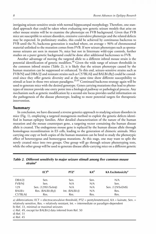
intriguing seizure-sensitive strain with normal hippocampal morphology. Therefore, one stan-dard approach that could be taken when evaluating new genetic seizure models that arise onother mouse strains will be to examine the phenotype on FVB background. Given that FVBmice are susceptible to seizure disorders, extensive convulsive phenotype and the related defectsmay be expected. In preliminary studies, this could be achieved by continuous backcross toFVB until the N5 backcross generation is reached where, on average > 96% of chromosomalmaterial unlinked to the mutation comes from FVB. If new seizure phenotypes such as sponta-neous seizures are seen in mutant N5 mice but not in littermate wild-type controls, furtherstudies on a purer genetic background could be done after additional backcrosses to FVB.
Another advantage of moving the targeted allele to a different inbred mouse strain is thepotential identification of genetic modifiers.48 Given the wide range of seizure thresholds inthe common inbred strains (Table 2), it is likely that the seizure phenotype caused by thehuman mutation can be suppressed or enhanced. To this end, seizure-sensitive strains such asFVB/NJ and DBA/2J and resistant strains such as C57BL/6J and BALB/cByJ could be consid-ered since they offer genetic diversity and at the same time show different susceptibility tostimuli at least in those two seizure paradigms.33,45 Continued backcross strategy again will beused to generate mice with the desired genotype. Genes carrying mutations that lead to pheno-types of interest provide one entry point into a biological pathway or pathological process. Anymechanism such as genetic modification by a second-site locus provides useful information onthe pathogenesis of the disease phenotype, leading to more potential targets for therapeuticintervention.
SummaryIn conclusion, we have discussed a reverse genetics approach to studying seizure disorders in
mice (Fig. 1), employing a targeted mutagenesis method to exploit the genetic defects identi-fied in human epilepsy families. After detailed characterization of the nature of the humanmutation and the mouse counterpart gene, a targeting vector containing the human diseaseallele is created. The endogenous mouse gene is replaced by the human disease allele throughhomologous recombination in ES cells, leading to the generation of chimeric animals. Micecarrying one copy or both copies of the human mutation can be bred to study the phenotypiceffect of heterozygous and homozygous mutations. At this stage, one may want to split thenewly created mice into two groups. One group will go through seizure phenotyping tests,while the other group will be used to generate disease allele-carrying mice on a different genetic
Table 2. Different sensitivity to major seizure stimuli among five common mousestrainsa
ECTb PTZc KAd KA Excitotoxicitye
DBA/2J Sen. Sen. Sen. N/AFVB/NJ Sen. N/A N/A Sen.129 Sen. (129S1/SvImJ) N/A N/A Sen. (129/SvEMS)BALB/c Res. (BALB/cByJ) Int. (BALB/cJ) N/A Res.C57BL/6J Res. Res. Res. Res.
a) abbreviations: ECT = electroconvulsive threshold, PTZ = pentylenetetrazol, KA = kainate, Sen. =relatively sensitive, Res. = relatively resistant, Int. = intermediate or paradigm-dependentb) Ref. 33, minimal or maximal seizuresc) Ref. 49, except for BALB/cJ data inferred from Ref. 50d) Ref. 51e) Ref. 45

9Genetic Approaches to Studying Mouse Models of Human Seizure Disorders
background. Phenotypic characterization of mice on different inbred strains includes behav-ioral monitoring and EEG analysis looking for the occurrence of spontaneous seizures, as wellas routine cage examination looking for handling-provoked seizure and ECT- and PTZ- in-duced seizure paradigms looking for sensitivity to these stimuli. A complete evaluation of theseizure phenotype at the whole-animal level establishes the relevance of the mouse model to thehuman condition. Further investigation including imaging, electrophysiology and AED re-sponse in these mouse models will shed light on the mechanistic basis of the convulsive disorder.
Current epilepsy research in mouse genetics offers promise for understanding the molecularmechanisms that underlie epileptogenesis in humans. A large-scale forward genetic effort tocreate novel mouse mutants with seizure phenotypes by in vivo chemical mutagenesis withethyl-nitroso urea (ENU) is underway at the Jackson Laboratory (http://www.jax.org/nmf/).Genetic mapping and isolation of the affected genes in these seizure-prone models will provideadditional molecular pathways involved in seizures. The mutant mice generated through bothforward and reverse genetic approaches will be a valuable resource for the biomedical commu-nity to study epilepsy at the molecular level and to characterize the pathological consequencesof seizures in the whole organism.
AcknowledgementsWe thank Drs. Greg A. Cox and Verity A. Letts for reviewing a preliminary version of this
manuscript. We gratefully acknowledge research support from the National Institutes of Health(NS31348 and NS40246 to W.N.F.). Y.Y. is a Jackson Laboratory Fellow supported by theinstitutional funds of The Jackson Laboratory.
References1. Anderson VE, Hauser WA, Rich SS. Genetic heterogeneity and epidemiology of the epilepsies. In:
Delgado-Escueta AV, Wilson WA, Olsen RW, Porter RJ, eds. Jasper’s Basic Mechanisms of theEpilepsies. Philadelphia: Lippincott Williams & Wilkins, 1999.
2. Meisler MH, Kearney J, Ottman R et al. Identification of epilepsy genes in human and mouse.Annu Rev Genet 2001; 35:567-88.
3. Burgess DL, Jones JM, Meisler MH et al. Mutation of the Ca2+ channel beta subunit gene Cchb4is associated with ataxia and seizures in the lethargic (lh) mouse. Cell 1997; 88:385-392.
4. Escayg A, De Waard M, Lee DD et al. Coding and noncoding variation of the humancalcium-channel beta4-subunit gene CACNB4 in patients with idiopathic generalized epilepsy andepisodic ataxia. Am J Hum Genet 2000; 66: 1531-1539.
5. Krall RL, Penry JK, Kupferberg HJ et al. Antiepileptic drug development: I. History and a pro-gram for progress. Epilepsia 1978; 4:393-408.
6. Frankel WN. Detecting genes in new and old mouse models for epilepsy: A prospectus throughthe magnifying glass. Epilepsy Res 1999; 36:97-110.
7. Gusella JF, MacDonald ME. Molecular genetics: Unmasking polyglutamine triggers inneurodegenerative disease. Nat Rev Neurosci 2000; 2:109-15.
8. Bronson SK, Smithies O. Altering mice by homologous recombination using embryonic stem cells.J Biol Chem 1994; 269:27155-8.
9. Capecchi MR. The new mouse genetics: Altering the genome by gene targeting. Trends Genet1989; 5:70-6.
10. Lewandoski M. Conditional control of gene expression in the mouse. Nat Rev Genet 2001;10:743-55.
11. Kask K, Zamanillo D, Rozov A et al. The AMPA receptor subunit GluR-B in its Q/R site-uneditedform is not essential for brain development and function. Proc Natl Acad Sci USA 1998;95:13777-82.
12. Cossette P, Liu L, Brisebois K et al. Mutation of GABRA1 in an autosomal dominant form ofjuvenile myoclonic epilepsy. Nature Genet 2002; 31:184-189.
13. Kalachikov S, Evgrafov O, Ross B et al. Mutations in LGI1 cause autosomal-dominant partialepilepsy with auditory features. Nature Genet 2002; 30:335–341.
14. Morante-Redolat JM, Gorostidi-Pagola A, Piquer-Sirerol A et al. Mutations in the LGI1/Epitempingene on 10q24 cause autosomal dominant lateral temporal epilepsy. Hum Mol Genet 2002;11:1119-1128.

Recent Advances in Epilepsy Research10
15. Baulac S, Huberfeld G, Gourfinkel-An I et al. First genetic evidence of GABA(A) receptor dys-function in epilepsy: a mutation in the gamma2-subunit gene. Nat Genet 2001; 28:46-8.
16. Wallace RH, Marini C, Petrou S et al. Mutant GABA(A) receptor gamma2-subunit in childhoodabsence epilepsy and febrile seizures. Nat Genet 2001; 28:49-52.
17. Sugawara T, Tsurubuchi Y, Agarwala KL et al. A missense mutation of the Na+ channel alpha IIsubunit gene Na(v)1.2 in a patient with febrile and afebrile seizures causes channel dysfunction.Proc Natl Acad Sci USA. 2001; 98:6384-9.
18. Escayg A, MacDonald BT, Meisler MH et al. Mutations of SCN1A, encoding a neuronal sodiumchannel, in two families with GEFS+2. Nat Genet 2000; 24:343-5.
19. Claes L, Del-Favero J, Ceulemans B et al. De novo mutations in the sodium-channel gene SCN1Acause severe myoclonic epilepsy of infancy. Am J Hum Genet 2001; 68:1327-32.
20. Wallace RH, Wang DW, Singh R et al. Febrile seizures and generalized epilepsy associated with amutation in the Na+-channel beta 1 subunit gene SCN1B. Nat Genet 1998; 19:366-70.
21. Singh NA, Charlier C, Stauffer D et al. A novel potassium channel gene, KCNQ2, is mutated inan inherited epilepsy of newborns. Nat Genet 1998; 18:25-9.
22. Biervert C, Schroeder BC, Kubisch C et al. A potassium channel mutation in neonatal humanepilepsy. Science 1998; 279:403-6.
23. Charlier C, Singh NA, Ryan Sg et al. A pore mutation in a novel KQT-like potassium channelgene in an idiopathic epilepsy family. Nat Genet 1998; 18:53-5.
24. Steinlein OK, Mulley JC, Propping P et al. A missense mutation in the neuronal nicotinic acetyl-choline receptor alpha 4 subunit is associated with autosomal dominant nocturnal frontal lobeepilepsy. Nat Genet 1995; 11:201-3.
25. De Fusco M, Becchetti A, Patrignani A et al. The nicotinic receptor beta 2 subunit is mutant innocturnal frontal lobe epilepsy. Nat Genet 2000; 26:275-6.
26. Steinlein OK Genes and mutations in idiopathic epilepsy. Am J Med Genet 2001; 106:139-45.27. Jacobs MP, Fischbach GD, Davis MR et al. Future directions for epilepsy research. Neurology
2001; 57:1536-1542.28. Dietschy JM, Turley SD. Control of cholesterol turnover in the mouse. J Biol Chem 2002;
277:3801-3804.29. Watanabe H, Nagata E, Kosakai A et al. Disruption of the epilepsy KCNQ2 gene results in neural
hyperexcitability. J Neurochem 2000; 75:28-33.30. Noebels JL. Single locus mutations in mice expressing generalized spike-wave absence epilepsies.
Ital J Neurol Sci 1995; 16:107-11.31. Noebels JL, Sidman RL. Inherited epilepsy: spike-wave and focal motor seizures in the mutant
mouse tottering. Science 1979; 204:1334-1336.32. Cox GA, Lutz CM, Yang CL et al. Sodium/hydrogen exchanger gene defect in slow-wave epilepsy
mutant mice. Cell 1997; 91:139-48.33. Frankel WN, Taylor L, Beyer B et al. Electroconvulsive thresholds of inbred mouse strains. Genomics
2001; 74:306-12.34. Peterson SL, Electroshock. In: Peterson SL, Albertson TE, eds. Neuropharmacology Methods in
Epilepsy Research. Boca Raton: CRC Press, 1998:1-26.35. Ferraro TN, Golden GT, Smith GG et al. Mouse strain variation in maximal electroshock seizure
threshold Brain Res 2002; 936:82-86.36. Kitano Y, Usui C, Takasuna K et al. Increasing-current electroshock seizure test: A new method
for assessment of anti- and pro-convulsant activities of drugs in mice. J Pharmacol Toxicol Meth-ods 1996; 35:25-9.
37. White HS, Wolf HH, Woodhead JH et al. The National Institutes of Health Anticonvulsant DrugDevelopment Program: Screening for efficacy. Adv Neurol 1998; 76:29-39.
38. Loscher W. New visions in the pharmacology of anticonvulsion. Eur J Pharmacol 1998; 342:1-13.39. White HS. Chemoconvulsants. In: Peterson SL, Albertson TE, eds. Neuropharmacology Methods
in Epilepsy Research. Boca Raton: CRC Press, 1998:27-40.40. Skradski SL, Clark AM, Jiang H et al. A novel gene causing a mendelian audiogenic mouse epi-
lepsy. Neuron 2001; 31:537-44.41. Zheng QY, Johnson KR, Erway LC. Assessment of hearing in 80 inbred strains of mice by ABR
threshold analyses. Hear Res 1999; 130:94-107.42. Gerlai R. Gene-targeting studies of mammalian behavior: Is it the mutation or the background
genotype? Trends Neurosci 1996; 19:177-81.43. Bucan M, Abel T. The mouse: Genetics meets behavior. Nat Rev Genet 2002; 2:114-23.44. Frankel WN. Mouse strain backgrounds: More than black and white. Neuron 1998; 20:183.45. Schauwecker PE, Steward O. Genetic determinants of susceptibility to excitotoxic cell death: Im-
plications for gene targeting approaches. Proc Natl Acad Sci USA 1997; 94:4103-8.

11Genetic Approaches to Studying Mouse Models of Human Seizure Disorders
46. Ben-Ari Y, Cossart R. Kainate, a double agent that generates seizures: Two decades of progress.Trends Neurosci 2000; 23:580-7.
47. Goelz MF, Mahler J, Harry J et al. Neuropathologic findings associated with seizures in FVBmice. Lab Anim Sci 1998; 48:34-37.
48. Cox GA, Mahaffey CL, Frankel WN. Identification of the mouse neuromuscular degenerationgene and mapping of a second site suppressor allele. Neuron 1998; 1327-37.
49. Ferraro TN, Golden GT, Smith GG et al. Mapping loci for pentylenetetrazol-induced seizure sus-ceptibility in mice. J Neurosci 1999; 19:6733-9.
50. Kosobud AE, Cross SJ, Crabbe JC. Neural sensitivity to pentylenetetrazol convulsions in inbredand selectively bred mice. Brain Res 1992; 592:122-8.
51. Ferraro TN, Golden GT, Smith GG et al. Mapping murine loci for seizure response to kainicacid. Mamm Genome 1997; 8:200-8.

Recent Advances in Epilepsy Research12
CHAPTER 2
Recent Advances in Epilepsy Research, edited by Devin K. Binder and Helen E. Scharfman.©2004 Eurekah.com and Kluwer Academic / Plenum Publishers.
Integrins, Synaptic Plasticityand EpileptogenesisChristine M. Gall and Gary Lynch
Abstract
A number of processes are thought to contribute to the development of epilepsyincluding enduring increases in excitatory synaptic transmission, changes in GABAergicinhibition, neuronal cell death and the development of aberrant innervation patterns in
part arising from reactive axonal growth. Recent findings indicate that adhesion chemistriesand, most particularly, activities of integrin class adhesion receptors play roles in each of theseprocesses and thereby are likely to contribute significantly to the cell biology underlyingepileptogenesis. As reviewed in this chapter, studies of long-term potentiation have shown thatintegrins are important for stabilizing activity-induced increases in synaptic strength andexcitability. Other work has demonstrated that seizures, and in some instances subseizureneuronal activity, modulate the expression of integrins and their matrix ligands and theactivities of proteases which regulate them both. These same adhesion proteins and proteasesplay critical roles in axonal growth and synaptogenesis including processes induced by seizurein adult brain. Together, these findings indicate that seizures activate integrin signaling andinduce a turnover in adhesive contacts and that both processes contribute to lasting changes incircuit and synaptic function underlying epileptogenesis.
IntroductionHypotheses about the causes of epileptogenesis usually posit that one or more of the follow-
ing long-lasting changes have occurred at critical sites in brain: increased strength of excitatoryconnections, a loss or change in strength of inhibitory input, neuronal cell death, and aberrantaxonal growth leading to greater excitability. The increased excitation hypothesis found impor-tant support with the discovery that high-frequency activity of a type not greatly different fromthat associated with seizures triggers a long-term potentiation (LTP) of glutamatergic transmis-sion.1,2 Specifically, in LTP bursts of high frequency or theta burst-like afferent activity lead toan enduring strengthening of transmission that is specific to synapses activated during theinducing phase.3-5 Later studies demonstrated that brief seizures can themselves induce LTP.6
Since then, an impressive list of points in common between LTP and epileptogenesis has beencompiled.7 NMDA-type glutamate receptors, for example, appear to be necessary for the in-duction but not the expression of either phenomenon across a range of experimental circum-stances.8-14 Di-acylglycerol metabolism and the density of voltage sensitive calcium channelsare other more recent examples of factors reported to shape the probability of LTP andepileptogenesis. Thus, although not identical,15,16 there is good evidence that LTP andepileptogenesis share cellular mechanisms. With regard to disturbances in inhibitory connec-tions, there is a large literature showing that changes in the components of GABAergic syn-

13Integrins, Synaptic Plasticity, and Epileptogenesis
apses and in inhibitory transmission itself occur in both animal models17-20 and human epi-lepsy.21 This includes recent reports of aberrant binding to GABA receptors in the humancondition.22,23 Most likely related to these changes in inhibition, neuronal cell death has beenobserved in most experimental paradigms17,24-28 and in human epilepsy.25 The loss of specificpopulations of cells, and particularly of local circuit neurons in hippocampus, may account forat least some of the changes in the balance of excitatory and inhibitory activities and reactiveaxonal growth with epileptogenesis. Regarding the latter point, several laboratories have de-scribed axonal sprouting and aberrant innervation patterns in hippocampus and the dentategyrus following seizures and in experimental models of epileptogenesis. This was originallydescribed, and has been most fully characterized, for sprouting of the granule cell mossy fiberaxons in animal seizure models6,9,29-31 and in human epilepsy.32,33 However, more recent stud-ies have found evidence for local sprouting in field CA1 in both animal and human mate-rial.28,34-37 Moreover, the balance of evidence indicates that in both fields sprouting contrib-utes to synchronous neuronal activity and excitability. For example, in field CA1 new axonalgrowth gives rise to aberrant collaterals to the cell body layer and proximal dendrites, leading tonew connections that are likely to underlie increased excitability. This growth is correlated intime with increases in the local response to applied glutamate and the increased probabilitythat stimulation of individual pyramidal cells will elicit spontaneous network bursts.28,37 Simi-larly, in the dentate gyrus the post-seizure emergence of aberrant mossy fiber collaterals to thedentate inner molecular layer has been associated with increased glutamate excitability andgranule cell burst activity that cannot be accounted for by changes in inhibition.38,39 Thus, inthe better-studied animal models of epileptogenesis, evidence suggests that along with the moreacute effects of seizure, there is a reorganization and growth of excitatory connections whichpotentiates network excitability and contributes to seizure proneness.28
As discussed throughout this volume, numerous cellular mechanisms have been proposedas steps leading to the epilepsy-related changes described above. One possibility that has begunto receive increasing attention involves cell adhesion chemistries. In particular, recent studieshave shown that integrins, and other classes of adhesion receptors, are required for sustainingdifferent phases of LTP and kindling. Moreover, there is evidence that integrins and theirmatrix ligands regulate GABAergic transmission40 in the cortical telencephalon and in somecases mediate seizure-induced cell death.41 Finally, cell adhesion and extracellular matrix pro-teins are well known to play critical roles in the motor-mechanisms and guidance of axonalgrowth and in the stabilization of synaptic contacts: recent studies have shown the same chem-istries are active in adult neuroplasticity as well. In all, several phenomena thought to contrib-ute to epileptogenesis are likely to be regulated by cell adhesion. This, combined with growingevidence that seizures activate a battery of adhesion-related enzymes and adhesion protein geneexpression, points to the possibility that epileptogenesis involves a local breakdown of normalcell-cell and cell-matrix relationships followed by new synthesis and reactive axonal growth,and the appearance of stable, hyperexcitable arrangements.
The present review covers a selection of recent results relating to the above hypothesis. Itbegins with an introduction to the structure and cellular functions of integrin receptors, as theyhave been characterized in non-neural systems, and a description of results demonstrating thatintegrins are differentially expressed across regions of the adult brain. This will be followed bypresentation of evidence that integrins contribute to the consolidation of LTP and kindling.Finally, results demonstrating that adhesive contacts and related chemistries are altered by sei-zures will be described and the implications of these findings to the development of seizuresusceptibility will be discussed.
Integrins: Cell-Matrix and Cell-Cell Adhesion ReceptorsThere are three major families of transmembrane adhesion receptors throughout the body
as well as several more minor groupings.42 The major families, illustrated in Figure 1, are theintegrins, cadherins, and immunoglobulin (Ig) adhesion proteins.43,44 The cadherins and Ig

Recent Advances in Epilepsy Research14
adhesion proteins are both homophilic receptors that bind the extracellular domain of identi-cal transmembrane proteins on adjacent cells. As shown, the cadherins are homodimers andtheir binding is calcium-dependent: calcium stabilizes a linear conformation and brings indi-vidual strands into the correct alignment for both dimerization and binding to cadherins onadjacent cells. Unlike cadherins and Ig receptors, the integrins are heterophilic receptors thatbind extracellular matrix proteins such as osteopontin, collagen, tenascin-C, and reelin as wellas transmembrane proteins on adjacent cells (e.g., VCAM-1).45,46 Thus, integrins mediate bothcell-cell and cell-extracellular matrix (ECM) adhesion and are the principal receptors for theECM throughout the body.
As reviewed in detail elsewhere,46-49 the integrins are non-covalently bound αβ heterodimers,each containing one of 18 known α subunits and one of 8 known β subunits to give rise to over20 dimer combinations. The α subunit has a relatively short cytoplasmic domain and is thoughtto be of particular importance for determining the ligand specificity of the receptor. The βsubunit also participates in ligand recognition but importantly, it has a longer cytoplasmicdomain that is associated with both the actin cytoskeleton and with a variety of signalingmolecules.46,50 Although the integrins have no catalytic activity themselves, they are seen ashaving both “inside out” and “outside-in” signaling functions that depend largely upon thethree-dimensional conformation of the receptor.47 Integrins within the membrane can exist ina latent or resting state, and can be activated by a variety of influences upon their cytoplasmicdomain including signals from protein kinase C, calcium/calmodulin-dependent protein ki-nase II and G proteins.47,51-53 These activities, as well as divalent cation levels,45 lead to confor-
Figure 1. Schematic illustration of the three major classes of adhesion receptors discussed in the text. Asshown, the cadherins form dimers which have homophilic interactions with like-cadherins on adjacent cells.The Ig class receptors, for which Neural Cell Adhesion Molecule (NCAM) is the prototypical example, alsoform homomeric cell-cell interactions. In contrast, the integrins are αβ heterodimers: their extracellulardomain binds extracellular matrix (ECM) proteins as well as proteins on adjacent cells whereas theirintracellular domain is linked both (1) to the actin cytoskeleton via a variety of integrin-scaffolding proteins(e.g., vinculin, talin, actinin, spectrin) and (2) to kinase and G-protein signaling cascades. This includesdirect associations with focal adhesion kinase (FAK) and Src kinases. Divalent cations (Mg+2, Mn+2, Ca+2)regulate integrin activity for ligand binding through influences on the binding “pocket” between α and βstrands.

15Integrins, Synaptic Plasticity, and Epileptogenesis
mational changes that propagate from the cytoplasmic to the extracellular domain to activatethe receptor and increase ligand binding activity. Similarly, ligand binding, and in particularbinding of anchored ligands, or in some instances simple integrin receptor clustering, leads toconformational changes that propagate from the outside to the inside of the cell (hence,“outside-in” signaling) leading to changes in the physical arrangements of integrin-associatedcytoskeletal proteins and signaling molecules.46,51 The latter are likely to be of particular im-portance for functional synaptic plasticity. Integrins bind either directly or indirectly to a num-ber of signaling intermediaries that are increasingly appreciated to play critical roles in synapticefficacy and long term changes in synaptic function including focal adhesion kinase (FAK) andits homologue proline rich tyrosine kinase 2 (PYK2), Src family kinases and PI(3) kinase toname a few.50,54-56 Through these and other elements, integrin ligand binding stimulates strongincreases in tyrosine phosphorylation,57 and increases signaling through Ras, Raf, MEK, andsmall GTPases (Rho, Rac among others).50,54,58-64 As a consequence, integrins play criticalroles in a wide variety of very basic cellular functions including cell cycling, phenotypic differ-entiation, gene expression, process outgrowth, cell survival and the regulation of intracellularpH and calcium levels.
Finally, regarding specialized neuronal functions to be discussed below, it is important tonote that integrins have functional associations with other codistributed receptors and trans-membrane proteins.65 Regarding issues of synaptic plasticity, it is intriguing that functionalinteractions have been particularly well-demonstrated for integrins and tyrosine kinase growthfactor receptors,54,62-64 potassium channels66 and voltage-dependent calcium channels.61,67 Asjust one example of this, integrin binding has been shown to both stabilize codistributed epi-dermal growth factor receptor (EGFR) in the cellular membrane and, presumably througheffects on receptor densities, to mediate EGF-independent signaling through the EGFR.62
Findings such as this raise the intriguing possibility that synaptic integrins may interact withsynaptic trophic factor receptors in processes mediating activity-induced synaptic plasticity.
Integrin Expression in the Adult CNSIntegrin functions within the CNS have been most extensively studied within the context
of development,68 and during early stages integrins have been shown to be important for fun-damental processes of process outgrowth,69,70 cellular migration,71-74 and substrate recogni-tion. However, there is now good evidence that integrins, along with the other major classes ofadhesion receptors, are highly expressed in the adult brain.
Given the involvement of integrins in very basic adhesive functions, one might expect theintegrins to be broadly expressed by virtually all cell types throughout the brain. Contrary tothis, localization studies have shown that integrin expression profiles are highly differentiated,being both brain region- and cell type-specific. Moreover, while both neurons and glia clearlyexpress integrins,75-78 immunocytochemical and in situ hybridization studies have shown thatin the adult integrins are most highly expressed by neurons. This point can be illustrated byresults of in situ hybridization studies conducted in our laboratories. Distributions and levelsof mRNAs for integrin subunits α1-8, αv, and β1-5 were evaluated in adult rat brain.79,80
Figure 2 shows photomicrographs of autoradiographic labeling obtained with 35S-cRNAs andillustrates some of the basic differences in expression profiles. Labeling patterns were highlyvaried: some subunits (e.g., α3, α5, αv, β5; Fig. 2B) were broadly expressed across brain withneuronal labeling throughout neocortex and hippocampus while other transcripts (e.g., α4, 1;Fig. 2A) were much more narrowly distributed and detected in only a few discrete cell types.Still others (e.g., β1, α7) were particularly highly expressed by neurons projecting outside ofthe CNS (e.g., efferent cranial nerve neurons and spinal motor neurons). As can be seen inthese examples, integrin subunit expression conformed to neuronal cytoarchitectonics and la-beling patterns clearly distinguished cell types within a specific region, and subfields within aparticular brain area. The latter features can be seen in the subfield-specific labeling in hippoc-ampus and layer-specific labeling in cerebellum. In hippocampus, integrin α7 expression clearly

Recent Advances in Epilepsy Research16
differentiates subfields, being highly expressed in the principal cell layers of the dentate gyrusand field CA3 but not field CA1 (Fig. 2D). In contrast, the α1 and α8 transcripts were mosthighly expressed in the pyramidal cells of CA1 and CA3, respectively (Fig. 2C,E). The absenceof neuronal labeling in the hippocampal molecular layers indicates that expression is greatest inglutamatergic neurons (in the principal cell layers) and low in GABAergic neurons and gliathat are scattered across the molecular layers. In cerebellum, α3 mRNA is discretely localized inthe granule cells (Fig. 2F) while α7 mRNA is highly expressed in both the granule and Purkinjecells (Fig. 2G). Results of these studies reinforce the conclusion that a given cell type expressesmore than one integrin.80-83 Again, this point is illustrated by consideration of labeling pat-terns in hippocampus. CA1 pyramidal cells clearly express mRNAs for α subunits 1, 3, 5, 8and v, and for β subunits 1 and 5. Moreover, PCR amplification of integrin transcripts84 inadult hippocampus and localization of expression in immature neurons85 (and Gall, unpub-lished results) indicates that these cells express β3 and β8 integrins as well. Together thesefindings indicate that the CA1 pyramidal cells express at least 8 integrins (α1β1, α3β1, α5β1,α8β1, αvβ1, αvβ3, αvβ5 and αvβ8).
Figure 2. Integrin gene expression is both region- and cell-specific. Photomicrographs show the autorad-iographic localization of 35S-cRNA in situ hybridization labeling of integrin mRNAs in adult rat brain(dark-field microscopy; cellular labeling seen as white). (A,B) Panels A and B show labeling of ααααα4 and αααααvmRNAs, respectively, and illustrate subunits with very limited (A) and broad (B) distributions. In the planeshown, ααααα4 mRNA is restricted to piriform cortex (pc) whereas αααααv mRNA is distributed across superficialand deep layers of neocortex, caudate/putamen (CPu), piriform cortex and olfactory tubercle (ot). (C-E)Photomicrographs show subfield-specific integrin expression profiles in hippocampus. As illustrated, ααααα1mRNA (C) is prominently expressed in CA3 stratum pyramidale alone, ααααα7 mRNA (D) is abundant instratum granulosum (sg) and CA3 stratum pyramidale, and ααααα8 mRNA (E) is most dense in CA1 stratumpyramidale. (F,G) Photomicrographs of cerebellar cortex (hemispheres) showing ααααα3 mRNA (F) is restrictedto the Purkinje cell layer (arrow) while ααααα7 mRNA is highly expressed in the granule cells (gc) as well as thePurkinje cells. Note, in panels C-G none of the transcripts are prominently expressed by cells in themolecular layers.

17Integrins, Synaptic Plasticity, and Epileptogenesis
The results of in situ hybridization studies (i.e., cell- and region-specific expression profiles,prominent neuronal labeling, multiple integrins expressed by single cells) are largely consistentwith immunocytochemical descriptions of integrin expression in brain. Analyses of α876,86,87,αv76, α188, α575,89,90 and β191-93 have demonstrated localization in neuronal perikarya, axonaland dendritic processes, and glial cells. Moreover, these immunocytochemical studies indicatethat integrins are localized, and in some cases seemingly concentrated, at synapses and limitedaspects of dendritic arbors. In central mammalian neurons, α1, α8, αv and α3 have all beenlocalized within synapse-like puncta or synaptic junctions at the light or electron microscopiclevel, respectively.76,86,91,94 Our own studies of cultured neurons have demonstrated a concen-tration of α5-immunoreactivity (ir) at GluR1 and PSD-95 immunoreactive dendritic spines inaddition to lower levels of α5-ir in dendritic processes and perikarya (Bernard and Gall, un-published observations). Similarly, integrin proteins are concentrated at synaptic junctions inDrosophila.95,96 These anatomical observations are consistent with results of neurochemicalstudies demonstrating that α5, αv, α3 and β1 subunits are enriched in synaptic membranefractions.97-99
As mentioned above, there is evidence that some integrins are preferentially distributedacross dendritic arbors. In particular, we have observed that in hippocampus and neocortexα5-ir is dense in apical dendrites but below detection in basal dendrites of the great majority ofpyramidal cells.90 This contrasts with labeling of non-pyramidal cells which is clearly distrib-uted across multiple primary dendritic arbors. The apparent apical compartmentalization ofα5 is not accounted for by differences in synapse or spine densities. Indeed, given the concen-tration of α5 at spines and synapses this observation suggests a fundamental neurochemicaldifference between the apical and basal dendritic spines which could be of consequence tocapacities for long term synaptic plasticity. We will return to this point below within the con-text of α5 involvement in the consolidation of LTP.
Adhesion Proteins Contribute to the Consolidation of Long-TermPotentiation
The argument that epileptogenesis arises from abnormal levels of excitatory input requires:1) a mechanism whereby remarkable increases in EPSPs can be produced; and 2) a process thatcould sustain those increases for very long periods. The evidence, derived from studies of LTPreviewed below, suggests that integrins are critical to processes that can satisfy these require-ments; i.e., as described, integrins are important for stabilization of enduring activity-inducedincreases in synaptic efficacy in hippocampal LTP. Having identified this as a plausible candi-date cellular mechanism for sustaining hyperexcitability, the question arises as to whether ithelps explain specific, poorly understood characteristics of epileptogenesis. By way of address-ing this question, the following sections also briefly describe findings indicating that variationsin adhesion chemistry may result in developmental and regional differences in the stability ofsynaptic modifications induced by intense neuronal activity.
LTP Has a Memory-Like Consolidation PeriodFunctional synaptic plasticity is widely recognized to pass through a ‘consolidation’ period
during which it becomes progressively more resistant to disruption and is converted into aform with extraordinary persistence. This became evident in early studies of the consolidationof memory100-102 and the principles identified in studies of learning and memory seem toapply to synaptic phenomena and, in particular, to LTP. Consolidation involves multiple pro-cesses that seemingly result in ever more enduring forms of encoding. Because of this, there isno single consolidation time although it is clear that with memory the process begins withinseconds of learning and produces a reasonably stable memory within 30 minutes. The firstevidence that LTP has a memory-like consolidation period came with the discovery that poten-tiation in anesthetized rats disappears if low-frequency afferent stimulation is applied within afew minutes of induction.103 Subsequent studies using chronic recording confirmed that LTP

Recent Advances in Epilepsy Research18
does not recover even after delays of 24 hours and that high-frequency stimulation deliveredafter reversal produces a normal degree of potentiation, as expected if the original effect had infact been erased.104 Other work demonstrated that the erasure effect is present throughout thecortical telencephalon.105,106
The first in vitro studies of consolidation showed that hypoxia or infusions of adenosinewould reverse potentiation if applied within 5 minutes of induction but were without effect 30minutes later.107 This time course resembles that described for memory consolidation in anynumber of paradigms.100,101 Subsequent slice experiments obtained reversal with naturalistictheta patterns of stimulation108 and showed that it probably involves adenosine receptors.109,110
The demonstration that LTP becomes steadily less vulnerable over the 30 min post inductionperiod111,112 added another and important point of agreement with memory consolidation.
Multiple Adhesion Receptors Contribute to Consolidation of LTPThe logic for investigating the role of adhesion receptors in consolidation is straightfor-
ward. There is broad agreement that changes in the shape of synapses and spines accompanychanges in synaptic efficacy, although the appropriate description of the change remains con-troversial. Given that synapses are adhesion junctions, it is unlikely that significant morpho-logical reorganization of the type associated with LTP could occur without extensive modifica-tions to integrins and other adhesion receptors.
Integrins bind discrete amino acid sequences in their matrix targets. Approximately onethird of the known integrins bind the arginine-glycine-aspartate, or RGD, amino acid sequencethat is shared by many matrix proteins (e.g., fibronectin, vitronectin, tenascin-C,thrombospondin);45 differences in ligand specificity derive from amino acids flanking the cen-tral binding motif.46,113,114 Small soluble peptides containing the RGD sequence interfere withmatrix adhesion for these particular integrins115 and disrupt a wide variety of integrin-mediatedevents (e.g., fibroblast attachment, platelet aggregation). To assess the contribution of synapticRGD-binding integrins to LTP the putative antagonist peptides, or control media, were in-fused into acute hippocampal slices and effects on baseline physiology and LTP assessed. Infu-sion of RGD peptide (i.e., GRGDSP) into hippocampal slices had little effect on baselinetransmission or the initial expression of LTP but caused potentiation to gradually dissipate overthe 60 minutes following induction.116,117 Additional studies found that RGD peptides werealso effective if infused 2 minutes after the induction of LTP but were not effective whenapplied after delays of 25 minutes or longer.118 As illustrated in Figure 3 (panels A-C), twosnake toxins (a.k.a., disintegrins) that potently block integrin recognition of the RGDligand-motif also disrupted the consolidation of LTP and, like RGD-peptides, were effective ifapplied after the inducing stimulation (Fig. 3B).97 In all, the effects of RGD peptides on thestabilization of LTP support the hypothesis that synaptic stimulation engages integrin activitieswhich then dictate the particular time course over which the increase in synaptic strengthbecomes stabilized.
As discussed above, hippocampal neurons express an array of integrins and at least some ofthese (α3β1, α5β1, αvβ8) are concentrated in synapses. The above results with antagoniststhat would be expected to interfere with functions of all RGD-binding integrins do not addressthe question of which particular receptors are involved in LTP consolidation. Given that integrinsand their signaling activities are functionally distinct, identifying the specific dimer(s) subservingconsolidation could be an important step towards understanding the manner in which thereceptors help stabilize the potentiated state. This problem was addressed using function-blockingantibodies locally infused into hippocampal fields containing synapses that were about to bepotentiated. The antibodies were infused during or immediately following theta-burst afferentstimulation and infusion was continued during the post-stimulus interval over which LTP wasassessed. Two synaptically localized integrins were in this way implicated in consolidation:α5β197 (Fig. 3D) and α3β1.98

19Integrins, Synaptic Plasticity, and Epileptogenesis
The effects produced by the function-blocking antibodies were partial and delayed com-pared to those achieved with RGD peptides. This opens the interesting possibility that LTPgoes through more than one integrin-dependent consolidation stage. An early stage involvingintegrins other than α5 or α3 would, according to this argument, produce a long-lasting butslowly decremental LTP that can be largely reversed. A slightly later stage of consolidationwould be expected to incorporate contributions from the α3β1 and α5β1 integrins and to
Figure 3. Blocking integrin function disrupts stabilization of LTP. In acute hippocampal slices, a stimulatingelectrode was placed in field CA1b stratum radiatum between two recording electrodes placed in CA1a andCA1c stratum radiatum. With this arrangement, in untreated slices theta-burst stimulation applied to fieldCA1b leads to comparable potentiation of synaptic responses at both recording electrodes. In the experi-mental preparations, the disintegrin echistatin (A,B), echistatin plus a second disintegrin triflavin (C), ora function-blocking antibody to integrin subunit α5 (D) was infused at the experimental recording siteduring the period indicated by the gray bar; the remaining recording site was used as a control. LTP wasinduced with trains of 10 theta bursts containing 4 pulses at 100 Hz and a burst separation of 200 ms.4 Theupper aspects of Panels A and B, and Panels C and D, show group average (± SEM) EPSP slopes for singletest pulses applied to the same site before and after the LTP-inducing stimulation. In each of these plots,EPSP slope is presented as a percent of baseline values collected before induction of LTP; values from thecontrol and experimental (drug-treated) sites are represented by open and filled circles, respectively. Thelower plots in A and B show the percent difference between EPSP slopes for the control and experimentalsites. As shown in A, in the presence of the disintegrin echistatin, LTP shows the same initial induction atthe control and experimental sites but the experimental site LTP slowly declines and, after 1 hr, potentiationhad almost disappeared at the experimental site but was robust and stable at the control site. (B) Echistatinapplied after the induction of LTP also led to a loss of potentiation over 1 hr. (C) Treatment with twodisintegrins, echistatin and triflavin, led to a similar but more rapid decay in LTP than did treatment withechistatin alone. (D) Treatment with anti-α5 caused a significant but slower decay in LTP; in other experi-ments the decay was shown to continue over 4 hrs of antibody treatment. For more details, refer to Chunet al, 2001.97

Recent Advances in Epilepsy Research20
result in a potentiation that is both stable and extremely resistant to interference. Sequentialactivation of integrins is not unknown; e.g., cell spreading is reported in some experimentsto require activation first of αvβ3 and then of α5β1 receptors.119 In all, the multiple stagesof consolidation described for memory may find integrin-based analogues in long-term po-tentiation.
Finally, there is evidence that other classes of adhesion receptors in addition to integrins areinvolved in stabilizing LTP (see 44,120-122 for review). The number of synapses with highconcentrations of neural cell adhesion molecule 180 (NCAM 180, the isoform with the long-est cytoplasmic domain) increases markedly following LTP induction123 while potentiationitself is impaired by function-blocking antibodies against NCAM118 or the related Ig adhesionmolecule telencephalin.125 Moreover, a selective disruption of LTP stabilization is found inmutant mice lacking one of the polysialyltransferases that adds sialic acid to the extracellulardomain of NCAM but that retain NCAM itself.126 This indicates that a specific,well-characterized NCAM state is a necessary ingredient for consolidation. Other studies im-plicate the third class of synaptic adhesion receptors in LTP as well. Specifically, the number ofsynapses immunopositive for Neural (N)-cadherin, a cell-cell adhesion protein known to beinvolved in synaptogenesis during development and that is present in high concentrations inadult synapses, increases after induction of LTP.127 The authors propose that the increase is partof the synapse building and remodeling that accompanies LTP.128-130 Moreover, N-cadherinfunction-blocking antibodies have been shown to disrupt LTP stabilization in a limited num-ber of experiments without disrupting induction or early expression.127 In agreement with this,Tang et al131 found that peptides designed to interfere with cadherin binding also interferewith LTP. Finally, function-blocking antibodies to a structurally related synaptic cadherin,arcadlin also impair LTP132 but as this manipulation also degraded baseline synaptic responsesthe results do not demonstrate if arcadlin is specifically involved in synaptic plasticity or, alter-natively, is important for synaptic transmission in general.
The above evidence makes a convincing argument that activity-driven changes in junc-tional adhesion can anchor excitatory synapses into a much more potent state. As discussedelsewhere in this review, a separate body of experiments shows that intense synaptic activityand seizures modify the abundance of extracellular matrix and adhesion receptors. Taken to-gether, the results strongly suggest that lasting increases in excitatory drive will be a concomi-tant of epileptogenesis. Given the evident connection between excitatory transmission andseizure production, this conclusion provides support for the broad hypothesis that such in-creases in excitatory drive, and cellular processes mediating them, contribute to seizure suscep-tibility and epileptogenesis.
Adhesion Receptors Are Involved in Multiple Forms of Plasticity IncludingKindling
Recently, it has become evident that integrin contributions to plasticity extend well beyondLTP and hippocampus. RGD peptides block activity-dependent synaptic changes in Droso-phila.96 Other work with mutant flies established that a variant of the α5 integrin subtypeplays an essential role in plasticity.133,134 Most directly relevant to the present review is evidencethat integrin antagonists block the induction of seizure susceptibility as seen in a kindlingmodel. Specifically, RGD peptides were infused into hippocampal slices for one hour prior toattempts to induce kindling with brief periods of high-frequency bursts of afferent stimulation.Baseline synaptic responses and the number of stimuli needed to induce bursting were de-scribed as being unaffected by RGD peptides but the later response to kindling stimulation wasprofoundly altered. That is, the RGD-treated kindled slices had a substantially lower rate ofspontaneous bursts but a longer burst period than did slices that had been treated with controlpeptides. These results establish a direct link between integrins and one form of epileptogenesis.135

21Integrins, Synaptic Plasticity, and Epileptogenesis
Variations in Synaptic Adhesion May Account for Regionaland Developmental Differences in the Plasticity of Excitatory Synapses
The characteristics and consequences of seizures change during development136,137 and varymarkedly between brain regions. As with any argument about causal factors for epileptogenesis,the enhanced excitation hypothesis needs to address these variations. Accordingly, it may beuseful to ask if the adhesion chemistries that appear to stabilize synaptic changes have ontoge-netic or regional characteristics that would be predictive of marked differences in the degree towhich excitatory inputs can be enhanced.
As described earlier, the brain is regionally differentiated with regard to expression of integrinsubtypes and, according to recent results, it does appear that these are predictive of variations inLTP. The extent to which LTP has become consolidated can be assessed by applyinglow-frequency trains of stimulation at various times after induction and, as described by Karmáret al,138 LTP in the apical dendrites of field CA1 is little affected by the trains at 30 minpost-induction. The basal dendrites of the same CA1 neurons lack the α5β1 integrin90 thatcontributes to the stabilization in their apical counterparts.97 Recent work has confirmed theprediction that consolidation is also incomplete in the basal dendrites.138 In the basal den-drites, initial LTP is larger and more easily induced than that in the apical dendrites;139 despitethis, the new results indicate that it is also less stable. In all, there is a possibility that sites atwhich activity-induced changes in excitatory drive are easily established and readily recordedmay not be the sites at which lasting adaptations (seizure proneness, LTP) emerge.
The unusual developmental history of the α5β1 integrin also leads to some interestingpredictions about plasticity. As described elsewhere,90 the integrin does not move into apicaldendrites until relatively late in postnatal maturation (i.e., during weeks 2-3 in rat), raising thepossibility that integrin-dependent consolidation of LTP does not have the same developmen-tal profile as the other components of synaptic plasticity. This was confirmed in a series ofstudies testing the reversibility of LTP at different post-natal ages.138 LTP can be induced in rathippocampus by postnatal day 9 but, at this age, in contrast to the adult, potentiation is fullyerased by low-frequency stimulation even at 60 minutes post-induction. This indicates that afully competent consolidation process may not emerge until well into the third postnatal weekand during that period would be coincident with the appearance of the α5β1 integrin in thesesame fields.
In summary, the emergence of adhesion molecules as a consolidation mechanism will likelyexpand the aberrant excitation hypothesis for epileptogenesis and allow it to make novel pre-dictions.
Seizures Activate Changes in Adhesion Chemistries:Evidence for Turnover in Adhesive Contacts
As described in the introduction, recurrent seizures and kindling are associated with in-creases in excitability and the formation of aberrant circuitry (e.g., sprouting) and both phe-nomena have long been suspected to contribute to changes in seizure threshold andepileptogenesis. Knowing the involvement of various adhesion receptors and matrix proteinsin facilitating and directing axonal growth during development, a number of investigators haveproposed that these proteins may play a role in synaptic rearrangements that occur followingseizures and have examined the possibility that seizures modify associations within the adhesiveenvironment. The results of these studies, together with the growing understanding of activity-regulated proteolytic activities in brain, indicate that seizures induce a massive reorganization,or turnover, in adhesive chemistries that clearly facilitates growth and circuit adjustments and,thereby, may play a significant role in epileptogenesis. In particular, results from many labora-tories indicate that activity- and seizure-regulated reorganization of adhesive proteins includesboth proteolytic cleavage and replacement of integrin-mediated adhesive contacts. However, as

Recent Advances in Epilepsy Research22
this is only part of a much larger story, it is best considered within the broader context of theeffects of seizures on adhesive chemistries as they occur, over time, from the onset of the seizureepisode. With this in mind, the following paragraphs will describe the effects of seizures onproteolysis of adhesion proteins and on the expression of adhesion receptors and their matrixligands. We have made an effort to include results that illustrate the magnitude of reorganiza-tion induced by seizure but the narrative should not be considered an exhaustive presentationof literature on the subject. Finally, the implications of these perturbations in adhesive chemis-tries for the functional consequences of seizure activity and epileptogenesis will be discussed.
Activity Regulates Proteolysis of Adhesion ProteinsRecent years have seen a tremendous increase in the understanding of regulated proteolysis
in brain and, in particular, there is now very good evidence that seizures, and NMDAreceptor-mediated neuronal activity in general, rapidly stimulate the activities of a battery ofproteases that modify adhesion proteins and their associations both inside and outside the cell.In the intracellular domain, calcium influx associated with seizures and intense glutamatergicsynaptic activation increases local activities of the neutral protease, calpain140-143 that includesboth integrins144-146 and integrin-associated proteins143,147-149 among its substrates. As calpainis localized within dendritic spines,150,151 this proteolysis can be tightly associated with regionsof activated synapses and, as a consequence, can be subfield- and possibly even synapse-specificwithin a given cell.142 Although calpain-mediated integrin cleavage has not been directly dem-onstrated to be induced by seizures, Einheber et al87 found that within the hilus of the dentategyrus, kainic acid-induced seizures lead to a rapid loss of immunoreactivity for the α8 integrincytoplasmic domain that is evident first within dendritic processes and only later in the perikaryonof mossy cells. The timing and distribution of change suggests that calpain, or another simi-larly regulated intracellular protease, accounts for this loss of integrin immunoreactivity.
Neuronal activity has also been shown to stimulate increases in extracellular proteases andproteolysis. Among these, the best studied within the contexts of seizures and synaptic plastic-ity are the trypsin-like serine proteases including tissue plasminogen activator (tPA) andneuropsin. Gene expression for tPA and neuropsin152,153 and secretion of tPA154,155 is stimu-lated by seizures and/or depolarization. Within the extracellular environment, tPA cleaves theintegrin ligand plasminogen to liberate the additional trypsin-like protease, plasmin. Togetherthese and related proteases cleave both matrix proteins and the adhesion receptors themselves.Specifically, seizures or brief NMDA receptor stimulation156,157 lead to the extracellular cleav-age of NCAM at a consensus serine protease site; this proteolysis can be mimicked by thetPA-plasmin system157,158 and is blocked by a serine protease inhibitor of tPA.158 Other studieshave demonstrated that activities of the tPA-plasmin system also regulate levels of NCAMligands phosphacan and neurocan,159 the integrin ligand laminin160 and, in other systems, theintegrin, fibrinogen receptor.161 These findings suggest that synaptic integrins may also besubstrates for activity-regulated serine protease activity in brain but there is, as yet, no informa-tion on this point.
In additional to the trypsin-like proteases, seizures increase the expression and activities ofmetalloproteinases (MMPs) in brain. The MMPs are secreted and both modify the extracellu-lar environment and reveal adhesion-receptor epitopes though proteolysis of ECM proteins.162
Kainate seizures increase MMP-9 mRNA, protein, and activity levels within hippocampus.163,164
This is associated with local increases in the expression and extracellular distribution of a natu-ral inhibitor of metalloproteinase (i.e., TIMP-1).165 Thus, seizures appear to stimulate a con-trolled MMP-mediated proteolysis of the ECM in adult brain. Given the involvement of MMPsin modifying the extracellular environment to facilitate growth and cellular migration in pe-ripheral tissues and brain,166 the possibility that these enzymes facilitate axonal growthand, in particular, seizure-induced sprouting and synaptogenesis in hippocampus is par-ticularly attractive.

23Integrins, Synaptic Plasticity, and Epileptogenesis
Seizures Regulate the Expression of Adhesion ReceptorsWhile seizures initiate proteolytic activities that degrade adhesion receptors and matrix pro-
teins, they also initiate changes in the expression of these same target proteins that would beexpected to replace those lost by proteolysis but which would also generate additional proteinsthat may facilitate axonal growth, synaptogenesis and changes in synaptic efficacy that accom-pany seizures and contribute to epileptogenesis. In particular, in situ hybridization and immu-nocytochemical studies have shown that seizures, and lesser fluctuations in neuronal activity,differentially regulate the expression of integrins, cadherins, and Ig cell adhesion molecules(CAMs).
Evidence that seizures influence integrin expression comes from in situ hybridization analy-ses in our laboratory. We have evaluated the effect of recurrent, lesion-induced limbic seizureson mRNA levels for β1 integrin and its dimer partners, subunits α1-8 and αv in adult ratbrain. This includes consideration of subunits forming RGD-binding integrins potentially in-volved in processes of LTP and kindling discussed above (i.e., α3β1, α5β1, α8β1, and αvβ1).In the hilar lesion paradigm used, bilateral recurrent limbic seizures occur intermittently over aperiod of 8 to 10 hours with the greatest number of seizures clustered early in the episode.167 Asillustrated for select transcripts in Figure 4, we found that each of the integrin mRNAs areinfluenced by seizures but in distinctly different ways. β1 mRNA is broadly increased duringthe seizure episode within both neurons and astroglial cells. Transcript levels increase duringthe period of seizures and slowly decline thereafter.79 Among the α subunits, responses arehighly transcript-specific.168 Transcripts for α1, α2, α8 and αv exhibit a monophasic increaseeither early (e.g., αv; Fig. 4A,D) or late (e.g., α8 and α1; Fig. 4B,E) in the seizure episode. Theα5 and α6 subunit mRNAs are also induced by seizure but these transcripts are clearly in-creased in both neurons and glia (Fig. 4C,F). The α3 and α7 mRNA levels are decreasedduring the period of recurring seizures but increase above control levels at later intervals. Fi-nally, α4 mRNA, which is one of the lesser abundant transcripts in untreated adult rat brain,80
declines to undetectable levels in entorhinal cortex through the period of seizures.Together, the changes in integrin gene expression induced by seizures show that neuronal
activity regulates integrin expression in very discrete, transcript-specific ways. This includesdifferential regulation of colocalized transcripts. For example, in neuronal layers of hippocam-pus, the α3 and α7 mRNAs are decreased during the same interval in which αv and α1 mRNAlevels are increased. Integrin subunits compete for dimer pairings. Thus, it is likely that changesin the availability of α partners for the β1 subunit over time leads to changes in the integrinreceptors expressed at the membrane; in this instance we would predict an decrease in α3β1and an increase in αvβ1 in the dentate granule cells in the aftermath of seizure activity. Fur-thermore, given evidence that receptor dimer composition determines the ligand specificityand signaling properties of the receptor, these changes could underlie fundamental differencesin the contribution of synaptic integrin receptors to neuronal functions including axonal growthand activity-dependent modifications in synaptic efficacy during, and in the wake of, seizures.
Other investigators have shown that neuronal activity regulates the expression of cadherinsand NCAM and have linked these effects with reactive growth and synaptic plasticity. Seizureshave been reported to increase expression of N-cadherin169 and the cadherin family memberarcadlin;132 as described above, both proteins are implicated in stabilizing synaptic potentia-tion in hippocampus. Moreover, in the case of N-cadherin, pilocarpine seizure-induced in-creases are rather long-lasting (lasting several weeks) and associated with increases in immu-noreactive puncta in the dentate gyrus inner molecular layer169 suggesting that newly expressedproteins are localized in growing sprouts and nascent synapses of the mossy fibers that areinduced by seizure.29,170,171 Other studies demonstrating induction of increases in synapticN-cadherin with LTP-inducing cAMP treatment127 and patterned afferent stimulation172 sug-gest that activity induction of this class of adhesion protein may be important to cadherin’scontribution to functional synaptic plasticity as well.44,120 For the Ig CAMs, increased expres-sion has been linked to patterned afferent stimulation,172 the induction of LTP123,173 or poten-

Recent Advances in Epilepsy Research24
tiation of AMPA-class glutamate receptor currents with ampakine drugs.174 Together, theseobservations indicate that Ig CAM expression can be positively regulated by neuronal activityand, in some instances, with very low threshold.
Seizures Regulate the Expression of Matrix ProteinsAlthough matrix proteins have proven difficult to study with anatomical techniques in the
adult brain, there is evidence that seizures modulate the expression of several including, mostparticularly, the integrin ligands tenascin-C, vitronectin, and fibronectin. Kainic acid-inducedseizures increase the expression of tenascin-C within a few hours of seizure onset; mRNA levelspeak at 24 hrs and then decline.175,176 Within hippocampus, increases in tenascin-C mRNAare prominently localized to the dentate gyrus granule cells, although this protein is expressedby reactive astrocytes as well. Similarly, kainate seizures induce fibronectin mRNA and proteinin hippocampus; in situ hybridization and immunocytochemical studies indicate increases arelocalized to both neurons and astroglia.177 Following intra-amygdalar kainic acid injection,seizures are associated with neuronal cell death in the hilar and CA3 regions of hippocampus;in rats so treated there is a delayed increase in vitronectin-ir in reactive astroglia in hippocampalfields of damage.178 These results demonstrate that in adult brain astroglia express vitronectinbut the increase observed in this instance may be part of the glial response to local neuronaldamage as opposed to the effects of seizure activity.
A more complex response to seizure is exhibited by agrin and the chondroitin sulfateproteoglycan receptor of protein tyrosine phosphatase β (RPTPβ). Agrin is best characterizedas a matrix protein at neuromuscular junctions where it is involved in clustering of acetylcho-line receptors. However, it is also broadly expressed by neurons in brain179 and recent studiesindicate that for central neurons agrin regulates patterns of dendritic and axonal growth180 andpositively regulates vulnerability to excitotoxic damage with seizure.181 We found agrin geneexpression is regulated in both positive and negative directions by recurrent limbic seizures.179
In hippocampus agrin mRNA levels are depressed during a recurrent seizure episode and thenincrease to supranormal levels through 4 days after seizure termination. In contrast, agrin mRNAis reduced in post-seizure intervals within the neocortex. Similarly, RPTPβ-ir, which is nor-mally evident in glia, is reduced in this compartment during status epilepticus but increased inthe extracellular environment; this is followed by increased RPTPβ levels in neurons.182
Significance of Seizure-Induced Proteolysis and Adhesion ProteinExpression to Epileptogenesis
The above paragraphs demonstrate that seizures, and in some instances more modest in-creases in neuronal activity, lead to proteolysis of adhesion proteins that overlap and are fol-lowed by increases in the expression of both the transmembrane adhesion receptors and several
Figure 4. Seizures stimulate integrin gene expres-sion. Photomicrographs show autoradiographic lo-calization of 35S-cRNA in situ hybridization label-ing of mRNAs for integrin subunits αααααv (A,D), ααααα1(B,E) and ααααα6 (C,D) in coronal sections throughcaudal hippocampus and cortex in control rats(A-C) and experimental-seizure rats killed 6 hr (D)or 24 hr (E,F) after the onset of recurrent limbicseizures. As shown, seizures induce marked increasesin αααααv (D) and ααααα1 (E) mRNAs that are restricted toneuronal cell layers and are particularly striking inhippocampus (sg, stratum granulosum; CA1 andCA3, stratum pyramidale indicated). In contrast,seizure-induced increases in ααααα6 mRNA (F) are morediffusely distributed and are localized in both neu-rons and glia.

25Integrins, Synaptic Plasticity, and Epileptogenesis
of the major matrix proteins. Do these events contribute to the functional sequelae of seizuresand to susceptibility for future seizures? Results pertinent to these important questions aresparse but sufficient to answer “yes” in both instances. Perhaps the most complete stories areassociated with activity- and seizure-induced trypsin-like proteolytic activity. As described, sei-zures and LTP stimulation induce increases in tPA expression and secretion, and proteolysis ofextracellular adhesion proteins including NCAM and laminin (see above). Several studies indi-cate this process is important for activity-dependent increases in synaptic strength and, impor-tantly for the present discussion, to kindling. Hoffman et al158 demonstrated that in hippo-campal slices treatment with a serine protease inhibitor (AEBSF), that significantly blocks tPAproteolysis of NCAM, also blocks the formation of stable LTP without disrupting baselinesynaptic responses. This accords with the observation that mice lacking tPA have specific defi-ciencies in the expression of late LTP.183 In a separate study, Hoffman and colleagues foundthat systemic treatment with the AEBSF inhibitor of tPA just prior to the induction phase ofrapid electrical stimulation-induced kindling, blocked kindling as assessed by measures of stimu-lated afterdischarge (AD) duration 10 days after the inducing session.184 In vehicle-treated rats,mean AD duration was 3.5-fold longer 10 days after the single kindling session as compared tobaseline responses. In contrast, in rats with protease inhibitor treatment, AD durations werecomparable on the first and last days of recording.
Seizures are well known to lead to the death of vulnerable neurons and to axonal sprouting,as most clearly demonstrated in hippocampus. Recent findings link tPA activities to both ofthese phenomena as well. Kainic acid (KA)-induced seizures are well known to lead to death ofhippocampal CA3 neurons. Strickland and colleagues 41,185 have demonstrated that KA-inducednecrosis is absent in mice deficient for tPA or the plasmin precursor plasminogen. Moreover,this same group found that KA-seizures normally cause tPA/plasmin dependent reductions inlaminin-ir in hippocampus that are coincident with neuronal death and that neutralizingantibody-mediated reductions in laminin bioactivity lead to cell death even with intact tPA/plasmin activities.160 It is argued that these results demonstrate that integrin-mediated adhe-sion to ECM laminin is protective and that loss of contact with this matrix substrate, as occurswith seizure-induced proteolysis, causes neuronal cell death. Together with results described inthe preceding paragraph, these findings implicate tPA in two adverse phenomena induced byseizure: increases in seizure proneness and neuronal cell death.
It is likely that seizure effects on tPA and the MMPs contribute to axonal sprouting. IntPA-deficient mice there is decreased seizure-induced mossy fiber sprouting and an accumula-tion of the ECM chondroitin sulfate proteoglycan DSD-1-PG/phosphacan, a matrix proteinwhich can inhibit neurite outgrowth and oppose NCAM function, in the dentate gyrus innermolecular layer.159 This suggests that the absence of phosphacan proteolysis by tPA leads to anaccumulation of a putative growth regulatory protein in the normal target of mossy fiber in-growth that inhibits or redirects the sprouting response. Although less is known about specificMMP functions following seizures, results from other systems suggest that seizure-inducedincreases in MMP expression should facilitate matrix remodeling and, in particular, axonalgrowth through the ECM. For example, with damage to spinal cord186 or peripheral nerve187,188
MMP levels are increased in association with growth cones and neurites; MMP enzymaticactivities allow the neurites to overcome the growth inhibitory activities of matrix chondroitinsulfate proteoglycans and to grow through the ECM. Similarly, with myelination MMP-9 isexpressed on the leading tip of oligodendroglial processes and either blocking MMP-9 gelatinaseactivity or the absence of MMP-9 in null mutants retards the extension of these processes andmyelin formation.189
Increases in the expression of the adhesion receptors and matrix proteins are also likely tofacilitate axonal growth following seizures. Clearly both classes of molecules are involved ingrowth processes: the matrix proteins provide the substrate for growth and can facilitate orretard outgrowth depending on the circumstance and the specific adhesion receptors expressedon growing neurites. On the other side of the equation, the adhesion receptors provide mecha-nisms for surface recognition, the mechanical force needed for process extension, and a means
Recent Advances in Epilepsy Research26
for stabilizing synapses as they mature (68,190,191 for reviews). More specifically for integrins,increases in gene expression have been positively correlated with axonal growth and experimen-tally induced increases in expression stimulate process outgrowth. With damage to peripheralnerve in adult rats, the ECM proteins laminin192 and fibronectin193 are upregulated in thedamaged nerve and both proteins facilitate axonal regeneration;194 this facilitation is mediatedby integrins α6β1 and α7β1 that are specific for laminin and are upregulated in motor neu-rons by nerve damage.83 With damage to the facial nerve, α7 integrin levels are increased ongrowth cones of regenerating axons, and transgenic ablation of α7 expression reduces axonalelongation and delays reinnervation.195 Other studies have shown that overexpression of integrinsin cultured cells facilitates axonal growth, most particularly in the presence of chondroitinsulfate proteoglycans thought to limit axonal extension in adult brain.196 The same adhesionproteins shown to facilitate axonal growth in these lesion studies (i.e., fibronectin; integrinsubunits α1, α6, α7, and β1) are induced by seizures and are elevated during periods ofseizure-induced axonal growth. Together, these results indicate that coordinated seizure-inducedincreases in integrin and matrix protein expression contribute both to the remodeling of axonalconnections known to occur in these same paradigms and to changes in circuit excitabilityassociated with these aberrant innervation patterns.
Concluding CommentsStudies described here have shown that several classes of adhesion proteins contribute to
activity-induced changes in synaptic efficacy and, in particular, to potentiation of glutamatergictransmission. Among these, the integrins are of particular importance for the stabilization ofsynaptic change in both LTP and kindling, and may determine the time course over whichthese changes in synaptic efficacy consolidate. Other studies have shown that seizures stimulateintegrin and matrix protein expression, and extracellular proteolysis; these processes are insome instances known, and in other instances likely, to facilitate reactive axonal growth andcircuit modification following seizures. Together, these results indicate that integrins and theirmatrix targets are significantly involved in multiple processes that contribute to the develop-ment of epilepsy most particularly in adult brain.
AcknowledgementsThe authors thank our many laboratory colleagues and collaborators who contributed to
research described in this review. This work was supported by grants NS37799 and MH61007.
References1. Bliss TVP, Lômo T. Long-lasting potentiation of synaptic transmission in the dentate area of the
anesthetized rabbit following stimulation of the perforant path. J Physiol 1973; 232:334-356.2. Lynch G, Bahr BA, Vanderklish PW. Induction and stabilization of LTP. In: Ascher P, Choi DW,
Christen Y, eds. Glutamate, cell death and memory. Berlin: Springer Verlag, 1991:45-60.3. Staubli U, Lynch G. Stable hippocampal long-term potentiation elicited by “theta” pattern stimu-
lation. Brain Res 1987; 435:227-234.4. Larson J, Lynch G. Theta pattern stimulation and the induction of LTP: the sequence in which
synapses are stimulated determines the degree to which they potentiate. Brain Res 1989; 489:49-58.5. Larson J, Wong D, Lynch G. Patterned stimulation at the theta frequency is optimal for the
induction of hippocampal long-term potentiation. Brain Res 1986; 368:347-350.6. Ben-Ari Y, Represa A. Brief seizure episodes induce long-term potentiation and mossy fiber sprout-
ing in the hippocampus. Trends Neurosci 1990; 13:312-314.7. Baudry M. Long-term potentiation and kindling: similar biochemical mechanisms. Adv Neurol
1986; 44:401-410.8. Muller D, Lynch G. Long-term potentiation differentially affects two components of synaptic re-
sponses in hippocampus. Proc Natl Acad Sci USA 1988; 85:9346-50.9. Sutula T, Kock J, Golarai G et al. NMDA receptor dependence of kindling and mossy fiber sprout-
ing: evidence that the NMDA receptor regulates patterning of hippocampal circuits in the adultbrain. J Neurosci 1996; 16:7398-7406.

27Integrins, Synaptic Plasticity, and Epileptogenesis
10. Behr J, Heinemann U, Mody I. Kindling induces transient NMDA receptor-mediated facilitationof high-frequency input in the rat dentate gyrus. J Neurosci 2001; 85:2195-2202.
11. Sayin U, Rutecki P, Sutula T. NMDA-dependent currents in granule cells of the dentate gyruscontribute to induction but not permanence of kindling. J Neurophysiol 1999; 81:564-574.
12. Loscher W. Pharmacology of glutamate receptor antagonists in the kindling model of epilepsy.Prog Neurobiol 1999; 54:721-741.
13. Lynch G, Kessler M, Arai A et al. The nature and causes of hippocampal long-term potentiation.Prog Brain Res 1990; 83:333-339.
14. Collingridge GL, Singer W. Excitatory amino acid receptor and synaptic plasticity. Trends PharmSci 1990; 11:290-296.
15. Cain DP. Long-term potentiation and kindling: how similar are the mechanisms? Trends Neurosci1989; 12:6-10.
16. Lynch M, Sayin U, Golarai G et al. NMDA receptor-dependent plasticity of granule cell spikingin the dentate gyrus of normal and epileptic rats. J Physiol 2000; 11:61-68.
17. Bouilleret V, Kiener T, Marescaux C et al. Early loss of interneurons and delayed subunit-specificchanges in GABA(A)-receptor expression in mouse model of mesial temporal lobe epilepsy. Hip-pocampus 2000; 10:305-324.
18. Nusser A, Hajos N, Somogyi P et al. Increased number of synaptic GABAA receptors underliespotentiation at hippocampal inhibitory synapses. Nature 1998; 395:172-177.
19. Kokaia Z, Kokaia M. Changes in GABAB receptor immunoreactivity after recurrent seizures inrats. Neurosci Lett 2001; 315:85-88.
20. Morimoto K. Seizure-triggering mechanisms in the kindling model of epilepsy: collapse ofGABA-mediated inhibition and activation of NMDA receptors. Neurosci Biobehav Rev 1989;13:253-260.
21. Olsen RW, Delorey TM, Gordey M et al. GABA receptor function and epilepsy. Adv Neurol1999; 79:499-510.
22. Billinton A, Baird VH, Duncan TM et al. GABA(B) receptor autoradiography in hippocampalsclerosis associated with human temporal lobe epilepsy. Br J Pharmacol 2001; 132:475-480.
23. Szelies B, Sobesky J, Pawlik G et al. Impaired benzodiazepine receptor binding in peri-lesionalcortex of patients with eymptomatic epilepsies studies by [(11)C]-flumazenil PET. Eur J Neurosci2002; 9:137-142.
24. Sloviter RS. Permanently altered hippocampal structure, excitability, and inhibition after experi-mental status epilepticus in the rat: the “dormant basket cell” hypothesis and its possible relevanceto temporal lobe epilepsy. Hippocampus 1991; 1:41-66.
25. Sutula TP, Pitkanen A. More evidence for seizure-induced neuron loss. Is hippocampal sclerosisboth cause and effect of epilepsy? Neurology 2001; 57:169-170.
26. Cavazos JE, Das I, Sutula T. Neuronal loss induced in limbic pathways by kindling: evidence forinduction of hippocampal sclerosis by repeated brief seizures. J Neurosci 1994; 14:3106-3121.
27. Bengzon J, Kokaia Z, Elmer E et al. Apoptosis and proliferation of dentate gyrus neurons aftersingle and intermittent limbic seizures. Proc Natl Acad Sci USA 1997; 94:10432-10437.
28. Smith BN, Dudek FE. Short- and long-term changes in CA1 network excitability after kainatetreatment in rats. J Neurophysiol 2001; 85:1-9.
29. Sutula TP, Golarai G, Cavazos J. Assessing the functional significance of mossy fiber sprouting. In:Ribak CE, Gall CM, Mody I, eds. The dentate gyrus and its role in seizures. Amsterdam: Elsevier,1992:251-259.
30. Cronin J, Obenhous A, Houser C et al. Electrophysiology of dentate granule cells afterkainate-induced synaptic reorganization of mossy fibers. Brain Res 1992; 573:305-310.
31. Represa A, Ben-Ari Y. Kindling is associated with the formation of novel mossy fibre synapses inthe CA3 region. Brain Res 1992; 92:69-78.
32. Babb TL, Kupfer WR, Pretorius JK et al. Synaptic reorganization by mossy fibers in human epi-leptic fascia dentata. Neuroscience 1991; 42:351-363.
33. Houser CR. Morphological changes in the dentate gyrus in human temporal lobe epilepsy. In:Ribak CE, Gall CM, Mody I, eds. The Dentate Gyrus and its Role in Seizures. Amsterdam: Elsevier,1992:223-234.
34. Lehmann TN, Gabriel S, Kovacs R et al. Alterations of neuronal connectivity in area CA1 ofhippocampal slices from temporal lobe epilepsy patients and from pilocarpine-treated epileptic rats.Epilepsia 2000; 41:S190-190.
35. Esclapez M, Hirsch J, Ben-Ari Y et al. Newly formed excitatory pathways provide a substrate forhyperexcitability in experimental temporal lobe epilepsy. J Comp Neurol 1999; 408:449-460.
36. Perez Y, Morin F, Beaulieu C et al. Axonal sprouting of CA1 pyramidal cells in hyperexcitablehippocampal slices of kainate-treated rats. Eur J Neurosci 1996; 8:736-748.

Recent Advances in Epilepsy Research28
37. Smith BN, Dudek FE. Network interactions mediated by new excitatory connections between CA1pyramidal cells in rats with kainate-induced epilepsy. J Neurophysiol 2002; 135:53-65.
38. Okazaki MM, Molnar P, Nadler JV. Recurrent mossy fiber pathway in rat dentate gyrus: synapticcurrents evoked in presence and absence of seizure-induced growth. J Neurophysiol 1999;81:1645-1660.
39. Wuarin J-P, Dudek FE. Excitatory synaptic input to granule cells increases with time after kainatetreatment. J Neurophysiol 2001; 1067-1077.
40. Costa E, Davis J, Grayson DR et al. Dendritic spine hypoplasticity and downregulation of reelinand GABAergic tone in schizophrenia vulnerability. Neurobiol Dis 2001; 8:723-742.
41. Tsirka S, Bugge T, Degen JL et al. An extracellular proteolytic cascade promotes neuronal degen-eration in the mouse hippocampus. J Neurosci 1997; 17:543-552.
42. Colman D, Filbin MT. Cell adhesion molecules. In: Siegel GJ, Agranoff BW, Albers RW et al.,eds.Basic neurochemistry; molecular, cellular and medical aspects, New York: Lippincott Williamsand Wilkins, 1998:139-153.
43. Alpin A, Howe A, Alahari S et al. Signal transduction and signal modulation by cell adhesionreceptors: the role of integrins, cadherins, immunoglobulin-cell adhesion molecules, and selectins.Pharmacol Rev 1998; 50:197-263.
44. Fields RD, Itoh K. Neural cell adhesion molecules in activity-dependent development and synapticplasticity. Trends Neurosci 1996; 19:473-480.
45. Plow EF, Haas TA, Zhang L et al. Ligand binding to integrins. J Biol Chem 2000;275:21785-21788.
46. Hynes R. Integrins, versatility, modulation, and signaling in cell adhesion. Cell 1992; 69:11-25.47. Mould AP. Getting integrins into shape: recent insights into how integrin activity is regulated by
conformational changes. J Cell Sci 1996; 109:2613-2618.48. Humphries MJ, Delwel GO, Kuikman I et al. Integrin structure. Biochem Soc Trans 2000;
28:311-39.49. Hynes RO. Cell adhesion: old and new questions. Trends Cell Biol 1999; 9:M33-M37.50. Giancotti FG, Ruoslahti E. Integrin signaling. Science 1999; 285:1028-1032.51. Dedhar S, Hannigan G. Integrin cytoplasmic interactions and bidirectional transmembrane signal-
ling. Current Opinion in Cell Biology 1996; 8:657-669.52. Hughes PE, Pfaff M. Integrin affinity modulation. Trends Cell Biol 1998; 8:359-364.53. Bouvard D, Molla A, Block MR. Calcium/calmodulin-dependent protein kinase II controls α5β1
integrin mediated inside out signaling. J Cell Sci 1998; 111:657-665.54. Miranti CK, Brugge JS. Sensing the environment: a historical perspective on integrin signal trans-
duction. Nature Cell Biol 2002; 4:83-90.55. Huang Y-Q, Lu WY, Ali DW et al. CAKβ/PYK2 kinase is a signaling link for induction of long-term
potentiation in CA1 hippocampus. Neuron 2001; 29:485-496.56. Ali DW, Salter MW. NMDA receptor regulation by Src kinase signalling in excitatory synaptic
transmission and plasticity. Curr Opin Neurobiol 2001; 11:336-342.57. Schlaepfer D, Hunter T. Integrin signalling and tyrosine phosphorylation: just the FAKs? Trends
Cell Biol 1998; 8:151-157.58. Yamada K, Miyamoto S. Integrin transmembrane signaling and cytoskeletal control. Curr Op Cell
Biol 1995; 7:681-689.59. Huhtala P, Humphries MJ, McCarthy JB et al. Cooperative signaling by α5β1 and α4β1 integrins
regulated matalloproteinase gene expression in fibroblasts adhering to fibronectin. J Cell Biol 1995;129:867-879.
60. Howe A, Aplin A, Alahari S et al. Integrin signaling and cell growth control. Curr Opin Cell Biol1998; 10:220-231.
61. Wu X, Davis GE, Meininger GA et al. Regulation of the L-type calcium channel by α5β1 integrinrequires signaling between focal adhesion proteins. J Biol Chem 2001; 276:30285-30292.
62. Moro L, Dolce L, Cabodi S et al. Integrin-induced epidermal growth factor (EGF) receptor activa-tion requires c-Src and p130Cas and leads to phosphorylation of specific EGF receptor tyrosines. JBiol Chem 2002; 277:9405-9414.
63. Yamada KM, Even-Ram S. Integrin regulation of growth factor receptors. Nature Cell Biol 2002;4:E75-E76.
64. Juliano R. Cooperation between soluable factors and integrin-mediated cell anchorage in the con-trol of cell growth and differentiation. BioEssays 1998; 18:911-917.
65. Porter JC, Hogg N. Integrins take partners: cross-talk between integrins and other membrane re-ceptors. Trends Cell Biol 1998; 8:390-396.
66. Platts SH, Mogford JE, Davis MJ et al. Role of K+ channels in arteriolar vasodilation mediated byintegrin interaction with RGD-containing peptide. Am J Physiol 1998; 275:H1449-H1454.

29Integrins, Synaptic Plasticity, and Epileptogenesis
67. Wildering WC, Herman PM, Bulloch AGM. Rapid neuromodulatory actions of integrin ligands. JNeurosci 2002; 22:2419-2426.
68. Reichardt L, Tomaselli K. Extracellular matrix molecules and their receptors: Functions in neuraldevelopment. Ann Rev Neurosci 1991; 14:531-570.
69. Yanagida H, Tanaka J, Maruo S. Immunocytochemical localization of a cell adhesion molecule,integrin alpha5beta1, in nerve growth cones. J Orthop Sci 1999; 4:353-360.
70. Grabham PW, Foley M, Umeojiako A et al. Nerve growth factor stimulates coupling of beta1integrin to distinct transport mechanisms in the filopodia of growth cones. J Cell Science 2000;113:3003-3012.
71. Dulabon L, Olson EC, Taglienti MG et al. Reelin binds α3β1 integrin and inhibits neuronalmigration. Neuron 2000; 27:33-44.
72. Cox EA, Huttenlocher A. Regulation of integrin-mediated adhesion during cell migration. MicroscRes Tech 1998; 43:412-419.
73. Sheetz MP, Felsenfeld DP, Galbraith CG. Cell migration: Regulation of force on extracellular-matrix-integrin complexes. Trends Cell Biol 1998; 8:51-54.
74. Palecek SP, Huttenlocher A, Horwitz AF et al. Physical and biochemical regulation of integrinrelease during rear detachment of migrating cells. J Cell Sci 1998; 111:929-940.
75. Jones LS. Integrins: possible functions in the adult CNS. Trends Neurosci 1996; 19:68-72.76. Nishimura SL, Boylen KP, Einheber S et al. Synaptic and glial localization of the integrin αvβ8 in
mouse and rat brain. Brain Res 1998; 791:271-282.77. Tanaka J, Maeda N. Microglial ramification requires nondiffusible factors derived from astrocytes.
Exp Neurol 1996; 137:367-375.78. Martin PT, Kaufman SJ, Kramer RH et al. Synaptic integrins in developing, adult, and mutant
muscle: selective association of α1, α7A and α7B integrins with the neuromuscular junction. DevBiology 1996; 174:125-139.
79. Pinkstaff JK, Lynch G, Gall CM. Localization and seizure regulation of integrin β1 mRNA inadult rat brain. Mol Brain Res 1997; 19:1541-1556.
80. Pinkstaff JK, Detterich J, Lynch G et al. Integrin subunit gene expression is regionally differenti-ated in adult brain. J Neurosci 1999; 19:1541-1556.
81. Kloss CU, Werner A, Klein MA et al. Integrin family of cell adhesion molecules in the injuredbrain: regulation and cellular localization in the normal and regenerating mouse facial motor nucleus.J Comp Neurol 1999; 411:162-178.
82. Previtali SC, Feltri ML, Archelos JJ et al. Role of integrins in the peripheral nervous system. ProgNeurobiol 2001; 64:35-49.
83. Hammarberg H, Wallquist W, Piehl F et al. Regulation of laminin-associated integrin subunitmRNAs in rat spinal motoneurons during postnatal development and after axonal injury. J CompNeurol 2000; 428:294-304.
84. Lee EH, Hsieh YP, Yang CL et al. Induction of integrin-associated protein (IAP) mRNA expres-sion during memory consolidation in rat hippocampus. Eur J Neurosci 2000; 12:1102-1112.
85. Chavis P, Westbrook G. Integrins mediate functional pre- and postsynaptic maturation at a hip-pocampal synapse. Nature 2001; 411:317-321.
86. Einheber S, Schnapp L, Salzer J et al. The regional and ultrastructural distribution of the α8integrin subunit in the developing and adult rat brain suggests a role in synaptic plasticity. J CompNeurol 1996; 370:105-134.
87. Einheber S, Pierce JP, Chow D et al. Dentate hilar mossy cells and somatostatin-containing neu-rons are immunoreactive for the α8 integrin subunit: characterization in normal and kainicacid-treated rats. Neuroscience 2001; 105:619-638.
88. Murase S-I, Hayashi Y. Integrin α1 localization in murine central and peripheral nervous system. JComp Neurol 1998; 395:161-176.
89. King VR, McBride A, Priestly JV. Immunohistochemical expression of the α5 integrin subunit inthe normal adult rat central nervous system. J Neurocytol 2001; 30:243-252.
90. Bi X, Zhou J, Lynch G et al. Polarized distribution of α5 integrin in the dendrites of hippocampaland cortical neurons. J Comp Neurol 2001; 435:184-193.
91. Schuster T, Krug M, Stalder M et al. Immunoelectron microscopic localization of the neural rec-ognition molecules L1, NCAM, and its isoform NCAM180, the NCAM-associated polysialic acid,beta1 integrin and the extracellular matrix molecule tenascin-R in synapses of the adult rat hippoc-ampus. J Neurobiol 2001; 49:142-158.
92. Grooms S, Terracio L, Jones L. Anatomical localization of β1 integrin-like immunoreactivity in ratbrain. Exp Neurol 1993; 122:253-259.
93. Murase S, Hayashi M. Expression pattern of integrin β1 subunit in Purkinje cells of rat and cer-ebellar mutant mice. J Comp Neurol 1996; 375:225-237.

Recent Advances in Epilepsy Research30
94. Rodriguez MA, Pesold C, Liu WS et al. Colocalization of integrin receptors and reelin in dendriticspine postsynaptic densities of adult nonhuman primate cortex. Proc Natl Acad Sci USA 2000;97:3550-3555.
95. Beumer KJ, Bohrbough J, Prokop A et al. A role for PS integrins in morphological growth andsynaptic function at the postembryonic neuromuscular junction of Drosophila. Development 1999;126:5833-5846.
96. Rohrbough J, Grotewiel MS, Davis RL et al. Integrin-mediated regulation of synaptic morphology,transmission and plasticity. J Neurosci 2000; 20:6868-6878.
97. Chun D, Gall CM, Bi X et al. Evidence that integrins contribute to multiple stages in the consoli-dation of long term potentiation. Neuroscience 2001; 105:815-829.
98. Kramár EA, Bernard JA, Gall CM et al. Alpha3 integrin receptors contribute to the consolidationof long term potentiation. Neuroscience 2002; 110:29-39.
99. Bahr BA, Staubli U, Xiao P et al. Arg-Gly-Asp-Ser-Selective adhesion and the stabilization oflong-term potentiation: Pharmacological studies and the characterization of a candidate matrix re-ceptor. J Neurosci 1997; 17:1320-1329.
100. Duncan CP. The retroactive effect of electroshock on learning. J Comp Physiol Psychol 1948;42:32-44.
101. Popik P, Mamczarz J, Vetulani J. The effect of electroconvulsive shock and nifedipine on spatiallearning and memory in rats. Biol Psychiatry 1994; 35:864-869.
102. McGaugh JL. Memory—a century of consolidation. Science 2000; 287:248-251.103. Barrionuevo G, Schottler S, Lynch G. The effects of repetitive low frequency stimulation on con-
trol and “potentiated” synaptic responses in the hippocampus. Life Sciences 1980; 27:2385-2391.104. Staubli U, Lynch G. Stable depression of potentiated synaptic responses in the hippocampus with
1-5 Hz stimulation. Brain Res 1990; 513:113-118.105. Martin SJ. Time-dependent reversal of dentate LTP by 5 Hz stimulation. NeuroReport 1998;
9:3775-3781.106. Burette F, Jay TM, Saroche S. Reversal of LTP in the hippocampal afferent fiber system to the
prefrontal cortex in vivo with low-frequency patterns of stimulation that do not produce LTD. JNeurophysiol 1997; 78:1155-1160.
107. Arai A, Larson J, Lynch G. Anoxia reveals a vulnerable period in the development of long-termpotentiation. Brain Res 1990; 511:353-357.
108. Larson J, Xiao P, Lynch G. Reversal of LTP by theta frequency stimulation. Brain Res 1993;600:97-102.
109. Fujii S, Kuroda Y, Ito K et al. Effects of adenosine receptors on the synaptic and EPSP-spikecomponents of long-term potentiation and depotentiation in the guinea-pig hippocampus. J Physiol(Lond) 1999; 521:451-466.
110. Abraham WC. Induction and reversal of long-term potentiation by repeated high frequency stimu-lation in rat hippocampal slices. Hippocampus 1997; 7:137-145.
111. Huang CC, Liang YC, Hsu KS. A role for extracellular adenosine in time-dependent reversal oflong-term potentiation by low-frequency stimulation at hippocampal CA1 synapses. J Neurosci1999; 19:9728-9738.
112. Staubli U, Chun D. Factors regulating the reversibility of long term potentiation. J Neurosci 1996;16:853-860.
113. Ruoslahti E. RGD and other recognition sequences for integrins. Ann Rev Cell Dev Biol 1996;12:697-715.
114. Ruoslahti E, Pierschbacher MD. Arg-Gly-Asp: a versatile cell recognition signal. Cell 1986;44:517-518.
115. Pierschbacher M, Ruoslahti E. Cell attachment activity of fibronectin can be duplicated by smallsynthetic fragments of the molecule. Nature 1984; 304:30-33.
116. Staubli U, Vanderklish PW, Lynch G. An inhibitor of integrin receptors blocks LTP. Behav Neu-ral Biol 1990; 53:1-5.
117. Xiao P, Bahr B, Staubli U et al. Evidence that matrix recognition contributes to stabilization butnot induction of LTP. NeuroReport 1991; 2:461-464.
118. Staubli U, Chun D, Lynch G. Time-dependent reversal of long-term potentiation by an integrinantagonist. J Neurosci 1998; 18:3460-3469.
119. Yang W, Asakura S, Sakai T et al. Two-step spreading mode of human glioma cells on fibrinmonomer: interaction of alpha(v)beta3 with the substratum followed by interaction of alpha5beta1with endogenous cellular fibronectin secreted in the extracellular matrix. Thromb Res 1999;93:279-290.
120. Benson DL, Schnapp LM, Shapiro L et al. Making memories stick: cell-adhesion molecules insynaptic plasticity. Trends Cell Biol 2000; 10:473-482.

31Integrins, Synaptic Plasticity, and Epileptogenesis
121. Murase S, Schuman EM. The role of cell adhesion molecules in synaptic plasticity and memory.Curr Opin Cell Biol 1999; 11:549-53.
122. Schachner M. Neural recognition molecules and synaptic plasticity. Curr Opin Cell Biol 1997;9:627-634.
123. Schuster T, Krug M, Hassan H et al. Increase in proportion of hippocampal spine synapses ex-pressing neural cell adhesion molecule NCAM180 following long-term potentiation. J Neurobiol1998; 37:359-372.
124. Luthl A, Laurent J, Figurov A et al. Hippocampal long-term potentiation and neural cell adhesionmolecules L1 and NCAM. Nature 1994; 372:777-779.
125. Sakurai E, Hashikawa T, Yoshihara Y et al. Involvement of dendritic adhesion molecule telencephalinin hippocampal long-term potentiation. Neuroreport 1998; 9:881-886.
126. Eckhardt M, Bukalo O, Chazal G et al. Mice deficient in the polysialyltransferase ST8SiaIV/PST-1allow discrimination of the roles of neural cell adhesion molecule protein and polysialic acid inneural development and synaptic plasticity. J Neurosci 2000; 20:5234-5244.
127. Bozdagi O, Shan W, Tanaka H et al. Increasing numbers of synaptic puncta during late-phaseLTP: N-cadherin is synthesized, recruited to synaptic sites, and required for potentiation. Neuron2000; 28:245-259.
128. Lee K, Schottler F, Oliver M et al. Brief bursts of high-frequency stimulation produce two types ofstructural changes in rat hippocampus. J Neurophysiol 1980; 44:247-258.
129. Chang F-LF, Greenough WT. Transient and enduring morphological correlates of synaptic activityand efficacy change in the rat hippocampal slice. Brain Res 1984; 309:35-46.
130. Toni N, Buchs PA, Nikonenko I et al. LTP promotes formation of multiple spine synapses be-tween a single axon terminal and a dendrite. Nature 1999; 402:421-425.
131. Tang L, Hung CP, Schuman EM. A role for the cadherin family of cell adhesion molecules inhippocampal long term potentiation. Neuron 1998; 20:1165-1175.
132. Yamagata K, Andreasson KI, Sugiura H et al. Arcadlin is a neural activity-regulated cadherin in-volved in long term potentiation. J Biol Chem 1999; 274:19473-19479.
133. Grotewiel MS, Beck CD, Wu CF et al. Integrin-mediated short-term memory in Drosophila. Na-ture 1998; 18:7847-7855.
134. Connolly JB, Tully T. Integrins: a role for adhesion molecules in olfactory memory. Curr Biol1998; 8:R386-R389.
135. Grooms S, Jones L. RGDS tetrapeptide and hippocampal in vitro kindling in rats: Evidence forintegrin-mediated physiological stability. Neurosci Lett 1997; 231:139-142.
136. Sankar R, Shin D, Mazarati AM et al. Epileptogenesis after status epilepticus reflects age- andmodel-dependent plasticity. Ann Neurol 2000; 48:580-589.
137. Lado FA, Sankar R, Lowenstein D et al. Age-dependent consequences of seizures: relationship toseizure frequency, brain damage, and circuitry reorganization. Ment Retard Dev Disabil Res Rev2000; 6:242-252.
138. Kramár EA, Lynch G. Developmental and regional differences in the consolidation of long termpotentiation. Submitted 2002.
139. Arai A, Black J, Lynch G. Origins of the variations in long-term potentiation between synapses inthe basal versus apical dendrites of hippocampal neurons. Hippocampus 1994; 4:1-9.
140. Bi X, Chang V, Siman R et al. Regional distribution and time-course of calpain activation follow-ing kainate-induced seizure activity in adult rat brain. Brain Res 1996; 726:98-108.
141. del Cerro S, Arai A, Kessler M et al. Stimulation of NMDA receptors activates calpain in culturedhippocampal slices. Neurosci Lett 1994; 167:149-152.
142. Vanderklish PW, Krushel LA, Holst BH et al. Marking synaptic activity in dendritic spines with acalpain substrate exhibiting fluorescence resonance energy transfer. Proc Natl Acad Sci USA 2000;97:2253-2258.
143. Vanderklish P, Saido TC, Gall C et al. Proteolysis of spectrin by calpain accompanies theta-burststimulation in cultured hippocampal slices. Mol Brain Res 1995; 32:25-35.
144. Pfaff M, Du X, Ginsberg MH. Calpain cleavage of integrin beta cytoplasmic domains. FEBS Lett1999; 460:17-22.
145. Huttenlocher A, Palecek SP, Lu Q et al. Regulation of cell migration by the calcium-dependentprotease calpain. J Biol Chem 1997; 272:32719-32722.
146. Du X, Saido T, Tsubuki S et al. Calpain cleavage of the cytoplasmic domain of the integrin β3subunit. J Biol Chem 1995; 270:26146-26151.
147. Schoenwaelder SM, Yuan Y, Cooray P et al. Calpain cleavage of focal adhesion proteins regulatesthe cytoskeletal attachment of integrin alphaIIbbeta3 (platelet glycoprotein IIb/IIIa) and the cellu-lar retraction of fibrin clots. J Biol Chem 1997; 272:1694-1702.

Recent Advances in Epilepsy Research32
148. Seubert P, Baudry M, Dudek S et al. Calmodulin stimulates the degradation of brain spectrin bycalpain. Synapse 1987; 1:20-24.
149. Siman R, Baudry M, Lynch G. Brain fodrin: substrate for the endogenous calcium-activated pro-tease, calpain I. Proc Natl Acad Sci USA 1984; 81:3572-3576.
150. Perlmutter LS, Siman R, Gall C et al. The ultrastructural localization of calcium-activated protease‘calpain’ in rat brain. Synapse 1988; 2:79-88.
151. Perlmutter LS, Gall C, Baudry M et al. Distribution of calcium-activated protease calpain in therat brain. J Comp Neurol 1990; 296:269-76.
152. Quan A, Gilbert M, Colicos M et al. Tissue-plasminogen activator is induced as an immediate-earlygene during seizure, kindling and long-term potentiation. Nature 1993; 361:453-457.
153. Okabe A, Momota Y, Yoshida S et al. Kindling induces neuropsin mRNA in the mouse brain.Brain Res 1996; 728:116-20.
154. Baranes D, Lederfein D, Huang YY et al. Tissue plasminogen activator contributes to the latephase of LTP and to synaptic growth in the hippocampal mossy fiber pathway. Neuron 1998;21:813-825.
155. Gualandris A, Jones TE, Strickland S et al. Membrane depolarization induces calcuim-dependentsecretion of tissue plasminogen activator. J Neurosci 1996; 16:2220-2225.
156. Hoffman KB, Larson J, Bahr BA et al. Activation of NMDA receptors stimulates extracellularproteolysis of cell adhesion molecules in hippocampus. Brain Res 1998; 811:152-155.
157. Endo A, Nagai N, Urano T et al. Proteolysis of neuronal cell adhesion molecule by the tissueplasminogen activator-plasmin system after kainate injection in the mouse hippocampus. NeurosciRes 1999; 33:1-8.
158. Hoffman KB, Martinez J, Lynch G. Proteolysis of cell adhesion molecules by serine proteases: arole in long term potentiation? Brain Res 1998; 811:29-33.
159. Wu YP, Siao CJ, Lu W et al. The tissue plasminogen activator (tPA)/plasmin extracellular pro-teolytic system regulates seizure-induced hippocampal mossy fiber outgrowth through a proteoglycansubstrate. J Cell Biol 2000; 148:1295-1304.
160. Chen A-L, Strickland S. Neuronal death in hippocampus is promoted by plasmin-catalyzed degra-dation of laminin. Cell 1997; 91:917-925.
161. Rabhi-Sabile S, Pidard D. Exposure of human platelets to plasmin results in the expression ofirreversibly active fibrinogen receptors. Thromb Haemost 1995; 73:693-701.
162. Basbaum C, Werb Z. Focalized proteolysis: spatial and temporal regulation of extracellular matrixdegradation at the cell surface. Curr Op Cell Biol 1996; 8:731-738.
163. Szklarczyk A, Lapinska J, Rylski M et al. Matrix metalloproteinase-9 undergoes expression andactivation during dendritic remodeling in adult hippocampus. J Neurosci 2002; 22:920-930.
164. Zhang JW, Deb S, Gottschall PE. Regional and differential expression of gelatinases in rat brainafter systemic kainic acid or bicuculline administration. Eur J Neurosci 1998; 10:3358-3368.
165. Rivera S, Tremblay E, Timsit S et al. Tissue inhibitor of metalloproteinases-1, (TIMP-1) is differ-entially induced in neurons and astrocytes after seizures; evidence for developmental, immediateearly gene, and lesion response. J Neurosci 1997; 17:4223-4235.
166. Binder DK, Berger MS. Proteases and the biology of glioma invasion. J Neuro-Oncol 2002;56:149-158.
167. Pico RM, Gall CM. Hippocampal epileptogenesis produced by electrolytic iron deposition in therat dentate gyrus. Epilepsy Res 1994; 19:27-36.
168. Gall CM, Pinkstaff JK. Integrin alpha subunit gene expression is differentially regulated by sei-zures. Soc Neurosci Abst 2000; 26:123.
169. Shan W, Yoshida M, Wu XR et al. Neural (N-) cadherin, a synaptic adhesion molecule, is inducedin hippocampal mossy fiber axonal sprouts by seizure. J Neurosci Res 2002; 69:292-304.
170. Represa A, Le Gall La Salle G, Ben-Ari Y. Hippocampal plasticity in the kindling model of epi-lepsy in rats. Neurosci Lett 1989; 99:345-350.
171. Sutula T, Cascino G, Cavazos J et al. Mossy fiber synaptic reorganization in the epileptic humantemporal lobe. Ann Neurol 1989; 26:321-330.
172. Itoh K, Ozaki M, Stevens B et al. Activity-dependent regulation of N-cadherin in DRG neurons:Differential regulation of N-cadherin, NCAM, and L1 by distinct patterns of action potentials. JNeurobiol 1997; 33:735-748.
173. Fazeli MS, Breen K, Errington ML et al. Increase in extracellular NCAM and amyloid percursorprotein following induction of long-term potentiation in the dentate gyrus of anesthetized rats.Neurosci Lett 1994; 169:77-80.
174. Holst BD, Vanderklish PW, Krushel LA et al. Allosteric modulation of AMPA-type glutamatereceptors increases activity of the promoter for the neural cell adhesion molecule, N-CAM. ProcNatl Acad Sci USA 1998; 95:2597-2602.

33Integrins, Synaptic Plasticity, and Epileptogenesis
175. Ferhat L, Chevassus-Au-Louis N, Khrestchatisky M et al. Seizures induce tenascin-C mRNA ex-pression in neurons. J Neurocytol 1996; 25:535-546.
176. Nakic M, Mitrovic N, Sperk G et al. Kainic acid activates transient expression of tenascin-C in theadult rat hippocampus. J Neurosci Res 1996; 44:355-362.
177. Hoffman KB, Pinkstaff JK, Gall CM et al. Seizure induced synthesis of fibronectin is rapid andage dependent: implications for long-term potentiation and sprouting. Brain Res 1998; 812:209-215.
178. Niquet J, Jorquera I, Ben-Ari Y et al. Proliferative astrocytes may express fibronectin-like proteinin the hippocampus of epileptic rats. Neurosci Lett 1994; 180:13-16.
179. O’Conner L, Lauterborn J, Smith M et al. Expression of agrin mRNA is altered following seizuresin adult rat brain. Mol Brain Res 1995; 33:277-287.
180. Mantych KB, Ferreira A. Agrin differentially regulates the rates of axonal and dendritic elongationin cultured hippocampal neurons. J Neurosci 2001; 21:6802-6809.
181. Hilgenberg LG, Ho KD, Lee D et al. Agrin regulates neuronal responses to excitatory neurotrans-mitters in vitro and in vivo. Mol Cell Neurosci 2002; 19:97-110.
182. Naffah-Mazzacoratti MG, Arganaraz GA, Porcionatto MA et al. Selective alterations ofglycosaminoclycans synthesis and proteoglycan expression in rat cortex and hippocampus inpilocarpine-induced epilepsy. Brain Res Bull 1999; 50:229-239.
183. Huang YY, Bach ME, Lipp HP et al. Mice lacking the gene encoding tissue-type plasminogenactivator show a selective interference with late-phase long-term potentiation in both Schaffer col-lateral and mossy fiber pathways. Proc Natl Acad Sci USA 1996; 93:8699-8704.
184. Hoffman KB, Hwee V, Larson J et al. Peripheral administration of a serine protease inhibitorblocks kindling. Brain Res 2000; 861:178-180.
185. Tsirka SE, Gualandris A, Amaral DG et al. Excitotoxin-induced neuronal degeneration and seizureare mediated by tissue plasminogen activator. Nature 1995; 377:340-344.
186. Duchossoy Y, Horvat JC, Stettler O. MMP-related gelatinase activity is strongly induced in scartissue of injured adult spinal cord and forms pathways for ingrowing neurites. Mol Cell Neurosci2001; 17:945-956.
187. Ferguson TA, Muir D. MMP-2 and MMP-9 increase the neurite-promoting potential of schwanncell basal laminae and are upregulated in degenerated nerve. Mol Cell Neurosci 2000; 16:157-167.
188. Zuo J, Ferguson TA, Hernandez YJ et al. Neuronal matrix metalloproteinase-2 degrades and inac-tivates a neurite-inhibiting chondriotin sulfate proteoglycan. J Neurosci 1998; 18:5203-5211.
189. Oh LY, Larsen PH, Krekoski CA et al. Matrix metalloproteinase-9/gelatinase B is required forprocess outgrowth by oligodendrocytes. J Neurosci 1999; 19:8464-8475.
190. Redies C. Cadherins in the central nervous system. Prog Neurobiol 2000; 61:611-648.191. Doherty P, Williams G, Williams E-J. CAMs and axonal growth: A critical evaluation of the role
of calcium and the MAPK cascade. Mol Cell Neurosci 2000; 16:283-295.192. Doyu M, Sobue G, Kimata KE et al. Laminin A, B1 and B2 chain gene expression in transected
and regenerating nerves: regulation by axonal signals. J Neurochem 1993; 60:543-551.193. Lefcort F, Venstrom K, McDonald JA et al. Regulation of expression of fibronectin and its recep-
tor, α5β1, during development and regeneration of peripheral nerve. Development 1992;116:767-782.
194. Wang GY, Hirai KI, Shimada H et al. Behavior of axons, Schwann cells and perineural cells innerve regeneration within transplanted nerve grants—effects of anti-laminin and anti-fibronectinantisera. Brain Res 1992; 583:216-226.
195. Werner A, Willern M, Jones LL et al. Impaired axonal regeneration in α7 integrin-deficient mice.J Neurosci 2000; 20:1822-1830.
196. Condic ML. Adult neuronal regeneration induced by transgenic integrin expression. J Neurosci2001; 21:4782-4788.

Recent Advances in Epilepsy Research34
CHAPTER 3
Recent Advances in Epilepsy Research, edited by Devin K. Binder and Helen E. Scharfman.©2004 Eurekah.com and Kluwer Academic / Plenum Publishers.
The Role of BDNF in Epilepsy and OtherDiseases of the Mature Nervous SystemDevin K. Binder
Abstract
The neurotrophin brain-derived neurotrophic factor (BDNF) is ubiquitous in the centralnervous system (CNS) throughout life. In addition to trophic effects on targetneurons, BDNF appears to be part of a general mechanism for activity-dependent
modification of synapses in the developing and adult nervous system. Thus, diseases of abnor-mal trophic support (such as neurodegenerative diseases) and diseases of abnormal excitability(such as epilepsy and central pain sensitization) can be related in some cases to abnormal BDNFsignaling. For example, various studies have shown that BDNF is upregulated in areas impli-cated in epileptogenesis, and interference with BDNF signal transduction inhibits the develop-ment of the epileptic state. Further study of the cellular and molecular mechanisms by whichBDNF influences cell survival and excitability will likely provide novel concepts and targets forthe treatment of diverse CNS diseases.
BDNF: IntroductionBrain-derived neurotrophic factor (BDNF) is a member of the “neurotrophin” family of
neurotrophic factors. It was originally purified from pig brain due to its survival-promotingaction on a subpopulation of dorsal root ganglion neurons.1 The amino acid sequence of BDNFhas a strong homology with nerve growth factor (NGF), the neurotrophin (NT) first describeddue to its trophic (survival and growth-promoting) effects on sensory and sympathetic neu-rons. Since the discovery of NGF in the early 1950s by Rita Levi-Montalcini and Viktor Ham-burger2 and the discovery of BDNF by Yves Barde and colleagues in 1982,1 other members ofthe NT family such as neurotrophin-3 (NT-3) and neurotrophin-4/5 (NT-4/5) have beendescribed. Each NT appears to have a unique profile of trophic effects on distinct subpopula-tions of peripheral nervous system and central nervous system neurons.
BDNF StructureThe mature form of human BDNF has been mapped to chromosome 113 and shares about
50% amino acid identity with human NGF, NT-3, and NT-4/5. The structure of each NTcontains: (1) a signal peptide following the initiation codon; (2) a pro-region containing anN-linked glycosylation site and a proteolytic cleavage site for furin-like pro-protein convertases,followed by the mature sequence; and (3) a distinctive three-dimensional structure containingtwo pairs of antiparallel β-strands and cysteine residues in a cystine knot motif. Mature NTsare noncovalently-linked homodimers with molecular weight about 28 kDa. Dimerizationappears essential for NT receptor activation.

35The Role of BDNF in Epilepsy and Other Diseases of the Mature Nervous System
BDNF SignalingEach NT binds one or more high-affinity receptors (the trk receptors) (Kd ~ 10-11 M).4 Trk
proteins are transmembrane receptor tyrosine kinases (RTKs) homologous to other RTKs suchas the epidermal growth factor (EGF) receptor and insulin receptor family.5 Signaling by RTKsinvolves ligand-induced receptor dimerization and dimerization-induced trans-autophosphorylation.6,7
Receptor autophosphorylation on multiple tyrosine residues creates specific binding sites forintracellular target proteins, which bind to the activated receptor via SH2 domains.6 For theNT family, these target proteins have been shown to include PLCγ1 (phospholipase C), p85(the noncatalytic subunit of PI-3 kinase), and Shc (SH2-containing sequence);4 activation ofthese target proteins can then lead to a variety of intracellular signalling cascades such as theRas-MAP (mitogen-activated protein) kinase cascade and phosphorylation of CREB (cyclicAMP response element binding protein).8-11 Binding specificity is conferred via thejuxtamembrane Ig-like domain of the extracellular portion of the receptor in the followingpattern:12 trkA is the high-affinity receptor for NGF (with low-affinity binding by NT-3 insome systems), trkB is the high-affinity receptor for BDNF and NT-4/5 with lower-affinitybinding by NT-3, and trkC is the high-affinity receptor for NT-3.5
In addition to the high-affinity NT receptors, all of the NTs bind to the low-affinity NTreceptor, designated p75NTR (Kd ~ 10-9 M).13 P75NTR has a glycosylated extracellular regioninvolved in ligand binding, a transmembrane region, and a short cytoplasmic sequence lackingintrinsic catalytic activity. It is related to proteins of the tumor necrosis factor (TNFR) super-family. NT binding to p75NTR is linked to several intracellular signal transduction pathways,including nuclear factor-κB (NF-κB), Jun kinase and sphingomyelin hydrolysis.14 P75NTR
signaling mediates biologic actions distinct from those of the high-affinity trk receptors, nota-bly the initiation of programmed cell death (apoptosis) as well as newly-described roles in theregulation of axonal elongation and synaptic transmission.15
Ligand-induced receptor tyrosine phosphorylation is necessary for NT-induced cellular re-sponses.5 For example, cooperative interaction between tyrosines in trkA mediates the neuriteoutgrowth effect of NGF.16 Thus, receptor tyrosine phosphorylation seems a logical measure ofthe biologic level of NT activity (see below). Tyrosine-490 is phosphorylated following NTapplication and is known to couple trk receptors to Shc binding and activation of the ras-MAPkinase cascade.11 Furthermore, recent evidence indicates that activated trk receptors may beendocytosed and retrogradely transported while still tyrosine phosphorylated.17-23
Localization, Transport and Release of BDNFBDNF mRNA as well as the mRNA encoding the high-affinity receptor for BDNF (trkB)
has a widespread distribution in the central nervous system, especially in the cerebral cortex,hippocampal formation, and amygdaloid complex.24-26 Notably, high levels of BDNF andtrkB expression are found in brain areas that have been associated with seizure susceptibility,such as hippocampus and entorhinal cortex.27 Within hippocampus, the granule cells, pyrami-dal cells, and some hilar GABAergic neurons express mRNA for BDNF and trkB.
In parallel, BDNF protein immunoreactivity is also widespread, and appears to be localizedin neuronal cell bodies, axons and dendrites.24 Like BDNF mRNA, constitutive BDNF pro-tein expression is high in the hippocampus, where the mossy fiber axons of dentate granulecells are intensely immunoreactive for BDNF.24,28
Unlike the classical target-derived trophic factor model in which NTs—such as NGF—areretrogradely transported, there is now abundant evidence that BDNF is also anterogradelytransported in brain.24,29-33 Indeed, a recent study using green fluorescent protein (GFP)-taggedBDNF demonstrated direct activity-dependent transneuronal transfer of BDNF to postsynap-tic neurons.34,35 In hippocampus, it appears that BDNF within the hilus and CA3 stratumlucidum is synthesized by the dentate granule cells, anterogradely transported and preferen-tially stored in mossy fiber terminal boutons.36

Recent Advances in Epilepsy Research36
Biochemical studies suggest that endogenous BDNF may be packaged in a releasable ve-sicular pool37 and recent evidence indicates that NTs are released acutely following neuronaldepolarization in an intracellular calcium and phospholipase C (PLC)-dependent manner.38-42
BDNF Effects in DevelopmentNTs are known to have profound survival, differentiation, and morphoregulatory effects
during brain development (leading to formation of appropriately matched functional cir-cuitry).43-45 The classical view of NT function derived initially from studies of NGF includeseffects on growth and survival of neurons, and indeed BDNF has also been shown to be neces-sary for the survival of some neurons during vertebrate development. Certain peripheral sen-sory neurons, especially those in vestibular and nodose-petrosal ganglia, depend on the pres-ence of BDNF because BDNF knockout mice (lacking both alleles for BDNF) demonstrateloss of these sensory neurons.45,46 Unlike NGF, however, sympathetic neurons are not affected,nor are motor neuron pools. BDNF knockout mice fail to thrive, demonstrate lack of propercoordination of movement and balance, and ultimately die by 3 weeks of age. Conversely,provision of BDNF or other NTs to peripheral nerves during development enhances outgrowth,47
and can support and/or rescue certain sensory neurons.48,49
BDNF expression increases in the early postnatal period and then stays high into adult-hood, consistent with a role in the mature CNS as well. In vitro and in vivo studies havedemonstrated that BDNF has survival- and growth-promoting actions on a variety of CNSneurons, including hippocampal and cortical neurons. Lack or blockade of BDNF leads todeath of certain identified forebrain neurons. For example, lack of cortical BDNF leads todeath of dorsal thalamic neurons.50 Similarly, deletion of trkB leads to loss of neocortical neu-rons.51
In addition to its effects on survival, BDNF appears to regulate neuronal morphology andsynaptogenesis. BDNF has been shown to enhance axonal branching in cultures of hippocam-pal neurons52,53 and also has been shown to have significant differential effects on dendriticbranching in cortex54-56. Evidence that activity-induced NT expression may modulate axonalsprouting in vivo comes from modulation of retinotectal axon branching by BDNF;57 inhibi-tion of normal ocular dominance column formation by NT infusion58,59 or trkB-Fc infu-sion;60 and inhibition of pilocarpine-induced cholinergic sprouting in hippocampus by NGFantisera.61
BDNF Gene RegulationA multitude of stimuli have been described that alter BDNF gene expression in both physi-
ologic and pathologic states. Physiologic stimuli are known to increase BDNF mRNA content.For example, light stimulation increases BDNF mRNA in visual cortex,62 and osmotic stimu-lation increases BDNF mRNA in the paraventricular hypothalamic nucleus.63 Other natural-istic behaviors in animals increase BDNF mRNA expression. For example, whisker stimula-tion increases BDNF mRNA expression in rodent somatosensory barrel cortex;64 and singingstimulates BDNF expression in the high vocal center (HVC) of adult male canaries.65 Electri-cal stimuli that induce long-term potentiation (LTP) in the hippocampus, a cellular model oflearning and memory, increase BDNF and NGF expression.66-68 Even physical exercise hasbeen shown to increase NGF and BDNF expression in hippocampus.69,70
This physiologic alteration in BDNF gene expression may be very important in the devel-opment of the brain. For example, there is an exciting body of work implicating BDNF inactivity-dependent development of the visual cortex.71 Provision of excess NGF58 or BDNF59
or blockade of BDNF signaling60 leads to abnormal patterning of ocular dominance columnsduring a critical period of visual cortex development. This suggests a role for BDNF in thepatterning of axonal arborizations from the lateral geniculate nucleus (LGN) to the visualcortex during development.

37The Role of BDNF in Epilepsy and Other Diseases of the Mature Nervous System
BDNF, Synaptic Plasticity, and LearningA great deal of evidence now indicates that BDNF and its high-affinity receptor trkB, in
addition to modulating neuronal survival and differentiation, are also critically involved inneuronal excitability72 and modulation of synaptic transmission.73-75 For example, applicationof NTs including BDNF has been shown to potentiate synaptic transmission in vitro76-82 andin vivo.83 BDNF enhances excitatory (glutamatergic) synaptic transmission76,79 and reducesinhibitory (GABAergic) synaptic transmission.84,85 In the hippocampus, a critical level of BDNF/trkB activation appears to be vital for modulation of synaptic efficacy. Incubation of hippoc-ampal or visual cortical slices with the BDNF scavenger trkB-Fc reduces LTP,86,87 and hippoc-ampal slices from BDNF knockout animals exhibit impaired LTP induction which is restoredby reintroduction of BDNF.88-90 In addition, antagonists such as K252a block hyperexcitabil-ity in hippocampus due to BDNF exposure in vitro.91
The site and mechanism of synaptic potentiation by BDNF is not yet clear, but couldinvolve facilitation of transmitter release,92 phosphorylation of specific NMDA receptor sub-units,93 and/or direct effects on ion channels and conductances.94-96 Enhanced excitatory trans-mission may also arise indirectly, because BDNF is known to have effects on the structure andfunction of inhibitory (GABAergic) neurons.97 Reduction of trkB has been shown to reducethe ability of tetanic stimulation to induce LTP.98 A recent study using imaging of dentategranule cells in mouse hippocampal slices identified BDNF-evoked calcium transients in den-dritic spines but not at presynaptic sites, suggesting a postsynaptic site for BDNF-inducedsynaptic potentiation.99,100
Learning and memory depend on persistent selective modification of synapses betweenCNS neurons. Since BDNF appears to be critically involved in activity-dependent synapticstrengthening of the sort observed in the LTP model, there is great interest in its role as amolecular mechanism of learning and memory.
The hippocampus, which is required for many forms of long-term memory in humans andanimals, appears to be an important site of BDNF action. Indeed, there is rapid and selectiveinduction of BDNF expression in the hippocampus during contextual learning.101 In addi-tion, tool-use learning increases BDNF mRNA in monkey parietal cortex.102 Function-blockingantibodies to BDNF, BDNF knockout,103 antisense oligonucleotides to BDNF104 and/or knock-out of forebrain trkB signaling in mice105 impairs spatial learning.
BDNF and DiseasePathologic states are also associated with alteration in BDNF gene expression. In
neurodegenerative diseases, inadequate trophic support may be partially responsible. In condi-tions such as epilepsy and chronic pain sensitization, excessive activation of excitatory synapticplasticity may contribute to the disease phenotype.
BDNF and EpilepsyEpilepsy is a disorder of the brain characterized by periodic and unpredictable occurrence of
seizures. Although complex partial epilepsy is the most common type of epilepsy in adults(40% of all cases),106 seizure control is achieved in only 25% of adults. It is clear that complexpartial epilepsy is a major public health problem in that approximately 1 million people in theUnited States are affected and sufferers experience the periodic and unpredictable occurrenceof seizures leading to impairment of consciousness. This handicap severely impairs the perfor-mance of many tasks and secondarily the procurement and maintenance of steady employ-ment.
Elucidating the mechanisms of epileptogenesis in cellular and molecular terms may providenovel therapeutic approaches. Seizures have been shown to stimulate the expression of a varietyof genes including those encoding transcription factors,107,108 neuropeptides,109 GAP-43,110
proteases111 and, quite prominently, NTs and trk receptors. The discovery that limbic seizuresincrease mRNA levels for nerve growth factor112 led to the idea that seizure-induced expression

Recent Advances in Epilepsy Research38
of neurotrophic factors may contribute to the lasting structural and functional changes under-lying epileptogenesis.27,113 Indeed, recent in vitro and in vivo findings implicate BDNF in thecascade of electrophysiologic and behavioral changes underlying the epileptic state.114
In particular, BDNF, NGF and trkB mRNA levels are increased in kindling and otherseizure models whereas NT-3 mRNA content is decreased.25,109,113,115-125 The magnitude ofincrease is greatest for BDNF in the hippocampus with BDNF mRNA being markedlyupregulated in the dentate gyrus and CA1-CA3 pyramidal cell layers.27,124
This mRNA upregulation is accompanied by protein upregulation as well; extracts and invivo microdialysates from animals after chemical convulsions show marked increases in neu-rotrophic factor activity.126,127 Increases in BDNF protein content have been described follow-ing hilar lesion-induced limbic seizures, kindling and kainate administration.128-131
Seizure-induced increases in BDNF mRNA levels are transient compared to a longer-lastingincrease in BDNF protein content. For example, following lesion-induced recurrent limbicseizures, BDNF mRNA levels peak 6 hours after seizure onset and return to control levels byabout 12 hours;113 in contrast, initial increases in BDNF protein content lag behind mRNAchanges by 4 hours but remain well elevated over 4 days after the seizure episode.128
Following seizures, newly expressed BDNF appears to be anterogradely transported. Usinghippocampal microdissection and quantification of BDNF by two-site ELISA, Elmer et alshowed that BDNF protein levels after seizures were maximal at 12 hours in the dentate gyrusbut at 24 hours in CA3,129 consistent with anterograde transport of seizure-induced BDNFprotein. Other evidence indicates that there is increased BDNF immunoreactivity in dentategranule cells by 4 hours followed by large increases in hilus and CA3 stratum lucidum at 12-24hours; at the latter time point BDNF immunoreactivity within the granule cell bodies hadreturned to control levels.132
Effects of Inhibition of BDNF/trkB in Seizure ModelsRecent studies using the kindling model of epilepsy have functionally implicated BDNF in
epileptogenesis. In the kindling model, repeated, focal application of initially subconvulsiveelectrical stimuli eventually results in intense focal and tonic-clonic seizures.133-136 Once estab-lished, this enhanced sensitivity to electrical stimulation persists for the life of the animal. Thekindling model has been an important tool, since it allows experimental control over seizuresand precise quantitation of effects of experimental manipulation on epileptogenesis in vivo.
Funabashi et al137 and Van der Zee et al138 found that kindling development was delayed byintraventricular infusion of anti-NGF antisera; however, the lack of specificity of the antiseralimited interpretation of these experiments. Kokaia et al139 reported a significant reduction inthe rate of kindling development in BDNF heterozygous (+/-) mutant mice. Both basal andseizure-induced levels of BDNF mRNA were lower in the BDNF +/- compared to wild-typemice. The two-fold reduction in kindling rate in these animals is striking given that there waspresumably some reduction but not elimination of trkB receptor signaling. Conversely, transgenicmice overexpressing BDNF display increased seizure severity in response to kainic acid andsome display spontaneous seizures.140 Infusion of BDNF itself into the hippocampus of adultrats leads to spontaneous limbic seizures as well as decreased threshold to chemoconvulsant-inducedstatus epilepticus.141 Of course, results from both the embryonic BDNF +/- knockouts and theBDNF transgenic mice must be cautiously interpreted in light of potential developmentaleffects of altered BDNF levels. The availability of conditional knockouts for trkB will enableanalysis of the importance of trkB signaling in adult animals de novo.51
Recently, we attempted to selectively block trkB receptors during kindling developmentusing trk-specific ‘receptor bodies’.142 These compounds are divalent homodimers that containthe ligand-binding domain of a given trk receptor and thus act as false receptors or ‘receptorbodies’ that putatively sequester endogenous NT (Figs. 1A, 1B). Intracerebroventricular (ICV)infusion of trkB receptor body (trkB-Fc) inhibited development of kindling in comparison tosaline or human IgG controls, trkA-Fc, or trkC-Fc142 (Figs. 2A, 2B). This effect manifested as

39The Role of BDNF in Epilepsy and Other Diseases of the Mature Nervous System
A
B
Figure 1. Schematic of structure and function of trkB receptor bodies.

Recent Advances in Epilepsy Research40
a reduction in behavioral seizure intensity during kindling development (Fig. 2C). Further-more, we found that the degree of immunohistochemical penetration of trkB-Fc into hippoc-ampus, but not striatum, septum or other structures correlated with the magnitude of inhibi-tion of kindling development (Fig. 2D).142
A
B
Figure 2A-D. TrkB receptor body inhibits kindling development. For details see Binder et al.142
A) Experimental design of ICV protein administration and kindling. B) TrkB-Fc inhibits kindling devel-opment compared to human IgG. Reprinted with permission from ref. 142, copyright 1999 Society forNeuroscience.

41The Role of BDNF in Epilepsy and Other Diseases of the Mature Nervous System
The finding that ICV trkB-Fc interferes with kindling suggests that BDNF and trkB signal-ing contributes to the development of kindled seizures. In apparent conflict with these find-ings, chronic intrahippocampal infusion of BDNF inhibits hippocampal kindling develop-ment and reduces electrographic seizure duration.143 However, prolonged exposure to increasedconcentrations of BDNF suppresses trkB receptor responsiveness and reduces trkB mRNA andprotein levels in vitro;144,145 likewise, a six-day infusion of BDNF into the adult hippocampusin vivo decreases levels of full-length trkB receptor by 80%.144 Thus, it is likely that chronicBDNF infusion in these kindling studies led to trkB downregulation and reduced responsive-ness; if so, the retarded kindling development observed is consistent with the findings of thetrkB-Fc infusion studies and those of BDNF heterozygotes139 in implicating trkB receptoractivation in kindling development. Alternatively, BDNF infusion could have upregulated theinhibitory neuropeptide Y (NPY) in these studies (see below).
Epileptogenesis in transgenic mice overexpressing the truncated form of trkB, a dominantnegative receptor for BDNF, has recently been examined.146 After kainic acid-induced statusepilepticus, development of spontaneous seizures was monitored by video-EEG. This study
C
D
Figure 2, cont’d. C) Representative electroencephalograms from animals at kindling stimulation #15 fromhuman IgG vs. trkB-Fc. Seizure duration and seizure intensity are decreased in trkB-Fc-treated animal.Reprinted with permission from ref. 142, copyright 1999 Society for Neuroscience. D) Hippocampal Fcimmunoreactivity correlates with inhibition of kindling by trkB-Fc. Presence of Fc immunoreactivityindicating hippocampal penetration of trkB-Fc correlates with inhibition of kindling development (>22 vs.12 stimulations to reach kindling criterion).
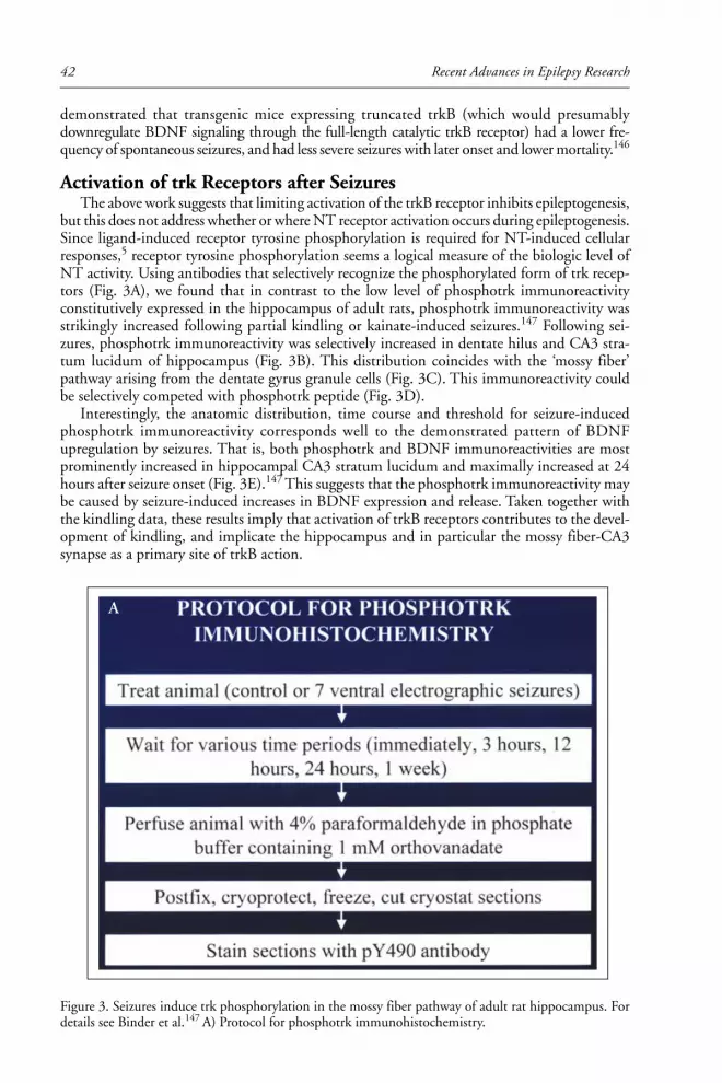
Recent Advances in Epilepsy Research42
demonstrated that transgenic mice expressing truncated trkB (which would presumablydownregulate BDNF signaling through the full-length catalytic trkB receptor) had a lower fre-quency of spontaneous seizures, and had less severe seizures with later onset and lower mortality.146
Activation of trk Receptors after SeizuresThe above work suggests that limiting activation of the trkB receptor inhibits epileptogenesis,
but this does not address whether or where NT receptor activation occurs during epileptogenesis.Since ligand-induced receptor tyrosine phosphorylation is required for NT-induced cellularresponses,5 receptor tyrosine phosphorylation seems a logical measure of the biologic level ofNT activity. Using antibodies that selectively recognize the phosphorylated form of trk recep-tors (Fig. 3A), we found that in contrast to the low level of phosphotrk immunoreactivityconstitutively expressed in the hippocampus of adult rats, phosphotrk immunoreactivity wasstrikingly increased following partial kindling or kainate-induced seizures.147 Following sei-zures, phosphotrk immunoreactivity was selectively increased in dentate hilus and CA3 stra-tum lucidum of hippocampus (Fig. 3B). This distribution coincides with the ‘mossy fiber’pathway arising from the dentate gyrus granule cells (Fig. 3C). This immunoreactivity couldbe selectively competed with phosphotrk peptide (Fig. 3D).
Interestingly, the anatomic distribution, time course and threshold for seizure-inducedphosphotrk immunoreactivity corresponds well to the demonstrated pattern of BDNFupregulation by seizures. That is, both phosphotrk and BDNF immunoreactivities are mostprominently increased in hippocampal CA3 stratum lucidum and maximally increased at 24hours after seizure onset (Fig. 3E).147 This suggests that the phosphotrk immunoreactivity maybe caused by seizure-induced increases in BDNF expression and release. Taken together withthe kindling data, these results imply that activation of trkB receptors contributes to the devel-opment of kindling, and implicate the hippocampus and in particular the mossy fiber-CA3synapse as a primary site of trkB action.
Figure 3. Seizures induce trk phosphorylation in the mossy fiber pathway of adult rat hippocampus. Fordetails see Binder et al.147 A) Protocol for phosphotrk immunohistochemistry.
A

43The Role of BDNF in Epilepsy and Other Diseases of the Mature Nervous System
Figure 3, cont’d. B) Seizures increase phosphotrk immunoreactivity in hilus and CA3 stratum lucidum. B-Ereprinted with permission from ref. 147, copyright 1999 Society for Neuroscience. C) Comparison ofdistribution of phosphotrk immunoreactivity with Nissl-stained section demonstrating mossy fiber path-way localization of phosphotrk immunoreactivity. D) Peptide competition of phosphotrk immunoreactiv-ity. Sections are from a rat 24 hours after 7 electrographic seizures.
B
C
D

Recent Advances in Epilepsy Research44
BDNF-Induced Hyperexcitability of the Mossy Fiber-CA3 SynapseBased on the above data, one might speculate that BDNF upregulation in the adult brain
could predispose certain areas to seizures. Indeed, in adult rat hippocampal slices BDNF expo-sure can produce multiple discharges and spreading depression in area CA3 and the entorhinalcortex upon afferent stimulation.91 Acute application of exogenous BDNF to hippocampalslices appears to preferentially enhance the efficacy of the excitatory mossy fiber synapse ontoCA3 pyramidal cells.91
Actions of BDNF have also been examined after pilocarpine-induced status epilepticus andchronic seizures, when sprouting of mossy fiber collaterals occurs. The new collaterals inner-vate processes in the inner molecular layer, including granule cell dendrites.148 In hippocampalslices isolated from pilocarpine-treated rats, BDNF enhances responses to stimulation of themossy fiber collaterals recorded in the inner molecular layer.149 These effects can be blocked byK252a, a trk inhibitor, and confirm a preferential enhancement of mossy fiber synaptic trans-mission by BDNF. In addition, BDNF exposure in these epileptic animals led to seizure-likeevents.149 Consistent with this are the observations of heightened seizure susceptibility, sponta-neous seizures, and hyperexcitability of hippocampal field CA3 in BDNF-overexpressingtransgenic mice.140
Figure 3, cont’d. E) Time course of phosphotrk immunoreactivity following 7 ventral hippocampal seizures(left: hippocampus, right: CA3). Note phosphotrk immunoreactivity in hilus and CA3 stratum lucidumat the 24-hour time point.
E

45The Role of BDNF in Epilepsy and Other Diseases of the Mature Nervous System
Cellular Model of BDNF-trkB InteractionThe studies summarized above indicate that upregulation of BDNF mRNA, protein and
receptor activation occurs during epileptogenesis, that this upregulation is functionally rel-evant to increased excitability, and that the hippocampus and closely associated limbic struc-tures may be particularly important in the pro-epileptogenic effects of BDNF. A cellular andmolecular model of the actions of BDNF in promoting excitability in the hippocampus fol-lows from these studies (Fig. 4). BDNF mRNA upregulation by seizure or perhaps by otherstimuli such as ischemia or traumatic brain injury leads to increased BDNF production by thedentate granule cells, heightened anterograde transport and release of BDNF from mossy fiberaxons and activation of trkB receptors in hilus and CA3 stratum lucidum. The locus of activa-tion of trkB receptors by released BDNF may be either pre- or postsynaptic.150,151 TrkB recep-tor activation could lead to acute depolarization,96 enhanced glutamatergic synaptic transmis-sion,79,92 or reduced inhibitory synaptic transmission.84 Recent data based on the LTP modelsuggest that BDNF’s actions may be primarily postsynaptic.100 These alterations in synaptictransmission, either alone or in combination with other changes (see below) could be suffi-ciently long-lived to underlie a permanent hyperexcitability of the hippocampal network (i.e.,the epileptic state).
The relevance of results implicating BDNF in modulation of synaptic transmission toepileptogenesis depends critically on whether such modulation occurs in epileptic tissue. Sev-eral lines of evidence suggest this is the case. First, BDNF expression is increased in hippoc-ampi of patients with temporal lobe epilepsy (see below). Second, evidence for modulation ofionotropic receptors with epilepsy comes from studies demonstrating altered electrophysiologyof dentate granule cells in kindling,152,153 other animal models 154-156 and human epileptictissue.157 Third, increased excitability of CA3 pyramidal cells is observed in kindled animals asdetected by increased epileptiform bursting induced by elevated K+ or lowered Mg++ in isolatedhippocampal slices.158,159 CA3 excitability is also present in other animal models.160 Fourth,
Figure 4. Model of BDNF/trkB involvement in epileptogenesis.
Recent Advances in Epilepsy Research46
tetanic stimulation of the mossy fiber pathway in hippocampal slices (such as might occurduring a seizure) induces synaptic potentiation onto CA3 pyramidal cells while inducing de-pression onto stratum lucidum interneurons.161
While modulation of multiple synaptic stations in the limbic system probably contributesto hyperexcitability following seizures, the pivotal role of the CA3 pyramidal cells in promot-ing epileptiform activity in the hippocampus; the role of BDNF in hippocampal synaptictransmission; the fact that constitutive and seizure-induced BDNF immunoreactivity withinthe hippocampus is most intense in the mossy fiber pathway;24,28 together with the localizationof seizure-induced trk receptor activation in CA3 stratum lucidum147 all suggest that strength-ening of the excitatory mossy fiber input onto CA3 pyramidal cells may be a primary mecha-nism by which BDNF promotes epilepsy.
Other Effects of BDNFBased on the known effects of BDNF, it is possible that trkB receptor activation could
contribute to epileptogenesis not only via synaptic effects on excitability but also by inducingchanges in dendritic or axonal sprouting, synaptic morphology and synapse formation on aslower time scale. The most prominent synaptic reorganization known to occur in the epilepticbrain is sprouting of the dentate granule cell mossy fibers.162 Interestingly, mossy fiber sprout-ing was increased in BDNF +/- compared to +/+ mice despite the inhibition of kindling devel-opment in these mutants.139 In addition, bath-applied trkB-Fc failed to inhibit kainate-inducedmossy fiber sprouting in hippocampal explant cultures.163 BDNF-overexpressing transgenicmice failed to demonstrate increased mossy fiber sprouting.164 Thus, there is little evidence todate to suggest that BDNF upregulation is responsible for mossy fiber sprouting in the adultbrain during epileptogenesis.
BDNF is known to modulate the expression of neurotransmitters and neuropeptides, manyof which have potential roles in seizures. Perhaps the best characterized of these is neuropeptideY (NPY). BDNF but not NGF is known to increase NPY levels.165 NPY is thought to beinhibitory to seizure generation because NPY knockout animals exhibit increased seizure sus-ceptibility.166 Interestingly, both kindling and kainate-induced seizures increase NPY immu-noreactivity in the mossy fibers131,167 in a distribution strikingly similar to that of phosphotrkimmunoreactivity. This suggests that BDNF-induced trk activation may lead to NPYupregulation in an overlapping anatomic distribution, thereby potentially limiting excitability.143
BDNF and Human EpilepsyAnimal models of epilepsy, in particular the kindling model described above, have impli-
cated BDNF in epileptogenesis. What direct evidence is there that BDNF is altered/involvedin human epilepsy?
Mathern et al found increased expression of BDNF mRNA in dentate granule cells frompatients with temporal lobe epilepsy.168 Similarly, Murray et al performed in situ hybridizationfor BDNF mRNA in resected temporal lobe epilepsy specimens and found increased hippoc-ampal BDNF expression compared to autopsy control tissue.169 Takahashi et al recently showedthat protein levels of BDNF but not other NTs were upregulated 2.6-fold in human epilepsytissue specimens.170 Interestingly, this study also demonstrated a corresponding upregulationof hippocampal NPY. Recently, Zhu and Roper recorded from hippocampal slices from pa-tients with temporal lobe epilepsy and found that BDNF application enhanced fast excitatorytransmission in dentate granule cells.171
BDNF may also be involved in tumor-associated epilepsy. Primary brain tumors are oftenassociated with seizures.172 Interestingly, immunohistochemical expression of BDNF and trkBhave been studied in glioneuronal brain tumors. In a study of 40 patients with gangliogliomasand 15 patients with dysembryoplastic neuroepithelial tumors (DNETs), tumors that are asso-ciated with chronic medically intractable epilepsy, Aronica et al have recently shown that thereis intense immunoreactivity for both BDNF and trkB in these tumors, and furthermore this

47The Role of BDNF in Epilepsy and Other Diseases of the Mature Nervous System
immunoreactivity is colocalized with NMDAR1 immunoreactivity suggesting a functionalinteraction potentially contributing to the epilepsy associated with these lesions.173
BDNF and Other Diseases of the Adult Nervous System
BDNF and Neurodegenerative DiseasesThe idea that degenerative diseases of the nervous system may result from insufficient sup-
ply of neurotrophic factors has generated great interest in BDNF as a potential therapeuticagent. Many reports have documented evidence of decreased expression of BDNF in Alzheimer’sand Parkinson’s disease.174 Selective reduction of BDNF mRNA in the hippocampus has beenreported in Alzheimer’s disease specimens and decreased BDNF protein has been demonstratedin the substantia nigra in Parkinson’s disease, areas that degenerate in these diseases. BDNFpromotes survival of all major neuronal types affected in Alzheimer’s and Parkinson’s disease,such as hippocampal and neocortical neurons, cholinergic septal and basal forebrain neurons,and nigral dopaminergic neurons. Interestingly, recent work has implicated BDNF inHuntington’s disease as well. Huntingtin, the protein mutated in Huntington’s disease,upregulates BDNF transcription, and loss of huntingtin-mediated BDNF transcription leadsto loss of trophic support to striatal neurons which subsequently degenerate in the hallmarkpathology of the disorder.175 In all of these disorders, provision of BDNF or increasing endog-enous BDNF production may conceivably be therapeutic if applied in the appropriate spa-tiotemporal context.
BDNF and Pain TransmissionBDNF also appears to play an important neuromodulatory role in pain transduction.176 In
particular, BDNF acts as a neuromodulator in small-diameter nociceptive neurons.177 BDNFis synthesized, anterogradely transported and packaged by these neurons into dense core vesiclesat nociceptor (C-fiber) terminals in the dorsal horn, and is markedly upregulated in inflamma-tory injury to peripheral nerves (along with NGF). Postsynaptic cells in this region express trkreceptors,178 and application of BDNF sensitizes nociceptive afferents and elicits hyperalgesia.179
An example of pathologic activity-dependent plasticity somewhat similar to epilepsy is cen-tral pain sensitization.180,181 Central pain sensitization is an activity-dependent increase inexcitability of dorsal horn neurons leading to a clinically intractable condition termed ‘neuro-pathic pain’ in which normally nonpainful somatosensory stimuli (touch and pressure) becomeexquisitely painful (allodynia). Like kindling and drug sensitization and dependence, centralsensitization is NMDA receptor-dependent and long-lasting.180
Furthermore, as in kindling, NTs have been implicated in central sensitization.182 BDNF isupregulated in dorsal root ganglia and spinal cord following peripheral inflammation.183 Inaddition, BDNF reduces GABAA-mediated currents in peripheral afferent fibers, suggestingthat it may facilitate nociceptive input into the dorsal horn.184 Woolf and colleagues havedemonstrated that pretreatment with trkB-Fc prevents central sensitization, presumably bycompeting with endogenous BDNF released at C-fiber terminals onto dorsal horn neurons,thereby preventing activation of trkB receptors on dorsal horn membranes.185 Inflammation-inducedhyperalgesia also appears to be related to NGF/trkA signaling since trkA-Fc or NGF antibodiesinhibit the development of hyperalgesia following an inflammatory stimulus.186-188
BDNF and Drug AddictionThe neurobiology of drug addiction is rapidly becoming better understood.189 Emerging
evidence indicates that BDNF-related plasticity may also occur in brain structures responsiblefor drug sensitization and dependence. For example, BDNF has been found to influence thereinforcing and locomotor activating properties of psychostimulants. Repeated injections ofamphetamine lead to elevated BDNF mRNA expression in the basolateral amygdala, piriformcortex and paraventricular nucleus of the hypothalamus.190 This is accompanied by increased

Recent Advances in Epilepsy Research48
BDNF immunoreactivity in target structures such as the nucleus accumbens, a well-known siterelated to reinforcing behavior and addiction.190
Chronic opiate exposure leads to numerous neurochemical adaptations, in particular in thenoradrenergic locus ceruleus (LC). Such adaptations are thought to contribute to physical drugdependence. Now, it appears that opiate administration and withdrawal lead to changes inBDNF expression. Numan et al demonstrate that whereas chronic morphine treatment resultsin only modest increases in BDNF in the locus ceruleus, withdrawal leads to a marked, rapidand prolonged increase in BDNF and trkB mRNA in the LC.191 More recently, Akbarian etal192 have shown that there are dramatic alterations of morphine administration-induced sig-naling in mice with a conditional deletion of BDNF in postnatal brain. In these mice, therewas a three-fold reduction in opiate withdrawal symptoms.192
All drugs of abuse increase dopamine in the shell of the nucleus accumbens, and the D3receptor is thought to be responsible for the reinforcing effects of drugs.193 A recent studysuggested a candidate molecular mechanism for the control of BDNF over behavioral sensiti-zation. Guillin et al demonstrated that BDNF from dopaminergic neurons is responsible forinducing normal expression of the dopamine D3 receptor in nucleus accumbens.194 Thus,pathologic alterations in BDNF expression may lead to the abnormal D3 expression seen indrug addiction.
BDNF and Affective BehaviorsBDNF signaling may also be involved in affective behaviors.195,196 BDNF may be
dysregulated in depressed individuals.196 Environmental stresses such as immobilization thatinduce depression also decrease BDNF mRNA.197 Conversely, physical exercise is associatedwith decreased depression and increased BDNF mRNA. Existing treatments for depression arethought to work primarily by increasing endogenous monoaminergic (i.e., serotonergic andnoradrenergic) synaptic transmission, and recent studies have shown that effective antidepres-sants increase BDNF mRNA in the brain. Exogenous delivery of BDNF promotes the func-tion and sprouting of serotonergic neurons in adult rat brains.195 Thus, new pharmacologicstrategies are focused on the potential antidepressant role of BDNF.
A recent study suggests that the BDNF gene may be a susceptibility gene for bipolar disor-der, a severe psychiatric disease that affects 1% of the population worldwide and is character-ized by recurrent bouts of mania and depression.198 This study demonstrates linkage disequi-librium between two polymorphisms of the BDNF gene and bipolar disorder in 283 nuclearfamilies.198
SummaryBDNF is widespread in the CNS during development and in adulthood, and is regulated in
a wide variety of physiologic and pathologic states. Overall, in addition to its trophic effects ontarget neurons, BDNF appears to constitute a general mechanism for activity-dependent modi-fication of synapses in the developing and adult CNS. Diseases of abnormal trophic support(such as neurodegenerative diseases) and diseases of abnormal excitability (such as epilepsy andcentral pain sensitization) can be related in some cases to abnormal BDNF signaling.
The evidence implicating BDNF in pathologic activity-dependent plasticity is most clear inthe case of epilepsy. BDNF mRNA and protein are markedly upregulated in the hippocampusby seizure activity in animal models, and interference with BDNF/trkB signaling inhibitsepileptogenesis. The hippocampus and closely associated limbic structures are thought to beparticularly important in the pro-epileptogenic effects of BDNF, and indeed increased BDNFexpression in the hippocampus is found in specimens from patients with temporal lobe epilepsy.
It is hoped that understanding of the hyperexcitability associated with BDNF may lead tonovel anticonvulsant or antiepileptic therapies. Further study of the cellular and molecularmechanisms by which BDNF influences cell survival and excitability will likely provide novelconcepts and targets for the treatment of diverse CNS diseases.

49The Role of BDNF in Epilepsy and Other Diseases of the Mature Nervous System
References1. Barde Y-A, Edgar D, Thoenen H. Purification of a new neurotrophic factor from mammalian
brain. EMBO J 1982; 1:549-553.2. Levi-Montalcini R, Hamburger V. Selective growth-stimulating effects of mouse sarcoma on the
sensory and sympathetic nervous system of the chick embryo. J Exp Zool 1951; 116:321-361.3. Maisonpierre PC, Le Beau MM, Espinosa R et al. Human and rat brain-derived neurotrophic
factor and neurotrophin-3: gene structures, distributions, and chromosomal localizations. Genomics1991; 10:558-68.
4. Patapoutian A, Reichardt LF. Trk receptors: mediators of neurotrophin action. Curr Op Neurobiol2001; 11:272-280.
5. Barbacid M. The trk family of neurotrophin receptors. J Neurobiol 1994; 25:1386-403.6. Schlessinger J, Ulrich A. Growth factor signaling by receptor tyrosine kinases. Neuron 1992;
9:381-391.7. Guiton M, Gunn-Moore FJ, Stitt TN et al. Identification of in vivo brain-derived neurotrophic
factor-stimulated autophosphorylation sites on the trkB receptor tyrosine kinase by site-directedmutagenesis. J Biol Chem 1994; 269:30370-30377.
8. Heumann R. Neurotrophin signalling. Curr Op Neurobiol 1994; 4:668-679.9. Kaplan DR, Stephens RM. Neurotrophin signal transduction by the trk receptor. J Neurobiol
1994; 25:1404-1417.10. Middlemas DS, Meisenhelder J, Hunter T. Identification of trkB autophosphorylation sites and
evidence that phospholipase C-gamma1 is a substrate of the trkB receptor. J Biol Chem 1994;269:5458-5466.
11. Segal RA, Greenberg ME. Intracellular signaling pathways activated by neurotrophic factors. AnnuRev Neurosci 1996; 19:463-489.
12. Urfer R, Tsoulfas P, O’Connell L et al. An immunoglobulin-like domain determines the specificityof neurotrophin receptors. EMBO J 1995; 14:2795-2805.
13. Chao MV, Hempstead BL. p75 and trk: a two-receptor system. Trends Neurosci 1995; 18:321-326.14. Roux P, Barker P. Neurotrophin signaling through the p75 neurotrophin receptor. Prog Neurobiol
2002; 67:203.15. Dechant G, Barde YA. The neurotrophin receptor p75NTR: novel functions and implications for
diseases of the nervous system. Nat Neurosci 2002; 5:1131-1136.16. Inagaki N, Thoenen H, Lindholm D. TrkA tyrosine residues involved in NGF-induced neurite
outgrowth of PC12 cells. Eur J Neurosci 1995; 7:1125-1133.17. Grimes ML, Zhou J, Beattie EC et al. Endocytosis of activated trkA: evidence that nerve growth
factor induces formation of signaling endosomes. J Neurosci 1996; 16:7950-7964.18. Von Bartheld CS, Williams R, Lefcort F et al. Retrograde transport of neurotrophins from the eye
to the brain in chick embryos: roles of the p75NTR and trkB receptors. J Neurosci 1996;16:2995-3008.
19. Bhattacharyya A, Watson FL, Bradlee TA et al. Trk receptors function as rapid retrograde signalcarriers in the adult nervous system. J Neurosci 1997; 17:7007-7016.
20. Riccio A, Pierchala BA, Ciarallo CL et al. An NGF-trkA-mediated retrograde signal to transcrip-tion factor CREB in sympathetic neurons. Science 1997; 277:1097-1100.
21. Senger DL, Campenot RB. Rapid retrograde tyrosine phosphorylation of trkA and other proteinsin rat sympathetic neurons in compartmented cultures. J Cell Biol 1997; 138:411-421.
22. MacInnis BL, Campenot RB. Retrograde support of neuronal survival without retrograde transportof nerve growth factor. Science 2002; 295:1536-9.
23. Barker PA, Hussain NK, McPherson PS. Retrograde signaling by the neurotrophins follows awell-worn trk. Trends Neurosci 2002; 25:379-381.
24. Conner JM, Lauterborn JC, Yan Q et al. Distribution of brain-derived neurotrophic factor (BDNF)protein and mRNA in the normal adult rat CNS—evidence for anterograde axonal transport. JNeurosci 1997; 17:2295-2313.
25. Bengzon J, Kokaia Z, Ernfors P et al. Regulation of neurotrophin and trkA, trkB and trkC ty-rosine kinase receptor messenger RNA expression in kindling. Neuroscience 1993; 53:433-46.
26. Fryer RH, Kaplan DR, Feinstein SC et al. Developmental and mature expression of full-lengthand truncated trkB receptors in the rat forebrain. J Comp Neurol 1996; 374:21-40.
27. Lindvall O, Kokaia Z, Bengzon J et al. Neurotrophins and brain insults. Trends Neurosci 1994;17:490-496.
28. Yan Q, Rosenfeld RD, Matheson CR et al. Expression of brain-derived neurotrophic factor proteinin the adult rat central nervous system. Neuroscience 1997; 78:431-448.
29. Von Bartheld CS, Byers MR, Williams R et al. Anterograde transport of neurotrophins andaxodendritic transfer in the developing visual system. Nature 1996; 379:830-833.

Recent Advances in Epilepsy Research50
30. Altar CA, Cai N, Bliven T et al. Anterograde transport of brain-derived neurotrophic factor and itsrole in the brain. Nature 1997; 389:856-860.
31. Zhou X-F, Rush RA. Endogenous brain-derived neurotrophic factor is anterogradely transported inprimary sensory neurons. Neuroscience 1996; 74:945-951.
32. Fawcett JP, Bamji SX, Causing CG et al. Functional evidence that BDNF is an anterograde neu-ronal trophic factor in the CNS. J Neurosci 1998; 18:2808-2821.
33. Tonra JR, Curtis R, Wong V et al. Axotomy upregulates the anterograde transport and expressionof brain-derived neurotrophic factor by sensory neurons. J Neurosci 1998; 18:4374-4383.
34. Kohara K, Kitamura A, Morishima M et al. Activity-dependent transfer of brain-derived neurotrophicfactor to postsynaptic neurons. Science 2001; 291:2419-2423.
35. Nawa H, Takei N. BDNF as an anterophin; a novel neurotrophic relationship between brain neu-rons. Trends Neurosci 2001; 24:683-684.
36. Smith MA, Zhang LX, Lyons WE et al. Anterograde transport of endogenous brain-derived neu-rotrophic factor in hippocampal mossy fibers. Neuroreport 1997; 8:1829-1834.
37. Fawcett JP, Aloyz R, McLean JH et al. Detection of brain-derived neurotrophic factor in a vesicu-lar fraction of brain synaptosomes. J Biol Chem 1997; 272:8837-40.
38. Blöchl A, Thoenen H. Characterization of nerve growth factor (NGF) release from hippocampalneurons: evidence for a constitutive and an unconventional sodium-dependent regulated pathway.Eur J Neurosci 1995; 7:1220-8.
39. Blöchl A, Thoenen H. Localization of cellular storage compartments and sites of constitutive andactivity-dependent release of nerve growth factor (NGF) in primary cultures of hippocampal neu-rons. Mol Cell Neurosci 1996; 7:173-90.
40. Goodman LJ, Valverde J, Lim F et al. Regulated release and polarized localization of brain-derivedneurotrophic factor in hippocampal neurons. Mol Cell Neurosci 1996; 7:222-238.
41. Griesbeck O, Canossa M, Campana G et al. Are there differences between the secretion character-istics of NGF and BDNF? Implications for the modulatory role of neurotrophins inactivity-dependent neuronal plasticity. Microsc Res Tech 1999; 45:262-75.
42. Canossa M, Gartner A, Campana G et al. Regulated secretion of neurotrophins by metabotropicglutamate group I (mGluRI) and Trk receptor activation is mediated via phospholipase C signal-ling pathways. EMBO J 2001; 20:1640-50.
43. Purves D, Lichtman JW. Principles of neural development. Sunderland, MA: Sinauer, 1985.44. Barde YA. Trophic factors and neuronal survival. Neuron 1989; 2:1525-1534.45. Huang EJ, Reichardt LF. Neurotrophins: roles in neuronal development and function. Annu Rev
Neurosci 2001; 24:677-736.46. Jones KR, Farinas I, Backus C et al. Targeted disruption of the BDNF gene perturbs brain and
sensory neuron development but not motor neuron development. Cell 1994; 76:989-99.47. Tucker KL, Meyer M, Barde YA. Neurotrophins are required for nerve growth during develop-
ment. Nat Neurosci 2001; 4:29-37.48. Carroll P, Lewin GR, Koltzenburg M et al. A role for BDNF in mechanosensation. Nat Neurosci
1998; 1:42-6.49. Acheson A, Conover JC, Fandl JP et al. A BDNF autocrine loop in adult sensory neurons prevents
cell death. Nature 1995; 374:450-3.50. Lotto RB, Asavaritikrai P, Vali L et al. Target-derived neurotrophic factors regulate the death of
developing forebrain neurons after a change in their trophic requirements. J Neurosci 2001;21:3904-10.
51. Xu B, Zang K, Ruff NL et al. Cortical degeneration in the absence of neurotrophin signaling:dendritic retraction and neuronal loss after removal of the receptor trkB. Neuron 2000; 26:233-45.
52. Patel MN, McNamara JO. Selective enhancement of axonal branching of cultured dentate gyrusneurons by neurotrophic factors. Neuroscience 1995; 69:763-70.
53. Lowenstein DH, Arsenault L. The effects of growth factors on the survival and differentiation ofcultured dentate gyrus neurons. J Neurosci 1996; 16:1759-1769.
54. McAllister AK, Lo DC, Katz LC. Neurotrophins regulate dendritic growth in developing visualcortex. Neuron 1995; 15:791-803.
55. McAllister AK, Katz LC, Lo DC. Neurotrophin regulation of cortical dendritic growth requiresactivity. Neuron 1996; 17:1057-1064.
56. McAllister AK, Katz LC, Lo DC. Opposing roles for endogenous BDNF and NT-3 in regulatingcortical dendritic growth. Neuron 1997; 18:767-78.
57. Cohen-Cory S, Fraser SE. Effects of brain-derived neurotrophic factor on optic axon branchingand remodelling in vivo. Nature 1995; 378:192-196.

51The Role of BDNF in Epilepsy and Other Diseases of the Mature Nervous System
58. Maffei L, Berardi N, Domenici L et al. Nerve growth factor (NGF) prevents the shift in oculardominance distribution of visual cortical neurons in monocularly deprived rats. J Neurosci 1992;12:4651-62.
59. Cabelli RJ, Hohn A, Shatz CJ. Inhibition of ocular dominance column formation by infusion ofNT-4/5 or BDNF. Science 1995; 267:1662-6.
60. Cabelli RJ, Shelton DL, Segal RA et al. Blockade of endogenous ligands of trkB inhibits formationof ocular dominance columns. Neuron 1997; 19:63-76.
61. Holtzman DM, Lowenstein DH. Selective inhibition of axon outgrowth by antibodies to NGF ina model of temporal lobe epilepsy. J Neurosci 1995; 15:7062-7070.
62. Castrén E, Zafra F, Thoenen H et al. Light regulates expression of brain-derived neurotrophicfactor mRNA in rat visual cortex. Proc Natl Acad Sci USA 1992; 89:9444-8.
63. Castrén E, Thoenen H, Lindholm D. Brain-derived neurotrophic factor messenger RNA is ex-pressed in the septum, hypothalamus and in adrenergic brain stem nuclei of adult rat brain and isincreased by osmotic stimulation in the paraventricular nucleus. Neuroscience 1995; 64:71-80.
64. Rocamora N, Welker E, Pascual M et al. Upregulation of BDNF mRNA expression in the barrelcortex of adult mice after sensory stimulation. J Neurosci 1996; 16:4411-4419.
65. Li XC, Jarvis ED, Alvarez-Borda B et al. A relationship between behavior, neurotrophin expres-sion, and new neuron survival. Proc Natl Acad Sci USA 2000; 97:8584-9.
66. Patterson SL, Grover LM, Schwartzkroin PA et al. Neurotrophin expression in rat hippocampalslices: a stimulus paradigm inducing LTP in CA1 evokes increases in BDNF and NT-3 mRNAs.Neuron 1992; 9:1081-8.
67. Castrén E, Pitkanen M, Sirvio J et al. The induction of LTP increases BDNF and NGF mRNAbut decreases NT-3 mRNA in the dentate gyrus. Neuroreport 1993; 4:895-8.
68. Bramham CR, Southard T, Sarvey JM et al. Unilateral LTP triggers bilateral increases in hippoc-ampal neurotrophin and trk receptor mRNA expression in behaving rats: evidence for interhemi-spheric communication. J Comp Neurol 1996; 368:371-382.
69. Neeper SA, Gomez-Pinilla F, Choi J et al. Physical activity increases mRNA for brain-derivedneurotrophic factor and nerve growth factor in rat brain. Brain Res 1996; 726:49-56.
70. Oliff HS, Berchtold NC, Isackson P et al. Exercise-induced regulation of brain-derived neurotrophicfactor (BDNF) transcripts in the rat hippocampus. Brain Res Mol Brain Res 1998; 61:147-53.
71. Frost DO. BDNF/trkB signaling in the developmental sculpting of visual connections. Prog BrainRes 2001; 134:35-49.
72. Thoenen H. Neurotrophins and neuronal plasticity. Science 1995; 270:593-8.73. Schuman EM. Neurotrophin regulation of synaptic transmission. Curr Opin Neurobiol 1999;
9:105-9.74. Thoenen H. Neurotrophins and activity-dependent plasticity. Prog Brain Res 2000; 128:183-91.75. Poo MM. Neurotrophins as synaptic modulators. Nat Rev Neurosci 2001; 2:24-32.76. Lohof AM, Ip NY, Poo MM. Potentiation of developing neuromuscular synapses by the
neurotrophins NT-3 and BDNF. Nature 1993; 363:350-3.77. Knipper M, Leung LS, Zhao D et al. Short-term modulation of glutamatergic synapses in adult rat
hippocampus by NGF. Neuroreport 1994; 5:2433-6.78. Lessmann V, Gottmann K, Heumann R. BDNF and NT-4/5 enhance glutamatergic synaptic trans-
mission in cultured hippocampal neurones. Neuroreport 1994; 6:21-5.79. Kang H, Schuman EM. Long-lasting neurotrophin-induced enhancement of synaptic transmission
in the adult hippocampus. Science 1995; 267:1658-62.80. Stoop R, Poo MM. Synaptic modulation by neurotrophic factors: differential and synergistic ef-
fects of brain-derived neurotrophic factor and ciliary neurotrophic factor. J Neurosci 1996;16:3256-64.
81. Carmignoto G, Pizzorusso T, Tia S et al. Brain-derived neurotrophic factor and nerve growthfactor potentiate excitatory synaptic transmission in the rat visual cortex. J Physiol 1997;498:153-164.
82. Wang X, Poo M. Potentiation of developing synapses by postsynaptic release of neurotrophin-4.Neuron 1997; 19:825-835.
83. Messaoudi E, Bardsen K, Srebro B et al. Acute intrahippocampal infusion of BDNF induces last-ing potentiation of synaptic transmission in the rat dentate gyrus. J Neurophysiol 1998; 79:496-499.
84. Tanaka T, Saito H, Matsuki N. Inhibition of GABAA synaptic responses by brain-derived neu-rotrophic factor (BDNF) in rat hippocampus. J Neurosci 1997; 17:2959-2966.
85. Frerking M, Malenka RC, Nicoll RA. Brain-derived neurotrophic factor (BDNF) modulates in-hibitory, but not excitatory, transmission in the CA1 region of the hippocampus. J Neurophysiol1998; 80:3383-6.

Recent Advances in Epilepsy Research52
86. Figurov A, Pozzo-Miller LD, Olafsson P et al. Regulation of synaptic responses to high-frequencystimulation and LTP by neurotrophins in the hippocampus. Nature 1996; 381:706-9.
87. Akaneya Y, Tsumoto T, Kinoshita S et al. Brain-derived neurotrophic factor enhances long-termpotentiation in rat visual cortex. J Neurosci 1997; 17:6707-6716.
88. Korte M, Carroll P, Wolf E et al. Hippocampal long-term potentiation is impaired in mice lackingbrain-derived neurotrophic factor. Proc Natl Acad Sci USA 1995; 92:8856-60.
89. Korte M, Griesbeck O, Gravel C et al. Virus-mediated gene transfer into hippocampal CA1 regionrestores long-term potentiation in brain-derived neurotrophic factor mutant mice. Proc Natl AcadSci USA 1996; 93:12547-52.
90. Patterson SL, Abel T, Deuel TA et al. Recombinant BDNF rescues deficits in basal synaptic trans-mission and hippocampal LTP in BDNF knockout mice. Neuron 1996; 16:1137-45.
91. Scharfman HE. Hyperexcitability in combined entorhinal/hippocampal slices of adult rat after ex-posure to brain-derived neurotrophic factor. J Neurophysiol 1997; 78:1082-1095.
92. Takei N, Sasaoka K, Inoue K et al. Brain-derived neurotrophic factor increases the stimulation-evokedrelease of glutamate and the levels of exocytosis-associated proteins in cultured cortical neuronsfrom embryonic rats. J Neurochem 1997; 68:370-5.
93. Suen P-C, Wu K, Levine ES et al. Brain-derived neurotrophic factor rapidly enhances phosphory-lation of the postsynaptic N-methyl-D-aspartate receptor subunit 1. Proc Natl Acad Sci USA 1997;94:8191-8195.
94. Berninger B, Garcia DE, Inagaki N et al. BDNF and NT-3 induce intracellular Ca2+ elevation inhippocampal neurones. Neuroreport 1993; 4:1303-6.
95. Levine ES, Dreyfus CF, Black IB et al. Differential effects of NGF and BDNF on voltage-gatedcalcium currents in embryonic basal forebrain neurons. J Neurosci 1995; 15:3084-91.
96. Kafitz KW, Rose CR, Thoenen H et al. Neurotrophin-evoked rapid excitation through TrkB re-ceptors. Nature 1999; 401:918-21.
97. Marty S, Berzaghi MP, Berninger B. Neurotrophins and activity-dependent plasticity of corticalinterneurons. Trends Neurosci 1997; 20:198-202.
98. Xu B, Gottschalk W, Chow A et al. The role of brain-derived neurotrophic factor receptors in themature hippocampus: Modulation of long-term potentiation through a presynaptic mechanism in-volving TrkB. J Neurosci 2000; 20:6888-97.
99. Manabe T. Does BDNF have pre or postsynaptic targets? Science 2002; 295:1651-3.100. Kovalchuk Y, Hanse E, Kafitz KW et al. Postsynaptic induction of BDNF-mediated long-term
potentiation. Science 2002; 295:1729-34.101. Hall J, Thomas KL, Everitt BJ. Rapid and selective induction of BDNF expression in the hippoc-
ampus during contextual learning. Nat Neurosci 2000; 3:533-535.102. Ishibashi H, Hihara S, Takahashi M et al. Tool-use learning induces BDNF expression in a selec-
tive portion of monkey anterior parietal cortex. Brain Res Mol Brain Res 2002; 102:110.103. Linnarsson S, Bjorklund A, Ernfors P. Learning deficit in BDNF mutant mice. Eur J Neurosci
1997; 9:2581-2587.104. Ma YL, Wang HL, Wu HC et al. Brain-derived neurotrophic factor antisense oligonucleotide im-
pairs memory retention and inhibits long-term potentiation in rats. Neuroscience 1998; 82:957-967.105. Minichiello L, Korte M, Wolfer D et al. Essential role for TrkB receptors in hippocampus-mediated
learning. Neuron 1999; 24:401-14.106. Hauser WA, Kurland LT. The epidemiology of epilepsy in Rochester, Minnesota, 1935 through
1967. Epilepsia 1975; 16:1-66.107. Morgan JI, Curran T. Stimulus-transcription coupling in the nervous system: Involvement of the
inducible proto-oncogenes fos and fun. Annu Rev Neurosci 1991; 14:421-451.108. Kiessling M, Gass P. Immediate early gene expression in experimental epilepsy. Brain Pathol 1993;
3:381-393.109. Gall C, Lauterborn J, Bundman M et al. Seizures and the regulation of neurotrophic factor and
neuropeptide gene expression in brain. Epilepsy Res—Suppl 1991; 4:225-45.110. Meberg PJ, Gall CM, Routtenberg A. Induction of F1/GAP-43 gene expression in hippocampal
granule cells after seizures. Brain Res Mol Brain Res 1993; 17:295-9.111. Qian Z, Gilbert M, Colicos MA et al. Tissue plasminogen activator is induced as an immediate-early
gene during seizure, kindling, and long-term potentiation. Nature 1993; 361:453-457.112. Gall CM, Isackson PJ. Limbic seizures increase neuronal production of messenger RNA for nerve
growth factor. Science 1989; 245:758-61.113. Gall CM. Seizure-induced changes in neurotrophin expression: Implications for epilepsy. Exp Neurol
1993; 124:150-66.114. Binder DK, Gall CM, Croll SD et al. BDNF and epilepsy: Too much of a good thing? Trends
Neurosci 2001; 24:47-53.

53The Role of BDNF in Epilepsy and Other Diseases of the Mature Nervous System
115. Ernfors P, Bengzon J, Kokaia Z et al. Increased levels of messenger RNAs for neurotrophic factorsin the brain during kindling epileptogenesis. Neuron 1991; 7:165-76.
116. Isackson PJ, Huntsman MM, Murray KD et al. BDNF mRNA expression is increased in adult ratforebrain after limbic seizures: temporal patterns of induction distinct from NGF. Neuron 1991;6:937-48.
117. Dugich-Djordjevic MM, Tocco G, Lapchak PA et al. Regionally specific and rapid increases inbrain-derived neurotrophic factor messenger RNA in the adult rat brain following seizures inducedby systemic administration of kainic acid. Neuroscience 1992; 47:303-15.
118. Dugich-Djordjevic MM, Tocco G, Willoughby DA et al. BDNF mRNA expression in the devel-oping rat brain following kainic acid-induced seizure activity. Neuron 1992; 8:1127-38.
119. Humpel C, Wetmore C, Olson L. Regulation of brain-derived neurotrophic factor messenger RNAand protein at the cellular level in pentylenetetrazol-induced epileptic seizures. Neuroscience 1993;53:909-18.
120. Merlio JP, Ernfors P, Kokaia Z et al. Increased production of the TrkB protein tyrosine kinasereceptor after brain insults. Neuron 1993; 10:151-64.
121. Schmidt-Kastner R, Olson L. Decrease of neurotrophin-3 mRNA in adult rat hippocampus afterpilocarpine seizures. Exp Neurol 1995; 136:199-204.
122. Schmidt-Kastner R, Humpel C, Wetmore C et al. Cellular hybridization for BDNF, trkB, andNGF mRNAs and BDNF-immunoreactivity in rat forebrain after pilocarpine-induced statusepilepticus. Exp Brain Res 1996; 107:331-47.
123. Mudo G, Jiang XH, Timmusk T et al. Change in neurotrophins and their receptor mRNAs in therat forebrain after status epilepticus induced by pilocarpine. Epilepsia 1996; 37:198-207.
124. Sato K, Kashihara K, Morimoto K et al. Regional increases in brain-derived neurotrophic factorand nerve growth factor mRNAs during amygdaloid kindling, but not in acidic and basic fibro-blast growth factor mRNAs. Epilepsia 1996; 37:6-14.
125. Gall CM, Lauterborn JC, Guthrie KM et al. Seizures and the regulation of neurotrophic factorexpression: associations with structural plasticity in epilepsy. In: Seil RJ, ed. Advances in Neurol-ogy, vol 72: Neuronal Regeneration, Reorganization, and Repair. Philadelphia: Lippincott-Raven,1997:9-24.
126. Lowenstein DH, Seren MS, Longo FM. Prolonged increases in neurotrophic activity associatedwith kainate-induced hippocampal synaptic reorganization. Neuroscience 1993; 56:597-604.
127. Humpel C, Lindqvist E, Soderstrom S et al. Monitoring release of neurotrophic activity in thebrains of awake rats. Science 1995; 269:552-4.
128. Nawa H, Carnahan J, Gall C. BDNF protein measured by a novel enzyme immunoassay in nor-mal brain and after seizure: Partial disagreement with mRNA levels. Eur J Neurosci 1995; 7:1527-35.
129. Elmer E, Kokaia Z, Kokaia M et al. Dynamic changes of brain-derived neurotrophic factor proteinlevels in the rat forebrain after single and recurring kindling-induced seizures. Neuroscience 1998;83:351-62.
130. Rudge JS, Mather PE, Pasnikowski EM et al. Endogenous BDNF protein is increased in adult rathippocampus after a kainic acid induced excitotoxic insult but exogenous BDNF is notneuroprotective. Exp Neurol 1998; 149:398-410.
131. Vezzani A, Ravizza T, Moneta D et al. Brain-derived neurotrophic factor immunoreactivity in thelimbic system of rats after acute seizures and during spontaneous convulsions: temporal evolutionof changes as compared to neuropeptide Y. Neuroscience 1999; 90:1445-1461.
132. Gall CM, Conner JM, Lauterborn JC et al. Cellular localization of BDNF protein after recurrentseizures in rat: evidence for axonal transport of the newly synthesized factor. Epilepsia 1996; 37Suppl. 5:47.
133. Goddard GV. The development of epileptic seizures through brain stimulation at low intensity.Nature 1967; 214:1020-1021.
134. Goddard GV, McIntyre DC, Leech CK. A permanent change in brain function resulting fromdaily electrical stimulation. Exp Neurol 1969; 25:295-330.
135. McNamara JO, Bonhaus DW, Shin C. The kindling model of epilepsy. In: Schwartzkroin PA, ed.Epilepsy: Models, mechanisms, and concepts. New York, NY: Cambridge University Press, 1993:27-47.
136. Binder DK, McNamara JO. Kindling: a pathologic activity-driven structural and functional plastic-ity in mature brain. In: Corcoran ME, Moshe S, eds. Kindling 5. New York: Plenum Press,1997:245-254.
137. Funabashi T, Sasaki H, Kimura F. Intraventricular injection of antiserum to nerve growth factordelays the development of amygdaloid kindling. Brain Res 1988; 458:132-6.
138. Van der Zee CE, Rashid K, Le K et al. Intraventricular administration of antibodies to nervegrowth factor retards kindling and blocks mossy fiber sprouting in adult rats. J Neurosci 1995;15:5316-23.

Recent Advances in Epilepsy Research54
139. Kokaia M, Ernfors P, Kokaia Z et al. Suppressed epileptogenesis in BDNF mutant mice. ExpNeurol 1995; 133:215-24.
140. Croll SD, Suri C, Compton DL et al. Brain-derived neurotrophic factor transgenic mice exhibitpassive avoidance deficits, increased seizure severity and in vitro hyperexcitability in the hippocam-pus and entorhinal cortex. Neuroscience 1999; 93:1491-1506.
141. Scharfman HE, Goodman JH, Sollas AL et al. Spontaneous limbic seizures after intrahippocampalinfusion of brain-derived neurotrophic factor. Exp Neurol 2002; 174:201-14.
142. Binder DK, Routbort MJ, Ryan TE et al. Selective inhibition of kindling development by intra-ventricular administration of trkB receptor body. J Neurosci 1999; 19:1424-1436.
143. Larmet Y, Reibel S, Carnahan J et al. Protective effects of brain-derived neurotrophic factor on thedevelopment of hippocampal kindling in the rat. Neuroreport 1995; 6:1937-41.
144. Frank L, Ventimiglia R, Anderson K et al. BDNF downregulates neurotrophin responsiveness,trkB protein and trkB mRNA levels in cultured rat hippocampal neurons. Eur J Neurosci 1996;8:1220-30.
145. Knusel B, Gao H, Okazaki T et al. Ligand-induced down-regulation of trk messenger RNA, pro-tein and tyrosine phosphorylation in rat cortical neurons. Neuroscience 1997; 78:851-862.
146. Lahteinen S, Pitkanen A, Saarelainen T et al. Decreased BDNF signalling in transgenic mice re-duces epileptogenesis. Eur J Neurosci 2002; 15:721-34.
147. Binder DK, Routbort MJ, McNamara JO. Immunohistochemical evidence of seizure-induced acti-vation of trk receptors in the mossy fiber pathway of adult rat hippocampus. J Neurosci 1999;19:4616-4626.
148. Okazaki MM, Evenson DA, Nadler JV. Hippocampal mossy fiber sprouting and synapse forma-tion after status epilepticus in rats: Visualization after retrograde transport of biocytin. J CompNeurol 1995; 352:515-534.
149. Scharfman HE, Goodman JH, Sollas AL. Actions of brain-derived neurotrophic factor in slicesfrom rats with spontaneous seizures and mossy fiber sprouting in the dentate gyrus. J Neurosci1999; 19:5619-5631.
150. Wu K, Xu J, Suen P et al. Functional trkB neurotrophin receptors are intrinsic components of theadult brain postsynaptic density. Mol Brain Res 1996; 43:286-290.
151. Drake CT, Milner TA, Patterson SL. Ultrastructural localization of full-length trkB immunoreac-tivity in rat hippocampus suggests multiple roles in modulating activity-dependent synaptic plastic-ity. J Neurosci 1999; 19:8009-26.
152. Kohr G, De Koninck Y, Mody I. Properties of NMDA receptor channels in neurons acutely iso-lated from epileptic (kindled) rats. J Neurosci 1993; 13:3612-27.
153. McNamara JO. Cellular and molecular basis of epilepsy. J Neurosci 1994; 14:3413-3425.154. Wuarin J-P, Dudek FE. Electrographic seizures and new recurrent excitatory circuits in the dentate
gyrus of hippocampal slices from kainate-treated epileptic rats. J Neurosci 1996; 16:4438-4448.155. Gibbs JW, Shumate MD, Coulter DA. Differential epilepsy-associated alterations in postsynaptic
GABA(A) receptor function in dentate granule and CA1 neurons. J Neurophysiol 1997;77:1924-1938.
156. Okazaki MM, Molnar P, Nadler JV. Recurrent mossy fiber pathway in rat dentate gyrus: Synapticcurrrents evoked in presence and absence of seizure-induced growth. J Neurophysiol 1999;81:1645-1660.
157. Williamson A, Patrylo PR, Spencer DD. Decrease in inhibition in dentate granule cells from pa-tients with medial temporal lobe epilepsy. Ann Neurol 1999; 45:92-99.
158. Behr J, Lyson KJ, Mody I. Enhanced propagation of epileptiform activity through the kindleddentate gyrus. J Neurophysiol 1998; 79:1726-1732.
159. King GL, Dingledine R, Giacchino JL et al. Abnormal neuronal excitability in hippocampal slicesfrom kindled rats. J Neurophysiol 1985; 54:1295-304.
160. Buzsaki G, Ponomareff GL, Bayardo F et al. Neuronal activity in the subcortically denervatedhippocampus: A chronic model for epilepsy. Neuroscience 1989; 28:527-538.
161. Maccaferri G, Toth K, McBain CJ. Target-specific expression of presynaptic mossy fiber plasticity.Science 1998; 279:1368-1370.
162. Sutula T, He XX, Cavazos J et al. Synaptic reorganization in the hippocampus induced by abnor-mal functional activity. Science 1988; 239:1147-50.
163. Routbort MJ, Ryan TE, Yancopoulos GD et al. TrkB-IgG does not inhibit mossy fiber sproutingin an in vitro model. Soc Neurosci Abstr 1997; 23:888.
164. Qiao X, Suri C, Knusel B et al. Absence of hippocampal mossy fiber sprouting in transgenic miceoverexpressing brain-derived neurotrophic factor. J Neurosci Res 2001; 64:268-76.
165. Croll SD, Wiegand SJ, Anderson KD et al. Regulation of neuropeptides in adult rat forebrain bythe neurotrophins BDNF and NGF. Eur J Neurosci 1994; 6:1343-53.

55The Role of BDNF in Epilepsy and Other Diseases of the Mature Nervous System
166. Baraban SC, Hollopeter G, Erickson JC et al. Knock-out mice reveal a critical antiepileptic rolefor neuropeptide Y. J Neurosci 1997; 17:8927-36.
167. Marksteiner J, Ortler M, Bellmann R et al. Neuropeptide Y biosynthesis is markedly induced inmossy fibers during temporal lobe epilepsy of the rat. Neurosci Lett 1990; 112:143-148.
168. Mathern GW, Babb TL, Micevych PE et al. Granule cell mRNA levels for BDNF, NGF, andNT-3 correlate with neuron losses or supragranular mossy fiber sprouting in the chronically dam-aged and epileptic human hippocampus. Mol Chem Neuropathol 1997; 30:53-76.
169. Murray KD, Isackson PJ, Eskin TA et al. Altered mRNA expression for brain-derived neurotrophicfactor and type II calcium/calmodulin-dependent protein kinase in the hippocampus of patientswith intractable temporal lobe epilepsy. J Comp Neurol 2000; 418:411-22.
170. Takahashi M, Hayashi S, Kakita A et al. Patients with temporal lobe epilepsy show an increase inbrain-derived neurotrophic factor protein and its correlation with neuropeptide Y. Brain Res 1999;818:579-82.
171. Zhu WJ, Roper SN. Brain-derived neurotrophic factor enhances fast excitatory synaptic transmis-sion in human epileptic dentate gyrus. Ann Neurol 2001; 50:188-94.
172. Villemure JG, de Tribolet N. Epilepsy in patients with central nervous system tumors. Curr OpinNeurol 1996; 9:424-8.
173. Aronica E, Leenstra S, Jansen GH et al. Expression of brain-derived neurotrophic factor and ty-rosine kinase B receptor proteins in glioneuronal tumors from patients with intractable epilepsy:colocalization with N-methyl-D-aspartic acid receptor. Acta Neuropathol (Berl) 2001; 101:383-392.
174. Murer MG, Yan Q, Raisman-Vozari R. Brain-derived neurotrophic factor in the control humanbrain, and in Alzheimer’s disease and Parkinson’s disease. Prog Neurobiol 2001; 63:71-124.
175. Zuccato C, Ciammola A, Rigamonti D et al. Loss of huntingtin-mediated BDNF gene transcrip-tion in Huntington’s disease. Science 2001; 293:493-498.
176. Bennett DL. Neurotrophic factors: Important regulators of nociceptive function. Neuroscientist2001; 7:13-17.
177. Thompson SW, Bennett DL, Kerr BJ et al. Brain-derived neurotrophic factor is an endogenousmodulator of nociceptive responses in the spinal cord. Proc Natl Acad Sci USA 1999; 96:7714-7718.
178. Zhou X-F, Parada LF, Soppet D et al. Distribution of trkB tyrosine kinase immunoreactivity inthe rat central nervous system. Brain Res 1993; 622:63-70.
179. Shu XQ, Mendell LM. Neurotrophins and hyperalgesia. Proc Natl Acad Sci USA 1999;96:7693-7696.
180. Woolf CJ, Thompson SWN. The induction and maintenance of central sensitization is dependenton N-methyl-D-aspartic acid receptor activation: implications for the treatment of post-injury painhypersensitivity states. Pain 1991; 44:293-299.
181. Woolf CJ, Salter MW. Neuronal plasticity: increasing the gain in pain. Science 2000; 288:1765-9.182. Millan MJ. The induction of pain: an integrative review. Prog Neurobiol 1999; 57:1-164.183. Cho HJ, Kim JK, Zhou XF et al. Increased brain-derived neurotrophic factor immunoreactivity in
rat dorsal root ganglia and spinal cord following peripheral inflammation. Brain Res 1997;764:269-72.
184. Oyelese AA, Rizzo MA, Waxman SG et al. Differential effects of NGF and BDNF onaxotomy-induced changes in GABA(A)-receptor-mediated conductance and sodium currents in cu-taneous afferent neurons. J Neurophysiol 1997; 78:31-42.
185. Mannion RJ, Costigan M, Decosterd I et al. Neurotrophins: peripherally and centrally acting modu-lators of tactile stimulus-induced inflammatory pain hypersensitivity. Proc Natl Acad Sci USA 1999;96:9385-90.
186. McMahon SB, Bennett DL, Priestley JV et al. The biological effects of endogenous nerve growthfactor on adult sensory neurons revealed by a trkA-IgG fusion molecule. Nat Med 1995; 1:774-80.
187. Dmitrieva N, Shelton D, Rice ASC et al. The role of nerve growth factor in a model of visceralinflammation. Neuroscience 1997; 78:449-459.
188. Ma QP, Woolf CJ. The progressive tactile hyperalgesia induced by peripheral inflammation isnerve growth factor dependent. Neuroreport 1997; 8:807-810.
189. Nestler EJ, Aghajanian GK. Molecular and cellular basis of addiction. Science 1997; 278:58-63.190. Meredith G, Callen S, Scheuer D. Brain-derived neurotrophic factor expression is increased in the
rat amygdala, piriform cortex and hypothalamus following repeated amphetamine administration.Brain Res 2002; 949:218.
191. Numan S, Lane-Ladd SB, Zhang L et al. Differential regulation of neurotrophin and trk receptormRNAs in catecholaminergic nuclei during chronic opiate treatment and withdrawal. J Neurosci1998; 18:10700-8.
192. Akbarian S, Rios M, Liu RJ et al. Brain-derived neurotrophic factor is essential for opiate-inducedplasticity of noradrenergic neurons. J Neurosci 2002; 22:4153-62.

Recent Advances in Epilepsy Research56
193. Le Foll B, Schwartz JC, Sokoloff P. Dopamine D3 receptor agents as potential new medicationsfor drug addiction. Eur Psychiatry 2000; 15:140-6.
194. Guillin O, Diaz J, Carroll P et al. BDNF controls dopamine D3 receptor expression and triggersbehavioural sensitization. Nature 2001; 411:86-89.
195. Altar CA. Neurotrophins and depression. Trends Pharmacol Sci 1999; 20:59-61.196. Nestler EJ, Barrot M, DiLeone RJ et al. Neurobiology of depression. Neuron 2002; 34:13-25.197. Alleva E, Santucci D. Psychosocial vs. “physical” stress situations in rodents and humans: role of
neurotrophins. Physiol Behav 2001; 73:313-20.198. Neves-Pereira M, Mundo E, Muglia P et al. The brain-derived neurotrophic factor gene confers
susceptibility to bipolar disorder: evidence from a family-based association study. Am J Hum Genet2002; 71:651-5.

CHAPTER 4
Recent Advances in Epilepsy Research, edited by Devin K. Binder and Helen E. Scharfman.©2004 Eurekah.com and Kluwer Academic / Plenum Publishers.
Vascular Endothelial Growth Factor(VEGF) in Seizures:A Double-Edged SwordSusan D. Croll, Jeffrey H. Goodman and Helen E. Scharfman
Abstract
Vascular endothelial growth factor (VEGF) is a vascular growth factor which inducesangiogenesis (the development of new blood vessels), vascular permeability, and inflam-mation. In brain, receptors for VEGF have been localized to vascular endothelium,
neurons, and glia. VEGF is upregulated after hypoxic injury to the brain, which can occurduring cerebral ischemia or high-altitude edema, and has been implicated in the blood-brainbarrier breakdown associated with these conditions. Given its recently-described role as aninflammatory mediator, VEGF could also contribute to the inflammatory responses observedin cerebral ischemia. After seizures, blood-brain barrier breakdown and inflammation is alsoobserved in brain, albeit on a lower scale than that observed after stroke. Recent evidence hassuggested a role for inflammation in seizure disorders. We have described striking increases inVEGF protein in both neurons and glia after pilocarpine-induced status epilepticus in thebrain. Increases in VEGF could contribute to the blood-brain barrier breakdown and inflam-mation observed after seizures. However, VEGF has also been shown to be neuroprotectiveacross several experimental paradigms, and hence could potentially protect vulnerable cells fromdamage associated with seizures. Therefore, the role of VEGF after seizures could be either pro-tective or destructive. Although only further research will determine the exact nature of VEGF’srole after seizures, preliminary data indicate that VEGF plays a protective role after seizures.
IntroductionDuring and after cerebral ischemia in animal models, there is a well-documented break-
down of the blood-brain barrier that peaks 1-3 days after the ischemic insult.1-8 Edema can besevere after ischemia, and much of the edema is thought to be vasogenic in nature, that is,caused by leakage of plasma fluids into the brain parenchyma. After seizures, blood-brain bar-rier breakdown has also been described, although the scope and magnitude of the breakdown issubstantially milder than that seen after cerebral ischemia.9-11 Further, ischemic and post-ischemicvasculature upregulates adhesion molecules which cause leukocytes to adhere to the luminalwall of vascular endothelium.12-18 Chemokine upregulation then leads to the extravasation ofleukocytes, which infiltrate the brain parenchyma in an inflammatory reaction. Thesevascular-mediated post-ischemic responses are likely to contribute to the damage observedafter stroke.15,19-23 Inflammatory cells can also be found in the brain after seizures.24 There is,in fact, accumulating evidence that inflammatory cytokines are involved in the expression ofseizures (see also Vezzani et al, this volume).25-28 Because of the qualitative, albeit not quantita-tive, similarities in inflammation and blood-brain barrier breakdown after both stroke and

Recent Advances in Epilepsy Research58
seizures, it is reasonable to investigate the proposed mediators of post-ischemic vascular abnor-malities after seizures. Among the protein factors recently implicated in post-ischemic blood-brainbarrier breakdown are vascular growth factors.
Vascular Endothelial Growth Factor (VEGF)In the past decade, there has been a surge of interest in vascular growth factors. During
development, these factors have potent effects on endothelial cells, and are thought to regulateproliferation, migration, endothelial tube formation, vascular differentiation, permeability, andregression (for reviews see refs. 29 and 30). Although much still remains to be understoodregarding the effects of these factors on adult vasculature, current data suggest that they playsimilar roles in the changes that occur in both normal and pathological states (for review seeref. 30). Cerebral ischemia is one such pathological state in which vascular changes are striking.During and after cerebral ischemia, alterations in the cerebral vasculature include blood-brainbarrier breakdown, endothelial cell apoptosis, upregulation of adhesion molecules, and angio-genesis (the development of new blood vessels from existing blood vessels).1-7,31-39 Vascularabnormalities, such as blood-brain barrier breakdown, have been observed after seizures also.Because these vascular alterations might contribute to the brain pathology observed after is-chemia or seizures, it is important to understand how changes in the levels of various vasculargrowth factors might contribute to these pathologies.
There are a large number of protein factors that act on vasculature including, but not lim-ited to, fibroblast growth factor, platelet-derived growth factor, transforming growth factor,hepatocyte growth factor (scatter factor), vascular endothelial growth factor (VEGF), and theangiopoietins. Proteins in the VEGF family of factors have potent vascular effects, and will bethe topic of this chapter. These factors modulate the structure and function of vasculature inboth developing and adult organisms.
VEGF was originally described as vascular permeability factor (VPF) because of its potentpermeabilizing effects on endothelium.40 Since the discovery of VEGF, four additionalVEGF-like family members have been described. These additional VEGF-like proteins areplacental growth factor (PlGF), VEGFB, VEGFC, and VEGFD (the original VEGF has beentermed “VEGFA”) (for review see ref. 41; refer to Fig. 1). The receptors currently described forthe VEGF family are VEGFR1 (Flt-1), VEGFR2 (Flk-1 or KDR), VEGFR3 (Flt-4), and theneuropilins (see Fig. 1).41,42,43,44 The primary receptors for VEGFA, VEGFR1 and VEGFR2,are localized predominantly to the vascular endothelium, including cerebral endothelium.However, several recent papers have reported neuronal localization of VEGFR2 in culturedhippocampal or dorsal root ganglion cells.45,46 In addition, neurons located in peri-infarctregions after focal cerebral ischemia or in VEGF-treated brain express VEGFR2.47,48 It is pos-sible that VEGFR2, while not normally detectable in resting neurons, is upregulated duringneuronal perturbation. One could argue that cultured neurons are in some way “perturbed,”having been removed from their normal neural microenvironment. VEGFR2 has also beendescribed on glial cells, particularly after cerebral ischemia.49 VEGFR1 has been localized al-most exclusively to vascular endothelium, but has been described on circulating inflammatorycells and VEGF-treated astroglia.48,50 In addition to VEGFR1 and VEGFR2, members of theneuropilin receptor family bind to VEGFA. Although neuropilin can be found on vascular endo-thelium, it is most densely expressed in nonendothelial cells, especially in the nervous system.51,52
There are 5 known isoforms of VEGFA in humans which are termed VEGF 121, 145, 165,189 and 206, corresponding to the number of amino acids found in each isoform (each isoformhas 1 amino acid fewer in rodents).53 While all isoforms of VEGFA bind with high affinity toboth VEGFR1 and VEGFR2, binding of VEGFA to the neuropilins is isoform-specific (e.g.,the 165 amino acid isoform of VEGFA binds to neuropilin-1, while the 121 amino acid isoformdoes not).44 In addition, the various VEGF family members have different receptor specifici-ties (e.g., VEGFA binds to VEGFR1 and 2, but PlGF only binds to VEGFR1; (Fig. 1); forreview see ref. 30). There is still much to be learned about the roles of the diverse members ofthe VEGF family of proteins, as well as of the various VEGFA isoforms. Because more is

59Vascular Endothelial Growth Factor (VEGF) in Seizures: A Double-Edged Sword
currently known about VEGFA than its protein relatives, the remainder of this chapter will bedevoted to discussions of VEGFA (hereafter referred to simply as “VEGF”).
In recent years, most of the in vivo research on VEGF has focused on its role as a potentangiogenic factor, responsible for the development of new vascular sprouts (for reviews see refs.54 and 55). Gene deletion studies have shown that VEGF is critical for the development ofnew blood vessels during development. Mutant mice lacking even a single VEGF allele dieduring embryonic development with a striking lack of secondary vasculature (i.e., deficientangiogenesis).56,57 Both VEGFR1and VEGFR2 null mutants are early embryonic lethal andshow a lack of vasculature (i.e., deficient vasculogenesis).58,59
In adult animals, application of recombinant VEGF protein induces the formation of newblood vessels from preexisting blood vessels (angiogenesis) in a variety of tissues, includingbrain.48,60-65 However, the blood vessels formed by application of VEGF to adult tissues aregrossly abnormal, characterized by profound permeability and a disorganized, dilated, tortu-ous morphology.48,63-67
Because of the leaky nature of the new blood vessels formed by exogenous administration ofVEGF, increasing attention has been given to VEGF’s originally-described function as a vascu-lar permeabilizing agent. Application of VEGF to adult tissues or cells results in edema andvascular leak. VEGF results in vascular leak in every tissue to which it has been applied includ-ing, but not limited to, brain, lung, testis, bladder, skin, duodenum, mesentery, and intes-tine.65,66,68-71 VEGF’s effects on vascular leak in the brain occur rapidly, within 30 minutes ofexposure to VEGF.68 In ischemic brain, the timing of VEGF mRNA and protein upregulationcorresponds closely to the peak of vasogenic edema.35,47,49,72-79 VEGF expression is increasedin both glia and neurons of the ischemic brain, as determined by both immunostaining and insitu hybridization.47,49,75,76 Presumably, the VEGF is secreted by the neurons and glia, andbinds to VEGF receptors on local vasculature to mediate the increases in vascular permeability.Upregulation of VEGF mRNA occurs both in vivo and in vitro under hypoxic conditions.36,80-83
Hypoxia-induced upregulation of VEGF mRNA is associated with increases in the transcrip-tion factor hypoxia inducible factor (HIF)1-α, which is also upregulated after cerebral is-chemia.36,80-82,84 Interestingly, upregulation of VEGF mRNA after cerebral ischemia some-times occurs in cells not directly affected by the ischemic insult, such as the cingulate cortexand hippocampus, suggesting that triggers other than hypoxia could lead to VEGFupregulation.47 Possible secondary mechanisms of VEGF upregulation could include damageto neuronal afferents or efferents, pressure effects of edema, or ischemia-related physiological
Figure 1. Schematic illustrating the VEGF family of proteins and their receptors.

Recent Advances in Epilepsy Research60
phenomena such as cortical spreading depression (for review see ref. 85). Cortical spreadingdepression has been shown to induce increases in other cytokines.86 Because some phenomenaobserved during cerebral ischemia can also be observed in association with seizures, we hypoth-esized that VEGF might increase in neural cells after seizures. Specifically, post-ischemic corti-cal spreading depression follows increases in synchronous neuronal firing such as those ob-served during seizures. Additionally, prolonged seizures can lead to hypoxic states due to breathingcompromise during tonus, and hypoxia is the most consistently confirmed trigger of VEGFupregulation.
VEGF Regulation after SeizuresTo study the possibility that seizures could lead to increases in VEGF, rats were treated with
380 mg/kg pilocarpine to induce status epilepticus (SE) as previously described.87 Animalswere sacrificed one day, one week, or one to two months after status epilepticus, and theirbrains were processed for VEGF immunostaining.88 One day after SE, VEGF protein is dra-matically increased both in neurons and glia in the hippocampus and limbic cortex (Fig. 2Cand D and Fig. 3). Specifically, what appears to be cytosolic immunostaining for VEGF isobserved in the neurons of CA1 and/or CA3 of the hippocampus as well as some of the pyra-midal neurons in entorhinal and perirhinal cortex. The increases in neuronal VEGF are similar
Figure 2. Upregulation of VEGF protein in CA1 of the hippocampus after seizures and VEGF mRNA inCA1 after cerebral ischemia. A) Control brain section stained for VEGF-ir 24h after the animal received avehicle injection. DG = dentate gyrus. B) Post-status epilepticus (SE) (1 hour of status followed by diazepaminjection) brain section stained for VEGF-ir from an animal that was perfused 24h after pilocarpineinjection. C) In situ hybridization for VEGF mRNA in CA1 neurons 24h after a sham surgery. SO= stratumoriens, SP= stratum pyramidale, SR = stratum radiatum. D) In situ hybridization for VEGF mRNA in CA1neurons 24h after MCAO. Note that MCAO does not cause the hippocampus to be ischemic, but ratherleads to indirect effects.

61Vascular Endothelial Growth Factor (VEGF) in Seizures: A Double-Edged Sword
in intensity to those observed in CA1 pyramidal neurons 24h after middle cerebral arteryocclusion (Croll et al, unpublished observations). These increases in neuronal VEGF reflectedincreased expression of VEGF mRNA in CA1 of the hippocampus, demonstrating that neu-rons can make VEGF (Fig. 2A and B).47 Interestingly, the hippocampus does not suffer directischemia during middle cerebral artery occlusion, and therefore the increases in VEGF mRNAand protein are likely to be caused by widespread consequences of focal ischemia. One interest-ing candidate mechanism is spreading depression, a phenomenon characterized by abnormaland synchronous cell firing not unlike that observed with seizures. Glial staining after seizuresappears to be at least partially cytosolic, but even more convincing is punctate cell surfacestaining on glial profiles (Fig. 3). Presumably, this staining pattern represents staining of VEGFprotein bound to its receptors. This same punctate cell surface staining can also be observed onvascular endothelial cells in the region of VEGF upregulation.
The trigger for increased neural VEGF after seizure activity is unclear. As previously dis-cussed, hypoxia is the best known trigger of VEGF expression across cell types, and hypoxiacan occur during seizures. However, heretofore undescribed triggers of VEGF upregulation arelikely, particularly given that the upregulation of neuronal VEGF has been observed after cere-bral ischemia in areas not directly affected by the hypoxic insult (Fig. 2C and D).47 For ex-ample, the upregulation of VEGF could result from increased neuronal activity, although thereis little prior evidence for activity-dependent increases in VEGF. The paucity of evidence arisesnot from negative findings, but rather because the question has not been studied yet. Increasesin neuronal activity induced by exposure of rats to complex environments increased microvas-cular density in the brains of these animals.89 Because VEGF has been shown to be critical forangiogenesis across a wide variety of tissues, one could speculate that active neural cells instimulated brain could upregulate VEGF in an attempt to induce increased vascular flow, hencemetabolically supporting their increased activity. The fact that increased neuronal activity doeslead to increased blood flow and an increased need for vascular investment is well-documented.
Figure 3. Both neurons and glia upregulate VEGF 24h after SE. All sections are stained for VEGF-ir. Aschematic in the upper left shows the locations of parts A-C. A) A low (1) and high (2) magnification viewof a post-SE brain section in dorsal CA1. Arrows point to glial VEGF-ir. SP = stratum pyramidale. B) Lowmagnification view of the entorhinal cortex of the same brain with the major layers of the medial entorhinalcortex marked. Sub = subiculum. C) Low (1), high (2), and higher (3) magnification of ventral CA3 in thesame brain. Arrows point to neuronal VEGF-ir.

Recent Advances in Epilepsy Research62
This premise serves as the basis for much of the current work in human functional brainimaging. Both fMRI and PET scans commonly use increased cerebral blood flow as a surrogatemarker of increased neuronal activity.
VEGF As a Neurotrophic FactorAlthough all original research, and most current research, on VEGF has focused on its
effects on vasculature, there is a growing body of literature studying VEGF as a potential neu-rotrophic factor (for review, see ref. 90). Many neurons constitutively contain neuropilins, andhave been shown under some circumstances, such as after ischemia, during development, andin culture, to express VEGFR2.46,47,49,91 Glial cells have also been reported to express VEGFreceptors. Although the exact roles of these receptors in neural cell function remain to bedetermined, accumulating evidence exists for such roles. VEGF has been shown to protectneurons across a wide variety of circumstances. For instance, VEGF has been reported to havea neuroprotective effect in the context of cerebral ischemia.45,92,93 In addition, VEGF supportsdeveloping sensory cells in retina and dorsal root ganglia.46,94,95 Further, VEGF has been shownto speed the recovery of damaged peripheral neurons, although increased vascular density inthe injured area could have partially or fully accounted for this effect.96,97 Elegant evidence fora role of VEGF in protecting adult neurons, either directly or indirectly, came in a recent studyshowing that deletion of the hypoxia response element from the VEGF promoter resulted inmotor neuron disease in adult mice.98
Perhaps most relevant to a neuroprotective role in seizure-induced damage is the recentreport that VEGF protects cultured hippocampal neurons against glutamate excitotoxicity.99
Using antisense oligonucleotides, Matsuzaki et al99 showed that this protective effect of VEGFis mediated through VEGFR2. In addition, blockade of VEGFR2 synthesis blocked inductionof the Akt survival pathway in these neurons. Because the Akt survival pathway has been re-peatedly demonstrated to be activated after VEGF treatment, it is possible that VEGF directlyprotects cells from excitotoxic damage by increasing signaling pathways important to sur-vival.100,101 Because these cultures only contained neurons, a direct protective role of VEGFcould be inferred. Further evidence for a direct protective role of VEGF through VEGFR2 andAkt signaling has been presented in cultured hypoxic neurons.45,93,102 In these hypoxic neu-rons, caspase-3 levels were increased when VEGF was blocked, providing further evidence fora direct effect of VEGF on pathways mediating cell survival.93
One final possibility is that VEGF produces its protective effects, at least in part, throughneuropilin-1, which is located on neurons. VEGF and semaphorin 3A compete for binding atneuropilin-1. The semaphorins have traditionally been thought to induce cell death duringdevelopment. VEGF could block this death pathway by binding to neuropilin-1, hence pre-venting semaphorin binding.103 Recent evidence suggests, however, that the relationship be-tween VEGF and semaphorin could be complex.103 In addition, a recent direct attempt toinduce neuroprotection in hypoxic cells with neuropilin-1 activation failed to show protec-tion.45 Therefore, although the potential role of neuropilin in VEGF-mediated neuroprotectioncannot be overlooked, neuroprotection mediated via VEGFR2 activation is currently the mostparsimonious explanation.
Potential Effects of VEGF on Seizures and Their SequelaeVEGF could be a double-edged sword for the epileptic brain, because it results in multiple
effects. Its proposed vascular effects might be expected to aggravate seizures and post-seizurebrain damage, while its direct effects on neurons could be neuroprotective. VEGF also acti-vates glia, which could potentially impact seizures or their sequelae.48,65
As previously discussed, VEGF has dramatic effects on brain vasculature, and results inblood-brain barrier breakdown. At low concentrations, VEGF has minimal angiogenic effects,especially within a short time frame. That is, VEGF’s effects on vascular leak and permeabilityare more striking than its effects on angiogenesis in low amounts or after brief exposures.65 In

63Vascular Endothelial Growth Factor (VEGF) in Seizures: A Double-Edged Sword
addition to its very potent effects as a vascular permeabilizing agent, VEGF also potently in-duces inflammation. At the same low doses and early time points which induce blood-brainbarrier leak, local application of VEGF causes a striking inflammation characterized by a pri-marily monocytic infiltrate.65,66 This effect could be caused by a direct chemoattractant effecton monocytes, which express VEGFR1, but it could also result from the complex pattern ofupregulation of multiple inflammatory mediators such as ICAM-1 and Mip-1α, both of whichhave been observed after VEGF administration to brain.50,65 Interestingly, VEGF is upregulatedby both IL-1 and TNF-α.104,105,106 These inflammatory cytokines are upregulated after sei-zures.25,26,27,28 Evidence accumulated to date suggests that these inflammatory cytokines in-crease the potential for seizures.27,28,107 Because both of these cytokines upregulate VEGF, it ispossible that VEGF is one common pathway through which these proteins exert their effects.Given the data suggesting detrimental effects of inflammation on seizures and their sequelae,one might hypothesize that VEGF aggravates seizures or seizure-related brain compromise. Incerebral ischemia, VEGF has been shown to worsen post-ischemic edema and VEGF antago-nism protects against damage.108,109 However, there is also evidence that anti-inflammatoryagents could lead to more post-seizure cell damage.110 Therefore, an increased understandingof the role of inflammation in seizures will be necessary before we can fully appreciate theimplications of VEGF’s pro-inflammatory and vascular (leak) effects on epileptic brain.
In contrast to the potentially detrimental vascular effects of VEGF, we have summarized thedeveloping evidence for a neuroprotective role of VEGF. There is accumulating evidence thatVEGF directly protects neurons from damage, including excitotoxic damage.99 Direct protec-tive effects of VEGF on any cell containing VEGF receptors, including potential induction ofVEGFR2 on neurons and glia, is quite possible given VEGF’s activation of the Akt intracellu-lar survival pathway.100,101
We have conducted preliminary studies to assess the effects of VEGF on cell damage afterseizures. By infusing VEGF (30-60 ng/day; continuously for 5 days into adult rat brains viaosmotic minipump) before inducing status epilepticus with pilocarpine (350 mg/kg), we havebegun to address the role of VEGF upregulation after seizures. These initial experiments haverevealed a statistically significant neuroprotective effect of VEGF on cell damage in CA1 andCA3 of the hippocampus, assessed 24 hours following status epilepticus (Fig. 4). Therefore, itseems possible that VEGF, while inducing inflammation, has the ability to protect cells fromany damage that could occur as a result of either the inflammation or the excitotoxic insult.
Figure 4. Cresyl violet stained hippocampal sections taken from animals 24 hours after status epilepticusinduced by pilocarpine. A) Hippocampus from the median animal in the PBS-infused group showingdamage in stratum pyramidale (SP) of area CA1. SO= stratum oriens, SR = stratum radiatum. B) Hippoc-ampus from the median animal in the VEGF-infused group. Note the lack of cell damage in area CA1 ofthe animal treated with VEGF.

Recent Advances in Epilepsy Research64
Much additional research needs to be conducted before we can fully understand the impli-cations of VEGF upregulation after seizures. Based on currently available data, VEGF could bea double-edged sword, directly protecting neurons from post-seizure cell death, but simulta-neously inducing blood-brain barrier breakdown and inflammation. VEGF could, however,also be the rare pro-inflammatory cytokine that possesses the ability to directly protect neuronsfrom the potentially detrimental effects of the very inflammation that it induces.
AcknowledgmentsWe thank our many colleagues, especially Drs. Stan Wiegand and John Rudge, for collabo-
rative experiments and stimulating discussions which have helped lead to the premises pre-sented in the current review. In addition, we acknowledge the technical contributions of ourstudents and technicians, Sachin Shah, Ahmed Elkady, and Jamee Nicoletti from Queens Col-lege and Anne Sollas and Adam MacLeod from Helen Hayes Hospital, who made possible thegeneration of the data presented in this chapter.
References1. Gotoh O, Asano T, Koide T et al. Ischemic brain edema following occlusions of the middle cere-
bral artery in the rat. I: The time courses of the brain water, sodium and potassium contents andblood-brain barrier permeability to 125I-albumin. Stroke 1985; 16:101-109.
2. Hatashita S, Hoff JT. Role of blood-brain barrier permeability in focal ischemic brain edema. AdvNeurol 1990; 52:327-333.
3. Hatashita S, Hoff JT. Brain edema and cerebrovascular permeability during cerebral ischemia inrats. Stroke 1990; 21:582-588.
4. Nakagawa Y, Fujimoto N, Matsumoto K et al. Morphological changes in acute cerebral ischemiaafter occlusion and reperfusion in the rat. Adv Neurol 1990; 52:21-27.
5. Kitagawa K, Matsumoto M, Tagaya M et al. Temporal profile of serum albumin extravasationfollowing cerebral ischemia in a newly established reproducible gerbil model for vasogenic brainedema: a combined immunohistochemical and dye tracer analysis. Acta Neuropathol (Berl) 1991;82:164-171.
6. Menzies SA, Betz AL, Hoff JT. Contributions of ions and albumin to the formation and resolu-tion of ischemic brain edema. J Neurosurg 1993; 78:257-266.
7. Matsumoto K, Lo EH, Pierce AR et al. Role of vasogenic edema and tissue cavitation in ischemicevolution on diffusion-weighted imaging: comparison with multiparameter MR and immunohis-tochemistry. Am J Neuroradiol 1995; 16:1107-1115.
8. Rosenberg GA. Ischemic brain edema. Prog Cardiovasc Dis 1999; 42:209-216.9. Ates N, Esen N, Ilbay G. Absence epilepsy and regional blood-brain barrier permeability: the ef-
fects of pentylenetetrazole-induced convulsions. Pharmacol Res 1999; 39:305-310.10. Cornford EM. Epilepsy and the blood brain barrier: endothelial cell responses to seizures. Adv
Neurol 1999; 79:845-862.11. Roch C, Leroy C, Nehlig A et al. Magnetic resonance imaging in the study of the lithium-pilocarpine
model of temporal lobe epilepsy in adult rats. Epilepsia 2002; 43:325-335.12. Feuerstein GZ, Wang X, Barone FC. Inflammatory gene expression in cerebral ischemia and trauma.
Potential new therapeutic targets. Ann NY Acad Sci 1997; 825,179-193.13. del Zoppo GJ, Wagner S, Tagaya M. Trends and future developments in the pharmacological
treatment of acute ischaemic stroke. Drugs 1997; 54:9-38.14. Becker KJ. Inflammation and acute stroke. Curr Opin Neurol 1998; 11:45-49.15. DeGraba TJ. The role of inflammation after acute stroke: utility of pursuing anti-adhesion mol-
ecule therapy. Neurology 1998; 51:S62-68.16. Jean WC, Spellman SR, Nussbaum ES et al. Reperfusion injury after focal cerebral ischemia: the
role of inflammation and the therapeutic horizon. Neurosurgery 1998; 43:1382-1396.17. del Zoppo GJ, Hallenbeck JM. Advances in the vascular pathophysiology of ischemic stroke. Thromb
Res 2000; 98:73-81.18. Stanimirovic D, Satoh K. Inflammatory mediators of cerebral endothelium: a role in ischemic brain
inflammation. Brain Pathol 2000; 10:3-126.19. Zhang RL, Chopp M, Jiang N et al. Anti-intercellular adhesion molecule-1 antibody reduces is-
chemic cell damage after transient but not permanent middle cerebral artery occlusion in the Wistarrat. Stroke 1995; 26:1438-1442.

65Vascular Endothelial Growth Factor (VEGF) in Seizures: A Double-Edged Sword
20. Chopp M, Li Y, Jiang N et al. Antibodies against adhesion molecules reduce apoptosis after tran-sient middle cerebral artery occlusion in rat brain. J Cereb Blood Flow Metab 1996; 16:578-584.
21. Chopp M, Zhang ZG. Anti-adhesion molecule and nitric oxide protection strategies in ischemicstroke. Curr Opin Neurol 1996; 9:68-72.
22. Goussev AV, Zhang Z, Anderson DC et al. P-selectin antibody reduces hemorrhage and infarctvolume resulting from MCA occlusion in the rat. J Neurol Sci 1998; 161:16-22.
23. Jiang N, Chopp M, Chahwala S. Neutrophil inhibitory factor treatment of focal cerebral ischemiain the rat. Brain Res 1998; 788:25-34.
24. Peltola J, Laaksonen J, Haapala AM et al. Indicators of inflammation after recent tonic-clonicepileptic seizures correlate with plasma interleukin-6 levels. Seizure 2002; 11:44-46.
25. Vezzani A, Conti M, De Luigi A et al. Interleukin-1beta immunoreactivity and microglia are en-hanced in the rat hippocampus by focal kainate application: functional evidence for enhancementof electrographic seizures. J Neurosci 1999; 19:5054-5065.
26. De Simoni MG, Perego C, Ravizza T et al. Inflammatory cytokines and related genes are inducedin the rat hippocampus by limbic status epilepticus. Eur J Neurosci 2000; 12:2623-2633.
27. Vezzani A, Moneta D, Conti M et al. Powerful anticonvulsant action of IL-1 receptor antagoniston intracerebral injection and astrocytic overexpression in mice. Proc Natl Acad Sci USA 2000;97:11534-11539.
28. Vezzani A, Moneta D, Richichi C et al. Functional role of inflammatory cytokines and antiinflam-matory molecules in seizures and epileptogenesis. Epilepsia 2002; 43(S5):30-35.
29. Tomanek RJ, Schatteman GC, Angiogenesis. New insights and therapeutic potential. Anat Rec2000; 261:126-135.
30. Yancopoulos GD, Davis S, Gale NW et al. Vascular-specific growth factors and blood vessel for-mation. Nature 2000; 407:242-248.
31. Bednar MM, Raymond S, McAuliffe T et al. The role of neutrophils and platelets in a rabbitmodel of thromboembolic stroke. Stroke 1991; 22:44-50.
32. Clark RK, Lee EV, Fish CJ et al. Development of tissue damage, inflammation and resolutionfollowing stroke: an immunohistochemical and quantitative planimetric study. Brain Res Bull 1993;31:565-572.
33. Morioka T, Kalehua AN, Streit WJ. Characterization of microglial reaction after middle cerebralartery occlusion in rat brain. J Comp Neurol 1993; 327:123-132.
34. Okada Y, Copeland BR, Mori E et al. P-selectin and intercellular adhesion molecule-1 expressionafter focal brain ischemia and reperfusion. Stroke 1994; 25:202-211.
35. Beck H, Acker T, Wiessner C et al. Expression of angiopoietin-1, angiopoietin-2 and tie receptorsafter middle cerebral artery occlusion in the rat. Amer J Pathol 2000; 157:1473-1483.
36. Marti HJ, Bernaudin M, Bellail A et al. Hypoxia-induced vascular endothelial growth factor ex-pression precedes neovascularization after cerebral ischemia. Am J Pathol 2000; 156:965-976.
37. Schwab JM, Nguyen TD, Postler E et al. Selective accumulation of cyclooxygenase-1-expressingmicroglial cells/macrophages in lesions of human focal cerebral ischemia. Acta Neuropathol (Berl)2000; 99:609-614.
38. Wang X, Yue TL, Barone FC et al. Demonstration of increased endothelial-leukocyte adhesionmolecule-1 mRNA expression in rat ischemic cortex. Stroke 1995; 26:1665-1668.
39. Zubkov AY, Ogihara K, Bernanke DH et al. Apoptosis of endothelial cells in vessels affected bycerebral vasospasm. Surg Neurol 2000; 53:260-266.
40. Senger DR, Perruzzi CA, Feder J et al. A highly conserved vascular permeability factor secreted bya variety of human and rodent tumor cell lines. Cancer Res 1986; 46:5629-5632.
41. Frelin C, Ladoux A, D’Angelo G. Vascular endothelial growth factors and angiogenesis. AnnEndocrinol (Paris) 2000; 61:70-74.
42. Quinn TP, Peters KG, De Vries C et al. Fetal liver kinase 1 is a receptor for vascular endothelialgrowth factor and is selectively expressed in vascular endothelium. Proc Natl Acad Sci USA 1993;90:7533-7537
43. Soker S, Takashima S, Miao H et al. Neuropilin-1 is expressed by endothelial and tumor cells asan isoform-specific receptor for vascular endothelial growth factor. Cell 1998; 92:735-745.
44. Gluzman-Poltorak Z, Cohen T, Herzog Y et al. Neuropilin-2 and neuropilin-1 are receptors forthe 165-amino acid form of vascular endothelial growth factor (VEGF) and of placenta growthfactor-2, but only neuropilin-2 functions as a receptor for the 145-amino acid form of VEGF. BiolChem 2000; 275:18040-5.
45. Jin KL, Mao XO, Greenberg DA. Vascular endothelial growth factor: direct neuroprotective effectin in vivo ischemia. Proc Natl Acad Sci 2000; 97:10242-10247.
46. Sondell M, Sundler F, Kanje M. Vascular endothelial growth factor is a neurotrophic factor whichstimulates axonal outgrowth through the flk-1 receptor. Eur J Neurosci 2000; 12:4243-4254.

Recent Advances in Epilepsy Research66
47. Croll SD, Wiegand SJ. Vascular growth factors and cerebral ischemia. Mol Neurobiol 2001;23:121-35.
48. Krum JM, Mani N, Rosenstein JM. Angiogenic and astroglial responses to vascular endothelialgrowth factor administration in adult rat brain. Neuroscience 2002; 110:589-604.
49. Lennmyr F, Ata KA, Funa K et al. Expression of vascular endothelial growth factor (VEGF) andits receptors (Flt-1 and Flk-1) following permanent and transient occlusion of the middle cerebralartery in the rat. J Neuropathol Exp Neurol 1998; 57:874-882.
50. Sawano A, Iwai S, Sakurai Y et al. Flt-1, vascular endothelial growth factor receptor 1, is a novelcell surface marker for the lineage of monocyte-macrophages in humans. Blood 2001; 97:785-791.
51. Kawakami A, Kitsukawa T, Takagi S et al. Developmentally regulated expression of a cell surfaceprotein, neuropilin, in the mouse nervous system. J Neurobiol 1996; 29:1-17.
52. Chédotal A, Del Rio JA, Ruiz M et al. Semaphorins III and IV repel hippocampal axons via twodistinct receptors. Develop 1998; 125:4313-4323.
53. Ferrara N, Davis-Smyth T. The biology of vascular endothelial growth factor. Endocr Rev 1997;18:4-25.
54. Carmeliet P, Collen D. Molecular basis of angiogenesis. Roles of VEGF and VE-cadherin. AnnNY Acad Sci 2000; 902, 249-262.
55. Dvorak HF. VPF/VEGF and the angiogenic response. Semin Perinatol 2000; 24:75-78.56. Ferrara N, Carver-Moore K, Chen H et al. Heterozygous embryonic lethality induced by targeted
inactivation of the VEGF gene. Nature 1996; 380:439-442.57. Carmeliet P, Ferreira V, Breier G et al. Abnormal blood vessel development and lethality in em-
bryos lacking a single VEGF allele. Nature 1996; 380:435-9.58. Fong GH, Rossant J, Gertsenstein M et al. Role of the Flt-1 receptor tyrosine kinase in regulating
the assembly of vascular endothelium. Nature 1995; 376:66-70.59. Shalaby F, Rossant J, Yamaguchi TP et al. Failure of blood-island formation and vasculogenesis in
Flk-1-deficient mice. Nature 1995; 376:62-66.60. Bauters C, Asahara T, Zheng LP et al. Physiological assessment of augmented vascularity induced
by VEGF in ischemic rabbit hindlimb. Am J Physiol 1994; 267:H1263-1271.61. Takeshita S, Zheng LP, Brogi E et al. Therapeutic angiogenesis. A single intraarterial bolus of
vascular endothelial growth factor augments revascularization in a rabbit ischemic hind limb model.J Clin Invest 1994; 93:662-670.
62. Pearlman JD, Hibberd MG, Chuang ML et al. Magnetic resonance mapping demonstrates benefitsof VEGF-induced myocardial angiogenesis. Nat Med 1995; 1:1085-1089.
63. Rosenstein JM, Mani N, Silverman WF et al. Patterns of brain angiogenesis after vascular endothe-lial growth factor administration in vivo and in vivo. Proc Natl Acad Sci USA 1998; 95:7086-7091.
64. Springer ML, Chen AS, Kraft PE et al. VEGF gene delivery to muscle: potential role forvasculogenesis in adults. Mol Cell 1998; 2:549-558.
65. Kasselman LJ, Ransohoff RM, Cai N et al. Vascular endothelial growth factor (VEGF)-mediatedinflammation precedes angiogenesis in adult rat brain. Soc Nsci Abstr 2002; in press.
66. Proescholdt MA, Heiss JD, Walbridge S et al. Vascular endothelial growth factor (VEGF) modu-lates vascular permeability and inflammation in rat brain. J Neuropathol Exp Neurol 1999;58:613-627.
67. Carmeliet P. VEGF gene therapy: stimulating angiogenesis or angioma-genesis? Nat Med 2000;6:1102-1103.
68. Dobrogowska A, Lossinsky AS, Tarnawski M et al. Increased blood-brain barrier permeability andendothelial abnormalities induced by vascular endothelial growth factor. J Neurocytol 1998;27:63-173.
69. Zhao L, Zhang MM, Ng K. Effects of vascular permeability factor on the permeability of culturedendothelial cells from brain capillaries. J Cardiovasc Pharmacol 1998; 32:1-4.
70. Kaner RJ, Ladetto JV, Singh R et al. Lung overexpression of the vascular endothelial growth factorgene induces pulmonary edema. Am J Respir Cell Mol Biol 2000; 22:657-664.
71. Thurston G, Rudge JS, Ioffe E et al. Angiopoietin-1 protects the adult vasculature against plasmaleakage. Nat Med 2000; 6:460-463.
72. Kovacs Z, Ikezaki K, Samoto K et al. VEGF and flt: expression time kinetics in rat brain infarct.Stroke 1996; 27:1865-1873.
73. Hayashi T, Abe K, Suzuki H et al. Rapid induction of vascular endothelial growth factor geneexpression after transient middle cerebral artery occlusion in rats. Stroke 1997; 28:2039-2044.
74. Cobbs CS, Chen J, Greenberg DA et al. Vascular endothelial growth factor expression in transientfocal cerebral ischemia in the rat. Neurosci Lett 1998; 249:79-82.
75. Issa R, Krupinski J, Bujny T et al. Vascular endothelial growth factor and its receptor, KDR, inhuman brain tissue after ischemic stroke. Lab Invest 1999; 79:417-425.
67Vascular Endothelial Growth Factor (VEGF) in Seizures: A Double-Edged Sword
76. Lee M-Y, Ju W-K, Cha J-H et al. Expression of vascular endothelial growth factor mRNA follow-ing transient forebrain ischemia in rats. Neurosci Lett 1999; 265:107-110.
77. Pichiule P, Chavez JC, Xu K et al. Vascular endothelial growth factor upregulation in transientglobal ischemia induced by cardiac arrest and resuscitation in rat brain. Brain Res Mol Brain Res1999; 74:83-90.
78. Plate KH, Beck H, Danner S et al. Cell type specific upregulation of vascular endothelial growthfactor in an MCA-occlusion model of cerebral infarct. J Neuropathol Exp Neurol 1999; 58:654-666.
79. Lin TN, Wang CK, Cheung WM et al. Induction of angiopoietin and tie receptor mRNA expres-sion after cerebral ischemia-reperfusion. J Cereb Blood Flow Metab 2000; 20:387-395.
80. Chiarugi V, Magnelli L, Chiarugi A et al. Hypoxia induced pivotal tumor angiogenesis controlfactors including p53, vascular endothelial growth factor and the NFKB-dependent inducible nitricoxide synthase and cyclo-oxygenase-2. J Cancer Res Clin Oncol 1999; 125:525-528.
81. El Awad B, Kreft B, Wolber EM et al. Hypoxia and interleukin-1 stimulate vascular endothelialgrowth factor production in human proximal tubular cells. Kidney Intl 2000; 58:43-50.
82. Tsuzuki Y, Fukumura D, Oosthuyse B et al. Vascular endothelial growth factor (VEGF) modula-tion by targeting hypoxia-inducible factor-10-hypoxia response element-VEGF cascade differentiallyregulates vascular response and growth rate in tumors. Cancer Res 2000; 60:6248-6252.
83. Yuan HT, Yang SP, Woolf AS. Hypoxia up-regulates angiopoietin-2, a Tie-2 ligand, in mousemesangial cells. Kidney Int 2000; 58:1912-1919.
84. Bergeron M, Yu AY, Solway KE et al. Induction of hypoxia-inducible factor-1 (HIF-1) and itstarget genes following focal ischaemia in rat brain. Eur J Neurosci 1999; 11:4159-4170.
85. Hossmann KA. The hypoxic brain. Insights from ischemia research. Adv Exp Med Biol 1999;474:155-169.
86. Jander S, Schroeter M, Peters O et al. Cortical spreading depression induces proinflammatorycytokine gene expression in the rat brain. J Cereb Blood Flow Metab 2001; 21:218-225.
87. Scharfman HE, Goodman JH, Sollas AL et al. Spontaneous limbic seizures after intrahippocampalinfusion of brain-derived neurotrophic factor. Exp Neurol 2002; 174:201-214.
88. Croll SD, Goodman JH, Sollas A et al. Vascular endothelial growth factor (VEGF) is upregulatedafter pilocarpine-induced seizures in rats. Soc Nsci Abs 2000; in press.
89. Sirevaag AM, Greenough WT. Differential rearing effects on rat visual cortex synapses. III. Neu-ronal and glial nuclei, boutons, dendrites and capillaries. Brain Res 1987; 424:320-332.
90. Carmeliet P, Storkebaum E. Vascular and neuronal effects of VEGF in the nervous system: impli-cations for neurological disorders. Semin Cell Dev Biol 2002; 13:39-53.
91. Ogunshola OO, Antic A, Donoghue MJ et al. Paracrine and autocrine functions of neuronal vas-cular endothelial growth factor (VEGF) in the central nervous system. J Biol Chem 2002;277:11410-11415.
92. Hayashi T, Abe K, Itoyama Y. Reduction of ischemic damage by application of vascular endothe-lial growth factor in rat brain after transient ischemia. J Cereb Blood Flow Metab 1998; 18:887-895.
93. Jin K, Mao XO, Batteur SP et al. Caspase-3 and the regulation of hypoxic neuronal death byvascular endothelial growth factor. Neuroscience 2001; 108:351-358.
94. Yourey PA, Gohari S, Su JL et al. Vascular endothelial cell growth factors promote the in vivodevelopment of rat photoreceptor cells. J Neurosci 2000; 20:6781-6788.
95. Robinson GS, Ju M, Shih SC et al. Nonvascular role for VEGF: VEGFR-1, 2 activity is criticalfor neural retinal development. FASEB J 2001; 15:1215-1217.
96. Hobson MI, Green CJ, Terenghi G. VEGF enhances intraneural angiogenesis and improves nerveregeneration after axotomy. J Anat 2000; 197:591-605.
97. Schratzberger P, Schratzberger G, Silver M et al. Favorable effect of VEGF gene transfer on is-chemic peripheral neuropathy. Nat Med 2000; 6:405-413.
98. Oosthuyse B, Moons L, Storkebaum E et al. Deletion of the hypoxia-response element in thevascular endothelial growth factor promoter causes motor neuron degeneration. Nat Genet 2001;28:131-138.
99. Matsuzaki H, Tamatani M, Yamaguchi A et al. Vascular endothelial growth factor rescues hippoc-ampal neurons from glutamate-induced toxicity: signal transduction cascades. FASEB J 2001;15:1218-1220.
100. Mazure NM, Chen EY, Laderoute KR et al. Induction of vascular endothelial growth factor byhypoxia is modulated by a phosphatidylinositol 3-kinase/Akt signlaing pathway in Ha-ras-transformedcells through a hypoxia-inducible factor-1 transcriptional element. Blood 1997; 90:3322-3331.
101. Gerber HP, McMurtrey A, Kowalski J et al. Vascular endothelial growth factor regulates endothe-lial cell survival through the phosphatidylinositol 3'-kinase/Akt signal transduction pathway. Re-quirement for Flk-1/KDR activation. J Biol Chem 1998; 273:30336-30343.

Recent Advances in Epilepsy Research68
102. Wick A, Wick W, Waltenberger J et al. Neuroprotection by hypoxic preconditioning requires se-quential activation of vascular endothelial growth factor receptor and Akt. J Neurosci 2002;22:6401-6407.
103. Bagnard D, Vaillant C, Khuth ST et al. Semaphorin 3A-vascular endothelial growth factor-165balance mediates migration and apoptosis of neural progenitor cells by the recruitment of sharedreceptor. J Neurosci 2001; 21:3332-3341.
104. Li J, Perrella MA, Tsai JC et al. Induction of vascular endothelial growth factor gene expression byinterleukin-1 beta in rat aortic smooth muscle cells. J Biol Chem 1995; 270:308-312.
105. Ryuto M, Ono M, Izumi H et al. Induction of vascular endothelial growth factor by tumor necro-sis factor alpha in human glioma cells. Possible roles of SP-1. J Biol Chem 1996; 271:28220-28228.
106. Jung YD, Liu W, Reinmuth N et al. Vascular endothelial growth factor is upregulated byinterleukin-1 beta in human vascular smooth muscle cells via the P38 mitogen-activated proteinkinase pathway. Angiogenesis 2001; 4:155-162.
107. Shandra AA, Godlevsky LS, Vastyanov RS et al. The role of TNF-alpha in amygdala kindled rats.Neurosci Res 2002; 42:147-153.
108. Zhang ZG, Zhang L, Jiang Q et al. VEGF enhances angiogenesis and promotes blood-brain bar-rier leakage in the ischemic brain. Clin Invest 2000; 106:829-838.
109. van Bruggen N, Thibodeaux H, Palmer JT et al. VEGF antagonism reduces edema formation andtissue damage after ischemia/reperfusion injury in the mouse brain. J Clin Invest 1999;104:1613-1620.
110. Baik EJ, Kim EJ, Lee SH et al. Cyclooxygenase-2 selective inhibitors aggravate kainic acid inducedseizure and neuronal cell death in the hippocampus. Brain Res 1999; 843:118-129.

CHAPTER 5
Recent Advances in Epilepsy Research, edited by Devin K. Binder and Helen E. Scharfman.©2004 Eurekah.com and Kluwer Academic / Plenum Publishers.
Plasticity Mechanisms UnderlyingmGluR-Induced EpileptogenesisRobert K.S. Wong, Shih-Chieh Chuang and Riccardo Bianchi
Abstract
Transient application of group I metabotropic glutamate receptor (mGluR) agonists tohippocampal slices produces ictal-like discharges that persist for hours after theremoval of the agonist. This effect of group I mGluR stimulation—converting a
‘normal’ hippocampal slice into an ‘epileptic-like’ one—may represent a form of epileptogenesis.Because this epileptogenic process can be induced in vitro and it occurs within hours, it hasbeen possible to examine the cellular and transduction processes underlying the generation andlong-term maintenance of ictal-like bursts. ImGluR(V), a voltage-dependent depolarizing currentactivated by group I mGluR agonists, appears to play an important role in the expression of theictal-like bursts. Long-term activation of ImGluR(V) following mGluR stimulation is a possibleplastic change that enables the long-term maintenance of ictal discharges. Induction of ImGluR(V)may represent a cellular event underlying the mGluR-induced epileptogenesis.
IntroductionGlutamate is the major excitatory neurotransmitter in the central nervous system. The exci-
tatory effects are mediated by three types of ionotropic glutamate receptors (the NMDA, kainateand AMPA receptors) classified according to their sensitivity to agonists. The effects of glutamateare also mediated by G-protein-coupled receptors. These receptors are termed metabotropicbecause they affect neuronal activities via G-protein-coupled intracellular pathways. Throughmetabotropic glutamate receptors (mGluRs), glutamate exerts short-term as well as long-terminfluences on the excitability of central neurons.
Eight genes for mGluRs have been cloned.11,13 The receptors are classified into three groups.Group I receptors (mGluR1 and mGluR5) are coupled to Gq/11-proteins. Activation of thisreceptor group elicits excitation. Group II receptors (mGluR2 and mGluR3) and group IIIreceptors (mGluR4, mGluR6, mGluR7 and mGluR8) are coupled to Gi/o-protein. Stimula-tion of these receptor groups elicits inhibition, often through presynaptic mechanisms. Recentstudies show that there are exceptions to this generalization. For example, activation of groupII mGluRs may also elicit excitation via a postsynaptic action.20
Activation of mGluRs produces robust effects on epileptiform activities in the cortex. Con-sistent with the overall effects of mGluR groups on neuronal activities, group I mGluR ago-nists have been shown to be proconvulsant4,5 and group II and III mGluR agonists are anticon-vulsant.2,12 A particularly interesting aspect of mGluR effects is that their actions are often longlasting. For example, the application of a group III mGluR agonist suppresses epileptiformdischarges in hippocampal slices. This anticonvulsant effect, once induced, persists even whenthe agonist is washed away.22 The convulsant action of group I mGluRs is also long-lasting.

Recent Advances in Epilepsy Research70
Ictal-like discharges elicited by group I mGluRs in hippocampal slices persist for hours afterthe washout of the agonist.17 This chapter summarizes progress-to-date on the epileptogeniceffects of group I mGluR activation.
Cellular Mechanisms for Group I mGluR-Induced Excitationin Hippocampal Neurons
Application of group I mGluR agonists (e.g., (S)-3,4-dihydrophenylglycine; DHPG) pro-duces depolarization of hippocampal neurons via two broad mechanisms—a suppression of K+
conductances and an activation of cationic conductances.1 Based on the sensitivity to trans-membrane voltage, cationic conductances activated by group I mGluRs can be further distin-guished into voltage-independent and voltage-dependent types. The voltage-independent cat-ionic current is elicited in CA1 pyramidal cells.10 The current is gated by increases in intracellularCa2+ (ICAN) and its amplitude responds linearly to changes in membrane potential. The secondtype of cationic current differs from ICAN mainly in that the amplitude of the current exhibitsa nonlinear relationship with the membrane voltage. The current-voltage relationship shows apronounced negative slope conductance region at some potential range more depolarized thanthe resting potential. At present two species of the voltage-dependent cationic current havebeen described. Activation of one of the currents appears to be G-protein-independent,14 whereasactivation of the other one is dependent on phospholipase C activation and is therefore pre-sumably G-protein-dependent.7 Other than a difference in G-protein-dependency, the twocurrent species have remarkably similar properties. The G-protein-independent current has areversal potential of about -9 mV and exhibits negative slope conductance in the membranevoltage range of -80 to -40 mV. The G-protein-dependent current reverses at about -12 mVand has a negative slope conductance in the membrane potential range of -70 to -30 mV. Bothcurrents are recorded in CA3 pyramidal cells and have peak amplitudes of about -200 pA.Furthermore, preliminary data suggest that both currents are activated by mGluR1 but notmGluR5 stimulation.9 At present, the possibility remains that a similar current is described bythe two studies since the main distinguishing feature—G-protein-dependency of the current—has not yet been rigorously tested. Detailed analysis on one of the currents (ImGluR(V)) suggeststhat the current markedly modifies single cell behavior and may thereby contribute to thegeneration of epileptiform discharges.
Role of ImGluR(V) in the Neuronal Activities of Single Pyramidal CellsImGluR(V) is elicited in CA3 (but not CA1) pyramidal cells by DHPG8,9 (50 µM). ImGluR(V)
is shut off completely at hyperpolarized membrane potentials and is activated by depolariza-tions positive to -70 mV. The current does not inactivate and is probably carried by a mixtureof cations. The specific charge carriers for the current have not yet been determined, however areversal potential of -12 mV indicates Na+, K+, and Ca2+ all as likely candidates.
Three interesting roles for ImGluR(V) have been demonstrated in the control of single CA3pyramidal cell activities that are fundamental to understanding its contribution to networkfunction. These functions are summarized in the following.
Determination of Resting PotentialBecause ImGluR(V) does not inactivate and has an activation threshold (-70 mV) hyperpolar-
ized to the normal resting potential (-65 mV) of CA3 pyramidal cells, the current, once in-duced, is tonically active at the resting state of CA3 pyramidal cells. The tonic activation ofImGluR(V) depolarizes the resting potential of pyramidal cells to about -50 mV. This depolarizedresting state can be reset to a more hyperpolarized level when ImGluR(V) is deactivated by animposed hyperpolarizing pulse or by an inhibitory postsynaptic potential. Thus, ImGluR(V) con-tributes to generate a bi-stable resting potential condition.8

71Plasticity Mechanisms Underlying mGluR-Induced Epileptogenesis
Regulation of Time Courses of Inhibitory and Excitatory PostsynapticPotentials (IPSPs and EPSPs)
ImGluR(V) is sensitive to small changes in membrane potentials at the resting level. Thus, animpinging IPSP often turns off a proportion of ImGluR(V), causing a lengthening of the inhibitoryevent. Conversely, an EPSP activates ImGluR(V), causing a more pronounced depolarization.
Control of Rhythmic OscillationsPerhaps the most interesting aspect of ImGluR(V) function is its contribution to the genera-
tion of pacemaker potentials for rhythmic bursting in single hippocampal pyramidal cells. Inpreparations where individual CA3 pyramidal cells are synaptically isolated by ionotropicglutamate receptor (iGluR) blockers and GABA receptor blockers, group I mGluR agonistsoften elicit rhythmic bursting activities in individual cells that last for 10-20 s and are separatedby intervals of about 7-15 s (e.g., Fig. 1Aa and ref. 7).
The bursts are presumably generated in the following fashion. As mentioned above, ImGluR(V)is active at the resting potential range of CA3 pyramidal cells. Thus, the current depolarizespyramidal cells to about -50 mV at which point it is counterbalanced by a K+-mediated out-ward current activated by the depolarization. The cell fires action potentials at this depolarizedstate. Ca2+ entry associated with the action potentials causes the cumulative activation of K+
currents (the portion that is not suppressed by group I mGluR activation6). Burst firing termi-nates when the total outward current exceeds the inward current ImGluR(V). During the interval,K+ current amplitude gradually decreases and eventually becomes smaller than ImGluR(V), al-lowing the latter to generate a gradual depolarization (pacemaker potential) leading to anotherbursting phase.
Thus, ImGluR(V) plays two roles in the patterning of single cell bursting in CA3 pyramidalcells: it generates the pacemaker potential that leads to the burst and it provides the depolariza-tion that sustains the burst.
The pattern of single cell bursts is qualitatively similar to that of the prolonged ictal-likesynchronized bursts also elicited by group I mGluR agonists when applied to hippocampalslices with intact synaptic connectivity (Fig. 1Aa and Ba). The properties of the network burstsand the plausible relationship between the single cell and network activities are discussed in thefollowing.
Role of ImGluR(V) in the Generation of Network ActivityGroup I mGluR agonists also elicit periodic bursting activities in a synaptically intact (‘con-
trol’) hippocampal slice17,23 (Fig. 1Ba). The bursts have comparable durations (up to 15 s) andintervals (up to 20 s) as the bursts elicited by group I mGluR agonists in single pyramidal cellsof synaptically isolated preparations. The major difference between the two types of bursting isthat the activities recorded in the control slices are synchronized – burst events always occursimultaneously in all the pyramidal cells of the slice.
Recently, a role of ImGluR(V) in the generation of the synchronized bursting in normal sliceshas been proposed7 and is briefly described in the following. The interaction of ImGluR(V) withintrinsic currents of pyramidal cells imparts on single pyramidal cells resonance frequency toburst at a preferred frequency and duration (see Fig. 1Aa). Similar resonance frequency is elic-ited in a population of CA3 pyramidal cells since the neurons have comparable intrinsic prop-erties including input resistance, time constant, firing characteristics,27 and conductances,19,24
as well as similar amplitudes of inducible maximum9 ImGluR(V). In a synaptically intact ‘normal’preparation, single cell activities can be synchronized by recurrent excitatory synapses to giverise to the synchronized network bursts.18,25 Similarity in the resonance frequency of the pyra-midal cells induced by group I mGluR agonists should facilitate the synchronization processand play a part in determining the frequency and duration of the network bursts.

Recent Advances in Epilepsy Research72
Figure 1. The group I mGluR agonist DHPG induces rhythmic intrinsic bursts and ictal-like networkdischarges in CA3 pyramidal cells of hippocampal slices. A, intracellular recordings from a hippocampalCA3 pyramidal cell 1 h after addition of (S)-3,4-dihydrophenylglycine (DHPG; 50 µM) to a solutioncontaining blockers of ionotropic glutamate receptors (CNQX and CPP; 20 µM each) and of GABAAreceptors (picrotoxin; 50 µM). Aa, the scheme (inset) illustrates that under these conditions glutamatergicsynaptic transmission was suppressed. At -65 mV, rhythmic intrinsic bursts with firing preceded by gradualdepolarizations were recorded. Ab, records (top trace) from the same cell as in Aa. Hyperpolarizing DCcurrent (bottom trace: current) held the cell at -76 mV (initial segment of the recording). The cell was thendepolarized to about -70 mV. This depolarization caused a gradual depolarization (pacemaker potential) todevelop that led to a burst. The gradual depolarization is due to the activation of ImGluR(V). Hyperpolarizingcurrent pulses (-0.1 nA; 300 ms) were applied every 3 s. At an appropriate time during the burst, a shortpulse could abruptly terminate the burst. Ac, expansion of the voltage segment indicated by a bar in Ab toillustrate the gradual depolarization (pacemaker potential) preceding the firing and the abrupt hyperpolar-ization caused by a short pulse during the burst. The increasing amplitude of the voltage responses to theshort hyperpolarizing pulses during the pacemaker depolarization was in part due to deactivation of thedeveloping ImGluR(V). B, intracellular recordings from another CA3 pyramidal cell 70 min after addition ofDHPG (50 µM) to normal perfusing solution. Ba, in these conditions synaptic transmission was notblocked (inset) and DHPG induced rhythmic network discharges, each of 6-7 s in duration. Bb, the segmentof the burst indicated by the bar in Ba is shown on a faster time base. At resting membrane potential, actionpotentials occurred at a regular frequency of 14-15 Hz (β-oscillations). Bc, hyperpolarization of the same cellto –92 mV revealed that EPSPs occurring at β-frequency underlie the rhythmic firing during a networkdischarge.

73Plasticity Mechanisms Underlying mGluR-Induced Epileptogenesis
Thus, single cell bursts driven by ImGluR(V) appear to be fundamental events underlying thegeneration of network bursts. Two additional observations support this notion. First, CA3pyramidal cells from phospholipase Cβ1 knockout mice do not generate ImGluR(V) when ex-posed to group I mGluR agonists. Group I agonists also do not elicit single cell and networkbursts in hippocampal slices prepared from the mutant.7 Second, CA3 pyramidal cells frommGluR1 knockout mice are also incapable of generating ImGluR(V). Group I mGluR agonistapplication to slices prepared from the mGluR1 -/- mice also failed to elicit single cell andnetwork bursts.9 In both types of aforementioned mutants, group I agonists retained theireffects of suppressing the ‘leak’ K+ conductance and the Ca2+-activated K+ current that is asso-ciated with the post-spike afterhyperpolarization. As noted above, these remaining effects didnot give rise to burst firing at the single cell and network levels.
Network bursts elicited by group I mGluR activation have been compared to the ictal burstsrecorded in cortical tissues in epilepsy. Both events involve the hypersynchronized discharge ofa large population of neurons for periods exceeding 1 s.26 Furthermore, the events are similar intheir synchronized oscillatory patterns. Within a period of group I mGluR-induced networkbursts, oscillating synaptic depolarizations occurred at 12 – 27 Hz (Fig. 1B, β-oscillations).Similar oscillatory depolarizations also accompany ictal discharges. mGluR-induced networkresponses thus serve as a model for epileptic discharges. From here on, group I mGluR-dependentnetwork bursts will be termed ictal-like bursts or ictal-like discharges.
Plasticity Mechanisms Underlying Group I mGluR-Induced NetworkBurst Discharges
The mGluR-induced ictal-like bursts have a particularly interesting feature relevant toepileptogenesis. The ictal-like bursts elicited by mGluR stimulation are long-lasting. The events,once induced by group I mGluR agonist, persist for hours after the agonist has been washedout.17 This effect of group I mGluR stimulation—transforming a ‘normal’ hippocampal sliceinto an ‘epileptic-like’ one—may represent a form of epileptogenesis. The phenomenon, as inmost plasticity responses, proceeds in two temporal stages—induction and maintenance. Dur-ing induction, biochemical and/or molecular activities modify synaptic and/or ionic channel(cellular) processes. Ictal-like bursts are then expressed by the activation of the modified cellu-lar processes. Induction is followed by maintenance, during which the cellular modificationsare stabilized for the long-term expression of the ictal-like bursts. Data regarding the induc-tion, maintenance, and expression of the ictal-like bursts are presented.
InductionThe induction of group I mGluR-dependent ictal-like discharges appears to be
protein-synthesis-dependent. Pretreatment of hippocampal slices with a protein synthesis in-hibitor (anisomycin or cycloheximide) prevented the appearance of ictal-like discharges.16 Whengroup I mGluR agonists were applied to hippocampal slices pretreated with iGluR blockers,ictal-like discharges were not expressed. And yet when the group I mGluR agonist was washedaway together with iGluR blockers, ictal-like discharges ensued.15 The data suggest that theinduction of ictal-like discharges can occur in silence—i.e., it does not require activation ofNMDA or AMPA receptors nor does it depend on the expression of ictal-like discharges. Acti-vation of group I mGluRs seems to be the only requirement for the induction of ictal-likedischarges.
MaintenanceIctal-like discharges, once induced, are no longer affected by protein synthesis inhibitors.16
Apparently, new protein(s) synthesized in response to group I mGluR stimulation can functionfor hours to maintain ictal-like bursts. Ictal-like bursts persisting after the washout of the ap-plied group I mGluR agonist are blocked by group I mGluR antagonists.17 Since endogenousglutamate is the only agonist available during the maintenance phase, ictal-like discharges aremost likely maintained by synaptically released glutamate.

Recent Advances in Epilepsy Research74
ExpressionThe cellular responses activated synaptically to maintain the ictal-like discharges have not
yet been identified. ImGluR(V) is a likely candidate. As mentioned above, ImGluR(V) is elicited bygroup I mGluR agonists and plays an important role in the generation of ictal-like discharges.The current may also be required for ictal-like discharges generation during the maintenancephase in the absence of applied agonist. ImGluR(V) may then be activated by synaptically releasedglutamate. At present, the contribution of ImGluR(V) in the long-term maintenance of ictal-likedischarges and its role in epileptogenesis remains unclear.
ConclusionA challenge to the mGluR model of epileptogenesis is whether the induction mechanism
studied in vitro is one that is engaged in epilepsy. The data show that ongoing ictal-like dis-charges can be maintained by synaptic processes endogenous to the hippocampal network.However, the model depends on the application of group I mGluR agonists for the inductionprocess. It is unclear whether conditions exist where synaptic activation of group I mGluRs inpyramidal cells can induce epileptogenesis. One possible example is the kindling model whereglutamatergic synapses are excessively activated to induce seizures and epileptogenesis. A roleof the NMDA receptors in the induction of epileptic activities in kindling has been empha-sized.3,21 To the extent that high frequency stimulations of glutamatergic input can activatepostsynaptic group I mGluRs,1 mGluRs may also contribute to the induction and mainte-nance of epileptogenesis in the kindling model.
References1. Anwyl R. Metabotropic glutamate receptors: electrophysiological properties and role in plasticity.
Brain Res Rev 1999; 29:83-120.2. Attwell PJ, Singh KN, Jane DE et al. Anticonvulsant and glutamate release-inhibiting properties of
the highly potent metabotropic glutamate receptor agonist (2S,2R,3R)-2-(2,3-dicarboxycyclopropyl)glycine(DCG-IV). Brain Res 1998; 805:138-143.
3. Behr J, Heinemann U, Mody I. Kindling induces transient NMDA receptor-mediated facilitationof high-frequency input in the rat dentate gyrus. J Neurophysiol 2001; 85:2195-2202.
4. Camon L, Vives P, De Vera N et al. Seizures and neuronal damages induced in the rat by activa-tion of group I metabotropic glutamate receptors with their selective agonist 3,5-dihydroxyphenyglycine.J Neuroscie Res 1998; 51:339-348.
5. Chapman AG, Nanan K, Meldrum BS. Anticonvulsant activity of the metabotropic glutamate groupI antagonists selective for mGluR5 receptor: 2-methyl-6-(phenylethynyle)-pyridine (MPEP), and(E)-6-methyl-2-styryl-pyridine (SIB 1983). Neuropharmacology 2000; 39:1567-1574.
6. Charpak S, Gähwiler BH, Do KQ et al. Potassium conductances in hippocampal neurons blockedby excitatory amino acid transmitters. Nature 1990; 347:765-767.
7. Chuang SC, Bianchi R, Kim D et al. Group I metabotropic glutamate receptors elicit epileptiformdischarges in the hippocampus through PLCβ1 signaling. J Neurosci 2001; 21:6387-6394.
8. Chuang SC, Bianchi R, Wong RKS. Group I mGluR activation turns on a voltage-dependentinward current in hippocampal pyramidal cells. J Neurophysiol 2000; 83:2844-2853.
9. Chuang SC, Zhao W, Young SJ et al. Activation of group I mGluRs elicits different responses inmurine CA1 and CA3 pyramidal cells. J Physiol 2002; 541:113-121.
10. Congar P, Leinekugel X, Ben-ari Y et al. A long-lasting calcium-activated nonselective cationiccurrent is generated by synaptic stimulation or exogenous activation of group I metabotropicglutamate receptors in CA1 pyramidal neurons. J Neurosci 1997; 17:5366-5379.
11. Conn PJ, Pin J-P. Pharmacology and functions of metabotropic glutamate receptors. Ann RevPharmacol Toxicol 1997; 37:205-237.
12. Gasparini F, Bruno V, Battaglia G et al. (R,S)-4-phosphonophenylglycine, a potent and selectivegroup III metabotropic glutamate receptor agonist, is anticonvulsive and neuroprotective in vivo. JPharmacol Exp Ther 1999; 289:1678-1687.
13. Hayashi Y, Sekiyama N, Nakanishi S et al. Analysis of agonist and antagonist activities ofphenylglycine derivatives for different cloned metabotropic glutamate receptor subtypes. J Neurosci1994; 14:3370-3377.
14. Heuss C, Scanziani M, Gahwiler BH et al. G-protein independent signaling mediated bymetabotropic glutamate receptors. Nature Neurosci 1999; 2:1070-1077.

75Plasticity Mechanisms Underlying mGluR-Induced Epileptogenesis
15. Merlin LR. Group I mGluR-mediated silent induction of long-lasting epileptiform discharges. JNeurophysiol 1999; 82:1078-1081.
16. Merlin LR, Bergold PJ, Wong RKS. Requirement of protein synthesis for group I mGluR-mediatedinduction of epileptiform discharges. J Neurophysiol 1998; 80:989-993.
17. Merlin LR, Wong RKS. Role of Group I metabotropic glutamate receptors in the patterning ofepileptiform activities in vitro. J Neurophysiol 1997; 78:539-544.
18. Miles R, Wong RKS. Single neurones can initiate synchronized population discharge in the hip-pocampus. Nature 1983; 306:371-373.
19. Numann RE, Wadman WJ, Wong RKS. Outward currents of single hippocampal cells obtainedfrom the adult guinea-pig. J Physiol 1987; 393:331-353.
20. Otani S, Daniel H, Takita M et al. Long-term depression induced by postsynaptic group IImetabotropic glutamate receptors linked to phospholipase C and intracellular calcium rises in ratprefrontal cortex. J Neurosci 2002; 22:3434-3444.
21. Sayin U, Rutecki PA, Sutula T. NMDA-dependent currents in granule cells of the dentate gyruscontribute to induction but not permanence of kindling. J Neurophysiol 1999; 81:564-574.
22. Shrestha A, Staley KJ. Induction of LTD by activation of group III metabotropic glutamate recep-tor in CA3. Society for Neuroscience Abstract 2001; 27:388.8. (Abstract).
23. Taylor GW, Merlin LR, Wong RKS. Synchronized oscillations in hippocampal CA3 neurons in-duced by metabotropic glutamate receptor activation. J Neurosci 1995; 15:8039-8052.
24. Thompson SM, Wong RKS. Development of calcium current subtypes in isolated rat hippocampalpyramidal cells. J Physiol 1991; 439:671-689.
25. Traub RD, Wong RKS. Cellular mechanism of neuronal synchronization in epilepsy. Science 1982;216, 745-747.
26. Wong RKS, Bianchi R, Taylor GW et al. Role of metabotropic glutamate receptors in epilepsy.Adv Neurol 1999; 79:685-689.
27. Wong RKS, Prince DA. Afterpotential generation in hippocampal pyramidal cells. J Neurophysiol1981; 45:86-97.
Recent Advances in Epilepsy Research76
CHAPTER 6
Recent Advances in Epilepsy Research, edited by Devin K. Binder and Helen E. Scharfman.©2004 Eurekah.com and Kluwer Academic / Plenum Publishers.
Role of the GABA Transporter in EpilepsyGeorge B. Richerson and Yuanming Wu
Abstract
The GABA transporter plays a well-established role in reuptake of GABA after synapticrelease. The anticonvulsant effect of tiagabine appears to result largely from blockingthis reuptake. However, there is another side to the GABA transporter, contributing to
GABA release by reversing in response to depolarization. We have recently shown that thisform of GABA release is induced by even small increases in extracellular [K+], and has a power-ful inhibitory effect on surrounding neurons. This transporter-mediated GABA release is en-hanced by the anticonvulsants gabapentin and vigabatrin. The latter drug also potently in-creases ambient [GABA], inducing tonic inhibition of neurons. Here we review the evidence insupport of a physiological role for GABA transporter reversal, and the evidence that it is in-creased by high-frequency firing. We postulate that the GABA transporter is a major determi-nant of the level of tonic inhibition, and an important source of GABA release during seizures.These recent findings indicate that the GABA transporter plays a much more dynamic role incontrol of brain excitability than has previously been recognized. Further defining this rolemay lead to a better understanding of the mechanisms of epilepsy and new avenues for treat-ment.
IntroductionThe GABA system plays a pivotal role in the pathophysiology of epilepsy, and is the target
of many different anticonvulsant drugs. At GABAergic synapses, termination of the postsynap-tic response results from diffusion of GABA away from the synaptic cleft and reuptake ofGABA by GABA transporters in the plasma membrane of neurons and glia. In recent years,our understanding of these GABA transporters has been drastically altered. Rather than simplybeing viewed as GABA vacuum cleaners responsible for GABA reuptake after an inhibitorypostsynaptic potential (IPSP), it is now known that the GABA transporter reverses surprisinglyeasily. The GABA transporter is thus a complex component of the inhibitory system with dualroles in both uptake and release. Some anticonvulsants target the GABA transporter, but blockingtransporter function could involve either anticonvulsant or proconvulsant effects. Therefore,the actions of these agents are not yet fully defined. Understanding the normal and patho-physiological roles of the GABA transporter will aid in defining the mechanisms of these drugs,and may help to develop new agents that target specific aspects of transporter function.
Diversity of GABA TransportersThere are four cloned isoforms of the GABA transporter in rats and humans, termed GAT-1,
GAT-2, GAT-3 and BGT-1 (a.k.a. GAT-4).5 It should be noted that the nomenclature forthese transporters is different in mice, and that there was not agreement on this nomenclaturein the rat in early papers. For example, rat GAT-3 was once also called GAT-B.

77Role of the GABA Transporter in Epilepsy
It was originally believed that there was a “neuronal” and a “glial” form of the GABA trans-porter, but data obtained since the GABA transporters were cloned does not support this con-clusion. In the rat and the human brain, GAT-1 is localized primarily on presynaptic terminalsof GABAergic neurons, but is also present on axons as well as astrocytic processes.11,15,28,37,40
In contrast, GAT-3 is located primarily on glia,29 but is also present on neurons.5 GAT-3 doesnot appear to be restricted to perisynaptic regions. GAT-2 is located primarily in the meninges,whereas BGT-1 is primarily in glia.15 However, all four isoforms have been reported to be inboth neurons and glia in various species.5,15 GAT-1 and GAT-3 are restricted to the nervoussystem, whereas GAT-2 and BGT-1 are also found in nonneural tissue.5 BGT-1 also transportsbetaine, which appears to be its primary function in the kidney. The full significance of thisdiversity, and the specific roles of each isoform, have not been fully defined.
The study of GABA transporters has been aided by the characterization of many antago-nists. Tiagabine, SKF-89976a and NO-711 are all GABA transport inhibitors that are highlyselective for GAT-1.5 There are not such highly specific antagonists of GAT-3, although β-alanineis relatively selective for GAT-2 and GAT-3 compared to GAT-1. There are also a variety ofnonspecific antagonists, some of which are substrates for the transporter. Nipecotic acid is anexample, and not only blocks the transporter but is also taken up by it. When applied rapidly,it first induces heteroexchange GABA release (see below), followed by blockade of transport.
Each of the GABA transporter isoforms are members of the family of Na+- and Cl--coupledtransporters. GAT-1 cotranslocates two sodium ions and one chloride ion with each unchargedGABA molecule,15,20 and is therefore electrogenic, carrying one positive charge into the cellwith each thermodynamic reaction cycle. GAT-3 is thought to be similar.10 In contrast, thestoichiometry of BGT-1 is different, cotranslocating three sodium ions and either one or twochloride ions with each GABA.26
Role of the GABA Transporter in GABA ReuptakeThe GABA transporter clears GABA from the extracellular space after it is released at syn-
aptic terminals. Evidence for this includes the finding that transporters are concentrated atsynaptic terminals of GABAergic neurons39 (see above). In addition, GABA transporter an-tagonists prolong the duration of GABAergic IPSPs (Fig. 1).12,18,41,51 However, this effect ofGABA transporter blockade is often relatively small, especially on GABAergic IPSPs fromlow-intensity presynaptic activity.12,18,30,34 In contrast, blocking GABA transport induces amuch larger increase in ambient [GABA], increasing the tonic form of GABAergic inhibitionthat occurs via high-affinity GABAA receptors.30,49
Reversal of the GABA TransporterIt is often assumed that the GABA transporter is so efficient that it clears all GABA from the
extracellular space after it is released during synaptic transmission—often being viewed simplyas a highly efficient GABA vacuum cleaner. However, [GABA]o at steady-state cannot dropbelow a finite nonzero level that is determined by the stoichiometry of the GABA transporter.Baseline [GABA]o has been reported to be 0.1 – 0.8 µM,22,52 whereas the [GABA] in thecytosol of the somata of GABAergic cerebellar Purkinje neurons has been reported to be 6.6mM.32 The driving force for transporting GABA into the cytoplasm up this steep concentra-tion gradient comes from the membrane potential and the concentration gradients for Na+ andCl-. If [GABA]o drops below the level that is supported by the driving force, then the GABAtransporter will reverse in order to raise [GABA]o. All ion-coupled transporters can reverse likethis, and will transport their substrate equally well either forwards or backwards, the directionsimply being dependent upon the thermodynamic conditions.
The GABA transporter will reverse in response to depolarization or when there is an in-crease in intracellular Na+ or Cl-.43,45 For example, an increase in K+ to 56 mM causes depolar-ization of cultured striatal neurons, which leads to GABA release that can be measured usingHPLC (Fig. 2).38 About 25% of this K+-induced GABA release is unaffected by removal of

Recent Advances in Epilepsy Research78
Figure 1. Blockade of the GABA transporter prolongs IPSPs. Shown are recordings from a neuron in ahippocampal slice culture. Top trace —Electrical stimulation evoked a monosynaptic GABAergic IPSP afterblocking glutamatergic neurotransmission. Middle trace—Tiagabine in the bath solution induces prolon-gation of the IPSP without changing the peak amplitude. Bottom trace—The response to tiagabine wasreversible. From Thompson and Gahwiler, 1992 (ref. 51).
extracellular calcium or pretreatment with tetanus toxin. This form of GABA release can alsobe induced by veratridine, ouabain, and glutamate, each of which raise intracellular [Na+].Similarly, calcium-independent release of preloaded, radiolabeled GABA can be induced fromhorizontal cells of the toad retina by 41.5 mM K+,45 from cultured cerebral cortical neurons by55 mM K+ or 100 µM glutamate,2 and from rat brain synaptosomes by 60-150 mM K+.53 Ineach of these cases, the nonvesicular GABA release is due to reversal of the GABA transporter,since it is blocked by GABA transporter antagonists.
An alternative way to assay GABA transporter reversal is to measure current flow throughthe transporter. In most native cell types, the density of GABA transporters is too low to allowtransporter currents to be measured directly. However, the GABA transporter is present at high

79Role of the GABA Transporter in Epilepsy
density in catfish horizontal cells. Using whole-cell voltage clamp recordings, an efflux currentis present when these neurons are filled with GABA, and this outward current increases withdepolarization.8 The outward current is associated with release of GABA, which can be de-tected by measuring the current induced by GABAA receptor activation. GABA transportercurrents have also been measured directly from GAT-1 artificially expressed in HEK293 cells7
and in Xenopus oocytes.25 These experiments also confirmed that the GABA transporter canwork in reverse in response to depolarization, or when the concentration gradients for Na+, Cl-
or GABA favor reversal.
Functional Significance of GABA Transporter ReversalThe work described above demonstrates that the GABA transporter will reverse, and when
it does there can be a substantial amount of carrier-mediated GABA flux. However, the stimuliused in most cases were so extreme that GABA transporter reversal has often been viewed as aphenomenon that only occurs under pathological conditions. In addition, these results do notindicate whether the GABA that is released produces any physiological effect on surroundingneurons.
We have recently shown that carrier-mediated GABA release can be induced by small, physi-ologically relevant increases in extracellular [K+].16,57 When neurons fire action potentials, effluxof K+ causes an increase in extracellular [K+], which can reach up to 12-15 mM21,47 (Fig. 3).This in turn causes depolarization of surrounding neurons and glia. To determine whetherGABA transporter reversal occurs in response to this K+-induced depolarization, we used patchclamp recordings from hippocampal neurons in culture. In response to an increase in extracel-lular [GABA], GABAA receptors are activated in recorded neurons, allowing us to use theserecordings as a biological assay of GABA release (Fig. 4A). In the absence of extracellular cal-cium, an increase in [K+]o from 3 mM to 12 mM induces a large current in recorded neurons
Figure 2. Transporter-mediated GABA release can be induced after blocking vesicular fusion. GABA releasewas measured from cultures of the mouse striatum using HPLC. Left panel—There is basal release of GABAthat is not prevented by pretreatment with tetanus toxin (5 µg/ml) for 24 hours. Depolarization with 56mM K+ induces an increase in GABA release that is only partially reduced by tetanus toxin. Treatment withveratridine (10 µM) induces a large amount of GABA release that is insensitive to tetanus toxin. GABArelease induced by glutamate (100 µM) is unaffected by tetanus toxin. Right panel—In the absence ofextracellular calcium, each of the stimuli induce an increase in GABA release that is roughly equivalent tothat induced after treatment with tetanus toxin in the presence of calcium. In each case, further treatmentwith tetanus toxin has no additional effect.38

Recent Advances in Epilepsy Research80
that is blocked by bicuculline (Fig. 4B). This current reverses at the Nernst potential for chlo-ride,16 and therefore is due to activation of GABAA receptors as a result of nonvesicular GABArelease. The increase in [GABA]o was prevented by blockade of the GABA transporter witheither SKF-89976a or NO-711,16 indicating that the nonvesicular GABA release was due toreversal of the GABA transporter. More recently, we have shown that reversal of the GABAtransporter can be induced by an increase in [K+]o to as little as 6 mM.57 These results indicatethat the GABA transporter reverses in response to a physiologically relevant stimulus. Theyalso show that the resulting nonvesicular GABA release causes potent inhibition of neighbor-ing neurons.
Other experiments also point to a functional role for GABA transporter reversal. For ex-ample, reversal of the GABA transporter can be induced by electrical stimulation. Horizontalcells of the fish retina release GABA onto bipolar cells in response to depolarization in theabsence of extracellular calcium (Fig. 5A).46 This GABA release is blocked by nipecotic acid(Fig. 5B & 5C). These neurons are deficient in synaptic vesicles, so that it appears that theyutilize reversal of the GABA transporter as a primary means of interneuronal communication.Although it would be easy to dismiss this observation as being irrelevant to mammalian brainfunction, there is no evidence to prove that this is the case. In fact, high-frequency electricalstimulation induces release of tritiated GABA from striatal slices in the absence of extracellularcalcium.3
The results from these diverse experiments point to two different mechanisms of GABArelease, whose properties are contrasted in (Table 1). Calcium-dependent vesicular release maybe most important for rapid point-to-point communication between neurons under normalconditions. As shown above, calcium-independent GABA release can occur via GABA trans-porter reversal under physiological conditions. However, transporter reversal may become mostimportant under conditions of metabolic stress and/or during high-frequency firing. It mayalso lead to more diffuse effects on surrounding cells. Thus, transporter-mediated GABA re-lease may play its most critical role as a fail-safe method of feedback inhibition, preventingrunaway excitation.
Figure 3. Neuronal firing induces a large increase in extracellular [K+] in vivo. Shown are a field potential(top trace) and a recording of extracellular [K+] (aK) in the CA3 region of the hippocampus of an anesthetizedrat in vivo. The fimbria was stimulated at 10 Hz during the time marked with arrows. The neuronal activityled to an increase in extracellular [K+] from 3 mM to more than 12 mM.21

81Role of the GABA Transporter in Epilepsy
Figure 4. The GABA transporter reverses in response to a physiological increase in extracellular [K+].A—Experimental setup for Figures 4, 6 & 7. Patch clamp recordings were made from hippocampal neurons.When the GABA transporter on neighboring neurons and glia reverses, the resulting nonvesicular GABArelease activates GABAA receptors on the recorded neuron. This approach is a highly sensitive bioassay oftransporter-mediated GABA release. B—Left: In the absence of extracellular calcium, an increase in extra-cellular [K+] from 3 mM to 12 mM led to a large increase in extracellular [GABA] that induced an inwardcurrent. Middle: This current was blocked by bicuculline. Right: Recovery. The increase in extracellular[GABA] could be prevented with SKF-89976a or NO-711 (data not shown).16

Recent Advances in Epilepsy Research82
Figure 5. GABA transporter reversal is used for interneuronal communication between catfish retinalneurons. Electrophysiological recordings were made from pairs of neurons isolated from the catfish retina.The postsynaptic response in bipolar cells was measured in response to presynaptic depolarization ofhorizontal cells. A—Current traces measured in a bipolar cell in response to step depolarization of ahorizontal cell to different voltages (shown to the right of each trace). Presynaptic depolarization (bottomtrace) led to a graded postsynaptic response whose amplitude was dependent on the presynaptic voltage. Thepostsynaptic response was inhibited by picrotoxin. B—Repeated voltage steps from -40 mV to 10 mV ina presynaptic horizontal cell (bottom trace) led to postsynaptic current responses in a bipolar cell (top trace).Bath application of 10 µM nipecotic acid decreased the postsynaptic response. C—Same experiment as inB. 100 µM nipecotic acid completely blocked the postsynaptic response.46

83Role of the GABA Transporter in Epilepsy
The issues addressed here regarding different mechanisms of GABA release should not beconfused with the separate issue of how GABA affects neurons after it is released. Activation ofGABAA receptors can be either inhibitory or excitatory,9,27,48 but the type of response is depen-dent upon properties of the postsynaptic target, not the mechanism of GABA release. Thus,under some conditions transporter-mediated GABA release, just like vesicular GABA release,could lead to excitation of surrounding neurons. When this occurs, both forms of GABArelease will have a proconvulsant effect.
The GABA Transporter As a Target of AnticonvulsantsThe dual role of the GABA transporter in both GABA reuptake and in nonvesicular GABA
release suggests that drugs that target the GABA transporter could influence neuronal excit-ability in different ways. Tiagabine is the anticonvulsant that is most widely recognized asacting on the GABA transporter. This agent blocks the GABA transporter, which as shownabove (Fig. 1) prolongs IPSPs by preventing GABA reuptake, thus enhancing inhibition. It isnot known whether the increase in inhibition induced by blockade of GABA reuptake by tiagabineis offset to some degree by blockade of nonvesicular GABA release. It is possible that the anticon-vulsant effect of the former is partially offset by a proconvulsant effect of the latter. It is alsopossible that tiagabine or other GABA transporter antagonists block GABA reuptake better thanthey block GABA release, which would mean that the anticonvulsant effect would dominate.
Vigabatrin also acts via the GABA transporter. Vigabatrin is an antagonist of GABA trans-aminase, the enzyme that degrades GABA. It is a “suicide inhibitor,” causing irreversible inac-tivation of the enzyme that requires resynthesis before recovery can occur.19 Thus, acute expo-sure causes an effect that is delayed in onset and recovery. As would be predicted, vigabatrincauses a slow increase in total brain [GABA].35 It has been assumed that the increase in brainGABA leads to increased neuronal inhibition, but the specific mechanism of inhibition wasnot known. The assumption had been that vigabatrin increases the size of IPSPs, but one studyshowed only a small increase in amplitude of miniature inhibitory postsynaptic conductances(mIPSCs),14 and another showed a small decrease.33 We performed similar experiments andfound no effect of vigabatrin on the amplitude of mIPSCs.58 In addition, we used patch-clamprecordings from pairs of neurons and found that vigabatrin induces a small decrease in GABAergicIPSCs induced by presynaptic stimulation.58
Table 1. Differences between vesicular and transporter-mediated GABA release
Vesicular Release Transporter-MediatedRelease
Cell depolarization Increased IncreasedLow [Ca2+]o Blocked No effectTetanus or botulinum toxin Blocked No effectIncreased [Na+]i No effect IncreasedCellular energy depletion Decreased IncreasedSource Neurons Neurons & gliaLocation of GABA pool Synaptic vesicles Free cytosolic GABACompetitive transporter Prolong IPSPs Heteroexchangeblocker release, then(e.g. nipecotic acid) blockadeNoncompetive transporter Prolong IPSPs Blockadeblockers (e.g. SKF-89976a, NO-711, tiagabine)

Recent Advances in Epilepsy Research84
In contrast to the lack of an increase in IPSP amplitude, we have found that vigabatrinpotently increases tonic inhibition by inducing spontaneous and continuous nonvesicular GABArelease.57 Treatment of rat hippocampal cultures with 100 µM vigabatrin induces a large in-crease in [GABA]o (Fig. 6A). The increase in [GABA]o is blocked by the GABA transporterantagonist SKF-89976a57 and NO-711,58 indicating that tonic inhibition is due to release ofGABA via reversal of the GABA transporter. This effect requires many days to reach a peak(Fig. 6B). In response to 100 µM vigabatrin for 4 days, there is an increase in tonic inhibitionof neurons averaging 9 nS, corresponding to a [GABA]o of more than 0.5 µM. Vigabatrin isextremely potent, inducing a response at a concentration as low as 50 nM (Fig. 6C). The largeincrease in ambient [GABA], and the resultant tonic inhibition, would have a powerful influ-ence on neuronal excitability and seizure threshold. An increase in tonic GABAergic inhibitionhas since been confirmed by two other groups.14,33
The anticonvulsant gabapentin also increases GABA transporter reversal. We found thatgabapentin increases heteroexchange GABA release.17 As described above, heteroexchange GABArelease occurs in response to rapid application of the GABA analogue nipecotic acid, and re-sults when the GABA transporter simultaneously takes up nipecotic acid and releases GABA.When hippocampal neurons are treated with gabapentin, there is a slow potentiation ofhetereoexchange GABA release (Fig. 7). The time course of this effect parallels the time courseof the anticonvulsant effect in whole animals and lags the increase in drug levels in the brain,54
suggesting that the drug has an indirect mechanism of action.The mechanism by which gabapentin increases heteroexchange GABA release is unknown.
However, gabapentin and vigabatrin both induce an increase in brain GABA levels in humans,as measured using magnetic resonance spectroscopy.35,36 In addition, vigabatrin shares the ef-fect of enhancing heteroexchange GABA release.57 Therefore, it is possible that these twoanticonvulsants share the same common final pathway of increasing cytosolic GABA levels andincreasing the driving force for GABA transporter reversal. In contrast to vigabatrin, gabapentinhas multiple other effects, so that it remains unclear which one(s) is responsible for the anticon-vulsant action or the effect on neuropathic pain.50 Further work characterizing the magnitude,the sensitivity and significance of each of these effects may help to answer this question. How-ever, the enhancement of nonvesicular GABA release by both gabapentin and vigabatrin, aswell as the enhancement of tonic inhibition by vigabatrin, would be predicted to lead to potentneuronal inhibition. Thus, these effects are excellent candidates for the mechanism of both ofthese drugs, and represent a novel mechanism of anticonvulsant action.
It has recently been shown that gabapentin and the related compound pregabalin bothcause redistribution of GAT-1 from intracellular locations to the plasma membrane.55 This issignificant because an increase in the number of GABA transporters in the plasma membranecould explain, by itself, why heteroexchange release is increased by gabapentin. In addition, anincrease in tonic extracellular [GABA] also decreases internalization of GABA transporters,4 sothat the increase in GAT-1 in the plasma membrane could either be induced by gabapentindirectly, or could be secondary to an increase in ambient [GABA] induced by some otherprimary mechanism. Either way, the end result is an increase in surface expression of GABAtransporters, which would lead to a larger amount of nonvesicular GABA release during high-frequency neuronal firing.
Contribution of the GABA Transporter to Tonic GABAergicInhibition
It is generally believed that the most important form of neuronal inhibition is fast GABAergicsynaptic transmission due to presynaptic action potentials. However, it is widely recognizedthat there is normally a finite [GABA] in the extracellular space.23,54 Under some conditionsthis ambient GABA reaches levels sufficient to tonically activate GABAA receptors.6 The tonicinhibitory current that is induced can be surprisingly large, exceeding the total charge transferinduced by fast GABAergic synaptic transmission.6,30 The GABA responsible for tonic inhibi-

85Role of the GABA Transporter in Epilepsy
Figure 6. Vigabatrin induces a large increasein ambient [GABA] due to reversal of theGABA transporter. A—After treatment ofcultured hippocampal neurons with 100 µMvigabatrin for 4 days, there was a large in-crease in inward holding current when neu-rons were voltage clamped at -60 mV. Thiscurrent could be blocked by bicuculline,indicating that it resulted from an increasein extracellular [GABA]. The majority of theincrease in ambient [GABA] was due to cal-cium-independent GABA release, since therewas little difference in the response tobicuculline with (gray trace) or without (blacktrace) extracellular calcium. The increase inambient [GABA] could be prevented bySKF-89976a (data not shown). B—The in-crease in ambient [GABA] required 4-5 daysto reach a peak. Shown is the amplitude ofthe bicuculline-sensitive leak current aftertreatment of hippocampal cultures for vari-able lengths of time. The slow increase incurrent is consistent with a slow inactivationof GABA transaminase followed by a slowbuildup of GABA in the cytosol of neuronsand/or glia. C—Ambient [GABA] was highlysensitive to vigabatrin concentration. Hip-pocampal cultures were treated with differ-ent concentrations of vigabatrin for 3-4 days.As little as 50 nM vigabatrin was sufficient toinduce a response. The GABAA receptor-me-diated current increased progressively withincreasing concentrations of vigabatrin. FromWu et al, 2001 (ref. 57).

Recent Advances in Epilepsy Research86
tion comes in part from spillover of synaptic GABA that has escaped the synaptic cleft before itcan be taken back up by GABA transporters.6,42 In addition, there is also a contribution fromthe continuous barrage of spontaneous miniature IPSPs that occurs in the absence of actionpotential-dependent GABA release.31 Our finding that vigabatrin leads to continuous reversalof the GABA transporter indicates that nonvesicular GABA release also contributes to ambientGABA.
We view tonic GABAergic inhibition as a natural consequence of the stoichiometry of theGABA transporter. In the absence of vesicular release, the GABA transporter will be the majordeterminant of ambient GABA. It is often assumed that the GABA transporter acts like avacuum cleaner to eliminate all GABA from the extracellular space. However, this is not actu-ally the case. Instead, the GABA transporter establishes an equilibrium GABA gradient that isdetermined by the membrane potential, and the concentration gradients for Na+ and Cl-. Thus,there will always be a finite, nonzero [GABA]o, and if [GABA]o drops below this level thetransporter will operate in reverse to raise [GABA]o to the equilibrium level. If the prevailingconditions dictate an equilibrium level of [GABA]o that is sufficiently high, there will be tonicactivation of GABA receptors. Based on the stoichiometry of the transporter, the GABA gradi-ent at equilibrium under normal conditions can be calculated to be approximately 25,000-fold.58
Assuming that [GABA]i is 6 mM32 that would lead to an ambient [GABA] of 0.24 µM atsteady state, which is high enough to activate high-affinity GABAA receptors.24,44 Ambient[GABA] could never drop below this equilibrium value, because if it did the transporter wouldreverse until [GABA]o increased up to the equilibrium value. Thus, it is not necessary to postu-late a continuous source of GABA efflux to explain tonic activation of GABA receptors, be-
Figure 7. Gabapentin enhances heteroexchange GABA release mediated by reversal of the GABA trans-porter. Shown are the responses of neurons in hippocampal slices to rapid application of nipecotic acid,which causes transporter-mediated GABA release due to heteroexchange. In control neurons, there was aslow decrease in the response, presumably due to rundown of GABAA receptors during whole-cell record-ings. In neurons exposed to gabapentin (100 µM) starting at the onset of whole-cell breakthrough (time =0), there was a slow increase in the response to nipecotic acid. After 45 minutes, heteroexchange GABArelease in gabapentin-treated neurons was 3.2 times that seen in control neurons.17

87Role of the GABA Transporter in Epilepsy
cause when the GABA transporter is at equilibrium [GABA]o would be a sufficiently high toactivate high-affinity GABAA receptors, even in the absence of vesicular release. Under normalconditions in vivo in which there is continuous vesicular release, the system would not be atsteady-state. Therefore, there would be net influx of GABA via the GABA transporter. Underthese conditions, ambient [GABA] would be determined by the equilibrium [GABA]o for theGABA transporter, combined with the rate of vesicular efflux and the rate of influx via thetransporter.
These theoretical considerations for the GABA transporter contrast with predictions for theglutamate transporter,59 which cotransports three Na+ ions and one proton with each glutamatemolecule, while counter-transporting one K+ ion. This stoichiometry, with greater dependenceon the Na+ gradient, the additional energy from the K+ gradient, and the transport of twopositive charges, leads to a transmembrane glutamate gradient exceeding 1,000,000-fold underphysiological conditions, in contrast to the 25,000-fold gradient that is the maximum theoreti-cal limit for the GABA transporter. Therefore, the theoretical lower limit for extracellular[glutamate] is 4.6 nM.59 Thus, there is a fundamental difference between these two systems,with the equilibrium for the glutamate transporter at an extremely low level that is way belowthe affinity of its receptors, whereas the equilibrium [GABA]o is high enough that it wouldtonically activate GABA receptors. Tonic glutamatergic excitation would be detrimental in thehippocampus—leading to excitotoxicity under normal conditions. In contrast, there may bean advantage to maintaining a high tonic [GABA]—providing a mechanism to control thelevel of neuronal activity. These predictions are also consistent with the hypothesis that reversalof the glutamate transporter is a phenomenon that occurs during pathological conditions,1,23
whereas reversal of the GABA transporter occurs under physiological conditions.16,57
Role of GABA Transporters in EpilepsySince GABA transporters play such a prominent role in maintaining [GABA]o, it follows
that changes in GABA transporter function might be linked to the pathophysiology of seizures.Although this aspect of the GABAergic system has received relatively less investigation thanothers, there is evidence for defects in transporter function in human epilepsy. For example, inpatients with temporal lobe seizures, there is a decrease in calcium-independent GABA releasein the epileptogenic hippocampus compared to the contralateral hippocampus.13 Inamygdala-kindled rats, there was a similar decrease in nonvesicular GABA release that wasassociated with a decrease in binding of nipecotic acid.13 The conclusion that there is impair-ment of GABA transport in sclerotic hippocampal tissue is supported by the finding that thereare prolonged responses to application of GABA in brain slices from epileptogenic humanhippocampus compared to control (tumor-related) hippocampus, and that GABA responses inthe epileptogenic hippocampus are minimally affected by NO-711.56
Based on the physiology of the GABA transporter and the effects of anticonvulsants onboth nonvesicular GABA release and ambient [GABA], we have developed a model of the roleof the GABA transporter in seizures (Fig. 8). Under normal conditions, the GABA transportertakes up GABA after it is released from the presynaptic terminal. The ambient [GABA] isdetermined by the numbers and isoforms of GABA transporters present in glial and neuronalmembranes, and the driving force for reuptake. Ambient [GABA] will rise when either the rateof vesicular GABA release exceeds the ability of the existing transporters to keep up with thedemand, or when there is an increase in the equilibrium [GABA]o for the GABA transporter.The latter is sufficient, by itself, to explain an increase in ambient [GABA] even in the absenceof vesicular GABA release, and occurs in response to vigabatrin due to an increase in intracel-lular [GABA]. The redistribution of GABA transporters to the plasma membrane in responseto increased [GABA]o
4 may be a mechanism to keep local ambient [GABA] near the equilib-rium [GABA]o in the face of a high rate of vesicular efflux. During high-frequency neuronalfiring, there is an increase in the outward driving force for the GABA transporter due to thecombination of neuronal firing, a rise in extracellular [K+], and a rise in intracellular [Na+].

Recent Advances in Epilepsy Research88
Under these conditions, transporter-mediated GABA release would occur. This nonvesicularGABA release may serve a critical role in neuroprotection during seizures, because it wouldmaintain neuronal inhibition at a time that energy stores are depleted and vesicular inhibitionmight fail.
This model for GABAergic inhibition could explain the paradox of why some anticonvulsantsincrease the number of GABA transporters,55 and there is a decrease in the number of trans-porters in epileptic tissue.13,56 These observations are difficult to reconcile with the traditionalview of the GABA transporter, which would predict that an increase in GABA transporterswould lead to less inhibition and be proconvulsant. In contrast, our model could explain theseobservations. To account for why blocking the GABA transporter with tiagabine has an anti-convulsant effect, we propose that a balance normally exists between the excitatory and inhibi-tory roles of the transporter. Disruption of this balance may occur at different sites (in a seizurefocus, propagation pathways, normal cortex) and at different times (interictally, at the onset ofseizures, during or between ictal bursts) in patients with epilepsy. Depending upon whichneurons are influenced (e.g., inhibitory or excitatory), their physiological state (i.e., whetherGABA hyperpolarizes or depolarizes them), and the role they play in circuit function (e.g.,whether they promote or destabilize neuronal synchronization), blocking the transporter couldeither lead to a proconvulsant or anticonvulsant effect. Further defining the physiology of theGABA transporter will help to understand its opposite roles. It is now clear that the GABA
Figure 8. Model of the dual roles of the GABA transporter. A—Under normal conditions, the GABAtransporter operates in the forward direction, taking GABA back up after vesicular release. The driving forcefor uptake also determines the lower limit of ambient [GABA], which in some cases is sufficiently high toactivate extrasynaptic high-affinity GABAA receptors. B—During high frequency firing, the increase inneuronal firing, extracellular [K+] and intracellular [Na+] all lead to an increase in the equilibrium [GABA]ofor the GABA transporter. This leads to reversal of the GABA transporter, maintaining GABAergic inhi-bition at a time that vesicular GABA release decreases. Nonvesicular GABA release may come from neurons,glia, or both.57

89Role of the GABA Transporter in Epilepsy
transporter is highly dynamic, and plays an active role in neuronal inhibition that goes farbeyond simply GABA reuptake. In the future, it may be possible to develop novel interven-tions that target only specific aspects of GABA transporter function, such as enhancing GABAtransporter reversal while also blocking reuptake. This may be a highly effective approach fordeveloping novel anticonvulsants to treat epilepsy without producing major side effects.
Reference1. Attwell D, Barbour B, Szatkowski M. Nonvesicular release of neurotransmitter. Neuron 1993;
11:401-407.2. Belhage B, Hansen GH, Schousboe A. Depolarization by K+ and glutamate activates different neu-
rotransmitter release mechanisms in GABAergic neurons: vesicular versus nonvesicular release ofGABA. Neurosci 1993; 54:1019-1034.
3. Bernath S, Zigmond MJ. Characterization of [3H]GABA release from striatal slices: evidence for acalcium-independent process via the GABA uptake system. Neuroscience 1988; 27:563-570.
4. Bernstein EM, Quick MW. Regulation of γ-aminobutyric acid (GABA) transporters by extracellu-lar GABA. J Biol Chem 1999; 274:889-895.
5. Borden LA. GABA transporter heterogeneity: pharmacology and cellular localization. NeurochemInt 1996; 29:335-356.
6. Brickley SG, Cull-Candy SG, Farrant M. Development of a tonic form of synaptic inhibition inrat cerebellar granule cells resulting from persistent activation of GABAA receptors. J Physiol Lond1996; 497:753-759.
7. Cammack JN, Rakhilin SV, Schwartz EA. A GABA transporter operates asymmetrically and withvariable stoichiometry. Neuron 1994; 13:949-960.
8. Cammack JN, Schwartz EA. Ions required for the electrogenic transport of GABA by horizontalcells of the catfish. J Physiol Lond 1993; 472:81-102.
9. Cherubini E, Gaiarsa JL, Ben-Ari Y. GABA: An excitatory transmitter in early postnatal life. TrendsNeurosci 1991; 14:515-519.
10. Clark JA, Amara SG. Stable expression of a neuronal gamma-aminobutyric acid transporter, GAT-3,in mammalian cells demonstrates unique pharmacological properties and ion dependence. MolPharmacol 1994; 46:550-557.
11. Conti F, Melone M, De Biasi S. Neuronal and glial localization of GAT-1, a high-affinitygamma-aminobutyric acid plasma membrane transporter, in human cerebral cortex: with a note onits distribution in monkey cortex. J Comp Neurol 1998; 396:51-63.
12. Dingledine R, Korn SJ. Gamma-aminobutyric acid uptake and the termination of inhibitory syn-aptic potentials in the rat hippocampal slice. J Physiol Lond 1985; 366:387-409.
13. During MJ, Ryder KM, Spencer DD. Hippocampal GABA transporter function in temporal-lobeepilepsy. Nature 1995; 376:174-177.
14. Engel D, Pahner I, Schulze K et al. Plasticity of rat central inhibitory synapses through GABAmetabolism. J Physiol Lond 2001; 535:473-482.
15. Gadea A, Lopez-Colome AM. Glial transporters for glutamate, glycine, and GABA: II. GABA trans-porters. J Neurosci Res 2001; 63:461-468.
16. Gaspary HL, Wang W, Richerson GB. Carrier-mediated GABA release activates GABA receptorson hippocampal neurons. J Neurophysiol 1998; 80:270-281.
17. Honmou O, Kocsis JD, Richerson GB. Gabapentin potentiates the conductance increase inducedby nipecotic acid in CA1 pyramidal neurons in vitro. Epilepsy Res 1995; 20:193-202.
18. Isaacson JS, Solis JM, Nicoll RA. Local and diffuse synaptic actions of GABA in the hippocampus.Neuron 1993; 10:165-175.
19. Jung MJ, Palfreyman MG. Vigabatrin mechanisms of action. In: Levy RH, Mattson RH, MeldrumBS, eds. Antiepileptic drugs. New York: Raven Press, 1995:903-913.
20. Kanner BI, Schuldiner S. Mechanism of transport and storage of neurotransmitters. CRC Crit RevBiochem 1987; 22:1-38.
21. Krnjevic K, Morris ME, Reiffenstein RJ. Changes in extracellular Ca2+ and K+ activity accompany-ing hippocampal discharges. Can J Physiol Pharmacol 1980; 58:579-582.
22. Lerma J, Herranz AS, Herreras O et al. In vivo determination of extracellular concentration ofamino acids in the rat hippocampus. A method based on brain dialysis and computerized analysis.Brain Res 1986; 384:145-155.
23. Levi G, Raiteri M. Carrier-mediated release of neurotransmitters. Trends Neurosci 1993; 16:415-419.24. Levitan ES, Schofield PR, Burt DR et al. Structural and functional basis for GABAA
receptor het-erogeneity. Nature 1988; 335:76-79.

Recent Advances in Epilepsy Research90
25. Lu CC, Hilgemann DW. GAT1 (GABA:Na+:Cl-) cotransport function. Steady state studies in gi-ant Xenopus oocyte membrane patches. J Gen Physiol 1999; 114:429-444.
26. Matskevitch I, Wagner CA, Stegen C et al. Functional characterization of the Betaine/gamma-aminobutyric acid transporter BGT-1 expressed in Xenopus oocytes. J Biol Chem 1999;274:16709-16716.
27. Michelson HB, Wong RK. Excitatory synaptic responses mediated by GABAA receptors in thehippocampus. Science 1991; 253:1420-1423.
28. Minelli A, Brecha NC, Karschin C et al. GAT-1, a high-affinity GABA plasma membrane trans-porter, is localized to neurons and astroglia in the cerebral cortex. J Neurosci 1995; 15:7734-7746.
29. Minelli A, DeBiasi S, Brecha NC et al. GAT-3, a high-affinity GABA plasma membrane trans-porter, is localized to astrocytic processes, and it is not confined to the vicinity of GABAergicsynapses in the cerebral cortex. J Neurosci 1996; 16:6255-6264.
30. Nusser Z, Mody I. Selective modulation of tonic and phasic inhibitions in dentate gyrus granulecells. J Neurophysiol 2002; 87:2624-2628.
31. Otis TS, Staley KJ, Mody I. Perpetual inhibitory activity in mammalian brain slices generated byspontaneous GABA release. Brain Res 1991; 545:142-150.
32. Otsuka M, Obata K, Miyata Y et al. Measurement of gamma-aminobutyric acid in isolated nervecells of cat central nervous system. J Neurochem 1971; 18:287-295.
33. Overstreet LS, Westbrook GL. Paradoxical reduction of synaptic inhibition by vigabatrin. JNeurophysiol 2001; 86:596-603.
34. Overstreet LS, Westbrook GL. Synapse density regulates independence at unitary inhibitory syn-apses. J Neurosci 2003; 23:2618-2626.
35. Petroff OA, Behar KL, Mattson RH et al. Human brain gamma-aminobutyric acid levels andseizure control following initiation of vigabatrin therapy. J Neurochem 1996a; 67:2399-2404.
36. Petroff OA, Rothman DL, Behar KL et al. The effect of gabapentin on brain gamma-aminobutyricacid in patients with epilepsy. Ann Neurol 1996b; 39:95-99.
37. Pietrini G, Suh YJ, Edelmann L et al. The axonal gamma-aminobutyric acid transporter GAT-1 issorted to the apical membranes of polarized epithelial cells. J Biol Chem 1994; 269:4668-4674.
38. Pin JP, Bockaert J. Two distinct mechanisms, differentially affected by excitatory amino acids,trigger GABA release from fetal mouse striatal neurons in primary culture. J Neurosci 1989;9:648-656.
39. Radian R, Ottersen OP, Storm-Mathisen J et al. Immunocytochemical localization of the GABAtransporter in rat brain. J Neurosci 1990; 10:1319-1330.
40. Ribak CE, Tong WM, Brecha NC. GABA plasma membrane transporters, GAT-1 and GAT-3,display different distributions in the rat hippocampus. J Comp Neurol 1996; 367:595-606.
41. Roepstorff A, Lambert JD. Comparison of the effect of the GABA uptake blockers, tiagabine andnipecotic acid, on inhibitory synaptic efficacy in hippocampal CA1 neurones. Neurosci Lett 1992;146:131-134.
42. Rossi DJ, Hamann M. Spillover-mediated transmission at inhibitory synapses promoted by highaffinity α6 subunit GABAA receptors and glomerular geometry. Neuron 1998; 20:783-795.
43. Saransaari P, Oja SS. Release of GABA and taurine from brain slices. Prog Neurobiol 1992;38:455-482.
44. Saxena NC, Macdonald RL. Properties of putative cerebellar gamma-aminobutyric acid A receptorisoforms. Mol Pharmacol 1996; 49:567-579.
45. Schwartz EA. Calcium-independent release of GABA from isolated horizontal cells of the toadretina. J Physiol Lond 1982; 323:211-227.
46. Schwartz EA. Depolarization without calcium can release gamma-aminobutyric acid from a retinalneuron. Science 1987; 238:350-355.
47. Somjen G, Giacchino JL. Potassium and calcium concentrations in interstitial fluid of hippocam-pal formation during paroxysmal responses. J Neurophysiol 1985; 53:1098-1108.
48. Staley KJ, Soldo BL, Proctor WR. Ionic mechanisms of neuronal excitation by inhibitory GABAAreceptors. Science 1995; 269:977-981.
49. Stell BM, Mody I. Receptors with different affinities mediate phasic and tonic GABA(A) conduc-tances in hippocampal neurons. J Neurosci 2002; 22:RC223.
50. Taylor CP, Gee NS, Su TZ et al. A summary of mechanistic hypotheses of gabapentin pharmacol-ogy. Epilepsy Res 1998; 29:233-249.
51. Thompson SM, Gahwiler BH. Effects of the GABA uptake inhibitor tiagabine on inhibitory syn-aptic potentials in rat hippocampal slice cultures. J Neurophysiol 1992; 67:1698-1701.
52. Tossman U, Jonsson G, Ungerstedt U. Regional distribution and extracellular levels of amino acidsin rat central nervous system. Acta Physiol Scand 1986; 127:533-545.

91Role of the GABA Transporter in Epilepsy
53. Turner TJ, Goldin SM. Multiple components of synaptosomal [3H]-gamma-aminobutyric acid re-lease resolved by a rapid superfusion system. Biochem 1989; 28:586-593.
54. Welty DF, Schielke GP, Vartanian MG et al. Gabapentin anticonvulsant action in rats: disequilib-rium with peak drug concentrations in plasma and brain microdialysate. Epilepsy Res 1993;16:175-181.
55. Whitworth TL, Quick MW. Upregulation of gamma-aminobutyric acid transporter expression: roleof alkylated gamma-aminobutyric acid derivatives. Biochem Soc Trans 2001; 29:736-741.
56. Williamson A, Telfeian AE, Spencer DD. Prolonged GABA responses in dentate granule cells inslices isolated from patients with temporal lobe sclerosis. J Neurophysiol 1995; 74:378-387.
57. Wu Y, Wang W, Richerson GB. GABA transaminase inhibition induces spontaneous and enhancesdepolarization-evoked GABA efflux via reversal of the GABA transporter. J Neurosci 2001;21:2630-2639.
58. Wu Y, Wang W, Richerson GB. Vigabatrin induces tonic inhibition via GABA transporter reversalwithout increasing vesicular GABA release. J Neurophysiol 2003; 89:2021-2034.
59. Zerangue N, Kavanaugh MP. Flux coupling in a neuronal glutamate transporter. Nature 1996;383:634-637.

Recent Advances in Epilepsy Research92
CHAPTER 7
Recent Advances in Epilepsy Research, edited by Devin K. Binder and Helen E. Scharfman.©2004 Eurekah.com and Kluwer Academic / Plenum Publishers.
GABA and Its Receptors in EpilepsyGünther Sperk, Sabine Furtinger, Christoph Schwarzer and Susanne Pirker
Abstract
γ -aminobutyric acid (GABA) is the principal inhibitory neurotransmitter in the mammalianbrain. It acts through 2 classes of receptors, GABAA receptors that are ligand-operatedion channels and the G-protein-coupled metabotropic GABAB receptors. Impairment of
GABAergic transmission by genetic mutations or application of GABA receptor antagonistsinduces epileptic seizures, whereas drugs augmenting GABAergic transmission are used forantiepileptic therapy. In animal epilepsy models and in tissue from patients with temporal lobeepilepsy, loss in subsets of hippocampal GABA neurons is observed. On the other hand,electrophysiological and neurochemical studies indicate a compensatory increase inGABAergic transmission at certain synapses. Also, at the level of the GABAA receptor,neurodegeneration-induced loss in receptors is accompanied by markedly altered expression ofreceptor subunits in the dentate gyrus and other parts of the hippocampal formation, indicat-ing altered physiology and pharmacology of GABAA receptors. Such mechanisms may be highlyrelevant for seizure induction, augmentation of endogenous protective mechanisms, and resis-tance to antiepileptic drug therapy. Other studies suggest a role of GABAB receptors in absenceseizures. Presynaptic GABAB receptors suppress neurotransmitter release. Depending on whetherthis action is exerted in GABAergic or glutamatergic neurons, there may be anticonvulsant orproconvulsant actions.
γ−aminobutyric acid (GABA) is the principal inhibitory neurotransmitter in the mamma-lian brain.1 It acts through 2 classes of receptors, GABAA receptors that are ligand-operated ionchannels and the G-protein-coupled metabotropic GABAB receptors (for review see ref. 2).GABAergic neurons are ubiquitously distributed and encompass a fundamental role in pro-cessing and integration of all neuronal functions. It is therefore not surprising that blockade ofthe fast inhibitory GABAA receptors by bicuculline, pentylenetetrazol or picrotoxin causes se-vere motor seizures in experimental animals.3,4 It has therefore been suggested that dysfunctionof the GABAergic system may have a fundamental role in the propagation of acute seizures andin the manifestation of epilepsy syndromes. Indeed, mutant mice lacking the enzyme glutamatedecarboxylase (GAD) or certain subunits of GABAA receptors are prone to spontaneous epilep-tic seizures.5-7 In the same way, patients with auto-antibodies to the enzyme GAD-67 sufferfrom the so called Stiff-man-syndrome, and often develop also epilepsy.8,9
One of the most serious and frequent epilepsy syndromes is temporal lobe epilepsy (TLE).It is initiated by prolonged febrile seizures or status epilepticus, and takes years or even morethan a decade until it is manifested.10,11 In the clinic, TLE is difficult to treat, and patientsfrequently become resistant to drug therapy.12 Repeated and prolonged seizures may also con-tribute to the severe neuronal damage observed in the temporal lobe, notably in the hippocam-pus, entorhinal cortex, amygdala and other brain areas.13,14 One of the most typical features isthe severe loss of principal neurons in the hippocampus proper, notably in sectors CA1 and

93GABA and Its Receptors in Epilepsy
CA3, whereas granule cells of the dentate gyrus, and pyramidal neurons of the sector CA2 andthe subiculum, are relatively spared.10,15
Because of its clinical relevance and the feature that TLE develops over a prolonged “silent”period, considerable effort has been made through the past decades to investigate its patho-physiology. Animal models mimicking different aspects of TLE, like the induction by severestatus epilepticus, subsequent epileptogenesis, and manifestation of spontaneous seizure activ-ity, have been developed.16-19 These models, and hippocampal tissue specimens obtained atsurgery from patients with drug-resistant TLE, became valuable objects for studying the patho-physiology of the disease (see refs. 20-23).
Changes in the Function of GABA NeuronsIn spite of considerable evidence for involvement of the GABA system in the pathophysiol-
ogy of epilepsy, neurochemical and neurophysiological results do not allow decisive conclu-sions on alterations in GABAergic functions. There is considerable evidence for a loss inGABAergic neurons or function, as well as, in contrast, indications for compensatory activa-tion of GABAergic neurons. Thus, in animal models of TLE involving an initial status epilepticus(induced by convulsant compounds such as kainic acid or pilocarpine, or by paradigms usingsustained electrical stimulation), considerable loss of interneurons and GABAergic markerswere found in the hippocampus, entorhinal cortex and other limbic brain areas.17-19 Cell lossincluded both glutamatergic mossy cells and pyramidal cells, and inhibitory GABAergic neu-rons.22,24-26
Evidence for a loss in GABAergic neurons came from neurochemical studies in the kainicacid model of TLE and from epileptic human tissue showing decreases in the activity of theGABA-synthesizing enzyme glutamate decarboxylase.27,28 Using immunocytochemistry, Ribaket al also demonstrated decreases in GABAergic terminals in a model of focal epilepsy, 29,30 andloss of GABA neurons were observed in kindled rats.31,32 Variable degeneration of GABA- orGAD-positive neurons was reported in the hippocampal formation in animal models withAmmon´s horn sclerosis. Notably, in the hilus of the dentate gyrus, a subpopulation of pre-sumed GABAergic neurons containing the neuropeptide somatostatin as a co-transmitter isvulnerable to epilepsy, as well as to ischemia-induced brain damage.26,33 On the other hand,basket cells containing the neurotransmitters cholecystokinin octapeptide, neurokinin B or thecalcium-binding protein parvalbumin are resistant to this excitotoxic brain damage.33-36 Con-sidering the extensive loss of pyramidal cells and that of glutamatergic mossy cells in the den-tate hilus, GABA neurons are apparently not preferentially lesioned and subpopulations ofthem may even be spared from epilepsy-induced brain damage.26,35-38
Electrophysiological experiments, however, suggested reduced GABA-mediated inhibitionin the CA1 sector during and shortly after experimentally-induced repeated epileptic seizuresin the rat.39-41 It therefore had to be considered that surviving GABA neurons may exert re-duced neuronal activity. This idea lead Sloviter to propose the so called “dormant basket cellhypothesis”.15,42 He suggested that GABAergic basket cells, normally driven by mossy cells inthe dentate gyrus, lose their excitatory input and may therefore be “silent” in epilepsy. Consid-ering the extensive loss in mossy cells and also in excitatory pyramidal cells, this seems to be alikely explanation for the transient reduction in inhibition during sustained epileptic seizures.43,44
In chronically epileptic rats, however, increased GAD activity and intensified staining for GABAand GAD has been observed in the hippocampus and in hippocampal interneurons respec-tively.28,37,45-48 At the same time, mRNAs for the neuropeptides somatostatin, neurokinin Band neuropeptide Y (all co-transmitters of GABA) are similarly up-regulated in local circuitneurons.26,34,35 This indicates that GABAergic basket cells (at least in chronically epileptic rats)are metabolically highly active and may have increased turnover rates for GABA and itsco-transmitters. This neurochemical evidence is clearly supported by electrophysiologicaldata.49,50 It becomes also more and more clear that GABAergic synapses may be heteroge-neously affected at different sites of the epileptic brain. Thus, in the CA1 sector of

Recent Advances in Epilepsy Research94
pilocarpine-treated rats, GABAergic inhibition is enhanced in somata of pyramidal cells, butdecreased in their dendrites.51 This difference may be due to different functioning of presynap-tic GABA neurons, enhanced activity of neurons innervating pyramidal cell somata, and a lossin neurons projecting to the dendrites, respectively.51
In terms of an overall response, it has to be considered that increased GABAergic activitymay not only suppress convulsions but may also cause inhibition of inhibitory neurons andthus provoke convulsions.52 The often dramatic increases in mRNA levels of surviving neuronsalso show the difficulty of performing unbiased cell counts using GABA and GAD immunocy-tochemistry or in situ hybridization. The marked increases in the expression of these param-eters26,46,47,53 make it likely that in epileptic rats also neurons become positively labeled whichexpress these parameters below the detection limit in control rats. Such a bias may sometimesobscure actual loss of GABA neurons.
Also, in the hippocampus of TLE patients, the loss of GABAergic cells is limited and ac-companied by enhanced labeling using immunocytochemical markers for GABA and its neu-ropeptide co-transmitters.37,54,55 In the epileptic human hippocampus, sprouting of neuropeptideY- or secretoneurin-containing GABAergic interneurons into the terminal field of mossy fibersand of the perforant path has been detected.23,54-56 At these sites, neuropeptide Y or GABA(through GABAB receptors) may suppress glutamate release.56
Enhanced Expression of GABA in Dentate Granule CellsAlthough dentate granule cells give rise to a main excitatory pathway, they also express
GAD and synthesize GABA.53,57,58 Notably, the expression of the 67 kD isoform of the en-zyme becomes strongly up-regulated after sustained epileptic seizures.53,57,59 Although theup-regulation of GAD67 mRNA occurs only transiently, the increase in GAD immunoreactiv-ity appears to be long-lasting in epileptic rats.53,58,60 A functional role for GABA formed ingranule cells of normal and notably of epileptic rats has been suspected. A likely explanationmay be that GABA formed in granule cells and their axons, the mossy fibers, becomes releasedby exocytosis onto dendrites of CA3 pyramidal neurons. This should result in augmentedinhibition. Evidence for such a mechanism has been reported by two different groups.61,62
Stimulation of granule cells results in monosynaptic GABAA receptor-mediated synaptic sig-nals in CA3 pyramidal neurons of normal guinea pigs62 and in kindled rats.61 These signalshave properties typical for mossy fibers. They are NMDA receptor-independent and inducelong-term potentiation upon repeated stimulation.62 Both research groups are in favor of mossyfibers being the source of GABA, but suggest that a small population of yet not demonstratedGABA neurons arising from the dentate gyrus could also explain the effects on CA3 pyramidalneurons.61,62
On the other hand, storage of GABA in synaptic vesicles and its exocytotic release fromthem implies the presence of a vesicular GABA transporter in mossy fiber vesicles. Although avesicular glutamate transporter is highly expressed in granule cells,63 the vesicular GABA trans-porter has been not found there.60,64 It is thus unlikely that GABA may be transported by thevesicular glutamate transporter.65 We therefore suggested that in addition or alternatively thenewly formed GABA may be released from granule cells by reversal of its transport through thecytoplasmic membrane.60 Such a mechanism would be favored by enhanced cytoplasmic Na+
ions66 or GABA concentration.67 It is likely that both intracellular Na+ concentration andGABA levels become elevated after sustained epileptic seizures.53,58 The observed decrease inthe expression of the GABA metabolizing enzyme GABA transaminase in granule cells of epi-leptic rats would also favor enhanced intracellular GABA levels.60,68 Interestingly, expression ofthe plasma membrane transporters GAT-1 and GAT-3 has been observed in granule cells.60,69,70
Taken together, these results indicate that alternatively or in addition to exocytotic release ofGABA from mossy fiber terminals, the transmitter may be released by reversed transport throughthe plasma membrane during epileptic seizures. If this happens at granule cell dendrites, the

95GABA and Its Receptors in Epilepsy
liberated GABA may exert an inhibitory action at extrasynaptic GABA receptors that are highlyconcentrated there.71
In human TLE, as in experimental animals, it has been also shown that GABA reuptakeinto GABA neurons may be impaired by reversal of transport during acute seizures.72,73 Thismechanism has been proposed to promote epileptic activity. Furthermore, Mathern and co-workers74 observed significant changes in the expression of cytoplasmic membrane GABAtransporters in tissue from TLE patients, also indicating altered GABAergic transmission.
Altered Expression of GABAA Receptor and Its Subunits in TLE
Binding StudiesFrom the pronounced functional and degenerative changes affecting GABAergic neurons,
it appears likely that pre- and postsynaptic GABA receptor functions may be altered in epi-lepsy. There are two types of GABA receptors present in the brain: GABAA receptors, which areligand-operated ion channels;2 and the G-protein coupled GABAB receptors (GABABR).75,76
Like for the presynaptic markers GABA and GAD, both a loss in receptors (due to degenera-tion in receptor-carrying structures) or adaptive changes in receptors are observed in epilepsy.Overall decreases in GABAA receptor binding in the hippocampus proper using [3H]-muscimolor [3H]-labeled benzodiazepines as ligands are seen in animal models of TLE, such as lithium/pilocarpine- or kainate-induced status epilepticus, producing severe hippocampal damage77,78.However, also in the kindling model which seems to produce less severe, or no neurodegeneration,loss in binding sites is seen in the CA1 sector of the hippocampus.79-81 Interestingly, also alasting reduction in GABAA receptor sites was reported in the cerebellum by one group.82
The picture is quite different in the molecular layer of the dentate gyrus containing thedendrites of granule cells. It is rich in GABAA receptor subunits,71 less vulnerable toneurodegeneration17 and has a fundamental function in gating the excitatory input to thehippocampus. In rat models of TLE, granule cells are actually resistant to epilepsy-induceddamage and express high concentrations of GABAA receptors. Neurochemical and electro-physiological studies in animal models suggest that the function of GABAA receptors is funda-mentally altered.83-85 Using ligands for GABA, benzodiazepine, and cage convulsant sites ofthe GABAA receptor, increases in binding sites were observed after electrical kindling or re-peated electroshock.79-81,86-89 In some studies, such changes persist for up to 4 weeks.79-81
Autoradiographic studies in the human brain are considerably more difficult to interpret.Extensive neuronal cell loss is seen in sectors CA1 and CA3 of the hippocampus proper and inthe dentate hilus.22,23 These are less severe in sector CA2 and in the granule cell layer, althoughthey may comprise cell loss of more than 50%. The subiculum appears to be more resistant.23,56
It is therefore difficult to judge whether an overall loss in binding sites is merely due to a loss incells or also a down-regulation of receptors. Cell loss, however, results also in a shrinkage of theentire area. This could artificially even enhance the apparent binding signal.
Viewing the entire hippocampus, a loss in binding sites is observed in TLE patients, both inreceptor autoradiography90-93 and in positron emission tomography (PET) studies using[11C]flumazenil accumulation.92,94,95 Clinically, a decrease in [11C]flumazenil binding withPET is even used as a diagnostic indicator for Ammon´s horn sclerosis. In the dentate gyrus,GABAA receptor binding seems to be unchanged or less severely affected than in other sub-fields of the hippocampus as revealed by receptor autoradiographies.91,96,97 Considering thesevere cell loss, a lack of a decrease in GABAA binding sites, as observed in the study of McDonaldet al,97 points to an actually increased binding in surviving granule cells. Interestingly, in onePET study, a decrease in [11C]flumazenil binding was reported in the insular cortex,98 presum-ably reflecting neuropsychological changes in these TLE patients.

Recent Advances in Epilepsy Research96
Expression of GABAA Receptor SubunitsGABAA receptors are constituted of five subunits enclosing the chloride channel. Various
subunits deriving from multiple gene families (α1−α6, β1−β3, γ1−γ3, δ, ε, θ) have beenidentified as components of differently constituted GABAA receptor complexes in the mam-malian brain (for review see ref. 2). While there are a huge number of theoretically possibledifferently constituted GABAA receptors, the actual number of differently assembled receptorpentamers, however, may be not as extensive. Preferred subunit combinations comprise combi-nations of two α−, two β− and one γ−subunit.2,99,100
From genetic studies, there is strong evidence that alteration of GABAA receptor functioncan be the cause of epilepsy syndromes. Thus, an altered composition of GABAA receptors,such as a deficiency in the β3-subunit, is related to epilepsy,7,101,102 and in patients with muta-tions of the γ2-subunit epilepsy was diagnosed.103-105
Changes in the expression of GABAA receptor subunits have been extensively studied inanimal models of TLE and more recently in human TLE specimens (Table 1). In animal mod-els with sclerosis of Ammon’s horn, losses in GABAA receptor subunits parallel those in pyrami-dal cells at least in the chronic situation.106-108 More conclusive data were obtained in theseanimal models for the dentate gyrus. Since granule cells are resistant to seizure-induced braindamage in the rat, changes in GABAA receptor subunit expression may be more closely relatedto receptor adaptation. Lasting increases in subunit α1, α2, α4, and β1-3 (and to a limitedextent γ2) mRNA levels and immunoreactivities were observed in the granule cell and molecu-lar layer following kainic acid-induced seizures in the rat.106,107 Consistent with this, increasedlevels of mRNAs encoding subunits α3, α4, β3, δ and ε were seen in granule cells also in thelithium/pilocarpine model of TLE, although subunit α1 and β1 mRNA levels were decreased.109
As in the kainate model, mRNA levels of subunits α1, α2, α4, β1-3 and γ2 were increased inthe granule cell layer in kindled rats.110,111 Effects seen acutely during kindling are partiallyreversed at prolonged intervals.110 This and the fact that little neurodegeneration is seen inthese rats indicate a link of the neurochemical effects to the stimulation of neuronal pathwaysrather than to brain damage.
Changes observed in the expression of GABAA receptor subunits in the dentate gyrus ofTLE patients are quite similar to those seen in rat models (Table 1).112-114 Increases were ob-served for subunit α2−, α3−, β1-, β2-, β3- and γ2−, but not for α1-immunoreactivity.112-114
On the other hand, marked reduction of subunit α1, α3, β1, β3 and γ2 immunoreactivitieswas observed in area CA1 of sclerotic specimens.112-114 The data obtained for the dentatemolecular layer are consistent with the idea of an over-expression of GABAA receptors in thedentate gyrus and with a possible shift of GABAA receptors containing the α1-subunit toreceptors containing other α−subunits. Such a change in the receptor composition may alsoalter its physiological and pharmacological properties.2 Evidence for receptor overactivity comesfrom work by Nusser et al.115 They found a marked increase in subunit β2/3-IR at the electronmicroscopic level in somatic synapses of granule cells which was associated with increasedsynaptic GABA-mediated currents.115
In electrophysiological studies, the efficacy of clonazepam to stimulate GABA-mediatedcurrents in cortical cells from TLE patients was comparable to that from adult rats.116 In epi-leptic rats, not only an enhanced efficacy of GABA transmission83,115 but also changes in theallosteric modulation of the GABAA receptor by drugs or zinc ions have been found in thedentate gyrus.83,109 During acute lithium/pilocarpine-induced status epilepticus, a reductionof the sensitivity of GABA-evoked currents to benzodiazepines and zinc ions but not to barbi-turates was observed in isolated dentate granule cells. Several weeks later, when these rats ex-erted spontaneous epileptic seizures, the effect of the benzodiazepine clonazepam on GABAcurrents became augmented.85,109 In both situations, stimulation of GABA currents by zolpidemacting only on type I benzodiazepine receptors was reduced.85,109 These diverse effects of clas-sical benzodiazepines and of zolpidem point to a shift from α1- to α2-, α4- or α5-subunit-mediated GABAA receptor functions.117 Finally, the augmented inhibitory action in chroni-

97G
AB
A and Its R
eceptors in Epilepsy
Table 1. Expression of GABAA receptor subunit mRNAs and immunoreactivities in the dentate gyrus in different rat models of TLE andpatients with TLE and Ammon´s horn sclerosis
Animal Models of TLE Human TLE
Kindling Kindling Kindling SE KA KA (IR) Li2+/PilocarpineKokaia Kamphuis Nishimura Nishimura Tsunashima Schwarzer Brooks-Kayal Loup Pirkeret al111 et al110 et al107a et al107a et al107 et al106 et al109 et al112 et al113
Time after Termination of Kindling Time after Status epilepticus
48h 5d 24h 28d 24h 7d 24h 30d 12h 7-30d 30d 24h >30d
α1 ++ = = = (-) (+) ++ + ++ + ++ -- - (+) =* α2 nd nd = = = + (-) (+) - = ++ nd nd + nd α3 nd nd (+) + nd nd nd nd - (+) = ++ + (-) (+) α4 nd nd = (+) - (+) + ++ + (+) + + ++ nd nd α5 nd nd = (+) (+) + - (-) -- - ± nd nd nd nd β1 nd nd = (+) + + + + + + ± -- - nd ++ β2 nd nd + = + + ++ ++ ± ++ ++ nd nd + ++ β3 + = + = + + + (+) (-) (+) ++ ++ ++ + ++ γ2 + = + + (+) (+) (+) (+) (-) = + nd nd + + δ nd nd = = -- - - - - - - ++ ++ nd nd
Symbols are used for the following ranges (% of control): ++, >150 %; +, 115-150 %; (+) 105-114 %; =, 96-104 %; (-) 91 – 95 %; -, 50 – 80 %; --, <50 %; nd, notdetermined. For the animal models, data represent changes in mRNA levels in the granule cell layer (except for the data derived from Schwarzer et al.106 in the kainatemodel). Data for the human tissue112,113 and those derived from Schwarzer et al.106 in the kainate model represent changes in the immunoreactivity in the dentatemolecular layer. * Wolf et al.114 reported a decrease in α1-IR.

Recent Advances in Epilepsy Research98
cally epileptic rats of zinc could lead to a collapse of GABAergic function when released fromsprouted mossy fibers onto GABAA receptors in the dentate molecular layer.85
Notably, in isolated CA1 pyramidal cells, different mechanisms for GABA-mediated trans-mission were observed. GABAergic transmission as well as clonazepam and zolpidem-medi-ated augmentation of GABA signals are reduced,83 pointing again to reduced GABAergic trans-mission in sector CA1 of epileptic rats. 39,41,118-120
GABAB ReceptorsThere is also strong evidence for an involvement of GABAB receptor (GABABR)-mediated
neurotransmission in the generation of epileptic seizures. GABABR are G-protein coupled re-ceptors121,122 and functionally active when inserted as a dimer of the two subforms GABABR-1and GABABR-2 into the receptor.75,76,123 GABABRs can act postsynaptically or presynapti-cally.75 Their main presynaptic action involves the inhibition of neurotransmitter release, nota-bly that of glutamate or GABA. Depending on this, one would expect a suppression of excita-tory and of inhibitory neurotransmitter action. In patients, baclofen, a GABABR agonist, exertsa spasmolytic action but can also provoke convulsions.124
Strong evidence for a role of GABABR-mediated actions, presumably in thalamic nuclei,exists in absence seizures. GABABR agonists promote and GABABR antagonists suppress ab-sence seizures in lethargic (lh/lh) mice, a model for absence seizures.125 In these mice, GABABRbinding and GABABR-mediated inhibition of N-methyl-D-aspartate effects are augmented.125
Similarly, injection of baclofen into thalamic relay nuclei and into the reticular nucleus in-creased, and injection of an GABABR antagonist decreased (dose-dependently), spontaneousspike and wave discharges in rats with inborn absence seizures.126
Whereas stimulation of GABABR appears to mediate proconvulsant effects in audiogenic(thalamus-mediated) seizures, baclofen has anticonvulsant effects in limbic (hippocampus-mediated) seizures. Baclofen prevents127 and GABABR antagonists accelerate amygdala kin-dling in the rat.128 Mice deficient in GABABR-1 experience seizures and develop epilepsy.129,130
Haas and coworkers131 demonstrated that down-regulation of GABABR enhanced inhibitoryfeedback in the dentate gyrus after kainic acid-induced epilepsy. These electrophysiologicaldata are consistent with our neurochemical findings that the expression of GABABR-1 andGABABR-2 mRNAs and binding of 3H-CGP54626 to GABABR receptors is reduced in gran-ule cells and pyramidal cells in kainic acid-treated rats.132 In human TLE, GABABR bindingand mRNA levels are reduced in the dentate gyrus and pyramidal layer and enhanced in thesubiculum.132a,133,134 When corrected for cell loss, both parameters (GABABR binding andmRNA expression) in surviving neurons appear to be enhanced.
In spite of solid evidence of a direct role of the GABA system on the seizure threshold, therole of the GABA system in epileptogenesis is still poorly understood. Activation of GABAturnover and modified expression of GABA (-A and -B) receptors may result in augmentationof GABA transmission and therefore in protection from subsequent seizures. Such a mecha-nism, however, may crucially depend on the anatomical site at which it is exerted and may alsopromote epileptogenesis by suppressing GABAergic inhibition.
AcknowledgementsWe thank the Austrian Federal Ministry for Science and Transport (GZ 70.039/2-Pr/4/98)
and the Dr. Legerlotz-Foundation.
References1. Cooper JR, Bloom FE, Roth RH. The biochemical basis of neuropharmacology. New York: Ox-
ford University Press, 1996.2. Sieghart W, Sperk G. Subunit composition, distribution and function of GABAA receptor sub-
types. Curr Top Med Chem 2002; 2:795-816.3. Fisher RS. Animal models of the epilepsies. Brain Res Brain Res Rev 1989; 14:245-278.

99GABA and Its Receptors in Epilepsy
4. Loscher W, Schmidt D. Which animal models should be used in the search for new antiepilepticdrugs? A proposal based on experimental and clinical considerations. Epilepsy Res 1988; 2:145-181.
5. Asada H, Kawamura Y, Maruyama K, et al. Mice lacking the 65 kDa isoform of glutamic aciddecarboxylase (GAD65) maintain normal levels of GAD67 and GABA in their brains but are sus-ceptible to seizures. Biochem Biophys Res Commun 1996; 229:891-895.
6. Kash SF, Johnson RS, Tecott LH, et al. Epilepsy in mice deficient in the 65-kDa isoform ofglutamic acid decarboxylase. Proc Natl Acad Sci USA 1997; 94:14060-14065.
7. DeLorey TM, Handforth A, Anagnostaras SG, et al. Mice lacking the beta3 subunit of the GABAAreceptor have the epilepsy phenotype and many of the behavioral characteristics of Angelman syn-drome. J Neurosci 1998; 18:8505-8514.
8. Peltola J, Kulmala P, Isojarvi J, et al. Autoantibodies to glutamic acid decarboxylase in patientswith therapy-resistant epilepsy. Neurology 2000; 55:46-50.
9. Solimena M, Folli F, Denis-Donini S, et al. Autoantibodies to glutamic acid decarboxylase in apatient with stiff-man syndrome, epilepsy, and type I diabetes mellitus. N Engl J Med 1988;318:1012-1020.
10. Gloor P. Mesial temporal sclerosis: Historical background and an overview from a modern per-spective. In: Lüders H, eds. Epilepsy Surgery. New York: Raven Press, 1991:689-703
11. Mathern GW, Babb TL, Mischel PS, et al. Childhood generalized and mesial temporal epilepsiesdemonstrate different amounts and patterns of hippocampal neuron loss and mossy fibre synapticreorganization. Brain 1996; 119:965-987.
12. Engel J, Jr. Finally, a randomized, controlled trial of epilepsy surgery. N Engl J Med 2001;345:365-367.
13. Sutula TP, Hermann B. Progression in mesial temporal lobe epilepsy. Ann Neurol 1999; 45:553-556.14. Van Paesschen W, Revesz T, Duncan JS, et al. Quantitative neuropathology and quantitative mag-
netic resonance imaging of the hippocampus in temporal lobe epilepsy. Ann Neurol 1997;42:756-766.
15. Sloviter RS. The functional organization of the hippocampal dentate gyrus and its relevance to thepathogenesis of temporal lobe epilepsy. Ann Neurol 1994; 35:640-654.
16. Sloviter RS, Dempster DW. “Epileptic” brain damage is replicated qualitatively in the rat hippoc-ampus by central injection of glutamate or aspartate but not by GABA or acetylcholine. Brain ResBull 1985; 15:39-60.
17. Sperk G. Kainic acid seizures in the rat. Prog Neurobiol 1994; 42:1-32.18. Turski L, Ikonomidou C, Turski WA, et al. Review: cholinergic mechanisms and epileptogenesis.
The seizures induced by pilocarpine: a novel experimental model of intractable epilepsy. Synapse1989; 3:154-171
19. VanLandingham KE, Lothman EW. Self-sustaining limbic status epilepticus. I. Acute and chroniccerebral metabolic studies: Limbic hypermetabolism and neocortical hypometabolism. Neurology1991; 41:1942-1949.
20. Houser CR. Granule cell dispersion in the dentate gyrus of humans with temporal lobe epilepsy.Brain Res 1990; 535:195-204.
21. Houser CR, Miyashiro JE, Swartz BE, et al. Altered patterns of dynorphin immunoreactivity sug-gest mossy fiber reorganization in human hippocampal epilepsy. J Neurosci 1990; 10:267-282.
22. Mathern GW, Pretorius JK, Babb TL. Quantified patterns of mossy fiber sprouting and neurondensities in hippocampal and lesional seizures. J Neurosurg 1995; 82:211-219.
23. Pirker S, Czech T, Baumgartner C, et al. Chromogranins as markers of altered hippocampal cir-cuitry in temporal lobe epilepsy. Ann Neurol 2001; 50:216-226.
24. Scharfman HE, Schwartzkroin PA. Consequences of prolonged afferent stimulation of the rat fas-cia dentata: Epileptiform activity in area CA3 of hippocampus. Neuroscience 1990; 35:505-517
25. Sloviter RS. A selective loss of hippocampal mossy fiber Timm stain accompanies granule cellseizure activity induced by perforant path stimulation. Brain Res 1985; 330:150-153.
26. Sperk G, Marksteiner J, Gruber B, et al. Functional changes in neuropeptide Y- and somatosta-tin-containing neurons induced by limbic seizures in the rat. Neuroscience 1992; 50:831-846.
27. Lloyd KG, Bossi L, Morselli PL, et al. Alterations of GABA-mediated synaptic transmission inhuman epilepsy. Adv Neurol 1986; 44:1033-1044
28. Sperk G, Lassmann H, Baran H, et al. Kainic acid induced seizures: neurochemical and histo-pathological changes. Neuroscience 1983; 10:1301-1315.
29. Ribak CE, Harris AB, Vaughn JE, et al. Inhibitory, GABAergic nerve terminals decrease at sites offocal epilepsy. Science 1979; 205:211-214.
30. Ribak CE, Bradurne RM, Harris AB. A preferential loss of GABAergic, symmetric synapses inepileptic foci: a quantitative ultrastructural analysis of monkey neocortex. J Neurosci 1982;2:1725-1735.

Recent Advances in Epilepsy Research100
31. Kamphuis W, Wadman WJ, Buijs RM, et al. Decrease in number of hippocampal gamma-aminobutyric acid (GABA) immunoreactive cells in the rat kindling model of epilepsy. Exp BrainRes 1986; 64:491-495.
32. Kamphuis W, Huisman E, Wadman WJ, et al. Decrease in GABA immunoreactivity and alterationof GABA metabolism after kindling in the rat hippocampus. Exp Brain Res 1989; 74:375-386.
33. Sloviter RS. Decreased hippocampal inhibition and a selective loss of interneurons in experimentalepilepsy. Science 1987; 235:73-76.
34. Gruber B, Greber S, Sperk G. Kainic acid seizures cause enhanced expression of cholecystokinin-octapeptide in the cortex and hippocampus of the rat. Synapse 1993; 15:221-228.
35. Marksteiner J, Wahler R, Bellmann R, et al. Limbic seizures cause pronounced changes in theexpression of neurokinin B in the hippocampus of the rat. Neuroscience 1992; 49:383-395.
36. Sloviter RS. Calcium-binding protein (calbindin-D28k) and parvalbumin immunocytochemistry:Localization in the rat hippocampus with specific reference to the selective vulnerability of hippoc-ampal neurons to seizure activity. J Comp Neurol 1989; 280:183-196.
37. Babb TL, Pretorius JK, Kupfer WR, et al. Glutamate decarboxylase-immunoreactive neurons arepreserved in human epileptic hippocampus. J Neurosci 1989; 9:2562-2574.
38. Davenport CJ, Brown WJ, Babb TL. Sprouting of GABAergic and mossy fiber axons in dentategyrus following intrahippocampal kainate in the rat. Exp Neurol 1990; 109:180-190.
39. Franck JE, Kunkel DD, Baskin DG, et al. Inhibition in kainate-lesioned hyperexcitable hippoc-ampi: physiologic, autoradiographic, and immunocytochemical observations. J Neurosci 1988;8:1991-2002.
40. Sloviter RS, Damiano BP. On the relationship between kainic acid-induced epileptiform activityand hippocampal neuronal damage. Neuropharmacology 1981; 20:1003-1011.
41. Fisher RS, Alger BE. Electrophysiological mechanisms of kainic acid-induced epileptiform activityin the rat hippocampal slice. J Neurosci 1984; 4:1312-1323.
42. Sloviter RS. Permanently altered hippocampal structure, excitability, and inhibition after experi-mental status epilepticus in the rat: the “dormant basket cell” hypothesis and its possible relevanceto temporal lobe epilepsy. Hippocampus 1991; 1:41-66.
43. Bekenstein JW, Lothman EW. Dormancy of inhibitory interneurons in a model of temporal lobeepilepsy. Science 1993; 259:97-100.
44. Sloviter RS. On the relationship between neuropathology and pathophysiology in the epileptichippocampus of humans and experimental animals. Hippocampus 1994; 4:250-253.
45. Babb TL, Pretorius JK, Mello LE, et al. Synaptic reorganizations in epileptic human and rat kainatehippocampus may contribute to feedback and feedforward excitation. Epilepsy Res Suppl 1992;9:193-202
46. Esclapez M, Houser CR. Up-regulation of GAD65 and GAD67 in remaining hippocampal GABAneurons in a model of temporal lobe epilepsy. J Comp Neurol 1999; 412:488-505.
47. Feldblum S, Ackermann RF, Tobin AJ. Long-term increase of glutamate decarboxylase mRNA in arat model of temporal lobe epilepsy. Neuron 1990; 5:361-371.
48. Marksteiner J, Sperk G. Concomitant increase of somatostatin, neuropeptide Y and glutamate de-carboxylase in the frontal cortex of rats with decreased seizure threshold. Neuroscience 1988;26:379-385.
49. Bernard C, Esclapez M, Hirsch JC, et al. Interneurones are not so dormant in temporal lobeepilepsy: a critical reappraisal of the dormant basket cell hypothesis. Epilepsy Res 1998; 32:93-103.
50. Esclapez M, Hirsch JC, Khazipov R, et al. Operative GABAergic inhibition in hippocampal CA1pyramidal neurons in experimental epilepsy. Proc Natl Acad Sci U S A 1997; 94:12151-12156.
51. Cossart R, Dinocourt C, Hirsch JC, et al. Dendritic but not somatic GABAergic inhibition isdecreased in experimental epilepsy. Nat Neurosci 2001; 4:52-62.
52. Peterson GM, Ribak CE. Hippocampus of the seizure-sensitive gerbil is a specific site for anatomi-cal changes in the GABAergic system. J Comp Neurol 1987; 261:405-422.
53. Schwarzer C, Sperk G. Hippocampal granule cells express glutamic acid decarboxylase-67 afterlimbic seizures in the rat. Neuroscience 1995; 69:705-709.
54. de Lanerolle NC, Kim JH, Robbins RJ, et al. Hippocampal interneuron loss and plasticity inhuman temporal lobe epilepsy. Brain Res 1989; 495:387-395.
55. Mathern GW, Babb TL, Pretorius JK, et al. Reactive synaptogenesis and neuron densities for neu-ropeptide Y, somatostatin, and glutamate decarboxylase immunoreactivity in the epileptogenic hu-man fascia dentata. J Neurosci 1995; 15:3990-4004.
56. Furtinger S, Pirker S, Czech T, et al. Plasticity of Y1 and Y2 receptors and neuropeptide Y fibersin patients with temporal lobe epilepsy. J Neurosci 2001; 21:5804-5812.
57. Sandler R, Smith AD. Coexistence of GABA and glutamate in mossy fiber terminals of the pri-mate hippocampus: an ultrastructural study. J Comp Neurol 1991; 303:177-192.

101GABA and Its Receptors in Epilepsy
58. Sloviter RS, Dichter MA, Rachinsky TL, et al. Basal expression and induction of glutamate decar-boxylase and GABA in excitatory granule cells of the rat and monkey hippocampal dentate gyrus.J Comp Neurol 1996; 373:593-618.
59. Lehmann H, Ebert U, Loscher W. Immunocytochemical localization of GABA immunoreactivityin dentate granule cells of normal and kindled rats. Neurosci Lett 1996; 212:41-44.
60. Sperk G, Schwarzer C, Heilman J, et al. Expression of plasma membrane but not vesicular GABAtransporter indicates inverse GABA transport in dentate granule cells after kainic acid inducedseizures. Hippocampus 2003; in press.
61. Gutierrez R, Heinemann U. Kindling induces transient fast inhibition in the dentate gyrus-CA3projection. Eur J Neurosci 2001; 13:1371-1379.
62. Walker MC, Ruiz A, Kullmann DM. Monosynaptic GABAergic signaling from dentate to CA3with a pharmacological and physiological profile typical of mossy fiber synapses. Neuron 2001;29:703-715.
63. Takamori S, Rhee JS, Rosenmund C, et al. Identification of a vesicular glutamate transporter thatdefines a glutamatergic phenotype in neurons. Nature 2000; 407:189-194.
64. Chaudhry FA, Reimer RJ, Bellocchio EE, et al. The vesicular GABA transporter, VGAT, localizesto synaptic vesicles in sets of glycinergic as well as GABAergic neurons. J Neurosci 1998;18:9733-9750.
65. Fykse EM, Iversen EG, Fonnum F. Inhibition of L-glutamate uptake into synaptic vesicles. NeurosciLett 1992; 135:125-128.
66. Attwell D, Barbour B, Szatkowski M. Nonvesicular release of neurotransmitter. Neuron 1993;11:401-407.
67. Wu Y, Wang W, Richerson GB. GABA transaminase inhibition induces spontaneous and enhancesdepolarization-evoked GABA efflux via reversal of the GABA transporter. J Neurosci 2001;21:2630-2639.
68. Kang TC, Park SK, Bahn JH, et al. The alteration of gamma-aminobutyric acid-transaminase ex-pression in the gerbil hippocampus induced by seizure. Neurochem Int 2001; 38:609-614.
69. Frahm C, Engel D, Piechotta A, et al. Presence of gamma-aminobutyric acid transporter mRNA ininterneurons and principal cells of rat hippocampus. Neurosci Lett 2000; 288:175-178.
70. Ribak CE, Tong WM, Brecha NC. GABA plasma membrane transporters, GAT-1 and GAT-3,display different distributions in the rat hippocampus. J Comp Neurol 1996; 367:595-606.
71. Sperk G, Schwarzer C, Tsunashima K, et al. GABAA receptor subunits in the rat hippocampus I:Immunocytochemical distribution of 13 subunits. Neuroscience 1997; 80:987-1000.
72. During MJ, Ryder KM, Spencer DD. Hippocampal GABA transporter function in temporal-lobeepilepsy. Nature 1995; 376:174-177.
73. Patrylo PR, Spencer DD, Williamson A. GABA uptake and heterotransport are impaired in thedentate gyrus of epileptic rats and humans with temporal lobe sclerosis. J Neurophysiol 2001;85:1533-1542.
74. Mathern GW, Mendoza D, Lozada A, et al. Hippocampal GABA and glutamate transporter im-munoreactivity in patients with temporal lobe epilepsy. Neurology 1999; 52:453-472.
75. Couve A, Moss SJ, Pangalos MN. GABAB receptors: a new paradigm in G protein signaling. MolCell Neurosci 2000; 16:296-312.
76. Kaupmann K, Malitschek B, Schuler V, et al. GABAB-receptor subtypes assemble into functionalheteromeric complexes. Nature 1998; 396:683-687.
77. Kish SJ, Sperk G, Hornykiewicz O. Alterations in benzodiazepine and GABA receptor binding inrat brain following systemic injection of kainic acid. Neuropharmacology 1983; 22:1303-1309.
78. Kapur J, Lothman EW, DeLorenzo RJ. Loss of GABAA receptors during partial status epilepticus.Neurology 1994; 44:2407-2408.
79. Titulaer MN, Kamphuis W, Pool CW, et al. Kindling induces time-dependent and regional spe-cific changes in the [3H]muscimol binding in the rat hippocampus: a quantitative autoradiographicstudy. Neuroscience 1994; 59:817-826.
80. Titulaer MN, Kamphuis W, Lopes da Silva FH. Long-term and regional specific changes in[3H]flunitrazepam binding in kindled rat hippocampus. Neuroscience 1995; 68:399-406.
81. Titulaer MN, Kamphuis W, Lopes da Si lva FH. Autoradiographic analys is of[35S]t-butylbicyclophosphorothionate binding in kindled rat hippocampus shows different changesin CA1 area and fascia dentata. Neuroscience 1995; 66:547-554.
82. Bazyan AS, Zhulin VV, Karpova MN, et al. Long-term reduction of benzodiazepine receptor den-sity in the rat cerebellum by acute seizures and kindling and its recovery 6 months later by apentylenetetrazole challenge. Brain Res 2001; 888:212-220.

Recent Advances in Epilepsy Research102
83. Gibbs JW, 3rd, Shumate MD, Coulter DA. Differential epilepsy-associated alterations in postsyn-aptic GABAA receptor function in dentate granule and CA1 neurons. J Neurophysiol 1997;77:1924-1938.
84. Kapur J, Coulter DA. Experimental status epilepticus alters γ-aminobutyric acid type A receptorfunction in CA1 pyramidal neurons. Ann Neurol 1995; 38:893-900.
85. Kapur J, Macdonald RL. Rapid seizure-induced reduction of benzodiazepine and Zn2+ sensitivityof hippocampal dentate granule cell GABAA receptors. J Neurosci 1997; 17:7532-7540.
86. Nobrega JN, Kish SJ, Burnham WM. Regional brain [3H]muscimol binding in kindled rat brain:a quantitative autoradiographic examination. Epilepsy Res 1990; 6:102-109.
87. Shin C, Pedersen HB, McNamara JO. γ-Aminobutyric acid and benzodiazepine receptors in thekindling model of epilepsy: a quantitative radiohistochemical study. J Neurosci 1985; 5:2696-2701.
88. Valdes F, Dasheiff RM, Birmingham F, et al. Benzodiazepine receptor increases after repeatedseizures: Evidence for localization to dentate granule cells. Proc Natl Acad Sci USA 1982;79:193-197.
89. Tuff LP, Racine RJ, Mishra RK. The effects of kindling on GABA-mediated inhibition in thedentate gyrus of the rat. II. Receptor binding. Brain Res 1983; 277:91-98.
90. Hand KS, Baird VH, Van Paesschen W, et al. Central benzodiazepine receptor autoradiography inhippocampal sclerosis. Br J Pharmacol 1997; 122:358-364.
91. Johnson EW, de Lanerolle NC, Kim JH, et al. “Central” and “peripheral” benzodiazepine recep-tors: Opposite changes in human epileptogenic tissue. Neurology 1992; 42:811-815.
92. Koepp MJ, Hand KS, Labbe C, et al. In vivo [11C]flumazenil-PET correlates with ex vivo[3H]flumazenil autoradiography in hippocampal sclerosis. Ann Neurol 1998; 43:618-626.
93. Olsen RW, Bureau M, Houser CR, et al. GABA/benzodiazepine receptors in human focal epilepsy.Epilepsy Res Suppl 1992; 8:383-391.
94. Koepp MJ, Labbe C, Richardson MP, et al. Regional hippocampal [11C]flumazenil PET in tempo-ral lobe epilepsy with unilateral and bilateral hippocampal sclerosis. Brain 1997; 120:1865-1876.
95. Savic I, Persson A, Roland P, et al. In-vivo demonstration of reduced benzodiazepine receptorbinding in human epileptic foci. Lancet 1988; 2:863-866.
96. Burdette DE, Sakurai SY, Henry TR, et al. Temporal lobe central benzodiazepine binding in uni-lateral mesial temporal lobe epilepsy. Neurology 1995; 45:934-941.
97. McDonald JW, Garofalo EA, Hood T, et al. Altered excitatory and inhibitory amino acid receptorbinding in hippocampus of patients with temporal lobe epilepsy. Ann Neurol 1991; 29:529-541.
98. Bouilleret V, Dupont S, Spelle L, et al. Insular cortex involvement in mesiotemporal lobe epilepsy:a positron emission tomography study. Ann Neurol 2002; 51:202-208.
99. Chang Y, Wang R, Barot S, et al. Stoichiometry of a recombinant GABAA receptor. J Neurosci1996; 16:5415-5424.
100. Tretter V, Ehya N, Fuchs K, et al. Stoichiometry and assembly of a recombinant GABAA receptorsubtype. J Neurosci 1997; 17:2728-2737.
101. Homanics GE, DeLorey TM, Firestone LL, et al. Mice devoid of gamma-aminobutyrate type Areceptor beta3 subunit have epilepsy, cleft palate, and hypersensitive behavior. Proc Natl Acad SciUSA 1997; 94:4143-4148.
102. Sugimoto T, Yasuhara A, Ohta T et al. Angelman syndrome in three siblings: Characteristic epi-leptic seizures and EEG abnormalities. Epilepsia 1992; 33:1078-1082.
103. Baulac S, Huberfeld G, Gourfinkel-An I et al. First genetic evidence of GABAA receptor dysfunc-tion in epilepsy: a mutation in the γ2-subunit gene. Nat Genet 2001; 28:46-48.
104. Harkin LA, Bowser DN, Dibbens LM et al. Truncation of the GABAA-Receptor γ2 Subunit in afamily with generalized epilepsy with febrile seizures plus. Am J Hum Genet 2002; 70:530-536.
105. Wallace RH, Marini C, Petrou S et al. Mutant GABAA receptor γ2-subunit in childhood absenceepilepsy and febrile seizures. Nat Genet 2001; 28:49-52.
106. Schwarzer C, Tsunashima K, Wanzenbock C et al. GABAA receptor subunits in the rat hippocam-pus II: Altered distribution in kainic acid-induced temporal lobe epilepsy. Neuroscience 1997;80:1001-1017.
107. Tsunashima K, Schwarzer C, Kirchmair E et al. GABAA receptor subunits in the rat hippocampusIII: Altered messenger RNA expression in kainic acid-induced epilepsy. Neuroscience 1997;80:1019-1032.
107a.Nishimura T, Schwarzer C, Furtinger S et al. GABAA receptor subunits after kindling and statusepilepticus induced by electrical stimulation. 2003; submitted.
108. Friedman LK, Pellegrini-Giampietro DE, Sperber EF et al. Kainate-induced status epilepticus altersglutamate and GABAA receptor gene expression in adult rat hippocampus: an in situ hybridizationstudy. J Neurosci 1994; 14:2697-2707.
103GABA and Its Receptors in Epilepsy
109. Brooks-Kayal AR, Shumate MD, Jin H et al. Selective changes in single cell GABAA receptorsubunit expression and function in temporal lobe epilepsy. Nat Med 1998; 4:1166-1172.
110. Kamphuis W, De Rijk TC, Lopes da Silva FH. Expression of GABA- receptor subunit mRNAs inhippocampal pyramidal and granular neurons in the kindling model of epileptogenesis: an in situhybridization study. Brain Res Mol Brain Res 1995; 31:33-47.
111. Kokaia M, Pratt GD, Elmer E et al. Biphasic differential changes of GABAA receptor subunitmRNA levels in dentate gyrus granule cells following recurrent kindling-induced seizures. BrainRes Mol Brain Res 1994; 23:323-332.
112. Loup F, Wieser HG, Yonekawa Y et al. Selective alterations in GABAA receptor subtypes in hu-man temporal lobe epilepsy. J Neurosci 2000; 20:5401-5419.
113. Pirker S, Schwarzer C, Czech T et al. Increased expression of GABAA receptor β-subunits in thehippocampus of patients with temporal lobe epilepsy. J Neuropath Exp Neurol 2003, in press.
114. Wolf HK, Spanle M, Muller MB, et al. Hippocampal loss of the GABAA receptor α1 subunit inpatients with chronic pharmacoresistant epilepsies. Acta Neuropathol 1994; 88:313-319
115. Nusser Z, Hajos N, Somogyi P, et al. Increased number of synaptic GABAA receptors underliespotentiation at hippocampal inhibitory synapses. Nature 1998; 395:172-177.
116. Gibbs JW, 3rd, Zhang YF, Kao CQ, et al. Characterization of GABAA receptor function in humantemporal cortical neurons. J Neurophysiol 1996; 75:1458-1471.
117. Sieghart W. Structure and pharmacology of γ-aminobutyric acidA receptor subtypes. PharmacolRev 1995; 47:181-234.
118. Kamphuis W, Gorter JA, da Silva FL. A long-lasting decrease in the inhibitory effect of GABA onglutamate responses of hippocampal pyramidal neurons induced by kindling epileptogenesis. Neu-roscience 1991; 41:425-431.
119. Mangan PS, Bertram EH, 3rd. Shortened-duration GABAA receptor-mediated synaptic potentialsunderlie enhanced CA1 excitability in a chronic model of temporal lobe epilepsy. Neuroscience1997; 80:1101-1111.
120. Kapur J, Stringer JL, Lothman EW. Evidence that repetitive seizures in the hippocampus cause alasting reduction of GABAergic inhibition. J Neurophysiol 1989; 61:417-426.
121. Dutar P, Nicoll RA. A physiological role for GABAB receptors in the central nervous system. Na-ture 1988; 332:156-158.
122. Klapstein GJ, Colmers WF. 4-Aminopyridine and low Ca2+ differentiate presynaptic inhibitionmediated by neuropeptide Y, baclofen and 2-chloroadenosine in rat hippocampal CA1 in vitro. BrJ Pharmacol 1992; 105:470-474.
123. Kuner R, Kohr G, Grunewald S, et al. Role of heteromer formation in GABAB receptor function.Science 1999; 283:74-77.
124. Kofler M, Kronenberg MF, Rifici C, et al. Epileptic seizures associated with intrathecal baclofenapplication. Neurology 1994; 44:25-27.
125. Hosford DA, Clark S, Cao Z, et al. The role of GABAB receptor activation in absence seizures oflethargic (lh/lh) mice. Science 1992; 257:398-401.
126. Liu Z, Vergnes M, Depaulis A, et al. Involvement of intrathalamic GABAB neurotransmission inthe control of absence seizures in the rat. Neuroscience 1992; 48:87-93.
127. Wurpel JN. Baclofen prevents rapid amygdala kindling in adult rats. Experientia 1994; 50:475-478.128. Karlsson G, Klebs K, Hafner T, et al. Blockade of GABAB receptors accelerates amygdala kindling
development. Experientia 1992; 48:748-751.129. Prosser HM, Gill CH, Hirst WD, et al. Epileptogenesis and enhanced prepulse inhibition in
GABAB1-deficient mice. Mol Cell Neurosci 2001; 17:1059-1070.130. Schuler V, Luscher C, Blanchet C, et al. Epilepsy, hyperalgesia, impaired memory, and loss of pre-
and postsynaptic GABAB responses in mice lacking GABAB1. Neuron 2001; 31:47-58.131. Haas KZ, Sperber EF, Moshe SL, et al. Kainic acid-induced seizures enhance dentate gyrus inhibi-
tion by downregulation of GABAB receptors. J Neurosci 1996; 16:4250-4260.132. Furtinger S, Bettler B, Sperk G. Altered expression of GABAB receptors in the hippocampus after
kainic acid-induced seizures in rats. Mol Brain Res 2003; 113:107-115.132a.Furtinger S, Pirker S, Czech T et al. GABAB receptors in the hippocampus temporal lobe epilepsy.
2003, submitted.133. Billinton A, Baird VH, Thom M, et al. GABAB1 mRNA expression in hippocampal sclerosis asso-
ciated with human temporal lobe epilepsy. Brain Res Mol Brain Res 2001; 86:84-89.134. Billinton A, Baird VH, Thom M, et al. GABAB receptor autoradiography in hippocampal sclerosis
associated with human temporal lobe epilepsy. Br J Pharmacol 2001; 132:475-480.135. Scharfman HE. Epilepsy as an example of neuronal plasticity. The Neuroscientist 2002; 8:155-174.

Recent Advances in Epilepsy Research104
CHAPTER 8
Recent Advances in Epilepsy Research, edited by Devin K. Binder and Helen E. Scharfman.©2004 Eurekah.com and Kluwer Academic / Plenum Publishers.
Role of the Depolarizing GABA Responsein EpilepsyKevin J. Staley
The term “seizure” underscores two fundamental characteristics of epileptic phenomena:they are sudden and unexpected deviations from the normal function of the nervoussystem. Thus 2 important criteria for a candidate convulsant mechanism are that the
mechanism is compatible with normal neural functioning, and that the mechanism can pro-duce rapid deviations from normal function. For example, the blockade of GABA-mediatedinhibition is a robust and frequently studied experimental convulsant mechanism,26,28 but isunlikely to underlie human epilepsy because it is not compatible with normal function nor is itcapable of sudden transitions from normal function. Of course, we can modify the GABA-blockhypothesis to incorporate the “sudden and unexpected” criterion by proposing that the suddenand unexpected features arise from the condition that imposes the reduction in GABA efficacy.
One such condition is a curious laboratory phenomenon that is frequently referred to as thedepolarizing GABA response. This mechanism was first described in the late 1970s and early1980s during focal application of GABA to hippocampal pyramidal cells: although brief appli-cations of agonists produced the expected hyperpolarizing response, more prolonged applica-tions resulted in a depolarizing response.1,3,6 Similar results were obtained by synaptic activa-tion of GABA receptors in hippocampal slices.1,17,19 The depolarizing response could be elicitedby focal activation of GABA receptors in the dendrites but not the somata of cultured pyrami-dal cells.6,21 These first experiments elucidated the salient characteristics of the depolarizingGABA response: there were both temporal and spatial features of GABA receptor activationthat seemed necessary to elicit the response.
At about this time, the GABAB response was discovered, which clarified some aspects of thetemporal pattern of GABA response. Activation of the GABAB receptor produced a slow hy-perpolarization, so that application of GABA could produce 3 responses: a hyperpolarizingresponse, a subsequent depolarizing response, and a late, small hyperpolarizing response. In1984, Newberry and Nicoll used the selective agonist baclofen to show that the late hyperpo-larizing response reflected activation of potassium-permeable GABAB receptors. The subse-quent development of specific GABAB antagonists supported the idea that the neither the earlyhyperpolarizing nor the depolarizing GABA response was related to GABAB receptors: in thepresence of the GABAB antagonist phaclofen, the prolonged activation of GABAA receptorsproduced a late depolarizing response in the dendrites but not the soma of pyramidal cells.22,25
The prolonged and dendritic features of GABA application were not absolute, however. Algerand Nicoll demonstrated that prolonged dendritic GABA application produced only a smallhyperpolarizing response in the presence of low concentrations of GABAA receptor antago-nists.1
What was the mechanism responsible for this depolarizing GABA response? Certainly aunique GABA receptor could be present in the dendrites. When the depolarizing GABA re-sponse was elicited by high-frequency activation of GABAergic synapses, the depolarizing response

105Role of the Depolarizing GABA Response in Epilepsy
had a slower onset and occurred later than the hyperpolarizing response, suggesting diffusionto a more distant receptor.2,9,17 Thus, an extrasynaptic GABAA receptor with a unique ionicpermeability was a particularly appealing way to explain this phenomenon. However, such areceptor was difficult to reconcile with the observation that when GABAB receptors were blocked,no matter where the GABA was applied on the dendrite, the depolarizing response never pre-ceded the hyperpolarizing response, as might be expected if some dendritic regions had a “de-polarizing” GABAA receptor and some had a “hyperpolarizing” receptor.
A second possibility was suggested by studies that demonstrated that prolonged applica-tions of GABA could actually change the GABAA reversal potential. This was first demon-strated in peripheral sympathetic ganglion neurons using Cl--sensitive electrodes and very pro-longed somatic GABA applications.5 Several groups subsequently demonstrated that shorterapplications of GABA could shift the GABA reversal potential at the soma of hippocampalpyramidal cells.10,26,27 Using the Nernst equation and the change in the GABA reversal poten-tial, these groups calculated that the GABA applications actually resulted in an increase inintracellular chloride of several millimolar. Although these GABA-induced chloride concentra-tion changes may seem large, they are in line with the micromolar increases in intracellularcalcium concentration that have been measured during proportionately smaller and briefercalcium conductance increases, and have been confirmed using intracellular Cl--sensitive dyes.8
These calculated increases in intracellular chloride during prolonged GABA applicationspresumed that chloride was the only anion that permeated the activated GABAA channel.However, in 1987 it was conclusively demonstrated that the bicarbonate anion also permeatesthe GABAA channel.7,12 The curious aspect of the GABA HCO3
- current is that it flows in theopposite direction from Cl-. The intracellular Cl- concentration is much lower than the extra-cellular concentration, so despite the fact that the intracellular membrane potential is morenegative than the extracellular potential, negatively charged Cl- anions flow into the neuronwhen the GABAA receptor channel is opened. This increases the negative charge in the neuronand further hyperpolarizes the neuronal membrane. In contrast to Cl-, the concentration ofbicarbonate anion is about the same on both sides of the neuronal membrane. This occurs forthree reasons: first, bicarbonate is in equilibrium with CO2 and protons: HCO3
- + H+ ↔H2CO3 ↔ H2O + CO2. Second, the proton concentration is about the same on both sides(pH 7.2 intracellular and 7.4 extracellular). Third, CO2 is freely permeable across cell mem-branes, so any accumulation of HCO3
- on one side of the membrane will simply drive CO2 theother way across the membrane until the HCO3
- concentrations reach equilibrium.13 If theHCO3
- concentration is symmetric across the membrane, then the only force driving HCO3-
through the open GABA channel is the negative intracellular potential. Thus, HCO3- anions
leave the neuron through the open GABA channel. This loss of negatively charged anionsdepolarizes the neuronal membrane.
One might think that the actions of HCO3- and Cl- should cancel each other out, so that
nothing happens when the GABA channel is open. However, the concentration of Cl- is about5 times higher than HCO3
-, and Cl- permeates the GABA channel about 5 times more rapidlythan HCO3
-.7 Thus, the GABAA current is predominantly Cl-, and the net effect of GABAAreceptor activation is accumulation of anions inside the neuron, with a consequent hyperpolar-ization of the neuronal membrane potential.
This accumulation of Cl- inside the neuron is what leads to the depolarizing response. Asdescribed above, the HCO3
- concentration never goes out of equilibrium: no matter how muchHCO3
- leaves, more sneaks back into the neuron as CO2. Thus the HCO3- concentration
remains stable as long as the neuron can buffer the H+ that accompanies the HCO3-. However,
as demonstrated by the somatic GABA application experiments, during prolonged GABA ap-plications the intracellular Cl- concentration can increase substantially. This results in a changein the ratio of intracellular to extracellular Cl-, which changes the Cl- reversal potential. If theintracellular Cl- increases a few mM, the Cl- reversal potential can equal the membrane poten-tial. At this point, the Cl- flux stops and the only net anion flux is due to the egress of HCO3
-.

Recent Advances in Epilepsy Research106
Thus during prolonged application of GABA, the driving force for Cl- is degraded, allowingthe smaller but more persistent HCO3
- flux to dominate. The result is that during prolongedGABA applications, the GABA response is initially hyperpolarizing (Cl- flux dominates) butthen becomes depolarizing (Cl- gradient collapses and HCO3
- flux dominates).23
The above mechanism explains why the depolarizing response requires prolonged GABAapplication, but why is the response only observed in the dendrites? The answer involves therelative volumes of the dendrites vs. the soma. During prolonged GABA application, the rise inintracellular Cl- concentration will be directly proportional to the volume of the intracellularcompartment into which the Cl- flows.24 Because the volume of a square cylinder or sphere isproportional to the cube of the radius, the same size GABA current will cause Cl- to accumu-late 1000 times faster in a 1 µm section of dendrite with a radius of 1 µm compared to a somawith a radius of 10 µm. This does not seem consistent with the early studies that demonstratedan increase in intracellular Cl- during prolonged GABA applications.5,10 However, the Cl-
accumulation has been demonstrated in voltage clamp experiments in which the membranepotential was clamped at very positive potentials; this increases the driving force for Cl- andgreatly increases the rate of Cl- accumulation. Near resting membrane potential, the Cl- drivingforce and consequent Cl- flux are much smaller, and not sufficient to increase intracellular Cl-
to the point of stopping Cl- flux.29
After a large GABA current, how does the Cl- ever get back out of the cell? KCC2 is aneuronal Cl- export protein that derives its energy from the transmembrane K+ gradient.16
Given enough time (several seconds at body temperature), KCC2 can export the Cl- that entersthrough the GABAA receptor.11 Thus, the Cl- that enters via low-amplitude or infrequentGABAA currents can be removed in an electroneutral manner (one K+ accompanies each Cl-
out of the cell). This is the best explanation for the finding that low concentrations of GABAantagonists prevented the depolarizing response:2 by diminishing the frequency of GABAAchannel openings, the antagonist limited the Cl- influx to a range that could be compensatedby KCC2. It also explains an apparent paradox in the literature: some laboratories think thatthe depolarizing response is due to extracellular K+ accumulation rather than intracellular Cl-
accumulation.14 However, because KCC2 is a KCl cotransporter, both phenomena shouldoccur nearly simultaneously: large increases in intracellular Cl- due to GABAA receptor-mediatedCl- flux will result in high KCl transport rates which may increase extracellular K+.
There are many findings that on first glance do not seem compatible with the Cl- accumu-lation mechanism of the depolarizing GABA response. For example, in the GABA antagonistexperiment,2 why didn’t the hyperpolarizing response get smaller in the presence of the antago-nist? The Cl- accumulation theory would predict that the size of the depolarizing response waslimited not by the amount of GABA channel opening but rather the Cl- driving force. Thus,low concentrations of antagonist did not affect the rate-limiting factor in the hyperpolarizingresponse (although higher concentrations of antagonist or lower amounts of applied GABAwould have reduced the hyperpolarizing component of the response).
Another puzzling set of observations were made using the convulsant 4-aminopyridine (4AP).This agent blocks potassium currents in the terminals of pyramidal cells and interneurons,resulting in large increases in both glutamate and GABA release. The large amounts of synap-tically released GABA produce very impressive depolarizing GABA responses.18 However, thedepolarizing responses sometimes occur before or even during hyperpolarizing responses:17
this does not seem to fit with the slow accumulation of Cl-. However, if we consider that thehyperpolarizing and depolarizing responses may occur at different cellular locations in thepresence of 4AP, the paradox can be resolved. For example, if a small somatic GABA synapticconductance occurs near the end of a large dendritic GABA synaptic conductance, there wouldbe an initial hyperpolarization (dendritic Cl- influx) followed by a depolarization (dendriticHCO3
- efflux) followed by a hyperpolarization (new somatic Cl- influx). The different kineticsof the responses observed in 4AP are also consistent with the idea that the hyperpolarizing anddepolarizing responses are not occurring at the same subsynaptic locations.
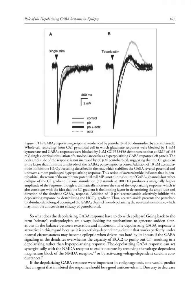
107Role of the Depolarizing GABA Response in Epilepsy
So what does the depolarizing GABA response have to do with epilepsy? Going back to theterm “seizure”, epileptologists are always looking for mechanisms to generate sudden alter-ations in the balance between excitation and inhibition. The depolarizing GABA response isattractive in this regard because it is so activity-dependent: a circuit that works perfectly undernormal circumstances may become epileptic when driven too hard by its inputs if the GABAsignaling in the dendrites overwhelms the capacity of KCC2 to pump out Cl-, resulting in adepolarizing rather than hyperpolarizing response. The depolarizing GABA response can actsynergistically with the NMDA receptor to excite neurons by removing the voltage-dependentmagnesium block of the NMDA receptor,23 or by activating voltage-dependent calcium con-ductances.9
If the depolarizing GABA response were important in epileptogenesis, one would predictthat an agent that inhibited the response should be a good anticonvulsant. One way to decrease
Figure 1. The GABAA depolarizing response is enhanced by pentobarbital but diminished by acetazolamide.Whole-cell recordings from CA1 pyramidal cell in which glutamate responses were blocked by 1 mMkynurenate and GABAB responses were blocked by 1µM CGP55845A demonstrates that at RMP of -65mV, single electrical stimulation of s. moleculare evokes a hyperpolarizing GABA response (left panel). Thepeak amplitude of the response is not increased by 60 µM pentobarbital, suggesting that the Cl- gradientis the factor that limits the amplitude of the GABAA postsynaptic response. Addition of 10 µM acetazola-mide inhibits the HCO3
- recycling described in the text, which stabilizes the GABA reversal potential anduncovers a more prolonged hyperpolarizing response. This action of acetazolamide indicates that in pen-tobarbital, the return of the membrane potential to RMP is not due to closure of GABAA channels but rathercollapse of the Cl- gradient. Tetanic stimulation (10 stimuli at 100 Hz) produces a marginally higheramplitude of the response, though it dramatically increases the size of the depolarizing response, which isalso consistent with the idea that the Cl- gradient is the limiting factor in determining the amplitude anddirection of the dendritic GABAA response. Addition of 10 µM acetazolamide selectively inhibits thedepolarizing response by destabilizing the HCO3
- gradient. Thus, acetazolamide prevents the pentobar-bital-induced prolonged opening of the GABAA channel from depolarizing the neuronal membrane, whichmay limit the anticovulsant efficacy of pentobarbital.

Recent Advances in Epilepsy Research108
the depolarizing response is to limit the ease with which HCO3- can sneak back into the cell as
CO2. Although it is not possible to alter CO2 permeability, it is possible to decrease the rate atwhich HCO3
- is dehydrated to H2O and CO2 by blocking the enzyme carbonic anhydrase.This is the primary action of the anticonvulsant acetazolamide. Acetazolamide blocks the de-polarizing GABA response,23 and this is the best-characterized anticonvulsant mechanism forthis drug discovered to date.15
Another prediction regarding the role of the depolarizing GABA response and seizures isthat anticonvulsants that act by increasing the open time of the GABA receptor should makethe depolarizing response more prominent. Pentobarbital prolongs the average open time ofthe GABA receptor, and has been demonstrated to increase the depolarizing GABA response.1,25
If acetazolamide is added to pentobarbital, the increased GABA effect of pentobarbital can beobserved without an increase in the depolarizing response (Fig. 1). Thus, one prediction is thatthese two clinically-used anticonvulsants should have synergistic actions. This has been experi-mentally verified in vivo.4,20 Benzodiazepines also increase the mean open time of the GABAchannel, so similar synergy should exist between acetazolamide and benzodiazepines. Aceta-zolamide is available for intravenous as well as oral administration, so it may be time to explorethis synergy clinically, perhaps in a status epilepticus therapeutic trial.
References1. Alger BE, Nicoll RA. Pharmacological evidence for two kinds of GABA receptor on rat hippocam-
pal pyramidal cells studied in vitro. J Physiol 1982; 328:125-141.2. Alger BE, Nicoll RA. Feed-forward dendritic inhibition in rat hippocampal pyramidal cells studied
in vitro. J Physiol 1982; 328:105-123.3. Andersen P, Dingledine R, Gjerstad L. Two different responses of hippocampal pyramidal cells to
application of gamma-amino butyric acid. J Physiol 1980; 305:279-296.4. Archer DP, Samanani N, Roth SH. Small-dose pentobarbital enhances synaptic transmission in rat
hippocampus. Anesth Analg 2001; 93:1521-15255. Ballanyi K, Grafe P. An intracellular analysis of gamma-aminobutyric-acid-associated ion move-
ments in rat sympathetic neurones. J Physiol 1985; 365:41-58.6. Barker JL, Ransom BR. Amino acid pharmacology of mammalian central neurones grown in tissue
culture. J Physiol 1978; 280:331-54.7. Bormann J, Hamill OP, Sakmann B. Mechanism of anion permeation through channels gated by
glycine and gamma-aminobutyric acid in mouse cultured spinal neurones. J Physiol 1987;385:243-286.
8. Dallwig R, Deitmer JW, Backus KH. On the mechanism of GABA-induced currents in culturedrat cortical neurons. Pflugers Arch 1999; 437:289-97.
9. Grover LM, Lambert NA, Schwartzkroin PA et al. Role of HCO3- ions in depolarizing GABAA
receptor-mediated responses in pyramidal cells of rat hippocampus. J Neurophysiol 1993;69:1541-1555.
10. Huguenard JR, Alger BE. Whole-cell voltage-clamp study of the fading of GABA-activated cur-rents in acutely dissociated hippocampal neurons. J Neurophysiol 1986; 56:1-18.
11. Jarolimek W, Lewen A, Misgeld U. A furosemide-sensitive K+-Cl- cotransporter counteracts intra-cellular Cl- accumulation and depletion in cultured rat midbrain neurons. J Neurosci 1999;19:4695-4704.
12. Kaila K, Voipio J. Postsynaptic fall in intracellular pH induced by GABA-activated bicarbonateconductance. Nature 1987; 330:163-165.
13. Kaila K. Ionic basis of GABAA receptor channel function in the nervous system. Prog Neurobiol1994; 42:489-537.
14. Kaila K, Lamsa K, Smirnov S. Long-lasting GABA-mediated depolarization evoked by high-frequencystimulation in pyramidal neurons of rat hippocampal slice is attribuatable to a network-driven,bicarbonate-dependent K+ transient. J Neurosci 1997; 17:7662-7672.
15. Meldrum B. Update on the mechanism of action of antiepileptic drugs. Epilepsia 1996; 37 Suppl6:S4-11.
16. Payne JA. Functional characterization of the neuronal-specific K-Cl cotransporter: implications for[K+]o regulation. Am J Physiol 1997; 273(5 Pt 1):C1516-1525.
17. Perkins KL, Wong RKS. Ionic basis of the postsynaptic depolarizing GABA response in hippocam-pal pyramidal cells. J Neurophysiol 1996; 76:3886-3894.

109Role of the Depolarizing GABA Response in Epilepsy
18. Perreault P, Avoli M. A GABAergic depolarizing potential in the hippocampus disclosed by theconvulsant 4-aminopyridine. Brain Res 1987; 400:191-195.
19. Perreault P, Avoli M. A depolarizing inhibitory postsynaptic potential activated by synapticallyreleased gamma-aminobutyric acid under physiological conditions in rat hippocampal pyramidalcells. Can J Physiol Pharmacol 1988; 66:1100-1102.
20. Sato J, Nioka M, Owada E. Effect of acetazolamide on the anticonvulsant potency of phenobar-bital in mice. J Pharmacobiodyn 1981; 4:952-960.
21. Scharfman HE, Sarvey JM. Responses to gamma-aminobutyric acid applied to cell bodies and den-drites of rat visual cortical neurons. Brain Res 1985; 358:385-389.
22. Scharfman HE, Sarvey JM. Responses to GABA recorded from identified rat visual cortical neu-rons. Neuroscience 1987; 23:407-422.
23. Staley KJ, Soldo BL, Proctor WR. Ionic mechanisms of neuronal excitation by inhibitory GABAAreceptors. Science 1995; 269:977-981.
24. Staley KJ, Proctor WR. Modulation of mammalian dendritic GABA(A) receptor function by thekinetics of Cl- and HCO3
- transport. J Physiol 1999; 519 Pt 3:693-712.25. Thalmann RH. Blockade of a late inhibitory postsynaptic potential in hippocampal CA3 neurons
in vitro reveals a late depolarizing potential that is augmented by pentobarbital. Neurosci Lett1988; 95:155-60.
26. Thompson SM, Deisz RA, Prince DA. Relative contributions of passive equilibrium and activetransport to the distribution of chloride in mammalian cortical neurons. J Neurophysiol 1988;60:105-124.
27. Thompson SM, Gähwhiler BH. Activity-dependent disinhibition. I. repetitive stimulation reducesIPSP driving force and conductance in the hippocampus in vitro. J Neurophysiol 1989a; 61:501-511.
28. Treiman DM. GABAergic mechanisms in epilepsy. Epilepsia 2001; 42(Suppl 3):8-12.29. Staley K, Smith R. A new form of feedback at the GABA(A) receptor. Nat Neurosci 2001;
4:674-676.

Recent Advances in Epilepsy Research110
CHAPTER 9
Recent Advances in Epilepsy Research, edited by Devin K. Binder and Helen E. Scharfman.©2004 Eurekah.com and Kluwer Academic / Plenum Publishers.
Gap Junctions, Fast Oscillationsand the Initiation of SeizuresRoger D. Traub, Hillary Michelson-Law, Andrea E.J. Bibbig,Eberhard H. Buhl and Miles A. Whittington
In this chapter, we shall review evidence that gap junctions can contribute to epileptogenesisin the hippocampus and cortex—but not just any gap junctions. Rather, we shall argue fora role for a newly described sort of gap junction, located between the proximal axons of
principal neurons. Such axon-axon gap junctions promote epileptogenesis not so much byenhancing synchrony, as by providing pathways for the direct spread of action potentials be-tween neurons. A by-product of such spread is the ability of axonally-coupled neurons to gen-erate oscillations at very high frequencies (>~70 Hz). It is of note that seizure activity, both invivo and in vitro, has been observed to begin with very high-frequency oscillations. If suchoscillations can be shown to initiate the seizure discharge, and not just be an epiphenomenon,then targeting gap junction conductances may prove useful as an anticonvulsant strategy.
In Vitro Population Activities Paroxysmal to Greater or Lesser ExtentWhich Depend on Gap Junctions
Field BurstsField bursts are a form of paroxysmal activity, most readily induced in CA1 but also present
in other areas (particularly those areas where the neurons are packed tightly in a laminar fash-ion), that may be noted when individual neurons are hyperexcitable and chemical synaptictransmission is suppressed.1-5 Thus, field bursts are favored by very low Ca2+ media, perhapswith Mn2+ added, and by elevations of extracellular [K+]. Field bursts are characterized both byslow extracellular potential shifts and by large population spikes. The amplitude of such extra-cellular potentials tends to be enhanced by tight laminar packing of the neurons. Given thesuppression of chemical synaptic transmission, the large population spikes suggested the exist-ence of “nonsynaptic” mechanisms for the tight temporal synchronization of action potentials.One such mechanism that was proposed was so-called field effects, produced by the flow ofcurrent loops in the extracellular space, across neuronal membranes, and through neuronalinteriors.3,6 Given that extracellular currents are produced by electrogenesis in the entire localneuronal population, field effects can, in principle, couple together nearby neurons. Indeed,the extracellular potential gradients occurring during field bursts can be large enough thateffects on spike-firing timings could take place;7 and small transmembrane depolarizations aresometimes seen just prior to action potential firing, with such depolarizations being expectedwith field effects.3
Nevertheless, experiments after the discovery of field bursts indicate that gap junctions areprobably required for field bursts to occur;8 the paroxysmal events are also influenced by pHchanges (i.e., with increased pH enhancing, and reduced pH suppressing), in the way onewould expect for gap junctions9 (see also Spray et al10). It is noteworthy as well that low [Ca2+]o

111Gap Junctions and Seizures
media, which promote the occurrence of field bursts, alkalinize the interior of cells, therebypresumably opening gap junctions.11
Fast (~200 Hz) Ripples in VitroTwo distinctive (though probably related) types of “ripples” have been described in the
hippocampus. The first to be discovered were in vivo ripples superimposed on the depolarizingpotentials associated with physiological sharp waves;12 intracellular recordings13 indicate thatsuch ripples consist predominantly of population IPSPs, and we shall refer to such events as“i-ripples”, where “i” stands for IPSP. Similar very fast rippling is now known to occur in cortexin vivo in anesthetized cats, where it is also superimposed on depolarizing potentials in princi-pal neurons—specifically, the depolarizing portion of the <1 Hz slow rhythm,14,15 and suchcortical ripples may be similar in mechanism to hippocampal i-ripples.
In addition, however, there are in vitro “p-ripples”, where the “p” stands for “populationspike”; the designation is applied because the ripple appears as a brief series of ~200 Hz smallpopulation spikes (usually 100 µV or less), even when synaptic transmission is blocked in low[Ca2+] media.16,17 P-ripples occur spontaneously in principal cell regions, changing from spo-radic brief trains in normal media, to almost continuous oscillation at similar frequency in low[Ca2+] media.16 The extracellular potentials—typically tens of µV—seem far too small for fieldeffects to be relevant, nor are intracellular recordings suggestive of this mechanism. On theother hand, a variety of gap-junction-blocking compounds suppress p-ripples, and p-ripplesare greatly enhanced by alkalinizing the medium—thus, p-ripples depend on gap junctions.Intracellular recordings during p-ripples show spikes with notches and inflections on theirrising phase (that is, appearing to be antidromic), as well as spikelets,16 also called fastprepotentials18 or d-spikes.19 The shape of these latter potentials suggested that the requisitegap junctions responsible for p-ripples were in axons, a notion for which experimental supportwas subsequently adduced (see below). Furthermore, it was shown in simulations that axonalgap junctions—having a density corresponding to observed dye-coupling densities20—wouldindeed be expected to generate very fast (>100 Hz) network oscillations21 (see also below). Thenecessary (and observed) density of gap junctions turns out to be surprisingly low, with eachcell coupled to an average of about 1.5 to about 3.5 other cells. [In contrast, synaptic connec-tivity between CA1 pyramidal cells is about 1%;22 in a slice with, say, 3,000 pyramidal cells,this means that each pyramidal cell would synaptically excite about 30 others.]
As noted above, field bursts seem to depend upon gap junctions.8 Furthermore, actionpotentials recorded intracellularly appear to be antidromic, arising directly from baseline, some-times with a notch on the rising phase; and small spikes are interspersed with large ones.2 Wewould guess, therefore, that field bursts are actually runs of p-ripples, associated with especiallylarge population spikes, due to hyperexcitability caused by the ionic conditions (or othernonsynaptic effects). If our hypothesis is correct, field bursts then represent a form ofepileptogenesis that is a direct consequence of axonal gap junctions between principal neurons.
P-ripples may be related to in vivo i-ripples also, by the following mechanism: a burst ofvery fast oscillation, generated by a network of electrically coupled principal cell axons, cansynaptically excite a collection of interneurons to generate a coherent fast oscillation as well.Recall that EPSPs in interneurons tend to be large and fast,23,24 and that many interneuronscan fire at high frequency. A fast network oscillation in a population of interneurons can, inturn, elicit a coherent train of IPSPs in pyramidal neurons, as observed in vivo.13 Simulationsbased on this concept replicate many of the in vivo experiments.25
Very Fast Oscillations and Seizure Discharge in TMA (Tetramethylamine)A more complex network behavior occurs in hippocampal slices bathed in TMA, an alka-
linizing compound that is presumed to open gap junctions. It has previously been shown26 thattetanic stimulation of the CA1 region leads to a ~1 second epoch of γ oscillation (~30-70 Hz),that is dependent upon activation of metabotropic glutamate receptors. When such a γ-inducingstimulus is given in the presence of TMA, the γ oscillation still occurs, but it can be followed by

Recent Advances in Epilepsy Research112
a period of low amplitude, very fast (~100 Hz) field potential oscillation, and then by a seizurelikedischarge, lasting > 10 seconds, and consisting of a series of synchronized bursts.27 Interest-ingly, the very fast oscillation continues during the seizure discharge (including being superim-posed on the burst complexes), and also continues afterwards—one is reminded of the very fast“afterdischarge termination oscillation” observed in the limbic system of rats, in vivo, aftertetanically-induced seizures,28 a phenomenon that could be nonsynaptically mediated. [One isalso reminded of the older observation of a ~200 Hz rhythm superimposed on single, tempo-rally isolated, synchronized bursts in various in vitro experimental epilepsy models, includingdisinhibition19,29 and low [Mg2+]o.30 Simulations indicate that recurrent excitatory synapses,acting in concert with axonal gap junctions, could together lead to a synchronized burst withsuperimposed very fast oscillation.21 While it is known that AMPA receptors are criticallyinvolved in these experimental epilepsies, the role of gap junctions in producing the superim-posed oscillation remains to be investigated pharmacologically.]
That gap junctions are involved in the in vitro VFO γ seizure discharge phenomenon issuggested not only by the bathing medium (i.e., alkaline conditions); it is also suggested by theobservation that the gap-junction-blocking compound carbenoxolone in the bath (along withTMA) does not qualitatively influence the evoked γ oscillation, but it aborts both the subse-quent VFO and the seizure discharge.27 For these reasons—and also because of strikingly simi-lar electrographic phenomenology in some patients with focal seizures (see below)—we haveproposed that the (putatively gap-junction-mediated) VFO might actually initiate the suc-ceeding seizure discharge27—an hypothesis which, if true, could have practical consequences.Possible mechanisms linking the VFO to the seizure itself will be discussed below.
Giant IPSPs in 4AP4AP (50 - 100 µM) applied to hippocampal and neocortical slices leads to a variety of
population phenomena, including: a) epileptiform depolarizing bursts, dependent on AMPAand depolarizing GABAA receptors;31,32 and b) giant GABA-dependent potentials, with hyper-polarizing and depolarizing GABAA, as well as GABAB, components.33-36 We shall considerhere, briefly, 4AP-induced GABAB-dependent “giant” potentials (in principal cells), that occurwhen ionotropic glutamate receptors and GABAA receptors are pharmacologically blocked.35
Not surprisingly, subpopulations of interneurons fire (at least approximately) in phase witheach other, so as to elicit the GABAB potential in principal cells. Furthermore, at least someinterneurons35—as well as some principal cells37—simultaneously generate spikelets, or smallaction potentials, during artificially induced hyperpolarizations, or during the giant IPSP itself.4AP-induced giant GABAB-dependent IPSPs, and synchronous interneuronal firing, are sup-pressed by the gap-junction-blocking compound carbenoxolone,38,39 presumably via electricaluncoupling of interneurons.
The question, then, is how might a population of interneurons fire trains of approximatelysynchronous action potentials, when ionotropic receptors are blocked. DC electrical coupling,and ultrastructural dendrodendritic (or dendrosomatic) gap junctions, are known to occurbetween selected pairs of nearby interneurons.40-45 Nevertheless, in simulations of interneu-ronal networks, electrically coupled between dendrites,46 network bursts occurred only underconditions when action potentials could cross from dendrite to dendrite—and this appearsunlikely, in view of the experimentally observed properties of interneuron gap junctions. If it isthe case, however, that interneurons are also electrically coupled between their axons (at least toa functionally significant extent in 4AP), then network bursts are readily simulated.38 As to thespikelets in principal cells—presumed to be antidromic—which occur during the giant IPSP:these spikelets can be explained if GABA released during the giant IPSP were to excite axons.This is not as paradoxical as it sounds: bicuculline has been observed to suppress ectopic spikesgenerated in Schaffer collaterals,47,48 and some presynaptic terminals are depolarized by GABA.49
The mechanisms of 4AP-induced interneuronal network bursting are of possible relevanceto epileptogenesis, given the contributions that excessive GABA release can make to neuronalhyperexcitability, at least under certain conditions.50-52

113Gap Junctions and Seizures
γγγγγ Oscillations Generated by Underlying VFOJust as seizure discharges and gap-junction mediated VFO may be interrelated, so may
certain types of γ oscillations and VFO also be interrelated. Such a relationship is important tounderstand: γ oscillations—at least some types of them—occur under physiological condi-tions, and in vivo γ oscillations are probably important for perception53,54 and memory.55
Nevertheless, certain in vitro γ oscillations are built, as it were, on a platform of VFO. (It is notknown if any of the multiple in vivo γ oscillations are related to VFO.) The sorts of in vitro γoscillations where this appears to be true are the ones evoked by carbachol,56 kainate,57 or bypressure ejection (so-called “puff ”) of hypertonic K+ solution.58,59 These sorts of γ shareseveral properties:60
a. AMPA and GABAA receptors are both required.b. Gap-junction-blocking agents, particularly octanol, suppress the γ.c. Principal cells are not strongly depolarized, and fire infrequently; but their firing, when it
occurs, is still (on average) time-locked to the population rhythm.d. Interneurons fire at higher rates than principal cells, and are also time-locked to the popu-
lation rhythm.e. The power spectrum of the field potential contains not only a γ peak, but also a higher
frequency—albeit smaller—peak, typically 80 Hz or above.27,56 Blockade of synaptic trans-mission, at least for γ produced by pressure ejection of potassium, suppresses the γ peak and“unmasks” the higher frequency oscillation.27
The above experimental observations, and others as well, can be explained by the followingtype of model60 (see also Fig. 2): a combination of spontaneous “ectopic” axonal spikes, alongwith axonal gap junctions, leads to firing of principal cell axons that is organized into ahigh-frequency oscillation.21 This activity synaptically (via AMPA receptors) forces the inter-neurons to fire. The interneurons, however, inhibit not only each other, but also the somataand axon initial segments of principal cells. Provided the gap junctions are not located too farfrom somata (and available data—see below—suggest that the distance is < ~150 µm), andprovided the IPSCs are large enough,25 then such synaptic inhibition “chops up” the VFO intosegments whose duration is determined by the IPSC decay time constants, and hence gives aγ-frequency rhythm (cf. Whittington et al;26 Traub et al61)—but leaving some VFO compo-nent as well. This scheme shows why AMPA receptors, GABAA receptors, and gap junctionsare all necessary for the rhythm.
Understanding the mechanisms of carbachol/kainate/pressure ejection γ oscillations is, webelieve, clinically relevant, particularly if similar mechanisms are at work in vivo—for exampleduring the γ and very fast oscillations that occur during the slow (<1 Hz) rhythms of sleep.14,15
Thus, suppose one targets gap junctions pharmacologically in order to suppress the VFO thatappear to initiate certain focal seizures.27 Such targeting could well interfere with normal brainrhythms. One type of experimental evidence that could be useful in analyzing this possibility isbased on a prediction of network simulations:60 many principal cell somatic action potentials,during the oscillation, are expected to be antidromic. It would help to know if this occurs invivo. Very fast oscillations also occur as an apparently normal phenomenon during the corticalsensory evoked responses, in rats, produced by whisker stimulation62,63—it would help toknow if these oscillations depend on gap junctions and involve (at least partly) antidromicspikes.
What Effects Do Interneuronal Gap Junctions Have on PopulationActivities in Vitro?
Up to this point, we have been discussing the effects on population behavior of axonal gapjunctions, primarily between principal cells, deferring for the moment a summary of the evi-dence that such axonal gap junctions even exist. Presentation in this way is motivated by ourbelief that the axonal gap junctions are the ones making a “primary” contribution toepileptogenesis. Nevertheless, the reader may well feel confused: what about dendritic gap
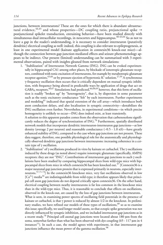
Recent Advances in Epilepsy Research114
junctions, between interneurons? These are the ones for which there is abundant ultrastruc-tural evidence,44,45 and whose properties—DC coupling ratio, prejunctional spike →postjunctional spikelet transduction, entraining behavior—have been studied directly withsimultaneous dual intracellular recordings, in neocortex and hippocampus.40-43,64 So as not toleave a gap in the reader’s understanding, it is necessary to consider interneuron (putativelydendritic) electrical coupling as well: indeed, this coupling is also relevant to epileptogenesis, atleast in one experimental model (kainate application in connexin36 knock-out mice)—al-though the connection betwen gap-junction-mediated effects and seizure phenomenology ap-pears to be indirect. Our present (limited) understanding can be summarized with 3 experi-mental observations, paired with insights gleaned from network simulations:
1. “Stabilization” of Interneuron Network Gamma (ING). ING can be evoked experimen-tally in hippocampal CA1 among other places, by blockade of ionotropic glutamate recep-tors, combined with tonic excitation of interneurons, for example by metabotropic glutamatereceptor agonists,65,66 or by pressure ejection of hypertonic K+ solution.27,59 A synchronousγ-frequency oscillation then occurs that is critically dependent on mutual synaptic inhibi-tion, with frequency being altered in predictable ways by application of drugs that act onGABAA receptors.66,67 Simulations had predicted,66,68,69 however, that this form of oscilla-tion is readily “broken up” by “heterogeneity”, that is, by dispersion in some parametersuch as the tonic excitatory conductance “felt” by each interneuron. Further experimentsand modeling61 indicated that spatial extension of the cell array—which introduces bothaxon conduction delays, and also localization in synaptic connectivity—destabilizes theING oscillation even further. Nevertheless, in experimental conditions—in which precisehomogeneity is unlikely to occur—ING does indeed exist, at least transiently.A solution to this apparent paradox comes from the observation that carbenoxolone signifi-cantly reduces the degree of synchronization of ING.70 Furthermore, spatially distributednetwork models that incorporate dendritic interneuron-interneuron gap junctions—at lowdensity (average 2 per neuron) and reasonable conductance (~0.5 - 1.0 nS)—have greatlyenhanced stability of ING, compared to the case where gap junctions are not present. Thesedata suggest, therefore, one possible physiological role for the anatomically and physiologi-cally observed dendritic gap junctions between interneurons: increasing coherence in a cer-tain type of γ oscillation.
2. “Stabilization” of γ oscillations produced in vitro by kainate or carbachol. The γ oscillationsinduced by these drugs (as noted above) require ionotropic glutamate (specifically, AMPA)receptors: they are not “ING”. Contributions of interneuron gap junctions to such γ oscil-lations have been studied by comparing hippocampal slices from wild-type mice with hip-pocampal slices from mice in which connexin36 has been knocked out.57 [Connexin36 is amajor neuronal gap junction protein that is expressed primarily in neurons, especially inter-neurons.57,71-73] In the connexin36 knockout mice, very fast oscillations observed in low[Ca2+] media16 are indistinguishable from wild-type; it therefore appears likely that princi-pal cell axon gap junctions do not depend critically upon connexin36. On the other hand,electrical coupling between nearby interneurons is far less common in the knockout micethan in the wild-type mice. Thus, it is reasonable to conclude that effects on oscillations,observed in the knock-out, are caused by the loss of gap junctions between interneurons.What is seen, in examining power spectra of oscillating field potentials in the presence ofkainate or carbachol, is that γ power is reduced by almost 1/2 in the knockout. In prelimi-nary studies, we have refined our models of these types of oscillations,60 so as to examinethis issue; specifically, we used longer model axons, so that ectopic spike generation was notdirectly influenced by synaptic inhibition, and we included interneuron gap junctions as ina recent study.70 Principal cell axonal gap junctions were located about 180 µm from thesoma, somewhat farther than what has been observed with dye coupling (65 - 117 µm in 4instances11). In such a case, the model agrees with experiment, in that interneuron gapjunctions influence the mean power of the gamma oscillation.98
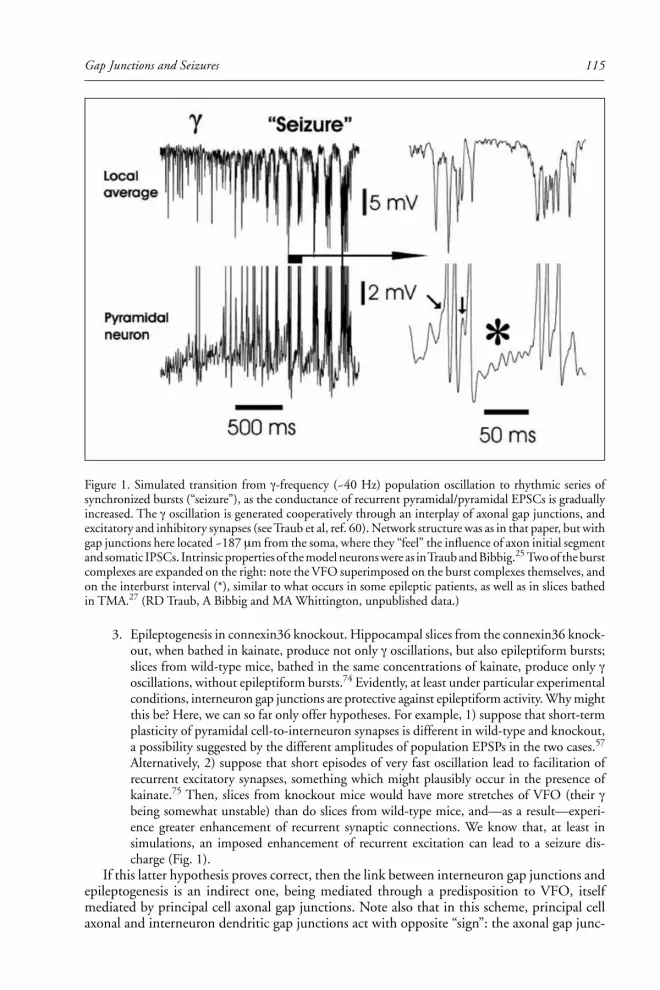
115Gap Junctions and Seizures
3. Epileptogenesis in connexin36 knockout. Hippocampal slices from the connexin36 knock-out, when bathed in kainate, produce not only γ oscillations, but also epileptiform bursts;slices from wild-type mice, bathed in the same concentrations of kainate, produce only γoscillations, without epileptiform bursts.74 Evidently, at least under particular experimentalconditions, interneuron gap junctions are protective against epileptiform activity. Why mightthis be? Here, we can so far only offer hypotheses. For example, 1) suppose that short-termplasticity of pyramidal cell-to-interneuron synapses is different in wild-type and knockout,a possibility suggested by the different amplitudes of population EPSPs in the two cases.57
Alternatively, 2) suppose that short episodes of very fast oscillation lead to facilitation ofrecurrent excitatory synapses, something which might plausibly occur in the presence ofkainate.75 Then, slices from knockout mice would have more stretches of VFO (their γbeing somewhat unstable) than do slices from wild-type mice, and—as a result—experi-ence greater enhancement of recurrent synaptic connections. We know that, at least insimulations, an imposed enhancement of recurrent excitation can lead to a seizure dis-charge (Fig. 1).
If this latter hypothesis proves correct, then the link between interneuron gap junctions andepileptogenesis is an indirect one, being mediated through a predisposition to VFO, itselfmediated by principal cell axonal gap junctions. Note also that in this scheme, principal cellaxonal and interneuron dendritic gap junctions act with opposite “sign”: the axonal gap junc-
Figure 1. Simulated transition from γ-frequency (~40 Hz) population oscillation to rhythmic series ofsynchronized bursts (“seizure”), as the conductance of recurrent pyramidal/pyramidal EPSCs is graduallyincreased. The γ oscillation is generated cooperatively through an interplay of axonal gap junctions, andexcitatory and inhibitory synapses (see Traub et al, ref. 60). Network structure was as in that paper, but withgap junctions here located ~187 µm from the soma, where they “feel” the influence of axon initial segmentand somatic IPSCs. Intrinsic properties of the model neurons were as in Traub and Bibbig.25 Two of the burstcomplexes are expanded on the right: note the VFO superimposed on the burst complexes themselves, andon the interburst interval (*), similar to what occurs in some epileptic patients, as well as in slices bathedin TMA.27 (RD Traub, A Bibbig and MA Whittington, unpublished data.)

Recent Advances in Epilepsy Research116
tions promoting, and the dendritic gap junctions (apparently) protecting against, epileptogenesis.Undoubtedly, this scheme represents a major oversimplification.
What Is the Evidence That Principal Cell Gap Junctions Exist?Much of the preceding discussion has assumed the existence of gap junctions between prin-
cipal cell axons. The hypothesis that such gap junctions exist arose from study of the shape ofspikelets—presumed electrotonic coupling potentials—which were recorded during a very fast(~200 Hz) population oscillation that depended on gap junctions.16 Here, we very brieflysummarize the experimental evidence from hippocampus11 that axon gap junctions exist, andthat (at least some) spikelets represent decrementally conducted antidromic spikes. (See also arecent review.76) The experimental data11 include these findings:
1. Spikelets can be evoked in low [Ca2+] media by stimulating s. oriens at some distance fromthe recorded neuron. Spikelets evoked in this way can follow, faithfully, stimulation fre-quencies as fast as 500 Hz, characteristic of axonal firing behavior.
2. Spikelets quickly disappear when the Na+-channel blocker QX314 is in the whole-cell re-cording pipette; additionally, spikelets increase in amplitude after a hyperpolarizing prepulse,such as would diminish Na+ channel inactivation. Hence, spikelets are not passively con-ducted electrotonic potentials.
3. The amplitude and rate-of-rise of spikelets are reduced by carbenoxolone; thisgap-junction-blocking drug, however, does not affect the shape of action potentials directlyevoked and recorded in axons or in mossy fiber presynaptic terminals. Thus, a gap junctionlies in the path between the stimulating electrode and the soma of the recorded neuron.
4. Dual simultaneous recordings at soma and proximal axon of a single neuron show that thespikelet is, indeed, conducted antidromically. Additionally, the spikelet appears in somabefore it appears in the apical dendrite.
5. Time-lapse confocal imaging shows that fluorescent dye injected into one pyramidal neu-ron through a recording pipette passes along the axon and into the axon of a “dye-coupled”second pyramidal neuron, whose soma and dendrites fill subsequently. Ultrastructural con-firmation of the existence of axonal gap junctions is not yet available, to our knowledge.
How Do Axonal Gap Junctions Lead to Very Fast Oscillations?The means by which axonal gap junctions generate very fast oscillations (> ~70 Hz) are not
intuitive. We use the word “generate” here deliberately and with reason, drawing a contrastwith a previously known role for gap junctions in rapidly oscillating systems, for example in themedullary pacemaker nucleus of the electric fish.77,78 In this pacemaker nucleus, gap junctionsact to couple together, to entrain, individual intrinsically oscillating neurons, each of which hasa natural frequency (were it to be isolated) that is close to the population frequency. This is notwhat happens in a hippocampal network, where the axons do not “naturally” and in isolationoscillate at 100-200 Hz. Indeed, so far as is known, the period of the hippocampal very fastoscillation is not directly related to a 5 or 10 ms time constant in the axonal membrane bio-physics, but rather is related most closely to the sparse connectivity of the gap junctional net-work. In principal cell networks, the gap junctions generate VFO, rather than entraining them.The reasons underlying this assertion are laid out elsewhere.21,76
What we can present here are the basic conditions under which VFO appear able to occurin an axonal network:
1. There is a low rate of spontaneous “ectopic” action potentials in the axons (or presynapticterminals), 0.05 Hz/axon being sufficient in simulations.
2. There are enough axoaxonal gap junctions (each axon coupled to more than one other onaverage), but not too many (each axon coupled to less than about 3 or 4, on average).
3. Action potentials can cross from axon to coupled axon.From these considerations, which are based on extensive network simulations, we see that
VFO should be favored by factors that increase axonal excitability, such as kainate in the bath:79-81

117Gap Junctions and Seizures
there should be more ectopic spikes and greater ease of spikes “crossing” the gap junctions; andVFO should be favored by measures (e.g., alkalinization) that increase gap junctional conduc-tance. On the other hand, synaptic inhibition on the axonal initial segment might shunt theaxonal membrane near the gap junction, and make it less likely for an antidromic spike toreach the axonal gap junction, or—if the spike does reach there—then it is less likely for thespike to cross the junction and evoke a spike in the coupled fiber.
Additional in Vitro Hippocampal Epilepsy ModelsBesides the in vitro models discussed so far, there are other studies as well suggesting that
gap-junction-blocking compounds suppress epileptogenesis, including Ross et al,82 Köhling etal,83 and (Q Yang and HB Michelson, personal comm.). The mechanisms by which theseeffects could be occurring are not yet worked out.
Gap Junctions and Epileptogenesis in VivoThe link between gap junctions and epileptogenesis in vivo is indirect: there are associa-
tions, temporal correlations, between very fast oscillations and seizures. In experimentalepileptogenesis, for example using spontaneous seizures occurring in rats after kainic acid in-jection, very fast field potential oscillations (even up to 500 Hz) have been observed prior toseizure discharges,84,85 with the sites of VFO generation being discrete and patchy. In clinicalobservations, fast oscillations have been observed leading into focal seizures, in both adults andchildren.27,86,87 In one study,27 a series of children with focal cortical dysplasias was brieflydescribed, with such a pattern of seizure initiation (i.e., with initial localized VFO) apparentlybeing characteristic.
Another suggestive set of clinical/genetic observations is this: on the one hand, the neuronalconnexin, connexin36, has been assigned to the gene band 15q14 in humans.88 On the otherhand, both familial rolandic epilepsy with centrotemporal spikes, and also juvenile myoclonicepilepsy, have an association with this same locus.89,90 It will be instructive to know the geneticlocalization of the protein(s) that mediate electrical coupling between axons.
How Could the Transition from Very Fast Oscillations to Seizure DischargeOccur?
We have noted that fast oscillations can evolve into seizure discharges, both in experimental(in vitro and in vivo), as well as clinical contexts.27,91 In addition, we showed (Fig. 1) that onepossible way for such an evolution to take place is through enhancement of recurrent synapticexcitation. It is important to emphasize, however, that the evolution could occur by severalother means, including reduction in synaptic inhibition (Fig. 2), or by reduction in synapticexcitation of interneurons (R Traub, A Bibbig, MA Whittington, unpublished data). Undoubt-edly, details of these mechanisms will be worked out in experimental models; determining themechanisms in humans will likely prove difficult. There are, however, potentially significantawards in therapy.
Summary and Unifying HypothesesThe most important points we have raised are these:
1. Both experimental (in vitro and in vivo) and human seizure discharges may begin with avery fast (>70 Hz) neuronal network oscillation.27,76
2. Very fast oscillations (VFO) can occur in vitro without chemical synapses, but dependingon gap junctions.16
3. The gap junctions which generate VFO are located between the axons of principal neu-rons.11,21,76
4. Axonal gap junctions, working in concert with chemical synapses, can generate—depend-ing on parameters—VFO or gamma oscillations.25,60

Recent Advances in Epilepsy Research118
5. The transition from fast (gamma or VFO) oscillation to seizure may depend upontime-dependent alterations in one or more synaptic conductances, including recurrent ex-citation between principal neurons.
6. The relation between interneuron dendritic gap junctions and epilepsy—at least in thekainate model74—appears to be an indirect one.
How might the presumed link between gap junctions and clinical epilepsy be further inves-tigated? Two possible approaches come to mind: First, does pharmacological blockade of neu-ronal gap junctions, in vivo, suppress seizures? It is of note, for example, that carbenoxolone isapproved for human use in Europe, for treatment of ulcer disease,92 and it seems to cross theblood-brain barrier.93 It is unclear, though, whether gap-junction-blocking compounds can
Figure 2. Simulated transition from continuous very fast oscillation (VFO) to synchronized burst, assynaptic inhibition of pyramidal cells is reduced (beginning at horizontal arrow, time constant for reducingmaximal IPSC conductance = 250 ms). The VFO was generated by distally located (~560 µm from thesoma) axonal gap junctions between pyramidal cells, with 1.6 junctions per neuron on average. At thisdistance, the gap junction site is little influenced by synaptic inhibition, so that VFO is continuous. Ectopicaction potentials originate in the axon distal to the gap junction. Synaptic interactions (including especiallyrecurrent excitation between pyramidal neurons) are present in this detailed network of 3,072 pyramidaland 384 inhibitory neurons, similar to Traub and Bibbig.25 Some of the spikelets in the pyramidal cell areindicated by vertical arrows. Note the fast oscillations superimposed as well on the synchronized burst. (RDTraub, A Bibbig and MA Whittington, unpublished data).

119Gap Junctions and Seizures
attain CNS concentrations sufficient to block putative pathological gap junctions withoutcausing visual loss.71,94,95 Second, how might one investigate whether axonal gap junctions areincreased in number, or have altered functional properties, in human epileptogenic foci? [Thereis reason to believe that neuronal injury leads both to axonal reorganization and to enhancedexpression of gap junctions.96,97] This issue may require technically difficult experiments inresected brain tissue specimens, and also a means of visualizing axons, as well as gap junctionslocated on them, in neuropathological material. For this, it would be most helpful to identifythe protein(s) constituting axonal gap junctions and to develop markers for them.
AcknowledgmentsSupported by NIH, the Wellcome Trust, and the Medical Research Council (U.K.). We
thank Dietmar Schmitz, Andreas Draguhn, Hannah Monyer, Nancy Kopell, Torsten Baldeweg,and Fiona E.N. LeBeau for helpful discussion, and Robert Walkup for invaluable help withcomputing.
References1. Haas HL, Jefferys JGR. Low-calcium field burst discharges of CA1 pyramidal neurones in rat
hippocampal slices. J Physiol 1984; 354:185-201.2. Taylor CP, Dudek FE. Synchronous neural after discharges in rat hippocampal slices without ac-
tive chemical synapses. Science 1982; 218:810-812.3. Taylor CP, Dudek FE. Excitation of hippocampal pyramidal cells by an electrical field effect. J
Neurophysiol 1984; 52:126-142.4. Schweitzer JS, Patrylo PR, Dudek FE. Prolonged field bursts in the dentate gyrus: Dependence on
low calcium, high potassium, and nonsynaptic mechanisms. J Neurophysiol 1992; 68:2016-2025.5. Konnerth A, Heinemann U, Yaari Y. Slow transmission of neural activity in hippocampal area
CA1 in absence of active chemical synapses. Nature 1984; 307:69-71.6. Traub RD, Dudek FE, Taylor CP et al. Simulation of hippocampal afterdischarges synchronized
by electrical interactions. Neuroscience 1985; 14:1033-1038.7. Jefferys JGR. Influence of electric fields on the excitability of granule cells in guinea-pig hippocam-
pal slices. J Physiol 1981; 319:143-152.8. Perez-Velazquez JL, Valiante TA, Carlen PL. Modulation of gap junctional mechanisms during
calcium-free induced field burst activity: a possible role for electrotonic coupling in epileptogenesis.J Neurosci 1994; 14:4308-4317.
9. Schweitzer JS, Wang H, Xiong ZQ et al. pH sensitivity of nonsynaptic field bursts in the dentategyrus. J Neurophysiol 2000; 84:927-933.
10. Spray DC, Harris AL, Bennett MVL. Gap junctional conductance is a simple and sensitive func-tion of intracellular pH. Science 1981; 211:712-715.
11. Schmitz D, Schuchmann S, Fisahn A et al. Axo-axonal coupling: a novel mechanism for ultrafastneuronal communication. Neuron 2001; 31:831-840.
12. Buzsáki G, Horváth Z, Urioste R et al. High-frequency network oscillation in the hippocampus.Science 1992; 256:025-1027.
13. Ylinen A, Bragin A, Nádasdy Z et al. Sharp wave-associated high frequency oscillation (200 Hz) inthe intact hippocampus: network and intracellular mechanisms. J Neurosci 1995; 15:30-46.
14. Grenier F, Timofeev I, Steriade M. Focal synchronization of ripples (80-200 Hz) in neocortex andtheir neuronal correlates. J Neurophysiol 2001; 86:1884-1898.
15. Steriade M, Contreras D, Curró Dossi R et al. The slow (<1 Hz) oscillation in reticular thalamicand thalamocortical neurons: scenario of sleep rhythm generation in interacting thalamic and neo-cortical networks. J Neurosci 1993; 13:3284-3299.
16. Draguhn A, Traub RD, Schmitz D et al. Electrical coupling underlies high-frequency oscillationsin the hippocampus in vitro. Nature 1998; 394:189-192.
17. Draguhn A, Traub RD, Bibbig A et al. Ripple (approximately 200-Hz) oscillations in temporalstructures. J Clin Neurophysiol 2000; 17:361-376.
18. Spencer WA, Kandel ER. Electrophysiology of hippocampal neurons. IV. Fast prepotentials. JNeurophysiol 1961; 24:272-285.
19. Schwartzkroin PA, Prince DA. Penicillin-induced epileptiform activity in the hippocampal in vitropreparation. Ann Neurol 1977; 1:463-469.
20. Church J, Baimbridge KG. Exposure to high-pH medium increases the incidence and extent ofdye coupling between rat hippocampal CA1 pyramidal neurons in vitro. J Neurosci 1991;11:3289-3295.

Recent Advances in Epilepsy Research120
21. Traub RD, Schmitz D, Jefferys JGR et al. High-frequency population oscillations are predicted tooccur in hippocampal pyramidal neuronal networks interconnected by axoaxonal gap junctions.Neuroscience 1999; 92:407-426.
22. Deuchars J, Thomson AM. CA1 pyramid-pyramid connections in rat hippocampus in vitro: dualintracellular recordings with biocytin filling. Neuroscience 1996; 74:1009-1018.
23. Miles R. Synaptic excitation of inhibitory cells by single CA3 hippocampal pyramidal cells of theguinea-pig in vitro. J Physiol 1990; 428:61-77.
24. Geiger JRP, Lübke J, Roth A et al. Submillisecond AMPA receptor-mediated signaling at a princi-pal neuron-interneuron synapse. Neuron 1997; 18:1009-1023.
25. Traub RD, Bibbig A. A model of high-frequency ripples in the hippocampus, based on synapticcoupling plus axon-axon gap junctions between pyramidal neurons. J Neurosci 2000; 20:2086-2093.
26. Whittington MA, Stanford IM, Colling SB et al. Spatiotemporal patterns of γ frequency oscilla-tions tetanically induced in the rat hippocampal slice. J Physiol 1997; 502: 591-607.
27. Traub RD, Whittington MA, Buhl EH et al. A possible role for gap junctions in generation ofvery fast EEG oscillations preceding the onset of, and perhaps initiating, seizures. Epilepsia 2001;42:153-170.
28. Bragin A, Penttonen M, Buzsáki G. Termination of epileptic afterdischarge in the hippocampus. JNeurosci 1997; 17:2567-2579.
29. Wong RKS, Traub RD. Synchronized burst discharge in disinhibited hippocampal slice. I. Initia-tion in CA2-CA3 region. J Neurophysiol 1983; 49:442-458.
30. Traub RD, Jefferys JGR, Whittington MA. Enhanced NMDA conductances can account for epi-leptiform activities induced by low Mg2+ in the rat hippocampal slice. J Physiol 1994; 478: 379-393.
31. Perreault P, Avoli M. Physiology and pharmacology of epileptiform activity induced by4-aminopyridine in rat hippocampal slices. J Neurophysiol 1991; 65:771-785.
32. Traub RD, Colling SB, Jefferys JGR. Cellular mechanisms of 4-aminopyridine-induced synchro-nized after-discharges in the rat hippocampal slice. J Physiol 1995; 489:127-140.
33. Aram JA, Michelson HB, Wong RKS. Synchronized GABAergic IPSPs recorded in the neocortexafter blockade of synaptic transmission mediated by excitatory amino acids. J Neurophysiol 1991;65:1034-1041.
34. Michelson HB, Wong RKS. Excitatory synaptic responses mediated by GABAA receptors in thehippocampus. Science 1991; 253:1420-1423.
35. Michelson HB, Wong RKS. Synchronization of inhibitory neurones in the guinea pig hippocam-pus in vitro. J Physiol 1994; 477:35-45.
36. Müller W, Misgeld U. Picrotoxin- and 4-aminopyridine-induced activity in hilar neurons in theguinea pig hippocampal slice. J Neurophysiol 1991; 65:141-147.
37. Avoli M, Methot M, Kawasaki H. GABA-dependent generation of ectopic action potentials in therat hippocampus. Eur J Neurosci 1998; 10:2714-2722.
38. Traub RD, Bibbig A, Piechotta A et al. Synaptic and nonsynaptic contributions to giant IPSPs andectopic spikes induced by 4-aminopyridine in the hippocampus in vitro. J Neurophysiol 2001; 85:1246-1256.
39. Yang Q, Michelson HB. Gap junctions synchronize the firing of inhibitory interneurons in guineapig hippocampus. Brain Res 2001; 907:139-143.
40. Galarreta M, Hestrin S. A network of fast-spiking cells in the neocortex connected by electricalsynapses. Nature 1999; 402:72-75.
41. Gibson JR, Beierlein M, Connors BW. Two networks of electrically coupled inhibitory neurons inneocortex. Nature 1999; 402:75-79.
42. Venance L, Rozov A, Blatow M et al. Connexin expression in electrically coupled postnatal ratbrain neurons. Proc Natl Acad Sci USA 2000; 97:10260-10265.
43. Tamás G, Buhl EH, Lörincz A et al. Proximally targeted GABAergic synapses and gap junctionsprecisely synchronize cortical interneurons. Nat Neurosci 2000; 3:366-371.
44. Fukuda T, Kosaka T. Gap junctions linking the dendritic network of GABAergic interneurons inthe hippocampus. J Neurosci 2000; 20:1519-1528.
45. Kosaka T. Gap junctions between nonpyramidal cell dendrites in the rat hippocampus (CA1 andCA3 regions). Brain Res 1983; 271:157-161.
46. Traub RD. Model of synchronized population bursts in electrically coupled interneurons contain-ing active dendritic conductances. J Comput Neurosci 1995; 2:283-289.
47. Stasheff SF, Hines M, Wilson WA. Axon terminal hyperexcitability associated with epileptogenesisin vitro. I. Origin of ectopic spikes. J Neurophysiol 1993; 70:960-975.
48. Stasheff SF, Mott DD, Wilson WA. Axon terminal hyperexcitability associated with epileptogenesisin vitro. II. Pharmacological regulation by NMDA and GABAA receptors. J Neurophysiol 1993;70:976-984.

121Gap Junctions and Seizures
49. Jang IS, Jeong HJ, Akaike N. Contribution of the Na-K-Cl cotransporter on GABAAreceptor-mediated presynaptic depolarization in excitatory nerve terminals. J Neurosci 2001;21:5962-5972.
50. Thalmann RH, Peck EJ, Ayala GF. Biphasic response of hippocampal pyramidal neurons to GABA.Neurosci Lett 1981; 21:319-324.
51. Wong RKS, Watkins DJ. Cellular factors influencing GABA response in hippocampal pyramidalcells. J Neurophysiol 1982; 48:938-951.
52. Taira T, Lamsa K, Kaila K. Posttetanic excitation mediated by GABAA receptors in rat CA1 pyra-midal neurons. J Neurophysiol 1997; 77:2213-2218.
53. Roelfsema PR, König P, Engel AK et al. Reduced synchronization in the visual cortex of cats withstrabismic amblyopia. Eur J Neurosci 1994; 6:1645-1655.
54. Singer W, Gray CM. Visual feature integration and the temporal correlation hypothesis. Ann RevNeurosci 1995; 18:555-586.
55. Tallon-Baudry C, Bertrand O, Fischer C. Oscillatory synchrony between human extrastriate areasduring visual short-term memory maintenance. J Neurosci 2001; 21:RC177:1-5.
56. Fisahn A, Pike FG, Buhl EH et al. Cholinergic induction of network oscillations at 40 Hz in thehippocampus in vitro. Nature 1998; 394:86-189.
57. Hormuzdi SG, Pais I, LeBeau FEN. Impaired electrical signaling disrupts gamma frequency oscil-lations in connexin 36-deficient mice. Neuron 2001; 31:487-495.
58. Towers SK, LeBeau FEN, Gloveli T et al. Fast network oscillations in the rat dentate gyrus invitro. J Neurophysiol 2002; 87:1165-1168.
59. LeBeau FEN, Towers SK, Traub RD et al. Fast network oscillations induced by potassium tran-sients in the rat hippocampus in vitro. J Physiol 2002; 542:167-179.
60. Traub RD, Bibbig A, Fisahn A et al. A model of gamma-frequency network oscillations induced inthe rat CA3 region by carbachol in vitro. Eur J Neurosci 2000; 12:4093-4106.
61. Traub RD, Jefferys JGR, Whittington MA. Fast Oscillations in Cortical Circuits. Cambridge, MA:MIT Press, 1999.
62. Jones MS, Barth DS. Spatiotemporal organization of fast (>200 Hz) electrical oscillations in ratvibrissa/barrel cortex. J Neurophysiol 1999; 82:1599-1609.
63. Jones MS, MacDonald KD, Choi B et al. Intracellular correlates of fast (>200 Hz) electrical oscil-lations in rat somatosensory cortex. J Neurophysiol 2000; 84:1505-1518.
64. Bartos M, Vida I, Frotscher M et al. Rapid signaling at inhibitory synapses in a dentate gyrusinterneuron network. J Neurosci 2001; 21:2687-2698.
65. Whittington MA, Traub RD, Jefferys JGR. Synchronized oscillations in interneuron networks drivenby metabotropic glutamate receptor activation. Nature 1995; 373:612-615.
66. Traub RD, Whittington MA, Colling SB et al. Analysis of gamma rhythms in the rat hippocam-pus in vitro and in vivo. J Physiol 1996; 493:471-484.
67. Faulkner HJ, Traub RD, Whittington MA. Disruption of synchronous gamma oscillations in therat hippocampal slice: a common mechanism of anaesthetic drug action. Br J Pharmacol 1998;125:483-492.
68. Wang X-J, Buzsáki G. Gamma oscillation by synaptic inhibition in a hippocampal interneuronalnetwork model. J Neurosci 1996; 16:6402-6413.
69. White JA, Chow CC, Ritt J et al. Synchronization and oscillatory dynamics in heterogeneous,mutually inhibited neurons. J Comput Neurosci 1998; 5:5-16.
70. Traub RD, Kopell N, Bibbig A et al. Gap junctions between interneuron dendrites can enhancelong-range synchrony of gamma oscillations. J Neurosci 2001; 21:9478-9486.
71. Teubner B, Degen J, Sohl G et al. Functional expression of the murine connexin 36 gene codingfor a neuron-specific gap junctional protein. J Membr Biol 2000; 176:249-262.
72. Rash JE, Staines WA, Yasumura T et al. Immunogold evidence that neuronal gap junctions inadult rat brain and spinal cord contain connexin-36 but not connexin-32 or connexin-43. ProcNatl Acad Sci USA 2000; 97:7573-7578.
73. Condorelli DF, Belluardo N, Trovato-Salinaro A et al. Expression of Cx36 in mammalian neurons.Brain Res Brain Res Rev 2000; 32:72-85.
74. Pais I, Hormuzdi SG, Monyer H et al. Sharp wave-like activity in the hippocampus in vitro inmice lacking the gap junction protein connexin 36. J Neurophysiol 2003; 89:2046-2054.
75. Lauri SE, Delany C, Clarke VR et al. Synaptic activation of a presynaptic kainate receptor facili-tates AMPA receptor-mediated synaptic transmission at hippocampal mossy fibre synapses.Neuropharm. 2001; 41:907-915.
76. Traub RD, Draguhn A, Whittington MA et al. Axonal gap junctions between principal neurons: anovel source of network oscillations, and perhaps epileptogenesis. Rev Neurosci 2002; 13:1-30.

Recent Advances in Epilepsy Research122
77. Moortgat KT, Bullock TH, Sejnowski TJ. Gap junction effects on precision and frequency of amodel pacemaker network. J Neurophysiol 2000; 83:984-97.
78. Moortgat KT, Bullock TH, Sejnowski TJ. Precision of the pacemaker nucleus in a weakly electricfish: network versus cellular influences. J Neurophysiol 2000; 83:971-83.
79. Schmitz D, Frerking M, Nicoll RA. Synaptic activation of presynaptic kainate receptors on hip-pocampal mossy fiber synapses. Neuron 2000; 27:327-338.
80. Schmitz D, Mellor J, Frerking M et al. Presynaptic kainate receptors at hippocampal mossy fibersynapses. Proc Natl Acad Sci USA 2001; 98:11003-11008.
81. Semyanov A, Kullmann DM. Kainate receptor-dependent axonal depolarization and action poten-tial initiation in interneurons. Nat Neurosci 2001; 4:718-723.
82. Ross FM, Gwyn P, Spanswick D et al. Carbenoxolone depresses spontaneous epileptiform activityin the CA1 region of rat hippocampal slices. Neuroscience 2000; 100:789-796.
83. Köhling R, Gladwell SJ, Bracci E et al. Prolonged epileptiform bursting induced by 0-Mg2+ in rathippocampal slices depends on gap junctional coupling. Neuroscience 2001; 105:579-587.
84. Bragin A, Engel Jr J, Wilson CL et al. Hippocampal and entorhinal cortex high-frequency oscilla-tions (100-500 Hz) in human epileptic brain and in kainic acid-treated rats with chronic seizures.Epilepsia 1999; 40:127-137.
85. Bragin A, Mody I, Wilson CL et al. A local generation of fast ripples in epileptic brain. J Neurosci2002; 22:2012-2021.
86. Alarcon G, Binnie CD, Elwes RDC et al. Power spectrum and intracranial EEG patterns at seizureonset in partial epilepsy. Electroencephalogr Clin Neurophysiol 1995; 94:326-337.
87. Fisher RS, Webber WRS, Lesser RP et al. High-frequency EEG activity at the start of seizures. JClin Neurophysiol 1992; 9:441-448.
88. Belluardo N, Trovato-Salinaro A, Mudo G et al. Structure, chromosomal localization, and brainexpression of human Cx36 gene. J Neurosci Res 1999; 57:740-752.
89. Neubauer BA, Fiedler B, Himmelein B et al. Centrotemporal spikes in families with rolandic epi-lepsy: linkage to chromosome 15q14. Neurology 1998; 51:1608-1612.
90. Sander T, Schulz H, Vieira-Saeker AM et al. Evaluation of a putative major susceptibility locus forjuvenile myoclonic epilepsy on chromosome 15q14. Am J Med Genet 1999; 88:182-187.
91. Bragin A, Engel Jr J, Wilson CL et al. High-frequency oscillations in the human brain. Hippocam-pus 1999; 9:137-142.
92. Nagy GS. Evaluation of carbenoxolone sodium in the treatment of duodenal ulcer. Gastroenterol-ogy 1978; 74:7-10.
93. Jellinck PH, Monder C, McEwen BS et al. Differential inhibition of 11 beta-hydroxysteroid dehy-drogenase by carbenoxolone in rat brain regions and peripheral tissues. J Steroid Biochem MolecBiol 1993; 46:209-213.
94. Dobbins KR, Saul RF. Transient visual loss after licorice ingestion. J Neuroophthalmol 2000;20:38-41.
95. Guldenagel M, Ammermuller J, Feigenspan A et al. Visual transmission deficits in mice with tar-geted disruption of the gap junction gene connexin36. J Neurosci 2001; 21:6036-6044.
96. Buckmaster PS, Dudek FE. In vivo intracellular analysis of granule cell axon reorganization inepileptic rats. J Neurophysiol 1999; 81:712-721.
97. Chang Q, Pereda A, Pinter MJ et al. Nerve injury induces gap junctional coupling among axotomizedadult motor neurons. J Neurosci 2000; 20:674-684.
98. Traub RD, Pais I, Bibbig A et al. Contrasting roles of axonal (pyramidal cell) and dendritic (inter-neuron) electrical coupling in the generation of neuronal network oscillations. Proc Natl Acad SciUSA 2003; 100:1370-1374.

CHAPTER 10
Recent Advances in Epilepsy Research, edited by Devin K. Binder and Helen E. Scharfman.©2004 Eurekah.com and Kluwer Academic / Plenum Publishers.
Functional Role of Proinflammatoryand Anti-Inflammatory Cytokines in SeizuresAnnamaria Vezzani, Daniela Moneta, Cristina Richichi, Carlo Peregoand Maria G. De Simoni
Abstract
Recent evidence has shown that proinflammatory and anti-inflammatory molecules aresynthesized during epileptic activity in glial cells in CNS regions where seizures initiateand spread. These molecules are released and interact with specific receptors on neu-
rons. Since various cytokines have been shown to affect neuronal excitability, this led to thehypothesis that they may have a role in altering synaptic transmission in epileptic conditions.Indeed, intracerebral application of IL-1β enhances epileptic activity in experimental modelswhile its naturally occurring receptor antagonist (IL-1Ra) mediates anticonvulsant actions.Transgenic mice overexpressing IL-1Ra in astrocytes are less susceptible to seizures, indicatingthat endogenous IL-1 has proconvulsant activity. Several studies indicate a central role of IL-1βfor the exacerbation of brain damage after ischemic, traumatic or excitotoxic insults, suggestingthat it may also contribute to neuronal cell injury associated with seizures. Finally, a functionalpolymorphism in the IL-1β gene promoter, possibly associated with enhanced ability to pro-duce this cytokine, has been specifically found in temporal lobe epilepsy patients with hippoc-ampal sclerosis and in children with febrile seizures. Thus, the IL-1 system may represent anovel target for controlling seizure activity and/or the associated long-term sequelae. Further-more, these studies suggest that other inflammatory and anti-inflammatory molecules pro-duced in the CNS may have a role in the pathophysiology of seizure disorders.
IntroductionThe concept of the brain as an immunologically isolated organ has been recently modified
since various evidence has shown functional interactions between components of the nervousand immune systems.
In particular, many molecules and pathways associated with immune and inflammatoryresponses have been identified in the CNS, and are activated in various pathophysiologicalconditions. Among these molecules, pro- and anti-inflammatory cytokines are polypeptidehormones which mediate a functional cross-talk between neurons, glia and cells of the immunesystem (Fig. 1).
Cytokines have been implicated as mediators and modulators of various physiological CNSfunctions, including effects on the hypothalamo-pituitary-adrenal axis, fever responses andsomnogenic effects.1,2 Cytokines also affect various neurotransmitter systems,3,4 and the ex-pression of neuropeptides and neurotrophic factors in several forebrain areas.5-7
Cytokines have been suggested to play a role in diverse forms of neurodegeneration occur-ring in hypoxia/ischemia, brain trauma, apoptosis and excitotoxicity (for review see ref. 8) andin neuronal network excitability. In particular, electrophysiological findings have shown that

Recent Advances in Epilepsy Research124
relatively low concentrations of IL-1β, IL-6 and TNF-α inhibit long-term potentiation9-11 andaffect excitatory synaptic transmission12-15 and ionic currents.15-17 Most recently, biochemicalstudies in in vivo models of seizures indicate that various cytokines are induced in CNS byseizures.17-22 Moreover, pharmacological evidence in rodents and in genetically-modified micesuggests that some cytokines have a significant role in modulating seizure activity.23-25
Cytokine Induction by SeizuresIn vitro data have shown that several cell types, including microglia, astrocytes and neurons,
produce and release cytokines under certain conditions.2,26,27 Recruitment of blood mono-cytes may also contribute to the in vivo CNS production of cytokines in response to an inflam-matory or neurotoxic insult.28,29 Activated microglia have been suggested to represent an earlyinducible and major source of cytokines in epileptic tissue.19,20,23,25,30 In particular, immuno-cytochemical analysis of cytokines (TNF-α, IL-1β, IL-6 and IL-1 receptor antagonist (Ra)) in
Figure 1. Schematic drawing depicting resident glia, neurons and blood monocytes recruited from bloodvessels. These cell types can synthesize and release proinflammatory cytokines (interleukin(IL)-1, tumornecrosis factor (TNF)-α, IL-6,IL-1 receptor antagonist (Ra)) in brain tissue following a variety of insults(e.g., ischemia, brain trauma, excitotoxicity, seizures).

125Functional Role of Proinflammatory and Anti-Infiammatory Cytokines in Seizures
rodent CNS has shown that constitutive expression of inflammatory cytokines in normal brainis low or barely detectable, whereas a marked increase both in microglia and astrocytes can beinduced within 2 hours by an acute epileptic event (Fig. 2).22-25 The increase in cytokines afterseizures is transient. For example, after limbic status epilepticus induced by electrical stimula-tion of the ventral hippocampus, cytokines are rapidly synthesized (≤ 2h) and return to theirbasal level within 24 hours (Fig. 3).24 Among the pro-inflammatory cytokines, only IL-1β wasincreased in the hippocampus of rats having spontaneous seizures 60 days after status epilepticus.Interestingly, immunocytochemistry showed IL-1β staining in hilar interneurons and CA3cells in the dentate gyrus but not in glia where this cytokine was produced after the acuteseizures were triggered.24
Cytokines are induced in forebrain regions where seizures originate and spread and theirsynthesis is higher when seizure activity is associated with nerve cell loss, as shown by compar-ing kainic acid- vs. bicuculline-induced seizures, the latter causing epileptic activity butnot cell loss.23
The evidence that both mRNA and protein content increase in brain tissue indicates thatseizures trigger de novo synthesis of cytokines. The mechanisms by which seizures induce thesynthesis of cytokines are still unknown. Although specific effects triggered by seizures mayplay a role (e.g., protein extravasation from the blood-brain barrier31 or stress-associated events
Figure 2. Photomicrographs showing IL-1β, IL-6 and TNF-α immunoreactivity in hippocampal subfieldsof control tissue (A,C,E) and after limbic seizures induced by electrical stimulation of the rat ventralhippocampus (B,D,F). Staining was enhanced by seizures in glia (arrows). Arrowheads depict immunopositiveneurons. GC, granule cell layer; h, hilus; CA3, CA3 pyramidal cell layer; ml, molecular layer. Adapted fromref. 24.

Recent Advances in Epilepsy Research126
Figure 3. Proinflammatory cytokine mRNA expression in rat hippocampus at different times after limbicseizures induced by electrical stimulation of the ventral hippocampus. The level of mRNA is expressed asthe ratio of densitometric measurement (OD) of the sample to the corresponding β-actin. Data are themean±SE. Time 0 represents rats implanted with electrodes but not stimulated. Note the rapid and transientincrease in cytokine mRNA. IL-1β transcript was still higher than control level 60 days after seizures (seetext for details). Adapted from ref. 24.
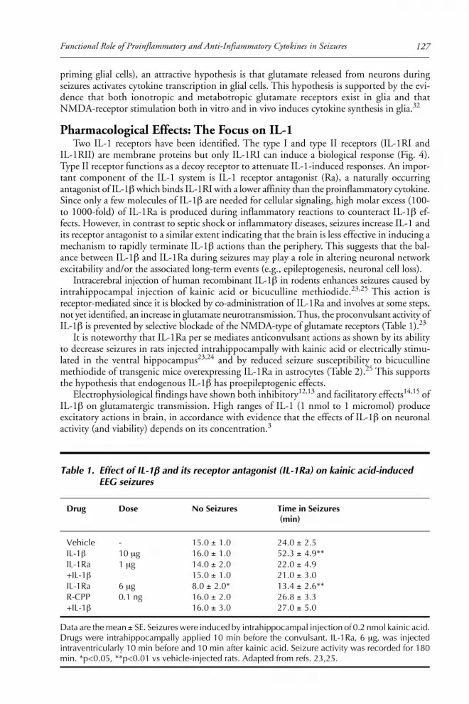
127Functional Role of Proinflammatory and Anti-Infiammatory Cytokines in Seizures
priming glial cells), an attractive hypothesis is that glutamate released from neurons duringseizures activates cytokine transcription in glial cells. This hypothesis is supported by the evi-dence that both ionotropic and metabotropic glutamate receptors exist in glia and thatNMDA-receptor stimulation both in vitro and in vivo induces cytokine synthesis in glia.32
Pharmacological Effects: The Focus on IL-1Two IL-1 receptors have been identified. The type I and type II receptors (IL-1RI and
IL-1RII) are membrane proteins but only IL-1RI can induce a biological response (Fig. 4).Type II receptor functions as a decoy receptor to attenuate IL-1-induced responses. An impor-tant component of the IL-1 system is IL-1 receptor antagonist (Ra), a naturally occurringantagonist of IL-1β which binds IL-1RI with a lower affinity than the proinflammatory cytokine.Since only a few molecules of IL-1β are needed for cellular signaling, high molar excess (100-to 1000-fold) of IL-1Ra is produced during inflammatory reactions to counteract IL-1β ef-fects. However, in contrast to septic shock or inflammatory diseases, seizures increase IL-1 andits receptor antagonist to a similar extent indicating that the brain is less effective in inducing amechanism to rapidly terminate IL-1β actions than the periphery. This suggests that the bal-ance between IL-1β and IL-1Ra during seizures may play a role in altering neuronal networkexcitability and/or the associated long-term events (e.g., epileptogenesis, neuronal cell loss).
Intracerebral injection of human recombinant IL-1β in rodents enhances seizures caused byintrahippocampal injection of kainic acid or bicuculline methiodide.23,25 This action isreceptor-mediated since it is blocked by co-administration of IL-1Ra and involves at some steps,not yet identified, an increase in glutamate neurotransmission. Thus, the proconvulsant activity ofIL-1β is prevented by selective blockade of the NMDA-type of glutamate receptors (Table 1).23
It is noteworthy that IL-1Ra per se mediates anticonvulsant actions as shown by its abilityto decrease seizures in rats injected intrahippocampally with kainic acid or electrically stimu-lated in the ventral hippocampus23,24 and by reduced seizure susceptibility to bicucullinemethiodide of transgenic mice overexpressing IL-1Ra in astrocytes (Table 2).25 This supportsthe hypothesis that endogenous IL-1β has proepileptogenic effects.
Electrophysiological findings have shown both inhibitory12,13 and facilitatory effects14,15 ofIL-1β on glutamatergic transmission. High ranges of IL-1 (1 nmol to 1 micromol) produceexcitatory actions in brain, in accordance with evidence that the effects of IL-1β on neuronalactivity (and viability) depends on its concentration.3
Table 1. Effect of IL-1βββββ and its receptor antagonist (IL-1Ra) on kainic acid-inducedEEG seizures
Drug Dose No Seizures Time in Seizures (min)
Vehicle - 15.0 ± 1.0 24.0 ± 2.5IL-1β 10 µg 16.0 ± 1.0 52.3 ± 4.9**IL-1Ra 1 µg 14.0 ± 2.0 22.0 ± 4.9+IL-1β 15.0 ± 1.0 21.0 ± 3.0IL-1Ra 6 µg 8.0 ± 2.0* 13.4 ± 2.6**R-CPP 0.1 ng 16.0 ± 2.0 26.8 ± 3.3+IL-1β 16.0 ± 3.0 27.0 ± 5.0
Data are the mean ± SE. Seizures were induced by intrahippocampal injection of 0.2 nmol kainic acid.Drugs were intrahippocampally applied 10 min before the convulsant. IL-1Ra, 6 µg, was injectedintraventricularly 10 min before and 10 min after kainic acid. Seizure activity was recorded for 180min. *p<0.05, **p<0.01 vs vehicle-injected rats. Adapted from refs. 23,25.

Recent Advances in Epilepsy Research128
Other factors are important for determining the effects of this cytokine, e.g., the functionalstate of neurons (healthy or injured), the timing of cytokine release, the duration of tissueexposure, and the brain region involved.
Figure 4. Upper diagram shows the increase in IL-1Ra mRNA as compared to IL-1β. IL-1Ra increase isdelayed by several hours and returns to control levels faster that IL-1β. In addition, the molar ratio of IL-1Rato IL-1 is approx. 1: 1 or lower. This indicates that the brain is ill equipped for rapidly terminating the actionsof IL-1, differing from the periphery (see text for details). Lower diagram depicts the major components ofthe IL-1 system: mature IL-1 is cleaved from the prepropeptide by caspase 1 and acts on IL-1 type I receptors.IL-1 and receptor type I, in turn, couple to the accessory protein (AcP) to trigger the intracellular signalcascade leading to activation of NFkB. IL-1 R type II acts as a decoy receptor; thus, by binding the cytokineprevents its interaction with R type I. Both receptors exist in a membrane-bound and soluble(metalloproteinase-cleaved, MMP) form. IL-1Ra is produced by differential splicing of mRNA in threedifferent forms: IL-1Ra is released extracellularly while type 2 and 3 remain intracellular. IL-1 levels aredecreased by glucocorticoids (GC) or PGE2; IL-1Ra levels can be enhanced by GC or by anti-inflammatorycytokines (IL-4, IL-13). GC also increase the synthesis of R type II (decoy).

129Functional Role of Proinflammatory and Anti-Infiammatory Cytokines in Seizures
IL-1, GABA and GlutamateThe interactions between IL-1β and these neurotransmitters are of particular relevance for
epilepsy since it is well established that epileptic activity depends on the imbalance betweeninhibitory and excitatory neurotransmission. In particular, 0.6 to 6 nmol/ml of IL-1 enhancesmuscimol-induced Cl- uptake in chick cortical synaptoneurosomes and potentiates GABAreceptor-evoked currents in chick cortical neurons in culture.16 In this study, systemic IL-1raised the threshold to pentylenetetrazol seizures in mice. However, high concentrations ofIL-1 (>60 nmol) have been shown to decrease the peak magnitude of currents elicited byGABA in hippocampal primary cultures and inhibitory postsynaptic potentials in CA3 neu-rons.14,15
In vivo microdialysis has shown that intracerebral injection of 0.6 pmol IL-1 enhancesextracellular glutamate levels in the cortex33 and 0.1 pmol/ml IL-1 inhibits astrocytic,high-affinity glutamate uptake by nitric oxide production.34 However, sub-femtomolar con-centrations of IL-1β increase adenosine release from hippocampal slices, which in turn inhibitsglutamate release acting at A1 receptors.35 Thus, the concentration of IL-1β in the extracellularcompartment appears to be a key factor for determining its functional actions in the brain.
TNF-ααααα and IL-6In addition to their effects on synaptic transmission (see Introduction), these cytokines
appear to affect seizure susceptibility. Thus, mice specifically over-expressing IL-6 and TNF-αin glia develop neurological symptoms and random seizures.36,37 Our recent evidence, how-ever, suggests that TNF-α has anticonvulsant actions mediated by the p75 receptor.38
Proinflammatory Cytokines and Nerve Cell InjurySeveral in vitro and in vivo studies indicate a central role of IL-1β in the exacerbation of
brain damage after focal ischemia, brain trauma or excitotoxicity (for a review see ref. 8). Therole of TNF-α is still controversial: protective effects are suggested by studies in TNF receptorknock-out mice,39 since these mice showed increased neurodegeneration after intrahippocampalinjection of kainic acid; however, transgenic mice overexpressing TNF-α or IL-6 in glia de-velop chronic inflammatory demyelination, reactive gliosis and neurodegeneration.36,37
The role of proinflammatory cytokines in seizure-induced cell damage is suggested by theevidence that glia produce them to a larger extent when seizures cause neuronal injury;23 how-ever, this aspect needs to be further investigated.
Table 2. Transgenic mice overexpressing the human soluble form of IL-1Ra inastrocytes (GILRA2) have decreased susceptibility to seizures
EEG Seizures Behavioral SeizuresOnset Duration Onset Duration
Clonic Tonic- Clonic Tonic-Clonic Clonic
(min) (min)
Wild- 2.0 ± 0.8 8.4 ± 1.5 2.9± 0.4 6.9± 4.2 68.9± 4.2 2.9± 0.7 typeGIRLA2 7.7± 4.4 2.8 ± 1.6* 2.5± 0.5 44.7 ±16.0* 22.4 ± 7.9* 0.7 ± 0.3**
Data are the mean ± SE. Seizures were induced by intrahippocampal administration of 0.24 nmolbicuculline methiodide. Note that EEG and behavioral seizures were strongly inhibited in transgenicmice (*p<0.05, p<0.01 vs wild-type). GIRLA2 mice show an increase of about 15-fold in CSF IL-1Ra.Adapted from ref. 25.

Recent Advances in Epilepsy Research130
Human Epileptic TissueThe expression of inflammatory cytokines has not been investigated in great detail in hu-
man epileptic tissue. Sheng et al40 and Barres41 have reported increased IL-1 and functionalactivation of microglia and astrocytes in brain tissue of humans affected by temporal lobeepilepsy. The mean concentration of IL-6 was significantly increased in CSF and plasma within24 h from tonic-clonic seizures in humans, while no significant differences were found inIL-1β and TNF-α. IL-1Ra was increased as well by 2-fold.42 In children with febrile seizures,the concentration of IL-1β in CSF was within the normal range.43,44 However, in these studiesCSF measurements were done on days 1-2 after seizures, which may be too late to detectcytokine changes. Thus, cytokine induction occurs within 2 h from seizure onset in experi-mental models and is transient. In addition, very low amounts of cytokines are enough toproduce autocrine/paracrine effects and it is possible that CSF measurements do not detectthese small changes.
Finally, a functional polymorphism in the IL-1β gene promoter, possibly associated withenhanced ability to produce this cytokine, has been specifically found in temporal lobe epi-lepsy patients with hippocampal sclerosis45 and in children with febrile seizures.46 Thus, asubtle anomaly in IL-1β to IL-1Ra ratio in the brain or developmental events during early lifecould set up a cascade of events leading to hippocampal sclerosis and epilepsy in individualswith this genetic predisposition.
Mechanism of ActionCytokines are released from cells producing them: e.g., TNF-α is released from hippoc-
ampal slices at a larger extent after seizures.47 Once released from glia, which are the majorsource of cytokines in the brain, they likely act upon their receptors on neurons (for re-view see ref. 8). Often these receptors show rapid up-regulation in response to injury, e.g.,IL-1R type II was increased in the rat hippocampus and various forebrain areas after lim-bic seizures.20
In this scenario, while information about the functional role of TNF-α and IL-6 is stillscarce, IL-1β will act on target neurons to enhance their excitability. This may occur directly byaffecting ionic currents or indirectly by enhancing glutamatergic function and/or decreasingGABA actions (Fig. 5).14,15,33 It is possible that IL-1β induces post-translational changes inNMDA or GABA receptor subunits.
Thus, IL-1R type I activation is associated with activation of signal transduction pathways(e.g., activation of protein kinase C, protein kinase A and nitric oxide production)1 which areknown to affect the responses of NMDA or GABA receptors to their endogenous ligands.IL-1Ra will afford protection from seizures by antagonizing the action of IL-1β on receptortype I. It is interesting to note that these receptors are densely concentrated on dendrites ofgranule neurons in the hippocampus.20,48,49
Thus, the balance between IL-1β and its receptor antagonist in brain may play a significantrole in the maintenance and spread of seizures and this may be of pharmacological relevance.
IL-1R antagonist is highly inducible and appears to be a well-tolerated protein,50 thus phar-macological means that increase IL-1Ra/IL-1β ratio may be useful for controlling seizure activ-ity even when other factors act to increase brain excitability.
AcknowledgementsWe gratefully acknowledge Prof K Iverfeldt (University of Stockholm, Sweden) and Prof T
Bartfai (Scripps Res Inst, La Jolla, CA) for their invaluable contribution to part of these studies.Some of the work presented in this chapter has been supported by Telethon Onlus Foundation.

131Functional Role of Proinflammatory and Anti-Infiammatory Cytokines in Seizures
Figure 5. Schematic drawing depicting the proposed mechanism of action of IL-1β. Seizures induceglutamate release from excitatory synaptic terminals. Glutamate acting on ionotropic and/or metabotropicreceptors on glia induces the transcription of the IL-1 gene and/or activates caspase 1. The mature cytokineis then released and acts on receptor (R) type I on neurons.49 Electrophysiological evidence indicates thatIL-1 inhibits GABA-mediated currents15 and increases extracellular glutamate disposition (enhanced re-lease and decreased reuptake33,34). These actions will mediate its proconvulsant activity in rodents. IL-1Raantagonizes these effects by blocking IL-1 R type 1.

Recent Advances in Epilepsy Research132
References1. Schöbitz B, Ron De Kloet E, Holsboer F. Gene expression and function of interleukin-1,
interleukin-6 and tumor necrosis factor in the brain. Prog Neurobiol 1994; 44:397-432.2. Hopkins SJ, Rothwell NJ. Cytokines and the nervous system I. Expression and recognition. Trends
Neurosci 1995; 18:83-88.3. Rothwell NJ, Hopkins SJ. Cytokines and the nervous system II: Actions and mechanisms of ac-
tion. Trends Neurosci 1995; 18:130-136.4. De Simoni MG, Imeri L. Cytokine-neurotransmitter interactions in the brain. Biol Signals 1998;
7:33-44.5. Scarborough DE, Lee SL, Dinarello CA et al. Interleukin-1β stimulates somatostatin biosynthesis
in primary cultures of fetal rat brain. Endocrinology 1986; 124:549-551.6. Spranger M, Lindholm D, Bandtlow C et al. Regulation of nerve growth factor (NGF) synthesis in
the rat central nervous system: comparison between the effects of interleukin-1 and various growthfactors in astrocyte cultures and in vivo. Eur J Neurosci 1990; 2:69-76.
7. Lapchak PA, Araujo DM. Hippocampal interleukin-2 regulates monoamine and opioid peptiderelease from the hippocampus. Neuroreport 1993; 4:303-306.
8. Allan SM, Rothwell NJ. Cytokines and acute neurodegeneration. Nature Rev Neurosci 2001;2:734-744.
9. Katsuki H, Nakai S, Hirai Y et al. Interleukin-1β inhibits long-term potentiation in the CA3region of the mouse hippocampal slice. Eur J Pharmacol 1990; 181:323-326.
10. Bellinger FP, Madamba S, Siggins GR. Interleukin 1β inhibits synaptic strength and long-termpotentiation in the rat CA1 hippocampus. Brain Res 1993; 628:227-234.
11. Cunningham AJ, Murray CA, O’Neil LAJ et al. Interleukin 1β and tumor necrosis factor (TNF)inhibit long-term potentiation in the rat dentate gyrus in vitro. Neurosci Lett 1996; 203:17-20.
12. Coogan A, O’Connor JJ. Inhibition of NMDA receptor-mediated synaptic transmission in the ratdentate gyrus in vitro by IL-1β. Neuroreport 1997; 8:2107-2110.
13. D’Arcangelo G, Dodt H, Zieglgansberger W. Reduction of excitation by interleukin-1β in ratneocortical slices visualized using infrared darkfield videomicroscopy. Neuroreport 1997;8:2079-2083.
14. Zeise ML, Espinoza J, Morales P et al. Interleukin-1β does not increase synaptic inhibition inhippocampal CA3 pyramidal and dentate gyrus granule cells of the rat in vitro. Brain Res 1997;768:341-344.
15. Wang S, Cheng Q, Malik S. Interleukin-1β inhibits γ−amino butyric acid type A (GABAA) recep-tor current in cultured hippocampal neurons. J Pharmacol Exp Ther 2000; 292:497-504.
16. Miller LG, Galpern WG, Lumpkin M et al. Interleukin-1 (IL-1) augments gamma-aminobutyricacid receptor function in the brain. Mol Pharmacol 1991; 39:105-108.
17. Plata-Salaman CR, French-Mullen JMH. Interleukin-1β depresses calcium current in CA1 hippoc-ampal neurons at pathophysiological concentrations. Brain Res Bull 1992; 29:221-223.
18. Minami M, Kuraishi Y, Satoh M. Effects of kainic acid on messenger RNA levels of IL-1 beta,IL-6, TNF alpha and LIF in the rat brain. Biochem Biophys Res Commun 1991; 176:593-598.
19. Yabuuchi K, Minami M, Katsumata S et al. In situ hybridization study of interleukin-1β mRNAinduced by kainic acid in the rat brain. Brain Res Mol Brain Res 1993; 20:153-161.
20. Nishiyori A, Minami M, Takami S et al. Type 2 interleukin-1 receptor mRNA is induced bykainic acid in the rat brain. Mol Brain Res 1997; 50:237-245.
21. Gahring LC, White SH, Skradski SL et al. Interleukin-1α in the brain is induced by audiogenicseizure. Neurobiol Dis 1997; 3:263-269.
22. Eriksson C, Winblad B, Schultzberg M. Immunohistochemical localization of interleukin-1beta,interleukin-1 receptor antagonist and interleukin-1beta converting enzyme/caspase-1 in the rat brainafter peripheral administration of kainic acid. Neuroscience 1999; 93:915-930.
23. Vezzani A, Conti M, De Luigi A et al. Interleukin-1β immunoreactivity and microglia are en-hanced in the rat hippocampus by focal kainate application: functional evidence for enhancementof electrographic seizures. J Neurosci 1999; 19:5054-5065.
24. De Simoni MG, Perego C, Ravizza T et al. Inflammatory cytokines and related genes are inducedin the rat hippocampus by limbic status epilepticus. Eur J Neurosci 2000; 12:2623-2633.
25. Vezzani A, Moneta D, Conti M et al. Powerful anticonvulsant action of IL-1 receptor antagoniston intracerebral injection and astrocytic overexpression in mice. Proc Natl Acad Sci USA 2000;97:11534-11539.
26. Benveniste EN. Inflammatory cytokines within the central nervous system: sources, function andmechanism of action. Am J Physiol 1992; 263:C1-C16.

133Functional Role of Proinflammatory and Anti-Infiammatory Cytokines in Seizures
27. Bartfai T, Shultzberg M. Cytokines in neuronal cell types. Neurochem Int 1993; 22:435-444.28. Perry VH, Andersson PB, Gordon S. Macrophages and inflammation in the central nervous sys-
tem. Trends Neurosci 1993; 16 (7):268-273.29. Montero-Menei CN, Sindji L, Garcion E et al. Early events of the inflammatory reaction induced
in rat brain by lipolysaccharide intracerebral injection: relative contribution of peripheral mono-cytes and activated microglia. Brain Res 1996; 724:55-66.
30. Zhao B, Schwartz JP. Involvement of cytokines in normal CNS development and neurologicaldiseases: recent progress and perspective. J Neurosci Res 1998; 52:7-16.
31. Nitsch C, Goping G, Klatzo I. Pathophysiological aspects of blood brain barrier permeability inepileptic seizures. In: Schwarcz R, Ben-Ari Y, eds. Advances in Experimental Medicine and Biol-ogy. New York, London: 1986:175-184.
32. Jander S, Schroeter M, Stoll G. Role of NMDA receptor signaling in the regulation of inflamma-tory gene expression after focal brain ischemia. J Neuroimmunol 2000; 109:181-187.
33. Kamikawa H, Hori T, Nakane H et al. IL-1β increases norepinephrine level in the rat frontalcortex: involvement of prostanoids, NO, and glutamate: Am J Physiol 1998; 275:R806-R810.
34. Ye ZC, Sontheimer H. Cytokine modulation of glial glutamate uptake: a possible involvement ofnitric oxide. Neuroreport 1996; 72:181-2185.
35. Luk WP, Zhang Y, White TD et al. Adenosine: a mediator of interleukin-1β induced hippocampalsynaptic inhibition. J Neurosci 1999; 19:4238-4244.
36. Campbell IL, Abraham CR, Masliah E et al. Neurologic disease induced in transgenic mice bycerebral overexpression of interleukin 6. Proc Natl Acad Sci USA 1993; 90:10061-10065.
37. Akassoglou K, Probert L, Kontogeorgos G et al. Astrocyte-specific but not neuron specific trans-membrane TNF triggers inflammation and degeneration in the central nervous system of transgenicmice. J Immunol 1997; 158:438-445.
38. Vezzani A, Moneta D, Richichi C et al. Functional role of TNF-α and IL-1β systems in seizuresusceptibility and epileptogenesis. 31st Annual meeting Society for Neuroscience Nov 10-15 2001;557:18.
39. Bruce AJ, Boling W, Kindy MS. Altered neuronal and microglial responses to excitotoxic andischemic brain injury in mice lacking TNF receptors. Nature Med 1996; 2:788-794.
40. Sheng JG, Boop FA, Mrak RE et al. Increased neuronal β-amyloid precursor protein expression inhuman temporal lobe epilepsy: association with interleukin-1α immunoreactivity. J Neurochem1994; 63:1872-1879.
41. Barres BA. New roles for microglia. J Neurosci 1991; 11:3685-3694.42. Peltola J, Palmio J, Korhonen L. Interleukin-6 and interleukin-1 receptor antagonist in cerebrospi-
nal fluid from patients with recent tonic-clonic seizures. Epilepsy Res 2000; 41:205-211.43. Lahat E, Livine M, Barr J. Interleukin-1 levels in serum and cerebrospinal fluid of children with
febrile seizures. Pediatr Neurol 1997; 17:34-36.44. Ichiyama T, Nishikawa M, Yoshitomi T. Tumor necrosis factor-α, interleukin-1, and interleukin-6
in cerebrospinal fluid from children with prolonged seizures. Neurology 1998; 50:407-411.45. Kanemoto K, Kawasaki J, Miyamoto T. Interleukin(IL)-1β, IL-1α, and IL-1 receptor antagonist
gene polymorphisms in patients with temporal lobe epilepsy. Ann Neurol 2000; 47:571-574.46. Hurme VM, Helminen M. Increased frequency of interleukin-1beta (-511) allele 2 in febrile sei-
zures. Pediatr Neurol 2002; 26:192-195.47. de Bock F, Dornand J, Rondouin G. Release of TNF in the rat hippocampus following epileptic
seizures and excitotoxic neuronal damage. Neuroreport 1996; 7:1125-1129.48. Takao T, Tracey DE, Mitchell WM. Interleukin-1 receptors in mouse brain: Characterization and
neuronal localization. Endocrinology 1990; 127:3070-3078.49. Ban E, Milon G, Prudhomme N et al. Receptors for interleukin-1 (alpha and beta) in mouse
brain: Mapping and neuronal localization in hippocampus. Neuroscience 1991; 43:21-30.50. Dinarello CA. Biological basis for interleukin-1 in disease. Blood 1996; 87:2095-2147.

Recent Advances in Epilepsy Research134
CHAPTER 11
Recent Advances in Epilepsy Research, edited by Devin K. Binder and Helen E. Scharfman.©2004 Eurekah.com and Kluwer Academic / Plenum Publishers.
Using the Immune System to Target EpilepsyDeborah Young and Matthew J. During
Introduction
The sudden and transient disruption from normal brain function by the disordered,synchronous and rhythmic firing of populations of neurons or seizures is the commonfeature of a diverse collection of disease syndromes collectively called the epilepsies.
The epilepsies are estimated to affect 1-2% of the population, with the most common formbeing temporal lobe epilepsy, which accounts for 40% of all types. The underlying cause of thisform of epilepsy is thought to involve an imbalance in excitatory and inhibitory neurotrans-mission due to altered circuitry in the hippocampus resulting from the selective degenerationof hilar, CA1 and CA3 pyramidal neurons but relative preservation of dentate granule neu-rons.39 Hyperexcitability in the hippocampus then occurs following synaptic reorganization ofthe surviving dentate granule cell mossy fiber axons into the inner molecular layer of the den-tate gyrus. While surgical resection is useful in alleviating seizures, particularly for intractabletemporal lobe epilepsy,38,24 pharmacotherapy is the mainstay of treatment for most forms. Atthe cellular level, anti-epileptic agents mediate their effects through three major pharmacologi-cal modes of actions: enhancement of inhibitory neurotransmission, attenuation of excitatorytransmission or modulation of voltage-dependent ion channels. While the use of one or acombination of antiepileptic drugs can provide seizure control in most cases, there are alsomany cases refractory to treatment. Additionally, many of these compounds have been re-ported to negatively impact cognitive function,11 suggesting the continuing requirement forbetter and more specific treatments.
Using the Immune System to Treat Neurological Disease—The Relationship between the Brain and the Immune System
While researchers have attempted to harness the power of the immune system for treating adiverse range of diseases including cancer, using the immune system to treat neurological dis-ease in general has never been seriously considered a viable option. This is because the humoraleffectors of the immune system, the immunoglobulins, are large proteins and have limitedaccess into the brain due to the presence of the blood-brain barrier (BBB) which impedes theirentry and thus subsequent binding to target antigens in the CNS. However, there is limitedevidence that under certain conditions, antibodies can gain access into the CNS. One of themost compelling and intriguing lines of evidence are the autoimmune diseases that involve theCNS. Rasmussen’s encephalitis (RE) is a rare form of pediatric epilepsy characterized by severeepileptic seizures that are relatively resistant to treatment with typical anti-seizure medicationand inflammatory neuropathology restricted to one cerebral hemisphere.50 Perivascular lym-phocytic cuffs and scattered microglial nodules in the cortex, neuronal degeneration, and glio-sis are key histopathological features of the affected hemisphere. The link between RE andautoimmune disease was established following the serendipitous discovery that a small subsetof rabbits immunized with a recombinant GluR3 glutamate receptor fusion protein not only

135Using the Immune System to Target Epilepsy
developed high-titer anti-GluR3 antibodies in the serum, but also a phenotype characterizedby seizures and inflammatory neuropathology strikingly similar to that found in RE.51 IgGsubsequently isolated from the sera from ‘epileptic’ GluR3-immunized rabbits was found tohave the ability to bind and evoke excitatory currents and kill mouse cortical neurons in cul-ture. These cytotoxic effects could be reversed by the selective competititve antagonist CNQX,suggesting anti-GluR3 antibodies promote seizures and excitotoxic cell death by acting as anagonist at AMPA receptors.56 Anti-GluR3 autoantibodies were subsequently demonstrated inhuman RE sera and shown to bind with moderate affinity to GluR3 overexpressed in HEK293cells.51 Removal of serum antibodies by plasmapheresis in RE patients transiently reduces theseizure frequency and improves neurologic function as serum concentrations of GluR3 anti-bodies are reduced, further corroborating humoral autoimmune processes in the pathogenesisof RE.2,51 Similar improvements in neurologic function were found with the selective removalof IgG and IgM from the plasma of a RE patient by protein A affinity chromatography, withdirect correlation between reduced GluR3 antibody titers and clinical benefit.3 A more recentand independent study suggests that GluR3 autoantibodies can also destroy cells by acomplement-dependent mechanism.26 Rabbits immunized with a glutathione S-transferase-fusedGluR3 protein developed anti-GluR3 antibodies that had no excitatory properties when ap-plied to cortical neurons in whole cell patch-clamp studies. However, robust cytotoxic effectswere found which were identified as being complement- dependent.26 In corroboration of thisfinding, IgG and complement factors of the membrane attack complex were found on neuronsand their processes in the cortex of brains of RE patients.58 This has led to the proposal thatthese autoantibodies may therefore gain access into the CNS to trigger complement-mediatedneuronal damage, events that may be important in the initial phases of the disease. Similarneuropathological findings have been reproduced in murine models of RE.31,32
While GluR3 may be the prototypic autoantigen, autoimmune attack on other brain anti-gens, some identified61 and others as yet to be identified, may also be involved in some cases ofRE. Autoantibodies to Munc-18, an intracellular protein found in presynaptic terminals witha role in synaptic vesicle release, has been reported in a RE patient also displaying serumanti-GluR3 autoantibodies.61 Thus, Yang et al, propose that additive or synergistic effects ofautoimmune attack on two different epitopes at two distinct sites of synaptic transmissioncould impair function, leading to seizures and neuronal death. The ratio of the autoantibodiesin the cerebrospinal fluid and the serum relative to the total IgG concentration in the twocompartments supports the passage of Munc-18 autoantibodies across the BBB.61 However,the significance of anti-Munc-18 antibodies in RE pathogenesis is debatable as it has not beenfound in 14 other patients, suggesting it may be ancillary to the disease. Similarly, recent re-ports suggest prevalence of anti-GluR3 autoantibodies in other forms of epilepsy, and thuswhether anti-GluR3 antibodies are specific for the diagnosis of RE is unclear.59
RE is but one of several autoimmune diseases involving the CNS that are associated withserum antibodies to brain antigens which appear to gain access into the brain and mediate thegeneration of complex neurological disease phenotypes.57 Paraneoplastic disorders are autoim-mune disorders that are a remote complication of some forms of cancer. These develop whenthe ectopic expression of brain antigens by the tumor leads to a T-cell mediated-immune re-sponse and/or autoantibodies that gain access to the brain to target neurons expressing thoseantigens and mediate a disease phenotype.1,14 Stiff-man syndrome is characterized by progres-sive skeletal muscle rigidity, deficiency in GABA and circulating autoantibodies to GAD65, thekey enzyme involved in GABA synthesis.17,33,55 Gephyrin, a cytosolic protein required for clus-tering of glycine and GABAA receptors at the postsynaptic density of inhibitory synapses, hasalso been identified as an autoantigen for Stiff-man.12 Rare cases of temporal lobe epilepsy andother forms of epilepsy are associated with GAD65 autoantibodies25,43 and it has also beensuggested that a subgroup of drug-refractory temporal lobe epilepsy patients may be associatedwith GAD65 autoantibodies, although further investigation is warranted.30,46 Additionally,autoantibodies to other brain antigens have also been linked to other rare forms of epilepsy.44

Recent Advances in Epilepsy Research136
Although many of these conditions are relatively rare, importantly these findings suggestthat under certain circumstances antibodies do have the ability to pass the BBB, bind andmodify the function of their target antigen. As antibodies to GluR3 are found in serum in thelarge majority of immunized animals without apparent disease,51 it is clear that secondary fac-tors are necessary for initiating the disease process. The identity of these factors has remainedelusive, although it has been proposed that a focal lesion leading to disruption of the BBB dueto trauma may be essential for immune-mediated attack. Indeed, trauma-induced breakdownof the BBB has been demonstrated in experimental epilepsy and excitotoxic lesions.10,48,52
Using the Immune System to Treat Neurological Disease—Therapeutic Agents for Neurological Disease
While these examples argue against exploiting the use of the immune system to treat neuro-logical disease, there is good evidence that antibodies can potentially be used as therapeuticagents for altering the function of brain proteins. Monoclonal antibodies have been applied inexperimental paradigms to disrupt signal transduction cascades mediated by neurotrophic fac-tor receptors.13 Following direct injection into the brain and binding to the extracellular do-mains of the receptor, the subsequent blockade of the TrkA signalling pathway has allowedresearchers to elucidate the role of nerve growth factor in maintenance of long term potentia-tion in rat visual cortex.47 More recently, the Alzheimer’s disease (AD) vaccines have sparked agreat deal of interest. One of the defining characteristics of AD pathology is the accumulationof amyloid β-peptide and its deposition as plaques within cortical and hippocampal brainregions. Immunization with amyloid β-peptide is associated with generation of high-titer amy-loid β-peptide antibodies and the dramatic reduction of amyloid plaque burden and cognitivedeficits in transgenic mouse models of AD.42,53 While several mechanisms of action of theresponse to immunization have been proposed, the reduction in amyloid burden followingpassive transfer of amyloid β-peptide antibodies indicates that antibodies can cross the BBBdirectly to ameliorate the disease phenotype in an AD mouse model.4 Importantly, this evi-dence supports the contention that the immune-privileged state of the brain as being relative,not absolute. Although anecdotal evidence has suggested the possibility of cerebritis associatedwith the early stage clinical trials of a β-amyloid vaccine,8 some researchers believe that thismight reflect the disease process, since AD is associated with a significant microglial compo-nent, cytokine release and inflammation, complicating the analysis of adverse events associatedwith the vaccine clinical trials.
Developing a Vaccine Strategy for EpilepsyThe NMDA receptor plays a key role in mediating excitotoxic-induced neuronal cell death
in addition to other activity-dependent functions such as learning and memory.9 Structurally,the NMDA receptor is a hetero-oligomeric complex composed of subunits from the NR1,NR2A-D and NR3A subunit families arranged to form a ligand-gated, voltage-dependent ionchannel principally permeable to Ca2+ ions.27 Although widely expressed throughout the brain,functional diversity of NMDA receptors in vivo is effected by incorporation of different NR2and NR3 subunits with the essential NR1 subunit. The complete repertoire of subunit combi-nations and heterogeneity of NMDA receptors throughout the CNS is still unclear. The NR1subunit is necessary for formation of a functional ion channel,22 while the NR2 and NR3subunits modulate channel properties.15,54 Both the NR1 and NR2 proteins are transmem-brane spanning proteins with an extracellular N-terminus and intracellular C-terminus andthree complete transmembrane regions. An intramembrane loop forms between the first andsecond complete transmembrane regions. The glycine binding site is found on the NR1 sub-unit while the glutamate binding site is found on the NR2 subunit.16 At resting membranepotential, the ion channel is blocked and activation of the receptor is dependent on binding ofthe coligands glutamate and glycine and simultaneous neuronal depolarization which relievesthe Mg2+-dependent block, allowing intracellular passage of Ca2+ ions.16

137Using the Immune System to Target Epilepsy
We have recently described a novel approach for epilepsy and stroke treatment involvingmanipulation of the immune system to generate a humoral response against a target brainantigen.21 In an experimental situation analogous to the autoimmune diseases described aboveand in particular GluR3 immunization,51 we hypothesized that ectopic expression of a specificbrain protein that has been sequestered behind the BBB may lead to generation of autoanti-bodies specific to the target. These circulating autoantibodies would have limited access intothe brain under normal basal conditions. However, following an insult and associated BBBcompromise, events that occur following kainate-induced seizures7,63 and excessive glutamaterelease,10,37 increased passage of these autoantibodies would occur. These antibodies, instead ofhaving excitotoxic or disease-inducing activity, would have the ability to bind and alter thefunction of the target protein to block processes involved in mediating insult-induced neu-ronal injury.21
Because the NR1 subunit is the common element in all NMDA receptors, we targeted theNR1 subunit in our vaccination strategy.22 Thus, we proposed that vaccination against theNR1 subunit would generate serum autoantibodies to NR1 that would have restricted brainaccess under normal conditions. However, following an insult and associated BBB breakdown,increased passage of NR1 autoantibodies, which could bind and neutralize NMDA receptorfunction and thus block injury-induced neuronal death, would occur.
A Novel Genetic Vaccine for Stroke and EpilepsyWe utilized a novel genetic vaccination strategy based on the use of recombinant
adeno-associated viral vectors (AAV). This idea stemmed from our previous work showingrobust gene transfer and expression of β-galactosidase protein in gut lamina propria cells fol-lowing oral administration of an AAV vector expressing a lacZ reporter gene.20 Many of thetransduced β-galactosidase-expressing cells were antigen-presenting cells involved in generat-ing humoral immune responses, indicating their potential for use as vaccines. AAV vectors arederived from a nonpathogenic virus and genetically modified to undergo a single round of hostinfection to efficiently deliver a gene-of-interest to mammalian cells. Several desirable featuresincluding a broad host cell range and the ability to transduce both nondividing and dividingcells leading to high levels of stable and sustained gene expression, make AAV vectors arguablythe vector of choice for human gene therapy.41,45
To test our vaccination hypothesis, a high-titer AAV vector expressing a full-length mouseNR1 cDNA under control of a CMV promoter was orally administered to fasted rats whilecontrol animals received an AAV vector expressing the lacZ reporter gene or the saline vehicle(Fig. 1). Robust NR1 or lacZ gene transfer and protein expression was found in gut laminapropria cells subsequently confirmed as antigen-presenting dendritic cells and macrophages bydouble-labelling immunohistochemistry using the corresponding cell markers (Fig. 1). PCRanalysis confirmed that transfer of the NR1 or lacZ genes following oral immunization wasconfined to the gut, with no spread to the spleen, liver or testes. NR1 protein expression wasassociated with NR1 IgG autoantibodies in the serum by as detected by Western blot analysis,which increased in titer over the 1 to 5 month period. Similarly, β-galactosidase autoantibodieswere found in the corresponding AAVlacZ-immunized animals. To further define the polyclonalresponse and the epitope regions to which the autoantibodies were generated against, a series ofoverlapping 16-mer peptides representing the entire 938 amino acids of the NR1 subunit wasused to screen the NR1 serums. The most prevalent peptides that the NR1 autoantibodiesbound to were those that corresponded to functional domains within the extracellular vesti-bule of the receptor. More specifically, NR1 autoantibodies were generated against epitopesthat mapped to regions in the extracellular domains that are critical for glycine binding.5 Theseresults suggested that targeted immunization against a brain antigen could result in generationof a polyclonal antibody response to the target protein.
Vaccinated rats were screened for neuroprotective efficacy against the kainate model of experi-mental epilepsy. Peripheral administration of kainate to rodents leads to a stereotypical progression

Recent Advances in Epilepsy Research138
through various seizure behaviors culminating in prolonged seizures or status epilepticus.6 Weadministered kainate at a dose of 10 mg/kg i.p. and monitored the appearance of electrographicseizures and found there was an overall reduction in the expected frequency of developingkainate-induced seizures from 68% in the control animals to 22% in the NR1-immunizedanimals. The anti-epileptic and neuroprotective efficacy of NR1 immunization was verified byTUNEL labelling and clusterin immunohistochemistry, a cell death marker18 (Fig. 2). Of inter-est, one of the two NR1-immunized rats that had a robust NR1 autoantibody titer and devel-oped status epilepticus did not show any TUNEL or clusterin labelling. The neuroprotectiveefficacy was further explored in the endothelin-1 model of ischemic stroke, and NR1-immunizedanimals were more protected against stroke-induced damage when compared to controls21 (Fig.3). Antibody passage across an intact BBB was confirmed by sampling cerebrospinal fluid fromthe cisterna magna from naïve and both lacZ- and NR1-immunized groups of animals, withonly NR1 autoantibodies detectable in cerebrospinal fluid obtained from NR1 animals.
Figure 1. The AAVNR1 vector construct (A) was a full-length mouse NR1 cDNA under the control of aCMV immediate-early promoter and bovine growth hormone (BGH) polyadenylation site flanked by theAAV inverted terminal repeats (ITR). B. NR1 protein expression (blue) in the lamina propria (lp) as assessedby immunohistochemistry with propidium iodide counterstaining (orange) from a section from the proxi-mal intestine at 4 weeks following NR1 immunization. The epithelial cell layer (ep) is also indicated. C.SIRP, a dendritic and macrophage cell marker (red) and (D) NR1 (blue) double immunofluorescent labelingwith acridine orange counterstaining (green) in the gut lamina propria, showed NR1 protein expression inantigen-presenting cells. (reproduced from During et al, 2000; Science 287:1453-1460).

139Using the Immune System to Target Epilepsy
Figure 2. Hippocampal damage in the hilus assessed by fluorescent TUNEL (green) or clusterin (green)immunohistochemistry combined with immunohistochemistry to the neuronal marker NeuN (orange).After 45 min of status epilepticus, control lacZ animals (A,B) had extensive damage as indicated by thesecell death markers. No injury was found in any animal not developing status epilepticus (C,D). Only 2 outof 9 NR1-immunized animals developed SE. One NR1-immunized rat showed extensive damage (E,F).However, subsequent serum analysis confirmed this animal had not developed an NR1 autoantibody titer.The remaining animal which developed a robust NR1 autoantibody titer showed no signs of injury despitehaving 45 min of status epilepticus (G,H). (reproduced from During et al, 2000; Science 287:1453-1460).

Recent Advances in Epilepsy Research140
Figure 3. Reduced ischemic damage in the cortex and striatum of NR1-vaccinated animals after middlecerebral artery occlusion. H&E staining of the striatum and/or cortical regions contralateral (A,C,E) andipsilateral (B,D,F) to the occlusion in lac (A-D) and NR1-vaccinated animals (E,F). Isolectin B4 staining(G) detected microglial infiltration in the ipsilateral hemisphere. H. Total infarct volume (mean ±SEM) innaïve (white bars), lacZ- (blue bars) and NR1-vaccinated groups (red bars). *P<0.01. (reproduced fromDuring et al, 2000; Science 287:1453-1460 ).

141Using the Immune System to Target Epilepsy
As our results showed that NR1-immunization is able to manipulate NMDA receptor func-tion, the effects on other NMDA receptor-mediated brain functions need also be considered.With these receptors playing major roles in mediating synaptic plasticity associated with learn-ing and memory, previous reports have established that systemic administration of pharmaco-logical NMDA receptor antagonists at neuroprotective doses to experimental animals is gener-ally associated with impaired mobility and deficits in learning and memory function.60,62 Forthis reason, we have conducted a preliminary assessment of the effects of NR1 immunizationon mobility and performance of NR1-vaccinated and control rats in tests of cognitive func-tion. Mobility as assessed by a line crossing and circular track test was similar for both NR1 andcontrol animals.21 Surprisingly, when these animals were challenged with more complex be-havioral tasks that test learning and memory function, significant differences in performancein these tasks were observed between the NR1 and controls. NR1-immunized animals showedimproved performance in the Barnes circular maze, a hippocampal-dependent spatial naviga-tion task that requires rats to locate a dark escape tunnel hidden beneath one of 18 holespositioned around the perimeter of a brightly lit table using visual cues. In comparison tocontrols, NR1-animals showed reduced latency and required fewer trials to find the escapetunnel.19 Similarly, NR1-immunized rats also exhibited a stronger freezing response thanlacZ-immunized rats in a fear conditioning behavioral paradigm, a contextual association task.28
The results from our study suggest the neuroprotective effects found in the stroke andepilepsy models following NR1 vaccination are mediated by NR1 autoantibodies that have theability to cross the BBB to bind and modulate NMDA receptor function. There is some evi-dence that suggests the NR1 vaccination approach has some clinical relevance. An analysis ofserum from 270 stroke patients in Russia showed that presence of significant NMDA receptorautoantibody titers was associated with improved neurological recovery, while the absence ofautoantibodies was associated with poor prognosis and death.25 Increased penetration of NR1autoantibodies was found after kainate administration, suggesting autoantibodies may directlymediate the neuroprotective effects at the time of insult. However, despite this observation,there is also the possibility that the neuroprotective and anti-epileptic effects observed with thevaccine may not be due to a direct effect of autoantibody antagonism at NMDA receptors atthe time of insult but result from downstream modulation of neuroprotective genes. This isundergoing further investigation.
Concluding RemarksOne of the inherent problems with epilepsy and stroke is that the cascade of molecular and
biochemical mechanisms mediating insult-induced neuronal death are switched on rapidly,leaving only a short therapeutic window for intervention against these events. The prophylacticnature of an epilepsy vaccine is a novel feature of this approach, providing the advantage thattreatment could be targeted to patients considered at high risk for developing stroke or chroni-cally epileptic patients that may be pharmacoresistant.
However, while these initial results look extremely promising, more research is required todetermine the efficacy and safety of moving such a treatment approach into humans. Thepersistence of NR1 autoantibodies for many months and perhaps even years may have long-termdetrimental effects on brain function. Further work is also required to determine the specificityof the effect and whether NR1 immunization modulates glutamatergic function at otherglutamate receptor subtypes, particularly in light of the effects of GluR3 immunization.51 Ad-ditional concerns relate to the cerebritis in the β-amyloid vaccinated Alzheimer’s disease pa-tients8 suggesting that caution is needed in moving towards clinical trials. Furthermore, com-parisons between an AAV approach and traditional protein vaccination strategies are also requiredto determine whether similar neuroprotective responses would occur or whether this is a uniquefeature of an AAV-based vaccine. Other considerations include determining the effect of thevaccination in animals with preexisting epileptic conditions modeling that found in humanpatients, particularly as altered NMDA receptor subunit composition in epileptic patients has

Recent Advances in Epilepsy Research142
been reported.34,35 The enthusiasm for trials of NMDA receptor antagonists for epilepsy andstroke treatment has diminished because they have performed relatively poorly in clinical trialsin comparison to the dramatic neuroprotective efficacy displayed in preclinical studies andthey have been associated with side-effects that have limited their clinical utility. Several of thelatest generation NMDA receptor antagonists have been reported to have fewer side-effects.29
The NR1 vaccination is not associated with any obvious detrimental behavioral abnormalities,but in fact may improve cognitive performance, reminiscent to that found with partial glycineagonists.36,40,49 One of the key issues for future work will be determining the precise site ofaction of these autoantibodies.
While we may be a long way from translating such technology to the clinic, it is clear thatthese approaches, if successful, could pave the way for alternative methods for epilepsy treat-ment. Active immunization against brain proteins and generation of autoantibodies that arecapable of entering the brain and modulating function of target brain proteins may open newavenues for treatment of neurological disease.
References1. Albert ML, Austin LM, Darnell RB. Detection and treatment of activated T cells in the cere-
brospinal fluid of patients with paraneoplastic cerebellar degeneration. Ann Neurol 2000; 47:9-17.2. Andrews PI, Dichter MA, Berkovic SF et al. Plasmapheresis in Rasmussen’s encephalitis. Neurol-
ogy 1996; 46:242-246.3. Antozzi C, Granata T, Aurisano N et al. Long-term selective IgG immuno-adsorption improves
Rasmussen’s encephalitis. Neurology 1998; 51:302-305.4. Bard F, Cannon C, Barbour R et al. Peripherally administered antibodies against amyloid
beta-peptide enter the central nervous system and reduce pathology in a mouse model of Alzheimerdisease. Nat Med 2000; 6:916-919.
5. Beck C, Wollmuth LP, Seeburg PH et al. NMDAR channel segments forming the extracellularvestibule inferred from the accessibility of substituted cysteines. Neuron 1999; 22:559-570.
6. Ben-Ari Y, Cossart R. Kainate, a double agent that generates seizures: two decades of progress.Trends Neurosci 2000; 23:580-587.
7. Bennett SA, Stevenson B, Staines WA et al. Periodic acid-Schiff (PAS)-positive deposits in brainfollowing kainic acid-induced seizures: relationships to fos induction, neuronal necrosis, reactivegliosis and blood-brain barrier breakdown. Acta Neuropathol (Berl) 1995; 89:126-138.
8. Birmingham K, Frantz S. Set back to Alzheimer vaccine studies. Nature Med 2002; 8:199-200.9. Bliss T, Collingridge GL. A synaptic model of memory: long-term potentiation in the hippocam-
pus. Nature 1993; 361:31-39.10. Bolton SJ, Perry VH. Differential blood-brain barrier breakdown and leucocyte recruitment fol-
lowing excitotoxic lesions in juvenile and adult rats. Exp Neurol 1998; 154:231-240.11. Brunbech L, Sabers A. Effect of antiepileptic drugs on cognitive function in individuals with epi-
lepsy: A comparative review of newer versus older agents. Drugs 2002; 62:593-604.12. Butler MH, Hayashi A, Ohkoshi N et al. Autoimmunity to gephyrin in Stiff-man syndrome. Neu-
ron 2000; 26:307-312.13. Cattaneo A, Capsoni S, Margotti E et al. Functional blockade of tyrosine kinase A in the rat basal
forebrain by a novel antagonistic anti-receptor monoclonal antibody. J Neurosci 1999; 19:9687-9697.14. Darnell RB. Onconeural antigens and the paraneoplastic neurologic disorders: at the intersection
of cancer, immunity and the brain. Proc Natl Acad Sci USA 1996; 93:4529-4536.15. Das S, Sasaki YF, Rothe T et al. Increased NMDA current and spine density in mice lacking the
NMDA receptor subunit NR3A. Nature 1998; 393:377-381.16. Dingledine R, Borges K, Bowie D et al. The glutamate receptor ion channels. Pharmacol Rev
1999; 51:7-61.17. Dinkel K, Meinck HM, Jury KM et al. Inhibition of gamma-aminobutyric acid synthesis by glutamic
acid decarboxylase autoantibodies in stiff-man syndrome: acute behavioral effects of MK-801 inthe mouse. Ann Neurol 1998; 44:194-201.
18. Dragunow M, Preston K, Dodd J et al. Clusterin accumulates in dying neurons following statusepilepticus. Brain Res Mol Brain Res 1995; 32:279-290.
19. During MJ, Jiao XY, Lawlor PA et al. NRI vaccine increases NMDA receptor expression,neurogenesis and memory. Nature (submitted).
20. During MJ, Xu R, Young D et al. Peroral gene therapy of lactose intolerance using anadeno-associated virus vector. Nat Med 1998; 4:1131-1135.

143Using the Immune System to Target Epilepsy
21. During MJ, Symes CW, Lawlor PA et al. An oral vaccine against NMDAR1 with efficacy inexperimental stroke and epilepsy. Science 2000; 287:1453-1460.
22. Forrest D, Yuzaki M, Soares HD et al. Targeted disruption of NMDA receptor 1 gene abolishesNMDA response and results in neonatal death. Neuron 1994; 13:325-338.
23. Giometto B, Nicolao P, Macucci M et al. Temporal-lobe epilepsy associated with glutamic-acid-decarboxylase autoantibodies. Lancet 1998; 352:457.
24. Gleissner U, Helmstaedter C, Schramm J et al. Memory outcome after selective amygdalo-hippocampectomy: a study in 140 patients with temporal lobe epilepsy. Epilepsia 2002; 43:87-95.
25. Gusev EI, Skvortsova VI, Izykenova GA et al. The level of autoantibodies to glutamate receptorsin the blood serum of patients in the acute period of ischemic stroke. Zh Nevropatol Psikhiatr ImS S Korsakova 1996; 96:68-72.
26. He XP, Patel M, Whitney KD et al. Glutamate receptor GluR3 antibodies and death of corticalcells. Neuron 1998; 20:153-163.
27. Hollmann M, Heinemann S. Cloned glutamate receptors. Annu Rev Neurosci 1994; 17:31-108.28. Kiyama Y, Manabe T, Sakimura K et al. Increased thresholds for long-term potentiation and con-
textual learning in mice lacking the NMDA-type glutamate receptor epsilon1 subunit. J Neurosci1998; 18:6704-6712.
29. Kohl BK, Dannhardt G. The NMDA receptor complex: a promising target for novel antiepilepticstrategies. Curr Med Chem 2001; 8:1275-1289.
30. Kwan P, Sills GJ, Kelly K et al. Glutamic acid decarboxylase autoantibodies in controlled anduncontrolled epilepsy: a pilot study. Epilepsy Res 2000; 42:191-195.
31. Levite M, Hermelin A. Autoimmunity to the glutamate receptor in mice—a model for Rasmussen’sencephalitis? J Autoimmun 1999; 13:73-82.
32. Levite M, Fleidervish IA, Schwarz A et al. Autoantibodies to the glutamate receptor kill neuronsvia activation of the receptor ion channel. J Autoimmun 1999; 13:61-72.
33. Lohmann T, Hawa M, Leslie RD et al. Immune reactivity to glutamic acid decarboxylase 65 instiffman syndrome and type 1 diabetes mellitus. Lancet 2000; 356:31-35.
34. Mathern GW, Pretorius JK, Mendoza D et al. Increased hippocampal AMPA and NMDA receptorsubunit immunoreactivity in temporal lobe epilepsy patients. J Neuropathol Exp Neurol 1998;57:615-634.
35. Mathern GW, Pretorius JK, Mendoza D et al. Hippocampal N-methyl-D-aspartate receptor sub-unit mRNA levels in temporal lobe epilepsy patients. Ann Neurol 1999; 46:343-358.
36. Matsuoka N, Aigner TG. D-cycloserine, a partial agonist at the glycine site coupled toN-methyl-D-aspartate receptors, improves visual recognition memory in rhesus monkeys. J PharmacolExp Ther 1996; 278:891-897.
37. Mayhan WG, Didion SP, Sheng M et al. Glutamate-induced disruption of the blood-brain barrierin rats. Role of nitric oxide. Stroke 1996; 27:965-969.
38. McIntosh AM, Wilson SJ, Berkovic SF. Seizure outcome after temporal lobectomy: current re-search practice and findings. Epilepsia 2001; 42:1288-1307.
39. McNamara JO. Emerging insights into the genesis of epilepsy. Nature 1999; 399:A15-22.40. Monahan JB, Handelmann GE, Hood WF et al. D-cycloserine, a positive modulator of the
N-methyl-D-aspartate receptor, enhances performance of learning tasks in rats. Pharmacol BiochemBehav 1989; 34:649-653.
41. Monahan PE, Samulski RJ. Adeno-associated virus vectors for gene therapy: more pros than cons?Mol Med Today 2000; 6:433-440.
42. Morgan D, Diamond DM, Gottschall PE et al. A beta peptide vaccination prevents memory lossin an animal model of Alzheimer’s disease. Nature 2000; 408:982-985.
43. Nemni R, Braghi S, Natali-Sora MG et al. Autoantibodies to glutamic acid decarboxylase in pala-tal myoclonus and epilepsy. Ann Neurol 1994; 36:665-667.
44. Palace J, Lang B. Epilepsy: an autoimmune disease? J Neurol Neurosurg Psychiatry 2000;69:711-714.
45. Peel AL, Klein RL. Adeno-associated virus vectors: activity and applications in the CNS. J NeurosciMethods 2000; 98:95-104.
46. Peltola J, Kulmala P, Isojarvi J et al. Autoantibodies to glutamic acid decarboxylase in patientswith therapy-resistant epilepsy. Neurology 2000; 55:46-50.
47. Pesavento E, Margotti E, Righi M et al. Blocking the NGF-TrkA interaction rescues the develop-mental loss of LTP in the rat visual cortex: role of the cholinergic system. Neuron 2000; 25:165-175.
48. Pont F, Collet A, Lallement G. Early and transient increase of rat hippocampal blood-brain barrierpermeability to amino acids during kainic acid-induced seizures. Neurosci Lett 1995; 184:52-54.
49. Popik P, Rygielska Z. A partial agonist at strychnine-insensitive glycine sites facilitates spatial learningin aged rats. J Physiol Pharmacol 1999; 50:139-151.

Recent Advances in Epilepsy Research144
50. Rasmussen T, Olszewski J, Lloyd-Smith DK. Focal seizures due to chronic localized encephalitis.Neurology 1958; 8:435-455.
51. Rogers SW, Andrews PI, Gahring LC et al. Autoantibodies to glutamate receptor GluR3 inRasmussen’s encephalitis. Science 1994; 265:648-651.
52. Saija A, Princi P, Pisani A et al. Blood-brain barrier dysfunctions following systemic injection ofkainic acid in the rat. Life Sci 1992; 51:467-477.
53. Schenk D, Barbour R, Dunn W et al. Immunization with amyloid-beta attenuates Alzheimer-disease-likepathology in the PDAPP mouse. Nature 1999; 400:173-177.
54. Sheng M, Cummings J, Roldan LA et al. Changing subunit composition of heteromeric NMDAreceptors during development of rat cortex. Nature 1994; 368:144-147.
55. Solimena M, Folli F, Aparisi R et al. Autoantibodies to GABA-ergic neurons and pancreatic betacells in stiff-man syndrome. N Engl J Med 1990; 322:1555-1560.
56. Twyman RE, Gahring LC, Spiess J et al. Glutamate receptor antibodies activate a subset of recep-tors and reveal an agonist binding site. Neuron 1995; 14:755-762.
57. Whitney KD, McNamara JO. Autoimmunity and neurological disease: antibody modulation ofsynaptic transmission. Annu Rev Neurosci 1999; 22:175-195.
58. Whitney KD, Andrews PI, McNamara JO. Immunoglobulin G and complement immunoreactivityin the cerebral cortex of patients with Rasmussen’s encephalitis. Neurology 1999; 53:699-708.
59. Wiendl H, Bien CG, Bernasconi P et al. GluR3 antibodies: prevalence in focal epilepsy but nospecificity for Rasmussen’s encephalitis. Neurology 2001; 57:1511-1514.
60. Wozniak DF, Olney JW, Kettinger L et al. Behavioral effects of MK-801 in the rat. Psychophar-macology (Berl) 1990; 101:47-56.
61. Yang R, Puranam RS, Butler LS et al. Autoimmunity to munc-18 in Rasmussen’s encephalitis.Neuron 2000; 28:375-383.
62. Ylinen A, Pitkanen M, Sirvio J et al. The effects of NMDA receptor antagonists at anticonvulsivedoses on the performance of rats in the water maze task. Eur J Pharmacol 1995; 274:159-165.
63. Zucker DK, Wooten GF, Lothman EW. Blood-brain barrier changes with kainic acid-inducedlimbic seizures. Exp Neurol 1983; 79:422-433.

CHAPTER 12
Recent Advances in Epilepsy Research, edited by Devin K. Binder and Helen E. Scharfman.©2004 Eurekah.com and Kluwer Academic / Plenum Publishers.
Cortical Dysplasia and Epilepsy:Animal ModelsPhilip A. Schwartzkroin, Steven N. Roper and H. Jurgen Wenzel
Abstract
Cortical dysplasia syndromes—those conditions of abnormal brain structure/organizationthat arise during aberrant brain development—frequently involve epileptic seizures.Neuropathological and neuroradiological analyses have provided descriptions and
categorizations based on gross anatomical and cellular histological features (e.g., lissencephaly,heterotopia, giant cells), as well as on the developmental mechanisms likely to be involved inthe abnormality (e.g., cell proliferation, migration). Recently, the genes responsible for severalcortical dysplastic conditions have been identified and the underlying molecular processes in-vestigated. However, it is still unclear how the various structural abnormalities associated withcortical dysplasia are related to (i.e., “cause”) chronic seizures. To elucidate these relationships,a number of animal models of cortical dysplasia have been developed in rats and mice. Somemodels are based on laboratory manipulations that injure the brain (e.g., freeze, undercut,irradiation, teratogen exposure) of immature animals; others are based on spontaneous geneticmutations or on gene manipulations (knockouts/transgenics) that give rise to abnormal corti-cal structures. Such models of cortical dysplasia provide a means by which investigators can notonly study the developmental mechanisms that give rise to these brain lesions, but also exam-ine the cause-effect relationships between structural abnormalities and epileptogenesis.
What Is Cortical Dysplasia?The term “cortical dysplasia” has been used by different authors/investigators to refer to a
number of different histopathological brain phenomena. For the purposes of the present dis-cussion, we will use the term in a relatively broad sense, to refer to a condition of abnormalbrain structure/organization that, in most cases, is due to a deviation from the normal patternof cortical development.35,36,108 It is present in a wide variety of human diseases and syn-dromes. The structural abnormality may involve general/widespread conditions (such as lissen-cephaly) that are often associated with profound mental retardation; other cortical dysplasticpatterns are restricted to discrete areas of focal cortical abnormality, and may be asymptomatic.Much of the recent interest in cortical dysplasia stems from its frequent association with seizuredisorders (i.e., epilepsy)—although the cause-effect relationships remain to be elucidated. Therehave been numerous attempts to classify cortical dysplasia syndromes based on gross pathol-ogy, radiographic findings, histological features, genetic abnormalities, and stages of develop-ment most affected by the pathological process. None of these methods of categorization isentirely satisfactory or all-encompassing, but each one may be useful depending on the ques-tion at hand.

Recent Advances in Epilepsy Research146
Gross PathologyLissencephaly refers to a diffusely smooth-surfaced cerebral hemisphere without sulcation.
This condition is often associated with severe mental retardation and seizures in affected indi-viduals. Lissencephaly has generally been classified based on histological characteristics. Type Ilissencephaly has a four-layered cortex: layer 1 corresponds to the remnants of the marginalzone; layer 2 contains a mixture of normal layer II, III, and V neurons; layer 3 is a cell-sparsezone; and layer 4 consists of a diffuse accumulation of heterotopic neurons.16 Type I lissen-cephaly most commonly occurs in a sporadic form (isolated lissencephaly sequence) or as theMiller-Dieker syndrome (MDS), a haploinsufficiency disorder with characteristic dysmorphicfacies, retardation, and seizures.53 MDS is caused by a mutation in the LIS1 gene,126 whichappears to be an important regulator of neuronal mictrotubule systems. Disruption of micro-tubule function may be the basis of impaired neuronal migration in MDS (see ref. 59 forreview). Mutations of LIS1 have also been identified in 40% of people with nonsyndromiclissencephaly.116
X-linked lissencephaly is another genetic disorder that produces lissencephaly in affectedmales, and gives rise to subcortical band heterotopia in females. Subcortical band heterotopia ischaracterized by a thick layer of cortex that extends diffusely throughout the white matter ofthe cerebral hemispheres. It is caused by mutation in a gene, XLIS (or DCX), which encodes fordoublecortin,49 a microtubule-associated protein that appears to be important for normal neu-ronal migration.
Lissencephaly with cerebellar hypoplasia (LCH) is a lissencephalic syndrome that includesmalformations in the cerebellum and brainstem. Two families with LCH have shown muta-tions in the gene, RELN, which encodes for the protein reelin.81 RELN was first identified inthe mutant mouse, reeler. Reelin is produced by Cajal-Retzius cells in the marginal zone and isimportant in the late phases of neuronal migration.45
Type II lissencephaly (also called cobblestone lissencephaly) is characterized by a completeloss of normal horizontal lamination. Cortical neurons are arranged in vertical columns andclumps, separated by thick septations of glial and vascular tissue; glioneuronal heterotopias arepresent on the surface of the hemispheres. Type II lissencephaly is present in the Walker-Warburgsyndrome, where lissencephaly is accompanied by hydrocephalus, retinal dysplasia, and (insome cases) encephalocele.16 Type II lissencephaly is also seen in the cerebro-ocular muscledisorders.
Agyria and pachygyria also describe “smooth” areas of the cortex. These terms usually refer,however, to focal or regional areas without sulcation (as opposed to lissencephaly, where theprocess affects both hemispheres diffusely).
Hemimegalencephaly is a rare disorder with enlargement of one cerebral hemisphere, andmay be accompanied by enlargement of the ipsilateral face and body as well. The involvedhemisphere may show several types of cortical dysplasia, and dysmorphic neurons have alsobeen described.39,130 Hemimegalencephaly may be seen in a sporadic neurocutaneous disorder,the epidermal nevus syndrome, where it is associated with skin lesions and abnormalities of theeyes and muscloskeletal system.114 Mental retardation, hemiparesis, and seizures (especiallyinfantile spasms) are the most common clinical manifestations of hemimegalencephaly.
Neuronal heterotopia is the occurrence of neurons anywhere in the brain outside of theirnormal position. In the cerebral hemispheres, it may be classified as periventricular or subcor-tical, and diffuse or nodular. Diffuse periventricular heterotopia may have relatively mild clini-cal manifestations or may be asymptomatic. X-linked periventricular heterotopia is a geneticdisorder characterized by embryonic lethality in males and bilateral, nodular, periventricularheterotopia in females. Affected females may also have patent ductus arteriosus andcoagulopathies. The gene responsible for X-linked periventricular heterotopia is FLN1. Thisgene encodes for filamin-1, an actin cross-linking protein.62
Focal cortical dysplasia (FCD) (also called Taylor’s-type dysplasia, forme fruste of tuberoussclerosis, and type III cortical dysplasia in the Montreal classification system as described by ref.

147Cortical Dysplasia and Epilepsy: Animal Models
112) refers to localized malformations of the cortex that occur in their normal location (i.e., atthe outer surface of the hemisphere). Epilepsy is a common manifestation of FCD and, be-cause it is localized, this disorder comprises the major disorder in patients who undergo surgi-cal resection for epilepsy associated with cortical dysplasia.109,120 Taylor et al151 gave one of thefirst detailed descriptions of FCD in surgical specimens from patients with intractable epilepsy,and proposed a causal role for FCD in seizure generation. Gross descriptions of FCD ofteninclude gyral abnormalities, thickening of the cortical mantle, and blurring of the interfacebetween the gray and white matter. Cortical areas exhibiting FCD show loss of lamination, lossof spatial orientation of neurons, enlarged neurons with abnormal processes, and giant ballooncells.154 Areas of FCD may also be associated with benign, cortically-based brain tumors (suchas ganglioglioma and dysembryoplastic neuroepithelial tumor).120 Because of their associationwith FCD and their indolent nature, these tumors are suspected to be developmental in origin.
Tuberous sclerosis (Bourneville disease) is an autosomal dominant neurocutaneous syn-drome characterized by cortical tubers, subependymal nodules, giant cell astrocytoma, andhamartomatous growths and tumors in the retina, skin, kidneys and lungs. Histologically,cortical tubers are indistinguishable from some types of FCD. Tuberous sclerosis is associatedwith defects in two genes, TSC1 and TSC2, which encode for hamartin and tuberin, respec-tively.155,163 The relationship between cortical dysplasia and the protein defects are still underinvestigation.
Schizencephaly refers to an abnormal cleft in the cerebral hemisphere that extends from thepial surface to the ependymal surface.161,162 This cleft is lined by malformed cortex that oftenshows polymicrogyria. Schizencephalic clefts are often bilateral or associated with an area ofcortical dysplasia in the homologous contralateral hemisphere.12 Because it involves loss oftissue, schizencephaly has often been described as the result of an in utero injury. However,recent reports of mutations in the homeobox gene, EMX2, suggest a genetic etiology in somecases of schizencephaly.19,75
Polymicrogyria is a gyral abnormality characterized by shallow, poorly formed, closely spacedsulci. Histologically, polymicrogyria is classified as layered and unlayered. Layered polymicrogyriashows a four-layered arrangement similar to that described for type I lissencephaly, with acell-sparse zone separating two neuronal layers. However, in polymicrogyria, the cell-sparsezone appears to result from laminar necrosis of layer V, whereas in type I lissencephaly thecell-sparse layer represents a zone of demarcation between the normotopic and heterotopicneuronal layers.10,12 Unlayered polymicrogyria does not have this cell-sparse zone, and consistsof the remnant of the marginal zone overlying a disorganized neuronal layer.60 In many cases,polymicrogyria is thought to result from ischemic injury to the cortex in the late prenatal orperinatal period. This hypothesis is based on the appearance of laminar necrosis in four-layeredpolymicrogyria, its vascular distribution, its common association with encephaloclastic lesions,and the fact that perinatal cortical injuries can produce a similar histology in animals.55
HistologyHistological classification schemes are potentially very important for our understanding of
cortical dysplasia, since microscopic analysis can give a much better indication of the cellularconstituents of the dysplastic cortex. Cortical dysplasia can be broadly described by loss oflamination, loss of spatial orientation of the neurons, abnormalities of neuronal morphology,and abnormalities of cellular commitment. Loss of normal lamination is an almost ubiquitousfinding in cortical dysplasia, since proper lamination is dependent upon successful completionof all of the major stages of cortical development.105,120 Loss of spatial orientation is verycommon in cortical dysplasia for the same reason. The incidence/frequency of neuronalcytomegaly and/or cytoskeletal abnormalities is less clear. Enlarged, dysmorphic neurons havebeen the focus of pathological descriptions of surgically resected dysplastic tissue since the earlyreport of Taylor et al.151 These neurons are usually described as “giant,” with abnormal neuriticprocesses, containing large amounts of clumped Nissl substance surrounding the nucleus and

Recent Advances in Epilepsy Research148
increased amounts of high and medium molecular mass neurofilament proteins.54 In a largesurgical series from children with cortical dysplasia and intractable epilepsy, 56% of specimensshowed neuronal cytomegaly.105 A particularly dramatic example of such a dysmorphic cellularcondition is the “balloon cell”—large, ovoid cells with eccentric nuclei and abundant opales-cent cytoplasm.105 They are found in some forms of FCD, and are also a hallmark of thecortical tubers of tuberous sclerosis. A report that showed simultaneous staining for both GFAP(a glial marker) and synaptophysin (a neuronal marker) in balloon cells pointed toward anabnormality of cell commitment in these cells.156 The actual incidence of balloon cells in cor-tical dysplasia is not well known; they were present in 22% of specimens in the pediatricsurgical series of Mischel et al.105
There have been several proposed classification systems based on histology. Mischel et allisted nine histological features of cortical dysplasia, and grouped them according to presumedearly, intermediate, and late developmental lesions. These authors hypothesized that earlierlesions would result in more severe clinical phenotypes and later lesions would produce milderproblems for the patient. Using this system, they classified histological changes as severe (bal-loon cells, neuronal cytomegaly), moderate (polymicrogyria, white matter heterotopia), or mild(absence of the findings that constitute severe or medium abnormalities). Analyzing 77 chil-dren with surgically-treated cortical dysplasia and epilepsy, they found that histologic gradecorrelated with seizure frequency (more severe grade meant higher seizure frequency) and ageat surgical intervention (more severe grade meant earlier intervention). Palmini et al111 re-viewed a surgical series of 30 patients and proposed a three-tiered histological grading system:grade I consisted of abnormalities of lamination; grade II contained dysmorphic neurons; andgrade III contained balloon cells. They found that seizure control after surgery correlated withhistological grade (and the extent of resection) of the FCD. In this context, it is noteworthythat Urbach et al154 recently reported favorable postsurgical outcomes (i.e., seizure control) forfocal cortical dysplastic lesions containing balloon cells.
Stages of DevelopmentA useful classification system for cortical dysplasia groups pathological entities according to
the earliest stage of cortical development during which the pathological process became mani-fest.13,14 It is based on abnormalities of three main stages of cortical development: group Iincludes abnormalities of neuronal and glial proliferation; group II consists of abnormalities ofneuronal migration; and group III is comprised of abnormalities of cortical organization. Eachgroup is subdivided into focal and diffuse processes, and in the case of proliferation abnormali-ties, whether proliferation is increased or decreased. This classification system has provided apoint of discussion for possible etiologies for the different cortical dysplastic syndromes (seebelow).
Association of Cortical Dysplasia and EpilepsyThe close association of cortical dysplasia and epilepsy is evident from the frequent presence
of dysplasia in people who require surgical treatment for intractable epilepsy. The incidence ofcortical dysplasia in people that undergo surgery for intractable epilepsy has been reported as39% in children58 and 14% across all age groups.120 However, these percentages are likely tovary among surgical epilepsy centers, referral patterns, and regional attitudes regarding epilepsysurgery in children and people with mental retardation. Since detection of cortical dysplasiaoften requires detailed MR imaging techniques or microscopic analysis of the tissue (fromsurgery or autopsy), estimates of asymptomatic cortical dysplasia (or cortical dysplasia thatproduces impaired brain function outside of epilepsy) remain uncertain. Incidence of corticaldysplasia in the general population is not known. Barkovich et al9 reviewed 537 brain MRstudies in children and found 13 (2%) cases of cortical malformations; 12 of these 13 childrenhad seizures and 8 had developmental delay. Leventer et al95 reviewed all brain MR scans inchildren from the Royal Children’s Hospital over a 6-year period (total number not provided)

149Cortical Dysplasia and Epilepsy: Animal Models
and identified 109 cases of malformation of cortical development. Seizures were present in75% of these children and 68% had developmental delay. Therefore, although the number ofpeople with asymptomatic cortical dysplasia is difficult to estimate, the incidence of seizures incortical dysplasia is quite high among patients who are referred for MR studies of the brain. Itis also appears that, at least in some cases, areas of cortical dysplasia are sufficient to produceseizures—since surgical resection of these lesions can abolish the seizures.79,112,154
What Are the Processes that Lead to Dysplasia?As indicated above, recent advances in clinical and experimental research have shown that
brain malformations often represent disorders of cortical development during the embryonic,fetal, and perinatal periods.105,146 These structural malformations are extremely varied, de-pending on the underlying processes and critical timing of the developmental aberration.71 Ineach of these disorders, some defect in the proliferation, migration, and/or organization ofneurons (from their place of birth to the cerebral cortex) is the apparent basis of the dysplasia.Depending on the severity and pattern of the lesion, the epilepsy-related clinical phenotypemay manifest at almost any developmental stage, from newborn to adult.
Developmental studies on human neocortex104 and on animal models29 have defined threebroad developmental processes as key to understanding the disorders of cortical dysplasia: 1)proliferation and differentiation (specification); 2) neuronal migration and further cell differ-entiation; and 3) cortical organization, including synaptogenesis and circuitry formation.72,158
Each of these developmental stages is defined by specific patterns of gene expression and asso-ciated multiple signaling mechanisms. Disruption or modification of these signaling pathwayscan lead to developmental aberrations and formation of cortical dysplasia.74,146 Detailed knowl-edge about the processes occurring at these critical time points in the developing brain is essen-tial to our understanding of the pathogenesis of cortical dysplasia (see Table 1).
Key Steps of Normal Neuronal Proliferation/Migration and CorticalOrganization
The development of the six-layered neocortex depends on a series of precisely timed prolif-erative, migratory and maturational events.2,103,123 During earliest development, cortical cellsarise from a proliferative neuroepithelium, the ventricular zone, which contains the precursorsof neurons and glia.123 These cells undergo many cycles of proliferation, followed by waves ofpostmitotic neuroblasts migrating to a final destination, to form the layered structure of theneocortex (for review see ref. 124). Characteristic features of the early cortical developmentinclude the following: 1) first-born neurons come to reside in the deepest layers of the corticalplate, while the last-born neurons settle in the most superficial layers (inside to outside order4);2) the majority of neurons migrate along radial glia into the cortical plate,97,122 although asmall proportion follows a tangential migration route46,159 (see also ref. 119); 3) after arrival inthe appropriate cortical layer, neurons form dendrites and axons, and initiate synaptic connec-tivity; 4) a substantial proportion of neurons undergo apoptosis. These early developmentalevents occur during the 8th to 14th weeks of gestation in human development, and betweenembryonic days 11 to 18 in the mouse34,67 and E14 to E20 in the rat.17 A defect in any of thesekey events can be linked to the pathogenesis of cortical dysplasia.136
Early cellular events within the ventricular zone—during proliferation/differentiation—and/or failed apoptosis in the cortical plate may result in pathologic cortical patterns. Forexample, neuronal precursors may fail to complete differentiation, or exhibit disturbances ofcytoskeletal elements essential for the initiation of migration.39,72 Indeed, it has been hypoth-esized that cortical dysplastic cells might arise as a clonal population of daughter progeny stem-ming from a solitary (or small number of ) abnormal progenitor cells. Similarly, the dysmorphiccell types in the tubers of tuberous sclerosis complex might arise from proliferation of a singleprecursor cell which sustains a “second hit” mutation in one of the TSC genes.163 Alternatively,

Recent Advances in Epilepsy Research150
normal neuronal proliferation may be followed by failure of appropriate migration (e.g., be-cause of disturbances in signaling pathways or defects within the radial glia that normallyprovide guidance).34,39,129 Recent studies on neuronal migration disorders, focusing on themolecular and genetic bases of these disorders, suggest that key steps of neuronal migrationdepend both upon proper actin and microtubule cytoskeleton interaction, as well as on propertransduction of extracellular signals by migrating neurons.40,72,117 The growing list of genesthat are known to regulate this process of neuronal migration1,73,158 promises to reveal impor-tant information about underlying molecular and cellular control mechanisms.
Table 1. A speculative attempt to classify animal models and human syndromes basedon the stages of cortical development as outlined by Walsh (1995),Schwartzkroin and Walsh (2000), and Gleeson and Walsh (2001)
Stage of Human Disorder Implicated Animal ModelsDevelopment Human Genes
Pattern Formation Schizencephaly Emx2
Neuronal/ Hemimegalencephaly Unknown Flathead rat/ CitronGlial Proliferation Microcephaly K knockout, Tish rat
Neuronal / Tuberous Sclerosis TSC1 & TSC2 Eker ratGlial Differentiation Taylors-type focal cortical (TSC2 mutation)(Cell fate dysplasiaspecification)
Neuronal Migration Periventricular and Subcortical FLN1Onset nodular heterotopia
Neuronal Migration Type I Lissencephaly LIS1, LIS1 mutation,Early/Ongoing X-linked Lissencephaly DCX MAM rat,
(Subcortical band heterotopia) Irradiation,X-linked periventricular Tish Rat (?)heterotopiaEarly acquired corticaldysplasia
Neuronal Migration Type II Lissencephaly RELN Reeler, Late/Completion (Cobblestone Lissencephaly) p35 and cdk5
Lissencephaly with cerebellar knockoutshypoplasia? Temporal lobe epilepsy withdentate granule cell dispersion
Cortical Polymicrogyria UnknownOrganization Microdysgenesis(including Focal cortical dysplasia (mild)synaptogenesisand circuitryformation)

151Cortical Dysplasia and Epilepsy: Animal Models
Identified Genetic Abnormalities in Humans and Animal ModelsThe pathogenesis of most forms of cortical dysplasia remains to be determined. However,
cortical malformations are now increasingly categorized with respect to recent advances inmolecular genetics and description of genetic syndromes.13 For example, genetic loci forMiller-Dieker lissencephaly syndrome (17p13.3125), bilateral periventricular heterotopia(Xq2856), X-linked lissencephaly/subcortical band heterotopia (Xq2251), and tuberous sclero-sis (16p13.357) etc. have been identified, and have provided information not only about thegenetic underpinnings of some forms of cortical dysplasia, but also about the underlying mo-lecular etiologies.72 The identification of genes for many of these disorders now allows forintegration of cortical defects in mice and in humans into a cellular framework, in which keysteps of cortical development are linked to gene-associated disorders (Table 1).
Tuberous Sclerosis—A Disorder of Disturbed Proliferation/DifferentiationTuberous sclerosis complex (TSC) is a multi-organ, autosomal dominant syndrome40,128
that causes characteristic malformations within the brain. The TSC lesions include tubers inthe cerebral cortex, and subependymal nodules and giant cell astrocytomas along the lateralventricle. In cortical tubers, the six-layered cortex is disordered (loss of lamination), and thereare abnormal-appearing (dysmorphic) neurons and astrocytes, including giant cells and bal-loon cells (a pathological hallmark of TSC). In addition, heterotopic neurons are often identi-fied in the deep white matter. Subependymal nodules located along the ventricular surfacecontain a heterogenous population of cell types; some of these cells can apparently become“transformed” into glial tumors. Mutations in two genes, TSC1 (chromosome 9) and TSC2(chromosome 16) account for virtually all cases of tuberous sclerosis. These genes encode dis-tinct proteins (hamartin and tuberin, respectively), which are widely expressed in the brain,and are thought to interact in a molecular cascade that modulates cellular differentiation.163
The observed defects in cell type specification characteristic of TSC lesions may result fromTSC1 or TSC2 mutations that interfere with the normal regulation of cell proliferation orspecification.40,146
Some types of focal cortical dysplasia151 show pathologies that closely resemble TSC lesions(see above). Sporadic focal cortical dysplasia has been associated with one or more spontaneousmutations at the TSC and/or other loci.158 However, there are no current genetic investigationsof focal cortical dysplasia that provide clues about the genetic bases of this dysplastic condition.
The Eker rat serves as an animal model to study the pathogenesis of TSC.164 Rats carryinga null mutation at either of the TSC2 alleles (homozygous TSC2 and TSC1 null mutations areembryonic lethal) exhibit glial hamartomas,165 subependymal hamartoma-like regions, anddysmorphic neuronal cells (which are immunocytochemically reactive to both neuronal andglial markers).
Periventricular Heterotopia—A Disorder of Neuronal Migration OnsetPeriventricular heterotopia are clusters of neurons and glial cells that form gray matter nod-
ules lining the ventricle (or sometimes found in the white matter).153 These “heterotopic”nodules (i.e., cells found outside their appropriate sites in cortical gray matter) are frequentlyassociated with refractory epilepsy. Neurons in these nodules may make synaptic connectionsamong themselves, between heterotopic nodules, and between heterotopic nodules and adja-cent cortex.78 In the human disorder, a fraction of postmitotic neurons appear incapable ofleaving the ventricular zone, and it is thought that these abnormal neurons give rise to theperiventricular heterotopia—i.e., the heterotopia are due to a primary defect in neuronal mi-gration.56,72 The genetic basis for X-linked periventricular nodular heterotopia has been iden-tified as FLN1.62 The role of FLN1 gene product—filamin1—in neuronal migration is medi-ated through modulation of the actin cytoskeleton. However, filamin1 also binds to otherproteins involved in neuronal migration (e.g., integrins, presenilin), so it is not clear which ofthese protein interactions is disrupted in the periventricular nodular heterotopic dysplasia.72

Recent Advances in Epilepsy Research152
Further, in a recent clinical study, periventricular heterotopia without FLN1 mutations werefound in five human fetuses (gestational ages 21-34 weeks). Analysis of these brains showeddisruption and abnormal projection pattern of the radial glia.139 This observation suggests thatat least one other mechanism for the development of periventricular heterotopia is a disruptiveprocess that directly affects radial glia, resulting in a failure of cells to migrate from the ven-tricular zone.139
Lissencephaly and Double Cortex—Disorders of Ongoing Processes of NeuronalMigration
Neurological disorders caused by disturbances during ongoing neuronal migration typicallyaffect the neocortex severely, and are associated with mental retardation and epilepsy. Migra-tion along the radial glia over long distances towards the cortical plate may be interrupted,giving rise to dysplastic patterns such as the lissencephalies.72,80 One of these disorders, Type-Ilissencephaly, is characterized by a smooth cortex (see above). In double cortex, there is a nor-mal six-layered outer neocortex, and an additional collection of neurons located “halfway tothe cortex”, in the subcortical white matter. The development of the normal neocortical archi-tecture and additional heterotopia in the white matter suggest that some neurons have a defectin ongoing neuronal migration.11,73 Double cortex is often associated with Lennox-Gastautsyndrome.127
Mutations of at least two genes have been implicated in the pathogenesis of these neurologi-cal disorders: LIS1 (or PAFAH1β1) and doublecortin (DCX).49,69,126 LIS1, an autosomal genewith normally two inherited copies, is located on chromosome 17p13.3. Patients with chro-mosome 17 lissencephaly have a mutation in one of those two copies. In patients carrying largemutations (including deletions involving LIS1 and the surrounding genes 14-33 epsilon andMNT), there is lissencephaly and additional dysmorphic features, which together constitutesthe Miller-Dieker syndrome.32,73 Mutations at the X-chromosome-linked lissencephaly locuscause a phenotype nearly identical to LIS1 mutations in males, but affected females displaysubcortical band heterotopia.49,69,72,118 Several putative functions of LIS1 and DCX genes havebeen hypothesized; for example, the proteins encoded by the LIS1 and DCX genes aremicrotubule-associated proteins63,70,140 that are highly developmentally regulated, and thoughtto be involved in regulation of the microtubule cytoskeleton. How mutations in these genesaffect neuronal migration remains to be determined (reviewed by refs. 72 and 146).
There are few animal models [e.g., a Lis1 mutation mouse “knockout”,80 and a spontane-ous “telencephalic internal structural heterotopia” (tish) mutation in rat94], which resemblesome aspects of human neuronal migration disorders. The mouse Lis1 model displays a disor-ganized cortex (e.g., poor layer specificity, with superficial neurons placed in deeper layers).61
In electrophysiological studies, hippocampal slices obtained from Lis1 mutant mice exhibit anabnormally high sensitivity to high potassium levels.61 The tish rat reveals morphological char-acteristics similar to those of humans with subcortical band heterotopia, although the geneticbasis of the rat abnormality is clearly different from the DCX mutation in humans. A strikingfeature of this rat cortex is a layer of gray matter localized below the normal gray of frontal andparietal cortex.94 This band of heterotopic neurons has connections with both the overlyingneocortex and with subcortical regions (e.g., thalamus);145 some animals develop behavioraland electrographic seizures.25
Other Lissencephalies (Lissencephaly with Cerebellar Hypoplasia, CobblestoneLissencephaly)—Disorders Affecting Completion of Neuronal Migration
Some disorders both in humans and in mice show abnormalities of the architecture of thedeveloping cerebral wall. These conditions reflect disruption of neuronal migration during itslater stages. For example:

153Cortical Dysplasia and Epilepsy: Animal Models
Lissencephaly with Cerebellar Hypoplasia—Which Involves Disturbed Penetration ofMigrating Neurons into the Preplate
Some patients with lissencephaly display severe cerebellar hypoplasia; however, cerebral gyriand sulci are present, and most of the patients have acquired microcephaly and severe cognitivedevelopmental delay, frequent seizures, and no ability to sit or stand without support.73 Themorphological pattern in the neocortex of these patients appears similar to that seen in thereeler mouse.23 As in the reeler mouse, migrating cortical neurons are unable to enter thepreplate,107 resulting in an inverted cortical lamination. Recent observations in patients alsosuggest an underlying defect similar to that seen in the reeler mouse;45 in particular, a mutationin the gene for reelin—the basis of the reeler mutation—has been identified in two families.81
Cobblestone Lissencephaly (Type-II Lissencephaly)—Disturbed Signals to Stop MigrationIn cobblestone lissencephaly, the neocortex lacks gyri and sulci, but its surface has a
“cobblestone-like” appearance due to a defect in the final step of neuronal migration. In thisdisorder, migrating neurons fail to arrest at the normal “stopping point”, penetrating the piaand forming nodular heterotopia in the overlying subarachnoidal tissue.64 The gene for theFukuyama muscular dystrophy associated with recessive Type-II lissencephaly has been identi-fied (named fukutin), but its function is not yet clear. Preliminary evidence shows that theprotein is expressed in layer-I neurons of the developing neocortex, suggesting that it acts as astop-signal for migrating neurons.73,137
Recently described gene mutations in animal models have revealed additional genes thatregulate crucial steps in corticogenesis. These genes, which encode cdk5 (cyclin-dependentkinase 5)68 and its regulator, p35,24 appear to disrupt migration. Cdk5 and p35 mutationsproduce phenotypes in mice with an inverted neocortex, similar (but not identical) to that seenin the reeler mouse. In the cdk5 mutant, the subplate is in the middle of the cortical plate;68
early cortical-plate neurons can penetrate the subplate, but later cortical-plate neurons can-not.72 In contrast, in the p35 mutant, the subplate is beneath the cortical plate;92 the underly-ing defect appears to be an incompletion of migration (i.e., neurons cannot migrate throughthe cortical plate). p35 mutants develop behavioral and electrographic seizures and displayabnormal neuronal features such as dispersion of hippocampal pyramidal neurons and dentategranule cells, abnormal dendritic and axonal arbors, and mossy fiber sprouting (often associ-ated with temporal lobe epilepsy).160
Prenatal Brain Insults Induce Development of Cortical DysplasiaDisorders of neuronal migration in the cerebral cortex in humans occur in the first half of
gestation. Beside genetic factors, early prenatal events may play crucial pathogenetic roles lead-ing to cortical dysplasia.110 Earlier reports in the clinical literature support the etiologic role ofspecific prenatal events in the genesis of different neuronal migration disorders [e.g., cytomega-lovirus infection associated with polymicrogyria;106 vascular abnormalities or maternal respira-tory or cardiac arrest associated with schizencephaly and/or polmicrogyria15]. Other case re-ports support the importance of environmental factors in the pathogenesis of neuronal migrationdisorders.31,86,150 In some of these cases, severe respiratory infection (and/or viral infection) ofthe mothers was thought to cause fetal circulatory failure or hypoxia, leading to Type-I lissen-cephaly. In a recent study, 63 patients with Type-I lissencephaly were analyzed; 40% withnonchromosomal forms of lissencephaly were linked with environmental prenatal factors.52
Palmini et al110 have identified several specific environmental factors associated with develop-ment of neuronal migration disorders and epilepsy (e.g., maternal ingestion of medication,uterine abnormalities, significant maternal trauma, and exposure to irradiation). Choi et al31
described 2 cases of full-term newborn infants exposed to methylmercury in utero (at 6 to 20weeks of gestation) following maternal ingestion, leading to numerous heterotopia in the whitematter of the cerebral and cerebellar cortices, and disorganized cortical lamination. The role of

Recent Advances in Epilepsy Research154
methylmercury as an etiologic factor for cortical dysplasia has been experimentally con-firmed;88,115 rat fetuses transplacentally exposed to methylmercury at E13 developed leptom-eningeal glioneuronal heterotopia similar to severe malformations in humans.
Hemorrhagic lesions during early fetal development play an important role in developingcortical dysplasia. Blood vessels of the periventricular zone and the external glial limitans mem-brane are particularly vulnerable to prenatal hypoxia, circulatory disturbances, and/or trauma;damage to these vessels often causes focal hemorrhagic lesions.98 The time point and size of thehemorrhagic lesion, the local disruption of the radial glia, and the degree of white matterinvolved are crucial factors in pathogenesis of the neurological sequelae. In periventricularhemorrhagic injury, the lesion disrupts migration above the lesion (resulting in cellular hetero-topia—acquired heterotopia—at any level above the injured site), and will cause post-injuryreorganization of the still-differentiating gray matter (leading to acquired neocortical dyspla-sia).98,99
Recent neuropathological studies on human infants who survived a variety of perinatallyacquired encephalopathies (and later died of unrelated causes) have shown that there is directdamage to the still developing neocortex, as well as indirect or postinjury impact on its subse-quent structural and functional differentiation. The post-injury differentiation of the primarilyundamaged cortical region adjacent to the injured site has been shown to undergo progressivealterations (e.g., neuronal, synaptic, glial), resulting in a variety of congenital and/or acquiredcortical dysplastic abnormalities.101
What Makes a Dysplastic Brain (or Brain Region) Epileptic?The question about the bases of epileptogenicity—posed within the above context of corti-
cal dysplasia—is really no different from the issue that has generated so much work in thegeneral epilepsy research field. What is it, indeed, that makes epilepsy? As has been argued formany years, one can initially separate theoretical “causes” into two categories—one focusingprimarily on the abnormal properties of individual cells, and the other emphasizing the abnor-mal connectivity of the cellular aggregate. Although many epilepsy researchers find this catego-rization rather artificial (and perhaps not very useful), it may have a more obvious role inhelping us think about cortical dysplasia. In some forms of dysplasia, there are fairly obviousabnormalities in the features of individual neurons (e.g., balloon cells) (Fig. 1); are these cellsresponsible for seizure initiation and/or discharge maintenance? In other forms of dysplasia,the individual cells appear relatively normal, but their organization and/or connectivity areobviously pathologic (Fig. 2); is it this organizational feature of the dysplasia that supports theepileptic state?
Abnormal Cells? (“The Epileptic Neuron”)
Processes that Give Rise to “Dysplastic” Brain CellsSome (but not all) forms of cortical dysplasia present with abnormal cell types. How
are those cells generated? And what are their properties, especially with respect to electri-cal activity?
One vulnerable step during brain development is cell proliferation. Cell division at theproliferative ventricular zone is normally highly regulated, with some daughter cells leaving thezone, and others reentering to make additional cells.22 Interference with this process—be itfrom external insult, or from “internal” abnormalities (e.g., absence of appropriate gene prod-ucts)—can lead to the generation of too many or too few cells. The timing of such interferencewill also have an impact on the nature of the developmental abnormality, since cells of differentcortical layers leave the proliferative zone to migrate through the cortical mantle at differentdevelopmental stages. Even the location of such interference may have a specific impact; forexample, since inhibitory interneuron populations are formed in, and migrate from, the me-dial ganglionic eminence3,119 (not the ventricular zone proliferative location of the primary(excitatory) cell populations), they may be differentially affected by some insults. Such a

155Cortical Dysplasia and Epilepsy: Animal Models
location-specific disruption may contribute to the loss of interneurons seen in the irradiated ratmodel of cortical dysplasia.134
It is important to note, however, that any inappropriate proliferative process that gives riseto a cortical dysplastic pattern seems to be restricted to the developing brain; it is not—as in acancerous tumor—an ongoing process that gives rise to continuing cell generation in the ma-ture brain.
Other features of proliferation may also determine cellular abnormalities. For example,incomplete cell division may yield a multi-nucleated cell. Or the DNA duplication processmay be inappropriately regulated, yielding cells with multiple gene copies. The consequencesof such pathologies are unclear, but may be key to understanding the balloon cell/giant cellcytopathologies.
Another key step in this developmental process is cell specification. Is a given cell destinedto become a neuron or a glial cell? That specification is provided, presumably, by the precursorcell and by signals from the surrounding tissue. As indicated above, abnormalities in suchsignaling, even for a brief developmental window, could result in cells that have been inappro-priately and/or incompletely specified. Specification may also become disturbed if/when daughtercells don’t divide completely—perhaps resulting in a cell that contains multiple specificationinstructions.
Figure 1. Epileptogenic cells. In some forms of cortical dysplasia, the abnormal cortex is characterized bydysmorphic cells that are poorly-specified (exhibit positive markers for both glia and neurons), and havemultiple nuclei and abnormal processes (e.g., the “balloon cells” of tuberous sclerosis and Taylor-type focalcortical dysplasia). “Giant cells” are also a feature of these dysplastic lesions. The origin of such abnormal cells,and their functional properties—i.e., their roles in generating seizure activity—remain to be elucidated.

Recent Advances in Epilepsy Research156
Finally, where do these cells go? The migratory patterns of newly-born cells is determinedby a complex set of signals, and interference with any of these signals may result in incompletemigration (premature stop signals), aberrant (heterotopic/ectopic) localization, or generationof superfluous structures in inappropriate locations (e.g., periventricular heterotopia). In suchcases, the migrating cells (and thus, the cells that make up the dysplasia) could be relatively“normal,” even though their migratory instructions are aberrant.
Abnormal Features of Neurons in Dysplastic Brain
Abnormal Cell Size and ShapeWhile normal neurons appear to come in a wide range of shapes and sizes,
cytologically-abnormal cells in dysplastic brain fall significantly beyond the normal range. Per-haps the most dramatic example is that of the “balloon cell,” characteristic of, e.g., the tubers oftuberous sclerosis.151,155 These cells are generally spherical, and may measure >100 µm indiameter; they have multiple processes radiating from the cell body—but the nature of theseprocesses (dendritic? axonal? neither?) remains unclear. Indeed, it is unclear whether to con-sider these cells as neurons or glia, since balloon cells stain positively for markers for both (e.g.,synaptophysin and GFAP).156 Preliminary electrophysiological studies indicate that these cellsare capable of action potential generation,102 but there are no studies to indicate whether thesecells receive functional synaptic input or send electrical information to other cells. Another
Figure 2. Epileptogenic circuits. In some forms of cortical dysplasia, the primary defect involves loss of cellsand/or abnormal localization of poorly organized (but otherwise normal?) cell groups. As a result, abnormalconnectivity develops between the affected brain region and surrounding cortical (and/or subcortical) brainregions. In these brains, it is not always clear where seizure activity is triggered (or generated) with respectto the dysplastic lesion.

157Cortical Dysplasia and Epilepsy: Animal Models
abnormal cell type—but one that seems to be more typically neuronal—is the “giant cell,”often found together with balloon cells in some types of dysplasia (e.g., Taylor’s focal dyspla-sia). The genetic characteristics and electrical properties of these unusually large cells remain tobe determined.
Disoriented/Displaced NeuronsAs described above, a large number of dysplastic syndromes are associated with heterotopic
cells and/or cell clusters (e.g., periventricular heterotopia), and even with additional corticallayers (e.g., subcortical band heterotopia). Individual neurons in such misplaced structuresoften appear relatively normal; as a population, however, these cell groupings are typicallyrather disorganized.33 In contrast, there are conditions in which individual cell structure isclearly aberrant. An example of this latter pathology can be seen in the dispersed granule cellsof the p35 knockout mouse.160 The granule cells of these animals are not only misplaced, butmany show gross dendritic and axonal abnormalities, including inverted and/or disorienteddendritic trees, basal dendrites, and recurrent axonal collaterals. Also sometimes noted in dys-plastic tissue is an abnormality in numbers and/or localization of inhibitory synaptic termi-nals149 (as well as changes in the colocalized peptides that are often expressed in GABA cells148).
Cells with Aberrant/Missing Channels/ReceptorsAnalysis of gene expression and protein products in dysplastic neurons has already begun to
yield evidence of abnormalities (compared to cells from “normal” brain regions). Studies in-clude immunocytochemical and in situ analyses, as well as single-cell PCR determinations ofgene expression. These investigations have shown, for example, that cells in dysplastic brainregions express different patterns of mRNAs for AMPA, NMDA, and GABAA receptors5,38—and results from electrophysiological/functional studies reflect these receptor abnormalities.96,102
In dysplastic tissue, long-duration (NMDA-mediated) excitatory synaptic events may becomemore prominent, up-regulation of AMPA receptor subunits may play a role in seizure spread,and GABA-mediated inhibition is sometimes much reduced.48,77,96 Interpretation of theseobservations is complicated somewhat by the fact that the patients (or animals) from whomthis material is obtained usually have had long histories of seizure activity—so that these changesmay be due, at least in part, to the seizures themselves. However, in one animal model ofdysplasia, evidence of potassium channel loss has been observed in dysplastic brain regions, atboth the functional/electrophysiological and mRNA levels.21 While these animals do not havespontaneous seizures, they are significantly more seizure-prone than normal rats (i.e., with nodysplastic abnormality).
Incomplete/Immature/Multiple Cell Type SpecificationAs suggested above, cells in focal cortical dysplasia and in tubers of tuberous sclerosis appear
abnormal at several levels. For example, balloon cells are often multi-nucleated, suggestingabnormalities in cell division. This observation, in turn, suggests that these cells are polyploid,and express multiple gene copies. How those abnormalities might be functionally translatedremains unclear. Problems associated with this proliferation stage of development also appearto extend to cell type determination. Studies of dysplastic tissue have found abnormalities inintracellular signaling elements (e.g., Dvl-1, Notch-1, Wnt) that are known to play a signifi-cant role in determining cell fate.35,36 As noted above, some dysplastic cells stain for both glialand neuronal markers;156 the neuronal markers tend to be characteristic of immature neu-rons.39,42,43 The function of these cells, that are neither (or both) neuronal nor glial, remains amystery. That they are characteristic of focal dysplasia associated with seizure initiation, and ofepilepsies that are rather resistant to traditional pharmacological treatment, suggest that grosslydysmorphic cells may play an important role in determining the epileptogenicity of these de-velopmental disorders.

Recent Advances in Epilepsy Research158
Glial AbnormalitiesGlia also must be affected by interference with cell proliferation, cell type determination,
and migration during brain development. Thus, it is no surprise that studies of glial function indysplastic brain have shown significant changes in glial voltage-gated ion channel expression,18
as well as reductions in such key features as glutamate transporters.41 Other key features of gliamay also be abnormal in various dysplastic lesions, including: enhanced glial proliferation;disruption of radial glia (thought to provide the scaffolding for neuronal migration);132 anddisturbance of the glial-pial barrier.
Abnormal Circuits? (“The Epileptic Aggregate”)
Dysplastic Patterns Associated with Abnormal CircuitsAll cortical dysplastic abnormalities are associated with some reorganization of cortical cir-
cuitry. Such reorganization may be a consequence of too many or too few cells, cells withdifferent structural properties (e.g., recurrent axon collaterals, disoriented dendrites), and/ordisplaced cells that must establish novel contacts. Although it is certainly possible to imagineepileptogenic processes that depend strictly on changes in cell properties (i.e., without alteredcircuitry), that situation does not apply to the dysplasia condition—which is defined by struc-tural alterations. The question, then, is not whether structural reorganization occurs, but ratherwhat aspects of the reorganization account for the epileptogenicity. Circuitry issues that havebeen studied in other epileptic conditions provide some guidance in this consideration. Wehave learned, for example, that the following types of changes can give rise to epileptogenicity:loss of inhibitory synaptic input to excitatory (principal) neurons; loss of excitatory input toinhibitory neurons (disinhibition); enhancement of recurrent excitatory collateral connectiv-ity; sprouting of excitatory (and inhibitory?) axon collaterals; changes in the relationships be-tween glial and neurons; and changes in spatial relationship involving the extracellular envi-ronment. How might these circuitry issues play out under dysplastic lesion conditions?
Cell LossAs described above, a variety of dysplastic syndromes are defined by loss/thinning of spe-
cific cortical layers, the occurrence of micro-gyri, and the loss of specific cell types. With thereduction of cell number, afferent axons face a significant problem in finding postsynaptictargets. When they cannot find appropriate targets, they are likely to make aberrant synapticcontacts. This situation has been investigated in, for example, the freeze focus microgyrusmodel, which results in the loss of cells in cortical layers IV and V.83,84 As a consequence,incoming (and local) axons make additional/abnormal synaptic contacts in the “normal” cor-tex surrounding the microgyrus, which begins to initiate hyperexcitable electrical activity. Thefate of normal circuitry within the generally lissencephalic cortex has not yet been elucidated.This type of brain must cope not only with absence of appropriate cortical targets for sub-corticalafferents, but also a significant decrease of cortical efferents. The net functional effects appearto be rather variable, and include seizure activity in a significant percentage of cases.
Additional/Misplaced Cells and/or Cell RegionsPachygyria is a condition of multiple (often fused) cortical gyri. The thickness of the corti-
cal mantle is often increased in these brains, and the number of cells in cortex is significantlyhigher than in normal cortex. These anatomical changes are presumably due to a defect inproliferation, perhaps involving an inadequate “stop” signal so that neurogenesis continuesbeyond a normal termination time. Just as lissencephaly results in a “dilemma” for cellularconnectivity, so in the pachygyria brain there is likely to be some confusion about how cellsshould interact with which targets. The net functional result at this point seems unpredictable.
An even more complex pathology is seen with subcortical band heterotopia (“double cor-tex”) syndrome, an X-linked lissencephaly which results in a relatively normal cortex, belowwhich (within the white matter) is a band of poorly organized neurons.69 The rat “tish” model94

159Cortical Dysplasia and Epilepsy: Animal Models
shows a similar sub-cortical band phenotype, which has been studied with respect to regionalconnectivity and hyperexcitability. The “extra” band of neurons establishes connections withsubcortical structures (e.g., thalamus) and with the overlying cortex. Seizure activity in theseanimals appears to be initiated within the overlying “normal” cortex.25 However, the aberrantconnectivity patterns within dysplastic gray matter—and its connectivity to “normal” cortex(perhaps including positive feedback loops)—may provide a basis for this seizure susceptibility.
Other forms of heterotopia also appear to be the result of aberrant/failed migration of neu-rons. Periventricular heterotopia, for example, involves a collection of disorganized but rela-tively normal appearing neurons, arrested near their site of origin. In periventricular heteroto-pia, connectivity of such cell collections remains unexplored. However, in themethylazoxymethanol (MAM) animal model of heterotopia, the cells in the heterotopic cluster
Figure 3. Circuitry and cellular abnormalities in models of dysplasia. A-C: Mossy fiber sprouting in dentategyrus of p35 knockout mouse. Deletion of the p35 gene gives rise to loss of normal cortical lamination andto dispersed granule cells in the dentate gyrus. p35 knockout animals also show mossy fiber sprouting,visualized with the Timm stain for heavy metals. A. Timm stain of normal dentate gyrus, showing mossyfiber terminals confined to the dentate hilus (dark area); B. Mossy fiber sprouting (arrows—darkly-stainedprocesses reaching through the granule cell layer and into the inner molecular layer) in dorsal hippocampusof p35 knockout mouse; C. Mossy fiber sprouting in ventral hippocampus (p35 knockout), where exuberantsprouting forms a supragranular layer of mossy fibers (arrows). Abbr.: IML—inner molecular layer; MML—middle molecular layer; OML—outer molecular layer; GCL—granule cell layer; H—hilus. From Wenzeland Schwartzkroin, unpublished. D-F: Giant cells in irradiated Eker rat cortex. Eker rat, a model of tuberoussclerosis with a mutation in one allele of the TSC2 gene, shows relatively normal cortical neuronal cell types.However, when Eker rats are subjected to a “second hit”—a single dose of irradiation during P1-3—thecortex shows dysmorphic “giant cells.” D. Low magnification NeuN-stained section through Eker rat cortex(layers I-VI); E. Higher magnification of NeuN-positive neurons in cortex (boxed area in D), showing thelarge, dysmorphic neurons more clearly; F. Nissl-stained, high-magnification view of “giant cell” (arrow) inEker rat cortex. From Wenzel and Schwartzkroin, unpublished.

Recent Advances in Epilepsy Research160
(often at the ventricular wall) appear to be of an origin consistent with a future neocorticalfate.28 The cells in the heterotopia establish synaptic connectivity not only with neocortex butalso to the underlying hippocampus (and to other structures?), and thus serve as a possible“disseminators” of electrical excitability—i.e., an aberrant nexus for the spread of seizureactivity.
Circuitry-Related Changes in Tissue ExcitabilityAs is virtually always the case in talking about basic mechanisms of the epilepsies, discussion
always starts with an evaluation of the balance—the relative levels—of excitation vs. inhibi-tion. Traditionally, the focus here has been on the relative strengths of excitatory and inhibitorypostsynaptic events. However, investigators have been able to assess a variety of related pro-cesses (and structures) that may result in epileptogenicity. In dysplastic cortex, these observa-tions include:
Changes in GABA and Glutamate MarkersImmunocytochemical and in situ hybridization studies have shown changes in the patterns
of receptor subunit composition, although these results are somewhat variable from study tostudy. For example, in one immunocytochemical analysis, “dysmorphic” neurons showed in-creased immunoreactivity for GluR1 and GluR2/3, prominent GABAARβ immunoreactivity,and varying NR2 immunoreactivity.89 Other immunocytochemical (ICC) studies have em-phasized the increased NR1 and NR2(A&B) immunoreactivity.5 Analysis of mRNA indysmorphic cells, in contrast, showed up-regulation of GluR4 and NR2B&C, butdown-regulation of NR2A and GABAARβ1 sub-units; GABAARα1&2, GABAARβ2, and GluR1mRNAs were reduced in all cells of the dysplastic brain.38 Electrophysiological studies of dys-plastic tissue have suggested alterations in the AMPA-mediated excitatory influences and inGABAA receptor-mediated inhibition,96,167 and in NMDA (NR2B-mediated) excitation.48
Analyses of transmitter uptake mechanisms in dysmorphic brain cells have shown an increasein neuronal glutamate transporter (EAAT3/EAAC1),41 and a decrease in GABA transporter(GAT1).149
Modulation of Extracellular EnvironmentEven cursory analysis of a tissue section through dysplastic cortex reveals often dramatic
changes in the relationship between neurons and glia, in the patterns/separations of fiber bundlesand cell groups, as well as in the orientation of dendritic processes. These changes, in turn,significantly alter the extracellular environment of neuronal populations. How such changesmay impact neuronal function remains an open question.
Sprouting and Aberrant WiringSprouting—the development of abnormal connectivity due to the generation of new axon
collaterals—has been identified in a number of models of cortical dysplasia. This phenomenonis most easily seen in the mossy fibers of the hippocampal dentate granule cells (e.g., in the p35model),160 but also characterizes a number of systems in which “normal” axonal targets havebeen removed. Both excitatory and inhibitory axons may sprout, so that the net effect of thesechanges is not always obvious. However, the resulting abnormal synaptic arrangements providepotential mechanisms for recurrent excitation and/or disinhibition, and thus for seizure initia-tion. A different type of aberrant wiring pattern is seen in models of neuronal heterotopia (e.g.,the MAM-exposed rat and the tish “double cortex” rat). Here, abnormal connections provideadditional pathways for spread of excitability from/to the abnormal cell structures,30 providingadditional avenues for synchronization and spread of seizure activities. These aberrant patternsof connectivity (and perhaps the related changes in receptor/transporter molecules) may resultfrom “normal” brain mechanisms that are “attempting to compensate” for the dysplastic lesion.

161Cortical Dysplasia and Epilepsy: Animal Models
In all these conditions, it remains unclear whether the dysplastic structure is the origination ofseizure activity, serves as a low-threshold trigger zone, and/or simply facilitates spread.
Seizure Variability in Dysplastic Brain—What Makes a GivenDysplastic Lesion Epileptogenic?
While it is possible to provide some experimentally-based suggestions with respect to thequestion “What makes a dysplastic brain epileptic?”, there are really no data to guide a responseto the question “Why is there so much seizure variability across different forms of dysplasia,and from case to case within a restricted category of dysplasia?” The comments below, there-fore, are rather subjective proposals of which factors that might contribute to the epileptogenicityarising from a given dysplastic lesion.
Given that there is so much variability from case to case, it seems unlikely that there is asingle factor that determines the epileptogenicity of a dysplastic lesion. Among the more salientvariables that could contribute to epileptogenicity are:
• Functionality of the lesion itself—i.e., to what degree does the combination of abnormal orga-nization and abnormal cell types coalesce in a structure that is intrinsically hyperexcitable?
• Location and connectivity of the lesion—i.e., how does the dysplastic lesion interact withthe rest of the brain?
• Genetically determined predisposition of the “host” brain—i.e., what is the seizure-sensi-tivity of the “background” on which the lesion is imposed?
• Developmental issues—i.e., when does the lesion develop and how does it interact with thesurrounding developing brain?
What Are the Qualitative and Quantitative Variables That Determinethe Epileptogenicity of the Dysplastic Lesion Itself?
Does Size of the Lesion Make a Difference?Dysplastic lesions may be small/discrete, or comprise an entire hemisphere. In theory, even
if the lesion itself is not epileptogenic, a large size would give rise to many alterations involvingsurrounding “normal” brain. Yet, it is not clear whether lesion size, per se, is related to netepileptogenicity. In an analysis of the MAM rat model, there was only a very weak correlationbetween extent of dysplasia and alterations in seizure threshold.7
How Is Epileptogenicity Related to Presence (or Absence) of Abnormal Cell Types?There appear to be two fairly distinct “types” of dysplasia—those that involve only disorga-
nization of normal-appearing neurons, and those in which abnormal (“dysmorphic”) cells arepresent. This distinction perhaps reflects the developmental timing of the dysplastic process.Does presence and/or number of abnormal cell types determine epileptic propensity? Giventhat cell-associated abnormalities in receptors/channels, etc. are most apparent in thesedysmorphic cells, multiplication of such cell types would, intuitively, lead to increased func-tional abnormality. Whether such functional abnormality means epilepsy is unclear.
What Is the Nature of the Internal Connectivity Within the Aberrant Structure?If dysplastic lesions are (or can be) the initiator zones for seizure genesis, then the properties
of cells intrinsic to the aberrant structure, and the patterns of connectivity within the structure,must be critical to its epileptogenic nature. Enhanced inhibition among neurons within a dys-plastic lesion would, for example, not provide obvious support for a “low-threshold” seizureinitiation zone. A high level of feedback excitation, however, would be an obvious means ofinitiating hyperexcitable activities.

Recent Advances in Epilepsy Research162
What Is the Nature of the Connectivity Between Lesion and SurroundingTissue?
Where Relative to the Lesion Is the Seizure Initiation Zone?Although the above discussion assumes the dysplastic lesion to be the seizure generation/
initiation zone, that may not always be the case. Current studies present rather different viewson that issue, and it may be that the site of seizure initiation varies with respect to the dysplasticlesion (perhaps depending on the type of lesion and/or its location). Instructively, in tuberoussclerosis, it appears that only some tubers are “active” (i.e, seizure generators)—although resec-tion of active tubers may lead to “activation” of previously inactive tubers.90 Many of the hu-man MRI/EEG coregistration studies are consistent with the view that seizure activity arisesfrom the dysplastic zone. However, many of the studies on animal models suggest that hyper-excitability occurs primarily in the surrounding “normal” brain tissue (e.g., ref. 83). This latterpossibility makes intuitive sense if one considers the likelihood that the lesion area may besignificantly “deafferented” (and/or be primarily inhibitory), and that the surrounding tissueexperiences considerable reorganization (with increased afferent input) as a “response” to thisdeafferentation. As indicated above, this reorganization may consist not only of gross anatomi-cal features, but also changes in cell properties (different receptors, channels, transporters, etc.)and/or changes in the number/position/nature of synaptic contacts.
What Brain Regions Are Most Likely to Support Lesion-AssociatedEpileptogenicity?
Even in the normal brain, some regions are more likely to be the site of seizure initiationthan other regions (e.g., parieto-temporal vs. frontal). This propensity undoubtedly reflects thenature of the region’s internal and external connectivity. How, then, does the presence of adysplastic lesion “interact” with the epileptic propensity of its host brain region? Do dysplasiasin temporal lobe more often give rise to seizures than similar lesions in frontal cortex? Also ofinterest are the connectivity patterns between the lesion (and of the host cortex) and sub-corticalstructures. In nondysplastic brain, some seizure types (e.g., spike-and-wave associated withabsence) appear to depend strongly on cortical-subcortical interactions. Does that type of in-teraction also influence the epileptogenicity of dysplastic lesions (e.g., ref. 87)?
Is There a Genetic Predisposition? What Is the Role of a 2nd Hit?
Does Background Seizure Predisposition Influence the Epileptogenicityof a Dysplastic Lesion?
Current data suggest that even epileptogenic treatment and/or single gene mutations aremore or less likely to lead to a seizure phenotype, depending on the background strain of theanimal that carries the mutation.143 It seems likely that such genetically-based variability inbackground predisposition must contribute strongly to dysplasia-associated seizure activity inthe human epileptic population. However, this possibility has not been systematically explored,even within genetically-defined experimental animal models. Of particular interest is the pos-sibility that some type of “two-hit mechanism”163—e.g., genetic predisposition and develop-mental trauma—is much more likely to lead to dysplasia-associated epilepsy than a singledysplasia-inducing factor.
Does the “Double Hit” Hypothesis Explain the Appearance of Abnormal(Dysmorphic) Cell Types?
Only a couple of the current animal models of cortical dysplasia exhibit dysmorphic cellpathological characteristics. And only a few of these models are spontaneously epileptic (mostexhibit reduced seizure thresholds). Imposing a second insult—e.g., irradiation or toxin expo-sure—on an already dysplastic tendency may give rise to aberrant cell types. Does such aninsult also alter epileptogenicity?

163Cortical Dysplasia and Epilepsy: Animal Models
When During Development Is the Lesion Generated?
How Does the Timing of Dysplasia Generation with Respect to the MaturationalSequence of Brain Development Influence Epileptogenicity?
Clearly, the timing of expression of the dysplastic influence determines (at least in part) thecell types, location, and regional preference of the lesion. For example, expression of a dysplas-tic influence during cell proliferation may result in incomplete cell division and the appearanceof dysmorphic cells with multiple nuclei. In contrast, if the disruption occurs during cell mi-gration, normal-appearing cells may end up in heterotopic positions. Developmental timingdetermines not only the “type” of dysplasia, but also the location (and, probably, connectivity)of the lesions. As discussed above, these factors are all likely to contribute to the epileptogenicityof any given dysplastic pattern.
Does Aberrant (e.g., Postnatal) Neurogenesis (or Gliogenesis) Contribute to theDysplasia and to Its Epileptogenic Propensity?
Current studies have shown that neurogenesis continues in many parts of the brain, evenafter the major periods of neuron (and glial) formation during early development. Can suchlate-developing cells give rise to (or contribute to) dysplastic patterns?113 If so, the nature ofthese cell types, and their connectivity patterns, undoubtedly influence the excitability of thedysplastic lesion, and the likelihood that such an aberrant structure would support epileptoge-nic function.
The list of factors that potentially contribute to epileptogenicity is perhaps discouraginglylong and complex. The list, however, is “hypothetical.” It may be that we can identify one ortwo factors that provide the predominant influence with respect to dysplastic epileptogenicity.The question simply awaits careful and systematic study.
What Do Animal Models of Cortical Dysplasia Tell Us about HumanEpilepsies?
Investigators have turned to animal models of cortical dysplasia to answer questions thatcannot be addressed in human studies. It is clear that no one study or model will answer allquestions regarding cortical dysplasia and epilepsy. Each model, therefore, should be evaluatedbased on the pathological process that it is attempting to mimic and the specific question thatit is trying to address. Comparisons of animal models to human disease can be made based onsimilarities in histological features, common gene alterations, and abnormalities in commonstages of cortical development. Animal models can be classified as injury-based, spontaneousmutations, and transgenics.
Injury-Based ModelsIn utero irradiation produces diffuse cortical dysplasia in rats. If pregnant females are ex-
posed to 200 cGy of external irradiation on E16 or 17, the offspring show microcephaly, dif-fuse cortical dysplasia, periventricular and subcortical heterotopia, hippocampal heterotopia,and agenesis/hypoplasia of the corpus callosum.37,134 The dysplastic cortex is reduced in depth,with loss of lamination and of spatial orientation of the cortical neurons (Fig. 4A). Spontane-ous electrographic seizures have been detected during long-term EEG recordings,91 and invitro slices (through regions of dysplasia) show enhanced epileptiform bursting compared tocontrols, in the presence of partial GABAA receptor blockade.135 There is also a selective reduc-tion in density of parvalbumin- and calbindin-immunoreactive neurons in dysplastic cortex.133
Pyramidal cells in dysplastic cortex show reduced frequency of miniature IPSCs compared tocontrols,167 but there is no difference in miniature EPSCs. These studies suggest that in uteroirradiation results in a selective impairment of inhibition in dysplastic cortex due, at least inpart, to a loss of (or failure to produce) cortical inhibitory neurons. Reduced inhibition couldenhance the epileptogenic potential of dysplastic cortex in this model.

Recent Advances in Epilepsy Research164
Methylazoxymethanol acetate (MAM) is a DNA methylating agent. When administered topregnant rats on E15, the offspring show microcephaly, cortical dysplasia, periventricular het-erotopia, and hippocampal heterotopia.147 Although they do not have spontaneous seizures,MAM-treated rats have an increased propensity for seizures in a number of seizure mod-els.7,26,65,66 Increased numbers of bursting neurons have been reported in the dysplastic cortex,periventricular heterotopia, and hippocampal heterotopia.6,138 In hippocampal heterotopia,Baraban and colleagues have found: increased epileptiform bursting in slices bathed in elevatedextracellular K+;6 loss of Kv4.2 (A-type) potassium channels in heterotopic neurons21 (Fig.5A-C); and the ability of heterotopic cell regions to generate epileptiform bursting indepen-dent of surrounding hippocampal cells.8 Heterotopic neurons in the hippocampus share manyfeatures with layer II/III neocortical neurons based on morphology, birthdating, and neuro-chemical profiles.28 These cells also form an abnormal functional bridge between adjacenthippocampal neurons and overlying neocortical neurons.8,27
The perinatal freeze lesion model produces a focal area of cortical dysplasia that resemblesfour-layered polymicrogyria. Application of a cold probe to the skull of a rat pup (P0 or P1)results in focal necrosis of layers IV and V; however, later-generated neurons are able to migratethrough this region to create the more superficial layers of the neocortex.55 The injury creates
Figure 4. Rat models of dysplasia. A) In utero irradation in rat. In utero irradiation induces regions ofdysplasia in neocortex, hippocampus, and around the ventricles. 1. Nissl-stained section, showing hetero-topic neurons in the hippocampus (white arrow) and the periventricular region (dark arrow). 2. Highermagnification section through neocortex, illustrating the loss of normal lamination, including collectionsof neurons that extend through layer I to the pial surface (arrows). From Roper, unpublished. B) Tish rat.Coronal section through the brain of a tish rat (acetylcholinesterase (AChE) staining), showing the hetero-topia (white arrow), a large structure located within the subcortical white matter. The heterotopic cell regionresembles the “double cortex” of subcortical band heterotopia (but is due to a different gene abnormalityand a different aberrant developmental process). Reprinted from Lee et al, 1997, with permission of theauthors and publisher. Copyright 1997 by the Society for Neuroscience.

165Cortical Dysplasia and Epilepsy: Animal Models
an infolding of the cortical surface that is called a microsulcus. Although the animals do notappear to have spontaneous seizures, the cortex adjacent to the microsulcus, the paramicrogyralzone (PMZ), is intrinsically hyperexcitable.82 One hypothesis to explain this hyperexcitabilitypoints to redirection of thalamic and callosal afferents away from the microgyrus and into thePMZ, resulting in an imbalance of excitatory/inhibitory connections.84 This hypothesis is sup-ported by findings of enhanced excitatory drive onto inhibitory neurons in the PMZ,121 anincrease in the relative preponderance of spontaneous EPSCs over IPSCs,84 and alterations inbarrel receptor fields that suggest reorganization of thalamic afferents.85 Other studies infreeze-lesioned rats have focused on alterations in post-synaptic receptors. DeFazio and Hablitz48
Figure 5. Cellular abnormalities in models of dysplasia. A-C: Methylazoxymethanol (MAM)-exposed rathippocampus. MAM-treated rats have heterotopic neurons in/near the hippocampus, which exhibit elec-trophysiological properties different from their normotopic neighbors. A. Expression of mRNA for theKv4.2 potassium channel subunit in control hippocampus. B. Kv4.2 mRNA expression in MAM-exposedrat. This subunit is absent from the heterotopic neurons (box) in CA1. C. Higher magnification of boxedarea in B. Abbr.: CA1 and CA3 - hippocampal subfields; DG – dentate gyrus; st. o-a – stratum oriens-alveus;st. rad – stratum radiatum. Reprinted from Castro et al, 2001, with permission of the authors and publisher.Copyright 2001 by the Society for Neuroscience. D-F: Interneurons in flathead rat. The flathead rat showsa selective loss of inhibitory interneurons, and the surviving interneurons are grossly abnormal. Three typesof inhibitory interneurons were imaged with antibodies against calretinin (CR), parvalbumin (PARV), andcalbindin (CAL). In all three types, the somal size was significantly larger in flathead rats (fh/fh) comparedto wild-type (wt) rats. Reprinted from Sarkisian et al, 2001, with permission of the authors and publisher.Copyright 2001 by Oxford University Press.

Recent Advances in Epilepsy Research166
showed increased sensitivity to ifenprodil, a selective NR2B agonist, in PMZ pyramidal neu-rons. This sensitivity is characteristic of immature cortical neurons, and therefore may repre-sent preservation of an immature phenotype in dysplastic neurons in this model. DeFazio andHablitz47 also showed that neurons of the PMZ are not sensitive to zolpidem, a selective ago-nist of the type I benzodiazepine receptor—a feature also characteristic of an immature recep-tor profile. Finally, Redecker et al125 showed specific alterations in GABA receptor subunitsthroughout the ipsilateral cortex and hippocampus (and even in homologous regions of thecontralateral hemisphere); these changes, at least in some cases, again suggested an immatureGABA receptor profile. These studies support the concept of cortical dysplasia as a state ofarrested development, and show that a focal perinatal injury can have widespread effects oncortical development.
Spontaneous MutantsThe telencephalic internal structure heterotopia (tish) rat is a spontaneous mutant that is
characterized by a large mass of gray matter that runs deep to the frontoparietal cortex (Fig.4B); the affected animals exhibit spontaneous seizures.94 Although this structural abnormalityresembles subcortical band heterotopia in humans, the two conditions are genetically distinct(tish is an autosomal recessive trait, whereas subcortical band heterotopia is X-linked). Duringcortical development, tish rats have one preplate but two proliferative zones and two corticalplates. The cortical plate that develops within the preplate shows relatively normal lamination,but the “cortex” that develops deep to the preplate shows absence of lamination.93 As withirradiated and MAM-treated rats, heterotopic cells in tish rats retain the capacity to formlong-range reciprocal connections with cortical and subcortical targets.144,145 Although themechanisms of seizure generation in tish rat are not known, in vitro slice experiments havesuggested that seizure activity may actually start in the “normal” cortex and then be synapti-cally transmitted to the heterotopic cortex.25
The flathead rat was discovered as a spontaneous mutant with microcephaly and spontane-ous seizures.131,142 It is now known to result from a mutation of the citron kinase (K) gene and,as such, is the rat counterpart to the Citron K knockout mouse.50 Citron K is normally ex-pressed in neuronal progenitor cells, and is involved in cytokinesis. Neuronal progenitors inCitron K knockout mice become stuck at the G2-M transition; this defect results in an in-creased number of tetraploid neurons, and appears to initiate apoptosis in a large number ofthese cells.50,141 Knockout animals show a preferential loss of late-generated neurons, so thatthe cerebellum and dentate gyrus are most severely affected. In addition, progenitors ofGABAergic interneurons in the ganglionic eminence show dramatic increases in apoptosis,resulting in a significant reduction in the density of cortical interneurons.141 Interestingly,surviving interneurons have giant proportions (Fig. 5D-F) with somal areas averaging 400 µm2
for parvalbumin-containing cells;141 the flathead/Citron K knockout is among the few animalmodels with quantitatively documented “giant” (i.e., “dysmorphic”) neurons.
Transgenic AnimalsLis1 +/- mice show major abnormalities of cortical development and seizures that correlate
with the degree of loss of function of the gene.80 Structural changes include diffuse corticaldysplasia and disorganization of the hippocampal cell layers. Birthdating studies in Lis1transgenics have shown impairment of neuronal migration in the neocortex and the hippoc-ampus.20,61 The Lis1 protein likely regulates neuronal migration through its interactions withthe microtubule system (for review, see ref. 59). Functional abnormalities in hippocampi fromLis1 +/- mice include increased epileptiform bursting in the presence of elevated extracellularK+ and hyperexcitability of the Schaffer collateral-CA1 synapse.61
p35 -/- mice have disruption of cortical lamination due to an inability of later-generatedneurons to bypass earlier-generated neurons in the cortical plate.92 These animals also showhippocampal abnormalities, and have spontaneous seizures.160 p35 is a neuron-specific activa-

167Cortical Dysplasia and Epilepsy: Animal Models
tor of cyclin-dependent kinase 5 (cdk5) that is expressed in migrating neurons but not neu-ronal progenitors;152 this p35/cdk5 complex is important in late neuronal migration and lami-nation. Investigations of p35 knockouts have focused on the hippocampal dysplasia, whichinclude heterotopic pyramidal neurons in the strata oriens and radiatum in CA3, the forma-tion of a second pyramidal cell layer in the stratum oriens in CA1, broadening of the dentategranule cell layer (with dispersion of granule cells into the molecular layer and hilus, and sproutingof mossy fibers into the inner molecular layer), and the development of abnormal dendriticorientations and prominent basal dendrites (in some granule cells). Since many of these abnor-mal features have been reported in human mesial temporal lobe epilepsy, it is tempting tohypothesize that disorders of neuronal migration—such as found in the p35 knockout mouse—provide an initial substrate for epileptogenesis in temporal lobe epilepsy. This hypothesis issupported by the recent finding of decreased reelin mRNA expression in Cajal-Retzius cellsfrom hippocampi of patients with temporal lobe epilepsy and granule cell dispersion in thedentate gyrus.76
Comparison with Human Cortical DysplasiaIn spite of all that has been learned from animal models of cortical dysplasia, direct com-
parison with human syndromes remains tenuous. For example, there is currently no goodmodel of severe focal cortical dysplasia characterized by dysmorphic, giant neurons and bal-loon cells. Given this limitation, it remains important to study dysplastic cells taken directlyfrom human tissue.
The inability to reproduce a human dysplastic disorder in a rat or mouse is perhaps notsurprising, given that many of these disorders are a function of a developmental defect – andthat rodent and human have significant differences (temporal and spatial) that determine thepatterns of developmental change. Further, if the epileptogenicity of dysplastic lesions is deter-mined, at least in part, by genetically-dictated predisposing factors, the different genetic make-ups (and gene expressions) of human vs. rodent neurons (and glia) will have important conse-quences for dysplasia-based epileptogenicity. It is nonetheless worthwhile to take advantage ofspecific aspects of animal model systems in order to gain a better understanding of the develop-mental mechanisms that lead to dysplasia, and of the “causal” relationships between variouspatterns of dysplasia and epileptogenicity.
Injury-based models may reflect human cortical dysplasia that result from in utero or peri-natal injury. The perinatal freeze lesion model looks very similar to human four-layeredpolymicrogyria. In addition to those characteristics described above, reactive astrocytes in themicrosulcus show reduced dye coupling and reduced inwardly-rectifying K+ channels. Thistype of change may represent another important mechanism of epileptogenesis in injury-baseddevelopmental lesions.18 Irradiated rat and MAM-treated rat represent cortical injuries earlierin development. The disorganized and heterotopic neurons that are seen in these models mimicmany of the dysplastic features that were described in children with “acquired” cortical dyspla-sia.101 The findings from the irradiated rat suggest that inhibitory interneurons may have anincreased susceptibility to some types of in utero injury—which would in turn have majorimplications for epilepsy associated with injuries of this type.
The connection between Lis1 +/- mice and human lissencephaly is obvious, since the modelwas based on the human gene defect. However, even here there are differences in dysplasticphenotype (and, presumably, epileptogenicity) based on the specific nature of the gene muta-tion.20,80 The more severely affected mice resemble some aspects of human lissencephaly typeI, although the classic four-layered arrangement of the cortex has not been described in themouse. In this lissencephalic condition, it remains difficult to isolate the specific abnormalitiesthat result in epilepsy. Modulators of late neuronal migration such as reelin and p35 can pro-duce cortical dysplasia if they are disrupted experimentally. Mutations in the RELN gene havebeen implicated in the rare syndrome of human lissencephaly with cerebellar hypoplasia, butmutations of p35 and the citron K gene have yet to be implicated in human disease. It isnoteworthy, however, that these latter mouse models do exhibit spontaneous seizure activity.

Recent Advances in Epilepsy Research168
Concluding CommentsOur current sophisticated characterization of developmental/structural brain lesions—made
possible in large measure by increased sensitivity of our tools for detecting these abnormali-ties—has led to an awareness of cortical dysplasia as a major structural “contributor” to manyepileptic syndromes. Working with human clinical material, investigators have generated adetailed characterization of cortical dysplasias, along with an analysis of the genetic mutationsunderlying many of these abnormalities. Our current challenge is to elucidate the epileptic—and epileptogenic—processes associated with cortical dysplasias, so that we can develop moreeffective therapies (including “prevention”) for these epileptic conditions. Toward that end,development of animal models provides an important window for investigation. What consti-tutes a “useful” model for studying these dysplastic syndromes? Different animal models pro-vide rather different perspectives on the dysplasias seen in human epileptic patients. Somemodels are based on known genetic abnormalities (e.g., lissencephaly or tuberous sclerosis).Other models are designed in an effort to “reproduce” the structural abnormalities (e.g., het-erotopia, double cortex) seen in human brain. And still other models emphasize the generationof chronic, spontaneous seizures. Few rat or mouse systems meet all the phenotypic require-ments of the “ideal” model. It is important to remain aware of the strengths—and limita-tions—of each new model, as we work toward elucidating the causal connections betweencortical dysplasia and epilepsy.
References1. Allen KM, Walsh CA. Genes that regulate neuronal migration in the cerebral cortex. Epilepsy Res
1999; 36:143-154.2. Allendoerfer KL, Shatz CJ. The subplate, a transient neocortical structure: its role in the develop-
ment of connections between thalamus and cortex. Ann Rev Neurosci 1994; 17:185-218.3. Anderson SA, Eisenstat DD, Shi L et al. Interneuron migration from basal forebrain to neocortex:
dependence on DLX genes. Science 1997; 278:474-476.4. Angevine JB, Sidman RL. Autoradiographic study of the cell migration during histogenesis of the
cerebral cortex in the mouse. Nature 1961; 192:766-768.5. Babb TL, Ying Z, Mikuni N et al. Brain plasticity and cellular mechanisms of epileptogenesis in
human and experimental cortical dysplasia. Epilepsia 2000; 41(Suppl 6):S76-S81.6. Baraban SC, Schwartzkroin PA. Electrophysiology of CA1 pyramidal neurons in an animal model
of neuronal migration disorders: prenatal methylazoxymethanol treatment. Epilepsy Res 1995;22:145-156.
7. Baraban SC, Schwartzkroin PA. Flurothyl seizure susceptibility in rats following prenatalmethylazoxymethanol treatment. Epilepsy Res 1996; 23:189-194.
8. Baraban SC, Wenzel HJ, Hochman DW et al. Characterization of heterotopic cell clusters in thehippocampus of rats exposed to methylazoxymethanol in utero. Epilepsy Res 2000; 39:87-102.
9. Barkovich AJ, Chuang SH, Norman D. MR of neuronal migration anomalies. AJNR 1987;8:1009-1017.
10. Barkovich AJ, Gressens P, Evrard P. Formation, maturation and disorders of brain neocortex. AJNR1992; 13:423-446.
11. Barkovich AJ, Jackson Jr DE, Boyer RS. Band heterotopias: a newly recognized neuronal migrationanomaly. Radiology 1989; 171:455-458.
12. Barkovich AJ, Kjos BO. Schizencephaly: correlation of clinical findings with MR characteristics.AJNR 1992; 13:85-94.
13. Barkovich AJ, Kuzniecky RI, Dobyns WB et al. A classification scheme for malformations of cor-tical development. Neuropediatrics 1996; 27:59-63.
14. Barkovich AJ, Kuzniecky RI, Jackson GD et al. Classification system for malformations of corticaldevelopment: Update 2001. Neurology 2001; 57:2168-2178.
15. Barkovich AJ, Rowley H, Bollen A. Correlation of prenatal events with the development ofpolymicrogyria. AJNR Am J Neuroradiol 1995; 16(suppl 4):822-827.
16. Barth PG. Disorders of neuronal migration. Can J Neurol Sci 1987; 14:1-16.17. Bayer SA, Altman J, Russo RJ. Time tables of neurogenesis in human brain based on experimen-
tally determined patterns in the rat. Neurotoxicology 1993; 14:83-144.18. Bordey A, Lyons SA, Hablitz JJ et al. Electrophysiological characteristics of reactive astrocytes in
experimental cortical dysplasia. J Neurophysiol 2001; 85:1719-1731.

169Cortical Dysplasia and Epilepsy: Animal Models
19. Brunelli S, Faiella A, Capra V et al. Germline mutations in the homeobox gene EMX2 in patientswith severe schizencephaly. Nat Genet 1996; 12:94-96.
20. Cahana A, Escamez T, Nowakowski RS et al. Targeted mutagenesis of LIS1 disrupts cortical devel-opment and LIS1 homodimerization. Proc Natl Acad Sci USA 2001; 98:6429-6434.
21. Castro PA, Cooper EC, Lowenstein DH et al. Hippocampal heterotopia lack functional Kv4.2potassium channels in the methylazoxymethanol model of cortical malformations and epilepsy. JNeurosci 2001; 21:6626-6634.
22. Caviness Jr VS, Hatten ME, McConnell et al. Developmental neuropathology and childhood epi-lepsies. In: Schwartzkroin PA, Moshe SL, Noebels JL, Swann JW, eds, Brain development andepilepsy. New York: Oxford University Press, 1995:94-121.
23. Caviness Jr VS, Sidman RL. Time of origin of corresponding cell classes in the cerebral cortex ofnormal and reeler mutant mice: an autoradiographic analysis. J Comp Neurol 1973; 148:141-151.
24. Chae T, Kwon YT, Bronson R et al. Mice lacking p35, a neuronal specific activator of Cdk5,display cortical lamination defects, seizures, and adult lethality. Neuron 1997; 18:29-42.
25. Chen Z-F, Schottler F, Bertam E et al. Distribution and initiation of seizure activity in a rat brainwith subcortical band heterotopia. Epilepsia 2000; 41:493-501.
26. Chevassus-au-Louis N, Ben-Ari Y, Vergnes M. Decreased seizure threshold and more rapid rate ofkindling in rats with cortical malformation induced by prenatal treatment with methylazoxymethanol.Brain Res 1998; 812:252-255.
27. Chevassus-au-Louis N, Congar P, Represa A et al. Neuronal migration disorders: heterotopic neo-cortical neurons in CA1 provide a bridge between the hippocampus and the neocortex. Proc NatlAcad Sci USA 1998; 95:10263-10268.
28. Chevassus-au-Louis N, Rafiki A, Jorquera I et al. Neocortex in the hippocampus: an anatomicaland functional study of CA1 heterotopias after prenatal treatment with methylazoxymethanol inrats. J Comp Neurol 1998; 394:520-536.
29. Chevassus-au-Louis N, Baraban SC, Gaiarsa J-L et al. Cortical malformations and epilepsy: newinsights from animal models. Epilepsia 1999; 40:811-821.
30. Chevassus-au-Louis N, Jorquera I, Ben-Ari Y et al. Abnormal connections in the malformed cortexof rats with prenatal treatment with methylazoxymethanol may support hyperexcitability. DevNeurosci 1999; 21:385-392.
31. Choi BH, Lapham LW, Amin-Zaki L et al. Abnormal neuronal migration, deranged cerbral corti-cal organization, and diffuse white matter astrocytosis of human fetal brain: a major effect of me-thylmercury poisoining in utero. J Neuropath Exp Neurol 1978; 37:719-733.
32. Chong SS, Pack SD, Roschke AV et al. A revision of the lissencephaly and Miller-Dieker syn-drome critical regions in chromosome 17p13.3. Hum Mol Genet 1997; 6:147-155.
33. Colacitti C, Sancini G, Franceschetti S et al. Altered connections between neocortical and hetero-topic areas in methylazoxymethanol-treated rat. Epilepsy Res 1998; 32:49-62.
34. Copp AJ, Harding BN. Neuronal migration disorders in humans and in mouse models—an over-view. Epilepsy Res 1999; 36:133-141.
35. Cotter DR, Honavar M, Everall I. Focal cortical dysplasia: a neuropathological and developmentalperspective. Epilepsy Res 1999; 36:155-164.
36. Cotter D, Honavar M, Lovestone S et al. Disturbance of Notch-1 and Wnt signaling proteins inneuroglial balloon cells and abnormal large neurons in focal cortical dysplasia in human cortex.Acta Neuropathol (Berl) 1999; 98:465-472.
37. Cowan D, Geller LM. Long-term pathological effects of prenatal X-irradiation on the central ner-vous system of the rat. J Neuropathol Exp Neurol 1960; 19:488-527.
38. Crino PB, Duhaime AC, Baltuch G et al. Differential expression of glutamate and GABAA recep-tor subunit mRNA in cortical dysplasia. Neurology 2001; 56:906-913.
39. Crino PB, Eberwine J. Cellular and molecular basis of cerebral dysgenesis. J Neurosci Res 1997;50:907-916.
40. Crino PB, Henske EP. New developments in the neurobiology of the tuberous sclerosis complex.Neurology 1999; 53:1384-1390.
41. Crino PB, Jin H, Shumate MD et al. Increased expression of the neuronal glutamate transporter(EAAT3/EAAC1) in hippocampal and neocortical epilepsy. Epilepsia 2002; 43:211-218.
42. Crino PB, Trojanowski JQ, Dichter MA et al. Embryonic neuronal markers in tuberous sclerosis:Single-cell moeluclar pathology. Proc Natl Acad Sci USA 1996; 93:14152-14157.
43. Crino PB, Trojanowski JQ, Eberwine J. Internexin, MAP1B, and nestin in cortical dysplasia asmarkers of developmental maturity. Acta Neuropathol (Berl) 1997; 93:619-627.
44. Cusmai R, Wheless JW, Berkovic S et al. Genetic and neuroradiological heterogeneity of doublecortex syndrome. Ann Neurol 2000; 47:265-9.

Recent Advances in Epilepsy Research170
45. D’Arcangelo G, Miao GG, Chen S-C et al. A protein related to extracellular matrix proteins de-leted in the mouse mutant reeler. Nature 1995; 374:719-723.
46. DeCarlos JA, Lopez-Mascaraque L, Valverde F. Dynamics of cell migration from the lateral gangli-onic eminence in the rat. J Neurosci 1996; 16:8313-8323.
47. DeFazio RA, Hablitz JJ. Reduction of zolpidem sensitivity in a freeze lesion model of neocorticaldysgenesis. J Neurophysiol 1999; 81:404-407.
48. DeFazio RA, Hablitz JJ. Alterations in NMDA receptors in a rat model of cortical dysplasia. JNeurophysiol 2000; 83:315-321.
49. des Portes V, Pinard JM, Billuart P et al. A novel CNS gene required for neuronal migration andinvolved in X-linked subcortical laminar heterotopia and lissencephaly syndrome. Cell 1998;92:51-61.
50. DiCunto F, Imarisio S, Hirsch E et al. Defective neurogenesis in citron kinase knockout mice byaltered cytokinesis and massive apoptosis. Neuron 2000; 28:115-127.
51. Dobyns WB, Andermann E, Andermann F et al. X-linked malformations of neuronal migrations.Neurology 1996; 47:331-339.
52. Dobyns WD, Ledbetter DH. Clinical and molecular studies in 62 patients with type I lissenceph-aly. Ann Neurol 1990; 28:240.
53. Dobyns WB, Reiner O, Carrozzo R et al. Lissencephaly. A human brain malformation associatedwith deletion of the LIS1 gene located at chromosome 17p3. JAMA 1993; 270:2838-2842.
54. Duong T, De-Rosa MJ, Poukens V et al. Neuronal cytoskeletal abnormalities in human cerebralcortical dysplasia. Acta Neuropathol (Berl) 1994; 87:493-503.
55. Dvorak K, Feit J. Migration of neuroblasts through partial necrosis of the cerebral cortex in new-born rats: contribution to the problems of morphological development and developmental periodof cerebral microgyria. Acta Neuropathol 1977; 38:203-212.
56. Eksioglu YZ, Sheffer IE, Cardenas P et al. Periventricular heterotopia: an X-linked dominant epi-lepsy locus causing aberrant cerebral cortical development. Neuron 1996; 16:77-87.
57. European Chromosome-16 Tuberous Sclerosis Consortium. Identification and characterization ofthe tuberous sclerosis gene on chromosome 16. Cell 1993; 75:1305-1315.
58. Farrell MA, DeRosa MJ, Curran JG et al. Neuropathologic findings in cortical resections (includ-ing hemispherectomies) performed for the treatment of intractable childhood epilepsy. ActaNeuropathol 1992; 83:246-259.
59. Feng Y, Walsh CA. Protein-protein interactions, cytoskeletal regulation and neuronal migration.Nature Rev Neurosci 2001; 2:408-416.
60. Ferrer I, Catala I. Unlayered polymicrogyria: structural and developmental aspects. Anat Embryol1991; 184:517-528.
61. Fleck MW, Hirotsune S, Gambello MJ et al. Hippocampal abnormalities and enhanced excitabilityin a murine model of human lissencephaly. J Neurosci 2000; 20:2439-2450.
62. Fox JW. Mutations in filamin 1 prevents migration of cerebral cortical neurons in humanperiventricular heteropia. Neuron 1998; 21:1315-1325.
63. Francis F, Koulakoff A, Boucher D et al. Doublecortin is a developmentally regulated,microtubule-associated protein expressed in migrating and differentiating neurons. Neuron 1999;23:247-256.
64. Gelot A, Billette de Villemeur T, Bordarier C et al. Developmental aspects of type II lissencephaly.Comparative study of dysplastic lesions in fetal and post-natal brains. Acta Neuropathol (Berl)1995; 89:72-84.
65. Germano IM, Sperber EF. Increased seizure susceptibility in adult rats with neuronal migrationdisorders. Brain Res 1997; 777:219-222.
66. Germano IM, Zhang YF, Sperber EF et al. Neuronal migration disorders increase susceptibility tohyperthermia-induced seizures in developing rats. Epilepsia 1996; 37:902-210.
67. Gillies K, Price DJ. The fates of cells in the developing cerebral cortex of normal andmethylazoxymethanol acetate-lesioned mice. Eur J Neurosci 1993; 5:73-84.
68. Gilmore EC, Ohshima T, Goffinet AM et al. Cyclin-dependent kinase 5-deficient mice demon-strate novel developmental arrest in cerebral cortex. J Neurosci 1998; 18:6370-7.
69. Gleeson JG, Allen KM, Fox JW et al. Doublecortin, a brain-specific gene mutated in humanX-linked lissencephaly and double cortex syndrome, encodes a putative signaling protein. Cell 1998;92:63-72.
70. Gleeson JG, Lin PT, Flanagan LA et al. Doublecortin is a microtubule-associated protein and isexpressed widely by migrating neurons. Neuron 1999; 23:257-271.
71. Gleeson JG, Luo RF, Grant PE et al. Genetic and neuroradiological heterogeneity of double cortexsyndrom. Ann Neurol 2000; 47:265-269.

171Cortical Dysplasia and Epilepsy: Animal Models
72. Gleeson JG, Walsh CA. Neuronal migration disorders: from genetic diseases to developmentalmechanisms. Trends Neurosci 2000; 23:352-359.
73. Gleeson JG. Neuronal migration disorders. Ment Retard Dev Disabil Res Rev 2001; 7:167-171.74. Golden JA. Cell migration and cerebral cortical development. Neuropathol Appl Neurobiol 2001;
27: 22-28.75. Granata T, Farina L, Faiella A et al. Familial schizencephaly associated with EMX2 mutation.
Neurology 1997; 48:1403-1406.76. Haas CA, Dudeck O, Kirsch M et al. Role for reelin in the development of granule cell dispersion
in temporal lobe epilepsy. J Neurosci 2002; 22:5797-5802.77. Hablitz JJ, DeFazio RA. Altered receptor subunit expression in rat neocortical malformations.
Epilepsia 2000; 41(Suppl 6):S82-S85.78. Hannan AJ, Servotte S, Katsnelson A et al. Characterization of nodular neuronal heterotopia in
children. Brain 1999; 122:219-238.79. Hirabayashi S, Binnie CD, Janota I et al. Surgical treatment of epilepsy due to cortical dysplasia:
clinical and EEG findings. J Neurol Neurosurg Psych 1992; 56:765-770.80. Hirotsune S, Fleck MW, Gambello MJ et al. Graded reduction of Pafahb1 (LIS1) activity results
in neuronal migration defects and early embryonic lethality. Nat Genet 1998; 19:333-339.81. Hong SE, Shugart YY, Huang DT et al. Autosomal recessive lissencephaly with cerebellar hypopla-
sia is associated with human RELN mutations. Nat Genet 2000; 26:93-96.82. Jacobs KM, Gutnick MJ, Prince DA. Hyperexcitability in a model of cortical maldevelopment.
Cereb Cortex 1996; 6:514-523.83. Jacobs KM, Hwang BJ, Prince DA. Focal epileptogenesis in a rat model of polymicrogyria. J
Neurophysiol 1999; 81:159-173.84. Jacobs KM, Kharazia VN, Prince DA. Mechanisms underlying epileptogenesis in cortical malfor-
mations. Epilepsy Res 1999; 36:165-188.85. Jacobs KM, Mogensen M, Warren Eet al. Experimental microgyri disrupt the barrel field pattern
in rat somatosensory cortex. Cereb Cortex 1999; 9:733-744.86. Jellinger K, Rett A. Agyria-pachygyria (lissencephaly syndrome). Neuropediatrie 1976; 7:66-91.87. Juhasz C, Chugani HT, Muzik O et al. Hypotheses from functional neuroimaging studies. In
Schwartzkroin PA, Rho JM eds, Epilepsy, Infantile spasms, and Developmental Encephalopathy.San Diego: Academic Press, 2002:37-55.
88. Kakita A, Wakabayashi K, Su M et al. Experimentally induced leptomeningeal glioneuronal het-erotopia and underlying cortical dysplasia of the lateral limbic area in rats treated transplacentallywith methylmercury. J Neuropathol Exp Neurol 2001; 60:767-777.
89. Kerfoot C, Vinters HV, Mathern GW. Cerebral cortical dysplasia: giant neurons show potentialfor increased excitation and axonal plasticity. Dev Neurosci 1999; 21:260-270.
90. Koh S, Jayakar P, Dunoyer C et al. Epilepsy surgery in children with tuberous sclerosis complex:presurgical evaluation and outcome. Epilepsia 2000; 41:1206-1213.
91. Kondo S, Najm I, Kunieda T et al. Electroencephalographic characterization of an adult rat modelof radiation-induced cortical dysplasia. Epilepsia 2001; 42:1221-1227.
92. Kwon YT, Tsai L-H. A novel disruption of cortical development in p35 -/- mice distinct fromreeler. J Comp Neurol 1998; 395:510-522.
93. Lee KS, Collins JL, Anzivino MJ et al. Heterotopic neurogenesis in a rat with cortical heterotopia.J Neurosci 1998; 18:9365-9375.
94. Lee KS, Schottler F, Collins JL et al. A genetic animal model of human neocortical heterotopiaassociated with seizures. J Neurosci 1997; 17:6236-6242.
95. Leventer RJ, Phelan EM, Coleman LT et al. Clinical and imaging features of cortical malforma-tions in childhood. Neurology 1999; 53:715-722.
96. Luhmann HJ, Raabe K, Qu M et al. Charactrization of neuronal migration disorders in neocroticalstructures: extracellular in vitro recordings. Eur J Neurosci 1998; 10:3085-3094.
97. Luskin MB, Shatz CJ. Neurogenesis of the cat’s primary visual cortex. J Comp Neurol 1985; 242:611-631.
98. Marin-Padilla M. Developmental neuropathology and impact of perinatal brain damage. I: Hemor-rhagic lesions of neocortex. J Neuropathol Exp Neurol 1996; 55:758-773.
99. Marin-Padilla M. Developmental neuropathology and impact of perinatal brain damage. II: Whitematter lesions of the neocortex. J Neuropathol Exp Neurol 1997; 56:219-235.
100. Marin-Padilla M. Developmental neuropathology and impact of perinatal brain damage. III: Graymatter lesions of the neocortex. J Neuropathol Exp Neurol 1999; 58:407-429.
101. Marin-Padilla M, Tsai R J-C, King MA et al. Abnormalities of neuronal morphology in an animalmodel of cortical dysgenesis with a comparison to human acquired cortical dysplasia. Epilepsia2002; 43(Suppl 7):24.

Recent Advances in Epilepsy Research172
102. Mathern GW, Cepdea C, Hurst RS et al. Neurons recorded from pediatric epilepsy surgery pa-tients with cortical dysplasia. Epilepsia 2000; 41(Suppl 6):S162-S167.
103. McConnell S. Constructing the cerebral cortex: neurogenesis and fate determination. Neuron 1995;15: 761-768.
104. Meyer G, Schaaps JP, Moreau L et al. Embryonic and early fetal development of the humanneocortex. J Neurosci 2000; 20:1858-1868.
105. Mischel PS, Nguyen LP, Vinters HV. Cerebral cortical dysplasia associated with pediatric epilepsy.Review of neuropathologic features and proposal for a grading system. J Neuropathol Exp Neurol1995; 54:137-153.
106. Norman MG, Roberts M, Sirois J et al. Lissencephaly. Can J Neurol Sci 1976; 3:39-46.107. Ogawa M, Miyata T, Nakajima K et al. The reeler gene-associated antigen on Cajal-Retzius neu-
rons is a crucial molecule for laminar organization of cortical neurons. Neuron 1995; 14:899-912.108. Palmini A. Disorders of cortical development. Curr Opin Neurol 2000; 13:183-192.109. Palmini A, Andermann F, Olivier A et al. Focal neuronal migration disorders and intractable epi-
lepsy: a study of 30 patients. Ann Neurol 1991; 30:741-749.110. Palmini A, Andermann E, Andermann F. Prenatal events and genetic factors in epileptic patients
with neuronal migration disorders. Epilepsia 1994; 35:965-973.111. Palmini A, Gambardella A, Andermann F et al. Operative strategies for patients with cortical dys-
plastic lesions and intractable epilepsy. Epilepsia 199b; 35(Suppl 6):S57-S71.112. Palmini A, Gambardella A, Andermann F et al. Intrinsic epileptogenicity of human dysplastic cor-
tex as suggested by corticography and surgical results. Ann Neurol 1995; 37:476-487.113. Parent JM, Lowenstein DY. Seizure-induced neurogenesis: are more new neurons good for an adult
brain? Prog Brain Res 2002; 135:121-131.114. Pavone L, Curatolo P, Rizzo R et al. Epidermal nevus syndrome: a neurologic variant with
hemimegalencephaly, gyral malformation, mental retardation, seizures, and facial hemihypertrophy.Neurology 1991; 41:266-271.
115. Peckham NH, Choi BH. Abnormal neuronal distribution within the cerebral cortex after prenatalmethylmercury intoxication. Acta Neuropathol (Berl) 1988; 76:222-226.
116. Pilz DT, Macha ME, Precht KS et al. Fluorescence in situ hybridization analysis with LIS1 specificprobes reveals a high deletion mutation rate in isolated lissencephaly sequence. Genet Med 1998;1:29-33.
117. Pilz D, Stoodley N, Golden JA. Neuronal migration, cerebral cortical development, and cerebralcortical anomalies. J Neuropathol Exp Neurol 2002; 61:1-11.
118. Pinard JM, Motte J, Chiron C et al. Subcortical laminar heterotopia and lissencephaly in twofamilies: a single X-linked dominant gene. J Neurol Neurosurg Psychiat 1994; 57:914-920.
119. Pleasure SJ, Anderson S, Hevner R et al. Cell migration from the ganglionic eminences is requiredfor the development of hippocampal GABAergic interneurons. Neuron 2000; 28:727-740.
120. Prayson RA, Estes ML. Cortical dysplasia: a histopathologic study of 52 cases of partial lobectomyin patients with epilepsy. Hum Pathol 1995; 26:493-500.
121. Prince DA, Jacobs KM, Salin PA et al. Chronic focal neocortical epileptogenesis: does disinhibi-tion play a role? Can J Physiol Pharmacol 1997; 75:500-507.
122. Rakic P. Mode of cell migration to the superficial layers of fetal monkey neocortex. J Comp Neurol1972; 145:61-83.
123. Rakic P. Timing of major ontogenetic events in the visual cortex of the rhesus monkey. In: BuchwaldNA, Brazier MA, eds. Brain Mechanisms in Mental Retardation. New York: Academic Press,1975:3-40.
124. Rakic P. Specification of cerebral cortical areas. Science 1988; 241:170-176.125. Redecker C, Luhmann HJ, Hagemann G et al. Differential downregulation of GABAA receptor
subunits in widespread brain regions in the freeze-lesion model of focal cortical malformations. JNeurosci 2000; 20:5045-5053.
126. Reiner O, Carrozzo R, Shen Y et al. Isolation of a Miller-Dieker lissencephaly gene containing Gprotein β-subunit-like repeats. Nature 1993; 364:717-721.
127. Ricci S, Cusmai R, Fariello G et al. Double cortex. A neuronal migration anomaly as a possiblecause of Lennox-Gastaut syndrome. Arch Neurol 1992; 49:61-4.
128. Richardson EP. Pathology of tuberous sclerosis. Ann NY Acad Sci 1991; 615:128-139.129. Rio C, Rieff HI, Qi P et al. Neuregulin and erbB receptors play a critical role in neuronal migra-
tion. Neuron 1997; 19:39-50.130. Robain O, Floquet C, Heldt N et al. Hemimegalencephaly: a clinicopathological study of four
cases. Neuropathol Appl Neurobiol 1988:125-135.131. Roberts MR, Bittman K, Li W-W et al. The flathead mutation causes CNS-specific developmental
abnormalities and apoptosis. J Neurosci 2000; 20:2295-2306.

173Cortical Dysplasia and Epilepsy: Animal Models
132. Roper SN, Abraham LA, Streit WJ. Exposure to in utero irradiation produces disruption of radialglia in rats. Dev Neurosci 1997; 19:521-528.
133. Roper SN, Eisenschenk S, King MA. Reduced density of parvalbumin- and calbindinD28k-immunoreactive neurons in experimental cortical dysplasia. Epilepsy Res 1999; 37:63-71.
134. Roper SN, Gilmore RL, Houser CR. Experimentally induced disorders of neuronal migration pro-duce an increased propensity for electrographic seizures in rats. Epilepsy Res 1995; 21:205-219.
135. Roper SN, King MA, Abraham LA et al. Disinhibited in vitro neocortical slices containing experi-mentally induced cortical dysplasia demonstrate hyperexcitability. Epilepsy Res 1997; 26:443-449.
136. Rorke LB. A perspective: the role of disordered genetic control of neurogenesis in the pathogenesisof migration disorders. J Neuropathol Exp Neurol 1994; 53:105-117.
137. Saito Y, Mizuguchi M, Oka A et al. Fukutin protein is expressed in neurons of the normal devel-oping human brain but is reduced in Fukuyama-type congenital muscular dystrophy brain. AnnNeurol 2000; 47:756-764.
138. Sancini G, Franceschetti S, Battaglia G et al. Dysplastic neocortex and subcortical heterotopias inmethylazoxymethanol-treated rats: an intracellular study of identified pyramidal neurons. NeurosciLett 1998; 246:181-185.
139. Santi MR, Golden JA. Periventricular heterotopia may result from radial glial fiber disruption. JNeuropathol Exp Neurol 2001; 60:856-862.
140. Sapir T, Elbaum M, Reiner O. Reduction of microtubule catastrophe events by LIS1,platelet-activating factor acetylhydrolase subunit. EMBO J 1997; 16:6977-6984.
141. Sarkisian MR, Frenkel M, Li W et al. Altered interneuron development in the cerebral cortex ofthe flathead mutant. Cereb Cortex 2001; 11:734-743.
142. Sarkisian MR, Rattan S, D’Mello SR et al. Characterization of seizures in the flathead rat: a newgenetic model of epilepsy in early postnatal development. Epilepsia 1999; 40:394-400.
143. Schauwecker PE. Complications associated with genetic background effects in models of experi-mental epilepsy. Prog Brain Res 2002; 135:139-148.
144. Schottler F, Fabiato H, Leland JM et al. Normotopic and heterotopic cortical representations ofmystacial vibrissae in rats with subcortical band heterotopia. Neuroscience 2001; 108:217-235.
145. Schottler F, Couture D, Rao A et al. Subcortical connections of normotopic and heterotopic neu-rons in sensory and motor cortices of the tish mutant rat. J Comp Neurol 1998; 395:29-42.
146. Schwartzkroin PA, Walsh CA. Cortical malformations and epilepsy. Ment Retard Dev Disabil ResRev 2000; 6:268-280.
147. Singh SC. Ectopic neurons in the hippocampus of the postnatal rat exposed to methylazoxymethanolduring foetal development. Acta Neuropathol (Berl) 1977; 44:197-202.
148. Spreafico R, Pasquier B, Minotti L et al. Immunocytochemical investigation on dysplastic humantissue from epileptic patients. Epilepsy Res 1998; 32:34-48.
149. Spreafico R, Tassi L, Colombo N et al. Inhibitory circuits in human dysplastic tissue. Epilepsia2000; 41(Suppl 6):S168-S173.
150. Stewart RM, Richman DP, Caviness Jr VS. Lissencephaly and pachygyria: an architectonic andtopographical analysis. Acta Neuropathol (Berl) 1975; 31:1-12.
151. Taylor DC, Falconer MA, Bruton CJ et al. Focal dysplasia of the cerebral cortex in epilepsy. JNeurol Neurosurg Psychiat 1971; 34:369-387.
152. Tsai L-H, Delalle I, Caviness Jr VS et al. p35 is a neural-specific regulatory subunit ofcyclin-dependent kinase 5. Nature 1994; 371:419-423.
153. Uher BF, Golden JA. Neuronal migration defects of the cerebral cortex: a destination debacle. ClinGenet 2000; 58:16-24.
154. Urbach H, Scheffler B, Heinrichsmeier T et al. Focal cortical dysplasia of Taylor’s balloon celltype: A clinicopathological entity with characteristic neuroimaging and histopathological featuresand favorable postsurgical outcome. Epilepsia 2002; 43:33-40.
155. Vinters HV. Histopathology of brain tissue from patients with infantile spasms. In: SchwartzkroinPA, Rho JM, eds. Epilepsy, infantile spasms and developmental encephalopathy. San Diego: Aca-demic Press, 2002:63-76.
156. Vinters HV, Fisher RS, Cornford FE et al. Morphological substrates of infantile spasms based onsurgically resected cerebral tissue. Childs Nerv Syst 1992; 8:8-17.
157. Walsh CA. Neuronal identity, neuronal migration and epileptic disorders of the cerebral cortex.In: Schwartzkroin PA, Noebels JL, Moshe SL, Swann JW, eds. Brain Development and Epilepsy.New York: Oxford University Press, 1995:122-143.
158. Walsh CA. Genetic malformations of the human cerebral cortex. Neuron 1999; 23:19-29.159. Walsh CA, Cepko CL. Widespread disperson of neuronal clones across functional regions of the
cerebral cortex. Science 1992; 255:434-440.

Recent Advances in Epilepsy Research174
160. Wenzel HJ, Robbins CA, Tsai L-H et al. Abnormal morphological and functional organization ofthe hippocampus in a p35 mutant model of cortical dysplasia associated with spontaneous seizures.J Neurosci 2001; 21:983-998.
161. Yakovlev PI, Wadsworth RC. Schizencephalies. A study of the congenital clefts in the cerebralmantle. I. Clefts with fused lips. J Neuropathol Exp Neurol 1946; 5:116-130.
162. Yakovlev PI, Wadsworth RC. Schizencephalies. A study of the congenital clefts in the cerebralmantle. II. Clefts with hydrocephalus and lips separated. J Neuropathol Exp Neurol 1946; 5:169-206.
163. Yeung RS. Tuberous sclerosis as an underlying basis for infantile spasm. In Schwartzkroin PA, RhoJM, eds. Epilepsy, infantile spasms and developmental encephalopathy. San Diego : Academic Press,2002:315-332.
164. Yeung RS, Xiao GH, Jin F et al. Predisposition to renal carcinoma in the Eker rat is determinedby germ-line mutation of the tuberous sclerosis 2 (TSC2) gene. Proc Natl Acad USA 1994;91:11413-11416.
165. Yeung RS, Katsetos CD, Kleinszanto A. Subependymal astrocytic hamartomas in the Eker rat modelof tuberous sclerosis. Am J Pathol 1997; 151(5):1477-1486.
166. Ying Z, Babb TL, Mikuni N et al. Selective coexpression of NMDAR2A/B and NMDAR1 sub-units proteins in dysplastic neurons of human epileptic cortex. Exp Neurol 1999; 159:409-418.
167. Zhu WJ, Roper SN. Reduced inhibition in an animal model of cortical dysplasia. J Neurosci 2000;20:8925-8931.

CHAPTER 13
Recent Advances in Epilepsy Research, edited by Devin K. Binder and Helen E. Scharfman.©2004 Eurekah.com and Kluwer Academic / Plenum Publishers.
Malformations of Cortical Development:Molecular Pathogenesis and Experimental StrategiesPeter B. Crino
Abstract
Malformations of cortical development (MCD) are developmental brain lesions char-acterized by abnormal formation of the cerebral cortex and a high clinical associa-tion with epilepsy in infants, children, and adults. Despite multiple anti-epileptic
drugs (AEDs), treatment of epilepsy associated with MCD may require cortical resection per-formed to remove the cytoarchitecturally abnormal region of cortex. Single genes responsiblefor distinct MCD including lissencephaly, subcortical band heterotopia, and tuberous sclero-sis, have been identified and permit important mechanistic insights into how gene mutationsresult in abnormal cortical cytoarchitecture. The pathogenesis of MCD such as focal corticaldysplasia, hemimegalencephaly, and polymicrogyria, remains unknown. A variety of new tech-niques including cDNA array analysis now allow for analysis of gene expression within MCD.
IntroductionMalformations of cortical development (MCD) include a heterogeneous group of disorders
also referred to as cortical dysplasias or neuronal migration disorders. MCD are characterizedby disruption of the normal hexalaminar structure of the cerebral cortex (Fig. 1) and aberrantindividual cellular morphologies (for reviews, see refs. 2, 23, 27, 115). MCD can uniformlyaffect broad regions of the cerebral cortex as in classical lissencepaly and hemimegalencephaly,or may be restricted to focal areas such as tubers in the tuberous sclerosis complex (TSC) orTaylor-type focal cortical dysplasia (FCD). In some of MCD, the normal six-layered organiza-tion of the cerebral cortex is replaced by a more primitive 4-layered arrangement, as in lissen-cephaly and polymicrogyria, whereas in FCD or tubers of TSC, there is a virtual loss of alllamination. MCD may also exhibit large collections of heterotopic neurons, as in syndromes ofsubcortical band heterotopia and periventricular nodular heterotopia. The morphology of in-dividual neurons in many MCD subtypes is abnormal, suggesting a pervasive disruption ofmany steps important in neuronal development. MCD have been previously categorized onthe basis of structural pathological features such as gyral patterning (agyria, pachygyria,polymicrogyria) or extent of cortical involvement (diffuse or focal). The developmental patho-genesis of many MCDs remains to be defined; however, recent advances in molecular biologyand positional cloning strategies have permitted the identification of gene mutations respon-sible for select MCDs that have led to more precise molecular diagnostic classification of sev-eral disorders. As a consequence of these findings and understanding the functions of mutatedgenes in normal development, a recent scheme has been proposed that classifies MCD accord-ing to distinct neurodevelopmental stages including proliferation, migration, cortical organiza-tion, as well as disorders that have not yet been specified (Table 1).6

Recent Advances in Epilepsy Research176
Important Clinical Issues in Patients with MCDThe single unifying feature of all MCD is a high clinical association with epilepsy.2,19 Mal-
formations of cortical development are a recognized cause of often intractable epilepsy in in-fants, especially infantile spasms (IS), children and even adults.44 Studies from most majorpediatric epilepsy surgery centers report that MCD are the most common neuropathologicabnormality encountered when cortical resection is performed to treat IS and related intrac-table seizure disorders of infancy or childhood.36,92,110,112 For example, it is estimated thatMCD may account for 20% of all epilepsies11,55 and in select extensive MCD such as lissen-cephaly, hemimegalencephaly, and TSC, seizures may occur in 70-90% of affected patients.More anatomically restricted malformations such as focal heterotopia or focal dysplasias arealso associated with medically intractable seizures.105 Estimates are that nearly 30% of corticalspecimens resected as treatment for neocortical epilepsy contain some type of MCD. In fact,recent advances in neuroimaging have demonstrated that many cases of “cryptogenic” epilepsyare actually associated with small regions of cortex containing subtle cytoarchitectural abnor-malities (microdysgenesis). Finally, a subgroup of adult patients with temporal lobe epilepsyexhibit radiographic and histopathologic evidence of MCD either alone or in combinationwith hippocampal sclerosis (“dual pathology” patients) suggesting that MCD is clinically rel-evant in both pediatric and adult epilepsy patients.57
From a clinical perspective, virtually all seizure subtypes (e.g., generalized tonic-clonic, com-plex partial, atonic, myoclonic, atypical absence seizures and infantile spasms) have been de-scribed within the broad family of MCD. Anti-epileptic drug therapy in patents with MCDoften fails and surgical therapy provides the only hope for seizure remission.35,76,88 Unfortu-nately, even with extensive attempts at localization of the seizure focus and skilled neurosurgi-cal technique, seizure cure following surgical resection of focal CDs is successful in less than50% of patients. Even worse, a subgroup of patients may not be surgical candidates at all.Unfortunately, the mechanisms of seizure initiation and epileptogenesis is unknown in mostMCD and the existing animal models for many MCD do not fully recapitulate the humancondition. Thus, challenging issues are to explain the origin of the cytoarchitectural abnor-malities in MCD and to understand how developmental malformations of the cerebral cortexproduce intractable epilepsy.
Developmental Contextual Background for MCDDevelopment of the human cerebral cortex is initiated at gestational week 7 and continues
through week 24 (for reviews, see refs. 51,79,96). The cortex is derived from progenitors that
Figure 1. Malformations of cortical development (MCD).

177Malformations of Cortical Development: Molecular Pathogenesis and Experimental Strategies
reside in the embryonic ventricular zone (VZ) or from the ganglionic eminence (GE) in fourbroad stages: 1) mitosis and proliferation of neural progenitor cells in the VZ and GE; 2)commitment to a neural lineage and exiting mitotic phases of the cell cycle; 3) dynamic migra-tion of postmitotic neurons out of the VZ and GE; and 4) the establishment of cortical lami-nae. The process of cortical lamination involves radial migration of nascent neurons from theVZ along radial glial fibers into the evolving cortical plate through an “inside-out gradient”.Neurons destined to reside in deeper cortical laminae (e.g., layer VI), arrive first in the nascentcortical plate, and subsequent waves of neurons destined for more superficial layers (e.g., V, IV,III), must migrate through each preceding and established layer. A small proportion of cellsfrom the VZ migrate via nonradial pathways. In addition, cells move in a tangential pathwayfrom the GE into the cortical plate. As we study the pathogenesis of each MCD subtype, it isimportant to place the putative molecular events responsible for the malformation into anappropriate developmental context, since the genesis of each malformation is inexorably linkedto cortical developmental processes. The genes responsible for MCD have important functionsin one or more stages of normal cortical development. Thus, the histopathologic features ofeach MCD render a fascinating view of the role that responsible gene plays in cortical develop-ment by demonstrating the sequelae of loss of encoded protein function. In addition, the effectof each gene mutation also highlights the developmental epoch in which that gene contributesto corticogenesis; these critical time-points provide a framework to understand the interfacebetween gene mutations and abnormal neural development. For example, gene mutations thatalter mitosis will result in an effect confined to the proliferative stages of cortical developmentbut may have little effect on post-mitotic neurons. Similarly, a gene mutation that alterscytoskeletal assembly during dynamic phases of neuronal migration will have distinct effects inan actively migrating neuron versus a neuron that has already achieved its laminar destination.In sum, a defined molecular event occurring within a specific developmental context results ina characteristic malformation (for reviews, see refs. 23, 27).
Table 1. Classification scheme for MCD (after Barkovich et al, 2001)6
I. Proliferative Disorders II. Migration DisordersTuberous Sclerosis Complex Lissencephaly Type I
-Miller-DiekerFocal Cortical Dysplasia -XLISHemimegalencephaly Band HeterotopiaMicrocephaly Nodular HeterotopiaDNET Cobblestone Lissencephaly
Type IIGanglioglioma -Walker-Warburg syndromeGangliocytoma -Muscle-eye-brain disease
-Fukuyama Muscular DystrophyIII. Organization DisordersPolymicrogyriaSchizencephalyMicrodysgenesis
IV. Malformations, not otherwise classifiedMitochondrialPeroxisomal
DNET = dysembryoplastic neuroectodermal tumor; XLIS = X-linked lissencephaly

Recent Advances in Epilepsy Research178
Table 2. Genetics of MCD syndromes
MCD Subtype Locus Gene
•Miller-Dieker Lissencephaly 17p13 LIS-1•Subcortical Band Heterotopia Xq22 DCX•XLIS Xq22 DCX•Periventricular Heterotopia Xq28 FLN-1•Tuberous Sclerosis Complex 16p13 tuberin
9q34 hamartin•Fukuyama Muscular Dystrophy 9q31 fukutin•Muscle-eye-brain disease 1p32 POMGnT1
DCX, doublecortin; FLN-1, filamin-1;POMGnT1, protein O-mannose beta-1,2-N-acetylglucosaminyltransferase
XLIS = X-linked lissencephaly
Molecular Neurobiology of MCDSubtle developmental malformations may be observed in a variety of neurological and psy-
chiatric disorders without a definitive molecular correlate, such as dyslexia, autism, and schizo-phrenia. Additionally, MCD may be observed in the setting of large chromosomal rearrange-ments such as trisomy syndromes. The molecular pathogenesis of these MCD subtypes remainsto be defined. The greatest progress has come from analysis of human pedigrees with inherited(autosomal and sex-linked) forms of MCD in which positional cloning strategies in have led tothe identification of at least 7 genes (Table 2) directly responsible for human MCD that areassociated with epilepsy. To better understand the function of the encoded gene products, therole of these genes in normal development is being studied experimentally in transgenic orknockout mouse strains. This chapter will focus on representative MCD from each category(proliferation, migration, organization, and not yet specified) in an attempt to highlight howthe molecular pathogenesis of many MCD is becoming more clearly understood.
Disorders of Cellular ProliferationThe tuberous sclerosis complex is an autosomal dominant disorder characterized by cere-
bral cortical tubers that are highly associated with epilepsy. Tubers are the likely source ofseizure initiation in TSC patients52,68 since resection of these lesions is often associated withseizure control. Histologically, the normal hexalaminar structure of cortex is lost within a tuberand neurons exhibiting aberrant somatodendritic morphologies and cytomegaly (dysplasticneurons) are abundant within tubers. Giant cells are a unique cell type and a defining feature oftubers.114 These bizarre cell types exhibit extreme cytomegaly, shortened processes of unknownidentity (axons versus dendrites), and often are bi- or multinucleate. Extensive astrocytosis is avariable feature of tubers (for review, see ref. 26).
TSC results from mutations in one of two nonhomologous genes TSC1109 and TSC2.108
The TSC1 gene encodes a protein, hamartin, and has virtually no homology to known verte-brate genes. The TSC2 gene encodes a 200kD protein, tuberin, that is structurally distinctfrom hamartin. Both hamartin and tuberin mRNA and protein are widely expressed in normaltissues including brain, liver, adrenal cortex, cardiac muscle, skin, and kidney.63,66,80 Identifica-tion of an encoded coiled-coil domain in the carboxy region of hamartin109 raised the possibil-ity of a functional protein-protein interaction with tuberin (and other proteins) that has re-cently been demonstrated in Drosophila.90 Hamartin also interacts with the ezrin-radixin-moesin(ERM) family of actin-binding proteins and may contribute to cell-cell interactions, cell adhe-sion, and cell migration.71 Tuberin contains a hydrophobic N-terminal domain and a con-served 163-amino-acid carboxy terminal region that exhibits sequence homology to the cata-

179Malformations of Cortical Development: Molecular Pathogenesis and Experimental Strategies
lytic domain of a GTPase activating protein (GAP) for Rap1. As a member of the superfamilyof Ras-related proteins, Rap1 likely functions in regulation of DNA synthesis and cell cycletransition. Tuberin displays GAP activity for Rap1 and colocalizes with Rap1 in the Golgiapparatus in several cell lines.118,119 The GAP activity of functional tuberin may modulate theeffects of Rap1 on G- to S-phase transition during cell division. Mutations in TSC2 mightresult in constitutive activation of Rap1 leading to enhanced cell proliferation or incompletecellular differentiation. Recent studies in Drosophila suggest that hamartin and tuberin form afunctional heteromeric complex that is an important component of a pathway that modulatesinsulin receptor or insulin-like growth factor mediated signaling.46,90 This pathway functionsdownstream of the cell signaling molecule Akt to regulate cell growth and potentially cell size.Thus, loss of hamartin or tuberin function following TSC1 or TSC2 mutations may result inenhanced proliferation of neural and astrocytic precursor cells and increased cell size character-istic of dysplastic neurons and giant cells commonly found in tubers of TSC. Of course, en-hanced cell size may compromise neuronal migration and account for the loss of laminationwithin tubers. Alternatively, loss of hamartin or tuberin function may independently compro-mise neural migration via an interaction with ERM or actin binding proteins. Recently it hasbeen shown that hamartin and tuberin interact with the G2/M cyclin-dependent kinase CDK1and its regulatory cyclins A and B.17 Abnormalities of radial glial cells have also been impli-cated in the pathogenesis of TSC.87
An interesting issue in TSC is whether the cellular constituents of tubers collectively reflectthe cellular manifestations of TSC gene haploinsufficiency (heterozygosity) versus completeloss of gene function in tubers (loss of heterozygosity). For example, all somatic cells in TSCpatients (including neurons), contain a single mutated copy of either TSC gene and are thushaploinsufficient or heterozygous. With the interesting exception of tubers, it has been shownthat a “second hit” somatic mutation occurs in the second unaffected TSC allele (loss of het-erozygosity) in a variety of TSC lesions resulting in loss of function of either hamartin ortuberin. A “second-hit” mutation has not been conclusively identified in tubers and recentstudies suggest that haploinsufficiency effects alone may lead to tuber formation. Indeed, sev-eral studies have demonstrated tuberin expression in tubers despite a known TSC2 mutationgenotype.62 Alternatively, only a select population of cell types in tubers (e.g., giant cells) mayactually have sustained two mutational hits and thus these cells represent the effects of loss offunction in maturing developing neurons. This important concept implies that only a selectpopulation of cell types in tubers (e.g., giant cells) may actually have sustained two mutationalhits and reflect loss of gene function during brain development. Other adjacent cell types intubers may be mere innocent (and haploinsufficient) bystanders whose migration into cortexhas been interrupted.
Focal cortical dysplasias (FCD) are identified frequently in resected surgical specimens frompatients with neocortical epilepsy.106 The precise developmental epoch in which FCD are gen-erated is unknown (they are classified as disorders of proliferation) and there is little data onwhether FCD reflects an abnormality in cell proliferation, migration or laminar destination.41,25
The search for candidate genes responsible for FCD is an area of intense research. There aretwo competing hypotheses regarding the formation of FCD. The first states these lesions resultfrom an abnormality affecting a single neural precursor cell which in turn undergoes successiverounds of division to yield a clonal progeny that comprise the cellular constituents of the FCD.This scenario might develop if a somatic mutation were to occur in a single progenitor cell.One intriguing possibility is that a mutation in one of the known MCD genes occurs as asomatic event in a neural precursor, that then gives rise to a clonal population of neuronswithin the FCD. The alternative hypothesis is that an external event affects the development ofmultiple precursor cells that yield multiple nonclonal cell types as progeny. Histologically,multiple cell types are present within FCD, including neurons with dysmorphic features (dys-plastic neurons), neurons within the subcortical white matter (heterotopic neurons), and neu-rons with excessively large cell bodies (neuronal cytomegaly). Thus, a pivotal question is whetherthese cell types all reflect a central pathogenetic process affecting their laminar distribution and

Recent Advances in Epilepsy Research180
morphology or there is a select cell type that is actually the “dysplastic” cell type. Indeed, we arenot sure whether cortical dysplasia reflects a regional or cellular abnormality or in fact, if FCDreflects an abnormality in radial glial cells. These considerations are relevant since the precisephenotype of cells within FCD has not been clearly defined. While FCDs rarely, if ever, have afamilial inheritance pattern, the histologic features of FCD suggest a consistent or uniformetiology.83,25 It has been speculated that FCD results from early somatic mutations in one ofthe known MCD genes, or in a yet to be defined, or even novel gene. Alternatively, somesuggest that FCD is a late-occurring event, possibly even a postnatal event (see ref. 74) result-ing from external injury such as trauma or hypoxia-ischemia. An interesting recent study dem-onstrates discordant incidence of select FCD in monozygotic twins, suggesting that these le-sions result from acquired factors, such as prenatal insults or postfertilization geneticabnormalities.10 Furthermore, it is not clear at what point in cortical development FCD occurs.
An interesting feature of many dysplastic, heterotopic, and “balloon” or enlarged neurons isthe expression of a variety of cytoskeletal genes and proteins, many of which are expressedunder normal circumstances only in neural precursor cells. These proteins are normally ex-pressed on a regulated developmental schedule during corticogenesis and are necessary forappropriate neuronal differentiation, migration, and process outgrowth. For example, expres-sion of select intermediate filament (IF) proteins such as nestin, α-internexin, and vimentin,proteins typically found in immature neurons, has been reported in subpopulations of dysplas-tic and heterotopic neurons within FCD.29,120 However, dysplastic neurons contain abnormalaccumulations of highly- and nonphosphorylated neurofilament (NF) protein isoforms whichare normally expressed in more differentiated neurons.34 Thus, detection of select IFs in FCDthat are normally expressed at either early or late stages of cortical development may yield cluesto the maturational phenotype of dysplastic neurons and supports the hypothesis that thesecells may retain components of an immature developmental phenotype.
Hemimegalencephaly (HMEG) is characterized by massive enlargement of one cerebralhemisphere with abnormal genesis of both cortical and subcortical structures. The oppositehemisphere may be histologically normal but subtle malformations or even focal dysplasiashave been reported radiographically or in rare autopsy specimens. The cortex shows a completeloss of laminar organization and the presence of dysplastic, heterotopic, and large “balloon”neurons. The cellular phenotype of these cells has not been defined although these cells expressmany distinct intermediate filament proteins.29,32,61 Rarely, HMEG may be associated withTSC as well as other syndromes including Proteus, Cowden, and linear sebaceous nevus syn-dromes. Early studies suggested that HMEG was a proliferative disorder and that DNA poly-ploidy could be demonstrated. However, more recent studies have not supported this hypoth-esis and the mechanism of the malformation remains to be defined. The most curious aspect ofHMEG is the clear asymmetry of the malformation which suggests a very early and pervasiveabnormality in cortical development.
Disorders of Neuronal MigrationLissencephaly is a severe developmental brain malformation characterized by loss of the
normal gyral patterns in the cerebral hemispheres, marked disorganization of the cerebral cor-tical cytoarchitecture, and a high association with profound neurologic deficits and epilepsy.The lissencephalies comprise a group of disorders sharing similar pathologic features that resultfrom mutations in distinct genes. Recent evidence has shown that the identified genes in eachsyndrome play important roles in normal neural migration and thus, these MCD result fromdefects in neural migration. There are two pathological subtypes of lissencephaly, Type I (clas-sical) and type II (cobblestone). In type I lissencephaly, the cortex is thickened and withoutgyri. The normal hexalaminar structure is reduced to a 4-layered pattern including a marginal,superficial cellular, sparsely cellular, and deep cellular laminae. Cells in the deeper layers aredysmorphic and exhibit features of pyramidal, fusiform, or rounded neurons without clearradial orientation. One of the first MCD genes discovered was LIS-1 on chromosome 17p13.3
181Malformations of Cortical Development: Molecular Pathogenesis and Experimental Strategies
in patients with the autosomal recessive Miller-Dieker lissencephaly syndrome.98 This syn-drome is an autosomal disorder characterized by classical lissencephaly, profound mental retar-dation, epilepsy, and craniofacial dysmorphism. Hemizygous deletions within the LIS-1 geneare associated with Miller-Dieker lissencephaly in over 90% of patients. The encoded LIS-1protein is the β subunit of platelet activating factor acetylhydrolase (PAFAH1β). Platelet acti-vating factor (PAF) is a potent phospholipid messenger molecule whose intra- and extracellularlevels are controlled by PAFAH, which functions as a degradative enzyme for PAF. LIS-1 is a βsubunit component of a trimeric α1/α1/β complex. PAFAH1β contains 7 repetitive stereo-typed tryptophan and aspartate repeats (WD40 repeats) that likely function in protein-proteininteractions. Stimulation of the PAF receptor disrupts neuronal migration in vitro8 and muta-tions in the LIS-1 gene may lead to defective nucleokinesis (movement of the neuronal nucleusduring dynamic phases of neural migration). LIS-1 is expressed in Cajal-Retzius cells, the ven-tricular neuroepithelium, a subset of thalamic neurons, and in the subplate in fetal brain speci-mens.20,97 The LIS1 protein interacts with microtubules101 and may function in destabiliza-tion of these cytoskeletal elements during nuclear migration (for review, see ref. 39). A fungalhomolog of LIS-1, NudF, has been identified in Aspergillus nidulans and interacts with a down-stream protein NUDE.58 These proteins interact with cytoplasmic dynein/dynactin subunitsand may contribute to nuclear transport. Two LIS-interacting proteins, Nudel and a mamma-lian homolog NudE, identified by a yeast two-hybrid screen, are components of the dyneinmotor complex and microtubule-organizing centers37 that are critical for neuronal migration.In addition, LIS-1-dynein interactions may also regulate cell division. In mutant LIS-1 mousestrains, there is a range of disorganization of cortical cytoarchitecture including abnormal hip-pocampal and cortical lamination56 and electrophysiologic studies have demonstrated hyper-excitability in this tissue.42
X-linked lissencephaly (XLIS) is a classical lissencephaly although a few abnormally largegyri (pachygyri) may be noted. While the neuropathologic features of XLIS are virtually indis-tinguishable from Miller-Dieker lissencephaly and mental retardation and epilepsy are invari-ably present in affected patients, there are no associated craniofacial abnormalities in XLIS.Mutations in the doublecortin gene (DCX) on chromosome Xq22 in hemizygous males resultsin lissencephaly48,33 whereas DCX gene mutations in females result in the subcortical bandheterotopia syndrome (see below). The mutational spectrum includes deletion, nonsense, mis-sense, and splice donor site mutations,77 many of which are clustered in two regions of theopen reading frame.49 The 40kDa DCX protein is normally expressed in post-mitotic neuronsduring the limited time window surrounding neuronal migration (human gestational weeks12-20; ref. 94) and thus mutational effects will be exerted only during this dynamic phase ofcortical development.50 A small proportion of mature neurons also express DCX.84 DCX is amicrotubule-associated protein that specifically interacts with, stabilizes, and stimulates tubu-lin polymerization via a binding domain (“beta-grasp” superfold motif ) that has been shown tobe disrupted in patients with DCX mutations.107 DCX coprecipitates with LIS-1 protein15
suggesting that these two molecules may reflect a pivotal common pathway in assembly of theneuronal cytoskeleton during dynamic phases of neuronal migration.49 A recent study has alsoshown that DCX interacts directly with the AP-1 and AP-2 adaptor complexes involved inclathrin-dependent protein sorting and potentially vesicular trafficking.45 Thus, in addition toeffects on neural migration, mutations in DCX might compromise movement of select pro-teins or vesicle-bound molecules such as neurotransmitters that may enhance excitability andfoster seizures (see below). A related molecule, Doublecortin-like kinase (DCLK), shares se-quence similarity to DCX in its N-terminal region and is also colocalized with microtubules.14
DCLK is also a substrate for the cysteine protease calpain.13 DCX contains a consensus sub-strate site for c-Abl, a nonreceptor tyrosine kinase that also modulates cytoskeletal assembly.48
The association with c-Abl may provide an important mechanistic link to specific cell path-ways since a mutation in the mouse disabled1 gene, a c-Abl binding protein, also results inabnormal neuronal migration.

Recent Advances in Epilepsy Research182
Although LIS-1 and doublecortin mutations account for the majority of classical lissenceph-aly syndromes, other lissencephalies exist that do not result from mutations at either of theseloci. A recent study reported two consanguineous pedigrees in which an autosomal recessivelissencephaly syndrome associated with cerebellar hypoplasia was mapped to chromosome 7q22by linkage analysis.59 The investigators postulated that the mutational locus for this syndromewould be at or near 7q22 since this chromosomal region contains the reelin (RELN) gene andmutations in this gene in mice result in neocortical migration abnormalities and cerebellarhypoplasia, similar to those seen in the patient cohort.30 Reelin is a secreted protein that modu-lates neuronal migration by binding to several cell-surface molecules including the very lowdensity lipoprotein receptor, the apoprotein E receptor 2, α3β1 integrin, and protocadherins.RELN is encoded by 65 exons and spans more than 400 kilobase pairs of genomic DNA. Aprecise 85 base pair deletion corresponding to exon 36 was identified in one pedigree thatresulted in abnormal splicing of exon 35 to exon 37. In the second pedigree, a second distinctmutation was identified in which 148 base pairs corresponding to exon 42 were deleted. Bothmutations produced a translational frameshift followed by a premature termination codon andresembled naturally occurring mouse reelin alleles. Western blot analysis of serum from af-fected patients demonstrated reduced or absent reelin protein expression. Thus, other lissen-cephaly candidate genes may exist.
Periventricular nodular heterotopia (PH) and subcortical band heterotopia (SBH) areX-linked disorders characterized by differential phenotypes in males and females (for review,see ref. 72). Nodules of abnormal neurons and astrocytes separated by layers of myelinatedfibers are identified along the lateral ventricles beneath the cortex in female PH patients.54,91
PH in females results from mutations in the filamin1 (FLN1) gene, which is located on chro-mosome Xq28.43 Hemizygous males die in utero although rare male cases occur (see below).The encoded protein filamin1 is an actin-cross-linking phosphoprotein that modulates actinreorganization necessary for cellular locomotion. In Drosophila, filamin is required for ringcanal assembly and actin organization during oogenesis.73 The precise mechanisms by whichloss of filamin1 function in PH leads to nodular accumulations of neurons remain to be fullydefined. A likely scenario is that neurons within the nodules are unable to migrate successfullyout of the embryonic ventricular zone as a consequence of actin cytoskeletal dysfunction andremain trapped within the nodules. While DCX gene mutations in males cause lissencephaly,DCX gene mutations in females are associated with the SBH (“double cortex”) syndrome inwhich there is a bilaterally symmetric band of cortical neurons extending through the underly-ing white matter of the centrum semiovale (see ref. 5). The subcortical bands contain hetero-topic neurons which are of small pyramidal shape without clear radial orientation. Neuronsmay be arrayed into clusters, sheets, or wide bands. Interestingly, however, the overlying cortexexhibits normal cytoarchitecture. The SBH is separated from the overlying cortex and underly-ing ventricles by normal white matter. A recent study has suggested that PH may result fromradial glial fiber disruption.100
The sexual dimorphism of the PH and SBH/XLIS syndromes likely reflects differentialexpression of the mutant or normal (FLN1) or DCX gene alleles within select populations ofneurons during brain development. Females with PH or SBH carry one normal and one mu-tant allele for either the filamin 1 or DCX genes. As a consequence of X-chromosome inactiva-tion (Lyonization), one of these alleles is no longer used for gene transcription. It has beenspeculated that neuronal migration will be compromised in neurons in which the normal alleleis inactivated and the mutant gene is expressed. Neurons expressing the mutant allele willbecome the cellular constituents of either the nodules in PH or the band heterotopia in SBH.In contrast, those cells that inactivate the mutant allele and express the normal allele will mi-grate to the appropriate destination and come to comprise the overlying “normal” cortex. Inmales, only a single X-chromosome is present and thus if the mutant gene is inherited the effectis either lissencephaly (XLIS) or a lethal state in PH. Sporadic male cases of PH have beenreported that result from truncating mutations in FLN1.102

183Malformations of Cortical Development: Molecular Pathogenesis and Experimental Strategies
The Fukuyama muscular dystrophy syndrome (FCMD) and muscle-eye-brain disease (MEB)are rare, autosomal recessive disorders (see refs. 21,22) that exhibit “cobblestone” (Type II)lissencephaly in which there is a complete loss of regional and laminar organization that isdistinct from classical (type I) lissencephalies such as the Miller-Dieker syndrome. FCMD isseen primarily in Japan and is associated with a debilitating muscular dystrophy as well asseizures. The FCMD gene encodes the protein fukutin, which maps to chromosome 9q3167
and may function as a secreted protein. MEB is associated with retinal dysplasia, congenitalmyopathy, and lissencephaly. The MEB gene, POMGnT1,122 which encodes anacetylglucosaminyltransferase (POMGNT1) that participates in O-mannosyl glycan synthesis,is located on chromosome 1p32-34.21
Disorders of Cortical OrganizationPolymicrogyria (PMG) and schizencephaly (SCHZ) are fascinating disorders that remain
poorly understood. Both malformations have a high association with epilepsy. In PMG, thecortex exhibits multiple small microgyri which reveal a four-layered lamination pattern. It isnot clear that PMG is a malformation of development per se and may instead reflect a destruc-tive process occurring during later stages of corticogenesis. PMG has been reported in associ-ated with a variety of neurological syndromes but a specific molecular mechanism has not beenidentified. Recent studies have identified both X-linked and autosomal forms of PMG89 sug-gesting that some PMG subtypes are in fact inherited.
Schizencephaly is a rare MCD characterized by a full-thickness cleft within the cerebralhemispheres. The cleft may be unilateral or bilateral and the missing portions of the cerebralhemispheres may be replaced by cerebrospinal fluid. The walls of the clefts are lined by dysplas-tic cortex. The clefts can be small or quite large. Radiographically, SCHZ is characterized aseither open- or closed-lip although as of yet these distinctions have no clear molecular corre-late. While a proportion of SCHZ may be a result of intrauterine hypoxic-ischemic injurythere has been evidence that mutations in the homeobox gene Emx2 may be associated withsome forms of SCHZ.12
Epileptogenesis and MCD: How Does Cortical MaldevelopmentLead to Epilepsy?
A pivotal question directly relevant to both clinical and laboratory investigations of MCD iswhy cortical malformations are so highly associated with epilepsy. An overriding hypothesisthat has been supported by clinical studies is that in patients with focal types of dysplasias, theseizures emanate from the malformation rather than surrounding cortex. For example, in Tay-lor-type FCD, tubers, and polymicrogyria, intracranial grid recordings have clearly demon-strated that the ictal onset zone is within the malformation.86,93 An important recent studyusing human depth electrode recording showed that seizures in patients with PH may emanatedirectly from the heterotopia69 but these individuals may have multifocal epileptiform abnor-malities. Interestingly, in a rat model for SBH, seizures were shown to emanate from the over-lying cortex18 and not the heterotopia, although this finding has not been confirmed in hu-mans. Surgical resection in SBH patients does not seem to be as effective as more focal resectionsin other MCD.7
Several theories have been proposed based on studies in human MCD and in animal mod-els of MCD that include altered synaptic connectivity, aberrant expression of molecules thatmediate synaptic transmission, and an imbalance between excitatory and inhibitory impulsesassociated with the dysplasia. Solid evidence has been provided in favor of each of these hy-potheses and it is likely that, broadly speaking, epilepsy in MCD reflects contributions frommultiple contigent and convolved factors. However, it is unclear at this time whether the mecha-nisms of epileptogenesis in MCD will be similar or distinct across multiple MCD subtypes. Inother words, epilepsy in MCD may be more akin to a final common electrophysiologic orclinical pathway for a variety of malformations. There may be important differences in

Recent Advances in Epilepsy Research184
epileptogenesis between individual MCD syndromes. As a corollary to this question, withineach MCD why are their distinct seizure phenotypes? For example, how can we mechanisti-cally explain or investigate the occurrence of infantile spasms and complex partial epilepsy intuberous sclerosis or in two distinct MCD subtypes such as tuberous sclerosis and lissenceph-aly? Implicit in this question is whether clinical epilepsy syndromes or semiologic subtypes arespecific for each MCD subtype or whether they reflect maturational contextual differences incortical development. This point is critical since the clear failures in antiepileptic drug treat-ment for many patients with MCD may imply an essential flaw in drug utility as a directconsequence of the pathoanatomic differences among these different brain lesions. Indeed, aninherent question in this idea is whether future anti-epileptic drug design may be predicated onindividual cellular and molecular differences in epileptogenesis among different MCD sub-types. A final point is that it is often a tacit assumption that aberrant cortical cytoarchitecturealone is responsible for seizures in MCD. This is an especially important consideration sinceMCD have been well-documented in patients with no history of epilepsy. Certainly, disorga-nized synaptic connectivity within MCD and between MCD and the surrounding cortex mayenhance the likelihood of recurrent and propagated synchronous, paroxysmal discharges. How-ever, an alternative hypothesis is that some of the genes responsible for individual MCD may infact also serve as epilepsy susceptibility loci that confer hyperexcitability to neurons regardlessof their laminar positioning.
The mechanisms of epileptogenesis in MCD have been investigated in human tissue andanimal models of MCD. Data addressing the physiological properties of neurons with MCDremain sparse largely as a consequence of technical limitations of studying resected tissue invitro, including limited viability of the tissue slice, the paucity of connections between the sliceand surrounding cortex, and lack of true control tissue. Surprisingly little is known aboutmechanisms controlling excitatory and inhibitory tone in several important MCD syndromesincluding lissencephaly, band heterotopia, and nodular heterotopia. However, one MCD sub-type that has been studied using cellular, electrophysiologic and recently, molecular strategies,is Taylor-type FCD since this MCD subtype is frequently identified in neocortical epilepsysurgical specimens as well as in temporal lobe resections. The cellular constituents of FCD mayinclude a variable population of dysplastic neurons, heterotopic neurons, and large “balloon”neurons intermingled with neuronal subtypes of indeterminate laminar destiny. It is importantto remember that it has yet to be defined whether all neurons within foci of FCD are in factabnormal or within an incorrect laminar position, and thus how we view the physiologic re-sponses of these cells may depend on how we weight the contributions of each cell type. Onehypothesis that has received experimental support is that there is decreased inhibitory andenhanced excitatory tone in FCD.3,103 The few studies that have addressed this hypothesisdirectly with field potential and intracellular recording within human FCD specimens haveshown that dysplastic cortex is hyperexcitable75,78 and that a subset of dysplastic neurons gen-erate repetitive ictal discharges in response to the K+ channel blocker 4AP that can be blockedby NMDA receptor antagonists.3 These electrophysiological responses may result from severalpossible causes. First, several reports have demonstrated a reduction in the number ofparvalbumin, somatostatin, and GAD65 immunolabeled inhibitory interneurons inFCD.40,103,104 In cortical tubers, there are few GABAergic neurons identified by GAD65immunolabeling and the expression of the vesicular GABA transporter (VGAT) mRNA, amarker for GABAergic neurons, is also reduced.116 Interestingly, using proton magnetic reso-nance spectroscopy to assay tuber samples, an increase in GABA was defined1 suggesting apossible compensatory response. Neurons within nodular subcortical heterotopias are largelycalbindin-immunoreactive, suggesting a GABAergic phenotype that has failed to migrate intocortex.54 Taken together, these studies suggest that there is a paucity of GABAergic interneu-rons in MCD that represents reduction in the genesis of GABAergic cells, a selective failure ofGABAergic cell migration from the median eminence during development, or enhanced deathof GABAergic neurons. A second contributory factor to hyperexcitability in MCD is a selective

185Malformations of Cortical Development: Molecular Pathogenesis and Experimental Strategies
reduction in GABAA receptor subunits in FCD which argues that the synaptic machinery tomodulate neural inhibition within FCD may be diminished. Reduced expression of GABAAα1, α2, β1, and β2 receptor subunit mRNAs is observed in human FCD24 and GABAergicterminals identified in close proximity to balloon neurons in FCD do not appear to makefunctional synapses.47 The reduction in GABAergic neurons and loss of GABAergic receptorsubunits would alter critical inhibitory synaptic control and render neurons in FCD moresusceptible to sudden and prolonged excitability. A third factor related to hyperexcitability isthat recent reports have demonstrated the expression of several glutamate receptor subunits isenhanced in FCD.24,65,121 For example, expression of the NR2B site has been shown to beincreased in FCD.24,81 This site has been shown to modulate sustained calcium-mediated de-polarization and is an ideal candidate protein to account for recurrent hyperexcitability inFCD. The expression of the NR2B site was correlated with more widespread epileptiformabnormalities detected by surface grid electrodes.85 Increased NR2B mRNA and protein ex-pression has been defined in tubers of the tuberous sclerosis complex by cDNA array andreceptor ligand pharmacology.116 Increased expression of several other glutamate receptor sub-units including GluR1 and GluR2 have been defined in FCD.24,65 Diminished coupling ofNR1 with calmodulin has been reported in 3 FCD specimens, an interaction that is essentialfor inactivation of the NR1 complex.81 Two recent studies suggest that the number of excita-tory neurons, as evidenced by expression of the neuronal glutamate transporter EAAT3/EAAC1,is increased in tubers116 and in FCD23,27 suggesting that regional hyperexcitability in selectMCD may result from enhanced numbers of excitatory neurons as well.
Corroborative studies in several animal models of MCD support the idea of an imbalancebetween excitatory and inhibitory tone in MCD.60 These models include the freeze-inducedmicrogyrus, methylazoxymethanol (MAM), in utero radiation, and several spontaneous andengineered rodent strains such as the flathead, OTX-1, p35, tish, and NZB strains (see ref. 4).The histologic features of these individual strains are quite distinct and only very few of theme.g., the flathead strain, exhibit spontaneous seizures. Yet differential expression of select glutamateand GABA receptor subunits have been demonstrated nonetheless. For example, in the freezemicrogyrus lesion model, selective upregulation of the NR2B site has been shown using patchclamp recordings31 and there is a reduction in parvalbumin-immunolabeled neurons withinthe microgyrus.60 Interestingly, in a mouse model of cortical dysplasia generated by in uteroirradiation, there is a reduction in the numbers of cortical parvalbumin- and calbindin-immu-noreactive neurons.99 In rats treated with methylazoxymethanol (MAM), altered expression ofGluR2, NR2A, and NR2B were reported within the heterotopic cell islands in the hippocam-pus and neocortex.95 In the mouse Lis1 mutant strain, hyperexcitability in the CA3 hippocam-pal sector reflected the cytoarchitectural disruption observed, including displaced somatosta-tin- and parvalbumin-immunoreactive neurons.42 Disruption of ion channel function mayalso contribute to epileptogenesis of MCD, although few data regarding expression of thesechannels in human brain tissue are available. In the freeze lesion model, loss of an inwardly-rectifying potassium current as well as a reduction in gap junction coupling have been demon-strated9 although similar findings in humans are unknown. In the MAM model, selective re-duction in Kv4.2 potassium channel was observed in heterotopia.16 These findings suggest thatanimal models of cortical malformations can provide important insights into epileptogenesisassociated with MCD.
Experimental Strategies for Studying Epilepsy in MCDFuture studies addressed at the molecular pathogenesis of MCD will require implementa-
tion of novel strategies. For example, the generation of animal models that closely recapitulatehuman pathologic abnormalities, direct electrophysiological recording from human specimensobtained intraoperatively, identification of candidate gene loci in family MCD pedigrees, andthe application of gene and protein array technologies to both animal and human specimensare several attractive approaches. Gene array analysis provides a rapid strategy to screen numerous

Recent Advances in Epilepsy Research186
candidate gene families in MCD. Alterations in mRNA expression can provide clues to thedevelopmental mechanisms leading to abnormal cortical cytoarchitecture in MCD. Analysis ofgene expression can be performed in whole tissue samples obtained intraoperatively or atpost-mortem examination and in fact the first application of gene expression analysis in humanepilepsy was implemented in cortical tubers. An additional advance was made when it wasshown that mRNA expression could be determined in single microdissected cells in fixed,immunohistochemically labeled sections.28 The implementation of single cell mRNA expres-sion analysis in human MCD permits assay of differential gene expression in distinct cell typesso that cellular heterogeneity can be addressed. Indeed, this strategy was used for the first timeto assay MCD associated with epilepsy including FCD,24 TSC28,116,70 and hemimegalencephaly29
that have yielded clues as to the cellular and molecular heterogeneity of neurons in these mal-formations.
Experimental MethodologyThe experimental protocol to amplify cellular poly (A) mRNA from fixed tissue using an
oligo-dT primer was first described using resected surgical epilepsy specimens28 and has subse-quently been outlined in detail.64 The approach uses an oligo-dT primer coupled to a T7 RNApolymerase promoter to convert poly (A) mRNA into cDNA directly on the fixed tissue sec-tion by in situ (reverse) transcription (IST). In general, tissue sections are immunolabeled priorto this so that cells can be characterized experimentally by phenotypic characteristics. Follow-ing immunolabeling, tissue sections are treated with proteinase K to reverse excessive fixationand then washed in diethyl pyrocarbonate (DEPC)-treated water. To initiate IST, an oligo-dT(24)primer-T7 RNA polymerase promoter is annealed to cellular poly (A) mRNA directly on thetissue section at room temperature. cDNA is synthesized on the section with avian myeloblas-tosis reverse transcriptase (AMVRT). At this point, cDNA can be extracted from cellular poly(A) mRNA and then processed for mRNA amplification and synthesis of a radiolabeled RNAprobe. This pool of aRNA will reflect the total population of genes present in the whole sec-tion. Alternatively, single immunolabeled cells may be microdissected so that the mRNA canbe amplified and used as a probe of cDNA arrays to define gene expression in that specific cell.Single cells can be microdissected from sections under light microscopy using a glass electrodeor a stainless steel microscalpel guided by a joystick micromanipulator. Recent technologiesalso include the use of laser capture although the utility of this approach in single cells has yetto be demonstrated. Fixed cells are then aspirated into a second glass microelectrode and trans-ferred to a microfuge tube reaction buffer and incubated at 40oC for 90 minutes to ensurecDNA synthesis in the single dissected cell.
cDNA extracted from whole sections or generated from single cells serves as a template forsynthesis of double-stranded template cDNA with T4 DNA polymerase I. mRNA is amplified(aRNA) from the double-stranded cDNA template with T7 RNA polymerase. aRNA serves asa template for a second round of cDNA synthesis with AMVRT, dNTPs, and N(6) randomhexamers. cDNA generated from aRNA is made double-stranded and serves as template for asecond aRNA amplification incorporating 32PCTP. Radiolabeled aRNA is then used to probecandidate cDNA arrays.
Gene Expression Analysis in Human MCDRecent work has demonstrated that the expression of select gene families in MCD is dis-
tinct from normal cortex. Furthermore, the transcriptional profiles of individual genes in MCDdiffers in phenotypically defined cells. For example, the expression of select glutamate andGABA receptor subunit mRNAs is altered in tubers in TSC.116 These changes in receptorsubunits are distinct from those identified in human FCD.24,27,23 Large scale cDNA screeningefforts using cDNA microchips have been undertaken and preliminary results reveal alter-ations in select gene families. For example, increased expression of two genes, collapsin re-sponse mediator protein 4 and doublecortin has been detected in tubers and focal cortical

187Malformations of Cortical Development: Molecular Pathogenesis and Experimental Strategies
dysplasias (Lee et al, submitted). These genes encode proteins that are associated with cellularproliferation and suggest that a subpopulation of cells in these MCD may be newly generated.Recent identification of select proinflammatory cytokine genes in tubers such as ICAM-1 andTNF-α suggests that inflammation may contribute to tubers and potentially epileptogenesis(Maldonado et al, submitted).
ConclusionsIn summary, the ability to assay gene expression provides a unique strategy to assay the
coordinate expression of multiple mRNAs in phenotypically defined cell types since interpre-tation of gene expression assays of whole brain regions is complicated by contributions frommultiple distinct cell types within a tissue sample. Indeed, analysis of gene transcription insingle disease-affected cells versus adjacent unaffected cells provides a unique opportunity tounderstand specifically which genes may be responsible for the pathologic features of the dis-ease in question within select populations of cells. Furthermore, alterations in mRNA levelshave been correlated with and thus can be used to predict, changes in expression of proteins insingle cells.
Summary and New Directions: Targeted Therapy for Epilepsy in MCDPrior to the 1990’s, the molecular pathogenesis of MCD was largely the source of specula-
tion. However, with the discovery of several genes responsible for MCD, including LIS1,doublecortin, FLN1, TSC1 and TSC2, it is clear that single gene mutations may account fornumerous subtypes of MCD. The identification of MCD genes now permits in-depth analysisof the proteins encoded by these genes and may aid in designing new therapies targeted at thesemolecules. Thus, in the future, we may hope to modulate pathways in select MCD syndromesso that specific agents can be used to treat seizures in, for example, PH or TSC. Perhaps evenmore exciting is the potential to design therapeutic strategies to actually abolish or prevent thedevelopment of these malfomations in utero.
AcknowledgementsWork supported by MH01658, NS39938, the Esther A. and Joseph Klingenstein Fund,
the Tuberous Sclerosis Alliance/Center Without Walls, and Parents Against Childhood Epi-lepsy (PBC).
References1. Aasly J, Silfvenius H, Aas TC et al. Proton magnetic resonance spectroscopy of brain biopsies from
patients with intractable epilepsy. Epilepsy Res 1999; 35(3):211-217.2. Andermann F. Cortical dysplasias and epilepsy: a review of the architectonic, clinical, and seizure
patterns. Adv Neurol 2000; 84:479-496.3. Avoli M, Bernasconi A, Mattia D et al. Epileptiform discharges in the human dysplastic neocortex:
in vitro physiology and pharmacology. Ann Neurol 1999; 46:816-826.4. Baraban S. Epileptogenesis in the dysplastic brain: a revival of familiar themes. Epilepsy Currents
2001; 1:22-29.5. Barkovich AJ, Kuzniecky RI. Gray matter heterotopia. Neurology 2000; 55:1603-1608.6. Barkovich AJ, Kuzniecky RI, Jackson GD et al. Classification system for malformations of cortical
development: update 2001. Neurology 2001; 57(12):2168-78.7. Bernasconi A, Martinez V, Rosa-Neto P et al. Surgical resection for intractable epilepsy in “double
cortex” syndrome yields inadequate results. Epilepsia 2001; 42:1124-1129.8. Bix GJ, Clark GD. Platelet-activating factor receptor stimulation disrupts neuronal migration in
vitro. J Neurosci 1998; 18:307-318.9. Bordey A, Lyons SA, Hablitz JJ et al. Electrophysiological characteristics of reactive astrocytes in
experimental cortical dysplasia. J Neurophysiol 2001; 85:1719-1731.10. Briellmann RS, Jackson GD, Torn-Broers Y et al. Causes of epilepsies: insights from discordant
monozygous twins. Ann Neurol 2001; 49:45-52.11. Brodtkorb E, Nilsen G, Smevik O et al. Epilepsy and anomalies of neuronal migration: MRI and
clinical aspects. Acta Neurol Scand 1992; 86:24-32.

Recent Advances in Epilepsy Research188
12. Brunelli S, Faiella A, Capra V et al. Germline mutations in the homeobox gene EMX2 in patientswith severe schizencephaly. Nat Genet 1996; 12:94-6.
13. Burgess HA, Reiner O. Cleavage of doublecortin-like kinase by calpain releases an active kinasefragment from a microtubule anchorage domain. J Biol Chem 2001; 276:36397-36403.
14. Burgess HA, Reiner O. Doublecortin-like kinase is associated with microtubules in neuronal growthcones. Mol Cell Neurosci 2000;16:529-41
15. Caspi M, Atlas R, Kantor A et al. Interaction between LIS1 and doublecortin, two lissencephalygene products. Hum Mol Genet 2000; 9:2205-2213.
16. Castro PA, Cooper EC, Lowenstein DH et al. Hippocampal heterotopia lack functional Kv4.2potassium channels in the methylazoxymethanol model of cortical malformations and epilepsy. JNeurosci 2001; 21:6626-6634.
17. Catania MG, Mischel PS, Vinters HV. Hamartin and tuberin interaction with the G2/Mcyclin-dependent kinase CDK1 and its regulatory cyclins A and B. J Neuropathol Exp Neurol2001; 60:711-723.
18. Chen ZF, Schottler F, Bertram E et al. Distribution and initiation of seizure activity in a rat brainwith subcortical band heterotopia. Epilepsia 2000; 41:493-501.
19. Chou K, Crino PB. Epilepsy and cortical dysplasias. Curr Treat Options Neurol 2000; 2:543-552.20. Clark GD, Mizuguchi M, Antalffy B et al. Predominant localization of the LIS family of gene
products to Cajal-Retzius cells and ventricular neuroepithelium in the developing human cortex. JNeuropathol Exp Neurol 1997; 56:1044-1052.
21. Cormand B, Avela K, Pihko H et al. Assignment of the muscle-eye-brain disease gene to 1p32-p34by linkage analysis and homozygosity mapping. Am J Hum Genet 1999; 64:126-135.
22. Cormand B, Pihko H, Bayes M et al. Clinical and genetic distinction between Walker-Warburgsyndrome and muscle-eye-brain disease. Neurology 2001; 56:1059-1069.
23. Crino, PB, Miyata H, Vinters HVV. Neurodevelopmental disorders as a cause of seizures: neuro-pathologic, genetic, and mechanistic considerations. Brain Pathol 2002; 12:212-33.
24. Crino PB, Duhaime AC, Baltuch G et al. Differential expression of glutamate and GABA-A recep-tor subunit mRNA in cortical dysplasia. Neurology 2001; 56:906-913.
25. Crino PB, Eberwine J. Cellular and molecular basis of cerebral dysgenesis. J Neurosci Res 1997;50:907-916.
26. Crino PB, Henske EP. New developments in the neurobiology of the tuberous sclerosis complex.Neurology 1999; 53:1384-1390.
27. Crino PB, Jin H, Robinson M et al. Increased expression of the neuronal glutamate transporter(EAAT3/EAAC1) in hippocampal and neocortical epilepsy. Epilepsia 2002; 43:211-8.
28. Crino PB, Trojanowski JQ, Dichter MA et al. Embryonic neuronal markers in tuberous sclerosis:single-cell molecular pathology. Proc Natl Acad Sci USA 1996; 93:14152-14157.
29. Crino PB, Trojanowski JQ, Eberwine J. Internexin, MAP1B, and nestin in cortical dysplasia asmarkers of developmental maturity. Acta Neuropathol 1997; 93:619-627.
30. D’Arcangelo G, Miao GG, Chen SC et al. A protein related to extracellular matrix proteins de-leted in the mouse mutant reeler. Nature 1995; 374:719-723.
31. DeFazio RA, Hablitz JJ. Alterations in NMDA receptors in a rat model of cortical dysplasia. JNeurophysiol 2000; 83:315-321.
32. De Rosa MJ, Secor DL, Barsom M et al. Neuropathologic findings in surgically treatedhemimegalencephaly: immunohistochemical, morphometric, and ultrastructural study. ActaNeuropathol 1992; 84:250-260.
33. des Portes V, Pinard JM, Billuart P et al. A novel CNS gene required for neuronal migration andinvolved in X-linked subcortical laminar heterotopia and lissencephaly syndrome. Cell 1998;92:51-61.
34. Duong T, De Rosa MJ, Poukens V et al. Neuronal cytoskeletal abnormalities in human cerebralcortical dysplasia. Acta Neuropathol 1994; 87:493-503.
35. Engel Jr J. Surgery for seizures. N Engl J Med 1996; 334:647-652.36. Farrell MA, DeRosa MJ, Curran JG et al. Neuropathologic findings in cortical resections (includ-
ing hemispherectomies) performed for the treatment of intractable childhood epilepsy. ActaNeuropathol 1992; 83:246-259.
37. Faulkner NE, Dujardin DL, Tai CY et al. A role for the lissencephaly gene LIS1 in mitosis andcytoplasmic dynein function. Nat Cell Biol 2000; 2:784-791.
38. Feng Y, Olson EC, Stukenberg PT et al. LIS1 regulates CNS lamination by interacting with mNudE,a central component of the centrosome. Neuron 2000; 28:665-679.
39. Feng Y, Walsh CA. Protein-protein interactions, cytoskeletal regulation and neuronal migration.Nat Rev Neurosci 2001; 2:408-16.

189Malformations of Cortical Development: Molecular Pathogenesis and Experimental Strategies
40. Ferrer I, Oliver B, Russi A et al. Parvalbumin and calbindin-D28k immunocytochemistry in hu-man neocortical epileptic foci. J Neurol Sci 1994; 123:18-25.
41. Ferrer I, Pineda M, Tallada M et al. Abnormal local-circuit neurons in epilepsia partialis continuaassociated with focal cortical dysplasia. Acta Neuropathol 1992; 83:647-652.
42. Fleck MW, Hirotsune S, Gambello MJ et al. Hippocampal abnormalities and enhanced excitabilityin a murine model of human lissencephaly. J Neurosci 2000; 20:2439-2450.
43. Fox JW, Lamperti ED, Eksioglu YZ et al. Mutations in filamin 1 prevent migration of cerebralcortical neurons in human periventricular heterotopia. Neuron 1998; 21:1315-1325.
44. Frater JL, Prayson RA, Morris III HH et al. Surgical pathologic findings of extratemporal-basedintractable epilepsy: a study of 133 consecutive resections. Arch Pathol Lab Med 2000; 124:545-549.
45. Friocourt G, Chafey P, Billuart P et al. Doublecortin interacts with µ subunits of clathrin adaptorcomplexes in the developing nervous system. Mol Cell Neurosci 2001; 18:307-319.
46. Gao X, Pan D TSC1 and TSC2 tumor suppressors antagonize insulin signaling in cell growth.Genes Dev 2001; 15:1383-1392.
47. Garbelli R, Munari C, De Biasi S et al. Taylor’s cortical dysplasia: a confocal and ultrastructuralimmunohistochemical study. Brain Pathol 1999; 9:445-461.
48. Gleeson JG, Allen KM, Fox JW et al. Doublecortin, a brain-specific gene mutated in humanX-linked lissencephaly and double cortex syndrome, encodes a putative signaling protein. Cell 1998;92:63-72.
49. Gleeson JG, Lin PT, Flanagan LA et al. Doublecortin is a microtubule-associated protein and isexpressed widely by migrating neurons. Neuron 1999a; 23:257-271.
50. Gleeson JG, Minnerath SR, Fox JW et al. Characterization of mutations in the gene doublecortinin patients with double cortex syndrome. Ann Neurol 1999b; 45:146-153.
51. Golden JA. Cell migration and cerebral cortical development. Neuropathol Appl Neurobiol 2001;27:22-28.
52. Guerreiro MM, Andermann F, Andermann E et al. Surgical treatment of epilepsy in tuberoussclerosis: strategies and results in 18 patients. Neurology 1998; 51:1263-1269.
53. Gutmann DH, Zhang Y, Hasbani MJ et al. Expression of the tuberous sclerosis complex geneproducts, hamartin and tuberin, in central nervous system tissues. Acta Neuropathol 2000;99:223-230.
54. Hannan AJ, Servotte S, Katsnelson A et al. Characterization of nodular neuronal heterotopia inchildren. Brain 1999; 122:219-238.
55. Hauser WA, Annegers JF, Kurland LT. Incidence of epilepsy and unprovoked seizures in Roches-ter, Minnesota: 1935-1984. Epilepsia 1993; 34:453-468.
56. Hirotsune S, Fleck MW, Gambello MJ et al. Graded reduction of Pafah1b1 (Lis1) activity resultsin neuronal migration defects and early embryonic lethality. Nat Genet 1998; 19:333-339.
57. Ho SS, Kuzniecky RI, Gilliam F et al. Temporal lobe developmental malformations and epilepsy:dual pathology and bilateral hippocampal abnormalities. Neurology 1998; 50:748-54
58. Hoffmann B, Zuo W, Liu A et al. The LIS1-related protein NUDF of Aspergillus nidulans and itsinteraction partner NUDE bind directly to specific subunits of dynein and dynactin and to α- andγ-tubulin. J Biol Chem 2001; 276:38877-38884.
59. Hong SE, Shugart YY, Huang DT et al. Autosomal recessive lissencephaly with cerebellar hypopla-sia is associated with human RELN mutations. Nat Genet 2000; 26:93-96.
60. Jacobs KM, Kharazia VN, Prince DA. Mechanisms underlying epileptogenesis in cortical malfor-mations. Epilepsy Res 1999; 36:165-188.
61. Jahan R, Mischel PS, Curran JG et al. Bilateral neuropathologic changes in a child withhemimegalencephaly. Pediatr Neurol 1997; 17:344-349.
62. Johnson MW, Emelin JK, Park SH et al. Colocalization of TSC1 and TSC2 gene products intubers of patients with tuberous sclerosis. Brain Pathol 1999; 9:45-54.
63. Johnson MW, Kerfoot C, Bushnell T et al. Hamartin and tuberin expression in human tissues.Mod Pathol 2001; 14:202-210.
64. Kacharmina JE, Crino PB, Eberwine J. Preparation of cDNA from single cells and subcellularregions. Methods Enzymol 1999; 303:3-18.
65. Kerfoot C, Vinters HV, Mathern GW. Cerebral cortical dysplasia: giant neurons show potentialfor increased excitation and axonal plasticity. Dev Neurosci 1999; 21:260-270.
66. Kerfoot C, Wienecke R, Menchine M et al. Localization of tuberous sclerosis 2 mRNA and itsprotein product tuberin in normal human brain and in cerebral lesions of patients with tuberoussclerosis. Brain Pathol 1996; 6:367-375.
67. Kobayashi K, Nakahori Y, Miyake M et al. An ancient retrotransposal insertion causesFukuyama-type congenital muscular dystrophy. Nature 1998; 394:388-392.
Recent Advances in Epilepsy Research190
68. Koh S, Jayakar P, Dunoyer C et al. Epilepsy surgery in children with tuberous sclerosis complex:presurgical evaluation and outcome. Epilepsia 2000; 41:1206-1213.
69. Kothare SV, VanLandingham K, Armon C et al. Seizure onset from periventricular nodular het-erotopias: depth-electrode study. Neurology 1998; 51:1723-1727.
70. Kyin R, Hua Y, Baybis M et al. Differential cellular expression of neurotrophins in cortical tubersof the tuberous sclerosis complex. Am J Pathol 2001; 159:1541-1554.
71. Lamb RF, Roy C, Diefenbach TJ et al. The TSC1 tumour suppressor hamartin regulates cell adhe-sion through ERM proteins and the GTPase Rho. Nat Cell Biol 2000; 2:281-287.
72. Leventer RJ, Mills PL, Dobyns WB. X-linked malformations of cortical development. Am J MedGenet 2000; 97:213-220.
73. Li MG, Serr M, Edwards K et al. Filamin is required for ring canal assembly and actin organiza-tion during Drosophila oogenesis. J Cell Biol 1999; 146:1061-1074.
74. Lombroso CT. Can early postnatal closed head injury induce cortical dysplasia. Epilepsia 2000;41:245-253.
75. Mathern GW, Cepeda C, Hurst RS et al. neurons recorded from pediatric epilepsy surgery pa-tients with cortical dysplasia. Epilepsia 2001; 41(Suppl 6):S162-7.
76. Mathern GW, Giza CC, Yudovin S et al. Postoperative seizure control and antiepileptic drug usein pediatric epilepsy surgery patients: The UCLA experience, 1986-1997. Epilepsia 1999;40:1740-1749.
77. Matsumoto N, Leventer RJ, Kuc JA et al. Mutation analysis of the DCX gene and genotype/phenotype correlation in subcortical band heterotopia. Eur J Hum Genet 2001; 9:5-12.
78. Mattia D, Olivier A, Avoli M. Seizure-like discharges recorded in human dysplastic neocortex main-tained in vitro. Neurology 1995; 45:1391-1395.
79. McConnell SK. Constructing the cerebral cortex: Neurogenesis and fate determination. Neuron1995; 15:761-768.
80. Menchine M, Emelin JK, Mischel PS et al. Tissue and cell-type specific expression of the tuberoussclerosis gene, TSC2, in human tissues. Mod Pathol 1996; 9:1071-1080.
81. Mikuni N, Babb TL, Ying Z et al. NMDA-receptors 1 and 2A/B coassembly increased in humanepileptic focal cortical dysplasia. Epilepsia 1999; 40:1683-1687.
82. Mikuni N, Nishiyama K, Babb TL et al. Decreased calmodulin-NR1 coassembly as a mechanismfor focal epilepsy in cortical dysplasia. Neuroreport 1999; 10:1609-1612.
83. Mischel PS, Nguyen LP, Vinters HV Cerebral cortical dysplasia associated with pediatric epilepsy:review of neuropathologic features and proposal for a grading system. J Neuropathol Exp Neurol1995; 54:137-153.
84. Nacher J, Crespo C, McEwen BS. Doublecortin expression in the adult rat telencephalon. Eur JNeurosci 2001; 14:629-644.
85. Najm IM, Ying Z, Babb T et al. Epileptogenicity correlated with increased N-methyl-D-aspartatereceptor subunit NR2A/B in human focal cortical dysplasia. Epilepsia 2000; 41:971-976.
86. Palmini A, Gambardella A, Andermann F et al. Intrinsic epileptogenicity of human dysplastic cor-tex as suggested by corticography and surgical results. Ann Neurol 1995; 37:476-487.
87. Park SH, Pepkowitz SH, Kerfoot C et al. Tuberous sclerosis in a 20-week gestation fetus: immu-nohistochemical study. Acta Neuropathol 1997; 94:180-186.
88. Peacock WJ, Wehby-Grant MC, Shields WD et al. Hemispherectomy for intractable seizures inchildren: a report of 58 cases. Childs Nerv Syst 1996; 12:376-384.
89. Piao X, Basel-Vanagaite L, Straussberg R et al. An autosomal recessive form of bilateral frontopa-rietal polymicrogyria maps to chromosome 16q12.2-21. Am J Hum Genet 2002; 70:1028-33.
90. Potter CJ, Huang H, Xu T. Drosophila Tsc1 functions with Tsc2 to antagonize insulin signaling inregulating cell growth, cell proliferation, and organ size. Cell 2001; 105:357-368.
91. Poussaint TY, Fox JW, Dobyns WB et al. Periventricular nodular heterotopia in patients withfilamin-1 gene mutations: neuroimaging findings. Pediatr Radiol 2000; 30:748-755.
92. Prayson RA. Clinicopathological findings in patients who have undergone epilepsy surgery in thefirst year of life. Pathol Int 2000; 50:620-625.
93. Preul MC, Leblanc R, Cendes F et al. Function and organization in dysgenic cortex. Case report.J Neurosurg 1997; 87:113-121.
94. Qin J, Mizuguchi M, Itoh M et al. Immunohistochemical expression of doublecortin in the hu-man cerebrum: comparison of normal development and neuronal migration disorders. Brain Res2000; 863:225-232.
95. Rafiki A, Chevassus-au-Louis N, Ben-Ari Y et al. Glutamate receptors in dysplastic cortex: an insitu hybridization and immunohistochemistry study in rats with prenatal treatment withmethylazoxymethanol. Brain Res 1998; 782:142-152.
96. Rakic P. Principles of neural cell migration. Experientia 1990; 46:882-891.

191Malformations of Cortical Development: Molecular Pathogenesis and Experimental Strategies
97. Reiner O, Albrecht U, Gordon M et al. Lissencephaly gene (LIS1) expression in the CNS suggestsa role in neuronal migration. J Neurosci 1995; 15:3730-3738.
98. Reiner O, Carrozzo R, Shen Y et al. Isolation of a Miller-Dieker lissencephaly gene containing Gprotein beta-subunit-like repeats. Nature 1993; 364:717-721.
99. Roper SN, Eisenschenk S, King MA. Reduced density of parvalbumin- and calbindinD28-immunoreactive neurons in experimental cortical dysplasia. Epilepsy Res 1999; 37:63-71.
100. Santi MR, Golden JA. Periventricular heterotopia may result from radial glial fiber disruption. JNeuropathol Exp Neurol 2001; 60:856-862.
101. Sapir T, Elbaum M, Reiner O. Reduction of microtubule catastrophe events by LIS1,platelet-activating factor acetylhydrolase subunit. EMBO J 1997; 16:6977-6984.
102. Sheen VL, Dixon PH, Fox JW et al. Mutations in the X-linked filamin 1 gene cause periventricularnodular heterotopia in males as well as in females. Hum Mol Genet 2001; 10:1775-1783.
103. Spreafico R, Battaglia G, Arcelli P et al. Cortical dysplasia: an immunocytochemical study of threepatients. Neurology 1998; 50:27-36.
104. Spreafico R, Tassi L, Colombo N et al. Inhibitory circuits in human dysplastic tissue. Epilepsia2000; 41(Suppl 6):S168-73.
105. Tassi L, Pasquier B, Minotti L et al. Cortical dysplasia: electroclinical, imaging, and neuropatho-logic study of 13 patients. Epilepsia 2001; 42:1112-1123.
106. Taylor DC, Falconer MA, Bruton CJ et al. Focal dysplasia of the cerebral cortex in epilepsy. JNeurol Neurosurg Psychiatry 1971; 34:369-387.
107. Taylor KR, Holzer AK, Bazan JF et al. Patient mutations in doublecortin define a repeatedtubulin-binding domain. J Biol Chem 2000; 275:34442-34450.
108. The European Chromosome 16 Tuberous Sclerosis Consortium. Identification and characterizationof the tuberous sclerosis gene on chromosome 16. Cell 1993; 75:1305-1315.
109. van Slegtenhorst M, de Hoogt R, Hermans C et al. Identification of the tuberous sclerosis geneTSC1 on chromosome 9q34. Science 1997; 277:805-808.
110. Vinters HV. Surgical pathologic findings of extratemporal-based intractable epilepsy. A study of133 consecutive cases. Arch Pathol Lab Med 2000; 124:1111-1112.
111. Vinters HV. Histopathology of brain tissue from patients with infantile spasms. Int Rev Neurobiol2002; 49 (in press).
112. Vinters HV, De Rosa MJ, Farrell MA. Neuropathologic study of resected cerebral tissue frompatients with infantile spasms. Epilepsia 1993; 34:772-779.
113. Vinters HV, Fisher RS, Cornford ME et al. Morphological substrates of infantile spasms: studiesbased on surgically resected cerebral tissue. Child’s Nerv Syst 1992; 8:8-17.
114. Vinters HV, Park SH, Johnson MW et al. Cortical dysplasia, genetic abnormalities and neurocuta-neous syndromes. Dev Neurosci 1999; 21:248-259.
115. Walsh CA. Genetic malformations of the human cerebral cortex. Neuron 1999; 23:19-29.116. White R, Hua Y, Scheithauer B et al. Selective alterations in glutamate and GABA receptor sub-
unit mRNA expression in dysplastic neurons and giant cells of cortical tubers. Ann Neurol 2001;49:67-78.
117. Wick W, Grimmel C, Wild-Bode C et al. Ezrin-dependent promotion of glioma cell clonogenicity,motility, and invasion mediated by BCL-2 and transforming growth factor-β2. J Neurosci 2001;21:3360-3368.
118. Wienecke R, Konig A, DeClue JE. Identification of tuberin, the tuberous sclerosis-2 product. Tuberinpossesses specific Rap1GAP activity. J Biol Chem 1995; 270:16409-16414.
119. Wienecke R, Maize Jr JC, Shoarinejad F et al. Colocalization of the TSC2 product tuberin withits target Rap1 in the Golgi apparatus. Oncogene 1996; 13:913-923.
120. Yamanouchi H, Jay V, Otsubo H et al. Early forms of microtubule-associated protein are stronglyexpressed in cortical dysplasia. Acta Neuropathol 1998; 95:466-470.
121. Ying Z, Babb TL, Comair YG et al. Induced expression of NMDAR2 proteins and differentialexpression of NMDAR1 splice variants in dysplastic neurons of human epileptic neocortex. JNeuropathol Exp Neurol 1998; 57:47-62.
122. Yoshida A, Kobayashi K, Manya H et al. Muscular dystrophy and neuronal migration disordercaused by mutations in a glycosyltransferase, POMGnT1. Dev Cell 2001; 1:717-24.

Recent Advances in Epilepsy Research192
CHAPTER 14
Recent Advances in Epilepsy Research, edited by Devin K. Binder and Helen E. Scharfman.©2004 Eurekah.com and Kluwer Academic / Plenum Publishers.
Functional Implicationsof Seizure-Induced NeurogenesisHelen E. Scharfman
Abstract
The neurobiological doctrine governing the concept of neurogenesis has undergone arevolution in the past few years. What was once considered dubious is now wellaccepted: new neurons are born in the adult brain. Science fiction is quickly becoming
a reality as scientists discover ways to convert skin, bone, or blood cells into neurons.In the epilepsy arena, widespread interest has developed because of the evidence that
neurogenesis increases after seizures, trauma, and other insults or injuries that alter seizuresusceptibility.
This review discusses some of the initial studies in this field, and their often surprisingfunctional implications. The emphasis will be on the granule cells of hippocampus, because theyare perhaps more relevant to epilepsy than other areas in which neurogenesis occurs throughoutlife, the olfactory bulb and subventricular zone. In particular, the following questions will beaddressed:
1. Do granule cells that are born in the adult brain become functional, and what are the limitsof their function? Do they behave homogeneously? Results from our own laboratory havefocused on cells that become established outside the normal boundaries of the granule celllayer, forming a group of “ectopic” granule cells in the hilar region.
2. Is increased neurogenesis beneficial, or might it actually exacerbate seizures? Evidence ispresented that supports the hypothesis that new granule cells may not necessarily act toameliorate seizures, and might even contribute to them. Furthermore, cognitive deficitsfollowing seizures might in part be due to new circuits that develop between new cells andthe host brain.
3. How do the new cells interact with the host brain? Several changes occur in the dentategyrus after seizures, and increased neurogenesis is only one of many. What is the interde-pendence of this multitude of changes, if any?
4. Is neurogenesis increased after seizures in man? Research suggests that the data from humanepileptics are actually inconsistent with the studies in animal models of epilepsy, becausethere is little evidence of increased neurogenesis in epileptic tissue resected from intractableepileptics. Yet neurogenesis has been shown to occur in humans throughout adult life.What might be the reasons for these seemingly disparate results?
IntroductionNeurogenesis is the creation of nerve cells. New neurons normally develop from progenitor
cells located in many areas of the immature nervous system during development. In the adult,it occurs primarily in three locations: the subgranular zone of the dentate gyrus, the olfactorybulb, and the subventricular zone lining the ventricular walls.
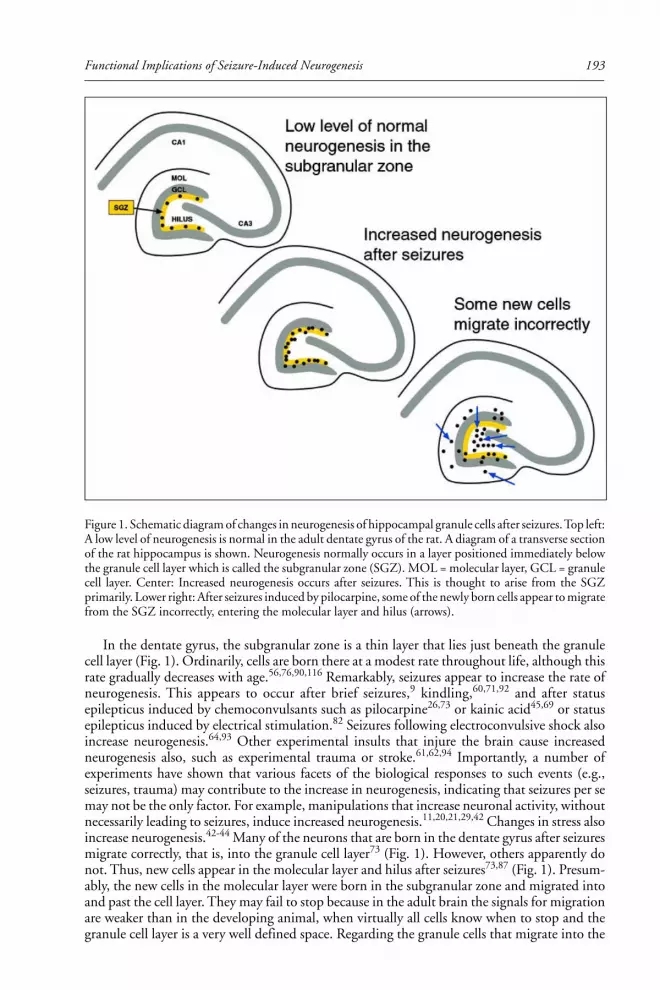
193Functional Implications of Seizure-Induced Neurogenesis
In the dentate gyrus, the subgranular zone is a thin layer that lies just beneath the granulecell layer (Fig. 1). Ordinarily, cells are born there at a modest rate throughout life, although thisrate gradually decreases with age.56,76,90,116 Remarkably, seizures appear to increase the rate ofneurogenesis. This appears to occur after brief seizures,9 kindling,60,71,92 and after statusepilepticus induced by chemoconvulsants such as pilocarpine26,73 or kainic acid45,69 or statusepilepticus induced by electrical stimulation.82 Seizures following electroconvulsive shock alsoincrease neurogenesis.64,93 Other experimental insults that injure the brain cause increasedneurogenesis also, such as experimental trauma or stroke.61,62,94 Importantly, a number ofexperiments have shown that various facets of the biological responses to such events (e.g.,seizures, trauma) may contribute to the increase in neurogenesis, indicating that seizures per semay not be the only factor. For example, manipulations that increase neuronal activity, withoutnecessarily leading to seizures, induce increased neurogenesis.11,20,21,29,42 Changes in stress alsoincrease neurogenesis.42-44 Many of the neurons that are born in the dentate gyrus after seizuresmigrate correctly, that is, into the granule cell layer73 (Fig. 1). However, others apparently donot. Thus, new cells appear in the molecular layer and hilus after seizures73,87 (Fig. 1). Presum-ably, the new cells in the molecular layer were born in the subgranular zone and migrated intoand past the cell layer. They may fail to stop because in the adult brain the signals for migrationare weaker than in the developing animal, when virtually all cells know when to stop and thegranule cell layer is a very well defined space. Regarding the granule cells that migrate into the
Figure 1. Schematic diagram of changes in neurogenesis of hippocampal granule cells after seizures. Top left:A low level of neurogenesis is normal in the adult dentate gyrus of the rat. A diagram of a transverse sectionof the rat hippocampus is shown. Neurogenesis normally occurs in a layer positioned immediately belowthe granule cell layer which is called the subgranular zone (SGZ). MOL = molecular layer, GCL = granulecell layer. Center: Increased neurogenesis occurs after seizures. This is thought to arise from the SGZprimarily. Lower right: After seizures induced by pilocarpine, some of the newly born cells appear to migratefrom the SGZ incorrectly, entering the molecular layer and hilus (arrows).

Recent Advances in Epilepsy Research194
hilus, the sites of origin may also be the subgranular zone, and in this case the cells may simplymiss the signal that tells them the correct direction to travel. Instead, they may migrate in theopposite direction than they normally would move. What is extraordinary is that they appar-ently continue to migrate in this direction, about 180o from normal, and do not stop forhundreds of microns. Apparently they do stop eventually, because clusters of cells appear toaccumulate at the border between the hilus and area CA3 (Fig. 2). Therefore, it is likely that theadult brain has a potent signal at that location that eliminates any chance that a dentate neuronwould cross into area CA3.
It is conceivable that the hilar granule cells actually don’t arise from the subgranular zone.Instead, the site of origin may be the hilus itself, because this is an area that contributes, as lateas postnatal day 10, to granule cells that are normally situated in the granule cell layer duringadulthood;2,3 vestiges of this tertiary matrix might still exist thereafter. It is not entirely clearthat this matrix converts completely to the subgranular zone at maturation. However,regardless of where in the hilus they are born, these cells still seem to migrate until the CA3/hilar border, as if they have migrated there and then must stop.
Recent evidence indicates that cells from the periphery can be transformed into cells withthe characteristics of neurons.5,47,68,105 This raises an even more heretical possibility. Some ofthe new neurons that appear in the dentate gyrus could originate from the blood cells thatenter the dentate gyrus through breaks in the blood-brain barrier during seizures. This wouldbe consistent with the empirical observation that prolonged seizures (e.g., status epilepticus)lead to more neurogenesis than weak seizures, because severe seizures would be associated withgreater breaks in the blood-brain barrier. In fact there does appear to be an increase in theendothelial cell staining of the hilus after pilocarpine-induced seizures (see chapter by Croll etal, this volume). But it is too soon to say whether these cells could become neurons.
Proving That Granule-Like Cells in the Hilus after Seizures WereNewly-Born Granule Cells
Our studies of granule-like cells in the hilus that appeared after seizures at first focused onwhether in fact they were granule cells, and the evidence that they were born after seizures. Thedata were at first anatomical: multiple cells that were immunoreactive for a neuronal marker ofgranule cells, the calcium-binding protein calbindin D28K, were present in animals that had
Figure 2. Ectopic hilar granule cells in the dentate gyrus after kainic acid-induced status epilepticus. A) Asection from a saline-injected rat demonstrates calbindin-immunoreactivity predominantly in the cellbodies, dendrites and axons of dentate gyrus granule cells. PCL = pyramidal cell layer; HIL = hilus; GCL=granule cell layer. Calibration = 200 µm. B) A section from a kainic acid-injected rat, over 1 month afterstatus epilepticus. Numerous calbindin-immunoreactive neurons surround the border of the hilus andpyramidal cell layer (arrows). Same calibration as A. Used with permission, ref. 87.

195Functional Implications of Seizure-Induced Neurogenesis
status epilepticus, but not saline-treated, age-matched controls.87 Our hypothesis, that thesewere newly-born granule cells that had somehow ended up in the hilus rather than the granulecell layer, clearly required proof. Our hypothesis stemmed from the work of Parent et al,73 whohad already shown that some of the new granule cells that arose after seizures appeared to be inthe hilus. Still, when we found very much larger numbers of calbindin-immunoreactiveneurons that were quite far from the granule cell layer, actually on the border with area CA3and the hilus (Fig. 2), it was not clear that these in fact were the same cells as shown in the studyby Parent et al,73 which were much closer to the granule cell layer.
One argument against the hypothesis was based on several papers which actually showedthat some granule cells in the normal rat do exist in the hilus.38,67 However, the number ofthese “ectopic” granule cells is extremely small, and in fact in our own saline-control tissue,there were rarely any hilar neurons that were calbindin-immunoreactive (Fig. 2). After ischemia,there appear to be some ectopic hilar cells that have the general morphology of granule cells,but again only a few.52 This contrasted with our observations in rats that had status epilepticusand recurrent seizures, where in the average 50 µm section there were often as many as 100cells (Fig. 2).
It is important to consider the validity of calbindin immunoreactivity to identify granulecells in this context. The immunoreactivity for calbindin appeared suitable to identify granulecells because, in saline-treated controls, granule cells were consistently immunoreactive forcalbindin, as indeed many others have shown.6,48,77,97 Yet calbindin is not a consistent markerof granule cells after seizures. Thus, seizures lead to a decrease in calbindin immunoreactivity insome granule cells,7,106 and this was also evident in our epileptic tissue (Fig. 1 of ref. 89).Developing granule cells also have weak labeling for calbindin,32,41,77 so the cells that wererecently born might not be immunoreactive. Therefore, calbindin may not be a faithful markerof granule cells after seizures. Thus, the number of granule neurons in the hilus after seizures isdifficult to estimate based entirely on calbindin immunoreactivity, and it could be underestimatedif many were recently born.
Based on the considerations above, it was highly unlikely that the neurons we found inpilocarpine-treated rats were simply normal granule cells that were in the wrong place.However, another possibility was that the hilar cells we surmised were granule cells because oftheir calbindin-immunoreactivity and general morphology were actually a type of hilarinterneuron. The primary argument against this was the data from saline-treated controls,which did not show many neurons in the hilus that were immunoreactive for calbindin.However, calbindin-immunoreactive hilar neurons have been reported by others.95 Further-more, some interneurons that normally have low levels of calbindin might increase expressionafter seizures. Indeed, this occurs for hilar neurons that express neuropeptide Y.91,108
Therefore, a series of anatomical experiments were conducted to address the hypothesis thatthe hilar neurons we suspected were granule cells were actually a type of interneuron. First, wefound that markers of interneurons (parvalbumin, somatostatin, GABA, GAD, calretinin) didnot coincide with the clusters of calbindin-immunoreactive, granule-like neurons in the hilus.Thus, in serial sections through the dentate gyrus, clusters of cells were present at the CA3/hilar border that were immunoreactive for calbindin, but adjacent sections failed todemonstrate hilar neurons in that location that stained for markers of GABAergic neurons.Interestingly, neuropeptide Y (NPY) did not label the hilar calbindin-immunoreactive plexus,although it would not have been surprising if it had, because NPY labels some granule cellaxons after seizures, and sometimes there is immunoreactivity in their somata.91,108 Glial fibril-lary acidic protein (GFAP), which labels adult glia, also did not label these clusters ofcalbindin-immunoreactive hilar neurons. Therefore, it was unlikely that the neurons in ques-tion were glia.
Although these results were useful, there still remained a possibility that the neurons wethought were newly-born granule cells were actually not. For example, perhaps they were VIP-or CCK-immunoreactive neurons; indeed there are so many possible types of interneurons it is

Recent Advances in Epilepsy Research196
difficult to completely test the spectrum of possibilities. Another possibility was that ourantibodies performed differently in epileptic tissue than in control sections, or identified anodd cell type that had not previously been described. Knowing that the hilus contains aremarkable number of cell types,4 and many undergo seizure-induced changes in geneexpression, questions still remained.
Perhaps the most convincing evidence came from the results of intracellular recordingsfrom slices of pilocarpine-treated rats with chronic seizures. Normally in an adult rat, or in anaged saline control, it is quite rare to encounter neurons in the hilus with electrophysiologicalcharacteristics of granule cells. Most neurons are either similar to mossy cells orinterneurons.84,85 Thus, granule cells have high resting membrane potential relative to othercell types (mossy cells, interneurons, and pyramidal cells). Strong spike frequency adaptation isanother distinguishing feature. Granule cells also have an action potential duration that isconsistent with a regular spiking neuron, the action potential has a high ratio of rate of rise torate of decay (dv/dt ratio), and there is no significant inward rectification or “sag” in responseto hyperpolarization from resting potential.84,85,87,103,104,113 Other cell types may have one oranother of these characteristics, but not all of them. Therefore, it was striking when many healthyhilar neurons were encountered that had electrophysiological characteristics of granule cells.
Furthermore, after intracellular injection of Neurobiotin into the hilar cells with theelectrophysiological “signature” of granule cells, the labeled neurons had the morphology of agranule cell. Thus, there was a small, ovoid soma, dendrites with many spines, and a mossyfiber axon. There were no thorny excrescences, consistent with the fact that these cells did nothave electrophysiological characteristics of mossy cells or pyramidal neurons. Some of the hilarcells with electrophysiology like granule cells had a polarized dendritic tree, like a normalgranule cell (Fig. 3). However, others had “basal” dendrites and their dendritic tree wastherefore bipolar. This is actually consistent with the description of granule cells in the normallocation (i.e., in the granule cell layer) in epileptic tissue, which may have basaldendrites.17,79,102,110 However, a very high percentage of hilar granule cells had basal dendritesrelative to granule cells in the granule cell layer of epileptic tissue. This difference raises thepossibility that there were fewer morphological constraints on the developing dendritic tree ofthe hilar “granule” cells as they differentiated, so they developed more basal dendrites. Insummary, these correlative electrophysiological-anatomical data provided additional evidence thatthe calbindin-immunoreactive hilar neurons observed in fixed tissue sections were granule cells.
Particularly important was the mossy fiber axon, given that some interneurons have smallovoid somata and can have spiny bipolar dendrites, somewhat similar to the bipolar hilar cellswe suspected were granule cells. But interneurons do not have a mossy fiber axon. The classicalmeaning of the term “mossy fiber” is a granule cell axon that innervates stratum lucidum,where its major branch projects to CA2 in a fiber that runs approximately 100-200 µm andparallel to the pyramidal cell layer. Along its length there are giant boutons at regular intervals,and these innervate the proximal dendrites of CA3 neurons.1,13,24,36,37 There are also manycollaterals in the hilus, where giant boutons innervate the excrescences of mossy cells, and inaddition there are smaller, more conventional terminals that innervate dendrites of severaltypes of hilar neurons. In epileptic tissue, mossy fiber axons develop additional collaterals thatproject to the inner molecular layer, referred to as “mossy fiber sprouting.” Sprouted axonscontact the dendrites of granule cells and interneurons in the inner molecular layer.55,70,111
Notably, the cells impaled in the hilus of epileptic rats that had electrophysiological features ofgranule cells had an axon with the characteristics of mossy fibers. Because no other hippocampalneurons have such an axon, it was a strong argument that the hilar cells which were impaledindeed were a type of granule cell.
Thus, the combination of electrophysiology and morphology allowed us to easilydistinguish granule cells from other cells in the hilus/CA3 region (see also refs. 84 85,86).

197Functional Implications of Seizure-Induced Neurogenesis
Taken together, the morphology and electrophysiology made a compelling case that thegranule-like cells in the hilus actually were granule cells (Fig. 3A,B; ref. 87).
What was the evidence that these hilar granule cells were in fact born after seizures? Toaddress this question, a marker of dividing cells, bromodeoxyuridine (BrdU), was used. BrdUwas injected after status epilepticus using two different schedules (1 injection per day, 50 mg/kg, for 5 days, either days 3-8 after status or 4-11). In all cases, there were cells in the hilus thatwere immunoreactive for both BrdU, which stains the nucleus, and calbindin, which stains thecytoplasm (Fig. 4). Brdu/NeuN double-labeled cells were also present (Fig. 4). However, noBrdU profiles were double-labeled with markers of GABAergic neurons (parvalbumin,neuropeptide Y; Fig. 4). In addition, BrdU injections in the hours before status epilepticus, orin saline controls, have not provided evidence of new neurons in the hilar region. Furthermore,injection long after status (over one month) for 5 days (as described above, 1 injection per day,50 mg/kg i.p.) did not result in labeled hilar neurons.
Figure 3. Morphology and electrophysiology of ectopic hilar granule cells born after seizures. A) Themorphology of a hilar granule cell from a pilocarpine-treated rat (arrowhead) is shown after intracellularinjection of Neurobiotin. For comparison, two granule cells in the granule cell layer (arrow) were alsoinjected. G = granule cell layer; H = hilus; P = pyramidal cell layer. Calibration = 100 µm. B) Physiology ofhilar granule cells. Responses to direct current injection (rectangular current pulses, as shown at the bottom)are superimposed for a granule cell located in the granule cell layer (left, “granule cell”) and a granule-likeneuron located in the hilus (right, “hilar cell”) of the same slice. This slice was from a pilocarpine-treated ratwhich had status epilepticus and recurrent seizures. A diagram of the location of these cells is shown at topright. G = granule cell layer, H = hilus, P = pyramidal cell layer. Arrowheads mark spontaneous synapticpotentials. Calibration = 20 mV, 50 msec. C) Comparison of pyramidal cell and ectopic granule cell activity.Simultaneous extracellular recordings from the pyramidal cell layer of CA3b (bottom) and intracellularrecordings are shown for a hilar granule cell (left) and a pyramidal cell (right). This slice was from apilocarpine-treated rat that had status epilepticus and recurrent seizures. A diagram of the location of the cells(dots) and extracellular recording (x) is shown at top right. The arrows point to the population spike of thespontaneous burst discharge recorded extracellularly. Calibration same as B. Used with permission, ref. 87.

Recent Advances in Epilepsy Research198
Do All the New Granule Neurons Become Functional? Do TheyBehave the Same?
Our experiments with the pilocarpine and kainic acid models suggested that hilar granulecells that are born after seizures survive for long periods of time and become functional.Evidence for their long term survival came from the studies of BrdU-labeled neurons inanimals that were not killed until over 1 year had passed since BrdU injection. In these animals,numerous BrdU-labeled neurons were found in the dentate gyrus granule cell layer as well asthe hilus, and they were double-labeled with NeuN or calbindin. Thus, neurons that dividedafter status appeared to survive as long as one year.
Regarding the development of neuronal function, intracellular recordings from hilargranule cells in slices demonstrated that they could mature into normal, functioning neuronswith action potentials and other physiological characteristics of adult neurons. A recent studyin normal adult rats has also identified that neurons which are born in the dentate gyrus in theadult animal can develop action potentials and membrane properties like normal adult granulecells.107 Therefore, in both the normal and pathological situation, granule cells that are born inthe adult rat dentate gyrus develop functional characteristics of neurons, surviving at the veryleast to maturity.
Figure 4. Ectopic hilar granule cells are born after status epilepticus. A-B: Following injection of BrdU afterstatus epilepticus, tissue sections were double-labeled for BrdU and either NeuN (A) or calbindin (B). Theresults showed numerous double-labeled neurons (arrows) at the border of the hilus and pyramidal cell layer.Calibration = 30 µm (A) and 60 µm (B). PCL = end of pyramidal cell layer. From ref. 87 with permission:C-D. In a different animal, double-labeling with BrdU and neuropeptide Y (C) or BrdU and parvalbumin(D) demonstrated no double-labeled cells, but numerous hilar neurons that were immunoreactive forneuropeptide Y or parvalbumin (arrows). Calibration (in A) = 60 µm (C) and 120 µm (D).

199Functional Implications of Seizure-Induced Neurogenesis
Additional evidence from both in vitro and in vivo studies indicates that not only are thesegranule cells able to mature into functional neurons, but they also integrate into the hostenvironment, so that they discharge in similar patterns as their neighbors.
In vivo studies used pilocarpine-treated rats at the stage when they displayed recurrent,spontaneous seizures. In these experiments, animals that had pilocarpine-induced statusepilepticus were observed approximately 1-5 months after status, during the period of timewhen recurrent spontaneous seizures occur. Three hours after a spontaneous stage 5 limbicseizure (convulsion) was observed, each animal was perfused with fixative and compared to ananimal that did not have a motor seizure in the last 8 hours, or a saline-treated, age-matchedcontrol, in which stage 5 seizures do not occur (and it is highly unlikely that subclinical seizureactivity occurs). The immediate early gene c-fos was used as a marker of neural activity: wehypothesized that if the newly-born granule cells were active during a seizure, they would stainfor c-fos, like other hippocampal neurons stain after a seizure. We thus used c-fos to mark aneuron that was active, regardless of the degree of activity, which c-fos immunoreactivitycannot describe. Indeed, in pilocarpine-treated rats with a recent seizure, there were numerousc-fos stained neurons in the hilus, and they could be double-labeled with markers of granulecells, such as calbindin and NeuN (see ref. 89; Fig. 5). There also were other cells in the hilusthat were c-fos immunoreactive, and they could be demonstrated, for example, by double-labelingc-fos and parvalbumin or c-fos and NPY.89
Interestingly, at the 3-hour delay following a seizure, granule cells located in the granule celllayer were either not immunoreactive or less immunoreactive than the hilar granule cells,presumably because they become c-fos immunoreactive sooner and their immunoreactivitywanes by 3 hours.30,40,59,115 Thus, hilar granule cells seemed to adopt a pattern of activitysimilar to their immediate neighbors, the hilar neurons, rather than the normal granule cells inthe granule cell layer. These data suggest that the development of seizure-related activity ofnewly-born granule cells is highly influenced by their neighbors and local environment,although the development of their intrinsic properties seems independent of their immediatesurroundings. Thus, there is a dissociation in the development of the interaction with hostneurons and the maturation of neuronal intrinsic properties. The data suggest that there islittle that can interfere with the membrane properties of these cells, the axon distribution, andthe general shape of the neuron (somata size and shape, dendritic patterns), but a great deal ofplasticity is possible for other variables, such as their final location and circuit-related behavior.
Figure 5. Ectopic granule cells in the hilus are active during a spontaneous seizure. Immunoreactivity forc-fos and calbindin in a pilocarpine-treated animal that had spontaneous seizures and was sacrificed 3 hoursafter a spontaneous seizure was observed. A-B) There are two double-labeled neurons in the hilus (arrow),enlarged in B. Calibration = 100 µm (A); 25 µm (B). C) Double-labeled hilar neurons in another animalare shown. Calibration (in A) = 50 µm. Used with permission, ref. 87.

Recent Advances in Epilepsy Research200
Do all newly born granule cells become functional? It is certainly not known at the presenttime that they all become mature, functional neurons. Indeed it is most likely that some do notsurvive, because there are reports that more BrdU-labeled neurons are detected after seizures ifthe animal is treated with a caspase inhibitor.31 And even if they survive, whether all becomefunctional or not is not clear. Some may develop to maturity, but others could have a differentfate. Although there is little evidence at the present time, it is possible that some become glia,interneurons, or become dormant, perhaps retaining a capacity to differentiate at a future time.
Is Increased Neurogenesis Beneficial, or Might It Actually IncreaseSeizure Susceptibility?
Another line of evidence that the hilar granule cells function differently with respect to thehost brain than normal granule cells came from in vitro studies. In these experiments,pilocarpine-treated rats that had status epilepticus and recurrent seizures were used to preparehippocampal slices, and intracellular recordings were made from hilar granule cells as well asother neurons in the slice.
Although the hilar granule cells that were recorded in slices were remarkably similar tonormal adult granule cells in their intrinsic properties (as described above), there was oneaspect of their physiology that was quite different than the activity of a normal granule cell.Many of the hilar granule cells displayed spontaneous bursts of action potentials (Fig. 3C; seeref. 87). These bursts occurred at variable frequency from slice to slice (usually ~1/10 sec), butwithin any given slice the burst frequency was consistent. Each burst was composed of avariable number of action potentials and arose on a large depolarization (Fig. 3C). Thedepolarization appeared to be a giant EPSP because it increased in amplitude with hyperpolar-ization and action potentials were triggered at its peak. We have also found subsequently thatbursts are blocked by the AMPA receptor antagonists CNQX (10 µM) but are prolonged ifexposed to the GABAA receptor antagonist bicuculline (25 µM).
Insight into the mechanism underlying these burst discharges was obtained whensimultaneous recordings were made in the area CA3 pyramidal cell layer, either intracellularlyor extracellularly (Fig. 3C). The bursts of ectopic hilar granule cells were synchronous withbursts of area CA3 neurons. Many of the recordings of extracellular CA3 bursts were small inamplitude, in all likelihood because there was some damage and loss of cells in the CA3 regiondue to status and chronic seizures. However, all CA3 pyramidal cells that were recordedintracellularly demonstrated epileptiform burst discharges that were synchronized with thesmall field bursts recorded from the CA3 cell layer, indicating it was a robust phenomenon.Interestingly, 54% of slices from animals that were examined less than 6 months after statusepilepticus did not exhibit bursts in area CA3, but 90% did if slices were from animalssacrificed >6 months after status. The delay may be due to the time required for themechanisms underlying these types of burst discharges to fully develop, perhaps becauseadditional recurrent connections that are not already present must sprout and form functionalsynapses. Some of this new circuitry may require synapses between hilar granule cells and CA3pyramidal cells. Indeed, it may be that the new synapses that are required are the new mossyfibers of the ectopic granule cells and the hilar collaterals from CA3 pyramidal neurons ontoectopic granule cells. The first would require time to form synapses on CA3 pyramidal cells, andthe latter would also require time to “find” the new hilar granule cells and form functionalsynapses. Since CA3 axons normally innervate the hilar region and some hilar neurons are lostafter seizures, the CA3 axons may be in a state that facilitates innervation of new hilar neurons.
This of course assumes that the circuitry required for burst discharges to develop arechemical synapses, but one cannot rule out the potential role of gap junctions. The mainargument for a role of chemical synapses lies in the relative timing of the bursts of CA3 neuronsand the ectopic hilar granule cells. The peak of the first action potential of a given CA3 burstbegan at least 1 millisecond before the onset of the depolarization of a simultaneously-recordedhilar granule cell, and often there was a delay far greater than 1 millisecond; if there was no

201Functional Implications of Seizure-Induced Neurogenesis
delay, gap junctions would be the more likely candidate. It is possible that ephaptic interactionsare a factor because the clusters of ectopic granule cells can be quite dense, and there may bedecreased efficacy in clearing extracellular potassium in epileptic tissue.27 However, there seemsto be quite a bit of damage in the CA3 cell layer in pilocarpine-treated rats, because it is verydifficult to obtain a healthy intracellular impalement outside of CA3b, and the field potentialsare quite small relative to control slices. This argues that ephaptic interactions would be lesslikely, at least in the CA3 cell layer.
These data address the long-standing controversy that seizures beget more seizures, aquestion that has clear implications for treating individuals who have just had their firstseizure. Especially when the individual is a child, it is not clear that anticonvulsant drugs areneeded to prevent another seizure, or desirable, given their side effects. It appears, at least in thepilocarpine model in adult rats, that the pathophysiology in the epileptic brain can indeed get“worse” as new circuits develop between excitatory neurons, in this case pyramidal cells andnewly-born granule cells. Therefore, intervention to block this recurrent excitatory circuitrycould be prudent. A major argument against this interpretation is that irradiation to blockneurogenesis after pilocarpine-induced status epilepticus did not block seizures.72 However,this may simply reflect that there are many potential foci after pilocarpine-induced status, andthat neurogenesis does not explain seizures and epilepsy completely.
Thus, the hilar-CA3 region may contribute to seizures as one of many epileptic foci. Afterstatus, the development of ectopic hilar granule cells would occur, and this would be followed,hypothetically, by the formation of circuitry between the new cells and surviving CA3 neurons.This would ultimately lead to repetitive “interictal” types of burst discharges. It could directlyfoster a transition between status and subsequent seizures.
Notably, this “focus” may actually include other cells as well, such as the hilar mossy cells thatsurvive repeated seizures.88 These neurons are glutamatergic hilar neurons that dischargesynchronously with pyramidal cells in slices of pilocarpine-treated rats. Although we cannot besure at the present time what percentage of hilar mossy cells survive status and chronic seizures,and what subset participates in pyramidal cell burst discharges, it is clear that at least some do.88
The implications would be slight if in fact these cell types, area CA3 pyramidal cells, ectopichilar granule cells, and hilar mossy cells, did not have such substantial projections to otherexcitatory neurons. However, the opposite is actually the case. CA3 projections includeadjacent CA3 neurons, hilar neurons and CA1 pyramidal cells in the normal rat, and mayinclude other cell types in the epileptic rat if sprouting of the CA3 axon occurs. CA3 alsoprojects to the contralateral hippocampus. Ectopic hilar granule cells have, at least, projectionsto the inner molecular layer and CA3 pyramidal cells, and may also innervate hilar neuronsbecause of the substantial collateralization of their axons in the hilus. These collaterals havenumerous varicosities, indicating potential synapses. The inner molecular layer innervationmay include granule cell dendrites, but it might also include processes of GABAergic neurons,analogous to the projections of sprouted mossy fibers from granule cells located in the granulecell layer. Mossy cells project to granule cells in the normal rat, both proximally and distally,ipsilateral and contralateral. They also innervate interneurons. Therefore, the initial focaldischarges between relatively small populations of pyramidal cells, ectopic granule cells andmossy cells could have substantial impact on other neuronal cell types, and have potential toexit the hippocampus.
Figure 6 shows schematically how bursts within the CA3 region, ectopic granule cells andmossy cells could lead to seizure-like activity in the hippocampus. Empirical observations sug-gest CA3 bursts would occur first because simultaneous recordings showed that the first actionpotential of a spontaneous burst in a CA3 pyramidal cell always occurred before the first actionpotential of a simultaneously recorded ectopic hilar granule cell or mossy cell. Second, ectopicgranule cells or mossy cells could excite the granule cells located in the granule cell layerbecause both project to the inner molecular layer. This would be particularly effective in depo-larizing granule cells if the ectopic neurons and mossy cells were synchronized; asynchronous

Recent Advances in Epilepsy Research202
depolarizations may not reach threshold in a granule cell, because granule cells are quitehyperpolarized normally. However, synchronous release of glutamate, particularly on theproximal portion of the granule cell dendritic tree, where both the terminals of mossy cells andectopic hilar granule cells are located, would be likely to depolarize a granule cell above itsthreshold. Action potentials in even a few granule cells would be likely to be amplified throughoutthe population because of recurrent excitatory connections within the sprouted network.Indeed, this activity may serve as a trigger for the sprouted network that normally appears to besilent. Of course, one would expect that at least some of this excitatory activity would belimited by concurrent activation of GABAergic neurons, but if interneurons are damaged orlost after seizures, as indeed appears to be the case,16,18,50 GABAergic neurons might not limitthe glutamatergic activity enough to stop its progression. The balance of excitation and inhibi-tion may shift as neuroactive peptides in the hippocampus wax and wane with the circadian
Figure 6. Schematic illustration of the development of widespread seizure activity from initial synchronizedburst discharges among CA3 pyramidal cells and ectopic hilar granule cells. 1. CA3 burst. Initially, activitythat is synchronized in area CA3 neurons develops after status epilepticus. The reasons may includeincreased recurrent excitatory activity due to loss of interneurons, as well as sprouting among residualpyramidal cells. 2. CA3 to hilus. Based on empirical findings in pilocarpine-treated rats, synchronized burstdischarges develop among pyramidal cells, newly-born hilar ectopic granule cells (EGCs) and hilar mossycells (MCs) several months after status epilepticus. This is likely to be due to the normal projection ofpyramidal cells to hilar neurons, and the mossy fiber axon that develops in EGCs. 3. hilus to GC. BecauseEGCs have mossy fiber collaterals that contribute to the sprouted fiber plexus in the inner molecular layer87,and MCs also project there, the bursts can potentially lead to activation of granule cells located in the granulecell layer. This would be limited by whatever interneurons are innervated by EGCs and MCs. 4. GC to GC.GC activation may be amplified because they are interconnected by sprouted fibers. However, this alsowould be limited by the extent that sprouted fibers activate GABAergic neurons. 5. GCs to CA3 to CA1.Strong excitatory activity that develops in the granule cell layer could potentially exit the hippocampus andtrigger a limbic seizure by the trisynaptic circuit.

203Functional Implications of Seizure-Induced Neurogenesis
rhythm.10,15,23 Indeed, there may be several times of day when GABAergic inhibition wouldnot balance the effect of glutamatergic neurons on the sprouted granule cell network. Thus,GABAergic inhibition could perhaps keep the network in control for the majority of the time,but at certain times inhibition might decrease, leading to periodic large discharges and possiblyseizures.
Thus, the focal burst discharges in CA3-hilus could generate a larger degree of reverberatoryactivity, and this might eventually leave the hippocampus via area CA3 neuronal projections toCA1. Based on the findings thus far, the results support the hypothesis that new granule cellsmay not necessarily act to ameliorate seizures, but might even contribute to them.
These circuit considerations also may have implications for the cognitive deficits followingseizures. Such deficits might in part be due to new circuits that develop between new hilargranule cells and the host brain. These new circuits would be likely to disrupt the trisynapticcircuit, and thus potentially interfere with normal learning and memory (Fig. 7A).
Figure 7. Effects of ectopic hilar granule cells on normal signal processing in the trisynaptic circuit. It isproposed that the development of ectopic hilar granule cells could contribute to cognitive deficits inepileptics by interfering with the normal trisynaptic circuit. A) The trisynaptic circuit is schematized asperforant path axons innervating distal granule cell dendrites (1), granule cell mossy fibers innervatingproximal CA3 pyramidal cell dendrites (2), and CA3 pyramidal cell projections to apical dendrites of CA1pyramidal cells (3). The accumulation of hilar ectopic granule cells with axons that innervate pyramidal cells,hilar neurons, and the inner molecular layer, suggests a multitude of complex pathways that would poten-tially interfere with normal transmission along the trisynaptic pathway, as shown diagrammatically in B.

Recent Advances in Epilepsy Research204
In Figure 7A, the trisynaptic circuit is illustrated schematically as: 1) the synapse of theperforant path onto granule cell outer dendrites; 2) the synapse of granule cell mossy fibersonto CA3 pyramidal proximal dendrites; and 3) the synapse of CA3 pyramidal Schaffer collateralsonto CA1 pyramidal apical dendrites. The activity along the trisynaptic pathway would clearlybe more circuitous by the addition of hilar granule cells, particularly given the evidence thatthese new neurons have perforant path input, pyramidal cell input, and their own mossy fiberaxons.87 Thus, information from the perforant path that would normally pass through thetrisynaptic circuit would be likely to pass through hilar granule cells. Information couldbecome “stalled” in a circular nest of intermediary pathways within the CA3-dentate region(Fig. 7B).
How Do the New Cells Interact with the Host Brain?This question actually can be addressed at a number of levels. One is the functional
relationship of the new neurons and surrounding adult neurons or “host environment,” oncethe new neurons have been born, developed, and matured. This is discussed above.
A second issue is how the new cells and host interact as the new cells are developing. Inother words, do the mature cells influence those that are newly-born, and vice-versa?
Influence of the Host Environment on Newly-Born CellsClearly the host environment normally is quite influential on new neurons. Studies in
culture have shown repeatedly that addition of specific factors to the culture medium can havea striking influence on neurodevelopment. In addition, transplantation of neurons into a newlocation often leads to the transformation of those neurons into those with characteristics ofthe new environment.
How might the characteristics of the epileptic “host” dentate gyrus influence cells that haverecently divided in the subgranular zone? One effect may be a spatial influence, because in theepileptic dentate gyrus there are fewer cells in the hilar region than normal,50 and this mightprovide an impetus to move into the hilus because there would be less competition for spacethere. Thus, hilar cell loss after seizures may facilitate the entry of new cells to the hilar region.In the normal adult hippocampus, the situation may be quite different, and indeed, newly-borngranule cells are found in the hilus under very few conditions other than those followingseizures. Furthermore, seizures without cell loss result in few new granule cells in the hilus(Scharfman et al, unpublished results).
Besides spatial cues, there is a likely chemical influence of numerous peptides, growthfactors, and other substances in the host dentate gyrus (Fig. 8). These may providechemoattractive/repulsive forces as well as supportive, proliferative and developmental influ-ence on developing neurons of the subgranular zone. Indeed, one common question of paststudies about the dentate gyrus has been the reason why so many substances that specificallyinfluence development are present in adult dentate neurons. The answer may lie in the fact thatongoing neurogenesis occurs in the adult. Thus, some of the substances in adult neurons mayaid growth and development of newly-born granule cells. These substances are even more likelyto influence newly-born neurons after seizures because they appear to increase their expressionafter seizures. Thus, although many neuroactive substances in the adult dentate gyrus modulateadult neurotransmission, they could also have another function: to modulate neurogenesis.
The neurotrophin BDNF is one example. BDNF is normally synthesized in granule cells atmuch greater concentration than other neurons in the brain,25,117 and until now the reason forthis has been unclear. In addition, the expression of BDNF increases after seizures.12,39 Whyshould granule cells possess high concentrations of BDNF? And why should this increase afterseizures? It may be due to the need for BDNF in the environment of the subgranular zone,where newly-born cells arise. Indeed, BDNF is localized in granule cell axons, not theirdendrites or soma, and the axons of course collateralize in the hilar region, where the subgranularzone exists. One might ask how BDNF is released so that it can affect newly-born cells, and this

205Functional Implications of Seizure-Induced Neurogenesis
is not clear at the present time. Although it is likely that BDNF can be released usingmechanisms common to peptide release at synapses,25 whether it can be secreted in anon-vesicular manner or transported in some other way to specifically affect newly-born cells isnot presently known.
Neuropeptide Y is another example. This peptide has been shown to promoteneuroproliferation outside the hippocampus,46 and it may do so in the dentate gyrus as well.51
It is localized to a subset of hilar GABAergic neurons normally, and after seizures its expressionincreases in those neurons.100 It also appears in granule cell axons of the hilus after seizures.66
For many years the physiological role of NPY has been unclear, and recently it has beenassociated with depression of synaptic transmission.109 These effects have been attributed to Y2and Y5 receptors, mostly.109 Yet there are also Y1 receptors in hippocampus, and specifically inthe subgranular zone as well as the molecular layer.109 What role might NPY play at its Y1receptor? The answer may be related to neurogenesis, because NPY’s proliferative effects appearto be mediated by this receptor.46,51 The new data make it seem likely that NPY has effects onsynaptic transmission as well as a role in neuroproliferation in the dentate gyrus.
Figure 8. Interaction of newly-born granule cells and the host environment. A-B) A schematic illustrates thepotential interactions between developing granule cells born after seizures and the adjacent adult (host)neurons. After seizures, new cells are born in the subgranular zone, and at a similar time, there is alteredexpression of various proteins in the surrounding adult granule cells and adult hilar neurons. The fact thatthese two phenomena occur at a similar time may be no coincidence, because the proteins may enhancegrowth, survival, and proliferation of newly-born cells. Examples include BDNF, zinc, neuropeptide Y andsomatostatin. BDNF and zinc are normally present in adult granule cells, and BDNF expression increasesin granule cells after seizures. NPY and somatostatin are present in a subset of dentate gyrus GABAergicneurons. After seizures, some of these cells increase their expression of NPY, and many somatostatin-containingneurons die. BDNF, NPY, and zinc appear to have a positive influence and somatostatin a negative influenceon processes relating to cell proliferation, so the increase in BDNF, NPY and decrease in somatostatin mayfoster phenomena associated with neurogenesis. Interestingly, these same substances also have effects onadult synaptic transmission, making their overall influence on the epileptic dentate gyrus network difficultto predict. For further discussion, see text.

Recent Advances in Epilepsy Research206
Similar arguments may be possible with other peptides in interneurons of the dentate hilusbesides NPY, such as somatostatin, which appears to influence proliferation in peripheraltissues and tumor growth.57,78,83 Interestingly, somatostatin appears to influence proliferationnegatively, and has been suggested as a potential therapy to treat cancer. Therefore, it may beno coincidence that, of all the peptidergic neurons in the dentate gyrus, the neurons whichsynthesize somatostatin are the ones that are most vulnerable to seizures.80,108 As a result,neuroproliferation after seizures may be positively modulated, i.e., by the loss of somatostatin.
Another player could very well be zinc, long known to be a native constituent of granulecells both before and after seizures. It has often been asked why granule cells require zinc, andmoreover, so much more zinc than other neurons. A potential purpose is to influenceproliferation. The basis for this hypothesis comes from studies of the immune system, wherezinc stimulates proliferation of T cells by effects on DNA synthesis75 and the cell cycle.28
Given that zinc is proliferative in the periphery, perhaps zinc also has a role in proliferation inthe dentate gyrus. Zinc could potentially influence neuroproliferation, or zinc could play a rolein glial changes, since some glia arise from the same lineage as some immune cells. Thus,analogous to NPY and BDNF, zinc may not only influence dentate gyrus physiology,19,63,96,112
but also have effects on proliferation.Finally, GABA is a potential substance that might influence newly-born granule cells of the
dentate gyrus. It is normally present in hilar interneurons and, to a lesser extent, granulecells.35,49,81 After seizures, GABA increases in both the interneurons and the granule cells34 (seealso Sperk et al, this volume). GABA has many actions that are trophic and influencedevelopment.8,65
Thus, many substances that are made in neurons of the dentate gyrus, substances previouslyassociated with an influence only on neurotransmission of adult neurons, may have animportant role in the development and synaptogenesis of newly-born granule cells. This rolemay explain the remarkable number of substances ordinarily synthesized by neurons in thedentate gyrus, and why they appear to increase expression after seizures.
An important element that is unknown presently is how the substances would be releasedinto the extracellular milieu from the axons of granule cells and other hilar neurons so that theycould influence newly-born cells; one would predict that this would be critical if they were toexert effects on immature neurons located in the hilus.
It is important to consider that some of the relevant factors may be released from astrocytes,which are known to influence neurogenesis, although the exact mechanisms are as yetunclear.99 Glia also have important roles in synaptic transmission in hippocampus.22,53,54 Afterseizures, these actions could become more potent as glia become increased in the hilar region.In addition, there is a transformation of many glia into reactive microglia, which may haveadditional functions besides those of normal glia.
Influence of New Cells on Host NeuronsOne cannot rule out the potential influence of the developing neurons in the hilus or
granule cell layer on the host hippocampus, even before they are matured completely. This isbecause immature granule cells have a few characteristics that may affect their neighbors. Oneis a high incidence of electrical coupling. A second is a chloride reversal potential that producesdepolarizing responses to GABA rather than hyperpolarizations (see Staley, this volume). Thesetwo characteristics are interesting in light of the fact that granule cells and interneurons of thedentate gyrus increase synthesis of GAD and GABA after seizures. In the case of the granulecells, it is not altogether clear when GABA is released, but it may not be through conventionalchemical neurotransmission, since the granule cells may not have the appropriate vesicular trans-porter.58,101 Instead, GABA could be released by reverse transport (see Richerson and Wu, thisvolume, see also refs. 74,114). In any case, one would predict that release of GABA onto theimmature hilar granule cells that are networked by gap junctions would lead to their synchro-nous depolarization. If this were to happen at a time when the synapses of the immature cellshad formed on host neurons, such as pyramidal cells or granule cells, it might lead to synchro-

207Functional Implications of Seizure-Induced Neurogenesis
nous activation and stimulate epileptiform activity. Indeed, it may be no coincidence that thematuration of the new granule cells in the hilus is similar in length to the so-called latentperiod, i.e., the time between status epilepticus and the first spontaneous seizure.
Is Neurogenesis Increased after Seizures in Man?As described in part above, new data now exist that suggest that neurogenesis after seizures
may play an important role in epilepsy. However, one of the areas of research that has beenpuzzling is that there has been minimal evidence for increased neurogenesis in humanepileptics. Thus, Blumcke et al14 showed that only the tissue from pediatric cases providedsome evidence consistent with laboratory animals, that neurogenesis might increase afterseizures. These data are surprising in light of the strong evidence in non-epileptics thatneurogenesis increases throughout life.33 One explanation could be that there is a differencebetween the stages of epileptogenesis. Although acute seizures or status epilepticus increaseneurogenesis in a non-epileptic brain, after repetitive seizures or the chronic condition theremay actually be a decreased rate of neurogenesis. Thus, if tissue is examined at a chronic stageof the disease, there may be little evidence of neurons that were recently-born. Another hy-pothesis is that the tissue examined thus far comes from a select population of pharmacologi-cally refractory epileptics who may not be representative of all individuals. It is also possiblethat the patients examined by Blumcke et al14 were not representative even ofmedically-intractable cases. Indeed, a different study of calbindin-immunoreactivity in humanepileptics demonstrated neurons in the hilus that had the general morphology of granule cells.98
Yet even if the phenomenon of dentate neurogenesis is not as important to human epilepsyas it appears to be in the rodent, there are other aspects of these findings that may be quiterelevant to the clinical condition. Indeed the possible abnormalities that arise from misplacedneurons in the dentate gyrus underscore that in perhaps many of the idiopathic epilepsies thereare small areas of abnormal circuits. Each might be too small for current imaging techniques torecognize, hence the use of the term idiopathic. Thus, a large subset of the idiopathic epilepsiesare actually epileptics with small migrational disorders. Whether the abnormal neurons wereborn prenatally or born in the adult brain, it points out the potential importance of under-standing every step in development. Indeed, the importance of understanding the molecularbasis of neuronal development has become a major emphasis in epilepsy research.
SummaryIn this review, the focus has been on the remarkable changes in neurogenesis and function
of adult rats after status epilepticus. A number of robust alterations of structure and functionoccur, independent of the plethora of changes already demonstrated and discussed concerningseizure-induced gene expression and mossy fiber sprouting. Our challenge will be to identifywhich changes are associated with epileptogenesis and which simply exemplify the remarkableplasticity of the epileptic brain. If this can be done, it may be possible to construct newstrategies that are tailored to block epileptogenesis and epilepsy.
AcknowledgementsI thank Annamaria Vezzani and Devin Binder for their comments on the manuscript and
Chris Hough for discussions about zinc. This work was supported by NS 37562, 38285, andthe Human Frontiers Science Program.
References1. Acsady L, Kamondi A, Sik A et al. GABAergic cells are the major postsynaptic targets of mossy
fibers in the rat hippocampus. J Neurosci 1998; 18:3386-3403.2. Altman J, Bayer SA. Migration and distribution of two populations of hippocampal granule cell
precursors during perinatal and postnatal periods. J Comp Neurol 1990; 301:365-381.3. Altman J, Bayer SA. Mosaic organization of the hippocampal neuroepithelium and the multiple
germinal sources of dentate granule cells. J Comp Neurol 1990; 301:325-342.

Recent Advances in Epilepsy Research208
4. Amaral DG. A Golgi study of cell types in the hilar region of the hippocampus in the rat. J CompNeurol 1978; 182:851-914.
5. Azizi SA. Exploiting nonneuronal cells to rebuild the nervous system: from bone marrow to brain.The Neuroscientist 2000; 6:353-361.
6. Baimbridge KG, Miller JJ. Immunohistochemical localization of calcium binding proteins in hip-pocampus, cerebellum and olfactory bulb of the rat. Brain Res 1982; 245:223-229.
7. Baimbridge KG, Mody I, Miller JJ. Reduction of rat hippocampal calcium-binding protein follow-ing commissural, amygdala, septal, perforant path, and olfactory bulb kindling. Epilepsia 1985;26:460-465.
8. Behar TN, Schaffner AE, Colton CA et al. GABA-induced chemokinesis and NGF-induced chemo-taxis of embryonic spinal cord neurons. J Neurosci 1994; 14:29-38.
9. Bengzon J, Kokaia Z, Elmer E et al. Apoptosis and proliferation of dentate gyrus neurons aftersingle and intermittent limbic seizures. Proc Natl Acad Sci USA 1997; 94:10432-10437.
10. Berchtold NC, Oliff HS, Isackson P et al. Hippocampal BDNF mRNA shows a diurnal regula-tion, primarily in the exon III transcript. Brain Res Mol Brain Res 1999; 71:11-22.
11. Bernabeu R, Sharp FR. NMDA and AMPA/kainate glutamate receptors modulate dentateneurogenesis and CA3 synapsin-I in normal and ischemic hippocampus. J Cereb Blood Flow Metab2000; 20:1669-1680.
12. Binder DK, Croll SD, Gall CM et al. BDNF and epilepsy: too much of a good thing? TrendsNeurosci 2001; 24:47-53.
13. Blackstad TW, Brink K, Hem J et al. Distribution of hippocampal mossy fibers in the rat. Anexperimental study with silver impregnation methods. J Comp Neurol 1970; 138:433-449.
14. Blumcke I, Schewe J-C, Normann S et al. Increase of nestin-immunoreactive neural precursor cellsin the dentate gyrus of pediatric patients with early-onset temporal lobe epilepsy. Hippocampus2001; 11:311-321.
15. Bova R, Micheli MR, Qualadrucci P et al. BDNF and trkB mRNAs oscillate in rat brain duringthe light-dark cycle. Brain Res Mol Brain Res 1998; 57:321-324.
16. Buckmaster PS, Dudek FE. Neuron loss, granule cell axon reorganization, and functional changesin the dentate gyrus of epileptic kainate-treated rats. J Comp Neurol 1997; 385:385-404.
17. Buckmaster PS, Dudek FE. In vivo intracellular analysis of granule cell axon reorganization inepileptic rats. J Neurophysiol 1999; 81:712-721.
18. Buckmaster PS, Jongen-Relo AL. Highly specific neuron loss preserves lateral inhibitory circuits inthe dentate gyrus of kainate-induced epileptic rats. J Neurosci 1999; 19:9519-9529.
19. Buhl EH, Otis TS, Mody I. Zinc-induced collapse of augmented inhibition by GABA in a tempo-ral lobe epilepsy model. Science 1996; 271:369-373.
20. Cameron HA, Hazel TG, McKay RDG. Regulation of neurogenesis by growth factors and neu-rotransmitters. J Neurobiol 1998; 36:287-306.
21. Cameron HA, McEwen BS, Gould E. Regulation of adult neurogenesis by excitatory input andNMDA receptor activation in the dentate gyrus. J Neurosci 1995; 15:4687-4692.
22. Castonguay A, Levesque S, Robitaille R. Glial cells as active partners in synaptic functions. ProgBrain Res 2001; 132:227-240.
23. Cirelli C, Tononi G. Gene expression in the brain across the sleep-waking cycle. Brain Res 2000;885:303-321.
24. Claiborne BJ, Amaral DG, Cowan WM. A light and electron microscopic analysis of the mossyfibers of the rat dentate gyrus. J Comp Neurol 1986; 246:435-458.
25. Conner JM, Lauterborn JC, Yan Q et al. Distribution of brain-derived neurotrophic factor (BDNF)protein and mRNA in the normal adult rat CNS: evidence for anterograde axonal transport. JNeurosci 1997; 17:2295-2313.
26. Covolan L, Ribeiro LTC, Longo BM. Cell damage and neurogenesis in the dentate granule celllayer of adult rats after pilocarpine-or kainate-induced status epilepticus. Hippocampus 2000;10:169-180.
27. D’Ambrosio R, Maris DO, Grady MS et al. Impaired K(+) homeostasis and altered electrophysi-ological properties of post-traumatic hippocampal glia. J Neurosci 1999; 19:8152-8162.
28. Dardenne M. Zinc and immune function. Eur J Clin Nutr 2002; 56 Suppl 3:S20-23.29. Derrick BE, York AD, Martinez Jr JL. Increased granule cell neurogenesis in the adult dentate
gyrus following mossy fiber stimulation sufficient to induce long-term potentiation. Brain Res 2000;857:300-307.
30. Dragunow M, Yamada N, Bilkey DK et al. Induction of immediate-early gene proteins in dentategranule cells and somatostatin interneurons after hippocampal seizures. Brain Res Mol Brain Res1992; 13:119-126.

209Functional Implications of Seizure-Induced Neurogenesis
31. Ekdahl CT, Mohapel P, Elmer E et al. Caspase inhibitors increase short-term survival ofprogenitor-cell progeny in the adult rat dentate gyrus following status epilepticus. Eur J Neurosci2001; 14:937-945.
32. Enderlin S, Norman AW, Celio MR. Ontogeny of the calcium binding protein calbindin D-28kin the rat nervous system. Anat Embryol (Berl) 1987; 177:15-28.
33. Eriksson PS, Perfilieva E, Bjork-Eriksson T et al. Neurogenesis in the adult human hippocampus.Nat Med 1998; 4:1313-1317.
34. Esclapez M, Houser CR. Up-regulation of GAD65 and GAD67 in remaining hippocampal GABAneurons in a model of temporal lobe epilepsy. J Comp Neurol 1999; 412:488-505.
35. Freund TF, Buzsaki G. Interneurons of the hippocampus. Hippocampus 1996; 6:347-470.36. Gaarskjaer FB. Organization of the mossy fiber system of the rat studied in extended hippocampi.
I. Terminal area related to number of granule and pyramidal cells. J Comp Neurol 1978; 178:49-72.37. Gaarskjaer FB. The hippocampal mossy fiber system of the rat studied with retrograde tracing
techniques. Correlation between topographic organization and neurogenetic gradients. J Comp Neurol1981; 203:717-735.
38. Gaarskjaer FB, Laurberg S. Ectopic granule cells of hilus fasciae dentatae projecting to the ipsilat-eral regio inferior of the rat hippocampus. Brain Res 1983; 274:11-16.
39. Gall CM. Seizure-induced changes in neurotrophin expression: implications for epilepsy. Exp Neurol1993; 124:150-166.
40. Gass P, Herdegen T, Bravo R et al. Spatiotemporal induction of immediate early genese in the ratbrain after limbic seizures: effects of NMDA receptor antagonist MK-801. Eur J Neurosci 1993;5:933-943.
41. Goodman JH, Wasterlain CG, Massarweh WF et al. Calbindin-D28k immunoreactivity and selec-tive vulnerability to ischemia in the dentate gyrus of the developing rat. Brain Res 1993;606:309-314.
42. Gould E, McEwen BS, Tanapat P et al. Neurogenesis in the dentate gyrus of the adult tree shrewis regulated by psychosocial stress and NMDA receptor activation. J Neurosci 1997; 17:2492-2498.
43. Gould E, Tanapat P. Stress and hippocampal neurogenesis. Biol Psychiatry 1999; 46:1472-1479.44. Gould E, Tanapat P, McEwen BS et al. Proliferation of granule cell precursors in the dentate
gyrus of adult monkeys is diminished by stress. Proc Natl Acad Sci USA 1998; 95:3168-3171.45. Gray WP, Sundstrom LE. Kainic acid increases the proliferation of granule cell progenitors in the
dentate gyrus of the adult rat. Brain Res 1998; 790:52-59.46. Hansel DE, Eipper BA, Ronnett GV. Neuropeptide Y functions as a neuroproliferative factor.
Nature 2001; 410:940-944.47. Hess DC, Hill WD, Martin-Studdard A et al. Bone marrow as a source of endothelial cells and
NeuN-expressing cells after stroke. Stroke 2002; 33:1362-1368.48. Holm IE, Geneser FA, Zimmer J et al. Immunocytochemical demonstration of the calcium-binding
proteins calbindin-D 28k and parvalbumin in the subiculum, hippocampus and dentate area of thedomestic pig. Prog Brain Res 1990; 83:85-97.
49. Houser CR, Esclapez M. Localization of mRNAs encoding two forms of glutamic acid decarboxy-lase in the rat hippocampal formation. Hippocampus 1994; 4:530-545.
50. Houser CR, Esclapez M. Vulnerability and plasticity of the GABA system in the pilocarpine modelof spontaneous recurrent seizures. Epilepsy Res 1996; 26:207-218.
51. Howell OW, Scharfman HE, Beck-Sickinger AG et al. Neuropeptide Y is neuroproliferative forhippocampal stem cells and neuroblasts. J Neurochem 2003; 86:646-659.
52. Hsu M, Buzsaki G. Vulnerability of mossy fiber targets in the rat hippocampus to forebrain is-chemia. J Neurosci 1993; 13:3964-3979.
53. Kang J, Jiang L, Goldman SA et al. Astrocyte-mediated potentiation of inhibitory synaptic trans-mission. Nat Neurosci 1998; 1:683-692.
54. Keyser DO, Pellmar TC. Synaptic transmission in the hippocampus: critical role for glial cells.Glia 1994; 10:237-243.
55. Kotti T, Riekkinen PJ, Sr., Miettinen R. Characterization of target cells for aberrant mossy fibercollaterals in the dentate gyrus of epileptic rat. Exp Neurol 1997; 146:323-330.
56. Kuhn HG, Dickinson-Anson H, Gage FH. Neurogenesis in the dentate gyrus of the adult rat:age-related decrease of neuronal progenitor proliferation. J Neurosci 1996; 16:2027-2033.
57. Kunert-Radek J, Stepien H, Radek A et al. Somatostatin suppression of meningioma cell prolifera-tion in vitro. Acta Neurol Scand 1987; 75:434-436.
58. Lamas M, Gomez-Lira G, Gutierrez R. Vesicular GABA transporter mRNA expression in the den-tate gyrus and in mossy fiber synaptosomes. Mol Brain Res 2001; 93:209-214.
59. LeGallaSalle G. Long-lasting and sequential increase of c-fos oncoprotein expression in kainicacid-induced status epilepticus. Neurosci Lett 1988; 88:127-130.

Recent Advances in Epilepsy Research210
60. Liptakova S, Jacobi H, Sperber EF. Kindling increases dentate granule neurogenesis in immaturerats. Epilepsia 1999; 40:13.
61. Liu J, Bernabeu R, Lu A et al. Neurogenesis and gliogenesis in the postischemic brain. The Neu-roscientist 2000; 6:362-370.
62. Liu J, Solway K, Messing RO et al. Increased neurogenesis in the dentate gyrus after transientglobal ischemia in gerbils. J Neurosci 1998; 18:7768-7778.
63. MacDonald RL, Kapur J. Pharmacological properties of recombinant and hippocampal dentategranule cell GABAA receptors. Adv Neurol 1999; 79:979-990.
64. Madsen TM, Treschow A, Bengzon J et al. Increased neurogenesis in a model of electroconvulsivetherapy. Biol Psychiatry 2000; 47:1043-1049.
65. Maric D, Liu QY, Maric I et al. GABA expression dominates neuronal lineage progression in theembryonic rat neocortex and facilitates neurite outgrowth via GABAA autoreceptor/Cl- channels. JNeurosci 2001; 21:2343-2360.
66. Marksteiner J, Ortler M, Bellmann R et al. Neuropeptide Y biosynthesis is markedly induced inmossy fibers during temporal lobe epilepsy of the rat. Neurosci Lett 1990; 112:143-148.
67. Marti-Subirana A, Soriano E, Garcia-Verdugo JM. Morphological aspects of the ectopic granule-likecellular populations in the albino rat hippocampal formation: a Golgi study. J Anat 1986; 144:31-47.
68. Mezey E, Chandross KJ, Harta G et al. Turning blood into brain: cells bearing neuronal antigensgenerated in vivo from bone marrow. Science 2000; 290:1779-1782.
69. Nakagawa E, Aimi Y, Yasuhara O et al. Enhancement of progenitor cell division in the dentategyrus triggered by initial limbic seizures in rat models of epilepsy. Epilepsia 2000; 41:10-18.
70. Okazaki MM, Evenson DA, Nadler JV. Hippocampal mossy fiber sprouting and synapse forma-tion after status epilepticus in rats: visualization after retrograde transport of biocytin. J CompNeurol 1995; 352:515-534.
71. Parent JM, Janumpalli S, McNamara JO et al. Increased dentate granule cell neurogenesis follow-ing amygdala kindling in the adult rat. Neurosci Lett 1998; 247:9-12.
72. Parent JM, Tada E, Fike JR et al. Inhibition of dentate granule cell neurogenesis with brain irra-diation does not prevent seizure-induced mossy fiber synaptic reorganization in the rat. J Neurosci1999; 19:4508-4519.
73. Parent JM, Yu TW, Leibowitz RT et al. Dentate granule cell neurogenesis is increased by seizuresand contributes to aberrant network reorganization in the adult rat hippocampus. J Neurosci 1997;17:3727-3738.
74. Patrylo PR, Spencer DD, Williamson A. GABA uptake and heterotransport are impaired in thedentate gyrus of epileptic rats and humans with temporal lobe sclerosis. J Neurophysiol 2001;85:1533-1542.
75. Prasad AS, Beck FW, Endre L et al. Zinc deficiency affects cell cycle and deoxythymidine kinasegene expression in HUT-78 cells. J Lab Clin Med 1996; 128:51-60.
76. Rakic P, Nowakowski RS. The time of origin of neurons in the hippocampal region of the rhesusmonkey. J Comp Neurol 1981; 196:99-128.
77. Rami A, Brehier A, Thomasset M et al. The comparative immunocytochemical distribution of 28kDa cholecalcin (CaBP) in the hippocampus of rat, guinea pig and hedgehog. Brain Res 1987;422:149-153.
78. Reisine T. Somatostatin. Cell Mol Neurobiol 1995; 15:597-614.79. Ribak CE, Tran PH, Spigelman I et al. Status epilepticus-induced hilar basal dendrites on rodent
granule cells contribute to recurrent excitatory circuitry. J Comp Neurol 2000; 428:240-253.80. Riekkinen PJ, Pitkanen A. Somatostatin and epilepsy. Metabolism 1990; 39:112-115.81. Sandler R, Smith AD. Coexistence of GABA and glutamate in mossy fiber terminals of the pri-
mate hippocampus: an ultrastructural study. J Comp Neurol 1991; 303:177-192.82. Sankar R, Shin D, Liu H et al. Granule cell neurogenesis after status epilepticus in the immature
rat brain. Epilepsia 2000; 40 (S6):134-143.83. Sara VR, Rutherford R, Smythe GA. The influence of maternal somatostatin administration on
fetal brain cell proliferation and its relationship to serum growth hormone and brain trophin activ-ity. Horm Metab Res 1979; 11:147-149.
84. Scharfman HE. Differentiation of rat dentate neurons by morphology and electrophysiology inhippocampal slices: granule cells, spiny hilar cells and aspiny ‘fast-spiking’ cells. Epilepsy Res Suppl1992; 7:93-109.
85. Scharfman HE. Electrophysiological diversity of pyramidal-shaped neurons at the granule cell layer/hilus border of the rat dentate gyrus recorded in vitro. Hippocampus 1995; 5:287-305.
86. Scharfman HE. The role of nonprincipal cells in dentate gyrus excitability and its relevance toanimal models of epilepsy and temporal lobe epilepsy. Adv Neurol 1999; 79:805-820.

211Functional Implications of Seizure-Induced Neurogenesis
87. Scharfman HE, Goodman JH, Sollas AL. Granule-like neurons at the hilar/CA3 border after statusepilepticus and their synchrony with area CA3 pyramidal cells: functional implications ofseizure-induced neurogenesis. J Neurosci 2000; 20:6144-6158.
88. Scharfman HE, Smith KL, Goodman JH et al. Survival of dentate hilar mossy cells afterpilocarpine-induced seizures and their synchronized burst discharges with area CA3 pyramidal cells.Neuroscience 2001; 104:741-759.
89. Scharfman HE, Sollas AL, Goodman JH. Spontaneous recurrent seizures after pilocarpine-inducedstatus epilepticus activate calbindin-immunoreactive hilar cells of the rat dentate gyrus. Neuro-science 2002; 111:71-81.
90. Schlessinger AR, Cowan WM, Gottlieb DI. An autoradiographic study of the time of origin andthe pattern of granule cell migration in the dentate gyrus of the rat. J Comp Neurol 1975;159:149-176.
91. Schwarzer C, Sperk G, Samanin R et al. Neuropeptides-immunoreactivity and their mRNA expres-sion in kindling: functional implications for limbic epileptogenesis. Brain Res Brain Res Rev 1996;22:27-50.
92. Scott BW, Wang S, Burnham WM et al. Kindling-induced neurogenesis in the dentate gyrus ofthe rat. Neurosci Lett 1998; 248:73-76.
93. Scott BW, Wojtowicz JM, McIntyre-Burnham W. Neurogenesis in the dentate gyrus of the ratfollowing electroconvulsive shock seizures. Exp Neurol 2000; 165:231-236.
94. Sharp FR, Liu J, Bernabeu R. Neurogenesis following brain ischemia. Dev Brain Res 2002;134:23-30.
95. Shetty AK, Turner DA. Hippocampal interneurons expressing glutamic acid decarboxylase andcalcium-binding proteins decrease with aging in Fischer 344 rats. J Comp Neurol 1998; 394:252-269.
96. Shumate MD, Lin DD, Gibbs JW, 3rd et al. GABAA receptor function in epileptic human dentategranule cells: comparison to epileptic and control rat. Epilepsy Res 1998; 32:114-128.
97. Sloviter RS. Calcium-binding protein (calbindin-D28k) and parvalbumin immunocytochemistry:localization in the rat hippocampus with specific reference to the selective vulnerability of hippoc-ampal neurons to seizure activity. J Comp Neurol 1989; 280:183-196.
98. Sloviter RS, Sollas AL, Barbaro NM et al. Calcium-binding protein (calbindin-D28K) andparvalbumin immunocytochemistry in the normal and epileptic human hippocampus. J Comp Neurol1991; 308:381-396.
99. Song H, Stevens CF, Gage FH. Astroglia induce neurogenesis from adult neural stem cells. Nature2002; 417:39-44.
100. Sperk G, Marksteiner J, Gruber B et al. Functional changes in neuropeptide Y-and somatostatin-containing neurons induced by limbic seizures in the rat. Neuroscience 1992; 50:831-846.
101. Sperk G, Schwarzer C, Hellman J. Membrane and vesicular GABA transporters after kainate-inducedseizures. Soc Neurosci Abs 2001; 551.553.
102. Spigelman I, Yan XX, Obenaus A et al. Dentate granule cells form novel basal dendrites in a ratmodel of temporal lobe epilepsy. Neuroscience 1998; 86:109-120.
103. St John JL, Rosene DL, Luebke JI. Morphology and electrophysiology of dentate granule cells inthe rhesus monkey: comparison with the rat. J Comp Neurol 1997; 387:136-147.
104. Staley KJ, Otis TS, Mody I. Membrane properties of dentate gyrus granule cells: comparison ofsharp microelectrode and whole-cell recordings. J Neurophysiol 1992; 67:1346-1358.
105. Terada N, Hamazaki T, Oka M et al. Bone marrow cells adopt the phenotype of other cells byspontaneous cell fusion. Nature 2002; 416:542-545.
106. Tonder N, Kragh J, Bolwig T et al. Transient decrease in calbindin immunoreactivity of the ratfascia dentata granule cells after repeated electroconvulsive shocks. Hippocampus 1994; 4:79-83.
107. van Praag H, Schinder AF, Christie BR et al. Functional neurogenesis in the adult hippocampus.Nature 2002; 415:1030-1034.
108. Vezzani A, Schwarzer C, Lothman EW et al. Functional changes in somatostatin and neuropeptideY containing neurons in the rat hippocampus in chronic models of limbic seizures. Epilepsy Res1996; 26:267-279.
109. Vezzani A, Sperk G, Colmers WF. Neuropeptide Y: emerging evidence for a functional role inseizure modulation. Trends Neurosci 1999; 22:25-30.
110. Wenzel HJ, Robbins CA, Tsai LH et al. Abnormal morphological and functional organization ofthe hippocampus in a p35 mutant model of cortical dysplasia associated with spontaneous seizures.J Neurosci 2001; 21:983-998.
111. Wenzel HJ, Woolley CS, Robbins CA et al. Kainic acid-induced mossy fiber sprouting and syn-apse formation in the dentate gyrus of rats. Hippocampus 2000; 10:244-260.
112. Williamson A, Spencer D. Zinc reduces dentate granule cell hyperexcitability in epileptic humans.Neuroreport 1995; 6:1562-1564.

Recent Advances in Epilepsy Research212
113. Williamson A, Spencer DD, Shepherd GM. Comparison between the membrane and synaptic prop-erties of human and rodent dentate granule cells. Brain Res 1993; 622:194-202.
114. Williamson A, Telfeian AE, Spencer DD. Prolonged GABA responses in dentate granule cells inslices isolated from patients with temporal lobe sclerosis. J Neurophysiol 1995; 74:378-387.
115. Woldbye DPD, Greisen MH, Bolwig TG et al. Prolonged induction of c-fos in neuropeptide Yand somatstatin-immunoreactive neurons of the rat dentate gyrus after electroconvulsive stimula-tion. Brain Res 1996; 720:111-119.
116. Yackel JW, Puri PS. Neurons in the rat dentate gyrus granular layer substantially increase duringjuvenile and adult life. Science 1982; 216:890-892.
117. Yan Q, Rosenfeld RD, Matheson CR et al. Expression of brain-derived neurotrophic factor proteinin the adult rat central nervous system. Neuroscience 1997; 78:431-448.

CHAPTER 15
Recent Advances in Epilepsy Research, edited by Devin K. Binder and Helen E. Scharfman.©2004 Eurekah.com and Kluwer Academic / Plenum Publishers.
Febrile Seizures and Mechanismsof Epileptogenesis:Insights from an Animal ModelRoland A. Bender, Celine Dubé and Tallie Z. Baram
Abstract
Temporal lobe epilepsy (TLE) is the most prevalent type of human epilepsy, yet thecauses for its development, and the processes involved, are not known. Most individualswith TLE do not have a family history, suggesting that this limbic epilepsy is a conse-
quence of acquired rather than genetic causes. Among suspected etiologies, febrile seizureshave frequently been cited. This is due to the fact that retrospective analyses of adults with TLEhave demonstrated a high prevalence (20->60%) of a history of prolonged febrile seizures dur-ing early childhood, suggesting an etiological role for these seizures in the development of TLE.Specifically, neuronal damage induced by febrile seizures has been suggested as a mechanismfor the development of mesial temporal sclerosis, the pathological hallmark of TLE. However,the statistical correlation between febrile seizures and TLE does not necessarily indicate a causalrelationship. For example, preexisting (genetic or acquired) ‘causes’ that result independentlyin febrile seizures and in TLE would also result in tight statistical correlation. For obviousreasons, complex febrile seizures cannot be induced in the human, and studies of their mecha-nisms and of their consequences on brain molecules and circuits are severely limited. There-fore, an animal model was designed to study these seizures. The model reproduces the funda-mental key elements of the human condition: the age specificity, the physiological temperaturesseen in fevers of children, the length of the seizures and their lack of immediate morbidity.Neuroanatomical, molecular and functional methods have been used in this model to deter-mine the consequences of prolonged febrile seizures on the survival and integrity of neurons,and on hyperexcitability in the hippocampal-limbic network. Experimental prolonged febrileseizures did not lead to death of any of the seizure-vulnerable populations in hippocampus, andthe rate of neurogenesis was also unchanged. Neuronal function was altered sufficiently topromote synaptic reorganization of granule cells, and transient and long-term alterations in theexpression of specific genes were observed. The contribution of these consequences of febrileseizures to the epileptogenic process is discussed.
Introduction: The Human ProblemAmong the epilepsies, temporal lobe epilepsy is often intractable and is associated with
significant morbidity in terms of cognitive and psychosocial dysfunction. The most commonpathology identified in resected temporal lobe tissue from patients with intractable TLE is theconstellation of mesial temporal lobe sclerosis.6 This entity is characterized by selective neu-ronal loss, gliosis and synaptic reorganization in discrete regions of the hippocampal formationand related structures.6,16,30,56 The neuroanatomical alterations of the mesial temporal lobe,
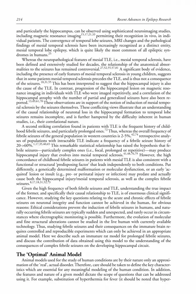
Recent Advances in Epilepsy Research214
and particularly the hippocampus, can be observed using sophisticated neuroimaging studies,including magnetic resonance imaging15,17,21,33 permitting their recognition in vivo, in indi-vidual patients. The convergence of temporal lobe seizures, MRI changes and the pathologicalfindings of mesial temporal sclerosis have been increasingly recognized as a distinct entity,mesial temporal lobe epilepsy, which is quite likely the most common of all epileptic syn-dromes in humans.25
Whereas the neuropathological features of mesial TLE, i.e., mesial temporal sclerosis, havebeen defined and extensively studied for decades, the relationship of the anatomical abnor-malities to the seizures has remained controversial.27,52,55,57,81 A significant body of evidence,including the presence of early features of mesial temporal sclerosis in young children, suggeststhat in some patients mesial temporal sclerosis precedes the TLE, and is thus not a consequenceof the seizures.18,31,55 This has been interpreted to suggest that the hippocampal injury is alsothe cause of the TLE. In contrast, progression of the hippocampal lesion on magnetic reso-nance imaging in individuals with TLE who were imaged repetitively, and a correlation of thehippocampal atrophy with the number of partial and generalized seizures have also been re-ported.13,49,61,78 These observations are in support of the notion of induction of mesial tempo-ral sclerosis by the seizures themselves. These conflicting views illustrate that an understandingof the causal relationship of neuronal loss in the hippocampal formation to temporal lobeseizures remains incomplete, and is further hampered by the difficulty inherent in humanstudies, i.e., their correlational nature.
A second striking correlation found in patients with TLE is the frequent history of child-hood febrile seizures, and particularly prolonged ones.72 Thus, whereas the overall frequency offebrile seizures of the general population in western countries is 2-5%,42,74 retrospective analy-ses of populations with intractable TLE indicate a frequency of a febrile seizure history of20->60%.1,17,35,40,65 This remarkable statistical relationship has raised the hypothesis that fe-brile seizures—particularly complex ones (i.e., focal, prolonged or repetitive)—may producehippocampal injury that evolves into mesial temporal sclerosis.41,48,76,79 However, the highconcordance of childhood febrile seizures in patients with mesial TLE is also consistent with afunctional or structural ‘predisposing factor’ that leads independently to both conditions. Putdifferently, a genetically determined malformation or molecular dysfunction, or an early ‘ac-quired’ lesion or insult (e.g., pre- or perinatal injury or infection) may predate and actuallycause both the hippocampal injury/mesial temporal sclerosis, as well as the complex febrileseizures.8,17,18,31,52,73
Given the high frequency of both febrile seizures and TLE, understanding the true impactof the former, and specifically their causal relationship to TLE, is of enormous clinical signifi-cance. However, studying the key questions relating to the acute and chronic effects of febrileseizures on neuronal integrity and function cannot be achieved in the human, for obviousreasons: Ethical considerations prevent the induction of febrile seizures in humans, and natu-rally occurring febrile seizures are typically sudden and unexpected, and rarely occur in circum-stances where electrographic monitoring is possible. Furthermore, the evolution of molecularand fine structural alterations cannot be studied in the live human with currently availabletechnology. Thus, studying febrile seizures and their consequences on the immature brain re-quires controlled and reproducible experiments which can only be achieved in an appropriateanimal model. Here we describe such an immature rat model for prolonged febrile seizures,and discuss the contribution of data obtained using this model to the understanding of theconsequences of complex febrile seizures on the developing hippocampal circuit.
The ‘Optimal’ Animal ModelAnimal models used for the study of human conditions are by their nature only an approxi-
mation of the ‘real’, actual disorder. Therefore, care should be taken to define the key character-istics which are essential for any meaningful modeling of the human condition. In addition,the features and nature of a given model dictate the scope of questions that can be addressedusing it. For example, substitution of hyperthermia for fever (it should be noted that hyper-

215Febrile Seizures and Mechanisms of Epileptogenesis: Insights from an Animal Model
thermia, typically drug-induced, is a not uncommon cause of seizures in children22,47,54) doesnot permit determination of the mechanisms involved in fever generation. Thus, it is conceiv-able that separate models might be needed to address different questions related to the samehuman problem. In addition to reproducing certain key elements of the human condition, it isadvantageous for a model to be relatively simple and inexpensive, and much should be knownabout the relevant brain structures. These considerations have led to the choice of rodents overprimates. In the following paragraphs we discuss several other characteristics of a meaningfulanimal model for prolonged febrile seizures.
Age SpecificityFebrile seizures are seen almost exclusively in infants and young children, specifically be-
tween 6 months and ~ 5 years of age, with peak incidence at ~18 months.42 An appropriate ratmodel for febrile seizures should therefore employ rats which are in a developmental age equiva-lent to the seizure-sensitive period in humans. However, data comparing rodent and humanbrain development are rare. In addition, different brain regions develop at variable rates andchronological ages, in terms of neurogenesis, migration, connectivity and function, and theseprocesses are not necessarily parallel in human and rat. These facts are important consider-ations in defining the ‘appropriate age’ of an animal model of febrile seizures.
Early, detailed studies correlated rat and human brain development based on neuronal birthdates, myelination and saltatory growth stages, and suggested that the 5-7 day old rat may be“equivalent” to the human full-term newborn.24,39 More selective comparative neuroanatomi-cal studies have focused on maturational milestones in discrete limbic regions, specifically thehippocampus and the prefrontal cortex.7,43 Overall, these converging and complementary studiessuggest that in the rat the first postnatal week may be comparable to the third trimester gesta-tional period of the human fetus, and the second postnatal week to the first year of human life.For the hippocampal formation, specifically, we have recently summarized the information oncomparative developmental milestones in human and rodent (see Table 1).7 For example, com-parison of the maturation of synaptic communication indicates that the maturational state ofthe hippocampus of a 8 day old rat4,64 is roughly equivalent to the maturational state of thehuman hippocampus at 7 months of age (see refs. 7, 71 for analysis of hippocampal neurogenesis,connectivity and maturation of select synapses). Thus, based on anatomical data, rat hippoc-ampal development during the second postnatal week seems to correspond best to the develop-mental stage at which human infants and young children are most susceptible to febrile sei-zures.
The ontogenetic profile of physiological responses of the developing rat brain to hyperther-mia supports the end of the second postnatal week as the ‘rat equivalent’ of the human suscep-tibility period for febrile seizures: The threshold temperatures required to generate experimen-tal febrile seizures are lowest during postnatal days 10-13, and rise rapidly thereafter44
(Eghbal-Ahmadi & Baram, unpublished observations). Thus, because febrile seizures are adevelopmental phenomenon confined to infants and young children, and because limbic neu-ronal circuits (and particularly the hippocampal formation) are those suspected of involvementin and vulnerability to febrile seizures, models of febrile seizures should employ developinganimals in which the stage of development of these limbic structures corresponds to the state ofmaturation of the human infant and young child, i.e., the middle of the second postnatal weekin rat, and the end of the second postnatal week in mouse.
TemperatureFebrile seizures are defined as those occurring in children with fever (rectal temperature of
at least 38.4°C) but without evidence of intracranial infection.60 In addition, it has been sug-gested that in most normal children, threshold temperatures for febrile seizures exceed ~ 41°C.50
Hence, animal models for febrile seizures should involve temperatures which are relevant to thehuman condition, and are observed in children with fever and should not rely on extremetemperatures (e.g., ref. 59). The ability to tightly regulate the temperature is an advantage of a

Recent Advances in Epilepsy Research216
hyperthermia-based model compared to fever-inducing agents. In addition, the majority ofestablished pyrogens do not induce substantive fever in the immature rat32,51 (Hatalski &Baram, unpublished observations).
The relationship of brain and core temperatures should be carefully considered. In animalmodels, core temperature is routinely monitored, rather than direct measurements of braintemperatures. It should be noted that the relationship between the two may not be consistentthroughout the range of temperatures induced to provoke a hyperthermic seizure.75 This mayresult in core temperatures that do not reflect actual brain temperatures. Therefore, calibration
Table 1. Selected milestones in hippocampal development: Human and rat
Category Event Human Rat
General Maximal growth velocity 2-3 postnatal months 8-12 postnatal daysHippocampal volume 10 postnatal months 11-30 postnatal daysapproximates that of adultHippocampal-dependent 4-5 postnatal years 15-16 postnatal dayslearning/ memory function
Neuronal Birth of pyramidal cells 1st half of gestation 2nd half of gestationFormation Onset: before 15th week ~15th day End: 24th week ~19th day
Birth of dentate gyrus (DG) ~70% prenatal (majority ~85% postnatalgranule cells by 34th week) (majority by 1st
postnatal month) Onset: 13-14th weeks ~18th prenatal day End: throughout life throughout life
Differentiation, ‘Thorny excrescences’ on postnatal years 1st postnatal monthSynaptogenesis proximal CA3 pyramidal
cell dendrites. Onset: 3-7 months ~9th day Maturation: 3-5 years 21st dayPedunculate spines: postnatal years 1st postnatal monthdistal CA3 pyramidalcell dendrites Onset: birth 7th day Maturation: 3-5 years 21st dayPeak synapse overshoot 1-2 postnatal years 9-14 postnatal days(DG)Synapse density reaches 7-10 postnatal months 21st postnatal dayadult levels: DGmolecular layerPeriod of maximal mossy 7-30 postnatal months 7-14 postnatal dayscell differentiation (DG) (adult-like by 5 years) (adult-like by 14th day)
Afferent input Entorhinal cortex toDG Prenatal: 19-20th weeks 17th prenatal daySupramammillary afferents Prenatal: ~20th week Presumed 1st postnatalreach juxtagranular and weekCA2 pyramidal cell layers
Note: Gestation lasts 270-280 and 21 days in human and rat, respectively.Modified from ref. 7, with permission.

217Febrile Seizures and Mechanisms of Epileptogenesis: Insights from an Animal Model
and standardization of parameters that influence the relationship of core and brain tempera-tures: the rate of heating, and volume, direction and diffusion of the heat, should carefully bestandardized. Ideally, chronic measurement of brain temperature should be employed, but thisis not feasible in the immature rat.
Ascertainment and Localization of the SeizuresThe behaviors induced by hyperthermia may resemble those seen during seizures, but
automatisms, stiffening and other motor phenomena may result from nonconvulsive dischargesin brainstem, basal ganglia or other subcortical regions. Therefore, electrophysiological correla-tion of such observed behaviors should be obtained from behaving animals. In infant rats,hyperthermia induces stereotyped seizure behaviors27,44 consisting of tonic body flexion ac-companied by biting and chewing (‘facial myoclonus’). These are typical for seizures of limbicorigin.9,46 Electrographic correlates should therefore be sought also in limbic regions, particu-larly in amygdala and hippocampus. In addition to pinpointing the likely source of the sei-zures, electrophysiological recording also from limbic rather than only from cortical regions isjustified by the normal sequence of maturation in these regions: cortical maturation is incom-plete during the second postnatal week in the rat, resulting typically in poorly organized andlow-voltage cortical EEG activity.9,69 It should be noted that because EEG correlations of genuinefebrile seizures in humans are exceedingly rare,59 they are not helpful in guiding placement ofelectrodes in experimental animals.
Absence of Immediate Morbidity and MortalityFebrile seizures, whether single, short and nonfocal (simple) or recurrent, more prolonged
or focal (complex) are typically not associated with immediate morbidity or mortality. There-fore, an appropriate animal model of febrile seizures should demonstrate similar low morbidityand mortality. In addition, a fundamental question related to hyperthermic seizures in theyoung human is whether they result in long-term effects, i.e., loss of hippocampal neuronsand/or alteration of hippocampal circuitry leading to epilepsy. Therefore, an optimal modelshould be suitable for long-term survival without the confounding effects of major stress, burns,infection or general moribund states. An optimal model should use a defined, benign mecha-nism for increasing brain and core temperatures, which is suitable for repeated exposures. Insummary, benign outcome not only reproduces the human situation, but permits meaningfulprospective long-term studies of the long-term consequences of these seizures on epileptogenesisand neuronal function in general.
Hyperthermic ControlsThe goals of setting up models of febrile seizures are to study the mechanisms or the out-
comes of these seizures. However, by definition, each model involves subjecting the brain tohyperthermia, to simulate fever. Therefore, the effects of the hyperthermia per se must bedistinguished from those of the associated seizures. The potential effects of hyperthermia maynot be inconsequential. Hyperthermia induces a significant number of genes (e.g., heat shockproteins), and both deleterious and protective effects on neuronal function and integrity. Forexample, hyperthermia enhances seizure severity and the neuronal loss induced by kainic acid,53
and when extreme, results in neuronal injury by itself.38 Therefore, experiments using febrileseizure models should compare three sets of animals: normothermic controls, hyperthermiccontrols, in which hyperthermia was induced, but seizures were prevented14,19,27,76 and theexperimental group which has experienced both hyperthermia and seizures.
The Immature Rat ModelBased on the criteria outlined above, a rat model for prolonged febrile seizures was devel-
oped:27,28,78 In this model, 10-11 day-old Sprague-Dawley rats are subjected to hyperthermiawhich raises body and brain temperatures gradually via a regulated stream of moderately heatedair (using a hair-dryer, low-intermediate settings, air temperature ~43°C). The air stream is

Recent Advances in Epilepsy Research218
directed ~ 30 cm above the rats, which are placed (1-2 at a time) on a towel in a 3 liter glass jar.Core temperatures are measured prior to initiating the hyperthermia, then every two minutesas well as at the onset of hyperthermia-induced seizures. These core temperatures have beenextensively correlated with brain temperatures (Eghbal-Ahmadi and Baram, unpublished ob-servations). In over 400 animals, we have found that raising core and brain temperatures to anaverage 40.88°C resulted in behavioral seizures in over 98%.
As mentioned above, the behavioral seizures are stereotyped, consisting of arrest of theheat-induced hyperkinesis, body flexion and biting of an extremity, occasionally followed byclonus. The epileptic nature of these seizures was confirmed by electrophysiological recordingfrom the hippocampi of behaving pups, using bipolar electrodes. As shown in (Fig. 1), hyper-thermia induces a change in hippocampal activity from a nonrhythmic pattern in the thetarange (Fig. 1A) to the onset of rhythmic epileptiform spikes (Fig. 1B), which correlate with theonset of behavioral seizures. In hyperthermic controls, given the rapid- and short-acting barbi-turate pentobarbital prior to the procedure, the behavioral as well as the electrophysiologicalseizures are blocked.
Animals are maintained hyperthermic (39-41.5°C) for 30 minutes, which is designed togenerate seizures lasting about 20 minutes. This reproduces the human condition of prolonged,or complex febrile seizures (defined as longer than 15 minutes, and comprising only ~10% ofall febrile seizures.12 It is these longer seizures which have been statistically implicated in thedevelopment of TLE.5,12,76 The 20-minute duration also avoids the onset of status epilepticus(defined as continuous seizures for 30 minutes), which may carry distinct implications foroutcome (see refs. 2, 23). Following the hyperthermia, animals are moved to a cool surface toregain normal body and brain temperatures, then returned to their mothers for rehydration. Itshould be noted that weighing the animals before and after the procedure indicates little evi-dence of dehydration (< 3% change in body weight). Furthermore, animals regain normalactivity rapidly after the procedure, and mortality has been <1%.
In summary, the immature rat model described above reproduces key features of prolongedfebrile seizures, those that have been statistically correlated with the development of epilepsyand/or the presence of mesial temporal sclerosis. Using this model, we have started addressingthe question of whether the relationship of these prolonged febrile seizures to neuronal loss andhyperexcitability is causal. In other words, the experiments described below query whetherprolonged experimental febrile seizures cause epilepsy. Furthermore, if these seizures do inducehippocampal hyperexcitability, what are the underlying mechanisms?
Figure 1. Electrophysiological characteristics of hyperthermia-induced seizures in immature rats. Recordswere performed via bipolar hippocampal electrodes in freely-moving 11-day-old rats. A) Baseline tracingof hippocampal activity during normothermia, showing a nonrhythmic pattern in the theta range. B) Thehyperthermia procedure provoked hippocampal electrographic seizures, manifest as trains of spike-waves.Calibration: vertical, 50 mV; horizontal 1 sec.
219Febrile Seizures and Mechanisms of Epileptogenesis: Insights from an Animal Model
Do Prolonged Experimental Febrile Seizures Increase SeizureSusceptibility?
To determine whether the susceptibility to seizures is altered after prolonged febrile sei-zures, rats were allowed to mature (three months), and then underwent extensivehippocampal-electrophysiology and behavioral seizure monitoring.27 Both the hippocampaltracings and the behavioral measures to date have failed to demonstrate the occurrence ofspontaneous seizures. However, when challenged with a sub-convulsant dose of the AMPA/kainate-type glutamate receptor agonist kainic acid, adult animals which had sustained devel-opmental febrile seizures were far more sensitive than age-matched controls to the develop-ment of further seizures. In essence, a dose that failed to provoke seizures in normothermic andhyperthermic littermate controls led to severe seizures in all adult animals which had sustainedprolonged experimental febrile seizures early in life (Fig. 2), demonstrating a ~four-fold in-creased sensitivity to kainic acid. This increased susceptibility to limbic convulsants was con-firmed in vitro:27 Spontaneous epileptiform discharges were not observed in hippocampal-entorhinalcortex slices derived from either control or experimental groups. However, Schaffer collateralstimulation induced prolonged, self-sustaining, status-epilepticus-like discharges exclusively inslices from experimental rats. These data indicate that experimental prolonged febrile seizuresdo not cause spontaneous limbic seizures during adulthood. However, they induce persistentenhancement of hippocampal excitability that may facilitate the emergence of subsequent sei-zures in response to even a mild (and perhaps not clearly demonstrable in the human situation)trigger later in life.
Figure 2. Differential induction of seizures and of status epilepticus in adult rats by low-dose kainic acid asa function of prolonged hyperthermic seizures early in life. Kainic acid led to seizures in all adult rats thathad experienced prolonged hyperthermic seizures on postnatal days 10-11 (H-seiz; n=11). The majority ofthese (n=8) developed status epilepticus. In contrast, only 2 out of 8 normothermic (N-ctl) and 1 out of 6hyperthermic control rats developed brief seizures, none of them status epilepticus (reproduced from ref.27, with permission).

Recent Advances in Epilepsy Research220
Do Prolonged Experimental Febrile Seizures Cause Neuronal Deathand/or Synaptic Reorganization?
Neuronal loss and resulting changes in hippocampal circuitry (e.g., mossy fiber sprouting)in specific hippocampal subfields are characteristic of mesial temporal sclerosis in patients withTLE (reviewed in refs. 6, 45). The loss of seizure-sensitive neuronal populations can criticallyalter the balance of excitation and inhibition in the hippocampus, which may lead to long-termhyperexcitability and a reduced seizure threshold later in life, as indeed found in this model ofprolonged febrile seizures. Therefore, we studied the short- and long-term effects of experi-mental febrile seizures on neuronal survival and synaptic connectivity.
Acute neuronal death was studied using the in situ end labeling (ISEL) technique for visu-alizing apoptotic cell death, as well as using the Gallyas silver stain method (“dark” neuron36)for visualizing neuronal injury. ISEL demonstrated no evidence for acute neuronal death in thehippocampus when studied 1, 4, 8.5, 24 or 48 hours after the seizures. However, the seizuresdid impact neuronal structure: the Gallyas method demonstrated dark, argyrophilic neuronsstarting within 24 hours and lasting as long as two weeks after the seizures. Whereas the precisemechanisms which render neurons argyrophilic are not known, selective uptake of the silverstain is considered to arise from alterations in proteins constituting the cytoskeleton. Thesechanges have often been suggested to signify cell death. However, such ‘dark’ neurons can alsobe generated by subjecting the brain to postmortem trauma, indicating that this process isindependent from the process of cell death.37 Indeed, neuronal counts carried out in the cen-tral nucleus of the amygdala, where ~30% of neurons became silver-stained after experimentalfebrile seizures, demonstrated no loss of cells.78 These findings suggest that the onset of theavidity to silver may not necessarily mean neuronal death: the changes or injury which rendera cell argyrophilic may be reversible and not lead to cell loss.
That experimental prolonged febrile seizures do not cause neuronal cell death was furtherconfirmed in a long-term study,11 in which neuronal densities in the hippocampal formationsof seizure-experiencing animals and age-matched controls were analyzed three months after theseizures. No difference was evident in the neuronal numbers of specific, seizure-sensitive hip-pocampal cell populations of these experimental groups. However, the density of the mossyfibers, the axons of granule cells, in granule cell and molecular layers was significantly increasedin seizure-experiencing compared to control rats 3 months after the seizures. These findingsindicate that despite the absence of seizure-induced neuronal loss, reorganization of the hip-pocampal circuit, evident by mossy fiber sprouting, did occur.
Do Prolonged Experimental Febrile Seizures Alter the Rateof Granule Cell Neurogenesis?
Altered neurogenesis of dentate gyrus granule cells, promoting aberrant, excitatory connec-tivity in the hippocampus, has recently been proposed as an additional mechanism by whichseizures can modulate the hippocampal network.63 Seizure-induced neurogenesis may be par-ticularly disruptive during hippocampal development, since neurogenesis in the dentate gyruspeaks during the first and second postnatal weeks.3,70 Therefore, we examined the influence ofexperimental prolonged febrile seizures on granule cell proliferation: Rats experiencing experi-mental febrile seizures and age-matched controls were injected with BrdU 3, 7 or 28 days afterthe seizures, and numbers of BrdU-labeled cells were determined 48 hrs later. No differenceswere found between seizure-experiencing and control animals at any of the time-points stud-ied.11 Thus, although granule cell neurogenesis in the immature hippocampus may be influ-enced by seizures during development,11,58,66 prolonged febrile seizures had no significant ef-fect on this process. This might be due to the relatively short duration of these seizures, or toother, as yet unresolved model-specific factors.
221Febrile Seizures and Mechanisms of Epileptogenesis: Insights from an Animal Model
Molecular Plasticity after Experimental Prolonged Febrile SeizuresElectrophysiological analyses in acute hippocampal slices from seizure-experiencing rats re-
vealed a surprising result: Despite the increased network hyperexcitability in the hippocampus,the inhibitory perisomatic drive onto CA1 pyramidal cells was increased, rather than decreased.19
Conversion of enhanced, GABA-mediated hyperpolarization into neuronal depolarization maybe mediated by activation of the hyperpolarization-activated, cyclic nucleotide-gated (Ih) cur-rent.26,62 This led to the hypothesis that the Ih-current was altered after prolonged febrile sei-zures.20,77 Indeed, whole-cell patch clamp recordings from CA1 pyramidal cells demonstratedthat the biophysical properties of the Ih-current were altered by the experimental febrile sei-zures.20 In slices from seizure-experiencing rats, the Ih-current was activated and deactivatedmuch more slowly, and its half-maximal activation (V50) was shifted towards a more depolar-ized membrane potential. Both changes opposed the increased presynaptic hyperpolarizinginput, and could convert it to a depolarizing overshoot and action potential burst firing.20,77
These changes persisted for at least 3 months.In teasing out the mechanisms which might mediate these changes of the Ih-current, it was
found that, unlike typical short-term modulation of the properties of the current, they did notdepend on alteration of cellular cyclic nucleotides. This led to the notion that this long-lastingalteration of the Ih-current might derive from transcriptional regulation of the molecules whichconstitute the h-channels: The Ih-current is generated by a specific type of channels, thehyperpolarization-activated cyclic nucleotide-gated cation channels (HCNs). Recently, fourdifferent genes encoding HCN isoforms (HCN1-4) have been discovered, each isoform form-ing channels with significantly differing physiological properties (reviewed in ref. 67). Three ofthese isoforms (HCN1, HCN2, HCN4) are expressed in CA1 pyramidal cells of the immaturerat during the age when febrile seizures can be provoked.10 The relative abundance of each ofthese isoforms in a given cell has been shown to be critical for the physiological properties ofthe channels, which, in turn, govern the overall HCN properties of the cell.34,68 In CA1 pyra-midal cells, the HCN1 isoform, forming fast activating and deactivating channels with limitedconductance, seems to be dominant under normal conditions.34,68 However, recent resultsindicate that prolonged experimental febrile seizures—occurring during a period of rapid evo-lution of the HCN isoform expression pattern10—influence the mRNA and protein expres-sion of these channel molecules. The expression of HCN1 mRNA was significantly decreasedand the expression of HCN2 mRNA significantly increased in seizure-sustaining animals byone week later.14 Both of these changes increase the relative abundance of HCN2 compared toHCN1 in a given neuron, favoring the formation of slower kinetics (and potentially larger-conductance) HCN2-homomeric channels, with altered biophysical properties. This alterationin the molecular make-up of the HCNs would promote neuronal activity-dependent depolar-ization and enhance the excitability of the hippocampal circuit.
Summary
What Has the Immature Rat Model of Prolonged Febrile Seizures TaughtUs so Far?
The original working hypotheses driving these studies of experimental febrile seizures sug-gested that these seizures would either kill vulnerable neurons, or lead only to transient injury,without long-term effects on the hippocampal circuit. As evident from the data above, bothhypotheses were refuted. The scenario emerging from the experimental data indicates that theprocess of epileptogenesis in the immature rat—the transformation of a ‘normal’ limbic net-work to a pro-epileptic one—is far more intricate and subtle than a simple composite of director compensatory changes in response to cell death.

Recent Advances in Epilepsy Research222
Indeed, the data clearly indicate that experimental prolonged febrile seizures do not resultin death of neurons in amygdala and hippocampus. Vulnerable populations, such as the mossycells or specific interneuronal subtypes, were specifically labeled and counted, and no loss orreduction in their numbers were found.11 The preservation of neuronal numbers was not dueto the birth of new neurons, since BrdU analyses demonstrated that the rate of neurogenesiswas not altered.
However, prolonged experimental febrile seizures were not “benign”. In the aftermath ofthe seizures, the hippocampus was far more susceptible to minor excitatory input (electricalcurrent in the slice, kainic acid in vivo) compared with a hippocampus not previously involvedin febrile seizures. These striking and long-lasting changes rendered the animal more likely togenerate seizures. Thus, pro-epileptogenic changes may occur without the requirement forneuronal death.
Insight into the mechanisms contributing to the hyperexcitability resulting from prolongedexperimental febrile seizures was derived from electrophysiological and molecular analyses,which, to date, have provided specific clues. Early (within hours of the seizures) regulation ofcalcium entry is modified, due to transient reduction of GluR2 expression and creation ofcalcium-permeable AMPA channels.29 By several days after the seizures, striking changes in ionchannels, specifically in the molecular make-up—and hence the kinetics and voltage-depen-dence—of the HCNs, emerge and persist long-term. These contribute significantly to conver-sion of augmented GABA-induced hyperpolarization to activity-dependent hyperexcitation.Many questions remain: How do the seizures lead to down-regulation of GluR2? What othercritical alterations result from altered calcium entry? What are the mechanisms governing HCNexpression in a spatially and temporally constrained pattern?
These and related questions are the focus of ongoing studies. These studies are carried outin the hope that they will lead to further clues and to the discovery of the specific moleculartargets, the key determinants, of this seizure-induced long-term hyperexcitability. It is the dis-covery of such specific molecular targets which could lead to the design of compounds whichwill specifically prevent the consequences of the seizures, and thus perhaps prevent thesepro-epileptogenic effects of prolonged febrile seizures.
AcknowledgmentAuthors work supported by NIH NINDS 35439 (TZB) and Epilepsy Foundation and
Milken family awards (RAB, CD).
References1. Abou-Khalil B, Andermann E, Andermann F et al. Temporal lobe epilepsy after prolonged febrile
convulsions: excellent outcome after surgical treatment. Epilepsia 1993; 34:878-883.2. Alldredge BK, Lowenstein DL. Status epilepticus: new concepts. Curr Opin Neurol 1999;
12:183-190.3. Altman J, Bayer SA. Migration and distribution of two populations of hippocampal granule cell
precursors during the perinatal and postnatal periods. J Comp Neurol 1990; 301:365-381.4. Amaral DG, Dent JA. Development of the mossy fibers of the dentate gyrus: I. A light and elec-
tron microscopic study of the mossy fibers and their expansions. J Comp Neurol 1981; 195:51-86.5. Annegers JF, Hauser WA, Shirts SB et al. Factors prognostic of unprovoked seizures after febrile
convulsions. N Engl J Med 1987; 316:493-498.6. Armstrong DD. The neuropathology of temporal lobe epilepsy. J Neuropath Exp Neurol 1993;
52:433-443.7. Avishai-Eliner S, Brunson KL, Sandman CA et al. Stressed out or in (utero)? Trends Neurosci
2002; 25:518-524.8. Baram TZ. Mechanisms and outcome of febrile seizures: What have we learned from basic science
approaches, and what needs studying? In: Baram TZ, Shinnar S, eds. Febrile seizures. San Diego,CA: Academic Press, 2002:325-328.
9. Baram TZ, Hirsch E, Snead III OC et al. Corticotropin-releasing hormone-induced seizures ininfant rats originate in the amygdala. Ann Neurol 1992; 31:488-494.

223Febrile Seizures and Mechanisms of Epileptogenesis: Insights from an Animal Model
10. Bender RA, Brewster A, Santoro B et al. Differential and age-dependent expression ofhyperpolarization-activated, cyclic nucleotide-gated cation channel isoforms 1-4 suggests evolvingroles in the developing rat hippocampus. Neuroscience 2001; 106:689-698.
11. Bender RA, Dubé C, Gonzalez-Vega R et al. Mossy fiber plasticity and enhanced hippocampalexcitability, without hippocampal cell loss or altered neurogenesis, in an animal model of pro-longed febrile seizures. Hippocampus 2002; 13:399-412.
12. Berg AT, Shinnar S. Complex febrile seizures. Epilepsia 1996; 37:126-133.13. Bower SP, Kilpatrick CJ, Vogrin SJ et al. Degree of hippocampal atrophy is not related to a
history of febrile seizures in patients with proved hippocampal sclerosis. J Neurol Neurosurg Psy-chiatry 2000; 69:733-8.
14. Brewster A, Bender RA, Chen Y et al. Developmental febrile seizures modulate hippocampal geneexpression of hyperpolarization-activated channels in an isoform and cell-specific manner. J Neurosci2002; 22:4591-4599.
15. Briellmann RS, Kalnins RM, Berkovic SF et al. Hippocampal pathology in refractory temporallobe epilepsy. T2-weighted signal change reflects dentate gliosis. Neurology 2002; 58:265-271.
16. Bruton CJ. The neuropathology of temporal lobe epilepsy (Maudsley Monographs, No 31). NewYork, NY: Oxford University Press, 1988.
17. Cendes F, Andermann F, Dubeau F et al. Early childhood prolonged febrile convulsions, atrophyand sclerosis of mesial structures and temporal lobe epilepsy. An MRI volumetric study. Neurology1993; 43:1083-1087.
18. Cendes F, Cook MJ, Watson C et al. Frequency and characteristics of dual pathology in patientswith lesional epilepsy. Neurology 1995; 45:2058-2064.
19. Chen K, Baram TZ, Soltesz I et al. Febrile seizures in the developing brain result in persistentmodification of neuronal excitability in limbic circuits. Nat Med 1999; 5:888-894.
20. Chen K, Aradi I, Thon N et al. Persistently modified h-channels after complex febrile seizuresconvert the seizure-induced enhancement of inhibition to hyperexcitability. Nat Med 2001;7:331-337.
21. Cook MJ, Fish DR, Shorvon SD et al. Hippocampal volumetric and morphometric studies infrontal and temporal lobe epilepsy. Brain 1992; 115:1001-1015.
22. Cooper AJ, Egleston C. Accidental ingestion of Ecstasy by a toddler: unusual cause for convulsionin a febrile child. J Accid Emerg Med 1997; 14:183-184.
23. Coulter DA, DeLorenzo RJ. Basic mechanisms of status epilepticus. Adv Neurol 1999; 79:725-733.24. Dobbing J, Sands J. Quantitative growth and development of human brain. Arch Dis Child 1973;
48:757-767.25. Engel Jr J, Williamson PD, Wieser HG. Mesial temporal lobe epilepsy. In: Engel Jr J, Pedley TA,
eds. Epilepsy: A comprehensive textbook. Philadelphia, PA: Lippincott-Raven Publishers,1997:2417-2426.
26. DiFrancesco D. Pacemaker mechanisms in cardiac tissue. Annu Rev Physiol 1993; 55:455-472.27. Dubé C, Chen K, Eghbal-Ahmadi M et al. Prolonged febrile seizures in the immature rat model
enhance hippocampal excitability long term. Ann Neurol 2000; 47:336-344.28. Dubé C. Do prolonged febrile seizures in an immature rat model cause epilepsy? In: Baram TZ,
Shinnar S, eds. Febrile seizures. San Diego, CA: Academic Press, 2002:215-229.29. Eghbal-Ahmadi M, Yin H, Stafstrom CE et al. Altered expression of specific AMPA type glutamate
receptor subunits after prolonged experimental febrile seizures in CA3 of immature rat hippocam-pus. Soc Neurosci Abstr 2001; 31:684.6.
30. Falconer MA, Serafetinides EA, Corsellis JAN. Etiology and pathogenesis of temporal lobe epi-lepsy. Arch Neurol 1964; 10:233-248.
31. Fernandez G, Effenberger O, Vinz B et al. Hippocampal malformation as a cause of familial febrileconvulsions and subsequent hippocampal sclerosis. Neurology 1998; 50:909-917.
32. Fewell JE, Wong VH. Interleukin-1beta-induced fever does not alter the ability of 5- to 6-day-oldrat pups to autoresuscitate from hypoxia-induced apnoea. Exp Physiol 2002; 87:17-24.
33. Fish DR, Spencer SS. Clinical correlations: MRI and EEG. Magn Reson Imaging 1995;13:1113-1117.
34. Franz O, Liss B, Neu A et al. Single-cell mRNA expression of HCN1 correlates with a fast gatingphenotype of hyperpolarization-activated cyclic nucleotide-gated ion channels (Ih) in central neu-rons. Eur J Neurosci 2000; 12:2685-2693.
35. French JA, Williamson PD, Thadani VM et al. Characteristics of medial temporal lobe epilepsy: I.Results of history and physical examination. Ann Neurol 1993; 34:774-780.
36. Gallyas F, Guldner FH, Zoltay G et al. Golgi-like demonstration of “dark” neurons with an argy-rophil III method for experimental neuropathology. Acta Neuropathol (Berlin) 1990; 79:620-628.

Recent Advances in Epilepsy Research224
37. Gallyas F, Zoltay G, Horvath Z et al. Light microscopic response of neuronal somata, dendrites,axons to postmortem concussive head injury. Acta Neuropathol (Berlin) 1992; 83:499-503.
38. Germano IM, Zhang YF, Sperber EF et al. Neuronal migration disorders increase seizure suscepti-bility to febrile seizures. Epilepsia 1996; 37:902-910.
39. Gottlieb A, Keydar I, Epstein HT. Rodent brain growth stages: an analytical review. Biol Neonate1977; 32:166-176.
40. Hamati-Haddad A, Abou-Khalil B. Epilepsy: diagnosis and localization in patients with antecedentchildhood febrile convulsions. Neurology 1998; 50:917-922.
41. Harvey AS, Grattan-Smith JD, Desmond PM et al. Febrile seizures and hippocampal sclerosis:frequent and related findings in intractable temporal lobe epilepsy of childhood. Pediatr Neurol1995; 12:201-206.
42. Hauser WA. The prevalence and incidence of convulsive disorders in children. Epilepsia 1994;35(Suppl 2) S1-S6.
43. Herschkowitz N, Kagan J, Zilles K. Neurobiological bases of behavioral development in the firstyear. Neuropediatrics 1997; 28:296-306.
44. Hjeresen DL, Diaz J. Ontogeny of susceptibility to experimental febrile seizures in rats. DevPsychobiol 1988; 21:261-275.
45. Houser CR. Neuronal loss and synaptic reorganization in temporal lobe epilepsy. Adv Neurol 1999;79:743-761.
46. Ikonomidou-Turski C, Cavalheiro EA, Turski WA et al. Convulsant action of morphine, [D-Ala2,D-Leu5] -enkephalin and naloxone in the rat amygdala: electroencephalographic, morphologicaland behavioral sequelae. Neuroscience 1987; 20:671-686.
47. Ioos C, Fohlen M, Villeneuve N et al. Hot water epilepsy: A benign and unrecognized form. JChild Neurol 2000; 15:125-128.
48. Jackson GD, McIntosh AM, Briellmann RS et al. Hippocampal sclerosis studied in identical twins.Neurology 1998; 51:78-84.
49. Kälviäinen R, Salmonperä T, Partanen K et al. Recurrent seizures may cause hippocampal damagein temporal lobe epilepsy. Neurology 1998; 50:1377-1382.
50. Knudsen FU. Febrile seizures—treatment and outcome. Brain Dev 1996; 18:438-449.51. Lagerspetz KY, Vaatainen T. Bacterial endotoxin and infection cause behavioural hypothermia in
infant mice. Comp Biochem Physiol A 1987; 88:519-521.52. Lewis DV. Febrile convulsions and mesial temporal lobe sclerosis. Curr Opin Neurol 1999;
12:197-201.53. Liu Z, Gatt A, Mikati M et al. Effect of temperature on kainic acid-induced seizures. Brain Res
1993; 631:51-58.54. Mani KS, Mani AJ, Ramesh CK et al. Hot-water epilepsy—a peculiar type of reflex epilepsy:
clinical and EEG features in 108 cases. Trans Am Neurol Assoc 1974; 99:224-226.55. Mathern GW, Babb TL, Vickrey BG et al. The clinical-pathogenic mechanisms of hippocampal
neuron loss and surgical outcomes in temporal lobe epilepsy. Brain 1995; 118:105-118.56. Mathern GW, Babb TL, Armstrong DL. Hippocampal sclerosis. In: Engel Jr J, Pedley TA, eds.
Epilepsy: A comprehensive textbook. Philadelphia, PA: Lippincott-Raven Publishers, 1997:133-155.57. Mathern GW, Pretorius JK, Leite JP et al. Hippocampal neuropathology in children with severe
epilepsy. In: Nehlig A, Motte J, Moshé SL, Plouin P, eds. Childhood epilepsies and brain develop-ment. London, England: John Libbey & Co., 1999:171-185.
58. McCabe BK, Silveira DC, Cilio MR et al. Reduced neurogenesis after neonatal seizures. J Neurosci2001; 21:2094-2103.
59. Morimoto T, Nagao H, Sano N et al. Electroencephalographic study of rat hyperthermic seizures.Epilepsy 1991; 32:289-293.
60. National Insitutes of Health Febrile seizures: Consensus development conference summary. Bethesda,MD: National Institutes of Health,1980:3(2).
61. O’Brien TJ, So EL, Meyer FB et al. Progressive hippocampal atrophy in chronic intractable tem-poral lobe epilepsy. Ann Neurol 1999; 45:526-529.
62. Pape HC. Queer current and pacemaker: the hyperpolarization-activated cation current in neurons.Annu Rev Physiol 1996; 58:299-327.
63. Parent JM, Yu TW, Leibowitz RT et al. Dentate granule cell neurogenesis is increased by seizuresand contributes to aberrant network reorganization in the adult rat hippocampus. J Neurosci 1997;17:3727-3738.
64. Ribak CE, Seress L, Amaral DG. The development, ultrastructure and synaptic connections of themossy cells of the dentate gyrus. J Neurocytol 1985; 14:835-857.

225Febrile Seizures and Mechanisms of Epileptogenesis: Insights from an Animal Model
65. Rocca WA, Sharbrough FW, Hauser WA et al. Risk factors for complex partial seizures: apopulation-based case-control study. Ann Neurol 1987; 21:22-31.
66. Sankar R, Shin D, Liu H et al. Granule cell neurogenesis after status epilepticus in the immaturerat brain. Epilepsia Suppl 2001; 7:53-56.
67. Santoro B, Tibbs GR. The HCN gene family: molecular basis of the hyperpolarization-activatedpacemaker channels. Ann NY Acad Sci 1999; 868:741-764.
68. Santoro B, Chen S, Lüthi A et al. Molecular and functional heterogeneity of hyperpolarization-activatedpacemaker channels in the mouse CNS. J Neurosci 2000; 20:5264-5275.
69. Schickerova R, Mares P, Trojan S. Correlation between electrocorticographic and motor phenom-ena induced by pentamethylenetetrazol during ontogenesis in rats. Exp Neurol 1984; 84:153-164.
70. Schlessinger AR, Cowan WM, Gottlieb ID. An autoradiographic study of the time of origin andthe pattern of granule cell migration in the dentate gyrus of the rat. J Comp Neurol 1975;159:149-176.
71. Seress L, Mrzljak L. Postnatal development of mossy cells in the human dentate gyrus: a lightmicroscopic Golgi study. Hippocampus 1992; 2:127-142.
72. Shinnar S. Prolonged febrile seizures and mesial temporal lobe sclerosis. Ann Neurol 1998;43:411-412.
73. Shinnar S. Human Data: What do we know about febrile seizures and what further information isneeded? In: Baram TZ, Shinnar S, eds. Febrile seizures. San Diego, CA: Academic Press,2002:317-324.
74. Stafstrom CE. The incidence and prevalence of febrile seizures. In: Baram TZ, Shinnar S, eds.Febrile seizures. San Diego, CA: Academic Press, 2002; 1-25.
75. Sundgren-Andersson AK, Ostlund P, Bartfai T. Simultaneous measurement of brain and core tem-perature in the rat during fever, hyperthermia, hypothermia and sleep. Neuroimmunomodulation1998; 5:241-247.
76. Theodore WH, Bhatia S, Hatta J et al. Hippocampal atrophy, epilepsy duration and febrile sei-zures in patients with partial seizures. Neurology 1999; 52:132-136.
77. Thon N, Chen K, Aradi I et al. Physiology of limbic hyperexcitability after experimental complexfebrile seizures: interactions of seizure-induced alterations at multiple levels of neuronal organiza-tion. In: Baram TZ, Shinnar S, eds. Febrile seizures. San Diego, CA: Academic Press, 2002:203-213.
78. Toth Z, Yan XX, Haftoglou S et al. Seizure-induced neuronal injury: vulnerability to febrile sei-zures in an immature rat model. J Neurosci 1998; 18:4285-4294.
79. VanLandingham KE, Heinz ER, Cavazos JE et al. Magnetic resonance imaging evidence of hip-pocampal injury after prolonged focal febrile convulsions. Ann Neurol 1998; 43:413-426.
80. Van Paesschen W, Duncan JS, Stevens JM et al. Longitudinal quantitative hippocampal magneticresonance imaging study of adults with newly diagnosed partial seizures: one-year follow-up results.Epilepsia 1998; 39:633-639.
81. Zimmerman HM. The histopathology of convulsive disorders in children. J Pediatr 1940;13:359-390.

Recent Advances in Epilepsy Research226
CHAPTER 16
Recent Advances in Epilepsy Research, edited by Devin K. Binder and Helen E. Scharfman.©2004 Eurekah.com and Kluwer Academic / Plenum Publishers.
The Tetanus Toxin Model of Chronic EpilepsyTimothy A. Benke and John Swann
Introduction
In experimental models of epilepsy, single and recurrent seizures are often used in anattempt to determine the effects of the seizures themselves on mammalian brain function.These models attempt to emulate as many features as possible of their human disease
counterparts without many of the confounding factors such as underlying disease processesand medication effects. Numerous models have been used in the past to address different ques-tions. Nevertheless, the basic questions are often the same:
1. Do seizures cause long-term damage?2. Do seizures predispose to chronic epilepsy (epileptogenesis), that is long-term spontaneous
repetitive seizures?3. Are these results developmentally regulated?4. Are the underlying mechanisms of epileptogenesis and brain damage related?
In pursuing these questions, the goal is to determine how seizures exert their effects and tominimize any side effects from the methods employed to induce the seizures themselves. Thisrequires a detailed characterization of the methods used to induce seizures.
In this chapter, we will review the literature regarding the tetanus toxin model of chronicepilepsy with regard to its mechanisms of action, clinical comparisons, how it is experimentallyimplemented and the results obtained thus far. These results will be compared to other modelsof chronic epilepsy in order to make generalizations about the effects of repetitive seizures inadult and early life. At this time, it appears that repetitive seizures cause long-term changes inlearning ability and may cause a predisposition to chronic seizures at all ages. In younger ani-mals, both features of learning impairment and epilepsy are not typically associated with cellloss as they are in adult animals. At all ages, some form of synaptic reorganization has beendemonstrated to occur.
Mechanisms of Tetanus Toxin
Clinical SyndromeTetanus is a neurological disease manifested by trismus (lockjaw) and severe generalized
muscular spasms. The syndrome is caused by the neurotoxin produced by the spore-forminganaerobic bacterium Clostridium tetani in a contaminated wound. Clostridial spores are world-wide, and ubiquitous, found in soil, dust, street dirt and human and animal feces. Spores areresistant to heat and disinfection. The bacterium itself is simply a wound contaminant, causingneither tissue destruction nor an inflammatory response.
Symptoms, caused by an exotoxin, may begin locally in areas contiguous to a wound withlocal muscle spasms. The incubation period is from 2 days to months and frequently withoutan obvious wound. Cephalic tetanus involves cranial nerves in association with head and neckwounds. Onset is gradual, typically with dysphagia and trismus (50%), occurring over 1 to 7
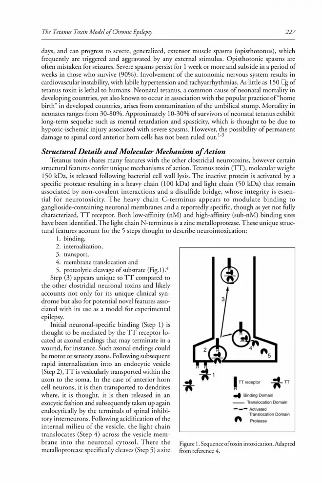
227The Tetanus Toxin Model of Chronic Epilepsy
days, and can progress to severe, generalized, extensor muscle spasms (opisthotonus), whichfrequently are triggered and aggravated by any external stimulus. Opisthotonic spasms areoften mistaken for seizures. Severe spasms persist for 1 week or more and subside in a period ofweeks in those who survive (90%). Involvement of the autonomic nervous system results incardiovascular instability, with labile hypertension and tachyarrhythmias. As little as 150 µg oftetanus toxin is lethal to humans. Neonatal tetanus, a common cause of neonatal mortality indeveloping countries, yet also known to occur in association with the popular practice of “homebirth” in developed countries, arises from contamination of the umbilical stump. Mortality inneonates ranges from 30-80%. Approximately 10-30% of survivors of neonatal tetanus exhibitlong-term sequelae such as mental retardation and spasticity, which is thought to be due tohypoxic-ischemic injury associated with severe spasms. However, the possibility of permanentdamage to spinal cord anterior horn cells has not been ruled out.1-3
Structural Details and Molecular Mechanism of ActionTetanus toxin shares many features with the other clostridial neurotoxins, however certain
structural features confer unique mechanisms of action. Tetanus toxin (TT), molecular weight150 kDa, is released following bacterial cell wall lysis. The inactive protein is activated by aspecific protease resulting in a heavy chain (100 kDa) and light chain (50 kDa) that remainassociated by non-covalent interactions and a disulfide bridge, whose integrity is essen-tial for neurotoxicity. The heavy chain C-terminus appears to modulate binding toganglioside-containing neuronal membranes and a reportedly specific, though as yet not fullycharacterized, TT receptor. Both low-affinity (nM) and high-affinity (sub-nM) binding siteshave been identified. The light chain N-terminus is a zinc metalloprotease. These unique struc-tural features account for the 5 steps thought to describe neurointoxication:
1. binding,2. internalization,3. transport,4. membrane translocation and5. proteolytic cleavage of substrate (Fig.1).4
Step (3) appears unique to TT compared tothe other clostridial neuronal toxins and likelyaccounts not only for its unique clinical syn-drome but also for potential novel features asso-ciated with its use as a model for experimentalepilepsy.
Initial neuronal-specific binding (Step 1) isthought to be mediated by the TT receptor lo-cated at axonal endings that may terminate in awound, for instance. Such axonal endings couldbe motor or sensory axons. Following subsequentrapid internalization into an endocytic vesicle(Step 2), TT is vesicularly transported within theaxon to the soma. In the case of anterior horncell neurons, it is then transported to dendriteswhere, it is thought, it is then released in anexocytic fashion and subsequently taken up againendocytically by the terminals of spinal inhibi-tory interneurons. Following acidification of theinternal milieu of the vesicle, the light chaintranslocates (Step 4) across the vesicle mem-brane into the neuronal cytosol. There themetalloprotease specifically cleaves (Step 5) a site
Figure 1. Sequence of toxin intoxication. Adaptedfrom reference 4.

Recent Advances in Epilepsy Research228
on synaptobrevin,5-7 a vesicle protein whose function is required for vesicle fusion with mem-branes. Synaptobrevin is associated with neurotransmitter-containing synaptic vesicles.8 Hence,in the case of glycinergic and GABAergic interneurons, by inhibiting the release of glycine orGABA onto anterior horn cell motor neurons, TT exerts its main clinical effects of a spasticparalysis. Since this is the main clinical effect of TT, one could assume that its effects on inhibi-tory transmission are primary and specific.
These processes have been confirmed experimentally. Experimental application of TT hasbeen shown to prevent the release of GABA in spinal cord cultures,9 hippocampal slices10,11
and cortical membrane preparations.12 Injection of TT into spinal cord and cerebellum blockssynaptic inhibition.13,14 Application of TT to spinal cord cultures results in epileptiform dis-charges associated with a presynaptic blockade of inhibitory synaptic transmission while noeffect is found on post-synaptic responsiveness to GABA or glycine.9
However, both clinical and experimental evidence suggests that the toxin’s effects are notsolely due to its interference with inhibitory synaptic transmission. Clinically, autonomic dys-function suggests that following uptake by sensory neurons, TT disrupts cholinergic neurotrans-mission. Experimentally, TT prevents not only release of GABA11 but also glutamate, aspartateand met-enkephalin release at similar concentrations.12 EEG activity is acutely suppressed fol-lowing intrahippocampal injection of TT,15 suggesting an effect on excitatory transmission. Inhippocampal cultures, TT preferentially binds to presynaptic glutamatergic terminals where itis taken up by synaptic vesicle endocytosis. Once in vesicles there, the characterized sequenceof toxin action follows, resulting in cytosolic cleavage of synaptobrevin.16
The actions of TT may not be entirely due to a presynaptic blockade of vesicular fusion.Since synaptobrevin is suggestively located on vesicles associated with insertion of membranereceptors,17,18 TT could also prevent their insertion in postsynaptic membranes. Applicationof TT to Torpedo organ results in not only a reduction in miniature endplate potential fre-quency, but also miniature endplate potential size7. Indeed, intracellular application of TT canprevent post-synaptic insertion of both GABA19 and glutamate17,20 receptors in hippocampalneurons. However, responses to exogenous GABA are not altered in hippocampus followingintrahippocampal TT injection, though these studies investigated only somatic GABA recep-tors on pyramidal neurons.21 Thus, the ultimate effects of TT depend on transport withinneurons and their associated neuronal networks as well as the half-life of the toxin.
Trans-synaptic transfer appears limited to “single-jumps”. That is, following initial uptakeand transport, TT moves across a single synapse to then exert its effect. This appears to be thecase for horseradish peroxidase-conjugated heavy chain TT fragment when injected intramus-cularly. In this case, it was found to be present in motor neurons and to eventually accumulatefollowing trans-synaptic transfer in inhibitory terminals but did not continue beyond fromthere.22 That is, TT did not then move to inhibitory somata and beyond. Similar results werefound at cholinergic terminals in spinal preparations.23 In both of these studies, some TT wasobserved extrasynaptically and found to persist for several days at the sites of synaptic transfer,leaving the possibility for further intoxication of these and other local neurons as possible.Following injection of TT into the CA3 region of hippocampus, transport would be expectedto at least encompass the projection of axon terminals and dendrites present in the CA3 region(Fig. 2). This would include mossy fiber terminals from dentate granule neurons, associationfiber terminals from CA3 pyramidal neurons and commissural axons from the contralateralCA3. Dentate granule neurons follow a laminar projection, while CA3 associational and Schaffercollateral axons to CA1, project diffusely to both dorsal and ventral hippocampus (ipsilateral).
Theoretically, following transport to CA3 cell bodies, TT could also be further transportedvia Schaffer commissural axons projecting to the contralateral CA1 and CA3 associationalregions.24 Following intrahippocampal injection of the conjugated heavy chain TT fragment,transport was noted into the contralateral hippocampal CA3 region but not in either ventralhippocampus or in cortex.25 However, it is not clear if the presence of the light chain mayfacilitate multi-synaptic transfer. This could take place at any infected terminal. Indeed, upon
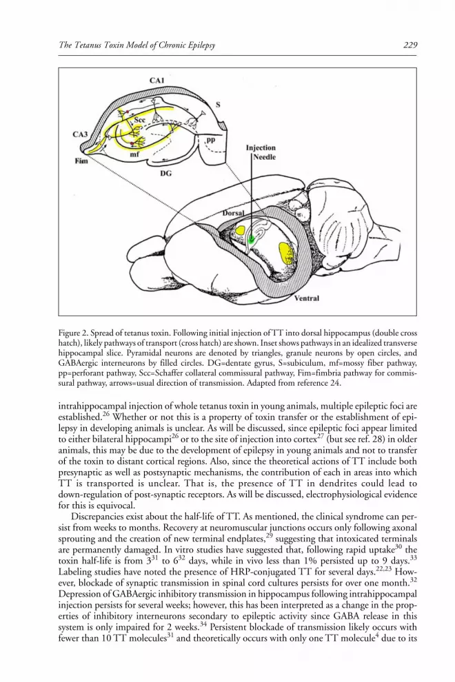
229The Tetanus Toxin Model of Chronic Epilepsy
intrahippocampal injection of whole tetanus toxin in young animals, multiple epileptic foci areestablished.26 Whether or not this is a property of toxin transfer or the establishment of epi-lepsy in developing animals is unclear. As will be discussed, since epileptic foci appear limitedto either bilateral hippocampi26 or to the site of injection into cortex27 (but see ref. 28) in olderanimals, this may be due to the development of epilepsy in young animals and not to transferof the toxin to distant cortical regions. Also, since the theoretical actions of TT include bothpresynaptic as well as postsynaptic mechanisms, the contribution of each in areas into whichTT is transported is unclear. That is, the presence of TT in dendrites could lead todown-regulation of post-synaptic receptors. As will be discussed, electrophysiological evidencefor this is equivocal.
Discrepancies exist about the half-life of TT. As mentioned, the clinical syndrome can per-sist from weeks to months. Recovery at neuromuscular junctions occurs only following axonalsprouting and the creation of new terminal endplates,29 suggesting that intoxicated terminalsare permanently damaged. In vitro studies have suggested that, following rapid uptake30 thetoxin half-life is from 331 to 632 days, while in vivo less than 1% persisted up to 9 days.33
Labeling studies have noted the presence of HRP-conjugated TT for several days.22,23 How-ever, blockade of synaptic transmission in spinal cord cultures persists for over one month.32
Depression of GABAergic inhibitory transmission in hippocampus following intrahippocampalinjection persists for several weeks; however, this has been interpreted as a change in the prop-erties of inhibitory interneurons secondary to epileptic activity since GABA release in thissystem is only impaired for 2 weeks.34 Persistent blockade of transmission likely occurs withfewer than 10 TT molecules31 and theoretically occurs with only one TT molecule4 due to its
Figure 2. Spread of tetanus toxin. Following initial injection of TT into dorsal hippocampus (double crosshatch), likely pathways of transport (cross hatch) are shown. Inset shows pathways in an idealized transversehippocampal slice. Pyramidal neurons are denoted by triangles, granule neurons by open circles, andGABAergic interneurons by filled circles. DG=dentate gyrus, S=subiculum, mf=mossy fiber pathway,pp=perforant pathway, Scc=Schaffer collateral commissural pathway, Fim=fimbria pathway for commis-sural pathway, arrows=usual direction of transmission. Adapted from reference 24.

Recent Advances in Epilepsy Research230
enzymatic nature. The long duration of poisoning is likely due to its compartmentalizationand a lack of cytosolic proteases capable of destroying TT.30,34 Thus, interpretation of resultsusing TT in experimental models of epilepsy must seek to separate those results that could bedue to a persistent action of TT from those due to the seizures themselves.
Experimental Implementation
Goals of Model DevelopmentIntra-cortical injection of tetanus toxin as a means of inducing seizures was first demon-
strated over a century ago.35 The technique re-emerged and was developed in the 1960s and1970s as an experimental tool for studying epilepsy. Its development became directed by theneed for an animal model that exhibited a semi-chronic epileptic syndrome in order to explorethe effect of epileptic discharges on cortical function. That is, the goal was to develop an animalmodel with spontaneous daily seizures for several weeks that then abated so that the effect ofchronic seizures versus the acute seizures themselves on cortical function could be studied.Such functions included mood, memory, recurrent seizures and the mechanisms involved intheir generation. Thus, given the persistence of the mechanism of tetanus toxin, it seems anideal agent. However, as is often argued, it often becomes difficult to determine which of theeffects observed in this model are due to the toxin or the seizures themselves. As it arises, thiswill be mentioned.
Laboratory personnel are typically immunized with tetanus toxoid and anti-titers are veri-fied prior to studies. In general, the method requires an appropriate humane animal anestheticfollowed by stereotactic injection of the toxin into the brain region of choice under asepticconditions (see Fig. 2). Injection sites are accessed by means of a burr hole. Typically only asingle injection of a very small volume (nanoliters) is given. However, in some instances, mul-tiple or bilateral injections are performed and in some cases this is done with the semi-permanentimplantation of a cannula.28,36 Following wound closure and care, animals are returned totheir cages. The dose of toxin is often noted in either amount in nanograms or in terms ofmouse LD50. In older studies, a mouse LD50 was defined as the amount required to kill half ofa group of mice injected intramuscularly within 7 days (104 mouse LD50s are equivalent to 1rat LD50).15 Typically the equivalent of a few mouse LD50s are used; 1 ng is approximately 3mouse LD50s.28 Currently, an MD100, defined as the minimal effective dose (0.25 ng) requiredto elicit hind limb paralysis in all animals following intramuscular injections in adult mice, hasbeen used to assess potency.26
Chronic Epilepsy in Adult Animals
Intra-Hippocampal Injections of TT
OverviewThe usage and consequences of intra-hippocampal (typically into CA3 region) TT injec-
tions in adult (21 days and older) rats have been reviewed.11,37,38 Studies have employed and,where noted, included bilateral and unilateral injections of TT into either dorsal or ventralhippocampus. The findings are updated and summarized here with respect to a behavioral andseizure description, correlation with EEG findings, duration and severity of epilepsy and invitro electrophysiological findings. This is followed by a summary of the pathological andbiochemical findings. Long-term behavioral consequences to animals are discussed. The sug-gested mechanistic schemes for these findings are reviewed.
Seizures, EEG Correlates and BehaviorInjected animals begin to have seizures 1-2 days following the injection. Seizures in adult
rats typically involve a behavioral arrest followed by focal or generalized myoclonus or facial

231The Tetanus Toxin Model of Chronic Epilepsy
twitching. Occasionally they begin in sleep. Head rearing and rearing on hind feet follows this.Occasionally the animals have so-called “wild running fits”. The ictal stage lasts 25 to 75 sec-onds and is followed by a post-ictal stage of unresponsiveness lasting up to 4 minutes followedby resumption of normal activities.15 Intracortical recordings obtained within hours followinginjection show that both evoked excitation and inhibition is impaired near the site of injec-tion.39 (This is also seen in vitro following acute application of very high doses of TT to hip-pocampal slices.40) One to two days later, the EEG shows epileptiform activity arising from theinjected hippocampus initially and later bilaterally. Electrographic seizures appear to begin ineither hippocampus with bursts of 3-20 Hz spike-wave activity that subsequently generalize.The non-injected hippocampus often develops larger discharges during the ictus and may dis-charge independently following an ictus.41 Following the electrographic seizure, relative elec-trical silence is noted coinciding with the time the animal is unresponsive post-ictally. Interictalspike frequency increases as the number of seizures increases. Animals have 4-5 seizures perhour for 4 weeks, however this varies around roughly a 4-day cycle and most seizures occurduring the afternoon. One week after injection, some of the hippocampal discharges occurwithout any overt behavioral seizure.41 After 5-7 weeks, the EEG returns to normal and sei-zures desist.15,37 Rarely (<10%), seizures or EEG abnormalities may recur or persist.11 In aradial arm maze-learning task, rats learned more slowly than controls long after seizures hadabated.42 Rats developed life-long aggressiveness and hyperactivity soon after TT injection.15
Further in Vivo and in Vitro Electrophysiological CharacterizationWhile initial studies into the electrophysiological characterization of the TT model in-
volved in vivo hippocampal recordings,42 the use of the hippocampal in vitro slice preparationhas allowed a more thorough characterization of the nature and origin of the epileptic dis-charges and their persistence following TT injection. The TT model is unusual in this regardsince most models of epilepsy do not retain epileptic discharges in vitro.43 Three major find-ings are apparent. First, hippocampal CA3 pyramidal neurons show permanently decreasedsynaptic excitability by mossy fiber (from ipsilateral dentate granule neurons) and commissural(from contralateral CA3 pyramidal neurons) inputs 4-6 months following bilateral hippocam-pal TT injections. This is apparently not due to diminished intrinsic excitability since antidro-mic population spikes are normal.42 Similar findings were found at 16 weeks following unilat-eral TT injections.21 Interestingly, given that these animals had impaired learning, these sameanimals did not have impairment of long-term potentiation (LTP) in the pathway of commis-sural inputs to CA3 neurons.42 LTP is thought to be an in vitro correlate of learning andmemory.44 Conversely, permanently enhanced excitability of dentate gyrus granule neurons(determined by larger, single orthodromic population spikes) and CA1 pyramidal neurons(determined by the presence of multiple orthodromic population spikes) is present ipsilaterallyand contralaterally to unilateral TT injections.21 Second, spontaneous epileptiform activityarising from the CA3 region is temporarily present in vitro at similar times to the observedepileptic syndrome, that is, up to 7 weeks following bilateral TT injection43 and up to, but notbeyond, 16 weeks following unilateral TT injection.21 This epileptic activity is present bilater-ally even following unilateral TT injection45 and may be more consistently present contralaterallyto TT injection.34 Third, synaptic inhibition in CA3 and dentate gyrus is temporarily abol-ished in the hippocampus ipsilateral to the TT injection site for up to 8 weeks following injec-tion.21,46 Synaptic inhibition contralateral to TT injection, while not abolished, is apparentlydiminished somewhat, and it was found that the stimuli required to generate monosynapticfast IPSPs were twice that found compared to control slices. This suggested that interneuronswere either less excitable (intrinsically or extrinsically) and/or that they were less efficient inGABA release.34 Such network alterations may explain the permanent changes in excitabilitynoted in CA1 and dentate gyrus. Concurrent studies by these authors suggested that GABArelease contralateral to TT injection was normal since they observed that GABA release ipsilat-eral to the injection was transiently impaired only in the first two weeks following injection.34

Recent Advances in Epilepsy Research232
Regardless, inhibition is apparently only impaired up to 8 weeks following TT injection, whileepileptic activity persists beyond this but it too diminishes finally 16 weeks following TTinjection.21
Neuropathological CorrelatesThe TT model of epilepsy in adult rats is unique in that it is not associated with apparently
significant pyramidal neuron loss compared to other models.15 Remote from the injection site,there is bilateral 10% cell loss in CA1 following unilateral injection of TT into dorsal hippoc-ampus and 30% cell loss in CA1 following unilateral injection of TT into ventral hippocam-pus47 (and reviewed in38). Consistent with some degree of cell loss, microglial activation isnoted in these regions.47 Much larger doses of TT than are normally used do apparently causepyramidal neuron loss at the site of injection.48 However, morphologically it has been brieflyreported that there is dendritic spine loss on CA3 pyramidal neurons near the site of injectionnoted 4 weeks following TT injection.49 CA1 pyramidal neurons ipsilateral to the site of injec-tion demonstrate a higher incidence of dye coupling, presumably via gap junctions along withsimplified apical and basal dendritic arbors.50 Aberrant mossy fibers that have apparently sproutedenter the outer and inner molecular layers of the dentate gyrus and are noted bilaterally 4 weeksfollowing unilateral TT injection.51 Expression of mRNA encoding specific glutamate recep-tor isoforms is transiently elevated bilaterally in a specific pattern (GluR1 flip in dentate gyrusand GluR2 flip in all hippocampal sub-fields) at 4 weeks following unilateral TT injections.52
While presumably this could underlie increased excitability, the significance of this is specula-tive given the unknown function of these specific glutamate receptor isoforms. Disparate con-clusions have been obtained in an attempt to address whether increased excitability is mediatedby a loss of inhibitory interneurons. In a study utilizing interneuron-specific expression ofglutamic acid decarboxylase (GAD), inhibitory interneurons are apparently unaffected andmay exhibit a transient up-regulation of GAD.53 Others have observed a selective, bilateral lossof somatostatin-immunoreactive interneurons in the hilus of the dentate gyrus noted at 8weeks following unilateral TT injection.54
Effects of Anti-Convulsant DrugsTwo studies have looked at the effect of anti-convulsant drugs on the development of sei-
zures in the adult TT model. The first study28 was different from others in that it involvedmultiple unilateral intrahippocampal injections of TT in cats. The resulting seizures were sosevere, that if control was not attempted with phenobarbital, the animals succumbed to statusepilepticus. This study, despite its limited size, suggested that if seizures were initially control-lable with phenobarbital, this led to remission of seizures. However, in those animals that hadremission of seizures, repetitive seizures could be re-induced with a single injection of TT thatrequired life-long phenobarbital for control for the resulting chronic epileptic syndrome. Im-portantly, it has not been determined whether or not treatment with anti-convulsants resultedin normal performance in learning tasks or minimization of any of the neuropathological changesfound.
In a subsequent study,55 adult rats were pretreated with either intrahippocampal TT orkainate, with or without suppression of seizures by phenobarbital, followed by a treatmentwith intrahippocampal TT. Animals pretreated with kainate were more likely to develop chronicspontaneous repetitive seizures, regardless of seizure suppression by phenobarbital, and in cor-relation with damage to the CA3 region of the hippocampus. All animals that received only TTtreatment (with and without phenobarbital treatment) recovered from their epileptic syndrome.Treatment with phenobarbital during the pretreatment with either kainate or TT did result ina less severe epileptic syndrome during the subsequent administration of TT. This study didnot investigate the presence of subclinical seizures nor did it ascertain performance in learn-ing tasks.

233The Tetanus Toxin Model of Chronic Epilepsy
Intra-Cortical Injections of TTCompared to intra-hippocampal injections of TT, injections of TT into adult rat neocortex
result in a more severe and prolonged epileptic syndrome. Seizures, abnormal EEG and in vitroepileptiform activity are present for greater than 7 months following injection. These abnor-malities are noted locally and diffusely, presumably due to transport of TT.27 Similar findingshave been found following injections of TT into cat motor cortex where it has been noted thatno significant cell loss is present.36 Similar to rat hippocampus, injection of TT into neocortexresults in a transient depression of GABA release only ipsilateral to the injection.56 Also, neo-cortical injection of TT is associated with a focal up-regulation of GAD mRNA, as well asmRNA for type II calcium/calmodulin-dependent protein kinase, GluR257 and brain-derivedneurotrophic factor (BDNF) which may play a role in aberrant neuronal sprouting.58 Whileinjection of TT into other brain regions has been attempted, these have been only brieflydescribed.15
DiscussionThe conclusion of many studies has suggested that the epileptic syndrome that follows TT
injection is due to impaired inhibition. However, investigators have argued that impaired inhi-bition is a consequence of the seizures themselves and not directly due to the toxin itself. Thisargument is based on the observations that the measured half-life of the toxin is short, release ofGABA is impaired only locally and in a transient fashion that mirrors the toxin half-life, thecontralateral hippocampus which has only mildly impaired inhibition has more dramatic epi-leptic discharges in vitro, epileptic discharges persist after the recovery of inhibition and, whenseizures are controlled with anti-convulsant drugs, the syndrome is prevented. It is still unclearif there is a central nervous system counterpart to the recovery from TT in the peripheralnervous system that requires axonal sprouting and the creation of new terminal endplates.29
The mechanism(s) of denervation (loss of CA3 dendritic spines) are equally unclear. It is thoughtthat focal loss of CA3 dendritic spines could underlie impaired CA3 excitation and perfor-mance in learning tasks. This has not been studied thoroughly, however. Since it is known thatTT inhibits insertion of postsynaptic glutamate receptors,17,18 and that blockade of this pro-cess can lead to spine retraction,59 a direct effect is theoretically possible. Perhaps by studyingdendritic spines in other hippocampal regions such as CA1 or dentate gyrus this could beclarified. Despite these reservations, the observations found in young animals suggest that it isthe impact of recurrent seizures that induces the epileptic syndrome.
Chronic Epilepsy in Young Animals
Intra-Hippocampal Injections of TT
OverviewThe use of intra-hippocampal injections of TT in young animals (9-11 days old) has been
performed in our laboratory. These have always involved single, unilateral injections of low-doseTT into the dorsal CA3 region. The findings are summarized here with respect to a behavioraland seizure description, correlation with EEG findings, duration and severity of epilepsy andin vitro electrophysiological findings. This is followed by a summary of the neuropathologicalfindings. Long-term behavioral consequences to animals are discussed. The suggested mecha-nistic schemes for these findings coupled with that found in adult animals are presented.
Seizures, EEG Correlates and BehaviorSuppression of background activity was observed in EEG recordings in the first 12-24 hours
following TT injection. Multifocal spikes from either hippocampi or neocortex developed inthe next 24 hours. Behavioral seizures, described as “wild-running” or “wet-dog shakes”, began24-72 hours following TT injection, were correlated with EEG recordings, and were found to

Recent Advances in Epilepsy Research234
arise either focally in the injected hippocampus or bilaterally. At their peak, approximately 1-3days following TT injection, as many as 4-5 wild-running seizures per hour were observed.Subsequently most seizures were sub-clinical in nature after 4 weeks. While only 20% of ani-mals might be observed to have behavioral seizures in adulthood, when these animals werereexamined in adulthood (up to 24 weeks following TT injection) with EEG, nearly all hadabnormal EEGs consisting of either brief bursts of fast generalized spike-wave activity or briefbursts of multifocal interictal spikes, typically without behavioral correlate.26,60 When tested asadults, TT-injected animals learned the Morris water maze more slowly compared to controlanimals, findings which were independent of EEG abnormalities and any impairment in mo-tor function.25
Further in Vitro Electrophysiological CharacterizationMost hippocampal slices obtained from rats injected with TT in infancy exhibited epilepti-
form bursting from both the ipsilateral and contralateral CA3 region; this was independent ofthe time following (up to 10 weeks) TT injection.26,61 Such bursting was not present in CA1,however some slices exhibited unusual rhythmic (2-8 Hz) IPSPs. Membrane properties of CA3pyramidal neurons were similar to control animals. Blockade of IPSPs in TT-injected ratsinduced rhythmic epileptiform bursting in both contralateral and ipsilateral hippocampi com-pared to rhythmic single spikes in control animals suggesting excitation was enhanced in TT-injected animals.61
Neuropathological CorrelatesInjection of TT in infancy shares many findings with that in adulthood with a few quanti-
tative differences. No total cell loss was noted in any hippocampal region,26 however there wasfound to be a dispersion of stratum pyramidale that was more prominent in the CA1 regionand could not be explained by gliosis.25 Aberrant mossy fiber sprouting was also found bilater-ally following TT injection.60 Dendritic spine loss along apical and basal dendrites of pyrami-dal neurons in CA3 has been quantified and found to exist bilaterally while axons from thesame CA3 neurons appear normal with regards to length and branching pattern.62
Effects of Anti-Convulsant DrugsThe effect of the anti-convulsant carbamazepine in infant rats following unilateral TT in-
jection in infancy was markedly different from that found in adult animals.63 While able tosignificantly limit the number of observed seizures, sub-clinical seizures persisted, though stillfewer than that found in control animals. Upon withdrawal of carbamazepine, behavioral sei-zures recurred and subsequently abated with a similar, though delayed, time course comparableto animals not treated with carbamazepine. This suggests that the seizures themselves appear tolimit their recurrence during development. Longer-term evaluation of these animals has notyet been performed to determine if treated animals had normalization of their EEGs, as wasfound in the study on adult animals. Again, as in adult animals, it has not been determinedwhether or not treatment with anti-convulsants resulted in normal performance in learningtasks or minimization of any of the neuropathological changes found.
DiscussionThe persistent epileptic state in rats following unilateral hippocampal injection of TT in
infancy suggests two important conclusions. First, since the epileptic state persists well beyondany conceivable persistence of TT action (at 24 weeks, assuming a half-life of 7 days, only onemolecule of TT would remain), it is the effect of persistent seizures that causes the chronicepileptic state. Second, the developing brain is more sensitive to early-life seizures to cause thechronic epileptic state. An important distinction with the seizures in adult rats is noted, in that,recurrent seizures in adult rats do not necessarily predispose to chronic epilepsy unless, asrecent reports have suggested,55 recurrent seizures induced by TT are coupled with other agentsor treatments (kainate,55 hypoxia64) that cause hippocampal neuronal loss. Next, it is impor-

235The Tetanus Toxin Model of Chronic Epilepsy
tant to determine how recurrent seizures induced by TT in both adult and infant rats lead tolearning impairment. While it appears that it is the effect of chronic seizures, one could alsoargue that it is the damage caused by TT. Mossy fiber sprouting is a common abnormalityfound in most animal models of epilepsy and is not likely to be a consequence of TT itself. Thisoccurs even in the infant model, and its presence bilaterally following unilateral TT injectionsuggests it is secondary to the seizures and not TT. Sprouting may also explain the increase inneuropil found in CA1 regions. Once the TT model is compared to other models of epilepsy,a common denominator emerges in that in different models, early-life seizures lead to not onlychronic epilepsy and similar neuropathological changes but also learning impairment.
Comparisons to Other Models of Chronic EpilepsyWhile clear distinctions exist in other models of chronic epilepsy between infant and adult
animals, these share many of the features of the TT model. For example, followingkainate-induced status epilepticus in adult animals, there are spontaneous repetitive seizures,CA3 cell loss, mossy fiber sprouting into CA3 and dentate gyrus, sprouting into CA1 stratumpyramidale and stratum radiatum, and impaired learning in memory tasks.65-67 However, ifanimals younger than 14-21 days old are treated, the animals are apparently unaffected, evenfollowing repetitive treatments.68 Newer studies now suggest that while kainate treatment inanimals younger than 14 days has neither neuropathological sequelae nor induction of sponta-neous repetitive seizures, it is found to be associated with diminished LTP, learning task perfor-mance, kindling rate and increased inhibition in the dentate gyrus.69 Furthermore, kainateinsult in infancy and again later in adulthood results in more prominent memory impairmentthan a single insult at either time.70 Similar findings are noted with lithium-pilocarpine-inducedstatus epilepticus.71-73 However, as suggested by kainate, repetitive episodes of status epilepticusin infancy caused by lithium-pilocarpine are not benign. Following 3 episodes of status epilepticusin this model, rats develop recurrent electrographic seizures (most of which were subclinical),increased CA1 excitability, and diminished learning, all without significant neuropathologicalchanges such as cell loss (however mossy fiber sprouting was not assessed).74-76 This is verysimilar to the findings of the chronic epileptic syndrome induced by TT in infancy and sug-gests that repetitive early-life seizures are a common mechanism capable of creating a chronicepileptic state that includes memory impairment without cell loss. Indeed, it is not just theseizures themselves, it is the molecular pathways they trigger, as has been demonstrated by theco-application of an NMDA receptor antagonist (MK-801) with lithium-pilocarpine in adultanimals. In this case, status epilepticus was not prevented but all of the sequelae were.77
The effects of early-life seizures are certainly model-dependent. In the fluorothyl-model,early-life seizures are associated with impaired learning and mossy fiber sprouting but notspontaneous repetitive seizures.78,79 In the hyperthermia model of febrile seizures, a singleprolonged seizure results in life-long susceptibility to convulsants, enhanced inhibition in CA1,enhanced in vitro kindling, and enhancement of the membrane conductance Ih, with subse-quent hyperexcitability in CA1.80-82 Following a prolonged hypoxia-induced seizure, experi-mental animals develop acute seizures and a chronic reduced seizure threshold to convulsants,but neither mossy fiber sprouting nor cognitive disturbances. This model has now been shownto be associated with increased expression of calcium-permeable glutamate receptors, howeverthe exact role of this in the pathogenesis of this model is unclear.83 Status epilepticus inducedby kainate in infant rats does not result in mossy fiber sprouting but does result in impairedLTP, impaired kindling and impaired learning associated with enhanced inhibition in the den-tate gyrus.69
Thus, while each of these models has distinct mechanisms in the induction of seizures, theircommon outcome suggests that it is directed by the repetitive experience of the seizures them-selves. Since there are likely many mechanisms that could lead to spontaneous repetitive sei-zures and/or learning impairment (similar to the clinical experience that there are many causesof epilepsy), seizures are the likely trigger of any or all of these pathways that could be favoredby one treatment or another.

Recent Advances in Epilepsy Research236
References1. Tetanus. In: Pickering LK, ed. Red Book: Report of the Committee on Infectious Diseases. Elk
Grove: American Academy of Pediatrics, 2000:563-568.2. Volpe JJ. Bacterial and fungal intracranial infections. In: Neurology of the Newborn. Philadelphia:
W.B. Saunders, 2001:774-810.3. Anaerobic infections. In Johnson KB, Oski FA, eds. Oski’s Essential Pediatrics. Philadelphia:
Lippincott-Raven, 1997:209-212.4. Rossetto O, Seveso M, Caccin P et al. Tetanus and botulinum neurotoxins: turning bad guys into
good by research. Toxicon 2001; 39:27-41.5. Hohne-Zell B, Ecker A, Weller U et al. Synaptobrevin cleavage by the tetanus toxin light chain is
linked to the inhibition of exocytosis in chromaffin cells. FEBS Letters 1994; 355:131-134.6. Schiavo G, Benfenati F, Poulain B et al. Tetanus and botulinum-B neurotoxins block neurotrans-
mitter release by proteolytic cleavage of synaptobrevin. Nature 1992; 359:832-834.7. Herreros J, Miralles FX, Solsona C et al. Tetanus toxin inhibits spontaneous quantal release and
cleaves VAMP/synaptobrevin. Brain Res 1995; 699:165-170.8. Rothman JE. Mechanisms of intracellular protein transport. Nature 1994; 372:55-63.9. Bergey GK, MacDonald RL, Habig WH et al. Tetanus toxin: convulsant action on mouse spinal
neurons in culture. J Neurosci 1983; 3:2310-2323.10. Collingridge GL, Davies J. The in vitro inhibition of GABA release by tetanus toxin.
Neuropharmacol 1982; 21:851-855.11. Mellanby J. Tetanus toxin as a tool for investigating the consequences of excessive neuronal excita-
tion. In: DasGupta BR, ed. Botulinum and Tetanus Neurotoxins. New York: Plenum, 1993:291-297.12. McMahon HT, Foran P, Dolly JO et al. Tetanus toxin and botulinum toxins type A and B inhibit
glutamate, gamma-aminobutyric acid, aspartate, and met-enkephalin release from synaptosomes.Clues to the locus of action. J Biol Chem 1992; 267:21338-21343.
13. Curtis DR, de Groat WC. Tetanus toxin and spinal inhibition. Brain Res 1968; 10:208-212.14. Curtis DR, Felix D, Game CJA et al. Tetanus toxin and the synaptic release of GABA. Brain Res
1973; 51:358-362.15. Mellanby J, George G, Robinson A et al. Epileptiform syndrome in rats produced by injecting
tetanus toxin into the hippocampus. J Neurol Neurosurg Psychiatry 1977; 40:404-414.16. Matteoli M, Verderio C, Rossetto O et al. Synaptic vesicle endocytosis mediates the entry of teta-
nus neurotoxin into hippocampal neurons. Proc Natl Acad Sci USA 1996; 93:13310-13315.17. Lledo PM, Zhang X, Sudhof TC et al. Postsynaptic membrane fusion and long-term potentiation.
Science 1998; 279:399-403.18. Lin JW, Sheng M. NSF and AMPA receptors get physical. Neuron 1998; 21:267-270.19. Ouardouz M, Sastry BR. Mechanisms underlying LTP of inhibitory synaptic transmission in the
deep cerebellar nuclei. J Neurophysiol 2000; 84:1414-1421.20. Lu W-Y, Man H-Y, Ju W et al. Activation of synaptic NMDA receptors induces membrane inser-
tion of new AMPA receptors and LTP in cultured hippocampal neurons. Neuron 2001; 29:243-254.21. Whittington MA, Jefferys JGR. Epileptic activity outlasts disinhibition after intrahippocampal teta-
nus toxin in the rat. J Physiol 1994; 481:593-604.22. Fishman PS, Savitt JM. Transsynaptic transfer of retrogradely transported tetanus protein-peroxidase
conjugates. Exp Neurol 1989; 106:197-203.23. Schwab ME, Suka K, Thoenen H. Selective retrograde transsynaptic transfer of a protein, tetanus
toxin, subsequent to its retrograde axonal transport. J Cell Biol 1979; 82:798-810.24. Amaral DG, Witter MP. The three-dimensional organization of the hippocampal formation: a re-
view of anatomical data. Neuroscience 1989; 31:571-591.25. Lee CL, Hannay J, Hrachovy R et al. Spatial learning deficits without hippocampal neuronal loss
in a model of early-onset epilepsy. Neuroscience 2001; 107:71-84.26. Lee CL, Hrachovy RA, Smith KL et al. Tetanus toxin-induced seizures in infant rats and their
effects on hippocampal excitability in adulthood. Brain Res 1995; 677:97-109.27. Brener K, Amitai Y, Jefferys JGR et al Chronic epileptic foci in neocortex: In vivo and in vitro
effects of tetanus toxin. Eur J Neurosci 1991; 3:47-54.28. Darcey TM, Williamson PD. Chronic/semichronic limbic epilepsy produced by microinjection of
tetanus toxin in cat hippocampus. Epilepsia 1992; 33:402-419.29. Duchen LW, Tonge DA. The effects of tetanus toxin on neuromuscular transmission and on the
morphology of motor end-plates in slow and fast skeletal muscle of the mouse. J Physiol 1973;228:157-172.
30. Critchley DR, Nelson PG, Habig WH et al. Fate of tetanus toxin bound to the surface of primaryneurons in culture: evidence for rapid internalization. J Cell Biol 1985; 100:1499-1507.

237The Tetanus Toxin Model of Chronic Epilepsy
31. Erdal E, Bartels F, Binscheck T et al. Processing of tetanus and botulinum A neurotoxin in iso-lated chromaffin cells. Naunyn Schmiedebergs Arch Pharmacol 1995; 351:67-78.
32. Habig WH, Bigalke H, Bergey GK et al. Tetanus toxin in dissociated spinal cord cultures: long-termcharacterization of form and action. J Neurochem 1986; 47:930-937.
33. Mellanby JH. Elimination of 125I from rat brain after injection of small doses of 125I-labelledtetanus toxin into the hippocampus. Neurosci Lett Suppl 1989; Suppl 36:S55
34. Empson RM, Jefferys JGR. Synaptic inhibition in primary and secondary chronic epileptic fociinduced by intrahippocampal tetanus toxin in the rat. J Physiol 1993; 465:595-614.
35. Roux E, Borrel A. Tetanos cerebral et immunite contre le tetanos. Ann Inst Pasteur 1898; 4:225-239.36. Louis ED, Williamson PD, Darcey TM. Chronic focal epilepsy induced by microinjection of teta-
nus toxin into the cat motor cortex. Electroencephalogy Clin Neurophysiol 1990; 75:548-557.37. Mellanby J, Hawkins C, Mellanby H et al. Tetanus toxin as a tool for studying epilepsy.
J Physiol (Paris) 1984; 79:207-215.38. Jefferys JGR, Whittington MA. Review of the role of inhibitory neurons in chronic epileptic foci
induced by intracerebral tetanus toxin. Epilepsy Res 1996; 26:59-66.39. Sundstrom LE, Mellanby JH. Tetanus toxin blocks inhibition of granule cells in the dentate gyrus
of the urethane-anaesthetized rat. Neuroscience 1990; 38:621-627.40. Calabresi P, Beneditti M, Mercuri NB et al. Selective depression of synaptic transmission by teta-
nus toxin: a comparative study on hippocampal and neostriatal slices. Neuroscience 1989;30:663-670.
41. Mellanby J, Strawbridge P, Collingridge GL et al. Behavioural correlates of an experimental hip-pocampal epileptiform syndrome in rats. J Neurol Neurosurg Psychiatry 1981; 44:1084-1093.
42. Brace HM, Jefferys JGR, Mellanby J. Long-term changes in hippocampal physiology and learningability of rats after intrahippocampal tetanus toxin. J Physiol (Lond) 1985; 368:343-357.
43. Jefferys JGR. Chronic epileptic foci in vitro in hippocampal slices from rats with tetanus toxinepileptic syndrome. J Neurophysiol 1989; 62:458-468.
44. Bliss T, Collingridge G. A synaptic model of memory: long-term potentiation in the hippocampus.Nature 1993; 361:31-39.
45. Jefferys JGR, Empson RM. Development of chronic secondary epileptic foci followingintrahippocampal injection of tetanus toxin in the rat. Exp Physiol 1990; 75:733-736.
46. Jordan SJ, Jefferys JGR. Sustained and selective block of IPSPs in brain slices from rats madeepileptic by intrahippocampal tetanus toxin. Epilepsy Res 1992; 11:119-129.
47. Shaw JAG, Perry VH, Mellanby J. Tetanus toxin-induced seizures cause microglial activation in rathippocampus. Neurosci Lett 1990; 120:66-69.
48. Bagetta G, Nistico G, Bowery NG. Prevention by the NMDA receptor antagonist, MK801 ofneuronal loss produced by tetanus toxin in the rat hippocampus. Br J Pharmac 1990; 101:776-780.
49. Hughes JT, Mellanby J. Experimental epilepsy induced by tetanus toxin injected into the rat hip-pocampus: a Golgi study. Neuropath Appl Neurobiol 1985; 11:73
50. Colling SB, Man WD-C, Draguhn A et al. Dendritic shrinkage and dye-coupling between rathippocampal CA1 pyramidal cells in the tetanus toxin model of epilepsy. Brain Res 1996; 741:38-43.
51. Mitchell J, Gatherer M, Sundstrom LE. Aberrant Timm-stained fibres in the dentate gyrus follow-ing tetanus toxin-induced seizures in the rat. Neuropath Appl Neurobiol 1996; 22:129-135.
52. Rosa MLNM, Jefferys JGR, Sanders MW et al. Expression of mRNAs encoding flip isoforms ofGluR1 and GluR2 glutamate receptors is increased in rat hippocampus in epilepsy induced bytetanus toxin. Epilepsy Res 2002; 36:243-251.
53. Najlerahim A, Williams SF, Pearson RCA et al. Increased expression of GAD mRNA during thechronic epileptic syndrome due to intrahippocampal tetanus toxin. Exp Brain Res 1992; 90:332-342.
54. Mitchell J, Gatherer M, Sundstrom LE. Loss of hilar somatostatin neurons following tetanustoxin-induced seizures. Acta Neuropathologica 1995; 89:425-430.
55. Mellanby J, Milward AJ. Do fits really beget fits? The effect of previous epileptic activity on thesubsequent induction of the tetanus toxin model of limbic epilepsy in the rat. Neurobiol Dis2001; 8:679-691.
56. Empson RM, Amitai Y, Jefferys JGR et al. Injection of tetanus toxin into the neocortex elicitspersistent epileptiform activity but only transient impairment of GABA release. Neuroscience 1993;57:235-239.
57. Liang F, Jones EG. Differential and time-dependent changes in gene expression for type II cal-cium/calmodulin-dependent protein kinase, 67 kDa glutamic acid decarboxylase, and glutamatereceptor subunits in tetanus toxin-induced focal epilepsy. J Neurosci 1997; 17:2168-2180.
58. Liang F, Jones EG. Reciprocal up- and down-regulation of BDNF mRNA in tetanus toxin-inducedepileptic focus and inhibitory surround in cerebral cortex. Cerebral Cortex 1998; 8:481-491.

Recent Advances in Epilepsy Research238
59. Bear MF, Rittenhouse CD. Molecular basis for induction of ocular dominance plasticity. J Neurobiol1999; 41:83-91.
60. Anderson AE, Hrachovy RA, Antalffy BA et al. A chronic focal epilepsy with mossy fiber sproutingfollows recurrent seizures induced by intrahippocampal tetanus toxin injection in infant rats. Neu-roscience 1999; 92:73-82.
61. Smith KL, Lee CL, Swann JW. Local circuit abnormalities in chronically epileptic rats afterintrahippocampal tetanus toxin injection in infancy. J Neurophysiol 1998; 79:106-116.
62. Jiang M, Lee CL, Smith KL et al. Spine loss and other persistent alterations of hippocampal pyra-midal cell dendrites in a model of early-onset epilepsy. J Neurosci 1998; 18:8356-8368.
63. Rashid S, Lee I-G, Anderson AE et al. Insights into the tetanus toxin model of early-onset epilepsyfrom long-term video monitoring during anticonvulsant therapy. Brain Res 1999; 118:221-225.
64. Milward AJ, Meldrum BS, Mellanby JH. Forebrain ischaemia with CA1 cell loss impairsepileptogenesis in the tetanus toxin limbic seizure model. Brain 1999; 122:1009-1016.
65. Stafstrom CE, Thompson JL, Holmes GL. Kainic acid seizures in the developing brain: statusepilepticus and spontaneous recurrent seizures. Brain Res Dev Brain Res 1992; 21:227-236.
66. Esclapez M, Hirsch JC, Ben-Ari Y et al. Newly formed excitatory pathways provide a substrate forhyperexcitability in experimental temporal lobe epilepsy. J Comp Neurol 1999; 498:449-460.
67. Yang Y, Tandon P, Liu Z et al. Synaptic reorganization following kainic acid-induced seizuresduring development. Dev Brain Res 1998; 107:169-177.
68. Sarkisian MR, Tandon P, Liu Z et al. Multiple kainic acid seizures in the immature and adultbrain: ictal manifestations and long-term effects on learning and memory. Epilepsia 1997;38:1157-1166.
69. Lynch M, Sayin U, Bownds J et al. Long-term consequences of early postnatal seizures on hippoc-ampal learning and plasticity. Eur J Neurosci 2000; 12:2252-2264.
70. Koh S, Storey TW, Santos TC et al. Early-life seizures in rats increase susceptibility toseizure-induced brain injury in adulthood. Neurology 1999; 53:915-921.
71. Sankar R, Shin DH, Liu H et al. Patterns of status epilepticus-induced neuronal injury duringdevelopment and long-term consequences. J Neurosci 1998; 18:8382-8393.
72. Sankar R, Shin D, Mazarati AM et al. Epileptogenesis after status epilepticus reflects age- andmodel-dependent plasticity. Ann Neurol 2000; 48:580-589.
73. Dube C, Marescaux C, Nehlig A. A metabolic and neuropathological approach to the understand-ing of plastic changes that occur in the immature and adult rat brain during lithium-pilocarpine-induced epileptogenesis. Epilepsia 2000; 41:S36-S43.
74. Priel MR, dos Santos NF, Cavalheiro EA. Developmental aspects of the pilocarpine model of epi-lepsy. Epilepsy Res 1996; 26:115-121.
75. dos Santos NF, Arida RM, Filho EM et al. Epileptogenesis in immature rats following recurrentstatus epilepticus. Brain Res Rev 2000; 32:269-276.
76. Santos NF, Marques RH, Correia L et al. Multiple pilocarpine-induced status epilepticus in devel-oping rats: a long-term behavioral and electrophysiological study. Epilepsia 2000; 41:S57-S63.
77. Rice AC, Floyd CL, Lyeth BG et al. Status epilepticus causes long-term NMDA receptor-dependentbehavioral changes and cognitive deficits. Epilepsia 1998; 39:1148-1157.
78. Holmes GL, Gairsa J-L, Chevassus-Au-Louis N et al. Consequences of neonatal seizures in the rat:morphological and behavioral effects. Ann Neurol 1998; 44:845-857.
79. Huang L, Cilio MR, Silveira DC et al. Long-term effects of neonatal seizures: a behavioral, elec-trophysiological and histological study. Brain Res Dev Brain Res 1999; 118:99-107.
80. Chen K, Baram TZ, Soltesz I. Febrile seizures in the developing brain result in persistent modifi-cation of neuronal excitability in limbic circuits. Nat Med 1999; 5:888-894.
81. Dube C, Chen K, Eghbal-Ahmadi M et al. Prolonged febrile seizures in the immature rat modelenhance hippocampal excitability long term. Ann Neurol 2000; 47:336-344.
82. Chan K, Aradi I, Thon N et al. Persistently modified h-channels after complex febrile seizuresconvert the seizure-induced enhancement of inhibition to hyperexcitability. Nat Med 2001;7:331-337.
83. Sanchez RM, Koh S, Rio C et al. Decreased glutamate receptor 2 expression and enhancedepileptogenesis in immature rat hippocampus after perinatal hypoxia-induced seizures. J Neurosci2001; 21:8154-8163.

CHAPTER 17
Recent Advances in Epilepsy Research, edited by Devin K. Binder and Helen E. Scharfman.©2004 Eurekah.com and Kluwer Academic / Plenum Publishers.
Brain Stimulation As a Therapy for EpilepsyJeffrey H. Goodman
Abstract
The failure of current antiepileptic therapies to adequately treat a significant number ofepileptic patients highlights the need for the development of new treatments for thedisorder. A new strategy that is currently being developed is to deliver electrical stimu-
lation directly to the brain to decrease or prevent seizure activity. Clinical evidence that electri-cal stimulation could interfere with seizure activity was initially reported in the 1930’s. How-ever, many of these early studies consisted of case reports or were poorly controlled. In addition,there were a number of studies that failed to observe any beneficial effect of brain stimulationon seizures. More recently, deep brain stimulation has been used successfully to treat patientswith movement disorders and vagus nerve stimulation has been shown to effectively decreaseseizure activity in a select population of epilepsy patients. These advances have led to a reexami-nation of the potential therapeutic benefits of deep brain stimulation for the treatment ofepilepsy. There is now experimental and clinical evidence that direct electrical stimulation ofthe brain can prevent or decrease seizure activity. However, several fundamental questions re-main to be resolved. They include where in the brain the stimulus should be delivered andwhat type of stimulation would be most effective. One goal of this research is to combine thebeneficial aspects of electrical stimulation with seizure detection technology in an implantableresponsive stimulator. The device will detect the onset of a seizure and deliver an electricalstimulus that will safely block seizure activity without interfering with normal brain function.
IntroductionCurrent pharmacologic therapies for epilepsy fail to effectively treat 25% of the patients
with the disorder.1 The remaining patients who derive some benefit from these treatments areat risk of developing a myriad of side effects as a consequence of long-term drug administra-tion. As a result, there is a need for the development of new effective therapies with a lowpotential for side effects. One candidate is the use of direct electrical stimulation of the brain tostop or prevent seizures.
There have been numerous attempts to use electrical stimulation of the brain to blockseizure activity but the reality is that while brain stimulation as a therapy for epilepsy holdsgreat promise, it is still in its infancy. Evidence from a number of animal and clinical studiessuggest that direct electrical stimulation of the brain can block or decrease the severity of sei-zures.2-4 However, there are also a number of contradictory studies that suggest deep brainstimulation does not decrease seizure activity.5-7 The problem with many of the clinical studiesis that they were not properly controlled, so it is difficult to determine the exact meaning oftheir results. One controlled clinical study that examined the effect of centromedian thalamicstimulation in patients with intractable epilepsy did not observe a significant decrease in thenumber of seizures.8 The only controlled clinical study that has reported a significant decreasein seizure frequency after electrical stimulation was the study that demonstrated the efficacy ofvagus nerve stimulation (VNS).9 Intermittent electrical stimulation applied to the left vagus

Recent Advances in Epilepsy Research240
nerve provides effective seizure control in a select population of epilepsy patients. The mecha-nism by which VNS decreases seizure activity is unknown.10
Given the limited success of VNS, it is tempting to hypothesize that the delivery of anelectrical stimulus directly to the CNS, instead of a peripheral nerve, will result in a moreeffective decrease in seizure activity. Deep brain stimulation (DBS) of the basal ganglia andthalamic structures is an approved and effective therapy for movement disorders. The successobtained with DBS as a treatment of movement disorders has led several investigators to exam-ine the potential anticonvulsant effect of electrical stimulation delivered to these same areas.There are several clinical reports of decreased seizure activity after high-frequency stimulationof the anterior thalamus,11 the subthalamic nucleus,12 and the hippocampus.13 The resultsfrom these clinical studies and recent studies using animal seizure models strongly suggest thatDBS has the potential to become a treatment for those patients with epilepsy who are currentlyunresponsive to pharmacologic therapy or are not candidates for surgical intervention.
However, before DBS can be considered as a therapy for epilepsy several fundamental ques-tions need to be answered. First, there is the overriding concern of safety. The effective stimulusshould not cause tissue damage, interfere with normal brain function, and most importantly,lead to seizures. Secondly, where in the brain should the stimulus be delivered? Should thestimulus be delivered directly at an identified seizure focus or should it be delivered at a siteoutside the focus that has inhibitory projections to the focus? Finally, what type of stimuluswill be most effective? Will one type of stimulation be effective against many forms of epilepsyor will the stimulus have to be customized to meet the needs of the individual patient?
Once an effective stimulation paradigm is developed, a method for delivering the stimulusto the patient will also have to be developed. The stimulators presently used for VNS, andthose used for movement disorders, have been defined as blind or unintelligent since theydeliver current at a predetermined intensity on a continual basis, independent of the patient’sphysiological state.14 An intelligent stimulator would be able to detect a seizure and deliver astimulus at the optimal time to prevent or stop a seizure. By decreasing the time that current isdelivered to the patient, issues of safety and the potential for side effects are minimized. Recentadvances in seizure prediction technology raise the possibility that in the near future an intelli-gent stimulator will be available as a treatment option for patients with epilepsy.14,15
Vagus Nerve StimulationVagus nerve stimulation is the only approved therapy for the treatment of epilepsy that uses
electrical stimulation. The efficacy of VNS has been rated to be equal to that of a new anticon-vulsant agent for patients with partial seizures but less effective than surgical resection.10 Themechanism underlying the anticonvulsant effect of VNS is unknown. Several pathways havebeen hypothesized as routes by which activation of the vagus nerve could lead to activation ofcentral structures with the ultimate effect of decreasing seizure activity.
The vagus nerve is a mixed nerve, which in addition to carrying parasympathetic fibers alsocarries sensory afferents into the CNS. The cell bodies for these afferents are located in thenodose ganglion. It has been hypothesized that activity in the vagus can enter the CNS bytraveling from the nodose ganglion to the nucleus tractus solitarius (NTS). One of the keystructures innervated by the NTS is the parabrachial nucleus. Neurons in this structure projectto a number of thalamic nuclei thereby providing a mechanism for activation of thalamocorti-cal circuits with the potential for modulation of seizure activity.16 The finding that electricalstimulation of the NTS interfered with the development of convulsive seizures in amygdaloidkindled cats supports an anticonvulsant role for the NTS.17 Results from animal studies sug-gest that the NTS also projects to the locus ceruleus, a brainstem structure with widespreadprojections to cortical and limbic structures. The locus ceruleus has also been implicated inmediating the effects of VNS, because when lesions were placed in the locus ceruleus, theanticonvulsant effect of VNS was prevented.18 Therefore, the anticonvulsant effect of NTSactivation and VNS may be mediated through activation of the locus ceruleus.

241Brain Stimulation As a Therapy for Epilepsy
Independent of the mechanism of action by which VNS decreases seizure activity, VNS isvery effective in a small population of patients. However, the number of patients that do notrespond to this therapy is significant enough that the need for new therapies still exists. Onedisadvantage of VNS is that it is impossible to predict before surgery which patients will ben-efit and which will not.10 In addition, VNS is not free of side effects. Besides the potentialcomplications from surgery, some patients complain of hoarseness, voice changes and dysp-nea.10 When VNS is ineffective, additional surgery is required to remove the implanted elec-trodes.
DBS As a Therapy for EpilepsyThe recent success obtained with deep brain stimulation for movement disorders and vagus
nerve stimulation for seizures has led to a renewed research effort to develop direct stimulationof the brain as a treatment for epilepsy. However, it is unclear where the stimulus should bedelivered to obtain the optimal anticonvulsant effect. Two approaches are being used to assessthe efficacy of DBS for epilepsy. The first is to examine the effect of stimulation of areas awayfrom the focus that may be able to disinhibit areas of the brain that regulate seizure susceptibil-ity. The second approach is to deliver the stimulus directly at an identifiable focus.
Activation of Seizure-Gating Networks—Animal StudiesThe results from a number of experimental animal studies have identified several areas in
the brain that, when stimulated, act to regulate seizure susceptibility by raising seizure thresh-old or by interfering with seizure propagation.19 These areas are referred to as seizure-gatingnetworks.19 It is interesting that many of the sites that decrease seizure activity are the sameareas that have been used in the treatment of movement disorders. A decrease in experimentally-inducedseizure activity has been reported to occur after stimulation of the basal ganglia,20 cerebel-lum,21,22 anterior thalamus,23 subthalamic nucleus,24 mammillary bodies,25 locus ceruleus,26,27
substantia nigra (SN),28-31 and disinhibition of the superior colliculus.32
Of the sites listed above, the SN, the superior colliculus, and the subthalamic nucleus havereceived the most attention. The pars reticulata of the SN is believed to be a central player in anetwork that includes the striatum and the superior colliculus.19 Manipulations that inhibitthe SN tend to be anticonvulsant.31,33 The SN sends inhibitory projections to the superiorcolliculus.34,35 Therefore, any manipulation that inhibits the release of GABA from the SNshould disinhibit the superior colliculus.
The anticonvulsant effect of electrical stimulation of the striatum and the subthalamic nucleusmay also be mediated through the superior colliculus.32,36 The striatum sends inhibitory pro-jections to the SN. Electrical stimulation of the striatum inhibits the SN, which disinhibits thesuperior colliculus.32 The subthalamic nucleus sends excitatory fibers to the pars reticulata ofthe SN.37 Stimulation of the subthalamic nucleus is believed to inhibit its output.32 Inhibitionof the output of the subthalamic nucleus results in an inhibition of the SN and a disinhibitionof the superior colliculus.32,38 The mechanism by which activation of the superior colliculus isanticonvulsant remains to be determined.
The locus ceruleus sends widespread noradrenergic projections throughout the cortex andlimbic system. Evidence from a number of studies suggests that activation of these ascendingmonoamine pathways can modulate seizure activity in several different experimental models ofepilepsy. Indirect activation of the locus ceruleus has been proposed as a possible mechanismfor the anticonvulsant effect of VNS.18 Direct stimulation of the locus ceruleus suppressedpenicillin-induced epileptiform activity in the rat26,27 and locus ceruleus lesions result in anincrease in epileptiform activity in the rat.39 Studies that examined kindling acquisition re-vealed a decrease in the time to kindle when norepinephrine was depleted40 and the alpha2-adrenoreceptor agonist clonidine suppresses amygdala-kindled seizures.41 The increased sei-zure susceptibility of the genetically epilepsy-prone rat has been partially attributed to a geneticdeficiency in norepinephrine.42 More recently it has been reported that transgenic mice that

Recent Advances in Epilepsy Research242
lack the ability to make norepinephrine have an increased susceptibility to seizures and that anintact noradrenergic system is required for the ketogenic diet to inhibit flurothyl-induced sei-zures in mice.43,44 The results from these studies strongly suggest that activation of the locusceruleus could be beneficial to epileptic patients.
The effects of anterior thalamic and cerebellar stimulation on experimentally-induced sei-zures have also been examined. High-frequency stimulation of the anterior thalamic nucleusraised the threshold for clonic seizures in rats injected with pentylenetetrazol (PTZ).23 How-ever, the effect of cerebellar stimulation is less clear. Several studies failed to observe any changein seizure activity after cerebellar stimulation.5,6 Maiti and Snider21 observed a decrease inelectrically-induced seizure activity in the amygdala and hippocampus of cats and monkeysafter midline cerebellar stimulation and cerebellar stimulation has been reported to decreasethe number and duration of paroxysmal events induced by penicillin in the cat.22
Activation of Seizure-Gating Networks—Clinical StudiesClinical studies of DBS and epilepsy have not yielded the positive results that have been
obtained in animal models. The results from many of the early clinical studies are contradic-tory because some find a decrease in seizure activity while others fail to observe any effect ofDBS. In addition, only a limited number of these clinical studies were properly controlled. Thecontrolled study by Fisher et al8 that examined the effect of centromedian thalamic stimulationon intractable seizures failed to detect a change in seizure frequency. Nevertheless, recent clini-cal studies have examined the effect of stimulation of structures that have been shown to de-crease seizure activity in animal models. There are reports of decreased seizure activity afterstimulation of the caudate,45,46 the cerebellum,45 the anterior thalamus,11 the subthalamicnucleus,12 the central median thalamic nucleus,47 and the hippocampus.13 Robert Fisher atStanford is currently heading a clinical trial to investigate the effect of anterior thalamic stimu-lation on refractory partial seizures.14
Focal StimulationAn alternative approach to using DBS to disinhibit seizure-gating networks is to apply the
stimulus to block seizures directly at an identified focus. Lesser et al48 were able to blockstimulation-induced seizure afterdischarges by applying a brief burst of high-frequency stimu-lation. Velasco et al13 observed a decrease in seizure activity in patients with intractable tempo-ral lobe seizures after application of direct stimulation to the hippocampus. More recently,direct cortical stimulation has been used to decrease interictal spiking from the cortex of apatient with mesial temporal epilepsy.49
The effect of focal stimulation on seizure activity has also been examined in the kindlingmodel of epilepsy. There are several reports that showed that electrical stimulation, delivered ata kindled focus, raised seizure threshold50,51 and decreased seizure frequency.52-55
Using the in vitro hippocampal slice preparation, several laboratories have demonstratedthat direct stimulation56-59 and the application of electric fields60-62 can disruptexperimentally-induced epileptiform activity. Low-frequency stimulation effectively blockedictal activity in the hippocampal-entorhinal slice63 and in the amygdala-perirhinal slice.64
Selection of Stimulation ParametersMany of the experimental and clinical studies that have examined the effect of DBS on
seizure activity have reported decreased seizure activity after stimulation. However, there arealso studies that failed to detect a change in seizure activity and some studies even observed anincrease in seizure activity after stimulation. A possible explanation for the variability of re-ported effects of electrical stimulation on seizures may be related to the type of stimulus used.Parameters such as stimulus frequency, intensity, duration and waveform may all be critical asto whether a given stimulus will successfully interfere with seizure activity.

243Brain Stimulation As a Therapy for Epilepsy
Stimulus frequency seems to be a key parameter for disinhibition of seizure-gating net-works. Many of the studies that report a decrease in seizure activity after stimulation of thebasal ganglia and thalamic nuclei used a high-frequency stimulus. Mirski et al23 observed anelevation in PTZ-induced clonic seizure threshold after 100 Hz stimulation of the anteriorthalamus, while 8 Hz stimulation was proconvulsant. Why high-frequency stimulation waseffective is unclear. It has been suggested that high-frequency stimulation has a lesion-likeeffect, causing the stimulated nucleus to stop functioning due to depolarization block, neuraljamming (disruption of the network caused by stimulation-induced neuronal impulses) orpreferential activation of inhibitory neurons.65
Low-frequency stimulation appears to be effective for focal stimulation in the kindlingmodel50-55 and in the in vitro slice preparation.58,63,64 The low-frequency stimulus used inthese studies is reminiscent of stimulations that lead to long-term depression (LTD). Durandand Bikson66 have suggested “…low-frequency stimulation possesses the greatest potential forclinical benefit since the effect of the stimulation can last well beyond the duration of thepulse.” High-frequency stimulation would increase synaptic efficacy, which could be epilepto-genic.66 Consistent with this hypothesis is the observation that the increase in kindled seizurethreshold that occurred after low-frequency stimulation lasted for days.50 It is interesting thathigh-frequency stimulation of the NTS, a site distant from the kindled focus, interfered withkindling acquisition.17 Together, these results suggest that low-frequency stimulation may bemore effective at the focus and high-frequency stimulation may be more effective in disinhibitingseizure-gating networks. However, there are clinical findings that don’t support this hypothesis.Lesser et al,48 blocked cortical afterdischarge activity induced during brain mapping stimula-tion with short bursts of high-frequency stimulation, and high-frequency focal hippocampalstimulation was effective in patients with mesial temporal sclerosis.13
It is unclear whether the choice of stimulus waveform is important. Several of the studies thatreported decreases in kindled seizure threshold and seizure frequency used preemptive sine wavestimulation.51-53,55 But low-frequency square wave stimulation also appears to be effective.50,54
SafetyThe two primary safety issues associated with DBS are the requirement of chronic implan-
tation of intracranial electrodes and the possibility that the repeated presentation of electriccurrent could induce tissue damage. There are conflicting reports as to whether DBS causestissue damage. The consequence of the long-term electrode implantation and the passing ofcurrent through these electrodes was examined in cat brains.67 The only histological changeobserved in the tissue was gliosis around the electrode tract, but no additional damage wasassociated with the passage of current or the chronic presence of the foreign material in thebrain. No tissue damage was found in the brains from patients with Parkinson’s disease exam-ined at autopsy, which received up to 70 months of DBS.68 However, there is a case report ofthalamic damage in a patient who received DBS for Parkinson’s disease.69 There is also thepotential for tissue damage due to high current density at the electrode tip with low-frequencysine wave stimulation.
There are reports that DBS can cause a decline in cognitive function accompanied by be-havioral changes.70,71 Since many of the structures stimulated for movement disorders are alsobeing stimulated for epilepsy, it is likely that changes in cognitive function and alterations inbehavior after stimulation will also be a concern in patients with epilepsy.
The Future Is Now: Seizure Prediction Combined with Pre-EmptiveStimulation
Any advancement that improves our ability to predict when a clinical seizure will occur willenhance our ability to intervene and potentially prevent the seizure from occurring. New ad-vances in seizure detection technology have provided evidence that seizures develop over timeand that the onset of seizure activity may actually begin hours rather than seconds before the

Recent Advances in Epilepsy Research244
appearance of a clinical seizure. This evidence came from studies that incorporated chaos theoryin their analysis of EEGs from epilepsy patients.72-76 This was consistent with reports frompatients who report symptoms hours before the seizure.77 In an important retrospective studyby Litt and colleagues1 three different methods were used to detect changes in the EEG thatmight predict seizure onset in a group of patients with mesial temporal lobe epilepsy. The threemethods used were: 1) to measure the total EEG energy in 5-min epochs; 2) detection of focalsubclinical seizures; 3) measurement of the accumulated energy for one hour, 50 min before aseizure and 10 min after the seizure compared to one hour baseline measurements. Data werecollected from depth electrodes and subtemporal strip electrodes. EEG energy was determinedby squaring the voltage at each time point in the EEG and summing those values over a prede-termined period of time. Using these methods, they were able to detect a series of subclinicalelectrographic seizures that tended to increase in frequency around clinical seizures. They foundincreases in EEG energy occurred as early as 7 hours before the unequivocal onset of seizureactivity. Increases in energy were not detected at electrodes distant from the focus. These resultssuggest that once the appropriate algorithms are developed a responsive intelligent stimulatorcan be designed. An intelligent stimulator would detect the early onset of seizure activity andbe able to deliver a preemptive blocking stimulus before a clinical seizure occurs. One of thebenefits of an intelligent stimulator over the stimulators currently used for DBS and VNS isthat an intelligent stimulator would deliver a stimulus only when needed, rather than continu-ously delivering current independent of the patient’s physiological state.14 This would decreasethe likelihood that the stimulus will cause tissue damage or interfere with normal brain func-tion. An external responsive neurostimulator is currently being tested in patients with intrac-table epilepsy.78
ConclusionDirect electrical stimulation of the brain has the potential to become a safe new therapy for
epilepsy. Several issues remain to be resolved. These include a determination of the optimalsites and types of stimuli that will be most effective. It is anticipated that once this occurs, thedevelopment of intelligent stimulators that combine seizure detection with the ability to de-liver therapeutic stimulation will be possible.
References1. Litt B, Esteller J, Eschauz M et al. Epileptic seizures may begin hours in advance of clinical onset:
a report of five patients. Neuron 2001; 30:51-64.2. Walker AE. An oscillographic study of the cerebello-cerebral relationship. J Neurophysiol 1938;
1:16-23.3. Cooper IS, Amin I, Gilman S. The effect of chronic cerebellar stimulation upon epilepsy in man.
Trans Am Neurol Assoc 1973; 98:192-196.4. Cooper IS, Upton AR. Effects of cerebellar stimulation on epilepsy, the EEG and cerebral palsy in
man. Electroencephalogr Clin Neurophysiol 1978; S34:349-354.5. Myers RR, Burchiel KJ, Stockard JJ et al. Effects of acute and chronic paleocerebellar stimulation
on experimental models of epilepsy in the cat: studies with enflurane, pentylenetetrazol, penicillinand chlorolose. Epilepsia 1975; 16:257-267.
6. Lockard JS, Ojemann GA, Condon WC et al. Cerebellar stimulation in alumina-gel monkey model:inverse relationship between clinical seizures and EEG interictal bursts. Epilepsia 1979; 20:223-234.
7. Wright GD, McLellan DL, Brice JG. A double-blind trial of chronic cerebellar stimulation intwelve patients with severe epilepsy. J Neurol Neurosurg Psychiatry 1984; 47:769-774.
8. Fisher RS, Uematsu S, Krauss GL et al. Placebo-controlled pilot study of thalamic stimulation intreatment of intractable seizure. Epilepsia 1992; 33:841-851.
9. The Vagus Nerve Stimulation Group. A randomized controlled trial of chronic vagus nerve stimu-lation for treatment of medically-intractable seizures. Neurology 1995; 45:224-230.
10. Schmidt D. Vagus nerve stimulation for the treatment of epilepsy. Epilepsy and Behavior 2001;2:S1-S5.
11. Hodaie M, Wennberg RA, Dostrovsky JO et al. Chronic anterior thalamus stimulation for intrac-table epilepsy. Epilepsia 2002; 43:603-608.

245Brain Stimulation As a Therapy for Epilepsy
12. Benabid AL, Minotti L, Koudsie A et al. Antiepileptic effect of high-frequency stimulation of thesubthalamic nucleus (Corpus Luysi) in a case of medically intractable epilepsy caused by focaldysplasia: a 30-month follow-up: Technical Case Report. Neurosurgery 2002; 50:1385-1392.
13. Velasco M, Velasco F, Velasco AL et al. Subacute electrical stimulation of the hippocampus blocksintractable temporal lobe seizures and paroxysmal EEG activities. Epilepsia 2000; 41:158-169.
14. Litt B, Baltuch G. Brain stimulation for epilepsy. Epilepsy and Behavior 2001; 2:S61-S67.15. Litt B, Lehnertz K. Seizure prediction and the preseizure period. Curr Opin Neurol 2002;
15:173-177.16. Krout KE, Lowewy AD. Parabrachial nucleus projections to midline and intralaminar thalamic
nuclei of the rat. J Comp Neurol 2000; 428:475-494.17. Magdaleno-Madrigal VM, Valdez-Cruz A, Martinez-Vargas D et al. Effect of electrical stimulation
of the nucleus of the solitary tract on the development of electrical amygdaloid kindling in the cat.Epilepsia 2002; 43:964-969.
18. Krahl SE, Clark KB, Smith DC et al. Locus coeruleus lesions suppress the seizure-attenuatingeffects of vagus nerve stimulation. Epilepsia 1998; 39:709-714.
19. Gale K. Subcortical structures and pathways involved in convulsive seizure generation. J ClinNeurophysiol 1992; 9:264-277.
20. Deransart C, Riban V, Le-Pham BT et al. Evidence for the involvement of the pallidum in themodulation of seizures in a genetic model of absence epilepsy in the rat. Neurosci Lett 1999;265:131-134.
21. Maiti A, Snider R. Cerebellar control of basal forebrain seizures: amygdala and hippocampus.Epilepsia 1975; 6:521-533.
22. Hablitz JJ, Rea G. Cerebellar nuclear stimulation in generalized penicillin epilepsy. Brain Res Bull1976; 1:599-601.
23. Mirski MA, Rossell LA, Terry JB et al. Anticonvulsant effect of anterior thalamic high frequencyelectrical stimulation in the rat. Epilepsy Res 1997; 28:89-100.
24. Vercueil L, Benazzouz A, Deransart C et al. High-frequency stimulation of the subthalamic nucleussuppresses absence seizures in the rat: comparison with neurotoxic lesions. Epilepsy Res 1998;31:39-46.
25. Mirski M, Fisher R. Electrical stimulation of the mammillary nuclei increases seizure threshold topentylenetetrazol in rats. Epilepsia 1994; 35:1309-1316.
26. Neuman RS. Suppression of penicillin-induced focal epileptiform activity by locus ceruleus stimu-lation: mediation by an alpha-1-adrenoreceptor. Epilepsia 1986; 27:359-366.
27. Ferraro G, Sardo P, Sabatino M et al. Locus coeruleus noradrenaline system and focal penicillinhippocampal epilepsy: neurophysiological study. Epilepsy Res 1994; 19:215-220.
28. Grutta VL, Sabatino M. Substantia nigra-mediated anticonvulsant action: a possible role of a dopam-inergic component. Brain Res 1990; 515:87-93.
29. Sabatino M, Gravante G, Ferraro G et al. Striatonigral suppression of focal epilepsy. Neurosci Lett1989; 98:285-290.
30. Sabatino M, Ferraro G, Vella N et al. Nigral influence on focal epilepsy. Neurophysiol Clin 1990;20:189-201.
31. Velisek L, Veliskova J, Moshe SL. Electrical stimulation of substantia nigra pars reticulata is anti-convulsant in adult and young male rats. Exp Neurol 2002; 173:145-152.
32. Bressand K, Dematteis M, Gao DM et al. Superior colliculus firing changes after lesion or electri-cal stimulation of the subthalamic nucleus in the rat. Brain Res 2002; 943:93-100.
33. Deransart C, Le-Pham BT, Hirsch E et al. Inhibition of the substantia nigra suppresses absencesand clonic seizures in audiogenic rats, but not tonic seizures: evidence for seizure specificity of thenigral control. Neuroscience 2001; 105:203-211.
34. DiChiarra G, Poceddu ML, Morelli M et al. Evidence for a GABAergic projection from the sub-stantia nigra to the ventromedial thalamus and to the superior colliculus of the rat. Brain Res1979; 272:368-372.
35. Garant DS, Gale K. Substantia nigra-mediated anticonvulsant actions: role of nigral output path-ways. Exp Neurol 1987; 97:143-159.
36. Chevlier G, Thierry AM, Shibazaki T et al. Evidence for a GABAergic inhibitory nigrotectal path-way in the rat. Neurosci Lett 1981; 21:67-70.
37. Kita H, Kitai ST. Efferent projections of the subthalamic nucleus in the rat: light and electronmicroscope analysis with the PHA-L method. J Comp Neurol 1987; 260:435-452.
38. Dybdal D, Gale K. Postural and anticonvulsant effects of inhibition of the rat subthalamic nucleus.J Neurosci 2000; 20:6728-6733.
39. Sullivan HC, Osorio I. Aggravation of penicillin-induced epilepsy in rats with locus ceruleus le-sions. Epilepsia 1991; 32:591-596.

Recent Advances in Epilepsy Research246
40. Corcoran ME. Characteristics of accelerated kindling after depletion of noradrenaline in the rat.Neuropharmacology 1988; 27:1081-1084.
41. Shouse M, Langer J, Bier M et al. The alpha 2-adrenoreceptor agonist clonidine suppresses sei-zures, whereas the alpha 2-adrenoreceptor antagonist idazoxan promotes seizures in amygdala-kindledkittens: a comparison of amygdala and pontine microinfusion effects. Epilepsia 1996; 37:709-717.
42. Jobe PC, Dailey JW, Reigel CE. Noradrenergic and serotonergic determinants of seizure suscepti-bility and severity in genetically epilepsy-prone rats. Life Sci 1986; 39:775-782.
43. Szot P, Weinshenker D, White SS et al. Norepinephrine-deficient mice have increased susceptibil-ity to seizure-inducing stimuli. J Neurosci 1999; 19:10985-10992.
44. Szot P, Weinshenker D, Rho JM et al. Norepinephrine is required for the anticonvulsant effect ofthe ketogenic diet. Brain Res Dev Brain Res 2001; 129:211-214.
45. Sramka M, Chkhenkeli SA. Clinical experience in intraoperative determination of brain inhibitorystructures and application of implanted neurostimulators in epilepsy. Stereotact Funct Neurosurg1990; 54-55:56-59.
46. Chkhenkeli SA, Chkhenkeli IS. Effects of therapeutic stimulation of nucleus caudatus on epilepticelectrical activity of brain in patients with intractable epilepsy. Stereotact Funct Neurosurg 1997;69:221-224.
47. Velasco F, Velasco M, Jimenez F et al. Stimulation of the central median thalamic nucleus forepilepsy. Stereotact Funct Neurosurg 2001; 77:228-232.
48. Lesser RP, Kim SH, Beyderman DL et al. Brief bursts of pulse stimulation terminate afterdischargescaused by cortical stimulation. Neurology 1999; 53:2073-2081.
49. Yamamoto J, Ikeda A, Satow T et al. Low-frequency electric cortical stimulation has an inhibitoryeffect on epileptic focus in mesial temporal lobe epilepsy. Epilepsia 2002; 43:491-495.
50. Ullal GR, Ninchoji T, Uemura K. Low-frequency stimulation induces an increase in afterdischargethresholds in hippocampal and amygdaloid kindling. Epilepsy Res 1989; 3:232-235.
51. McIntyre DC, Gilby K, Carrington CA. Effect of low-frequency stimulation on amygdala-kindledafterdischarge thresholds and seizure profile in fast and slow kindling rat strains. Epilepsia 2002;43(S7):12.
52. Gaito J. The effect of variable duration one hertz interference on kindling. Can J Neurol Sci 1980;7:59-64.
53. Gaito J, Nobrega JN, Gaito ST. Interference effect of 3 Hz brain stimulation on kindling behaviorinduced by 60 Hz stimulation. Epilepsia 1980; 21:73-84.
54. Velisek L, Veliskova J, Stanton PK. Low-frequency stimulation of the kindling focus delays basolateralamygdala kindling in immature rats. Neurosci Lett 2002; 326:61-63.
55. Goodman JH, Berger RE, Scharfman HE et al. Low-frequency sine wave stimulation decreasesseizure frequency in amygdala-kindled rats. Epilepsia 2002; 43(S7):10.
56. Durand DM, Warren EN. Desynchronization of epileptiform activity by extracellular current pulsesin rat hippocampal slices. J Physiol 1994; 480:527-537.
57. Warren RJ, Durand DM. Effects of applied currents on spontaneous epileptiform activity inducedby low calcium in the rat hippocampus. Brain Res 1998; 806:186-195.
58. Jerger K, Schiff SJ. Periodic pacing an in vitro epileptic focus. J Neurophysiol 1995; 73:876-879.59. Gluckman BJ, Neel EJ, Netoff TI et al. Electric field suppression of epileptiform activity in hip-
pocampal slices. J Neurophysiol 1996; 76:4202-4205.60. Gluckman BJ, Nguyen H, Weinstein SL et al. Adaptive electric field control of epileptic seizures.
J Neurosci 2001; 21:590-600.61. Ghai RS, Bikson M, Durand DM. Effects of applied electric fields on low-calcium epileptiform
activity in the CA1 region of rat hippocampal slices. J Neurophysiol 2000; 84:274-280.62. Bikson M, Lian J, Hahn PJ et al. Suppression of epileptiform activity by high frequency sinusoidal
fields in rat hippocampal slices. J Physiol 2001; 531:181-191.63. Barbarosie M, Avoli M. CA3-driven hippocampal-entorhinal loop controls rather than sustains in
vitro limbic seizures. J Neurosci 1997; 17:9308-9314.64. Kano T, D’Antuono M, d Guzman P et al. Low-frequency stimulation of the amygdala inhibits
ictogenesis in the perirhinal cortex. Epilepsia 2002; 43(S7):129.65. Benazzouz A, Hallet M. Mechanism of action of deep brain stimulation. Neurology 2000; 55(12
Suppl 6):S13-16.66. Durand DM, Bikson M. Suppression and control of epileptiform activity by electrical stimulation:
a review. Proc IEEE 2001; 89:1065-1082.67. Stock G, Strum V, Schmitt HP et al. The influence of chronic deep brain stimulation on excitabil-
ity and morphology of the stimulated tissue. Acta Neurochir (Wien) 1979; 47:123-129.68. Haberler C, Alesch F, Mazal PR et al. No tissue damage by chronic deep brain stimulation in
Parkinson’s disease. Ann Neurol 2000; 48:372-376.

247Brain Stimulation As a Therapy for Epilepsy
69. Henderson JM, O’Sullivan DJ, Pell M et al. Lesion of thalamic centromedian-parafascicular com-plex after chronic deep brain stimulation. Neurology 2001; 56:1576-1579.
70. Saint-Cyr JA, Trepanier LL, Kumar R et al. Neuropsychological consequences of chronic bilateralstimulation of the subthalamic nucleus in Parkinson’s disease. Brain 2000; 123:2091-2108.
71. Dujardin K, Defebvre L, Krystkowiak P et al. Influence of chronic bilateral stimulation of thesubthalamic nucleus on cognitive function in Parkinson’s disease. J Neurol 2001; 248:603-611.
72. Iasemidis LD, Sackellares JC, Zaveri HP et al. Phase space topography and the Lyapunov exponentof electrocorticograms in partial seizures. Brain Topogr 1990; 2:187-201.
73. Martinerie J, Adam C, Quyen MLV et al. Epileptic seizures can be anticipated by nonlinear analy-sis. Nat Med 1998; 4:1173-1176.
74. Lehnertz K, Elger C. Can epileptic seizures be predicted? Evidence from nonlinear time seriesanalysis of brain electrical activity. Phys Rev Lett 1998; 80:5019-5022.
75. Osorio I, Frei M, Wilkinson S. Real-time automated detection and quantitative analysis of seizureand short-term prediction of clinical onset. Epilepsia 1998; 39:615-627.
76. Jerger KK, Netoff TI, Francis JT et al. Early seizure detection. J Clin Neurophysiol 2001;18:259-268.
77. Sperling M, O’Conner M. Auras and subclinical seizures: characteristics and prognostic signifi-cance. Ann Neurol 1990; 28:320-328.
78. Bergey GK, Britton JW, Cascino GD et al. Implementation of an external responsive neurostimulatorsystem (eRNS) in patients with intractable epilepsy undergoing intracranial seizure monitoring.Epilepsia 2002; 43(S7):191.

Index
A
Acetazolamide 106, 108Actin 14, 15, 126, 146, 150, 151, 178, 179,
182Adeno-associated viral vector (AAV) 137, 139,
141Adhesion molecules 15, 20, 21, 23, 57, 58Agyria 146, 175Allodynia 47Alzheimer’s disease (AD) 25, 47, 136, 141Ammon´s horn sclerosis 93, 95, 97AMPA receptor 24, 69, 73, 112-114, 135,
157, 200, 219Amygdala 47, 87, 92, 98, 217, 220, 222, 241,
242Amygdala-kindled seizure 241Angiogenesis 57-59, 61, 62Anterior thalamus 240-243Anti-GluR3 antibodies 135Antiepileptic drug (AED) 1, 4, 5, 9, 92, 134,
175, 184Astrocytes 24, 123, 124, 125, 127, 129, 130,
151, 167, 182, 206Audiogenic seizure (AGS) 6Avian myeloblastosis reverse transcriptase
(AMVRT) 186Axonal growth 12, 13, 21-23, 25, 26
B
Basal ganglia 217, 240, 241, 243Beta carbolines 5BGT-1 76, 77Bicuculline 5, 32, 80, 81, 85, 92, 112, 125,
127, 129, 200Blood-brain barrier (BBB) 57, 58, 62-64, 118,
125, 134-138, 141, 194Brain-derived neurotrophic factor (BDNF)
34-38, 41, 42, 44-48, 204-206, 233
C
Cadherins 13-15, 20, 23Cajal-Retzius cell 146, 167, 181Calbindin 163, 165, 184, 185, 194-199, 207Calmodulin 14, 185, 233
Calpain 22, 181Cephalic tetanus 226Cerebellum 15, 16, 95, 146, 166, 228, 241,
242Cerebral ischemia 57-63Cerebro-ocular muscle disorder 146Chemoconvulsant 5-7, 38, 193Cl- 77, 105-107Clostridium tetani 226Clusterin 138, 139Cobblestone 180, 183Cobblestone lissencephaly 146, 151-153, 176Collagen 14Congenital myopathy 183Connexin36 knockout 114, 115Cortex 4, 16, 23, 25, 35-37, 44, 47, 59-61,
69, 75, 88, 92, 93, 95, 110, 111,134-136, 140, 146, 147, 149, 151-153,155, 158-160, 162-168, 175, 176,178-180, 182-184, 186, 215, 216, 219,228, 233, 241, 242
Cortical dysplasia 117, 145-149, 151,153-155, 157, 160, 162-164, 166-168,175, 176, 179, 180, 185, 186
Cyclic AMP response element binding protein(CREB) 35
D
Deep brain stimulation (DBS) 239, 240, 241,242, 243, 244
Dentate gyrus 13, 16, 22-25, 38, 42, 60, 74,75, 92-98, 125, 134, 158, 165-167,192-195, 198, 204-207, 216, 220, 229,231-233, 235
Disabled1 181Dorsal root ganglia 47, 62Double cortex 152, 158, 160, 165, 168, 182Doublecortin 146, 152, 178, 181, 182, 186,
187Doublecortin gene (DCX) 146, 151, 152,
178, 181, 182Doublecortin-like kinase (DCLK) 181Drosophila 17, 20, 178, 179, 182Dysembryoplastic neuroepithelial tumor
(DNET) 46, 147, 176

Recent Advances in Epilepsy Research250
E
Electroconvulsive shock 193Electroconvulsive threshold (ECT) 5-9Electroencephalography (EEG) 4, 9, 41, 127,
129, 162, 163, 171, 217, 228, 230, 231,233, 234, 244
Embryonic stem (ES) cell 1, 2, 8Epidermal growth factor (EGF) 15, 35Epidermal growth factor receptor (EGFR) 15Epilepsy 1-4, 7, 8, 9, 12, 13, 26, 34, 37, 38,
45-48, 73-76, 87-89, 92, 93, 95, 96, 98,104, 107, 112, 117, 118, 123, 129, 130,134-137, 141, 142, 145, 147-149,151-154, 161-163, 167, 168, 175, 176,178-181, 183-187, 192, 201, 207, 213,214, 217, 218, 222, 226, 227, 229-235,239-244
Epilepsy gene 1Epileptic aggregate 158Epileptogenic cell 155Epileptogenic circuit 157Epileptogenicity 154, 157, 158, 160-163, 167Excitatory amino acid 5, 74Excitatory postsynaptic potential (EPSP) 17,
19, 71, 73, 111, 115, 200Extracellular matrix (ECM) 13-15, 20, 22, 25,
26Ezrin-radixin-moesin (ERM) family 178, 179
F
Febrile seizures 3, 92, 123, 130, 213-215,217-222, 235
Fibroblast 18, 58Fibronectin 18, 24, 26Field bursts 110, 111, 200Filamin1 (FLN1) 146, 151, 152, 178, 182,
187Flathead rat 151, 165, 166Focal adhesion kinase (FAK) 15Focal cortical dysplasia (FCD) 117, 146-148,
151, 155, 157, 167, 175, 176, 179, 180,183-186
Fukutin 153, 178, 183Fukuyama muscular dystrophy syndrome
(FCMD) 153, 176, 178, 183
G
γ oscillations 111-115γ-aminobutyric acid (GABA) 13, 71, 76-89,
92-96, 98, 104-108, 112, 129-131, 135,157, 160, 166, 184-186, 195, 206, 221,222, 228, 229, 231, 233, 241
GABA transporters 76-89, 94, 95, 160, 184GABA transporter reversal 76, 78-80, 82, 84,
89GABA
A receptor 3, 73, 77, 79-81, 83-87, 89,
92, 94-98, 104-106, 112-114, 135, 157,160, 163, 185, 200
GABAB receptor 92, 94, 95, 98
GABAergic inhibition 12, 77, 84, 86, 88, 89,94, 98, 203
GABAergic synapse 12, 93, 104Gabapentin 76, 84, 86Gamma-hydroxybutyrate (GHB) 5Ganglioglioma 46, 147, 176Ganglionic eminence (GE) 154, 166, 177Gap junctions 110-119, 185, 200, 201, 206,
232GAT-1 76, 77, 79, 84, 94, 160GAT-2 76, 77GAT-3 76, 77, 94Gene expression 13, 15, 16, 22-26, 36, 37,
137, 149, 157, 167, 175, 186, 187, 207Glial cells 17, 58, 62, 123, 127, 151, 155,
179, 180Glial fibrillary acidic protein (GFAP) 148,
156, 195Gliogenesis 163Glutamate 5, 12, 13, 24, 62, 69, 71, 73-75,
78, 79, 87, 92-94, 98, 106, 111, 112,114, 127, 129, 131, 134, 136, 137, 141,158, 160, 185, 186, 191, 202, 219, 228,232, 233, 235
Glutamate decarboxylase (GAD) 92-95, 195,206, 232, 233
Glutamate receptor 12, 24, 69, 71, 73-75,111, 112, 114, 127, 134, 141, 185, 219,232, 233, 235
G proteins 14Granule cells 13, 16, 23, 24, 35, 37, 38, 42,
44-46, 75, 93-98, 125, 134, 151, 153,157, 158, 160, 167, 192-207, 213, 216,220
Green fluorescent protein (GFP) 35GTPase activating protein (GAP) 37, 179

Index 251
H
Hamartin 147, 151, 178, 179HCO
3- 105, 106, 108
Hemimegalencephaly 146, 151, 175, 176,180, 186
Hippocampus 6, 13, 15-17, 20-25, 35-38,40-42, 44-48, 59-61, 63, 74, 75, 80, 87,92-95, 98, 110, 111, 114, 125, 126, 130,134, 158, 160, 164-166, 185, 192, 193,201-206, 213-215, 217, 220-222,228-234, 240, 242
Huntingtin 47Huntington’s disease 3, 47Hypothalamus 47Hypoxia inducible factor (HIF)1-α 59
I
Ictal-like discharge 69, 70, 73, 74Idiopathic epilepsy 3Idiopathic generalized epilepsy 1, 3IL-1 63, 123, 124, 127-131IL-1β 123-131IL-6 124, 125, 129, 130Immunoglobulin (Ig) 13-15, 20, 23, 24, 35,
134In situ (reverse) transcription (IST) 186Infantile spasm (IS) 146, 176, 184Inhibitory postsynaptic potential (IPSP) 70,
71, 76, 77, 78, 82, 83, 86, 111, 112, 129,231, 234
Integrin 12-26, 151, 182Intermediate filament (IF) 180Interneuron Network Gamma (ING) 114
K
K+ channels 167, 184Kainate 5, 8, 22, 24, 33, 38, 42, 46, 69,
95-97, 113-116, 118, 137, 138, 141,219, 232, 234, 235
Kainic acid (KA)-induced seizure 22, 24, 25,96
Kcnq2 3KCNQ2 3KCNQ3 3Knock-In Technique 2
L
Lateral geniculate nucleus (LGN) 36Leukocytes 57Limbic seizures 23-25, 37, 38, 125, 126, 130,
199, 202, 219Lis1 +/- mice 166, 167Lissencephaly 145-147, 151-153, 158, 167,
168, 175, 176, 178, 180-184Lissencephaly with cerebellar hypoplasia
(LCH) 146, 152, 153, 167Locus ceruleus (LC) 48, 240-242Long-term potentiation (LTP) 12, 13, 17-21,
23, 25, 26, 36, 37, 45, 94, 124, 136, 231,235
LoxP 2
M
Malformations of cortical development(MCD) 175-180, 183-187
Mammillary bodies 241Matrix proteins 13, 14, 18, 21-26MEK 15Metabotropic glutamate receptor (mGluR)
69, 70, 71, 73-75, 111, 114, 127Metalloproteinase (MMP) 22, 25, 128Microglia 124, 125, 130, 206Microtubule 146, 150, 152, 166, 181Middle cerebral artery 61, 140Miller-Dieker syndrome (MDS) 146Morphine 48Mossy fiber 13, 25, 35, 42-46, 94, 116, 134,
153, 158, 196, 202, 204, 207, 220, 228,229, 231, 234, 235
Mossy fiber pathway 42, 43, 46, 229Mossy fiber-CA3 synapse 42, 44Muscle-eye-brain disease (MEB) 176, 178,
183

Recent Advances in Epilepsy Research252
N
Neo 2Nerve growth factor (NGF) 34-38, 46, 47,
136NCAM 15, 20, 22, 23, 25Neural cell adhesion molecule 180 (NCAM
180) 20Neurodegenerative disease 34, 37, 47, 48Neurogenesis 158, 163, 192-194, 200, 201,
204-207, 213, 215, 220, 222Neuronal migration disorders 150, 152, 153,
175Neuropeptide Y (NPY) 41, 46, 93, 94, 195,
197-199, 204-206Neuropilin 58, 62Neuropsin 22Neurotrophin (NT) 34-38, 42, 46, 47, 204Nicotine 5NMDA receptor 12, 22, 37, 47, 69, 73, 74,
94, 107, 127, 130, 136, 137, 141, 142,157, 184, 235
Nociceptor terminals 47Nuclear factor-κB (NF-κB) 35, 128
O
Opiate 48Oscillations 71, 73, 75, 110-119Osteopontin 14
P
P35 -/- mice 166Pachygyria 146, 158, 175Paramicrogyral zone (PMZ) 165, 166Paraventricular hypothalamic nucleus 36Parkinson’s disease 47, 243Pentobarbital 106, 108, 218Pentylenetetrazol (PTZ) 5, 6, 8, 9, 92, 129,
242, 243Perirhinal cortex 60Periventricular heterotopia 146, 151, 152,
154, 156, 157, 159, 163-165, 175, 178,182
Periventricular nodular heterotopia (PH) 151,175, 182, 183, 187
Phosphotrk 42, 43, 44, 46
Placental growth factor (PlGF) 58Plasmin 22, 25Plasminogen 22, 25Plasticity 15, 17, 20-23, 37, 47, 48, 69, 73,
74, 115, 141, 199, 207, 221Platelet 18, 58, 181Platelet activating factor (PAF) 181Polymicrogyria 147, 148, 151, 153, 164, 167,
175, 176, 183Positional cloning 1, 175, 178Protein kinase C 14, 130Proteolysis 22-26Pyramidal cells 13, 16, 17, 35, 38, 44-46, 70,
71, 73, 74, 75, 93, 94, 96, 98, 104-106,111, 115, 119, 125, 163, 167, 195, 196,198, 200-202, 204, 206, 216, 221
R
Rac 15Raf 15Ras 15, 35, 179Rasmussen’s encephalitis (RE) 75, 134, 135Receptor tyrosine kinase (RTK) 35, 181Reelin (RELN) 14, 146, 151, 153, 167, 182Retinal dysplasia 146, 183Rho 15Rhythmic oscillation 71Ripples 111
S
Schizencephaly (SCHZ) 147, 151, 153, 176,183
SCN1A 2, 3Seizures 1-9, 12, 13, 20-26, 35, 37, 38,
41-46, 48, 57, 58, 60-64, 74, 76, 84, 87,88, 92-96, 98, 104, 107, 108, 110-115,117, 118, 123-127, 129-131, 134, 135,138, 145-149, 152-155, 157-168, 176,178, 181, 183-185, 187, 192-207,213-222, 226, 227, 230-235, 239-244
Seizure Threshold Model 4Somatostatin 93, 184, 185, 195, 204, 206,
232Sprouting 13, 21, 22, 25, 36, 44, 46, 48, 94,
153, 158, 160, 167, 196, 201, 202, 207,220, 229, 233-235

Index 253
Status epilepticus (SE) 24, 38, 41, 44, 57, 60,61, 63, 92, 93, 95-97, 108, 125-127,129, 138, 139, 193-202, 207, 218, 219,232, 235
Stratum lucidum 35, 38, 42-46, 196Subcortical band heterotopia (SBH) 146, 151,
152, 157, 158, 165, 166, 175, 178,181-183
Substantia nigra (SN) 47, 241Subthalamic nucleus 240-242Synaptic inhibition 113, 114, 117, 119, 228,
231Synaptic junctions 17Synaptosomes 78
T
Taylor’s-type focal dysplasia 146, 150, 155,157, 175, 183, 184
Telencephalic internal structure heterotopia(tish) 151, 152, 158, 160, 165, 166, 185
Temporal lobe epilepsy (TLE) 3, 45, 46, 48,92-98, 123, 130, 134, 135, 151, 153,167, 176, 213, 214, 218, 220, 244
Tenascin-C 14, 18, 24Tetanus 78, 79, 82, 226, 227, 229, 230Tetramethylamine (TMA) 111, 112, 115Thrombospondin 18Thymidine kinase (tk) gene 2Tiagabine 76, 77, 78, 82, 83, 88
Tissue Excitability 160Tissue plasminogen activator (tPA) 22, 25TNF-α 63, 124, 125, 129, 130, 187TrkB 35-42, 45-48TrkB receptor body 38-40Tuberous sclerosis 146149, 151, 155-158,
162, 168, 175, 176, 178, 184, 185, 187
V
Vagus nerve stimulation (VNS) 239-241, 244Vascular endothelial growth factor (VEGF)
57-64Vascular permeability factor (VPF) 58Ventricular zone (VZ) 149, 151, 152, 154,
177, 182, 192Very fast oscillation (VFO) 111-119Vesicular GABA transporter (VGAT) 94, 184Vigabatrin 76, 83-87Visual cortex 36, 136Vitronectin 18, 24
X
X-linked lissencephaly (XLIS) 146, 151, 158,176, 178, 181, 182
Z
Zinc 96, 98, 204, 206, 207, 227


ADVANCES INEXPERIMENTALMEDICINEAND BIOLOGYVolume 548
BinderScharfman
RECENTADVANCES
IN EPILEPSYRESEARCH
Edited by Devin K. Binderand Helen E. Scharfman
RECENT ADVANCES IN EPILEPSY RESEARCH
AEMB548
�
� � � � �� � � ��� ��