role of gluk1 kainate receptors in seizures, epileptic discharges, and epileptogenesis
TRANSCRIPT

Neurobiology of Disease
Role of GluK1 Kainate Receptors in Seizures, EpilepticDischarges, and Epileptogenesis
Brita Fritsch,1,2 Janine Reis,1,2 Maciej Gasior,1 Rafal M. Kaminski,1 and Michael A. Rogawski1,3
1Epilepsy Research Section, National Institute of Neurological Disorders and Stroke, National Institutes of Health, Bethesda, Maryland 20892, 2Departmentof Neurology, University Hospital, 79106 Freiburg, Germany, and 3Department of Neurology, School of Medicine and Center for Neuroscience, University ofCalifornia, Davis, Sacramento, California 20817
Kainate receptors containing the GluK1 subunit have an impact on excitatory and inhibitory neurotransmission in brain regions, such asthe amygdala and hippocampus, which are relevant to seizures and epilepsy. Here we used 2-amino-3-(3-hydroxy-5-tert-butylisoxazol-4-yl) propanoic acid (ATPA), a potent and selective agonist of kainate receptors that include the GluK1 subunit, in conjunction with micedeficient in GluK1 and GluK2 kainate receptor subunits to assess the role of GluK1 kainate receptors in provoking seizures and in kindlingepileptogenesis. We found that systemic ATPA, acting specifically via GluK1 kainate receptors, causes locomotor arrest and forelimbextension (a unique behavioral characteristic of GluK1 activation) and induces myoclonic behavioral seizures and electrographic seizuredischarges in the BLA and hippocampus. In contrast, the proconvulsant activity of systemic AMPA, kainate, and pentylenetetrazol is notmediated by GluK1 kainate receptors, and deletion of these receptors does not elevate the threshold for seizures in the 6 Hz model. ATPAalso specifically activates epileptiform discharges in BLA slices in vitro via GluK1 kainate receptors. Olfactory bulb kindling developedsimilarly in wild-type, GluK1, and GluK2 knock-out mice, demonstrating that GluK1 kainate receptors are not required for epileptogen-esis or seizure expression in this model. We conclude that selective activation of kainate receptors containing the GluK1 subunit cantrigger seizures, but these receptors are not necessary for seizure generation in models commonly used to identify therapeutic agents forthe treatment of epilepsy.
Key words: ATPA; BLA; epilepsy; kainate receptor; kindling; seizure
IntroductionEpileptic seizures occur when the excitability in certain braincircuits exceeds the restraints imposed by inhibitory mechanisms(Nadler, 2012). Ionotropic glutamate receptors of the AMPA andNMDA types are the primary mediators of excitation in the CNS(Traynelis et al., 2010) and are critical to seizure generation andspread (Rogawski, 2011). Less is known about role of kainatereceptors (KARs). KARs occur predominantly as homomeric orheteromeric combinations of GluK1 and GluK2 or as heteromersof these subunits with GluK4 or GluK5 subunits (Pinheiro andMulle, 2006). There is evidence that GluK2 KARs, which are
widely distributed in the CNS, represent a major target throughwhich the convulsant kainate induces seizures and status epilep-ticus (Mulle et al., 1998). GluK1 KARs have a more restricteddistribution, and their contribution to seizures is uncertain.Studies in various brain regions have demonstrated that GluK1KARs contribute to postsynaptic excitation of principal neurons(Vignes et al., 1997; Li and Rogawski, 1998; Wu et al., 2005; Kogaet al., 2012) and interneurons (Cossart et al., 1998; Wondolowskiand Frerking, 2009) and also act presynaptically to modulateGABA (Christensen et al., 2004) and glutamate (Aroniadou-Anderjaska et al., 2012) release. Given that some of these actionswould enhance excitation whereas others might suppress it, thereis no unified notion of the function of GluK1 KARs in seizures.Some authors have concluded that seizure protection can beachieved through inhibition of GluK1 KARs (Smolders et al.,2002), whereas others have proposed that activation of these re-ceptors is a promising antiepileptic strategy (Khalilov et al.,2002).
The aim of the present study was to elucidate the role ofGluK1-containing KARs in seizure generation and epileptogen-esis. We used mice in which GluK1 and GluK2 are deleted by genetargeting in conjunction with seizure and epilepsy models. A crit-ical tool in these studies is (RS)-2-amino-3-(3-hydroxy-5-tert-butylisoxazol-4-yl) propanoic acid (ATPA), which is a potentagonist of KARs that include the GluK1 subunit (Clarke et al.,
Received Dec. 15, 2013; revised Feb. 12, 2014; accepted March 7, 2014.Author Contributions: B.F. and M.A.R. designed research; B.F., J.R., M.G., and R.M.K. performed research; B.F.,
R.M.K. and M.A.R. analyzed data; B.F. and M.A.R. wrote the paper.This work was supported by the National Institute of Neurological Disorders and Stroke (NINDS) of the National
Institutes of Health (NS072094, NS079292) and by the NINDS Intramural Research Program. B.F. was a fellow of theNINDS Intramural Research Program. We thank Wayne D. Yonekawa and Megan Lyle Desai for assistance withexperiments, and David Ide and Danny Trang for technical support.
The authors declare no competing financial interests.Correspondence should be addressed to Dr. Michael A. Rogawski, Department of Neurology, University of Cali-
fornia, Davis, 4860 Y Street, Suite 3700, Sacramento, CA 95817. E-mail: [email protected]. Gasior’s present address: Department of Pharmacology and Physiology, Drexel University College of Medicine,
Philadelphia, PA 19102.R.M. Kaminski’s present address: CNS Research, UCB Pharma, B-1420 Braine-l’Alleud, Belgium.DOI:10.1523/JNEUROSCI.5307-13.2014
Copyright © 2014 the authors 0270-6474/14/345765-11$15.00/0
The Journal of Neuroscience, April 23, 2014 • 34(17):5765–5775 • 5765

1997; Hoo et al., 1999). ATPA is an analog of AMPA, but inradioligand binding studies it is �2000-fold selective for GluK1KARs than AMPA receptors (AMPARs) and has no binding af-finity for homomeric GluK2 KARs (Clarke et al., 1997; Bleakmanet al., 2002), although it weakly interacts with GluK2/GluK5 het-eromers (Paternain et al., 2000; Alt et al., 2004). Therefore, ATPAhas high selectivity, unlike kainate, which not only nonselectivelyactivates KARs with high potency but can also activate and de-sensitize AMPARs, and AMPA, which predominantly acts as anAMPAR agonist but can also serve as a partial agonist of someKARs (Kerchner et al., 2001; Park et al., 2010; Traynelis et al.,2010). To assess the role of these receptors in the triggering ofseizures, ATPA and other glutamate receptor agonists were ad-ministered systemically and ATPA was superfused onto slices ofthe amygdala, a brain region prone to seizures that denselyexpresses GluK1 KARs (Bettler et al., 1990; Li et al., 2001). Inaddition, to determine whether KARs are required for epilepto-genesis, we examined olfactory bulb kindling in the knock-outmice.
Materials and MethodsAnimals. Adult mice (8 –12 weeks of age) and Sprague Dawley rats wereprovided free access to food and water in an animal facility with con-trolled temperature (22°C-24°C), humidity (40%–50%), and lighting(artificial 12 h light/dark cycle). Mice were housed in groups or singlyafter surgery. Rats were housed in groups of two. The animal facilities arefully accredited by the American Association for Accreditation of Labo-ratory Animal Care, and the experiments were performed under a pro-tocol approved by the National Institute of Neurological Disorders andStroke Animal Care and Use Committee, in full compliance with theGuide for Care and Use of Laboratory Animals of the National ResearchCouncil.
KAR knock-out mice (GluK1 �/� and GluK2 �/�) originated from amixed 129/Sv and C57BL/6 background (Fisahn et al., 2004). They werebackcrossed for at least 12 generations to 129Sv/Ev mice to provide anisogenic 129Sv/Ev strain. Mating pairs were genotyped before each back-cross. After six generations of mating to 129Sv/Ev, �99% of the geneticbackground is 129Sv/Ev. Experiments were conducted with male knock-out mice from homozygote crossings and wild-type mice of the sameparent generation also from homozygote crossings.
Mouse locomotor activity. Testing was performed during the light phaseof the circadian cycle, between 09:30 and 15:30 h. Locomotor activity wasmonitored for a period of 30 min in a Plexiglas box equipped with infra-red beams aimed at photoelectric detectors placed 2.6 cm apart along theperimeter (Accuscan Instruments). The sensors are capable of sensingmovement at a height of up to 2.0 cm off the floor. Immediately after testagent administration, animals are placed in the center of the arena andactivity levels are recorded using Versamax software. The total distancetraveled was assessed by activity counts in 10 min epochs. These valueswere used as the basis for the determination of area under the curve. Thetest chambers were cleaned with 70% ethanol solution after each indi-vidual session to prevent subsequent mice from being influenced bydeposited odors.
Tail vein infusion of glutamate agonists and pentylenetetrazol (PTZ) inmice. Testing was conducted during the light cycle after at least 30 minof acclimation to the experimental room. Each mouse was placed in aRotating Tail Injector (Braintree Scientific). Lidocaine was applied onthe tail before catheterization to decrease pain during the procedure,and the lateral tail vein was catheterized with a 0.5-inch-long 30-gauge needle attached to a 45-cm-long polyethylene tube (PE-10).Correct needle placement was verified by the appearance of blood inthe tubing. The needle was gently secured to the tail using plastic tape.The tubing was attached to a 12 ml plastic syringe containing the gluta-mate agonist solutions. The syringe was mounted on an infusion pump(Harvard Apparatus). After catheterization, unrestrained mice were ob-served for behavioral changes and a subgroup of mice (slow infusion)was additionally connected to the EEG machine for simultaneous video
and EEG recordings during the infusion. For glutamate agonists, the slowinfusion rate was 0.031 ml/min (with EEG) and the fast infusion rate was0.125 ml/min (without EEG). The concentration for all glutamate recep-tor agonists was 25 mM. PTZ solution (10 mg/ml) was infused at a rate of0.05 ml/min. The time of onset of the following behavioral signs wasnoted during glutamate receptor agonist infusion: (1) locomotor inac-tivity (freezing), (2) tonic forelimb extension, (3) myoclonus (singlewhole-body twitch), (4) clonus (repeated jerking movements of all fourlimbs lasting at least 5 s with loss of the righting reflex), and (5) tonus(tonic hindlimb extension). The behavioral seizure signs noted duringPTZ infusion were as follows: (1) first twitch (rapid upward flick of rigidtail), (2) clonus, and (3) tonus. The threshold doses (TD) of the convul-sant substances (in mmol/kg) for each endpoint were calculated as fol-lows: TD � (time of onset [s] � infusion rate [ml/min] � drugconcentration [mmol/ml] � 1000)/(60 [s] � weight of mouse [g]).
Video-EEG recording. Mice were prepared for EEG recording by im-plantation of a 3 channel EEG electrode assembly (MS333/1; PlasticOne). The animals were anesthetized with intraperitoneal ketamine (100mg/kg) and xylazine (10 mg/kg) and mounted in a stereotaxic frame.One recording electrode tip was implanted in the right BLA (from breg-ma: anteroposterior �1.4 mm, mediolateral �3.25 mm, dorsoventral�5.0 mm) and the other in the right dorsal hippocampus (from bregma:anteroposterior �1.4 mm, mediolateral �1.25 mm, dorsoventral �1.6mm). The reference electrode was placed above the cerebellum in themidline directly behind lambda. The electrode assembly was anchoredwith dental acrylic cement (Geristore resin, gift from DenMat Holdings)to two small stainless steel screws placed in the skull. A period of 7 d wasallowed for recovery.
The onset of behavioral seizure signs (as noted above) and first EEGseizure activity was determined during glutamate receptor agonist infu-sions with a clinical video-EEG monitoring system (Stellate Systems)adapted for the use in rodents (Respitech Medical). After acclimation tothe recording cage for 30 min, a baseline recording was acquired for 5min followed by tail vein infusions of glutamate receptor agonists; videoand EEG were acquired continuously during the infusion. Power analysiswas conducted offline with Stellate Reviewer software using artifact-free20 s epochs at 1.5, 3, 4.5, 6, and 7 min.
6 Hz mouse seizure threshold determination. The threshold for induc-tion of seizures in the 6 Hz model was determined as described by Ka-minski et al. (2004). In brief, corneal stimulation (0.2 ms-durationmonopolar rectangular pulses at 6 Hz for 3 s) was delivered by a constant-current device (ECT Unit 5780; Ugo Basile) at various current intensitiesin the range of 10 –20 mA. Topical anesthetic (0.5% tetracaine hydro-chloride ophthalmic solution) was applied to the corneas 15 min beforestimulation. Saline (0.9%) was used to wet the electrodes immediatelybefore testing to ensure good electrical contact. Each animal was stimu-lated once. During the stimulation, mice were manually restrained andreleased into the observation cage (27.5 � 20 � 15 cm) immediately afterthe current application. The seizures were often preceded by a brief pe-riod (�2–3 s) of intense locomotor agitation (wild running and jump-ing). The animals then exhibited a “stunned” posture associated withrearing (bipedal standing), forelimb clonus, and twitching. At the end ofthe seizure, animals resumed their normal exploratory behavior. Seizureslasting �10 s were not scored.
Brain slice physiology. Mice or rats were anesthetized with CO2, decap-itated, and the brain rapidly removed and transferred to an ice-coldcutting solution composed of the following (in mM): 202 sucrose, 26NaHCO3, 3 KCl, 1.25 Na2PO4, 2 CaCl2, 1 MgCl2, and 10 glucose. Thecutting solution was maintained at pH 7.4 by bubbling with 95% O2/5%CO2. Coronal amygdala slices (500 �m thick) were prepared using avibratome tissue slicer. The slices were equilibrated for 1 h at 37°C inaCSF of the same composition as the cutting solution, except that 130 mM
NaCl replaced the sucrose. Single slices were placed into a submerged-type recording chamber superfused continuously (3 ml/min) with oxy-genated aCSF at 34°C containing various concentrations of ATPAranging from 1 to 30 �M. ATPA was dissolved in DMSO so that the finalconcentration in the aCSF was 0.06%. This concentration of DMSO didnot affect the cellular behavior of the slices. Glass micropipette electrodes(tip resistance 2–5 M�) were pulled from thin-wall borosilicate glass
5766 • J. Neurosci., April 23, 2014 • 34(17):5765–5775 Fritsch et al. • GluK1 Kainate Receptors and Seizures
(A-M Systems) with a Sutter PC-80 micropipette puller (Sutter Instru-ment). The electrodes were filled with aCSF, positioned in the BLA re-gion of the slice with a micromanipulator, and the extracellular electricalactivity was monitored with an extracellular preamplifier (Dagan) andClampex 9.2 software (Axon). After a 10 min equilibration period, theelectrode signal was monitored for epileptiform activity. If no epilep-tiform activity occurred within 3 min, the electrode was repositionedsuccessively at different sites in the BLA with monitoring at each sitefor 3 min. Slices not exhibiting epileptiform activity at the end of 30min were scored as negative. In some experiments, LY293558 wasadded to the superfusion solution to block GluK1 KARs.
Olfactory bulb kindling. Mice were anesthetized by intraperitoneal in-jection of a mixture of ketamine (100 mg/kg) and xylazine (10 mg/kg). Atwisted bipolar stainless steel wire electrode (MS303/1; Plastics One) wasstereotaxically implanted in the right olfactory bulb (1 mm anterior tothe confluence of sinuses, 1.0 mm lateral to bregma, and 1 mm ventral tothe dura mater) and anchored with dental acrylic cement to two smallstainless steel screws placed in the skull. A period of 7 d was allowed forrecovery.
The afterdischarge threshold was determined by delivering a 2 sduration, 60 Hz train of bipolar, square current pulses at an intensityof 25 �A. The stimulus was repeated at 5 min intervals with increasingamplitude until an afterdischarge of at least 5 s was obtained. Theafterdischarge was recorded with a Grass CP511 AC EEG preamplifier(Astro-Med) and stored in digital form using Clampex software (Mo-lecular Devices). Afterdischarge duration was the total duration ofamygdala electroencephalographic spike activity (amplitude � 2 �baseline) occurring in a rhythmic pattern at a frequency �1 Hz.Stimulation on subsequent days used a stimulation intensity 125% ofthe threshold value. The behavioral seizure activity after each stimu-lation was rated according to the criteria of Racine (1972) as modifiedfor the mouse: Stage 0, no response or behavioral arrest; Stage 1,chewing or head nodding; Stage 2, chewing and head nodding; Stage3, forelimb clonus; Stage 4, bilateral forelimb clonus and rearing;Stage 5, falling. Stimulation was continued twice daily each morningbetween 9:00 and 10:30 h and afternoon between 15:30 and 17:00 h for5 d per week. The day of afterdischarge threshold determination wasconsidered day 1 of kindling. Daily kindling stimulation was contin-ued until Stage 5 behavioral seizures were elicited on 5 consecutivestimulation days. For the purposes of determination of the postkin-dling afterdischarge threshold, the procedure for prekindling after-discharge threshold determination was repeated 5 times (twice perday) and averaged or stopped after three determinations if the thresh-old was stable.
Drugs. The following convulsant substances were used: (1) ATPA (TocrisBioscience); (2) kainic acid [kainate; (2S,3S,4R)-carboxy-4-(1-methy-lethenyl)-3-pyrrolidineacetic acid; Sigma]; (3) AMPA (Tocris Bioscience);and (4) PTZ (Sigma). For in vivo administration, ATPA was dissolved in a5% solution of hydroxypropyl-�-cyclodextrin (CDT) in PBS. Kainate,AMPA, and PTZ were dissolved in PBS. LY293558 {(3S,4aR,6R,8aR)-6-[2-(1(2)H-tetrazole-5-yl)ethyl]decahydroisoquinoline-3-carboxylic acid)} wasa gift from Lilly Research Laboratory.
Statistical analysis. Group data are expressed as the mean SEM.The two-tailed Student’s t test was used to determine the statisticalsignificance of differences in the means. The level of significance wasp � 0.05. ANOVA, including repeated-measures ANOVA andTukey’s honest significant difference (HSD) test, was performedwhere appropriate, including in the analysis of EEG power and seizurestage and afterdischarge duration in kindling development. The areaunder the curve was determined from the EEG power provided by theStellate Reviewer software and locomotor activity data (total distancein 10 min epochs) provided by the Versamax software using the trap-ezoidal rule.
CS50 values (stimulation current causing seizures in 50% of animals)and 95% confidence intervals in the 6 Hz model were determined by alogistic fit to the quantal percentage response data for each group con-strained between 0% and 100% protection.
ResultsEffect of ATPA on locomotor activity in wild-type, GluK1 �/�,and GluK2 �/� miceTo characterize the gross behavioral actions of systemically ad-ministered ATPA and to determine whether systemic ATPA ac-tivates GluK1 receptors, we assessed the effects of vehicle orATPA injection on locomotor activity in wild-type, GluK1�/�,and GluK2�/� mice. After administration of vehicle, wild-typeand GluK1�/� mice exhibited a similar travel distance during a30 min observation period (Fig. 1). GluK2�/� mice exhibitedmodestly increased travel distance compared with wild-typemice, but the difference did not reach statistical significance.ATPA injected subcutaneously at a dose of 10 mg/kg (0.44 mmol/kg) caused marked impairment of locomotion in wild-type ani-mals but did not induce behavioral seizures. A comparison of thepattern of locomotion after vehicle and ATPA is illustrated inFigure 1B. Wild-type animals receiving ATPA failed to explorethe arena and remained confined to a corner. In contrast, ATPAinjection did not affect locomotion in GluK1�/� mice, althoughit did inhibit locomotion in GluK2�/� mice (Fig. 1A,B). Thefractional inhibition of locomotion caused by ATPA in wild-typeand GluK2�/� mice was similar (respectively, 86% and 83%),and the mean total distance values in the two groups were notstatistically different. These results indicate that the action of sys-temic ATPA on locomotion at the dose used is specifically medi-ated by GluK1 KARs and confirm the utility of systemicallyadministered ATPA as a tool to activate these receptors.
Responses to intravenous ATPA in wild-type, GluK1 �/�, andGluK2 �/� miceHaving shown that systemically administered ATPA can selec-tively activate GluK1 KARs, we next sought to determine whetherATPA can elicit behavioral and electrographic seizure activity.ATPA was administered by continuous intravenous infusion,which allows the total exposure to be progressively escalated. Thecumulative threshold doses required to evoke individual behav-ioral seizure signs and the first EEG seizure was determined asdescribed in Materials and Methods. In wild-type animals,intravenous ATPA (25 mM at 0.031 ml/min) caused locomotorinactivity as was observed with subcutaneous ATPA in theexperiment of Figure 1 followed by a characteristic progression ofseizure signs from bilateral forelimb extension (sometimes asso-ciated with a backward movement) to myoclonus (brief twitches)(Fig. 2). At substantially greater dose levels, EEG seizure activityoccurred. GluK1�/� mice exhibited a marked elevation in thethresholds for inactivity and myoclonus while forelimb extensionwas absent, indicating that GluK1 KARs activation can triggerthese behaviors. Inactivity and myoclonus did occur inGluK1�/� mice, albeit at greater doses than in wild-type mice,demonstrating that these behaviors can be elicited by an action ofATPA on other brain mechanisms. However, forelimb extensionfailed to occur in any GluK1�/� animals, even at the highestcumulative dose (0.4 mmol/kg) administered, suggesting thatthis behavior is specifically triggered by an action of ATPA onGluK1 KARs and cannot be evoked by the action of ATPA onother targets. There was no difference between wild-type andGluK1�/� animals in the threshold for the first EEG seizure,indicating that the induction of electrographic seizures in thehippocampus by ATPA does not occur via an action on GluK1KARs.
Analysis of the depth EEG revealed changes consistent withthe behavioral effects of intravenous ATPA in wild-type,GluK1�/�, and GluK2�/� animals. Before the onset of behav-
Fritsch et al. • GluK1 Kainate Receptors and Seizures J. Neurosci., April 23, 2014 • 34(17):5765–5775 • 5767

ioral seizure activity or either interictal or ictal epileptiform dis-charges in the amygdale, there was a marked reduction in EEGpower as illustrated in Figure 3. In wild-type and GluK2�/� mice,the power was reduced by approximately one-half at 1.5 mincorresponding to a cumulative dose of 0.058 mmol/kg. In con-trast, a similar reduction in power required a 6 min infusion inGluK1�/� mice corresponding to a cumulative dose of 0.23mmol/kg. The reduction in EEG power is interpreted as desyn-chronization, likely associated with a higher excitable state (Ste-riade et al., 1990). The occurrence of sharp waves during the EEGsuppression supports this interpretation. A distinct EEG patterncould not be discerned for the sudden forelimb extension seen inwild-type mice receiving low doses of ATPA. At greater cumula-tive intravenous doses of ATPA (threshold 0.07 0.01 mmol/kgin wild-type animals), interictal and ictal epileptiform dischargeswere recorded in the BLA and hippocampus as illustrated in Fig-ure 4. In the BLA in wild-type mice, the depth EEG signal afterATPA treatment was dominated by sharp waves; whereas in thehippocampus, seizure-like discharges were evident. In those caseswhere the EEG was not obscured by movement artifacts, sharpwaves were occasionally associated with myoclonus. Seizure-likedischarges occurred in the BLA after they were initiated in thehippocampus.
For comparison, we examined the response of GluK2�/�
mice in a similar slow ATPA infusion paradigm. As shown inFigure 2, these animals generally behaved as did wild-type ani-mals, except that there was a significantly reduced threshold tofirst seizure compared with wild-type or GluK1�/� mice (Fig. 3).
When ATPA was infused intravenously at a more rapid rate(25 mM at 0.125 ml/min) in animals that were not implanted forEEG monitoring, clonic and tonic seizures occurred in rapid pro-gression obscuring the initial myoclonic seizure (Table 1). Also,because of the rapid progression, forelimb extension and behav-ioral arrest occurred together in wild-type and GluK2�/� mice.Forelimb extension was not observed in GluK1�/� mice, con-firming that it is a specific behavior resulting from activation ofGluK1�/� KARs. The threshold for forelimb extension was mod-estly elevated in GluK2�/� mice, possibly because of the absenceof ATPA-sensitive GluK1/GluK2 and GluK2/GluK5 heteromersleaving more rapidly desensitizing homomers (Cui and Mayer,1999; Paternain et al., 2000). There was no difference betweenwild-type and GluK1�/� animals in the cumulative dose re-quired to elicit clonus and tonus, confirming that unlike myoc-lonus frank seizures induced by systemic ATPA are notdependent upon GluK1 KARs.
Induction of seizure signs by intravenous kainate and AMPAIn contrast to ATPA, low-dose intravenous infusion of kainateand AMPA in wild-type mice did not elicit forelimb extension.Rather, wild-type animals exhibited behavioral arrest (kainatebut not AMPA) and a sequence of seizure signs consisting ofmyoclonus, generalized clonus, and tonic seizures with low (Fig.5) or high (Table 1) rate infusion. In the case of kainate, electro-graphic seizure activity was observed either beginning in the hip-pocampus or simultaneously in the hippocampus and BLAbefore the onset of behavioral seizure signs, whereas with AMPAEEG seizures occurred after initial seizure signs (myoclonus) wasobserved (Figs. 5 and 6). GluK1�/� animals exhibited a slightlyincreased threshold to first electrographic seizure and reducedthresholds to the onset of clonus and tonus in response to low-dose intravenous kainate (Fig. 5). Similarly, the thresholds forbehavioral arrest and clonic seizures were reduced with high-dose kainate in GluK1�/� mice (Table 1). In contrast, in
GluK2�/� mice, there was a significant increase in the thresholdfor the first EEG seizure with slow infusion of kainate (Fig. 5) andfor behavioral arrest and clonic and tonic seizures with fast infu-sion (Table 1), confirming the results reported previously (Mulleet al., 1998). The threshold for AMPA to evoke clonus and tonusin the slow intravenous infusion experiment in GluK1�/� micewas also reduced (Fig. 6), but this was not obtained with rapidseizure progression with rapid AMPA infusion (Table 1). Therewere no threshold differences in GluK2�/� mice with either slow(Fig. 6) or fast (Table 1) infusion of AMPA.
Responses to PTZ in wild-type, GluK1 �/�, andGluK2 �/� miceTimed intravenous PTZ infusion is a commonly used model toassess seizure susceptibility (Mandhane et al., 2007). We used themodel to determine whether deletion of GluK1 or GluK2 altersthe overall seizure threshold. As shown in Table 2, the mean
***
Vehicle ATPA (10 mg/kg)
Tota
l Dis
tanc
e
Vehicle ATPA
Wild-type
GluK1–/–
GluK2–/–
A
B
Wild-type
GluK2–/–GluK1–/–
2000
1200
1600
800
400
0
Figure 1. Effects of ATPA on spontaneous locomotor activity in wild-type, GluK1 �/�, andGluK2 �/� mice. Animals were subcutaneously injected with vehicle or ATPA (10 mg/kg).Locomotion was monitored for 30 min immediately after the injection. A, Bars representmean SEM of total distance traveled (area under the curve) in groups of 8 –12 animals.***p � 0.001 compared with corresponding ATPA-treated wild-type group. There is no signif-icant difference between total distance traveled between wild-type and GluK2 �/� micetreated with ATPA; there are no significant differences between the distance traveled betweenwild-type and GluK1 �/� or GluK2 �/� mice treated with vehicle. B, Tracings of the infraredbeam tracking the typical movements of mice within the arena. Each panel is from a represen-tative wild-type, GluK1 �/�, or GluK2 �/� mouse treated with either vehicle or ATPA. Each ofthe 10 min epochs in the 30 min sessions is represented by a different color.
5768 • J. Neurosci., April 23, 2014 • 34(17):5765–5775 Fritsch et al. • GluK1 Kainate Receptors and Seizures

threshold values in GluK1�/� and GluK2�/� mice were not sta-tistically different from the values in wild-type animals, exceptthat the threshold for tonic seizures in GluK2 �/� mice wasmarkedly elevated; the significance of this isolated differenceis uncertain given that the threshold for other seizure signs inGluK2 �/� mice was similar to that in wild-type animals. Nev-ertheless, GluK2 �/� mice may have reduced seizure propen-sity, at least for tonic seizures.
6 Hz modelThe 6 Hz corneal electroshock stimulation induces limbic-likebehavioral seizures associated with intense activation of theamygdala as well as the hippocampus (Barton et al., 2001). Themodel, which avoids the use of chemoconvulsants, is widely usedto assess antiseizure agents. Figure 7 shows the results of seizure
threshold determinations in wild-type, GluK1�/�, andGluK2�/� mice. The estimated threshold values in wild-type andGluK1�/� mice were not statistically different, whereas thethreshold in GluK2�/� mice was elevated, indicating a reducedseizure propensity.
ATPA induction of epileptiform discharges in the BLA slicesIn the absence of ATPA in the superfusion solution, extracellularrecordings from the BLA in rat brain slices failed to exhibit de-monstrable activity. However, when ATPA was included in thesuperfusion solution, spontaneous synchronized epileptiformdischarges were elicited (Fig. 8). The discharges consisted of�15–30 s-duration rhythmic, seizure-like runs of predominantlynegative-going decrementing spikes (each �100 ms in duration;peak-to-peak �100 �V) and isolated negative/positive-goingspike-and-waves (100 –700 ms in duration; peak-to-peak �200�V). The threshold concentration was 2.5 �M, and robust re-sponses were obtained at 10 �M. Inclusion of AMPA in the per-fusion solution failed to evoke epileptiform discharges similar tothose seen with ATPA (data not shown). As illustrated in Figure9, addition of LY293558, a selective GluK1 KAR antagonist(Traynelis et al., 2010), inhibited ATPA-induced epileptiformactivity at 100 nM and completely blocked discharges at 500 nM;discharges resumed upon washout of the antagonist. LY293558 isknown to antagonize AMPARs as well as GluK1 KARs, but theaffinity for AMPARs is in the micromolar concentration range,whereas the equilibrium dissociation constant (KD) for GluK1KARs is 220 nM. Thus, the block by LY293558 at concentrationsof 100 –500 nM suggests that GluK1 KARs mediates the epilepti-form activity.
To confirm this conclusion, we used brain slices from wild-type, GluK1�/�, and GluK2�/� mice. As shown in Figure 10B,
0.0
0.1
0.2
0.3
0.4
Inactivity ForelimbExtension Myoclonus First EEG
Seizure
Thre
shol
d (m
mol
/kg) GluK2–/–
GluK1–/–Wild-type
**
†
***
**
Figure 2. Thresholds for behavioral signs and first EEG seizure during slow intravenous in-fusion of ATPA. ATPA (25 mM) was infused at a rate of 0.031 ml/min through the lateral tail veinof wild-type, GluK1 �/�, and GluK2 �/� mice. Bars indicate mean SEM threshold for induc-tion of behavioral arrest (inactivity), forelimb extension (often accompanied by backwardmovement), myoclonus, and the first EEG seizure recorded in the hippocampus or BLA. Each barrepresents 8 –10 mice. Forelimb extension did not occur in any of the 10 GluK1 �/� mice testedand in only 1 of 8 GluK2 �/� mice; the bar represents the value for this animal. **p � 0.01 withrespect to wild-type. ***p � 0.001 with respect to wild-type. †No threshold reached at maxi-mum dose.
0
0.25
0.5
0.75
1
1.5 3 4.5 6 7.5
* ***
**
*
EE
G P
ower
Rel
ativ
e to
Bas
elin
e (=
1)
Time After Onset of ATPA Infusion (min)
GluK1–/–
GluK2–/–
Wild-type
Figure 3. Time-dependent effect of intravenous ATPA (25 mM infused at 0.031 ml/min) onEEG power in the amygdala before the onset of seizure activity in wild-type, GluK1 �/�, andGluK2 �/� mice. Power spectra were computed from artifact free 20 s epochs of the continu-ously recorded EEG. Data points indicate the mean SEM of the integrated power within the8 –50 Hz frequency band normalized to the preinfusion power. Each point represents valuesfrom 4 to 6 animals. EEG power was not quantified in GluK2 �/� mice after 4.5 min because EEGseizure discharges occurred after this time point in these animals.
Figure 4. Sample EEG traces recorded from the right BLA and the right hippocampus in awild-type mouse during slow infusion of ATPA (25 mM at 0.031 ml/min via lateral tail vein).Traces labeled 1, 2, and 3 are EEG segments that occurred in consecutive order during theinfusion; traces 2 and 3 are continuous. BLA trace 1 illustrates the early initiation of sharp waveactivity not obtained in the hippocampal recording. A silent period (traces 2) is followed by theinitiation of seizures activity in the hippocampus and then with a delay in the BLA (traces 3).Recordings are with respect to a reference electrode placed above the cerebellum.
Fritsch et al. • GluK1 Kainate Receptors and Seizures J. Neurosci., April 23, 2014 • 34(17):5765–5775 • 5769
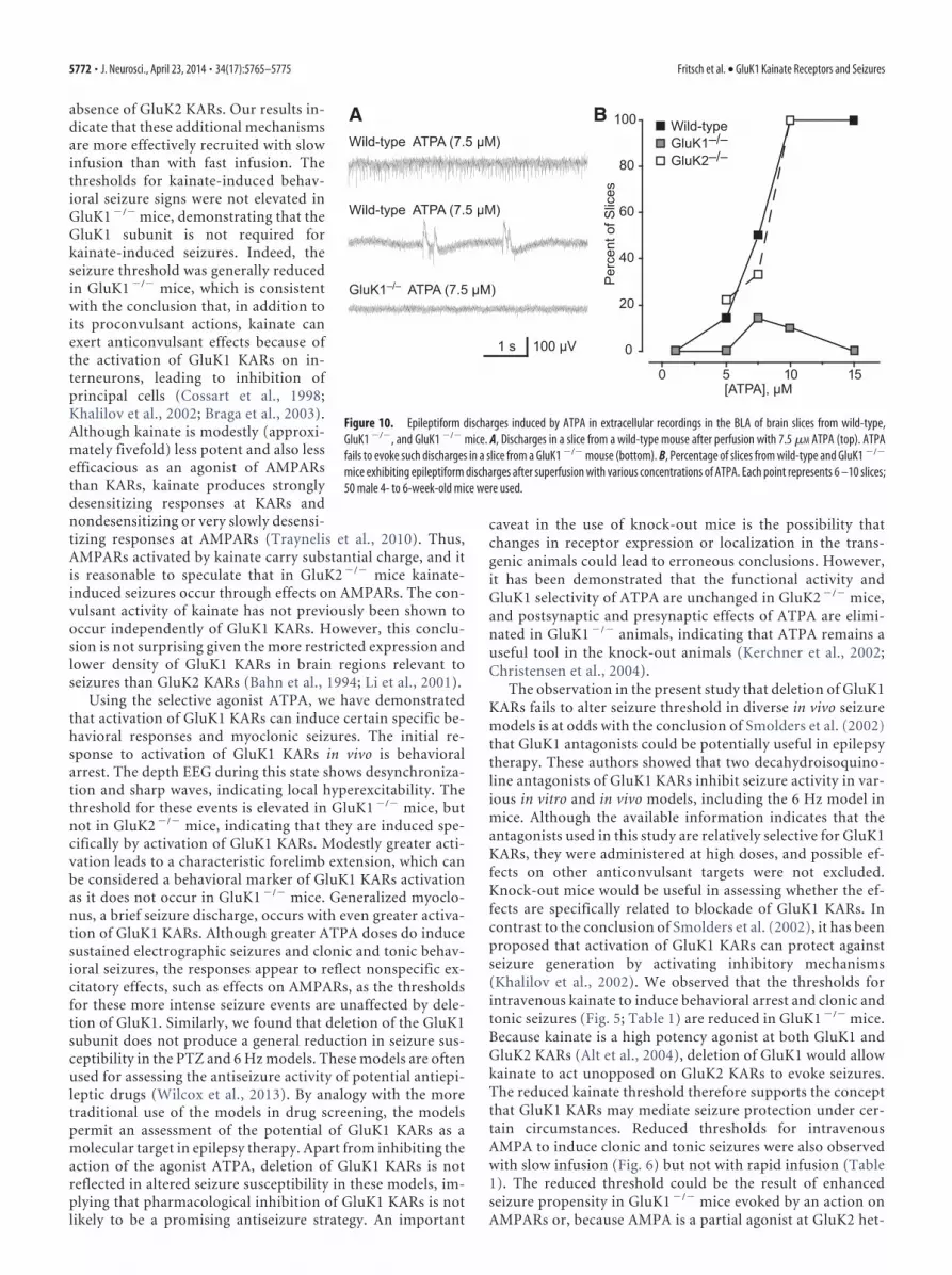
exposure of wild-type mouse brain slices to increasing concen-trations of ATPA was associated with increasing likelihood ofrecording epileptiform discharges. No slices exhibited dischargeswhen superfused with 1 �M ATPA, whereas 89% and 93% ofslices exhibited discharges with 10 or 15 �M ATPA, respectively.
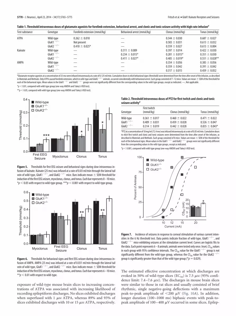
The estimated effective concentration at which discharges areevoked in 50% of wild-type slices (EC50) is 7.5 �M (95% confi-dence limit: 7.4 –7.6 �M). The discharges in mouse brain sliceswere similar to those in rat slices and usually consisted of briefrhythmic, single negative-going deflections with a maximumpeak-to-peak amplitude of �200 �V (Fig. 10A). In addition,longer duration (100 –1000 ms) biphasic events with peak-to-peak amplitude of 100 – 400 �V occurred in some slices. Epilep-
Table 1. Threshold intravenous doses of glutamate agonists for forelimb extension, behavioral arrest, and clonic and tonic seizure activity with high rate infusiona
Test substance Genotype Forelimb extension (mmol/kg) Behavioral arrest (mmol/kg) Clonus (mmol/kg) Tonus (mmol/kg)
ATPA Wild-type 0.262 0.010 — 0.544 0.030 0.687 0.027GluK1 �/� Not present — 0.583 0.031 0.613 0.032GluK2 �/� 0.410 0.025* — 0.559 0.027 0.613 0.004
Kainate Wild-type — 0.311 0.009 0.397 0.014 0.422 0.030GluK1 �/� — 0.234 0.015* 0.281 0.015* 0.351 0.030GluK2 �/� — 0.411 0.027* 0.485 0.019* 0.551 0.028**
AMPA Wild-type — — 0.354 0.056 0.385 0.056GluK1 �/� — — 0.359 0.042 0.391 0.042GluK2 �/� — — 0.317 0.015 0.459 0.052
aGlutamate receptor agonists at a concentration of 25 mM were infused intravenously at a rate of 0.125 ml/min. Cumulative doses to elicit behavioral signs (threshold) were determined from the time after onset of the infusion, as describedin Materials and Methods. Only ATPA caused forelimb extension, which in wild-type and GluK2 �/� animals, occurred coincidentally with behavioral arrest. Each group consisted of 7–12 mice. Values are mean SEM of the threshold foreach of the behavioral signs. Mean values in the GluK1 �/� and GluK2 �/� groups were not significantly different from the corresponding values in the wild-type groups, except as indicated. —, Not applicable.
*p � 0.01, compared with wild-type group (one-way ANOVA and Tukey’s HSD test).
**p � 0.05, compared with wild-type group (one-way ANOVA and Tukey’s HSD test).
0.0
0.1
0.2
0.3
0.4
Clonus TonusMyoclonusFirst EEGSeizure
Thre
shol
d (m
mol
/kg) GluK2–/–
GluK1–/–Wild-type
*** **
Figure 5. Thresholds for first EEG seizure and behavioral signs during slow intravenous in-fusion of kainate. Kainate (25 mM) was infused at a rate of 0.03 ml/min through the lateral tailvein of wild-type, GluK1 �/�, and GluK2 �/� mice. Bars indicate mean SEM threshold forinduction of the first EEG seizure, myoclonus, clonus, and tonus. Each bar represents 8 –10 mice.*p � 0.05 with respect to wild-type group. ***p � 0.001 with respect to wild-type group.
TonusClonusMyoclonus First EEGSeizure
GluK2–/–GluK1–/–Wild-type
** **
Thre
shol
d (m
mol
/kg)
0.0
0.1
0.2
0.3
0.4
0.5
0.6
Figure 6. Thresholds for behavioral signs and first EEG seizure during slow intravenous in-fusion of AMPA. AMPA (25 mM) was infused at a rate of 0.031 ml/min through the lateral tailvein of wild-type, GluK1 �/�, and GluK2 �/� mice. Bars indicate mean SEM threshold forinduction of the first EEG seizure, myoclonus, clonus, and tonus. Each bar represents 6 –10 mice.**p � 0.01 with respect to wild-type.
Table 2. Threshold intravenous doses of PTZ for first twitch and clonic and tonicseizure activitya
GenotypeFirst twitch(mmol/kg) Clonus (mmol/kg) Tonus (mmol/kg)
Wild-type 0.361 0.017 0.460 0.022 0.471 0.022GluK1 �/� 0.409 0.031 0.459 0.028 0.526 0.047GluK2 �/� 0.314 0.019 0.442 0.028 0.835 0.043*aPTZ at a concentration of 10 mg/ml (72.4 mM) was infused intravenously at a rate of 0.05 ml/min. Cumulative dosesto elicit first twitch and clonic and tonic seizures were determined from the time after onset of the infusion, asdescribed in Materials and Methods. Each group consisted of 8 mice. Values are mean SEM of the threshold foreach of the behavioral signs. Mean values in the GluK1 �/� and GluK2 �/� groups were not significantly differentfrom the corresponding values in the wild-type groups, except as indicated.
*p � 0.001, compared with wild-type group (one-way ANOVA and Tukey’s HSD test).
Figure 7. Incidence of seizures in response to corneal stimulation of various current inten-sities in the 6 Hz threshold test. Data points indicate fraction of wild-type, GluK1 �/�, andGluK2 �/� mice exhibiting seizures at the stimulation current level. Curves are logistic fits tothe data. Each point represents 6 – 8 animals; animals were tested only once. Inset, CS50 valuesin each group with 95% confidence intervals. The CS50 value for the GluK1 �/� group is notsignificantly different from the wild-type group, whereas the CS50 value for the GluK2 �/�
group is significantly greater than that of the wild-type group (*p � 0.029).
5770 • J. Neurosci., April 23, 2014 • 34(17):5765–5775 Fritsch et al. • GluK1 Kainate Receptors and Seizures

tiform events were rarely recorded in slices from GluK1�/� mice.In slices superfused with 7.5 and 10 �M ATPA, discharges weredetected in only 1 of 7 and 1 of 10 slices, respectively. At the otherconcentrations tested, no epileptiform activity was detected. Incontrast, ATPA caused a concentration-dependent increase in
the frequency of discharges in slices fromGluK2�/� mice similar to that in slicesfrom wild-type mice. These observationsconfirm that ATPA evokes epileptiformdischarges via activation of GluK1 KARs.
Olfactory bulb kindling in wild-type,GluK1 �/� and GluK2 �/� miceTo examine the role of the KARs in epi-leptogenesis, we used olfactory bulbkindling, which has been found to cor-respond more closely than other kin-dling models in sensitivity to epilepsytreatments (Fujiwara et., 2010). Twice-daily olfactory bulb stimulation resultedin a fluctuating but a generally progres-sive rise in mean seizure stage until allanimals exhibited Stage 5 seizures by atleast the 20th stimulation (Fig. 11A).Despite a brief initial delay in the wild-type group, there was no difference inthe rate of kindling development be-tween the wild-type and the GluK1 �/�
or GluK2 �/� groups as assessed bymean seizure stage values (repeated-
measures ANOVA, p � 0.09). The mean number of stimula-tions needed to achieve Stages 2–5 and the fully kindled state(5 consecutive Stage 5 seizures) were similar in the wild-type,GluK1 �/�, and GluK2 �/� groups (Fig. 11B). There was also afluctuating but generally progressive rise in mean normalizedafterdischarge duration (Fig. 11C). Repeated-measuresANOVA failed to detect any significant difference among thegroups in the mean normalized afterdischarge duration ( p �0.38). Finally, as shown in Figure 11D, mean prekindling andpostkindling afterdischarge threshold values did not differ sig-nificantly in the GluK1 �/� and GluK2 �/� groups from themean values in the wild-type group (prekindling: GluK1 �/�,p � 0.21; GluK2 �/�, p � 0.36; postkindling: GluK1 �/�, p �0.17; GluK2 �/�, p � 0.20) and the percentage change valueswere similar in the wild-type, GluK1 �/�, and GluK2 �/�
groups. The results demonstrate that olfactory bulb kindlingdevelopment, a form of epileptogenesis, is unaffected by dele-tion of either GluK1 or GluK2 and that seizure propensity infully kindled animals that lack these subunits is similar to thatof wild-type animals.
DiscussionA key problem in epilepsy research is the identity of the gluta-mate receptors at which the widely used convulsant toxin kai-nate acts to induce seizures (Ben-Ari, 2012). Mulle et al.(1998) found that GluK2 �/� mice fail to exhibit seizures inresponse to low doses of kainate. The present demonstrationof an elevated seizure threshold for kainate in GluK2 �/� mice(Table 1) confirms this observation and supports the conclu-sion that low doses of kainate trigger seizures via an action onGluK2 KARs. However, modestly higher doses of kainate doinduce seizures in GluK2 �/� mice, indicating that other re-ceptors are also involved in the convulsant activity of thetoxin. Moreover, in the slow kainate infusion experiment ofFigure 5, whereas the threshold for the first EEG seizure waselevated in GluK2 �/� mice, the thresholds for other seizuresigns did not differ from those in wild-type mice confirmingthat kainate can effectively trigger seizure behaviors in the
50 s
50 µV5 s
100 µV
Figure 8. Extracellular recording from the BLA in a rat brain slice during superfusion with 10 �M ATPA beginning at the pointmarked with the arrow. The upper two traces are continuous; the two regions boxed are shown expanded below.
[1] ATPA (10 µM)
[2] ATPA (10 µM) + LY293558 (100 nM)
[3] ATPA (10 µM) + LY293558 (500 nM)
[4] ATPA (10 µM)
50 s100 µV
Figure 9. Extracellular recording from the BLA in a rat brain slice during superfusion with 10�M ATPA. Selected segments are shown after inclusion of 100 and 500 nM LY293558 in theperfusion solution and after washout of the antagonist.
Fritsch et al. • GluK1 Kainate Receptors and Seizures J. Neurosci., April 23, 2014 • 34(17):5765–5775 • 5771
absence of GluK2 KARs. Our results in-dicate that these additional mechanismsare more effectively recruited with slowinfusion than with fast infusion. Thethresholds for kainate-induced behav-ioral seizure signs were not elevated inGluK1 �/� mice, demonstrating that theGluK1 subunit is not required forkainate-induced seizures. Indeed, theseizure threshold was generally reducedin GluK1 �/� mice, which is consistentwith the conclusion that, in addition toits proconvulsant actions, kainate canexert anticonvulsant effects because ofthe activation of GluK1 KARs on in-terneurons, leading to inhibition ofprincipal cells (Cossart et al., 1998;Khalilov et al., 2002; Braga et al., 2003).Although kainate is modestly (approxi-mately fivefold) less potent and also lessefficacious as an agonist of AMPARsthan KARs, kainate produces stronglydesensitizing responses at KARs andnondesensitizing or very slowly desensi-tizing responses at AMPARs (Traynelis et al., 2010). Thus,AMPARs activated by kainate carry substantial charge, and itis reasonable to speculate that in GluK2 �/� mice kainate-induced seizures occur through effects on AMPARs. The con-vulsant activity of kainate has not previously been shown tooccur independently of GluK1 KARs. However, this conclu-sion is not surprising given the more restricted expression andlower density of GluK1 KARs in brain regions relevant toseizures than GluK2 KARs (Bahn et al., 1994; Li et al., 2001).
Using the selective agonist ATPA, we have demonstratedthat activation of GluK1 KARs can induce certain specific be-havioral responses and myoclonic seizures. The initial re-sponse to activation of GluK1 KARs in vivo is behavioralarrest. The depth EEG during this state shows desynchroniza-tion and sharp waves, indicating local hyperexcitability. Thethreshold for these events is elevated in GluK1 �/� mice, butnot in GluK2 �/� mice, indicating that they are induced spe-cifically by activation of GluK1 KARs. Modestly greater acti-vation leads to a characteristic forelimb extension, which canbe considered a behavioral marker of GluK1 KARs activationas it does not occur in GluK1 �/� mice. Generalized myoclo-nus, a brief seizure discharge, occurs with even greater activa-tion of GluK1 KARs. Although greater ATPA doses do inducesustained electrographic seizures and clonic and tonic behav-ioral seizures, the responses appear to reflect nonspecific ex-citatory effects, such as effects on AMPARs, as the thresholdsfor these more intense seizure events are unaffected by dele-tion of GluK1. Similarly, we found that deletion of the GluK1subunit does not produce a general reduction in seizure sus-ceptibility in the PTZ and 6 Hz models. These models are oftenused for assessing the antiseizure activity of potential antiepi-leptic drugs (Wilcox et al., 2013). By analogy with the moretraditional use of the models in drug screening, the modelspermit an assessment of the potential of GluK1 KARs as amolecular target in epilepsy therapy. Apart from inhibiting theaction of the agonist ATPA, deletion of GluK1 KARs is notreflected in altered seizure susceptibility in these models, im-plying that pharmacological inhibition of GluK1 KARs is notlikely to be a promising antiseizure strategy. An important
caveat in the use of knock-out mice is the possibility thatchanges in receptor expression or localization in the trans-genic animals could lead to erroneous conclusions. However,it has been demonstrated that the functional activity andGluK1 selectivity of ATPA are unchanged in GluK2 �/� mice,and postsynaptic and presynaptic effects of ATPA are elimi-nated in GluK1 �/� animals, indicating that ATPA remains auseful tool in the knock-out animals (Kerchner et al., 2002;Christensen et al., 2004).
The observation in the present study that deletion of GluK1KARs fails to alter seizure threshold in diverse in vivo seizuremodels is at odds with the conclusion of Smolders et al. (2002)that GluK1 antagonists could be potentially useful in epilepsytherapy. These authors showed that two decahydroisoquino-line antagonists of GluK1 KARs inhibit seizure activity in var-ious in vitro and in vivo models, including the 6 Hz model inmice. Although the available information indicates that theantagonists used in this study are relatively selective for GluK1KARs, they were administered at high doses, and possible ef-fects on other anticonvulsant targets were not excluded.Knock-out mice would be useful in assessing whether the ef-fects are specifically related to blockade of GluK1 KARs. Incontrast to the conclusion of Smolders et al. (2002), it has beenproposed that activation of GluK1 KARs can protect againstseizure generation by activating inhibitory mechanisms(Khalilov et al., 2002). We observed that the thresholds forintravenous kainate to induce behavioral arrest and clonic andtonic seizures (Fig. 5; Table 1) are reduced in GluK1 �/� mice.Because kainate is a high potency agonist at both GluK1 andGluK2 KARs (Alt et al., 2004), deletion of GluK1 would allowkainate to act unopposed on GluK2 KARs to evoke seizures.The reduced kainate threshold therefore supports the conceptthat GluK1 KARs may mediate seizure protection under cer-tain circumstances. Reduced thresholds for intravenousAMPA to induce clonic and tonic seizures were also observedwith slow infusion (Fig. 6) but not with rapid infusion (Table1). The reduced threshold could be the result of enhancedseizure propensity in GluK1 �/� mice evoked by an action onAMPARs or, because AMPA is a partial agonist at GluK2 het-
A BWild-type ATPA (7.5 µM)
Wild-type ATPA (7.5 µM)
1 s 100 µV
GluK1–/– ATPA (7.5 µM)
0 5 10 15
0
20
40
60
80
100
[ATPA], µM
Wild-type
GluK2–/–GluK1–/–
Per
cent
of S
lices
Figure 10. Epileptiform discharges induced by ATPA in extracellular recordings in the BLA of brain slices from wild-type,GluK1 �/�, and GluK1 �/� mice. A, Discharges in a slice from a wild-type mouse after perfusion with 7.5 �M ATPA (top). ATPAfails to evoke such discharges in a slice from a GluK1 �/� mouse (bottom). B, Percentage of slices from wild-type and GluK1 �/�
mice exhibiting epileptiform discharges after superfusion with various concentrations of ATPA. Each point represents 6 –10 slices;50 male 4- to 6-week-old mice were used.
5772 • J. Neurosci., April 23, 2014 • 34(17):5765–5775 Fritsch et al. • GluK1 Kainate Receptors and Seizures

eromers such as GluK2/GluK5, by an action on GluK2 KARs(Traynelis et al., 2010). Overall, our results indicate that acti-vation of GluK1 KARs can trigger seizures (Rogawski et al.,2003; Aroniadou-Anderjaska et al., 2012), but deletion ofthese receptors may remove an endogenous mechanism forseizure suppression.
Within the BLA, GluK1 KARs are present on the soma anddendrites of principal neurons and GABAergic interneurons,as well as on the presynaptic terminals of interneurons (Li andRogawski, 1998; Braga et al., 2003; Gryder and Rogawski,2003). The complex effects of GluK1 KAR activation on sei-zure susceptibility in the BLA is the result of the interplay ofreceptors at these various sites. Activation of principal neuronsomatodendritic GluK1 KARs would enhance amygdalar ex-citability by depolarizing these neurons, leading to glutamaterelease onto their postsynaptic targets. Recently, it has beenobserved that activation of GluK1 KARs can also facilitateglutamate release in the BLA via a presynaptic action(Aroniadou-Anderjaska et al., 2012). Activation of somato-dendritic GluK1 KARs on interneneurons should suppressamygdalar excitability because of interneuronal depolariza-tion and enhanced GABA release (Wu et al., 2007). This actionas noted could, in part, be the basis for antiseizure effectsmediated by GluK1 KARs. Additionally, low-level activationof presynaptic GluK1 KARs localized to the terminals ofGABAergic neurons facilitates GABA release and could alsocontribute to seizure suppression (Aroniadou-Anderjaska etal., 2008). However, more intense activation inhibits GABArelease, which would be expected to facilitate seizures. In viewof the marked reduction in EEG seizure threshold inGluK2 �/� mice compared with wild-type and GluK1 �/� an-imals (Fig. 2), it is interesting to speculate that GluK1/GluK2
heteromers could be of particular importance in mediatinginhibition because such heteromers are absent in GluK2 �/�
animals.Although GluK1 KARs are not necessary for seizure gener-
ation in diverse models, they could be required for epilepto-genesis. We previously presented pharmacological evidencethat GluK1 KARs are not required for the development ofamygdala kindling in the mouse (Rogawski et al., 2001). Theseconclusions are supported by the present results with olfactorybulb kindling in GluK1 �/� mice, in which kindling occurredsimilarly in wild-type and knock-out animals. Our results alsoindicate that GluK2 KARs are not required for kindling devel-opment. In addition, it is apparent that the GluK1 and GluK2subunits do not contribute to the expression of kindled sei-zures. The extent to which this observation can be generalizedto other epileptogenesis models remains to be determined.There is evidence for enhanced functional activity of KARs ataberrant mossy fiber– granule cell synapses in a poststatus epi-lepticus model of temporal lobe epilepsy (Epsztein et al., 2005;Artinian et al., 2011), suggesting that there could be differ-ences among epileptogenesis models.
In conclusion, the present results demonstrate that selec-tive activation of GluK1 KARs can elicit seizure activity in vivoand epileptiform discharges in the BLA in vitro but fail toconfirm blockade of GluK1 KARs as a likely epilepsy treatmentstrategy. We recently found that epileptogenesis after kainate-induced status epilepticus is associated with a major loss ofinterneurons in the amygdala and impaired modulation ofGABAergic transmission by GluK1 KARs (Fritsch et al., 2009),diminishing the opportunity for these receptors to serve as atreatment target. Although pharmacological inhibition ofGluK1 KARs is unlikely to ameliorate seizures, the present
0
50
100
150
200
250
300
Pre-kindling Post-kindling0
1
2
3
4
Nor
mal
ized
Afte
r-di
scha
rge
Dur
atio
n
Stimulation Number1 5 10 15 20
Stimulation Number1 5 10 15 20
0
1
2
3
4
5S
eizu
re S
tage
Wild-type
0
5
10
15
20
25
Num
ber o
f Stim
ulat
ions
2 3 4 5 FullyKindledKindling Stage
GluK1–/–
GluK2–/– GluK1–/–
GluK2–/–
Wild-type
Afte
rdis
char
ge T
hres
hold
(µA)
0
50
100Percent change
A B
C D
Figure 11. Comparison of olfactory bulb kindling development in wild-type, GluK1 �/�, and GluK2 �/� mice. A, Mean SEM behavioral seizure stage is plotted with respect to stimulationnumber. B, Mean SEM number of stimulations required to achieve the stage indicated. Fully kindled stage is defined as the occurrence of 5 consecutive Stage 5 seizures. C, Mean SEMelectrographic seizure duration normalized to the duration of the first afterdischarge. D, Mean SEM of the prekindling and postkindling afterdischarge threshold and the percentage change inthreshold. Afterdischarge threshold is the stimulation intensity (�A) required to evoke an electrographic seizure of at least 5 s duration. Each group consists of 14 mice.
Fritsch et al. • GluK1 Kainate Receptors and Seizures J. Neurosci., April 23, 2014 • 34(17):5765–5775 • 5773

results do not eliminate the possibility that the receptors couldplay a role in the pathophysiology of certain forms of epilepsy.
ReferencesAlt A, Weiss B, Ogden AM, Knauss JL, Oler J, Ho K, Large TH, Bleakman
D (2004) Pharmacological characterization of glutamatergic ago-nists and antagonists at recombinant human homomeric and hetero-meric kainate receptors in vitro. Neuropharmacology 46:793– 806.CrossRef Medline
Aroniadou-Anderjaska V, Fritsch B, Qashu F, Braga MF (2008) Pathologyand pathophysiology of the amygdala in epileptogenesis and epilepsy.Epilepsy Res 78:102–116. CrossRef Medline
Aroniadou-Anderjaska V, Pidoplichko VI, Figueiredo TH, Almeida-SuhettCP, Prager EM, Braga MF (2012) Presynaptic facilitation of glutamaterelease in the basolateral amygdala: a mechanism for the anxiogenic andseizurogenic function of GluK1 receptors. Neuroscience 221:157–169.CrossRef Medline
Artinian J, Peret A, Marti G, Epsztein J, Crepel V (2011) Synaptic kainatereceptors in interplay with INaP shift the sparse firing of dentate granulecells to a sustained rhythmic mode in temporal lobe epilepsy. J Neurosci31:10811–10818. CrossRef Medline
Bahn S, Volk B, Wisden W (1994) Kainate receptor gene expression in thedeveloping rat brain. J Neurosci 14:5525–5547. Medline
Barton ME, Klein BD, Wolf HH, White HS (2001) Pharmacological char-acterization of the 6 Hz psychomotor seizure model of partial epilepsy.Epilepsy Res 47:217–227. CrossRef Medline
Ben-Ari Y (2012) Kainate and temporal lobe epilepsies: 3 decades ofprogress. In: Jasper’s basic mechanisms of the epilepsies [Internet], Ed4 (Noebels JL, Avoli M, Rogawski MA, Olsen RW, Delgado-EscuetaAV, eds). Bethesda, MD: National Center for Biotechnology Informa-tion. http://www.ncbi.nih.gov/books/NBK98166/.
Bettler B, Boulter J, Hermans-Borgmeyer I, O’Shea-Greenfield A, DenerisES, Moll C, Borgmeyer U, Hollmann M, Heinemann S (1990) Clon-ing of a novel glutamate receptor subunit, GluR5: expression in thenervous system during development. Neuron 5:583–595. CrossRefMedline
Bleakman D, Gates MR, Ogden AM, Mackowiak M (2002) Kainate receptoragonists, antagonists and allosteric modulators. Curr Pharm Des 8:873–885. CrossRef Medline
Braga MF, Aroniadou-Anderjaska V, Xie J, Li H (2003) Bidirectionalmodulation of GABA release by presynaptic glutamate receptor 5 kai-nate receptors in the basolateral amygdala. J Neurosci 23:442– 452.Medline
Christensen JK, Paternain AV, Selak S, Ahring PK, Lerma J (2004) A mosaicof functional kainate receptors in hippocampal interneurons. J Neurosci24:8986 – 8993. CrossRef Medline
Clarke VR, Ballyk BA, Hoo KH, Mandelzys A, Pellizzari A, Bath CP, ThomasJ, Sharpe EF, Davies CH, Ornstein PL, Schoepp DD, Kamboj RK, Col-lingridge GL, Lodge D, Bleakman D (1997) A hippocampal GluR5 kai-nate receptor regulating inhibitory synaptic transmission. Nature 389:599 – 603. CrossRef Medline
Cossart R, Esclapez M, Hirsch JC, Bernard C, Ben-Ari Y (1998) GluR5 kai-nate receptor activation in interneurons increases tonic inhibition of py-ramidal cells. Nat Neurosci 1:470 – 478. CrossRef Medline
Cui C, Mayer ML (1999) Heteromeric kainate receptors formed by thecoassembly of GluR5, GluR6, and GluR7. J Neurosci 19:8281– 8291.Medline
Epsztein J, Represa A, Jorquera I, Ben-Ari Y, Crepel V (2005) Recurrentmossy fibers establish aberrant kainate receptor-operated synapses ongranule cells from epileptic rats. J Neurosci 25:8229 – 8239. CrossRefMedline
Fisahn A, Contractor A, Traub RD, Buhl EH, Heinemann SF, McBain CJ(2004) Distinct roles for the kainate receptor subunits GluR5 and GluR6in kainate-induced hippocampal gamma oscillations. J Neurosci 24:9658 –9668. CrossRef Medline
Fritsch B, Qashu F, Figueiredo TH, Aroniadou-Anderjaska V, Rogawski MA,Braga MF (2009) Pathological alterations in GABAergic interneuronsand reduced tonic inhibition in the basolateral amygdala during epilep-togenesis. Neuroscience 163:415– 429. CrossRef Medline
Fujiwara A, Watanabe Y, Takechi K, Ishikawa T, Kaida Y, Akagi M, Kamei C(2010) The usefulness of olfactory bulb kindling as a model for evalua-tion of antiepileptics. Epilepsia 51:445– 453. CrossRef Medline
Gryder DS, Rogawski MA (2003) Selective antagonism of GluR5 kainate-receptor-mediated synaptic currents by topiramate in rat basolateralamygdala neurons. J Neurosci 23:7069 –7074. Medline
Hoo K, Legutko B, Rizkalla G, Deverill M, Hawes CR, Ellis GJ, Stensbol TB,Krogsgaard-Larsen P, Skolnick P, Bleakman D (1999) [ 3H]ATPA: ahigh affinity ligand for GluR5 kainate receptors. Neuropharmacology 38:1811–1817. CrossRef Medline
Kaminski RM, Livingood MR, Rogawski MA (2004) Allopregnanolone an-alogs that positively modulate GABA receptors protect against partialseizures induced by 6-Hz electrical stimulation in mice. Epilepsia 45:864 –867. CrossRef Medline
Kerchner GA, Wilding TJ, Li P, Zhuo M, Huettner JE (2001) Presynaptickainate receptors regulate spinal sensory transmission. J Neurosci 21:59 –66. Medline
Kerchner GA, Wilding TJ, Huettner JE, Zhuo M (2002) Kainate receptorsubunits underlying presynaptic regulation of transmitter release in thedorsal horn. J Neurosci 22:8010 – 8017. Medline
Khalilov I, Hirsch J, Cossart R, Ben-Ari Y (2002) Paradoxical anti-epilepticeffects of a GluR5 agonist of kainate receptors. J Neurophysiol 88:523–527. Medline
Koga K, Sim SE, Chen T, Wu LJ, Kaang BK, Zhuo M (2012) Kainatereceptor-mediated synaptic transmissions in the adult rodent insular cor-tex. J Neurophysiol 108:1988 –1998. CrossRef Medline
Li H, Rogawski MA (1998) GluR5 kainate receptor mediated synaptic trans-mission in rat basolateral amygdala in vitro. Neuropharmacology 37:1279 –1286. CrossRef Medline
Li H, Chen A, Xing G, Wei ML, Rogawski MA (2001) Kainate receptor-mediated heterosynaptic facilitation in the amygdala. Nat Neurosci4:612– 620. CrossRef Medline
Mandhane SN, Aavula K, Rajamannar T (2007) Timed pentylenetetrazolinfusion test: a comparative analysis with s.c.PTZ and MES models ofanticonvulsant screening in mice. Seizure 16:636 – 644. CrossRefMedline
Mulle C, Sailer A, Perez-Otano I, Dickinson-Anson H, Castillo PE, BureauI, Maron C, Gage FH, Mann JR, Bettler B, Heinemann SF (1998)Altered synaptic physiology and reduced susceptibility to kainate-induced seizures in GluR6-deficient mice. Nature 392:601– 605.CrossRef Medline
Nadler JV (2012) Plasticity of glutamate synaptic mechanisms. In: Jasper’sbasic mechanisms of the epilepsies [Internet], Ed 4 (Noebels JL, Avoli M,Rogawski MA, Olsen RW, Delgado-Escueta AV, eds). Bethesda, MD: Na-tional Center for Biotechnology Information. http://www.ncbi.nih.gov/books/NBK98204/.
Park SA, Yin H, Bhattarai JP, Park SJ, Lee JC, Kim CJ, Han SK (2010) Post-natal change of GluR5 kainate receptor expression in the substantia gela-tinosa neuron of the trigeminal subnucleus caudalis in mice. Brain Res1346:52– 61. CrossRef Medline
Paternain AV, Herrera MT, Nieto MA, Lerma J (2000) GluR5 and GluR6kainate receptor subunits coexist in hippocampal neurons and coas-semble to form functional receptors. J Neurosci 20:196 –205. Medline
Pinheiro P, Mulle C (2006) Kainate receptors. Cell Tissue Res 326:457– 482.CrossRef Medline
Racine RJ (1972) Modification of seizure activity by electrical stimulation:II. Motor seizure. Electroencephalogr Clin Neurophysiol 32:281–294.CrossRef Medline
Rogawski MA (2011) Revisiting AMPA receptors as an antiepileptic drugtarget. Epilepsy Curr 11:56 – 63. CrossRef Medline
Rogawski MA, Kurzman PS, Yamaguchi SI, Li H (2001) Role of AMPAand GluR5 kainate receptors in the development and expression ofamygdala kindling in the mouse. Neuropharmacology 40:28 –35.CrossRef Medline
Rogawski M, Gryder D, Castaneda D Yonekawa W, Banks MK, Li H (2003)GluR5 kainate receptors, seizures, and the amygdala. Ann N Y Acad Sci985:150 –162. CrossRef Medline
Smolders I, Bortolotto ZA, Clarke VR, Warre R, Khan GM, O’Neill MJ, Orn-stein PL, Bleakman D, Ogden A, Weiss B, Stables JP, Ho KH, Ebinger G,Collingridge GL, Lodge D, Michotte Y (2002) Antagonists of GLUK5-containing kainate receptors prevent pilocarpine-induced limbic sei-zures. Nat Neurosci 5:796 – 804. CrossRef Medline
Steriade M, Gloor P, Llinas RR, Lopes de Silva FH, Mesulam MM (1990)
5774 • J. Neurosci., April 23, 2014 • 34(17):5765–5775 Fritsch et al. • GluK1 Kainate Receptors and Seizures

Report of IFCN Committee on Basic Mechanisms: basic mechanisms ofcerebral rhythmic activities. Electroencephalogr Clin Neurophysiol 76:481–508. CrossRef Medline
Traynelis SF, Wollmuth LP, McBain CJ, Menniti FS, Vance KM, Ogden KK,Hansen KB, Yuan H, Myers SJ, Dingledine R (2010) Glutamate receptorion channels: structure, regulation, and function. Pharmacol Rev 62:405–496. CrossRef Medline
Vignes M, Bleakman D, Lodge D, Collingridge GL (1997) The synaptic ac-tivation of the GluR5 subtype of kainate receptor in area CA3 of the rathippocampus. Neuropharmacology 36:1477–1481. CrossRef Medline
Wilcox KS, Dixon-Salazar T, Sills GJ, Ben-Menachem E, White HS, Porter RJ,Dichter MA, Moshe SL, Noebels JL, Privitera MD, Rogawski MA (2013)
Issues related to development of new antiseizure treatments. Epilepsia54[Suppl 4]:24 –34.
Wondolowski J, Frerking M (2009) Subunit-dependent postsynaptic ex-pression of kainate receptors on hippocampal interneurons in area CA1.J Neurosci 29:563–574. CrossRef Medline
Wu LJ, Zhao MG, Toyoda H, Ko SW, Zhuo M (2005) Kainate receptor-mediated synaptic transmission in the adult anterior cingulate cortex.J Neurophysiol 94:1805–1813. CrossRef Medline
Wu LJ, Ko SW, Toyoda H, Zhao MG, Xu H, Vadakkan KI, Ren M, Knifed E,Shum F, Quan J, Zhang XH, Zhuo M (2007) Increased anxiety-like be-havior and enhanced synaptic efficacy in the amygdala of GluR5 knock-out mice. PLoS One 2:e167. CrossRef Medline
Fritsch et al. • GluK1 Kainate Receptors and Seizures J. Neurosci., April 23, 2014 • 34(17):5765–5775 • 5775