regulatory t cells in the induction and maintenance of peripheral transplantation tolerance
TRANSCRIPT

Stephen P. Cobbold Luis Graca Chun-Yen Lin Elizabeth Adams Herman Waldmann
Received: 31 October 2002 Accepted: 26 November 2002 Published online: 4 February 2003 0 Springer-Verlag 2003
S.P. Cobbold (m) . L. Graca . C.-Y. Lin E. Adams . H. Waldmann Sir William Dunn School of Pathology, University of Oxford, South Parks Road, Oxford, Oxfordshire, OX1 3RE, UK E-mail: [email protected] Tel.: + 44-1865-275504 Fax: + 44-1865-275501
Present address: C.-Y. Lin Department of Gastroenterology, Chang-Gung Memorial Hospital, Taipei, Taiwan
Regulatory T cells in the induction and maintenance of peripheral transplantation tolerance
Abstract It is now possible to induce donor-specific transplantation toler- ance in adult rodents using non-de- pleting monoclonal antibodies against T cell co-receptor and co- stimulation molecules or by immu- nisation with tolerogenic antigen- presenting cells. It is a common finding of all these models of pe- ripheral tolerance, as well as of var- ious mouse models of autoimmune disease, that regulatory CD4+ T cells are the principal mediators. There are currently no specific markers for regulatory T cells, but in some autoimmune models their ac- tivity has been associated with the expression of activation markers
such as CD25 and CTLA4, or anti- inflammatory cytokines such as IL- 10 and TGF-P. CD4+CD25+ T cells from both na'ive and tolerised do- nors are able to transfer tolerance to grafts in lymphopenic recipients, and this may be directly applicable to bone-marrow transplantation. The challenge is now to understand the biological principles that allow such immune re-programming so that they can be safely applied to clinical organ grafting.
Keywords Transplantation . Tole- rance . Regulation . CD4 . CD25
~
Chimerism and central tolerance
For many years, the classical experiments of Medawar and colleagues [l] were used as a paradigm for the in- duction of tolerance in the adult. Over the next 40 years, approximately, it became clear that if one could elimi- nate the mature immune system, usually by total body irradiation, and then introduce long-lived donor anti- gen-carrying cells (usually bone marrow) and allow the haemopoietic system to regenerate as a chimera, the recipient was generally able to accept donor-type organ grafts. The predominant mechanism was found to be a continuous clonal elimination of donor antigen-specific T cells in the thymus [2], providing an absolute unre- sponsiveness both in vivo and in vitro. The problem with this approach has always been the need to ablate the mature immune system. Numerous studies in allogeneic
bone-marrow transplantation have shown us that in practice this requires lethal irradiation as well as im- munosuppressive or lympholytic agents, even if the do- nor and recipient are siblings matched for major histocompatibility (MHC) loci [3]. Although there has been a recent revival of interest in donor chimerism as a means of generating tolerance, this has generally not proven to be easily practicable.
Immunosuppression and chronic rejection
One might ask why we need to induce tolerance at all. With appropriate cocktails of conventional immuno- suppressive drugs we are able to control the acute re- jection of renal allografts very effectively, with a successful outcome higher than 90% at 1 year. The

67
problem is, however, that by 10 years, more than 50% of the grafts will have been lost through a process that is still poorly understood but is generally thought to be a form of chronic rejection. In addition, the use of long- term immunosuppression risks the development of seri- ous infections and tumours, and each drug is associated with specific toxicities. Some of the immunosuppressive agents in common use are probably even counter-toler- ogenic [4]. Therefore, we need to identify which, if any, of the currently available immunosuppressive agents are compatible with tolerance induction, either when used alone or in combination with emerging tolerogenic therapies.
Immune re-programming with non-depleting monoclonal antibodies
Although many effector systems play a role in the re- jection of allogeneic grafts, including B cells, NK cells, activated macrophages and polymorphs, it is clear that all rejection is absolutely dependent on T cells. The majority of peripheral T cells can be divided into those expressing CD4, which recognise antigen peptides in association with MHC-11, usually associated with helper T cell activity, and those expressing CD8, which recog- nise MHC-I that traditionally include the cytotoxic effector T cells.
Monoclonal antibodies against the T cell antigen CD4 can be used in vivo to block immune responses in rodents, but, surprisingly, the immune system records such aborted immune challenges as tolerogenic events [5 ] . Although CD4 antibodies used alone induced tolerance only in certain MHC-matched skin-graft combinations, they were able to induce indefinite ac- ceptance of MHC-mismatched cardiac grafts in some recipient strains of mice [6]. It has been possible to extend the range and reliability of tolerance induction by the addition of other non-depleting antibodies, es- pecially to CD8 and CD40L(CD154), and under these conditions, donor-specific lifelong tolerance can be in- duced to fully MHC plus multiple minors mismatched skin grafts [7].
The fact that such tolerance is lifelong and indepen- dent of the presence or absence of a thymus shows that the adult immune system can indeed be re-programmed in the periphery to accept donor antigens as if they are self. While mice, which are centrally tolerant, are unable to proliferate or generate cytotoxic T cells in vitro against donor antigen-presenting cells because the T cells have been clonally deleted in the thymus, peripheral tolerance induction has rarely generated any observable change in vitro. Donor antigen-specific proliferation, cytotoxicity and Thl or Th2 cytokine assays are usually similar to those seen in primed (i.e. rejecting) recipients [8]. This lack of a clear in vitro correlate of tolerance has
been a major hurdle in the understanding of the cellular and molecular mechanisms.
Regulatory T cells in transplantation tolerance
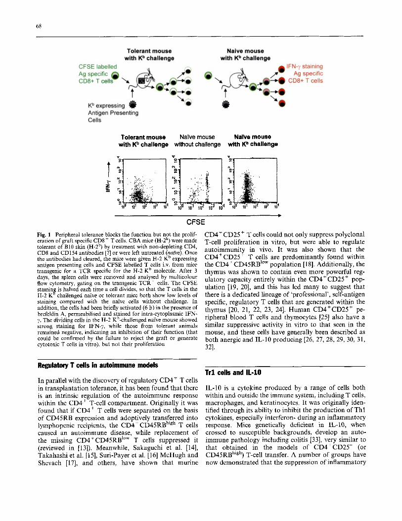
In the absence of appropriate in vitro readouts, much effort was put into in vivo systems to study peripheral tolerance. It soon became clear that powerful CD4+ regulatory T cells enforce tolerance after adoptive transfer into secondary recipients. Although tolerance is dominant in this situation [9], with the secondary CD4+ T cells themselves becoming tolerant and regulatory (‘infectious tolerance’), there is no elimination of the antigen-specific effector T cells: depletion of the regula- tory CD4+ population can reveal primed, CD8+ T cell- mediated rejection, suggesting that there is an element of active suppression [lo]. The mechanisms of such sup- pression remain elusive, even if we apply modern tech- nology to follow T cell activity in situ. For example, it has recently been shown that donor graft antigen-spe- cific CD8+ T cells can proliferate normally in tolerant recipients (using CFSE tracking studies-see Fig. l), although they fail to develop effector functions and do not reject the graft [7].
linked suppression of graft rejection
Possibly the most convincing and perhaps the only reli- able assay for regulatory cell activity in transplantation models is that of linked suppression [l 1 , 121 in the original recipient. Although the tolerant mouse can reject third- party grafts, demonstrating that there is no non-specific immunosuppression and that tolerance has overall donor specificity, a graft from an F1 cross between the donor and a third party is only rejected slowly or may be fully accepted. Acceptance of this F1 graft then leads to full tolerance of the third-party graft, with no other external manipulation of the immune system. This suggests that having the two antigens brought closely together, either on the same antigen-presenting cell or target tissue, elicits donor-directed regulatory CD4+ T cells to drive toler- ance in third-party specific T cells.
Linked suppression is not only highly significant for the understanding of the mechanisms of tolerance, it also has important therapeutic implications. It may not be necessary to induce tolerance to every transplanta- tion antigen in order to achieve graft acceptance if there is sufficient regulatory T-cell activity against a propor- tion of the graft antigens. It might therefore be possible to induce strong regulatory T-cell tolerance to certain common MHC antigens in advance of a transplant becoming available, perhaps reducing the requirement for aggressive tolerogenic therapies at the time of grafting.
68
Tolerant mouse with Kb challenge
Naive mouse with Kb challenge
CFSE labelled Ag specific CD8+ T cell
~b expressing I) Antigen Presenting Cells
Tolerant mouse with Kb challenge
i
Naive mouse Nabe mouse without challenge with Kb challenge
4
* CFSE
Fig. 1 Peripheral tolerance blocks the function but not the prolif- eration of graft specific CD8+ T cells. CBA mice (H-2k) were made tolerant of B10 skin (H-2b) by treatment with non-depleting CD4, CD8 and CD154 antibodies [7] or were left untreated (naive). Once the antibodies had cleared, the mice were given H-2 Kb expressing antigen presenting cells and CFSE labelled T cells i.v. from mice transgenic for a TCR specific for the H-2 Kb molecule. After 3 days, the spleen cells were removed and analysed by multicolour flow cytometry, gating on the transgenic TCR+ cells. The CFSE stainin is halved each time a cell divides, so that the T cells in the H-2 K challenged naTve or tolerant mice both show low levels of staining compared with the naive cells without challenge. In addition, the cells had been briefly activated (6 h) in the presence of brefeldin A, permeabilised and stained for intra-cytoplasmic IFN- y. The dividing cells in the H-2 Kb-challenged naYve mouse showed strong staining for IFN-y, while those from tolerant animals remained negative, indicating an inhibition of their function (that could be confirmed by the failure to reject the graft or generate cytotoxic T cells in vitro), but not their proliferation
%
CD4+CD25+ T cells could not only suppress polyclonal T-cell proliferation in vitro, but were able to regulate autoimmunity in vivo. It was also shown that the CD4+CD25 + T cells are predominantly found within the CD4+CD45RB1OW population [18]. Additionally, the thymus was shown to contain even more powerful reg- ulatory capacity entirely within the CD4+CD25+ pop- ulation [19, 201, and this has led many to suggest that there is a dedicated lineage of 'professional', self-antigen specific, regulatory T cells that are generated within the thymus [20, 21, 22, 23, 241. Human CD4+CD25+ pe- ripheral blood T cells and thymocytes [25] also have a similar suppressive activity in vitro to that seen in the mouse, and these cells have generally been described as both anergic and IL-I0 producing [26, 27, 28, 29, 30, 31, 321.
Regulatory T cells in autoimmune models
In parallel with the discovery of regulatory CD4+ T cells in transplantation tolerance, it has been found that there is an intrinsic regulation of the autoimmune response within the CD4+ T-cell compartment. Originally it was found that if CD4+ T cells were separated on the basis of CD45RB expression and adoptively transferred into lymphopenic recipients, the CD4+ CD45RBhigh T cells caused an autoimmune disease, while replacement of the missing CD4+CD45RB1"" T cells suppressed it (reviewed in [13]). Meanwhile, Sakaguchi et al. [14], Takahashi et al. [15], Suri-Payer et al. [16] McHugh and Shevach [17], and others, have shown that murine
Tr l cells and 11-10
IL-10 is a cytokine produced by a range of cells both within and outside the immune system, including T cells, macrophages, and keratinocytes. It was originally iden- tified through its ability to inhibit the production of Thl cytokines, especially interferon- during an inflammatory response. Mice genetically deficient in IL-10, when crossed to susceptible backgrounds, develop an auto- immune pathology including colitis [33], very similar to that obtained in the models of CD4+CD25- (or CD45RBhigh) T-cell transfer. A number of groups have now demonstrated that the suppression of inflammatory

69
bowel disease by CD4+CD25+ regulatory T cells in vivo is dependent on the presence of IL-I0 [34, 351.
It is possible to polarise na CD4+ T cells by antigen (or non-specific anti-CD3) stimulation in vitro in the presence of high levels of recombinant IL-10 [35]. This may also require TGF-P [36], although this is often al- ready present in the serum used for cell cultures. This stimulation produces a mixed population of IL-4 and IL-10 producing Th2 as well as IL-10 only producing Trl cells. The latter can be cloned, particularly by use of high levels of solid phase anti-CD3, which generally causes T cell apoptosis but to which Trl cells are rela- tively resistant. Alternatively, the stimulation of naive T cells in the presence of two drugs, dexamethasone and vitamin D3, and the additional neutralisation of IL-12, IFN-y and IL-4, can reliably polarise cultures to this IL- 10 only-producing Trl phenotype [37]. Trl cells against the antigen ovalbumin, generated by either method, have been shown to be capable of suppressing autoim- mune colitis or experimentally induced acute encepha- lomyelitis in vivo caused by CD4+ CD45RB'O" cells, when the animals are given oral ovalbumin as a stimulus for the Trl cells [35, 371. Human Trl cells can also be generated in vitro by stimulation in the presence of IL- 10 and interferon-a [38].
There have also been reports that IL-10 is required for the regulatory activity that can be transferred after CD4 antibody-induced transplantation tolerance [39], but this tolerance, linked suppression and infectious tolerance cannot be broken by anti-IL10 or anti-ILlOR monoclonal antibodies in vivo in the original recipient, even when they are re-challenged with fresh grafts [40]. However, Tr 1 -like T-cell clones generated in vitro against the male antigen, as presented by MHC-11, are able to block skin graft rejection by either Thl [41] or Th2 (manuscript unpublished) clones against the same antigen, after adoptive transfer into T cell-deficient recipient mice.
population, in a manner similar to that described for CD4+CD25+ regulatory cells [49]. However, it has recently been shown that CD4+CD25+ cells from TGF-PI knockout mice are as effective as those from normal mice at suppressor function in vitro, and that T cells from TGF-P unresponsive Smad3-'- or dominant negative TGF-P type-2 receptor transgenic mice are effective targets of suppression in vitro [50]. Others have demonstrated that CD4+CD25+ T cells are able to suppress proliferation in vitro even after fixation, ruling out the need for any secreted product in this assay, suggesting that cell contact mechanisms may be suffi- cient [51, 521. There have been some reports that this could be due to TGF-P expressed on the surface of regulatory T cells [53, 541, but this remains controversial.
Contactdependent mechanisms of regulatory T cells
CD4+CD25' and Trl regulator cells also both con- stitutively express on their surface CD25, CDI 52 (CTLA4) and, most recently, the glucocorticoid-induc- ible TNF receptor (TNFRsfl8 or GITR) [41,55,56]. All three have been implicated in regulatory function. IL-2 [57] or CD25 [58] knockout mice develop a lympho- proliferative disease similar to that in some of the autoimmune models. Similarly, CD 1 52-deficient mice develop autoimmune pathology [59], and Fab fragments of anti-CTLA4 monoclonal antibodies can block sup- pression by CD4+CD25+ cells in vitro and in vivo [15, 181. Both IL-10 and CTLA-4 have been implicated in the transferable suppression associated with tolerance to allogeneic murine cardiac grafts [60], but not to skin grafts [40]. Most recently, antibodies to GITR have also been shown to block regulatory function in vitro and in the lymphopenic autoimmune models [55, 561.
Th3 cells and transforming growth factor-fi
Many of the autoimmune models that show a role for IL-10 in suppression also implicate TGF-P [42,43, 441, a cytokine that has a strong ability to block the differen- tiation of T cells towards either Thl or Th2 responses. TGF-P seems to be particularly abundant in the anterior chamber of the eye, and seems to play an important part in the induction of tolerance that can be obtained to both protein and histocompatibility antigens introduced through this route [45, 461.
A CD4+ T-cell subset, sometimes called Th3, has been described, where TGF-P is a major cytokine pro- duced [47]. Mice made deficient in TGF-P1 develop an inflammatory disease [48], and this may be a factor needed for the generation of the IL-10-producing Trl
Does infectious tolerance require both contact and cytokines?
Two recent papers claim to clarify the conflicting data on the requirements for cell contact compared to soluble mediators [51, 521. First, the secreted products such as cytokines are excluded by fixing the initial CD4+ CD25 +
suppressive population after CD3- and CD28-induced activation. When these cells are tested in mixture experiments in vitro, the normally responsive CD4+ CD25- population is not only suppressed but also be- comes anergic and able to suppress further CD4+ T cells in a contact-independent manner through the produc- tion of cytokines. Although the two papers disagree on which cytokine is most important for this secondary suppression, one claims it is IL-10 and not TGF-8 [51] while the other claims the opposite [52], these may be
70
examples where multiple or sequential mechanisms can act together to increase the potency of regulatory T cells. It is also possible that similar mechanisms operate in bystander or linked suppression, and infectious toler- ance in vivo. However, the in vitro data are based on the proliferation of polyclonal populations of T cells and are not antigen presenting cell dependent, while linked suppression and infectious tolerance in non-lymphope- nic adult mice seem to be antigen specific, dependent on how the antigen is presented, and do not involve a defect in T cell proliferation. We therefore have to consider very carefully the evidence for any major role of CD4+ CD25 + regulatory T cells in transplantation tol- erance.
Are regulatory T cells in peripheral transplantation tolerance CD4+CD25+?
There are now a few published examples where CD4+CD25+ regulatory T cells would seem to transfer transplantation tolerance. Davies et al. were able to suppress the rejection of allogeneic islets by CD4+ CD45RBhigh cells by co-transfer of CD4+ CD45RB'"" T cells from naive donors [61]. Wood and colleagues have claimed that antigen-specific tolerance generated to MHC-mismatched cardiac or skin grafts can be trans- ferred with CD4+CD25+ T cells (Hara et al. [39 and Kingsley et al. 601). It has also been shown that CD4'CD25+ T cells can be generated in vitro in a donor-versus-host mixed lymphocyte reaction under the cover of antibodies to CD40L (CD154), and that these are able to suppress graft-versus-host disease (GVHD) after bone-marrow transplantation [62]. Others have shown that simply increasing the proportion of donor CD4+CD25+ T cells in the marrow inoculum may be sufficient to suppress GVHD [63]. Such studies are in- teresting, particularly with regard to the potential of regulatory T cells that might be used for adoptive cell therapies as a means to induce tolerance clinically.
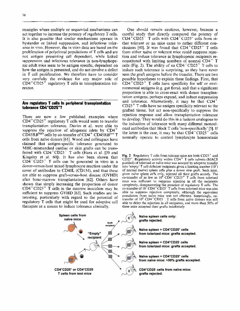
One should remain cautious, however, because a careful study that directly compared the potency of CD4+CD25+ T cells with CD4+CD25- cells from ei- ther tolerant or na mice came to rather different con- clusions [40]. It was found that CD4+CD25+ T cells from either naive or tolerant mice could suppress rejec- tion and induce tolerance in lymphopenic recipients re- constituted with limiting numbers of normal CD4+ T cells (Fig. 2). The ability of na CD4+CD25+ T cells to induce such tolerance is surprising, as they have never seen the graft antigens before the transfer. There are two possible hypotheses to explain these findings. First, that CD4+CD25+ T cells have specificity for self or envi- ronmental antigens (e.g. gut flora), and that a significant proportion is able to cross-react with donor transplan- tation antigens, perhaps expand, and induce suppression and tolerance. Alternatively, it may be that CD4+ CD25 + T cells have no antigen specificity relevant to the grafted tissue, but act non-specifically to suppress the rejection response and allow transplantation tolerance to develop. They would do this in a fashion analogous to the induction of tolerance with many different monocl- onal antibodies that block T cells 'non-specifically' [5]. If the latter is the case, it may be that CD4+CD25+ cells normally operate to control lymphocyte homeostasis
Fig. 2 Regulatory T cells from tolerant mice are both CD25+ and CD25-. Regulatory activity within CD4+ T cells subsets (MACS purified) of tolerised or naive mice was assayed by adoptive transfer into 'empty' T cell-deficient recipients given a limiting number (lo7) of normal (naive) spleen cells plus a donor skin graft. Such mice, given nake spleen cells on18 rejected all their grafts acutely. The co-transfer of as few as 10 CD4+CD25+ T cells from tolerised mice was sufficient to suppress rejection in all the recipients completely, demonstrating the presence of regulatory T cells. The co-transfer of lo7 CD4+CD25- T cells from tolerised mice was also able to suppress rejection completely, although the equivalent population from nai've mice was not effective. Surprisingly, co- transfer of lo6 CD4+CD25+ T cells from naive donors was still able to delay the rejection in all recipients, and more than 50% of these mice accepted their grafts indefinitely
71
[64] and have no specific role in peripheral transplanta- tion tolerance.
We therefore have to consider if there is evidence for any new population of regulatory T cells that can be observed in tolerant but not naive mice. In their exper- iments, Graca et al. did indeed observe a new population of CD4+CD25- cells with regulatory activity only from tolerant mice [40]. Although these were less potent ‘per cell’ than CD4+CD25+ cells when titrated, we do not know the frequency of graft reactive cells within the two populations, and there are ten times as many CD4+CD25- cells in the tolerant animals. This raises the possibility that this CD4’CD25- population con- tains the induced, antigen-specific regulatory T cells that are responsible for infectious transplantation tolerance and linked suppression. Interestingly, these observations are compatible with some recent data from TCR trans- genic mice made tolerant of their cognate peptide anti- gen [65]. In these mice it was found that CD4+CD25+ regulatory T cells were derived from the thymus, while CD4+CD25- regulatory T cells were generated by pe- ripheral presentation of antigen for tolerance.
The experiments by Gavin et al. [66] may shed some light on the relationship between the two subsets of regulatory T cells. It was found that if CFSE-labelled CD4+CD25 + T cells were transferred into lymphopenic recipients alone, then they proliferated (either homeo- statically or to self- or environmental antigens) but in the process became CD4+CD25-. Such expanded, now CD4+CD25-, T cells seemed even more potent (on a per-cell basis) at suppressing proliferation of na T cells in vitro. Similarly, in the transplantation tolerance models, treatment with non-depleting CD4 or other monoclonal antibodies may allow the expansion of a CD4 + CD25 +, graft cross-reactive population to gen- erate a more potent CD4’CD25- antigen-specific reg- ulatory T cell.
Gene expression analyses of regulatory T cells
One of the major limitations to both our understanding of the mechanisms in the experimental models and our ability to induce tolerance in the clinic is that we still have no good markers for tolerance or regulatory T cells. All the molecules that have been discussed, such as CD25, CTLA4, GITR, and cytokines, are also expressed by recently activated effector T cells. We really need to identify molecules that are uniquely expressed by regu- latory T cells, both to identify them for diagnostic pur- poses and to focus on their mechanism(s) of action.
DNA microarrays or ‘gene chips’ [67] and serial analysis of gene expression (SAGE) [68] are similar in that they simultaneously measure the expression pattern of many thousands of gene mRNA transcripts and try to identify how this changes from one cell or tissue sample
to another, preferably using as many relevant samples as possible. In principle, this allows the identification of specific clusters of genes that are up or down regulated in just the cells of interest, such as the regulatory T cells.
A direct comparison, by SAGE, between murine Thl, Th2 and Trl cells with identical T-cell receptors against the male transplantation antigen (HY) presented by MHC-11, has been published [41, 691. Perhaps surpris- ingly, there were very few, if any, transcripts that were uniquely expressed by the regulatory Trl clone, al- though a number of genes expressed in Th2 cells were up-regulated. More significantly, Trl cells had lost many transcripts associated with, either directly or as tran- scription factors for, effector functions of Thl and Th2 cells. Examples included the loss, by Trl cells, of Th2 transcription factors GATA-3 and Egr-1, and Thl ef- fector molecules RANTES and Ly6C. It is possible that the most important characteristic of regulatory T cells is this loss of effector functions, while retaining an ability to recognise antigen and compete with na or memory T cells for antigen, APC or cytokines (the ‘civil-service model’ [5]). Additionally, they may retain molecules that normally intrinsically limit the clonal expansion or ag- gressive functions of effector Thl or Th2 cells, such as CTLA4.
Microarray analyses of both activated CD4+ CD25 +
T cells [56, 661 and SAGE analyses of Trl clones [41, 691 have identified a number of genes in common that are increased on regulatory T cells. These include GITR (mentioned above), 0x40, preproenkephalin, aE/P7 (CD103), and granzyme A. However, all of these are ex- pressed by Th2 cells as well [41]. We therefore still have no specific molecular marker for regulatory T cells, and it may be for diagnostic purposes that we will have to devise assays based on a differential loss of effector molecules.
The role of the antigen-presenting cell
One hypothesis that may explain linked suppression is that anergic or regulatory T cells are able to down- regulate co-stimulatory ligands on antigen-presenting cells [5 , 70, 711. In order for CD80 and CD86 to be up- regulated during an immune response, the APC must first be given a signal to mature, predominantly through the CD40 interaction with CD40 ligand on activated CD4+ T cells. However, the expression of CD40 is itself tightly controlled in the APC at both the transcriptional level and in the production of specific splice variants of the CD40 message [72], depending on whether inflam- matory signals have also been received through the Toll family of receptors for various pathogen products such as LPS or CpG [73].
As we begin to understand more about the relation- ship between T cells and antigen-presenting cells, there is a growing interest in the possibility that there might be

12
natural lineages or subsets of dendritic cells specialised in presenting antigen to naive T cells (a unique feature of dendritic cells) for tolerance. Indeed, immature dendritic cells may themselves be inherently tolerogenic in the absence of inflammatory and maturation stimuli [74]. Many groups now attempt to identify agents that can modify or lock the dendritic cell activity into this puta- tive tolerogenic phenotype. Probably the two most ad- vanced candidates are IL-10 and l a 25-dihydroxy vitamin D3. We have already considered IL-10 as a product of anergic or regulatory T cells, but it also has profound effects on dendritic cell maturation. IL-10 treated, immature bone-marrow derived dendritic cells fail to mature normally in response to inflammatory stimuli and are unable to stimulate a mixed lymphocyte reaction in vitro [75]. Vitamin D3- (or various metabo- lite- and analogue-) modulated dendritic cells are also able to induce tolerance and evidence of regulatory T cells to allogenenic islet transplants given under the cover of mycophenolate mofetil [76]. Dendritic cells can be further modulated by treatment with proteasome inhibitors to preferentially present antigen for regulatory T cells [77].
There have also been some interesting developments in the genetic manipulation of antigen-presenting cells.
Fig. 3 Regulatory T cells are found within tolerated skin grafts. The presence of regulatory T cells within tolerated skin grafts was demonstrated by transferring the grafts onto ’empty’ T cell deficient mice and waiting 30 days to allow any T cells carried over in the graft to repopulate the recipient. Any repopulating T cells in these recipients were then tested for their regulatory capacity by giving a fresh donor skin graft together with limiting numbers of na‘ive spleen cells. It was found that tolerated grafts could indeed carry over regulatory cells that could induce donor- specific tolerance in the recipient, and that these were clearly T cells (as T cell depletion at the time of graft transfer eliminated the suppression). By additional controls, it was shown that any T cells carried over by normal syngeneic skin from a tolerised donor, or grafts that would have proceeded to rejection on non-tolerant donors, were unable to suppress rejection by the naYve spleen cells
The evolutionary conserved Notch1 cell surface receptor plays an important role in a wide range of develop- mental decisions [78], including T-cell development in the thymus. Over-expression of Serrate1 (a ligand for Notchl) in antigen-pulsed APCs is able to generate tolerance [79], regulatory cells, and linked suppression in murine models of house-dust-mite allergy [80]. Interest- ingly, the regulatory T cells also seem to up-regulate additional Notch ligands that may play a role in contact- mediated signalling to naive T cells for infectious toler- ance, and similar changes can be observed after activa- tion of CD4+CD25+ regulatory T cells [28]. The data implicating Notch in tolerance are, at present, indirect, and have not yet been supported by any direct evidence of a physiological role for Notch family molecules in tolerance.
_____
The local graft environment
It is, therefore, clear that there is a synergistic inter- action between the regulatory T cells and modulated APCs in the generation and maintenance of peripheral tolerance. The recent demonstration that regulatory T cells are concentrated within a tolerated graft [8 11 (see Fig. 3) suggests that the graft micro-environment may be very important for the maintenance of tolerance. It is possible that regulatory T cells function predomi- nantly within the graft itself, perhaps explaining why we have been unable to detect any differences in pro- liferation or cytokine secretion between tolerant and rejecting recipients, either in vitro or ex vivo, using spleen or draining lymph node cells as a source of ‘tolerant’ T cells [8].
Most tissues have a variety of protective responses to various types of stress, including immune attack. For example, there are pathways to protect cells from reac- tive oxygen species generated as by-products of oxida- tive metabolism that are also used as cytocidal agents by

73
activated macrophages [82]. Indeed, it has been sug- gested that the up-regulation of one of these protective proteins, haemoxygenase, may be associated with tol- erance to cardiac grafts in CDCtreated mice [83]. Sim- ilarly, the ligand for CD95 (FasL) is expressed in immuno-privileged sites [84], such as the testes, although no role for Fas-FasL interactions has been found in the transplantation tolerance or suppression induced by anti-CD4 treatment [85].
Summary and conclusions
Peripheral tolerance depends on regulatory CD4+ T cells that interact with and modulate both the antigen- presenting cells and the graft local environment. We do not yet have any good markers or in vitro assays for regulatory T cells, nor do we really understand how they are induced or how they function. If we look at the range of monoclonal antibodies that have been claimed to induce peripheral tolerance or regulatory T cells [5], they include TCR and co-receptor specificities (non-
activating CD3, CD4, CD8), co-stimulatory blockade (CD40L, CTLA4-Ig, CD80 and CD86), adhesion mol- ecules (LFA-l, CD2) and cytokine receptors (CD25 or IL-2 receptor). One possibility is that the specificity is largely irrelevant, as long as the immune system is blocked from acute rejection and allowed to generate tolerance for itself. Indeed, this may be the way that some of the experiments that achieve tolerance through the transfer of (non-specific) CD4+CD25 + cells are operating, and further effort may identify safe ways to use adoptive regulatory cell therapies clinically. In gen- eral, we need to identify those agents, either among the currently available immunosuppressants, or newer agents such as monoclonal antibodies, that work to- gether to block rejection but promote the generation of regulatory T cells. Most important of all, we still need to identify surrogate markers for regulatory T cells and tolerance that allow effective monitoring of the patient after a transplant, and these are most likely to come from detailed gene-expression studies of appropriate T-cell populations.
References
1. Billingham RE, Brent, L, Medawar, PB (1953) Acquired immunological toler- ance to foreign cells. Nature 172:603
Kyburz D, Hengartner H, Zinkernagel RM (1992) Clonal deletion induced by either radioresistant thymic host cells or lymphohemopoietic donor cells at dif- ferent stages of class I-restricted T cell ontogeny. J Exp Med 175:1277
results using CAMPATH-1 antibodies to control GVHD and graft rejection. Bone Marrow Transplant 17:305
4. Li Y, Li XC, Zheng XX, Wells AD, Turka LA, Strom TB (1999) Blocking both signal 1 and signal 2 of T-cell ac- tivation prevents apoptosis of alloreac- tive T cells and induction of peripheral allograft tolerance. Nat Med 5:298
5. Waldmann H, Cobbold S (2001) Regu- lating the immune res onse to trans- plants; a role for CD4 regulatory cells? Immunity 14:399
6. Chen ZK, Cobbold SP, Waldmann H, Metcalfe S (1996) Amplification of natural regulatory immune mechanisms for transplantation tolerance. Trans- plantation 62: 1200
Waldmann H (2002). Dominant trans- plantation tolerance impairs CD8' T cell function but not expansion. Nat Immunol3:1208
2. Speiser DE, Pircher H, Ohashi PS,
3. Hale G, Waldmann H (1996) Recent
P
7. Lin C-Y, Graca L, Cobbold SP,
8. Cobbold SP, Adams E, Marshall SE, Davies JD, Waldmann H (1996). Mechanisms of peripheral tolerance and suppression induced by monoclonal antibodies to CD4 and CD8. Immunol Rev 1493
9. Qin S, Cobbold SP, Pope H (1993) 'Infectious' transplantation tolerance. Science 259:974
10. Marshall SE, Cobbold SP, Davies JD, Martin GM, Phillips JM, Waldmann H (1 996) Tolerance and suppression in a primed immune system. Transplanta- tion 62:1614
11. Davies JD, Leong LY, Mellor A, Cob- bold SP, Waldmann H (1996) T cell suppression in transplantation tolerance through linked recognition. J Immunol 156:3602
12. Wise MP, Bemelman F, Cobbold SP, Waldmann H (1998) Linked suppres- sion of skin graft rejection can operate through indirect recognition. J Immu- no1 161:5813
13. Read S, Powrie F (2001) CD4+ regu- latory T cells. Curr Opin Immunol 13544
14. Sakaguchi S, Sakaguchi N, Asano M, Itoh M, Toda M (1995) Immunologic self-tolerance maintained by activated T cells expressing IL-2 receptor alpha- chains (CD25). Breakdown of a single mechanism of self-tolerance causes var- ious autoimmune diseases. J Immunol 155: 1151
15. Takahashi T, Tagami T, Yamazaki S (2000) Immunologic self-tolerance maintained by CD25 +CD4+ regulatory T cells constitutively expressing cyto- toxic T lymphocyte-associated antigen 4. J Exp Med 192:303
16. Suri-Payer E, Amar AZ, Thornton AM (1998) Shevach EM. CD4+CD25+ T cells inhibit both the induction and ef- fector function of autoreactive T cells and represent a unique lineage of im- munoregulatory cells. J Immunol 160: 12 12
17. McHugh RS, Shevach EM (2002) Cut- ting edge: depletion of CD4+CD25+ regulatory T cells is necessary, but not sufficient, for induction of organ-spe- cific autoimmune disease. J Immunol 1685979
18. Read S, Malmstrom V, Powrie F (2000) Cytotoxic T lymphocyte-associated an- tigen 4 plays an essential role in the function of CD25+CD4' regulatory cells that control intestinal inflamma- tion. J Exp Med 192:295
19. Stephens LA, Mason D (2000) CD25 is a marker for CD4+ thymocytes that prevent autoimmune diabetes in rats, but peripheral T cells with this function are found in both CD25+ and CD25- subpopulations. J Immunol 165:3 105
74
20. Hori S, Haury M, Coutinho A, Demengeot J (2002). Specificity requirements for selection and effect or functions of CD25+4+ regulatory T cells in anti-myelin basic protein T cell receptor transgenic mice. Proc Natl Acad Sci U S A 99:8213
21. Jordan MS, Boesteanu A, Reed AJ (2001) Thymic selection of CD4 CD25+ regulatory T cells in- duced by an agonist self-peptide. Nat Immunol 2:301
22. Kawahata K, Misaki Y, Yamauchi M (2002) Generation of CD4+CD25+ regulatory T cells from autoreactive T cells simultaneously with their negative selection in the thymus and from nonautoreactive T cells by endoge- nous TCR expression. J Immunol 168:4399.
CD4+CD25+ T-cell develoDment is 23. Suto A, Nakajima H, Ikeda K (2002)
.~
regulated by at least 2 distiAct mecha- nisms. Blood 99555
24. Pacholczyk R, Kraj P, Ignatowicz L (2002) Peptide specificity of thymic selection of CD4+CD25+ T cells. J Immunol 168:613
25. Stephens LA, Mottet C, Mason D, Powrie F (2001) Human CD4+CD25+ thymocytes and peripheral T cells have immune suppressive activity in vitro. Eur J Immunol3 1 : 1247
Roncarolo MG (1996) Interleukin-10 induces a long-term antigen-specific anergic state in human CD4+ T cells. J Exp Med 184:19
27. Levings MK, Sangregorio R, Ronca- rolo MG (2001) Human CD25+CD4+ T regulatory cells suppress naive and memory T cell proliferation and can be expanded in vitro without loss of func- tion. J Exp Med 193:1295
28. Ng WF, Dugfan PJ, Ponchel F (2001) Human CD4 CD25+ cells: a naturally occurring population of regulatory T cells. Blood 98:2736
Tuettenberg A, Knop J, Enk AH (2001) Identification and functional character- ization of human CD4ICD25' T cells with regulatory properties isolated from peripheral blood. J Exp Med 193:1285
30. Dieckmann D, Plottner H, Berchtold S, Berger T, Schuler G (2001) Ex-vivo isolation and characterization of CD4+CD25+ T cells with regulatory properties from human blood. J Exp Med 193:1303
3 1. Baecher-Allan C, Brown JA, Freeman GJ, Hafler DA (2001) CD4+CD25 high regulatory cells in human peripheral blood. J Immunol 167:1245
26. Groux H, Bigler M, de Vries JE,
29. Jonuleit H, Schmitt E, Stassen M,
32. Taams LS, Smith J, Rustin MH, Salm- on M, Poulter LW, Akbar AN (2001) Human anergic/suppressive CD4+CD25 T cells: a highly differ- entiated and apoptosis-prone popula- tion. Eur J Immunol 3 1 : 1 122
33. Davidson NJ, Leach MW, Fort MM (1996) T helper cell 1-type CD4+ T cells, but not B cells, mediate colitis in interleukin 10-deficient mice. J Exp Med 184:241
Coffman RL, Powrie F (1999) An es- sential role for interleukin 10 in the function of regulatory T cells that in- hibit intestinal inflammation. J Exp Med 190:995
35. Groux H, O'Garra A, Bigler M (1997) A CD4+ T-cell subset inhibits antigen- specific T-cell responses and prevents colitis. Nature 389:737
Murphy WJ, et al. (1999) Induction of CD4+ T cell alloantigen-specific hypo- responsiveness by IL-10 and TGF-beta. J Immunol 163:3684
37. Barrat FJ, Cua DJ, Boonstra A (2002) In-vitro generation of interleukin 10- producing regulatory CD4' T cells is induced by immunosuppressive drugs and inhibited by T helper type 1 (Th1)- and Th2-inducing cytokines. J Exp Med 195:603
38. Levings MK, Sangregorio R, Galbiati F, Squadrone S, de Waal Malefyt R, Roncarolo MG (2001) IFN-alpha and IL-10 induce the differentiation of hu- man type 1 T regulatory cells. J Immu- no1 1665530
39. Hara M, Kingsley CI, Niimi M (2001) IL-10 is required for regulatory T cells to mediate tolerance to alloantigens in vivo. J Immunol 166:3789
40. Graca L, Thompson S, Lin CY, Adams E, Cobbold SP, Waldmann H (2002) Both CD4+CD25+ and CD4+CD25(-) regulatory cells mediate dominant transplantation tolerance. J Immunol 1685558
41. Zelenika D, Adams E, Humm S (2002) Regulatory T cells overexpress a subset of Th2 gene transcripts. J Immunol 168:1069
42. Shevach EM, McHugh RS, Piccirillo CA, Thornton AM (2001) Contro) of T-cell activation by CD4+ CD25 suppressor T cells. Immunol Rev 182:58
43. Han HS, Jun HS, Utsugi T, Yoon JW (2001) Molecular role of TGF-P, secreted from a new type of CD4+ suppressor T cell, "4.2, in the pre- vention of autoimmune IDDM in NOD mice. J Autoimmun 10:299
44. Zhang X, Izikson L, Liu L, Weiner HL (2001) Activation of CD25+CD4+ regulatory T cells by oral antigen administration. J Immunol 167:4245
34. Asseman C, Mauze S, Leach MW,
36. Zeller JC, Panoskaltsis-Mortari A,
45. Kosiewicz MM, Alard P, Streilein JW (1998) Alterations in cytokine produc- tion following intraocular injection of soluble protein antigen: impairment in IFN-gamma and induction of TGF-and IL-4 production. J Immunol 1615382
(1997) Induction of donor-specific AC- AID can prolong orthotopic corneal allograft survival in 'high-risk' eyes. Curr Eye Res 16:1171
mechanism of action of transforming growth factor-beta-secreting Th3 regu- latory cells. Immunol Rev 182:207
48. Shull MM, Ormsby I, Kier AB (1992) Targeted disruption of the mouse transforming growth factor-beta 1 gene results in multifocal inflammatory dis- ease. Nature 359:93
49. Yamagiwa S, Gray JD, Hashimoto S, Honvitz DA (2001) A role for TGF-/I in the generation and expansion of CD4+CD25+ regulatory T cells from human peripheral blood. J Immunol 166:7282
50. Piccirillo CA, Letterio JJ, Thornton AM (2002) CD4+CD25+ regulatory T cells can mediate suppressor function in the absence of transforming growth factor beta1 production and respon- siveness. J Exp Med 196:237
51. Dieckmann D, Bruett CH, Ploettner H, Lutz MB, Schuler G (2002) Human CD4+CD25+ regulatory, contact- dependent T cells induce interleukin 10-producing, contact-independent type 1-like regulatory T cells [corrected]. J Exp Med 196:247
52. Jonuleit H, Schmitt E, Kakirman H, Stassen M, Knop J, Enk AH (2002) Infectious tolerance: human CD25 regulatory T cells convey suppressor activity to conventional CD4 T helper cells. J Exp Med 196:255
(200 1) Cell contact-dependent immu- nosuppression by CD4+CD25+ regu- latory T cells is mediated by cell surface- bound transforming growth factor beta. J Exp Med 194:629
54. Roelen D, van Bree S, van Hulst P, van Beelen E, Claas F (2002) Regulatory functions of human CD4+ T cells rec- ognizing allopeptides in the context of self-HLA class 11. Hum Immunol 63:902
55. Shimizu J, Yamazaki S, Takahashi T, Ishida Y, Sakaguchi S (2002) Stimula- tion of CD25+CD4+ regulatory T cells through GITR breaks immunological self-tolerance. Nat Immunol 2002 3: 135
56. McHugh RS, Whitters MJ, Piccirillo CA (2002) CD4+CD25+ immunoregu- latory T cells: gene expression analysis reveals a functional role for the gluco- corticoid-induced TNF receptor. Immunity 16:311
46. Sano Y, Okamoto S, Streilein JW
47. Weiner HL (2001) Induction and
53. Nakamura K, Kitani A, Strober W
75 ~~~ ~
57. Horak I, Lohler J, Ma A, Smith KA (1995) Interleukin-2 deficient mice: a new model to study autoimmunity and self-tolerance. Immunol Rev 148:35
58. Willerford DM, Chen J, Ferry JA, Davidson L, Ma A, Alt FW (1995) Interleukin-2 receptor alpha chain regulates the size and content of the peripheral lymphoid compartment. Immunity 3521
59. Tivol EA, Borriello F, Schweitzer AN, Lynch WP, Bluestone JA, Sharpe AH (1995) Loss of CTLA-4 leads to massive lymphoproliferation and fatal multior- gan tissue destruction, revealing a criti- cal negative regulatory role of CTLA-4. Immunity 3541
Wood KJ (2002) CD25+CD4+ regula- tory T cells prevent graft rejection: CTLA-4- and IL-10-dependent immu- noregulation of alloresponses. J Immu- no1 168:1080
61. Davies JD, O'Connor E, Hall D, Krahl T, Trotter J, Sarvetnick N (1999) CD4+ CD45RB low-density cells from un- treated mice prevent acute allograft re- jection. J Immunol 1635353
62. Taylor PA, Lees CJ, Blazar BR (2002) The infusion of ex vivo activated and expanded CD4+CD25+ immune regu- latory cells inhibits graft-versus-host disease lethality. Blood 99:3493
63. Hoffmdnn P, Ermann J, Edinger M, Fathman CG, Strober S (2002) Donor- type CD4+CD25+ regulatory T cells suppress lethal acute graft-versus-host disease after allogeneic bone marrow transplantation. J Exp Med 196:389
64. Annacker 0, Pimenta-Araujo R, Bur- len-Defranoux 0, Barbosa TC, Cuma- no A, Bandeira A (2001) CD25+ CD4+ T cells regulate the expansion of pe- ripheral CD4 T cells through the pro- duction of IL-10. J Immunol 166:3008
65. Apostolou I, Sarukhan A, Klein L, von Boehmer H (2002) Origin of regulatory T cells with known specificity for anti- gen. Nat Immunol 3:756
Gallegos A, Rudensky A (2002) Ho- meostasis and anergy of CD4+CD25+ suppressor T cells in vivo. Nat Immunol 3:33
60. Kingsley CI, Karim M, Bushell AR,
66. Gavin MA, Clarke SR, Negrou E,
61. Harrington CA, Rosenow C, Retief J (2000) Monitoring gene expression us- ing DNA microarrays. Curr Opin Mi- crobiol 3:285
68. Velculescu VE, Zhang L, Vogelstein B, Kinzler KW (1995) Serial analysis of gene expression. Science 270:484
69. Zelenika D, Adams E, Humm S, Lin CY, Waldmann H, Cobbold SP (2001) The role of CD4+ T-cell subsets in de- termining transplantation rejection or tolerance. Immunol Rev 182:164
E, Dyson J, Scott D (2002) CD4+CD25+ T cells as immunoregu- latory T cells in vitro. Eur J Immunol 32:2365
71. Cederbom L, Hall H, Ivars F (2000) CD4+CD25+ regulatory T cells down- regulate co-stimulatory molecules on antigen-presenting cells. Eur J Immunol 30: 1538
72. Tone M, Tone Y, Fairchild PJ, Wykes M, Waldmann H (2001) Regulation of CD40 function by its isoforms generat- ed through alternative splicing. Proc Natl Acad Sci U S A 98:1751
73. Underhill DM, Ozinsky A. (2002) Toll-like receptors: key mediators of microbe detection. Curr Opin Immunol 14:103
sovsky J, Munz C, Bhardwaj N (2001) Antigen-specific inhibition of effector T cell function in humans after injection of immature dendritic cells. J Exp Med 193:233
Knop J, Enk AH (2002) CD4+ and CD8+ anergic T cells induced by inter- leukin-1 0-treated human dendritic cells display antigen-specific suppressor ac- tivity. Blood 99:2468
76. Gregori S, Casorati M, Amuchastegui S, Smiroldo S, Davalli AM, Adorini L (2001) Regulatory T cells induced by 1 alpha,25-dihydroxyvitamin 0 3 and mycophenolate mofetil treatment medi- ate transplantation tolerance. J Immu- no1 167:1945
70. Chai JG, Tsang JY, Lechler R, Simpson
74. Dhodapkar MV, Steinman RM, Kra-
75. Steinbrink K, Graulich E, Kubsch S,
77. Yoshimura S, Bondeson J, Brennan FM, Foxwell BM, Feldmann M (2001) Role of NFkappaB in antigen presen- tation and development of regulatory T cells elucidated by treatment of den- dritic cells with the proteasome inhibi- tor PSI. Eur J Immunol 31:1883
78. Allman D, Aster JC, Pear WS (2002) Notch signaling in hematopoiesis and early lymphocyte development. Immu- no1 Rev 187:75
79. Hoyne GF, Le Roux I, Corsin-Jimenez M (2000) Serratel-induced notch sig- nalling regulates the decision between immunity and tolerance made by pe- ripheral CD4+ T cells. Int Immunol 12:177
80. Hoyne GF, Dallman MJ, Lamb JR (1999) Linked suppression in peripheral T cell tolerance to the house dust mite derived allergen Der p 1. Int Arch Al- lergy Immunol 118:122
81. Graca L, Cobbold SP, Waldmann H (2002) Identification of regulatory T cells in tolerated allografts. J Exp Med 19: 1641
82. Ke B, Ritter T, Kato H (2000) Regula- tory cells potentiate the efficacy of IL-4 gene transfer by up-regulating Th2-de- pendent expression of protective mole- cules in the infectious tolerance pathway in transplant recipients. J Immunol 1645739
83. Chandraker A, Azuma H, Nadeau K (1998) Late blockade of T cell costi- mulation interrupts progression of experimental chronic allograft rejection. J Clin Invest 101 2309
84. Kang SM, Hoffmann A, Le D, Springer ML, Stock PG, Blau HM (1997) Immune response and myoblasts that express Fas ligand. Science 278: 1322
85. Honey K, Cobbold SP, Waldmann H (2000) Dominant tolerance and linked suppression induced by therapeutic antibodies do not depend on Fas-FasL interactions. Transplantation 69: 1683