cardiac allograft hypertrophy is associated with impaired exercise tolerance after heart...
TRANSCRIPT
Cardiac Allograft Hypertrophy Is Associated With ImpairedExercise Tolerance After Heart Transplantation
Eugenia Raichlin, MD3, Malik A. Al-Omari, MD1, Courtney L. Hayes1, Brooks S. Edwards,MD1, Robert P. Frantz, MD1, Barry A. Boilson, MD1, Alfredo L. Clavell, MD1, Richard J.Rodeheffer, MD1, John A. Schirger, MD1, Sudhir S. Kushwaha, MD1, Thomas G. Allison,PhD2, and Naveen L Pereira, MD1
1Division of Cardiology, Mayo Clinic, Rochester, MN 55905, USA2William J. Von Liebig Transplant Center; Mayo Clinic, Rochester, MN 55905, USA3Division of Cardiology: University of Nebraska Medical Center
AbstractBackground—Exercise performance, an important aspect of quality of life, remains limited afterheart transplantation (HTx). This study examines the effect of cardiac allograft remodeling onfunctional capacity after HTx.
Methods—The total cohort of 117 HTx recipients based on echocardiographic determination ofleft ventricle mass and relative wall thickness at 1 year after HTx, was divided into 3 groups: (1)NG - normal geometry (2) CR - concentric remodeling and (3) CH - concentric hypertrophy.Cardiopulmonary exercise testing was performed 5.03±3.08 years after HTx in all patients.Patients with acute rejection or significant graft vasculopathy were excluded.
Results—At 1 year after HTx, 30% patients had CH, 55% had CR and 15% demonstrated NG.Exercise tolerance measured by maximum achieved metabolic equivalents (4.62±1.44vs.5.52±0.96 kcal/kg/h), normalized peak VO2 (52±14% vs. 63±12%) and VE/VCO2 (41±17 vs.34±6) were impaired in the CH group compared to the NG group. A peak VO2≤14 ml/kg/min wasfound in 6%, 22% and 48% in the NG, CR and CH groups respectively (p=0.01). The CH patternwas associated with a 7.4 – fold increase in relative risk for a peak VO2≤14 ml/kg/min ascompared to NG patients (95% CI 1.1 – 51.9, p=0.001). After multivariable analysis, a 1-year CHpattern was independently associated with reduced normalized peak VO2 (p=0.018) and anelevated VE/VCO2 (p=0.035).
Conclusion—The presence of CH one year after HTx is independently associated withdecreased normalized peak VO2 and increased ventilatory response in stable heart transplantrecipients. The identification of CH, a potentially reversible mechanism of impairment in exercisecapacity after HTx, could have important clinical implications.
© 2011 International Society for Heart and Lung Transplantation. Published by Elsevier Inc. All rights reserved.
Correspondence: Naveen Pereira, MD, Cardiovascular Diseases, Mayo Clinic, Go 5-469, Rochester, MN 55905, USA, Phone:507-538-8151, Fax: 507-266-0103, [email protected].
Publisher's Disclaimer: This is a PDF file of an unedited manuscript that has been accepted for publication. As a service to ourcustomers we are providing this early version of the manuscript. The manuscript will undergo copyediting, typesetting, and review ofthe resulting proof before it is published in its final citable form. Please note that during the production process errors may bediscovered which could affect the content, and all legal disclaimers that apply to the journal pertain.
Disclosures: no conflicts to disclose.
NIH Public AccessAuthor ManuscriptJ Heart Lung Transplant. Author manuscript; available in PMC 2012 November 19.
Published in final edited form as:J Heart Lung Transplant. 2011 October ; 30(10): 1153–1160. doi:10.1016/j.healun.2011.04.012.
$waterm
ark-text$w
atermark-text
$waterm
ark-text
IntroductionHeart transplantation (HTx) is an established therapeutic option for patients with end-stageheart failure resulting in improvement in survival and symptoms. However, despiterestoration of left ventricular ejection fraction (EF), exercise performance, an importantdeterminant of quality of life(1), remains limited after HTx and reported to be just 50% to60% level of age-matched healthy controls(2–4) which may not be different from that ofmedically stable heart failure patients.(5)
There are multiple factors that could affect exercise performance after heart HTx and theseinclude recipient’s age, gender, body mass index (BMI),(6) chronotropic incompetence, (5,7) diastolic dysfunction,(5, 8) defective adrenergic signaling, altered intracellular calciumhandling,(9–11) transplant vasculopathy,(12) endothelial dysfunction(13) and skeletalmuscle abnormalities.(11, 14, 15)
The implications of left ventricular hypertrophy (LVH) are widely recognized inhypertension(16) and systolic heart failure(17) in terms of cardiovascular events(18) andexercise performance.(19) Only recently has cardiac allograft remodeling after HTx beenrecognized to be of prognostic importance in the management of HTx recipients. Thepresence of LVH one year after HTx has been shown to be associated with subsequentmortality in HTx.(20, 21) However, the effect of LVH on exercise performance after HTxremains unclear. This study was therefore undertaken to characterize the significance ofcardiac allograft remodeling assessed at one year by echocardiography, on subsequentexercise capacity as assessed by cardiopulmonary exercise testing in HTx recipients.
MethodsThe study was performed at Mayo Clinic, Rochester, and approved by the appropriateinstitutional review board.
Patient characteristicsA total of 151 consecutive subjects who underwent HTx between 1997 and 2005 andsurvived at least 1 year were analyzed. Patients with history of significant rejection (ISHLTGrade ≥2R), cardiac allograft vasculopathy (stenosis ≥40% in any major coronary branch)and significant valvular disease at the exercise test were excluded. Although C4d stainingwas not routinely performed in our program, patients with acute cardiac allograftdysfunction in the absence of significant cellular rejection which responded to antibody-directed therapeutic interventions were defined as antibody-mediated rejection (AMR) andwere also excluded from the study.
Finally, 117 patients were enrolled in the study. All subjects were in sinus rhythm withoutcardiac pacing. The total rejection score (TRS) was calculated as previously described.(21)Heart rate, blood pressure and BMI were recorded at the time of the echocardiogram.
Echocardiographic measuresAll echocardiograms were performed at Mayo Clinic, Rochester using a standardizedprotocol one year after HTX and at the time CPET. Left ventricular end-diastolic dimension(LVDD) and end-systolic dimension (LVSD), posterior wall thickness (EDPWT) andinterventricular septum thickness (EDST) were measured by 2-D echocardiography.(22)Relative wall thickness was calculated as (RWT= EDPWT + EDST / LVDD).(23) Leftventricular mass (LVM) was calculated according the formula 0.8 × (1.04 [(LVDD +EDPWT + EDST)3] −(LVDD) 3 + 0.6g.(24)
Raichlin et al. Page 2
J Heart Lung Transplant. Author manuscript; available in PMC 2012 November 19.
$waterm
ark-text$w
atermark-text
$waterm
ark-text

Combining RWT with the value of LVM allows determination of the type of geometricpattern.(23) Thus, the sample cohort was divided for analysis into sub-groups based on theechocardiographic determination of left ventricle geometry at 1 year after transplantation:(1) patients with NG, (RWT <0.42 and LVM < 225 gm for men and < 163 gm for women);(2) patients with CR, (RWT ≥0.42, LVM< 225 gm (163 gm for women)); (3) patients withEH, (RWT < 0.42, LVM≥225 gm (163 gm for women)) and (4) patients with CH, (RWT≥0.42, LVM ≥225 gm (163 gm for women).(22) Diastolic characteristics were assessed bypulse and tissue Doppler.(25, 26) However, early (E) and late (A) mitral inflow velocitieswaves were differentiated only in 58% of the patients and non interpretable because offusion as a result of sinus tachycardia. Therefore, in most patients diastolic function couldnot be graded. Medial mitral annular velocity (E’) as assessed by tissue Doppler and E/E’was used as a marker of diastolic function.
Cardiopulmonary Exercise Testing (CPET)CPET was performed 5.03±3.08 years after HTx as part of the post-transplantationevaluation. Symptom-limited, treadmill exercise testing with respiratory gas exchangeanalysis using a modified Naughton protocol (2–min workloads, 2 METs/workloadincrements in work) was performed.(27) Electrocardiograms were continuously monitoredand blood pressure was assessed the last 30 seconds of each 2-min workload. Exerciseduration was expressed in minutes and as a percentage of age and gender-predicted values.(28) Breath-by breath minute ventilation (VE), carbon dioxide production (VCO2),ventilator equivalent for CO2 (VE/VCO2), and oxygen consumption (VO2) were measuredusing a Medical Graphics metabolic cart (St. Paul, Minnesota). Calibration used gravimetricquality gases before each test and physiologic calibration was performed for weekly qualitycontrol. Peak VO2 was the highest averaged 30 second VO2 during exercise and wasexpressed as absolute peak VO2 or normalized peak VO2 (percentage of age, gender, andweight predicted).(28) Normalized peak VO2 was calculated offline. Maximal exercisetolerance was measured in metabolic equivalents (METs). One MET of task is the energyexpended by an average individual at rest, defined by convention as a whole-body oxygenconsumption of 3.5 mL of oxygen per kilogram of body weight per minute. Quality ofexercise effort was assessed by respiratory exchange ratio (RER).(29)
Statistical AnalysisData were summarized using means ± standard deviation for numeric variables, and percentsand counts for categorical variables. Baseline characteristics between the groups werecompared using an ordinal logistic regression. Correlations were assessed using linearregression for continuous variables and Spearman regression for categorical variables.Least-square regression analysis models included variables with significance level p≤0.1 inunivariate analysis and were used to estimate the relative contributions of the baselineclinical and echocardiographic variables to exercise capacity assessed in metabolicequivalents, absolute and normalized peak VO2 and VE/VCO2. Differences in LVM, RWTand LA from baseline to 1 year and 5.03±3.08 years follow-up within groups werecompared using a paired t-test All of the analyses were 2-sided. A p value <0.05 wasconsidered to be statistically significant.
ResultsStudy Population
Amongst the 117 HTx recipients 34 (30%) patients had CH, 64 (55%) had CR and 18 (15%)demonstrated NG one year after HTx. No patient exhibited EH pattern.
Raichlin et al. Page 3
J Heart Lung Transplant. Author manuscript; available in PMC 2012 November 19.
$waterm
ark-text$w
atermark-text
$waterm
ark-text
Patients with CH had a higher BMI as compared to patients in the NG and CR groups; andwere on a higher mean prednisone dose as compared to the CR group. (Table 1)
Echocardiographic Parameters—There were no differences in systolic or diastolicparameters between the CH, CR and NG groups at the 1 year post HTx echocardiographicstudy (Table 2A). Stroke volume and LV ejection fraction was preserved in all patients.
The echocardiographic parameters obtained at the time of CPET are listed in Table 2B.LVM at one year after HTx correlated strongly with LVM (rs = 0.73, p<0.001) and RWT (rs= 0.52, p<0.001) measured during the echocardiographic study performed at the time ofCPET. There were no significant differences in RWT and LVM (Table 3) in the 3 groupswhen compared to values obtained during echocardiography one year after HTx suggestiveof minimal change in LV mass overall in the 3 groups over time.
LA volume increased in the CR (from 80±29 ml to 94±40 ml, p=0.38) and in the CH group(from 98±46 ml to 105±39 ml, p=0.48) and did not change in the NG group (71±34 ml vs.73±35 ml, p=0.61) during the follow-up period. However, LA volume measured at the timeof CPET was significantly higher in the CR and the CH groups as compared to the NGgroup.
Exercise Parameters—The exercise data obtained during CPET is outlined in Table 3.The resting heart rate and blood pressure was similar in all 3 groups at the time of CPET.The average exercise duration for the entire cohort was 6.88±2.20 min and did notsignificantly differ between the 3 groups. Exercise capacity as measured in METs wassignificantly reduced in the CH group (4.62±1.44 kcal/kg/h) as compared to the NG group(5.52±0.96 kcal/kg/h, p=0.03) with a trend towards reduction noted in the CR (5.14±1.36kcal/kg/h, p=0.09) group. The primary reason for stopping exercise was fatigue in 61 (53%),dyspnea in 37 (32%) and leg discomfort in 17 (15%) of the patients. The respiratoryequivalent ratio (RER) was 1.20±0.11and did not differ between the groups indicating goodexercise effort. There was no significant difference between the 3 groups in changes in heartrate or blood pressure during exercise.
The average peak VO2 was 18.9±2.9, 18.2±5.1 and 16.3±5.0 ml/kg/min in the NG, CR andCH groups respectively with a trend towards a reduced peak VO2 in the CH group ascompared to the NG (p=0.06) and CH group (0.07). A peak VO2≤14 ml/kg/min was foundin 1 (6%) patient with NG, 14 (22%) patients with CR and 14 (48%) patients with CHpattern (p=0.01). The presence of CH pattern one year after transplantation was associatedwith a 7.4 – fold increase in relative risk for a peak VO2≤14 ml/kg/min as compared to NGpatients (95% CI 1.1 – 51.9, p=0.001) and 1.9 – fold increase in relative risk for peakVO2≤14 ml/kg/min as compared to CR group (95% CI 1.0 – 3.5, p=0.04).
Normalized peak VO2 was significantly lower in the CH group (52±14 % predicted) ascompared to the NG group (63±12 % predicted, p=0.01) and the CR group (60±16 %predicted, p=0.02).
The ventilatory equivalent for CO2 (VE/VCO2) was 41±17 in the CH group and wassignificantly higher than the CR (36±7, p=0.04) and NG groups (34±6, p=0.05). A VE/VCO2≥40 was found in 1 (6%) patient with NG, 14 (22%) patients with CR and 14 (48%) patientswith a CH pattern at 1 year (p=0.04). The presence of a CH pattern was associated with a 2.2– fold increase in relative risk for a peak VE/VCO2 ≥ 40 ml/kg/min as compared to NG andCR patients (95% CI 1.02 –4.58, p=0.04).
Raichlin et al. Page 4
J Heart Lung Transplant. Author manuscript; available in PMC 2012 November 19.
$waterm
ark-text$w
atermark-text
$waterm
ark-text

Relationship between the Clinical and Echocardiographic Variables and Post-transplantExercise Capacity
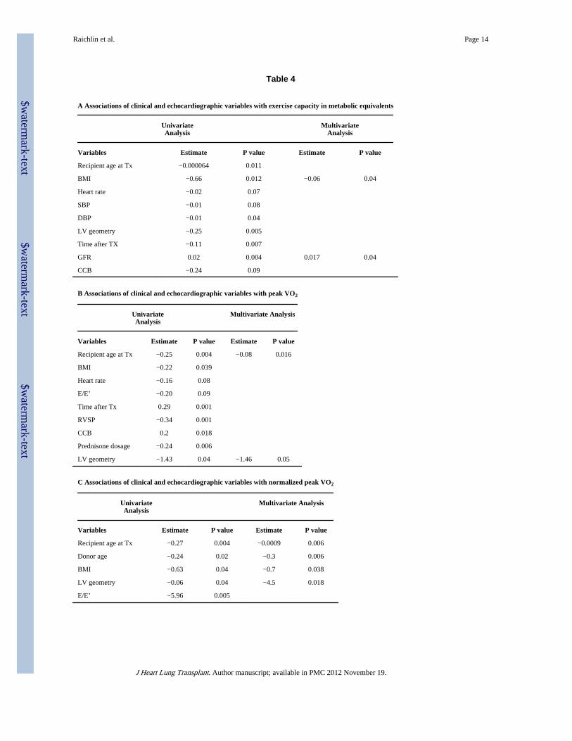
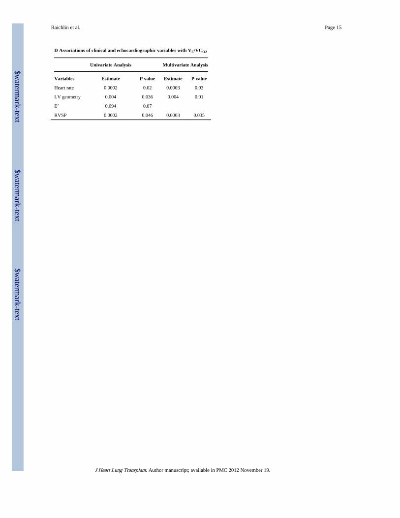
The geometric LV pattern (p=0.005), BMI (p=0.01) and GFR (p=0.004) one year after HTxcorrelated significantly with exercise intolerance as measured by metabolic equivalents byunivariate analysis. However by multivariable analysis, only BMI and GFR remainedstatistically significant. Importantly the geometric pattern of the LV was, both by univariateand multivariable analysis, a significant correlate of a reduced peak VO2, reducednormalized peak VO2 and elevated VE/VCO2. This and other correlates after univariate andmultivariable analysis are presented in Tables 4A, 4B, 4C and 4D. A reduction in absoluteand normalized peak VO2 was associated with older recipients’ age at HTx, older donors’age and higher BMI in addition to an abnormal LV geometric pattern. Elevated VE/VCO2was associated with an increase in heart rate and right ventricular systolic pressure inaddition to an abnormal geometric pattern of the LV.
DiscussionThe current study is the first to demonstrate that the presence of concentric LVH one yearafter HTx is one of the strongest correlates of exercise intolerance after HTx. ConcentricLVH was independently associated with a reduction in normalized peak VO2 and aworsening of ventilatory response to exercise as evidenced by an increase in peak VE/VCO2.This relationship remained significant after taking into account the resting blood pressure,heart rate and blood pressure response to exercise, immunosuppression, antihypertensivemedication use and other echocardiographic parameters.
Normalized peak VO2 is now recognized as a more reliable and important exerciseparameter determining prognosis in heart failure than peak VO2 alone.(30) In addition, VE/VCO2 suggested to be the most stable and reproducible marker of ventilatory efficiency,(31)and has been demonstrated to be a powerful predictor of risk for mortality, hospitalization,and other outcomes especially at values greater or equal to 40.(32) Interestingly, in our studyapproximately 50% of the HTX recipients with CH had peak VO2 ≤14 ml/kg/min and a VE/VCO2 ≥ 40. These values are seen more typically in patients with severe heart failure, and itis not surprising that patients with CH have a poor prognosis as demonstrated in our earlierstudy.(21)
The reduced normalized peak VO2 in conjunction with an increased VE/VCO2 in patientswith CH also provides a possible mechanistic insight into the cardiopulmonary limitations toexercise in the patients with CH. LV diastolic function is an important determinant ofmaximal exercise performance after HTx.(8, 33). The combination of reduced normalizedpeak VO2 and increased VE/VCO2 is suggestive of increased pulmonary congestion likelydue to worsening diastolic dysfunction occurring during exercise. (34) Although exercisehemodynamics were not obtained in this study by invasive or non-invasive techniques, anelevated VE/VCO2 ratio is indicative of pulmonary congestion and in the setting of preservedLVEF may imply diastolic dysfunction as a cause of impaired exercise capacity. In a non-transplant population evidence of concentric LV remodeling is a potential surrogate fordiastolic LV dysfunction.(35) Moreover, concentric hypertrophy is considered to besufficient evidence for the diagnosis of diastolic dysfunction when tissue Doppler yieldsnon-conclusive results.(36) Therefore, the current study suggests that decreased exercisetolerance in heart transplant recipients with concentric hypertrophy of cardiac allograft mayresult from LV diastolic dysfunction, although we do not have conclusive evidence to provethe same.
Left atrial volume is another useful marker of the severity and duration of diastolicdysfunction in a non-transplant population,(37) After HTx the left atrium is usually
Raichlin et al. Page 5
J Heart Lung Transplant. Author manuscript; available in PMC 2012 November 19.
$waterm
ark-text$w
atermark-text
$waterm
ark-text
enlarged, and the use of this marker for assessment of cardiac allograft diastolic function hasnot been validated. However, an increase in left atrial size post heart transplantationcorrelates inversely with survival and hence is an important echocardiographic parameter forthe heart transplant recipient.(38). In the present study LA volume increased in the CR andthe CH groups during follow-up and was significantly higher when measured at the time ofthe CPET in the CH and the CR groups as compared to the NG patients. This increase in leftatrial size may reflect the cumulative effect of increased filling pressures over time (39).
Amongst the various echocardiographic parameters, an E/E’≥ 10 was the strongest predictorof reduced exercise tolerance in non-transplant population.(40–42) In the present study themean E/E’ was greater than 10 in both the CR and CH groups. Furthermore, a lower E’ andhigher E/E’ ratio was associated with reduced oxygen uptake and increased ventilatoryequivalent by univariate analysis (Table 4C and 4D). This association, however, disappearedafter multivariable analysis when LV geometry was added to the model demonstrating theindependent importance of concentric hypertrophy and its association with cardiopulmonarylimitations to exercise. HTx recipients despite having a preserved left ventricular ejectionfraction historically have suboptimal exercise performance with a reported 60% reduction inexercise capacity as compared to normal controls.(33) In this study, HTx recipients with NGand CR patterns similarly demonstrated normalized peak VO2 of approximately 60% ofnormal controls. Moreover, normalized peak VO2 in recipients with a CH pattern wasfurther reduced at approximately 52% of controls. Clinical factors such as increased BMI,uncontrolled hypertension, oral prednisone and calcium channel blocker use reported tocontribute to reduced exercise tolerance in HTx recipients. This study confirms theassociation of obesity and increased recipient age with impaired exercise capacity as shownin prior studies.(6) Chronotropic incompetence has been reported as a mechanism forreduced exercise tolerance in HTx recipients,(5, 13) however, chronotropic and bloodpressure response to exercise was not significantly different amongst the 3 groups in ourstudy. The other variables that were associated with a reduced VO2 and increased VE/VCO2ratio were higher right ventricular systolic pressure and reduced renal function as measuredby GFR. Mean pulmonary arterial pressures can increase by 45% in HTx recipients withexercise(43) and hence increased right ventricular systolic pressures at rest could beassociated with exercise intolerance. Chronic kidney disease is common after cardiactransplantation(44) and can be a major source of morbidity and mortality(45). Consistentwith previous data(46) we demonstrate that recipients receiving long-term prednisone havedecreased exercise capacity: the maintenance dose of prednisone at 1 year inverselycorrelated with exercise capacity in univariate analysis. However this associationdisappeared after echocardiographic measurements were added to the multivariate model.
The role of concentric hypertrophy in limiting exercise capacity after HTx has not beenpreviously explored although this phenomenon has been described in other patientpopulations.(47, 48) Concentric LVH is an independent risk factor for cardiovascularcomplications and morbidity in essential hypertension(49–51) and it is associated withimpaired exercise performance in hypertensive patients.(19) After HTx, adverse leftventricle remodeling at 1year has been shown to be associated with subsequent developmentof cardiac allograft vasculopathy and mortality.(20, 21) This process of cardiac allograftremodeling is common and occurs early after HTx. It has been shown previously by ourgroup that most recipients with concentric LVH at 1 year after HTx continue to demonstratean abnormal LV geometric pattern up to 5 years of follow up.(21) In addition to the 1 yearpost transplant echocardiogram performed in this study, echocardiography was alsoperformed at the time of CPET. Importantly, there were no significant changes in relativewall thickness, left ventricle mass and geometry in the 3 groups at the time of CPET.
Raichlin et al. Page 6
J Heart Lung Transplant. Author manuscript; available in PMC 2012 November 19.
$waterm
ark-text$w
atermark-text
$waterm
ark-text
It is now increasingly recognized in the HTx community that there exists a need for asurrogate marker of long term prognosis of HTx recipients and the one year post transplantechocardiogram may help in part fulfill that need.(52) The present study extends ourprevious findings (20, 21) of the importance of CH and demonstrates that the presence ofCH 1 year after HTx could serve as a surrogate marker for subsequent development ofexercise intolerance. A change in immunosuppressants from cyclosporine to tacrolimus orsirolimus has been associated with regression of LV mass.(53, 54) Whether suchinterventions will lead to improved exercise capacity and quality of life in HTx recipientsremains to be proven.
Cardiac allograft remodeling and the presence of concentric LVH at one year after HTx instable heart transplant recipients is associated with decreased exercise capacity, decreasednormalized peak VO2 and increased ventilatory response. The detection, prevention, andreversal of concentric LVH could serve as important goals to improve morbidity andmortality after HTx.
AcknowledgmentsThis study was supported, in part by HL 84904 (Heart Failure Clinical Research Network), a Marie IngallsCardiovascular Career Development Award, and NIH grants UL1RR24150 (N. L. Pereira)
References1. Grady KL. Quality of life after heart transplantation: are things really better? Curr Opin Cardiol.
2003; 18:129–135. [PubMed: 12652219]
2. Osada N, Chaitman BR, Donohue TJ, Wolford TL, Stelken AM, Miller LW. Long-termcardiopulmonary exercise performance after heart transplantation. Am J Cardiol. 1997; 79:451–456.[PubMed: 9052349]
3. Mandak JS, Aaronson KD, Mancini DM. Serial assessment of exercise capacity after hearttransplantation. J Heart Lung Transplant. 1995; 14:468–478. [PubMed: 7654732]
4. Stevenson LW, Sietsema K, Tillisch JH, et al. Exercise capacity for survivors of cardiactransplantation or sustained medical therapy for stable heart failure. Circulation. 1990; 81:78–85.[PubMed: 2297851]
5. Kao AC, Van Trigt P 3rd, Shaeffer-McCall GS, et al. Central and peripheral limitations to uprightexercise in untrained cardiac transplant recipients. Circulation. 1994; 89:2605–2615. [PubMed:8205672]
6. Leung TC, Ballman KV, Allison TG, et al. Clinical predictors of exercise capacity 1 year aftercardiac transplantation. J Heart Lung Transplant. 2003; 22:16–27. [PubMed: 12531409]
7. Savin WM, Haskell WL, Schroeder JS, Stinson EB. Cardiorespiratory responses of cardiactransplant patients to graded, symptom-limited exercise. Circulation. 1980; 62:55–60. [PubMed:6991158]
8. Roten L, Schmid JP, Merz F, et al. Diastolic dysfunction of the cardiac allograft and maximalexercise capacity. J Heart Lung Transplant. 2009; 28:434–439. [PubMed: 19416770]
9. Marconi C, Marzorati M, Fiocchi R, et al. Age-related heart rate response to exercise in hearttransplant recipients. Functional significance. Pflugers Arch. 2002; 443:698–706. [PubMed:11889566]
10. Andreassen AK. Point: Cardiac denervation does/does not play a major role in exercise limitationafter heart transplantation. J Appl Physiol. 2008; 104:559–560. [PubMed: 17615275]
11. Stratton JR, Kemp GJ, Daly RC, Yacoub M, Rajagopalan B. Effects of cardiac transplantation onbioenergetic abnormalities of skeletal muscle in congestive heart failure. Circulation. 1994;89:1624–1631. [PubMed: 8149530]
12. Schwaiblmair M, von Scheidt W, Uberfuhr P, Reichart B, Vogelmeier C. Lung function andcardiopulmonary exercise performance after heart transplantation: influence of cardiac allograftvasculopathy. Chest. 1999; 116:332–339. [PubMed: 10453859]
Raichlin et al. Page 7
J Heart Lung Transplant. Author manuscript; available in PMC 2012 November 19.
$waterm
ark-text$w
atermark-text
$waterm
ark-text
13. Patel AR, Kuvin JT, DeNofrio D, et al. Peripheral vascular endothelial function correlates withexercise capacity in cardiac transplant recipients. Am J Cardiol. 2003; 91:897–899. [PubMed:12667585]
14. Lampert E, Mettauer B, Hoppeler H, Charloux A, Charpentier A, Lonsdorfer J. Skeletal muscleresponse to short endurance training in heart transplant recipients. J Am Coll Cardiol. 1998;32:420–426. [PubMed: 9708470]
15. Schaufelberger M, Eriksson BO, Lonn L, Rundqvist B, Sunnerhagen KS, Swedberg K. Skeletalmuscle characteristics, muscle strength and thigh muscle area in patients before and after cardiactransplantation. Eur J Heart Fail. 2001; 3:59–67. [PubMed: 11163737]
16. Ganau A, Devereux RB, Roman MJ, et al. Patterns of left ventricular hypertrophy and geometricremodeling in essential hypertension. J Am Coll Cardiol. 1992; 19:1550–1558. [PubMed:1534335]
17. Cohn JN, Ferrari R, Sharpe N. Cardiac remodeling--concepts and clinical implications: aconsensus paper from an international forum on cardiac remodeling. Behalf of an InternationalForum on Cardiac Remodeling. J Am Coll Cardiol. 2000; 35:569–582. [PubMed: 10716457]
18. Devereux RB, Dahlof B, Gerdts E, et al. Regression of hypertensive left ventricular hypertrophy bylosartan compared with atenolol: the Losartan Intervention for Endpoint Reduction inHypertension (LIFE) trial. Circulation. 2004; 110:1456–1462. [PubMed: 15326072]
19. Lam CS, Grewal J, Borlaug BA, et al. Size, shape, and stamina: the impact of left ventriculargeometry on exercise capacity. Hypertension. 1143; 55:1143–1149. [PubMed: 20215563]
20. Goodroe R, Bonnema DD, Lunsford S, et al. Severe left ventricular hypertrophy 1 year aftertransplant predicts mortality in cardiac transplant recipients. J Heart Lung Transplant. 2007;26:145–151. [PubMed: 17258148]
21. Raichlin E, Villarraga HR, Chandrasekaran K, et al. Cardiac allograft remodeling after hearttransplantation is associated with increased graft vasculopathy and mortality. Am J Transplant.2009; 9:132–139. [PubMed: 19067662]
22. Lang RM, Bierig M, Devereux RB, et al. Recommendations for chamber quantification. Eur JEchocardiogr. 2006; 7:79–108. [PubMed: 16458610]
23. de Simone G, Daniels SR, Kimball TR, et al. Evaluation of concentric left ventricular geometry inhumans: evidence for age-related systematic underestimation. Hypertension. 2005; 45:64–68.[PubMed: 15557389]
24. Devereux RB, Alonso DR, Lutas EM, et al. Echocardiographic assessment of left ventricularhypertrophy: comparison to necropsy findings. Am J Cardiol. 1986; 57:450–458. [PubMed:2936235]
25. Redfield MM, Jacobsen SJ, Burnett JC Jr, Mahoney DW, Bailey KR, Rodeheffer RJ. Burden ofsystolic and diastolic ventricular dysfunction in the community: appreciating the scope of the heartfailure epidemic. Jama. 2003; 289:194–202. [PubMed: 12517230]
26. Nishimura RA, Tajik AJ. Evaluation of diastolic filling of left ventricle in health and disease:Doppler echocardiography is the clinician's Rosetta Stone. J Am Coll Cardiol. 1997; 30:8–18.[PubMed: 9207615]
27. Daida H, Allison TG, Johnson BD, Squires RW, Gau GT. Further increase in oxygen uptakeduring early active recovery following maximal exercise in chronic heart failure. Chest. 1996;109:47–51. [PubMed: 8549215]
28. Wasserman KHJ, Sue D. Principles of Exercise Testing and Interpretation (3rd edition). 1999
29. ATS/ACCP Statement on cardiopulmonary exercise testing. Am J Respir Crit Care Med. 2003;167:211–277. [PubMed: 12524257]
30. Arena R, Myers J, Abella J, et al. Determining the preferred percent-predicted equation for peakoxygen consumption in patients with heart failure. Circ Heart Fail. 2009; 2:113–120. [PubMed:19808326]
31. Sun XG, Hansen JE, Garatachea N, Storer TW, Wasserman K. Ventilatory efficiency duringexercise in healthy subjects. Am J Respir Crit Care Med. 2002; 166:1443–1448. [PubMed:12450934]
32. Myers J, Arena R, Oliveira RB, et al. The Lowest VE/VCO2 ratio during exercise as a predictor ofoutcomes in patients with heart failure. J Card Fail. 2009; 15:756–762. [PubMed: 19879461]
Raichlin et al. Page 8
J Heart Lung Transplant. Author manuscript; available in PMC 2012 November 19.
$waterm
ark-text$w
atermark-text
$waterm
ark-text

33. Kao AC, Van Trigt P 3rd, Shaeffer-McCall GS, et al. Allograft diastolic dysfunction andchronotropic incompetence limit cardiac output response to exercise two to six years after hearttransplantation. J Heart Lung Transplant. 1995; 14:11–22. [PubMed: 7727459]
34. Biasucci LM, Liuzzo G, Grillo RL, et al. Elevated levels of C-reactive protein at discharge inpatients with unstable angina predict recurrent instability. Circulation. 1999; 99:855–860.[PubMed: 10027805]
35. Yturralde RF, Gaasch WH. Diagnostic criteria for diastolic heart failure. Prog Cardiovasc Dis.2005; 47:314–319. [PubMed: 16003646]
36. Paulus WJ, Tschope C, Sanderson JE, et al. How to diagnose diastolic heart failure: a consensusstatement on the diagnosis of heart failure with normal left ventricular ejection fraction by theHeart Failure and Echocardiography Associations of the European Society of Cardiology. EurHeart J. 2007; 28:2539–2550. [PubMed: 17428822]
37. Pritchett AM, Mahoney DW, Jacobsen SJ, Rodeheffer RJ, Karon BL, Redfield MM. Diastolicdysfunction and left atrial volume: a population-based study. J Am Coll Cardiol. 2005; 45:87–92.[PubMed: 15629380]
38. Gudmundsson GS, Smull DL, Pisani BA, et al. Increase in atrial size in long-term survivors ofheart transplant. J Am Soc Echocardiogr. 2003; 16:1043–1048. [PubMed: 14566297]
39. Tsang TS, Barnes ME, Gersh BJ, Bailey KR, Seward JB. Left atrial volume as amorphophysiologic expression of left ventricular diastolic dysfunction and relation tocardiovascular risk burden. Am J Cardiol. 2002; 90:1284–1289. [PubMed: 12480035]
40. Guazzi M, Brenner DA, Apstein CS, Saupe KW. Exercise intolerance in rats with hypertensiveheart disease is associated with impaired diastolic relaxation. Hypertension. 2001; 37:204–208.[PubMed: 11230272]
41. Pichard AD, Smith H, Holt J, Meller J, Gorlin R. Coronary vascular reserve in left ventricularhypertrophy secondary to chronic aortic regurgitation. Am J Cardiol. 1983; 51:315–320. [PubMed:6218741]
42. Skaluba SJ, Litwin SE. Mechanisms of exercise intolerance: insights from tissue Doppler imaging.Circulation. 2004; 109:972–977. [PubMed: 14967722]
43. Pflugfelder PW, McKenzie FN, Kostuk WJ. Hemodynamic profiles at rest and during supineexercise after orthotopic cardiac transplantation. Am J Cardiol. 1988; 61:1328–1333. [PubMed:3287883]
44. Stehlik J, Edwards LB, Kucheryavaya AY, et al. The Registry of the International Society forHeart and Lung Transplantation: twenty-seventh official adult heart transplant report--2010. JHeart Lung Transplant. 1089; 29:1089–103. [PubMed: 20870164]
45. Ojo AO, Held PJ, Port FK, et al. Chronic renal failure after transplantation of a nonrenal organ. NEngl J Med. 2003; 349:931–940. [PubMed: 12954741]
46. Renlund DG, Taylor DO, Ensley RD, et al. Exercise capacity after heart transplantation: influenceof donor and recipient characteristics. J Heart Lung Transplant. 1996; 15:16–24. [PubMed:8820079]
47. Gharavi AG, Diamond JA, Goldman AY, et al. Resting diastolic function and left ventricular massare related to exercise capacity in hypertensive men but not in women. Am J Hypertens. 1998;11:1252–1257. [PubMed: 9799044]
48. Pierson LM, Bacon SL, Sherwood A, et al. Association between exercise capacity and leftventricular geometry in overweight patients with mild systemic hypertension. Am J Cardiol. 2004;94:1322–1325. [PubMed: 15541259]
49. Levy D, Garrison RJ, Savage DD, Kannel WB, Castelli WP. Prognostic implications ofechocardiographically determined left ventricular mass in the Framingham Heart Study. N Engl JMed. 1990; 322:1561–1566. [PubMed: 2139921]
50. Koren MJ, Devereux RB, Casale PN, Savage DD, Laragh JH. Relation of left ventricular mass andgeometry to morbidity and mortality in uncomplicated essential hypertension. Ann Intern Med.1991; 114:345–352. [PubMed: 1825164]
51. Pierson LM, Bacon SL, Sherwood A, et al. Relationship between exercise systolic blood pressureand left ventricular geometry in overweight, mildly hypertensive patients. J Hypertens. 2004;22:399–405. [PubMed: 15076200]
Raichlin et al. Page 9
J Heart Lung Transplant. Author manuscript; available in PMC 2012 November 19.
$waterm
ark-text$w
atermark-text
$waterm
ark-text

52. Torre-Amione G. Cardiac allograft hypertrophy: a new target for therapy, a surrogate marker forsurvival? Am J Transplant. 2009; 9:7–8. [PubMed: 19133927]
53. Peura JL, Zile MR, Feldman DS, et al. Effects of conversion from cyclosporine to tacrolimus onleft ventricular structure in cardiac allograft recipients. J Heart Lung Transplant. 2005; 24:1969–1972. [PubMed: 16297806]
54. Kushwaha SS, Raichlin E, Sheinin Y, et al. Sirolimus affects cardiomyocytes to reduce leftventricular mass in heart transplant recipients. Eur Heart J. 2008; 29:2742–2750. [PubMed:18790727]
Raichlin et al. Page 10
J Heart Lung Transplant. Author manuscript; available in PMC 2012 November 19.
$waterm
ark-text$w
atermark-text
$waterm
ark-text
$waterm
ark-text$w
atermark-text
$waterm
ark-text
Raichlin et al. Page 11
Table 1
Patients’ Demographic and Clinical characteristics stratified by LV geometric pattern at 1 year after HTx(n=117)
VariablesNG group CR group CH group P
N=18 N=64 N=35 value
Recipient Age, years 49.01 ±11.85 48.86 ±15.31 46.61. ±15.61 0.73
Male gender, n (%) 14(78%) 50(78%) 25(71%) 0.74
Donor Age, years 32.72±14.17 34.93±13.47 30.33±14.50 0.37
Ischemic Time, min 176±536 173±48 149±44* 0.07
Ischemic Cardiomyopathy, n (%) (37%) (30%) (26%) 0.77
Body Mass Index, g/m2 24.75±3.26 24.52±5.06 28.87±6.23* 0.05
Donor/Recipient Weight 0.96±0.20 1.05±0.23 1.04±0.26 0.64
GFR, ml/min 58.07±17.35 54.47±19.32 56.84±23.24 0.76
CMV viremia, n (%) 2(10%) 10(16%) 9 (26%) 0.33
Hypertension, n (%) 14 (78%) 46 (73%) 29 (83%) 0.46
Diabetes Mellitus, n (%) 2(11%) 13 (21%) 10 (29%) 0.34
Rejection Score at 1 year 0.39±0.30 0.32±0.21 0.38±0.22 0.06
ACE inhibitor, n (%) 3 (16%) 16(25%) 10 (29%) 0.55
Beta Blocker, n (%) 1 (5%) 5 (8%) 3 (9%) 0.20
Calcium Channel Blocker, n (%) 5 (28%) 18 (29%) 10 (29%) 0.76
Cyclosporin/Tacrolimus, n (%) 15(83%)/3(17%) 54 (87%)/8(13%) 30(94%)/2(6%) 0.49
Azathioprine / MMF, n (%) 7(41%)/10(59%) 29(48%)/31(52%) 11(39%)/17(61%) 0.69
Prednisone dosage (mg) 8.28±3.53 8.17±3.75 10.27±4.54† 0.04
Heart Rate, bpm 95±11 91±13 92±13 0.49
Systolic Blood Pressure, mmHg 126±18 129±17 125±24 0.91
Diastolic Blood Pressure, mmHg 80±13 80±12 78±16 0.94
*Difference between the CH/CR and the NG groups, p ≤ 0.05
†Difference between the CH and the CR groups, p ≤ 0.05
J Heart Lung Transplant. Author manuscript; available in PMC 2012 November 19.
$waterm
ark-text$w
atermark-text
$waterm
ark-text
Raichlin et al. Page 12
Table 2
A Echocardiographic measurements at 1 year following HTx stratified by LV geometric pattern
VariablesNG group CR group CH group
P valueN=18 N=64 N=35
Heart Rate, bpm 95±11 91±13 92±13 0.49
Systolic Blood Pressure, mmHg 126±18 129±17 125±24 0.91
Diastolic blood pressure, mmHg 80±13 80±12 78±16 0.94
RWT 0.38±0.02 0.50±0.05 0.57±0.20* <0.0001
LVM, g 167±30 177±35 261±21* <0.0001
LVEF, % 62±6 63±5 61±9 0.54
SV, mL 68±12 67±15 71±13 0.68
E’, m/s 9±1 8±3 8±3 0.58
E/E’ 9.47±3.22 10.88±5.48 10.94±5.02 0.64
Left Atrium Volume, mL 71±34 80±29 98±46 0.64
RVSP, mmHg 37±8 32±8 36±11 0.11
B Echocardiographic measurements at time CPET stratified by LV geometric pattern
VariablesNG group CR group CH group
P valueN=18 N=64 N=35
Heart Rate, bpm 91±16 87±13 87±16 0.69
SBP, mmHg 114±14 121±16 123±17 0.86
DBP, mmHg 72±9 74±10 74±11 0.29
RWT 0.38±0.02 0.51±0.07 0.53±0.09* <0.0001
LVM, g 142±33 171±37 261±52* <0.0001
LVEF, % 62±6 64±7 61±9 0.94
SV, mL 68±14 71±16 82±18 0.26
E’, m/s 9±3 9±2 9±4 0.74
E/E’ 9.96 ±3.66 10.32 ±3.28 11.36 ±6.65 0.78
LA volume, mL 73±35 94±40* 105±39* 0.29
RVSP, mmHg 29±8 34±9 33±8 0.11
*Difference between the CH/CR and the NG groups, p ≤ 0.05
†Difference between the CH and the CR groups, p ≤ 0.05
J Heart Lung Transplant. Author manuscript; available in PMC 2012 November 19.

$waterm
ark-text$w
atermark-text
$waterm
ark-text
Raichlin et al. Page 13
Table 3
CPET results stratified by LV geometric pattern at 1 year following HTx
Variables NG groupN=18
CR groupN=64
CH groupN=35
P value
Δ Heart Rate, bpm 46±15 39±21 37±21 0.22
Δ SBP, mmHg 38±16 26±29 28±23 0.23
Δ DBP, mmHg 5±19 7±19 4±11 0.29
Time after transplantation, years 6.29±3.75 4.80±2.56 4.44±2.76 0.20
Exercise duration, min 7.32±2.00 6.94±2.07 6.70±0.38 0.65
Actual METs, kcal/kg/h 5.52±0.96 5.14±1.36 4.62±1.44* 0.03
Peak VO2, ml/kg × min 18.90±2.87 18.16±5.12 16.26±5.07 0.06
Normalized VO2, % 63±12 60±16* 52±14* 0.013
RER 1.21±0.09 1.20±0.11 1.19±0.12 0.81
Peak Ventilation, L/min 58.18±15.79 59.82±16.15 76.11±31.01*† 0.017
Breathing Reserve, % 50±12 49±13 48±13 0.79
Anaerobic Threshold, ml/kg × min 1036±242 1076±305 1177±486 0.68
Peak VCO2, L/min 1762±527 1719±513 2078±847† 0.22
VE/VCO2 34±6 36±7 41±17*† 0.15
*Difference between the CH/CR and the NG groups, p ≤ 0.05
†Difference between the CH and the CR groups, p ≤ 0.05
J Heart Lung Transplant. Author manuscript; available in PMC 2012 November 19.
$waterm
ark-text$w
atermark-text
$waterm
ark-text
Raichlin et al. Page 14
Table 4
A Associations of clinical and echocardiographic variables with exercise capacity in metabolic equivalents
UnivariateAnalysis
MultivariateAnalysis
Variables Estimate P value Estimate P value
Recipient age at Tx −0.000064 0.011
BMI −0.66 0.012 −0.06 0.04
Heart rate −0.02 0.07
SBP −0.01 0.08
DBP −0.01 0.04
LV geometry −0.25 0.005
Time after TX −0.11 0.007
GFR 0.02 0.004 0.017 0.04
CCB −0.24 0.09
B Associations of clinical and echocardiographic variables with peak VO2
UnivariateAnalysis
Multivariate Analysis
Variables Estimate P value Estimate P value
Recipient age at Tx −0.25 0.004 −0.08 0.016
BMI −0.22 0.039
Heart rate −0.16 0.08
E/E’ −0.20 0.09
Time after Tx 0.29 0.001
RVSP −0.34 0.001
CCB 0.2 0.018
Prednisone dosage −0.24 0.006
LV geometry −1.43 0.04 −1.46 0.05
C Associations of clinical and echocardiographic variables with normalized peak VO2
UnivariateAnalysis
Multivariate Analysis
Variables Estimate P value Estimate P value
Recipient age at Tx −0.27 0.004 −0.0009 0.006
Donor age −0.24 0.02 −0.3 0.006
BMI −0.63 0.04 −0.7 0.038
LV geometry −0.06 0.04 −4.5 0.018
E/E’ −5.96 0.005
J Heart Lung Transplant. Author manuscript; available in PMC 2012 November 19.
$waterm
ark-text$w
atermark-text
$waterm
ark-text
Raichlin et al. Page 15
D Associations of clinical and echocardiographic variables with VE/VCO2
Univariate Analysis Multivariate Analysis
Variables Estimate P value Estimate P value
Heart rate 0.0002 0.02 0.0003 0.03
LV geometry 0.004 0.036 0.004 0.01
E’ 0.094 0.07
RVSP 0.0002 0.046 0.0003 0.035
J Heart Lung Transplant. Author manuscript; available in PMC 2012 November 19.