peripheral nerve tumors
TRANSCRIPT
Peripheral NerveTumorsJoey Grochmal R1 Neurosurgery
Overview
• Presentation andDefinitions/Anatomy
• Physical Exam• Tumors and lesions by subtype• Peripheral Nerve Tumor
Syndromes• Diagnostic Testing• Management
Tumors of the PNS
• Peripheral Nervous System:including spinal and cranialnerves, distal to their duralcoverings, and their respectiveterminal branches.
• Optic nerve not included
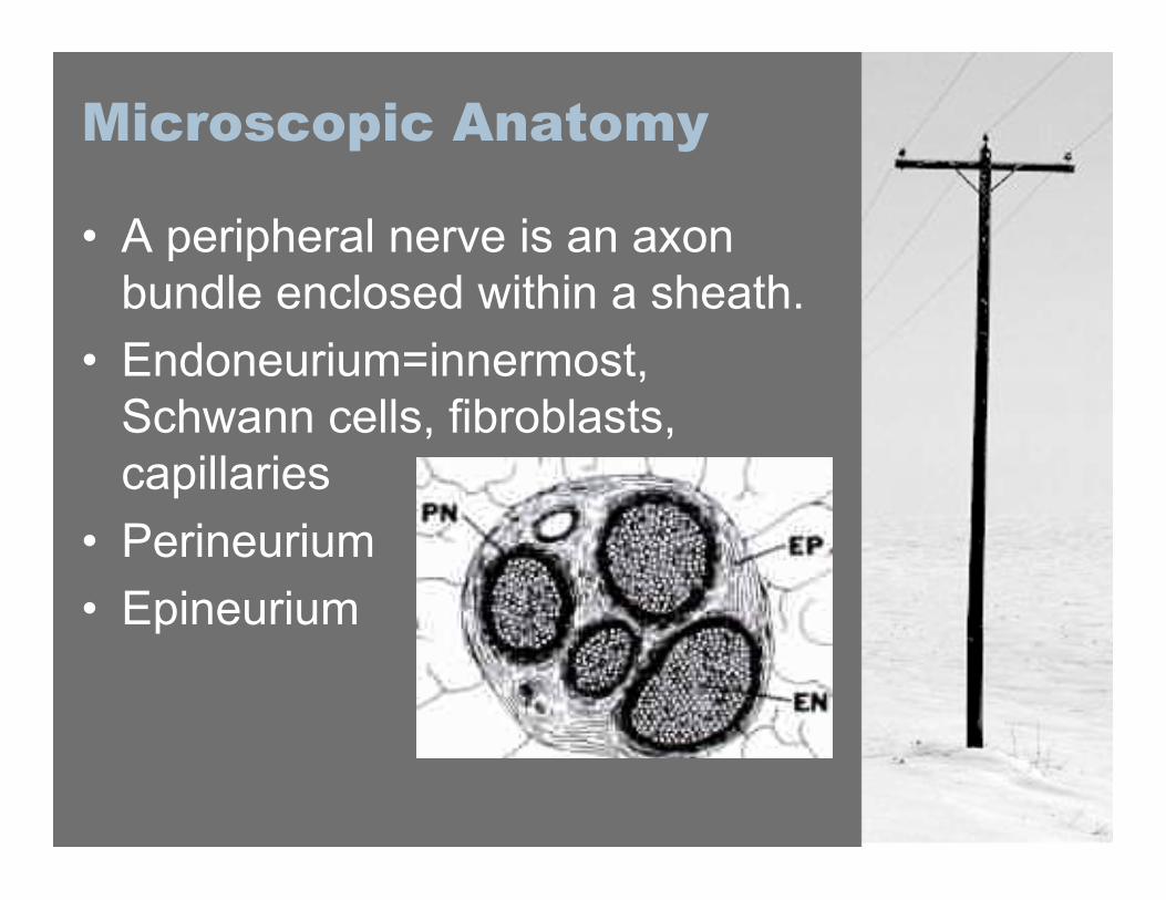
Microscopic Anatomy
• A peripheral nerve is an axonbundle enclosed within a sheath.
• Endoneurium=innermost,Schwann cells, fibroblasts,capillaries
• Perineurium• Epineurium
Presentation
• History– mass: asymptomatic or symptomatic– sensory alteration (paresthesia,
hyper/hypoalgesia, allodynia)– pain, weakness– autonomic dysfunction– Compressive symptoms: eg. Bowel
or bladder, limb edema or ischemia

Physical Examination
– careful characterization of the tumorincludes:
• size (measured)• location and relation to adjacent structures• ROM, perpendicular to direction of nerves• margins and consistency• Tinel’s sign• auscultation
Examine for NeurocutaneousStigmata!
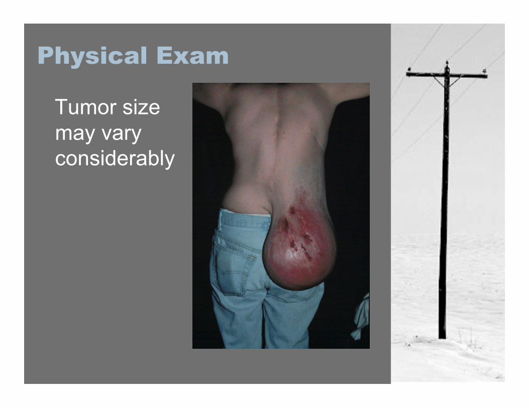
Physical Exam
Tumor sizemay varyconsiderably
Physical Exam Pearls
• PN Tumors are mobileperpendicular to the long axis ofthe limb, and not parallel to it.
• Sensory changes present early,motor findings present late andare worrisome for malignancy (asis progressive pain).
• New palpable mass often the onlyfinding.
PeripheralNerveTumors
Peripheral Nerve Tumors
Annual incidence 6 / 1 000 000, ofPNT’s that get operated on.
90% are benign tumors45% arise in the Head and Neck
1)Nerve Sheath Tumors2)Tumors of Neural Origin3)Other Peripheral Nerve Lesions
Nerve Sheath Tumors
•1)Nerve SheathTumors -Schwannoma–Neurofibroma–Perineuroma–Malignant PeripheralNerve Sheath Tumor(MPNST)
Schwannoma (PNST)
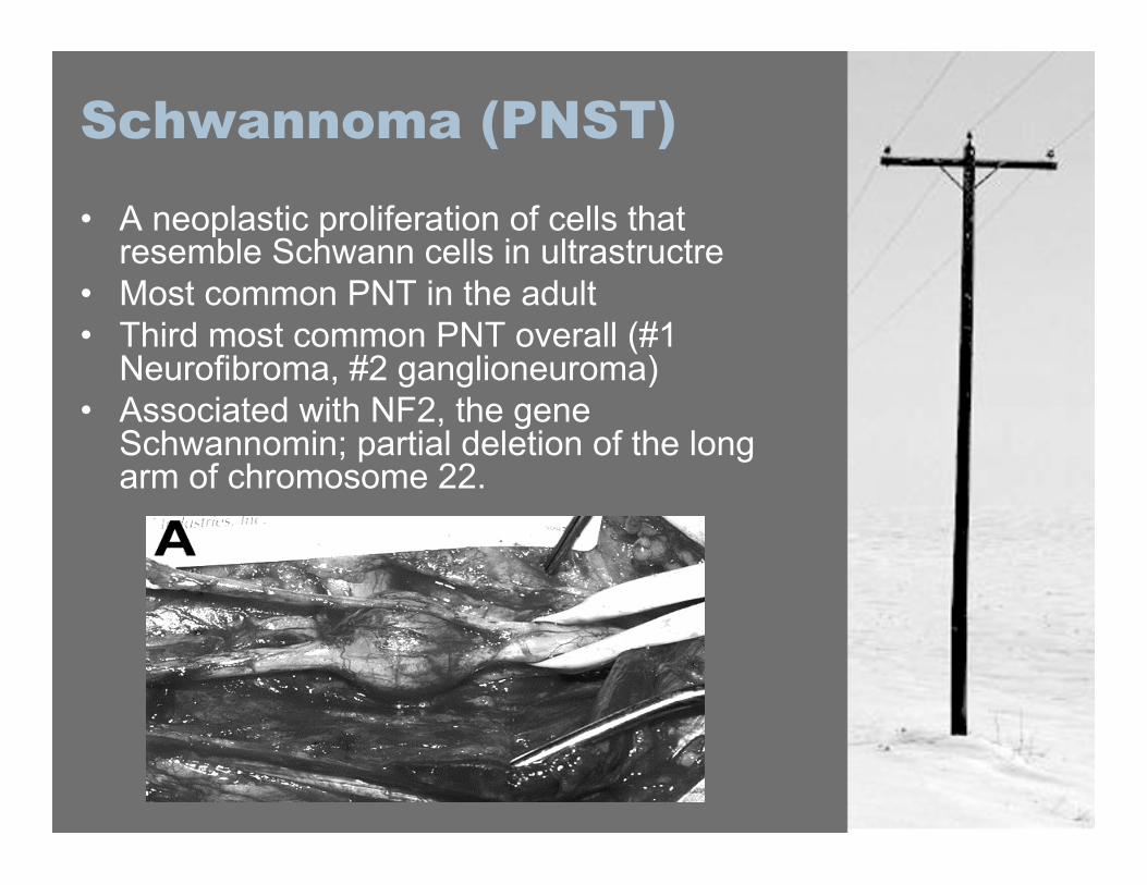
• A neoplastic proliferation of cells thatresemble Schwann cells in ultrastructre
• Most common PNT in the adult• Third most common PNT overall (#1
Neurofibroma, #2 ganglioneuroma)• Associated with NF2, the gene
Schwannomin; partial deletion of the longarm of chromosome 22.
Schwannoma (PNST)
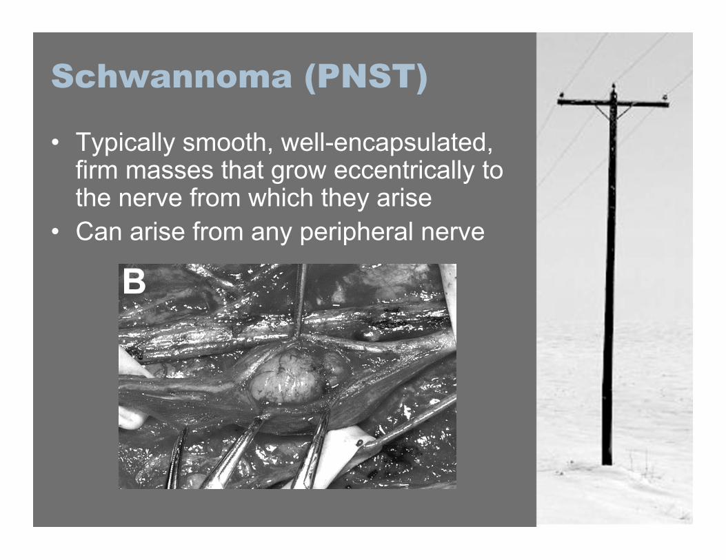
• Typically smooth, well-encapsulated,firm masses that grow eccentrically tothe nerve from which they arise
• Can arise from any peripheral nerve
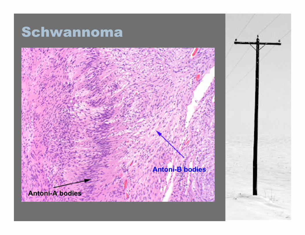
Schwannoma
Antoni-A bodies
Antoni-B bodies
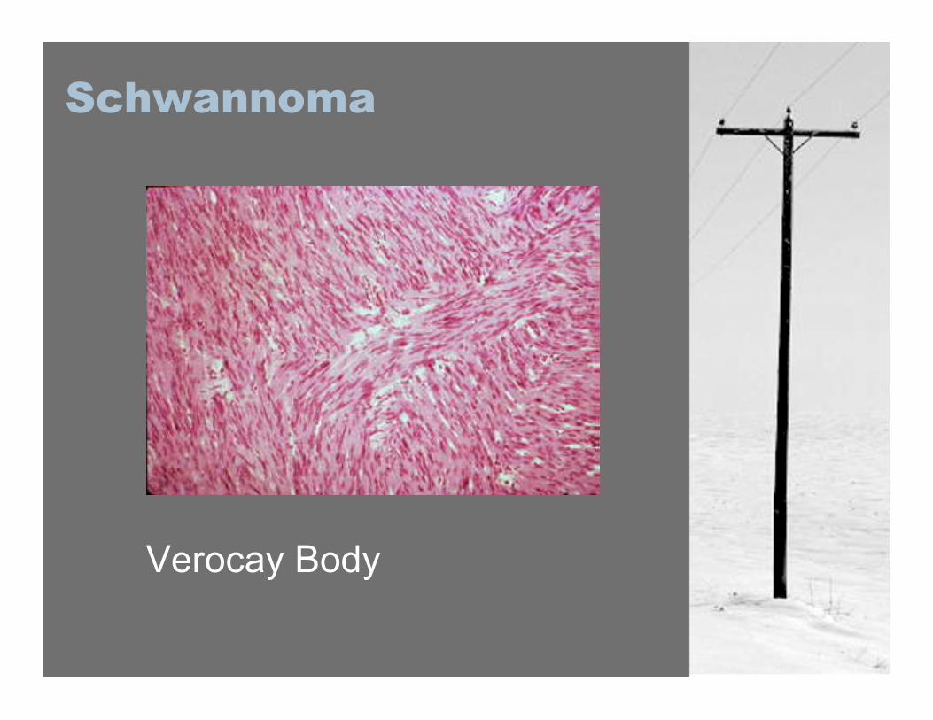
Schwannoma
Verocay Body
Schwannoma
-Most presenting patients aged 20-50 years old
-Male and Female incidence equal-Predominance for1)Sensory Nerves2)Flexor surfaces3)Upper extremities (esp. ulnar)
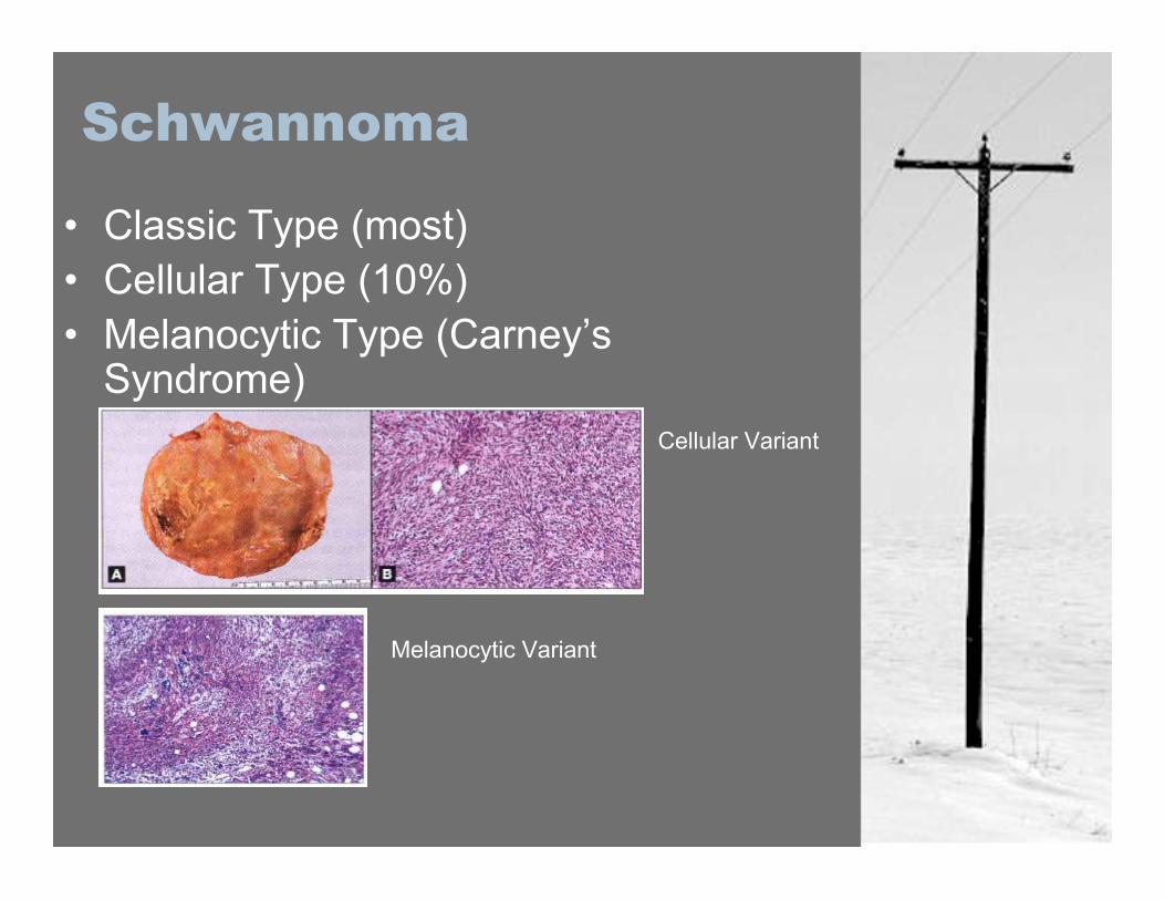
Schwannoma
• Classic Type (most)• Cellular Type (10%)• Melanocytic Type (Carney’s
Syndrome)Cellular Variant
Melanocytic Variant
Neurofibroma (PNST)
• General Type vs. Plexiform• Sex predilection equal• 1/25 000 Adults• Most common PNT in younger people
(avg age 20-30)• Soliatary type (90%) usually
spontaneous and not associated withNF1.
• Plexiform and multiple typesassociated with NF1
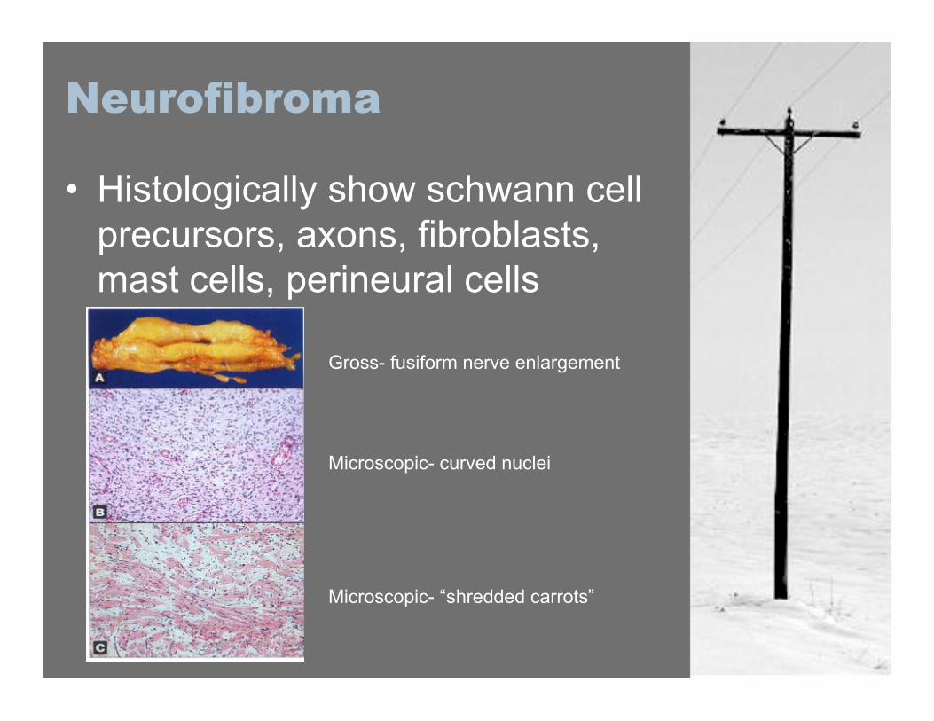
Neurofibroma
• Histologically show schwann cellprecursors, axons, fibroblasts,mast cells, perineural cells
Gross- fusiform nerve enlargement
Microscopic- curved nuclei
Microscopic- “shredded carrots”
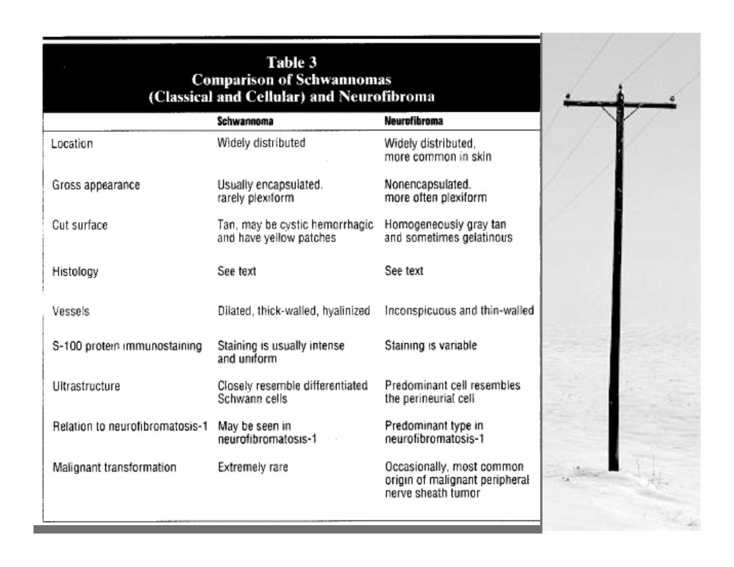
Neurofibroma vs.Schwannoma
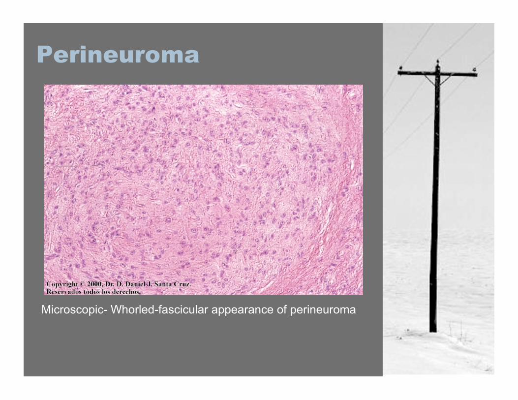
Perineuroma
• Comprised exclusively ofperineural cells
• Rare tumor; 50 reported cases ofintraneural perineuroma.
• Female>Male (middle agedwomen)
• Most are subcutaneous or notassociated with peripheral nerves
Perineuroma
Microscopic- Whorled-fascicular appearance of perineuroma
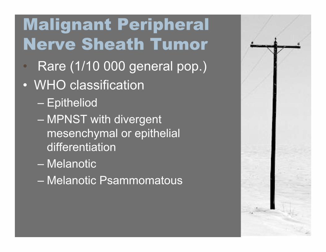
Malignant PeripheralNerve Sheath Tumor• Rare (1/10 000 general pop.)• WHO classification
– Epitheliod– MPNST with divergent
mesenchymal or epithelialdifferentiation
– Melanotic– Melanotic Psammomatous
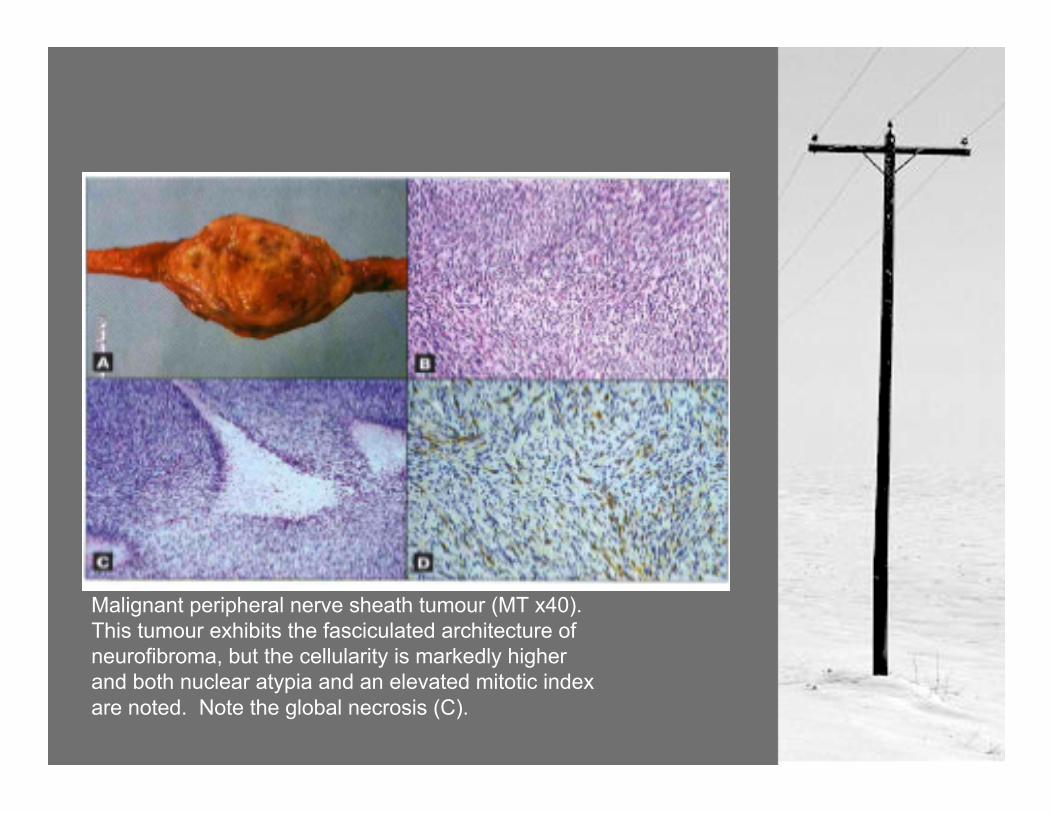
Malignant peripheral nerve sheath tumour (MT x40).This tumour exhibits the fasciculated architecture ofneurofibroma, but the cellularity is markedly higherand both nuclear atypia and an elevated mitotic indexare noted. Note the global necrosis (C).
MPNST
• Pearls:– Look for family HX of NF (1 or 2)– 50% MPNST associated with
underlying neurocutaneous disorder.– “Loss of mobility, rapid growth, and
the evolution of neurologic deficitsare features that favor the diagnosisof a malignant over a benignperipheral nerve tumor”
MPNST
• 10% of all soft tissue sarcoma• 4% of NF1 patients will develop
MPNST• 10% of those are post radiation
for plexiform neurofibroma• New onset or post irradiation
rapid growth of a neurofibroma =malignant change.
Tumors of Neural Origin
• Pheochromocytoma– 90% found in adrenal medulla– NF2, von Hippel-Lindau, Tuberous Sclerosis, Sturge-
Weber, MEN– Headache, palpitations, sweating
• Paraganglioma– Arise from extra-adrenal chromaffin cells– Most intra-abdominal, paraspinal gainglia
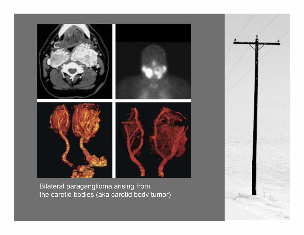
Bilateral paraganglioma arising fromthe carotid bodies (aka carotid body tumor)
Tumors of Neural Origin
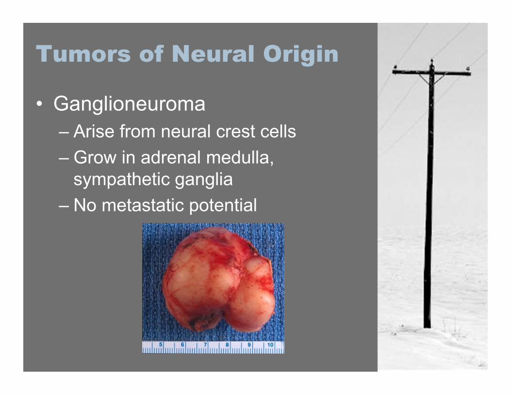
• Ganglioneuroma– Arise from neural crest cells– Grow in adrenal medulla,
sympathetic ganglia– No metastatic potential
Tumors of Neural Origin
Neuroblastoma- very common in childhood (8-10% of all
childhood malignancy)- Neural crest origin, primarily paravertebral,
abdominal, posterior mediastial presentation- 60% 5 year survival rate
Peripheral Neuroepithelioma- rare, less than 1% of all sarcomas- small, round cell tumor- 10-35% 5year survival with metastatic
disease, 54-75% survival with local disease
Other Peripheral NerveLesions
These lesions wither arise from extrinsicnerve elements, or are situated tomimic nerve tumors
Desmoid-deep seated fibromatosis-non-metatstatic, locally invasive
Meningioma-similarity of arachnoidal cells toperineural cells-extra cranial meningioma extremelyrare

Tumor Mimics
Ganglion Cyst-mucinous filled, arising fromjoints and tendon sheaths-back of hand, peroneal nerveregion most common
Morton’s Neuroma-common thickening of inter-digital plantar nerves-women who wear ill-fitting shoes
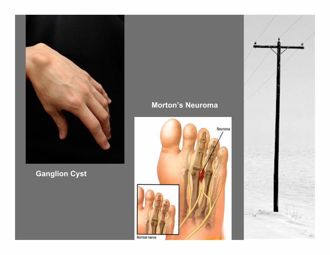
Ganglion Cyst
Morton’s Neuroma
Tumor Mimics
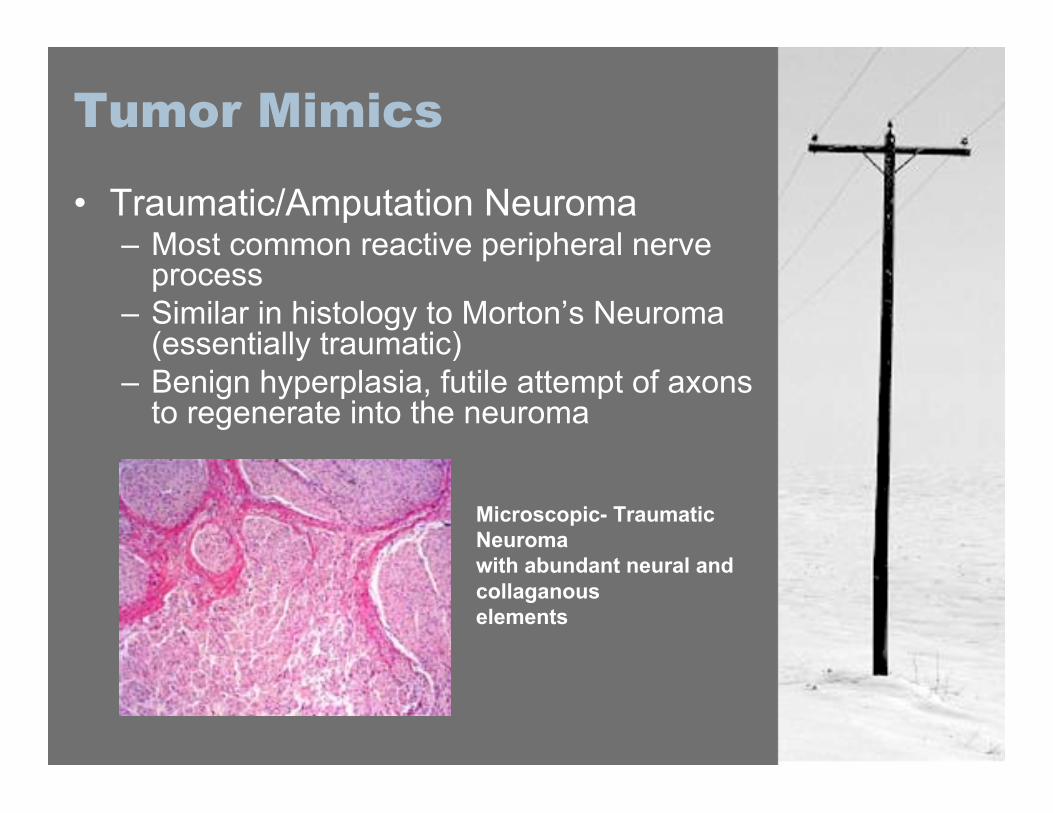
• Traumatic/Amputation Neuroma– Most common reactive peripheral nerve
process– Similar in histology to Morton’s Neuroma
(essentially traumatic)– Benign hyperplasia, futile attempt of axons
to regenerate into the neuroma
Microscopic- TraumaticNeuromawith abundant neural andcollaganouselements
Peripheral NerveTumorSyndromes• Neurofibromatosis 1• Neurofibromatosis 2• Schwannomatosis
Neurofibromatosis 1
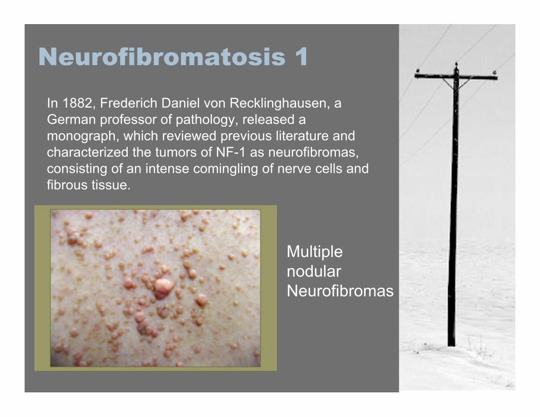
In 1882, Frederich Daniel von Recklinghausen, aGerman professor of pathology, released amonograph, which reviewed previous literature andcharacterized the tumors of NF-1 as neurofibromas,consisting of an intense comingling of nerve cells andfibrous tissue.
MultiplenodularNeurofibromas
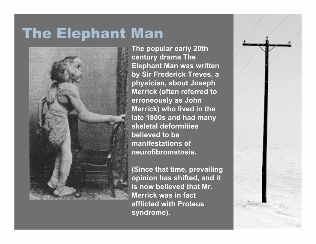
The popular early 20thcentury drama TheElephant Man was writtenby Sir Frederick Treves, aphysician, about JosephMerrick (often referred toerroneously as JohnMerrick) who lived in thelate 1800s and had manyskeletal deformitiesbelieved to bemanifestations ofneurofibromatosis.
(Since that time, prevailingopinion has shifted, and itis now believed that Mr.Merrick was in factafflicted with Proteussyndrome).
The Elephant Man

The Elephant Man
Neurofibromatosis 1
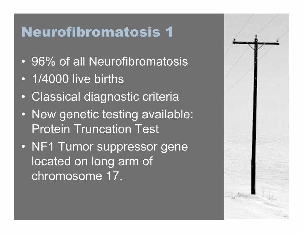
• 96% of all Neurofibromatosis• 1/4000 live births• Classical diagnostic criteria• New genetic testing available:
Protein Truncation Test• NF1 Tumor suppressor gene
located on long arm ofchromosome 17.
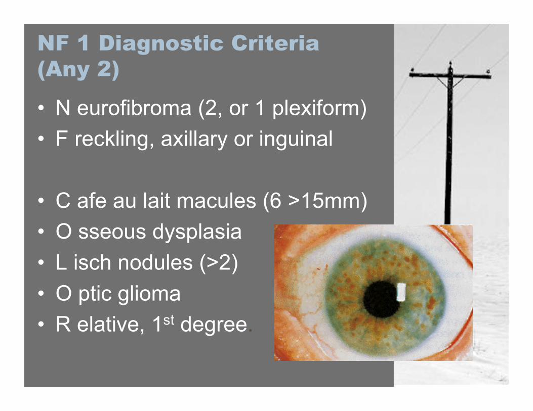
NF 1 Diagnostic Criteria(Any 2)
• N eurofibroma (2, or 1 plexiform)• F reckling, axillary or inguinal
• C afe au lait macules (6 >15mm)• O sseous dysplasia• L isch nodules (>2)• O ptic glioma• R elative, 1st degree.
Treatment/Prognosis NF1
• Surgical intervention for cosmesisor functional decay, or for rapidgrowth.
• 4% lifelong risk of malignanttransformation of neurofibromas
• 5 times increase cancer risk fromgeneral population
• Treatment of Optic Glioma iscontroversial, early vs. late
Neurofibromatosis 2
• Less Common than NF1• 1/40 000 live births• Autosomal Dominant Inheritance
– 22q11 locus (schwannomin; merlin)\
Presentation is usually that ofsymptoms associated with vestibularschwannoma; hearing impairment,imbalance, tinnitus, weakness,seizure, impaired vision
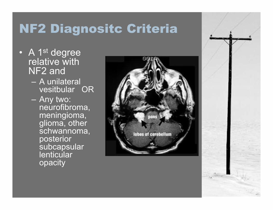
NF2 Diagnositc Criteria
• A 1st degreerelative withNF2 and– A unilateral
vesitbular OR– Any two:
neurofibroma,meningioma,glioma, otherschwannoma,posteriorsubcapsularlenticularopacity
Treatment/Prognosis NF2
• Surgery or radiotherapy indicatedfor the treatment of vestibularschwannomas
• Early surgery gives best chanceof preserving nerve function, whileradiotherapy remains thetreatment of choice.
Schwannomatosis
• Similar prevalence to NF2,approximately 1/40 000
• Age >30 years, not NF2, and two ormore intradermal schwannomas withat least one pathologically proven.OR
• One schwannoma and a relativemeeting the above criteria
• Hereditary disorder, gene locus not yetidentified.
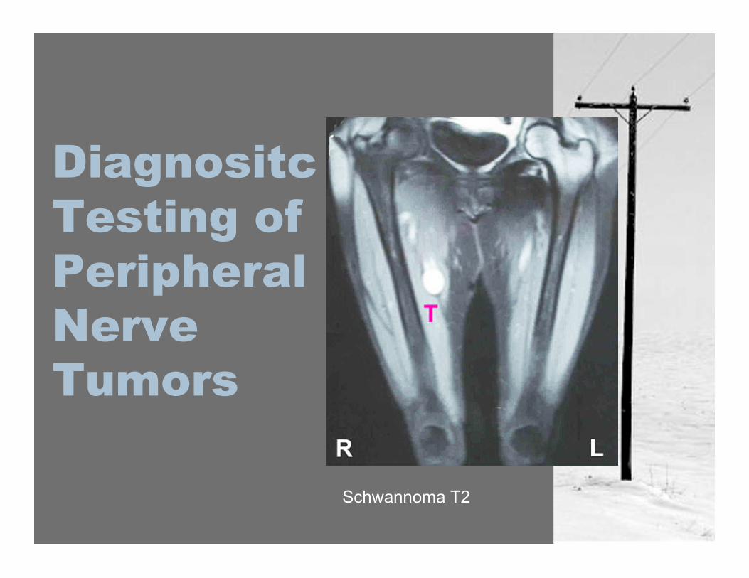
DiagnositcTesting ofPeripheralNerveTumors
Schwannoma T2
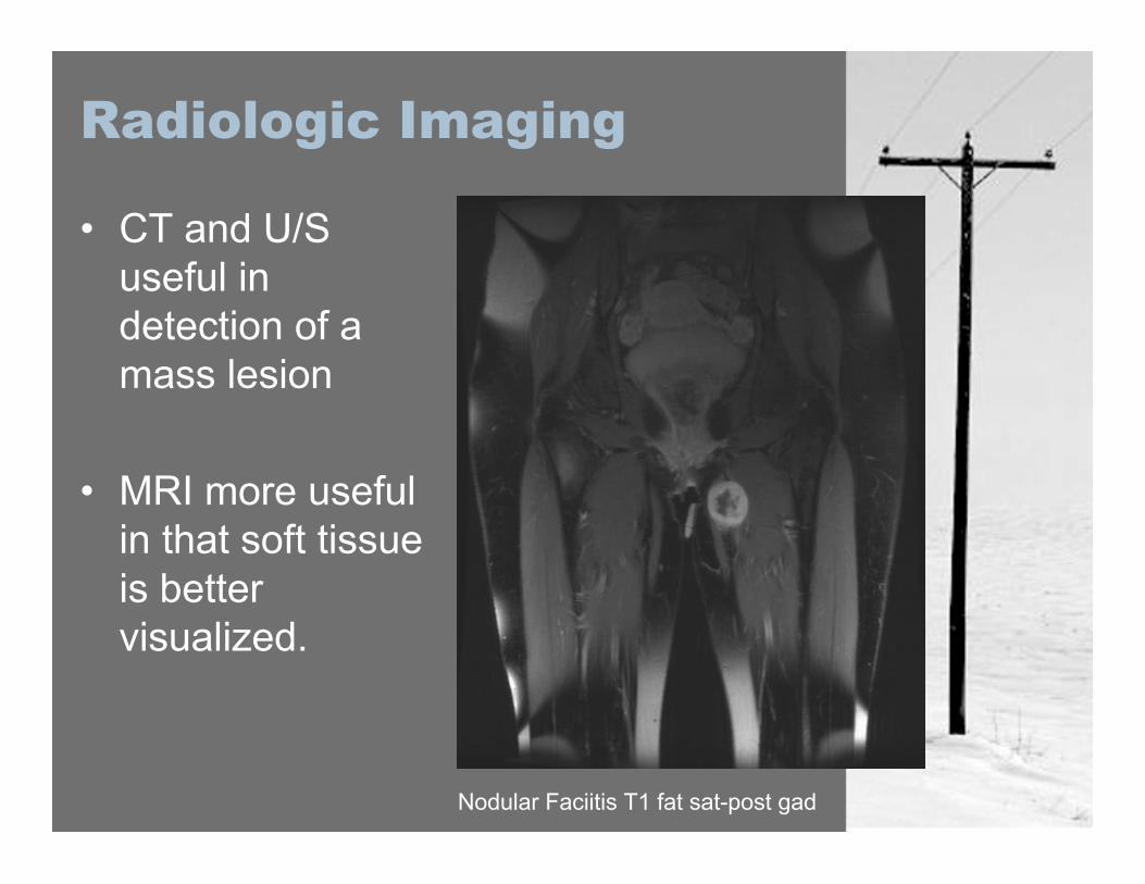
Radiologic Imaging
• CT and U/Suseful indetection of amass lesion
• MRI more usefulin that soft tissueis bettervisualized.
Nodular Faciitis T1 fat sat-post gad
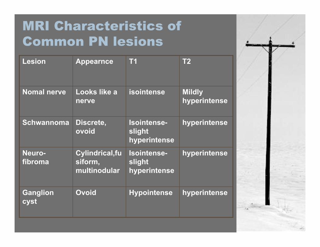
MRI Characteristics ofCommon PN lesions
hyperintenseHypointenseOvoidGanglioncyst
hyperintenseIsointense-slighthyperintense
Cylindrical,fusiform,multinodular
Neuro-fibroma
hyperintenseIsointense-slighthyperintense
Discrete,ovoid
Schwannoma
Mildlyhyperintense
isointenseLooks like anerve
Nomal nerve
T2T1AppearnceLesion
Radiologic Imaging
• Currently no imaging study,including CT and MRI, that canseparate the classes of peripheralnerve tumor, or distinguishbetween a benign or malignanttumor.

Electrical Studies
• Useful in localizing a mass to aparticular nerve, though often(schwannoma) there is no deficit.
• Useful in determining grade of nerveinjury and degree of axonal damage
• EMG can show muscular denervationand hence axonal loss
• Baseline pre-op function and operativerisk assessment
• Intraoperative use of electrical studiesto determine approach to the lesion.
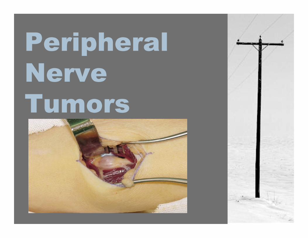
Management ofPeripheral NerveTumors
Management of PNT
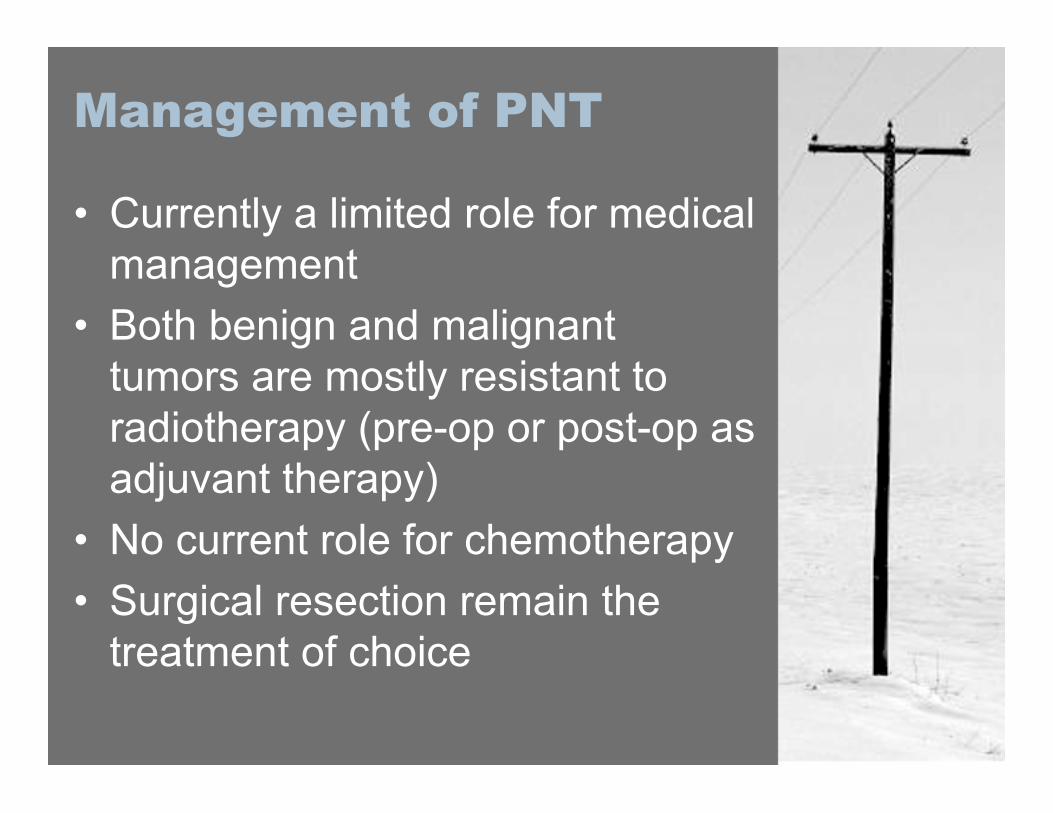
• Currently a limited role for medicalmanagement
• Both benign and malignanttumors are mostly resistant toradiotherapy (pre-op or post-op asadjuvant therapy)
• No current role for chemotherapy• Surgical resection remain the
treatment of choice
IntraoperativeElectrophysiology
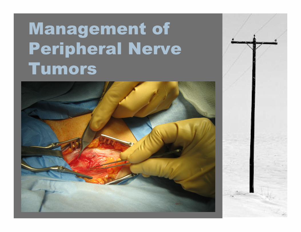
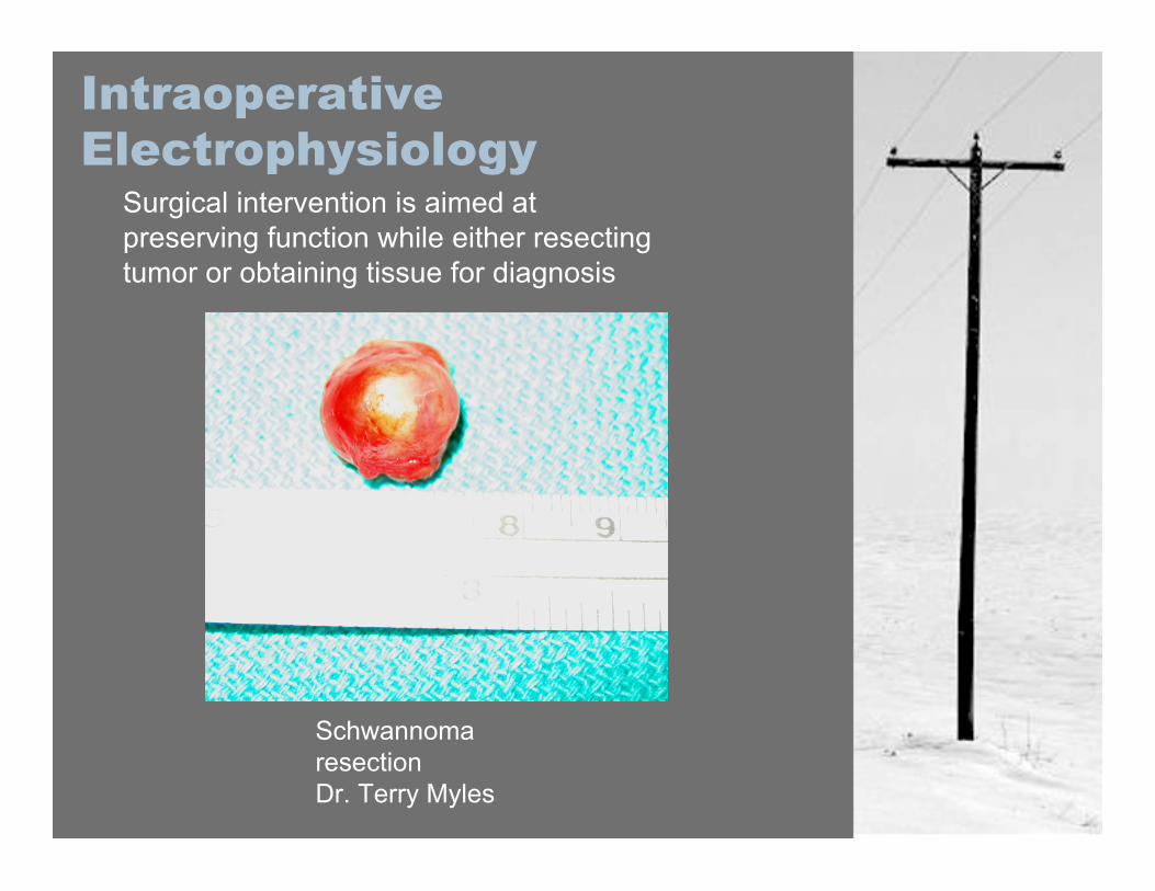
Surgical intervention is aimed atpreserving function while either resectingtumor or obtaining tissue for diagnosis
SchwannomaresectionDr. Terry Myles
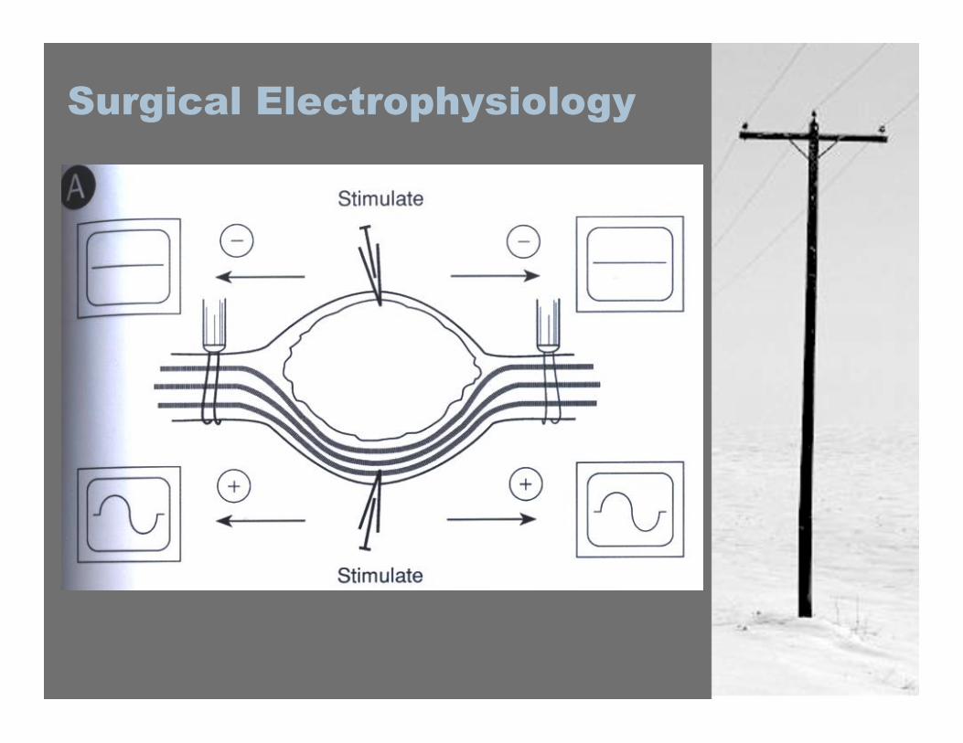
Surgical Electrophysiology
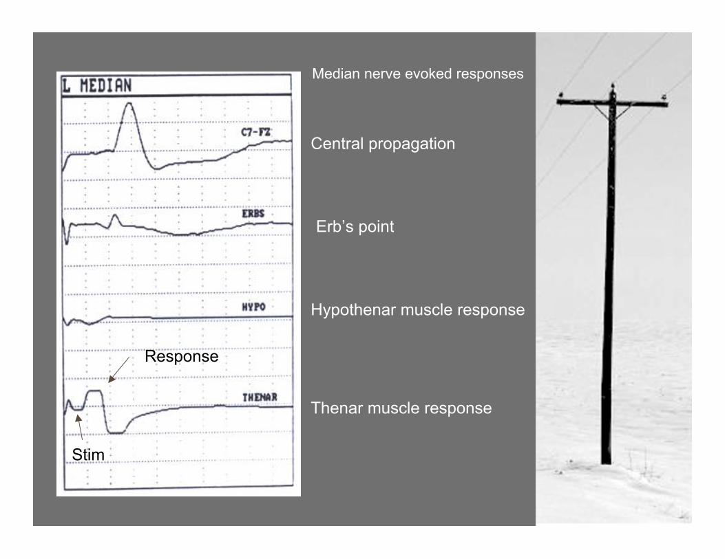
Median nerve evoked responses
Stim
Response
Central propagation
Erb’s point
Hypothenar muscle response
Thenar muscle response
Surgical Indications
• Biopsy specimen needed• Rapidly growing lesion• Progressive symptoms• Intolerable pain, not well
controlled by medications
Management ofMalignant PNT
• Best managed by aninterdisciplinary team (Rad. Onc,DI, Surgery, Pathology)
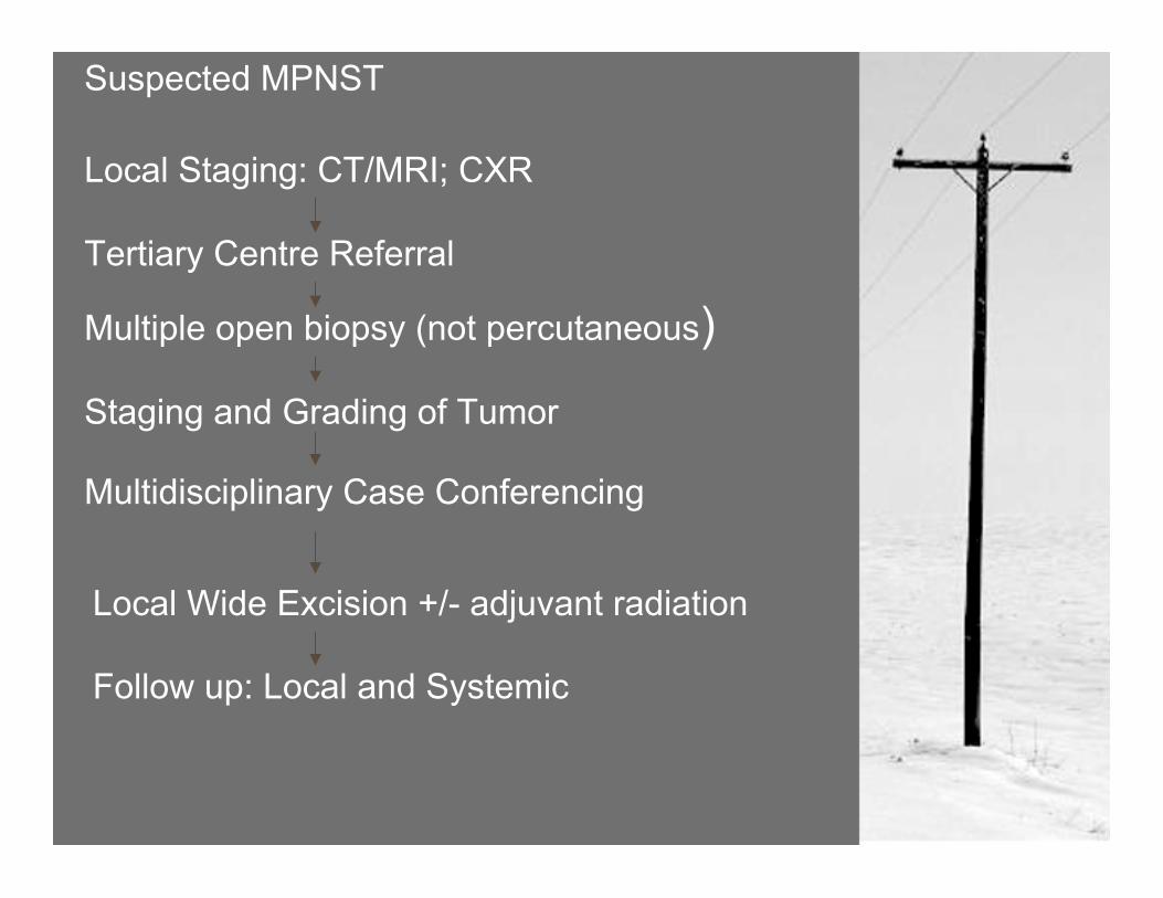
Suspected MPNST
Local Staging: CT/MRI; CXR
Tertiary Centre Referral
Multiple open biopsy (not percutaneous)
Staging and Grading of Tumor
Multidisciplinary Case Conferencing
Local Wide Excision +/- adjuvant radiation
Follow up: Local and Systemic
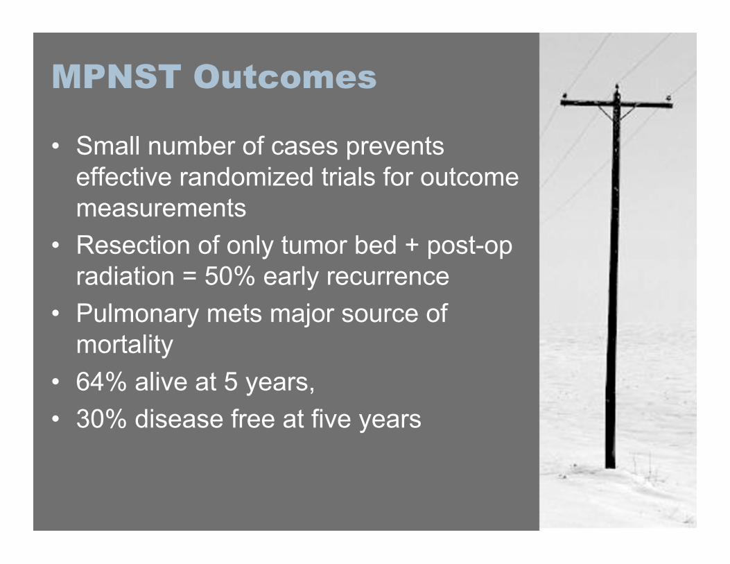
MPNST Outcomes
• Small number of cases preventseffective randomized trials for outcomemeasurements
• Resection of only tumor bed + post-opradiation = 50% early recurrence
• Pulmonary mets major source ofmortality
• 64% alive at 5 years,• 30% disease free at five years

The End