ramirez 2014 lu sens
TRANSCRIPT
Toxicology in Vitro 28 (2014) 1482–1497
Contents lists available at ScienceDirect
Toxicology in Vitro
journal homepage: www.elsevier .com/locate / toxinvi t
LuSens: A keratinocyte based ARE reporter gene assay for usein integrated testing strategies for skin sensitization hazardidentification
http://dx.doi.org/10.1016/j.tiv.2014.08.0020887-2333/� 2014 The Authors. Published by Elsevier Ltd.This is an open access article under the CC BY-NC-SA license (http://creativecommons.org/licenses/by-nc-sa/3.0/).
⇑ Corresponding author.E-mail address: [email protected] (R. Landsiedel).
Tzutzuy Ramirez a, Annette Mehling b, Susanne N. Kolle a, Christoph J. Wruck c, Wera Teubner d,Tobias Eltze e, Alexandra Aumann a, Daniel Urbisch a, Ben van Ravenzwaay a, Robert Landsiedel a,⇑a BASF SE, Experimental Toxicology and Ecology, Germanyb BASF Personal Care and Nutrition GmbH, Germanyc RWTH Aachen, Germanyd BASF Schweiz AG, Switzerlande BASF SE, Paper Chemicals, Germany
a r t i c l e i n f o
Article history:Received 9 May 2014Accepted 10 August 2014Available online 27 August 2014
Keywords:Skin sensitizationAntioxidant response element (ARE)In vitroAdverse outcome pathway (AOP)Keratinocyte activation
a b s t r a c t
Allergic contact dermatitis can develop following repeated exposure to allergenic substances. To date,hazard identification is still based on animal studies as non-animal alternatives have not yet gained glo-bal regulatory acceptance. Several non-animal methods addressing key-steps of the adverse outcomepathway (OECD, 2012) will most likely be needed to fully address this effect. Among the initial cellularevents is the activation of keratinocytes and currently only one method, the KeratinoSens™, has been for-mally validated to address this event. In this study, a further method, the LuSens assay, that uses a humankeratinocyte cell line harbouring a reporter gene construct composed of the antioxidant response ele-ment (ARE) of the rat NADPH:quinone oxidoreductase 1 gene and the luciferase gene. The assay was val-idated in house using a selection of 74 substances which included the LLNA performance standards. Thepredictivity of the LuSens assay for skin sensitization hazard identification was comparable to other non-animal methods, in particular to the KeratinoSens™. When used as part of a testing battery based on theOECD adverse outcome pathway for skin sensitization, a combination of the LuSens assay, the DPRA and adendritic cell line activation test attained predictivities similar to that of the LLNA.� 2014 The Authors. Published by Elsevier Ltd. This is an open access article under the CC BY-NC-SA license
(http://creativecommons.org/licenses/by-nc-sa/3.0/).
1. Introduction Currently, there is no non-animal alternative test method for
In a regulatory context, the sensitizing potential of substances isgenerally evaluated using animal tests, such as the murine locallymph node assay (LLNA; OECD TG 429) or the guinea pig-basedtests described in OECD TG 406 (OECD TG 406). As of March 11,2013, the European Union imposed an animal testing ban on bothcosmetic products and their ingredients which is accompanied by aconcomitant marketing ban, if animal tests conducted after thisdate for the purpose of the cosmetics legislation (EU, 2009).
In addition, under the European chemicals legislation REACH,skin sensitization data for any substances registered under theEuropean Chemicals Legislation (REACH, EC 1907/2006) is manda-tory, animal testing should only be performed only a last resort(http://echa.europa.eu/documents/10162/13639/alternatives_test_animals_2014_en.pdf).
the endpoint of skin sensitization yet available that has gained fullregulatory acceptance. During the last decades, extensive work hasbeen conducted to develop in vitro assays able to replace currentanimal test methods for the predictive identification of skin sensi-tizers (reviewed in Mehling et al., 2012). Given the complexity ofthe sensitization pathway, a combination of tests will be neededto achieve reliable predictions of the skin sensitization potentialof a substance.
In 2012 the OECD published the adverse outcome pathway(AOP) for skin sensitization (OECD 2012a, 2012b, 2012c) in whichthe key steps in the sensitization process are defined. According tothe AOP, one of the early key events of the sensitization process isthe induction of cytoprotective gene pathways that occur withinkeratinocytes (KCs) upon contact with a sensitizer. KCs are thedominant cells in the epidermis and are among the first cells tocome into contact with a sensitizer. In this context, various studieshave shown that the Nrf2-Keap1 pathway plays an important rolein skin sensitization (Ade et al., 2009; Natsch and Emter, 2008;

T. Ramirez et al. / Toxicology in Vitro 28 (2014) 1482–1497 1483
Vandebriel et al., 2010). Under physiological conditions, the tran-scription factor nuclear factor erythroid 2 (Nrf2) is constitutivelyexpressed but is complexed and targeted for ubiquinylation inthe cytosol by the cytosensor protein Kelch-like ECH-associatedprotein 1 (Keap1). In response to covalent modification of thehighly reactive cysteine residues of Keap1 via stressors, Nrf2 isreleased. Free Nrf2 translocates to the nucleus, where it heterodi-merizes with other molecules (e.g., small Maf or Jun proteins). Thiscomplex then binds to the so called ‘‘antioxidant response element(ARE)’’ in the promoter region of several genes, including hmox1and nqo1, subsequently initiating transcription of the downstreamgenes (Ade et al., 2009). The Keap1 protein therefore constitutes anintracellular sensor protein for electrophilic and oxidative stress.Skin sensitizers are thought to directly or indirectly react with cys-teine residues of Keap1, thereby enhancing Nrf2 release (Emteret al., 2010; Motohashi and Yamamoto, 2004). The principle of thispathway has been used to develop reporter cells lines includingAREc32; ARE-HepG2 (Emter et al., 2010; Simmons et al., 2011)and KeratinoSens™ (Emter et al., 2010; Natsch and Emter 2008).
In light of the urgent need to evaluate non-animal approachescapable of reliably identifying skin sensitization hazards therebyproviding the basis for classification and labelling, the EuropeanUnion Reference Laboratory for Alternatives to Animal Testing(EURL ECVAM) has recently published its strategy on skin sensiti-zation (EURL ECVAM, 2013a) describing its plan for the next fiveyears towards achieving these goals: the two main points of focuswill be the development of integrated testing strategies (ITS) to beused within integrated assessment and testing approaches (IATA)for skin sensitization and the facilitation of global acceptance ofthe new approaches for skin sensitization hazard identification(EURL ECVAM, 2013a). Currently, the assessment of the reliabilityand reproducibility of several test methods for skin sensitization,namely the Direct Peptide Reactivity Assay (DPRA), human cell lineactivation test (h-CLAT), KeratinoSens™) under the formal valida-tion process at ECVAM has progressed to an advanced stage. TheECVAM Scientific Advisory Committee (ESAC) EURL ECVAMrecently issued their recommendations on the DPRA (EURLECVAM, 2013b) and KeratinoSens™ (EURL ECVAM, 2013c) andthe methods appear adequate to be considered for inclusion inan ITS using test methods that address various key events of theOECD skin sensitization AOP (EURL ECVAM, 2013a). A pragmaticapproach to assess the skin sensitization without the use of ani-mals and using these nonanimal alternative test methods has pre-viously been proposed and a very good accuracy using asubstantial number of substances was found (Bauch et al. 2012(n = 54); Natsch et al., 2013 (n = 145)).
In this study, the development and performance of the LuSensassay is described. The LuSens assay utilizes a similar principle asthe KeratinoSens™ assay: human keratinocytes harbouring theluciferase reporter gene under the control of an antioxidantresponse element (ARE) are used to assess the induction of thecytoprotective responses elicited by the genes controlled by theARE. The luciferase activity is used as a measure for this response.The individual results obtained with 74 substances are reportedand the predictivity when the results are used as part of anin vitro test battery when assessing 50 and 53 substances in com-parison to human and LLNA data, respectively.
2. Materials and methods
2.1. Generation of a transgenic cell line for identification of skinsensitizers
A human keratinocyte cell line (provided by RWTH, Aachen,Germany) was genetically modified at the Institute of Anatomy
and Cell Biology of the RWTH, Aachen (laboratory of Wruck).Briefly, the modification was achieved by transfection of the cellswith the pGL4.20 [luc2/Puro] vector (Promega, Germany) carryingthe regulatory antioxidant response element (ARE) upstream of theluciferase gene (Luc2, Promega, Germany). The ARE itself wasderived from the NADPH:quinone oxidoreductase 1 gene from rat(ggtaccagtctagagtcacagtgacttggcaaaatcgctagc) cloned using theKpnI (GGTACC) and NheI (GCTAGC) sites of the multiple cloningsite. Transfection was performed with JetPrime™ transfectionreagent and according to manufacturer’s protocol (Polyplus-trans-fection SA, USA). Apart from the transfection of the ARE-Luc2sequence, no further modifications were performed to this cell line.Two days after transfection, cells were cultured in selection mediacontaining different concentrations of puromycin (0.25; 0.5; 1.0and 1.5 lg/mL). In parallel, non-transfected cells were also incu-bated in the same selection media in order to determine the con-centration of antibiotic needed to select transfected cells. Fromthese experiments it was found that 1 lg/mL was the optimalpuromycin concentration to obtain only cells expressing the resis-tance marker and concomitantly the ARE-Luc2 construct. After clo-nal selection, transfected cells were maintained in culture withpuromycin (0.45 lg/mL).
2.1.1. Cell maintenanceCells were maintained in T75 flasks (TPP, Switzerland) with
20 mL of growth media (DMEM with 10% FBS Superior, 1% penicil-lin/streptomycin, and 0.005% puromycin, all components from Bio-chrom (Germany)) except puromycin, which was obtained fromSigma (Germany), at 37 �C in a humidified atmosphere containing5% CO2 to a confluence of 80–90%. Cells were propagated twice aweek as follows; cells were trypsinized, seeded at a density of0.4 � 106 cells in T75 culture flasks containing 20 mL of culturemedia. Cells were incubated at 37 �C in a humidified atmospherecontaining 5% CO2.
2.1.2. Selection of stable cell clonesFollow antibiotic selection, clonal colony selection was per-
formed in order to obtain a cell population homogeneouslyexpressing luciferase. For this purpose, single cells were seededin individual wells of 96 well plates. 24 single colonies were iso-lated, further propagated, aliquots frozen and the clones testedfor their proficiency to activate ARE-Luc2. Proficiency was evalu-ated by measuring the relative increase in luciferase activity fol-lowing exposure to two concentrations of ethylene glycoldimethacrylate (EGDMA, 75 lM and 150 lM) at non-cytotoxicconcentrations. The viability of the cells was evaluated using theMTT assay. Briefly, for luminescence analysis, 200 lL of cell sus-pension corresponding to 1 � 104 cells per well were seeded in96 white flat-bottom well plates and incubated for 24 h. All incu-bation steps were carried out in a humidified atmosphere at37 �C and 5% CO2. After incubation, culture media was replacedwith 150 lL of fresh media, 50 lL of EGDMA stock solution wasadded to a final concentration of 75 lM or 150 lM and the cellswere incubated for 48 h. After treatment, cell culture media wasremoved and cells were washed twice with 300 lL PBS (withCa2+/Mg2+). After washing, 100 lL PBS (Ca2+/Mg2+ free) and100 lL Steady-Glo�-Mix reagent (Promega, Germany) were addedto each well. Plates were gently shaken in the dark for 10 min andluminescence measured using a luminometer (Perkin Elmer ‘‘Vic-tor 3’’ 1420 Multilabel counter or GloMax�, Promega). For analysisof cell viability, 200 lL of cell suspension, corresponding to1 � 104 cells per well, were seeded into clear 96 well plate andincubated for 24 h. After 24 h, cell culture media was replaced by180 lL of fresh media and 20 lL of MTT solution added to eachwell. Plates were sealed with breathable tape and incubated for2 h. After incubation, media and MTT solution were removed,
1484 T. Ramirez et al. / Toxicology in Vitro 28 (2014) 1482–1497
100 lL of lysis buffer added to each well and plates gently shakenfor 5 min. Absorptions at 570 nm and at 690 nm were measured ina spectrophotometer; the latter wavelength was used as reference.
2.2. Test substances
Table 1 summarizes 74 test substances and their followingproperties; molecular weights, purities, supplier, CAS number,chemical classes, proposed reaction mechanisms, informationabout known pro- or prehapten properties, human literature data,EC3 (%) value of LLNA data and the respective literature reference.
2.3. LuSens assay
The LuSens assay consists of a cytotoxicity range findingexperiment from which the concentrations to be used in themain experiment are calculated. In the case no cytotoxicity isobserved the recommended maximum concentration to be testedis 2000 lM.
2.3.1. Cytotoxicity range finder experimentCells were suspended in 9 mL of assay media (DMEM with 10%
FBS Superior, Biochrom) per T75 flask and subsequently quantifiedwith a Casy cell counting system (Roche, Germany). For analysis ofcell viability, cells were seeded into clear flat bottom 96 well plates(TPP, Switzerland; 1 � 104 in 200 lL per well). Test substanceswere dissolved in DMSO in a series of 1:2 dilutions starting at2000 mM (100� stock solution). Substances were further diluted(1:25) in medium to obtain 4� stock solution. Final DMSO concen-trations in the assay did not exceed 1%. Treatment was performedby applying 50 lL of the test substance to each well (final volume:200 lL) for 48 h. Each substance was tested at twelve concentra-tions in triplicate. Assessment of cell viability was performed usingthe MTT assay as mentioned above. From the range finding exper-iments, the concentration in which cell viability corresponds to noless than 75% (CV75) was calculated. The highest tested concentra-tion in the main experiment was then 1.2� CV75 (or 2000 lM if nocytoxicity was observed).
2.3.2. Main experiment for luciferase expression and cell viabilityFor analysis of luciferase expression, cells were seeded into
white flat bottom 96 well plates (TPP, Switzerland; 1 � 104 in200 lL per well). Test substances were dissolved in DMSO (100�stock solution) at concentrations according to the preliminarycytotoxicity data. Substances were further diluted (1:25) in med-ium to obtain 4� stock solution. Final DMSO concentration onthe cells did not exceed 1%. The highest tested concentration was1.2� CV75. Treatment was performed by applying 50 lL of the testsubstance dilution to each well (final volume: 200 lL) for 48 h.Each substance was tested at six concentrations in triplicate. Ifthe classification in both tests differed, a third test was conducted.After treatment, cell culture media was removed and cells werewashed twice with 300 lL PBS (with Ca2+/Mg2+). After washing,100 lL PBS (Ca2+/Mg2+ free) were added to each well and also100 lL Steady-Glo�-Mix reagent (Promega, Germany). Plates weregently shaken in the dark for 10 min and luminescence then mea-sured using a Perkin Elmer ‘‘Victor 3’’ 1420 Multilabel counter. Foranalysis of cell viability, cells were seeded in clear flat bottom 96well plates (TPP, Switzerland; 1 � 104 in 200 lL per well). Test sub-stances were dissolved in DMSO (100� stock solution). Substanceswere further diluted (1:25) in medium to obtain 4� stock solution.Final DMSO concentration on the cells did not exceed 1%. Treat-ment was performed by applying 50 lL of the test substance dilu-tion to each well (final volume: 200 lL) for 48 h. Each substancewas tested in six concentrations (each concentration in triplicate).In addition, the assay was performed in at least 2 independent
experiments. Concentrations were chosen according to preliminaryMTT cytotoxicity assays. Assessment of viable cells was also per-formed by via MTT cytotoxicity assays. In parallel to the test sub-stances, a positive control (EGDMA, 120 or 150 lM) was alsotested in all cases and in most cases a negative control was alsoincluded (lactic acid (LA), 5000 lM).
2.3.3. Acceptance criteria and prediction modelFor acceptance of the assay, at least 3 tested concentrations
with a viability above or equal to 70% must be available. The posi-tive control, EGDMA, must lead to a 2.5 fold or higher induction ofluciferase in comparison to the vehicle control (VC). The luciferaseactivity induced by lactic acid must be below 1.5 fold of the VC andat viabilities above 70%. Moreover, average standard deviation ofthe VC should not exceed 20%.
The fold induction (FI) of the luminescent signal was calculatedby dividing the relative luminescence units (RLU) of the treatedcells (TC) by the RLU of VC cells using following equation: FI = (RLUTC)/(RLU VC). A test compound was considered to have ARE acti-vating potential (sensitizing potential) when the luciferase induc-tion was statistically significant above or equal to 1.5 foldcompared to the VC in more than 2 consecutive non-cytotoxictested concentrations (according to the t-test) whereby three con-centrations must be non-cytotoxic. A test compound is consideredto not to have sensitizing potential if the above criteria for an AREactivating potential were not met.
In order to come to a conclusion on the ARE activating potentialof a substance, one complete experiment needs to be conducted. Acomplete experiment consists of two valid independent repeti-tions. If both repetitions come to the same result (i.e., either beingnegative or being positive) no further testing is required. In casethat the first two repetitions give discordant results (i.e., one isnegative and the other is positive), a third independent repetitionneeds to be conducted to complete the experiment.
The ARE activating potential of a test substance is determinedby the result of the majority of the repetitions of an experiment.If two of two or two of three repetitions are negative/positive,the substance is considered to be a nonsensitizer/sensitizer.
In addition, in accordance to the above prediction models, thepredictivity of LuSens was determined according to Cooper statis-tics (Cooper et al., 1979) using the following to assess sensitivity,specificity, positive and negative predictive value and accuracy:sensitivity: (TP/[TP + FN] � 100); specificity: (TN/[TN + FP] � 100);positive predictivity: (TP/[TP + FP] � 100); negative predictivity:(TN/[TN + FN] � 100); accuracy: (TP + TN/[TN + TP + FP + FN] �100) with FN being the number of false negative calls, FP the num-ber of the false positive calls, TN the number of the true negativecalls and TP the number of the true positive calls.
3. Results
Herein, we report on the development of the LuSens assay,which utilizes a reporter gene cell line based on the ARE pathwayactivation to assess the skin sensitization potential of a chemical.Cells transfected with the pGL4.20-ARE-Luc2 construct wereselected via resistance to the antibiotic puromycin. From theseexperiments, a population expressing luciferase under the controlof ARE was obtained and the clone with the most suitable charac-teristics selected.
3.1. Generation of cell line with ARE-dependent reporter gene function
Following transfection with the reporter gene construct, a clo-nal selection was performed in order to generate a stably transfec-ted cell line which expresses homogeneous levels of luciferase and

Table 1Overview of the test substances used for the validation of the LuSens assay. Information about the sensitization potential of the test substances in mice (LLNA) and humans is indicated by ‘‘+’’ (sensitizing) and ‘‘�’’ (not sensitizing); ‘‘NC’’indicates not calculated.
Chemical information Human data LLNA data
No. Substance Molecularweight [g/mol]
Purity Supplier CAS # Chemical class Mechanism Literature Pro/prehapten
Human Literature EC3(%)
Potencyclass
LLNA Literature
1 Oxazolone 217.2 98% Sigma 15646-46-5
Oxazole Acylatingagent
Patlewiczet al. (2008)
+ Basketter et al.(1999)
0.003 Extreme + Kimberet al. (2003)
2 MCI/MI Mixture 99% Sigma–Aldrich
26742-55-4/2682-20-4
Aromatic halide Michaelacceptor
Patlewiczet al. (2008)
+ Basketter et al.(1999)
0.009 Extreme + Kimberet al. (2003)
3 p-Benzoquinone 108.1 99% Sigma–Aldrich
106-51-4
quinone Michaelacceptor
Patlewiczet al. (2008)
+ Basketter et al.(1999)
0.0099 Extreme + Gerbericket al. (2005)
4 1-Chloro-2,4-dinitrobenzene
202.6 95% Aldrich 97-00-7
Nitroaromatic,aromatic halide
SNAr agent Patlewiczet al. (2008)
+ Basketter et al.(1999)
0.049 Extreme + Kimberet al. (2003)
5 Potassiumdichromate
294.2 P99.5% Sigma–Aldrich
7778-50-9
Inorganic salt SN1 agent Roberts et al.(2007)
+ Basketter et al.(2014)
0.08 Extreme + Basketteret al. (1994)
6 Metol 344.4 P98.0% Fluka 55-55-0
Aromatic alcohol,aromatic amine
Quinoneprecursor
Aptula et al.(2009),Roberts et al.(2007)
Pre/pro-MA
+ Basketter et al.(2014)
0.8 Strong + Basketteret al. (2014)
7 Glutaraldehyde 100.1 25% inH2O
Sigma-Aldrich
111-30-8
Ketone Schiff base Aptula andRoberts(2006)
+ Basketter et al.(2014)
0.1 Strong + Basketteret al. (2014)
8 4-Phenylenediamine 108.1 97% Sigma 106-50-3
Aromatic amine Quinoneprecursor
Patlewiczet al. (2008)
Pre/pro-MA
+ Basketter et al.(1999)
0.16 Strong + Gerbericket al. (2005)
9 Propyl gallate 212.2 99% Fluka 121-79-9
Polysubstitutedaromatic alcohol
Quinoneprecursor
Aptula et al.(2009)
Pre/pro-MA
+ Basketter et al.(1999)
0.32 Strong + Natsch andEmter(2008)
10 2,4,6-Trinitrobenzenesulfonic acid
293.2 98% Sigma 2508-19-2
Nitroaromatic SNAr agent Roberts et al.(2007)
No suitable dataavailable
0.36 Strong + Robinsonet al. (1989)
11 Phthalic anhydride 148.1 5% Fluka 85-44-9
Aromatic carboxylicacid anhydride
Acylatingagent
Aptula andRoberts(2006)
+ Basketter et al.(1999)
0.36 Strong + Kimberet al. (2003)
12 Formaldehyde > 36%(1% in DMSO)
30.0 99% Sigma 50-00-0
Aliphatic aldehyde Schiff base Patlewiczet al. (2008)
+ Basketter et al.(1999)
0.61 Strong + Gerbericket al. (2005)
13 Methyldibromoglutaronitrile
265.9 98% Aldrich 35691-65-7
Nitrile/alkyl halide SN2 agent Author’s data + SCCP (2005) 0.9 Strong + Basketteret al. (2008)
14 Glyoxal solution 58.0 �40%in H2O
Sigma 107-22-2
Aliphatic aldehyde Schiff base Roberts et al.(2007)
+ Basketter et al.(2014)
1.4 Moderate + Basketteret al. (2014)
15 Isoeugenol 164.2 99% Sigma-Aldrich
97-54-1
Phenylpropanoid Quinoneprecursor
Aptula andRoberts(2006)
Pre/pro-MA
+ Basketter et al.(1999)
1.5 Moderate + Kimberet al. (2003)
16 Diethyl maleate 172.2 99% Sigma 141-05-9
a, ß-unsaturatedester
Michaelacceptor
Patlewiczet al. (2008)
+ Ryan et al. (2000) 2.1 Moderate + Kimberet al. (2003)
17 Ethylene diamine 60.1 99% Sigma 107-15-3
Aliphatic amine Schiff base Patlewiczet al. (2008)
Pre/pro-SB
+ Basketter et al.(1999)
2.2 Moderate + Gerbericket al. (2005)
18 Benzylidene acetone 146.2 99% Aldrich 122-57-6
a, ß-unsaturatedketone
Michaelacceptor
Aptula andRoberts(2006)
+ Schneider and Akkan(2004)
3.7 Moderate + Gerbericket al. (2005)
(continued on next page)
T.Ram
irezet
al./Toxicologyin
Vitro
28(2014)
1482–1497
1485

Table 1 (continued)
Chemical information Human data LLNA data
No. Substance Molecularweight [g/mol]
Purity Supplier CAS # Chemical class Mechanism Literature Pro/prehapten
Human Literature EC3(%)
Potencyclass
LLNA Literature
19 Cinnamic aldehyde 132.2 >95% Aldrich 104-55-2
a, ß-unsaturatedaldehyde
Michaelacceptor
Roberts et al.(2007)
+ Basketter et al.(2014)
3 Moderate + Basketteret al. (2014)
20 Cobalt chloride 129.8 98% Aldrich 7791-13-1
Inorganic salt Coordinationbonds
Author’s data + Basketter et al.(1999)
0.57 Strong + OECD(2010)
21 Thiram 240.4 97% Aldrich 137-26-8
Dithiocarbamate SN2 agent Roberts et al.(2007)
+ Basketter et al.(2014)
5.2 Moderate + Basketteret al. (2014)
22 2-Phenylpropionaldehyde
134.2 99% Sigma–Aldrich
93-53-8
Aldehyde Schiff base Roberts et al.(2007)
+ Schneider and Akkan(2004)
5.9 Moderate + Natsch andEmter(2008)
23 Resorcinol 110.1 99% Sigma–Aldrich
108-46-3
Aromatic alcohol Quinoneprecursor
Aptula et al.(2009)
Pre/pro-MA
� Aptula et al. (2009) 6.3 Moderate + Basketteret al. (2014)
24 a-Hexyl-cinnamicaldehyde
216.3 98% Aldrich 101-86-0
Aldehyde Michaelacceptor
Patlewiczet al. (2008)
+ Basketter et al.(1999)
8 Moderate + Kimberet al. (2003)
25 Tartaric acid 150.1 98% Aldrich 133-37-9
Aliphatic carboxylicacid/glycol
Non-binding Patlewiczet al. (2008)
- Basketter et al.(1999)
8.7 Moderate + Gerbericket al. (2005)
26 2-Mercaptobenzothiazole
167.2 99.50% Sigma 149-30-4
Thiazole/heterocyclic
SN2 agent Roberts et al.(2007)
+ Basketter et al.(1999)
9.7 Moderate + Kimberet al. (2003)
27 2.3-Butanedione 86.1 10.15% Sigma 431-03-8
(1.2-di-) ketone Schiff base Patlewiczet al. (2008)
No suitable dataavailable
11 Weak + Gerbericket al. (2005)
28 Citral 152.2 98.10% BASF 5392-40-5
Aldehyde/terpenoids
Schiff base Patlewiczet al. (2008)
+ Basketter et al.(1999)
13 Weak + Kimberet al. (2003)
29 Eugenol 164.2 90% Fluka 97-53-0
Phenylpropanoid Quinoneprecursor
Patlewiczet al. (2008)
Pre/pro-MA
+ Basketter et al.(1999)
13 Weak + Gerbericket al. (2005)
30 Farnesal 220.4 >99.5% Sigma 19317-11-4
Sesquiterpenalkohol Schiff base Patlewiczet al. (2008)
No suitable literature 13 Weak + Gerbericket al. (2005)
31 Sodium lauryl sulfate 288.4 99% Sigma–Aldrich
151-21-3
Alkyl sulfate Non-binding Patlewiczet al. (2008)
- Basketter et al.(1999)
14 Weak + Gerbericket al. (2005)
32 4-Allylanisole 148.2 95% Aldrich 140-67-0
Aromatic alkylether/phenylpropanoid
Michaelacceptor
Patlewiczet al. (2008)
Pro No suitable dataavailable
18 Weak + Gerbericket al. (2005)
33 Hydroxycitronellal 172.3 99% Sigma 107-75-5
Terpene aldehyde Schiff base Patlewiczet al. (2008)
+ Basketter et al.(1999)
20 Weak + Kimberet al. (2003)
34 Phenyl benzoate 198.2 99.4% Fluka 93-99-2
Benzoate (ester) Acylatingagent
Patlewiczet al. (2008)
+ OECD (2010) 20 Weak + Gerbericket al. (2005)
35 Cinnamic alcohol 134.2 97% AlfaAesar
104-54-1
a, ß-unsaturatedalcohol
Michaelacceptor
Patlewiczet al. (2008)
Pro + OECD (2010) 21 Weak + Gerbericket al. (2005)
36 Imidazolidinyl urea 388.3 99% Sigma 39236-46-9
Imidazoline Acylatingagent
Patlewiczet al. (2008)
+ Basketter et al.(1999)
24 Weak + Gerbericket al. (2005)
37 Undecylenic acid 184.3 >95% Fluka 112-38-9
Carboxylic acid Non-binding Author’s data + Kreiling et al. (2008) 25 Weak + Kreilinget al. (2008)
38 Ethylene glycoldimethacrylate
198.2 99% Aldrich 97-90-5
a, ß-unsaturatedester
Michaelacceptor
Patlewiczet al. (2008)
+ Basketter et al.(1999)
35 Weak + Kimberet al. (2003)
39 Pyridin 79.1 99% Sigma 110-86-1
Aromaticheterocyclic
Non-binding Patlewiczet al. (2008)
� Basketter et al.(1999)
71.2 Weak + Gerbericket al. (2005)
1486T.R
amirez
etal./Toxicology
inV
itro28
(2014)1482–
1497

40 Aniline 93.1 99% Sigma 62-53-3
Aromatic amine Non-binding Roberts et al.(2007)
+ Basketter et al.(1999)
89 Weak + Gerbericket al. (2005)
41 Methyl methacrylate 100.1 99% SAFC 80-62-6
a, ß-unsaturatedester
Michaelacceptor
Author’s data + OECD (2010) 90 Weak + OECD(2010)
42 4-Nitrobenzylbromide
216.0 99% Aldrich 100-11-8
Benzyl halide SN2 agent Roberts et al.(2007)
No suitable dataavailable
n.d. n.d. + NIH (1999)
43 Butyl paraben 194.2 >99.5% Aldrich 94-26-8
Aromatic ester Non-binding Roberts et al.(2007)
+ Cosmetic reviewexpert panel (2008)
n.d. n.d. + Cosmeticreviewexpertpanel(2008)
44 Methyl paraben 152.2 P98.0% Sigma 99-76-3
Aromatic ester Non-binding Roberts et al.(2007)
+ Cosmetic reviewexpert panel (2008)
n.d. n.d. + Cosmeticreviewexpertpanel(2008)
45 Sorbic acid 112.1 99.7% Fluka 110-44-1
a, ß-unsaturatedacid
Non-binding Author’s data + Menné et al. (2009) n.d. Weak + Patrizi et al.(1999)
46 Benzoylperoxide 242.2 75% Aldrich 94-36-0
Peroxy ester Acylatingagent
Author’s data + Haustein et al.(1985), Schneiderand Akkan (2004)
41 Weak + Schneiderand Akkan(2004)
47 Butyl glycidyl ether 130.2 95% Aldrich 2426-08-6
Epoxide SN2 agent Roberts et al.(2007)
+ Schneider and Akkan(2004)
28 Weak + Basketteret al. (1996)
48 Ethylparaben 166.2 na Fluka 120-47-8
Aromatic ester Non-binding Roberts et al.(2007)
+ Cosmetic reviewexpert panel (2008)
n.d. n.d. + MarzulliandMaibach(1974)
49 2-Bromo-2-nitro-1.3-propanediol
200.0 98% Aldrich 52-51-7
Halogenated nitroalcohol
SN1/2 agent Author’s data + Hahn et al. (2005),Marzulli andMaibach (1974),Maibach (1977)
No suitabledataavailable
50 Xylene 106.2 95% Aldrich 1330-20-7
Aromatichydrocarbon
Non-binding Author’s data � Basketter et al.(1999)
95.8 Weak � OECD(2010)
51 Glycerol 92.1 90% Fluka 56-81-5
Aliphatic alcohol Non-binding Patlewiczet al. (2008)
– Basketter et al.(1999)
100.02 Non � Natsch andEmter(2008)
52 1.2-Propanediol 76.1 99.5% Supelco 57-55-6
Aliphatic alcohol Non-binding Patlewiczet al. (2008)
� Basketter et al.(1999)
100.11 Non � Natsch andEmter(2008)
53 4-Hydroxybenzoicacid
138.1 99% Aldrich 99-96-7
Phenolic acid Non-binding Patlewiczet al. (2008)
� Basketter et al.(1999)
NC Non � Gerbericket al. (2005)
54 4-Methoxyacetophenone
150.2 99% Sigma–Aldrich
100-06-1
Ketone ether Non-binding Patlewiczet al. (2008)
� Gerberick et al.(2001)
NC Non � Gerbericket al. (2005)
55 6-Methylcoumarin 149.6 99% Sigma 92-48-8
a, ß-unsaturatedester/lactone
Michaelacceptor
Patlewiczet al. (2008)
� Basketter et al.(1999)
NC Non � Gerbericket al. (2005)
56 Chlorobenzene 112.6 98.5% Fluka 108-90-7
Aromatic halide Non-binding Patlewiczet al. (2008)
� OECD (2010) NC Non � Gerbericket al. (2005)
57 DL-lactic acid 90.1 99.80% Sigma 50-21-5
Organic acid,aliphatic alcohol
Non-binding Patlewiczet al. (2008)
� Basketter et al.(1999)
NC Non � Gerbericket al. (2005)
58 Fumaric acid 116.1 99.70% Sigma 110-17-8
Dicarbonic acid Non-binding Author’s data � Hansson andThoerneby-Andersson (2003)
NC Non � Kreilinget al. (2008)
(continued on next page)
T.Ram
irezet
al./Toxicologyin
Vitro
28(2014)
1482–1497
1487

Table 1 (continued)
Chemical information Human data LLNA data
No. Substance Molecularweight [g/mol]
Purity Supplier CAS # Chemical class Mechanism Literature Pro/prehapten
Human Literature EC3(%)
Potencyclass
LLNA Literature
59 Glucose 180.2 98% Aldrich 55-99-7
Carbohydrate Non-binding Author’s data � Bauch et al. (2012) � Bauch et al.(2012)
60 Isopropanol 60.1 99% Fluka 67-63-0
Aliphatic alcohol Non-binding Patlewiczet al. (2008)
� Basketter et al.(1999)
NC Non � Gerbericket al. (2005)
61 Methyl salicylate 152.1 99% Aldrich 119-36-8
Aromatic ester Non-Binding Patlewiczet al. (2008)
� Basketter et al.(1999)
NC Non � Gerbericket al. (2005)
62 n-Butanol 74.1 99% Sigma–Aldrich
71-36-3
Aliphatic alcohol Non-binding Patlewiczet al. (2008)
– Ryan et al. (2000) NC Non � Gerbericket al. (2005)
63 n-Hexane 86.2 99% Sigma 110-54-3
Hydro carbon Non-binding Patlewiczet al. (2008)
� Basketter et al.(1999)
NC Non � Gerbericket al. (2005)
64 Nickel chloride 129.6 98.5% Fluka 7718-54-9
Inorganic salt Coordinationbonds
Author’s data + Basketter et al.(1999)
NC Non � OECD(2010)
65 p-Aminobenzoic acid 137.1 98% Fluka 150-13-0
Aromatic amineorganic acid
Non-binding Aptula andRoberts(2006)
� Basketter et al.(1999)
NC Non � Basketteret al. (2008)
66 Propyl paraben 180.2 99.80% Sigma 94-13-3
Aromatic ester Non-binding Roberts et al.(2007)
� Basketter et al.(1999)
NC Non � Gerbericket al. (2005)
67 Salicylic acid 138.1 98% Sigma-Aldrich
69-72-7
Aromatic alcohol,organic acid
Non-binding Patlewiczet al. (2008)
� Basketter et al.(1999)
NC Non � Gerbericket al. (2005)
68 Sulfanilamide 172.2 99% Sigma 63-74-1
Aromatic sulfon,aromatic amine
Non-binding Patlewiczet al. (2008)
� Basketter et al.(1999)
NC Non � Gerbericket al. (2005)
69 Vanillin 152.2 98.5% T.J.Baker
121-33-5
Phenolic aldehyde Non-binding Patlewiczet al. 2008
� Uter et al. (2010) NC Non � Gerbericket al. (2005)
70 Hexadecyltrimethylammonium bromide
364.45 99% Sigma 57-09-0
Aliphatic cation(quaternaryammonium)
Non-binding Author’s data � Andersen andFrankild (1997)
No suitabledataavailable
71 Tween 80 1310.0 P58.0% Sigma–Aldrich
9005-65-6
Polyfunctional ester Non-binding Author’s data � Basketter et al.(2014)
NC Non � Emter et al.(2010)
72 Diethyl phthalate 222.2 99.5% Aldrich 84-66-2
Aromatic ester Non-binding Roberts et al.(2007)
� Schneider and Akkan(2004)
NC Non � Ryan et al.(2000)
73 Sodium benzoate 144.1 99.5% Aldrich 532-32-1
Organic carboxylate Non-binding Author’s data � SCCP (2005) NC Non � SCCP(2005)
74 Benzyl alcohol 108.1 99.80% Sigma–Aldrich
100-51-6
Benzyl alcohol Non-binding Author’s data + Schnuch et al. (1998) NC Non � RIFMdatabase
1488T.R
amirez
etal./Toxicology
inV
itro28
(2014)1482–
1497

T. Ramirez et al. / Toxicology in Vitro 28 (2014) 1482–1497 1489
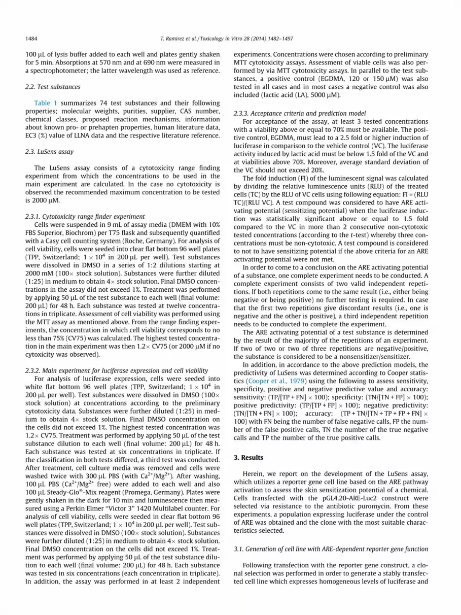
the optimal signal to noise ratio of luciferase induction. Single cellclones (n = 24) were selected and propagated. The relative basalexpression level of these clones was compared (Fig. 1): 17 out of24 colonies expressed basal luciferase levels which changed onlymarginally to 0.5–1 fold when treated with solvent only (VC). 5out of 24 colonies expressed luciferase levels between >1 and 1.5fold compared to VC, one clone expressed basal luciferase levelsbelow 0.5 fold compared to VC (Fig. 1), and only one clone (clone18) did not show any luciferase expression. To determine the rangeof induction of luciferase following exposure to sensitizing sub-stances, the clones were exposed to a weak sensitizer, EGDMA, attwo concentrations (75 lM and 150 lM). As illustrated in Fig. 2,the clones exhibited a concentration dependent induction of lucif-erase. The lower concentration of EGDMA, 75 lM, induced lucifer-ase expression from 1.5 to 4.6 fold in 22 of 24 clones; an inductionof only 1.1 fold was observed for one clone (24). In addition toluciferase induction, viability was also measured: in 21 of 24clones EGDMA did not induce cytotoxicity above 20% and in onlytwo clones (1 and 13) the viability was below 80% when treatedwith 75 lM EGDMA (Fig. 2). Luciferase induction was higher incells treated with 150 lM EGDMA, ranging from 2 to 9 fold in 22
Fig. 1. (A) Basal luciferase activity of 23 clones of LuSens cells (grey bars), the expressiovehicle control (VC, DMSO 1%). Data for clone 18 are not displayed since this clone didwithout DMSO (blue diamonds) was compared to the viability of each clone treated witdimethacrylate (EGDMA, 75 lM white bars; 150 lM grey bars, the black bars correspoViability relative to VC is also illustrated per clone (black lines). (For interpretation of thethis article.)
of 24 clones; one clone (clone 24) exhibited an induction below2 fold. Viability was reduced below 70% in three clones (1, 5 and9), while the rest of the clones retained good viability. Based onthese experiments clone 16 was selected as the best clone becauseof its low basal activity and its good capacity to be activated whenexposed to EGDMA and was selected to further develop the LuSensassay.
The cytogenetic analysis of clone 16 was also performed and theresults were consistent with a cell-line of human origin, presentinga hypertriploid karyotype with an average number of 77 chromo-somes (modal range from 74 to 80) and 6 marker chromosomes(M1–M6), of which 3 showed 2 copies each. In addition, genomicDNA of this clone was isolated for verification of the presence ofthe ARE sequence. Sequencing demonstrated that the sequence ofthe ARE of the reporter gene construct was present without anymodification (data not shown). When a Blast search of the AREsequence using whole genomic DNA sequences from the NCBI gen-ome database (http://www.ncbi.nlm.nih.gov/genome) was con-ducted, the sequence from LuSens clone 16, provided a 100%identity with the NAD(P)H dehydrogenase [quinone] 1 gene(Nqo1) sequence of Rattus norvegicus strain BN; Sprague-Dawley
n of single clones were compared to the expression of each clone treated with thenot show detectable basal luciferase activity. In addition, the viability of the clonesh VC. (B) Effect of increasing concentrations of the weak sensitizer, ethylene glycolnd to VC) towards the luciferase expression of 23 selected clones of LuSens cells.references to color in this figure legend, the reader is referred to the web version of

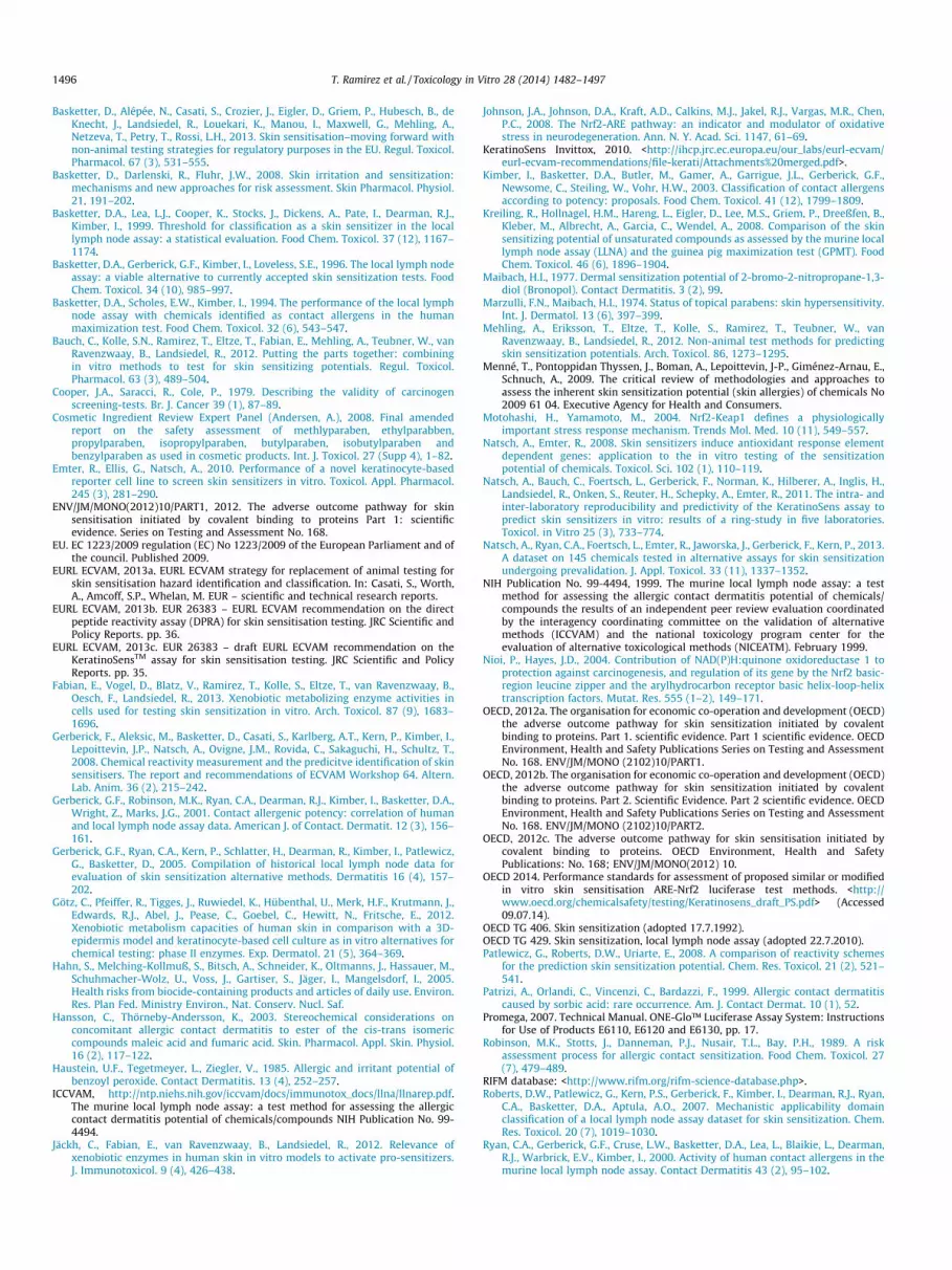
Fig. 2. Exemplary performance of LuSens cells clone 16 towards skin sensitizers and non-sensitizers. Effect of increasing concentrations of EGDMA (A), cinnamyl alcohol (B),sulfanilamide (C) and vanillin (D) on the luciferase expression (bars) and viability (green squares) of LuSens cells clone 16, illustrated are results of three independentexperiments. After exposure with skin sensitizers (A and B) an increase in the luciferase expression above the 1.5 fold threshold is observed in a dose dependent manner innon-cytotoxic concentrations. Contrastingly, no luciferase induction is observed above the 1.5 fold threshold, even at concentrations close to cell toxicity, indicating noactivation of LuSens cells.
1490 T. Ramirez et al. / Toxicology in Vitro 28 (2014) 1482–1497
(Sequence ID: ref AC_000087.1). It is important to highlight thatARE consensus sequence, responsible for ARE functionality amongspecies (i.e., human, mouse, rat), TMAnnRTGAYnnnGCRwww(where M = A or C, R = A or G, Y = C or T, W = A or T, S = G or C)(Wasserman and Fahl, 1997) is present in the rat ARE sequencefound in LuSens cells.
3.2. Assay development and predictivity according to Cooper statistics
In order to determine the predictive capacity of the LuSensassay, 74 test compounds with animal and/or human sensitizationdata available (Table 1) were tested in at least two independentexperiments in order to compare to human and LLNA data fromthe literature. The LuSens assay correctly predicted 57 of 69 or53 of 74 substances when compared to human or LLNA data,respectively. When compared to human data, seven substanceswere incorrectly rated to be negative: aniline, ethylene diamine,Luperox A75 (benzoyl peroxide), nickel chloride, phenyl benzoate,phthalic anhydride and propyl gallate. Five substances were incor-rectly rated as positive when compared to human data: 4methoxyacetophenone, 6-methylcoumarin, methyl salicylate, pro-pyl-4-hydroxybenzoate, and tween 80. When compared to LLNAdata, twelve substances would be incorrectly rated to be negative:4-allylanisole, aniline, ethylene diamine, farnesal, hexadecyltrime-thylammonium bromide, phthalic anhydride, phenyl benzoate,propyl gallate, pyridin, sodium lauryl sulfate (SDS), resorcinol, tar-taric acid and xylene. It should be noted that hexadecyltrimethy-lammonium bromide, pyridine, SDS, tartaric acid, resorcinol andxylene yield false positive results in the LLNA when compared tohuman data. Eight substances would be incorrectly rated to bepositive when compared to LLNA data: 4-methoxyacetophenone,6-methlycoumarin, benzyl alcohol, methyl salicylate, propyl 4-hydroxybenzoate, and tween 80 (Table 2). For the assessment ofthe predictive capacity of the LuSens assay, the data obtained fromthe in vitro assay were compared to human or LLNA data from theliterature using cooper statistics (Table 3). From this analysis thefollowing predictivity values were calculated: Sensitivity of 83%
or 73%, specificity of 81% or 74% and an overall accuracy of 83%or 74% when compared to human or LLNA data, respectively.
3.3. Intralaboratory reproducibility
The intralaboratory reproducibility of the LuSens assay wasassessed based on the concordance of the classification of indepen-dent valid runs testing the same substance. All valid runs, namelythose that adhered to the acceptance criteria, were consideredeven if an overall conclusion was already reached after two runs.From this analysis it was found that the assessment of 69 of 74compounds was consistently reproducible (93%) and the assess-ment of 5 compounds, 4-allylanisole, benzyl alcohol, glyoxal solu-tion, Luperox A75 and n-butanol, were not reproducible along theindependent runs (Table 2).
3.4. Comparison of the LuSens and KeratinoSens™ assays
In general KeratinoSens™ and LuSens assays possess similarcapacity to predict sensitizers. In KeratinoSens™ the luciferasegene is regulated by the ARE-element of the human aldoketoreduc-tase (AKR1C2), whereas in LuSens it is under the control of ARE ele-ment from the rat NADPH:quinone oxidoreductase (nqo1). Anothermajor difference between both assays is that LuSens uses a cyto-toxicity range finder experiment to select 6 testing concentrationto analyze the luciferase activity, KeratinoSens™ in contrast usesa range of 12 fixed concentrations from 1 to 200 lM. In addition,while LuSens plate set up uses all replicates for all concentrationsin one plate in order to avoid potential plate differences effect onthe replicates, KeratinoSens™ distribute the three replicates ofeach testing concentrations in 3 independent plates. The last majordifference will be validity controls of both assays, for LuSens a neg-ative and positive control is used, DL-Lactic acid and EGDMA,respectively, while KeratinoSens™ uses as positive control cin-namic aldehyde. The analysis of same data set in comparison tothe data reported for KeratinoSens™ (Natsch et al., 2013; Natschet al., 2011; Bauch et al., 2012) indicates a good concordance of

Table 2Summary results of the LuSens assay. Depicted are the Imax (maximal induction), the concentration at which the 1.5 fold and 2 fold induction was observed (lM), the number ofexperiments showing a statistical significant 1.5 fold and 2 fold induction, the concentration (lM) at which 50% cytotoxicity was induced. Generally no cytotoxicity was observedat the 1.5 fold induction. In addition, the reproducibility is indicated by ‘‘R’’ for reproducible and ‘‘NR’’ for not reproducible. The results obtained from the LuSens assay werecompared to those reported on the literature for KeratinoSens™ assay. Where results from both assays were concordant, a match is indicated as ‘‘M’’, in contrast, when data fromboth assays were discordant, a mismatch is indicated by ‘‘MM’’. When literature data from KeratinoSens™ was inconclusive (different results reported in the literature), no ‘‘M’’ or‘‘MM’’ could be assessed, then this was indicated as ‘‘?’’. Where no KeratinoSens™ data were available, no comparison was made; indicated by ‘‘ND’’.
Test substance Imax
(fold)Concentration(lM) for 1.5fold induction
Numberof EC1.5positives
Concentration(lM) for 2 foldinduction
Numberof EC2.0positives
Concentration(lM) causing50%cytotoxicity
LuSensprediction
Reproducibility ComparisonwithKeratinoSens™
References
Oxazolone 7.49 <743.47 2 of 2 <743.47 2 of 2 >1850 + R M Natsch et al.(2013)
MCI/MI 6.78 <4.6 3 of 3 <4.6 3 of 3 >900 + R M Natsch et al.(2013)
p-Benzoquinone 2.58 9.69 2 of 2 <11.11 2 of 2 >16 + R M Natsch et al.(2013)
1-Chloro-2,4-dinitro benzene 3.34 3.23 2 of 4 3.65 2 of 4 >5 + NR M Natsch et al.(2013)
Potassium dichromate 2.95 0.82 2 of 2 1.19 2 of 2 >1.56 + R M Natsch et al.(2013)
Metol 7.82 <10.99 4 of 4 <10.99 4 of 4 >27.35 + R M Natsch et al.(2013)
Glutaraldehyde 5.99 <45.05 2 of 2 45.05 2 of 2 104.24 + R M Natsch et al.(2013)
4-Phenylenediamine 11.44 <53.05 4 of 4 <53.05 4 of 4 >132 + R M Natsch et al.(2013)
Propyl gallate 0.89 >140 0 of 2 >140 0 of 2 >140 � R MM Natsch et al.(2013)
Pricrysulfonic acid 5.94 <964.51 2 of 2 <964.51 2 of 2 >2400 + R NDPhthalic anhydride 0.33 >2400 0 of 2 >2400 0 of 2 >2400 � R M Natsch et al.
(2013)Formaldehyde 3.99 184.38 3 of 3 214.28 3 of 3 >288 + NR M Natsch et al.
(2013)Methyldibromo glutaronitrile 2.71 <16.08 3 of 3 18.98 2 of 3 >34.72 + R M Natsch et al.
(2013)Glyoxal solution 4.64 <180.84 2 of 3 278.47 1 of 3 >450 + NR M Natsch et al.
(2013)Isoeugenol 25.41 <120.56 3 of 3 <120.56 3 of 3 >300 + R M Natsch et al.
(2013)Diethyl maleate 13.83 <24.11 2 of 2 <24.11 2 of 2 >60 + R M Natsch et al.
(2013)Ethylene diamine 1.12 >750 0 of 2 >750 0 of 2 >750 � R MM Natsch et al.
(2013)Benzylidene acetone 21.81 <10.05 2 of 2 <10.05 2 of 2 >25 + R M Natsch et al.
(2013)Cinnamic aldehyde 23.25 <44.21 3 of 3 <44.21 3 of 3 >110 + R M Natsch et al.
(2013)Cobalt chloride 7.48 <241.13 3 of 3 <241.13 3 of 3 >600 + R M Natsch et al.
(2013)Thiram 5.29 <20.82 4 of 4 <20.82 4 of 4 49.50 + R ND2-Phenyl propionaldehyde 3.45 48.23 4 of 4 70.35 4 of 4 112.03 + R M Natsch et al.
(2013)Resorcinol 0.58 >920.74 0 of 4 >920.74 0 of 4 >1104.89 � R M Natsch et al.
(2013)a-Hexyl-cinnamic aldehyde 3.30 14.53 2 of 2 18.99 2 of 2 29.56 + R ? Natsch et al.
(2013), Bauchet al. (2012)
Tartaric acid 1.28 >2400 0 of 4 >2400 0 of 4 >2400 � R ? Natsch et al.(2013), Bauchet al. (2012)
2-Mercapto benzothiazole 23.48 <361.69 3 of 3 <361.69 3 of 3 >900 + R M Natsch et al.(2013)
2.3-Butanedione 8.30 <160.75 2 of 2 189.50 2 of 2 >400 + R M Nat sch et al.(2013)
Citral 32.32 <48.23 3 of 3 <48.23 3 of 3 >120 + R M Natsch et al.(2013)
Eugenol 5.49 <241.13 4 of 4 <241.13 4 of 4 >600 + R ? Natsch et al.(2013), Bauchet al. (2012)
Farnesal 0.85 >11.11 0 of 2 >11.11 0 of 2 13.51 – R MM Natsch et al.(2013)
Sodium lauryl sulfate 0.77 >30 0 of 3 >30 0 of 3 >36 � R M Natsch et al.(2013)
4-Allylanisole 2.00 >540 1 of 4 540.00 1 of 4 >540 - NR MM Natsch et al.(2013)
Hydroxycitronellal 2.03 189.04 2 of 3 424.68 1 of 3 >434 + NR ND
(continued on next page)
T. Ramirez et al. / Toxicology in Vitro 28 (2014) 1482–1497 1491

Table 2 (continued)
Test substance Imax
(fold)Concentration(lM) for 1.5fold induction
Numberof EC1.5positives
Concentration(lM) for 2 foldinduction
Numberof EC2.0positives
Concentration(lM) causing50%cytotoxicity
LuSensprediction
Reproducibility ComparisonwithKeratinoSens™
References
Phenyl benzoate 1.14 >250 0 of 3 >250 0 of 3 >360 � R M Natsch et al.(2013)
Cinnamyl alcohol 7.28 <168.79 3 of 3 <168.79 3 of 3 >420 + R M Natsch et al.(2013)
Imidazolidinyl urea 4.72 31.10 3 of 4 37.20 3 of 4 55.56 + NR M Natsch et al.(2013)
Undecylenic acid 8.05 <172.36 2 of 2 <172.36 2 of 2 >428.88 + R M Natsch et al.(2013)
Ethylene glycoldimethacrylate
31.84 <120.56 3 of 3 <120.56 3 of 3 >300 + R M Natsch et al.(2013)
Pyridine 1.46 >2400 0 of 4 >2400 0 of 4 >2400 � R M Natsch et al.(2013)
Aniline 1.04 >2400 0 of 2 >2400 0 of 2 >2400 � R ? Natsch et al.(2013), Bauchet al. (2012)
Methyl methacrylate 7.33 <1205.63 2 of 2 <1205.63 2 of 2 >3000 + R ? KeratinoSensInvittox (2010),Bauch et al.(2012)
4-Nitrobenzyl bromide 3.18 <0.88 3 of 3 <0.88 3 of 3 >2.18 + R M Natsch et al.(2013)
Butyl 4-hydroxy benzoate 4.69 <22.10 3 of 3 <22.10 3 of 3 >55 + R NDMethyl 4-hydroxy benzoate 6.70 <381.78 3 of 3 <381.78 3 of 3 >950 + R M Natsch et al.
(2013)Sorbic acid 4.20 <964.51 3 of 3 1893.00 3 of 3 >2400 + R NDLuperox A75 2.28 41.50 1 of 4 46.82 0 of 4 >49.8 � NR NDButyl glycidyl ether 4.80 <72.34 3 of 3 <72.34 3 of 3 >180 + R M Natsch et al.
(2013)Ethylparaben 3.67 <108.51 3 of 3 <108.51 3 of 3 >270 + R ND2-Bromo-2-nitro-1,3-
propanediol5.47 <29.18 3 of 3 38.84 3 of 3 72.10 + R ND
Xylene 1.43 >578.70 0 of 4 >578.70 0 of 4 814.45 � R M Natsch et al.(2013)
Glycerol 1.16 >2400 0 of 3 >2400 0 of 3 >2400 � R M Natsch et al.(2013)
1,2-Propanediol 1.21 >2400 0 of 2 >2400 0 of 2 >2400 � R M Natsch et al.(2013)
4-Hydroxybenzoic acid 1.06 >2400 0 of 2 >2400 0 of 2 >2400 � R M Natsch et al.(2013)
4-Methoxy acetophenone 8.97 <964.51 2 of 2 <964.51 2 of 2 >2400 + R M Natsch et al.(2013)
6-Methylcoumarin 3.54 <120.56 3 of 3 141.48 3 of 3 >300 + R M Natsch et al.(2013)
Chlorobenzene 1.48 >2000 0 of 4 >2000 0 of 4 1989.00 � R M Natsch et al.(2013)
DL-lactic acid 0.98 >7000 0 of 2 >7000 0 of 2 >7000 � R M Natsch et al.(2013)
Fumaric acid 1.27 >2400 0 of 3 >2400 0 of 3 >2400 � R M Natsch et al.(2013)
D(+)glucose 1.07 >3000 0 of 3 >3000 0 of 3 >3000 � R M Natsch et al.(2013)
Isopropanol 1.23 >3000 0 of 3 >3000 0 of 3 >3000 � R M Natsch et al.(2013)
Methyl salicylate 2.22 <803.76 3 of 4 1851.96 1 of 4 >2000 + NR MM Natsch et al.(2013)
n-Butanol 1.62 2400.00 1 of 4 >2400 0 of 4 >2400 � NR Mn-Hexane 1.23 >2400 0 of 3 >2400 0 of 3 >2400 � R ? Natsch et al.
(2013), Bauchet al. (2012)
Nickel chloride 0.79 >261.20 0 of 3 >261.20 0 of 3 >261.20 � R ? Natsch et al.(2013), Bauchet al. (2012)
4-Aminobenzoic acid 1.11 >2400 0 of 2 >2400 0 of 2 >2400 � R M Natsch et al.(2013)
Propyl-4-hydroxybenzoate 3.94 <65.104 2 of 2 <65.104 2 of 2 >162 + R NDSalicylic acid 0.80 >3000 0 of 3 >3000 0 of 3 >3000 � R M Natsch et al.
(2013)Sulfanilamide 1.08 >2400 0 of 2 >2400 0 of 2 >2400 � R M Natsch et al.
(2013)Vanillin 1.48 >1650 0 of 3 >1650 0 of 3 >1650 � R M Natsch et al.
(2013)Hexadecyltrimethylamonium
bromide0.95 >0.69 0 of 4 >0.69 0 of 4 1.03 � R ND
1492 T. Ramirez et al. / Toxicology in Vitro 28 (2014) 1482–1497

Table 2 (continued)
Test substance Imax
(fold)Concentration(lM) for 1.5fold induction
Numberof EC1.5positives
Concentration(lM) for 2 foldinduction
Numberof EC2.0positives
Concentration(lM) causing50%cytotoxicity
LuSensprediction
Reproducibility ComparisonwithKeratinoSens™
References
Tween 80 3.76 <65.91 4 of 4 <65.91 3 of 4 >164 + R M Natsch et al.(2013)
Diethyl phthalate 3.37 <516.41 0 of 3 516.41 0 of 3 >1285 � R M Natsch et al.(2013)
Sodium benzoate 1.01 >2400 0 of 3 >2400 0 of 3 >2400 � R NDBenzyl alcohol 1.93 <964.51 2 of 3 >2400 1 of 3 >2400 + NR ND
Table 3Summary of predictivity of LuSens assay based on 69 test substances.
In comparison to
Human LLNA
n 69 72Sensitivity 83% 74%Specificity 81% 74%PPV 88% 86%NPV 76% 56%Accuracy 83% 74%
T. Ramirez et al. / Toxicology in Vitro 28 (2014) 1482–1497 1493
92% for 63 compounds in total (for which data for both assays wereavailable). A concordance of 94% was reached for the 52 test com-pounds for which human data from the literature were availableand 93% for the 61 test compounds from which LLNA data wereavailable. From these 52 compounds, for which human data wereavailable, non-concordant data (mismatch) were observed forthree substances. In the LuSens methyl salicylate (non to veryweak sensitizer in humans) tested positive, ethylenediamine (pro-hapten) and propyl gallate (both sensitizers in humans) tested neg-ative. The discordant results with methyl salicylate are probablythe consequence of borderline readouts in both assays. It is worthnoticing that non-concordant data are also reported for substancestested multiple times in the same assay since some variationswithin the same system are expected under the frame of technicalor biological difference (intra-laboratory variation).
In the case of the substances predicted as false negative, ethy-lenediamine and propyl gallate both substances were rated as posi-tive in the KeratinoSens™ assay but negative in the LuSens assay.KeratinoSens™ only requires one single concentration of the testsubstance yielding an induction higher than 1.5 fold whereasLuSens requires at least two consecutive concentrations.
Seven substances did not yield consistent results in the datapreviously published for KeratinoSens™ assay, i.e., tartaric acid,hexane, nickel chloride, methylmethacrylate, aniline, eugenol anda-hexyl cinnamic aldehyde (Natsch et al., 2013; Bauch et al.,2012; KeratinoSens Invittox, 2010). The results of these substanceswere therefore not used for comparisons to LuSens data (Table 2).
For 57 of the 61 compounds with available LLNA data consistentresults in both the LuSens and KeratinoSens™ resulted in 93% con-cordance among both assays. Only four test substances did not pro-vided concordant data among both assays, i.e., methyl salicylate,farnesal, ethylenediamine and propyl gallate. For these substancesthe LuSens assay results were consistent with the LLNA results. Foradditional seven substances no comparison was possible since datain the literature were not concordant for following substances inthe KeratinoSens™ assay: tartaric acid, n-hexane, nickel chloride,methylmethacrylate, aniline, eugenol and a-hexyl cinnamic alde-hyde (Natsch et al., 2013; Bauch et al., 2012; KeratinoSensInvittox, 2010; Natsch et al., 2011, Table 2).
According to the performance standard guidance for Keratino-Sens™, the value for similar or modified test methods is required
to be equal to or greater than 80% based on the data of three inde-pendent experiments for 12 reference substances (OECD 2014). Inthe LuSens assay all but one substance of these 12 reference sub-stances were reproducible (92%) and hence the criteria for theintralaboratory reproducibility fully met and almost identical tothe overall within laboratory reproducibility described above forthe 74 compounds assessed.
3.5. LuSens assay as part of a testing strategy
The pragmatic ‘weight of evidence’ approach predicts skin sen-sitization potential based on two of three tests addressing proteinreactivity (e.g., DPRA), keratinocyte ARE activation (e.g., LuSens)and dendritic cell activation (e.g., h-CLAT); i.e., concordant resultsof two tests then determines whether a substance is consideredto be a sensitizer or not (if at least two tests are negative the sub-stance is assessed as a being a non-sensitizer and vice versa; Bauchet al., 2012). When the LuSens results are used in combination withpreviously reported data for DPRA and mMUSST and compared toLLNA data, the sensitivity is 75%, specificity is 94% and the accuracyis 81% (43 of 53 correct predictions) (Table 4). 9 Substances wouldbe rated as false negative: phthalic anhydride, a-hexyl cinnamicaldehyde, tartaric acid, farnesal, SDS, pyridine, aniline, xylene and6-methylcoumarin, whereby only nickel chloride as false positive.Similar calculations were performed for the combination of DPRA,LuSens and h-CLAT (data are summarized in Table 4). When com-paring the results of this set of chemicals to human data, the LLNAhad 96% sensitivity, 81% specificity and an accuracy of 90% indicat-ing that the LLNA also did not correctly predict the sensitizationpotential for humans for all chemicals tested. When compare tohuman data the combination of LuSens, DPRA and mMUSST yields,the predictivity of the combination yields sensitivity, 82%, specific-ity of 100% and accuracy of 92% (45 of 50 correct predictions)(Table 4, compare to human data), Five substances would be ratedas false negative: phtalic anhydride, tartaric acid, a-hexyl cinnamicaldehyde, undecylenic acid, and aniline.
4. Discussion
Although animal tests encompass all steps leading to an adversereaction, they remain a ‘‘blackbox’’ as the final reactions are mon-itored but no knowledge is generated elucidating the pathwaysleading to these effects. When developing new toxicological tests,the current approach is therefore to understand the key steps lead-ing to the reactions assessed by the toxicological endpoint and toutilize this knowledge to optimize tests and test strategies. Accord-ing to the OECD, ‘‘an adverse outcome pathway (AOP) is thesequence of events from the chemical structure of a target chemi-cal or group of similar chemicals through the molecular initiatingevent to an in vivo outcome of interest’’. Skin sensitization is a mul-tistep process and the key steps involved are fairly well understood(EURL ECVAM, 2013a). The OECD recently published a documentdescribing adverse outcome pathway for skin sensitization

Table 4Summary of predictivities of single assays and combinations thereof: LuSens assay and other assays (OECD QSAR toolbox, Direct Peptide Reactivity Assay (DPRA), modifiedmyeloid U937 skin sensitization test (mMUSST), human cell line activation test (h-CLAT). Data were obtained from Bauch et al., 2012). These figures are based on 50 testsubstances (out of a total of 69) for which all data are available.
Compared to human
Sensitivity (%) Specificity (%) PPV (%) NPV (%) Accuracy (%)
LLNA 96 81 87 94 90Single assays OECD QSAR toolbox 64 100 100 69 80
DPRA 89 82 86 86 86LuSens 79 82 85 75 80KeratinoSens™ 86 73 80 80 80mMUSST 68 100 100 71 82h-CLAT 75 77 81 71 76
Combinations DPRA and LuSens 96 64 77 93 82DPRA and KeratinoSensTM 100 59 76 100 82DPRA and mMUSST 93 82 87 90 88DPRA and h-CLAT 96 59 75 93 80LuSens and mMUSST 93 82 87 90 88LuSens and h-CLAT 93 73 81 89 84KeratinoSens™ and MUSST 90 56 77 77 77KeratinoSens™ and h-CLAT 93 64 76 88 80
Prediction model (high overall accuracy) DPRA. LuSens and mMUSST 82 100 96 84 90DPRA. KeratinoSensTM and mMUSST 86 95 96 84 90DPRA. LuSens and h-CLAT 86 86 89 83 86DPRA. KeratinoSensTM and h-CLAT 89 82 86 86 86
Compared to LLNA
Sensitivity (%) Specificity (%) PPV (%) NPV (%) Accuracy (%)
Single assays OECD QSAR toolbox 56 100 100 52 70DPRA 81 76 88 65 79LuSens 69 82 89 56 74KeratinoSens™ 83 76 88 68 81mMUSST 64 94 96 55 74h-CLAT 72 76 87 57 74
Combinations DPRA and LuSens 89 59 82 71 79DPRA and KeratinoSens™ 94 59 83 83 83DPRA and mMUSST 86 76 89 72 83DPRA and h-CLAT 89 53 80 69 77LuSens and mMUSST 83 76 88 68 81LuSens and h-CLAT 86 71 86 71 81KeratinoSens™ and MUSST 89 71 86 75 83KeratinoSens™ and h-CLAT 89 65 84 73 81
Prediction model DPRA, LuSens and mMUSST 75 94 96 64 81DPRA, KeratinoSensTM and mMUSST 81 88 94 68 83DPRA, LuSens and h-CLAT 81 88 94 68 83DPRA, KeratinoSens™ and h-CLAT 83 82 91 70 83
1494 T. Ramirez et al. / Toxicology in Vitro 28 (2014) 1482–1497
describing key steps in the sensitization process thereby facilitat-ing the development of new toxicological test methods and ITSto assess skin sensitization (ENVJMV/MONO, 2012). Among thesteps defined, protein binding, keratinocyte and dendritic cell acti-vation are key initiating steps in the sensitization process.
In this study, a new stable ARE reporter gene assay based on ahuman keratinocyte cell line, LuSens, was developed and evaluatedfor its use in the identification of skin sensitizers. The LuSens assayprovides information on both protein reactivity and keratinocyteactivation. This is accomplished by monitoring the activity of lucif-erase, which is regulated by the ARE of the rat Nqo1 gene locatedupstream the luciferase reporter gene, following contact to a testsubstance. Protein reactivity of a substance can be indirectlyassessed, as reactivity with the cysteine residues of Keap1 leadsto the dissociation of Nrf2 and its subsequent binding to ARE andthe expression of the downstream genes, in this case luciferase.As described extensively in the literature, ARE plays a crucial rolein the activation of cytoprotective genes in the elicitation of thetoxicity pathway induced by skin sensitizers (Natsch and Emter,2008; Johnson et al., 2008) and therefore cell activation can beassessed by the expression of luciferase. Since keratinocytes arethe first cells exposed to a substance when skin contact occurs,the LuSens assay was designed using the primary target cells of
the skin, namely the keratinocytes, and thereby gives a measureof their activation as cellular event leading to skin sensitization.
The validity criteria of the LuSens assay described here wererefined from the initially used criteria (Bauch et al., 2012). Theassay avoids the use of concentrations inducing toxicity greaterthan 30%. For this purpose, the assay includes a range findingexperiment to select the concentration range that will be used inthe main experiment and thereby avoids testing of unneeded toxicconcentrations. In addition, a rigorous prediction model wasapplied, meaning that sensitizer compounds were only those thathad the capacity to induced P1.5� luciferase induction in twoconsecutive test concentrations and at least half of the tested con-centrations should have yielded a viability P70%. Substances thatinduced luciferase expression only in one concentration or in non-consecutive concentrations were considered to be non-sensitizers.
Although the validation of the assay has been designed to beused with the SteadyGlo™ luciferase assay system to measureluciferase activity, recent data from our laboratory with a smallset of test compounds (i.e., 8 compounds in at least 2 independentexperiments, data not shown) demonstrated the same proficiencywhen using OneGlo™ luciferase detection system. This detectionsystem is reported by the producer to be more robust (Promega,2007), as some detection difficulties can be encountered

T. Ramirez et al. / Toxicology in Vitro 28 (2014) 1482–1497 1495
depending on the equipment used when using the SteadyGlo™luciferase assay (personal communication). However, in our hands,both luciferase detection system delivered comparable data, butthe OneGlo™ system provided a stronger signal.
The LuSens assay shows good accuracy, sensitivity and specific-ity values that are comparable to those of the KeratinoSens™ assay.The luciferase reporter gene in the LuSens assay is under the con-trol of the ARE-element of Nqo1 gene from the rat, which is acti-vated by the Nrf2 pathway and is comparable to the ARE fromhuman AKR1C2 gene that has been cloned into KeratinoSens™(Nioi and Hayes, 2004; Natsch and Emter, 2008). In addition,LuSens cell-line used in this study was derived from the selectedclone which gave the best signal to noise ratio of the light outputof luciferase induction, and which showed good luciferase induc-tion when treated with weak sensitizers. The latter is also reflectedin the use of the weak sensitizer EGDMA as a positive control of theassay. Recent experiments in which the cells have been cultured inthe absence of puromycin for more than 15 cell passages exhibitedthe same proficiency towards a small set of skin sensitizers (Fig. 3),an indication that the inserted sequence was stably inserted intothe cells.
Based on the established validity criteria, an analysis of the pre-dictivity of the method was conducted with a set of 74 substancescomprising 42 skin sensitizers with a wide range of potencies and27 non-sensitizing substances comprised of various industriallyused substances and cosmetic ingredients. Human data were avail-able for 69 substances and LLNA data was available for 72 sub-stances. An overall accuracy of 83% was achieved whencompared to human data. Seven false negative predictions werefound indicating limited applicability for acyl transferases (3 of 7FN predictions), prohaptens (3 of 9 FN predictions, whereby thepro Michael acceptors, except propyl gallate, were correctly pre-dicted), and the difficulty to predict nickel chloride (false negativein the LLNA). In both the KeratinoSens™ and LuSens assays, phtha-lic anhydride is not correctly predicted. In the DPRA, phthalic anhy-dride exhibits virtually no cysteine binding but a strong reactivityto lysine (Gerberick et al., 2008). As the LuSens assay assesses theactivation of the ARE dependent gene expression in keratinocytesby modification of a cysteine in the Nrf2 protein, some molecularor cellular events contributing to skin sensitization may be missed.This may, however, be balanced by using a testing battery of threeassays addressing several steps of the adverse outcome pathwayand in particular the DPRA which assesses both cysteine and lysinereactivity.
Other limitations of the LuSens assay, as is the case with mostcell-based methods, solubility and cytotoxicity of the substancecan limit the applicability as the cells are cultured in aqueous med-ium. The metabolic capacity of the cells which is required to
Fig. 3. Effect of increasing concentrations of the weak sensitizer, EGDMA on theluciferase expression (bars) and viability (green squares) of LuSens cells clone 16that have been culture without the puromycin antibiotic as selection pressure.Illustrated are results of four independent experiments. (For interpretation of thereferences to color in this figure legend, the reader is referred to the web version ofthis article.)
activate certain pro-haptens is not identical to the metaboliccapacity of native skin (Götz et al., 2012; Jäckh et al., 2012;Fabian et al., 2013).
There are limitations to any toxicological test method, and thesemust be considered to reliably ensure predictions of potential haz-ards to human health. The LLNA is the method of choice to assessthe skin sensitization potential of a substance under REACH. Thepredictive accuracy of the LLNA, compared to human data was72% (n = 74) based on the formal validation conducted by ICCVAM(http://ntp.niehs.nih.gov/iccvam/docs/immunotox_docs/llna/llna-rep.pdf). To date, no non-animal test method has been accepted as afull replacement of the animal based tests for skin sensitization.Even those currently advanced in validation process (DPRA, Kerati-noSens™, h-CLAT) will not achieve regulatory acceptance as astand-alone method but they will find use in ITSs. The combinationof these assays should be acceptable to fully replace animal testingfor the identification of a skin sensitization potential in the nearfuture in particular in the context of an AOP-based IATA. In a recentworkshop with regulators (ECHA and member state representa-tives) and industry, it was agreed that an ITS should preferably becomposed of methods which had a sound mechanistic rationale,e.g., by reflecting key steps in the AOP (Basketter et al., 2013).Two studies with large databases have demonstrated, that a combi-nation of these or similar assays covering three key steps of the AOPoffers a high predicitivity of the skin sensitization potential (haz-ard) even when using a simple prediction model such as ‘‘2 out of3’’ weight of evidence approach (also sometimes termed the‘‘majority vote’’ approach; Bauch et al., 2012; Natsch et al., 2013).
The LuSens assay (as does the KeratinoSens™ assay) contributesto the 3R principles as it addresses one step of the adverse outcomepathway, namely keratinocyte activation. ITSs using combinationsof the LuSens assay with other non-animal tests can result in pre-dictivities comparable to or even higher than the LLNA. In Phase 3of REACH an evaluation of the sensitization hazard for thousands ofsubstances produced at 1–100 tons will need to be submitted byspring 2018. Bearing this in mind, the application of the LuSensassay in a testing battery can quite conceivably significantly con-tribute to the reduction of the number of animals used for REACH.
Conflict of Interest
The authors declare that there are no conflicts of interest.
Transparency Document
The Transparency document associated with this article can befound in the online version.
References
Ade, N., Leon, F., Pallardy, M., Peiffer, J.L., Kerdine-Romer, S., Tissier, M.H., Bonnet,P.A., Fabre, I., Ourlin, J.C., 2009. HMOX1 and NQO1 genes are upregulated inresponse to contact sensitizers in dendritic cells and THP-1 cell line: role of theKeap1/Nrf2 pathway. Toxicol. Sci. 107 (2), 451–460.
Andersen, K.E., Frankild, S.r., 1997. Allergic contact dermatitis. Clin. Dermatol. 15(4), 645–654.
Aptula, A.O., Roberts, D.W., 2006. Mechanistic applicability domains for nonanimal-based prediction of toxicological end points: general principles and applicationto reactive toxicity. Chem. Res. Toxicol. 19 (8), 1097–1105.
Aptula, A.O., Enoch, S.J., Roberts, D.W., 2009. Chemical mechanisms for skinsensitization by aromatic compounds with hydroxy and amino groups. Chem.Res. Toxicol. 22 (9), 1541–1547.
Basketter, D.A., Alépée, N., Ashikaga, T., Barroso, J., Gilmour, N., Goebel, C.,Hibatallah, J., Hoffmann, S., Kern, P., Martinozzi-Teissier, S., Maxwell, G.,Reisinger, K., Sakaguchi, H., Schepky, A., Tailhardat, M., Templier, M., 2014.Categorization of chemicals according to their relative human skin sensitizingpotency. Dermatitis 25 (1), 11–21.
1496 T. Ramirez et al. / Toxicology in Vitro 28 (2014) 1482–1497
Basketter, D., Alépée, N., Casati, S., Crozier, J., Eigler, D., Griem, P., Hubesch, B., deKnecht, J., Landsiedel, R., Louekari, K., Manou, I., Maxwell, G., Mehling, A.,Netzeva, T., Petry, T., Rossi, L.H., 2013. Skin sensitisation–moving forward withnon-animal testing strategies for regulatory purposes in the EU. Regul. Toxicol.Pharmacol. 67 (3), 531–555.
Basketter, D., Darlenski, R., Fluhr, J.W., 2008. Skin irritation and sensitization:mechanisms and new approaches for risk assessment. Skin Pharmacol. Physiol.21, 191–202.
Basketter, D.A., Lea, L.J., Cooper, K., Stocks, J., Dickens, A., Pate, I., Dearman, R.J.,Kimber, I., 1999. Threshold for classification as a skin sensitizer in the locallymph node assay: a statistical evaluation. Food Chem. Toxicol. 37 (12), 1167–1174.
Basketter, D.A., Gerberick, G.F., Kimber, I., Loveless, S.E., 1996. The local lymph nodeassay: a viable alternative to currently accepted skin sensitization tests. FoodChem. Toxicol. 34 (10), 985–997.
Basketter, D.A., Scholes, E.W., Kimber, I., 1994. The performance of the local lymphnode assay with chemicals identified as contact allergens in the humanmaximization test. Food Chem. Toxicol. 32 (6), 543–547.
Bauch, C., Kolle, S.N., Ramirez, T., Eltze, T., Fabian, E., Mehling, A., Teubner, W., vanRavenzwaay, B., Landsiedel, R., 2012. Putting the parts together: combiningin vitro methods to test for skin sensitizing potentials. Regul. Toxicol.Pharmacol. 63 (3), 489–504.
Cooper, J.A., Saracci, R., Cole, P., 1979. Describing the validity of carcinogenscreening-tests. Br. J. Cancer 39 (1), 87–89.
Cosmetic Ingredient Review Expert Panel (Andersen, A.), 2008. Final amendedreport on the safety assessment of methlyparaben, ethylparabben,propylparaben, isopropylparaben, butylparaben, isobutylparaben andbenzylparaben as used in cosmetic products. Int. J. Toxicol. 27 (Supp 4), 1–82.
Emter, R., Ellis, G., Natsch, A., 2010. Performance of a novel keratinocyte-basedreporter cell line to screen skin sensitizers in vitro. Toxicol. Appl. Pharmacol.245 (3), 281–290.
ENV/JM/MONO(2012)10/PART1, 2012. The adverse outcome pathway for skinsensitisation initiated by covalent binding to proteins Part 1: scientificevidence. Series on Testing and Assessment No. 168.
EU. EC 1223/2009 regulation (EC) No 1223/2009 of the European Parliament and ofthe council. Published 2009.
EURL ECVAM, 2013a. EURL ECVAM strategy for replacement of animal testing forskin sensitisation hazard identification and classification. In: Casati, S., Worth,A., Amcoff, S.P., Whelan, M. EUR – scientific and technical research reports.
EURL ECVAM, 2013b. EUR 26383 – EURL ECVAM recommendation on the directpeptide reactivity assay (DPRA) for skin sensitisation testing. JRC Scientific andPolicy Reports. pp. 36.
EURL ECVAM, 2013c. EUR 26383 – draft EURL ECVAM recommendation on theKeratinoSensTM assay for skin sensitisation testing. JRC Scientific and PolicyReports. pp. 35.
Fabian, E., Vogel, D., Blatz, V., Ramirez, T., Kolle, S., Eltze, T., van Ravenzwaay, B.,Oesch, F., Landsiedel, R., 2013. Xenobiotic metabolizing enzyme activities incells used for testing skin sensitization in vitro. Arch. Toxicol. 87 (9), 1683–1696.
Gerberick, F., Aleksic, M., Basketter, D., Casati, S., Karlberg, A.T., Kern, P., Kimber, I.,Lepoittevin, J.P., Natsch, A., Ovigne, J.M., Rovida, C., Sakaguchi, H., Schultz, T.,2008. Chemical reactivity measurement and the predicitve identification of skinsensitisers. The report and recommendations of ECVAM Workshop 64. Altern.Lab. Anim. 36 (2), 215–242.
Gerberick, G.F., Robinson, M.K., Ryan, C.A., Dearman, R.J., Kimber, I., Basketter, D.A.,Wright, Z., Marks, J.G., 2001. Contact allergenic potency: correlation of humanand local lymph node assay data. American J. of Contact. Dermatit. 12 (3), 156–161.
Gerberick, G.F., Ryan, C.A., Kern, P., Schlatter, H., Dearman, R., Kimber, I., Patlewicz,G., Basketter, D., 2005. Compilation of historical local lymph node data forevaluation of skin sensitization alternative methods. Dermatitis 16 (4), 157–202.
Götz, C., Pfeiffer, R., Tigges, J., Ruwiedel, K., Hübenthal, U., Merk, H.F., Krutmann, J.,Edwards, R.J., Abel, J., Pease, C., Goebel, C., Hewitt, N., Fritsche, E., 2012.Xenobiotic metabolism capacities of human skin in comparison with a 3D-epidermis model and keratinocyte-based cell culture as in vitro alternatives forchemical testing: phase II enzymes. Exp. Dermatol. 21 (5), 364–369.
Hahn, S., Melching-Kollmuß, S., Bitsch, A., Schneider, K., Oltmanns, J., Hassauer, M.,Schuhmacher-Wolz, U., Voss, J., Gartiser, S., Jäger, I., Mangelsdorf, I., 2005.Health risks from biocide-containing products and articles of daily use. Environ.Res. Plan Fed. Ministry Environ., Nat. Conserv. Nucl. Saf.
Hansson, C., Thörneby-Andersson, K., 2003. Stereochemical considerations onconcomitant allergic contact dermatitis to ester of the cis-trans isomericcompounds maleic acid and fumaric acid. Skin. Pharmacol. Appl. Skin. Physiol.16 (2), 117–122.
Haustein, U.F., Tegetmeyer, L., Ziegler, V., 1985. Allergic and irritant potential ofbenzoyl peroxide. Contact Dermatitis. 13 (4), 252–257.
ICCVAM, http://ntp.niehs.nih.gov/iccvam/docs/immunotox_docs/llna/llnarep.pdf.The murine local lymph node assay: a test method for assessing the allergiccontact dermatitis potential of chemicals/compounds NIH Publication No. 99-4494.
Jäckh, C., Fabian, E., van Ravenzwaay, B., Landsiedel, R., 2012. Relevance ofxenobiotic enzymes in human skin in vitro models to activate pro-sensitizers.J. Immunotoxicol. 9 (4), 426–438.
Johnson, J.A., Johnson, D.A., Kraft, A.D., Calkins, M.J., Jakel, R.J., Vargas, M.R., Chen,P.C., 2008. The Nrf2-ARE pathway: an indicator and modulator of oxidativestress in neurodegeneration. Ann. N. Y. Acad. Sci. 1147, 61–69.
KeratinoSens Invittox, 2010. <http://ihcp.jrc.ec.europa.eu/our_labs/eurl-ecvam/eurl-ecvam-recommendations/file-kerati/Attachments%20merged.pdf>.
Kimber, I., Basketter, D.A., Butler, M., Gamer, A., Garrigue, J.L., Gerberick, G.F.,Newsome, C., Steiling, W., Vohr, H.W., 2003. Classification of contact allergensaccording to potency: proposals. Food Chem. Toxicol. 41 (12), 1799–1809.
Kreiling, R., Hollnagel, H.M., Hareng, L., Eigler, D., Lee, M.S., Griem, P., Dreeßfen, B.,Kleber, M., Albrecht, A., Garcia, C., Wendel, A., 2008. Comparison of the skinsensitizing potential of unsaturated compounds as assessed by the murine locallymph node assay (LLNA) and the guinea pig maximization test (GPMT). FoodChem. Toxicol. 46 (6), 1896–1904.
Maibach, H.I., 1977. Dermal sensitization potential of 2-bromo-2-nitropropane-1,3-diol (Bronopol). Contact Dermatitis. 3 (2), 99.
Marzulli, F.N., Maibach, H.I., 1974. Status of topical parabens: skin hypersensitivity.Int. J. Dermatol. 13 (6), 397–399.
Mehling, A., Eriksson, T., Eltze, T., Kolle, S., Ramirez, T., Teubner, W., vanRavenzwaay, B., Landsiedel, R., 2012. Non-animal test methods for predictingskin sensitization potentials. Arch. Toxicol. 86, 1273–1295.
Menné, T., Pontoppidan Thyssen, J., Boman, A., Lepoittevin, J-P., Giménez-Arnau, E.,Schnuch, A., 2009. The critical review of methodologies and approaches toassess the inherent skin sensitization potential (skin allergies) of chemicals No2009 61 04. Executive Agency for Health and Consumers.
Motohashi, H., Yamamoto, M., 2004. Nrf2-Keap1 defines a physiologicallyimportant stress response mechanism. Trends Mol. Med. 10 (11), 549–557.
Natsch, A., Emter, R., 2008. Skin sensitizers induce antioxidant response elementdependent genes: application to the in vitro testing of the sensitizationpotential of chemicals. Toxicol. Sci. 102 (1), 110–119.
Natsch, A., Bauch, C., Foertsch, L., Gerberick, F., Norman, K., Hilberer, A., Inglis, H.,Landsiedel, R., Onken, S., Reuter, H., Schepky, A., Emter, R., 2011. The intra- andinter-laboratory reproducibility and predictivity of the KeratinoSens assay topredict skin sensitizers in vitro: results of a ring-study in five laboratories.Toxicol. in Vitro 25 (3), 733–774.
Natsch, A., Ryan, C.A., Foertsch, L., Emter, R., Jaworska, J., Gerberick, F., Kern, P., 2013.A dataset on 145 chemicals tested in alternative assays for skin sensitizationundergoing prevalidation. J. Appl. Toxicol. 33 (11), 1337–1352.
NIH Publication No. 99-4494, 1999. The murine local lymph node assay: a testmethod for assessing the allergic contact dermatitis potential of chemicals/compounds the results of an independent peer review evaluation coordinatedby the interagency coordinating committee on the validation of alternativemethods (ICCVAM) and the national toxicology program center for theevaluation of alternative toxicological methods (NICEATM). February 1999.
Nioi, P., Hayes, J.D., 2004. Contribution of NAD(P)H:quinone oxidoreductase 1 toprotection against carcinogenesis, and regulation of its gene by the Nrf2 basic-region leucine zipper and the arylhydrocarbon receptor basic helix-loop-helixtranscription factors. Mutat. Res. 555 (1–2), 149–171.
OECD, 2012a. The organisation for economic co-operation and development (OECD)the adverse outcome pathway for skin sensitization initiated by covalentbinding to proteins. Part 1. scientific evidence. Part 1 scientific evidence. OECDEnvironment, Health and Safety Publications Series on Testing and AssessmentNo. 168. ENV/JM/MONO (2102)10/PART1.
OECD, 2012b. The organisation for economic co-operation and development (OECD)the adverse outcome pathway for skin sensitization initiated by covalentbinding to proteins. Part 2. Scientific Evidence. Part 2 scientific evidence. OECDEnvironment, Health and Safety Publications Series on Testing and AssessmentNo. 168. ENV/JM/MONO (2102)10/PART2.
OECD, 2012c. The adverse outcome pathway for skin sensitisation initiated bycovalent binding to proteins. OECD Environment, Health and SafetyPublications: No. 168; ENV/JM/MONO(2012) 10.
OECD 2014. Performance standards for assessment of proposed similar or modifiedin vitro skin sensitisation ARE-Nrf2 luciferase test methods. <http://www.oecd.org/chemicalsafety/testing/Keratinosens_draft_PS.pdf> (Accessed09.07.14).
OECD TG 406. Skin sensitization (adopted 17.7.1992).OECD TG 429. Skin sensitization, local lymph node assay (adopted 22.7.2010).Patlewicz, G., Roberts, D.W., Uriarte, E., 2008. A comparison of reactivity schemes
for the prediction skin sensitization potential. Chem. Res. Toxicol. 21 (2), 521–541.
Patrizi, A., Orlandi, C., Vincenzi, C., Bardazzi, F., 1999. Allergic contact dermatitiscaused by sorbic acid: rare occurrence. Am. J. Contact Dermat. 10 (1), 52.
Promega, 2007. Technical Manual. ONE-Glo™ Luciferase Assay System: Instructionsfor Use of Products E6110, E6120 and E6130, pp. 17.
Robinson, M.K., Stotts, J., Danneman, P.J., Nusair, T.L., Bay, P.H., 1989. A riskassessment process for allergic contact sensitization. Food Chem. Toxicol. 27(7), 479–489.
RIFM database: <http://www.rifm.org/rifm-science-database.php>.Roberts, D.W., Patlewicz, G., Kern, P.S., Gerberick, F., Kimber, I., Dearman, R.J., Ryan,
C.A., Basketter, D.A., Aptula, A.O., 2007. Mechanistic applicability domainclassification of a local lymph node assay dataset for skin sensitization. Chem.Res. Toxicol. 20 (7), 1019–1030.
Ryan, C.A., Gerberick, G.F., Cruse, L.W., Basketter, D.A., Lea, L., Blaikie, L., Dearman,R.J., Warbrick, E.V., Kimber, I., 2000. Activity of human contact allergens in themurine local lymph node assay. Contact Dermatitis 43 (2), 95–102.

T. Ramirez et al. / Toxicology in Vitro 28 (2014) 1482–1497 1497
Schnuch, A., Geier, J., Uter, W., Frosch, P.J., 1998. Patch testing with preservatives,antimicrobials and industrial biocides. Results from a multicentre study. British.J. Dermatol. 138 (3), 467–476.
SCCP, 2005. Scientific committee on consumer products (SCCP) Opinion on BenzoicAcid and Sodium Benzoate. pp. 30.
Schneider, K., Akkan, Z., 2004. Quantitative relationship between the local lymphnode assay and human skin sensitization assays. Regul. Toxicol. Pharmacol. 39(3), 245–255.
Simmons, S.O., Fan, C.Y., Yeoman, K., Wakefield, J., Ramabhadran, R., 2011. NRF2oxidative stress induced by heavy metals is cell type dependent. Curr. Chem.Genomics 5, 1–12.
Uter, W., Geier, J., Frosch, P., Schnuch, A., 2010. Contact Allergy to fragrances:current patch test results (2005–2008) from the information network ofdepartments of dermatology. Contact Dermatitis. 63 (5), 254–261.
Vandebriel, R.J., Pennings, J.L., Baken, K.A., Pronk, T.E., Boorsma, A., Gottschalk, R.,Van Loveren, H., 2010. Keratinocyte gene expression profiles discriminatesensitizing and irritating compounds. Toxicol. Sci. 117 (1), 81–89.
Wasserman, W.W., Fahl, W.E., 1997. Comprehensive analysis of proteins whichinteract with the antioxidant responsive element: correlation of ARE-BP-1 withthe chemoprotective induction response. Arch Biochem. Biophys. 344 (2), 387–396.