quantitative molecular analysis of sentinel lymph node may be predictive of axillary node status in...
TRANSCRIPT
Quantitative Molecular Analysis of Sentinel Lymph NodeMay Be Predictive of Axillary Node Status in BreastCancer Classified by Molecular SubtypesSimonetta Buglioni1*, Franco Di Filippo2, Irene Terrenato3, Beatrice Casini1, Enzo Gallo1,
Ferdinando Marandino1, Carlo L. Maini4, Rossella Pasqualoni4, Claudio Botti2, Simona Di Filippo2,
Edoardo Pescarmona1, Marcella Mottolese1
1 Department of Pathology, Regina Elena National Cancer Institute, Rome, Italy, 2 Department of Surgery, Regina Elena National Cancer Institute, Rome, Italy,
3 Biostatistics, Regina Elena National Cancer Institute, Rome, Italy, 4 Department of Nuclear Medicine, Regina Elena National Cancer Institute, Rome, Italy
Abstract
To determine the performance of intraoperative one-step nucleic acid amplification (OSNA) assay in detecting sentinellymph node metastases compared to postoperative histology taking into account breast cancer molecular classification andto evaluate whether the level of cytokeratin 19 mRNA copy number may be useful in predicting the likelihood of a positiveaxillary lymph node dissection. OSNA assay was performed in a prospective series of 903 consecutive sentinel lymph nodesfrom 709 breast cancer patients using 2 alternate slices of each sentinel lymph node. The remaining 2 slices wereinvestigated by histology. Cytokeratin 19 mRNA copy number, which distinguishes negative cases (,250 copies),micrometastases (+, $250#5000 copies) and macrometastases (++, .5000 copies), was compared to axillary lymph nodedissection status and to the biological tumor profile. Concordance between OSNA and histopathology was 95%, specificity95% and sensitivity 93%. Multiple Corresponce Analysis and logistic regression evidenced that positive axillary lymph nodedissection was significantly associated with a higher cytokeratin 19 mRNA copy number (.5000; p,0.0001), HER2 subtype(p = 0.007) and lymphovascular invasion (p,0.0001). Conversely, breast cancer patients with cytokeratin 19 mRNA copynumber ,2000 mostly presented a luminal subtype and a negative axillary lymph node dissection. We confirmed that OSNAassay can provide standardized and reproducible results and that it represents a fast and quantitative tool for intraoperativeevaluation of sentinel lymph node. Omission of axillary lymph node dissection could be proposed in patients presenting asentinel lymph node with a cytokeratin 19 mRNA copy number ,2000 and a Luminal tumor phenotype.
Citation: Buglioni S, Di Filippo F, Terrenato I, Casini B, Gallo E, et al. (2013) Quantitative Molecular Analysis of Sentinel Lymph Node May Be Predictive of AxillaryNode Status in Breast Cancer Classified by Molecular Subtypes. PLoS ONE 8(3): e58823. doi:10.1371/journal.pone.0058823
Editor: Syed A. Aziz, Health Canada, Canada
Received January 9, 2013; Accepted February 7, 2013; Published March 22, 2013
Copyright: � 2013 Buglioni et al. This is an open-access article distributed under the terms of the Creative Commons Attribution License, which permitsunrestricted use, distribution, and reproduction in any medium, provided the original author and source are credited.
Funding: These authors have no support or funding to report.
Competing Interests: The authors have declared that no competing interests exist.
* E-mail: [email protected]
Introduction
Sentinel lymph node (SLN) biopsy is a highly accurate
predictor of overall axillary status and in the last 15 years has
become the standard method in breast cancer (BC) patients who
are clinically negative for lymph node [1,2]. In cases of negative
SLN, patients can safely avoid axillary lymph node dissection
(ALND), thus preventing the associated morbidity [3]. Never-
theless, adequate diagnostic standardization has yet to be
achieved and the protocols for the histological evaluation of
the SLN are highly variable between centers. Application of
multilevel sectioning and immunohistochemistry (IHC) have
increased the accuracy of metastatic deposit detection by up to
25% compared to standard morphological analysis. Despite this,
the lack of well standardized guidelines leads to great difficulties
in comparing results among different diagnostic approaches,
particularly when the SLN is infiltrated by micrometastasis
(,0.2 mm) or isolated tumor cells (ITC) [4]. In addition,
conventional histological methods may provide conclusive results
only postoperatively. This is a significant limit since ideal
management in BC patients with positive SLN should include
the ALND during the same operation in order to avoid both
the cost and the burden of a second surgery. Aimed to more
rapidly assess the SLN status, different intraoperative diagnostic
procedures, such as frozen sections or imprint cytology, are
currently used. Although the latter procedures are low-cost and
display a high specificity (95–100%), they are quite variable and
not very sensitive (58–87%) compared to postoperative histol-
ogy, especially in detecting micrometastases or ITC [5–7]. As a
consequence, there is an urgent need for alternative, highly
standardized and reproducible methods to be applied intraop-
eratively. A semi-automated molecular method called the one-
step nucleic acid amplification (OSNA) assay has recently been
made available. This procedure allows a rapid intraoperative
evaluation of SLN status. The method, based on the direct
quantification of the cytokeratin 19 (CK19) mRNA in about
30–40 minutes provides quantitative results which are related to
the size of the metastases [8]. As highlighted in the last St
Gallen Conference [9], BC should be considered a heteroge-
neous disease in which different subtypes may be detected by
genetic array testing [10–12] and by IHC as surrogate. In fact,
according to the expression of a few protein biomarkers, BC
PLOS ONE | www.plosone.org 1 March 2013 | Volume 8 | Issue 3 | e58823

can be divided into four main subtypes with distinct behavior in
terms of prognosis and response to therapy: Luminal A (LA)
and Luminal B (LB), both estrogen (ER) and progesterone (PgR)
receptors positive, but characterized by a low and high
proliferation index respectively, HER2 subtype defined by
overexpression/amplification of the HER2 gene and Triple
Negative/basal-like (TN) lacking ER, PgR and HER2 expres-
sion. To date, there are limited published data concerning the
evaluation of SLN status by OSNA in BC stratified by
molecular subtypes. In February 2008 we introduced the
OSNA method in our Institute with the clinical aim to provide
standardized results intraoperatively. The aims of this observa-
tional prospective study were twofold: (a) to compare the
performance of the intraoperative OSNA assay with conven-
tional postoperative histological procedures in 903 SLNs
sampled from 709 early BC patients (b) to determine whether
the CK19 mRNA copy number in the SLN may predict the
risk of a positive ALND within the different BC molecular
subtypes.
Materials and Methods
Study PopulationThe study was reviewed and approved by the ethics committee
of the Regina Elena National Cancer Institute (Prot. CE/913/10).
The study was conducted from February 2008 to December 2010,
prospectively testing 903 fresh SLNs sampled from 709 consec-
utive patients bearing a tumor with a maximum diameter of 3 cm
or less and with clinically non palpable axillary lymph nodes.
Patients with locally advanced BC (T3–T4), or with a previous
diagnosis of another type of carcinoma, previous breast or axillary
surgery or receiving neoadjuvant therapy were excluded from the
study. Patients were subjected to modified radical mastectomy or
breast-conserving surgery (quadrantectomy) and, in cases where
the OSNA assay were positive, ALND was performed in the same
operative session of the SLN biopsy. In our consecutive series of
patients we tested 62 in situ (54 intraductal and 8 intralobular) and
647 invasive (596 ductal, 43 lobular and 8 other) BC. Tumors were
graded according to Bloom and Richardson and staged according
to the Unione Internationale Contre le Cancer tumor-node-
metastasis (TNM) system criteria [13]. The pathological charac-
teristics of the 647 invasive BC are shown in Table 1. In the subset
of invasive BC, the OSNA results were analyzed within the four
different molecular subtypes: LA (ER/PgR+, HER22 and Ki-
67#15%), LB (ER/PgR+, HER22 and Ki-67.15%), TN (ER/
PgR and HER22), and HS (ER/PgR2/+ and HER2+). A
written informed consent was obtained from all patients before
surgical procedures.
Sentinel Lymph Node Sampling MethodSLNs were identified using technetium 99m-labeled, nano-
sized, human serum albumin colloids. To avoid any contamina-
tion during tumor manipulation, SLNs were surgically excised
before the lumpectomy and sent on ice to the Pathology
Department. Each SLN was weighed, measured and divided into
four nearly equal slides (a, b, c, d) with a special cutting device
consisting of three blades [8]. SLNs with a weight less than 50 mg
were excluded from the study. SLNs with a weight of more than
600 mg were either halved or cut into several pieces, and each
piece was divided into four slices. Alternate slices (a&c) were
processed by the OSNA method. The remaining two slices (b&d)
were fixed with neutral buffered formaldehyde and embedded in a
single paraffin block for postoperative histological examination as
described below.
One-step Nucleic Acid AmplificationOSNA assay was performed according to the manufacturer’s
instructions (Sysmex, Kobe, Japan). In short, slices a&c cut from
the SLN was homogenized in 4 ml of the Lynorhag homogenizing
buffer (Sysmex) on ice. A small aliquot was used for automated
real-time amplification of CK19 mRNA via reverse transcription
loop-mediated isothermal amplification (RT-LAMP) with the
ready-to-use Lynoamp Kit (Sysmex) on the RD-100i (Sysmex). It
was possible to analyze up to 4 SLNs in one run. The degree of
amplification was detected via a by-product of the reaction, i.e.
pyrophosphate. After use, the excess lysate was stored at 280uC. A
CK19 mRNA copy number/ml lysate (a) less than 250 was
regarded as negative (2); (b) from 250 to 5000 as positive, score +,
and (c) more than 5000, score ++. The OSNA results were
immediately communicated by telephone to the Surgery Depart-
ment within 30–40 minutes. In case where there was a positive
OSNA result, both for micrometastasis (+) and macrometastasis
(++), the patient underwent ALND in the same operative session.
Histological Work-upFour mm thick sections were cut from the slices b and d of the
SLN, stained with Haematoxylin/Eosin (H&E), and immuno-
stained for both anti-pan cytocheratin monoclonal antibody (mAb)
MNF116 (Dako, Milan, Italy) and anti-CK19 mAb RCK108
(Dako). If the initial sections were tumor positive no further
Table 1. Clinico-pathological characteristics of the 647invasive breast carcinomas.
Characteristics Number of cases %
Number of patients 647
Median age (range) 55 (26–83) /
Histotype
Ductal 596 92
Lobular 43 6.6
Other 8 1.2
Grading
G1 56 8.7
G2 465 71.9
G3 126 19.4
Tumor size
T1a 80 12.4
T1b 122 18.8
T1c 293 45.3
T2 152 23.5
Lymph node status
N0 454 70.2
N1mi 87 13.4
N1 88 13.6
N2 14 2.2
N3 4 0.6
LVI
Absent 503 78
present 144 22
LVI: lymphovascular invasion.doi:10.1371/journal.pone.0058823.t001
OSNA Assay in Breast Cancer Sentinel Node
PLOS ONE | www.plosone.org 2 March 2013 | Volume 8 | Issue 3 | e58823
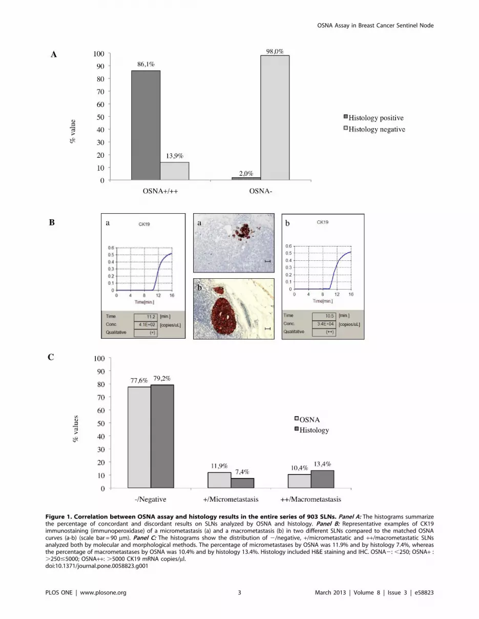
Figure 1. Correlation between OSNA assay and histology results in the entire series of 903 SLNs. Panel A: The histograms summarizethe percentage of concordant and discordant results on SLNs analyzed by OSNA and histology. Panel B: Representative examples of CK19immunostaining (immunoperoxidase) of a micrometastasis (a) and a macrometastasis (b) in two different SLNs compared to the matched OSNAcurves (a-b) (scale bar = 90 mm). Panel C: The histograms show the distribution of 2/negative, +/micrometastatic and ++/macrometastatic SLNsanalyzed both by molecular and morphological methods. The percentage of micrometastases by OSNA was 11.9% and by histology 7.4%, whereasthe percentage of macrometastases by OSNA was 10.4% and by histology 13.4%. Histology included H&E staining and IHC. OSNA2: ,250; OSNA+ :.250#5000; OSNA++: .5000 CK19 mRNA copies/ml.doi:10.1371/journal.pone.0058823.g001
OSNA Assay in Breast Cancer Sentinel Node
PLOS ONE | www.plosone.org 3 March 2013 | Volume 8 | Issue 3 | e58823

sections were cut. Otherwise, additional sections at further 6 levels
at an interval of 100 mm were cut and analyzed both morpho-
logically and immunohistochemically. When the SLN was OSNA
positive (+ or ++) and, subsequently, morphologically negative, the
Figure 2. Relationship between number of SLNs tested by OSNA and ALND status. The percentage of negative ALND was significantlyhigher in patients with 1 OSNA positive SLN (65.8% vs 34.2%). Conversely, the percentage of positive ALND was significantly higher in patients with 2or more OSNA positive SLNs (66.7% vs 33.3%) (p = 0.004).doi:10.1371/journal.pone.0058823.g002
Table 2. Relationship between axillary non-sentinel lymph node status and number of SLNs analyzed.
Number ofSLNs OSNA+91 OSNA++88
Number of SLNs(%)
57 61
1 ALND Negative ALND Positive ALND Negative ALND Positive 118 (66%)
47 (+) 10 (+) 30 (++) 31 (++)
28 24
ALND Negative ALND Positive ALND Negative ALND Positive
18 (2/+) 5 (2/+) 6 (2/++) 7 (2/++)
2 3 (+/+) 2 (+/+) 1 (+/++) 5 (+/++) 52 (29%)
2 2 1 (++/++) 4 (++/++)
6 3
ALND Negative ALND Positive ALND Negative ALND Positive
3 (2/2/+) 2 1 (+/+/++) 1 (+/++/++)
3/4 1 (2/+/+) 2 (2/+/+) 2 1 (2/2/2/++) 9 (5%)
Total 72 19 39 49 179
SLN: sentinel lymph node;OSNA: one step nucleic acid amplification;OSNA+: .250#5000 cytokeratin 19 mRNA copies/ml; OSNA++: .5000 cytokeratin 19 mRNA copies/ml;ALND: axillary lymph node dissection.doi:10.1371/journal.pone.0058823.t002
OSNA Assay in Breast Cancer Sentinel Node
PLOS ONE | www.plosone.org 4 March 2013 | Volume 8 | Issue 3 | e58823

histological work-up was extended to all levels of paraffin blocks
with an interval of 50 mm. Otherwise, when the SLN was OSNA
negative and morphologically positive (micrometastases or macro-
metastases), patients subsequently underwent ALND. In addition,
as a control, the OSNA assay was repeated after checking the IHC
expression of CK19 on the corresponding primary tumor.
Metastatic deposits in the SLN were recorded, according to the
TNM classification, as follows: a. isolated tumor cells (ITC) if their
largest diameter was smaller than 0.2 mm, b. micrometastases if
Figure 3. Relationship between SLN status and bio-pathological parameters. The histograms show that OSNA positive results aresignificantly correlated with (Panel A) poor differentiated tumor (p = 0.039), (Panel B) high proliferation index (p = 0.028) and (Panel C) presence oflymphovascular invasion (LVI p,0.0001).doi:10.1371/journal.pone.0058823.g003
OSNA Assay in Breast Cancer Sentinel Node
PLOS ONE | www.plosone.org 5 March 2013 | Volume 8 | Issue 3 | e58823
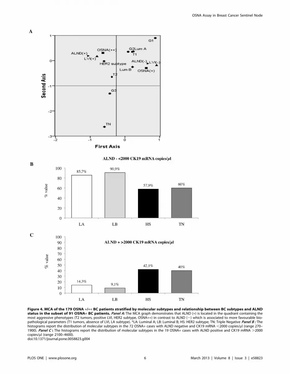
Figure 4. MCA of the 179 OSNA +/++ BC patients stratified by molecular subtypes and relationship between BC subtypes and ALNDstatus in the subset of 91 OSNA+ BC patients. Panel A: The MCA graph demonstrates that ALND (+) is located in the quadrant containing themost aggressive phenotypes (T2 tumors, positive LVI, HER2 subtype, OSNA++) in contrast to ALND (2) which is associated to more favourable bio-pathological parameters (T1 tumors, absence of LVI, LA subtype). *LA: Luminal A; LB: Luminal B; HS: HER2 subtype; TN: Triple Negative Panel B : Thehistograms report the distribution of molecular subtypes in the 72 OSNA+ cases with ALND negative and CK19 mRNA ,2000 copies/ml (range 270–1900). Panel C : The histograms report the distribution of molecular subtypes in the 19 OSNA+ cases with ALND positive and CK19 mRNA .2000copies/ml (range 2100–4600).doi:10.1371/journal.pone.0058823.g004
OSNA Assay in Breast Cancer Sentinel Node
PLOS ONE | www.plosone.org 6 March 2013 | Volume 8 | Issue 3 | e58823
they were larger than 0.2 mm but not larger than 2 mm in
diameter, c. macrometastases if they were larger than 2 mm in
diameter. In this study, according to the TNM definition, lymph
nodes presenting only ITC were considered negative [pN0 (i+)].
Axillary non-sentinel lymph nodes were routinely examined by
H&E staining.
ImmunohistochemistryThe presence of metastatic cells in the slices b&d of the SLN
were further evaluated by IHC using mAb anti-pancytokeratin
(MNF116, Dako) and anti-CK19 (RCK108, Dako). Immunore-
actions were revealed by a streptavidin-biotin enhanced immuno-
peroxidase technique in an automated autostainer (BondTM Max,
Menarini, Florence, Italy). Our series of primary breast tumors
were tested for ER and PgR expression using mAb 6F11
(Menarini) and mAb 1A6 (Menarini) respectively, for proliferative
activity using the anti Ki-67 mAb (MIB1, Dako), for HER2
overexpression using the polyclonal antibody A0485 (Dako). TN
tumors were also characterized for epidermal growth factor
receptor (EGFR) (Pharmdx kit, Dako) and cytokeratin 5 expres-
sion (mAb XM26, Menarini). HER2 IHC positivity was deter-
mined according to ASCO/CAP guidelines [14] and was scored
as follows: 0 and 1+ negative, 2+ equivocal, and 3+ positive. ER
and PgR were considered positive when .10% of the neoplastic
cells showed distinct nuclear immunoreactivity, whereas Ki-67,
based on the median value of our series, was regarded as high if
more than 15% of the cell nuclei were immunostained. Evaluation
of the IHC results, blinded to all patient data, was performed
independently and in blinded manner by two investigators (SB and
MM).
Silver In Situ HybridizationTo assess HER2 gene amplification in tumors score 2+ by IHC
we used a fully automated single color in situ hybridization assay
based on the use of a validated silver deposition technology (SISH,
Inform HER2 DNA Probe; Inform Chromosome 17 probe,
Roche Tissue Diagnostic, Milan, Italy). The silver precipitation
was visualized as a black dot in cell nuclei with 1006 oil
immersion objective. SISH results were analyzed by using a light
microscope (Nikon, Eclipse 55i) equipped with a software able to
capture images (Eureka Interface System). Cases were defined as
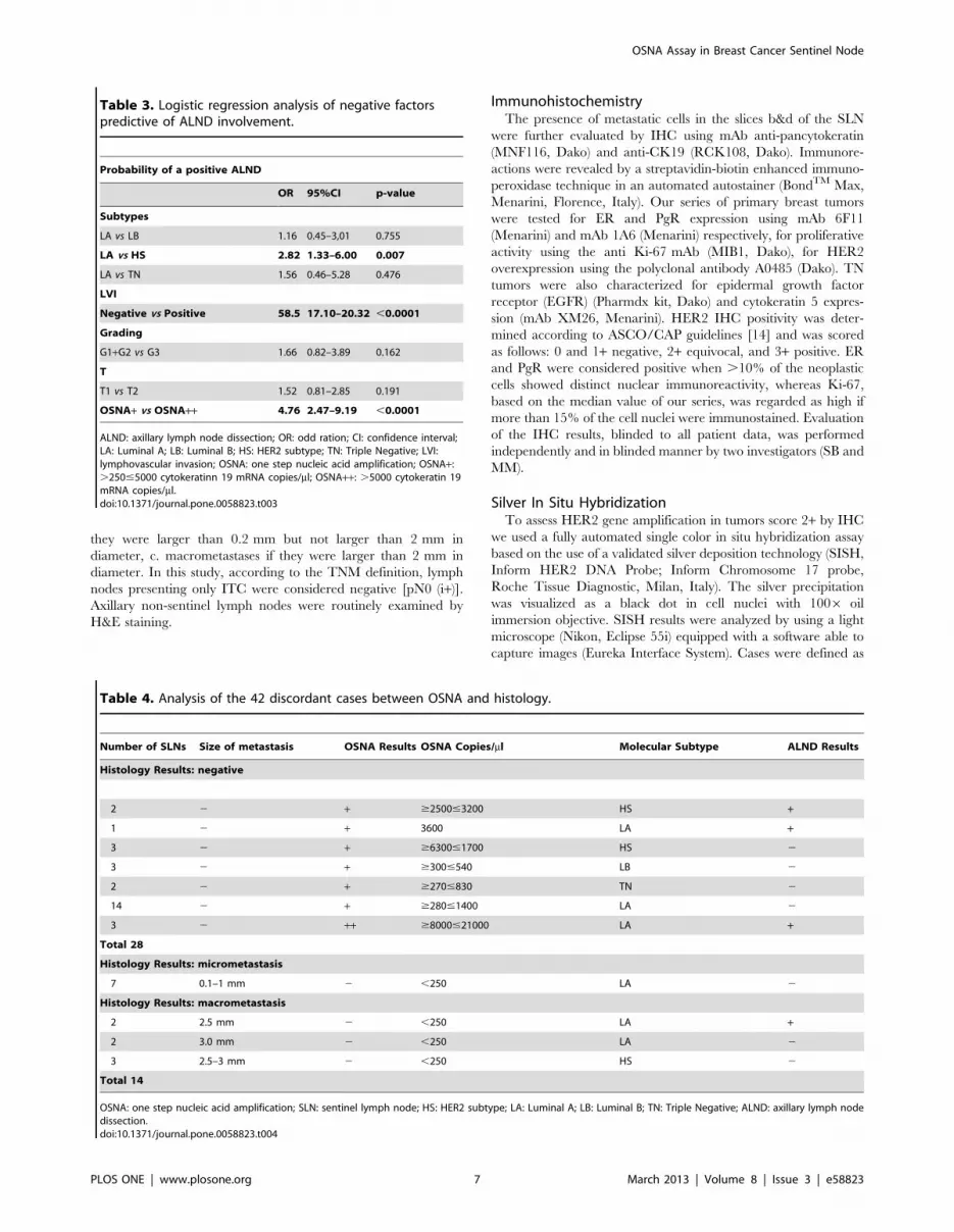
Table 3. Logistic regression analysis of negative factorspredictive of ALND involvement.
Probability of a positive ALND
OR 95%CI p-value
Subtypes
LA vs LB 1.16 0.45–3,01 0.755
LA vs HS 2.82 1.33–6.00 0.007
LA vs TN 1.56 0.46–5.28 0.476
LVI
Negative vs Positive 58.5 17.10–20.32 ,0.0001
Grading
G1+G2 vs G3 1.66 0.82–3.89 0.162
T
T1 vs T2 1.52 0.81–2.85 0.191
OSNA+ vs OSNA++ 4.76 2.47–9.19 ,0.0001
ALND: axillary lymph node dissection; OR: odd ration; CI: confidence interval;LA: Luminal A; LB: Luminal B; HS: HER2 subtype; TN: Triple Negative; LVI:lymphovascular invasion; OSNA: one step nucleic acid amplification; OSNA+:.250#5000 cytokeratinn 19 mRNA copies/ml; OSNA++: .5000 cytokeratin 19mRNA copies/ml.doi:10.1371/journal.pone.0058823.t003
Table 4. Analysis of the 42 discordant cases between OSNA and histology.
Number of SLNs Size of metastasis OSNA Results OSNA Copies/ml Molecular Subtype ALND Results
Histology Results: negative
2 2 + $2500#3200 HS +
1 2 + 3600 LA +
3 2 + $6300#1700 HS 2
3 2 + $300#540 LB 2
2 2 + $270#830 TN 2
14 2 + $280#1400 LA 2
3 2 ++ $8000#21000 LA +
Total 28
Histology Results: micrometastasis
7 0.1–1 mm 2 ,250 LA 2
Histology Results: macrometastasis
2 2.5 mm 2 ,250 LA +
2 3.0 mm 2 ,250 LA 2
3 2.5–3 mm 2 ,250 HS 2
Total 14
OSNA: one step nucleic acid amplification; SLN: sentinel lymph node; HS: HER2 subtype; LA: Luminal A; LB: Luminal B; TN: Triple Negative; ALND: axillary lymph nodedissection.doi:10.1371/journal.pone.0058823.t004
OSNA Assay in Breast Cancer Sentinel Node
PLOS ONE | www.plosone.org 7 March 2013 | Volume 8 | Issue 3 | e58823
amplified when SISH displayed a gene copy number .6.
Polysomy 17– intended as an increased centromere 17 enumer-
ation probe copy number – is considered to be present in BC when
a mean number of 3 signals is shown.
Statistical AnalysesPatient dataset. All descriptive statistics were calculated.
The proper method of analysis and the tests for statistical
significance depended on the variables under study. The
categorical variables were reported through frequencies and
percentage values, while we considered age at surgery as a
continuous variable, reporting it through the median value and its
range.
Lymph node dataset. Correlations among tests were
estimated using the Cohen’s Kappa Test. This test expresses the
amount of agreement (over and above the expected due to chance
alone) between the two assays.
Specificity, sensitivity, negative and positive predictive value
(NPV and PPV), and the 95% confidence interval (CI) of the
OSNA assay were estimated considering histology as the gold
standard. The correlation between the size of intranodal metastasis
(expressed in squared centimeters) evaluated by morphology and
the copy number of CK19 mRNA evaluated by OSNA was
determined using the non-parametric Spearman correlation
coefficient. A p-value ,0.05 was considered to be statistically
significant.
Multiple correspondence analysis. Multiple correspon-
dence analysis (MCA), a descriptive/exploratory technique
designed to analyze simple two-way and multi-way frequency,
was used to identify biological profiles associated to SLN and
ALND status [15,16]. This representation aims to visualize the
similarities and/or differences of profiles, simultaneously identify-
ing those dimensions that contain the majority of the data
variability. The positions of the points in the MCA graph are
informative. Categories plotting close to each other are statistically
related and are similar with regard to the pattern of relative
frequencies and this association is statistically valuable (Lebart’s
statistic) when the points are located far from the origin of the
graph which represents a mean uninformative profile. SNL status
detected by OSNA, BC subtypes, tumor size (T), histological grade
(G), lymphovascular invasion (LVI) were introduced in the analysis
as active variables whereas ALND status was introduced as
supplementary variable. MCA provides a graphical representation
of the active and supplementary variables projected on a plane
formed by axes 1 and 2, which accounted for 67.6% of total
variability, reproducing quite a significant percentage of the total
chi-square value of the multi-way frequency table.
Results
Comparison between OSNA Assay and HistologyIn our series of 709 early BC patients, we analyzed a total of 903
SLNs, 1 in 535 (75%), 2 in 156 (22%) and more than 2 in 18 (3%)
patients with an average of 1.3 SLNs per patient. All the SLNs
were investigated both by intraoperative OSNA assay and by
postoperative histological procedures (i.e. conventional H&E
staining and IHC). The percentage of positive SLNs was 22.4%
(202/903) by OSNA and 20.8% (188/903) by histological
methods. Of the 709 early BC patients included in this study,
179 had at least one positive SLN. As summarized in Figure 1
panel A, 174 out of 202 (86.1%) and 687 out of 701 (98.0%) cases
presented overlapping results by both molecular and histological
methods. Among the 42 discordant cases, 28 (13.9%) were positive
by OSNA and negative by histology whereas 14 (2.0%) were
negative by OSNA and positive by histology. Of the 28 OSNA
positive and histology negative SLNs, 25 were OSNA+ and 3
OSNA++. In the 14 OSNA negative and histology positive SLNs,
we found 7 micrometastases and 7 macrometastases. Finally, IHC
identified ITC in 6/687 SLNs (0.9%; data not shown). Taking into
account the entire series of 903 SLNs included in our study, the
percentage of micrometastases by OSNA (CK19 mRNA copies/ml
.250#5000) was 11.9% (108/903) and by histology 7.4% (67/
903) whereas the percentage of macrometastases by OSNA (CK19
mRNA copies .5000) was 10.4% (94/903) and by histology
13.4% (121/903) (Figure 1 panel B-C). Whenever 2 or more SLNs
from the same patient were tested by OSNA and showed different
results (negative vs positive or+vs ++), the positive result or the one
with the highest copy number was taken into account. The raw
concordance between the OSNA and histological methods was
95%. Cohen’s Kappa statistic was equal to 86% [CI 95% (80–93)]
with a statistically significant value (p,0.0001). By considering the
histological procedures as the gold standard, in the entire series of
903 SLNs, the sensitivity of OSNA in detecting micrometastases
and/or macrometastases was 93% [CI 95% (88–97)] and the
specificity was 96% [CI 95% (94–98)]. The negative predictive
value (NPV) was 98% [CI 95% (96–99)] and the positive
predictive value (PPV) was 86% [CI 95% (79–91)]. In the 174
cases which were positive by both methods, a significant
correlation was observed between the size of the metastases
assessed by IHC and the CK19 mRNA copies detected by OSNA
assay (Sperman’s coefficient: 0.827; p,0.0001).
ALND Status Correlates to SLN Status Detected by OSNAOf the 179 patients with a positive OSNA assay, 111 (62%) had
an ALND negative and 68 (38%) positive. Interestingly, of the 111
negative ALND, 72 (65%) were from patients with OSNA+ and 39
(35%) from patients OSNA++. Conversely, of the 68 positive
ALND, 49 (72%) were from patients with macrometastasis
(OSNA++) and only 19 (28%) from patients with micrometastasis
(OSNA+). As shown in Table 2, in 118 out of 179 (66%) OSNA+/
++ patients we tested 1 SLN whereas in 52 (29%) and in 9 (5%)
patients we tested 2 or 3 or 4 SLNs respectively. Taking the CK19
mRNA copies into account, we found that of the 91 OSNA+ cases,
83 (91.2%) had at least 1 SLN positive of which 15 (18%) had also
a positive ALND, 8 (8.8%) had 2 SLNs positive of which 4 (50%)
had a positive ALND. In the group of 88 OSNA++ patients, we
found 75 (85.2%) cases with 1 positive SLN of which 39 (44.3%)
had a positive ALND and 13 cases (14.7%) with 2 or 3 positive
SLNs of which 11 (78.5%) had a positive ALND. The percentage
of negative ALND was significantly higher in patients with 1
OSNA positive SLN (65.8% vs 34.2%). Conversely, the percentage
of positive ALND was significantly higher in patients with 2 or
more OSNA positive SLNs (66.7% vs 33.3%) (p = 0.004) (Figure 2).
Relationship between SLN Status and Bio-pathologicalParameters
Figure 3 shows that OSNA positive results were significantly
correlated with (Panel A) poor differentiated tumor (p = 0.039),
(Panel B) high proliferation index (p = 0.028) and (Panel C)
presence of lymphovascular invasion (LVI p,0.0001). Of interest,
all these correlations progressively and significantly increased from
patients with negative to patients with macrometastatic SLNs
(p(trend) for G = 0.026, for Ki-67 index = 0.008, for LVI
,0.0001). No significant correlations were observed between
SLN status detected by OSNA and the other conventional bio-
pathological parameters analyzed (data not shown). In order to
better investigate the relationship between SLN status detected by
OSNA, ALND status and conventional bio-pathological factors,
OSNA Assay in Breast Cancer Sentinel Node
PLOS ONE | www.plosone.org 8 March 2013 | Volume 8 | Issue 3 | e58823
we stratified our series of 647 invasive BC by molecular
classification. We found that 428 BC (66.2%) were LA, 65
(10%) were LB, 108 (16.7%) were HS, and 46 (7.1%) were TN.
We used MCA to study the complex interrelationships among
pathological (T, G, LVI), and biological variables (ER, PgR,
HER2, Ki-67) the latter clustered into phenotypic subtypes (LA,
LB, HS, TN) visualizing their link with SLN and ALND status. As
illustrated in Figure 4 panel A along the first axis, the test
demonstrates the contrast between T2 tumors, positive LVI,
HER2 subtype, OSNA++ (upper left quadrant) and T1 tumors,
negative LVI, Luminal subtype, OSNA+ (upper right quadrant).
In order to obtain an indication of the predictive value of the
graphical configuration produced by MCA, we added ALND
status as a supplementary variable. Interestingly, the 2 categories
(ALND+vs ALND 2) are located in opposite quadrants (upper left
quadrant ALND+vs upper right quadrant ALND 2) contrasting
the 2 groups previously described; such location of the ALND
status in the graph may identify bio-pathological profiles as
potential predictive factors of ALND positivity, providing the basis
to investigate their predictive role using regression analysis.
Focusing on the 91 OSNA+ BC patients, we observed that all
the 72 cases with negative ALND presented a CK19 mRNA copy
number ,2000 (range 270–1900) regardless of BC molecular
subtypes (Figure 3 panel B). Conversely, all the 19 cases with
positive ALND had a CK19 mRNA copy number .2000 (range
2100–4600) (Figure 3 panel C). Of interest, nearly 88% of luminal
BC with a CK19 mRNA copy number ,2000 had an ALND
negative.
Logistic Regression AnalysisThe following variables were studied by logistic regression
analysis: BC subtypes, LVI, tumor grading, tumor size and CK19
mRNA copies/ml. As summarized in Table 3, the model indicates
that the probability of finding an ALND positive is significantly
higher in patients with HER2 positive tumors (OR 2.82, 95% CI
1.33–6.00; p = 0.007), with LVI positive (OR 58.85, 95% CI
17.10–200.32; p,0.0001) and with OSNA++ SLNs (OR 4.76,
95% CI 2.47–9.19; p,0.0001).
Analysis of Discordant CasesIn our series there were 42 discordant cases, which were all
sampled from patients for whom a single SLN was analyzed by
OSNA. The 28 SLNs positive by OSNA and negative by histology
and the 14 SLNs negative by OSNA and positive by histology
(discordance rate 4.65%) were investigated in more detail as
reported in Table 4. In the first group, 25 out of 28
morphologically negative SLNs were OSNA+ (89.3%) and 3
OSNA++ (10.7%). Only 3 out of the 25 (12%) OSNA+ SLNs
presented a positive ALND whereas all 3 OSNA++ cases had a
positive ALND. Interestingly, the copy number of the 6 SLNs (3
OSNA+ and 3 OSNA++) with a positive ALND were consistently
higher (OSNA+ $2500#3200 and OSNA++ $8000#21000)
than the copy number detected in the remaining 19 SLNs
presenting a negative ALND ($270#1700). Concerning BC
subtypes, we found that 40% of HS (2/5) and only 6.6% of LA
tumors (1/15) presented a positive ALND. To find out to what
extent these discrepancies may be influenced by a sampling bias,
the 28 OSNA positive and morphologically negative SLNs were
cut into further levels at an interval of 50 mm until no remnants
remained. After this extended analysis, we did not detect
metastatic deposits in any of the evaluated samples. Postoperative
IHC detected metastases, up to 3 mm in diameter, in 14 OSNA
negative patients who underwent ALND in a second surgery. In
detail, of the 14 cases investigated, 7 were micrometastases (0.1–
1 mm) and 7 were intranodal metastases (2.5–3.0 mm) by
histology. All the latter 7 cases were LA BC with a negative
ALND, and a negligible LVI. Otherwise, of the 7 macrometastatic
cases, 2 (28.5%) had a positive ALND with the primary tumor
presenting a significant LVI.
Discussion
More than 20 studies, including a wide range of BC patients,
have been published in the last five years which show the reliability
of the molecular OSNA assay in detecting metastases in the SLN.
The first set of papers consists of the early works whose goal was to
develop, debug and validate the OSNA method [8,17–19], testing
both SLN and/or non-SLN; a second one consists of studies in
which the authors, analyzing one half of the SLN by OSNA,
compared molecular results with histology which represented the
gold standard [20–22]. A third set of papers consists of studies in
which the whole or almost whole SLN was analyzed by OSNA in
an aim to verify the reliability of the method compared to standard
procedures conducted on historical or randomized prospective
cohorts of patients [23–30]. The present study is part of the second
group of papers in which one half of SLN was investigated by
OSNA assay comparing results to standard morphological
procedures. Nevertheless, unlike other studies, we took our
analysis one step further and we assessed the risk of a positive
ALND in relation to CK19 mRNA copy number in the SLN.
Concomitantly, we considered the latter parameters in the context
of the molecular classification of BC. Our series consisted of 903
SLNs sampled from 709 consecutive BC patients and, to the best
of our knowledge, this is one of the largest prospective series in
which patients with both micrometastases (+) and macrometastases
(++), detected by OSNA assay, underwent immediate ALND. In
our consecutive series of BC the concordance rate between OSNA
and histology was 95% and, by considering the histological
procedures as the gold standard, the sensitivity and specificity of
OSNA assay in detecting SLN metastases was 93% and 96%
respectively. In contrast to other authors [17,20], our concordance
analysis did not exclude discordant cases mainly due to tissue
allocation bias. Nonetheless, the concordance rate between OSNA
and histology reported in our series resembles other studies. In the
prospective multicenter study by Snook and colleagues [20], which
included intraoperative examination of SLN from 204 BC
patients, the overall concordance rate between OSNA and
histopathology was 96% with a sensitivity of 91.7% and a
specificity of 96.9%. Overlapping results were reported by Bernet
and collegues [31] and by Le Frere-Belda et al [22]. Also the two
studies [24,27] which intraoperatively examined the whole SLN
by OSNA except for a 1 mm thick central slice of the lymph node,
arrived at the conclusion that OSNA is an accurate assay which
could significantly reduce the need for second surgery. Starting
from the hypothesis that combining histological and molecular
assessment of metastases on the same SLN might not fully
reproduce the actual load of cancer cells present in the SLN and
may create problems in decisions regarding axillary dissection,
several recently published studies analyzed the whole SLN by
OSNA comparing results with those obtained on historical cohort
[26,28,29] or randomizing patients to receive OSNA assay or
conventional histological procedures [25,31,32]. In both cases
authors concluded that OSNA makes it possible to standardize
SLN analysis and it is clinically useful for immediate decisions
regarding axillary dissection. In our large prospective series of 709
BC patients, the concomitant analysis of SLN by OSNA and by
histology may allow to verify that the rate of micrometastasis was
higher by molecular assay than by standard histology. These
OSNA Assay in Breast Cancer Sentinel Node
PLOS ONE | www.plosone.org 9 March 2013 | Volume 8 | Issue 3 | e58823

findings are in line with studies [25] which analyzed the entire
SLN by OSNA and confirmed that this difference may be a
reflection of the higher sensitivity of the molecular method
compared to histology. Our OSNA positive patients were
submitted to ALND independent from the presence of microme-
tastasis (+) or macrometastasis (++). Of the 179 OSNA positive BC
patients, 61 had more than 1 SLN tested. As expected, we
evidenced that the probability of having a subsequent positive
ALND was significantly higher when more than 1 SLN was
positive. We delved deeper into the matter and we examined the
likelihood of a positive ALND according to the levels of OSNA
positivity (OSNA+ vs OSNA++) taking concomitantly into account
the different, molecularly distinct, BC subtypes (10,12). First, we
evaluated the association between ALND positivity, conventional
pathological factors and BC subtypes by multiple correspondence
analysis (MCA) [15,16], an alternative method for analyzing
multiple categorical variables by graphically visualizing their
interrelationships. MCA showed that OSNA++ and ALND+pre-
sented a significant dispersion around the origin and were located
in the same quadrant associated to aggressive tumor phenotypes
such as HER2 subtype, presence of LVI and larger tumor size.
These findings strongly support the correlation between OSNA++and ALND positivity. MCA associations were statistically
supported by multivariate Cox regression analysis in which both
HER2 subtype, LVI and presence of macrometastasis by OSNA
are independent predictor factors of ALND positivity. Our
findings, in line with Ohi et al. [30], demonstrated that the
OSNA assay significantly predicts ALND positivity mainly when
associated to unfavourable bio-pathological parameters. Further-
more, focusing on OSNA+ SLNs, we showed, for the first time,
that the concomitance of a low CK19 mRNA copy number
(,2000) and a luminal tumor phenotype may be a useful,
objective tool for predicting non-SLN positivity. In agreement with
other studies which analyzed SLN both by OSNA and histology
[8,17,19,20], we found a discordant rate between the two methods
of 4.7%. Although the tissue allocation bias is one of the main
causes of discordance, another cause, as already reported [25,32],
is the higher sensitivity of OSNA in detecting micrometastases. In
this context, our data are in agreement with other authors since,
among the 42 discordant cases, 25 (60%) were OSNA+/histology
negative. Of interest, in this series we found that the two cases with
a positive ALND presented a OSNA copy number .2500
whereas the remaining 23 cases, with a negative ALND, had a
OSNA copy number ,2000. Starting from this observation it
would be of particular clinical relevance to define a cut-off value
which is capable of accurately identifying the subset of patients
bearing a micrometastatic SLN to be selected for therapeutic
ALND. This is an important issue because the clinical impact of
SLN micrometastasis continues to be an area of great debate
[33,34]. The availability of a molecular method which provides
less subjective and quantitative results may be a useful tool in this
context. To date, all the breast nomograms [35–38] which have
been set up to predict the presence of metastatic spread in non-
SLN are based on standard histology and have a number of
limitations mainly due to the difficulties in assigning the
discriminating size of tumor load. Therefore, the choice of a
further axillary treatment for a SLN with minimal involvement,
mainly for small BC (#2 cm) of low-intermediate grade, remains
largely a decision between the surgeon and the patient.
The OSNA assay, together with a presurgical analysis of tumor
subtype, could represent a valid and objective tool to set up a novel
model capable of more accurately predicting axillary involvement.
Omission of ALND could be proposed in patients with a
micrometastatic SLN with a low CK19 mRNA copy number
(,2000) and luminal tumor phenotype. To validate these findings,
the predictive value of CK19 mRNA copy number is currently
under investigation on a new prospective series of BC patients
treated in our Institute in whom the whole SLN is analyzed by the
molecular OSNA method.
Acknowledgments
We would like to thank Andrea Novelli and Maria Assunta Fonsi for their
graphic editing assistance and Michael Kenion for his English language
editing.
Author Contributions
Conceived and designed the experiments: SB MM FDF. Performed the
experiments: SB FDF BC EG FM CLM RP CB SDF. Analyzed the data:
SB IT FDF MM EP. Contributed reagents/materials/analysis tools: SB
MM FDF EP CLM. Wrote the paper: SB MM.
References
1. Veronesi U, Paganelli G, Viale G, Luini A, Zurrida S, et al. (2006) Sentinel-lymph-node biopsy as a staging procedure in breast cancer: update of a
randomised controlled study. Lancet Oncol 7: 983–990.
2. Lyman GH, Giuliano AE, Somerfield MR, Benson AB 3rd, Bodurka DC, et al.
(2005) American Society of Clinical Oncology guideline recommendations forsentinel lymph node biopsy in early-stage breast cancer. J Clin Oncol 23: 7703–
7720.
3. Fleissig A, Fallowfield LJ, Langridge CI, Johnson L, Newcombe RG, et al. (2006)
Post-operative arm morbidity and quality of life. Results of the ALMANACrandomised trial comparing sentinel node biopsy with standard axillary
treatment in the management of patients with early breast cancer. BreastCancer Res Treat 95: 279–293.
4. Cserni G, Bianchi S, Boecker W, Decker T, Lacerda M, et al. (2005) Improvingthe reproducibility of diagnosing micrometastases and isolated tumor cells.
Cancer 103: 358–367.
5. Layfield DM, Agrawal A, Roche H, Cutress RI (2011) Intraoperative assessment
of sentinel lymph nodes in breast cancer. Br J Surg 98: 4–17.
6. Beach RA, Lawson D, Waldrop SM, Cohen C (2003) Rapid immunohisto-
chemistry for cytokeratin in the intraoperative evaluation of sentinel lymphnodes for metastatic breast carcinoma. Appl Immunohistochem Mol Morphol
11: 45–50.
7. Leikola JP, Toivonen TS, Krogerus LA, von Smitten KA, Leidenius MH (2005)
Rapid immunohistochemistry enhances the intraoperative diagnosis of sentinellymph node metastases in invasive lobular breast carcinoma. Cancer 104: 14–19.
8. Tsujimoto M, Nakabayashi K, Yoshidome K, Kaneko T, Iwase T, et al. (2007)
One-step nucleic acid amplification for intraoperative detection of lymph node
metastasis in breast cancer patients. Clin Cancer Res 13: 4807–4816.
9. Goldhirsch A, Wood WC, Coates AS, Gelber RD, Thurlimann B, et al. (2011)
Strategies for subtypes–dealing with the diversity of breast cancer: highlights ofthe St. Gallen International Expert Consensus on the Primary Therapy of Early
Breast Cancer 2011. Ann Oncol 22: 1736–1747.
10. van ‘t Veer LJ, Dai H, van de Vijver MJ, He YD, Hart AA, et al. (2002) Gene
expression profiling predicts clinical outcome of breast cancer. Nature 415: 530–536.
11. Desmedt C, Haibe-Kains B, Wirapati P, Buyse M, Larsimont D, et al. (2008)Biological processes associated with breast cancer clinical outcome depend on
the molecular subtypes. Clin Cancer Res 14: 5158–5165.
12. Prat A, Perou CM (2011) Deconstructing the molecular portraits of breast
cancer. Mol Oncol 5: 5–23.
13. Tavassoli FA DP (2003) Pathology and Genetics Tumours of the breast andfemale genital organs. Lyon (France): IARC Press. 9–112 p.
14. Wolff AC, Hammond ME, Schwartz JN, Hagerty KL, Allred DC, et al. (2007)American Society of Clinical Oncology/College of American Pathologists
guideline recommendations for human epidermal growth factor receptor 2testing in breast cancer. Arch Pathol Lab Med 131: 18–43.
15. Greenacre M (1984) Theory and applications of correspondence analysis.Aca-demic Press, London.
16. Lebart L Morineau A, Warwick KM (1984) Multivariate descriptive statisticalanalysis. Wiley, New York.
17. Visser M, Jiwa M, Horstman A, Brink AA, Pol RP, et al. (2008) Intra-operativerapid diagnostic method based on CK19 mRNA expression for the detection of
lymph node metastases in breast cancer. Int J Cancer 122: 2562–2567.
18. Schem C, Maass N, Bauerschlag DO, Carstensen MH, Loning T, et al. (2009)
One-step nucleic acid amplification-a molecular method for the detection of
OSNA Assay in Breast Cancer Sentinel Node
PLOS ONE | www.plosone.org 10 March 2013 | Volume 8 | Issue 3 | e58823
lymph node metastases in breast cancer patients; results of the German study
group. Virchows Arch 454: 203–210.19. Tamaki Y, Akiyama F, Iwase T, Kaneko T, Tsuda H, et al. (2009) Molecular
detection of lymph node metastases in breast cancer patients: results of a
multicenter trial using the one-step nucleic acid amplification assay. Clin CancerRes 15: 2879–2884.
20. Snook KL, Layer GT, Jackson PA, de Vries CS, Shousha S, et al. (2010)Multicentre evaluation of intraoperative molecular analysis of sentinel lymph
nodes in breast carcinoma. Br J Surg 98: 527–535.
21. Feldman S, Krishnamurthy S, Gillanders W, Gittleman M, Beitsch PD, et al.(2011) A novel automated assay for the rapid identification of metastatic breast
carcinoma in sentinel lymph nodes. Cancer 117: 2599–2607.22. Le Frere-Belda MA, Bats AS, Gillaizeau F, Poulet B, Clough KB, et al. (2012)
Diagnostic performance of one-step nucleic acid amplification for intraoperativesentinel node metastasis detection in breast cancer patients. Int J Cancer 130:
2377–2386.
23. Laia BV, Marcos MB, Refael CM, Francisco SC, Jose T, et al. (2011) Moleculardiagnosis of sentinel lymph nodes for breast cancer: one step ahead for
standardization. Diagn Mol Pathol 20: 18–21.24. Khaddage A, Berremila SA, Forest F, Clemenson A, Bouteille C, et al. (2011)
Implementation of molecular intra-operative assessment of sentinel lymph node
in breast cancer. Anticancer Res 31: 585–590.25. Castellano I, Macri L, Deambrogio C, Balmativola D, Bussone R, et al. (2011)
Reliability of whole sentinel lymph node analysis by one-step nucleic acidamplification for intraoperative diagnosis of breast cancer metastases. Ann Surg
255: 334–342.26. Sagara Y, Ohi Y, Matsukata A, Yotsumoto D, Baba S, et al. (2011) Clinical
application of the one-step nucleic acid amplification method to detect sentinel
lymph node metastasis in breast cancer. Breast Cancer.27. Tamaki Y, Sato N, Homma K, Takabatake D, Nishimura R, et al. (2011)
Routine clinical use of the one-step nucleic acid amplification assay for detectionof sentinel lymph node metastases in breast cancer patients: Results of a
multicenter study in Japan. Cancer.
28. Osako T, Iwase T, Kimura K, Yamashita K, Horii R, et al. (2011) Accuratestaging of axillary lymph nodes from breast cancer patients using a novel
molecular method. Br J Cancer 105: 1197–1202.
29. Godey F, Leveque J, Tas P, Gandon G, Poree P, et al. (2011) Sentinel lymph
node analysis in breast cancer: contribution of one-step nucleic acid
amplification (OSNA). Breast Cancer Res Treat 131: 509–516.
30. Ohi Y, Umekita Y, Sagara Y, Rai Y, Yotsumoto D, et al. (2012) Whole sentinel
lymph node analysis by a molecular assay predicts axillary node status in breast
cancer. Br J Cancer.
31. Bernet L, Cano R, Martinez M, Duenas B, Matias-Guiu X, et al. (2011)
Diagnosis of the sentinel lymph node in breast cancer: a reproducible molecular
method: a multicentric Spanish study. Histopathology 58: 863–869.
32. Osako T, Iwase T, Kimura K, Yamashita K, Horii R, et al. (2011)
Intraoperative molecular assay for sentinel lymph node metastases in early
stage breast cancer: a comparative analysis between one-step nucleic acid
amplification whole node assay and routine frozen section histology. Cancer
117: 4365–4374.
33. Wasif N, Maggard MA, Ko CY, Giuliano AE (2010) Underuse of axillary
dissection for the management of sentinel node micrometastases in breast
cancer. Arch Surg 145: 161–166.
34. Galimberti V, Botteri E, Chifu C, Gentilini O, Luini A, et al. (2012) Can we
avoid axillary dissection in the micrometastatic sentinel node in breast cancer?
Breast Cancer Res Treat 131: 819–825.
35. Van Zee KJ, Manasseh DM, Bevilacqua JL, Boolbol SK, Fey JV, et al. (2003) A
nomogram for predicting the likelihood of additional nodal metastases in breast
cancer patients with a positive sentinel node biopsy. Ann Surg Oncol 10: 1140–
1151.
36. Degnim AC, Reynolds C, Pantvaidya G, Zakaria S, Hoskin T, et al. (2005)
Nonsentinel node metastasis in breast cancer patients: assessment of an existing
and a new predictive nomogram. Am J Surg 190: 543–550.
37. Kohrt HE, Olshen RA, Bermas HR, Goodson WH, Wood DJ, et al. (2008) New
models and online calculator for predicting non-sentinel lymph node status in
sentinel lymph node positive breast cancer patients. BMC Cancer 8: 66.
38. Pal A, Provenzano E, Duffy SW, Pinder SE, Purushotham AD (2008) A model
for predicting non-sentinel lymph node metastatic disease when the sentinel
lymph node is positive. Br J Surg 95: 302–309.
OSNA Assay in Breast Cancer Sentinel Node
PLOS ONE | www.plosone.org 11 March 2013 | Volume 8 | Issue 3 | e58823