connexins and the atrioventricular node
TRANSCRIPT
Connexins and the atrioventricular node
Ian P. Temple, MBChB,* Shin Inada, PhD,† Halina Dobrzynski, PhD,* Mark R. Boyett, PhD*
From the *Institute of Cardiovascular Sciences, University of Manchester, Core Technology Facility, 46 Grafton St,Manchester, UK and yNational Cerebral and Cardiovascular Center Research Institute, Laboratory of Biomedical Sciencesand Information Management, Fujishiro-dai, Suita, Osaka 565-8565, Japan.
The structure and functioning of the atrioventricular (AV) node
has remained mysterious owing to its high degree of complexity. In
this review article, we integrate advances in knowledge regarding
connexin expression in the AV node. Complex patterning of 4
different connexin isoforms with single channel conductances
ranging from ultralow to high explains the dual pathway electro-
physiology of the AV node, the presence of 2 nodal extensions,
longitudinal dissociation in the penetrating bundle, and, most
importantly, how the AV node maintains slow conduction between
the atria and the ventricles. It is shown that the complex
patterning of connexins is the consequence of the embryonic
development of the cardiac conduction system. Finally, it is argued
Address reprint requests and correspondence: Mark R. Boyett, PhD,Institute of Cardiovascular Sciences, University of Manchester, CoreTechnology Facility, 46 Grafton St, Manchester M13 9NT, UK. E-mailaddress: [email protected].
1547-5271/$-see front matter B 2013 Heart Rhythm Society. All rights reserved.
that connexin dysregulation may be responsible for AV node
dysfunction.
KEYWORDS Atrioventricular node; Inferior nodal extension; Left nodalextension; Compact node; His bundle; Dual pathway electrophysiology;Atrioventricular nodal reentrant tachycardia; Connexins
ABBREVIATIONS AV ¼ atrioventricular; AVNRT ¼ atrioventricularnodal reentrant tachycardia; CN ¼ compact node; Cx ¼ connexion;INa ¼ Naþ current; INE ¼ inferior nodal extension; PB ¼penetrating bundle
(Heart Rhythm 2013;10:297–304) I 2013 Heart Rhythm Society.All rights reserved.
IntroductionThe atrioventricular (AV) node is the “gatekeeper” betweenthe atria and the ventricles and is located at the AV junctionon the right side of the heart (Figure 1A). The primary role ofthe node is to conduct the action potential from the atria tothe ventricles. However, the AV node also acts as a backuppacemaker in the case of failure of the sinus node and stopsarrhythmias in the atria, such as atrial fibrillation, from beingconducted into the ventricles at dangerously high rates. Onthe other hand, the AV node is part of the circuit underlyingAV nodal reentrant tachycardia (AVNRT).1 An importantaspect of AV node conduction is to introduce a delaybetween atrial and ventricular excitation to allow time foratrial contraction to complete filling of the ventricles. In theelectrocardiogram, the delay between atrial and ventricularexcitation corresponds to the PR interval and in humans it is120–200 ms; in large part, this reflects the slow conductionthrough the AV node (although the PR interval must alsoinclude conduction time across the atrium and through theHis-Purkinje system).2 The conduction velocity of the slowpathway of the AV node, in rabbits, for example, is 2–10 cm/s, whereas the conduction velocity of the atrial muscle is80 � 29 cm/s and the conduction velocity of the Purkinjefibers is 150 � 20 cm/s.2–4 In part, the slow conduction
velocity of the AV node is the result of the small diameter ofnodal myocytes (conduction velocity is function of celldiameter) and the complex arrangement of the myocytes(nodal myocytes can be separated by extensive connectivetissue), which is expected to slow conduction, because theaction potential will have to follow a more tortuous paththrough the AV node. In part, the slow conduction velocityof the AV node is also the result of the poor expression ofNaþ channels (Nav1.5; responsible for Naþ current [INa]) inthe AV node and the consequent low upstroke velocity of theaction potential (conduction velocity is a function ofupstroke velocity).3,5,6 However, the slow conduction velo-city of the AV node is also the result of poor electricalcoupling between the myocytes of the AV node. Electricalcoupling between cardiac myocytes is provided by gapjunctions made of connexins. While some of the story iswell known, the detailed examination of connexins at the AVnode has revealed some surprising complexities, asexplained in this brief review. An expansion of this review(including outline of history, embryology, and anatomy ofthe AV node) is available as an online supplement.
Connexin expression within the AV nodeFour connexin isoforms are expressed in heart: Cx40, whichforms large-conductance (200 pS) gap junction channels;Cx43, which forms medium-conductance (60–100 pS) gapjunction channels; Cx45, which forms small-conductance(20–40 pS) gap junction channels; and Cx30.2 (or Cx31.9 in
http://dx.doi.org/10.1016/j.hrthm.2012.10.020
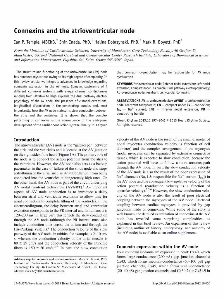
Figure 1 Connexin expression at themRNA level in different regions of humanatrioventricular (AV) junction. A: Anatomyof the AV node. Left: Heart viewed frombehind with window cut in the right atrium toexpose the AV node (shown in red). Modifiedfrom Li et al.10 Right: Exploded view ofboxed region on the left. B: Relative abun-dance of mRNA for 4 connexin isoforms indifferent regions of the AV junction of thehuman heart. Means � SEM values areshown (n ¼ 6). From Greener et al.3 Ao ¼aorta; CN ¼ compact node; CS ¼ coronarysinus; FO ¼ fossa ovalis; INE ¼ inferiornodal extension; IVC ¼ inferior vena cava;LBBB ¼ left bundle branch block; PA ¼pulmonary artery; PB ¼ penetrating bundle;RA ¼ right atrium; RBBB ¼ right bundlebranch block; RV ¼ right ventricle; SVC ¼superior vena cava.
298 Heart Rhythm, Vol 10, No 2, February 2013
humans), which forms ultra-small-conductance (9 pS) gapjunction channels.7 Cx30.2/Cx31.9 is special in that it willform heterotypic gap junction channels with other connex-ins, and the heterotypic channels have a small conductance(15–18 pS).7 Electrical coupling in the AV node is poorbecause there are few gap junctions between the nodalmyocytes and, also, the gap junctions are small.8,9 However,this is not the only reason: another reason is the nature of theconnexin isoform expressed. The principal connexin in theworking atrial and ventricular myocardium is the medium-conductance Cx43. The transitional tissue in the triangle ofKoch in humans and rabbits does express Cx43, but the levelis reduced as compared with the surrounding atrial muscle(Figure 1B).3,5,10,11 The compact node (CN) shows lowexpression of Cx43 in humans and animals; Figures. 1B, 2B,and 3B show that Cx43 is largely absent from the CN inhumans11 and rabbits.12 Most studies have shown Cx43mRNA and protein are also largely absent from the inferiornodal extension (INE) in humans3 and rabbits,5,10,13 and, asan example, Figure 1B shows the low level of Cx43 mRNAin the INE in humans.3 However, surprisingly, Hucker et al11
have reported high levels of Cx43 throughout the INE inhumans. Cx43 is expressed in the penetrating bundle (PB),His bundle, and bundle branches in humans and rabbits atleast (Figure 1B).5,6,9,10,13 The high-conductance Cx40channel is known to be expressed in the working atrial (butnot ventricular) myocardium. The pattern of expression ofCx40 shares some similarities with that of Cx43. Figure 1Bshows that in humans, expression of Cx40 mRNA is lower inthe transitional tissue and INE than in the atrial muscle, butnot significantly so.3 Figures 1B and 3 show that, however,expression of Cx40 (at both mRNA and protein levels) is
high in the CN and PB in humans and rats.3 Both bundlebranches have been shown to have high levels of Cx40 inhumans, rabbits, rats, and mice.6,9 There is more limitedinformation regarding connexin expression in the Purkinjefibers, but Cx40 has been shown to be expressed highly indog and rat Purkinje fibers.14 The pattern of expression of themedium- and high-conductance connexins Cx40 and Cx43 iscompatible with the known fast conduction in the atrialmuscle (�80 cm/s), slower conduction in the INE (r10 cm/s), and very fast conduction in the PB, His bundle, andPurkinje fibers (�150 cm/s). What is responsible forelectrical coupling (even though weak electrical coupling)in the AV node? It is assumed that the small-conductanceCx45 is responsible. Figure 1B shows a low level ofexpression of Cx45 mRNA in all tissues at the AV junctionin humans.3 It has been shown that the ultra-small-conductance Cx30.2 is expressed in the AV node of mice.15
However, Figure 1B shows that the expression of Cx31.9(human equivalent of Cx30.2) mRNA is very low in alltissues at the AV junction in humans. Cx31.9 is alsoundetected at the protein level in humans.3,15 It is unlikely,therefore, to be functionally important in humans.3
Gene knockout studies have been used to determine thefunctional significance of connexins in the mouse AV nodeand have demonstrated the importance of Cx30.2, Cx40, andCx45 but not Cx43.15 Paradoxically, homozygous knockoutof Cx30.2 in mice accelerates conduction in the AV node.16
It is possible that Cx30.2 reduces electrical couplingbetween nodal myocytes in the AV node by formingsmall-conductance heterotypic gap junctions with Cx40 orby a competitive effect of low-conductance channels onhigh-conductance channels. However, as already discussed,

Figure 2 Heterogeneous expression of connexin 43 (Cx43) in thecompact node (CN) of the rabbit heart. A: Masson’s trichrome stainedsection through the CN of the rabbit heart (myocytes stained red; connectivetissue stained blue). The CN is ringed with a dashed line. B and C: High-magnification images of boxed regions in panel A (B ¼ CN; C ¼ lowernodal bundle) showing Cx43 expression (immunofluorescence; bright greenpunctate spots). In panel C, dotted yellow lines divide tissue into Cx43-negative (top) and Cx43-positive (bottom) regions. Modified from Dobr-zynski et al.12 CFB ¼ central fibrous body; TT ¼ tendon of Todaro.
Temple et al Connexins and the Atrioventricular Node 299
the human equivalent of Cx30.2 (Cx31.9) is not or poorlyexpressed.15 A homozygous Cx40 knockout mouse demon-strates a 20% increase in the PR interval with the slowing ofconduction in both the AV node and the His-Purkinjesystem.15 The acceleration of conduction in the AV node
Figure 3 Heterogeneous expression of con-nexins in atrioventricular (AV) conductionaxis. A: Expression of connexin 40 (Cx40)(immunofluorescence; green signal) in atrialmuscle (Ai), compact node (CN) (Aii), andventricular muscle (Aiii) of the human heart.Sections are also immunolabeled for caveolin3(red signal), which is present in the cellmembrane of all cardiac myocytes. Cx40labeling is present at an intercalated disk inatrial myocytes, punctate in the CN, and absentin ventricular myocytes. From H Dobrzynski,unpublished data. B: Expression of Cx43(immunofluorescence; white punctate signal)in the CN of the human heart. CN (ringed inyellow) is Cx43 negative, whereas lower nodalbundle (LNB; also ringed in yellow) is Cx43positive. Modified from Hucker et al.11 Bi:Section corresponding to that in panel Bstained with Masson’s trichrome (myocytesstained red; connective tissue stained blue). C:Expression of Cx40 in PB (ringed in yellow)of the rat heart (immunofluorescence; greenpunctate signal). The upper part of the bundleis Cx40 negative, and the lower part is Cx40positive. Modified from Yoo et al.6 Ci: Sectioncorresponding to that in panel C stained withMasson’s trichrome.
caused by the knockout of Cx30.2 is normalized by knockingout Cx40 in addition to Cx30.2.16 It is possible that Cx30.2 isprimarily expressed in the proximal part of the AV conduc-tion axis and Cx40 in the distal part and the acceleration ofconduction caused by the knockout of Cx30.2 from theproximal part equals the slowing of conduction caused by theknockout of Cx40 from the distal part.16 Homozygousknockout of either Cx43 or Cx45 is lethal during embry-ogenesis, which has a limited study of the AV node.15 Aheterozygous Cx43 knockout mouse does not show anyalteration in any electrocardiographic parameters, includingthe PR interval. The heterozygous knockout of Cx45, inaddition to the homozygous knockout of Cx40, has beenshown to lead to further increases in the PR interval beyondthat caused by the knockout of Cx40 alone.15 As an aside, thePR interval as a measure of AV node conduction should beused with caution, because it also includes the conductiontime from the sinus node to the AV node and also from theAV node to the ventricular muscle. For example, if there ispacemaker shift from the high crista terminalis to the lowcrista terminalis, there is a substantial shortening of the PRinterval. It should be confirmed that AV conduction isaffected (eg, by measuring AH interval). Furthermore,conduction in different parts of the AV node may be affecteddifferentially; there is a paucity of data on the consequencesof connexin manipulations on different parts of the AV node.
The AV node (or parts of it at least) not only lacks high-conductance connexins but also Nav1.5 as already mentionedand, consequently, the large and fast inward INa.
17,18 As aresult, the action potential upstroke in the AV node isdependent on the smaller and slower inward Ca2þ current,which is the reason why the upstroke velocity of the action
300 Heart Rhythm, Vol 10, No 2, February 2013
potential is low—one of the factors responsible for the slowconduction velocity in the AV node.17,18 We have usedcomputer modeling to compare the roles of connexins andNav1.5 (S Inada; data not published). In the first simulation,we used a model of a string of electrically coupled humanatrial myocytes; each myocyte was represented by a biophy-sically detailed model of the human atrial action potential.19
The coupling conductance between the myocytes wasreduced in line with the reduction in Cx40 and Cx43 mRNAsobserved in the INE (compared to that in the atrial muscle) inhumans (Figure 1B)3; it was assumed that the reduction inmRNA is translated into a similar reduction in protein andcoupling conductance. This simulation suggests that thereduction in connexin expression in the INE will result in a�36% reduction in the conduction velocity (compared to thatin the atrial muscle). In the second simulation, the model ofthe action potential of the human atrial myocyte was modifiedon the basis of mRNA levels of ion channels and so on in theINE (as a fraction of those in the atrial muscle)3; this resultedin an action potential typical of the INE. In particular, theaction potential had a slow upstroke as a result of the lowexpression of Nav1.5. This simulation suggests that thereduction in Nav1.5 expression in the INE will result in a�77% reduction in the conduction velocity (compared to thatin the atrial muscle). The final simulation suggests that,together, the decreases in Cx40, Cx43, and Nav1.5 expressionwill cause a �84% decrease in the conduction velocity.Although this suggests that Nav1.5 is more important than theconnexins, this prediction has not been tested experimentally.The lack of Cx40, Cx43, and Nav1.5 in the AV node may notbe coincidental: Shaw and Rudy20 have argued that ifelectrical coupling is weak, safe conduction of the actionpotential is dependent on Ca2þ current (rather than on INa).
Patterning of connexins underlies substrate ofdual pathway nodal electrophysiologyDual AV nodal electrophysiology refers to the concept offast and slow pathways within the AV node. The fast andslow pathways are so called because they are the fastest andslowest pathways for the action potential through the AVnode. Whereas the fast pathway is the normal route for actionpotential conduction, the slow pathway is important for theconduction of action potentials at short coupling intervalsbecause the refractory period of the slow pathway is shorterthan that of the fast pathway.18 The dual pathways are thesubstrate of AVNRT. During slow-fast AVNRT, there isantegrade conduction of the action potential along the slowpathway, retrograde conduction along the fast pathway, andthen activation of the atrial muscle, after which there can befurther cycles of reentry (online supplement Figures 1A and 1Band related text). In addition to slow-fast AVNRT, there is fast-slow AVNRT when the action potential circles in the oppositedirection.21 Correlation of electrophysiology (optical mappingas well as intracellular and extracellular action potentialrecording) with the identification of the dual pathways (byimmunolabeling of marker proteins as well as histology) has
shown that the fast pathway corresponds to the transitionaltissue, whereas the slow pathway corresponds to the INE(Figure 1A).10,22,23 The standard treatment of AVNRT iscatheter ablation of the slow pathway: radiofrequency energyis applied to the coronary sinus ostium (site of INE), and thissupports the hypothesis that the INE is the structure thatunderlies the slow pathway supporting AVNRT.1 The speed ofconduction of the 2 pathways is consistent with connexinexpression at the AV junction: the relatively high Cx40 andCx43 expression in the transitional tissue is expected to lead tofast conduction, whereas the lower Cx40 and Cx43 expressionin the INE is expected to lead to slower conduction.12
However, this is likely to be the only part of the reason forthe difference in conduction velocity, because Nav1.5 expres-sion in the transitional tissue is relatively high while it is low inthe INE.3 By using a 3-dimensional anatomical model of theAV node incorporating the dual pathways10 together withbiophysically detailed models of AV node action potentials,18
we are able to simulate AVNRT (online supplement Figure 1Cand Movie 1). In the simulation, AVNRT is dependent on thepoor electrical coupling (low coupling conductance) andconsequent low conduction velocity in the slow pathway; inthe simulation in online supplement Figure 1C, the ratio oflongitudinal coupling conductance in the slow pathway, fastpathway, and atrial muscle was 29:160:625.
Patterning of connexins reveals 2 nodalextensionsThe patterning of connexins at the AV junction has resulted insome surprising findings. The first is that there are 2 nodalextensions. Figure 4 shows sister sections through the mouseheart immunolabeled for Cx43—caveolin3 (myocyte marker)and HCN4, the ion channel responsible for the funny current(nodal marker).24 It shows a left nodal extension as well as thewell-known right nodal extension (INE) from the AV node;both nodal extensions are Cx43 negative. Right and left nodalextensions have also been demonstrated in rabbits and guineapigs.24,25 The right nodal extension continues around thetricuspid valve annulus as the right AV ring, whereas the leftnodal extension continues around the mitral valve annulus asthe left AV ring.24,26 The right and left AV rings loop round the2 valves and meet again to form the retroaortic node.24 The ringtissues are thought to arise from the embryological “primarymyocardium” (AV canal in particular; see online supplement)that gives rise to the tissues of the cardiac conduction system,and in rats, they have a similar gene expression profile to theINE and CN.6,24 There are also reports of right and left nodalextensions in humans.26,27 Human studies have shown a limitedleft nodal extension that is shorter than the right nodal extensionand not present in all subjects studied.11,26,27 Surprisingly,Hucker et al11 reported that whereas the left nodal extension isCx43 negative, the right one is Cx43 positive; compare thiswith Figures. 1B and 4, which show the right nodal extension tobe Cx43 negative in humans and mice. The significance of a leftas well as a right nodal extension with regard to AVNRT isunclear. However, in a recent large case series of ablations for
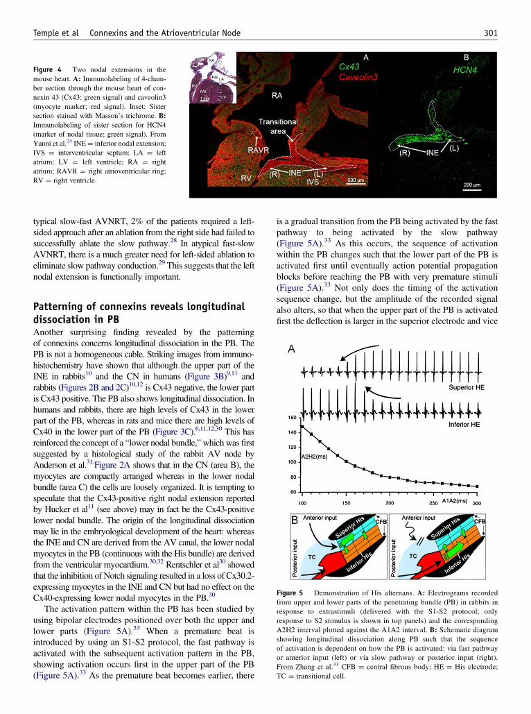
Figure 4 Two nodal extensions in themouse heart. A: Immunolabeling of 4-cham-ber section through the mouse heart of con-nexin 43 (Cx43; green signal) and caveolin3(myocyte marker; red signal). Inset: Sistersection stained with Masson’s trichrome. B:Immunolabeling of sister section for HCN4(marker of nodal tissue; green signal). FromYanni et al.24 INE¼ inferior nodal extension;IVS ¼ interventricular septum; LA ¼ leftatrium; LV ¼ left ventricle; RA ¼ rightatrium; RAVR ¼ right atrioventricular ring;RV ¼ right ventricle.
Temple et al Connexins and the Atrioventricular Node 301
typical slow-fast AVNRT, 2% of the patients required a left-sided approach after an ablation from the right side had failed tosuccessfully ablate the slow pathway.28 In atypical fast-slowAVNRT, there is a much greater need for left-sided ablation toeliminate slow pathway conduction.29 This suggests that the leftnodal extension is functionally important.
Figure 5 Demonstration of His alternans. A: Electrograms recordedfrom upper and lower parts of the penetrating bundle (PB) in rabbits inresponse to extrastimuli (delivered with the S1-S2 protocol; onlyresponse to S2 stimulus is shown in top panels) and the correspondingA2H2 interval plotted against the A1A2 interval. B: Schematic diagramshowing longitudinal dissociation along PB such that the sequenceof activation is dependent on how the PB is activated: via fast pathwayor anterior input (left) or via slow pathway or posterior input (right).From Zhang et al.33 CFB ¼ central fibrous body; HE ¼ His electrode;TC ¼ transitional cell.
Patterning of connexins reveals longitudinaldissociation in PBAnother surprising finding revealed by the patterningof connexins concerns longitudinal dissociation in the PB. ThePB is not a homogeneous cable. Striking images from immuno-histochemistry have shown that although the upper part of theINE in rabbits10 and the CN in humans (Figure 3B)9,11 andrabbits (Figures 2B and 2C)10,12 is Cx43 negative, the lower partis Cx43 positive. The PB also shows longitudinal dissociation. Inhumans and rabbits, there are high levels of Cx43 in the lowerpart of the PB, whereas in rats and mice there are high levels ofCx40 in the lower part of the PB (Figure 3C).6,11,12,30 This hasreinforced the concept of a “lower nodal bundle,” which was firstsuggested by a histological study of the rabbit AV node byAnderson et al.31,Figure 2A shows that in the CN (area B), themyocytes are compactly arranged whereas in the lower nodalbundle (area C) the cells are loosely organized. It is tempting tospeculate that the Cx43-positive right nodal extension reportedby Hucker et al11 (see above) may in fact be the Cx43-positivelower nodal bundle. The origin of the longitudinal dissociationmay lie in the embryological development of the heart: whereasthe INE and CN are derived from the AV canal, the lower nodalmyocytes in the PB (continuous with the His bundle) are derivedfrom the ventricular myocardium.30,32 Rentschler et al30 showedthat the inhibition of Notch signaling resulted in a loss of Cx30.2-expressing myocytes in the INE and CN but had no effect on theCx40-expressing lower nodal myocytes in the PB.30
The activation pattern within the PB has been studied byusing bipolar electrodes positioned over both the upper andlower parts (Figure 5A).33 When a premature beat isintroduced by using an S1-S2 protocol, the fast pathway isactivated with the subsequent activation pattern in the PB,showing activation occurs first in the upper part of the PB(Figure 5A).33 As the premature beat becomes earlier, there
is a gradual transition from the PB being activated by the fastpathway to being activated by the slow pathway(Figure 5A).33 As this occurs, the sequence of activationwithin the PB changes such that the lower part of the PB isactivated first until eventually action potential propagationblocks before reaching the PB with very premature stimuli(Figure 5A).33 Not only does the timing of the activationsequence change, but the amplitude of the recorded signalalso alters, so that when the upper part of the PB is activatedfirst the deflection is larger in the superior electrode and vice
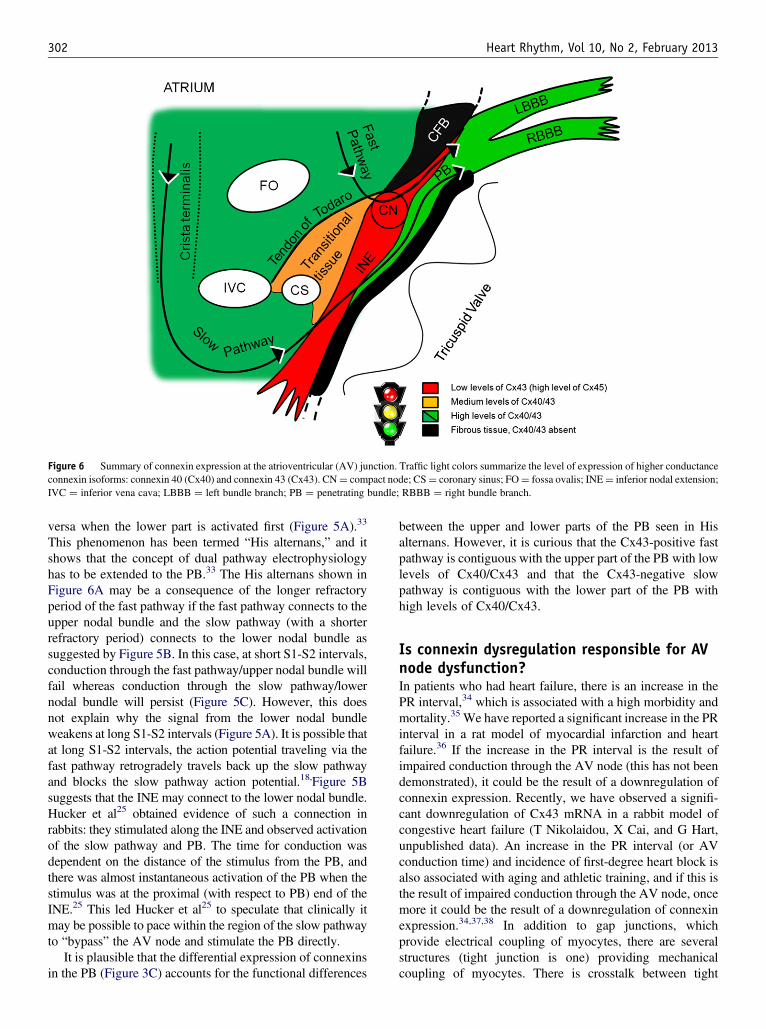
Figure 6 Summary of connexin expression at the atrioventricular (AV) junction. Traffic light colors summarize the level of expression of higher conductanceconnexin isoforms: connexin 40 (Cx40) and connexin 43 (Cx43). CN¼ compact node; CS¼ coronary sinus; FO¼ fossa ovalis; INE¼ inferior nodal extension;IVC ¼ inferior vena cava; LBBB ¼ left bundle branch; PB ¼ penetrating bundle; RBBB ¼ right bundle branch.
302 Heart Rhythm, Vol 10, No 2, February 2013
versa when the lower part is activated first (Figure 5A).33
This phenomenon has been termed “His alternans,” and itshows that the concept of dual pathway electrophysiologyhas to be extended to the PB.33 The His alternans shown inFigure 6A may be a consequence of the longer refractoryperiod of the fast pathway if the fast pathway connects to theupper nodal bundle and the slow pathway (with a shorterrefractory period) connects to the lower nodal bundle assuggested by Figure 5B. In this case, at short S1-S2 intervals,conduction through the fast pathway/upper nodal bundle willfail whereas conduction through the slow pathway/lowernodal bundle will persist (Figure 5C). However, this doesnot explain why the signal from the lower nodal bundleweakens at long S1-S2 intervals (Figure 5A). It is possible thatat long S1-S2 intervals, the action potential traveling via thefast pathway retrogradely travels back up the slow pathwayand blocks the slow pathway action potential.18,Figure 5Bsuggests that the INE may connect to the lower nodal bundle.Hucker et al25 obtained evidence of such a connection inrabbits: they stimulated along the INE and observed activationof the slow pathway and PB. The time for conduction wasdependent on the distance of the stimulus from the PB, andthere was almost instantaneous activation of the PB when thestimulus was at the proximal (with respect to PB) end of theINE.25 This led Hucker et al25 to speculate that clinically itmay be possible to pace within the region of the slow pathwayto “bypass” the AV node and stimulate the PB directly.
It is plausible that the differential expression of connexinsin the PB (Figure 3C) accounts for the functional differences
between the upper and lower parts of the PB seen in Hisalternans. However, it is curious that the Cx43-positive fastpathway is contiguous with the upper part of the PB with lowlevels of Cx40/Cx43 and that the Cx43-negative slowpathway is contiguous with the lower part of the PB withhigh levels of Cx40/Cx43.
Is connexin dysregulation responsible for AVnode dysfunction?In patients who had heart failure, there is an increase in thePR interval,34 which is associated with a high morbidity andmortality.35 We have reported a significant increase in the PRinterval in a rat model of myocardial infarction and heartfailure.36 If the increase in the PR interval is the result ofimpaired conduction through the AV node (this has not beendemonstrated), it could be the result of a downregulation ofconnexin expression. Recently, we have observed a signifi-cant downregulation of Cx43 mRNA in a rabbit model ofcongestive heart failure (T Nikolaidou, X Cai, and G Hart,unpublished data). An increase in the PR interval (or AVconduction time) and incidence of first-degree heart block isalso associated with aging and athletic training, and if this isthe result of impaired conduction through the AV node, oncemore it could be the result of a downregulation of connexinexpression.34,37,38 In addition to gap junctions, whichprovide electrical coupling of myocytes, there are severalstructures (tight junction is one) providing mechanicalcoupling of myocytes. There is crosstalk between tight
Temple et al Connexins and the Atrioventricular Node 303
and gap junctions: Lisewski et al39 showed that the inducibleheart-specific knockout of a tight junction protein (Cox-sackie virus-adenovirus receptor) leads to severe AV blockand a downregulation of Cx43 and Cx45. Although there isno known equivalent human condition, this finding high-lights a route of connexin regulation. Wolff-Parkinson-White syndrome is a heart condition in which there is anelectrical pathway (accessory pathway), other than the AVnode, connecting the atria and the ventricles. The conditioncan lead to episodes of reentrant tachycardia and is one of themost common causes of fast heart rate disorders in infantsand children. The development of accessory pathways hasbeen demonstrated in mice by using either activation ofnotch signaling or inactivation of Tbx2, indicating thatdysregulation of the cell signaling pathways that are essentialfor the development of the AV canal and the AV node maybe responsible for the Wolff-Parkinson-White syn-drome.30,40 Tbx2 normally suppresses the expression ofCx40 and Cx34, and knockout of Tbx2 leads to the loss ofthe AV canal phenotype and high expression of Cx40 andCx43 in an accessory pathway, allowing fast conductionbetween the atria and the ventricles.40
ConclusionsConnexins are central to the functional role of the AV node. InFigure 6, we summarize the distribution of connexins indifferent tissues at the AV junction. The traffic light colorscheme corresponds to high-, medium-, and low-strengthelectrical coupling, that is, fast, moderate, and slow conduc-tion. The fast pathway corresponds to the transitional tissueand possibly the upper part of the PB. The slow pathwaycorresponds to the INE and possibly the lower part of the PB. Itis not clear whether the CN is involved only in the fast pathwayor is common to both pathways. However, in both pathways,the action potential will have to course through poorly coupledtissue lacking Cx40 and Cx43. As the PB extends distally intothe bundle branches and Purkinje fibers, there is high expres-sion of Cx40 and Cx43, facilitating rapid conduction.
AppendixSupplementary DataSupplementary data associated with this article can be foundin the online version at http://dx.doi.org/10.1016/j.hrthm.2012.10.020.
References1. Markowitz S, Stein K, Mittal S, Lerman B. Dual atrionodal physiology in the
human heart. In: Mazgalev T, Tchou MD, eds. Atrial-AV Nodal Electrophysiol-ogy: A View from the Millennium. Armonk, NY: Futura Publishing Company;2000:353–370.
2. Efimov IR, Nikolski VP, Rothenberg F, et al. Structure-function relationship inthe AV junction. Anat Rec A Discov Mol Cell Evol Biol 2004;280:952–965.
3. Greener ID, Monfredi O, Inada S, et al. Molecular architecture of the humanspecialised atrioventricular conduction axis. J Mol Cell Cardiol 2011;50:642–651.
4. de Groot JR, Veenstra T, Verkerk AO, et al. Conduction slowing by the gapjunctional uncoupler carbenoxolone. Cardiovasc Res 2003;60:288–297.
5. Greener ID, Tellez JO, Dobrzynski H, et al. Ion channel transcript expression atthe rabbit atrioventricular conduction axis. Circ Arrhythm Electrophysiol 2009;2:305–315.
6. Yoo S, Dobrzynski H, Fedorov VV, et al. Localization of Naþ channel isoformsat the atrioventricular junction and atrioventricular node in the rat. Circulation2006;114:1360–1371.
7. Boyett MR, Inada S, Yoo S, et al. Connexins in the sinoatrial and atrioventricularnodes. Adv Cardiol 2006;42:175–197.
8. Pollack GH. Intercellular coupling in the atrioventricular node and other tissuesof the rabbit heart. J Physiol 1976;255:275–298.
9. Davis LM, Rodefeld ME, Green K, Beyer EC, Saffitz JE. Gap junction proteinphenotypes of the human heart and conduction system. J Cardiovasc Electro-physiol 1995;6:813–822.
10. Li J, Greener ID, Inada S, et al. Computer three-dimensional reconstruction of theatrioventricular node. Circ Res 2008;102:975–985.
11. Hucker WJ, McCain ML, Laughner JI, Iaizzo PA, Efimov IR. Connexin 43expression delineates two discrete pathways in the human atrioventricularjunction. Anat Rec 2008;291:204–215.
12. Dobrzynski H, Nikolski VP, Sambelashvili AT, et al. Site of origin and molecularsubstrate of atrioventricular junctional rhythm in the rabbit heart. Circ Res2003;93:1102–1110.
13. Ko YS, Yeh HI, Ko YL, et al. Three-dimensional reconstruction of the rabbitatrioventricular conduction axis by combining histological, desmin, and connexinmapping data. Circulation 2004;109:1172–1179.
14. Kanter HL, Laing JG, Beau SL, Beyer EC, Saffitz JE. Distinct patterns ofconnexin expression in canine Purkinje fibers and ventricular muscle. Circ Res1993;72:1124–1131.
15. Jansen JA, van Veen TA, de Bakker JM, van Rijen HV. Cardiac connexins andimpulse propagation. J Mol Cell Cardiol 2010;48:76–82.
16. Schrickel JW, Kreuzberg MM, Ghanem A, et al. Normal impulse propagation inthe atrioventricular conduction system of Cx30.2/Cx40 double deficient mice.J Mol Cell Cardiol 2009;46:644–652.
17. Hancox JC, Yuill KH, Mitcheson JS, Convery MK. Progress and gaps inunderstanding the electrophysiological properties of morphologically normalcells from the cardiac atrioventricular node. Int J Bifurcation Chaos 2003;13:3675–3692.
18. Inada S, Hancox J, Zhang H, Boyett M. One-dimensional mathematical model ofthe atrioventricular node including atrio-nodal, nodal, and nodal-his cells.Biophys J 2009;97:2117–2127.
19. Chandler NJ, Greener ID, Tellez JO, et al. Molecular architecture of the humansinus node. Circulation 2009;119:1562–1575.
20. Shaw RM, Rudy Y. Ionic mechanisms of propagation in cardiac tissue: roles ofthe sodium and L-type calcium currents during reduced excitability and decreasedgap junction coupling. Circ Res 1997;81:727–741.
21. Nikolski VP, Jones SA, Lancaster MK, Boyett MR, Efimov IR. Cx43 and dual-pathway electrophysiology of the atrioventricular node and atrioventricular nodalreentry. Circ Res 2003;92:469–475.
22. Janse M, Van Capelle F, Freud G, Durrer D. Circus movement within the AVnode as a basis for supraventricular tachycardia as shown by multiple micro-electrode recording in the isolated rabbit heart. Circ Res 1971;28:403–414.
23. Loh P, Ho SY, Kawara T, et al. Reentrant circuits in the canine atrioventricularnode during atrial and ventricular echoes: electrophysiological and histologicalcorrelation. Circulation 2003;108:231–238.
24. Yanni J, Boyett MR, Anderson RH, Dobrzynski H. The extent of the specializedatrioventricular ring tissues. Heart Rhythm 2009;6:672–680.
25. Hucker WJ, Sharma V, Nikolski VP, Efimov IR. Atrioventricular conductionwith and without AV nodal delay: two pathways to the bundle of His in the rabbitheart. Am J Physiol Heart Circ Physiol 2007;293:H1122–H1130.
26. Waki K, Kim JS, Becker AE. Morphology of the human atrioventricular node isage dependent: a feature of potential clinical significance. J Cardiovasc Electro-physiol 2000;11:1144–1151.
27. Inoue S, Becker AE. Posterior extensions of the human compact atrioventricularnode: a neglected anatomic feature of potential clinical significance. Circulation1998;97:188–193.
28. Giazitzoglou E, Korovesis S, Kokladi M, Venetsanakos I, Paxinos G, KatritsisDG. Slow-pathway ablation for atrioventricular nodal re-entrant tachycardia withno risk of atrioventricular block. Hellenic J Cardiol 2010;51:407–412.
29. Otomo K, Okamura H, Noda T, et al. “Left-variant” atypical atrioventricularnodal reentrant tachycardia: electrophysiological characteristics and effect ofslow pathway ablation within coronary sinus. J Cardiovasc Electrophysiol2006;17:1177–1183.
30. Rentschler S, Harris BS, Kuznekoff L, et al. Notch signaling regulates murineatrioventricular conduction and the formation of accessory pathways. J ClinInvest 2011;121:525.
304 Heart Rhythm, Vol 10, No 2, February 2013
31. Anderson RH, Janse MJ, van Capelle FJ, Billette J, Becker AE, Durrer D. Acombined morphological and electrophysiological study of the atrioventricularnode of the rabbit heart. Circ Res 1974;35:909–922.
32. Aanhaanen WTJ, Mommersteeg MTM, Norden J, et al. Developmental origin,growth, and three-dimensional architecture of the atrioventricular conduction axisof the mouse heart. Circ Res 2010;107:728–736.
33. Zhang Y, Bharati S, Mowrey KA, Zhuang S, Tchou PJ, Mazgalev TN. Hiselectrogram alternans reveal dual-wavefront inputs into and longitudinal dis-sociation within the bundle of His. Circulation 2001;104:832–838.
34. Crisel RK, Farzaneh-Far R, Na B, Whooley MA. First-degree atrioventricularblock is associated with heart failure and death in persons with stable coronaryartery disease: data from the Heart and Soul Study. Eur Heart J 2011;32:1875–1880.
35. Gervais R, Leclercq C, Shankar A, et al. Surface electrocardiogram to predictoutcome in candidates for cardiac resynchronization therapy: a sub-analysis of theCARE-HF trial. Eur J Heart Fail 2009;11:699–705.
36. Yanni J, Tellez JO, Maczewski M, et al. Changes in ion channel gene expression
underlying heart failure-induced sinoatrial node dysfunction. Circ Heart Fail
2011;4:496–508.37. Schmidlin O, Bharati S, Lev M, Schwartz J. Effects of physiological aging on
cardiac electrophysiology in perfused Fischer 344 rat hearts. Am J Physiol Heart
Circ Physiol 1992;262:H97–H105.38. Stein R, Medeiros CM, Rosito GA, Zimerman LI, Ribeiro JP. Intrinsic sinus and
atrioventricular node electrophysiologic adaptations in endurance athletes. J Am
Coll Cardiol 2002;39:1033–1038.39. Lisewski U, Shi Y, Wrackmeyer U, et al. The tight junction protein CAR
regulates cardiac conduction and cell-cell communication. J Exp Med 2008;205:
2369–2379.40. Aanhaanen WTJ, Boukens BJD, Sizarov A, et al. Defective Tbx2-dependent
patterning of the atrioventricular canal myocardium causes accessory pathway
formation in mice. J Clin Invest 2011;121:534.