anatomy of the atrioventricular conduction system in ventricular septal defect
TRANSCRIPT

Anatomy of the Atrioventricular Conduction Systemin Ventricular Septal Defect
By JACK:L. TITUS, M.D., GuY W. DAUGHERTY, M.D.,AND JESSE E. EDWARDS, M.D.
IT IS important to know the location, inrelation to congenital ventrieular septal
defects, of the atrioventricular (AV) conduc-tion system of the heart, in order to avoiddamaging conduction tissue during surgicalcorrection of such defects.1 2 Furthermore,knowledge of the anatomic relationship of theventricular septal defect to the conductionisystem may cast light on the embryology ofvarious types of defects. Therefore, havingfamiliarized ourselves with the gross anatomicand histologic features of the proximal por-tions of the AV conduction system in normalhuman hearts,3 we undertook an investigationof the location and course of these structuresin hearts having congenital ventricular septaldefects. Interest centered on the AVN node(node of Tawara), AV bundle (common bun-dle or bundle of His), and the proximal por-tions of the right and left bundle branches.
In the normal heart, the AV node is sit-uated in the floor of the right atrium on thefibrous AV ring, at or just anterior to thecoronary sinus ostium; the AV bundle ex-tends anteriorly and inferiorly from the nodethrough the fibrous valvular ring into theinferior part of the membranous septum; theleft bundle branches are usually given off asdiscrete muscular fascicles over a broad ex-tent of the common bundle, usually beginningat a point just distal to the fibrous valvularrino and ending at the posterior-inferior
From the Mayo Clinic and the Mayo Foundation,Rochester, Minnesota.Abridgment of portion of thesis submitted by
Dr. Titus to the Faculty of the Graduate Schoolof the University of Minnesota in partial fulfillmenitof the requirements for the degree of Doctor ofPhilosophy in Pathology.
Supported in part by Research Grant H-4014 ofthe National Heart Institute, U. S. Public HealthService.
72
angle of the membranlous septum; and theright bundle branch, usually forming a con-tinuation of the common bundle, passes ob-liquely anteriorly and inferiorly through theupper part of the ventricular septum towardthe crista supraventricularis.3
Historical NotesMdnekeberg,4 Keith,5 Abbott,6 Yater and
co-workers,7 and Yaters all found that AVbundles were situated along the lower rimsof ventricular septal defects. Kirklin and co-authors,1 Lillehei,2 and Rodriguez and Wof-ford9 concluded from their surgical experi-ences that the bundle is situated at or nearthe posterior-inferior margin of the defectwhen the defect is inferior to the crista supra-ventricularis. Morris10 pointed out that inthe fetus the bundle lies along the posteriorand inferior margins of the foramen of theunclosed septun. Reemtsma and CopenhaverlIfound the bundle along the posterior-inferioraspect of a "imembranous" ventricular septaldefect, but not closely related to a more pos-terior defect. In a study of the conductionsystems in 15 malformed hearts, Truex andBishof12 found the common bundles and theirbranches along the posterior-inferior marginsof septal defects in 13 specimens. The bundleanid its branches passed anterior to a lowerdefect in one specimen and close to the ante-rior margin of the defect in another. Reemut-sina, Copenhaver, and Creech13 found thebundles in the posterior-inferior margins of"membranous" ventricular septal defects,but not closely related to high muscular de-fects.Lev studied the conduction system in hearts
with persistent common atrioventricular ca-nal14 and, later, in hearts with tetralogy ofFallot.15 In these studies, he found the fol-lowing variations from the usual situations:
Circulation, Volume XXVIII, July 1968
by guest on February 18, 2016http://circ.ahajournals.org/Downloaded from

CONDUCTION SYSTEM IN SEPTAL DEFECT
1. The common bundle was on the left sideof the septum below the defect. (It may alsooccur on the left in normal hearts.) 2. Theright bundle often divided into two or moreparts. 3. The left bundle was more compactthan usual. 4. The AV node deviated "hori-zontally " from its usual location, if a per-sistent left superior vena eava entered thecoronary sinus. In hearts with ventricularseptal defects, lievlv6 17 found the bundles tobe close to the posterior margins of the de-fects, except when the defects were situatedposteriorly.
Materials and MethodsSpecimensBeginning in 1957, we selected 21 hearts for
this study. Each heart had a ventricular septaldefect, either as the only anomaly or as part of arecognized complex, and each had been obtainedat necropsy during the years 1954 through 1959and had been preserved in formalin for up to 3years. Specimens were selected without regard tolocation of the defect, presence of other cardiacanomalies, or age or sex of the deceased patient.
Eight hearts came from patients who had notbeen operated on and 13 from patients whose ven-tricular septal defect had been closed surgically.Methods of Examination
In 12 instances, gross dissection of the conduc-tion system preceded histologic examination ofserial sections, while, in nine instances, only his-tologic examination of serial sections was done.When the heart was viewed from the right side,
the area included in the block cut for histologicstudy extended approximately from the level ofthe coronary sinus ostium posteriorly to the cristasupraventricularis anteriorly. The anterior por-tion of the block included the posterior-inferiorrim of the defect. The block also included a por-tion (approximately 1 cm.) of the atrial septumlabove the AV groove. Below the AV groove,the full thickness of the interventricular septumlto approximately 0.5 cm. below the inferiormostlimit of the ventricular septal defect was included.The blocks were cut serially so that individualsections were approximately 7 ,u thick. Then everytwentieth section (every tenth in the case of smallhearts) was stained with hematoxylin and eosin.The next succeeding section to the hematoxylin andeosin-stained section was stained with the Mallory-Heidenhain stain.
Sections were studied at magnifications of 35to 400 and, occasionally, of 1000. The number of
Circulation, Volume XXVIII. July 196*
sections of each specimen examined varied consid-erably, but usually ranged from 250 to 350. (Fromprevious studies,3 it was known that examinationof every section was not necessary for a "geo-graphic" study of the conduction system.)Classification of Defects StudiedThe classification of ventricular septal defects
proposed by Becu and co-workers18 was modifiedto give the following groupings of the 21 speci-mens examined:Group 1 (cases 1 through 14). Defect in right
ventricular outflow tract, posterior and inferior tocrista supraventricularis.Group 2 (case 15). Defect not in right ventric-
ular outflow tract.Group 3 (cases 16 through 19). Multiple de-
fects.Group 4 (cases 20 and 21). Tetralogy of Fallot.
Clinical and General Pathologic Features of CasesStudiedThe ages of the patients from whom the speci-
iliens were obtained ranged from 2 days to 37+-ears. The specimens included the hearts of 12niales and eight females. (The sex and other clin-ical features of the patient represented by onespecimen could not be obtained.)
The factors responsible for death in these caseswere most often cardiac, either related to the con-genital anomaly (most cases) or a complication ofit (for example, one patient died of brain abscesssecondary to subacute bacterial endocarditis). Ina few instances, death was related to the presenceof associated noneardiae congenital anomalies.Many surgically repaired specimens represented
an early period in intracardiac surgery when clo-sure of the defect frequently was accomplished byusing an Ivalon sponge, a procedure less com-mnonly employed in current reparative procedures.
ResultsGroup 1The location of the conduction system and
its relation to the ventricular septal defectwere similar in the 14 cases in this group.Therefore, the generally prevailing circum-stances will be described and illustrated (figs.1 to 5), and exceptions will be noted.The AV node usually occupied its normal
position in the base of the right atrium. Intwo instances (cases 1 and 2), it was situatedslightly posterior to the usual location. Nomorphologic abnormalities of nodal structurewere noted.The common bundle penetrated the fibrous
73
by guest on February 18, 2016http://circ.ahajournals.org/Downloaded from
TITUS, DAIJGlIhERTY, EDWARDS
that the bulk of these fibers was n-ot intimatelyrelated to the defect. Generallv, all the leftbuildle-branch fibers had been given off fromthe common bundle before it reached the levelof the posterior-inferior angle of the defect.
The right bundle branch pursued its usualanterior-inferior course. In some instances itwas intimately associated with the inferior_margin of the defect, while in other cases itwas inferior to the margin.
In 13 of these 14 cases, some part of theconduction system was close to the posterior
_ ~~~~~~~~~~~~~~~~~~~~~~~~~~~~~~~~~~~~~~~_Figure 1
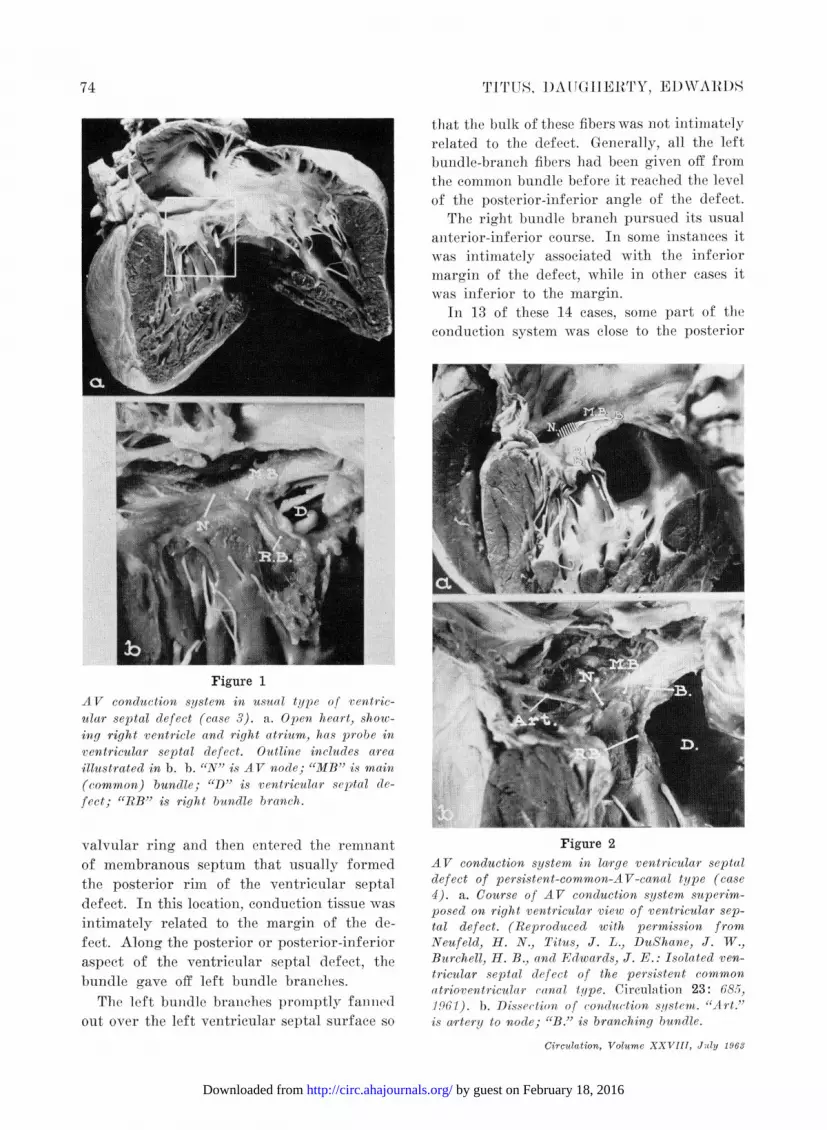
AV conduction system in usual type of rentric-ular septal defect (case 3). a. Open heart, show-ing right ventricle and right atrium, has probe inventricular septal defect. Outline includes atreaillustrated in b. b. "N" is AV node; "JIB" is main(common) bundle; "D" is ventricular sep,tal de-fect; "RB" is right bundle branch.
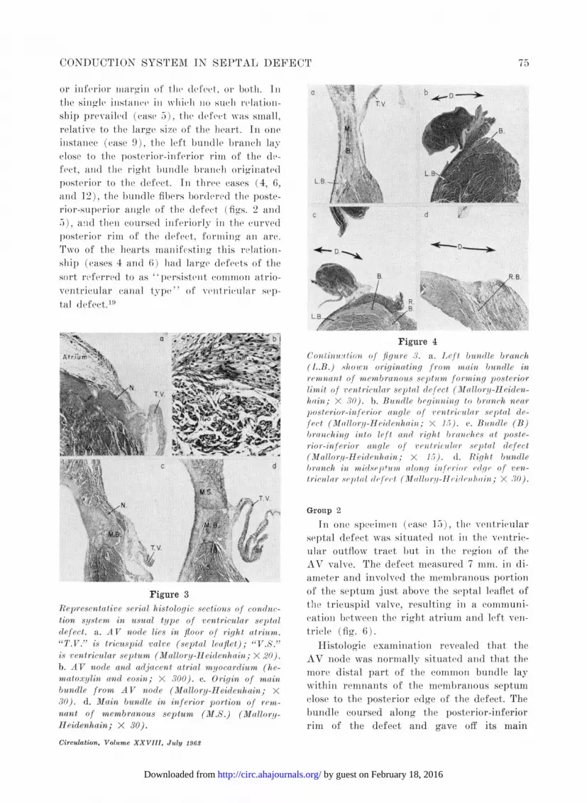
valvular ring and then entered the reninant Figure 2of membranous septum that usually formed AV conduction system in lairge ventricular septalthe posterior rim of the ventricular septal defect of persistent-common-AV-canal type (case
4). a. Course of AV conduction system superim-defec.Inthislocaion,ondutio posed on right ventricular view of ventricular sep-intimately related to the margin of the de- tal defect. (Reproduced with permission fromfeet. Along the posterior or posterior-inferior Neufeld, H. N., Titus, J. L., DuShane, J. W.,aspect of the ventricular septal defect, the Burchell, H. B., and Edwards, J. E.: Isolated yen-bundle gave off left bundle branches. tricular septal defect of the persistent commonatrioventricular (anal type. Circulation 23: 68 5,The left bunidle braniehes promptlv fanied 1 . Dissection of conduction sqstem. "Art."out over the left ventricular septal surface so is a,rtery to node; "B." is branching bundle.
Circulation, VolU?c XXVIII, JUIY 196,Y
74
by guest on February 18, 2016http://circ.ahajournals.org/Downloaded from
CONDUCTION SYSTEM IN SEPTAL DEFECT
or iniferior miaromin of tfle defeet, or both. Inltlhe sing(le instance in axhIiiel 11o such, relation-ship prevailed (ease 5), the defeet was small,relative to the laroe size of the heart. In oneinistaniee (ease 9), the left bundle branch layclose to the posterior-inferior rim of the de-fect, and the rioht bundle branch orioinatedposterior to the defect. In three cases (4, 6,and 12), the bundle fibers bordered the poste-rior-superior angle of the defect (figs. 2 and5), anid tlheni coursed inferiorly in the curvedposterior rim of the defect, formiing an are.Two of the hearts manifesting this relation-ship (cases 4 and 6) had large defects of thesort referred to as "persistenit common atrio-ventricular canal type " of veentricular sep-tal defect.19
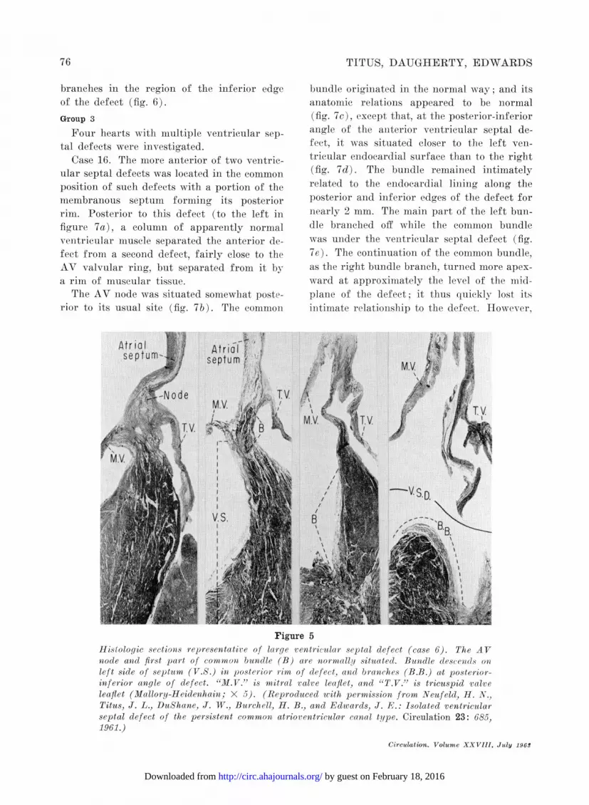
Figure 3Rep,resentative serial histologic sections of conduc-tion system in usual type of ventricular septaldefect. a. AV node lies in floor of right atrium."T.V." is tricuspid valve (septal leaflet); "V.S."is ventricular septum (Mallory-Heidenhain; X 20).b. AV node anad adjacent atrial myocardium (he-matoxylin and eosin; X 300). c. Origin of mainbundle from AV node (Mallory-Heidenahain; X30). d. Main bundle in inferior portion of remia-nant of membranous septum (M.S.) (Mallory-Heidenhain; X 30).
Circulation, Volume XXVIII, July 1968
Figure 4Couitinauitioni of figure 3. a. Left bundle branch(L.B.) showen origina,ting fr-omn main bundle inreminant of memb ranous septum forming posteriorlimit of rentricula r septal clefect (Mallory-Heiden-hain; X 30). b. Bundle beginning to branch near
posterior-iniferior a nyle of lentricula r septal cle-
fect (Malloryj-Heidenihaiui; X 15). c. Buindle (B)brawnchinig into left anid right braniches at poste-rior-inferior aulgle of ventriculalr septal defect(Mallory-Heidenhain; X 15). d. Right bundlebranch in idsevp'u along il fer-ion ed gPe of yien-tricular septaIl defect (M allor!I-H cifleuh aiu; X 30).
Group 2
In one specimneni (ease 15)), the venitricularseptal defect was situated not in the ventric-ular outflow tract but in the region of theAV valve. The defect nmeasured 7 mm. in di-ameter and involved the membranous portionof the septum just above the septal leaflet ofthe tricuspid valve, resulting in a eommuii-cationi betwxeen the right atrium and left veii-triele (fig. 6).
Histologic exanmination revealed that theAV node was normally situated anid that themore distal part of tile common bundle laywithin remnants of the membranious septumclose to the posterior edge of the defect. Thebundle coursed along the posterior-inferiorrim of the defect and gave off its main
b D-A
a
LB.
C
75
by guest on February 18, 2016http://circ.ahajournals.org/Downloaded from
TITUS, DAUGHERTY, EDWARDS
branches in the region of the inferior edgeof the defect (fig. 6).Group 3
Four hearts with multiple ventricular sep-tal defects were investigated.
Case 16. The more anterior of two ventric-ular septal defects was located in the commonposition of such defects with a portion of themembranous septum forming its posteriorrim. Posterior to this defect (to the left infigure 7a), a colunmn of apparently normalventricular muscle separated the ailterior de-fect from a second defect, fairly close to theAV valvular ring, but separated from it bya rim of muscular tissue.
The AV node was situated somewhat poste-rior to its usual site (fig. 7b). The common
bundle originated in the normal way; and itsanatomic relations appeared to be niormal(fig. 7c), except that, at the posterior-inferioran-gle of the aniterior veentricular septal de-feet, it was situated eloser to the left ven-tricular enidocardial surface than to the right(fig. 7d). The bundle remained intimatelyrelated to the endocardial lining along theposterior and inferior edges of the defect fornearly 2 mm. The main part of the left bun-dle branched off while the common bundlewas uinder the venitricular septal defect (fig.7e). The continuation of the common bundle,as the right bundle branch, turned more apex-ward at approximeately the level of the mid-plane of the defect; it thus quickly- lost itsintimate relationisihip to the defect. However,
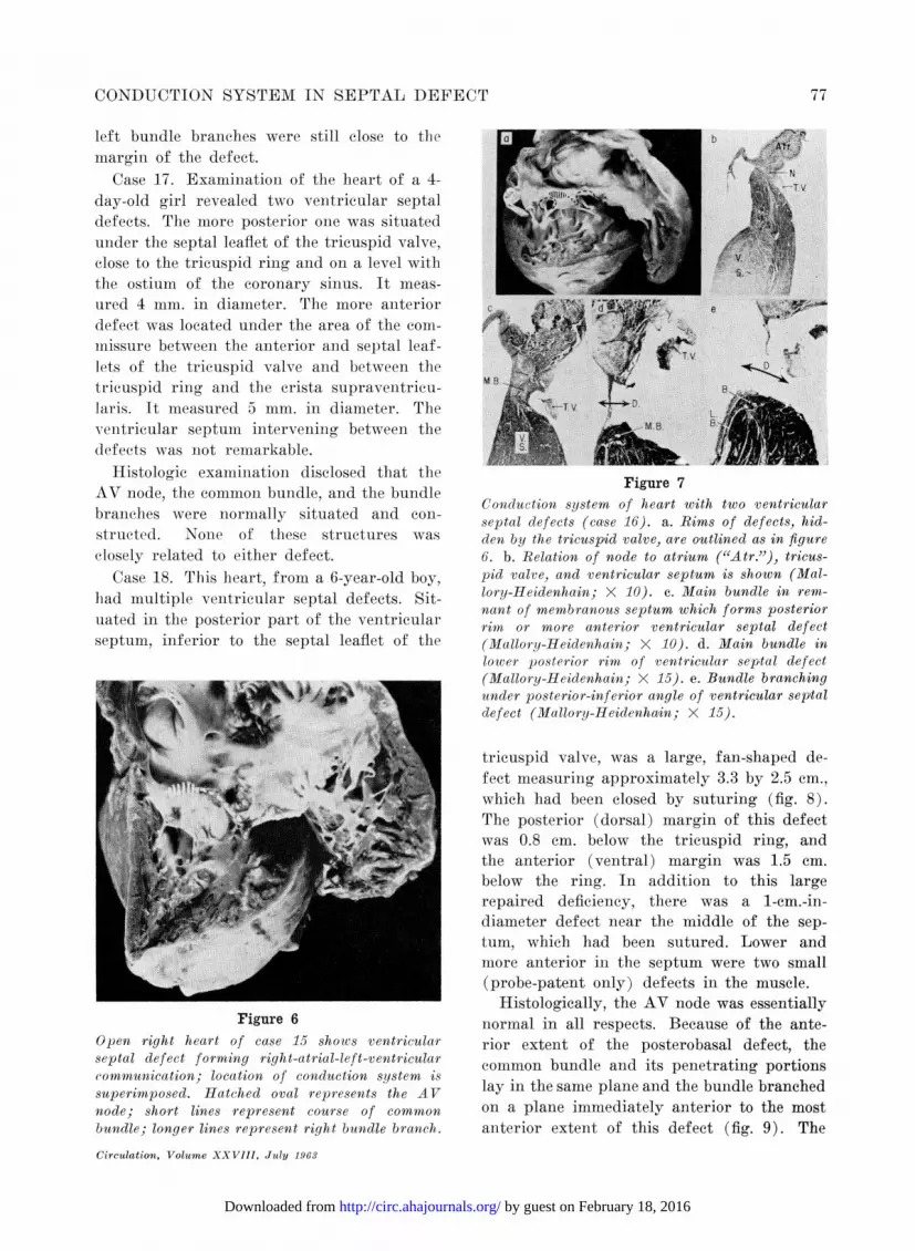
Figure 5Histologic sections representatire of large ventricular septal defect (case 6). The AVnode and first part of common bundle (B) are normally situated. Bundle descends OMleft side of septum (V.S.) in posterior rim of defect, and branches (B.B.) a!t posterior-inferior angle of defect. "M.V." is mitral valve leaflet, and "T.V." is tricuspid valveleaflet (Mallory-Heidenhain; X 5). (Reproduced with permission from Neufeld, H. N.,Titus, J. L., DuShane, J. W., Burchell, H. B., and Edwards, J. E.: Isolated ventricularseptal defect of the persistent common atrioventricular canal type. Circulation 23: 685,1961.)
Circulation. Volume XXVIII, July 1963
76
by guest on February 18, 2016http://circ.ahajournals.org/Downloaded from
CONDUCTION SYSTEM IN SEPTAL DEFECT
left bundle branches were still close to themargin of the defect.
Case 17. Examination of the heart of a 4-day-old girl revealed two ventricular septaldefects. The more posterior one was situatedunder the septal leaflet of the tricuspid valve,close to the tricuspid ring and on a level withthe ostium of the coronary sinus. It meas-ured 4 mm. in diameter. The more anteriordefect was located under the area of the com-missure between the anterior and septal leaf-lets of the tricuspid valve and between thetricuspid ring and the crista supraventrieu-laris. It measured 5 mm. in diameter. Theventricular septuin intervening between thedefects was not remarkable.
Histologic examination disclosed that theAV node, the common bundle, and the bundlebranches were normally situated and con-structed. None of these structures wasclosely related to either defect.
Case 18. This heart, from a 6-year-old boy,had multiple ventricular septal defects. Sit-uated in the posterior part of the ventricularseptum, inferior to the septal leaflet of the
Figure 6Open right heart of case 15 shows ventricularseptal defect forming right-atrial-left-ventricularcommunication; location of conduction system issuperimposed. Hatched oval represents the AVnode; short lines represent course of common
bundle; longer lines represent right bundle branch.
Circulation, Volume XXVIII, July 1963
Figure 7Conduction system of heart with two ventricularseptal defects (case 16). a. Rims of defects, hid-den by the tricuspid valve, are outlined as in figure6. b. Relation of node to atrium ("Atr."), tricus-pid valve, and ventricular septum is shown (Mal-lory-Heidenhain; X 10). c. Main bundle in rem-nant of membranous septum which forms posteriorrim or more anterior ventricular septal defect(Mallory-Heidenhain; X 10). d. Main bundle inlower posterior rim of ventricular septal defect(Mallory-Heidenhain; X 15). e. Bundle branchingunder posterior-inferior angle of ventricular sep,taldefect (Mallory-Heidenhain; X 15).
tricuspid valve, was a large, fan-shaped de-fect measuring approximately 3.3 by 2.5 cm.,which had been closed by suturing (fig. 8).The posterior (dorsal) margin of this defectwas 0.8 cm. below the tricuspid ring, andthe anterior (ventral) margin was 1.5 cm.below the ring. In addition to this largerepaired deficiency, there was a 1-cm.-in-diameter defect near the middle of the sep-tum, which had been sutured. Lower andmore anterior in the septum were two small(probe-patent only) defects in the muscle.
Histologically, the AV node was essentiallynormal in all respects. Because of the ante-rior extent of the posterobasal defect, thecommon bundle and its penetrating portionslay in the same plane and the bundle branchedon a plane immediately anterior to the mostanterior extent of this defect (fig. 9). The
77
by guest on February 18, 2016http://circ.ahajournals.org/Downloaded from

TITUS, DAUGHERTY, EDWARDS
Figure 8Right ventricular vievw of heart with multiple ven-tricular septal (defects including large basilar ven-tricular septall defect in inflow tract (case 18).Large ventricular septal defect beneath tricuspidvalre has been surgically closed. In the midsep-turn is a second ventricular septal defect andprobes lie in two others.
anterior-inferior pathway of the right bun-dle, extending toward the base of the papil-lary muscle of the conus, led away fromii thetwo major defects.
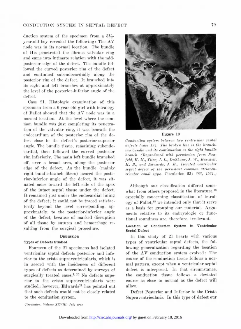
Case 19. This specimen, froml a 12-year-oldboy, had a surgically closed ventricular septaldefect of the usual type and a second, poste-rior, unrepaired defect (fig. 10). This seconddefect was situated under the septal leafletof the tricuspid valve and immlinediately belowthe tricuspid valve ring.The AV node, comnon bundle, and bundle
branches were related to the more anteriordefect in the same fashion as described ingroup 1. Conduction tissue was not closelyrelated to the niore posterior defect.
Group 4
Two patienits, both surgieally treated, hadventricular septal defect as one component ofthe classic tetralogy of Fallot.Case 20. Histologic study of the AV con-
Figure 9Conduction system of heart illustrated in figure S is showui-ii in relation to busilar ven-tricular septal defect from which sutures have been removed. "P.M.C." is papillarymuscle of conus; 'C.S." is crista supraventricularis. (Reproduced wcith permission fromNeufeld, H. M., Titus, J. L., DuShane, J. IV., Burchell, H. B., and Edwards, J. E.:Isolated ventricular septal defect of the persistent common atrioventricular canal type.Circulation 23: 685, 1961.)
Circulation, Volume XXVIII, July 1963
7S
by guest on February 18, 2016http://circ.ahajournals.org/Downloaded from
CONDUCTION SYSTEM IN SEPTAIL DEFECT
duction system of the specimen from a 31/2-year-old boy revealed the following: The AVnode was in its normal location. The bundleof His penietrated the fibrous valvular ringand came into intimate relation with the mid-posterior edge of the defect. The bundle fol-lowed the curved posterior rim of the defectand continued subendocardially along theposterior rim of the defect. It braniehed intoits right and left branehes at approxinmatelythe level of the posterior-inferior angle of thedefect.
Case 21. Histolooic exanmination of thisspecimen from a 6-year-old girl with tetralogyof Fallot showed that the AV node was in anormal location. At the level where the conm-mon bundle was just completing its penetra-tion of the valvular ring, it was beneath theendocardium of the posterior rinm of the de-fect close to the defeet 's posterior-superiorangle. The bundle tissue, remaining subendo-cardial, then followed the curved posteriorrim inferiorly. The main left bundle branchedoff, over a broad area, along the posterioredge of the defect. As the bundle (mainlyright bundle-branch fibers) neared the poste-rior-inferior angle of the defect, it was sit-uated more toward the left side of the apexof the intact septal tissue under the defect.It remained just under the endocardial liningof the defect; it could not be traced satisfac-torily beyond the level corresponding, ap-proximately, to the posterior-inferior anglleof the defect, because of marked disruptionof all tissue by sutures and hemorrhage re-sulting from the surgical procedure.
DiscussionTypes of Defects Studied
Fourteen of the 21 specimens had isolatedventricular septal defects posterior and infe-rior to the crista supraventricularis, which isin accord with the incidences of differenttypes of defects as determined by surveys ofsurgically treated cases.1' 20 No defects supe-rior to the crista supraventricularis werestudied; however, Edwards21 has pointed outthat such defects would not be closely relatedto the conduction system.Circulation, Volume XXVIII, July 1963
Figure 10Conduction system betwveen two ventricular septaldefects (case 19). The broken line is the branch-ing bundle and its continuation as the right bundlebranch. (Reproduced ivith per-mission fr-om Neu-feld, H. M., Titus, J. L., DuShane, J. W., Bturchell,H. B., and Edwards., J. E.: Isolated ventricularseptal defect of the persistent commoon atr-ioven-tr icular canal type. Circulation 23: 685, 1961.)
Although our classificationi differed some-what from others proposed in the literature,"'especially concerning elassification of tetral-ogy of Fallot,5 we intended only that it serveas a basis for grouping our material. Argu-ments relative to its embryologic or func-tional soundness are, therefore, irrelevant.
Location of Conduction System in VentricularSeptal Defect
In this study of 21 hearts with varioustypes of ventricular septal defects, the fol-lowing generalization regarding the locationof the AV conduction system evolved: Thecourse of the conduction tissue follows a nor-mal pattern, except when a ventricular septaldefect is interposed. In that circumstance,the conduction tissue follows a deviatedcourse as close to normal as the defect willallow.
Defect Posterior and Inferior to the CristaSupraventricularis. In this type of defect our
79
by guest on February 18, 2016http://circ.ahajournals.org/Downloaded from
TITUS, DAUGHERTY, EDWARDS
findings relative to the conduction systemagreed with those previously described in theliterature.4 5 7 11 13 16 Variations from thenormal situation3 included slight posteriordisplacement of the AV node, in two speci-mens, and origination of the right bundlebranch from the common bundle before thatof left bundle branches, in one instance (case9).The position of the bundle within the sep-
tum was central in five of the hearts, closerto the left in six, closer to the right in one,
and not accurately determined in two.Mahaim fibers were noted in most of these
cases.
In none of these cases were any parts ofthe proximal divisions of the AV conductionsystem related to superior-anterior edges ofthe septal defect.
Defect Not in the Ventricular OutflowTract (case 15). The conduction system was
related to this defect in essentially the same
manner as in the usual type of defect.Multiple Defects. Truex and Bishof12 stud-
ied two specimens with two defects each;however, since the exact location of the de-fects was not specified for either, comparisonof their findings with ours was not possible.
In hearts with both a posterior septal de-fect and an anterior defect in the mem-
branous septum (cases 16 and 19), the con-
duction system was not closely related to themore posterior defect, but had an intimaterelationship to the posterior and inferioredges of the more anterior defect.
In a heart (case 17) with a small anteriordefect of the common type and a small poste-rior defect, no part of the conduction systemwas intimately related to either defect, ap-
parently because both defects were too smallto impinge on the tissues normally carrying
conduction fibers.The location of the posterior defect of the
specimen representing case 18 was basicallysimilar to that of case 16; however, becauseof its large size, its anterior margin was
closely related to the common bundle and itsbranches as these traversed the intact septum,
A similar situation in specimens with poste-rior defects had been reported previously inthe description of two of the specimens exam-ined by Truex and Bishof,12 and Lev17 specu-lated that such a relationship might occur indefects in this location.
Defect of Tetralogy of Fallot. Specimen 4of Truex and Bishof,12 though not labeled assuch, seemed to be an example of Fallot 'stetralogy, according to their description. Init, an aberrant fascicle of the right bundlebranch passed above the defect and then de-scended in the anterior edge of the defect.The bulk of the bundle tissue, however, cameinto close relation with the defect at its pos-terior-superior angle and followed the poste-rior, then inferior, edges of the deficiency.Iev15 did not observe any such aberrantbranches in the four cases of tetralogy of Fal-lot that he studied, in each of which the bun-dle was situated on the left side of the septumbelow the defect.No aberrant branch of the right bundle was
found in either of the examples of the tetral-ogy of Fallot studied in the present series.Otherwise, our study of the two cases in thepresent series confirmed the essential partsof the works previously mentioned. The re-lationship of the AV conduction system tothe defect was the same as in those heartswitb the usual type of ventricular septaldefect.
SummaryThe major parts of the atrioventricular
conduction system of the human heart weretraced in 21 instances of ventricular septaldefect: 19 were examples of variously locateduncomplicated ventricular septal defects andtwo of the tetralogy of Fallot.In the presence of a ventricular septal de-
fect, the conduction system was found to havea normal course, except when the ventricularseptal defect lay in a position normally oc-cupied by the conduction system. In eachspecimen with a defect posterior and inferiorto the crista supraventricularis, the condue-tion system occupied a position posterior andinferior to the defect. In no instance did the
Circulation, Volume XXVIII, July 1963
80
by guest on February 18, 2016http://circ.ahajournals.org/Downloaded from
CONDUCTION SYSTEM IN SEPTAL DEFECT
conduction system occupy a position superiorto a defect of this type. Defects located inthe posterobasal portion of the muscular partof the ventricular septum sometimes wereposterior to the main parts of the conductionsystem, so that the conduction tissue was re-lated to the anterior edge of the defect. Noexample of a defect lying superior to thecrista supraventricularis was studied. In ourtwo examples of tetralogy of Fallot, the posi-tion of the conduction system was essentiallysimilar to that occurring in the usual varietyof ventricular septal defect, that is, posteriorand inferior to the crista supraventricularis.
References1. KIRKLIN, J. W., HARSHBARGER, H. G., DONALD,
D. E., AND EDWARDS, J. E.: Surgical correc-tion of ventricular septal defect: Anatomicand technical consideration. J. Thoracic Surg.33: 45, 1957.
2. LILLEHEI, C. W.: Discussion. J. Thoracic Surg.33: 57, 1957.
3. TITUS, J. L., DAUGHERTY, G. W., AND EDWARDS,J. E.: Anatomy of the normal human atrio-ventricular conduction system. Unpublisheddata.
4. MONCKEBERG, J. G.: Die Missbildunges desHerzens. In Henke, F., and Lubarsh, O.:Handbuch der Speziellen pathologischen Anato-mie und Histologie. Berlin, Verlag von JuliusSpringer, 1924, Bd. 2, pp. 183.
5. KEITH, A.: Malformations of the heart. Lancet2: 519, 1909.
6. ABBOTT, M. E.: Quoted by Yater, W. M., Lyon,J. A., and McNabb. P. E.7
7. YATER, W. M., LYON, J. A., AND McNABB, P. E.:Congenital heart block: Review and reportof second case of complete heart block studiedby serial sections through the conduction sys-tem. J.A.M.A. 100: 1831, 1933.
8. YATER, W. M.: Congenital heart-block: Reviewof the literature; report of a case with incom-plete heterotoxy; the electrocardiogram in dex-trocardia. Am. J. Dis. Child. 38: 112, 1929.
9. RODRIGUEZ, J. A., AND WOFFORD, J. L.: Surgicalanatomy of the cardiac septa. S. Forum 8:274, 1957.
10. MoaRis, E. W.: The interventricular septum.Thorax 12: 304, 1957.
11. REEMTSMA, K., AND COPENHAVER, W. M.: Ana-tomic studies of the cardiac conduction systemin congenital malformations of the heart.Circulation 17: 271, 1958.
12. TRUEX, R. C., AND BISHOF, J. K.: Conductionsystem in human hearts with interventricularseptal defects. J. Thoracic Surg. 35: 421,1958.
13. REEMTSMA, K., COPENHAVER, W. M., AND CREECH,O., JR.: The cardiac conduction system incongenital anomalies of the heart: Studieson its embryology, anatomy, and function.Surgery 44: 99, 1958.
14. LEv, M.: The architecture of the conductionsystem in congenital heart disease. I. Commonatrioventricular orifice. Arch. Path. 65: 174,1958.
15. LEV, M.: The architecture of the conductionsystem in congenital heart disease. II. Tet-ralogy of Fallot. Arch. Path. 67: 572, 1959.
16. LEV, MI.: The architecture of the conductionsystem in congenital heart disease. III. Ven-tricular septal defect. Arch. Path. 70: 529,1960.
17. LEV, M.: The pathologic anatomy of ventricularseptal defects. Dis. Chest 35: 533, 1959.
18. BECU, L. M., FONTANA, R. S., DUSHANE, J. W.,KIRKLIN, J. W., BURCHELL, H. B., ANDEDWARDS, J. E.: Anatomic and pathologicstudies in ventricular septal defect. Circulation14: 349, 1956.
19. NEUFELD, H. N., TITUS, J. L., DUSHANE, J. W.,BURCHELL, H. B., AND EDWARDS, J. E.: Iso-lated ventricular septal defect of the persistentcommon atrioventricular canal type. Circula-tion 23: 685, 1961.
20. WARDEN, H. E., DEWALL, R. A., COHEN, M.,VAROO, R. L., AND LILLEHEI, C. W.: A surgical-pathologic classification for isolated ventricularseptal defects and for those observed inFallot's tetralogy based on observations madeon 120 patients during repair under directvision. J. Thoracic Surg. 33: 21, 1957.
21. EDWARDS, J. E.: Malformations of the ventricu-lar septal complex. In Gould, S. E.: Pathologyof the Heart. Ed. 2, Springfield, Illinois,Charles C Thomas, Publisher, 1960, p. 303.
Circulation, Volume XXVIII, July 1965
81
by guest on February 18, 2016http://circ.ahajournals.org/Downloaded from
JACK L. TITUS, GUY W. DAUGHERTY and JESSE E. EDWARDSAnatomy of the Atrioventricular Conduction System in Ventricular Septal Defect
Print ISSN: 0009-7322. Online ISSN: 1524-4539 Copyright © 1963 American Heart Association, Inc. All rights reserved.
is published by the American Heart Association, 7272 Greenville Avenue, Dallas, TX 75231Circulation doi: 10.1161/01.CIR.28.1.72
1963;28:72-81Circulation.
http://circ.ahajournals.org/content/28/1/72located on the World Wide Web at:
The online version of this article, along with updated information and services, is
http://circ.ahajournals.org//subscriptions/
is online at: Circulation Information about subscribing to Subscriptions:
http://www.lww.com/reprints Information about reprints can be found online at: Reprints:
document. and Rights Question and Answer
Permissionsthe Web page under Services. Further information about this process is available in thewhich permission is being requested is located, click Request Permissions in the middle column ofClearance Center, not the Editorial Office. Once the online version of the published article for
can be obtained via RightsLink, a service of the CopyrightCirculationoriginally published in Requests for permissions to reproduce figures, tables, or portions of articlesPermissions:
by guest on February 18, 2016http://circ.ahajournals.org/Downloaded from