quantitative imaging of lymphatic function with liposomal indocyanine green
TRANSCRIPT

Quantitative Imaging of Lymphatic Function with LiposomalIndocyanine Green
Steven T. Proulx*, Paola Luciani*, Stefanie Derzsi, Matthias Rinderknecht, VivianeMumprecht, Jean-Christophe Leroux, and Michael Detmar1Institute of Pharmaceutical Sciences, Swiss Federal Institute of Technology, ETH Zurich, 8093Zurich, Switzerland
AbstractLymphatic vessels play a major role in cancer progression and in postsurgical lymphedema, andseveral new therapeutic approaches targeting lymphatics are currently being developed. Thus,there is a critical need for quantitative imaging methods to measure lymphatic flow. Indocyaninegreen (ICG) has been used for optical imaging of the lymphatic system but it is unstable insolution and may rapidly enter venous capillaries after local injection. We developed a novelliposomal formulation of ICG (LP-ICG), resulting in vastly improved stability in solution and anincreased fluorescence signal with a shift towards longer wavelength absorption and emission.When injected intradermally to mice, LP-ICG was specifically taken up by lymphatic vessels andallowed improved visualization of deep lymph nodes. In a genetic mouse model of lymphaticdysfunction, injection of LP-ICG showed no enhancement of draining lymph nodes and slowerclearance from the injection site. In mice bearing B16 luciferase expressing melanomas expressingvascular endothelial growth factor-C (VEGF-C), sequential near infrared imaging ofintradermally-injected LP-ICG enabled quantification of lymphatic flow. Increased flow throughdraining lymph nodes was observed in mice bearing VEGF-C expressing tumors withoutmetastases while a decreased flow pattern was seen in mice with a higher lymph node tumorburden. This new method likely will facilitate quantitative studies of lymphatic function inpreclinical studies and may also have potential for imaging of lymphedema or improved sentinellymph detection in cancer.
KeywordsLymph node; near-infrared; lymphangiogenesis; liposome; metastasis
INTRODUCTIONThe lymphatic vascular system has recently garnered an increasing amount of attention as itsdiverse roles in tumor metastasis, chronic inflammation, and lymphedema have been betterunderstood (1–2). In particular, there is increasing evidence that tumor-associated lymphaticvessel growth promotes lymph node metastasis (3–5) and that tumor-inducedlymphangiogenesis within sentinel lymph nodes enhances further metastatic spread (6–8).Since novel therapeutic approaches for the treatment of tumor lymphangiogenesis, as well asfor the therapy of postsurgical lymphedema in cancer patients are currently developed, there
Correspondence: Michael Detmar, M.D., Institute of Pharmaceutical Sciences, Swiss Federal Institute of Technology, ETH Zurich,Wolfgang-Pauli-Str. 10, HCI H303, CH-8093 Zürich, Switzerland, Tel.: ++41-44-633-7361, Fax: ++41-44-633-1364,[email protected].*These authors contributed equally to this work
NIH Public AccessAuthor ManuscriptCancer Res. Author manuscript; available in PMC 2012 July 17.
Published in final edited form as:Cancer Res. 2010 September 15; 70(18): 7053–7062. doi:10.1158/0008-5472.CAN-10-0271.
NIH
-PA Author Manuscript
NIH
-PA Author Manuscript
NIH
-PA Author Manuscript

is a critical need for quantitative imaging methods to measure lymphatic function and toquantify lymphatic flow.
Lymphatic imaging has traditionally been performed clinically with directlymphangiography methods using computed tomography or lymphscintigraphy with 99mtechnetium bound to either sulfur colloid or albumin (9). These methods have severalshortcomings, particularly when applied to animal models. These include the need todirectly inject contrast agent into lymphatic vessels, the use of radioactive tracers and pooranatomical spatial resolution (9). More recently, magnetic resonance imaging (MRI)methods that can detect gadolinium or iron based contrast agents in lymphatic vessels andwithin lymph nodes have been developed (10–14). However, the costly and technicallydemanding nature of MRI, especially when applied to preclinical research, has led toinvestigation of alternative modalities to image the lymphatic system.
Fluorescence imaging methods focusing on contrast agents that have spectral properties inthe near infrared (NIR) region have recently shown great promise for lymphatic imaging(15–16). NIR light, in the wavelength range between 700 and 900 nm, allows deeperpenetration of photons into living tissue due to reduced absorption and scattering andminimal tissue autofluorescence (17). The NIR dye indocyanine green (ICG) has been FDAapproved, as an agent to evaluate hepatic function, cardiac output, and for ophthalmicangiography (16,18). ICG binds quickly to serum albumin after intravenous injection and israpidly cleared through the liver and bile duct (19–21). Recent studies have demonstratedthe potential of this dye to image lymphatics in animal models and in humans (22).However, there are several aspects of the dye that make it nonideal for quantitativelymphatic imaging. First, ICG is unstable in solution. It aggregates and self-quenches (23)and therefore must be injected within hours as it otherwise rapidly loses its fluorescence. Invivo, plasma proteins, such as albumin, bind preferentially to the monomeric ICG moleculesand reduce the aggregation (24–25). Thus, prevention of aggregation of ICG is dependent onthe availability of plasma proteins at the injection site, placing a limitation on theconcentration that can be administered. Second, the quantum yield of ICG is inherently low,although this is improved to some extent with protein binding (26). Third, because of its lowmolecular weight, ICG rapidly leaks out of collecting lymphatics into the extravascularspace or is being taken up by venous capillaries at the site of injection or by high endothelialvenules within the lymph nodes. For these reasons, ICG has been complexed to humanserum albumin (HSA) prior to injection, improving the optical characteristics and preventingaggregation (26–27). In attempts to increase ICG stability in biological fluids withoutaltering its biocompatibility, the dye has been encapsulated in PLGA nanoparticles (28) orconjugated to polyether polyol dendrons (29).
Colloidal particles can interact with ICG and induce changes in spectral properties (27,30).For instance, it was found that large phospholipid vesicles reduced the dye’s self-quenchingand slightly increased its quantum yield (30). More recently, ICG was stabilized by itsassociation to surfactant micelles (31). Despite interesting in vitro data, the in vivo efficacyof these systems has not been assessed so far. In the case of micelles, their dilutionfollowing injection may rapidly induce their dissociation (32) and premature release of ICG.Owing to their well established safety profile, stability upon dilution, small size andversatility in terms of composition and surface-properties, we hypothesized that liposomesmight constitute a promising vehicle to image the lymphatic system.
In this study, we designed a liposomal formulation of ICG (LP-ICG) that possesses idealattributes for lymphatic function imaging. We found that the LP-ICG displays improvedoptical properties and prolonged fluorescence stability in solution over weeks. Afterintradermal injection, several draining lymph nodes were clearly visualized allowing the
Proulx et al. Page 2
Cancer Res. Author manuscript; available in PMC 2012 July 17.
NIH
-PA Author Manuscript
NIH
-PA Author Manuscript
NIH
-PA Author Manuscript

dynamics of lymphatic flow to be quantified. Quantitative imaging of lymphatic functionwith LP-ICG was further validated in a genetic model of lymphatic dysfunction and in a B16melanoma tumor model of lymphatic metastasis.
MATERIALS AND METHODSChemicals
ICG (IR-25, laser grade pure) was purchased from Acros Organics (Geel, Belgium). 1,2-oleoyl-sn-glycero-3-phosphocholine (DOPC) and 1,2-distearoyl-sn-glycero-3-phosphoethanolamine-N-[methoxy(polyethylene glycol)-2000] (PEG-DSPE) were a giftfrom Lipoid GmbH (Ludwigshafen, Germany). D-(+)-Glucose monohydrate and HSA (97–99% purity) were purchased from Sigma Aldrich (St. Louis, MO). Chloroform (analyticalgrade, Fluka, St. Louis, MO) and dimethyl sulfoxide (DMSO) (Uvasol®, spectroscopicgrade, Merck, Darmstadt, Germany) were used as received.
Liposome preparationLiposomes were prepared by the film-hydration/extrusion method (33). DOPC and PEG-DSPE (95:5 mol %, 3.9 mM total lipid concentration) were co-dissolved in chloroform. Theorganic solvent was eliminated under nitrogen and the lipid film placed under vacuumovernight. The dried film was then hydrated with an isotonic glucose solution (5% w/v)containing ICG (15 µM, lipid/dye molar ratio of 260). The ICG-containing dispersion wasfreeze-thawed 6 times and extruded 10 times through double stacked 50-nm pore sizepolycarbonate membranes using a Lipex thermobarrel extruder (Northern Lipids, Burnaby,BC, Canada) to yield small unilamellar vesicles. Free dye was removed by size exclusionchromatography on a PD MidiTrap G-25 column (GE Healthcare, Little Chalfont, UK)using glucose buffer as eluent.
Filtered liposomes had a mean diameter of 58.8 nm ± 1.8 nm (n=6) as measured by dynamiclight scattering (DelsaNano Zetasizer, Beckmann-Coulter Inc., Fullerton, CA), adjustingviscosity and refractive index as needed (34). The encapsulation efficiency was determinedby spectrophotometry in a Cary 300 Bio UV-visible spectrophotometer (Varian Inc., PaloAlto, CA) after a 10-fold dilution of the samples in DMSO. It was 96 ± 7 %.
Formulation stabilityAbsorbance and fluorescence measurements were performed over 42 days on a InfiniteM200 microplate reader (Tecan Ltd., Männedorf, Switzerland) using black/clear (flatbottom, untreated) Optilux 96-well plates (BD Biosciences, San Jose, CA). The scannedvisible range was 600–900 nm. The set parameters for the fluorescence scans were: λex =720 nm, λem = 750–850 nm (manual gain optimization). During the time of observation,ICG-containing formulations were stored in the dark at room temperature. Stability of freeand encapsulated ICG was monitored in a serum-containing medium (FBS mixed at 50% v/vwith isotonic glucose) over 12 h. Fluorescence spectra were recorded on a Cary EclipseFluorescence spectrophotometer (Varian Inc., Palo Alto, CA), equipped with a Peltiercirculating water bath to strictly control the temperature. Serum-induced bilayerdestabilization was monitored using a fluorescence dequenching assay (details provided asSupplemental Methods). DOPC/PEG-DSPE liposomes were incubated in media containinga high or low concentration of serum (50% or 10% FBS, respectively) and the releasekinetics was monitored over 3 h at 37°C.
MiceC57BL/6J-Tyrc-J albino mice (Jackson Laboratories, Bar Harbor, ME) were maintained inpathogen free conditions until imaging. NMRI mice with a mutation in VEGFR3 (Chy mice)
Proulx et al. Page 3
Cancer Res. Author manuscript; available in PMC 2012 July 17.
NIH
-PA Author Manuscript
NIH
-PA Author Manuscript
NIH
-PA Author Manuscript

were a kind gift of Dr. Kari Alitalo, University of Helsinki (35). Mice were fed an alfalfa-free diet (Experimental diet #2222, Provimi Kliba, Penthalaz, Switzerland) to reduce tissueautofluoresence. All animal experiments were approved by the cantonal veterinarian officeZurich (Kantonales Veterinäramt Zürich; protocol 128/2008).
B16 murine melanoma modelB16.F10-luc2 murine melanoma cells were purchased from Caliper Life Sciences (Alameda,CA). Cells were maintained in culture with DMEM medium (Invitrogen, Carlsbad, CA)containing 10% FBS. Cells were transfected by electroporation with full-length humanVEGF-C subcloned into the pcDNA3.1 (Invitrogen) vector. Stable clones (B16.F10-VEGF-C cells) were selected and human VEGF-C expression was confirmed by RT-PCR andELISA in comparison to B16.F10.pcDNA empty vector transfected cells. We detected 0.8ng/ml of human VEGF-C in the cell culture supernatant of the stably VEGF-C transfectedclone that was used for further studies, as assessed by quantitative ELISA (BenderMedSystems, Vienna, Austria) at 24 hours after seeding 1×10⁵ cells. These levels aresimilar to those reported for the highly metastatic PC3 human prostate tumor cell line (36–37). B16.F10-VEGF-C or B16.F10.pcDNA cells (2×10⁵) in 10 µL PBS were injected intothe right footpads of 9- to 11-week-old female C57BL/6J-Tyrc-J mice (38). The tumors wereallowed to grow for 21 days at which time they reached a diameter of approximately 6 mm.
In vivo near infrared fluorescent (NIRF) imagingMice were anesthesized with 2% isoflurane and the fur was removed from the leg andabdomen using a shaver and topical hair remover cream. The mice were then positionedinside an IVIS Spectrum (Xenogen, Caliper Life Sciences) on their ventral side and pre-contrast injection images were taken to establish background signal intensities at the tissuesof interest. The imaging parameters were: λex = 745 nm, λem = 840 nm, exposure time 2 to6 s, f/stop 2, medium binning, field of view 6.6 × 6.6 cm2. Five µL of either ICG dye or theICG liposomes (15 µM in 5% glucose each) were intradermally injected at the top of thefoot. Immediately after injection, serial images were acquired every 30 s for 10 min andevery 60 s thereafter. Total acquisition time varied from 25 to 70 min depending on themodel. Imaging of the site of injection was performed with the above settings, but withexposure time of 0.1 s.
For image analysis, Living Image software (Caliper Life Sciences) was used. Regions ofinterest (ROI) were placed over the popliteal lymph node, medial iliac lymph node (whenvisible), and liver. Average signal intensity values were recorded for each ROI and plottedversus time in Graphpad Prism. For assessments of flow through the popliteal lymph node,the data were analyzed via normalizing the values based on a percent of total enhancement(maximum average ROI signal intensity minus baseline average ROI signal intensity).Beginning at the time point of maximum enhancement in the popliteal lymph node, the datawas fit to an exponential decay model:
with maximum signal enhancement = 1 at time = 0 and a plateau = 0 this reduces to:
The rate of signal decay in the lymph node be expressed by either KLN or by half life (=0.67 / KLN).
Proulx et al. Page 4
Cancer Res. Author manuscript; available in PMC 2012 July 17.
NIH
-PA Author Manuscript
NIH
-PA Author Manuscript
NIH
-PA Author Manuscript
In vivo bioluminescent imagingThe IVIS Spectrum was also used for in vivo bioluminescent imaging of B16-F10-luc2tumors and draining lymph node metastases. Mice were injected intraperitoneally with 150mg/kg body weight D-luciferin substrate (Caliper Life Sciences,). Pilot studies revealed thatthe peak bioluminescent intensity of tumors was reached at approximately 25 min after D-luciferin injection, therefore this timepoint was chosen for imaging. Images of the footpadtumor were taken under the following settings: exposure time 5 s, f/stop 1, medium binning,field of view 6.6 × 6.6 cm2. Afterwards, the tumor was covered with black tape and oneimage (exposure time 3 min, f/stop 1, medium binning, field of view 3.9 × 3.9 cm2) of theregion comprising the popliteal lymph node was collected. On the final day of imaging,mice were sacrificed via cervical dislocation and images of the dissected lymph nodes weretaken with exposure time of 1 min. Living Image software was used to quantify thebioluminescent signal, reported as units of tissue radiance (photons/s/cm2/sr).
StatisticsMean and standard deviation are reported for all data. Comparisons between two groups atthe same timepoints were made with two-tailed t-tests. Comparisons between multiplegroups or multiple timepoints were made with two-way ANOVA analyses. Spearman rankcorrelations were made to test for association between two variables.
RESULTSSpectral properties and stability of LP-ICG
Several liposomal formulations differing in composition, size and loading procedures werefirst screened (data not shown). Figure 1 shows the absorption (A) and fluorescence (B)spectra of ICG dissolved in isotonic glucose buffer compared to the spectra of optimizedLP-ICG (DOPC/PEG-DSPE 95:5 mol/mol, 60 nm) (C). The remarkable red shift (22 nm) inthe absorption spectrum of ICG liposomes confirmed the affinity of ICG for lipid bilayers(24–25). The emission spectrum of the ICG-loaded lipid dispersion exhibited a 4-foldincrease in fluorescence intensity, as well as an expected red shift in λmax,em. The seeminglyminor shifts toward longer wavelengths resulted in a dramatic decrease of in vivobackground signal during detection (Figure S1), leading to an improved signal-to-noise invivo.
The optical properties of ICG and LP-ICG were monitored as a function of time. Asillustrated in Figure 1D, the free ICG completely lost its fluorescence signal within 7 days.In contrast, the LP-ICG was highly stable with unaltered spectral properties for almost 14days. The impact of HSA on the spectral properties of ICG and LP-ICG was then compared.At an HSA:ICG molar ratio of 1, HSA had no influence on the absorption and emissionspectra of LP-ICG (Figure S2). As reported previously, the complexation of HSA with freeICG induced a red shift of the maxima which was similar to the one observed upon theencapsulation of ICG in liposomes, but the fluorescence intensity remained 2-fold lower. Inorder to assess the stability of the liposomal bilayer, the liposomes were incubated in mediacontaining either a high or low concentration of serum (50% or 10% FBS, respectively), andleakage was monitored using a fluorescence dequenching assay (Figure S3). The high serumconcentration was used to mimic the blood, while the low serum concentration was used as asurrogate of the lymph. At the low serum concentration, we found that less than 10% of theICG content was released within 3 h, indicating that the liposomes maintained theirintegrity. In contrast, there was a rapid destabilization at high serum concentrations,suggesting that the liposomes are not stable in the systemic circulation.
Proulx et al. Page 5
Cancer Res. Author manuscript; available in PMC 2012 July 17.
NIH
-PA Author Manuscript
NIH
-PA Author Manuscript
NIH
-PA Author Manuscript

In vivo imaging of C57BL/6 albino miceLP-ICG was compared to ICG after intradermal injection into the paws of normal C57BL/6albino mice. Surprisingly, no major differences in the dynamics of flow of the contrastagents through the popliteal lymph node were seen in the initial hour postinjection (Figure2) despite the larger hydrodynamic diameter (~60 nm) of LP-ICG. Both imaging agentsdemonstrated extremely rapid enhancement of the popliteal lymph nodes, indicating that theincreased interstitial pressure created by the injection volume in the intradermal space wassufficient to open gaps between the lymphatic endothelial cells and quickly fill thelymphatic space with contrast agent. There was no significant difference in the signal decayat the popliteal lymph node between groups, indicating that the free ICG did not flow fasterthrough draining lymph nodes than the LP-ICG.
At later timepoints, two major differences were found between ICG and LP-ICG. First, asecond draining lymph node, the medial iliac, was visualized repeatedly after LP-ICGinjection but never seen with ICG. This lymph node is much deeper (~1 cm) compared tothe popliteal lymph node (39). Second, LP-ICG showed a more specific uptake by thelymphatic system than ICG alone. By placing a region of interest over the liver, we wereable to determine when the contrast agent had reached the bloodstream as both imagingagents distribute to this organ after i.v. injection (data not shown). As shown in Figure 2B,after intradermal injection of LP-ICG, enhancement peaks were seen in both draining lymphnodes before the liver showed enhancement at 60 to 70 min. In contrast, the ICG (Figure2A) demonstrated much earlier enhancement at the liver (~10 min), suggesting partialuptake of the free dye by venous capillaries at the injection site and/or by high endothelialvenules in the lymph nodes.
We also evaluated the clearance of LP-ICG from the injection site and from several tissuesin mice after intradermal administration in comparison to ICG (Figure S4). No significantdifferences in tissue fluorescence were detected between LP-ICG injected animals versusICG injected animals at 24h or 48h post-injection. At 48 h after injection, there was nosignificantly increased signal intensity in any of the evaluated tissues above normal values,indicating that LP-ICG is efficiently cleared with no evidence of retention by thereticuloendothelial system.
Imaging in Chy mutant mice after intradermal injectionThe LP-ICG was then tested in a model of lymphatic dysfunction. Chy mice, which have amutation in VEGFR3, lack dermal lymphatics and develop lymphedema in the hind paws(35). Three male Chy mice and three wild type littermates were injected with 5 µL of LP-ICG and sequential imaging was performed for 1 h. Images were also taken shortly afterinjection, at 4 h, and at 24 h to track clearance of the contrast agents from the injection site.
Representative sequential images after injection in Chy mice (Figure 3A, left panels)showed no enhancement of the popliteal or iliac lymph nodes for up to 1 h after imaging.There was additionally no signal increase in the liver, indicating that the liposomes remainedconfined to the interstitial space. Conversely, all wild type NMRI littermates showed rapidenhancement and flow through popliteal lymph nodes after injection (Figure 3A, rightpanels), with similar dynamics to those observed in normal albino C57BL/6 mice.
Evaluation of the clearance of LP-ICG in Chy mice at later timepoints indicated that despitethe lack of dermal lymphatics, liposomes were cleared from the site of injection (Figure 3B,right panels). However, this process occurred at a reduced rate compared to wild typelittermates. Whereas the signal intensity at the injection site was comparable in both groupsat 10 min, there were significant differences at both 4 and 24 h after injection (Figure 3C).The clearance in Chy mice appeared to occur via interstitial diffusion as a diffuse
Proulx et al. Page 6
Cancer Res. Author manuscript; available in PMC 2012 July 17.
NIH
-PA Author Manuscript
NIH
-PA Author Manuscript
NIH
-PA Author Manuscript

fluorescent signal shifted proximally up the hind limb at later timepoints (Figure 3B, leftpanels).
Lymphatic imaging in B16 tumor bearing animalsWe next investigated whether LP-ICG could be used to quantify tumor-associated lymphflow. We hypothesized that lymphatic flow through tumor draining lymph nodes would beincreased in comparison to normal nodes, and that tumors overexpressing VEGF-C wouldhave a further acceleration in flow. To test this, B16-F10-luc2 mouse melanoma cellsexpressing luciferase were stably transfected with a human VEGF-C overexpression vector.After injection into the footpad of C57BL/6 albino mice, the dynamics of tumor growthwere evaluated using bioluminescent (data not shown) and caliper measurements. Nosignificant differences in tumor growth were found between tumors overexpressing VEGF-C or control vector (Figure 4A).
Tumors expressing high levels of VEGF-C have been shown to increase lymph nodemetastases (3–5,40). Thus, we next performed in vivo bioluminescent imaging of thepopliteal lymph node region. No metastases were detected in either group at days 10 and 15.At day 21, an evaluation of in vivo bioluminescence intensity indicated a significantincrease in popliteal lymph node signal in the VEGF-C group (Figure 4B). We found that7/9 mice bearing VEGF-C overexpressing tumors had popliteal lymph node metastases(Figure 4C), while only 2/8 mice bearing pcDNA control tumors (Figure 4D) hadmetastases. These metastases rates are similar to those previously reported for the B16-F10model (36). The luciferase signals of dissected lymph nodes detected ex vivo werecomparable to the in vivo detected signals (Figures 4C and 4D, insets).
The lymphatic flow dynamics after peritumoral, intradermal injection of LP-ICG were thenevaluated in VEGF-C overexpressing tumor bearing animals and compared to pcDNA tumorbearing and normal mice. To quantify the dynamics of flow, the data of signal intensity inthe popliteal lymph node versus time were fitted to a model of exponential decay (FigureS5). Data analysis yielded the elimination (KLN) constant and half-life of signal decay. Itwas found that pcDNA tumor bearing mice (Figure 5A, left) had increased lymphaticclearance through popliteal lymph nodes in comparison to non-tumor bearing animals.Surprisingly, the VEGF-C overexpressing tumor bearing mice had a much greater variabilityin clearance rates, with one group of mice showing rapid flow (Figure 5A, middle) andanother showing dramatically reduced flow (Figure 5A, right). A direct comparison of the invivo bioluminescence values of popliteal lymph node metastases to the KLN values inVEGF-C tumor-bearing mice mice resulted in a negative correlation (Figure 5B, left, p <0.05). By setting a threshold of average in vivo bioluminescence of the popliteal lymphnodes to 1×10⁴ photons/s/cm2/sr, we separated the VEGF-C tumor bearing mice into twogroups, a low (or “early”) metastatic burden group and a high metastatic burden group. Withthis threshold, it was found that low metastatic VEGF-C tumor bearing mice had acceleratedlymphatic flow that was significantly higher than that in both non-tumor bearing mice andpcDNA tumor bearing mice (Figures 5, middle and right). In contrast, VEGF-Coverexpressing tumor bearing mice with a higher metastatic burden in tumor draining lymphnodes had a much slower lymphatic flow, which was not significantly different from non-tumor bearing mice. Immunofluorescence stains for the melanoma cell marker gp100demonstrated that large tumor cell masses could be found in the subcapsular sinus region oflymph nodes of mice with high metastatic burden (Figure S6). In comparison, lymph nodesfrom mice with a low metastatic burden revealed only few gp100 positive cells.
Proulx et al. Page 7
Cancer Res. Author manuscript; available in PMC 2012 July 17.
NIH
-PA Author Manuscript
NIH
-PA Author Manuscript
NIH
-PA Author Manuscript
DISCUSSIONIn this study, a novel near-infrared contrast system containing ICG was developed to imagelymphatic flow and clearance in several mouse models. The LP-ICG formulation has severaladvantages over ICG including: (1) increased fluorescence signal with a shift towards thelonger wavelength absorption and emission, (2) vastly improved stability in solution andbiological fluids, (3) specific uptake into the lymphatic system, (4) increased clearance fromthe injection site with no retention in lymph nodes or other organs, and (5) improvedvisualization of deeper lymph nodes in vivo. These characteristics of LP-ICG have enableddevelopment of quantifications of lymphatic function in mice.
ICG is considered one of the most attractive exogenous contrast agents for in vivo NIRFimaging, thanks to its spectral properties, minimal toxicity, low cost and FDA-approvedstatus as a medical diagnostic compound (16). The design of an efficient ICG deliverysystem should preserve its optical properties and improve them whenever possible. Thechemical and physical features of the molecule and the physiology of the target tissue haveto be taken into consideration in the development process. For the current application, theformulation was optimized to include (a) a hydrophobic domain capable of preventing dyeoligomerization, prolonging life-time and reducing self-quenching, (b) a flexible hydrophilicpolymeric coating (PEG) to prevent phagocytosis after injection (41), and (c) a suitable sizefor specific uptake into lymphatic vessels. The optimized formulation (DOPC/PEG-DSPE95:5 mol/mol, 60 nm) exhibited an increased absorbance, a bathochromic shift in theabsorption spectrum and a total fluorescence yield ~4-fold higher than ICG. The spectralproperties of LP-ICG were preserved in serum-containing media over the time range of thein vivo experiments. The low liposomal content release in low-serum medium indicatesbilayer stability in the lymphatic fluids during the time course of the imaging studies.
After intradermal injection, the increased brightness of LP-ICG and the reduction ofbackground signal due to the bathochromic shifts, along with specificity of uptake into thelymphatic system, allowed clear visualization of not only the popliteal lymph nodes, but alsothe medial iliac lymph nodes. These lymph nodes, downstream of the popliteal in thelymphatic drainage route from rear paws in mice (42), are located deep in the abdomen ofthe mouse (approximately 1 cm below the surface of the skin under the intestines), a regionof high autofluorescence in the visible spectrum.
Unlike sentinel lymph node localization, in which it is considered to be advantageous tohave the injected contrast agent retained by lymph node macrophages for sustaineddetection, we aimed to develop an agent that would passively flow through the lymphaticsystem. This feature is necessary to enable development of quantification methods oflymphatic flow with no interference from phagocytotic cells. It is generally accepted, mostlyfrom experiences with lymphscintigraphy, that the ideal size for lymphatic uptake rangesfrom 5 to 100 nm (9). Smaller particles may be taken up by venous capillaries and enter thebloodstream directly. Larger particles cannot enter the endothelial cell junctions and may beretained for long periods at the site of injection (43). Instead of size, surface characteristicsof liposomes play a more important role in lymph node retention (44–45). By coating thesurface of the 60-nm LP-ICG with PEG, retention in the lymph nodes was avoided. Inaddition, LP-ICG was not retained in any other organ of the mice, similar to the clearancedynamics of ICG. Both the free ICG and the encapsulated ICG demonstrated no evidence ofuptake by the reticuloendothelial system, and they were quickly cleared through the liverand bile, indicating that LP-ICG were destabilized once in the systemic circulation. This isan important difference in comparison to other nanoparticles tested as lymphatic tracers inanimals. Quantum dots, even when coated with PEG, are retained in many organs after
Proulx et al. Page 8
Cancer Res. Author manuscript; available in PMC 2012 July 17.
NIH
-PA Author Manuscript
NIH
-PA Author Manuscript
NIH
-PA Author Manuscript

injection (46). This efficient clearance, as well as the fact that all excipients used in thisstudy are already used in humans, indicates a potential excellent safety profile for LP-ICG.
Our findings of increased lymphatic flow from murine melanoma tumors are consistent withrecent studies using PEGylated quantum dots (38) or intravital microscopy methods (47).However, in our study, we collected sequential images, enabling clear visualization of thedynamics of signal at draining lymph nodes. By normalizing our measures of signalintensity of ICG to a peak enhancement value, we were able to develop a novelquantification method based on an exponential decay model over time that revealedsignificant increases in lymphatic flow from B16 tumors compared to non-tumor bearinganimals.
NIRF imaging with LP-ICG could be combined with bioluminescence imaging of luciferaseexpressing tumor cells during a single imaging session to assess lymphatic flow, primarytumor bioluminescent signal, and lymph node metastases. The noninvasive nature of thesemodalities, as well as the fact that LP-ICG clears rapidly from the mouse, enableslongitudinal imaging to be performed in the same animal. These methods will be powerfulnew tools to test novel therapeutic agents intended to improve or decrease lymphaticfunction in preclinical studies.
The combined bioluminescent/fluorescent imaging revealed an interesting phenomenon inmice with an increased lymph node tumor burden. The clearance rates of LP-ICG throughthe draining lymph nodes in these mice were dramatically reduced in comparison to tumorbearing animals with low lymph node tumor cell burden. It has been well-described thattumor cells after metastasizing through lymphatics seed in subcapsular sinuses of the lymphnode (47–48), in agreement with our findings (Figure S6). It is conceivable that proliferationof tumor cells at this location could hinder flow of the liposomes through the lymph node. Asimilar phenomenon has been reported in several clinical studies during sentinel lymph nodemapping in which total occlusion by tumor cells prevents blue dye and/or radioisotopes fromidentifying sentinel nodes (49–50). We are currently undertaking further studies to evaluatewhether similar methods may be sensitive to predict which draining lymph nodes arepositive for tumor cells. With the recent development of intraoperative NIR imaging systems(51), there may be a possibility to evaluate liposome-based contrast agent drainage patternsduring surgery to improve the sensitivity of sentinel lymph node detection.
Supplementary MaterialRefer to Web version on PubMed Central for supplementary material.
AcknowledgmentsThe authors would like to thank Carlos Ochoa, Jeannette Scholl, and Annamari Alitalo for technical assistance.
This work was supported by National Institutes of Health grant CA69184, Swiss National Science Foundation grant3100A0-108207, Commission of the European Communities grant LSHC-CT-2005-518178, Oncosuisse andKrebsliga Zurich (to M.D.).
REFERENCES1. Karpanen T, Alitalo K. Molecular biology and pathology of lymphangiogenesis. Annu Rev Pathol.
2008; 3:367–397. [PubMed: 18039141]
2. Jurisic G, Detmar M. Lymphatic endothelium in health and disease. Cell Tissue Res. 2009; 335:97–108. [PubMed: 18648856]
Proulx et al. Page 9
Cancer Res. Author manuscript; available in PMC 2012 July 17.
NIH
-PA Author Manuscript
NIH
-PA Author Manuscript
NIH
-PA Author Manuscript

3. Mattila MM-T, Ruohola JK, Karpanen T, Jackson DG, Alitalo K, Härkönen PL. VEGF-C inducedlymphangiogenesis is associated with lymph node metastasis in orthotopic MCF-7 tumors. Int JCancer. 2002; 98:946–951. [PubMed: 11948478]
4. Mumprecht V, Detmar M. Lymphangiogenesis and cancer metastasis. J Cell Mol Med. 2009;13:1405–1416. [PubMed: 19583813]
5. Skobe M, Hawighorst T, Jackson DG, et al. Induction of tumor lymphangiogenesis by VEGF-Cpromotes breast cancer metastasis. Nat Med. 2001; 7:192–198. [PubMed: 11175850]
6. Hirakawa S, Kodama S, Kunstfeld R, Kajiya K, Brown LF, Detmar M. VEGF-A induces tumor andsentinel lymph node lymphangiogenesis and promotes lymphatic metastasis. J Exp Med. 2005;201:1089–1099. [PubMed: 15809353]
7. Hirakawa S, Brown LF, Kodama S, Paavonen K, Alitalo K, Detmar M. VEGF-C-inducedlymphangiogenesis in sentinel lymph nodes promotes tumor metastasis to distant sites. Blood. 2007;109:1010–1017. [PubMed: 17032920]
8. Van den Eynden GG, Smid M, Van Laere SJ, et al. Gene Expression Profiles Associated with thePresence of a Fibrotic Focus and the Growth Pattern in Lymph Node–Negative Breast Cancer. ClinCancer Res. 2008; 14:2944–2952. [PubMed: 18483361]
9. Barrett T, Choyke P, Kobayashi H. Imaging of the lymphatic system: new horizons. Contrast MediaMol Imaging. 2006; 1:230–245. [PubMed: 17191764]
10. Kobayashi H, Kawamoto S, Bernardo M, Brechbiel M, Knopp M, Choyke P. Delivery ofgadolinium-labeled nanoparticles to the sentinel lymph node: Comparison of the sentinel nodevisualization and estimations of intra-nodal gadolinium concentration by the magnetic resonanceimaging. J Control Release. 2006; 111:343–351. [PubMed: 16490277]
11. Kobayashi H, Kawamoto S, Star R, Waldmann T, Tagaya Y, Brechbiel M. Micro-magneticresonance lymphangiography in mice using a novel dendrimer-based magnetic resonance imagingcontrast agent. Cancer Res. 2003; 63:271–276. [PubMed: 12543772]
12. Wunderbaldinger P, Josephson L, Bremer C, Moore A, Weissleder R. Detection of lymph nodemetastases by contrast-enhanced MRI in an experimental model. Magn Reson Med. 2002; 47:292–297. [PubMed: 11810672]
13. Misselwitz B. MR contrast agents in lymph node imaging. Eur J Radiol. 2006; 58:375–382.[PubMed: 16464554]
14. Herborn C, Lauenstein T, Vogt F, Lauffer RB, Debatin JF, Ruehm SG. Interstitial MRlymphography with MS-325: characterization of normal and tumor-invaded lymph nodes in arabbit model. Am J Roentgenol. 2002; 179:1567–1572. [PubMed: 12438057]
15. Rasmussen JC, Tan I-C, Marshall MV, Fife CE, Sevick-Muraca EM. Lymphatic imaging inhumans with near-infrared fluorescence. Curr Opin Biotechnol. 2009; 20:74–82. [PubMed:19233639]
16. Frangioni JV. In vivo near-infrared fluorescence imaging. Curr Opin Chem Biol. 2003; 7:626–634.[PubMed: 14580568]
17. Rao J, Dragulescuandrasi A, Yao H. Fluorescence imaging in vivo: recent advances. Curr OpinBiotechnol. 2007; 18:17–25. [PubMed: 17234399]
18. Cherrick GR, Stein SW, Leevy CM, Davidson CS. Indocyanine green: observations on its physicalproperties, plasma decay, and hepatic extraction. J Clin Invest. 1960; 39:592–600. [PubMed:13809697]
19. Saxena V, Sadoqi M, Shao J. Degradation kinetics of indocyanine green in aqueous solution. JPharm Sci. 2003; 92:2090–2097. [PubMed: 14502548]
20. Sharma R, Wang W, Rasmussen JC, et al. Quantitative imaging of lymph function. Am J PhysiolHeart Circ Physiol. 2007; 292:H3109–H3118. [PubMed: 17307997]
21. Unno N, Nishiyama M, Suzuki M, et al. Quantitative Lymph Imaging for Assessment of LymphFunction using Indocyanine Green Fluorescence Lymphography. Eur J Vasc Endovasc Surg.2008; 36:230–236. [PubMed: 18534875]
22. Sharma R, Wendt JA, Rasmussen JC, Adams KE, Marshall MV, Sevick-Muraca EM. Newhorizons for imaging lymphatic function. Ann NY Acad Sci. 2008; 1131:13–36. [PubMed:18519956]
Proulx et al. Page 10
Cancer Res. Author manuscript; available in PMC 2012 July 17.
NIH
-PA Author Manuscript
NIH
-PA Author Manuscript
NIH
-PA Author Manuscript

23. Saxena V, Sadoqi M, Shao J. Indocyanine green-loaded biodegradable nanoparticles: preparation,physicochemical characterization and in vitro release. Int J Pharm. 2004; 278:293–301. [PubMed:15196634]
24. Devoisselle J-M, Soulie-Begu S, Maillols H, Desmettre T, Mordon SR. Fluorescence properties ofindocyanin green: II. In-vitro study related to in-vivo behavior SPIE, Advances in FluorescenceSensing Technology III. 1997; 2980:293–302.
25. Mordon S, Devoisselle JM, Soulie-Begu S, Desmettre T. Indocyanine green: physicochemicalfactors affecting its fluorescence in vivo. Microvasc Res. 1998; 55:146–152. [PubMed: 9521889]
26. Moody ED, Viskari PJ, Colyer CL. Non-covalent labeling of human serum albumin withindocyanine green: a study by capillary electrophoresis with diode laser-induced fluorescencedetection. J Chromatogr B Biomed Sci Appl. 1999; 729:55–64. [PubMed: 10410927]
27. Ohnishi S, Lomnes SJ, Laurence RG, Gogbashian A, Mariani G, Frangioni JV. Organicalternatives to quantum dots for intraoperative near-infrared fluorescent sentinel lymph nodemapping. Mol Imaging. 2005; 4:172–181. [PubMed: 16194449]
28. Sadoqi, M.; Kumar, S.; Lau-Cam, C.; Saxena, V. Biocompatible nanoparticulate systems for tumordiagnosis and therapy. In: Kumar, CSSR., editor. Nanotechnologies for Life Sciences. Weinheim:Wiley-VCH Verlag GmbH & Co.; 2006. p. 304-348.
29. Bahner, M.; Haag, R.; Heek, T.; Licha, K.; Schirner, M.; Wyszogrodzka, M.; Mivenion, GmbH.New polyether polyol dendron conjugate comprises a polyether polyol dendron moiety, and atleast one effector molecule, e.g. fluorescent dyes, useful for treating or diagnosing a disease stateor condition, e.g. tumors and atherosclerosis. WO2009112488-A2. 2009 Sep 16. inventorsassignee
30. Devoisselle J-M, Soulie-Begu S, Mordon SR, Desmettre T, Maillols H. Fluorescence properties ofindocyanin green: I. In-vitro study with micelles and liposomes. SPIE, Advances in FluorescenceSensing Technology III. 1997; 2980:453–460.
31. Kirchherr A-K, Briel A, Mäder K. Stabilization of Indocyanine Green by Encapsulation withinMicellar Systems. Mol Pharm. 2009; 6:480–491. [PubMed: 19228053]
32. Lasic DD. Novel applications of liposomes. Trends Biotechnol. 1998; 16:307–321. [PubMed:9675915]
33. Hope MJ, Bally MB, Webb G, Cullis PR. Characterization of size distribution, trapped volume andability to maintain a membrane potential. Biochim Biophys Acta. 1985; 812:55–65.
34. Pfohl T, Riegler H. Critical Wetting of a liquid/vapor interface by octane. Phys Rev Lett. 1999;82:783.
35. Karlsen TV, Karkkainen MJ, Alitalo K, Wiig H. Transcapillary fluid balance consequences ofmissing initial lymphatics studied in a mouse model of primary lymphoedema. J Physiol. 2006;574:583–596. [PubMed: 16675495]
36. Li J, Wang E, Rinaldo F, Datta K. Upregulation of VEGF-C by androgen depletion: theinvolvement of IGF-IR-FOXO pathway. Oncogene. 2005; 24:5510–5520. [PubMed: 15897888]
37. Tuomela J, Valta M, Seppanen J, Tarkkonen K, Vaananen HK, Harkonen P. Overexpression ofvascular endothelial growth factor C increases growth and alters the metastatic pattern oforthotopic PC-3 prostate tumors. BMC Cancer. 2009; 9:362. [PubMed: 19821979]
38. Harrell MI, Iritani BM, Ruddell A. Tumor-induced sentinel lymph node lymphangiogenesis andincreased lymph flow precede melanoma metastasis. Am J Pathol. 2007; 170:774–786. [PubMed:17255343]
39. Vandenbroeck W, Derore A, Simoens P. Anatomy and nomenclature of murine lymph nodes:Descriptive study and nomenclatory standardization in BALB/cAnNCrl mice. J ImmunolMethods. 2006; 312:12–19. [PubMed: 16624319]
40. Karpanen T, Egeblad M, Karkkainen MJ, et al. Vascular endothelial growth factor C promotestumor lymphangiogenesis and intralymphatic tumor growth. Cancer Res. 2001; 61:1786–1790.[PubMed: 11280723]
41. Lasic DD, Martin FJ, Gabizon A, Huang SK, Papahadjopoulos D. Sterically stabilized liposomes: ahypothesis on the molecular origin of the extended circulation times. Biochim Biophys Acta.1991; 1070:187–192. [PubMed: 1751525]
Proulx et al. Page 11
Cancer Res. Author manuscript; available in PMC 2012 July 17.
NIH
-PA Author Manuscript
NIH
-PA Author Manuscript
NIH
-PA Author Manuscript
42. Harrell MI, Iritani BM, Ruddell A. Lymph node mapping in the mouse. J Immunol Methods. 2008;332:170–174. [PubMed: 18164026]
43. Weissleder R, Thrall JH. The lymphatic system: diagnostic imaging studies. Radiology. 1989;172:315–317. [PubMed: 2748809]
44. Oussoren C, Storm G. Liposomes to target the lymphatics by subcutaneous administration. AdvDrug Deliv Rev. 2001; 50:143–156. [PubMed: 11489337]
45. Phillips, WT.; Goins, BA.; Medina, LA. Targeting of liposomes to lymph nodes. In: Gregoriadis,G., editor. Liposome Technology. 3rd Edition. New York, N. Y: Informa Healthcare; 2007. p.231-252.
46. Ballou B, Lagerholm BC, Ernst LA, Bruchez MP, Waggoner AS. Noninvasive imaging ofquantum dots in mice. Bioconjug Chem. 2004; 15:79–86. [PubMed: 14733586]
47. Hoshida T, Isaka N, Hagendoorn J, et al. Imaging steps of lymphatic metastasis reveals thatvascular endothelial growth factor-C increases metastasis by increasing delivery of cancer cells tolymph nodes: therapeutic implications. Cancer Res. 2006; 66:8065–8075. [PubMed: 16912183]
48. Hayashi K, Jiang P, Yamauchi K, et al. Real-time imaging of tumor-cell shedding and traffickingin lymphatic channels. Cancer Res. 2007; 67:8223–8228. [PubMed: 17804736]
49. Goyal A, Douglas-Jones AG, Newcombe RG, Mansel RE. Effect of lymphatic tumor burden onsentinel lymph node biopsy in breast cancer. Breast J. 2005; 11:188–194. [PubMed: 15871704]
50. Wong SL, Edwards MJ, Chao C, Simpson D, McMasters KM. The effect of lymphatic tumorburden on sentinel lymph node biopsy results. Breast J. 2002; 8:192–198. [PubMed: 12100110]
51. Tanaka E, Choi HS, Fujii H, Bawendi MG, Frangioni JV. Image-guided oncologic surgery usinginvisible light: completed pre-clinical development for sentinel lymph node mapping. Ann SurgOncol. 2006; 13:1671–1681. [PubMed: 17009138]
Proulx et al. Page 12
Cancer Res. Author manuscript; available in PMC 2012 July 17.
NIH
-PA Author Manuscript
NIH
-PA Author Manuscript
NIH
-PA Author Manuscript
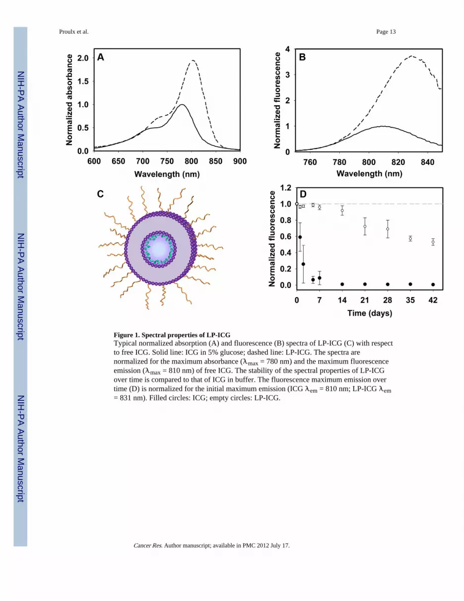
Figure 1. Spectral properties of LP-ICGTypical normalized absorption (A) and fluorescence (B) spectra of LP-ICG (C) with respectto free ICG. Solid line: ICG in 5% glucose; dashed line: LP-ICG. The spectra arenormalized for the maximum absorbance (λmax = 780 nm) and the maximum fluorescenceemission (λmax = 810 nm) of free ICG. The stability of the spectral properties of LP-ICGover time is compared to that of ICG in buffer. The fluorescence maximum emission overtime (D) is normalized for the initial maximum emission (ICG λem = 810 nm; LP-ICG λem= 831 nm). Filled circles: ICG; empty circles: LP-ICG.
Proulx et al. Page 13
Cancer Res. Author manuscript; available in PMC 2012 July 17.
NIH
-PA Author Manuscript
NIH
-PA Author Manuscript
NIH
-PA Author Manuscript

Figure 2. Dynamics of ICG signal after intradermal injection in normal C57BL/6 albino miceTime series of images from representative animals before and after intradermal injection ofthe left rear paw with ICG (A) and LP-ICG (B). Images were collected with 4-s exposuretime in both animals. Enhancement of popliteal lymph node and liver are seen in (A) and(B), while the medial iliac lymph node shows also signal enhancement in (B). Signalintensity of enhanced tissues is plotted versus time in ICG (C) and LP-ICG (D) injectedmice. Black circles: popliteal lymph node; red squares: liver; grey triangles: medial iliaclymph node.
Proulx et al. Page 14
Cancer Res. Author manuscript; available in PMC 2012 July 17.
NIH
-PA Author Manuscript
NIH
-PA Author Manuscript
NIH
-PA Author Manuscript

Figure 3. NIRF imaging of Chy mice after intradermal injectionTime series of images (exposure time 6 s) after intradermal injection of LP-ICG in arepresentative 5 month old male Chy mouse (A, left) and wild type littermate (A, right).Images of injection site (exposure time 0.1 s) taken 10 min, 4 h, and 24 h after injection intofoot of a representative Chy mouse (B, left) and wild type littermate (B, right). Whitearrowhead at 24 h in (B, left) shows interstitial diffusion pattern from foot in Chy mouse. InC, scatter plots of signal intensity measurements of injected feet at 10 min, 4 h, and 24 h. * p< 0.05
Proulx et al. Page 15
Cancer Res. Author manuscript; available in PMC 2012 July 17.
NIH
-PA Author Manuscript
NIH
-PA Author Manuscript
NIH
-PA Author Manuscript
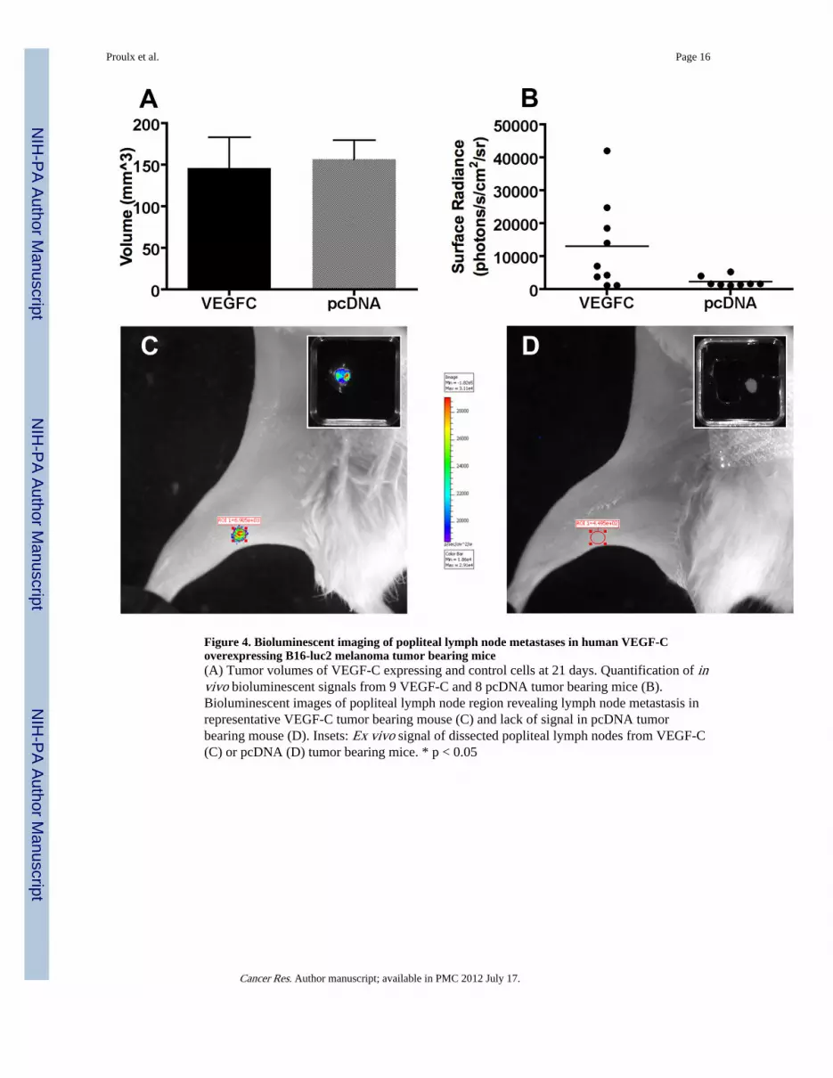
Figure 4. Bioluminescent imaging of popliteal lymph node metastases in human VEGF-Coverexpressing B16-luc2 melanoma tumor bearing mice(A) Tumor volumes of VEGF-C expressing and control cells at 21 days. Quantification of invivo bioluminescent signals from 9 VEGF-C and 8 pcDNA tumor bearing mice (B).Bioluminescent images of popliteal lymph node region revealing lymph node metastasis inrepresentative VEGF-C tumor bearing mouse (C) and lack of signal in pcDNA tumorbearing mouse (D). Insets: Ex vivo signal of dissected popliteal lymph nodes from VEGF-C(C) or pcDNA (D) tumor bearing mice. * p < 0.05
Proulx et al. Page 16
Cancer Res. Author manuscript; available in PMC 2012 July 17.
NIH
-PA Author Manuscript
NIH
-PA Author Manuscript
NIH
-PA Author Manuscript

Figure 5. Dynamic NIRF imaging of ICG liposome injected B16-luc2 tumor bearing miceRepresentative figures of lymphatic drainage patterns of LP-ICG after intradermal injection(A) from pcDNA, VEGF-C low metastatic, and VEGF-C high metastatic mice. Blackcircles: popliteal lymph node; red squares: liver; grey triangles: medial iliac lymph node.Correlation plot (B) showing KLN rates through popliteal lymph node versus popliteallymph node luciferase signals. Dashed line represents threshold between low metastatic andhigh metastatic VEGF-C tumor bearing mice. KLN rates (C) and half life measurements (D)of normal, pcDNA tumor, VEGF-C low metastatic and VEGF-C high metastatic groups. * p< 0.05, ** p < 0.01, *** p < 0.001.
Proulx et al. Page 17
Cancer Res. Author manuscript; available in PMC 2012 July 17.
NIH
-PA Author Manuscript
NIH
-PA Author Manuscript
NIH
-PA Author Manuscript