new drug delivery nanosystem combining liposomal and dendrimeric technology (liposomal locked-in...
TRANSCRIPT
New Drug Delivery Nanosystem Combining Liposomal andDendrimeric Technology (Liposomal Locked-In Dendrimers)for Cancer Therapy
KONSTANTINOS GARDIKIS,1,2 SOPHIA HATZIANTONIOU,2 MADALINA BUCOS,1 DIMITRIOS FESSAS,3
MARCO SIGNORELLI,3 THEODOROS FELEKIS,1 MARIA ZERVOU,1 CONSTANTINOS G. SCRETTAS,1 BARRY R. STEELE,1
MAKSIM IONOV,4 MARIA MICHA-SCRETTAS,1 BARBARA KLAJNERT,4 MARIA BRYSZEWSKA,4 COSTAS DEMETZOS2
1National Hellenic Research Foundation (N.H.R.F.), Institute of Organic and Pharmaceutical Chemistry, Vas. Konstantinou 48,Athens 11635, Greece
2Department of Pharmaceutical Technology, School of Pharmacy, University of Athens, Panepistimioupolis, Zografou,Athens 15771, Greece
3Universita di Milano, DISTAM, via Celoria 2, Milano 20133, Italy
4Department of General Biophysics, University of Lodz, Banacha 12/16, Lodz 90-237, Poland
Received 12 October 2009; revised 11 December 2009; accepted 15 January 2010
Published online 29 March 2010 in Wiley InterScience (www.interscience.wiley.com). DOI 10.1002/jps.22121
Additional Sversion of this a
This work is aCorresponden
0302107274596;E-mail: demetzo
Journal of Pharm
� 2010 Wiley-Liss
ABSTRACT: Liposomal locked-in dendrimers (LLDs), the combination of liposomes and den-drimers in one formulation, represents a relatively new term in the drug carrier technology.LLDs undergone appropriate physicochemical investigation can merge the benefits of liposomaland dendrimeric nanocarriers. In this study generation 1 and 2 hydroxy-terminated dendrimerswere synthesized and were then ‘‘locked’’ in liposomes consisting of DOPC/DPPG. The antic-ancer drug doxorubicin (Dox) was loaded into pure liposomes or LLDs and the final productswere subjected to lyophilization. The loading of Dox as well as its in vitro release rate from allsystems was determined and the interaction of liposomes with dendrimers was assessed bythermal analysis and fluorescence spectroscopy. The results were very promising in terms ofdrug encapsulation and release rate, factors that can alter the therapeutic profile of a drug withlow therapeutic index such as Dox. Physicochemical methods revealed a strong, generationdependent, interaction between liposomes and dendrimers that probably is the basis for thehigher loading and slower drug release from the LLDs comparing to pure liposomes. � 2010
Wiley-Liss, Inc. and the American Pharmacists Association J Pharm Sci 99:3561–3571, 2010
Keywords: liposome; dendrimer; doxoru
bicin; in vitro release; differential scanningcalorimetry; fluorescence spectroscopyINTRODUCTION
Since the late 1960s, interest in methods of drugdelivery has focused on the creation of new modifica-tions of established drugs with the objective of gettinga drug into the patient in the simplest possible way.The proper choice of delivery system can overcomeproblems relating to solubility, can regulate bioavail-ability and can therefore improve the overall ADME
upporting Information may be found in the onlinerticle.part of PhD thesis of Konstantinos Gardikis MSc.
ce to: Costas Demetzos (Telephone:Fax: 0302107274027;[email protected])
aceutical Sciences, Vol. 99, 3561–3571 (2010)
, Inc. and the American Pharmacists Association
JOURNAL O
profile (Absorption, bioDistribution, Metabolism, andExcretion) of a candidate drug.1
The effectiveness of a drug can generally beimproved in cases where there is need of controlledrelease in the bloodstream. This is particularlyimportant in the case of the treatment of certaindiseases, cancer therapy, for example, in which theadministration of low molecular weight cytostaticdrugs by themselves can cause severe side effects dueto their poor biodistribution, whereas controlleddelivery can greatly improve their therapeutic profile.In this respect, drug delivery systems based onnanoscale materials have the potential for minimumrelease prior to reaching the target site and selectiveaccumulation at the desired locations in vivo due tothe enhanced permeation and retention (EPR) effect.2
Polymers and liposomes represent two of the mostthoroughly studied categories of nanoparticles with
F PHARMACEUTICAL SCIENCES, VOL. 99, NO. 8, AUGUST 2010 3561
3562 GARDIKIS ET AL.
potential application as carriers of bioactive mole-cules. Liposomes, which constitute the earliest usedcategory of nanocarriers, are able to encapsulateeither lipophilic or hydrophilic drugs in their lipidicchains or aqueous interior, respectively. Researchhas proven that they are able to ameliorate thepharmacokinetic and pharmacological profile ofmany drugs3–5 giving way to the appearance ofseveral liposomal formulations in the market. Theapplication of liposomes, though, is limited becauseof their thermodynamic instability giving rise tophenomena such as aggregation, fusion, or drugleakage upon storage. However, these problems havebeen overcome to a large extent due to freezedrying.6,7
Dendrimers, a so-called 4th new architectural classof polymers, represent a much newer category of drugdelivery vehicles.8–10 The dendritic macromolecularstructure is well-defined and consists of a central core,branching units, and terminal functional groupswhich can be further chemically modified. Due totheir precise architecture, dendrimers possess anadvantage over other generally polydisperse nano-particles and this allows for greater control over theirpharmacodynamic profile, while as vehicles for drugdelivery they can be used either for encapsulation ofbioactive compounds or for their covalent or non-covalent attachment at the periphery. They also offerother potential advantages such as prolongation ofdrug circulation time, protection of a drug from itssurroundings, increase in drug stability (and possiblyeffectiveness), and the ability to target diseasedtissue.11–14
Although the first attempt to incorporate a druginto dendrimers was done in 198915, it was only in2001 that a combination between dendrimers andliposomes and the study of the interactions of thecomponents took place for the first time.16 Liposomallocked-in dendrimers (LLDs) technology—liposomesincorporating dendrimers—is a relatively new termin the drug delivery literature. Locked-in dendrimersmay be viewed as a dendrimer-based class ofmodulatory liposomal controlled release systems(MLCRS) leading to high entrapment and modifica-tion of the release profile of bioactive molecules fromliposomal vesicles.1 It has been established thatliposomal formulations of certain anticancer agentsare extremely sensitive to the drug release rates, withthe slowest releasing systems exhibiting the bestefficacy profiles.17,18 Therefore, for liposomal formu-lations, it is very important to control the drug releaserate.
The milestone for the creation of the liposomallocked in dendrimer concept was the work byKhopade et al.19 In that study, cationic PAMAMdendrimers were incorporated in the aqueous interiorof liposomes in order to increase the encapsulation
JOURNAL OF PHARMACEUTICAL SCIENCES, VOL. 99, NO. 8, AUGUST 2010
efficiency for the acidic anticancer drug methotrex-ate. The loading of the drug indeed increasedproportionally to dendrimer generation while theleakage of the drug from the system decreased. Theresults from this work were impressive, though littledata on the physicochemical interactions between thecomponents were given. An analogous study wasmade by Papagiannaros et al.20 in which a PAMAMG4–doxorubicin (Dox) complex was formed prior toencapsulation in liposomes and the results seempromising in terms of drug release and cytotoxicactivity against cancer cell lines. The immobilizationof anionic liposomes with PAMAM G4 has beenexplored using FT-IR, X-ray diffraction, and SPR21
and the results revealed that PAMAM G4 dendrimersmay be used to fabricate porous carrier films on theliposome surface in which ions or small molecules canbe released from liposomes and can diffuse throughthe PAMAM layers. Another study by 31P NMR andAFM, using neutral liposomes and lipid bilayersinteracting with PAMAM G7 dendrimers gavesimilar results which were attributed to the forma-tion of lipid–dendrimer aggregates.22,23
A very important method for studying interactionsin complex systems such as LLDs is differentialscanning calorimetry (DSC) and in recent years ourgroups have published several reports of thermalanalysis data for the interaction of dendrimers withmodel lipid membranes or liposomes.24–26 Despite thenumerous methods applied for dendrimer–lipidinteractions there is certainly a gap in the literatureconcerning the physicochemical characterization ofLLDs.1
In this study new synthetic generation 1 and 2polyether–polyester dendrimers (PG1&PG2) wereincorporated into liposomes consisting of DOPC/DPPG. The anticancer drug Dox was loaded intopure liposomes or LLDs and the final products weresubjected to lyophilization. The loading of Dox as wellas its in vitro release rate from all systems wasdetermined and the interaction of liposomes withdendrimers was assessed by thermal analysis andfluorescence spectroscopy.
MATERIALS AND METHODS
PG1&PG2 Dendrimer Synthesis
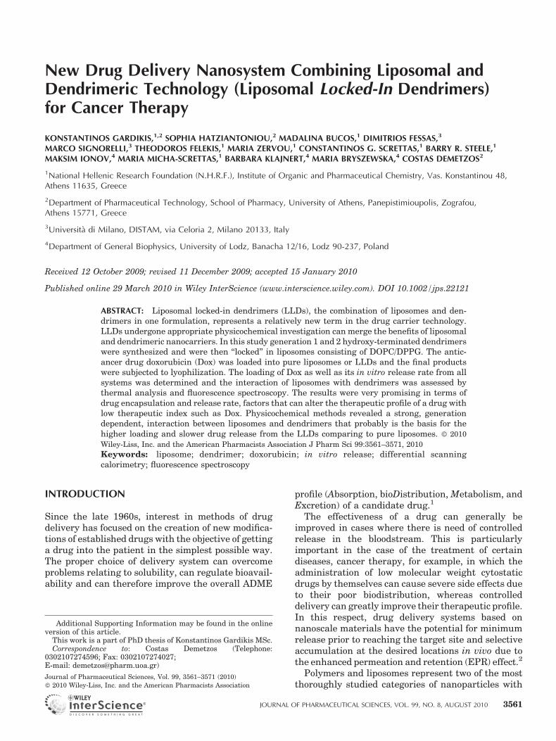
We have synthesized two new hydroxy-terminateddendrimers (Scheme 1) containing an aliphaticpolyether–polyester backbone. Both bear branchescomposed of glycerol and acetic acid monomers. Thechoice of building blocks was based on a requirementfor biocompatible, biodegradable, and water solublecompounds.
Following a divergent strategy we prepared den-drimers G1 and G2. Using the pentaerythritol
DOI 10.1002/jps
Scheme 1. Structure of G1 and G2 PG dendrimer.
NEW DRUG DELIVERY NANOSYSTEM 3563
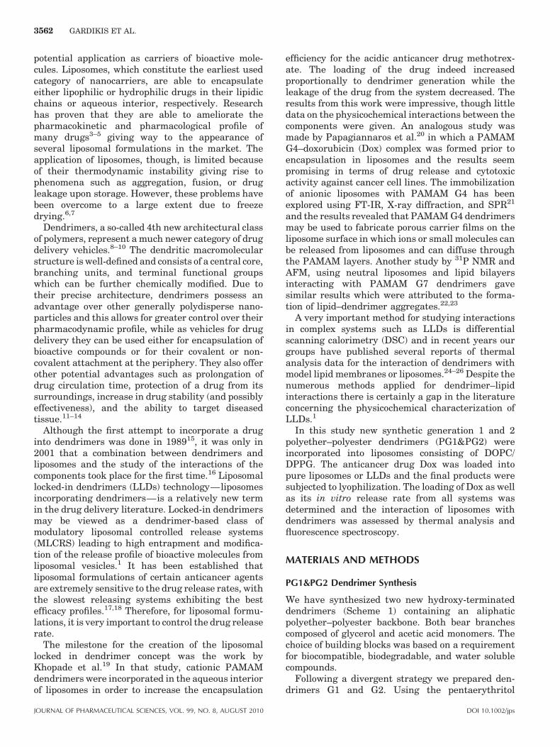
molecule as the core, esterification with 2-[1,3–bis(benzyloxy)propan-2-yloxy]acetic acid gave G1-Bn. Removal of the benzyl groups by hydrogenolysisled to G1-OH. Following the same procedure, we alsoprepared G2-OH (Scheme 2).
We chose to use the benzyl ether moiety asprotective group for its additional ability to serveas an NMR ‘‘tag’’ for the characterization of thegrowing molecule.
General Procedures
All reactions requiring dry or inert conditions werecarried out in flame-dried glassware under an atmo-sphere of argon. Pd/C (10%, w/w) was purchasedfrom Sigma Aldrich (St Louis, MO, USA), N,N0-dicyclohexylcarbodiimide (DCC) from Merck (Darm-stadt, Germany). 4-(dimethylamino)pyridiniump-toluenesulfonate (DPTS) was prepared accordingto the literature.27 Solvents were dried under argonby conventional methods. (THF distilled over sodiumbenzophenone and DMF over molecular sieve 40A.)Reactions were monitored by TLC (Merck Kieselgel60 F254). After aqueous work-up of reactionsmixtures, organic solutions were routinely dried overanhydrous sodium sulfate. Column chromatographywas carried out on Kieselgel 60 (particle size 40–63mm) as supplied by Merck. Size exclusion chroma-tography (SEC) was performed using Bio-Beads SX1Beads 200-400Mesh from Bio-Rad (California, USA).HR NMR experiments were acquired to Varian 600and 300 MHz spectrometers at 258C. Compoundswere dissolved in CDCl3 or D2O. The 2D 1H-1H DQF-COSY, 1H-13C edited-HSQC, and 1H-13C HMBCexperiments assisted structure characterization. Ex-perimental data were processed using VNMR rou-tines. Chemical shifts (d) are reported in ppm whilespectra were referenced by the standard experimen-
DOI 10.1002/jps
tal setup. Elemental analyses were performed at theNational Hellenic Research Foundation in Athensusing a Perkin Elmer (Massachusetts, USA) PE2400II analyzer. MS analyses were performed using a TSQ7000 Finnigan MAT instrument operating in ESImode.
Synthesis of 1,3-Bis(Benzyloxy)-2-Propanol (1)
1,3-bis(benzyloxy)-2-propanol was prepared by thereaction of 2 equiv. of benzyl alcohol with epichlor-ohydrin according to a literature procedure.28
Synthesis of 2-{2-(Benzyloxy)-1-[(Benzyloxy)Methyl]Ethoxy}Acetic Acid (2)
To a suspension of NaH (5.2 g, 130 mmol, washedwith toluene) in 120 mL dry THF, were added slowly27.2 g of compound 1,3-bis(benzyloxy)-2-propanol(100 mmol). Dry sodium a-chloroacetate (12.5 g,108 mmol) was added and the solution then refluxedfor 48 h. After removal of organic solvent, the solidresidue was dissolved in hot water. The aqueoussolution was extracted with toluene and hexane. Theaqueous phase was acidified with conc. aq. HCl andthen extracted with CH2Cl2. After removal of organicsolvent 25.7 g of the product were obtained as a yellowliquid (yield 78%).29 1H-NMR (CDCl3): d 3.56 (d,J¼ 5.4 Hz, 4H, CH–CH2), 3.79 (q, J¼ 5.4 Hz, 1H, CH),4.31 (s, 2H, CH2–C––O), 4.57 (s, 4H, benzyl-CH2), 7.37(m, 10H, arom.), 13C NMR (CDCl3): d 68.45, 69.67,73.46, 79.21, 127.68, 127.88, 128.39, 136.92, 172.8(C––O).
Synthesis of G1-Bn (3)
9.9 g (30 mmol) of 2-{2-(benzyloxy)-1-[(benzyloxy)-methyl]ethoxy}acetic acid (2), 5.1 g (5.1 mmol) ofpentaerythritol, and 1.76 g (6 mmol) DPTS were
JOURNAL OF PHARMACEUTICAL SCIENCES, VOL. 99, NO. 8, AUGUST 2010
Scheme 2. Divergent synthesis of G1 and G2 PG dendrimer.
3564 GARDIKIS ET AL.
dissolved in 50 mL of dry CH2Cl2. The mixture wasflushed with argon and 6.2 g (30 mmol) DCC wasadded. The reaction mixture was stirred for 72 h atroom temperature under an argon atmosphere.After filtration of urea, extraction with CH2Cl2,washing with water and drying over Na2SO4, anoily residue was obtained which after purificationby silica gel column chromatography (hexane/ethylacetate: 70/30) gave the product as an oil in a yieldof 4.6 g (60%). 1H-NMR (CDCl3): d 3.64 (d, J¼ 9.6 Hz,16H, CH2-6), 3.85 (q, J¼ 9.6 Hz, 4H, CH-5), 4.08(s, 8H, CH2-2), 4.39 (s, 8H, CH2-4), 4.54 (s, 16 H,benzyl-CH2), 7.34 (m, 40H, arom.), 13C NMR (CDCl3):d 42.0 (C1), 62.1(C2), 67.7(C4), 70.6 (C6), 73.4(C7benzyl), 78.7(C5), 127.7, 128.4, 138.1(arom.),170.2(C3, C––O), ESI m/z: 1408.9 (MHþNaþ), (theory:1385.6 MH). Anal. Calcd for C81H92O20: C, 70.21; H,6.69. Found: C, 70.18; H 6.57.
JOURNAL OF PHARMACEUTICAL SCIENCES, VOL. 99, NO. 8, AUGUST 2010
Synthesis of G1
To a solution of 0.2 g (0.144 mmol) of G1-Bn in amixture of 3 mL CH2Cl2 and 8 mL EtOH was addedPd/C (10%, w/w). The flask was first evacuated andthen filled with H2 and the reaction mixture stirredfor 4 h at RT. After completion of the reaction, thecatalyst was removed by filtration through Celite andwashed with EtOH. Evaporation of the filtrate gave0.07 g (73%) of the desired product as a colorlessviscous oil.
1H NMR (D2O) d: 3.42 (s, 8H, CH2-2), 3.45 (m, 4H,CH-5), 3.48 ((dd J¼ 11.8, 5.6 Hz), 8H, CH2-6), 3.55((dd J¼ 11.8, 3.8 Hz), 8H, CH2-6), 4.07 (s, 8H, CH2-4);13C NMR (D2O) d: 45.3 (C1), 60.6 (C6), 60.9 (C2), 67.6(C4), 81.5 (C5), 176.2 (C3) ESI m/z: 687.5 (MþNaþ),(theory: 664.2 M). Anal. Calcd for C25H44O20: C, 45.18;H, 6.67. Found: C, 45.00; H 6.45.
DOI 10.1002/jps
NEW DRUG DELIVERY NANOSYSTEM 3565
Synthesis of G2-Bn (5)
G2-Bn was obtained following a procedure similar tothat described for compound G1-Bn. 0.390 g(0.58 mmol) of G1, 2.29 g (5.1 mmol) of 2-{2-(benzy-loxy)-1-[(benzyloxy)methyl]ethoxy}acetic acid (2) and0.733 g (6 mmol) DPTS were dissolved in 22 mL of dryTHF and (1.74 mL) dry DMF. The mixture wasflushed with argon and 1.44 g (30 mmol) DCC wasadded. The reaction mixture was stirred for 10 days atroom temperature under an argon atmosphere. Afterfiltration of urea and concentration to remove solvent,an oily residue was obtained which was purified bySEC (CH2Cl2) in a yield of 40% (0.73 g).
1H NMR (CDCl3) d: 3.61 ((dd, J¼ 14 Hz, 4.5 Hz),32H, CH2-10), 3.7 ((q, J¼ 4.8 Hz), 4H, CH-5), 3.82 (m,8H, CH-9), 4.04 (s, 8H, CH2-2), (4.04-4.23) (m, 16H,CH2-6), 4.29 (s, 8H, CH2-4), 4.33 (s, 16H, CH2-8), 4.5(br.s, 32H, CH2-benzyl), (7.25–7.31) (m, 80H, ar-H);13C NMR (CDCl3) d: 42.1 (C1), 62.0 (C2), 62.8 (C6),67.7 (C4), 67.8 (C8), 70.6 (C10), 73.4 (C11), 75.8 (C5),78.7 (C9), 127.5, 128.4 (phenyl C13-C15), 138 (phenylC12), 170.1 (C3), 170.4 (C7) ESI m/z: 3.200.4(MþKþ), 3.161.4, (theory: 3.161.3 M).
Anal. Calcd for C177H204O52: C, 67.20; H, 6.50.Found: C, 66.9; H 6.35.
Synthesis of G2
G2-OH was obtained following a similar procedure tothat used for compound 4. To a solution of 0.5 g(0.158 mmol) of G2-Bn in a mixture of 5 mL THF and5 mL MeOH was added Pd/C (10%, w/w). The flaskwas first evacuated and then filled with 50 psi of H2
and the reaction mixture stirred for 20 h at roomtemperature. After completion of the reaction, thecatalyst was removed by filtration through Celite andwashed with MeOH and THF. Evaporation of thefiltrate gave 0.245 g (yield 90%) of the desired productas a colorless viscous oil. 1H-NMR (D2O) d: 3.47 (s,8H, CH2-2), 3.62-3.51 (m, 60H, CH-5,9, CH2-6,10),4.21 (s, 24H, CH2-4,8); 13C NMR (D2O) d: 42.1 (C1),60.5 (C6, C10), 60.9 (C2), 66.8 (C4, C8), 81.5 (C5, C9),174.7(C3, C7). ESI m/z: 1744.5 (MþNaþ), (theory:1721.52 M). Anal. Calcd for C65H108O52: C, 45.35; H,6.32. Found: C, 45.55; H 6.45.
Pure Liposome and Liposomal Locked in DendrimerPreparation
The liposomes prepared in this study consisted ofDOPC and DPPG at a 10:0.6 ratio (the lipid systemfrom now on will be referred to as just DOPC). In thecase of LLDs, dendrimeric solutions in methanol weremixed with the lipid solutions. The initial dendrimer/lipid molar ratio was 0.1. (NH4)2SO4 150 mM, pH 5.5,with 150 mM sucrose as a cryoprotectant was addedand the mixture was vortexed until the induction of ahomogenous emulsion. MLV preparation was made
DOI 10.1002/jps
using the reverse phase evaporation method (REV).30
Sonication was applied to afford SUVs with reducedP.I. and the extraliposomal pH was changed to 7.5through gel permeation chromatography using aSephadex G75 column with PBS 10 mM pH 7.5/150 mM sucrose as a mobile phase.
Dox was loaded to pure liposomes or LLDs byincubation in room temperature for 1 h. UnentrappedDox was removed by gel permeation chromatography(Sephadex G75).
Freeze Drying of Liposomal Suspensions
Free or Dox-loaded liposomes were frozen at �808Covernight and were subjected to lyophilization inorder to overcome stability issues concerning liposo-mal suspensions.31 The lyophilization was achievedusing a freeze drier (TELSTAR Cryodos-50, Terrassa,Spain) under the following conditions: condensertemperature from �508C, vacuum 8.2� 10�2 mb).Reconstitution was made by adding the appropriateamount of HPLC-grade water.
Characterization of Free and Doxorubicin-LoadedLiposomes
The hydrodynamic diameter of empty and Dox-loadedliposomes was measured by light scattering. Size andz-potential of liposomes are the parameters thatindicate their physical stability. 100mL of theliposomal suspension was 30-fold diluted in HPLC-grade water (pH 5.6–5.7) immediately after prepara-tion or after reconstitution and z-average mean and z-potential of the empty and Dox-loaded liposomes weremeasured. Samples were scattered (633 nm) at 908,and measurements were made at 258C in a photoncorrelation spectrometer (Zetasizer 3000 HS, Mal-vern Instruments, Malvern, UK) and analyzed by theCONTIN method (MALVERN software).
Dendrimer and lipid quantification was done byHPTLC-FID (Iatroscan)32 with chloroform/methanol/water 60:20:3.2 (v:v) as a mobile phase.
The incorporation of Dox into liposomes wasdetermined by UV spectrometry (UV-1700, UV–Visible Spectrophotometer, SHIMADZU, Pharmas-pec, Kyoto, Japan) at wavelength 480 nm after theaddition of methanol to the liposomal suspension,with the aid of a Dox calibration curve in methanol.Pure methanol was used as blank.
In Vitro Release Studies
Dox-loaded Liposomal suspensions were placed in12000 MWCO 25 mm width dialysis sacks (SigmaAldrich, St Louis, MO, USA). Dialysis sacks wereinserted in RPMI 5% medium in shaking water bath(Selecta, Barcelona, Spain) set at 378C. Aliquots ofsamples (1 mL) were taken from the external solutionat specific time points and that volume was replacedwith RPMI incubated at 378C. Dox concentrations
JOURNAL OF PHARMACEUTICAL SCIENCES, VOL. 99, NO. 8, AUGUST 2010
3566 GARDIKIS ET AL.
were measured with UV spectrometry after theaddition of 2 mL HPLC-grade water. As referencesample RPMI at 378C, diluted threefold with HPLC-grade water was used. The cumulative percentage ofdrug release was calculated and plotted versus timeusing the equation:
% Released Dox ¼ ½Dox�released
½Dox�initial
Membrane Fluidity Measurements
The change of fluidity of the lipid bilayer due toincreasing dendrimer incorporation in the membranewas measured using a steady-state fluorescence pol-arization technique. Two different fluorescent probeswere used: 1,6-diphenyl-1,3,5-hexatriene (DPH), anapolar molecule which is incorporated into the hydro-phobic region of the liposome bilayer with its long axisparallel to the acyl chains, and 1-(4-trimethylammo-niumphenyl)-6-phenyl-1,3,5-hexatriene (TMA-DPH),which is anchored at the surface of the liposomebilayer in contact with the water due to its positivelycharged amino groups. Since DPH probe is incorpo-rated deeper into the lipid bilayer than TMA-DPH,the use of both probes in the same lipid membraneallows for the comparison of membrane order atdifferent depths of the bilayer. Measurements weremade using a Perkin Elmer luminescence spectro-meter LS-50B equipped for fluorescence polarizationmeasurement. Three hundred micromolar liposomalsuspension was added to the cuvette followed by theaddition of 1mM DPH (in tetrahydrofuran) or TMA-DPH (in methanol). The sample was stirred well andincubated in the dark at room temperature for 20 min.The cuvette holder was temperature controlled bywater thermostat (MLW-U) with 378C. The readingswere taken at intervals of 2 s. The fluorescence aniso-tropy values (r) of the samples were calculated by thefluorescence data manager program FL WinLabThe Perkin-Elmer corporation, Version 3.00 usingthe following equation:
r ¼ ðIVV � GIVHÞðIVV þ 2GIVHÞ
where IVV and IVH are the vertical and horizontalfluorescence intensities, respectively, to the verticalpolarization of the excitation light beam. The factorG¼ IHV/IHH (grating correction factor) corrects thepolarizing effects of the monochromator. The excita-tion wavelengths were 348 and 340 nm the fluores-cence emission was measured at 426 and 430 nm nmfor DPH and TMA-DPH, respectively.
Thermal Analysis Measurements
The DSC method was used for the stability char-acterization of the DOPC lipid bilayers incorporating
JOURNAL OF PHARMACEUTICAL SCIENCES, VOL. 99, NO. 8, AUGUST 2010
dendrimers. The measurements were performed witha TA Instruments DSC 2920. The lipid bilayersamples where prepared by hydration of dry lipid/dendrimer mixtures at appropriate ratios (0%, 3%,10%, 20% of dendrimer content) in excess of HPLC-grade water and were placed in stainless steelpressure-resistant 60mL pans which were sealed.Empty pan was used as the reference. Two cooling-heating cycles were performed from 25 to �508C at28C/min scanning rate. The second heating run wastaken into account.
The raw data were worked out with the software‘‘THESEUS’’33 dedicated for handling raw calori-metric data. Briefly, the output signal in mW unitswas divided by the sample lipid mass and by theheating rate to be converted into apparent heatcapacity, Capp
P in kJ K�1 mol�1 units. The trace of CappP
was finally scaled with respect to the baseline toobtain the excess (with respect to the low temperaturelipids state) specific heat, Cex
P ðTÞ. The area underlyingthe recorded peaks, so treated, directly corresponds tothe lipid phase transitions relevant enthalpy inkJ mol�1 units. Errors were evaluated on the basisof at least three replicas.
Statistical Analysis
Results are shown as meanSD of n¼ 3 independentexperiments. To analyze differences in variablesbefore and after freeze drying the paired Student’st-test was used. Comparison between different groupswas done using One-way ANOVA followed by Tukeymultiple comparison test when equal variances wereassumed and Dunnett’s C multiple comparison testwhen equal variances were not assumed. In order toassess the correlations between the pairs of thevariables parameters of the regression line wereestimated together with the regression coefficient r.The no correlation hypothesis was rejected on thep¼ 0.05 significance level. p-Values <0.05 wereconsidered statistically significant. Statistical analy-sis was done using SPSS 14.0.
RESULTS AND DISCUSSION
Characterization of Free and Dox-Loaded Liposomes
The physicochemical characteristics of free and Dox-loaded liposomes and LLDs before freeze drying orafter reconstitution can be seen in Table 1. PG1incorporation in DOPC liposomes did not affect theirsize, z-potential or P.I. Respective data were obtainedfor PG2 LLDs, except the case of vesicular size thatwas augmented from 70 to 96.7 nm. This fact could bedue to bigger dendrimer size and higher encapsulateddendrimer/lipid ratio in the case of PG2 leading tohigher level pressure of the aqueous interior of theliposome and consequently to larger vesicles.34 Dox
DOI 10.1002/jps
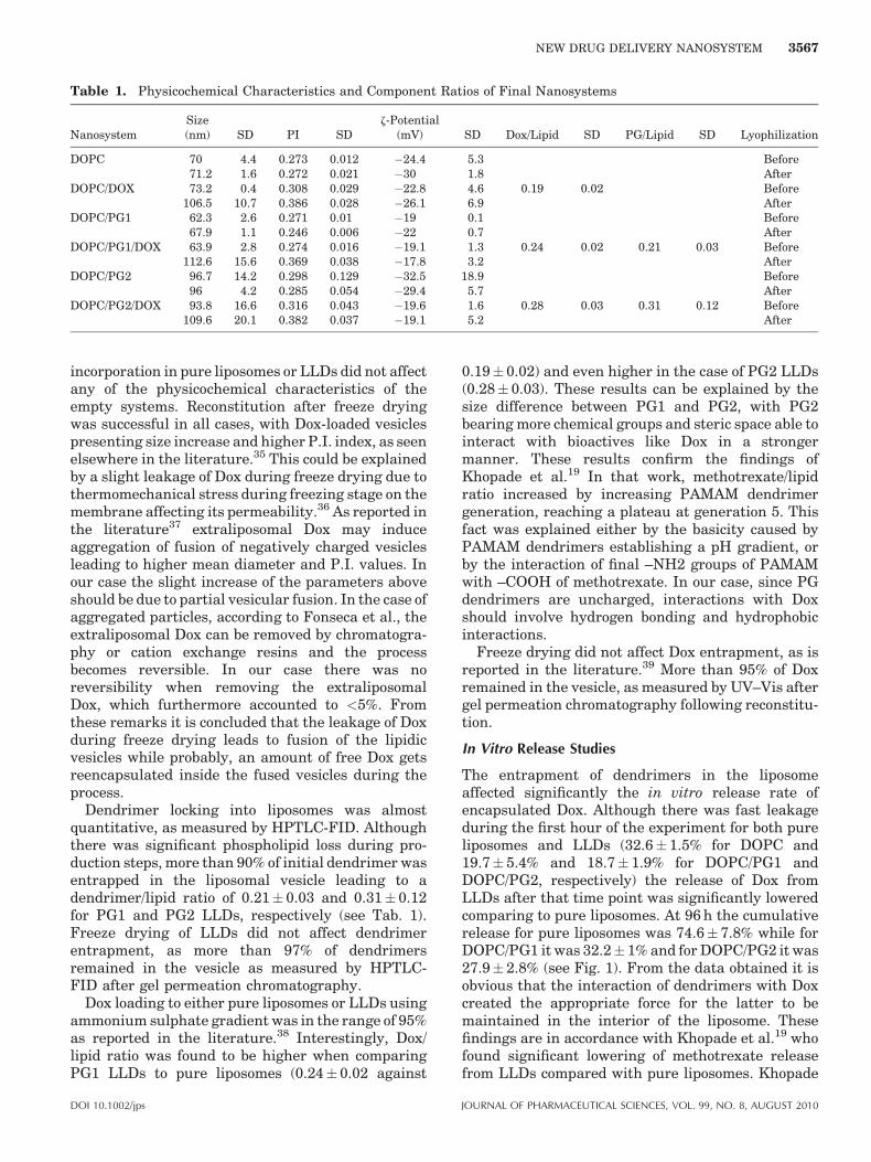
Table 1. Physicochemical Characteristics and Component Ratios of Final Nanosystems
NanosystemSize(nm) SD PI SD
z-Potential(mV) SD Dox/Lipid SD PG/Lipid SD Lyophilization
DOPC 70 4.4 0.273 0.012 �24.4 5.3 Before71.2 1.6 0.272 0.021 �30 1.8 After
DOPC/DOX 73.2 0.4 0.308 0.029 �22.8 4.6 0.19 0.02 Before106.5 10.7 0.386 0.028 �26.1 6.9 After
DOPC/PG1 62.3 2.6 0.271 0.01 �19 0.1 Before67.9 1.1 0.246 0.006 �22 0.7 After
DOPC/PG1/DOX 63.9 2.8 0.274 0.016 �19.1 1.3 0.24 0.02 0.21 0.03 Before112.6 15.6 0.369 0.038 �17.8 3.2 After
DOPC/PG2 96.7 14.2 0.298 0.129 �32.5 18.9 Before96 4.2 0.285 0.054 �29.4 5.7 After
DOPC/PG2/DOX 93.8 16.6 0.316 0.043 �19.6 1.6 0.28 0.03 0.31 0.12 Before109.6 20.1 0.382 0.037 �19.1 5.2 After
NEW DRUG DELIVERY NANOSYSTEM 3567
incorporation in pure liposomes or LLDs did not affectany of the physicochemical characteristics of theempty systems. Reconstitution after freeze dryingwas successful in all cases, with Dox-loaded vesiclespresenting size increase and higher P.I. index, as seenelsewhere in the literature.35 This could be explainedby a slight leakage of Dox during freeze drying due tothermomechanical stress during freezing stage on themembrane affecting its permeability.36 As reported inthe literature37 extraliposomal Dox may induceaggregation of fusion of negatively charged vesiclesleading to higher mean diameter and P.I. values. Inour case the slight increase of the parameters aboveshould be due to partial vesicular fusion. In the case ofaggregated particles, according to Fonseca et al., theextraliposomal Dox can be removed by chromatogra-phy or cation exchange resins and the processbecomes reversible. In our case there was noreversibility when removing the extraliposomalDox, which furthermore accounted to <5%. Fromthese remarks it is concluded that the leakage of Doxduring freeze drying leads to fusion of the lipidicvesicles while probably, an amount of free Dox getsreencapsulated inside the fused vesicles during theprocess.
Dendrimer locking into liposomes was almostquantitative, as measured by HPTLC-FID. Althoughthere was significant phospholipid loss during pro-duction steps, more than 90% of initial dendrimer wasentrapped in the liposomal vesicle leading to adendrimer/lipid ratio of 0.21 0.03 and 0.31 0.12for PG1 and PG2 LLDs, respectively (see Tab. 1).Freeze drying of LLDs did not affect dendrimerentrapment, as more than 97% of dendrimersremained in the vesicle as measured by HPTLC-FID after gel permeation chromatography.
Dox loading to either pure liposomes or LLDs usingammonium sulphate gradient was in the range of 95%as reported in the literature.38 Interestingly, Dox/lipid ratio was found to be higher when comparingPG1 LLDs to pure liposomes (0.24 0.02 against
DOI 10.1002/jps
0.19 0.02) and even higher in the case of PG2 LLDs(0.28 0.03). These results can be explained by thesize difference between PG1 and PG2, with PG2bearing more chemical groups and steric space able tointeract with bioactives like Dox in a strongermanner. These results confirm the findings ofKhopade et al.19 In that work, methotrexate/lipidratio increased by increasing PAMAM dendrimergeneration, reaching a plateau at generation 5. Thisfact was explained either by the basicity caused byPAMAM dendrimers establishing a pH gradient, orby the interaction of final –NH2 groups of PAMAMwith –COOH of methotrexate. In our case, since PGdendrimers are uncharged, interactions with Doxshould involve hydrogen bonding and hydrophobicinteractions.
Freeze drying did not affect Dox entrapment, as isreported in the literature.39 More than 95% of Doxremained in the vesicle, as measured by UV–Vis aftergel permeation chromatography following reconstitu-tion.
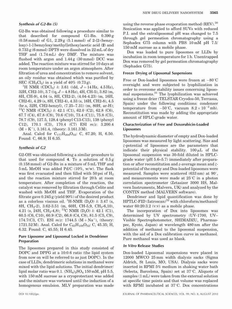
In Vitro Release Studies
The entrapment of dendrimers in the liposomeaffected significantly the in vitro release rate ofencapsulated Dox. Although there was fast leakageduring the first hour of the experiment for both pureliposomes and LLDs (32.6 1.5% for DOPC and19.7 5.4% and 18.7 1.9% for DOPC/PG1 andDOPC/PG2, respectively) the release of Dox fromLLDs after that time point was significantly loweredcomparing to pure liposomes. At 96 h the cumulativerelease for pure liposomes was 74.6 7.8% while forDOPC/PG1 it was 32.2 1% and for DOPC/PG2 it was27.9 2.8% (see Fig. 1). From the data obtained it isobvious that the interaction of dendrimers with Doxcreated the appropriate force for the latter to bemaintained in the interior of the liposome. Thesefindings are in accordance with Khopade et al.19 whofound significant lowering of methotrexate releasefrom LLDs compared with pure liposomes. Khopade
JOURNAL OF PHARMACEUTICAL SCIENCES, VOL. 99, NO. 8, AUGUST 2010
Figure 2. Fluorescence anisotropy of DPH probe incor-porated in DOPC liposomes with increasing concentrationsof PG1 dendrimer.
Figure 1. In vitro release of Dox-loaded pure liposomes orLLDs in RPMI 5%.
Figure 3. Fluorescence anisotropy of DPH probe incor-porated in DOPC liposomes with increasing concentrationsof PG2 dendrimer.
3568 GARDIKIS ET AL.
et al. also noticed dependence of release rate ondendrimer generation, comparing generations 2, 3,and 4. In our case the difference between generations1 and 2 was not significant and this fact should beattributed to the open structure of small generationdendrimers (below generation 3).40,41 Such openstructure could not provide the appropriate dendri-mer conformation for encapsulation of relatively bigmolecules such as Dox. We may thus assume thatthere is no internalization of Dox in the dendrimercavity but more likely the formation of Dox–dendri-mer network, meaning Dox molecules surrounded byand interacting with dendrimer molecules. In thiskind of network the size of dendrimer and thedendrimer to lipid molar ratio would be lesssignificant than the conformation of generation 1and 2 dendrimers that should be similar for bothdendrimers due to their similar chemistry and isaffected by the greater mobility of dendrimermolecules at 378C compared to 258C that the Doxloading process takes place. The fast release of Doxfrom LLDs during the first hour should be due to theliquid crystalline structure of DOPC membranes at378C. Liposomes in liquid crystalline state bear lipidchains in gauche conformation and in a state of greatmobility, permitting Dox to cross the membraneeasily.42 Thus, probably, during the first hour of theexperiment the Dox fraction in the interior of theliposome that does not interact with dendrimers leaksout of the vesicle leading to high release rate. Fromthen on the release rate diminishes as the inter-liposomal Dox inside the liposome is in dendrimer‘‘bound’’ form.
Membrane Fluidity Measurements
Fluorescence spectroscopy was applied in order toestimate the interaction of PG1 and PG2 dendrimerswith DOPC liposomes. The polar head groups ofphosphatidyl choline did not seem to interactsignificantly with either dendrimer as TMA-DPHanisotropy values did not change significantly upon
JOURNAL OF PHARMACEUTICAL SCIENCES, VOL. 99, NO. 8, AUGUST 2010
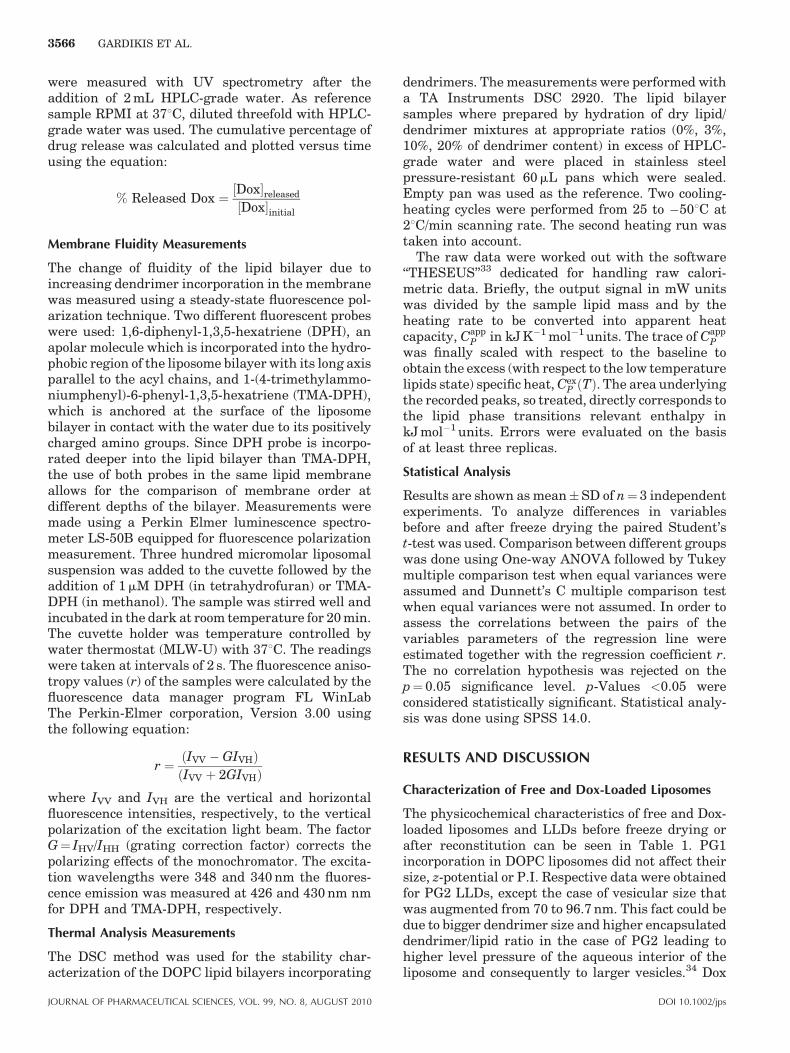
dendrimer incorporation (data not shown). PG1exhibited a significant interaction with the apolarpart of the membrane, inducing a concentration-dependent fluidization of the membrane (Fig. 2). TheDPH anisotropy decreased until a PG1/DOPC ratio of0.09 and then remained constant, meaning thathigher dendrimer concentration did not induce morefluid membrane. In the case of PG2 (that issignificantly bigger compared to PG1) the data werevery scattered probably due to the induction ofmembrane phase separation (see thermal analysischapter). Thus clear anisotropy conclusions cannot beobtained for the DOPC/PG2 system (Fig. 3).
Thermal Analysis Measurements
The incorporation of PG1 and PG2 dendrimers intothe DOPC lipid bilayers affected significantly thethermotropic behavior of the membrane lowering theTm, as can be seen in Figures 4 and 5. On the otherhand, the DH of the transition was not affected by theincorporation of the dendrimers (all enthalpies werein the order of 36 2 kJ mol�1) leading to theconclusion that the destabilization effect induced bydendrimer incorporation was predominantly of entro-pic nature. This is in line with the lack of interactionsof the lipid polar groups with the dendrimers
DOI 10.1002/jps
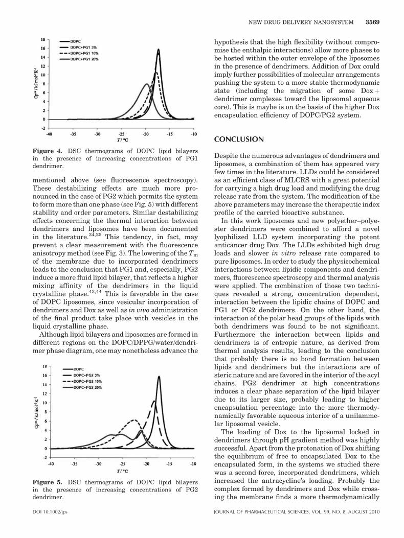
Figure 4. DSC thermograms of DOPC lipid bilayersin the presence of increasing concentrations of PG1dendrimer.
NEW DRUG DELIVERY NANOSYSTEM 3569
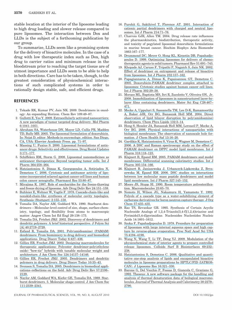
mentioned above (see fluorescence spectroscopy).These destabilizing effects are much more pro-nounced in the case of PG2 which permits the systemto form more than one phase (see Fig. 5) with differentstability and order parameters. Similar destabilizingeffects concerning the thermal interaction betweendendrimers and liposomes have been documentedin the literature.24,25 This tendency, in fact, mayprevent a clear measurement with the fluorescenceanisotropy method (see Fig. 3). The lowering of the Tm
of the membrane due to incorporated dendrimersleads to the conclusion that PG1 and, especially, PG2induce a more fluid lipid bilayer, that reflects a highermixing affinity of the dendrimers in the liquidcrystalline phase.43,44 This is favorable in the caseof DOPC liposomes, since vesicular incorporation ofdendrimers and Dox as well as in vivo administrationof the final product take place with vesicles in theliquid crystalline phase.
Although lipid bilayers and liposomes are formed indifferent regions on the DOPC/DPPG/water/dendri-mer phase diagram, one may nonetheless advance the
Figure 5. DSC thermograms of DOPC lipid bilayersin the presence of increasing concentrations of PG2dendrimer.
DOI 10.1002/jps
hypothesis that the high flexibility (without compro-mise the enthalpic interactions) allow more phases tobe hosted within the outer envelope of the liposomesin the presence of dendrimers. Addition of Dox couldimply further possibilities of molecular arrangementspushing the system to a more stable thermodynamicstate (including the migration of some Doxþdendrimer complexes toward the liposomal aqueouscore). This is maybe is on the basis of the higher Doxencapsulation efficiency of DOPC/PG2 system.
CONCLUSION
Despite the numerous advantages of dendrimers andliposomes, a combination of them has appeared veryfew times in the literature. LLDs could be consideredas an efficient class of MLCRS with a great potentialfor carrying a high drug load and modifying the drugrelease rate from the system. The modification of theabove parameters may increase the therapeutic indexprofile of the carried bioactive substance.
In this work liposomes and new polyether–polye-ster dendrimers were combined to afford a novellyophilized LLD system incorporating the potentanticancer drug Dox. The LLDs exhibited high drugloads and slower in vitro release rate compared topure liposomes. In order to study the physicochemicalinteractions between lipidic components and dendri-mers, fluorescence spectroscopy and thermal analysiswere applied. The combination of those two techni-ques revealed a strong, concentration dependent,interaction between the lipidic chains of DOPC andPG1 or PG2 dendrimers. On the other hand, theinteraction of the polar head groups of the lipids withboth dendrimers was found to be not significant.Furthermore the interaction between lipids anddendrimers is of entropic nature, as derived fromthermal analysis results, leading to the conclusionthat probably there is no bond formation betweenlipids and dendrimers but the interactions are ofsteric nature and are favored in the interior of the acylchains. PG2 dendrimer at high concentrationsinduces a clear phase separation of the lipid bilayerdue to its larger size, probably leading to higherencapsulation percentage into the more thermody-namically favorable aqueous interior of a unilamme-lar liposomal vesicle.
The loading of Dox to the liposomal locked indendrimers through pH gradient method was highlysuccessful. Apart from the protonation of Dox shiftingthe equilibrium of free to encapsulated Dox to theencapsulated form, in the systems we studied therewas a second force, incorporated dendrimers, whichincreased the antracycline’s loading. Probably thecomplex formed by dendrimers and Dox while cross-ing the membrane finds a more thermodynamically
JOURNAL OF PHARMACEUTICAL SCIENCES, VOL. 99, NO. 8, AUGUST 2010
3570 GARDIKIS ET AL.
stable location at the interior of the liposome leadingto high drug loading and slower release compared topure liposomes. The interaction between Dox andLLDs is the subject of a forthcoming publication byour group.
To summarize, LLDs seem like a promising systemfor the delivery of bioactive molecules. In the case of adrug with low therapeutic index such as Dox, highdrug to carrier ratios and minimum release in thebloodstream prior to reaching the target tissue are ofutmost importance and LLDs seem to be able to helpin both directions. Care has to be taken, though, to thegreatest consideration of physicochemical interac-tions of such complicated systems in order torationally design stable, safe, and efficient drugs.
REFERENCES
1. Tekade RK, Kumar PV, Jain NK. 2009. Dendrimers in oncol-ogy: An expanding Horizon. Chem Rev 109:49–87.
2. Gullotti E, Yeo Y. 2009. Extracellularly activated nanocarriers:A new paradigm of tumor targeted drug delivery. Mol Pharm6:1041–1051.
3. Abraham SA, Waterhouse DN, Mayer LD, Cullis PR, MaddenTD, Bally MB. 2005. The liposomal formulation of doxorubicin.In: Nejat D, editor. Methods in enzymology, edition. San Diego,CA, USA: Elsevier Academic Press. pp. 71–97.
4. Massing U, Fuxius S. 2000. Liposomal formulations of antic-ancer drugs: Selectivity and effectiveness. Drug Resist Updates3:171–177.
5. Schiffelers RM, Storm G. 2008. Liposomal nanomedicines asanticancer therapeutics: Beyond targeting tumor cells. Int JPharm 364:258–264.
6. Hatziantoniou S, Dimas K, Georgopoulos A, Sotiriadou N,Demetzos C. 2006. Cytotoxic and antitumor activity of lipo-some-incorporated sclareol against cancer cell lines and humancolon cancer xenografts. Pharmacol Res 53:80–87.
7. Miyajima K. 1997. Role of saccharides for the freeze-thawingand freeze drying of liposome. Adv Drug Deliv Rev 24:151–159.
8. Buhleier E, Wehner W, Vogtle F. 1978. Cascade-chain-like andnonskid-chain-like syntheses of molecular cavity topologies.Synthesis (Stuttgart) 2:155–158.
9. Tomalia DA, Naylor AM, Goddard WA. 1990. Starburst den-drimers—Molecular-level control of size, shape, surface-chem-istry, topology, and flexibility from atoms to macroscopicmatter. Angew Chem Int Ed Engl 29:138–175.
10. Tomalia DA, Frechet JMJ. 2002. Discovery of dendrimers anddendritic polymers: A brief historical perspective. J Polym Sci[A] 40:2719–2728.
11. Esfand R, Tomalia DA. 2001. Poly(amidoamine) (PAMAM)dendrimers: From biomimicry to drug delivery and biomedicalapplications. Drug Discov Today 6:427–436.
12. Gillies ER, Frechet JMJ. 2002. Designing macromolecules fortherapeutic applications: Polyester dendrimer-poly(ethyleneoxide) ‘‘bow-tie’’ hybrids with tunable molecular weight andarchitecture. J Am Chem Soc 124:14137–14146.
13. Gillies ER, Frechet JMJ. 2005. Dendrimers and dendriticpolymers in drug delivery. Drug Discov Today 10:35–43.
14. Svenson S, Tomalia DA. 2005. Dendrimers in biomedical appli-cations–reflections on the field. Adv Drug Deliv Rev 57:2106–2129.
15. Naylor AM, Goddard WA, Kiefer GE, Tomalia DA. 1989. Star-burst dendrimers. 5. Molecular shape control. J Am Chem Soc111:2339–2341.
JOURNAL OF PHARMACEUTICAL SCIENCES, VOL. 99, NO. 8, AUGUST 2010
16. Purohit G, Sakthivel T, Florence AT. 2001. Interaction ofcationic partial dendrimers with charged and neutral lipo-somes. Int J Pharm 214:71–76.
17. Charrois GJR, Allen TM. 2004. Drug release rate influencesthe pharmacokinetics, biodistribution, therapeutic activity,and toxicity of pegylated liposomal doxorubicin formulationsin murine breast cancer. Biochim Biophys Acta Biomembr1663:167–177.
18. Drummond DC, Meyer O, Hong KL, Kirpotin DB, Papahadjo-poulos D. 1999. Optimizing liposomes for delivery of chemo-therapeutic agents to solid tumors. Pharmacol Rev 51:691–743.
19. Khopade AJ, Caruso F, Tripathi P, Nagaich S, Jain NK. 2002.Effect of dendrimer on entrapment and release of bioactivefrom liposomes. Int J Pharm 232:157–162.
20. Papagiannaros A, Dimas K, Papaioannou GT, Demetzos C.2005. Doxorubicin-PAMAM dendrimer complex attached toliposomes: Cytotoxic studies against human cancer cell lines.Int J Pharm 302:29–38.
21. Moraes ML, Baptista MS, Itri R, Zucolotto V, Oliveira ON, Jr.2008. Immobilization of liposomes in nanostructured layer-by-layer films containing dendrimers. Mater Sci Eng C28:467–471.
22. Mecke A, Uppuluri S, Sassanella TM, Lee D-K, RamamoorthyA, Baker JJR, Orr BG, Banaszak Holl MM. 2004. Directobservation of lipid bilayer disruption by poly(amidoamine)dendrimers. Chem Phys Lipids 132:3–14.
23. Hong S, Hessler JA, Banaszak Holl MM, Leroueil P, Mecke A,Orr BG. 2006. Physical interactions of nanoparticles withbiological membranes: The observation of nanoscale hole for-mation. J Chem Health Saf 13:16–20.
24. Gardikis K, Hatziantoniou S, Viras K, Wagner M, Demetzos C.2006. A DSC and Raman spectroscopy study on the effect ofPAMAM dendrimer on DPPC model lipid membranes. Int JPharm 318:118–123.
25. Klajnert B, Epand RM. 2005. PAMAM dendrimers and modelmembranes: Differential scanning calorimetry studies. Int JPharm 305:154–166.
26. Klajnert B, Janiszewska J, Urbanczyk-Lipkowska Z, Brys-zewska M, Epand RM. 2006. DSC studies on interactionsbetween low molecular mass peptide dendrimers and modellipid membranes. Int J Pharm 327:145–152.
27. Moore JS, Stupp SI. 1990. Room temperature polyesterifica-tion. Macromolecules 23:65–70.
28. Nemoto H, Wilson JG, Nakamura H, Yamamoto Y. 1992.Polyols of a cascade type as a water-solubilizing element ofcarborane derivatives for boron neutron capture therapy. J OrgChem 57:435–435.
29. Rao TS, Revankar GR. 1995. Synthesis of Certain AcyclicNucleoside Analogs of 1,2,4-Triazolo[3,4-f][1,2,4]triazine andPyrimido[5,4-d]pyrimidine. Nucleosides Nucleotides NucleicAcids 14:1601–1612.
30. Szoka F, Papahadjopoulos D. 1978. Procedure for preparationof liposomes with large internal aqueous space and high cap-ture by reverse-phase evaporation. Proc Natl Acad Sci USA75:4194–4198.
31. Wang N, Wang T, Li TF, Deng YJ. 2009. Modulation of thephysicochemical state of interior agents to prepare controlledrelease liposomes. Colloids Surf B Biointerfaces 69:232–238.
32. Hatziantoniou S, Demetzos C. 2006. Qualitative and quanti-tative one-step analysis of lipids and encapsulated bioactivemolecules in liposome preparations by HPTLC/FID (IATROS-CAN). J Liposome Res 16:321–330.
33. Barone G, Del Vecchio P, Fessas D, Giancola C, Graziano G,1992. Theseus: A new software package for the handling andanalysis of thermal denaturation data of boilogical macromo-lecules. Journal of Thermal Analysis and Calorimetry 38:2279–2790.
DOI 10.1002/jps
NEW DRUG DELIVERY NANOSYSTEM 3571
34. Hupfeld S, Moen HH, Ausbacher D, Haas H, Brandl M. Lipo-some fractionation and size analysis by asymmetrical flow field-flow fractionation/multi-angle light scattering: Influence ofionic strength and osmotic pressure of the carrier liquid. ChemPhys Lipids 163:141–147.
35. Papagiannaros A, Hatziantoniou S, Dimas K, Papaioannou GT,Demetzos C. 2006. A liposomal formulation of doxorubicin,composed of hexadecylphosphocholine (HePC): Physicochem-ical characterization and cytotoxic activity against humancancer cell lines. Biomed Pharmacother 60:36–42.
36. Van Bommel EMG, Crommelin DJA. 1984. Stability of doxor-ubicin-liposomes on storage: As an aqueous dispersion, frozenor freeze-dried. Int J Pharm 22:299–310.
37. Fonseca M, vanWinden ECA, Crommelin DJA. 1997. Doxor-ubicin induces aggregation of small negatively charged lipo-somes. Eur J Pharm Biopharm 43:9–17.
38. Fritze A, Hens F, Kimpfler A, Schubert R, Peschka-Suss R.2006. Remote loading of doxorubicin into liposomes driven by atransmembrane phosphate gradient. Biochim Biophys ActaBiomembr 1758:1633–1640.
39. vanWinden ECA, Crommelin DJA. 1997. Long term stability offreeze-dried, lyoprotected doxorubicin liposomes. Eur J PharmBiopharm 43:295–307.
DOI 10.1002/jps
40. Tomalia DA. 2005. Birth of a new macromolecular architec-ture: Dendrimers as quantized building blocks for nanoscalesynthetic polymer chemistry. Prog Polym Sci 30:294–324.
41. Tomalia DA, Naylor AM, Goddard WA. 1990. Starburstdendrimers: Molecular-level control of size, shape, surfacechemistry, topology, and flexibility from atoms to macro-scopic matter. Angew Chem Int Ed Engl 29:138–175.
42. Charrois GJR, Allen TM. 2004. Drug release rate influencesthe pharmacokinetics, biodistribution, therapeutic activity,and toxicity of pegylated liposomal doxorubicin formulationsin murine breast cancer. Biochim Biophys Acta Biomembr1663:167–177.
43. Ivanova VP, Makarov IM, Schaffer TE, Heimburg T. 2003.Analyzing heat capacity profiles of peptide-containing mem-branes: Cluster formation of gramicidin A. Biophys J 84:2427–2439.
44. Freire E, Markello T, Rigell C, Holloway PW. 1983. Calori-metric and fluorescence characterization of interactionsbetween cytochrome b5 and phosphatidylcholine bilayers. Bio-chemistry 22:1675–1680.
JOURNAL OF PHARMACEUTICAL SCIENCES, VOL. 99, NO. 8, AUGUST 2010