prefrontal cortex regulates inhibition and excitation in distributed neural networks
TRANSCRIPT
Prefrontal cortex regulates inhibition and excitation indistributed neural networks
Robert T. Knight a,b,c,*, W. Richard Staines a,b,c, Diane Swick b,c,Linda L. Chao b,c,d
a Department of Psychology, University of California, Berkeley, 3210 Tolman Hall, Berkeley,
CA 94720-1650, USAb Department of Neurology, University of California, Davis, Veterans Medical Center, 150 Muir Road,
Martinez, CA 94553, USAc Center for Neuroscience, University of California, Davis, Veterans Medical Center, 150 Muir Road,
Martinez, CA 94553, USAd Laboratory of Brain and Cognition, National Institute of Mental Health, Building 10, Room 4C104,
Bethesda, MD 20892, USA
Abstract
Prefrontal cortex provides both inhibitory and excitatory input to distributed neural cir-
cuits required to support performance in diverse tasks. Neurological patients with prefrontal
damage are impaired in their ability to inhibit task-irrelevant information during behavioral
tasks requiring performance over a delay. The observed enhancements of primary auditory
and somatosensory cortical responses to task-irrelevant distractors suggest that prefrontal
damage disrupts inhibitory modulation of inputs to primary sensory cortex, perhaps through
abnormalities in a prefrontal-thalamic sensory gating system. Failure to suppress irrelevant
sensory information results in increased neural noise, contributing to the de®cits in decision
making routinely observed in these patients. In addition to a critical role in inhibitory control
of sensory ¯ow to primary cortical regions, and tertiary prefrontal cortex also exerts excitatory
input to activity in multiple sub-regions of secondary association cortex. Unilateral prefrontal
damage results in multi-modal decreases in neural activity in posterior association cortex in
the hemisphere ipsilateral to damage. This excitatory modulation is necessary to sustain neural
activity during working memory. Thus, prefrontal cortex is able to sculpt behavior through
parallel inhibitory and excitatory regulation of neural activity in distributed neural net-
works. Ó 1999 Elsevier Science B.V. All rights reserved.
Acta Psychologica 101 (1999) 159±178
* Corresponding author. Tel.: +1-925-372-2572; fax: +1-925-229-2315; e-mail: [email protected]
ley.edu
0001-6918/99/$ ± see front matter Ó 1999 Elsevier Science B.V. All rights reserved.
PII: S 0 0 0 1 - 6 9 1 8 ( 9 9 ) 0 0 0 0 4 - 9
PsycINFO classi®cations: 2346; 2520; 2530; 2540; 3213; 3297
Keywords: Prefrontal; Inhibition; Excitation; Attention; Schizophrenia
1. Introduction
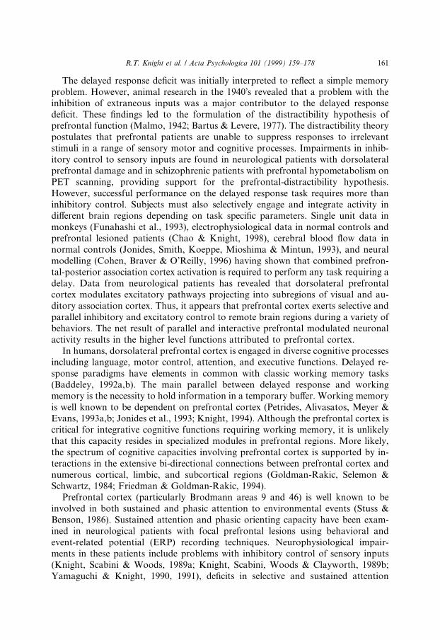
In 1935 Jacobsen reported that monkeys with bilateral frontal lesions involvingthe sulcus principalis, a putative analogue of human dorsolateral prefrontal cortex(Brodmann areas 9 and 46: Rajkowska & Goldman-Rakic, 1995a,b; see Fig. 1), wereseverely impaired at delayed response tasks (Jacobsen, 1935). In delayed responsetasks, subjects are initially presented with information necessary to perform a speci®ctask. The experimenter then interposes a delay period before the animal or human isallowed to perform the task. Thus, for successful performance, the information mustbe reliably held in a working memory bu�er during the delay period. Jacobsen re-ported that monkeys with prefrontal lesions involving the sulcus principalis wereunable to remember the location of a baited well at short retention intervals. TheJacobsen ®nding provided a landmark observation for insight into the role of pre-frontal cortex in organized behavior (Jacobsen, 1935). Subsequent research hasshown that prefrontal spatial memory de®cits are apparent at delays as short as 1 s(Funahashi, Bruce & Goldman-Rakic, 1993).
Fig. 1. This ®gure shows the location of the sulcus principalis area in monkeys considered to be the
analogue of areas 9 and 46 in humans. The Bordmann classi®cation and a more recent cytoarchitectonic
post-mortem de®nition of areas 9 and 46 in humans are also shown (see Brodmann, 1909; Goldman-
Rakic, 1987; Rajkowska & Goldman-Rakic, 1995a,b). The cytoarchitectonic de®nitions of Rajkowska and
Goldman-Rakic are shown on the Talairach coordinate system. The maps represent the region of cyto-
architectonic overlap of areas 9 and 46 averaged from from ®ve subjects.
160 R.T. Knight et al. / Acta Psychologica 101 (1999) 159±178
The delayed response de®cit was initially interpreted to re¯ect a simple memoryproblem. However, animal research in the 1940Õs revealed that a problem with theinhibition of extraneous inputs was a major contributor to the delayed responsede®cit. These ®ndings led to the formulation of the distractibility hypothesis ofprefrontal function (Malmo, 1942; Bartus & Levere, 1977). The distractibility theorypostulates that prefrontal patients are unable to suppress responses to irrelevantstimuli in a range of sensory motor and cognitive processes. Impairments in inhib-itory control to sensory inputs are found in neurological patients with dorsolateralprefrontal damage and in schizophrenic patients with prefrontal hypometabolism onPET scanning, providing support for the prefrontal-distractibility hypothesis.However, successful performance on the delayed response task requires more thaninhibitory control. Subjects must also selectively engage and integrate activity indi�erent brain regions depending on task speci®c parameters. Single unit data inmonkeys (Funahashi et al., 1993), electrophysiological data in normal controls andprefrontal lesioned patients (Chao & Knight, 1998), cerebral blood ¯ow data innormal controls (Jonides, Smith, Koeppe, Mioshima & Mintun, 1993), and neuralmodelling (Cohen, Braver & O'Reilly, 1996) having shown that combined prefron-tal-posterior association cortex activation is required to perform any task requiring adelay. Data from neurological patients has revealed that dorsolateral prefrontalcortex modulates excitatory pathways projecting into subregions of visual and au-ditory association cortex. Thus, it appears that prefrontal cortex exerts selective andparallel inhibitory and excitatory control to remote brain regions during a variety ofbehaviors. The net result of parallel and interactive prefrontal modulated neuronalactivity results in the higher level functions attributed to prefrontal cortex.
In humans, dorsolateral prefrontal cortex is engaged in diverse cognitive processesincluding language, motor control, attention, and executive functions. Delayed re-sponse paradigms have elements in common with classic working memory tasks(Baddeley, 1992a,b). The main parallel between delayed response and workingmemory is the necessity to hold information in a temporary bu�er. Working memoryis well known to be dependent on prefrontal cortex (Petrides, Alivasatos, Meyer &Evans, 1993a,b; Jonides et al., 1993; Knight, 1994). Although the prefrontal cortex iscritical for integrative cognitive functions requiring working memory, it is unlikelythat this capacity resides in specialized modules in prefrontal regions. More likely,the spectrum of cognitive capacities involving prefrontal cortex is supported by in-teractions in the extensive bi-directional connections between prefrontal cortex andnumerous cortical, limbic, and subcortical regions (Goldman-Rakic, Selemon &Schwartz, 1984; Friedman & Goldman-Rakic, 1994).
Prefrontal cortex (particularly Brodmann areas 9 and 46) is well known to beinvolved in both sustained and phasic attention to environmental events (Stuss &Benson, 1986). Sustained attention and phasic orienting capacity have been exam-ined in neurological patients with focal prefrontal lesions using behavioral andevent-related potential (ERP) recording techniques. Neurophysiological impair-ments in these patients include problems with inhibitory control of sensory inputs(Knight, Scabini & Woods, 1989a; Knight, Scabini, Woods & Clayworth, 1989b;Yamaguchi & Knight, 1990, 1991), de®cits in selective and sustained attention
R.T. Knight et al. / Acta Psychologica 101 (1999) 159±178 161
(Knight, Hillyard, Woods & Neville, 1981; Woods & Knight, 1986; Knight, 1991),and abnormalities in the detection of novel events (Knight, 1984, 1996, 1997). Theinability to inhibit irrelevant inputs and sustain attention, coupled with de®cits innovelty detection, impairs the coding and the processing of discrete external eventsand may underlay the temporal order (Shimamura, Janowsky & Squire, 1990) anddecision making impairments observed subsequent to prefrontal damage (Shim-amura, 1995a; Shimamura, Jurica, Mangels, Gershberg & Knight, 1995b). Similar tothe role prefrontal cortex plays in regulating the interaction with the external world,this region is crucial for attention to and inhibitory control of internal mental rep-resentations engaged during working memory, employment of strategies, planningand decision-making (Janowsky, Shimamura, Kritchevsky & Squire, 1989a;Janowsky, Shimamura & Squire, 1989b; Shimamura et al., 1995b; Knight &Grabowecky, 1995; Stuss & Benson, 1984, 1986) The inability to inhibit internalrepresentations of previous responses which are no longer correct contributes topoor performance on the Wisconsin card sorting task (WCST) and the Stroop task(Shimamura, 1995a; Shimamura et al., 1995b; Vendrell et al., 1995). In the WCSTtask subjects are required to change their criteria for sorting a deck of cards varyingin shape or color. Patients with prefrontal damage are impaired at switching to a newsorting criteria and continue to incorrectly sort by the prior rule. This tendency topreseverate is viewed by cognitive theorists as a failure in inhibitory control of priormental sets. Damage in prefrontal cortex also results in a failure in inhibition ofre¯exive saccadic eye movements (Guitton, Buchtel & Douglas, 1985; Pierrot-De-seilligny, Rivaud, Gaymard & Agid, 1991). The animal and human data supportingthese contentions will be discussed.
2. Inhibition in animals
Inhibition of neural activity under prefrontal control has been reported in a va-riety of mammalian preparations. Net prefrontal inhibitory control of both sub-cortical (Edinger, Siegel & Troiano, 1975) and cortical regions has been documented(Alexander, Newman & Symmes, 1976; Skinner & Yingling, 1977). Galambos (1956)provided the ®rst physiological evidence of an inhibitory auditory pathway inmammals with the description of the brainstem olivo-cochlear bundle. The olivo-cochlear bundle projects from the olivary nucleus to the cochleus in the inner ear.Stimulation of this bundle results in inhibition of transmission from the cochlea tothe brainstem cochlear nucleus as measured by reductions in evoked responses in theauditory nerve. This pathway provides a system for early sensory suppression in theauditory system. The evidence for sensory ®ltering at the cochlear or brainstem levelin humans is controversial, with most laboratories ®nding no evidence of attention-related manipulation of the brainstem auditory evoked response (Woods & Hillyard,1978; Woldor� & Hillyard, 1991).
Subsequent research in the 1970Õs reported evidence of a multi-modal prefrontal-thalamic inhibitory system in cats that regulates sensory ¯ow to primary corticalregions. Reversible suppression of prefrontal cortex by cooling of cat prefrontal
162 R.T. Knight et al. / Acta Psychologica 101 (1999) 159±178
cortex (crygenic blockade) increased the amplitudes of evoked responses recorded inprimary sensory cortex evoked responses (Skinner & Yingling, 1977; Yingling &Skinner, 1977). Conversely, stimulation of the thalamic region (nucleus reticularisthalami) surrounding the sensory relay nuclei resulted in modality speci®c suppres-sion of activity in primary sensory cortex. These data provided the ®rst physiologicalevidence of a prefrontal inhibitory pathway regulating sensory transmission throughthalamic relay nuclei. This prefrontal-thalamic inhibitory system provides a mech-anism for intermodality suppression of irrelevant inputs at an early stage of sensoryprocessing. The system is modulated by an excitatory prefrontal projection to thenucleus reticularis thalami. The nucleus reticularis thalami in turn sends inhibitoryGABA-ergic projections to sensory relay nuclei, providing a neural substrate forselective sensory suppression (Guillery, Feig & Lozsadi, 1998).
3. Inhibition in humans
The attention de®cits and perseveration observed behaviorally in frontal patientshave been linked to problems with inhibitory control of posterior sensory and per-ceptual mechanisms (Lhermitte, 1986; Lhermitte, Pillon & Serdaru, 1986). Earlysensory gating de®cits (20±50 ms), sustained attention problems (100±500 ms) andabnormalities in the phasic detection of novel events (250±500 ms) are all observedafter prefrontal damage. ERPs are brain potentials that are time-locked to the oc-currence of sensory, motor, or cognitive events and extracted from the ongoing EEGby signal averaging techniques (Hillyard & Picton, 1987). Because of their excellenttemporal resolution, ERPs index the timing of both sensory and cognitive processingin humans that complements both the behavioral measures of cognitive psychologyand the spatial resolution advantage of functional neuroimaging techniques.
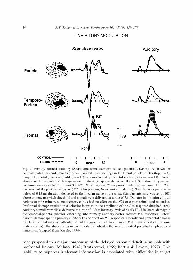
Task irrelevant auditory and somatosensory stimuli (monaural clicks or briefelectric shocks to the median nerve) were presented to patients with comparablysized lesions in dorsolateral prefrontal cortex, the temporal-parietal junction, orlateral parietal cortex. Evoked responses from primary auditory (Kraus, Ozdamar &Stein, 1982) and somatosensory (Leuders, Leser, Harn, Dinner & Klem, 1983;Sutherling et al., 1988; Wood et al., 1988) cortices were recorded from these patientsand age-matched controls (Fig. 2). Damage to primary auditory or somatosensorycortex reduced the early latency (20±40 ms) evoked responses generated in theseregions. Posterior association cortex lesions that spared the primary sensory regionshad no e�ect on early sensory potentials and served as a brain-lesioned controlgroup. Prefrontal damage produced disinhibition of both the primary auditory andsomatosensory evoked responses (Knight et al., 1989a; Yamaguchi & Knight, 1990).Spinal cord and brainstem potentials were not a�ected by prefrontal damage, sug-gesting that the amplitude enhancement was due to abnormalities in either a pre-frontal-thalamic or a prefrontal-sensory cortex mechanism.
Chronic disinhibition of sensory inputs likely contributes to some of the behav-ioral sequelae noted after prefrontal damage. For instance, decision con®dencewould be decremented in a noisy internal milieu. As discussed, distractibility has
R.T. Knight et al. / Acta Psychologica 101 (1999) 159±178 163
been proposed to a major component of the delayed response de®cit in animals withprefrontal lesions (Malmo, 1942; Brutkowski, 1965; Bartus & Levere, 1977). Thisinability to suppress irrelevant information is associated with di�culties in target
Fig. 2. Primary cortical auditory (AEPs) and somatosensory evoked potentials (SEPs) are shown for
controls (solid line) and patients (dashed line) with focal damage in the lateral parietal cortex (top, n� 8),
temporal-parietal junction (middle, n� 13) or dorsolateral prefrontal cortex (bottom, n� 13). Recon-
structions of the center of damage in each patient group are shown on the left. Somatosensory evoked
responses were recorded from area 3b (N20; N for negative, 20 ms post-stimulation) and areas 1 and 2 on
the crown of the post-central gyrus (P26; P for positive, 26 ms post-stimulation). Stimuli were square-wave
pulses of 0.15 ms duration delivered to the median nerve at the wrist. Stimulus intensity was set at 10%
above opponents twitch threshold and stimuli were delivered at a rate of 3/s. Damage in posterior cortical
regions sparing primary somatosensory cortex had no e�ect on the N20 or earlier spinal cord potentials.
Prefrontal damage resulted in a selective increase in the amplitude of the P26 response (hatched area).
Auditory stimuli were clicks delivered at a rate of 13/s at intensity levels of 50 dB HL. Unilateral damage in
the temporal-parietal junction extending into primary auditory cortex reduces P30 responses. Lateral
parietal damage sparing primary auditory has no e�ect on P30 responses. Dorsolateral prefrontal damage
results in normal inferior collicular potentials (wave V) but an enhanced P30 primary cortical response
(hatched area). The shaded area in each modality indicates the area of evoked potential amplitude en-
hancement (adapted from Knight, 1994).
164 R.T. Knight et al. / Acta Psychologica 101 (1999) 159±178
detection and match-to-sample paradigms. For example, patients with frontal re-sections are impaired at detecting multiple visual targets embedded among dis-tractors (Richer et al., 1993). Likewise, patients with lesions con®ned to dorsolateralprefrontal cortex are impaired in a delay task requiring the matching of two envi-ronmental sounds only when distractors intervened between cue and target (Chao &Knight, 1995; Chao & Knight, 1998).
There is extensive literature supporting an abnormality in prefrontal function inschizophrenics. Findings of altered dorsolateral prefrontal function include evidencefrom both cerebral blood ¯ow (Weinberg, Berman & Zec, 1986; Weinberg, Berman,Suddath & Torrey, 1992) and post-mortem studies (Akbarian et al., 1995, 1996).Thus, schizophrenia may represent a ``non-lesion'' model of prefrontal dysfunctionin humans. Schizophrenics are also reported to have a de®cit in inhibitory control ofauditory processing. Freedman and colleagues developed and ERP auditory gatingparadigm to study inhibitory control in schizophrenics. In normals, presentation of apair of clicks results in a decrease in amplitude of the evoked response to the secondstimulus in the pair. This response suppression occurs in a latency range of 30±65msec and has been referred to as the P50 gating paradigm in the schizophrenia lit-erature. This ®nding has been disputed by some authors (Kathman & Engel, 1990),but this may be due in part to di�erences in recording parameters and state ofalertness (Boutros, Zouridakis & Overall, 1991a; Boutros, Overall & Zouridakis,1991b; Smith, Boutros & Schwarzkopf, 1994; Gri�th, Ho�er, Adler & Zerbe, 1995).Freedman, Adler, Waldo, Pachtman and Franks (1983) reported that the secondstimulus in a pair of auditory pulses did not habituate in schizophrenics. Thiselectrophysiological ®ndings supported the longstanding proposal that schizo-phrenics fail to properly ®lter extraneous inputs (McGhie & Chapman, 1961; Ven-ables, 1964).
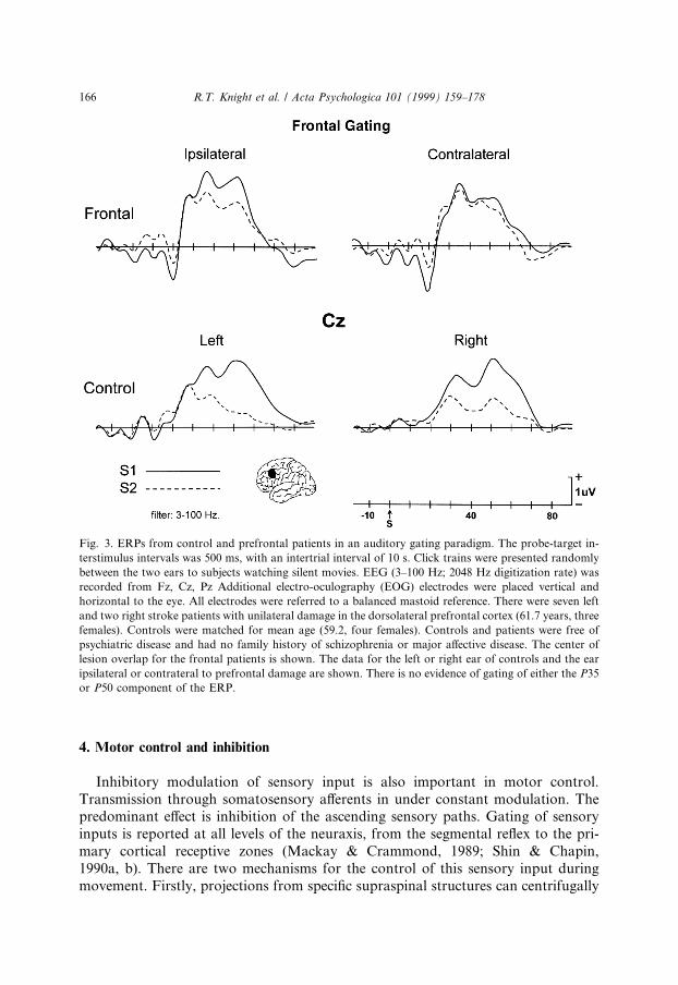
This auditory gating de®cit is reliably seen in a signi®cant percentage of non-psychotic relatives of schizophrenics and has been proposed to be a neurophysiol-ogicl trait for schizophrenia. Phenotypic segregation of schizophrenics and ®rst orderrelatives using the auditory gating paradigm has been employed in recent geneticstudies. This research has isolated a putative schizophrenia gene localized to a regionof chromosome 15q 13±14 which controls alpha 7 nicotinic receptor expression(Freedman et al., 1997). Thus, the neural network controlling the P50 gating de®cit isof both theoretical and clinical relevance. We examined auditory gating in patientswith dorsolateral prefrontal damage and in age-matched controls (see Fig. 3). Aninitial study has shown that controls have normal suppression of the second stimulusin an auditory pulse pair. Prefrontal patients showed evidence of an inhibitoryfailure in the auditory gating paradigm in both ears. As can be seen in Fig. 3, pre-frontal patients showed problems with suppression of the second stimulus in bothears with the defect more apparent in the ear contralateral to prefrontal damage(Knight, Finkbeiner & Lawler, in preparation). This failure to suppress is observedfor both an early latency component generated in auditory cortex (P35) and a latercomponent (P50) thought to arise in the thalamus. The data suggests that prefrontalcortex dysfunction may underlie or contribute to the auditory gating de®cit inschizophrenics.
R.T. Knight et al. / Acta Psychologica 101 (1999) 159±178 165
4. Motor control and inhibition
Inhibitory modulation of sensory input is also important in motor control.Transmission through somatosensory a�erents in under constant modulation. Thepredominant e�ect is inhibition of the ascending sensory paths. Gating of sensoryinputs is reported at all levels of the neuraxis, from the segmental re¯ex to the pri-mary cortical receptive zones (Mackay & Crammond, 1989; Shin & Chapin,1990a, b). There are two mechanisms for the control of this sensory input duringmovement. Firstly, projections from speci®c supraspinal structures can centrifugally
Fig. 3. ERPs from control and prefrontal patients in an auditory gating paradigm. The probe-target in-
terstimulus intervals was 500 ms, with an intertrial interval of 10 s. Click trains were presented randomly
between the two ears to subjects watching silent movies. EEG (3±100 Hz; 2048 Hz digitization rate) was
recorded from Fz, Cz, Pz Additional electro-oculography (EOG) electrodes were placed vertical and
horizontal to the eye. All electrodes were referred to a balanced mastoid reference. There were seven left
and two right stroke patients with unilateral damage in the dorsolateral prefrontal cortex (61.7 years, three
females). Controls were matched for mean age (59.2, four females). Controls and patients were free of
psychiatric disease and had no family history of schizophrenia or major a�ective disease. The center of
lesion overlap for the frontal patients is shown. The data for the left or right ear of controls and the ear
ipsilateral or contrateral to prefrontal damage are shown. There is no evidence of gating of either the P35
or P50 component of the ERP.
166 R.T. Knight et al. / Acta Psychologica 101 (1999) 159±178
through ascending paths can be altered by a�erent volleys arising from peripheralreceptor discharge, termed centripetal modulation.
In primates, the prefrontal cortex has direct reciprocal connections with the pa-rietal cortex, including the primary sensory region (Mountcastle, 1984; Pandya &Barnes, 1987). A net inhibitory in¯uence of the prefrontal cortex onto both corticaland subcortical structures has been shown to act at the dorsal column nuclei (Co-ulter, 1974; Ghez & Pisa, 1972), the thalamus (Tsumoto, Nakamura & Iwama, 1975),and at the primary somatosensory cortex (Yamamoto, Samejima & Oka, 1988;Chapin & Woodward, 1981). In the somatosensory system. Yamaguchi and Knight(1990, 1991) demonstrated that patients with damage to the prefrontal cortex cen-tered in areas 9 and 46 exhibited enhanced somatosensory evoked potentials (SEPs)to median nerve stimulation. Selective enhancement of the P26 component of theSEP which is known to arise in the crown of the post-central gyrus (Sutherling et al.,1988) was proposed to arise from loss of inhibition of activity in the primary sensorycortex.
In addition to prefrontal cortex, other areas such as the somatosensory cortex,cerebellum, and basal ganglia, may be involved in the modulation of sensory inputduring movement. Corticothalamic in¯uences from the primary and secondary so-matosensory areas modulate the transmission of tactile inputs through the thalamusin the cat (Gosh, Murray, Turman & Rowe, 1994). These e�ects could be facilitoryvia direct connections to thalamic relay nuclei or inhibitory through local inhibitoryinterneurons and the nucleus reticularis thalami, thus complementing the prefrontal-thalamic sensory control mechanism discussed above. The cerebellum receivesconvergent cutaneous and proprioceptive inputs and has also been implicated in thegating of these inputs (Apps, Atkins & Garwicz, 1997). In addition, the basal gangliaappears to be involved in the gating of noxious somatosensory stimuli (Chudler &Dong, 1995). Thus, through its intricate connections with higher motor areas via thethalamus, the basal ganglia may also contribute the gating of somatosensory inputs.The functional implications of these sensory gating mechanisms remains to be es-tablished for normal motor control.
5. Selective attention and prefrontal cortex
In a seminal report, Hillyard, Hink, Schwent and Picton (1973) found that fo-cussed attention to tones in one ear resulted in a systematic negative enhancement ofevoked potentials to all stimuli in that ear. This enhancement onsets at about 50 mspost-stimulation and was shown to be sustained for at least 200 and 500 ms (Hansen& Hillyard, 1980). These electrophysiological results were critical to attention the-orists. First, stimulus discriminability was shown to be dependent on the degree ofattention related evoked potential enhancement, providing a link between physiol-ogy and attention in humans. Second, the early onset of the attention modulationprovided clear evidence of an early sensory ®ltering mechanism in humans, ad-dressing the long-standing early vs. late selection controversy (Broadbent, 1958;Treisman, 1960; Kahneman & Treisman, 1984). Subsequent work has shown that the
R.T. Knight et al. / Acta Psychologica 101 (1999) 159±178 167
e�ects of attention can onset as early as 25 ms after stimulation, indicating thathumans are able to exert attention e�ects on inputs to the primary auditory cortex(McCallum, Curry, Cooper, Pocock & Papakostopoulos, 1983; Woldor� & Hillyard,1991; Woldor� et al., 1993). Early onset selective attention e�ects have also beenreported in the visual and somatosensory modalities (Desmedt, Hut & Bourguet,1983; Woods, 1990). In the visual modality attention may not modulate primarysensory activity in calcarine cortex but instead acts on subsequent stages of pro-cessing in visual association cortices (Gomez-Gonzalez, Clark, Fan, Luck & Hill-yard, 1994; Mangun, 1995).
Normal subjects generate comparable selective attention e�ects for left or rightear stimulation. Left prefrontal patients have an intact pattern but reduced in am-plitude attention e�ect in both ears. A di�erent pattern is observed after right pre-frontal damage. Right prefrontal patients show electrophysiological and behavioralevidence of a dense hemi-inattention to left ear stimuli (Knight et al., 1981). Thisparallels the human hemi-neglect syndrome which is more common after righthemisphere lesions in prefrontal or temporal-parietal cortex. Hemi-neglect is adramatic syndrome consisting of a failure to attend or orient to stimuli in thehemispace contralateral to right hemisphere damage (Mesulam, 1981; Kertesz &Dobrolowski, 1981; Hier, Mondlock & Caplan, 1983; Stein & Volpe, 1983). Patientsare often unaware of their de®cit and deny there is anything wrong despite obviousweakness on the left side of the body. One popular theory states that the contra-lateral neglect after temporal-parietal or prefrontal right hemisphere damage is dueto innate hemispheric attention asymmetries. The left frontal lobe is proposed to becapable of allocating attention only to the contralateral right hemispace, whereas theright frontal lobe can allocate attention to both the contralateral and ipsilateralhemispace. Thus, neglect is mild or not apparent after left hemisphere lesions sincethe intact right hemisphere is capable of allocating attention to both hemi-spaces.Dense contralateral neglect is seen after right hemisphere damage since the lefthemisphere is incapable of allocating attention to the left hemi-space.
The right frontal lobe is larger than the left in humans, and this asymmetry mayprovide the underlying anatomical substrate or the hemi-inattention syndrome inhumans (Wada, Clarke & Hamm, 1975; Weinberger, Luchins, Morisha & Wyatt,1982). Posterior association cortex lesions in the temporal-parietal junction havecomparable attention de®cits for left and right sided lesions indicating that theseareas are not asymmetrically organized for auditory selective attention (Woods,Knight & Scabini, 1993). This suggests that some aspects of the hemi-neglect syn-drome subsequent to temporal-parietal damage may be due to remote e�ects ofdisconnection from asymmetrically organized prefrontal regions.
Attention allocation is better at short versus long interstimulus intervals in pre-frontal lesioned patients. This could be due to either a problem with temporalbridging or to a distractibility de®cit. Prefrontal cortex is necessary for bridgingtemporal discontinuities (Fuster, 1989), and attention de®cits at longer interstimulusintervals might be due to temporal bridging problems. However, at longer inter-stimulus intervals prefrontal subjects are also more likely to encounter interveningirrelevant stimuli. ERP-behavioral experiments provide evidence supporting the
168 R.T. Knight et al. / Acta Psychologica 101 (1999) 159±178
distractibility hypothesis. In normal subjects, delivery of an irrelevant stimulus in thenon-attended ear during a dichotic experiment has no e�ect on attention e�ects to asubsequent stimulus in the attended ear unless the stimulus is highly deviant (Woods& Knight, 1996; Woods et al., 1993). However, presentation of an irrelevant stimulusreduces attention to a subsequent stimulus in prefrontal patients. This e�ect isparticularly pronounced in the ear contralateral to a prefrontal lesion at long in-terstimulus intervals. Since attention performance is improved in prefrontal patientsif no irrelevant stimuli are present, the results favor distractibility due to a failure ininhibition as a major contributor to prefrontal attention de®cits (Woods & Knight,1986).
Behavioral and electrophysiological experiments have also documented as im-portant role for prefrontal cortex in inhibiting response to irrelevant sensory in-formation in delay tasks (Chao & Knight, 1995, 1998). Prefrontal patients weretested on an auditory delayed-match-to-sample task. Subjects reported whether a cue(S1) and a subsequent target (S2) sound were identical. On some trials, S1 and S2were separated by a silent period of 5 s. On other trials, the 5 s delay between S1 andS2 was ®lled with irrelevant tone pips. Frontal patients were impaired behaviorallywhen distractor stimuli were presented in the interstimulus interval. Electrophysio-logically, prefrontal patients generated enhanced primary auditory cortex evokedresponses to the distractor tone pips presented in the delay window. The early la-tency Na component generated in primary auditory cortex was enhanced and thedegree of enhancement of the subsequent Pa component correlated with the numberof delay errors in the prefrontal patients. This supports a failure in inhibitory controlof irrelevant sensory inputs during the delay window (Chao & Knight, 1998).
6. Prefrontal cortex and excitatory control
In addition to suppressing response to irrelevant stimuli, subjects must sustainneural activity in distributed brain regions in order to perform delay and work-ing memory tasks. There is evidence of failure in excitatory control in patients withprefrontal damage. Prefrontal lesioned patients were tested on an auditory delayed-match-to-sample task. As noted above, prefrontal patients were behaviorallyimpaired by distractors and generated enhanced primary auditory cortex evokedresponses to these tones. In addition to this inhibitory failure, prefrontalpatients had problems in excitatory modulation of activity in posterior associationcortex.
Visual stimuli elicit a prominent, attention-sensitive N170 (N1) scalp potentialthat is maximal over temporal-occipital sites (Mangun & Hillyard, 1988; Luck,Heinze, Mangun & Hillyard, 1990; Mangun, 1995). Topographic and dipole mod-eling studies have suggested an N170 source in extrastraite cortex (Gomez-Gonzalezet al., 1994). The in¯uence of prefrontal cortex on the visual N170 has been exam-ined in both non-linguistic and linguistic tasks. In one non-linguistic experiment,controls and frontal patients performed a visual detection task requiring detection ofa infrequent target imbedded in a series of irrelevant background and novel stimuli
R.T. Knight et al. / Acta Psychologica 101 (1999) 159±178 169
(Knight, 1997). Dorsolateral prefrontal damage decreased visual N170 amplitude forall stimuli in the lesioned hemisphere. N170 was normal in the non-lesioned hemi-sphere. Maximal reductions were seen at posterior temporal sites over extrastriatecortex (Fig. 4a,b). Performance was slow but accurate. This may be due to the factthat the subjects could make the discrimination (inverted from upright triangles)with visual information from only one hemi-®eld.
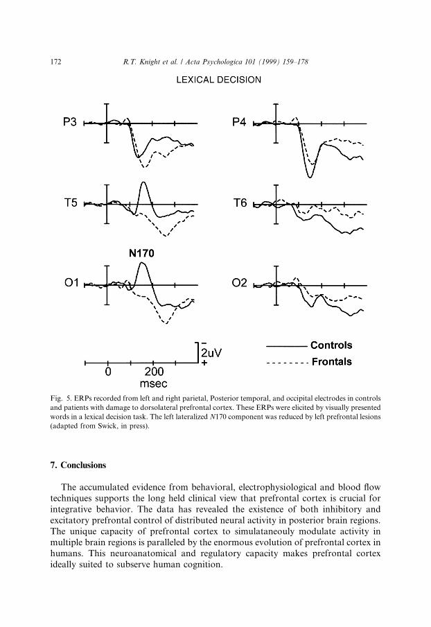
N170 reductions in frontal patients were also observed in tasks using verbalstimuli (Swick & Knight, 1996, in press; Swick, in press). In two di�erent experi-ments, subjects read centrally presented words and pronounceable nonwords andperformed lexical decision or recognition memory tasks. In controls, both stimulustypes elicited an N170 that was maximal at posterior temporal and occipital elec-trodes. In controls, N170 was signi®cantly larger over the left hemisphere in bothtasks, similar to previous studies with words (Neville, Kutas, Chesney & Schmidt,1986; Curran, Tucker, Kutas & Posner, 1993). N170 amplitude over extrastriaterecording sites was markedly reduced in frontal patients ipsilateral to damage, butpeak latency was una�ected (see Fig. 5). These visual experiments indicate thatdorsolateral prefrontal cortex provides an ipsilateral facilitory input to neural pro-cessing in extrastriate cortex, enhancing neural activity within 120 ms post-stimulus(Raines, Azaad & Miller, 1998). Further support for prefrontal modulation of vi-sual processing in extrastriate areas during sustained attention and spatial memoryperformance comes from blood ¯ow data in humans (Roland, 1982), networks an-alyses of PET and fMRI results (McIntosh, Grady, Ungerleider, Haxby, Rapoport& Horwitz, 1994; Buchtel & Friston, 1997), and single unit and lesion datain monkeys (Fuster, 1985; Funahashi et al., 1993). Projections from prefrontal areas45 and 8 to higher order visual association areas TE and TEO in the inferior tem-poral cortex have been demonstrated in monkeys (Webster, Bachevalier & Ungerl-eider, 1994). This anatomical connection provides a pathway by which prefrontalcortex could exert a facilitory in¯uence on activity in posterior visual associationcortex.
Similar e�ects of an ipsilateral reduction of neural activity in auditory associationcortex have been observed after prefrontal damage (Knight, 1997; Swick & Knight,1996; Swick, in press). Both the S1 and S2 stimuli generate a prominent N100 ERPresponse which measures neural activity in auditory association cortex (Woods,1990). Prefrontal lesions markedly reduced the N100 component generated to boththe S1 and S2 stimuli throughout the hemisphere ipsilateral to damage (Fig. 4c,d;Chao & Knight, 1998; Knight, 1997). Additional intrahemispheric reductions oflonger latency attention-related prefrontal activity were also observed. There are welldescribed prefrontal projections to auditory cortex in the superior temporal planewhich may subserve this excitatory input (Alexander et al., 1976). Together, these®ndings provide evidence that dorsolateral prefrontal cortex is crucial for inhibitingdistracting information as well as maintaining distributed intrahemispheric neuralactivity during auditory working memory. This result is also in accord with ®ndingsthat patients with prefrontal lesions are impaired in their ability to focus attention ontask-relevant stimuli (Fuster, 1989; Knight et al., 1981; Damasio, 1985; Woods &Knight, 1986).
170 R.T. Knight et al. / Acta Psychologica 101 (1999) 159±178
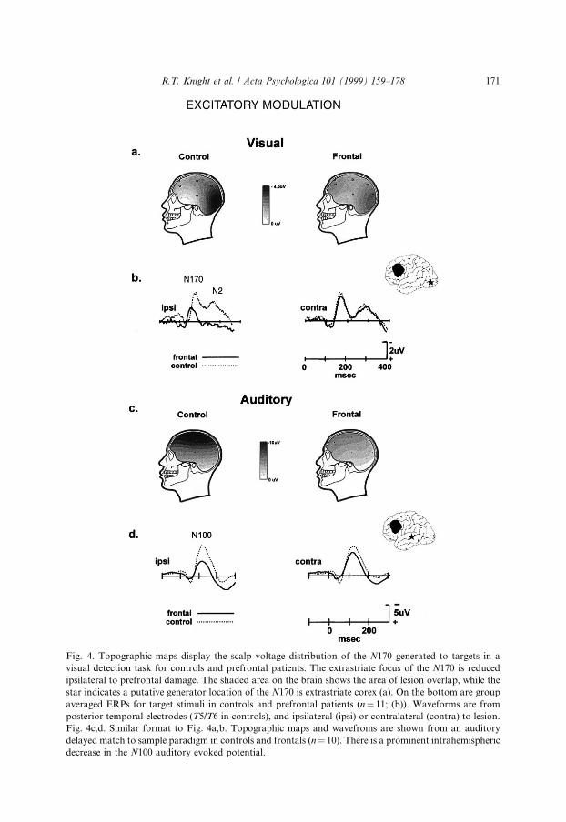
Fig. 4. Topographic maps display the scalp voltage distribution of the N170 generated to targets in a
visual detection task for controls and prefrontal patients. The extrastriate focus of the N170 is reduced
ipsilateral to prefrontal damage. The shaded area on the brain shows the area of lesion overlap, while the
star indicates a putative generator location of the N170 is extrastriate corex (a). On the bottom are group
averaged ERPs for target stimuli in controls and prefrontal patients (n� 11; (b)). Waveforms are from
posterior temporal electrodes (T5/T6 in controls), and ipsilateral (ipsi) or contralateral (contra) to lesion.
Fig. 4c,d. Similar format to Fig. 4a,b. Topographic maps and wavefroms are shown from an auditory
delayed match to sample paradigm in controls and frontals (n� 10). There is a prominent intrahemispheric
decrease in the N100 auditory evoked potential.
R.T. Knight et al. / Acta Psychologica 101 (1999) 159±178 171
7. Conclusions
The accumulated evidence from behavioral, electrophysiological and blood ¯owtechniques supports the long held clinical view that prefrontal cortex is crucial forintegrative behavior. The data has revealed the existence of both inhibitory andexcitatory prefrontal control of distributed neural activity in posterior brain regions.The unique capacity of prefrontal cortex to simulataneouly modulate activity inmultiple brain regions is paralleled by the enormous evolution of prefrontal cortex inhumans. This neuroanatomical and regulatory capacity makes prefrontal cortexideally suited to subserve human cognition.
Fig. 5. ERPs recorded from left and right parietal, Posterior temporal, and occipital electrodes in controls
and patients with damage to dorsolateral prefrontal cortex. These ERPs were elicited by visually presented
words in a lexical decision task. The left lateralized N170 component was reduced by left prefrontal lesions
(adapted from Swick, in press).
172 R.T. Knight et al. / Acta Psychologica 101 (1999) 159±178
Acknowledgements
This work was supported by the National Institute of Neurological Disorders andStroke grants NS21135 and PO17778 to RTK, the Veterans Administration MedicalResearch Service and Medical Research Council of Canada (to WRS). Specialthanks to Clay Clayworth for technical assistance in all phases of work.
References
Alexander, G. E., Newman, J. D., & Symmes, D. (1976). Convergence of prefrontal and acoustic inputs
upon neurons in the superior temporal gyrus of the awake squirrel monkey. Brain Research, 116, 334±
338.
Akbarian, S., Huntsman, M. M., Kim, J. J., Tafazolli, A., Potkin, S. G., Bunney, W. E., & Jones, E. G.
(1995). GABAa receptor subunit gene expression in human prefrontal cortex: comparison of controls
and schizophrenics. Cerebral Cortex, 5, 550±560.
Akbarian, S., Kim, J. J., Potkin, S. G., Hetrick, W. P., Bunney, W. E., & Jones, E. G. (1996).
Maldistribution of interstitial neurons in prefrontal white matter of the brains of schizophrenic
patients. Archives of General Psychiatry, 53, 425±436.
Apps, R., Atkins, M. J., & Garwicz, M. (1997). Gating of cutaneous input to cerebellar climbing ®bres
during a reaching task in the cat. Journal of Physiology, 502, 203±214.
Baddeley, A. (1992a). Working memory. Science, 255, 556±560.
Baddeley, A. (1992b). Working memory; the interface between memory and cognition. Journal of
Cognitive Neuroscience, 4, 281±288.
Bartus, R. T., & Levere, T. E. (1977). Frontal decortication in Rhesus monkeys. A test of the interference
hypothesis. Brain Research, 119, 233±248.
Broadbent, D. E. (1958). Perception and communication. London: Pergamon Press.
Brodmann, K. (1909). Vergleichende lokalisationlehre der grosshirnrinde in ihren prinzipoen dargestellt auf
grund des zellenbaues (p. 324). Leipzig: JA Barth.
Brutkowski, S. (1965). Functions of prefrontal cortex in animals. Physiological Reviews, 45, 721±746.
Boutros, N. N., Zouridakis, G., & Overall, J. (1991a). Replication and extension of P50 ®ndings in
schizoprenia. Clinical Electroencephalography, 22 (1), 40±45.
Boutros, N. N., Overall, J., & Zouridakis, G. (1991b). Test-retest reliability of the mid-latency auditiry
evoked response. Psychiatry Research, 39, 181±192.
Buchtel, C., & Friston, K. J. (1997). Modulation of connectivity in visual pathways by attention: cortical
interations evaluated with structures equation modelling and fMRI. Cerebral Cortex, 7, 768±778.
Chao, L. L., & Knight, R. T. (1995). Human prefrontal lesions increase distractibility to irrelevant sensory
inputs. Neuroreport, 6, 1605±1610.
Chao, L. L., & Knight, R. T. (1998). Contribution of human prefrontal cortex to delay performance. J.
Cognitive Neuroscience, 10 (2), 167±177.
Chapin, J. K., & Woodward, D. J. (1981). Modulation of sensory responsiveness of single somatosensory
cortical cells during movement and arousal behaviors. Experimental Neurology, 72, 164±178.
Cheng, J., Brooke, J. D., Misiaszek, J. E., & Staines, W. R. (1995). The relationship between the
kinematics of passive movement, the stretch of extensor muscles of the leg and the change induced in
the gain of the soleus H re¯ex in humans. Brain Research, 672, 89±96.
Chudler, E. H., & Dong, W. K. (1995). The role of the basal ganglia in nociception and pain. Pain, 60, 3±
38.
Cohen, J. D., Braver, S. B., & OÕReilly, R. C. (1996). A computational approach to prefrontal cortex,
cognitive control and schizophrenia: recent developments and current challenges. Philosophical
Transactions of the Royal Society of London, 351, 1515±1527.
Curran, T., Tucker, D. M., Kutas, M., & Posner, M. I. (1993). Topography of the N400: brain electrical
activity re¯ecting semantic expectancy. Electroencephalography and clinical Neurophysiology, 88, 188±
209.
R.T. Knight et al. / Acta Psychologica 101 (1999) 159±178 173
Damasio, A. S. (1985). The frontal lobes. In K. M. Heilman, E. Valenstein, Clinical neuropsychology (2nd
ed., pp. 339±374). New York: Oxford University Press.
Desmedt, J. E., Hut, N. T., & Bourguet, M. (1983). The cognitive P40, N60 and P100 components of
somatosensory evoked potentials and the earliest signs of sensory processing in man. Electroencephalo-
graphy and clinical Neurophysiology, 56, 272±282.
Edinger, H. M., Siegel, A., & Troiano, R. (1975). E�ect of stimulation of prefrontal cortex and amygdala
diencephalic neurons. Brain Research, 97, 17±31.
Freedman, R., Adler, L. E., Waldo, M. C., Pachtman, E., & Franks, R. D. (1983). Neurophysiological
evidence for a defect in inhibitory pathways in schizophrenia: comparison of medicated and drug-free
patients. Biological Psychiatry, 18, 537±551.
Freedman, R., Coon, H., Myles-Worsley, M., Orr-Urtreger, A., Olincy, A., Davis, A., Polymeropoulos,
M., Holik, J., Hopkins, J., Rosenthal, J., Waldo, M. C., Reimherr, F., Wender, P., Yaw, J., Young, D.
A., Breese, C. R., Adams, C., Parrerson, D., Adler, L. E., Kruglyak, L., Leonard, S., & Byerly, W.
(1997). Linkage of a neurophysiological de®cit in schizophrenia to a chromosome 15 locus. Proceedings
of the National Academy of Sciences, 94, 587±592.
Friedman, H. R., & Goldman-Rakic, P. S. (1994). Coactivation of prefrontal and inferior parietal cortex
in working memory tasks revealed by 2DG functional mapping in the Rhesus monkey. Neuroscience,
14, 2775±2788.
Funahashi, S., Burce, C. J., & Goldman-Rakic, P. S. (1993). Dorsolateral prefrontal lesions and
oculomotor delayed-response performance: Evidence for mnemonic scotomas. Journal of Neurosci-
ence, 13, 1479±1497.
Fuster, J. M. (1985). The prefrontal cortex, mediator of cross-temporal contingencies. Human
Neurobiology, 4, 169±179.
Fuster, J. M. (1989). The prefrontal cortex: anatomy, physiology, and neuropsychology of the frontal lobe
(2nd ed.). New York: Raven Press.
Galambos, R. (1956). Suppression of auditory nerve activity by stimulation of e�erent ®bers to the
cochlea. Journal of Neurophysiology, 19, 424±437.
Goldman-Rakic, P. S., Selemon, L. D., & Schwartz, M. L. (1984). Dual pathways connecting the
dorsolateral prefrontal cortex with the hippocampal formation and parahippocampal cortex in the
Rhesus monkey. Neuroscience, 12, 719±743.
Goldman-Rakic, P. S. (1987). Circuitry of primate prefrontal cortex and regulation of behavior by
representational memory. In F. Plum, Handbook of physiology: the nervous system (pp. 373±417).
Baltimore: American Physiology Society Baltimore.
Gomez-Gonzalez, C. M. G., Clark, V. P., Fan, S., Luck, S. J., & Hillyard, S. A. (1994). Sources of
attention-sensitive visual event-related potentials. Brain Topography, 7, 41±51.
Gosh, S., Murray, G. M., Turman, A. B., & Rowe, M. J. (1994). Corticothalamic in¯uences on
transmission of tactile information in the ventroposterolateral thalamus of the cat: e�ect of
reversible inactivation of somatosensory cortical areas I and II. Experimental Brain Research, 100,
276±286.
Gri�th, J., Ho�er, L. E., Adler, L. E., & Zerbe, G. O. (1995). E�ects of sound intensity on a midlatency
evoked response to repeated auditory stimuli in schizophrenics and normal controls. Psychophysiology,
32, 460±466.
Guillery, R. W., Feig, S. L., & Lozsade, D. A. (1998). Paying attention to the thalamic reticular nucleus.
Trends in Neuroscience, 21, 28±32.
Guitton, D., Buchtel, H. A., & Douglas, R.M. (1985). Frontal lobe lesions in man cause di�culties in
suppressing re¯exive glances and in generating goal-directed saccades. Experimental Brain Research,
58, 455±472.
Hansen, J.C., & Hillyard, S. A. (1980). Endogenous brain potentials associated with selective auditory
attention. Electroencephalogr Clinical Neurophysiology, 49, 277±290.
Hier, D. B., Mondlock, J., & Caplan, L. R. (1983). Recovery of behavioral abnormalities after right
hemisphere stroke. Neurology, 33, 345±350.
Hillyard, S. A., Hink, R. F., Schwent, U. L., & Picton, T. W. (1973). Electrical signs of selective attention
in the human brain. Science, 182, 177±180.
174 R.T. Knight et al. / Acta Psychologica 101 (1999) 159±178
Hillyard, S. A., & Picton, T. W. (1987). Electrophysiology of cognition. In F. Plum, Handbook of
physiology: the nervous system (pp. 519±584). Baltimore: American Physiology Society.
Jacobsen, C. F. (1935). Functions of frontal association areas in primates. XXX Archives of Neurology and
Psychiatry, 33, 58±569.
Janowsky, J. S., Shimamura, A. P., Kritchevsky, M., & Squire, L. R. (1989a). Cognitive impairment
following frontal lobe damage and its relevance to human amnesia. Behavioral Neuroscience, 103, 548±
560.
Janowsky, J. S., Shimamura, A. P., & Squire, L. R. (1989b). Source memory impairment in patients with
frontal lobe lesions. Neuropsychologia, 27, 1043±1056.
Jonides, J., Smith, E. E., Koeppe, R. A., Mioshima, S., & Mintun, M. A. (1993). Spatial working memory
in humans as revealed by PET. Nature, 363, 623±625.
Kahneman, D., & Treisman, A. (1984). Changing views of attention of automaticity. In R. Parasuraman
& R. Davies, Varieties of attention (pp. 29±61). San Diego: Academic Press.
Kathman, N., & Engel, R. R. (1990). Sensory gating in normals and schizophrenics: a failure to ®nd strong
P50 suppression in normals. Biological Psychiatry, 27, 1216±1226.
Kertesz, A., & Dobrolowski, S. (1981). Right-hemisphere de®cits, lesion size and location. Journal of
Clinical Neurophysiology, 3, 283±299.
Knight, R. T., Hillyard, S. A., Woods, D. L., & Neville, H. J. (1981). The e�ects of frontal cortex lesions
on event-related potentials during auditory selective attention. Electroencephalography and Clinical
Neurophysiology, 52, 571±582.
Knight, R. T. (1984). Decreased response to novel stimuli after prefrontal lesions in man.
Electroencephalography and clinical Neurophysiology, 59, 9±20.
Knight, R. T., Scabini, D., & Woods, D. L. (1989a). Prefrontal Cortex gating of auditory transmission in
humans. Brain Research, 504, 338±342.
Knight, R. T., Scabini, D., Woods, D. L., & Clayworth, C. C., (1989b). Contribution of the temporal-
parietal junction to the auditory P3. Brain Research, 502, 109±116.
Knight, R. T. (1991). Evoked potential studies of attention capacity in human frontal lobe lesions. In H.
Levin, H. Eisenberg & F. Benton, Frontal lobe function and dysfunction (pp. 139±153). New York:
Oxford University Press.
Knight, R. T., (1994). Attention regulation and human prefrontal cortex. In A. M. Thierry, J. Glowinski,
P. Goldman-Rakic & Y. Christen (pp. 160±173). Motor and cognitive functions of the prefrontal cortex.
Research and Perspectives in Neurosciences.
Knight, R. T., & Grabowecky, M. (1995). Escape from linear time: prefrontal cortex and conscious
experience. In M. Gazzaniga, The cognitive neurosciences (pp. 1357±1371). Cambridge, MA: MIT
Press.
Knight, R. T. (1996). Contribution of human hippocampal region to novelty detection. Nature, 383, 256±
259.
Knight, R. T. (1997). Distributed cortical network for visual stimulus detection. Journal of Cognitive
Neuroscience, 9, 75±91.
Knight, R. T., Finkbeiner, A., & Lawler, R. (in preparation). Prefrontal cortex dysfunction contributes to
the schizophrenic P50 gating de®cit.
Kraus, N., Ozdamar, O., & Stein, L. (1982). Auditory middle latency responses (MLRs) in patients with
cortical lesions. Electroencephalography and Clinical Neurophysiology, 54, 275±287.
Leuders, H., Leser, R. P., Hard, J., Dinner, D. S., & Klem, G. (1983). Cortical somatosensory evoked
potentials in response to hand stimulation. Journal of Neurosurgery, 58, 885±894.
Lhermitte, F., Pillon, B., & Serdaru, M. (1986). Human anatomy and the frontal lobes. Part I:
Imitation and utilization behavior: A neuropsychological study of 75 patients. Annals of Neurology, 19,
326±334.
Lhermitte, F. (1986). Human autonomy and the frontal lobes. Part II: patient behavior in complex and
social situations: the ``environmental dependency syndrome''. Annals of Neurology, 19, 335±343.
Luck, S. J., Heinze, H. J., Mangun, G. R., & Hillyard, S. A. (1990). Visual event-related potentials index
focussed attention within bilateral stimulus arrays. II. Functional dissociation of P1 and N1
components. Electroencephalography and Clinical Neurophysiology, 75, 528±542.
R.T. Knight et al. / Acta Psychologica 101 (1999) 159±178 175
MacKay, W. A., & Crammond, D. J. (1989). Cortical modi®cation of sensorimotor linkages in relation to
intended action. In W. A. Hershberger Volitional action (pp. 169±193). Elsevier: New York.
Malmo, R. R. (1942). Interference factors in delayed response in monkeys after removal of frontal lobes. J.
Neurophysiology, 5, 295±308.
Mangun, G. R., & Hillyard, S. A. (1988). Spatial gradients of visual attention: behavioral and
electrophysiological evidence. Electroencephalography and clinical Neurophysiology, 70, 417±428.
Mangun, G. R. (1995). Neural mechanisms of visual selective attention. Psychophysiology, 32, 4±18.
McCallum, W. C. Curry, S. H., Cooper, R., Pocock, P. V., & Papakostopoulos, D. (1983). Brain event-
related potentials as indicators of early selective processes in auditory target localization.
Psychophysiology, 20, 1±17.
McGhie, A., & Chapman, J. (1961). Disorders of attention and perception in early schizophrenia. British
Journal of Medicine Psychology, 34, 103±116.
McIntosh. A. R., Grady, C. L., Ungerleider, L. G., Haxby, J. V., Rapoport, S. I., & Horwitz, B. (1994).
Network analysis of cortical visual pathways mapped with PET. J. Neuroscience, 14, 655±666.
Mesulam, M. M. (1981). A cortical network for directed attention and unilateral neglect. Annals of
Neurology, 10, 309±325.
Neville, H., Kutas, M., Chesney, G., & Schmidt, A. (1986). Event-related brain potentials during initial
encoding and recognition memory of congruous and incongruous words. Journal of Memory and
Language, 25, 75±92.
Pandya, D. N., & Barnes, C. L. (1987). Architecture and connections of the frontal lobe. In E. Perecman
The frontal lobes revisited (pp. 41±72). New York: IRBN.
Petrides, M., Alivasatos, B., Meyer, E., & Evans, A. C. (1993a). Dissociation of human mid-dorsolateral
from posterior dorsolateral frontal cortex in memory processing. Proceedings of the National Academy
of Science, 90, 873±877.
Petrides, M., Alivisatos, B., Meyer, E., & Evans, A. C. (1993b). Functional activation of the human
prefrontal cortex during the performance of verbal working memory tasks. Proceedings of the National
Academy of Science, 90, 878±882.
Pierrot-Deseillingny, C. H., Rivaud, S., Gaymard, B., & Agid, Y. (1991). Cortical control of re¯exive
visually-guided saccades. Brain, 114, 1473±1485.
Raines, G., Azaad, W. F., & Miller, E. K. (1998). Selective representation of relevant information in the
primate prefrontal cortex. Natwe, 393, 577±579.
Rajkowska, G., & Goldman-Rakic, P. S. (1995a). Cytoarchitechtonic de®nition of prefrontal areas in the
normal human cortex: I. Remapping of areas 9 and 46 using quantitative criteria. Cerebral Cortex, 5,
307±322.
Rajkowska, G., & Goldman-Rakic, P. S., (1995b). Cytoarchitechtonic de®nition of prefrontal areas in the
normal human cortex: II. Variability in locations of areas 9 and 46 and relationship to the Talairach
coordinate system. Cerebral Cortex, 5, 323±337.
Richer, F., Decary, A., Lapierre, M. F., Rouleau, I., Bouvier, G., & Saint-Hilaire, J. M. (1993), Target
detection de®cits in frontal lobectomy. Brain and Cognition, 21, 203±211.
Roland, P.E. (1982). Cortical regulation of selective attention in man. A regional cerebral blood ¯ow
study. Journal of Neurophysiology, 48, 1059±1078.
Shimamura, A. P., Janowsky, J. S., & Squire, L. R. (1990). Memory for the temporal order of events in
patients with frontal lobe lesions and amnesic patients. Neuropsychologia, 28, 803±814.
Shimamura, A. P. (1995a). Memory and the frontal lobe. In M. Gazzaniga, The cognitive neurosciences
(pp. 803±813). Boston: MIT Press.
Shimamura, A. P., Jurica, P. J., Mangels, J. A., Gershberg, F. B., & Knight, R. T. (1995b). Susceptibility
to memory interference e�ects following frontal lobe damage: ®ndings from tests of paired-associated
learning. Journal of Cognitive Neuroscience, 7, 144±152.
Shin, H. -C., & Chapin, J. K. (1990a). Modulation of a�erent transmission to single neurons in the
ventroposterior thalamus during movement in rats. Neuroscience Letters, 108, 116±120.
Shin, H. -C., & Chapin, J. K. (1990b). Movement induced modulation of a�erent transmission to single
neurons in the ventroposterior thalamus and somatosensory cortex in rat. Experimental Brain
Research, 81, 515±522.
176 R.T. Knight et al. / Acta Psychologica 101 (1999) 159±178
Skinner, J. E., & Yingling, C. D. (1977). Central gating mechanisms that regulate event-related potentials
and behavior. Progress in Clinical Neurophysiology, 1, 30±69.
Smith, D. A., Boutros, N. A., & Schwarzkopf, S. B. (1994). Reliability of P50 auditory event-related
potential indices of sensory gating. Psychophysiology, 31, 495±502.
Stein, S., & Volpe, B. T. (1983). Classic parietal neglect syndrome after subcortical right frontal lobe
infarction. Neurology, 33, 797±799.
Stuss, D. T., & Benson, F. (1984). Neuropsychological studies of the frontal lobes. Psychological Bulletin,
95, 3±28.
Stuss, D. T., Benson, D. F. (1986). The frontal lobes. New York: Raven Press.
Sutherling, W. W., Crandall, P. H., Darcey, T. M., Becker, D. P., Levesque, M. F., & Barth, D. S. (1988).
The magnetic and electric ®elds agree with intracranial localizations of somatosensory cortex.
Neurology, 38, 1705±1714.
Swick, D., & Knight, R. T. (1996). Dorsolateral prefrontal cortex modulates visual processing in
extrastriate cortex. Society for Neuroscience, Abstracts, 22, 1107.
Swick, D., & Knight, R. T., Lesion (in press). studies of prefrontal cortex modulates visual processing in
extrastriate cortex. In R. Parasuraman, The attentive brain .
Swick, D., (in press). E�ects of prefrontal lesions on lexical processing and repetition priming: and ERP
study. Cognitive Brain Research.
Treisman, A. M. (1960). Contextual cues in selective listening. Quarterly Journal of Experimental
Psychology, 12, 242±248.
Tsumoto, T., Nakamura, S., & Iwama, K. (1975). Pyramidal tract control over cutaneous and kinesthetic
sensory transmission in the cat thalamus. Experimental Brain Research, 22, 281±294.
Venables, P. (1964). Input dysfunction in schizophrenia. In B. A. Maher, Progess in experimental
personality research (pp. 1±47). Orlando: Academic Press.
Vendrell, P., Junque C., Pujol, J., Jurado, M. A., Molet, J., & Grafman, J. (1995). The role of prefrontal
regions in the Stroop task. Neuropsychologia, 33, 341±352.
Wada, J.A., Clarke, R., & Hamm, A. (1975). Cerebral hemispheric asymmetry in humans. Archives of
Neurology, 32, 239±246.
Webster, M. J., Bachevalier, J., & Ungerleider, L. G. (1994). Connections of inferior temporal areas TEO
and TE with parietal and frontal cortex in macaque monkeys. Cerebral Cortex, 5, 470±483.
Weinberger, D.R., Luchins, D.J., Morisha, J., & Wyatt, R. J. (1982). Asymmetric volumes of the right and
the left frontal and occipital regions of the human brain. Annals of Neurology, 11, 97±100.
Weinberger, D. R., Berman, K. F., & Zec, R. F. (1986). Physiological dysfunction of dorsolateral
prefrontal cortex in schizophrenia, I: regional cerebral blood ¯ow evidence. Archives of General
Psychiatry, 43, 114±124.
Weinberger, D. R., Berman, K. F., Suddath, R., & Torrey, E. F. (1992). Evidence of dysfunction of a
prefrontal-limbic network in schizophrenia: a magnetic resonance imaging and regional blood ¯ow
study of discordant monozygotic twins. American Journal of Psychiatry, 149, 890±897.
Woldor�, M. G., & Hillyard, S.A. (1991). Modulation of early auditory processing during selec-
tive listening to rapidly presented tones. Electroencephalograph of Clinical Neurophysiology, 79, 170±
191.
Wood, C. C., Spencer, D. D., Allison, T., McCarthy, G., Williamson, P. D., & Go�, W. B. (1988).
Localization of human sensorimotor cortex during surgery by cortical surface recording of
somatosensory evoked potentials. Journal of Neurosurgery, 68, 99±111.
Woods, D. L., & Hillyard, S. A. (1978). Attention at the cocktail party: Brainstem evoked responses reveal
no peripheral gating. In D. A. Otto, Multidisciplinary perspectives in event-related brain potential
research (pp. 230±233). Washington, DC, US Government Printing O�ce.
Woods, D. L. (1990). The physiological basis of selective attention: implications of event-related potential
studies. In J. Rohrbaugh, J. R. Johnson & R. Parasurman, Event-related brain potentials (pp. 178±210).
New York: Oxford University Press.
Woods, D. L., & Knight, R. T. (1986). Electrophysiological evidence of increased distractibility after
dorsolateral prefrontal lesions. Neurology, 36, 212±216.
R.T. Knight et al. / Acta Psychologica 101 (1999) 159±178 177

Woods, D. L, Knight, R. T., & Scabini, D. (1993). Anatomical substrates of auditory selective attention:
behavioral and electrophysiological e�ects of temporal and parietal lesions. Cognitive Brain Research,
1, 227±240.
Yamaguchi, S., & Knight, R. T. (1990). Gating of somatosensory input by human prefrontal cortex. Brain
Research, 521, 281±288.
Yamaguchi, S., & Knight, R. T. (1991). Anterior and posterior association cortex contributions to the
somatosensory P300. Journal of Neuroscience, 11, 2039±2054.
Yamamoto, T., Samejima, A., & Oka, H. (1988). Short latency activation of local circuit neurons in the cat
somatosensory cortex. Brain Research, 461, 199±203.
Yingling, C. D, & Skinner, J. E. (1977). Gating of thalamic input to cerebral cortex by nucleus reticularis
thalami. Progress Clinical Neurophysiology, 1, 70±96.
178 R.T. Knight et al. / Acta Psychologica 101 (1999) 159±178