poster session iidecember 6, 2016 - nature
TRANSCRIPT
Poster Session IIDecember 6, 2016
Sponsorship Statement: Publication of this supplement issponsored by the ACNP.Presenter disclosures are found within the abstracts.Disclosure Format: Company Name (Financial Contributor),Type of Financial Disclosure, Funding Received for Self orSpouse. Asterisks in the author lists indicate presenter of theabstract at the annual meeting.
T1. Brain Activation in Mid-Insula Dynamically TracksCardiorespiratory Interoception
Sahib Khalsa*, Mahlega Hassanpour, Qingfei Luo,Justin Feinstein, W Kyle Simmons, Jerzy Bodurka,Martin Paulus
Laureate Institute for Brain Research, Tulsa, Oklahoma,United States
Background: Interoceptive sensations, such as palpitationsand dyspnea, are fundamental symptoms of panic anxiety.While leading theories emphasize key roles for the insularcortex in the central representation of interoceptive sensa-tions, it is unclear whether this region responds dynamicallyto changes in cardiovascular state. To modulate cardior-espiratory afferent processing in healthy individuals werecently used bolus infusions of isoproterenol, a rapidlyacting peripheral beta-adrenergic agonist similar to adrena-line. We observed dose-related increases in BOLD activationof the right mid dorsal insula (Hassanpour et al, in press). Inthe current study, we measured arterial spin labeling (ASL)during stimulation with isoproterenol. We selected ASL forthis study because it is more quantitative and localized to theparenchyma than BOLD (Kim & Ogawa. 2012), and iscommonly used in pharmacological studies (Wang et al,2011). We hypothesized that the insular cortex would showincreased ASL activation during peripheral adrenergicstimulation with isoproterenol.Methods: 23 healthy participants (11 female, mean age: 26.3+/-6.9 years) underwent fMRI scanning during bolusintravenous administrations of isoproterenol and saline.Infusions contained either saline or isoproterenol (1 or 2micrograms(mcg)). Each dose was repeated twice (sixinfusion scans). Infusion administration was double blinded,delivered 60 seconds into the scan, and infusion order wasrandomized across participants. During each infusionparticipants continuously rated their experience of cardior-espiratory sensation intensity by rotating a dial. Anatomicaland functional MRI data were acquired using a 3 Tesla GEMR750 MRI scanner with an 8-channel receive-only headcoil. Perfusion images were acquired using a pseudocontinuous ASL (pCASL) pulse sequence with a single- shotaccelerated gradient echo EPI readout (SENSE = 2, FOV/slice 220/5mm, matrix 64 × 64, 24 slices, TR/TE = 4000-/13ms, labeling duration/post-labeling delay= 1800/1500ms). Pulse oximetry and respiration waveforms were
concurrently recorded, and RETROICOR (Glover et al,2000) was applied separately on tag and control pCASLimages to reduce physiological noise artifacts due torespiratory and cardiac pulsations. Perfusion images werecalculated using pair-wise subtraction between control andlabel images, and cerebral blood flow (CBF) was quantifiedusing a general kinetic model (Buxton et al, 1998). CBF datawere normalized to MNI152 atlas using a linear affinetransformation. A voxelwise whole brain analysis was used tomap the brain’s response to each infusion. Response mapswere generated using a block averaging method, bysubtracting brain activation during each baseline period(0-60 seconds) from the corresponding peak response period(80-140 seconds). Group-level statistical maps were gener-ated using a random effects analysis.Results: As expected, isoproterenol infusions elicited dose-dependent increases in cardiorespiratory sensation(po0.001), with 100% of participants endorsing increasedpalpitations and dyspnea at the 2 mcg dose. Cardiorespira-tory intensity ratings with the dial correlated significantlywith observed heart rate changes during the 1 mcg(R2= 0.25, p= 0.013) and 2 mcg dose (R2= 0.20, p= 0.03).A voxelwise whole brain analysis of the saline and 1 mcginfusions did not reveal any significant clusters of activationin the insula or other somatosensory brain regions. Duringthe 2 mcg infusion, a significant cluster of activation wasobserved in the mid dorsal insula on the right (po0.005,uncorrected). A time series analysis of the right insularevealed an increase in CBF corresponding to the time courseof both the objective heart rate changes and the subjectivedial ratings.Conclusions: These findings, which represent a replication ofour previous BOLD imaging study, confirm that the insularcortex, particularly the right mid dorsal insula, is a keyregion that responds dynamically to changes in cardior-espiratory interoceptive state. Given the prominent role ofinteroceptive sensations in anxiety and eating disorders,future studies examining whether abnormal insula activityexplains interoceptive processing deficits in these disordersare warranted.Keywords: Interoception, Palpitation, Dyspnea, Anxiety andInsula.Disclosure: Nothing to disclose.
T2. Functional and Anatomical Organization ofAmygdala Neural Populations Encoding EmotionalValence
Anna Beyeler*, Margaux Silvestre, Praneeth Namburi,Gwendolyn Calhoon, Amy Sutton, Gordon Glober,Craig Wildes, Kay Tye
Massachusetts Institute of Technology, Cambridge,Massachusetts, United States
Background: Inappropriate assignment of emotional valenceand dysfunctions of the circuits that govern valenceprocessing are thought to underlie many psychiatric diseases
Neuropsychopharmacology (2016) 41, S289–S454© 2016 American College of Neuropsychopharmacology. All rights reserved 0893-133X/16
www.neuropsychopharmacology.org
ACNP 55th Annual Meeting
including anxiety, depression, addiction and compulsivedisorders. From a fundamental perspective, understandinghow the brain attributes valence to contexts and elements ofour environment is one of the main challenges inneuroscience. The basolateral amygdala (BLA) is known toplay a critical role in the formation of associative memoriesof both positive and negative valence and contains popula-tions of projection neurons, including populations targetingthe nucleus accumbens (BLA-NAc), the central nucleus ofthe amygdala (BLA-CeA) and the ventral hippocampus(BLA-vHPC). We previously showed that the BLA-vHPCprojection regulates anxiety-related behaviors and thatlearning experiences of opposite valence differentiallyregulate the synaptic inputs onto BLA-NAc and BLA-CeAprojector populations. However, the topographical organiza-tion of these populations, their local connectivity and theirnaturally-occurring firing patterns during information pro-cessing are incompletely understood.Methods: To characterize the topographical organization ofBLA-NAc, BLA-CeA and BLA-vHPC projector populationswithin the BLA, we used a retrograde as well as anterogradetracing approaches. To retrogradely fill the projectors weinjected three different retrograde tracers (fast blue,choleratoxin-B (CTB) coupled to a red fluorophore (AF-555)or coupled to a far red fluorophore (AF-647)) in the threedownstream targets (NAc, CeA or vHPC). Which fluor-ophore was injected to which region was counterbalancedbetween animals. One week after injection we collected thebrains and processed them with classical histology protocols.To anterogradely label the projectors we used a dual virusstrategy. We injected a retrograde viral vector carrying thegene coding for the Cre recombinase (cre) in one down-stream target and injected a Cre-dependent vector coding foran enhanced yellow fluorescent protein (eYFP) in the BLA.Five weeks after viral expression we collected the brains andmade them optically transparent by using CLARITY beforeimaging the entire brain for quantification. To stimulate BLAprojectors, we also used a dual virus strategy. In this case, weinjected a retrograde viral vector carrying the gene coding forcre in one downstream target and injected a Cre-dependentvector coding for the light sensitive proteinchannelrhodopsin-2 (ChR2) fused to eYFP within the BLA.To identify the local connectivity of BLA-vHPC populationwith BLA-NAc and BLA-CeA population, we performedex vivo whole-cell patch-clamp recordings from visuallyidentified projectors and photo-stimulated one of the twoother projector populations expressing ChR2. Finally, toreveal the coding properties of the three populations we usedin vivo optogenetic-mediated phototagging in combinationwith large-scale electrophysiological recordings during theretrieval of positive or negative associative memories.Results: Our anatomical tracing experiments showed thatBLA-NAc projectors are denser in the medial BLA whileBLA-CeA projectors are denser in the lateral part of the BLA.The BLA-vHPC are evenly represented in the medial andlateral BLA but are denser in the dorsal amygdala (LA).Although there is heterogeneity in response profiles withineach of these projection target defined populations, we foundthat BLA subpopulations differentially encode the retrieval ofpositive or negative associative memories. Specifically, wefound that the proportion of BLA-NAc neurons excitedduring presentation of a reward predictive cue was
overrepresented relative to the entire population of BLAneurons. Contrastingly, the relative proportion of BLA-CeAneurons excited during the presentation of an aversive cuewas enhanced compared to the entire population of BLAneurons. Finally, BLA-vHPC did not preferentially code forlearned associations of positive or negative valence.Conclusions: We observed that BLA populations defined bytheir downstream target (NAc, CeA and vHPC) aretopographically organized in the amygdala and have localsynaptic interactions. During memory retrieval of emotionalassociations, we found heterogeneous responses to theconditioned stimuli within the BLA projector populations,yet we provided evidence that information regarding cues ofpositive and negative valence is routed to divergent circuits.Although the BLA-vHPC projection has been shown to drivenegative valence in innate - rather than learned - behaviors,these neurons did not preferentially code for negative valencememories, supporting a model of distinct routing for learnedand innate valence-related information. Together, thesefindings suggest that valence encoding in the BLA is at leastpartially accomplished through the divergent activity ofanatomically defined neuronal populations.Keywords: Anxiety, Valence, Amygdala, Neural Populationsand Photoidentification.Disclosure: Nothing to disclose.
T3. Rethinking Extinction: Utilizing Advances inComputational Learning Theory and Neuroscience toAugment Traditional Fear Extinction Protocols
Joseph Dunsmoor*
New York University, New York, New York, United States
Background: Once acquired, it is often necessary to update along-term memory with new information in order toregulate behaviors that are no longer relevant. Research onovercoming unwanted behavior relies extensively on theprinciples of extinction—in which omission of expectedevents diminishes learned behavior. However, as a techniqueto permanently reduce fear behaviors, extinction is unsa-tisfactory as extinguished behaviors return under a variety ofcircumstances. Moreover, research on extinction focuses onone component of aversive memories—the behavioralexpression—and it remains unclear whether extinction hasany consequence on memory for episodic details of a fearfulexperience. The studies presented here tested novel methodsfor enhancing extinction and reducing relapse in healthyadults, and, for the first time, examined whether extinctionhas any effect on episodic memory for details of a fearfulexperience.Methods: The first study (Study 1) improved upon standardmodels of extinction by replacing—rather than merelyomitting—aversive outcomes with novel outcomes. Toexamine the effects of extinction on episodic memory, thesecond study used a validated category-conditioning para-digm in which subjects view unique (non-repeating) imagesfrom two semantic categories. Images from one category arepaired with an aversive electrical shock and the othercategory is safe. Fear conditioning was identical acrossmultiple groups who then underwent different forms ofextinction: immediate extinction, delayed extinction, massive
Abstracts
S290
ACNP 55th Annual Meeting
extinction, or novelty extinction (as in Study 1), or noextinction. Subjects all returned 24-hours later for a surpriserecognition memory test for items they had seen during fear-conditioning and extinction.Results: First, the ‘novelty-facilitated extinction’ techniquesuccessfully prevented relapse of conditioned fear in humansand in rats, providing cross-species evidence that replacingexpected threats is more effective than simply removingthem. Second, we found that neither immediate, delayed, normassive extinction diminished recognition memory for itemsassociated with an aversive outcome during fear condition-ing This finding is predicted by contemporary models ofextinction and animal neuroscience research suggesting thatextinction is not a form of unlearning. However, novelty-facilitated extinction did diminish the episodic memoryenhancements for fear-conditioned stimuli.Conclusions: These findings provide novel non-pharmacological behavioral strategies for enhancing fearextinction that can be straightforwardly adapted andimplemented in clinical situations. That episodic informa-tion associated with a feared event is not altered byextinction training provides new clues as for why fearbehaviors are prone to relapse over time, as the memory ofthe original fear memory is preserved despite new safetylearning. We discuss ongoing work examining the crossoverbetween novel approaches to optimize extinction andreducing the emotional enhancement for negative memories.Keywords: Fear Conditioning, Fear extinction, MemoryConsolidation and Extinction.Disclosure: Nothing to disclose.
T4. Brain Mechanisms and Predictors of Response toProlonged Exposure Therapy in PTSD
Gregory Fonzo*, Madeleine Goodkind, DesmondOathes, Yevgeniya Zaiko, Meredith Harvey,Kathy Peng, Elizabeth Weiss, Colleen Mills-Finnerty,Allison Thompson, Sanno Zack, Steven Lindley,Bruce Arnow, Booil Jo, James Gross,Barbara Rothbaum, Amit Etkin
Stanford University, Stanford, California, United States
Background: Trauma-focused exposure therapies such asprolonged exposure (PE) are the most efficacious interven-tions for post-traumatic stress disorder (PTSD), but ourunderstanding of the neural mechanisms underlying theirefficacy is lacking. This knowledge gap is due partly to a lackof well-controlled imaging studies that report findings over arange of relevant neural processes. Moreover, no PTSDtreatment studies have utilized causal manipulations of braincircuits, such as combining transcranial magnetic stimula-tion (TMS) and functional imaging, to deepen understandingof how functional brain changes during emotion tasks reflectchanges in the causal dynamics of integrated circuits. Here,we report final analyses from a two-arm, RCT-style imagingintervention study in PTSD investigating the mechanismsunderlying efficacy of PE therapy. We focused specifically ontasks that capture constructs theoretically relevant to PTSDand exposure therapy—emotional reactivity and regulation.We investigated therapeutic effects on brain function using apatient waitlist (WL) control condition and full intent-to-
treat linear mixed models. We furthermore broughtcausality-focused interventions to bear on the findings byinterrogating the causal connections of cortical substratesconveying a beneficial response to treatment and thoseshowing treatment-related changes using single-pulse TMSand fMRI.Methods: Sixty-six individuals meeting criteria for PTSDwere randomized to 9-12 sessions of PE treatment (N= 36)or WL (N= 30) for a comparable time period. Both beforeand after treatment or WL, individuals completed a clinicalassessment and battery of three functional imaging tasksassessing the identification, processing, response, andregulation of emotional responses as well as resting statefMRI. Full intent-to-treat voxelwise linear mixed modelswere utilized to identify brain activation and connectivitypredicting a favorable therapeutic outcome to treatment aswell as that which showed differential treatment-relatedchanges not due to the passage of time or repeatedmeasurements. Single-pulse TMS with concurrent fMRIwas utilized to probe causal connections of select brain focishowing relevant treatment effects.Results: When reacting to fear cues, individuals displayinggreater anterior insula, dorsomedial, and dorsolateralprefrontal activation and less amygdala activation at baselinedisplayed a more favorable treatment response. Greaterfunctional decoupling of the right dorsolateral prefrontalcortex and the left amygdala during emotional reactivity andgreater TMS-induced right dorsolateral prefrontal causalinhibition of the left amygdala at rest also predicted morefavorable treatment response. During regulation of emo-tional conflict, individuals displaying greater ventromedialprefrontal (vmPFC)/ventral striatal activation at baseline alsodisplayed a more favorable response to treatment and betterregulation of emotional conflict. Activation during cognitivereappraisal did not predict treatment response. Regardingtreatment-related brain changes, there were no effects oftreatment on brain function during emotional reactivity orregulation of emotional conflict. However, treatment-randomized individuals displayed a selective increase in leftlateral frontopolar (LatFP) activation during cognitivereappraisal, which was associated with greater improvementin hyperarousal symptoms and psychological well-being andwas anti-correlated with changes in dorsal and ventrolateralregions typically recruited by reappraisal. LatFP changeoccurred concomitant with increased task-dependent con-nectivity with the same vmPFC/ventral striatal region thatpredicted therapeutic response during regulation of emo-tional conflict at baseline. Moreover, greater LatFP activita-tion during emotional conflict regulation at baselinepredicted a greater increase in LatFP activation duringreappraisal following treatment. Single-pulse TMS in healthyindividuals during fMRI revealed that manipulation of leftLatFp exerts a causal influence on vmPFC/ventral striatalactivation. Both the LatFP and vmPFC/ventral striatumdisplayed treatment-related increases in entropy at rest, i.e.more flexible and less predictable time courses of signalfluctuations.Conclusions: One’s capacity to benefit from prolongedexposure for PTSD is gated by the degree to which prefrontalresources can be spontaneously engaged to controlamygdala-mediated threat orienting, attune to relevantemotional information, and equilibrate visceral state to
Abstracts
S291
ACNP 55th Annual Meeting
emotional salience, but not when attempting to reducenegative emotionality. Moreover, the frontopolar cortex,implicated in gating the balance of attention between theinternal and external world, is a key locus of adaptivepsychotherapeutic change. We propose this change reflectsgreater flexibility in switching attention away from aprovocative external stimulus and towards an internal signalof equilibrium when consciously reducing negative emotion.TMS and task findings implicate the right dorsolateralprefrontal cortex and left LatFP as potential sites for causalintervention development.Keywords: Post-Traumatic Stress Disorder, BOLD Imaging,Psychotherapy, Interleaved TMS/fMRI and AnteriorPrefrontal Cortex.Disclosure: Nothing to disclose.
T5. Fatty Acid Amide Hydrolase Overexpression in theBasolateral Nucleus of the Amygdala InducesParadoxical Effects on Anxiety and Fear Memory in Rats
Maria Morena*, Kira Leitl, Asim Rashid,Sheena Josselyn, Matthew Hill
University of Calgary, Calgary, Canada
Background: A growing amount of evidence has demon-strated that elevations in the endocannabinoid anandamide(AEA) signaling within the basolateral complex of theamygdala (BLA) attenuate stress responses such as anxietyand fear expression. Specifically, inhibition of AEA hydro-lysis by the enzyme fatty acid amide hydrolase (FAAH)within the BLA has been shown to reduce anxiety,neuroendocrine responses to stress and promote fearextinction. To determine if impairments in AEA signalingwithin the BLA would produce the opposite effects, andinduce a stress-like state characterized by heightened anxietyand sustained fear, we examined the effects of overexpressionof FAAH locally within the BLA on behavioural indices ofanxiety and fear memory dynamics.Methods: Male adult Sprague Dawley rats were bilaterallyinfused in the BLA with a Herpes simplex virus type 1 vector,which preferentially infects principal neurons, containingFAAH and green fluorescent protein (HSV-FAAH-GFP) or acontrol vector containing only GFP (HSV-GFP). Rats werethen tested for biochemical measurements, anxiety or fearmemory behaviour. All experimental procedures were incompliance with protocols approved by the University ofCalgary Animal Care Committee and guidelines from theCanadian Council on Animal Care.Results: 72 h following HSV administration, a time point inwhich protein transfection is maximal, we found increasedFAAH-mediated AEA hydrolysis together with decreasedAEA levels within the BLA, confirming that the virus didsuccessfully increase FAAH expression. At this same timepoint, a separate cohort of rats was tested for anxietybehaviour in an elevated plus maze, a light/dark box and anopen field task. Quite surprisingly, we found that theoverexpression of FAAH induced consistent anxiolyticeffects in all three behavioural tasks we performed, relativeto HSV-GFP rats. An additional cohort of animals was testedfor fear memory extinction in an auditory fear conditioningparadigm. To prevent any effects of FAAH overexpression
on the initial fear memory consolidation, animals werebilaterally cannulated in the BLA, a week later were fearconditioned and 24 h after conditioning, the animals wereinjected with HSV-FAAH-GFP or its control vector. 72 hfollowing HSV administration rats were re-exposed to thetone alone for 4 consecutive days to examine fear extinctiondynamics. Unexpectedly, rats infused with the HSV-FAAH-GFP vector exhibited a dramatic reduction in fear expressionduring the extinction training and first extinction retrievalsessions when exposed to the tone, as compared to theirHSV-GFP control rats. Moreover, both HSV-GFP and HSV-FAAH-GFP animals showed reinstatement of fear memory,when given unsignaled foot shocks after the extinction hadoccurred in both groups and presented 24 h later the tonealone, thus demonstrating that the effects of the FAAHoverexpression were selective for the fear and extinctionmemory retrieval, without interfering with the animal abilityto correctly acquire the conditioned-unconditioned stimulusassociation and to express freezing behaviour.Conclusions: These findings suggest that the exact modes ofaction of AEA within the amygdala in the regulation ofemotional states and memory are still far from being clear,thus, opening the avenue to investigate new potentialmechanisms by which these processes may occur.Keywords: Endocannabinoids, Fear extinction and Anxiety.Disclosure: Nothing to disclose.
T6. Theta-Frequency Oscillatory Stimulation of vHPCInputs to the mPFC Increases Anxiety-Like Behavior
Nancy Padilla-Coreano*, Sarah Canetta,Richard Warren, Christoph Kellendonk,Joshua Gordon
Columbia University, New York, New York, United States
Background: Anxiety states are distinguished by an increasein theta-frequency (4-12 Hz) synchrony in the amygdala-hippocampal-prefrontal circuit. Recently, we demonstratedthat optogenetically inhibiting the ventral hippocampal(vHPC) input to the medial prefrontal cortex (mPFC)decreases anxiety-like behavior and theta synchrony betweenthe mPFC and vHPC, without affecting other frequencies(Padilla-Coreano et al, Neuron. 2016). These data suggest thehypothesis that theta-frequency input from the vHPC plays acausal role in anxiety-like behavior.Methods: We therefore asked whether stimulating thevHPC-mPFC at a theta frequency was sufficient to increaseavoidance behavior in the elevated plus maze, and whetherany such effects might be frequency- and/or pattern-specific.CamKIIa-hChR2-eYFP was expressed bilaterally in thevHPC and optical fibers implanted in the mPFC. In a subsetof mice, single-unit recording electrodes were also implantedin the mPFC.Results: Stimulating the vHPC-mPFC pathway with asinusoidal light pattern at 8 Hz significantly increasedavoidance behavior, while stimulating with brief pulses oflight at 8 Hz or sinusoidal light at 20 Hz had no effect. Theseexperiments demonstrate that the anxiogenic effect of vHPCterminal stimulation is frequency- (8 Hz but not 20 Hz) andpattern- (sinusoids but not pulses) specific. This result wassurprising, as pulses of light are the most common light
Abstracts
S292
ACNP 55th Annual Meeting
pattern used to stimulate neurons and terminals optogeneti-cally. To understand how pulses and sinusoidal lightmodulate mPFC neurons differentially, mPFC pyramidalneurons were recorded both in vitro and in vivo whilestimulating vHPC terminals with the same sinusoidal orpulsatile patterns. In vitro, sinusoidal stimulation increasedthe rate of spontaneous EPSCs, while pulses evoked strong,time-locked EPSCs. In vivo, sinusoidal stimulation of vHPCterminals were capable of increasing the phase-locking ofmPFC single units to the optical stimulation during anxietybehaviors.Conclusions: These results suggest that sinusoidal stimula-tion at 8 Hz increases spontaneous activity in a rhythmic,naturalistic manner that enhances theta-frequency activity inmPFC neurons as well as anxiety-related behavior. More-over, they suggest that theta-frequency components of neuralactivity play a privileged role in vHPC-mPFC communica-tion and hippocampal-dependent forms of anxiety.Keywords: Optogenetics, Hippocampus-mPFC Pathway andAnxiety Circuitry.Disclosure: Nothing to disclose.
T7. Network Mechanisms of 5Hz Repetitive TranscranialMagnetic Stimulation in Patients With ComorbidPosttraumatic Stress and Major Depressive Disorders
Noah Philip*, Jennifer Barredo, Mascha van 't Wout,Jorge Almeida, Audrey Tyrka, Lawrence Price,Linda Carpenter
Brown University, Providence, Rhode Island, UnitedStates
Background: Treatment with repetitive transcranial mag-netic stimulation (TMS) can selectively modulate pathologi-cal functional connectivity of the default mode network inpatients with major depressive disorder (MDD). Posttrau-matic stress disorder (PTSD) is often comorbid with MDD,and research indicates that TMS can also alleviate symptomsof PTSD. This is the first study to evaluate TMS-associatedchanges in connectivity in a sample of patients withcomorbid PTSD and MDD. We predicted that clinicallyefficacious TMS would be associated with reduced defaultnetwork connectivity and uncoupling of salience andmemory regions associated with PTSD.Methods: Resting state functional connectivity (RSFC) wasacquired using a 3T MRI on 32 participants from an open-label trial of 5Hz rTMS to the left dorsolateral prefrontalcortex (up to 40 daily sessions, 3-4000 pulses per session).5Hz was selected based on our prior report of efficacy for co-morbid PTSD and MDD. Imaging analyses used a prioriseeds identified from prior research in TMS and PTSD,including the subgenual anterior cingulate (sgACC), dorso-lateral prefrontal cortex (DLPFC), hippocampus (anteriorand posterior components) and the basolateral amygala.These seeds were used to compare pre- vs. post-treatmentRSFC associated with clinical changes, adjusting for age andbaseline symptom severity. To examine if changes infunctional connectivity between a priori seed regions andextracted clusters were predictive of meaningful clinicaloutcomes, the mean change in beta values across sessions foreach cluster and subject was entered into a logistic regression
of symptomatic recovery for both disorders. Model fitstatistics are reported as Nagelkerke R2. To address concernsof insufficient thresholding and validity in imaging studies, avoxel height threshold (po 0.005) was applied to all wholebrain imaging results, plus cluster-based false discovery ratethresholding (i.e., FDR-corrected cluster size of po .05).Furthermore, a leave-one-out cross-validation procedure wasimplemented as a stringent test of cluster validity. Clusterswhere the cross-validated t-value fell below the critical valuewere considered invalid and excluded from the logisticregression.Results: Twenty-five participants had usable data availableprior to and following TMS. Compared to positiveconnectivity with DMN regions prior to treatment, aftertreatment, the sgACC demonstrated anticorrelated RSFCwith a broad range of regions within the default modenetwork, dorsomedial prefrontal cortex and insula (allcorrected po .001). The logistic regression model indicatedthat changes in sgACC to default mode regions werepredictive of remission (po .001; R2 = .63). DLPFCconnectivity increased to the right prefrontal cortex (cor-rected po .001) that was more modestly predictive ofremission (p= .008, R2 = .32). Compared to positive RSFCat baseline, after TMS there was anticorrelated connectivitybetween the anterior hippocampal seed and frontal cortico-limbic regions, including the dorsal anterior cingulate cortexand putamen (corrected po .001), which was also predictiveof remission (p= .006; R2 = .52). Posterior hippocampalconnectivity with the right DLFPC was increased followingtreatment (corrected po .001); this modestly predictedremission (p= .03, R2 = .32). No statistically significantchanges in connectivity were observed from the basolateralamygdalConclusions: These results are consistent with prior imagingstudies of TMS therapy in MDD that found reduced defaultnetwork connectivity and more modest effects of TMS onexecutive network connectivity. Furthermore, these resultshighlight that TMS can uncouple hippocampal connectivityfrom salience network regions implicated in PTSD. Theseresults show that TMS can correct clinically relevant networkpathology, with results relevant to both disordersunder study.Keywords: Repetitive Transcranial Magnetic Stimulation,Resting State Functional Connectivity and PTSD Depression.Disclosure: Neuronetics: Grant support, Self.
T8. Basomedial Amygdala Mediates Top-Down Controlof Anxiety and Fear
Avishek Adhikari*, Talia Lerner, Joel Finkelstein,Sally Pak, Joshua Jennings, Thomas Davidson,Emily Ferenczi, Lisa Gunaydin, Julie Mirzabekov,Li Ye, Sung-Yon Kim, Anna Lei, Karl Deisseroth
Stanford University, Palo Alto, California, United States
Background: Anxiety-related conditions are among the mostdifficult neuropsychiatric diseases to treat pharmacologically,but respond to cognitive therapies. There has therefore beeninterest in identifying relevant top-down pathways fromcognitive control regions in medial prefrontal cortex(mPFC). Identification of such pathways could contribute
Abstracts
S293
ACNP 55th Annual Meeting
to our understanding regarding cognitive regulation of affect,and provide pathways for intervention. Prior studies suggestdorsal and ventral mPFC subregions exert opposing effectson fear, as do subregions of other structures. However,precise causal targets for top-down connections among thesediverse possibilities have not been established.Methods: We optogenetically targeted the vmPFC-amygdalaprojection in mice during learned fear conditioning andinnate anxiety behavioral assays. We also monitored theeffect of this manipulation in physiological measures of highanxiety states (respiratory rate and heart rate) through pulseoxymetry. We applied in vivo and in vitro assays offunctional connectivity to identify which amygdala nucleiare recruited by the vmPFC.Results: We show the basomedial amygdala (BMA) repre-sents the major target of ventral mPFC (vmPFC) inamygdala. Moreover, BMA neurons differentiate safe andaversive environments, and BMA activation decreases fear-related freezing and high anxiety states. Lastly, the vmPFC-BMA projection implements top-down control of anxietystate and learned freezing, both at baseline and in stress-induced anxiety.Conclusions: Our data show activity in the vmPFC-amygdala projection is sufficient and necessary to suppresshigh anxiety states, as measured by multiple behavioral andphysiological parameters. Multiple streams of evidenceincluding viral tracing, patch clamping, in vivo physiologyand immediate early gene expression indicate that theseeffects were mediated by the basomedial amygdala (BMA).Accordingly, activation of the BMA also was anxiolytic.These results establish the vmPFC-BMA as a key node thatmediates cortical-subcortical top-down anxiety suppression.Considering that abnormalities in mPFC-amygdala connec-tivity is commonly displayed in patients with diverse anxietydisorders, our results indicate that the vmPFC-BMA is anode that may be targeted to ameliorate these disorders.Keywords: Medial Prefrontal Cortex, Mood and AnxietyDisorders, Fear Conditioning and Post-Traumatic StressDisorder.Disclosure: Nothing to disclose.
T9. Group Differences and Treatment OutcomePrediction With Resting State and Task-BasedConnectivity in Pediatric Anxiety
David Pagliaccio*, Caroline Swetlitz, Lauren White,Daniel Pine
National Institute of Mental Health, Bethesda, Maryland,United States
Background: Anxiety disorders have been characterized byalterations in connectivity between the amygdala andprefrontal and other regions, both at rest and during task.Particularly, recent work has shown robust group differencesand test-retest of task-based connectivity during a dot-probeattentional task. Additionally, efforts have been made to usefunctional connectivity as a potential predictor of treatmentoutcomes in anxiety disorders. Yet, it is unclear whatrelationship may exist between resting state connectivityand connectivity modulated by task and how this related toour understandings of anxiety disorders.
Methods: The current study examines data from 71 childrenand adolescents (8-18 years old), 33 with anxiety and 38healthy volunteers. Anxious patients were scanned prior toan 8-week treatment trial. Participants completed two runsof an fMRI dot-probe task followed by a 10-minute restingstate scan. Anatomically defined amygdala seeds were usedto examine functional connectivity during the dot-probeusing generalized psychophysiological interaction and duringrest using standard processing in AFNI.Results: Preliminary analyses indicate that more negativeresting state connectivity between the right amygdala andmedial prefrontal cortex (Monte Carlo voxel-wise correctionpo.001, k423) strongly predicts greater reduction inanxious symptoms following treatment over and abovepretreatment anxiety and demographic control factors(= 0.65, t= 3.55, p= .002).Conclusions: These results suggest key relationships betweenamygdala connectivity and successful treatment outcomes.On-going analyses will examine the relationship betweenresting state and task-based connectivity, group differencesin connectivity, and further prediction of treatmentoutcomes.Keywords: Anxiety, Children and Adolescents, Connectivity,Functional MRI (fMRI) and Amygdala.Disclosure: Nothing to disclose.
T10. Glucocorticoid, Immune and Genomic Mediators ofAmygdalaactivation Following Extreme Stress/Trauma
Nikolaos Daskalakis*, Hagit Cohen, Janine Flory, IouriMakotkine, Joseph Buxbaum, Charles Marmar, OwenWolkowitz, Marti Jett, Rasha Hammamieh, Bin Zhang,Rachel Yehuda
Icahn School of Medicine at Mount Sinai, New York, NewYork, United States
Background: Delineating the molecular basis of individualdifferences in the stress response is critical to understandingthe pathophysiology of post-traumatic-stress-disorder(PTSD), with the ultimate goal of identifying biomarkers.Methods: We analyzed genome-wide expression of periph-eral blood mononuclear cells (PBMCs) and functionalneuroendocrinology and immunology plasma/serum datafrom a cross-sectional biomarker study with and withoutPTSD (n= 83/group). In addition, gene expression in bloodand brain (amygdala, hippocampus and prefrontal cortex)and neuronal morphology were analyzed from a PTSD ratmodel, in which vulnerable and resilient phenotypes areidentified according to the long-term behavioral response topredator scent stress (PSS). Analyses of the human andanimal data were performed separately, and then comparedand integrated with the use of gene co-expression networkanalyses.Results: Differentially expressed (DE) genes were identifiedin association with PTSD. These DE genes were consistentwith peripheral downregulation of glucocorticoid receptor(GR) signaling and upregulation of pro-inflammatorycytokine signaling. The gene expression differences in thesepathways were validated in cultured PBMCs incubated invarying doses of dexamethasone (DEX). These findings werealso replicated by the DE-signatures identified in rat blood
Abstracts
S294
ACNP 55th Annual Meeting
and brain, in association with exposure-related individualdifferences. Gene co-expression networks were then identi-fied in the human and rat samples. Among the mostpromising networks, there was a large (4100 genes) PTSDco-expression module showing a high level of dysregulation(high modular differential connectivity in the two groups)and high level of conservation in the blood and brain ofPTSD-like rats. Functionally, this module is enriched withthe genes related to innate immune response, and itseigengene expression associates deferentially with the PBMClysozyme inhibition by DEX and plasma cortisol decline inthe dexamethasone suppression test in the two groups.Finally, GR-agonist administration in rats shortly after PSSprevented PTSD-like phenotypes by reversing amygdalavulnerability-associated pro-inflammatory cytokine signalingand neuronal morphology patterns.Conclusions: We identified genes, pathways and gene co-expression networks in the blood of combat veterans withPTSD. GR-dependent immune pathways and networks areassociated with trauma-related individual differences inblood and amygdala, and can be the basis of treatmentfor PTSD.Keywords: PTSD, Gene Network Analysis, Glucocorticoids,Immune Mechanisms and Amygdala.Disclosure: Nothing to disclose.
T11. Exposure to Inner-City Violence Associated WithInhibition-Related Activation in the Developing Brain
Sanne van Rooij*, Jennifer Stevens, Ye Ji Kim, TimothyEly, L. Alexander Vance, Bekh Bradley, TanjaJovanovic
Emory University School of Medicine, Atlanta, Georgia,United States
Background: Exposure to trauma and stress in early life is awell-known risk factor for the development of psychiatricdisorders across the lifespan. As most studies are retro-spective, however, the developmental effects of traumaexposure during childhood are not well understood. Thisstudy investigated the effects of trauma exposure on thedeveloping brain in children growing up in neighborhoodswith high levels of inner-city violence.Methods: Mothers with a child between ages 8-14 wererecruited through the Grady Trauma Project, an ongoingstudy on the effects of trauma exposure in an at-riskpopulation. A subset of children (N= 31) from this samplecompleted an fMRI scan while they performed an emotionalGo/NoGo task. During this task, fearful and neutral faces arepresented in pseudo-randomized order. The participantswere asked to press a button for neutral faces and not pressfor fearful faces. This contrast was used to measureemotional response inhibition (NoGo Fear 4 Go Neutral).Trauma exposure was assessed with self-report measures ofviolence exposure (VEX-R). The relationship betweeninhibition-related brain activation and violence exposure(frequency) was investigated. First, exploratory whole brainanalyses were performed, and secondary small volumecorrection was performed for our regions of interest: theamygdala, the hippocampus and the rostral ACC.
Results: After quality control, data from 16 children wereavailable for preliminary whole brain analyses (po0.01).Exposure to violence correlated positively with inhibition-related activation in limbic structures (amygdala, hippocam-pus), motor control regions (basal ganglia, right inferiorfrontal gyrus), and the salience network (anterior cingulatecortex, insula). Small volume correction showed significantpositive correlations between violence exposure and the rightamygdala (FWE-corrected po0.05), the left and righthippocampus (FWE-corrected po0.05), and the rostralACC (FWE-corrected po0.05). Further analyses investigat-ing the effects of gender and age are of high interest when thesample size of this ongoing study increases.Conclusions: Although preliminary, our results providesome evidence for a relationship between trauma exposureand alterations in the neurocircuitry underlying fear andresponse inhibition in children. Higher levels of exposure toviolence were associated with more activation in theamygdala, hippocampus and rostral ACC during thecombined process of viewing fearful faces and suppressinga behavioral response. Given that this neurocircuitry hasbeen implicated in many psychiatric disorders, increasedunderstanding of the effects of trauma exposure on thedeveloping brain is essential for early detection of individualsat risk for developing psychiatric disorders.Keywords: Childhood Trauma, Emotional Response Inhibi-tion, Violence, Functional MRI (fMRI).Disclosure: Nothing to disclose.
T12. Fractional Anisotropy in Cingulum-HippocampusTracts Predicts Suicidal Ideation in Young Adolescents
Tiffany Ho*, Sarah Ordaz, Josiah Leong, Daniel Lowet,Meghan Goyer, Manpreet Singh, Ian Gotlib
Stanford University, Stanford, California, United States
Background: Suicide is the second leading cause of death inadolescents worldwide (CDC, 2014). Therefore, it isimperative that we identify individuals at risk and interveneas early as possible. Suicidal ideation is an importantprecursor to attempt; increased severity of suicidal ideationis associated with a greater likelihood of subsequent suicideattempt (Sampasa-Kanyinga et al, 2015). The peak onset ofsuicidal ideation occurs during adolescence (Nock et al,2013), which is also a period of significant neurodevelop-ment (Dahl. 2004). While investigators have identifiedsuicide-related endophenotypes—notably aberrant frontal,limbic, and cingulate gray and white matter—the neurobio-logical markers associated with early suicidal ideation are stillunknown (Cox Lippard et al, 2014). Furthermore, no studiesto date have examined whether neural structure canprospectively predict the development of suicidal ideationin adolescents. Given that adolescence is a sensitive periodfor both neurodevelopmental maturation and increasedsuicidal risk, successful identification of neural risk factorsfor suicidal ideation has important implications for thedevelopment of neural models of suicide and of novel earlyinterventions. In the present study, we collected diffusion-weighted imaging data from young adolescents at a baselinetime point, and performed tractography to identify theuncinate fasciculus (UF) and the cingulum (CG). The UF
Abstracts
S295
ACNP 55th Annual Meeting
and CG are two major white matter tracts that connectlimbic structures with frontal and with cingulate regions,respectively. We computed fractional anisotropy (FA) acrossthe entirety of these tracts as an index of tract coherence, andtested if tract coherence at baseline was associated withseverity of suicidal ideation 18 months later.Methods: Forty-six youth (14 males; mean ± SD age: 11.42± 1.03 years) recruited from the San Francisco Bay Areacompleted MRI scanning at Stanford University using a 3TDiscovery MR750 (GE Medical Systems, Milwaukee, WI)equipped with a 32-channel head coil (Nova Medical). For allparticipants, 3D T1-weighted images were acquired with aSPGR sequence (TR/TE/TI= 6.24/2.34/450 ms; flip angle=12°; 186 sagittal slices; 0.9 mm isotropic voxels) anddiffusion-weighted images were acquired with an EPIsequence (TR/TE= 8500/93.5 ms; flip angle= 90°; b= 2000mm2/s, 64 axial slices, 60 gradient directions, 6 b0 images; 2mm isotropic voxels). Diffusion-weighted data were pre-processed using standard methods and a continuous tensorfield was estimated with trilinear interpolation of the tensorelements. We used Automatic Fiber Quantification (AFQ;Yeatman et al, 2012) software to identify and segment eachparticipant’s brain into 20 white matter fiber groups,including bilateral UF and CG. Candidate fibers for UFand CG were defined using an automated 2 region-of-interest approach, and fibers were estimated using adeterministic streamlines tracking algorithm (Basser et al,2000; Mori et al, 1999). Tracts were then refined bycomparing each fiber within the tract to a probabilisticwhite matter atlas (Wakana et al, 2007). Finally, each tractwas represented as a 3D Gaussian, and outlier fibers (definedas those that exceeded a maximum Gaussian distance of 5 orthat deviated more than 4 SDs from the mean of the fibergroup center) were excluded (Yeatman et al, 2012). Tractcoherence was then calculated along the trajectory of eachtract by interpolating the FA value at 100 evenly spacednodes along each fiber, and then averaged across the entiretract. Importantly, we conducted automated tractographyfrom the AFQ software given that these methods have beenvalidated in the age range of our participants (Yeatman et al,2012; Lebe et al, 2012). Suicidal ideation and depressionsymptom severity were assessed with self-report measures(SIQ-15; Reynolds & Mazza. 1999 and CDI; Kovacs. 1992,respectively) at an 18-month follow-up session. Linearregression models were used to predict severity of suicidalideation at follow-up from FA of left and right UF and CG,controlling for baseline age, gender, and depression symp-tom severity at follow-up. Given evidence of higher rates ofsuicidal ideation in female adolescents (Nock et al, 2013), wealso modeled the interaction effect of gender and FA fromeach tract as a predictor of SIQ-15 scores.Results: Tract coherence of the left cingulum bundlesconnecting the cingulate with the hippocampus showed asignificant interaction with gender in predicting severity ofsuicidal ideation (F1,42= 3.66; po0.05). Specifically, femaleswith higher FA in the cingulum-hippocampal tracts atbaseline endorsed higher levels of suicidal ideation. Tractcoherence of the right cingulum and of bilateral UF (withand without the interaction of gender) did not significantlypredict severity of suicidal ideation (all p’s40.05).Conclusions: This study is the first to demonstrate that brainstructure prospectively predicts the severity of suicidal
ideation in young adolescents. Fractional anisotropy isthought to reflect axonal myelination, which is known toundergo major changes throughout early development. Thecingulum is one of the last white matter tracts to develop andis implicated in emotion regulation and broad cognitivefunctioning (Lebel et al, 2012). Thus, greater cingulum tractcoherence among females during early adolescence mayrepresent aberrant neurodevelopment and serve as a riskfactor for the onset of suicidal behaviors.Keywords: Adolescence, DTI, Suicide Assessment, Cingu-lum, Hippocampus.Disclosure: Nothing to disclose.
T13. Subcortical Shape Deformation AssociationsRelated to Substance Use in Youth With Perinatally-Acquired HIV
Christine Paula Lewis-de los Angeles*, Kathryn IAlpert, Paige L Williams, Kathleen Malee, YanlingHuo, John Csernansky, Ram Yogev, Russell B VanDyke, Eilzabeth R Sowell, Lei Wang, Pediatric HIV/AIDS Cohort Study (PHACS)
Northwestern University, Chicago, Illinois, United States
Background: As youth with perinatally-acquired HIV(PHIV) enter adolescence, they may engage in substanceuse. Prior work has demonstrated that worse HIV diseaseseverity is associated with substance use (Williams 2010). Itis important to understand the effects of PHIV and substanceuse on brain structure.Methods: 40 PHIV youth were recruited from one site (Ann& Robert Lurie Children’s Hospital of Chicago) of the NIHPediatric HIV/AIDS Cohort Study (PHACS). Structural T1-weighted magnetic resonance imaging was collected.FreeSurfer-Initiated Large Deformation Diffeomorphic Me-tric Mapping was used to quantify shape deformation ofsubcortical brain structures. Historic HIV disease severitymeasures (peak HIV RNA load, nadir CD4 percentage) wereobtained from medical charts. Audio Computer-AssistedSelf-Interview (ACASI) was used to obtain self-reportedsubstance use. Substance use was coded as Y/N for prior usefor alcohol, marijuana, or tobacco. Multiple linear regressionwas used to obtain clusters of significant (po0.05) relation-ships between deformation and substance use on subcorticalsurfaces. Multiple comparisons were corrected with randomfield theory. Models were adjusted for age at scan and sex,with further adjustment for HIV disease severity measures.Results: Mean age was 16.7 years. Median log peak HIVRNA load was 5.7 copies/mL (IQR: 5.2,5.9) and nadir CD4percentage was 16.5% (IQR: 8.0, 23.8). Forty-five percentreported substance use (25% tobacco, 35% alcohol, 35%marijuana); substance use was unknown for two subjectswho were excluded from further neuroimaging analyses. Inmodels including age at scan and sex, significant relation-ships between surface shape deformation and substance usewere found in the hippocampus, nucleus accumbens,caudate, and putamen. In models including log peak HIVRNA viral load and log of age at peak viral load, significantrelationships between surface shape deformation and sub-stance use were found only in the hippocampus. In modelsincluding nadir CD4 percentage and age at nadir CD4
Abstracts
S296
ACNP 55th Annual Meeting
percentage, significant relationships between surface shapedeformation and substance use were found in the hippo-campus and caudate.Conclusions: Substance use was significantly related todeformation of the brain’s subcortical structures. Futurework should be aimed at understanding the relationshipsamong different types of substance use, adherence tocombinational anti-retroviral therapy, HIV disease severity,and structural brain changes.Keywords: HIV, Human Neuroimaging, Substance Abuse,Adolescence.Disclosure: Nothing to disclose.
T14. Novel fMRI Approaches Reveal DevelopmentalChanges in Frontoamygdala Circuitry With Implicationsfor the Emergence of Psychiatric Disorders DuringDevelopment
Dylan Gee*, Leyla Calgar, Colleen Mills-Finnerty,Bonnie Goff, Laurel Gabard-Durnam, Dominic Fareri,Christina Caldera, Daniel Lumian, Jessica Flannery,Catherine Hanson, Stephen Hanson, Nim Tottenham
Yale University, New Haven, Connecticut, United States
Background: Interactions between the prefrontal cortex(PFC) and amygdala are fundamental to emotion regulation.Substantial changes in frontoamygdala circuitry take placeduring development, and this circuitry has been implicatedin myriad psychiatric disorders including anxiety, depres-sion, and schizophrenia. Given dynamic neurodevelopmen-tal changes and the emergence of many psychiatric disordersduring childhood and adolescence, delineating developmen-tal changes in prefrontal cortex and amygdala function iscritical to understanding the onset and treatment of mentalillness in youth. However, extant research has been limitedby reliance on correlational fMRI data.Methods: Here we delineate developmental changes infrontoamygdala connectivity through two novel approaches:experimental manipulation of this circuitry, and Bayesnetwork analyses. Experimental manipulation of prefrontalfunction has been shown to have sustained and lasting effectson amygdala reactivity during adulthood. The current studytook advantage of PFC-mediated effects to investigate thenature of the amygdala-PFC relationship during develop-ment. Forty healthy children, adolescents, and young adults(4-19 years) completed an fMRI task that manipulates theeffects of cognitive load on subsequent brain activation toemotional stimuli, facilitating the examination of temporaleffects of prefrontal engagement on subsequent amygdalareactivity.Results: Behavioral performance confirmed the effectivenessof the cognitive load manipulation. Specifically, participantsresponded more slowly and with lower accuracy to incon-gruent versus congruent words. High versus low cognitiveload increased dorsal ACC recruitment across development(po.01, corrected), but ACC engagement had differentialeffects on activation to emotional faces for children (reducedmedial PFC, increased amygdala) versus adolescents (in-creased medial PFC, reduced amygdala). Network analysesdemonstrated a shift in effective connectivity, such that theamygdala more strongly influenced the mPFC during
childhood, but the mPFC more strongly influenced theamygdala during adolescence.Conclusions: The cognitive load manipulation and networkanalyses reveal qualitative differences in the nature offrontoamygdala interactions during childhood and adoles-cence, with evidence for a shift from bottom-up to top-downfrontoamygdala connectivity. These findings indicate thatexperimental activation of prefrontal cortex may serve toregulate amygdala reactivity during adolescence, but notduring childhood. Our findings may serve as a foundationfor understanding aberrations in the maturation of frontoa-mygdala connections across neurodevelopmental disordersin which this circuitry has been implicated. Furthermore,this study has important implications for differential risk foranxiety and mood disorders at unique developmental periodsand may inform efforts to optimize types and timing ofinterventions to target the biological state of thedeveloping brain.Keywords: Children and Adolescents, Brain Development,Emotion Regulation, Amygdala, Prefrontal Cortex.Disclosure: Nothing to disclose.
T15. Altered Prefrontal Activation During ResponseInhibition in Women Remitted From Bulimia Nervosa:The Influence of Metabolic State
Laura Berner*, Amanda Bischoff-Grethe, Christina EWierenga, Alan N Simmons, Ursula F Bailer, Alice V Ely,Walter Kaye
University of California, San Diego, La Jolla, California,United States
Background: Bulimia nervosa (BN) is characterized byrecurrent episodes of binge eating, most saliently definedby a sense of “loss of control” over eating, and compensatorybehaviors. Between binge-purge episodes, extreme dietaryrestriction or fasting is common. Hunger normally increasesthe motivational aspects of rewards, and some evidencesuggests that strict dieting increases the likelihood of bingeeating in BN. Despite brief periods of successfully restrictedintake, compromised inhibitory control in a fasted state mayincrease binge eating risk and contribute to BN. Conversely,individuals report feeling less hungry before binges thannon-binge eating episodes, and binge eating is defined by“difficulty stopping eating once one has started.” Therefore,particularly compromised inhibitory control in a fed statecould instead drive BN symptoms. Extant behavioral andneuroimaging evidence suggests inhibitory control impair-ments and associated frontostriatal dysfunction in BN, whichmay explain “loss of control” eating and other impulsivebehaviors characteristic of the disorder; however, theextreme overcontrol this population displays betweenbinge-purge episodes remains unexplained, and no study todate has manipulated metabolic state before neurocognitivetesting or scanning. To address this gap in the literature, thecurrent study examined whether being in a fasted or fed stateabnormally impacts the function of self-regulatory controlcircuits in BN.Methods: Women remitted from BN (RBN; n= 23) andhealthy control women (CW; n= 22) of equivalent age, bodymass index, and full-scale IQ completed a parametric Stop
Abstracts
S297
ACNP 55th Annual Meeting
Signal Task during fMRI on two counterbalanced visits: oneafter a 16-hour fast, and one after a standardized meal (30%of daily caloric needs). This task requires the participant toinhibit a button-press response after it is initiated. On “hard”stop trials, the delay between the go and stop signals islonger, and stop signals occur shortly (0-200 ms) before aparticipant’s pre-scan mean reaction time (MRT). On “easy”trials, stop signals occur farther in advance (300-500 ms) ofMRT. To examine the influence of metabolic state on neuralactivation associated with successful inhibition, BOLD fMRIdata for correct inhibitory trials were analyzed using Group(RBN, CW) x Visit (fasted, fed) x Difficulty (easy, hard)linear mixed effects analyses (LMEs) computed in R. Toexamine neural activation associated with inhibition failures,neural response during errors of commission were analyzedusing a Group (RBN, CW) x Visit (fasted, fed) LME. Onlyhard trials were included in this analysis, as errors duringeasy trials were infrequent. Primary analyses were conductedin bilateral ROIs associated with self-regulatory control:lateral prefrontal cortices, cingulate, insula, and striatum.Each ROI was treated as a search region. Exploratoryvoxelwise analyses were also conducted. Correction formultiple comparisons was determined with Monte Carlosimulations using AFNI’s 3dClustSim (ROI-wise and voxel-wise corrected ps o 0.05 for all comparisons). Post-hocpairwise comparisons were FDR corrected for multiplecomparisons.Results: Groups performed similarly on the task, regardlessof metabolic state. During correct inhibitory responses, asignificant Group x Visit x Difficulty interaction was foundin left dorsolateral prefrontal cortex (DLPFC). Post-hocanalyses revealed that when fed, RBN women relative toCW showed greater inhibitory activation during hard trials(p= 0.016). RBN also showed greater inhibitory activationin this region during hard trials when they were fedrelative to when they were fasted (p= 0.047). Exploratoryvoxelwise analyses revealed a similar three-way interactionin right DLPFC. Analysis of inhibitory failures indicatedthat, regardless of metabolic state, the RBN group comparedwith CW showed increased activation during errors in leftDLPFC (p= 0.008) and right anterior cingulate (ACC;p= 0.003).Conclusions: Given equivalent task performance, thesefindings suggest that BN women require fewer lateralprefrontal resources to successfully inhibit responses whenfasted, but greater effort to maintain performance whenfed. This pattern may partially explain BN symptoms: theability to inhibit eating and restrict intake to an extremedegree between binge episodes, but difficulty stoppingonce eating begins. These results provide initial supportfor therapeutic interventions for BN that are specificto metabolic state. Increased ACC activation duringinhibitory control errors has been found to predict develop-ment of BN in adolescence and has been documented inwomen ill with BN. Our error-related results in womenremitted from BN suggest that increased ACC activationduring inhibitory failures may represent a trait biomarker forthe disorder.Keywords: bulimia nervosa, Inhibitory Control, MetabolicState, Functional MRI (fMRI).Disclosure: Nothing to disclose.
T16. Psychophysiological Source Activity and CoherenceDuring an Aggression Paradigm
Jennifer Fanning*, Royce Lee, Mitchell Berman, EmilCoccaro
University of Chicago, Chicago, Illinois, United States
Background: Previous research implicates neural circuitsinvolved in emotion regulation and cognitive control inaggressive behavior. In a previous fMRI study in our lab,healthy subjects and subjects with pathological aggression(Intermittent Explosive Disorder; IED) viewed a series ofemotional faces while undergoing fMRI. Compared tohealthy subjects, subjects with IED showed enhanced BOLDactivity in the left amygdala, diminished activity in theorbitofrontal cortex (OFC), and a lack of coupling betweenthe OFC and amygdala when viewing angry faces (Coccaroet al, 2007). These findings implicate hyperactive amygdalaand deficient prefrontal control in aggressive behavior. Thepurpose of this study was to test whether these patternswould characterize escalating (aggressive) behavior (versusde-escalating behavior) in response to provocation during alaboratory-controlled aggression paradigm. Other researchusing laboratory-based measures has found engagement ofthe anterior cingulate cortex (rostral and dorsal), insula, andmediofrontal gyrus (Kramer et al, 2007), as well greater thetaEEG power over frontal scalp regions (Kramer et al, 2009),related to response selection under provocation.Methods: Right-handed men and women (HC= 26) partici-pants with no past or current psychiatric disorderscompleted the Taylor Reaction Time Task modified forelectroencephalogram (EEG). An aggressive interaction wassimulated in the laboratory such that participants believedthey were competing in a reaction time task against anotherparticipant. During the course of the task the participant andopponent exchanged electric shocks of varying intensity tothe fingertips. Participants’ behavior (shock intensity selec-tion) was coded as escalating, matching or de-escalating inresponse to the opponent’s shock selection. During the task,EEG was recorded using a BioSemi 128 channel EEGacquisition system. A model was fit to activity in the timeframe around the button press response. The model includedsources in temporal lobes, anterior and posterior cingulate(ACC and PCC), and dorsolateral prefrontal cortex(DLPFC), among others. Time frequency power analysiswas conducted on source waveforms and coherence wascomputed between sources in the OFC, temporal regions,and ACC.Results: Participants showed greater theta power in dACC/supplementary motor area, dorsolateral prefrontal cortex(DLPFC), angular gyrus, and posterior cingulate ~ 400 msprior to the response on de-escalating trials relative toescalating trials. Subjects also showed greater alpha power ina source near the angular gyrus during the time frame of theresponse when de-escalating. In general, healthy subjectsshowed greater coherence between OFC and other sources(including bilateral temporal lobes, DLPFC, and dACC)when escalating against the opponent.Conclusions: Previous research points to the involvement ofemotion regulation and cognitive control in aggressivebehavior. Prior research on oscillatory EEG activity duringan aggression paradigm shows that subjects who are less
Abstracts
S298
ACNP 55th Annual Meeting
aggressive on the task on average show greater theta powerover frontal electrode sites compared to those who are moreaggressive. Research has also shown that healthy subjectsshow inverse coupling of the OFC and amygdala whenviewing angry faces while aggressive participants show a lackof coupling. The current data examines neural oscillations ona trial-by-trial basis and finds that healthy subjects exhibitgreater theta power in several key regions prior to their de-escalating responses. More unexpectedly, healthy subjectsshow greater coherence between sources when escalatingaggressively against the opponent. This finding may reflectthe non-normative nature of the response for this popula-tion. The current results are discussed light of previousstudies using alternative paradigms and neuroimagingmethods.Keywords: Quantitative Electroencephalography (qEEG),Aggression, Cognitive Control, Emotion Regulation, Cogni-tive Neuroscience.Disclosure: Nothing to disclose.
T17. Methylation of the Dopamine Transporter Gene asa Biomarker for Dopamine Transporter Binding inUnmedicated ADHD Patients
Corinde Wiers*, Falk Lohoff, Christine Muench,Elena Shumay, James Swanson, Gene-Jack Wang,Nora Volkow
National Institute on Alcohol Abuse and Alcoholism,Bethesda, Maryland, United States
Background: Attention deficit hyperactivity disorder(ADHD) is characterized by symptoms of inattention,hyperactivity and impulsivity. Dopamine transporters(DAT) have been shown to be decreased in adult ADHDpatients compared to control groups (Volkow et al, 2007;2009), and upregulated by chronic treatment with methyl-phenidate (Wang et al, 2013). Methylation of the DAT gene(SLC6A3) extracted from brain was negatively associatedwith DAT protein expression in the striatum of rats (Kimet al, 2014), and from DNA extracted from blood with DATavailability in the basal ganglia and impulsivity in monkeys(Rajala et al, 2014). However, it remains unknown whethermethylation of SLC6A3 obtained from DNA extracted fromblood is associated with DAT availability in the brain andwith symptomatology in ADHD patients.Methods: Here, we tested the association of DAT promotermethylation variation in peripheral blood with DATavailability in the brain in n= 13 unmedicated ADHDpatients (mean age= 35.2, 6.0SD) and in n= 34 healthyvolunteers (mean age = 34.1, 8.2SD). All participantsunderwent a Positron Emission Tomography (PET) scanwith [11C]Cocaine to measure DAT availability, providedblood DNA methylation analyses of SLC6A3, and wereassessed with the CAARS for ADHD symptomatology.Groups were matched for age, education, BMI and socio-economic status (all p4.33 NS). There were, however, morefemales in the ADHD group (n= 7) than the healthy group(n= 1) (χ2 = 17.7, po.001).Results: While CAARS inattention, impulsivity and hyper-activity scores were higher in ADHD subjects compared tonormal volunteers (NV) (all p ≤ 0.001), there were no
between-group differences in DAT promoter methylation orstriatal DAT availability, and neither factors were associatedwith age or gender. First, degree of methylation in thepromoter region of SLC6A3 correlated with DAT bindingpotential in striatal regions in ADHD subjects only (Caudate:r= -0.80, p= .001; Putamen: r= -0.66, p= .014, VS: r= -0.52,p= .069). DAT availability in the VS further correlated withCAARS inattention scores in ADHD subjects only (r= -.59,p= .035). Second, several differentially methylated sites inexon7 of SLC6A3 (average of CPG sites 553-555) correlatednegatively with DAT availability in both groups in caudate(NV: r= -.478, p= .001, ADHD: r= -0.61, p= .03), putamen(NV: r= -.36, p= .049, ADHD: r= -.53, p= .065) and VS innormal volunteers (r= -.47, p= .052). For normal volunteersonly, DAT exon 7 methylation correlated positively withCAARS hyperactivity scores (r= 0.53, p= .006).Conclusions: These findings suggest that peripheral DATmethylation may be predictive for striatal DAT availability inADHD subjects and normal volunteers, and may beassociated with inattention in ADHD cases and hyperactivityin non-ADHD individuals. Methylation of SLC6A3 extractedfrom DNA obtained from blood may therefore be a relevantbiomarker for DAT expression in brain in ADHD.Keywords: ADHD, Dopamine Transporter, DNA Methyla-tion, Positron Emission Tomography Imaging.Disclosure: Nothing to disclose.
T18. Functional Connectivity in the Default ModeNetwork: Establishing Reproducibility and IndividualNorms
Tali Ball*, Andrea N Goldstein-Piekarski, Justine Gatt,Alex Fornito, Leanne M Williams
Stanford University, Stanford, California, United States
Background: The Default Mode Network (DMN) is definedby medial prefrontal, posterior cingulate, and temporallobe regions that co-activate when the brain is at rest, andshow reduced activation during tasks. The DMN is anintrinsic resting state network implicated in self-reflectivefunctions, and is therefore important for understandingdimensions of health and psychopathology. Althoughgroup-level analyses have found that DMN connectivitydiffers in healthy adults compared to those with psychiatricconditions (see Broyd et al, 2009; Whitfield-Gabrieli &Ford. 2012, for review), each study uses a new and typicallysmall sample of healthy controls. We do not yet have areproducible summary metric of healthy normative DMNfunction. Such a metric is essential for meaningful commu-nication about normal vs. abnormal function at theindividual patient level. Using three sizeable samples ofhealthy adults, our aim was to develop a summary metric ofDMN connectivity and to assess its reproducibility andinternal consistency. We considered topological properties(clustering, path length) as well as average DMN connectiv-ity, aiming to identify a parsimonious metric that stillcaptures sufficient network information, suited to futuretranslation.Methods: 310 healthy adults from three samples completedfunctional magnetic resonance scanning. Resting DMNfunctional connectivity was calculated in each participant
Abstracts
S299
ACNP 55th Annual Meeting
as the absolute value of a Fisher-transformed Pearsoncorrelation between each pair of DMN nodes (defined byprior parcellation; Gordon et al, 2015). We computedtopological properties of DMN-wide weighted global cluster-ing and weighted characteristic path length, thresholdingeach individual’s network such that 70% of connections wereretained. In addition, average connectivity across all pairs ofnodes was computed. We assessed the internal consistencyand reproducibility of all metrics across samples. Regionalestimates of connectivity within medial prefrontal cortexand between posterior cingulate and medial prefrontalcortex were also compared against estimates for the fullDMN. Finally, normative data were computed for thebest-performing metrics and applied to a case examplewith major depressive disorder to illustrate theirtranslational use.Results: DMN-wide global clustering, characteristic pathlength, and average connectivity were all normally distrib-uted and replicated well across samples. Internal consistencywas highest and most consistent for average connectivity(Cronbach’s α = 0.69 – 0.72 across samples). Regionalconnectivity averages had less consistent estimates (medialprefrontal cortex Cronbach’s α = 0.62 – 0.80, posteriorcingulate to medial prefrontal Cronbach’s α = 0.54 – 0.74).Combining across all 310 healthy adult participants, meanDMN connectivity strength, in units of Fisher’s Z, was 0.39(standard deviation [SD] = 0.07). Mean medial prefrontalconnectivity was 0.63 (SD = 0.13), and mean posteriorcingulate cortex to medial prefrontal connectivity was 0.47(SD = 0.14). A 39-year-old single male with majordepressive disorder provides an illustrative clinical caseexample. Overall DMN connectivity was within normallimits (0.2 SD below the mean), however, regional averagesshowed that while medial prefrontal cortex connectivity waswithin normal limits (0.7 SD above the mean), posteriorcingulate to medial prefrontal connections showed hypo-connectivity (1.9 SD below the mean). Connectivity did notchange substantially in this patient following successfultreatment with Escitalopram 10mg over eight weeks (all post-treatment values within 0.2 SD of pre-treatment), despite a13-point reduction in Hamilton Depression Rating Scalescores.Conclusions: Across three samples of healthy adults totalingover 300 people, we found that a simple average of func-tional connectivity performed as well as, or better than,more complex topological properties (i.e., global clusteringand characteristic path length). The norms presentedhere allow for a new patient’s DMN connectivity to beexpressed in a single summary metric in units of standarddeviations outside the mean, as in the case example.Thus, these norms have clear clinical relevance, pavingthe way for functional imaging measures to be usedsimilarly to cognitive and self-report testing in under-standing an individual patient's dysfunction relative tonormative peers.Keywords: Resting State Functional Connectivity, DefaultMode Network, Reproducibility, Design, Replication.Disclosure: One Mind Institute: Psyberguide Division,Consultant / Independent Evaluator, Self.
T19. Nocturnal Wakefulness as a Clinical Correlate ofNext-Day Suicidal Thoughts and Antisuicidal Responseto Ketamine in Depressed Patients
Elizabeth Ballard*, Jennifer Vande Voort, DavidLuckenbaugh, Rebecca Bernert, Erica Richards, MarkNiciu, Rodrigo Machado-Vieira, Lawrence Park,Wallace Duncan, Carlos Zarate
National Institute of Mental Health, Bethesda, Maryland,United States
Background: The suicide rate has increased since 1999,and there are few clinical options for the 400,000 Americanswho present to the emergency department each year withsuicidal thoughts. Sleep may represent an important modifi-able risk factor for suicide; as the hours between 12- 5 AMare a particularly high risk time for suicidal behavior.Ketamine, a glutamate modulator, has been associated withrapid reductions in suicidal thoughts within minutes tohours in depressed patients. Ketamine’s antidepressantresponse has also been linked to changes in sleep. Identifyingbiomarkers of ideation response can aid in understandingketamine’s mechanism of action and may provide insightsinto the neural underpinnings of suicidal thoughts.Methods: Data from 65 participants with suicidal ideationand treatment-resistant MDD or BP in ketamine clinicaltrials will be presented. These patients underwent clinicalratings and polysomnography as part of their participation.Sleep-related biomarkers were evaluated in two areas: 1.)Baseline predictors of suicidal thoughts, and; 2.) Clinicalcorrelates of ideation response to ketamine. Ideationresponse was measured using the suicide item fromHamilton Depression Rating Scale. The timeframe of interestwas one day after ketamine infusion.Results: First, nocturnal wakefulness, as defined by poly-somnography, over the course of the night was associatedwith next morning suicidal ideation. Specifically, wakefulnessduring the 4-5 AM hour was associated with suicidalthoughts the next morning, when controlling for age,diagnosis, gender and depression severity, F(4,136) = 3.65,p= .007. Second, nocturnal wakefulness was associated withsuicide ideation response, with improved sleep qualitydemonstrated in individuals with a reduction in suicidalthoughts after ketamine infusion, F(1,22)= 5.04, p= .04.Conclusions: Nocturnal wakefulness was associated withboth baseline suicidal thoughts and antisuicidal response toketamine. While it is likely that suicidal ideation is responseto ketamine is multifactorial, wakefulness may represent animportant biomarker of suicide risk. Results underscore theimportance of a translational approach to suicide research.Future directions to further evaluate the relationship betweennocturnal wakefulness, suicide ideation response and keta-mine will be discussed.Keywords: Suicide, Ketamine, Sleep.Disclosure: Nothing to disclose.
Abstracts
S300
ACNP 55th Annual Meeting
T20. The Role of Dentate Gyrus Activin Signaling in theAntidepressant Treatment Response
Christine Yohn, Marjorie Levinstein, Rene Hen,Benjamin Samuels*
Rutgers University, Piscataway, New Jersey, United States
Background: Approximately 32-35 million adults in theUS population (16%) experience an episode of majordepression in their lifetime, and commonly used treatments,such as selective serotonin reuptake inhibitors (SSRIs), arenot ideal since only a subset of patients (~33%) achievesremission with initial treatment. The reasons why someindividuals remit to antidepressant treatments while othersdo not are unknown. Our overall research program addressesthis question by assessing antidepressant treatment resis-tance in mice. Proper assessment of the antidepressantresponse in mice first requires manipulations that will yieldbehaviors associated with negative valence constructs thatcan then be reversed by antidepressant treatment. Chronictreatment of mice with corticosterone (CORT) effectivelyinduces multiple changes in behavior associated withenhanced responses to potential harm and sustained threats.Subsequent chronic treatment with antidepressants such asfluoxetine (FLX) reverses these behavioral changes in some,but not all, of the mice, permitting stratification intoresponders and non-responders to FLX. We found thatthere are differences in expression of Activin signaling-related genes between responders and non-responders toFLX in the dentate gyrus, a region that we recently reportedis critical for the beneficial effects of FLX on behavior and thehypothalamic-pituitary-adrenal (HPA) axis (Samuelset al, 2015).Methods: Our studies were aimed at confirming ourgene expression data in multiple stress paradigms andthen determining whether manipulation of dentate gyrusActivin signaling can alter the behavioral response of miceto FLX.Results: Mice were grouped into responders and non-responders to CORT+FLX by assessing behavior in both thenovelty suppressed feeding and forced swim tests. RNA wasthen prepared from dentate gyrus tissue and used for qPCR.We found decreases in expression of the Activin signalingcomponents Acvr1, Acvr1c, Acvr2b, and Smad4 in non-responders relative to responders. In addition, we foundincreased expression of the inhibitory Smad7 in non-responders relative to responders. We next confirmed theseresults in mice exposed to Social Defeat+FLX. Finally, wefound that activation of Activin signaling, achieved viachronic infusions of Activin peptide into dentate gyrus,converted behavioral non-responders to CORT+FLX intoresponders.Conclusions: Our data suggest that molecular manipulationof the dentate gyrus may provide an effective augmentationstrategy for non-responders to SSRI treatment. These dataindicate that the dentate gyrus is a critical component of theneural circuitry underlying the antidepressant response andthat local Activin signaling manipulations are a potentialavenue for novel treatments.Keywords: Antidepressant Response, Behavioral Pharmacol-ogy, Mice, Dentate Gyrus.Disclosure: Nothing to disclose.
T21. Withdrawn
T22. Shifting Priorities: Brain Network Adaptations toChronic Stress
Yuliya Nikolova*, Keith Misquitta, Brad Rocco, JacobEllegood, Jason Lerch, Ahmad Hariri, Mounira Banasr,Etienne Sibille
Centre for Addiction and Mental Health, Toronto,Canada
Background: Human in vivo magnetic resonance imaging(MRI) studies have associated depression with changes in themorphology, function, and connectivity of a distributedcorticolimbic circuitry including the amygdala, hippocam-pus, and prefrontal cortex. Studies in stress-based rodentmodels have begun to provide complementary insight intothe disorder’s putative molecular mechanisms. To helpbridge these parallel lines of work, we used MRI to assessregional brain volume and global structural covariancepatterns in the unpredictable chronic mild stress (UCMS)mouse model of depression and a human sample of DukeNeurogenetics Study (DNS) participants reporting high andlow levels of childhood trauma.Methods: Male Balb/c mice were exposed to six weeks ofUCMS and behaviorally characterized. High-resolution 7Tstructural MRI images were collected ex vivo from fixedbrains (n= 12 UCMS-exposed, n= 9 controls), followed byan immunohistochemistry analysis of the postsynapticdensity protein 95 (PSD95), vesicular glutamate transporter1 (Vglut1) and the microtubule-associated protein 2 (MAP2)in amygdala sections to index synaptic number and strength.Volumetric comparisons between groups were carried out onMRI data from 26 regions of interest (ROIs) pre-selectedbased on broad cross-species relevance to behavioral andemotion regulation. Graph theory analysis using whole-brain(i.e., 155 ROIs) structural covariance metrics was applied toassess group differences in overall brain network organiza-tion. We evaluated mean connection strength, shortest pathlength (indicative of global efficiency), and local clusteringon the whole-network level. In addition, on the node level,we assessed degree (i.e., number of connections) andconnection strength for the regions showing strongeststress-related volumetric changes in the univariate analysis.Permutation testing (n= 10,000 iterations) across a range ofdensity thresholds (10-30%) was applied to assess thesignificance of the observed between-group network andnode attribute differences. The same structural covarianceanalysis was conducted in DNS participants scoring in thetop 25% (n= 299, 177 women) and bottom 25% (n= 237, 143women) of the observed range of total Childhood TraumaQuestionnaire (CTQ; 437 and o27, respectively) scores.Results: UCMS-exposed mice showed relative volumeincreases in the amygdala, as well as the frontal associationcortex, prelimibic, medial and dorsomedial orbital cortices(po0.05, FDR corrected) in the absence of differences intotal brain volume. Furthermore, stressed animals showed atrend toward having a lower average network connectionstrength and significantly reduced local network clustering,
Abstracts
S301
ACNP 55th Annual Meeting
concurrent with higher degree and connection strength ofthe amygdala, particularly at densities greater than 20%.These changes occurred in the absence of any differences inshortest path length. No consistent differences in degree andconnection strength emerged for any of the other ROIsshowing volumetric changes as a function of stress.Furthermore, stress-exposed mice had higher levels ofPSD95 (p= 0.025), but not Vglu1 or MAP2 (p40.1) in theamygdala. A convergent structural covariance pattern wasobserved in the human in vivo sample where individualsreporting high childhood trauma had reduced networkclustering and increased amygdala degree and connectionstrength, particularly in the 10-12% density range.Conclusions: The increase in amygdala in UCMS isconsistent with human work showing larger amygdalavolumes in first-episode depression or following early lifestress. Furthermore, the increased number and strength ofthe amygdala’s structural covariance connections mayindicate a greater ability of the amygdala to “drive” behaviorthrough enhanced connectivity with regions of the brain notnormally part of its subconnectome. This is furthersupported by the observed increase in PSD95, which suggestsgreater number and strength of synapses in the amygdala ofUCMS-exposed animals. The concurrent decrease in meannetwork connection strength and local clustering suggests aglobal reorganization pattern favoring the selective strength-ening of nodes involved in the avoidance of threat at theexpense of other nodes of potential behavioral relevance,reflecting a network adaptation to shifting behavioralpriorities. The convergent pattern we observe in humanslends cross-species translational utility of the proposedmodel and offers promise for further synergy between basicpreclinical work and in vivo human neuroimaging indepression treatment and prevention.Keywords: Stress Models, Amygdala, MR Imaging,Depression.Disclosure: Nothing to disclose.
T23. Early Life Stress and Glutamate Neurotransmissionin Major Depressive Disorder
Lynnette Averill*, Chadi Abdallah, Mark Niciu,Lisa Fenton, Madonna Fasula, Lihong Jiang,Douglas Rothman, Graeme Mason, Gerard Sanacora
National Center for PTSD, West Haven, Connecticut,United States
Background: Early life stress (ELS) and glutamate neuro-transmission have been implicated in the pathophysiology ofmajor depressive disorder (MDD). In non-human primates,ELS was positively correlated with cortical Glx (i.e. glutamate+ glutamine). However, the relationship between ELS andcortical glutamate in adult patients with MDD is notfully known.Methods: Using 1H Magnetic Resonance Spectroscopy(MRS), we measured occipital cortical glutamate andglutamine levels in 36 medication-free patients with MDD.In a subsample (n= 11), we measured dynamic glutamate/glutamine cycling (Vcycle) using advanced 13C MRSmethods. ELS history was assessed using Early-life TraumaInventory (ETI).
Results: We found a significant positive correlation betweenETI scores and occipital glutamine (rs= 0.39, p= 0.017), butnot glutamate. Post-hoc analyses showed that the associationwith glutamine was driven by ETI emotional abuse (ETI-EA)subscale (rs= 0.39, p= 0.02). Vcycle was not significantlycorrelated with ETI (rs= 0.55, p= 0.087), but positivelycorrelated with ETI-EA (rs= 0.67, p= 0.03).Conclusions: Patients with childhood emotional abuseappear to have increased occipital glutamate neurotransmis-sion as reflected by increased glutamate/glutamine cyclingand glutamine level. Future studies would be needed toconfirm this pilot evidence and to examine whether ELSeffects on glutamate neurotransmission underlie the relation-ship between ELS and psychopathology.Keywords: Early Life Stress, Major Depressive Disorder(MDD), Glutamate, MR Spectroscopy.Disclosure: Nothing to disclose.
T24. Potential Antidepressant Mechanism of Quetiapinein the Chronic Mild Stress Rodent Model of Depression
Jared Moreines*, Zoe Owrutsky, Anthony Grace
University of Pittsburgh School of Medicine, Pittsburgh,Pennsylvania, United States
Background: Quetiapine and other second-generation do-pamine (DA) D2 receptor antagonists (D2RA), e.g. lurasi-done, are increasingly popular monotherapy and adjunctantidepressant pharmacologic strategies in treatment-resistant patients. However, the therapeutic mechanism ofthese agents in depression is unknown. Specifically, giventhat the therapeutic effect of D2RA in schizophrenia isthought to be a reduction in DA transmission, how such amechanism could counteract depressive symptoms remainspoorly understood given evidence that, in contrast to inschizophrenia, the DA system is underactive in depression.Thus we sought to elucidate the effects of quetiapine on themesolimbic DA system to identify possible mechanisms forits antidepressant effect. We examined the effect ofquetiapine administered acutely and repeatedly in both theshort (10-day) and long (21-day) term. These effects werestudied both in normal rats and rats exposed to the chronicmild stress (CMS) rodent model of depression, which wehave previously shown to inhibit ventral tegmental area(VTA) DA neuron activity.Methods: Single-unit extracellular recordings of identifiedVTA DA neurons were performed in anesthetized Sprague-Dawley rats to quantify DA neuron population activity, i.e.the number of DA neurons that are spontaneously active inthe baseline state. This is significant because only sponta-neously active DA neurons can burst fire to environmentallysalient stimuli (e.g. unexpected reward). Furthermore, DAneuron population activity, quantified as the average numberof spontaneously active DA neurons encountered duringeach passage of an electrode through the VTA, is consistentlyreduced in rats exposed to CMS. We quantified the effects ofquetiapine on VTA DA neuron population activity in normalrats and rats exposed to the CMS model of depression,allowing us to compare the impact of quetiapine on a naïveDA system to that in a hypoactive state more relevant toindividuals with depression. We used a quetiapine dose of
Abstracts
S302
ACNP 55th Annual Meeting

10mg/kg, which has previously been shown to induceantidepressant effects in rodent models.Results: Following acute administration of quetiapine tonormal, stress-naïve rats, we observed a significant (po0.05)increase in the number of spontaneously active VTA DAneurons. This effect persisted in normal rats exposed to10 days of repeated quetiapine administration. How-ever, following longer term repeated administration, i.e.421-days, VTA DA neuron population activity returned to alevel comparable to normal vehicle-treated rats. In contrast,in rats that were exposed to CMS beginning 5 weeks prior tothe initiation of quetiapine and continuing throughout the21-day course of repeated administration, VTA DA neuronpopulation activity was significantly increased compared tovehicle-treated CMS-exposed rats and was indistinguishablefrom that of unstressed vehicle-treated control rats.Conclusions: These data lend insight into the effects ofquetiapine on the dopamine system. In normal rats,quetiapine acutely increases VTA DA neuron populationactivity; however, this effect does not persist followingchronic administration. In contrast, chronic administrationof quetiapine to CMS-exposed rats restored normal DAsystem function, suggesting that in the presence of ahypodopaminergic state, there is a persistent increase inDA population activity with chronic administration. Thesefindings raise important questions about how chronictreatment with novel D2RA therapy differentially impactsthe DA system of healthy individuals compared to patients ina depressive state. Moreover, in contrast to the higher dosesecond generation drug treatment for schizophrenia, whichreduces DA neuron population activity due to depolarizationblock, at doses used in treating depression repeatedadministration does not induce depolarization block, butinstead eventually normalizes DA neuron population activ-ity. We propose that with repeated administration ofquetiapine, the resultant D2 supersensitivity offsets the DAblockade produced by the antagonist, allowing the increasedpopulation activity to reverse the CMS-induced diminishedreward-related responsivity of the DA system. In summary,our data indicate that quetiapine’s antidepressant effect maybe mediated through the DA system, via weak antagonism ofDA D2 receptors resulting in a compensatory up-regulationof DA neuron activity. This suggests that enhancing DAsystem activity via selective D2 autoreceptor antagonism maybe a potential avenue for future antidepressant therapeuticdevelopment.Keywords: Dopamine, Antipsychotic Agents, Animal Mod-els, Chronic Stress.Disclosure: Nothing to disclose.
T25. Cognitive Behavioral Therapy to Sustain theAntidepressant Effects of Ketamine
Samuel Wilkinson*, Robert Ostroff, Madonna Fasula,DaShaun Wright, Matthew Griepp, Gerard Sanacora
Yale University School of Medicine, New Haven,Connecticut, United States
Background: Ketamine has been shown to be a rapid androbust antidepressant, though effects of a single treatment
are short-lived. Repeated infusions may prolong the effectsomewhat, but worrisome cognitive effects of repeatedexposure in rodents and substance abusers give pause toindefinite treatment. Thus, there is a great need to identifyalternative augmentation/maintenance strategies capable ofsustaining the ketamine’s rapid antidepressant effects.Ketamine has also been noted to enhance learning andmemory within a 24-hr window following administration inhumans and rodents, suggesting the drug may create awindow of enhanced plasticity. Cognitive behavioral therapy(CBT) is an effective treatment to augment classic anti-depressant medications and is highly effective in relapseprevention. In a sense, the goals of CBT are to re-learn non-pathologic thought patterns and core beliefs, leading us tospeculate that patients may be particularly able to engage inCBT in the period following ketamine exposure. This pilotstudy aims to explore the efficacy and feasibility ofcombining cognitive behavioral therapy (CBT) and intra-venous ketamine infusions for treatment-resistant depres-sion. Our hypothesis is that the state of neural hyperplasticityinduced by ketamine infusions provides an opportune periodfor the therapeutic aims of CBT, namely extinction andmodification of negative core beliefs.Methods: Eighteen subjects with a Major DepressiveEpisode will be recruited to undergo a brief course of 4intravenous infusions of ketamine, given twice weekly fortwo weeks. Concurrent with ketamine, they will receive CBTtwice weekly for two weeks, then weekly for the following8 weeks (12 total sessions in 10 weeks). Efficacy will beassessed using measures of sustained response (450%decrease in Montgomery-Asberg Depressive Rating Scale[MADRS]) for 8 weeks following the last infusion. Amongresponders, relapse is defined as o50% improvement on theMADRS scale compared to baseline for two concurrentvisits. As a measure of target engagement (i.e., that ketaminerenders subjects better able to engage in CBT), cognition willbe assessed (within-subject comparison) pre- and 24-hourspost-ketamine.Results: To date, 16 subjects have enrolled and initiated orcompleted the protocol with a mean age of 42.2 years (SD13.3). Most (12/16, 75%) are women and 5 (38%) had ahistory of ECT. The mean length in months of the currentepisode is 46.7 months (SD 75.6) and the mean number ofantidepressant trials in the current episode was 2.6 (SD 2.0).Half of the subjects (8/16) achieved response within theketamine treatment period. Among completed responders,75% (6/8) retained response through 8 weeks following thelast infusion. On longer-term follow-up of responders, themedian time to relapse was 12 weeks.Conclusions: Preliminary data demonstrate the feasibility ofcombining ketamine and CBT, with suggestions that CBTmay sustain antidepressant effects following 4 ketamineinfusions. Additional large and well-powered randomizedcontrolled studies are warranted to definitively demonstrateefficacy.Keywords: Ketamine, Cognitive Behavior Therapy, Relapse,Antidepressant, Major Depressive Disorder.Disclosure: Nothing to disclose.
Abstracts
S303
ACNP 55th Annual Meeting
T26. Sex-Specific Transcriptional Signatures in HumanDepression
Benoit Labonté*, Olivia Engmann, ImmanuelPurushothaman, Caroline Menard, Junshi Wang,Chunfeng Tan, Joseph Scarpa, Gregory Moe, Eddie Loe,Michael Cahill, Zachary Lorsch, Hamilton Peter,Erin Calipari, Georgia Hodes, Orna Issler, HopeKronman, Madeline Pfau, Aleksander Obradovic,Yan Dong, Rachael Neve, Scott Russo,Andrew Kazarskis, Carol Tamminga, NaguibMechawar, Gustavo Turecki, Bin Zhang, Li Shen,Eric Nestler
Icahn School of Medicine at Mount Sinai, New York,New York, United States
Background: Major depressive disorder (MDD) is a severedebilitating mental illness, affecting yearly 350 million peopleworldwide, inducing major economic and medical burdenson societies. While it affects both males and females, MDD ischaracterized by a strong sexual dimorphism. Compared tomales, females are 2-3 times more susceptible to developMDD, exhibit higher symptom severity, tend to associatewith greater functional impairments, atypical depressivesymptoms and higher rates of co-morbid anxiety. Further-more, males and females with MDD have been reported torespond differently to antidepressant treatment. However,while the incidence, symptoms and treatment of MDD allpoint toward major sex differences in MDD, the molecularmechanisms underlying this sexual dimorphism remainlargely unknown.Methods: Transcriptional profiles from male and femalepost-mortem brains with MDD and controls were scanned in6 brain regions (BA25, BA11, BA8/9, anterior insula, HPCand NAc) using RNAseq. Transcriptional profiles from theNAc and PFC in both male and female mice after chronicvariable stress (CVS) were also assessed using RNAseq. Sexand brain region transcriptional profiles were analyzedthrough differential analysis and weighted gene co-expression network analysis (WGCNA). Human and mousetranscriptional profiles were overlapped to identify points ofinterspecies convergence in the genes and pathways com-monly affected by chronic variable stress (CVS). Wecapitalized on human/mouse converging pathways to definethe molecular and physiological mechanisms underlying theexpression of stress susceptibility in males and females.Viral-mediated gene transfer was used to assess thebehavioral relevance of our findings. We identified thecellular substrate in which these effects take place in humansand mice. We used electrophysiological measures to definethe physiological impact sex-specific genes have on cellularactivity and then used RNAseq to relate these changes to thestructure of gene networks.Results: Our results show a major rearrangement oftranscriptional patterns, with male and female transcrip-tional profiles sharing less than 5% of overlap in genesdifferentially expressed across brain regions. This smalloverlap was reproduced in male and female mice after CVS.The comparison between our human MDD and stress micetranscriptional profiles revealed sex-specific genes and
functional pathways commonly affected in the PFC andNAc. The strong sexual dimorphism characterizing MDD inmales and females was confirmed through our gene networkanalysis. Our findings suggest that MDD in males andfemales arise in part from the activity of similar genemodules, which share cellular and biological specificity, butwhich are organized and expressed differently across brainregions in the two sexes. We identified male and female hubgenes and confirmed their sex-specific impact as stress-susceptibility mediators. Using viral-mediated gene transferin the PFC, we showed that the downregulation of thefemale-specific hub gene DUSP6, a cytoplasmic dual-specificity phosphatase, increases stress susceptibility infemales but not in males. We confirmed that this effectassociates with increased neuronal excitability defined byhigher frequency of spontaneous excitatory post-synapticcurrents (sEPSC) in pyramidal neurons. We further showedthat these effects take place in a subpopulation of CamKIIexpressing pyramidal neurons in the PFC of both femaleswith MDD and stress female mice. Consistent with the roleof DUSP6, these effects were associated with higherphosphorylation levels of ERK in the PFC of females withMDD and stress female mice. Using RNAseq, we showedthat DUSP6 downregulation induces its behavioral andmolecular effects by interfering with ERK translocation tothe nucleus thus changing the global organization oftranscriptional profiles in the PFC of females. Furthermore,we provide evidence for interactions between DUSP6-ERKand other signaling members including small GTPases andATP-dependent signaling pathways in mediating stresssusceptibility in females.Conclusions: In this study, we combined network-based anddifferential gene expression approaches to provide anunbiased and comprehensive characterization of the tran-scriptional sexual dimorphism in MDD. We furthercharacterized the effects of stress in males and females byidentifying specific functional pathways preserved in amouse model of variable stress. Besides defining thetranscriptional profiles of males and females with MDD,we also provided solid evidence uncovering the molecularand cellular mechanisms underlying sex-specific stresssusceptibility. Together, this study represents a greatresource for the investigation of sex-differences in mooddisorders and the identification of molecular pathways whichmay be significantly involved in mediating the effect of stressin males and females and which may be considered aspotential targets for the treatment of MDD.Keywords: Major Depressive Disorder (MDD), Sex Differ-ence, Transcriptome, ERK, Coexpression Network.Disclosure: Nothing to disclose.
T27. Artificially Modulating Memories to AlleviatePsychiatric Disease-Like States
Steve Ramirez*
Harvard University, Cambridge, Massachusetts, UnitedStates
Background: Chronic stress affects numerous brain areasinvolved in memory, emotion, and motivation, such as the
Abstracts
S304
ACNP 55th Annual Meeting
hippocampus, amygdala, and prefrontal cortex; it abnor-mally alters a variety of cellular events, includingdendritic morphology and gene expression patterns; and, itcan precipitate several maladaptive states, such asdepression- and anxiety-like behaviors. Traditionally,reversing these conditions has relied on drug-based inter-ventions, which by their nature produce brain-wide non-specific effects and rely on drugs that are iterations of,and without improved efficacy over, their 1960s counter-parts. These sobering results highlight the need fornovel interventions in the biomedical community. Inboth mice and humans, the hippocampus has beenimplicated in storing and retrieving positive memoriesand in modulating stress-related states. Promisingly,our recent work has demonstrated that artificially stimu-lating cells in the hippocampus that previously wereactive during positive memory formation are sufficient toincrease reward-like behavior and motivation. Here,we seek to leverage these findings by testing whetheror not directly activating endogenous neuronal processessupporting memory is an effective means to intervenewith and prevent stress-induced psychiatric disease-likestates.Methods: We utilized a recently developed virus systemin which the promoter of the immediate early gene c-Fosdrives the expression of the light-sensitive ion channelchannelrhodopsin-2 in a manner that is under the control ofthe antibiotic Doxycycline. In the absence of Doxycycline(Dox), learning-induced neuronal activity selectively labelsactive c-Fos-expressing neurons with channelrhodopsin-2,thus conferring activity-dependent and inducible labelingof, in addition to optical control over, hippocampus cellsand their corresponding axon terminals. Animals weretaken off Dox and exposed to a social reward or tocontextual fear conditioning to label hippocampus cellsactive during each process. These cells were next eitheracutely or chronically activated and the results behavioralphenotypes were assayed in a variety of depression, anxiety,and memory-related tasks.Results: We find that acute activation of hippocampus cellsthat were previously active during the formation of negativeor positive memories is sufficient to mimic or to alleviate theeffects of chronic stress in a battery of depression andanxiety-related assays. Moreover, chronic optical activationof negative memories via the dorsal hippocampus wassufficient to induce memory extinction in a context-specific manner. Unexpectedly, we observed that chronicstimulation of negative memories via the ventral hippocam-pus produced a context-specific enhancement of theassociated memory.Conclusions: Overall, our conclusions are twofold: wedemonstrate the feasibility of harnessing the brain’s en-dogenous plasticity mechanisms by means of positive andnegative memory activation to resolve its therapeuticpotential as well as its role in generating maladaptivebehavior, respectively; and, we demonstrate that chronicstimulation protocols offer effective means to lastinglysuppress the return of fear.Keywords: Hippocampus, Memory and Learning, Neurop-sychiatric Disorders, Depression, Anxiety.Disclosure: Nothing to disclose.
T28. Treatment Mechanism of Magnetic Seizure Therapyfor Major Depression: Insights From TMS-EEGMeasures
Yinming Sun*, Faranak Farzan, Benoit Mulsant, TarekRajji, Paul Fitzgerald, Mera Barr, Jonathan Downar,Willy Wong, Daniel Blumberger, Zafiris Daskalakis
University of Toronto, Toronto, Canada
Background: Magnetic seizure therapy (MST) is a promisingintervention for treatment resistant depression (TRD). WhileMST has been shown to be effective, its mechanism of actionis still not clear. Increased neural plasticity, which can arisefrom activation of large brain networks through a seizure,has been proposed as a therapeutic mechanism followingECT-induced seizures. To evaluate if neuroplasticity isenhanced through MST, we measured changes in corticalevoked activity (CEA) and cortical inhibition (CI) usingcombined transcranial magnetic stimulation and electro-encephalography (TMS-EEG).Methods: Twenty TRD patients (11 females, mean age 44.8[13.0] years) with pre and post TMS-EEG measures werepart of this study. Clinical symptoms were assessed with the24-items Hamilton Depression Rating Scale (HDRS-24) andScale for Suicidal Ideation (SSI). All subjects received singleand paired pulse stimulation delivered to the dorsolateralprefrontal cortex (DLPFC) and motor cortex as a control site.Both clinical and TMS-EEG measures were collected atbaseline and after a course of MST treatment. CEA wasassessed by the area under the curve of the single pulse TMSevoked potential (TEP). CI was assessed by the suppressionof the paired pulse TEP relative to the single pulse TEP (i.e.long interval cortical inhibition [LICI]). Subjects weredivided into responders (n= 8, HDRS-24 change≥ 50%)and non-responders (n= 12). Subjects who had a nonzeroSSI score at baseline (n= 15) were also divided into suicidalideation remitters (n= 7, post SSI = 0) and non-remitters(n= 8, post SSI 4 0). All comparison results were correctedusing cluster based statistics.Results: CEA was increased in the theta frequency range (4-7Hz) after MST treatment for the frontal central electrodes(cluster po 0.005; max at FC3 and C3, t-score = 4.0 and 4.1)and was higher in responders than non-responders over thesame region (cluster po 0.05; max at C1, t-score = 3.3). Thechange in LICI was found to correlate with the decrease inSSI over the frontal and central electrodes (cluster po 0.05;max at CZ, spearman rho = 0.77). Moreover, based on theLICI change from the electrode showing the maximumcorrelation with SSI change (i.e. CZ), suicidal ideationremitters and non-remitters can be correctly identified with85.7% sensitivity and 100% specificity (area under curve,0.98; p= 0.002).Conclusions: These results suggest that neural plasticity isenhanced through MST. Our findings also suggest thatenhanced neural plasticity may account for differences intreatment response. Changes in neural plasticity predictedremission of suicidal ideations and may point to thetherapeutic mechanism that underlies the treatment.Keywords: Magnetic Seizure Therapy, Treatment ResistantDepression, Transcranial Magnetic Stimulation, QuantitativeElectroencephalography (qEEG).Disclosure: Nothing to disclose.
Abstracts
S305
ACNP 55th Annual Meeting

T29. Role of Blood-Brain Barrier Permeability and TightJunction Protein Claudin-5 in Vulnerability to SocialStress and Major Depressive Disorder
Caroline Menard*, Madeline Pfau, Veronika Kana,Victoria Wang, Georgia Hodes, Sylvain Bouchard,Hossein Aleyasin, Meghan Flanigan, Aki Takahashi,Sam Golden, Mitra Heshmati, Matthew Campbell,Cheuk Ying Tang, Miriam Merad, Scott Russo
Icahn School of Medicine at Mount Sinai, New York, NewYork, United States
Background: Multiple clinical studies suggest that heigh-tened peripheral inflammation contributes to major depres-sion disorder (MDD) pathogenesis. Co-morbidity ofinflammatory diseases is highly prevalent in MDD patients,and MDD patients have a higher risk of developing thosediseases. It has been hypothesized that circulating inflam-matory molecules are released following chronic stress,penetrate the blood brain barrier (BBB), and affect neuralcircuits, mediating stress vulnerability and depression.Methods: In this study we investigated the effect of chronicsocial defeat stress (CSDS), a mouse model of depression, onBBB permeability and regulation of tight junction proteinCldn5. Briefly, experimental mice are exposed to a largerdominant aggressor for 10 min each day for 10 consecutivedays. Following CSDS most of the mice will developdepressive-like behaviors such as social avoidance andanhedonia. Tight junction proteins are produced byendothelial cells of the blood vessels and establish theparacellular barrier that controls the flow of moleculescirculating between these cells. Consequently, Cldn5 proteinsactively contribute to the maintenance of BBB imperme-ability to circulating inflammatory markers. We evaluatedthe effect of CSDS on tight junction expression in the nucleusaccumbens (NAc). NAc is a key brain region in reward,aversion and motivation processes and has been linked to thecircuitry underlying depression symptoms.Results: We found that after 10 days of CSDS, Cldn5 mRNAand protein expression is reduced in the NAc of stress-susceptible mice when compared to resilient mice andunstressed controls. In fact, lower permissive acetylation andenhanced repressive methylation was measured along theCldn5 gene promoter in stress-susceptible mice in compar-ison to resilient animals. We found a similar decrease ofCldn5 mRNA in the NAc of depressed patients. Interestingly,in mice, chronic treatment with the antidepressant imipra-mine promotes resilience and rescues Cldn5 expression inthe NAc. Moreover, chronic down-regulation of Cldn5expression with an adeno-associated virus including a shorthairpin RNA specific to CLDN5 was sufficient to inducesocial avoidance and depression-like behaviors as assessedwith sucrose preference and forced swim tests. Magneticresonance imaging scans revealed higher penetration of agadolinium-based contrasting agent in stress-related brainregions of defeated animals suggesting reduced BBBintegrity.Conclusions: Our study highlights a functional role for thetight junction protein Cldn5 in social defeat-induced changesin BBB permeability and the development of depression-likebehaviors related to MDD. By understanding how chronicstress affects the BBB we may be able to augment current
antidepressant treatment or design new therapeutic strategiestargeting BBB permeability.Keywords: Major Depression, Blood-Brain Barrier, ImmuneMechanisms.Disclosure: Nothing to disclose.
T30. Genome-Wide Markers of Illness Vulnerability inBipolar Disorder: A Preliminary Study in Youth at HighRisk
Gabriel Fries*, Joao Quevedo, Cristian P Zeni,Giovana Zunta-Soares, Danielle E Spiker,Charles L Bowden, Consuelo Walss-Bass, Jair C Soares
University of Texas Health Science Center at Houston,Houston, Texas, United States
Background: First-degree relatives of patients with bipolardisorder (BD) have a higher risk of developing BD and othermental illnesses than the general population. However, thebiological underpinnings of this increased risk are stillunknown, particularly because most of the studies so farhave been conducted in chronically ill adults and not inunaffected youth at high-risk. DNA methylation, which isthe most commonly studied epigenetic marker and isresponsive to both genotype and environmental exposure,may underlie this risk and act as a trigger for BD. Inaddition, it can lead to changes in the expression of keygenes, potentially contributing to disease susceptibility. Inthis sense, we hypothesized that DNA methylation and geneexpression alterations in peripheral pathways would dis-criminate between unaffected subjects at high-risk for BDand healthy controls and could be promising biomarkers tomonitor early presentation in youth at risk fordeveloping BD.Methods: In this preliminary cross-sectional study weanalyzed genome-wide expression and methylation levels inperipheral blood mononuclear cells from children andadolescents from three matched groups: BD patients(n= 6), unaffected offspring of bipolar parents (high-risk,n= 6), and healthy controls (low-risk, n= 6). Subjects wererecruited as part of the Houston Area Pediatric BipolarRegistry, and informed consent was obtained from allparticipants and parents/guardians. Peripheral blood mono-nuclear cells were isolated from blood and subjected to theisolation of RNA and DNA using commercial kits. Thesewere interrogated by the Human HT-12 v4 ExpressionBeadChip array (Illumina) and the Infinium HumanMethy-lation450 BeadChip array (Illumina), respectively. Geneexpression data were adjusted for background and quantilenormalized in GenomeStudio software v2011.1, followed bydifferential expression analysis in the software JMP Geno-mics 7. Student’s t test was used to analyze values, with aBenjamini Hochberg false discovery rate correlation of 1%,alpha = 0.05, and a cutoff of –log10 (p-value) 4 1.5. A fewgenes shown to be differentially expressed between groupswere selected for validation by quantitative real-time PCR.Genome-wide methylation data were processed using theGenomeStudio v2011.1 software, where quality controlchecks, normalization (controls), and background adjust-ment were performed. Illumina custom was employed as theerror model and false discovery rate was computed. Probes
Abstracts
S306
ACNP 55th Annual Meeting
with a detection p-value 4 0.01, located on the X or Ychromosomes, and containing single nucleotide polymorph-isms were removed from the analysis. Ingenuity® PathwayAnalysis (Qiagen) was used for the assessment of enrichmentin canonical pathways and networks in the list of genesidentified in the previous analyses. Cell-based assays werealso performed in lymphoblastoid cell lines from 10 healthycontrols and 10 adult BD patients matched for age,gender, ethnicity, and race. Cells were treated for 96 h witheither 1 or 5uM 5-aza-2’-deoxycytidine (5AzadC), aDNA methyltransferase inhibitor, after which the levels of5-methylcytosine and genes from the glucocorticoid receptorpathway were assessed.Results: By integrating gene expression and DNA methyla-tion data and comparing the lists of differentially expressedgenes and differentially methylated probes between groups,we were able to identify 43 risk genes that discriminatepatients and high-risk youth from controls. Pathway analysiswith the list of risk genes showed an enrichment of theglucocorticoid receptor pathway (p= 0.00194) with thegenes MED1, HSPA1L, GTF2A1, and TAF15, which mightunderlie the previously reported role of stress response in therisk for BD in vulnerable populations. Cell-based assayssuggest that these genes can be modulated by DNAmethylation in cells from adult BD patients and controls,which suggests that manipulating their expression couldcounteract the familial risk presented by those subjects.Specifically, treatment with 5AzadC significantly reduced theexpression levels of MED1 and TAF15 and increased theexpression of HSPA1L in both patients and controls (po0.05 for all comparisons). Finally, in order to identify thepotential impact of the risk genes in clinical measures, wecorrelated the expression of MED1, GTF2A1, HSPA1L, andTAF15 with variables related to family environment (cohe-sion and conflict). Family cohesion scores were positivelycorrelated with the expression levels of HSPA1L (p= 0.049),GTF2A1 (p= 0.042), and MED1 (p= 0.011), but notsignificantly with TAF15 (p= 0.066). In contrast, familyconflict scores were negatively correlated with the expressionof the four genes (po 0.05 for all correlations). Of note, thecorrelations for each gene clearly discriminated controlsfrom BD patients and high-risk offspring.Conclusions: Although preliminary, our results suggest theuse of peripheral measures in the identification of biomar-kers of risk in vulnerable populations. While our data reportbaseline findings in this at-risk cohort, an in-depth long-itudinal investigation of this sample – which is being yearlyfollowed up in our Department as subjects transition throughthe peak period of risk for developing BD – will be able todetermine if these markers are useful in future riskprediction for the development of BD.Keywords: Bipolar Disorder, DNA Methylation, GeneExpression, Glucocorticoid Receptor, Risk.Disclosure: Nothing to disclose.
T31. Symptomatology and Predictors of AntidepressantEfficacy in Extended Responders to a Single KetamineInfusion
Mark Niciu*, Steven Pennybaker, David Luckenbaugh,Carlos Zarate
National Institute of Mental Health, Bethesda, Maryland,United States
Background: The antidepressant response to a singlesubanesthetic dose infusion of the glutamatergic modulatorketamine is transient in most depressed patients; however, aminority continue to experience an extended response. Thisstudy examined depressive symptoms and potential clinicalpredictors of extended response to ketamine in subjects withtreatment-resistant major depression in order to identifysuch symptoms and predictors.Methods: All subjects were diagnosed with either Diagnosticand Statistical Manual of Mental Disorder (DSM)-IV-TextRevision (TR) major depressive disorder (MDD) or bipolardepression. All subjects were treatment-resistant and in acurrent major depressive episode of at least moderateseverity. MDD subjects were unmedicated and those withbipolar depression were receiving therapeutic-dose lithiumor valproate. All subjects received a single 0.5 mg/kgketamine infusion. Data were collected pre-infusion (base-line) and at days one, 14, and 28 post-infusion.Results: Twelve of 93 (12.9%) participants continued to meetresponse criteria [as defined by a 50% reduction inMontgomery-Asberg Depression Rating Scale (MADRS)score] at two weeks. All depressive symptoms were improvedat two weeks in ketamine responders except for sleepduration/depth. Family history of alcohol use disorder in afirst-degree relative (FHP) and dissociation during theinfusion were both associated with antidepressant responseat two weeks. Improved apparent sadness, reported sadness,inability to feel, and difficulty concentrating at day 1correlated most strongly with antidepressant efficacy attwo weeks.Conclusions: The correlation between improvements inspecific depressive symptoms at day one with overall two-week antidepressant efficacy may provide a clinically usefulway to gauge the likelihood of extended antidepressantresponse to a single ketamine administration in individualsubjects. Unchanged sleep symptoms at two weeks suggeststhat sleep may be one of the more refractory symptoms inglutamatergic antidepressant trials (as previously observedwith monoamine-based antidepressants). FHP and increasedintra-infusion dissociation, also previously reported tocorrelate with ketamine's antidepressant efficacy at earliertime points, may be critical predictors to improve ourunderstanding of ketamine’s mechanism of action. In sum,our findings may assist future discoveries of glutamate-basedantidepressant mechanisms as well as assist clinicians inpredicting which subjects are more likely to experienceextended antidepressant response to ketamine. Thesepredictors should also be considered in the design andanalysis of multiple ketamine infusion studies in majordepression.Keywords: Major Depressive Disorder, Bipolar Depression,Ketamine, Antidepressant, Predictors of Response.Disclosure: Nothing to disclose.
Abstracts
S307
ACNP 55th Annual Meeting
T32. Parsing Heterogeneity in the Brain Connectivity ofDepressed and Healthy Adults During Positive Mood
Rebecca Price*, Stephanie Lane, Kathleen Gates,Thomas Kraynak, Michelle Horner, Michael Thase,Greg Siegle
University of Pittsburgh, Pittsburgh, Pennsylvania, UnitedStates
Background: There is well-known heterogeneity in affectivemechanisms in depression that may extend to positive affect.We used data-driven parsing of neural connectivity to revealsubgroups present across depressed and healthy individualsduring positive processing, informing targets for mechanisticintervention.Methods: 92 individuals (68 depressed patients, 24 never-depressed controls) completed a sustained positive moodinduction during fMRI. Directed functional connectivitypaths within a depression-relevant network were character-ized using Group Iterative Multiple Model Estimation, amethod shown to accurately recover the direction andpresence of connectivity paths in individual participants.During model-selection, individuals were clustered usingcommunity detection on neural connectivity estimates.Subgroups were externally tested across multiple levels ofanalysis.Results: Two subgroups emerged: Subgroup A, characterizedby weaker connectivity overall, and Subgroup B, exhibitinghyperconnectivity (relative to Subgroup A), particularlyamong ventral affective regions. Subgroup predicted diag-nostic status (Subgroup B contained 81% of patients; 50% ofcontrols; χ2= 8.6, p= .003) and default mode networkconnectivity during a separate resting state task. Amongpatients, Subgroup B members had higher self-reportedsymptoms, lower sustained positive mood during theinduction, and higher negative bias on a reaction time task(p'so.05). Symptom-based depression subgroups did notpredict these external variables.Conclusions: Neural connectivity-based categorization tra-vels with diagnostic category and is clinically predictive, butnot clinically deterministic. Both patients and controlsshowed heterogeneous, and overlapping, profiles. The larger,and more severely affected patient subgroup was character-ized by ventrally-driven hyperconnectivity during positiveprocessing. Data-driven parsing suggests heterogeneoussubstrates of depression, and possible resilience in a largeproportion of controls in spite of biological overlap.Keywords: Depression, Positive Mood, Neural NetworkConnectivity, fMRI, Community Detection.Disclosure: Nothing to disclose.
T33. Anxious Depression as a Potential Biomarker forKetamine’s Antisuicidal Effects
Dawn Ionescu*, Lee Baer, Samuel Petrie, AbigailArchibald, Maurizio Fava, Cristina Cusin
Massachusetts General Hospital, Boston, Massachusetts,United States
Background: Patients with anxious depression generallyrespond more poorly to traditional monoaminergic
antidepressants compared to those with nonanxious depres-sion. Ketamine, a glutamatergic modulator that acts rapidlyto reduce symptoms of depression in patients withtreatment-resistant unipolar and bipolar depression, repre-sents a mechanistic departure from the status quo ofantidepressant medications. Previously, medication-freepatients with anxious depression exhibited a superiorantidepressant response to a single infusion of ketaminecompared to those with nonanxious depression, with longertime-to-relapse, and a similar side effect profile. However,the extent to which anxious depression represents anantisuicidal response advantage in medicated patients withdepression and suicidal thinking remains unknown. Wepredicted that, consistent with the existent literature onketamine, patients with anxious depression would have asuperior antisuicidal response.Methods: Between January 2013 and November 2015, 26medicated outpatients with severe major depressive disorder(MDD) with current, chronic (≥ 3 months) suicidal ideationwere enrolled and randomized in a double-blind fashion tosix infusions over three weeks of ketamine (0.5mg/kg over45 minutes) or saline placebo. A post-hoc analysis comparedantisuicidal treatment response (ketamine vs. placebo)between patients with anxious (n= 12) vs. nonanxious(n= 14) depression using the Concise Health Risk Tracking(CHRT) scale; antidepressant treatment response wasmeasured with the Hamilton Depression Rating Scale(HDRS). Anxious depression was defined as MDD plus aHamilton Depression Rating Scale Anxiety/SomatizationFactor Score ≥7 at baseline.Results: A fixed effects model analysis of the CHRT revealeda significant three-way group by time by treatment interac-tion (po0.01). Post-hoc testing revealed that patients withanxious depression in the ketamine group had significantlygreater score decreases from baseline through the finalinfusion on the CHRT compared to those with anxiousdepression on placebo or those with nonanxious depressionon ketamine or placebo. Interestingly, for the HDRS, a mixedmodel analysis also revealed a significant three-way group bytime by treatment interaction (po0.001). Post-hoc testsindicated that patients with anxious depression in theketamine group had significantly greater decreases indepression symptoms from baseline compared to those withanxious depression in the placebo group and nonanxiousdepression in either treatment group over the course of the 6infusions.Conclusions: Baseline anxious depression predicted a super-ior antisuicidal response (as measured by the CHRT) andantidepressant response (as measured by the HDRS) torepeated-doses of ketamine compared to those with anxiousdepression on placebo and those with nonanxious depres-sion, regardless of treatment group assignment. These dataprovide further support for the use of anxious depression asa potential clinical biomarker for ketamine’s superiorantidepressant and antisuicidal effects.Keywords: Ketamine, suicide, Treatment Resistant Depres-sion, Anxious Depression.Disclosure: Nothing to disclose.
Abstracts
S308
ACNP 55th Annual Meeting
T34. Oxytocin Receptors Regulate Social Cognition andArc Response to Social Stress Level
Hailian Xiao, Jeff Sanders*
Emory Univeristy, Atlanta, Georgia, United States
Background: The oxytocin receptor (Oxtr) plays influentialroles in social cognition and in resilience to social stress. Westudied the Oxtr regulation of social cognition and of activityregulated cytoskeleton-associated protein (Arc) within asocial stress paradigm.Methods: Male C57Bl/6J mice (WT) and Oxtr knockoutmice (Oxtr-KO) were divided into ‘socially housed’ or‘socially stressed’ experimental groups. Socially stressed micewere isolated for 6 weeks. During this period, they weretested for social investigation and discrimination at week 5,and repeatedly exposed to an intruder during week 6. Thebrains of these socially stressed mice were removed aftertheir last intruder encounter and assayed for Arc mRNAwith in situ hybridization. All experimental protocols inthese studies were approved by the Institutional Animal Careand Use Committee and were conducted in accordance withthe National Institutes of Health Guide for the Care and Useof Laboratory Animals.Results: Socially stressed WT mice had impaired socialinvestigation and discrimination and decreased ArcmRNA within many brain regions. Oxtr deletion createdmany of these social stress-induced alterations within sociallyhoused animals. Socially housed Oxtr-KO had impairedsocial discrimination and decreased Arc mRNA withinseveral brain areas. Social stress decreased the socialinvestigation of Oxtr-KO to a greater extent than WT, butit did not alter their baseline social discrimination impair-ments. Moreover, in contrast to decreased Arc mRNA foundin socially stressed WT, socially stressed Oxtr-KO eithershowed no change in Arc mRNA or displayed increases fromits baseline level.Conclusions: These data show that the Oxtr is critical forregulating baseline levels of social cognition and Arc mRNA.Furthermore, these data indicate that the Oxtr plays a crucialrole in organizing both behavioral and brain plasticity inresponse to social stress.Keywords: Oxytocin, Stress, Plasticity, Arc.Disclosure: Nothing to disclose.
T35. Genome Wide Changes in Long Noncoding RNA,Transcript Splicing, and Transcriptional Patterning inAutism Brain
Neelroop Parikshak*, Vivek Swarup, T Grant Belgard,Manuel Irimia, Gokul Ramaswami, Christopher Hartl,Luis de la Torre-Ubieta, Virpi Leppa, Jennifer Lowe,Benjamin Blencowe, Steve Horvath, Daniel Geschwind
University of California, Los Angeles, Los Angeles,California, United States
Background: Autism spectrum disorder (ASD) is a neuro-developmental disorder characterized by deficits in socialcommunication and mental flexibility. Genetic risk factorscontribute substantially to ASD risk, and recent studiessupport the potential contribution of more than a thousand
genetic loci. However, given the shared cognitive andbehavioral features across the autism spectrum, one majorhypothesis is that diverse risk factors may converge oncommon molecular, cellular, and circuit level pathways toresult in the shared phenotype. Analysis of the transcriptomehas supported this model by identifying common molecularpathways in the cerebral cortex from postmortem humanbrain tissue in individuals with ASD. However, there hasbeen no systematic evaluation of long noncoding RNA(lncRNA) or splicing in ASD brain and most studies of theprotein coding transcriptome lack independent replicationand do not assess gene expression patterns across brainregions.Methods: We performed rRNA-depleted RNA-seq toevaluate whole transcriptomes, including lncRNA andsplicing alterations, from a large set of ASD and control(CTL) human brain samples including neocortex (frontaland temporal, FC and TC) and cerebellum across 97individuals (48 ASD, 49 CTL, 235 unique samples acrossregions). We evaluated differential gene expression, differ-ential alternative splicing, and transcriptional patterningacross cortical regions.Results: Investigation of transcriptome-wide differentialgene expression in ASD revealed robust alterations inthe noncoding transcriptome, including several dozenprimate-specific lncRNA. We also identified an alterationin splicing of neuronal genes in the cerebral cortex in ASDand implicated three splicing regulators that contributeto the observed changes. Evaluation of transcriptomicalterations in an independent subset of individuals with agenetically defined syndrome causing ASD, Duplication15q Syndrome (dup15q), revealed that transcriptome-widegene expression and splicing alterations from idiopathicASD were highly reproducible in this genetically definedpopulation. We also characterized cortical patterningbetween FC and TC in CTL, and identified the transcrip-tional regulator SOX5 as a driver of this pattern. Wedemonstrate that a loss of patterning in SOX5 is associatedwith a loss of patterning in its targets between FC and TCin ASD.Conclusions: We provide the first comprehensive pictureof largely unexplored aspects of transcription in ASD.We show reproducible global transcriptomic alterations inprotein coding genes, noncoding RNAs, and alternativesplicing. We identify similar changes in dup15q, whichsuggests that the observed transcriptional alterationsin ASD can be the consequence of a primary geneticalteration. We also show that the loss of patterning in SOX5contributes to a loss of cortical patterning in ASD. Takentogether, these analyses implicate novel transcriptionalalterations and identify their upstream molecular regulatorsin ASD.Keywords: Autism Spectrum Disorder, Transcriptomics,RNA-seq, Alternative Splicing, Long Noncoding RNA.Disclosure: Nothing to disclose.
Abstracts
S309
ACNP 55th Annual Meeting
T36. Disruption of Medial Geniculate NucleusConnectivity to Auditory Cortex in Patients WithSchizophrenia
Jared Van Snellenberg*, Guillermo Horga, Roberto Gil,Juan Sanchez-Pena, Seth Baker, Rachel Rosengard,Anissa Abi-Dargham
Columbia University Medical Center, New York, NewYork, United States
Background: Recent work in a 22q11 deletion syndrome(22q11DS) mouse model of schizophrenia identified aspecific disruption of synaptic transmission from the medialgeniculate nucleus (MGN) of the thalamus to auditorycortex, which was sensitive to treatment with antipsychoticmedication (Chun et al, 2014. Science). However, no directevidence for such a disruption exists in clinical samples ofpatients with schizophrenia.Consequently, we employed high-resolution multibandfunctional Magnetic Resonance Imaging (fMRI) during aresting state and during a novel task designed to identifyMGN and lateral geniculate nucleus / pulvinar (LGN/P)voxels in fMRI images, in order to directly assess theconnectivity of MGN and LGN/P to auditory and visualcortices (AC and VC) in unmedicated patients withschizophrenia.Methods: Eighteen unmedicated patients with schizophreniaand 18 healthy controls participated in the study. The belowmethods failed to identify anatomically plausible MGN orLGN/P regions-of-interest (ROIs) in 4 patients and 2controls, leaving 14 patients (10 male, mean age 36.4 years)and 16 controls (11 male, 28.8 years). All participants werescanned on a 3 Tesla GE MR 750. We acquired functionalEPI volumes with 2 mm isotropic voxels and 850 ms TR witha multiband acceleration factor of 6, no in-plane acceleration,192 mm FOV, 66 slices, 60° FA, and 25 ms TE. The localizertask used a sparse temporal sampling sequence, withacquisition clusters of 3 volumes (2550 ms) followed by a9450 ms inter-cluster interval, during which the scanner wasnot collecting data and either auditory or visual stimuli werepresented in pseudo-random order. Each participant com-pleted 4 runs of 16 acquisition clusters each. Auditory stimuliconsisted of 900 ms segments of music followed by 100 ms ofsilence, amplitude normalized and presented in randomorder. The visual stimulus was a circular checkerboardalternating between black and white at 7.5 Hz, withmaximum contrast. Participants also completed 4 restingstate runs (with fixation) of 7.5 minutes each.Data werepreprocessed through the Human Connectome Projectpipeline. MNI template aligned images were smoothed witha 4 mm FWHM Gaussian filter. Template masks wereobtained from the SPM WFU PickAtlas for Brodmann’sAreas (BAs) 41 and 42 in each hemisphere (AC mask), BAs17 and 18 (VC mask), and medial and lateral geniculatebodies (ST mask). The VC and AC masks were dilated by 1voxel in 3 dimensions while the ST mask was dilated by 2voxels, but with voxels showing greater than 97.5%probability of white matter in SPM tissue probability mapsremoved, along with voxels within 2 mm of the PickAtlashippocampus. An Auditory – Visual condition contrast wascalculated for each subject, and voxels showing contrastvalues in the top 10% within each mask were identified. A
single contiguous cluster of at least 10 voxels in eachhemisphere was retained as an ROI for auditory or visualcortex for each subject. ROIs for auditory thalamus werethen identified as the largest contiguous cluster of voxelswithin the ST mask showing a correlation with the averagesignal in the AC mask that was in the top 7.5% of ST maskvoxels, but that was not also in the top 7.5% of voxels interms of their correlation with the VC mask. The visualthalamic ROI was identified in the converse manner. Thesecorrelations were determined after regressing out run- andvolume-specific regressors, to account for run effects as wellas T1 relaxation effects. The MGN and LGN/P ROIs werethen used as seeds to evaluate whole-brain connectivityduring resting state, by averaging the time series of all voxelswithin each ROI to create a single time series. Connectivitydata was ‘scrubbed’ for both high-motion volumes andvolumes during which participants closed their eyes, andbandpass filtered between 0.008 and 0.09 Hz. Pearsoncorrelations between each thalamic ROI and every voxel inthe brain were calculated for each subject, Fisher r-To-Ztransformed, and averaged together for right- and left-hemispheres. Voxels exhibiting significant connectivity wereassessed within each group, as well between-group differ-ences in connectivity, using robust regression and correctionfor multiple comparisons (alphasim corrected at Po 0.05).Results: Both groups exhibited significant connectivitybetween MGN and primary and secondary auditory cortex,as well as between LGN/P and primary and secondary visualcortex. Moreover, patients with schizophrenia showedsignificantly reduced connectivity between MGN andprimary auditory cortex in the right hemisphere, ascompared to healthy control participants, confirming ourhypothesis. A similar finding was not observed between thenearby LGN/P and auditory cortex, suggesting this result isspecific to MGN. In addition, patients demonstrated areduction in connectivity between LGN/P and primary andsecondary visual cortex, although a similar reduction wasalso observed between MGN and visual cortex.Conclusions: We demonstrate a reduction in MGN-auditorycortex connectivity in unmedicated patients with schizo-phrenia, which was directly hypothesized on the basis of a22q11DS mouse model of schizophrenia. These findingsdirectly support the notion that the mechanism of impairedsynaptic transmission identified in this model is alsoimpaired in human patients with schizophrenia.Keywords: Schizophrenia, Thalamus, Connectivity, AuditoryCortex, Medial Geniculate.Disclosure: Nothing to disclose.
T37. Interneuron Circuit-Specific Interaction WithMental Disorder Risk Factor During Adult HippocampalNeurogenesis
Juan Song*, Eunchai Kang, Hechen Bao, Guo-li Ming,Hongjun Song
University of North Carolina at Chapel Hill, Chapel Hill,North Carolina, United States
Background: Schizophrenia etiology is thought to involvethe interaction between genetic and environment factorsduring brain development. Despite significant progress in
Abstracts
S310
ACNP 55th Annual Meeting
understanding both genetic susceptibility and neuronalcircuit dysfunction in schizophrenia, fundamental gaps existin our knowledge about how circuitry mechanisms mayinteract with genetic susceptibility to affect neuronaldevelopment. Disrupted-in-schizophrenia 1 (DISC1), a riskgene for major mental disorders, regulates various processesof neuronal development and DISC1-deficiency in adult-born dentate granule neurons alone leads to cognitive andaffective behavior deficits.Methods: Using a combination of optogenetic, electrophy-siological, retroviral and morphological approaches, weexamine the role of distinct local interneuron circuits inregulating discrete processes of normal and aberrantdevelopment of newborn neurons labeled by retrovirus co-expressing GFP and shRNA-control or shRNA-DISC1 in theadult hippocampus.Results: Using a combination of optogenetic, electrophysio-logical, retroviral and morphological approaches, we exam-ine the role of distinct local interneuron circuits in regulatingdiscrete processes of normal and aberrant development ofnewborn neurons labeled by retrovirus co-expressing GFPand shRNA-control or shRNA-DISC1 in the adulthippocampus.Conclusions: Distinct neuronal circuit interacts with thegenetic susceptibility to manifest different aspects of aberrantneuronal development.Keywords: Adult Hippocampal Neurogenesis, Neural Cir-cuitry, Mental Disorder, Genetic Risk Factor.Disclosure: Nothing to disclose.
T38. Cell Type-Specific Transcriptional andUltrastructural Analyses of Oxidative Phosphorylationin the Prefrontal Cortex of Schizophrenia Subjects
Jill Glausier*, Dominique Arion, John Enwright,Zhiguang Huo, George Tseng, David Lewis
University of Pittsburgh, Pittsburgh, Pennsylvania, UnitedStates
Background: Working memory, a core cognitive functionimpaired in schizophrenia, depends upon gamma oscillatoryneuronal activity in the prefrontal cortex (PFC). Accordingly,individuals diagnosed with schizophrenia show lower powerof gamma oscillations in the PFC during tasks that involveworking memory. Gamma oscillations emerge from thecoordinated activity of layer 3 excitatory pyramidal cells andlayer 3 inhibitory parvalbumin (PV) cells. This reciprocalmicrocircuit involves dense excitatory innervation from localaxon collaterals, and robust innervation of those pyramidalcells by PV cells. As such, gamma oscillations have aparticularly high energetic demand that is met by adenosinetriphosphate (ATP) production via oxidative phosphoryla-tion (OXPHOS) within pyramidal and PV cell mitochondria.In the PFC of schizophrenia subjects, layer 3 pyramidal cellshave prominent reductions in OXPHOS-related gene path-ways. Importantly, OXPHOS can be regulated by twodistinct processes: ATP demand to support neuronal firing,or upstream deficits in OXPHOS enzyme expression. Layer-and cell type-specific analyses of select OXPHOS enzymesubunits and mitochondrial morphology can help todistinguish between these two possibilities. Expression of
cytochrome c oxidase (COX), the terminal and rate-limitingOXPHOS enzyme, is controlled by the level of neuronalexcitation. Transcription of all COX subunits is directlycoupled to excitation by multiple transcription factors,including Nuclear Respiratory Factor 1 (NRF1). Thus,persistent reductions in neuronal excitation lower NRF1expression, decreasing expression of all COX subunits.During low neuronal firing and ATP demand, mitochondriahave an Orthodox conformation that reduces cristae area asa consequence of reduced OXPHOS enzyme production.During high neuronal firing and ATP demand, mitochondriaincrease cristae area to accommodate increased enzymeexpression and have a Condensed conformation. On theother hand, defective OXPHOS enzyme expression lowersthe expression of few COX subunits, has no effect on NRF1expression, and results in a distinct Impaired conformationof mitochondria. To determine which upstream factor islikely operative in the illness, we quantify COX-relatedtranscripts in layer 3 pyramidal and PV cells, and performultrastructural analyses of mitochondrial abundance andconformation within pyramidal and PV axon boutons inlayer 3 of the PFC in schizophrenia and unaffectedcomparison subjects.Methods: For mRNA analyses, frozen tissue sectionscontaining area 9 from 36 pairs of schizophrenia andcomparison subjects were stained with thionin for Nisslsubstance to identify pyramidal cells, or labeled usingimmunoperoxidase for aggrecan to identify PV cells. Layer3 pyramidal and PV somata were dissected using a Leicalaser microdissection (LMD) system. Transcriptome profil-ing was performed by microarray using Affymetrix Gene-Chips specific to the human genome. Expression levels ofCOX subunits and NRF1 in each neuronal population acrossdiagnostic groups was assessed at po0.05 in covariate- andmultiple comparisons-corrected analyses. Electron micro-scopic analyses were performed in area 46 of matched pairsof schizophrenia and comparison subjects. Single-labelimmunoperoxidase using mouse anti-PV (Swant) antibodywas performed, and samples from layer 3 were analyzed foreach subject. Established ultrastructural criteria were used toidentify pyramidal cell axon boutons forming synapses, andPV axon boutons were identified by presence of immuno-reactivity. Mitochondria in each population of boutonswere counted, and classified as Orthodox, Condensed, orImpaired. Chi-square analysis was used to examine whetherthe percentages of each type differ across diagnostic groups.Results: In layer 3 pyramidal cells, probes for all 10 COXsubunits encoded by nuclear DNA showed 11-30% reduc-tions in schizophrenia relative to unaffected subjects. AllCOX subunits except COX6C (-20%, p= 0.1) and COX8A(-20%, p= 0.2) were significantly lower (all p≤ 0.03) inschizophrenia subjects. In contrast, expression of probes forNRF1 were not altered in layer 3 pyramidal cells (all p40.2)in schizophrenia. Analyses of gene expression in LMD-collected PV cells are ongoing. Initial electron microscopicanalyses indicate that significantly more PV boutonscontained mitochondria than did pyramidal cell boutons(w2= 20.1, po0.00001), consistent with the fast-spikingcharacteristics of PV cells. In unaffected comparisonsubjects, nearly all mitochondria in pyramidal or PV cellboutons were Orthodox or Condensed, consistent with theabsence of OXPHOS impairments in these subjects.
Abstracts
S311
ACNP 55th Annual Meeting
Additional studies of mitochondrial abundance and con-formation within PV and pyramidal cell boutons in bothdiagnostic groups are ongoing.Conclusions: Accumulating evidence indicates that OX-PHOS is lower in the PFC of subjects with schizophrenia.Determining whether this finding reflects the consequence oflower ATP production due to reduced neuronal activity, oran impaired capacity to produce ATP due to deficientOXPHOS enzyme expression is a critical first step in thedevelopment of mechanistically novel treatments. Ourcontinued targeted analyses of select OXPHOS-related genesand ultrastructural analyses of mitochondria within specificneuronal populations may differentiate between thesealternatives.Keywords: Mitochondria, Pyramidal Neuron, ParvalbuminInterneurons, Electron Microscopy, Schizophrenia.Disclosure: Nothing to disclose.
T39. Neuronal Circuitry Dependent DNA MethylationVariation Within the Human Hippocampus
William Ruzicka*, Francine Benes
Harvard Medical School, Belmont, Massachusetts, UnitedStates
Background: Chromatin modification contributes to thegeneration and maintenance of diverse neuronal pheno-types within the brain, and regulation of neuronal physio-logy in health and disease. Recent studies describe highlydistinct DNA methylomes among phenotypic subclassesof neurons within the human brain, but variation in DNAmethylation among neuronal populations defined bytheir function and location within neural circuits remainsan unexplored concept. Studies able to resolve epi-genetic profiles at the level of microcircuits, beyondhomogenized whole brain structures, are needed to illumi-nate chromatin dynamics in the regulation of specificneuronal populations and circuits mediating normal andabnormal behaviors.Methods: The Illumina HumanMethylation450 BeadChipwas used to assess genome-wide DNA methylation differ-ences between layer-specific populations of GABAergicinterneurons laser-microdissected from two discrete micro-circuits along the trisynaptic pathway in postmortem humanhippocampus from eight control, eight schizophrenia, andeight bipolar disorder subjects. Data was normalized bystratified quantile normalization using the minfi Bioconduc-tor package in R software version 3.1.0. Group differenceswere assessed both as single differentially methylatedpositions with Bonferroni corrected p value o 0.05, and asdifferentially methylated regions using the bumphunteralgorithm with FWER o 0.05.Results: We identified 11 highly significant differentiallymethylated regions associated with a group of genes withhigh construct-validity, including multiple zinc finger of thecerebellum gene family members and WNT signaling factors.While the genomic location of differently methylated regionswas highly similar between diagnostic categories, there weresignificantly more sites of methylation difference betweencircuit locations in bipolar disorder cases than in schizo-phrenia or control.
Conclusions: These findings identify distinct DNA methy-lomes among phenotypically equivalent populations ofGABAergic interneurons performing discrete functions inseparate hippocampal subfields. These data complimentrecent studies that describe highly diverse epigenotypesamong separate neuronal subclasses, extending this conceptto distinct epigenotypes within equivalent neuronal pheno-types functioning in distinct tissue environments at separatemicrocircuits within the human brain.Keywords: DNA Methylation, Hippocampus, Psychosis,Epigenetics, Postmortem Human Brain.Disclosure: Nothing to disclose.
T40. 7T Proton Magnetic Resonance Spectroscopyof the Anterior Cingulate Cortex in Minimally-TreatedFirst-Episode Schizophrenia
Meredith Reid*, Nouha Salibi, Thomas Denney,Adrienne Lahti
Auburn University, Auburn University, Alabama,United States
Background: Recent magnetic resonance spectroscopy(MRS) studies suggest abnormalities of the glutamatergicsystem in schizophrenia are dependent on illness stage andmedication status. Glutamate levels are elevated in theprodromal and early stages of schizophrenia but unchangedor reduced below normal in chronic, medicated patients.However, few of these studies have measured metabolites at7T, which offers higher signal-to-noise ratio and betterspectral resolution than 3T and facilitates separation ofglutamate and glutamine into distinct signals. In this study,we examined glutamate and other metabolites in the dorsalanterior cingulate cortex (ACC), a region known to befunctionally altered, of minimally-treated first-episode schi-zophrenia patients.Methods: 21 individuals with schizophrenia and 18 age- andsex-matched healthy controls were recruited from theUniversity of Alabama at Birmingham psychiatric clinicsand the general community. Imaging was performed at theAuburn University MRI Research Center on a MAGNETOM7T MRI scanner (Siemens Healthcare, Erlangen, Germany)equipped with a 32-channel head coil (Nova Medical). 3DMPRAGE structural images (0.7 mm isotropic resolution)were obtained for voxel placement. Spectra were acquiredfrom the dorsal ACC (27x20x10 mm) using an ultra-shortTE STEAM sequence (TR/TE/TM = 10,000/5/45 ms, 32averages), FASTESTMAP shimming, and VAPOR watersuppression. MRS analysis was performed in LCModel usinga simulated basis set. Spectra were eddy current correctedand quantified using the unsuppressed water signal (4averages). Cramer-Rao lower bounds (CRLB) were used asa measure of fit, and only metabolites with CRLB o 20%were included in further analysis. Univariate ANOVAcovarying for age, sex, and smoking was used to compareCSF-corrected metabolite levels between the groups. Pearsoncorrelation coefficients were used to assess relationshipsamong metabolites, symptom severity, and neuropsycholo-gical status.Results: Glutamate and N-acetylaspartate (NAA) weresignificantly lower in schizophrenia versus controls. No
Abstracts
S312
ACNP 55th Annual Meeting
differences were observed in levels of glutamine, GABA,creatine, or choline. Glutamate was positively correlated withGABA and NAA in both groups. GABA was negativelycorrelated with the total score on the Repeatable Battery forthe Assessment of Neuropsychological Status (RBANS) inschizophrenia but not controls.Conclusions: Our finding of lower glutamate in the dorsalACC suggests glutamatergic alterations are not only statespecific but also regionally specific. NAA is generallyconsidered to be a marker of neuronal health or integrity.Our finding of reduced NAA is consistent with neuroima-ging and postmortem evidence of reduced cortical thickness,neuronal density, and dendritic density in the prefrontalcortex of people with schizophrenia. The negative correlationbetween GABA and RBANS is consistent with a recentreport showing a negative relationship between GABA andIQ in schizophrenia. These findings suggest that abnormalneuronal physiology and integrity underlie the functionalalterations observed in schizophrenia.Keywords: First Episode Schizophrenia, 1H MRS, Glutamate,Anterior Cingulate Cortex.Disclosure: Nothing to disclose.
T41. Molecular Subtypes of Schizophrenia Accountingfor Ancestry and Sex Show Different Symptom Profiles:Application of Pathway-Based Polygenic ScoringMethods to the Wellcome Trust SchizophreniaCase-Control Sample
Anna Docherty*, Silviu-Alin Bacanu
Virginia Commonwealth University, Richmond, Virginia,United States
Background: Identifying genetic subtypes of schizophrenia(SZ) could reduce genetic heterogeneity prior to quantitativephenotypic profiling. Thus far, genetic subtyping analyses ofSZ have been largely unsuccessful, mainly due to using allmarkers in the genome and not controlling for criticalcovariates such as population stratification and sex. Theauthors developed, tested, and present here methods toidentify putative genetic subtypes of SZ.Methods: We used the implicated human biological path-ways from the Psychiatric Genomics Consortium (PGC2) SZGWAS to calculate pathway-based risk scores in the Well-come Trust case-control sample. Two algorithms, bothaccounting for population stratification and sex, wereconsidered feasible: a preferred method used model-basedclustering in R, and an alternative that used model-basedrecursive partitioning, which constructs its trees using onlycuts which are statistically significant after adjusting formultiple testing. We then examined the resulting subtypeswith respect to SZ symptom dimensions and age at onset.Results: While hierarchical clustering consistently resulted inonly one genetic subtype, recursive partitioning resulted in 4genetic subtypes with sufficient sample sizes. Notably,emergent subtypes were based on robust effects of sex andon pathways specific to mRNA and to DNA repair. Whentesting these methods on the PGC subsample of SZ cases andcontrols, we observed that individuals in one of the geneticsubtypes had elevated positive symptoms, and in another,earlier age at onset. Because of its size, the large PGC2
sample will provide optimal data for final application of thegenetic subtyping methods; thus, forthcoming analyses bythis awardee will apply these methods to the largestphenotyped SZ sample to date.Conclusions: Previous applications focused on classificationalgorithms of whole, high dimensional genotypic data, andneglected to account for sex and ancestry. The application ofsignificance-based recursive partitioning methods to PGC2biological pathway-based polygenic risk scores, to modelgenetic subtypes in GWAS, could significantly aid inreducing the genetic heterogeneity of SZ risk. These analyseshighlight the power of a careful, stepwise genetic subtypingframework, that is both computationally feasible andaccounts for covariates, to provide new potential avenuesfor prediction and prevention of psychosis.Keywords: Molecular Mechanisms, Subtypes, Schizophrenia,Genetic, Polygenic.Disclosure: Nothing to disclose.
T42. Should Single Strains Suffice?
Laura Sittig*, Peter Carbonetto, Kyle Engel,Kathleen Krauss, Camila Barrios-Camacho,Abraham Palmer
University of California, San Diego, La Jolla, California,United States
Background: Genome wide association studies (GWAS)have now identified numerous loci that influence risk forpsychiatric and neurological diseases. Genetically engineeredmice are increasingly being employed to explore the role ofgenes implicated by GWAS for a variety of traits. Most suchstudies utilize inbred strains and are based on the assump-tion that observed genotype-phenotype relationships willgeneralize to humans. This assumption implies that theresults would at least generalize to other inbred mousestrains. Given current concerns about experimental reprodu-cibility in biomedical science, we sought to directly test thisassumption.Methods: We examined the phenotypic effects of null allelesof Cacna1c and Tcf7l2, which have been robustly implicatedin numerous disease traits by GWAS. We produced F1crosses between C57BL/6J +/- males and wild-type femalesfrom 30 commonly used inbred laboratory strains. Mice inthe Cacna1c cohort (N= 723) were tested for anxiety,methamphetamine sensitivity, depression-like behavior, andacoustic startle response. Mice in the Tcf7l2 cohort (N= 630)were tested for several behavioral traits: anxiety, fearconditioning, and sensorimotor gating, as well as severalmetabolic traits: body weight, baseline blood glucose levelsand fasted blood glucose levels. Thus, we obtained data for 15phenotypes, 12 of which were behavioral.Results: We found extremely strong interactions betweengenetic background and both mutant alleles. In several casesthe same mutation increased a trait in one background butdecreased the same trait in another.Conclusions: These results do not negate the invaluablecontributions of mouse genetics to biomedical science, butthey do show that genotype-phenotype relationships cannotbe reliably inferred by studying a single genetic background,and thus constitute a major challenge to the status quo.
Abstracts
S313
ACNP 55th Annual Meeting
Keywords: Inbred Mouse Strains, Mouse Behavior, GeneticMouse Models.Disclosure: Nothing to disclose.
T43. Mapping Cortical Functional Networks inIndividuals
Danhong Wang, Randy Buckner, Dost Ongur, JustinBaker, Hesheng Liu*
Harvard Medical School, Charlestown, Massachusetts,United States
Background: A major obstacle for exploring subtle aspects ofnetwork organization and translating insights into clinicalapplications is the lack of tools for mapping networks inindividual subjects. Until recently, most studies of humanbrain organization have focused on averaging data overmany individuals to estimate network properties. However,marked inter-individual variability has been demonstrated inthe organization of functional systems of the brain,particularly in higher order association areas. A functionalmapping technique with high sensitivity to individualvariations is needed to facilitate discovery of meaningfulbiomarkers for cognitive ability or disease states and providegreater statistical power for investigating behavioral orgenetic associations.Methods: We developed a novel brain parcellation approachto accurately map functional organization at the individuallevel using resting-state fMRI. A population-based functionalatlas and a map of inter-individual variability were employedto guide the iterative search for functional networks inindividual subjects. The individual-level functional parcella-tion was then used as a basis for cross-subject alignmentaccording to functional characteristics, instead of macro-scopic anatomical landmarks, to improve group-levelanalyses. Finally, individual-level cortical networks ofschizophrenia patients and healthy control subjects werecompared.Results: Functional networks mapped by this approach werehighly reproducible within subjects and effectively capturedthe variability across subjects, including individual differ-ences in brain lateralization. The algorithm performed wellacross different subject populations and data types includingtask fMRI data. The resulting parcellation networks weresignificantly more reliable than networks localized bytraditional task-evoked response.Aligning subjects based on the individual-level functionalparcellation could improve group analysis. Functionalconnectivity strength between the regions of interest (ROIs)of the same network was significantly increased if the ROIswere individually-specified compared to population-based.On average, within-network connectivity strength increasedby 25.32% if the ROIs were specified in individuals whreasbetween-network connectivity values decreased by 10.94%.The cortical networks of schizophrenia patients and matchedhealthy control subjects were then localized using theindividual-level cortical network parcellation technology.We found that functional connectivity strength and the sizeof some networks, including the sensorimotor and fronto-parietal networks, were significantly altered in schizophrenia.
Conclusions: An individual-level functional parcellation cannot only be used as the ‘localizer’ for specific functions, butcan also provide a basis for cross-subject alignment toimprove group-level functional analyses. The technologyallows the estimate of size, location and connectivity strengthof the functional nodes in each individual and may help toidentify specific changes in functional networks associatedwith psychiatric disorders.Keywords: Functional Connectivity, Connectivity-BasedParcellation, Schizophrenia.Disclosure: Nothing to disclose.
T44. Functional Connectivity Underlying Dynamic SceneAssessment During Naturalistic Social Viewing inSchizophrenia
Gaurav Patel*, Sophie Arkin, Nicole Strauss, HeloiseDe Baun, Casimir Klim, Rebecca Berman, DavidLeopold, Daniel Javitt
Columbia University, New York, New York, United States
Background: Deficits in social cognition are importantdeterminants of functional outcome in schizophreniapatients (SzP), and are predicted by impairments in bothauditory and visual sensory function. Here we looked atauditory and visual contributions to social cognitive impair-ment using the The Awareness of Social Inference Test(TASIT). As opposed to other social cognition measures, theTASIT uses video-based stimuli that can be assessed usingdynamic scene assessment approaches. In the TASIT, twocategories of information are presented, 1) sarcasm/sincerity,which is conveyed by tone of voice ("prosody') and facialexpression, and 2) lies/truthfulness, which is conveyed byevaluation of information content of current statements vs.prior events. Dynamic scene assessment was evaluated usingcontinuous eye tracking during TASIT processing. Wepredicted that SzP would have significant deficits indetection of sarcasm/sincerity vs. truthfulness/lie as com-pared to healthy controls (HC), and that this would berelated, in part, to impairment in dynamic scene assessmentabilities. Underlying these operations are face-processingareas, dorsal/ventral attention networks, and theory of mindnetworks. SzP have been shown to be impaired in potentiallyall of these networks, especially in dynamic situations, all ofwhich have key nodes in or near the temporoparietaljunction/posterior superior temporal sulcus (TPJ-pSTS).We studied the integrity of these networks using resting-state functional connectivity to determine what degree eachcontributes to overall social cognition deficits.Methods: 31 SzP and 25 HC performed TASIT with eye-tracking, along with perceptual/cognitive measures of faceemotion recognition (ER-40), processing speed (PSI), andunderstanding of auditory sarcasm (attitudinal prosody). Foreach subject, the eye-position on each TASIT video framewas automatically scored for the distance from the mean HCeye-position, which was then used to calculate the percentageof time spent fixating (HC eye-position was compared to allother HC). The perceptual/cognitive measures and eye-tracking measure were used to predict the TASIT perfor-mance using a linear mixed-effects model (LME), with theauditory sarcasm measure controlling for the contribution of
Abstracts
S314
ACNP 55th Annual Meeting
the auditory modality. The subjects also underwent bothresting-state and task functional magnetic resonance imaging(fMRI). Task fMRI data were used to localize the visual, face-processing, attention network, and theory of mind areas,which were then used to calculate interareal functionalconnectivity at rest. Connectivity was then used to calculatenetwork topology measures.Results: As predicted, SzP performed significantly worse inanswering questions about TASIT videos involving sarcasmvs. lies (po.05). With PSI controlling for general cognitivedeficits in Sz, TASIT sarcasm performance in the two groupswas predicted by a combination of attitudinal prosody andeye-tracking performance (po.01); adding ER-40 to themodel did not improve prediction. Eye-tracking patternsappear to differ most during video frames with multiple faceswith moving facial expressions mixed with other biologicalmotion, such as hands grasping objects. Functional con-nectivity between TPJ-pSTS nodes of the face-processing andventral attention networks to the other networks werereduced in SzP, resulting in reduced node degree/between-ness centrality of these areas.Conclusions: The difference in eye-tracking patterns mayreflect impaired detection of and orientation to sociallyrelevant features of the environment, such as facial expres-sions of emotion, leading to poor understanding of complexsocial situations. Decreased connectivity of TPJ-pSTS nodesof face processing and ventral attention networks mayunderlie these deficits. Overall, this study demonstrates theutility of using eye-tracking during naturalistic stimuli tostudy social cognition deficits in Sz.Keywords: Resting State Functional Connectivity, SocialCognition, Face Emotion Processing, Dorsal AttentionNetwork, Ventral Attention.Disclosure: Nothing to disclose.
T45. The Role of Muscarinic M1-KCNQ Signaling inWorking Memory Circuits of the Prefrontal Cortex
Taber Lightbourne*, Veronica Galvin, Yang Yang,Constantinos Paspalas, Min Wang, Amy FT Arnsten
Yale University School of Medicine, New Haven,Connecticut, United States
Background: Schizophrenia (SZ) is associated with profoundcognitive deficits, including impairments in working mem-ory that drive occupational, social and economic disabilities(Barch and Ceaser. 2012; Keefe and Harvey. 2012). Patientswith SZ have significant deficits in the working memoryfunctions of the dlPFC that correlate with symptoms ofthought disorder (Perlstein et al, 2001).Persistent activity across a delay period is considered theneuronal basis of working memory, which is generated byrecurrent activity among a group of layer III PFC pyramidalcells, that excite each other via NMDA receptor synapses ondendritic spines. NMDAR requires a depolarized membraneto open; in some areas of the brain (e.g. visual cortex) this isdone by AMPAR, but acetylcholine performs this function inlayer III PFC, through actions at nicotinic a7 receptors (Yanget al, 2013), and possibly, through muscarinic M1 receptor(M1R) closing of M-type KCNQ potassium channels in thesynaptic membrane. Immunoelectron microscopy data has
demonstrated the presence of both M1R and KCNQ inglutamate-like synapses in layer III of primate dlPFC. Thisstudy aimed to test the hypothesis that stimulation of theM1R would enhance working memory though closure ofhyperpolarizing KCNQ channels. To examine the role of theM1-KCNQ signaling pathway in working memory, we testedthe effect of agents that modulated M1R and the KCNQchannel on neuronal activity in dlPFC.Methods: Single unit neuronal recordings were performed innon-human primates performing a working memory task.Drugs manipulating M1R and KCNQ were iontophoresedonto the recording site and the effect on firing was capturedby a carbon fiber electrode.Results: We found that both M1R activation and KCNQblockade enhanced working memory related activity in themonkey dlPFC while M1R blockade and KCNQ channelopening reduced neuronal activity in the dlPFC. Finally, theeffect of the M1R was found to be influenced by the openstate of the KCNQ channel. Reduced firing from M1Rblockade in the dlPFC was reversed by closure of KCNQchannel.Conclusions: The current study shows that both M1R andKCNQ channels regulate working memory circuitry at theneuronal level. The effects of M1R on neural activity in theworking memory circuitry, is mediated by the open state ofKCNQ channel. These findings suggest that M1R activationis a viable strategy to enhance working memory circuits andmaybe be a potential target for agents developed to improvecognition. This strategy would have special relevance tocognitive dysfunction in schizophrenia, as patients have beenfound to have working memory deficits and downregulationof the M1R in working memory circuitry of the dlPFC (Deanet al, 2002).Keywords: Working Memory, dlPFC, Schizophrenia, Synap-tic Aberrations, Muscarinic Acetylcholine Receptor,Electrophysiology.Disclosure: Nothing to disclose.
T46. Control of Drug-Related Plasticity by theActivity-Regulated Cytoskeleton-Associated Protein(Arc)
Laura Smith*, Rachel Penrod, Morgane Thomsen,Michelle Doyle, Yuhong Guo, Jaswinder Kumar,Jakub Jedynak, Makoto Taniguchi, Rajeev Desai,Christopher Cowan
Harvard Medical School, Belmont, Massachusetts,United States
Background: Long-lasting synaptic alterations in brainreward regions accompany repeated exposure to drugs ofabuse, suggesting that addiction is promoted and sustainedthrough the coercion of normal synaptic plasticity mechan-isms including those involved in learning and memory.Understanding the molecules that mediate such responses todrug experience is critical to developing successful treatmentand prevention strategies. The immediate early gene,activity-regulated cytoskeleton-associated protein (Arc), iscritical for certain types of plasticity associated with long-term memory. Arc mRNA and protein expression are alsotransiently upregulated by acute cocaine exposure in reward-
Abstracts
S315
ACNP 55th Annual Meeting
related brain regions of rodents, an effect that becomes morepersistent after multiple exposures. As a key regulator ofstructural and functional synaptic features that are altered bycocaine treatment and withdrawal, including AMPA receptorsurface expression, and as an early responder to cocaineexposure, Arc is well positioned to mediate aspects of drugaddiction. In the past, we have observed several differencesin anxiety-, depression-, and addiction-related behaviorsbetween mice lacking Arc and their wild-type (WT)littermates. In particular, prior cocaine experience appearsto facilitate reward function in the absence of Arc (increasedcocaine preference following previous experience); however,this addiction-relevant alteration in behavior is not explainedsufficiently by changes in surface glutamatergic AMPAreceptor expression in the nucleus accumbens (NAc), amajor reward-related brain region.Methods: Here, using the intravenous drug self-administration (IVSA) assay, we show Arc KO and WTmice responding over a range of cocaine concentrationsfollowing acquisition, and give particular emphasis to shiftsin the pattern of cocaine taking observed between groups andsessions. In addition, we tested the mice on more demandingIVSA schedules of reinforcement. Given behavioral re-sponses to cocaine in Arc KO mice across several tasks,and the lack of behaviorally relevant surface AMPA receptorchanges in NAc, we suspected that Arc KO mice may haveenhanced glutamatergic and/or dopaminergic transmission.To test this possibility, a separate group of mice werepretreated with cocaine and, at a behaviorally relevant timepoint following withdrawal, underwent microdialysis tomeasure neurotransmitter release in the NAc both basally,as well as in response to increasing doses of cocaine.Results: Here we show that Arc KO mice acquire cocaineIVSA normally. Much like our prior finding in placepreference, IVSA-trained Arc KO mice later responddifferently to lower doses of cocaine than WT mice; however,in seeming opposition to our previous finding of rewardfacilitation, in this task mice lacking Arc had significantreductions in cocaine responding. Examination of patterns ofresponding over each trial suggest that Arc KO mice perceivelower doses of cocaine like higher doses, indicating that priorcocaine experience enhances cocaine sensitivity in theabsence of Arc. Preliminary microdialysis data in micetreated with cocaine and allowed to withdraw suggest thatdopamine release is decreased in NAc after cocaine challengein Arc KO mice, while baseline glutamate levels are enhancedcompared to control mice.Conclusions: Overall, our results are consistent with anenhanced predisposition for cocaine sensitivity in micelacking Arc. While this tendency is evident in locomotorbehavior upon the first exposure, in reward-related tasks itsdevelopment may require prior cocaine experience. Coupledwith our previous findings of increased basal surface AMPAreceptor expression, our results suggest overall enhancedNAc glutamatergic transmission in KO mice, which maycontribute to the observed behavioral responses to cocaine. Abetter understanding of the development of cocaine sensi-tivity, particularly as it relates to reward, could informtherapeutic approaches to addiction. As such, future studiesin this area, such as those addressing the role of Arc in cue-induced craving and relapse, are warranted.
Keywords: Cocaine, Addiction, Arc, Intravenous Drug Self-Administration, Microdialysis.Disclosure: Nothing to disclose.
T47. Circuit Dynamics of in Vivo Dynorphin Release inthe Nucleus Accumbens Shell
Ream Al-Hasani*, Jenny Wong, Jordan McCall, OmarMabrouk, Kirsten Porter-Stransky, Gavin Schmitz,Brandon Aragona, Robert Kennedy, Michael Bruchas
Washington University, St. Louis, Missouri, United States
Background: The nucleus accumbens (NAc) and thedynorphinergic system are widely implicated in motivatedbehaviors. Prior studies have shown that activation of thedynorphin-kappa opioid receptor (KOR) system leads toaversive, dysphoria-like behavior. KOR agonists induce placeaversions, depression-like behavior, and dysphoria in bothhuman and animal models. However, the mechanisms androle of endogenous dynorphin in the regulation of KOR-mediated negative affective behaviors remain unresolved. Werecently used an optogenetic approach to demonstrate thatstimulation of dynorphinergic cells in the ventral nucleusaccumbens shell (vNAcSh) elicits robust aversive behaviorand photostimulation of dorsal NAcSh dynorphin (dNAcSh)cells induces a place preference and is positively reinforcing.Both of which appear to be dependent on kappa opioidreceptor (KOR) activation. To follow these recently pub-lished findings, we are investigating how KOR is able tomediate these opposing behaviors in two distinct regions ofthe NAcSh.Methods: We virally targeted channelrhodopsin-2 to NAcShdynorphinergic neurons and photostimulated neuronalpopulations in both the dorsal and ventral NAc shell. Weused an opto-dialysis approach which combines optogeneticswith microdialysis for use in awake, freely moving mice. Thissystem allows quantification of neuropeptide release whiledirectly modulating cell-type specific neuronal firing in theNAcSh. Samples were analysed using liquidchromatography-mass spectrometry (LC-MS) detection.We injected modified rabies virus and cholera toxin retro-grade tracers in both the dorsal and ventral region NAc totrace the neuronal projections that may act to mediate theaversion and preference.Results: We have identified that the amount of dynorphinand met-enkephalin released during optogenetic stimulationis equal in the dNAc and vNAc. Interestingly, release of leu-enkephalin and dopamine is only detectable followingphotostimulation in the dNAc release. To further understandthe circuitry driving the opposing unique behaviors anddistinct neuropeptide release profiles, we are mapping theprojections to and from discrete regions with the dyn-reporter mouse (dyn-CretdTomato) and using tracingapproaches (Rabies, canine adenovirus and cholera-toxin B).Conclusions: Thus far we have identified projections fromthe lateral septum, dorsal and ventral tegmental area (VTA)in addition to a unique GABAergic projection from the VTAto the vNACSh that drives a preference behavior. Togetherthese experiments will help us understand how these distinctpopulations of dynorphin neurons in the NAcSh are
Abstracts
S316
ACNP 55th Annual Meeting
engaged, altered, and recruited in stress and reward-relatedbehaviors.Keywords: Kappa Opioid Receptor, Dynorphin, NucleusAccumbens.Disclosure: Nothing to disclose.
T48. Intermittent Access to Cocaine Confers a StrongerAddiction-Like Phenotype Than Long Access: EffectsDependent on Orexin-1 Receptor Signaling
Morgan James*, Colin Stopper, Nikki Koll, BenjaminZimmer, Gary Aston-Jones
Rutgers University, Piscataway, New Jersey, United States
Background: Intermittent access (InTA) to cocaine is a novelself-administration paradigm that restricts cocaine intake tobrief (5 min) access periods every 30 min (Zimmer et al,2012). InTA results in a ‘spiking’ pattern of cocaine intakethat is thought to mimic the pattern of drug intake in humanaddicts more accurately than other self-administrationparadigms (Beveridge et al, 2012). Behavioral economic(BE) analyses revealed that InTA to cocaine increasesmotivation for drug compared to animals given 6-h longaccess (LgA) to cocaine (Zimmer et al, 2012). It is unknownif InTA to cocaine also produces greater changes in otherindices of addiction such as resistance to punishment andreinstatement. Further, the neural mechanisms that underlieInTA-induced changes in motivation are not understood.We previously found that the orexin-1 receptor (Ox1R)antagonist SB-334867 (SB) decreases motivation for cocainemeasured with BE in high-demand animals (Bentzley &Aston-Jones. 2015), indicating that the orexin system maydrive pathological drug-seeking behavior. Here, we com-pared animals given InTA versus LgA to cocaine on a rangeof addiction indices, including economic demand, compul-sive (punished) responding for cocaine, extinction respond-ing, as well as cued and cocaine-primed reinstatement.Secondly, we assessed whether differences in addictionbehaviors between InTA and LgA are orexin-dependent.Methods: Male Sprague-Dawley rats were implanted withjugular catheters for i.v. cocaine self-administration. Afterinitial FR1 self-administration training, rats were traineduntil stable on a BE procedure to obtain baseline demandvalues (Bentzley et al, 2013). Rats were then trained for 14don InTA (5 min access every 30 min for 6h), LgA(uninterrupted access for 6h) or short access (ShA; unin-terrupted access for 1h) FR1 self-administration. FollowingInTA, LgA or ShA self-administration, animals were againre-stabilized on the BE procedure before being tested forcompulsive (punished) responding for cocaine wherebyinfusions of cocaine were paired with footshock of increasingintensity. Rats then underwent extinction training, and weretested for cued and or cocaine-primed reinstatement. Allbehavioral tests were carried out in a within-subjects fashion,with each animal receiving counterbalanced systemic injec-tions of the Ox1R antagonist SB (0,10,30 mg/kg).Results: InTA decreased demand elasticity (alpha parameter;increased motivation) and increased the maximum priceanimals paid for a mg of cocaine (Pmax) compared to ShA.A similar trend was observed following LgA but to a lesserextent than for InTA. In all InTA animals, SB dose-
dependently altered cocaine demand towards baseline values.In contrast, SB only normalized demand in animals thatexhibited high demand for cocaine following LgA. InTAanimals accepted a greater amount of footshock than eitherLgA or ShA animals, and this was attenuated by pretreat-ment with SB30. InTA animals showed higher levels ofresponding on the first day of extinction, and tended to takelonger to reach extinction criteria, compared to LgA and ShAanimals. InTA animals also showed higher levels of bothcued- and cocaine-primed reinstatement, compared to LgAand ShA animals, and both types of reinstatement weredecreased by SB in InTA animals.Conclusions: InTA to cocaine induces a greater magnitudeof addiction-like behavior across several endophenotypescompared to ShA or LgA. This suggests that the pattern ofintake, rather than the total amount of cocaine consumed,may be important in producing changes in motivation forcocaine. These findings are interesting given recent evidencethat the pattern of human intake may more closely resembleintermittent administration (Beveridge et al, 2012). We alsoshow that InTA-induced changes are mediated at least inpart by Ox1R signaling, pointing to a potential role forpharmacotherapies that target the orexin system in thetreatment of addiction.Keywords: Cocaine Addiction, Orexin, Behavioral Pharma-cology, Orexin Receptor Antagonist, Self-Administration.Disclosure: Supported by PHS grant 1-R01 DA006214 andNHMRC fellowship 1072706.
T49. Effects of Opioids on Responses to AcutePsychosocial Stress in Healthy Young Adults
Anya Bershad*, Harriet de Wit
University of Chicago, Chicago, Illinois, United States
Background: The opioid system plays an important role inthe regulation of responses to social stress. Deactivation ofthe μ-opioid system is associated with social rejection andnegative social experiences, and in animal models, activationof the system, via opioid agonists, is associated withdampened responses to isolation distress. Despite evidenceimplicating the opioid system in mediating responses topsychosocial stress in laboratory animals, relatively fewstudies have investigated these effects in humans. Here weexamined the effects of buprenorphine, a μ-opioid partialagonist and κ-opioid antagonist, and hydromorphone, a pureμ-opioid agonist, on subjective and physiological responsesto a stressful public speaking task in healthy volunteers. Wehypothesized that both drugs would reduce subjective andphysiological stress responses.Methods: In Study 1, healthy adult volunteers were randomlyassigned to receive placebo (N= 18), 0.2mg buprenorphine(N= 15), or 0.4mg buprenorphine (N= 15), under double-blind conditions. They attended two sessions, during whichthey performed either a stressful speaking task or a non-stressful control task, in counterbalanced order. During thesessions the participants completed self-report question-naires, a measure of task appraisal, and provided measures ofsalivary cortisol, heart rate, and blood pressure at regularintervals. In Study 2, participants received placebo (N= 13),
Abstracts
S317
ACNP 55th Annual Meeting
2mg (N= 12), or 4mg (N= 12) hydromorphone using asimilar design as Study 1.Results: In both studies, the stress task produced its expectedincrease in heart rate, blood pressure, salivary cortisol, andsubjective ratings of anxiety and negative mood in theplacebo groups. The higher doses of each drug producedcomparable increases ratings of “feel drug.” In Study 1,buprenorphine (both doses) reduced salivary cortisol re-sponses to stress. Although buprenorphine did not affectsubjective measures of anxiety, it dose-dependently reducedhow threatening participants found the tasks, and increasedperformance satisfaction. In Study 2, hydromorphone dose-dependently dampened cortisol responses to stress, but,unlike buprenorphine, it did not affect ratings of threatappraisal.Conclusions: These results support the idea that μ-opioidagonists dampen physiological responses to acute stress inhumans, in line with results from preclinical studies. Thecombined actions of buprenorphine at μ- and κ- receptorsmay more effectively reduce psychological responses com-pared to a pure μ-agonist alone. The potential anti-stresseffects of buprenorphine may contribute to its effectivenessduring the treatment of opioid abuse.Keywords: Buprenorphine, Acute Stress, Cortisol, OpioidSystem, Anxiety.Disclosure: This research was supported by NIDA DA02812.
T50. Ventral Pallidum Roles in Cue-Elicited RewardSeeking and Reinforcement
Jocelyn Richard*, Benjamin Saunders, Patricia Janak
Johns Hopkins University, Baltimore, Maryland, UnitedStates
Background: The mechanisms by which neutral cues elicitreward seeking are a critical area of inquiry in theneurobiology of motivation. Recently we showed that activityof ventral pallidum (VP) neurons in response to discrimi-native cue encodes both the likelihood and latency ofsubsequent instrumental reward-seeking actions. Addition-ally, inactivation of VP neurons during the cue reduces thelikelihood and rapidity of reward-seeking behavior, suggest-ing that VP neurons encode and functionally contribute tothe incentive motivational properties of cues. More recently,we have begun to probe whether VP neuron activity plays asimilar role for conditioned responses to Pavlovian cuespredicting delivery of sucrose or alcohol, and what aspects ofincentive motivation may be driven by activation of VPneurons.Methods: To assess the degree to which VP neurons encodethe incentive value of Pavlovian cues for sucrose or alcohol,male and female Long Evans rats were trained to associateone auditory cue (the CS+) with delivery of 20% alcohol or10% liquid sucrose reward and an alternative auditory cue(CS-) with no delivery of reward. Once rats met trainingcriteria (port entries during 470% of CS+ presentations ando30% of CS- presentations), they were implanted withdrivable electrode arrays, and activity of VP neurons wasrecorded during the task. Next, we assessed the degree towhich activation of VP neurons was reinforcing, bymeasuring optogenetic intracranial self-stimulation (ICSS)
of the VP, in female Long Evans rats expressing virus forchannelrhodopsin-2 (ChR2) or YFP control in VP. During60 min ICSS sessions, responses at the “active” nosepokeresulted in optical stimulation of VP (20 pulses, 5 msduration, 20 Hz, 473 nm), along with LED illumination ofthe nosepoke port.Results: We found that ~ 25% of VP neurons (42/180) areexcited by the CS+, and that these excitations are greater tothe CS+ than the CS-, and greater on trials when rats make aport entry during the cue, suggesting that VP excitationsfunctionally contribute to cue-elicited port entries. We alsofound that photo-illumination of VP supported robust ICSSin rats expressing ChR2 in VP (active nokepokes on day 3:mean 1208 +/- 413 SEM, n= 6), but not in YFP control rats(8.8 +/- 2.9, n= 4).Conclusions: In addition to encoding and contributing to theability of cues to invigorate instrumental reward-seekingactions, our results suggest that VP neuronal activity duringcue presentations also contributes to Pavlovian responses toreward cues. Furthermore, our observation of robustoptogenetic self-stimulation of VP suggests that activationof VP neurons is itself reinforcing. Ongoing experiments areaimed at determining whether activation of VP neurons issufficient to support conditioned approach and conditionedreinforcement.Keywords: Ventral Pallidum, Incentive Motivation, Electro-physiology, Reinforcement.Disclosure: Nothing to disclose.
T51. Quantify Morphine-Induced Mu-Opioid ReceptorDesensitization Using Simultaneous PET/MRI
Hsiao-Ying Wey*, Jacob Hooker, Michael Placzek,Bruce Rosen, Joseph Mandeville
Harvard Medical School, Charlestown, Massachusetts,United States
Background: μ-Opioid receptor (MOR) agonists are themost effective analgesics for pain. However, the developmentof opioid tolerance results in less effective pain relief andprompts an increase in the dose needed over time. MORagonists-induced receptor desensitization is a pivotal me-chanism leading to opioid tolerance (1). Recent in vitrostudies showed that desensitized MORs have an increasedaffinity for agonist binding (2). Therefore, it is anticipatedthat MOR agonists-induced desensitization and drug-radiotracer competition result in an increase and a decreasein PET binding potential signal, respectively. With conven-tional PET techniques alone, it is impossible to differentiatethe contribution of two competing signaling mechanisms, i.e.drug-radiotracer competition vs. receptor desensitization. Inthe current study, we propose and demonstrate a simulta-neous PET/MRI method that enables in vivo quantificationof morphine-induced MOR desensitization with the additionof a concurrent fMRI measurement.Methods: PET/MRI images were acquired from twomacaques (male, ~ 12 kg) using a 3T Siemens BrainPETand an 8-channel coil. Animals were anesthetized withisoflurane and ventilated. PET/MR images were acquiredfrom each animal using a μ-opioid selective radiotracer,[11C]carfentanil (~8 mCi; specific activity: ~ 3 mCi/nmol),
Abstracts
S318
ACNP 55th Annual Meeting
given as a bolus-infusion. PET data were stored in list modeand binned into 1-min frames. CBV-fMRI data wereobtained following an iron oxide (Feraheme, 10 ug/kg, i.v.)injection (3). Graded doses of an MOR agonist, morphine(baseline, 0.2, 0.5, and 1.0 mg/kg) were given intravenously at35 min post radiotracer administration. PET data wasanalyzed for binding potentials referenced to a non-displaceable compartment (BPND) using the simplifiedreference tissue model (4). A gamma-variant function wasused to model the PET and fMRI temporal response to drugchallenge. Changes in fMRI signal were converted to CBVchanges (3).Results: Baseline time-activity curves (TACs) show that PETsignal reached a steady-state with bolus/infusion of [11C]carfentanil (Fig 1a). TACs of the reference tissue arecomparable between baseline and morphine scans. Apparentincreases in TACs in high-binding regions followingmorphine injection (Fig 1a) was observed, suggesting apotential increase in receptor affinity. The largest BPNDincreases were observed in the thalamus, basal ganglia,amygdala, and the brainstem (Fig 1b). Percent increases inBPND (defined as) is about 26% at 0.2 mg/kg, 41% at 0.5 mg/kg, and 49% at 1.0 mg/kg. We observed negative morphine-induced CBV changes as expected (-3.1% at 0.2 mg/kg, -1.5%at 0.5 mg/kg, and -1.1% at 1.0 mg/kg) because MOR isinhibitory. Dose-dependent changes in BPND and CBV werefound between 0.2 mg/kg and 0.5 mg/kg of morphine, but adose of 0.5 mg/kg and 1.0 mg/kg caused similar BPND andCBV responses.Conclusions: In this study, we demonstrated an increase inPET BPND, which potentially reflects morphine-inducedMOR desensitization. An increased in PET BPND followingMOR agonist challenges cannot be attributed to drug-radiotracer competition. In addition, morphine is known tobe deficient to internalize MORs. Because [11C]carfentanil isan agonist radiotracer, it is possible to detect a change inaffinity. With the help of simultaneously collected fMRI data,it is possible to quantify the amount of morphine-inducedMOR desensitization in vivo. Additional experiments areongoing to complete the dose-response study that will allowus to fit a PET/MRI model to quantitatively differentiatecompetition vs. desensitization.Keywords: Simultaneous PET-MR, Mu-Opioid Receptors,Desensitization, Morphine.Disclosure: Nothing to disclose.
T52. Granulocyte-Colony Stimulating Factor (G-CSF)Modulates Neuronal and Behavioral Plasticity inResponse to Cocaine
Drew Kiraly*, Erin Calipari, Benoit Labonte, DeenaWalker, Orna Issler, Scott Russo, Eric Nestler
Icahn School of Medicine at Mount Sinai, New York, NewYork, United States
Background: Pathologic use of illicit drugs represents amajor public health concern and creates significant econom-ic and social costs. Addiction to cocaine and otherpsychostimulants remains a major cause of this morbidity.The pathophysiological mechanisms that lead to persistentand dysregulated drug use remain incompletely understood,
and there are currently no FDA-approved pharmacothera-pies for treatment of psychostimulant use disorders. There isgrowing evidence that dysregulation of the immune systemplays a role in the pathophysiology of multiple psychiatricdisorders including major depressive disorder and schizo-phrenia. While cocaine is known to have immunomodula-tory effects, the link between these immune interactions andpathological use behaviors has only recently been investi-gated. To further clarify this link, we performed broadcharacterization of serum cytokines after self orexperimenter-administered cocaine in mice, and used thisinformation to interrogate how changes in cytokinesexpression may affect cocaine-mediated neuronal andbehavioral plasticity.Methods: Serum profiling was performed on male C57/BL6mice treated with experimenter-administered (20mg/kg/day)or self-administered (0.5mg/kg/infusion x 2hr/day) cocainefor ten days. Quantitative analysis of 32 cytokines from eachsample was performed using Luminex multiplex immunoas-say technology. Cocaine-induced c-Fos expression wasanalyzed in mice pre-treated with G-CSF (50mcg/kg) orsaline, who then received a single injection of saline orcocaine (20mg/kg i.p.). Animals were killed 90 minutes laterand multiple brain regions dissected out and processed forqPCR analysis. For locomotor sensitization and conditionedplace preference (CPP) testing, all animals were treated witha subcutaneous injection of saline or G-CSF (50mcg/kg) onthe morning of each day. Locomotor testing was performedwith two days of saline injections followed by five days ofcocaine (7.5mg/kg i.p.). Conditioned place preference testswere performed using an unbiased design. Extinction andreinstatement experiments were performed on adult maleSprague-Dawley rats trained to self-administer cocaine(0.8mg/kg/infusion) for seven days of stable responding.Animals were then split into two separate groups forextinction training, and were injected with saline or G-CSF(50mcg/kg) on each day of extinction. Once both groups hadextinguished responding, they were returned to home cagesfor 7 days before being brought back for a single cue-inducedreinstatement session.Results: Multiplex analysis of serum from cocaine-treatedanimals showed regulation of multiple cytokines, but onlylevels of IL-1β and G-CSF were significantly upregulated inanimals treated with experimenter and self-administeredcocaine, and only G-CSF showed significant positivecorrelation with the behavioral change in both paradigms.To assess effects of G-CSF on cocaine-induced neuronalactivation we analyzed expression of c-Fos in animals pre-treated with systemic G-CSF injections. Animals treated withG-CSF alone had similar levels of c-Fos expression to salinecontrols in all brain regions examined, and treatment withcocaine produced the expected induction of c-Fos. However,treatment with G-CSF and cocaine in combination produceda more robust increase in c-Fos in both the nucleusaccumbens and prefrontal cortex, but not in other brainregions examined. To examine if elevated levels of G-CSFhad direct effect on behavioral plasticity response to cocainewe performed several analyses. In locomotor analyses G-CSFtreatment had no effect on baseline locomotor activity, butresulted in a markedly enhanced locomotor sensitizationresponse. Similarly, pre-treatment with G-CSF caused amarked shift in the dose response curve on the CPP assay.
Abstracts
S319
ACNP 55th Annual Meeting
G-CSF-treated animals showed enhanced preference forlower dose (3.75 & 7.5mg/kg) cocaine while exhibitingnormal preference for high dose (15mg/kg) cocaine. Finally,in rats previously trained to stably self-administer cocaine,those animals treated with injections of G-CSF prior toextinction training demonstrated an accelerated extinctioncurve, and showed decreased reactivity to a cue-inducedreinstatement paradigm.Conclusions: Our results demonstrate that the pleiotropiccytokine G-CSF is elevated after cocaine treatments, and is apotent regulator of cocaine-induced behavioral and neuronalplasticity. Given that G-CSF enhances extinction processesand reduces reinstatement of cocaine seeking, treatment withG-CSF or one of its downstream signaling pathways mayrepresent an important possible therapeutic treatment forcocaine use disorder.Keywords: Cytokine, Cocaine, Neuroimmune Interaction.Disclosure: Nothing to disclose.
T53. Week-to-Week Fluctuations in Risky DecisionMaking Track Heroin Use in Treatment-Seeking OpioidUsers
Anna Konova*, Silvia Lopez-Guzman, AdelyaUrmanche, John Messinger, Soteri Polydorou, StephenRoss, Kenway Louie, John Rotrosen, Paul Glimcher
New York University, New York, New York, United States
Background: The degree to which opioid replacementtherapy (e.g., methadone), the gold standard in opioidaddiction management, is effective at reducing illicit opioiduse depends on how well titrated it is for the current needs ofan individual. However, good proximal predictors of whenan individual is at risk for relapse—and therefore in need ofadditional behavioral and/or pharmacological intervention—are currently lacking. Here, we use standard computationallydriven neuroeconomic measurements to decompose the risktaking behavior of opioid users undergoing opioid replace-ment therapy, as a way to identify behavioral markers thatmight predict illicit opioid use.Methods: We had individuals starting opioid replacementtherapy (i.e., who were within 4 weeks of treatmentinitiation) perform simple and easy-to-automate monetarydecision making tasks weekly (and then every other week)over several months of treatment. We established when oursubjects returned to illicit opioid (or any drug) use by bothself-report and randomly administered (at least 1/week)urine toxicology tests. A matched sample of drug-freecommunity controls also completed the decision makingtasks. These subjects both served as a baseline control groupas well as allowed us to assess the test-retest reliability of ourmeasurements. A subset of subjects from both groups alsocompleted the tasks while we acquired functional magneticresonance imaging (MRI) data at two time points: once at thebeginning of the treatment and again 8-12 weeks later. Themeasurements we used are based on a standard neuroeco-nomic model that decomposes the behavior of each subjectinto two parameters: “risk attitude” and “ambiguity attitude”,indexing how sensitive that subject is to known andunknown risks, respectively. We computed these parametersfor each subject at each study session, and using generalized
linear mixed models, we examined how fluctuations in theseparameters related retrospectively and prospectively to illicitopioid use events.Results: We find a high degree of test-retest reliability acrossthe study sessions for both parameters. Attesting to thedistinct aspects of risky decision making captured by theseparameters, we find that only sudden increases in an opioid-dependent subject’s willingness to take risks in our taskcorrelated with, and in some cases preceded, illicit opioiduse. But importantly, we find both parameters are notstationary in the opioid-dependent subjects: both parametersfluctuate as individuals approach and recover from opioiduse events in a way not seen in controls. Both risk andambiguity tolerance increases surrounding opioid use, albeitat different rates.Conclusions: These data suggest that risk attitudes, whichcan be quickly and easily measured by our behavioral tasks,might be suitable behavioral markers—and perhaps evenpredictors—of relapse in opioid addiction. Our ongoingwork seeks to examine the neurobiological basis of thisrelationship between risky decision making and drug use.Based on previous findings in health with these tasks, weanticipate a common neural mechanism of the risk andambiguity parameters to include activation in regions thatform the brain’s valuation network (striatum, ventromedialprefrontal cortex), and distinct mechanisms to includeactivation in the insular cortex (for risk attitudes) andamygdala (for ambiguity attitudes).Keywords: Opioids, Treatment, Reward-Based Decision-Making, Risk and Resilience, Relapse and TreatmentOutcome.Disclosure: Nothing to disclose.
T54. Exaggerated Reactivity to Uncertain Threat is aFamilial Vulnerability Factor for Alcohol Use Disorder
Stephanie Gorka*, Stewart Shankman, K Luan Phan
University of Illinois at Chicago, Chicago, Illinois, UnitedStates
Background: Exaggerated anticipatory anxiety, or reactivity,in response to uncertain threat (U-threat) is associated withrisk for fear-based anxiety disorders. There is somepreliminary evidence to suggest that this same affectiveresponse tendency exists in individuals with remitted alcoholuse disorder (AUD) and thus reflects a shared vulnerabilityfactor for AUD and fear-based psychopathology. To date, nostudy has attempted to disentangle whether reactivity toU-threat is a risk factor or acquired factor for AUD andimportantly, what neural processes underlie this aberrantresponse tendency.Methods: The current study had three aims to address threefundamental questions: 1) Do current problematic drinkersdisplay exaggerated reactivity to U-threat?; 2) Do individualdifferences in reactivity to U-threat aggregate within familiesand relatedly, is exaggerated reactivity to U-threat associatedwith risk for AUD?; 3) What are the neural correlates ofexaggerated reactivity to U-threat? Participants for all threeaims were drawn from two large community samples ofadults. All individuals completed the same well-validatedthreat-of-shock behavioral task designed to probe responses
Abstracts
S320
ACNP 55th Annual Meeting
to U-threat and predictable threat (P-threat) (total n= 295).During the task, startle eyeblink response was collected as anindex of aversive responding. To assess familial aggregation,one biological sibling of a subset of participants alsocompleted the behavioral threat task (n= 157 sibling dyads).To assess neural responding, a subset of participants (n= 32)also completed an analogous threat-of-shock task duringfunctional magnetic resonance imaging (fMRI).Results: For Aim 1, current, problematic drinkers displayedexaggerated reactivity to U-threat, but not P-threat, relative tonon-problematic drinkers (b= 16.87, t= 4.44, po0.001). ForAim 2, startle potentiation to U-threat, but not P-threat, waspositively associated with risk for AUD, defined by first-degreefamily density of AUD (β= 0.21, t= 2.61, po 0.01); moreover,within biological siblings, startle potentiation to U-threat(intraclass correlation [ICC]= 0.35) was significantly corre-lated. For Aim 3, problematic drinkers displayed hyperactivedorsal anterior cingulate (dACC; MNI peak [4, 34, 34],Z= 3.28, po0.05, corrected) response to U-threat, but notP-threat, compared with non-problematic drinkers; addition-ally, startle response to U-threat was positively correlated withdACC response to U-threat (r = 0.37, po0.01).Conclusions: These findings demonstrate that similar toindividuals with remitted AUD, current problematic drin-kers display an exaggerated reactivity to U-threat. Inaddition, this response tendency aggregates within familiesand is associated with risk for AUD. In other words,exaggerated reactivity to U-threat is familial and evident inhealthy individuals at-risk for problematic drinking. Lastly,the findings show that exaggerated reactivity to U-threat isdriven by hyperactive dACC responding and the dACC maytherefore be a neural correlate of threat reactivity astransmitted in families. Taken together, the evidence high-lights the role of exaggerated reactivity to U-threat as avulnerability factor for AUD. This behavioral-brain responsetendency may be a novel target for AUD prevention and/orintervention.Keywords: Alcohol Use Disorder, Threat of Shock, fMRI,Anxiety, Threat.Disclosure: Nothing to disclose.
T55. Translating Mesocortical Dopamine Into anAversive Signal via Specific Downstream Projections
Cody Siciliano*, Caitlin Vander Weele, GillianMatthews, Edward Nieh, Eyal Kimchi, Isabella Espinel,Ehsan Izadmehr, Evelien Schut, Romy Wichmann,Kay Tye
Massachusetts Institute of Technology, Vassar Street,Massachusetts, United States
Background: A great deal of research has highlighted the roleof the medial prefrontal cortex (mPFC) in encodingmotivational value and decision making. Similarly, thedopamine system has been implicated in these processes;however, the large majority of these studies have focused onthe encoding of positive rewarding stimuli in mesolimbicand nigrostriatal dopamine projections. More recently,emerging studies have suggested that dopaminergic innerva-tion of the mPFC from the ventral tegmental area may play arole in aversively motivated behaviors. Here, we sought to
dissect the role of mesocortical dopamine projections inaversively motivated behaviors.Methods: To examine the role of dopaminergic innervation ofmPFC we used tyrosine hydroxylase (TH)-Cre rats as well aswildtype C57BL/6J mice. Voltammetry experiments wereperformed in TH-Cre rats under urethane anesthesia, andresponse to tail-pinch was recorded. To examine the role ofsub-populations in the mPFC, a cre-dependent geneticallyencoded calcium indicator GCaMP was virally delivered to themPFC of wildtype mice and a retrogradely traveling viruscarrying cre-recombinase was infused into downstreamprojection targets. Calcium dynamics were visualized andrecorded using chronically implanted gradient-index lensesand Inscopix mini-microscopes. For optogenetic experiments,channelrhodopsin-2 was expressed in a projection-specificmanner using the same viral strategy, and 473 nm light wasdelivered through chronically implanted optic fibers.Results: Using fast-scan cyclic voltammetry, we found thataversive stimuli produced rapid increases in dopamine releasein the mPFC. To investigate how this signal may beinfluencing activity to encode aversive stimuli, we usedsingle-cell calcium imaging to record from projection-defined sub-populations within the mPFC during exposureto either foot shock or sucrose. We found that mPFC cellsprojecting to the periaqueductal gray area (PAG) wererobustly activated by foot shock, while having fewer responsesto sucrose. Conversely, mPFC cells projecting to the nucleusaccumbens (NAc) showed modest responses to foot shock andwere activated during sucrose drinking bouts. To determine ifthese encoding patterns were causally related to behavior, wephotostimulated these projector populations. We found thatstimulation of PFC-PAG neurons produced robust avoidance,supporting a role for these cells in encoding of aversivestimuli. Finally, to determine the role of dopaminergicinnervation of these sub-populations, we performed whole-cell patch-clamp electrophysiology paired with optical stimu-lation of dopamine terminals in mPFC. We found thatdopamine inhibited cells in the mPFC that project to the NAc,while having no effect on cells projecting to the PAG.Conclusions: Together, these results support a model bywhich dopamine in the mPFC encodes allows for theencoding of innately aversive stimuli through differentialaction on projection defined populations. We postulate thatthrough inhibition of NAc projectors in the mPFC, whichencode appetitive stimuli, dopamine acts as a permissivesignal by allowing aversive stimuli to be encoded throughPAG projecting cells without competition from populationsencoding opposing valence.Keywords: Dopamine, Medial Prefrontal Cortex, Rewardand Aversion, Calcium Imaging, Optogenetics.Disclosure: Nothing to disclose.
T56. Epigenome and Transcriptome Analysis forTobacco Smoking in a Community Population
Ke Xu*, Xinyu Zhang, Ying Hu, Rajita Sinha
Yale University School of Medicine, New Haven,Connecticut, United States
Background: Tobacco smoking has significant healthadverse impacts and increases mortality and morbidity in
Abstracts
S321
ACNP 55th Annual Meeting
the world. The molecular mechanisms of how smokingimpacts health is largely unknown. Previous epigenome andtranscriptome studies have revealed differentially methylatedand expressed genes between smokers and non-smokers atindividual gene level. However, the relationship of thesegenes at the system level has not been understood. Whetherdifferential gene expression for smoking is modulated byepigenetic modification is still unknown. Our goal is tosystematically evaluate transcriptomes between smokers andnon-smokers and to identify epigenetically regulated geneexpression for tobacco smoking in a community sample.Methods: We recruited 506 healthy subjects with andwithout current smoking from a community sample inNew Haven, Connecticut area. Subjects with medical andpsychiatric diagnoses except nicotine dependence wereexcluded. Subjects who took any medications for medicalor psychiatric conditions were also excluded from the study.DNA and total RNA were extracted from whole blood foreach individual. Epigenome-wide DNA methylation wasprofiled using Illumina HumanMehtylation 450K Beadchip.Generalized linear regression model was applied to identifydifferential DNA methylation between smokers and non-smokers. For gene expression, array-based transcriptomeexpression was performed using Illumina HumanHT-12 v4Expression BeadChip. To identify co-expression gene net-work, we applied an unsupervised network analysisapproach, weighted gene co-expression network analysis(WGCA), to identify gene expression clusters and to test therelationship between co-expressed genes and smoking. Wethen examined correlations between smoking-associatedDNA methylation and gene co-expressed modules.Results: Epigenetically, we replicated previously reported17 CpG sites differentially methylated between smokersand non-smokers including 3 hypomethylated CpGsites on AHRR (Aryl-Hydrocarbon Receptor Repressor)(cg05575921: t= -10.9, p= 1.34 E-24; cg26703534: t= -8.22,p= 2.33E-15; and cg21161138: t= -7.42, p= 6.18E-13). AHRRis involved in regulation of cell growth and differentiationand is associated with lung cancer. For transcriptomeexpression, we identified 25 co-expression modules fromtop 5,000 variable genes in all 506 samples. Interestingly, onlyone co-expression module differentiated smokers and non-smokers (p= 0.013). This module contained 88 genes thatenriched on the adaptive immune response, regulation ofdefense, and regulation of stress responses by a hypergeo-metric test (FDR qo 0.05). This smoking-associated co-expression module contained 2 hub genes, which were highlyconnected with other genes in the same module. Two hubgenes were RNF13 (ring finger protein 13) and GCA (EF-hand calcium-binding protein). RNF13 is a critical mediatorthat facilitates stress-induced apoptosis by activating theIRE1α-TRAF2-JNK signaling pathway. To examine therelationship between DNA methylation and gene expressionin the smoking-associated co-expressed gene module, wetested the correlation of methylated CpG sites with geneexpression. In this module, we found that methylation of 8genes were inversely correlated with gene expression(RNF13, PICALM, RPS6KA5, EIF4E3, IPS2, ST2SLA4,EXOC6 and DIP2B) (ps adjusted o 0.05). Particularly,methylation of the hub gene, RNF13, was negatively correlatedwith expression, indicating that methylation mediates geneco-expression of this smoking-associated module. We further
identified 4 differentially expressed individual genes (psadjustedo 0.05) (SGK1, SLC31A2, LOC728319, AOAP2)between smokers and non-smokers in this module.Conclusions: Our results suggest that tobacco smoking altersDNA methylation on multiple genes that are involved cellgrowth and differentiation. Smoking-altered transcripomeacts as a functional group that is partially modulated by DNAmethylation. Such co-expressed genes are enriched onbiological pathways related to immune adaptation and stressresponses. These findings provide novel molecular insightsof smoking in healthy individuals.Keywords: DNA Methylation, Transcriptome, TobaccoSmoking, Gene Network Analysis.Disclosure: Nothing to disclose.
T57. Neural Structure in Substance and BehavioralAddictions: Diagnostic and Transdiagnostic Findings
Sarah Yip*, Patrick Worhunsky, Jiansong Xu,Kristen Morie, R Todd Constable, Robert Malison,Marc Potenza
Yale University School of Medicine, New Haven,Connecticut, United States
Background: Alterations in neural structure have beenreported in both cocaine-use disorder and gamblingdisorder, separately, suggesting similarities across addictiondiagnoses. Individual variation in neural structure has alsobeen associated with impulsivity, a dimensional constructimplicated in addictions.Methods: Categorical (diagnosis-based) and dimensional(transdiagnostic) approaches were combined to identifyneural structural alterations linked to addiction subtypesand trait impulsivity, respectively, across individuals withgambling disorder (n= 35), individuals with cocaine-usedisorder (n= 37) and healthy comparison individuals(n= 37). High-resolution T1-weighted data were analyzedusing modulated voxel-based morphometry (VBM). Statis-tical analyses were conducted using whole-brain general-linear models, corrected for family-wise error (pFWEo.05).A conjunction analysis was conducted to identify areas ofanatomical overlap between categorical versus dimensionalfindings.Results: Whole-brain categorical analyses indicated a maineffect of diagnostic group on prefrontal (dorsal anteriorcingulate, ventromedial prefrontal cortex) gray mattervolumes (GMVs), involving decreased GMVs amongcocaine-use disorder participants only. Follow-up correla-tional analyses indicated a negative linear associationbetween GMVs in these regions and years of cocaine-use.Whole-brain dimensional analyses indicated a negativeassociation between trait impulsivity and cortical (insula)and subcortical (amygdala, hippocampus) GMVs across allparticipants. Conjunction analysis indicated little anatomicaloverlap between regions identified as differentiating diag-nostic groups and regions co-varying with impulsivity.Conclusions: These data provide, in direct comparison, firstevidence of neural structural differences between gamblingdisorder and an illicit substance-use disorder. They furtherindicate largely dissociable effects of diagnostic groupingsand trait impulsivity on neural structure among individuals
Abstracts
S322
ACNP 55th Annual Meeting
with behavioral and drug addictions. Study findings high-light the importance of considering both categorical anddimensional (e.g., Research Domain Criteria; RDoC) analysisapproaches within the context of addictions research.Keywords: Cocaine Addiction, Gambling Disorder, Voxel-Based Morphometry (VBM), Impulsivity, Research DomainCriteria (RDoC).Disclosure: Nothing to disclose.
T58. Signaling Kinetics of Stimulated Dopamine Releasein the Nucleus Accumbens Core and Shell AreDifferentially Altered Following Abstinence FromCocaine Self-Administration in Behaving Rats
Michael Saddoris*
University of Colorado Boulder, Boulder, Colorado,United States
Background: Repeated experience with drugs of abuse caninduce persistent changes in neural signaling properties evenafter weeks of abstinence from the drug-taking episodes. Weand others have shown that these alterations are associatedwith impairments in motivated and goal-directed behavior,including deficits in Pavlovian-to-instrumental transfer,conditioned approach, and second-order conditioning. Atthe neural level, these changes are correlated with alterationsin the appropriate phasic dopamine (DA) signals in thenucleus accumbens (NAc), and these impairments showeddistinct changes between the NAc core and shell. However,in these tasks, impaired signaling was investigated relative totask stimuli during the performance of a motivated task.Thus, impairments may be due to either an inability toappropriately signal the relevance of the stimuli (indicating ageneral motivational or learning deficit), or instead tofundamental alterations in the DA release and reuptakekinetics that occur as neuroadaptations following drugexperience.Methods: Rats were first trained to self-administer cocaineintravenously (~1mg/kg) in daily 2hr sessions for 14d.Controls self-administered water to a foodcup and yoked i.v.vehicle infusions. Following at least 30d of enforcedabstinence from self-administration and at least 1wk priorto recordings, rats were implanted with guide cannulas overthe NAc core (n= 7 Cocaine, n= 17 Controls) or shell (n= 6Cocaine, n= 12 Controls) and a bipolar electrical stimulatingprobe into the ventral tegmental area (VTA). On the dayof recording, an acutely-placed carbon fiber electrodewas lowered into the region of interest in awake andbehaving rats. Electrical stimulation of the VTA wasperformed at a variety of frequencies (12-60 Hz) and pulsenumber (1-24) with a 4ms pulsewidth; rapid DA release andreuptake kinetics were measured via fast scan cyclicvoltammetry.Results: In Controls, we confirmed previous findingsdemonstrating a slower release rate to peak and slowerreuptake rate following peak (e.g., Vmax) in the shellcompared to the core. However, cocaine experienced ratsshowed significantly altered signaling kinetics. Shell DAkinetics were less sensitive to changes in applied stimulationrates, while core DA kinetics slowed and came to resemblenormal NAc shell kinetics. Indeed, the distributions of peak
[DA] in the both Cocaine subjects were nearly indistinguish-able from Control shell recordings.Conclusions: While both NAc core and shell in cocaine-experienced rats display altered phasic release relative todrug-naive controls, the general effect of abstinence fromdrug self-administration was to induce a shell-like state ofDA release/reuptake kinetics throughout the NAc. Thus,cocaine experience appears to alter DA signaling of both taskrelevance and terminal function. For example, while task-related DA release during behavior in the shell is largelyeliminated (Saddoris et al, 2016), stimulated DA is relativelyintact. In contrast, DA signaling in the core during behavioris robust but displays abnormal signaling of salient stimuli,while also showing more shell-like signaling kineticsfollowing stimulation. These findings demonstrate persistentand region-specific changes in DA synaptic propertiesfollowing cocaine self-administration related to abnormaltask signaling and terminal function, both of which likelycontribute to behavioral deficits in motivated learning wellafter the cessation of drug-taking episodes.Keywords: Dopamine, Voltammetry, Nucleus AccumbensShell, NAc core.Disclosure: Nothing to disclose.
T59. Changes in Social Rank and Dopamine Levels inCerebrospinal Fluid: Findings From a Novel Pig Model
Hui Cheng*, Abiy Mohammed, Anthony Pease,Joshua Gehrke, George Bohart, Michael Nader,James Anthony
Michigan State University, East Lansing, Michigan,United States
Background: Social rank, defeat, and ‘coming out on top or onbottom’ are interwoven with human mental life and behavior,with analogues in other species. Evidence from socially housednon-human primates suggests selective changes in dopami-nergic systems (e.g., downward regulation of D2 receptorsafter defeat and upward regulation after win) and dampenedcocaine self-administration among winners. Here, a rank-and-re-rank crossover design with just-weaned piglets (Sus scrofus)is described. Preliminary results are shown for hypothesizedchange in endogenous CSF DA explained by social rankchange, versus an expectation of no change in ‘control’monoamines from CSF or peripheral blood.Methods: We randomly assigned 16 unrelated males to 4identical social housing conditions for 2 weeks, with video-recorded interactions, followed by standardized ratings ofsocial rank to identify each group’s alpha, B, C, D status.Then, 4 at observed alpha rank were housed together, as werethe 4 Bs, the 4 Cs, and the 4 Ds, for 2 weeks, and video-recorded re-ranking. HPLC assays for CSF and circulatingblood monoamines were measured at pre- (‘baseline’), mid-,and post- time points. Hypothesis-testing was via regression(2-tailed alpha= 0.05). Animal housing, handling and allexperimental procedures followed 2011 National ResearchCouncil Guidelines for the Care and Use of Mammals inNeuroscience and Behavioral Research, with Animal Careand Use Committee approval.Results: Neither baseline weight nor monoamine levelspredict 1st social ranking outcome (regression coefficient
Abstracts
S323
ACNP 55th Annual Meeting
β = -0.002 to 0.195; all p40.09, n= 15). As hypothesized, foreach unit increase in social mobility at re-ranking, there wasincrease in CSF dopamine (DA) levels, while decreases insocial mobility predicted decreases in CSF DA concentra-tions (β = 17.4 pg/ml; 95% CI= 1.2, 33.7; p= 0.04). Incontrast, with baseline level held constant, there were noincreases in ‘control' monoamine values. For example, rankpredicted neither CSF norepinephrine (NE) levels (β = 10.2pg/ml; 95% CI= -113.4, 133.7; p= 0.85) nor peripheralblood DA and NE levels (p40.05).Conclusions: The rank-and-re-rank crossover design withjust-weaned male piglets is a shift in species that helpsconfirm and extend prior non-human primate research onhow change in social rank might influence DA processing.Systematic replication with piglets is needed (includingfemales). Analogous experiments for less complex specieswith DA and social rank would be helpful (e.g., anoles,zebrafish). In time, post re-ranking ethanol, cocaine,cannabinoid, and other drug self-administration componentswill push these preliminary studies forward, as will beforeand after monoamine receptor imaging. Eventually, thesesame study parameters in human social re-ranking experi-ments can be estimated in extensions of ongoing psychoso-cial stress research with paired-subject competitive gamesduring neuroimaging, designed to include post-game etha-nol, nicotine, and cannabinoid self-administration patterns.Keywords: Social Stress, Dopamine, Swine Model.Disclosure: Nothing to disclose.
T60. Neural Activity in Childhood Predicts AdolescentSubstance use Initiation
Lora Cope*, Jillian Hardee, Michael Angstadt,Joseph Heffernan, Robert Zucker, Chandra Sripada,Mary Heitzeg
University of Michigan, Ann Arbor, Michigan, UnitedStates
Background: Substance use at an early age conveyssubstantial risk for later substance-related problems. Onestudy found that those who began using drugs before age 14had a lifetime dependence rate that was twice as high asthose who started after 21. A better understanding of theearly risk factors could result in more timely and effectiveintervention. The aim of this study was to investigate thepredictive utility of neural functioning as a risk factor forearly substance use initiation.Methods: Subjects were 51 children (15 female) from anongoing longitudinal functional magnetic resonance imagingstudy, scanned at a mean age of 10.5 years (SD 1.1). Eighteensubjects later initiated substance use (mean age 13.6, SD 1.3;users); 33 subjects did not (non-users). We used monetaryincentive delay and go/no-go tasks to examine the hemody-namic response to reward anticipation and failed inhibitorycontrol, respectively. Independent components analysis andlogistic regression were used to test the hypothesis that brainresponse patterns would have predictive utility over andabove two known risk factors for substance use problems—externalizing behavior and family history of substance usedisorder (SUD)—in the differentiation of users and non-users.
Results: Nucleus accumbens activation during rewardanticipation significantly predicted group membership (p=.024), whereas failed inhibitory control components did not.Using the likelihood ratio test, the model that also includedneural data was significantly better than the model that hadonly externalizing and family history variables (p= .003).Conclusions: Heightened reward responsivity in the nucleusaccumbens may predispose individuals to early substanceuse, beyond the risk conveyed by other known factors. Incontrast, failed inhibitory control appears to be lessinfluential at this age. Future studies should investigate thepossibility that, later in development, both reward respon-sivity and inhibitory control components are predictive ofSUD in young adults.Keywords: Functional Magnetic Resonance Imaging, NucleusAccumbens, Reward, Amygdala, Adolescence.Disclosure: Nothing to disclose.
T61. Input-Specific Mechanisms of Drug-EvokedPlasticity in the Mesolimbic Dopamine System
Johannes de Jong, Seyedeh Atiyeh Afjei, James Peck,Ignas Cerniauskas, Vivian Han, Stephan Lammel*
University of California, Berkeley, Berkeley, California,United States
Background: Drug addiction is a major public issue world-wide because it strongly affects a person’s health, behaviorand places a costly burden upon society. Drugs of abuse canhijack synaptic plasticity mechanisms in brain circuits thatare central to reward processing. One of the most wellestablished synaptic modifications caused by in vivo admin-istration of drugs of abuse is an increase in the strength ofexcitatory synapses onto ventral tegmental area (VTA)dopamine (DA) neurons. We have previously shown that asingle injection of cocaine induces a strong and long-lasting(421 days) potentiation of excitatory synapses onto Ih-lacking VTA DA neurons projecting to the medial shell ofthe nucleus accumbens (DA→mShell) (Lammel et al, 2011;Neuron). However, the anatomical origin of the inputs tothis population of neurons which undergo synaptic potentia-tion following cocaine administration is largely unknown.Two prominent inputs to the VTA, which have beenassociated with motivated behaviors, arise from the lateralhypothalamus (LH) and dorsal raphe (DR). Here, we testedthe hypothesis that in vivo cocaine exposure induces synapticadaptations in the LH→VTA and/or DR→VTA pathways.Methods: We used a multidisciplinary approach combiningsynaptic electrophysiology, in vivo and ex vivo optogeneticmanipulations, viral-mediated tracing and immunohisto-chemistry in adult (8-12 weeks old) mice.Results: We first examined the functional role of theDR→VTA and LH→VTA pathways using a real-time placepreference assay. As expected, and consistent with recentstudies, in vivo optogenetic stimulation of the DR→VTApathway induced real-time place preference, confirming itsrole in mediating reward-related behaviors. However, whenstimulating the LH→VTA pathway, we observed robustreal-time place avoidance behavior, suggesting that activa-tion of this pathway is involved in aversion-relatedbehaviors. Next, we studied the studied the functional
Abstracts
S324
ACNP 55th Annual Meeting
synaptic connectivity of LH and DR inputs to mesolimbicDA neurons. By combining ex vivo optogenetics andsynaptic electrophysiology, we found that light stimulationof LH and DR terminals in the VTA induced large excitatorypostsynaptic currents (EPSCs) in DA→mShell neurons. Weassessed the synaptic strength of each pathway by measuringthe relative contributions of AMPA receptors (AMPARs)and NMDA receptors (NMDARs). We found that a singlein vivo injection of cocaine (15 mg/kg, IP) increased thesynaptic strength of DR inputs in a small proportion ofDA→mShell neurons when assessed 24 hours later. How-ever, a robust increase in the AMPAR/NMDAR ratio of DRinputs to DA→mShell neurons was observed following 5consecutive days of cocaine treatment. In addition, we foundthat the rectification index (RI) of the AMPAR current wassignificantly increased, suggesting an increase in GluR2-lacking AMPARs, which show a characteristic inwardlyrectifying current at positive potentials. Surprisingly, how-ever, when stimulating LH inputs to DA→mShell neurons, asingle injection of cocaine induced a significant decrease inthe AMPAR/NMDAR ratio and no change in the RI.Conclusions: Our results suggest that in vivo cocaineadministration reinforces synaptic transmission in brainpathways that mediate reward-related behaviors (DR→VTA) and weakens synaptic transmission in pathways thatmediate aversion-related behaviors (LH→VTA). Thus, byreorganizing synaptic transmission in distinct excitatoryinputs to the VTA, drug exposure can fundamentally alterhow mesolimbic DA neurons are activated. The drug-induced shift in balance between pathways that mediatereward and aversion might be the first critical step in thecascade of neural circuit remodeling that ultimately couldlead to addiction. Combined, we provide a novel frameworkfor the neural circuit basis of addiction, which may inspirenovel treatment strategies that could involve reprogrammingof specific VTA afferent pathways for treating substanceabuse disorder.Keywords: Cocaine Addiction, Dopamine, Synaptic Plasti-city, Ventral Tegmental Area (VTA).Disclosure: Nothing to disclose.
T62. Lithium Monotherapy Effects on the Brain'sFunctional and Structural Connectome in BipolarDisorder
Amit Anand*, Jeffrey Spielberg
Cleveland Clinic Lerner College of Medicine, Cleveland,Ohio, United States
Background: Lithium is one of the most specific andeffective treatments for bipolar disorder (BD) yet itsmechanism of action remains unclear. Neuroimaging studieshave reported a number of abnormalities in bipolar disorderbut the effect of lithium on these neuroimaging measures hasbeen much less studied. Importantly, until now, suitable toolshave not been utilized to integrate lithium’s multiple effectson the brain in terms of network-property metrics. In thisstudy, we addressed these important gaps in knowledge bymeasuring brain functional and structural connectomicsbefore and after acute and longer term treatment withlithium.
Methods: Thirteen medication free bipolar type II subjectsunderwent resting state functional magnetic resonanceimaging (fMRI) scans and diffusion weighted structuralimaging scans at baseline and after 8 weeks of lithiumtreatment. Thirteen matched healthy controls were alsoimaged at the same time but did not receive any treatment.Functional and structural scans were analyzed using graphtheory analytics. Graph theory metrics move beyond simpledescription of brain connectivity and provide insight intonetwork function (e.g., BD is related to less efficientpropagation of information).Results: A significant effect of lithium on these metrics andcorrelation of the changes in these metrics to clinicalimprovement was seen. Specifically, lithium treatmentincreased Assortativity (p= 0.02) and decreased orbitofron-tal cortex (OFC) Brokerage Centrality (p= 0.07) withinbrain networks and these changes correlated (P= 0.003 andp= 0.03 respectively) with decrease in depression scores.Lithium also led to changes in the structural connectome:increased global Assortativity (p= 0.04) which showed atrend level correlation with time (p= 0.08). Furthermore,increases in white matter connectivity in a network centeredaround the insula correlated with symptom improvement(p= 0.03).Conclusions: The findings from this study indicate thatlithium monotherapy effects in the treatment of bipolardepression can be measures using graph theory connectomicmarkers and that these markers may provide novelbiomarkers to monitor and predict lithium effects.Keywords: Bipolar Disorder, Resting State FunctionalConnectivity, Lithium Treatment, Lithium, FunctionalMRI (fMRI).Disclosure: This project was funded by a NIMH grant to AA(R01MH075025).
T63. Depression and Anxiety Scores Predict In VivoPlaque and Tangle Burden in Aging and Dementia
Florence Roussotte*, Prabha Siddarth, Linda Ercoli,Natacha Donoghue, David Merrill, Helen Lavretsky,Jorge Barrio, Gary Small
David Geffen School of Medicine, Los Angeles, California,United States
Background: Positron emission tomography (PET)scans with 2-(1-{6-[(2-[F-18]fluoroethyl)(methyl)amino]-2-naphthyl}ethylidene)malononitrile (FDDNP) provide in vivomeasurement of brain amyloid plaque and tau tangle bindinglevels. We previously observed an association betweendepression and anxiety symptoms and FDDNP-PET bindingin a small sample of non-demented middle-aged and olderadults. APOE-4 genetic risk increases FDDNP-PET binding,and APOE-4 carriers are more susceptible to neuropsychia-tric symptoms, particularly late-life depression. Thus, thesmall sample size and lack of control for APOE genotypelimited conclusions from our earlier study. In the presentstudy, we address this knowledge gap in a larger cohort thatincludes subjects with and without dementia, and determineif depression and anxiety symptoms predict in vivo plaqueand tangle burden independently of APOE genotype anddiagnosis.
Abstracts
S325
ACNP 55th Annual Meeting
Methods: A total of 102 middle-aged and elderly subjects(aged 64.39 +/- 11.64 years, 41 men/61 women) who receivedFDDNP-PET scans, clinical assessments, and a neuropsy-chological battery that included the Geriatric DepressionScale (GDS) and the State-Trait Anxiety Inventory, weredrawn from a larger study of cognitive decline. The samplecomprised 34 normal aging controls, 52 patients with mildcognitive impairment (MCI), and 16 with dementia. Theprimary outcome measure was global plaque and tanglebinding from an FDDNP-PET scan, which was calculated byaveraging binding levels from five brain regions of interest(parietal, medial temporal, lateral temporal, posteriorcingulate, and frontal). General Linear Models determinedif the GDS and Trait Anxiety scores were associated withFDDNP binding, controlling for age, sex, diagnosis, andAPOE-4 status. We then conducted post-hoc tests toexamine whether these mood and anxiety measures wereassociated with regional FDDNP binding. We used the sameset of covariates across all models.Results: In non-demented participants (N= 86), greaterdepressive symptoms were significantly associated withhigher global plaque and tangle load (p= 0.004, F= 8.78)after controlling for age, sex, diagnosis (normal aging vs.MCI), and APOE genotype. Higher levels of trait anxietywere also associated with global plaque and tangle load(p= 0.009, F= 7.18). In the entire sample (N= 102), higherdepression and anxiety scores were likewise associated withhigher global FDDNP binding (p= 0.002, F= 10.01 andp= 0.003, F= 9.01, respectively) after controlling for age, sex,diagnosis (normal aging vs. MCI vs. dementia), and APOE-4status. Post-hoc analyses in non-demented subjects revealedthat depression and anxiety symptoms were associated withFDDNP binding in the frontal lobes (p= 0.002, F= 10.74 andp= 0.01, F= 6.20, respectively) and in the posterior cingulate(p= 0.01, F= 6.83 and p= 0.01, F= 6.99), but not in theparietal cortex (p= 0.18, F= 1.80 and p= 0.23, F= 1.46) or inthe medial (p= 0.17, F= 1.94 and p= 0.14, F= 2.2) andlateral temporal lobes (p= 0.15, F= 2.08 and p= 0.26,F= 1.29). We observed similar results when includingpatients with dementia in the analyses. Specifically, depres-sion and anxiety scores were associated with plaque andtangle burden in the frontal lobes (p= 0.001, F= 11.79 andp= 0.005, F= 8.34, respectively) and in the posteriorcingulate (p= 0.01, F= 6.78 and p= 0.01, F= 6.40), but notin the parietal cortex (p= 0.08, F= 3.18 and p= 0.11,F= 2.58) or in the medial (p= 0.09, F= 2.94 for bothmeasures) and lateral temporal lobes (p= 0.29, F= 1.14 andp= 0.29, F= 1.11).Conclusions: This is the first study to show that depressionand anxiety symptoms are significantly related to in vivoplaque and tangle burden in middle-aged and older adults,independently of APOE genotype and dementia status.While these results will need to be replicated in separatecohorts with larger proportions of demented participants,our findings are consistent with previous reports suggesting alink between neuropsychiatric symptoms and acceleratedbrain aging. As FDDNP-PET binding also correlates withcognitive function in dementia and normal aging, our nextsteps will consist of determining whether cognitive declinemediates the link between depression and anxiety scores andin vivo plaque and tangle accumulation, and whether theseassociations vary across brains regions and cognitive
domains. Addressing these questions will provide aninvestigational framework for identifying critical moderatingfactors and may provide new leads for early intervention orprevention strategies.Keywords: Aging and Dementia, Late-Life Depression,Anxiety, Amyloid, Tau.Disclosure: Nothing to disclose.
T64. Selective Estrogen Enhancement of Cholinergic-Related Cognitive Performance in Women With orWithout Subjective Cognitive Decline After Menopause
Kimberly Albert, Julie Dumas, Savannah Boyd,Brenna McDonald, Andrew Saykin, Joon Hyuk Park,Magdalena Naylor, Paul Newhouse*
Vanderbilt University Medical Center, Nashville,Tennessee, United States
Background: Changes in cognitive performance noticed byan individual, so-called subjective cognitive decline (SCD),has begun to emerge as identifiable risk factor or marker forincreased risk of late life objective cognitive decline anddementia. Women appear at higher risk for late life cognitiveimpairment and thus hormonal changes that occur atmenopause have emerged as a target of investigation.Cognitive symptoms reported by postmenopausal womenmay be linked to the loss of estradiol (E2) support to basalforebrain cholinergic systems. The cholinergic system hasbeen implicated in many aspects of the cognitive effectsshown after E2 administration including improving atten-tion, working memory, and improved performance oneffort-demanding tasks such as verbal memory. Thecholinergic system is also an important site of action forestradiol in the brain and E2 appears to modulate cholinergicneurotransmission. We have shown previously that 3 monthsof estradiol administration blunts the detrimental effects ofcholinergic antagonists on cognitive function. Recently weexamined the brain activity associated with working memorytasks in postmenopausal women with significant SCD(Dumas et al, 2013) and showed that women with SCDshowed increased working memory-related activity com-pared to non-SCD women. To further clarify potentialsources of these brain activity differences in SCD, weexamined the effects of 3 months of E2 administration onthe response to cholinergic blockade compared to non-SCD women.Methods: We enrolled 36 non-smoking young (50-60 yearold) postmenopausal women (mean age 56.03 years, SD =2.84), 18 of whom who reported a significant decline incognitive abilities after menopause, as confirmed by at least20% endorsement rate on a comprehensive battery ofcognitive symptoms as well as 18 women without suchpostmenopausal symptoms. Subjects underwent baselinestructural and functional brain imaging and were thenplaced on oral 17-β estradiol (1mg/day) or placebo for threemonths and scanned again. Subjects then completed fourpharmacological challenge days, receiving one of thefollowing medications on each day: 2.5 μg/kg of theantimuscarinic scopolamine (IV), 20 mg of the antinicotinicmecamylamine (oral), a combination of scopolamine andmecamylamine, or placebo. Scopolamine placebo was an
Abstracts
S326
ACNP 55th Annual Meeting
injection of saline. Mecamylamine placebo consisted ofidentical capsules filled with lactose. A double placebosystem was used such that on each day. These challenge dayconditions were randomized among subjects and doubleblinded. Working memory performance was assessed duringcholinergic challenge days through the use of the N-BackTask (NBT).Results: d’ was calculated for each condition of the NBT (0,1, 2, and 3 back). Repeated measures mixed-effects ANOVAwas used to examine the effects SCD status, E2 treatment,task load, and challenge medication (2 x 2 x 4 x 4). Asexpected, there was a main effect of N-Back task condition (F(3,288) = 179.20, po 0.001) with decreasing mean d’ withincreasing working memory load. There was also a maineffect of challenge medication (F (3,288) = 40.26, po 0.001),with worse mean performance during scopolamine (t (35) =3.48, po 0.05) and combined scopolamine and mecamyla-mine (t (35) = 5.77, po 0.01) compared to placebo. Separate(for each challenge medication) post hoc univariate mixed-effects ANOVAs for the effects of SCD status, E2 treatment,and task load on performance (2 x 2 x 4) were thenperformed. During the mecamylamine challenge there was asignificant main effect of SCD status (F (3,96) = 92.74,po 0.001) and a significant interaction effect between SCDstatus and E2 treatment (F(3, 96) = 5.04, po0.005).There were no significant differences between participantgroups during placebo challenge. During mecamylaminechallenge women with SCD who received E2 treatmenthad worse performance than women who did not receiveE2 treatment (t (16) = -3.68, po0.05: E2 d’ mean = 2.13,SD = 0.14; no E2 d’ mean = 2.72, SD = 0.14). Womenwithout SCD who received E2 treatment had better3-back performance during mecamylamine challenge thanwomen who did not receive estradiol (t (16) = 2.45, po0.05:E2 d’ mean = 2.41 SD = 0.49; no E2 d’ mean = 1.82, SD= 0.53).Conclusions: A significant beneficial effect was seen onworking memory performance after chronic E2 treatmentfollowing nicotinic blockade, but only in women whohad not experienced postmenopausal cognitive changes.No such beneficial effect was seen in women withpostmenopausal SCD. We have hypothesized that womenwho notice significant changes in cognitive performanceor cognitive effort after menopause may represent ahigher risk group for later life cognitive dysfunction.These results suggest that the basis of such subjectivedysfunction may be alterations in cholinergic systemactivity, specifically nicotinic receptor changes. Estradiolhas salutary effects on cholinergic system functioning innormal postmenopausal women but may not in women withSCD. These results suggest that beneficial effects of the E2administration on cholinergic-related cognitive functioningmay be restricted to women without menopausal-relatedcognitive changes, consistent with the healthy cell bias ofestrogen effect hypothesis (Brinton 2008), and point towardsfurther needed investigation of women with postmenopausalSCD to further understand the underlying neurobiology andpotential treatment or prevention of late-life cognitiveimpairment.Keywords: Estrogen, Acetylcholine, Aging, Nicotinic.Disclosure: Nothing to disclose.
T65. Association of Anxious-DepressionSymptomatology With Neuro-Cognition Among Middle-Aged and Older Hispanic/Latino Adults; Results Fromthe Hispanic Community Health Study/Study of Latinos(HCHS/SOL)
Alvaro Camacho*, Wassim Tarraf, Daniel Jimenez,Linda Gallo, Robert Kaplan, Melissa Lamar,Martha Daviglus, Patricia Gonzalez,Sylvia Wassertheil-Smoller, Hector Gonzalez
University of California, San Diego, Imperial, California,United States
Background: Anxious-depression is a constellation ofanxiety and depressive symptoms that do not meet criteriafor a major depressive or an anxiety disorder1-4. Wepreviously found a significant inverse association betweendepressive symptoms and verbal learning, word fluency andprocessing speed5. Therefore, we hypothesized an inverseassociation of moderate to severe anxious-depression withcognitive domains among Hispanics/Latinos.Methods: This cross-sectional study included 9,357 Hispa-nic/Latino adults 45-74 years recruited from four HCHS/SOLcenters (Bronx,NY; Chicago,IL; Miami,FL; San Diego,CA).An anxious-depression construct was derived from alatent class analysis of symptoms from the 10-item-Centerfor Epidemiological Studies for Depression (CESD-10) andthe Spielberger State-Trait Anxiety Inventory (STAI)measures, defined as low, moderate and high as previouslyreported1. Cognitive measures included standardizedscores on the Brief Spanish English Verbal Learning Test(B-SEVLT trials and recall), phonemic Word Fluency(WF), Digit Symbol Substitution (DSS) test, and a globalcognitive score (GCS; average z-score across the fourcognitive tests). The associations between anxious-depression and cognitive function were tested using surveylinear regression models.Results: Mean age was 56.4 years; among men 71% reportedlow, 24% moderate and 5% high anxious-depressionsymptomatology. Among women 54.7% reported low,33.2% moderate and 12.2% high anxious-depression symp-tomatology, respectively. After controlling for age, sex,weight, sociodemographic, cardiovascular risk factors andantidepressant use, we found significant and consistentinverse associations between moderate and high anxious-depression (ref:low) and B-SEVLT (3-trials: β= -0.18,po0.05; β= -0.40, po0.05) (recall: β= -0.17, po0.05; β= -0.45; po0.05), DSS (β= -0.19, po0.05; β= -0.26, po0.05)and GCS (β= -0.16, po0.05; β= -0.31, po0.05). Moderate,but not high, anxious-depression was also inversely asso-ciated with WF (β= -0.10, po0.05).Conclusions: Elevated anxious-depression symptoma-tology was associated with poorer neurocognitive functionamong middle-aged and older Hispanic/Latinos. Longi-tudinal studies are needed to establish if the associationof anxiety and depression symptomatology with neuro-cognitive function changes overtime in order to guide clinicalinterventions.Keywords: Anxiety State, Late-Life Depression, Neurocogni-tion, Hispanic/Latinos.Disclosure: Nothing to disclose.
Abstracts
S327
ACNP 55th Annual Meeting

T66. Genome-Wide Mapping of Conditioned Fear in theDiversity Outbred Mouse Population
Clarissa Parker*, Daniel Gatti, Troy Wilcox,Elissa Chesler, Andrew Holmes
Middlebury College, Middlebury, Vermont, United States
Background: Mice offer a powerful tool for elucidating thegenetic basis of behavioral and physiological traits relevant toanxiety disorders; yet conventional experimental crosseshave only been able to identify large chromosomal regionsrather than specific genes. Recent advances have led togenetically diverse, highly recombinant mouse populations.These characteristics allow for the opportunity to observe awider range of phenotypic variation, offer greater mappingprecision, and thus increase the potential for efficient geneidentification.Methods: We have taken advantage of the newly developedDiversity Outbred (DO) mouse population to identify andmap narrow quantitative trait loci (QTL) associated withconditioned fear. We phenotyped 587 JAX Diversity Outbredmice (DO) for the acquisition, extinction, and renewal ofconditioned fear using a three-day paradigm. We genotypeda subset of these mice at ~ 150k markers across the genomeand performed high precision QTL mapping using the Rprogram DOQTL.Results: A one-way repeated measures ANOVA found asignificant increase in freezing following each tone-shockpairing during acquisition, (F 1.8, 892.8 = 799.5, po0.0001;ƞp2 = 0.612), demonstrating the ability to learn to associatethe tone and foot-shock. Freezing behavior in response to thetone significantly decreased across trial-blocks duringextinction training (F 6.2, 3619.4 = 145.6, po0.0001; ƞp2= 0.199) suggesting mice were able to successfully extinguishthe fearful association over time. On the renewal test, micedisplayed less freezing relative to the first trial-block ofextinction training (t(586) = 13.7, po0.0001). QTL analysesidentified numerous suggestive and significant QTLs asso-ciated with conditioned fear on chromosomes 2, 3, 7, and 12.Importantly, we observed tremendous variation in all threetraits which enables genetic mapping of naturally occurringgenetic variation that is associated with trait variation.Conclusions: With increased sample size to improvemapping power and resolution, and the inclusion of RNA-Seq and other molecular profiling we will be able to apply asystems genetic strategy to construct the network ofcorrelations that exist between DNA sequence, gene expres-sion values and anxiety-related phenotypes. This informationcan in turn be used to identify alleles that contribute toanxiety disorders in humans, elucidate causative biologicalmechanisms, or assist in the development of putativetreatment strategies.Keywords: GWAS, Fear Conditioning, Outbred Mice.Disclosure: Nothing to disclose.
T67. IGFBP2 Produces Rapid-Acting and Long-LastingEffects in Rat Models of Post-Traumatic Stress Disordervia a Novel Mechanism Associated With StructuralPlasticity
Jeffrey Burgdorf*, Elizabeth Colechio, NayerehGhoreishi-Haack, Amanda Gross, Christopher Rex,Xiao-lei Zhang, Patric Stanton, Roger Kroes,Joseph Moskal
Northwestern University, Evanston, Illinois, United States
Background: Post traumatic stress disorder is an anxietydisorder characterized by deficits in the extinction of aversivememories. Insulin-like growth factor I (IGF1) is the onlygrowth factor that has shown anxiolytic and antidepressantproperties in human clinical trials. In animal studies, insulin-like growth factor binding protein 2 (IGFBP2) shows bothIGF1-dependent and IGF1-independent pharmacologicaleffects, and IGFBP2 expression is upregulated by rough-and-tumble play which induces resilience to stress.Methods: IGFBP2 was evaluated in Porsolt, contextual fearconditioning and chronic unpredictable stress models ofPTSD. The dependence of IGFBP2 effects on IGF1- andAMPA-receptor activation was tested using selective receptorantagonists. Dendritic spine morphology was measured inthe dentate gyrus and the medial prefrontal cortex 24 hrsafter in vivo dosing.Results: IGFBP2 was 100 times more potent than IGF1 in thePorsolt test. Unlike IGF1, effects of IGFBP2 were not blockedby the IGF1-receptor antagonist JB1, or by the AMPA-receptor antagonist NBQX in the Porsolt test. IGFBP2(1 Eg/kg) and IGF1 (100 Eg/kg IV) each facilitated contextualfear extinction and consolidation. Using a chronic unpre-dictable stress paradigm, IGFBP2 reversed stress-inducedeffects in the Porsoltnovelty induced hypophagia, sucrosepreference, and ultrasonic vocalization assays. IGFBP2increased mature dendritic spine densities in the medialprefrontal cortex and hippocampus 24 hrs post-dosing.Conclusions: These data suggest that IGFBP2 hastherapeutic-like effects in multiple rat models of PTSD viaa novel IGF1 receptor-independent mechanism. These dataalso suggest that the long-lasting effects of IGFBP2 may bedue to facilitation of structural plasticity at the dendriticspine level. IGFBP2 and mimetics may have therapeuticpotential for the treatment of PTSD.Keywords: PTSD, Insulin-Like Growth Factor 1, DendriticSpines, Resilience.Disclosure: Aptniyx: Consultant fee and stock, Self.
T68. Sleep and HPA Axis Responses to Metyrapone inPosttraumatic Stress Disorder
Sabra Inslicht*, Madhu Rao, Anne Richards,Aoife O'Donovan, Carolyn Gibson, Thomas Metzler,Thomas Neylan
University of California, San Francisco, San Francisco,California, United States
Background: Disturbed sleep is a core feature of post-traumatic stress disorder (PTSD). Patients often reportnon-restorative sleep, frequent awakenings, and nightmares.
Abstracts
S328
ACNP 55th Annual Meeting
Objectively measured sleep disturbance, including decreaseddelta sleep, have also been observed. The decrease in deltasleep in PTSD may be related to stress-related alterations incorticotropin releasing factor (CRF) and the hypothalamic-pituitary-adrenal axis. The aim of this study was to examinethe neuroendocrine factors mediating sleep disturbances inpatients with PTSD. Metyrapone, which blocks the enzy-matic conversion of 11-deoxycortisol to cortisol andincreases the release of hypothalamic CRF, was used as anexperimental probe of these neuroendocrine factors. Wepredicted that metyrapone would result in an increase inACTH and a decrease in delta sleep in PTSD subjects.Further, given the possible relationship between CRF anddelta sleep, we predicted that the ACTH response tometyrapone would be associated with a decreased delta sleepresponse, and that this relationship differs in PTSD subjectsvs. controls. A secondary aim was to examine sex differencesin these relationships.Methods: Sixty-six medically healthy medication-free menand women with chronic PTSD (16 Women, 17 Men; meanage = 29.6 (5.3)) and age- and sex-matched controls (14Women, 19 Men; mean age = 30.3 (8.4)) participated in astudy to examine the effects of PTSD status on sleep andACTH responses to a metyrapone challenge. Participantswere admitted into a General Clinical Research Center(GCRC) for 3 nights. PTSD status was determined usingDSM-IV criteria for PTSD with the Clinician AdministeredPTSD Scale (CAPS) interview. Women participated duringthe follicular phase of their menstrual cycle, and femalegroups were blocked on the presence of menopausesymptoms. On the second morning, subjects were given anoral dose of metyrapone (750mg) every 4 hours starting at12 hours before habitual sleep onset, for a total of 3 doses,and one dose of 2.5g at habitual sleep onset along with 30ccsof an antacid. Two hours before habitual sleep onset, acatheter was inserted in an antecubital vein for repeatedsampling of blood on nights 2 & 3 (5.5 ccs q 15 minutesproviding 32 samples for ACTH (16- pre and 16- postmetyrapone)). Samples were analyzed using radioimmu-noassay, and delta sleep was quantified using visualelectroencephalographic analysis. Regression analyses wereconducted on baseline-adjusted change in delta sleep (logtransformed) predicted from baseline-adjusted change inACTH (log transformed), sex, PTSD status, and interactionsof ACTH, PTSD status, and sex.Results: Regression analyses revealed a significant relation-ship between baseline-adjusted change in ACTH and deltasleep that was negative in PTSD, but not in controls, B = -16.3 (SE = 4.3), po.001 and B = 4.4 (SE = 4.9), p= .375,respectively. The difference between slopes in PTSD wassignificant, F (1, 56) = 10.11, p= .002. The relationshipbetween ACTH and delta sleep was negative in males but notin females: B = -13.0 (SE = 5.2), p= .016 and B = 2.5 (SE= 3.4), p= .473, respectively. The difference in slopes inmales vs. females was significant: F (1, 56) = 6.17, p= .016.There was no ACTH by sex by PTSD interaction, F (1, 56) =0.45, p= .515.Conclusions: Our findings suggest that PTSD statusmoderates the association between ACTH and delta sleepresponse to metyrapone. We previously found that theACTH response to metyrapone was higher in PTSD subjectscompared to controls in plasma sampled immediately and at
intervals over 8 hours. Previous studies have shown thatelevations in both hypothalamic (neurohormonal) andextrahypothalamic (neurotransmitter) CRF release are asso-ciated with decreased delta sleep activity. There is evidence ofelevated CSF corticotrophin-releasing hormone in PTSD,and a link between single nucleotide polymorphisms of theCRF type 1 receptor gene with PTSD. Decreased delta sleepin PTSD could be related to changes in pituitary ACTH oradrenal cortisol release. The increase in PVN CRF may alsoaffect other brain areas involved in sleep or arousal, such asthrough projections to the locus ceruleus. Sex-specific effectsmay result from modulation by reproductive hormones. Animplication of these findings is that CRF antagonists mayincrease delta sleep in stressed subjects. Further studies areneeded to address whether hormones that are affected bymetyrapone (i.e., progesterone) may modulate these ob-served sleep changes.Keywords: PTSD, Sleep, Metyrapone, Sex Differences.Disclosure: Nothing to disclose.
T69. Corticotropin Releasing Factor Regulation ofForebrain Cholinergic Nuclei Impairs Attention andLearning in Rats
Debra Bangasser*, Kimberly Wiersielis, Brittany Wicks,Madeleine Salvatore, Sarah Cohen, Attilio Ceretti, JoyBergmann, Nina Duncan, Hanna Lefebo
Temple University, Philadelphia, Pennsylvania, UnitedStates
Background: Stress can impair a variety of cognitiveprocesses, including attention, learning, and memory.Previous studies in rodents have demonstrated that oftenthese disruptions in cognition can be mimicked by a centralinfusion of the stress-neuropeptide, corticotropin releasingfactor (CRF). However, where CRF is working within thebrain to regulate attention and memory is largely under-explored. Candidate regions for direct CRF regulationinclude the nucleus basalis of Meynet (nbM) and medialseptum (MS), because these forebrain cholinergic nuclei arecritical for attention and mnemonic processes, respectively,and both contain CRF receptors. Here we begin to assesswhether administering CRF directly into these regionsimpairs cognition in rats.Methods: The first study examined the effect of CRF in thenbM on attention. To this end, adult male and female ratswere trained on a touchscreen, operant, sustained attentiontask in which they had to discriminate visual signals fromnon-signaled events. After attaining criterion (70% correctresponses on signal and non-signal trials), they weresurgically implanted with cannulas into the nbM. Followingrecovery and reacquisition of baseline, one of two doses ofCRF (30ng, 100ng) or vehicle (artificial cerebrospinal fluid,aCSF) were administered directly into the nbM 10-min priorto the task onset. The doses were administered in acounterbalanced fashion using a within-subjects design(successive infusions were separated by at least a week).The second study used an object location task to assess theeffect of CRF in the MS on spatial memory, which ismodulated by the MS cholinergic projection to thehippocampus. Additionally, hippocampal-independent
Abstracts
S329
ACNP 55th Annual Meeting
memory was tested using the novel object recognition task.Specifically, male and female rats with cannulas implanted inthe MS, received one of two doses of CRF (30ng, 100ng) oraCSF 10 min prior to the administration of the objectlocation task (where one of two familiar objects was movedto a new location), or the novel object recognition task(where one of two familiar objects was replaced with anunfamiliar object).Results: For the attention task, there was a sex × druginteraction for the vigilance index, a measure of overallattentional performance [F(2, 16) = 5.22, p= .037], with thehigh dose of CRF impairing attention in male (p= .002) butnot female (p= .179) rats. For the memory tasks, there was aneffect of CRF in the MS on the object location task [F(2, 29) =5.22, p= .012], such that the low and high doses of CRFimpaired spatial memory in both sexes (pso .05 compared toaCSF). In contrast, there was no effect of CRF in the MS onobject recognition memory [F(2, 28) = 2.64, p= .09].Conclusions: The first study revealed that CRF in the nbMimpairs sustained attention in male but not female rats,suggesting that attention in males would be more susceptibleto disruption by stressors that increase CRF in the basalforebrain. The second study revealed that CRF in the MSimpaired spatial memory in both males and females, but notrecognition memory. Given the dense cholinergic projectionsof the MS to the hippocampus, it was not surprising thatCRF in the MS impaired hippocampal-dependent memory,while leaving hippocampal-independent memory unaffected.Together, the studies reveal that direct infusions of CRF intoforebrain cholinergic nuclei can impair attention andmemory, but that these effects, at times, are sex-specific.CRF regulation of the cholinergic system may be animportant, yet unexplored, mechanism by which stress canregulate cognition. Finally, these findings suggest that drugsthat block the effects of CRF could represent a viabletherapeutic option to treat cognitive impairments duringtimes of stress.Keywords: Attention, Memory and Learning, Sex Difference,Acetylcholine, Corticotropin-Releasing Factor (CRF).Disclosure: Nothing to disclose.
T70. Forebrain Glutamatergic, but Not GABAergic,Neurons Mediate Anxiogenic Effects of theGlucocorticoid Receptor
Jakob Hartmann*, Nina Dedic, Max L Pöhlmann,Alexander Häusl, Henk Karst, Clara Engelhardt, SörenWesterholz, Klaus V Wagner, Christiana Labermaier,Lianne Hoeijmakers, Maurice Kertokarijo, Alon Chen,Marian Joëls, Jan M Deussing, Mathias V Schmidt
Harvard Medical School, Belmont, Massachusetts, UnitedStates
Background: Anxiety disorders constitute a major disease andsocial burden worldwide, however many questions concerningthe underlying molecular mechanisms still remain open.Besides the involvement of the major excitatory (glutamate)and inhibitory (GABA) neurotransmitter circuits in anxietydisorders, the stress system has been directly implicated in thepathophysiology of these complex mental illnesses. Theglucocorticoid receptor (GR) is the major receptor for the
stress hormone cortisol (corticosterone in rodents) and iswidely expressed in excitatory and inhibitory neurons, as wellas in glial cells. However, currently it is unknown which ofthese cell populations mediate GR-actions that eventuallyregulate fear and anxiety-related behaviors.Methods: In order to address this question, we generatedmice lacking the receptor specifically in forebrain glutama-tergic (GRGlu-CKO mice) or GABAergic (GRGABA-CKOmice) neurons by breeding GRflox/flox mice to Nex-Cre-mice or Dlx5/6-Cre-mice, respectively. We assessed neu-roendocrine and physiological parameters in both mouselines, as well as fear and anxiety-related behavior using,classical fear conditioning, open field, dark-light and elevatedplus maze tests. Miniature excitatory postsynaptic currentswere recorded in BLA neurons of GRGlu-CKO mice andlittermate controls. In addition, we investigated the specificcontribution of the GR in glutamatergic BLA neurons to fearand anxiety-related phenotypes via AAV-mediated knockoutof the receptor in GRflox/flox mice.Results: GR deletion specifically in glutamatergic, but not inGABAergic neurons induced hypothalamic-pituitary-adrenalaxis hyperactivity, and reduced fear and anxiety-relatedbehavior. This was paralleled by reduced GR-dependentelectrophysiological responses in the basolateral amygdala.Importantly, viral-mediated GR deletion additionally showedthat fear expression, but not anxiety, is regulated by GRs inglutamatergic neurons of the BLA.Conclusions: Our findings suggest that pathological anxietylikely results from altered GR signaling in glutamatergiccircuits of several forebrain regions, while modulation offear-related behavior can largely be ascribed to GR signalingin glutamatergic neurons of the BLA. Collectively, our resultsreveal a major contribution of GRs in the brain’s keyexcitatory, but not inhibitory neurotransmitter system in theregulation of fear and anxiety behaviors, which is crucial toour understanding of the molecular mechanisms underlyinganxiety disorders.Keywords: Glucocorticoid Receptor, Anxiety, Glutamate,GABA, Amygdala.Disclosure: Nothing to disclose.
T71. Skin Conductance Response as a Marker ofConditionability: Do Poor Conditioners’ Brains RespondDifferently During Fear Conditioning?
Marie-France Marin*, Florentine Barbey,Blake L Rosenbaum, Scott P Orr,Mohammed R Milad
Harvard Medical School, Montreal, Canada
Background: Fear conditioning and extinction paradigmshave been used to study the biological mechanisms under-lying threat and safety learning. Skin conductance response(SCR) is a measure that has been widely used as an index ofconditioned fear. SC reactivity is highly variable acrossindividuals and various demographic and personality vari-ables have been shown to influence its conditioning. Datafrom subjects who exhibit very small SCRs are commonlyexcluded from analysis. Small SCR may be blamed ontechnical issues such as faulty electrodes or connections.However, this very low SC reactivity may result from non-
Abstracts
S330
ACNP 55th Annual Meeting
technical factors and convey important information aboutthe biological mechanism underlying individual differencesin SCR conditioning. The present study examined the neuralcorrelates associated with SCR conditionability.Methods: Data from 109 healthy adults aged between 18 and60 years were pooled. All subjects participated in a validated2-day fear conditioning and extinction protocol. Duringconditioning (on Day 1), subjects were exposed to threecolored lamps that served as conditioned stimuli (CSs). Ofthese three cues, two were partially reinforced (CS+), i.e.paired with an electrical shock, and one was never reinforced(CS-). The procedure took part in a functional magneticresonance imaging (fMRI) environment, during whichblood-oxygenated-level-dependent (BOLD) signal and SCRwere measured. Subjects were ranked as a function of themagnitude of their SCR to the threat cues. Three groups werecreated based on that ranking: High Conditioners (largestSCRs to the threat cues), Medium Conditioners (mediumSCR to the threat cue), and Low Conditioners (smallest SCRto the threat cue). The neuroimaging analysis of interestduring fear conditioning was to contrast the CS+ trials withthe CS- trials. Based on the fear conditioning literature, thefollowing regions of interest were examined: amygdala,hippocampus, insular cortex, dorsal anterior cingulate cortex(dACC) and ventromedial prefrontal cortex (vmPFC).Results: The three groups did not differ statistically in termsof sex distribution, age, years of education, and ethnicity.Moreover, all three groups selected an equivalent level ofshock. Importantly, magnitudes of the SCRs that wereelicited by the shock (unconditioned response) did not differbetween the groups. Examination of the CS+ vs. CS- contrastduring fear conditioning yielded significant Group maineffects for dACC and amygdala. The extracted beta-weightsrevealed that the Low Conditioners group showed signifi-cantly less activation in both of these regions, compared tothe Medium and High Conditioners groups.Conclusions: Our findings suggest that low SCR condition-ability has a distinct neural signature, with hypoactivation ofbrain regions known to promote fear learning and expres-sion. The hypoactivations are not attributable to differenceson demographic and personality factors known to influenceSCR conditioning or to the magnitude of the unconditionedresponse, as the groups were comparable across thesevariables. Our findings suggest that low SCR conditionabilitylikely reflects an individual difference that has a discernableneurobiological basis. This highlights the need to be cautiouswhen excluding SCR non-conditioners and to consider thepotential implications of such exclusion when interpretingthe findings from studies of conditioned fear.Keywords: Skin Conductance Responses, fMRI, Fear Con-ditioning, Amygdala, dACC.Disclosure: Nothing to disclose.
T72. Endocannabinoid Mechanisms PromotingResilience to Acute Traumatic Stress
Rebecca Bluett, Baldi Rita, Sachin Patel*
Vanderbilt University, Nashville, Tennessee, United States
Background: Stress is a ubiquitous risk factor for theexacerbation and development of affective disorders
including major depression and posttraumatic stress dis-order, however, stress-resilience is protective against thedevelopment of psychopathology. Understanding the neuro-biological mechanisms conferring resilience to the adverseconsequences of stress could have broad implications for thetreatment and prevention of mood and anxiety disorders.Anxiety and tension reduction are highly cited motives forchronic cannabis use, and cannabis use is highly co-morbidwith stress-related psychiatric disorders including posttrau-matic stress disorder. However, neither the biological basisfor this co-morbidity, nor the role of endogenous cannabi-noid signaling in the regulation of intra-individual responsesto stress are well understood.Methods: We developed and validated a modified novelty-induced hypophagia assay, combined with tradition cue-fearconditioning, to detect individual differences in the suscept-ibility to develop generalized anxiety responses to traumaticstress exposure. We utilized behavioral pharmacology, novelgenetic approaches, and ex vivo optogenetic electrophysiol-ogy to test the role of the endogenous cannabinoid,2-arachidonoylglycerol (2-AG), in promoting resiliency totraumatic stress exposure.Results: Acute pharmacological 2-AG augmentation (andacute THC treatment) promoted the expression of a stress-resilient phenotype, and enhanced resilience in previouslysusceptible mice. In contrast, CB1 receptor blockadeprevented the resiliency promoting effects of 2-AG augmen-tation and increased stress-induced generalized anxiety inpreviously resilient mice. Moreover, amygdala-specific 2-AGdepletion, using conditional genetic approaches, impairedsuccessful adaptation to repeated stress. Mechanistically,stress-resilience was associated with increased phasic, but nottonic, 2-AG-mediated retrograde synaptic suppression athippocampal-amygdala glutamatergic synapses.Conclusions: Our data indicate amygdala 2-AG signalingmechanisms promote resilience to adverse effects of acutetraumatic stress and facilitate adaptation to repeated stressexposure. Based on our work, we propose 2-AG deficiencystates represent a stress-susceptibility endophenotype pre-disposing to the development of stress-related neuropsychia-tric disorders, and that such a state could partially explain thehigh rates of cannabis use in patients with anxiety disorders.Our data suggest pharmacological augmentation of 2-AGsignaling could represent a novel treatment approach forstress-related psychiatric disorders and co-morbid cannabisuse disorders in self-medicating patients.Keywords: Endocannabinoids, Acute Stress, PTSD.Disclosure: Nothing to disclose.
T73. Placebo Effects in a PTSD Trial of a CRHAntagonist: Not Limited to Clinical Measures
Gabrielle Hodgins*, Jared Blommel, Boadie Dunlop,Dan Iosifescu, Sanjay Mathew, Thomas Neylan,Charles Nemeroff, Philip Harvey
University of Miami Miller School of Medicine, Miami,Florida, United States
Background: In order to receive FDA approval, generally atreatment must be more efficacious than a placebo. Forexample, over the past few years, trials for multiple drugs,
Abstracts
S331
ACNP 55th Annual Meeting
including selective serotonin reuptake inhibitors (SSRIs)failed due to increased placebo response. Not only has theplacebo effect always been a significant presence in trials, butseems to be increasing within patient populations. Theseincreased placebo responses appear to be present forclinician rated and self-reported measures. The presentstudy is based on data collected from participants in a trialfor a novel drug treatment for post-traumatic stress disorder(PTSD). The body of literature suggests that PTSD, amongstother anxiety related illness, might be associated withchronically increased activity of corticotropin releasinghormone (CRH). This trial examined the effects of a CRHantagonist and examined self-reported, clinically-rated, andperformance based assessments.Methods: Women with chronic PTSD were randomized tosix weeks of double-blind treatment with either GSK561679or placebo in a 1:1 manner. Subjects completed clinical andfunctional assessments prior to beginning the trial and thesewere repeated after five weeks on the medication to evaluateclinical change. The clinician-based measures used in thetrial included the Clinician-Administered PTSD Scale(CAPS) and the Montgomery-Asberg Depression RatingScale (MADRS). Self-reported PTSD symptoms were exam-ined with the PTSD Symptom Scale, Self-Report (PSS-SR),self-reported depression severity was assessed with using theQuick Inventory of Depressive Symptoms, Self-Report(QIDS-SR), and self-reported disability was examined withthe Sheehan Disability Scale (SDS). An infrequency scale wasalso administered as a component of the PSS-SR. Finally, toassess neuropsychological impairment and performance-based functional capacity, the MATRICS Consensus Cogni-tive Battery (MCCB) and UCSD Performance-Based SkillsAssessment (UPSA) were administered.Results: Drug GSK561679 failed to produce any significantimprovement in the participants of the trial, in that activetreatment and placebo did not separate on any measures.There was a substantial placebo effect seen in self-report andclinical measures. The effect sizes for placebo effects weregreater than 1.5 SD across the self-report and clinicalmeasures. For neuropsychological test performance, theeffect size for change was 0.5 SD, while for the UPSA therewas a minimal change of 0.1 SD. Analyses found that theclinical change scores were more highly intercorrelated witheach other than with changes in the MCCB. No singlevariable predicted placebo-related changes, with baselinescores on each clinical variable consistently being the largestpredictor of placebo related change. Despite the smallereffect size for changes in cognitive performance, this is alarger retest associated change than previous reports with theMCCB, including healthy individuals and patients withsevere mental illness. In fact, 25% of the participantsimproved by more than 0.5 SD on the MCCB, a level ofchange that would be viewed as clinically significant ifactually induced by treatment. In contrast, less than 10% ofparticipants had scores at retest that were worse thanbaseline and no patient worsened by 0.5 SD or more.Conclusions: The placebo effect encountered in this PTSDdrug trial was substantial, with large changes in clinicalsymptoms and some participants manifesting an increase incognitive performance equivalent to over 10 IQ points. Thus,using performance-based assessments does not obviateplacebo effects, although these changes were small in
comparison to the clinical symptom changes. The PTSDpopulation has fewer pharmacological treatment optionsthan other mental illnesses, and it is likely that patients aredesperate for further treatment modalities to be available andto be able to participate in research. The women participat-ing in the trial manifested particularly substantial retesteffects. One possibility for this finding is that they hadminimal previous experience with performance-based psy-chological assessments and taking the test in the first week ofthe trial lead to a substantial increase in familiarity andcomfort with the second assessment. This may be uniqueamongst drug trials aimed at cognition, where in many otherconditions participants have been exposed to the tests before,thus diminishing the novelty and exposure effects. Schizo-phrenia patients have many trials to participate in; PTSDpatients have few to none. Although it must be consideredthat the patients were not exerting adequate effort on theinitial round of tests, the baseline performance-based scorescorrelated with lifetime functional status, suggesting that theparticipants were exerting their best effort on the initialUPSA and MCCB. These findings have implications forfuture studies using completely novel performance basedassessment paradigms, such as would be expected in therDOCS initiative. If lack of previous experience withassessment is associated with increased retest effects, suchcompletely novel paradigms might be particularly vulnerableto these outcomes.Keywords: Placebo Response, PTSD, Placebo-ControlledTrial, Randomized Clinical Trial.Disclosure: Nothing to disclose.
T74. Inflammation in the Neurocircuitry of ObsessiveCompulsive Disorder
Sophia Attwells*, Elaine Setiawan, Alan A Wilson,Pablo M Rusjan, Romina Mizrahi, Laura Miler,Sarita Sharma, Margaret Anne Richter, Sylvain Houle,Jeffrey Meyer
Centre for Addiction and Mental Health, Toronto,Canada
Background: An autoimmune model within the basalganglia was proposed for a small percentage of obsessivecompulsive disorder (OCD) cases after exposure to Group ABeta-Hemolytic Streptococcus. It is possible that elevatedneuroinflammation, which may be induced by a morediverse range of mechanisms, could be broadly important inOCD. Microglial activation, a key component of neuroin-flammation, may be measured with positron emissiontomography (PET) imaging applying the new advance inradioligands binding to the translocator protein (TSPO).TSPO distribution volume (TSPO VT), an index of TSPOlevel, is increased when microglia are activated. The primaryaim of the present study is to determine if TPSO VT iselevated within the dorsal caudate, orbitofrontal cortex,thalamus, anterior cingulate cortex, and ventral striatumin OCD.Methods: Eighteen OCD subjects and 18 healthy controlswere recruited. All were drug and medication free, were non-smoking and had no additional psychiatric or medicalillnesses. Cases and controls were matched for age. [18F]
Abstracts
S332
ACNP 55th Annual Meeting
FEPPA PET was applied to measure TSPO VT in the dorsalcaudate, orbitofrontal cortex, anterior cingulate cortex,thalamus, and ventral striatum. Participants were alsogenotyped for the rs6971 polymorphism which influencesbinding of [18F] FEPPA (and all second generation PETradiotracers) to TSPO.Results: TSPO VT was elevated in OCD subjects in thedorsal caudate, orbitofrontal cortex, thalamus, anteriorcingulate cortex, and ventral striatum by 33.1%, 25.5%,31.4%, 21.8%, and 32.7% respectively (multivariate analysisof variance: effect of OCD versus health: F5,28 = 5.6, P=.001; effect of genotype: F5,28 = 11.6, Po.001).Conclusions: The best explanation for the elevation in TSPOVT is that it represents microglial activation. This is the firststudy to find strong evidence of neuroinflammation in OCD.Whether microglial activation is helpful or harmful in OCDwill be an important direction for future study as it isplausible that microglial activation represents a newimmunomodulatory target in OCD.Keywords: Obsessive Compulsive Disorder, TSPO VT,Positron Emission Tomography Imaging, [18F]-FEPPA.Disclosure: Nothing to disclose.
T75. Temporal Dynamics and Dose Dependency ofIntranasal Oxytocin Effects on the HumanamygdalaResponse to Emotional Faces
Rene Hurlemann*, Franny Spengler, Johannes Schultz,Wolfgang Maier, Dirk Scheele
University of Bonn, Bonn, Germany
Background: There is broad interest in developing theneuropeptide oxytocin (OXT) into a treatment for anxietyand social deficits. Most studies in humans have assessedbehavioral and neural effects of intranasal OXT (IN-OXT)with a standard single dose of 24 IU and a latency of 45 min.The objective of the present study was to determine whetherIN-OXT effects on blood oxygenation level-dependentfunctional magnetic resonance imaging (BOLD fMRI)activation in the amygdala vary as a function of dose andlatency.Methods: In this double-blind, placebo-controlled, rando-mized study, 104 healthy men underwent imaging afterreceiving 12, 24 or 48 IU of IN-OXT or placebo. Data wereacquired at 15-40, 45-70 and 75-100 min after IN-OXTadministration. During fMRI, subjects completed an emo-tion face assessment task.Results: Our results show that the IN-OXT-inducedsuppression of fear-evoked responses in the amygdala wasstrongest in a time window between 45 and 70 min afteradministration of a 24 IU dose.Conclusions: Our findings confirm the widely establishedprotocol of IN-OXT administration (24 IU and 45 minlatency) in humans and may help to support the develop-ment of IN-OXT into an anxiolytic medication.Keywords: Oxytocin, Amygdala, fMRI.Disclosure: Nothing to disclose.
T76. Acute Exercise Improves Working Memory WhileDecreasing Anxiety in Humans
Tiffany Lago*, Abigail Hsiung, Brooks Leitner,Courtney Duckworth, Kong Chen, Christian Grillon,Monique Ernst
National Institute of Mental Health, Bethesda, Maryland,United States
Background: Exercise reduces anxiety and sharpens cogni-tive function (for review DeBoer et al, 2012; Chang et al,2015; Ensari et al, 2015), two effects that may be functionallyrelated. To our knowledge, no studies have yet examinedhow exercise modulates cognition and anxiety in the samesubjects. A better understanding of the mechanisms under-lying these modulatory effects could help generate insightsinto the nature of anxiety-cognition interactions, but alsointo potential novel avenues for treatment. Interactions ofanxiety with cognitive function have been amply demon-strated. While anxiety can impair cognition, cognitiveprocesses can also decrease anxiety (Vytal et al, 2012).Working memory (WM) tasks are especially efficient atreducing anxiety, although the mechanisms are unclear. Thisstudy examines the modulatory effects of exercise on theinteractions between anxiety and WM performance, throughorthogonal manipulations of all three factors (exercise,anxiety and cognition) in a within-subject design. Exercisewas executed at two levels of difficulty, moderate and light(30 min biking at 60-70% vs. 10-20% of maximum exertion).Task comprised 3 levels of difficulty. Anxiety was experi-mentally manipulated through anticipation of unpredictableelectrical shocks. Compared to light exercise, moderateexercise was expected to improve WM performance, whiledecreasing self-reported anxiety and physiological measures(anxiety-potentiated startle) of anxiety.Methods: At this point of this ongoing study, 15 healthyvolunteers completed 3 outpatient visits 5-8 days apart. Thefirst visit consisted of a VO2 max test to assess fitness anddetermine target heart rate of exercise. The next two visitsconsisted of the completion of a moderate or light exercisefor 30 min (including warm-up and cool-down) followed30 min later by a WM task performance tasks (n-back task)under shock threat and safe conditions. Exercise visits werecounterbalanced. WM performance, subjective anxiety, andstartle variables were analyzed using 3-way ANOVAs, withCondition (threat, safety), Load (0-back, 1-back, 3-back), andExercise (light, moderate) as within-subject factors.Results: Regardless of exercise level, participants exhibitedworse cognitive performance at high vs. low load. Moderatevs. light exercise speeded up reaction time (F(1,13)= 5.02,po.05). This performance improvement was most beneficialfor the high-load WM level (3-back) (F(1,13)= 7.07, po.05).Finally, subjective anxiety was significantly lower in themoderate vs. light exercise session (F(1,12)= 5.34, po.05),especially during threat, accounting for the significantinteraction of condition x exercise (F(1,12)= 4.97, po.05).Anxiety-potentiated startle results did not reach statisticalsignificance.Conclusions: The present findings support the beneficialeffects of moderate (vs. light) exercise on anxiety self-reportsto threat. The 3-back task was the cognitive load that was themost sensitive to the effects of moderate exercise, suggesting
Abstracts
S333
ACNP 55th Annual Meeting
a stronger blockade of the effect of threat on the mostdifficult level of cognitive performance. If this patternstrengthens with the full sample (n= 40), the next steps willbe to (1) move to a patient population, and (2) apply thisprotocol to a neuroimaging study.Keywords: Anxiety, Working Memory, Exercise.Disclosure: Nothing to disclose.
T77. Resting State Functional Connectivity of the BNSTvs. Amygdala Cea at Ultra-High Field Imaging
Monique Ernst*, Adam Gorka, Salvatore Torrisi,Christian Grillon
National Institute of Mental Health, Bethesda, Maryland,United States
Background: The neural substrates of defensive responses tothreat consist of widely distributed networks that comprisetwo main circuitries, one that subserves fear or the responseto imminent proximal threat, and one that subserves anxiety,or the response to uncertain distal threat (Davis et al, 2009).These circuitries are partly overlapping and partly distinct.Although much progress has been made in understandingthe mechanisms underlying defensive behaviors in animals,how these circuitries in humans differ from one another,and, particularly how they are differentially modulated,remains unclear. A critical obstacle to addressing thisquestion has been the difficulty of imaging small structures,e.g., those identified as key nodes in the coding of fear(amygdala sub-nuclei) and anxiety (bed nucleus of the striaterminalis [BNST]). The advent of ultra-high field 7T-fMRIcan help circumvent this limitation. Accordingly, we recentlyused 7T fMRI to examine the BNST intrinsic functionalconnectivity (iFC) (Torrisi et al, 2015) and found that theBNST was strongly coupled with the CeA, which motivatedthe present study. Here, we use 7T-fMRI to compare whole-brain iFC of the BNST with that of the central nucleus of theamygdala (CeA).Methods: 27 healthy adults completed a 10 min resting statefMRI study on a 7T Siemens Magnetom MRI with a 32-channel head coil. Participants passively viewed a whitefixation cross on a black screen (TR= 2.5s, 1.3mm isotropicvoxels). High resolution (0.7mm isotropic) structural T1-weighted MPRAGE images were collected for the hand-tracing of the BNST masks. The CeA mask was derived fromNacewicz et al, 2006. Preprocessing and analyses wereconducted on AFNI (ANATICOR, Jo et al. 2010). FreeSurferwas used for MPRAGE image segmentation. Non-linearnormalization into MNI space was employed, and theresulting parameters were applied to the functional data.Importantly, timecourses from ROIs were extracted beforesmoothing. Nuisance variance was removed from bothfunctional images and ROI timecourses. A 2.6mm FWHMGaussian kernel was applied for smoothing functionalimages, and the residual timecourses were entered into amultiple regression model at the single subject level(3dDeconvolve). Group-level paired T tests were used toanalyze the brain regions where the intrinsic connectivityof the BNST significantly differed from that of theCeA. Corrected threshold (3dClustSim) was set atpo0.0001, k= 15.
Results: Whole brain iFC maps of the BNST and CeA wereconsistent with prior reports demonstrating mostly positivecoupling across cortical and subcortical regions. The novelfindings consisted of the iFC differences between the BNSTand CeA maps. Two broad significant patterns emergedfrom this analysis: (1) The CeA, vs. the BNST, was morestrongly coupled with the dorsal anterior cingulate cortex,anterior insula, thalamus, hippocampus, and areas of sensorycortex. (2) The BNST, vs. the CeA, was more stronglycoupled with the caudate nucleus.Conclusions: The CeA exhibited relatively stronger couplingwith neural regions that are involved in information gating(thalamus), conflict monitoring, salience coding and inter-oceptive awareness (dorsal anterior cingulate cortex/anteriorinsula), and sensory perception (auditory and visual cortex).This CeA iFC pattern fits well with the prominent impact offear on motor responses. Relative to the CeA, the BNSTshowed stronger coupling with the caudate nucleus, animportant structure for action-outcome learning. Abnorm-alities in striatal function have been noted in patients withanxiety disorders as well as individuals at higher risk fordeveloping clinical anxiety. The stronger coupling of theBNST with the caudate nucleus, relative to the CeA, mightsuggest a specific contribution of the BNST to the striatalhypersensitivity demonstrated in clinical anxiety. Given therole of dopamine in obsessive compulsive behavior and indrug addiction, the iFC patterns identified herein might alsoinform the compulsive symptoms observed in certain anxietydisorders and the behaviors that maintain drug addiction.Replication and extension of these findings to functionalmapping of task-related processes using ultra-high field 7TfMRI methodology are warranted to further elucidate thedifferential role of these two key structures (CeA and BNST)in defensive behaviors.Keywords: Resting State Functional Connectivity, BedNucleus of the Stria Terminalis, Amygdala, AnxietyCircuitry.Disclosure: Nothing to disclose.
T78. Effect of a Novel NMDA Receptor Modulator,Rapastinel (Formerly GLYX-13) in OCD: Proof-of-Concept
Carolyn Rodriguez*, Jordana Zwerling, EyalKalanthroff, Hanyang Shen, Maria Filippou, Booil Jo,Helen Simpson, Ronald Burch, Joseph Moskal
Stanford University School of Medicine, Stanford,California, United States
Background: A single intravenous dose of ketamine, aN-methlyl D-aspartate receptor (NMDAR) full antagonist,produces robust and rapid anti-obsessional effects inobsessive-compulsive disorder (OCD), but ketamine’s sideeffects, including dissociation and nausea, may limit clinicaluse. Rapastinal (formerly GLYX-13), a putative NMDARfunctional glycine-site partial agonist, has shown rapid anti-depressant activity without ketamine-like side effects, andmay be a new therapeutic strategy for OCD. We conductedthe first test of the tolerability and potential efficacy ofrapastinel administration in OCD. Specifically, we exploredthe drug’s acute effects on obsessive-compulsive symptoms,
Abstracts
S334
ACNP 55th Annual Meeting
depression and anxiety at 90 and 230 minutes post-infusionand at one-week post-infusion.Methods: With IRB approval, seven unmedicated OCDoutpatients (aged 18-55) were recruited (March, 2014 –March, 2015) and provided written informed consent.Patients met criteria for OCD (both DSM-IV and DSM-5)with at least moderate symptoms (Yale-Brown Obsessive-Compulsive Scale [Y-BOCS] score ≥ 16). Exclusion criteriaincluded severe depression (Hamilton Depression RatingScale [HDRS] 425), current cognitive behavioral therapy,and other comorbid psychiatric or medical conditions thatmade participation unsafe.Patients (n= 7) received a single 3-5 minute IV push ofrapastinel (dose= 10 mg/kg).At baseline, 90, and 230 minutes post-infusion, patients self-rated the severity of their obsessions and compulsions (Y-BOCS Challenge Scale [YBOCCS], a 10 item self-report formthat assesses OCD symptoms [i.e., time spent, degree ofcontrol, severity] over the previous 60 minutes [range 0-40]),anxiety (Beck Anxiety Inventory [BAI]), and depression(Beck Depression Inventory [BDI]). Side effects of dissocia-tion, mania, and psychosis were assessed at baseline, 90, and230 minutes post-infusion. At baseline and one-week post-infusion, an independent evaluator, blind to study design,evaluated patients using the Y-BOCS, which appraisesobsessive and compulsive symptoms over the prior week,and patients self-rated anxiety (BAI) and depression (BDI).Treatment response was defined a priori as ≥ 35% Y-BOCSreduction. Outcomes were analyzed using a non-parametricWilcoxon signed-rank matched-pairs test (α = .05, two-tailed) without adjustment for multiple comparisons giventhe exploratory nature of this study.Results: All seven patients who received rapastinel com-pleted the infusion, which was well tolerated and withoutadverse events. Assessments of dissociation, mania, andpsychosis were not significantly changed from baseline.Compared to baseline, YBOCCS, BAI, and BDI scores weresignificantly lower at 90 and 230 minutes post-infusion (all pvalues o .05). From baseline to one-week post-infusion,OCD severity, as measured by the Y-BOCS, was notsignificantly decreased (p= .20), BDI was not significantlydecreased (p= .20), although BAI was significantly de-creased (p= .02). No patient met the treatment responsecriterion (≥35% Y-BOCS reduction) at one-week post-infusion.Conclusions: The findings suggest that rapastinel is welltolerated in unmedicated OCD patients, as it is in patientswith depression. Specifically, it did not increase psychoto-mimetic effects following dosing in this open-label sample ofOCD patients, unlike ketamine in prior studies. In this smallsample, rapastinel had acute effects on obsessions andcompulsions, anxiety and depression. However, rapastineldid not have significant effects on OCD symptoms one-weekpost-infusion. To have clinical utility, glutamate modulatorsshould refine molecular targets for rapid and sustainedaction while minimizing side effects. Mechanistic preclinicaldata suggests drugs that act on AMPA receptor modulationpathways like rapastinel and ketamine’s metabolite hydro-xynorketamine may be promising therapeutic strategies.Keywords: OCD, Glutamate, Human Clinical Trial, GLYX--13, Depression.Disclosure: Nothing to disclose.
T79. Obstructive Sleep Apnea Syndrome ImpactsDentate Gyrus and Neurocognitive Development inYouth
Jiook Cha*, Johanna Zeahernandez, Sanghun Sin,Katharina Graw-Panzer, Eileen Moran, Mark Wagshul,Jonathan Posner, Molly Zimmerman, Raanan Arens
Columbia University, New York, New York, United States
Background: Obstructive sleep apnea syndrome (OSAS) is arespiratory disorder that manifest in all age groups fromearly infancy to adulthood. OSAS causes detrimental long-term effects such as hypertension, metabolic derangementsof glucose intolerance, and neurological impairments.Impact of OSAS may be particularly damaging to a child’sbrain because disrupted neurodevelopment is hardly rever-sible after a critical period. The dentate gyrus in thehippocampus may be the most important affected regionwith respect to neurocognitive development, because it is acentral region not only for learning and memory, but also forneurogenesis. However, to date, few studies have tested theimpact of OSAS on the dentate gyrus or the hippocampus inyouth. In this study, we tested the hypothesis that theinfluence of OSAS on the dentate gyrus microstructuremediates the effects of childhood OSAS on cognitivedevelopment.Methods: 12 youths with OSAS and 12 age- and sex-matchedcontrol youths without OSAS were recruited. Polysomno-graphy was conducted to diagnose OSAS using standardcriteria. Once a diagnosis was made, subjects underwentmagnetic resonance imaging (MRI) sessions. To assessmicrostructure and macrostructure of the dentate gyrus, wemeasured diffusion anisotropy and volumes, respectively,using diffusion tensor imaging and T1-weighted structuralMRI. To then segment the dentate gyrus, based on structuralMRI, we conducted a recently developed automatedsubdivision segmentation method using the Freesurfer MRIanalysis suite. After preprocessing, diffusion MRI was usedto calculate diffusion anisotropy measures using a diffusiontensor model in FSL (Functional MRI of the Brain SoftwareLibrary). If the neurogenesis in the dentate gyrus wasaffected by OSAS, it would affect volumes of other brainregions where new born neurons would migrate. To test this,we assess grey matter volumes across the whole brain andcorrelate them with dentate gyrus mean diffusivity usingstructural MRI and voxel-based morphometry (VBM) inFSL. Neuropsychological evaluation was performed on theday of the MRI scan. Tests included verbal learning andmemory (using the International Shopping List Task), andvisual learning and memory (using the Groton Maze Test).Statistical analyses were conducted using general linearmodel (GLM) and non-parametric bootstrapping (bias-corrected and accelerated) based mediation analysis in“mediation” R package, and GLM combined with permuta-tion tests in randomize in FSL.Results: We found a significant impact of OSAS on meandiffusivity of the left dentate gyrus with a large effect size (t= 3.36, P= 0.003, partial η2 = 0.386, adjusting for effects ofage, sex and BMI). That is, childhood OSAS was linked to adecrease in dentate gyrus mean diffusivity. An exploratoryanalysis showed no significant effects in other subdivisions ofthe hippocampus (P’s 4 0.12). This suggests a specific effect
Abstracts
S335
ACNP 55th Annual Meeting
OSAS on mean diffusivity of the dentate gyrus. Next, wetested functional correlates of decreased mean diffusivity ofthe dentate gyrus in childhood OSAS. Decreased meandiffusivity of the dentate gyrus was positively correlated bothwith Apnea (pauses in breathing)–Hypopnea (shallowbreathing) Index (Spearman’s r = -0.495, P= 0.016, two-sided) and with a decrease in non-verbal learning andmemory (Spearman’s r = 0.548, P= 0.007, two-sided).Youths with OSAS showed neuropsychological measures oflearning and memory comparable to that of control youths(P’s 40.1).A path model revealed a likely causal pathway: a decrease inmean diffusivity of the dentate gyrus mediated the associa-tion between OSAS and a decrease in non-verbal learningand memory (average causal mediation effect, ACME =-6.732, P= 0.02, Bias-corrected & accelerated CI).Lastly, we tested an association between the dentate gyrusmean diffusivity and development of other brain regions.Whereas no significant impact of OSAS on the brainvolumes were found at alpha of 0.05 (whole braincorrection), a significant correlation between a decrease indentate gyrus mean diffusivity and a decrease in volumes ofthe hippocampus, striatum, and thalamus was found(Po0.01, whole brain correction). This reflects the linkbetween microstructure of the dentate gyrus and volumes ofother brain regions.Conclusions: This pediatric OSAS study, in combinationwith neuroimaging and neuropsychological testing, showthat a disruption of the microstructure of the dentate gyrusmediates the effects of childhood OSAS on development oflearning and memory. OSAS effects were only significant inthe dentate gyrus mean diffusivity, but not in other brainregions or structural measures, or on learning and memory.The specific impact on dentate gyrus mean diffusivity and itsassociations with volumes of neighboring regions andcognition, suggest disrupted neurogenesis in the dentategyrus as a potential mechanism of the impact of OSAS onneurocognitive development.Keywords: Sleep Disturbance, Dentate Gyrus, Neurogenesis,Learning and Memory, Diffusion Weighted Imaging.Disclosure: Nothing to disclose.
T80. Establishing the Reproducibility of Autism-RelatedDifferences in the Brain Connectome: A Large-ScaleEvaluation Using the Second Edition of the AutismBrain Imaging Data Exchange (ABIDE-II)
Adriana Di Martino*, Dorothea Floris, DavidO'Connor, Bosi Chen, Michael Milham
NYU Child Study Center, New York, New York, UnitedStates
Background: Multiple sources of evidence, including func-tional neuroimaging, have supported models of autismspectrum disorder (ASD) as a condition characterized byabnormal connections among brain regions. Yet, both thecomplexity of the brain connectome and the strikingheterogeneity of ASD have hampered efforts to specify thenature of putative dysconnections. For example, whileresting state functional resonance imaging (R-fMRI) studieshave substantiated the dysconnection model of ASD, notable
variation exists in findings of hyper vs. hypo connections53,61 and on the extent of the circuitry involved. To addressthese challenges, the Autism Brain Imaging Data Exchange(ABIDE) in 2012 openly shared 1112 R-fMRI scans ofindividuals with ASD and typical controls (TC). Initialanalyses of this dataset – now termed ABIDE I –demonstrated the feasibility of analyzing such an aggregate,and weighed in on the controversy regarding the contribu-tion of hypo- vs. hyperconnections (Di Martino et al, 2014).Results from whole-brain intrinsic functional connectivity(iFC) analyses of 360 ASD vs. 403 TC revealed that bothhypo and hyperconnectivity existed in ASD, but they variedas a function of the circuits involved. Hypoconnectivitymostly encompassed cortico-cortical circuits, while hyper-connectivity was primarily involved on sensory motorcircuits anchored on subcortical regions (e.g., thalamus).Here, we aim to explore the extent to which these findingscan be replicated in ABIDE II - a recently released multi-sitedataset of over 1000 new independent datasets (ASD N=487; TC N= 557).Methods: Consistent with the initial ABIDE I study, weselected R-fMRI and corresponding structural imaging datafrom males with 1) full-scale IQ (FIQ) within 2 s.d. of theoverall new sample mean (111± 15), 2) anatomical imagesproviding near full brain coverage and successful registra-tion, and 3) functional data motion within 2 s.d. of thesample mean (i.e., mean framewise displacement [FD]0.08± 0.46 mm). Finally, we included only data from siteswith FIQ available for at least 75%/diagnostic group and withat least 10 participants per diagnostic group after the aboveexclusions. This yielded a total of 637 individual datasetsacross 13 sites (N= 298 ASD and N= 339 TC; groupsmatched for M age; M age across data= 16± 10 yrs).Standard preprocessing including removal of 24 motionderivatives and nuisance regression using Compcor wasconducted via the Configurable Pipeline for the Analysis ofConnectomes. We examined whole-brain iFC of theparcellation units defined by the Harvard–Oxford Atlas. Todo so, we extracted the mean time series for each of the 110units from the preprocessed time series data in MNI spaceand calculated whole-brain iFC matrices using Pearson’scorrelations. Group comparisons accounted for age, FIQ,site, mean FD and global mean connectivity. Multiplecomparisons were controlled for by applying the falsediscovery rate (qo0.05).Results: Similar to prior findings obtained in ABIDE I,analyses in ABIDE II suggested that both hypo- andhyperconnectivity coexisted in ASD, and varied as a functionof the circuit involved. The number of connectionsexhibiting ASD-related hyperconnectivity in the ABIDEsample was similar to that previously found in ABIDE I. Asimilar localization to subcortical connections was alsorevealed. In particular, iFC between each, the thalamus andglobus pallidus, and primary parietal sensorimotor regionswas significantly increased in ASD relative to TC. Whilefindings of ASD-related hypoconnectivity for the ABIDE IIsample were similar to those previously reported for ABIDE Iwith respect to their localization to cortico-cortical iFC, theywere notably less prominent.Conclusions: Leveraging ABIDE II we were able to identifyan appropriately sized replication subsample to examine thereproducibility of the ASD-related iFC patterns previously
Abstracts
S336
ACNP 55th Annual Meeting
observed using ABIDE I. Findings of subcortical hypercon-nectivity with sensorimotor circuitry, were robustly repli-cated. They highlight the relevance of sensorimotor processesin ASD, which are commonly underexplored in theliterature. In contrast, findings regarding hypoconnectivity,a prominent theme in the literature, were notably less robust,though similar in spatial distribution. Future efforts wouldbenefit from parsing potential sources of heterogeneity inthis novel resource, which may have limited the extentreproducibility of prior findings.Keywords: Autism, Resting State Functional Connectivity,Data Sharing.Disclosure: Nothing to disclose.
T81. Trauma Exposure Predicts Greater EpisodicMemory Function in the Hippocampus and Amygdala inChildren
Jennifer Stevens*, Sanne van Rooij, Ye Ji Kim,Timothy Ely, L Alexander Vance, Minhnguyen Cao,Bekh Bradley, Tanja Jovanovic
Emory University School of Medicine, Atlanta, Georgia,United States
Background: Childhood trauma is a primary risk factor formultiple mental and physical health problems, includingintrusive memories associated with traumatic stress. Non-human animal findings indicate the hippocampus is highlysensitive to the damaging effects of stress, but little previousresearch has addressed effects of childhood traumatic stresson human hippocampal function. Here we investigated theeffects of trauma exposure on memory encoding-relatedfMRI activation, in a sample of school-age children at risk forexperiencing high levels of inner-city violence.Methods: Mother-child pairs were recruited from the GradyTrauma Project, an epidemiological sample of participantsrecruited through the primary care waiting rooms of a largepublicly funded urban hospital. We have previously found thispopulation (both children and adults) to be exposed to highlevels of community violence. Children ages 8-14 (N= 37)participated in an fMRI study; this age window targets a timeof peak childhood trauma exposure, as well as substantialmaturational change in medial temporal lobe connectionswith the prefrontal cortex. After quality control, data from 31children were included in the analyses. In the fMRI task,children viewed static scene stimuli with emotional content ofnegative, positive, or neutral valence. After a 30-minute delay,children completed a cued recall task to indicate their memoryfor each scene from the scanning session. FMRI analysesfocused on memory encoding-related activation. Traumaexposure was measured using the Traumatic Events ScreeningInventory for Children (TESI-C).Results: Children showed greater memory for negative andpositive scenes, relative to neutral (pso.05). Age waspositively associated with overall recall performance(R2= .07, po.05) and the enhancing effect of emotion onrecall (negative–neutral scene recall; R2= .08, po.05).Trauma exposure was positively associated with recallperformance, particularly for negative scenes (all scenes:R2= .14, negative scenes: R2= .25, pso.05). Furthermore,trauma exposure was positively associated with encoding-
related activation in the left hippocampus (R2= .25, po.05)and amygdala (R2= .11, po.05) for negative stimuli. Girlsshowed better recall performance than boys across all 3emotional valence categories (overall Mgirls= .36, Mboys=.23, po.05). Girls and boys did not differ in amygdala orhippocampal activation, but girls showed less entorhinalcortex activation than boys.Conclusions: Findings point to a surprising increase inhippocampal activity with greater childhood trauma load,which may be particularly relevant to negative emotionalstimuli. We posit that this may represent a neurodevelop-mental adaptation for children in a high-trauma environ-ment, facilitating memory for negative stimuli in theenvironment. However, such an adaptation may increaselater risk for impairing intrusive memory symptoms.Investigation of how early trauma exposure influences thedeveloping brain will be critical to our understanding of whytrauma and other forms of chronic stress have such strongimpacts on mental health when experienced early in life, andwill help us to identify developmental time windows that willbe key targets for intervention.Keywords: Childhood Trauma, Hippocampus, Violence,Amygdala, Episodic Memory.Disclosure: Nothing to disclose.
T82. Common Genetic Variation in Oxytocin Receptorsis Differentially Associated With Social Abilities AcrossNeurodevelopmental Disorders
Danielle Baribeau*, Annie Dupuis, Tara A Paton,Stephen W Scherer, Russell J Schachar,Paul D Arnold, Peter Szatmari, Rob Nicolson,Stelios Georgiades, Jennifer Crosbie, Jessica Brian,Alana Iaboni, Evdokia Anagnostou
Univeristy of Toronto, Toronto, Canada
Background: Children with autism spectrum disorder (ASD)or attention deficit hyperactivity disorder (ADHD) sharemany overlapping symptoms, including difficulties withsocial information processing. A large body of human andanimal research has demonstrated associations betweensocial abilities/behaviors and the oxytocin and/or vasopressinneuropeptide systems. Common genetic variation in theoxytocin or vasopressin receptors (OXTR and AVPR1a) maycontribute to differences in social abilities in typicallydeveloping humans. The degree to which genetic differencesin these receptors modify the severity of social impairmentsin children with neurodevelopmental disorders, such as ASDor ADHD, is less well studied. Whether the magnitude ordirection of the effects of genetic differences in neuropeptidereceptors on social abilities varies depending on thediagnosis of the individual is also unknown.Methods: In this study, social abilities were assessed in acohort of children with either autism spectrum disorder (ASD,n= 339) or attention deficit hyperactivity disorder (ADHD,n= 275) using the Reading the Mind in the Eyes Test (RMET)and the Social Communication Questionnaire (SCQ). Geno-type was determined for four OXTR single nucleotidepolymorphisms (SNPs) (i.e. rs53576, rs237887, rs13316193and rs2254298), and AVPR1a RS3 microsatellite length, fromblood/saliva samples. Social abilities were compared by
Abstracts
S337
ACNP 55th Annual Meeting
genotype within and across diagnostic groups using regressionmodels while accounting for ethnic differences.Results: Several significant associations between neuropep-tide receptor genotype and social abilities were detected;findings survived correction for multiple comparisons forOXTR rs237887, rs2254298 and rs53576. For these SNPs,interactions between genotype and diagnosis were highlysignificant. For OXTR rs53576 and rs2254298, the oppositedirection of an association between genotype and socialabilities was observed in the ASD group as compared toADHD group; specifically, the risk-conferring variant inASD was advantageous to social abilities in ADHD.Conclusions: Results suggest that neuropeptide receptor genepolymorphisms may modulate the severity of social deficitsin ASD and ADHD. However, specific common variantsmay not be inherently risk-conferring with respect to theirimpact on social abilities. Whether a genotype is advanta-geous or not may vary depending on the diagnostic context.Our findings therefore contribute to the often-conflictingliterature regarding these SNPs and behavioral-geneticstudies more generally, implicating potential diagnosticdifferences as one explanation for heterogeneity in findings.Keywords: Oxytocin, Social Behavior, Autism, AttentionDeficit Hyperactivity Disorder.Disclosure: Nothing to disclose.
T83. Critical Role of Brain-Derived NeurotrophicFactor-Trkb Signaling in the Function of Cortistatin-Positive Inhibitory Interneurons
Julia Hill*, Dennisse Jimenez, Nicholas Hardy, MingRen, Huei-Ying Chen, Kristen Maynard, Feng Yang,Brady Maher, Robert Schloesser, Keri Martinowich
Lieber Institute for Brain Development, Baltimore,Maryland, United States
Background: Activity-dependent brain-derived neurotrophicfactor (BDNF) signaling via tropomyosin kinase receptor B(TrkB) plays a critical role in synaptic plasticity and thematuration of cortical inhibition. We previously showeddecreased expression of several interneuron markers, includingthe neuropeptide cortistatin (CST), in mice with selective loss ofactivity-dependent BDNF signaling. CST is selectively expressedin hippocampal and cortical inhibitory interneurons. CSTinfluences brain excitability-it has anticonvulsant effects and itpromotes slow-wave sleep via antagonizing cholinergic signal-ing. Genes that are misregulated in the brain of individuals withautism spectrum disorder (ASD) as well as clinically identifiedcandidate risk genes are over-represented in the CSTinterneuron transcriptome, strongly suggesting their impair-ment in ASD. We hypothesized that CST interneurons areimportant in controlling hyperexcitability and that BDNF-TrkBsignaling plays a critical role in this function. Decreased BDNF-TrkB signaling across the lifespan and in a number ofneuropsychiatric disorders is linked to changes in inhibitorysignaling. Hence, it is important to elucidate the mechanisms bywhich BDNF-TrkB signaling controls the function of specificsubpopulations of inhibitory interneurons.Methods: We used several genetic mouse models tointerrogate the functional role of CST interneurons and thecell autonomous role of TrkB signaling in these interneurons.
To investigate the role of decreased activity-dependentBDNF expression upon hyperexcitability, we used a trans-genic knock-in mouse model in which transcription ofBDNF derived from promoter IV is inhibited by insertion ofan enhanced-green fluorescent protein (eGFP)-stop cassettefollowing exon IV (BDNF-KIV). To investigate the functionof CST-positive interneurons, we selectively ablated thispopulation by crossing CST cre-expressing mice (CSTcre) tomice carrying a loxP-flanked STOP cassette associated withan attenuated diphtheria toxin cassette. In these mice,diphtheria toxin expression is activated by cre expressionin CST-positive cells, leading to their ablation. Finally, toelucidate the specific effects of BDNF-TrkB loss in CST cells,CST cre-expressing mice were crossed to mice harboring afloxed TrkB allele (CSTcre/TrkBflox/flox). We furthercrossed CSTcre/TrkBflox/flox mice to mice with a floxedTdTomato reporter to visualize and quantify CST-positivecells. We quantified the presence of spontaneous seizuresusing automated home cage recordings and video EEG andassessed seizure threshold in response to chemoconvulsants.We used standard behavioral analysis to assess socialinteraction and routine locomotor measures. We utilizedimmunohistochemical techniques and confocal microscopyto quantify location and number of CST-expressing cells inanimals lacking expression of TrkB in these cells.Results: BDNF-KIV mice exhibit decreased latency toseizure onset (n= 8/group, p= 0.0002) and increased seizureseverity following injection with a chemoconvulsant agent(po0.0001). Mice with complete ablation of CST-positivecells showed onset of tremors by P11, and death followingstatus epilepticus by P23 (n= 22/genotype, po0.0001).Finally, CSTcre/TrkBflox/flox mice, also died as a result ofspontaneous seizures, with the majority living only 5-6 weeks(n= 16/genotype, p= 0.0173). Additionally, mice with aheterozygous deletion of TrkB (CSTcre/TrkB+/flox) exhib-ited abnormal social interaction (n= 5/group, p= 0.01). Wefound that there was no decrease in the number of CST-positive cells in CSTcre/TrkBflox/flox mice.Conclusions: These findings demonstrate that CST-expressing cells are critical in maintaining excitatory-inhibitory balance, and that disruption of this populationresults in seizure-induced death. Proper functioning of CST-expressing cells appears to be largely dependent upon BDNF-TrkB signaling, as deletion of the TrkB receptor also resultedin epilepsy. We are working to elucidate the specificbiological mechanism by which BDNF-TrkB signaling resultsin this epileptic phenotype.Keywords: BDNF, TrkB, Epilepsy, GABAergic Interneurons,Cortistatin.Disclosure: Nothing to disclose.
T84. Are Children Treated With Second-GenerationAntipsychotics at Higher Risk of Weight Gain ThanAdults?
Celso Arango*, Covadonga M Diaz-Caneja,Laura Pina-Camacho, David Fraguas
Hospital Gregorio Marañon, Madrid, Spain
Background: Second-generation antipsychotics (SGAs) havebeen consistently associated with weight gain and metabolic
Abstracts
S338
ACNP 55th Annual Meeting
disturbances in young people and adults [1-2]. Children andadolescents treated with SGAs seem to be at higher risk ofsome these adverse effects, but no prospective studies havespecifically compared weight gain between antipsychotic-naïve pediatric and adult patients in the same representativesample.Methods: Six-month naturalistic longitudinal study assessingantipsychotic-naïve or quasi-naïve (lifetime exposure toantipsychotics ≤ 10 days) pediatric and adult patientsprescribed SGAs. Anthropometric and metabolic parameterswere measured at baseline, 6-week, 3- and 6-month follow-up. In children, age- and sex-adjusted body mass index(BMI) z-scores were calculated using Spanish normativedata. In adults, sex-adjusted BMI z-scores were calculatedbased on an age- and sex-matched healthy control samplerecruited within the same study. Repeated measures mixed-model analyses were used to compare 6-month trajectories ofweight gain between children and adults in each of the mainantipsychotic groups (risperidone, olanzapine and quetia-pine), controlling for potential confounders (sex, socio-economic status, diagnosis of psychotic disorder, drug use,concomitant psychotropic treatment and cumulativeantipsychotic dose).Results: One hundred and fifty-seven adult patients (age44.22 ± 18.01 years, 51.6% male) and 226 pediatricpatients (age 15.2 ± 2.7 years, 64.2% male) comprised thestudy sample. In the adult sample, 46 patients receivedrisperidone, 44 olanzapine, 58 quetiapine and nine otherantipsychotics. Among children and adolescents, 140 re-ceived risperidone, 37 olanzapine, 44 quetiapine and fiveother antipsychotics. 56.1% of adults and 53.4% of youthwere diagnosed with a psychotic disorder. BMI z-scoresincreased significantly both in adult (mean increase: 0.43,F= 17.64, po0.001) and pediatric (mean increase: 0.74,F= 27.58, po0.001) patients and in all antipsychotic groups(risperidone: F= 11.2, po0.001; olanzapine: F= 49.6,po0.001; quetiapine: F= 10.6, po0.001) during the first sixmonths of treatment with SGAs. In children, treatment witholanzapine was significantly associated with greater weightgain at 3- and 6-month follow-up than treatment withrisperidone (mean difference (MD)= 0.41, po0.001 andMD= 0.62, po0.001, respectively) or quetiapine (MD= 0.50,po0.001; MD= 0.83, po0.001). In adults, olanzapine wasassociated with significantly greater weight than risperidoneand quetiapine in all visits. In all antipsychotic groups,pediatric patients showed significantly greater weight gainthan adults during follow-up (risperidone: F= 14.4,po0.001; olanzapine: F= 18.7, po0.001; quetiapine:F= 25.5, po0.001). Trajectories of weight gain weresignificantly different in pediatric and adult patients treatedwith olanzapine (F= 11.2, po0.001); even if therewas significant weight gain between all visits in both agegroups, weight gain was significantly in children between the6-week and 3-month visits and between the 3- and 6-monthvisits.Conclusions: Antipsychotic-naïve children treated withSGAs seem to be at higher risk of significant weight gainthan adults during the first months of treatment. Carefulevaluation of the risk-to-benefit balance should guide thechoice of an SGA for children and adolescents. Close
monitoring of weight and metabolic disturbances in youngpeople and adults treated with SGAs is warranted.Keywords: Antipsychotic Induced Weight Gain, Childrenand Adolescents, Drug Naive, Longitudinal.Disclosure: Janssen, Otsuka, Astrazeneca, Lilly: Consultantand Honoraria, Self.
T85. Lymphocytic Extracellular Signal Related KinaseActivation in Autism Spectrum Disorder
Craig Erickson*, Logan Wink, Rebecca Shaffer, ErnestPedapati, Tori Schaefer, Kelli Dominick, Hilary Meyer,Michael Hong, Amie Duncan, Carrie Thomas, KaelaO'Brien, Charles Tessier, John Sweeney
Cincinnati Children's Hospital Medical Center,Cincinnati, Ohio, United States
Background: The extracellular signal-related kinase (ERK)signaling cascade plays critical roles in brain development,learning, and memory. In neurons, the ERK cascade isactivated by synaptic activity and ERK in turn phosphor-ylates numerous proteins involved in a diverse number ofcellular processes including translational and transcriptionalregulation, long-term potentiation and depression, andsynaptogenesis. Evidence supporting potential ERK dysre-gulation in autism spectrum disorder (ASD) includes ASD-associated copy number variation at 1611.2 including theERK1 locus and known excessive ERK activation in theRASopathies including Noonan syndrome, Costello syn-drome and cardio-facio-cutaneous syndrome, all disordersassociated with increased autistic traits. ERK activation(phosphorylation) has been evaluated in peripheral blood inhumans with fragile X syndrome (FXS), a known single genecause of ASD. In persons with FXS, peripheral lymphocyticERK activation kinetics have been shown to be delayedcompared to match typically developing control samples. Todate, lymphocytic ERK activation kinetics has not beenassessed in persons with idiopathic ASD.Methods: We enrolled persons aged 3 to 25 years with adiagnosis of ASD confirmed by clinical interview usingDSM-V criteria and testing with the Autism DiagnosticObservation Schedule (ADOS). Control group subjectsincluded age- and gender-matched neurotypical participants(typically developing control group (TDCG)) and forsubjects with ASD and an IQ of less than 90, an age- andIQ-matched subject without an ASD diagnosis was addi-tionally enrolled (developmental delay control group(DDCG)). All subjects additionally underwent IQ testingand completion of the Social Communication Questionnaire(SCQ), a screening measure for features associated withASD. Blood samples were drawn at Cincinnati Children’sHospital and shipped chilled overnight to the flow cytometerlab at Indiana University School of Medicine-South Bend. Atthe lab, lymphocytes from whole blood were purified bydensity gradient centrifugation and activated with 20nMphorbol myristate acetate (PMA) to activate protein kinase Cand stimulate ERK phosphorylation. Aliquots of cells wereremoved from activation at 1 minute intervals over a 15-minute time course, fixed with 2% paraformaldehyde, and
Abstracts
S339
ACNP 55th Annual Meeting
stained with anti-ERK1/2(pT202/Y204) antibody conjugatedto Alexa Fluor 488. Phospho-ERK levels were then assayedby flow cytometry. Background unstained median fluores-cence intensity was first subtracted from the antibody stainedmedian fluorescence intensity at each time point.These values were then divided by the backgroundsubtracted median fluorescence intensity of a non-activatedsample in order to obtain the phospho-ERK fold changeof each time point after PMA activation. Each foldchange time course was then fit to a sigmoidal curve tocalculate the time to half maximum activation (t1/2activation) value for each subject. All lab analysis wascompleted blinded to participant group assignment. Wecompleted a 1-way ANOVA test analysis of t1/2 activation(reported in minutes) values among the three groupsfollowed by use of the post-hoc Least Significant Difference(LSD) test to possible pair-wise comparisons of t1/2activation means.Results: Subjects we enrolled as follows: ASD, n= 73, meanage 12.0 ± 5.9 years; TDCG, n= 65, mean age 13.8 ± 14.1years; DDCG, n= 36, mean age 10.2 ± 5.8 years. Mean t1/2activation were 5.81 ± 1.49 minutes (ASD), 6.35 ±1.04 minutes (TDCG), and 6.78 ± 1.59 minutes (DDCG).The ANOVA result was significant with a F ratio of 6.57 anda p value of 0.002. Post-hoc LSD testing noted no t1/2activation difference between the developmental delaycontrol group and the neurotypical control group(p= 0.14). Significant t1/2 activation differences betweenthe ASD group and the developmental disability (p= 0.001)and neurotypical (p= 0.02) groups were noted. As expectedthe TDCG mean IQ (103.8 ± 9.2) was significantly higherthan the mean IQ of the ASD (76.3 ± 28.3) or DDCGgroups (71.4 ± 14.0) and the ASD and DDCG mean IQvalues did not differ.Conclusions: The reduced time to half maximum lympho-cytic ERK activation noted in ASD points to a potentiallyhyper-responsive cellular state in ASD. The use of twomatched controlled groups designed to essentially controlmore pointedly for the impact of ASD and not generaldevelopmental delay highlights the potential specificity ofthis finding. This work is also potentially consistent with ourprior work analyzing baseline or static ERK activation inperipheral lymphocytes in persons with ASD that notedenhanced phosphorylated or activated ERK without efforts tostimulate cells. Future work will be required to validate thesignaling abnormality noted in this study including efforts tocorrelate the dysregulation with phenotypic features andobtain test-retest data to evaluate the consistency of theresult. The impact of potential pharmacotherapy on thisfinding also must be considered in the future.Keywords: Autism Spectrum Disorder, ERK, Lymphocytes,Biomarker.Disclosure: Takeda, Fulcrum Therapeutics, Neurotrope:Consultant, Self; Confluence Pharmaceuticals: Consultant/Equity, Self; Cincinnati Children's Hospital ResearchFoundation, Indiana University School of Medicine:Patents, Self.
T86. Mutations in Mitochondrial Enzyme GlutamatePyruvate Transaminase 2 (GPT2) Cause a NovelNeurogenetic Disorder
Eric Morrow*, Qing Ouyang, Tojo Nakayama,Ozan Baytas, Shawn Davidson, Chendong Yang,Michael Schmidt, Sophia Lizarraga, Sasmita Mishra,Malak El-Quessny, Saima Niaz, Mirrat Gul Butt,Syed Imran Murtaza, Afzal Javed, Haroon RashidChaudhry, R Sean Hill, Jennifer Partlow,Muna Al-Saffar, Anna Rajab, Ralph DeBerardinis,David Housman, Ganesh Mochida
Brown University, Providence, Rhode Island, UnitedStates
Background: We have identified mutations in the mitochon-drial enzyme glutamate pyruvate transaminase 2 (GPT2) in anovel neurogenetic syndrome involving postnatal microce-phaly. Postnatal microcephaly likely reflects failures inprocesses driving postnatal brain growth such as neuronalarborization, synaptogenesis, and gliogenesis. In needing tosupport these critical processes, the metabolic demands aresubstantial. To this end, mitochondria play an essential rolein neurons and glia of the developing brain given the highenergy demands of these cells. The mitochondrial tricar-boxylic acid (TCA) cycle is a hub of metabolism, with centralimportance in both energy production and biosynthesis.TCA cycle intermediates may be extracted for biosyntheticmetabolism through a process termed cataplerosis, and TCAintermediates are replenished via anaplerosis. The enzymeglutamate pyruvate transaminase 2 (GPT2), also known asalanine transaminase 2 (ALT2), is one of two relatedtransaminases that catalyze the reversible addition of anamino group from glutamate to pyruvate yielding alanineand α-ketoglutarate. GPT2 and highly related GPT areamong several transaminases that regulate key metabolicprocesses, including amino acid metabolism and the TCAcycle. In addition to being the main excitatory neurotrans-mitter in CNS, glutamate is also a substrate in the synthesisof glutathione, an important neuroprotective mechanismin brain.Methods: We have studied two large pedigrees with a newneurodevelopmental condition using linkage analysis andhigh-throughput sequencing. We have identified autosomalrecessive GPT2 mutations. Biochemically, we characterizedthe enzyme activity of control and disease-relevant mutantforms of GPT2. Using techniques of molecular biology andcell biology, we then determined the localization of controland mutant forms of GPT2 in cultured cells and theirpartitioning into specific cellular fractions, namely, acytosolic fraction vs. a mitochondrial fraction. We alsogenerated a Gpt2-null mouse strain in order to model thegenetic mutations in brain. We also applied state-of-the-artmethods in metabolomics to cells and brains derived fromour Gpt2-null mouse.Results: We report autosomal recessive mutations in GPT2in large kindreds initially ascertained for intellectual anddevelopmental disabilities (IDD). In addition to IDD, allaffected individuals show postnatal microcephaly andapproximately 80% of those followed over time showprogressive motor symptoms, a spastic paraplegia. Homo-zygous nonsense p.Arg404* and missense p.Pro272Leu
Abstracts
S340
ACNP 55th Annual Meeting
mutations are shown biochemically to be loss-of-function. Atthe cellular level, GPT2 protein localizes to mitochondria andpartitions to a mitochondrial fraction. Akin to the humanphenotype, Gpt2-null mice exhibit reduced brain growth, aswell as defects in synaptogenesis. Through metabolomics anddirect isotope tracing experiments, we find a number ofmetabolic abnormalities associated with loss of GPT2. Theseinclude defects in amino acid metabolism, such as lowalanine levels and elevated essential amino acids. Also, wefind defects in anaplerosis, the metabolic process involved inreplenishing TCA cycle intermediates. Finally, Gpt2-mutantmouse brains demonstrate misregulated metabolites inpathways implicated in neuroprotective mechanisms pre-viously associated with neurodegenerative disorders.Conclusions: The study of newly identified genetic muta-tions relating to brain development such as those we arestudying in GPT2 is a powerful approach for investigatingcritical metabolic pathways, including those involvingmitochondrial and glutamate metabolism in brain. Overall,our data reveal an important role for the GPT2 enzyme inmitochondrial metabolism with relevance to developmentalas well as potentially to neurodegenerative mechanisms.Keywords: Neurodevelopmental Disorders, Neurodegenera-tive Disease, Glutamate, Mitochondria, Metabolism.Disclosure: Nothing to disclose.
T87. Intranasal Vasopressin Treatment Improves SocialAbilities in Children With Autism
Karen Parker*, Ozge Oztan, Robin Libove, RaenaSumiyoshi, Jacqueline Summers, Kyle Hinman,Lawrence Fung, Kara Motonaga, Dean Carson, JenniferPhillips, Joseph Garner, Antonio Hardan
Stanford University, Stanford, California, United States
Background: There are currently no medications that targetautism spectrum disorder (ASD)’s core social deficits.However, neurobiological systems that are critical for socialfunctioning are arguably one of the most promising for ASDtherapeutic target discovery. Arginine vasopressin (AVP) isone such candidate; it plays a critical role in promoting socialbehavior and experimental dysregulation of the AVPsignaling pathway produces social deficits in animal models.Here we present preliminary data from a double-blindrandomized placebo-controlled trial which tested the effectsof 4-week intranasal AVP administration to childrenwith ASD.Methods: Participants were medically healthy outpatients(N= 18 males, N= 4 females), aged 6 to 12 years.Participants underwent comprehensive diagnostic and be-havioral testing, and blood samples for safety monitoringand biomarker quantification were obtained. Participantswere then randomized to receive either AVP treatment (amaximum of 12 IU BID or 16 IU BID based on age) orplacebo treatment. The primary outcome measure waschange in social ability as assessed by parent ratings on theSocial Responsiveness Scale 2 (SRS-2) between baseline andafter treatment. Throughout the trial, drug safety wasassessed. Upon completion of treatment, SRS-2 ratings andparticipants’ blood samples were again obtained. Blood AVPlevels and oxytocin receptor (OXTR) and AVP receptor v1a
(AVPRv1a) gene expression levels were quantified viaenzyme immunoassay and qPCR, respectively.Results: Using a general linear model, including treatment(drug vs. placebo) and the biomarker measures, we foundthat treatment efficacy depended on pre-treatment AVPlevels (F1,9= 7.3544; P= 0.0239). Further analysis revealedthat pre-treatment AVP levels predicted treatment responsein participants receiving drug (P= 0.0099), but not in thosereceiving placebo, suggesting that pre-treatment AVP levelsmay be useful in discerning children most likely to respondto AVP treatment. The treatment x AVP levels interactiontherefore informed our comparison of drug- and placebo-treated participants. AVP-treated participants improved byan average of 12.8 ± 3.5 points on the SRS-2 Total Score(P= 0.0102), but placebo-treated participants’ SRS-2 TotalScores did not significantly differ from a 0-point improve-ment. We also observed a pre-treatment “biomarkersignature” that predicted treatment efficacy. Thus, inaddition to the effect of pre-treatment AVP levels, partici-pants with lower OXTR gene expression, and higherAVPRv1a gene expression, showed greater improvement inSRS-2 Total Scores, particularly when relative expression wascontrasted in each participant (F1,9= 8.060; P= 0.0194).Finally, there were no significant differences in adverseevents between treatment groups, nor any significantchanges from baseline in electrocardiogram, vital sign, orclinical laboratory measurements during AVP treatment.Conclusions: This is the first study to show that intranasalAVP treatment is well tolerated and improves social abilitiesin individuals with ASD. Findings from this study alsosuggest that pre-treatment neuropeptide measures may helpidentify patients most likely to benefit from AVP treatment.This research has high potential to lead to development ofthe first effective and personalized medication to treat ASD’scurrently intractable social deficits.Keywords: Autism Spectrum Disorder, Vasopressin, Rando-mized Clinical Trial, Social Functioning, Biomarkers.Disclosure: Nothing to disclose.
T88. Variation in the Infant Gut Microbiome isAssociated With Cognitive Development and BrainMorphometry
Alexander Carlson, Kai Xia, Andrea Azcarate-Peril,Barbara Goldman, Martin Styner, Amanda Thompson,Xiujuan Geng, John Gilmore, Rebecca Knickmeyer*
University of North Carolina at Chapel Hill, Chapel Hill,North Carolina, United States
Background: Studies conducted in rodents provide compel-ling evidence that the gut microbiome influences braindevelopment and function. In particular, experimentalmanipulations which alter the intestinal microbiota impactexploratory, social, and communicative behaviors, as well ascognitive performance. In humans, altered gut microbiotahave been reported in individuals with autism and depres-sion, but no studies have addressed when these relationshipsemerge or directly examined which brain regions may beinvolved. Early development is likely of critical importance asthe first year of life is the foundational period for microbialcolonization of the gut and the most rapid and dynamic
Abstracts
S341
ACNP 55th Annual Meeting
phase of postnatal brain development. The objective of thecurrent study was to determine how microbial compositionat 1 year of age impacts cognitive development and toidentify neural circuits mediating this relationship using highresolution magnetic resonance imaging (MRI), diffusiontensor imaging (DTI) and resting state fMRI (rfcMRI).Methods: Fecal samples were collected from 89 typicallydeveloping one-year old infants. 16s rRNA ampliconsequencing was used for identification and relative quanti-fication of bacterial taxa. Measures of alpha diversity weregenerated and distance metrics and cluster scoring methodswere used to identify genus level enterotypes. Cognition wasassessed at 1 and 2 years of age using the Mullen Scales ofEarly Learning. Brain scans were acquired on a Siemenshead-only 3T TIM-Trio scanner (Siemens Medical System,Erlangen, Germany) during unsedated natural sleep.Results: There was moderate support for clustering subjectsinto three enterotypes by the relative abundance of differentbacterial genera. One cluster was characterized by high levelsof Faecelibacterium, one by high levels of Bacteroides, andone by high levels of an unclassified genus of Ruminococca-ceae. Mullen scores at age 2 differed significantly betweenclusters with the cluster characterized by Bacteroidesabundance showing higher cognitive ability on the overallComposite score as well as the Expressive Language Scale.Alpha diversity measures were also significantly associatedwith overall Composite score at age 2 and the ExpressiveLanguage Scale. Lower diversity was associated with betterperformance. Exploratory analyses of regional gray mattervolumes in 46 1-year-old children suggested that the gutmicrobiome influences neural circuits for visual processing,emotion regulation, and reward. We are current testingwhether these relationships are also evident when assessinganatomical and functional connectivity.Conclusions: Microbial colonization of the gut at 1 year ofage predicts later cognitive development particularly in thearea of communicative behavior, a key domain disrupted inmany psychiatric disorders. Ongoing studies by our groupare aimed at determining whether patterns of microbialcolonization at 2 weeks and 1 year of age are related to otherbehaviors relevant to psychiatric risk including fear reactiv-ity. We are also actively investigating the role of immunesignaling molecules and tryptophan metabolism in mediat-ing microbial effects on neurodevelopment in humaninfants. Ultimately this line of research is expected toidentify early interventions and therapeutics to promote ahealthy microbiome, thereby improving cognitive outcomesand reducing risk for psychiatric disorders.Keywords: Gut Microbiome, Infancy, Human Neuroima-ging, Cognition, Language.Disclosure: Pfizer: Grant, Self.
T89. Spectroscopic Glutamate Correlates of Trauma andAnhedonia in Adolescents
Paul Croarkin*, Charles Lewis, Jennifer Vande Voort,Jasmin Kohli, Frank Kozel, Mark Frye, John Port
Mayo Clinic, Rochester, Minnesota, United States
Background: Prior work suggests that adolescents withmood disorders have dysregulated glutamatergic
neurotransmission. The clinical correlates and implicationsof this are still poorly understood. A trauma history andensuing anhedonia may be important dimensional charac-teristics to consider. This study examined cortical protonmagnetic resonance spectroscopy scans, a self-reportedinventory of trauma, and a dimensional measure ofanhedonia in depressed and healthy control adolescentparticipants. We hypothesized that adolescents with elevatedtrauma scale scores would have increased levels of anhedo-nia. Further, depressed adolescents with elevated trauma andanhedonia scale scores would have decreased corticalglutamate metabolite levels as compared to participants withlow trauma and anhedonia scale scores.Methods: Healthy control (n= 15) and depressed adolescents(n= 33) underwent a semi-structured diagnostic interviewand completed the Childhood Trauma Questionnaire (CTQ).A threshold of 30 and above on the combined subscales ofthe CTQ was used to define a trauma history for categoricalanalyses. Depressive symptom severity was assessed with theChildren’s Depression Rating Scale-Revised (CDRS-R), andthe Quick Inventory of Depressive Symptomatology-Adolescent (QIDS-A17). A composite anhedonia scaleincluded items 2 and 3 on the CDRS-R and item 14 on theparent and self-report QIDS-A17. Proton magnetic reso-nance spectroscopy scans of the anterior cingulate cortex(ACC) and left dorsolateral prefrontal cortex (L-DLPFC)were collected at 3T with 8 cm3 voxels. A TE-optimizedPRESS (TE= 80 ms, TR= 2000 ms, No. of excitations= 8,No. of acquisitions= 128) and a 2-dimensional J-resolvedaveraged PRESS (TE35-195 ms in 16 steps, TR= 2000 ms,No. of excitations= 8) sequences were collected in the ACCand L-DLPFC. Glutamate (Glu), glutamine (Gln), and Glx(corrected to creatine) were examined in each voxel ofinterest. Mann-Whitney U tests were run to examinedifferences in cortical glutamate metabolite differencesamong groups. Spearman’s rank-order correlations wererun to assess the putative relationship between CTQmeasures and cortical metabolites.Results: The composite anhedonia scale and the CTQtrauma index had a significant positive correlation in thissample of adolescents (Spearman’s rho= .492, po.001).Thirty-two participants had a threshold trauma score onthe CTQ. Participants with a threshold trauma score hadsignificant increased anhedonia ratings compared to parti-cipants without trauma (Mann-Whitney U Test, po.001).Adolescents with a threshold CTQ score had significantlyelevated Glx/Cr (Mann-Whitney U Test, p= .042) and Glu/Cr (Mann-Whitney U Test, p= .015) in the L-DLPFC(assessed with optimized PRESS) as compared to adolescentswith subthreshold CTQ scores. There were nonsignificant,negative correlations between the overall CTQ trauma indexand L-DLPFC Glu/Cr assessed with optimized PRESS(Spearman’s rho= -.306, p= .062) and L-DLPC Glx/Cr(Spearman’s rho-.284, p= 0.84). However, the CTQ physicalabuse scale had significant correlations with L-DLPFC Glu/Cr (Spearman’s rho= -.479, p= .002) and L-DLPC Glx/Cr(Spearman’s rho-.444, p= 0.005) both assessed with opti-mized PRESS. The composite anhedonia scale had aninsignificant correlation with ACC Glx/Cr assessed withthe optimized PRESS sequence (Spearman’s rho= .314,p= .055). However, the parental report of anhedonia onthe QIDS-A17 had a positive correlation with ACC Glx/Cr
Abstracts
S342
ACNP 55th Annual Meeting
assessed with the optimized PRESS sequence (Spearman’srho= .46, p= 0.004).Conclusions: The present findings suggest that historicalphysical abuse may be an important clinical correlate ofcortical glutamate dysregulation in adolescent depression.Childhood trauma may have important clinical and neuro-biologic relationships with subsequent anhedonia whichwarrant further study. Dimensional measures of trauma andanhedonia may have utility in spectroscopic biomarker workwith adolescents. Further study of glutamate neurochemistryin depressed adolescents with a history of trauma couldguide intervention development and drug discovery foradolescent mood disorders and post-traumatic stressdisorder.Keywords: Adolescent Depression, Childhood Trauma,Anhedonia, Glutamate, MR Spectroscopy.Disclosure: Nothing to disclose.
T90. High Maternal Serum Choline at 16 WeeksGestation Protects Against Infection-AssociatedImpairments in Infant Auditory P50 Sensory Gating
Randal Ross*, M Camille Hoffman, Sharon Hunter,Robert Freedman
University of Colorado School of Medicine, Aurora,Colorado, United States
Background: Prenatal maternal infection is associated withincreased risk in the offspring for a variety of neuropsychia-tric illnesses including schizophrenia, autism, and ADHD.Impaired cerebral inhibition is associated with schizophreniaand other mental illnesses and has been proposed as amechanistic contributor. Prenatal infection predicting post-natal cerebral inhibition deficits would be consistent withthis model, yet has never been assessed. In both animal andhuman models, prenatal stimulation of the alpha7 nicotiniccholinergic receptor with dietary choline supplementationleads to improved development of sensory gating, improvedmemory, and decreased anxiety; however, whether thatrelationship extends to non-supplemented women and theirchildren has not been assessed. Using P50 sensory gating as ameasure of infant cerebral inhibition, this report examineswhether maternal prenatal infection and/or maternal serumcholine levels predict infant cerebral inhibition, and whetherthere is an interaction between these two factors.Methods: 155 initially healthy pregnant women weresurveyed at 16 and 28 weeks gestation as to whether theyhad experienced moderate-to-severe symptoms consistentwith any infection in the previous six weeks. Serum wasdrawn for choline levels at the same gestational time points.P50 sensory gating was assessed during active sleep in theresultant infants at a mean adjusted age of (SD) 28 (10) days.Results: At 16, but not 28 weeks gestation, women whoreport moderate-to-severe symptoms of any infection(n= 61) birth infants with more impaired P50 sensory gating(t= 2.285, p= .024, standardized σ= .187 and t= 0.374,p= .709, standardized σ= .030 respectively). At 16, but not28 weeks gestation, lower maternal serum choline levels areassociated with more impaired infant P50 sensory gating(t= 2.068, p= .040, standardized σ= .167 and t= 1.169,p= .244, standardized σ= .094 respectively). At 16 weeks
gestation, there is an interaction between maternal reportedinfection and serum choline (t= 2.921, p= .004, standardizedσ= .343): in mothers who report infection, maternal serumcholine is inversely correlated with infant sensory gatingratio (Pearson r= -.405, p= .001); no similar correlation isidentified for women who did not report such an infection(Pearson r= -.003, p= .975).Conclusions: Prenatal exposure to maternal infection in thelate first trimester/early second trimester period haspreviously been associated with increased risk for schizo-phrenia and other psychiatric disorders. These resultssupport a similar relationship between infection anddevelopment of cerebral inhibition raising the possibilitythat cerebral inhibition is a mediator between prenatalinfection and later onset of psychiatric illness. Highermaternal serum choline, which impacts cerebral inhibitiondevelopment via stimulation of the alpha7 nicotinic choli-nergic receptor, compensates for maternal infection. Prenatalcholine supplementation remains a potential primary pre-vention strategy.Keywords: Pregnancy, Infection, Infant, P50, Choline.Disclosure: Nothing to disclose.
T91. Age-Dependent Effects on Social Interaction ofNmda GluN2A Receptor Subtype-Selective Antagonism
Torrian Green, Jessica Burket, Stephen Deutsch*
Eastern Virginia Medical School, Norfolk, Virginia,United States
Background: The heterotetrameric NMDA receptor(NMDAR) is composed of two obligatory GluN1 and eithertwo modulatory GluN2A or GluN2B receptor subunits.GluN2A and GluN2B-containing receptors differ in theirdevelopmental expression, distribution between synaptic andextrasynaptic locations, and channel kinetic properties,among other differences. Clarifying the relative contributionsof GluN2A- and GluN2B-containing NMDARs to theregulation of sociability will have important translationalimplications for the development of selectively targetedNMDAR agonist interventions for the treatment of impairedsociability. The current study explored a possible regulatoryrole of the GluN2A subtype-selective NMDAR in normalmouse sociability. Specifically, PEAQX, an NMDAR antago-nist, was used to explore effects of the GluN2A-containingNMDAR on the salience of a stimulus mouse, socialinteraction, stereotypic behaviors emerging spontaneouslyduring social interaction, and spatial working memory in 4-and 8-week old male Swiss Webster mice.Methods: 4- and 8-week old male, outbred Swiss Webstertest mice were individually weighed prior to drug adminis-tration (No20 mice in each condition). Stimulus mice were4-week old male ICR mice. PEAQX dissolved in 0.85% PBSwas injected intraperitoneally in a volume of 0.01 mL/g ofbody weight 20 min prior to behavioral testing. Doseselection of PEAQX was determined by preliminary rotaroddata; 32.0 mg/kg of PEAQX was the highest administereddose devoid of motor incoordination and ataxic effects, anddid not affect locomotor activity in the sociability apparatus.Sociability was tested using a 3-compartment apparatus.Spatial working memory was assessed using the Y-maze
Abstracts
S343
ACNP 55th Annual Meeting
spontaneous alternation test. Paired t-tests were used todetermine effects of PEAQX on the salience of the enclosedstimulus mouse for Swiss Webster mice (Session II). A two-way ANOVA was used to examine effects of age (i.e., 4-weekvs 8-week), treatment (i.e., PEAQX vs vehicle), and theirinteraction on social and stereotypic behaviors observedduring session III, and percentage of spontaneous alterna-tions. A Mann-Whitney U test was used to evaluatedifferences in discrete episodes of mounting betweenPEAQX-treated and vehicle-treated 8-week old mice duringsession III. A one-way ANOVA was used to explore theeffects of PEAQX on the rotarod performance of 4-week oldmice. In all instances, when ANOVA was significant, post-hoc comparisons were made where appropriate.Results: Vehicle-treated 4- and 8-week old Swiss Webstertest mice preferred the social compartment containing theenclosed stimulus mouse to the compartment containing theempty inverted cup (po0.01 and po0.001), and spent moretime exploring/sniffing the enclosed stimulus mouse than theempty inverted cup (po0.001 and po0.0001). 8-Week oldtest mice treated with PEAQX preferred the social compart-ment containing the enclosed stimulus mouse to thecompartment containing the empty inverted cup (po0.01),and both 4- and 8-week old test mice treated with PEAQXspent more time exploring/sniffing the enclosed stimulusmouse than the empty inverted cup (po0.001 andpo0.0001). Thus, the stimulus mouse retained salience forSwiss Webster mice treated with PEAQX. Interestingly, incontrast to Session II measures, when test and stimulus micewere allowed to interact freely during Session III, 8-week oldSwiss Webster mice treated with PEAQX showed diminishedsocial interaction compared to age-matched, vehicle-treatedmice on several measures, including: discrete episodes ofsocial approach (po0.0001), discrete episodes of anogenitalsniffing (po0.01), discrete episodes of mounting(po0.0001), discrete episodes of social pursuit (po0.0001)and total time spent engaged in pursuit behavior(po0.0001). Although PEAQX worsened the social interac-tion of 8-week old Swiss Webster mice, it diminished theintensity of rearing emerging spontaneously during socialinteraction in both 4- and 8-week old mice (po0.0001) andtotal time spent engaged in grooming in the 4-week old mice(po0.01). Thus, directionally-opposite adverse (i.e., disrup-tion of social interaction) and beneficial (i.e., attenuation ofintensity of stereotypic behaviors) effects of PEAQX occur,which may be influenced by age. Comparisons of age-matched mice showed that PEAQX did not significantlyaffect spatial working memory assessed in the Y-maze.Conclusions: The data implicate an age-dependent contribu-tion of GluN2A-containing NMDARs to the regulation ofnormal social interaction in mice. At a dose of PEAQXdevoid of any effect on locomotor activity and mouse rotarodperformance, the social interaction of 8-week old mice wasdisrupted without any effect on the social salience of astimulus mouse. Moreover, PEAQX attenuated stereotypicbehavior emerging during social interaction in 4- and 8-weekold mice. However, PEAQX had no effect on spontaneousalternations, a measure of spatial working memory, suggest-ing that neural circuits mediating sociability and spatialworking memory may be discrete and dissociable from eachother. The data suggest that the regulation of stereotypicbehaviors and sociability may occur independently of each
other. Because expression of GluN2A-containing NMDARsoccurs at a later developmental stage, they may be moreinvolved in mediating the pathogenesis of ASDs in patientswith histories of “regression” after a period of normaldevelopment than GluN2B receptors.Keywords: NMDA Receptor, GluN2A Recepter Subunit,Sociability, Stereotypic Behavior, Cognition.Disclosure: Nothing to disclose.
T92. Copy Number Elevation of 22q11.2 Genes Arreststhe Developmental Maturation of Working MemoryCapacity and Adult Neurogenesis
Shuken Boku*, Seiji Abe, Takeshi Izumi,Tomohisa Takahashi, Takeshi Hiramoto,Yasuhiko Naka, Hiroko Nomaru, Akira Nishi,Gina Kang, Akitoyo Hishimoto, GilbertoDuran-Torres, Kenji Tanigaki, Jinghang Zhang,Kenny Ye, Shigeki Kato, Kazuto Kobayashi,Pekka Mannisto, Noboru Hiroi
Kobe University Graduate School of Medicine, Kobe,Japan
Background: Working memory capacity, a critical compo-nent of executive function, developmentally expands fromchildhood to adulthood. This developmental process halts itsupward trajectory during adolescence and increasingly lagsbehind toward adulthood in individuals with autismspectrum disorder (ASD), suggesting that this process isfunctionally relevant to the developmental trajectory ofneuropsychiatric disorders. However, the cellular andneuronal substrates contributing to this process are notunderstood. As duplication/triplication of human chromo-some 22q11.2 is one of the copy number variants (CNVs)consistently and robustly associated with developmentalneuropsychiatric disorders, we used this genetic variant as anentry point to delve into the cellular substrates for thisdevelopmental atypicality.Methods: Using a region- and cell-specific gene expressionapproach, we over-expressed catechol-O-methyl-transferase(COMT) or Tbx1, two 22q11.2 CNV-encoded genes, in adultneural stem/progenitor cells in the hippocampus of C57BL/6J mice. Mice were tested for spontaneous alternation in aT-maze, a measure of spatial working memory, at either1 month or 2 months of age. Following behavioral analysis,mice were sacrificed and the location of gene-transducedcells within the granule cell layer of the hippocampal dentategyrus was examined. In a separate experiment, cells weretaken from the hippocampus of C57BL/6J mice and culturedand passaged to isolate adult neural stem/progenitor cells.Cells in culture were then tranfected with plasmid carryingeither COMT or Tbx1, and the rate of proliferation andapoptosis was examined.Results: Mice increased working memory capacity from 1 to2 months of age, corresponding to adolescence to youngadulthood. Over-expression of COMT in adult neural stem/progenitor cells reduced the maximum working memorycapacity at 2 months, but not at 1 month of age. Similarly,over-expression of Tbx1 in the same cell population at2 months reduced working memory capacity. Moreover,over-expression of COMT or Tbx1 reduced the rate of
Abstracts
S344
ACNP 55th Annual Meeting
migration of progenies of adult neural stem/progenitor cellsin the hippocampus granule cell layer. When COMT or Tbx1was over-expressed in adult neural stem/progenitor cellsin vitro, their proliferation was reduced without elevating therate of apoptosis.Conclusions: Our data provide evidence for the novel notionthat elevated levels of these 22q11.2 genes deter the typicaldevelopmental maturation of working memory capacity viaan altered rate of proliferation–and migration of progeniesof–adult neural stem/progenitor cells in the hippocampus.Keywords: 22q11.2 CNV, Working Memory, Adult Hippo-campal Neurogenesis, COMT, Tbx1.Disclosure: Nothing to disclose.
T93. The Ginkgo Special Extract EGb761® ImprovesMitochondrial Dysfunction and ImpairedNeuroplasticity: A Possible Explanation for ClinicalEfficacy Over the Whole Continuum of Age-AssociatedCognitive Disorders
Walter Mueller*, Kristina Friedland
University Frankfurt, Frankfurt, Germany
Background: After the failure of all attempts to treatAlzheimer’s disease (AD) by reducing ß-amyloid deposits(Aß) in the brain, alternative concepts to explain cause andprogression of the disease become more and more relevant.As probably the most important example, the “mitochon-drial cascade hypothesis”, put forward more than 10 yearsago by Swerdlow and coworkers, assumes mitochondrialdysfunction as the major underlying pathomechanism ofAD. Driven by genetic and environmental factors, mito-chondrial dysfunction cumulates in susceptible patients overyears, slowly increasing by age-associated elevation of freeradical (ROS) production, leading at some point to furthermitochondrial deficits due to elevated Aß levels, whichfinally result in Aß aggregation and plaque formation. Themajor aspect of this concept relates to mitochondrialdysfunction as the major pathomechanism directly drivingneurodegeneration and psychopathology independent of Aßdeposits. It finally leads to impaired cognition from agingover mild cognitive impairment (MCI) to dementia. Toconfirm this concept pharmacologically, we show that theginkgo special extract EGb761® reduces mitochondrialdysfunction and improves impaired neuroplasticity inanimal and cell models of aging and AD.Methods: Since many different ginkgo extracts have beeninvestigated over the years with sometimes, we only reportdata using the specific standardized ginkgo extract [email protected] extract has been used all of the preclinical workpresented and for nearly all clinical studies published overthe last 10 years.Results: EGb761® has been demonstrated to improvemitochondrial function and to protect mitochondria againstoxidative stress by its radical scavenging properties as well asby direct effect at the respiratory chain of mitochondria.These properties have not only been show in a large numberof in vitro experiments using many different cell lines butalso in many animal models. Its effect on mitochondrialfunction are mainly seen when mitochondrial function isimpaired. Similarly, in many animal models improvement of
cognition as well as of synaptic plasticity was observed, againmuch more pronounced when these mechanisms wereimpaired by aging, reduced vascular function, or byoverexpression of human Aß.Conclusions: In patients, EGb761@ improved cognition andactivities of daily living in cases of mild to moderatedementia (AD and vascular dementia) as indicated in recentmeta-analyses. Moreover, it shows improvement of impairedcognition in Minimal Cognitive Impairment (MCI) as well inelderly patients with very mild cognitive symptoms.The data presented show that EGb761® improves mitochon-drial dysfunction in animal models of aging and vascular aswell as Alzheimer’s dementia. This parallels improvedcognition and activties of daily living in man over the wholecontinuum of age-associated cognitive disorders from verymild memory problems in aging up to mild to moderatedementia. These data confirm the hypothesis of mitochon-drial dysfunction as an important mechanism for theinitiation and the progression of a cascade of events finallyleading to dementia.Keywords: Mitochondrial Dysfunction, Ginkgo Extract,Neuroplasticity, Cognitive Impairment.Disclosure: Dr. W Schwabe: Research Grant, SpeakersHonorarium, Self.
T94. Validation of a Novel Computerized Self-Administered Memory-Screening Test With AutomatedReporting (SAMSTAR) in Patients With Mild CognitiveImpairment and Normal Control Participants:A Randomized, Crossover, Controlled Study
Randall Morrison*, Huiling Pei, Gerald Novak,Daniel Kaufer, Kathleen Welsh-Bohmer,Stephen Ruhmel, Vaibhav A Narayan
Janssen Research & Development, LLC, Titusville,New Jersey, United States
Background: Mild cognitive impairment (MCI) is a transi-tional state between normal cognition and dementia orAlzheimer’s disease. Decline in verbal episodic memory is ahallmark feature observed in patients with MCI. Reliabledetection of episodic memory impairment is thereforecrucial in assessing disease progression and success ofpotential treatments. The self-administered memory-screen-ing test with automated reporting (SAMSTAR) is a newlydeveloped word list recall test (WLR) and is an adaptation ofthe Rey Auditory Verbal Learning Test (RAVLT) designedfor use with portable electronic devices (i.e., iPad). Thepresent study evaluates the validity and feasibility ofSAMSTAR vs the standard examiner-administered RAVLTin quantifying deficits in verbal episodic memory in patientswith MCI and cognitively normal control (NC) individuals.Methods: In this randomized, two-site, crossover study,patients with MCI (Montreal Cognitive Assessment [MoCA]score, 24 to 27) and NC individuals (MoCA score ≥ 28) aged55 to 84 years, who were English-speaking with normal orcorrected visual and hearing acuity were enrolled. Enroll-ment was based on stratification and even distribution bysite, age and sex for NC and by site for patients with MCI(approximate ratio 3:1, NC: MCI). Randomization wasbalanced and all participants underwent either conventional
Abstracts
S345
ACNP 55th Annual Meeting
examiner-administered WLR (RAVLT) or computer-administered WLR (SAMSTAR) during period 1. After a 7to 14-day memory washout period, each participant wascrossed-over to the alternate test (period 2). Primary efficacyendpoint was WLR scores (number of words successfullyrecalled from15-word lists) based on 8 trials (I to Vacquisition trials [List A], distractor trial [List B], post-distraction recall trial and 20-minute delayed recall trial [ListA]) administered as either RAVLT or SAMSTAR to evaluatelearning efficiency, retroactive interference effects andimmediate/ delayed verbal memory. Primary analysis toestablish equivalence between SAMSTAR (based on inde-pendent rater scorings of audio recordings) and RAVLT wasperformed using repeated measure mixed model, withdependent variable (WLR scores from the 8 trials), fixedeffects (period, evaluation sequence, evaluation group[computer or examiner], trial, interaction of evaluationgroup and trial), and participants as random effect.Equivalence was established if the 90% confidence intervals(CI) of least square (LS) mean difference were well within thepre-specified range of (−1.35, 1.35). Three sensitivityanalyses were performed (1) using primary analysis modelwith only scores from Trials I to V as dependent variable; (2)adding demographic factors (NC/MCI status, age and sex) tothe primary analysis model; (3) using WLR scores fromautomated scoring (speech recognition engine: NuanceSpeech Anywhere) as dependent variable. Secondary end-points included further analysis of WLR scores to assess totallearning (total score from Trials I to V), learning over trials(LOT) index and serial position analysis of recalled words(primacy, middle region and recency). Adverse events (AEs)were recorded for safety evaluation.Results: Of the 161 participants screened, 153 (SAMSTAR:n= 75; RAVLT: n= 78) were randomized and 148 (97%)participants completed the entire crossover study. Rando-mized participants had mean (SD) age of 69.9 (7.86) yearsand were mostly women (68.6%). Measures of verballearning and memory based on WLR scores from the 8trials were comparable between the 2 tests. Pearson’scorrelation coefficients for the 8 trials ranged from 0.12 to0.70. The overall LS mean difference between SAMSTAR andRAVLT was − 0.84 (90% CI, − 1.15; − 0.54), which waswithin the pre-specified equivalence limit and suggestedsignificant equivalence (equivalence margin adjusted Pvalue= 0.003) between the 2 tests. Model factor estimatesindicated trial (Po0.001; suggestive of learning effect fromtrial-to-trial), period (Po0.001) and evaluation sequence(P= 0.038) as significant factors. Higher mean scores foreach trial were noted in period 2 vs period 1, suggestive oflearning effect from period 1 to period 2, despite the washoutperiod. Sensitivity analyses from repeated measure mixedmodel supported equivalence between the 2 tests, andanalysis using automated scoring from Nuance speechrecognition system showed similar results but did notsupport equivalence claim for SAMSTAR. The 2 tests werealso comparable on LOT index and serial position effectswith primacy and recency being superior to middle-region inboth tests. AEs were reported in 7 (5%) participants andnone led to study discontinuation. One serious AE and nodeaths were reported.Conclusions: Findings from the current study demonstratedequivalence between the iPad computer-administered
SAMSTAR and the examiner-administered RAVLT in termsof the participant’s verbal recall performance. Collectively,SAMSTAR provided a comprehensive profile of cognitiveabilities related to verbal episodic memory, includinglearning, recall and susceptibility to proactive interference.Thus, computerized measurements of cognitive attributesusing tools such as SAMSTAR may enhance the prospects ofyielding standardized, sensitive and reproducible endpoints,assisting large-scale screening during clinical research andoptimizing treatment choices in mainstream clinical practice.Keywords: Automated Memory-Screening Test, Mild Cog-nitive Impairment, Verbal Episodic Memory, Word ListRecall Test.Disclosure: Janssen Research & Developement, LLC: FullTime Employee, Self.
T95. Upregulation of Glutamate Release in Cortex ofAged PDAPP and tg4510 Mice: Implications forTherapeutics in Alzheimer’s Disease
Xia Li, Scott Geason, James Monn, Jeffrey Witkin*
Eli Lilly and Company, Carmel, Indiana, United States
Background: Murine models of Alzehimer’s disease such asPDAPP and Tg4510 mice recapitulate some aspects ofhuman disease and disease progression. Hence they havebeen used to help elucidate the biological underpinnings ofthis neurodegenerative disorder and to ascertain methods fordisease correction. The precise mechanism of neurodegen-eration is still not definitively defined. It is, however, wellestablished that glutamate can be neurotoxic. We postulatedthat excessive glutamate release might be associated withaged and diseased transgenic mice.Methods: We utilized female PDAPP and Tg4510 mice atyoung and older ages to create cortical synaptosomes thatwere from disease-progressing and disease established mice.Cortical synaptasomes were prepared by using the methoddescribed in Nature Protocols (3). Briefly mouse cortex werehomogenized in 0.32M sucrose containing protease inhibi-tors. The homogenate was first centrifuged at 1000g for10 min; the resulting supernatant was layered ontodiscontinuous sucrose-Percoll gradients and centrifuged at20,000g for 5 min. Purified synaptosome was collected andresuspended in HB buffer with composition (in mM) (140NaCl, 5 KCl, 5 NaHCO3, 1.2 NaH2PO4, 1 MgCl2, 10 Hepes,10 glucose, pH 7.4). Protein content was measured.Synaptosomal solution were diluted to obtained a finalconcentration of 0.2 ug/ml for glutamate evaluation. 50ulsamples of synaptosomes were incubated at 370C for 60 minwith 25ul of either testing compounds or control per well in a96 well plate. Synaptosomes were stimulated using 25ul of35mM of KCl in the presence of 0.2mM Ca2 and incubatedfor a further 8 mins at 370C. Samples were then filtered with0.22uM filters and 50 ul of the filtrate was used for detectingof glutamate release by using Amplex® Red Glutamic Acid/Glutamate Oxidase Assay Kit (ThermoFisher) according tothe manufactures instruction.Results: Basal release of glutamate efflux from synaptosomeswas increased in both strains and these increases wereremarkably exacerbated by age. An mGlu2/3 receptoragonist, LY354740.H20, was able to dampen K+-evoked
Abstracts
S346
ACNP 55th Annual Meeting
glutamate release. Likewise, LY354740.H20 dose-dependently dampened the excessive age-dependent hyper-activity observed in tg4510 mice, an effect comparable to theantipsychotic drug chlorpromazine.Conclusions: Enhanced basal glutamate release from corticalsynaptasomes is a common age-related phenotype in twocommonly used murine models of Alzheimer's disease. Theenhanced glutamate release predicted the ability of a pre-synaptic glutamate braking system to dampen age- and disease-associated behavioral abnormalities. Consequently, glutamatedampening methods might function to help symptomaticcontrol of Alzheimer’s disease and slow disease progression.Keywords: Alzheimer's Disease, mGlu 2/3, BehavioralPharmacology, Neurodegenerative Disease.Disclosure: Eli Lilly and Company, Employee, Self.
T96. Perinatal Acetaminophen Risk: CharacterizingPotential Developmental Risk
Shona Ray-Griffith*, Prit Gill, Sudeepa Bhattacharyya,Richard Frye, Laura James, D Jeffrey Newport, ZacharyStowe
University of Arkansas for Medical Sciences, Little Rock,Arkansas, United States
Background: The role(s) of perinatal events and fetalexposures on neurodevelopmental trajectory have garneredincreased scrutiny. Remarkably, the most common pharma-cologic exposure in pregnancy, acetaminophen (APAP), hasno established treatment guideline and is often presumedinnocuous. Recent studies have found associations withmaternal APAP use and fetal respiratory disorders, smalltestes, and behavioral aberrations. This project sought to 1)elucidate the placental passage of APAP and cysteine proteinadducts, 2) explicate if fetal APAP exposure alters sexsteroids, and 3) investigate the risk of early neurobehavioralaberrations with maternal APAP use.Methods: The project uses a clinical biobank of prospectivelycollected biological samples (maternal serum and urine, cordblood) from a well characterized cohort of women. Womenwere enrolled prior to 16 weeks’ gestation and followed throughdelivery and the early postpartum period at 4-8 week intervals.At each visit, exposures (prescription, over-the-counter, andenvironmental) were documented and biological samples wereobtained. The Brazelton Neonatal Behavioral Assessment Scale(NBAS) was completed at 7 days of age (corrected forgestational age at delivery), and the women completed theChild Behavior Checklist (CBCL) at 18-36 months postpartum.Across pregnancy, exposure to APAP will be objectivelyconfirmed and quantified in order to separate APAP exposedand non-exposed subjects. Assay results will be utilized as bothcategorical and continuous (ng/ml) variables. Repeated mea-sures of APAP results will be used to estimate cumulativeexposure as area under the curve (AUC) for each subject.APAP exposed and non-exposed groups will be comparedregarding demographic characteristics, lifetime psychiatricdiagnoses, depression symptoms, and other exposures. Toisolate the potential effects of APAP exposure on neurobeha-vioral outcomes, subjects with major obstetrical complications,thyroid disorders, substance use disorders, and tobacco use willbe excluded. APAP exposed and non-exposed subjects will be
matched 1:1 for maternal age, maternal education, infantgender, gestational age at delivery, and method of delivery tolimit confounds in examining the potential impact of APAPexposure. We will incorporate the extent of exposure, timing(trimester), and peak exposure (ng/ml) into analyses of theavailable NBAS and CBCL assessments. Lastly, the relationshipbetween fetal APAP exposure and sex steroids will be elucidatedby assaying paired maternal/cord blood collected at deliveryfrom male children for cord blood testosterone and APAPcysteine protein adducts.Results: Full assay completion is pending at time ofsubmission. Preliminary data derived from a single measurein pregnancy(n= 608) indicate: 1) No differences in demo-graphic characteristics or lifetime psychiatric diagnosesbetween the APAP exposed (n= 143) and non-exposedgroups (n= 466); 2) APAP exposed group demonstratedelevated depressive scores (BDI = 15.96± 0.90) compared tonon-exposed (BDI = 13.51± 9.62) (p= 0.015); and 3) TheAPAP exposed group had greater number of exposures toadditional classes of medications (p= 0.011), includingopiates (po0.001) and sedative-hypnotics (po0.001). In apilot sample of paired maternal/cord samples, the cysteineprotein adduct concentration in nM/ml demonstrated ahighly variable placental passage with a higher concentrationin the cord blood compared to maternal plasma (ratio[cord]/[maternal plasma] = 2.51± 1.9) in the majority ofsamples (16/18). Upon completion of all assays, remaininganalyses will be completed.Conclusions: The results of the ongoing study have yet toidentify a particular demographic profile predictive of APAPutilization. A pattern of additional exposures associated withAPAP further confounds the ability to isolate potentialindependent effects of in utero pharmacological exposures.The extent of placental passage confirms that maternalAPAP use is associated with fetal exposure warranting theformulation of clinical guidelines with a focus on adminis-tration of APAP during the perinatal period.Keywords: Pregnancy, Neurodevelopment, Acetaminophen.Disclosure:The project described was supported by 1) theSusan A Hickman Research Award from PostpartumSupport International; 2) a NARSAD Young InvestigatorGrant from the Brain & Behavior Research Foundation; and3) the Translational Research Institute, grants UL1TR000039and KL2TR000063 through the NIH National Center forResearch Resources and the National Center for AdvancingTranslational Sciences. The content is solely the responsi-bility of the authors and does not necessarily represent theofficial views of the NIH.
T97. ASD-Associated De Novo Mutations in POGZImpair the DNA-Binding Activity of POGZ
Takanobu Nakazawa*, Kensuke Matsumura, KazukiNagayasu, Atsushi Kasai, Atsuko Hayata-Takano,Norihito Shintani, Kazuhiro Takuma, HidenagaYamamori, Yuka Yasuda, Ryota Hashimoto,Hitoshi Hashimoto
Osaka University, Osaka, Japan
Background: Autism spectrum disorders (ASDs) are neuro-developmental disorders characterized by impairments in
Abstracts
S347
ACNP 55th Annual Meeting
social interactions, reduced verbal communication abilities,stereotyped repetitive behaviors and restricted interests.Although genetic studies have identified numerous candidategenetic variants, the genetic etiology of ASD remains poorlyunderstood. Recent studies have demonstrated that de novomutations contribute to the risk of ASD and often producelarge effects. Currently, de novo mutations in multipleunrelated patients have been identified in several genes.Among these high-confidence ASD risk genes, POGZ is oneof the most recurrently mutated genes in ASD patients; weand other groups have recently identified at least 18independent de novo possible loss-of-function mutations inPOGZ. Therefore, de novo mutations in POGZ can bepossible cause of ASD pathogenesis; however, the biologicaleffects of these mutations remain largely unknown. Here, wepresent the functional characterization of de novo POGZmutations identified in sporadic ASD cases.Methods: POGZ encodes a heterochromatin protein 1 alpha-binding protein that contains a zinc-finger cluster, anHP1-binding motif, a centromere protein-B-like DNA-binding (CENPB-DB) domain and a transposase-derivedDDE domain. We recently identified a de novo Q1042Ramino acid substitution within the CENPB-DB domain ofPOGZ in a sporadic ASD case. The CENPB-DB domain islikely to be involved in CENP-B box sequence-specificDNA-binding, therefore, this substitution may affect theDNA-binding activity of POGZ. To examine the possibility,we performed DNA-binding experiments using wild-typeand Q1042R-mutated and ASD-associated R1008X-mutated POGZ.Results: We found that wild-type POGZ co-precipitatedwell with the DNA fragment carrying the CENP-B boxsequence, indicating that wild-type POGZ binds theCENP-B box sequence. The Q1042R mutation resulted in areduction of approximately 60% in DNA-binding, suggest-ing that Q1042 is involved in the DNA-binding activityof POGZ. We also examined the DNA-binding activity ofASD-associated R1008X de novo mutated POGZ that lacksthe entire CENPB-DB domain. We found that R1008X-mutated POGZ did not co-precipitate with the DNAfragment, indicating that the CENPB-DB domain isimportant for the DNA-binding activity of POGZ in vitro.Furthermore, we also found that POGZ is highly expressedin the neuron and that POGZ is involved in the neuraldevelopment.Conclusions: It has been suggested that POGZ regulateschromatin structure and gene expression. Our resultsindicate that ASD-associated de novo mutations disrupt theDNA-binding activity of POGZ, an effect likely to result inthe perturbation of chromatin function and the neuronaltranscription network. Chromatin regulation plays animportant role in the ASD-related central nervous systemfunction, therefore, the disruption of chromatin-relatedmechanisms can cause ASD pathogenesis. Further analysisof the function of de novo mutations in POGZ will provideimportant clues to understand the molecular link betweenchromatin remodeling and ASD.Keywords: Autism Spectrum Disorders, De Novo Mutation,POGZ, Neural Development.Disclosure: Nothing to disclose.
T98. Identifying Markers to Individualize DBS Targetingin a Rodent Model of Binge Eating
Wilder Doucette*, Lucas Dwiel, Jared Boyce, AmandaSimon, Jibran Khokhar, Alan Green
Dartmouth Hitchcock Medical Center, Lebanon, NewHampshire, United States
Background: Binge eating is a difficult to treat andproblematic behavior that complicates the management ofobesity and other psychiatric disorders. The therapeuticpotential of neuromodulation targeted to the brain rewardcircuit in binge eating has been demonstrated in pre-clinicalmodels and in a clinical study using repetitive transcranialmagnetic stimulation (rTMS) of the prefrontal cortex.Unfortunately, these studies have demonstrated significantinter-individual variability in treatment outcomes, thusindicating the need to develop individualizedneuromodulation-based treatment approaches. This studyleverages our prior work which demonstrated that a singlesite for neuromodulation was not effective at reducing bingesizes across all animals in a rodent model of binge eating.Our prior work demonstrated that the optimal DBS target(nucleus accumbens [NAc] core or shell) varied betweenanimals, with rats responding to DBS in only one of the twotargets. The goal of this study is to better understand thesource of our previously observed heterogeneity in DBSoutcomes. Here we assess the relative contributions of twopotential sources of DBS outcome variation: 1) individualdifferences in brain reward circuit function; and 2) variationin electrode targeting within the NAc sub-regions.Methods: All experiments were carried out in accordancewith the National Institute of Health Guide for the Care andUse of Laboratory Animals and approved by the InstitutionalAnimal Care and Use Committee of Dartmouth College.Adult male Sprague-Dawley rats were implanted bilaterallyin both the NAc core and shell (N= 13) using custom-builtelectrode arrays. Following recovery from surgery, ratsacquired binge eating through a limited access paradigmuntil a stable binge baseline was established (previouslydefined as o26% variance from baseline average [2 Std]).Rats were then stimulated bilaterally (biphasic, monopolar,130 Hz, 90 μs, 200 μA) in either core or shell for threeintervention binge sessions followed by 2 weeks of shamsessions to re-established baseline. This was then followed by3 intervention binge sessions in the alternate target. If DBSproduced 426% (42 Std) reduction in binge size frombaseline on all three intervention days, the rat wascharacterized as a “responder” to stimulation at that site(core or shell). Animals were then tested on a small battery ofreward related tasks (locomotor response to novelty;conditioned place preference; and food-deprived bingeing)and relationships to DBS outcomes were evaluated usinglinear regression analysis. In addition, local field potentialswere recorded from bilateral NAc core and shell during 2separate recording sessions with rest intervals selected foranalysis (video scoring). Total power per frequency band(theta= 4-7 Hz, alpha= 8-13 Hz, beta= 15-30 Hz, lowgamma= 45-65 Hz, and high gamma= 70-90 Hz) wascalculated from each electrode and average coherence perband was calculated between all possible electrode combina-tions (50 LFP features per animal).Given that there were
Abstracts
S348
ACNP 55th Annual Meeting
many more predictor variables (LFP features) than observa-tions (DBS outcomes), we used a penalized regressionmethod (elastic net) with a 5-fold cross-validation repeated1000 times to identify possible predictors and provide anoverall misclassification error for each model (core or shellresponse). Lastly, animals were sacrificed and underwenthistological analysis to determine electrode tip coordinatesfor each animal that were then correlated with DBSoutcomes.Results: Overall, 15% of rats had significant binge reductionswith DBS in both the NAc core and shell, while 69% hadsignificant reductions with DBS to one of the 2 sites only(shell [38%] and core [31%]). Of the behavioral measures,food-deprived bingeing correlated with core DBS outcomes(R= 0.62, p= 0.015), and performance in the conditionedplace preference task correlated with shell stimulationoutcomes (R= 0.056, p= 0.042). When DBS outcomes wereclassified binomially (“responder” and “non-responder”)only conditioned place preference showed a trend towarddifferentiating shell responders from non-responders (un-paired t-test, p= 0.082). Interestingly, the local field potentialderived features were able to produce a model that couldpredict shell DBS response with a 12% misclassification errorand core response with a 4% misclassification error (coreand shell analyzed with separate models). Lastly, whileelectrode targeting variation along the anterior posterior axisdid not correlate with core or shell DBS outcomes (responseor non-response), among of the responder sub-group, moreanterior electrode placements correlated with larger bingereductions.Conclusions: Our findings highlight the potential ofneuromodulation (DBS) targeted to the reward circuit(NAc) to regulate binge eating. These findings suggest thatdirect and indirect measures of individual brain rewardcircuit function could serve as potential biomarkers forindividualized target selection. They also suggest that themajority of inter-individual variation in DBS outcomes islikely due to differences in brain function and not variationsin precise electrode targeting within each NAc sub-region.Keywords: Deep Brain Stimulation, Binge Eating, Local FieldPotentials, Machine Learning.Disclosure: Nothing to disclose.
T99. Dysfunction of the Orbitofrontal Cortex inDiet-Induced Obesity
Lindsay Naef*, Corey Baimel, Stephanie Borgland
University of Calgary, Calgary, Canada
Background: The orbitofrontal cortex (OFC) is involved inthe cognitive control of reward processing. It keepsinformation online and updates behaviour based on chan-ging reward contingencies. Human studies have demon-strated that obesity is associated with lower behaviouraladaptation to reward devaluation. The goal of the presentexperiments was to examine the function of the OFC and toassess reward devaluation in an animal model of diet-induced obesity.Methods: Mice were maintained on a high-fat (60% fat) orlow-fat (10% fat) diet for 12 weeks. They were then trained tolever press for liquid sucrose. Once stable lever pressing was
achieved, they were trained on random ratio (RR) schedulesof reinforcement and the reward devaluation test wasperformed. We used in-vitro patch clamp electrophysiologyto assess inhibitory synaptic transmission and excitability oflateral OFC neurons. To determine if decreased inhibitoryinput onto pyramidal neurons leads to impairment in rewarddevaluation in normal weight animals, we expressed aninhibitory Designer Receptor Exclusively Activated byDesigner Drugs (DREADD) in VGAT ires cre mice andperformed reward devaluation as described above.Results: Obese mice display deficits in reward devaluation.This behavioural impairment is associated with reducedGABAergic transmission onto pyramidal neurons in thelateral OFC and increased excitability of pyramidal neurons.Furthermore, inhibition of GABAergic transmission in thelateral OFC by clozapine N-oxide (in DREADD-expressinganimals) impairs reward devaluation.Conclusions: Together, these results demonstrate thatobesity induces neuroadaptations in the lateral OFC andalters the cognitive control of reward processing.Keywords: Obesity, Lateral Orbitofrontal Cortex, RewardDevaluation.Disclosure: Nothing to disclose.
T100. Cytoplasmic FMR1-Interacting Protein 2 is aMajor Genetic Factor Underlying Binge Eating
Stacey Kirkpatrick, Lisa Goldberg, Neema Yazdani,Richard Babbs, Jiayi Wu, Eric Reed, David Jenkins,Amanda Bolgioni, Kelsey Landaverde, Kimberly Luttik,Karen Mitchell, Vivek Kumar, William Johnson,Megan Mulligan, Pietro Cottone, Camron Bryant*
Boston University School of Medicine, Boston,Massachusetts, United States
Background: Eating disorders are lethal and heritable;however, the underlying genetic factors are unknown. Bingeeating is a highly heritable trait associated with eatingdisorders, obesity, and food addiction and is comorbid withmood and substance use disorders. Furthermore, compulsivebinge eating shares several features with addictive behaviors,including an escalation in consumption, physiological andemotional-affective dependence, continued use despiteknown harm, cue-induced craving, and relapse. Under-standing the genetic basis of binge eating will informtherapeutic development and treatment and could berelevant to improving several comorbid neuropsychiatricand physiological conditions.Methods: We developed a binge eating paradigm in outbredCFW mice in a conditioned place reference (CPP) paradigmof intermittent, limited access (30 min) to palatable food (PF;5-TUL diet) over four weeks. Next, we assessed binge eatingin closely related C57BL/6J and C57BL/6NJ mouse substrainsand in an F2 cross (N= 156) between these strains to identifyquantitative trait loci (QTL) associated with binge eating. Wethen used gene-targeted knockout mice in a 2 x 2 design(Genotype and Treatment as factors) to validate the causalgenetic factor. In this case, we extended our protocol toinclude the light/dark conflict test to assess compulsiveeating and concomitant behaviors. Finally, we used tran-scriptome analysis of the striatum via mRNA sequencing
Abstracts
S349
ACNP 55th Annual Meeting
(RNA-seq) from a subset of knockout and wildtype mice toidentify the premorbid transcriptome as a function ofGenotype and the binge-induced transcriptome as a functionof Treatment (PF versus Chow pellets) to inform molecularmechanisms that mediate binge eating susceptibility andestablishment, respectively.Results: Outbred CFW mice showed a significant escalationin PF intake and PF-CPP that correlated highly withincreasing PF consumption. C57BL/6NJ mice, but notC57BL/6J mice showed a rapid and robust escalation in PFconsumption and conditioned PF reward. We mapped asingle genome-wide significant QTL on chromosome 11(LOD= 7.4) in both female and male F2 mice to a missensemutation in cytoplasmic FMR1-interacting protein 2 (Cy-fip2). Female F2 mice were much more prone to escalatingtheir PF intake and showed the strongest QTL signal as wellas increased freezing behavior in the elevated plus maze testof anxiety-like behavior. We subsequently validated Cyfip2as a causal genetic factor underlying binge eating andcompulsive behavior in heterozygous knockout mice on aC57BL/6N background that showed reduced binge eatingtoward a C57BL/6J-like level. Striatal transcriptome analysisof premorbid genetic risk identified several KEGG biologicalenrichment terms relevant to the addictions, including“morphine addiction”, “retrograde endocannabinoid signal-ing”, “glutamatergic synapse”, and “dopaminergic synapse”.Furthermore, enrichment analysis of binge eating identified alarge set of downregulated genes enriched for decreasedmyelination, oligodendrocyte differentiation, and expression.Conclusions: We identified Cyfip2 as a genome-widesignificant genetic factor underlying binge eating andcompulsive behavior and provide a behavioral paradigmfor additional genome-wide association studies in mousepopulations with increased genetic complexity. Cyfip2 wasrecently identified in the same genetic cross to underliedifferences in cocaine neurobehavioral sensitivity and hasbeen implicated in neurodevelopmental/neuropsychiatricdisorders, including schizophrenia and autism. The closelyrelated human gene CYFIP1 is deleted in a subset of patientswith a more severe symptomatology in Prader-WilliSyndrome, a neurodevelopmental disorder characterized byextreme hyperphagia. Thus, our results implicate for the firsttime both CYFIP2 and CYFIP1 in disordered eating inhumans. Interestingly, the results of the binge-inducedtranscriptome suggest that decreased myelination could bea crucial neurobiological adaptation that supports bingeeating. In support, multiple human imaging studies havefound evidence for perturbations in white matter integrity inpatients with eating disorders. Experiments are currentlyunderway to examine binge-induced changes in myelin-associated proteins and whether manipulating these changescan be used to affect the development and treatment of bingeeating.Keywords: GWAS, Binge Eating, Compulsive eating, RNA-seq, Oligodendrocytes.Disclosure: Nothing to disclose.
T101. Relationships Between the Clinical GlobalImpressions Scale, Binge Eating Days, and theYale-Brown Obsessive Compulsive Scale Modified forBinge Eating in Adults With Moderate to Severe BingeEating Disorder in Two Phase 3 Studies
Leslie Citrome*, Judith Kando, Caleb Bliss, BarryHerman
New York Medical College, Suffern, New York, UnitedStates
Background: In two randomized, placebo-controlled, phase3 studies, lisdexamfetamine dimesylate (LDX) reduced bingeeating days/week in adults with moderate to severe bingeeating disorder (BED) and was associated with improvementon the Clinical Global Impressions–Improvement (CGI-I)scale. Here, we describe relationships between global clinicaldisease severity and improvement, as measured by theClinical Global Impressions–Severity (CGI-S) and CGI-Iscales, and the number of binge eating days/week and Yale-Brown Obsessive Compulsive Scale Modified for BingeEating (Y-BOCS-BE) total score in individuals who partici-pated in the aforementioned studies. Similar analysesexamining the relationship between clinical scale changesand changes in global clinical disease severity and improve-ment have been conducted in other disease states, but nosuch analyses have examined this relationship in BED. Theobjective of these post hoc analyses was to understand theclinical significance of the baseline relationship and therelationship for the change from baseline between a clinicalobservation (ie, the number of binge eating days/week) and aclinical rating scale (ie, the CGI-S or CGI-I) in individualswith BED.Methods: Two 12-week, double-blind, placebo-controlledstudies randomized (1:1) adults meeting DSM-IV-TR BEDcriteria (study 1, N= 383; study 2, N= 390) to placebo ordose-optimized LDX (50 or 70 mg). Eligible participants hadprotocol-defined moderate to severe BED (defined as a bingeeating frequency of ≥ 3 binge eating days/week for 2consecutive weeks before baseline and a CGI-S score atscreening and baseline of ≥ 4). Assessments included thenumber of binge eating days/week based on daily bingeeating diaries, scores on the CGI-S (1 [normal, not at all ill]to 7 [among the most extremely ill]) and CGI-I (1 [verymuch improved] to 7 [very much worse]), and Y-BOCS-BEtotal score (range: 0–40, with higher scores indicating worsesymptoms). For these post hoc analyses, data at baseline or atstudy endpoint (weeks 11–12 for number of binge eatingdays/week; week 12/early termination for CGI-I; week 12 forY-BOCS-BE total score) were pooled across studies andtreatment arms from participants in the full analysis set(those taking ≥ 1 dose of study drug and having 41 post-baseline primary efficacy assessment) of each study withnonmissing values for both variables in each relationship.Statistical assessments included Spearman correlations andequipercentile linking analyses to obtain scores having thesame percentile rank for each variable. Reported p values arenot adjusted for multiplicity and are presented for descrip-tive purposes only.Results: At baseline, nominally significant correlations werereported for the number of binge eating days/week and CGI-S score (n= 724, Spearman r= 0.374, po0.001) and for
Abstracts
S350
ACNP 55th Annual Meeting
Y-BOCS-BE total score and CGI-S score (n= 721, Spearmanr= 0.319, po0.001). Equipercentile linking analyses at base-line indicated that CGI-S scores of 4 (moderately ill), 5(markedly ill), and 6 or 7 (severely ill/extremely ill),respectively, corresponded to numbers of binge eatingdays/week ranging from 1.995 to 4.501, 4.502 to 6.997, and6.997 to 7.005 and to Y-BOCS-BE total scores ranging from7.5 to 21.6, 21.6 to 28.0, and 28.1 to 39.5. Nominallysignificant correlations were reported for changes frombaseline at study endpoint in the number of binge eatingdays/week and CGI-I score (n= 606, Spearman r= 0.647,po0.001) and for the change from baseline at study endpointin Y-BOCS-BE total score and CGI-I (n= 617, Spearmanr= 0.741, po0.001). Equipercentile linking analyses indi-cated that CGI-I scores of 1 (very much improved), 2 (muchimproved), 3 (minimally improved), and 4 to 7 (notimproved) at study endpoint, respectively, corresponded tochanges in the number of binge eating days/week frombaseline at study endpoint ranging from –7.005 to –3.502, –3.502 to –2.500, –2.499 to –1.497, and –1.496 to 3.005 and tochanges from baseline in Y-BOCS-BE total score at studyendpoint ranging from –36.5 to –13.9, –13.8 to –7.8, –7.7 to –2.9, and –2.8 to 10.5.Conclusions: These post hoc analyses from two phase 3studies indicate that indices of global disease severity andimprovement positively correlate with binge eating behaviorand with the obsessive and compulsive features of BED asmeasured with the Y-BOCS-BE. A decrease in binge eatingdays/week of approximately 3.5–7.0 days/week from baselinecorresponded with a CGI-I score of 1 (“very muchimproved”). When interpreting these findings, it is impor-tant to note that the use of the CGI-S as a proxy for clinicalseverity could limit the precision of the findings becausefunctional impairment is not taken into account. Further-more, interpretations should be considered in light ofpotential ceiling effects, because relatively few participantswere categorized as “severely ill” or “among the mostseverely ill” at baseline, and potential floor effects, becausestudy entry required a binge eating frequency of ≥ 3 bingeeating days/week and a CGI-S score of ≥ 4.Keywords: Binge Eating Disorder, Lisdexamfetamine, RatingScales, Clinical Trial Rating Methods.Disclosure: Acadia, Alkermes, Allergan, Janssen, Lundbeck,Merck, Otsuka, Pfizer, Shire, Sunovion, Takeda, Vanda:Consultant & Speaker, Personal Fees, Self; Eli Lilly, Forum,Neurocrine, Noven, Reviva, Teva: Consultant, Self.
T102. Discovery of the First Evidence for a DirectAssociation Between the Inattentive Symptoms ofAttention Deficit Hyperactivity Disorder (ADHD) andBinge Eating: Mediation by Mood and Eating inResponse to Internal Hunger and Satiety Signals
Colin Dourish*, Panagiota Kaisari, Suzanne Higgs
P1vital, Wallingford, United Kingdom
Background: Attention Deficit Hyperactivity Disorder(ADHD) has been associated with disordered eating, inparticular Binge Eating. However, whether core symptoms ofADHD are specifically associated with subtypes of dis-ordered eating remains unclear. In addition, there has been
little or no investigation of mediating factors that may haveimportant implications for treatment. Mood disorders areoften associated with both ADHD and disordered eating. Inaddition, inattentiveness to internal signs of hunger andsatiety may provide another pathway that links ADHD anddisordered eating. However, to the best of our knowledge, nostudy to date has investigated these potential mediationpathways. This study investigated the potential relationshipsbetween ADHD symptomatology and binge/disinhibitedeating versus restrictive eating and assessed for first time,whether mood and/or reliance on internal hunger and satietysignals mediate any relationship between ADHD sympto-matology and disordered eating. The potential influence ofgender on the direction and/or strength of any relationshipbetween ADHD symptomatology and disordered eating wasalso investigated.Methods: Adult men and women, aged 18-60 years, wereinvited to take part in an online study through postings onthe web and advertisements at the University of Birming-ham, UK. Recruitment took place between November 2015and March 2016. The final sample consisted of 237individuals (27.4% male) with a mean age of 26.79 years(SD = 8.96). The majority of the respondents were residentin either Europe (57.4%) or North America (35.0%).Symptoms of ADHD were assessed using the Conner’sAdult ADHD Rating Scale Self Report-Screening Version.The Binge Eating Scale (BES), the Bulimic Investigatory Test,Edinburgh (BITE), the Loss of Control over Eating Scale(Brief version) the Dutch Eating Behaviour Questionnaire(DEBQ) and the Eating Attitudes Test (EAT-26) were usedto assess disordered eating. To evaluate the mediating role ofmood in any relationship between ADHD symptomatologyand disordered eating a composite measure was createdbased on the combined score of the Hospital Anxiety andDepression scale (HADS) and the Perceived Stress Scale(PSS) which were used to assess current levels of anxiety anddepression and current levels of stress, respectively. TheReliance on Hunger/Satiety cues sub-scale of the IntuitiveEating Scale was used to assess eating in response to internalhunger and satiety signals.Results: Principal component analysis reduced the disor-dered eating measures to two components: "binge/disin-hibited eating" and "restrictive eating". Together thesecomponents explained 73% of the variance observed.Composite scores were created for each of the twocomponents based on the sum score of the measures, whichhad their primary loadings on each component. Mediationanalyses, controlling for important confounds (age, gender,Body Mass Index, socio-economic status, comorbidities,alcohol use and ADHD medication) revealed that bothinattentive and hyperactive/impulsive symptoms of ADHDrelated to both of the two components of disordered eating.However, a significant direct relationship was identified onlyfor the association between the inattentive symptoms ofADHD and binge/disinhibited eating b= 0.36, BCa CI [0.02,0.70]. Both mood and eating in response to internal hungerand satiety signals were found to be significant mediators ofthe relationship between the inattentive symptoms of ADHDand disordered eating (po0.05). Hyperactivity/impulsivitysymptoms were indirectly associated with binge/disinhibitedand restrictive eating, relationships that were mediated bymood, b= 0.18, BCa CI [0.07, 0.35] and b= 0.13, BCa CI
Abstracts
S351
ACNP 55th Annual Meeting
[0.06, 0.23] respectively, but not by responses to internalhunger and satiety cues. Gender had no influence on any ofthe relationships identified between ADHD symptomatologyand disordered eating.Conclusions: This is the first study that was specificallydesigned to assess the role of the core symptoms of ADHD inthe development of disordered eating and the potentialmediating factors of any relationships identified. The resultsshow that both the inattentive and hyperactive/impulsivesymptoms of ADHD are associated with disordered eatingand that these effects are independent of gender. However,most importantly, only the inattentive symptoms of ADHDwere found to be directly related to binge eating whereasboth the inattentive symptoms and the hyperactive symp-toms of the disorder were indirectly related to binge eatingand restrictive eating. Mood was identified as a significantmediator of the relationship between ADHD symptomatol-ogy and disordered eating suggesting that treatment inter-ventions specially designed to target mood disturbances inADHD could prevent the development of disordered eating.Our findings show for first time that binge eating is directlyassociated with inattentive symptoms of ADHD and that thismay relate to disturbances in mood and eating in response tointernal signals of hunger and satiety. Further investigationof the role of the inattentive symptoms of ADHD indisordered eating may be helpful in developing noveltreatments for both ADHD and binge eating.Keywords: ADHD, Binge Eating, Inattentive Symptoms,Mood, Hunger and Satiety.Disclosure: P1vital Limited: Employee, Director andShareholder, Self.
T103. Cortical Morphology in Hair Pulling Disorder:Multi-Site Mega-Analysis
Samuel Chamberlain*, Dan Stein, Christine Lochner,Nancy Keuthen, Jon Grant
University of Cambridge, Cambridge, United Kingdom
Background: Hair pulling disorder (trichotillomania) is acommon but neglected neuropsychiatric disorder, character-ized by the repetitive pulling out of one's own hair, leading tosignificant functional impairment. Trichotillomania mayconstitute a particularly useful model for understandingbrain-behavior relationships in psychiatry, given the rela-tively specific nature of the symptoms, and the availability ofanimal models with ostensibly good face validity. Previousstudies have explored morphometric brain abnormalities intrichotillomania, but have typically comprised relativelysmall sample sizes, which would have limited statisticalpower.Methods: We contacted authors of all available structuralMRI studies of adult trichotillomania patients, based on aPubMed review of the existing literature (conducted August2016). Authors were invited to contribute de-identified MRIscans from patients and controls for a neuroimaging mega-analysis. Available scans were pooled and pre-processedusing standard techniques (skull stripping, identification ofgrey matter, warping to standard template). Cortical
thickness across the whole brain was compared betweenpatients and controls using permutation analysis, as well asvolumes of sub-cortical structures, as implemented inFreesurfer software. Relationships between cortical thicknessand symptom severity were explored using correlationalanalyses.Results: Scans were available from total 61 patients and 59matched healthy controls. Trichotillomania was associatedwith morphometric abnormalities across distributed fronto-striatal circuitry.Conclusions: In the largest pooled analysis of trichotilloma-nia MRI data to date, patients exhibited morphometriccortical abnormalities in regions involved in error monitor-ing and habit generation. Data are discussed in relation toprevailing neurobiological models of Obsessive Compulsiveand Related Disorders, and animal models such as the hoxb8knockout mouse. Future work will evaluate white mattertract abnormalities in patients, and seek to establish a rollingplatform for collection of new data from internationalcollaborations in this area.Keywords: Impulsivity, Structural MRI, Meta-Analysis.Disclosure: Nothing to disclose.
T104. A Double-Blind, Placebo-Controlled Study ofInositol in Trichotillomania
Jon Grant*
University of Chicago, Chicago, Illinois, United States
Background: Trichotillomania is characterized by repetitivepulling that causes noticeable hair loss. Data on thepharmacological treatment of trichotillomania are limitedwith no clear first-line agent. The goal of the current studywas to determine the efficacy and tolerability of inositol inadults with trichotillomania.Methods: 38 individuals (35 women; mean age = 28.9 ±11.4) with trichotillomania entered a 10-week, double-blind,placebo-controlled trial to evaluate the safety and efficacy ofinositol (dosing ranging from 6 grams/day to 18grams/day).Subjects were assessed with the Massachusetts GeneralHospital Hair Pulling Scale, the NIMH TrichotillomaniaSeverity Scale, Clinical Global Impression scale, andmeasures of depression, anxiety, and psychosocial function-ing. Outcomes were examined using a linear mixed-effects model.Results: Subjects assigned to inositol failed to demonstratesignificantly greater reductions on primary or secondaryoutcomes measures compared to placebo. At study endpoint,42.1% of subjects were “much or very much improved” oninositol compared to 35.3% on placebo.Conclusions: This study, the first to examine the efficacy ofinositol in the treatment of trichotillomania, found nodifferences in symptom reductions between inositol andplacebo. Future studies will have to examine whether inositolmay provide promise in controlling pulling behavior in asubgroup of individuals with trichotillomania.Keywords: Pharmacotherapy, Myoinositol, Trichotillomania.Disclosure: Nothing to disclose.
Abstracts
S352
ACNP 55th Annual Meeting
T105. Neuronal Correlates of Motivational Sensitivity toNatural and Drug Rewards
Bernadette O'Donovan, Srimal Samaranayke,Rhiannon Robke, Parastoo Hashemi, Pavel Ortinski*
University of South Carolina, Columbia, South Carolina,United States
Background: Individual differences in motivation for naturalrewards may predict future response to psychostimulants.Regulation of motivated behavior is known to be dependenton dopamine signaling in the nucleus accumbens (NAc)shell. However, the neuronal mechanisms underlyingmotivation remain largely unexplored. This study evaluatesthe mechanisms underlying individual differences in motiva-tion for natural reward and the effect of motivational state onneuronal response to cocaine.Methods: Rats were identified as high (HighS) and low(LowS) motivated responders based on their performance ona progressive ratio sucrose self-administration task. Followingthe final behavioral session, dopamine levels in the NAc shellwere measured using fast scan cyclic voltammetry (FSCV) andfast scan adsorption controlled voltammetry (FSCAV), orslices containing NAc were prepared and whole-cell patchclamp recordings were performed from NAc shell mediumspiny neurons (MSNs). Separate groups of HighS and LowSrats were exposed to “binge” cocaine treatment (5 days of 3daily injections at 1 hour intervals, 15 mg/kg i.p) or underwent14 days of cocaine self-administration training on a fixed ratioschedule of reinforcement (2 hour daily sessions).Results: HighS rats had elevated levels of phasic and tonicdopamine and slower dopamine clearance in the NAc shellwhen compared to LowS rats. HighS rats also had morespontaneous dopamine transients. In whole-cell patch clamprecordings from NAc shell MSNs in the slice, MSNs inLowS rats were significantly more excitable than MSNs inHighS rats. This excitability was suppressed followingcocaine exposure in both “binge” and cocaine self-administration paradigms.Conclusions: We propose that individual differences inmotivation for a sucrose reward are linked to alterations indopamine signaling in the NAc shell and are also associatedwith susceptibility to cocaine-induced intrinsic plasticity. Weare exploring whether motivation for natural reward predictsa pattern of cocaine self-administration and altered dopa-mine responses to cocaine in the NAc shell.Keywords: Cocaine, Motivation, Patch Clamp Recording,Voltammetry, Reward System.Disclosure: Nothing to disclose.
T106. Multimodal Assessment of the Positive ValenceNetwork in Major Depressive Disorder
Sara Weisenbach*, Patrick Pruitt, Marta Pecina,Maggie Sikora, Tiffany Love, Erich Avery, Jon-KarZubieta, Robert Welsh
University of Utah/VA Salt Lake City, Salt Lake City,Utah, United States
Background: The Positive Valence System (PVS) is one ofthe NIMH Research Domain Criteria (RDoC) constructs. It
is relevant to aspects of hedonic functioning, includingconstructing stimulus reward-associations, anticipating re-ward, motivation to attain a reward, effort (sustained energy)to attain a reward, consummatory pleasure, and integratingfeedback regarding reward presence and value (Kring &Barch. 2014; Rizvi et al, 2016). Neural structures relevant toPVS functioning include the anterior cingulate (ACC),orbital prefrontal cortex, nucleus accumbens (NAcc), ventralpallidum, and midbrain dopamine neurons (Haber &Knutson. 2010). Disruption to PVS functioning, includingdevaluation of rewards, difficulty initiating goal-directedactivity, and reduced flexibility in learning stimulus-rewardcontingencies is a well-documented finding in the depressionliterature (see Sakina et al, 2016 for review). Depressiondecreases the ability to work hard for desirable rewards(Sherdell et al, 2012) and results in a decreased likelihood ofintegrating reinforcement history over time when makingchoices (Pizzagalli et al, 2009). During the resting state,connectivity of the VS with other key regions in the PVS isreduced in the context of depression, and this reduction ispositively correlated with depression severity (Satterthwaiteet al, 2015) as well as with apathetic symptoms in olderadults with Major Depressive Disorder (MDD) (Alexopouloset al, 2013). While findings are somewhat mixed, fMRIstudies of individuals with MDD generally demonstratehyporesponsivity of striatal brain regions (Pizzagalli et al,2009; Smoski et al, 2009; Stoy et al, 2011) and hyperrespon-sivity of prefrontal regions (Knutson et al, 2008) duringreward anticipation and outcome. While disruption to thePVS is well documented in MDD, little is known about howmultimodal measures of the PVS relate to one another inMDD. The aim of the current study was to measure the PVSmultimodally, using self-report, behavioral (i.e., perfor-mance-based), and neuroimaging (i.e., resting state andtask-based fMRI).Methods: Forty-one adults with MDD (M age = 34.1)completed a battery of self-report questionnaires andneuropsychological tests, then underwent fMRI, duringwhich measurements were acquired at rest and whileperforming two tasks. For the current study, we measuredPVS functioning with the Apathy Evaluation Scale (AES;self-report), the Iowa Gambling Task (IGT; behavioral),activation of the NAcc during anticipation of reward(Monetary Incentive Delay Task), and connectivity of theNAcc with the ACC during the resting state. We thenassessed relationships of these measures with one anotherusing Pearson product-moment correlations to ascertainhow well they are associated in a depressed sample. Masksfor the NAcc (9, 10, -7, 8 mm spheres) and ACC (usingBrodmann Areas) were created using the Wake ForestPickatlas. Activation/connectivity values were extractedusing MarsBaR.Results: Participants reported symptoms of apathy that were,on average, higher than the suggested clinical cut-off of 39-41(AES M = 46.31, SD = 9.2; Marin et al, 1991). At the sametime, performance on the IGT, which measures reward-based learning, revealed that participants chose a greaternumber of advantageous (M = 54.68, SD = 12.14) thandisadvantageous (M= 45.32, SD = 12.14) decks. Significantactivation at the group level during anticipation of monetaryrewards was shown in bilateral NAcc (pso .001, uncor-rected). During the resting state, the left NAcc demonstrated
Abstracts
S353
ACNP 55th Annual Meeting
significant connectivity with the left ACC, while the rightNAcc showed significant connectivity with the right ACC (pso .001, uncorrected). Relationships among self-report (AESTotal Score), performance-based (Advantages-Disadvantageson IGT), resting state fMRI (extracted connectivity valuesbetween NAcc and ACC), and task-based fMRI (activation inNAcc) were not significantly correlated with one another. Infact, the correlations were weak, ranging from -.03 to .30(p= .07, right NAcc connectivity with right ACC and IGTperformance).Conclusions: Despite high self-report of symptoms related toamotivation (i.e., “wanting), depressed individuals demon-strated engagement of the NAcc during anticipation ofreward, and showed intact connectivity within the rewardsystem during rest. They also demonstrated the ability tolearn in the face of reward on the IGT. The four measures ofthe PVS used did not relate to each other. This may suggestthat each of these measures is tapping a different aspect ofPVS functioning that does not predict other aspects.Consistent with RDoC, these findings suggest that multi-modal measurement of the PVS is essential to fullycomprehend the different facets of this construct in a givenindividual.Keywords: Depression, fMRI, Behavior.Disclosure: Nothing to disclose.
T107. Transcranial Photobiomodulation for theTreatment of Major Depressive Disorder: The ELATED-2Clinical Trial
Paolo Cassano*, Samuel Petrie, David Mischoulon,Dawn Ionescu, Cristina Cusin, Husam Katnani,Albert Yeung, Luis De Taboada, Abigail Archibald,Eric Bui, Lee Baer, Trina Chang, Justin Chen,Paola Pedrelli, Lauren Fisher, Amy Farabaugh,Michael Hamblin, Jonathan Alpert, Maurizio Fava,Dan Iosifescu
Massachusetts General Hospital, Boston, Massachusetts,United States
Background: Transcranial Photobiomodulation (t-PBM)with red and near-infrared light can increase brainmetabolism and neuroplasticity; it also modulates endogen-ous opioids, while decreasing inflammation and oxidativestress. t-PBM has been shown to penetrate deeply into thecerebral cortex, to modulate cortical excitability and improvecerebral perfusion and oxygenation. t-PBM can alsosignificantly improve cognition in healthy subjects, and insubjects with traumatic brain injury. The safety of t-PBM hasbeen studied in a sample of 1410 stroke patients, where therewere no significant differences in rates of adverse eventsbetween t-PBM and sham exposure. Preliminary, uncon-trolled evidence suggested both an antidepressant andanxiolytic effect of t-PBM in subjects suffering from majordepressive disorder (MDD). For the first time we rigorouslytested the antidepressant effect of t-PBM.Methods: We conducted a double-blind, sham-controlledstudy on the safety and efficacy of t-PBM delivered to theforehead (dorso-lateral prefrontal cortex, dlPFC) twice aweek in subjects with MDD. Standard clinical trialsexclusionary criteria applied; subjects were allowed into the
study if they had failed at most one antidepressantmedication and psychotherapy during the current MDDepisode. The treatment course was 8 weeks of t-PBM(Omnilux New U – LED, 830nm; 33.2mW/cm2; up to 60J/cm2; 3.4kJ per session vs. sham for a total of 16 sessions).The HAM-D17 total score was the primary outcomemeasure. All reported analyses are unpaired t-test and chi-squared test.Results: Eighteen subjects (9 vs. 9) had received at least 4t-PBM sessions and a post-treatment assessment and wereincluded in the analyses. There were no statistical differencesbetween groups in age (44.0± 13.1 (SD) vs. 49.3± 14.2 in theNIR- and sham-mode groups, respectively), gender (56% vs.44% females), or baseline severity of depression (HAM-D17was 20.4± 3.3 versus 19.1± 3.5). The NIR-mode group had aslightly earlier age of onset of MDD (15.3± 7.1 years versus30.5± 20.9; df= 16, t= -2.06, p= .055), and a higher numberof MDD episodes (4.4± 1.7 versus 2.7± 1.8; df= 16, t= 2.00,p= .06). Most subjects were not on an antidepressanttreatment − else than t-PBM− , in both the NIR- (56%)and sham-mode (67%) groups. At endpoint, the meanchange in HAM-D17 total score in subjects receiving t-PBMin NIR-mode (n= 9) was significantly greater than insubjects receiving sham-mode (n= 9): -11.7± 7.47 (SD) vs.-5.3± 7.03 (LOCF, df= 16, t= 1.85, p= .04). In the com-pleters, at endpoint, the mean change in HAM-D17 totalscore in subjects receiving t-PBM in NIR-mode (n= 6) wasalso significantly greater than in subjects receiving sham-mode (n= 7): -15.7± 4.41 (SD) vs.-6.1± 7.86 (LOCF, df= 11,t= 2.62, p= .01). The effect size for the antidepressant effectof t-PBM, based on change in HAM-D17 total score atendpoint, was 0.87 (Cohen’s d). At endpoint, response andremission per the HAM-D17 occurred in 5 out of 9 (55%)subjects in the NIR-mode; in the sham-mode response andremission occurred in 3 and 2 subjects out of 9, respectively(33% and 22%). The timing of response in the NIR-modewas after 2 weeks of t-PBM (n= 3) and after 3 and 4 weeks(n= 1 for each time point); in the sham-mode it was after 3,4 and 5 weeks of t-PBM (n= 1 for each time point). Atendpoint, 67% vs. 22% of the subjects were at least “muchimproved” according to the CGI, respectively in the NIR-and sham-mode. The t-PBM was well tolerated. All adverseevents were mild to moderate; none was serious and all butone had resolved at study end. Only one subject in the NIR-mode group required dose adjustment due to her irritability,which became first apparent at day 35 (after 8 t-PBMsessions). Only 2 subjects receiving the sham-mode devel-oped an adverse event at least “possibly related” according tothe investigators: insomnia at day 28 and headaches at day14. Instead, in the NIR-mode 5 subjects developed one ormore adverse events at least “possibly related” to theintervention: 3 subjects experienced insomnia (at day 0, 2,14); 3 subjects experienced illusions such as “seeing vividcolors” or “tasting from an ashtray” (at day 0, 0, 7); 2 subjectsexperienced irritability (at day 14 and 35) and 1 subjectexperienced headaches at day 2 and abdominal bloating atday 10.Conclusions: These findings suggest that t-PBM with near-infrared light could be a novel intervention for patients withMDD. t-PBM is mechanistically different from other existingdevice-based treatments for MDD, typically employingelectro-magnetic modulation. Numerous LED devices are
Abstracts
S354
ACNP 55th Annual Meeting
FDA-cleared for sale over-the-counter; they are safe forpersonal use and would represent an inexpensive option forthe treatment of MDD. If t-PBM were to be confirmed as aneffective and safe treatment for MDD, it could be swiftlyadopted.Keywords: Depression, Light Therapy, Neuromodulation.Disclosure: Janssen: Consultancy, Self, Janssen: Patent Filing(not the technology presented in the abstract), Self.
T108. JNJ-54175446, a CNS Penetrant P2X7 Antagonistin Clinical Development for Mood Disorders
Anindya Bhattacharya*, Brad Savall, Brian Lord,Pascal Bonaventure, Leah Aluisio, TatianaKoudriakova, Kia Sepassi, Freddy Schoetens,Marc Ceusters, Nicholas Carruthers,Timothy Lovenberg, Michael Letavic
Janssen, San Diego, California, United States
Background: The ATP-gated ion channel P2X7 has emergedas a potential central nervous system (CNS) drug targetbased on the hypotheses that pro-inflammatory cytokinessuch as IL-1β that are released by microglia, may contributeto the etiology of various disorders of the CNS includingmood disorders. Emerging science has strengthened the roleof P2X7-IL-1β signaling in animal models of depression(Iwata et al 2016. Biol Psychiatry 80(1): 12-22). To that end,we disclose a CNS penetrant, high-affinity and selective P2X7antagonist JNJ-54175446, that has progressed into clinicalstudies in healthy volunteers.Methods: Pharmacology of JNJ-54175446 was evaluated in aseries of in vitro, ex vivo, and in vivo assays. Radioligandbinding was carried out to evaluate affinity (pKi) using [3H]-A804598 and [3H]-JNJ54232334, as described elsewhere(Lord et al 2015. Eur J Pharmacology 765: 551-59). Bindingto human and rat brain tissue was assessed by autoradio-graphy. IL-1b release was measured by standard ELISA.Standard ADMET studies were carried out according toindustry standards. Anhedonia was measured by sucroseintake in rats subjected to chronic mild stress paradigm.Results: JNJ-54175446 is a high-affinity, CNS penetrantP2X7 antagonist. The affinity (pKi) of the compound at therecombinant human and rat P2X7 was 8.0 ± 0.1 and 8.3 ±0.05, respectively. The affinity at the native rat brain tissue(cortex) was 8.0 ± 0.1. As such, in human and rat brainslices, the compound demonstrated concentration dependentbinding to brain P2X7 channels. Since LPS primed mono-cytes release IL-1 in a P2X7 dependent manner,JNJ-54175446 was tested and found to attenuate ex-vivoIL-1 release in human blood with a pIC50 of 8.1 ± 0.1. Thecompound exhibited good drug-like properties and dosedorally, JNJ-54175446 produced excellent bioavailability, goodpharmacokinetic properties in rodents, dogs and non-humanprimates, with a brain:plasma ratio of unity. Brain targetengagement was assessed by autoradiography and IL-1release: for ex-vivo autoradiography, JNJ-54175446 demon-strated an ED50 of 0.46 mg/kg with plasma concentration of100 ng/ml; for brain IL-1 modulation, the compounddemonstrated dose dependent suppression of IL-1release.JNJ-54175446 eventually progressed into a phase I clinicalstudy after demonstrating adequate safety margins in
preclinical toxicology studies. In order to better understandthe profile of P2X7 occupancy in the brain needed forefficacy, we dosed rats subjected to chronic stress with once-daily oral dosing of P2X7 antagonists with different durationof occupancy profiles; compounds that had sustainedoccupancy of brain P2X7, produced efficacy in the chronicmild stress model of anhedonia. Considering the overallpreclinical profile, JNJ-54175446 is a suitable candidate to beevaluated in clinical proof-of-concept studies in mooddisorders.Conclusions: JNJ-54175446 is a CNS penetrant P2X7antagonist with good drug-like properties, demonstrateddose-dependent target engagement with adequate safetymargins to progress into clinical development. The moleculehas completed a successful phase I clinical study and willprogress into proof-of-concept studies in the near term.Keywords: Mood Disorder, P2X7, IL-1b, ChronicMild Stress.Disclosure: Nothing to disclose.
T109. Molecular Insights of Dysregulated MicrornaNetwork in Locus Coeruleus of Suicide Subjects
Yogesh Dwivedi*, Bhaskar Roy, Qingzhong Wang,Miklos Palkovits, Gabor Faludi
University of Alabama at Birmingham, Birmingham,Alabama, United States
Background: Norepinephrine (NE), one of three catechola-mine neurotransmitters in the brain, is produced primarilyby neurons in the locus coeruleus (LC). Retrograde andultrastructural examinations reveal that the core of the LCand its surrounding regions receive afferent projections fromseveral brain areas which provide multiple neurochemicalinputs to the LC with changes in LC neuronal firing makingit a highly coordinated event. On the other hand, NEcontaining fibers from the LC innervate nearly the entirebrain. Although NE, NE receptors, and mediated signalingsystem have been studied in relation to suicide as well aspsychiatric disorders that significantly increase the risk ofsuicide but less is documented for corresponding changes inmolecular network within LC. Molecular and cellular eventslike epigenetic modifications influencing gene expression arethe major focus of research in stress-related disordersincluding major depression and suicide. MicroRNA (miR-NA) is one of the candidate epigenetic modifiers from smallnon coding RNA family which has the innate ability toinduce disease phenotype by regulating expression of a largenumber of genes in a cohesive and coordinated fashion. Inthis study, we examined miRNA networks in LC ofdepressed-suicide subjects and matched healthy controls.Methods: The study was approved by the IRB of Universityof Alabama at Birmingham. Brain tissues were collectedfrom the Lenhossek Human Brain Program, SemmelweisUniversity, Budapest, Hungary. miRNAs in LC of 9 suicidesubjects and 11 healthy controls were by analyzing by Lowdensity qPCR based expression array (TLDA). For globalanalysis, the statistics significance was calculated using thepaired t.test. SAM analysis, which estimates statisticalsignificance by subjecting the data to multiple randompermutations, was used to analyze significant differences in
Abstracts
S355
ACNP 55th Annual Meeting
individual miRNAs. Finally, igraph package in R was usedanalyze pairwise coexpression relationships where nodesrepresent miRNAs and edges connect pairs of miRNAs. Twosets of target mRNAs were analyzed: (1) a set of mRNAtarget gene was prepared using TarBase, Ingenuity ExpertFinding and miRecords with experimental validation; (2)another set of mRNA target gene was prepared based onnumber of conserved targeting site and total context scoreprediction value with a high-to-moderate degree of 3ʹuntranslated region-binding specificity with the miRNAseed sequence using TargetScan. The short listed target geneswere further analyzed with IPA core analysis module forfunctional enrichment of target genes deciphering their rolein canonical pathway, molecular network along with diseasepathway using Fisher Exact Test and P-value threshold set at⩽ 0.05. The initial data output from canonical pathway werefurther filtered by setting the criteria stringently to representonly a few selected pathways related to stress, and psychiatricdisorder related pathophysiology.Results: Global analysis indicated a mixed trend betweensuicide and control group, however, the trend was towardsupregulation especially for plate A (p= 0.000393). Whentested individually, a total of thirteen miRNAs achievedsignificance at p= 0.05 or better. Perturbation analysis,which takes testing of multiple miRNAs into account,identified eight of these miRNAs as significant at a verystringent false discovery rate of 1.52% (miR-17-5p, miR-20b-5p, miR-330-3p, miR-330-3p, miR-541-3p, miR-582-5p,miR-890, miR-1179, miR-1197). Out of a total of 367 pairsof miRNAs that were included in the co-expression analysisdisplayed a network for the core set of 25 miRNAs that wereeach pairwise specifically correlated with suicide group, butnot with healthy control group. On the other hand, a set of30 microRNAs formed a very extensive inter-connectednetwork in the control group which was not present in thesuicide group. Interaction between altered miRNAs andtarget genes showed dense interconnected molecular net-work, in which multiple genes were predicated to be targetedby the same miRNAs. Functional clustering of predicatedtarget genes yielded stress induced disorders such as anxiety,hyperactive behavior, post traumatic disorders that collec-tively showed the complex nature of suicidal behavior.Conclusions: Altogether, our study reveals the involvementof LC based dysregulated miRNA network in disruptingcellular pathways associated with suicidal behavior. Inaddition, correlation analysis revealed the existence of factorsthat drive co-expression of a group of miRNAs acrossindividuals specifically in the suicide group.Keywords: Suicide, Depression, Postmortem Human Brain,Locus Coeruelus, MicroRNA.Disclosure: Nothing to disclose.
T110. Circuit-Specific Role of Ventral HippocampalΔFosB in Resilience to Social Defeat Stress
Andrew Eagle, Claire Manning, Paula Gajewski,Rachael Neve, Alfred Robison*
Michigan State University, East Lansing, Michigan,United States
Background: The dorsal hippocampus (dHipp) is essentialfor spatial learning and memory, while ventral hippocampus(vHipp) appears to regulate emotional and motivatedbehaviors. vHipp efferents modulate reward circuitry andemotional behavior through projections to nucleus accum-bens (NAc) and amygdala (Amy), and the vHipp-NAccircuit is critical for resilience to chronic social defeat stress(CSDS; Bagot et al, 2015). However, it is unknown howCSDS alters vHipp gene expression and function, andwhether such alterations may underlie resilience. Althoughthe transcription factor ΔFosB is induced in the hippocam-pus by CSDS, and its expression in other brain regionsregulates mood, the role of ΔFosB in vHipp function remainsunknown. Therefore, we explored the induction and functionof ΔFosB in vHipp efferents to both NAc and Amy. Using anovel dual-virus CRISPR system, we show that silencing ofthe FosB gene specifically in vHipp-NAc cells increasessusceptibility to CSDS, and provide evidence that this occursthrough decreases in neuronal excitability.Methods: Male, C57Bl6/J mice from Jackson Labs were usedin this study and all experiments were performed inaccordance with Michigan State University IACUC-approved protocols. Chronic Social Defeat Stress (CSDS)and subchronic SDS were performed essentially as previouslydescribed (Vialou et al, 2010). Briefly, mice were exposed toaggressors for 5 min sessions once per day for 10 days whilebeing co-housed but physically separated from the aggressorfor the remaining time each day (CSDS), or were exposed toaggressors for three 5 min sessions in a single day(subchronic SDS). Social interaction tests were performedone day after the final defeat session. The open fieldapparatus consisted of a custom-made, square white poly-vinylchloride foam box (38 cm x 38 cm x 35 cm) andelevated plus maze was performed using a custom builtapparatus based on plans from ANY-maze (www.anymaze.com). Movement was recorded with a digital CCD cameraand automated video tracking software package (CleverSys,Inc.). Viral vectors were injected by stereotaxic surgery intovHipp (3° angle; -3.2 mm anteroposterior (AP), ± 3.4 mmmediolateral (ML) and -4.8 mm dorsoventral (DV)), NAc(10°; +1.6 AP; ± 1.5 ML; -4.4 DV), or Amy (0°; -1.3 AP; ± 3.4ML; -4.5 DV). For immunohistochemistry, animals weretranscardially perfused with (and brains were postfixed in)10% formalin and 35μm slices were made on a fixed-blademicrotome. Slices of vHipp were stained with a polyclonalFosB antibody (5G4, Cell Signaling), and positive cells werecounted by a double-blind experimenter. Western blots wereperformed on tissue punches from vHipp using the sameantibody and quantified using ImageJ (www.NIH.gov). Forelectrophysiology, coronal slices containing vHipp (250 μmthick) were cut in ice-cold sucrose artificial cerebrospinalfluid (ACSF), and recordings were performed at 30–32 °C inACSF. vHipp was identified under visual guidance usinginfrared differential interference contrast video microscopywith a 40 × water-immersion objective (Olympus BX51-WI).Whole-cell voltage-clamp recordings were performed with acomputer-controlled amplifier (MultiClamp 700B), digitized(Digidata 1440), and acquired with Axoscope 10.1 (Mole-cular Devices) at a sampling rate of 10 kHz.Results: We find that multiple FosB gene products, includingΔFosB, are induced in dentate gyrus (DG), CA1, and CA3 ofvHipp by CSDS, and that this induction also occurs in vHipp
Abstracts
S356
ACNP 55th Annual Meeting
cells projecting to either NAc or Amy. We show that generalinhibition of ΔFosB function throughout the vHipp (but notdHipp) promotes susceptibility to subchronic (microdefeat)stress. Importantly, we find that specific silencing of FosBgene expression in vHipp cells projecting to NAc promotessusceptibility to CSDS, while inhibition in vHipp-Amy cellsdoes not. Conversely, silencing FosB in vHipp-Amy cellspromotes anxiety behaviors in the absence of stress, whilevHipp-NAc silencing does not. Finally, we show thatoverexpression of ΔFosB in both DG and CA1 cells reducesexcitability, both in the dorsal and ventral Hipp.Conclusions: ΔFosB is a unique candidate factor formediating long-term responses to stress and antidepressanttreatment because, unlike other immediate early genes, itpossesses remarkable stability (weeks in a living brain). Here,we demonstrate that ΔFosB is induced in the mouse vHippby stress, and that its expression in vHipp-NAc projectingcells is required for resilience to CSDS. Because we find thatΔFosB reduces the excitability of Hipp neurons, and becausereduced function of vHipp-NAc projections promotesresilience (Bagot et al, 2015), we suggest that vHipp-NAcΔFosB may drive the expression of genes critical for cellexcitability and subsequent resilience to stress and/orantidepressant function. Thus, vHipp ΔFosB and its down-stream targets may represent novel therapeutic inroads forthe treatment or prevention of depression.Keywords: Ventral Hippocampus, ΔFosB, Social DefeatStress, CRISPR, Neurocircuits.Disclosure: Nothing to disclose.
T111. Cognitive Behavioral Therapy Restores PrefrontalCortex Activity in Major Depression and Post-TraumaticStress Disorder: Evidence from Longitudinal Task-BasedfMRI
Zhen Yang, Desmond Oathes, Steven Bruce,Theodore Satterthwaite, Phillip Cook, Emma Satchell,Russell Shinohara, Haochang Shou, Yvette Sheline*
University of Pennsylvania, Philadelphia, Pennsylvania,United States
Background: Patients with anxiety disorders endorse ele-vated depressive symptoms and likewise depressed patientsendorse heightened levels of anxiety. Traditionally, majordepressive disorder (MDD) and post-traumatic stressdisorder (PTSD) have been considered distinct disorderseach with its own psychopathology. In the present study, theauthors examined whether depression and anxiety share acommon dimensional neural substrate, and whether suchimaging biomarkers responded similarly to cognitive beha-vioral therapy. The key question was what are the brainindices of dimensional symptoms in MDD and PTSD andare they corrected by CBT treatment?Methods: Functional MRI data were collected on the sameSiemens 3T Trio scanner during a cognitive/affective task in19 healthy control subjects as well as in 27 patients withmajor depressive disorder and 50 posttraumatic stressdisorder patients. Patients received 12 weeks of manualizedcognitive behavioral therapy by a skilled neuropsychologist(SEB) and both patients and controls were scanned again at12 weeks using the same task and protocol (17 control, 15
MDD and 16 PTSD at Time 2). For each participant, fMRItask timeseries were process using standard methods in FSL.Primary analyses assessed dimensional brain associationswith depression and anxiety symptoms using a voxel-wiseLinear Mixed Effects model. We followed up with three ROI-based LME analyses to test for 1) whether the baseline brain-symptom associations differ between MDD and PTSD; 2)whether the brain disruptions in patients identified atbaseline were normalized by CBT; and 3) whether CBTeffects on brain activation differed between MDD and PTSD.Results: The two primary patient groups showed highlysimilar patterns of baseline brain relationships with symp-toms and brain activation predictors of response totreatment. Task related fMRI BOLD activation was corre-lated with anxious arousal (MASQ-AA) and depression(MADRS) symptoms in patients in an overlapping region ofthe lateral prefrontal cortex. An additional region in themedial thalamus was related to depressive symptoms only.These regions all normalized (showed increased activation)following CBT among patients. Adjacent bilateral prefrontalcortex activation at baseline predicted reduced symptomsand response times on the task following treatment. CBTeffects on brain activation did not differ between MDD andPTSD with or without comorbid depression.Conclusions: The results suggest common dimensionaldeficits associated with anxiety and depression in MDDand PTSD. Further, the results delineate a common set oflateral prefrontal areas across these two disorders contribut-ing to treatment outcome. These results provide empiricalevidence to support the value of dimensional approaches tocharacterize trans-diagnostic symptoms and contributions toCBT treatment outcome. We had the explicit goal ofunderstanding what biological processes are targeted andtreated with CBT across affective/anxiety disorders. Weprovide evidence supporting the need for additional RDoCanalyses across disorders and treatment modalities to betterinform diagnostic nosology and to improve treatmentdecisions.Keywords: Cognitive Control, Depression, PTSD, CBT,Research Domain Criteria (RDoC).Disclosure: Nothing to disclose.
T112. Ketamine-Mediated Antidepressant-Like Activityin Mice is Enhanced by Melatonin
Gloria Benitez-King*, *Rosa Estrada-Reyes,Daniel Quero-Chávez, Ana Dorantes-Barrón,Marcela Valdés-Tovar, Citlali Trueta-Segovia,Margarita Dubocovich
Instituto Nacional de Psiquiatría Ramón de la FuenteMuñíz, Ciudad de México, Mexico
Background: Major depressive disorder (MDD) is arecurrent and disabling psychiatric illness characterized byanhedonia, cognitive impairment, low mood and diminishedability to think and concentrate (Pittengier C. and DumanRS. 2008). Patients with MDD also have low levels ofcirculating nocturnal plasma melatonin (MEL) and distur-bances in circadian rhythms (Bumb JM et al, 2016; Robillard Ret al, 2013). MEL mediates neuroprotective actions in rodentexperimental models of oxidative stress (Reiter R et al, 2007;
Abstracts
S357
ACNP 55th Annual Meeting
Gupta YK et al, 2003) and its chronic administration producesantidepressant-like effects in mice (Solberg LC et al, 1999;Ramírez-Rodríguez G et al, 2014). Current available anti-depressants require long term administration (6-8 week) toreach full efficacy, yet a high proportion of patients areresistant to treatment (Trivedi et al, 2006). Ketamine (KET) atnon-anesthetic doses induces antidepressant effects withinhours after administration (Murrough et al, 2013). However,adverse dissociative and psychotomimetic effects seriouslylimit its use in the treatment of MDD and resistant MDD(Cooper et al, 2016; Krystal et al, 1994). This study tested thehypothesis that a sub-effective KET dose in combination withMEL would induce antidepressant-like activity in twopredictive mice models of depression, the forced swimmingtest (FST) and the tail suspension test (TST).Methods: Male Swiss Webster mice (25 – 35 g) obtainedfrom the local vivarium were maintained in an inverted12:12h light:dark cycle with lights on at 08:00 pm and lightsoff at 08:00 am. Mice were treated with various combinationsof MEL (ip) and/or KET (ip) before the FST or TSTperformed at ZT18.5 [Zeitgeber Time (ZT); ZT 0 = LightsON) using the following protocols: a) Single doses of MEL (4mg/kg or 16 mg/kg) or KET (1.5, 3, 10, 20 and 30 mg/kg)administered at ZT18; b) Administration of three doses ofvehicle or MEL (ip) 24.5h, 17.5h and 0.5h before the start ofthe FST (ZT 18.5) (Martínez-Vazquez et al 2012. J Ethno-pharmacol 139: 164-170) with mice receiving (ip) at ZT 18either b1) vehicle, or b2) a submaximal dose of KET (1.5 mg/kg). Mice were handled in strict accordance with national andinternational bioethical policies and the general principles oflaboratory animal care. All protocols were approved by theInstitution´s ethics committee. Results were analyzed by OneWay ANOVA followed by Bonferroni’s test.Results: Administration of MEL (4 or 16 mg/kg) at ZT18 didnot affect mice immobility time in the FST (F(2,24)= 0.0017,df= 2, p= 0.983), while a single administration of 3, 10, 20,and 30 mg/kg KET significantly decreased immobility timein the FST in a dose-dependent manner. Administration ofthree doses of MEL (16 mg/kg) 24.5h, 17.5h and 0.5h beforethe FST decreased the duration of immobility by 50% (F(2,31)= 11.57, df= 2, p≤ 0.001). In the TST both 16 mg/kgMEL and either dose of KET (3 or 10 mg/kg) were able tosignificantly decrease the immobility time similarly toimipramine (12.5 and 25 mg/kg), the antidepressant drugused as reference. A single administration of MEL (4 or 16mg/kg) in combination with the sub-effective dose of KET(1.5 mg/kg) at 0.5 h before the FST induced anantidepressant-like effect in mice compared with the grouptreated with vehicle (po 0.001). Administration of eitherdose of MEL under the three- dose administration scheme (4or 16 mg/kg) in combination with KET (1.5 mg/kg)produced a significant decrease in the immobility time onthe FST (F(2,31)= 39.00, po 0.001). MEL at 4 mg/kgproduced an optimal effect. Neither MEL (4 and 16 mg/kg),KET (1.5 and 3 mg/kg) or the combination treatmentsproduced changes on ambulatory activity in mice subjectedto the open field test (F(2,23)= 0.545, p= 0.589).Conclusions: MEL administered in a single dose is noteffective to elicit the antidepressant effect in the FST. However,administration of MEL following a three- dose schedule
showed an antidepressant-like effect in both the FST and TST.KET induced antidepressant-like effects in both FST and TSTin a dose-response manner. Administration of a sub-effectivedose of KET in combination with MEL in either a single ormultiple administration scheme significantly augmented theantidepressant-like effect. The results suggest that the use ofKET/MEL combination in patients with resistant depressioncould induce a rapid antidepressant-like effect withoutbehavioral stereotypies, freezing, and/or the ambulatoryexcitatory behavior caused by KET.Keywords: Melatonin, Ketamine, Major depression, ForcedSwim Test.Disclosure: Supported by SEP/CONACYT/ Mexico GrantsNo. 178075 and 252935.*Equal contribution to the work as first author
T113. A Comparison of Cortical Inhibition in a Sampleof Treatment Resistant Depression Patients Pre and PostrTMS Treatment
Daphne Voineskos*, Andrea Levinson, Nigel Rogasch,Yinming Sun, Faranak Farzan, Tarek Rajji,Daniel Blumberger, Zafiris J. Daskalakis
Centre for Addiction and Mental Health, Toronto,Canada
Background: Dysfunctional cortical inhibition (CI) has beenpostulated as a mechanism through which MDD symptomsare mediated. To index neurophysiological measures of CI(long-interval cortical inhibition (LICI) and the N100response), TMS is combined with electroencephalography(TMS-EEG) and applied to the cortex. CI deficits have beenestablished in MDD as a whole, however, require replicationand more investigation in treatment resistant depression(TRD) specifically how brain stimulation such as rTMS mayaffect these indices. We hypothesize that indices of CI willTRD patients will exhibit abnormal baseline indices of CIcompared to healthy subjects.Methods: Subjects with TRD were recruited to a clinical trialcomparing different forms of rTMS. Prior to the trial,cortical inhibition measures were assessed in the dorsolateralprefrontal cortex through the N100 and LICI TMS-EEGparadigms.Results: The N100 measure was performed on 35 subjectswith TRD. The LICI measure was performed on 21 subjectswith TRD. TRD subjects were recruited from a clinical rTMStrial. Statistical analyses are currently being performed toexamine cortical inhibition measures before and after atherapeutic course of rTMS.Conclusions: The N100 measure was performed on 35subjects with TRD. The LICI measure was performed on 21subjects with TRD. TRD subjects were recruited from aclinical rTMS trial. Statistical analyses are underway toexamine differences in cortical inhibition measures betweenhealthy subjects and those with TRD at baseline, as well asdifference in cortical inhibition between rTMS respondersand nonresponders.Keywords: rTMS, Treatment Resistant Depression, CorticalInhibition.Disclosure: Nothing to disclose.
Abstracts
S358
ACNP 55th Annual Meeting
T114. Pharmacogenetic Modulation of the BehavioralEffects of Buprenorphine in a Mouse Model of theOPRM1 (A118G) Polymorphism
Caroline Browne*, Rebecca Erickson, Julie Blendy,Irwin Lucki
Uniformed Services University of the Health Sciences,Bethesda, Maryland, United States
Background: Pharmacogenetic studies have identified thenon-synonymous single nucleotide polymorphism (A118G)in the human mu opioid receptor (MOR) gene (OPRM1) as acritical genetic variant capable of altering the efficacy ofopioid therapeutics including morphine and fentanyl. Todate few studies have explored the potential impact of theOPRM1 gene on the therapeutic efficacy of buprenorphine(BPN), a potent MOR partial agonist and kappa opioidreceptor (KOR) antagonist. As BPN is approved by the FDAfor the treatment of opioid addiction and chronic pain, thelack of information pertaining to the OPRM1 A118G SNP inrelation to BPN’s effects is surprising. Therefore, the goal ofthis study was to determine whether the common singlenucleotide polymorphism in the OPRM1 gene, A118G, altersthe efficacy of BPN in behavioral paradigms mediatedby MORs.Methods: All studies were approved by the InstitutionalAnimal Care and Use Committee of the University ofPennsylvania. Mice were generated on a C57BL/6 geneticbackground, and bred using a heterozygous breedingparadigm. Briefly site-directed mutagenesis introduced thepolymorphism into exon 1 of the Oprm1 gene, where theadenine (A) nucleotide at position 112 was changed to aguanine (G). This substitution is equivalent to that in thehuman OPRM1 gene at position 118. Female mice with AA,AG and GG genotypes were used for all studies. BPNproduced significant antinociception in the hot plate test,reduced the latency to approach and start consuming apalatable food in the novelty induced hypophagia (NIH) testand induced robust psychomotor stimulant hyperactivityimmediately following administration. These behavioraleffects of BPN are mediated by MORs. Therefore, the impactof the Oprm1 A112G of BPN’s effects was evaluated in thesetasks and also in the forced swimming test (FST), where BPNproduces significant reductions in immobility mediatedby KORs.Results: The maximal analgesic effect of BPN in the hot platetest was obtained in AA mice and significantly blunted in AGand GG mice (Genotype*Treatment Interaction; F (8,156)= 2.608, p= 0.010, AG v’s AA po0.01 at 3 mg/kg andGG v’s AA po0.05 at 1 and 3 mg/kg). Similarly, the BPN-induced reduction of latency to consume food in the NIHtest in AA mice was blocked entirely in both heterozygousAG and homozygous GG littermates (Genotype*TreatmentInteraction; F (2, 40)= 3.394, p= 0.0435, AA BPN v’s AAVehicle po0.05). In addition, GG mice exhibited markedreductions in psychomotor stimulant locomotor activitycompared to the AA group, diminishing both horizontal (F(2, 72) = 3.29, p= 0.043, GG v’s AA po0.01) and verticalactivity (F (2, 72)= 4.165, p= 0.019 GG v’s AA po0.05). Incontrast, reduced immobility in FST, an effect of BPNmediated by kappa opioid receptors, was not affected by
genotype (main effect of treatment; F (2, 56)= 5.913,p= 0.018).Conclusions: These studies demonstrate the ability of theOprm1 A112G SNP to attenuate the analgesic, anxiolytic andhyperlocomotor effects of BPN. Overall, these data suggestthat the OPRM1 A118G SNP will significantly impact thetherapeutic efficacy of BPN, especially if employed forchronic pain and opioid dependence. As such, clinicalstudies are required to fully explore the impact of OPRM1A118G SNP in different patient populations. These studieswill be timely, given the recent effort to repurpose BPN foruse as a therapeutic for major depressive disorder. Con-sidering the increasing number of individuals prescribedBPN for its many indications and the large number ofindividuals diagnosed with depression and comorbid pain/addiction, such pharmacogenetic studies will provide invalu-able assistance in improving clinical outcomes.Keywords: Pharmacogenetics, OPRM1 A118G, Buprenor-phine.Disclosure: Nothing to disclose.
T115. Real-World Effectiveness of PharmacologicalTreatments in Severe Unipolar Depression in aNationwide Cohort of 123,712 Patients
Jari Tiihonen*, Markku Lähteenvuo, Fabian Hoti, PiaVattulainen, Antti Tanskanen
Karolinska Institutet, Stockholm, Sweden
Background: Very little is known about the comparativeeffectiveness of long-term pharmacological treatments ofsevere unipolar depression.Methods: We studied the risk of re-hospitalization during1996–2012 among all patients who had been hospitalizedbecause of unipolar depression in Finland (N= 123,712;mean follow-up time 7.7 years), by using nationwidedatabases for hospitalization and purchased medications.The primary analysis was within-individual analysis, inwhich each individual was used as his/her own control toeliminate selection bias. The effect of concomitant psycho-tropic medications and the temporal order of exposure andnon-exposure periods were adjusted.Results: Lithium use was associated with a markedly lowerrisk of re-hospitalization due to mental disorders (HR 0.47,95% CI 0.40–0.55), while antidepressant treatments (1.10,1.06–1.13) and antipsychotic treatments (1.16, 1.12–1.20)were not associated with any beneficial effect in this regard.Risk of re-hospitalization was lower during sole lithiumtherapy (0.31, 0.21–0.47) than its concomitant use with otherantidepressants (0.50, 0.43–0.59). Analysis among incidentcases (N= 30,004) showed slightly lower re-hospitalizationrisks during antidepressant (0.87, 0.82–0.93) and antipsy-chotic (1.03, 0.94–1.13) use, and markedly lower risks forlithium (0.31, 0.18–0.54) than in the total cohort. The onlyspecific agent approaching the effectiveness of lithium wasclozapine (HR 0.65, 0.46–0.90 in the total cohort; 0.33, 0.19–0.58 in the incident cohort). A sensivity analysis for all-causehospitalizations showed the same rank order as the primaryanalysis.Conclusions: Lithium, and especially without concomitantantidepressant, is the most effective long-term treatment for
Abstracts
S359
ACNP 55th Annual Meeting
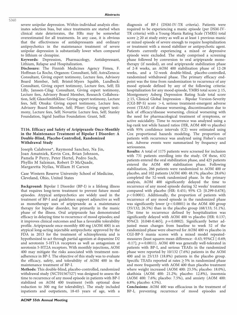
severe unipolar depression. Within-individual analysis elim-inates selection bias, but since treatments are started whenclinical state deteriorates, the HRs may be somewhatoverestimated for all treatments. In any case, it is obviousthat the effectiveness of antidepressants and ordinaryantipsychotics in the maintenance treatment of severeunipolar depression is substantially lower when comparedto lithium or clozapine.Keywords: Depression, Pharmacology, Antidepressant,Lithium, Relapse and Hospitalization.Disclosure: The Finnish Medicines Agency Fimea, F.Hoffman-La Roche, Organon: Consultant, Self; AstraZeneca:Consultant, Giving expert testimony, Lecture fees, AdvisoryBoard Member, Self; Bristol-Myers Squibb, Lundbeck:Consultant, Giving expert testimony, Lecture fees, Self; EliLilly, Janssen-Cilag: Consultant, Giving expert testimony,Lecture fees, Advisory Board Member, Research Collabora-tion, Self; GlaxoSmithKline: Giving expert testimony, Lecturefees, Self; Otsuka: Giving expert testimony, Lecture fees,Advisory Board Member, Self; Pfizer: Giving expert testi-mony, Lecture fees, Self; Novartis: Lecture fees, Self; StanleyFoundation, Sigrid Jusélius Foundation: Grant, Self.
T116. Efficacy and Safety of Aripiprazole Once-Monthlyin the Maintenance Treatment of Bipolar I Disorder: ADouble-Blind, Placebo-Controlled, RandomizedWithdrawal Study
Joseph Calabrese*, Raymond Sanchez, Na Jin,Joan Amatniek, Kevin Cox, Brian Johnson,Pamela P Perry, Peter Hertel, Pedro Such,Phyllis M Salzman, Robert D McQuade,Margaretta Nyilas, William H Carson
Case Western Reserve University School of Medicine,Cleveland, Ohio, United States
Background: Bipolar I Disorder (BP-I) is a lifelong illnessthat requires long-term treatment to prevent future moodepisodes. Atypical antipsychotics are widely used in thetreatment of BP-I and guidelines support adjunctive as wellas monotherapy uses of aripiprazole as a maintenancetherapy for bipolar disorder, but primarily in the manicphase of the illness. Oral aripiprazole has demonstratedefficacy in delaying time to recurrence of mood episodes; andit improves clinical outcomes and has a favorable tolerabilityprofile. Aripiprazole once-monthly 400 mg (AOM 400) is anatypical long-acting injectable antipsychotic approved by theFDA in 2013 for the treatment of schizophrenia and ishypothesized to act through partial agonism at dopamine D2and serotonin 5-HT1A receptors as well as antagonism atserotonin 5-HT2A receptors. With monthly injections, AOM400 may mitigate the risks associated with treatment non-adherence in BP-I. The objective of this study was to evaluatethe efficacy, safety, and tolerability of AOM 400 in themaintenance treatment of BP-I.Methods: This double-blind, placebo-controlled, randomizedwithdrawal study (NCT01567527) was designed to assess thetime to recurrence of any mood episode in patients with BP-Istabilized on AOM 400 treatment (with optional dosereduction to 300 mg for tolerability). The study includedinpatients and outpatients, 18-65 years of age, with a
diagnosis of BP-I (DSM-IV-TR criteria). Patients wererequired to be experiencing a manic episode (per DSM-IV-TR criteria) with a Young-Mania Rating Scale (YMRS) totalscore ≥ 20 at study entry as well as at least 1 previous manicor mixed episode of severe enough to require hospitalizationor treatment with a mood stabilizer or antipsychotic agent.Patients currently experiencing a mixed or depressiveepisode were excluded. The study comprised a screeningphase followed by conversion to oral aripiprazole mono-therapy (if needed), an oral aripiprazole stabilization phaseof 2-8 weeks, an AOM 400 stabilization phase of 12-28weeks, and a 52-week double-blind, placebo-controlled,randomized withdrawal phase. The primary efficacy end-point was the time from randomization to recurrence of anymood episode defined by any of the following criteria:hospitalization for any mood episode, YMRS total score ≥ 15,Montgomery Asberg Depression Rating Scale total score≥ 15, Clinical Global Impression - Bipolar Version-Severity(CGI-BP-S) score 44, serious treatment-emergent adverseevent (TEAE) of disease worsening, discontinuation due tolack of efficacy/disease worsening, clinical worsening withthe need for pharmacological treatment of symptoms, oractive suicidality. Time to recurrence was analyzed using alog-rank test while hazard ratios (HR, AOM 400 vs placebo)with 95% confidence intervals (CI) were estimated usingCox proportional hazards modeling. The proportion ofpatients with recurrence was analyzed using Fisher’s exacttest. Adverse events were summarized by frequency andseriousness.Results: A total of 1175 patients were screened for inclusionwith 731 patients enrolling into the study. Of these, 632patients entered the oral stabilization phase, and 425 patientsentered the AOM 400 stabilization phase. Followingstabilization, 266 patients were randomized to AOM 400 orplacebo, and 102 patients (AOM 400: 48.1%; placebo: 28.6%)completed the 52-week randomized phase. In the primaryanalysis, AOM 400 significantly delayed the time torecurrence of any mood episode during 52 weeks’ treatmentcompared with placebo (HR: 0.451; 95% CI: [0.299-0.678];po0.0001). Additionally, the proportion of patients withrecurrence of any mood episode in the randomized phasewas significantly lower (po0.0001) in the AOM 400 group(35/132; 26.5%) than in the placebo group (68/133; 51.1%).The time to recurrence defined by hospitalization wassignificantly delayed with AOM 400 vs placebo (HR: 0.137;95%CI: [0.040-0.465]; p= 0.0002). Significantly better ad-justed mean changes from baseline to week 52 of therandomized phase were observed for AOM 400 vs placebo inCGI-BP-S mania scores with a mixed model repeated-measures (least squares mean difference: -0.43; 95%CI [-0.69;-0.17]; p= 0.0011). AOM 400 was generally well-tolerated inpatients with BP-I, and serious TEAEs in the randomizedphase were reported by 10/132 (7.6%) patients in the AOM400 and in 25/133 (18.8%) patients in the placebo group.Specific TEAEs reported at rates ≥ 5% in randomized phaseand more frequently with AOM 400 than placebo treatmentwhere weight increased (AOM 400: 23.5%; placebo: 18.0%),akathisia (AOM 400: 21.2%; placebo: 12.8%), insomnia(AOM 400: 7.6%; placebo: 7.5%), and anxiety (AOM 400:6.8%; placebo: 4.5%).Conclusions: AOM 400 was efficacious in the treatment ofBP-I, preventing recurrence of mood episodes and
Abstracts
S360
ACNP 55th Annual Meeting
maintaining stability in recently manic patients, and treat-ment was well-tolerated. The efficacy and tolerability profilefor monthly injection of AOM 400 in BP-I is consistent withthat of aripiprazole as daily oral administration. These resultssuggest that AOM 400 is a viable once-monthly option formaintenance treatment in BP-I, preventing reoccurrence ofmood episodes.Keywords: Bipolar I Disorder, Aripiprazole Once-Monthly,Maintenance Treatment, Long-Acting Injectable Anti-psychotic.Disclosure: Otsuka Pharmaceutical Development & Com-mercialization, Inc.: Employee, Consultant/Advisory BoardMember/Speaker, Self; H. Lundbeck A/S: Employee, GrantSupport, Consultant/Advisory Board Member/Speaker, Self;Abbott Laboratories, Pfizer, Inc, AstraZeneca, Bristol-MyersSquibb Company, Teva Pharmaceutical Industries Ltd,Dainippon Sumitomo Pharma Co., Ltd, GlaxoSmithKline,Janssen Pharmaceuticals, Inc., Sunovion PharmaceuticalsInc., Takeda Pharmaceutical Company Limited: GrantSupport, Consultant/Advisory Board Member/Speaker, Self;Eli Lilly and Company, Intra-Cellular Therapies, Inc.: GrantSupport, Self; Allergan, Pfizer, Inc., Repligen Corporation,Servier, Solvay Pharmaceuticals, Inc.: Consultant/AdvisoryBoard Member/Speaker, Self; Department of Defense, HealthResources Services Administration, National Institute ofMental Health: Federal Funding, Self.
T117. Electroconvulsive Therapy Modulates NeuralResponse to Emotional Faces in Depressed Patients: ARandomized Controlled fMRI Study
Kamilla Miskowiak*, Lars Kessing, Caroline Ott, JulianMacoveanu, Catherine Harmer, Rasmus Revsbech,Hartwig Siebner, Martin Jorgensen
Copenhagen University Hospital, Rigshospitalet,Copenhagen, Denmark
Background: Negative bias in the neurocognitive response toemotional information is a core feature of major depressivedisorder (MDD) that is associated with depression severityand risk of relapse. Emerging evidence suggests that reversalof negative bias is a common mechanism for pharmacolo-gical and psychological treatments for depression. This studyaimed to investigate with functional magnetic resonanceimaging (fMRI) whether electroconvulsive therapy (ECT)modulates neural and cognitive processing of emotionalinformation in MDD.Methods: Twenty-nine MDD patients were randomized toreceive one active or a sham ECT session at the beginning oftheir ECT course in a double-blind, between-groups design.The following day, patients underwent whole-brain fMRI at3T during which time they viewed emotional faces and weregiven facial expression recognition and faces dot-probe tasksafter the scan. Mood symptoms were assessed at baseline, onday 1 and after six ECT sessions.Results: Data was lost for two patients (sham) and analysestherefore included 27 patients (ECT: N= 15, sham: N= 12).Electroconvulsive therapy reduced parahippocampal activity,increased superior frontal activity to fearful vs. happy facesand enhanced fear-specific functional connectivity betweenthe amygdala and dorsolateral prefrontal cortex (dlPFC),
orbitofrontal and occipito-temporal regions. Across theentire cohort, stronger fear-specific amygdala–occipitalcoupling was associated with lower fear vigilance. Greaterfear-specific amygdala–dlPFC coupling correlated with bettersubsequent clinical response. These effects occurred in theabsence of differences between groups in cognitive perfor-mance, mood symptoms or medication.Conclusions: This exploratory study shows for that first timethat ECT modulates neural response to emotional informa-tion. The findings suggest that early positive shift in neuralresponse to emotional information may be a commonmechanism of distinct treatments for MDD.Keywords: Functional MRI (fMRI), Major Depression,Electroconvulsive Therapy, Facial Emotional Processing.Disclosure: Lundbeck: Consultancy fees, Self.
T118. Repetitive Transcranial Magnetic Stimulation inthe Olfactory Bulbectomy Mouse Model of Depression
Alesha Heath*, Avalon Young, Sofia Rinaldi,Kalina Makowiecki, Jennifer Rodger
University of Western Australia, Crawley, Australia
Background: Depression is one of the leading causes ofdisability worldwide. With 30% of sufferers not respondingto conventional antidepressant treatments there is a growingneed for the development of novel therapies to alleviatesymptoms in these patients. One of the most promising ofthese is repetitive transcranial magnetic stimulation (rTMS),however, the mechanisms of action and most effectivetreatment parameters remain unknown. This study aimed toreplicate clinically used methods of rTMS in an animalmodel of depression and assess behavioural and neurobio-logical changes.Methods: C57Bl\6J mice underwent either surgery to aspiratethe olfactory bulbs (n= 38, olfactory bulbectomy) or shamsurgery (n= 11, craniotomy but no aspiration). Mice witholfactory bulbectomy received either rTMS (n= 11), shamrTMS (n= 9), fluoxetine (n= 9) or a vehicle treatment(n= 9) administered for four weeks starting two weeks aftersurgery. rTMS was delivered at 10 Hz for 3 minutes (1800pulses) daily. Fluoxetine (18mg/kg) was given orally incookie dough daily. A subset of animals in each groupreceived an injection of EdU two weeks after the start oftreatment. The forced swim behavioural test was carried outat three time points: before surgery (baseline), two weeksafter surgery to confirm the bulbectomy model, and at theend of the treatment period to assess efficacy. After the finalbehavioural test, mice were euthanased (150mg/kg pento-barbitone, i.p.) and, serotonin levels in the prefrontal cortexwas assessed using an enzyme immunoassay and cellproliferation in the dentate gyrus was assessed using EdUhistochemistry.Results: With respect to baseline values; after the olfactorybulbectomy mice showed significantly higher activity levelsin the forced swim test and this was rescued only by rTMS,fluoxetine showed no significant effect in this test. In theprefrontal cortex serotonin levels were significantly increasedonly in mice treated with fluoxetine, whereas rTMS appearedto increase the survival of newly differentiated cells in thedentate gyrus.
Abstracts
S361
ACNP 55th Annual Meeting
Conclusions: These results indicate a replication of thetherapeutic effect of rTMS treatment in a specific animalmodel of depression, providing a foundation for exploringthe mechanisms of rTMS antidepressant effects in humanpatients. Our findings showing improved behavioural out-comes in the forced swim test without a significant increasein serotonin levels suggest that rTMS may have a mechanismof action that is distinct from serotonin-related antidepres-sant treatments, consistent with its beneficial effects in drugresistant patients.Keywords: Neurostimulation, Depression, Animal Models.Disclosure: Nothing to disclose.
T119. Genome-Wide Association Study of MajorDepressive Disorder in European Americans and AfricanAmericans
Li Wen, Yinghao Yao, Thomas Payne, Jennie Ma,Ming Li*
Seton Hall University, South Orange, New Jersey, UnitedStates
Background: Major depressive disorder (MDD) is acommon complex mental illness, which poses one of leadingcauses of preventable deaths and disabilities throughout theworld. To date, genome-wide association study (GWAS) forMDD and its-related phenotypes, have not yielded consistentresults.Methods: We conducted a GWAS for the Center forEpidemiological Studies Depression (CES-D) score, a widelyused questionnaire for measuring MDD, in EuropeanAmerican (EA) and African American (AA) populations,with age, gender, ancestry-informative principal componentscores and smoking status as covariates. Considering thereexisted no evidence for the presence of heterogeneitybetween the AA and EA samples, we analyzed two samplestogether with a total sample size of 4,817.Results: Our results revealed a genome-wide significantassociation between a nonsynonymous SNP rs61753730in FZD6 (frizzled class receptor 6; functions as a negativeregulator of the canonical Wnt/beta-catenin signalingcascade) gene that gives rise to a glutamine to glutamatesubstitution and the CES-D score (P= 8.46 × 10⁻⁹). Such afinding has also received independent support from amolecular study using animal model where knockdownof FZD6 gene expression lead to a depressive effect onrat. Due to there were a few SNPs included in theIllumina Infinium HumanExome BeadChip for FZD6 gene,we performed imputation analysis for the gene withIMPUTE2 using 1000 Genomes as references and thenconducted gene-based association study using SNP-setKernel Association Test (SKAT) in the pooled AA and EAsamples, which revealed a significant association of the FZD6gene with the CES-D score (Po0.01). Additional gene-by-gene interaction analysis using a generalized multifactordimensionality reduction (GMDR) algorithm showed asignificant interaction between FZD6 and CREB3L1 (P=0.0016) or FZD6 and CREB3L4 (P= 0.002). Finally, weshowed that tobacco smoking plays a significant role in theassociation of FZD6 gene with depression where thesignificant association of the gene with CES-D score was
only detected in smoker samples, but not in nonsmokersamples.Conclusions: Taken together, this study showed that FZD6gene represents a plausible candidate gene for MDD andfurther study with a large sample size is warranted.Keywords: Major Depressive Disorder (MDD), GWAS,CESD Score.Disclosure: Nothing to disclose.
T120. Selective Dietary Supplementation in EarlyPostpartum is Associated With High Resilience AgainstDepressed Mood
Yekta Dowlati*, Arun Ravindran, Zindel Segal,Donna Stewart, Meir Steiner, Jeffrey Meyer
Centre for Addiction and Mental Health, Toronto,Canada
Background: Postpartum depression, the most commoncomplication of childbearing has a 13% prevalence, but thereare no widespread prevention strategies and no nutraceuticalinterventions have been developed. Postpartum blues is oftena prodromal state for postpartum depression, since severepostpartum blues strongly elevates risk for postpartumdepression. To counter the effects of the greater than 40%elevation of monoamine oxidase-A levels that occur duringpostpartum blues, a dietary supplement kit consisting ofmonoamine precursor amino acids, tryptophan and tyrosine,and dietary antioxidants was created and key ingredientswere shown not to affect their total concentration in breastmilk. In a series of studies, we show that oral tryptophan andoral tyrosine supplements do not increase their totalconcentrations in breast milk; despite the significant increasein maternal plasma. In contrast, some dosing regimen of oralcysteine supplements increase total cysteine concentrationsin breast milk yet have a lesser effect on maternal plasmalevels. The aim of this open-label study was to assess whethera dietary supplement consisting of tryptophan, tyrosine andblueberry extract + blueberry juice reduces the vulnerabilityto depressed mood at day-5 postpartum, the typical peak ofpostpartum blues.Methods: Forty-one healthy day-5 postpartum womensuccessfully completed all study procedures. One group(n= 21) received the dietary supplement composed of 2grams tryptophan, 10 grams tyrosine and blueberry juicewith blueberry extract. The control group (n= 20) did notreceive any dietary supplement. Postpartum blues severitywas quantitated by the elevation in depressed mood on thevisual analogue scale and the profile of mood statedepression scores following the sad mood inductionprocedure.Results: Following the sad mood induction procedure,univariate analysis of variance (ANOVA) demonstrated arobust induction of depressed mood on the visual analogscale in the controls but no effect in the supplement group (F(1,39)= 88.33, po0.001; 43.85± 18.98 mm versus0.05± 9.57 mm shift, effect size 2.9). Moreover, the effectof the dietary supplement was assessed as a predictor ofchange in profile of mood state depression scores and thisshowed a highly significant effect of group (ANOVA, F(1,39)= 19.81, po0.001).
Abstracts
S362
ACNP 55th Annual Meeting
Conclusions: This is the first study to investigate the effect ofa combination of monoamine precursors, tryptophan andtyrosine, and blueberry extract/juice on the intensity ofpostpartum blues. This dietary supplement designed tocounter functions of elevated monoamine oxidase-A activityvirtually eliminated the vulnerability to depressed moodduring the peak of postpartum blues. This suggests that thisnutraceutical intervention is a highly promising approach totarget this prodromal state of postpartum depression.Keywords: Postpartum Blues, Prodrome, Prevention, Mono-amine Oxidase-A, Monoamines.Disclosure: Nothing to disclose.
T121. Metabolic Outcomes in Response to Chronic Stressand Antidepressant Treatment
Ma-Li Wong*, Suhyun Lee, Ethan Dutcher,Martin Lewis, Claudio Mastronardi, Julio Licinio
South Australian Health, Adelaide, Australia
Background: Antidepressant prescribing has risen 400% sincethe late 1980s; in parallel, obesity rates have doubled in adultsand tripled in childhood during this period. Antidepressantstreatment can lead to significant weight gain but the interplaybetween MDD, obesity and antidepressant treatment iscomplex. It is conceivable that nearly 25% of the cases ofobesity may be attributable to the association with MDD andantidepressant use. Our lab has developed an animal model tounderstand the mechanisms of weight recovery after chronicstress, as we hypothesized that the effects of antidepressantson body weight would be unmasked by environmental factors,such as an obesogenic high-fat diet.Methods: Male Sprague-Dawley rats were subjected to short-term exposure to recurrent restraint stress and antidepres-sants (fluoxetine or imipramine) for 2 weeks, followed bylong-term high-fat diet intake for 295 days. Animals werethen sacrificed and various organs were collected andweighed. Measurements: Body weight, food intake ratio,behavioural testing, and bone weight.Results: Obesity-prone rats, defined as those within theupper 50% of body weight gain, treated with fluoxetine (FX)had increased body weight, in comparison to the controlgroup treated with saline and non-restraint control group.The antidepressant-treated groups had significantly lowerfood intake ratio in comparison to the non-restraint controlgroup. Obesity-prone FX rats had significantly longer bodylength in comparison to all other groups. Rats in the FXgroup were significantly less anxious and had heavier bones.New metabolic (leptin, adipose tissue gene expression) andIGF-1 data will be presented.Conclusions: We present the conceptually novel andmedically relevant, data-based conclusion that the interac-tion of stress, antidepressant treatment, and high-fat dietmay have long-lasting body, bone allometric, and metaboliceffects. In the context of CDC data showing that 11% of theUS population over 12 years of age is on antidepressants, theclinical and public health implications of these findings aresubstantial.Keywords: Antidepressants, Chronic Stress, Body Weight,Adipose Tissue.Disclosure: Nothing to disclose.
T122. Deep Brain Stimulation in the NeuroanatomicalFramework of Medial Forebrain Bundle GeneratesAntidepressant-Like Phenotype in Rats
Albert Fenoy*, Manoj Dandekar, VijayasreeGiridharan, Dustin Luse, Carson Hoffmann, TravisPeery, Christian Ruiz, Patrick Cotton, Jair Soares, Joaode Quevedo
University of Texas Health Science Center at Houston,Houston, Texas, United States
Background: Deep brain stimulation (DBS) is currentlybeing investigated as a one of the therapeutic modalities fordepressive disorders and treatment-resistant depression(TRD). In recent years, the superolateral branch of themedial forebrain bundle (MFB) has attracted much attentionin the treatment of depression. One uncontrolled studytargeting the superolateral branch of the MFB reported arapid antidepressant effect and 85% response rate. MFB is akey structure of the mesolimbic-dopamine system forappetitive motivation and euphoric feelings, connecting theventral tegmental area (VTA) to the nucleus accumbens(NAc) and prefrontal cortex (PFC). However, the mechan-ism underlying such rapid antidepressant effect due to MFBmodulation remains speculative. The objective of this studywas to examine if DBS of the MFB is an effective treatmentmodality for depression and to perform biochemical analysisof reward mechanisms associated with dopaminergic systemmodulation.Methods: A concentric bipolar electrode (Plastics One, VA)was stereotactically implanted into the left MFB of maleWistar rats. Animals were divided into three groups, sham-operated, DBS-Off (no stimulation), or DBS-On (stimulationfor 1 week, 8 hrs/day). Exploratory activity and depression-like behavior were evaluated using the open-field andPorsolt’s forced swim test (FST), respectively, in the rat.Bilateral rat brain samples of the medial prefrontal cortex(PFC), hippocampus, amygdala and NAc were processedwith Western blot analysis of markers of dopaminergicactivity that included the tyrosine hydroxylase, dopaminetransporter (DAT) and dopamine receptors D1 through D5.Protein expressions were then correlated to immobility timeduring the FST.Results: Animals undergoing stimulation of the MFBshowed significant increases (po0.05) in swimming timewithout altering the locomotor activity, relative to the DBS-Off and sham stimulation groups. This indicated anantidepressant-like phenotype in the animals. Interestingly,we have seen trends towards an increase in protein levels ofdopamine D1, D2, D4 and D5 receptors in the medial PFCfollowing MFB DBS. However, we did not observe anappreciable difference in tyrosine hydroxylase expression inthe medial PFC, hippocampus, amygdala and NAc.Conclusions: MFB-DBS showed behavioral improvement indepression-like phenotype and biochemical alterations of thePFC that demonstrate changes within the circuitry of thebrain different from the target area of stimulation. Suchobserved neurochemical alterations may underlie the ther-apeutic efficacy of this treatment. In the future, furthertesting of MFB-DBS in TRD models for antidepressant-likeeffect will help us advance this intervention for ultimateclinical use.
Abstracts
S363
ACNP 55th Annual Meeting
Keywords: Deep Brain Stimulation, Treatment ResistantDepression, Medial Forebrain Bundle, Rat Model.Disclosure: Nothing to disclose.
T123. Neurocognitive Effects of CombinedElectroconvulsive Therapy and Venlafaxine in ElderlyAdults With Major Depressive Disorder
Sarah Lisanby, Shawn McClintock*, Rebecca Knapp,Martina Mueller, Robert Greenberg,Matthew Rudorfer, Mustafa Husain, Richard Weiner,Robert Young, W. Vaughn McCall, Peter Rosenquist,Georgios Petrides, Shirlene Sampson, Joan Prudic,C. Munro Cullum, George Alexopoulos,Samuel Bailine, Elisabeth Bernhardt, Kristen Tobias,Zhi-De Deng, Lauren Liebman, Mimi Briggs,Emma Geduldig, Styliani Kaliora, VassiliosLatoussakis, Abeba Teklehaimanot, Charles Kellner
University of Texas Southwestern Medical Center, Dallas,Texas, United States
Background: Major depressive disorder is a chronicneuropsychiatric disease that results in significant healthconsequences. The rate of MDD in elderly adults has beensteadily increasing and unfortunately results in cognitivedifficulties, debilitation, morbidity, and mortality. Electro-convulsive therapy (ECT) is a safe and effective antidepres-sant treatment for MDD, particularly in elderly adults. Whencombined with pharmacotherapy, ECT may produce greaterantidepressant benefits. Also, while ECT is associated withneurocognitive adverse effects, ECT parameters such as rightunilateral (RUL) electrode configuration ultrabrief pulsewidth (UB) help to minimize the impact on cognitivefunctions. However, there is limited information regardingthe neurocognitive effects of combined pharmacotherapyand ECT, particularly with newer parameters, in geriatricdepression. Thus, the purpose of this study was to determinethe neurocognitive effects of an acute course of right RUL-UB ECT combined with venlafaxine in elderly adultswith MDD.Methods: The Prolonging Remission in Depressed Elderly(PRIDE) study was a two-phase, multicenter, randomizedstudy of an individualized continuation ECT schedulecombined with pharmacotherapy to enhance long-termoutcomes in elderly adults with depression. The study designinvolved two phases. In Phase 1, patients received acute ECT3x weekly combined with VLF. In the randomized phase(Phase 2), those who remitted in Phase 1 were randomized toreceive pharmacotherapy (VLF and lithium) alone or thecombined modalities (pharmacotherapy and continuationECT). Elderly adults (age 4 60) with major depressivedisorder, based on the SCID-I or MINI, were recruited andenrolled in the study. All participants provided writteninformed consent for this IRB approved investigation beforecompleting study procedures. Patients discontinued psycho-tropic medications within 1-week of starting Phase 1. ECTwas provided 3x per week and procedures were standardizedas follows: RUL electrode placement using a SomaticsThymatron System IV (Somatics, LLC, Lake Bluff, IL) withan ultrabrief pulse width of 0.25ms and current of 0.89Ampsor a MECTA SPECTRUM (MECTA Corporation, Portland,
OR) device with an ultrabrief pulse width of 0.3ms andcurrent of 0.8Amps. Dose titration to determine seizurethreshold was conducted at the first ECT session. Subsequenttreatments were administered at 6x the seizure threshold.Multiple cognitive domains including attention and proces-sing speed, verbal fluency, verbal learning and memory,autobiographical memory consistency, and executive func-tion were assessed with a neurocognitive battery. Specificneurocognitive instruments included the AutobiographicalMemory Interview-Short Form (AMI-SF), California VerbalLearning and Memory Test-II (CVLT-II), Delis-KaplanExecutive Function System (DKEFS) Verbal Fluency Test,Dementia Rating Scale-2nd Edition Initiation PerseverationIndex (DRS-2 IP), Stroop Color and Word Test, and TrailMaking Test Parts A and B (TMT A and B). Theneurocognitive battery was administered at baseline andthen within 72 hours following the last ECT session.Descriptive statistics were used to describe the sociodemo-graphic and clinical characteristics of the study population.Means and standard deviations are presented for continuousvariables, and frequency distributions are presented fordiscrete variables. Paired t tests were computed to determinethere was a significant change in neurocognitive measureperformance from baseline to end and corresponding 95%confidence intervals were calculated for the difference inmeans between baseline and end. Statistical significance wasdefined as a two-sided p-value of less than 0.05.Results: Changes in performance were statistically signifi-cantly lower from baseline to end on neuropsychologicalmeasures of psychomotor (po0.0001) and verbal(p= 0.0002) processing speed, autobiographical memoryconsistency (po0.0001), short-term verbal recall (p= 0.001)and recognition (p= 0.0006) of learned words, phonemicfluency (po0.0001), inhibition (p= 0.05), and complexvisual scanning and cognitive flexibility (p= 0.0004). How-ever, in terms of qualitative changes from baseline to end,performance across most neurocognitive measures remainedrelatively stable and ranged from intact/average to mildlyimpaired.Conclusions: This is the first study to characterize theneurocognitive effects of combined RUL-UB ECT andvenlafaxine in elderly adults with MDD. While changes inperformance on some neuropsychological measures werestatistically significant, the majority of neuropsychologicalindices remained stable. Indeed, only autobiographicalmemory consistency, phonemic fluency, and complex visualscanning and cognitive flexibility qualitatively decreasedfrom low average to mildly impaired. These findings areconsistent with prior research and provide new evidence forthe relative neurocognitive advantage of RUL-UB ECTparameters. Future research is warranted to confirm theneurocognitive effects of combined ECT and psychotropicmedications, and to determine the association with under-lying neural mechanism, and clinical and functionaloutcomes.Keywords: Electroconvulsive Therapy, Neurocognition,Major Depression, Late-Life Depression.Disclosure: Nothing to disclose.
Abstracts
S364
ACNP 55th Annual Meeting
T124. Change in Leukocyte Telomere Length Over TwoYears in a Sample of Individuals With Major DepressiveDisorder and Controls
Mary Zeng, Eric Bui, Benjamin Kovachy, Mireya Nadal,David Mischoulon, Maurizio Fava, Immaculata deVivo, Kwok-Kin Wong, Naomi Simon*
Massachusetts General Hospital, Boston, Massachusetts,United States
Background: Reduced leukocyte telomere length (LTL) hasbeen found to be associated with multiple common diseasesof aging, including heart disease, diabetes, and cancer (e.g.,Price et al, 2013). More recently, a link has also beensuggested between shortened LTL and psychiatric condi-tions, especially major depressive disorder (MDD) (e.g., Linet al, 2016; Ridout et al, 2016). These findings suggest thatMDD may be a disease of accelerated aging. Prior studies onthe association between MDD and LTL were limited by across-sectional design, use of banked blood from investiga-tions conducted for other purposes, and use of self-reportmeasures or clinician-rated measures assessed by non-doctoral-level clinicians. The present prospective longitudi-nal study aims to examine the association between MDDdiagnosis at baseline and change in LTL over two years in awell-characterized sample of individuals with or withoutMDD at baseline.Methods: Participants were n= 67 individuals with MDD (M(SD) age = 42.7(13.2); 52% women) and n= 50 psychia-trically healthy controls (M(SD) age = 44.1(14.0); 58%women) who participated in a prior study (Simon et al,2016), and were reassessed at two-year follow-up. Diagnoseswere assessed by doctoral-level raters using the StructuredClinical Interview for DSM-IV (SCID; First et al, 2002). Atboth time points, participants were also assessed for:demographic and clinical characteristics, including smokingstatus, body-mass index (BMI), and exercise level; MDDsymptom severity using the Montgomery Asberg DepressionRating Scale (MADRS; Montgomery and Asberg, 1979);anxiety symptoms using the Hamilton Anxiety Rating Scale(HAM-A; Hamilton. 1959); childhood trauma exposureusing the short form of the Early Trauma Inventory Self-Report (ETISR-SF; Bremner et al, 2007); lifetime traumaexposure using the Traumatic Events Questionnaire (TEQ;Vrana & Lauterbach. 1994); and perceived stress using thePerceived Stress Scale (PSS; Cohen et al, 1983). Participantswere asked to provide blood samples at baseline and twoyears, and LTL was assayed with qPCR. T-tests were used toexamine whether change in LTL (calculated with z-scores toallow comparison between LTL measurements at differenttime points). Pearson’s correlations were used to examinewhether change in LTL over two years in participants withbaseline MDD was significantly associated with baseline,follow-up, and change covariates.Results: At baseline, there were no significant differencesbetween depressed and non-depressed participants in age,sex, race, ethnicity, or highest educational level. Change inLTL between baseline and 2-year follow-up was significantlydifferent between individuals with MDD and the controlgroup (M(SD) = -0.34(1.2) vs. M(SD) = 0.25(1.4), t(115) =2.45, po0.05). While this change in LTL significantlydiffered from zero change in the MDD group (t(49) =
-2.07, p= 0.04), this was not the case for the this was not truefor control group. Further, Pearson’s correlation analyses inthe MDD group failed to show any significant associationbetween LTL change, and baseline, follow-up, changecovariates including age, sex, marital status, MDD andanxiety symptom severity, lifetime depression duration,lifetime substance use disorder, months of antidepressantuse, cigarette smoking pack-years, body mass index, exerciselevel, childhood and lifetime trauma exposure, and perceivedstress.Conclusions: Despite limitations including a relatively smallsample size, and the use of z-scores to compare LTL assayedat two different time points, our results provide furthersupport for MDD as a disease of accelerated aging, in a well-characterized sample using validated, clinician-rated mea-sures, and suggest an independent mechanism by whichMDD might shorten telomeres length.Keywords: Leukocyte Telomere Length, Depression,Chronic Stress.Disclosure: Nothing to disclose.
T125. Vortioxetine Versus Placebo in Major DepressiveDisorder Comorbid With Social Anxiety Disorder
Michael Liebowitz*, Jason Careri, Kyra Blatt,Ann Draine, Rita Hanover
Columbia University, New York, New York, United States
Background: Both major depressive disorder (MDD) andsocial anxiety disorder (SAD) are common psychiatricconditions that can at times be highly disabling. Moreover,they often occur together; SAD is the most common anxietydisorder found in patients with MDD, and individuals withSAD are at increased risk for MDD. Given the prevalenceand severity of MDD comorbid with SAD, it is striking thatno drug approved for MDD or SAD has been subject to aplacebo controlled trial in patients with both disorders.Vortioxetine is a recently marketed antidepressant withactivity at the serotonin transporter as well as at severalserotonin receptor subtypes. It has documented efficacy in anumber of MDD trials, positive findings in one of two GADtrials, and was found effective in reducing anxiety as well asdepressive symptoms in depressed patients. We thereforehypothesized that it would be effective in patients with bothMDD and comorbid SAD.Methods: Forty outpatients between the ages of 18 and 70who met criteria for both MDD and SAD per DSM-5 wererandomized on a 1:1 basis to vortioxetine 10-20 mg/day ormatching placebo in a 12 week double blind trial. Theprimary outcome measure was the investigator ratedcomposite CGI-I, with a rating of responder indicated byan endpoint rating of much improved (2) or very muchimproved (1). These ratings were operationalized as follows:much improved- at least moderate benefit in both MDD andSAD features, or at least marked benefit in of of the twodomains if there was no or only minimal benefit in the other;very much improved- marked benefit in both MDD andSAD. Secondary endpoints included the the MontgomeryAsberg Depression Rating Scale (MADRS), the LiebowitzSocial Anxiety Scale (LSAS), the Hamilton Anxiety Scale
Abstracts
S365
ACNP 55th Annual Meeting
(HAM-A), the self-rated Sheehan Disability Scale (SDS) anda subject rated global improvement scale (PGIC).Results: The final patients have entered the trial, and whenthey complete, vortioxetine and placebo will be compared onthe following variables: responder rates (1 or 2) on thecomposite CGI-I, change from baseline to endpoint onMADRS, LSAS, HAM-A, SDS, and PGIC, and time course toresponse (50% or greater reduction in MADRS) and toremission (MADRS 7 or less) for MDD and for remission ofSAD (LSAS or 30 or less). Adverse event and medical safetydata will also be reported.Conclusions: The findings should be significant in severalways. In planning this trial, we found that there was noestablished methodology for conducting treatment trials inMDD comorbid with an anxiety disorder such as SAD. Aprevious open trial had rated change in each disorderseparately, but did not provide any composite measure ofoverall improvement in individual patients. The presentstudy allowed us to pilot a method for assessing overallimprovement in individual patients with MDD comorbidwith SAD, and could provide a model for future investiga-tions in this area. If successful, this model should also betransferrable to MDD comorbid with other anxiety dis-orders, as well to other comorbid conditions in general, aneglected but important field for treatment studies. Inaddition to guiding future investigations, the study issufficiently powered to provide informative effect size datathat could influence current clinical decision making aboutthe treatment of MDD comorbid with SAD.Keywords: Psychiatric Comorbidity, Major Depressive Dis-order, Social Anxiety, Clinical Trial Methodology.Disclosure: Takeda Pharmaceuticals: Financial support forthe trial via funding of investigator initiated proposal, Self,Holds copyright to the LSAS, Self.
T126. Histone Serotonylation: A Novel Mechanism ofEpigenetic Plasticity
Lorna Farrelly, Robert Thompson, Shuai Zhao,Ashley Lepack, Yang Lu, Olivier Berton, Haitao Li,Tom Muir, Ian Maze*
Icahn School of Medicine at Mount Sinai, New York, NewYork, United States
Background: Alterations in gene expression promotephysiological adaptations implicated in a wide variety ofhuman diseases. More recently, histone mechanisms havebeen shown to regulate transcriptional programs contribut-ing to neurodevelopmental, neurodegenerative and psychia-tric disorders; however, our understanding of how thesemechanisms mediate persistent patterns of transcriptionaldysfunction remains limited. Monoaminergic signaling inbrain plays a critical role in neuronal plasticity, withalterations in monoamine production being implicated inthe development and treatment of numerous brain disorders.Although vesicular packaging of monoamines is essential forneurotransmission, recent data have demonstrated theadditional presence of ‘reserve’ pools of extravesicularmonoamines in the nucleus of monoaminergic neurons; itremains unclear, however, whether nuclear monoaminesplay roles independent of neurotransmission. Serotonin, as
well as other monoamines, has previously been shown toform covalent bonds with certain cytoplasmic proteins viatransamidation by the tissue Transglutaminase 2 enzyme, amodification that alters the signaling properties of modifiedproteins. We therefore hypothesized that nuclear proteinsmay similarly be modified to control distinct aspects of theirfunction.Methods: We employ a large variety of biochemical,biophysical and molecular approaches to fully delineate thefunctions of protein serotonylation in brain. These ap-proaches include, but are not limited to, enzymatic analysesof covalent serotonin transfer to substrate proteins, massspectrometry, biophysical interrogations of serotonyl inter-acting proteins, genome-wide ChIP-/RNA-seq sequencinganalyses, molecular manipulations of serotonylation in vitroand in brain, and behavioral analyses.Results: We have identified histone proteins, specificallyhistone H3, as robust substrates for monoaminylation inbrain. Our data indicate that H3 serotonylation acts tofacilitate the establishment of permissive histone posttransla-tional modifications, such as H3K4me3, potentiate bindingof specific chromatin interacting proteins, and plays a criticalrole in regulating active transcription via alterations in thedeposition/removal of nearby histone/DNA modifications.Furthermore, histone serotonylation appears to be criticalduring both early neurodevelopment in the establishment ofcell-type identity in brain, as well as in the adult centralnervous system in the regulation of environmentally-inducedneural plasticity (e.g., behavioral responses to stress).Conclusions: Human neurological diseases associated withmonoaminergic dysfunction affect millions of individualsworldwide; however, treatments for these neurodevelop-mental, neurodegenerative and/or psychiatric disordersremain inadequate. Employing this unique combination ofbiochemical, molecular and behavioral approaches, we areexamining novel, nuclear-specific roles for monoamines inbrain in the direct regulation of neuronal gene expression.Our data suggest that alterations in the expression of theseepigenetic marks likely contribute to deficits in neuronalplasticity and the subsequent precipitation of disease states.Therefore, our work aims to fully characterize these so-calledhistone monoaminylation states in brain, as well as theiraberrant regulation in neurological disease, in order toidentify novel targets for the development of more effectivefuture therapeutics.Keywords: Epigenetics, Serotonin, Neuroplasticity.Disclosure: Nothing to disclose.
T127. Chronic Unpredictable Stress Exposure AugmentsHabenular 2-Arachidonoylglycerol Signaling
Anthony Berger, Janelle Lugo, Martin Sticht,Maria Morena, Matthew Hill, Ryan McLaughlin*
Washington State University, Pullman, Washington,United States
Background: The societal and economic burden of majordepressive disorder (MDD) is currently at an all-time high,and more efficacious treatment strategies are urgentlyneeded. In recent years, there has been a resurgence ofinterest in the phylogenetically ancient, epithalamic
Abstracts
S366
ACNP 55th Annual Meeting
habenular nuclei as a novel putative target for the treatmentof MDD. The habenula receives multiple converging inputsfrom cortical and subcortical structures involved in emo-tional processing and action selection, the summation ofwhich determines the activity of principal output neuronsthat negatively regulate midbrain monoaminergic transmis-sion. The lateral subdivision of the habenula (LHb) has beenshown to be hyperactive in preclinical models of learnedhelplessness and clinically depressed individuals (see Proulxet al, 2014, Nat Neurosci for review), while LHb deep brainstimulation produces robust antidepressant-like effects inboth rodents (Meng et al, 2011, Brain Res) and treatment-resistant MDD patients (Sartorius et al, 2010, Biol Psychia-try). Moreover, synaptic potentiation within the LHb ispositively correlated with the degree of despair-like behaviorin preclinical studies via enhanced presynaptic releaseprobability (Li et al, 2011, Nature). Despite these intriguingfindings, the mechanisms that confer hyperexcitability of theLHb remain unknown. We have recently shown that theendocannabinoid (ECB) system in the LHb controls theselection of behavioral coping strategies to aversive environ-ments in preclinical paradigms (Berger et al, under review).However, the impact of chronic stress, which is inextricablylinked to MDD pathology, on ECB signaling in the LHb hasyet to be empirically evaluated.Methods: The objectives of the current study were to assesswhether exposure to chronic unpredictable stress (CUS)elicits alterations in 1) content of the primary ECBsanandamide (AEA) and 2-arachidonoylglycerol (2-AG), 2)the hydrolytic efficacy of the 2-AG catabolizing enzymemonoacylglycerol lipase (MGL), and 3) mRNA expressionfor enzymes responsible for the synthesis (NAPE-PLD,DGLα) and degradation (FAAH, MGL) of AEA and 2-AGin the habenula of adult male and female Sprague Dawleyrats. Rats were subjected to six weeks of CUS (2-3randomized stressors/day) as described previously(McLaughlin et al, 2013, Behav Brain Res). On the morningfollowing the final stressor, rats were rapidly decapitated andthe habenula (comprised of both the medial and lateralsubdivisions) was dissected, flash-frozen, and stored at -80°Cuntil analysis. ECB content was measured using liquidchromatography-tandem mass spectrometry (LC-MS/MS) asdescribed previously (Qi et al, 2015, Rapid Commun MassSpectrom). The maximal velocity of 2-AG hydrolysis (Vmax)and binding affinity of MGL for 2-AG (Km) weredetermined using membrane fractions prepared fromhomogenized habenula tissue as previously described (Hillet al, 2009, Neuropsychopharmacology). Expression ofmRNA for ECB-related enzymes was determined via real-time quantitative polymerase chain reaction (RTqPCR) usinga QiaCube robot (Qiagen) and TaqMan primers targeted atECB proteins, and stress and non-stress groups werecompared using the ΔΔCt method (Ct values normalizedto housekeeping gene Rn45s). All experimental protocolswere approved by the Institutional Animal Care and UseCommittee and conducted in accordance with the NationalInstitutes of Health Guide for the Care and Use ofLaboratory Animals.Results: Preliminary results indicate that 2-AG content wassignificantly elevated in the habenula of female (but not male)rats exposed to CUS compared to non-stressed control rats.Although CUS exposure did not significantly affect habenular
AEA content in male or female rats, AEA content wassignificantly elevated in female rats compared to male ratsunder basal, non-stress conditions. Moreover, concentrationsof AEA and 2-AG were also strongly correlated with adrenalgland weights and body weight gain. With respect to MGLactivity, male rats subjected to CUS had significantly elevatedVmax and Km compared to non-stressed controls, whichimplies augmented MGL activity but reduced binding affinityof MGL for 2-AG. Lastly, mRNA concentration for MGL wassignificantly decreased in the habenula of CUS-exposed malerats compared to non-stressed rats. There was also a trend forCUS exposure to decrease both DGLα and NAPE-PLDexpression in male rats, although neither reached statisticalsignificance. Studies exploring the effect of CUS exposure onhabenular MGL activity and ECB-related mRNA expressionin female rats are currently underway.Conclusions: Our preliminary data suggest that chronicstress exposure preferentially augments 2-AG signaling inthe habenula, likely via modulation of MGL-mediatedcatabolism. These data support the hypothesis that recruit-ment of ECB signaling in the habenula has detrimentalconsequences that may contribute to the adoption of adespair-like phenotype. Future work is required to fullydelineate the functional impact of augmented habenular 2-AG signaling, its influence on the excitability of this nucleus,and its physiological, emotional, and behavioral implications.Keywords: Endocannabinoids, Chronic Stress, Habenula, Rat.Disclosure: Nothing to disclose.
T128. HIV and Symptoms of Depression areIndependently Associated With Impaired GlucocorticoidSignaling
Gretchen Neigh*, Mandakh Bekhbat, C ChristinaMehta, Igho Ofotokun, Jennifer Felger, Gina Wingood,Kathryn Anastos, Tracey Wilson, Seble Kassaye,Joel Milam, Bradley Aouizerat, Kathleen Weber,Elizabeth Golub, Michelle Floris Moore,Ralph Diclemente, Margaret Fischl,Mirjam-Colette Kempf, Pauline Maki
Virginia Commonwealth University, Richmond, Virginia,United States
Background: Chronic inflammation caused by HIV infectionmay lead to deficient glucocorticoid (GC) signaling, thuspredisposing people living with HIV (PLWH) to psychiatricdisorders linked to GC resistance, such as depression. Thefemale bias in depression renders women living with HIVparticularly vulnerable to developing depression. We hy-pothesized that comorbid symptoms of depression in PLWHwould synergistically associate with deficits in GC signalingwithout stimulation and when stimulated with a syntheticGC, dexamethasone (Dex).Methods: This cross-sectional study used PBMCs obtainedfrom participants in the Women’s Interagency HIV Study inone of four groups: 1) HIV-neg, non-depressed (n= 32); 2)HIV-neg, depressed (n= 32); 3) HIV-pos, non-depressed(n= 34); and 4) HIV-pos, depressed (n= 35). Participantswith depressive symptoms had a CES-D score ≥ 16 at thetime of sample collection. Following stimulation of PBMCswith 10-8 M Dex for 12 hrs, expression of the target genes
Abstracts
S367
ACNP 55th Annual Meeting
Fkbp5 and Nr3c1 (GR) was assessed via qPCR. A generalizedestimating equation model was used to examine theassociation between gene expression and HIV status anddepressive symptoms, controlling for current pot use.Results: When unstimulated, cells from patients with depres-sive symptoms showed increased FKBP5 (p= 0.036) and GR(p= 0.005), these associations were also present in PLWH(p= 0.041; p= 0.008). Decreased unstimulated GR (p= 0.004),but not FKBP5 (p= 0.164), was observed in cells from PLWHand symptoms of depression compared to HIV-neg non-depressed participants. In Dex-stimulated cells, depression wasassociated with decreased FKBP5 (p= 0.006) and GR expres-sion (p= 0.001) in HIV-neg individuals. In subjects withelevated depressive symptoms, HIV was associated withelevated FKBP5 and GR expression (p= 0.020; p= 0.049).Conclusions: These data suggest that both HIV and depres-sive symptoms are associated with GR and FKBP5 expres-sion but do not appear to exacerbate the impact of eithercondition when they co-occur.Keywords: HIV, Depression, Glucocorticoid Receptor,Inflammation, Cytokines.Disclosure: Nothing to disclose.
T129. Placebo-Induced Changes in Peripheral Levels ofβ-Endorphins are Associated With the NeuralRepresentation of Placebo Responses
Marta Pecina*, Joseph Heffernan, Erich Avery,Kristen Villalobos
University of Pittsburgh Medical Center, Pittsburgh,Pennsylvania, United States
Background: The placebo effect is a striking example of howexpectations shape outcomes in medicine. A neural andmolecular signature of the placebo effect has been previouslydescribed in conditions such as pain (Pecina and Zubieta,2015) and major depression (Pecina et al, 2015), whereplacebo effects were observed in response to the centralrelease of endogenous opioids. Still, the association betweenplacebo-induced changes in peripheral levels of endogenousopioids and the neural representation of placebo responses inthe brain has not been demonstrated. Here, we aimed toinvestigate whether peripheral measures of endogenousopioids were associated with: 1) the subjective moodimprovement in response to an intravenous (i.v.) placebowith expectations of fast-acting antidepressant effects and 2)placebo-induced changes in blood-oxygen-level dependent(BOLD) during an fMRI scanning session. We hypothesizedthat peripheral increases in β-endorphins will be associatedwith: 1) greater placebo responsiveness during the fMRI taskand 2) increased BOLD responses in placebo-related net-works, mainly the rostral anterior cingulate cortex (rACC),the orbitofrontal cortex (OFC), dorsolateral prefrontal cortex(dlPFC), the anterior and posterior insular cortex (aIns,pIns), the nucleus accumbens (NAcc), the amygdala (Amy),and the periaqueductal grey (PAG).Methods: Twenty patients (15 females; mean age= 27, s.d.= 8.3) with a DSM-5 diagnosis of major depressionunderwent the “simulated real-time neurofeedback (NF)fMRI task” during the i.v. infusion of a fast-actingantidepressant treatment (i.v. saline: “the placebo”). Briefly,
the task included six 12 trial runs, where each trial beganwith a 10 second timer cue reflecting the anticipation periodof the expected drug infusion (infusion/no infusion) followedby 12 seconds of NF signal of different valance (positive/negative) along with a filling bar graph, representing thedrug infusion/no-infusion. After both, the anticipation ofdrug infusion and the simulated NF signal/drug infusionperiod, subjects rated their expected and actual moodimprovement, respectively. A continuous measure of placeboresponsiveness was created based on the patient’s subjectivemood improvement in response to the positive simulated NFduring the administration of the i.v. placebo. β-endorphinplasma levels were measured before and after the simulatedreal-time NF task. Neuroimaging data and blood sampleswere analyzed according to standard procedures. Regions apriori hypothesized were deemed significant at a po0.001uncorrected (others at po0.05 FWE-corr).Results: We found no significant relationships betweenBOLD responses during the placebo real-time NF fMRI taskand the overall changes in peripheral β-endorphin plasmalevels. As hypothesized, greater increases of β-endorphinplasma levels were associated with greater placebo respon-siveness (r= 0.55, p= 0.02). During the anticipation of thedrug infusion compared to the no-infusion period, greaterplacebo-induced increases in β-endorphin plasma levels wereassociated with increased BOLD responses in the bilateralDLPFC (left: -34, 26, 50; right: 44, 24, 44). During the i.v.placebo administration + the presentation of the simulatedpositive NF, greater placebo-induced increases in β-endor-phins plasma levels were associated with decreased BOLDresponses in the bilateral aINS (left: -48, 12, -10; right: 58, 12,4) and the bilateral DLPFC (left: -40, 40, 30; right: 42, 38, 32).Conclusions: These results suggest that placebo-inducedchanges in β-endorphins plasma levels contribute to under-standing the overall neural signature of placebo responses inpatients with depression. Increases in β-endorphins plasmalevels correlated positively with BOLD responses in theDLPFC during the anticipation of drug infusion andnegatively with BOLD responses in the DLPFC and aINSduring the administration of the i.v. placebo + presentationof the simulated positive NF. This evidence suggests acomplex role of the opioid system in the modulation of theexpectations and outcomes during the administration ofplacebos. Still, peripheral measures of β-endorphins may be apotential predictor of brain levels of endogenous opioidrelease in response to the administration of placebos.Keywords: Placebo Response, Endogenous Opioids, MajorDepressive Disorder.Disclosure: Nothing to disclose.
T130. Altered Levels of Apoptotic and Anti-ApoptoticFactors in Peripheral Mononuclear Cells of BipolarDisorder Patients
Joao de Quevedo*, Giselli Scaini, Gabriel Fries,Samira Valvassori, Giovana Zunta-Soares, Jair Soares
University of Texas Health Science Center at Houston,Houston, Texas, United States
Background: Bipolar disorder (BD) is a progressive psy-chiatric disorder characterized by recurrent changes of mood
Abstracts
S368
ACNP 55th Annual Meeting
and is associated with cognitive decline. Currently, studieshave reported that numbers and sizes of glia and neurons arereduced in brain areas, suggesting the involvement ofapoptosis in the pathophysiology of BD. However, thespecific processes of apoptosis in BD have not been fullyelucidated. To test this hypothesis, we measured proteinlevels of the pro-apoptotic (Bax, Bad, Bak, Smac, caspase-3)and anti-apoptotic factors (Bcl-xL, Bcl-xL/Bak dimer,survivin, Mcl-1) in peripheral blood mononuclear cells(PBMCs) from BD patients and healthy controls.Methods: 16 patients with BD type I and 16 age- and sex-matched healthy controls were recruited from outpatientclinics at the University of Texas Health Science Center atHouston. Human blood samples were collected in heparincollection tubes. Then, PBMCs were separated usingLeucoPREP brand cell separation tubes, and intrinsic path-way of apoptosis was assayed using multiplex fluorescentimmunoassay kits (Bio-Plex Pro RBM apoptosis assays).Results: Our results showed that the levels of anti-apoptoticproteins, Bcl-xL, survivin and Bcl-xL/Bak dimer weresignificantly decreased in PBMCs from BD patients whencompared with HC. With regard to active caspase-3 proteinlevels, the levels were significantly increased in PBMCs fromBD patients when compared with HC. In contrast, nodifferences in protein levels of pro-apoptotic factors Bad,Bax, Bad and Smac, as well as in anti-apoptotic proteins Mcl-1 and Mcl-1/Bak dimer were found between the control andBD group.Conclusions: Overall, the data reported here are consistentwith the working hypothesis that apoptosis may contributeto cell dysfunction, brain atrophy and progressive cognitivein BD. Moreover, our results indicate that the anti-apoptoticfactors are decreased in BD, suggesting that mechanism tocompensate for increased neuronal apoptosis fails in BDpatients.Keywords: Bipolar Disorder, Apoptosis, Peripheral BloodMononuclear Cells.Disclosure: Nothing to disclose.
T131. Glutamate Homeostasis in the Adult Rat MedialFrontal Cortex is Modified by Dietary Omega-3 FattyAcid Intake During Peri-Adolescent Development: An inVivo 1H MRS Study
Robert McNamara*, Richard Komoroski,Diana Lindquist
University of Cincinnati College of Medicine, Cincinnati,Ohio, United States
Background: Although 1H magnetic resonance spectroscopy(1H MRS) studies have found that different psychiatricdisorders are associated with abnormalities in regionalglutamate and glutamine concentrations, the etiologicalmechanisms remain poorly understood. Psychiatric disor-ders are also associated with deficits in the long-chainomega-3 fatty acid docosahexaenoic acid (DHA, 22:6n-3)which is the most abundant omega-3 fatty acid in corticalmembranes. DHA promotes the functional maturation of
cortical astrocytes which regulate glutamatergic homeostasisand recent in vitro evidence suggests that DHA directlymodulates astrocyte-mediated glutamate uptake. To investi-gate the role of cortical DHA on glutamatergic homeostasisin vivo, the present study determined the effects of dietary-induced alterations in cortical DHA accrual during peri-adolescent development on glutamate homeostasis in theadult rat brain by 1H MRS.Methods: From P21-P90 male rats were fed a diet with non-3 fatty acids (Deficient, n= 20), a diet fortified withpreformed DHA (fish oil, FO, n= 20), or a control dietfortified with alpha-linolenic acid (18:3n-3, n= 20)(Harlan-TEKLAD, Madison, WI). On P90 1H MRS data wereacquired from voxels in the mPFC and thalamus using a 7TBruker Biospec system (Bruker BioSpin, Ettlingen, Ger-many). Spectra were imported into LCModel for quantita-tion and absolute concentration calculated. Primarymeasures of interest were glutamate, glutamine, glutamate+glutamine (Glx), and the glutamine/glutamate ratio, anindex of glutamine synthetase activity. Postmortem PFC andred blood cell (RBC) DHA composition were determined bygas chromatography.Results: Compared with controls, PFC and RBC DHA levelswere significantly lower in rats fed the n-3-free diet andsignificantly higher in rats fed the FO diet. Among all rats(n= 60) PFC and RBC DHA levels were positively correlated(r = +0.96, p= 0.0001). In the mPFC, there was a significantmain effect of diet for glutamate (p= 0.006) which wassignificantly higher in DHA-deficient rats compared withrats fed the FO diet (+12%, p= 0.003) and controls (+7%,p= 0.05). There were no significant group differences forglutamine (p= 0.69) and there was a trend for Glx (p= 0.09).The glutamine/glutamate ratio was lower in DHA-deficientrats compared with controls (-10%, p= 0.05) and rats fed theFO diet (-10%, p= 0.04). Among all rats (n= 60) glutamateconcentrations in the mPFC were inversely correlated withPFC (r = -0.57, p= 0.0002) and RBC (r = -0.36, p= 0.009)DHA levels. In the thalamus, there were no significant groupdifferences in glutamate (p= 0.54), glutamine (p= 0.78), Glx(p= 0.56), or the glutamine/glutamate ratio (p= 0.72). Weare currently investigating whether alterations in mPFCglutamate concentrations are associated with changes in theexpression of astrocyte glutamate transporters and glutaminesynthetase.Conclusions: Cortical DHA accrual during peri-adolescentdevelopment is an important determinant of prefrontal, butnot thalamic, glutamatergic homeostasis in adult rats.Deficits in rat cortical DHA accrual recapitulates elevatedglutamate concentrations frequently observed in patientswith psychiatric disorders, and increasing cortical DHAaccrual may represent a novel strategy to promote glutamatehomeostasis. Studies are warranted to determine whetherincreasing DHA intake and biostatus can modify prefrontalglutamate activity in adolescents with or at risk forpsychiatric disorders.Keywords: Omega-3 Fatty Acid, Medial PrefrontalCortex, 1H MRS.Disclosure: Nothing to disclose.
Abstracts
S369
ACNP 55th Annual Meeting
T132. A Randomized, Double-Blind, Parallel-Group,Placebo- and Active-Controlled Study to Evaluate theEfficacy and Safety of MIN-117 in Patients With MajorDepressive Disorder
Michael Davidson*, Corinne Staner, Jay Saoud, NadineNoel, Sandra Werner, Elisabeth Luthringer, Joe Reilly,Remy Luthringer
Tel Aviv University, Tel Aviv, Israel
Background: The usefulness of the currently available SSRIsand SNRIs is limited by their delayed onset of action,relatively poor tolerability and large proportion of non-responders. Attempts to prove the efficacy of ketamine-likedrugs and electrophysiological interventions are still underinvestigation. Hence, broadly effective, well-tolerated anti-depressants with rapid onset of actions are an urgentpriority. MIN-117 is a potential antidepressant drug with adifferentiated mechanism of action targeting adrenergicalpha 1a, alpha 1b, 5-HT1A, and 5-HT2A receptors, as wellas serotonin and the dopamine transporter.Methods: This four-arm, parallel group, randomized doubleblind, placebo and positive-controlled trial tested two doses ofMIN-117: 0.5 mg and 2.5 mg. The study included 84 patients(21 per arm) with moderate to severe MDD in three Europeancountries. The antidepressant, paroxetine, was used as anactive control to confirm assay sensitivity. Change on theMontgomery-Asberg Depression Rating Scale (MADRS) wasused as the main outcome measurement. As establishedprospectively in the statistical analysis plan, this trial wasdesigned for signal detection and effect size estimation. Assuch, it was not powered to demonstrate statisticallysignificant differences between MIN-117 and placebo.Results: A dose-dependent superiority of MIN-117 overplacebo as measured by change in the MADRS wasdemonstrated. MIN-117 at the 0.5 mg daily dose had aneffect size (ES) as compared to the placebo group of 0.23while the 2.5 mg dose had an ES of 0.33. These ES(s) aresimilar to those observed with currently marketed anti-depressants. Improvement in MADRS with MIN-117 againstplacebo was observed at two weeks. Furthermore, 24% of thepatients treated with 2.5 mg of MIN-117 achieved remissionas prospectively defined. Paroxetine also differentiated fromplacebo, confirming assay sensitivity. Both doses of MIN-117demonstrated a favorable tolerability profile, and theincidence and types of side effects did not differ significantlybetween the MIN-117 group and the placebo group. Nounexpected adverse events were reported. Treatment withMIN-117 was not associated with cognitive impairment,sexual dysfunction, suicidal ideation or weight gain.MIN-117 preserved sleep continuity and architecture andtherefore is not expected to have detrimental effects on rapideye movement sleep distribution and duration unlike mostmarketed antidepressants.Conclusions: MIN-117 may be an effective, well toleratedantidepressant with a novel mechanism of action thataddresses certain shortcomings of currently availableantidepressants.Keywords: Depression, Treatment, Alpha 1A, Alpha 1B,5-HT1A, and 5-HT2A Receptors.Disclosure: Minerva Neuroscience: Consultant fee andstock, Self.
T133. Verbal Memory Impairments in Bipolar Disorder;Effect of Type of Word Learning Tasks
Tomiki Sumiyoshi*, Atsuhito Toyomaki,Naoko Kawano, Tomoko Kitajima, Ichiro Kusumi,Norio Ozaki, Nakao Iwata, Kazuyuki Nakagome
National Center of Neurology and Psychiatry, Tokyo,Japan
Background: Disturbances of cognitive functions, includingverbal (learning) memory, in bipolar disorder have attractinginterest. The degree of the impairment varies across studies,depending on several factors, including stage of the illness,mood state, medications, and etc. The present study wasconducted to determine if type of word learning tasks affectsseverity of cognitive decline in patients with bipolar disorder.Methods: Thirty-six patients with bipolar disorder with mildsymptoms and 42 healthy volunteers participated in thestudy. We first compared effect sizes for memory deficits inpatients among the California Verbal Learning Test (CVLT)-II, Brief Assessment of Cognition in Schizophrenia (BACS)List-Learning, and Hopkins Verbal Memory Tests-Revised(HVLT-R). We next evaluated the correlations betweenscores of the CVLT-II vs. those of the BACS and HVLT-R.Bipolar patients were re-assessed with the same (standard)or alternate forms of the CVLT-II and HVLT-R onemonth later.Results: Scores on the CVLT-II 1-5 Free Recall and Long-delay Free Recall, and the HVLT-R Immediate Recall, but notthe BACS List Learning were significantly lower for patientscompared to control subjects. The effect sizes for cognitivedecline due to the illness were comparable when measuredby the CVLT-II and HVLT-R, ranging from 0.5 to 0.6.CVLT-II scores were significantly correlated with those ofthe HVLT-R and BACS.Conclusions: These results suggest the degree of verbalmemory deficits in bipolar disorder depends on type of wordlearning tasks. The ability of the CVLT-II and HVLT-R, butnot BACS List Learning to discriminate between patients andcontrol subjects may be related to the use of memoryorganization strategy specific to the former word list tasks.Keywords: Bipolar Disorder, Learning and Memory, Mem-ory Organization, California Verbal Learning Test.Disclosure: Nothing to disclose.
T134. Efficacy and Safety of Intranasal Esketamine forthe Rapid Reduction of Symptoms of Major DepressiveDisorder, Including Suicidal Ideation, in PatientsAssessed to be at Imminent Risk for Suicide: A Proof-of-Concept Study
Carla Canuso*, Jaskaran B Singh, Maggie Fedgchin,Larry Alphs, Rosanne Lane, Pilar Lim, Christine Pinter,Husseini Manji, Wayne Drevets
Janssen Research & Development, LLC, Titusville,New Jersey, United States
Background: Patients with major depressive disorder(MDD) have an increased risk of suicide that is 20 timesgreater than that of general population. The standard of care(SoC) for patients with MDD who are at imminent risk for
Abstracts
S370
ACNP 55th Annual Meeting
suicide includes hospitalization and treatment with conven-tional antidepressants for underlying depression. However,hospitalizations are temporary and do not eliminate the riskof suicide, and conventional antidepressant pharmacothera-pies show a delayed onset of action that limits their utility intreating vulnerable patients. Recent clinical studies of theN-methyl-D-aspartate receptor antagonist ketamine and oneof its active isomers, esketamine (ESK), have suggested rapidonset of antidepressant action. In addition, preliminarystudies of intravenous ketamine demonstrated rapid reduc-tion of suicidal ideation (SI) in patients with MDD.Intranasal ESK is currently in development for the rapidreduction in symptoms of MDD, including SI, in patientswho are assessed to be at imminent risk for suicide.Methods: The PeRSEVERe trial was a randomized, double-blind (DB), placebo-controlled, multicenter, phase 2, proof-of-concept study. Eligible patients (aged 18 to 64 years) whomet the inclusion criteria of having a DSM-IV diagnosis ofMDD as well as active SI and intent as confirmed by the MiniInternational Psychiatric Interview (MINI) and of being inneed of acute psychiatric hospitalization, were enrolled. Thestudy consisted of a screening phase (24 to 48 hours), a DBtreatment phase (day 1 to 25), wherein patients wererandomized (1:1) to intranasal ESK (84 mg) or intranasalplacebo, administered 2 times/week, and a post-treatmentfollow-up phase (day 26 to 81). All patients received SoCtreatment, including hospitalization and oral antidepres-sants. The primary endpoint for ESK 84 mg+SoC (ESK 84mg) vs placebo+SoC (placebo) was change from baseline(day 1, predose) to 4 hours postdose on day 1 in MADRStotal score. Secondary and exploratory endpoints includedchange from baseline to day 2 (~24 hours postdose) inMADRS total score, change from baseline to 4 hourspostdose and day 2 in MADRS suicide item and in ClinicalGlobal Judgment of Suicide Risk (CGJ-SR) measured usingthe Suicide Ideation and Behavior Assessment Tool (SIBAT),as well as response rate (≥50% improvement on MADRS)and remission rate (MADRS total score ≤ 12) at day 2. Safetyincluded assessment of treatment-emergent adverse events(TEAEs), vital signs and Clinician Administered DissociativeStates Scale (CADSS) total score. Statistical analyses wereperformed using analysis of covariance models; as a proof-of-concept study, a two-sided 0.20 significance level wasprespecified to indicate evidence of potential therapeuticeffect.Results: Of the 68 patients randomized, 49 (ESK: 27; placebo:22) completed the DB treatment phase. Mean (SD) baselineMADRS total scores were: 38.5 (6.17) and 38.8 (7.02) for theESK and placebo groups, respectively. The primary endpoint,change from baseline in MADRS total score at day 1 (4 hourspostdose) was significantly greater for ESK treatmentcompared with placebo (Least-square [LS] mean difference:-5.3 [SE: 2.10]; 2-sided p= 0.015). Secondary endpointfindings showed significantly greater change from baselinein MADRS total score at day 2 for ESK treatment vs placebo(LS mean difference: -7.2 [SE: 2.85]; 2-sided p= 0.015).Patients in the ESK group showed significantly greaterimprovement vs placebo in the MADRS suicide item at day 1(4 hours postdose; 2-sided p= 0.002). The ESK treatmentshowed evidence of potential therapeutic effect vs placebo forthe MADRS suicide item at day 2 (2-sided p= 0.129) andCGJ-SR change from baseline at 4 hours (2-sided p= 0.112)
and day 2 (2-sided p= 0.150). A greater proportion ofpatients in ESK group achieved resolution of suicide risk(CGJ-SR score of 0 or 1) vs placebo at 4 hours (21.2% [7/33]vs 9.7% [3/31]) and day 2 (40% [14/35] vs 6.5% [2/31]). Theresponse rate at day 2 was higher in ESK group (54.3%;19/35) vs placebo group (29%; 9/31). Similarly, the remissionrate at day 2 was higher for patients treated with ESK (34.3%;12/35) vs placebo (16.1%; 5/31). During the DB phase, themost common (≥20%) TEAEs reported in the ESK groupwere nausea (37.1%), dizziness (34.3%), dysgeusia, headacheand dissociation (31.4% each) and vomiting (20%); 4 patientsexperienced serious TEAEs during the DB phase (suicideideation: 2, agitation: 1, depressive symptoms: 1). No deathoccurred in either group. In the ESK group, the dissociative/perceptual changes measured by CADSS had an onsetshortly after the start of intranasal dosing and generallyresolved by 2 hours post dose.Conclusions: In patients with MDD who were assessed to beat imminent risk for suicide, intranasal ESK 84 mgdemonstrated statistically significant and clinically mean-ingful effects in reducing depressive symptoms comparedwith placebo at 4 hours (day 1) and ~ 24 hours (day 2) afterinitial dose. A significant improvement in suicidality (asmeasured by MADRS suicide item and the CGJ-SR fromSIBAT) was also observed at these time points. Moreover,intranasal ESK treatment exhibited rapid onset of responseand greater likelihood of remission in this vulnerablepopulation. Treatment with intranasal ESK 84 mg wasgenerally tolerated.Keywords: Antidepressant, Esketamine, Intranasal, MajorDepressive Disorder, Suicide Assessment.Disclosure: Janssen Research & Development: Full-timeemployement, Self.
T135. Ketamine Treatment and Global BrainConnectivity in Major Depression
Chadi Abdallah*, Lynnette Averill, Katherine Collins,Paul Geha, Jaclyn Schwartz, Christopher Averill,Kaitlin DeWilde, Edmund Wong, Alan Anticevic,Cheuk Ying Tang, Dan Iosifescu, Dennis Charney,James Murrough
Yale University School of Medicine, West Haven,Connecticut, United States
Background: Capitalizing on recent advances in resting statefunctional connectivity magnetic resonance imaging (rs-fcMRI) and the distinctive paradigm of rapid moodnormalization following ketamine treatment, the currentstudy investigated intrinsic brain networks in majordepressive disorder (MDD) during a depressive episodeand following treatment with ketamine.Methods: Eighteen medication-free patients with MDD and25 healthy control subjects (HC) completed baseline rs-fcMRI. MDD patients received a single infusion of ketamineand underwent repeated rs-fcMRI at 24h post-treatment.Global brain connectivity GBC values were computed as theaverage of correlations of each voxel with all other graymatter voxels in the brain. Whole-brain voxel-wise fullydata-driven analyses with appropriate correction wereconducted.
Abstracts
S371
ACNP 55th Annual Meeting
Results: MDD group showed reduced GBC in the prefrontalcortex (PFC), but increased GBC in the posterior cingulate,precuneus, lingual gyrus, and cerebellum. Ketamine signifi-cantly increased GBC in the PFC and reduced GBC in thecerebellum. At baseline, 2174 voxels of altered GBC wereidentified, but only 310 voxels significantly differed relativeto controls following treatment (corrected αo0.05). Over thetreatment period, clinical response was associated with anincrease in GBC, such that responders to ketamine showedincreased GBC in the right later PFC and left anterior insula.Post treatment, responders to ketamine also showed higherGBC values in the lateral PFC and caudate. Followup seed-based analyses illustrated a pattern of dysconnectivitybetween the PFC/subcortex and the rest of the brain inMDD, which appeared to normalize following ketaminetreatment.Conclusions: The extent of the functional dysconnectivityidentified in MDD and the swift and robust normalizationfollowing treatment, suggest that GBC may serve as atreatment response biomarker for the development of rapidacting antidepressants. The data also identified uniqueprefrontal and striatal circuitry as putative marker ofsuccessful treatment and target for antidepressantsdevelopment.Keywords: Ketamine, Major Depressive Disorder (MDD),Resting State Functional Connectivity, Rapid Antidepressant,Prefrontal Circuit.Disclosure: Genentech: Consultant, Self; Genentech:Grant, Self.
T136. Resilience is Associated With More Dentate GyrusGranule Neurons and no Change in Glia
Maura Boldrini*, Tanya H Butt, Lauren Bonilla,Cynthia Zizola, Soo H Kim, Gorazd B Rosoklija,Andrew J Dwork, Victoria Arango, J John Mann
Columbia University, New York, New York, United States
Background: Childhood adversity increases the risk of majordepression (MDD), suicidal behavior, anxiety disorders andaggression. We previously reported lower neuron density inprefrontal cortex (PFC) in MDD who died by suicide andhypothesized this may be the result of altered trophic andapoptotic mechanisms. Exposure to early life adversity (ELA)may lead to changes in BDNF expression, elevated cortisoland structural changes in the brain. Stress affects cellularplasticity, neurogenesis, cell survival, and hippocampalvolume. The number of mature GNs following exposure tochildhood adversity has never been studied. Geneticvariability may interact with childhood stress, resulting inthe development of psychopathology in adult life in asubgroup of exposed individuals. We aimed to assess theeffect of ELA exposure in subjects with MDD who died bysuicide and in controls with no psychiatric diagnosis lifetime.We also aimed to dissect cellular changes found in suicideversus MDD comparing GN and glial cell numbers in MDDswho died by suicide with MDD subjects who died fromnatural causes.Methods: To assess the effect of ELA exposure, we quantifiednumbers of DG GNs and glia in age- and sex- matchedsubject groups (n= 10 per group): group 1. MDD who died
by suicide; and group 2: non-psychiatric controls, bothgroups included subjects with reported exposure to ELA(before 15 y of age) and subjects without ELA exposure. Todissect cellular changes related to suicide versus MDD, weincluded a group (n= 10) of untreated subjects with MDDwho did not die by suicide. Adverse events includeddeprivation due to separation from parents (e.g. death ofparents, growing up in foster care or orphanage) or abuse(physical or sexual). All subjects underwent neuropatholo-gical examination, pH determination, toxicology, andpsychological autopsies to determine DSM-V axis I and IIdiagnoses. The whole hippocampal formation was dissectedfrom frozen coronal blocks, fixed in 4% paraformaldehyde at4°C, cryoprotected in 30% sucrose, sectioned at 50μm on asliding microtome (Microm HM440E) and stored in 40-wellsboxes at -20°C in cryoprotectant. Immunohistochemistrywas performed on sections at 2-mm interval using anti-NeuN Ab (1:100,000; Chemicon, Temecula, CA), Nissl stainwas used to identify glia. Cells were counted using stereology(MBF Biosciences Inc., Williston, VT).Results: Subjects with ELA who had no psychiatric diagnosislifetime, had more anterior DG GNs compared with controlswithout ELA (p= 0.010), non-suicide MDDs (p= 0.021),suicide-MDDs with ELA (p= 0.0037) and suicide-MDDswithout ELA (po0.001). Between these same groups, therewere no differences in GN number in mid and posterior DG.There were no differences in number of GNs in anterior, midand posterior DG between suicide MDDs with or withoutELA and between suicide groups and non-suicide MDDs.There were no group differences in anterior, mid andposterior DG glial cell number.Conclusions: More GNs in resilient individuals exposed toELA who never developed psychiatric diseases, may provideresilient subjects with enhanced cognitive flexibility andadaptive coping, while problem solving and cognitiveflexibility are reported to be impaired in suicide. MoreGNs are found selectively in the anterior DG in resilientindividuals. The mid and anterior hippocampus in primates,and the rodent ventral hippocampus, connect with thefrontal lobe and amygdala, regulating stress responses,suggesting that neuroplastic changes occurring in humananterior DG may affect emotional processing. Subjects withno psychopathology and no ELA had fewer GNs than thoseexposed, suggesting that their hippocampal circuit had noneed to adapt to an adverse environment through theimplementation of more complex brain circuits. Studies ofchildren exposed to adversity revealed that a successfuladaptation was associated with good intellectual functioning,effective self-regulation of emotions, capacity to converttraumatic helplessness into learned helpfulness, and an activecoping style in confronting stressors. These cognitive abilitiespossibly reflect brain circuits adaptations that are the resultof DG cellular plasticity, hippocampus excitability, amygdalaand PFC connectivity, and final encoding of emotion-relatedmemories. No difference in GN number in MDDs who diedby suicide with and without ELA exposure and non-suicideMDDs, suggests that their psychopathology has a similarfinal effect on DG cell numbers. Unaltered glial cell betweengroups indicate that ELA, MDD and suicide primarily affectthe remodeling of DG neurons, although we did not assessnumber of astrocytes in these groups. The major limitationof the study is that postmortem studies cannot establish
Abstracts
S372
ACNP 55th Annual Meeting
cause and effect relationships between experiences likechildhood adversity and brain biology, but this method candetect correlations of heuristic importance.Keywords: Risk and Resilience, Hippocampus, ChildhoodAdversity, Suicide, Major Depressive Disorder.Disclosure: Nothing to disclose.
T137. Human Amygdala Engagement Moderated byEarly Life Stress Exposure is a Biobehavioral Target forPredicting Recovery on Antidepressants
Andrea Goldstein*, Mayuresh S Korgaonkar, ErinGreen, Trisha Suppes, Alan Schatzberg, Trevor Hastie,Charles Nemeroff, Leanne Williams
Stanford University School of Medicine, Stanford,California, United States
Background: Amygdala reactivity and exposure to adversechildhood events (ACEs) are both strongly implicated indepression mechanisms in both animal and human models.The amygdala plays an important role in emotion proces-sing, including evaluating biologically salient emotionalstimuli, generating emotional states and potentiating emo-tional memories. It also plays a central role in the stressresponse, both promoting downstream hypothalamic-pituitary-adrenal (HPA) axis stimulation and receivingHPA axis feedback. Engagement of the stress system canfundamentally change amygdala structure and function,especially as a result of ACEs. Moreover, the amygdala islikely a component of the neural circuit involved antide-pressant action, and the antidepressant response is modifiedby prior stress exposure. Despite these mechanistic founda-tions, amygdala-ACE interactions have not been investigatedas a prognostic biobehavioral therapeutic target for depres-sion. Addressing this issue, we tested whether the interactionof these two factors can predict who would benefit fromtreatment in MDD patients.Methods: 80 patients from the International Study to PredictOptimized Treatment in Depression (iSPOT-D) werescanned using fMRI when unmedicated, randomized toone of three treatments (escitalopram, sertraline andvenlafaxine-XR) and re-assessed 8 weeks post-treatment.Amygdala reactivity was assessed using an establishedemotional face task. Adverse events were assessed using the19-item Early Life Stress Questionnaire. Participants weresplit into low (≤1 event), mid (2-5 events) and high (≥6events) ACE groups. Because childhood adversity producesdepressive-anxious symptoms as well as stress-relatedadjustment problems we defined functional remission by acombined measure of clinician-rated depression symptomseverity using the HRSD17, self-reported symptom severityusing the 16-item Quick Inventory of Depressive Sympto-matology – Self-Rated (QIDS-SR16) and observer-ratedfunctional capacity using the Social and OccupationalFunctioning Assessment Scale (SOFAS) at week 8. Remitterswere defined as being in the normative range on symptoms(≤7 on the HRSD17 and ≤ 5 on the QIDS-SR16) and withhealthy adjustment (≥10 point improvement from baselineto achieve ≥ 61 on the SOFAS). Hierarchical logisticregression models and ROC analyses with leave-one-out
cross-validation implemented in R were used to test thepredictive performance of models.Results: A logistic regression model including ACE interac-tions with pre-treatment amygdala reactivity to maskedhappy and fearful faces significantly predicted post-treatment outcome (χ2 = 47.10, df = 9, po0.001). Thisfull model predicted functional remission over and above thecontribution of demographics, symptom severity, ACE andamygdala reactivity alone (Δχ2= 14.9, df= 2, po0.001). Pre-treatment depression severity and age were not significantcontributors to this prediction. Using ROC analysis, thepredictive accuracy for the ACE-amygdala model was 92%(sensitivity, 89%; specificity, 88%) and the leave-one-outcross-validated model maintained a high overall accuracy of81% (sensitivity, 84%; specificity, 69%), suggesting goodgeneralizability. Our model suggests that in depressed peoplewith high ACE, the likelihood of remission is highest withgreater amygdala reactivity to socially rewarding stimuli,whereas for those with low ACE exposure, remission isassociated with lower amygdala reactivity to both rewardingand threat-related stimuli.Conclusions: Our results advance our understanding of howexposure to adverse childhood events and the amygdalafunction synergistically to predict subsequent depressionremission with antidepressants. Moreover, we have demon-strated that a composite brain-stress metric, based onvalidated predictive model might be used to supportdecisions about individual patients. The findings demon-strate that metrics based on combined brain and lifeexperience data hold promise for developing aneuroscience-informed approach to mental disorder and itsmanagement.Keywords: Adverse Childhood Events, Functional MRI(fMRI), Biomarker, Major Depressive Disorder, Antidepres-sant Response.Disclosure: Nothing to disclose.
T138. Social Behavior Abnormalities Associated WithDepression and Suicidality
Ricardo Caceda*, Jessica Carbajal, Jordan Moore,Favrin Smith
University of Arkansas for Medical Sciences, Little Rock,Arkansas, United States
Background: Abnormalities in social function represent acardinal finding in major depression. Suicide, at the severeend of the depression severity spectrum, is almost universallytriggered by social crises. We have previously shown gender-specific abnormalities in reciprocity behavior in depressedpatients with and without suicidal ideation. Perception ofsocial mistreatment or neglect is commonly found indepressed individuals. Hence, we aimed to characterizeresponse to unfairness in depressed and acutely suicidalindividuals.Methods: We examined four groups of adult individuals ofboth genders: a) recent suicide attempters; b) suicidalideators; c) non-suicidal depressed patients; and d) healthycontrols (n= 135) with the Ultimatum Game- a behavioraleconomic task which examines the behavioral response tofairness/unfairness, driven by the interaction of an emotional
Abstracts
S373
ACNP 55th Annual Meeting
reaction and a rational utilitarian perspective. We alsomeasured demographic, cognitive and clinical variables.Results: All depressed groups rejected lower (unfair) offersthan healthy controls (t= 13.9, po0.01), with no differencebetween the three depressed groups. The higher acceptanceof low offers with higher amounts in healthy controls wasmaintained in the three depressed groups. There were nogender differences in offer rejection rates or amountsbetween the study groups. Rejection of offers correlatedwith education years and hopelessness.Conclusions: Depressed individuals showed proclivity ofincreased emotional response to unfairness. In contrast toreciprocity behavior in depression, response to unfairnessdoes not seem to be influenced by gender or depressionseverity. A detailed characterization of social behavior canprovide targets for psychotherapeutic interventions fordepression and improvement of quality of life.Keywords: Suicide, Social Behavior, Depression.Disclosure: Nothing to disclose.
T139. Hippocampal Subfield and Medial TemporalCortical Volumes in Late-Life Depression
Warren Taylor*
Vanderbilt University Medical Center, Nashville,Tennessee, United States
Background: Structural and functional alterations of thehippocampus are observed with aging and across numerousneuropsychiatric disorders. These differences are particularlyimportant in late-life depression, where the effects ofrecurrent depressive episodes intersect with aging processes.Importantly, hippocampal alterations may also have prog-nostic value as older adults with depression have significantlyhigher risk for Alzheimer’s disease. In this study we testedfor differences in the volumes of hippocampal subfields andrelated medial cortex regions in older adults with andwithout depression.Methods: We enrolled adults aged 60 years or older, 32 withcurrent Major Depressive Disorder and 21 with nopsychiatric history. All depressed participants were offantidepressant medications for at least two weeks. Afterclinical evaluation, participants completed 3T MRI thatincluded a standard T1-weighted acquisition as well as aspecialized T2-weighted acquisition obtaining coronal slicesorthogonal to the long axis of the hippocampus. This T2-weighted acquisition was utilized in automated medialtemporal lobe volumetric processing using the ASHS(Automated Segmentation of the Hippocampal Subfields)tool. Statistical analyses tested for group differences involumes of hippocampal subfields and medial temporal graymatter cortical regions while controlling for age, sex, andtotal intracranial volume. As we had no a priori hypothesesrelated to laterality, we examined the mean value acrosshemispheres for each region; for regions where we observeda statistically significant group difference, in subsequentanalyses we examined subregions and hemispheresseparately.Results: There were no significant group differences indemographic variables or global cognitive performance onthe Mini-Mental State Exam. After controlling for covariates,
we did not observe any statistically significant groupdifferences in volumes of the total hippocampus, the cornuammonis (CA 1-3), the dentate gyrus or the subiculum.There was no statistically significant group difference in thethickness of the entorhinal cortex, however the depressedgroup exhibited smaller volumes of the perirhinal cortex (D:1.75ml (SD= 0.21); ND: 1.88ml (SD= 0.28); F= 4.40, p=0.0405). As the perirhinal cortex consists of two regions,BA35 and BA36, we tested for group differences in theseregions, observing a statistically significant group differenceonly for BA36 (D: 1.38ml (SD= 0.17); ND: 1.48 (SD= 0.25);F= 4.55, p= 0.0374). On further analysis, group differenceswere apparent in the left (D: 1.38ml (SD= 0.21); ND: 1.56(SD= 0.31); F= 7.37, p= 0.0088) but not right hemisphere.Conclusions: In contrast to past work associating depressionwith smaller hippocampal volumes, using advanced auto-mated processing techniques we did not observe groupdifferences in either total hippocampal volume or in subfieldvolumes. We did observe a group difference in the lefthemisphere perirhinal cortex, a key medial temporal regioninvolved in associative memory and recognition memory.Early atrophy in this area is reported in Mild CognitiveImpairment and very-early Alzheimer’s disease and, intypical Alzheimer’s disease, neurofibrillary tangle depositionbegins in the perirhinal cortex before spreading to othermedial temporal regions. Thus our observations are con-cordant with clinical reports associating late-life depressionwith an increased risk of dementia. Our study provides cluesabout the underlying neurobiology of that clinicalrelationship.Keywords: Late-Life Depression, Hippocampus, MRImaging, Aging.Disclosure: Nothing to disclose.
T140. Is Suicidal Behavior Linked to Early Aging?
Jorge Gamboa, Pedro Delgado*, Ricardo Caceda
University of Arkansas for Medical Sciences, Little Rock,Arkansas, United States
Background: One of the barriers to implement suicideprevention measures is the lack of understanding of thebiological mechanisms leading to it. Suicidal events areusually triggered by social or interpersonal crises and conflictassociated with intense psychological pain. Psychologicalstress and major depression have been associated withincreased cellular aging. We aimed to examine the relation-ship of cellular senescence and acute suicidality in adultdepressed patients.Methods: We examined saliva telomere length as a marker ofcellular aging in four groups of adults of both genders(n= 133): 1) depressed patients admitted following a suicideattempt with persistent suicidal ideation; 2) depressedpatients with suicidal ideation and no suicidal attempts inthe last six months; 3) depressed non-suicidal patients; and4) healthy controls. Additionally, we gathered demographicdata and clinical data.Results: There was no difference in telomere length betweenrecent suicide attempters and suicidal ideators (288.9± 44.8vs. 287.3± 44.9, t= 0.24, p= .981). Depressed patents showedshorter telomere length than healthy controls (301.4± 38.8
Abstracts
S374
ACNP 55th Annual Meeting
vs. 202.8± 46.9, t= 3.9, p= 0.01). However, telomere lengthin recent suicide attempters and suicidal ideators did notdiffer from healthy controls. Telomere length was correlatedwith gender and use of antidepressant drugs.Conclusions: These preliminary results suggest that acutesuicidality is associated with cellular senescence in adirection opposite than in depression and acute psychologi-cal stress. Replication of this findings and analysis ofmitochondrial DNA as a second marker of cellular agingare currently underway.Keywords: Suicide, Telomere, Depression.Disclosure: Nothing to disclose.
T141. Complement Component 3 Signaling in PFCMediates Depressive-Like Behavior
Chirayu Pandya, Amanda Crider, Anthony Ahmed,Gustavo Turecki, Anilkumar Pillai*
Medical College of Georgia, Augusta, Georgia, UnitedStates
Background: Accumulating evidence suggest that immunesystem plays an important role in the pathophysiology ofMajor depressive disorder (MDD). Although many CNSfunctions, such as cognition and mood, are affected bycytokines, but less is known about the regulation andfunctional effects of innate immune system in the CNSassociated with depression. The complement system is partof the innate arm of immunity, and complement proteins arewidely expressed in neurons and glia in the brain. Among thevarious components, component 3 (C3) plays an importantrole as a converging point of the activation pathways ofcomplement system. Given the important role of C3 inneuroplasticity, and the implications of altered immunesignaling and synaptic plasticity in depression, we investi-gated whether C3 has regulatory effects on the susceptibilityto depression.Methods: We used real time PCR to analyse gene expressionof complement components in postmortem prefrontal cortexsamples from depressed suicide and age and gender-matchedcontrol subjects. The C3 expression in PFC and hippocam-pus was determined in mice following chronic unpredictablestress. The role of C3 in depressive-like behavior was furtherdetermined using C3 agonist, C3a antagonist, C3 knock outmice and lentiviral C3siRNA approaches.Results: We found significant increase in C3 mRNA in theprefrontal cortex of depressed-suicide subjects. The exam-ination of mRNA levels of other members of complementsystem revealed that mRNA levels of C1q, C4, MASP2, andMBL were increased in the PFC of depressed suicide subjects.However, we did not find any significant change in themRNA levels of C1r, C1s, C2 and MSP between depressedsuicide and control subjects. C3 expression was increased inmouse PFC following chronic stress. Selective activation ofC3aR using a potent C3a agonist induced depressive-likebehavior in mice. Moreover, inhibition of C3a signalingusing a C3aR anatgonist attenuated CUS-induced depressive-like behavior. Further studies using C3 knockout miceshowed that C3 deficiency attenuated CUS-induced depres-sive-like behavior. In addition, C3aR inhibition in PFC using
siRNA lentiviral particles blocked CUS-induced depressivebehavior in mice.Conclusions: These postmortem and preclinical studiesidentify C3 signaling as a key factor in MDD pathophysiol-ogy and as a new target for therapeutic interventions.Keywords: Complement, Depression, Behavior.Disclosure: Nothing to disclose.
T142. Periphery-To-Brain Immune Communications inBrain Function and Behavior: Potential Role forCirculating Extracellular Vesicles
Eisuke Dohi, Rei Mitani, Takashi Imai, Indigo Rose,Eric Choi, Shin-ichi Kano*
Johns Hopkins University, Baltimore, Maryland, UnitedStates
Background: Peripheral immune alterations have beendescribed in psychiatric disorders such as schizophrenia,depression, and autistic spectrum disorders. In addition,behavioral changes have been observed in various immuno-deficient animal models. However, the mechanisms by whichperipheral immune system influences brain developmentand function are poorly understood. In this study, we haveexplored the mechanisms by which adaptive immune cell-derived extracellular vesicles influence brain cellular pheno-types and behaviors.Methods: Mice deficient for Rag1 gene (Rag1 KO mice) wereused as a model to study the effects of loss of T and B cells onbrain cellular phenotypes and behaviors. Brain cellularphenotypes were assessed by immunofluorescent stainingand qRT-PCR analysis. Behavioral phenotypes of Rag1 KOand WT mice were examined in the following paradigms:open field test (OFT), marble burying test (MBT), andelevated plus maze (EPM). Circulating extracellular vesicles(EVs) were analyzed in peripheral blood by using electronmicroscopy, nanoparticle tracking assay, and Westernblotting.Results: Microglia in the frontal cortex of Rag1 KO micesubstantially differed in morphology from those of WT mice.c-Fos immunoreactivity, an indicator of neuronal activity,was also enhanced in the frontal cortex of these mice. Inaddition, expression of genes related to neuronal activitiesand microglial function was increased in Rag1 KO micecompared to WT mice. These cellular phenotypic changeswere accompanied by behavioral alterations in OFT and inMBT. Unexpectedly, the amount of circulating immune cell-derived EVs was decreased in Rag1 KO mice. Furtheranalysis revealed that a set of microRNAs (miRNAs) incirculating EVs were lost in Rag1 KO mice. Interestingly, theexpression of target gene(s) of such EV-associated miRNAswas altered in the frontal cortex of Rag1 KO mice. Furthercharacterization of distribution and effects of EVs in thebrain is in progress.Conclusions: Our data suggest that peripheral immune cell-derived EVs may reach the brain, modify its function, andinfluence behaviors. This study provides a novel biologicalinsight into the mechanisms underlying peripheral-to-brainimmune communications and may eventually have a broadimpact on psychiatric disorders and other brain diseases.
Abstracts
S375
ACNP 55th Annual Meeting
Keywords: Neuroimmunology, Adaptive Immunity, Extra-cellular Vesicles, MicroRNA.Disclosure: Nothing to disclose.
T143. Understanding the Mechanisms of Cognitive-Behavioral Therapy: Treatment-Related Changes inBrain Connectivity in Obsessive Compulsive Disorder
Jamie Feusner*, Teena Moody, Gigi Cheng,Joseph O'Neill
Semel Institute for Neuroscience & Human Behavior,Los Angeles, California, United States
Background: Cognitive-behavioral therapy (CBT) is aneffective treatment for reducing symptoms of obsessive-compulsive disorder (OCD). Previous studies in OCD havefound changes in brain activity with CBT both within andoutside of classic cortico-striatal-thalamo-cortical circuits,yet the mechanisms are still poorly understood and no studyhas examined regional connectivity changes. Here we applieda data-driven approach to determine regional functionalconnectivity changes as a result of treatment withintensive CBT.Methods: We acquired resting state functional magneticresonance image data in 43 medicated and unmedicatedadults with OCD. All were scanned before and after 4 weeksof intensive exposure and response prevention, a form ofCBT; 21 of the 43 were additionally scanned before and aftera 4-week minimal-contact waitlist period that precededtreatment. As an additional control, we scanned 24 matchedhealthy controls before and after 4 weeks of no treatment.Image preprocessing included parcellation of the brain into160 nodes, previously derived from meta-analyses offunctional studies. We assessed whole-brain resting statefunctional connectivity using the network-based statisticsapproach to identify nodes that significantly changed frompre- to post-treatment. We additionally compared connec-tivity in OCD to healthy controls before and after treatment,and assessed for relationships between changes in connec-tivity in the OCD group and changes in symptomatology onthe Yale-Brown Obsessive Compulsive Scale (YBOCS).Results: OCD participants showed significant clinicalsymptom improvements pre- to post-CBT (41.0% decreasein YBOCS; t42= 12.4, po.001), but not pre- to post-waitlist(.03% decrease; t20= 1.3, p= .211). Pre- to post-CBT therewere significant increases in connectivity in 8 networks (t-statistics ranging from 6.0 to 7.3; po.01, corrected),predominantly involving prefrontal, striatal, and cerebellarregions. Strongest increases were in networks involvingconnectivity between cerebellum and caudate/putamen, andbetween cerebellum and dorsolateral and ventrolateralprefrontal cortex. There were no significant associationsbetween connectivity changes and YBOCS scores. Pre-CBTthere were no significant differences in connectivity patternsbetween the OCD and healthy control group. However, aftertreatment the OCD group demonstrated significantlystronger connectivity than HC in three networks (t-statisticsranging from 4.0 to 5.0; po.05, corrected), partiallyoverlapping with pre- to post-CBT increases in connectivityinvolving the caudate, thalamus, and occipital cortex. Therewere no significant changes in connectivity in the pre- to
post-waitlist control condition. Likewise, there were nosignificant changes in connectivity in the healthy controlsbefore and after 4 weeks of no intervention.Conclusions: This represents the first study of the effects ofCBT on regional brain connectivity in OCD. CBT resulted inincreases in resting state functional connectivity in multiplenetworks both within and outside of classic cortico-striatal-thalamo-cortical circuits, in a predominantly anterior-posterior pattern. Given the strong behavioral emphasis ofthe treatment, increased connectivity between cerebellar andstriatal regions, and between cerebellar and prefrontalregions, may reflect the effect of CBT on strengtheningpatients’ control over compulsive motor behaviors andthoughts, respectively. The observation that there were nosignificant connectivity differences between OCD andhealthy controls pre-treatment, but significant differencespost-treatment, could therefore reflect strengthening ofprocesses specific to the mechanism of CBT rather thannormalization of pathophysiological circuits.Keywords: Obsessive Compulsive Disorder, Resting StateFunctional Connectivity, Cognitive-Behavioral Therapy,Cerebellum, Cortico-Striatal Plasticity.Disclosure: Nothing to disclose.
T144. Shared Economic Roles of Subgenual and DorsalAnterior Cingulate Cortices in Decision Making
Benjamin Hayden*
University of Rochester, Rochester, New York, UnitedStates
Background: Theories of dorsal anterior cingulate cortex(dACC) function generally emphasize its cognitive andregulatory functions, while theories of subgenual ACC(sgACC) emphasize its emotional, limbic, and arousal-related roles. But how different are these areas whencompared directly?Methods: We recorded neuronal responses in both regions ina task with cognitive and limbic aspects, a token gamblingtask (using tokens allowed us to compare wins and losses).We used rhesus macaques and standard electrophysiologicalrecording techniques.Results: Both regions phasically encoded several importanteconomic variables including offer values, rememberedvalues, chosen values, and outcome values. We found thatdACC (but not sgACC) signals value difference and choiceprobability, suggesting it contributes directly to choice.Neurons in sgACC showed enhanced firing for losses anddACC showed enhanced firing for outcome magnitude,regardless of valence.Conclusions: These results highlight the common economicfunctions of the anterior cingulum and suggest that domainsof specialization do not necessarily correspond to cognitivevs. emotional.Keywords: Subgenual, Anterior Cingulate Cortex,Neuroeconomics.Disclosure: Nothing to disclose.
Abstracts
S376
ACNP 55th Annual Meeting
T145. Cross-Species Tests of the Cognitive-EnhancingPotential of Low-Dose Nicotinic Antagonism
Britta Hahn*, Marie Yuille, Carolyn Reneski,Ashleigh Wells, Olmstead Cory
University of Maryland School of Medicine, Baltimore,Maryland, United States
Background: Several preclinical studies suggested paradox-ical performance-enhancing effects of nicotinic acetylcholinereceptor (nAChR) antagonists at very low doses. Earlyreports employed paradigms of learning and memory; laterstudies showed beneficial effects also in tasks of attention.Our own data suggested enhanced response accuracy in therat 5-Choice Serial Reaction Time Task (5-CSRTT) ofattention with low doses of the alpha 7-selective nAChRantagonist methyllycaconitine (MLA), and preliminary datasuggested a similar effect with the non-selective nAChRantagonist mecamylamine. A human study testing low dosesof mecamylamine in adult non-smokers with ADHD (Potteret al 2009. Hum Psychopharmacol 24: 309-17) reportedslowed reaction time in some tasks but enhanced recognitionmemory under specific task conditions. The present series ofexperiments aimed at systematically evaluating the cognitive-enhancing potential of low doses of mecamylamine inhealthy human non-smokers, and of low doses of mecamy-lamine and MLA in rats performing the 5-CSRTT.Methods: The human study employed a double-blindwithin-subject design. Over four separate days, 23 healthyadult non-smokers were tested with placebo and three tracedoses of mecamylamine (0.25, 0.5, 1 mg, p.o.), adjusted forbody weight. Participants performed three computerizedtasks: a task of spatial selective attention and stimulusdetection; the Rapid Visual Information Processing Task(RVIPT) taxing sustained attention and working memory;and a change detection short-term memory task. Subjectivestate and vital signs were assessed repeatedly.For the preclinical studies, 46 male Wistar rats were trainedin the 5-CSRTT for approximately 4 months. The taskrequired the animals to detect 1-s light stimuli, presentedrandomly in one of five apertures located along the curvedrear wall of the operant conditioning chambers. Rats werefood-reinforced for nose-poking into a lit hole. All studiesemployed a within-subject design, with different doses testedon different days. Four separate mecamylamine dose-response curves were established (N= 19-24 per experiment),the first two testing 0, 0.02, 0.04, & 0.08 mg/kg, the last twotesting 0, 0.02, 0.04, 0.08, & 0.16 mg/kg (s.c.). MLA wastested at 0, 0.2, 0.4, 0.8, & 1.6 mg/kg (i.p.), with each dosetested twice (N= 15).Doses of mecamylamine in both the human and the ratexperiments were approximately one order of magnitudelower than those typically reported to impair performance.Results: Mecamylamine did not improve performance in anyof the tasks. Instead, even at the trace doses tested,mecamylamine was found to increase omissions errors inthe spatial attention task, and to decrease stimulus detectionsensitivity and slow reaction time in the RVIPT. Other than asubtle increase in sleepiness at the largest dose, mecamyla-mine had no effects on subjective state, or vital signs.Similarly, mecamylamine did not improve performance ofrats on any response index of the 5-CSRTT. At the largest
dose tested (0.16 mg/kg), there was a trend for mecamyla-mine to reduce anticipatory responding in the intertrialinterval, suggesting subtle rate-suppressant effects. Thepreclinical setting allowed us to test whether low-dosenAChR antagonism may have beneficial effects on perfor-mance when selectively targeting the alpha 7 nAChRsubtype. However, MLA also had no effects on any measure.Conclusions: No performance-enhancing effects were ob-served with low doses of mecamylamine in either humanparticipants or rats, with the largest tested dose causingsubtle impairment or rate suppression in both species. Thelack of MLA effect in rats suggests that selective low-doseantagonism at alpha 7 nAChRs also does not engenderbeneficial cognitive effects. The dose ranges chosen for allexperiments included doses at which some types ofperformance-enhancing effects had been reported pre-viously, in studies employing smaller sample sizes with noreplication. The present results do not support the hypothe-sized cognitive-enhancing potential of low-dose nAChRantagonism and speak against it as a novel avenue fortreating cognitive deficits.Keywords: Nicotinic Acetylcholine Receptors, Low-DoseAntagonism, Cognitive Enhancement, Humans, Rats.Disclosure: Nothing to disclose.
T146. Sex and BMI-Related Alterations in IntrinsicBrain Activity and Connectivity in Reward andInteroceptive Regions
Arpana Gupta*, Emeran Mayer, Jennifer Labus,Tiffany Yu, Aubrey Love, Amanat Bal, Kirsten Tillisch,Bruce Naliboff, Claudia Sanmiguel, Lisa Kilpatrick
University of California, Los Angeles, Los Angeles,California, United States
Background: Neuroimaging studies in obese individualshave identified morphological and evoked functional brainalterations in networks that process interoceptive andreward-related information. We aimed to identify: 1.BMI-related differences in intrinsic activity of the brain. 2. Sexrelated differences in the association between BMI andintrinsic brain activity.Methods: Functional resting state magnetic resonanceimaging was acquired in 43 male (range= 22.71-33.60kg/m2) and 43 female (range= 22.23-34.78kg/m2) healthysubjects. Fractional amplitude of low frequency fluctuationswas computed and using partial least square analyses, sexdifferences and commonalities were investigated in BMI-related alterations in intrinsic connectivity.Results: 1. BMI-related differences: Greater BMI wasassociated with increased low frequency power in rewardregions including the globus pallidus (GP) and ventraltegmental area/substantia nigra (VTA/SN). 2. Sex-relateddifferences: Mid frequency power in these regions alsoincreased with BMI for women but not for men. In menonly, frequency-specific sex differences in the relationshipbetween BMI and VTA/SN and GP functional connectivitywas demonstrated, particularly with right medial orbitalfrontal gyrus and middle frontal gyrus in the low frequencyrange, and with bilateral insula (primary interoceptivecortex) and somatosensory regions (bilateral precentral
Abstracts
S377
ACNP 55th Annual Meeting
gyrus and supplementary motor area) in the midfrequency range.Conclusions: These results demonstrate sex differences aswell as commonalities in BMI-related altered intrinsicactivity of brain regions of the mesolimbic reward system.In addition, BMI-related alterations in functional connectiv-ity with interoceptive and somatosensory regions wereobserved in men but not women. These insights may beuseful to identify sex-based specific targets for futuremechanistic studies and treatments aimed at abnormalingestive behavior and obesity.Keywords: Obesity, Sex Differences, Extended RewardNetwork, Resting State Intrinsic Connectivity, Somatosen-sory Network.Disclosure: Nothing to disclose.
T147. Increased Incidence of Parkinson’s Disease inPatients With a History of Attention-Deficit/Hyperactivity Disorder
Annette Fleckenstein*, Karen Curtin, Brooks Keeshin,Glen Hanson
University of Utah, Salt Lake City, Utah, United States
Background: Attention-deficit/hyperactivity disorder(ADHD; ICD-9-CM 314.0-314.2, 314.8/9) is a brain disordermarked by an ongoing pattern of hyperactivity-impulsivityand/or inattention that interferes with functioning and/ordevelopment. Although amphetamine (AMPH) and methyl-phenidate (MPD)-based psychostimulant medications arethe most commonly prescribed agents to treat ADHD,relatively little is known regarding long-term consequencesof treatment by these agents in ADHD patients, particularlyas related to Parkinson’s Disease (PD). Of interest are ourfindings and those of others that abuse of AMPH/methamphetamine (AMPH/METH; ICD-9-CM 304.4,305.7, 969.7, 854.2) increases the likelihood of PD/parkin-sonism/essential tremor (PD/PT; ICD-9-CM 332.0/1,333.0/1). Because psychostimulants are often prescribed forprimary treatment of ADHD, the purpose of the presentstudy was to investigate potential links among AMPH/METH, ADHD and PD/PT.Methods: Statewide medical records data from 1996 through2013 were retrieved from the Utah Population Database (i.e.,the same database used in our previous study thatdemonstrated the association between AMPH/METH abuseand PD/PT; Curtin et al, Drug and Alcohol Dependence,2015). A retrospective cohort design was used to determineincident PD/PT in 34,749 ADHD diagnosed patients(inclusion criteria: no prior PD/PT at baseline, age 20 orolder at last follow-up, and no abuse history of AMPH/METH, other illicit drugs, or alcohol) compared to 173,681random population controls matched by gender and birthyear in a target 5:1 ratio. A Cox model using a competing-risks framework was used to provide hazard ratio estimates.Results: Diagnosis of ADHD was associated with a 2-foldincreased risk for development of PD/PT when compared tothe population controls (95%CI 1.8-2.3; Po0.0001). In asubset of 4,116 ADHD patients determined to have beenprescribed psychostimulants (MPD and/or mixed ampheta-mine salts), risk of early onset PD/PT (age ≤ 60) was
especially pronounced; that is, more than 9-fold (95%CI 5.4-15.8; Po0001). Based on historic ADHD prevalence adjustedto current Center for Disease Control surveys, we estimatethat as many as 38 – 49% of early onset PD/PT patients inUtah have an ADHD history. Excluding subjects with adiagnosis history of psychotic conditions or tobacco-usedisorders did not significantly alter our findings of anincreased PD/PT risk in ADHD patients, and this risk didnot differ between males and females.Conclusions: Patients diagnosed with ADHD in Utah wereat a 2-fold increased incident PD/PT risk. Further, thosepatients known to be prescribed psychostimulants exhibiteda pronounced increased risk of early-onset PD/PT. Futurestudies designed to determine specific subgroups of ADHDphenotype and/or treatment that are most associated withdevelopment of PD/PT, and to address mechanisms under-lying this phenomenon, are warranted. (Supported byDA031883, DA039145, Huntsman Cancer Foundation, Uni-versity of Utah Center for Clinical and Translational Science)Keywords: Attention Deficit Hyperactivity Disorder, Parkin-son's Disease, Methylphenidate, Amphetamine, Nicotine.Disclosure: Nothing to disclose.
T148. Mining for Neurotherapeutic Targets:Genome-Wide Human Tissue Transcriptome Profilingand Ligand Screening Identifies Seven Striatum-SpecificOrphan GPCRs
John Allen*, Sweta Raval, Robert Yang, Simon Xi
University of Texas Medical Branch, Galveston, Texas,United States
Background: The striatum serves as the primary input of thebasal ganglia performing essential brain functions includingmovement control, regulation of attention and motivationand is a critical component of the reward system. Disruptionof striatal function is associated with many neurological andpsychiatric diseases including Parkinson’s and Huntington’sdisease, addiction and psychosis. Of the approximately 350non-sensory human G protein-coupled receptors (GPCRs),roughly 120 remain orphan receptors whose endogenousligands and physiological functions are largely unknown;however, brain orphan GPCRs likely include valuable targetsfor creating future neurotherapeutics. Here we employed agenome-wide tissue transcriptomic analysis to identifyGPCRs selectively expressed in the human striatum andutilized a high throughput screening platform to search forligands active at newly identified striatal orphan receptors.Methods: We conducted a comprehensive genome-widesurvey of human tissue-selective gene expression using anunprecedented collection of 1640 high-quality RNA-seqsamples from the Genotype Tissue Expression (GTEx) pilotproject, covering 31 human peripheral tissues and 13 brainsubregions, including the caudate, putamen and nucleusaccumbens. We developed a weighted tissue-selectivityscoring method that takes into account the similarity anddifference of gene expression in all tissues and variabilityacross individual samples. To identify receptor activity andscreen for new pharmacology at the identified striatal orphanGPCRs, human clones of receptors were expressed inHEK293 cells and signaling assessed using the PRESTO-
Abstracts
S378
ACNP 55th Annual Meeting
Tango beta arrestin assay or the GloSensor cAMP assay. As ascreening proof-of-concept validation, the PRESTO-Tangoassay was used to screen the LOPAC library of 1280pharmacologically active small molecules against the striatalorphan receptors.Results: The gene expression analysis identified a total of 99protein-coding genes and 76 non-coding RNAs selectivelyexpressed in the human striatum. Functional annotation ofstriatum selective protein-coding genes showed strong enrich-ment in dopamine neurotransmission and signaling (e.g.DRD1, DRD2, DRD3, PDE1B, PDE10A, PPP1R1B andRGS14), consistent with the well-described function of thestriatum to coordinate dopamine-dependent brain functions.Strikingly, a large number of GPCRs were selectively expressedin the human striatum with 18 out of the 99 striatum-selectivegenes identified as encoding GPCRs. Out of these GPCRs, 11have known ligands including many established therapeutic orinvestigational drug targets (e.g. dopamine D1, D2, D3receptors, serotonin 5-HT3, 5-HT4 and 5-HT6 receptors).Seven of the remaining human GPCRs identified were all classA orphan receptors, namely, GPR6, GPR52, GPR55, GPR88,GPR101, GPR139 and GPR149. When expressed in HEK293cells, the human striatal orphan receptors exhibited varyingdegrees of basal/constitutive activity for altering cAMP levelswith GPR6, GPR101 and GPR149 increasing basal or forskolin-stimulated cAMP suggesting Gs/olf coupling, while GPR88decreased basal or forskolin-stimulated cAMP suggesting Gi/ocoupling. Preliminary screening results from the PRESTO-Tango platform indicated all orphan receptors were appro-priately plasma membrane localized and they induced varyingdegrees of basal/constitutive activity to recruit beta arrestin.Conclusions: Although future pharmacological and physio-logical studies are required and ongoing, identification of thislarge number of orphan receptors selectively expressed in thehuman striatum suggests these receptors may modulatestriatal neurotransmission and they function to regulate thebasal ganglia. Our findings also suggest the seven identifiedhuman orphan GPCRs are potential drug targets amenable toscreening whose pharmacological modulation may be ther-apeutic for treating striatum-related neurological diseases.Keywords: GPCR, Basal Ganglia, Drug Discovery - NewApproaches, Transcriptomics, Beta Arrestin.Disclosure: Nothing to disclose.
T149. Novel Synergistic Actions of Multiple StressHormones Mediate Memory Impairments After AcuteSimultaneous Stresses
Yuncai Chen, Jenny Molet, Julie Lauterborn,Christine Gall, Gary Lynch, Tallie Z Baram*
University of California-Irvine, Irvine, California,United States
Background: Acute stress typically enhances memory, anadaptive process crucial for survival. Surprisingly, werecently found that several acute concurrent stresses provokeenduring memory problems. During stress, hippocampalsynapses are bathed in a cocktail of stress-released molecules,yet it is unknown how these molecules interact to mediatethe profound effects of stress on memory.
Methods: We employed in vivo and in vitro approachesincluding transgenic mice, slice electrophysiology and novelimaging methods such as wide-field confocal imaging and3D deconvolution tomography, to examine the mechanismsof the enduring memory problems provoked by acuteconcurrent stresses.Results: The multiple acute concurrent stresses led to the lostability of hippocampal synapses to sustain a potentiated state.These physiological deficits derived from structural alterationsof synapse-bearing dendritic spines. Investigating the mole-cular mediators of these effects of stress, we found that bothphysiological and structural defects were recapitulated by thecombined actions of the steroid stress hormone corticosterone(CORT) and the peptide corticotropin-releasing hormone(CRH). Mechanistically, CORT and CRH converged on thestability and plasticity of the actin skeleton of dendritic spines.Specifically, CORT and CRH exerted synergistic effects on thespine actin-regulating Rho-GTPase enzyme, RhoA. Support-ing the convergent actions of CORT and CRH in mediatingthe effect of concurrent acute stresses on memory, blockingthe brain actions of both hormones, but not each alone,rescued spatial memory in stressed mice.Conclusions: Memory impairment by concurrent acutestresses requires interactions of multiple stress hormones athippocampal synapses, with significant clinical impact.Keywords: Memory, Hippocampus-mPFC pathway, Acuteand Chronic Stress, Long-Term Potentiation, Rho FamilyGTPase.Disclosure: Supported by NIH P50MH 096889 and NS28912.
T150. Patients With Autism Spectrum Disorder HaveLower Functional Connectivity Density in the DorsalStream of the Attention Network
Dardo Tomasi*, Ehsan Shokri Kojori,Corinde E Wiers, Şükrü B Demiral, Gene-Jack Wang,Nora Volkow
National Institute on Alcohol Abuse and Alcoholism,Bethesda, Maryland, United States
Background: Autism spectrum disorder (ASD) is a neurode-velopmental disorder characterized by deficits in socialunderstanding and behavior, as well as delayed verbal andnon-verbal language skills. Recently studies on ASD alsodocumented deficits in visual motion perception and proposedthat these may be related to a dorsal stream ("where” pathway)dysfunction (Koldewyn, 2010). We hypothesized that patientswith ASD would demonstrate lower local functional con-nectivity density (lFCD) than controls in brain regions of thedorsal stream of attention and language areas, which would beassociated with increased symptom severity.Methods: Resting-state functional magnetic resonance ima-ging (rfMRI) datasets from 237 patients with ASD (18.1 - 8.7years old, mean - sd.; 27 females) and 318 healthy controls(NML; 18.1 -8.3 years; 50 females) of the open access AutismBrain Imaging Data Exchange (ABIDE) repository were usedin this study. Subjects were included if they had low headmotion (mean frame wise displacement (FD)o0.2mm)during the resting state session. The anatomical MRI scanswere segmented into cortical and subcortical gray matterstructures with FreeSurfer. The functional scans were
Abstracts
S379
ACNP 55th Annual Meeting
realigned and normalized to the MNI space (3-mm isotropicvoxels) using FSL. Scrubbing was used to remove timeframesexcessively contaminated with motion artifacts (FD 40.5mm and DVARS 4 0.5%). Multilinear regression wasused to minimize motion related signal fluctuations and0.01-0.08 Hz band-pass filtering was used to minimizephysiological noise. lFCD was computed using the standardcorrelation threshold, R 4 0.6. Voxelwise ANOVA (withage, gender and mean motion covariates) and simple linearregression analyses in SPM8 were used for statistical tests. Acluster-level PFWEo0.05 (family-wise error corrected)statistical threshold was used to evaluate the statisticalsignificance of group differences in functional connectivity.Results: As expected the ASD group had deficits in socialand communication skills (measured with the AutismDiagnostic Observation Schedule, ADOS) and lower IQ thanthe control group. The ASD group also had lower lFCD inbilateral parietal (superior parietal, precuneus and postcen-tral gyrus), occipital (calcarine cortex, lingual and lateraloc-cipital gyri, and cuneus), temporal (superior temporal sulcusand transverse temporal gyrus) cortices and cerebellum thanthe control group. An inverted U-shaped aging trajectorywas found for the thalamic lFCD, which reached maximumat 31 years of age in both groups. The amplitude of theU-shaped aging trajectory was 4 times larger for controlsthan for ASDs. The volumes of thalamus, diencephalon andcerebellum, and the pericalcarine cortex were smaller for theASD group than for the control group. The lFCD in language(pars orbitalis), memory (parahippocampal gyrus) and visual(lingual and lateral occipital gyri) areas decreased withincreased symptoms severity (ADOS total).Conclusions: The dorsal stream (also known as the“parietal”, "where", or the "how" stream) is involved inorienting attention and in the localization of objects in space.This pathway stretches from the primary visual cortex (V1)in the occipital lobe forward into the parietal lobe. It isinterconnected with the parallel ventral stream (the "what"stream) which runs downward from V1 into the temporallobe. The lower lFCD in regions of the dorsal stream isconsistent with impairment in higher-level dynamic atten-tional processes in ASD. In addition, our results areconsistent with structural and functional alterations insubcortical regions that support a range of cognitive,emotional, and motor functions in ASD. Capitalizing onenhanced statistical power of large open access imagedataset, this study highlights the potential of lFCD, a graphtheory metric, as a neuroimaging marker of ASD.Keywords: Resting State, Autism, MRI, ABIDE.Disclosure: Nothing to disclose.
T151. Preliminary Evaluation of the Effects of ElectronicCigarettes Versus Own Cigarette on Withdrawal,Craving, and Smoking Severity in Tobacco-DependentVolunteers
Richard De La Garza*, Jin Yoon, Luba Yammine,Manuela Holst, Ramiro Salas
Baylor College of Medicine, Houston, Texas, United States
Background: In place of quitting smoking, many individualshave turned to electronic cigarettes (e-cigs) to help control
withdrawal and craving for nicotine. In the current project,we evaluated the effects of Blu® e-cigs, as compared to theparticipant’s own cigarette, on baseline craving and with-drawal, virtual reality (VR)-induced cue reactivity, andsmoking severity.Methods: Using a within-subjects, placebo-controlled studydesign, 7 tobacco-dependent participants maintained absti-nence overnight then during 4 separate visits completeddistinct phases of this inpatient protocol. Specifically,participants were randomized to one of 3 e-cig doses (0, 8or 16 mg) or their own cigarette. Study sessions were~ 3 hours in duration and included assessments of with-drawal (using WSWS), craving (using QSU), VR-inducedcue reactivity (VAS craving), and smoking severity.Results: On average, participants were 50.6± 6.6 (mean± S.D.) years of age, Black (71%) and male (85%). They hadFTND scores of 6.4± 1.3, smoked 15.1± 4.3 cigarettesper day, and smoked for 30.3± 10.1 years. At the beginningof each session, participants abstained overnight as required(CO ≤ 4 ppm). Smoking an own cigarette, but no e-cig dose,resulted in a significant increase in breath CO (p= .002). Atbaseline, participants were evenly matched on their QSUscores and after 2 distinct bouts of smoking 10 puffs of theirown cigarette these symptoms were reduced (p= .06), but noe-cig dose resulted in a changes in QSU scores. Aftersmoking each e-cig dose, participants completed an e-cigquestionnaire and the data showed that the 16 mg dosereduced withdrawal symptoms as compared to 0 mg(p= .05), but participants did not report that any dose ofe-cig reduced was considered “rewarding” or that they“matched the feeling” of smoking their own cigarette (all p’s4.10). VR data did not reveal differences among e-cig vs.own cigarette conditions for craving a cigarette in either theparty or paraphernalia conditions (all p’s4.10). Finally, withregard to smoking severity, the time to first puff (in minutes)and number of cigarettes chosen in a cigarette purchasingtask were not significantly different across conditions (all p’s4.10).Conclusions: The proposed experiments provide novel dataon e-cig use and perceptions, and data showing whethere-cigs alter craving and withdrawal responses amongcigarette smokers. As expected, smoking an own cigarettereduced withdrawal and craving, but these same effects werenot observed after any e-cig dose. Despite this, participantsdid indicate that the high e-cig dose was “rewarding”, but didnot answer affirmatively to any other questions that wouldindicate that these products would be likely to be used andadopted by these participants. The outcomes described hereare likely the result of the specific e-cig device (Blu) utilized,which has been shown by others to deliver an inferioramount of nicotine (~4 ng/ml; 25% of advertised dose).Ongoing research involves use of a new e-cig device andliquid shown to deliver nicotine doses more closelyapproximating that of a combustible cigarette.Funding: NCI - 5P30CA125123-09S1.Keywords: Electronic Cigarette (e-cigarette), Tobacco Smok-ing, Withdrawal, Craving.Disclosure: Nothing to disclose.
Abstracts
S380
ACNP 55th Annual Meeting
T152. Swedish Massage Therapy Clinically andStatistically Reduced Fatigue in Breast Cancer Survivors
Mark Rapaport*, Boadie Dunlop, Becky Kinkead,Jeffrey Rakofsky, Pamela Schettler, Sherry Edwards,Mylin Torres, Erica Larsen, Leticia Allen,Dedric Carroll, James Nettles
Emory Brain Health Center, Atlanta, Georgia, UnitedStates
Background: There are approximately 15.5 million cancersurvivors in the United States. A consequence of this successis that many surviving patients suffer from long-termtreatment related sequelae that diminish the quality of theirlives (QOL). One of the most prevalent and debilitating ofthese sequelae is cancer-related fatigue (CRF). Based on ourprior work, we hypothesized that Swedish Massage Therapy(SMT) would decrease symptoms of fatigue more than eitherthe Light Touch (LT) active control or a wait list control(WLC) group.Methods: Fifty-seven subjects completed this randomized 3-arm trial that compared weekly manualized SMT; weeklymanualized LT, and a 6-week wait list control group. All ofthe subjects were women who were between 6 months and 4years post treatment for stage 0-III breast cancer, hadcompleted surgery, along with chemotherapy, radiation, orthe combination of the two therapies, and were experiencingsignificant cancer-related fatigue (Brief Fatigue Inventory,BFI, ≥ 25). All subjects were assessed with the Multi-dimensional Fatigue Inventory (MFI), PROMIS FatigueShort Form 7a, and the Quality of Life, Enjoyment andSatisfaction Scale Short-form (Q-LES-Q). Assessments oc-curred pre session 1 and after sessions 3 and 6. WLC subjectswere also assessed pre WLC period.Results: At baseline (pre session 1) mean MFI scores were59.35 for SMT, 54.5 for LT and 62.95 for WLC. After 6 weeksthere was a significant treatment-by-time interaction withmean (sd) changes in MFI scores of -16.50 (1.46) for SMT,-8.06 (1.49) for LT and + 5.88 (1.62) for WLC; (F, 24.31; df 2,144; po0.0001). The standard Effect Size (ES) for thecontrasts were: -3.39 SMT vs WLC; -2.09 LT vs WLC and-1.28 SMT vs LT. There was also a significant treatment-by-time interaction with the PROMIS Fatigue score (F, 7.55; df2, 144; po0.0008) with a similar pattern for the ES contrastsbetween the 3 arms: ES -2.06 SMT vs WLC; -1.20 LT vsWLC; and -0.86 SMT vs LT. There was a similar pattern ofimprovement in the Q-LES-Q scores with a treatment-by-time interaction of (F, 6.54; df 2, 137; po0.0019): ES 1.64SMT vs WLC; 0.89 LT vs WLC; .74 SMT vs LT.Conclusions: SMT and LT resulted in significant decreaseson 2 different measures of fatigue. SMT had clinically andstatistically greater benefits for subjects suffering from CRFthan LT or WLC conditions. Both SMT and LT led toimprove in QOL with SMT group having a more robustenhancement in QOL.Keywords: Cancer, Massage, QOL, Fatigue, CAM.Disclosure: Nothing to disclose.
T153. Male-Specific Elevations in ERK Phosphorylationin a Mouse Model of 16p11.2 Hemideletion
Nicola Grissom*, Sarah McKee, Hannah Schoch,Nicole Bowman, Robbert Havekes,Thomas Nickl-Jockschat, Teresa Reyes, Ted Abel
University of Minnesota, Minneapolis, Minnesota,United States
Background: Neurodevelopmental disorders, including autismspectrum disorders (ASD), are highly male biased, but theunderpinnings of this are unknown. Striatal dysfunction hasbeen strongly implicated in the pathophysiology of neurodeve-lopmental disorders, raising the question of whether there aresex differences in how the striatum is impacted by genetic riskfactors linked to neurodevelopmental disorders. Here, we reportmale-specific deficits in striatal function important to rewardlearning in a mouse model of 16p11.2 hemideletion, a geneticmutation that is strongly associated with risk of neurodevelop-mental disorders, particularly autism and ADHD.Methods: We examined male and female 16p11.2 deletionanimals in a series of operant tasks designed to assessreward-directed learning, motivation, and executive func-tion. Striatal tissue samples from male and female wildtypeand 16p11.2 deletion mice were collected for protein andgene expression analysis.Results: We find that male, but not female, 16p11.2 deletionanimals show impairments in reward-directed learning andmaintaining motivation to work for rewards. Male, but notfemale, deletion animals overexpress mRNA for dopaminereceptor 2 and adenosine receptor 2a in the striatum,markers of medium spiny neurons signaling via the indirectpathway, associated with behavioral inhibition. Despiteequivalent effects in males and females on the mRNA levelsof genes located within the 16p11.2 region in the striatum,including the kinase ERK1, hemideletion males showincreased activation in the striatum for ERK1 at baselineand in response to sucrose, a signaling change associatedwith decreased striatal plasticity. In contrast, we find thathemideletion females show increased protein for ERK1 aswell as the related kinase ERK2, and no change inphosphorylation either at baseline or in response to sucrose.Conclusions: These data indicate male-specific vulnerabilityin the mechanisms regulating intracellular signaling in thebrain as a result of a genetic lesion associated with autism.Keywords: Autism, Sex Differences, Striatum.Disclosure: Nothing to disclose.
T154. Withdrawn
T155. Evaluation of Efficacy and Safety of LY2951742 inRandomized, Double-Blind, Placebo-Controlled, Single-Dose and Dose-Ranging Studies in Patients With Migraine
Smriti Iyengar*, David W Dodick, Peter J Goadsby,Qi Zhang, Margaret B Ferguson, Tina M Oakes,James M Martinez, Michael R Due, Aaron L Schacht,Vladimir Skljarevski, Kirk W Johnson
Eli Lilly and Company, Indianapolis, Indiana, United States
Background: Migraine remains poorly treated with feweffective preventive medications available. LY2951742, a
Abstracts
S381
ACNP 55th Annual Meeting
monoclonal antibody that binds to calcitonin gene-relatedpeptide, was studied in the prevention of migraine headachein two different Phase 2 trials (ART-01 and CGAB). Theresults of the two studies are described.Methods: (1) In Study ART-01, subjects with 4-14 migraineheadache days (MHD) per month were enrolled in a double-blind, randomized (1:1), 12-week placebo-controlled trial ofbiweekly subcutaneous injections (SC) of LY2951742 (150mg) versus placebo. The primary endpoint was the meanchange from baseline in number of MHD per 28-day periodassessed at month 3. Secondary efficacy endpoints weremean change from baseline in the number of migraine orprobable migraine (MHD + pMHD) and migraine attacksper 28-day period, and the proportion of responders.Responders were defined as patients who had a greater than50% reduction in the number of migraine headache days in a28-day period. (2) In Study CGAB, patients with 4-14 MHDper month were randomized (2:1:1:1:1) to placebo or 1 of 4LY2951742 dose groups (5, 50, 120, and 300 mg) given SConce monthly for 3 months. The primary objective was toassess whether at least one dose of LY2951742 was superiorto placebo in the prevention of migraine headache. Theprimary endpoint was the mean change from baseline in thenumber of MHD per 28-day period assessed at month 3.Secondary efficacy endpoints were mean change frombaseline in number of MHD + pMHD and migraine attacksper 28-day period assessed at month 3, and the proportion ofresponders. Responders were defined as a patient with atleast 50% or greater reduction in MHD. In both studies,statistical analyses were performed using Mixed-ModelRepeated Measures based mean change from baseline forall endpoints described. Treatment emergent adverse eventswere evaluated for those with an incidence of ≥ 5% ofLY2951742 treated patients and greater than that for placebo.Analyses were conducted on an intent-to-treat population(ClinicalTrials.gov, ART-01 = NCT01625988 and CGAB =NCT02163993).Results: (1) For Study ART-01, patients were randomized toLY2951742 (N= 107) or placebo (N= 110). LY2951742showed greater reduction in the mean change from baselinein the number of MHD at month 3 (-4.2 vs. -3.0 days forLY2951742 vs. placebo, respectively, p= 0.003). LY2951742was also superior to placebo at month 3, for MHD + pMHD,(-4.8 vs. -3.5 days, p= 0.010), migraine attacks, (-3.1 vs. -2.3attacks, p= 0.005), and 50% response rate, (75% vs. 49%responders, p= 0.0002), respectively. Treatment emergentadverse events seen more frequently with LY2951742 thanplacebo included injection site pain, upper respiratory tractinfections, and abdominal pain. (2) For Study CGAB,patients were randomized to LY2951742 (N= 273) orplacebo (N= 137). Compared with placebo, LY2951742 120mg showed greater reduction in mean change from baselinein the number of MHD at month 3 (-4.9 vs. -3.6 days forLY2951742 vs. placebo, p= 0.004). LY2951742 120 mg wasalso superior to placebo at month 3 for MHD + pMHD, (-5.9vs.-4.0 days, po0.001), migraine attacks, (-3.5 vs. -2.7attacks, po0.003), and 50% response rate, (77% vs. 61%responders p= 0.036), respectively. Treatment emergentadverse events that occurred more frequently withLY2951742 than with placebo included injection site pain,upper respiratory tract infections, nasopharyngitis, dysme-norrhoea, and nausea.
Conclusions: (1) In study ART01, LY2951742,150 mg(biweekly), at month 3, significantly decreased the numberof MHD, MHD+pMHD, and migraine attacks, and wassuperior for 50% responder rate compared to placebo insubjects with migraine headaches. (2) In study CGAB,LY2951742, 120 mg (monthly), at month 3, significantlydecreased the number of MHD, MHD+pMHD, andmigraine attacks, and was superior for 50% responder ratecompared to placebo in subjects with migraine headaches.These results provide evidence that LY2951742 is efficaciousin the prevention of migraine at the doses described.LY2951742 is safe and well tolerated.Keywords: CGRP, Antibody, Migraine.Disclosure: Eli Lilly and Company: Employee, Self.
T156. Regular Oral Lithium Treatment is AssociatedWith a Reduced Prevalence of Severe NeurologicalDisease and Myocardial Infarction: The Lithium ArchiveProject
Ronald Fieve, Barbara Orlowski, James Prosser*
Foundation for Mood Disorders, New York, New York,United States
Background: Lithium has been used as a mood stabilizermedication for bipolar disorder for more than 50 years. Avariety of studies have examined the medical consequencesof long-term lithium treatment. In-vitro and animal studieshave demonstrated that lithium has neurotrophic andcytoprotective actions in nerve cells and other tissue. In arat model of cerebral vascular accident, post-infarct lithiumtreatment has been demonstrated to reduce both strokevolume and functional deficits compared to control treat-ments. We are interested in examining if a disease-sparingeffect of lithium may be observable in human patients takingregular oral lithium therapy. This study compares theprevalence of cardiovascular and neurological disease inpsychiatric outpatients receiving and not receiving lithium.Methods: This study consists of a retrospective chart reviewof adult psychiatric outpatients treated at the New York StatePsychiatric Institute Lithium Clinic and two affiliate lithiumclinics of the Columbia University Medical Center and theFoundation for Mood Disorders. These clinics specialize inthe treatment of mood disorders and the majority of patientswere diagnosed with either major depression or bipolardisorders. All patient diagnoses were made by board-certifiedpsychiatrists, and patients were assigned to lithium treatmentas appropriate for their individual diagnoses. All patients inthe practice underwent yearly physical exams and bloodchemistries performed by an independent medical practice.Data extracted from the medical records included patientdemographic information, diagnosis, treatment information,and any reported medical or neurological disorders. Oddsratios were calculated to assess the risk of having a disorderfor patients receiving lithium compared to patients notreceiving lithium. A logistic regression model was used toexamine the predictive role of lithium in the prevalence ofneurological and cardiovascular disease among the studysubjects.Results: To date, 1084 patients have been entered in thedatabase (53.5% female, and 46.0% male), ranging in age
Abstracts
S382
ACNP 55th Annual Meeting
from 18 to 88 years old (mean = 42.2 yrs; sd = 14.8 yrs). Ofthese, 611 patients (56.4%) received lithium treatment, withthe average duration of lithium therapy being 2.75 years(range 0.1 – 30.0 yrs; sd = 4.82 yrs). The frequency of anyneurological disease in this group was low: there were 178unique disorders occurring in 117 patients (10.8% of allpatients). The frequency of specific conditions ranged from39 patients with a history of Migraine Headache, to zeropatients with Huntington’s Chorea, Down’s Syndrome, LewyBody Disease, and Multi-Infarct Dementia. For seizures, theOR was 0.096 (95% CI: 0.021 – 0.428). For dementia – nototherwise specified (D-NOS), the OR was 0.112 (95% CI:0.017 – 0.921). For amyotrophic lateral sclerosis (ALS), theOR was 0.112 (95% CI: 0.015 – 0.920). For myocardialinfarction (MI), the OR was 0.291 (95% CI: 0.112 – 0.753).Odds ratios for all other disorders (Alzheimer’s Disease,Cerebrovascular Disease, CNS Neoplasm, Down’s Syndrome,Huntington’s Disease, Lewy-Body Disease, Migraine Head-ache, Multi-Infarct Dementia, Multiple Sclerosis, OpticAtrophy/Neuritis, Parkinson’s Disease, Stroke, Syncope)were found to be equivalent to unity. For ALS, D-NOS,Seizures, and MI, lithium treatment was found to be negativepredictor of disease occurrence when patient age, duration ofclinic attendance, and use of any anti-psychotic medicationwere controlled.Conclusions: Patients receiving long-term lithium treatmentfor psychiatric illness have a significantly lower prevalence ofseizure disorders, ALS, Dementia (NOS), and myocardialinfarction, compared to psychiatric outpatients not receivinglithium therapy. These results suggest that long-term lithiumtreatment may protect against some neurological disordersand myocardial infarction in human patients taking lithium.Keywords: Lithium, Mood Disorders, Seizures, Dementia,Myocardial Infarction.Disclosure: Nothing to disclose.
T157. Striatal Dopamine Receptor Type 2-ExpressingMedium Spiny Neurons Represent a Key Domain ofApathy Pathogenesis
Iku Tsutsui-Kimura*, Hiroyuki Takiue,Kenji F Tanaka
Keio University, Tokyo, Japan
Background: Apathy is clinically defined as a lack ofmotivation (Marin. 1990, 1991). It is a common feature ofmany pathologies involving neural degeneration, lesion, ordysfunction such as Alzheimer’s disease (Mega et al, 1996;Lyketsos et al, 2011), Parkinson’s disease (den Brok et al,2015), Huntington’s disease (Walker 2007; Thompson et al,2012), progressive supranuclear palsy (Burrell et al, 2014),and stroke (Hama et al, 2011; Caeiro et al, 2013). Apathylowers activities of daily living in patients and obstructs theirrehabilitation. Therefore, the understanding of apathypathophysiology and the establishment of its treatment arepressing issues. To model neurodegeneration-associateddecreased motivation and understand the pathogenesis indetail in animals, we focused on two previous findings: 1)apathy is the most common behavioral disturbance resultingfrom striatal lesion (Bhatia and Marsden. 1994), 2) apathy isa major neuropsychiatric symptom in the early stages of
Huntington’s disease, in which dopamine receptor type 2-expressing striatal medium spiny neurons (D2-MSNs) areparticularly degenerated (Reiner et al, 1988; Albin et al,1992). These findings encouraged us to address whetherbilateral D2-MSN-specific ablation or dysfunction inducesdecreased motivation in animals.Methods: To achieve D2-MSNs specific loss-of-function, weexploited tetracycline-controllable gene expression system toinduce progressive expression of diphtheria toxin (DTA) inD2-MSNs. DTA expression always started in the ventrolat-eral striatum (VLS) and expanded concentrically. Wecombined VLS-initiated, stepwise expanding diphtheriatoxin expression with food-reinforced instrumental tasks inmice. To narrow the manipulation within the VLS, we alsoconducted acute optogenetic inhibition and chronic optoge-netic ablation.Results: We found that a loss-of-function of a specific celltype (D2-MSNs) within a restricted region (VLS) wassufficient to produce a reduction of goal-directed behaviors.We further demonstrated that optogenetic inhibition andablation of VLS D2-MSNs caused, respectively, transient andchronic reductions of goal-directed behaviors.Conclusions: Our data demonstrate that the circuitrycontaining VLS D2-MSNs control motivated behaviors andthat the loss-of-function of VLS D2-MSNs causes decreasedmotivation in mice, thus implicating this circuit in apathyassociated with neurodegenerative diseases such asHuntington’s.Keywords: Apathy, Motivation, Ventrolateral Striatum,Dopamine Receptor Type 2-Expressing Striatal MediumSpiny Neuron, Food-Reinforced Instrumental Task.Disclosure: Nothing to disclose.
T158. Worse the Second Time Around: Sensitization toRe-Encountered Emotional Stimuli in BorderlinePersonality Disorder Patients
Harold Koenigsberg*, Bryan Denny, Jin Fan,Samuel Fels, Hayley Galitzer, SaraJo Mayson,Liza Rimsky
Icahn School of Medicine at Mount Sinai, Bronx,New York, United States
Background: Extreme emotional reactivity to psychosocialcues is a defining feature of borderline personality disorder(BPD), yet the psychological and neural mechanismsunderlying this affective instability are poorly understood.One possible contributor would be an inversion of theadaptive emotion regulatory mechanism of habituationresulting in sensitization, rather than habituation, whenemotionally-salient stimuli are reencountered. We hypothe-sized that when re-encountering emotional stimuli, border-line patients, in contrast to healthy volunteers and patientswith avoidant personality disorder (AvPD), would show anintensified rather than reduced behavioral response andincreased rather than reduced activity in the neural saliencenetwork, which has previously been shown to includeamygdala, insula, dorsal anterior cingulate cortex (dACC),and hippocampus. AvPD patients were selected as apsychopathological control group since they are highlyreactive to social cues.
Abstracts
S383
ACNP 55th Annual Meeting
Methods: 26 BPD patients, 25 AvPD patients and 24 healthyvolunteers (HC) were shown emotionally negative Interna-tional Affective Pictures System (IAPS) pictures as fMRI datawas obtained on two occasions separated by 2 to 4 days. Oneach day, the same 30 negative and 30 neutral images wereeach presented five times in a pseudorandom sequence for 3s each, followed by a 3 s negative affect rating for each trialand a 1 s intertrial interval. Data were analyzed using arandom effects GLM with regressors for each picturepresentation iteration of each stimulus valence plus aregressor for the rating period (undifferentiated by condi-tion) as well as 6 motion parameters. BOLD signal intensities(beta-weights) were examined in anatomically-definedregions-of-interest for amygdala, insula, dorsal anteriorcingulate (dACC) and hippocampus.Results: On the first day of repeated exposures to the samenegative images, all three groups show a decrease in rightamygdala activity from first to fifth presentation (allpo0.01). However, when re-encountering the same pictures2-4 days later, BPD patients, unlike the other two groups,show a marked increase in right amygdala (po0.01) andright anterior insula activity (po0.02), but not dACCactivity. Behaviorally, the HC (po0.03), but not BPD orAvPD, subjects show a decrease in negative ratings ofpictures from first viewing on day 1 to last viewing on theday 2. The increase in right amygdala activity from day 1 today 2 is negatively correlated with the level of hippocampalengagement during habituation on day 1 for BPD patients (r= -0.55 and r = -0.58, R and L hippocampus respectively).Conclusions: BPD patients, a group characterized by intensereactivity to recurring negative psychosocial stimuli, show areversal of initial habituation and a sensitized amygdala andinsula response to negative emotional pictures when thepictures are re-encountered a few days later. For BPD’s, thedegree of amygdala sensitization upon rechallenge isinversely related to the level of hippocampal engagementduring initial habituation, potentially reflecting poor initialencoding of the emotional material being associated withgreater reactivity during subsequent encounters. Thesefindings provide a neural basis for the persistent hyper-reactivity to negative psychosocial cues seen in BPD patientsand have implications for psychotherapeutic work with thesepatients.Keywords: Borderline Personality Disorder, Habituation,Salience Network.Disclosure: Nothing to disclose.
T159. Sequencing Identifies Candidate GenesUnderlying Borderline Personality Disorder
Colin Hodgkinson*, Mercedes Perez-Rodriguez,Marc Ferrer, Qiaoping Yuan, Ming Leung,Oscar Andion, Natalia Calvo, Monica Prat,Cristina Sanchez-Mora, Marta Ribases, Miguel Casas,Antonia New, Larry Siever, David Goldman
National Institute on Alcohol Abuse and Alcoholism,Rockville, Maryland, United States
Background: Borderline personality disorder (BPD) is aserious psychiatric disorder characterized by an inability toappropriately control emotions, reckless and impulsive
behavior, and unstable relationships with others. In someinstances, individuals exhibit extreme reactions to perceivedabandonment by people to whom they feel close, andpatterns of unstable relationships which oscillate fromextreme closeness to intense dislike. Inappropriate angerdirected at self or at others and reckless impulsive behaviorssuch as unsafe sex, gambling and substance abuse are alsoobserved in BPD patients and self-harm or suicide is asignificant risk. Often BPD symptoms are accompanied bydepression, anxiety disorders, substance abuse, eatingdisorders or antisocial personality disorder. These co-morbid disorders potentially result in under diagnosis ofBPD, estimated to have a prevalence of 1.4% amongst adults.BPD usually manifests in late adolescence to early adulthoodwith approximately equal numbers of males and femalesaffected. Twin studies have shown BPD to have a highheritability (~60%) and variation at genes involved inserotonin signaling, (TPH1, 5HTTLPR) have been associatedwith the disorder. However, the genetic underpinnings andthe etiology of BPD remain largely unknown.The International Borderline Disorder Consortium hasperformed exome sequencing on a cohort of BPD patientsand matched controls to identify genes or pathways in whichfunctional coding variants are over represented in BPDpatients, and followed up candidate variants in two largercohorts.Methods: Recruitment and ascertainment of BPD cases andcontrol subjects was carried out by the BPD Research Groupat the Psychiatry Department of the Hospital Vall d'Hebron,Barcelona. Exome sequencing was performed on 102 BPDcases along with 100 controls. Target enrichment wasperformed using the Ampliseq Exome kit which targets33Mb of coding exons through highly multiplexed amplifica-tion. Sequencing was performed on the Ion Proton platform(Life Technologies). The reads were mapped to UCSC hg19with tmap-f3. The variants were called with TVC plugin inTorrent Suite. The genotypes at the variant positions werecalled by a pipeline consisting of mpileup function withinsamtools (version 1.2) along with calls function and vcfutils.pl in bcftools (version 1.2). AB genotypes were generated byparsing the counts in the VCF files. The variant functionalannotation was performed with an in-house tool (SNP.to.ucsc-functions.pl) and Ensembl's VEP. Follow up genotypingof candidate variants was performed by 5’-exonuclease assayin an expanded cohort from Barcelona and in a Finnishcohort.Results: Subjects were sequenced to an average depth of89.3x (89.4x cases, 89.2x controls) and on average 54,690variants (29,837-74,325) were detected in each sample.Analysis of the variant data in VEP identified a total of254,166 SNV, 158,255 substitutions (where the length ofchange in the variant is the same as the reference length),10,381 insertions, 4,719 deletions and 2,064 sequencealterations (variants that alter sequence and length).Approximately 10% of all coding variants identified arepredicted to have functional consequences. A variant thatintroduces a premature termination codon into the gapjunction protein alpha 10 (GJA10) was identified to bepresent in the BPD discovery samples at a frequency of 3.8%,compared to 0.5% in controls. The frequency of this variantis below 1% for all populations in EXaC. When analyzed inan expanded Catalan cohort the association did not reach
Abstracts
S384
ACNP 55th Annual Meeting
significance (p= 0.157). However, in a large Finnish cohort(n= 712) where BPD was grouped with ASPD (antisocialpersonality disorder), a related and possibly overlappingdisorder the stop codon was associated with disease(p= 0.014). When the Finnish and Catalan samples areanalyzed together the stop codon showed association toBPD/ASPD (p= 0.004).Conclusions: The gap junction protein GJA10 represents apotential candidate for BPD/ASPD. The frequency at whichthe stop codon is observed in our sample is higher thanexpected given the frequencies seen in world-wide popula-tions and the initial association is replicated in an unrelatedcohort. This association to BPD/ASPD needs to beconfirmed in much larger samples.Keywords: Borderline Personality Disorder, Whole ExomeSequencing, Psychiatric Genetics.Disclosure: Nothing to disclose.
T160. Shared Architecture of Dimensional PsychoticSymptoms Across Psychosis Spectrum Disorders
M. Mercedes Perez-Rodriguez*, Luz Ospina,Amanda Fisher, Armando Cuesta-Diaz,George Nitzburg, Manuela Russo, Megan Shanahan,Margaret McNamara, Erin Hazlett,Harold Koenigsberg, Antonia New, Larry Siever,Katherine Burdick
Icahn School of Medicine at Mount Sinai, New York,New York, United States
Background: There is evidence that the individual diagnoseswithin the psychosis spectrum (e.g., schizophrenia, SZ,bipolar disorder, BD, and schizotypal personality disorder,SPD, among others) share significant overlap in symptoms,biomarkers, genetic factors, outcomes and treatment re-sponse. Moreover, within each individual disorder, there issignificant heterogeneity in many aspects of these illnesses,including symptom profiles, neurocognitive performanceand everyday functioning. This has lead to recent efforts, inline with the Research Domain Criteria (RDoC) framework,to develop novel classifications of patients based ondimensional measures that span the full range from normalto abnormal and cut across traditional diagnostic boundaries.While a substantial amount of research has focused on thestructure of psychotic symptoms in schizophrenia and theirrelationship to neurocognition and functioning, whetherthere are homogeneous subgroups of BD patients withdistinct profiles of psychotic symptoms has not been widelystudied. Moreover, the potential impact of these subgroupson neurocognitive and everyday functioning is also un-known. In this study we aimed to cluster BD patients inhomogeneous subgroups based on their profiles of psychoticsymptoms. Then, we described the clinical, neurocognitiveand functional characteristics of each homogeneous sub-group. Finally, to test whether these homogeneous subgroupsextend across the psychosis spectrum, we aimed to replicateour results in an independent sample of patients withschizotypal personality disorder (SPD).Methods: Rather than focusing on overt psychotic symp-toms, in this study we chose to classify patients based on theschizotypal personality questionnaire (SPQ). The SPQ is a
multidimensional measure of psychosis proneness thatcaptures the full range from normal to abnormal, includingsubthreshold psychotic symptoms that may be missed bystandard psychotic symptom scales in individuals withouthistory of overt psychosis. We administered the SPQ to asample of 138 patients with BD I or II and 282 patients withSPD diagnosed with DSM criteria using SCID and SIDP-IV.Neurocognitive functioning was assessed using the MA-TRICS Consensus Cognitive Battery (MCCB). Disability wasmeasured using the World Health Organization DisabilityAssessment Schedule, 2nd Edition (WHODAS-II). Weempirically tested for homogeneous subgroups in eachsample (BD and SPD patients) based on multidimensionalmeasures of psychosis (the 9 subscales of the SPQ) usinghierarchical cluster analyses to determine the optimalnumber of clusters and assign cases to subgroups based onsimilar profiles. To determine the impact of psychosissymptom profile on outcome, we used ANOVAs to comparethe clusters on measures of neurocognitive and everydayfunctioning.Results: Our results suggest that the propensity to differentdimensional profiles of psychotic symptoms is heteroge-neous. In the sample of BD patients, we identified 3subgroups with distinct profiles of psychotic symptoms (3clusters): 1) A subgroup with high propensity to positivesymptoms, with significantly higher scores on the ideas ofreference, suspiciousness, unusual perceptions, odd beliefs,odd behavior and odd speech SPQ subscales than the other 2clusters. This subgroup also had a significantly higher rate oflifetime history of subclinical and overt persecutory delu-sions assessed with SCID (Pearson Chi-Square= 18.41;df= 4; p= 0.001); 2) A subgroup with high negativesymptoms and social dysfunction, characterized by highscores on the excess social anxiety and lack of friendssubscales, and 3) A subgroup with a mild psychosis symptomprofile, with the lowest scores across all SPQ subscales. TheBD subgroup with high propensity to positive symptomsscored significantly lower on standardized scores in allcognitive areas (MATRICS composite, F= 3.72;df= 2;p= 0.027) and had lower premorbid IQ (F= 5.6;df= 124;p= 0.005) and significantly higher functional disability(WHODAS overall disability, F= 9.19; df= 2; po0.001) ascompared with the other subgroups.When applying the same approach in the SPD sample, wefound a subgroup structure that was highly consistent withthat described above.Conclusions: We found three homogeneous subgroups withdistinct profiles of psychotic symptoms in a sample ofpatients with BD, and replicated this finding in anindependent sample of patients with SPD. The subgroup ofBD patients characterized by high propensity to positivepsychotic symptoms had significantly lower neurocognitiveperformance and everyday functioning. Our finding of acommon structure of psychotic symptom profiles acrossdifferent patient populations within the psychosis spectrumsupports the dimensional nature of the propensity to positivesymptoms in a continuum across diagnostic boundaries. Ourresults further highlight the potential implications that thisnew classification system may have on outcome.Keywords: Psychosis Continuum, Bipolar Disorder, Schizo-typy, Cognition, Functioning.Disclosure: Nothing to disclose.
Abstracts
S385
ACNP 55th Annual Meeting
T161. Early Clinical Evaluation of AUT00206, a NovelSelective Kv3 Channel Modulator for the Treatment ofSchizophrenia
John Hutchison*, Anil Sajjala, Omair Sahgal,Bill Deakin, Oliver Howes, Colin Dourish,Giuseppe Alvaro, Charles Large
Autifony Therapeutics, London, United Kingdom
Background: It is universally acknowledged that there is apressing need for novel treatments for schizophrenia,particularly in respect of negative and cognitive symptomswhich are not well served by current therapeutic options(Keef et al Arch. Gen. Psychiatry 2007; 64:633-647). Here wepresent the first clinical data for AUT00206, a novel selectivemodulator of the Kv3 channel which has exhibited promis-ing activity in preclinical models of schizophrenia (Largeet al, Neill et al, this meeting). We also describe thetranslational medicine approach we have adopted to bridgefrom preclinical studies to clinical trials in patients.Methods: The first in human study was a randomised,double-blind, placebo-controlled study of single and multipleascending oral doses of AUT00206 in healthy malevolunteers, which also incorporated an evaluation of foodeffect. The primary objective was to explore the safety,tolerability (adverse events: AEs) and pharmacokinetics (PK)of AUT00206, with pharmacodynamic (PD) biomarkers asexploratory outcome measures. In Part A, 3 cohorts of 8subjects participated in up to 5 dosing sessions, in which theyreceived single oral doses of AUT00206 or placebo (6:2 ratio)in a dose escalating design. Progression from one dose levelto the next was made after a medical review of the safety,tolerability and PK of the previous dose. The highest doseachieved by one cohort was repeated in the first dosingsession of the next. The effect of food on the PK ofAUT00206 was assessed in the first cohort and all subsequenttreatments were administered in the fed state. In Part B, 4cohorts of 8 subjects (6:2 ratio) received multiple oral dosesof AUT00206 once or twice daily for 14 days in the fed state.Pharmaco-EEG (PhEEG) was performed in all subjectspredose and at several timepoints post dosing. The 3 higherdose groups in Part B also had cognitive function assess-ments (using the CANTAB battery) and assessments ofEvent-Related Potentials (ERPs). The ERPs included mis-match negativity (MMN), P300 oddball paradigm (P300) andgamma response to an auditory tone pre- and post- dosing,as exploratory biomarkers.Results: AUT00206 was considered to be safe and welltolerated at all dose levels tested in Parts A and B of thestudy. The data remain blinded at the time of writing,however somnolence was more common following highsingle doses, compared to lower doses in Part A andheadache was more prevalent at the higher dose levels in PartB. There were no deaths, serious AEs or severe AEs in eitherpart of the study. Treatment-related AEs were more commonin the Nervous System, compared to other system organclasses. There were no dropouts relating to treatment-emergent AEs. The PK analyses revealed a significant foodeffect, with Cmax and AUC0-inf increased 4 fold and 2 foldrespectively in the presence of food. Following single doses inthe fed state, exposure increased in a slightly less than doseproportional manner with low inter-individual variability.
Half-life ranged from 10-15 h, compatible with once dailydosing. The accumulation ratio (Racc) following multipledoses was approximately 1.5, with steady state achievedwithin 2-3 days in Part B. Data from the PhEEG, cognitivebattery and ERPs assessments are currently being analysed.Conclusions: The first in human data both support andinform the continued development of AUT00206, as plasmaexposure in the single and multiple dose parts of the studyachieved levels in the range predicted to be pharmacologi-cally active, based on preclinical studies. Two dose levelsfrom Part A have been chosen to explore the effects ofAUT00206 on blood oxygen-level dependent (BOLD) fMRIand short term memory (N-Back) in healthy volunteersgiven a low i.v. dose ketamine challenge, in a randomiseddouble-blind, placebo-controlled 4-way single dose crossoverstudy. This study will explore whether signals identified in aprevious rodent ketamine challenge model translate tohumans and is due to commence shortly in Manchester,UK. The top dose level from Part B will be employed in anexperimental study in patients diagnosed with schizophreniawithin the last 5 years, who are taking no more than 2recognised antipsychotics. This will be a randomised double-blind, placebo-controlled study (part inpatient, part out-patient) with daily dosing for 28 days. The study will beconducted jointly at King’s College and HammersmithMedicines Research, both in London, UK. In addition toclinical rating scales, cognitive function (CANTAB), ERPs(MMN, P300, ASSR) and imaging (18 F-DOPA PET, BOLDfMRI) will be performed at various timepoints to investigatePD effects in the target patient population. This study willcommence later in the year.Keywords: Kv3 Family, Schizophrenia, Clinical Trials.Disclosure: Autifony: Employee and Shareholder, Self;Autifony: Employee, Self; Hammersmith Medicines Re-search: Employee, Self, University of Manchester: Consul-tancy and Grant, Self; Kings College London: Consultancyand Grant, Self; P1Vital: Employee and Shareholder, Self;Autifony: Employee and Shareholder, Self; Autifony: Em-ployee and Shareholder, Self.
T162. Neural Complexity as a Potential TranslationalBiomarker for Psychosis
Albert Yang*, Brandon Hager, Roscoe Brady,Brett Clementz, Godfrey Pearlson, John Sweeney,Carol Tamminga, Matcheri Keshavan
Harvard Medical School, Boston, Massachusetts, UnitedStates
Background: The adaptability of the human brain to theconstantly changing environment is reduced in patients withpsychotic disorders, leading to impaired cognitive functions.Brain signal complexity, which may reflect adaptability, canbe readily quantified via resting-state functional magneticresonance imaging (fMRI) signals. We hypothesized thatresting-state brain signal complexity is altered in psychoticdisorders, and is correlated with cognitive impairment.Methods: We assessed 156 healthy controls (HC) and 330probands, including 125 patients with psychotic bipolardisorder (BP), 107 patients with schizophrenia (SZ), 98patients with schizoaffective disorder (SAD) and 230 of their
Abstracts
S386
ACNP 55th Annual Meeting
unaffected first-degree relatives (76 BPR, 79 SADR, and 75SZR) from four sites of the Bipolar-Schizophrenia Networkon Intermediate Phenotypes (B-SNIP) consortium. Usingmulti-scale entropy analysis, we determined whether patientsand/or relatives had pathologic differences in complexity ofresting-state fMRI signals toward regularity (reduced entropyin all time scales), or toward uncorrelated randomness(increased entropy in fine time scales that decays as the timescale increases) and how these complexity differences mightbe associated with cognitive impairment.Results: Compared to HC subjects, proband groups showedeither decreased complexity toward regularity or towardrandomness. SZ probands showed decreased complexitytoward regular signal in hypothalamus, and BP probands inleft inferior occipital, right precentral and left superiorparietal regions, whereas no brain region with decreasedcomplexity toward regularity was found in SAD probands.All proband groups showed significantly increased brainsignal randomness in dorsal and ventral prefrontal cortex(PFC), and unaffected relatives showed no complexitydifferences in PFC regions. SZ had the largest area ofinvolvement in both dorsal and ventral PFC. BP and SADprobands shared increased brain signal randomness inventral medial PFC, BP and SZ probands shared increasedbrain signal randomness in ventral lateral PFC, whereas SADand SZ probands shared increased brain signal randomnessin dorsal medial PFC. Only SZ showed increased brain signalrandomness in dorsal lateral PFC. The increased brain signalrandomness in dorsal or ventral PFC was weakly associatedwith reduced cognitive performance in psychotic probands.Conclusions: These observations support the loss of braincomplexity hypothesis in psychotic probands. Furthermore, wefound significant differences as well as overlaps of pathologicbrain signal complexity between psychotic probands by DSMdiagnoses, thus suggesting a biological approach to categorizingpsychosis based on functional neuroimaging data.Keywords: Schizophrenia, Schizoaffective Disorder, BipolarDisorder, fMRI Resting State, Neural Complexity.Disclosure: Nothing to disclose.
T163. The In Vivo Electrophysiological Effects ofCariprazine on the Rat Midbrain Dopaminergic System
Ken Kramer*, Sarah Delcourte, Renaud Rovera,Béla Kiss, Nika Adham, Bence Farkas, Nasser Haddjeri
Allergan, Inc., Jersey City, New Jersey, United States
Background: Schizophrenia symptoms are thought to resultfrom the dysregulation of dopaminergic signaling. Atypicalantipsychotics with partial agonist activity at dopaminereceptors have demonstrated efficacy in relieving symptomsassociated with this aberrant dopaminergic neurotransmis-sion. Cariprazine, a potent dopamine D3/D2 receptor partialagonist with preferential binding to the D3 receptor, is FDAapproved in the US for the treatment of schizophrenia andacute manic or mixed episodes associated with bipolar Idisorder. This study used in vivo electrophysiologicalmethods in naïve rats to investigate the acute effects ofcariprazine on dopamine cell activity in the ventral tegmentalarea (VTA) and substantia nigra pars compacta (SNc)regions of the rat midbrain.
Methods: Extracellular recordings of individual dopamineneurons were performed on anesthetized rats via electrodesplaced stereotaxically in the VTA or SNc. Rats (VTA studies,n= 5-10 per treatment group; SNc studies, n= 5-9 pertreatment group) received intravenous injections of caripra-zine (VTA, up to 70 μg/kg; SNc, up to 80 μg/kg) and/or theselective D3 receptor antagonist SB-277011A (500 μg/kg),selective D2 receptor antagonist L741,626 (500 μg/kg), D3preferring D3/D2 receptor full agonist PD128,907 (10 μg/kg;VTA), and the non-selective dopamine receptor full agonist,apomorphine (40 μg/kg; SNc). Drug-induced changes inspontaneous neuronal firing were analyzed.Results: In the VTA and SNc, cariprazine partially and dose-dependently inhibited dopamine neuronal firing by up to 38%(Po.01) and 46% (Po.001), respectively. Interestingly,cariprazine (20 μg/kg) administration partially but signifi-cantly reversed PD128,907-induced full suppression of neuronfiring activity in the VTA (Po.05). In this same region,pretreatment with cariprazine (20 μg/kg), SB-277011A, orL741,626 partially but significantly inhibited PD128,907-induced full suppression of firing activity (Po.05). Similarly,cariprazine (15-20 μg/kg) significantly promoted the reversalof the apomorphine-induced full suppression of neuronalfiring in the SNc (Po.05). In both regions, pretreatment withL741,626, but not SB-277011A, counteracted the dose-dependent suppressant effect of cariprazine (5-20 μg/kg) ondopamine neuron firing (VTA, Po.01; SNc, Po.0001). Lowdose (10 μg/kg) cariprazine treatment produced long-lastingsuppression of neuronal firing in the VTA, while a completereturn to baseline activity was seen in the SNC.Conclusions: In the current study, cariprazine behaved as apotent D3/D2 receptor partial agonist in the VTA and theSNc by both partially suppressing dopaminergic firing andby reversing dopamine receptor full agonist-induced firinginhibition. These effects of cariprazine were prevented by theselective D2 receptor antagonist L741,626, but not theselective D3 receptor antagonist SB-277011A, suggesting thatdopamine D2 receptor activation was the major componentcontributing to the suppressive effect of cariprazine on thedopaminergic cell bodies. Due to the lack of highly selectivedopamine D2 and D3 receptor agonist compound tools,future studies using D3 and/or D2 knockout mice may benecessary to determine whether D3 receptors contribute tothe cariprazine-mediated suppression of dopamine neuronalfiring activity in the midbrain.Keywords: Cariprazine, Dopamine, Schizophrenia,Electrophysiology.Disclosure: Allergan, Inc.: Employee, Self.
T164. Exploring Somatic Variation in Schizophrenia
John Fullard, Alexander Charney, Mads Hauberg,Vahram Haroutunian, Panos Roussos*
Icahn School of Medicine at Mount Sinai, New York,New York, United States
Background: Multiple lines of evidence suggest a strong geneticcomponent to schizophrenia (SCZ), and identification the SCZ-associated genetic variants may provide critical knowledge forunderstanding and treating the disease. Although multiplerecent genetic studies have identified numerous common and
Abstracts
S387
ACNP 55th Annual Meeting
rare SCZ risk variants, overall these genetic variants accountoverall for only a small portion of the disease’s heritability. Onepotential explanation for this result is that SCZ risk variants arenot inherited through the germline, but instead arise duringdevelopment through spontaneous mutations within brain ofaffected individuals, a phenomenon known as somatic mosai-cism (SM). The aim of this study is to explore the hypothesisthat SM in brain tissue contributes to the pathologic mechan-isms associated with the development of SCZ.Methods: Specimens from the prefrontal cortex (PFC) andtemporal muscle of 5 SCZ cases and 4 controls were obtainedfrom the Mount Sinai NIH Brain and Tissue Repository.Deep whole-exome sequencing (250X coverage) was per-formed using DNA extracted from neuronal and non-neuronal nuclei, which were isolated from cortical specimensby fluorescent-activated nuclear sorting (FANS). We fol-lowed standard protocols for mapping reads and callingsomatic single nucleotide variants (SNVs). Validation ofsomatic variants was performed with digital PCR (dPCR)and parallel sequencing of multiple clones (clone-seq).Results: We identified 35 somatic SNVs in SCZ cases (7SNVs per sample) and 35 somatic SNVs in controls (8.75SNVs per sample), including 10 and 13 exonic variants incontrols and SCZ cases, respectively. Somatic variants werevalidated by dPCR and clone-seq experiments. The majorityof exonic variants in SCZ was non-synonymous (85% or 11out of 13); in controls only 40% of exonic variants were non-synonymous (Fisher exact two-tailed p= 0.039). Moreover,based on the Combined Annotation Dependent Depletionapproach, which integrates multiple annotations into onemetric by contrasting variants that survived natural selectionwith simulated mutations, the SCZ SNVs have higherdeleteriousness on protein sequence compared to the controlSNVs (t test: t = -3.3, p= 0.0017). Pathway analysis of genesaffected by SCZ variants showed enrichment for pathwaysrelated to neurodevelopment and cellular response to cAMP(po0.001, fold change 4 60). No pathways were significantfor variants detected in controls.Conclusions: In this study, we identified a number of cell-specific somatic variants by performing deep whole exomesequencing of DNA from FANS-separated neuronal andnon-neuronal nuclei from the PFC. While there was nodifference in the SCZ vs. control burden, we found moredeleterious somatic variants within genes with an importantrole in neurodevelopment in SCZ compared to controls. Ourstudy supports the hypothesis that somatic mosaicism mightplay a significant role in the etiopathogenesis of SCZ.Keywords: Somatic Mutation, Postmortem Brain Tissue,Whole Exome Sequencing.Disclosure: Nothing to disclose.
T165. Cariprazine Modulates Hippocampal GammaOscillations In Vitro: Potential Involvement inSchizophrenia Symptoms
Nika Adham*, Clement Lemercier, Christoph Kulisch,Béla Kiss, Balázs Lendvai, Zoltan Gerevich
Allergan, Inc., Jersey City, New Jersey, United States
Background: Cortical gamma oscillations, which establishprecise temporal relationships between distinct neural
networks, are disturbed in patients with schizophrenia.Positive and negative schizophrenia symptoms are thoughtto correlate with increased and decreased gamma oscillationpower, respectively. Dopamine D3 receptor activation hasbeen shown to decrease gamma oscillation power andperiodicity in vitro by reducing the firing rate of pyramidalcells. Cariprazine, a potent D3/D2 receptor partial agonistwith preferential binding to the D3 receptor, is FDAapproved in the US for the treatment of schizophrenia andacute manic or mixed episodes associated with bipolar Idisorder. This study investigated the effects of cariprazine incomparison with aripiprazole, another dopamine receptorpartial agonist antipsychotic, on in vitro gamma oscillationsin the CA3b hippocampal area. The dopaminergicmechanism of action of cariprazine on these effects was alsoexplored using relatively selective D2 and/or D3 receptorcompound tools.Methods: Gamma oscillations were induced in 400 μm rathippocampal slices (n= 6-16 per treatment condition) usingbath perfusion of acetylcholine (ACh, 10 or 5 μM) andphysostigmine (Physo, 2 or 1 μM). Extracellular fieldpotentials were recorded from the stratum pyramidale ofarea CA3b in the hippocampal slices. To assess the effects ofdopamine D2 and D3 receptor agonists and antagonists ongamma oscillations, slices were treated at various times withthe selective D3 receptor antagonist SB 277011 (30 μM), D2preferring D2/D3 receptor partial agonist aripiprazole(30 μM), selective D2 receptor antagonist SV-156 (30 μM),and/or D3 receptor full agonist pramipexole (10 and 30 μM).To investigate whether cariprazine affects gamma oscilla-tions, slices were treated with cariprazine (10 μM) alone or incombination with both concentrations of pramipexole.Power, frequency, and half bandwidth values were normal-ized to a 10-min period after 90 min of ACh/Physo treatment(prior to drug application). The effects of the last 10 min ofcompound application were compared to this baselineperiod. To assess the effects of cariprazine on oscillationdynamics in vitro, ACh/Physo was washed out and then re-applied to induce desynchronization and resynchronization,respectively.Results: Pramipexole (30 μM), but not SB-277011, SV-156,or aripiprazole, inhibited the stable gamma oscillationparameters induced by higher ACh/Physo concentrations(10 μM/2 μM). Similar to aripiprazole and the two dopaminereceptor antagonists, cariprazine did not significantlymodulate stable gamma oscillations. Cariprazine demon-strated partial agonist activity on the stable gammaoscillations in the presence of low or high concentrationsof pramipexole. Treatment with a high concentration(30 μM) of pramipexole significantly decreased stablegamma oscillation power and increased bandwidth andfrequency (Po.05 for each), whereas at a lower concentra-tion (10 μM) it had no significant effect on any parameter.Pretreatment with cariprazine potentiated the effect of thelow pramipexole concentration, significantly decreasingpower and increasing bandwidth relative to control(Po.05). In contrast, cariprazine antagonized the inhibitoryeffect of the high concentration of pramipexole on all threeparameters. The effect of cariprazine was also explored usinglower ACh/Physo concentrations (5 μM/1 μM) that produceunstable gamma oscillations, which may be more similar tothe disturbed oscillations observed in patients with
Abstracts
S388
ACNP 55th Annual Meeting
schizophrenia. Cariprazine significantly decreased the band-width of these unstable oscillations (Po.05); power andfrequency were unchanged. In the dynamic oscillationmodel, cariprazine treatment significantly accelerated band-width normalization during resynchronization compared tocontrol.Conclusions: Cariprazine increased the periodicity of in vitrohippocampal gamma oscillations and acted as a D3 receptorpartial agonist by augmenting the effects of low receptoractivation and antagonizing those of high receptor activation.These results suggest that cariprazine may have a balancingeffect on gamma oscillations in the hippocampus. Further-more, the ability of cariprazine to modulate the speed ofnetwork oscillation resynchronization and normalization ofunstable gamma oscillations may contribute to the improve-ments in schizophrenia symptoms seen in cariprazine-treated patients.Keywords: CaripraZINe, Dopamine, Schizophrenia, GammaOscillation.Disclosure: Allergan, Inc.: Employee, Self.
T166. Two Subgroups of First Episode AntipsychoticNaïve Schizophrenia Patients Identified From aGaussian Mixture Model Based on Cognition andElectrophysiology Data That Indicate DifferentResponse to Treatment
Nikolaj Bak*, Bjørn H Ebdrup, Bob Oranje,Birgitte Fagerlund, Maria Jensen, Signe Dûring,Mette Nielsen, Birte Glenthoj, Lars Hansen
Copenhagen University Hospital, Glostrup, Denmark
Background: Deficits in information processing are amongthe most robust findings in schizophrenia patients. Previousefforts to translate group-level deficits into individualized,clinically relevant information on treatment response havebeen non-successful possibly explained by the existence ofbiologically different disease subgroups. We applied machinelearning algorithms on measures of early and late informa-tion processing as measured with electrophysiology andcognition to identify potential subgroups of schizophrenia.Methods: Sixty-six antipsychotic-naïve first-episode schizo-phrenia patients and sixty-five healthy controls underwentan electrophysiological test battery of early informationprocessing, and a neurocognitive test battery to measure lateinformation processing. Patients were assessed on thepositive and negative syndrome scale (PANSS) before andafter six weeks of monotherapy with the relatively selectiveD2 antagonist, amisulpride (280.3 mg/d± 159). A statisticallydistinct principal component subspace was found based on19 electrophysiological variables and 26 cognitive variables,using the Akaike Information Criterion (AIC). This reducedprincipal component subspace was used as input for aGaussian Mixture Model (GMM) to identify subgroups ofpatients. The optimal number of groups were found bycalculating the mean negative log likelihood in a leave-one-out cross-validation approach. To estimate the clinicalrelevance of the subgroup structure identified we usedsupport vector machines (SVMs) to explore the relationbetween PANSS subscores and the identified subgroups.
Results: The optimal number of principal components wasfound to be four so the subspace based on the first fourprincipal components was used for the GMM. We identifiedtwo statistically distinct subgroups of antipsychotic-naïvefirst-episode patients with the GMM analyses. We found nosignificant baseline psychopathological differences betweenthese subgroups, but with SVM analyses, group could bepredicted by the effect of treatment with an accuracy of74.3%, indicating that the two groups have a differentialeffect of treatment. This difference was primarily driven by adifference in the effect on negative symptoms.Conclusions: Information processing data may be used toclassify subgroups of schizophrenia patients. We identifiedtwo distinct subgroups, which were psychopathologicallyinseparable before treatment, yet their response to dopami-nergic blockade was predicted with noticeable accuracy. Thisproof of principle study supports the presence of biologicallybased, clinically relevant subgroups of schizophrenia andimplies that stratification of patients is required to recognizespecific treatment needs in individual subgroups. Thecurrent results encourage further endeavors to apply data-driven, multivariate and multimodal models to facilitateprogress from symptom-based psychiatry towards indivi-dualized treatment regimens.Keywords: Antipsychotic-Naïve First-Episode Schizophre-nia, Subgroups, Gaussian Mixture Model, Cognition,Electrophysiology.Disclosure: Nothing to disclose.
T167. Effect of MIN-101on Cognition of inSchizophrenia Patient With Predominant NegativeSymptoms: A 12-Week Randomized, Double Blind,Placebo-Controlled Trial
Richard Keefe*, Anzalee Khan, Corinne Staner, JoeReilly, Jay Saoud, Michael Davidson, Remy Luthringer
Duke University Medical Center, Durham, NorthCarolina, United States
Background: To explore the effect of MIN-101, a compoundwith high affinities for sigma 2 and 5-HT2A receptors,compared to placebo on cognitive functioning by treatingnegative symptoms, in patients with stable symptoms ofschizophrenia.Methods: This multi-national Phase 2b trial enrolled 244patients diagnosed with schizophrenia who were sympto-matically stable for ≥ 3 months prior to entering the trialand had baseline scores ≥ 20 on the 3-factors negativesubscale of the PANSS. Patients were randomized to dailymonotherapy with MIN 101 32 mg, MIN-101 64 mg, orplacebo in a 1:1:1 ratio. The primary endpoint was thePANSS negative symptom score based on the 5-factors(pentagonal) model. Cognitive function was evaluated usingthe Brief Assessment of Cognition in Schizophrenia (BACS)just prior to drug administration as well as 4 weeks, and12 weeks after treatment initiation. BACS subscale raw scoreswere converted to age and gender corrected z and t scoresand composite z and t scores. The Mixed-Effect ModelRepeated Measure (MMRM) with Last Observation CarriedForward (LOCF) was used to compare the BACS subscalesand composite scores; violation of normality assumption
Abstracts
S389
ACNP 55th Annual Meeting
(Shapiro-Wilk W p-value ≤ 0.01), and examination of Q-Qplots were assessed across treatment groups.Results: The three treatment groups were balanced on alldemographic and illness-related baseline characteristics.Completion rates for randomized patients in this 12-weekstudy were as follows: MIN-101 62 mg = 69%, MIN-101 32mg = 69% and placebo = 65%. Both doses of MIN-101 weresuperior to placebo on the PANSS negative symptom forMIN 101 (p c 0.022 for the 32 mg; p ≤ 0.003 for the 64 mg)in a dose-dependent way. At baseline, the age- and gender-adjusted BACS composite score (T) were 30.06, 29.00, and30.11 for the placebo, MIN-101 32 mg, and MIN-101 64 mg,respectively. Following LOCF, the MIN-101 32 mg wassuperior to placebo after 12 weeks of treatment on thefollowing subtests: verbal fluency (p ≤ 0.05), and thecomposite z score (p ≤ 0.05; Effect size: 0.439). The 64 mgdose did not have a statistically significant effect compared toplacebo.Conclusions: The lower dose of 32 mg/day demonstratedstatistically significant effect on subtests of the BACS and theBACS composite z-score. The observed improvement incognitive performance needs to be evaluated further It is notimmediately clear why only the lower dose showed an effecton cognition and further analysis of patient characteristics atbaseline may help explain.in terms of patient characteristicsand relation to symptom change.Keywords: Cognition, Schizophrenia, Negative Symptoms.Disclosure: Nothing to disclose.
T168. Association Between Inflammatory Markers andNegative Symptoms in Individuals With PersistentSymptoms of Schizophrenia Treated With Clozapine
David Goldsmith*, Sarah Kopelovich, Derek Novacek,Jonathon Widener, Evanthia Wommack, JenniferFelger, Andrew Miller, Robert Cotes
Emory University School of Medicine, Atlanta, Georgia,United States
Background: Negative symptoms of schizophrenia arecharacterized by absent or diminished behavior and includedecreased motivation, social withdrawal, as well as poverty ofspeech, decreased emotional reactivity and psychomotorretardation. Importantly, negative symptoms have consis-tently been identified as those features of the disorder thatare most predictive of functional impairment and pooroutcome. Moreover, whereas antipsychotic medications areeffective in treating positive symptoms (e.g., delusions andhallucinations), they are less effective in treating negativesymptoms. One pathophysiologic pathway that may con-tribute to negative symptoms in schizophrenia is inflamma-tion. There is a growing literature that inflammatorycytokines may be linked to negative symptoms in individualswith schizophrenia, and no one has explored this specificconnection in patients on clozapine.Methods: Ten participants (9M,1F) with diagnoses ofschizophrenia or schizoaffective disorder, all treated withclozapine, were included in this analysis. All subjects wererecruited from the PSTAR (Persistent Symptoms: Treatment,Assessment and Recovery) Clinic, at Grady MemorialHospital in Atlanta, Georgia. Inclusion criteria included
evidence of persistent symptoms as measured by cutoffscores on the Positive and Negative Symptom Scale(PANSS), treatment with clozapine for at least three months,a documented clozapine level 4350 ng/ml, and no change inclozapine dose greater then 100mg at least one month priorto study enrollment. The following inflammatory markerswere measured: high sensitivity c-reactive protein (hsCRP),interleukin (IL) 1beta, IL-6, IL-10, and tumor necrosis factor(TNF). Blood sampling for plasma inflammatory markerswere completed at standardized times to control forcircadian variations. Clinical rating scales to assess negativesymptoms included the PANSS as well as the Scale for theAssessment of Negative Symptoms (SANS). To assessfunctional ability, the World Health Organization DisabilityAssessment Schedule (WHODAS 2.0) was also administered.Pearson correlation coefficients were calculated for therelationship among inflammatory markers and negativesymptoms.Results: Mean clozapine dose in this analysis was 557.5mg(SD 128.05). Mean clozapine level was 759.8 mcg/L (SD601.68). One subject had an hsCRP and IL-6 43SD from themean concentration and that subject’s hsCRP and IL-6values were excluded from analysis. Mean hsCRP concentra-tion (n= 9) was 3.72 mg/L (SD 2.74). Mean IL-1betaconcentration (n= 10) was 0.67 pg/ml (SD 0.21). MeanIL-6 concentration (n= 9) was 1.16 pg/ml (SD 0.18). MeanIL-10 concentration (n= 10) was 0.44 pg/ml (SD 0.44).Mean TNF alpha concentration (n= 10) was 5.71 pg/ml(SD 2.48). IL-1beta was significantly correlated with thepassive/apathetic social withdrawal (r = 0.657, p= 0.039)and the disturbance of volition (r = 0.686, p= 0.029) itemsof the PANSS, as well as the avolition – impersistence atwork or school (r = 0.644, p= 0.045), global rating ofavolition-apathy (r = 0.751, p= 0.012), and the attention –social inattentiveness (r = 0.665, p= 0.036) items of theSANS. IL-10 was negatively correlated with the emotionalwithdrawal (r = -0.638, p= 0.047) and the passive/apatheticsocial withdrawal (r = -0.655, p= 0.04) items of the PANSSas well as the negative symptom total score of the PANSS(r = -0.792, p= 0.006). IL-10 was also negatively correlatedwith the total score of the WHODAS 2.0 (r = -0.792,p= 0.006).Conclusions: IL-1beta, a pro-inflammatory cytokine, wassignificantly correlated with items assessing negative symp-tom domains of avolition and social deficits. Moreover,IL-10, an anti-inflammatory cytokine, was negatively corre-lated with similar negative symptom items, including thenegative symptom total score on the PANSS as well as thetotal score on a scale of real-world functional and roleimpairment. Though the sample size is small, these datasuggest that inflammation may be relevant for these negativesymptom domains. Indeed, administration of inflammatorystimuli has been reliably linked to deficits in rewardprocessing and motivation via effects of inflammatorycytokines on regions of the basal ganglia, including theventral striatum. Individuals with depression and increasedinflammation also exhibit decreased functional connectivityin reward circuits in association with anhedonia. Futurework should further explore the relationship betweennegative symptoms and inflammation in individuals withschizophrenia.
Abstracts
S390
ACNP 55th Annual Meeting

Keywords: Schizophrenia, Negative Symptoms, Inflamma-tion, Cytokines, Clozapine.Disclosure: Nothing to disclose.
T169. D2/D3 Dopamine Receptor Binding With [F-18]Fallypride Correlates of Executive Function inMedication-Naïve Patients With Schizophrenia
Monte Buchsbaum*, Nora Vyas, Douglas Lehrer,Bradley Christian, Brian Merrill, Nicholas Doninger,Jogesh Mukherjee
University of California, San Diego, San Diego, California,United States
Background: Converging evidence indicates that the pre-frontal cortex is critically involved in executive control andthat executive dysfunction is implicated in schizophrenia.Reduced dopamine D2/D3 binding potential has beenreported in schizophrenia, and the correlations withneuropsychological test scores have been positive andnegative for different tasks. The aim of this study was toexamine D2/D3 dopamine correlates with frontal corticalrelated cognitive task performance in schizophrenia.Methods: Resting-state 18F-fallypride positron emissiontomography was performed on 20 medication-naïve and 5previously medicated (for brief earlier periods) patients withschizophrenia and 19 age- and sex-matched normal controls.Striatal and extra-striatal dopamine D2/D3 receptor levelswere quantified as binding potential using fallyprideimaging. Magnetic resonance images in standard Talairachposition and segmented into gray and white matter were co-registered to the fallypride images, and the AFNI stereotaxicatlas was applied. Two neuropsychological tasks known toactivate frontal and temporal lobe function were chosen, theWisconsin Card Sorting Test (WCST) and the CaliforniaVerbal Learning Test (CVLT).Results: Images of the voxel-by-voxel correlation coefficientbetween Fallypride binding and WCST and CVLT perfor-mance showed predominantly negative correlations inpatients in contrast to positive correlations in healthyvolunteers. This was especially marked for the temporallobe, Brodmann areas 46 and 47, striatum, and thalamus.Examination of the distribution of correlation coefficientsbetween neuropsychological scores and binding potential inregions of interest showed healthy volunteer correlationssignificantly more positive correlations with a more positiveskewed distribution. Patients with schizophrenia had anegative skew to the distribution. Permutation analysis isused to confirm correlation differences, addressing the issueof regional intercorrelations.Conclusions: The results of this study demonstrate thatlower Fallypride binding potential in patients with schizo-phrenia may be associated with better performance andconsistent with previous studies that failed to find cognitiveimprovement with typical dopamine-blocking medications.Our findings in this study reinforce the concept ofdistributed influence of dopamine on performance involvingboth striatal and extrastriatal regions. These influences aretrait-like in nature extending across cognitive tasks in healthy
subjects. Patients with schizophrenia show relationshipsbetween performance and binding potential that areanomalous and likely heterogeneous, leaving a disturbeddistribution of regional binding potential versus performancecorrelations. Our findings are also consistent with the widelyinterconnected network patterns observed in fMRI connec-tivity studies rather than with a restricted network of severalsmall, localized, and specialized neural loci.Keywords: Dopamine (D2, D3) Receptors, Positron Emis-sion Tomography Imaging, Neurocognition.Disclosure: Nothing to disclose.
T170. Late Adolescent Shift From Local to DistributedMonosynaptic Inputs Onto Fronto-Posterior CorticalProjection Neurons
Elisa Nabel, Michael Demars, Sarah Lopez, GiuliaTaccheri, Hiroyuki Koike, Hirofumi Morishita*
Icahn School of Medicine at Mount Sinai, New York, NewYork, United States
Background: Visual attention, the cognitive ability thatenhances the perception of selected stimuli, developspostnatally and is disrupted in neurodevelopmental disorderssuch as autism. A direct long-range projection from theanterior cingulate cortex (ACC) to primary visual cortex is akey circuit mediating “top-down” modulation of visualprocessing, which is a hallmark component of attention.However, little is known about this projection’s inputsources, and if inputs undergo developmental modifications.Methods: An intersectional viral genetic technique wasemployed to map the monosynaptic inputs onto top-downcells in adolescence and adulthood. Whole brain maps oftop-down inputs were generated, and regions of interest werefurther characterized by cell type.Results: Cortical areas contributed the majority of mono-synaptic inputs onto top-down cells. Local inputs from theACC and secondary motor area comprise the large source;other major sources of cortical contributions include theretrosplenal, prelimbic, and infralimbic cortex. Non-corticalregions provide distributed inputs from the basal forebrain,dorsal thalamus, and hypothalamus. Inputs from the basalforebrain, the major neuromodulatory source for thisprojection, comprise of approximately 80% cholinergic,14% putative glutamatergic, and 6% inhibitory neurons.Comparison of the adult and adolescent whole brain mapsrevealed a larger number of inputs from local cortical regionsduring adolescence lost in adulthood. No age-relateddifferences were observed in other cortical and non-cortical areas.Conclusions: This result suggests that local pruning duringlate adolescence causes a relative shift in input weight fromlocal to distal brain regions between adolescence andadulthood. This cortical maturation may be required toestablish effective top-down control of attention.Keywords: Top-Down Control, Cortical Circuit Develop-ment, Anterior Cingulate Cortex, Visual Cortex, SynapticPruning.Disclosure: Nothing to disclose.
Abstracts
S391
ACNP 55th Annual Meeting
T171. Brexpiprazole Reduces Impulsivity in PatientsWith Schizophrenia: A Task-Based fMRI Study
Theo GM van Erp*, Ross A Baker, Kevin Cox,Taka Okame, Yoshitsugu Kojima, Anna Eramo,Steven G Potkin
University of California, Irvine, Irvine, California,United States
Background: Patients with schizophrenia show high levels ofself-reported impulsivity and demonstrate self-control per-formance deficits on tasks that test response inhibition suchas the stop-signal task. Impulsivity is a risk factor for suicideand drug abuse, both of which are increased in schizo-phrenia. It is hypothesized that inhibition of serotonin mayreduce impulsivity in humans, but related pharmacologictreatments have not been well studied in controlled trials ofimpulsive behavior. Brexpiprazole is a serotonin-dopamineactivity modulator that is a partial agonist at 5-HT1A anddopamine D2 receptors, and an antagonist at 5-HT2A andnoradrenaline alpha1B/2C receptors, all at similar potency.The receptor profile of brexpiprazole suggests the potentialto reduce impulsivity, thus the primary objective of our studywas to evaluate the effect of brexpiprazole, via functionalmagnetic resonance imaging (fMRI), on inhibition-associated right ventrolateral prefrontal cortex (VLPFC)activation. Right VLPC activity is associated with inhibitionof impulsive behavior. The secondary objective was toevaluate the effect of brexpiprazole on safety and tolerabilityin patients with schizophrenia who have impulsivity.Methods: The study (NCT02194933) was an exploratory,multicenter, open-label, blinded-to-dose fMRI trial in adultswith schizophrenia, currently stable (CGI-So= 4) withsome impulsivity as defined by Barrett Impulsivity Scale(BIS) score 4= 50 at screening. After screening/washout,eligible patients were randomized to 6-week (42-day)treatment with either 2 mg or 4 mg brexpiprazole. Patientsreceived an fMRI scan at baseline (BL, Day 0), Week 3 (Day21), and Week 6 (Day 42) of the treatment phase, andcompleted a number of tasks related to impulsivity (Go/No-go Task, Stop Signal Reaction Time [SSRT] Task) whileundergoing fMRI. Patients also completed a self-reportquestionnaire (BIS-11) on the same days as the fMRI scans.Right VLPFC activation at Week 6 was compared with thecorresponding baseline values on the No-go – Go contrast ofthe Go/No-go task and on the Stop – Go contrast of the StopSignal Reaction Time Task (SSRT). Mixed model regressionanalysis with repeated measures (MMRM) was used toexamine baseline to Week 3 and Week 6 mean blood oxygen-level dependent (BOLD) activation in the right VLPFC. Themodel predicts BOLD activation with fixed class effect termsfor the treatment and visit week, the treatment by visit weekinteraction, age and sex included as covariates. Statisticalcontrasts of differential change in activation between thetreatments from baseline to followup were conducted withsignificance level of 0.05 (2-sided).Results: A total of 38 patients were randomized, 19 to 2 mg/day and 19 to 4 mg/day. Mean BIS-11 scores at baseline were76.5 [+/- standard deviation (sd) of 15.0] for the 2 mg groupand 75.4 +/- 16.1 for the 4 mg group. Twenty-seven patientsat BL and Week 3 (14 at 2 mg; 13 at 4 mg) and 23 patients atBL and Week 6 (13 at 2 mg; 10 at 4 mg) had evaluable fMRI
scans. No changes in BOLD activation in the right VLPFC(fMRI) nor on the false alarm rate (behavior) on the Go/No-Go task were observed. In contrast, brexpiprazole producedsignificant decreases in inhibition-related rVLPFC BOLDactivation [t= 3.07, df= 23, p= 0.005] and stop signalreaction time (SSRT; behavior) [t= 3.20, df= 17, p= 0.005]between baseline and Week 6. Moreover, inhibition-relatedrVLPFC BOLD activation on the SSRT task showed a patternconsistent with a more rapid decrease in the 4 mg comparedwith the 2 mg brexpiprazole dose. I.e., rVLPFC activation forthe 4mg dose shows a trend-level significant decrease frombaseline to Week 3 [t= 1.81, df= 46, p= 0.08, two-tailed]with a continued and significant decrease from baseline byWeek 6 [t= 2.24, df= 47, p= 0.03], while the rVLPFCactivation for the 2mg dose appears to be similar to baselineat Week 3 with a trend-level decrease by Week 6 [t= 1.60,df= 47, p= 0.12, two-tailed]. No unexpected safety ortolerability concerns were identified; tolerability was in linewith what was observed in the Phase 3 studies ofbrexpiprazole for the treatment of schizophrenia.Conclusions: Baseline impulsivity in patients entering thestudy was confirmed by relatively high baseline BIS-11scores. The relative ease of the Go/No-Go task may explainthe lack of change in rVLPFC activity or behavior.Significant decreases in both right VLPFC activity andbehavior in the more complex and adaptive SSRT supportthe hypothesis that brexpiprazole not only reduces impulsivebehavior, but also reduces inhibition-related rVLPFC activi-tion. We acknowledge that the brexpiprazole-inducedreduction in rVLPFC activitation observed here needs to bereplicated,, and further elucidation of impulsive neurocir-cuitry is needed. Our novel combination of behavioral tasksand fMRI measures of brain activity was sensitive topharmacologic effects, suggesting further studies usingsimilar techiques may provide additional insights about drugeffects during psychiatric drug development.Keywords: Brexpiprazole, Schizophrenia, Impulsivity.Disclosure: Otsuka Pharmaceutical, Inc.: Grant, Self.
T172. Cortico-Striatal Circuitry Regulates SensorimotorGating via DISC1/Huntingtin-Mediated AxonalTransport
Minae Niwa*, Hanna Jaaro-Peled, Kazunori Nakajima,Atsushi Kamiya, Kafui Dzirasa, Toshifumi Tomoda,Akira Sawa
Johns Hopkins University School of Medicine, Baltimore,Maryland, United States
Background: Adult brain function and behavior areinfluenced by neuronal network formation. Deficits inprepulse inhibition (PPI) have been linked to abnormalitiesof sensorimotor gating and are associated with many braindisorders, such as schizophrenia, mood disorders, andHuntington’s disease. Nonetheless, the biological mechan-isms underlying behavioral performance in PPI have notbeen well examined.Methods: A genetically-engineered mouse model with theDisrupted-in-Schizophrenia 1 locus impairment (Disc1 LI),which has a 40-kilo base locus covering exons 1, 1b, 2, and 3deleted, and carries the 25-bp deletion in exon 6, was used in
Abstracts
S392
ACNP 55th Annual Meeting
the present study. The validity of this model was initiallystudied with the disturbance of neural migration andprogenitor proliferation, as roles for DISC1 in thesebiological processes are well established. As a behavioraltest, PPI test was performed. Brain volume was examined byex vivo MRI. Levels of brain-derived neurotrophic factor(BDNF) in the cortex and the striatum were measured byenzyme-linked immunosorbent assay. Trafficking velocitywas examined by the transport assay. Adeno-associated virusof BDNF was injected into the striatum for the rescueexperiment in PPI. Lithium, which is a representative moodstabilizer and is reportedly neurotrophic and neuroprotectiveby increasing levels of BDNF, was administered to micein drinking water. To examine the roles of Huntingtin (Htt)and DISC1 in BDNF transport machinery, co-immuno-precipitation and Western blots were performed.Results: We observed the PPI deficits in Disc1 LI model,compared with wild-type mice. Smaller striatum volume andreduced striatal BDNF, likely due to its attenuated transportalong the cortico-striatal tract, were observed in the Disc1 LImodel in comparison to wild-type mice. The viral vector-mediated BDNF supply to the striatum tended to rescue thePPI deficits in Disc1 LI model. Chronic lithium treatmentnormalized the PPI deficits in Disc1 LI model, at least inpart, via the augmentation of striatal BDNF. The directinteraction of Htt and DISC1 protein likely accounted for atleast part of the integration mechanism, as DISC1 is includedin the motor machinery responsible for BDNF transport.Conclusions: In the present study, we show that cortico-striatal circuitry regulates sensorimotor gating via DISC1/Huntingtin-mediated axonal transport. The present studyprovides new insight on a novel, critical biological mechan-ism that underlies PPI and information processing.Keywords: Cortico-Striatal Circuit, Sensorimotor Gating,DISC1, Huntingtin, Axonal Transport.Disclosure: Nothing to disclose.
T173. A Phase 1 Functional Neuroimaging Study ofSEP-363856 in Healthy Volunteers With High or LowSchizotypy
Jadwiga Nazimek, Francesca Perini, Liliana Capitao,Shane McKie McKie, Michael Browning,Catherine Harmer, Gerard Dawson, Una Campbell,Hiroyuki Nishikawa, Seth Hopkins, Antony Loebel,Kenneth Koblan, Bill Deakin*
University of Manchester, Manchester, United Kingdom
Background: SEP-363856 is a novel compound identifiedusing phenotypic behavioral screening to be effective inanimal models of schizophrenia (positive and negativesymptoms) and depression, but without any of the D2 or5-HT2A receptor activity of currently marketed antipsycho-tics. The molecular target(s) responsible for the profile ofeffects may include agonism at 5 HT1A and TAAR1 (traceamine associated receptor 1) receptors. A study was designedto evaluate the potential for antipsychotic- or antidepressant-like effects on reward- or emotional-processing, respectively,in order to inform subsequent decisions in the clinicaldevelopment of SEP-363856.
Methods: In this Phase 1, double-blind, placebo-controlledstudy, 96 healthy volunteers with high (HS) or lowschizotypy (LS) scores on the Schizotypal PersonalityQuestionnaire were randomized to receive a single dose ofSEP-363856 (50 mg), amisulpride (400 mg) or placebo. Thefunctional magnetic resonance imaging (fMRI) studymeasured blood oxygen level dependent (BOLD) signals inbrain regions of interest (ROIs) during performance of aMonetary Incentive Delay (MID) and N-back task. Second-ary imaging endpoints included resting state connectivity(RSC). The antidepressant-like effects of SEP-363856 onemotional processing were also assessed outside the scannerusing the P1vital Oxford Emotional Testing Battery (ETB).Results: MID. The MID task induced neural activity asmeasured by BOLD signal responses in pre-specified ROIs inventral striatum (VS), medial orbitofrontal cortex (mOFC) andinsula. In both LS and HS, reward anticipation activated VSand deactivated mOFC, reflecting known dopamine incentiveprocesses. An effect of schizotypy was observed in the insula inanticipation of loss, where HS appeared to be associated withinsula activations relative to deactivations in LS. Comparedwith placebo, SEP-363856 decreased striatal activation andinduced mOFC activation. In addition, SEP-363856 preventedthe insula activation in HS. During the anticipation phase, thegeneral pattern of effects (magnitude and direction) ofSEP-363856 on neural activity in striatum and insula weresimilar to amisulpride. During the outcome phase, in the leftinsula and independently of schizotypy, SEP-363856 enhancedactivation to both win and loss outcomes compared toamisulpride which reduced activation.RSC. HS participants had significantly reduced default mode(DMN), salience (SN) and right executive control network(ECN) connectivity compared to LS. Both drugs reduced theeffect of schizotypy in all networks, amisulpride significantlyin DMN and SEP-363856 in the anterior SN - notably inanterior insula.N-back. Neither drug nor HS modified activations in thedorsolateral prefrontal cortex ROI.ETB. Compared to placebo, SEP-363856 generally reducedperformance of emotional processing across the task batterythat were independent of the emotional valence, and was notdifferentially effective in HS versus LS. The pattern of effectswith SEP-363856 was qualitatively similar to those observedwith amisulpride, and different from the established profileof antidepressants such as SSRI’s and SNRI’s.Conclusions: The overall pattern of activity in striatum,mOFC and insula during MID task performance suggeststhat the novel mechanism of action of SEP-363856 modulatesdopaminergic incentive circuitry in a way relevant toimprovement in positive and negative symptoms of schizo-phrenia. This inference is encouraged by the ability ofSEP-363856 to reverse the effects of HS on insula BOLDsignals in both the MID task and anterior SN resting stateconnectivity. The signature of SEP-363856 on emotionalprocessing was similar to the D2-based antipsychoticamisulpride and distinct from monoamine-based antide-pressants. Taken together, the results indicate thatSEP-363856 presents a novel mechanistic profile withpotential therapeutic benefit in psychotic disorders.Keywords: Trace Amine-Associated Receptor 1, FunctionalMRI (fMRI), Reward Neural Circuitry, Individual Items ofPositive and Negative Syndrome Scale, Salience Network.
Abstracts
S393
ACNP 55th Annual Meeting
Disclosure: Sunovion: Research Funding, Self; P1vital:Research Funding, Self; P1vital: Shareholder, Self.
T174. The Nucleus Reuniens of the Midline ThalamusGates Prefrontal-Hippocampal Modulation of VentralTegmental Area Dopamine Neuron Activity
Eric Zimmerman*, Anthony Grace
University of Pittsburgh, Pittsburgh, Pennsylvania,United States
Background: Excess dopamine (DA) signaling, ie “hyper-dopaminergic states”, are thought to underlie psychosis in avariety of psychiatric conditions, including schizophrenia,schizotypal personality disorder, bipolar disorder, andtemporal lobe epilepsy. Previous work from our group hasdescribed a tripartite circuit comprised of the ventralsubiculum (vSub), nucleus accumbens, and ventral pallidum,which governs the proportion of ventral tegmental area(VTA) DA neurons that are spontaneously active, ie“population activity”. Population activity is a key parameterof VTA DA neuron activity: DA cells can only exhibit burstfiring if they are spontaneously active. Therefore, populationactivity serves as a means of adjusting the amplitude of rapid,phasic DA system responses. For example, our group andothers have observed aberrantly high population activity inthe methylaxozymethanol acetate (MAM) model of schizo-phrenia. More recently, our group reported that inhibition ofthe infralimbic subdivision of the medial prefrontal cortex(ilPFC) increases DA neuron population activity, and thatthis effect depends on vSub. However, there is no directprojection from ilPFC to the ventral hippocampus. Wepropose that communication between the two structures ismediated by the nucleus reuniens of the midline thalamus(reuniens). Several anatomical studies have characterizeddense, reciprocal connections between reuniens and ilPFC/vSub, suggesting that reuniens could be involved incontrolling VTA DA neuron population activity, and thatilPFC might exert its influence on VTA via reuniens. Wehypothesize that reuniens governs VTA DA neuron popula-tion activity via the ilPFC-reuniens-vSub circuit, and alteredsignaling in this circuitry contributes to hyperdopaminergicstates.Methods: All experimental protocols were approved by theInstitutional Animal Care and Use Committee at theUniversity of Pittsburgh and were conducted in accordancewith the National Institutes of Health Guide for the Care andUse of Laboratory Animals. Electrophysiological studies:recordings of VTA DA neurons were performed inanesthetized rats following intracranial infusion of pharma-cological agents into reuniens, ilPFC, and/or vSub just priorto recordings. DA neurons were identified using well-established electrophysiological criteria. Three parametersof DA neuron activity were measured: (1) population activity(number of spontaneously firing DA neurons per electrodetrack, i.e. cells/track), (2) firing rate, and (3) the percentageof action potentials occurring in bursts. Population activitywas determined by counting the number of spontaneouslyfiring DA neurons encountered while making 6–9 verticalpasses (tracks) through the VTA. Behavioral studies: loco-motor activity was measured in an open field immediately
following amphetamine administration in animals receivingintracranial infusion of NMDA or vehicle into reuniens.Results: We show that pharmacological stimulation ofreuniens enhances VTA DA neuron population activityand amphetamine-induced hyperlocomotion, a behavioralindicator of an over-responsive DA system. This effect ofreuniens stimulation on population activity is prevented ifvSub is also inhibited. In addition, pharmacological inhibi-tion of ilPFC enhances VTA DA neuron population activity,but this effect does not occur if reuniens is also inhibited.Pharmacological inhibition of ilPFC also modulates firingactivity in reuniens and the reticular nucleus of the thalamus(TRN), highlighting a potential pathway mediating the effectof ilPFC manipulation on VTA DA neuron activity.Conclusions: These findings suggest that increased DAneuron population activity following inhibition of ilPFC mayoccur via disinhibition of reuniens, enhancing DA systemgain. Indeed, dysfunction in prefrontal cortex, as well ashyperactivity in thalamus and ventral hippocampus, arethought to underlie the psychotic symptoms of schizophre-nia and other disorders, and show activation with psycho-tomimetic drugs. Therefore, loss of prefrontal regulation viadisruption of ilPFC-reuniens or ilPFC-TRN communicationcould lead to a dysregulated hyperdopaminergic state, andmay play a role in psychotic disorders.Keywords: Thalamus, Schizophrenia, Ventral TegmentalArea (VTA).Disclosure: Nothing to disclose.
T175. Dysfunctional Emotion Discrimination inSchizophrenia is Associated With HSV-1 Infection andImproves With Antiviral Treatment
Vishwajit Nimgaonkar*, Triptish Bhatia, Joel Wood,Kehui Chen, Sreelatha Narayan, Satish Iyengar,Konasale Prasad, Robert Yolken, Faith Dickerson,Ruben Gur, Raquel Gur, Smita N Deshpande
University of Pittsburgh School of Medicine, Pittsburgh,Pennsylvania, United States
Background: Dysfunction in social cognition (SC) isassociated with considerable disability in schizophrenia(SZ). A recent meta-analysis indicates that deficits in SCcontribute to long term outcome in SZ. Like other cognitivedomains, variation in SC is likely to be multi-factorial. Weinvestigated its association with Herpes Simplex Virus, type 1(HSV-1), which infects over 50% of US adults and causeslifelong, latent infection in human neurons. HSV-1 rarelycauses encephalitis, but over ten cross-sectional studiesindicate that persistent, non-encephalitic infection is asso-ciated with cognitive dysfunction, particularly amongpersons with SZ. The association between HSV-1 dysfunc-tion and cognitive dysfunction is further supported by MRIstudies that indicate reduced fronto-temporal gray mattervolume in HSV-1 infected individuals. One randomizedcontrolled trial (RCT) also suggested beneficial cognitiveeffects of Valacyclovir (VAL), an HSV-1 specific antiviraldrug, over placebo when added to antipsychotic treatmentfor outpatients with SZ. Therefore, we investigated whetherHSV-1 related to non-encephalitic cognitive dysfunction isprogressive or remediable with VAL.
Abstracts
S394
ACNP 55th Annual Meeting
Methods: We investigated two samples from Delhi, Indiafollowing written informed consent: (i) Sample 1, prospectivenaturalistic follow up (PNFU): Among persons with orwithout SZ, temporal changes in cognitive functions wereanalyzed in relation to HSV-1 infection at baseline (N= 226,SZ, N= 138, without SZ, N= 88). (ii) Sample 2, RCT. Amongstabilized HSV-1 infected outpatients with SZ, VAL (1.5 Gtwice daily for 16 weeks) or placebo (PLA) were added toongoing antipsychotic treatment (N= 67), following a 2-week placebo run in. VAV/PLA were allocated using astratified randomization design. All clinical evaluations wereconducted blind to VA/PLA status. In both samples,participants were evaluated using structured diagnosticinterview schedules and consensus diagnoses established byboard certified psychiatrists/psychologists. HSV-1 infection(seropositivity) was estimated using sensitive and specificserum IgG antibody assays. Individuals with prior encepha-litis were excluded. Eight cognitive domains were evaluatedusing the Hindi version of the Penn ComputerizedNeurocognitive Battery.DATA ANALYSIS. The accuracy scores on each cognitivedomain were standardized using a separate group of Indiancontrol participants, and the resultant Z-scores were used inall subsequent analysis. PNFU sample: A fully conditionalspecification (FCS) was used to perform multiple imputa-tions. Baseline comparisons utilized generalized linearmodels (GLM) with HSV-1 seropositive state as the predictorand diagnostic status, sex and age as covariates. For temporaltrajectory analyses of cognitive functions, GLM analyseswere conducted for each cognitive domain using as thedependent variable the change in z score between baselineand follow-up divided by the duration of follow up for therespective cognitive domain (i.e., slope of cognitive change).HSV-1 infection (based on elevated HSV-1 antibody titers)was used as the predictor and the correlates includeddiagnostic status (SZ/not SZ), sex, age and the baselinemeasure for the relevant cognitive domain. An interactionterm between diagnostic status and HSV-1 seropositive statewas added in a subsequent model. All analyses wereconducted using the Statistical Package for Social Sciences(SPSS v21). RCT sample: We used intent-to-treat analysis.With three time points of assessment, baseline (week 0), aftercompleting VAV/PLA (week 16) and one month aftercompleting VAV/PLA (week 20), we used a mixed modelrepeated measures analyses. The model included arms(VAV, PLA), time (visit 2, visit 7 and visit 8), time*arminteraction, gender, age and SES. For the two interventiongroups (VAV, PLA), effect sizes for the changes in cognitivedomains at different assessment points in relation to baselinevalues were calculated by Cohen’s d, adjusted for thecovariates age, gender and SES. Changes from baseline wereexamined using the mixed model analysis.Results: PNFU: At study entry, HSV-1 infected participantshad significantly lower accuracy scores than uninfectedparticipants for Emotion Identification and Discrimination(EMOD), Spatial memory and Spatial ability (p= 0.025,0.029 and 0.046, respectively), regardless of SZ diagnosis.Over a mean follow up of 1.93± 1.07 years, significantlysteeper temporal change for EMOD was observed in the
HSV-1 seropositive group (p= 0.03). HSV-1 seropositivitywas not associated with SZ diagnosis.RCT: VAV was well tolerated, and dropout rates were similarin the VAV/PLA groups (N= 56 completers, VAV 25; PLA31). EMOD improved significantly in VAV-treated patients(p= 0.048, Cohen’s d= 0.43).Conclusions: Dysfunction in EMOD was associated withnon-encephalitic HSV-1 exposure, regardless of SZ diag-nosis. It worsened over time and improved with VAVtreatment in SZ patients infected with HSV-1. Since emotionidentification and discrimination is proposed as a corefeature of social cognition, HSV-1 infection could lead toimpairments in social cognition and adjunctive treatmentwith VAV may improve associated social cognitive deficits.A previous study similarly noted temporal worsening ofexecutive function over time and their improvementfollowing VAV. Thus, the improvements observed followingVAV are the domains that show progressive worsening withHSV-1 exposure.Keywords: Cognition, Schizophrenia, Emotion.Disclosure: Nothing to disclose.
T176. NeuroDam: Longitudinal Assessment of Neuronal3D Genomes in Mouse Prefrontal Cortex
Amanda Mitchell*, Behnam Javidfar, Lucy Bicks,Rachael Neve, Krassimira Garbett, Sharon S Lander,Karoly Mirnics, Hirofumi Morishita, Marcelo Wood,Yan Jiang, Inna Gaisler-Salomon, Schahram Akbarian
Icahn School of Medicine at Mount Sinai, New York, NewYork, United States
Background: Neuronal epigenomes, including chromosomalloopings moving distal cis-regulatory elements into proxi-mity of target genes, could serve as molecular proxy linkingpresent-day-behavior to past exposures. However, long-itudinal assessment of chromatin state is challenging becauseconventional chromosome conformation capture assaysessentially provide single snapshots at a given time point,thus reflecting genome organization at the time of brainharvest and therefore are non-informative about the past.Methods: Here, we introduce ‘NeuroDam’ to assess epigen-ome status retrospectively. Short-term expression of thebacterial DNA adenine methyltransferase Dam, tethered tothe Gad1 gene promoter in mouse prefrontal cortex neurons,resulted in stable GmethylATC tags at Gad1-bound chro-mosomal contacts.Results: We show by NeuroDam that mice with defectivecognition 4 months after pharmacological NMDA receptorblockade already were affected by disrupted chromosomalconformations shortly after drug exposure.Conclusions: Retrospective profiling of neuronal epigenomesis likely to illuminate epigenetic determinants of normal anddiseased brain development in longitudinal context.Keywords: Neuronal Epigenome, Behavior, Time.Disclosure: Nothing to disclose.
Abstracts
S395
ACNP 55th Annual Meeting
T177. Characterizing the Splicing Variants of GAD1 inHuman Brain
Ran Tao*, Kasey Davis, Chao Li, Joo Heon Shin,Bin Xie, Yuan Gao, Andrew Jaffe, Joel Kleinman,Daniel Weinberger, Thomas Hyde
Lieber Institute for Brain Development, Baltimore,Maryland, United States
Background: GAD1 gene located at chromosome 2q31.1 inhuman, encodes glutamic acid decarboxylase 67 (GAD67),which is the rate limiting enzyme in the synthesis of gamma-aminobutyric acid (GABA), the major inhibitory neurotrans-mitter in mammalian brain. Abnormalities of GABAergicinterneurons are one of the most consistent findings inpostmortem studies for schizophrenia, especially the decreasedexpression of GAD67 in schizophrenia. Previous studies havesuggested that GAD1 promoter genetic variants are associatedwith schizophrenia. Schizophrenia is a highly heritabledisorder with a strong developmental component. Many riskassociated schizophrenia genes manifest expression abnorm-alities early in development. GAD1 expression changesmarkedly over the course of brain development. In the fetalrodent, two GAD1 alternative transcripts, named for theinsertion of two cassette exons (I80 and I86) are highlyexpressed in the brain, leading to two enzymatically inactiveproteins, a 25-kDa protein (GAD25) and a 44-kDa protein(GAD44). These two truncated transcripts are preferentiallyexpressed in the fetus, with a rapid decline after birth. On theother hand, the full length transcript, GAD67, is lowlyexpressed in the fetal brain and dramatically increased afterbirth. To better understand the role of GAD1 transcripts inboth normal brain development and the pathology ofschizophrenia, we conducted a serious of experiments tocharacterize the alternative splicing of GAD1 and theexpression pattern of these variants. We also explored the roleof clinical risk SNPs and DNA methylation in humanprefrontal cortex and hippocampus across development andin schizophrenia.Methods: Postmortem Brains: Postmortem brains werecollected at the CBDB, NIMH with informed consent ofthe next kin under NIMH protocol 90-M-0142 and theBTBDD at the NICHHD under contracts NO1-HD-4-3368and NO1-HD-4-3383.Identify GAD1 transcripts: To find potential splicingvariants, RNA sequencing was performed on two commer-cial (Clontech) pooled poly A+ RNA samples (human fetalbrain poly A+ RNA and human adult brain poly A+ RNA).The results were mapped to GRCh37/hg19 by TopHat. Basedon the previously identified GAD1 gene exons and RNAsequencing results, we designed primer pairs to amplify full-length and portions of GAD1 transcripts using Platinum TaqDNA polymerase (Invitrogen). All PCR products werecloned into pCR4-TOPO vector (Invitrogen) and sequenced.RNA sequencing: 1ug DNase treated total RNA of eachsubjects was used to generate a cDNA library for highthroughput DNA sequencing according to manufacturer’sprotocol. Paired-end reads of cDNA sequences obtained bythe HiSeq 2000 (Illumina) were aligned to the human
genome reference (UCSC hg19) by TopHat (v2.0.4). Tomeasure mRNA expression of GAD1 transcripts, weacquired the mapped reads covering GAD1 genomic region,corresponding to chr2:171,658,206-171,748,200 on genomebuild GRCh37/hg19. The mapped reads on the uniquejunction for each transcript have been used as quantificationfor GAD1 transcripts.Quantitative real-time PCR and genotyping: The expressionlevels of GAD1 transcripts were measured in a largepostmortem DLPFC sample cohort using the TaqManqRT-PCR method. Genotyping was conducted using theTaqMan 5’ exonuclease allelic discrimination assay (AppliedBiosystems).DNA methylation: Methylation of DNA extracted fromDLPFC was assessed according to the manufacturer’sinstructions using the Infinium HumanMethylation450BeadChip Kit (Illumina), which measures CpG methylationacross 4485,000 probes covering 99% RefSeq gene promo-ters, including the GAD1 gene. The methylation data wereprocessed and normalized using the minfi Bioconductorpackage.Results: We report the discovery of 10 previously unan-notated transcripts of GAD1 in human brain, produced bynovel exons, exon skipping and/or alternative exonicboundaries. Among those transcripts, expression levels offour novel GAD1 transcripts (I80, I86, 8A and 8B) showed alife trajectory expression pattern that is the inverse of theexpression of full length GAD1 transcript, which encodesGAD67, in PFC and hippocampus. The quantification ofthose alternative transcripts suggested that GAD67 andGAD25 are two most abundant isoforms in human brain.Besides confirming the decreased expression of GAD67 inthe patients with schizophrenia, we have provided furtherevidence that GAD25/GAD67 ratio was up-regulated in thepatients with schizophrenia and also associated with allelicvariation at the schizophrenia-risk SNP rs3749034 in theGAD1 promoter. The methylation levels of two CpG lociwithin the putative GAD1 promoter were significantlyassociated with rs3749034. The genotype at theschizophrenia-associated risk locus in GAD1 (rs3749034) isassociated with regionally specific DNA methylation at thetwo CpG loci in the promoter region of the gene.Conclusions: GAD1, as a candidate gene for schizophrenia,has been intensively studied in the postmortem brain in thepast decade. Our study has identified multiple novelalternative transcripts in human brain derived from GAD1,including four fetal predominant transcripts, which lead tofour truncated GAD1 isoforms. We confirmed our previousfindings of expression abnormalities for GAD1 in schizo-phrenia. However, we also have observed two DNAmethylation loci in the GAD1 promoter that are significantlyassociated with the schizophrenia-risk SNP rs3749034 inPFC and hippocampus. Our results suggest that the splicingof GAD1 is complex in human brain and epigeneticregulation elements may contribute to the expressionchanges of GAD1 in patients with schizophrenia.Keywords: Schizophrenia, DNA Methylation, Gene Expres-sion, GAD, Alternative Splicing.Disclosure: Nothing to disclose.
Abstracts
S396
ACNP 55th Annual Meeting
T178. Genetic Variants Associated With NeurocognitionImpact Antipsychotic Response in Patients WithChronic Schizophrenia
Maju Koola*, Kristin Bigos, Fengyu Zhang
Sheppard Pratt Health System, Baltimore, Maryland,United States
Background: Patients with schizophrenia experience acognitive deficit, and poor cognition is associated with apoor long-term outcome in patients with schizophrenia. Theobjective of this study was to examine how common geneticvariants associated with neurocognition predict the clinicalresponse to antipsychotic treatment.Methods: The sample includes 418 individuals of Europeanancestry from the Clinical Antipsychotic Trial of Interven-tion Effectiveness (CATIE) study. Clinical data and genome-wide single nucleotide polymorphisms (SNPs) genotype datawere acquired through the publically available CATIEschizophrenia dataset. We performed a genome-wideassociation analysis of five cognitive domains (workingmemory, processing speed, verbal memory, reasoning andproblem solving, vigilance/attention) measured in patientswith schizophrenia at their baseline visit in the CATIE trial.The analysis was performed using a general linear modelwhile controlling for age, sex, years of treatment. Polygenescores based on additive model and weighted by coefficientswere created for each subject based on SNPs that showedassociation signal with at least one of the cognitive domains.Cox proportional hazard regression model was used to testthe effect of polygene scores on time to discontinuation foreach antipsychotic. Positive and Negative Syndrome Scale(PANSS) and cognitive measures were analyzed using ageneral linear mixed model that used the data at baseline andlast observation (end of phase or end of study). Individualestimates of antipsychotic drug clearance were used ascovariates for all analyses related to antipsychotic response.Analyses were performed using PLINK and SAS 9.3.Results: We observed 15862 SNPs associated with at leastone of the five cognitive domains at the threshold of po0.01.No SNPs were associated with cognition at the GWA level(po5e-08), but strong signals (po10e-07) were observed atrs10492093, rs6589208, rs17575566, rs4740196 andrs531269), mostly located at intergenic regions. SNPs in ornearby genes MACC1, FHIT, PCBD1, UNC5B, KALRN,HEPHL1 were associated with neurocognition at (po5e-06).Polygene scores explained a large proportion of thevariability in most cognitive domains (r240.5), exceptreasoning and problem solving (r2= 0.27) and was stronglyassociated with the neurocognitive composite score(r2= 0.87). Time to antipsychotic discontinuation wasassociated with the polygene scores for all five cognitivedomains and the neurocognition composite score, as well associal cognition (po0.05). The stronger significant associa-tion between polygene scores for neurocognition and time todiscontinuation was for olanzapine (Po0.005 for mostdomains) compared with all medications combined. Poly-gene score for social cognition was more significantlyassociated with time to discontinuation especially inolanzapine, quetiapine, and ziprasidone (Po0.01), than inrisperidone and perphenazine. Finally, fifty-eight SNPsnominally associated with at least one of the domains of
neurocognition affected both discontinuation and theimprovement of PANSS scores.Conclusions: In this study, we found SNPs associated withcognition significantly predict the treatment response inpatients with schizophrenia, especially in individuals treatedwith olanzapine. Future studies are warranted to investigatethe 58 SNPs associated with cognition, which impact theclinical response to antipsychotics.Keywords: Schizophrenia, Cognitive Impairments, Genetics.Disclosure: Nothing to disclose.
T179. Fluctuations in Hallucinatory Experiences Withina day in Relation to Dopamine D2receptor BlockadeWith Antipsychotics in Patients With Schizophrenia
Hiroyuki Uchida*, Teruki Koizumi, Robert Bies, BrucePollock, Takefumi Suzuki
Keio University School of Medicine, Tokyo, Japan
Background: Dosing schedule of antipsychotics has con-ventionally aimed at assuring constant delivery of the drug,based on the assumption that blockade of dopamine D2receptors needs to be continuous to counteract positivesymptoms. On the other hand, recent evidence shows thatextended but regular blockade of dopamine D2 receptorswith antipsychotic drugs may be sufficient for maintenancetreatment, suggesting a possibility that lower blockade ofdopamine D2 receptors at trough does not always result inworsening of positive symptoms. However, there has been nostudy to examine fluctuation in positive symptoms within aday in relation to dopamine D2 blockade with antipsychoticdrugs in patients with schizophrenia in the chronic phase ofthe illness. Whether the fluctuation is unrelated to the degreeof dopamine D2 receptor blockade with antipsychotic drugscould have significant impact on the dosing schedule; forexample, burdensome twice or thrice dosing could beconverted to once daily dosing irrespective of the biologicalhalf-life of the compound. This study explored thisintriguing clinical question.Methods: Out- or in-patients who fulfilled the followingcriteria were included: (1) having diagnosis of schizophreniaaccording to the International Classification of Diseases, the10th edition, (2) having received stable treatment withantipsychotic monotherapy for 2 weeks or more withrisperidone or olazanpine, (3) experiencing auditory hallu-cinations of at least minimal severity as assessed by thephysician in charge, (4) being capable to provide informedconsent, and (5) age of 18 years-old or older. Subjects wereasked to record the time, frequency, and duration during 8time periods (i.e. 00:00-03:00, 03:00-06:00, 06:00-09:00,09:00-12:00, 12:00-15:00, 15:00-18:00, 18:00-21:00, and21:00-24:00) for 3 days. Time periods that overlapped thetime for sleep were excluded from the analysis. Time anddegree of peak and trough dopamine D2 receptor blockadelevels with antipsychotics were calculated from 2 sparselycollected plasma drug concentrations for each subject, usingpopulation pharmacokinetic models and our D2 predictionmodel (Nakajima et al. Schizophr Bull 2016). Frequenciesand durations of auditory hallucinations were comparedbetween the peak and trough time periods in estimated D2
Abstracts
S397
ACNP 55th Annual Meeting
receptor blockade, using a paired t-test. Values are shown asmean ± SD.Results: 27 patients have participated in this study as of 3rdAugust 2016; the preliminary analysis included the data from11 participants (11 inpatients; 7 men; age, 61.5 ± 9.2 years;duration of illness, 36.4 ± 61.3 years; Positive and NegativeSyndrome Scale total score, 70.2 ± 20.8). No significantdifference was found in the frequency or duration ofauditory hallucinations between the peak and trough timeperiods (frequency, 0.36 ± 0.50 vs. 0.45 ± 0.52; duration,21.8 ± 30.3 vs. 27.3 ± 31.3 mins). Auditory hallucinationwas found to occur most frequently during the time period of18:00-21:00 at 63.3% (p= 0.03 by chi-squared test).Conclusions: The preliminary results suggest that theoccurrence of auditory hallucinations is not related todopamine D2 receptor blockage with antipsychotics withina day in patients with schizophrenia; rather, it may beassociated more with circadian rhythm. Although a smallsample size and large SDs call for a cautions interpretation, ifthis finding is confirmed in further investigations, theconventional dosing schedule in the treatment of schizo-phrenia will need to be revisited.Keywords: Dopamine (D2, D3) Receptors, D2 DopamineAntagonists, Schizophrenia, Dopamine, Antipsychotics,Antipsychotics.Disclosure: Nothing to disclose.
T180. Imaging Genetic Correlations in StructureBetween Brain Regions
Aaron Alexander-Bloch*, Samuel R Mathias,Ravi Duggirala, Joanne Curran, John Blangero,David Glahn
Yale University School of Medicine, New Haven,Connecticut, United States
Background: How genetics influence human brain morphol-ogy remains an outstanding question for clinical neu-roscience. The majority of research to date has investigatedbrain regions as relatively independent phenotypes, forexample, describing regional heritability and in some casessuggesting regionally-specific genetic factors. In contrast, thestudy of the genetic relationships between brain regions isincreasingly recognized as a critical area of investigation atthe intersection of neuroimaging, genetics and networkscience.Methods: This study is based on data from the Genetics ofBrain Structure and Function study (GOBS). The sampleconsists of ~ 1500 brain MR images of individuals ofMexican-American heritage living in San Antonio, Texas,in the form of large families (extended pedigrees) facilitatingrelatively powerful genetic inference. All scans were acquiredon a Siemens 3T TIM Treo at the University of Texas HealthScience Center at San Antonio, using multiple high-resolution T1-weighted 3D turbo-flash sequences. FreeSurfersoftware was used to generate regional measures of corticalthickness and cortical surface area. SOLAR software wasused to estimate pairwise genetic correlations between everypair of brain regions, a measurement of the geneticcontribution to inter-regional covariance, to generate large-scale genetic correlation networks.
Results: The average genetic correlation (rg) is 0.45 forthickness, 0.47 for surface area. The genetic correlationmatrices for thickness and surface area are significantlycorrelated with one another (r = 0.5, po0.001). The highestrg for each region tends to be the contralateral homologue inthe opposite hemisphere. Compared to the statistical controlof randomly generated networks of the same size, networksof genetically correlated brain regions have significantlyhigher modularity (network community structure, po0.001)as well as significant skew in degree distribution towards arelatively small number of hub regions (po0.001). Highdegree hubs in these networks occur in multimodalassociation areas including prefrontal cortex and the lateraltemporal-parietal junction. There is a significant, non-linearrelationship with anatomical distance such that strongergenetic correlations tend to occur with regions in anatomicalproximity to a region (or to its contralateral homologue).The modular genetic communities are formed largely ofspatially contiguous regions of cortex that are symmetricbetween left and right hemispheres. Genetic communitiesdiffer in the modular structure (which regions belong towhich modules) between cortical thickness and surface area.Conclusions: This is the first systematic study of inter-regional genetic correlations using the GOBS sample,replicating and extending previous work on the the geneticcorrelation structure between regions. Networks of geneti-cally correlated brain regions have both similarities andsignificant differences from other complex networks such asfunctional connectivity or white matter networks. Inter-regional relationships may be used to facilitate genediscovery in both healthy and clinical populations at thenetwork level.Keywords: Imaging Genetics, Network Analysis,Structural MRI.Disclosure: Nothing to disclose.
T181. AUT00206, a Novel and Selective Kv3 ChannelModulator for the Treatment of Schizophrenia RestoresCognitive Function in an Animal Model in 2 Tasks in thePresence of Antipsychotic Drugs
Jo Neill*, Daniela Cadinu, Mike Harte, Ben Grayson,John Gigg, Colin Dourish, Giuseppe Alvaro,Charles Large
University of Manchester, Manchester, United Kingdom
Background: Although antipsychotic drugs alleviate positivesymptoms of schizophrenia, cognitive deficit and negativesymptoms remain an unmet clinical need (Keefe et al 2007.Arch Gen Psychiatry 64: 633-647). Accumulating evidencesupports glutamatergic dysfunction in the pathophysiologyof schizophrenia, leading to disinhibition of corticalcircuitry, dysregulation of gamma oscillations and reductionsin the calcium binding protein parvalbumin (PV), located infast spiking GABAergic interneurons (Tse et al 2015. BiolPsychiatry 77: 929–939, for review). The voltage gatedpotassium channel Kv3.1 is predominantly localized to PV-positive inhibitory interneurons and has been shown to bereduced in un-medicated schizophrenia patients (Yanagiet al 2014. Mol Psychiatry 19: 573-579). We have extensivelydemonstrated efficacy of a novel molecule targeting these
Abstracts
S398
ACNP 55th Annual Meeting
Kv3.1 channels, AUT00206 to restore sub-chronic PCP(scPCP) induced cognitive and social behaviour deficits, inaddition to restoration of PV in cortex and hippocampus(Neill et al, Poster M153, this meeting, 2015). AlthoughscPCP is a well validated model for chronic schizophreniaand of particular relevance to this project as it producesrobust cognitive and PV deficits (Neill et al 2010. Pharmacol& Ther. 128(3): 419-432) in order to fully mimic the clinicalcondition, the animal model must include chronic anti-psychotic treatment prior to, and concomitant with, treat-ment with any novel drug. Here we test the efficacy ofAUT00206 to restore cognitive function in two tests fordifferent domains affected in schizophrenia in scPCP treatedrats that also received sub-chronic treatment with thedopamine antagonist drug for schizophrenia, haloperidolor the dopamine/serotonin antagonist drug, olanzapine atdoses providing 470% dopamine D2 receptor occupancy(Kapur et al 2003. JPET 305: 625-631).Methods: Fifty adult female Lister Hooded rats, receivedvehicle or scPCP (2 mg/kg) i.p. twice daily for 7 days,followed by 7-days washout. scPCP-treated rats then received14 days treatment with haloperidol (0.1 mg/kg, i.p. onceper day) or olanzapine (1.2 mg/kg, i.p. once per day)followed by acute AUT00206 at 60 mg/kg, given orallyonce per day (for novel object recognition-NOR testing,visual recognition memory). A separate cohort of 40 femaleHooded Lister rats received 21 days’ treatment withhaloperidol plus AUT00206 at 60 mg/kg orally onceper day for 14 days from day 7 onwards (attentional setshifting task-ASST, executive function). AUT00206 was alsotested alone in these tests in scPCP-treated rats. We chose adose of 60 mg/kg given via the oral route, which waspreviously shown to be most effective in our scPCP modeland is predicted to be a clinically relevant dose (seeHutchison et al. this meeting).Results: Acute treatment with AUT00206 restored NORdeficits induced by scPCP alone and after sub-chronictreatment with haloperidol (Po0.05) and olanzapine(Po0.05). scPCP treatment produced a significant andselective deficit in the extra dimensional shift phase of theASST (Po0.001) which was significantly attenuated by 14-day treatment with AUT00206 at 60 mg/kg (Po0.001) aloneas well as by the combination of AUT00206 (14 days) andhaloperidol (21 days; Po0.001).Conclusions: Studies in animals (Dean Prog 2006. Neurop-sychopharmacol Biol Psych. 30: 174–189) and in chronicschizophrenia patients (Navari and Dazzan 2007. PsycholMed. 39: 1763-1777) suggest that antipsychotics canadversely affect neuronal structure and function which mayfurther impair cognition. Our data demonstrate the efficacyof a novel Kv3.1 channel modulator in the scPCP model forschizophrenia, in the presence of antipsychotic drugs at D2blocking doses. These results provide support for thehypothesis that modulation of Kv3.1 channels can restorecognitive function in 2 different cognitive domains affectedin schizophrenia in the presence of chronic antipsychotictreatment. Work to date with this molecule has demon-strated efficacy in in vitro models for schizophrenia (Largeet al, this meeting) and efficacy in restoring functional andpathological deficits in extensive studies in our scPCP model.Phase Ia clinical evaluation of the molecule has now beensuccessfully completed (Hutchison et al, this meeting) and
Phase Ib trials are currently underway. We continue toevaluate the efficacy of AUT00206 in an animal model forprodromal schizophrenia under development in our labora-tory (Edye et al 2016. J Psychopharmacol. 30(8): A40).Parallel on-going studies in our laboratory are exploring theeffects of AUT00206 on restoring fronto-parietal-striatalnetwork functional connectivity in the scPCP model usingin vivo electrophysiology.Keywords: Kv3 Family, Cognitive Impairments, AnimalModels, Parvalbumin Interneurons, Schizophrenia, Dopa-mine, Antipsychotics.Disclosure: Autifony: Research Grant, Self via University ofManchester.
T182. Conjoint Analysis Reveals Divergent OutcomePreferences in Patients With Schizophrenia
Robert Zipursky*, Charles Cunningham,Bailey Stewart, Heather Rimas, Emily Cole,Stephanie McDermid Vaz
McMaster University, Hamilton, Canada
Background: Current pharmacologic approaches to thetreatment of schizophrenia are effective in bringing about aremission of psychotic symptoms for the majority ofpatients. A small minority of patients, however, will achievebroader outcome goals such as recovery or sustained full-time work. The factors that contribute to the gap between thenumber that achieve remission versus recovery or work arenot yet fully delineated. Little is known, in particular, aboutwhat outcome goals patients prefer, which they consider tobe most important, and how these align with the outcomestypically associated with recovery. Conjoint analysis methodssuch as discrete choice experiments (DCE) provide anopportunity to characterize patient preferences. DCEsinvolve developing a survey that requires respondents tomake trade-offs between different attributes each presentedwith a range of levels. Conjoint surveys are more likely thantraditional surveys to reflect processes that drive real-worlddecision-making. We developed a DCE to investigate thefollowing questions: 1) What outcomes are preferred bypatients with schizophrenia? 2) Are respondents distributedinto distinct segments or classes with different outcomepreferences? 3) What clinical and demographic factors areassociated with different outcome preferences?Methods: Participants were recruited from two outpatientclinics at St. Joseph’s Healthcare Hamilton that specialize inthe treatment of patients with psychotic disorders: TheCleghorn Early Intervention Clinic treats individuals experi-encing their first episode of psychosis and the SchizophreniaOutpatient Clinic treats individuals with more longstandingpsychotic disorders. Twelve outcomes were identified:psychotic symptoms, medication side effects, physical health,work, income, housing, independence, family, friends,relationships, religion, and recreational activity. Each out-come was defined at three levels and incorporated into acomputerized survey with 15 choice tasks using SawtoothSoftware’s SSI Web version 8.3.8. Utility values andimportance scores were calculated for each outcome level.Latent class analysis was carried out using Latent Goldsoftware version 5.0 to determine whether participants were
Abstracts
S399
ACNP 55th Annual Meeting
distributed into segments with different preferences. Multi-nomial logistic regression was carried out to test whethersegment membership was associated with age, duration ofillness, gender, education, socioeconomic status, work status,psychotic symptom severity, and psychological distress.Results: Surveys were completed by a total of 300 subjects.Latent class analysis revealed that respondents were dis-tributed between three segments based on their outcomepreferences. We have labelled these segments as “Achieve-ment-focused”, “Stability-focused” and “Health-focused” toreflect the preferences endorsed. The Achievement-focusedsegment (48%) had preferences consistent with achieving afull recovery including working full-time, living indepen-dently, being in a long-term relationship and having nopsychotic symptoms. The Stability-focused segment (29%)had a strong preference for not working, for livingindependently and was accepting of having some ongoingpsychotic symptoms. The Health-focused segment (23%)also had a strong preference for not working but preferred tolive in a supervised setting and to be free of all psychoticsymptoms. Members of all segments preferred to have morefrequent involvement with family and friends, to be moreactive in recreational activities, and to have some involve-ment with their religion. Membership in the Stability-focused segment versus the Achievement-focused segmentwas predicted by greater severity of psychotic symptoms(Wald = 21.401, exp(B) = 2.464, po.001), lower education(Wald = 8.130, exp(B) = 2.537, p= .004), and lowersocioeconomic status (Wald = 7.451, exp(B) = 2.697, p=.006). Membership in the Health-focused segment versus theAchievement-focused segment was predicted by not cur-rently working (Wald = 9.922, exp(B) = 2.846, p= .002),and lower education (Wald = 6.198, exp(B) = 2.259, p=.013). Taken together, these variables predicted 28.5% of thevariance in segment membership. Neither age nor durationof illness was significantly associated with membership in theStability-focused or Health-focused segments.Conclusions: We have demonstrated using a DCE surveythat individuals with schizophrenia and other psychoticdisorders show substantial variability in their outcomepreferences and priorities. One half of our sample ofrespondents were distributed into a segment that preferredoutcomes that are typically associated with full recoverywhile the other half were distributed between two segmentsthat expressed preferences that did not include working.That only a small minority of patients with schizophreniawill meet criteria for recovery may in part reflect a mismatchbetween patient outcome preferences and current definitionsof recovery. It was striking that membership in the lattersegments was not predicted by age or duration of illness.Rather, education, socioeconomic status, psychotic symptomseverity and current work status proved to be the significantpredictors. Our findings underscore the importance ofunderstanding patient outcome preferences when interpret-ing outcomes observed from schizophrenia and otherpsychotic disorders. Clinical trials of both pharmacologicaland psychosocial interventions for schizophrenia as well aslong-term outcome studies should be informed by a greaterunderstanding of patient preferences and priorities.Keywords: Treatment Outcome, Recovery, First-EpisodePsychosis, Multi-Episode Schizophrenia.Disclosure: Nothing to disclose.
T183. In Vitro Evaluation of AUT00206, a Novel andSelective Kv3 Channel Modulator for the Treatment ofSchizophrenia
Charles Large*, Tamara Modebadze, Nadia Pilati,Fiona LeBeau, Claire Gilloughley, Giuseppe Alvaro,Mark Cunningham
Autifony Therapeutics, London, United Kingdom
Background: Accumulating evidence supports a central roleof fast spiking GABAergic interneurons in the pathophysiol-ogy of schizophrenia. Dysfunction of these interneurons,which is associated with reductions in the calcium bindingprotein, parvalbumin (PV) leads to disinhibition of corticalcircuitry, dysregulation of gamma oscillations, and is thoughtto contribute to cognitive deficits observed in patients withschizophrenia (Lewis et al 2012. TiNS 35: 57–67). Voltagegated, Kv3.1 potassium channels are selectively expressed byPV interneurons in cortical circuits, where they permit rapidand accurate firing necessary to synchronise the coordinatedfiring of pyramidal principle neurons at gamma frequencies.Kv3.1 channels are found to be reduced in un-medicatedschizophrenia patients (Yanagi et al 2014. Mol Psychiatry19: 573-579). Modulation of this channel may thereforeprovide a means to restore PV interneuron function inschizophrenia patients, and improve cognitive and perhaps,negative symptoms, an unmet clinical need. This studydescribes in vitro assessment of the pharmacology of a novel,first-in-class Kv3 channel modulator, AUT00206, which hasproven effective at improving cognitive and social beha-vioural deficits in a sub-chronic PCP (scPCP) model ofschizophrenia symptoms (see also Neill et al, this meeting),and is currently in early clinical development for thetreatment of schizophrenia (see also Hutchison et al. thismeeting). The present study further explores the compound’sability to enhance gamma frequency cortical networksynchrony in an animal model for schizophrenia pathology,and in in vitro brain slices obtained from human patients.Methods: In vitro pharmacology studies were conductedusing mammalian cell lines stably expressing human Kv3.1and Kv3.2 channels. Standard patch-clamp recordingtechniques were used to determine the effects ofAUT00206 on Kv3.1 and Kv3.2 channel function. For nativetissue studies, cohorts of adult female Lister-Hooded (LH)rats received phencyclidine (2 mg/kg, scPCP) or saline i.p.for 7 days, followed by 6 weeks washout. This procedure hasbeen shown to produce enduring cognitive and socialbehavioural deficits associated with reduced PV in frontalcortex and hippocampus (Neill et al 2010. Pharmacol &Ther. 128(3): 419-432). Rats were then tested to confirmcognitive deficits using a novel object recognition task.Prefrontal cortical slices of the prelimbic and infralimbicregions were prepared from these rats and efficacy ofAUT00206 to modulate kainate/carbachol-induced fast (20-80 Hz) network oscillations was examined. Human frontal ortemporal neocortex slices were obtained from patientsundergoing brain surgery for removal of a tumour orepilepsy focus. Effects of AUT00206 on gamma oscillationsinduced by kainate/carbachol in the presence or absence ofPCP was investigated in these slices.Results: In recombinant cell line experiments, AUT00206enhanced hKv3.1 channel whole-cell currents, which was
Abstracts
S400
ACNP 55th Annual Meeting
associated with a leftward-shift of the voltage-activationcurve, but no change in maximal conductance at positivemembrane potentials. Similar effects were observed forhKv3.2 channels. In rat brain slice experiments, AUT00206at 10 microM significantly enhanced the power of fastnetwork oscillations by 26.1± 8.1 % (n= 11, po0.01) inprelimbic cortex and by 19.7± 8.4% (n= 19, po0.05) ininfralimbic cortex from scPCP treated rats with nosignificant effect in slices taken from vehicle treated animals.At 10microM AUT00206 enhanced gamma oscillations by40.4± 12.5 % (n= 3, ns) in human neocortical slices acutelypre-treated with PCP, but had no effect on oscillations in theabsence PCP.Conclusions: AUT00206 positively modulated recombinanthuman Kv3.1 and Kv3.2-mediated currents, primarilythrough effects on channel activation. The compound has aweaker effect on Kv3.3 and Kv3.4 channels, and little or noeffect on other ion channels tested, as well as a wide range ofother receptors, enzymes and transporters. In native tissueexperiments, AUT00206 enhanced gamma oscillations incortical slices from scPCP treated, but not normal rats, andslices acutely treated with PCP from humans. These effectsare consistent with the modulation of Kv3 channels on PVneurons by AUT00206, and thus suggests potential toprovide an important novel approach for improvingsymptoms and function in schizophrenia patients.Keywords: Kv3 Family, Gamma Oscillation, ParvalbuminInterneurons, Human Brain Slice.Disclosure: Autifony Therapeutics: Financial, Self.
T184. Expression Changes in Prefrontal Cortex AfterNeurotransmission Blocking of the Nucleus AccumbensPathways
Takatoshi Hikida*, Shuhei Yao, Ayumi Fukakusa,Makiko Morita, Haruhide Kimura, Keisuke Hirai,Tatsuya Ando, Hiroyoshi Toyoshiba, Akira Sawa
Kyoto University Graduate School of Medicine, Kyoto,Japan
Background: The nucleus accumbens (NAc) is a keysubstrate in the control of motivation, cognition, andpsychomotor functions. Dysfunction of the NAc is associatedwith several mental disorders, such as schizophrenia,depression, and drug addiction. The NAc is part of a loopcircuit, including the prefrontal cortex (PFC) – NAc –ventral pallidum (VP) – substatia nigra pars reticulate (SNr)– thalamus pathway. The NAc has two projection neurontypes, D1- and D2-receptor expressing medium spinyneurons (D1/D2-MSN), which have distinct roles in rewardand aversive learning, respectively. However, molecularmechanisms of information processing via D1/D2-MSNpathways in the PFC-NAc-VP circuit are unknown.Methods: We evaluated changes of the transcriptome level inthe PFC and VP after blocking neurotransmission of NAcD1- or D2-MSN using a pathway-specific reversibleneurotransmission blocking method (D1/D2-RNB: Hikidaet al 2010. Neuron). RNA-seq data were calculated using twoindependent methods, edgeR and voom. The PFC includesmany cell types, and gene lists enriched in specific cell lines(Gene Sets GSE13379 and GSE35766) have been published
(Doyle et al, Cell 2008; Schmidt et al, Cell 2012). We appliedGene Set Enlichment Analysis (GSEA), in which gene listswere sorted in descending order by edgeR/voom statistics(ranked gene lists), and then compared these gene lists withthe published Gene Sets and calculated the EnrichmentScores (ES).Results: GSEA using both edgeR and voom statistics revealedthat the ES of two types of corticothalamic neurons, Glt25d2-positive layer 5b and Ntsr1-positive layer 6 neurons, weredownregulated by both NAc D1- and D2-RNB. In contrast,the ES of S100a10-positive corticostriatal neurons wereupregulated and downregulated by NAc D1- and D2-RNB,respectively.Conclusions: These data show that cortical neurons areasymmetrically regulated by subcortical pathways in thePFC-NAc-VP circuit.Keywords: Neurocircuitry, RNA-seq, Fronto-StriatalNetworks.Disclosure: Takeda Pharmacoceutical Company Limited:Research Grants, Self.
T185. Effects of Extended Cannabis Abstinence onCognitive Outcomes in Cannabis DependentSchizophrenia Patients Versus Non-Psychiatric Controls
Rachel Rabin*, Mera Barr, Michelle Goodman,Konstantine Zakzanis, Stephen Kish, Michael Kiang,Gary Remington, Tony George
Icahn School of Medicine at Mount Sinai, New York, NewYork, United States
Background: Cross-sectional studies of the effects ofcannabis use on cognition in patients with schizophreniahave produced mixed results. Heavy and persistent cannabisuse in people with schizophrenia is a common clinicalproblem, and the effects of controlled abstinence fromcannabis in these patients have not been carefully evaluated.We utilized a prospective 28-day cannabis abstinenceparadigm to investigate the state-dependent effects ofcannabis on cognitive outcomes in cannabis dependentschizophrenia patients versus non-psychiatric controls.Methods: Nineteen patients and 20 non-psychiatric malecannabis dependent participants underwent 28 days ofcannabis abstinence. Cognition was assessed on Day 0, 14and 28 using a comprehensive neuropsychological battery.Clinical symptoms were assessed weekly. Abstinence wasfacilitated using weekly therapy sessions and contingencymanagement, confirmed by twice weekly urine assays.Results: Forty-two percent of patients and 55% of controlsachieved end-point cannabis abstinence, which was bio-chemically verified by full cannabis elimination (Day 28urinary THC-COOHo20ng/mL). Schizophrenia-abstainersdemonstrated significant improvements in performance onthe Hopkins Verbal Learning Test-Revised (HVLT-R) overtime [F(2,14)= 4.73, po0.03]; improvements were notobserved on other cognitive tests, or in non-psychiatriccontrols. In a subset of these patients, relapse to cannabissmoking reversed these abstinence-related improvementsobserved in HVLT-R performance.Conclusions: Verbal memory and learning performanceselectively improved in patients with schizophrenia who
Abstracts
S401
ACNP 55th Annual Meeting
successfully abstained from cannabis for one-month. Thissuggests that certain cannabis-induced deficits in schizo-phrenia are state-dependent and may be reversible. Recoverymay favor cognitive processes facilitated by brain regionsrich in cannabinoid type 1 (CB1) receptors, such as thehippocampus. Our findings underscore the importance ofdeveloping effective treatment interventions for cannabis usedisorders in patients with schizophrenia.Keywords: Cannabis Dependence, Schizophrenia, Cognition.Disclosure: Nothing to disclose.
T186. Treatment of Antipsychotic-Associated ObesityWith a GLP-1 Receptor Agonist: An Investigator-Initiated Prospective, Randomized, Placebo-Controlled,Double-Blinded Intervention Study (The TAO Study)
Pelle L Ishøy, Filip K Knop, Brian V Broberg,Nikolaj Bak, Ulrik B Andersen, Niklas R Jørgensen,Jens J Holst, Birte Y Glenthøj, Bjørn Ebdrup*
Center for Neuropsychiatric Schizophrenia Research,Glostrup, Denmark
Background: Schizophrenia is associated with cardiovascularco-morbidity and a reduced life-expectancy of up to 20 years.Antipsychotics are dopamine D2 receptor antagonists andthe standard of medical care in schizophrenia, but the drugsare associated with severe metabolic side effects like obesityand diabetes. Glucagon-like peptide-1 receptor agonists(GLP-1RAs) are registered for treatment of both obesityand type 2 diabetes. We investigated metabolic effects of theGLP-1RA, exenatide once-weekly, in non-diabetic, antipsy-chotic-treated, obese patients with schizophrenia.Methods: Antipsychotic-treated, obese, non-diabetic, schizo-phrenia spectrum patients were randomized to double-blinded adjunctive treatment with once-weekly subcutaneousexenatide (n= 20) or placebo (n= 20) injections for threemonths.The primary outcome was body weight loss after treatmentand repeated measures analysis of variance was used asstatistical analysis. Secondary endpoints comprised bloodpressure, biochemistry, and measurements of bodycomposition.Results: Between March 2013 and June 2015, 40 patientscompleted the trial. At baseline, the mean body weight was118.3± 16.0 kg in the exenatide group and 111.7± 18.0 kg inthe placebo group, with no group differences (P= 0.23).After three months of treatment, the exenatide and placebogroups experienced significant (P= 0.004), but, similar(P= 0.98) weight losses of 2.24± 3.3 kg and 2.23± 4.4 kg,respectively.The exenatide group had a significant decrease in central 24hsystolic blood pressure of 6.8 mm/Hg (P= .004) as well as adecrease in the pulse wave velocity (a measure of arterialstiffness) of 0.3 m/s (P= .007). Changes in biochemistry andbody composition were similar in the groups (P-valueso.47). Exenatide once-weekly was well-tolerated.Conclusions: Treatment with exenatide once-weekly did notpromote weight loss in obese, antipsychotic-treated patientswith schizophrenia compared to placebo. This suggests thatthe body weight-lowering effect of GLP-1RAs involvesdopaminergic signaling and implies that anti-obesity
regimens effective in the general population may not bereadily implemented in antipsychotic-treated patients withschizophrenia.ClinicalTrials.gov identifier: NCT01794429.Keywords: Schizophrenia, Dopamine, Antipsychotics, Meta-bolic Side Effects, Antipsychotic-Associated Obesity, GLP-1Receptor Agonist.Disclosure: Nothing to disclose.
T187. Epigenetic-Related mRNA Levels in Lymphocytesof Schizophrenic and Non-Psychotic Controls
Robert Smith*, Henry Sershen, Mary Youssef,Muhammed Sharifi, Sylvia Boules, Hua Jin,James Auta, Abel Lajtha, John Davis,Alessandro Guidotti
NYU Medical School, Hewlett, New York, UnitedStates
Background: Epigenetic dysregulation may be involved inthe underlying molecular deficits in schizophrenia (SZ).Previous research by our group has show hypermethylationof GABAergic promoter gene in and increases in DNMT1and DNMT3A in post mortem brain samples of patientswith SZ. We have also shown that differences in DNMT1and other epigenetically related enzymes are also found inthe lymphocytes of living patients with chronic schizophre-nia (CSZ). We now report preliminary results on epigeneticrelated mRNA’s in lymphocytes on a larger sample ofchronic schizophrenics and controls.Methods: CSZ (n= 29) and non-psychotic controls (NPC)(n= 31) subjects had a blood sample (50-60 cc) drawn, andlymphocyte pellet extracted by Ficoll gradient procedure.qPCR assays were used to measure epigenetically relatedmRNA’s – DNMT1, DMNT3A, TET1, TET2, TET3, BDNFand NR3C (glucocorticoid receptor). We also assayed severalimmunological-related mRNA (which appeared as stronghits from RNA sequence analysis): T-Cell Surface Glycopro-tein CD4, CCR1 (C-C motif chemokine receptor 1), FPRL3(Formyl Peptide Receptor). Assays were performed usingTaqman probes with B-Actin as housekeeping gene. Patientswere also evaluated with psychopathology with PANSS, forcognitive function with MATRICS, and for odor identifica-tion and discrimination with Sniff and Sticks smell test.Results: In this sample CSZ showed significantly higherDMNT3A (P= .048) than NPC. Male CSZ showed signifi-cantly higher DNMT1 than NPC (P= .014), but in the totalsample there was a trend, but no significant difference inDNMT1. Compared to NPC, CSZ subjects showed signifi-cantly higher levels of GABAergic enzymes measured by theGAD1 probe (which assayed GAD67 and GAD25 mRNA)(P= .030), higher levels of glucocorticoid receptor measuredby the NRC3 probe (P= .006), and significantly lower levelsof FRPRL3 (P= 039). Higher levels of FPRL3 and CD4 weremoderately correlated with PANSS positive symptoms inCSZ (FRPL3 r=+.43 P= .019, CD4 r=+.37 P= .054) andthese correlations were slightly stronger in male CSZ. HigherDNMT1 and DNMT3A levels in lymphocytes of CSZcorrelated negatively with scores on MATRICs battery(DNMT1 – attention/vigilance—r= –.41, P= .031, workingmemory r= -.37, P= .048, composite score r= -.36, P= .063).
Abstracts
S402
ACNP 55th Annual Meeting
Conclusions: CSZ demonstrate differences in epigeneticallyrelated mRNA’s in their lymphocytes. In CSZ higher levels ofDNMT were related to poorer cognitive performance andhigher levels of immunological related mRNA to greaterpositive symptom scores. Some potential differences to ourpreviously published results may be related to differences inmRNA’s transcript variants detected by the Taqman probes andearlier research with specifically generated sequence probes.Keywords: Epigenetic, Schizophrenia, Immunological.Disclosure: Nothing to disclose.
T188. Cell Cycle and p53 Gate the Direct Conversion ofHuman Fibroblasts to Dopaminergic Neurons
Houbo Jiang, Zhimin Xu, Ping Zhong, Yong Ren,Gaoyang Liang, Haley Schilling, Zihua Hu, Yi Zhang,Xiaomin Wang, Shengdi Chen, Zhen Yan, Jian Feng*
State University of New York at Buffalo, Buffalo, NewYork, United States
Background: In a multicellular organism, different types ofcells are generated in a deterministic manner duringdevelopment from a single totipotent cell. This unidirectionalprocess and the stability of cell type identity suggest that aself-reinforcing mechanism is at work to maintain distinctcell type identities, all of which are expressed from the samegenome. However, the direct conversion of fibroblasts toinduced dopaminergic (iDA) neurons and other cell typesdemonstrates the plasticity of cell fate. The low efficiency ofthese relatively fast conversions suggests that kinetic barriersexist to safeguard cell type identity. Identification of kineticbarriers to transdifferentiation would reveal significantmechanistic insight into cellular reprogramming in generaland produce highly efficient ways to generate many types ofuseful cells from readily available sources such as fibroblasts.Methods: We use lentivirus to express Ascl1, Nurr1, Lmx1a,miR124 and p53 shRNA in a variety of primary humanfibroblasts and screen for the optimal condition for the directconversion of human fibroblasts to iDA neurons, which arecharacterized by immunostaining, qRT-PCR, electrophysiol-ogy, neurochemistry and transplantation.Results: We find that suppression of p53, in conjunctionwith cell cycle arrest at G1 and appropriate extracellularenvironment, markedly increase the efficiency in thetransdifferentiation of human fibroblasts to iDA neuronsby Ascl1, Nurr1, Lmx1a and miR124. The conversion isdependent on the DNA hydroxylase Tet1, as G1 arrest, p53knockdown or expression of the reprogramming factorsinduces Tet1 synergistically. Tet1 knockdown abolishes thetransdifferentiation while its overexpression enhances it. TheiDA neurons express markers for midbrain DA neurons,support dopaminergic transmission, and extend arborizationand synaptic connections when grafted in rat brains.Conclusions: Our results suggest that overcoming kineticbarriers such as p53, cell cycle and extracellular environmentmay enable highly efficient epigenetic reprogramming ingeneral. In the specific case of dopaminergic neurons, thismethod enables the generation of patient-specific iDAneurons for research and drug development on a variety ofdopamine-related neurological and neuropsychiatricdisorders.
Keywords: Dopaminergic System, Dopamine, Transdifferen-tiation.Disclosure: Nothing to disclose.
T189. A Concierge Model of Customized AdherenceEnhancement Plus Long-Acting Injectable Antipsychoticin Individuals With Schizophrenia at Risk for TreatmentNon-Adherence and for Homelessness
Martha Sajatovic*, Luis Ramirez, Edna Fuentes-Casiano, Jamie Cage, Curtis Tatsuoka, Ashley Bukach,Kristin Cassidy, Jennifer Levin
Case Western Reserve University, Gates Mills, Ohio,United States
Background: People with serious mental illness (SMI) oftenhave difficulty with medication adherence, which contributesto illness relapse and poor outcomes. Among homelessindividuals, rates of highly symptomatic schizophrenia arehigh. Long-acting injectable antipsychotic medication (LAI)can be a practical treatment option to optimize adherence forhigh-risk groups such as homeless individuals with SMI.These investigators have developed a Concierge ModelCustomized Adherence Enhancement (CAE) approach thatis practical to deliver in standard clinical settings and canimprove outcomes in homeless, poorly adherent SMIindividuals. This study tested CAE, which can be deliveredby social workers in community settings, combined with theLAI paliperidone palmitate (CAE-L).Methods: This was a prospective 6-month, non-controlledtrial of CAE-L in 30 homeless and recently-homelessindividuals with schizophrenia or schizoaffective disorder.Research assessments were conducted at screening, baseline,3-month and 6-month follow-up. Primary outcome wasmedication treatment adherence as measured with the tabletsroutine questionnaire (TRQ) and LAI injection frequency.Additional outcomes included psychiatric symptoms, globalpsychopathology, social functioning, extrapyramidal symp-toms, reported side effects, health resource use, and housingstatus. Psychiatric symptoms were assessed with the Positiveand Negative Syndrome Scale (PANSS), and Brief PsychiatricRating Scale (BPRS) and global psychopathology with theClinical Global Impressions (CGI). Social functioning wasassessed via the Social and Occupational FunctioningAssessment Scale (SOFAS). Standardized measures ofextrapyramidal symptoms included the Simpson AngusScale (SAS), the Barnes Akathisia Scale (BAS), and theExtrapyramidal Symptoms Scale-Abbreviated version(ESRS-A).Results: Mean age of the sample was 43.6 years (SD= 9.53),mainly minorities (86.7% African-American), mainly single/never married (72.4%) with a mean of 11.55 years ofeducation. Baseline rate of substance abuse within the pastyear was 40.0%, and rate of incarceration within the past6 months was 32.1%. Four individuals (13.3 %) terminatedthe study prematurely. CAE-L was associated with goodadherence to LAI (90.0%) and improved adherence on past-week TRQ (p= .02). There were significant improvements inPANSS (po.01), BPRS (po.01), CGI (po.01) and SOFAS(po.01). There were no significant changes on SAS, BAS, orESRS at 6-months. The proportion of days spent in sub-
Abstracts
S403
ACNP 55th Annual Meeting
optimal housing (jail, streets, shelter) was reduced from 41.7% in the 6 months prior to study enrollment compared to18.1% at 6-month follow-up (p= .003).Conclusions: Although results interpretation must betempered by the methodological limitations, CAE-L appearsto be associated with improved adherence, symptoms andfunctioning in homeless and recently homeless individualswith schizophrenia/schizoaffective disorder. While sideeffects limit tolerability in some individuals and not allindividuals will remain engaged, paliperidone palmitate + apatient-centered behavioral approach can improve outcomesfor some high –risk groups with SMI.Keywords: Schizophrenia, Antipsychotic Medication, Treat-ment Adherence.Disclosure: Janssen: Research funding, Self; WoodruffFoundation, Reinberger Foundation, Reuter Foundation:Research funding, Self; Clinical and Translational ScienceCollaborative (Dahms Clinical Research Unit) NIH grantnumber UL1 RR024989: Research funding, Self.
T190. Auditory Steady State Response in Patients WithSchizophrenia and Bipolar Disorder: A Neural Correlateof Clinical State, Medication, and Community Function
Tian-Hang Zhou, Nora Mueller, Sonal Mallya,Kevin Spencer, Kathryn Eve Lewandowski,Lesley Norris, Deborah L Levy, Bruce M Cohen,Dost Ongur, Mei-Hua Hall*
Harvard Medical School, Belmont, Massachusetts,United States
Background: Gamma oscillations are important concomi-tants of cortico-cortical transmission and the integration, orbinding, of information across neural networks. The presentstudy assembled a large clinical sample and used an auditorysteady-state response paradigm (ASSR) to examine (1) ASSRdysfunction in patients with schizophrenia (SZ) and bipolardisorder (BPD) as compared to controls (HC), (2) therelationship between ASSR and clinical symptom type andseverity, (3) whether psychotropic medications have anyeffect on cortical oscillations, and (4) whether the strength ofgamma oscillations is associated with community function-ing in patients.Methods: EEGs were recorded from 156 HC, 130 SZ and 141BPD patients during 20-30-40-Hz binaural click trains.Clinical assessments included the Positive and NegativeSyndrome Scale, Young Manic Rating Scale, MontgomeryAsberg Depression Scale, Multimodal Community AbilityScale, Snaith–Hamilton Pleasure Scale, and Mood andAnxiety Symptom Questionnaire. Linear regression analysiswas used to compare ASSR responses between groups.Associations between ASSR, medication regimen and clinicalsymptoms were examined controlling for age, sex, andeducation.Results: SZ patients had selective deficits in ASSR gammafrequency, whereas BPD patients had deficits in both betaand gamma ASSR. 20 Hz ASSR in BPD was associated withmanic symptoms. ASSR anomalies were lesser in thosereceiving GABA (A) receptor agonist benzodiazepinemedication. Both hallucinations and depressive symptomswere significant and independent predictors of 40 Hz ASSR.
Finally, ASSR at gamma frequency was highly associatedwith real-life functional status, particularly in the domain ofindependence in community living and ability to managemoney successfully.Conclusions: Patients with SZ and BPD appear to sharesome of the same neural circuit abnormalities underlyinggamma oscillation. 40 Hz ASSR may be informative as abiomarker for predicting functional outcome and informingstudies of the pathophysiology of SZ and related disorders.Keywords: Schizophrenia, Bipolar Disorder, FunctionalCapacity, Gamma Oscillation, Clinical Symptom Type andSeverity, Psychotropic Medications.Disclosure: Nothing to disclose.
T191. Working Memory in Individuals With ChildhoodOnset Schizophrenia and Their Siblings
Siyuan Liu*, Frances Loeb, Kirsten Craddock, JudithRapoport
National Institute of Mental Health, Bethesda, Maryland,United States
Background: Working memory (WM) deficits are consis-tently reported in the adult onset schizophrenia (AOS)literature and severely impair patients’ functional outcomes.It is unclear if WM function is also disrupted in childhoodonset schizophrenia (COS), which is a rare and severe form,as well as how it is associated with clinical severity. Morecritically, the neural correlates of WM impairment in COSare under-studied and a clear understanding of these wouldhelp to determine how WM deficits are developed inschizophrenia. And studying the nonpsychotic siblingsallows us to assess the genetic influence on WM deficits inschizophrenia. Here, using fMRI of an n-back paradigm, weexamine whether COS patients and their siblings showimpairment in WM function compared with healthycontrols, and examine whether WM performance iscorrelated with clinical symptoms in COS, and examinewhether WM related brain activation patterns are abnormal.Methods: 32 COS patients (21.3+6.1 years), 30 non-psychoticsiblings (19.4+ 4.1), and 39 healthy controls (20.0+4.5),matched for age and sex, were scanned at 3T and performeda block-designed paradigm. It included 4 permuted runs, onefor each of 1-, 2-back letter (verbal) and location (visual)WM tasks. The total duration was 17.64 min. COS patientsmet DSM-IV criteria for schizophrenia with the onset ofpsychosis before 13. The Scale for the Assessment of PositiveSymptoms (SAPS) and Scale for the Assessment of NegativeSymptoms (SANS) were used to quantify symptom severityin COS. ANOVA was used to compare groups in accuracyrates and regression was used to examine clinical relevance.A standard pipeline was used to process fMRI images. Arandom-effects ANOVA model was used to draw statisticalinferences at the group level. A family-wise error of 0.05 wasused to determine corrected significance.Results: COS patients scored significantly lower in accuracyrate than controls in all tasks (Cohen’s D4 0.98, po.01). Tobe noted, only 40% COS patients were able to accomplish 2-back tasks, which is not an issue in AOS, indicating therelative severity of their illness. In addition, their accuracyrate of 1-back location task was significantly correlated with
Abstracts
S404
ACNP 55th Annual Meeting
SAPS (p= .029, R-square = 0.36) scores. Unlike patients,siblings showed no significantly lower accuracy ratescompared to controls. However, when switching from 1- to2-back tasks, their averaged effect sizes increased from 0.11to 0.39 and p value reached 0.16, indicating that siblingssuffer a subthreshold WM functional loss. fMRI analysesrevealed significant hypo brain activations in both patientsand siblings compared with controls at 2-back tasks. Patientsand siblings shared hypo activations in the dorsal lateralprefrontal cortex (DLPFC), a core area performing WMfunctions. Furthermore, unique hypo activations were foundin the caudate in COS patients, and the anterior cingulatecortex (ACC) in siblings. It suggests different mechanismsunderlie WM impairment in patients and siblings.Conclusions: Consistent with adult literature, our resultssupport that WM dysfunction is a core feature of COS. It isworthy to note that this experiment was restricted to patientsin adolescence and early adulthood because a majority ofyounger COS patients failed to perform even 1-back tasks.Together with the fact that a low percentage of patientsaccomplished 2-back tasks, it suggests that COS patients maysuffer a more severe loss of WM functions than AOS peers,in alignment with our previous findings in clinics, neuroa-natomy, and genetics. The association between the visual-spatial WM deficits and the positive psychotic symptoms likevisual hallucination highlights clinical relevance of WMdysfunction. Since siblings took a smaller but clear hit, weargue that such WM impairment at least partially is causedby genetic factors. Our imaging results support thatdysfunction of the DLPFC plays an unequivocal role inWM dysfunction in both patients and siblings. The detailedmechanisms are bifurcated: executive function relatedfrontostriatal circuit is more critical in patients, and theACC associated with monitoring in siblings. Taken together,given that COS represents an early stage of schizophrenia,our findings elucidate that WM impairment could be apremorbid phenotype and result from abnormal develop-ment of the frontal-subcortical network.Keywords: Childhood-Onset Schizophrenia, WorkingMemory, fMRI.Disclosure: Nothing to disclose.
T192. A Diffusion Magnetic Resonance Imaging Study ofCorticostriatal Brain Miswiring in ChronicSchizophrenia
James Levitt*, Marek Kubicki, Robert McCarley,Martha Shenton, Yogesh Rathi
Harvard Medical School, Brockton, Massachusetts, UnitedStates
Background: It is believed that SZ may be based ondisturbances in brain connectivity which could be due toabnormalities of long-range axonal connectivity or toabnormal synaptic function. Using diffusion weightedimaging (DWI) tractography to study white matter connec-tions between brain regions allows us to test for thepossibility of structural abnormalities in long-range axonalconnectivity. The primary network target assessed here is theassociative striatum and its corticostriatal connections, asthese structures modulate executive function, which is a core
deficit in SZ. The corticostriatal circuitry in normal brains isparsed into functionally segregated, anatomically parallel andfunctionally integrative, anatomically converging whitematter pathways that project from the cortex to the striatum.The distinction between these two types of tract input mayhave clinical importance as it has been suggested thatinformation processing via segregated tracts allows for therefining of skills already learned, whereas integrativeinformation processing allows for new reward basedlearning. Here we have developed a novel method, usingtractography, enabling us to label the surface area on thestriatum into 2 types; that receiving segregated pathways andthat receiving integrative pathways.Methods: We used MR Diffusion tractography to calculatefrontostriatal pathway streamline counts, an estimate of fibercounts, and FA between the cortex and striatum in 27chronic schizophrenia (CSZ) patients and 26 matchedhealthy controls (HCs). For structural and diffusion-weighted imaging (DWI) measurements, images wereacquired on a 3T Siemens Magnetic Resonance Imaging(MRI) scanner at the Brigham and Women's Hospital. Weperformed whole brain two-tensor tractography Corticalregions of interest (ROIs) were automatically acquired usingFreeSurfer, and the whole striatum ROI, not subdivided, wasacquired using manual tracing. All ROIs were acquired usingstructural MRIs and then registered to the diffusion MRIimages. We performed whole brain two-tensor tractography,which, in turn, allows for the extraction of specific fibertracts that connect any specific ROIs of choice. We separatelyextracted fiber streamlines connecting 4 distinct frontal ROIswith the striatum [limbic cortex (L), 2 associative cortexsubregions (dorsolateral prefrontal cortex (DLPFC: A1),ventrolateral prefrontal cortex (VLPFC: A2), and sensor-imotor cortex (SM), comprised of supplementary motorarea, premotor area and precentral gyrus]. For each surfacevoxel on the striatum, we thus obtained fiber streamlinecount measures of 4 distinct fiber tracts projecting from 4distinct frontal ROI origins with their destination endpointson the surface voxels of the striatum. We chose a thresholdproportion of 0.7 such that if a given surface voxel exceededthe threshold from a single cortical ROI source, then thissurface voxel would be labeled as a functionally segregatedvoxel corresponding to the dominant input source (either L,A1, A2 or SM). However, if no single fiber input type reachesthe threshold proportion of 0.7 then the voxel is labeled as afunctionally integrative voxel, i.e., a mixed-input voxel (MX).We then added up the number of striatal surface voxels thatfit into L, A1, A2, SM and MX categories.Results: We found that first, mixed model ANOVA showeda group difference for surface voxel number (p= 0.045).Post-hoc t-tests showed that CSZ subjects had significantlyfewer integrative (MX) surface voxels compared with HCs inthe left (p= 0.007) but not right hemisphere (p= 0.2).Second, to test for a group difference in the relative amountof striatal surface area allocated to receiving segregated vsintegrative-mixed input we calculated a relative segregated vsintegrative (RSI) surface area quotient. This represents theratio of surface area of the striatum allocated to receivingsegregated-input vs. integrative-input; i.e., the proportion ofthe 2 areas. A mixed model ANOVA showed a groupdifference in RSI quotient between groups. The RSI quotientin the striatum was larger, i.e., there is more segregated vs
Abstracts
S405
ACNP 55th Annual Meeting
integrated surface area, in both hemispheres for both groups.Further, we found a significant group by hemisphereinteraction for RSI (p= 0.04) and post-hoc t-tests showed aleft, but not right, hemisphere increase in RSI quotient in SZvs HCs (p= 0.006; p= 0.5).Conclusions: The results show that striatal surface area asdefined by frontostriatal long-tract white matter connectivityis miswired in CSZ subjects.We concluded first, CSZs had significantly fewer integrativesurface voxels in the left, but not right, hemisphere, which isof interest as such voxels serve an integrative function; and,second CSZs had significantly higher RSI quotient scores inthe left, but not right, hemisphere indicating an imbalance inthe proportion between segregated and integrative surfaceareas on the striatum, i.e., a greater surface area allocated toreceiving segregated compared to integrative inputs, later-alized to left hemisphere. These finding support brainmiswiring of the corticostriatal network in CSZ.Keywords: Multi-Episode Schizophrenia, DiffusionWeighted Imaging, Associative-Striatum, Cognitive Control,Neurodevelopment.Disclosure: Nothing to disclose.
T193. CRMP2 Contributes to Dendritic Arbor Pathologyin Schizophrenia
Glenn Konopaske*, Brian Tobe, Evan Snyder,Francine Benes, Joseph Coyle
University of Connecticut Health Center, Farmington,Connecticut, United States
Background: Cortical dendritic arbor pathology in schizo-phrenia has been reported by several groups including ourown. CRMP2 (DPYSL2) has been reported as a schizo-phrenia risk gene and regulates the RAC1 and RHOAsignaling pathways. CRMP2 also regulates microtubulepolymerization and dendrite morphology. In the currentstudy, we sought to determine whether CRMP2 is involved indendritic arbor pathology in schizophrenia.Methods: Using western blotting, the protein expression ofCRMP2 and phospho-CRMP2 was assessed in the DLPFC(BA 46) grey matter from subjects with schizophrenia(n= 19) and unaffected control subjects (n= 19). Proteinexpression data were then correlated with basilar dendriteparameters of pyramidal cells in the deep layer III of theDLPFC (BA 46) which were obtained previously in many ofthe subjects. CRMP2 protein expression was also assessed inthe frontal lobe from rats administered haloperidol orclozapine for 28 days.Results: Relative to controls, CRMP2 was significantlyincreased by 10% (p= 0.05). In addition, CRMP2 proteinexpression was inversely correlated with dendrite length(r= -0.37, p= 0.04). There was no difference between groupsfor phospho-CRMP2 protein expression or for the phospho-CRMP2:CRMP2 ratio (p40.05). This is in contrast with datafrom the DPLFC of bipolar disorder subjects where thephospho-CRMP2:CRMP2 ratio appears to be altered ordysregulated (Tobe et al. in preparation). Relative to controls,CRMP2 protein expression was not different in the frontallobe of rats administered haloperidol or clozapine.
Conclusions: In the current study, CRMP2 protein expres-sion was increased and inversely correlated with dendritelength in subjects with schizophrenia. Although the effects ofantipsychotic medications cannot be ruled out, the increasedCRMP2 in schizophrenia subjects appears unlikely to be dueto antipsychotic medication. The findings of the currentstudy are in contrast to prior studies which suggest thatCRMP2 overexpression is associated with dendrite growth.The inverse relationship between increased CRMP2 proteinexpression and dendrite length might reflect ineffectivefunctioning of CRMP2 or altered interactions with otherregulators of dendritic morphology in schizophrenia. More-over, the presence or absence of an altered phospho-CRMP2:CRMP2 ratio might provide a molecular basis to distinguishschizophrenia from bipolar disorder. In sum, CRMP2 doesappear to be involved in the pathophysiology of schizo-phrenia and further study of CRMP2, including its post-translational modification, might lead to novel diagnosticand therapeutic interventions.Keywords: CRMP2, Schizophrenia, Bipolar Disorder, Post-mortem Brain Tissue, Protein Expression.Disclosure: Nothing to disclose.
T194. Pharmacogenetic Study of Treatment-ResistantSchizophrenia in a Mexican Population
Humberto Nicolini*, Jose Jaime Martinez Magaña,Nuria Lanzagorta, Michael Escamilla, Alma DeliaGenis Mendoza, Vanessa Gonzalez-Covarrubias,Xavier Soberon
Instituto Nacional de Medicina Genómica, Mexico City,Mexico
Background: Treatment resistant schizophrenia (TRS) hasbeen defined mainly by severity of symptoms and responseto antipsychotics derived from a relative change in therepresentative scales, but these definitions have beeninconsistent. Suzuki et al. (2012) proposed that TRS couldbe defined by at least two failed adequate trials with differentantipsychotics that could be retrospective or preferablyinclude prospective failure to respond to one or moreantipsychotic trials. Approximately up to 30-40% of patientswith schizophrenia experience only partial remission andeven more do not reach a level of full functional recovery.Methods: Fourty-eight patients of Mexican descent diag-nosed with schizophrenia receiving haloperidol (N= 23)were classified as responders or non-responders (N= 5) bystandard medical criteria including SAPS (Scale for Assess-ment of Positive Symptoms) and SANS (Scale for Assess-ment of Negative Symptoms). All individuals signed aninformed consent, and the study protocol was approved byan Ethics Committee. For genotyping, we used the DMET(Affymetrix) microarray, which includes 1936 polymorph-isms in 225 genes. Exploratory analyses to identify a potentialassociation between genetic variants and antipsychoticsresponses were defined significant at P-value o0.05 afterFDR correction. Regression models were developed usingSTATA 11.0 (StataCorp LP, TX, USA, 2009); regressionmodels accounted for sex, age, and time of untreatedpsychosis; independent variables modeled were involuntarymovements and response to treatment using the scales SAPS
Abstracts
S406
ACNP 55th Annual Meeting
(Scale for Assessment of Positive Symptoms) and SANS(Scale for Assessment of Negative Symptoms).Results: After genotyping, quality control arrangements weremade and the p value for the false discovery rate wasadjusted; age, gender, duration of untreated psychosis andancestry genotypes were analyzed. Six polymorphisms wereassociated with TRS: two were intron variants on AOX1(rs7563682 and rs11684227, p - value 0.02183 and 0.02245);two were exon variants on ABCC6 (rs8058694 andrs8058696, p – value 0.03963 for both); one variant wasupstream of CYP2E1 (rs2070672, p - value of 0.01410) andone variant was on 3'-UTR of CYP2C18 (rs2860840, p –value of 0.02177). Members of cytochrome P-450, includingCYP2E1 and CYP2C18 are involved in phase I metabolism ofseveral antipsychotics. Additionally, the product of ABCC6belongs to a group of proteins putatively related to resistanceto drugs directed to treat brain disorders. Regressionanalyses, showed a significant association between 9 variantson ABCB11, SLC22A14, GSTO1, CYP2C18, CHST3 anddrug efficacy, for which the presence of the variant allele wasassociated to a lack of pharmacological response. Further-more, analyses of five patients with schizophrenia that didnot respond to any treatment (haloperidol, risperidone,clozapine, olanzapine, trifluoperazine, piperazine, and zu-clopentixol, or their combination) showed that the frequencyof the variant allele of SNPs on AOX1, CYP2C18, CYP2E1,and ABCC6 was significantly higher in resistant patients.Conclusions: Pharmacological treatment in schizophrenia isnot always completely effective. It may vary depending onthe therapeutic class or population studied. A lack of controlover drug safety and efficacy motivates research to pinpointmarkers to predict them. The use of genetic polymorphismsrepresents an approach, to address these predictions in aMexican population.Keywords: Treatment-Resistant Schizophrenia, Pharmaco-genetics, Mexican Population.Disclosure: Nothing to disclose.
T195. Trajectories and Changes in Individual Items ofPositive and Negative Syndrome Scale AmongSchizophrenia Patients Prior to Impending Relapse
Husseini Manji*, Dai Wang, Srihari Gopal, Qingqin Li,Gayle Wittenberg, Susan Baker, Vaibhav Narayan
Janssen Research & Development, LLC, Titusville, NewJersey, United States
Background: Relapse in schizophrenia may lead to treatmentresistance, cognitive impairment, decreased quality of life,and increased economic burden. Effective early detectionof relapse through symptom monitoring may offer earlyinterventions to prevent full relapse. The Positive andNegative Syndrome Scale (PANSS) assesses the full spectrumof schizophrenia symptoms but is resource demanding,which prevents it from being administered frequently. It isunclear which PANSS symptoms change immediately priorto relapse, and when these symptoms start to change beforerelapse. We analyzed 3 relapse-prevention studies conductedby Janssen to identify individual PANSS items that changedthe most prior to relapse and to understand exactly whenthese symptoms manifested.
Methods: Data were pooled from 3 randomized withdrawalstudies to determine the efficacy of paliperidone oralextended-release formulation, paliperidone palmitate 1-month injectable formulation, and paliperidone palmitate3-month injectable formulation, respectively, in delayingpsychosis relapse in patients with schizophrenia. PANSS wasadministered every 4 weeks in these studies except that in thepaliperidone oral extended-release study it was administeredweekly or biweekly until 8 weeks into the double-blind phaseand every 4 weeks thereafter. Relapse was defined as havingeither a psychiatric event (hospitalization, suicidal orhomicidal ideation, or violent behavior) or a significantincrease in the PANSS total score or several pre-specifiedindividual PANSS item scores. Individual PANSS items weresorted by their changes at relapse from randomization inpatients who experienced a relapse during the double-blindphase of the 3 studies to identify PANSS items that had mostincreases at relapse. Individual PANSS items were also sortedamong patients whose relapses were defined by psychiatricevents as well as by their changes from the last pre-relapsevisit. Linear and non-linear mixed effect models were appliedto model the trajectories of individual PANSS items from astable state to the time of relapse.Results: A total of 267 patients experienced a relapse duringthe double-blind phase of the 3 studies. Among theserelapsed patients, a subset of 7 PANSS items had on averagemore than 1-point of increase at relapse from randomization.These 7 PANSS items included P1 [delusions] (mean change(standard error): 1.53 (0.08)), P2 [conceptual disorganiza-tion] (1.12 (0.07)), P3 [hallucinations] (1.44 (0.09)), P4[excitement] (1.29 (0.07)), and P6 [suspiciousness] (1.49(0.08)) from the positive symptom subscale as well as G2[anxiety] (1.32 (0.07)) and G4 [tension] (1.24 (0.07)) fromthe general psychopathology subscale. Similar patterns wereobserved among patients whose relapses were defined bypsychiatric events as well as for changes at relapse from thelast pre-relapse visit. The trajectories of these items suggestedthat these items started to increase 7-10 days before relapseand reached on average 1-point of increase about 0.3 ~1.2 days before relapse.Conclusions: Relapse is abrupt in schizophrenia patients.Close monitoring is needed for early detection of relapse. Asubset of PANSS items (P1, P2, P3, P4, P6, G2, and G4)exhibited greater increases than other items immediatelybefore relapse. Focusing on this subset of items may offer theopportunity to intervene prior to an actual relapse event.This work provides a basis for developing technologyenabled remote assessment solutions for tracking andpredicting relapse in schizophrenia patients.Keywords: Schizophrenia, Relapse Prevention, Positive andNegative Syndrome Scale, Individual Items, Remote Assess-ment Solution.Disclosure: Janssen Pharmaceutical Companies of Johnson& Johnson: Full-time employee, Self.
Abstracts
S407
ACNP 55th Annual Meeting
T196. Impaired Brain Reward Circuitry May UnderlieAlcohol Drinking in a Rat Model of Schizophrenia andCo-Occurring Alcohol Use Disorder
Jibran Khokhar*, Hanbing Lu, Xi Chen, Barjor Gimi,Elliot Stein, Alan Green
Dartmouth College, Lebanon, New Hampshire, UnitedStates
Background: Alcohol use disorder commonly occurs inpatients with schizophrenia and contributes greatly to itsmorbidity. We have suggested that alcohol use maytransiently ameliorate a brain reward circuit (BRC) dysfunc-tion that underlies alcohol drinking in these patients.Additionally, very few options are available for the treatmentof co-occurring alcohol use disorder and schizophrenia. Tounderstand the mechanisms underlying, and to developmedications for, co-occurring alcohol use in schizophrenia,we have recently established and validated a novel rat modelof alcohol drinking in schizophrenia based upon the neonatalventral hippocampal lesion (NVHL) rat.Methods: All animal studies were approved by DartmouthCollege’s Institutional Animal Care and Use Committee andwere conducted in accordance with the National Institutes ofHealth Guide for the Care and Use of Laboratory Animals.Sprague-Dawley rat pups (n= 240) on post-natal day 7(PND7, 15-20 g) were bilaterally injected with excitotoxicibotenic acid (or aCSF in sham [unlesioned] animals) intotheir ventral hippocampi (AP -3.0 mm, ML ± 3.5 mm, VD± 5.0 mm relative to bregma). Rats were weaned on PND21and then given access to 10% alcohol (v/v) in a 2 two-bottlechoice design from PND28-42. Upon reaching adulthood(PND90) animals were allowed to drink 20% alcohol in afree-access two-bottle preference design until a stabledrinking baseline was established. In the first experiment,rats were allowed to drink alcohol in an intermittent orcontinuous access paradigm until a stable drinking level wasestablished. Rats were then divided into three groups(vehicle, 8 mg/kg clozapine and 0.8 mg/kg haloperidol) andinjected daily for 28 days. Another cohort of female NVHLrats were also used in a similar design to study alcoholdrinking and the effects of clozapine. For the magneticresonance (MR) studies, one cohort of animals (n= 9/group)were scanned for resting state-functional connectivity priorto alcohol drinking in adulthood under isoflurane anddexmedetomidine anesthesia. A second cohort was placedunder forced abstinence for 30 days and then scanned forMR spectroscopy in three brain regions (anterior cingulatecortex, hippocampus and nucleus accumbens) under iso-flurane anesthesia (n= 5-6/group).Results: The male NVHL rat, like patients with schizo-phrenia, drinks more alcohol than sham rats (2.5-fold; ifexposed to alcohol during adolescence; po0.0001 maineffects of time and lesion status), and reduces its alcoholdrinking when treated with clozapine, and not haloperidol(po0.005 main effects of time and treatment). FemaleNVHL rats also displayed higher alcohol drinking comparedto sham rats (2-fold; po0.005 main effects of time and lesionstatus), but unlike the male rats, an escalation in alcoholdrinking was not observed over time; the effects of clozapineon alcohol drinking were also transient and not aspronounced in the female NVHL rats. Importantly, prior
to alcohol drinking in adulthood, male NVHL rats displayedimpaired MRI resting-state functional connectivity withinthe BRC (po0.05; hypoconnectivity between the nucleusaccumbens and pre-frontocortical regions [i.e., anteriorcingulate, orbitofrontal, infralimbic/prelimbic); this is con-sistent with a hypoconnected BRC observed in patients withschizophrenia and a substance use disorder. Moreover, likepatients with schizophrenia alone, and those with alcohol usedisorder alone, significantly higher glutamine and GABAlevels (1.4- and 1.3-fold respectively; po0.05) were observedin the anterior cingulate cortex after 1-month of alcoholabstinence, and the higher glutamine levels correlatedsignificantly with alcohol intake on the last day of drinking(R= 0.84; P= 0.002).Conclusions: This study describes a novel animal model ofalcohol use disorder in schizophrenia and establishes thatthis model exhibits both face and predictive validity. Further,using translational neuroimaging techniques, these findingsalso suggest that connectivity and neurometabolic abnorm-alities in the BRC may underlie alcohol drinking in thismodel, and potentially in patients with schizophrenia. Futurestudies using this model can help to identify novel treatmenttargets while uncovering mechanisms underlying alcohol usein patients with schizophrenia.Keywords: Schizophrenia, Co-Morbid, Alcohol Abuse,Adolescent Alcohol, fMRI Functional Connectivity.Disclosure: Nothing to disclose.
T197. Computerized Functional Skills Training in OlderPeople with Schizophrenia
Philip Harvey*, Ronald Berkowsky, Sara Czaja
University of Miami Miller School of Medicine, Miami,Florida, United States
Background: Cognitive enhancement with pharmacologicalor remediation strategies is a topic of major interest in severemental illness. Despite success in enhancing cognitiveperformance, these intervention strategies have not beenshown to improve their distal target of everyday functioningwithout additional skills-based interventions. In manylocations, however, there is no availability of psychosocialinterventions, leading us to develop a technology-based skillstraining system and testing its efficacy in this study. Hereinwe present the results of a computerized training systemfocused on everyday skills that require the use of technology.Methods: Twenty people with schizophrenia and 20 healthyolder adults participated in this study. Two differentfunctional skills, ATM banking and telephone voice menuutilization for prescription refill, were trained using cognitiveremediation principles (strategy learning, titrated difficulty,feedback). These training tasks used “walk-up” strategies notrequiring the use of a human trainer and were delivered in 460-minute training sessions. Dependent variables of time tocomplete the fixed difficulty version of the task and numberof errors made were examined prior to and after the trainingsessions.Results: The two subject samples had an average age of 53,with 63% male participants and 67% with a high schooleducation or less. All participants earned less than $40,000per year. Both groups of participants demonstrated
Abstracts
S408
ACNP 55th Annual Meeting
substantial and statistically significant improvements in taskperformance with training. Healthy controls reduced theirtime to completion of the ATM task by 50% (4 minutes to 2)and their performance on the refill task by 66%. Patientsperformed more poorly at baseline on both tasks, but alsoreduced their performance time by 60% on both tasks. Aftertraining, there was no difference in time to completion ofeither of the tasks across the two subject samples.Conclusions: Computerized skills training without a humantrainer significantly improved functional capacity in healthyolder people and people with schizophrenia. There was nocognitive enhancement therapy provided and performancein an abbreviated neuropsychological assessment did notimprove significantly over the training period. These resultssuggest that computer-delivered functional skills training canimprove functional capacity and that this intervention maybe an important adjunct to pharmacological or remediationfocused cognitive enhancement, with a goal of improvingeveryday outcomes through a combination of improvedcognitive functioning and improvement of functionalcapacity.Keywords: Schizophrenia, Technology, Functional Capacity,Technology, Cognitive Enhancement.Disclosure: Nothing to disclose.
T198. Neural Responses to Feedback in SchizophreniaPatients Predict Behavioral Inhibition Scores
James Waltz*, Ziye Xu, Elliot Brown, Rebecca Ruiz,James Gold
University of Maryland School of Medicine, Baltimore,Maryland, United States
Background: Extreme scores on the BIS/BAS, an instrumentfor assessing the functioning of systems for BehavioralInhibition (BIS) and Behavioral Activation (BAS) have beenassociated with psychopathology, including in psychoticdisorders. Compared to healthy controls, individuals withschizophrenia report higher BIS sensitivity, despite BASsensitivity in the normal range (Horan et al, 2006; Barch et al,2008; Strauss et al, 2011). The neural correlates of thiselevated BIS sensitivity in schizophrenia have not beendefinitively established. Previous work from our group hasrevealed robust sensitivity to losses in schizophrenia –especially in patients with more severe negative symptoms,despite reduced sensitivity to gains, in the context ofreinforcement learning tasks. We hypothesized that elevatedBIS sensitivity observed in schizophrenia would be accom-panied by relatively greater sensitivity to losses, as manifestedby feedback responses in the basal ganglia (BG) andprefrontal cortex (PFC).Methods: We acquired event-related MRI data (81 2-mmaxial slices; 128 x 128 matrix; FOV = 22 x 22 cm; TR = 2 s;TE = 30 ms; FA = 90°) from 27 participants with SZ and 27controls performing a variant of a probabilistic reinforce-ment learning paradigm (Pessiglione et al. 2006). In this task,participants learned three discriminations where a choice ofthe better stimulus was reinforced 80%, and punished 20% ofthe time, and a choice of the worse stimulus was reinforced20% and punished 80% of the time. In a "Gain-Miss" (GM)pair, possible outcomes were a gain of 25 cents, or a neutral
outcome. In a "Loss-Avoid" (LA) pair, outcomes were eithera loss of 25 cents, or the avoidance of a loss. In a "Correct-Incorrect" (CI) pair, subjects received only “Correct" and"Incorrect" as verbal feedback. Following standard prepro-cessing of data, functional datasets for individual subjectswere submitted to general linear models using AFNI (Cox,1996). For group analyses, we performed whole-brainanalyses (multivariate models, using the AFNI 3dMVMfunction; Chen et al, 2014) with factors of stimulus pair (GM,LA, or CI) and RPE valence. Whole-brain analyses werecorrected for multiple comparisons with cluster size thresh-olds, determined with Monte Carlo simulations to provide anoverall false-positive rate of 0.05. We performed regions-of-interest (ROI) analyses, using these functional ROIs, as wellas coordinates for dorsal anterior cingulate cortex (dACC)drawn from the literature. We then examined correlationsbetween z-transformed BIS and BAS scores, as well as theirdifference, and ROI contrasts.Results: Whole-brain analyses revealed main effects ofoutcome valence in the BG, as well as numerous areas inthe frontal and temporal cortices, including dorsolateralprefrontal cortex (DLPFC), bilaterally, dorsomedial prefron-tal cortex (DMPFC), left anterior insula, right frontopolarcortex, and right superior and middle temporal gyrus. Wefound that, in patients with schizophrenia, the [BIS – BAS]contrast correlated significantly with the [LOSS-AVOID-ANCE – LOSS] contrast in two regions of interest: dorsalACC (r = 0.424; p= 0.034) and right DLPFC (r = 0.495;p= 0.012). Thus, feedback responses in two brain regionsassociated with the signaling of outcomes and predictionerrors were predictive the balance of behavioral inhibitionand behavioral activation.Conclusions: The results of this study help to identify theneural correlates of elevated BIS sensitivity in schizophrenia,suggesting that enhanced BIS sensitivity may emerge fromenhanced sensitivity to losses. Further study is required todetermine how neural signals associated with behavioralinhibition and activation systems relate specifically to thenegative symptoms of schizophrenia.Keywords: Reinforcement Learning, Motivation, BehavioralInhibition.Disclosure: This work supported by NIH Grant#5R01MH094460 (PI: Waltz).
T199. A Long-Term Open-Label Study to Evaluate theSafety and Tolerability of Brexpiprazole as MaintenanceTreatment in Adults With Schizophrenia
Robert Forbes*, Mary Hobart, John Ouyang, StephaniePfister, Mika Hakala
Otsuka Pharmaceutical Commercialization andDevelopment, Inc., Princeton, New Jersey, United States
Background: Brexpiprazole is a serotonin-dopamineactivity modulator that is a partial agonist at 5-HT1A anddopamine D2 receptors, and an antagonist at 5-HT2Aand noradrenaline alpha1B/2C receptors, all at similarpotency. Brexpiprazole was approved in 2015 in theUnited States for treatment of schizophrenia and for useas adjunctive treatment in MDD. The current study(NCT01397786) was designed to assess the safety and
Abstracts
S409
ACNP 55th Annual Meeting

tolerability of brexpiprazole as maintenance treatment forpatients with schizophrenia.Methods: This was a multicenter, open-label study of oralbrexpiprazole (1 to 4 mg/day) as monotherapy in adults withschizophrenia. The study included rollover patients from thedouble-blind, phase 3 efficacy studies (ie, VECTOR,BEACON, and EQUATOR) and de novo subjects. Theprimary outcome variable was the safety and tolerability ofbrexpiprazole, which was assessed by examining thefrequency and severity of treatment-emergent adverse events(TEAEs), body weight, laboratory parameters includingmetabolics and prolactin, vital signs and ECG. Secondaryoutcomes included change from baseline in PANSS totalscore, in CGI-S score, and in PSP total score.Results: A total of 1044 patients entered the open-labeltreatment phase (de novo n= 229; rollover n= 815) and47.4% completed the open-label treatment phase. The mostcommon reason for discontinuation was withdrawn consent(16.5%) and AEs (14.8%). The discontinuation rate for lackof efficacy was 4.0%. TEAEs with an incidence ≥ 5%included schizophrenia (11.6%), insomnia (8.6%), weightincrease (7.8%), headache (6.4%), and agitation (5.4%). Themost commonly-reported treatment-emergent EPS-relatedAE was akathisia (4.8%), followed by parkinsonian events(3.8%). The incidence of somnolence was 2.1%. The meanincrease in body weight from baseline to the last visit was 1.0kg (2.1 kg in patients who completed 52 weeks of treatment).The incidence of patients that had an increase in bodyweight ≥ 7% was 18.6% and the incidence of patients thathad a decrease in body weight ≥ 7% was 9.2%. The changesin metabolic parameters were small and there were noclinically relevant findings for events related to orthostatichypotension, QT prolongation, and prolactin. Long- term,open-label treatment was associated with sustained and/orcontinued improvement in efficacy measures and functionaloutcomes (mean change from baseline to week 52 in PANSStotal score, CGI-S score and PSP total score were -12.20points, -0.63 points and 7.7 points respectively).Conclusions: The data shows that up to 52-week treatmentwith brexpiprazole 1 to 4 mg (flexibly dosed) was generallysafe and well-tolerated. No unexpected safety or tolerabilitysignals were observed. Efficacy measures showed that long-term exposure to brexpiprazole produced sustained/contin-ued improvement on symptoms and patient functioning.Keywords: Brexpiprazole, Schizophrenia, MaintenanceTreatment.Disclosure: Pharmaceutical Commercialization and Devel-opment, Inc.: Employee, Self.
T200. Lower Brain pH as a Shared Endophenotype ofPsychiatric Disorders
Hideo Hagihara, Vibeke Catts, Yuta Katayama,Tsuyoshi Takagi, Freesia L Huang, Kuo-Ping Huang,Shunsuke Ishii, Isabella A Graef, Gerald R Crabtree,Keiichi I Nakayama, Cynthia Shannon Weickert,Tsuyoshi Miyakawa*
Fujita Health University, Toyoake, Japan
Background: Schizophrenia and bipolar disorder are severeand debilitating psychiatric disorders, affecting approxi-
mately 1% of the population worldwide. Accumulatingevidence indicates that a substantial part of geneticinfluences on schizophrenia, bipolar disorder, and autismspectrum disorder, overlap (IS Consortium, Nature, 2009;Carroll and Owen, Genome Medicine, 2009), suggesting acommon biological basis underlying the diseases. LowerpH is a well-replicated finding in the postmortem brainof patients with schizophrenia and bipolar disorder.Interpretation of the data, however, is controversial as towhether it reflects a primary feature of the diseases oris a result of confounding factors such as medication,postmortem interval, and agonal state (Prabakaran et al 2004.Mol. Psychiatry; Halim et al 2008. J. Neurosci. Methods, ).Methods: We first reevaluated the pH of the postmortembrains of patients with schizophrenia and bipolar disorder byconducting a meta-analysis of existing datasets from ninestudies. Then, by using animal models of psychiatricdisorders with high validity, we tested the hypothesis thatlower brain pH exists in these brains compared to controls toprovide support for the proposal that lower pH is part of thepathophysiology or is an endophenotype of these brainsdisorders. To our knowledge, this is the first study thatsystematically evaluates the pH and lactate levels in mousemodels of psychiatric disorder that can be studied withoutconfounds inherent in the human studies. We used threemouse models of schizophrenia (Schnurri-2 knockout [KO],forebrain-specific calcineurin KO, and Neurogranin KOmice) and one of bipolar disorder (Camk2a heterozygous KO[HKO] mice), as well as an autism model (Chd8 HKO mice)to measure the pH, lactate, and the related metabolite levelsin brain homogenates. All mice were drug-naïve with thesame postmortem interval and agonal state at death.Results: Two-way ANOVA revealed that the brain pH wassignificantly lower in patients with schizophrenia (6.35 ±0.02, Po0.0001) and bipolar disorder (6.42 ± 0.02, P=0.0053) as compared to control subjects (6.51 ± 0.02) (n=240, 147, 280, respectively). In the animal studies, all miceused to model psychiatric disease showed significantly lowerpH in the postmortem brains as compared to thecorresponding controls (Schnurri-2 KO, 7.17 ± 0.0060,Con, 7.20 ± 0.0056, P= 0.0083; Calcineurin KO, 7.08 ±0.0057, Con, 7.13 ± 0.0080, P= 0.0014; Neurogranin KO,7.10 ± 0.017, Con, 7.16 ± 0.0080, P= 0.0090; Camk2aHKO, 7.14 ± 0.0093, Con, 7.21 ± 0.0090, P= 0.0014; Chd8HKO, 7.08 ± 0.0066, Con, 7.12 ± 0.0031, P= 0.00080). Inaddition, these mouse strains had a significantly higherlactate level in their brains. There was a highly significantnegative correlation between pH and lactate level.Conclusions: In this study, we confirmed the lower pH in thepostmortem brains of patients with schizophrenia andbipolar disorder by conducting a meta-analysis of existingdata. Lower pH was also found in five different mousemodels of psychiatric disorders. These mouse models alsoexhibited increased lactate levels in their brains. There was ahighly significant negative correlation between pH andlactate levels in the models, suggesting that increased lactatecauses lower brain pH in these models. These results suggestthat lower pH and increased lactate level are a pathophysiol-ogy of the diseases rather than artifacts.Keywords: Brain pH, Schizophrenia, Bipolar Disorder,Animal Models, Lactate.
Abstracts
S410
ACNP 55th Annual Meeting
Disclosure: Astellas Pharma Inc.: Grant, Self, Lundbeck:Australia Pty, Ltd.: Consultant, Self.
T201. Fragile X Mental Retardation Protein SignalingPartners Display Altered Subcellular Expression inSuperior Frontal Cortex of Subjects With Schizophrenia
Timothy D Folsom, S Hossein Fatemi*
University of Minnesota, Minneapolis, Minnesota, UnitedStates
Background: Recently, our laboratory has identified reducedexpression of fragile X mental retardation protein (FMRP) insubjects with schizophrenia; a finding that has subsequentlybeen verified by other laboratories. Little, however, is knownabout the mechanism behind this reduction. One hypothesisis that FMRP and its downstream targets display abnormalsubcellular distribution in schizophrenia.Methods: Postmortem, superior frontal cortex samplesderived from subjects with schizophrenia (N= 12) andmatched healthy controls (N= 12) underwent subcellularfractionation. SDS-PAGE and western blotting were used tomeasure protein levels. We measured protein levels ofFMRP, phosphorylated FMRP (P-FMRP), protein phospha-tase 2A catalytic subunit (PP2AC), p70 S6 kinase (P70S6K),and amyloid precursor proteins 120 kDa and 88 kDa(APP120, APP88). All protein measurements for subjectswith schizophrenia and control subjects were normalizedagainst neuronal specific enolase (NSE). Additionally,fraction-specific markers [i.e., histone H3 for nucleus;postsynaptic density protein 95 kDa (PSD95) for synapse;and Jena-Muenchen 4 (JM4) for rough endoplasmicreticulum] were used to verify the validity of our technique.Group differences were analyzed statistically using student’st-test. Significant differences were defined as those with a pvalue o 0.05. Confound effects (i.e., age and PMI) will beexamined using Pearson’s correlation test.Results: We found that each fraction displayed enhancedexpression of its biochemical marker relative to the othertwo, indicating that our procedure produced the desiredsubcellular fractions. In total homogenate of subjects withschizophrenia, we identified lower protein levels of FMRP(po0.015), P-FMRP (po0.025), and PP2AC (po0.0038)when compared with controls. In the nuclear fraction ofsubjects with schizophrenia we identified significantly higherlevels of APP 120 kDa (po0.041), APP 88 kDa (po0.038),PP2AC (po0.0026), and p70 S6K (po0.044) when com-pared with controls. In the rough endoplasmic reticulumfraction, we identified significantly lower levels of APP 120kDa (po0.043) and p70 S6K (po0.042) in subjects withschizophrenia vs. healthy controls. There were no significantdifferences in the synaptic fraction.Conclusions: Our results are the first to demonstrate alteredsubcellular expression of FMRP signaling molecules in thesuperior frontal cortex of subjects with schizophrenia. It islikely that altered expression of these molecules maycontribute to cognitive and behavioral deficits associatedwith schizophrenia.Keywords: Schizophrenia, FMRP, Subcellular Fractionation,Superior Frontal Cortex.
Disclosure: Grant support by the Winston and MaxineWallin Neuroscience Discovery Fund and the BernsteinChair for Adult Psychiatry is appreciated.
T202. Prefrontal Cortex Dysfunction IncreasesSusceptibility to Schizophrenia-Like Changes Induced byAdolescent Stress Exposure
Felipe Gomes*, Anthony Grace
University of Pittsburgh, Pittsburgh, Brazil
Background: Adolescence is a developmental period ofcomplex neurobiological changes and heightened vulner-ability to psychiatric disorders. In particular, evidencesuggests that stress during adolescence is an important riskfactor in the etiology of schizophrenia, a developmentaldisorder that typically manifests in late adolescence or earlyadulthood. Indeed, the emergence of psychosis is oftenassociated with stressful life events, and adolescents that areat high risk for schizophrenia experience abnormally highreactivity to stress. A dysfunction of the medial prefrontalcortex (mPFC) is proposed to interfere with stress control,increasing the susceptibility to stress and, consequently,contributing to the emergence of psychiatric disorders,including schizophrenia. Thus, we evaluated the impact ofsingle and combined stressful events during adolescence onschizophrenia-like signs in rats as adults and whetherdisruption of prelimbic (pl) PFC during adolescence affectsstress-induced pathology that emerges in adulthood.Methods: Adolescent male rats (n= 10-12/group) weresubmitted to different stressful events [restraint stress (RS;1 h session at postnatal day (PD) 31, PD32 and PD40);footshock (FS; 25 footshocks of 1.0 mA/2s/session dailythrough PD31-40); or a combination of FS and RS]. Naïveanimals were left undisturbed in their home cages. Atadulthood, animals were tested for anxiety responses(elevated plus-maze, EPM; PD65), cognitive function (no-vel-object recognition test, NOR; PD66-67), and locomotorresponse to amphetamine (PD68-69). One week after thebehavioral experiments, the activity of VTA DA neurons wasevaluated using in vivo electrophysiology (PD77-102). Threeparameters of activity were measured: population activity, ie,the number of spontaneously active DA neurons perelectrode track; average firing rate; and the percentage ofaction potentials occurring in bursts. We also evaluatedwhether the exposure to the combination of FS and RS inadulthood (PD65-74) produced behavioral (PD99-103) andelectrophysiological changes (4PD111) similar to theadolescent stressors. In another experiment, we sought todetermine if a lesion within the plPFC would increase thevulnerability to FS exposure during adolescence in the DAsystem activity in rats as adults. The plPFC lesion wasinduced by infusing ibotenic acid (5 μg/0.5 μl) bilaterally intothe plPFC in rats at PD25. Six days after surgery, adolescentrats (n= 6-9/group) were submitted to FS (daily throughPD31-40). At adulthood, they were tested in the EPM(PD65), NOR test (PD66-67), locomotor response toamphetamine (PD68) and activity patterns of VTA DAneurons (PD77-94).Results: All stressors impaired body weight gain and inducedanxiety-like responses in the EPM. FS and FS+RS also
Abstracts
S411
ACNP 55th Annual Meeting
disrupted cognitive function as assessed by the NOR test.Additionally, only animals exposed to the combination of FSand RS showed a dopaminergic hyper-responsivity in termsof amphetamine hyperlocomotion and increased VTA DApopulation activity resembling that observed in animalmodels of schizophrenia. Interestingly, the increased numberof spontaneously active DA neurons was confined exclusivelyto the lateral VTA, which project to associative striatalregions analogous to those found to be hyper-responsive inschizophrenia patients. In contrast, no change was observedwhen rats were exposed to the combination of FS and RSduring adulthood, underscoring that adolescence is adevelopmental period of particular susceptibility. Unlikeintact rats, animals with a plPFC lesion exposed only to theFS during adolescence showed DA hyper-responsivity.However, plPFC lesioned animals exposed to FS displayeda more widespread increase in DA neuron activity, withsignificant differences in both medial and lateral VTAregions. Given that the medial and central parts of theVTA send projections to the mPFC and amygdala and theseprojections play a role in emotional states, an increased DAactivity in these VTA subregions may reflect a mechanismrelated to a disruption of the plPFC control of amygdalareactivity to stress.Conclusions: Our results are in agreement with previousstudies showing long-lasting changes induced by stressfullive events during adolescence. The impact on DA systemactivity, however, seems to depend on higher-level multiplestressors. Furthermore, a failure of the plPFC to regulate theimpact of stress, which may be present in at-risk individuals,increases the vulnerability to stress consequences. Thus,predisposition to stress hyper-responsivity, or exposure tosubstantial stressors, during adolescence can initiate acascade of events that result in a schizophrenia-like profilein adults. This data can provide information with respect toidentifying markers for schizophrenia vulnerability early inlife and, by mitigating the impact of stressors, prevent thetransition to psychosis in susceptible individuals.Keywords: Stress, Schizophrenia, Adolescence, PrefrontalCortex, Dopamine.Disclosure: Nothing to disclose.
T203. An Integrative Approach to Test the RelationshipBetween Social Impairment and the Development ofPsychosis
Eva Velthorst*, Avi Reichenberg, Iliyan Ivanov,Sean Froudist-Walsh, Douglas Ruderfer, Eli Stahl,Gunter Schumann
Icahn School of Medicine at Mount Sinai, New York,New York, United States
Background: Despite the known importance of impairedsocial functioning in the pathway to psychosis, there is noclear understanding about the etiological underpinnings ofthis relationship. A number of hypothesis have beenproposed, including the idea that (1) social impairment isan independent contributor to psychosis risk, (2) socialimpairment and psychosis are two independent outings ofthe same genetic vulnerability, and that (3) social impair-ment may be the confounding results from brain
abnormalities that are directly associated with psychosisrisk. Our objective was to combine imaging, clinical andgenome-wide association data to dissect how early disrup-tions in social functioning are related to psychoticexperiences.Methods: We combined data on social functioning, the socialbrain (fMRI faces task), SCZ and ASD polygenic risk scores(PRS) and psychotic experiences (CAPE scores) from 2,257participants (mean age 14.4 (SD= .41)) of the multi-centeredIMAGEN study (Schumann et al, 2010) that were followed-up between from age 14 -18. By means of univariate analysesof covariance and regression analyses, we mapped theseverity and developmental trajectories of social functioning(assessed at age 14, 16 and 18) and examined theinterrelationship between genetic vulnerability, deviationsin the social brain areas and psychotic experiences at age 18.Results: We found compelling evidence for the firsthypothesis, indicating independent associations betweensocial functioning problems (t= -10.69, po .001), andbetween PRS for psychosis (p= .001) and the development ofpsychotic experiences at age 18. There was no significantinteraction between PRS* social functioning, which mayindicate that peer problems and high polygenic risk are twoindependent pathways to psychotic experiences. Our data arecurrently being linked to data on deviations in the socialbrain, which will be presented at ACNP as well.Conclusions: Our results underscore both the significanceand complexity of premorbid social functioning in thepathway to psychosis and may indicate that early socialimpairment and high polygenic risk are two independentpathways to psychotic experiences. There is a paucity ofknowledge about the genetic contribution to the develop-ment of social impairment in psychosis and our findingsmay provide important new directions to this field.Keywords: Social Functioning, Psychosis, Imaging, PolygenicRisk Score.Disclosure: Nothing to disclose.
T204. A Gene-Based Study of Acoustic Startle Latency
Alicia K Smith, Tanja Jovanovic, Varun Kilaru,Adriana Lori, Lauren Gensler, Samuel S Lee,Seth Norrholm, Bruce Cuthbert, Bekh Bradley,Kerry J Ressler, Erica Duncan*
Emory University, Decatur, Georgia, United States
Background: The acoustic startle response and its modula-tions have been very well studied in schizophrenia. Inhumans the eyeblink component of the startle response iseasily measured by electromyographic recording of the rightorbicularis oculi muscle. Latency of acoustic startle is thetime required from the presentation of the startling auditorystimulus until the startle response is elicited. It is determinedby the time required for the auditory signal to travel throughthe 3-synapse subcortical circuit that mediates the startleresponse and thereby provides an index of neural processingspeed. Latency is prolonged in subjects with schizophreniacompared to healthy controls and is highly heritable (90%).We previously reported significant associations of latencywith single nucleotide polymorphisms (SNPs) on genesrelevant to schizophrenia. We now report a gene-based
Abstracts
S412
ACNP 55th Annual Meeting
analysis of latency in a larger sample, followed by a SNPbased analysis in an independent sample to determinegenetic associations with latency as a potential inroad intogenetically based vulnerability to psychosis.Methods: The subjects in the first sample were 380individuals tested as part of the Grady Trauma Project, aprimarily African American inner city cohort with a varietyof psychiatric diagnoses and including some subjects withoutpsychiatric diagnosis. Startle testing was conducted by meansof a Biopac M150 system according to published methodsfrom our group. DNA was obtained from saliva samples, andgenotyping was performed with the Omni1-Quad andOmniExpress BeadChips. We applied gene-based testingusing the minSNP method implemented in the FASTpackage to identify genes that associate with acoustic startlelatency, while controlling the false discovery rate at 5%. Apathway analysis of significant genes was conducted usingDAVID. Next, a SNP-based replication study was conductedon a Veterans Affairs (VA) cohort of 185 subjects withschizophrenia and 139 psychiatric controls. Startle testingwas conducted using a computerized EMG startle responsemonitoring system (SR-LAB, San Diego Instruments)according to published methods from our group. Genotypingwas performed on DNA samples in a 384-well format usingiPlex Gold kits and the Sequenom MassARRAY system.Amplification and extension primers were designed bySpectroDESIGNER software. The MassARRAY™ typer soft-ware was used to assign the genotype calls. Candidate SNPswere selected from genes that were significant in our gene-based analysis and were associated with startle and/orschizophrenia in the animal or human literature. Becausewe had no assumptions about the mechanism of latencyheritability, we examined the association between latencyand each SNP with separate linear regressions using anadditive model (e.g. 0, 1, or 2 copies of a reference allele),including age, race, sex and diagnosis as additional variables.Results: The gene-based analysis of the Grady TraumaProject cohort yielded 2870 genes that associated with startlelatency after multiple test correction (FDR). A pathwayanalysis indicated that these genes were enriched for relevantbiological processes including neurotransmission (pcorr=0.0029) among others. For example, latency-associated genesincluded DISC1 (p= .0038), ERBB4 (p= 0.00011), NOS1AP(p= 0.00053), and NRG1 (p= 0.0026), all of which geneshave been implicated in SCZ. We next focused on thesignificantly associated genes relevant to SCZ and/or startlein a SNP-based replication study in the VA cohort. rs901561(NRG1) predicted latency (Beta= 0.20, p= 0.004), indicatingthat subjects with the AA and AG genotypes had slowermean latency than subjects with GG genotype. Similarly,rs2082552 in DISC1 significantly predicted slowing oflatency (Beta= 0.01, p= 0.03) indicating that subjects withthe AA genotype had slower latency than GG subjects, withAG subjects’ latency being intermediate.Conclusions: Latency of the acoustic startle response, anindex of neural processing speed, is a potential endopheno-type for schizophrenia. Latency is prolonged (i.e. slower) inschizophrenia and is highly heritable. A gene-based analysisof GWAS data revealed an association of latency with genesknown to be implicated in schizophrenia: NRG1, DISC1,ERBB4, and NOS1AP. Our SNP based replication studyconfirmed a strong association of latency with genetic
variation in NRG1 and DISC1. An association of slowedlatency with SNPs on these genes is consonant with the wellreplicated significance of these genes in schizophreniaGWAS studies. Our findings also fit with prior endopheno-type findings for these genes in a large consortium study ofschizophrenia probands and their families (confer Consor-tium on the Genetics of Schizophrenia, COGS). NRG1 haspreviously shown association with another startle endophe-notype (prepulse inhibition) and cognitive phenotypes inhumans, and with prepulse inhibition in animals. DISC1 isassociated with P50 gating, the antisaccade task, and twocognitive endophenotypes in the COGS study. The geneencodes a protein that modulates cortical growth anddevelopment, which could explain why malfunctions in thegene could increase latency and slow neuronal processingspeed. These results need replication in larger datasets; how-ever, our latency findings may provide a foundation fromwhich we can characterize a distinct subgroup of schizo-phrenia patients and discover the neurobiology underlyingthe association of slowed latency with schizophrenia risk.Keywords: Schizophrenia, Latency of Acoustic Startle,Genetics.Disclosure: Nothing to disclose.
T205. Early Parental Loss and Family History of MentalIllness in Relation to Schizophrenia: A Case-ControlStudy
Ofer Agid*, Cynthia Siu, Gagan Fervaha, RobertZipursky, Huma Shireen, Hiroyoshi Takeuchi, GeorgeFoussias, Gary Remington
Centre for Addiction and Mental Health, Toronto,Canada
Background: Previous studies suggest that environmentalstressors early in a child’s development may interact with agenetic predisposition to increase the risk of developingschizophrenia. Environmental factors can influence theexpression of neurodevelopmental defects and hence therisk of schizophrenia. The primary objective of this case-control study was to investigate the association between earlyparental loss (EPL) and schizophrenia risk. Secondaryobjectives were to investigate the associations and interactionbetween EPL, family history of mental illness and gender onrisk of schizophrenia.Methods: We conducted a case control study in which effectsof early parental loss (EPL before the age of 17 years) wereevaluated in 1,600 participants: 800 patients with a diagnosisof schizophrenia (based on DSM-V) compared to heathycontrol subjects. Cases were matched to control subjects on aone-to-one basis according to age (±5 years) as well asgender, and researchers were blind to EPL status. Matchedcontrols were drawn from individuals in the Greater TorontoArea who completed the General Health Questionnaire(GHQ 4 6) and reported no psychiatric medications use.Selection of controls was carried out by a market researchcompany, Ipsos Canada, (http://www.ipsos.ca/en/), via theinternet. EPL was defined as the loss of a parent before thesubject reached the age of 17 years due to (i) death or (ii)separation resulting from a parent permanently leavingthe home and residing elsewhere. The definition of
Abstracts
S413
ACNP 55th Annual Meeting
separation did not require that there be no contact betweenthe subject and the parent but did clearly stipulate that theparent was no longer resident in the home from the time ofthe separation.Logistic regressions were applied to estimate the effects ofearly parental loss (EPL) on risk of schizophrenia. Propensityanalyses were applied to control for bias in comparisongroups (EPL vs. no EPL) and prognostic imbalances in age,gender, family history of mental illness, and smoking status.Similar analyses were conducted to evaluate the influences ofage, gender and family history of mental illness and theirinteractions with EPL on schizophrenia risk.Results: In this case-control study, 654 schizophrenia cases(63.4%) and 160 controls (18.3%) had early parental loss.Early parental loss was significantly associated with anincreased risk of schizophrenia (adjusted odds ratio= 5.6,95%CI: 4.5, 7.1), after exact matching on age (mean 23.8years), gender (72.6% male), family history of mental illness(36.6%), and smoking status (35.6%). There was significantdifference in frequency of family history of mental illnessbetween those with (403/1032) and without schizophrenia(125/872) (adjusted odds ratio= 2.7, 95% CI: 2.1, 3.5),indicating significant association between family history ofmental illness and risk of schizophrenia. In addition, wefound significant quantitative interaction between EPL andfamily history of mental illness for development of schizo-phrenia in logistic regression (p= 0.036). The adjusted oddsratio for association between schizophrenia and EPL was3.31 (95%CI 2.1, 5.2) and 6.8 (95% CI: 5.2, 9.1) for subjectswith and without family history of mental illness, respec-tively. Significant interaction was also found between EPLand gender (p= 0.043), with increased risks of developingschizophrenia in male subjects (odds ratio = 6.8, 95% CI 5.0,9.2) compared to female subjects (odds ratio = 3.2, 95% CI2.2, 4.8).Conclusions: These findings suggest that EPL alone or ininteraction with family history of mental illness is associatedwith an elevated risk of schizophrenia. EPL does not increasethe risk of schizophrenia uniformly for all individuals. Thereis significant interactive influence of family history of mentalillness on the association between EPL and schizophrenia,suggesting some gene-environmental interaction for thedevelopment of schizophrenia. Though EPL, gender, andfamily history increased the risk of schizophrenia, noteveryone with these risk factors developed the illness,suggesting that other (protective) factors are also at play(e.g., resilience). Genetic predisposition may influence thedegree of susceptibility of the individual to the effects of earlyenvironmental stress, and may also determine the psycho-pathological entity to which the individual is rendered morevulnerable as a consequence of the stress. These issues willneed to be disentangled in studies which take both geneticand environmental factors and their interactions intoaccount and seek to define their respective contributions toschizophrenia.Keywords: Schizophrenia, Environmental Risk Factors, EarlyParental Loss.Disclosure: Sunovion: Research Support, Self.
T206. Inflammatory Processes and Schizophrenia: TwoIndependent Lines of Evidence From a Study of TwinsDiscordant and Concordant for Schizophrenic Disorders
Silke Braun, René Bridler, Norbert Müller,Markus Schwarz, Erich Seifritz, Matthias Weisbrod,Alexandra Zgraggen, Hans Stassen*
University Hospital of Psychiatry, Zurich, Switzerland
Background: The concept of twin concordance involvesquantifying the resemblance between co-twins in an“objective” and reproducible way. Yet quantifying resem-blance in the case of complex psychiatric traits likeschizophrenic disorders leads to methodological problems,as the yes-no dichotomy of diagnostic schemata does notallow one to assess between-subject differences in psycho-pathology patterns sufficiently accurate. Therefore, we reliedon a multidimensional, quantitative concordance measurethat provided a high resolution and differentiation whenassessing the resemblance between subjects regardingpsychopathology patterns.Methods: In a first normative step, we gauged theconcordance measure’s performance by (1) comparing thepsychopathology patterns of 269 index cases suffering fromfunctional psychoses with the respective patterns of the350 “affecteds” among their 1,501 first-degree relatives; (2)systematically comparing the psychopathology patterns of100 unrelated patients with a diagnosis of schizophrenicdisorders with each other; and (3) detailing the within-pairconcordance of elementary traits among 2,734 healthy twinpairs. The psychopathology assessments were based on thesyndrome-oriented instruments SSCL-16 and SSCL-16Supplement which extend the DSM-V and ICD-10 defini-tions of psychiatric disorders by replacing the yes-nodichotomy of diagnostic schemata by dimensional quantities,thus enabling the inclusion of details of psychopathologypatterns that do not reach diagnostic thresholds and wouldotherwise be ignored. In the second step, we determined theextent to which (1) the observed variation of between-subjectpsychopathology concordance among 100 schizophrenicpatients, and (2) the observed variation of within-pairpsychopathology concordance among 71 twin pairs withschizophrenic disorders, could be explained by Immunoglo-bulin M (IgM) levels.Results: For our family data, the multidimensional, quanti-tative concordance approach revealed striking similaritiesbetween the psychopathology profiles of index cases and thecorresponding profiles of their affected first-degree relatives.In particular, concordance analyses yielded for all syndromeprofiles under investigation approximately normal distribu-tions with robust and virtually identical concordance rates.The mean values lay around 0.536± 0.091 (concordance53.6%), as long as the key syndromes of schizophrenicdisorders were included. As to the role of active immuneprocesses in the context of schizophrenic disorders, we foundthat there exists a 25-30% subgroup of patients for whomaberrancies of the inflammatory response system, asquantified through IgM levels, appeared to be linked to thepathogenesis of schizophrenic disorders (r= 0.7515/0.8184,po0.0001). None of the cytokines under investigation had asignificant influence on the observed IgM levels, thusindicating that acute infections played a minor role in this
Abstracts
S414
ACNP 55th Annual Meeting
study, if at all. The variation of within-pair psychopathologyconcordance among twins with schizophrenic disorders wasfound to be “explainable” in part by chronically elevated IgMlevels (24.5% of observed phenotypic variance; p= 0.0434),thus suggesting that monozygotic twins concordant forschizophrenic disorders may possess a less “robust” variantof the inflammatory response system which can more easilybe triggered by exogenous factors than the more “robust”variants of discordant pairs.Conclusions: Based on samples of quite respectable size, ourresults provided two independent lines of evidence for aninvolvement of the inflammatory response system in thepathogenesis of schizophrenic disorders: (1) there existed asubgroup of patients for whom aberrancies of the inflam-matory response system appeared to be linked to thepathogenesis of schizophrenic disorders; (2) the variationin mz concordance regarding schizophrenic disorders was inpart “explainable” through chronically elevated IgM levels.Although our data did not establish “causality” in a strictsense, they may have cleared the way for an earlyidentification of patients with schizophrenic disorders forwhom the inflammatory response system is a target fortherapeutic intervention. We expect that our findings willlead to new treatment strategies with focus on a morepersonalized medicine.Keywords: Twins, Within-Pair Concordance, Psychopathol-ogy Patterns, Inflammatory Response System, Vulnerability.Disclosure: Funded in part through the 6th EU FrameworkProgramme for Research and Technological Development(Marie Curie Action: 035987; EUTwinsS).
T207. Safety, Pharmacokinetics (PK) and Clinical Effectsof PF-04958242, an α-Amino-3-Hydroxy-5-Methyl-4-Isoxazolepropionic Acid (AMPA) Positive AllostericModulator (PAM) in Subjects With Stable Schizophrenia
Barbara Evans, Nicholas DeMartinis, FrancoisGaudreault, Jessica Mancuso, Laura Zumpano,David Walling, Michael Kelley Erb, Martin Bednar,Brendon Binneman*
Pfizer, Inc., Cambridge, Massachusetts, United States
Background: PF 04958242 is being developed for thetreatment of cognitive impairment associated with schizo-phrenia (CIAS). PF 04958242 is a potent and highly selectivepositive allosteric modulator of a amino 3 hydroxy 5 methyl4 isoxazolepropionic acid (AMPA) receptors (an AMPApotentiator) that represents a mechanistically novel approachto the treatment of CIAS. In response to glutamate,postsynaptic AMPA receptors produce a rapid membranedepolarization that removes the magnesium block from colocalized N methyl D aspartate (NMDA) receptor-gated ionchannels and allows calcium influx into the cell. Theresulting intracellular cascade activates kinases and tran-scription factors, which induce long-term potentiation andgene expression. This process, which produces changes insynaptic morphology and strength, is believed to underlielearning and memory. Pharmacologic antagonism of NMDAreceptors in healthy humans and genetic manipulation ofNMDA receptors in rodents produce cognitive impairmentand behavioral effects suggestive of schizophrenia;
postmortem studies of subjects with schizophrenia havefound decreased AMPA receptor density in the hippocampusto be among the strongest evidence for dysregulation ofglutamatergic transmission in schizophrenia.PF-04958242 has been evaluated in 4 preclinical models intwo species (rat and non-human primate [NHP]), including3 models with NMDA receptor antagonist-mediated deficitsand a rat functional model examining fluorodeoxyglucose-positron emission tomography (FDG-PET). Overall, thesepreclinical studies, in aggregate, predicted a minimalefficacious plasma exposure of 2.4 ng/mL in clinical studies.These preclinical models have translated to prior clinicalstudies, where PF-04958242 has demonstrated pharmaco-logical activity both via functional Magnetic ResonanceImaging (fMRI) detected reduction in activity in the leftinferior parietal cortex of healthy subjects undergoing a 2Dspatial working memory (WM) task, p= 0.0246 andbehaviorally via an attenuation of ketamine-induced cogni-tive impairment on the Hopkins Verbal Learning Test -Revised - Immediate Recall (HVLT-R-IR), p= 0.0787 atexposures predicted to be efficacious.Methods: 2 studies were conducted to evaluate the safety,tolerability and PK of multiple escalating doses of oralPF-04958242 administered to subjects with stable schizo-phrenia receiving antipsychotic medication. The studies wererandomized, double-blind, sponsor-open, placebo-con-trolled, parallel-group, multiple ascending dose studies ofPF-04958242 (N= 51) or placebo (N= 18) administereddaily for 14 consecutive days. Exploratory objectives includedan evaluation of PF-04958242 on cognitive behavioralendpoints.Results: 69 subjects with schizophrenia were enrolled inthese studies. PF-04958242 was safe and well-tolerated.Exploratory efficacy analyses demonstrated a statisticallysignificant dose-related improvement on the Measurementand Treatment Research to Improve Cognition in Schizo-phrenia (MATRICS) Consensus Cognitive Battery (MCCB)WM Domain at the highest dose evaluated after 2 weeks ofdosing. These results are supported by a significant relation-ship between average plasma PF-04958242 concentration atsteady-state (Css,avg, ng/mL) and the MCCB WM Domain(change from baseline to 14 days).Conclusions: PF-04958242 was safe and well-tolerated aftermultiple doses. The exploratory efficacy data from theMCCB WM domain provides clinical translation frompreclinical models and support further evaluation in longerterm efficacy studies in subjects with cognitive impairmentassociated with stable schizophrenia.Keywords: Cognition, Working Memory, Schizophrenia,AMPA Receptors, Glutamate.Disclosure: Pfizer Inc.: Employee, Self; Collaborative Neu-roscience Network, Employee, Self.
T208. Transcriptomic Alterations in Psychiatric Diseaseand Rodent Models: Inflammatory and Redox Pathways
Thomas Lanz*, Simon Xi, Patricio O'Donnell
Pfizer, Inc., Cambridge, Massachusetts, United States
Background: Schizophrenia is a neurodevelopmental dis-order with risk factors of both genetic and environmental
Abstracts
S415
ACNP 55th Annual Meeting
origins. Several neurodevelopmental animal models havebeen developed in which a genetic manipulation or anenvironmental insult is introduced to alter the normaltrajectory of the developing brain. Certain features ofschizophrenia pathology, such as deficits in parvalbumininterneurons, have been recapitulated in such animal models.The present study aimed to compare transcriptional altera-tions in neurodevelopmental animal models to post-mortemschizophrenia brain.Methods: We first analyzed prefrontal cortex, hippocampusand striatum from subjects with schizophrenia by micro-array. We than collected tissues from several developmentalrodent models, including the MAM rat, neonatal ventralhippocampal lesion rat, the Df1/+ model of 22q11 micro-deletion syndrome, a truncated DISC1 mouse model, and aprenatal poly I:C mouse model. Animal model tissues wereanalyzed by RNAseq, and select pathways significantlyenriched in the schizophrenia data were examined acrossanimal models.Results: Increased expression of numerous genes involved ininflammatory pathways was observed in schizophrenia,including pro-inflammatory cytokines (e.g. IL-6 and IL-2).Several signaling pathways were enriched based on genesthat were reduced in schizophrenia, but the pathway with thelargest burden of down-regulated transcripts was oxidativephosphorylation. The oxidative phosphorylation pathwaywas significantly enriched in the Df1/+ mouse, thoughchanges in select redox related genes could be observedacross models. While several models showed enrichment inselect inflammatory pathways, the MAM rat showed thegreatest number of these. Several inflammatory pathwaysenriched in schizophrenia PFC were enriched in MAM ratPFC, such as IL-6 signaling, acute phase response signaling,and nF-kB signaling.Conclusions: Transcriptional profiling of post-mortemschizophrenia brain samples suggested an increased inflam-matory state and an altered state of redox homeostasis.Several neurodevelopmental animal models showed signs ofaltered inflammatory pathways and some alterations inredox-related genes at ages when loss of cortical parvalbuminhas been reported. The MAM rat in particular showed asignificant overlap in pathways found to be enriched inschizophrenia. Taken together, these data suggest that someelements of schizophrenia molecular pathology can berecapitulated in neurodevelopmental animal models.Keywords: RNAseq, MAM, 22q11, Redox Dysregulation,Inflammation.Disclosure: Pfizer: Employer, Self.
T209. Ectopic Mossy Fiber Pathfinding in theHippocampus Occurs in Bipolar Patients and isRecapitulated in a Mouse Model of Psychiatric Disease
Soichiro Nakahara*, Megumi Adachi, Noah Walton,Carrie Heusner, Hiroshi Yamada, Mickey Matsumoto,Hiroyuki Ito, Katsunori Tajinda
Astellas Pharma, Tsukuba, Japan
Background: A recurring challenge in the discovery ofpsychiatric drugs is the identification of the pathophysiolo-gical signature(s) that underpin symptoms. Because the
hippocampal dentate-CA circuit is likely an impacted regionunderlying cognitive dysfunction and emotional regulationin psychiatric disorders, we investigated the pathfindingcharacteristics of dentate gyrus mossy fibers in thehippocampus of psychiatry patients (and subsequently amouse model of psychiatric disease) in an attempt tocorrelate pathophysiological alterations with informationcoding deficits appreciated in psychiatric populations.Methods: Postmortem brain tissues of healthy controls,along with schizophrenia, bipolar disorder, and majordepression patients were donated from the Stanley MedicalResearch Institute. Mossy fiber distribution and maturationalstate of granule cells was profiled using immunohistochem-istry (IHC) in the hippocampus of both human subjects(n= 7-9 slices/group) and mice (n= 10 slices from 5animals). For stratification analysis, we applied principalcomponent analysis for all human subjects based on thepattern of mossy fiber distribution and the expression levelsof neuronal maturation markers. For mechanistic studies, weexamined the involvement of microglia engulfment (n= 5-6animals) via IHC, western blotting and/or qPCR. Electro-physiological properties of mossy fiber synapses in thehippocampus (including fEPSP and gamma oscillationactivity) were measured via field recording (n= 8-9 slicesfrom 6 animals).Results: Ectopic mossy fiber distribution was observed instratum oriens (SO) region in the CA3 of male bipolardisorder patients, although the main bundle of mossy fiberremained primarily within the stratum lucidum (SL) region.These patients displayed increased calretinin (immatureneuronal marker) and decreased calbindin (mature marker)expression. PCA indicated that this population of bipolarpatients was distinct from control subjects, suggesting thatpatients possessing ectopic mossy fiber pathfindings alsoharbor immature granule cells. CaMKIIα heterozygousknockout (hKO) mice, a longstanding psychiatric mousemodel, display similar ectopic mossy fiber localization, aswell as increased calretinin and doublecortin (immatureneuronal marker) expression and decreased calbindinexpression in granule cells. This suggests these animalsmay be a suitable model in which to study the mechanistic-and functional basis for these aberrancies in translationalvalidity.Using this model, we attempted to identify the molecularunderpinnings of the aberrant axonal pathfinding. CaM-KIIα-hKO mice displayed deficits in microglial engulfmenton mossy fiber terminals coinciding with increased CD47expression on mossy fibers in the SO and decreased MECP2expression in immature granule cells, indicating one possiblemechanism for ectopic mossy fiber distribution. Examiningthe relationship between these constituent factors indissociated hippocampal primary cultures, we found thatCaMKIIα overexpression increased MECP2, whereasMECP2 overexpression decreased CD47 expression. Wehypothesize that MECP2 reduction in CaMKIIα-hKO miceleads to increased CD47 expression, resulting in the observedectopic sprouting. Field recordings in the hippocampal slicesindicated that the mossy fiber-CA3 connection was wea-kened in CaMKIIα-hKO mice, while the directionality ofgamma oscillation between SO and SL was inverted,suggesting ectopic gamma oscillations occur in theseanimals.
Abstracts
S416
ACNP 55th Annual Meeting
Conclusions: We have identified an ectopic projection ofmossy fibers to the SO subfield in male bipolar patients thatis accompanied by systemic immaturity of granule cellsneurons within the dentate gyrus. This ectopic mossy fibersprouting may result in inefficient conduction in the axonaltract and generate aberrant gamma oscillations, potentiallyleading to deficits in information encoding. Mechanisticstudies in a mouse model displaying similar pathophysiolo-gical aberrancies indicate that pathfinding deficiencies arise -at least in part - from microglia engulfment deficits andreduced MECP2 expression. As such, the relationshipbetween these factors in bipolar disorder patients mayrepresent a promising avenue for future study of therelevance of neuron-microglia interaction (neuroinflamma-tion) in psychiatric disorders.Keywords: Mossy Fiber, Camk2a, Bipolar Disorder,γ Oscillation, Microglia.Disclosure: Astellas Pharma: Employer, Self; AstellasResearch Institute of America: Employer, Self.
T210. Cognitive Impairment and Clozapine Response inTreatment-Resistant Schizophrenia – A Cross-SectionalStudy
Shinichiro Nakajima*, Yusuke Iwata, Eric Plitman,Jun Ku Chung, Philip Gerretsen, Wanna Mar,Vincenzo De Luca, Gary Remington,Ariel Graff-Guerrero
Centre for Addiction and Mental Health, Tokyo, Japan
Background: Response to clozapine is proposed to be amarker by which patients with schizophrenia can bestratified into biologically distinct subgroups. While cogni-tive impairment is a core symptom of schizophrenia, fewstudies have examined the relationship between cognitivefunction and response to clozapine in patients withtreatment-resistant schizophrenia.Methods: This present study included 16 patients withschizophrenia/schizoaffective disorder who responded toclozapine (age: 43.2± 11.9 years; female 37.5%), 19 patientswho did not respond to clozapine (age: 44.6± 11.3 years;female 26.3%), and 19 healthy controls (age 44.1± 12.5 years;female 31.6%). Participants were age- and sex-matched. Thefollowing cognitive assessments were administered: Execu-tive Interview (EXIT), Finger Tapping (FT), GroovedPegboard (GP), Letter Fluency (F, A, and S) (LF), Letter-Number Span (LNS), Mini-Mental Status Exam (MMSE),Repeatable Battery for the Assessment of NeuropsychologicalStatus (RBANS), Stroop Test (Color and Color-Word tests),Trail Making Test A and B (TMT), and Wechsler Test ofAdult Reading (WTAR). Cognitive functions were comparedamong responders, non-responders, and healthy controlsusing analyses of variance. Tests with an associated p-valueless than 0.0022 (0.05/23) were considered significant. Ifthere was a significant difference among them, post-hocTukey's tests were conducted. Post-hoc tests with anassociated p-value less than 0.0038 (0.05/13) were consideredsignificant since there were 13 cognitive domains in whichsignificant differences were found among the 3 groups.
Results: PANSS total, positive subscale, and global subscalescores were higher in non-responders than in responders(t (33) = 4.7, po.001; t (33) = 6.1, po.001; t (33) = 4.2,po.001, respectively), while no difference was found inclozapine dose between the groups (t (33) = 0.90, p= .38).Significant differences were found in scores of the WTAR,EXIT, RBANS Immediate Memory, Visuospatial/Construc-tional, Language, Attention, and Delayed Memory domains,GP (dominant and non-dominant hands), LF F test,LNS, Stroop Color-Word tests, and TMT B among the 3groups (F(2, 46)= 9.62, po.001; F(2, 51)= 27.09, p=o.001;F(2, 51)= 10.99, po.001; F(2, 51)= 8.62, p= .001; F(2, 50)=37.71, po.001; F(2, 50)= 15.14, po.001; F(2, 50)= 15.71,po.001; F(2, 50)= 9.00, po.001; F(2, 50)= 8.31, p= .001;F(2, 44)= 12.88, po.001; F(2, 49)= 12.45, po.001; F(2,51)= 18.06, po.001, respectively). No differences were foundin MMSE, FT (dominant and non-dominant hands), LF Aand S tests, Stroop Color test, and TMT A scores (F(2,50)= 0.11, p= .90; F(2, 51)= 4.76, p= .04; F(2, 51)= 4.77,p= .01; F(2, 50)= 4.75, p= .01; F(2, 50)= 2.84, p= .07; F(2,50)= 1.44, p= .25, F(2, 51)= 6.52, p= .003, respectively), andscore ratios of Stroop Color and Color-Word tests and TMTA and B (F(2, 43)= 4.42, p= .02; F(2, 51)= 5.38, p= .008,respectively).Among these 13 domains, there was no domain where anydifference was found between responders and non-responders. WTAR, EXIT, RBANS Language domain, LF Ftest, and Stroop Color-Word test scores were lower in non-responders than healthy controls without any significantdifferences among other comparisons (po.001; po.001;po.001; p= .001; po.001, respectively). Both responders andnon-responders performed significantly worse than healthycontrols on the RBANS Total scale, Immediate Memory,Visuospatial/Constructional, Attention, and Delayed Mem-ory domains, GP (dominant and non-dominant hands),LNS, and TMT B without any significant differences betweenresponders and non-responders (po.001 and po.001;po.001 and po.001; p= .001 and po.001; po.001 andpo.001; p= .002 and po.001; p= .002 and po.001; p= .003and p= .001; po.001 and po.001; po.001 and po.001,respectively).Conclusions: Regardless of response to clozapine, patientswith treatment-resistant schizophrenia appear to haveimpairments in attention, processing speed, working mem-ory, cognitive control/executive function, and visual learning.These results are consistent with the finding that cognitivesymptoms are generally unresponsive to antipsychotictreatment. Since cognitive deficits are among the strongestpredictors of functioning in patients with schizophrenia,further research is warranted to elucidate the biologicalmechanisms underlying these symptoms in this patientpopulation.Keywords: Treatment-Resistant Schizophrenia, Clozapine,Cognition.Disclosure: Nothing to disclose.
Abstracts
S417
ACNP 55th Annual Meeting
T211. Olanzapine Abolishes the Ability of CentralInsulin to Inhibit Hepatic Glucose Production
Chantel Kowalchuk, Celine Teo, Virginia Wilson,Denise Belsham, Adria Giacca, Gary Remington,Margaret Hahn*
Centre for Addiction and Mental Health, Toronto,Canada
Background: Atypical antipsychotics (AAP) such as olanza-pine (OLA) are widely prescribed but associated with highrates of type 2 diabetes (T2D). Historically the risk of T2Dwas attributed to their weight gain propensity, which variesamong AAPs. However, existing work shows that: a)independently of weight gain AAPs have an immediateeffect, and b) they act at least in part via the brain, to perturbglucose homeostasis. Intriguingly, links between centralinsulin pathways and AAP therapeutic efficacy have beenproposed. Thus understanding brain-mediated glucosedysregulation may be critical not only to treating theseside-effects, but to maximizing AAP efficacy.Methods: Here, we used our established rodent model ofAAP-induced glucose dysregulation to examine if OLAcauses insulin resistance via an impairment of hypothalamicinsulin sensing. Gold-standard pancreatic clamps were usedto measure glucose kinetics (hepatic glucose production(HGP) and peripheral glucose disposal). This procedureallows maintenance of basal peripheral insulin levels andseparate manipulations of central insulin levels. Rats weretreated with an acute subcutaneous dose of OLA (2mg/kg) orvehicle during the clamp procedure according to ourestablished model of OLA-induced insulin resistance. Aintracerbroventricular(ICV) infusion of insulin (30 μU over210 minutes) or vehicle (VEH) was administered during theclamp procedure. The treatment groups were as follows(ICV-peripheral): VEH-VEH (n= 8); VEH-OLA (n= 7);INS-OLA(n= 6); INS-VEH(n= 6).Results: There were no differences between any treatmentgroups in glucose or insulin levels during the basal or clampphase. The glucose infusion rate (GIR), a measure of wholebody insulin sensitivity, was increased in the INS-VEH andINS-OLA groups relative to VEH-VEH and VEH-OLA, aneffect driven by glucose uptake. Glucose uptake during theclamp phase was significantly higher in the INS-OLA(po0.05) and INS-VEH (po0.01) relative to both groupsreceiving ICV-VEH. The INS-VEH group demonstratedsignificant suppression of HGP relative to basal phase(83.26% +/- 22; po0.05), an effect which was no longerobserved when OLA was co-administered (i.e. INS-OLA).Hepatic glucose suppression (% clamp relative to basal) wassignificantly higher in the INS-VEH group than all othertreatment groups (po0.001), which did not differ signifi-cantly from each other.Conclusions: We demonstrate that OLA, a high metabolicliability AAP, abolishes the well-established ability of acentral insulin infusion to suppress HGP. These findingssuggest that OLA induces central insulin resistance. Thiswork is critical to furthering our understanding of a veryserious adverse effect (risk of T2D), which through the brainmay overlap with treatment dimensions of schizophrenia.
Keywords: Atypical Antipsychotics, Insulin Resistance,Type-2 Diabetes, Intracerebroventricular, Central NervousSystem.Disclosure: Nothing to disclose.
T212. Synaptic Plasticity: A New Strategy to RevealEarly Phenotypes in DISC1 (L100P) Mutant Mice and toTest Candidate Treatments
Daniela Tropea*, Ines Molinos, Emilie Petit, DavidQuigley, John Waddington, Aiden Corvin
Trinity College Dublin, Dublin, Ireland
Background: Mice mutant for rare, highly penetrant riskvariants can be useful in dissecting the molecular mechan-isms involved in neuropsychiatric disorders. The geneDisrupted in Schizophrenia 1 (DISC1) has been associatedwith increased risk for several conditions, such as depression,autism and schizophrenia. Mice mutant for Disc1 (L100P)display morphological, functional and behavioral deficitsthat are consistent with impairments observed across thesedisorders.Methods: Here we use protocols normally used to elicitsynaptic reorganization, to test the ability of the mutants toreorganize the circuitry, and to measure the effects ofpotential treatments.Results: Molecular analysis in vitro reveals that, in responseto altered stimulation (chemical Long Term Potentiation –cLTP), the L100P mutants have a reduced expression ofPSD95 and pCREB compared to WT. However, if during thechallenge the cultures were treated with a plasticitymodulator: insulin-like growth factor 1 (IGF1), the L100Pmutants showed a reduced dysregulation of the candidatecellular marker.Conclusions: These findings suggest that synaptic plasticityprotocols can be used to reveal abnormal phenotypes inL100P mutants, even before onset of the symptoms. Thesame strategy may be used to uncover unexpected cellularphenotypes for other risk gene mutants, and to test newcandidate treatments.Keywords: Synaptic Plasticity, DISC1, Insulin-like GrowthFactor 1.Disclosure: IGF1: Patent, Self.
T213. Variability of 128 Schizophrenia-Associated GeneVariants Across Distinct Ethnic Populations
Kazutaka Ohi*, Takamitsu Shimada, ToshikiYasuyama, Takashi Uehara, Yasuhiro Kawasaki
Kanazawa Medical University, Ishikawa, Japan
Background: The second Psychiatric Genomics Consortium(PGC II), the largest available genome-wide associationstudy (GWAS) investigating the genetic risk factors forschizophrenia, previously identified 128 independentschizophrenia-associated gene variants (GVs). Schizophreniais a common polygenetic disease affecting approximately 0.5-1% of individuals across distinct ethnic populations. Thecurrent study examined the genetic variability of GVs acrossdifferent ethnic populations.
Abstracts
S418
ACNP 55th Annual Meeting
Methods: To assess the genetic variability across differentpopulations, the “variability indices” (VIs) of the 128schizophrenia-associated GVs were calculated. We used2,504 genomes from the 1000 Genomes Project taken from26 worldwide samples comprising five major ethnicities: EastAsian (EAS: n= 504), European (EUR: n= 503), African(AFR: n= 661), American (AMR: n= 347) and South Asian(SAS: n= 489).Results: The GV with the lowest variability was rs36068923(VI = 1.07). The minor allele frequencies (MAFs) associatedwith this GV were .189, .192, .256, .183 and .194 for the EAS,EUR, AFR, AMR and SAS populations, respectively. The GVwith the highest variability was rs7432375 (VI = 9.46). TheMAFs associated with this GV were .791, .435, .041, .594 and.508 for the EAS, EUR, AFR, AMR and SAS populations,respectively. The allele frequencies of 86 GVs significantlydiffered between the EAS and EUR populations (po3.91 ×10-4). The GV with the highest variability was rs4330281(p= 1.55 × 10-138). The MAFs associated with this GV were.023 and .519 for the EAS and EUR populations, respectively.The GV with the lowest variability was rs2332700 (p=9.80 × 10⁻¹). The MAFs associated with this GV were similarbetween these populations (i.e., .246 and .247 for the EASand EUR populations, respectively). Interestingly, the meanallele frequencies of the GVs did not significantly differbetween these populations (p4.05).Conclusions: Although genetic heterogeneities were ob-served in the schizophrenia-associated GVs across ethnicgroups, the combination of these GVs might increase the riskof schizophrenia.Keywords: Population Genetics, Schizophrenia, Genome-Wide Association Studies, Genetic Variability, 1000 Gen-omes Project.Disclosure: Nothing to disclose.
T214. The Validity and Sensitivity of PANSS-6 in theClinical Antipsychotic Trials of InterventionEffectiveness (CATIE) Study
Soren Dinesen Ostergaard*, Leslie Foldager, Ole Mors,Per Bech, Christoph Correll
Aarhus University Hospital, Risskov, Denmark
Background: The 30-item Positive and Negative SyndromeScale (PANSS-30) is frequently used in research, butconsidered too time consuming for clinical use. We recentlydemonstrated that a six-item version of PANSS (PANSS-6:P1=Delusions, P2=Conceptual disorganization, P3=Hal-lucinations, N1=Blunted Affect, N4= Social withdrawal,N6= Lack of spontaneity/flow of conversation) may be amore practical alternative to PANSS-30. The aim of thepresent study was to test the validity and sensitivity ofPANSS-6 further via a reanalysis of data from the ClinicalAntipsychotic Trials of Intervention Effectiveness(CATIE) study.Methods: First, we tested the scalability of PANSS-6 andPANSS-30. Scalability is present when each symptom item ina rating scale provides unique information regarding syn-drome severity. Subsequently, we tested the level ofcorrelation between the total scores of PANSS-6 andPANSS-30 in order to determine whether PANSS-6 conveys
similar information as PANSS-30. Finally, to test whetherPANSS-6 was equally sensitive as PANSS-30 in detectingdifferences in antipsychotic efficacy, we compared the effectof the five antipsychotics studied in CATIE, using the totalscores of PANSS-6 and PANSS-30 as outcomes.Results: For the 577 subjects contributing data to thescalability analyses (those with complete PANSS ratings atbaseline, month 1, month 3, and month 6), PANSS-6 wasscalable, whereas this was not the case for PANSS-30. In the1,432 subjects in the intention to treat (ITT) sample, the totalscores on PANSS-6 and PANSS-30 were highly correlated(Spearman coefficient= 0.86 based on 5,081 ratings). In theITT sample, PANSS-6 and PANSS-30 identified the samestatistically significant differences in antipsychotic efficacy,namely that olanzapine was superior to risperidone (PANSS--6 P-value= 0.0003 & PANSS-30 P-value= 0.0003) andziprasidone (PANSS-6 p-value = 0.0018 & PANSS-30P-value= 0.0046).Conclusions: PANSS-6 is a brief and scalable, clinician-based rating scale for schizophrenia that adequatelymeasures symptom severity and antipsychotic efficacy.Research should test acceptability of PANSS-6 for routine,measurement-based care.Keywords: Schizophrenia, Rating Scales, PsychiatricMeasurement.Disclosure: Alkermes, Bristol-Myers Squibb, Forum, GersonLehrman Group, IntraCellular Therapies, Janssen/J&J,Lundbeck, Medavante, Medscape, Otsuka, Pfizer, ProPhase,Sunovion, Supernus, Takeda, and Teva, Christoph Correll:Financial Involvement with a pharmaceutical or biotechnol-ogy company, a company providing clinical assessment,scientific, or medical products or companies doing businesswith or proposing to do business with ACNP over past2 years/Income Sources & Equity of $10,000 per year orgreater/Financial Involvement with a pharmaceutical orbiotechnology company, a company providing clinicalassessment, scientific, or medical products or companiesdoing business with or proposing to do business with ACNPwhich constitutes more than 5% of personal income/Grantsfrom pharmaceutical or biotechnology company, a companyproviding clinical assessment, scientific, or medical productsdirectly, or indirectly through a foundation, university, orany other organization., Self
T215. Should Antipsychotic Medications forSchizophrenia be Given for a Lifetime? A NaturalisticFollow-Up Study
Ira Glick*, John Davis
Stanford University School of Medicine, Stanford,California, United States
Background: Antipsychotic medication has been the treat-ment of choice for schizophrenia for many decades. Whathas not been clear – since a double blind randomizedcontrolled trial is not feasible– is how long after the initialepisode or onset of antipsychotic treatment to continuemedication to achieve the best outcome. We designed asmall, clinical study to retrospectively examine medicationadherence and outcome.
Abstracts
S419
ACNP 55th Annual Meeting
Methods: This is a naturalistic study of 35 patients withchronic schizophrenia examining antipsychotic medicationadherence from 8-50, average 21 years after onset ofantipsychotic treatment. The sample was derived from allpatients in one physician’s academic clinic. Information wasgathered on medication adherence, long-term global out-comes (based on both patient ratings as well as a blind-clinician’s assessment on both a Global Outcome Scale andthe Global Assessment of Function scale), and the patient-rated Satisfaction with Life Scale. Spearman’s rank ordercorrelations were used to relate medication adherence toglobal outcomes and life satisfaction, as were linearregression models adjusted for demographic and clinicalcharacteristics.Results: A total of 35 patients, mean age 45, and mean 21years of possible medication since onset of treatment wereassessed. Medication adherence was a statistically significantpredictor of better long-term global outcomes and lifesatisfaction, both in Spearman’s rank order correlationsand in covariate-adjusted linear regressions (all p-valueso0.01). Poor medication adherence was associated withpoor outcomes, often disastrous, with low life satisfaction.Other variables did not explain the difference between thosewho adhered and those who didn’t.Conclusions: In this naturalistic study, patients who adheredto antipsychotic medication had better long-term outcomes.Study limitations include the potential for residual con-founding. This sample provides data consistent with therecommendation for long-term, continuous antipsychotictreatment for chronic schizophrenia. Further research onother patient samples, including VA clinics and low-incomeclinics, is warranted.Keywords: Schizophrenia, Psychopharmacology, Treatment.Disclosure: Janssen: Consultant, Self; Otsuka: Consultant,Research, Lecture honorariums, Self; Forrest: Consultant,Self; Takada: Consultant, Self; Teva: Consultant, Self;Synovian: Consultant, Self; Envivo: Research, Self; Alkames:Research, Self; Lilly: Research, Self; Neurex: Research, Self;Pfizer: Research, Self; Allergen: Research, Self; Johnson &Johnson: Equity, Self.
T216. Rebalancing of Activity in Frontal CorticalPopulations During Sleep
Brendon Watson*, Daniel Levenstein, John P Greene,Jennifer N Gelinas, Gyorgy Buzsaki
Weill Cornell Medical College, New York, New York,United States
Background: Sleep has many regulatory effects on the brainand body. A major hypothesis of the action of sleep on thebrain is that while waking activity upregulates activity andsynaptic strengths, sleep must homeostatically downregulateactivity and synaptic strength.Methods: We implanted silicon probes into the frontalcortex of 11 male Long-Evans rats to record neural activityover Wake-Sleep cycles in these animals. We used spikesorting and clustering do identify 1100 neurons and classifiedputative excitatory and putative inhibitory neurons. We alsoidentify brain states including WAKE, SLEEP, NREM, REMand Microarousal using automated algorithms. We then
identify changes in spiking rates of various classes of neuronsover these various brain states.Results: Here we describe a novel form of rebalancing offrontal cortical circuits during sleep wherein the neuronsmost active in wake have their spike rates downregulatedover sleep whereas the neurons least active in wake have theirspike rates upregulated over sleep. This leads to a relativehomogenization of spike rates by the end of sleep.Furthermore, we show that REM, NREM and microarousalseach play unique roles in the overall spike rate regulationover sleep.Conclusions: These results indicate a novel target ofhomeostasis in the brain and may have implications forboth sleep and beyond.Keywords: Sleep, Homeostasis, Neuron, Electrophysio-logy, Rat.Disclosure: Nothing to disclose.
T217. Microglial Associated Circadian Regulation ofPerineuronal Net Composition
Harry Pantazopoulos*, Emrah Yildiz, Phoebe Seltzer,Lilla Turiak, Joseph Zaia, Sabina Berretta, MagdalenaArdent
Harvard Medical School, Belmont, Massachusetts, UnitedStates
Background: Perineuronal Nets (PNNs), extracellular matrixstructures that envelop subpopulations of neurons andrestrict synaptic plasticity, were long thought to be stable,cartilage-like structures of the brain. Recent studies howeversuggest that PNN composition, particularly the biochemicalcharacteristics of one of the main components, i.e. chon-droitin sulfate proteoglycans (CSPGs) is modified duringlearning tasks, possibly allowing for formation of newsynapses in response to environmental stimuli. New synapsesare believed to undergo strengthening during sleep, part ofthe ‘memory consolidation’ process. We tested the hypoth-esis that PNNs are regulated in a circadian manner, possiblyallowing for increased plasticity during active periods andcontributing to strengthening of new synapses during sleep.The protease cathepsin-S has been reported to cleave CSPGs,and was shown to be expressed in a circadian manner in theprefrontal cortex, coinciding with rhythmic changes indendritic spine densities. We tested the hypothesis thatexpression of cathepsin-S may vary according to circadianrhythms in association with similar rhythmic PNNcomposition.Methods: Total numbers and numerical densities of PNNslabeled with wisteria floribunda agglutinin lectin (WFA), orwith the antibody cat-301 directed against the aggrecan coreprotein, were quantified in the amygdala and thalamicreticular nucleus (TRN) in a cohort of postmortem brainsamples from normal human subjects (12-15 subjects) andplotted by time of death for each subject. Glycomics analysiswas carried out using mass spectrometry on free-floatingsections from the human mediodorsal thalamus to quantifyspecific sulfated chondroitin sulfate (CS) disaccharides. Inaddition, WFA was used to quantify PNNs, and anti-cathepsin-S was used to quantify expression of this proteasein microglia in a cohort of wild type male 129sv mice housed
Abstracts
S420
ACNP 55th Annual Meeting
in a 12:12 light: dark cycle, sacrificed every 4 hours across the24-hour cycle in the amygdala, thalamus, hippocampus, andhabenula. A second cohort of C57Bl6 mice housed inconstant darkness was used to confirm circadian rhythms ofmarkers of interest in the absence of light entrainment.Results: In human, we observed a ‘rhythmic-like’ relation-ship of WFA labeled PNNs in the amygdala and TRN. Inaddition, mass spectrometry analysis of CS disaccharides inthe mediodorsal thalamus revealed a rhythmic-like relation-ship with time of death of non-sulfated CS, as well as the 4Sto 6S sulfation ratio. In contrast, aggrecan immunoreactive(IR) PNNs in the human amygdala did not show a‘rhythmic-like’ relationship with time of death. In mice, weobserved a similar rhythmic distribution of WFA labeledPNNs in the hippocampus, habenula, TRN and in the lateraland basolateral amygdala nuclei. We confirmed rhythmicPNN composition in the hippocampus in a second set ofmice housed in constant darkness. Circadian rhythms ofcathepsin-S IR microglia in the hippocampus of mice wereantiphase to the rhythm of WFA labeled PNNs. Further-more, incubation of tissue sections with cathepsin-Seliminated WFA labeling of PNNs, providing evidence thatthis microglial-derived protease alters PNN components.Conclusions: Our data shows that PNN numbers vary in acircadian manner in several brain regions in human androdents, and that cathepsin-S expression varies in acomplementary pattern. These results suggest that PNNcomposition varies in a circadian manner and that microgliamay contribute to this process through rhythmic expressionof the protease cathepsin-S. Rhythmic modification of PNNsmay contribute to circadian regulation of neuronal firingproperties, increased plasticity during active periods, andmemory consolidation during sleep.Keywords: Circadian Rhythm, Schizophrenia, Microglia,Bipolar Disorder, Parvalbumin Interneurons/PerineuronalNet.Disclosure: Nothing to disclose.
T218. Selective Inhibition of Orexin-2 ReceptorsPrevents Stress-Induced ACTH Release in Mice
Pascal Bonaventure*, Su Jin Yun, MichelleWennerholm, Jonathan Shelton, Michael Letavic,Brock Shireman, Timothy Lovenberg, ChristineDugovic
Janssen Research & Development, LLC, San Diego,California, United States
Background: Orexins are peptides produced by lateralhypothalamic neurons that exert a prominent role inarousal-related process, including stress by activatingorexin-1 (OX1R) and orexin-2 (OX2R) receptors locatedwidely throughout the brain. Selective pharmacologicalblockade of OX2R promotes sleep and in contrast, pharma-cological or genetic selective inhibition of OX1R minimallyaffects sleep. Stress or orexin administration stimulateshyperarousal, ACTH and corticosterone release, and selec-tive OX1R blockade can attenuate several stress-inducedbehavioral and cardiovascular responses but not thehypothalamic-pituitary-adrenal (HPA) axis activation. Asopposed to OX1R, OX2R are predominantly expressed in the
paraventricular hypothalamic nucleus which is involved inthe HPA axis regulation. In the present study, weinvestigated the effects of a psychological stress elicited bycage exchange on ACTH release in two models (pharmaco-logical and genetic) of selective OX2R inhibition.Methods: The cage exchange stress procedure was performedin male C57Bl6 mice. The animal was removed from itshome cage and placed into a dirty cage previously occupiedby another animal for at least one week. As a controlprocedure, the animal was removed from its home cage andreturned to the same cage (brief handling). Mice were dosedwith vehicle or the OXR antagonists (JNJ-42847922, aselective OX2R antagonist or SB-649868, a non-selectiveOXR antagonist 30 min prior to cage exchange. After 20 min,blood was drawn by a submandibular punch. The sameprocedure was performed in non-treated OX2R knockoutand corresponding wild type mice. An Elisa kit was used tomeasure serum ACTH. To assess whether the intrinsic anddistinct sleep-promoting properties of each antagonist couldaccount for the differential stress response, a separate groupof mice chronically implanted with electrodes for EEG sleeprecording were orally dosed with JNJ-42847922 or SB-649868during the light phase.Results: Cage exchange-induced stress produced a signifi-cant increase in ACTH plasma levels. Mice lacking the OX2Rexhibited a blunted stress response. Stress-induced ACTHrelease was absent in mice pre-treated with the selectiveOX2R antagonist JNJ-42847922 (30 mg/kg po), whereas pre-treatment with the dual OX1/OX2R antagonist SB-649868(30 mg/kg po) only partially attenuated the increase ofACTH. The EEG sleep recording study indicate that bothcompounds reduced the latency to NREM sleep withoutaffecting its duration, however a prominent REM-sleeppromoting effect was observed only in mice treated with thedual OX1/OX2R antagonist.Conclusions: While it is well established that selectivepharmacological blockade of OX2R is sufficient to promotesleep and OX1R minimally affects sleep, the differentialcontribution of OX1R and OX2R signaling in HPA axis isunclear. Here we demonstrate that in a psychological stressmodel, increased ACTH secretion can be prevented by aselective OX2R antagonist which is consistent with thepredominant expression of OX2R in the paraventricularhypothalamic nucleus. These data complement a previousstudy where we demonstrated that selective OX1R blockadedid not affect the cage exchange stress-induced ACTHrelease in mice. Based on these new preclinical datademonstrating that a selective OX2R antagonist preventedstress induced ACTH secretion, we propose that a selectiveOX2R antagonist might be suitable for the treatment ofhyperarousal insomnia characterized by overactivity of theHPA axis.Keywords: Orexin Receptor Antagonist, HPA Axis, AcuteStress, Hyperarousal, Insomnia.Disclosure: Janssen Research and Development: Employee,Self.
Abstracts
S421
ACNP 55th Annual Meeting
T219. Fronto-Cerebellar and Other Resting ConnectivityCircuits Demonstrate Inverse Correlations to DelayDiscounting in Smokers and Controls
Betty Jo Salmeron*, Miji Um, Sufang Li, Thomas Ross,Elliot Stein, Yihong Yang, Li Ming Hsu
National Institute on Drug Abuse, Baltimore, Maryland,United States
Background: Delay discounting (DD) refers to the reductionin current value of a reward to be received at some point inthe future. Individuals with substance use disorders (SUD),including smokers, are known to discount more steeply thanhealthy individuals. Imaging studies during the act ofchoosing between an immediate reward and a larger, delayedreward activate frontal, ventral striatal and posteriorcingulate regions. Investigators have conceptualized thechoice as a product of a single evaluation system or as acompetition between separate systems for immediate anddelayed rewards. DD, however, can also be viewed as apersonality trait, as it is stable over time and highly heritable.As such, it may be possible to identify a neural signature ofDD in resting fMRI data, since connectivity of brain regionsat rest is thought to underlie how they are engaged whenbehavior is required. DD is also thought to relate toimpulsivity, which has been shown to have some novelneural underpinnings in several SUDs, a result consistentwith the fact that impulsivity both predisposes to SUDs andis worsened by them. In this study, we took a data-drivenapproach to identifying neural circuits relating to DD inhealthy controls and smokers as well as those that differbetween them.Methods: 34 smokers and 34 matched controls wererecruited under a protocol approved by Institutional ReviewBoard at National Institute on Drug Abuse IntramuralResearch Program (NIDA-IRP). Participants completed acomputerized DD task, and the log-transformed discountrate calculated from this was used in all correlations. Theyalso underwent a 6-minute whole brain eyes open restingscan (3T Siemens TRIO) with repetition time (TR) = 2000ms; echo time (TE) = 27ms; flip angle (FA) = 80° alongwith an MPRAGE anatomical scan. After standard pre-processing steps, functional connectivity strength (FCS)maps were generated for each participant by attributing toeach voxel the sum of functional connectivity to all othervoxels passing a minimum threshold (r40.2) of connectivitywith that voxel. An LME analysis identified regions whereDD was significantly related to FCS resulting in two, ratherlarge clusters which were subjected to a modularity analysisto identify regions within these clusters that were morehomogeneous. These clusters were then used as seed regionsto identify circuits contributing to significant FCS correla-tions with DD. This was done by creating individual maps ofconnectivity with each seed region which were used as thedependent variable in an LME with DD and Group (smoker/control).Results: Smokers and controls did not differ on DD. FCS wassignificantly related to DD in two large frontal clusters. Themodularity analysis subdivided the first cluster into right (R)lateral OFC, R frontal pole and R dlPFC clusters. Itsubdivided the second cluster into bilateral subgenual ACC(sgACC) and R mPFC. Using these five clusters as seeds,
main effects of DD were found in five circuits: right frontalpole:left (L) lingual gyrus, R frontal pole:L cerebellum, RdlPFC:R inferior parietal lobule, sgACC:R frontal pole(BA10) and sgACC:cerebellar tonsil. No circuits wereidentified for the R lateral OFC or R mPFC clusters.Interactions between DD and Group were identified in Rfrontal pole:R cerebellum, R lateral OFC: L inferior frontalgyrus, R lateral OFC:L cerebellum and sgACC:R lingualgyrus. Visual inspection of the interaction effect inscatterplots between connectivity strength and discountingrate showed that high discounting controls had higherconnectivity strength in R frontal pole:R cerebellum, R lateralOFC:L cerebellum and sgACC:R lingual gyrus circuits, whilehigh discounting smokers had lower connectivity strength inthese circuits. The R lateral OFC: L inferior frontal gyruscircuit, on the other hand, showed opposite relationship:high discounting controls had lower connectivity strengthwhile high discounting smokers had higher connectivitystrength in the circuit.Conclusions: This data driven method identified, at the FCSstep, frontal regions previously associated with DD such asmPFC, dlPFC and OFC, as well as novel regions (sgACC andfrontal pole). The circuits underlying the FCS results,however, are novel and include fronto-cerebellar circuitsabout which little is known and circuits involving frontalpole. Frontal pole is a phylogenetically new region that mayrelate to learning from feedback and was associated withboth main effects of DD and interactions with group.Cerebellum, notably regions associated with executivecontrol networks, was involved in circuits showing highconnectivity in high discounting controls but low connectiv-ity in high discounting smokers. High discounting smokers,on the other hand, showed high connectivity between Rlateral OFC and L inferior frontal cortex while this wasassociated with low discounting in controls. Lateral OFC isassociated with learning new associations, especially frompunishment, while L inferior frontal cortex has beenassociated with inhibition of learning from undesirableinformation, making this inverse relationship with DD insmokers and controls particularly interesting. Reversals ofthese correlations in smokers compared to controls mayindicate a reversal of the direction of influence betweenregions in smokers.Keywords: Delay Discounting, Resting State FunctionalConnectivity, Nicotine Dependence.Disclosure: Nothing to disclose.
T220. Sativex Associated With Behavioral-RelapsePrevention Strategy as Treatment for CannabisDependence, a Pilot Study
Jose Trigo*, Alexandra Soliman, Gregory Staios, LenaQuilty, Benedikt Fischer, Tony George, Jurgen Rehm,Peter Selby, Allan Barnes, Marilyn Huestis, Bernard LeFoll
Centre for Addiction and Mental Health, Toronto,Canada
Background: Cannabis is the most commonly used illicitdrug. The current lack of pharmacological treatments forcannabis use disorder (CUD) warrants the use of novel
Abstracts
S422
ACNP 55th Annual Meeting
approaches and further investigation of promising pharma-cotherapy. In a previous laboratory study, we observed thathigh doses of Sativex (~1:1, Δ9-tetrahydrocannabinol(THC)/cannabidiol (CBD) combination) were well toleratedand significantly reduced cannabis withdrawal duringabstinence as compared to placebo. In this pilot study, weassessed the use of self-titrated dosages of active ~ 1:1 THC/CBD or placebo in combination with Motivational Enhance-ment Therapy and Cognitive Behavioral Therapy (MET/CBT) for the treatment of cannabis dependence amongtreatment-seeking community-recruited cannabis-dependentsubjects.Methods: Forty treatment-seeker community-recruitedcannabis-dependent subjects underwent a double-blind,placebo-controlled randomized clinical trial (RCT) consist-ing in a 12 weeks treatment phase (up to 113.4 of THC/105mg of CBD) and MET/CBT, followed by a 12 week follow-up phase.Results: ~ 1:1 THC/CBD was well tolerated by all partici-pants (average daily doses ranged 10.9 THC/10.2 mg CBD to36.2 THC/33.5 mg CBD). We did not observe any seriousadverse events on any of the experimental conditions. Seven-day point prevalence cannabis abstinence was at week 13:41.2% for ~ 1:1 THC/CBD group and 30% on the placebogroup; and at week 24: 29.4% for ~ 1:1 THC/CBD group and20% on the placebo group (NS). The results of this pilot RCTrevealed a decrease in cannabis use in the group receiving the~ 1:1 THC/CBD combination, as compared the placebogroup. Interestingly, we identified that some participants didnot use the sprays much (i.e. ‘low medication intake’ group,average 8.4 mg THC/ 7.8 mg CBD), while other participantsclearly used the medication more (i.e. ‘high medicationintake’ group, average 49.2 mg THC/ 45.5 mg CBD). Byanalyzing the results based on the medication intake levels,we observed that cannabis use greatly differed in the high vslow medication intake subgroups. We found a significanteffect of ~ 1:1 THC/CBD on cannabis use as compared tocannabis intake at baseline during treatment (weeks 3 and 5-10) and follow-up (weeks 13, 16 and 21-23) phases (Po0.05),while the placebo group did not show significant changes ascompared to cannabis intake at baseline. Similarly, betteroutcomes on craving and withdrawal appeared in the highmedication intake subgroup when using ~ 1:1 THC/CBD.Conclusions: This pilot study indicates good tolerability of~ 1:1 THC/CBD combinations among participants withCUD. Our pilot study supports the possible efficacy of~ 1:1 THC/CBD at high doses combinations with MET/CBT.Due to the promising results study, further systematicexploration of ~ 1:1 THC/CBD combinations as a treatmentoption for CUD should be performed.Keywords: Cannabis Dependence, THC, Cannabidiol, With-drawal, Craving.Disclosure: Nothing to disclose.
T221. Chronic Ethanol Exposure Alters GABA(A)-RExpression Through Epigenetic Mechanisms That arePrevented by HDAC Inhibition
J Peyton Bohnsack, Todd K O'Buckley, A LeslieMorrow*
University of North Carolina School of Medicine, ChapelHill, North Carolina, United States
Background: Alcohol Use Disorders are a leading cause ofpreventable death and account for $234 billion dollars in losteconomic costs in the United States. Nevertheless, fewtherapeutic interventions for the treatment of alcohol usedisorders are available. Chronic ethanol exposure in rodentmodels causes GABA(A) receptor (GABA(A)-R) hypofunc-tion in the cerebral cortex, associated with a decrease in theprominent α1 subunit and increase in α4 subunit expression.These changes in receptor expression contribute to many ofthe phenotypic symptoms associated with alcohol usedisorders, including anxiety, benzodiazepine cross-tolerance,insomnia, and increased seizure susceptibility. Previous workby other groups demonstrated that histone deacetylase(HDAC) inhibitors can prevent GABA(A)-R hypofunctionin the VTA and elevations in anxiety-like and drinkingbehavior after chronic ethanol consumption. Therefore, weexamined the role of histone deacetylation in ethanol effectson GABA(A)-Rs in cerebral cortex and cortical culturedneurons.Methods: We evaluated the effects of HDAC inhibitors onthe effects of chronic ethanol exposure (5g/kg, i.g., 14 days)followed by 24h withdrawal. The HDAC inhibitor, Trichos-tatin A (TSA), was administered on the three days prior tosacrifice (2mg/kg, i.p.). We then performed open fieldlocomotor, loss of righting reflex, qPCR, western blot, andchromatin immunoprecipitation assays to determine if TSAwould prevent changes induced by chronic ethanol exposure.We then utilized a cultured cortical neuron model thatrecapitulates the changes found after chronic ethanolexposure in vivo to determine the role of HDACs inethanol-induced changes in GABA(A)-R expression usingoverexpression and lentiviral knockdown strategies. Statis-tical significance was determined using 2-way ANOVAs andStudent’s unpaired two-tailed t-tests unless otherwise noted.Results: Chronic ethanol exposure reduced GABA(A)-R α1subunit expression by 32± 2%, that was prevented by TSA(po0.001). TSA further prevented ethanol-induced de-creases in overall locomotor activity (32± 1%, po0.05),and the duration of zolpidem-induced (60mg/kg) loss ofrighting reflex (48± 6%, po0.05). Ethanol exposure reducedhistone pan-acetylation by 22± 5% (po0.05) and TSAadministration with ethanol increased acetylation by123± 13% (po0.0001). Quantitative chromatin immunopre-cipitation assays revealed there were specific decreases inacetylation associated with the Gabra1 promoter and genefollowing chronic ethanol exposure (75± 5%), that wereprevented by the administration of TSA (po0.05). Decreasesin histone-3 acetylation were accompanied by a selectiveincrease in HDAC2 (46± 15%, po0.01) and HDAC3(35± 9%, po0.05) expression in the chromatin fractionand HDAC2/3 enrichment on the Gabra1 promotor(250± 15%, 283± 10%, respectively, po0.0001). Mechanisticstudies in cultured cortical neurons demonstrate that ethanol
Abstracts
S423
ACNP 55th Annual Meeting
(50mM) exposure for 4hrs decreased GABA(A)-R α1 subunitlevels in the membrane fraction by 17± 3% (po0.01) andthis was prevented by co-exposure to TSA (500 nM). Next,HDAC2 was overexpressed in cortical neurons usingFuGENE HD reagent (DIV 15) to determine if over-expression caused changes in GABA(A)-R subunit expres-sion. HDAC2 overexpression decreased α1 expression(40± 5%, po0.01). Next, we next specifically inhibitedHDAC2 using a lentiviral-shRNA strategy (DIV0). Knock-down of HDAC2 prevented the ethanol-induced decrease inα1 expression in cortical neurons, indicating that ethanolactivation of HDAC2 is sufficient to regulate GABA(A)-R α1subunits in cultured cortical neurons. Pharmacologicalinhibition of HDAC3 using RGFP966 (80 nM and 8 nM)also completely prevented the ethanol-induced reduction inα1 expression. Finally, targeted acetylation of the Gabra1promotor using a novel dCas9-P300 construct prevented thedecreased GABA(A)-R α1 caused by ethanol (dCas9-P300 +EtOH, 553± 182%, po0.0001). This effect was not observedwhen the dCas9-P300 construct was targeted to a distal exon.Conclusions: The summation of the present studies demon-strate a novel mechanism for the development of alcoholdependence involving regulation of GABA(A)-Rs by HDAC2or HDAC3 and a putative therapeutic avenue to informtreatment of alcohol use disorders. Further, these studiesdemonstrate a novel mechanism for regulation of the mostabundant GABA(A)-R, providing a new target for manyCNS pathways dependent upon GABA inhibition for properfunction.Keywords: Alcohol Dependence, Epigenetics, GABA-AReceptors, HDAC 2/3.Disclosure: Nothing to disclose.
T222. β3 Integrin, Focal Adhesion Kinase and Cofilin asa Signaling Pathway for MMP-9 Induction of TransientSynaptic Plasticity in Cocaine Relapse
Coti Garcia-Keller*, Michael Scofield,Ana-Clara Bobadilla, Sade Spencer, Cara Monforton,Townsend Reeves, Stewart Bryant, Peter Kalivas
Medical University of South Carolina, Charleston, SouthCarolina, United States
Background: Cocaine use elicits neuroplasticity within thenucleus accumbens core (NAcore) that leads to increasedvulnerability to relapse, even after protracted abstinence.Matrix metalloproteinases (MMPs) are inducible endopepti-dases that degrade extracellular matrix (ECM) proteins (suchas fibronectin, laminin and thrombospondin) as well as non-ECM signaling molecules, and reveal an RGD domain thatbinds and signals through integrins. Integrins are hetero-dimeric receptors composed of αβ subunits, and theirprimary signaling kinases are the focal adhesion kinase(FAK) and integrin linked kinase (ILK). Previous resultsshow that β3 integrin is upregulated after cocaine self-administration and MMP-9 activity is increased during cued-reinstatement of cocaine and promotes transient synapticplasticity (t-SP: increases in spine head diameter (dh) andAMPA/NMDA). Here we endeavor to understand if β3integrin signaling through FAK and cofilin (actin depoly-merization factor) is necessary to promote synaptic growth
and regulate actin polymerization during t-SP producedduring drug-seeking.Methods: Following a single 2-hour session of food training,adult Sprague-Dawley rats were trained to self-administercocaine or yoked-saline on an FR1 schedule where responseson an active lever resulted in a drug infusion paired withdiscrete cues for 10 days, followed by 10-14 days ofextinction where lever pressing no longer had any pro-grammed consequences. Animals were assigned to one of thefollowing experiments for each of the different conditions:saline, extinguished and reinstated. 1) Animals were micro-injected with an antisense oligo sequence (morpholino) todownregulate the expression of β3 Integrin in the NAcoreduring 5 days or FAK inhibitor 10 min before cue-inducedreinstatement of cocaine and heroin. 2) Animals receiveddaily morpholino treatment for 5 days and were sacrificedafter 15 min cued-reinstatement to measure changes in spinedh and density. 3) Animals were sacrificed after 15 min ofextinction or cued-reinstatement and p-FAK (active) andp-Cofilin (inactive) expression was measure on NAcorelabeled spines with AAVC2-hSYN-ChR2-YFP virus. 4)Animals were microinjected with FAK inhibitor before15 min of extinction or cued-reinstatement to measureMMP activity (zymography). The contrateral hemisphere ofeach animal was used as a control. 5) As controls, sucroseseeking and locomotor activity was studed after FAKinhibitor or β3 Integrin morpholino microinjection in theNAcore. All experimental protocols in animal studies wereapproved by the Institutional Animal Care and UseCommittee at MUSC and were conducted in accordancewith the National Institutes of Health Guide for the Care andUse of Laboratory Animals.Results: β3 Integrin down-regulation and FAK, but not ILK,inhibition reduced cocaine drug seeking and prevented theincrease in spine dh produced during 15 min of cue-inducedreinstatement compared to control animals. Also, dhfrequency was leftward shift in β3 integrin antisense injectedanimals compared to control morpholino sequence aftercued-reinstatement session, indicating that the increase inspine dh induced by MMP activation during t-SP wasblocked. Importantly, neither the FAK inhibitor nor β3Integrin morpholino impacted cued sucrose seeking orlocomotor activity following intra-NAcore microinjection.Immunohistochemistry in NAcore labeled spines, revealedincreased p-FAK localization in dendritic spines of extin-guished and cue-reinstated animals, but p-Cofilin wasincreased only in the cue-reinstated condition suggestingthese proteins comprise part of the pathway regulating theincrease in the spine dh.Conclusions: These data propose that β3 Integrin, FAK andCofilin constitute a signaling pathway downstream of MMPsactivation that is involved in promoting transient synapticgrowth induced by cocaine-conditioned cues that reinstatedrug seeking.Keywords: Matrix Metalloproteinase-9 (MMP-9), Relapse,Integrin, Dendritic Spines.Disclosure: Nothing to disclose.
Abstracts
S424
ACNP 55th Annual Meeting
T223. Sex-Dependent Effects in the Effects ofUnpredictable Stress Upon Alcohol Sensitivity
Sema Quadir, Eugenie Guezlian, Mason Palmer,Douglas Martin, Jennifer Kim, Daniel Colin,Praveena Motupalli, Karen Szumlinski*
University of California, Santa Barbara, Santa Barbara,California, United States
Background: A high rate of co-morbidity exists betweenalcohol use (AUDs) and affective disorders, suggestingcommon biological underpinnings in the etiology of thesediseases. In support of this suggestion, a history ofunpredictable, chronic, mild stress (UCMS) increasessensitivity to both the psychomotor and rewarding propertiesof alcohol – a phenomenon known as stress-alcohol cross-sensitization. Stress reactivity is sexually dimorphic in bothhumans and laboratory animals, with females typicallyexhibiting greater stressor sensitivity than males. However,no study to date has examined for sex differences in stress-alcohol cross-sensitization of relevance to our understandingof the etiology of AUDs or disease co-morbidity.Methods: As females tend to be more stressor-reactive thanmales, we hypothesized that female mice would exhibitgreater stress-alcohol cross-sensitization than age-matchedmales. To test this hypothesis, adult male and female mice ona mixed C57BL/6J X 129X1/SvJ background were subjectedto an 11-day UCMS procedure, while control animals wereinjected daily with saline (IP; vol= 0.02 ml/g). In the firststudy, mice were then injected with 2 g/kg alcohol (IP),assayed for locomotor cross-sensitization and then tested foralcohol consumption under continuous-access conditions. Inthe second study, a dose-response approach was employed(1-4 g/kg alcohol) to assay for sex differences in the effects ofUCMS upon alcohol-induced locomotion and motor co-ordination/intoxication. Additional cohorts of mice wereallowed to binge-drink alcohol following stressor presenta-tion. Given evidence that binge drinking augments stressor-reactivity, subgroups of male and female mice were includedin Study 2 that were allowed to binge-drink alcohol for2 weeks under modified Drinking-in-the-Dark (DID) para-digm (2-h access to 5, 10, 20 and 40% alcohol) prior toundergoing UCMS/control procedures, testing for alcoholsensitivity and subsequent binge-drinking to examine foradditive or synergistic effects upon behavior. As the results ofthe 2nd study did not indicate an additive effect of sequentialbinging and UCMS upon alcohol sensitivity, in a third study,mice underwent binge-drinking and UCMS proceduressimultaneously, prior to testing for alcohol sensitivity andintake.Results: In study 1, female mice exhibited greater sensitivityto 2 g/kg alcohol and consumed more alcohol undercontinuous-access procedures than males. In this single-dose study, UCMS augmented both alcohol-induced loco-motion and intake in males, with no UCMS effect apparentin females. In Study 2, we detected significant interactionsfor locomotor activity between alcohol dose and sex(females4males), dose and binge-drinking history (water4-DID), and dose and UCMS history (UCMS4controls at 1 g/kg) for locomotor activity. We did not detect any additiveeffect of prior binge drinking and stressor history uponalcohol-induced locomotion or motor co-ordination in either
sex and, irrespective of binge-drinking history and sex,UCMS reduced the motor-incoordinating effects of 3 g/kgalcohol. When tested for their righting reflex, a sex-dependent difference in the ability to right was apparent innon-binging mice, with UCMS increasing intoxication inmales, but reducing intoxication in females. UCMS historypotentiated subsequent binge-drinking in alcohol-naive miceof both sexes and in male bingers. While a prior binge-drinking history did not alter subsequent binging in males,female bingers exhibited greater alcohol intake that wasinsensitive to prior stress history.Conclusions: These data indicate that the subject factors ofsex, prior stressor history and prior binge-drinking historyinteract in complex ways in mice to impact sensitivity toalcohol’s motor-stimulating, motor-incoordinating and in-toxicating effects, as well as influence subsequent heavydrinking. While we have yet to complete our characterizationof the effects of simultaneous drinking and stress uponalcohol sensitivity and intake, the available data argue thatwhile females tend to exhibit greater stressor-reactivityalcohol-sensitivity and binge-drinking than males, they areless vulnerable to stressor-induced potentiation of alcoholbehavioral sensitivity, of potential relevance to the etiology ofaffective disorder and alcoholism co-morbidity.Keywords: Binge Drinking, Unpredictable Stress, Co-Mo-bidity, Cross-Sensitization.Disclosure: NIAAA grants AA016650 and AA024044 toKKS, the UCSB Faculty Research Assistance Program toSGQ and MP and the McNair Scholars Program to EG.
T224. Synaptic Plasticity Induced by THC+CBD Self-Administration Resembles Opioids, NotPsychostimulants
Sade Spencer*, Daniela Neuhofer, Danielle Schwartz,Michael Scofield, Constanza Garcia-Keller, PeterKalivas
Medical University of South Carolina, Charleston, SouthCarolina, United States
Background: It is well established that chronic consumptionof addictive drugs leads to lasting changes in structure andfunction in brain reward nuclei including the nucleusaccumbens core (NAcore), however the nature of thesechanges may differ between drug classes. As a prominentexample, stimulants like cocaine increase complexity andnumbers of dendritic spines on NAcore medium spinyneurons (MSNs) while opiates like heroin have the oppositeeffect. Likewise, cocaine increases AMPA:NMDA ratios atcortico-accumbens synapses while heroin does not. None-theless, both types of drugs produce analogous behavioralprofiles including a high susceptibility to relapse afterwithdrawal suggesting that it may be the magnitude ratherthan the direction of the neuroadaptation that is mostimportant for influencing future behavior. Cannabis is themost widely used illicit drug worldwide, and the mainpsychoactive component of cannabis is Δ9‐tetrahydrocan-nabinol (THC). THC on its own can produce unpleasant sideeffects including increased anxiety, but cannabis containsover 60 cannabinoids and more than 400 additionalchemicals. Cannabidiol (CBD), a non-psychoactive
Abstracts
S425
ACNP 55th Annual Meeting
cannabinoid, can counter some of the aversive properties ofTHC by producing anxiolytic and anti-psychotic effects.Here we combined THC and CBD in a rat intravenousself-administration model in order to assay enduringneuroadaptations produced by chronic cannabinoid use.Importantly, our model consistently produces cue-inducedreinstatement leading to us to hypothesize that we mightsimilarly observe enduring morphological and physiologicalchanges in NAcore MSNs motivating the liability to relapse.Methods: Adult Sprague-Dawley rats were trained to self-administer THC+CBD in a 10:1 ratio on an FR1 schedule(1.5 hrs per day) where responses on an active lever resulted ina drug infusion (1-4 μg/kg per infusion) paired with discretelight and tone cues for 10+ days followed by 7+ days ofextinction where lever pressing no longer had any pro-grammed consequences. Following extinction, animals weretested for reinstatement of drug seeking to drug-paired cues orsacrificed to assay various biological endpoints in the NAcore.Whole-cell patch clamp electrophysiology was used tomeasure AMPA:NMDA ratios and NMDA-LTD. Diolisticlabeling and confocal microscopy was employed to quantifychanges in dendritic spine morphology. In vivo zymographywas adapted to measure matrix metalloproteinase activity. Allexperimental protocols in animal studies were approved bythe Institutional Animal Care and Use Committee at MUSCand were conducted in accordance with the National Institutesof Health Guide for the Care and Use of Laboratory Animals.Results: Rats extinguished after THC+CBD self-administration showed marked reinstatement of pressing onthe active lever compared to extinction in response to THC-paired cues (F(3,160)= 39.84, po0.0001). THC+CBD self-administration occluded NMDA-LTD in NAcore (po0.05,unpaired t-test). Delta9-tetrahydrocannabinol self-administra-tion also reduced the density of dendritic spines on NAcoreMSNs (t-test by segment p= 0.006; t-test by animal numberp= 0.1289) but did not affect spine diameter (t-test bysegment p= 0.3699; t-test by animal number p= 0.5259).However, extinction from THC+CBD self-administration didnot alter basal properties of synaptic physiology or AMPA:NMDA ratio (p= 0.6493, unpaired t test) in NAcore.Likewise, we found no basal effect of THC+CBD on matrix-metalloproteinase (MMP) activity (p= 0.6188, unpaired t test),which is permissive for synaptic plasticity.Conclusions: In summary, these data illustrate that THC+CBD self-administration and extinction results in enduringchanges in morphological and physiological plasticity in theNAcore and suggests both distinct and overlapping mechan-isms between cannabinoids and other drugs of abuse mayunderlie the high susceptibility to relapse engendered bychronic drug use. Overall, we find that the basal neuroa-daptations produced by THC+CBD self-administration aremore similar to those produced by opiates than psychosti-mulants. Yet heroin, cocaine, and now THC have all beenshown to reduce NMDA-LTD, and increasing evidenceindicates that this loss in the ability to induce synapticplasticity might be a potential hallmark of addiction. Futurestudies will investigate if and how relapse to THC seekingfurther modulates synaptic plasticity, as we have shown thattransient synaptic potentiation at NAcore synapses is also ashared feature of cue-induced relapse across abusedsubstances.
Keywords: Delta9-Tetrahydrocannabinol, Nucleus Accum-bens, Long-Term Depression, Self-Administration.Disclosure: Nothing to disclose.
T225. HDAC5 and NPAS4 Regulate Cocaine Reward-Environment Associations and Reinstatement of DrugSeeking
Makoto Taniguchi*, Maria Carreira, Yonatan Cooper,Daniel Guzman, Evan Balmuth, Jaswinder Kumar,Laura Smith, Nobuya Koike, Joseph Takahashi,Tae-Kyung Kim, David Self, Yingxi Lin,Christopher Cowan
Medical University of South Carolina, Charleston, SouthCarolina, United States
Background: Drug addiction is defined as a long-lastingdisorder characterized by compulsive drug seeking andconsumption despite adverse, negative consequences to theindividual. The persistence of craving and high incidence ofrelapse following prolonged periods of abstinence in theaddicted patient population is a major hurdle for therapeutictreatment. A central goal for current drug abuse research is tounderstand the molecular mechanisms that include patholo-gical plasticity in brain reward circuits and underlie thedevelopment and persistence of drug addiction. Althoughcomplex in nature, one key mechanism involved in relapseinvolves the formation of enduring associations between theprimary rewarding properties of drugs and the environmentalcues linked to drug use. Although initially these external cuesare motivationally neutral, their association with drug use canencourage repeated drug seeking and taking. Increasingevidence suggests that epigenetic regulation of gene expressionby histone modifying enzymes contributes to persistent,maladaptive synaptic and behavioral plasticity associated withthe development of addictive-like behaviors. Previously, wedemonstrated that cocaine exposure induces the transientnuclear accumulation of the class IIa histone deacetylase,HDAC5, through a signaling pathway involving the D1dopamine receptor, cAMP signaling and PP2A phosphatase-dependent dephosphorylation of three HDAC5 residues.Overexpression of a dephosphorylated form of HDAC5 inthe nucleus accumbens (NAc) reduced the development, butnot expression, of cocaine conditioned place preference(CPP), suggesting that transient nuclear accumulation ofHDAC5 limits cocaine’s ability to produce reward-associatedbehaviors. However, the genomic targets of nuclear HDAC5that mediate these effects, and the influence of nuclearHDAC5 on volitional cocaine administration, were unknown.Methods: We identified HDAC5 binding peaks, includingNpas4’s activity-sensitive enhancer region, in striatal neuronsusing chromatin immunoprecipitation followed by high-throughput sequencing (ChIP-seq). Npas4 gene and proteinexpression and HDAC5 association on the Npas4 enhancerregion were examined in NAc and striatal tissues after exposureto novelty, stress and/or cocaine or in cultured primary striatalneurons. Characterization of the Npas4 genomic enhancerregion was performed using a luciferase reporter gene. HDAC5and NPAS4 manipulations in vivo were accomplished withadult mice or rats receiving intra-NAc infusions of adeno-associated viruses (AAVs) expressing HDAC5 (WT or S259A/
Abstracts
S426
ACNP 55th Annual Meeting
S279A/S498A, 3SA), Cre recombinase, or Npas4 shRNA. Micewere either WT (C57BL/6) or homozygous floxed-Npas4(C57BL/6 backcrossed), as appropriate. We examined cocainereward related behaviors using several behavioral paradigms,including rat intravenous cocaine self-administration (IVSA),mouse cocaine CPP and mouse locomotor sensitization.Control behavioral testing also included the sucrose preferencetest, contextual fear conditioning, and the open field test.Results: Virus-mediated overexpression of nuclear HDAC5(3SA) in the rat medial NAc significantly reduced cue- andcocaine prime-induced drug seeking in the cocaine IVSAextinction-reinstatement assay. In contrast, no significanteffects were observed on stable cocaine intake or extinctiontraining. To identify HDAC5 target genes that mediate itsrole in cocaine reward-related behavior, we performed ChIP-seq and identified numerous candidate genes, includingNpas4 – an activity-induced neuronal transcription factor.NPAS4 is rapidly and transiently induced by glutamatergicsynaptic activity, and it is reported to regulate inhibitory andexcitatory synapse density in a cell-type specific manner. Weidentified a ~ 400 bp enhancer region in the 5’ end of theNpas4 gene that is necessary and sufficient to mediatedepolarization-induced activation of NPAS4 expression, andwe show that HDAC5 3SA blocks the activity-dependentexpression of Npas4 via this enhancer region, and endogen-ous HDAC5 is recruited to the Npas4 enhancer followingcocaine administration. Hdac5 KO mice show elevated levelsof Npas4 mRNA in the NAc. We found that exposure tonovel context induced transient NPAS4 mRNA and proteinexpression in a small subset of FOS+ neurons in the NAc.Conditional genetic deletion or reduction of Npas4 in theNAc significantly reduces cocaine CPP, without alteringpsychomotor responses to cocaine, natural reward behavior,anxiety-like behavior, or fear-related contextual memory.Conclusions: In this study, we found that enhanced nuclearaccumulation of HDAC5 in the NAc reduces cue- and drugprime-induced reinstatement of drug seeking behaviors incocaine IVSA. Using an unbiased, genome-wide analysis ofHDAC5 binding sites, we identified the activity-sensitiveenhancer of Npas4 as a novel genomic target, and we showthat reduction of Npas4 expression, like overexpression ofdephosphorylated HDAC5, in the adult NAc reduces cocaineCPP, without altering the sensitivity to cocaine’s motoractivating effects, natural reward preference or aversive learningand memory. Together our data suggested that HDAC5regulates the association between external and internal cuesassociated with drug administration and cocaine reward, atleast in part, through repression of Npas4 expression.Keywords: Epigenetic, Cocaine, HDAC.Disclosure: Nothing to disclose.
T226. Resting State Functional Connectivity in TobaccoSmokers and Electronic Cigarette Users: CorrelationsWith Gut Bacterial Diversity
Ramiro Salas*, Christopher Stewart,Richard De La Garza, Kaylah Curtis
Baylor College of Medicine, Houston, Texas, United States
Background: Electronic cigarettes (ecigs) are devices thatdeliver nicotine and are increasingly popular, especially
among adolescents and young adults. A major question inthe field is the extent to which tobacco cigarettes and ecigsaffect brain functioning and if differences occur betweenthese exposures. Of additional interest, preliminary data inboth human and rodent models showed that the bacterialmicrobiome may be differentially altered by ecig use vs.cigarette smoking.Methods: We used brain functional MRI to study restingstate functional connectivity (RSFC) in a sample of currentcigarette smokers (N= 6), ecig users (N= 8), and non-smoking controls (N= 7). We also collected stool and oralsamples for microbiome analyses. Bacterial profiles weregenerated to assess the total and viable bacterial populationsby V4 16S sequencing of extracted DNA and RNA (cDNA),respectively. Alpha- and Beta-diversity metrics were used tocompare bacterial diversity and composition between cigar-ette smokers or ecig users, and matched controls. For RSFCwe used a series of regions hypothesized to be important fortobacco use disorder (accumbens, striatum; inferior, medial,superior, orbitofrontal prefrontal cortices; precuneus; ante-rior, medial, and posterior cingulate cortices) as seeds in anROI-to-ROI analysis. For microbiome studies, we collectedstool samples and used operational taxonomic units (OTUs)as a measure of bacterial diversity in a preliminary analysis.Although inconclusive, increased microbial richness isusually associated with good health measures.Results: We found differences in RSFC between cigarettesmokers and ecig users in several regions, including:accumbens/mid cingular (higher in cigarette smokers), medprefrontal/precuneus, medial prefrontal/cerebellum crus 2,frontal medial orbital/temporal inferior cortex, and precu-neus/thalamus (higher in ecig users). In most cases, non-smoking controls had intermediate values between cigarettesmokers and ecig users. Next, we performed correlationsbetween RSFC and OTUs in participants for which we hadboth kinds of data. We found that in all cases except nucleusaccumbens/med cingulum, RSFC negatively correlated withOTUs. We are currently collecting additional data to completethese studies.Conclusions: In conclusion, our data suggests that ecig use haseffects in certain brain regions opposite that seen in cigarettesmokers. In addition, RSFC between the regions that showeddifferences between cigarette smokers and ecig users negativelycorrelated with OTUs. If these preliminary data are confirmedin a larger cohort, the microbiome may be implicated in theeffects that cigarette smoking and ecig use have on brainfunctioning, providing a novel therapeutic target.Keywords: Tobacco Smoking, Electronic Cigarette (E-Cigar-ette), Gut Microbiome, Resting State Brain Imaging, MedialPrefrontal Cortex.Disclosure: Nothing to disclose.
Abstracts
S427
ACNP 55th Annual Meeting
T227. When Passion Persists: Cocaine Patients Exhibit“Mesolimbic Persistence” to 33 msec Drug Cues That isBoth Medication-Sensitive and Positively CorrelatedWith Years of Prior Cocaine Use
Anna Rose Childress*, Kanchana Jagannathan,Paul Regier, Stefanie Darnley, Jesse Suh,Teresa Franklin, Zachary Monge, Kimberly Young,Reagan Wetherill, Elliott Berkowitz Sturgis,Kyle Kampman, Michael Gawrysiak, Daniel Langleben,Regina Szucs-Reed, Charles O'Brien
University of Pennsylvania, Philadelphia, Pennsylvania,United States
Background: Addicted individuals often experience cue-triggered responses (subjective arousal and drug motivation)that recur long after the last dose of their preferred drug. Inthe “cue-vulnerable” individual, these behavioral responsescan be remarkably resistant to extinction: they persist despiterepeated exposure to real-world cues, and in the absence ofcontinued drug use. Using brain imaging tools, we andothers have begun to characterize the brain response to drugcues. Most of these imaging studies of cue-triggered brainresponses have been based in simple “magnitude” compar-isons, e.g., comparing the averaged BOLD fMRI response forrepetitions of a target (e.g., cocaine cues) vs. a neutralcomparator. In an effort to model the real-world phenom-enon of “persistence”, we have recently used an analyticapproach that measures the change in the brain response tomultiple repetitions of an individual cue type (e.g., drug, sex,aversive, neutral), in the first vs. second half of a task. Wehypothesized that our cocaine patients would exhibit“persistence” in the mesolimbic response to drug cues (withsustained, or even increased, responding in the second half ofthe task) – while healthy young controls would show theusual decline (extinction) in response to repeated, non-reinforced appetitive (sexual) stimuli. We also predicted thatthe “persistence” response might be positively correlatedwith years of prior cocaine use (offering more “conditioningtrials”) and that the “persistent” brain response might bemodulated by candidate medications with known actions onthe mesolimbic dopamine (DA) system (the GABA B agonistbaclofen, and the nicotinic partial agonist, varenicline). All ofthe tests reported here were carried out using our special 33msec cue probe that captures the ‘earliest’ brain response tothe cues, prevents conscious recognition, and thus precludessecondary responses (e.g., embarrassment, regret) that couldconfound the primary motivational response.Methods: In a "fast" event-related BOLD fMRI paradigm,male cocaine inpatients (n= 85 total: n= 64 unmedicated;n= 10 baclofen (20 m.g. t.i.d in the week prior to scanning);n= 11 varenicline (1 mg b.i.d. in the week prior to scanning),and healthy young male controls (n= 18; average age 22.4years) were exposed to cocaine-related and to comparison(sexual, aversive and neutral) cues of 33 msec duration. Eachcue (48 presentations of each cue category) was “backward-masked” by a 467 msec neutral stimulus to prevent consciousrecognition. A standard SPM 8 pipeline was used for imagepreprocessing (realignment, normalization, smoothing). Thefocus of the current analyses was “persistence”, as measuredby pre-planned contrasts comparing (especially) mesolimbicbrain responses in the first half vs. second half of the task,
within each cue category (drug2-drug1; sex2-sex1, etc.). Forthe unmedicated cocaine patients, we additionally tested forcorrelations between years of cocaine use or age with theeffects of interest, using each as a separate, single covariate ofinterest in the a priori contrasts.Results: As expected, the healthy young controls showed arobust mesolimbic (ventral tegmental area, ventral striatum,pallidum, amygdala, medial orbitofrontal cortex and insula;2oto5; peak t in ventral striatum/pallidum, 3.47) inresponse to the 33 msec sexual cues in the first half of thetask, followed by an equally dramatic decline (same brainregions) in the second half of the task (their response to drugcues was much smaller, and also rapidly declined). Forcocaine patients, the ‘persistence’ analysis revealed anincrease in responding to drug cues across the task (drug2-drug1) in the bilateral ventral striatum and pallidum that wasindeed positively correlated with years of cocaine use(2oto5; peak t in ventral striatum/pallidum, 3.83). Agewas not significantly correlated with the 'persistent' cueresponse. Cocaine patients receiving the GABA B agonistbaclofen showed an encouraging decline in the mesolimbicresponse to drug cues across the task; in contrast, patientstaking varenicline actually showed recruitment of (meso-limbic) responding to repeated drug, sexual and aversivestimuli.Conclusions: These findings highlight the potential utilityand clinical relevance of capturing the “persistent” limbicbrain response to drug cues, even when these cues occurcompletely outside conscious awareness. Healthy youngcontrols do not exhibit persistence, either to relevant (sexual)or novel (drug) stimuli – but many of our cocaine patientsdo, and the response is well-correlated with their prior yearsof cocaine exposure. Encouragingly, a medication such asbaclofen – that increases inhibition of VTA cell firing andthus inhibits DA release to cues – may have the potential toprevent the ‘persistence’, and to reset or restore the brain’sability to extinguish responding to non-reinforced stimuli.The results with varenicline are not positive, but theyunderscore the sensitivity of the 33 msec paradigm, and the‘persistence’ analysis, for screening candidate medications.Worth noting, these results with “persistence” in theappetitive domain of addiction have direct parallels in fear-driven disorders (e.g., anxiety, phobias, PTSD), where thecore pathology is the “persistent”, non-extinguishing re-sponse to learned cues for danger. The familiar co-morbiditybetween the (appetitive and aversive) disorders of “persis-tence”may reflect an underlying, shared brain vulnerability –the (over-)learned response to cues – that could be treatedwith a single, “extinction-encouraging” medication.Keywords: Functional MRI (fMRI), Cocaine, Extinction, CueReactivity.Disclosure: Nothing to disclose.
Abstracts
S428
ACNP 55th Annual Meeting
T228. Alcoholism Drug Repurposing FromComputational Analyses With High Drinking in theDark Mice
Angela Ozburn*, Laura Ferguson, Pamela Metten,Dayne Mayfield, Igor Ponomarev, Robert AdronHarris, John Crabbe
Portland VA Medical Center, Portland, Oregon,United States
Background: The FDA has approved only three drugs fortreatment of alcoholism – disulfiram, naltrexone, andacamprosate. One of the impediments to discovery of newtherapeutic compounds has been that existing animal modelsused for screening typically do not voluntarily drink tointoxication. However, recent efforts have led to animalmodels of drinking to intoxication (i.e. Drinking in the Dark,DID). Further work has been carried out to developselectively bred mice for High Drinking in the Dark(HDID-1 and HDID-2), by mating individuals reaching thehighest blood alcohol levels (BALs) after a seconddaily DID session. HDID mice achieve BALs between 170and 150 mg% after 32 (HDID-1) and 25 (HDID-2) selectedgenerations, respectively; this is approximately twicethe 80 mg% level defined by the NIAAA as binge drinking.Binge drinking is a strong predictor of alcohol use disorderdiagnosis and has deleterious health consequences, particu-larly in US combat veteran populations.Methods: We used regional brain tissue from HDID-1 malemice and compared gene expression profiles between HDIDmice and their non-selected HS/Npt control line with datafrom the Library of Integrated Cellular Signatures (LINCS),which comprises gene expression response profiles for419,000 compounds in multiple cell lines, includingresponses to many FDA-approved compounds. We gener-ated a list of genes that have increased or decreasedexpression in alcohol-naïve HDID mice vs HS. We thenused this input to query LINCS and identify compounds withsignatures that have a strong connectivity to the diseasesignature. We prioritized compounds and tested the effectsof selected compounds on binge-like drinking in HDID-1mice. We hypothesized that these novel drugs wouldnormalize HDID-1 expression differences from HS andwould successfully predict drug efficacy to reduce binge-likedrinking.Results: Testing has been carried out successfully in separategroups of male and female HDID-1 mice for two targets, aphosphodiesterase type 4 (PDE4) inhibitor and a Bruton’styrosine kinase (BTK) inhibitor. We first tested the PDE4inhibitors rolipram and apremlilast, and both drugs sig-nificantly reduced binge-like alcohol drinking. Additionally,generalizability of these findings has been established viatesting carried out in another mouse strain at an additionalsite. Notably, BTK inhibition (via administration of terreicacid) also resulted in a significant reduction in alcohol intakeand associated blood alcohol levels.Conclusions: Our analysis and testing approach successfullyidentified novel compounds that drastically reduce alcoholintake and blood alcohol levels in HDID mice. Therefore, weconclude that other novel compounds suggested by ourinvestigation could have therapeutic potential for thetreatment of Alcohol Use Disorders.
Keywords: Alcohol Intake, Pharmacotherapy, Drug Discov-ery - New Approaches, Gene Network Analysis.Disclosure: These studies are a part of the IntegrativeNeuroscience Initiative on Alcoholism-Neuroimmune, anNIAAA-sponsored consortium effort (U01 AA10760). Addi-tional support: NIH (R24 AA020245, F31 AA024332,AA017234), US Department of Veterans Affairs Awards(IK2 BX002488, 101BX000313, and 02488) and BBRFNARSAD Young Investigator Award.
T229. Organic Cation Transporter 3: An UnsuspectedPlayer in the Actions of Psychostimulant Drugs
Harald Sitte*, Felix Mayer, Diethart Schmid,W. Anthony Owens, Georgianna Gould,Mia Apuschkin, Isabella Salzer, Stefan Böhm,Peter Chiba, Piper Williams, Hsiao-Huei Wu,Ulrik Gether, Wouter Koek, Lynette Daws
Medical University of Vienna, Vienna, Austria
Background: Medications used to treat psychiatric disordersand psychostimulant abuse often fail to provide therapeuticbenefit. For example, selective serotonin reuptake inhibitors(SSRIs), which target the serotonin transporter (SERT) andare the frontline treatment for depression, do not providesymptom relief for the majority of patients. In the case ofpsychostimulants such as amphetamine, which target thedopamine transporter (DAT), strategies targeting DAT haveyielded little to no benefit in treating abuse of these drugs. Arapidly growing literature supports a prominent role fororganic cation transporters, particularly the organic cationtransporter 3 (OCT3) in regulating biogenic amine neuro-transmission. Here we discuss recent findings pointing tothese transporters as important players modulating theeffects of psychostimulants.Methods: We obtained functional measures of transporteractivity using uptake assays in cell-based systems in vitro byradioactive and fluorescent ligand flux measurements; wedetermined monoamine clearance in vivo by high-speedchronoamperometry. We examined transporter phosphor-ylation sites by mass spectrometric analysis. Locomotoractivity was assessed using beam break measurements.Radioligand binding and western blotting using specificantibodies were used to examine transporter expression.Results: Our results demonstrate that these transporterssignificantly contribute to the action of amphetamines andare a yet underappreciated but nevertheless important,clinically relevant target of psychostimulants.Conclusions: Our studies hence contribute to the basicunderstanding of psychostimulant drug action which arewidely used and abused. The poster will also discuss OCT3 asa promising target for the development of novel drugs withimproved therapeutic outcome for psychostimulant abuse.Keywords: Psychostimulants, Amphetamine, Organic CationTransporters.Disclosure: Nothing to disclose.
Abstracts
S429
ACNP 55th Annual Meeting
T230. Maladaptive Behavioral Regulation in AlcoholDependence: Role of Kappa-Opioid Receptors in the BedNucleus of the Stria Terminalis
Brendan Walker*
Washington State University, Pullman, Washington,United States
Background: An important role of the endogenous opioiddynorphin (DYN) and its receptor, the kappa-opioid receptor(KOR) has been established for the treatment of addictivedisorders such as alcohol dependence. The extended amygdalais critically involved with the regulation of motivational andemotional behavior in a manner that promotes personalsurvival and propagation of the species and is defined byfunctional connectivity between the central nucleus of theamygdala (CeA), the nucleus accumbens (Acb), and a thirdimportant brain structure: the bed nucleus of the striaterminalis (BNST). Recent data strongly implicates the DYN/KOR system in the extended amygdala, including the BNST, inthe regulation of alcohol self-administration in dependence.Until more information is established regarding the role of theBNST in alcohol dependence, a critical gap in our knowledgebase will exist and prevent the development of comprehensivestrategies for the treatment of alcohol dependence.Methods: Using a combination of molecular and neuropsy-chopharmacological approaches, the current study evaluatedKORs in the BNST for their contribution to escalated alcoholself-administration during acute withdrawal in alcoholdependent rats. To this end, male Wistar rats were trainedto self-administer alcohol, with a subset of animals under-going stereotaxic intra-BNST cannula guide implantations.All animals were subjected to an alcohol-dependenceinduction procedure using intermittent alcohol vaporexposure (14 h alcohol exposure/10 h alcohol withdrawaldaily) with control animals air-exposed. Following one-month of alcohol vapor (or air) exposure, animals weretested for alcohol self-administration at a time-point equal tosix-hours into withdrawal until stable responding wasachieved. Non-cannulated animals were sacrificed duringacute withdrawal to collect the BNST via brain microdissec-tion for later quantitative reverse transcription PCR (RT-qPCR) while those animals that were previously cannulatedreceived intra-BNST KOR antagonist infusions to function-ally assess the role of BNST KORs in alcohol dependence.Results: In self-administering alcohol-dependent animals,Oprk1 mRNA expression was shown to be elevated in theBNST compared to non-dependent animals. Furthermore,intra-BNST infusions of a KOR antagonist dose-dependentlyattenuated escalated self-administration during acute with-drawal without altering non-dependent alcohol self-administration.Conclusions: These results functionally confirm an impor-tant role of dysregulated KORs in the BNST and togetherwith previous results showing recruitment of DYN / KORsystem in the Acb and CeA during dependence, illustrate theimportance of the extended amygdala in the maladaptivebehavioral regulation observed in alcohol dependence.Keywords: Kappa Opioid Receptor, Bed Nucleus of the StriaTerminalis, Alcohol Self-Administration, Gene Expression,Alcohol Dependence.Disclosure: Nothing to disclose.
T231. Neurokinin-1 Receptor Antagonism AttenuatesAlcohol Intake in Multiple Models of EscalatedConsumption
Jesse Schank*, Courtney King, Kenner Rice, MarkusHeilig, Nelson Britta
University of Georgia, Athens, Georgia, United States
Background: We have previously demonstrated that theneurokinin-1 receptor (NK1R) mediates stress-inducedreinstatement of alcohol seeking as well as escalated alcoholconsumption in alcohol preferring (P) rats. Specifically, wehave found that NK1R antagonism, either when adminis-tered systemically or infused directly into the nucleusaccumbens (NAC) shell, attenuates the expression offootshock-induced reinstatement. Additionally, we havedemonstrated that NK1R antagonism, either systemic orvia direct infusion into the central nucleus of the amygdala(CeA), attenuates escalated alcohol self-administration in Prats, a strain of rats that has been selected over manygenerations for high alcohol preference and shows NK1Rupregulation in the CeA. It is unclear if non-genetic modelsof escalated alcohol self-administration are mediated byNK1R signaling, and if so, what brain regions govern thiseffect.Methods: In the experiments presented here, we use twomethods of inducing escalated alcohol intake in outbredWistar rats: yohimbine pretreatment and intermittent accessschedules. In addition to triggering reinstatement of alcoholseeking, yohimbine injection can induce an increase inalcohol self-administration rates. Intermittent access (Mon-day, Wednesday, and Friday availability) of 20% alcohol intwo bottle choice procedures has also been shown toconsistently induce elevated alcohol consumption relativeto continuous access schedules.Results: In these experiments, we found that escalated alcoholconsumption induced by both yohimbine injection andintermittent access are attenuated by systemic administrationof the NK1R antagonist L822429. There was no effect of NK1Rantagonism on baseline alcohol self-administration or con-sumption of 20% alcohol on a continuous access schedule. Todetermine if alterations in NK1R expression occur followingintermittent access to alcohol, we sacrificed rats following twobottle choice procedures. We found that, when compared tocontinuous alcohol access or access to water alone, NK1Rexpression was increased in the NAC and dorsal striatum, butnot the amygala.Conclusions: Taken together, these results suggest thatNK1R upregulation contributes to escalated alcohol con-sumption that is induced by genetic selection, acuteyohimbine injection, and intermittent access schedules.However there appears to be a dissociation between theregions involved in these behaviors with amygdalar upregu-lation contributing to genetic predisposition to escalatedconsumption and striatal upgregulation driving escalationthat is induced by environmental exposures. Future experi-ments will examine the impact of viral vector drivenoverexpression of the NK1R in discrete brain regions inattempts to phenocopy the consumption of rats exposed tointermittent access.Keywords: Alcohol Intake, Neuropeptides, Stress Models.Disclosure: Nothing to disclose.
Abstracts
S430
ACNP 55th Annual Meeting
T232. Effects of a Heroin-Tetanus Toxoid ConjugateVaccine on the Behavioral Pharmacology andPharmacokinetics of Heroin in Nonhuman Primates
Matthew Banks*, Paul Bremer, Justin Poklis, Kim Janda
Virginia Commonwealth University, Richmond, Virginia,United States
Background: Opioid addiction represents a significant andgrowing public health issue. Although there are FDA-approved pharmacotherapies for opioid addiction, thesedrugs have undesirable effects that limit their clinical utility.For example, methadone also possesses abuse liability andnaltrexone non-discriminately antagonizes both abusedmu-opioid agonists and prescribed mu-opioid agonists forpain management. The high recidivism of heroin addictionand undesirable effects of current pharmacotherapies high-lights the need for preclinical research to develop novelpharmacotherapies for heroin addiction. The present studyaim was to determine the effects of a novel heroin-tetanustoxoid conjugated vaccine on the behavioral pharmacologyand pharmacokinetics of heroin in rhesus monkeys.Methods: Adult, male rhesus monkeys (n= 4) were trainedto respond under a fixed-ratio 30 schedule of foodpresentation during daily experimental sessions that con-sisted of 5 components. Each component consisted of a25-min timeout period and a 5-min response period.Behavioral sessions were conducted 5 days per week andtest session were usually conducted on Tuesdays and Fridays.Initially, cumulative dose-effect functions were determinedfor heroin (0.01-1.8 mg/kg, IM) and oxycodone (0.01-3.2mg/kg, IM) alone and following pretreatment with naltrex-one (0.032 mg/kg, IM). Mu-opioid agonists were testedup to doses that suppressed rates of operant respondinggreater than 50% of the control rate. In addition to thesebehavioral dependent measures, pharmacokinetic studieswere also conducted in the same monkeys to determine6-acetylmorphine and morphine plasma levels after heroin(0.32 mg/kg, IM) administration. Subsequently, the heroin-tetanus toxoid conjugate vaccine was administered at weeks0, 2, 4, and 11. Heroin and oxycodone cumulative dose-effectfunctions were redetermined at weeks 6, 9, 10, 13, 14, and 15.Heroin (0.32 mg/kg, IM) pharmacokinetic studies wereredetermined at week 7.Results: Under baseline conditions, both heroin andoxycodone dose-dependently decreased rates of operantresponding and the corresponding mean ED50 values(SEM) were 0.1 (0.03) and 0.2 (0.06), respectively. Acutenaltrexone pretreatment produced an 8-fold and 6.7-foldshift in the potency of heroin and oxycodone to decreaserates of operant responding, respectively. Following, heroin-tetanus toxoid conjugate vaccine administration, there was asignificant 4.3-fold shift in the potency of heroin to decreaserates of operant responding. In contrast, the potency ofoxycodone to decrease rates of operant responding was non-significantly shifted 1.4-fold. This heroin potency shift wastime dependent and diminished as a function of timefollowing the last heroin-tetanus toxoid vaccine administra-tion. For the pharmacokinetic studies, heroin-tetanus toxoidvaccine administration significantly increased plasma6-acetylmorphine levels approximately 14-fold, whereasplasma morphine levels were unaltered. Plasma heroin levels
were not detectable either before or after vaccineadministration.Conclusions: Overall, these experiments suggest two mainfindings. First, these results provide behavioral evidence ofefficacy and selectivity for a heroin-tetanus toxoid conjugatevaccine to shift the potency of heroin to decrease rates ofoperant responding in rhesus monkeys. Second, these resultsprovide pharmacokinetic evidence that the heroin vaccine issequestering 6-acetylmorphine in the plasma. In summary,these results support further preclinical research evaluatingthe effects of a heroin-tetanus toxoid conjugate vaccine as acandidate immunopharmacotherapy for heroin addiction.Keywords: Heroin, Nonhuman Primates, Pharmacokinetics,Behavioral Pharmacology.Disclosure: Nothing to disclose.
T233. Dopaminergic Dynamics Underlying Sex-SpecificCocaine Reward Processing
Erin Calipari*, Barbara Juarez, Carole Morel, DeenaWalker, Efrain Riberio, Charu Ramakrishnan, KarlDeisseroth, Ming-Hu Han, Eric Nestler
Icahn School of Medicine at Mount Sinai, New York, NewYork, United States
Background: Drug addiction is a debilitating neuropsychia-tric disorder characterized by high levels of drug intake, drugseeking and repeated cycles of abstinence and relapse. Whileboth males and females become addicted to cocaine, femalestransition to dependence faster, take more cocaine, experi-ence more adverse consequences and have more difficultyremaining abstinent in both humans and animal models.The underlying neural mechanisms controlling these sexual-dimorphisms in motivated behaviors and how they mediatethe increased abuse liability of cocaine in females remainunclear. There are several reports of menstrual-cycledependent fluctuations in cocaine craving, suggesting thatestrous-cycle effects may be involved. However, most ofthese investigations used ovariectomized females withhormone replacement to observe these effects, ablating thenatural fluctuations in hormones observed with the estrouscycle. Here, we define a sex-specific neural mechanismcontributing to this enhanced cocaine reward in females.Methods: Using a combination of in vivo electrophysiologyof ventral tegmental area (VTA) dopamine neurons and fastscan cyclic voltammetry in the nucleus accumbens (NAc),in vivo calcium imaging and conditioned place preference,we were able to record pathway specific VTA and NAcresponses to contextual cues that were previously paired withcocaine in male mice and in intact females over differentstages of the estrous cycle. Using ELISAs we measuredcirculating levels of ovarian hormones and determined theirrelationship to estrous cycle dependent fluctuations inreward processing. Finally using designer receptors exclu-sively activated by designer drugs (DREADDs) we manipu-lated mesolimbic dopamine system activity to directly linkdownstream alterations in dopamine transporter function toactivity dependent changes at the level of the VTA.Results: Here we identified an estrous-cycle dependentenhancement of dopaminergic function in both the VTAand NAc, which acts to increase dopaminergic responses to
Abstracts
S431
ACNP 55th Annual Meeting
salient stimuli. This enhancement is converged with anincreased ability of cocaine to augment synaptic dopaminelevels due to a robust increase in cocaine’s ability to binddirectly to the dopamine transporter and inhibit its uptakefunction selectively during estrus. These changes were linkedto alterations in circulating estradiol levels, which predictedchanges in both VTA firing rate as well as cocaine effects atthe dopamine transporter. Using DREADD-induced activa-tion of VTA dopamine neurons to enhance firing we wereable to mimic the effects of estrous cycle on the dopaminesystem and subsequent reward processing in both diestrusfemales and males. Importantly, these changes lead tonot only enhanced reward processing during estrus, butpotent and long-lasting associations between the rewardingeffects of cocaine and associated environmental cuesthat extend to other stages of the estrous cycle. Females thatwere exposed to cocaine during estrus had enhanced VTAand NAc responses to cocaine-associated cues, as comparedto males and females in diestrus, that were correlated withdrug seeking. These enhanced responses to environmentalcues, in the absence of drug, occur even at later stages in theestrus cycle, and acts to enhance motivation and drugseeking.Conclusions: Together we define a basic mechanism bywhich estrus-cycle dependent changes in the dopaminetransporter underlies susceptibility to cocaine addiction bypromoting persistent cue-reward associations that canprecipitate relapse. Since enhanced drug seeking is char-acteristic of individuals who transition to addiction, ourfindings which reveal novel sex-specific factors controllingdrug seeking are critical to guiding evidence-based ther-apeutic interventions for female addicts.Keywords: Cocaine Seeking, Dopamine, Calcium Imaging,Sex Differences, Reward Processing.Disclosure: Nothing to disclose.
T234. Inhalation Self-Administration of Addictive Drugsvia E-Cigarette Technology in Rats
Michael Taffe*, Sophia Vandewater, Jacques Nguyen,Maury Cole
The Scripps Research Institute, La Jolla, California, UnitedStates
Background: Addiction to psychostimulants such asmethamphetamine (MA) or cocaine, as well as to cannabisand other drugs interferes with many aspects of personalhealth, vocational performance, interpersonal relationshipsand financial well-being. Operant intravenous self-administration models in experimental species have provento be highly effective in many investigations into the effectsof, and treatment for, drugs of abuse. Studies have shownthat smoked MA leads to a similar subjective high at lowerplasma exposure, and a more rapid diminution of that high,relative to i.v. or intranasal administration in humans.Despite higher rates of treatment admissions for smokedversus other routes of cocaine administration, increasingproportions of inhalation MA users and a majorityof cannabis users who smoke, there are no convenientmodels of inhalation self-administration available for rodentresearch.
Methods: We have developed an e-cigarette based technol-ogy that can deliver physiologically and behaviorally relevantdoses of psychostimulants and Δ9-tetrahydrocannabinol(THC) to rats via vapor inhalation within a sealed home-cage style chamber. The present study determined if inhaledMA, 3,4-methylenedioxymethamphetamine (MDMA), 3,4-methylenedioxypyrovalerone (MDPV) or THC serve toreinforce operant behavior. Groups of rats were trained tonose poke for 2-min epochs of exposure to vaporized drug ina propylene glycol (PG) vehicle in 1 h sessions. Initialresponse requirements of FR1 were gradually incrementedto FR5.Results: Stable or gradually increasing numbers of stimulantvapor epochs were obtained. Responses on the drug-associated hole gradually increased relative to the alternatehole; 89% selectivity was observed in some groups. Pretreat-ment with the dopamine D1-like receptor antagonistSCH23390 decreased responding for MA vapor and doserelated changes in drug deliveries were observed for THC.Conclusions: This study shows that rats will make nose pokeresponses to obtain access to vaporized drugs. This esta-blishes a new model of intrapulmonary self-administrationof drugs in rats which uses a technology (e-cigarette) that isincreasingly being adapted in human users for non-nicotinedrug use.Keywords: Self-Administration, Cannabis Dependence,Methamphetamine, Electronic Cigarette (E-Cigarette).Disclosure: La Jolla Alcohol Research, Inc.: Company, Self.
T235. New BAC-Derived Transgenic Models for Studiesof Alpha-5 Nicotinic Acetylcholine Receptor SubunitFunction
Nailyam Nasirova, Lely Quina, Evelyn Lambe,Eric Turner*
University of Washington School of Medicine, Seattle,Washington, United States
Background: The alpha-5 nicotinic receptor, encoded by theChrna5 gene, is a non-channel forming subunit of thepentameric nicotinic receptor that has been shown to alterchannel properties (Kuryatov et al, 2008; Bailey et al, 2010).Chrna5 is co-located with two related genes, Chrna3 andChrnb4, in the mouse and human genome. However, in therodent CNS, Chrna5 mRNA is generally not co-expressedwith that of Chrna3/b4, perhaps because Chrna5 andChrna3/b4 are transcribed on opposite chromosomalstrands, and thus may be independently regulated. TheChrna5/a3/b4 gene cluster is of special interest becausemarkers at this locus and a Chrna5 coding polymorphismhave been associated with smoking (Berrettini and Doyle,2012). Chrna5 is expressed in brain areas that are implicatedin drug reinforcement, such as the prefrontal cortex andventral tegmental area, and in the interpeduncular nucleus,which has been shown to mediate nicotine withdrawalsymptoms. However, the mechanism by which Chrna5 andits polymorphisms affect behavioral responses to nicotineremain largely unknown. Generally, reporter genes inte-grated into BAC constructs disrupt the expression of thenative gene. However, this is confounded in the case of theco-localized Chnra5/a3/b4 genes, since the available BACs
Abstracts
S432
ACNP 55th Annual Meeting
contain all of the genes in the cluster, leading to over-expression of the untargeted genes. In prior work, we haveemployed a Chrna5-GFP BAC transgenic to mark Chrna5-expressing neurons for gene expression studies (Hsu et al,2013), and related BAC transgenics have been used in otherstudies (Frahm et al, 2011). In the current work we have re-engineered a Chrna5 BAC-cre transgene (Chrna5[delta]a3b4Cre) in order to map the regulatory elements of thelocus and create a suitable model for functional studies.Methods: To generate the Chrna5[delta]a3b4Cre transgene,an existing Chrna5 BAC integrant containing a cre-recombinase expression cassette was obtained from theGENSAT project. BAC recombineering was employed toexcise the Chrna3 and Chrnb4 genes from the BAC vector.The resulting construct containing 20kb of Chrna5 upstreamregulatory sequence was transferred to a plasmid vector andinjected into mouse oocytes to generate three transgeniclines. In order to assess the extent of cre-recombinaseexpression in Chrna5[delta]a3b4Cre transgenic mice, foun-ders were crossed with a reporter strain B6.Cg-Gt(ROSA)26Sortm6(CAG-ZsGreen1)Hze/J (Ai6, gift of Hongkui Zeng,Allen Institute for Brain Science), which expresses thefluorescent reporter ZsGreen in the presence of cre. Toidentify the specific efferent projections of cortical neuronsthat express Chrna5-cre, the prefrontal (prelimbic + infra-limbic) cortex of adult mice was injected with a mixture ofcolor-contrasting AAV reporter viruses activated or inacti-vated by cre.Results: The transgenic reporter expression pattern inducedby Chrna5[delta]a3b4Cre was compared with the expressionof Chrna5 mRNA and a previously described Chrna5GFPreporter containing the intact Chrna5/a3/b4 locus (Hsu et al,2013). Only one of three lines mediated expression in layer 6of the cortex, in a pattern strongly resembling endogenousmRNA expression. In contrast, all three lines expressedcorrectly in the interpeduncular nucleus (IP). Immunostain-ing of the IP for afferent medial habenula (MHb) fibersshows that Chrna5[delta]a3b4Cre marks a population of IPneurons that receive cholinergic input from the ventral MHb,but not peptidergic input from the dorsal MHb. Two Chrna5[delta]a3b4Cre lines mediated expression in the habenulaitself, but the variable pattern of expression did not correlatewith the expression of Chrna3/b4 mRNA observed there, norwith the expression of the ventral MHb cholinergic markerChAT. In contrast, the Chrna5[delta]a3b4Cre lines showedvery sparse expression in the ventral tegmental area (VTA).Immunostaining for tyrosine hydroxylase, a marker ofdopaminergic neurons, showed rare co-localization withChrna5[delta]a3b4Cre-driven reporter expression. ThusChrna5[delta]a3b4Cre does not reproduce the endogenouspattern of Chrna5 mRNA expression in the VTA. Localinjection of adeno-associated virus (AAV) was then used totrace efferent fibers of Chrna5[delta]a3b4Cre in the pre-frontal (prelimbic + infralimbic) cortex of adult mice. Theavailability of AAV reporter strains both activated andsilenced by cre-recombinase in contrasting colors allowedspecific mapping of efferent fibers of Chrna5[delta]a3b4Cre,transgenic line 3. Efferent fiber terminals of Chrna5 neuronswere located in the lateral hypothalamus, anteromedialthalamus, amygdala, VTA, and paramedian raphe. Incontrast, labeled fibers were sparse in the dorsal and ventralstriatum.
Conclusions: We conclude that regulation of the mouseChrna5 gene locus is discrete from the adjacent Chrna3/Chrnb4 loci. Gene regulatory elements that govern Chrna5expression in layer 6 of the cerebral cortex and the IP, butnot the VTA, lie within (20kb) of the Chrna5 transcriptionalstart site. While Chrna5[delta]a3b4Cre gives only a partialreplication Chrna5 mRNA expression in the CNS, specificexpression in cortical layer 6 and the IP may be very usefulfor isolating pre- and post-synaptic roles of neuronsexpressing Chrna5. Chrna5-cre expressing neurons projectstrongly to areas associated with the regulation of affectivestates and reinforcement. Chrna5[delta]a3b4Cre mice thusprovide a useful model for physiological, behavioral, andoptogenetic studies of the function of the alpha-5 nicotinicreceptor subunit, and the subset of cortical and tegmentalneurons which express this receptor.Keywords: Interpeduncular Nucleus, Habenula, Nicotine,Prefrontal Cortex, Nicotinic Acetylcholine Receptors.Disclosure: Nothing to disclose.
T236. A Genome Wide Association Study RevealsAssociation of Genetic Markers With Length ofAbstinence in Alcoholics Treated With Acamprosate
Victor Karpyak*, Jennifer Geske, Man Choi (Ada) Ho,Julie Cunningham, Antony Batzler, Daniel Hall-Flavin,Terry Schneekloth, Mark Frye, Richard Weinshilboum,Doo-Sup Choi, Joanna Biernacka
Mayo Clinic, Rochester, Minnesota, United States
Background: Acamprosate supports abstinence in somealcoholics and is believed to counteract the “relief craving”associated with increased brain glutamate levels. Yet notevery patient responds to acamprosate treatment andpredictors of response are unknown. We previously in-vestigated associations between acamprosate response andgenetic variation in pathways responsible for glycine andglutamate neurotransmission, and found that abstinencelength in acamprosate-treated alcoholics is associated withthe GRIN2B rs2058878 variant. Here we present results ofthe first genome wide association study (GWAS) searchingfor genetic markers associated with abstinence length inalcoholics treated with acamprosate.Methods: Illumina HumanCore genotyping array was usedto genotype DNA samples collected from Illumina Human-Core genotyping array was used to genotype DNA samplescollected from alcoholics treated with acamprosate in asingle-arm study performed by the Mayo Clinic Center forIndividualized Treatment of Addiction (CITA). Data from297 subjects with 3-month treatment outcomes were used toperform a GWAS. Following quality control and imputation,over 5 million single nucleotide polymorphisms (SNPs) withminor allele frequency ≥ 0.05 were analyzed. Cox propor-tional hazard models were used to test for association ofgenotype with the length of abstinence (time to first alcoholuse measured by Time Line Follow Back) during the first3 months of acamprosate treatment. Statistical models wereadjusted for covariates previously found to be associated withtreatment outcome in the study sample, including: numberof sober days between the last drink and initiation ofacamprosate, craving score at baseline (measured by Penn
Abstracts
S433
ACNP 55th Annual Meeting
Alcohol Craving Scale) and recruitment site. Top associationsignals were queried in databases such as GTEx and Braineacto identify potential expression quantitative loci (eQTLs).Results: Shorter abstinence was found to be associated withthe minor allele of rs140982243 (p= 1.01 × 10⁻⁸); this SNPlies in a gene-rich region on chromosome 19, which includesthe genes GTF2F1 (encoding subunit 1 of a generaltranscription initiation factor that binds to RNA polymeraseII and helps to recruit it to the initiation complex) and PSPN(encoding a neurotrophic factor, belonging to the GDNFfamily known to promote survival of ventral midbraindopaminergic neurons). Other top association findingsinclude rs72819704 (p= 9.5 × 10⁻⁷) located in a gene richregion on chromosome 17 that includes PCGF2 (encodingtranscriptional repressor polycomb group ring finger 2, thatmay play a role in neural cell proliferation and development).eQTL analysis suggests an association between rs72819704and PCGF2 expression in the frontal cortex (p= 0.0016).Conclusions: Our findings indicate that abstinence length inacamprosate-treated alcoholics may be associated withsequence variation in genes encoding proteins involved inneural cells function, development and survival. Futureresearch should focus on replication of these findings andinvestigation of the potential functional role of the identifiedvariants.Keywords: Alcohol Use Disorders, PharmacogeneticResponse, Genome-Wide Association Studies.Disclosure: Nothing to disclose.
T237. Animal Modeling Polysubstance AddictionVulnerability in Mental Illness: Concurrent Alcohol andNicotine Use in Rats With Neonatal VentralHippocampal Lesions
Robert Chambers*, Alena Sentir, Richard Bell,Eric Engleman
Indiana University School of Medicine, Indianapolis,Indiana, United States
Background: Substance use disorders, particularly thoseinvolving multiple substances are common in patients withmajor mental illnesses, such as schizophrenia. Building pre-clinical animal models of these poly-comorbidities will beimportant to advancing our neuroscientific understanding ofthese complex, real-world clinical conditions.Methods: A widely studied and well-validated neurodevelop-mental rat model of schizophrenia (produced by neonatalventral hippocampal lesions (NVHL)) was studied incomparison to healthy (SHAM-operated) controls in adultself-administration of ethanol and nicotine consumedconcurrently. To encourage concurrent oral ethanol withiv. nicotine self-administration in adulthood, all rats werefirst given 1 hour daily, limited free access to a 19-daysequence of a cross-tapering sucrose and ethanol solutionfrom mid to late adolescence (Post-natal days 35-63). Adultrats aged 64 days were then allowed to acquire lever pressingfor oral access to the final ethanol solution (2% sucrose/10%ethanol) concurrently with access to i.v. nicotine (0.015 mg/kg/inj) over 15 acquisition sessions spanning 3 weeks. There-after, rats were tested in 2 sets of 5 extinction sessions, wherein the first set, access to nicotine was denied, but alcohol
continued. For the second set, access to both drugs wasdenied. Finally, all rats proceeded through 3 reinstatementsessions where the effects of nicotine re-exposure and alcoholaccess on bar pressing was examined.Results: NVHL and SHAM rats did not differ in amounts offree access ethanol solution consumption during adoles-cence. However, in adulthood, self- administration viainstrumental responding for oral ethanol solution and iv.nicotine was greater in NVHL compared to SHAM rats.During acquisition, NVHL-based differences in bar pressingfor drug delivery generally increased across sessions and wasmost strongly expressed for nicotine compared to alcohol,even as the largest fraction of overall bar pressing by bothgroups of rats were directed at the ethanol lever. Lesion-based differences in bar pressing was also more robust forhits that actually delivered the drug as opposed to time-outresponses (when drug was not available) or hits on the blank(non-drug paired lever). In subsequent extinction sessions,rats showed expected extinction patterns in respondingspecific to each drug-paired lever as first nicotine then bothdrugs were withheld. However, NVHL rats persisted inshowing greater drug-seeking on both the ethanol andnicotine-paired levers across these sessions. In reinstatement,all rats showed increased lever responding upon the re-introduction of ethanol access, while NVHL rats persisted inresponding significantly more on the nicotine paired lever.Conclusions: This is the first demonstration of concurrentmulti-substance use vulnerability in a previously wellvalidated animal model of mental illness. Neurodevelop-mental ventral hippocampal abnormalities of the ventralhippocampus may thus set up the brain not only for mentalillness but single and polysubstance addictions. Extendingthis line of preclinical modeling in future research mayinform improved, integrative prevention and treatmentstrategies for complex but common real world behavioralhealth comorbidities.Keywords: Nicotine Addiction, Alcohol Dependence,Schizophrenia.Disclosure: Nothing to disclose.
T238. Pain Interference and Psychological Wellbeing inPatients Receiving Medication Assisted Therapy forOpioid Treatment (MAT)
Tanya Alim*, Peggy Compton, Kathryn Cavallo,Beverlyn Settles-Reaves
Howard University Hospital, White Plains, Maryland,United States
Background: The prevalence of chronic pain in patients onmedication assisted therapy (MAT) for opioid treatment ishigh; rates have been estimated to range from 55-60% (Eyler.2013; Voon et al, 2015), compared with 31% in the generalpopulation. The literature is sparse utilizing the multi-dimensional McGill Pain Questionnaire (MPQ) to assesspain in patient receiving MAT. We report on a small sampleof patients receiving MAT with chronic neuropathic pain(n= 22), examining the somatic and affective qualities oftheir pain, as well as the degree to which their pain interfereswith life activities. Cold-pressor pain responses were
Abstracts
S434
ACNP 55th Annual Meeting
available on a subset of subjects (n= 13), and pain-relatedcorrelates of responses are described.Methods: As part of screening for a larger study, 22participants were included were between the ages of 18-62(15 male and 7 female), stable in MAT and reported an activechronic pain condition. Data was collected during a singlesession using both interview and self-report measures. Inaddition to demographics and drug use history, qualities ofthe pain were assessed using the McGill Pain Questionnaire(MPQ) and pain interference captured with the Brief PainInventory (BPI). A random subsample of 13 subjects (3female) underwent a single standardized cold-pressor trialwith pain threshold and tolerance measured.Results: The sample was older (mean age = 56yr, SD=4.69). On average, self-reported illicit drug use was low; daysof heroin, cocaine and marijuana use were less than one inthe past month, although urine toxicology found 3 of thesubjects positive for amphetamine, 7 positive for cocaine, 4positive for THC, 4 positive for methamphetamine and 4positive for opioids other than MAT opioid. On a scale of 1-10, with 10 being worst, subjects rated their average pain at6.1 (SD= 2.19), with average worst pain rated at 6.7 and leastat 4.9. Sleep (x= 5.8, SD= 3.28) and walking (x= 5.8,SD= 2.98) appeared to be the life domains with which thepain interfered the most, followed by enjoyment with life(x= 5.3, SD = 3.16), overall activity (x= 5.1, SD= 2.76), andwork (x= 5.0, SD= 3.14). Pain interfered least with mood(x= 4.8, SD= 3.03) and the ability to relax (x= 3.2, SD=(3.36). The mean total somatic score on the MPQ was 17(SD= 8.07) and affective score, 3.65 (SD 3.59). As antici-pated, subjects appeared hyperalgesic on cold-pressor paintesting with mean pain threshold of 14.05 sec (SD= 7.51) andpain tolerance of 24.20 sec (SD= 11.29). Cold-pressor paintolerance was negatively and significantly correlated withpain interference with mood, ability to relax and ability toenjoy life.Conclusions: In this sample of patients on MAT, severity ofchronic pain was reported as relatively high, and with agreater somatic than affective component. Significant paininterference was noted in multiple life domains, and poortolerance for pain predicted greatest pain interference withpsychological well-being, including enjoyment, mood andability to relax.Keywords: Opioid, Chronic Pain, Psychiatric Comorbidity.Disclosure: Nothing to disclose.
T239. Effect of Dreadd-Mediated Modulation ofGq-Coupled Signaling in Lateral Habenula Neurons onCue-Induced Reinstatement of Cocaine Seeking in Rats
Sunila Nair*, Jonathan Browne, Melissa Estabrook,John Neumaier
University of Washington, Seattle, Washington, UnitedStates
Background: The lateral habenula (LHb), an epithalamicnucleus located in the dorsal diencephalon is an importantregulator of midbrain dopaminergic systems that are knownto be involved in the reinforcing properties of cocaine. Wepreviously determined that DREADD-induced activation ofGi/o-coupled signaling in LHb neurons increases operant
cocaine self-administration, but decreases cocaine-priminginduced reinstatement of cocaine seeking. In contrast, wefound that activation of Gq-coupled signaling in LHbneurons decreases both operant cocaine self-administrationand cocaine-induced reinstatement of lever responding.Here, we further examined the effect of DREADD(hM3Dq)-induced transient activation of Gq-coupled signal-ing in LHb neurons on reinstatement of cocaine seekinginduced by contingent tone and light cues.Methods: Firstly, male Long-Evans rats were injected withadeno-associated virus expressing hM3Dq into the LHb andwere implanted with jugular venous catheters. Approxi-mately two weeks after viral infusions, rats were trained toself-administer cocaine (0.75 mg/kg/infusion) on a fixedratio 1 reinforcement schedule in the presence of contingenttone-light cues and the operant response was subsequentlyextinguished. Following extinction, rats were re-exposed tocontingent tone and light cues in the presence or absence ofclozapine-N-oxide (CNO) (1 and 3 mg/kg), the inert ligandfor DREADD’s to determine the effect of activation of Gq-coupled signaling in LHb neurons on cue-induced reinstate-ment of cocaine seeking. Secondly, a distinct cohort of ratswas infused with viral vectors as described above and trainedfor five days (two trials/day) on a rotarod. Rats were theninjected with vehicle or CNO (1 and 3 mg/kg) to determinethe effect of activation of hM3Dq in the LHb on motor co-ordination.Results: Our results indicate that activation of hM3Dq inLHb neurons CNO (3 mg/kg, i.p) has no effect on cue-induced reinstatement of cocaine seeking. Further, CNO (1and 3 mg/kg i.p) - induced activation of hM3Dq in the LHbhad no effect on motor co-ordination in the rotarod test.Conclusions: Taken together, our results suggest thatDREADD-mediated transient activation of Gq-coupledsignaling in LHb neurons selectively decreases cocainepriming-induced reinstatement of cocaine seeking and thatthis effect is not due to a deficit in motor co-ordinationin rats.Keywords: Cocaine Seeking, Relapse, Lateral Habenula,DREADDs.Disclosure: Nothing to disclose.
T240. Modafinil for the Treatment of Cocaine UseDisorder; Follow-Up Results
Kyle Kampman*, Kevin Lynch, Charles O'Brien
University of Pennsylvania, Philadelphia, Pennsylvania,United States
Background: Modafinil is a medication approved fornarcolepsy and shift work sleep disorder. It has bothdopaminergic and glutamatergic activity that could be usefulfor the treatment of cocaine dependence. Modafinil hasreduced cocaine subjective effects in human laboratory trials,reduced cocaine self-administration in human laboratorytrials and has reduced cocaine use in cocaine dependentpatients in some clinical trials. In a previously reportedclinical trial of modafinil for the treatment of cocainedependence, 23% of modafinil-treated subjects maintainedabstinence from cocaine for the last 3 weeks of an 8-weektrial compared to 9% of placebo-treated subjects. The current
Abstracts
S435
ACNP 55th Annual Meeting
study evaluated the durability of treatment response inmodafinil and placebo responders 1 and 4 months after trialcompletion.Methods: This was an 8-week, double blind, placebocontrolled parallel group clinical trial involving 94 DSM IVcocaine dependent subjects without concurrent alcoholdependence. Subjects received 300 mg of modafinil oridentical placebo each day along with weekly individualcognitive behavioral relapse prevention psychotherapy. Theprimary outcome measure was cocaine use measured by self-report, and confirmed by twice weekly urine drug screens. At1 and 4 months after the end of the medication phase of thetrial, subjects returned for a follow up visit. At each follow upvisit, a urine drug screen was obtained; the AddictionSeverity Index (ASI) and the Clinical Global ImpressionScale (CGI) were administered.Results: During the medication phase of the trial, the oddsratio (OR) in favor of abstinence for modafinil vs. placebowas 2.28 (p= . 038) and modafinil-treated subjects weresignificantly more likely than placebo-treated subjects to beabstinent from cocaine during the last 3 weeks of the trial,23% vs. 9%, χ2 = 3.9, po. 05. Modafinil–treated subjectswere also more likely than placebo-treated subjects to ratethemselves as “very much improved” on the CGI (OR =2.15, p= .02). There was no significant difference betweenthe modafinil-treated subjects and the placebo-treatedsubjects in ASI Composite Scores in any of the 7 ASIdomains at 1 or 4 month follow up. There was also nodifference in CGI scores between the two groups at the 1 or4 month follow up. Nearly half (45%) of the modafinil-treated subjects who achieved three weeks of abstinence atthe end of the medication phase of the trial maintainedabstinence from cocaine at the 1 and 4 month follow up.None of the placebo-treated subjects abstinent at the end ofthe medication phase of the sustained abstinence at the 1 and4 month follow up.Conclusions: In this trial, modafinil treatment was associatedwith reduced cocaine use, and greater self-reported andobserver-reported overall improvement in cocaine depen-dent subjects during the medication phase. During the followup phase, a significant proportion of modafinil-treatedsubjects who achieved end of medication phase abstinencefrom cocaine were able to maintain abstinence.Keywords: Cocaine Addiction, CNS Clinical Trials, Pharma-cotherapy, Substance Abuse.Disclosure: Indivior: Grant Support, Consultant, Self;Opiant: Consultant, Self; Alkermes: Grant Support, Self;Braeburn: Grant Support, Self.
T241. Individual Variation in Model-Free andModel-Based Reinforcement Learning andMethamphetamine-Taking Behavior in Rats
Stephanie Groman*, Bart Massi, Daeyeol Lee,Jane Taylor
Yale University, New Haven, Connecticut, United States
Background: The emergence and persistence of drug takingbehaviors may be due to disruptions in reward-guidedlearning. In particular, stimulant-dependent individuals areimpaired in their decision-making behaviors. Recent studies,
using a multi-stage decision making task, have suggested thatthe disruptions in goal-directed behavior might be due todysfunction in prospective, or model-based, learning and notin retrospective, or model-free, learning. However, it isunclear if model-based deficits are a cause or a consequenceof chronic drug use.Methods: To determine how model-free and model-basedlearning systems are involved in the process of addiction, weexamined decision-making processes in rats using a novelmulti-stage decision making (MSDM) task before and afterrats self-administered methamphetamine. Twenty male,Long Evans rats were trained on the MSDM task and thenthey self-administered methamphetamine in 6-hour dailysessions for 14 days. Following one week of forced abstinencefrom methamphetamine, performance on the MSDM was re-assessed and subsequently brain tissue was collected forbiochemical analyses.Results: Prior to drug exposure, there were markedindividual differences in the degree to which rats usedmodel-free and/or model-based learning strategies on theMSDM that paralleled those observed in humans. Further-more, individual differences in model-free learning, but notmodel-based learning, predicted future methamphetamine-taking behaviors (R= 0.56; p= 0.02): rats with greater model-free learning took less methamphetamine than those ratswith poorer model-free learning. Methamphetamine self-administration significantly disrupted model-free learning(p= 0.006) with a similar trend for model-based learning(p= 0.12).Conclusions: These data suggest that chronic exposure todrugs of abuse impairs both model-free and model-basedlearning and that individual differences in model-freeprocesses might be a behavioral marker of vulnerability fordrug use. Ongoing biochemical analyses will determine therole of different neurotransmitter systems in model-free andmodel-based learning strategies to identify novel pharmaco-logical targets for the prevention and treatment of addiction.Keywords: Addiction, Methamphetamine, Reward-BasedDecision-Making, Computational Neuroscience, Model-Free And Model-Based Learning.Disclosure: Nothing to disclose.
T242. Childhood Trauma as a Premorbid Marker ofOrbitofrontal Cortex Structural Integrity in CocaineAddiction, Beyond Drug Use and Demographic Effects
Keren Bachi*, Muhammad A. Parvaz, Gabriela Gan,Scott J Moeller, Rita Z Goldstein, Nelly Alia-Klein
Icahn School of Medicine at Mount Sinai, New York,New York, United States
Background: Adverse childhood experiences are majorstressors during development that may affect vulnerabilityto addiction. As a potential premorbid marker, adversechildhood experiences may be associated with aberrant braindevelopment, potentially contributing to the reductions ingray matter integrity in the prefrontal cortex observed inindividuals with cocaine use disorders (iCUD). Since iCUDreport disproportionately more adverse childhood experi-ences, we hypothesized that childhood stressors contribute toreduced structural integrity of the prefrontal cortex above
Abstracts
S436
ACNP 55th Annual Meeting
and beyond contributing drug effects and demographics. Theassociation between adverse childhood experiences andstructural morphological abnormalities in iCUD can helpidentify biomarkers that could predispose individuals todeveloping addiction and enhance prevention and treatment.Methods: All 76 participants completed the ChildhoodTrauma Questionnaire (CTQ; a standardized self-reportinventory that measures emotional, physical, and sexualabuse, and emotional and physical neglect), and regionalgray matter concentration (GMC) was obtained using voxel-based morphometry in SPM on T1-weighted magneticresonance imaging scan. We compared adverse childhoodexperiences and brain structural integrity between 29 healthycontrols with low childhood trauma (CON; CTQ mean, SD:34.2± 5.1), 23 iCUD with low childhood trauma (CUD-L:33.2± 5.4), and 24 iCUD with high childhood trauma (CUD-H: 57.9± 11.1); low versus high childhood trauma wasdetermined based on a median split (= 43) of the total CTQscore among all iCUD. The three groups were matched ongender (men; CON: 20; CUD-L: 20; CUD-H: 18), race (black;CON: 22; CUD-L: 19; CUD-H: 16), and age (CON:41.9± 7.9; CUD-L: 47.0± 7.9; CUD-H: 45.8± 7.8), butdiffered significantly in education (CON4CUD-L=CUD-H,po.001), used as a covariate in all analyses. We alsoinspected selected lifetime drug use (in years) comprised ofcocaine (CON: NA; CUD-L: 18.5± 8.8; CUD-H: 16.8± 9.5),alcohol (CON: 9.3± 10.1; CUD-L: 14.8± 12.1; CUD-H:17.3± 10.1), and cannabis (CON: .6± 2.1; CUD-L: 5.9± 7.5;CUD-H: 10.3± 12.2). Whole-brain group comparisons con-trolled for education, age, and total intracranial volume usingheight threshold puncorro.01 and extent threshold of k4100voxels. False discovery rate correction for multiple compar-isons at the cluster level was applied (pFDRo.05). Multipleregression in SPSS was then used to evaluate the contributionof childhood trauma to GMC variance, beyond demographicsand select drug use variables.Results: A main group effect was observed in the right lateralorbitofrontal cortex (OFC) (883 voxels; peak-voxel: x= 29,y= 47, z= -14, cluster-level pFDR-corr= .02). Follow-upwhole-brain comparisons showed significantly reducedGMC in CUD-H as compared to CUD-L (1537 voxels:x= 30, y= 48, z= -12; cluster-level pFWE-corr= .035) andCON (15097 voxels: x= 39, y= 41, z= -15; cluster-levelpFWE-corro.001). The CUD-L and CON groups did notdiffer. The hierarchical regression (where GMC in the rightlateral OFC was the dependent variable and two hierarchicalblocks were tested including, first: age, education, totalintracranial volume, lifetime drug use, and second: the CTQscores) revealed that GMC in the right lateral OFC was notsignificantly affected by the demographic and drug usevariables (first block p= .839), while the CTQ scores (secondblock) accounted for 48.4% of variance in this dependentvariable in the CUD groups (F= 31.8, po.001).Conclusions: Above and beyond effects of lifetime drug useor demographics, our results point to a unique andsignificant contribution of adverse childhood experiences,predating drug use, to structural integrity of the OFC incocaine addiction, possibly due to a particular impact ofchildhood trauma and associated excessive environmentalstress. As one of the last brain regions to fully develop inhumans, the OFC may be particularly vulnerable to theeffects of early stress, where exposure to even brief periods of
intense stress is sufficient to cause significant structuralremodeling within the rodent prefrontal cortex, and involveimpairments in OFC-dependent cognitive functions viaexcessive stimulation of dopaminergic and noradrenergicreceptors. Thus, these findings may have important clinicalimplications as they underscore a link between premorbidenvironmental stress and OFC GMC in addicted individuals,with a potential impact for developing individualizedinterventions and timely prevention efforts.Keywords: Cocaine Addiction, Orbitofrontal Cortex, Child-hood Trauma, Gray matter Concentration, Voxel-BasedMorphometry.Disclosure: Nothing to disclose.
T243. Effects of TAAR 1 Agonists on Nicotine-Takingand -Seeking Behaviors in Male Rats
Jianfeng Liu, Yanan Zhang, Jun-Xu Li*
State University of New York at Buffalo, Buffalo,New York, United States
Background: Trace amine-associated receptor 1 (TAAR1), aG protein coupled receptor, modulates the dopaminergicsystem and dopamine related behaviors. Selective TAAR1agonists could inhibit the rewarding effects of stimulants, e.g.methamphetamine and cocaine, reducing drug intake andrelapse in animal drug addiction model. However, nothing isknown about the role of TAAR1 in nicotine addiction or itsunderlying mechanism.Methods: In the present study, we tested the effects ofselective TAAR1 full agonist RO5166017 and partial agonistRO5263397 on nicotine-taking behavior (intravenous self-administration) and nicotine-seeking behavior (reinstate-ment model). For nicotine taking behavior, rats were trainedto self-administer nicotine (0.003, 0.01, 0.03, and 0.1 mg/kg/infusion) for 1 hr per day under a fixed ratio 3 schedule ofreinforcement followed by a 30-s time-out period. Infusionswere accompanied by a 5-s illumination of the stimulus lightabove the active lever and the house light was extinguishedfor the duration of the time-out period. For nicotine-seekingbehavior, rats were trained for 0.03 mg/kg/infusion nicotineself-administration for 15 days in total. The responserequirement was gradually increased from FR 1 to FR 3over a period of 7 days, and then maintained for 8 daysunder FR 3. Rats were then given six daily extinctionsessions, during which lever presses had no consequence (nodrug or cues).Results: Nicotine self-administration followed an invertedU-shaped pattern, with the peak number of injections (about9-10 injections in 1 hr) maintained by 0.03 mg/kg/infusion.Pretreatment with RO5166017 (5.6 and 10 mg/kg, i.p.) andRO5263397 (3.2 and 5.6 mg/kg, i.p.) 10 min before nicotineself-administration each day dose-dependently decreased thedose-effect curve of nicotine injections and total nicotineintake. For the reinstatement of nicotine-seeking behavior,RO5166017 (10 mg/kg, i.p.) was administrated 10 min beforecue- or drug (0.3 mg/kg nicotine, i.p.)-induced reinstatementof nicotine-seeking. Both cue- and drug-induced reinstate-ment of nicotine-seeking were significantly inhibited byRO5166017.
Abstracts
S437
ACNP 55th Annual Meeting
Conclusions: These results indicate that TAAR1 plays anessential role in nicotine-related behaviors, suggesting thatTAAR 1 may represent a novel target for the treatment ofnicotine addiction.Keywords: Nicotine Addiction, Self-Administration, Rein-statement, Trace Amine-Associated Receptor 1.Disclosure: Nothing to disclose.
T244. Analysis of Astroglial Complexity at the SingleCell Level
Michael Scofield*, Hao Li, Siemsen Ben,Kathryn Reissner, Peter Kalivas
Medical University of South Carolina, Charleston,South Carolina, United States
Background: A myriad of labs around the world investigatethe morphological properties of neurons in different brainregions, often observing changes brought on by chemicalinsult, disease states, or exposure to drugs of abuse. To date,the state of the art level of analysis for the morphometricproperties of neurons is of course the analysis measurementand classification of spines, individual synapses that protrudefrom a dendritic shaft. Indeed, using this level of analysis, thefield has revealed dynamic changes in the amount, shape andsize of these structures that have been functionally linked tobiologically relevant events. A large body of work confirmsthe importance of quantifying alterations in the morpholo-gical properties of neurons at this single synapse level in aneffort to gain a better understanding of synaptic plasticity.Understandably, this level of analysis of astrocytes, the non-neuronal cells responsible for ion buffering and glutamateuptake among other things, has not advanced quite asrapidly. Yet as studies continue to demonstrate that astrogliaplay an important in maintaining synaptic communicationand in regulating synaptic plasticity, the study of astrocytestructure becomes increasingly important. Here we describea set of methods used to analyze complexity of the labeledastrocytes at the single cell level. Moreover, we detailmethods for analysis of the size and shape of the finestindividual astroglial processes.Methods: Using a viral vector that allows for membranespecific expression of GFP selectively in astrocytes, andconfocal microscopy, we are able to isolate and imageindividual astrocyte cells. Then using software modules inImaris, we create 3D models from our confocal datasets todetermine both the area and volume of each cell. From thispoint, we generate filaments that accurately recapitulate thearchitecture of the astroglial plasma membrane in 3D. Fromthese filaments we can obtain measures of astroglialmorphological complexity including levels of branchingand sholls intersections. In addition, using this techniquewe are also able to measure the diameter of the finestastroglial processes that interdigitate the synaptic machinery.Results: We have developed and characterized a simpleworkflow for evaluating the complexity of astrocytes usingconfocal microscopy and Bitplane Imaris software. 3Danalysis of the astrocyte plasma membrane reveals that it isfar more complex than the GFAP filament skeletons, a signalthat has been previously used as a metric for astroglialmorphology and complexity. Moreover, using a previously
published data set where cocaine exposure caused reducedvolume and surface area of accumbens astrocytes, we showthat the markers of astrocyte complexity discussed abovewere also reduced. Finally, we present a method for themeasurement of the very fine astrocyte processes thatinsulate the pre and postsynapse at a level akin to what isthe current state of the art for spines along a dendritic shaft.Conclusions: Analysis of glial morphological complexity as itrelates to synaptic transmission is revealing alterationscaused by exposure to drugs of abuse that could be easilymissed if an analysis of branching or complexity of the morecentral skeletal GFAP filament is performed. Moreover, ourdata demonstrate that much like neurons, astrocytes aredynamic with a less static morphology than previouslyhypothesized. This supports the hypothesis that astrocytesactively alter levels of synaptic contacts in order to influenceplasticity. Measurements made with these techniques mayallow us to link changes in the size of astroglial processes toalterations in level of synaptic coverage, potentially providinginsight to changes in glutamate uptake, ion buffering, trophicsupport and synaptic communication that accompanyneuropsychiatric diseases.Keywords: Astrocyte, Nucleus Accumbens, ComputationalNeuroscience.Disclosure: Nothing to disclose.
T245. Increased Striatal Dopamine Release UnderExpectation of Alcohol as a Risk Biomarker for AlcoholUse Disorders: A Positron Emission TomographyImaging Study With [11C]raclopride
Lawrence Kegeles*, Guillermo Horga, RassilGhazzaoui, Rachel Rosengard, Xiaoyan Xu,Mark Slifstein, Ismene Petrakis, Stephanie O'Malley,John Krystal, Anissa Abi-Dargham
Columbia University, New York, New York, United States
Background: The dopaminergic response to alcohol chal-lenge has been studied in humans but has not beencompared to alcohol-cue related response in alcoholics andat-risk subjects. We used Positron Emission Tomography(PET) imaging with the D2/3 receptor radioligand [11C]raclopride to examine the effects of administering an alcoholor placebo beverage to subjects with alcoholism compared tohealthy controls with and without family history ofalcoholism.Methods: We enrolled 65 participants in three groups:subjects with alcoholism (ALC, n= 15) as well as controlsubjects who were family history negative (FHN, n= 34) orfamily history positive (FHP, n= 16) for alcoholism.Subjects consumed a placebo (n= 65) or alcohol (n= 63)beverage prior to [11C]raclopride PET scanning (n= 128scans). Groups were matched for age and sex and were giventhe drinks in counterbalanced order. A linear mixed-effectsmodel was used to compare binding potential BPND in thetwo drink conditions and the percent change ΔBPNDbetween conditions across striatal subregions. The effects ofalcohol on BPND were assessed. Effects of group, drinkorder, and sex on BPND and ΔBPND were evaluated.Subjective effects of beverage consumption were rated withthe Subjective Effects of Alcohol Scale.
Abstracts
S438
ACNP 55th Annual Meeting
Results: Alcohol resulted in greater dopamine release thanplacebo across all groups, most pronounced in ventralstriatum (VST) (po.001). There were no main effects ofgroup, drink order, or sex on VST BPND or ΔBPND, or sexby group interaction. However, there was a drink order bygroup interaction (p= .02) whereby FHP subjects’ ΔBPNDresponse to the drink conditions was markedly differentfrom the FHN and ALC groups. FHP subjects showed amuch larger BPND reduction following placebo – nearly asgreat as following alcohol – when placebo was consumedfirst. This contrasted with FHN and ALC subjects, whoseresponses to placebo were substantially less than to alcohol inboth drink orders. Subjective effects of alcohol showed asimilar pattern.Conclusions: FHP participants showed a distinct dopaminerelease (and subjective) response pattern: when theyconsumed the placebo drink first, VST DA release wasessentially as great as alcohol-induced release. This hyperre-sponsivity of the dopaminergic system in FHP participants toplacebo consumed first may represent an excessive responseto alcohol-related cues and may constitute a risk factor foralcoholism. Further investigation is needed to determinewhether the hyperresponsivity of the dopaminergic system toalcohol-related cues associated with family history is anindependent risk factor for alcoholism.Keywords: PET Imaging, Alcohol Dependence, At-Risk,Biomarker.Disclosure: Nothing to disclose.
T246. Pharmacogenetic Effects of DAT1 Variation onAripiprazole Response Among Non-Treatment-SeekingIndividuals With Alcohol Use Disorder
Joseph Schacht*, Konstanin Voronin, Patrick Randall,Raymond Anton
Medical University of South Carolina, Charleston,South Carolina, United States
Background: Dopamine signaling regulates many aspects ofAlcohol Use Disorder (AUD). Alcohol cues and alcohol self-administration both increase dopamine release in the ventralstriatum in animal and human models of AUD. Thedopamine transporter (DAT) is the primary mechanismfor dopamine clearance in this region, and a 40-base-pairvariable number tandem repeat (VNTR) polymorphism inthe 3’ untranslated region of the dopamine transporter gene(DAT1), whose most common allelic variants are 9 and 10repeats, affects expression of the DAT protein, such that the9-repeat (9R) allele is associated with lower DAT expression.This allele, which has also been associated with increasedstriatal response to reward, may thus engender greatersynaptic dopamine concentration and prolonged dopamineeffects after phasic release produced by alcohol cues oralcohol self-administration. The atypical antipsychotic ar-ipiprazole has been investigated as a treatment for AUD,with the hope that its unique partial agonist property atdopamine receptors might allow it to stabilize dopaminetone among individuals with abnormally high or lowdopamine tone as a function of their alcohol exposure and/or genetic background. A large multisite clinical trial ofaripiprazole found no effects on the primary drinking
outcome (percent days abstinent), but reported positive drugeffects on other outcomes, including drinks per drinking dayand an alcohol biomarker, that were highly variable,suggesting the possibility of a pharmacogenetic interaction.The current study examined whether DAT1 VNTR genotypemay moderate aripiprazole effects on laboratory-based AUDphenotypes.Methods: Eighty-three non-treatment-seeking individualswith AUD (mean age = 27, 76% male, 9.2 drinks perdrinking day, with 57% heavy drinking days in the past90 days) were randomized to receive aripiprazole (titrated to15 mg) or placebo for eight days. Subjects were genotyped forthe DAT1 VNTR (genotype frequencies: 9/9, 0.17; 9/10, 0.39;10/10, 0.44) and completed an fMRI alcohol cue reactivitytask on day 7 and an alcohol self-administration paradigm(bar lab) on day 8. In the bar lab, subjects were administereda priming dose of alcohol and were subsequently offered thechoice to consume up to eight additional drinks. Primaryoutcomes analyzed were alcohol cue-elicited activation of theventral striatum (anatomically defined) and the number ofdrinks individuals chose to consume in the bar lab.Results: Subjects who carried the 9R allele were compared to10R homozygotes. DAT1 genotype significantly moderatedthe effect of aripiprazole on both primary outcomes, suchthat aripiprazole, relative to placebo, reduced cue-elicitedventral striatal activation (p= .02) and bar-lab drinking (p=.04) only among individuals who carried at least one copy ofthe 9R allele. Interestingly, there was some evidence thataripiprazole had the opposite effects on these outcomesamong 10R homozygotes.Conclusions: Collectively, these data suggest a novelpharmacogenetic interaction between variation at theDAT1 VNTR and aripiprazole response in AUD, such thatindividuals who carry the allele of this variant associatedwith lower DAT expression (and potentially greater synapticdopamine accumulation or prolonged effects in the striatum)displayed reduced cue-elicited ventral striatal activation andreduced drinking with aripiprazole, presumably due todopamine receptor blockade or stabilization. This furthersuggests that aripiprazole’s partial agonist property may bebeneficial among individuals predisposed to enhanceddopamine effects but ineffective or potentially deleteriousamong others; this mixed profile could account for previousnegative clinical trial results. Further exploration of whetherDAT1 genetic variation may predict aripiprazole effectsamong individuals with AUD is warranted.Keywords: Atypical Antipsychotics, Dopamine Transporter,fMRI/Imaging Genetics, Pharmacogenetics, Aripiprazole.Disclosure: Nothing to disclose.
T247. Chronic Extended Access (18-H) IntravenousCocaine Self-Administration Procedure: Pattern ofCocaine Intake and Role of the Kappa Opioid Receptor
Marta Valenza*, Eduardo R Butelman, Mary JeanneKreek
Rockefeller University, New York, New York, UnitedStates
Background: Cocaine addiction is a chronic brain diseasecharacterized by compulsive drug intake and dysregulation
Abstracts
S439
ACNP 55th Annual Meeting
of brain reward and stress systems. Addicted patientstypically undergo cycles of active cocaine use, abstinenceand relapse. Few preclinical studies have modeled the naturallongitudinal course of cocaine addiction and its molecularconsequences. Furthermore, the precise role of exacerbatedcocaine intake over time (“escalation”) remains unclear.Identifications of changes in the pattern of cocaine intakeduring the development of addictive-like states may allowbetter treatments for vulnerable subjects. Extended accessself-administration protocols are powerful methodologies tomodel the advanced stages of addiction, which may includechanges in the daily pattern and amount of total intake.Several protocols have been published in literature; however,few have a duration of drug access greater than 12 hoursper day, potentially resulting in limited construct validity.We therefore characterized the behavioral pattern of intakeduring our recently developed 18-hour intravenous cocaineself-administration protocol, analyzing behavioral differencebetween high- and low-intake rats, across chronic exposure(14 consecutive daily sessions). Moreover, since a promisingpharmacological target to relieve distress during abstinence/withdrawal, thus potentially preventing relapse, is the kappaopioid receptor (KOPr), we tested the effect of its blockade inrelieving neuroendocrine signs of stress and depression, suchas serum corticosterone and brain-derived neurotrophicfactor in rats in early (30-hour) withdrawal from chroniccocaine self-administration and compared it to yoked-saline rats.Methods: Adult male Sprague Dawley rats were implantedwith a catheter in the jugular vein under isofluraneanesthesia. Upon recovery, rats were trained to press a leverto receive 0.5 mg/kg cocaine unit doses, associated with alight cue (FR1). Training sessions lasted 2h; rats thenunderwent a 14-day self-administration paradigm, in whichthey had free access to cocaine for 18 hours/day. Analysis ofdaily intake and hourly active lever presses during the 18-hself-administration procedure was performed. Yoked-salinerats were run concurrently and paired with cocaine rats, andreceived an equal number of infusions of physiologicalsaline. Cocaine and yoked-saline rats were sacrificed 30 hoursin withdrawal, their brains were dissected and brain regionswere analyzed through custom microarray analysis as well asqPCR gene expression. An additional cohort was devoted tothe serum CORT and BDNF quantification comparingcocaine-exposed and yoked-saline rats that received acutesystemic administration of a selective short-acting kappaopioid receptor antagonist (LY2444296; 0, 3 mg/kg). Datawere analyzed with factorial or repeated measures two-wayANOVA, followed by the Student Neumann Keul’s test formultiple comparisons. Significance was set at po0.05.Results: Over 14 days of 18-h self-administration sessions,the hourly pattern of intake changed between the earlysessions and the later ones. Thus during the first week ofaccess rats showed greater intake during the first hoursof each operant session, whereas during the second week ofself-administration rats showed the greater intake during thelast hours of the sessions. This change in behavior acrossdays was particularly pronounced in high-intake rats,compared to low-intake rats. Additionally, rats in earlywithdrawal from chronic extended access cocaine self-administration showed depressive and anxiety-like behaviorcompared to yoked-saline rats, as well as an overall reduction
in circulating serum CORT. Administration of a selectiveshort-acting kappa opioid receptor antagonist affected serumCORT level. Rats re-exposed to the self-administrationsessions after 2 weeks of abstinence showed cocaine intakecomparable to the last session before withdrawal. Of note,there was a significant positive correlation between ratcocaine intake during the last day of withdrawal and the firstsession of re-exposure.Conclusions: Taken together, these results demonstrate thatthe pattern of self-infusions of cocaine changes over timeduring chronic extended access 18h self-administrationsessions, as well as a characteristic pattern of within-daycircadian changes of self-exposure. A single dose of KOPrantagonist with “medication-like” duration of action pre-vented cocaine withdrawal-induced neuroendocrine changes.Further studies are needed to determine the effectiveness inpreventing relapse-like and depressant-like or anxiety likebehaviors in specific addictive diseases.Keywords: Kappa Opioid Receptor, Cocaine Addiction, Self-Administration, Withdrawal.Disclosure: Nothing to disclose.
T248. Investigating Endocannabinoid Metabolism inStress-Related Psychiatric Illnesses: NeuroimagingStudies With the Novel FAAH Probe, [11C]-CURB
Isabelle Boileau*, Stephen Kish, Bernard Le Foll,Belinda Williams, Esmaeil Mansouri, Rachel Tyndale,Don Richardson, Romina Mizrahi, Pablo M. Rusjan,Markus Heilig, Sylvain Houle, Junchao Tong
Centre for Addiction and Mental Health, Toronto,Canada
Background: Preclinical and brain imaging studies (of CB1receptors levels) have implicated endocannabinoid signalingand status of Fatty Acid Amide Hydrolase (FAAH), theenzyme metabolizing the major endocannabinoid ananda-mide in stress-related neuropsychiatric conditions includingsubstance use disorders (SUD) and post-traumatic-stressdisorder (PTSD).The major specific aim of our work was to provide the firstin vivo measurement of FAAH levels in brain of individualswith SUD (cannabis and alcohol) and PTSD.Methods: Subjects with PTSD (n= 7) and with cannabis(CUD) (n= 10) and alcohol use disorder (AUD) (n= 14)and matched controls completed [11C]CURB/PET scanswith arterial sampling. Cannabis users were scanned afterovernight abstinence and alcohol users during early (~5 days)and late abstinence (+3 weeks). An irreversible 2-tissuecompartment model was used to assess FAAH levels (λk3) inMRI delineated regions of interest.Results: Relative to healthy matched controls, early absti-nence from cannabis and alcohol in CUD and AUD wasassociated with significantly lower FAAH ([11C]CURB λk3)across all brain regions examined (CUD: -14-17%, AUD:-14-19%; po0.05). Lower FAAH levels in AUD tended torecover after 3 weeks abstinence (n= 11). In contrast PTSDwas associated marginally elevated FAAH ([11C]CURB λk3).Cerebral blood flow (arterial spin labeling MRI) did notaffect the quantification of λk3.
Abstracts
S440
ACNP 55th Annual Meeting
Conclusions: We report the first preliminary evidence thatFAAH activity and presumably endocannabinoid tone areaffected in stress-related neuropsychiatric conditions. Thesefindings have implication for the translation of the FAAHmodulation strategy to the clinic.Keywords: Positron Emission Tomography Imaging, Endo-cannabinoids, Substance-Related Disorders, Post-TraumaticStress Disorder.Disclosure: Nothing to disclose.
T249. Identification of Genetic Loci InfluencingVulnerability to Cocaine Addiction in SelectivelyBred High Responder and Low Responder Rat Lines
Zhifeng Zhou, Peter Blandino, Qiaoping Yuan,Pei-Hong Shen, Huda Akil, David Goldman*
National Institute on Alcohol Abuse and Alcoholism,Rockville, Maryland, United States
Background: High responder (bHR) and low responder(bLR) Sprague-Dawley rat lines were selectively bred forexploratory locomotion, a behavioral trait closely correlatedwith novelty-seeking, impulsive response to reward. Theserat lines are widely used as a genetic model to investigate theneurobiology of addiction. The purpose of this study was toidentify the genes and loci that are responsible for intrinsicdifferences in exploratory locomotion between the bHR andbLR strains.Methods: Exome-based genomic sequencing of bHR andbLR rats was conducted to uncover the genetic variants ingene-coding regions that segregate between the two lines.Quantitative trait loci (QTL) analysis was then performed inthe F2 rats derived from intercross between bHR and bLRrats, with densest coverage of the segregating genomicregions identified by exome-sequencing. Targeted geneticassociation analysis was also carried out in two unrelatedhuman population samples, Finnish and African American,with drug and alcohol addictions and related personalitytraits by genotyping selected SNPs in human genesorthologous to the genes located at the most significantQTL peak in the bHR x bLR F2 rats.Results: A total of 8,123 partially and fully segregating SNPs(Fisher Exact Test, Po10-4) were identified by exome-sequencing. Among those variants, 1,582 SNPs showed fullallelic segregation between the 12 bHR and 12 bLR ratssequenced. 862 partially and fully segregating missensevariants located in 620 genes were also identified. For QTLanalysis, a panel of 416 SNPs was genotyped in 314 F2 ratsfor genome wide QTL analysis with enhanced coverage ofthe segregating regions. A genome-wide significance level ofLOD score 3.99 (α = 0.05) was determined by permutation(n= 1,000) test. The strongest QTL peak (LOD = 7.766)located at SNP S126771200 on chromosome 1 (126,771,200bp) accounted for over 10% of the total variance in motoractivation, and we identified seven genome wide significantQTL regions. The combined effects of the QTLs withgenome-wide significance explained approximately half ofthe genetic variance in novelty induced locomotion. Humangenetic association analysis also linked APBA2, a humanortholog of the rat gene located at the center of the strongestQTL, with substance addictions and related behavioral
phenotypes, convergent with previous molecular and animalbehavioral studies.Conclusions: Via combined exome sequencing and F2linkage analysis, we identified seven genetic loci influencingnovelty-induced hyperlocomotion, a behavior trait related tovulnerability to drug addiction that had been selected in bHRand bLR rats. These results demonstrate that our approach ofcombining exome-based genomic sequencing in the selec-tively bred parental lines with QTL analysis in the F2 animalswith dense coverage of the segregating genomic regions is aneffective strategy for locus identification in model organisms.Keywords: Quantitative Trait Locus, Exome Sequencing,Locomotion, Selectively Bred bHR and bLR Rats.Disclosure: Nothing to disclose.
T250. Effects of Acute Exposure to Cannabis Smoke onWorking Memory
Barry Setlow*, Shelby Blaes, Caitlin Orsini,Shandera Ferguson, Sara Heshmati, Shannon Wall,Marcelo Febo, Adriaan Bruijnzeel, Jennifer Bizon
University of Florida College of Medicine, Gainesville,Florida, United States
Background: Cannabis is the most widely used illicit drug inthe United States and worldwide, and cannabis use isreported to cause cognitive impairments. Studies in animalmodels show that acute administration of delta-9-tetrahydrocannabinol (THC, the primary psychoactivecomponent of cannabis) or synthetic cannabinoid receptortype 1 (CB1) agonists can impair performance on cognitivetasks that depend upon the hippocampus and prefrontalcortex. Given that the primary route of cannabis use inhumans is through smoking, however, there is comparativelylittle research in animal models that has investigated thisroute of administration. The primary goal of these experi-ments was to determine how acute exposure to cannabissmoke affects performance in a delayed response workingmemory task in rats that depends upon the integrity of theprefrontal cortex. A secondary goal was to determinewhether any such effects differ in males and females, asthere are reported sex differences in sensitivity tocannabinoids.Methods: Adult male (n= 7) and female (n= 8) Long-Evansrats were trained in a food motivated delayed responseworking memory task in operant test chambers. In each ofthe 40 minute sessions in this task, rats had to remember thelocation of a response lever over a variable delay period thatranged from 0-24 s. Smoke exposure began once rats reachedstable performance in the task. One hour prior to sessions inthe working memory task, rats were placed in a ventilatedchamber where they were exposed to smoke generated byburning either cannabis (5.3% THC, ~ 0% CBD) or placebo(0% THC, 0% CBD) cigarettes in an automated cigarettesmoking machine. A semi-randomized within-subjectsexperimental design was used such that each rat was exposedto smoke from 0, 1, 3, and 5 cigarettes of each type, with atleast a 48-hour washout period between successive expo-sures. Three weeks after the last smoke exposure, the effectsof the CB1 receptor antagonist rimonabant (SR141716A) ontask performance were evaluated. Using a randomized
Abstracts
S441
ACNP 55th Annual Meeting
within-subjects design, rats received i.p. injections ofrimonabant (0.2, 0.6, 2.0 mg/kg) or vehicle (5% Tween80and 20% DMSO in 0.9% saline) 10 minutes before testing inthe working memory task, with at least a 48-hour washoutperiod between successive injections.Results: Prior to smoke exposure sessions, male ratsperformed more accurately than females on the workingmemory task, particularly at longer delays. Analysis ofperformance in females revealed no effects of estrous cycle.Exposure to smoke from cannabis cigarettes had no effectson working memory accuracy in males, but enhancedperformance in females. This enhancing effect was notevident following exposure to placebo smoke, in which therewere trends toward impairing effects of exposure in males.Acute administration of the CB1 antagonist rimonabantimpaired working memory accuracy; however, this effect wascarried largely by the highest dose, which also reduced thetotal number of trials completed.Conclusions: The results of these experiments suggest thatpassive exposure to cannabis smoke can enhance perfor-mance on a prefrontal cortex-dependent working memorytask. The fact that this effect was evident only in female ratsis consistent with evidence that females are more sensitivethan males to the behavioral effects of cannabinoids undersome conditions. In addition, the absence of these enhancingeffects following placebo smoke exposure suggests thatsmoke itself (or stress/arousal resulting from passive smokeexposure) was not the cause of enhanced performance.Ongoing experiments will determine whether enhancingeffects of cannabis smoke on working memory performanceare mediated by CB1 receptors and mimicked by THCadministration at doses that produce blood THC levelscomparable to those obtained with smoke exposure.Keywords: Marijuana, Cannabinoid, WorkingMemory, Rats.Disclosure: Nothing to disclose.
T251. Nicotine Withdrawal and Deficits in CognitiveFlexibility: Contribution of FrontostriatalNeurochemical Mechanisms
Vinay Parikh*, Robert Cole, Matty Zimmerman,Munir Kutlu, Thomas Gould
Temple University, Philadelphia, Pennsylvania, UnitedStates
Background: Nicotine addiction continues to be a leadingcause of preventable death worldwide. Despite the plethoraof available treatments for smoking cessation, smokingrelapse after attempts to quit remains high. The behavioraland cellular mechanisms underlying nicotine dependenceand withdrawal are not fully understood. Cognitive flexibilityis the ability to switch strategic responses adaptively betweenchanging environmental stimuli. Loss of cognitive controlduring nicotine withdrawal decreases an individual’s abilityto maintain cognitive flexibility. It is possible that impair-ments in cognitive flexibility and underlying neurochemicalcircuits in nicotine addicts may foster maladaptive behaviorsthat affect individuals’ ability to refrain from taking drugs.The present study was designed to examine the effects ofspontaneous nicotine withdrawal on cognitive flexibility in
mice using an operant strategy set-shifting task. Becausefrontostriatal circuits are critical for cognitive flexibility andbrain-derived neurotrophic factor (BDNF) modulates gluta-mate plasticity in these circuits, we also explored the effectsof nicotine withdrawal on these neurochemical substrates.Methods: Adult male C57BL/6J mice were trained in anoperant task that required them to switch from using aspatial response-driven strategy to a visual cue-based strategyto achieve rewards. Animals were exposed to either chronicnicotine (18mg/kg/d) or saline using subcutaneous mini-osmotic pumps for 14 days. During the treatment duration,the animals remained on the spatial lever discriminationphase of the task. Spontaneous nicotine withdrawal wasinduced by removing the pumps and the animals were testedon the strategy switching phase until criterion (≥80% correctresponses for 3 consecutive days) was attained. Followingbehavioral testing, brains were extracted for q-PCR andimmunoblot analysis of BDNF mRNA and protein expres-sion. In another cohort of animals, the effects of nicotinewithdrawal and BDNF responsiveness were assessed onstriatal glutamatergic transmission using in vivo ampero-metric recordings.Results: Mice undergoing nicotine withdrawal requiredmore trials to attain strategy switching criterion as comparedto the controls (F(1,15)= 89.0, po0.001). Error analysisshow that animals withdrawn from nicotine committedhigher perseverative errors as well as strategy maintenanceerrors (p= 0.03 & p= 0.02; respectively). Anxiety symptomsobserved during nicotine withdrawal in mice were associatedwith higher perseverative responding to the previouslyreinforced stimulus. BDNF mRNA expression increased inthe prefrontal cortex (PFC) following nicotine withdrawal.Surprisingly, the level of the mature form of BDNF protein ofthese animals declined in the PFC (p= 0.02) but was elevatedin the dorsal striatum (DS; p= 0.02). Higher DS:PFC BDNFratios were inversely related to the task acquisition. BDNFprotein levels remained unaltered in the nucleus accumbens(NAc) during withdrawal. Glutamate release following thelocal application of BDNF in the DS was suppressed innicotine withdrawal mice as compared to the saline group(po0.01). On the contrary, the amplitude of BDNF-inducedglutamate spikes was significantly higher in the NAc duringnicotine withdrawal (nic= 16.8± 1.9μM vs sal= 8.69±1.1μM).Conclusions: Our data illustrate that robust impairments instrategy switching observed in nicotine withdrawal miceresulted from a generalized behavioral disinhibition. More-over, withdrawal-related affective dysfunction may in partaccount for some of the observed deficits in cognitiveflexibility. Nicotine withdrawal-related disruption in thetrafficking of mature BDNF between the PFC and DS, andaltered glutamate responsiveness to BDNF in the DS andNAc is indicative of aberrations in BDNF signaling indiscrete frontostriatal glutamate circuits. As BDNF is criticalfor cognitive and affective processes, therapeutic strategiesaimed at normalizing BDNF signaling and restoringfrontostriatal plasticity may improve relapse rate in smokers.Keywords: Nicotine Addiction, BDNF, Glutamate, PFC,Cognition.Disclosure: Nothing to disclose.
Abstracts
S442
ACNP 55th Annual Meeting
T252. Cocaine Dependence and Thalamic FunctionalConnectivity: A Multivariate Pattern Analysis
Chiang-Shan Li*, Sheng Zhang, Sien Hu, Rajita Sinha,Robert Malison, Marc Potenza
Yale University School of Medicine, New Haven,Connecticut, United States
Background: Cocaine dependence is associated with deficitsin cognitive control. Previous studies demonstrated thatchronic cocaine use affects the activity and functionalconnectivity of the thalamus, a subcortical structure criticalfor cognitive functioning. However, the thalamus containsnuclei heterogeneous in functions, and it is not known howthalamic subregions contribute to cognitive dysfunctions incocaine dependence. For instance, chronic cocaine usersshowed hypo-activation of the thalamus during a visual spatialattention task (Tomasi et al, 2007b). In a longitudinal study,decreased error-related activation of the thalamus predictsrelapse and time to relapse to drug use in cocaine dependentindividuals (Luo et al, 2013). On the other hand, increasedthalamic activations were observed in individuals with cocainedependence and other addictive disorders when they wereexposed to drug cues or situational factors related to drug use(Gozzi et al, 2011; Jia et al, 2011; Tomasi et al, 2007a). Thisseeming inconsistency may relate to functional heterogeneityof the thalamus, with some subregions mediating craving andothers subserving control of craving. Understanding howthalamic cortical circuits respond to different psychologicalconstructs and behavioral contexts will help elucidate themultifaceted etiologies of cocaine addiction.Methods: To address this issue, we used multivariate patternanalysis (MVPA) to examine how functional connectivity ofthe thalamus distinguishes 100 cocaine-dependent partici-pants (CD) from 100 demographically matched healthycontrol individuals (HC). We characterized six task-relatednetworks with independent component analysis (ICA) offMRI data of a stop signal task and employed MVPA todistinguish CD from HC on the basis of voxel-wise thalamicconnectivity to the six independent components. First, thefMRI data of 100 CD and 100 HC were analyzed by ICA togenerate 30 networks, from which six task-related networkswere selected for further analysis. A thalamus mask wasapplied to generate feature set including only voxels withinthe thalamus for MVPA. MVPA was applied using leave-one-out cross-validation to obtain true accuracy rates ingroup classification. Classification features were selected andsupport vector machine (SVM) classifier was trained basedon the training data set. During testing, the same featuresfrom the testing data set were applied to the trained SVMclassifier to obtain classification results (CD or HC). Thisprocedure was repeated until each of the 100 CD and 100 HCwas selected once as the validation data. The mean accuracyrate was computed to index overall accuracy. The selectedthalamic voxels from each ICA network were analyzed across200 leave-one-out cross validation runs. Statistical signifi-cance of the classification was examined using a permutationscheme. Finally, we explored the correlation of thesethalamic connectivities with clinical and performancevariables. In addition to the thalamus, we examined othermasks with a similar volume, including the caudate andmiddle orbital frontal gyrus, for comparison.
Results: In an unbiased model of distinct training and testingdata, the analysis correctly classified 72% of subjects withleave-one-out cross-validation (po0.001), superior to com-parison brain regions with similar voxel counts (po0.004,two-sample t test). Thalamic voxels that form the basis ofclassification aggregate in distinct subclusters, suggesting thatconnectivities of thalamic subnuclei distinguish CD from HC.Specifically, a total of 513 ± 10 features were selected atpo0.05, uncorrected. The results of MVPA showed that, withconnectivity to the motor cortical network, bilateral ventro-lateral nuclei (VL) possibly including mediodorsal nuclei(MD) distinguished CD from HC. Compared to HC, CDshowed significantly lower functional connectivity betweenthis cluster and the motor cortical network (p= 0.0002, two-sample t test). With connectivity to the right frontoparietalnetwork, three clusters, each in bilateral ventroposteriorlateral nuclei (VPL) and pulvinar (PUL), and left VLdistinguished CD from HC. Compared to HC, CD showedsignificantly higher functional connectivity in the right VPL/PUL (p= 0.0001) and lower connectivity in the left VPL/PUL(p= 0.0002) and left VL (p= 0.0002). With connectivity tothe left frontoparietal network (IC027), clusters in the area ofbilateral VL and right MD distinguished CD from HC.Compared to HC, CD showed significant lower connectivityin bilateral VL (all p’so0.00001) and higher connectivity inright MD (p= 0.0009). With connectivity to the midlinecortical-subcortical network, clusters in the area of bilateralMD distinguished CD from HC. Compared to HC, CDshowed higher connectivity in bilateral MD (all p’s o0.0001). With connectivity to the cuneus-precuneus network,clusters in the area of bilateral VPL distinguished CD fromHC. Compared to HC, CD showed higher connectivity toVPL (all p’so 0.000001). Further, linear regressions providedsuggestive evidence for a correlation of the thalamicconnectivities with clinical variables and performance mea-sures on the stop signal task. In particular, Pearson regressionacross subjects showed positive correlations between the stopsignal reaction time and right MD connectivity to the leftfrontoparietal network in CD (p= 0.007, r = 0.27) but notHC (p= 0.39, r = 0.09).Conclusions: In summary, MVPA demonstrated thalamicfunctional connectivities as a potential diagnostic circuitmarker of cocaine addiction. The intricate patterns ofthalamic subregional connectivities that distinguishcocaine-addicted from non-addicted individuals confirmthe utility of this computational approach in psychiatricneuroscience research.Keywords: Thalamus, Cocaine Addiction, Connectivity-Based Parcellation.Disclosure: Nothing to disclose.
T253. Effects of Guanfacine on a Laboratory Model ofCannabis Withdrawal and Relapse
Margaret Haney*, Ziva Cooper, Gillinder Bedi, EvanHerrmann, Sandra Comer, Stephanie Collins Reed,Richard Foltin, Frances Levin
Columbia University, New York, New York, United States
Background: Medications to improve outcome for the treat-ment of cannabis use disorder (CUD) are a needed option to
Abstracts
S443
ACNP 55th Annual Meeting
reduce the high rates of relapse observed in placebo-controlled clinical trials. In the human laboratory, we haveshown that the α2a adrenergic receptor agonist, lofexidine,improved sleep during cannabis withdrawal and reduced alaboratory measure of cannabis relapse (Haney et al, 2008).Yet lofexidine has a short half-life, so was administeredfrequently throughout the day (0.6 mg, QID) in thatinpatient study. Morever, in a recent clinical trial lofexidinewas poorly tolerated even at a lower target dose (0.6 mgTID), with 40% of patients experiencing dizziness, fatigueand pressor effects (Levin et al, 2016). A possible alternativeis guanfacine hydrochloride (Tenex®), an α2-adrenergicagonist that is FDA-approved for the treatment of hyperten-sion which has been shown in some studies to reduce opioid,alcohol and nicotine withdrawal symptoms. The objective ofthis study was to test whether a single daily administration ofguanfacine at bedtime would reduce cannabis withdrawalsymptoms, particularly sleep disruption, while producinglittle daytime fatigue or pressor effects relative to placebo.Methods: The study comprised two 19-day outpatient/inpatient phases, with one phase testing placebo and theother testing guanfacine maintenance in counter-balancedorder. The 10-day outpatient phase preceding each 9-dayinpatient phase was used for medication clearance or doseinduction (target dose: 2.0 mg). Nontreatment-seeking, dailycannabis smokers came to the laboratory every 48 hourswhile outpatient for measures of blood pressure, medicationcompliance (riboflavin), ongoing cannabis use (timelinefollowback, urine toxicology) and side effects. During theinpatient phases, participants repeatedly smoked activecannabis (5.6% THC) under controlled conditions on thefirst 2 inpatient days in order to standardize recent cannabisexposure and to allow assessment of guanfacine’s effects onmeasures of cannabis intoxication. For the next 4 days, nocannabis was available (withdrawal), followed by 3 dayswhen active cannabis was available for self-administration(relapse). Participants had to pay for self-administeredcannabis using their study earnings. Ratings of mood,drug-related effects, sleep, cardiovascular effects, food intakeand cognitive task performance were assessed repeatedlythroughout each inpatient day.Results: Male (n= 13) and female (n= 2) daily cannabissmokers (4.7 + 3.3 grams/day) completed the study.Cannabis Withdrawal: Under placebo medication condi-tions, withdrawal from cannabis produced: (1) significantsleep disruption (e.g., decreased sleep efficiency and sleepsatisfaction, increased latency to fall asleep), (2) significantlyworsened mood (increased cluster ratings of ‘miserable’ and‘irritable’), and (3) small but significant increases in systolicand diastolic pressure and decreases in heart rate relative toactive cannabis administration. Guanfacine significantlyimproved objective measures of sleep (efficiency and latency)during withdrawal, and attenuated withdrawal-related rat-ings of ‘miserable’ and ‘irritable.’ Guanfacine had no effecton ratings of fatigue or cluster ratings of ‘bad effects’(including dizziness) relative to placebo. Guanfacine pro-duced small but significant decreases in systolic and diastolicblood pressure and heart rate, independent of cannabiscondition; no dose was held due to symptoms of hypotension(BP o 90/50, lightheadness, dizziness). Cannabis relapse:Under placebo medication conditions, half of the partici-pants (53.3%) ‘relapsed,’ i.e., paid to self-administer cannabis
after 4 days of abstinence, purchasing 5.9 ± 1.0 cannabispuffs (5 sec/each). Guanfacine did not alter the amount ofcannabis self-administered relative to placebo. Data analysisis still underway for other outcome measures (cognitive taskperformance, food intake, and outpatient measures).Conclusions: Once/daily guanfacine administration at bed-time produced several positive signals in this model ofmedications development for CUD. The medication was welltolerated and significantly improved sleep and reduced thenegative mood symptoms of cannabis withdrawal relative toplacebo. In fact, guanfacine is the only non-cannabinoidreceptor agonist tested in our laboratory model (out of 13medications) to significantly improve withdrawal-relatedmood symptoms. The failure of guanfacine to alter cannabisself-administration is noteworthy, as our working hypothesisis that medications that reduce both self-administration andcannabis withdrawal in the laboratory provide the strongestjustification for clinical trial testing. Nonetheless, thefindings support further studies to optimize guanfacine’seffects on cannabis withdrawal and relapse or combineguanfacine with other medications for the potential treat-ment of CUD.Keywords: Marijuana, Noradrenergic, Treatment.Disclosure: Nothing to disclose.
T254. OPRM1 Asp40 Interacts With DAT1 VNTRs inPredicting Naltrexone Response in an Alcohol UseDisorder Randomized Clinical Trial
Raymond Anton*, Sarah Book, Konstanin Voronin,Patricia Latham, Patrick Randall
Medical University of South Carolina, Charleston, SouthCarolina, United States
Background: There is growing interest in “personalized”treatment of AUD with the opiate antagonist, naltrexone.Whether the putative mu opioid receptor polymorphismOPRM1 asp40 status influences naltrexone response iscontroversial, with retrospective studies being positive (Oslin.2003; Anton. 2008) and one prospective study reportedlynegative (Oslin 2015). We had previously reported (Anton2012), in those not seeking treatment for AUD, that naltrexoneeffects on drinking were influenced by an interaction of theOPRM1 asp40 allele and VNTR variation in the dopaminetransporter gene (DAT1-SLC6A3). We wanted to examine thiseffect in treatment seeking AUD individuals participating in aclinical trial for naltrexone (ntx.) designed to examine geneticpredictors of response.Methods: 146 Caucasian alcohol dependent individuals(DSM IV) with a mean 80% heavy drinking days (HDD)were randomized to naltrexone (50mg) or placebo, based ontheir OPRM1 allele status (asp40 vs. asn40 homozygotes –taqman PCR) on a one to one basis in a blinded fashion. Inan exploratory fashion we evaluated DAT1 VNTR (usingprimers from ABI, PCR, and chromatographic separation)status in these participants. Smoking (40%), antidepressantuse (33%), and sex (30% Female) were equally distributedacross groups. Nine sessions of Medical Management (wks.1,2,3,4,6,8,10,12,16) were provided and drinking assessed byTLFB during the 16-week treatment. Percent heavy drinkingdays (%HDD) over the 4 study months were analyzed in
Abstracts
S444
ACNP 55th Annual Meeting
mixed model of medication (ntx vs. plac.) x OPRM1 allele (atleast one asp40 allele vs. asn/asn homozygotes) x DAT1 (atleast one 9 VNTR vs. 10/10 homozygotes) x time (studymonth).Results: Of 146 evaluable individuals n= 73 were OPRM1asp40 carriers and 73 asn40 homozygotes. DAT1 9 carriersand 10/10 homozygotes were not significantly differentacross OPRM1 and medication groups (group size range 12-24). Study completers (73%) and complete drinking data(86%) did not differ across groups. There was a significantfour-way interaction (p= 0.015) such that those Asp40carriers who also had DAT1 10/10 genotypes and thoseAsn40 genotypes that who were DAT1 9 carriers all had less%HDD over the course of the study when on naltrexonecompared to placebo. This was not modified by sex, age,antidepressant use or smoking status. The same pharmaco-genetic relationships held when percent days abstinent(p= 0.022), drinks per day (p= 0.051) and drinks perdrinking day (p= 0.024) were similarly analyzed.Conclusions: While great emphasis has been placed on theOPRM1 asp40 predicting naltrexone response, results havebeen inconsistent. One reason is that OPRM1 asp40 might beinteracting with other genetic variants (epistasis). Oneimportant possibility is the functional VNTR variant in thedopamine transporter coding (DAT1) gene. Many reportshave indicated DAT1-VNTR 9 carriers to have lower DATfunction leading to more synaptic dopamine availabilityunderlying more reward and cue sensitivity. We previouslyreported in a sub-acute drinking experiment in non-treatment seeking AUD individuals, that OPRM1 asn40/DAT1 9 carriers had less drinking on naltrexone that wasalso observed in OPRM1 asp40/DAT1 10/10 carriers.Replication in treatment seeking individuals in this studylends credence to that observation. Overall, this data suggeststhat genetically based dopamine system variation/tone mayinfluence how opiate systems might respond and interactwith medication. This could have both important scientificmeaning and clinical implication in regards to pharmacoge-netic treatment response and personalized medicineinitiatives.Supported by NIAAA R01AA017633 and K05AA017435.Keywords: Naltrexone, Pharmacogenetic Response, AUD,OPRM1 Gene, DAT1 GeneDisclosure: Alkermes: Past Consultant - Supporter ofACTIVE, Self; Lilly: Past Consultant - Supporter of ACTIVE,grant, Self; Lundbeck: Past Consultant - Supporter of ACTIVE,Self; Xenoport: Past Consultant - Supporter of ACTIVE, Self;Indivior: Past Consultant - Supporter of ACTIVE, Self;Ethypharm: Supporter of ACTIVE, Self; Otsuka: Supporterof ACTIVE, Self; Pfizer: Supporter of ACTIVE, Self.
T255. Protein Kinase C Epsilon Inhibitors ReduceAlcohol Consumption in Mice
Robert Messing*, Dan Wang, Jingyi Wang,Angelo Blasio
The University of Texas at Austin, Austin, Texas,United States
Background: Although alcohol use disorder is a major publichealth problem worldwide, only three drugs have been
approved by the FDA for treatment: Disulfuram, naltrexoneand acamprosate. Our previous studies have provided strongevidence that the enzyme protein kinase C epsilon (PKCep-silon) promotes ethanol consumption and reward. Werecently identified a lead compound derived from thecommercially available Rho-associated coiled-coil formingprotein kinase (ROCK) inhibitor Y-27632, which inhibitsPKCepsilon. We tested whether this lead, compound 1.0 andits analogs are efficacious in reducing ethanol drinkingin mice.Methods: Novel compounds were tested for inhibitoryactivity in vitro using PKC assays, and in vivo in mice fortheir ability to inhibit ethanol consumption in an MWFintermittent access, 24- hour, 2-bottle choice procedure, toalter ethanol clearance following i.p. administration of 4g/kgethanol, and to prolong the duration of the ethanol-inducedloss of the righting reflex.Results: Compound 1.0 and 3 analogs inhibited PKCepsilonwith Ki o 20nM. All four compounds weakly inhibitedconventional PKCgamma at 10μM, while compounds 1.3, 1.4and 1.7 showed little activity against atypical PKCzeta at thathigh concentration. All inhibited the highly related novelPKCs, PKCdelta and PKCtheta, but showed some selectivitywithin the novel PKC subfamily being less potent ininhibiting PKCdelta and PKCtheta than PKCepsilon. Com-pound 1.0 was screened against a panel of 395 non-mutantkinases using a binding competition assay at a concentrationof 200nM and was found to be selective, inhibiting only 10other kinases to o35% of control activity. A pharmacoki-netic study with 1.0 (20mg/kg) and 1.3 (40mg/kg) showedthat these compounds enter the brain and achieve a brain/plasma ratio at 4 hours of 0.89 and 0.16, respectively. Thesetwo compounds dose-dependently reduced voluntary etha-nol consumption in an intermittent (3 days per week) accessprocedure in which mice drank on average 5.4± 0.2mg/kg toachieve a mean BEC of 104± 10 mg/dL in the first 4 hours ofdrinking. In addition, these compounds prolonged theethanol-induced LORR in wild type but not PKCepsilonknockout mice, and they did not alter ethanol clearance.Conclusions: These data indicate that compounds 1.0 and1.3 are relatively selective PKCepsilon inhibitors that reducevoluntary ethanol consumption in mice. In conclusion,PKCepsilon inhibitors may prove useful as new therapeuticsfor alcohol use disorder.Keywords: Protein Kinase C, Alcohol Self-Administration,Kinase Inhibitor.Disclosure: Patent US8785648B1, Self.
T256. The Role of Glutamate in Mediating theReinstatement of Cocaine-Seeking is Altered by Both aHistory of Stress and Combined Alcohol and CocaineSelf-Administration
Lori Knackstedt*, Bethany Stennett, Marek Schwendt
University of Florida, Gainesville, Florida, United States
Background: Drug addiction is often co-morbid with otherpsychiatric disorders. Furthermore, the majority of addictedindividuals are polysubstance users. Particularly common isthe combination of alcohol and psychostimulants: 50-90% ofcocaine addicts also drink alcohol. Poorer health outcomes
Abstracts
S445
ACNP 55th Annual Meeting
and greater resistance to treatment are reported for patientswith comorbidities. Few pre-clinical studies have directlyexamined the behavioral or neurobiological underpinningsor consequences of comorbidity. Here we present datautilizing rat models of both comorbid PTSD + cocaineaddiction and comorbid cocaine and alcohol use thatindicate that these comorbidities alter the neurobiology ofcocaine relapse relative to cocaine alone.Methods: We used a predator stress model of PTSD in whichrats undergo a single 10-minute exposure to 2,4,5-trimethylthiazoline (TMT). Rats are then tested 7 days laterin both the elevated plus maze (EPM) and acoustic startleresponse (ASR) to classify rats as PTSD-like, Resilient orintermediate. Resilient and PTSD rats subsequently under-went cocaine self-administration, extinction and were testedfor cue-primed reinstatement. To model alcohol-cocaine co-abuse, rats were trained to drink unsweetened ethanol (20%v/v) in the home cage for two weeks prior to beginningcocaine self-administration. Immediately following dailyoperant cocaine self-administration sessions, rats were givenaccess to either 20% ethanol or water in the home cage.Following extinction training, rats were tested for cocaineand cue-primed reinstatement during a microdialysis sessionwith probes inserted into the nucleus accumbens core (NAc).Glutamate content in microdialysis samples was quantifiedwith HPLC.Results: Approximately 20% of rats were classified as“PTSD-like” based on meeting the dual criteria of scoringabove the median ASR and below the median open armentries in the EPM. An equal number of rats were classifiedas “Resilient” upon scoring below the median ASR and abovethe median open arm entries in the EPM. Rodents exposed toTMT that developed a PTSD-like phenotype displayed bothblunted extinction learning and enhanced cue-primedreinstatement of cocaine-seeking relative to Controls andResilient rats. The antibiotic ceftriaxone (200 mg/kg), whichhas been repeatedly demonstrated to prevent cue-primedreinstatement of cocaine-seeking in non-trauma exposedanimals, did not prevent cue-primed reinstatement in PTSDanimals but did in Control and Resilient rats. However,administration of the mGlu5 positive allosteric modulatorCDPPB prior to only the first 5 sessions of operant and fearextinction followed by 5 days of ceftriaxone (200 mg/kg)completely prevented cue-primed reinstatement in PTSDrats when animals were tested without either ceftriaxone orCDPPB onboard. The number of lever presses duringreinstatement tests did not differ between groups thatconsumed water or alcohol following operant cocainesessions. However, rats that self-administered alcohol andcocaine did not display increased glutamate release in theNAc during either cocaine- or cue+cocaine-primed rein-statement tests as did rats that self-administered onlycocaine. Ceftriaxone (200 mg/kg) did not alter relapse inrats that self-administered both cocaine and alcohol as itdoes in rats that self-administer only cocaine.Conclusions: These findings indicate that rats with ananxious phenotype (PTSD-like) following predator stressexposure display enhanced cue-induced relapse of cocaine-seeking that is resistant to treatment with ceftriaxone.Medications that reduce cocaine-seeking in non-traumaexposed patients may not be effective in PTSD patientsand these patients may require polytherapy to reduce
symptoms of both disorders. Positive allosteric modulationof mGlu5 receptors in combination with ceftriaxonerepresents an attractive target to reduce cocaine-seeking inthe PTSD population. Furthermore, glutamate transmissionin the NAc does not mediate relapse to cocaine-seeking inanimals that consume alcohol with cocaine and medicationstargeting glutamate, such as ceftriaxone, may not be effectivetherapies for preventing relapse in cocaine addicts that alsoconsume alcohol. The results of these studies indicate thatthe neurobiology underlying cocaine-seeking is altered bycomorbid disease.Keywords: GLT-1, Addiction, Cocaine, Alcohol, PTSD.Disclosure: Nothing to disclose.
T257. Stressful Events During Medically Assisted OpioidTreatment: Comparison With Daily Stress
Kenzie Preston*, Karran Phillips, William Kowalczyk,David Epstein
National Institute on Drug Abuse, Baltimore, Maryland,United States
Background: Studies using ecological momentary assess-ment (EMA) have shown a strong relationship betweenstress and craving when both are assessed at random times inparticipants’ natural environments. We recently completedthe first EMA study in which participants were asked toreport every stressful event they underwent, as soon as ithappened. The participants were receiving outpatient treat-ment for drug dependence; their data enabled us to examinecraving for drugs in the context of discrete episodes of stressas well as fluctuations in randomly assessed backgroundstress.Methods: Outpatients (N= 190) maintained on daily bupre-norphine or methadone self-reported stress, craving andmood and behavior on electronic diaries for up to 16 weeks.Participants initiated an entry each time they felt morestressed than usual (event-contingent entries; ECs) and maderandomly prompted entries (RPs) three times per day. Inboth types of entries, participants rated the severity of stressand craving and the context of the report (location, activity,companions). Stressful events were also identified in terms ofconsequences (“a hassle”, “something that could spoil theday”, or “something that could do more than spoil the day”),causes, and nature of the emotional response (stressed,overwhelmed, or anxious). We compared context andseverity of stress, and its relationship to craving, in ECsversus RPs.Results: Participants made nearly 45,000 RP entries, and 166participants (87%) reported at least one stress event in an ECentry, for a total of 1818 EC entries. In the EC entries,participants specified their emotional responses as feelingstressed (60%), overwhelmed (32%), and/or anxious (25%).The most common precipitants were interpersonal conflict(19%), just thinking about stress (18%), having too much todo (16%), money problems (17%), and “other” (10%) withfamily and other miscellaneous problems the most frequentlycited other. Mean (± SEM) ratings of stress severity (0-10scale) increased linearly (F(1,200) = 100.27, po.0001)across the categories of hassle (4.0 ± 0.1), day spoiler (5.1± 0.1), or more than a day spoiler (6.1 ±0.1). Craving for
Abstracts
S446
ACNP 55th Annual Meeting
heroin and cocaine also increased linearly across those stress-severity categories [heroin: F(1,114) = 69.91, po.0001;cocaine: F(1,114) = 13.06, p= .0005] and with numericallyrated severity of background (RP) stress [heroin (F(1,571) =1282.29, po.0001) and cocaine (F(1,571) = 411.65,po.0001)]. Compared to RPs, stress ECs were significantly(po .05) less likely to be reported when participants werewith their spouse or a child and more likely whenparticipants were with acquaintances, coworkers, strangers,friends, or someone they frequently used with (gate partner).Compared to RPs, stress ECs were significantly (po0.05)more likely to be reported when participants were arguing,walking or riding, on the phone, talking, waiting, or working,and less likely when they were resting, watching TV, eating,or on the internet.Conclusions: We found that it is feasible to ask participantsto report all stressful events in real time. Our qualitative andquantitative measures of severity each showed that a broadspectrum of stressor severities was reported. The contexts ofthe stressful event reports differed somewhat from those ofthe highest ratings of ongoing background stress in random-prompt entries. Nevertheless, background stress andpatient-initiated stress-event reports had similarly positiverelationships with craving for both heroin and cocaine.Further analyses of EMA and urine drug screen data shouldprovide additional information on the relationship betweendrug use and daily-life stress.Keywords: Craving, Acute Stress, Substance Abuse, Opioids,Cocaine.Disclosure: This work was supported by the IntramuralResearch Program, National Institute on Drug Abuse, NIH.
T258. Amygdala Volume, Resting State FunctionalConnectivity, and NEO Impulsiveness (N5) in CocaineUse Disorders
Bryon Adinoff*, Ingrid Kepinski, Hong Gu,Sabrina Blackledge, Yihong Yang, Elliot Stein
University of Texas Southwestern Medical Center, Dallas,Texas, United States
Background: The neural correlates of substance use disorder(SUD) often overlap with those of personality, specificallydomains associated with negative affect and impulsivity.Alterations in amygdalar size, activity and connectivity, inparticular, has been associated with disorders of negativeaffect. Healthy controls, for example, show a negativecorrelation between neuroticism and right amygdalar graymatter volume (Omara et al. 2005) and a positive correlationbetween neuroticism and right amygdala-dorsomedial pre-frontal cortex (dmPFC) functional connectivity (Cremeret al. 2010). Healthy controls also demonstrate a positivecorrelation between neuroticism and white matter integrityin tracts interconnecting the amygdala and PFC (Xu et al.2012), as well as with amygdalar activation during highemotional conflict (Haas et al. 2007). In SUD individuals, theamygdala is activated during craving and is stronglyimplicated in drug-seeking behaviors and drug reinforce-ment. Although the PFC is critical for top-down behavioralcontrol (inhibition, constraint), impulsivity is negativelycorrelated with resting state functional connectivity (rsFC)
between the right amygdala and the anterior cingulate inhealthy controls (Gu et al, 2010; Kerr et al, 2014). Bi-directionalconnectivity between the prefrontal cortex (PFC) and theamygdala is considered to be a key neural mechanism for theover-expression of negative affect (Belcher et al, 2014).“Impulsiveness” (N5), a subdomain of the Five Factor Modeltrait of Neuroticism, captures aspects of both negative affectand impulsivity (“Sometimes I do things on impulse that I laterregret”). N5 is significantly different between CUD and controlparticipants (Lobue et al, 2014) and we have observed that ofall NEO-PR-I and TCI subdomains, N5 best differentiatesbetween the two groups. Based upon these findings, we used apre-existing database of healthy controls and individuals withCUD to explore the Impulsiveness by Group interaction effectson 1) right amygdalar volume and 2) right amygdalar rsFC(whole brain exploration).Methods: 21 healthy controls and 42 2-4 week abstinent,treatment-seeking CUD participants, all completed the NEO-PI-R and a 6-min rsFC and structural MRI scan. Individualright amygdala volume was determined using FreeSurfer andadjusted by participant’s total subcortical gray matter volume.The corresponding amygdala mask was extracted individuallyand then used as the seed region to generate rsFC connectivitymaps. The relationships (controlled for age, gender and yearseducation) between the behavior and imaging indices weretested by the Group x Impulsiveness interaction effects on (1)right amygdalar volume and (2) voxel-wise right amygdalarrsFC strength using a General Linear Model. For whole-brainvoxel-wise rsFC analysis, effects were considered significant atpcorrected o 0.05 (uncorrected po0.02 combined with acluster threshold of 112 voxels).Results: Main Effects: There was not a significant differencein right amygdalar volume between the control and CUDgroup. Group interaction of right amygdalar volume andImpulsiveness (N5): There was a significant Group interac-tion in the right amygdalar volume and Impulsivenessrelationship (po0.003). This interaction was driven by apositive correlation between right amygdalar volume andImpulsiveness in the CUDs (r = 0.50, p= 0.001) and a non-significant negative correlation in controls (r = -0.37, p=0.10) Group interaction of right amygdalar rsFC andImpulsiveness (N5): There was a significant Group interac-tion in the right amygdalar rsFC strength and Impulsivenessrelationship (po0.05). This interaction was driven by apositive correlation between Impulsiveness and rightamygdalar-PFC rsFC strength in healthy controls (r =0.67, p= 0.001) and no correlation in the CUD group (r =0.16, p= 0.31).Conclusions: The presence of CUD altered the relationshipbetween 1) Impulsiveness (N5) and right amygdalar volumeand 2) Impulsiveness and right amygdalar functionalconnectivity with regions involved in affect regulation andresponse inhibition. These findings suggest a neural diathesisin individuals at risk for CUDs and/or are a result of chroniccocaine use. Increased right amygdalar volume has beenassociated with impulsivity in psychiatric populations(Gopal, et al, 2011). However, Impulsiveness, as a sub-domain of neuroticism, represents not only impulsivity butalso a propensity towards negative affect. In general, highneuroticism individuals are more likely to experience stressand/or trauma, and to react more strongly to it, as well as todevelop SUDs. In trauma exposed populations, smaller
Abstracts
S447
ACNP 55th Annual Meeting
amygdala volume has been observed (Morey et al, 2012),along with associated increases in amygdalar reactivitytowards negative stimuli (Lidell et al, 2005). While the rightamygdala of our control group showed decreases in volumethat would be expected of high-impulsiveness individualsthat may be more vulnerable towards increased experience ofstress and/or trauma, our CUD group showed the reverse.This suggests that prolonged cocaine use may result in adifferential reactivity towards negative affective experiencesthan would be expected in non-CUD individuals. Greaterconnectivity between the amygdala and prefrontal corticeswould indicate greater top-down control over emotionalresponses. In our controls, connectivity appeared to increasewith Impulsiveness (N5), suggesting that healthy controlindividuals may increasingly call on engagement from top-down inhibitory processes with higher trait Impulsiveness.High N5 CUD individuals appeared not to have similarly-enhanced top-down control; however, our study cannotdetermine whether this is due to the chronic effects ofcocaine use or was pre-existent to the onset of druginitiation.Keywords: Impulsiveness, Amygdala-Based Networks, Co-caine Addiction.Disclosure: Supported by DA023203, NIDA IntramuralResearch Program, and UTSW Center for TranslationalMedicine UL1TR000451.
T259. Enhanced Transition From Impulsive toCompulsive Ethanol Seeking Behavior is AssociatedWith Increased ERK-ΔFosB Activity During EthanolExtinction
Phillip Starski, Alfredo Oliveros, Allen Cui, Sun Choi,Doo-Sup Choi*
Mayo Clinic College of Medicine, Rochester, Minnesota,United States
Background: Individuals with Alcohol use disorder (AUD)often display an increase in risk-prone behavior, resulting ina propensity for compulsive (acting without forethought toprevent a consequence) conduct. Compulsivity is a majorbarrier for individuals who are trying to maintain prolongedabstinence (extinction) for their addiction, where thedevelopment of habitual behavior (drinking) gradually stopsdue to the removal of the stimulus (alcohol). However, themolecular mechanisms underlying compulsive ethanol seek-ing behaviors and abstinence have remained largelyunexplored.Methods: We investigated whether male C57BL/6J prefer asucrose-ethanol (SE) reward over a sucrose (S) reward in the5-choice serial reaction time task (5-CSRTT) throughultrasonic vocalization analysis. We then tested the animalsfor various impulsive and compulsive behaviors using the 5-CSRTT between mice given an S reward or a SE reward.After continuous testing, we removed the reward to induceextinction. Finally, we examined ERK1/2 and ΔFosBprotein expression in the amygdala and nucleus accumbensduring extinction.Results: We found that SE mice emitted positive ultrasonicvocalizations during 5-CSRTT pre-training and 5-CSRTTtesting, suggesting increased positive affective state during
ethanol seeking. Interestingly, SE-mice displayed exacerbatedcompulsivity during early 5-CSRTT testing with a rewardand during extinction. The SE-mice also exhibited increasedbehavior during spontaneous relapse. Using Western blotanalysis, we revealed that ΔFosB protein expression isincreased in the amygdala in both S-mice and SE-micecompared to unconditioned control mice along with FosBand ERK1/2. Additionally, an active form of ERK1/2(phosphorylation of ERK1/2, Thr202/Tyr204) was alsoincreased in S-mice compared to both unconditioned andSE-mice in both the amygdala and nucleus accumbens. DeltaFosB was also increased in the nucleus accumbens of S-micecompared to the unconditioned control. Whereas, SE-micehad lower amounts of total ERK1/2 compared to uncondi-tioned and S-mice.Conclusions: Our present study demonstrated that amygdalaΔFosB and pERK expression play an essential role duringethanol extinction. Since ΔFosB is implicated in otherpsychiatric disorders, our findings suggest that dampeningΔFosB expression in the amygdala may be correlated withreward extinction or withdrawal associated psychiatricsymptoms.Keywords: Impulsivity, Compulsive Models of Drug Use,Alcohol Use Disorder.Disclosure: Nothing to disclose.
T260. The Role of Microglia in the Neuroimmune Effectsof Acute Binge Alcohol
Thomas Jordan Walter*, Leon Coleman, Liya Qin,Fulton Crews
University of North Carolina, Carrboro, North Carolina,United States
Background: Microglia are the resident macrophages of thecentral nervous system. Dysregulation of microglia has beenimplicated in the pathogenesis of a variety of neuropsychia-tric diseases, including alcohol use disorders. However, theeffects of alcohol on microglia in vivo have not beenthoroughly defined. Furthermore, studies causally examiningthe role of microglia in the brain neuroimmune response toalcohol have not been undertaken.Methods: First, in order to define the effects of acute bingealcohol on microglia in vivo, C57BL/6J mice were gavagedwith a single dose of alcohol (3, 4.5 or 6 g/kg, 25% v/v) andsacrificed at 0, 6, 12, 18, 24 or 48 hours post-gavage. Thebrains were isolated and the expression of various microglialand neuroimmune genes was assessed by RT-PCR. Next, inorder to define the role of microglia in the neuroimmuneeffects of acute binge alcohol, microglia were depleted fromthe brains of mice using a colony stimulating factor 1receptor (CSF1R) inhibitor, PLX5622, from Plexxikon Inc.The mice were gavaged with a single dose of alcohol (6 g/kg,25% v/v) and sacrificed at 18 hours. The brains were isolatedand the expression of various neuroimmune genes wasassessed by RT-PCR.Results: Acute binge alcohol decreased expression of themicroglial genes Iba1 and CD68 during the initial intoxica-tion period (6 hrs), but increased expression of the samegenes later during the withdrawal period (12-24 hrs). Acutebinge alcohol caused similar changes in multiple pro-
Abstracts
S448
ACNP 55th Annual Meeting
inflammatory genes. For example, TNFα and Ccl2 expres-sion increased, but not until the withdrawal period(12-24 hrs). Dose-response curves showed that high dosesof alcohol altered the expression of microglial and neuroim-mune genes, while low doses did not. Lastly, microglia weredepleted from the brains of mice using the CSF1R inhibitorPLX5622. Microglial depletion blunted the alcohol-inducedincrease in TNFα and Ccl2, but did not change the responseof IL-1β or IL-6 to alcohol. Interestingly, microglial depletionenhanced the alcohol-induced response of anti-inflammatorygenes such as IL-10, IL-4 and IL-1ra.Conclusions: These results show that acute binge alcoholcauses dynamic changes in the expression of microglial andneuroimmune genes. Microglial depletion decreased expres-sion of some pro-inflammatory genes and enhanced expres-sion of multiple anti-inflammatory genes to alcohol.Targeting microglia may be an effective means of treatingalcohol-induced pathology related to neuroimmune systemdysregulation.Keywords: Microglial Activation, Ethanol, Neuroimmune.Disclosure: Nothing to disclose.
T261. Hnrnph1 Deletion Shifts MethamphetaminePreference and Reinforcement via a MechanismInvolving Increased Dopaminergic Innervation in theMesocorticolimbic System
Neema Yazdani*, Eric R Reed, Qiu T Ruan,John Shahin, Farzad Mortazavi, Douglas Rosene,W Evan Johnson, Karen Szumlinski, Camron Bryant
Boston University School of Medicine, Boston,Massachusetts, United States
Background: In humans, sensitivity to the subjective andphysiological responses to amphetamine can be explained, inpart, by heritable genetic factors that may underlie risk/resilience toward substance abuse. We recently identifiedHnrnph1 (heterogeneous nuclear ribonucleoprotein H1) as aquantitative trait gene for the locomotor stimulant propertiesof methamphetamine (MA). Here, we wished to test thehypothesis that Hnrnph1+/- mice also exhibit reduced MAreward and reinforcement. Furthermore, based on ourprevious transcriptome analysis of the QTL suggesting adeficit in midbrain dopaminergic neuron development, weexamined dopaminergic neuronal innervation of the dorsaland ventral striatum as a neuroanatomical mechanismunderlying reduced MA sensitivity.Methods: Extending Hnrnph1 to MA addiction-relevantbehaviors, we conducted conditioned place preference (CPP)and oral drug self-administration (SA) assessments inHnrnph1+/- mice. CPP consisted of baseline preferenceassessment (Day 1), alternating saline and MA training(Days 2-5), consolidation (Days 6-7), and final MApreference assessment (Day 8). Oral SA consisted of MAtraining at low (5-80 mg/L) and high (160-400 mg/L) dosesof drug for 4-5 days per dose. To test the hypothesis thataltered sensitivity to MA reward/reinforcement was causedby changes in dopaminergic innervation of the striatum, weconducted immunohistochemical analysis of tyrosine hydro-xylase (TH) or hnRNP H in the mesocorticolimbic circuit.
Results: Hnrnph1+/- mice showed a rightward shift in theinverted U-shaped MA dose-place preference curve, wherebythey were less sensitive to low dose reward (0.5 mg/kg, i.p.)and more sensitive to high dose reward (2 mg/kg, i.p.),relative to wild-type mice. Interestingly, MA intake (mg/kg/day) in Hnrnph1+/- mice was significantly higher at lowdoses (80 mg/L) and significantly lower at high doses (160-400mg/L). In parallel with changes in intake, Hnrnph1+/-mice also presented significantly fewer active nose-pokes forMA delivery at high MA doses. IHC analysis of TH inHnrnph1+/- mice indicated a pronounced increase indopaminergic innervation in the ventral striatum. Addition-ally, hnRNP H was expressed pan-neuronally throughout theadult prefrontal cortex and striatum, and excluded in glia.Conclusions: These studies provide novel evidence thatHnrnph1 deletion perturbs catecholaminergic innervationand/or catecholaminergic expression in the striatum that isassociated with profound changes in MA reward and intake.Keywords: Methamphetamine, Addiction, Genetics.Disclosure: Nothing to disclose.
T262. miR-132 in the Nucleus Accumbens RegulatesCocaine Intake
Alexander Smith*, Purva Bali, Paul Kenny
Icahn School of Medicine at Mount Sinai, New York,New York, United States
Background: Chronic cocaine use results in maladaptiveneuroplasticity that underlies development of compulsivedrug seeking and vulnerability to relapse, even afterprotracted abstinence. Targeting this plasticity is seen as apotential point of pharmacotherapeutic intervention. Ex-tensive experimental efforts have shown that dopaminergicand glutamatergic plasticity within the striatum followingextended access to cocaine self-administration contributes toescalation of cocaine intake and development of compulsive-like drug seeking behavior. MicroRNAs are small (~21-23nucleotide) noncoding RNAs that bind to complementaryregions of 3’ UTRs in target mRNAs to regulate their stabilityand translation. Many microRNA are enriched in neuronsand exhibit unique regional expression patterns.MicroRNA-132 and -212 are transcribed from the samegene, and are both enriched in the striatum. It was recentlyestablished that miR-212 plays a key role in regulating themotivational properties of cocaine. Specifically our labora-tory has shown that miR-212 expression is increased indorsal striatum of rats with extended access to cocaine.Moreover, virus-mediated overexpression of miR-212 instriatum dramatically decreased cocaine intake in rats withextended access to the drug. Conversely, antisenseoligonucleotide-mediated knockdown of miR-212 in stria-tum increased cocaine intake in rats under extended accessconditions. miR-132 and miR-212 have identical seed regions(i.e. nucleotides 2-8 that predict target binding), and thushave identical predicted targets. However, our laboratory hasgenerated data showing that they have differential effects onmRNA and protein expression, indicating that non-seedregion differences may confer diverse biological roles forthese two miRNA. A particularly interesting target that isdifferentially affected by miR-132/212 is cylindromatosis
Abstracts
S449
ACNP 55th Annual Meeting
(CYLD), a K63 deubiquitinase that regulates signaling of theIkK kinase and thus NF-kB transcription factor activity.Methods: For in vitro experiments, HEK293 cells were usedto determine the effects of miR-212 and miR-132 on CYLDmRNA and protein expression, using qPCR and westernblots, respectively. For in vivo experiments, male Wistar ratswere implanted with chronic indwelling catheters andintracranial guide cannulae, and then trained to self-administer cocaine in the presence of a light conditioningcue. Following acquisition of stable cocaine self-administration 50 pmol of a locked nucleic acid (LNA)antisense against miR-132 was injected into the nucleusaccumbens one hour prior to beginning self-administrationfor three consecutive days. CYLD expression following LNA-anti-miR-132 was examined using qPCR.Results: We show that miR-132 significantly decreasesCYLD mRNA and protein expression in cell culture, whilemiR-212 does not. We have also validated that in vivo LNA-anti-miR-132 increases CYLD mRNA in the rat striatum.Furthermore, while we previously showed that LNA-anti-miR-212 increased cocaine self-administration, here we showthat LNA-anti-miR-132 in the nucleus accumbens decreasescocaine intake during restricted access cocaine self-administration.Conclusions: These data bring to light differential effects oftwo miRNA that are in the same cluster, and have identicalseed regions. This indicates that non-seed region regulationof miRNA-mRNA interaction confers biologically relevantdifferential effects of these two miRNA. Further experimen-tation is ongoing to examine the cellular effects of CYLD,focusing on its regulation of NF-kB signaling, as well asGluA1 insertion.Keywords: Cocaine Addiction, MicroRNA, Epigenetics.Disclosure: Nothing to disclose.
T263. Stress and Chronic Ethanol Interactions onDrinking and Cognitive Contro
Ellen Rodberg, Carol den Hartog, David Moorman,Elena Vazey*
University of Massachusetts Amherst, Amherst,Massachusetts, United States
Background: Stress is a risk factor implicated in thetransition from initial alcohol drinking to dependence, aswell as relapse following abstinence. Ethanol consumption isinfluenced by the intersection of genetic and environmentalfactors. In particular, a history of repeated cycles ofintoxication and withdrawal reliably escalates drinking inrodents. Additionally, chronic stress increases ethanolconsumption in rodents with a history of ethanol depen-dence. Chronic exposure to stress or alcohol can driveneuroadaptations in areas such as prefrontal cortex (PFC)and locus coeruleus (LC), disrupting cognition and con-tributing to alcohol use disorders. Here we examinedinteractive effects of chronic ethanol exposure and stresson volitional ethanol drinking and prefrontal dependentcognition.Methods: Adult male C57BL/6J mice were trained to drinkethanol (15%, v/v) on a 1hr/day 1-bottle choice scheduleuntil a baseline was established. Mice were exposed to weekly
cycles of chronic intermittent ethanol (CIE) or air-controlvapor exposure (Air), followed by test cycles of 1hr/dayethanol drinking. Mice were split into non-stress control(NS) or stress groups. Stress groups received 10 minutes offorced swim stress (FSS) 4 hours before each drinking test.This schedule produced four experimental groups: control,Air/NS; ethanol-dependent no stress, CIE/NS; non-dependent stress, Air/FSS; or ethanol-dependent stress,CIE/FSS. Prefrontal dependent cognition was assessed usingobject/context recognition and attentional set shifting. Aftertesting brains were collected for histological analysis ofneuronal activity using immunohistochemistry for c-Fos andDeltaFosB.Results: CIE/FSS mice escalated ethanol intake faster thanCIE/NS during CIE cycles one and two. CIE/FSS miceconsumed more ethanol than Air/NS across all test cycles. Inthe object/context recognition task, CIE/FSS mice performedat chance, indicating that stress and ethanol interactionsdisrupted context specific recognition memory. In theattentional set shifting task, CIE/FSS mice required moretrials to reach criterion during the extradimensional shiftthan all other groups, indicating that combined chronicethanol and stress impaired behavioral flexibility. We arecurrently analyzing neuronal signaling in PFC and LC tocharacterize circuit changes underlying this transition.Conclusions: Together, these findings show that previousethanol exposure and stress escalates drinking and disruptsnormal cognitive functions. This “double-hit” may facilitatetransitions to alcohol dependence.Keywords: Behavioral Flexibility, Prefrontal Cortex, AlcoholDependence.Disclosure: Nothing to disclose.
T264. How Does Baclofen Affect the Subjective andPhysiological Responses to Alcohol Cues andDrinking? A Human Laboratory Study in AnxiousAlcohol-Dependent Individuals
Mehdi Farokhnia*, Melanie Schwandt, Mary Lee,Lisa Farinelli, Jared Bollinger, Jonathan Amodio,David Spero, Thomas Lionetti, Lorenzo Leggio
National Institutes of Health, Bethesda, Maryland,United States
Background: The GABAergic system plays a key role in theneurobiology of alcohol consumption, and has receivedconsiderable attention as a potential target for medicationdevelopment for alcohol use disorder. Baclofen, the proto-typic GABA-B receptor agonist, has been investigated as apromising pharmacotherapy; however, not all patients withalcohol dependence respond to this medication. Differentstudies suggest that baclofen might be particularly efficaciousin those alcoholic patients suffering from high levels ofanxiety. Furthermore, the biobehavioral effects of baclofenare not well understood. In the present study, our goal was toinvestigate the subjective and physiological effects ofbaclofen in relation to alcohol drinking and cue-inducedcraving in a population of anxious alcohol-dependentindividuals.Methods: This was a between-subject, double-blind, placebo-controlled, randomized human laboratory study. Non-
Abstracts
S450
ACNP 55th Annual Meeting
treatment seeking alcohol-dependent individuals with highanxiety (Spielberger State Trait Anxiety Inventory – traitversion score ≥ 40) were randomized to receive baclofen (30mg/day) or placebo. Thirty-nine individuals were enrolledand thirty-four participants completed the study. Aftertaking the study medication for at least a week, anexperimental session was conducted in a private bar-likelaboratory room. This session was a combination of twoprocedures: alcohol cue-reactivity followed by alcohol self-administration. During the alcohol cue-reactivity session,participants were exposed to visual, tactile, olfactory andproprioceptive stimuli associated with the beverage in awater trial followed by two consecutive alcohol trials.Subjective craving for alcohol, as well as cue-inducedphysiological changes were assessed. For the alcohol self-administration session, forty minutes after having a fixedpriming drink which was designed to raise the breath alcoholconcentration (BrAC) to 0.03 g/dl (alcohol challenge phase),more alcohol was presented in the form of two trays withfour mini-drinks each. During this two-hour period,participants could choose to drink any or all glasses (freechoice phase) and three dollars was provided as analternative reinforcer for not drinking each mini-drink.Total amount of alcohol consumed during the free choicephase was recorded as the primary outcome; alcohol craving,subjective response to alcohol drinking, BrAC, mood andanxiety symptoms were also evaluated.Results: No significant medication effect was found on theoutcomes assessed during the alcohol cue-reactivity session(p’s 4 0.05). Similarly, there was no significant effect for themedication group on the total amount of alcohol consumedduring the free choice phase (p40.05). However, there was asignificant medication ×max alcohol challenge BrAC inter-action effect [F(1, 25) = 5.22, p= 0.03]; while the placebo-treated patients showed a trend-level positive correlationbetween their max BrAC during the alcohol challenge phaseand the total amount of alcohol consumed during the freechoice phase (R2 = 0.22, p= 0.06), this association wasblunted in the baclofen group (R2 = 0.08, p= 0.25).Notably, there was no significant medication effect on theBrAC, indicating no change in the pharmacokinetics ofalcohol (p40.05). There were significant main effects forbaclofen in increasing subjective effects of alcohol related to‘feeling intoxicated’ during both the alcohol challenge [F(1,26.3) = 6.02, p= 0.02] and free choice [F(1, 26.3) = 4.92,p= 0.03] phases, as well as ‘feeling high’ during the freechoice phase [F(1, 28.6) = 4.36, p= 0.04]. Also, baclofen-treated participants reported significantly lower scores on theFatigue/Inertia factor of the Profile of Mood States comparedto the placebo group [F(1, 35.2) = 8.70, p= 0.005]. As forthe physiological data, baclofen-treated individuals hadsignificantly lower heart rates during both the alcoholchallenge [F(1, 30.5) = 9.45, p= 0.004] and free choice [F(1, 32.5) = 17, po0.001] phases, as well as a trend-levelhigher mean arterial pressure during the alcohol challengephase [F(1, 26.8) = 4.12, p= 0.05].Conclusions: The results of this human laboratory studysuggest that baclofen modulates some of the behavioral andphysiological effects of alcohol without affecting alcoholpharmacokinetics. These findings suggest that baclofen,combined with alcohol, may result in an amplification ofalcohol effects, which may in turn lead to reduced alcohol
drinking. By contrast, baclofen does not seem to reduce cue-induced craving for alcohol. Future larger studies are neededto better understand baclofen’s mechanisms of action inrelation to alcohol seeking behaviors.Keywords: Baclofen, Alcohol, Human Laboratory Study,Biobehavioral Effects, Subjective Response.Disclosure: Nothing to disclose.
T265. Academic Achievement Based on Neuroimaging,Measures of Initiation of Cannabis and Alcohol use, andPrior Performance
Alejandro Meruelo*, Norma Castro, Susan Tapert
University of California, San Diego, San Diego, California,United States
Background: Academic performance has important bearingson scholastic advancement, including the opportunity toattend college, graduate school, and other career-determiningeducational programming. The role of substance use andbrain maturation in predicting academic performance havenot be extensively explored. Memory, attention, and planninghave been thought to influence success in high school (Jungand Haier. 2007; Shaw. 2007). However, the specific brainregions involved in predicting academic performance have notbeen as carefully explored. Cannabis use has been associatedwith poorer academic performance, higher rates of highschool dropout, and reduced socioeconomic status andemployability (Henry et al, 2007; Schulenberg et al, 1994;Paulson et al,1990; Mensch and Kandel. 1988; Bryant et al,2003; Hawkins et al, 1992). Though smaller studies have beenconducted, it is not yet known whether initiation of cannabisuse is associated with poor academic performance, or if thosedoing poorly are more likely to engage in cannabis use.Similarly, alcohol use has been reported to be associated withdecreased years of schooling and dropout rates (Chatterji.2006; Chatterji and DeSimone. 2005; Cook and Moore. 1993;Gil-Lacruz. 2007). Conflicting studies have suggested sucheffects are minimal and not significant (Evans. 2003; Koch andRibar. 2001). Similarly, it is not clear whether initiation ofalcohol use is associated with poor academic performance, orif those doing poorly are more likely to engage in alcohol use.Here we address these gaps.Methods: We analyzed brain MRI morphometry metrics(surface areas, cortical thicknesses, and subcortical volumes)in early adolescence (age 12-14 years) as predictors ofacademic performance (GPA) over high school using a naïveBayesian classifier approach with n= 170 subjects. We alsoexamined metrics of middle school academic performance(GPA at 7th, 8th, and 9th grade) and initiation of cannabisand alcohol use (age of first use, number of drug use days,length of use, use during junior year, any use) in predictingGPA. Subjects were divided into high (GPA ≥ 3.54; n= 87)and low (GPA o3.54; n= 83). Covariance analysis wasperformed to look at subject demographics. We examinedpredictive features from the 343 neuroimaging parametersavailable (surface areas, cortical thickness, and subcorticalvolumes), and 36 metrics of middle school academicperformance/initiation of alcohol and cannabis use. Weapplied several algorithms for selection and reduction of
Abstracts
S451
ACNP 55th Annual Meeting
attributes, in addition to performing principal componentanalysis for the latter 36 metrics.Results: Cortical thickness measures performed better thansurface areas or subcortical volumes as predictors. Weidentified 15 cortical thickness regions most predictive ofacademic performance, with a sensitivity of 0.57 andspecificity of 0.73 using 10-fold cross validation. Three newand previously unidentified predictive regions were found:the left hemisphere fusiform, bilateral insula, and lefthemisphere paracentral regions. Independent t-test analyseson these 15 features identified 10 that showed significance atthe Po0.05 level. We applied the same 4 algorithms forselection and reduction of 36 attributes. We also usedprincipal components analysis using metrics of middleschool academic performance and of initiation of cannabisand alcohol use (age of first use, number of drug use days,length of use, use during junior year, any use) to account forthe greatest variance in GPA at the junior level of highschool. Prior academic performance predicted high schoolacademic performance less well than cortical thicknessfeatures but better than measures of drug use, with asensitivity of 0.57 and specificity of 0.56. Beyond prior GPA,age of alcohol use onset predicted academic performancebetter than any other measures of drug use, with a sensitivityof 0.54 and specificity of 0.52.Conclusions: Early cortical thickness metrics predictedacademic performance at the junior and senior level of highschool better than 7th, 8th, and 9th grade GPA, whichpredicted better than age of alcohol use onset, whichpredicted better than any other substance use metrics forcannabis, alcohol, or other drug use. Cortical thicknessmetrics reflect a more accurate snapshot of adolescent braindevelopment trajectory that predicts performance better thanmiddle school GPA based on easier coursework thanencountered in high school. Prior performance has oftenbeen found to be a reasonable predictor of futureperformance; e.g., SAT scores and college performance.Nonetheless, it was notable that middle school GPA wasmore predictive of high school academic performance thanany metrics of substance use (cannabis, alcohol, or otherdrug use). Of all substance variables, age of onset of alcoholuse was found to be most predictive of high school academicperformance. That alcohol over cannabis use predictedperformance better is consistent with findings that alcoholuse has longer lasting effects on school performance andimpact of cannabis on school performance is less clear.Compared to alcohol, much higher levels of cannabis andmore frequent use are required to affect similar deteriorationin school performance. Our findings suggest a basis forimproving current screening practices for early alcohol andcannabis use, and those struggling in school at a young age.Keywords: Magnetic Resonance Imaging, Artificial Learning,Adoelscent Academic Performance, Cannabis Use,Alcohol Use.Disclosure: Nothing to disclose.
T266. Initial Response to Intranasal Nicotine YoungAdults With and Without Attention DeficitHyperactivity Disorder
Scott Kollins*, Francis McClernon, Joseph English,Denny Hood, Kenneth Perkins
Duke University School of Medicine, Durham, NorthCarolina, United States
Background: Individuals with Attention deficit hyperactivitydisorder (ADHD) are at increased risk for a range of adversesmoking outcomes, including earlier initiation, faster pro-gression to regular use, heavier smoking/nicotine depen-dence, more severe withdrawal, and worse cessationoutcomes. It has been speculated that nicotine and cigaretteuse serves a self-medicating function for those with ADHDgiven the well-documented effects of nicotine on attentionand related processes. However, little is known about thebehavioral or pharmacological mechanisms that confer riskfor smoking outcomes among patients with ADHD,especially early in the process of experimentation. Thepurpose of the present investigation was to model an initialsmoking experience in non-smoking young adults with andwithout ADHD using a novel intranasal delivery approach.We hypothesized that the subjective and reinforcing effectsof nicotine would differ between the groups, thus offeringsome insight into the processes that might potentially lead toworse smoking outcomes.Methods: Ninety-four young adults (mean age = 21.8 years)with (n= 31) and without (n= 63) a verified clinicaldiagnosis of ADHD were enrolled in the experimentalprotocol. Participants were excluded if they reported havingever smoked more than 2 puffs of a cigarette in their livesand smoking status was verified biologically (via urinarycotinine and expired breath CO) and through social contacts.Other exclusionary criteria included presence of any otherDSM-V Axis I condition, major medical conditions, andpositive urine drug screen for any illicit substances.Following Screening, participants completed 5 experimentalsessions. During the first 3, they were exposed to 3 differentfixed doses of intranasally administered nicotine (0.0, 0.5, 1.0mg), which were given under blinded conditions and incounterbalanced order. Vital signs, self-reported subjectiveeffects, and neurocognitive measures were collected aftereach administration. During the last 2 sessions, participantschose to self-administer nicotine or placebo under 2 differentconditions – one in which self-administration immediatelypreceded a cognitively demanding task; and one in whichself-administration occurred prior to a low demand condi-tion. Across experimental days, general linear models wereused to evaluate the effects of dose and group on a range ofoutcomes.Results: The ADHD and Control groups did not differ interms of estimated IQ, sex, or race/ethnicity. The ADHDgroup was somewhat younger (20.9 years vs. 22.2 years; t =-2.9, po0.01) and age was thus used as a covariate in allanalyses. There were dose-dependent increases in heart rate(F= 3.3, po0.05) across groups. Nicotine also dose-dependently increased systolic and diastolic blood pressureacross groups, though this effect was not statisticallysignificant. In general, ADHD participants reported a patternof self-reported subjective effects consistent with greater
Abstracts
S452
ACNP 55th Annual Meeting
positive effects and lower negative effects of nicotine,compared to non-ADHD participants. There were dose-dependent increases in self-reported ratings of “Feel drug”on a Visual Analog Scale, although there were no groupdifferences. ADHD participants reported significantly higherratings of “Good Effects,” “Like Drug” and “Would Like toTake Drug Again”, (F-values 4 5.8, p-values o 0.02). Therewas also a trend for ADHD participants to report lowerratings of “Bad Effects” (F= 2.9, p= 0.09). On a validatedmeasure of Initial Reactions to nicotine, ADHD participantsreported significantly higher ratings of Pleasant Sensations(F= 16.0, po0.001) and a trend toward higher ratings ofDizziness (F= 3.2, p= 0.08). During self-administrationsessions, there was a trend for a main effect of group and acondition x group interaction. This pattern was characterizedby the ADHD generally choosing to self-administer nicotinemore than the Control group across both conditions. Therewas a significant difference between the groups during thelow demand conditions with the ADHD group choosing totake more nicotine (t= 2.5, po0.02).Conclusions: These findings demonstrate for the first timethat a clinical group at risk for adverse smoking outcomesexhibits differential response to initial exposure to nicotine.Across measures, individuals with ADHD self-reportedgreater positive responses to nicotine. Moreover, individualswith ADHD self-administered higher levels of nicotine,especially under conditions of low cognitive demand. Giventhat initial reactions to smoking experiences have beenshown to be predictive of later smoking behavior, thesefindings have substantial implications for prevention efforts.Young people with ADHD experience a qualitativelydifferent experience to initial exposure to nicotine and undersome conditions, choose to self-administer nicotine signifi-cantly more than their non-ADHD peers. These resultshighlight the importance of preventing even isolatedexperimentation with nicotine/smoking among youthwith ADHD.Keywords: ADHD, Nicotine Addiction, BehavioralPharmacology.Disclosure: Akili Interactive: Consulting and ResearchSupport, Self; Alcobra: Consulting and Research Support,Self; Atentiv: Consulting, Self; Ironshore: Consulting andResearch Support, Self; Medgenics: Research Support, Self;Arbor: Research Support, Self; Neos: Research Support, Self;Neurovance: Consulting and Research Support, Self; PurduePharma: Consulting and Research Support, Self; Rhodes:Consulting and Research Support, Self; Shire: Consulting andResearch Support, Self; SK Life Science: Consulting, Self;Sunovion: Consulting and Research Support, Self; Tris:Consulting and Research Support, Self.
T267. Differential Cerebrovascular Perfusion andMetabolism After Prolonged Exposure to Cannabis inMale and Female Users
Francesca Filbey*, Sina Aslan, Hanzhang Lu
The University of Texas at Dallas, Dallas, Texas,United States
Background: The primary psychoactive ingredient incannabis, δ-9-tetrahydrocannabinol (THC), relaxes arterial
walls resulting in lower blood pressure and increased bloodflow to tissues. In the brain, THC binds to ubiquitouscannabinoid receptors (CB1), which are present in arterialtissue 1 and regulate the microvascular environment viadose-dependent dilation of cerebral arterioles 2. Despite theknown cardiovascular effects of δ-9-tetrahydrocannabinol(THC), its long-term effects on brain perfusion andmetabolism remain largely unexplored. The aim of thisstudy was to evaluate differences in cerebrovascular functionin chronic cannabis users relative to non-using controlsusing global and regional resting cerebral blood flow (CBF),oxygen extraction via oxygenation of blood in superiorsagittal sinus, and cerebral metabolic rate of oxygen(CMRO2) as a marker of aggregated neural activity.Methods: 73 cannabis users and 101 non-users wererecruited for this study. Cannabis users were recruited basedon self-reported history of regular cannabis use with aminimum of 5000 lifetime occasions, as well as daily use overthe preceding 60 days. Verification of cannabis use wasconducted via presence of THC metabolites during gaschromatography/mass spectrometry. The non-using controlswere recruited based on the absence of daily marijuana use atany period in their lifetime, in addition to no current illicitdrug use in the past 60 days. MRI scans were performed on a3 Tesla MR system. A body coil was used for radiofrequencytransmission and an 8-channel head coil with parallelimaging capability was used for signal reception. We usedseveral MRI techniques to investigate brain metabolism atrest: a time-of-flight angiogram to visualize the internalcarotid arteries and vertebral arteries, a phase contrast MRIto measure whole brain blood flow by positioning theimaging slab perpendicular to internal and vertebralarteries3, T2-Relaxation-Under-Spin-Tagging (TRUST)MRI to assess blood oxygenation of the brain via superiorsagittal sinus, and pCASL MRI sequence to measure regionalblood flow of the brain. Last, a high resolution T1 weightedimage was acquired as an anatomical reference. Duration ofcannabis use, total number of lifetime occurrences, andTHC/Cr were collected as measures of cannabis use. Voxelwise correlation analyses were conducted to determinecorrelations between cerebrovascular function with measuresof cannabis use and cognition while controlling for age andgender. For voxel wise correlation, we applied a cluster-defining magnitude threshold of po0.02 with minimumvolume of 341 voxels (2,728 mm3) and for main effectanalysis we applied a po0.005 with minimum volume of 156voxels (1,248 mm3) to achieve FWE cluster correction ofpo0.05.Results: There were no significant differences between thegroups on whole brain blood flow (p= 0.18). However, therewas higher regional CBF in the users compared to non-usersin the right pallidum/putamen and lower regional CBF in leftangular cortex (BA 39/40) (FWE cluster corrected o0.05,k≥ 1,248 mm3). The oxygenation extraction fraction (viaTRUST) was trending higher in users compared to non-users(p= 0.067). This was inversely correlated with total CBF(r= -0.60 and po0.001) for all participants. No significantdifference between the groups was found with CMRO2although there was a positive correlation between CMRO2and levels of THC metabolites in the users (p= .05, r= .33).Secondary analyses revealed significant ‘group x sex’interactions such that male users had higher oxygenation
Abstracts
S453
ACNP 55th Annual Meeting
extraction fraction compared to male non-users (p= 0.03),whereas no significant difference was found between thefemale users and non-users (p= 0.58). CMRO2 was trendinglower in the male users compared to male controls (p= 0.08).Male users had higher CBF in the right pallidum/putamenregion whereas controls had higher CBF in left middle/superior frontal cortex (BA9) (FWE cluster corrected o0.05,k≥ 1,248 mm3). Female controls showed higher CBF in theright angular cortex compared to users (FWE clustercorrected o0.05, k≥ 1,248 mm3).Conclusions: Because CBF and brain function are closelycoupled, it is likely that widely observed alterations in brainfunctional activation may be partly explained by latentcerebrovascular changes. These findings demonstrate sig-nificant cerebrovascular effects as a result of prolongedexposure to cannabis that is both region- and sex-specificthat should be considered in both research and clinicalapplications related to THC.Keywords: Cannabis, THC, MRI, Cerebral Blood Flow.Disclosure: Nothing to disclose.
T268. Dorsal Anterior Cingulate Glutamate is AssociatedWith Engagement of the Default Mode Network DuringExposure to Smoking Cues
Amy Janes*, Jennifer Betts, J Eric Jensen, Scott Lukas
Harvard Medical School, Belmont, Massachusetts,United States
Background: When exposed to smoking cues, nicotinedependent individuals activate brain regions overlappingwith the default mode network (DMN), a network of regionsinvolved in internally-focused cognition. However, the DMNhas not been directly evaluated in the context of smoking cuereactivity. The salience network (SN), which includes thedorsal anterior cingulate cortex (dACC), is thought tointeract with the DMN and aids in directing attentiontoward salient internal or external stimuli. One possibility isthat neurochemical variation in SN regions such as thedACC impact DMN reactivity to personally relevant stimuli
such as smoking cues. This is consistent with emergingevidence suggesting an association between midline corticalglutamate (Glu) and activity in brain regions overlappingwith the DMN.Methods: Data analysis included 18 nicotine-dependentindividuals in our primary cohort and N= 14 in ourreplication sample. Using functional magnetic resonanceimaging (fMRI) whole brain activation to the smoking vs.neutral contrast was compared to the DMN region of interest(ROI) defined by Smith et al. (2009) by calculating the crosscorrelation between these maps. To more directly evaluatehow the DMN responds to smoking 4 neutral cues, beta-weights from the DMN ROI were extracted using FSL’sfeatquery from each individual. The Pearson’s correlationcoefficient was then calculated between DMN beta-weightsand dACC Glu/Creatine as measured by magnetic resonancespectroscopy for each cohort to assess the associationbetween these measures.Results: There was substantial overlap between thebrain map generated by the whole brain smoking vs. neutralcontrast and the DMN ROI as defined by Smith et al.(r= 0.46). Further, activation within the DMN ROI wassignificantly less suppressed during exposure to smokingrelative to neutral cues (t = 5.2 po0.001), and also there wasa positive association between DMN reactivity to smokingrelative to neutral cues and dACC Glu (r = 0.56, po0.02).This finding was confirmed in the independent replicationcohort (r = 0.64, po0.02).Conclusions: The current findings confirm that the DMN isless suppressed when smokers view smoking relative toneutral cues, suggesting that smoking cues engage self-relevant processing. Furthermore, these results indicate thatdACC Glu is associated with enhanced DMN engagementwhen nicotine-dependent individuals are exposed to self-relevant smoking cues and suggests a neurochemical linkbetween the DMN and SN.Keywords: Nicotine, Default Mode Network, Glutamate,fMRI, Anterior Cingulate Cortex.Disclosure: Nothing to disclose.
Abstracts
S454
ACNP 55th Annual Meeting