pheochromocytoma presenting as fever of
TRANSCRIPT

Centro EditorialFacultad de Medicina
Universidad Nacional de ColombiaSede Bogotá
CASE REPORTS VOL. 6 NUM. 2
PHEOCHROMOCYTOMA PRESENTING AS FEVER OF UNKNOWN ORIGIN, A CASE REPORT
CASE REPORTSEDITORIAL COMMITEE
Editor in chiefEdith Ángel Müller. Obstetrics and Gynecology Universidad Nacional de Colombia, Colombia
Associated EditorsJairo Amaya Guio. Obstetrics & Gynecology Universidad Nacional de Colombia, ColombiaGilberto Marrugo Pardo. Otolaryngology Universidad Nacional de Colombia, Colombia
International Scientific CommitteeJorge Tolosa. Obstetrics and Gynecology Oregon Health and Science University, USACindy Farquhar. Obstetrics and Gynecology University of Auckland, New ZealandLilia María Sánchez. Pathology and Cell Biology Université de Montréal, CanadaErnesto Ayala. Blood and Marrow Transplant Moffitt Cancer Center, USAJosé López Medina. Endocrinology and Nutrition Hospital Universitario Virgen de la Victoria, SpainErna Navarrete Salas. Occupational Therapy Universidad de Chile, ChileRina Kaminsky. Infectious Diseases and Parasitology Instituto Antonio Vidal, HondurasFrancisco Prado. Atlagic Pediatrics Universidad de Chile, ChileDaniel Torres Lagares. Stomatology Universidad de Sevilla, SpainOscar Daniel Salomon. Tropical Medicine Instituto Nacional de Medicina Tropical, ArgentinaLuis Mario Villela. Hematology Universidad del Valle de México, MexicoFlora Zárate Mondragón. Gastroenterology Instituto Nacional de Pediatría, MexicoJulio Torales. Psychiatry and Medical Psychology Universidad Nacional de Asunción, ParaguayCarlos Luis Errando-Oyonarte. Anesthesiology Consorcio Hospital General Universitario de Valencia, SpainGabriel de Arriba de la Fuente. Nephrology Hospital Universitario de Guadalajara, Universidad
de Alcalá, Spain
National Scientific CommitteeÓscar Guevara Cruz. General Surgery Universidad Nacional de Colombia, ColombiaFranklin Escobar Córdoba. Psychiatry Universidad Nacional de Colombia, ColombiaAriel Iván Ruíz. Obstetrics and Gynecology Universidad Nacional de Colombia, Colombia
e-ISSN 2462-8522
Editorial CoordinatorMaría José Zambrano Moreno Universidad Nacional de Colombia, Colombia
Copy editingYuri Paola Sarmiento Alonso Universidad Nacional de Colombia, Colombia
Design and diagrammingOscar Gómez Franco Universidad Nacional de Colombia, Colombia
TranslationLina Johana Montoya Polo Universidad Nacional de Colombia, Colombia
The concepts expressed hereinafter are the sole responsibility of their authors and do not necessarily represent the criteria of the Editors of the Case reports of Universidad Nacional de Colombia. License granted by the Colombian National Library since 2015. All correspondence should be sent to: Edith Angel Müller, office 219, Faculty of Medicine • Telephone numbers: 3165000 Ext. 15161 • Bogotá, D.C., Colombia • email: [email protected]
Case reports (e-ISSN: 2462-8522) is a specialized official publication by the Universidad Nacional de Colombia, in digital versions, peer-reviewed, in open access, published since 2015, whose objective is the dissemination of case reports, reports of adverse events, case series, reviews and editorials; which are relevant for the advancement in education, reflection and action of the health sciences, social sciences and related. Is indexed/tracked/covered by: SciELO (Scientific Electronic Library Online), DOAJ (Directory of Open Access Journals), REDIB (Ibero-American Network of Innovation and Scientific Knowledge), OJS (Open Journal Systems), the Universidad Nacional de Colombia OJS website and Google Scholar. The journal is published biannual: an annual volume consisting of two issues (January-June, July-December). The purpose of the journal is to be a leader and model in the field of national and international case report. Reproduction and printed copies: photocopies of papers and texts are authorized for academic purposes or internal use of the institutions with citation of the source.
CONTENTS
EDITORIAL
Fever of unknown origin. A changing clinical spectrum and a diagnostic challenge
Moisés Casarrubias-Ramírezhttps://doi.org/10.15446/cr.v6n2.87649
CASE REPORTS
Pheochromocytoma presenting as fever of unknown origin, a case report
Angélica María González-Clavijo, Luis Felipe Fierro-Maya, Juan David Muñoz-Loaiza, Jennifer Daniela Guzmán-Rojas , Johiner Jahir Vanegas-Antolinez, Laura Natalia Bermúdez-Silvahttps://doi.org/10.15446/cr.v6n2.84240
Adult Onset Still’s Disease (AOSD): A rare condition with a classic clinical presentation. Case Report
Andrés Eduardo Prieto-Torres, Wilson Suárez-Molina, Jaime Iván Pantoja-Agredahttps://doi.org/10.15446/cr.v6n2.83482
Ataxia telangiectasia: A diagnostic challenge. Case reportNatalia Martínez-Córdoba, Eugenia Espinosa-García https://doi.org/10.15446/cr.v6n2.83219
Fibrinolytic therapy in newborns with superior vena cava syndrome. Case report
Ángela Milena Díaz-Díaz, María Alejandra Ardila-Gutiérrez, Catalina Cáceres-Ramírez, Melquisedec Galvis-Méndez, Santiago Zuluaga-Salazar, María Fernanda Zuluaga-Amayahttps://doi.org/10.15446/cr.v6n2.83256
89
92
100
109
118

Treatment with type-I collagen scaffolds in patients with venous ulcers. Case report
Martha Isabel González-Duque, Julián Daniel Hernández-Martinez, Sofía Elizabeth Muñoz-Medina, Marta Raquel Fontanilla https://doi.org/10.15446/cr.v6n2.83815
Congenital hyperextension deformity of the knees due to arthro-gryposis multiplex congenital? Case report
Paola Andrea Romero-Campiño, Lina Paola Montaña-Jimenez, Liliana San-doval-Tristancho, María Camila Jaramillo, Anna Claicihttps://doi.org/10.15446/cr.v6n2.83824
Brain MRI findings in non-ketotic hyperglycemic crisis: Case reportEstefanía Arenas-Vargas, Rubén Arenas-Díaz, Enrique Steff-Hernandez, Fabián Riaño-Montañezhttps://doi.org/10.15446/cr.v6n2.83866
Anabolic steroid-induced myositis and osteitis. Case report through a radiologic approach
Andrés Felipe Donado-Moré, Enrique Calvo-Páramohttps://doi.org/10.15446/cr.v6n2.84717
Laparoscopy for traumatic pancreatitis. Case reportJesús David Sendoya, María Juliana Ruiz, Héctor Conrado-Jiménezhttps://doi.org/10.15446/cr.v6n2.85029
128
137
146
156
165
case reports 2020; 6(2)
FEVER OF UNKNOWN ORIGIN. A CHANGING CLINICAL SPECTRUM AND A DIAGNOSTIC CHALLENGE
Received: 26/05/2020 Accepted: 02/06/2020
Moisés Casarrubias-RamírezInstituto Mexicano del Seguro Social - Centro Médico Nacional La Raza -
Department of Internal Medicine - Mexico City - Mexico.
https://doi.org/10.15446/cr.v6n2.87649
Editorial
Corresponding authorMoisés Casarrubias-Ramírez.
Department of Internal Medicine Centro Médico Nacional La Raza.
Mexico City. México. Email: [email protected]
90
case reports Vol. 6 No. 2: 89-91
This issue of Case Reports presents two cases of fever of unknown origin (FUO) that illustrate the etiological diversity and diagnostic complexity of this condition. (1,2)
The world literature reports over 200 caus-es of FUO, including a complex mix of old and emerging diseases, as well as rare and frequent ones. In order to systematize this extensive list, the causes are usually grouped into “causal categories”, which include 5 groups with some variations among the authors: infections, malig-nancies, inflammatory diseases, miscellaneous, and unknown causes when the etiology is not identified even after appropriate diagnostic pro-tocols have been applied. (3) These categories, besides summarizing and systematizing the identified causes, aim to give them an etiopatho-genic classification. Although this classification is useful, such a separation is artificial, and that can be seen in the articles by Prieto-Torres et al. (1) and González-Clavijo et al. (2) because the pathogenic mechanisms are similar, but the causal diseases are clearly different (a neoplasm and an inflammatory disease).
There is still no consensus on the defini-tion of the cases. While many of the published series continue to use the criteria proposed by Petersdorf (4) in 1961, which define FUO as repeated fevers >38.3°C for a minimum of three weeks and without a clear cause after one week of studies, others accept the modifications proposed by Durack & Street in 1991 (5) about shortening the hospital stay to three days or replacing this criterion with three consecutive outpatient consultations. It should be noted that these authors (5) made a classification in which they distinguished patients with “classic” FUO from those with nosocomial fever or fever associated with human immunodeficiency virus, neutropenia, and other states of immunodeficiency. Given this scenario, in 2003, Vanderschueren et al. (6) raised the need to complete a protocol
with a minimum number of diagnostic studies with negative results before considering a case as positive for FUO, (6) as discrepancies in the definition of the cases lead to variations and inconsistencies with respect to the distribution of causes in each of the published series.
From 1961 to date, the causes of classic FUO have gradually changed. On the one hand, there has been a relative decrease in infections and neoplasms, while a proportional increase in inflammatory diseases and cases with unknown cause has been reported. This reflects stricter patient selection and the availability of better imaging and molecular biology resources. (7-9)
Adult Still’s disease, recently reclassified as an autoinflammatory disorder, accounts for a considerable proportion of the inflammatory causes reported in the most recent studies of FUO. (8,9) This is even more evident since the use of autoantibody tests (antinuclear and neutrophil antibody) has become widespread in the early stages of the study of fever, allowing earlier diagnosis of connective tissue diseases and vasculitis. The pathogenic mechanism in this condition has been associated with a cyto-kine storm, particularly with the production of interleukin-1, interleukin-6, and tumor necrosis factor alpha after the activation of the innate immune response. (10)
In this regard, Prieto-Torres et al. (1) depict the difficulty of diagnosing Adult Onset Still’s Disease due to the complexity and prolonged nature of the febrile syndrome, the non-speci-ficity of the symptoms, the lack of confirmatory laboratory tests, and the need for a diagnostic strategy by exclusion. Similarly, the article ad-dresses another dilemma related to FUO: at some point, despite having a margin of uncertainty in the diagnosis, there will be sufficient evidence to try a therapeutic approach (in this case with steroids) that reverses the fever and confirms the diagnosis.

fever of unknown origin. a changing clinical spectrum and a diagnostic challenge
91In contrast, as evidenced by González-Clavijo
et al., (2) the patient with FUO secondary to a pheochromocytoma represents other aspects of interest, since it highlights the usefulness of early imaging studies (abdominal ultrasound) in the approach to fever and anatomo-functional localizers (fluorodeoxyglucose positron emission tomography + computerized axial tomography) to study suspicious anatomical lesions and plan confirmatory studies, either directed or excisional biopsies. (11) Likewise, the study of this type of patient allows for a better analysis of the crossroads of the causal mechanisms of fever, since pheochromocytoma can be an endocrine, neoplastic, and inflammatory cause at the same time.
Pheochromocytomas have been related to several mechanisms of hyperthermia that go from the hyperadrenergic state, caused by catecholamine-producing variants, to the proin-flammatory state of interleukin-6-producing variants, as occurred in the case presented by González-Clavijo et al. (2)
In conclusion, FUO continues to be a diag-nostic challenge that requires an individualized approach based on data obtained from clinical records and basic laboratory and specialized studies. In addition, such data must be supple-mented by clinical judgement that, in a balanced way, is based on evidence and experience.
REFERENCES
1. Prieto-Torres AE, Suárez-Molina W, Panto-ja-Agreda JI. Adult Onset Still’s Disease (AOSD): A rare condition with a classic clinical presenta-tion. Case Report. Case Reports. 2020;6(2).
2. González-Clavijo AM, Fierro-Maya LF, Mu-ñoz-Loaiza JD, Guzmán-Rojas JD, Vane-gas-Antolinez JJ, Bermúdez-Silva LN. Pheo-chromocytoma presenting as fever of unknown
origin, a case report. Case Reports. 2020;6(2).3. Wright WF, Auwaerter PG. Fever and fever of unk-
nown origin: review, recent advances, and lingering dogma. Open Forum Infect Dis. 2020;7(5):ofaa132. http://doi.org/d2jf.
4. Petersdorf RG, Beeson PB. Fever of unexplai-nes origin: report on 100 cases. Medicine (Balti-more). 1961;40:1-30. http://doi.org/ccwft6.
5. Durack DT, Street AC. Fever of unknown origin, reexamined and redefined. Curr Clin Top Infect Dis. 1991;11:35-51.
6. Vanderschueren S, Knockaert D, Adrianssen T, Demey W, Durnez A, Blockmans D, et al. Frome prolonged febrile illness to fever of unknown origin: The challenge continues. Arch Intern Med. 2003;163(9):1033-41. http://doi.org/fqk44d.
7. Bleeker-Rovers C, Vos FJ, De Kleijn EMHA, Mudde AH, Dofferhoff TSM, Richter C, et al. A prospective multicenter study of fever of unk-nown origin. Medicine (Baltimore). 2007;80(1):26-38. http://doi.org/d2jf.
8. Shang J, Yan L, Du L, Liang L, Zhou Q, Liang T, et al. Recent trends in the distribution of cau-sative diseases of fever of unknown origin. Wien Klin Wochenschr. 2017;129(5-6):201-7. http://doi.org/f9zxq4.
9. Casarrubias-Ramírez M, Alfaro-Mefía JA, De Santiago Leaños J, Mendoza-Álvarez SA, Pi-neda-Galindo LF, Vera-Lastra OL. Fiebre de origen oscuro, comparación de dos series con 26 años de diferencia. Rev Med Ins Mex Seguro Soc. 2015;53(Suppl 1):6-17.
10. Ludwig DR, Amin TN, Manson JJ. Suspected systemic rheumatic diseases in adults presenting with fever. Best Practice & Research Clinical Rheu-matol. http://doi.org/d2qs.
11. Cheng X, Zhang M, Xiao V, Li H, Zhang Y, Ji Z. Interleukin-6 producing pheochromocytoma as a new reason for fever of unknown origin. A retros-pective study. Endocr Pract. 2018;24(6):507-11. http://doi.org/d2f6.
PHEOCHROMOCYTOMA PRESENTING AS FEVER OF UNKNOWN ORIGIN, A CASE REPORT
Keywords: Pheochromocytoma; Fever of Unknown Origin; Interleukin-6.Palabras clave: Feocromocitoma; Fiebre de origen desconocido; Interleucina-6.
Received: 18/12/2019 Accepted: 17/05/2020
Corresponding authorAngélica María González-Clavijo.
Department of Physiological Sciences, Faculty of Medicine, Universidad Nacional de Colombia.
Bogotá D.C. Colombia. Email: [email protected]
Angélica María González-ClavijoJuan David Muñoz-Loaiza
Jennifer Daniela Guzmán-RojasJohiner Jahir Vanegas-AntolinezLaura Natalia Bermúdez-Silva
Universidad Nacional de Colombia - Bogotá Campus - Faculty of Medicine - Department of Physiological Sciences
- Bogotá D.C. - Colombia.
https://doi.org/10.15446/cr.v6n2.84240
Luis Felipe Fierro-MayaInstituto Nacional de Cancerología - Department
of Endocrinology - Bogotá D.C. - Colombia.

pheochromocytoma presenting as fever of unknown origin
93RESUMEN
Introducción. Un feocromocitoma es una neoplasia generalmente benigna de las células cromafines de la médula suprarrenal que se ca-racteriza por producir grandes cantidades de ca-tecolaminas y que tiene la capacidad de secretar citoquinas como interleucina-1 IL-1, interleucina-6 IL-6 y factor de necrosis tumoral (TNF) alfa.
Presentación del caso. Paciente masculino de 24 años de edad, quien consultó por fiebre, mialgias y coluria. El sujeto presentó laboratorios compatibles con respuesta inflamatoria sistémica sin causa infecciosa o autoinmune y estudio de tomografía por emisión de positrones con fluoro-desoxiglucosa que evidenció masa suprarrenal izquierda sin lesiones extra-adrenales. Al ingreso, los niveles de metanefrinas diferenciadas en orina y de cortisol basal se encontraban elevados; la hormona adrenocorticotropa (ACTH) no estaba suprimida, y el test de supresión de cortisol con dexametasona registró rango de hipercortisolis-mo. Se sospechó diagnóstico de feocromocitoma productor de catecolaminas y ACTH, por lo que se llevó a resección tumoral, con lo cual, llamativamen-te, se resolvieron todas las anomalías de respues-ta inflamatoria. El reporte de patología confirmó un feocromocitoma, pero la inmunotinción para ACTH fue negativa. La revisión de la literatura y la comparación de los hallazgos con otros casos reportados permitieron inferir que se trató de un feocromocitoma productor de interleucinas.
Conclusión. El feocromocitoma puede ser una causa de síndrome febril, siendo la IL-6 el media-dor principal que explicaría las manifestaciones de inflamación sistémica y el hipercortisolismo mediado por ACTH.
ABSTRACT
Introduction: Pheochromocytoma is a general-ly benign neoplasm derived from chromaffin cells of the adrenal medulla. It is characterized by the production of large amounts of catecholamines and also by the capacity to secrete bioactive peptides such as cytokines, mainly interleukin-1 IL-1, interleukin-6 IL-6 and TNF alpha.
Case presentation: 24-year-old man, who consulted for fever, myalgia, and choluria. His laboratory tests were compatible with a systemic inflammatory response without infectious or autoimmune causes. However, a fluorodeoxy-glucose positron emission tomography (FDG-PET) revealed a left adrenal mass, without ex-tra-adrenal lesions. On admission, increased levels of differentiated urine methanephrines, elevated baseline cortisol, non-suppressed adrenocorticotrophic hormone (ACTH), and positive low dose dexamethasone suppression test for cortisol were found. With suspicion of catecholamine and ACTH-producing pheochro-mocytoma, a tumor resection was performed, which conspicuously resolved all alterations of the inflammatory response. The histologic findings confirmed a pheochromocytoma, but the immunostaining for ACTH was negative. A literature review and the comparison of the find-ings with other reported cases allowed inferring that this was a case of interleukin-producing pheochromocytoma.
Conclusion: Pheochromocytoma may be a cause of febrile syndrome, with IL-6 being the main mediator, which explains the manifesta-tions of systemic inflammation and ACTH-me-diated hypercortisolism.
94
case reports Vol. 6 No. 2: 92-9
INTRODUCTION
Fever of unknown origin (FUO) is a fever >38.3°C that lasts for more than one week, whose cause cannot be established despite thorough inves-tigations in the hospital. (1) There are over 200 causes of FUO, including infections, connective tissue diseases, and neoplasms. (2)
Generally, pheochromocytoma is a benign neoplasm derived from chromaffin cells of the adrenal medulla, with an estimated annual in-cidence of 0.5-0.8 cases per 100 000 people. (3) This type of tumor is characterized by the production of large amounts of catecholamines and also by the capacity to secrete bioactive peptides such as cytokines, mainly interleukin-1 IL-1, interleukin-6 IL-6, and TNF alpha.
IL-6 is an endogenous pyrogen that has the capacity to activate the hypothalamic–pi-tuitary–adrenal axis at each of its central levels (4-7) and to stimulate the secretion of gluco-corticoids directly in the adrenal cortex. This cytokine, together with IL-1, is the major inducer of hepatic acute phase protein synthesis. (8)
The following is the case of a patient with FUO, increased inflammatory markers, hyper-cortisolism secondary to excess production of adrenocorticotropic hormone (ACTH) and an adrenal mass with histologic features of pheochromocytoma, in whom all symptoms normalized after tumor removal.
CASE PRESENTATION
A 24-year-old male patient from Bogotá (Co-lombia), an industrial engineer, attended the emergency room of a private tertiary referral hospital for fever, most often >39°C, for 10 days, accompanied by myalgia from the first day and choluria in the last 3 days. Although the fever-ish peaks had decreased with treatment with
non-steroidal anti-inflammatory drugs (NSAIDs) and acetaminophen, adequate control of this clinical sign had not been achieved.
The subject reported that two days before the onset of the fever, he visited a rural area with a warm climate (Melgar, Tolima). He had a history of smoking for 10 years until the year before the consultation, bicuspid aortic valve without hemodynamic consequences and appendectomy during childhood. With the exception of fever, his physical examination was normal; however, laboratory results showed marked elevation of leukocyte count (18 000 U/microliter, normal value 4 000-10 000) and platelet count (920 000 U/microliter, normal value up to 350 000), as well as increased liver transaminases (>500 U/L, normal value up to 32) and C-reactive protein (5.5 mg/dL, normal value 0.0-0.8). According to serum creatinine and blood urea nitrogen values, kidney function was preserved.
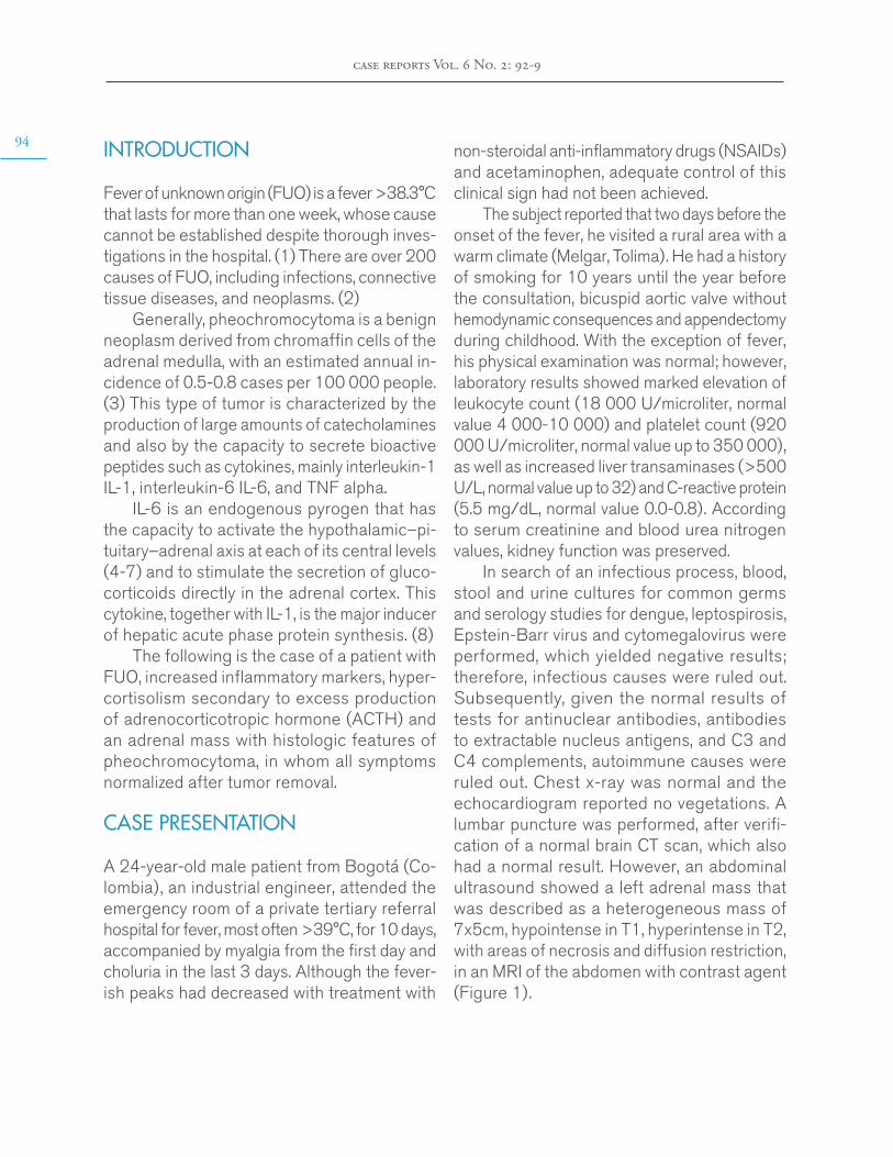
In search of an infectious process, blood, stool and urine cultures for common germs and serology studies for dengue, leptospirosis, Epstein-Barr virus and cytomegalovirus were performed, which yielded negative results; therefore, infectious causes were ruled out. Subsequently, given the normal results of tests for antinuclear antibodies, antibodies to extractable nucleus antigens, and C3 and C4 complements, autoimmune causes were ruled out. Chest x-ray was normal and the echocardiogram reported no vegetations. A lumbar puncture was performed, after verifi-cation of a normal brain CT scan, which also had a normal result. However, an abdominal ultrasound showed a left adrenal mass that was described as a heterogeneous mass of 7x5cm, hypointense in T1, hyperintense in T2, with areas of necrosis and diffusion restriction, in an MRI of the abdomen with contrast agent (Figure 1).
pheochromocytoma presenting as fever of unknown origin
95
Figure 1. MRI of the abdomen (axial plane). The arrow points to the left adrenal mass of 7cm in diameter. Source: Document obtained during the study.
At first, the fever was considered to be an incidentaloma but, since the fever persisted, tumor
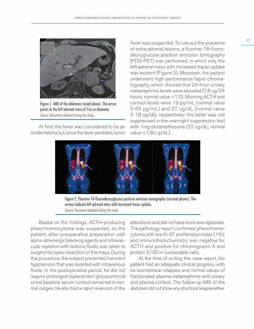
fever was suspected. To rule out the presence of extra adrenal lesions, a fluorine-18-fluoro-deoxyglucose positron emission tomography (FDG-PET) was performed, in which only the left adrenal mass with increased tracer uptake was evident (Figure 2). Moreover, the patient underwent high-performance liquid chroma-tography, which showed that 24-hour urinary metanephrine levels were elevated (7.8 ug/24 hours, normal value <1.0). Morning ACTH and cortisol levels were 19 pg/mL (normal value 5-65 pg/mL) and 27 ug/dL (normal value 5-18 ug/dl), respectively; the latter was not suppressed in the overnight suppression test with 1mg dexamethasone (22 ug/dL, normal value <1.8U g/dL).
Figure 2. Fluorine-18-fluorodeoxyglucose positron emission tomography (coronal planes). The arrows indicate left adrenal mass with increased tracer uptake. Source: Document obtained during the study.
Based on the findings, ACTH-producing pheochromocytoma was suspected, so the patient, after preoperative preparation with alpha-adrenergic blocking agents and intravas-cular repletion with isotonic fluids, was taken to surgery for open resection of the mass. During the procedure, the subject presented transient hypotension that was reverted with intravenous fluids. In the postoperative period, he did not require prolonged replacement glucocorticoid since baseline serum cortisol remained in nor-mal ranges; he also had a rapid reversion of the
alterations and did not have more ever episodes. The pathology report confirmed pheochromo-cytoma with low Ki-67 proliferation index (1%), and immunohistochemistry was negative for ACTH and positive for chromogranin A and protein S100 in sustainable cells.
At the time of writing this case report, the patient had an adequate clinical progress, with no biochemical relapses and normal values of fractionated plasma metanephrine and urinary and plasma cortisol. The follow-up MRI of the abdomen did not show any structural relapse either.
96
case reports Vol. 6 No. 2: 92-9
Given the age of the patient and the in-creased PET-FDG uptake of the adrenal mass, it is likely that the pheochromocytoma found was associated with a mutation in the succinate dehydrogenase enzyme. However, this diagno-sis was not confirmed because genetic testing could not be performed.
DISCUSSION
Pheochromocytomas are tumors derived from chromaffin cells of the adrenal medulla, charac-terized by increased and uncontrolled secretion of catecholamines, (9) which can also produce other biologically active peptides such as IL-6.
This cytokine is a glycoprotein known primarily for its role in innate immune response due to its pyrogenic and acute-phase protein inducing capacity. (10)
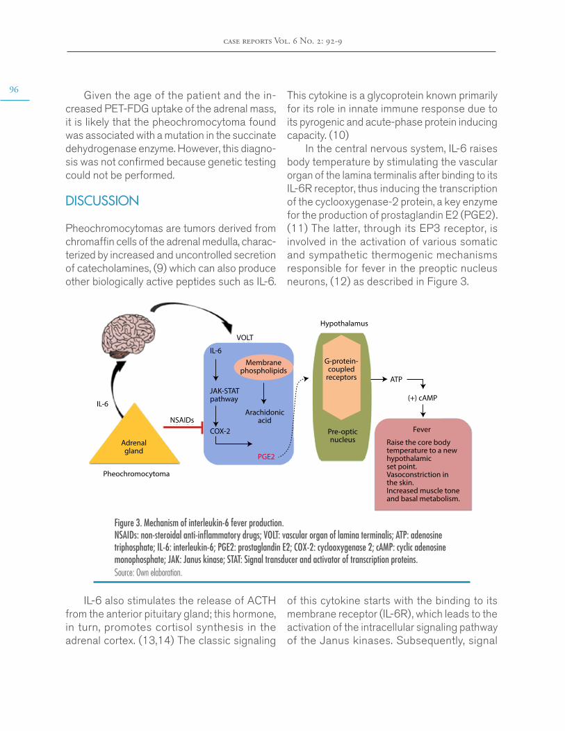
In the central nervous system, IL-6 raises body temperature by stimulating the vascular organ of the lamina terminalis after binding to its IL-6R receptor, thus inducing the transcription of the cyclooxygenase-2 protein, a key enzyme for the production of prostaglandin E2 (PGE2). (11) The latter, through its EP3 receptor, is involved in the activation of various somatic and sympathetic thermogenic mechanisms responsible for fever in the preoptic nucleus neurons, (12) as described in Figure 3.
Adrenalgland
Pheochromocytoma
NSAIDs
Membranephospholipids
Arachidonicacid
PGE2
IL-6
VOLT
Hypothalamus
ATP
(+) cAMP
G-protein-coupled
receptors
Pre-opticnucleus
Fever
Raise the core bodytemperature to a newhypothalamic set point.Vasoconstriction inthe skin.Increased muscle toneand basal metabolism.
IL-6
JAK-STATpathway
COX-2
Figure 3. Mechanism of interleukin-6 fever production. NSAIDs: non-steroidal anti-inflammatory drugs; VOLT: vascular organ of lamina terminalis; ATP: adenosine triphosphate; IL-6: interleukin-6; PGE2: prostaglandin E2; COX-2: cyclooxygenase 2; cAMP: cyclic adenosine monophosphate; JAK: Janus kinase; STAT: Signal transducer and activator of transcription proteins.Source: Own elaboration.
IL-6 also stimulates the release of ACTH from the anterior pituitary gland; this hormone, in turn, promotes cortisol synthesis in the adrenal cortex. (13,14) The classic signaling
of this cytokine starts with the binding to its membrane receptor (IL-6R), which leads to the activation of the intracellular signaling pathway of the Janus kinases. Subsequently, signal
pheochromocytoma presenting as fever of unknown origin
97transduction-activated transcription factors translocate into the nucleus and gene expression is induced, as in the case of pro-opiomelano-cortin (POMC), (15,16) which then generates ACTH by cleavage. In this way, it is possible to explain ACTH-dependent hypercortisolism, which was one of the biochemical alterations presented by the reported patient. It should be noted that, given the short time of evolution and the direct effect of IL-6 on corticotrophs, no suppression of the adrenal axis is expected when removing the IL-6 producing tumor, as in the reported case.
IL-6 hypersecretion also explains the changes in the leukocyte and platelet count, since it stimulates the differentiation of B and T lymphocytes (which causes leukocytosis) and intervenes in the proliferation and maturation of the megakaryocyte cell lines, generating thrombocytosis. In addition, it has proapoptotic effects on hepatocytes that generate hyper-transaminasemia. (17,18)
In the case presented herein, the feverish peaks decreased after using NSAIDs, which act by blocking the production of PGE2. (19,20) However, complete remission of fever was only possible after surgery, which also allowed nor-malizing inflammatory markers (leukocytosis, thrombocytosis, hypoalbuminemia and hyper-transaminasemia). This supports the role of IL-6 as responsible for the unusual manifestations observed in this case.
Although plasma levels could not be de-termined, the reversal of all inflammatory alter-ations after tumor removal and the review of similar clinical cases reported in the literature (21-28) allow us to infer that the secretion of IL-6 by a pheochromocytoma should be con-sidered as an explanation of the fever and the marked inflammatory response (thrombocyto-sis, transaminasemia, and ACTH-dependent hypercortisolism).
CONCLUSION
FUO is a condition with multiple causes, includ-ing tumors, which must be considered in the eti-ological diagnosis process. The presence of an IL-6 producing pheochromocytoma is a rather rare cause of FUO (less than 10 cases reported in the medical literature), so the description and publication of this report will allow clinicians to increase diagnostic suspicion and thus improve therapeutic interventions.
ETHICAL CONSIDERATIONS
This article was prepared after obtaining the patient’s informed consent to treat and disclose his medical history for scientific and academic purposes.
CONFLICT OF INTEREST
None stated by the authors.
FUNDING
None stated by the authors.
ACKNOWLEDGEMENTS
None stated by the authors.
REFERENCES
1. Brown I, Finnigan NA. Fever of Unknown Origin (FUO). In: StatPearls. Treasure Island: StatPearls Publishing; 2019.
2. Kantorovich V, Pacak K. Pheochromocytoma and paraganglioma. Prog Brain Res. 2010;182:343-73. http://doi.org/cpwzpv.
3. Cunha BA, Lortholary O, Cunha CB. Fever of unknown origin: a clinical approach. Am. J. Med. 2015;128(10):1138.e1-1138.e15. http://doi.org/f3jbfm.

98
case reports Vol. 6 No. 2: 92-9
4. Zarkovic M, Ignjatovic S, Dajak M, Ciric J, Beleslin B, Savic S, et al. Cortisol response to ACTH stimulation correlates with blood interleu-kin-6 concentration in healthy humans. Eur J Endo-crinol. 2008;159(5):649-52. http://doi.org/bvxpg4.
5. Navarra P, Tsagarakis S, Faria MS, Rees LH, Besser GM, Grossman AB. Interleukins-1 and -6 stimulate the release of corticotropin-releasing hormone-41 from rat hypothalamus in vitro via the eicosanoid cyclooxygenase pathway. Endocrino-logy. 1991;128(1):37-44. http://doi.org/dbd6cn.
6. Spinedi E, Hadid R, Daneva T, Gaillard RC. Cytokines stimulate the CRH but not the vasopres-sin neuronal system: evidence for a median emi-nence site of interleukin-6 action. Neuroendocrino-logy. 1992;56(1):46-53. http://doi.org/b4fshc.
7. Tsigos C, Papanicolaou DA, Defensor R, Mit-siadis CS, Kyrou I, Chrousos GP. Dose effects of recombinant human interleukin-6 on pituitary hormo-ne secretion and energy expenditure. Neuroendocri-nology. 1997;66(1):54-62. http://doi.org/c3b3fm.
8. Bethin KE, Vogt SK, Muglia LJ. Interleukin-6 is an essential, corticotropin-releasing hormone-in-dependent stimulator of the adrenal axis during immune system activation. Proc Natl Acad Sci U S A. 2000;97(16):9317-22. http://doi.org/dw2hjf.
9. López-Calderón A, Calderón MD, Tresgue-rres JAF. Médula suprarrenal. In: Pombo M, Audí L, Bueno M, Calzada R, Cassorla F, Diéguez C, et al., editors. Tratado de endocrinología pediátrica. 4th ed. Barcelona: McGraw-Hill; 2019.
10. Filella X, Molina R, Ballesta AM. Estructura y fun-ción de las citocinas. Med Integr. 2003;39(2):63-71.
11. Blomqvist A, Engblom D. Neural Mechanis-ms of Inflammation-Induced Fever. Neuroscientist. 2018;24(4):381-99. http://doi.org/gc7bmr.
12. Saavedra-Ramírez PG, Vásquez-Duque GM, González-Naranjo LA. Interleucina-6: ¿ami-ga o enemiga? Bases para comprender su utili-dad como objetivo terapéutico. Iatreia. 2011 [ci-ted 2019 Oct 11];24(2):157-66. Available from: https://bit.ly/380MDZ2.
13. Venihaki M, Dikkes P, Carrigan A, Karalis KP. Corticotropin-releasing hormone regulates IL-6 expression during inflammation. J. Clin. Invest. 2001;108(8):1159-66. http://doi.org/dqn8s2.
14. Contreras MG, Bonilla-Lara D, Pérez-Guerreo EE, Ruiz-Padilla AJ. Saucedo-Ulloa M, Sal-daña-Angiano JM, et al. Niveles altos de IL-6 asociados a efectos sistémicos y locales en la ar-tritis reumatoide. El Residente. 2015;10(1):38-42.
15. Melmed S, Jameson JL. Hipófisis anterior: fisio-logía de las hormonas hipofisarias. In: Kasper D, Fauci A, Hauser S, Longo D, Jameson JL, Loscalzo J, editors. Harrison. Principios de Medicina Interna. 19th ed. New York: McGraw-Hill; 2019 [cited 2020 Jun 25]. Available from: https://bit.ly/3dDCAdr.
16. Sokabe A, Mizooka M, Sakemi R, Koba-yashi T, Kishikawa N, Yokobayashi K. et al. Systemic Inflammatory Syndrome Associated with a Case of Jugular Paraganglioma. Intern Med. 2016;55(15):2105-8. http://doi.org/f8xxtc.
17. Willenberg HS, Päth G, Vögeli TA, Scher-baum WA, Bornstein SR. Role of interleukin-6 in stress response in normal and tumorous adrenal cells and during chronic inflammation. Ann N Y Acad Sci. 2002;966:304-14. http://doi.org/dnkcpf.
18. Toth B, Yokoyama Y, Schwacha MG, George RL, Rue LW, Bland KI, et al. Insights into the role of interleukin-6 in the induction of hepatic injury after trauma-hemorrhagic shock. J Appl Physiolo (1985). 2004;97(6):2184-2189. http://doi.org/b3fk6n.
19. Pérez-Guerra V, Ramírez-Cardona L, Ye-pes-Grajales OM, Vélez-Rivera JD, Marín-Zu-luaga JI. Falla hepática aguda sobre crónica. Rev Col Gastroenterol. 2016 [cited 2019 Oct 11];31(3):262-72. Available from: https://bit.ly/2VjbEtn.
20. Vilalta NP, de Puig AC. Gonadotropinas (LH y FSH) y corticotropina (ACTH). Endocrinol Nutr. 2007;54(2):109-17. http://doi.org/b3qfzg.
21. Tokuda H, Hosoi T, Hayasaka K, Okamura K, Yoshimi N, Kozawa O. Overexpression of protein kinase C-delta plays a crucial role in inter-leukin-6-producing pheochromocytoma presen-

pheochromocytoma presenting as fever of unknown origin
99ting with acute inflammatory syndrome: a case report. Horm Metab Res. 2009;41(4):333-8. http://doi.org/bn7zs4.
22. Suzuki K, Miyashita A, Inoue Y, Iki S, Eno-moto H, Takahashi Y, et al. Interleukin-6-Pro-ducing Pheochromocytoma. Acta Haematol. 1991;85(4):217-9. http://doi.org/dbtjch.
23. Shimizu C, Kubo M, Takano K, Takano A, Kijima H, Saji H, et al. Interleukin-6 IL-6 produ-cing pheochromocytoma: direct IL-6 suppression by non-steroidal anti-inflammatory drugs. Clin Endocrinol (Oxf). 2001;54(3):405-10. http://doi.org/bc4d7v.
24. Yarman S, Soyluk O, Altunoglu E, Tanakol R. Interleukin-6-producing pheochromocytoma pre-senting with fever of unknown origin. Clinics (Sao Paulo). 2011;66(10):1843-5. http://doi.org/dkps95.
25. Cheng X, Zhang M, Xiao Y, Li H, Zhang Y, Ji Z. Interleukin-6-producing pheochromocytoma
as a new reason for fever of unknown origin: a re-trospective study. Endocr Pract. 2018;24(6):507-511. http://doi.org/d2f6.
26. Siddiqui UM, Matta S, Wessolossky MA, Haas R. Fever of Unknown Origin: Could It Be a Pheochromocytoma? A Case Report and Review of the Literature. Case Rep Endocrinol. 2018;2018:3792691. http://doi.org/d2f8.
27. Minetto M, Dovio A, Ventura M, Cappia S, Daffar F, Terzolo M, et al. Interleukin-6 pro-ducing pheochromocytoma presenting with acu-te inflammatory syndrome. J Endocrinol Invest. 2003;26(5):453-7. http://doi.org/d2f9.
28. Carvalho-Cunha N, Gomes L, Saraiva J, Paiva I. Interleukin-6 Producing Pheochromo-cytoma: A Rare Cause of Systemic Inflammatory Response Syndrome. Case Rep Endocrinol. 2019;2019:7906272. http://doi.org/d2gb..

case reports 2020; 6(2)
ADULT ONSET STILL’S DISEASE (AOSD): A RARE CONDITION WITH A CLASSIC CLINICAL PRESENTATION. CASE REPORT
Keywords: Still’s Disease, Adult-Onset; Fever of Unknown Origin; Arthralgia; Exanthema.Palabras clave: Enfermedad de Still del adulto; Fiebre de origen desconocido; Artralgia;
Exantema.
Received: 12/11/2019 Accepted: 24/01/2020
Corresponding authorAndrés Eduardo Prieto-Torres.
Internal Medicine Service, Clínica Medifaca. Facatativá,
Cundinamarca. Colombia. Email: [email protected]
Andrés Eduardo Prieto-TorresClínica Medifaca - Internal Medicine Service -
Facatativá, Cundinamarca - Colombia.
https://doi.org/10.15446/cr.v6n2.83482
Wilson Suárez-MolinaClínica Medifaca - Internal Medicine Service -
Facatativá, Cundinamarca - Colombia.Hospital Militar Central - Internal Medicine Service - Bogotá D.C. - Colombia.
Jaime Iván Pantoja-AgredaClínica Medifaca - Internal Medicine Service -
Facatativá, Cundinamarca - Colombia.
adult onset still’s disease (aosd)
101RESUMEN
Introducción. La enfermedad de Still del adulto (ESA) es una enfermedad inflamatoria sistémica de baja incidencia y prevalencia en población general y cuya etiología aún no es clara. La ESA puede causar fiebre de origen desconocido has-ta en el 20% de los casos, pero suele pasar inadvertida dentro de los diagnósticos diferen-ciales iniciales debido a su desconocimiento, lo que empeora el pronóstico y aumenta las complicaciones en los pacientes.
Presentación del caso. Paciente femenina de 32 años con síndrome febril prolongado que no respondía a tratamientos antimicrobianos instaurados previamente y en quien, finalmen-te, se diagnosticó ESA aplicando los criterios clasificatorios de Yamaguchi. La mujer recibió tratamiento de primera línea con corticosteroi-des y obtuvo buenos resultados.
Conclusiones. La ESA requiere un exhaustivo proceso para su diagnóstico, en el cual, a pesar de la disponibilidad de herramientas diagnósticas avanzadas, la verificación de la historia clínica y la realización de un adecuado examen físico son los aspectos más importantes a tener en cuenta.
ABSTRACT
Introduction: Adult Onset Still´s Disease (AOSD) is a rare systemic inflammatory disease of unclear etiology, with low incidence and prev-alence among the general population. AOSD is a common cause of fever of unknown origin (FUO) in up to 20% of cases. Due to the scarce knowledge about this disease and its diagnosis, it is usually unrecognized in the differential diag-noses, worsening the prognosis and increasing complications in some patients.
Case presentation: This is the case of a 32-year-old female patient with prolonged febrile illness, who did not respond to the antimicrobial treatments previously established. She was diag-nosed with AOSD according to the Yamaguchi criteria after an extensive exclusion process. She was treated with first-line treatment with corticosteroids, achieving satisfactory results
Conclusions: The diagnosis of AOSD is an exhaustive process. Regardless of the availability of cutting-edge diagnostic tools, the medical history of the patient and an adequate physical examination are the most important aspects to consider.
INTRODUCTION
Adult Onset Still’s disease (AOSD) is a rare in-flammatory disease of unclear origin. Its incidence and prevalence vary according to the geographi-cal area: 0.16 to 0.4 cases per 100 000 popula-tion and 1 to 34 cases per 1 000 000 population, respectively. (1) This is a disorder that affects men and women alike. (2,3)
Although the pathogenesis of AOSD is not entirely clear, it has been suggested that it is an uncontrolled inflammatory response in the form
of a “cytokine storm” that occurs when individuals with non-inherited genetic susceptibility asso-ciated with different genes of human leukocyte antigen and polymorphisms in genes coding for interleukin 18 (IL-18) and macrophage migration inducer are subjected to a trigger or “second hit” such as viruses, bacteria, and hematological, or solid tumors. Such a response would cause the multi-systemic symptoms associated with the disease. It should be noted that, even though
102
case reports Vol. 6 No. 2: 100-8
this is one of the most widely disseminated hypotheses, it is still debated. (4,5)
AOSD is a little known disease that is not included in the initial differential diagnosis of the febrile illness, leading to a delay in the diagnosis, the initiation of unnecessary phar-macological treatments that may cause adverse effects, and costly complementary studies that are not useful or comfortable for the patient.
This disease is characterized by a triad of fever, evanescent maculopapular exanthema and arthralgia:
• Fever is generally high (>39°C), with a double quotidian pattern, and usually increases in the evening and at night. This clinical sign tends to resolve on its own or rapidly with antipyretics and precedes the onset or exacerbation of other systemic symptoms. (6,7)
• The exanthema typically described in the literature is an evanescent salmon-pink maculopapular rash, occasionally pruritic, predominantly on the trunk and proximal parts of the limbs. Atypical presentations with urticarial exanthema or with persistent erythematous plaques and papules have also been described. (8)
• Multiple joints may be initially involved, pre-dominantly the knees, ankles, and wrists, with subsequent progression to small joints.
Other manifestations of AOSD include odynophagia, splenomegaly, lymphadenopathy, and hepatomegaly. (7,9)
Macrophage activation syndrome is the complication most associated with AOSD, followed by disseminated intravascular coag-ulation, thrombotic thrombocytopenic purpura, and diffuse alveolar hemorrhage. Other less known complications with isolated case reports are serositis, pericardial effusion, pleural effu-sion, abdominal pain, pulmonary involvement, myocarditis, (10) and neurological and kidney complications.
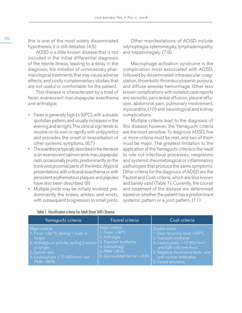
Multiple criteria lead to the diagnosis of this disease; however, the Yamaguchi criteria are the most sensitive. To diagnose AOSD, five or more criteria must be met, and two of them must be major. The greatest limitation to the application of the Yamaguchi criteria is the need to rule out infectious processes, neoplasms and systemic rheumatological or inflammatory pathologies that produce the same symptoms. Other criteria for the diagnosis of AOSD are the Fautrel and Cush criteria, which are less known and barely used (Table 1). Currently, the course and treatment of the disease are determined based on whether the patient has a predominant systemic pattern or a joint pattern. (11)
Table 1. Classification criteria for Adult Onset Still’s Disease.
Yamaguchi criteria Fautrel criteria Cush criteria
Major criteria: 1. Fever >39 °C, lasting 1 week or
longer. 2. Arthralgia or arthritis, lasting 2 weeks
or longer. 3. Typical rash. 4. Leukocytosis >10 000/mm3 con
PMN >80%.
Major criteria: 1. Fever >39°C. 2. Arthralgia. 3. Transient erythema. 4. Odinophagy. 5. PMN >80%. 6. Glycosylated ferritin <20%.
Double score: 1. Daily recurring fever >39°C. 2. Transient erythema. 3. Leukocytosis >12 000/mm3
and ESR >40 mm/hour. 4. Negative rheumatoid factor and
anti-nuclear antibodies. 5. Carpal ankylosis.
adult onset still’s disease (aosd)
103Table 1. Classification criteria for Adult Onset Still’s Disease. (continued)
Yamaguchi criteria Fautrel criteria Cush criteria
Minor criteria: 1. Sore throat. 2. Lymphadenopathy. 3. Splenomegaly. 4. Abnormal liver function tests. 5. Negative tests for antinuclear anti-
body and rheumatoid factor.
Minor criteria: 1. Leukocytosis >10 000/mm3. 2. Maculopapular rash.
Simple score: 1. Onset <35 years old. 2. Arthritis. 3. Sore throat. 4. Reticuloendothelial system
involvement. 5. Serositis. 6. Cervical or tarsal ankylosis
Exclusion criteria: Infections, malignancies, other rheu-matic or inflammatory diseases.
It does not require the exclu-sion of other pathologies.
Probable: 10 points after 12 weeks of observation. Definitive: 10 points after 6 months of follow-up.
PMN: polymorphonuclear cells; ESR: erythrocyte sedimentation rate. Source: Elaboration based on Giacomelli et al. (1) y Li et al. (2)
hospital after being referred from a primary care center because of a 15-day history of prolonged febrile illness without a clear description of the pattern. She presented migratory polyarthralgias, predominantly in wrists and ankles, and inter-mittent pruritic salmon-pink macular exanthema mainly in the trunk, upper limbs and proximal part of lower limbs; according to the patient, the exanthema appeared after experiencing febrile peaks, myalgias and intense odynophagia.
The patient had been previously treated with a single dose of intramuscular benzathine penicillin for suspected tonsillopharyngitis caused by Streptococcus pyogenes. Moreover, five days before being admitted, she was hospi-talized and treated with antibacterial spectrum of ampicillin/sulbactam for suspected urinary tract infection; since she did not respond to the treatment and the fever peaks and leukocytosis persisted, she was transferred to be assessed by the internal medicine service.
No fever or changes in vital signs were re-ported in the initial physical examination. There were no signs of an inflammatory process in the tonsils, but painless and mobile posterior cervical lymphadenopathies were palpated,
The first-line treatment for AOSD involves anti-inflammatory drugs and steroids. In 60% of the cases, the disease is controlled with glu-cocorticoids, and patients who do not respond to this therapy may be treated with disease modifying drugs, including methotrexate, which is the one with more evidence. (1) Biologic re-sponse modifiers are recommended in cases where the first or second line of treatment does not work, or in patients with contraindications to the use of these drugs. When the pattern is systemic, medications are initiated to control IL-1B (anakinra, canakinumab, or rilonacept) and IL-18 (tadekinig); if the condition has a predominantly joint pattern, anti-IL-6 (tocilizumab) and anti-TNF (etanercept) therapy should be initiated.
CASE PRESENTATION
This is the case of a 32 year-old female patient with no relevant medical history, mestizo, single, with complete high school education, head of household, mother to two adolescents, worker in a flower farm, from a rural area of a munici-pality of Cundinamarca (Colombia) located at 2 580 m.a.s.l. The patient was admitted to the

104
case reports Vol. 6 No. 2: 100-8
and pink exanthemas that disappeared under pressure were observed on thighs and fore-arms. Upon admission, studies were initiated for fever of unknown origin (FUO), antimicrobial and antipyretic management was suspended, and analysis was oriented towards neoplastic, inflammatory, rheumatological and infectious causes that could explain the symptoms re-ferred by the patient.
Initial laboratory tests showed a hemogram with a marked increase in the leukocyte count (25 750 leukocytes/mm3) and predominance
of polymorphonuclear cells (93.5%). Moreover, the levels of C-reactive protein, aspartate ami-notransferase and alanine aminotransferase were 25.4 mg/dl, 70.5 U/L and 228 U/L, respectively. A chest X-ray was taken, reveal-ing an unclear image of retrocardiac alveolar infiltrate (Figure 1). Based on this initial profile, and since the patient completely denied any respiratory or urinary symptoms, additional blood chemistry tests and a thoracoabdominal CT scan were ordered to rule out solid tumors and lymph node involvement.
A B
Figure 1. Chest X-ray on patient’s admission. A) anteroposterior plane without evidence of relevant alterations; B) lateral plane with segmental atelectasis.Source: Document obtained during the study.
During hospitalization, the patient present-ed with feverish peaks >39°C at around dusk and at night, which made the limb exanthema more noticeable and responded quickly to man-agement with non-steroidal anti-inflammatory drugs. Additional tests ruled out infections by human immunodeficiency virus, syphilis, hep-atitis B and hepatitis C; the thick blood smear was negative for hemoparasites, the globular sedimentation rate was established at 48
mm/hour, lactate dehydrogenase was 431.7 U/L, and the peripheral blood smear did not report alterations in red cells or platelets, but indicated elevated white blood cell count with neutrophilic predominance.
Based on the reported management that the patient received prior to her arrival at the hospital, diagnostic suspicion began to shift towards AOSD or, less likely, acute rheu-matic fever. Tests for antinuclear antibodies,
adult onset still’s disease (aosd)
105rheumatoid factor, and antistreptolysin were negative, as were blood cultures for aerobic or anaerobic bacterial growth. An echocardiogram was performed, ruling out subclinical carditis, which was described in the 2015 revision of the Jones criteria for rheumatic fever. (12,13)
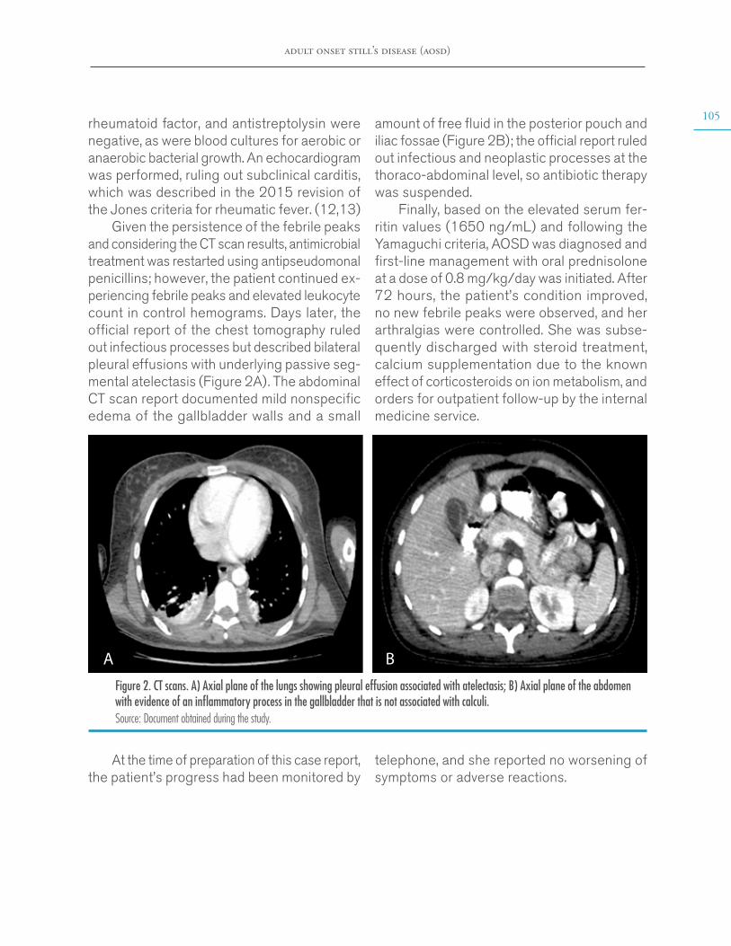
Given the persistence of the febrile peaks and considering the CT scan results, antimicrobial treatment was restarted using antipseudomonal penicillins; however, the patient continued ex-periencing febrile peaks and elevated leukocyte count in control hemograms. Days later, the official report of the chest tomography ruled out infectious processes but described bilateral pleural effusions with underlying passive seg-mental atelectasis (Figure 2A). The abdominal CT scan report documented mild nonspecific edema of the gallbladder walls and a small
amount of free fluid in the posterior pouch and iliac fossae (Figure 2B); the official report ruled out infectious and neoplastic processes at the thoraco-abdominal level, so antibiotic therapy was suspended.
Finally, based on the elevated serum fer-ritin values (1650 ng/mL) and following the Yamaguchi criteria, AOSD was diagnosed and first-line management with oral prednisolone at a dose of 0.8 mg/kg/day was initiated. After 72 hours, the patient’s condition improved, no new febrile peaks were observed, and her arthralgias were controlled. She was subse-quently discharged with steroid treatment, calcium supplementation due to the known effect of corticosteroids on ion metabolism, and orders for outpatient follow-up by the internal medicine service.
A B
Figure 2. CT scans. A) Axial plane of the lungs showing pleural effusion associated with atelectasis; B) Axial plane of the abdomen with evidence of an inflammatory process in the gallbladder that is not associated with calculi.Source: Document obtained during the study.
At the time of preparation of this case report, the patient’s progress had been monitored by
telephone, and she reported no worsening of symptoms or adverse reactions.
106
case reports Vol. 6 No. 2: 100-8
DISCUSSION
AOSD is a rare inflammatory disorder whose inci-dence and prevalence have not been reported in Colombia. This is an understudied disease that is generally not considered in the initial differential approach to patients with prolonged febrile syn-drome given the limited knowledge of its clinical manifestations and treatment, even though it is a common documented cause of FUO.
The case presented here illustrates, precisely, the typical diagnostic triad of the disease: febrile peaks, posterior exacerbated exanthema, and joint involvement in the appendicular skeleton, in addition to the characteristic serological bio-markers. Furthermore, as a novelty, the patient presented with an inflammatory process in the gallbladder, which was not associated with calculi and had not been previously reported; this is an incidental finding that serves as a precedent for follow-up in future cases related to this pathology. This case is an example of the procedure and diagnostic method used for this disease, which is within the group of pathologies to be ruled out in the diagnosis of FUO according to the definition originally proposed by Petersdorf and Beeson, cited by Cunha et al. (14)
Regarding the available literature on AOSD, there are multiple articles worldwide on new findings concerning its pathogenesis, course, clinical markers, and treatment. (15,16) In Latin America, mainly in Brazil, Argentina, Chile, and Peru, there are a large number of case reports about its typical and atypical characteristics, and its more common complications. (17-20) Specifically in Colombia, updated literature is scarce, and there are only two retrospective studies on case series that analyze the response to monotherapy and combined treatment, (21,22) namely, a review article on its history and patho-physiology (23) and a case report that briefly
describes the diagnostic process. (24) It should be noted that there is an additional report of a Colombian case published abroad. (18)
This case report attempts to provide more detailed and meticulous information about the diagnosis of AOSD by exposing the complexity of this process and highlighting common errors in the interventions performed on patients. Sim-ilarly, it stresses the importance of considering this pathology as a possible cause of the febrile syndrome and the fact that it is not necessary to carry out specialized laboratory tests to make the initial approach to the patient; in fact, a good anamnesis could lead to the suspicion of this disease and prevent the indiscriminate use of antibiotics or other types of interventions.
The authors consider that the publication of this typical case of AOSD contributes to the description of the classic characteristics that guide the diagnosis of the disease and to enrich the Colombian and world literature on the subject, since there are still large gaps in terms of diagnostic tools for this disease in which further research is needed.
CONCLUSION
Although a wide variety of laboratory tests and advanced imaging are available today, the pa-tient’s medical history and a detailed physical examination remain the cornerstone of AOSD diagnosis, making it a challenge to physicians. In this sense, the case presented here demon-strates the difficulty of its diagnosis and the importance of its early recognition to avoid com-plications and improve the prognosis of patients.
PATIENT’S PERSPECTIVE
The patient had trouble during her hospital stay due to the length of her stay and uncertainty about her diagnosis. In addition, the woman had
adult onset still’s disease (aosd)
107concerns about her financial responsibilities as a stay-at-home mother and anxiety about being away from her children, so she required support from the psychology service in order to cope with her condition and the hospital management.
ETHICAL CONSIDERATIONS
This case report was approved by the Ethics Committee of Clínica Medifaca, with the patient’s informed consent. The institution where the di-agnosis and follow-up were made is part of the Medilaser complex.
CONFLICTS OF INTEREST
None stated by the authors.
FUNDING
None stated by the authors.
ACKNOWLEDGEMENTS
To the medical research group that reviewed and endorsed this document.
REFERENCES
1. Giacomelli R, Ruscitti P, Shoenfeld Y. A com-prehensive review on adult onset Still’s disease. J Autoimmun. 2018;93:24-36. http://doi.org/dvd5.
2. Li S, Zheng S, Tang S, Pan Y, Zhang S, Fang H, et al. Autoinflammatory Pathogenesis and Targeted Therapy for Adult-Onset Still’s Disease. Clin Rev Allerg Immunol. 2020;58(1). http://doi.org/dvd7.
3. Sfriso P, Bindoli S, Galozzi P. Adult-Onset Sti-ll’s Disease: Molecular Pathophysiology and The-rapeutic Advances. Drugs. 2018;78(12):1187-95. http://doi.org/gd5549.
4. Ruscitti P, Giacomelli R. Pathogenesis of adult onset still’s disease: current understanding and new
insights. Expert Rev Clin Immunol. 2018;14(11):965-76. http://doi.org/dvd8.
5. Gerfaud-Valentin M, Jamilloux Y, Iwaz J, Sève P. Adult-onset Still’s disease. Autoimmun Rev. 2004;13(7):708-22. http://doi.org/c8ht.
6. Agha-Abbaslou M, Bensaci AM, Dike O, Poznansky MC, Hyat A. Adult-Onset Still’s Di-sease: Still a Serious Health Problem (a Case Re-port and Literature Review). Am J Med Case Rep. 2017;18:119-24. http://doi.org/f9pcwx.
7. Colafrancesco S, Priori R, Valesini G. Pre-sentation and diagnosis of adult-onset Still’s di-sease: the implications of current and emerging markers in overcoming the diagnostic challenge. Expert Rev Clin Immunol. 2015;11(6):749-61. http://doi.org/f7mmrj.
8. Díaz P, Hidalgo-Parra I, Benavidez V, Cae-tano M, Reppel J, Aloise I. Enfermedad de Still del Adulto: diagnóstico a partir de las mani-festaciones cutáneas. Med Cutan Iber Lat AM. 2015;43(3):196-8.
9. Narváez-García F, Pascual M, López-de Re-calde M, Juarez P, Morales-Ivorra I, Notario J, et al. Adult-onset Still’s disease with atypical cutaneous manifestations. Medicine (Baltimore). 2017;96(11):e6318. http://doi.org/f9vf5f.
10. Peralta-Vargas CE, Vasquez-Kunze S. Mio-pericarditis en enfermedad de Still del adulto. Reporte de un caso. Rev Med Hered. 2018;19 (4):167.
11. Narváez J. Enfermedad de Still del adulto. Med Clin (Barc). 2018;150(9):348-53. http://doi.org/dvd9.
12. Karthikeyan G, Guilherme L. Acute rheu-matic fever. Lancet. 2018;392(10142):161-74. http://doi.org/ggtnvq.
13. Szczygielska I, Hernik E, Kotodziejczyk B, Gazda A, Maslinska M, Gietka P. Rheumatic fever - new diagnostic criteria. Rheumatologia. 2018;56(1):37-41. http://doi.org/ggtnzq.
14. Cunha BA, Lortholary O, Cunha CB. Fever of Unknown Origin: A Clinical Approach. Am J Med. 2015;128(10):1138e1-e15. http://doi.org/f3jbfm.
108
case reports Vol. 6 No. 2: 100-8
15. Mitrovic S, Fautrel B. New Markers for Adult-Onset Still’s Disease. Joint Bone Spine. 2018;85(3):285-93. http://doi.org/gdq4zm.
16. Feist E, Mitrovic S, Fautrel B. Mechanisms, biomarkers and targets for adult-onset Still’s di-sease. Nat Rev Rheumatol. 2018;14(10):603-18. http://doi.org/gfb4p3.
17. Balcázar R, Garate G, Brigante A, Gómez G, Hogrefe J, Yucra D, Hamaui A, Duvins-ky D. Manifestación atípica de enfermedad de Still. Reporte de un caso. Rev Arg Reumatol. 2018;29(2):54-6.
18. Posada-López AF, Yepes-Gaviria V, Agui-rre-Henao HD, Quevedo-Cámera ML. Enfer-medad de Still del adulto asociado a compromiso pulmonar. Rev Arg Reumatol. 2014;25(2):42-6.
19. Suárez.Ale H, Solís-Torres S. Enfermedad de Still del Adulto asociada a endocarditis marántica: Reporte clínico y revisión de la literatura. Rev Soc Peru Med Interna. 2015;28 (1):18-24.
20. Peruilh-Bagolini L, Tapia-Tudela G, Pe-tit-Breuilh V, Valenzuela F, Carreño L. En-
fermedad de Still del adulto, a propósito de un caso: Un desafío diagnóstico. Rev Chil Dermatol. 2016;32(4):197-201. http://doi.org/dvfb.
21. Muriel ÁJ, Rueda JM, González-Buriticá H, Castaño O. Una patología poco frecuente: la en-fermedad de Still del adulto. Experiencia clínica con 17 casos. Rev Colomb Reumatol. 2016;23(2):126-30. http://doi.org/c8hz.
22. Panqueva U, Ramírez LA, Restrepo JF, Rondón F, Mora S, Valle R, et al. Enferme-dad de Still del adulto: estudio de cohorte. Rev Col Reumatol. 2009;16(4):336-41. http://doi.org/f2mbbp.
23. Iglesias A, Panqueva U, Toro C, Mejía J, Rondón F, Restrepo JF, et al. Enfermedad de Still: Una perspectiva histórica y una revi-sión actual. Rev Colomb Reumatol. 2008;15(3): 197-206
24. Quilindo C, Morales K, Guerrero A. Enferme-dad de Still, un diagnóstico diferencial importante: Reporte de un Caso. RFS Revista Facultad de Sa-lud. 2017;9(1):21-5. http://doi.org/dvfc.
case reports 2020; 6(2)
ATAXIA TELANGIECTASIA: A DIAGNOSTIC CHALLENGE. CASE REPORT
Keywords: Ataxia Telangiectasia; Neurodegenerative Diseases; Cerebellar Ataxia; Spinocerebellar Degenerations; Telangiectasia.
Palabras clave: Ataxia telangiectasia; Enfermedades neurodegenerativas; Ataxia cerebelosa; Degeneraciones espinocerebelosa; Telangiectasia.
Received: 28/10/2019 Accepted: 08/01/2020
Corresponding authorNatalia Martínez-Córdoba. Faculty of Medicine,
Universidad Militar Nueva Granada. Bogotá D.C. Colombia. Email: [email protected].
Natalia Martínez-CórdobaUniversidad Militar Nueva Granada
- Faculty of Medicine - Pediatric Neurology Research Group - Bogotá D.C. - Colombia.
Hospital Militar Central - Pediatric Neurology Service - Bogotá D.C. - Colombia.
https://doi.org/10.15446/cr.v6n2.83219
Eugenia Espinosa-GarcíaUniversidad Militar Nueva Granada
- Faculty of Medicine - Pediatric Neurology Research Group - Bogotá D.C. - Colombia.
Hospital Militar Central - Pediatric Neurology Service - Bogotá D.C. - Colombia.
Universidad del Rosario - Medical School - Bogotá D.C. - Colombia.
110
case reports Vol. 6 No. 2: 109-17
RESUMEN
Introducción. La ataxia-telangiectasia (AT) es un síndrome neurodegenerativo con baja inciden-cia y prevalencia mundial que es causado por una mutación del gen ATM, es de herencia autosó-mica recesiva y se asocia a mecanismos defec-tuosos en la regeneración y reparación del ADN. Este síndrome se caracteriza por la presencia de ataxia cerebelosa progresiva, movimientos ocula-res anormales, telangiectasias oculocutáneas e inmunodeficiencia. El diagnóstico oportuno de la AT es muy importante para poder iniciar un manejo interdisciplinario temprano, mejorar la sintomato-logía aguda y controlar las múltiples comorbilida-des que causa. A continuación se presenta el caso de una paciente con las características clásicas de esta enfermedad y una adecuada respuesta y evolución al manejo médico instaurado.
Presentación de caso. Paciente femenina de 7 años de edad, procedente de Bogotá, quien presentó cuadro clínico inicial de retraso global del neurodesarrollo, ataxia cerebelosa, infec-ciones respiratorias frecuentes y telangiectasias oculares. La sintomatología se asoció a elevación de alfa fetoproteína e inmunodeficiencia, lo que permitió plantear el diagnóstico de AT e iniciar de manera oportuna el manejo interdisciplinario.
Conclusión. La AT es un síndrome de inestabili-dad cromosómica con signos clínicos y síntomas característicos, por lo que es primordial conocer la etiopatogenia, el cuadro clínico, los criterios diag-nósticos y las propuestas terapéuticas, pues la detección y la sospecha clínica temprana pueden favorecer el manejo precoz de las diferentes co-morbilidades y mejorar el curso progresivo.
ABSTRACT
Introduction: Ataxia-telangiectasia (AT) is a neurodegenerative syndrome with low incidence and prevalence worldwide, which is caused by a mutation of the ATM gene. It is an autosomal re-cessive disorder that is associated with defective cell regeneration and DNA repair mechanisms. It is characterized by progressive cerebellar atax-ia, abnormal eye movements, oculocutaneous telangiectasias and immunodeficiency. Early diagnosis is critical to initiate a timely interdis-ciplinary treatment, improve acute symptoms, and control the multiple comorbidities of the disease. The following is the case of a patient who presented with the aforementioned char-acteristics and had an adequate response to the established medical treatment.
Case presentation: A 7-year-old female pa-tient from Bogotá, who presented clinical signs of global neurodevelopmental delay, cerebellar ataxia, frequent respiratory infections and ocu-lar telangiectasias. Symptoms were associated with elevation of alpha fetoprotein and immu-nodeficiency, which allowed for a diagnosis of AT and the initiation of a timely interdisciplinary treatment.
Conclusion: AT is a chromosomal instability syndrome with characteristic signs and symp-toms. It is essential to know the etiopathogene-sis, clinical manifestations, diagnostic criteria, and therapeutic options, emphasizing that ear-ly detection and clinical suspicion could favor the proper management of the comorbidities and improve the progressive course of the disease.

ataxia telangiectasia: a diagnostic challenge
111INTRODUCTION
Ataxia-telangiectasia (AT), also known as Bod-er-Sedgwick syndrome or Louis-Bar syndrome, is a multisystemic, neurodegenerative, autoso-mal recessive disease associated with the ATM gene mutation (A-T-mutated), which is located on the long arm of chromosome 11 (11q22-23). (1) This syndrome affects multiple organs of the body and has moderate and severe long-term sequelae.
AT is characterized by progressive neu-rological dysfunction with multisystemic alterations and predisposition to cancer. The characteristic symptoms of this disease usually appear early in childhood and include cere-bellar ataxia, oculomotor apraxia, chorea, and cognitive impairment. (2) Moreover, the cells of AT patients present chromosomal instability, hypersensitivity to X-rays, propensity to lym-phoid neoplasms, variable immunodeficiency, and susceptibility to infections, which cause systemic symptoms such as endocrinopathies, leukemias, radiosensitivity and ocular telangi-ectasias. (3,4)
While over 400 mutations of the ATM gene have been detected, it has been reported that AT has an incidence of 1 between 40 000 and 100 000 people (5) and that its frequency of heterozygosity in the mutant allele is 1.4% to 2% in the general population. (1,3)
This disease is usually diagnosed late, as it is only identifiable when patients have severe symptoms that begin with alterations in the motor system. In addition, it is often misdiag-nosed as cerebral palsy. (6)
So far there is no specific treatment for AT, so its management is palliative and supportive. (7) Patients with this disease should participate in daily and social integration activities, always trying to maintain their quality of life.
CASE PRESENTATION
Mestizo, female patient, 7 years old, from Bogotá D.C., Colombia, and the product of a second pregnancy by young, non-blood related, mid-dle-class parents. The girl had no significant pre-natal history and was born by cesarean section at 35 weeks of gestation. Her weight at birth was 1 800gr and her size was 40cm; the APGAR score was 4/10, 7/10 and 7/10 at 1 minute, 5 minutes and 10 minutes, respectively. She also presented with neonatal hypoxia, requiring mechanical ventilation for 24 hours and hospi-talization in the neonatal intensive care unit for 20 days.
The child’s psychomotor development was normal until the age of 20 months, at which time it was evident that there was language delay, café-au-lait spots and unsteady gait. At 31 months, she was referred to the Neuropedi-atrics Service, which requested magnification and brain magnetic resonance imaging (MRI) studies; management with physical and lan-guage therapy was initiated.
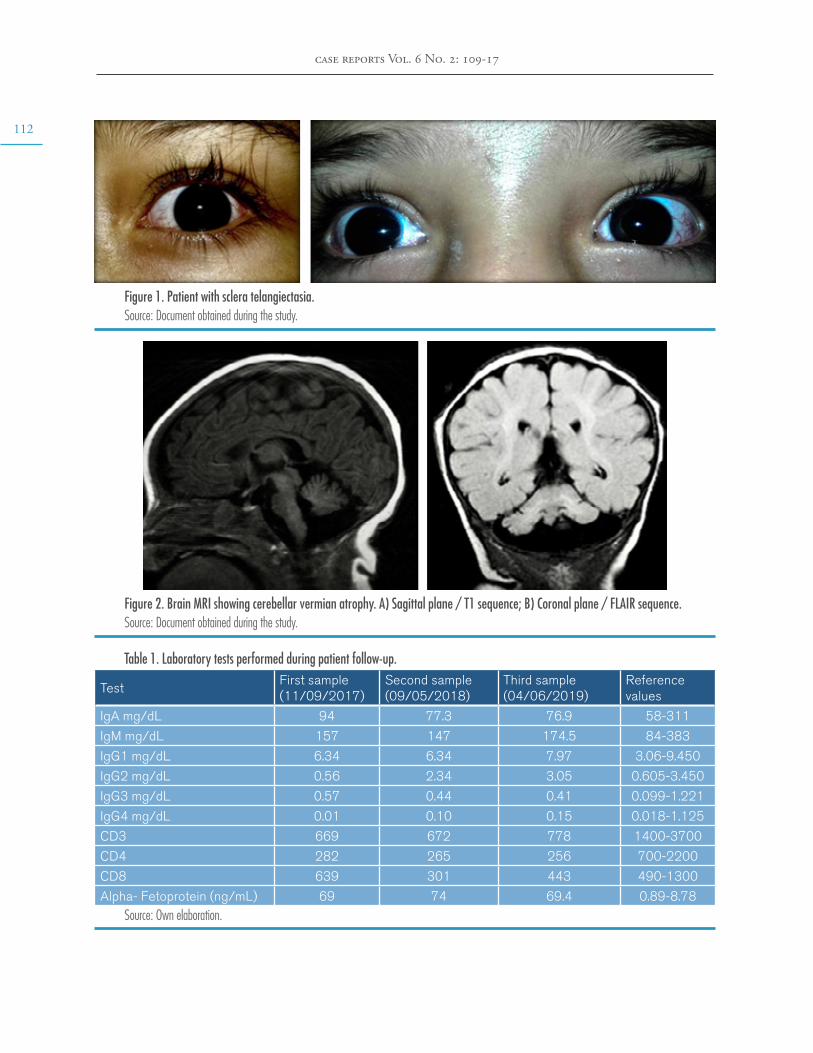
At 5 years of age, the patient was taken to the emergency department due to a possible focal onset impaired awareness seizure. During the interview, psychomotor and language delays were observed, as well as multiple respirato-ry and gastrointestinal tract infections. The neurological examination revealed bradylalia, bradypsychia, ataxic gait with enlargement of the support polygon, presence of oculomotor apraxia, dysmetria and telangiectasias in sclera (Figure 1), so more specific tests were requested. Finally, since MRI showed cerebellar atrophy (Figure 2), video-electroencephalography (EGG) was normal, alpha-fetoprotein (AFP) levels were elevated, and immunological tests were altered (Table 1), a diagnosis of AT was suggested.
112
case reports Vol. 6 No. 2: 109-17
Figure 1. Patient with sclera telangiectasia.Source: Document obtained during the study.
Figure 2. Brain MRI showing cerebellar vermian atrophy. A) Sagittal plane / T1 sequence; B) Coronal plane / FLAIR sequence.Source: Document obtained during the study.
Table 1. Laboratory tests performed during patient follow-up.
TestFirst sample (11/09/2017)
Second sample (09/05/2018)
Third sample (04/06/2019)
Reference values
IgA mg/dL 94 77.3 76.9 58-311
IgM mg/dL 157 147 174.5 84-383
IgG1 mg/dL 6.34 6.34 7.97 3.06-9.450
IgG2 mg/dL 0.56 2.34 3.05 0.605-3.450
IgG3 mg/dL 0.57 0.44 0.41 0.099-1.221
IgG4 mg/dL 0.01 0.10 0.15 0.018-1.125
CD3 669 672 778 1400-3700
CD4 282 265 256 700-2200
CD8 639 301 443 490-1300
Alpha- Fetoprotein (ng/mL) 69 74 69.4 0.89-8.78Source: Own elaboration.
ataxia telangiectasia: a diagnostic challenge
113The child was assessed by the Pediatric
Immunology Service, which considered cellular and humoral immunodeficiency (IgG2 and IgG4 deficiency) , so she started treatment with gamma globulin IV every 28 days. She was also assessed by the pediatric hema-tology-oncology service due to bicytopenia (leukopenia+thrombocytopenia) during a hospital stay without evidence of neoplastic alteration. The patient continued attending child psychiatry and physical, occupational and language therapy.
The specialties of pediatric neurology and clinical genetics gathered and concluded that it was not necessary to perform a genetic study since AT could be diagnosed due to the elevat-ed AFP levels, the evolution of the symptoms,
the findings on physical examination and the associated comorbidities.
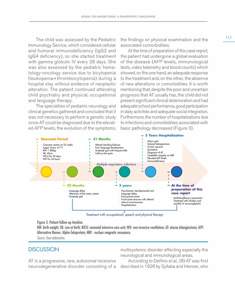
At the time of preparation of this case report, the patient had undergone a global evaluation of the disease (AFP levels, immunological tests, video telemetry and blood counts) which showed, on the one hand, an adequate response to the treatment and, on the other, the absence of new alterations or comorbidities. It is worth mentioning that, despite the poor and uncertain prognosis that AT usually has, the child did not present significant clinical deterioration and had adequate school performance, good participation in daily activities and adequate social integration. Furthermore, the number of hospitalizations due to infections and comorbidities associated with basic pathology decreased (Figure 3).
Figure 3. Patient follow-up timeline. BW: birth weight; SB: size at birth; NICU: neonatal intensive care unit; NIV: non-invasive ventilation; AT: ataxia telangiectasia; AFP: Alternative Names: Alpha-fetoprotein; MRI - nuclear magnetic resonance. Source: Own elaboration.
Neonatal Period 31 Months5 Years Hospitalization
20 Months 5 years At the time ofpreparation of thiscase report
Treatment with occupational, speech and physical therapy
Multiple respiratory infections
Cesarean section at 35 weeksApgar Score: 4-7-7BW: 1 800grSB: 40cmNICU for 20 daysNIV for 24 hours
······
Altered standing balancePoor language developmentUnsteady gait with frequent fallsCafé-au-lait spots
····
Ataxic gaitSclereal telangiectasiaOcular apraxiaDysmetriaDiagnosis of ATCerebellar atrpphy on MRIElevated AFP levelsImmunide�ciency
········
Psycchomotr, developmental and language delayParoxysmal eventsFocal onset seizures with altered state of consciousnessHospitalization
·
····
Language delayAlteration of the motor systemUnsteady gait
···
Multidisciplonary assessmentTreatment with inhalers and monthly IV immunoglobulin
··
DISCUSSION
AT is a progressive, rare, autosomal recessive neurodegenerative disorder consisting of a
multisystemic disorder affecting especially the neurological and immunological areas.
According to Delfino et al., (8) AT was first described in 1926 by Syllaba and Henner, who
114
case reports Vol. 6 No. 2: 109-17
observed progressive choreoathetosis and ocular telangiectasia in three family members. Later, in 1941, Louis-Bar (9) reported progressive cerebellar ataxia and cutaneous telangiecta-sias in a Belgian boy, but it was not until 1957 that Boder, Sedwick and Biedmond described it as a distinct clinical entity characterized by abnormalities of organic development, neu-rological disorders and recurrent pulmonary infections. (10,11)
The product of the ATM gene is a protein associated to the DNA repair process and the cell cycle, and it has multiple mechanisms of action. These include the phosphorylation of different substances such as the p53 tumor suppressor protein, which is responsible for stopping the cell cycle or apoptosis; (12) the protein tyrosine kinase c-Abl, involved in the repair of DNA after ionizing radiation; (13) the tumor suppressor BRCA1, involved in breast cancer (11,13-15); and the protein phospha-tase 2A, which regulates the nuclear import of histone deacetylase 4 (HDAC4) and neuronal gene expression. (16,17)
Abnormalities in ATM-mediated functions damage DNA by poor cell repair, accumu-lation of somatic mutations, and increased sensitivity to ionizing radiation, and this may cause increased risk of cancer, early aging, and neurodegeneration. (6) Similarly, thymo-cytes, immature B-lymphocytes, Purkinje cells of the cerebellum, and vascular endothelium are compromised by the presence of these abnormalities. (1,8)
The clinical presentation of AT usually begins with progressive cerebellar ataxia as-sociated with deterioration of fine and gross motor skills, oculomotor apraxia, nystagmus, and delayed onset and speed of speech. (18) Telangiectasias appear between the ages of three and five, mainly in the bulbar conjunctiva, nose, face, neck, and palate veil, as well as
both humoral and cellular immunodeficiency (in approximately 70% of the patients). (19) Associated systemic findings include lung dis-ease, radiation sensitivity, neurodevelopmental delay, skin conditions (hypertrichosis, seborrheic dermatitis, vitiligo, acanthosis nigricans), insu-lin-resistant diabetes mellitus and increased incidence of malignancy. (5)
AT can appear in three different forms: Pure A-T, where patients have all or almost all of the diagnostic symptoms; attenuated or type II T-A, where patients lack some of the typical findings but have radio sensitivity; and carrier T-A, where patients with a single ATM gene mutation are at increased risk of developing cancer. (20)
Regarding diagnostic criteria, Delfino et al. (8) and Lazo-Rivera & Pastor-Vizcarra, (21) in accordance with the Lederman’s group from the Ataxia-telangiectasia Clinical Center, established three characteristic neurological findings of AT: A) ataxic gait at the ages of 2-3; B) dysarthria and oculomotor disorders such as apraxia; and C) associated movement disorders, progressive ataxia, hypomimia, swallowing disorders and peripheral neuropathy. These authors, together with Cabana et al., (22) also stated that at least one of the following conditions must be met: ocular telangiectasia, elevated AFP level, and spontaneous or X-ray-induced chromosome breakage.
In this sense, the diagnosis is made based on clinical findings, identification of mutations in the ATM gene, elevated levels of AFP of at least 2 standard deviations (which was found in the case presented here with a sensitivity of 95%), humoral and/or cellular immunodeficiency and increased chromosomal breakage after exposure to radiation, besides the semiological characteristics exposed. (3)
This report presents the case of a patient with classic AT phenotype, ataxic gait from an
ataxia telangiectasia: a diagnostic challenge
115early age, oculomotor apraxia and dysarthria, who developed ocular telangiectasias at the age of 5. This allowed to suspect the AT diagnosis, which was later confirmed with the elevation in AFP levels (69 mg/dL); given the findings it was not necessary to do ATM gene sequencing. It should be noted that the only ataxia associated with elevated AFP is ataxia-telangiectasia, a fact known for over thirty years and reported in over 95% of patients with this disease. (23)
Although cellular or humoral immunodefi-ciency are not diagnostic criteria for AT, they may lead to this diagnosis; in this patient, it was possible to identify them because of the associated recurrent respiratory infections. Furthermore, regarding AT, the literature also reports defects in the T- and B-lymphocyte system, thymic hypoplasia, absence of IgA and IgE (in 70% and 80% of patients, respectively) and IgG deficiency (predominantly G2). (7)
AT increases cancer susceptibility by increasing radiosensitivity and chromosomal instability, causing a genetic and telomeric break. (16) Therefore, although no neoplas-tic alterations were detected in the reported patient, the hematology-oncology service should monitor her since the early detection of oncological problems is important for the prognosis and follow-up of cancer.
Finally, it should be noted that since the population mentioned is not representative, no epidemiological data can be generated or generalized about the semiology or clinical benefit of treatment for AT.
CONCLUSIONS
The diagnosis of AT is based on clinical semiol-ogy and should be considered in patients with early-onset cerebellar ataxia, altered language development, and subsequent appearance of
telangiectasias (at three to five years of age). However, the disease should be confirmed by verifying elevated levels of AFP and, if neces-sary, a genetic study, all in order to generate an appropriate clinical approach to better guide the management and prognosis of patients.
Although no curative therapy exists, symp-tomatic treatment for AT should be timely and interdisciplinary and should include physical therapy, occupational therapy, speech therapy and pediatric immunology, pediatric hematolo-gy-oncology, physiatry, nutrition, child psychiatry, and pediatric neurology services.
INFORMED CONSENT
This case report was elaborated after the pa-tient’s parents gave their consent, always main-taining privacy and anonymity.
CONFLICT OF INTEREST
None stated by the authors.
FUNDING
None stated by the authors.
ACKNOWLEDGEMENTS
None stated by the authors.
REFERENCES
1. Martínez-Grau I, Vargas-Díaz J. Inmunodefi-ciencia con ataxia telangiectasia. Reporte de un caso. Inmunología. 2009;28(1):12-8. http://doi.org/fk8ngm.
2. Rothblum-oviatt C, Wright J, Lefton-greif MA, Mcgrath-morrow SA, Crawford TO, Lederman HM. Ataxia telangiectasia: a review.

116
case reports Vol. 6 No. 2: 109-17
Orphanet J Rare Dis. 2016;11(159):159. http://doi.org/dt6m.
3. Teive HAG, Moro A, Moscovich M, Arruda WA, Munhoz RP, Raskin S, et al. Ataxia - te-langiectasia - a historical review and a proposal for a new designation: ATM syndrome. J Neurol Sci. 2015;355(1-2):3-6. http://doi.org/f3g582.
4. Chun HH, Gatti RA. Ataxia - telangiectasia, an evolving phenotype. DNA Repair (Amst). 2004;3(8-9):1187-96. http://doi.org/b9snsc.
5. Celiksoy MH, Cubuk PO, Guner SN, Yildiran A. A Case of Ataxia-telangiectasia Presented With Hemophagocytic Syndrome. J Pediatr Hematol Oncol. 2018;40(8):547-9. http://doi.org/gfmzqf.
6. Navratil M, Đuranovic´ V, Nogalo B, Švigir A, Dumbovic-Dubravcic´ I, Turkalj M. Ataxia-Te-langiectasia Presenting as Cerebral Palsy and Re-current Wheezing: A Case Report. Am J Case Rep. 2015;16:631-6. http://doi.org/gcbtcq.
7. Perlman S, Becker-Catania S, Gatti RA. Ataxia-Telangiectasia: Diagnosis and Treatment. Semin Pediatr Neurol. 2003;10(3):173-82. http://doi.org/br9td9.
8. Delfino M, Bruzzone R, Rey A, Delfino A, Pírez MC. Ataxia-telangiectasia (síndrome de Louis-Bar). Arch. Pediatr. Urug. 2006;77(2):154-9.
9. Louis-Bar D. Sur un syndrome progresiff com-prenant des télangiectasies capillaries cutanées et conjontivales symétriques à disposition nae-voîde et des troubles cérébelleux. Confin Neurol. 1941;2(1):33-42.
10. Boder E, Sedwick RP. Ataxia telangiecta-sia; a familial syndrome of progressive cere-bellar ataxia, oculocutaneous telangiectasia and frequent pulmonary infection. Pediatrics. 1958;21(4):526-54.
11. Biemond A. Paleocerebellar atrophy with ex-trapyramidal manifestations in association with bronchiectasis and telangiectasis of the conjunc-tiva bulbi as a familial syndrome. In: van Bogaert L, Radermecker J, eds. Proceeding of the First
International; Congress of Neurological Sciences. London: Pergamon Press; 1957. p. 206.
12. Khanna KK, Keating KE, Kozlov S, Scott S, Gatei M, Hobson K, et al. ATM associates with and phosphorylates p53 : mapping the region of interaction. Nat Genet. 1998;20(4):398-400. http://doi.org/cxd2dp.
13. Baskaran R, Wood LD, Whitaker LL, Can-man CE, Morgan SE, Xu Y, et al. Ataxia telan-giectasia mutant protein activates c-Abl tyrosine kinase in response to ionizing radiation. Nature. 1997;387(6632):516-9. http://doi.org/cnwq3p.
14. Cortez D, Wag Y, Qin J, Elledge Sj. Require-ment of ATM-Dependent Phosphorylation of Brcal in the DNA Damage Response to Double-Strand Breaks. Science. 2000;286(5442):1162-6. http://doi.org/fnh4cx.
15. Chen J. Advances in Brief Ataxia Telangiecta-sia-related Protein Is Involved in the Phosphoryla-tion of BRCA1 following Deoxyribonucleic Acid Damage. Cancer Res. 2000;60(18):5037-9.
16. Li J, Chen J, Ricupero CL, Hart RP, Schwartz MS, Kusnecov A, et al. Nuclear accumulation of HDAC4 in ATM deficiency promotes neuro-degeneration in ataxia telangiectasia. Nat Med. 2012;18(5):783-90. http://doi.org/f3x3cf.
17. Amirifar P, Ranjouri MR, Yazdani R, Abol-hassani H, Aghamohammadi A. Ataxia-te-langiectasia: A review of clinical features and molecular pathology. Pediatr Allergy Immunol. 2019;30(3):277-88. http://doi.org/dt6n.
18. Perlman SL, Deceased EB, Sedgewick RP, Gatti RA. Ataxia-telangiectasia. Handb Clin Neu-rol. 2012;103(3):307-32. http://doi.org/b7d8fh.
19. Chopra C, Davies G, Taylor M, Anderson M, Bainbridge S, Tighe P, et al. Immune deficiency in Ataxia-Telangiectasia: a longitudinal study of 44 patients. Cin Exp Inmmunol. 2014;176(2):275-82. http://doi.org/f5467j.
20. Hadi-Babikir HE. Classic ataxia-telangiectasia in a Sudanese boy : Case report and review of the lite-

ataxia telangiectasia: a diagnostic challenge
117rature. Sudan J Paediatr. 2011;11(1):60-3.
21. Lazo-Rivera E, Pastor-Vizcarra LF. Ataxia-Te-langiectasia: Reporte de caso. Rev Neuropsiquiatr. 2014;77(4):283-7.
22. Cabana MD, Crawford TO, Winkelstein JA, Christensen JR, Lederman HM. Consequen-
ces of the delayed diagnosis of Ataxia-Telan-giectasia. Pediatrics. 1998;102(1 Pt I):98-100. http://doi.org/dvz7z2.
23. Waldmann TA, Mcintire KR. Serum-Alpha-Feto-protein Levels in Patients With Ataxia-Telangiectasia. Lancet. 1972;2(7787):1112-5. http://doi.org/cv6tc2.
case reports 2020; 6(2)
FIBRINOLYTIC THERAPY IN NEWBORNS WITH SUPERIOR VENA CAVA SYNDROME. CASE REPORT
Keywords: Premature Infant; Superior Vena Cava; Alteplase; Catheterization, Central Venous.
Palabras clave: Bebé prematuro; Vena cava superior; Alteplasa; Cateterismo venoso central.
Received: 14/11/2019 Accepted: 31/01/2020
Corresponding authorÁngela Milena Díaz-Díaz. Clínica FOSCAL.
Bucaramanga. Colombia. Email: [email protected]
Ángela Milena Díaz-DíazClínica FOSCAL - Pediatrics Specialty
- Floridablanca - Colombia.Universidad Autónoma de Bucaramanga
- Faculty of Medicine - Bucaramanga - Colombia.
https://doi.org/10.15446/cr.v6n2.83526
María Alejandra Ardila-GutiérrezCatalina Cáceres-RamírezSantiago Zuluaga-Salazar
María Fernanda Zuluaga-AmayaUniversidad Autónoma de Bucaramanga
- Faculty of Medicine - Bucaramanga - Colombia.
Melquisedec Galvis-MéndezClínica FOSCAL - Radiology Specialty
- Floridablanca - Colombia.
fibrinolytic therapy in newborns with superior vena cava syndrome
119RESUMEN
Introducción. El síndrome de vena cava supe-rior es la obstrucción del flujo sanguíneo a través de la misma. La incidencia de esta patología varía entre 1 caso por cada 650 habitantes y 1 caso por cada 3 100 habitantes. Al ser una condición de muy baja frecuencia en población pediátrica, no se ha reportado una cifra clara con respecto a la incidencia en niños. El uso de catéter venoso central en recién nacidos es un factor de riesgo para esta condición, ya que puede causar un trombo originado por la reacción inflamatoria al dispositivo, por lo que es necesario iniciar manejo anticoagulante y retirar el catéter.
Presentación del caso. Paciente masculino prematuro (31 semanas y 4 días de gestación) con síndrome de dificultad respiratoria aguda, sepsis neonatal temprana, neumonía, entero-colitis necrotizante en 2 ocasiones, obstrucción intestinal por bridas y vólvulo intestinal. A los 90 días de vida presentó trombosis de la vena cava superior sin compromiso del confluente yugulosubclavio de la aurícula derecha. Se inició manejo anticoagulante, pero dada la evolución desfavorable se realizó junta médica multidis-ciplinaria donde se evaluaron los riesgos, bene-ficios y opciones de tratamiento en este grupo etario y se decidió realizar trombectomía me-cánica y angioplastia de la vena cava superior.
Debido a la dificultad para realizar ensayos clínicos en recién nacidos y las tasas de com-plicaciones hemorrágicas mayores obtenidas con las terapias trombolíticas, es muy poca la información disponible sobre el uso del activador tisular de plasminógeno en esta población; por esto también es muy inusual que se considere a la alteplasa como terapia de elección. Sin embar-go, en pacientes con trombosis potencialmente mortales, como el del caso presentado, se podrían extrapolar los resultados obtenidos en los adultos en búsqueda de una evolución favorable.
Conclusiones. El manejo fibrinolítico es una opción para reducir el tamaño del trombo, pero aumenta notoriamente el riesgo de sangrado, por lo que se necesita vigilancia estricta del pa-ciente. En población pediátrica, debido al diá-metro de los vasos sanguíneos, es complejo realizar trombectomía mecánica; adicionalmente, es frecuente que se presente trombosis recu-rrente y se necesite trasfundir hemoderivados.
ABSTRACT
Introduction: Superior vena cava syndrome is described as the obstruction of blood flow through the superior vena cava. The literature reports that the incidence of this pathology varies from 1 case in every 650 inhabitants and 1 case in every 3 100 inhabitants. Since this condition is very rare in the pediatric population, no clear figure has been reported regarding its incidence in children. The use of a central venous catheter in newborns is a risk factor for this condition, as it may cause a thrombus due to the inflammatory reaction against the device. Therefore, it is nec-essary to initiate anticoagulation management and remove the catheter.
Case presentation: Premature male newborn, (31.4 weeks gestation), with acute respiratory distress syndrome, early neonatal sepsis, pneu-monia, necrotizing enterocolitis on 2 occasions, intestinal obstruction due to adhesions and in-testinal volvulus. At 90 days of age, he presented thrombosis of the superior vena cava without involvement of the jugular and subclavian vein junction in the right atrium. Anticoagulant man-agement was started, but given his unfavorable evolution, a multidisciplinary medical board was held to assess the risks, benefits, and treatment options in this age group. It was decided to start intracavitary tissue plasminogen activator treat-ment associated with mechanical thrombectomy and angioplasty of the superior vena cava.
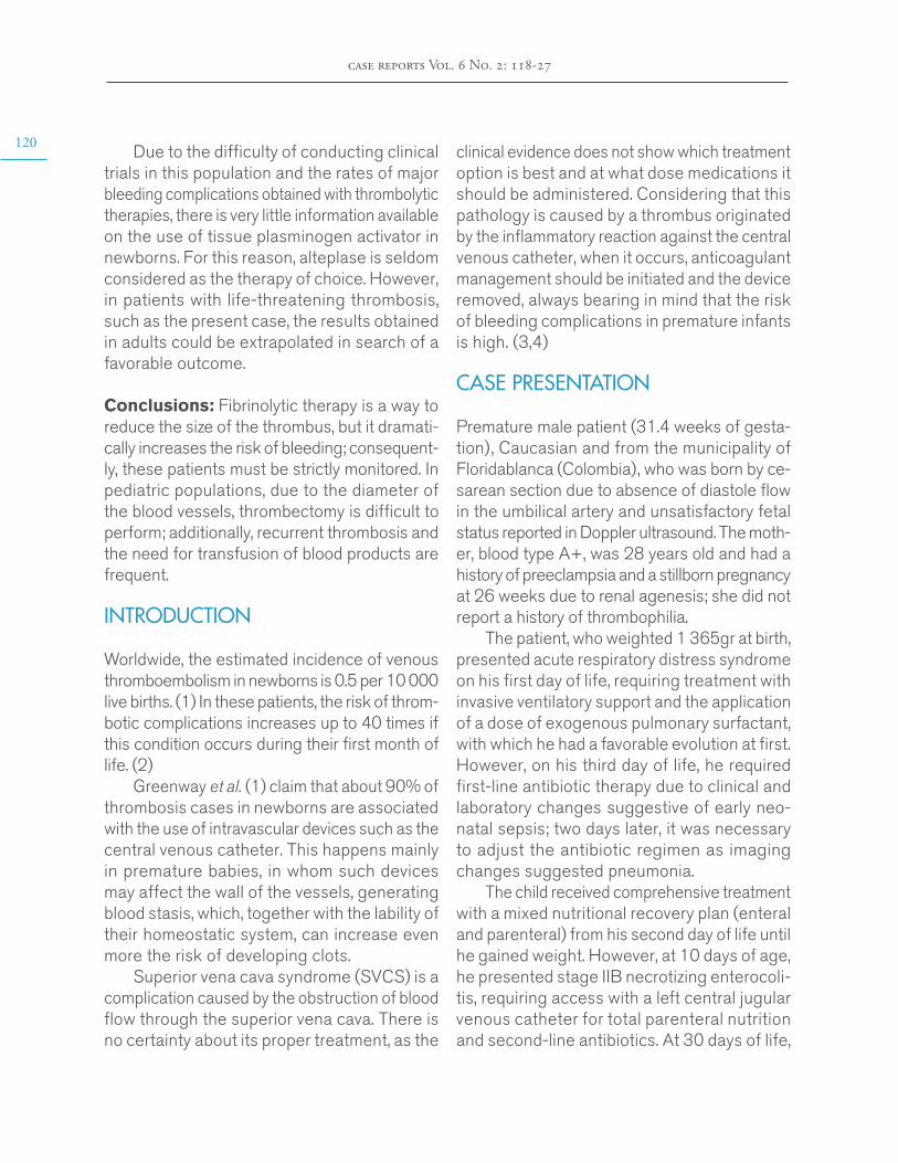
120
case reports Vol. 6 No. 2: 118-27
Due to the difficulty of conducting clinical trials in this population and the rates of major bleeding complications obtained with thrombolytic therapies, there is very little information available on the use of tissue plasminogen activator in newborns. For this reason, alteplase is seldom considered as the therapy of choice. However, in patients with life-threatening thrombosis, such as the present case, the results obtained in adults could be extrapolated in search of a favorable outcome.
Conclusions: Fibrinolytic therapy is a way to reduce the size of the thrombus, but it dramati-cally increases the risk of bleeding; consequent-ly, these patients must be strictly monitored. In pediatric populations, due to the diameter of the blood vessels, thrombectomy is difficult to perform; additionally, recurrent thrombosis and the need for transfusion of blood products are frequent.
INTRODUCTION
Worldwide, the estimated incidence of venous thromboembolism in newborns is 0.5 per 10 000 live births. (1) In these patients, the risk of throm-botic complications increases up to 40 times if this condition occurs during their first month of life. (2)
Greenway et al. (1) claim that about 90% of thrombosis cases in newborns are associated with the use of intravascular devices such as the central venous catheter. This happens mainly in premature babies, in whom such devices may affect the wall of the vessels, generating blood stasis, which, together with the lability of their homeostatic system, can increase even more the risk of developing clots.
Superior vena cava syndrome (SVCS) is a complication caused by the obstruction of blood flow through the superior vena cava. There is no certainty about its proper treatment, as the
clinical evidence does not show which treatment option is best and at what dose medications it should be administered. Considering that this pathology is caused by a thrombus originated by the inflammatory reaction against the central venous catheter, when it occurs, anticoagulant management should be initiated and the device removed, always bearing in mind that the risk of bleeding complications in premature infants is high. (3,4)
CASE PRESENTATION
Premature male patient (31.4 weeks of gesta-tion), Caucasian and from the municipality of Floridablanca (Colombia), who was born by ce-sarean section due to absence of diastole flow in the umbilical artery and unsatisfactory fetal status reported in Doppler ultrasound. The moth-er, blood type A+, was 28 years old and had a history of preeclampsia and a stillborn pregnancy at 26 weeks due to renal agenesis; she did not report a history of thrombophilia.
The patient, who weighted 1 365gr at birth, presented acute respiratory distress syndrome on his first day of life, requiring treatment with invasive ventilatory support and the application of a dose of exogenous pulmonary surfactant, with which he had a favorable evolution at first. However, on his third day of life, he required first-line antibiotic therapy due to clinical and laboratory changes suggestive of early neo-natal sepsis; two days later, it was necessary to adjust the antibiotic regimen as imaging changes suggested pneumonia.
The child received comprehensive treatment with a mixed nutritional recovery plan (enteral and parenteral) from his second day of life until he gained weight. However, at 10 days of age, he presented stage IIB necrotizing enterocoli-tis, requiring access with a left central jugular venous catheter for total parenteral nutrition and second-line antibiotics. At 30 days of life,
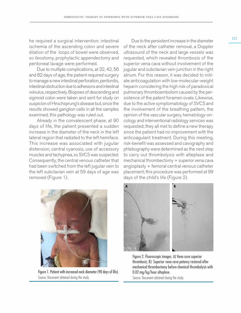
fibrinolytic therapy in newborns with superior vena cava syndrome
121he required a surgical intervention; intestinal ischemia of the ascending colon and severe dilation of the loops of bowel were observed, so ileostomy, prophylactic appendectomy and peritoneal lavage were performed.
Due to multiple complications, at 32, 42, 56 and 82 days of age, the patient required surgery to manage a new intestinal perforation, peritonitis, intestinal obstruction due to adhesions and intestinal volvulus, respectively. Biopsies of descending and sigmoid colon were taken and sent for study on suspicion of Hirschsprung’s disease but, since the results showed ganglion cells in all the samples examined, this pathology was ruled out.
Already in the convalescent phase, at 90 days of life, the patient presented a sudden increase in the diameter of the neck in the left lateral region that radiated to the left hemiface. This increase was associated with jugular distension, central cyanosis, use of accessory muscles and tachypnea, so SVCS was suspected. Consequently, the central venous catheter that had been switched from the left jugular vein to the left subclavian vein at 59 days of age was removed (Figure 1).
Figure 1. Patient with increased neck diameter (90 days of life).Source: Document obtained during the study.
Due to the persistent increase in the diameter of the neck after catheter removal, a Doppler ultrasound of the neck and large vessels was requested, which revealed thrombosis of the superior vena cava without involvement of the jugular and subclavian vein junction in the right atrium. For this reason, it was decided to initi-ate anticoagulation with low-molecular-weight heparin considering the high risk of paradoxical pulmonary thromboembolism caused by the per-sistence of the patent foramen ovale. Likewise, due to the active symptomatology of SVCS and the involvement of the breathing pattern, the opinion of the vascular surgery, hematology-on-cology and interventional radiology services was requested; they all met to define a new therapy since the patient had no improvement with the anticoagulant treatment. During this meeting, risk-benefit was assessed and cavography and phlebography were determined as the next step to carry out thrombolysis with alteplase and mechanical thrombectomy + superior vena cava angioplasty + femoral central venous catheter placement; this procedure was performed at 99 days of the child’s life (Figure 2).
Figure 2. Fluoroscopic images. A) Vena cava superior thrombosis; B) Superior vena cava patency restored after mechanical thrombectomy before chemical thrombolysis with 0.02 mg/kg/hour alteplase.Source: Document obtained during the study.
A B
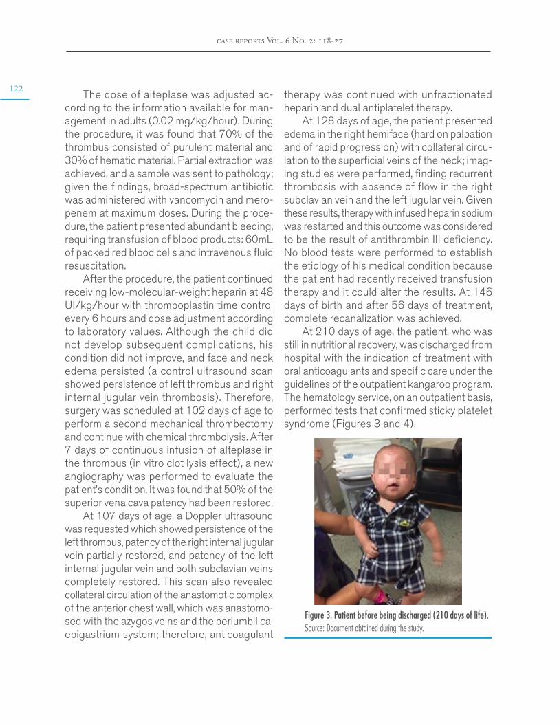
122
case reports Vol. 6 No. 2: 118-27
The dose of alteplase was adjusted ac-cording to the information available for man-agement in adults (0.02 mg/kg/hour). During the procedure, it was found that 70% of the thrombus consisted of purulent material and 30% of hematic material. Partial extraction was achieved, and a sample was sent to pathology; given the findings, broad-spectrum antibiotic was administered with vancomycin and mero-penem at maximum doses. During the proce-dure, the patient presented abundant bleeding, requiring transfusion of blood products: 60mL of packed red blood cells and intravenous fluid resuscitation.
After the procedure, the patient continued receiving low-molecular-weight heparin at 48 UI/kg/hour with thromboplastin time control every 6 hours and dose adjustment according to laboratory values. Although the child did not develop subsequent complications, his condition did not improve, and face and neck edema persisted (a control ultrasound scan showed persistence of left thrombus and right internal jugular vein thrombosis). Therefore, surgery was scheduled at 102 days of age to perform a second mechanical thrombectomy and continue with chemical thrombolysis. After 7 days of continuous infusion of alteplase in the thrombus (in vitro clot lysis effect), a new angiography was performed to evaluate the patient’s condition. It was found that 50% of the superior vena cava patency had been restored.
At 107 days of age, a Doppler ultrasound was requested which showed persistence of the left thrombus, patency of the right internal jugular vein partially restored, and patency of the left internal jugular vein and both subclavian veins completely restored. This scan also revealed collateral circulation of the anastomotic complex of the anterior chest wall, which was anastomo-sed with the azygos veins and the periumbilical epigastrium system; therefore, anticoagulant
therapy was continued with unfractionated heparin and dual antiplatelet therapy.
At 128 days of age, the patient presented edema in the right hemiface (hard on palpation and of rapid progression) with collateral circu-lation to the superficial veins of the neck; imag-ing studies were performed, finding recurrent thrombosis with absence of flow in the right subclavian vein and the left jugular vein. Given these results, therapy with infused heparin sodium was restarted and this outcome was considered to be the result of antithrombin III deficiency. No blood tests were performed to establish the etiology of his medical condition because the patient had recently received transfusion therapy and it could alter the results. At 146 days of birth and after 56 days of treatment, complete recanalization was achieved.
At 210 days of age, the patient, who was still in nutritional recovery, was discharged from hospital with the indication of treatment with oral anticoagulants and specific care under the guidelines of the outpatient kangaroo program. The hematology service, on an outpatient basis, performed tests that confirmed sticky platelet syndrome (Figures 3 and 4).
Figure 3. Patient before being discharged (210 days of life).Source: Document obtained during the study.
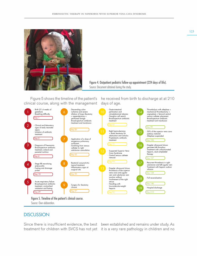
fibrinolytic therapy in newborns with superior vena cava syndrome
123
Figure 4. Outpatient pediatric follow-up appointment (224 days of life). Source: Document obtained during the study.
Figure 5 shows the timeline of the patient’s clinical course, along with the management
he received from birth to discharge at at 210 days of age.
Day 1
Day 3
Day 30
Day 63Day 99
Day 106
Day 107
Day 128
Day 146
Day 210
Day 73
Day 90
Day 92
Day 43
Day 50
Day 57
Day 6
Day 10
Day 29
1 6
7
8
9
10
11
12
13
14
15
16
17
18
19
2
3
4
5
Birth (31.4 weeks of gestation)Breathing dif�culty
·
·
Descending colon ischemia and severe dilation of loops Ileostomy + appendectomy + peritoneal lavage Broad-spectrum antibiotic treatment and transfusion
·
·
·
Gastrointestinal perforation and intraabdominal abscessGanglion cell search Broad-spectrum antibiotic treatment
·
··
Doppler ultrasound shows thrombosis of the superior vena cava and jugular vein and subclavian vein junction without involvement of the right atriumHandling with low-molecular-weight heparin
·
·
Right hemicolectomy + distal ileostomy for obstruction and volvulusProphylactic antibiotic treatment
·
·
Suspected Superior Vena Cava Syndrome Central venous catheter removal
·
·
Application of a dose of exogenous pulmonary surfactant Switching from venous catheter to right subclavian cannulation
·
·
Bacterial conjunctivitis: topical treatment In�ammatory signs at surgical site
·
·
Surgery for ileostomy closure
·
50% of the superior vena cava patency restoredAlteplase suspended
·
·
Doppler ultrasound shows persistent left thrombus Treatment with unfractionated heparin, dual antiplatelet therapy
·
·
Recurrent thrombosis in right subclavian and left jugular veinTreatment with heparin sodium infusion
·
·
Full recanalization·
Hospital discharge·
Clinical and laboratory signs of early neonatal sepsisInitiation of antibiotic treatment
·
·
Diagnosis of PneumoniaBroad-spectrum antibiotic treatment, enteral and parental nutrition
··
Stage IIB necrotizing enterocolitisFasting and drainage probe
·
·
Acute respiratory failureBroad-spectrum antibiotic treatment, orotracheal intubation and fasting
··
Thrombolysis with alteplase + mechanical thrombectomy + angioplasty + femoral central venous catheter placementBroad-spectrum antibiotic treatment anti transfusion
·
·
Figure 5. Timeline of the patient’s clinical course. Source: Own elaboration.
DISCUSSION
Since there is insufficient evidence, the best treatment for children with SVCS has not yet
been established and remains under study. As it is a very rare pathology in children and no
124
case reports Vol. 6 No. 2: 118-27
clear figures have been reported regarding its incidence in this population. Furthermore, the safety and efficacy of drugs administered in newborns is still doubtful. Described treatments include anticoagulant agents such as unfrac-tionated heparin, low-molecular-weight heparin, and thrombolytic therapy with r-TPA (alteplase). Nowadays, thrombectomy is no longer one of the first management options because the size of the blood vessels limits the adequate approach and makes this a procedure of high complexity and morbimortality. (5)
The first point to consider regarding medical management is the importance of prevention in patients with central venous catheters. In them, low continuous doses of heparin in-fusions (0.02-0.03 mg/kg/hour) have been shown to reduce bleeding and to be useful in the prevention of intravascular thrombosis associated with these devices. (6)
For anticoagulation therapy, low-molecu-lar-weight heparin is preferred over unfraction-ated heparin because the former has 100% bioavailability and has 2-4 times longer plasma half-life and dose-independent renal clearance, making its response more predictable. More-over, unlike unfractionated heparin, which has a higher risk of producing thrombocytopenia and osteoporosis, low-molecular-weight hep-arin requires less laboratory monitoring and dosage adjustment. (7) On the other hand, unfractionated heparin has the advantage of being rapidly reversible and cheaper, but its pharmacokinetics is unpredictable due to its high ability to bind to proteins and cells; it also has significant side effects such as in-creased prevalence of bleeding and induced thrombocytopenia, which occurs due to the formation of antibodies against the heparin/platelet factor complex in at least 1 out of every 100 patients receiving treatment for 5 days or more. (8) If treatment with this drug
is decided, a dedicated intravenous catheter must be placed for its administration so that therapy is not interrupted. (9)
It should be noted that the management of SVCS in adults is extrapolated to the pediatric population due to the lack of studies, being thrombolytic therapy the treatment of choice. Thus, for example, Gray et al., (10) in a study of 16 patients with central venous catheters who were treated with urokinase (n=11) or streptokinase (n=5), reported that thrombolytic therapy was effective in 73% (n=8) of the pa-tients treated with urokinase and in 20% (n=1) of those treated with streptokinase. Likewise, these authors concluded that fibrinolysis per-formed through the central venous catheter is more effective since the drugs reach the clot more directly than when the same drug is administered systemically and that the factors predicting successful thrombolysis are 1) using urokinase instead of streptokinase, 2) having a central venous catheter, and 3) symptoms lasting for ≤5 days.
In general, thrombolytic agents are not rec-ommended for the pediatric population, except in life-threatening conditions due to the high risk of bleeding. In this regard, several authors agree that tissue plasminogen activator is the most widely used fibrinolytic drug. (1,5,11,12) At this point, it is important to mention that fi-brinogen levels are the most reliable and fastest indicator of response to thrombolysis, with 1.0 g/L being the accepted lower limit. A platelet count >100 x 10-9 may also indicate a positive response to thrombolytic treatment. (13)
Alteplase is a thrombolytic agent often used in cases of central venous catheter thrombosis to achieve local clot lysis with intracatheter administration. However, careful monitoring is required as systemic administration increases the possibility of local blood vessel damage and the formation of new thrombi. (5)
fibrinolytic therapy in newborns with superior vena cava syndrome
125As mentioned above, information on the
treatment of SVCS is scarce, and available data are obtained from case reports where the dose of alteplase varies. Álvarez et al. (14) state that if the gestational age of the newborn is between 24 and 38 weeks, the dose should be 0.1 mg/kg/hour in continuous infusion, titrating as needed to maintain fibrinogen levels >100 mg/dL; Yang et al. (15) established that the dose in newborns should be 0.1-0.15 mg/kg/h; (15) finally, Giglia et al. (16) recommend a dose of 0.1-0.6 mg/kg/hour for infants, children and adolescents.
It should be noted that the most frequent adverse reactions to alteplase are intracranial bleeding (15%), ischemic stroke (6%), ecchy-mosis (1%), gastrointestinal bleeding (5%) and genitourinary tract bleeding (4%). To a lesser extent (<1%), anaphylaxis, angioedema, arrhythmias, deep vein thrombosis, cerebral herniation, pulmonary edema, and pericardial effusion, among others, may occur. (17)
In the present case, anticoagulant man-agement with enoxaparin was initiated without achieving clinical improvement and with throm-bus progression to deep vein thrombosis, so pharmacological thrombolysis was performed. Because the symptoms persisted, it was nec-essary for the interventional radiology service to perform mechanical thrombectomy and angioplasty of the superior vena cava, which restored the patency in 50% of the right internal jugular vein and complete patency of the left internal jugular vein and both subclavian veins. Subsequently, treatment continued with anti-coagulants to avoid recurrence of thrombosis at the previously treated site.
With a morbidity of 30% and a mortality of 18%, thromboembolic disorders have a profound impact on public health; they can cause pulmonary embolism (6-8%) and post- thrombotic syndrome (12-19%) and have a
significant percentage of recurrence (10-20%). (18) In this scenario, and taking into account that treatment in the pediatric population has not yet been clearly established, it is important to make a risk-benefit comparison to define the best therapy for each patient based on antithrombotic and anticoagulant therapeutic options, especially for the neonatal population. In this sense, it is necessary to stress the im-portance of conducting therapeutic research in this age group to support the therapies described in medical practice, since the lack of evidence has led to the need to extrapolate valid recommendations in the adult population without adequate consensus.
CONCLUSIONS
Fibrinolytic therapy with alteplase is a viable option to reduce the size of a thrombus. How-ever, it should be borne in mind that the risk of bleeding is markedly increased with this drug, so strict monitoring of patients is required. On the other hand, mechanical thrombectomy is a procedure that, although effective for treating thromboembolic disorders, is difficult to perform on newborns given the diameter of their blood vessels; it is also associated with high rates of thrombotic recurrence.
Strict monitoring of bleeding and taking control lab test allows adjusting anticoagulant doses without having complications such as massive bleeding.
PATIENT’S PERSPECTIVE
The parents of the newborn were informed of the clinical condition and the diagnosis and progno-sis of the patient on a daily basis. They, in turn, agreed with the procedures proposed to treat the patient during his hospital stay.
126
case reports Vol. 6 No. 2: 118-27
INFORMED CONSENT
The mother of the patient whose case is reported here signed an informed consent and authorized the publication of the data obtained from the medical records and the attached photographs.
CONFLICTS OF INTEREST
None stated by the authors.
FUNDING
None stated by the authors.
ACKNOWLEDGEMENTS
None stated by the authors.
REFERENCES
1. Greenway A, Massicotte MP, Monagle P. Neonatal thrombosis and its treatment. Blood Rev. 2003;18(2):75-84. http://doi.org/d5r36q.
2. Bacciedoni V, Attie M, Donato H. Trombosis en el recién nacido. Arch Argent Pediatr. 2016 [cited 2019 Abr 27];114(2):159-66. Available from: https://bit.ly/3doQHnd.
3. Wan JF, Bezjak A. Superior Vena Cava Syndro-me. Emerg Med Clin North Am. 2009;27(2):243-55. http://doi.org/dbxhdm.
4. Seligson MT, Surowiec SM. Superior Vena Cava Syndrome. Treasure Island: StatPearls Publishing; 2019 [cited 2019 May 5]. Available from: https://bit.ly/3b2GM5f.
5. Monagle P, Chan AK, Goldenberg NA, Ichord RN, Journeycake JM, Nowak-Göttl U, et al. Antithrombotic therapy in neonates and children: Antithrombotic Therapy and Preven-tion of Thrombosis, 9th ed. American College of Chest Physicians Evidence-Based Clinical Prac-tice Guidelines. Chest. 2012;141(2):e737S-e801S. http://doi.org/gcx8dw.
6. Bhatt MD, Paes BA, Chan AK. How to use unfractionated heparin to treat neonatal thrombo-sis in clinical practice. Blood Coagul Fibrinolysis. 2016;27(6):605-14. http://doi.org/f8xb5p.
7. Cabestrero-Alonso D, Gálvez-Mágica MA, Martín-Rodríguez C, Cindoncha-Gallego M, García de Lorenzo-Mateos A. Heparinas de bajo peso molecular en pacientes críticos: usos, indicaciones y tipos. Med Intensiva. 2001. [ci-ted 2019 May 17];25(1):18-26. Available from: https://bit.ly/3c92J48.
8. Cruz-Ginzález I, Sánchez-Ledesma M, Sán-chez PL, Jang I. Trombocitopenia inducida por heparina. Rev Esp Cardiol. 2007;60(10):1071-82. http://doi.org/bkbnxd.
9. Edstrom CS, Christensen RD. Evaluation and treatment of thrombosis in the neonatal intensi-ve care unit. Clin Perinatol. 2000;27(3):623-41. http://doi.org/dq9mz2.
10. Gray BH, Olin JW, Graor RA, Young JR, Bar-tholomew JR, Ruschhaupt WF. Safety and efficacy of thrombolytic therapy for superior vena cava syndrome. Chest. 1991;99(1):54-9. http://doi.org/ccs6vk.
11. Albisetti M. Thrombolytic therapy in children. Thromb Res. 2006;118(1):95-105. http://doi.org/cfq4gn.
12. Andrew M, Brooker L, Leaker M, Paes B, Weitz J. Fibrin clot lysis by thrombolytic agents is impaired in newborns due to a low plasmino-gen concentration. Thromb Haemost. 1992 [cited 2019 May 30];68(3):325-30. Disponible en: ht-tps://bit.ly/2xA5hsS.
13. Braga JAP, Loggetto SR, Hoepers ATC, Bernardo WM, Medeiros L, Veríssimo MPA. Guidelines on the diagnosis of primary immune thrombocytopenia in children and ado-lescents: Associação Brasileira de Hematologia, Hemoterapia e Terapia Celular Guidelines Pro-ject: Associação Médica Brasileira - 2012. Rev Bras Hematol Hemoter. 2013;35(5):358-65. http://doi.org/dtv8.
14. Álvarez P, Verdugo P, Carvajal L, Múhlhau-sen G, Ríos P, Rodríguez D. Activador del
fibrinolytic therapy in newborns with superior vena cava syndrome
127plasminógeno tisular recombinante en el manejo de trombos cardiacos en recién nacidos. Rev Chil Pediatr. 2015;86(3):194-9. http://doi.org/f3hh8s.
15. Yang L, Ye L, Lin R. The use of alteplase in a newborn with an aortic arch thrombus during extracorporeal membrane oxygenation. Cardiol Young. 2019;29(10):1307-9. http://doi.org/dtv9.
16. Giglia TM, Massicotte MP, Tweddell JS, Barst RR, Bauman M, Erickson CC, et al. Prevention and Treatment of Thrombosis in Pe-diatric and Congenital Heart Disease: A Scientific
Statement From the American Heart Association. Circulation. 2013;128(24):2622-703. http://doi.org/dtwb.
17. Genentech Inc. Label for ACTIVASE®. San Fran-cisco: Food and Drug Administration; 2015 [cited 2019 Jun 15]. Available from: https://bit.ly/3c9AylA.
18. Nossair F, Schoettler P, Starr J, Chan AKC, Kiro I, Paes B, et al. Pediatric superior vena cava syndrome: An evidence-based systematic review of the literature. Pediatr Blood Cancer. 2018;65(9):e27225. http://doi.org/dtwc.
case reports 2020; 6(2)
TREATMENT WITH TYPE-I COLLAGEN SCAFFOLDS IN PATIENTS WITH VENOUS ULCERS. CASE REPORT
Keywords: Venous Ulcer; Collagen Type I; Regeneration.Palabras clave: Úlcera venosa; Colágeno Tipo I; Regeneración.
Received: 01/12/2019 Accepted: 06/02/2020
Corresponding authorMarta Raquel Fontanilla. Department of Pharmacy,
Faculty of Science, Universidad Nacional de Colombia. Email: [email protected]
Martha Isabel González-DuqueJulián Daniel Hernández-Martinez
Marta Raquel FontanillaUniversidad Nacional de Colombia
- Bogotá Campus - Faculty of Science - Department of Pharmacy - Tissue Engineering
Working Group - Bogotá D.C. Colombia.
https://doi.org/10.15446/cr.v6n2.83815
Sofía Elizabeth Muñoz-MedinaUniversidad Nacional de Colombia
- Bogotá Campus - Faculty of Medicine - Health Technology and Policy Evaluation Group
- Bogotá D.C. - Colombia.
treatment with type-i collagen scaffolds in patients with venous ulcers
129RESUMEN
Introducción. La insuficiencia venosa crónica afecta alrededor del 5% de la población adulta en el mundo; una de sus mayores complicaciones son las úlceras en miembros inferiores, las cuales tienen una prevalencia mundial del 2%. Las úlceras afectan la calidad de vida de los pacientes e impac-tan al sistema de salud debido a los altos costos de atención que genera. El tratamiento de referencia es la terapia compresiva y la cura húmeda de las heridas, sin embargo estas intervenciones pueden no ser efectivas cuando las lesiones se complican.
Presentación del caso. Paciente femenina de 66 años con úlceras venosas en miembros infe-riores acompañadas de fiebre y dolor local que no respondían a las terapias convencionales. La pa-ciente fue tratada con un nuevo sustituto dérmico basado en una membrana acelular de colágeno tipo I que contribuye al cierre de la úlcera al estimu-lar el remplazo del tejido lesionado por tejido similar al sano, con lo cual tuvo mejoría a las 16 semanas; después de 8 meses de terminado el tratamiento no se presentó recurrencia de las lesiones.
Conclusiones. La membrana acelular de colá-geno tipo I desarrollada por el Grupo de Trabajo en Ingeniería de Tejidos del Departamento de Farmacia de la Universidad Nacional de Co-lombia es efectiva en el tratamiento de úlceras venosas en miembros inferiores y su bajo costo facilita el acceso de toda la población a terapias basadas en su aplicación.
ABSTRACT
Introduction: Chronic venous insufficiency affects about 5% of the global adult population. Venous leg ulcers are one of the most frequent complications of this pathology, with a global prevalence of 2%. This disease affects both the quality of life of patients and, due to the high cost of the treatment, the health system. Compressive therapy and moist wound healing have been the gold standard treatment. However, when complications occur, they may not be effective.
Case report: This is the case of a 66-year-old female patient with venous ulcers on her lower limbs and symptoms of fever and local pain that did not respond to conventional therapies. The patient was treated with a new dermal substitute made of an acellular type-I collagen membrane, which promotes the closure of the ulcer by stim-ulating the replacement of injured tissue with tissue similar to the healthy one. The condition of the patient improved at 16 weeks, and after 8 months of treatment there was no recurrence of the lesions.
Conclusions: Acellular type-I collagen mem-brane developed by the Tissue Engineering Working Group of the Department of Pharmacy of the Universidad Nacional de Colombia is effective in treating venous ulcers of the low-er limbs. Its low cost facilitates the access of the whole population to therapies based on its application.
INTRODUCTION
Venous ulcers occur due to a venous valve insufficiency that causes venous hypertension, which generates hypervolemia (predominantly in the lower limbs) associated with lipoderma-
tosclerosis and skin ulcers. In turn, the phys-iopathology of valve insufficiency is associ-ated with an increase in the number of fibrin degradation products and pro-inflammatory
130
case reports Vol. 6 No. 2: 128-36
molecules in the dermis, leading to cell lysis and ulceration. (1)
The prevalence of chronic venous insuffi-ciency ranges between 5% and 30% in the adult population, (2) and this condition is considered a public health problem due to high treatment costs and the negative impact it has on the quality of life of patients suffering from it. (3)
Available treatments for venous ulcers are based on compression measures, which improve venous return, and hydrocolloids that promote tissue repair. (4) However, these therapies often do not satisfy patients’ needs, leading to ther-apeutic failure and recurrence. (5) To address this situation, the Tissue Engineering Working Group of the Department of Pharmacy of the Universidad Nacional de Colombia developed a sterile porous type-I collagen membrane of bovine origin approved by INVIMA through the Sanitary Registration Number No. 2017DM-0015999. (6) The membrane acts as a dermal substitute and promotes the replacement of injured tissue with healthy tissue similar to the demarcated borders of the wound. Besides, it has the advantage of being a low-cost and affordable product for any patient.
The present work reports the case of a patient who received the membrane designed at the Universidad Nacional de Colombia to treat two venous ulcers of her lower limbs with a satisfactory outcome.
CASE PRESENTATION
The following is the case of a 66-year-old female patient, mestizo, with basic secondary education, from the municipality of Soacha (Cundinamarca, Colombia), of low socioeconomic status, who was working as a caregiver for an elderly per-son. The patient attended a consultation due to having 2 ulcerated lesions in her lower limbs (the lower third of the right lower limb and the
medial region in the malleolus of the left lower limb) for 12 months. The affected areas pre-sented with erythema, local heat, ulceration and seropurulent secretion.
The patient reported the following history: personal: high blood pressure (in treatment with nifedipine 30 mg/12 hours orally) and venous insufficiency; surgical: hallux valgus correction, ligament reconstruction and sec-ond toe arthroplasty; gynecological-obstetric: menarche at age 15, 2 births, 0 abortions and menopause at age 58, and family: sibling diagnosed with high blood pressure. During the review of systems, cough and runny nose were observed. The patient was diagnosed with infected venous ulcers and was prescribed outpatient treatment with clindamycin (600 mg/day) and oral diclofenac (50 mg/12 hours). However, the symptoms did not improve.
After 2 weeks, she presented a 3-day episode of fever with intense pain in the lower limbs, predominantly on the right leg at the inner edge of the gastrocnemius muscle area, with limited walking and altered sleep pattern due to pain intensity. Therefore, she consulted the emergency department where she was evaluated by the general medicine service. On physical examination, the physician reported a medial ulcer in the malleolus (2cm x 2.5cm) of the lower right limb with well-defined borders, fibrin mesh, an area of necrosis, a perilesional edema and yellow discharge, and a satellite lesion (approximately 1cm deep) with fibrin mesh and irregular borders in the lower left limb.
Pedal pulses and ochre dermatitis were observed in both affected limbs. Considering the fever, the local pain, the functional limita-tion, and the characteristics of the injuries, the patient was admitted to the hospital for 20 days with a diagnosis of superinfected ulcers in the lower limbs and suspicion of osteomyelitis. The patient received antibiotic treatment with
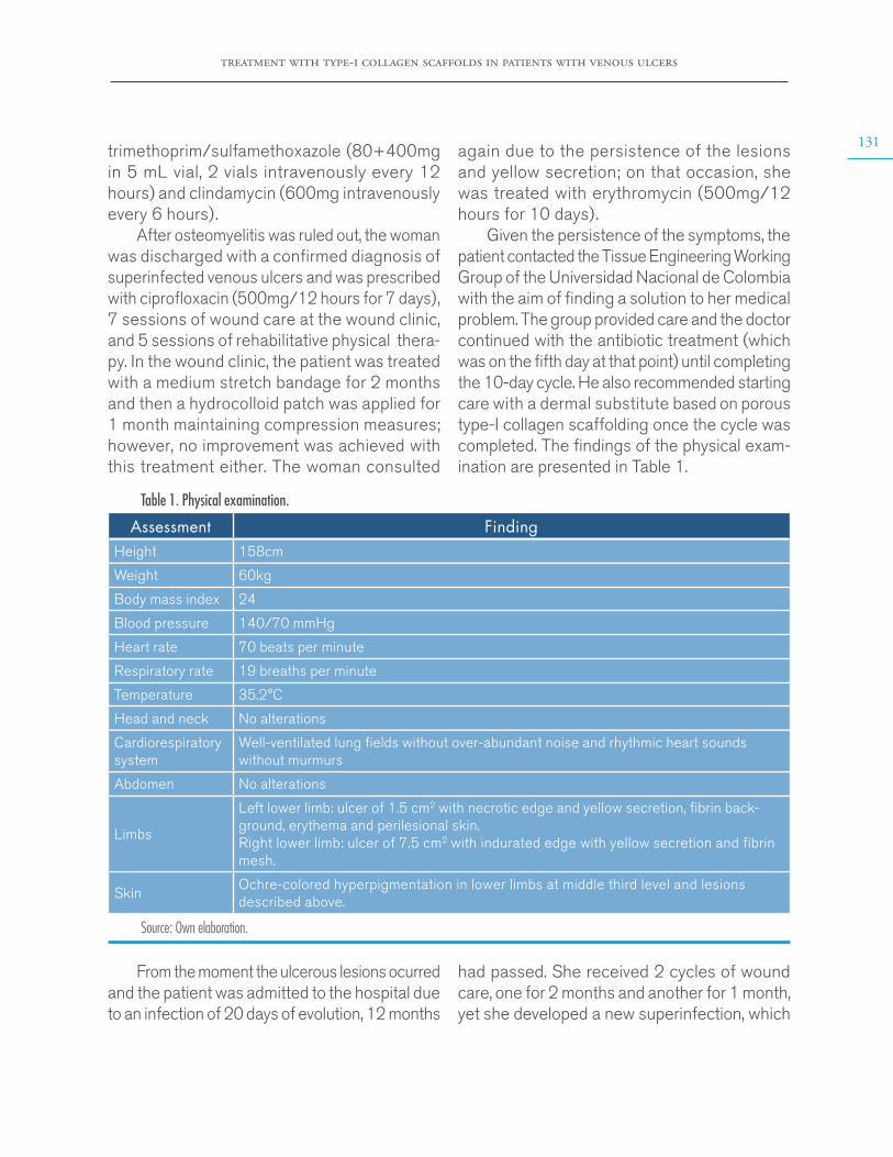
treatment with type-i collagen scaffolds in patients with venous ulcers
131trimethoprim/sulfamethoxazole (80+400mg in 5 mL vial, 2 vials intravenously every 12 hours) and clindamycin (600mg intravenously every 6 hours).
After osteomyelitis was ruled out, the woman was discharged with a confirmed diagnosis of superinfected venous ulcers and was prescribed with ciprofloxacin (500mg/12 hours for 7 days), 7 sessions of wound care at the wound clinic, and 5 sessions of rehabilitative physical thera-py. In the wound clinic, the patient was treated with a medium stretch bandage for 2 months and then a hydrocolloid patch was applied for 1 month maintaining compression measures; however, no improvement was achieved with this treatment either. The woman consulted
again due to the persistence of the lesions and yellow secretion; on that occasion, she was treated with erythromycin (500mg/12 hours for 10 days).
Given the persistence of the symptoms, the patient contacted the Tissue Engineering Working Group of the Universidad Nacional de Colombia with the aim of finding a solution to her medical problem. The group provided care and the doctor continued with the antibiotic treatment (which was on the fifth day at that point) until completing the 10-day cycle. He also recommended starting care with a dermal substitute based on porous type-I collagen scaffolding once the cycle was completed. The findings of the physical exam-ination are presented in Table 1.
Table 1. Physical examination.
Assessment FindingHeight 158cm
Weight 60kg
Body mass index 24
Blood pressure 140/70 mmHg
Heart rate 70 beats per minute
Respiratory rate 19 breaths per minute
Temperature 35.2°C
Head and neck No alterations
Cardiorespiratory system
Well-ventilated lung fields without over-abundant noise and rhythmic heart sounds without murmurs
Abdomen No alterations
Limbs
Left lower limb: ulcer of 1.5 cm2 with necrotic edge and yellow secretion, fibrin back-ground, erythema and perilesional skin. Right lower limb: ulcer of 7.5 cm2 with indurated edge with yellow secretion and fibrin mesh.
SkinOchre-colored hyperpigmentation in lower limbs at middle third level and lesions described above.
Source: Own elaboration.
From the moment the ulcerous lesions ocurred and the patient was admitted to the hospital due to an infection of 20 days of evolution, 12 months
had passed. She received 2 cycles of wound care, one for 2 months and another for 1 month, yet she developed a new superinfection, which
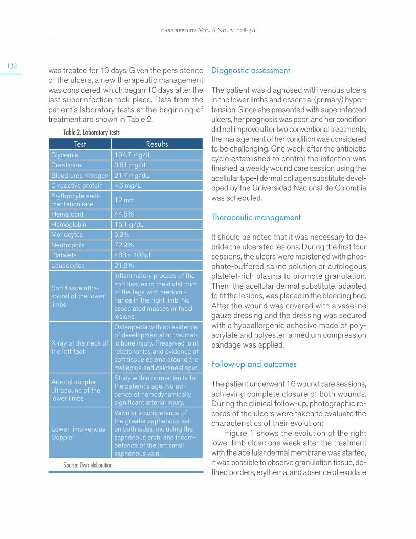
132
case reports Vol. 6 No. 2: 128-36
was treated for 10 days. Given the persistence of the ulcers, a new therapeutic management was considered, which began 10 days after the last superinfection took place. Data from the patient’s laboratory tests at the beginning of treatment are shown in Table 2.
Table 2. Laboratory tests
Test ResultsGlycemia 104.7 mg/dL
Creatinine 0.81 mg/dL
Blood urea nitrogen 21.7 mg/dL
C-reactive protein <6 mg/L
Erythrocyte sedi-mentation rate
12 mm
Hematocrit 44.5%
Hemoglobin 15.1 g/dL
Monocytes 5.3%
Neutrophils 72.9%
Platelets 488 x 103µL
Leucocytes 21.8%
Soft tissue ultra-sound of the lower limbs
Inflammatory process of the soft tissues in the distal third of the legs with predomi-nance in the right limb. No associated masses or focal lesions.
X-ray of the neck of the left foot
Osteopenia with no evidence of developmental or traumat-ic bone injury. Preserved joint relationships and evidence of soft tissue edema around the malleolus and calcaneal spur.
Arterial doppler ultrasound of the lower limbs
Study within normal limits for the patient’s age. No evi-dence of hemodynamically significant arterial injury.
Lower limb venous Doppler
Valvular incompetence of the greater saphenous vein on both sides, including the saphenous arch, and incom-petence of the left small saphenous vein.
Source: Own elaboration.
Diagnostic assessment
The patient was diagnosed with venous ulcers in the lower limbs and essential (primary) hyper-tension. Since she presented with superinfected ulcers, her prognosis was poor, and her condition did not improve after two conventional treatments, the management of her condition was considered to be challenging. One week after the antibiotic cycle established to control the infection was finished, a weekly wound care session using the acellular type-I dermal collagen substitute devel-oped by the Universidad Nacional de Colombia was scheduled.
Therapeutic management
It should be noted that it was necessary to de-bride the ulcerated lesions. During the first four sessions, the ulcers were moistened with phos-phate-buffered saline solution or autologous platelet-rich plasma to promote granulation. Then the acellular dermal substitute, adapted to fit the lesions, was placed in the bleeding bed. After the wound was covered with a vaseline gauze dressing and the dressing was secured with a hypoallergenic adhesive made of poly-acrylate and polyester, a medium compression bandage was applied.
Follow-up and outcomes
The patient underwent 16 wound care sessions, achieving complete closure of both wounds. During the clinical follow-up, photographic re-cords of the ulcers were taken to evaluate the characteristics of their evolution:
Figure 1 shows the evolution of the right lower limb ulcer: one week after the treatment with the acellular dermal membrane was started, it was possible to observe granulation tissue, de-fined borders, erythema, and absence of exudate
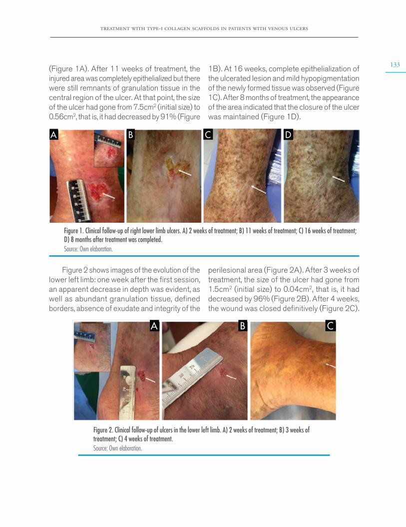
treatment with type-i collagen scaffolds in patients with venous ulcers
133(Figure 1A). After 11 weeks of treatment, the injured area was completely epithelialized but there were still remnants of granulation tissue in the central region of the ulcer. At that point, the size of the ulcer had gone from 7.5cm2 (initial size) to 0.56cm2, that is, it had decreased by 91% (Figure
1B). At 16 weeks, complete epithelialization of the ulcerated lesion and mild hypopigmentation of the newly formed tissue was observed (Figure 1C). After 8 months of treatment, the appearance of the area indicated that the closure of the ulcer was maintained (Figure 1D).
A B C D
Figure 1. Clinical follow-up of right lower limb ulcers. A) 2 weeks of treatment; B) 11 weeks of treatment; C) 16 weeks of treatment; D) 8 months after treatment was completed.Source: Own elaboration.
Figure 2 shows images of the evolution of the lower left limb: one week after the first session, an apparent decrease in depth was evident, as well as abundant granulation tissue, defined borders, absence of exudate and integrity of the
perilesional area (Figure 2A). After 3 weeks of treatment, the size of the ulcer had gone from 1.5cm2 (initial size) to 0.04cm2, that is, it had decreased by 96% (Figure 2B). After 4 weeks, the wound was closed definitively (Figure 2C).
A B C
Figure 2. Clinical follow-up of ulcers in the lower left limb. A) 2 weeks of treatment; B) 3 weeks of treatment; C) 4 weeks of treatment.Source: Own elaboration.
134
case reports Vol. 6 No. 2: 128-36
Throughout the follow-up, adherence to the treatment was complete: the patient attended all wound care sessions and always wore the com-pression bandage when she attended the control appointments. No adverse events associated with the intervention were reported. High compression stockings were indicated after ulcer closure.
DISCUSSION
Due to the length of the wound care sessions, venous ulcers in the lower limbs impact the cost of care and quality of life of people who suffer them. (7,8) These lesions can become chronic due to the inflammatory response, which is sec-ondary to venous hypertension and caused by mi-crocirculation damage. (9) Short- and long-term complications of venous ulcers are common due to local or systemic infections and the formation of new ulcers in the perilesional tissues. (10)
So far, there is no consensus on the most appropriate treatment for venous ulcers, and the devices used for wound care do not always pro-mote closure of the lesion and regeneration of the skin. (11,12) Type-I collagen, the most abundant extracellular protein in the dermis, plays a key role in skin lesions as it guides cell migration, promotes the proliferation and differentiation of fibroblasts and keratinocytes, and sequesters and releases controlled growth factors that modulate wound closure and accelerate angiogenesis. (13)
The Tissue Engineering Working Group of the Universidad Nacional de Colombia developed some type-I membranes or acellular collagen scaffolds that have helped close connective tissue wounds and have favored the regeneration of skin and oral mucosa wounds in preclinical models. (13-16) To document their performance in cruciate areas, these membranes were used to treat two venous ulcers of different sizes in one patient. The closure time of the ulcers depended on the initial area of each lesion: the
complete epithelization of the largest wound occurred at 16 weeks, while the smallest ulcer closed at 4 weeks. Overall, the data suggest that the product evaluated promotes the closure of the operated ulcers by stimulating the formation of tissue with characteristics similar to those of the surrounding healthy tissue, which, in turn, suggests it stimulates skin skin regeneration in the affected areas.
The main limitation of this case study is that it shows the data obtained during the treatment of two lower limb ulcers in the same patient; consequently, the results only suggest that debridement, weekly application of acellular dermal membranes and moderate compres-sive therapy promote the closure of lower limb venous ulcers and favor tissue regeneration over contracture repair. Therefore, in order to demonstrate the benefits of this therapeutic option, a clinical study of its safety and efficacy should be carried out with a representative sample that allows that allows drawing con-clusions about its effects on the closure and healing of venous ulcers.
CONCLUSION
The application of acellular type-I collagen mem-brane in debrided and bleeding ulcers, together with compression measures and basic care, fa-cilitated the closure of the lesions. The quality of the tissue formed and the non-recurrence of the ulcers after 8 months of treatment indicate that the product developed by the Tissue Engineering Working Group of the Department of Pharmacy at the Universidad Nacional de Colombia favors tissue regeneration and helps prevent scar contracture.
PATIENT’S PERSPECTIVE
After completing the treatment, the patient re-ported a subjective improvement of 80% in her

treatment with type-i collagen scaffolds in patients with venous ulcers
135quality of life. At that time, the SF-36 quality of life questionnaire was administered and in the eight domains evaluated (vitality, physical func-tioning, pain, general health perception, physical role functioning, social role functioning, emotional role functioning and mental health) a score similar to that of patients without venous ulcer was ob-tained. (17,18) In addition, the patient stated that she could now perform activities that the ulcers had prevented her from doing, such as moving around without pain and bathing in the sea.
At the time of preparation of this case report, the woman was still wearing high-compression stockings and was assessed by the vascular surgery service for the treatment of venous insufficiency. The peripheral vascular surgeon who treated her indicated the need to perform a saphenectomy. Twenty months after complet-ing the treatment with acellular type-I collagen scaffolds, the patient said she was satisfied since the venous ulcers had not reappeared.
INFORMED CONSENT
At the beginning of the treatment, the patient was informed of the possible adverse events that could be caused by the therapy. Likewise, all the details described in the report were ex-plained to her and she was asked to sign to sign an informed consent form. The witness and the treating physician-investigator also signed it.
CONFLICT OF INTEREST
The Tissue Engineering Working Group declares a conflict of interest since it developed the new acellular type-I collagen dermal substitute used in this case.
FUNDING
Supplies for wound care were provided by the
Tissue Engineering Working Group.
ACKNOWLEDGEMENTS
To Dr. Juan Bobadilla for his suggestions and comments.
REFERENCES
1. Raffetto JD. Pathophysiology of Chronic Venous Disease and Venous Ulcers. Surg Clin North Am. 2018;98(2):337-47. http://doi.org/gdcwqp.
2. Eberhardt RT, Raffetto JD. Chronic venous insufficiency. Circulation. 2014;130(4):333-46. http://doi.org/ds5p.
3. Epstein D, Gohel M, Heatley F, Davies AH. Cost-effectiveness of treatments for superficial ve-nous reflux in patients with chronic venous ulceration. BJS open. 2018;2(4):203-12. http://doi.org/ds5q.
4. Pinheiro L da S, Borges EL, Donoso MTV. Uso de hidrocolóide e alginato de cálcio no trat-amento de lesões cutâneas. Rev Bras Enferm. 2013;66(5):760-70. http://doi.org/ds5r.
5. Lurie F, Bittar S, Kasper G. Optimal Compression Therapy and Wound Care for Venous Ulcers. Surg Clin North Am. 2018;98(2):349-60. http://doi.org/gf2qzq.
6. Suesca E, Dias AMA, Braga MEM, de Sousa HC, Fontanilla MR. Multifactor analysis on the effect of collagen concentration, cross-linking and fiber/pore orientation on chemical, microstructur-al, mechanical and biological properties of colla-gen type I scaffolds. Mater Sci Eng C Mater Biol Appl. 2017;77:333-41. http://doi.org/gbjw97.
7. Wittens C, Davies AH, Bækgaard N, Bro-holm R, Cavezzi A, Chastanet S, et al. Editor’s Choice - Management of Chronic Venous Disease : Clinical Practice Guidelines of the European Society for Vascular Surgery (ESVS). Eur J Vasc Endovasc Surg. 2015;49(6):678-737. http://doi.org/f3pgx8.
8. Edwards H, Courtney M, Finlayson K, Shuter P, Lindsay E. A randomised controlled trial of a com-munity nursing intervention: improved quality of life
136
case reports Vol. 6 No. 2: 128-36
and healing for clients with chronic leg ulcers. J Clin Nurs. 2009;18(11):1541-9. http://doi.org/bnhp4f.
9. Crawford JM, Lal BK, Durán WN, Pappas PJ. Pathophysiology of venous ulceration. J Vasc Surg Venous Lymphat Disord. 2017;5(4):596-605. http://doi.org/ds5x.
10. European Wound Management Associa-tion. Position Document: Identifying criteria for wound infection. London: Medical Education Part-nership LTD; 2005 [cited 2018 Oct 10]. Available from: https://bit.ly/35ckXz1.
11. Andriessen A, Apelqvist J, Mosti G, Partsch H, Gonska C, Abel M. Compression therapy for venous leg ulcers: risk factors for adverse events and complications, contraindications - a review of present guidelines. J Eur Acad Dermatology Vene-reol. 2017;31(9):1562-8. http://doi.org/gbv2c4.
12. Evidence-based (S3) Guidelines for Diagnos-tics and Treatment of Venous Leg Ulcers. J Eur Acad Dermatology Venereol. 2016;30(11):1843-75. http://doi.org/ds5z.
13. Millán D, Jiménez RA, Nieto LE, Linero I, La-verde M, Fontanilla MR. Preclinical evaluation of collagen type I scaffolds, including gelatin-colla-gen microparticles and loaded with a hydroglycolic Calendula officinalis extract in a lagomorph mod-el of full-thickness skin wound. Drug Deliv Transl Res. 2016;6(1):57-66. http://doi.org/f8nnjf.
14. Espinosa L, Sosnik A, Fontanilla MR. De-velopment and preclinical evaluation of acellular collagen scaffolding and autologous artificial con-nective tissue in the regeneration of oral mucosa wounds. Tissue Eng Part A. 2010;16(5):1667-79. http://doi.org/bsnnf2.
15. Fontanilla MR, Espinosa LG. In vitro and in vivo assessment of oral autologous artificial connec-tive tissue characteristics that influence its perfor-mance as a graft. Tissue Eng Part A. 2012;18(17-18):1857-66. http://doi.org/f38qzr.
16. Fontanilla MR, Casadiegos S, Bustos RH, Patarroyo MA. Comparison of healing of full-thickness skin wounds grafted with multi-directional or unidirectional autologous artificial dermis: differential delivery of healing biomark-ers. Drug Deliv Transl Res. 2018;8(5):1014-24. http://doi.org/ds52.
17. Alonso J, Prieto L, Antó JM. La versión españo-la del SF-36 Health Survey (Cuestionario de Salud SF-36): un instrumento para la medida de los re-sultados clínicos. Med Clin (Barc). 1995;104(20): 771-776.
18. Dias TYAF, Costa IKF, Melo MDM, Torres SM da SGS de O, Maia EMC, Torres G de V, et al. Quality of life assessment of patients with and without venous ulcer. Rev Lat Am Enferma-gem. 2014;22(4):576-81. http://doi.org/ds53.
case reports 2020 6(2)
CONGENITAL HYPEREXTENSION DEFORMITY OF THE KNEES DUE TO ARTHROGRYPOSIS MULTIPLEX CONGENITA? CASE REPORT
Keywords: Arthrogryposis; Infant, Newborn, Diseases; Contracture; Rehabilitation.Palabras clave: Artrogriposis; Enfermedades del recién nacido; Contractura;
Rehabilitación.
Received: 01/12/2019 Accepted: 04/02/2020
Corresponding authorLina Paola Montaña-Jiménez. Neonatal Intensive Care Unit,
Hospital Universitario San Ignacio. Bogotá D.C. Colombia.
Email: [email protected].
Paola Andrea Romero-CampiñoLiliana Sandoval-Tristancho
María Camila JaramilloAnna Claici
Pontificia Universidad Javeriana - Faculty of Medicine - Department of Pediatrics - Research Student group for Neonatal Infectious
Diseases - Bogotá D.C. - Colombia.
https://doi.org/10.15446/cr.v6n2.83824
Lina Paola Montaña-JiménezPontificia Universidad Javeriana
- Faculty of Medicine - Department of Pediatrics - Research Student group for Neonatal Infectious
Diseases - Bogotá D.C. - Colombia.Hospital Universitario San Ignacio - Neonatal Intensive Care Unit -
Bogotá D.C. - Colombia.
138
case reports Vol. 6 No. 2: 137-45
ABSTRACT
Introduction: Arthrogryposis multiplex congen-ita is a disorder characterized by non-progressive joint contractures. It has an estimated prevalence of 1 in every 3 000-5 000 live births, with the same male-to-female ratio.
Case presentation: This is the case of a male newborn with adequate prenatal care checkup appointments, who presented with a congenital deformity of the lower limbs. On physical ex-amination, he had hyperextension of the knees (passive flexion of 20° in the left leg and 30° in the right leg), and painful active movement. On admission, peripheral pulses had good inten-sity, and adequate distal perfusion was found. Barlow and Ortolani maneuvers were negative, and no midline lesions were observed in the spine. The patient was diagnosed with arthro-gryposis multiplex congenita and received mul-tidisciplinary treatment to avoid early morbidity and mortality.
Conclusion: To attain satisfactory clinical de-velopment in patients with arthrogryposis, it is es-sential to have a high level of antenatal suspicion, as well as appropriate prenatal checkups. All this allows for proper management, minimizing diag-nostic errors, avoiding unnecessary procedures, and performing effective and timely treatment with outstanding results.
RESUMEN
Introducción. La artrogriposis múltiple congénita es un desorden caracterizado por contracturas articulares no progresivas y que tiene una preva-lencia estimada de 1 caso por cada 3 000-5 000 nacidos vivos con igual proporción entre géneros.
Presentación del caso. Paciente masculino recién nacido con adecuados controles prena-tales, quien al nacimiento presentó deformidad en miembros inferiores debido a hiperextensión de las rodillas (flexión pasiva de 20° en pierna izquierda y 30° en pierna derecha) que provo-caba dolor a la movilización activa. Al ingreso se registró perfusión distal adecuada y pulsos periféricos simétricos y de buena intensidad; las maniobras de Ortolani y Barlow fueron nega-tivas y no se evidenciaron lesiones en la línea media del dorso. El paciente fue diagnosticado con artrogriposis múltiple congénita y recibió tratamiento multidisciplinario que evitó morbi-mortalidad temprana.
Conclusión. La sospecha prenatal, el examen físico exhaustivo y el diagnóstico diferencial son de vital importancia para lograr una evolución clínica satisfactoria en la artrogriposis múltiple congénita; con esto es posible hacer un manejo adecuado, minimizar los errores diagnósticos, evitar procedi-mientos innecesarios y realizar un tratamiento efectivo y oportuno con excelentes resultados.
congenital hyperextension deformity of the knees due to arthrogryposis multiplex congenita?
139INTRODUCTION
The term arthrogryposis is derived from the Greek words “arthron” (joint) and “gryposis” (hooking). Arthrogryposis multiplex congenita (AMC) is a multifactorial and heterogeneous disorder characterized by non-progressive joint contractures involving at least two joints; this condition can be recognized at birth. (1)
AMC has an estimated prevalence of 1 case per every 3 000-5 000 live births, with the same male-to-female ratio. There are multiple types of AMCs, and most of them have a ge-netic origin; likewise, there are isolated cases or cases derived from environmental causes. (2,3) The exact etiology of this disorder is not clear yet; however, it has been established that any situation, whether maternal or fetal, that leads to decreased fetal movement can pro-duce contractures and progress to deformity. In view of the unusual nature of the pathology, this paper reports the case of a newborn with AMC who received multidisciplinary treatment and had an early and adequate outcome.
CASE PRESENTATION
Newborn male patient, from non-consanguin-eous parents, product of a second pregnancy with low obstetric risk, and with adequate pre-natal care checkup appointments, who pre-sented a lower limb deformity at birth. The mother was 27 years old, had a high school diploma, worked as a baker, was enrolled in a health promotion entity through the contributo-ry scheme, lived in a middle-income household and had a history of ectopic pregnancy. She did not have gestational diabetes, nor TORCH infections (Table 1), and the ultrasound scans were normal and showed no data suggesting malformations.
Table 1. TORCH panel test results during pregnancy.
Test Term of pregnancy ResultHIV 1, 2, 3 Negative
IgG Toxoplasma 1 Positive
IgM Toxoplasma 1, 3 Negative
IgM Rubella 1 Negative
Antichagas 1 Negative
TPPA 1, 2, 3 Negative
HBsAg 1 Negative
HIV: human immunodeficiency virus; IgG: immunoglobulin G; IgM: immunoglobulin M; HBsAg: Hepatitis B surface antigen; TPPA: Treponema pallidum particle agglutination test.Source: Own elaboration.
At 35.6 weeks of pregnancy, as calculated by first trimester ultrasound measurement, the mother had a rupture of membranes for less than 18 hours, so she received antibiotic prophylaxis with ampicillin and labor was induced with oxyto-cin. The patient was born vaginally with cephalic presentation and difficult extraction that did not require using assisted vaginal delivery; he was transferred to the neonatal intensive care unit for neonatal resuscitation with supplementary oxygen.
Physical examination at birth found gesta-tional age of 36 weeks according to Ballard’s test, weight of 2 670gr (suitable for gestational age according to intergrowth), length of 49cm and head circumference of 31cm. A hyperex-tension deformity of the knees (passive flexion of 20° in the left leg and 30° in the right leg) was observed in the lower limbs, causing pain on movement (Figure 1). Similarly, retractile left testicle, adequate distal perfusion and symmetrical peripheral pulses of good intensity were docu-mented. Ortolani and Barlow maneuvers were negative, and no midline injuries were evident. The patient was hospitalized with initial clinical suspicion of AMC; congenital knee dislocation was considered as a differential diagnosis.

140
case reports Vol. 6 No. 2: 137-45
Figure 1. Newborn patient with hyperextended knees. Source: Document obtained during the study.
of the femoral diaphysis and no ossification of the proximal femoral nuclei; the morphology of the iliac wings was normal (Figure 2).
During the hospital stay, a pelvis X-ray was taken, showing flattening and bilateral increase in acetabular tilt angles with partial discovery
Figure 2. Pelvis x-ray in newborn with hyperextension of the knees. Source: Document obtained during the study.
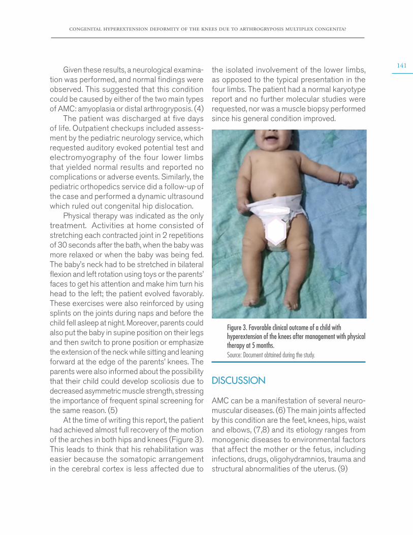
congenital hyperextension deformity of the knees due to arthrogryposis multiplex congenita?
141Given these results, a neurological examina-
tion was performed, and normal findings were observed. This suggested that this condition could be caused by either of the two main types of AMC: amyoplasia or distal arthrogryposis. (4)
The patient was discharged at five days of life. Outpatient checkups included assess-ment by the pediatric neurology service, which requested auditory evoked potential test and electromyography of the four lower limbs that yielded normal results and reported no complications or adverse events. Similarly, the pediatric orthopedics service did a follow-up of the case and performed a dynamic ultrasound which ruled out congenital hip dislocation.
Physical therapy was indicated as the only treatment. Activities at home consisted of stretching each contracted joint in 2 repetitions of 30 seconds after the bath, when the baby was more relaxed or when the baby was being fed. The baby’s neck had to be stretched in bilateral flexion and left rotation using toys or the parents’ faces to get his attention and make him turn his head to the left; the patient evolved favorably. These exercises were also reinforced by using splints on the joints during naps and before the child fell asleep at night. Moreover, parents could also put the baby in supine position on their legs and then switch to prone position or emphasize the extension of the neck while sitting and leaning forward at the edge of the parents’ knees. The parents were also informed about the possibility that their child could develop scoliosis due to decreased asymmetric muscle strength, stressing the importance of frequent spinal screening for the same reason. (5)
At the time of writing this report, the patient had achieved almost full recovery of the motion of the arches in both hips and knees (Figure 3). This leads to think that his rehabilitation was easier because the somatopic arrangement in the cerebral cortex is less affected due to
the isolated involvement of the lower limbs, as opposed to the typical presentation in the four limbs. The patient had a normal karyotype report and no further molecular studies were requested, nor was a muscle biopsy performed since his general condition improved.
Figure 3. Favorable clinical outcome of a child with hyperextension of the knees after management with physical therapy at 5 months.Source: Document obtained during the study.
DISCUSSION
AMC can be a manifestation of several neuro-muscular diseases. (6) The main joints affected by this condition are the feet, knees, hips, waist and elbows, (7,8) and its etiology ranges from monogenic diseases to environmental factors that affect the mother or the fetus, including infections, drugs, oligohydramnios, trauma and structural abnormalities of the uterus. (9)
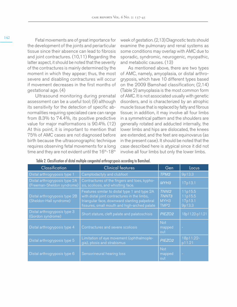
142
case reports Vol. 6 No. 2: 137-45
Fetal movements are of great importance for the development of the joints and periarticular tissue since their absence can lead to fibrosis and joint contractures. (10,11) Regarding the latter aspect, it should be noted that the severity of the contractures is mainly determined by the moment in which they appear; thus, the most severe and disabling contractures will occur if movement decreases in the first months of gestational age. (4)
Ultrasound monitoring during prenatal assessment can be a useful tool; (9) although its sensitivity for the detection of specific ab-normalities requiring specialized care can range from 8.3% to 74.4%, its positive predictive value for major malformations is 90.4%. (12) At this point, it is important to mention that 75% of AMC cases are not diagnosed before birth because the ultrasonographic technique requires observing fetal movements for a long time and they are not evident until the 16th-18th
week of gestation. (2,13) Diagnostic tests should examine the pulmonary and renal systems as some conditions may overlap with AMC due to sporadic, syndromic, neurogenic, myopathic, and metabolic causes. (13)
As mentioned above, there are two types of AMC, namely, amyoplasia, or distal arthro-gryposis, which have 10 different types based on the 2009 Bamshad classification; (2,14) (Table 2) amyoplasia is the most common form of AMC. It is not associated usually with genetic disorders, and is characterized by an atrophic muscle tissue that is replaced by fatty and fibrous tissue; in addition, it may involve all four limbs in a symmetrical pattern and the shoulders are generally rotated and adducted internally, the lower limbs and hips are dislocated, the knees are extended, and the feet are equinovarus (as in the present case). It should be noted that the case described here is atypical since it did not involve all four limbs but only the lower limbs.
Table 2. Classification of distal multiple congenital arthrogryposis according to Bamshad.
Classification Clinical features Gen LocusDistal arthrogryposis type 1 Camptodactyly and clubfoot TPM2 9p13.3
Distal arthrogryposis type 2A (Freeman-Sheldon syndrome)
Contractures of the fingers and toes, kypho-sis, scoliosis, and whistling face.
MYH3 17p13.1
Distal arthrogryposis type 2B (Sheldon-Hall syndrome)
Features similar to distal type 1 and type 2A with distal joint contractures in the limbs, triangular face, downward slanting palpebral fissures, small mouth and high-arched palate
TNNI2TNNT3MYH3TMP2
11p15.511p15.517p13.19p13.3
Distal arthrogryposis type 3 (Gordon syndrome)
Short stature, cleft palate and palatoschisis PIEZO2 18p11.22-p11.21
Distal arthrogryposis type 4 Contractures and severe scoliosisNot mapped out
-
Distal arthrogryposis type 5Limitation of eye movement (ophthalmople-gia), ptosis and strabismus
PIEZO218p11.22- p11.21
Distal arthrogryposis type 6 Sensorineural hearing loss Not mapped out
-
congenital hyperextension deformity of the knees due to arthrogryposis multiplex congenita?
143Table 2. Classification of distal multiple congenital arthrogryposis according to Bamshad. (continued)
Classification Clinical features Gen Locus
Distal arthrogryposis type 7Trismus- pseudocamptodactyly, short stature and shortened paralyzed muscles
MYH8 17p13.1
Distal arthrogryposis type 8Autosomal dominant multiple pterygium syndrome
MYH3 17p13.1
Distal arthrogryposis type 9 or Beals syndrome
Congenital contractural arachnodactyly, phe-notypes similar to those of Marfan syndrome, but without cardiovascular abnormalities
FBN2 5q23.2
Distal arthrogryposis type 10 Congenital plantar contracturesAssigned to the 2q gene
2q31.3-q32.1
Source: Own elaboration based on Bamshad et al. (14)
Patients with suspected AMC and corpus callosum agenesis, lisencephaly, ventriculomeg-aly, microcephaly or cerebellar vermis aplasia require fetal MRI for the correct diagnosis of brain disease. (9) Similarly, in order to guide AMC classification, differential diagnosis and give advice to the family, a complete physical assessment should be performed at birth. It must include neurological and affected joint examination; identification of limb contractures (most frequent finding in AMC patients and found in 67% of cases diagnosed prenatally); (15) electrophysiological studies; muscle and nerve biopsy; and gene sequencing (3,7).
AMC is related to over 400 specific medical conditions and over 350 genes are involved in its development. (7) The majority (70-85%) of patients present with associated neurological alterations, which usually have a genetic origin with autosomal recessive, autosomal dominant or X-linked mechanisms of inheritance; howev-er, there are a large number of cases that are sporadic or idiopathic. (7,15) Thus, given the wide variety of clinical presentations and the considerable number of triggering mutations, determining the primary cause of a specific type of AMC is a challenge.
The therapeutic approach to AMC is based on surgical reconstructions, orthopedic manage-
ment, and physical and rehabilitative therapies for extended periods. (14) In this sense, it is important to know that the long-term prognosis of patients with this disease is favorable if there is an adequate multidisciplinary treatment that improves their quality of life. In this respect, Polania-Rodríguez et al. (11) demonstrated that patients who receive adequate treatment have a normal level of intelligence and have a more independent life in adulthood.
The literature only has three similar case reports in which patients received care with a multidisciplinary approach for their rehabilitation and orthopedic treatment. (5,10,11) This led to rethinking other differential diagnoses that might be worth including to guide readers to similar findings.
In this case, it is noteworthy that the deformi-ties did not involve all four limbs and, therefore, did not fully fit into any of the existing clas-sifications. In addition, the patient presented a favorable clinical course. Consequently, it is important to consider that the patient’s condition could also be explained by an isolated congenital dislocation of the knee, or by an AMC-associated dislocation. (16-18)
Likewise, the patient presented with some clinical features compatible with the alternative diagnosis of congenital knee dislocation, such
144
case reports Vol. 6 No. 2: 137-45
as joint instability, reduction of dislocation with snap or piston, muscle retraction (knee quadriceps, hip adductors) , restricted range of motion (hip abduction) and presence of skin folds or grooves, (18) which were more noticeable after achieving complete improve-ment (Figure 3).
Congenital dislocation of the knee is a rare condition (1 case per 100 000 live births) (17) and is easily diagnosed in the first hours of life. Usually, physical examination is sufficient to observe the marked hyperextension of the knee in contrast to the habitual position in flexion, although a radiography can support the diagnosis. At this point, it is worth clarifying that knee involvement is common in AMC dis-orders, ranging from soft tissue contractures to subluxation and dislocation; specifically, knee problems can be found in up to 38-90% of amyoplasia cases. (19)
The treatment for AMC and congenital knee dislocation is the same, and the recommen-dations for physical therapy described above should be maintained for 4-8 weeks; depending on the case, a splint and even surgery may be indicated if therapy fails. (16)
CONCLUSIONS
Antenatal suspicion, thorough physical exam-ination and differential diagnosis are of vital importance to achieve a satisfactory clinical course in AMC patients; this allows providing proper management, minimizing diagnostic errors, avoiding unnecessary procedures, and performing effective and timely treatment with outstanding results. It should be noted that for the optimal and accurate management of this condition, specialties such as pediatrics, ortho-pedics, physiatry, neurology and genetics should be involved.
ETHICAL CONSIDERATIONS
This case report was prepared after obtaining the informed consent of the patient’s parents and the approval of the ethics committee of the hospital where the child was treated.
CONFLICT OF INTEREST
None stated by the authors.
FUNDING
None stated by the authors.
ACKNOWLEDGEMENTS
None stated by the authors.
REFERENCES
1. Smith C, Parboosingh JS, Boycott KM, Bön-nemann CG, Mah JK, Lamont RE, et al. Ex-pansion of the GLE1-associated arthrogryposis multiplex congenita clinical spectrum. Clin Ge-net. 2017;91(3):426-30. http://doi.org/f9n7hk.
2. Ma L, Yu X. Arthrogryposis multiplex congeni-ta: classification, diagnosis, perioperative care, and anesthesia. Front Med. 2017;11(1):48-52. http://doi.org/dtcv.
3. Valdés-Flores M, Casas-Avila L, Hernán-dez-Zamora E, Kofman S, Hidalgo-Bravo A. Characterization of a group unrelated patients with arthrogryposis multiplex congenita. J Pediatr (Rio J). 2015;92(1):58-64. http://doi.org/dtcw.
4. Álvarez-Quiroz P, Yokoyama-Rebollar E. Abordaje clínico y diagnóstico de la artrogripo-sis. Acta Pediatr Mex. 2019;40(1):44-50.
5. Azbell K, Dannemiller L. A Case Report of an Infant with Arthrogryposis. Pediatr Phys Ther. 2015;27(3):293-301. http://doi.org/dtcz.
congenital hyperextension deformity of the knees due to arthrogryposis multiplex congenita?
1456. Lowry RB, Sibbald B, Bedard T, Hall JG.
Prevalence of multiple congenital contractures including arthrogryposis multiplex congenita in Alberta, Canada, and a strategy for classifica-tion and coding. Birth Defects Res Part A Clin Mol Teratol. 2010;88(12):1057-61. http://doi.org/dv7rmp.
7. Ahmed AA, Skaria P, Safina NP, Thiffault I, Kats A, Taboada E, et al. Arthrogryposis and pterygia as lethal end manifestations of genetically defined congenital myopathies. Am J Med Genet A. 2017;176(2):359-67. http://doi.org/gdbnq6.
8. Hoff JM, Loane M, Gilhus NE, Rasmussen S, Daltveit AK. Arthrogryposis multiplexa congenita: An epidemiologic study of nearly 9 million births in 24 EUROCAT registers. Eur J Obstet Gynecol Reprod Biol. 2011;159(2):347-50. http://doi.org/bp7cz7.
9. Binkiewicz-Glinska A, Wierzba J, Szurows-ka E, Ruckeman-Dziurdzinska K, Bakula S, Sokolów M, et al. Arthrogryposis multiplex congenital - multidisciplinary care - including own experience. Dev period Med. 2016;20(3):191-6.
10. Binkiewicz-Glinska A, Sobierajska-Rek A, Bakula S, Wierzba J, Drewek K, Kowalski IM, et al. Arthrogryposis in infancy, multidis-ciplinary approach: case report. BMC Pediatr. 2013;13(1):184. http://doi.org/gbd665.
11. Polania-Rodríguez M, Nájera-Ruano S, Lamshing-Salinas P. Artrogriposis múltiple congénita: presentación de un caso. An Med (Mex). 2016;61(4):286-90.
12. Nakling J, Backe B. Routine ultrasound scree-ning and detection of congenital anomalies outside a university setting. Acta Obstet Gynecol Scand. 2005;84(11):1042. http://doi.org/b2rnmq.
13. Haliloglu G, Topaloglu H. Arthrogryposis and fetal hypomobility syndrome. Handb Clin Neurol. 2013;113:1311-9. http://doi.org/f4tq9z.
14. Bamshad M, Van Heest AE, Pleasure D. Arthro-gryposis: A review and update. J Bone Joint Surg Am. 2009;91(Suppl 4):40-6. http://doi.org/c8n844.
15. Skaria P, Dahl A, Ahmed A. Arthrogrypo-sis multiplex congenita in utero: radiologic and pathologic findings. J Matern Neonatal Med. 2019;32(3):502-11. http://doi.org/dtgk.
16. Rampal V, Mehrafshan M, Ramanoudjame M, Seringe R, Glorion C, Wicart P. Congenital dislocation of the knee at birth - Part 2: Impact of a new classification on treatment strategies, results and prognostic factors. Orthop Traumatol Surg Res. 2016;102(5):635-8. http://doi.org/f836x9.
17. Mottershead NJ, Patel UD, Reynolds P. Con-genital dislocation of the knee. Arch Dis Child Fetal Neonatal Ed. 2012;97(5):F352. http://doi.org/dtgn.
18. Mehrafshan M, Wicart P, Ramanoudjame M, Seringe R, Glorion C, Rampal V. Congeni-tal dislocation of the knee at birth - Part I: Clinical signs and classification. Orthop Traumatol Surg Res. 2016;102(5):631-3. http://doi.org/dtgm.
19. Lampasi M, Antonioli D, Donzelli O. Manage-ment of knee deformities in children with arthro-gryposis. Musculoskelet Surg. 2012;96(3):161-9. http://doi.org/f4jgd5.
case reports 2020; 6(2)
BRAIN MRI FINDINGS IN NON-KETOTIC HYPERGLYCEMIC CRISIS: CASE REPORT
Keywords: Hyperglycemia; Seizures; Magnetic Resonance Imaging.Palabras clave: Hiperglicemia; Convulsiones; Imagen por resonancia magnética.
Received: 03/12/2019 Accepted: 26/02/2020
Corresponding authorLaura Estefanía Arenas-Vargas. Unit of Clinical Neurology,
Department of Internal Medicine, Faculty of Medicine, Universidad Nacional de Colombia.
Bogotá D.C. Colombia. Email: [email protected].
Laura Estefanía Arenas-VargasRuben Darío Arenas-DiazEnrique Hernandez-Rojas
Universidad Nacional de Colombia - Bogotá Campus - Faculty of Medicine -
Department of Internal Medicine - Unit of Clinical Neurology - NeuroUnal Research Group - Bogotá D.C. - Colombia.
https://doi.org/10.15446/cr.v6n2.83866
Fabián Riaño-MontañezUniversidad Nacional de Colombia
- Bogotá Campus - Faculty of Medicine - Department of Diagnostic Imaging
- Bogotá D.C. - Colombia.
brain mri findings in non-ketotic hyperglycemic crisis: case report
147ABSTRACT
Introduction: Seizures related to metabolic disorders are common phenomena in many clin-ical contexts. However, clinical manifestations and neuroimaging findings in the context of a hyperglycemic crisis are less frequent phenom-ena with unclear pathophysiology.
Case report: A 68-year-old man presented focal seizures and right homonymous hemian-opsia after a non-ketotic hyperglycemic crisis. Brain MRI showed cortical diffusion restriction and subcortical T2 / FLAIR hypointensity in left occipital, temporal (mesial) and parietal lobes. Spectroscopy was performed show-ing a nonspecific pattern, cerebrospinal flu-id was normal and there was improvement with glycemic control. MRI findings were considered secondary to the hyperglycemic crisis.
Conclusion: Non-ketotic hyperglycemic states can manifest with several rare neuro-logical alterations and recognizing them early is of vital importance given their potential revers-ibility. As in other metabolic disorders, epileptic seizures in this context can have focal-type characteristics. Although pathophysiological mechanisms are not clearly elucidated yet, multiple neuroimaging techniques promise to establish patterns that allow accurate and timely diagnosis.
RESUMEN
Introducción. Las crisis convulsivas secun-darias a trastornos metabólicos son frecuentes en muchos contextos clínicos; sin embargo, las manifestaciones clínicas y los hallazgos en neuroimágenes en pacientes con crisis hiper-glicémicas son menos frecuentes y tienen una fisiopatología poco clara.
Presentación del caso. Paciente masculino de 68 años de edad quien presentó una crisis convulsiva focal y hemianopsia homónima derecha después de haber presentado una crisis hiper-glicémica no cetósica. La resonancia magnética cerebral mostró una lesión hipointensa en T2 y FLAIR con restricción a la difusión en la corteza cerebral de los lóbulos occipital, temporal medial y parietal del lado izquierdo. La espectroscopía mos-tró un patrón inespecífico y el estudio del líquido cefalorraquídeo (LCR) fue normal, lo que descartó etiologías infecciosas, vasculares y tumorales. El paciente mejoró con el control glicémico.
Conclusión. Los estados hiperglicémicos no cetóticos pueden manifestarse con varias alte-raciones neurológicas poco frecuentes; dada su reversibilidad potencial, su reconocimiento temprano es de vital importancia. A pesar de que los mecanismos fisiopatológicos aún no se han aclarado del todo, las diferentes técnicas de neuroimagen prometen establecer patrones que permiten un diagnóstico preciso y oportuno.
148
case reports Vol. 6 No. 2: 146-55
INTRODUCTION
Seizures related to metabolic disorders are common phenomena in many clinical contexts due to hydroelectrolyte alterations, liver, or kidney failure and, more frequently, hypogly-cemia. However, clinical manifestations (vi-sual disturbances, convulsions) and imaging findings, in the context of a hyperglycemic crisis, are less frequent and have an unclear pathophysiology.
Seizures are a known complication of hypo-glycemia. (1) Conversely, convulsions induced by hyperglycemia, although described in some case reports, do not appear often in clinical practice, as do neurological manifestations of hyperglycemia such as abnormal movements (chorea, athetosis, ballism), headache and visual hallucinations. (2,3) The underlying pathophys-iological mechanism of epileptic crises induced by hyperglycemia is not clearly understood, however, they usually have some characteristics in common: they are focal and are associated with an imaging pattern characterized by cortical edema and T2 white matter hypointensity pre-dominating in the posterior and mesial regions. (4) Hyperglycemic crises associated with these complications are always non-ketotic.
The literature describes a few similar cases with characteristic imaging findings for hyperglycemia, which requires considering it as a differential diagnosis in the context of other serious processes such as infections or vascular disorders. The following is the case of a patient with occipital epileptic seizures and imaging findings suggestive of this condition.
CASE REPORT
A 68-year-old-man, right-handed, white, from a middle-income household in Bogotá, unem-ployed, without relevant medical or family history, presented with polydipsia, polyuria and noctu-ria for two weeks, accompanied by episodes of time and spatial disorientation, amnestic failures, inattention, and gait abnormality. He consulted the emergency room where hyperglycemia was found at 489 mg/dL; he initially received treat-ment for the hyperglycemic crisis with hydration and insulin, achieving metabolic control. Subse-quently, he was discharged with treatment for type II diabetes mellitus.
The patient attended medical consultation once again a week later because of persistent cognitive impairment, drowsiness, increased confusion, worsening of the gait pattern, and visual complaints. Blood glucose levels were at 400 mg/dLL, so he was admitted to the hospital. During his stay, he presented a focal epileptic crisis manifested with tonic posture of the four limbs and clonic seizure movements of the lower right limb which required the ad-ministration of intravenous levetiracetam with consequent seizure improvement.
One day later, he had two seizures with tonic posture of the four limbs accompanied by clonic seizure movements lasting two minutes; benzodiazepines were administered to control the crisis. The patient was somnolent, inattentive, disoriented with right congruous homonymous hemianopia with macular sparing and sensory ataxic gait with right hemihypoesthesia without motor deficit. Initial laboratory tests showed hyperglycemia without other relevant findings.
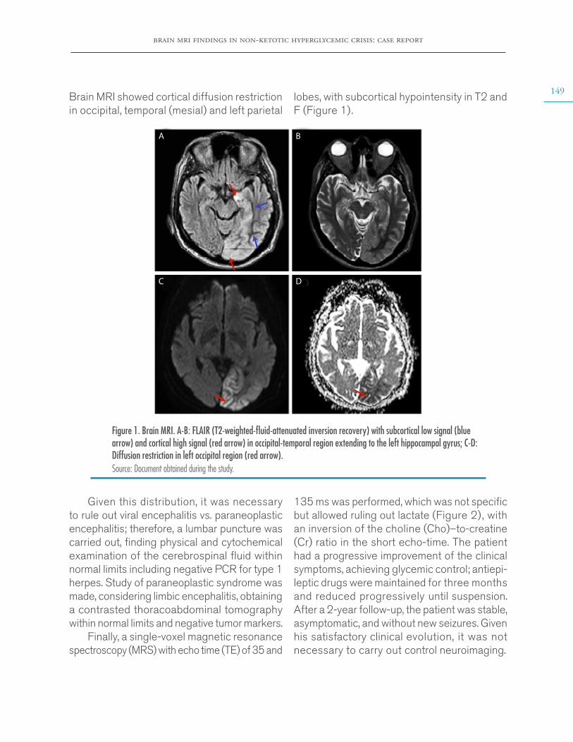
brain mri findings in non-ketotic hyperglycemic crisis: case report
149Brain MRI showed cortical diffusion restriction in occipital, temporal (mesial) and left parietal
lobes, with subcortical hypointensity in T2 and F (Figure 1).
A B
C D
Figure 1. Brain MRI. A-B: FLAIR (T2-weighted-fluid-attenuated inversion recovery) with subcortical low signal (blue arrow) and cortical high signal (red arrow) in occipital-temporal region extending to the left hippocampal gyrus; C-D: Diffusion restriction in left occipital region (red arrow). Source: Document obtained during the study.
Given this distribution, it was necessary to rule out viral encephalitis vs. paraneoplastic encephalitis; therefore, a lumbar puncture was carried out, finding physical and cytochemical examination of the cerebrospinal fluid within normal limits including negative PCR for type 1 herpes. Study of paraneoplastic syndrome was made, considering limbic encephalitis, obtaining a contrasted thoracoabdominal tomography within normal limits and negative tumor markers.
Finally, a single-voxel magnetic resonance spectroscopy (MRS) with echo time (TE) of 35 and
135 ms was performed, which was not specific but allowed ruling out lactate (Figure 2), with an inversion of the choline (Cho)–to-creatine (Cr) ratio in the short echo-time. The patient had a progressive improvement of the clinical symptoms, achieving glycemic control; antiepi-leptic drugs were maintained for three months and reduced progressively until suspension. After a 2-year follow-up, the patient was stable, asymptomatic, and without new seizures. Given his satisfactory clinical evolution, it was not necessary to carry out control neuroimaging.
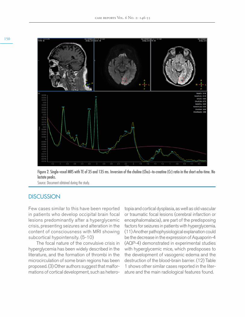
150
case reports Vol. 6 No. 2: 146-55
Figure 2. Single-voxel MRS with TE of 35 and 135 ms. Inversion of the choline (Cho)–to-creatine (Cr) ratio in the short echo-time. No lactate peaks. Source: Document obtained during the study.
DISCUSSION
Few cases similar to this have been reported in patients who develop occipital brain focal lesions predominantly after a hyperglycemic crisis, presenting seizures and alteration in the content of consciousness with MRI showing subcortical hypointensity. (5-10)
The focal nature of the convulsive crisis in hyperglycemia has been widely described in the literature, and the formation of thrombi in the microcirculation of some brain regions has been proposed. (3) Other authors suggest that malfor-mations of cortical development, such as hetero-
topia and cortical dysplasia, as well as old vascular or traumatic focal lesions (cerebral infarction or encephalomalacia), are part of the predisposing factors for seizures in patients with hyperglycemia. (11) Another pathophysiological explanation could be the decrease in the expression of Aquaporin-4 (AQP-4) demonstrated in experimental studies with hyperglycemic mice, which predisposes to the development of vasogenic edema and the destruction of the blood-brain barrier. (12) Table 1 shows other similar cases reported in the liter-ature and the main radiological features found.
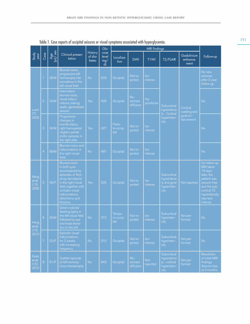
brain mri findings in non-ketotic hyperglycemic crisis: case report
151Table 1. Case reports of occipital seizures or visual symptoms associated with hyperglycemia.
Stud
yye
ar
Cas
e
Age
(yr)/
sex Clinical presen-
tation
History of dia-betes
Glu-cose level mg/dL
MRI findings
Follow-upLocaliza-tion DWI T1WI T2/FLAIR
Gadolinium enhance-
ment
Lavin (7)2005
1 39/M
Blurred vision, progressive left hemianopia; hal-lucinations in the left visual field.
No 503 OccipitalNot re-ported
Iso-intense
Subcortical hypointensi-ty - Cortical hyperinten-sity
Cortical swelling and gyral en-hancement
No new seizures after 2-year follow-up.
2 54/M
Intermittent blurred vision, visual halluci-nations, staring spells, generalized seizure.
Yes 426 OccipitalRe-stricted diffusion
Hy-pointense
No
3 34/M
Progressive changes in mental status, right hemispatial neglect, partial motor seizures in the right side.
Yes 427Parie-to-occip-ital
Not re-ported
Iso-intense
No
4 69/M
Blurred vision and hallucinations in the right visual field.
No 487 OccipitalNot re-ported
Iso-intense
No
Wang et al. (13)2005
5 59/F
Blurred vision in both eyes accompanied by episodes of flick-ering red objects in the right visual field, together with complex visual hallucinations, distortions, and illusions.
Yes 535 OccipitalNot re-ported
Iso-intense
Subcortical hypointensi-ty and gyral hyperinten-sity
Not reported
On follow-up MRI taken 19 days later, the patient was seizure-free and the sub-cortical T2 hypointensity was less intense.
Hung et al. (14)2010
6 30/M
Green-colored flashing lights in the left visual field followed by eye and head devia-tion to the left.
No 372Tempo-ro-occip-ital
Not re-ported
Iso-intense
Subcortical hypointen-sity
Not per-formed
No
7 53/F
Episodic visual hallucinations for 2 weeks with increasing frequency.
No 310 OccipitalNot re-ported
Iso-intense
Subcortical hypointen-sity
Not per-formed
No
Guez et al. (15)2010
8 61/FSudden episode of left homony-mous hemianopia.
No 943 OccipitalRe-stricted diffusion
Not reported
Subcortical hypointensi-ty - cortical hyperinten-sity
Not per-formed
Resolution of initial MRI findings. Seizure-free at 3 months.
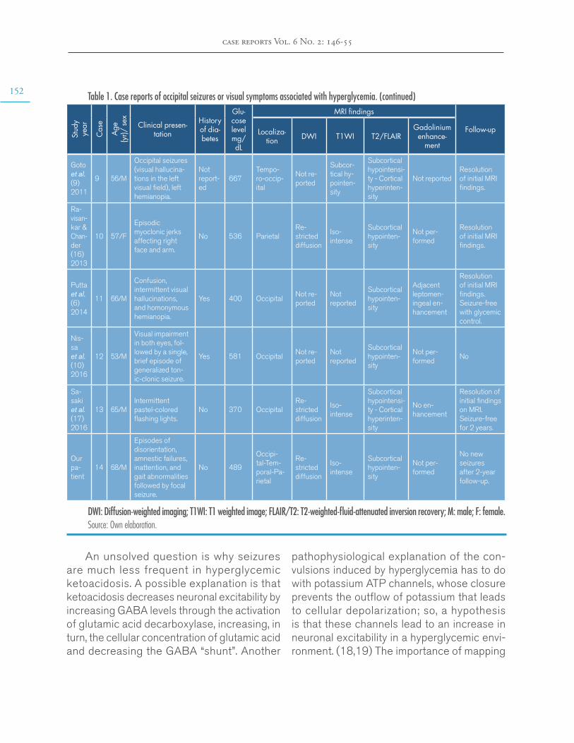
152
case reports Vol. 6 No. 2: 146-55
Table 1. Case reports of occipital seizures or visual symptoms associated with hyperglycemia. (continued)
Stud
yye
ar
Cas
e
Age
(yr)/
sex Clinical presen-
tation
History of dia-betes
Glu-cose level mg/dL
MRI findings
Follow-upLocaliza-tion DWI T1WI T2/FLAIR
Gadolinium enhance-
ment
Goto et al. (9)2011
9 56/M
Occipital seizures (visual hallucina-tions in the left visual field), left hemianopia.
Not report-ed
667Tempo-ro-occip-ital
Not re-ported
Subcor-tical hy-pointen-sity
Subcortical hypointensi-ty - Cortical hyperinten-sity
Not reportedResolution of initial MRI findings.
Ra-visan-kar & Chan-der (16)2013
10 57/F
Episodic myoclonic jerks affecting right face and arm.
No 536 ParietalRe-stricted diffusion
Iso-intense
Subcortical hypointen-sity
Not per-formed
Resolution of initial MRI findings.
Putta et al. (6)2014
11 66/M
Confusion, intermittent visual hallucinations, and homonymous hemianopia.
Yes 400 OccipitalNot re-ported
Not reported
Subcortical hypointen-sity
Adjacent leptomen-ingeal en-hancement
Resolution of initial MRI findings. Seizure-free with glycemic control.
Nis-sa et al. (10)2016
12 53/M
Visual impairment in both eyes, fol-lowed by a single, brief episode of generalized ton-ic-clonic seizure.
Yes 581 OccipitalNot re-ported
Not reported
Subcortical hypointen-sity
Not per-formed
No
Sa-saki et al. (17)2016
13 65/MIntermittent pastel-colored flashing lights.
No 370 OccipitalRe-stricted diffusion
Iso-intense
Subcortical hypointensi-ty - Cortical hyperinten-sity
No en-hancement
Resolution of initial findings on MRI. Seizure-free for 2 years.
Our pa-tient
14 68/M
Episodes of disorientation, amnestic failures, inattention, and gait abnormalities followed by focal seizure.
No 489
Occipi-tal-Tem-poral-Pa-rietal
Re-stricted diffusion
Iso-intense
Subcortical hypointen-sity
Not per-formed
No new seizures after 2-year follow-up.
DWI: Diffusion-weighted imaging; T1WI: T1 weighted image; FLAIR/T2: T2-weighted-fluid-attenuated inversion recovery; M: male; F: female.Source: Own elaboration.
pathophysiological explanation of the con-vulsions induced by hyperglycemia has to do with potassium ATP channels, whose closure prevents the outflow of potassium that leads to cellular depolarization; so, a hypothesis is that these channels lead to an increase in neuronal excitability in a hyperglycemic envi-ronment. (18,19) The importance of mapping
An unsolved question is why seizures are much less frequent in hyperglycemic ketoacidosis. A possible explanation is that ketoacidosis decreases neuronal excitability by increasing GABA levels through the activation of glutamic acid decarboxylase, increasing, in turn, the cellular concentration of glutamic acid and decreasing the GABA “shunt”. Another

brain mri findings in non-ketotic hyperglycemic crisis: case report
153the cerebral distribution of these receptors has been mentioned, which would explain the predilection for certain brain areas. (5)
Another interesting finding that could lead to additional studies and research is the behavior of spectroscopy in this patient, which showed a relative increase in creatine (Cr) and a decrease in N-acetylaspartate (NAA). The literature has described changes in the NAA/Cr ratio, as experimental biomarkers in animal models, in oxidative disorders of cerebral metabolism. (20)
The main differential diagnoses for im-aging changes induced by hyperglycemia include post-convulsive edema, autoimmune encephalitis, herpes simplex encephalitis and early and incidental atypical manifestations of primary neoplastic pathology (pattern of gliomatosis cerebri and diffuse astrocytoma). Clinical correlation and imaging follow-up are necessary since the distribution and signal of the MRI alterations are similar in several clinical entities.
To achieve the differential diagnosis between autoimmune encephalitis and herpes virus, clinical and laboratory tests were performed during the hospitalization of this patient, including analysis of cerebrospinal fluid and extension studies. The results clarified the anatomical distribution of the brain injury which, in this case, is not typical for herpes or autoimmune encephalitis due to cortical predilection, uni-lateral location, poor expansive effect in white matter, or hemorrhagic changes. It should be noted that the anatomical distribution does not correspond to a particular arterial territory, and this, together with spectroscopy, ruled out acute ischemic events.
Finally, the almost null expansive effect of the lesion (particularly in the white matter), would make less probable an infiltrative/neo-plastic pattern in the first place. Because our
patient fully recovered, and based on the MRI signal characteristics, autoimmune encephalitis was unlikely. Decreased T2 signal changes are better explained as the result of intracellular dehydration of glial and supporting tissue and the probable accumulation of free radicals.
This is a relevant case because this ra-diological pattern associated with seizures in the context or non-ketotic hyperglycemia is a very rare complication of diabetes and prompt recognition is imperative for an early treatment and seizure control.
CONCLUSIONS
Non-ketotic hyperglycaemic states can man-ifest with various rare neurological alterations and recognizing them early is of vital importance given their potential reversibility. As in other met-abolic disorders, epileptic seizures can be iden-tified in this context because of their focal-type characteristics.
Even though the pathophysiological mecha-nisms are not clearly elucidated yet, the different neuroimaging techniques promise to establish patterns that allow for an accurate and timely diagnosis. Similarly, differential diagnoses, which are conditions that threaten life, should be part of the approach and routine study of these patients.
A detailed clinical history and laboratory studies, such as cerebrospinal fluid and mag-netic resonance, should guide the definitive diagnosis, avoiding unnecessary treatments and interventions that could entail more risks than benefits, and occasionally unnecessary paraclinical studies.
It should be noted that this type of hyper-glycemic disorder is poorly understood because of its rare occurrence, which is further affected by underreporting and misdiagnosis.
154
case reports Vol. 6 No. 2: 146-55
ETHICAL CONSIDERATIONS
The patient provided his written consent for the publication of this case.
CONFLICT OF INTEREST
None stated by the authors.
FUNDING
None stated by the authors.
ACKNOWLEDGEMENTS
None stated by the authors.
REFERENCES
1. Monami M, Mannucci E, Breschi A, Marchion-ni N. Seizures as the only clinical manifestation of reactive hypoglycemia: A case report. J Endocrinol Invest. 2005;28(10):940-1. http://doi.org/dt4b.
2. Chung SJ, Lee JH, Lee SA, No YJ, Im JH, Lee MC. Co-occurrence of seizure and chorea in a pa-tient with nonketotic hyperglycemia. Eur Neurol. 2006;54(4):230-2. http://doi.org/fnqhw8.
3. Stahlman GC, Auerbach PS, Strickland WG. Neurologic manifestations of non-ketotic hypergly-cemia. J Tenn Med Assoc. 1988 [cited 2020 Jan 10];81(2):77-80. Available from: https://bit.ly/2W-DABzs.
4. Lee EJ, Kim KK, Lee EK, Lee JE. Characteristic MRI findings in hyperglycaemia-induced seizures: diagnostic value of contrast-enhanced fluid-atte-nuated inversion recovery imaging. Clin Radiol. 2016;71(12):1240-7. http://doi.org/dt4d.
5. Moien-Afshari F, Téllez-Zenteno JF. Occipital seizures induced by hyperglycemia: A case report and review of literature. Seizure. 2009;18(5):382-5. http://doi.org/bpj6bx.
6. Putta SL, Weisholtz D, Milligan TA. Occi-pital seizures and subcortical T2 hypointensity in the setting of hyperglycemia. Epilepsy Be-hav Case Reports. 2014;2(1):96-9. http://doi.org/dt4f.
7. Lavin PJM. Hyperglycemic hemianopia: A rever-sible complication of non-ketotic hyperglycemia. Neurology. 2005;65(4):616-9. http://doi.org/cd7fgw.
8. Raghavendra S, Ashalatha R, Thomas SV, Kesavadas C. Focal neuronal loss, reversible subcortical focal T2 hypointensity in seizures with a nonketotic hyperglycemic hyperosmolar state. Neuroradiology. 2007;49(4):299-305. http://doi.org/c64xrr.
9. Goto H, Kumagai T, Momozaki N. MRI fin-dings of occipital seizures in non-ketotic hyper-glycemia. Intern Med. 2011;50(4):367-8. http://doi.org/fv4fj3.
10. Nissa Z, Siddiqi SA, Abdool SAM. Occipital seizures and persistent homonymous hemianopia with T2 hypointensity on MRI in nonketotic hyper-glycemia. Epilepsy Behav Case Rep. 2016;6:3-5. http://doi.org/dt4h.
11. Siddiqi ZA, VanLandingham KE, Husain AM. Reflex seizures and non-ketotic hyperglycemia: An unresolved issue. Seizure. 2002;11(1):63-6. http://doi.org/bx3snz.
12. Chiu CD, Chen CCV, Shen CC, Chin LT, Ma HI, Chuang HY, et al. Hyperglycemia exacer-bates intracerebral hemorrhage via the down-regulation of aquaporin-4: temporal assess-ment with magnetic resonance imaging. Stroke. 2013;44(6):1682-9. http://doi.org/f46tfp.
13. Wang CP, Hsieh PF, Chen CCC, Lin WY, Hu WH, Yang DY, et al. Hyperglycemia with occipital seizures: Images and visual evoked po-tentials. Epilepsia. 2005;46(7):1140-4. http://doi.org/c3k2mv.
14. Hung WL, Hsieh PF, Lee YC, Chang MH. Occipital lobe seizures related to marked elevation
brain mri findings in non-ketotic hyperglycemic crisis: case report
155of hemoglobin A1C: Report of two cases. Seizure. 2010;19(6):359-62. http://doi.org/fvrpz4.
15. Guez A, Obadia M, Lafitte F, Tin SN, Héran F, Gout O. Magnetic resonance spectroscopy findings in a case of hyperglycaemic hemianopia. Rev Neurol (Paris). 2010;166(8-9):737-40. http://doi.org/ctv2qx.
16. Ravisankar S, Chander RV. Cerebral pheo-hyphomycosis: Report of a rare case with review of literature. Neurol India. 2013;61(5):526-8. http://doi.org/dt4k.
17. Sasaki F, Kawajiri S, Nakajima S, Yamagu-chi A, Tomizawa Y, Noda K, et al. Occipital lobe seizures and subcortical T2 and T2* hypoin-tensity associated with nonketotic hyperglycemia: A case report. J Med Case Rep. 2016;10(1):228. http://doi.org/dt4s.
18. Dunn-Meynell AA, Rawson NE, Levin BE. Distribution and phenotype of neurons contai-ning the ATP-sensitive K+ channel in rat brain. Brain Res. 1998;814(1-2):41-54. http://doi.org/ctn4fx.
19. Miki T, Seino S. Roles of KATP channels as me-tabolic sensors in acute metabolic changes. J Mol Cell Cardiol. 2005;38(6):917-25. http://doi.org/d3mkk5.
20. Ben Salem D, Walker PM, Bejot Y, Aho SL, Tavernier B, Rouaud O, et al. N-Ace-tylaspartate/Creatine and Choline/Creatine Ratios in the Thalami, Insular Cortex and White Matter as Markers of Hypertension and Cogni-tive Impairment in the Elderly. Hypertens Res. 2008;31(10):1851-7. http://doi.org/dcs9qh.
case reports 2020; 6(2)
ANABOLIC STEROID-INDUCED MYOSITIS AND OSTEITIS. CASE REPORT THROUGH A RADIOLOGIC APPROACH
Keywords: Myositis; Osteitis; Steroids; Anabolic Agents.Palabras clave: Miositis; Osteítis; Esteroides; Agentes anabólicos.
Received: 24/01/2020 Accepted: 03/05/2020
Corresponding authorAndrés Felipe Donado-Moré.
Department of Radiology and Diagnostic Imaging, Hospital Universitario Nacional de Colombia.
Bogotá D.C. Colombia. Email: [email protected]
Andrés Felipe Donado-MoréEnrique Calvo-Páramo
Hospital Universitario Nacional de Colombia - Department of Radiology and Diagnostic Imaging -
Bogotá D.C. - Colombia.Universidad Nacional de Colombia
- Bogotá Campus - Faculty of Medicine - Department of Diagnostic Imaging
- Bogotá D.C. - Colombia.
https://doi.org/10.15446/cr.v6n2.84717
anabolic steroid-induced myositis and osteitis
157RESUMEN
Introducción. La miositis es una complicación muy rara de las inyecciones extraarticulares de esteroides anabólicos y la osteítis no ha sido re-portada como efecto adverso por esta causa. El presente reporte de caso aporta información sobre los hallazgos imagenológicos de estos dos tipos de inflamaciones.
Presentación del caso. Paciente masculino de 37 años, dedicado al fisicoculturismo, quien cinco días después de recibir una inyección de estano-zolol presentó dolor y edema en la región glútea izquierda asociados a limitación funcional. El suje-to asistió a consulta por este motivo y se le realizó una ecografía y una resonancia magnética con-trastada de pelvis, cuyos resultados permitieron diagnosticarle miositis del glúteo mayor izquierdo y osteítis del hueso ilíaco. Se indicó tratamiento con piperacilina-tazobactam y vancomicina por 10 días y no se requirió manejo quirúrgico dado que se obtuvieron buenos resultados.
Conclusión. La miositis es una complicación rara de las inyecciones de esteroides anabólicos en donde el mecanismo fisiopatológico de estas sus-tancias es incierto. Por su parte, la osteítis es una complicación aún más rara de la cual se presenta el primer caso conocido por esta causa. Dados los hallazgos se plantea que la miositis reportada es de tipo infecciosa; sin embargo, se requieren estudios adicionales que demuestren la asociación causal real.
ABSTRACT
Introduction: Myositis is a rare complication of extra-articular anabolic steroid injections, while osteitis has not been reported as an adverse effect from this cause. This case report provides information about imaging findings of these two entities.
Case presentation: A 37-year-old male, body-builder, presented pain and edema in the left gluteal region, associated with functional lim-itation, 5 days after receiving an intramuscular anabolic steroid injection (stanozolol). The man underwent an ultrasound scan and magnetic resonance imaging of the pelvis with contrast, which allowed making the diagnosis of myositis of the left gluteus maximus and osteitis of the iliac bone. The patient was treated with pipera-cillin-tazobactam and vancomycin for 10 days, without complications. No surgical management was required.
Conclusion: Myositis is a rare complication of anabolic steroid injections and the pathophysio-logical mechanism of this substance is unknown. Osteitis, on the other hand, is an even rarer com-plication and, to the best of our knowledge, this is the first known case associated with this cause. Given the findings, the myositis reported herein has an infectious nature; however, further studies are required to demonstrate the actual causal association.
158
case reports Vol. 6 No. 2: 156-64
INTRODUCTION
Myositis is an inflammation of the muscles that rarely occurs as a complication of extra-articular anabolic steroid injections. The incidence of this condition is not known and, during the prepara-tion of the present study, only 16 publications on this issue were found. Osteitis, on the other hand, is an inflammation of the bones; the occurrence of this complication after the injection of these substances has not been previously reported, which makes the present case unique.
Anabolic steroids are human-made hormones that act on the androgen receptor and they are typically used by bodybuilders. (1) These sub-stances are administered intramuscularly and the most commonly used injectable compounds are testosterone salts (testosterone cypionate, testosterone decanoate, testosterone propionate, testosterone phenylpropionate, testosterone isocaproate, and stanozolol —the compound used by the patient in this case—); 19-nortestoster-one, used in the form of nandrolone decanoate and nandrolone phenylpropionate; boldenone undecylate; and methenolone enanthate.
Although the pathophysiological mechanism of anabolic steroids in myositis is unknown, three hypotheses have been put forward: the inoculation of microorganisms from the skin, the hematogenous spread of bacteria from another focus to the injection site, and the in-nate immune response to steroid ester crystals.
In the absence of abscess or necrosis, the standard of care for steroid myositis includes local anti-inflammatory measures, muscle rest, and intravenous antibiotics. On the contrary, if abscesses are present, they must be drained and, if necrosis is generated, debridement of the dead tissue must be performed; likewise, specific tissue repair techniques must be ap-plied depending on each case.
CASE PRESENTATION
This is the case of a 37-year-old male patient, mestizo, from Bogotá D.C. (Colombia), bodybuild-er, with a stable socioeconomic condition and a healthy lifestyle without cardiovascular risk factors. He reported consuming a high-protein diet, as well as amino acids and fatty acids sup-plements.
The patient attended the emergency service of a quaternary care center on January 6, 2020, due to a feverish sensation (not quantified) for 3 days, stabbing pain and a rash in the left buttock, which prevented normal mobilization of the left leg. A soft tissue ultrasound was performed, which showed no abscesses or necrosis, but did show changes due to myositis of the left gluteus maximus. The subject was discharged with outpatient antibiotic therapy (sultamicillin) and, since he had no appropriate response to the treatment, he attended the same center again on January 8, 2020. The next day, he underwent magnetic resonance imaging (MRI) of the hip with contrast that confirmed the diagnosis of left gluteal myositis. Intravenous antibiotic treatment with 600mg of clindamycin every 8 hours was indicated and he was transferred to the Hospital Universitario Nacional de Colombia (HUN) by ambulance.
On January 11, 2020, at 04:00 a.m., the patient was admitted to the hospitalization service of the HUN with blood pressure of 146/93 mmHg, heart rate of 74 beats/minute, respiratory rate of 19 breaths/minute, temperature of 36.6°C and oxygen saturation of 92% on room air. Physical examination showed edema and erythema of the right gluteal region, induration without areas of fluctuation and loss of thigh extension strength without distal neurovascular deficit. The patient claimed that he did not have any previous rec-ognized medical, genetic, or surgical history, but
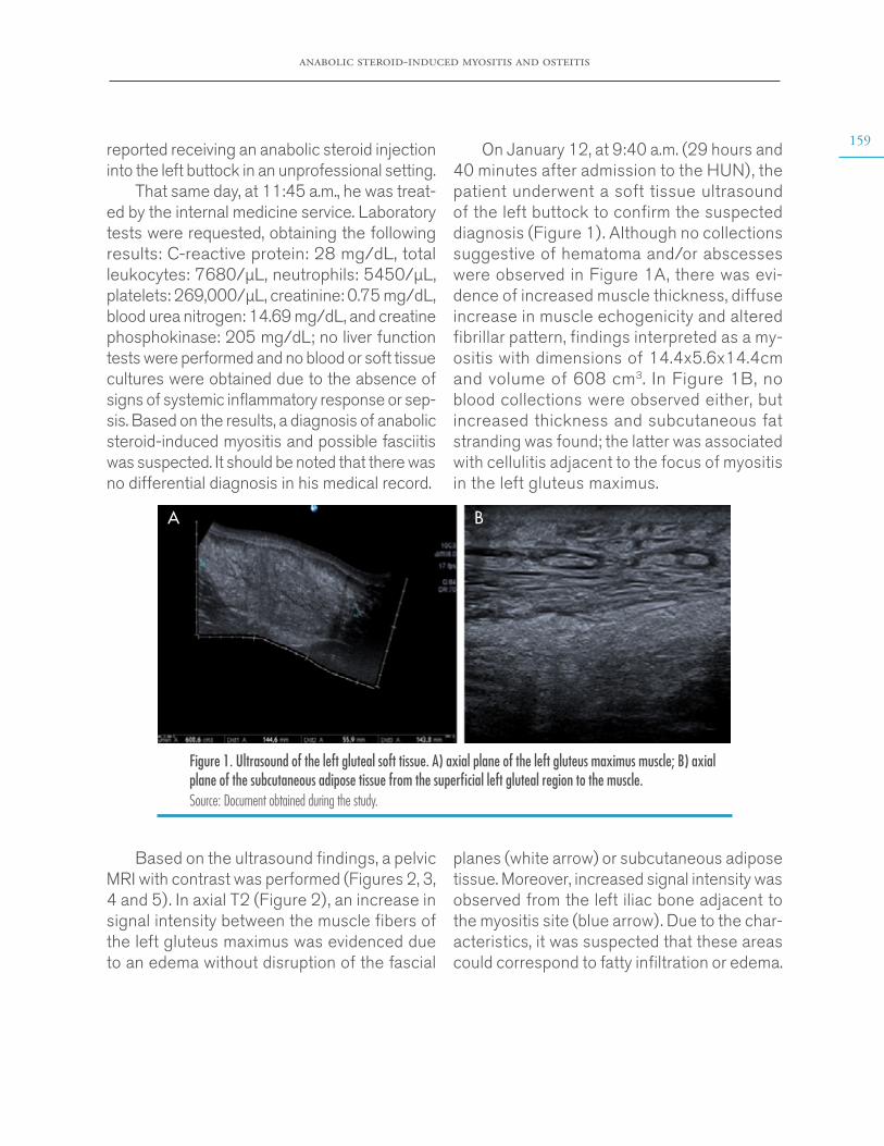
anabolic steroid-induced myositis and osteitis
159reported receiving an anabolic steroid injection into the left buttock in an unprofessional setting.
That same day, at 11:45 a.m., he was treat-ed by the internal medicine service. Laboratory tests were requested, obtaining the following results: C-reactive protein: 28 mg/dL, total leukocytes: 7680/µL, neutrophils: 5450/µL, platelets: 269,000/µL, creatinine: 0.75 mg/dL, blood urea nitrogen: 14.69 mg/dL, and creatine phosphokinase: 205 mg/dL; no liver function tests were performed and no blood or soft tissue cultures were obtained due to the absence of signs of systemic inflammatory response or sep-sis. Based on the results, a diagnosis of anabolic steroid-induced myositis and possible fasciitis was suspected. It should be noted that there was no differential diagnosis in his medical record.
On January 12, at 9:40 a.m. (29 hours and 40 minutes after admission to the HUN), the patient underwent a soft tissue ultrasound of the left buttock to confirm the suspected diagnosis (Figure 1). Although no collections suggestive of hematoma and/or abscesses were observed in Figure 1A, there was evi-dence of increased muscle thickness, diffuse increase in muscle echogenicity and altered fibrillar pattern, findings interpreted as a my-ositis with dimensions of 14.4x5.6x14.4cm and volume of 608 cm3. In Figure 1B, no blood collections were observed either, but increased thickness and subcutaneous fat stranding was found; the latter was associated with cellulitis adjacent to the focus of myositis in the left gluteus maximus.
A B
Figure 1. Ultrasound of the left gluteal soft tissue. A) axial plane of the left gluteus maximus muscle; B) axial plane of the subcutaneous adipose tissue from the superficial left gluteal region to the muscle. Source: Document obtained during the study.
Based on the ultrasound findings, a pelvic MRI with contrast was performed (Figures 2, 3, 4 and 5). In axial T2 (Figure 2), an increase in signal intensity between the muscle fibers of the left gluteus maximus was evidenced due to an edema without disruption of the fascial
planes (white arrow) or subcutaneous adipose tissue. Moreover, increased signal intensity was observed from the left iliac bone adjacent to the myositis site (blue arrow). Due to the char-acteristics, it was suspected that these areas could correspond to fatty infiltration or edema.
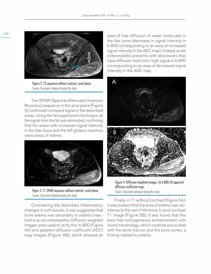
160
case reports Vol. 6 No. 2: 156-64
Figure 2. T2 sequence without contrast, axial plane.Source: Document obtained during the study.
The SPAIR (Spectral Attenuated Inversion Recovery) sequence in the axial plane (Figure 3) confirmed increased signal in the described areas. Using the fat suppression technique, all the signal from the fat was eliminated, confirming that the areas with increased signal intensity in the iliac bone and the left gluteus maximus were areas of edema.
Figurr 3. T1 SPAIR sequence without contrast, axial plane.Source: Document obtained during the study.
Considering the described inflammatory changes in soft tissues, it was suggested that bone edema was secondary to osteitis (reac-tive) or acute osteomyelitis. Diffusion-weighted images were used to verify this: b-800 (Figure 4A) and apparent diffusion coefficient (ADC) map images (Figure 4B), which showed an
area of free diffusion of water molecules in the iliac bone (decrease in signal intensity in b-800 corresponding to an area of increased signal intensity in the ADC map). Instead, acute osteomyelitis presents with abscesses that have diffusion restriction: high signal in b-800 corresponding to an area of decreased signal intensity in the ADC map.
Figure 4. Diffusion-weighted images. A) b-800; B) apparent diffusion coefficient map. Source: Document obtained during the study.
Finally, in T1 without contrast (Figure 5A), it was evident that the area of edema was iso-intense to the rest of the bone. In post-contrast T1 image (Figure 5B), it was found that this area had homogeneous enhancement with round morphology, which could be associated with the bone marrow and the bone cortex, a finding related to osteitis.
A
B
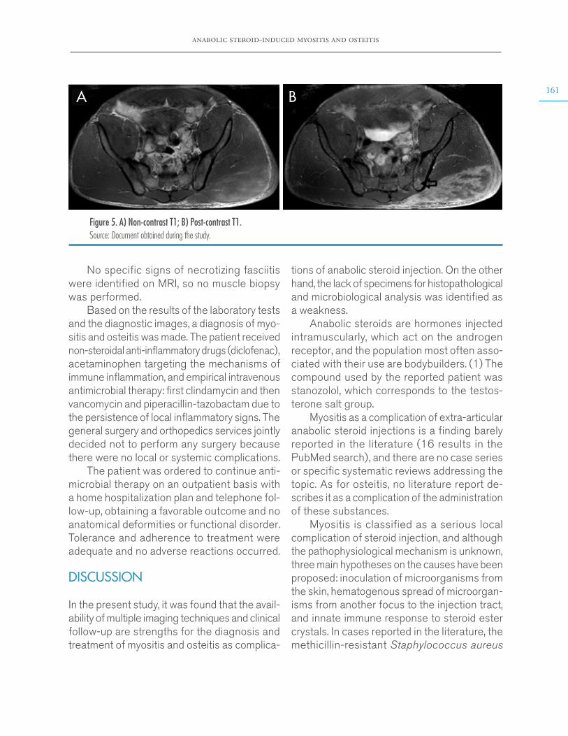
anabolic steroid-induced myositis and osteitis
161
No specific signs of necrotizing fasciitis were identified on MRI, so no muscle biopsy was performed.
Based on the results of the laboratory tests and the diagnostic images, a diagnosis of myo-sitis and osteitis was made. The patient received non-steroidal anti-inflammatory drugs (diclofenac), acetaminophen targeting the mechanisms of immune inflammation, and empirical intravenous antimicrobial therapy: first clindamycin and then vancomycin and piperacillin-tazobactam due to the persistence of local inflammatory signs. The general surgery and orthopedics services jointly decided not to perform any surgery because there were no local or systemic complications.
The patient was ordered to continue anti-microbial therapy on an outpatient basis with a home hospitalization plan and telephone fol-low-up, obtaining a favorable outcome and no anatomical deformities or functional disorder. Tolerance and adherence to treatment were adequate and no adverse reactions occurred.
DISCUSSION
In the present study, it was found that the avail-ability of multiple imaging techniques and clinical follow-up are strengths for the diagnosis and treatment of myositis and osteitis as complica-
tions of anabolic steroid injection. On the other hand, the lack of specimens for histopathological and microbiological analysis was identified as a weakness.
Anabolic steroids are hormones injected intramuscularly, which act on the androgen receptor, and the population most often asso-ciated with their use are bodybuilders. (1) The compound used by the reported patient was stanozolol, which corresponds to the testos-terone salt group.
Myositis as a complication of extra-articular anabolic steroid injections is a finding barely reported in the literature (16 results in the PubMed search), and there are no case series or specific systematic reviews addressing the topic. As for osteitis, no literature report de-scribes it as a complication of the administration of these substances.
Myositis is classified as a serious local complication of steroid injection, and although the pathophysiological mechanism is unknown, three main hypotheses on the causes have been proposed: inoculation of microorganisms from the skin, hematogenous spread of microorgan-isms from another focus to the injection tract, and innate immune response to steroid ester crystals. In cases reported in the literature, the methicillin-resistant Staphylococcus aureus
A B
Figure 5. A) Non-contrast T1; B) Post-contrast T1. Source: Document obtained during the study.
162
case reports Vol. 6 No. 2: 156-64
bacteria (2-8) has been frequently isolated, so most anabolic steroid-induced myositis is be-lieved to be infectious (pyomyositis). However, this conclusion cannot be reached since there are no studies that prove the causal relationship.
The most common sites for anabolic steroid injection are the gluteus maximus, the deltoid, and the vastus lateralis muscles. (9) Therefore, infectious signs should be looked for in these sites in the first instance.
Pyomyositis occurs in three stages: 1) in-vasive stage, characterized by muscle edema and pain from bacterial seeding; 2) suppurative
stage, characterized by abscess formation that occurs 10-21 days after injection; and 3) late stage, characterized by multiple organ failure. It should be noted that if the first two stages are not treated promptly, the third stage can lead to death. (10)
In ultrasound, myositis is characterized by increased thickness and echogenicity of the muscle, both focal and diffuse, and altered fibrillar pattern secondary to edema and hyperemia.
The characteristics of myositis and osteitis observable on MRI and CT scans are summa-rized in Table 1:
Table 1. Imaging characteristics of myositis and osteitis
Technique/Pathology Myositis Osteitis
Magnetic Reso-nance Imaging
Increased muscle thickness and signal intensity on T2-weighted or STIR sequences, decreased T1-weighted signal between muscle fibers, and secondary alteration of muscle fibrillar pattern. There is no diffusion restriction to the flow of water molecules but a homogeneous enhancement of the inflammation with the contrast agent.
Edema of the bone cortex next to the focus of infection in the soft tissues: ill-defined foci of hyperintensity on T2, with no decrease in signal in the fat suppression sequences, and isointense on T1-weighted imaging. There are no foci of diffusion restriction of free water molecules and the enhancement pat-tern is usually homogeneous and generally confined to the bone cortex.
Computerized axial tomography
Increase in muscle thickness, which be-comes asymmetric with respect to the contralateral side, and homogeneous decrease in the attenuation values with loss of fibrillar pattern and definition of the borders between the muscle and the subcutaneous cellular tissue.
Signs with low sensitivity.
Source: Elaboration based on Hayeri et al., (10) Kim et al., (11) Malghem et al., (12) Gupta et al. (13) and Lee et al. (14)
In the MRI, the areas of myositis have a homogeneous enhancement with the use of gadolinium-based contrast agent, while the areas of muscle necrosis do not present any enhancement. If abscesses coexist, they have well-defined walls (often thick) that are enhanced by the contrast agent.
Muscle tumors, both benign and primary malignant, are an important differential diagnosis,
since they can be differentiated by diffusion restriction on MRI. In addition, malignant tumors are very likely to cause disruption of the fascial planes with invasion of adjacent structures.
Another relevant aspect of MRI is the identification of specific signs of necrotizing fasciitis, such as extensive involvement of the deep fascia (intramuscular or contacting the superficial fascia in more than three locations),

anabolic steroid-induced myositis and osteitis
163absence of fascial enhancement with gadolin-ium-based contrast agent, and, less frequently, the presence of gas. Computed tomography (CT) is more sensitive in detecting gas using bone window but the detection of myositis is more difficult with this technique due to its poor soft tissue resolution.
As mentioned above, osteitis is a rare complication of extra-articular anabolic steroid injection that is not reported in the literature, which makes the case presented herein valuable. In that sense, it is worth mentioning that such inflammation is characterized by an edema of the bone cortex adjacent to the focus of the infection in the soft tissues, in addition to ill-defined foci of hyperintensity on T2 and isointense on T1-weighted imaging. In osteitis, there are no diffusion restriction foci on MRI and the enhancement pattern is usu-ally homogeneous and confined to the bone cortex. (10,14)
CONCLUSION
Myositis is a rare complication of anabolic steroid injections and the pathophysiological mechanism of this substance is unknown. Osteitis, on the oth-er hand, is an even rarer complication and, to the best of our knowledge, this is the first known case associated with this cause. Given the findings, the myositis reported herein has an infectious nature; however, further studies are required to demonstrate the actual causal association.
PATIENT’S PERSPECTIVE
The patient stated that he received a high-quality, comprehensive, and timely treatment.
ETHICAL CONSIDERATIONS
This case report was prepared after obtaining the informed consent of the patient.
CONFLICT OF INTEREST
None stated by the authors.
FUNDING
None stated by the authors.
ACKNOWLEDGEMENTS
None stated by the authors.
REFERENCES
1. de Ronde W, Smit DL. Anabolic androgenic steroid abuse in young males. Endocr Connect. 2020;9(4):R102-R111. http://doi.org/d2rf.
2. Peterson C, Hodler J. Adverse events from di-agnostic therapeutic joint injections: a literature review. Skeletal Radiol. 2011;40(1):5-12. http://doi.org/d7t6kc.
3. Honcharuk E, Monica J. Complications asso-ciated with intra-articular and extra-articular cor-ticosteroid injections. JBJS Rev. 2016;4(12):1-8. http://doi.org/d2tk.
4. Crum-Cianflone NF. Bacterial, fungal, parasit-ic and viral myositis. Clin Microbiol Rev. 2008;21 (3):473-94. http://doi.org/dvpscz.
5. Freire V, Bureau NJ. Injectable corticosteroids: take precautions and use caution. Semin Musku-loskelet Radiol. 2016;20(5):401-8. http://doi.org/f9knkj.
6. Gautshi OP, Zellwegger R. Images in Clinical Medicine. Methicilin-Resistant Staphylococcus au-reus Abscess after intramuscular steroid injection. N Engl J Med. 2006;355 (7):713. http://doi.org/fftb3r.
7. Hong-Lim J, Woo Y, Kim M, Jeong-Park H. Myositis in a patient after steroid injection via Der-moject® injector. Ann Dermatol. 2018;30(5):641-2. http://doi.org/d2tn.
8. Grant S, Dearing J, Ghosh S, Coillier A, Bal AM. Necrotizing myositis of the deltoid fol-lowing intramuscular injection of anabolic ste-
164
case reports Vol. 6 No. 2: 156-64
roid. Int J Infect Dis. 2010;14(9):e823-4. http://doi.org/dzqzzp.
9. Evans NA. Local complications of self adminis-tered anabolic steroid injections. Br J Sports Med. 1997;31(4):349-50. http://doi.org/d83t62.
10. Hayeri MR, Ziai P, Shehata ML, Teytelboym OM, Huang BK. Soft-tissue infections and their imaging mimics: from cellulitis to necrotizing fasci-itis. Radiographics. 2016;36(6):1888-1910. http://doi.org/f9cvb7.
11. Kim KT, Kim YJ, Won-Lee J, Kim YJ, Park SW, Lim MK, et al. Can necrotizing infectious fasciitis be differentiated from nonnecrotizing infectious fas-
ciitis with MR imaging. Radiology. 2011;259(3):816-24. http://doi.org/dsjmjk.
12. Malghem J, Lecouvet FE, Omoumi P, Maldague BE, Vande-Berg BC. Necrotizing fasciitis: contribu-tion and limitations of diagnostic imaging. Joint Bone Spine. 2013;80(2):146-54. http://doi.org/f4s6wc.
13. Gupta H, Bhat V, Edward S, Robinson P. Spec-trum of imaging findings of local adverse events of anabolic steroid use. EPOS. 2020. 10.1594/ecr2010/C-2350. http://doi.org/d2tp.
14. Lee YJ, Sadigh S, Mankad K, Kapse N, Rajes-waran G. The imaging of osteomyelitis. Quant Imag-ing Med Surg. 2016;6(2):184-98. http://doi.org/d46c.
case reports 2020; 6(2)
LAPAROSCOPY FOR TRAUMATIC PANCREATITIS. CASE REPORT
Keywords: Pancreatitis; Abdominal Injuries; Laparoscopy.Palabras clave: Pancreatitis; Traumatismos abdominales; Laparoscopia.
Received: 08/02/2020 Accepted: 23/04/2020
Corresponding authorJesús David Sendoya. Faculty of Health
Universidad Surcolombiana. Neiva. Colombia.
Email: [email protected].
Jesús David SendoyaMaría Juliana Ruiz
Universidad Surcolombiana - Faculty of Health - Neiva - Colombia.
https://doi.org/10.15446/cr.v6n2.85029
Héctor Conrado-JiménezEmpresa Social del Estado Hospital Universitario
Hernando Moncaleano Perdomo - Surgery Service - Neiva - Colombia.

166
case reports Vol. 6 No. 2: 165-73
RESUMEN
Introducción. La pancreatitis postraumática ocurre en menos del 10% de los traumas abdo-minales, pero su mortalidad y morbilidad llegan a ser de 34% y 64%, respectivamente. El abordaje de esta condición es conservador en primera instancia, seguido de técnicas mínimamente inva-sivas y finalmente cirugía si hay evolución pobre.
Presentación del caso. Paciente masculino de 54 años de edad con trauma cerrado en hipocon-drio derecho por patada de bovino, quien presentó pancreatitis postraumática moderadamente severa y trauma pancreático grado IV. El sujeto, interveni-do mediante laparoscopia en dos ocasiones con adecuada evolución clínica, requirió terapia con an-tibióticos por 19 días y hospitalización por 29 días.
Conclusión. El diagnóstico de la pancreatitis postraumática es difícil dada la localización re-troperitoneal del páncreas. La tendencia en el manejo de esta condición es conservadora, prefi-riendo el manejo clínico acompañado de drenajes percutáneos o endoscópicos sobre el manejo quirúrgico. El abordaje quirúrgico recomendado en pacientes con pancreatitis postraumática es la laparotomía; sin embargo, el abordaje laparoscó-pico es una opción terapéutica a tener en cuenta dentro de las opciones del manejo integral.
ABSTRACT
Introduction: Traumatic pancreatitis (TP) com-prises less than 10% of all abdominal traumas but can reach mortality and morbidity rates of up to 34% and 64%, respectively. The treatment of TP has a conservative approach, followed by minimally invasive procedures and surgery if the evolution is torpid.
Case report: A 54-year-old male patient with blunt trauma in right hypochondrium due to a bovine kick developed moderate-severe TP and grade IV pancreatic injury (PI). He underwent laparoscopic surgery twice with adequate clinical evolution. He required antibiotic therapy for 19 days and hospitalization for 29 days, of which 9 were in the ICU.
Conclusion: The diagnosis of TP is difficult to achieve due to the retroperitoneal location of the pancreas. The treatment of this condi-tion is usually conservative, preferring clinical management with percutaneous or endoscopic drainage over surgical drainage due to its low morbidity and mortality. The recommended sur-gical approach to these patients is laparotomy; however, the laparoscopic approach is a thera-peutic option to be considered for comprehen-sive management.
INTRODUCTION
Pancreatic injury (PI) is rare but catastrophic and difficult to diagnose due to the retroperitoneal location of the pancreas and the fact that 90% of the cases are associated with an injury in other organs, mainly the duodenum, which can lead to an erroneous initial diagnostic approach. 37% of PI occur after receiving a blunt trauma, especially in the epigastric region, leading to
the compression of the intra-abdominal organs against the spine and damaging the pancreas. The most common PI include acute bleeding, pancreatic pseudocysts, pancreatic abscesses, pancreatic fistulas, pancreatitis and, to a lesser extent, acute bleeding, and splenic vein throm-bosis. (1-7)
Traumatic pancreatitis (TP) comprises less than 10% of all abdominal traumas but can reach mortality and morbidity rates of up to
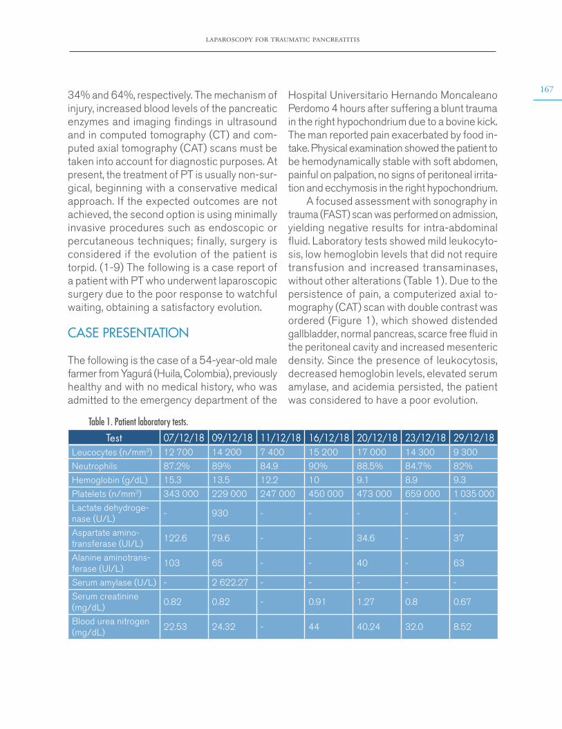
laparoscopy for traumatic pancreatitis
16734% and 64%, respectively. The mechanism of injury, increased blood levels of the pancreatic enzymes and imaging findings in ultrasound and in computed tomography (CT) and com-puted axial tomography (CAT) scans must be taken into account for diagnostic purposes. At present, the treatment of PT is usually non-sur-gical, beginning with a conservative medical approach. If the expected outcomes are not achieved, the second option is using minimally invasive procedures such as endoscopic or percutaneous techniques; finally, surgery is considered if the evolution of the patient is torpid. (1-9) The following is a case report of a patient with PT who underwent laparoscopic surgery due to the poor response to watchful waiting, obtaining a satisfactory evolution.
CASE PRESENTATION
The following is the case of a 54-year-old male farmer from Yagurá (Huila, Colombia), previously healthy and with no medical history, who was admitted to the emergency department of the
Hospital Universitario Hernando Moncaleano Perdomo 4 hours after suffering a blunt trauma in the right hypochondrium due to a bovine kick. The man reported pain exacerbated by food in-take. Physical examination showed the patient to be hemodynamically stable with soft abdomen, painful on palpation, no signs of peritoneal irrita-tion and ecchymosis in the right hypochondrium.
A focused assessment with sonography in trauma (FAST) scan was performed on admission, yielding negative results for intra-abdominal fluid. Laboratory tests showed mild leukocyto-sis, low hemoglobin levels that did not require transfusion and increased transaminases, without other alterations (Table 1). Due to the persistence of pain, a computerized axial to-mography (CAT) scan with double contrast was ordered (Figure 1), which showed distended gallbladder, normal pancreas, scarce free fluid in the peritoneal cavity and increased mesenteric density. Since the presence of leukocytosis, decreased hemoglobin levels, elevated serum amylase, and acidemia persisted, the patient was considered to have a poor evolution.
Table 1. Patient laboratory tests.
Test 07/12/18 09/12/18 11/12/18 16/12/18 20/12/18 23/12/18 29/12/18Leucocytes (n/mm3) 12 700 14 200 7 400 15 200 17 000 14 300 9 300
Neutrophils 87.2% 89% 84.9 90% 88.5% 84.7% 82%
Hemoglobin (g/dL) 15.3 13.5 12.2 10 9.1 8.9 9.3
Platelets (n/mm3) 343 000 229 000 247 000 450 000 473 000 659 000 1 035 000
Lactate dehydroge-nase (U/L)
- 930 - - - - -
Aspartate amino-transferase (UI/L)
122.6 79.6 - - 34.6 - 37
Alanine aminotrans-ferase (UI/L)
103 65 - - 40 - 63
Serum amylase (U/L) - 2 622.27 - - - - -
Serum creatinine (mg/dL)
0.82 0.82 - 0.91 1.27 0.8 0.67
Blood urea nitrogen (mg/dL)
22.53 24.32 - 44 40.24 32.0 8.52

168
case reports Vol. 6 No. 2: 165-73
Table 1. Patient laboratory tests. (continued)
Test 07/12/18 09/12/18 11/12/18 16/12/18 20/12/18 23/12/18 29/12/18pH - 7.44 7.33 7.37 7.41 7.45 -
Lactate (mmol/L) - 1.85 2.35 1.17 0.58 0.44 -
Calcium (mmol/L) - 0.79 0.7 1.19 1.07 1.11 1.073
Sodium (mmol/L) - 132.7 129 154 140 132 131
Potassium (mmol/L) - 3.7 2.61 2.98 5.06 3.86 4.07Source: Own elaboration.
Figure 1. First CAT scan with double contrast.Source: Document obtained during the study.
Considering the deterioration of the pa-tient’s condition and the persistence of pain, a serum amylase test was taken 72 hours after admission, obtaining a value of 2 622.27 U/L (25-125 U/L) that allowed diagnosing TP with a score of 2 according to the APACHE II system (mortality <4%) (10). 96 hours af-ter admission, the patient presented emetic syndrome, diarrhea, abdominal distension and a 3.1 g/dL drop in hemoglobin levels. A second CAT scan was performed (Figure 2), revealing a laceration of 3mm in diameter at the head of the pancreas and increased free fluid in the peritoneal cavity, indicating a grade IV PI according to the Organ Injury Scale of the American Association for the Surgery of Trauma. (11)
Figure 2. Second CAT scan with double contrast.Source: Document obtained during the study.
The patient’s condition remained critical; for 2 more days he presented increased intra-ab-dominal pressure and abdominal pain, decreased urine output and intolerance to the oral route, so compartment syndrome was suspected, and an exploratory laparoscopy was performed. The following findings were observed: steatonecrosis in the parietal peritoneum and greater omentum with involvement of all the peritoneal cavity, multiple peritoneal adhesions from the greater omentum to the parietal peritoneum, 600cm3 of dark yel-lowish serosanguinous peritoneal fluid, thickened omentum and blood collection the transcavity of the omenta around the area of pancreatic injury, drained towards the peritoneal cavity with friable and inflammatory tissue that evidenced scarce bleeding of the traumatic fracture in the neck of
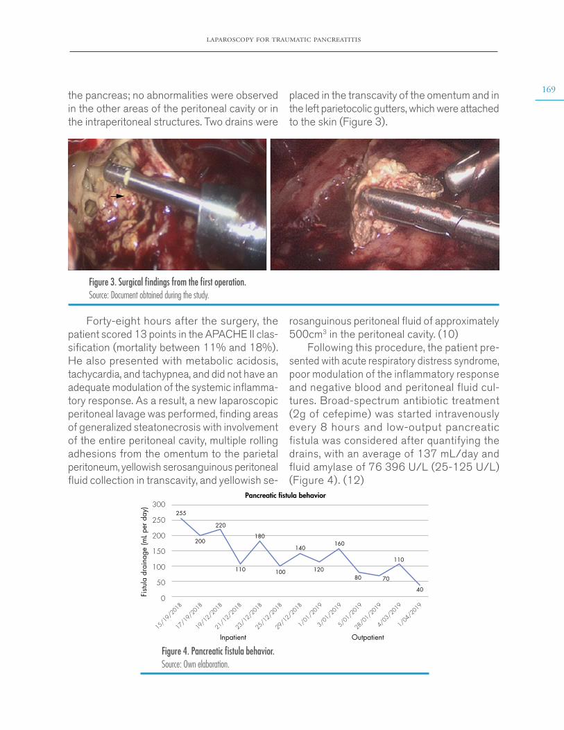
laparoscopy for traumatic pancreatitis
169the pancreas; no abnormalities were observed in the other areas of the peritoneal cavity or in the intraperitoneal structures. Two drains were
placed in the transcavity of the omentum and in the left parietocolic gutters, which were attached to the skin (Figure 3).
Figure 3. Surgical findings from the first operation. Source: Document obtained during the study.
Forty-eight hours after the surgery, the patient scored 13 points in the APACHE II clas-sification (mortality between 11% and 18%). He also presented with metabolic acidosis, tachycardia, and tachypnea, and did not have an adequate modulation of the systemic inflamma-tory response. As a result, a new laparoscopic peritoneal lavage was performed, finding areas of generalized steatonecrosis with involvement of the entire peritoneal cavity, multiple rolling adhesions from the omentum to the parietal peritoneum, yellowish serosanguinous peritoneal fluid collection in transcavity, and yellowish se-
rosanguinous peritoneal fluid of approximately 500cm3 in the peritoneal cavity. (10)
Following this procedure, the patient pre-sented with acute respiratory distress syndrome, poor modulation of the inflammatory response and negative blood and peritoneal fluid cul-tures. Broad-spectrum antibiotic treatment (2g of cefepime) was started intravenously every 8 hours and low-output pancreatic fistula was considered after quantifying the drains, with an average of 137 mL/day and fluid amylase of 76 396 U/L (25-125 U/L) (Figure 4). (12)
Pancreatic �stula behavior300
250
200
150
100
50
0Fistu
la d
rain
age
(mL
per d
ay)
15/19/2018
255
200
220
110
110
180
100
140
120
160
80 70
40
17/19/2018
19/12/2018
21/12/2018
23/12/2018
Inpatient Outpatient25/12/2018
29/12/2018
1/01/2019
3/01/2019
5/01/2019
28/01/2019
4/03/2019
1/04/2019
Figure 4. Pancreatic fistula behavior. Source: Own elaboration.
170
case reports Vol. 6 No. 2: 165-73
Finally, the patient was diagnosed with moderate-severe TP. Multidisciplinary manage-ment was indicated along with mixed nutritional support (enteral and parenteral for 3 days and total parenteral for 7 days) and antibiotic ther-apy for 19 days. The surgeries were performed on the fifth and eighth day of admission and two days after the patient was transferred to the intensive care unit where he stayed for 9 days; the patient spent the rest of his stay on the floor and was discharged from hospital 29 days after admission with an arranged low-output pancreatic fistula.
The patient was subsequently monitored on an outpatient basis with progressive as-sessment of pancreatic fistula output. He was also given medical treatment and nutritional support with a diet rich in low-medium chain triglycerides and fat restriction. Adherence to treatment was adequate and the pancreatic fistula decreased progressively until it closed spontaneously in the fifth month after surgery.
DISCUSSION
PI represents between 0.2% and 1.1% of all traumas and can present in various forms: pan-creatic leak, abscesses, fistulas, TP, pancreat-ic pseudocysts (which are the most common manifestation), acute bleeding (which has the highest mortality) and other rare manifestations such as peritonitis, gastrointestinal bleeding and splenic vein thrombosis. (1-4,6-8,13,14)
TP has high morbidity and mortality rates and its diagnostic approach should be guided by clinical suspicion and the mechanism of injury. In this regard, it should be suspected in patients with blunt abdominal trauma caused by falls, blows with bicycle handlebars but es-pecially automobile accidents, which account for 75-85% of the causes of PT due to direct impact of seat belts on the umbilical region. The
clinical presentation of TP is characterized by nausea, vomiting and generalized abdominal pain or epigastric pain irradiated to the back (50% of patients) that may improve by adopt-ing a genupectoral position; in severe cases, it may present with abdominal distension or hypovolemic shock secondary to hemoperi-toneum. (1,3-7,13,14)
Abdominal examination to treat TP may yield false negative results on the initial assessment in up to 34% of cases. In our patient, localized pain in the right hypochondrium and diffuse abdominal pain on palpation did not suggest TP; however, this pathology could not be ruled out because of the exacerbated postprandial pain and the mechanism of injury. (1,3,4,13,14)
The diagnosis of TP should be multidis-ciplinary and rule out other causes of acute abdomen such as perforated hollow viscus, hemoperitoneum or peritonitis, so it is important to perform conventional laboratory test (blood count, blood urea nitrogen, serum creatinine, coagulation tests, serum amylase and arte-rial gases), standing chest X-ray (to rule out pneumoperitoneum) and FAST ultrasound. It should be noted that the latter is not useful for identifying TP because its aim is to rule out the presence of free abdominal fluid in the hepatorenal, splenorenal, pelvic and pericar-dial spaces and not to observe the pancreas. (1-3,6-8,15)
Another method for diagnosing TP is the CAT scan; however, it has a sensitivity of 47-79% for PI, may be normal for up to 12 hours after trauma in 20-40% of patients, and its sensitivity for identifying lesions in the duct of Wirsung is 43%. For these reasons, magnetic resonance cholangiopancreatography (MRCP) and endoscopic retrograde cholangiopancrea-tography (ERCP) are preferred. This patient underwent two CAT scans: the first did not show relevant findings (Figure 1), while the

laparoscopy for traumatic pancreatitis
171second showed a fracture of the head of the pancreas, intraperitoneal free fluid and peri-pancreatic fluid (Figure 2), findings that have a sensitivity of 67-85% for TP. (1,3,4)
Serum amylase has a sensitivity of 67-83% and a specificity of 85-98% for detecting TP. Nevertheless, to be useful for diagnosis, its levels in the tests must be three times above the upper normal limit of the laboratory; in the reported patient this enzyme was elevated almost 21 times. (1,4,5,16)
The treatment of TP depends on several fac-tors. If the FAST ultrasound shows hemodynamic instability and peritonitis, laparotomy is possible; if other organs are involved, the extent of the injury and the grade of TP (injury in the duct of Wirsung, parenchymal involvement and location of the trauma) must be considered. (1,3-8)
In patients with hemodynamically stable TP, the treatment is conservative and imaging studies are conducted to assess the degree of the PI. Performing ERCP or MRI is recom-mended if there is a change in lab test results and findings suggestive of injury in the duct of Wirsung in the CAT scan; those studies have sensitivity and specificity close to 100% for this type of injury. It should be noted that ERCP has the advantage of being both a diagnostic and therapeutic tool since, in some cases, it allows placing a stent in the duct of Wirsung, thus achieving endoscopic management of the pancreatic fistula. (1-7,17,18)
For TP grades I and II, conservative man-agement is recommended because surgery is associated with greater morbidity from chronic pancreatitis, fistulas, pseudocyst formation and longer hospital stays. For grades III to V diagnosed by CAT scan, the first therapeutic option should be clinical management with fluid resuscitation and nutritional support, depending on the patient’s clinical condition. If the condition keeps deteriorating, a surgery
should be performed given the risk of increased morbidity and mortality.
In general, and regardless of the grade of the injury, the open surgical approach is recommended; however, in recent years, with the rise of minimally invasive procedures, ul-trasound-guided percutaneous drainage and laparoscopic surgery have been proposed. There is a group of patients with chronic for-mation of pancreatic pseudocysts in which drainage could be performed endoscopically; this procedure reaches an efficiency close to 90% and a significant reduction in morbidity and mortality rates. (2-7,19,20)
Since the patient had grade IV TP due to the fracture of the head of the pancreas (Figure 2), as well as abdominal distension and a decrease of 3.1 g/dL in hemoglobin levels in two days, he was taken to laparoscopic surgery with the objective of draining intraperitoneal abscesses and doing a peritoneal lavage to reduce the local intra-abdominal inflammatory response. Few reports describe the laparoscopic approach to acute pancreatitis, as it is more common in pa-tients with necrotizing pancreatitis who undergo pancreatic necrosectomy, (1-7,9,19,20) condition that can be treated using the retroperitoneal or the transperitoneal approach.
Both techniques differ in terms of the an-atomical site involved. Their advantages and disadvantages are presented below: (21-23)
The retroperitoneal surgical approach has less risk of peritoneal contamination, but the type of access and the visualization of the pan-creas that it allows limit the amount of detritus removed, so reinterventions may be needed to control infectious processes. This technique has a success, morbidity, and mortality rate of 64%, 47% and 14%, respectively. (21-23)
The transperitoneal surgical approach has the advantage of controlling the retro-peritoneal and intraperitoneal spaces, which
172
case reports Vol. 6 No. 2: 165-73
allows draining blood collections, exploring the omental pouch and performing peritoneal lavage, formal pancreatic necrosectomy and partial or total pancreatectomy; because of its wide field of vision and maneuverability, only one intervention is necessary to achieve a complete pancreatic necrosectomy. Among the disadvantages of this approach are the contamination of the intraperitoneal space and the difficulty of reintervention due to increased development of peritoneal adhesions. (21-23)
In the present case, due to the imaging findings and the presence of free intraperito-neal fluid, the transperitoneal surgical approach was selected. During the second surgery, blood collections were controlled, and the pancreatic fistula was satisfactorily arranged, obtaining a favorable outcome. To achieve the patient’s improvement, a multidisciplinary outpatient management guided by general surgery, clinical nutrition and physiotherapy was performed, with which the fistula output was progressively reduced, with its subsequent closure, while the patient’s nutritional status improved.
CONCLUSIONS
TP diagnosis is challenging due to the retroperi-toneal location of the pancreas. The treatment is usually conservative and watchful waiting and per-cutaneous or endoscopic drains are preferred over surgery. The recommended surgical approach is laparotomy, although the laparoscopic approach may be a good option to treat this pathology.
PATIENT’S PERSPECTIVE
“I want to thank the doctors and God that I was able to recover from this accident. After the two surgeries, I felt much better and my fistula was controlled promptly and without the need for another surgery.”
ETHICAL CONSIDERATIONS
This case report was prepared with the informed consent of the patient.
CONFLICT OF INTEREST
None stated by the authors.
FUNDING
None stated by the authors.
ACKNOWLEDGEMENTS
To the patient and his family for their collabora-tion in the preparation of this article.
REFERENCES
1. Johnsen NV, Betzold RD, Guillamondegui OD, Dennis BM, Stassen NA, Bhullar I, et al. Surgical Management of Solid Organ Injuries. Surg Clin North Am. 2017; ;97(5):1077-105. http://doi.org/gchtqx.
2. Ull C, Bensch S, Schildhauer TA, Swol J. Blunt Pancreatic Injury in Major Trauma: Decision-Ma-king between Nonoperative and Operative Treat-ment. Case Rep Surg. 2018;2018:6197261. http://doi.org/gdfbhv.
3. Sharbidre KG, Galgano SJ, Morgan DE. Traumatic pancreatitis. Abdom Radiol (NY). 2019;45(5):1265-76. http://doi.org/dvkj.
4. Ho VP, Patel NJ, Bokhari F, Madback FG, hambley JE, Yon JR, et al. Management of adult pancreatic injuries: A practice management guideli-ne from the Eastern Association for the Surgery of Trauma. J Trauma Acute Care Surg. 2016;82(1):185-99 http://doi.org/gfz358.
5. Dave S, Toy FK, London S. Pancreatic Trauma. Tre-asure Island (FL): StatPearls publishing; 2019 [cited 2019 Nov 24]. Available from: https://bit.ly/35XKUCA.
laparoscopy for traumatic pancreatitis
1736. Petrone P, Asensio JA, Pardo M, Kimbrell B,
Kuncir E. Traumatismos de páncreas: diagnóstico y tratamiento. Rev Colomb cirugía. 2008;23(1):44-52.
7. Puerto LJ, Medina R, Núñez LR, Jiménez HC, Olaya JG, Sanjuán JF, et al. Manejo y desen-laces de la pancreatitis aguda en un hospital de cuarto nivel (Huila, Colombia), 3 años de expe-riencia. Rev Col Gastroenterol. 2019;34(1):10-6. http://doi.org/dvkq.
8. Guidi M, Curvale C, Pasqua A, Hwang HJ, Pires H, Basso S, et al. Actualización en el manejo inicial de la pancreatitis aguda. Acta Gas-troenterol Latinoam. 2019;49(4):307-23.
9. Working Group IAP/APA acute pancreatitis guide-lines. IAP/APA evidence-based guidelines for the management of acute pancreatitis. Pancreatology. 2013;13(Suppl 4):e1-15. http://doi.org/f246bz.
10. Banks PA, Freeman ML, Practice Parameters Committee of the American College of Gastroen-terology. Practice guidelines in acute pancreatitis. Am J Gastroenterol. 2006;101(10):2379-400. http://doi.org/ddhfpn.
11. Moore EE, Cogbill TH, Malangoni MA, Jur-kovich GJ, Champion HR, Gennarelli TA, et al. Organ injury scaling, II: Pancreas, duode-num, small bowel, colon, and rectum. J Trauma. 1990;30(11):1427-9.
12. Bassi C, Dervenis C, Butturini G, Finger-hut A, Yeo C, Izbicki J, et al. Postoperative pancreatic fistula: An international study group (ISGPF) definition. Surgery. 2005;138(1):8-13. http://doi.org/fsp76g.
13. Swaroop VS, Chari ST, Clain JE. Severe acu-te pancreatitis. JAMA. 2004;291(23):2865-8. http://doi.org/bvmjgr.
14. Asensio JA, Demetriades D, Hanpeter DE, Gambaro E, Chahwan S. Management of pan-creatic injuries. Curr Probl Surg. 1999;36(5):325-419. http://doi.org/dgr9wh.
15. ATLS Advanced Trauma Life Support. Stu-dente Course Manual. 10th ed. Chicago: American College of Sugeons; 2018.
16. Yadav D, Agarwal N, Pitchumoni CS. A critical evaluation of laboratory tests in acute pancreatitis. Am J Gastroenterol. 2002;97(6):1309-18. http://doi.org/dcntfd.
17. Howard TJ, Patel JB, Zyromski N, Sandrase-garan K, Yu J, Nakeeb A, et al. Declining morbi-dity and mortality rates in the surgical management of pancreatic necrosis. Journal of Gastrointestinal Surgery. 2007;11(1):43-9. http://doi.org/cf5rjc.
18. Hartwig W, Maksan SM, Foitzik T, Schmi-dt J, Herfarth C, Klar E. Reduction in mortali-ty with delayed surgical therapy of severe pan-creatitis. J Gastrointest Surg. 2002;6(3):481-7. http://doi.org/df3vc2.
19. Vijay A, Abdelrahman H, El-Menyar A, Al-Tha-ni H. Early laparoscopic approach to pancreatic in-jury following blunt abdominal trauma. J Surg Case Rep. 2014;2014(12):rju129. http://doi.org/dvk2.
20. Hookey LC, Debroux S, Delhaye M, Arvanita-kis M, le Moine O, Devière J. Endoscopic draina-ge of pancreatic-fluid collections in 116 patients: a comparison of etiologies, drainage techniques, and outcomes. Gastrointest Endosc. 2006;63(4):635-43. http://doi.org/c92mp9.
21. Parekh D. Laparoscopic-assisted pancreatic ne-crosectomy: A new surgical option for treatment of severe necrotizing pancreatitis. Arch Surg. 2006;141(9):895-902. http://doi.org/ctz9fw.
22. Bradley EL, Howard TJ, van Sonnenberg E, Fotoohi M. Intervention in necrotizing pancrea-titis: an evidence-based review of surgical and percutaneous alternatives. J Gastrointest Surg. 2008;12(4):634-9. http://doi.org/c4dc4p.
23. Wysocki AP, McKay CJ, Carter CR. Infected pancreatic necrosis: minimizing the cut. ANZ J Surg. 2010;80(1-2):58-70. http://doi.org/fbznm7.
174
case reports Vol. 6 No. 2
EVALUATING PEERS
NAME INSTITUTION/ORGANIZATION COUNTRYAlonso Sánchez María Francisca Universidad de Valparaíso Chile
Álvarez Quiroz Paulina Vianey Hospital de la Ceguera México
Angamarca Angamarca Edison Hospital de la Universidad Técnica Particular de Loja Ecuador
Arango Gómez Fernando Universidad de Manizales Colombia
Aristizabal Alzate Arbey Hospital Pablo Tobón Uribe Colombia
Arturi Alfredo Silvio Universidad Nacional de La Plata Argentina
Arroyo Amparo Instituto de Medicina Legal de Catalunya España
Barousse Rafael Universidad de Buenos Aires Argentina
Blasco Gil Silvia Hospital de Alcañiz-Teruel España
Botet Francesc Hospital Clínic de Barcelona España
Camilo Pantaleón Elsa Hospital Infantil Dr. Robert Reid CabralRepública Dominicana
Campbell Silva Santiago Emilio Clínica Medilaser Colombia
Casarrubias Ramírez MoisésHospital de Especialidades Centro Médico Nacional La Raza
México
Casas Parera IgnacioUniversidad de Buenos AiresInstituto de Oncología Ángel H. Roffo
Argentina
Castillo Sepúlveda Michael Centro Hospitalario Sanatorio Durango México
Centurión Osmar Antonio Universidad Nacional de Asunción Paraguay
Conde Castro BenjamínUniversidad Nacional Autónoma de MéxicoInstituto Mexicano del Seguro Social
México
Contreras Alejandro Hospital Privado Universitario de Córdoba Argentina
Cortés Díaz Fabio Felipe Universidad Nacional de Colombia Colombia
Corzo Paula Ariadna Universidad Corporativa de Colombia Colombia
De Arriba de la Fuente GabrielUniversidad de AlcaláHospital Universitario de Guadalajara
España
De la rosa Anabel Universidad Nacional Autónoma de México México
Delgado Roche Eduardo Instituto Catalán de Salud España
Del Sol Calderón Mariano Universidad de La Frontera Chile
Domingo Jiménez Rosario Hospital Clínico Universitario Virgen de la Arrixaca España
Domínguez Massa Carlos Hospital Universitario y Politécnico La Fe España
Duque Ramírez Mauricio Universidad CES Colombia
Errando Oyonarte Carlos Luis Hospital General Universitario de Valencia España
Faúndez Eduardo Universidad de Magallanes Chile
Ferrer Elizabeth Universidad de Carabobo Venezuela
Ferrer Lozano YovannyHospital Universitario Julio Aristegui VillamilUniversidad Médica de Matanzas
Cuba
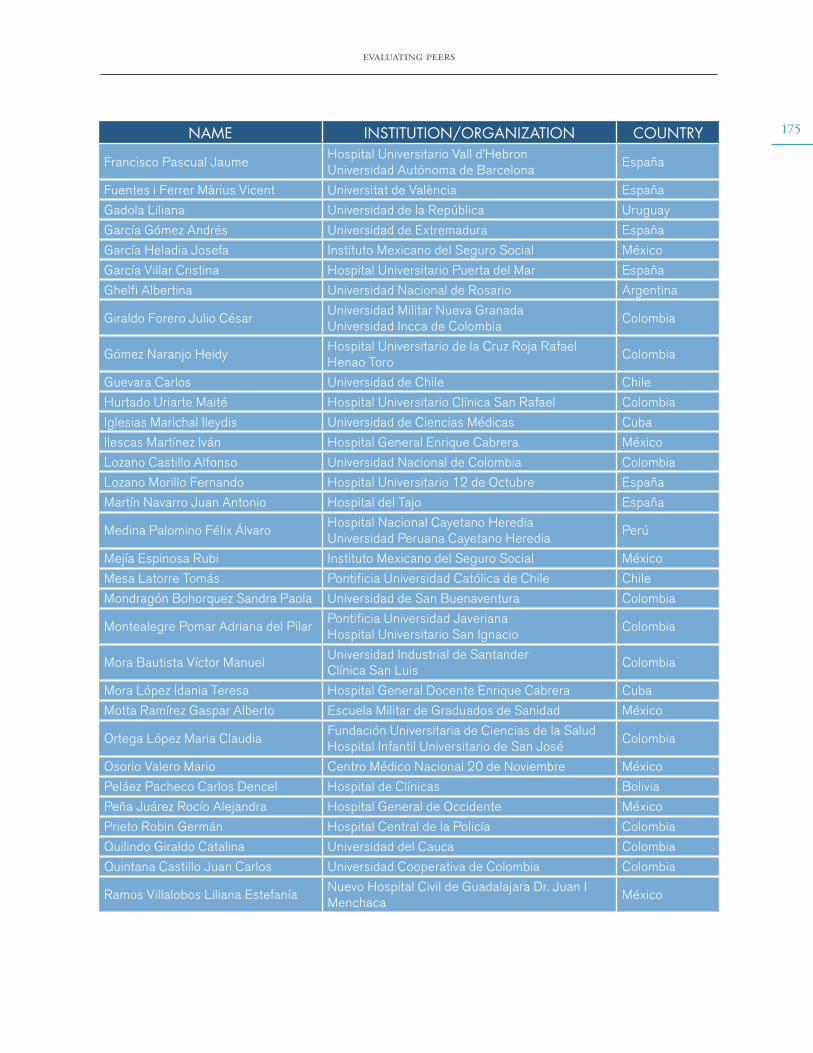
evaluating peers
175NAME INSTITUTION/ORGANIZATION COUNTRY
Francisco Pascual JaumeHospital Universitario Vall d’HebronUniversidad Autónoma de Barcelona
España
Fuentes i Ferrer Màrius Vicent Universitat de València España
Gadola Liliana Universidad de la República Uruguay
García Gómez Andrés Universidad de Extremadura España
García Heladia Josefa Instituto Mexicano del Seguro Social México
García Villar Cristina Hospital Universitario Puerta del Mar España
Ghelfi Albertina Universidad Nacional de Rosario Argentina
Giraldo Forero Julio CésarUniversidad Militar Nueva GranadaUniversidad Incca de Colombia
Colombia
Gómez Naranjo HeidyHospital Universitario de la Cruz Roja Rafael Henao Toro
Colombia
Guevara Carlos Universidad de Chile Chile
Hurtado Uriarte Maité Hospital Universitario Clínica San Rafael Colombia
Iglesias Marichal Ileydis Universidad de Ciencias Médicas Cuba
Ilescas Martínez Iván Hospital General Enrique Cabrera México
Lozano Castillo Alfonso Universidad Nacional de Colombia Colombia
Lozano Morillo Fernando Hospital Universitario 12 de Octubre España
Martín Navarro Juan Antonio Hospital del Tajo España
Medina Palomino Félix ÁlvaroHospital Nacional Cayetano HerediaUniversidad Peruana Cayetano Heredia
Perú
Mejía Espinosa Rubi Instituto Mexicano del Seguro Social México
Mesa Latorre Tomás Pontificia Universidad Católica de Chile Chile
Mondragón Bohorquez Sandra Paola Universidad de San Buenaventura Colombia
Montealegre Pomar Adriana del PilarPontificia Universidad JaverianaHospital Universitario San Ignacio
Colombia
Mora Bautista Víctor ManuelUniversidad Industrial de SantanderClínica San Luis
Colombia
Mora López Idania Teresa Hospital General Docente Enrique Cabrera Cuba
Motta Ramírez Gaspar Alberto Escuela Militar de Graduados de Sanidad México
Ortega López Maria ClaudiaFundación Universitaria de Ciencias de la SaludHospital Infantil Universitario de San José
Colombia
Osorio Valero Mario Centro Médico Nacional 20 de Noviembre México
Peláez Pacheco Carlos Dencel Hospital de Clínicas Bolivia
Peña Juárez Rocío Alejandra Hospital General de Occidente México
Prieto Robin Germán Hospital Central de la Policía Colombia
Quilindo Giraldo Catalina Universidad del Cauca Colombia
Quintana Castillo Juan Carlos Universidad Cooperativa de Colombia Colombia
Ramos Villalobos Liliana EstefaníaNuevo Hospital Civil de Guadalajara Dr. Juan I Menchaca
México
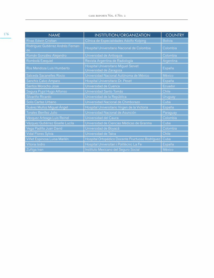
176
case reports Vol. 6 No. 2
NAME INSTITUTION/ORGANIZATION COUNTRYRivas Edwin Cristian Clínica de Especialidades Adolfo Kolping Bolivia
Rodríguez Gutiérrez Andrés Fernan-do
Hospital Universitario Nacional de Colombia Colombia
Román González Alejandro Universidad de Antioquia Colombia
Rombolá Ezequiel Revista Argentina de Radiología Argentina
Ros Mendoza Luis HumbertoHospital Universitario Miguel ServetUniversidad de Zaragoza
España
Salceda Sacanelles Rocio Universidad Nacional Autónoma de México México
Sanchis Calvo Amparo Hospital Universitario Dr. Peset España
Santos Morocho Jose Universidad de Cuenca Ecuador
Segura Pujol Hugo Alfonso Universidad Santo Tomás Chile
Silvariño Ricardo Universidad de la República Uruguay
Solis Cartas Urbano Universidad Nacional de Chimborazo Cuba
Suárez Muñoz Miguel Ángel Hospital Universitario Virgen de la Victoria España
Torales Benítez Julio Universidad Nacional de Asunción Paraguay
Vásquez Arteaga Luis Reinel Universidad del Cauca Colombia
Vázquez Gutiérrez Giselle Lucila Universidad de Ciencias Médicas de Granma Cuba
Vega Padilla Juan David Universidad de Boyacá Colombia
Vidal Flores Sylvia Universidad de Talca Chile
Viñet Espinosa Luisa Marlén Hospital Ortopédico Docente Fructuoso Rodríguez Cuba
Vitoria Isidro Hospital Universitari i Politècnic La Fe España
Zuñiga Ivan Instituto Mexicano del Seguro Social México

Faculty of MedicineBoard of Directors
Universidad Nacional de ColombiaBogotá Campus ~ Bogotá D. C., Colombia
Faculty of Medicine • Building 471 • Office 219Telephone numbers: 3165000 Ext. 15161 • Bogotá, D.C., Colombia
http://unal.edu.co/email: [email protected]
Web: https://revistas.unal.edu.co/index.php/care/index
José Ricardo Navarro MD. MSc. José Fernando Galván-Villamarín MD. MSc.Javier Eslava-Schmalbach MD. MSc. PhD.
Silvia Cristina Duarte-Torres OT. MSc.María Fernanda Lara-Díaz SLT. MSc. PhD.
Vivian Marcela Molano Soto
Dean of Faculty Academic Vice ChancellorVice Chancellor for ResearchDirector of BienestarSecretary of FacultyPublications Unit Coordinator
Board of Directors
Dolly Montoya CastañoPablo Enrique Abril ContrerasLuz Teresa Gómez de Mantilla
Carlos Augusto HernándezJaime Franky Rodríguez
Edgar Cortés ReyesCarlos Eduardo Cubillos Peña
ChancellorGeneral Vice ChancellorVice Chancellor for Research and University Extension
Academic Vice ChancellorVice Chancellor for Bogotá CampusSecretary for Bogotá CampusAcademic Director
Extension