outcome and efficacy of a transobturator polypropylene mesh kit in the treatment of anterior pelvic...
TRANSCRIPT
Središnja medicinska knjižnica
Grgić O., Orešković S., Lovrić Gršić H., Kalafatić D., Župić T., Maurac I.
(2012) Outcome and efficacy of a transobturator polypropylene mesh
kit in the treatment of anterior pelvic organ prolapse. International
Journal of Gynecology and Obstetrics, 116 (1). pp. 72-5. ISSN 0020-
7292
http://www.elsevier.com/locate/issn/00207292 http://www.sciencedirect.com/science/journal/00207292 http://dx.doi.org/10.1016/j.ijgo.2011.08.014 http://medlib.mef.hr/1437
University of Zagreb Medical School Repository
http://medlib.mef.hr/
1
Outcome and efficacy of transobturator polypropylene mesh kit in
treatment of anterior pelvic organ prolapse
Grgic Ozren (1), Oreskovic Slavko (2), Lovric Grsic Helena (2), Kalafatic Drzislav (2),
Zupic Tomislav (2), Maurac Ivana (2).
(1) Department of Obstetrics and Gynecology, University hospital “Sestre Milosrdnice”, Zagreb,
Croatia
(2) Department of Obstetrics and Gynecology, Medical School, University of Zagreb, Croatia
Correspondence author address:
Ozren Grgic, M.D. Ph.D.
University hospital “Sestre Milosrdnice”
Department of Obstetrics and Gynecology
Vinogradska 29, 10000 ZAGREB, Croatia
Tel/Fax: +385 1 37 835 49
E-mail: [email protected]
Synopsis: The repair of anterior pelvic organ prolapse with transobturator mesh is effective
treatment even in a population with previously hysterectomy or traditional anterior
colporrhaphy.
Key words: Anterior prolapse; Hysterectomy; Mesh complications; Perigee®; Polypropylene
mesh; Transobturator route
Word count: 2445
2
Abstract
Objective: To report the efficacy and complications of anterior pelvic organ prolapse (POP)
repair with mesh trough transobturator route.
Methods: Totally, 198 woman with anterior POP > II grade according to the POP-Q system
were treated by Perigee® procedure. The primary outcome was defined as anterior POP ≤
stage I at 12 months follow-up. The secondary outcomes included the incidence of
perioperative, mesh, short and long-term postoperative complications.
Results: The overall cure rate was 92.9 %, and in population with previously hysterectomy or
traditional anterior colporrhaphy was 90.6%. Postoperative mean POP-Q Aa and Ba points
were significantly improved (Aa 2.2 [0-3.1] cm versus -2.1 [-1.2- -3] cm and Ba -2.4 [-1.6 – -
5.5] cm versus 2.5 [-1 – 4.2] cm, p < 0.001 Wilcoxon signed rank test). Vaginal or bladder
erosions had observed in three patients. The other short and long term complications were low
and not significant.
Conclusion: The Perigee® procedure is effective treatment of anterior POP without serious
complications even in a population with previously hysterectomy or traditional anterior
colporrhaphy.
3
Introduction
It has been estimated that over the next 30 years, the demand for the treatment of pelvic organ
prolapse (POP) will increase for 45% [1]. According to the Women’s Health Initiative study
anterior POP was the most frequent type and had observed in 34.2% of women [2].
Approximately 10% of them will have a surgical intervention (hysterectomy or correction
procedure) for this condition during their lifetime, and 30% of these women will undergo
repeat operation for prolapse recurrence in a 5 years period [3-5]. Due to the limitations and
high recurrence rate after traditional surgery various synthetic implants have been developed
in the last couple of years [6-8]. There are numerous types of mesh and graft materials
available, which vary according to type and structure of material and physical properties such
as absorbability and pore size [8].
The Perigee®
system (Perigee system, AMS, Minnetonka, MN) was designed and first used in
Townsville, Australia, and it appears to be valuable and time-efficient approach for all types
of anterior vaginal wall defects with high cure rate and minimal complications [9-15]. It
contains four specific, helical needles designed for each anatomic pass through the obturator
space to attach a graft to the pelvic sidewall distally at the level of the bladder neck, and
apically near the ischial spines and reinforce the pubocervical fascia with minimally invasive
approach [9-11]. The results of recent studies have shown high anatomic success rate and low
incidence of complications [9-15]. Moreover, subpopulation of patients with previous
hysterectomy and traditional anterior colporrhapy has been considered as risk group for lower
cure rate with reoperation [16]. The major concerns with this method have been related to
mesh complications including erosion of the vagina or bladder and formation of the
vesicovaginal fistula [9-15, 17].
The purpose of our study was to evaluate the efficacy and safety together with short and long
term outcomes of anterior vaginal wall repair using tension-free polypropylene mesh.
4
Materials and methods
Between May 1. 2007 and September 30. 2009, 198 women with anterior POP > grade II
underwent anterior colporrhaphy using Perigee® system at University hospital KBC Zagreb,
Croatia. Local and national ethical committee approved the study protocol, and all patients
included in the study gave their informed written consent.
The exclusion criteria for enrollment were: previous placement of an anterior wall graft;
predominant urge incontinence diagnosed by urodynamics; pelvic infection or systemic
infection; inguinal or vulvar abscesses; pregnancy; urinary tract obstruction or renal
insufficiency; pelvic pain (unrelated to prolapse); vaginal bleeding unknown etiology; blood
coagulation disorders; pelvic malignancy or previously radiation of pelvic area; vaginal
erosion or severe vaginal atrophy; vaginal or urethral fistula; and known allergy to mesh
material.
Preoperative evaluation was consisted of a complete history and gynecologic examination.
Prolapse severity was assessed using the pelvic organ prolapse quantification (POP-Q) system
adopted by the International Continence Society [18]. Urinary incontinence was diagnosed
clinically and with urodynamic studies. Appropriate local estrogen replacement (Vagifem,
Novo Nordisk, Denmark) were applied to all patients at least in 8 weeks period before the
procedure and it was continued after it. All urogynecologists participating in the study have
been experienced surgeons, had pre-trial hands-on training and were performed at least 50
procedures before the beginning of this study. The patients were followed-up at 7 days, 6-8
weeks and 12 months postoperatively.
The primary endpoint of this study and treatment success was defined as ≤ stage I of anterior
POP according to the POP-Q system at 12-month follow-up. The secondary outcomes
included the incidence of intraoperative (hemorrhage, infection), short term postoperative
(nerve, vessel, and bladder or bowel injury) and long term postoperative complications
5
(erosion, rejection, de-novo incontinence, urinary retention, constipation and dyspareunia).
Residual urine was measured by the catheterization with cut off point at 50 ml of urine. We
also measured operating time, intraoperative blood loss and hospital stay. Follow-up was
obtained by gynecologic examination and survey using a questionnaire from a study of Shah
et al [19].
Perigee®
system consists of polypropylene mesh for repairing central defects and four self-
attached arms for correction of lateral vaginal detachments. Following a vertical incision from
the bladder neck, around 4 cm from the apex of vagina the dissection is similar to that of the
anterior colporaphy. Structure of the needle provides fixation of the mesh at the white line of
arcus tendineus so there was no need for deep lateral sutures. After orientation about inferior
needle pathway, lateral dissection toward the white line of the arcus tendineus may be done.
Plication sutures in the midline were placed to reduce the anterior POP but also to prevent the
bladder perforation when the inferior needles pass by. Superior bilateral incisions were made
in the genitofemoral folds at the base of the adductor longus tendon at the level of clitoris.
The inferior incisions are made 2 cm lateral and 3 cm inferior to the superior. Narrow-
diameter superior helical needles are guided at 45 degrees to patient's midline and perforate
the obturator membrane laterally to the ischiopubic ramus under the finger guidance to avoid
the bladder perforation. The inferior helical needles are inserted with the handle at 90 degrees
and are driven under finger control directly to ischial spines in straightforward direction. After
penetrating the levators they exit along white line at about 1.5-2 cm below the spines. It is
extremely important to direct the needle tips proximally towards spines before rotation
throughout the levator muscles. The mesh was subsequently adjusted to vaginal wall extent in
a tension-free manner and the proximal tail was drowning to the lower-most portion of the
cystocele. The distal arms were than adjusted with slight tension beneath the bladder neck, to
return the anterior vaginal wall back to its natural anatomical position. Further step was the
6
fixation of the apical edge of the graft to the pericervical ring by the sutures in order to fasten
the attachment. The redundant tail of the mesh was additionally removed. After rolling over
the mesh only a small portion (less than 25%) of surplus vaginal wall was excided. Vaginal
incision was closed, avoiding the superposition of the mesh and its exposure or infection. The
outer plastic sheets were cut at the level of skin incision. After the positioning of the mesh
cistoscopy was obligate to eliminate the bladder injury and verify potency of both ureters. The
Foley catheter was inserted in the bladder for 24 hours. We have practiced one day
perioperative antibiotic prophylaxis and analgetic therapy if necessary. Patients were
recommended to avoid heavy lifting, exercise, and sexual activity for at least 4 weeks.
As a part of the study design, a sample size calculation was performed (Sample size
calculator, MaCorr Inc., Toronto, Ontario, Canada). Based on the previously results the
calculation was designed to detect at least 85 % success rate at 12 months follow-up.
Confidence interval was determined using the confidence level (CL) of 95%. The program
has calculated that confidence interval is 7.0%. Using CL of 95% and confidence interval of
7.0 % the program has calculated that for our population the sample size needed to be 196.
Data were analyzed using SPSS version 15.0 (SPSS Inc., Chicago IL, USA). For comparison
of categorical data before and 12 months after procedure we calculated likelihood ratio with
95% confidence interval. The Wilcoxon signed-rank test was used in comparison the POP-Q
measurements before and after procedure. Two-tailed p values are reported throughout and
statistical significance was defined as p < 0.05.
7
Results
Totally, 243 patients with anterior POP > II stage were consented to participate in the study.
We excluded 41 patients (10 had dominantly urge incontinence; 5 had previously placement
of mash for anterior wall prolapse; 4 had chronic renal insufficiency; 4 had previously
radiation of pelvic area due to the gynecologic malignancy; 3 had acute pelvic infection; 2 had
severe forms of vonWillebrand disease, 2 had cervical carcinoma, 2 had severe vaginal
atrophy despite of local estrogen therapy; 1 had endometrial carcinoma and finally 8 patients
refused to participate in the study). Of the remaining 202 patients the 12 month follow up data
was obtained for 198 (98%) patients, and our results were based on their analysis. The
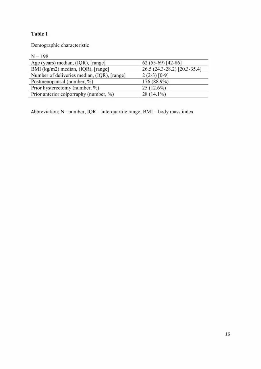
baseline data of our patients are shown in Table 1.
Median time for operation was 25 minutes [range 13-75]. The average hospital stay was 6
days [range 5 - 11 days]. Average time to void was 2.2 days [range 1–10 days].
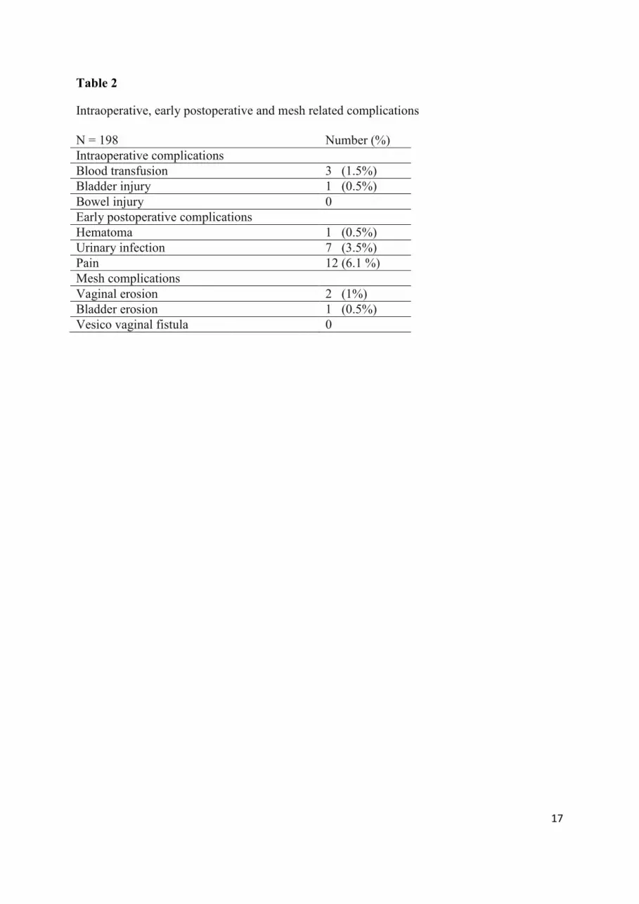
Intraoperative, early postoperative and mesh related complications are presented in Table 2.
Moderate levator pain and groin discomfort at 12 patients (6.1%) was the most prominent
short-term postoperative problem but have disappeared 2 months with analgetic therapy.
Median blood loss was 50 ml [range 5-300 ml], and three patients received blood transfusion
(two of them because of heavy bleeding during the procedure, and one because of formation
of hematoma under the anterior wall after initial closure).
Regarding the complications related to mesh only two patients had vaginal erosion, and one
had erosion of the bladder. The median days-to-onset of mesh complications were 62 days
[range 14 – 98]. A patient with bladder erosions required surgical correction that involved
excision of the exposed mesh, secondary closure of the vaginal defect, and subsequently
application of vaginal estrogen cream. Vaginal erosions were resolved with vaginal estrogen
cream only.
8
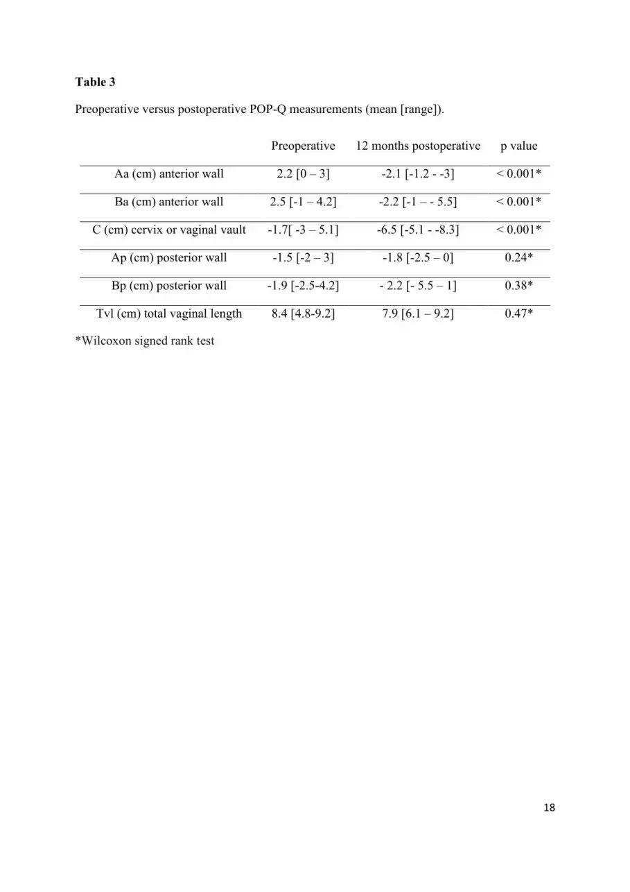
Preoperative versus postoperative POP-Q measurements were shown in Table 3. Due to the
primary outcome the objective cure rate 12 months after procedure was 92.9 % (184/198).
The failure rate was 7.1 % (14/198), 10 patients had anterior POP-Q II and 4 had III stage
prolapse 12 months after the procedure.
The co-occurrence of posterior vaginal prolapse in our study population was 13.1% (26/198).
In 17/198 the correction procedure was made with Apogee system whereas in remaining
patients traditionally procedure (posterior colporraphy and levatoroplasty) was performed.
Since posterior prolapse was not the objective of this study we did not analyze the short and
long term outcomes regarding the posterior correction procedure in this subpopulation of
patients.
In a population of patients with previously traditional anterior colporrhapy 28/198 (14.1%) or
hysterectomy 25/198 (12.6 %) the objective cure rate 12 month after the procedure were high
48/53 (90.6%) and did not different from patients without these procedures 136/145 (92.4 %).
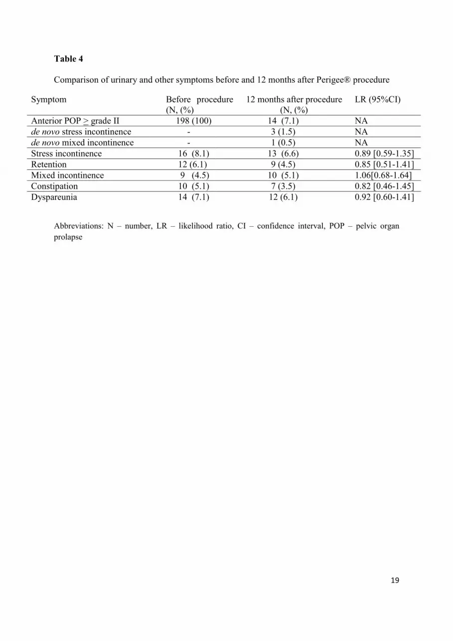
Comparison of urinary and other symptoms before and 12 months after Perigee® procedure
was presented in Table 4. There were three cases of mild de novo stress urinary incontinence
which have manifested by occasional leak, one case of de novo mixed incontinence, and in
two patients de novo urinary retention developed immediately after releasing from the
hospital and has been managed promptly by recateterisation and antibiotic therapy. The other
long term symptoms were unchanged 12 months after procedure.
9
Discussion
Unacceptably high failure rate of traditional anterior POP repair triggered the attempts in
pelvic floor reconstructive surgery for innovative vaginal approaches and evolution of new
graft augmentation techniques [3-5]. Few years ago Rane et al. invented a technique to utilize
the obturator space to attach the graft not only at the bladder neck but also more apically by
the ischial spine utilizing needles passed through the obturator space in order to repair all
types of anterior vaginal wall defects [9]. Bilateral superior and inferior needles allow the
surgeon minimal lateral dissection and therefore reduce the blood loss and other healing
abnormalities [9-11]. The inferior helical needles provide the positioning of the proximal part
of the mesh adjacent to the ischial spines which is fundamental route for the reattachment of
the vaginal wall to its normal position at the level of the white line. It seems that lateral
detachments of white line have usually been unrecognized, neglected and maltreated with
traditional approaches. Consequently, it could be the strongest explanation for high success
rate after the Perigee®
procedure [9-15].
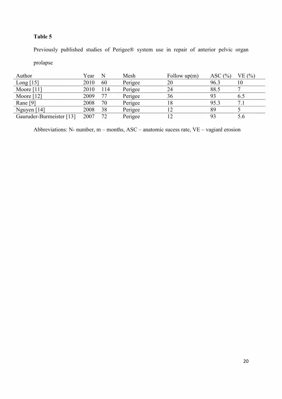
Results of our study have shown favorable results of transvaginal mesh surgery with anatomic
success rate of 92.9 % 12 months after the procedure. Comparisons of our results with
previously published trials using Perigee mesh are shown in Table 5. The anatomic success
rate of our study was high (92.9 % versus 88.5-96.3%) and comparable with previously
published trials [11-15]. Additionally, this technique did not short the vaginal length which is
very common with traditional procedures.
Recently, concerns over complications with vaginal mesh have been raised and the erosion of
the vagina or bladder seems to be the most common complication seen with the use of
synthetic mesh graft [8-13]. Comparison of our results regarding the formation of erosion of
vagina or bladder after the procedure is lower in comparison to the others (1.5 % versus 3.1-
10
10 %). This difference could be connected with the use of long term local estrogen therapy
before procedure. Long term estrogen therapy improves the status of the vaginal mucosa and
subsequently decreased the formation of erosion [20]. All participants in our study had long
term (at least 8 weeks before procedure) estrogen therapy. Furthermore, our lower incidence
of formation of erosion could be connected with the final step of the procedure. When the
mesh was embedded in its anatomical position we excised only a small part of surplus anterior
vaginal wall tissue. The excessive excision of anterior vaginal wall could compress the mesh
to the bladder or vagina and leads to the formation of erosion or even vesicovaginal fistula.
Additionally, this complication could be reduced with newer techniques which we did not use
in our study such as hydro dissection and the use of lighter, softer, and less dense type of
mesh [11]. The overall rates of other intraoperative and early postoperative complications
seem very low in our study and the results are comparable with previously published trials [9-
15].
In our study 12.6 % patients had previously hysterectomy and 14.1 % had previous repair of
the anterior POP with traditionally anterior colporraphy. This population could be considered
as risk group for developed complications and lower cure rate [16]. However, the results in
this subgroup are very encouraging and showed that their cure rate was consistent with
patients without these procedures.
Regarding the long term urinary symptoms occasional leak of urine and de novo mild stress
incontinence manifested in three cases and suggesting that Perigee®
interposition does not
interfere with continence mechanism. Loss of sharp vesico-urethral angle after anterior POP
repair was probably the only mechanism of de novo urinary stress incontinence, so the effort
should be focused on preservation of vesico-urethral junction.
In conclusion, our results confirm that this procedure is minimally invasive, reproducible, and
efficient. It has low morbidity and is well tolerated by the patients and seems to be an

11
satisfactory procedure for definitive repair of anterior POP even in a population of patients
with previously hysterectomy or traditionally anterior colporraphy.

12
Conflict of interest
None
13
References
1. Boyles SH, Weber AM, Meyn L. Procedures for urinary incontinence in the United
States, 1979-1997. Am J Obstet Gynecol. 2003;189(1):70-5
2. Hendrix SL, Clark A, Nygaard I, Aragaki A, Barnabei V, McTiernan A. Pelvic organ
prolapse in the Women's Health Initiative: gravity and gravidity. Am J Obstet
Gynecol. 2002;186(6):1160-6
3. Olsen A, Smith V, Bergstrom J, Colling J, Clark A. Epidemiology of surgically
managed pelvic organ prolapse and urinary incontinence. Obstet Gynecol.
1997;89:501-6
4. Julian TM. The efficacy of Marlex mesh in the repair of severe,recurrent vaginal
prolapse of the anterior midvaginal wall. Am J Obstet Gynecol. 1996;175:1472-5
5. Shull BL, Benn SJ, Kuehl TJ. Surgical management of prolapse of theanterior vaginal
segment: an analysis of support defects, operative morbidity, and anatomic outcome.
Am J Obstet Gynecol. 1994;171:1429-36
6. Abdel-Fattah M, Ramsay I. Retrospective multicentre study of the new minimally
invasive mesh repair devices for pelvic organ prolapse. BJOG. 2008;115:22-30
7. Hiltunen R, Nieminen K, Takala T, Heiskanen E, Merikari M, Niemi K et al. Low-
weight polypropylene mesh for anterior vaginal wall prolapse: a randomized
controlled trial. Obstet Gynecol. 2007; 110(2 Pt 2):455-62
8. Jia X, Glazener C, Mowatt G, MacLennan G, Bain C, Fraser C et al. Efficacy and
safety of using mesh or grafts in surgery for anterior and/or posterior vaginal wall
prolapse: systematic review and meta-analysis. BJOG. 2008;115(11):1350-61
9. Rane A. A novel transobturator system for the repair of anterior vaginal wall prolapse:
A pilot study. European Association of the Urology Annual Meeting 2005
14
10. Rane A, Kannan K, Barry C, Balakrishnan S, Lim Y, Corstiaans A. Prospective study
of the Perigee system for the management of cystocoeles--medium-term follow up.
Aust N Z J Obstet Gynaecol. 2008;48(4):427-32
11. Moore RD, Beyer RD, Jacoby K, Freedman SJ, McCammon KA, Gambla MT.
Prospective multicenter trial assessing type I, polypropylene mesh placed via
transobturator route for the treatment of anterior vaginal prolapse with 2-year follow-
up. Int Urogynecol J Pelvic Floor Dysfunct. 2010;21:545-52
12. Moore R, Miklos J. Vaginal repair of cystocele with anterior wall mesh via
transobturator route: efficacy and complications with up to 3 years follow-up.
Advances in Urology. 2009:743831
13. Gauruder-Burmester A, Koutouzidou P, Rohne J, Gronewold M, Tunn R. Follow-up
after polypropylene mesh repair of anterior and posterior compartments in patients
with recurrent prolapse. Int Urogynecol J Pelvic Floor Dysfunct. 2007;18:1059-64
14. Nguyen JN, Burchette RJ. Outcome after anterior vaginal prolapse repair: a
randomized controlled trial. Obstet Gynecol. 2008;111:891-8
15. Long CY, Hsu CS, Jang MY, Liu CM, Chiang PH, Tsai EM. Comparison of clinical
outcome and urodynamic findings using "Perigee and/or Apogee" versus "Prolift
anterior and/or posterior" system devices for the treatment of pelvic organ prolapse.
Int Urogynecol J Pelvic Floor Dysfunct. 2011;22(2):233-9
16. Mant J, Painter R, Vessey M. Epidemiology of genital prolapse: observations from the
Oxford Family Planning Association Study. BJOG.1997;104: 579-85
17. Yamada BS, Govier FE, Stefanovic KB, Kobashi KC. Vesicovaginal fistula and mesh
erosion after Perigee (transobturator polypropylene mesh anterior repair). Urology.
2006; 68(5):1121.e5-7
15
18. Haylen BT, de Ridder D, Freeman RM, Swift SE, Berghmans B, Lee J et al. An
International Urogynecological Association (IUGA)/International Continence Society
(ICS) joint report on the terminology for female pelvic floor dysfunction. Int
Urogynecol J Pelvic Floor Dysfunct. 2010; 21(1):5-26
19. Shah DK, Paul EM, Rastinehad AR, Eisenberg ER, Baldani GH. Short-term outcome
analysis of total pelvis reconstruction with mesh: the vaginal approach. J Urol. 2004;
171(1):261-3
20. Palacios S. Managing urogenital atrophy. Maturitas. 2009;63(4):315-8
16
Table 1
Demographic characteristic
N = 198
Age (years) median, (IQR), [range] 62 (55-69) [42-86]
BMI (kg/m2) median, (IQR), [range] 26.5 (24.3-28.2) [20.3-35.4]
Number of deliveries median, (IQR), [range] 2 (2-3) [0-9]
Postmenopausal (number, %) 176 (88.9%)
Prior hysterectomy (number, %) 25 (12.6%)
Prior anterior colporraphy (number, %) 28 (14.1%)
Abbreviation; N –number, IQR – interquartile range; BMI – body mass index
17
Table 2
Intraoperative, early postoperative and mesh related complications
N = 198 Number (%)
Intraoperative complications
Blood transfusion 3 (1.5%)
Bladder injury 1 (0.5%)
Bowel injury 0
Early postoperative complications
Hematoma 1 (0.5%)
Urinary infection 7 (3.5%)
Pain 12 (6.1 %)
Mesh complications
Vaginal erosion 2 (1%)
Bladder erosion 1 (0.5%)
Vesico vaginal fistula 0
18
Table 3
Preoperative versus postoperative POP-Q measurements (mean [range]).
Preoperative 12 months postoperative p value
Aa (cm) anterior wall 2.2 [0 – 3] -2.1 [-1.2 - -3] < 0.001*
Ba (cm) anterior wall 2.5 [-1 – 4.2] -2.2 [-1 – - 5.5] < 0.001*
C (cm) cervix or vaginal vault -1.7[ -3 – 5.1] -6.5 [-5.1 - -8.3] < 0.001*
Ap (cm) posterior wall -1.5 [-2 – 3] -1.8 [-2.5 – 0] 0.24*
Bp (cm) posterior wall -1.9 [-2.5-4.2] - 2.2 [- 5.5 – 1] 0.38*
Tvl (cm) total vaginal length 8.4 [4.8-9.2] 7.9 [6.1 – 9.2] 0.47*
*Wilcoxon signed rank test
19
Table 4
Comparison of urinary and other symptoms before and 12 months after Perigee® procedure
Symptom Before procedure
(N, (%)
12 months after procedure
(N, (%)
LR (95%CI)
Anterior POP > grade II 198 (100) 14 (7.1) NA
de novo stress incontinence - 3 (1.5) NA
de novo mixed incontinence - 1 (0.5) NA
Stress incontinence 16 (8.1) 13 (6.6) 0.89 [0.59-1.35]
Retention 12 (6.1) 9 (4.5) 0.85 [0.51-1.41]
Mixed incontinence 9 (4.5) 10 (5.1) 1.06[0.68-1.64]
Constipation 10 (5.1) 7 (3.5) 0.82 [0.46-1.45]
Dyspareunia 14 (7.1) 12 (6.1) 0.92 [0.60-1.41]
Abbreviations: N – number, LR – likelihood ratio, CI – confidence interval, POP – pelvic organ
prolapse
20
Table 5
Previously published studies of Perigee® system use in repair of anterior pelvic organ
prolapse
Author Year N Mesh Follow up(m) ASC (%) VE (%)
Long [15] 2010 60 Perigee 20 96.3 10
Moore [11] 2010 114 Perigee 24 88.5 7
Moore [12] 2009 77 Perigee 36 93 6.5
Rane [9] 2008 70 Perigee 18 95.3 7.1
Nguyen [14] 2008 38 Perigee 12 89 5
Gauruder-Burmeister [13] 2007 72 Perigee 12 93 5.6
Abbreviations: N- number, m – months, ASC – anatomic sucess rate, VE – vagianl erosion