papillary muscle traction in mitral valve prolapse: quantitation by two-dimensional echocardiography
TRANSCRIPT
564 lACC Vol. 19, No.3March I, 1992:564-71
Papillary Muscle Traction in Mitral Valve Prolapse: Quantitation byTwo-Dimensional EchocardiographyANTHONY J. SANFILIPPO, MD, PAMELA HARRIGAN, RDCS,ALEKSANDAR D. POPOVIC, MD, ARTHUR E. WEYMAN, MD, FACC,ROBERT A. LEVINE, MD, FACC
Boston, Massachusetts
Previous angiographic observations in patients with mitral valveprolapse have suggested that superior leaflet displacement resultsin abnormal superior tension on the papillary muscle tips thatcauses their superior traction or displacement. It has further beenpostulated that such tension can potentially affect the mechanicaland electrophysiologic function of the left ventricle. The purposeof this study was to confirm and quantitate this phenomenonnoninvasively by using two-dimensional echocardiography to determine whether superior displacement of the papillary muscletips occurs and its relation to the degree of mitral leaflet displacement.
Directed echocardiographic examination of the papillary muscles and mitral anulus was carried out in a series of patients withclassic mitral valve prolapse and results were compared with thosein a group of normal control subjects. Distance from the anulus tothe papillary muscle tip was measured both in early and at peakventricular systole. In normal subjects, this distance did notchange significantly through systole, whereas in the patient groupit decreased, corresponding to a superior displacement of thepapillary muscle tips toward the anulus in systole (8.5 ± 2.6 vs.
Mitral valve prolapse is characterized by abnormal superiordisplacement of the mitral leaflets above the level of themitral anulus during systole. It has been postulated (1-4)that this leaflet displacement may exert abnormal tension onthe papillary muscle tips, causing their superior traction ordisplacement, and that such traction may have adversepathophysiologic effects. For example, experimental traction of the papillary muscles (5) has been shown to cause
From the Cardiac Ultrasound Laboratory, Massachusetts General Hospital and the Department of Medicine, Harvard Medical School, Boston,Massachusetts. Dr. Sanfilippo is a Fellow in Medicine of the R. SamuelMcLaughlin Foundation of Canada, Toronto, Ontario, Canada. Dr. Levine isan Established Investigator of the American Heart Association, Dallas,Texas. This study was supported in part by Grant HL-38176 from the NationalInstitutes of Health, Bethesda, Maryland; grants from the American HeartAssociation, Dallas, and its Massachusetts Affiliate Inc., Needham, Massachusetts; and a grant from the Whitaker Foundation, Mechanicsburg, Pennsylvania.
Manuscript received July 26, 1989; revised manuscript received July 23,1991, accepted August 2, 1991.
Address for reprints: Robert A. Levine, MD, Cardiac Ultrasound Laboratory, Phillips House 8, Massachusetts General Hospital, Boston, Massachusetts 02114.
©1992 by the American College of Cardiology
0.8 ± 0.7 mm; p < 0.0001). This superior papillary muscle motionparalleled the superior displacement of the leaflets in individualpatients (y = l.Ox + 0.8; r = 0.93) and followed a similar timecourse. The systolic motion of the mitral anulus toward the apex,assessed with respect to a fixed external reference, was notsignificantly different in the patients and control groups (14.3 ± 4vs. 15.5 ± 4.4 mm; p =0.4) and therefore could not explain thesuperior papillary muscle tip motion relative to the anulus in thepatients with mitral valve prolapse.
These results demonstrate that normal mechanisms maintain arelatively constant distance between the papillary muscle tips andthe mitral anulus during systole. In classic mitral valve prolapse,superior leaflet displacement is paralleled by superior displacement of the papillary muscles that is consistent with superiorlydirected forces causing their traction. Two-dimensional echocardiography can therefore be used to measure these relations andtest hypotheses as to their clinical correlates in patients with mitralvalve prolapse.
(J Am Coil Cardio11992;19:564-71)
electrophysiologic instability that may predispose to ventricular arrhythmia. Angiographic studies (6,7) have demonstrated a hyperdynamic inward "buckling" of the posteriorleft ventricular wall in patients with severe prolapse andventricular arrhythmias and have related this buckling totraction of myocardial segments by the superiorly drawnpapillary muscles. However, such papillary muscle motion isdifficult to assess in ordinary ventriculographic studies (7),and such studies would be difficult to perform routinely inpatients referred for evaluation of mitral valve prolapse. Thepresent study originated with the observation that similarmotion could be identified during echocardiographic examination of patients with classic mitral valve prolapse. Thesestudies suggested that in such patients the papillary musclesand mitral leaflets move in parallel toward the left atrium insystole, whereas in normal subjects, the papillary musclesmove apically during systole in parallel with the mitralanulus, maintaining a relatively constant distance with respect to the anulus.
This observation of altered papillary muscle motion inpatients with mitral valve prolapse is of interest for several
0735-1097/92/$5.00
lACC Vol. 19, No.3March I, 1992:564-71
SANFILIPPO ET AL.PAPILLARY MUSCLE TRACTION IN MITRAL VALVE PROLAPSE
565
reasons. Even with refined echocardiographic criteria, uncertainty remains as to what constitutes abnormal leafletdisplacement relative to the mitral anulus (8). Abnormalsuperior motion of the papillary muscles may serve toconfirm the abnormality of superior leaflet motion in patientsevaluated for mitral valve prolapse by demonstrating disturbed motion of other cardiac structures connected to theleaflets. Observations of the motion of the leaflets andpapillary muscles might provide additional insight into themechanical link between the base of the heart and thepapillary muscles, which has been proposed to playa role inventricular function (9-17). Finally, the ability to confirm theobservation of papillary muscle displacement and its relationto leaflet displacement by echocardiography would serve asthe starting point for exploring the role of such motion in thepathophysiology of mitral valve prolapse.
The purpose of this study was therefore to describe thisphenomenon of altered papillary muscle motion by twodimensional echocardiography and to test the hypothesisthat in patients with mitral valve prolapse, exaggeratedsuperior displacement of the papillary muscle tip relative tothe mitral anulus occurs and is related in magnitude to thedegree of leaflet displacement.
MethodsSelection of subjects. Our hypothesis was tested in pa
tients selected over an 8-month period for having classicmitral valve prolapse by echocardiography, no other structural cardiac abnormalities (such as myocardial or valvulardisease that could affect leaflet or papillary muscle motion)and images suitable for quantitative analysis. Classic mitralvalve prolapse was defined as leaflet thickening (>5 mm[18,19]) and superior leaflet displacement above the level ofthe mitral anulus in the parasternal long-axis view (20),These criteria were chosen because the phenomenon wasinitially observed in such patients and it is well accepted thatthese changes represent mitral valve prolapse (8,20,21).
Over an 8-month period, studies were performed in 1,792consecutive patients in the outpatient laboratory of ourinstitution. Forty-one (2.3%) of these patients were diagnosed as having classic mitral valve prolapse. Twenty-eightof the 41 were excluded because they had other significantstructural abnormalities, were unable to stay or return forthe study examination or had images unsuitable for quantitative assessment. Thus, 13 patients met the selection criteria. To compare papillary muscle tip motion in mitral valveprolapse to that observed in its absence, 18 healthy controlsubjects who had been recruited for projects in threedimensional reconstruction because of excellent image quality were also examined.
The patients with mitral valve prolapse consisted of 10women and 3 men with a mean age of 38.6 ± 17.1 years. All13 had been referred for evaluation of auscultatory abnormalities. In 10 of these patients, the abnormality was believed to suggest the diagnosis of mitral valve prolapse (that
is, a late systolic murmur or mid to late systolic click, orboth). The 18 control subjects included 7women and II menwith a mean age of 31.2 ± 8.6 years. There was no significantdifference in the mean ages of the groups (p = 0.42).
Echocardiographic imaging. A Hewlett-Packard phasedarray cardiac imaging system equipped with a 2.5- or3.5-MHz transducer was used. To measure papillary muscletip displacement, an apical long-axis view was obtained withthe patient lying in the left lateral decubitus position. Thetransducer was then tilted medially to image the medialpapillary muscle along its axis as well as the mitral anulus.This view was adjusted so that the motion of a particular andconsistent papillary muscle tip could be observed throughoutthe cardiac cycle along with the mitral leaflets and mitralanulus at its greatest diameter. The apical view was usedbecause it allows imaging of the papillary muscle-mitralanulus distance with the axial resolution of the transducer,which is superior to its lateral resolution; the medial papillary muscle could be most consistently imaged in thismanner.
Measurements. To describe the relative motion of thepapillary muscle tips and mitral leaflets, a common frame ofreference was required. Because the mitral annular hingepoints are easily defined and have been generally used todescribe mitral leaflet motion, they were chosen as thisframe of reference.
With a commercially available Sony off-line video analysis system, the distance from the papillary muscle tip to theline connecting the mitral annular hinge points was measuredat two points: 1) at full closure of the mitral valve in earlysystole, and 2) in late systole at the time of maximal superiorleaflet displacement (Fig. 1). The change in this distancebetween these two points in time was calculated and termedthe papillary muscle displacement with respect to the anuIus. This was correlated with leaflet displacement, whichwas defined as the distance from the line connecting theannular hinge points to the ventricular surface of each leafletat its maximal superior excursion.
The observations leading to this study involved motion ofthe papillary muscle tip toward the mitral anulus along anapical to basal axis. To explore the logical possibility thatthis relative motion was caused by abnormally vigorousapical motion of the anulus, the component of annularmotion along that axis was assessed relative to a fixedexternal frame of reference. To accomplish this, a point lyingalong the axis of apex to base contraction and located apicalto the mitral anulus was chosen. The shortest perpendiculardistance from this point to the anulus (the line connecting themitral annular hinge points) was measured at full mitral valveclosure and at the time of maximal superior leaflet displacement. The difference in these values was determined andreflected the component of annular motion along this axis insystole. (Because only the difference was desired, the exactposition of the reference point along the apex to base axiswas inconsequential.)
566 SANFILIPPO ET AL.PAPILLARY MUSCLE TRACTION IN MITRAL VALVE PROLAPSE
lACC Vol. 19, No.3March I. 1992:564-71
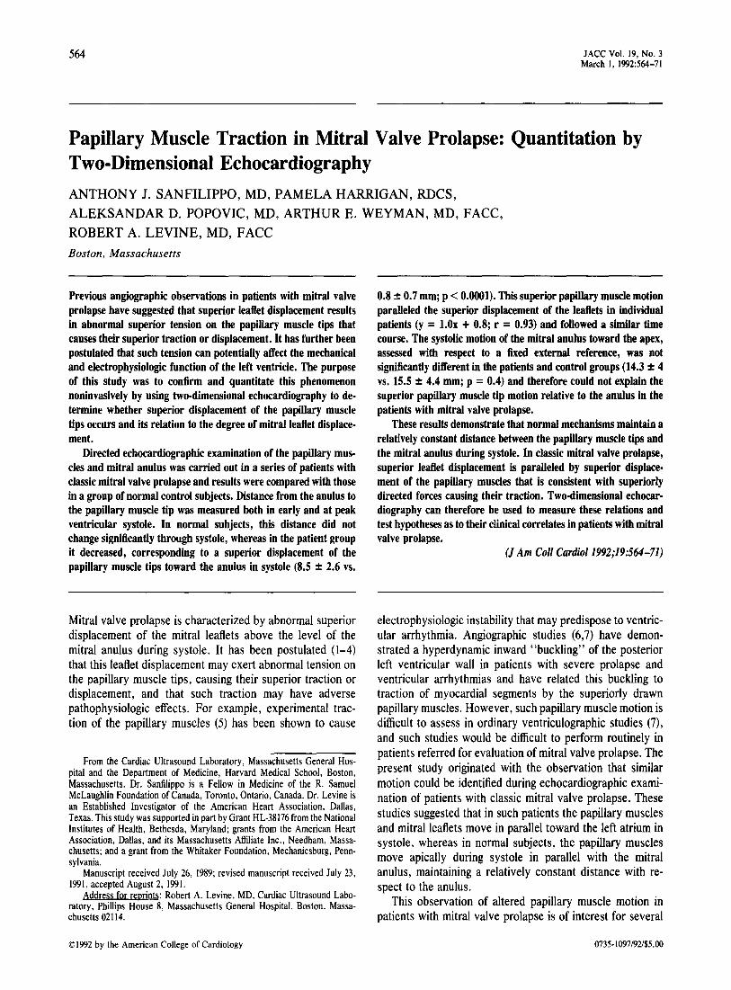
Figure 1. Diagrammatic representation of measurements made fromechocardiographic images. The distance from the papillary muscle(PM) tip to the level of the mitral anulus was measured at full closureof the mitral valve in early systole and at peak systole. Thedifference in these measurements was calculated and termed thepapillary muscle displacement (PM DIS). Ao = aorta; L DIS =leaflet displacement; LA = left atrium; LV = left ventricle.
Observer variability. Two observers measured bothleaflet displacements and the distance from the papillarymuscle tip to mitral anulus in both early and late systole in10 study subjects (5 patients with mitral valve prolapse and
5 normal control subjects; a total of 30 measurements perobserver). Identical video frames were used. Observer variability was assessed as the standard deviation of the differences between the measurements of the two observers. Foreach type of movement (leaflet and papillary muscle tip), themeasurements of one observer were repeated 5 months laterto assess intraobserver variability in a similar manner; thisalso includes the variability due to potential variation in thecycle chosen in the two measurement sessions.
Statistical analysis. Comparisons between the patientswith mitral valve prolapse and control subjects were carriedout using the two-tailed Student t test for unpaired data, withsignificance assessed as p < 0.05. The relation betweenpapillary muscle and leaflet displacement was tested withlinear regression analysis.
ResultsObservations of papillary muscle motion (Fig. 2 to 4). On
real time review of the video images, all normal controlsubjects displayed motion of the papillary muscle tip towardthe apex in systole that paralleled the apical motion of themitral anulus (Fig. 2). In contrast, patients with mitral valveprolapse displayed some degree of superior motion of thepapillary muscle tip in systole that was opposite in directionto the apical motion of the mitral anulus and appeared toparallel the superior displacement of the leaflets (Fig. 3). Thedifference between normal subjects and patients with mitralvalve prolapse is diagrammed in Figure 4.
Quantitative analysis (Fig. 5 to 7). Table 1 lists the measurements of anterior, posterior and maximal leaflet displacement, the distances between the papillary muscle tipand mitral anulus and the superior systolic displacement of
EARLYSYSTOLE
PEAKSYSTOLE
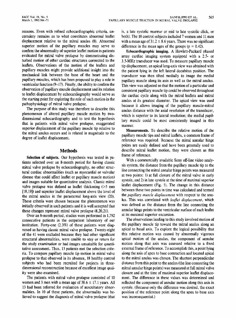
Figure 2. Still frame echocardiographic images from a study in a normal subject takenduring early systole (left) and peak systole(right). The distance from the papillary muscle tip (arrow) to the mitral anulus does notchange significantly. Abbreviations as inFigure I.
lACC Vol. 19, No.3March 1, 1992:564-71
SANFILIPPO ET AL.PAPILLARY MUSCLE TRACTION IN MITRAL VALVE PROLAPSE
567
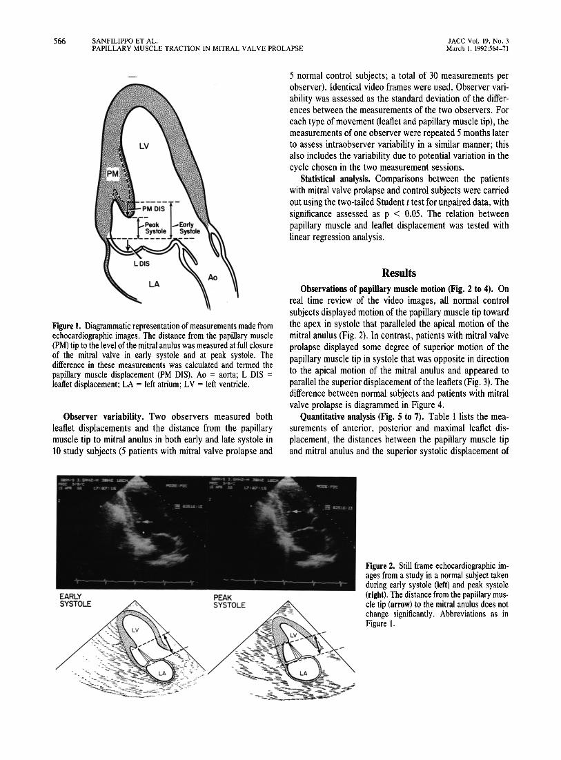
Figure 3. Still frame echocardiographic imagestaken during early systole (left) and peak systole(right) in a patient with classic mitral valveprolapse. The distance from the papillary muscletip (arrow) to the mitral anulus decreases fromearly to late systole. Abbreviations as in Figurel.
EARLYSYSTOLE
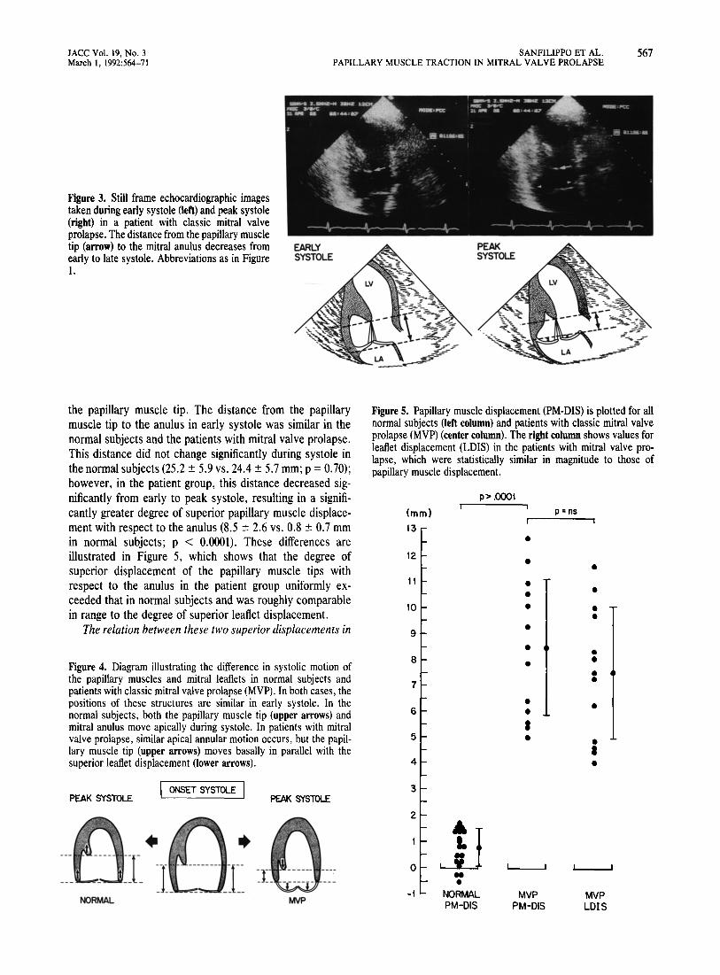
Figure 5. Papillary muscle displacement (PM-DIS) is plotted for allnormal subjects (left column) and patients with classic mitral valveprolapse (MVP) (center column). The right column shows values forleaflet displacement (LDlS) in the patients with mitral valve prolapse, which were statistically similar in magnitude to those ofpapillary muscle displacement.
the papillary muscle tip. The distance from the papillarymuscle tip to the anulus in early systole was similar in thenormal subjects and the patients with mitral valve prolapse.This distance did not change significantly during systole inthe normal subjects (25.2 ± 5.9 vs. 24.4 ± 5.7 mm; p = 0.70);however, in the patient group, this distance decreased significantly from early to peak systole, resulting in a significantly greater degree of superior papillary muscle displacement with respect to the anulus (8.5 ± 2.6 vs. 0.8 ± 0.7 mmin normal subjects; p < 0.0001). These differences areillustrated in Figure 5, which shows that the degree ofsuperior displacement of the papillary muscle tips withrespect to the anulus in the patient group uniformly exceeded that in normal subjects and was roughly comparablein range to the degree of superior leaflet displacement.
The relation between these two superior displacements in
Figure 4. Diagram illustrating the difference in systolic motion ofthe papillary muscles and mitral leaflets in normal subjects andpatients with classic mitral valve prolapse (MVP). In both cases, thepositions of these structures are similar in early systole. In thenormal subjects, both the papillary muscle tip (upper arrows) andmitral anulus move apically during systole. In patients with mitralvalve prolapse, similar apical annular motion occurs, but the papillary muscle tip (upper arrows) moves basally in parallel with thesuperior leaflet displacement (lower arrows).
(mm)
13
12
11
10
9
8
7
6
5
4
p> .0001
••••••••
••I•
p =ns
••••
••:•
•I•
PEAK SYSTOLEI ONSET SYSTOLE I
PEAK SYSlOLE3
MVPLOIS
MVPPM-DIS
..•
NORMALPM-DIS
2
o
-1MVP
••___J l~-~.J__
NORMAL
568 SANFILIPPO ET AL.PAPILLARY MUSCLE TRACTION IN MITRAL VALVE PROLAPSE
JACC Vol. 19, No.3March I, 1992:564-71
12
10
4
•
•• = MVP Patientso = Normalsy = 1.0X + 0.8r = .93
•
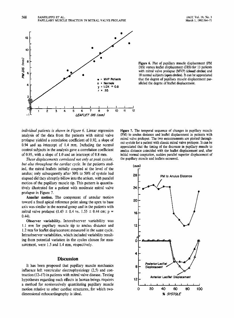
Figure 6. Plot of papillary muscle displacement (PMDIS) versus leaflet displacement (DIS) for 13 patientswith mitral valve prolapse (MVP) (closed circles) and18 normal subjects (open circles). It can be appreciatedthat the degree of papillary muscle displacement paralleled the degree of leaflet displacement.
2 3 4
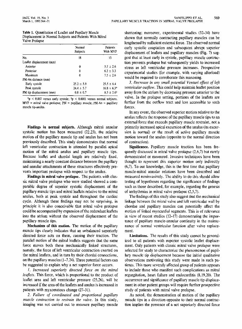
Figure 7. The temporal sequence of changes in papillary muscle(PM) to anulus distance and leaflet displacement in patients withmitral valve prolapse. The two measurements are plotted throughout systole for a patient with classic mitral valve prolapse. It can beappreciated that the timing of the decrease in papillary muscle toanulus distance coincided with the leaflet displacement and, afterinitial normal coaptation, sudden parallel superior displacement ofthe papillary muscle and leaflets occurred.
100
/"Anterior Leaflet Displacement
PM to Anulus Distance
20 40 60 80
" SYSTOLE
Posterior LeafleyDisplacement8
o
4
16
12
12
20
24
28
(mm)
DiscussionIt has been proposed that papillary muscle mechanics
influence left ventricular electrophysiology (2,5) and contraction (12-17) in patients with mitral valve disease. Testinghypotheses regarding such effects in human beings requiresa method for noninvasively quantitating papillary musclemotion relative to other cardiac structures, for which twodimensional echocardiography is ideal.
individual patients is shown in Figure 6. Linear regressionanalysis of the data from the patients with mitral valveprolapse yielded a correlation coefficient of 0.92, a slope of0.94 and an intercept of 1.4 mm. Including the normalcontrol subjects in the analysis gave a correlation coefficientof 0.93, with a slope of 1.0 and an intercept of 0.8 mm.
These displacements correlated not only at peak systole,but also throughout the cardiac cycle. In the patients studied, the mitral leaflets initially coapted at the level of theanulus; only subsequently after 30% to 50% of systole hadelapsed did they abruptly billow into the atrium, with parallelmotion of the papillary muscle tip. This pattern is quantitatively illustrated for a patient with moderate mitral valveprolapse in Figure 7.
Annular motion. The component of annular motiontoward a fixed apical reference point along the apex to baseaxis was similar in the normal group and in the patients withmitral valve prolapse (1.43 ± 0.4 vs. 1.55 ± 0.44 cm; p =0.44).
Observer variability. Interobserver variability was1.1 mm for papillary muscle tip to anulus distance and1.2 mm for leaflet displacement measured in the same cycle.Intraobserver variabilities, which included variability resulting from potential variation in the cycles chosen for measurement, were 1.5 and 1.4 mm, respectively.
lACC Vol. 19, No.3March 1, 1992:564-71
SANFILIPPO ET AL.PAPILLARY MUSCLE TRACTION IN MITRAL VALVE PROLAPSE
569
Table 1. Quantitation of Leaflet and Papillary MuscleDisplacement in Normal Subjects and Patients With MitralValve Prolapse
Normal PatientsSubjects WithMVP
No. 18 13Leaflet displacement (mm)
Anterior 0 5.5 ± 2.6Posterior 0 6.2 ± 3.3Maximum 0 7.5 ± 2.6
PM-An distance (mm)Early systole 25.2 ± 5.9 25.5 ± 6.4Peak systole 24.4 ± 5.7 16.8 ± 6.3*
PM tip displacement (mm) 0.8 ± 0.7 8.5 ± 2.6t
*p < 0.002 versus early systole; tp < 0.0001 versus normal subjects.MVP = mitral valve prolapse; PM = papillary muscle; PM-An = papillarymuscle tip-anulus.
Findings in normal subjects. Although mitral annularsystolic motion has been measured (22,23), the relativemotion of the papillary muscle tip and anulus has not beenpreviously described. This study demonstrates that normalleft ventricular contraction is attended by parallel apicalmotion of the mitral anulus and papillary muscle tips.Because leaflet and chordal length are relatively fixed,maintaining a nearly constant distance between the papillaryand annular attachments of these structures effectively prevents important prolapse with respect to the anulus.
Findings in mitral valve prolapse. The patients with classic mitral valve prolapse who were studied showed a comparable degree of superior systolic displacement of thepapillary muscle tips and mitral leaflets relative to the mitralanulus, both at peak systole and throughout the cardiaccycle. Although these findings may not be surprising, inprinciple it is also conceivable tHat mitral valve prolapsecould be accompanied by expansion of the redundant leafletsinto the atrium without the observed displacement of thepapillary muscle tips.
Mechanism of this motion. The motion of the papillarymuscle tips clearly indicates that an unbalanced superiorlydirected force acts on them, causing their traction. Theparallel motion of the mitral leaflets suggests that the sameforce moves both these mechanically linked structures,namely, the force of left ventricular contraction exerted onthe mitral leaflets, and in tum by their chordal connections,on the papillary muscles (1-7,24). Three potentialfactors canbe suggested to explain why a net superior force occurs.
1. Increased superiorly directed force on the mitralleaflets. This force, which is proportional to the product ofleaflet area and left ventricular pressure (25,26), will beincreased if the area of the leaflets and anulus is increased inpatients with myxomatous change (27-31).
2. Failure of coordinated and proportional papillarymuscle contraction to restrain the valve. In this study,imaging was not carried out to measure papillary muscle
shortening; moreover, experimental studies (32-34) haveshown that normally contracting papillary muscles can belengthened by sufficient external force. The observed normalearly systolic coaptation and subsequent abrupt superiordisplacement of leaflets and papillary muscles (Fig. 7) suggest that at least early in systole, papillary muscle contraction prevents prolapse but subsequently yields to increasedstress as left ventricular pressure increases. Prospectiveexperimental studies (for example, with varying afterload)would be required to corroborate this reasoning.
3. Decrease in any small potential Venturi effect of leftventricular outflow. This could help maintain leaflet positionaway from the atrium by decreasing pressure anterior to thevalve. In the prolapse setting, portions of the leaflets arefurther from the outflow tract and less accessible to suchforces.
In any event, the observed superior motion relative to theanulus reflects the response of the papillary muscle tips to anexternal force that exceeds papillary muscle restraint, not aprimarily increased apical excursion of the anulus (its excursion is normal) or the result of active papillary musclemotion toward the anulus (opposite to the normal directionof contraction).
Significance. Papillary muscle traction has been frequently discussed in mitral valve prolapse (2,6,7) but rarelydemonstrated or measured. Invasive techniques have beenthought to represent this superior motion only indirectly(6,7). To our knowledge, this is the first time that papillarymuscle-mitral annular relations have been described andmeasured noninvasively. The ability to do this should allowtesting of hypotheses regarding the significance of findingssuch as those described, for example, regarding the genesisof arrhythmias in mitral valve prolapse (2,5,7).
The findings of this study also suggest that the mechanicallinkage between the mitral valve and left ventricular wall bychordae and papillary muscles can potentially affect themotion of linked myocardial segments. This is of relevancein view of recent studies (12-17) demonstrating the importance of papillary muscle-annular continuity in the maintenance of normal ventricular function after valve replacement.
Limitations. The results of this study cannot be generalized to all patients with superior systolic leaflet displacement. Only patients with classic mitral valve prolapse wereselected for study to characterize the phenomenon of papillary muscle tip displacement because the initial qualitativeobservations motivating this study were made in such patients. This more severely affected group of patients appearsto include those who manifest such complications as mitralregurgitation, heart failure and endocarditis (8,19,20). Theoccurrence and significance of papillary muscle tip displacement in other patient groups will require further prospectivestudy of patients with mitral valve prolapse.
As noted, the demonstration of motion of the papillarymuscle tips in a direction opposite to their normal contraction implies the presence of a net superiorly directed force
570 SANFILIPPO ET AL.PAPILLARY MUSCLE TRACTION IN MITRAL VALVE PROLAPSE
lACC Vol. 19, No.3March I, 1992:564-71
acting on them, but does not determine the origin of thatforce. Left ventricular pressure will always generate forceacting in that direction because of left ventricular geometry.To what extent that force is increased and the counterbalancing force of papillary muscle contraction is either decreased or fails to increase proportionately cannot be determined from this study, but it might be learned fromexperimental studies that use echocardiography to observepapillary muscle tip motion.
Measurements of papillary muscle tip to annular distances can vary among observers largely because of individual variability in the choice of reference points. We foundthis could be minimized by 1) objective identification of thepoint of chordal insertion into the papillary muscle tip as apoint of increased echo density that could be followedthroughout systole, and 2) frame by frame review of videotape sequences to identify mitral leaflet hinge points consistently. These methods of landmark identification helpedminimize observer variability.
Conclusions. The papillary muscles in normal subjectsmaintain a relatively constant distance between their tipsand the mitral anulus. In contrast, in patients with classicmitral valve prolapse, the papillary muscle tips move superiorly with respect to the mitral anulus in parallel with leafletdisplacement. This superior papillary muscle tip displacement is consistent with the existence of net superior forcesacting on the papillary muscle in mitral valve prolapse. Suchpapillary muscle displacement may have important pathophysiologic implications in patients with mitral valve prolapse, and the mechanical continuity it demonstrates maycontribute to normal left ventricular function. Twodimensional echocardiography provides a means of measuring these relations and therefore testing hypotheses as totheir pathophysiologic significance.
We thank Richard J. Cohen, MD, PhD of the Massachusetts Institute ofTechnology for his insights into the mechanics of papillary muscle and mitralvalve dynamics. We also thank Dianne FinkelsteIn, PhD for statisticalconsultation.
ReferencesI. Cobbs BW. Clinical recognition and medical management of rheumatic
heart disease and other acquired valvular disease. In: Hurst JW, ed. TheHeart. New York: McGraw-Hill, 1974:874-89.
2. Nutter DO, Wickliffe C, Gilbert CA, Moody CC, King SA. The pathophysiology of idiopathic mitral valve prolapse. Circulation 1975;52:297305.
3. Pocock WA, Barlow JW. Etiology and electrocardIOgraphic features ofthe billowing posterior mitral leaflet syndrome: analysis of a further 130patients with a late systolic murmur or non-ejection systolic click. Am JMed 1971;51:731-9.
4. LeWinter MM, Hoffman JR, Shell WE, Karliner JS, O'Rourke RA.Phenylephrine-induced atypical chest pain in patients with prolapsingmitral valve leaflets. Am J Cardiol 1974:34: 12-8.
5. Gornick CC, Tobler HG, Pritzker MC, Tuna IC, Almquist A, Benditt DG.Electrophysiologic effects of papillary muscle traction In the intact heart.Circulation 1986;73:1013-21.
6. Grossman H, Fleming RJ, Engle MA, Levin AH, Ehlers KH. Angiocardiography in the apical systolic click syndrome, Radiology 1968;91:898904.
7. Cobbs BW, King SB. Ventricular buckling: a factor in the abnormalventriculogram and peculiar hemodynamics associated with mitral valveprolapse. Am Heart J 1977;93:741-58,
8. Levine RA, Stathogiannis E, Newell JB, Harrigan P, Weyman AE.Reconsideration of echocardiographic standards for mitral valve prolapse: lack of association between leaflet displacement isolated to theapical four chamber view and independent echocardiographic evidence ofabnormality. J Am Coli Cardiol 1988; II: 1010-9.
9. Wiggers CJ. Katz LN. Contour of the ventricular volume curves underdifferent conditions, Am J Physiol 1922;58:439-75.
10. Rushmer FF, Finlayson BL, Nash AA. Movements of the mitral valve.Circ Res 1956;4:337-42.
II. Lillehei CW, Levy MU, Bonnabeau RC Jr. Mitral valve replacement withpreservation of papillary muscles and chordae tendineae. J ThoracCardiovasc Surg 1964;47:532-43.
12. Miller DW, Johnson DD. Ivey TD. Does preservation of the posteriorchordae tendineae enhance survival during mitral valve replacement?Ann Thorac Surg 1979;28:22-7.
13. Rastelli GC, Tsakiris AG, Frye RL, Kirklin JW. Exercise tolerance andhemodynamic studies after replacement of canine mitral valve with andwithout preservation of chordae tendineae. Circulation 1%7;35(suppl1):1-34-41.
14. David TE, Strauss HD, Mesher E, Anderson MJ, MacDonald IL, BudaAJ. Is it important to preserve the chordae tendineae and papillarymuscles during mitral valve replacement? Can J Surg 1981;24:236-9.
15. Spence PA, Peniston CM, David TE, et aI. Toward a better understandingof the etiology of left ventricular dysfunction after mitral valve replacement: an experimental study with possible clinical implications. AnnThorac Surg 1986:41:363-71.
16. Salter DR, Pellom GL, Murphy CE, et al. Papillary-annular continuityand left ventricular systolic function after mitral valve replacement.Circulation 1986;74(suppll):I-121-9.
17. Hansen DE, Cahill PD, DeCampli WM, et al. Valvular-ventricularinteraction: importance of the mitral apparatus in canine left ventricularsystolic performance. Circulation 1986:73:1310-20.
18. Chandraratna PAN. N1ffialasuriya A, Duncan p. Rosin B, RahimtoolaSH. Identification of the increased frequency of cardiovascular abnormalities associated with mitral valve prolapse by two-dimensional echocardiography. Am J CardioI1984;54:1283-5.
19. Nishimura RA, McGoon MD, Shub C, Miller FA Jr, I1strup DM, TajikAJ. Echocardiographically documented mitral valve prolapse: long-termfollow-up of 232 patients. N Engl J Med 1985;313:1305-9.
20. Marks AR, Choong CY, Sanfilippo AJ, Ferre M, Weyman AE. Identification of high-risk and low-risk subgroups of patients with mitral valveprolapse. N Engl J Med 1989:320:1031-7.
21. Devereux RB, Kramer-Fox R, Shear MK, Kligfield P, Pini R, Savage DD.Diagnosis and classification of severity of mitral valve prolapse: methodologic, biologic and prognostic considerations. Am Heart J 1987;113:1265-80.
22. Tsakiris AG, vonBernuth G, Rastelli GC, Bourgeois MJ, Titus JL, WoodEH. Size and motIOn of the mitral valve annulus in anesthetized dogs.J Appl Physiol 1971 ;30:611-8.
23. Simonson JS, Schiller NB. Descent of the base of the left ventricle: anechocardiographic index of left ventricular function. J Am Soc Echocardiogr 1989:2:25-35.
24. Crawford MH, O'Rourke RA. Mitral valve prolapse: a cardiomyopathicstate? Prog Cardiovasc Dis 1984;27: 133-9.
25. Burch GE, DePasquale NP. Time course of tension in papillary musclesof the heart. JAMA 1965;192: 117-20.
26. Arts T, Meerbaum S, Reneman R, Corday E. Stresses in the closed mitralvalve: a model study. J Biomechanics 1983;16:539-47.
27. Hill DG, Davies MJ, Braimbridge MV. The natural history and surgicalmanagement of the redundant cusp syndrome (floppy mitral valve).J Thorac Cardiovasc Surg 1974;67:519-25.
28. Davies MJ, Moore BP, Braimbridge MV. The floppy mitral valve: studyof incidence, pathology, and complications in surgical, necropsy, andforensic material. Br Heart J 1978;40:468-81.
29. Bulkley BH, Roberts WC. Dilatation of the mitral annulus. Am J Med1975;59:457-63.

lACC Vol. 19, No.3March I, 1992:564-71
SANFILIPPO ET AL.PAPILLARY MUSCLE TRACTION IN MITRAL VALVE PROLAPSE
571
30. Leachman RD, DeFrancheschi A, Zamalloa O. Late systolic murmursand clicks associated with abnormal mitral valve ring. Am J Cardiol1969;23:679-83.
31. Ormiston lA, Shah PM, rei C, Wong M. Size and motion of the mitralvalve annulus in man. II. Abnormalities in mitral valve prolapse. Circulation 1982;65:713-9.
32. Hagl S, Heimisch W, Meisner H, Mendler N, Sebening F. In-situ functionof the papillary muscles in the intact canine left ventricle. In: Duran C,
Angell WW, Johnson AD, Oury JH. eds. Recent Progress in Mitral ValveDisease. London: Butterworths, 1984:397-409.
33. Karas S, Elkins RC. Mechanism of function of the mitral valve leaflets,chordae tendineae and left ventricular papillary muscles in dogs. eire Res1970;26:689-96.
34. Grimm AF, Lendrum BL, Lin HL. Papillary muscle shortening in theintact dog. Circ Res 1975;36:49-57.