prevalence of mitral prolapse by two dimensional echocardiography in healthy young women
TRANSCRIPT
Br Heart J 1983; 49: 334-40
Prevalence of mitral prolapse by two dimensionalechocardiography in healthy young women
L SAMUEL WANN, J ROBERT GROVE, TIMOTHY R HESS, LINDA GLISCH, MICHAEL J
PTACIN, C VINCENT HUGHES, CHARLES M GROSSFrom Medical College of Wisconsin, the Wood VA Medical Center, and the Milwaukee County Medical Complex,Milwaukee, Wisconsin, USA
SUMMARY In order to establish the relative prevalence of mitral valve prolapse as diagnosed by twodimensional echocardiography, we studied 100 presumably healthy young women with two dimen-sional echocardiography and M-mode echocardiography, history, physical examination, electrocar-diography, and phonocardiography. Two dimensional echocardiograms were obtained from para-stemal, apical, and subcostal acoustic windows. Mitral valve prolapse was defined as extension ofleaflet tissue cephalad to the plane of the mitral annulus into the left atrium; note was also made ofany valvular thickening, redundancy, or excessive annular motion. One subject had a midsystolicclick and late systolic murmur with evidence of mitral prolapse on both M-mode and two dimen-sional echocardiography. One subject had a midsystolic click with mitral prolapse demonstrated bytwo dimensional but not on M-mode echocardiography. One subject had a thick mitral valve onechocardiography but no click or murmur. Four subjects had midsystolic clicks without echocar-diographic abnormalities. Mild artefactual pansystolic posterior bowing of the mitral valve on theM-mode echocardiogram could be produced in 20 subjects by incorrect transducer position. Weconclude that the prevalence of mitral valve prolapse by two dimensional echocardiography isrelatively low in presumably healthy young women. Use of two dimensional echocardiography mayavoid overdiagnosis of mitral prolapse and identify a smaller group of individuals with true anatomi-cal abnormalities of the mitral valve.
Over the past 20 years, numerous reports havedocumented the association of mitral valve prolapsewith the auscultatory findings of non-ejection midsys-tolic clicks and late systolic murmurs.1-5 Subse-quently, numerous reports have appeared6-'0 sug-gesting the usefulness of M-mode echocardiographyin making a diagnosis of mitral valve prolapse even inthe absence of the accepted auscultatory findings.This has resulted in a dramatic increase in the allegedincidence of mitral valve prolapse, up to 21%, of pre-sumably healthy young women in one report. "1 Thus,there is considerable controversy regarding the clini-cal significance of an M-mode echocardiographicdiagnosis of mitral prolapse to the point where someclinicians are reluctant to base a diagnosis of mitralprolapse solely on the M-mode echocardiogram.'2
Supported in part by grants from the Veterans Administration, the NIH-NHLBI, and the American Heart Association of Wisconsin, Inc.
Accepted for publication 11 January 1983
More recently, two dimensional echocardiographyhas been used to detect the abnormal pathology rep-resenting the prolapsed mitral valve.'3 14 Because ofits unique ability to provide information on intracar-diac structure, two dimensional echocardiographymay be a more specific and sensitive means for diag-nosing mitral prolapse and has been proposed as the"gold standard" for such diagnosis.'5 We thereforeused two dimensional echocardiography to assess theprevalence- of mitral prolapse in presumably healthyyoung women, a group generally thought to be morefrequently affected by this syndrome.
Subjects and methods
Studies were performed on 100 female volunteersaged 18 years to 35 years (mean 25 years) who consi-dered themselves to be in good health and who denieda prior diagnosis of cardiac disease. Most subjectswere nurses, medical students, and other hospital per-sonnel. The subjects were not paid.
334
Prevalence of mitral prolapse by two dimensional echocardiography in healthy young women
After giving informed consent, each subject-filledout a medical questionnaire which was later reviewedwith an examining cardiologist. Specific note wasmade of drug use, symptoms possibly related to heartdisease, and family history of cardiovascular abnor-mality. A thorough cardiovascular physical examina-tion was performed independently by two cardiolog-ists in a quiet room. Auscultation of the heart wasperformed in the supine, sitting, and standing posi-tions and after squatting and during the strain phaseof a Valsalva manoeuvre. A search was made forthoracic bony abnormalities including pectusexcavatum and absence of normal thoracic kyphosisand for stigmata of Marfan's syndrome. Abnor-malities detected by either examiner were recorded aspositive.A 12 lead electrocardiogram was recorded.
Phonocardiograms were obtained in the supine posi-tion during suspended respiration before and afterinhalation of amyl nitrite. Microphones were placedat the mid left sternal border and apex and filtered todisplay high (200 to 500 Hz), medium (100 to 200Hz), and low (50 to 100 Hz) frequencies. Tracingswere made at 50 mm/s with a simultaneous elec-trocardiogram and carotid pulse tracing using an Elec-tronics for Medicine VR- 12 recorder. Each record wasexamined independently by two cardiologists who hadno knowledge of other findings. Clicks, murmurs,and other abnormalities reported by either examinerwere regarded as positive.M-mode echocardiograms were performed using an
Electronics for Medicine Echo IV ultrasonoscope anda 2.25 MHz transducer focused at 5 cm. Subjects werestudied in the 30 degree left lateral position. Record-ings were made with the transducer at the left sternalborder from as many intercostal spaces as possible.Note was made of the intercostal space from whichthe mitral valve could be recorded with the transducerremaining most nearly perpendicular to the chestwall. Numerous sweeps from the aortic valve and leftatrium to the mitral valve and left ventricle wereobtained. Each M-mode echocardiogram wasreviewed by two independent observers who had noknowledge of other findings. Particular attention wasdirected toward detection of late systolic step-likeposterior displacement of the mitral valve or pansys-tolic posterior bowing of greater than 2 mm posteriorto a line connecting the C and D points of the mitralvalve recording. Pansystolic or late systolic posteriormotion greater than 2 mm recorded with the trans-ducer perpendicular to the chest wall was regarded astrue prolapse while posterior motion recorded from anon-perpendicular position was regarded as artefac-tual prolapse.Two dimensional echocardiograms were obtained
with either a V3000 or V3400 Varian Associates
ultrasonoscope and a 2*25 MHz wide angle phasedarray transducer. Standard views were obtained fromthe parasternal, apical, and subcostal acoustic win-dows and recorded on videotape for subsequentreview. Records were analysed independently by twoobservers in real time, slow motion, and stop frameformats. Neither observer had knowledge of theM-mode echocardiographic findings or other clinicalfeatures of the cases. A specific search was made forevidence of mitral tissue extending through the planeof the mitral annulus into the left atrium during sys-tole on the parasternal long axis, apical four and twochamber, and subcostal four chamber views using themethods of Gilbert et al.14 and Morganroth et al.'5 Aqualitative judgement was also made regarding thepresence of any valvular thickening, redundancy, orexcessive motion of the leaflets or valve ring.'4 Afinding of mitral prolapse by either observer on eitherthe M-mode or two dimensional examination wasregarded as positive evidence for mitral valve pro-lapse. Special note was made of any potential falsepositive findings.
Results
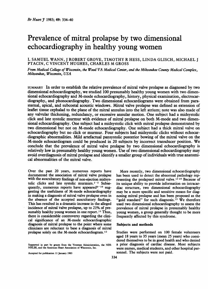
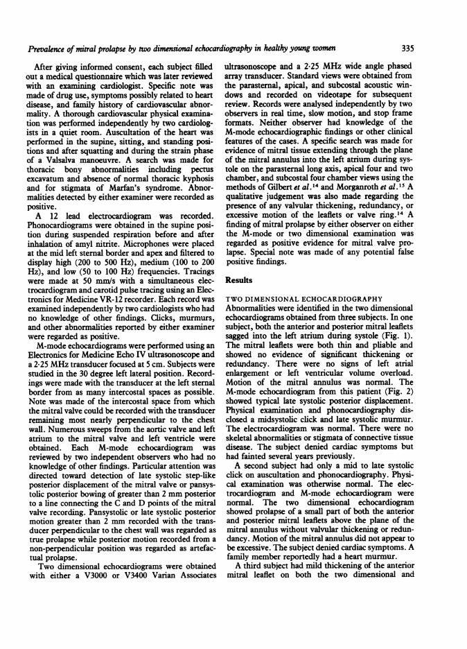
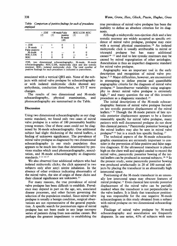
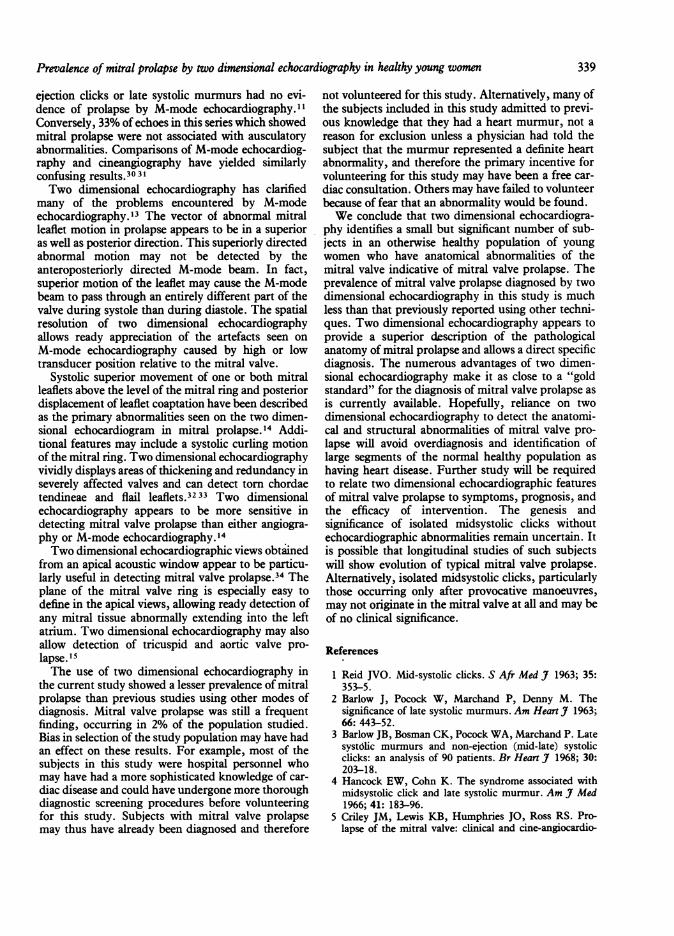
TWO DIMENSIONAL ECHOCARDIOGRAPHYAbnormalities were identified in the two dimensionalechocardiograms obtained from three subjects. In onesubject, both the anterior and posterior mitral leafletssagged into the left atrium during systole (Fig. 1).The mitral leaflets were both thin and pliable andshowed no evidence of significant thickening orredundancy. There were no signs of left atrialenlargement or left ventricular volume overload.Motion of the mitral annulus was normal. TheM-mode echocardiogram from this patient (Fig. 2)showed typical late systolic posterior displacement.Physical examination and phonocardiography dis-closed a midsystolic click and late systolic murmur.The electrocardiogram was normal. There were noskeletal abnormalities or stigmata of connective tissuedisease. The subject denied cardiac symptoms buthad fainted several years previously.A second subject had only a mid to late systolic
click on auscultation and phonocardiography. Physi-cal examination was otherwise normal. The elec-trocardiogram and M-mode echocardiogram werenormal. The two dimensional echocardiogramshowed prolapse of a small part of both the anteriorand posterior mitral leaflets above the plane of themitral annulus without valvular thickening or redun-dancy. Motion of the mitral annulus did not appear tobe excessive. The subject denied cardiac symptoms. Afamily member reportedly had a heart murmur.A third subject had mild thickening of the anterior
mitral leaflet on both the two dimensional and
335
Wann, Grove, Hess, Glisch, Ptacin, Hughes, Gross
Fig. 1 Apicalfour chamber view ofthe two dimensional echocardiogram ofa subject with mitral valve prolapse.RV, right ventricle; LV, left ventricle; LA, left atrium; RA, right atrium. The mitral leaflets can be seen curlinginto the left atrium in this systolic frame (arrow).
Fig. 2 M-mode echocardiogramiUustrated in Fig 1. The arrow is dlate systolic posterior bowing of therepresents mitral valve prolapse.
M-mode echocardiograms. There was no evidence ofmitral valve prolapse. The physical examination,phonocardiogram, and electrocardiogram were nor-mal. The subject denied cardiac symptoms and hadno history of rheumatic fever.
mLGM-MODE ECHOCARDIOGRAPHYM-mode echocardiography showed the characteristiclate systolic step-like posternor displacement charac-
- teristic of mitral valve prolapse in the one previouslymentioned subject. In the remaining subjects a widevanety of mitral valve motion patterns was recorded
_ during systole, but none satisfied the criteria for true- _ -rmitral valve prolapse. Multiple patterns were seen in
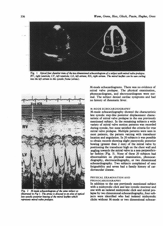
^ 44¢ most patients, the pattern varying with transducer. ~ location and angulation. In 20 subjects it was possible
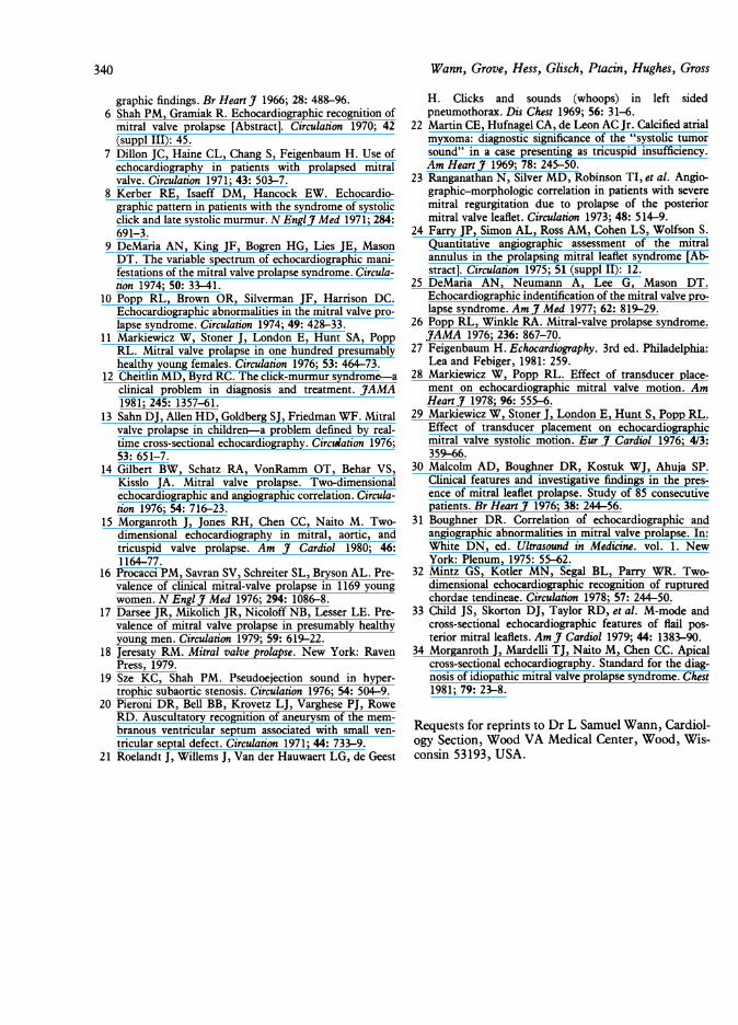
to obtain records showing slight pansystolic posterior
bowing (greater than 2 mm) of the mitral valve bypositioning the transducer high on the chest wall andangling towards the mitral valve in a non-perpendicu-lar fashion (Fig. 3). None of these 20 subjects hadabnormalities on physical examination, phonocar-diography, electrocardiography, or two dimensionalechocardiography. Two subjects complained of easyfatiguability and seven had a family history of car-
4ii. diovascular disease.
PHYSICAL EXAMINATION ANDPHONOCARDIOGRAPHYIn addition to the one previously mentioned subjectwith a midsystolic click and late systolic murmur and
of the same subject as one with an isolated midsystolic click and mitral pro-!irected to an area of Wpical lapse by two dimensional echocardiography, four sub-mitral leaflets which jects were identified who had isolated midsystolic
clicks without M-mode or two dimensional echocar-
L--WSto
336
-A- -j -.hI I
4% -2
Prevalence of mitral prolapse by two dimensional echocardiography in healthy youngwomen3
2lCS 4CS 51CS
=~~~~~~~~..... _--=-----
4 l#-e, , ^ , ,,,, 4.
LV
fi ~~~~: r z:'-- >rf i
Fig. 3 M-mode echocardiogram ofa normal subject who had no evidence ofmitral prolapse on two dimensinalechocardiography, phonocardiography, orphysical examination. The panel at thefar left was obtained at the thirdintercostal space (3 ICS) with the transducer angled in a caudad fashion. The arrow is directed to an area ofposterior motion of the mitral valve which represents artefactual mitral prolapse. The middle panel was obtainedfrom the fourth intercostal space (4 ICS) with the transducer perpendicular to the chest wall. The mitral valvecourses in a normal anterior direction during systole. In the right hand panel, the transducer was in thefifthintercostal space and angled in a cranial direction. The arrov is directed to an area ofexaggerated systolic anteriormotion of the mitral valve.
diographic abnormalities. All had normal electrocar-diograms and denied cardiac symptoms. In two sub-jects the midsystolic clicks were only identified on
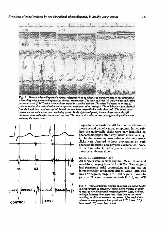
N < J lphonocardiography after amyl nitrite inhalation (Fig.4). In the remaining two subjects the midsystolicclicks were observed without provocation on both
s r-_~-phonocardiography and physical examination. Noneof the four subjects had any other evidence of car-diovascular abnormalities.
l l | ELECTROCARDIOGRAPHY|f _114h i AR subjects were in sinus rhythm. Mean PR interval
Ift j was 0-14 s, ranging from 0 11 to 0-20 s. Two subjectsSi S2 had premature atrial contractions and one had an
intraventricular conduction defect. Mean QRS axisStIS2 was + 57 degrees, range 0 to + 100 degrees. Two sub-
jects had T wave inversion in leads II, III, and aVF
Fig. 4 Phonocardiogram recorded at the mid left sternal borderI& 2.8-_f>, i in a patient with no evidence ofmitral valve prolapse on eitherll 11 mrs 1 r , I 1 1 11 { PM-mode or two dimensional echocardiography. Loxv, medium,
and high frequencyfilters were used. Note that on the controlrecording, no clicks or murmurs are present. After amyl nitrite
AMYL administration a prominent late systolic click (C) is seen. Sl frstROL I heart sound: S2 second heart sound.
337
Wann, Grove, Hess, Glisch, Ptacin, Hughes, Gross
Table Companrson ofpositivefindings for each ofproceduresemployed
+ 2DE +M-modeFalse MSC-LSM MSC(2) (1) positive (1) (5)
M-mode(20)
+ 2DE - 1 0 1 1+ M-mode 1 - 0 1 0False positiveM-mode 0 0 - 0 0
MSC-LSM 1 1 0 - 0MSC 1 0 0 0 -
2DE, two dimensional echocardiography; M-mode, M-modeechocardiography; MSC-LSM, midsystolic click and late systolicmurmur; MSC, isolated midsystolic click. The total number ofpositive findings for each procedure is indicated in parentheses.
associated with a vertical QRS axis. None of the sub-jects with mitral valve prolapse by echocardiographyor with isolated midsystolic clicks showed anyarrhythmia, conduction disturbance, or ST-T wavechanges.The results of two dimensional and M-mode
echocardiography, physical examination, andphonocardiography are summarised in the Table.
Discussion
Using two dimensional echocardiography as our diag-nostic standard, we found only two cases of mitralvalve prolapse in a series of 100 presumably healthyyoung women. One of these cases could not be diag-nosed by M-mode echocardiography. One additionalsubject had slight thickening of the mitral leaflets, afinding of unknown significance. The prevalence ofmitral valve prolapse as diagnosed by two dimensionalechocardiography in our study population thusappears to be much less than that determined by pre-vious studies which used phonocardiography, auscul-tation, and M-mode echocardiography as diagnosticstandards.2 11 16 17We also observed four additional subjects who had
isolated midsystolic clicks; the click appeared in twosubjects only after amyl nitrite inhalation. In theabsence of other evidence indicating abnormality ofthe mitral valve, the site of origin of these clicks andtheir clinical significance are unknown. "I
For many reasons, the true prevalence of mitralvalve prolapse has been difficult to establish. Preval-ence may depend in part on the age, sex, associateddisease processes, and other factors governing theselection of the population studied. Since mitral valveprolapse is usually a benign condition, surgical obser-vations are not representative of the general popula-tion. A specific search for postmortem signs of mitralprolapse has not been made in any large necropsyseries of patients dying from non-cardiac causes. Butperhaps the greatest impediment to establishing the
true prevalence of mitral valve prolapse has been theinability to define an absolute criterion for its diag-nosis.Although a midsystolic non-ejection click and a late
systolic murmur are widely accepted as specific evi-dence of mitral valve prolapse, prolapse may existwith a normal physical examination.'8 An isolatedmidsystolic click is usually attributable to mitral ortricuspid prolapse but has many additionalcauses'9-22 and mid to late systolic murmurs may becaused by mitral regurgitation of other aetiologies.Auscultation is thus an imperfect diagnostic standardfor mitral valve prolapse.
Angiography was an important tool in the earlydescription and recognition of mitral valve pro-lapse.4 23 Major difficulties, however, are encounteredin attempting to define precise and quantifiableangiographic criteria for the diagnosis of mitral valveprolapse.24 Interobserver variability using angiogra-phy to detect mitral valve prolapse is extremelyhigh,25 and many question the use of this invasiveprocedure as a diagnostic standard.26The initial descriptions of the M-mode echocar-
diographic features of mitral valve prolapse focusedon late systolic posterior displacement of the mitralleaflets.6-8 Though the M-mode pattern of late sys-tolic posterior displacement appears to be a featurereasonably specific for mitral valve prolapse, manypatients with well documented prolapse do not showthis classical pattern.9 Pansystolic posterior bowing ofthe mitral leaflets may also be seen in mitral valveprolapse9 10 but is a much less specific finding.27The technical aspects of the M-mode echocardio-
graphic examination are extremely important to con-sider in the prevention of false positive and false nega-tive diagnoses. If the ultrasound transducer is placedhigh on the chest wall and angled caudad to record themitral valve, pansystolic posterior bowing of the ini-tial leaflets can be produced in normal subjects.28 29 Inthe present study, some pansystolic posterior bowingwas produced artefactually in 20 of 100 normal sub-jects by placing the transducer in an abnormally highintercostal space.
Positioning of the M-mode transducer in an unusu-ally low intercostal space may obscure features ofmitral prolapse. "I Even classical late systolic posteriordisplacement of the mitral valve can be partiallymasked when the transducer is not perpendicular tothe valve leaflets. It is likely that transducer position-ing was responsible for the one negative M-modeechocardiogram in this study obtained from a subjectwith mitral prolapse on two dimensional echocardiog-raphy.
Findings of mitral valve prolapse by M-modeechocardiography and auscultation are frequentlydisparate. In one series, 41% of subjects with non-
338
Prevalence of mitral prolapse by two dimensional echocardiography in healthy young women
ejection clicks or late systolic murmurs had no evi-dence of prolapse by M-mode echocardiography."lConversely, 33% of echoes in this series which showedmitral prolapse were not associated with ausculatoryabnormalities. Comparisons of M-mode echocardiog-raphy and cineangiography have yielded similarlyconfusing results.303'Two dimensional echocardiography has clarified
many of the problems encountered by M-modeechocardiography.'3 The vector of abnormal mitralleaflet motion in prolapse appears to be in a superioras well as posterior direction. This superiorly directedabnormal motion may not be detected by theanteroposteriorly directed M-mode beam. In fact,superior motion of the leaflet may cause the M-modebeam to pass through an entirely different part of thevalve during systole than during diastole. The spatialresolution of two dimensional echocardiographyallows ready appreciation of the artefacts seen onM-mode echocardiography caused by high or lowtransducer position relative to the mitral valve.
Systolic superior movement of one or both mitralleaflets above the level of the mitral ring and posteriordisplacement of leaflet coaptation have been describedas the primary abnormalities seen on the two dimen-sional echocardiogram in mitral prolapse.'4 Addi-tional features may include a systolic curling motionof the mitral ring. Two dimensional echocardiographyvividly displays areas of thickening and redundancy inseverely affected valves and can detect torn chordaetendineae and flail leaflets.3233 Two dimensionalechocardiography appears to be more sensitive indetecting mitral valve prolapse than either angiogra-phy or M-mode echocardiography.'4Two dimensional echocardiographic views obtained
from an apical acoustic window appear to be particu-larly useful in detecting mitral valve prolapse.34 Theplane of the mitral valve ring is especially easy todefine in the apical views, allowing ready detection ofany mitral tissue abnormally extending into the leftatrium. Two dimensional echocardiography may alsoallow detection of tricuspid and aortic valve pro-lapse. 5The use of two dimensional echocardiography in
the current study showed a lesser prevalence of mitralprolapse than previous studies using other modes ofdiagnosis. Mitral valve prolapse was still a frequentfinding, occurring in 2% of the population studied.Bias in selection of the study population may have hadan effect on these results. For example, most of thesubjects in this study were hospital personnel whomay have had a more sophisticated knowledge of car-diac disease and could have undergone more thoroughdiagnostic screening procedures before volunteeringfor this study. Subjects with mitral valve prolapsemay thus have already been diagnosed and therefore
not volunteered for this study. Alternatively, many ofthe subjects included in this study admitted to previ-ous knowledge that they had a heart murmur, not areason for exclusion unless a physician had told thesubject that the murmur represented a definite heartabnormality, and therefore the primary incentive forvolunteering for this study may have been a free car-diac consultation. Others may have failed to volunteerbecause of fear that an abnormality would be found.We conclude that two dimensional echocardiogra-
phy identifies a small but significant number of sub-jects in an otherwise healthy population of youngwomen who have anatomical abnormalities of themitral valve indicative of mitral valve prolapse. Theprevalence of mitral valve prolapse diagnosed by twodimensional echocardiography in this study is muchless than that previously reported using other techni-ques. Two dimensional echocardiography appears toprovide a superior description of the pathologicalanatomy of mitral prolapse and allows a direct specificdiagnosis. The numerous advantages of two dimen-sional echocardiography make it as close to a "goldstandard" for the diagnosis of mitral valve prolapse asis currently available. Hopefully, reliance on twodimensional echocardiography to detect the anatomi-cal and structural abnormalities of mitral valve pro-lapse will avoid overdiagnosis and identification oflarge segments of the normal healthy population ashaving heart disease. Further study will be requiredto relate two dimensional echocardiographic featuresof mitral valve prolapse to symptoms, prognosis, andthe efficacy of intervention. The genesis andsignificance of isolated midsystolic clicks withoutechocardiographic abnormalities remain uncertain. Itis possible that longitudinal studies of such subjectswill show evolution of typical mitral valve prolapse.Alternatively, isolated midsystolic clicks, particularlythose occurring only after provocative manoeuvres,may not originate in the mitral valve at all and may beof no clinical significance.
References
1 Reid JVO. Mid-systolic clicks. S Afr Med J 1963; 35:353-5.
2 Barlow J, Pocock W, Marchand P, Denny M. Thesignificance of late systolic murmurs. Am Heart J 1963;66: 443-52.
3 Barlow JB, Bosman CK, Pocock WA, Marchand P. Latesystolic murmurs and non-ejection (mid-late) systolicclicks: an analysis of 90 patients. Br Heart J 1968; 30:203-18.
4 Hancock EW, Cohn K. The syndrome associated withmidsystolic click and late systolic murmur. Am J Med1966; 41: 183-96.
5 Criley JM, Lewis KB, Humphries JO, Ross RS. Pro-lapse of the mitral valve: clinical and cine-angiocardio-
339
Wann, Grove, Hess, Glisch, Ptacin, Hughes, Gross
graphic findings. Br Heart J 1966; 28: 488-96.6 Shah PM, Gramiak R. Echocardiographic recognition of
mitral valve prolapse [Abstract]. Circulation 1970; 42(suppl III): 45.
7 Dillon JC, Haine CL, Chang S, Feigenbaum H. Use ofechocardiography in patients with prolapsed mitralvalve. Circulation 1971; 43: 503-7.
8 Kerber RE, Isaeff DM, Hancock EW. Echocardio-graphic pattern in patients with the syndrome of systolicclick and late systolic murmur. N Engl J Med 1971; 284:691-3.
9 DeMaria AN, King JF, Bogren HG, Lies JE, MasonDT. The variable spectrum of echocardiographic mani-festations of the mitral valve prolapse syndrome. Circula-tion 1974; 50: 33-41.
10 Popp RL, Brown OR, Silverman JF, Harrison DC.Echocardiographic abnormalities in the mitral valve pro-lapse syndrome. Circulation 1974; 49: 428-33.
11 Markiewicz W, Stoner J, London E, Hunt SA, PoppRL. Mitral valve prolapse in one hundred presumablyhealthy young females. Circulation 1976; 53: 464-73.
12 Cheitlin MD, Byrd RC. The click-murmur syndrome-aclinical problem in diagnosis and treatment. JAMA1981; 245: 1357-61.
13 Sahn DJ, Allen HD, Goldberg SJ, Friedman WF. Mitralvalve prolapse in children-a problem defined by real-time cross-sectional echocardiography. Circzdation 1976;53: 651-7.
14 Gilbert BW, Schatz RA, VonRamm OT, Behar VS,Kisslo JA. Mitral valve prolapse. Two-dimensionalechocardiographic and angiographic correlation. Circula-tion 1976; 54: 716-23.
15 Morganroth J, Jones RH, Chen CC, Naito M. Two-dimensional echocardiography in mitral, aortic, andtricuspid valve prolapse. Am J Cardiol 1980; 46:1164-77.
16 Procacci PM, Savran SV, Schreiter SL, Bryson AL. Pre-valence of clinical mitral-valve prolapse in 1169 youngwomen. N Engl J Med 1976; 294: 1086-8.
17 Darsee JR, Mikolich JR, Nicoloff NB, Lesser LE. Pre-valence of mitral valve prolapse in presumably healthyyoung men. Circulation 1979; 59: 619-22.
18 Jeresaty RM. Mitral valve prolapse. New York: RavenPress, 1979.
19 Sze KC, Shah PM. Pseudoejection sound in hyper-trophic subaortic stenosis. Circulation 1976; 54: 504-9.
20 Pieroni DR, Bell BB, Krovetz LJ, Varghese PJ, RoweRD. Auscultatory recognition of aneurysm of the mem-branous ventricular septum associated with small ven-tricular septal defect. Circulation 1971; 44: 733-9.
21 Roelandt J, Willems J, Van der Hauwaert LG, de Geest
H. Clicks and sounds (whoops) in left sidedpneumothorax. Dis Chest 1969; 56: 31-6.
22 Martin CE, Hufnagel CA, de Leon AC Jr. Calcified atrialmyxoma: diagnostic significance of the "systolic tumorsound" in a case presenting as tricuspid insufficiency.Am Heart J 1969; 78: 245-50.
23 Ranganathan N, Silver MD, Robinson TI, et al. Angio-graphic-morphologic correlation in patients with severemitral regurgitation due to prolapse of the posteriormitral valve leaflet. Circulation 1973; 48: 514-9.
24 Farry JP, Simon AL, Ross AM, Cohen LS, Wolfson S.Quantitative angiographic assessment of the mitralannulus in the prolapsing mitral leaflet syndrome [Ab-stract]. Circulation 1975; 51 (suppl II): 12.
25 DeMaria AN, Neumann A, Lee G, Mason DT.Echocardiographic indentification of the mitral valve pro-lapse syndrome. Am J Med 1977; 62: 819-29.
26 Popp RL, Winkle RA. Mitral-valve prolapse syndrome.JAMA 1976; 236: 867-70.
27 Feigenbaum H. Echocardiography. 3rd ed. Philadelphia:Lea and Febiger, 1981: 259.
28 Markiewicz W, Popp RL. Effect of transducer place-ment on echocardiographic mitral valve motion. AmHeart J 1978; 96: 555-6.
29 Markiewicz W, Stoner J, London E, Hunt S, Popp RL.Effect of transducer placement on echocardiographicmitral valve systolic motion. Eur J Cardiol 1976; 4/3:359-66.
30 Malcolm AD, Boughner DR, Kostuk WJ, Ahuja SP.Clinical features and investigative findings in the pres-ence of mitral leaflet prolapse. Study of 85 consecutivepatients. Br Heart J 1976; 38: 244-56.
31 Boughner DR. Correlation of echocardiographic andangiographic abnormalities in mitral valve prolapse. In:White DN, ed. Ultrasound in Medicine. vol. 1. NewYork: Plenum, 1975: 55-62.
32 Mintz GS, Kotler MN, Segal BL, Parry WR. Two-dimensional echocardiographic recognition of rupturedchordae tendineae. Circulation 1978; 57: 244-50.
33 Child JS, Skorton DJ, Taylor RD, et al. M-mode andcross-sectional echocardiographic features of flail pos-terior mitral leaflets. Am J Cardiol 1979; 44: 1383-90.
34 Morganroth J, Mardelli TJ, Naito M, Chen CC. Apicalcross-sectional echocardiography. Standard for the diag-nosis of idiopathic mitral valve prolapse syndrome. Chest1981; 79: 23-8.
Requests for reprints to Dr L Samuel Wann, Cardiol-ogy Section, Wood VA Medical Center, Wood, Wis-consin 53193, USA.
340