cmr predictors of mitral regurgitation in mitral valve prolapse
TRANSCRIPT

J A C C : C A R D I O V A S C U L A R I M A G I N G V O L . 3 , N O . 1 0 , 2 0 1 0
© 2 0 1 0 B Y T H E A M E R I C A N C O L L E G E O F C A R D I O L O G Y F O U N D A T I O N I S S N 1 9 3 6 - 8 7 8 X / $ 3 6 . 0 0
P U B L I S H E D B Y E L S E V I E R I N C . D O I : 1 0 . 1 0 1 6 / j . j c m g . 2 0 1 0 . 0 6 . 0 1 6
CMR Predictors of Mitral Regurgitation in MitralValve Prolapse
Francesca N. Delling, MD,* Lih Lisa Kang, MS,‡ Susan B. Yeon, MD,†Kraig V. Kissinger, RT,* Beth Goddu, RT,* Warren J. Manning, MD,† Yuchi Han, MD*
Boston and Arlington, Massachusetts
O B J E C T I V E S We sought to assess the correlation between mitral valve characteristics and severity
of mitral regurgitation (MR) in subjects with mitral valve prolapse (MVP) undergoing cardiac magnetic
resonance (CMR) imaging.
B A C K G R O U N D Compared with extensive echocardiographic studies, CMR predictors of MVP-
related MR are unknown. The severity of MR at the time of diagnosis has prognostic implication for
patients; therefore, the identification of determinants of MR and its progression may be important for
risk stratification, follow-up recommendations, and surgical decision making.
M E T H O D S Seventy-one MVP patients (age 54 � 11 years, 58% males, left ventricular [LV] ejection
fraction 65 � 5%) underwent cine CMR to assess annular dimensions, maximum systolic anterior and
posterior leaflet displacement, papillary muscle (PM) distance to coaptation point and prolapsed leaflets,
as well as diastolic anterior and posterior leaflet thickness and length, and LV volumes and mass.
Velocity-encoded CMR was used to obtain aortic outflow and to quantify MR volume.
R E S U L T S Using multiple linear regression analysis including all variables, LV mass (p � 0.001),
anterior leaflet length (p � 0.006), and posterior displacement (p � 0.01) were the best determinants of
MR volume with a model-adjusted R2 � 0.6. When the analysis was restricted to valvular characteristics,
MR volume correlated with anterior mitral leaflet length (p � 0.001), posterior mitral leaflet displacement
(p � 0.003), posterior leaflet thickness (p � 0.008), and the presence of flail (p � 0.005) with a
model-adjusted R2 � 0.5. We also demonstrated acceptable intraobserver and interobserver variability
in these measurements.
C O N C L U S I O N S Anterior leaflet length, posterior leaflet displacement, posterior leaflet thickness,
and the presence of flail are the best CMR valvular determinants of MVP-related MR. The acceptable
intraobserver and interobserver variability of our measurements confirms the role of CMR as an imaging
modality for assessment of MVP patients with significant MR. (J Am Coll Cardiol Img 2010;3:1037–45)
© 2010 by the American College of Cardiology Foundation
From the *Department of Medicine (Cardiovascular Division) Beth Israel Deaconess Medical Center, Harvard Medical School,Boston, Massachusetts; †Department of Radiology, Beth Israel Deaconess Medical Center, Harvard Medical School, Boston,Massachusetts; and ‡LK Biostat Consulting Inc., Arlington, Massachusetts. Dr. Han has received grant support from theAmerican College of Cardiology Foundation and Beth Israel Deaconess Medical Center and the Clinical Investigator TrainingProgram: Beth Israel Deaconess Medical Center–Harvard/MIT Health Sciences and Technology, in collaboration with PfizerInc. and Merck & Co. All authors have reported that they have no relationships to disclose.
Manuscript received January 20, 2010; revised manuscript received June 15, 2010, accepted June 22, 2010.
Mtiwmcd
Mssancb
ghanlii
tit(tfimm
tsv
tC
M
Pctea(�lo(q(nnCAMwssacg(pt7etfiTbctooIuMBamdfiait
A
A
C
r
L
M
M
PM � papillary muscle
J A C C : C A R D I O V A S C U L A R I M A G I N G , V O L . 3 , N O . 1 0 , 2 0 1 0
O C T O B E R 2 0 1 0 : 1 0 3 7 – 4 5
Delling et al.
CMR Predictors of MR in MVP
1038
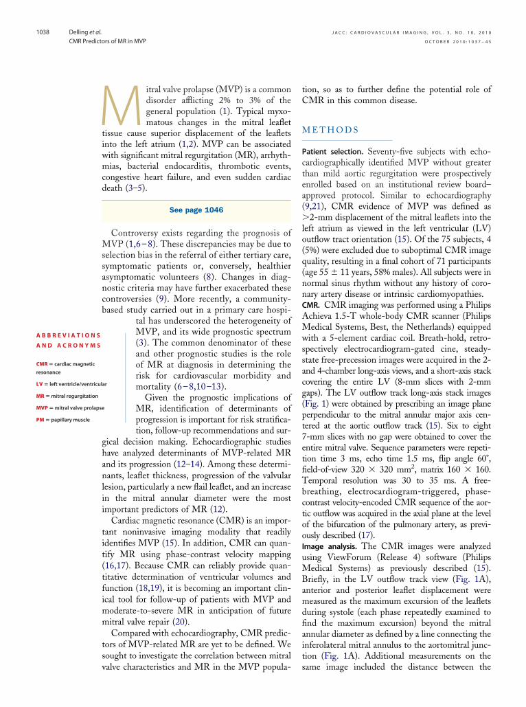
itral valve prolapse (MVP) is a commondisorder afflicting 2% to 3% of thegeneral population (1). Typical myxo-matous changes in the mitral leaflet
issue cause superior displacement of the leafletsnto the left atrium (1,2). MVP can be associatedith significant mitral regurgitation (MR), arrhyth-ias, bacterial endocarditis, thrombotic events,
ongestive heart failure, and even sudden cardiaceath (3–5).
See page 1046
Controversy exists regarding the prognosis ofVP (1,6 – 8). These discrepancies may be due to
election bias in the referral of either tertiary care,ymptomatic patients or, conversely, healthiersymptomatic volunteers (8). Changes in diag-ostic criteria may have further exacerbated theseontroversies (9). More recently, a community-ased study carried out in a primary care hospi-
tal has underscored the heterogeneity ofMVP, and its wide prognostic spectrum(3). The common denominator of theseand other prognostic studies is the roleof MR at diagnosis in determining therisk for cardiovascular morbidity andmortality (6 – 8,10 –13).
Given the prognostic implications ofMR, identification of determinants ofprogression is important for risk stratifica-tion, follow-up recommendations and sur-
ical decision making. Echocardiographic studiesave analyzed determinants of MVP-related MRnd its progression (12–14). Among these determi-ants, leaflet thickness, progression of the valvular
esion, particularly a new flail leaflet, and an increasen the mitral annular diameter were the mostmportant predictors of MR (12).
Cardiac magnetic resonance (CMR) is an impor-ant noninvasive imaging modality that readilydentifies MVP (15). In addition, CMR can quan-ify MR using phase-contrast velocity mapping16,17). Because CMR can reliably provide quan-itative determination of ventricular volumes andunction (18,19), it is becoming an important clin-cal tool for follow-up of patients with MVP and
oderate-to-severe MR in anticipation of futureitral valve repair (20).Compared with echocardiography, CMR predic-
ors of MVP-related MR are yet to be defined. Weought to investigate the correlation between mitral
r
alve characteristics and MR in the MVP popula- s
ion, so as to further define the potential role ofMR in this common disease.
E T H O D S
atient selection. Seventy-five subjects with echo-ardiographically identified MVP without greaterhan mild aortic regurgitation were prospectivelynrolled based on an institutional review board–pproved protocol. Similar to echocardiography9,21), CMR evidence of MVP was defined as2-mm displacement of the mitral leaflets into the
eft atrium as viewed in the left ventricular (LV)utflow tract orientation (15). Of the 75 subjects, 45%) were excluded due to suboptimal CMR imageuality, resulting in a final cohort of 71 participantsage 55 � 11 years, 58% males). All subjects were inormal sinus rhythm without any history of coro-ary artery disease or intrinsic cardiomyopathies.MR. CMR imaging was performed using a Philipschieva 1.5-T whole-body CMR scanner (Philipsedical Systems, Best, the Netherlands) equipped
ith a 5-element cardiac coil. Breath-hold, retro-pectively electrocardiogram-gated cine, steady-tate free-precession images were acquired in the 2-nd 4-chamber long-axis views, and a short-axis stackovering the entire LV (8-mm slices with 2-mmaps). The LV outflow track long-axis stack imagesFig. 1) were obtained by prescribing an image planeerpendicular to the mitral annular major axis cen-ered at the aortic outflow track (15). Six to eight-mm slices with no gap were obtained to cover thentire mitral valve. Sequence parameters were repeti-ion time 3 ms, echo time 1.5 ms, flip angle 60°,eld-of-view 320 � 320 mm2, matrix 160 � 160.emporal resolution was 30 to 35 ms. A free-reathing, electrocardiogram-triggered, phase-ontrast velocity-encoded CMR sequence of the aor-ic outflow was acquired in the axial plane at the levelf the bifurcation of the pulmonary artery, as previ-usly described (17).mage analysis. The CMR images were analyzedsing ViewForum (Release 4) software (Philipsedical Systems) as previously described (15).
riefly, in the LV outflow track view (Fig. 1A),nterior and posterior leaflet displacement wereeasured as the maximum excursion of the leaflets
uring systole (each phase repeatedly examined tond the maximum excursion) beyond the mitralnnular diameter as defined by a line connecting thenferolateral mitral annulus to the aortomitral junc-ion (Fig. 1A). Additional measurements on the
B B R E V I A T I O N S
N D A C R O N YM S
MR � cardiac magnetic
esonance
V � left ventricle/ventricula
R � mitral regurgitation
VP � mitral valve prolapse
ame image included the distance between the

ptlvt(psdam-aLdetcMbfldc
524Sstccmldlpwa
fcnpbm
J A C C : C A R D I O V A S C U L A R I M A G I N G , V O L . 3 , N O . 1 0 , 2 0 1 0
O C T O B E R 2 0 1 0 : 1 0 3 7 – 4 5
Delling et al.
CMR Predictors of MR in MVP
1039
apillary muscle (PM) and the mitral leaflet coap-ation point and the anterior and posterior pro-apsed leaflets, respectively (Fig. 1B). In the sameiew, we measured anterior and posterior leaflethickness and length in diastole (Figs. 1C and 1D)each phase repeatedly examined for the most in-lane view). The annulus was measured at end-ystole in the 2- and 4-chamber views. LVimensions were measured in the short-axis viewt the level of the chordae. LV volumes wereeasured by tracing the end-diastolic and
systolic LV endocardial contours in each slicend applying a summation of discs method. TheV ejection fraction was calculated as: (LV end-iastolic volume � LV end-systolic volume)/LVnd-diastolic volume. LV mass was measured byracing endocardial and epicardial contours in-luding the PMs in the ventricular volumes. The
R volume was calculated as the differenceetween LV stroke volume and the forward aorticow volume. The MR fraction was obtained byividing MR volume by the LV stroke volume. MR
Figure 1. Cine CMR LV Outflow View
(Top panels) Systolic measurements of (A) anterior leaflet displacemdiameter (MAD); and (B) papillary muscle (PM) distance to anterior(PMC). (Bottom panels [zoomed in]): diastolic measurements of (C)rior leaflet thickness (AT), posterior leaflet thickness (PT).
ategories were graded as: 0 (none to trace) (0% to t
%); 1� (mild) (5% to 16%); 2� (moderate) (6% to5%); 3� (moderate to severe) (26% to 48%); and� (severe) (�48%) (17).tatistical methods. Subject characteristics are pre-ented as means with standard deviations for con-inuous traits and as frequency and percentage forategorical traits. The relationship between subjectharacteristics (age, gender, body surface area, LVass, number of prolapsed leaflets, presence of flail
eaflet) and mitral valve measurements (annularimensions, leaflet displacement, thickness, and
ength, and PM distance to coaptation point androlapsed leaflets) with the outcome (MR volume)as assessed using univariate linear regression withsignificance level of 0.05.Because MR volume calculations are derived
rom LV volumes, the LV volumes were not in-luded in the models to investigate MR determi-ants. A multiple linear regression model was firsterformed with stepwise regression (forward andackward selection) and included all the afore-entioned univariate variables, and interaction
(AD), posterior leaflet displacement (PD), and mitral annularet (PMA), to posterior leaflet (PMP), and to coaptation pointrior leaflet length (AL), posterior leaflet length (PL), and (D) ante-
entleaflante
erms for variables with high correlation (correla-
tcbsnsmcpsttfomv
mPldbiwdmpbomt
STN
R
ScsssF(h(spiDy
J A C C : C A R D I O V A S C U L A R I M A G I N G , V O L . 3 , N O . 1 0 , 2 0 1 0
O C T O B E R 2 0 1 0 : 1 0 3 7 – 4 5
Delling et al.
CMR Predictors of MR in MVP
1040
ion coefficient �0.7). The resulting model in-luded variables appearing in both forward andackward selection, as these variables had the mostignificant effects on MR volume. Recognizing theumber of variables was large with respect to theample size (overfitting), we further performedultiple linear regression, focusing only on valvular
haracteristics (the presence of flail, anterior andosterior length, thickness, and displacement). Re-idual plots were examined for a relationship be-ween residual and predicted values. Shapiro-Wilkest was employed to test the normality of residualsor the overall and the valvular model. Receiverperating curve analysis was performed to deter-ine the best cutoff for severe (3� and 4�) MR for
alvular determinants when appropriate.Two independent observers performed measure-ents of annular dimension, leaflet displacement,M distance to coaptation point and prolapsed
eaflets in systole, and leaflet length and thickness iniastole in 30 randomly selected subjects separatedy at least 5 days to assess intraobserver andnterobserver variability. Intraobserver variabilityas calculated as the average of the percentageifference between the same observer’s 2 measure-ents divided by the mean of the 2 measurements
er subject. Interobserver variability was calculatedy the average of percentage difference of the 2bservers’ measurements over their mean measure-ents per subject. Bland-Altman graphs were plot-
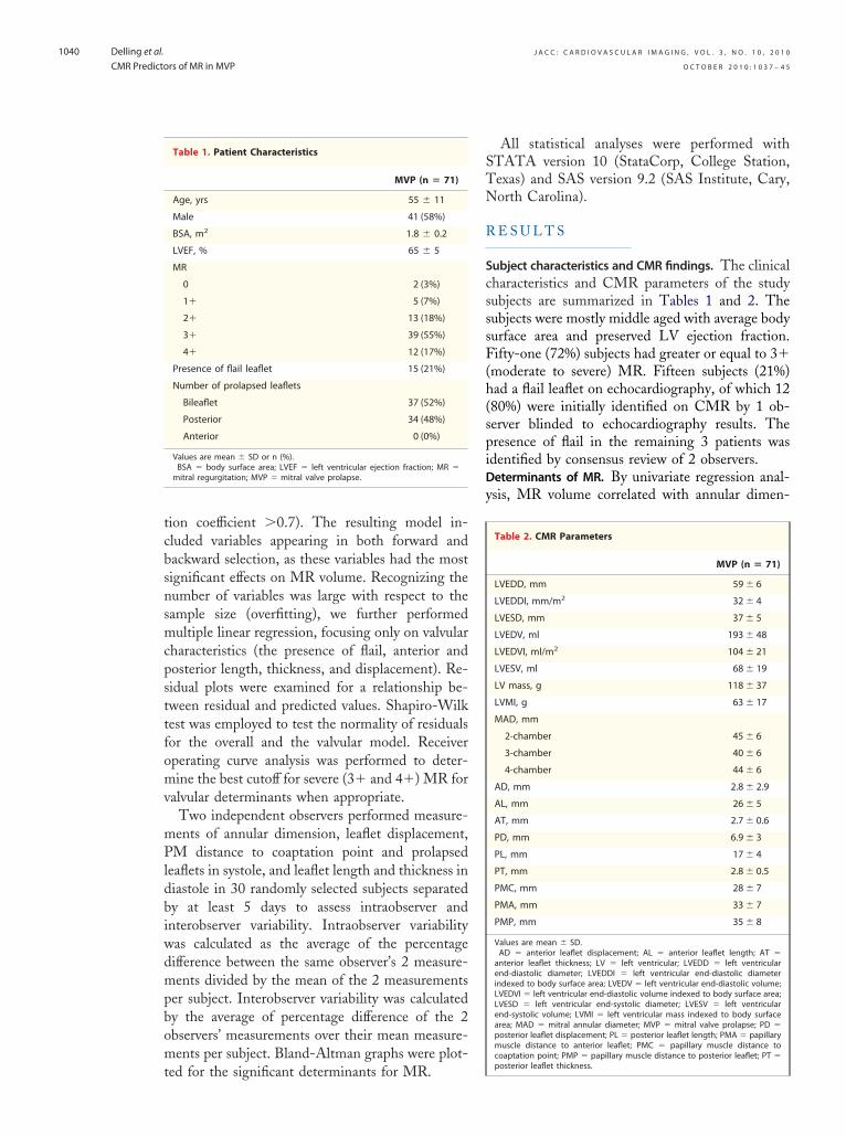
Table 1. Patient Characteristics
MVP (n � 71)
Age, yrs 55 � 11
Male 41 (58%)
BSA, m2 1.8 � 0.2
LVEF, % 65 � 5
MR
0 2 (3%)
1� 5 (7%)
2� 13 (18%)
3� 39 (55%)
4� 12 (17%)
Presence of flail leaflet 15 (21%)
Number of prolapsed leaflets
Bileaflet 37 (52%)
Posterior 34 (48%)
Anterior 0 (0%)
Values are mean � SD or n (%).BSA � body surface area; LVEF � left ventricular ejection fraction; MR �
mitral regurgitation; MVP � mitral valve prolapse.
ed for the significant determinants for MR.
All statistical analyses were performed withTATA version 10 (StataCorp, College Station,exas) and SAS version 9.2 (SAS Institute, Cary,orth Carolina).
E S U L T S
ubject characteristics and CMR findings. The clinicalharacteristics and CMR parameters of the studyubjects are summarized in Tables 1 and 2. Theubjects were mostly middle aged with average bodyurface area and preserved LV ejection fraction.ifty-one (72%) subjects had greater or equal to 3�
moderate to severe) MR. Fifteen subjects (21%)ad a flail leaflet on echocardiography, of which 1280%) were initially identified on CMR by 1 ob-erver blinded to echocardiography results. Theresence of flail in the remaining 3 patients wasdentified by consensus review of 2 observers.eterminants of MR. By univariate regression anal-sis, MR volume correlated with annular dimen-
Table 2. CMR Parameters
MVP (n � 71)
LVEDD, mm 59 � 6
LVEDDI, mm/m2 32 � 4
LVESD, mm 37 � 5
LVEDV, ml 193 � 48
LVEDVI, ml/m2 104 � 21
LVESV, ml 68 � 19
LV mass, g 118 � 37
LVMI, g 63 � 17
MAD, mm
2-chamber 45 � 6
3-chamber 40 � 6
4-chamber 44 � 6
AD, mm 2.8 � 2.9
AL, mm 26 � 5
AT, mm 2.7 � 0.6
PD, mm 6.9 � 3
PL, mm 17 � 4
PT, mm 2.8 � 0.5
PMC, mm 28 � 7
PMA, mm 33 � 7
PMP, mm 35 � 8
Values are mean � SD.AD � anterior leaflet displacement; AL � anterior leaflet length; AT �
anterior leaflet thickness; LV � left ventricular; LVEDD � left ventricularend-diastolic diameter; LVEDDI � left ventricular end-diastolic diameterindexed to body surface area; LVEDV � left ventricular end-diastolic volume;LVEDVI � left ventricular end-diastolic volume indexed to body surface area;LVESD � left ventricular end-systolic diameter; LVESV � left ventricularend-systolic volume; LVMI � left ventricular mass indexed to body surfacearea; MAD � mitral annular diameter; MVP � mitral valve prolapse; PD �posterior leaflet displacement; PL � posterior leaflet length; PMA � papillarymuscle distance to anterior leaflet; PMC � papillary muscle distance tocoaptation point; PMP � papillary muscle distance to posterior leaflet; PT �
posterior leaflet thickness.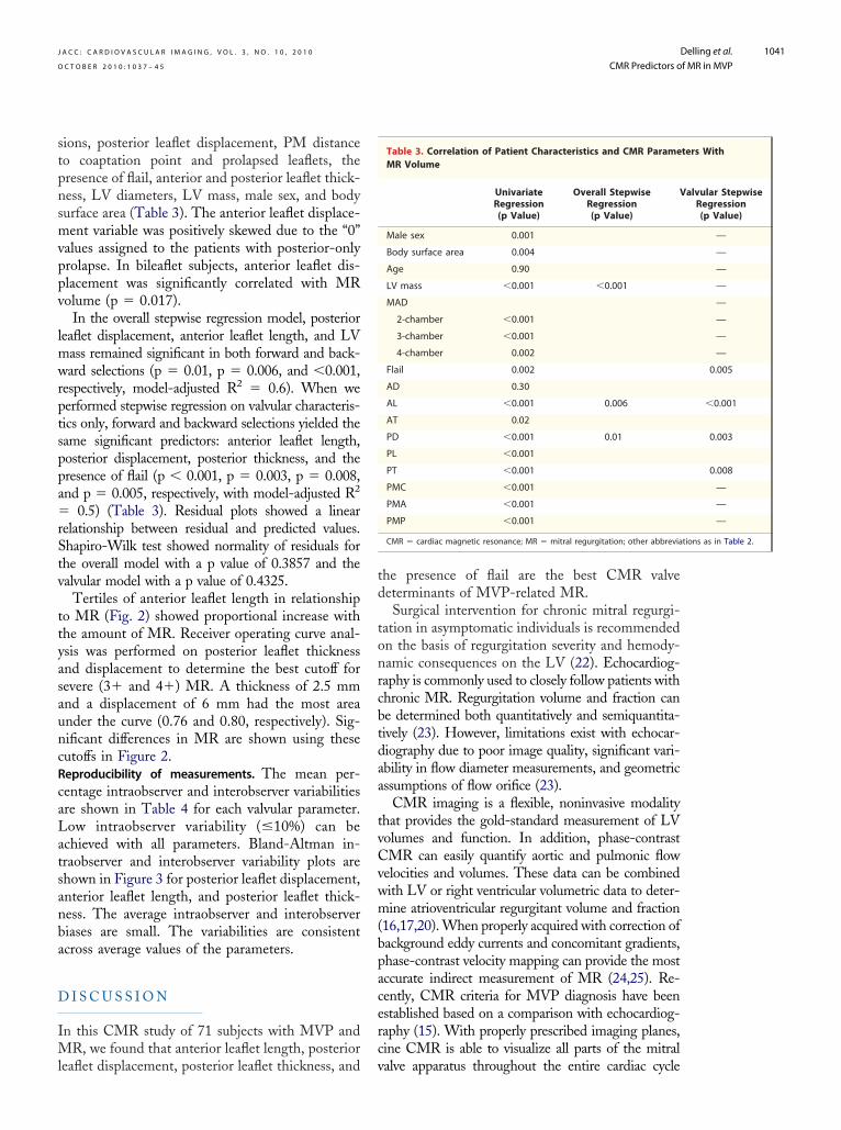
stpnsmvppv
lmwrptsppa�rStv
ttyasauncRcaLatsanba
D
IMl
td
tonrcbtdaa
tvCvwm(bpacerc
tions as in Table 2.
J A C C : C A R D I O V A S C U L A R I M A G I N G , V O L . 3 , N O . 1 0 , 2 0 1 0
O C T O B E R 2 0 1 0 : 1 0 3 7 – 4 5
Delling et al.
CMR Predictors of MR in MVP
1041
ions, posterior leaflet displacement, PM distanceo coaptation point and prolapsed leaflets, theresence of flail, anterior and posterior leaflet thick-ess, LV diameters, LV mass, male sex, and bodyurface area (Table 3). The anterior leaflet displace-ent variable was positively skewed due to the “0”
alues assigned to the patients with posterior-onlyrolapse. In bileaflet subjects, anterior leaflet dis-lacement was significantly correlated with MRolume (p � 0.017).
In the overall stepwise regression model, posterioreaflet displacement, anterior leaflet length, and LV
ass remained significant in both forward and back-ard selections (p � 0.01, p � 0.006, and �0.001,
espectively, model-adjusted R2 � 0.6). When weerformed stepwise regression on valvular characteris-ics only, forward and backward selections yielded theame significant predictors: anterior leaflet length,osterior displacement, posterior thickness, and theresence of flail (p � 0.001, p � 0.003, p � 0.008,nd p � 0.005, respectively, with model-adjusted R2
0.5) (Table 3). Residual plots showed a linearelationship between residual and predicted values.hapiro-Wilk test showed normality of residuals forhe overall model with a p value of 0.3857 and thealvular model with a p value of 0.4325.
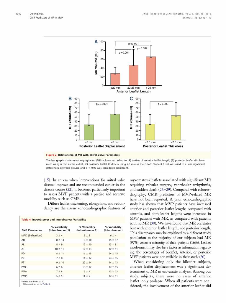
Tertiles of anterior leaflet length in relationshipo MR (Fig. 2) showed proportional increase withhe amount of MR. Receiver operating curve anal-sis was performed on posterior leaflet thicknessnd displacement to determine the best cutoff forevere (3� and 4�) MR. A thickness of 2.5 mmnd a displacement of 6 mm had the most areander the curve (0.76 and 0.80, respectively). Sig-ificant differences in MR are shown using theseutoffs in Figure 2.eproducibility of measurements. The mean per-entage intraobserver and interobserver variabilitiesre shown in Table 4 for each valvular parameter.ow intraobserver variability (�10%) can be
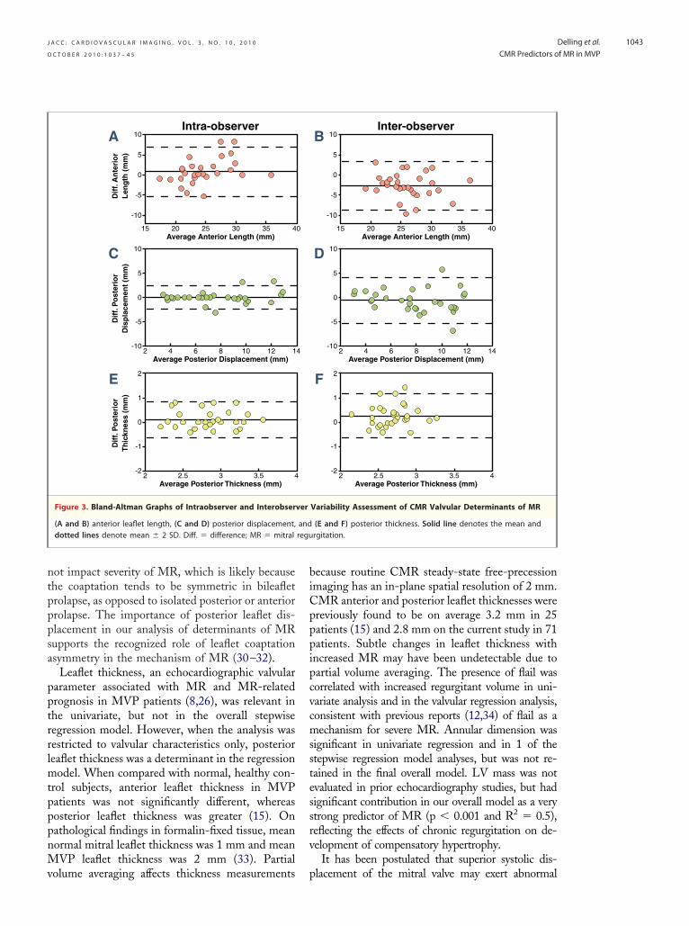
chieved with all parameters. Bland-Altman in-raobserver and interobserver variability plots arehown in Figure 3 for posterior leaflet displacement,nterior leaflet length, and posterior leaflet thick-ess. The average intraobserver and interobserveriases are small. The variabilities are consistentcross average values of the parameters.
I S C U S S I O N
n this CMR study of 71 subjects with MVP andR, we found that anterior leaflet length, posterior
eaflet displacement, posterior leaflet thickness, and v
he presence of flail are the best CMR valveeterminants of MVP-related MR.Surgical intervention for chronic mitral regurgi-
ation in asymptomatic individuals is recommendedn the basis of regurgitation severity and hemody-amic consequences on the LV (22). Echocardiog-aphy is commonly used to closely follow patients withhronic MR. Regurgitation volume and fraction cane determined both quantitatively and semiquantita-ively (23). However, limitations exist with echocar-iography due to poor image quality, significant vari-bility in flow diameter measurements, and geometricssumptions of flow orifice (23).
CMR imaging is a flexible, noninvasive modalityhat provides the gold-standard measurement of LVolumes and function. In addition, phase-contrastMR can easily quantify aortic and pulmonic flow
elocities and volumes. These data can be combinedith LV or right ventricular volumetric data to deter-ine atrioventricular regurgitant volume and fraction
16,17,20). When properly acquired with correction ofackground eddy currents and concomitant gradients,hase-contrast velocity mapping can provide the mostccurate indirect measurement of MR (24,25). Re-ently, CMR criteria for MVP diagnosis have beenstablished based on a comparison with echocardiog-aphy (15). With properly prescribed imaging planes,ine CMR is able to visualize all parts of the mitral
Table 3. Correlation of Patient Characteristics and CMR ParameMR Volume
UnivariateRegression(p Value)
Overall StepwiseRegression(p Value)
V
Male sex 0.001
Body surface area 0.004
Age 0.90
LV mass �0.001 �0.001
MAD
2-chamber �0.001
3-chamber �0.001
4-chamber 0.002
Flail 0.002
AD 0.30
AL �0.001 0.006
AT 0.02
PD �0.001 0.01
PL �0.001
PT �0.001
PMC �0.001
PMA �0.001
PMP �0.001
CMR � cardiac magnetic resonance; MR � mitral regurgitation; other abbrevia
ters With
alvular StepwiseRegression(p Value)
—
—
—
—
—
—
—
—
0.005
�0.001
0.003
0.008
—
—
—
alve apparatus throughout the entire cardiac cycle
(ddtm
d
mradhsacMwbTp(iiM
atsl
Abbreviations as in Ta
nt.
J A C C : C A R D I O V A S C U L A R I M A G I N G , V O L . 3 , N O . 1 0 , 2 0 1 0
O C T O B E R 2 0 1 0 : 1 0 3 7 – 4 5
Delling et al.
CMR Predictors of MR in MVP
1042
15). In an era when interventions for mitral valveisease improve and are recommended earlier in theisease course (22), it becomes particularly importanto assess MVP patients with a precise and accurateodality such as CMR.Diffuse leaflet thickening, elongation, and redun-
ancy are the classic echocardiographic features of
rver and Interobserver Variability
% Variability(Intraobserver 1)
% Variability(Intraobserver 2)
% Variability(Interobserver)
3 � 4 5 � 5 6 � 4
8 � 14 8 � 10 15 � 17
8 � 9 12 � 10 13 � 9
10 � 11 17 � 13 12 � 11
8 � 11 16 � 15 24 � 15
7 � 8 14 � 12 24 � 15
9 � 10 22 � 14 14 � 13
6 � 6 13 � 12 17 � 15
7 � 8 6 � 7 13 � 13
5 � 5 11 � 9 12 � 11
.
A
0
20
40
60
80
100
< 22 mm
MR
Vo
lum
e (m
l)Anteri
p=0.0
B
0
20
40
60
80
70
50
30
10
90
6 mm
MR
Vo
lum
e (m
l)
Posterior Leaflet Displacement
p=0.0001
> 6 mm≤
Figure 2. Relationship of MR With Mitral Valve Parameters
The bar graphs show mitral regurgitation (MR) volume according tment using 6 mm as the cutoff, (C) posterior leaflet thickness usingdifferences between groups, and p � 0.05 was considered significa
sble 2.
yxomatous leaflets associated with significant MRequiring valvular surgery, ventricular arrhythmia,nd sudden death (26–29). Compared with echocar-iography, CMR predictors of MVP-related MRave not been reported. A prior echocardiographictudy has shown that MVP patients have increasednterior and posterior leaflet lengths compared withontrols, and both leaflet lengths were increased in
VP patients with MR, as compared with patientsith no MR (30). We have found that MR correlatesest with anterior leaflet length, not posterior length.his discrepancy may be explained by a different studyopulation as the majority of our subjects had MR97%) versus a minority of their patients (16%). Leafletnvolvement may also be a factor as information regard-ng the percentages of bileaflet, anterior, or posterior
VP patients were not available in their study (30).When considering only the bileaflet subjects,
nterior leaflet displacement was a significant de-erminant of MR in univariate analysis. Among ourtudy subjects, there were no cases of anterioreaflet–only prolapse. When all patients were con-
eaflet Length-26 mm
<0.001
p=0.009
> 26 mm
0
20
40
60
80
70
50
30
10
90
2.5 mm
MR
Vo
lum
e (m
l)
Posterior Leaflet Thickness
p=0.005
> 2.5 mm≤
) tertiles of anterior leaflet length, (B) posterior leaflet displace-mm as the cutoff. Student t test was used to assess significant
Table 4. Intraobse
CMR Parameters
MAD (3-chamber)
AD
AL
AT
PD
PL
PT
PMC
PMA
PMP
Values are mean � SD
or L22
p
04
C
o (A2.5
idered, the involvement of the anterior leaflet did
ntpppsa
pptrrlmtpppnMv
biCpppipcvcmsstessrv
regu
J A C C : C A R D I O V A S C U L A R I M A G I N G , V O L . 3 , N O . 1 0 , 2 0 1 0
O C T O B E R 2 0 1 0 : 1 0 3 7 – 4 5
Delling et al.
CMR Predictors of MR in MVP
1043
ot impact severity of MR, which is likely becausehe coaptation tends to be symmetric in bileafletrolapse, as opposed to isolated posterior or anteriorrolapse. The importance of posterior leaflet dis-lacement in our analysis of determinants of MRupports the recognized role of leaflet coaptationsymmetry in the mechanism of MR (30–32).
Leaflet thickness, an echocardiographic valvulararameter associated with MR and MR-relatedrognosis in MVP patients (8,26), was relevant inhe univariate, but not in the overall stepwiseegression model. However, when the analysis wasestricted to valvular characteristics only, posterioreaflet thickness was a determinant in the regression
odel. When compared with normal, healthy con-rol subjects, anterior leaflet thickness in MVPatients was not significantly different, whereasosterior leaflet thickness was greater (15). Onathological findings in formalin-fixed tissue, meanormal mitral leaflet thickness was 1 mm and meanVP leaflet thickness was 2 mm (33). Partial
Intra-observerA 10
5
0
-5
-10
2015 25 30 35 4
Dif
f. A
nte
rio
rL
eng
th (
mm
)
Average Anterior Length (mm)
C 10
5
0
-5
-104 62 8 10 12 1
Dif
f. P
ost
erio
rD
isp
lace
men
t (m
m)
Average Posterior Displacement (mm)
E 2
1
0
-1
-22.52 3 3.5 4
Dif
f. P
ost
erio
rT
hic
knes
s (m
m)
Average Posterior Thickness (mm)
Figure 3. Bland-Altman Graphs of Intraobserver and Interobser
(A and B) anterior leaflet length, (C and D) posterior displacement,dotted lines denote mean � 2 SD. Diff. � difference; MR � mitral
olume averaging affects thickness measurements p
ecause routine CMR steady-state free-precessionmaging has an in-plane spatial resolution of 2 mm.MR anterior and posterior leaflet thicknesses werereviously found to be on average 3.2 mm in 25atients (15) and 2.8 mm on the current study in 71atients. Subtle changes in leaflet thickness withncreased MR may have been undetectable due toartial volume averaging. The presence of flail wasorrelated with increased regurgitant volume in uni-ariate analysis and in the valvular regression analysis,onsistent with previous reports (12,34) of flail as aechanism for severe MR. Annular dimension was
ignificant in univariate regression and in 1 of thetepwise regression model analyses, but was not re-ained in the final overall model. LV mass was notvaluated in prior echocardiography studies, but hadignificant contribution in our overall model as a verytrong predictor of MR (p � 0.001 and R2 � 0.5),eflecting the effects of chronic regurgitation on de-elopment of compensatory hypertrophy.
It has been postulated that superior systolic dis-
D 10
5
0
-5
-104 62 8 10 12 14
Average Posterior Displacement (mm)
Inter-observerB 10
5
0
-5
-10
2015 25 30 35 40Average Anterior Length (mm)
F 2
1
0
-1
-22.52 3 3.5 4
Average Posterior Thickness (mm)
Variability Assessment of CMR Valvular Determinants of MR
(E and F) posterior thickness. Solid line denotes the mean andrgitation.
0
4
ver
and
lacement of the mitral valve may exert abnormal

ttptibspvbwbPwb
pehocMpaOiacmssCSsgn
pOttavcpddmhneMoct
C
Idpdats
ATY
RCe
R
J A C C : C A R D I O V A S C U L A R I M A G I N G , V O L . 3 , N O . 1 0 , 2 0 1 0
O C T O B E R 2 0 1 0 : 1 0 3 7 – 4 5
Delling et al.
CMR Predictors of MR in MVP
1044
ension on the PM tips, causing their superior trac-ion, and that such traction may have adverse patho-hysiologic effects (35). The distance between the PMip and the mitral valve has been previously quantifiedn MVP patients in an echocardiographic study (36),ut correlation with MR was not studied. In ourtudy, we looked at the distance between the PM, theoint of maximum leaflet displacement, and the mitralalve coaptation point in relation to MR. The distanceetween the PM and the mitral valve coaptation pointas included in the analysis as it expresses the contri-ution of 1 leaflet length relative to the other. TheM distance to valve leaflets and coaptation pointere significant MR determinants in the univariateut not the stepwise overall model.
Despite its many advantages over echocardiogra-hy, CMR is not as widely used, and clinicalxperience is relatively limited in assessing valvulareart disease with, in particular, a lack of correlationf CMR valvular parameters with clinical out-omes. Therefore, finding valvular determinants of
VP-related MR is crucial for development ofrospective studies to define disease progressionnd the optimal timing of surgical intervention.ur study showed acceptable intraobserver and
nterobserver variability with low percentage vari-bility achievable in a trained observer, furtheronfirming the ideal role of CMR as an imagingodality for the assessment of MVP patients with
ignificant MR. To our knowledge, this is the firsttudy to fully investigate the reproducibility ofMR measurements for myxomatous valve disease.tudy limitations. Several limitations exist in ourtudy. This is a cross-sectional study without lon-itudinal follow-up. To fully understand the prog-ostic role of CMR in patients with MVP,
4. Devereux RB, Kramer-Fox R, ShearMK, Kligfield P, Pini R, Savage DD. AJ. Echocardiog
rogression of MR would be of particular interest.ur sample size is small when all the variables of
he mitral apparatus are considered. To overcomehe overfitting issue when performing regressionnalysis for significant predictors, we limited thealvular regression model input variables to valvularharacteristics only and determined the final 4redictors. Reassuringly, the strongest valvular pre-ictors—anterior leaflet length and posterior leafletisplacement—were present in both the overallodel and the valvular regression model. Our study
as laid the foundation for conducting a longitudi-al study by assessing comprehensive CMR param-ters of valvular and ventricular characteristics of
VP patients. An additional limitation is the lackf anterior leaflet–only prolapse patients, whichould be the result of excluding patients with morehan mild aortic regurgitation in our study design.
O N C L U S I O N S
n summary, anterior leaflet length, posterior leafletisplacement, posterior leaflet thickness, and theresence of flail leaflet are the best CMR valvulareterminants of MVP-related MR. Our findingsre reproducible and represent an important stepowards the development of prospective CMRtudies to define MR progression.
cknowledgmenthe authors would like to thank Christopher M. Fang-en, PhD, for editorial assistance with the manuscript.
eprint requests and correspondence: Dr. Yuchi Han,ardiovascular Division, E/SH-457, Beth Israel Deacon-
ss Medical Center, 330 Brookline Avenue, Boston,
follow-up studies in valvular determinants for the Massachusetts 02215. E-mail: [email protected].E F E R E N C E S
1. Freed LA, Levy D, Levine RA, et al.Prevalence and clinical outcome ofmitral-valve prolapse. N Engl J Med1999;341:1–7.
2. Tamura K, Fukuda Y, Ishizaki M,Masuda Y, Yamanaka N, Ferrans VJ.Abnormalities in elastic fibers andother connective-tissue components offloppy mitral valve. Am Heart J 1995;129:1149–58.
3. Avierinos JF, Gersh BJ, Melton LJ 3rd,et al. Natural history of asymptomaticmitral valve prolapse in the community.Circulation 2002;106:1355–61.
Diagnosis and classification of severityof mitral valve prolapse: method-ologic, biologic, and prognostic con-siderations. Am Heart J 1987;113:1265–80.
5. Perloff JK, Child JS. Clinical and ep-idemiologic issues in mitral valve pro-lapse: overview and perspective. AmHeart J 1987;113:1324–32.
6. Duren DR, Becker AE, Dunning AJ.Long-term follow-up of idiopathicmitral valve prolapse in 300 patients: aprospective study. J Am Coll Cardiol1988;11:42–7.
7. Nishimura RA, McGoon MD, ShubC, Miller FA Jr., Ilstrup DM, Tajik
raphically docu-
mented mitral-valve prolapse. Long-term follow-up of 237 patients.N Engl J Med 1985;313:1305–9.
8. St John Sutton M, Weyman AE.Mitral valve prolapse prevalence andcomplications: an ongoing dialogue.Circulation 2002;106:1305–7.
9. Levine RA, Stathogiannis E, NewellJB, Harrigan P, Weyman AE. Re-consideration of echocardiographicstandards for mitral valve prolapse:lack of association between leafletdisplacement isolated to the apicalfour chamber view and independentechocardiographic evidence of ab-normality. J Am Coll Cardiol 1988;
11:1010 –9.
1
1
1
1
1
1
1
1
1
1
2
2
3
3
3
3
3
3
3
Ky
J A C C : C A R D I O V A S C U L A R I M A G I N G , V O L . 3 , N O . 1 0 , 2 0 1 0
O C T O B E R 2 0 1 0 : 1 0 3 7 – 4 5
Delling et al.
CMR Predictors of MR in MVP
1045
0. Freed LA, Benjamin EJ, Levy D, etal. Mitral valve prolapse in the generalpopulation: the benign nature of echo-cardiographic features in the Fram-ingham Heart Study. J Am Coll Car-diol 2002;40:1298–304.
1. Enriquez-Sarano M, Avierinos JF,Messika-Zeitoun D, et al. Quantita-tive determinants of the outcome ofasymptomatic mitral regurgitation.N Engl J Med 2005;352:875–83.
2. Enriquez-Sarano M, Basmadjian AJ,Rossi A, Bailey KR, Seward JB, TajikAJ. Progression of mitral regurgita-tion: a prospective Doppler echocar-diographic study. J Am Coll Cardiol1999;34:1137–44.
3. Avierinos JF, Detaint D, Messika-Zeitoun D, Mohty D, Enriquez-Sarano M. Risk, determinants, andoutcome implications of progressionof mitral regurgitation after diagnosisof mitral valve prolapse in a singlecommunity. Am J Cardiol 2008;101:662–7.
4. Singh RG, Cappucci R, Kramer-FoxR, et al. Severe mitral regurgitationdue to mitral valve prolapse: risk fac-tors for development, progression, andneed for mitral valve surgery. Am JCardiol 2000;85:193–8.
5. Han Y, Peters DC, Salton CJ, et al.Cardiovascular magnetic resonancecharacterization of mitral valve pro-lapse. J Am Coll Cardiol Img 2008;1:294–303.
6. Fujita N, Chazouilleres AF, HartialaJJ, et al. Quantification of mitral re-gurgitation by velocity-encoded cinenuclear magnetic resonance imaging.J Am Coll Cardiol 1994;23:951–8.
7. Gelfand EV, Hughes S, Hauser TH,et al. Severity of mitral and aorticregurgitation as assessed by cardiovas-cular magnetic resonance: optimizingcorrelation with Doppler echocardi-ography. J Cardiovasc Magn Reson2006;8:503–7.
8. Chuang ML, Hibberd MG, SaltonCJ, et al. Importance of imagingmethod over imaging modality innoninvasive determination of left ven-tricular volumes and ejection fraction:assessment by two- and three-dimensional echocardiography andmagnetic resonance imaging. J AmColl Cardiol 2000;35:477–84.
9. Salton CJ, Chuang ML, O’Donnell
CJ, et al. Gender differences and nor-mal left ventricular anatomy in anadult population free of hypertension.A cardiovascular magnetic resonancestudy of the Framingham Heart StudyOffspring cohort. J Am Coll Cardiol2002;39:1055–60.
20. Cawley PJ, Maki JH, Otto CM. Car-diovascular magnetic resonance imag-ing for valvular heart disease: tech-nique and validation. Circulation2009;119:468–78.
21. Levine RA, Triulzi MO, Harrigan P,Weyman AE. The relationship of mi-tral annular shape to the diagnosis ofmitral valve prolapse. Circulation1987;75:756–67.
22. Bonow RO, Carabello BA, ChatterjeeK, et al. 2008 focused update incorpo-rated into the ACC/AHA 2006guidelines for the management of pa-tients with valvular heart disease: areport of the American College ofCardiology/American Heart Associa-tion Task Force on Practice Guide-lines (Writing Committee to revisethe 1998 guidelines for the manage-ment of patients with valvular heartdisease). J Am Coll Cardiol 2008;52:e1–142.
23. Zoghbi WA, Enriquez-Sarano M,Foster E, et al. Recommendations forevaluation of the severity of nativevalvular regurgitation with two-dimensional and Doppler echocardi-ography. J Am Soc Echocardiogr2003;16:777–802.
24. Chan KM, Wage R, Symmonds K,et al. Towards comprehensive as-sessment of mitral regurgitation usingcardiovascular magnetic resonance.J Cardiovasc Magn Reson 2008;10:61.
25. Buchner S, Debl K, Poschenrieder F,et al. Cardiovascular magnetic reso-nance for direct assessment of ana-tomic regurgitant orifice in mitral re-gurgitation. Circ Cardiovasc Imaging2008;1:148–55.
26. Marks AR, Choong CY, SanfilippoAJ, Ferre M, Weyman AE. Identifi-cation of high-risk and low-risk sub-groups of patients with mitral-valveprolapse. N Engl J Med 1989;320:1031–6.
27. Vohra J, Sathe S, Warren R, TatoulisJ, Hunt D. Malignant ventricular ar-rhythmias in patients with mitral valveprolapse and mild mitral regurgita-tion. Pacing Clin Electrophysiol 1993;
16:387–93. v8. Zuppiroli A, Mori F, Favilli S, et al.Arrhythmias in mitral valve pro-lapse: relation to anterior mitral leaf-let thickening, clinical variables, andcolor Doppler echocardiographicparameters. Am Heart J 1994;128:919 –27.
9. Sanfilippo AJ, Abdollah H, BurggrafGW. Quantitation and significance ofsystolic mitral leaflet displacement inmitral valve prolapse. Am J Cardiol1989;64:1349–55.
0. Weissman NJ, Pini R, Roman MJ,Kramer-Fox R, Andersen HS, De-vereux RB. In vivo mitral valve mor-phology and motion in mitral valveprolapse. Am J Cardiol 1994;73:1080–8.
1. Grayburn PA, Berk MR, Spain MG,Harrison MR, Smith MD, DeMariaAN. Relation of echocardiographicmorphology of the mitral apparatus tomitral regurgitation in mitral valveprolapse: assessment by Doppler colorflow imaging. Am Heart J 1990;119:1095–102.
2. Jyo Y, Yoshikawa J, Yoshida K, KatoH, Shakudo M. [A new diagnosticcriteria of mitral valve prolapse syn-drome]. J Cardiol Suppl 1988;18:29–41, discussion 42–4.
3. Rabkin E, Aikawa M, Stone JR, Fu-kumoto Y, Libby P, Schoen FJ. Acti-vated interstitial myofibroblasts ex-press catabolic enzymes and mediatematrix remodeling in myxomatousheart valves. Circulation 2001;104:2525–32.
4. Mills WR, Barber JE, Skiles JA, et al.Clinical, echocardiographic, and bio-mechanical differences in mitral valveprolapse affecting one or both leaflets.Am J Cardiol 2002;89:1394–9.
5. Gornick CC, Tobler HG, PritzkerMC, Tuna IC, Almquist A, BendittDG. Electrophysiologic effects of pap-illary muscle traction in the intactheart. Circulation 1986;73:1013–21.
6. Sanfilippo AJ, Harrigan P, PopovicAD, Weyman AE, Levine RA. Pap-illary muscle traction in mitral valveprolapse: quantitation by two-dimensional echocardiography. J AmColl Cardiol 1992;19:564–71.
ey Words: mitral regurgitationmitral valve prolapse y
alvular characteristics.