osteoporosis canada 2010 guidelines for the assessment of fracture risk
TRANSCRIPT
Canadian Association of Radiologists Journal 62 (2011) 243e250www.carjonline.org
Health Policy and Practice / Sant�e: politique et pratique m�edicale
Osteoporosis Canada 2010 Guidelines for the Assessment of Fracture Risk
Brian Lentle, MDa,*, Angela M. Cheung, MD, PhDb, David A. Hanley, MDc,William D. Leslie, MD, MScd, David Lyons, MDe, Alexandra Papaioannou, MD, MScf,
Stephanie Atkinson, PhDg, Jacques P. Brown, MDh, Sidney Feldman, MDi,Anthony B. Hodsman, MDj, Abida Sophina Jamal, MD, PhDk, Robert G. Josse, MB, BSl,
Stephanie M. Kaiser, MDm, Brent Kvern, MDn, Suzanne Morin, MD, MSco,Kerry Siminoski, MDp; for the Scientific Advisory Council of Osteoporosis Canada
aDepartment of Radiology, University of British Columbia, Vancouver, British Columbia, CanadabDepartments of Medicine and Medical Imaging, University of Toronto, Toronto, Ontario, Canada
cDivision of Endocrinology and Metabolism, Departments of Medicine, Community Health Sciences, and Oncology,
University of Calgary, Calgary, Alberta, CanadadDepartments of Medicine and Radiology, University of Manitoba, Winnipeg, Manitoba, Canada
eCanadian BMD Facility Accreditation Program, Ontario Association of Radiologists, Toronto, Ontario, CanadafDepartment of Medicine, McMaster University, Hamilton, Ontario, Canada
gDepartment of Pediatrics and Biomedical Sciences, Faculty of Health Sciences, McMaster University, Hamilton, Ontario, CanadahDepartment of Medicine, Laval University, Qu�ebec City, Qu�ebec, Canada
iDepartment of Family and Community Medicine, University of Toronto, Toronto, Ontario, CanadajDepartment of Medicine, University of Western Ontario, London, Ontario, Canada
kDepartment of Medicine, Faculty of Medicine, University of Toronto, Toronto, Ontario, CanadalDepartment of Medicine, Faculty of Medicine, University of Toronto, Toronto, Ontario, Canada
mDivision of Endocrinology and Metabolism, Faculty of Medicine, Dalhousie University, Halifax, Nova Scotia, CanadanDepartment of Family Medicine, University of Manitoba, Winnipeg, Manitoba, Canada
oDivision of General Internal Medicine, Department of Medicine, McGill University, Montr�eal, Qu�ebec, CanadapRadiology and Medicine, University of Alberta, Edmonton, Alberta, Canada
Abstract
Osteoporosis Canada’s 2010 Clinical Practice Guidelines for the Diagnosis and Management of Osteoporosis in Canada focus on theclinical impact of fragility fractures, and on the assessment and management of women and men at high risk for fragility fracture. Theseguidelines now integrate a 10-year absolute fracture risk prediction into an overall management approach by using validated risk assessmenttools. There currently is a large gap between optimal practices and those that are now being provided to Canadians with osteoporosis. Theseguidelines are part of a concerted effort to close this gap. Key changes from the 2002 guidelines of interest and relevance to radiologists arehighlighted in this report.� 2011 Canadian Association of Radiologists. All rights reserved.
Key Words: Osteoporosis; Fracture; Risk assessment; Bone mineral densitometry
The evaluation of persons suspected of having osteopo-rosis has evolved. Because the disease typically causessymptoms only when fractures result, the diagnosis tended,in the past, to be made late. The development of tools tomeasure bone mineral density (BMD), especially in the
* Address for correspondence: Brian Lentle, MD, Women’s Health Centre,
Room F2, 4599 Oak Street, Vancouver, British Columbia V6H 3N1, Canada.
E-mail address: [email protected] (B. Lentle).
0846-5371/$ - see front matter � 2011 Canadian Association of Radiologists. A
doi:10.1016/j.carj.2011.05.001
central skeleton, has allowed identification of individuals athigh risk of fragility fracture before the first fracture.
A landmark was the publication of a diagnostic classifi-cation by a working group of the World Health Organization(WHO) in 1994 [1]. This defined osteoporosis in a post-menopausal woman as a BMD of 2.5 or more standarddeviations (SD) below a young adult norm, that is, a T scoreof e2.5 or lower. This value roughly corresponded with thefraction of the population sustaining fragility fractures,
ll rights reserved.
244 B. Lentle et al. / Canadian Association of Radiologists Journal 62 (2011) 243e250
although only a minority of fragility fractures occur inwomen with a T score this low. However, this became widelyaccepted as a BMD-derived definition of osteoporosis, wascommonly used as a treatment threshold, and is now alsoapplied to men older than age 50 years (Table 1) [2,3].
However, the WHO classification, despite its merits,proved to be limiting for a number of reasons, not the least ofwhich was that it placed undue emphasis on the importanceof BMD as a risk factor for osteoporotic fractures whileobscuring the complexity of fracture risk, which is due tomany factors.
The 2010 Osteoporosis Canada Clinical PracticeGuidelines
The development of these guidelines followed theAppraisal of Guidelines, Research and Evaluation frame-work [4]. Primary care physicians, patients, osteoporosisspecialists from different disciplines, radiologists, alliedhealth professionals, and health policy makers were surveyedto identify priorities for the guidelines. Based on these,systematic reviews of the literature were conducted to updateour knowledge in 2 key areas: (1) fracture risk assessmentand (2) therapies for osteoporosis. Also, Canadian data arenow available to inform many of these recommendations.
Table 1
Recommended diagnostic categories for both men and women based on
bone densitometry
Age Category Criteriaa
<50 y Below expected range for age Z score �e2.0
Within expected range for age Z score >e2.0
�50 y Severe (established) osteoporosis T score �e2.5
with fragility fractures
Osteoporosis T score �e2.5
Low bone mass T score ¼e1.1 and e2.4
Normal T score �e1.0
BMD ¼ bone mineral density.aNotes:
1) The T score is the number of standard deviations that BMD is above or
below the mean normal peak BMD for young white women (the National
Health and Nutrition Education Survey III for hip measurements).
The Z score is the number of standard deviations that BMD above or
below the mean normal BMD for sex, age, and (if reference data are
available) race or ethnicity.
2) Osteoporosis cannot be diagnosed by BMD alone below age 50 y.
3) Based upon lowest value for lumbar spine (minimum 2 vertebral levels),
total hip, and femoral neck. If either the lumbar spine or hip is invalid,
then the forearm should be scanned and the distal third of the region
reported.
4) Fracture risk assessment under the Fracture Risk Assessment Tool / 2010
Canadian Association of Radiologists and Osteoporosis Canada system is
based upon the femoral neck T score alone.
Appropriate interventions to prevent fractures needto accurately identify those at risk and, therefore, most likelyto benefit from treatment [5]. Unfortunately, many of thosewho sustain a fragility fracture are neither appropriatelyassessed nor treated [6,7]. The 2010 guidelines focus on theassessment and management of women and men at high riskfor such a fracture [7]. Low BMD is only one of several riskfactors for fracture, and, in recognition of this, OsteoporosisCanada (OC) and the Canadian Association of Radiologists(CAR) adopted a system for 10-year absolute fracture riskassessment to be used in BMD reporting [8]. The 2010guidelines have updated this absolute risk assessment modeland developed recommendations for comprehensive care(Figure 1).
Key Concepts
The 2010 guidelines use an integrated risk assessment(based on BMD and other risk factors) and treatment modelto stratify women and men older than age 50 years into 3fracture risk groups. These categories align with treatmentimplications: low risk (usually not requiring pharmacologictreatment), moderate risk (consider additional clinical riskfactors to determine any need for pharmacologic treatment),and high risk (should be considered for pharmacologictreatment) [7]. Nonpharmacologic and lifestyle measures areapplicable to all: optimizing calcium and vitamin D intake,regular weight-bearing activity, balance and strengtheningexercises, and smoking cessation. In older patients, fallprevention should be considered, including a multifactorialassessment of causes contributing to risk.
These categories are defined as follows:
� High risk for future fractures. Implied is a >20% prob-ability of major osteoporotic fracture (ie, proximalfemoral, clinical vertebral, forearm, or proximal humeralbreak) over the ensuing 10 years. Individuals older thanage 50 years who have had a fragility fracture of the hipor vertebra and those who have had more than 1 fragilityfracture are also considered to be high risk for futurefractures, irrespective of BMD [9e12]. Pharmacologictherapy should be offered to these individuals.
� Moderate risk for future fractures. Implied is 10%-20%probability for major osteoporotic fracture over 10 years.For those at moderate fracture risk with no other riskfactors, treatment should be individualized and mayinclude pharmacologic therapy, or just basic lifestylemeasures with monitoring. There are more osteoporoticfractures in the moderate fracture risk group than in thehigh fracture risk group (because there are more indi-viduals at moderate risk than at high risk), even thoughthe individual fracture risk is higher in the high-riskgroup [13]. The moderate-risk group requires a carefulevaluation to identify vertebral fractures (Table 2) oradditional clinical risk factors (Figure 1), which maycontribute to a decision to offer pharmacologic therapy.
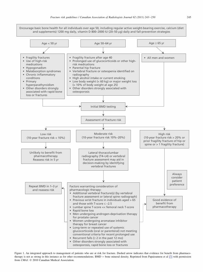
Figure 1. An integrated approach to management of patients who are at risk for fracture. Dashed arrow indicates that evidence for benefit from pharmaco-
therapy is not as strong in this instance as for other recommendations. BMD ¼ bone mineral density. Reprinted from Papaioannou et al [7] with permission
from CMAJ. � 2010 Canadian Medical Association.
245Fracture risk guidelines / Canadian Association of Radiologists Journal 62 (2011) 243e250
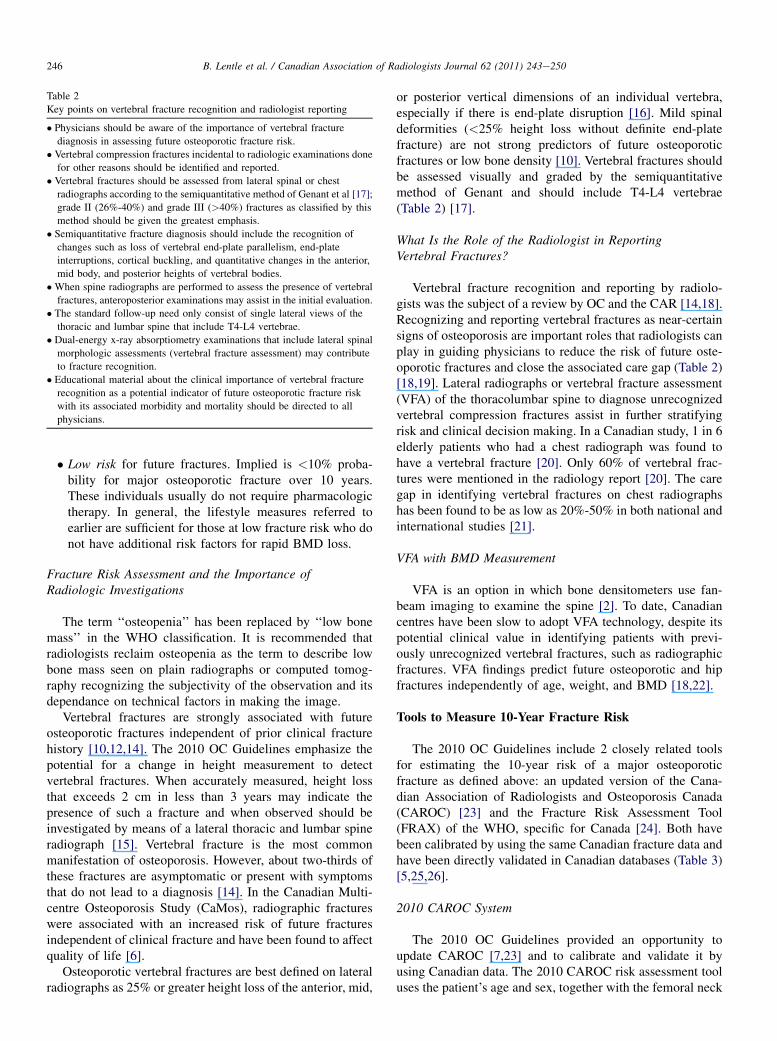
Table 2
Key points on vertebral fracture recognition and radiologist reporting
� Physicians should be aware of the importance of vertebral fracture
diagnosis in assessing future osteoporotic fracture risk.
� Vertebral compression fractures incidental to radiologic examinations done
for other reasons should be identified and reported.
� Vertebral fractures should be assessed from lateral spinal or chest
radiographs according to the semiquantitative method of Genant et al [17];
grade II (26%-40%) and grade III (>40%) fractures as classified by this
method should be given the greatest emphasis.
� Semiquantitative fracture diagnosis should include the recognition of
changes such as loss of vertebral end-plate parallelism, end-plate
interruptions, cortical buckling, and quantitative changes in the anterior,
mid body, and posterior heights of vertebral bodies.
�When spine radiographs are performed to assess the presence of vertebral
fractures, anteroposterior examinations may assist in the initial evaluation.
� The standard follow-up need only consist of single lateral views of the
thoracic and lumbar spine that include T4-L4 vertebrae.
� Dual-energy x-ray absorptiometry examinations that include lateral spinal
morphologic assessments (vertebral fracture assessment) may contribute
to fracture recognition.
� Educational material about the clinical importance of vertebral fracture
recognition as a potential indicator of future osteoporotic fracture risk
with its associated morbidity and mortality should be directed to all
physicians.
246 B. Lentle et al. / Canadian Association of Radiologists Journal 62 (2011) 243e250
� Low risk for future fractures. Implied is <10% proba-bility for major osteoporotic fracture over 10 years.These individuals usually do not require pharmacologictherapy. In general, the lifestyle measures referred toearlier are sufficient for those at low fracture risk who donot have additional risk factors for rapid BMD loss.
Fracture Risk Assessment and the Importance ofRadiologic Investigations
The term ‘‘osteopenia’’ has been replaced by ‘‘low bonemass’’ in the WHO classification. It is recommended thatradiologists reclaim osteopenia as the term to describe lowbone mass seen on plain radiographs or computed tomog-raphy recognizing the subjectivity of the observation and itsdependance on technical factors in making the image.
Vertebral fractures are strongly associated with futureosteoporotic fractures independent of prior clinical fracturehistory [10,12,14]. The 2010 OC Guidelines emphasize thepotential for a change in height measurement to detectvertebral fractures. When accurately measured, height lossthat exceeds 2 cm in less than 3 years may indicate thepresence of such a fracture and when observed should beinvestigated by means of a lateral thoracic and lumbar spineradiograph [15]. Vertebral fracture is the most commonmanifestation of osteoporosis. However, about two-thirds ofthese fractures are asymptomatic or present with symptomsthat do not lead to a diagnosis [14]. In the Canadian Multi-centre Osteoporosis Study (CaMos), radiographic fractureswere associated with an increased risk of future fracturesindependent of clinical fracture and have been found to affectquality of life [6].
Osteoporotic vertebral fractures are best defined on lateralradiographs as 25% or greater height loss of the anterior, mid,
or posterior vertical dimensions of an individual vertebra,especially if there is end-plate disruption [16]. Mild spinaldeformities (<25% height loss without definite end-platefracture) are not strong predictors of future osteoporoticfractures or low bone density [10]. Vertebral fractures shouldbe assessed visually and graded by the semiquantitativemethod of Genant and should include T4-L4 vertebrae(Table 2) [17].
What Is the Role of the Radiologist in ReportingVertebral Fractures?
Vertebral fracture recognition and reporting by radiolo-gists was the subject of a review by OC and the CAR [14,18].Recognizing and reporting vertebral fractures as near-certainsigns of osteoporosis are important roles that radiologists canplay in guiding physicians to reduce the risk of future oste-oporotic fractures and close the associated care gap (Table 2)[18,19]. Lateral radiographs or vertebral fracture assessment(VFA) of the thoracolumbar spine to diagnose unrecognizedvertebral compression fractures assist in further stratifyingrisk and clinical decision making. In a Canadian study, 1 in 6elderly patients who had a chest radiograph was found tohave a vertebral fracture [20]. Only 60% of vertebral frac-tures were mentioned in the radiology report [20]. The caregap in identifying vertebral fractures on chest radiographshas been found to be as low as 20%-50% in both national andinternational studies [21].
VFA with BMD Measurement
VFA is an option in which bone densitometers use fan-beam imaging to examine the spine [2]. To date, Canadiancentres have been slow to adopt VFA technology, despite itspotential clinical value in identifying patients with previ-ously unrecognized vertebral fractures, such as radiographicfractures. VFA findings predict future osteoporotic and hipfractures independently of age, weight, and BMD [18,22].
Tools to Measure 10-Year Fracture Risk
The 2010 OC Guidelines include 2 closely related toolsfor estimating the 10-year risk of a major osteoporoticfracture as defined above: an updated version of the Cana-dian Association of Radiologists and Osteoporosis Canada(CAROC) [23] and the Fracture Risk Assessment Tool(FRAX) of the WHO, specific for Canada [24]. Both havebeen calibrated by using the same Canadian fracture data andhave been directly validated in Canadian databases (Table 3)[5,25,26].
2010 CAROC System
The 2010 OC Guidelines provided an opportunity toupdate CAROC [7,23] and to calibrate and validate it byusing Canadian data. The 2010 CAROC risk assessment tooluses the patient’s age and sex, together with the femoral neck
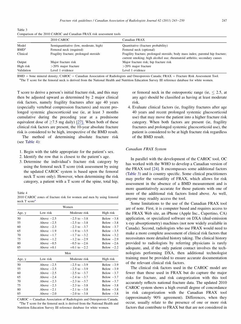
Table 3
Comparison of the 2010 CAROC and Canadian FRAX risk assessment tools
2010 CAROC Canadian FRAX
Model Semiquantitative (low, moderate, high) Quantitative (fracture probability)
BMDa Femoral neck (required) Femoral neck (optional)
Clinical Fragility fracture; prolonged steroids Fragility fracture; prolonged steroids; body mass index; parental hip fracture;
current smoking; high alcohol use; rheumatoid arthritis; secondary causes
Output Major fracture risk Major fracture risk; hip fracture risk
High risk >20% major fracture >20% major fracture
Validation Level 1 evidence Level 1 evidence
BMD ¼ bone mineral density; CAROC ¼ Canadian Association of Radiologists and Osteoporosis Canada; FRAX ¼ Fracture Risk Assessment Tool.aThe T score for the femoral neck is derived from the National Health and Nutrition Education Survey III reference database for white women.
247Fracture risk guidelines / Canadian Association of Radiologists Journal 62 (2011) 243e250
T score to derive a person’s initial fracture risk, and this maythen be adjusted upward as determined by 2 major clinicalrisk factors, namely fragility fractures after age 40 years(especially vertebral compression fractures) and recent pro-longed systemic glucocorticoid use (ie, at least 3 monthscumulative during the preceding year at a prednisoneequivalent dose of �7.5 mg daily) [27]. When both of theseclinical risk factors are present, the 10-year absolute fracturerisk is considered to be high, irrespective of the BMD result.
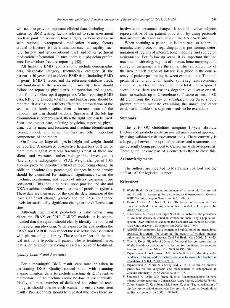
The method of determining absolute fracture risk(see Table 4):
1. Begin with the table appropriate for the patient’s sex.2. Identify the row that is closest to the patient’s age.3. Determine the individual’s fracture risk category by
using the femoral neck T score (risk assessment by usingthe updated CAROC system is based upon the femoralneck T score only). However, when determining the riskcategory, a patient with a T score of the spine, total hip,
Table 4
2010 CAROC zones of fracture risk for women and men by using femoral
neck T scorea
Women
Age, y Low risk Moderate risk High risk
50 Above �2.5 �2.5 to �3.8 Below �3.8
55 Above �2.5 �2.5 to �3.8 Below �3.8
60 Above �2.3 �2.3 to �3.7 Below �3.7
65 Above �1.9 �1.9 to �3.5 Below �3.5
70 Above �1.7 �1.7 to �3.2 Below �3.2
75 Above �1.2 �1.2 to �2.9 Below �2.9
80 Above �0.5 �0.5 to �2.6 Below �2.6
85 Above þ0.1 þ0.1 to �2.2 Below �2.2
Men
Age, y Low risk Moderate risk High risk
50 Above �2.5 �2.5 to �3.9 Below �3.9
55 Above �2.5 �2.5 to �3.9 Below �3.9
60 Above �2.5 �2.5 to �3.7 Below �3.7
65 Above �2.4 �2.4 to �3.7 Below �3.7
70 Above �2.3 �2.3 to �3.7 Below �3.7
75 Above �2.3 �2.3 to �3.8 Below �3.8
80 Above �2.1 �2.1 to �3.8 Below �3.8
85 Above �2.0 �2.0 to �3.8 Below �3.8
CAROC ¼ Canadian Association of Radiologists and Osteoporosis Canada.aThe T score for the femoral neck is derived from the National Health and
Nutrition Education Survey III reference database for white women.
or femoral neck in the osteoporotic range (ie, � 2.5, atany age) should be classified as having at least moderaterisk.
4. Evaluate clinical factors (ie, fragility fractures after age40 years and recent prolonged systemic glucocorticoiduse) that may move the patient into a higher fracture riskcategory. When both factors are present (ie, fragilityfractures and prolonged systemic glucocorticoid use), thepatient is considered to be at high fracture risk regardlessof the BMD result.
Canadian FRAX System
In parallel with the development of the CAROC tool, OChas worked with the WHO to develop a Canadian version ofthe FRAX tool [24]. It encompasses some additional factors(Table 3) and is country specific. Some clinical practitionersmay prefer the versatility of FRAX, which allows for riskassessment in the absence of a BMD measurement and ismore quantitatively accurate for those patients with one ofmore of the additional risk factors listed above. As well,anyone may readily access the tool.
Some limitations to the use of the Canadian FRAX toolare of note. First, it is computer based and requires access tothe FRAX Web site, an iPhone (Apple Inc., Cupertino, CA)application, or specialized software on DXA (dual-emissionx-ray absorptiometry) machines (not now widely available inCanada). Second, radiologists who use FRAX would need tomake a more complete assessment of clinical risk factors thatnecessitates more detailed history taking. The clinical historyprovided to radiologists by referring physicians is rarelyadequate, and, if the only patient contact involves the tech-nologists performing DXA, then additional technologisttraining must be provided to ensure accurate documentationof the relevant clinical risk factors.
The clinical risk factors used in the CAROC model arefewer than those used in FRAX but do capture the majorrisks for fracture, and risk categorization with this toolaccurately reflects national fracture data. The updated 2010CAROC system shows a high overall degree of concordancein risk categorization with the Canadian FRAX tool(approximately 90% agreement). Differences, when theyoccur, usually relate to the presence of one or more riskfactors that contribute to FRAX but that are not considered in
248 B. Lentle et al. / Canadian Association of Radiologists Journal 62 (2011) 243e250
the CAROC tool (namely, parental history of hip fracture,smoking, excess alcohol intake, rheumatoid arthritis).
The updated version of the CAROC fracture risk assess-ment tool is easy to use and, therefore, is recommendedinstead of FRAX for BMD reporting in Canada. Familyphysicians, osteoporosis specialists, and those communi-cating DXA findings are already familiar both with riskassessment in general [28] and with the 2005 CAROC modelin particular; this should allow for a more seamless inte-gration of the 2010 CAROC system into reporting. Thissituation may change as FRAX becomes more widely used.
Addressing Other Issues
Why Is the Lumbar Spine Not Included in the FractureRisk Assessment? How Do I Evaluate Risk When theLumbar Spine BMD Is Much Lower Than That at theHip?
The FRAX and related 2010 CAROC risk assessmentsystems were calibrated for use of femoral neck BMD basedupon: (a) the strength of the association of BMD withsubsequent fractures (particularly hip fractures), (b) repre-sentation among the FRAX derivation cohorts, and (c)availability of a reference standard database for BMDnormalization (NHANES III [National Health and NutritionExamination Survey III] white female). These risk assess-ment models do not include lumbar spine BMD, which isknown to be strongly associated with vertebral fracture risk[29]. Given the modest correlation between lumbar spine andfemoral neck BMD [29e33], the T scores from these 2 sitesare not uncommonly ‘‘discordant’’ [34]. Although there is noaccepted definition of discordance, it usually is described asan absolute difference in T scores higher than 1 or 2 SDs.The idea of using the minimum T score for major osteopo-rotic fracture prediction is not supported by evidence frommultiple cohorts [30e33]. Substitution of the minimum Tscore in the FRAX paradigm overestimates fracture proba-bility. Simple procedures that integrate the femoral neck andlumbar spine T scores in the assessment of major osteopo-rotic fracture risk within the FRAX and 2010 CAROCsystems are currently in development but require furthervalidation before they can be recommended [34]. Mean-while, a lumbar spine T score that is significantly worse thanthe femoral neck T score is considered an additional factorthat may warrant pharmacologic treatment in those atmoderate fracture risk (Figure 1).
Why Is It Recommended That Male T Scores BeGenerated by Using a Female Reference Database?
In 1994, the WHO expert panel set the operational defi-nition of osteoporosis in postmenopausal white women asa BMD T score of 2.5 or more SDs below the normal BMDfor young healthy white women [1]. The WHO CollaboratingCentre has recently provided guidance on the diagnosis ofosteoporosis in older white and nonwhite women and men,
designating BMD measurement made at the femoral neckwith DXA as the reference standard [35]. The recommendedreference range is the NHANES III reference database forfemoral neck measurements in white women aged 20-29years by using a similar cutoff value for both men andwomen (BMD T score 2.5 SD or more below the average foryoung adult women). The WHO position remains contro-versial, and other groups advocate sex-matched referencedata [36,37]. A recent report from CaMos supports the WHOposition, and, therefore, this is now the recommendation forBMD reporting in Canada [38]. Using T scores derived frommale reference data (currently the default on DXA machines)will slightly overestimate the fracture risk in men.
What Recommendations Apply to Other Groups?
For premenopausal women, children, and younger men,the diagnosis of osteoporosis should not be made on the basisof BMD score alone (Table 1). In these age groups, OC andthe International Society for Clinical Densitometry recom-mend using a Z score above or below e2.0 to categorizeBMD as ‘‘within the expected range for age’’ or ‘‘below theexpected range for age’’ [33].
What Is a Fragility Fracture?
The most serious manifestation of osteoporosis isa fragility fracture, defined as a fracture that occurs sponta-neously or after minor trauma such as a fall from standingheight or less [39]. Fragility fractures (which excludecraniofacial, hand, ankle, and foot fractures) represent 80%of all fractures that occur in postmenopausal women aged 50years and older. A fracture remains one of the most-significant risk factors for predicting future fractures [12].Forty percent of women who experience a fracture havea history of prior fracture [40]. The risk of experiencinganother fracture in the year after a hip fracture is 5%-10% [6]and 20% after a vertebral fracture [10].
What Do I Do When Someone Has Had More ThanOne Fragility Fracture?
Refining the history of fracture is important in risk strat-ification. In the CAROC and FRAX systems, fractures areonly captured as a dichotomous (yes/no) variable. However,multiple fractures confer greater risk than a single fracture.Individuals with more than 1 low-trauma fracture should beregarded as at particularly high risk for future fracture [12].Multiple vertebral fractures also confer a stronger risk forfuture fractures than a single vertebral fracture.
DXA in Practice
Reporting
CAR recently published a standard for BMD reporting[41]. To generate a meaningful report, the referring physician
249Fracture risk guidelines / Canadian Association of Radiologists Journal 62 (2011) 243e250
will need to provide important clinical data, including indi-cation for BMD testing, factors relevant to scan assessment(such as joint replacement, bone surgery, or bone disease inscan regions), osteoporosis medication history, factorscrucial to fracture-risk determination (such as fragility frac-ture history and glucocorticoid use) and other pertinentmedication information. In turn, there is a physician prefer-ence for absolute fracture reporting [42].
All first-time BMD reports should include demographicdata, diagnostic category, fracture-risk category (if thepatient is 50 years old or older), BMD data (including BMDin g/cm2, BMD T score, and the reference database used),and limitations to the assessment, if any [8]. There shouldfollow the reporting physician’s interpretation and sugges-tions for any follow-up, if appropriate. When reporting BMDdata, left femoral neck, total hip, and lumbar spine should bereported. If disease or artifacts affect the interpretation of thescan at the lumbar spine, then a forearm scan of thenondominant arm should be done. Similarly, if the left hipexamination is compromised, then the right side can be used.Scan date, report date, referring physician, reporting physi-cian, facility name and location, and machine identification(brand, model, and serial number) are other importantcomponents of the report.
On follow-up, large changes in height and weight shouldbe reported. A measured prospective height loss of 2 cm ormore may suggest vertebral fracturing (most of these aresilent) and warrants further radiographic investigations(lateral spine radiographs or VFA). Weight changes of 10%also are prone to introduce artifact in monitoring change. Inaddition, absolute (not percentage) changes in bone densityshould be examined for statistical significance (when themachine, positioning, and region of interest assignment areconsistent). This should be based upon practice and site andDXA-machine-specific determinations of precision (g/cm2).These data are then used for the specific determination of theleast significant change (g/cm2) and the 95% confidencelevels for statistically significant change at the different scansites.
Although fracture-risk prediction is valid when usingeither the FRAX or 2010 CAROC models, it is recom-mended that the reports should clearly identify the tool usedto the referring physician. With respect to therapy, neither theFRAX nor CAROC tools reflect the risk reduction associatedwith pharmacologic therapy. These tools reflect the theoret-ical risk for a hypothetical patient who is treatment na€ıve,that is, on treatment or having ceased a course of treatment.
Quality Control and Assurance
For a meaningful BMD result, care must be taken inperforming DXA. Quality control starts with scanninga spine phantom daily to exclude machine drift. Preventivemaintenance of the machine should be done at least annually.Ideally, a limited number of dedicated and educated tech-nologists should operate each scanner to ensure consistentresults. Precision tests should be repeated whenever there are
hardware or personnel changes. It should involve subjectsrepresentative of the patient population by using protocolsthat are published and available on the CAR Web site.
When scanning a patient, it is important to adhere tomanufacturer protocols regarding proper positioning, deter-mination of regions of interest, bone mapping, and subregionassignments. For follow-up scans, it is important that themachine, positioning, regions of interest, bone mapping, andsubregion assignments are the same. The reproducibility ofthe area in each region of interest is a guide to the consis-tency of patient positioning between examinations. The totalproximal femur and L1-L4 lumbar spine segments combinedshould be used for the determination of total lumbar spine Tscore, unless there are reasons, degenerative disease or arti-facts, to exclude up to 2 vertebrae (a T score at least 1 SDdifferent from the supra- or subadjacent vertebrae shouldprompt but not mandate examining the image and otherevidence to decide if a segment needs to be excluded).
Summary
The 2010 OC Guidelines integrate 10-year absolutefracture risk prediction into an overall management approachby using validated risk assessment tools. There currently isa large gap between the optimal practices and treatments thatare currently being provided to Canadians with osteoporosis.These guidelines are part of a concerted effort to close this.
Acknowledgements
The authors are indebted to Ms Donna Spafford and thestaff at OC for logistical support.
References
[1] World Health Organization. Assessment of osteoporotic fracture risk
and its role in screening for postmenopausal osteoporosis. Geneva:
WHO Technical Report Series, no. 843; 1994: 5.
[2] Kanis JA, Oden A, Johnell O, et al. The burden of osteoporotic frac-
tures: a method for setting intervention thresholds. Osteoporos Int
2001;12:417e27.
[3] Tenenhouse A, Joseph L, Kreiger N, et al. Estimation of the prevalence
of low bone density in Canadian women and men using a population-
specific DXA reference standard: the Canadian Multicentre Osteopo-
rosis Study (CaMos). Osteoporos Int 2000;11:897e904.
[4] AGREE Collaboration. Development and validation of an international
appraisal instrument for assessing the quality of clinical practice
guidelines: the AGREE project. Qual Saf Health Care 2003;12:18e23.
[5] Chen P, Krege JH, Adachi JD, et al. Vertebral fracture status and the
World Health Organization risk factors for predicting osteoporotic
fracture risk. J Bone Miner Res 2009;24:495e502.
[6] Papaioannou A, Wiktorowicz ME, Adachi JD, et al. Mortality, inde-
pendence in living, and re-fracture, one year following hip fracture in
Canadians. J SOGC 2000;22:591e7.[7] Papaioannou A, Morin S, Cheung AM, et al. 2010 clinical practice
guidelines for the diagnosis and management of osteoporosis in
Canada: summary. CMAJ 2010;182:1864e73.
[8] Siminoski K, Leslie WD, Frame H, et al. Recommendations for bone
mineral density reporting inCanada.CanAssocRadiol J 2005;56:178e88.
[9] Colon-Emeric C, Kuchibhatia M, Pieper C, et al. The contribution of
hip fracture to risk of subsequent fractures: data from two longitudinal
studies. Osteoporos Int 2003;14:879e93.
250 B. Lentle et al. / Canadian Association of Radiologists Journal 62 (2011) 243e250
[10] Lindsay R, Silverman SL, Cooper C, et al. Risk of new vertebral
fracture in the year following a fracture. JAMA 2001;285:320e3.
[11] Hodsman AB, Leslie WD, Tsang JF, et al. 10-year probability of
recurrent fractures following wrist and other osteoporotic fractures in
a large clinical cohort: an analysis from the Manitoba Bone Density
Program. Arch Intern Med 2008;168:2261e7.
[12] Kanis JA, Johnell O, De Laet C, et al. A meta-analysis of previous
fracture and subsequent fracture risk. Bone 2004;35:375e82.[13] Marshall D, Johnell O, Wedel H. Meta-analysis of how well measures
of bone mineral density predict occurrence of osteoporotic fractures.
BMJ 1996;312:1254e9.
[14] Lentle BC, Brown JP, Khan A, et al. Recognizing and reporting
vertebral fractures: reducing the risk of future osteoporotic fractures.
Can Assoc Radiol J 2007;58:27e36.
[15] Siminoski K, Adachi JG, Hanley DA, et al. Accuracy of height loss
during prospective monitoring for detection of incident vertebral
fractures. Osteoporosis Int 2005;16:403e10.
[16] Ferrar L, Jiang G, Adams J, et al. Identification of vertebral fractures:
an update. Osteoporos Int 2005;16:717e28.[17] Genant HK, WuC, van Kujik C, et al. Vertebral fracture assessment
using a semiquantitative technique. J Bone Miner Res 1993;8:1137e48.
[18] Guglielmi G, Diacinti D. Vertebral morphometry. In: Grampp S, editor.
Radiology of Osteoporosis. 2nd ed. Berlin Heidelberg, Germany:
Springer-Verlag; 2008. p. 125e36.
[19] Bessette L, Ste-Marie LG, Jean S, et al. The care gap in diagnosis and
treatment ofwomenwith a fragility fracture.Osteoporos Int 2008;19:79e86.
[20] Colman I, Chahal AM, Raymond G, et al. Incidental vertebral fractures
discovered with chest radiography in the emergency department:
prevalence, recognition, and osteoporosis management in a cohort of
elderly patients. Arch Intern Med 2009;165:905e9.[21] Gehlbach SH, Begelow C, Heimisdottir M, et al. Recognition of
vertebral fracture in a clinical setting. Osteoporos Int 2000;11:577e82.
[22] McCloskey EV, Vasireddy S, Threlkeld J, et al. Vertebral fracture
assessment (VFA)with a densitometer predicts future fractures in elderly
women unselected for osteoporosis. J BoneMiner Res 2008;23:1561e8.
[23] Osteoporosis Canada. Available at: http://www.osteoporosis.ca.
Accessed February 8, 2011.
[24] World Health Organization.WHO facture risk assessment tool. Available
at: http://www.shef.ac.uk./FRAX/. Accessed August 3, 2011.
[25] Leslie WD, Tsang JF, Lix L. Simplified system for absolute fracture
risk assessment: clinical validation in Canadian women. J Bone Miner
Res 2009;24:353e60.
[26] Leslie WD, Berger C, Langsetmo L, et al. Construction and validation
of a simplified fracture risk assessment tool for Canadian women and
men: results from the CaMos and Manitoba cohorts. Osteoporos Int
2011;22:1873e83.
[27] Kanis JA, Johansson H, Oden A, et al. A meta-analysis of prior
corticosteroid use and fracture risk. J Bone Miner Res 2004;19:
893e9.
[28] McPherson R, Frohlich J, Fodor G, et al. Canadian Cardiovascular
Society position statement: Recommendations for the diagnosis and
treatment of dyslipidemia and prevention of cardiovascular disease.
Can J Cardiol 2006;22:913e27.
[29] Leslie WD, Tsang JT, Caetano PA, et al. Number of osteoporotic sites
and fracture risk assessment: a cohort study from the Manitoba Bone
Density Program. J Bone Miner Res 2007;22:476e83.
[30] Blake GM, Knapp KM, Spector TD, et al. Predicting the risk of frac-
ture at any site in the skeleton: are all bone mineral density measure-
ment sites equally effective? Calcif Tissue Int 2006;78:9e17.
[31] Leslie WD, Lix LM, Tsang JF, et al. Single-site vs multisite bone
density measurement for fracture prediction. Arch Intern Med 2007;
167:1641e7.
[32] Kanis JA, Johnell O, Oden A, et al. The use of multiple sites for the
diagnosis of osteoporosis. Osteoporos Int 2006;17:527e34.
[33] Lewiecki EM, Watts NB, McClung MR, et al. Official positions of the
International Society for Clinical Densitometry. J Clin Endocrinol
Metab 2004;89:3651e5.
[34] Leslie WD, Lix LM, Johansson H, et al. Spine-hip discordance and
fracture risk assessment: a physician-friendly FRAX enhancement.
Osteoporos Int 2011;22:839e47.
[35] Kanis JA, McCloskey EV, Johansson H, et al. A reference standard for
the description of osteoporosis. Bone 2008;42:467e75.
[36] Leslie WD, Adler RA, El-Hajj Fuleihan G, et al. Application of the
1994 WHO classification to populations other than postmenopausal
Caucasian women: the 2005 ISCD Official Positions. J Clin Densitom
2006;9:22e30.[37] Binkley NC, Schmeer P, Wasnich RD, et al. What are the criteria by
which a densitometric diagnosis of osteoporosis can be made in males
and non-Caucasians? J Clin Densitom 2002;5(suppl):S19e27.
[38] Langsetmo L, Leslie WD, Zhou W, et al. Using the same bone density
reference database for men and women provides a simpler estimation
of fracture risk. J Bone Miner Res 2010;25:2108e14.
[39] Kleerekoper M. The evaluation of patients with osteoporosis. In:
Marcus R, Feldman D, Kelsey J, editors. Osteoporosis. San Diego, CA:
Academic Press; 1966. p. 1011e8.
[40] Kanis JA, Johansson H, Oden A, et al. A family history of fracture and
fracture risk: a meta-analysis. Bone 2004;35:1029e37.[41] Siminoski K. Canadian Association of Radiologists technical standards
for bone mineral densitometry reporting. Can Assoc Radiol J 2011;62:
166e75.
[42] Leslie WD. Absolute fracture risk reporting in clinical practice:
a physician-centered survey. Osteoporosis Int 2008;19:459e63.