origin and evolution of human language: emergence from a trimodal auditory, visual and vocal network
TRANSCRIPT

CHAPTER
Origin and evolution ofhuman speech: Emergencefrom a trimodal auditory,visual and vocal network
13Maeva Michona,b, Vladimir Lopeza,c, Francisco Aboitizc,d,*
aLaboratorio de Psicologıa Experimental, Escuela de Psicologıa,
Pontificia Universidad Catolica de Chile, Santiago, RM, ChilebLaboratorio de Neurociencia Cognitiva y Social, Facultad de Psicologıa,
Universidad Diego Portales, Santiago, RM, ChilecInterdisciplinary Center for Neuroscience, Pontificia Universidad Catolica de Chile, Santiago,
RM, ChiledDepartment of Psychiatry, Medical School, Pontificia Universidad Catolica de Chile, Santiago,
RM, Chile
*Corresponding author: Tel.: +562-2354-3808, e-mail address: [email protected]
AbstractIn recent years, there have been important additions to the classical model of speech processing
as originally depicted by the Broca–Wernickemodel consisting of an anterior, productive region
and a posterior, perceptive region, both connected via the arcuate fasciculus. The modern view
implies a separation into a dorsal and a ventral pathway conveying different kinds of linguistic
information, which parallels the organization of the visual system. Furthermore, this organization
is highly conserved in evolution and can be seen as the neural scaffolding from which the
speech networks originated. In this chapter we emphasize that the speech networks are embedded
in a multimodal system encompassing audio-vocal and visuo-vocal connections, which can be
referred to an ancestral audio-visuo-motor pathway present in nonhuman primates. Likewise, we
propose a trimodal repertoire for speech processing and acquisition involving auditory, visual
and motor representations of the basic elements of speech: phoneme, observation of mouth
movements, and articulatory processes. Finally, we discuss this proposal in the context of a
scenario for early speech acquisition in infants and in human evolution.
KeywordsHuman speech, Dorsal and ventral streams, Vocalizations, Orofacial movements, Trimodal
repertoire, Motor system
Progress in Brain Research, Volume 250, ISSN 0079-6123, https://doi.org/10.1016/bs.pbr.2019.01.005
© 2019 Elsevier B.V. All rights reserved.345
AbbreviationsAF arcuate fascicule
aVLPFC anterior ventrolateral prefrontal cortex
DTI diffusion tensor imaging
ECoG electrocorticalgraphy
fMRI functional magnetic resonance imaging
LMC laryngeal motor cortex
MEG magnetoencephalography
MNS mirror neuron system
MTG medial temporal gyrus
NHP nonhuman primate
pSTS posterior superior temporal sulcus
SLF superior longitudinal fascicule
STG superior temporal gyrus
STS superior temporal sulcus
VLPFC ventrolateral prefrontal cortex
1 IntroductionLanguage is arguably the hallmark of humanity, marking a qualitative difference be-
tween our species and other primates. However, the process by which our direct an-
cestors acquired language remains one of the most challenging problems for modern
evolutionary theory. While traditional linguistics has been resistant to the notion that
a system as complex as human language could have emerged through the process of
biological evolution, more modern views based on comparative psychology and
neuroscience have pointed to the existence, in nonhuman primates (NHPs), of
auditory-vocal neural networks similar but simpler to those involved in human lan-
guage processing (Aboitiz, 2018a,b). The organization of these networks follows a
general pattern that is conserved in different sensory modalities, and in which there is
space for cross-modal interactions where one network establishes associations with
neighboring networks. This evidence fits the emerging notion that human commu-
nication is a multimodal process, where different sensory modalities contribute to
transmit and perceive meaningful signals.
In this chapter, we will address the problem of speech origins in human evolu-
tion, discussing recent evidence from comparative neuroanatomy and developmen-
tal psychology. We claim that this evidence supports the concept that speech
originated as a result of the co-evolution of several sensory and motor domains that
conflated in the generation of the auditory-visual-vocal network that participates
during modern speech processing in infants and adults. Our focus will be on speech
instead of language because speech is a behavior more dependent on well-defined
sensorimotor systems and therefore more tractable in evolutionary analyses. Like-
wise, we will not address other forms of communication like hand gestures and
pantomimes, which may have been important channels for communication in
our early ancestors. There is now a fashionable hypothesis that language evolved
346 CHAPTER 13 Origin and evolution of human speech

first by gestures and speech is a late development, to which we will make some
brief reference in the article.
More precisely, we will first review the evidence of the parallel organization of
the visual and auditory systems into dorsal and ventral components, each processing
different aspects of the perceived stimulus. We will also point out to the high con-
servation of these pathways in humans and in nonhumans primates, and discuss how
the networks involved in speech processing could have emerged from this ancestral
scaffolding. Particularly, in this paper we focus on the convergence of auditory and
visual afferents into the homolog of anterior Broca’s area in the NHP, proposing this
audio-visual region as a node for the evolution of speech in our lineage. An important
point is that we distinguish a trimodal repertoire that is relevant for speech acquisi-
tion that includes an auditory representation of sounds (phonemes), a visual repre-
sentation of mouth movements (visemes), and a motoric representation of the
articulation process during speech execution (articulemes). In this line, we discuss
the role of visual processing in vocal imitation and speech acquisition in infants,
where predictive coding mechanisms and mirror neurons may participate both in
the visual and auditory modalities.
2 Parallelism between the neuroanatomy of auditoryand visual systems2.1 The dual pathway organization of the visual systemIn the early 1990s, based on previous neuropsychological, electrophysiological and
behavioral evidence, Goodale and Milner (1992) proposed one of the most influen-
tial models for understanding the visual pathways for perception and action in the
human brain. This framework postulates an anatomical and functional segregation
of visual processing in two independent streams (Goodale and Milner, 1992). The
ventral stream, anatomically running along the inferior temporal lobe, is also known
as the “what” pathway, and is widely thought to support the processing of visual in-
formation about the identity of objects. The dorsal stream, well known as the
“where” pathway, runs along the parietal lobe reaching the prefrontal cortex and
is hypothesized to underlie the processing of visual information about objects’ spatial
location and contribute to execute movements under visual control. For this reason,
some authors have preferred to term this as the “how” pathway (Arbib, 2012). Well
before Goodale and Milner proposed their model, experiments with split-brain mon-
keys already led to the idea of a functional segregation of visual processing and sug-
gested that anatomically distinct brain mechanisms are involved of the vision of
space and vision of object identity (Trevarthen, 1968).
2.2 Ventral and dorsal streams for speechInterestingly, the auditory system has revealed a similar dual pathway organization
with a ventral stream projecting from the superior temporal lobe (auditory cortex)
into the ventrolateral prefrontal cortex (corresponding to Broca’s region in the
3472 Parallelism between the neuroanatomy of auditory and visual systems
human), which is crucial for auditory object identification and recognition, and a
dorsal pathway from the posterior auditory cortex, that runs along the inferior pari-
etal and frontal lobes and is involved in spatio-temporal processing of sounds
and auditory-motor transformations (Kaas and Hackett, 1999; Romanski, 2007;
Romanski et al., 1999).
In a series of articles summarized in Petrides (2014), Petrides and collaborators
described the neuroanatomy of the ventral and dorsal pathways where auditory
speech input coming from the ventral pathway is received in the anterior ventrolat-
eral prefrontal cortex (aVLPFC, area 45 in Fig. 1). On the other hand, the dorsal
stream maps auditory input from auditory area Tpt from the posterior superior tem-
poral lobe into vocal motor representations in areas 44 and 45 of the VLPFC, via the
arcuate fasciculus and the superior longitudinal fasciculus (Fig. 1). As most cortico-
cortical projections, both streams carry not only bottom-up (from sensory to motor
regions) but also top-down (from motor to sensory areas) information. Accordingly,
the dorsal pathway has been suggested to provide backward efference copies of
ongoing motor activity from prefrontal and motor cortices toward auditory areas
via the dorsal pathway (Rauschecker, 2012).
Progress in our understanding of the functional anatomy of the human visual and
auditory systems, alongside with the increasing use of new brain imaging techniques
such as diffusion tensor imaging (DTI), have inspired new theoretical approaches for
the neurobiology of language. These studies confirmed that auditory and visual pro-
jections are similarly configured in humans and in nonhuman primates, both subdi-
vided in dorsal and ventral streams (Petrides, 2014, see Fig. 1). In line with these
findings, Hickok and Poeppel (2004) proposed that from early processing stages,
speech perception initiates in the bilateral superior temporal gyrus and then dissoci-
ates in two main processing streams. The ventral stream is postulated to be involved
in mapping sound-based representations of speech onto meaning, while the dorsal
stream projects from the posterior superior temporal lobe and reaches frontal regions,
allowing the association of speech sounds and articulatory representations of speech
(Hickok and Poeppel, 2004). Of special interest in this context, an area called the
Sylvian-parieto-temporal region (Spt) has been proposed as an interface between
auditory and vocal representations in the dorsal pathway (Hickok and Poeppel,
2007; Rauschecker and Scott, 2009). This region probably overlaps with cytoarch-
itectonic area Tpt (see Fig. 1). The authors remark, however, that the characterization
of the dorsal stream as the “where” area may not be sufficient to reflect its critical
role in visuo-motor and auditory-motor integration when visually or auditory guided
orienting actions are required. Importantly, they argue that those sensorimotor
interactions are domain general, serving both linguistic and nonlinguistic processes.
2.3 The dual pathway model revisited: What, where and when?Although a highly influential model, the anatomico-functional segregation of these
two sensorimotor pathways has been reconsidered in recent studies. Accumulating
evidence from both human and nonhuman primate (NHP) studies show that the
348 CHAPTER 13 Origin and evolution of human speech

FIG. 1
Auditory-vocal cortical connectivity in nonhuman primates (A) and humans (B). White arrows
depict the ventral pathway and solid dark arrows represent the dorsal pathway, running
via the arcuate fasciculus (AF) and the superior longitudinal fasciculus (SLF). For reference,
the dorsal and ventral visual pathways are schematized as a series of arrows emerging
from V1 (primary visual area). A, primary auditory area; AF, arcuate fasciculus; DLF,
dorsolateral frontal cortex; EC, extreme capsule; ILF, inferior longitudinal fasciculus; LC,
laryngeal and orofacial cortex; MLF, medial longitudinal; PF, PFG, PG, inferior parietal areas;
SLF, ventral superior longitudinal fasciculus; STG, superior temporal gyrus; Tpt,
cytoarchitectonic area Tpt; UF, uncinate fasciculus; V1, primary visual area.
3492 Parallelism between the neuroanatomy of auditory and visual systems

“where” dorsal pathway is also implicated in “what” aspects of visual processing and
has a functional role in object recognition, independently of object-directed action
planning or execution. Similarly, the ventral stream is not solely involved in object
vision but also contributes to the processing of several other visual features (Freud
et al., 2016). Recent studies about the retinotopic organization of the visual system in
primates and humans showing that visual space is sampled differently in the ventral
and dorsal pathways have inspired a new dichotomy consisting of high versus low
visual resolution processing (Arcaro and Livingstone, 2017; Janssens et al., 2014;
Silson et al., 2018). Sheth and Young (2016) propose that the receptive fields of
the ventral stream cells are focal compared with those of the dorsal stream, generally
including the fovea and parafovea, consequently providing more detailed informa-
tion about objects features for their categorization. Cells of the dorsal stream, in con-
trast, have larger receptive fields providing information about the presence and the
location of salient object in peripheral vision and thus, contributing mostly in visuo-
spatial processing (Sheth and Young, 2016).
In the auditory system, evidence coming from very different fields robustly sug-
gests that the dorsal stream has higher temporal resolution compared with the ventral
stream (Rauschecker, 2018a,b). The importance of the auditory dorsal stream for the
processing of temporal characteristics of sounds becomes particularly relevant for
speech perception and production. Speech is a continuous stream of auditory infor-
mation in which the meaning of sentences can be extracted because word sequences
follow syntactic rules. This sequence analysis of auditory input is accomplished
largely but not exclusively by the dorsal pathway (Rauschecker, 2018a,b; Wilson
et al., 2011), connecting preferentially area 44 with the superior and medial temporal
gyri (STG and MTG) (Aboitiz, 2018a; Friederici, 2016) (see Fig. 1). Lexico-
semantic processing, on the other hand, preferentially recruits the ventral stream,
including anterior Broca’s area (area 45) and anterior temporal lobe (Friederici,
2011). Nonetheless, studies with neurological patients has shown that structural
integrity of several ventral pathway areas is also required for correct syntactic
processing, especially during meaningful discourse processing (Brennan et al.,
2012; Tyler et al., 2011). Using artificial grammars as stimuli, it has been found that
processing of simple, linear grammatical forms relies preferentially on the ventral
pathway and its connections to the ventral frontal cortex. On the other hand, complex
syntactic forms involving recursion activate both the operculum and Broca’s area
and its connection to the posterior superior temporal gyrus via the dorsal pathway
(Friederici et al., 2006). However, the subtracting technique used to analyze these
findings tends to underestimate the participation of widespread networks that may
be relevant for natural sentence comprehension. Furthermore, processing grammat-
ical meaningful sentences may require interactions between the dorsal pathway and
the ventral pathway, and their interactions with the multimodal neural systems in the
superior temporal sulcus, inferior temporal lobe, and temporoparietal and frontal
areas supporting semantic representations depicting movement, actions and events
(Beauchamp, 2015; Binder and Desai, 2011; Binder et al., 2009). In this line, the
dorsal pathway may contribute to manipulate and process structural rearrangements
350 CHAPTER 13 Origin and evolution of human speech

of the sentences, while the ventral pathway and additional regions confer meaningful
elements to grammatical processing like the distinction between subject and object,
some aspects of verb processing, and more generally the lexical component (Aboitiz,
2017; Langacker, 2012). Thus, the close interaction between the dorsal and the
ventral auditory and visual processing pathways, a large part of which takes place
in Broca’s area and its vicinities, may be critical for correct grammatical and
syntactic processing.
3 The ventrolateral prefrontal cortex: A convergence areafor multimodal integration3.1 The dorsal stream for auditory-articulatory transductionAs mentioned, both the dorsal and the ventral auditory pathways have been shown to
have fiber projections into the VLPFC (see Fig. 1). The dorsal stream targets pref-
erentially the posterior portion of Broca’s area (area 44) which receives phonological
information from the posterior auditory cortex and the anterior inferior parietal lobe,
as well as sensorimotor inputs from premotor and motor cortices. This network has
been proposed to be particularly important for the transduction of phonemes into
motor-articulatory gestures (Papoutsi et al., 2009). Recent studies using intraopera-
tive recordings challenge the traditional role of Broca’s area as the “seat of speech
area” and postulate that area 44 rather works as a functional gate that authorizes the
phonetic translation preceding the articulation of speech, thus acting at a prearticu-
latory stage (Duffau, 2018; Ferpozzi et al., 2018). During phonetic encoding, the syl-
lables to be produced are organized into temporal motoric sequences, which are later
executed by premotor and motor areas that directly control phono-articulatory mus-
culature (Long et al., 2016). These predictions are supported by a study of Flinker
and collaborators (2015) that investigated the timing in the recruitment of Broca’s
area respect to other language regions during cued speech production using electro-
corticography (ECoG). The fine temporal and spatial resolution of ECoG allowed
them to detect activity in Broca’s area as soon as 240ms after cue onset, but this ac-
tivity surprisingly shuts down when the production of words commenced, strongly
suggesting that Broca’s area is recruited for prearticulatory phonetic processing
rather than for the on-line coordination of speech articulators (Flinker et al., 2015).
3.2 Overlap of auditory and visual ventral streamsfor faces-voices associationsThe ventral auditory stream, on the other hand, projects into the anterior portion of
Broca’s area (area 45) and neighboring regions where afferences from the superior
temporal sulcus (STS) and anterior/inferior temporal regions are also received,
providing multimodal sensory and lexico-semantic information. This network is
thought to have a key role in the transformation of speech sounds to meaning
3513 The ventrolateral prefrontal cortex

(Saur et al., 2008). Noteworthy, the projections of the ventral visual pathway and
those of the ventral auditory pathway overlap in areas 45 and 47, making these areas
especially suited to link visual aspects of the speaker’s face and mouth with auditory
aspects of her voice (Romanski, 2007). In rhesus monkeys, Romanski and collab-
orators reported the existence of a neuronal population located in the VLPFC that is
responsive to both visual processing of conspecific faces and to auditory processing of
vocalizations (Diehl and Romanski, 2012; Romanski, 2012; Sugihara et al., 2006),
strongly suggesting that the ventral auditory and visual streams converge in this region
and integrate orofacial gestures and vocalizations. Interestingly, a few years later
Hage and Nieder (2015) reported that the discharge rate of single neurons located
in the VLPFCwasmodulated both whenmonkeys produced vocalizations or passively
listened to other monkey calls. These results indicate that, in addition to the integration
of audio-visual (i.e., voice-face) inputs, the VLPFC is well positioned for the integra-
tion of audio-motoric input (i.e., voice-articulation), turning this region into a critical
region for the evolution of a complex audio-visual-motoric integration that may have
contributed to the emergence of human speech (Hage and Nieder, 2015).
3.3 Homologies and differences between human andnonhuman primatesSimilarly to the human neuroanatomical organization described above, NHPs
display a subdivision of the homolog of Broca’s area into a posterior component
(area 44) involved in temporal sequencing of articulation that hosts the terminations
of the dorsal stream, and an anterior component (area 45) receiving multimodal
(audio-visual) sensory inputs from the projections of the ventral auditory and visual
pathway. Structural and functional evidence, however, revealed substantial interspe-
cies differences in the way the two streams connect to each other. Whereas the
human brain displays robust and direct connections betweenWernicke’s and Broca’s
areas via the arcuate fasciculus (AF) and he superior longitudinal fasciculus (SLF),
tractography analyses evidenced that this connection is less developed in NHPs and
seems to have gradually expanded from monkey to chimpanzee to human (Aboitiz,
2017, 2018a; Catani and Bambini, 2014; Rilling et al., 2008). Moreover, the AF
appears to be left lateralized in humans and to a lesser extent in chimpanzees
(Rilling et al., 2008, 2012), but not in macaques (Eichert et al., in press). Studies
of functional connectivity are in line with the reported anatomical differences.
The functional connectivity between auditory and ventrolateral prefrontal region
is weaker in macaques than in humans (Neubert et al., 2014). Altogether, the evi-
dence indicates that the dorsal pathway has strengthened over the course of human
evolution (Romanski, 2007). In early australopithecines the condition was probably
more like that of NHPs, relying more intensely on the ventral auditory stream to pro-
cess vocalizations and associating them to visual cues provided by faces and orofa-
cial gestures in the anterior VLPFC. We have proposed that the functional and
structural strengthening of the dorsal pathway in the human lineage contributed to
an amplification of auditory-vocal working memory capacity, which in turn was
352 CHAPTER 13 Origin and evolution of human speech
critical for learning new and more complex phonological sequences, increasing dra-
matically the vocal repertoire of our direct ancestors (Aboitiz and Garcıa, 1997).
Another cortical structure claimed to have been strengthened in human evolution
is the laryngeal motor cortex (LMC). Lesion studies have shown that LMC is essen-
tial for the production of learned but not for innate vocalizations such as crying,
laughing or monkey calls, suggesting that different neural representations are
recruited for these two kinds of vocalizations depending on the degree of motor con-
trol they require (Dichter et al., 2018). Several brainstem nuclei are known to be in-
volved in the control of vocalizations and orofacial movements; the hypoglossal
nucleus controlling tongue movements, the facial nucleus involved in the production
of face and lips movements and finally, the ambiguus nucleus controlling the vibra-
tion of vocal cords in the larynx. A tractographic study comparing the cortical con-
nectivity of LMC of healthy humans and rhesus monkeys demonstrate that, while the
structural network of this area is comparable in the two species, the pattern of
connectivity between LMC, somatosensory and inferior parietal cortices strongly
differ, being seven-fold stronger in the human than in the monkey (Kumar et al.,
2016). Additionally, an evolutionary theory accounting for the emergence of com-
plex vocalizations proposes that the LMC of humans sends direct motor neurons
projections innervating the ambiguous nucleus that control vocal cords, contrary
to other primates in which LMC and ambiguous nucleus are indirectly connected
via interneurons (J€urgens, 2009). Attempting to integrate our knowledge about the
evolution of laryngeal control and the neuroscience research on the dorsal
auditory-articulatory stream of the human brain, Hickok recently proposed the
hypothesis that the “Sylvian parietal-temporal circuit evolved in step with the direct
cortico-laryngeal control pathway and together represented a key advance in the evo-
lution of speech” (Hickok, 2016). In this same line, a recent report indicates a strong
correlation between the vocal repertoire size and the relative size of cortical associ-
ation areas in NHPs, and a quantitative discontinuity in both the relative volume of
cortical association areas and the hypoglossal nucleus in hominids, suggesting a
close coevolution of the auditory-vocal circuit and the descending control of the
larynx (Dunn and Smaers, 2018).
Another key aspect that is important to consider is the control of the upper vocal
tract, especially the control of lips. The appearance of a continuous skin between the
nose and the upper lip in some NHPs (instead of being split by the upward direction
of the lips toward the nose, as in cats and dogs) permitted them to gain orofacial
mobility crucial for feeding and communicative behaviors (Fleagle, 2013). In fact,
NHPs display a series of social interactions based on sophisticated and likely volun-
tary neural control of their lip movements, the most common of them being lip-
smacking. It has been demonstrated that the production of lip smacking movements
recruit monkey’s homologues of Broca’s area and neighboring areas, such as area
44 and/or area 45 and F5 (Garcıa et al., 2014; Neubert et al., 2014; Petrides,
2005; Rizzolatti and Craighero, 2004; Shepherd and Freiwald, 2018). In addition
to the common cortical network supporting volitional communicative articulations,
rhythmic patterns and developmental trajectories have been shown to be highly
3533 The ventrolateral prefrontal cortex
similar between human speech articulation and NHP lip-smacking (Ghazanfar et al.,
2012; Morrill, 2012). It has been argued for long that primate facial communication
could not have served as a precursor to human speech (Arbib, 2005) and it is not until
recently, considering the above mentioned evidence, that brain circuits for volitional
vocal communication have been restated to “have a common evolutionary origin that
arose well prior to the hominin radiation” (Shepherd and Freiwald, 2018, see also
Aboitiz and Garcıa, 1997). In the following section, the multi-modal nature of speech
will be addressed, and will propose a general model with which we will intend to
contribute to bridge the gap between vocal perception and production.
4 Trimodal repertoire: Phoneme, viseme, articuleme4.1 A visual counterpart of vocal articulationsAn aspect that has long been understudied is the visual processing of speech cues.
Historically, the question of human language perception has been prevalently fo-
cused on auditory processing of speech. Recent investigations, however, postulate
that language perception rather relies on an interactive multimodal system, including
not only auditory but also visual (Bernstein and Liebenthal, 2014) and motor systems
(Glenberg and Gallese, 2012; Pulverm€uller and Fadiga, 2010). The ability to asso-
ciate motor articulation of speech and its auditory counterpart is important because it
provides a model for the target outcome of the vocalization (e.g., What does it have to
sound like?), but associating the motor sequence of an articulation with its visual
counterpart is also relevant because it informs about the orofacial movements needed
to produce the vocalization (e.g., How to make it sound like this?).
An illustration of the importance of the visual counterpart of articulations can be
found in speech ontogenesis. As early as 4 months of age, infants have been shown to
detect a switch from native to nonnative language (and vice-versa) in silently dis-
played videos, suggesting that visual input alone is sufficient for native language dis-
crimination at these early ages (Sebastian-Gall�es et al., 2012; Weikum et al., 2007).
At about 6 months of age, when infants begin to produce repeated syllables, a devel-
opmental stage called canonical babbling, they begin to shift the orientation of their
gaze from the eyes to the mouth of their interlocutors. This reorientation of visual
attention to the lower part of the face has been interpreted as a strategy used by pre-
lingual infants to collect complementary audiovisual information about speech cues
(Lewkowicz and Hansen-Tift, 2012; Tenenbaum et al., 2013). At 12 months, when
infants start to produce their first words, their gaze shifts back to the talker’s eyes,
possibly looking for other kind of socio-communicative intentions like adults do.
Noteworthy, when audiovisual speech is desynchronized, losing its informative sta-
tus, 10 months old infants no longer show the typical gaze pattern of preference for
the mouth (Hillairet de Boisferon et al., 2017). Importantly, it has been demonstrated
that orienting attention to the talker’s mouth is more strongly associated with expres-
sive language skills than chronological age between 6 and 12 months in both
354 CHAPTER 13 Origin and evolution of human speech
monolingual and bilingual infants (Tsang et al., 2018). Altogether, this evidence as-
cribes a more important role than previously thought to visual speech processing in
infancy. That is, visual information alone is sufficient for native tongue discrimina-
tion and the orientation of visual attention to articulatory movements of the mouth
very likely help to construct a sensory-motor model for the emerging speech produc-
tion by the end of the first year of life. Noteworthy, although the visual system pro-
vides an additional source of information supporting language acquisition, the access
to visual speech cues is not strictly necessary for language development. For in-
stance, after profound reorganization of visual areas for auditory functions, syntax
and spoken language develop within normal range in congenitally blind subjects
(Bedny et al., 2015; Lane et al., 2015; P�erez-Pereira, 2006; R€oder et al., 2002;
Watkins et al., 2013).
The relevance of visual speech processing for language perception is not limited
to infancy. Nowadays, the literature consensually supports that vision provides a
complementary source of information that improves the perception of speech in
adulthood as well. Having access to the visual speech cues afforded by the interloc-
utor’s face can be especially advantageous in a noisy environment (Ross et al., 2007;
Sumby and Pollack, 1954) and when hearing acuity is impaired (Auer and Bernstein,
2007; Bernstein et al., 2000). Pimperton et al. (2017) have recently corroborated that
patients with early deafness adopt an adaptive strategy known as perceptual compen-
sation (i.e., tendency to rely more on other modalities when one modality is im-
paired). They also reported a positive correlation between lip reading abilities and
the age of cochlear implantation; the older you’re implanted the better you lip read.
Eye-tracking studies revealed that early deaf adults proportionally orient their visual
attention more toward the mouth than the eyes whereas hearing adults typically and
almost exclusively look to the top half of a face (Dole et al., 2017; Mitchell et al.,
2013). This evidence indicates that early audition deprivation leads to a prolonged
dependence on vision during speech perception in deaf people, which in turn results
in a reorientation of visual attention in order to improve the perception of visual
speech cues provided by orofacial movements. Finally, evidence from investigations
in nonnative speech perception show that language familiarity modulates the relative
attention to the eyes and mouth, the attention to the mouth increasing in response to
an unfamiliar language (Barenholtz et al., 2016). Moreover, adults who often fail to
hear the difference between certain nonnative phonemic contrasts when presented
only auditorily can successfully distinguish these contrasts when presented audiovi-
sually (Hirata and Kelly, 2010; Navarra and Soto-Faraco, 2007). The latter suggests
that, as infants do, adult learners of a foreign language look at the mouth taking ad-
vantage of orofacial movements in order to facilitate phoneme discrimination.
Intriguingly, it has been widely documented that an incongruence between audi-
tory and visual speech information can lead to a decreased auditory perception.
A convincing illustration of this phenomena is the so-called McGurk effect, where
the syllable jba j was auditorily presented in a videoclip where a face simultaneously
articulated the syllable jga j. As a result, the auditory processing is biased by the
visual input and subjects consistently reported to hear the syllable jga j or jda j
3554 Trimodal repertoire: Phoneme, viseme, articuleme

(McGurk and MacDonald, 1976). Because orofacial speech movements (i.e., espe-
cially lips) are typically perceived before the auditory signal, visual information pre-
cedes auditory information. Paris et al. (2013) reported that when speech is presented
audiovisually, the prior access to visual speech form speeds up the processing of au-
ditory speech compared to when speech is presented only auditorily. They argued
that the temporal priority of visual speech may serve as a potential cue to predict
aspects of up-coming auditory signal. The more the articulatory movements were
salient and predictive of a possible speech sound, the speediest was the processing
of auditory signal. The authors propose that human adults possess abstract internal
representations that link a specific visual form of the mouth to a restrained set of
possible subsequent auditory input (Van Wassenhove et al., 2005).
4.2 Neuronal correlates of visual speech perception4.2.1 The involvement of visual and auditory corticesThe use of functional magnetic resonance imaging (fMRI) and magnetoencephalo-
graphic (MEG) recordings has refined the localization of brain regions involved in
visual processing of articulatory movements. Calvert et al. (1997) reported that silent
lip-reading activated areas of the temporal auditory cortex that considerably overlap
with those activated by auditory speech perception. Furthermore, the activity in the
angular gyrus in the inferior parietal lobe shows significant differences in response to
a static face compared to a face silently articulating words. This area is known to be
involved in the mapping of a visual form (including words and numbers) to its lin-
guistic representation. The activation of angular gyrus in response to orofacial move-
ments suggests that this region may also be involved in mapping visual speech cues
to their corresponding verbal representations. The same group published a fMRI
study where the pattern of cortical activity between still images of a talking face
and video-clips of a face naturally speaking were compared. The neural circuit ac-
tivated was highly similar for both stimuli with an involvement of traditional lan-
guage regions of the left hemisphere, including the auditory cortex (areas 41/42,
lateral Heschl’s gyrus), the STS and the VLPFC or areas 44/45. Different responses
were observed however in the visual cortex, where still faces predominantly acti-
vated the primary visual cortex (V1/V2) whereas moving faces elicited more activity
in the visual movement areas (V5/MT) in the occipito-temporal lobe (Calvert and
Campbell, 2003).
More recent evidence has consistently reported that the posterior part of the left
STS is involved in visual speech recognition and appears to be activated to a greater
extent when auditory input mismatches visual speech input. For instance, Blank and
von Kriegstein (2013) demonstrated a greater functional connectivity between left
pSTS and auditory-speech areas when a visual cue mismatches an upcoming audi-
tory cue. The pSTS is thought to have a crucial role in predicting up-coming auditory
speech based on visual information that typically precedes the acoustic signal in
a natural face to face conversation, this predictive role being even more crucial in
a noisy environment or in hearing-impaired persons. In line with this statement,
356 CHAPTER 13 Origin and evolution of human speech
a MEG study investigating the neural activity elicited by viewing mouth producing
nonlinguistic movements reported a clear activation of occipito-temporal areas
(V5/MT) but no activation of the STS (Miki et al., 2004). This result suggests that
the involvement of the STS is specific to mouth movement associated to speech
production (but see Puce et al., 1998). Moreover, when the pSTS is stimulated by
transcranial direct current the behavioral performance of both visual-only and
auditory-only speech recognition is altered compared to a control group who receive
no current stimulation (Riedel et al., 2015). Using intracortical ERPs in temporal
cortices, a study of Besle and collaborators (2008) provided critical insights
into the cortical dynamics of visual speech processing both in its spatial and temporal
dimension. They first replicated the finding that lip-reading activates the temporo-
occipital junction and the posterior middle temporal gyrus, areas which correspond
to the movement-sensitive visual cortex (V5/MT) as well as the secondary auditory
cortex. Importantly, the activation of these visual areas occurred around 140ms
after stimulus onset and within the 10 subsequent milliseconds superior temporal
regions were activated as well, suggesting that V5/MT directly feedforwards visual
information to the secondary auditory cortex (Besle et al., 2008). Altogether, the
evidence indicates that the neural basis of visual speech processing includes the
temporo-occipital junction (V5/MT area) involved in movement processing as well
as the posterior part of the auditory cortex (pSTS) with a possible role of angular
gyrus in mapping the visual form of the mouth to its corresponding phoneme.
4.2.2 The involvement of motor corticesAlthough movement-sensitive visual areas and auditory cortex have been consis-
tently reported to be involved in visual speech processing, it seems that other areas
are also recruited. A considerable number of studies demonstrate the recruitment of
motor cortices during speech processing. Dubois et al. (2012) investigated visemic
processing that is the visual counterpart of phonemic processing based on visemes,the distinctive visual units of speech. In this study, participants were asked to dis-
criminate syllables and nonphonological stimuli (i.e., the same syllables played
backward) presented audiovisually either with images of a speaking facial configu-
ration (i.e., still condition) or with videos of dynamic mouth movements associated
to the articulation of speech (i.e., dynamic condition). They reported an increase in
discrimination performance in the dynamic compared to the still condition that was
associated with hemodynamic activity in the bilateral occipito-temporal visual areas
V5/MT and the left premotor cortex. Interestingly, the former was responsive to facial
movements independently of its linguistic content (phonological¼nonphonological)
whereas the activation of the latter was specific to the phonological discrimination.
However, because stimuli were presented in audiovisual modality, it is difficult to
determine if the activation of premotor cortex is attributable to the sensorimotor
representations associated with the phonological form of the syllables or to the visemic
processing per se (Dubois et al., 2012).
Skipper et al. (2005) used fMRI to examine brain activity associated with the
comprehension of short stories presented in three different conditions: audiovisual,
3574 Trimodal repertoire: Phoneme, viseme, articuleme

auditory-only and visual-only. They show that audiovisual speech perception acti-
vated not only regions that are typically associated with language perception but also
regions associated with language production. First, the activity of posterior superior
temporal gyrus and sulcus, already known to be a hub of multimodal integration for
speech perception, was modulated by the saliency of articulatory movements,
becoming more active as visemic content increases. Second, the activation of
Broca’s area and particularly of pars opercularis (i.e., area 44) was greater in the
visual-only condition compared to the audiovisual condition. Based on their shared
functional properties and connectivity, the authors suggest that pSTS and area
44 work together to associate the sensory patterns of phonemes and/or visemes with
the motor commands needed to produce them. Finally, similarly to the pSTS, the
activity of the dorsal precentral gyrus and sulcus (i.e., premotor and motor cortices)
was modulated by the amount of visemic content. These areas are postulated to be
involved in the encoding of motor plans of the specific articulatory effectors (e.g.,
lips, tongue, jaws) based on the sensorimotor representation generated by the pSTS
and pars opercularis (Skipper et al., 2005). Even though Broca’s area, premotor and
motor cortices have traditionally been associated with language production, it seems
that they also are part of a highly interactive network that “translates” visual infor-
mation of mouth movements into phonetic information based on the motor com-
mands required to generate those movements. As it will be exposed in the next
section, the existence of such a network implies that, over a lifetime of speech
production, humans develop a kind of multimodal repertoire associated with the
speech forms of their language(s).
4.3 Trimodality, the missing link?The evidence reviewed above (see Sections 3.1 and 3.2) emphasizes the importance
of the visual counterpart of speech sounds and the involvement of motor cortices
during visual speech perception, leading the scientific community of the field to
reconsider the weight accorded to visual and motor processes in the models of speech
perception. Neural circuits for language have long been separated into perceptive and
productive components. In this section, the multi-modal nature of speech will be
addressed in an attempt to bridge the gap between perception and production. An
insightful beginning would be to define to the fundamental units of speech in the
different modalities involved in language processing. The smallest meaningful unit
of sound that can distinguish two words from each other (“light” vs “right”) is known
as a phoneme. Phonemic contrasts have been extensively investigated by linguists
and psycholinguists both in childhood and adulthood. In contrast, the visual equiv-
alents of phonemes, known as visemes, have received relatively little attention.
In fact, visemes are not precisely defined. No one-to-one correspondence exists
between a phoneme and a viseme, instead an identical configuration of the lips
can be mapped with several phonemes (Bear and Harvey, 2017). Last but not least,
the third fundamental unit of speech is the motor sequence of articulatory movements
required to produce a given phoneme. We will call this motor unit the articuleme in
358 CHAPTER 13 Origin and evolution of human speech

the rest of this chapter. Accounting for the above mentioned neuroanatomic evidence
from both monkeys and humans, we propose the existence a trimodal repertoire
where the auditory (i.e., phoneme), the visual (i.e., viseme) and the motor (i.e.,
articuleme) counterparts of speech forms are interactively linked.
The overlap between the projections of ventral auditory and visual pathways in
VLPFC in primates and the responsiveness of neural populations within the same
area to both visual and auditory input provide a first robust argument in favor of
the integration of orofacial gestures and vocalizations (Romanski, 2007; see black
link in Fig. 2). Moreover, and despite the lack of one-to-one mapping, the involve-
ment of auditory cortex in lip-reading tasks strongly suggests that phonemes and
visemes are somehow associated. Namely, visual input serves as a potential cue
to predict the up-coming speech sound and consequently it can disambiguate audi-
tory processing when acoustic processing is difficulted (e.g., in noisy environment,
for foreign language learners or for persons with hearing impairment) or interfere
with auditory processing when visual and auditory information mismatch (e.g.,
Mc Gurk effect). Respect to the link between articulemes and phonemes (see dark
gray link in Fig. 2), the pars opercularis (area 44) is preferentially positioned since
it receives phonological information from the posterior auditory cortex and the an-
terior inferior parietal lobe, as well as sensorimotor inputs from premotor and motor
cortices. This network in the dorsal auditory stream has been proposed to be partic-
ularly important for the transduction of phonemes into motor-articulatory gestures
(Papoutsi et al., 2009). Moreover, a recent ECoG study reports that the inferior
frontal gyrus represents both gestures and phonemes, while ventral precentral gyrus
represents gestures to a greater extent than phonemes (Mugler et al., 2018). On the
other hand, neuroimaging studies consistently advocate for the pSTS to be respon-
sible of auditory-motor integration of speech (see Hickok and Poeppel, 2007, for a
review). In addition to visual and auditory cortices, there is increasing evidence that
the pars opercularis of Broca’s area (area 44) as well as motor and premotor cortices
are also recruited during visual speech perception since the activity of these regions
is modulated by the saliency of visemic content. Finally, and may be the less docu-
mented link within the proposed trimodal repertoire, is the articuleme-viseme link
(see light gray link in Fig. 2). A recent study proposes a visual cortical network
in the dorsal stream for viseme-phoneme mapping where motor-related areas exert
FIG. 2
Trimodal repertoire for speech.
3594 Trimodal repertoire: Phoneme, viseme, articuleme

top-down control on visual cortex (Hauswald et al., 2018). This is in line with
developmental studies documenting that strategical orientation of the attention to
the mouth during the first years of life is associated with greater expressive language
skills (Tsang et al., 2018). Orienting attention to visual speech information may
help to construct a highly interactive network that “translates” visual information
of mouth movements into phonetic information based on the motor commands
required to generate those sounds. These interactions between viseme, phoneme
and articuleme must have been as relevant for the appearance of a proto-lexicon
in early humans as it is for the emergence of speech production by the end of the
first year of life.
5 Mirror neuron system, predictive coding and imitativebehaviorsThis section aims to discuss the possible general domain mechanisms involved in
the construction of the above hypothesized trimodal repertoire for speech.
5.1 The mirror neuron systemIn order to better understand the nature of the sensorimotor representations of speech,
the literature on the human mirror neuron system (MNS) becomes relevant. Mirror
neurons are neurons that fire both when an individual executes an action and when
she observes the same action being performed by another individual. Mirror neurons
were discovered in the premotor area F5 of monkeys while they were performing or
observing a hand grasping action (Rizzolatti et al., 1996). Comparative neuroana-
tomical studies have demonstrated that area F5 in macaques, where mirror neurons
are located, is homologous to the ventral premotor area 6v in humans (Aboitiz, 2012;
Petrides et al., 2005). The MNS has been considered by some as a link between inter-
acting individuals that “mirror” the action performed by the other person in one’s
own system allowing a common understanding of the willed communicative act
(Rizzolatti and Arbib, 1998). This motor resonance, also known as specular activity
of other’s action, is thought to be the mechanism that underlies imitative behaviors.
Vocal imitation and mimicry has been proposed to play a crucial role both in
the ontogenetic development (Messum and Howard, 2015; Nguyen and Delvaux,
2015) and in the phylogenetic evolution (Garcıa et al., 2014) of language. As pos-
tulated by the motor theory of language perception developed by Lieberman in
the 1980s: “In all communication, sender and receiver must be bound by a common
understanding about what counts; what counts for the sender must count for the
receiver, else communication does not occur. Moreover, the processes of pro-
duction and perception must somehow be linked” (Lieberman, 1993, see also
Lieberman, 2015). However, the link between the observed and the executed
may not be straightforward: cetaceans and parrots can imitate the human voice with
sound-producing systems very different than the human vocal system, which
360 CHAPTER 13 Origin and evolution of human speech
indicates that the motor programs of the imitator and the imitated do not need to
be similar (Aboitiz, 2018a; Abramson et al., 2018).
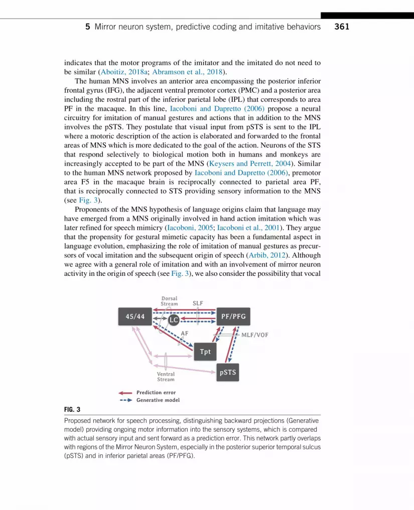
The human MNS involves an anterior area encompassing the posterior inferior
frontal gyrus (IFG), the adjacent ventral premotor cortex (PMC) and a posterior area
including the rostral part of the inferior parietal lobe (IPL) that corresponds to area
PF in the macaque. In this line, Iacoboni and Dapretto (2006) propose a neural
circuitry for imitation of manual gestures and actions that in addition to the MNS
involves the pSTS. They postulate that visual input from pSTS is sent to the IPL
where a motoric description of the action is elaborated and forwarded to the frontal
areas of MNS which is more dedicated to the goal of the action. Neurons of the STS
that respond selectively to biological motion both in humans and monkeys are
increasingly accepted to be part of the MNS (Keysers and Perrett, 2004). Similar
to the human MNS network proposed by Iacoboni and Dapretto (2006), premotor
area F5 in the macaque brain is reciprocally connected to parietal area PF,
that is reciprocally connected to STS providing sensory information to the MNS
(see Fig. 3).
Proponents of the MNS hypothesis of language origins claim that language may
have emerged from a MNS originally involved in hand action imitation which was
later refined for speech mimicry (Iacoboni, 2005; Iacoboni et al., 2001). They argue
that the propensity for gestural mimetic capacity has been a fundamental aspect in
language evolution, emphasizing the role of imitation of manual gestures as precur-
sors of vocal imitation and the subsequent origin of speech (Arbib, 2012). Although
we agree with a general role of imitation and with an involvement of mirror neuron
activity in the origin of speech (see Fig. 3), we also consider the possibility that vocal
FIG. 3
Proposed network for speech processing, distinguishing backward projections (Generative
model) providing ongoing motor information into the sensory systems, which is compared
with actual sensory input and sent forward as a prediction error. This network partly overlaps
with regions of the Mirror Neuron System, especially in the posterior superior temporal sulcus
(pSTS) and in inferior parietal areas (PF/PFG).
3615 Mirror neuron system, predictive coding and imitative behaviors

imitation leading to early speech is an ancient character in the human lineage, and
may have developed together, rather than deriving from, manual imitation. Vocal
imitation is observed very early in human infancy (12–20 weeks) and can be ob-
served in other highly social species that lack grasping abilities, indicating that
the latter is not a requisite for the former (Abramson et al., 2018; Kuhl and
Meltzoff, 1996). In this sense, a vocal mirror circuit may have developed in our
ancestors and other vocal learning species, in parallel or independently to a grasping
mirror neuron circuit (Aboitiz, 2017, 2018a).
5.2 Predictive coding and imitative behaviorsSome years ago, Kilner et al. (2007) proposed a feedforward recognition model in
which the observation of an action results in “the firing of neurons in the STS, which
drives activity in area PF, which in turn drives activity in area F5”, suggesting that
low-level visual and kinematic information are transformed in high-level represen-
tations of intentions subjacent to the action performed. The authors suggest that
MNS function can be understood within a predictive coding framework appealing
to a statistical approach called empirical Bayesian statistics. Predictive coding relies
on the bidirectional interactions between the frontal and motor areas mentioned
above (see Fig. 3), that exert strong top-down influences on sensory cortices.
These projections serve to anticipate the perception of the executed action, minimiz-
ing prediction errors and refining the subsequent motor sequences (Aboitiz, 2017;
Kilner et al., 2007; Rauschecker, 2012). In the domain of action perception, prior
contextual knowledge makes it possible to construct a representation of the goals
and intentions of the person performing the action and therefore predict her motor
commands and kinematics on the basis of their resonance in the observer’s own
motor system (Kilner et al., 2007). The comparison between the observed and the
predicted kinematics permit to elaborate a prediction error that is used to update
our representation of other’s actions.
Fig. 3 depicts a proposed scheme for a predictive coding network anchored in the
speech circuit rather than in the visuo-manual gestural circuit. The blue arrows are
hypothesized to be re-afferent connections that send copies of motor commands back
to the pSTS and sensory regions, that are matched with the sensory representation of
the observed behavior which may be one’s own or others’ (i.e., “Am I actually doing
what I have seen?”). In audio-visual speech interactions, there is no visual informa-
tion about one’s own mouth movements, and the observation of other’s gestures
during social interactions becomes particularly relevant (Ray and Heyes, 2011).
During face-to-face interaction, humans spontaneously and unconsciously mimic
a variety of behaviors, a phenomenon called automatic mimicry. These imitative
behaviors emerge early in ontogenetic development and are thought to be crucial
for predictive coding and error minimizing during speech perception (Cook et al.,
2014; Pickering and Garrod, 2013; Schomers and Pulverm€uller, 2016; Skeide and
Friederici, 2016; Skipper et al., 2017). In this context, we previously mentioned
the findings of Kumar and collaborators (Kumar et al., 2016) showing a much more
362 CHAPTER 13 Origin and evolution of human speech

robust connectivity between the laryngeal motor cortex and the inferior parietal lobe
(particularly area PF) in the human than in the macaque. This may be anatomical
evidence of a strengthened predictive coding circuit for learned vocalizations
(Hickok, 2016), that probably conveys both auditory and visual information, and in-
cludes mirror neurons as well.
We have mentioned that proponents of the MNS hypothesis claim that manual
imitative gestures triggered the development of vocal mimicry in early humans.
In contrast, in the trimodal repertoire model proposed in this chapter, the emphasis
is rather put on the auditory-visual-vocal circuits and the capacity to imitate vocal-
izations supported by the observation of others’ orofacial gestures. Noteworthy, mir-
ror neurons have been demonstrated to fire not only in response to executed or seen
actions but also in response to heard actions (Keysers et al., 2003). As mentioned
above, Hage and Nieder (2015) reported that single neurons located in the VLPFC
were modulated both when monkeys produced or listened to conspecific vocaliza-
tions, a feature strongly reminiscent of mirror neuron properties. These results indi-
cate that, in addition to the integration of audio-visual (i.e., phoneme-viseme) inputs,
the VLPFC seems well positioned for the integration of audio-motoric input (i.e.,
phoneme-articuleme) turning it into a critical region for the evolution of a complex
audio-visual-motoric integration that may have contributed to the emergence of
speech in humans (Hage and Nieder, 2015). Analogously, from an ontogenetic
perspective the existence of those imitative behaviors seems to be relevant for speech
perception and the construction of the phonologic repertoire during the first year of
life. A behavioral study conducted on 6 months-old infants who used teething toys
in order to interfere with their imitative orofacial movements, showed that the auditory
discrimination between nonnative phonemes was impaired in these subjects compared
to infants not using teething toys. The outcomes of this experimental manipulation on
infant’s performance provides evidence of how the sensorimotor information from the
articulators influences speech perception (Bruderer et al., 2015).
6 DiscussionStructural, functional and behavioral evidence comparing the evolution of the vocal
system of humans and NPHs and their neural circuits have been reviewed along this
chapter.We first mentioned the anatomical parallelism and overlap between auditory
and visual systems which are organized in a dorsal and a ventral pathway and are
present in both species. The dorsal stream projects into area 44 conveying phonolog-
ical information from the posterior auditory cortex and the anterior inferior parietal
lobe, as well as sensorimotor inputs from premotor and motor cortices. In this
sense, the dorsal pathway has an important role in the transduction of phoneme into
articuleme. On the other hand, the projections of auditory and visual ventral streams
overlap in the VLPFC where voices and faces are associated. Homologies and dif-
ferences between human and NHP neuroanatomy have also been addressed to foster
our understanding of the evolutionary trajectory that may have led to the emergence
3636 Discussion
of speech. First, we saw that the structural connectivity of the dorsal stream via the
AF and the SLF seems to have strengthened frommonkeys to humans since it is more
robust and direct in humans compared to NHPs. Likewise, behavioral and functional
connectivity analyses suggest that NHPs are more likely to rely on the ventral
auditory stream to associate vocalizations and orofacial gestures whereas the human
lineage took advantages from the growth of the dorsal pathway to increase vocal
working memory capacity and amplify the vocal repertoire (Aboitiz, 2017,
2018b). Finally, the auditory-vocal network is claimed to have evolved in step with
the control of the larynx via direct and robust projections to the nucleus ambiguous.
Special attention has been given to the visual processing of speech in this chapter.
Evidence consistently reports that not only visual but also auditory and motor cor-
tices are engaged during the perception of orofacial gestures and that the access to
congruent or incongruent visual speech cues has a large influence on auditory pro-
cessing. The latter leads us to propose the hypothesis of a trimodal repertoire where
auditory, visual and motor aspects of speech are transduced; the smallest units of
these modalities being the phonemes, the visemes and the articulemes, respectively.Observation and imitation are proposed as tentative general mechanisms fostering
the construction of the trimodal repertoire. The presence of neural populations dis-
playing mirror properties within the traditional speech circuitry in the left hemi-
sphere may support the associative learning of observed orofacial gestures and
the motoric sequence required to produce that gesture. Moreover, the reciprocity
of the projections permits to generate predictions and minimize errors during speech
perception.
Alongside with the evolution of the brain matter, the human mind has also
evolved. The increasing complexity of ancestral societies urged adaptive and selec-
tive behaviors. The cortical and subcortical adaptations for language are necessary
but may not be sufficient to fully explain the origin of speech. The need to organize
communication within highly social communities is likely to have contributed to the
emergence of increasingly richer vocalizations. Consistent with this idea, the com-
plexity of vocalizations in birds is associated with more elaborate social behavior
such as collaborative breeding (Leighton, 2017) and highly social mammals like
cetaceans, elephants or orangutans are able to imitate the human voice, which points
to an increasing relevance of vocal communication as social interactions become
more complex (Aboitiz, 2018a,b).
Finally, we would like to stress that human communication is, and has always
been, a multimodal process in which gestures, vision, voice and audition play a role.
The gestural theory of language origins views learned gestures like pantomimes and
so-called “protosigns” as the initial channel for symbolic human communication,
which through some unknown process triggered the acquisition of vocal mimicry
and the development of speech (Arbib, 2012). However, there is no compelling
reason or evidence indicating that vocal learning had to wait for symbolic gestural
communication to develop first. As we have said, other species have developed vocal
learning capacity without need of a hand gestural repertoire. In other words, our point
is not whether the first symbolic messages were first carried by gestures or by voice
364 CHAPTER 13 Origin and evolution of human speech

(a purely speculative issue for which that we may never have a definite answer), but
rather to track the evolution of speech mechanisms from ancestral multimodal
networks in nonhuman primates. Considering this, there is another possible scenario
where vocal imitation appeared early in the genus Homo (or even in australopithe-
cines), and coexisted with gestural communication for a long time. According to this
view, in these animals vocal mimicry was more related to social bonding, particu-
larly between mother and child, rather than transmitting referential contents. This
kind of communication was supported by the trimodal visual-auditory-motor reper-
toire we have proposed in Fig. 2. At some undetermined point in our history, early
humans started making reference to events using gestural pantomimes, vocal imita-
tions, vocal alarms like those of vervet monkeys and any other possible way to
communicate simple meanings (Aboitiz, 2018a,b). However, the progressive devel-
opment of the dorsal auditory-vocal pathway enabled them to displace the gestural
modality and to establish early speech as the main communication channel. This
perspective has the advantage of providing a preexisting visual-auditory-vocal
scaffolding from which the speech circuitry could later emerge to transmit
symbolic content, an aspect that is in our view neglected by the gestural theory
of language origins.
AcknowledgmentsSpecial thanks to Isabel Guerrero for producing the illustrations. This research was supported
in part by FONDECYT Grant 1160258.
ReferencesAboitiz, F., 2012. Gestures, vocalizations, and memory in language origins. Front.
Evol. Neurosci. 4, 2. https://doi.org/10.3389/fnevo.2012.00002.
Aboitiz, F., 2017. A Brain for Speech. Palgrave Macmillan, UK.
Aboitiz, F., 2018a. A brain for speech. Evolutionary continuity in primate and human auditory-
vocal processing. Front. Neurosci. 12, 174. https://doi.org/10.3389/fnins.2018.00174.
Aboitiz, F., 2018b. Voice gesture and working memory in the emergence of speech. Interact.
Stud. 19 (1–2), 70–85.Aboitiz, F., Garcıa, R., 1997. The evolutionary origin of the language areas in the human brain.
A neuroanatomical perspective. Brain Res. Rev. 25 (3), 381–396.Abramson, J.Z., Hernandez-Lloreda, M.V., Garcıa, L., Colmenares, F., Aboitiz, F., Call, J.,
2018. Imitation of novel conspecific and human speech sounds in the killer whale
(Orcinus orca). Proc. R. Soc. B 285 (1871), 20172171.
Arbib, M.A., 2005. From monkey-like action recognition to human language: an evolutionary
framework for neurolinguistics. Behav. Brain Sci. 28 (2), 105–124.Arbib, M.A., 2012. How the Brain Got Language: The Mirror System Hypothesis. vol. 16.
Oxford University Press.
Arcaro, M.J., Livingstone, M.S., 2017. Retinotopic organization of scene areas in macaque
inferior temporal cortex. J. Neurosci. 37 (31), 7373–7389.
365References
Auer, E.T., Bernstein, L.E., 2007. Enhanced visual speech perception in individuals with
early-onset hearing impairment. J. Speech Lang. Hear. Res. 50 (5), 1157–1165.Barenholtz, E., Mavica, L., Lewkowicz, D.J., 2016. Language familiarity modulates relative
attention to the eyes and mouth of a talker. Cognition 147, 100–105.Bear, H.L., Harvey, R., 2017. Phoneme-to-viseme mappings: the good, the bad, and the ugly.
Speech Commun. 95, 40–67.Beauchamp, M.S., 2015. The social mysteries of the superior temporal sulcus. Trends Cogn.
Sci. 19 (9), 489–490.Bedny, M., Richardson, H., Saxe, R., 2015. “Visual” cortex responds to spoken language in
blind children. J. Neurosci. 35 (33), 11674–11681.Bernstein, L.E., Liebenthal, E., 2014. Neural pathways for visual speech perception. Front.
Neurosci. 8, 386.
Bernstein, L.E., Tucker, P.E., Demorest, M.E., 2000. Speech perception without hearing.
Percept. Psychophys. 62 (2), 233–252.Besle, J., Fischer, C., Bidet-Caulet, A., Lecaignard, F., Bertrand, O., Giard, M.H., 2008. Visual
activation and audiovisual interactions in the auditory cortex during speech perception:
intracranial recordings in humans. J. Neurosci. 28 (52), 14301–14310.Binder, J.R., Desai, R.H., 2011. The neurobiology of semantic memory. Trends Cogn. Sci.
15 (11), 527–536.Binder, J.R., Desai, R.H., Graves, W.W., Conant, L.L., 2009. Where is the semantic system?
A critical review and meta-analysis of 120 functional neuroimaging studies. Cereb. Cortex
19 (12), 2767–2796.Blank, H., von Kriegstein, K., 2013. Mechanisms of enhancing visual–speech recognition by
prior auditory information. NeuroImage 65, 109–118.Brennan, J., Nir, Y., Hasson, U., Malach, R., Heeger, D.J., Pylkk€anen, L., 2012. Syntactic
structure building in the anterior temporal lobe during natural story listening. Brain Lang.
120 (2), 163–173. https://doi.org/10.1016/j.bandl.2010.04.002.Bruderer, A.G., Danielson, D.K., Kandhadai, P., Werker, J.F., 2015. Sensorimotor influences
on speech perception in infancy. Proc. Natl. Acad. Sci. U. S. A. 112 (44), 13531–13536.Calvert, G.A., Campbell, R., 2003. Reading speech from still and moving faces: the neural
substrates of visible speech. J. Cogn. Neurosci. 15 (1), 57–70.Calvert, G.A., Bullmore, E.T., Brammer, M.J., Campbell, R., Williams, S.C., McGuire, P.K.,
Woodruff, P.W., Iversen, S.D., David, A.S., 1997. Activation of auditory cortex during
silent lipreading. Science 276 (5312), 593–596.Catani, M., Bambini, V., 2014. A model for social communication and language evolution and
development (SCALED). Curr. Opin. Neurobiol. 28, 165–171. https://doi.org/10.1016/j.conb.2014.07.018.
Cook, R., Bird, G., Catmur, C., Press, C., Heyes, C., 2014. Mirror neurons: from origin to func-
tion. Behav. Brain Sci. 37 (2), 177–192.Dichter, B.K., Breshears, J.D., Leonard, M.K., Chang, E.F., 2018. The control of vocal pitch in
human laryngeal motor cortex. Cell 174 (1), 21–31.e9. https://doi.org/10.1016/
j.cell.2018.05.016.
Diehl, M.M., Romanski, L.M., 2012. Representation and integration of faces and vocalizations
in the primate ventral prefrontal cortex. In: Integrating Face and Voice in Person Percep-
tion. Springer, New York, pp. 45–69. https://doi.org/10.1007/978-1-4614-3585-3_3.Dole, M., M�eary, D., Pascalis, O., 2017. Modifications of visual field asymmetries for face
categorization in early deaf adults: a study with chimeric faces. Front. Psychol. 8, 30.
366 CHAPTER 13 Origin and evolution of human speech
Dubois, C., Otzenberger, H., Gounot, D., Sock, R., Metz-Lutz, M.N., 2012. Visemic
processing in audiovisual discrimination of natural speech: a simultaneous fMRI–EEGstudy. Neuropsychologia 50 (7), 1316–1326.
Duffau, H., 2018. The error of Broca: from the traditional localizationist concept to a connec-
tomal anatomy of human brain. J. Chem. Neuroanat. 89, 73–81. https://doi.org/10.1016/j.jchemneu.2017.04.003.
Dunn, J.C., Smaers, J.B., 2018. Neural correlates of vocal repertoire in primates. Front.
Neurosci. 12, 534.
Eichert N., Verhagen L., Folloni D., Jbabdi S., Khrapitchev A.A., Sibson N.R.,…Mars R.B.,
in press, What is special about the human arcuate fasciculus? Lateralization projections,
and expansion, Cortex https://doi.org/10.1016/j.cortex.2018.05.005.
Ferpozzi, V., Fornia, L., Montagna, M., Siodambro, C., Castellano, A., Borroni, P., …
Cerri, G., 2018. Broca’s area as a pre-articulatory phonetic encoder: gating the motor
program. Front. Hum. Neurosci. 12, 64. https://doi.org/10.3389/fnhum.2018.00064.
Fleagle, J.G., 2013. Primate Adaptation and Evolution. Academic Press.
Flinker, A., Korzeniewska, A., Shestyuk, A.Y., Franaszczuk, P.J., Dronkers, N.F.,
Knight, R.T., Crone, N.E., 2015. Redefining the role of Broca’s area in speech. Proc. Natl.
Acad. Sci. U. S. A. 112 (9), 2871–2875. https://doi.org/10.1073/pnas.1414491112.Freud, E., Plaut, D.C., Behrmann, M., 2016. ‘What’ is happening in the dorsal visual
pathway. Trends Cogn. Sci. 20 (10), 773–784. https://doi.org/10.1016/j.tics.2016.
08.003.
Friederici, A.D., 2011. The brain basis of language processing: from structure to function.
Physiol. Rev. 91 (4), 1357–1392. https://doi.org/10.1152/physrev.00006.2011.Friederici, A.D., 2016. Evolution of the neural language network. Psychon. Bull. Rev. 24 (1),
41–47. https://doi.org/10.3758/s13423-016-1090-x.Friederici, A.D., Bahlmann, J., Heim, S., Schubotz, R.I., Anwander, A., 2006. The brain
differentiates human and non-human grammars: functional localization and structural
connectivity. Proc. Natl. Acad. Sci. U. S. A. 103 (7), 2458–2463.Garcıa, R.R., Zamorano, F., Aboitiz, F., 2014. From imitation to meaning: circuit plasticity
and the acquisition of a conventionalized semantics. Front. Hum. Neurosci. 8, 605.
https://doi.org/10.3389/fnhum.2014.00605.
Ghazanfar, A.A., Takahashi, D.Y., Mathur, N., Fitch,W.T., 2012. Cineradiography of monkey
lip-smacking reveals putative precursors of speech dynamics. Curr. Biol. 22 (13),
1176–1182.Glenberg, A.M., Gallese, V., 2012. Action-based language: a theory of language acquisition,
comprehension, and production. Cortex 48 (7), 905–922.Goodale, M.A., Milner, A.D., 1992. Separate visual pathways for perception and action.
Trends Neurosci. 15 (1), 20–25. https://doi.org/10.1016/0166-2236(92)90344-8.Hage, S.R., Nieder, A., 2015. Audio-vocal interaction in single neurons of the monkey
ventrolateral prefrontal cortex. J. Neurosci. 35 (18), 7030–7040. https://doi.org/10.1523/jneurosci.2371-14.2015.
Hauswald, A., Lithari, C., Collignon, O., Leonardelli, E., Weisz, N., 2018. A visual cortical
network for deriving phonological information from intelligible lip movements. Curr.
Biol. 28 (9), 1453–1459.Hickok, G., 2016. A cortical circuit for voluntary laryngeal control: implications for the
evolution language. Psychon. Bull. Rev. 24 (1), 56–63. https://doi.org/10.3758/s13423-016-1100-z.
367References
Hickok, G., Poeppel, D., 2004. Dorsal and ventral streams: a framework for understanding
aspects of the functional anatomy of language. Cognition 92 (1–2), 67–99. https://doi.org/10.1016/j.cognition.2003.10.011.
Hickok, G., Poeppel, D., 2007. The cortical organization of speech processing. Nat. Rev.
Neurosci. 8 (5), 393–402. https://doi.org/10.1038/nrn2113.Hillairet de Boisferon, A., Tift, A.H., Minar, N.J., Lewkowicz, D.J., 2017. Selective attention
to a talker’s mouth in infancy: role of audiovisual temporal synchrony and linguistic
experience. Dev. Sci. 20 (3), e12381.
Hirata, Y., Kelly, S.D., 2010. Effects of lips and hands on auditory learning of second-
language speech sounds. J. Speech Lang. Hear. Res. 53 (2), 298–310.Iacoboni, M., 2005. Neural mechanisms of imitation. Curr. Opin. Neurobiol. 15 (6), 632–637.Iacoboni, M., Dapretto, M., 2006. The mirror neuron system and the consequences of its
dysfunction. Nat. Rev. Neurosci. 7 (12), 942.
Iacoboni, M., Koski, L.M., Brass, M., Bekkering, H., Woods, R.P., Dubeau, M.C., …
Rizzolatti, G., 2001. Reafferent copies of imitated actions in the right superior temporal
cortex. Proc. Natl. Acad. Sci. U. S. A. 98 (24), 13995–13999.Janssens, T., Zhu, Q., Popivanov, I.D., Vanduffel, W., 2014. Probabilistic and single-subject
retinotopic maps reveal the topographic organization of face patches in the macaque
cortex. J. Neurosci. 34 (31), 10156–10167. https://doi.org/10.1523/jneurosci.2914-13.2013.J€urgens, U., 2009. The neural control of vocalization in mammals: a review. J. Voice 23 (1),
1–10. https://doi.org/10.1016/j.jvoice.2007.07.005.Kaas, J.H., Hackett, T.A., 1999. What and where processing in auditory cortex. Nat. Neurosci.
2 (12), 1045–1047. https://doi.org/10.1038/15967.Keysers, C., Perrett, D.I., 2004. Demystifying social cognition: a Hebbian perspective. Trends
Cogn. Sci. 8 (11), 501–507.Keysers, C., Kohler, E., Umilta, M.A., Nanetti, L., Fogassi, L., Gallese, V., 2003. Audiovisual
mirror neurons and action recognition. Exp. Brain Res. 153 (4), 628–636.Kilner, J.M., Friston, K.J., Frith, C.D., 2007. Predictive coding: an account of the mirror
neuron system. Cogn. Process. 8 (3), 159–166.Kuhl, P.K., Meltzoff, A.N., 1996. Infant vocalizations in response to speech: vocal imitation
and developmental change. J. Acoust. Soc. Am. 100 (401), 2425–2438.Kumar, V., Croxson, P.L., Simonyan, K., 2016. Structural organization of the laryngeal motor
cortical network and its implication for evolution of speech production. J. Neurosci.
36 (15), 4170–4181. https://doi.org/10.1523/jneurosci.3914-15.2016.Lane, C., Kanjlia, S., Omaki, A., Bedny, M., 2015. “Visual” cortex of congenitally blind adults
responds to syntactic movement. J. Neurosci. 35 (37), 12859–12868.Langacker, R.W., 2012. Essentials of Cognitive Grammar. Oxford University Press.
Leighton, G.M., 2017. Cooperative breeding influences the number and type of vocalizations
in avian lineages. Proc. R. Soc. B 284 (1868), 20171508.
Lewkowicz, D.J., Hansen-Tift, A.M., 2012. Infants deploy selective attention to the mouth of a
talking face when learning speech. Proc. Natl. Acad. Sci. U. S. A. 109 (5), 1431–1436.Lieberman, P., 1993. Uniquely Human: The Evolution of Speech, Thought, and Selfless
Behavior. Harvard University Press.
Lieberman, P., 2015. The evolution of language. In: Goldstein, S., Princiotta, D., Naglieri, J.A.
(Eds.), Handbook of Intelligence: Evolutionary Theory, Historical Perspective, and
Current Concepts. Springer, New York, pp. 47–64.Long, M.A., Katlowitz, K.A., Svirsky, M.A., Clary, R.C., Byun, T.M.A., Majaj, N., …
Greenlee,W, J.D., 2016. Functional segregation of cortical regions underlying speech timing
and articulation. Neuron 89 (6), 1187–1193. https://doi.org/10.1016/j.neuron.2016.01.032.
368 CHAPTER 13 Origin and evolution of human speech
McGurk, H., MacDonald, J., 1976. Hearing lips and seeing voices. Nature 264 (5588), 746.
Messum, P., Howard, I.S., 2015. Creating the cognitive form of phonological units: the speech
sound correspondence problem in infancy could be solved by mirrored vocal interactions
rather than by imitation. J. Phon. 53, 125–140.Miki, K., Watanabe, S., Kakigi, R., Puce, A., 2004. Magnetoencephalographic study of occi-
pitotemporal activity elicited by viewing mouth movements. Clin. Neurophysiol. 115 (7),
1559–1574.Mitchell, T.V., Letourneau, S.M., Maslin, M.C., 2013. Behavioral and neural evidence of
increased attention to the bottom half of the face in deaf signers. Restor. Neurol. Neurosci.
31 (2), 125–139.Morrill, G.V., 2012. Type Logical Grammar: Categorial Logic of Signs. Springer Science &
Business Media.
Mugler, E.M., Tate, M.C., Livescu, K., Templer, J.W., Goldrick, M.A., Slutzky, M.W., 2018.
Differential representation of articulatory gestures and phonemes in precentral and inferior
frontal gyri. J. Neurosci. 38 (46), 9803–9813.Navarra, J., Soto-Faraco, S., 2007. Hearing lips in a second language: visual articulatory
information enables the perception of second language sounds. Psychol. Res. 71 (1), 4–12.Neubert, F.-X., Mars, R.B., Thomas, A.G., Sallet, J., Rushworth, M.F.S., 2014. Comparison of
human ventral frontal cortex areas for cognitive control and language with areas in mon-
key frontal cortex. Neuron 81 (3), 700–713. https://doi.org/10.1016/j.neuron.2013.11.012.Nguyen, N., Delvaux, V., 2015. Role of imitation in the emergence of phonological systems.
J. Phon. 53, 46–54.Papoutsi, M., de Zwart, J.A., Jansma, J.M., Pickering, M.J., Bednar, J.A., Horwitz, B.,
2009. From phonemes to articulatory codes: an fMRI study of the role of Brocas area
in speech production. Cereb. Cortex 19 (9), 2156–2165. https://doi.org/10.1093/cercor/bhn239.
Paris, T., Kim, J., Davis, C., 2013. Visual speech form influences the speed of auditory speech
processing. Brain Lang. 126 (3), 350–356.P�erez-Pereira, M., 2006. Language development in blind children. Editor in-Chief In: Brown, K.
(Ed.), Encyclopedia of Language & Linguistics, vol. 6. Elsevier, Oxford, pp. 357–361.Petrides, M., 2005. Lateral prefrontal cortex: architectonic and functional organization. Philos.
Trans. R. Soc. Lond. B 360 (1456), 781–795.Petrides, M., 2014. Neuroanatomy of Language Regions of the Human Brain. Academic Press,
New York.
Petrides, M., Cadoret, G., Mackey, S., 2005. Orofacial somatomotor responses in the macaque
monkey homologue of Broca’s area. Nature 435 (7046), 1235.
Pickering, M.J., Garrod, S., 2013. An integrated theory of language production and compre-
hension. Behav. Brain Sci. 36 (4), 329–347.Pimperton, H., Ralph-Lewis, A., MacSweeney, M., 2017. Speechreading in deaf adults with
cochlear implants: evidence for perceptual compensation. Front. Psychol. 8, 106.
Puce, A., Allison, T., Bentin, S., Gore, J.C., McCarthy, G., 1998. Temporal cortex activation in
humans viewing eye and mouth movements. J. Neurosci. 18 (6), 2188–2199.Pulverm€uller, F., Fadiga, L., 2010. Active perception: sensorimotor circuits as a cortical basis
for language. Nat. Rev. Neurosci. 11 (5), 351.
Rauschecker, J.P., 2012. Ventral and dorsal streams in the evolution of speech and language.
Front. Evol. Neurosci. 4, 7. https://doi.org/10.3389/fnevo.2012.00007.
Rauschecker, J.P., 2018a. Where did language come from? Precursor mechanisms in nonhu-
man primates. Curr. Opin. Behav. Sci. 21, 195–204. https://doi.org/10.1016/j.cobeha.2018.06.003.
369References

Rauschecker, J.P., 2018b.Where when, and how: are they all sensorimotor? Towards a unified
view of the dorsal pathway in vision and audition. Cortex 98, 262–268. https://doi.org/10.1016/j.cortex.2017.10.020.
Rauschecker, J.P., Scott, S.K., 2009. Maps and streams in the auditory cortex: nonhuman pri-
mates illuminate human speech processing. Nat. Neurosci. 12 (6), 718–724. https://doi.org/10.1038/nn.2331.
Ray, E., Heyes, C., 2011. Imitation in infancy: the wealth of the stimulus. Dev. Sci. 14 (1),
92–105.Riedel, P., Ragert, P., Schelinski, S., Kiebel, S.J., von Kriegstein, K., 2015. Visual face-
movement sensitive cortex is relevant for auditory-only speech recognition. Cortex
68, 86–99.Rilling, J.K., Glasser, M.F., Preuss, T.M., Ma, X., Zhao, T., Hu, X., Behrens, T.E.J., 2008. The
evolution of the arcuate fasciculus revealed with comparative DTI. Nat. Neurosci. 11 (4),
426–428. https://doi.org/10.1038/nn2072.Rilling, J., Glasser, M.F., Jbabdi, S., Andersson, J., Preuss, T.M., 2012. Continuity,
divergence, and the evolution of brain language pathways. Front. Evol. Neurosci. 3, 11.
Rizzolatti, G., Arbib, M.A., 1998. Language within our grasp. Trends Neurosci. 21 (5),
188–194.Rizzolatti, G., Craighero, L., 2004. The mirror-neuron system. Annu. Rev. Neurosci.
27, 169–192.Rizzolatti, G., Fadiga, L., Gallese, V., Fogassi, L., 1996. Premotor cortex and the recognition
of motor actions. Cogn. Brain Res. 3 (2), 131–141.R€oder, B., Stock, O., Bien, S., Neville, H., R€osler, F., 2002. Speech processing activates visual
cortex in congenitally blind humans. Eur. J. Neurosci. 16 (5), 930–936.Romanski, L.M., 2007. Representation and integration of auditory and visual stimuli in the
primate ventral lateral prefrontal cortex. Cereb. Cortex 17 (Suppl. 1), i61–i69. https://doi.org/10.1093/cercor/bhm099.
Romanski, L.M., 2012. Integration of faces and vocalizations in ventral prefrontal cortex:
implications for the evolution of audiovisual speech. Proc. Natl. Acad. Sci. U. S. A.
109 (Suppl. 1), 10717–10724. https://doi.org/10.1073/pnas.1204335109.Romanski, L.M., Bates, J.F., Goldman-Rakic, P.S., 1999. Auditory belt and parabelt projec-
tions to the prefrontal cortex in the rhesus monkey. J. Comp. Neurol. 403 (2), 141–157.https://doi.org/10.1002/(sici)1096-9861(19990111)403:2<141::aid-cne1>3.0.co;2-v.
Ross, L.A., Saint-Amour, D., Leavitt, V.M., Molholm, S., Javitt, D.C., Foxe, J.J., 2007.
Impaired multisensory processing in schizophrenia: deficits in the visual enhancement
of speech comprehension under noisy environmental conditions. Schizophr. Res.
97 (1–3), 173–183.Saur, D., Kreher, B.W., Schnell, S., Kummerer, D., Kellmeyer, P., Vry, M.-S., …Weiller, C.,
2008. Ventral and dorsal pathways for language. Proc. Natl. Acad. Sci. U. S. A. 105 (46),
18035–18040. https://doi.org/10.1073/pnas.0805234105.Schomers, M.R., Pulverm€uller, F., 2016. Is the sensorimotor cortex relevant for speech per-
ception and understanding? An integrative review. Front. Hum. Neurosci. 10, 435.
Sebastian-Gall�es, N., Albareda-Castellot, B., Weikum, W.M., Werker, J.F., 2012. A bilingual
advantage in visual language discrimination in infancy. Psychol. Sci. 23 (9), 994–999.Shepherd, S.V., Freiwald, W.A., 2018. Functional networks for social communication in the
macaque monkey. Neuron 99 (2), 413–420.Sheth, B.R., Young, R., 2016. Two visual pathways in primates based on sampling of space:
exploitation and exploration of visual information. Front. Integr. Neurosci. 10, 37. https://
doi.org/10.3389/fnint.2016.00037.
370 CHAPTER 13 Origin and evolution of human speech

Silson, E.H., Reynolds, R.C., Kravitz, D.J., Baker, C.I., 2018. Differential sampling of visual
space in ventral and dorsal early visual cortex. J. Neurosci. 38 (9), 2294–2303. https://doi.org/10.1523/jneurosci.2717-17.2018.
Skeide, M.A., Friederici, A.D., 2016. The ontogeny of the cortical language network. Nat.
Rev. Neurosci. 17 (5), 323–332. https://doi.org/10.1038/nrn.2016.23.Skipper, J.I., Nusbaum, H.C., Small, S.L., 2005. Listening to talking faces: motor cortical
activation during speech perception. NeuroImage 25 (1), 76–89.Skipper, J.I., Devlin, J.T., Lametti, D.R., 2017. The hearing ear is always found close to the
speaking tongue: review of the role of the motor system in speech perception. Brain Lang.
164, 77–105.Sugihara, T., Diltz, M.D., Averbeck, B.B., Romanski, L.M., 2006. Integration of auditory
and visual communication information in the primate ventrolateral prefrontal cortex.
J. Neurosci. 26 (43), 11138–11147. https://doi.org/10.1523/jneurosci.3550-06.2006.Sumby, W.H., Pollack, I., 1954. Visual contribution to speech intelligibility in noise.
J. Acoust. Soc. Am. 26 (2), 212–215.Tenenbaum, E.J., Shah, R.J., Sobel, D.M., Malle, B.F., Morgan, J.L., 2013. Increased focus on
the mouth among infants in the first year of life: a longitudinal eye-tracking study. Inf.
Dent. 18 (4), 534–553.Trevarthen, C.B., 1968. Two mechanisms of vision in primates. Psychol. Forsch. 31 (4),
299–337. https://doi.org/10.1007/bf00422717.Tsang, T., Atagi, N., Johnson, S.P., 2018. Selective attention to the mouth is associated with
expressive language skills in monolingual and bilingual infants. J. Exp. Child Psychol.
169, 93–109.Tyler, L.K., Marslen-Wilson, W.D., Randall, B., Wright, P., Devereux, B.J., Zhuang, J., …
Stamatakis, E.A., 2011. Left inferior frontal cortex and syntax: function structure and be-
haviour in patients with left hemisphere damage. Brain 134 (2), 415–431. https://doi.org/10.1093/brain/awq369.
VanWassenhove, V., Grant, K.W., Poeppel, D., 2005. Visual speech speeds up the neural pro-
cessing of auditory speech. Proc. Natl. Acad. Sci. U. S. A. 102 (4), 1181–1186.Watkins, K.E., Shakespeare, T.J., O’Donoghue, M.C., Alexander, I., Ragge, N., Cowey, A.,
Bridge, H., 2013. Early auditory processing in area V5/MT+ of the congenitally blind
brain. J. Neurosci. 33 (46), 18242–18246.Weikum, W.M., Vouloumanos, A., Navarra, J., Soto-Faraco, S., Sebastian-Gall�es, N.,
Werker, J.F., 2007. Visual language discrimination in infancy. Science 316 (5828), 1159.
Wilson, S.M., Galantucci, S., Tartaglia, M.C., Rising, K., Patterson, D.K., Henry, M.L., …
Gorno-Tempini, M.L., 2011. Syntactic processing depends on dorsal language tracts.
Neuron 72 (2), 397–403. https://doi.org/10.1016/j.neuron.2011.09.014.
Further readingIacoboni, M., Hurley, S., Chater, N., 2005. Perspectives on Imitation: From Neuroscience to
Social Science. vol. 1. MIT Press.
371Further reading