nitric oxide generation in vicia faba phloem cells reveals them to be sensitive detectors as well as...
TRANSCRIPT
634 www.newphytologist.org
Research
Blackwell Publishing Ltd
Nitric oxide generation in Vicia faba phloem cells reveals them to be sensitive detectors as well as possible systemic transducers of stress signals
Frank Gaupels1,2,4, Alexandra C. U. Furch2, Torsten Will2, Luis A. J. Mur3, Karl-Heinz Kogel1 and Aart J. E. van Bel2
1Institute of Phytopathology and Applied Zoology, IFZ, Heinrich-Buff-Ring 26-32, D-35392 Gießen and 2Plant Cell Biology Research Group,
Institute of General Botany, Senckenbergstrasse 17, D-35390 Gießen, Justus-Liebig-University, Gießen, Germany; 3Institute of Biological Sciences,
University of Wales Aberystwyth, Aberystwyth, Ceredigion SY23 2DA, UK; 4Dipartimento Scientifico e Tecnologico, Università degli Studi di Verona,
Strada le Grazie 15, I-37134 Verona, Italy
Summary
• Vascular tissue was recently shown to be capable of producing nitric oxide (NO),but the production sites and sources were not precisely determined. Here, NOsynthesis was analysed in the phloem of Vicia faba in response to stress- andpathogen defence-related compounds.• The chemical stimuli were added to shallow paradermal cortical cuts in the mainveins of leaves attached to intact plants. NO production in the bare-lying phloemarea was visualized by real-time confocal laser scanning microscopy using theNO-specific fluorochrome 4,5-diaminofluorescein diacetate (DAF-2 DA).• Abundant NO generation in companion cells was induced by 500 µM salicylic acid(SA) and 10 µM hydrogen peroxide (H2O2), but the fungal elicitor chitooctaose wasmuch less effective. Phloem NO production was found to be dependent on Ca2+ andmitochondrial electron transport and pharmacological approaches found evidencefor activity of a plant NO synthase but not a nitrate reductase. DAF fluorescenceincreased most strongly in companion cells and was occasionally observed in phloemparenchyma cells. Significantly, accumulation of NO in sieve elements could bedemonstrated.• These findings suggest that the phloem perceives and produces stress-relatedsignals and that one mechanism of distal signalling involves the production andtransport of NO in the phloem.
Key words: hydrogen peroxide (H2O2), nitric oxide (NO), nitric oxide synthase,pathogen resistance, phloem, salicylic acid, stress, systemic signalling.
New Phytologist (2008) 178: 634–646
© The Authors (2008). Journal compilation © New Phytologist (2008) doi: 10.1111/j.1469-8137.2008.02388.x
Author for correspondence:Frank GaupelsTel: +39 045 802 7063Fax: +39 045 802 7929Email: [email protected]
Received: 24 October 2007Accepted: 7 January 2008
Introduction
Plant responses to abiotic and biotic stress involve the earlygeneration of chemical cues, which can have discrete but alsooverlapping roles in conferring plant stress tolerance. Onecommon stress response is the generation of partiallyreduced oxygen species ( + 2H → H2O2 → 2⋅OH). Elevated
oxidative stress is a feature of the hypersensitive response(HR), a localized programmed cell death associated withresistance to pathogens (Levine et al., 1994), wounding (Orozco-Càrdenas et al., 2001), anoxia (Baxter-Burrell et al., 2003),chilling (Prasad et al., 1994) and thermotolerance (Dat et al.,1998). Other stress signals have been thought to exhibitgreater specificity in their production. Salicylic acid (SA) hasO2
−
© The Authors (2008). Journal compilation © New Phytologist (2008) www.newphytologist.org New Phytologist (2008) 178: 634–646
Research 635
been associated with local resistance mechanisms to biotrophicpathogens based on the HR, and with systemic acquiredresistance (SAR; Ryals et al., 1996). However, subsequentinvestigations have shown much wider roles in plant stressbiology with, for example, SA being involved in abiotic stresssignalling (Clarke et al., 2004; Scott et al., 2004).
Nitric oxide (NO) is a gaseous signal molecule involved inplant reactions to various stresses (for recent reviews, see Neillet al., 2003; del Río et al., 2004; Crawford & Guo, 2005). Forinstance, heat, salt and hyperosmotic stress induce NOproduction in tobacco (Nicotiana tabacum) cell suspensions(Gould et al., 2003). Moreover, NO accumulation uponwounding was detected in Arabidopsis and sweet potato(Ipomoea batatas; Jih et al., 2003; Huang et al., 2004). Theproduction of NO during plant defence against pathogens isalso well documented, with roles during the HR (reviewed byMur et al., 2006) and also in papilla formation in barley/powderymildew (Hordeum vulgare/Blumeria graminis) interactions (Pratset al., 2005). Detailed analyses revealed a tight interaction ofNO and hydrogen peroxide (H2O2) in the HR induced byphytopathogens or pathogen-derived elicitors (Delledonneet al., 1998, 2001; de Pinto et al., 2002). Furthermore, NO isa potent regulator of defence-related gene expression (Delledonneet al., 1998; Durner et al., 1998; Zeidler et al., 2004; Zeieret al., 2004), enzyme activity (Clark et al., 2000; Navarre et al.,2000; Igamberdiev et al., 2006) and phytoalexin production(Modolo et al., 2002; Xu et al., 2005). NO is likely to be amajor modulator of other defence signals with, for example,the initiation of SA and jasmonic acid biosynthesis beinginfluenced by NO (Durner et al., 1998; Xu et al., 2005).
Use of NO synthase (NOS) inhibitors showed that anNOS-like enzyme was activated in plant stress reactions (Foissneret al., 2000; Xu et al., 2005). Recently, a gene for a plant NOSwas identified in Arabidopsis thaliana (Guo et al., 2003).AtNOS1 is involved in induction of defence gene expressionby lipopolysaccharides (Zeidler et al., 2004). However, NOSactivity of AtNOS1 could not be confirmed (and AtNOS1has since been re-designated AtNOA1 – nitric oxide associated);thus, the identity of the plant NOS remains obscure(Zemojtel et al., 2006). Other workers have suggested that nitriteis a source of NO in plants, derived either nonenzymaticallyat high pH or via nitrate reductase (NR) activity (Neill et al.,2003; del Río et al., 2004).
The mobilization of signals from one plant organ toanother is a major theme of plant physiology. In view of itshigh mobility, NO has been suggested to be a systemic stresssignal (Durner & Klessig, 1999; Foissner et al., 2000; Neillet al., 2003; van Bel & Gaupels, 2004). In support of thissuggestion, injection of NO donors into tobacco leavesreduced the size of lesions caused by tobacco mosaic viruson treated and systemic nontreated leaves (Song & Good-man, 2001). Also, local treatment with NOS inhibitors oran NO scavenger attenuated SAR in distal leaves (Song &Goodman, 2001).
Grafting and girdling experiments provided evidence thattranslocation of distal signalling takes place in the sieve tubes(Durrant & Dong, 2004; van Bel & Gaupels, 2004). Usingthe specific NO-sensing fluorochrome 4,5-diaminofluoresceindiacetate (DAF-2 DA), NO was detected in vascular bundlesand was suggested to have functions in senescense, cell walllignification and the salt stress response (Corpas et al., 2004;Gabaldon et al., 2005; Valderrama et al., 2007). In pepper(Capsicum annuum) plants, infection with Phytophtora capsiciinduced NO production more abundantly in vascular bundlesas compared with other tissues (Requena et al., 2005). Signi-ficantly, Rusterucci et al. (2007) recently showed that antisenselines of the S-nitrosoglutathione (GSNO) catabolizing S-nitrosoglutathione reductase (GSNOR) displayed elevatedresistance and constitutive SAR. These workers also observedthat GSNOR was primarily located in companion cells andproposed that inhibition of GSNOR, leading to the accumulationof GSNO, may be an important factor in the generation ofSAR. Despite these hints at a possible role of NO in systemicsignalling, to date detailed reports on inducible NO synthesisor transport in phloem tissue are lacking.
Our goal was to investigate whether phloem tissue is ableto synthesize and/or transport NO as a distal signal. We testedthe ability of the stress signals H2O2 and SA as well as thefungal elicitor chitooctaose to induce NO production in intactphloem tissue. Confocal laser scanning microscopy (CLSM)with DAF-2 DA revealed rapid and strong NO synthesisprimarily in companion cells (CCs) which was dependent oncalcium and could be suppressed by NOS inhibitors. Ourresults further provide indirect evidence for a role of NO insystemic signalling through sieve tubes.
Materials and Methods
Plant material
Potted plants of Vicia faba L. cv. Witkiem major and Cucurbitamaxima Duchesne ex Lam. cv. Gele Centenaar (pumpkin)were grown in a 3 : 3 : 1 mixture of compost, peat and sandin a glasshouse at 60–70% relative humidity, a minimum daytemperature of 22°C and a minimum light period of 14 h.Minimum irradiance of 250 µmol m–2 s–1 at plant level wasmaintained with daylight plus additional lamp light (modelSONT Agro 400 W; Philips, Eindhoven, the Netherlands).Plants 3–5-wk old were used for the experiments.
Chemicals
All chemicals for microscopy were dissolved in loading buffer(2 mm KCl, 1 mm MgCl2, 1 mm CaCl2 and 2.5 mm morpholineethanesulfonic acid (MES), adjusted to pH 5.7 with NaOH).DAF-2 DA (dissolved as a 5 mm stock solution in dimethylsulphoxide (DMSO)), NG-nitro-L-arginine-methyl ester(L-NAME), NG-nitro-D-arginine-methyl ester (D-NAME),
New Phytologist (2008) 178: 634–646 www.newphytologist.org © The Authors (2008). Journal compilation © New Phytologist (2008)
Research636
2-(4-carboxyphenyl)-4,4,5,5-tetramethylimidazoline-1-oxyl-3-oxide (cPTIO) and the nitrotyrosine monoclonal antibody(mouse) were purchased from Axxora (Grünberg, Germany).All other chemicals including octa-N-acetylchitooctaose (ONA)were from Sigma-Aldrich (Taufkirchen, Germany). The SAsolutions were adjusted to pH 5.7 with NaOH beforeapplication.
Tissue preparation for in vivo microscopy of the intact phloem
Tissue preparation was carried out as described by Knoblauch& van Bel (1998). A shallow paradermal incision was madeusing a razor blade, and a few cortical cell layers from the midvein of a V. faba leaf, which stayed attached to the plant, wereremoved from the abaxial side, leaving the phloem intact. Thecortical window allowed observation of undamaged phloemtissue through one or two cell layers. The leaf was fixed, the lowerside upwards, on a microscope slide with double-sided adhesivetape and the window was covered with loading buffer.
Confocal laser scanning microscopy
The leaf was placed on the stage of a confocal laser scanningmicroscope. After 30 min of recovery time, the buffer on thecortical window was exchanged with 10 µm DAF-2 DA inloading buffer. The tissue was stained in the dark for 30 minand covered again with loading buffer, and control pictureswere taken. All elicitors and modulators of NO synthesiswere carefully supplied directly to the phloem tissue undercontinuous optical surveillance.
For in vivo observation of the phloem tissue, use was madeof a Leica Microsystems (Bensheim, Germany) TCS SP2confocal laser scanning microscope equipped with a 63× waterimmersion objective. DAF-2 DA was excited by the 488-nmline of an argon/krypton laser and emission was recordedusing a 500–520-nm band-pass filter. Autofluorescence ofchloroplasts was detected in the range of 600–700 nm.Pictures were processed with Leica Confocal Software andAdobe Photoshop 7.0.
Western blot analyses of pumpkin phloem exudates using nitrotyrosine antibodies
Pumpkin plants were watered with 10 mm H2O2 or water(control) and phloem samples were subsequently collectedfrom cut petioles and stems after cleaning the cut surface withtissue paper. Phloem exudates treated for 30 min with 2 mmperoxynitrite (or KOH, control) served as positive controls.For western blot analyses, a 1-µl sample was mixed with50 µl of reducing buffer (50 mm Tris/HCl, pH 7.8, and 0.1%2-mercaptoethanol) and the samples were loaded onto anitrocellulose membrane using a vacuum dot-blot device.Equal loading was checked by Ponceau staining of the blot
membrane before blocking with milk powder. The primaryantibody against nitrotyrosine (of mouse origin) was diluted1 : 1000 and the second antibody (anti-mouse/peroxidaseconjugate) 1 : 20 000. After addition of the chemiluminescentperoxidase substrate, X-ray films were exposed to the blotmembranes for 10–20 s and processed in a developer machine.
Results
Detection of inducible NO synthesis in the phloem of Vicia faba
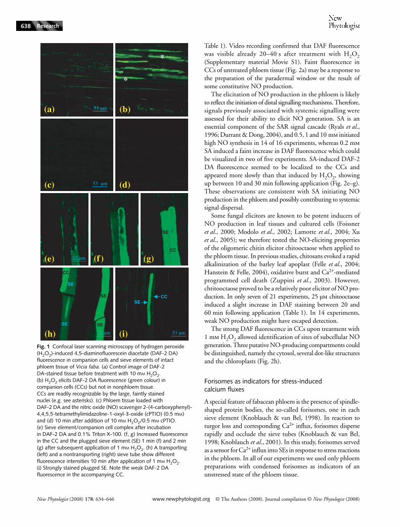
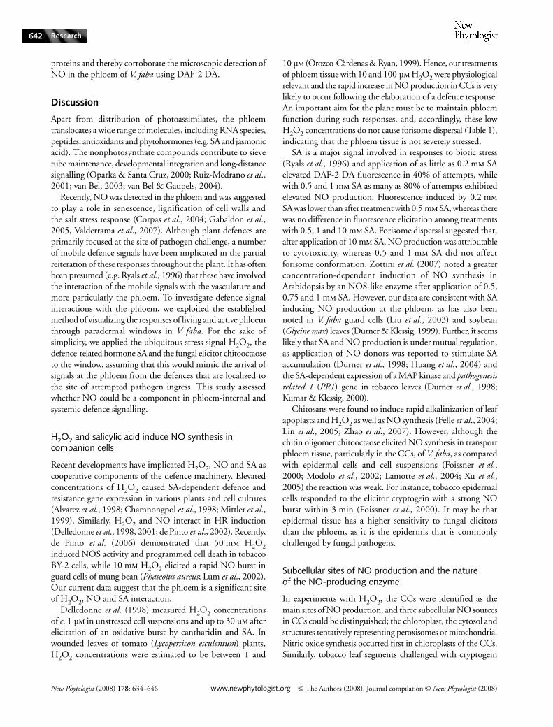
Our group has pioneered in planta observation of phloemfunction through the dissection of a paradermal window inV. faba plants (Knoblauch & van Bel, 1998), which we hereapplied to the analysis of stress-initiated NO effects. H2O2 isregarded as a universal stress signal and acts with NO in avariety of defence and signalling processes (Neill et al., 2002).We initially applied 10 mm H2O2 to the phloem window andvisualized NO production using the membrane-permeantfluorochrome DAF-2 DA dye. The strong increase in DAFfluorescence in CCs in response to the supply of H2O2 wasobserved (green colour, Fig. 1a,b, Table 1). The specifity of thisresponse was demonstrated when fluorescence was suppressedby the addition of the NO scavenger cPTIO (0.5 mm; Fig. 1c,d,Table 1).
Companion cells are the predominant sites of NO production
The phloem is comprised of phloem parenchyma (PP), CCsand sieve elements (SEs). The enucleate SEs, which form theassimilate-transporting sieve tubes, are dependent on supplyof metabolites by the metabolically highly active CCs (e.g. vanBel, 2003).
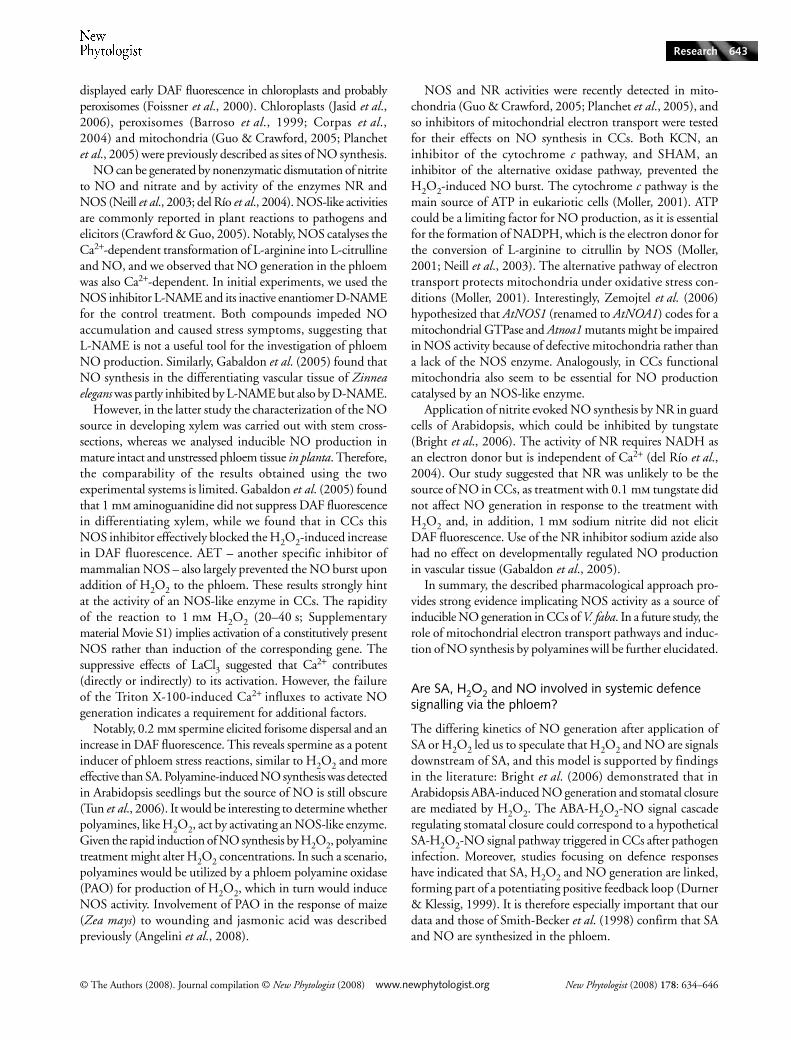
CCs appeared to be the main site of NO generation (Figs 1,2), although in all experiments a slow and weak increase inDAF fluorescence was observed in sieve tubes after H2O2treatment (Fig. 1h, left sieve tube and Fig. 2b,c, right sieve tube).It was hypothesized that the weak NO-derived fluorescencein SEs is a result of permanent mass flow away from theproduction site. To test this hypothesis, the detergent TritonX-100 (0.1%) was applied, which causes SE plugging byirreversible dispersal of forisomes (Knoblauch et al., 2001; seebelow for details on forisome functions). When 1 mm H2O2was subsequently added, strong DAF fluorescence emergedin SEs within 2 min (Fig. 1e–g), consistent with increasedaccumulation as a result of suspended mass flow. Occasionally,H2O2-induced fluorescence was observed in naturally occludedSEs (Fig. 1h, right sieve tube; Figs 1i, 2b,c, left sieve tube). Itis not clear if staining of the SEs is caused by an influx of NOor fluorescent dye from CCs or if there is a source of NO inthe SEs themselves. Figure 1i would argue for the latter, as thestrongly fluorescent SE was accompanied by an only faintly
© The Authors (2008). Journal compilation © New Phytologist (2008) www.newphytologist.org New Phytologist (2008) 178: 634–646
Research 637
fluorescing CC. No DAF fluorescence was noted withinorganelles or in any distinct cellular structure, which suggeststhat NO in SEs might arise from a cytosolic source.
Apart from the CCs, phloem parenchyma cells (PPCs)were sometimes stained (Fig. 2b,c). Nonphloem cells nevershowed a distinct increase in fluorescence in response to thetreatments, which were sometimes harsh (Figs 1, 2). Takentogether, these observations suggest that the phloem is asensitive sensor-transducer of oxidative stress signals ratherthan only a transducer.
Induction of NO synthesis by H2O2, SA and chitooctaose and subcellular localization of the NO source
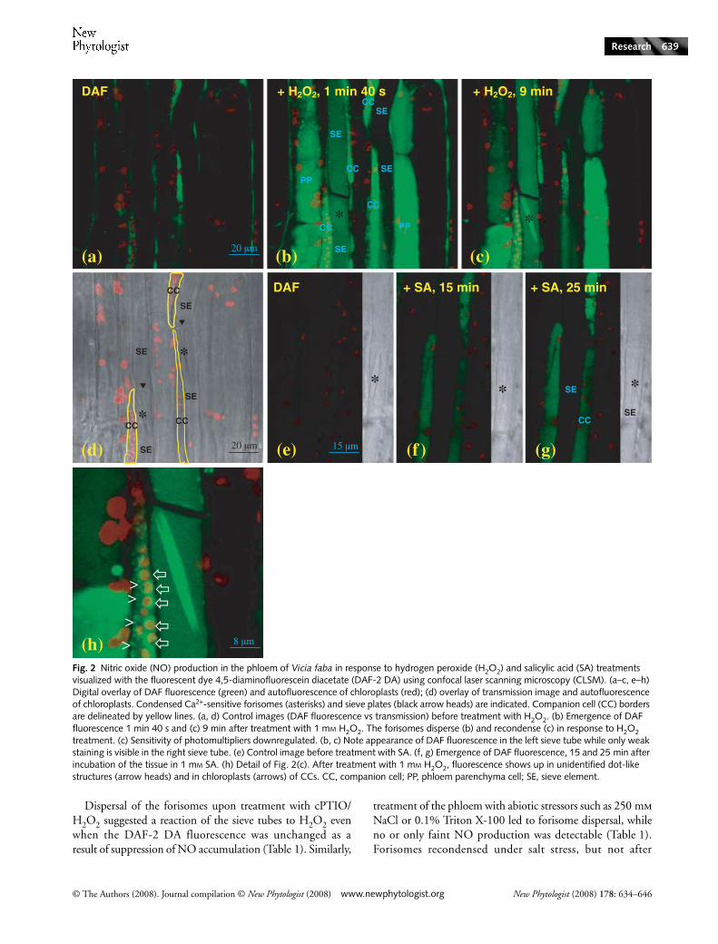
Because 10 mm H2O2 is probably a nonphysiological con-centration, we tested lower concentrations for their abilityto induce NO synthesis in the phloem. As little as 0.01 and0.1 mm H2O2 resulted in an increase in DAF-2 DA fluorescencewithin 10 min (Table 1), while concentrations of 0.5 and1 mm H2O2 elicited an NO burst within 1–2 min (Fig. 2a–c,
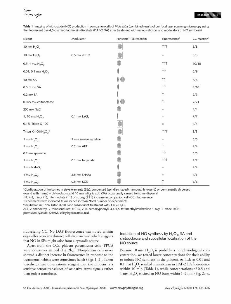
Table 1 Imaging of nitric oxide (NO) production in companion cells of Vicia faba (combined results of confocal laser scanning microscopy using the fluorescent dye 4,5-diaminofluorescein diacetate (DAF-2 DA) after treatment with various elicitors and modulators of NO synthesis)
Elicitor Modulator Forisome1 (SE reaction) Fluorescence2 CC reaction3
10 mM H2O2 ↑↑↑ 8/8
10 mM H2O2 0.5 mM cPTIO = 5/5
0.5, 1 mM H2O2 ↑↑↑ 10/10
0.01, 0.1 mM H2O2 ↑↑ 5/6
10 mM SA ↑↑ 6/6
0.5, 1 mM SA ↑↑ 8/10
0.2 mM SA ↑ 2/5
0.025 mM chitooctaose ↑ 7/21
250 mM NaCl = 4/4
1, 10 mM H2O2 0.1 mM LaCl3 = 7/7
0.1% Triton X-100 = 4/4
Triton X-100/H2O24 ↑↑↑ 3/3
1 mM H2O2 1 mM aminoguanidine = 5/5
1 mM H2O2 0.2 mM AET ↑ 4/4
0.2 mM spermine ↑↑ 5/5
1 mM H2O2 0.1 mM tungstate ↑↑↑ 3/3
1 mM NaNO2 = 4/4
1 mM H2O2 2.5 mM SHAM = 4/5
1 mM H2O2 0.5 mM KCN ↑ 6/6
1Configuration of forisomes in sieve elements (SEs): condensed (spindle-shaped), temporarily (round) or permanently dispersed (round with frame) – chitooctaose and 10 mM salicylic acid (SA) occasionally caused forisome dispersal.2No (=), minor (↑), intermediate (↑↑) or strong (↑↑↑) increase in companion cell (CC) fluorescence.3Experiments with indicated fluorescence increase/total number of experiments.4Incubation in 0.1% Triton X-100 and subsequent treatment with 1 mM H2O2.AET, 2-aminoethyl-2-thiopseudurea; cPTIO, 2-(4-carboxyphenyl)-4,4,5,5-tetramethylimidazoline-1-oxyl-3-oxide; KCN, potassium cyanide; SHAM, salicylhydroxamic acid.
New Phytologist (2008) 178: 634–646 www.newphytologist.org © The Authors (2008). Journal compilation © New Phytologist (2008)
Research638
Table 1). Video recording confirmed that DAF fluorescencewas visible already 20–40 s after treatment with H2O2(Supplementary material Movie S1). Faint fluorescence inCCs of untreated phloem tissue (Fig. 2a) may be a response tothe preparation of the paradermal window or the result ofsome constitutive NO production.
The elicitation of NO production in the phloem is likelyto reflect the initiation of distal signalling mechanisms. Therefore,signals previously associated with systemic signalling wereassessed for their ability to elicit NO generation. SA is anessential component of the SAR signal cascade (Ryals et al.,1996; Durrant & Dong, 2004), and 0.5, 1 and 10 mm initiatedhigh NO synthesis in 14 of 16 experiments, whereas 0.2 mmSA induced a faint increase in DAF fluorescence which couldbe visualized in two of five experiments. SA-induced DAF-2DA fluorescence seemed to be localized to the CCs andappeared more slowly than that induced by H2O2, showingup between 10 and 30 min following application (Fig. 2e–g).These observations are consistent with SA initiating NOproduction in the phloem and possibly contributing to systemicsignal dispersal.
Some fungal elicitors are known to be potent inducers ofNO production in leaf tissues and cultured cells (Foissneret al., 2000; Modolo et al., 2002; Lamotte et al., 2004; Xuet al., 2005); we therefore tested the NO-eliciting propertiesof the oligomeric chitin elicitor chitooctaose when applied tothe phloem tissue. In previous studies, chitosans evoked a rapidalkalinization of the barley leaf apoplast (Felle et al., 2004;Hanstein & Felle, 2004), oxidative burst and Ca2+-mediatedprogrammed cell death (Zuppini et al., 2003). However,chtitooctaose proved to be a relatively poor elicitor of NO pro-duction. In only seven of 21 experiments, 25 µm chitooctaoseinduced a slight increase in DAF staining between 20 and60 min following application (Table 1). In 14 experiments,weak NO production might have escaped detection.
The strong DAF fluorescence in CCs upon treatment with1 mm H2O2 allowed identification of sites of subcellular NOgeneration. Three putative NO-producing compartments couldbe distinguished, namely the cytosol, several dot-like structuresand the chloroplasts (Fig. 2h).
Forisomes as indicators for stress-induced calcium fluxes
A special feature of fabacean phloem is the presence of spindle-shaped protein bodies, the so-called forisomes, one in eachsieve element (Knoblauch & van Bel, 1998). In reaction toturgor loss and corresponding Ca2+ influx, forisomes disperserapidly and occlude the sieve tubes (Knoblauch & van Bel,1998; Knoblauch et al., 2001). In this study, forisomes servedas a sensor for Ca2+ influx into SEs in response to stress reactionsin the phloem. In all of our experiments we used only phloempreparations with condensed forisomes as indicators of anunstressed state of the phloem tissue.
Fig. 1 Confocal laser scanning microscopy of hydrogen peroxide (H2O2)-induced 4,5-diaminofluorescein diacetate (DAF-2 DA) fluorescence in companion cells and sieve elements of intact phloem tissue of Vicia faba. (a) Control image of DAF-2 DA-stained tissue before treatment with 10 mM H2O2. (b) H2O2 elicits DAF-2 DA fluorescence (green colour) in companion cells (CCs) but not in nonphloem tissue. CCs are readily recognizable by the large, faintly stained nuclei (e.g. see asterisks). (c) Phloem tissue loaded with DAF-2 DA and the nitric oxide (NO) scavenger 2-(4-carboxyphenyl)-4,4,5,5-tetramethylimidazoline-1-oxyl-3-oxide (cPTIO) (0.5 mM) and (d) 10 min after addition of 10 mM H2O2/0.5 mM cPTIO. (e) Sieve element/companion cell complex after incubation in DAF-2 DA and 0.1% Triton X-100. (f, g) Increased fluorescence in the CC and the plugged sieve element (SE) 1 min (f) and 2 min (g) after subsequent application of 1 mM H2O2. (h) A transporting (left) and a nontransporting (right) sieve tube show different fluorescence intensities 10 min after application of 1 mM H2O2. (i) Strongly stained plugged SE. Note the weak DAF-2 DA fluorescence in the accompanying CC.
© The Authors (2008). Journal compilation © New Phytologist (2008) www.newphytologist.org New Phytologist (2008) 178: 634–646
Research 639
Dispersal of the forisomes upon treatment with cPTIO/H2O2 suggested a reaction of the sieve tubes to H2O2 evenwhen the DAF-2 DA fluorescence was unchanged as aresult of suppression of NO accumulation (Table 1). Similarly,
treatment of the phloem with abiotic stressors such as 250 mmNaCl or 0.1% Triton X-100 led to forisome dispersal, whileno or only faint NO production was detectable (Table 1).Forisomes recondensed under salt stress, but not after
Fig. 2 Nitric oxide (NO) production in the phloem of Vicia faba in response to hydrogen peroxide (H2O2) and salicylic acid (SA) treatments visualized with the fluorescent dye 4,5-diaminofluorescein diacetate (DAF-2 DA) using confocal laser scanning microscopy (CLSM). (a–c, e–h) Digital overlay of DAF fluorescence (green) and autofluorescence of chloroplasts (red); (d) overlay of transmission image and autofluorescence of chloroplasts. Condensed Ca2+-sensitive forisomes (asterisks) and sieve plates (black arrow heads) are indicated. Companion cell (CC) borders are delineated by yellow lines. (a, d) Control images (DAF fluorescence vs transmission) before treatment with H2O2. (b) Emergence of DAF fluorescence 1 min 40 s and (c) 9 min after treatment with 1 mM H2O2. The forisomes disperse (b) and recondense (c) in response to H2O2 treatment. (c) Sensitivity of photomultipliers downregulated. (b, c) Note appearance of DAF fluorescence in the left sieve tube while only weak staining is visible in the right sieve tube. (e) Control image before treatment with SA. (f, g) Emergence of DAF fluorescence, 15 and 25 min after incubation of the tissue in 1 mM SA. (h) Detail of Fig. 2(c). After treatment with 1 mM H2O2, fluorescence shows up in unidentified dot-like structures (arrow heads) and in chloroplasts (arrows) of CCs. CC, companion cell; PP, phloem parenchyma cell; SE, sieve element.
New Phytologist (2008) 178: 634–646 www.newphytologist.org © The Authors (2008). Journal compilation © New Phytologist (2008)
Research640
application of the detergent. Thus, by observing the forisomeconfiguration it was possible to assess the contribution ofCa2+-dependent effects on the diverse treatments used in thisstudy, irrespective of changes in DAF-2 DA fluorescence.
Detailed analyses revealed that the forisome reaction to H2O2and SA was concentration dependent. Upon treatment with10 mm H2O2, the dispersed forisomes did not recondense(Table 1) and the tissue turned brown and died. The inabilityof forisomes to recondense hints at severe damage of thephloem tissue inflicted by treatment with 10 mm H2O2 orwith Triton X-100 (Table 1). By contrast, 1 mm H2O2 causedtransient dispersal of the forisomes which recondensed after afew minutes even when H2O2 remained on the treated surface(Fig. 2a–d, Table 1). Application of neither 0.01 and 0.1 mmH2O2 (Table 1) nor 0.2, 0.5 and 1 mm SA (Fig. 2e–g, Table 1)induced forisome dispersal, suggesting that no strong stressreaction of the phloem had occurred. This correlated withrather low NO production in the phloem. However, 10 mmSA – which is probably a nonphysiological concentration –provoked occasional forisome dispersal but no stronger NOproduction than with 0.5 and 1 mm SA. Finally, forisomesonly rarely dispersed upon chitooctaose treatment.
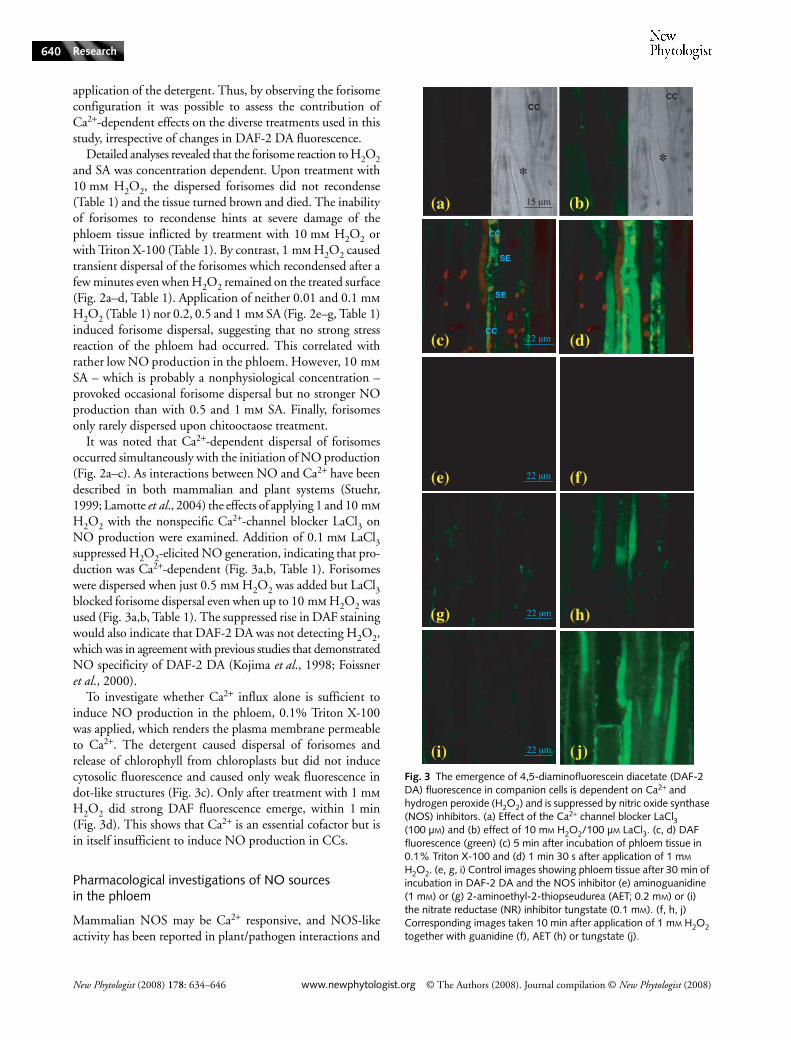
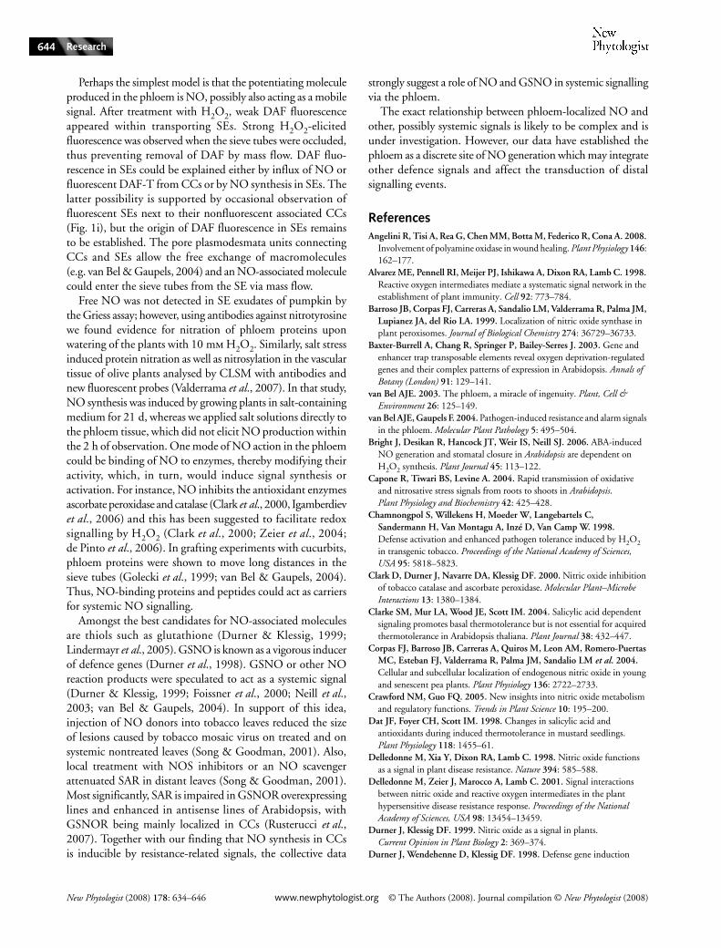
It was noted that Ca2+-dependent dispersal of forisomesoccurred simultaneously with the initiation of NO production(Fig. 2a–c). As interactions between NO and Ca2+ have beendescribed in both mammalian and plant systems (Stuehr,1999; Lamotte et al., 2004) the effects of applying 1 and 10 mmH2O2 with the nonspecific Ca2+-channel blocker LaCl3 onNO production were examined. Addition of 0.1 mm LaCl3suppressed H2O2-elicited NO generation, indicating that pro-duction was Ca2+-dependent (Fig. 3a,b, Table 1). Forisomeswere dispersed when just 0.5 mm H2O2 was added but LaCl3blocked forisome dispersal even when up to 10 mm H2O2 wasused (Fig. 3a,b, Table 1). The suppressed rise in DAF stainingwould also indicate that DAF-2 DA was not detecting H2O2,which was in agreement with previous studies that demonstratedNO specificity of DAF-2 DA (Kojima et al., 1998; Foissneret al., 2000).
To investigate whether Ca2+ influx alone is sufficient toinduce NO production in the phloem, 0.1% Triton X-100was applied, which renders the plasma membrane permeableto Ca2+. The detergent caused dispersal of forisomes andrelease of chlorophyll from chloroplasts but did not inducecytosolic fluorescence and caused only weak fluorescence indot-like structures (Fig. 3c). Only after treatment with 1 mmH2O2 did strong DAF fluorescence emerge, within 1 min(Fig. 3d). This shows that Ca2+ is an essential cofactor but isin itself insufficient to induce NO production in CCs.
Pharmacological investigations of NO sources in the phloem
Mammalian NOS may be Ca2+ responsive, and NOS-likeactivity has been reported in plant/pathogen interactions and
Fig. 3 The emergence of 4,5-diaminofluorescein diacetate (DAF-2 DA) fluorescence in companion cells is dependent on Ca2+ and hydrogen peroxide (H2O2) and is suppressed by nitric oxide synthase (NOS) inhibitors. (a) Effect of the Ca2+ channel blocker LaCl3 (100 µM) and (b) effect of 10 mM H2O2/100 µM LaCl3. (c, d) DAF fluorescence (green) (c) 5 min after incubation of phloem tissue in 0.1% Triton X-100 and (d) 1 min 30 s after application of 1 mM H2O2. (e, g, i) Control images showing phloem tissue after 30 min of incubation in DAF-2 DA and the NOS inhibitor (e) aminoguanidine (1 mM) or (g) 2-aminoethyl-2-thiopseudurea (AET; 0.2 mM) or (i) the nitrate reductase (NR) inhibitor tungstate (0.1 mM). (f, h, j) Corresponding images taken 10 min after application of 1 mM H2O2 together with guanidine (f), AET (h) or tungstate (j).
© The Authors (2008). Journal compilation © New Phytologist (2008) www.newphytologist.org New Phytologist (2008) 178: 634–646
Research 641
in plant reactions to fungal elicitors (Neill et al., 2003; Crawford& Guo, 2005). Therefore, the mammalian NOS inhibitorsL-NAME, aminoguanidine and 2-aminoethyl-2-thiopseudurea(AET) were employed for characterization of the mode of NOproduction in CCs. L-NAME turned out to be inappropriatefor phloem analyses as both the inhibitor and its inactiveenantiomer D-NAME inhibited NO synthesis and impairedforisome dispersal after application of H2O2, which isdiagnostic for stressed phloem tissue (data not shown). Bycontrast, 1 mm aminoguanidine blocked H2O2-induced DAFfluorescence without affecting the forisome reaction in a totalof five experiments (Fig. 3e,f, Table 1), while 0.2 mm AETlargely suppressed NO production in CCs in four experiments(Fig. 3g,h). AET was recently shown to be an inhibitor of NOproduction evoked by treatment of Arabidopsis seedlings withpolyamines (Tun et al., 2006). Phloem tissue responded to1 mm spermine by rapid sieve plate plugging and irreversibleforisome dispersal (not shown), whereas 0.2 mm caused dispersaland recondensation of forisomes and DAF fluorescence(Table 1).
To investigate NR as a potential source of NO, the phloemtissue was incubated with 0.1 mm tungstate. The NRinhibitor had no effect on the H2O2-induced increase in DAFfluorescence and the forisome reaction (Fig. 3i,j, Table 1).Also, 1 mm sodium nitrite, the substrate of NR, elicited anincrease neither in DAF fluorescence nor in forisome dispersal(Table 1), but both could be observed after subsequentaddition of 1 mm H2O2 (not shown).
Recently, NO production in mitochondria was demonstrated(Guo & Crawford, 2005; Planchet et al., 2005) and NRactivity in mitochondria of tobacco was found to be dependenton functional electron transport (Planchet et al., 2005). Aswe localized an NO source in dot-like structures within CCs(Fig. 2h), inhibitors of mitochondrial electron transport wereused to investigate whether these organelles represent mito-chondria. Potassium cyanide (KCN) inhibits the cytochromec and salicylhydroxamic acid (SHAM) alternative oxidasepathway. Surprisingly, the increase in fluorescence not onlyin dot-like structures but also in whole CCs was completely(SHAM) or largely (KCN) suppressed by preincubation ofthe phloem tissue in 2.5 mm SHAM or 0.5 or 1 mm KCNbefore H2O2 treatment (Table 1). Hence, functional mito-chondrial electron transport seems to be essential for NOproduction in CCs.
In summary, the results suggest that the NO source in CCsof V. faba is dependent on Ca2+ and mitochondrial electrontransport and can be suppressed by NOS inhibitors but notby the NR inhibitor tungstate.
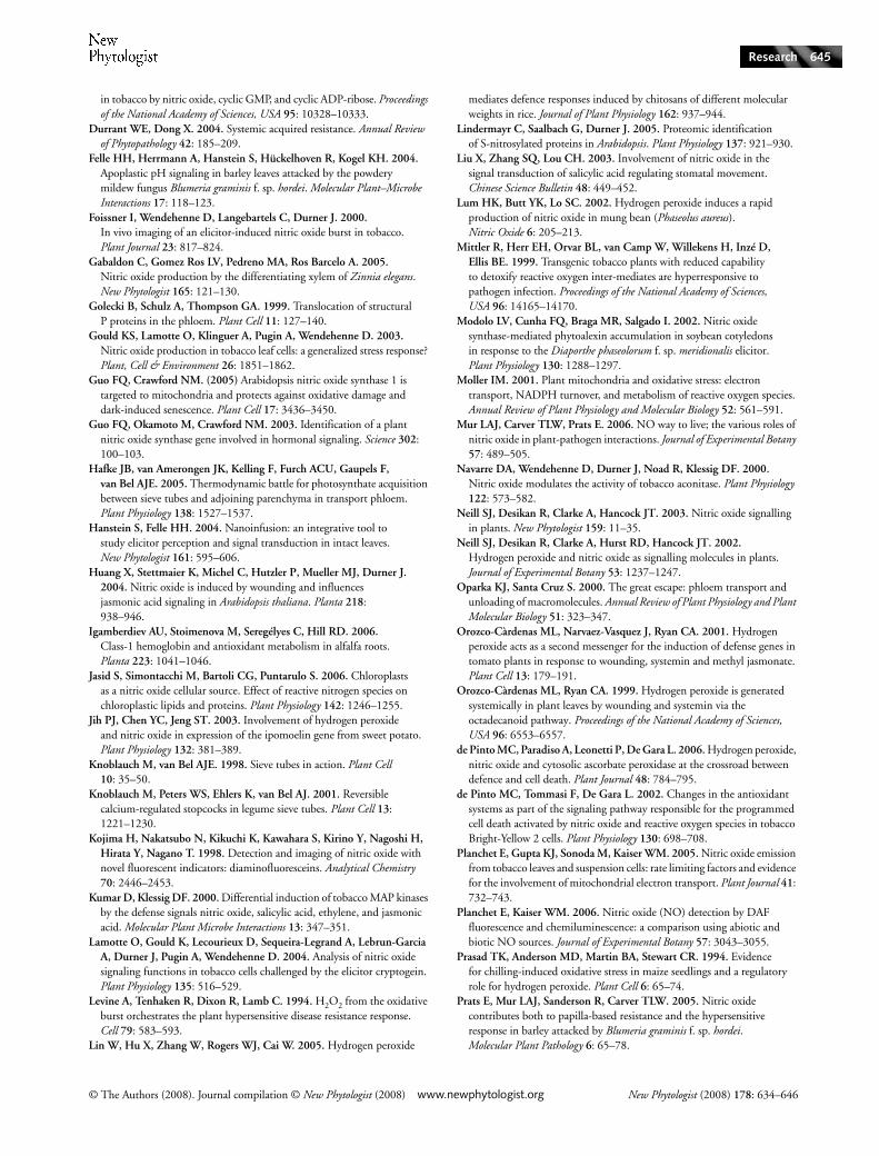
Treatment of Cucurbita maxima with H2O2 induces nitration of phloem proteins
As the specificity of diaminofluorescein for NO has recentlybeen questioned (Planchet & Kaiser, 2006, and references
therein), confirmation of microscopy results by independentmethods is required. Unfortunately, the maximal volume ofpure sieve tube exudates that can be collected from V. fababy aphid stylectomy (Hafke et al., 2005) is too low to allowbiochemical measurements of NO content using, for example,Griess and haemoglobin assays. Therefore, we examined NOcontents in phloem exudates from pumpkin (C. maxima),which produces a significant yield of phloem exudates fromcut petioles and stems. In order to elicit NO synthesis, pumpkinplants were watered with 10 mm H2O2. In Arabidopsis thistreatment induced expression of MAPK (mitogen-activatedprotein kinase) genes in the shoot (Capone et al., 2004). Althoughthe authors assumed xylem-based transport of H2O2 into theshoot, this treatment may act by induced systemic signallingfollowing root-based stress.
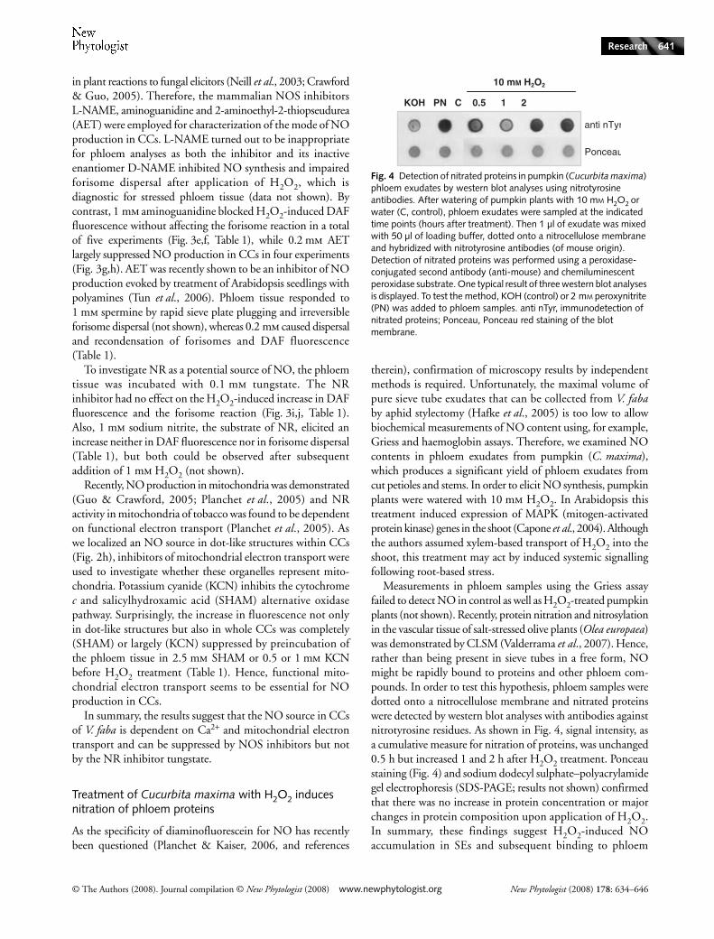
Measurements in phloem samples using the Griess assayfailed to detect NO in control as well as H2O2-treated pumpkinplants (not shown). Recently, protein nitration and nitrosylationin the vascular tissue of salt-stressed olive plants (Olea europaea)was demonstrated by CLSM (Valderrama et al., 2007). Hence,rather than being present in sieve tubes in a free form, NOmight be rapidly bound to proteins and other phloem com-pounds. In order to test this hypothesis, phloem samples weredotted onto a nitrocellulose membrane and nitrated proteinswere detected by western blot analyses with antibodies againstnitrotyrosine residues. As shown in Fig. 4, signal intensity, asa cumulative measure for nitration of proteins, was unchanged0.5 h but increased 1 and 2 h after H2O2 treatment. Ponceaustaining (Fig. 4) and sodium dodecyl sulphate–polyacrylamidegel electrophoresis (SDS-PAGE; results not shown) confirmedthat there was no increase in protein concentration or majorchanges in protein composition upon application of H2O2.In summary, these findings suggest H2O2-induced NOaccumulation in SEs and subsequent binding to phloem
Fig. 4 Detection of nitrated proteins in pumpkin (Cucurbita maxima) phloem exudates by western blot analyses using nitrotyrosine antibodies. After watering of pumpkin plants with 10 mM H2O2 or water (C, control), phloem exudates were sampled at the indicated time points (hours after treatment). Then 1 µl of exudate was mixed with 50 µl of loading buffer, dotted onto a nitrocellulose membrane and hybridized with nitrotyrosine antibodies (of mouse origin). Detection of nitrated proteins was performed using a peroxidase-conjugated second antibody (anti-mouse) and chemiluminescent peroxidase substrate. One typical result of three western blot analyses is displayed. To test the method, KOH (control) or 2 mM peroxynitrite (PN) was added to phloem samples. anti nTyr, immunodetection of nitrated proteins; Ponceau, Ponceau red staining of the blot membrane.
New Phytologist (2008) 178: 634–646 www.newphytologist.org © The Authors (2008). Journal compilation © New Phytologist (2008)
Research642
proteins and thereby corroborate the microscopic detection ofNO in the phloem of V. faba using DAF-2 DA.
Discussion
Apart from distribution of photoassimilates, the phloemtranslocates a wide range of molecules, including RNA species,peptides, antioxidants and phytohormones (e.g. SA and jasmonicacid). The nonphotosynthate compounds contribute to sievetube maintenance, developmental integration and long-distancesignalling (Oparka & Santa Cruz, 2000; Ruiz-Medrano et al.,2001; van Bel, 2003; van Bel & Gaupels, 2004).
Recently, NO was detected in the phloem and was suggestedto play a role in senescence, lignification of cell walls andthe salt stress response (Corpas et al., 2004; Gabaldon et al.,2005, Valderrama et al., 2007). Although plant defences areprimarily focused at the site of pathogen challenge, a numberof mobile defence signals have been implicated in the partialreiteration of these responses throughout the plant. It has oftenbeen presumed (e.g. Ryals et al., 1996) that these have involvedthe interaction of the mobile signals with the vasculature andmore particularly the phloem. To investigate defence signalinteractions with the phloem, we exploited the establishedmethod of visualizing the responses of living and active phloemthrough paradermal windows in V. faba. For the sake ofsimplicity, we applied the ubiquitous stress signal H2O2, thedefence-related hormone SA and the fungal elicitor chitooctaoseto the window, assuming that this would mimic the arrival ofsignals at the phloem from the defences that are localized tothe site of attempted pathogen ingress. This study assessedwhether NO could be a component in phloem-internal andsystemic defence signalling.
H2O2 and salicylic acid induce NO synthesis in companion cells
Recent developments have implicated H2O2, NO and SA ascooperative components of the defence machinery. Elevatedconcentrations of H2O2 caused SA-dependent defence andresistance gene expression in various plants and cell cultures(Alvarez et al., 1998; Chamnongpol et al., 1998; Mittler et al.,1999). Similarly, H2O2 and NO interact in HR induction(Delledonne et al., 1998, 2001; de Pinto et al., 2002). Recently,de Pinto et al. (2006) demonstrated that 50 mm H2O2induced NOS activity and programmed cell death in tobaccoBY-2 cells, while 10 mm H2O2 elicited a rapid NO burst inguard cells of mung bean (Phaseolus aureus; Lum et al., 2002).Our current data suggest that the phloem is a significant siteof H2O2, NO and SA interaction.
Delledonne et al. (1998) measured H2O2 concentrationsof c. 1 µm in unstressed cell suspensions and up to 30 µm afterelicitation of an oxidative burst by cantharidin and SA. Inwounded leaves of tomato (Lycopersicon esculentum) plants,H2O2 concentrations were estimated to be between 1 and
10 µm (Orozco-Càrdenas & Ryan, 1999). Hence, our treatmentsof phloem tissue with 10 and 100 µm H2O2 were physiologicalrelevant and the rapid increase in NO production in CCs is verylikely to occur following the elaboration of a defence response.An important aim for the plant must be to maintain phloemfunction during such responses, and, accordingly, these lowH2O2 concentrations do not cause forisome dispersal (Table 1),indicating that the phloem tissue is not severely stressed.
SA is a major signal involved in responses to biotic stress(Ryals et al., 1996) and application of as little as 0.2 mm SAelevated DAF-2 DA fluorescence in 40% of attempts, whilewith 0.5 and 1 mm SA as many as 80% of attempts exhibitedelevated NO production. Fluorescence induced by 0.2 mmSA was lower than after treatment with 0.5 mm SA, whereas therewas no difference in fluorescence elicitation among treatmentswith 0.5, 1 and 10 mm SA. Forisome dispersal suggested that,after application of 10 mm SA, NO production was attributableto cytotoxicity, whereas 0.5 and 1 mm SA did not affectforisome conformation. Zottini et al. (2007) noted a greaterconcentration-dependent induction of NO synthesis inArabidopsis by an NOS-like enzyme after application of 0.5,0.75 and 1 mm SA. However, our data are consistent with SAinducing NO production at the phloem, as has also beennoted in V. faba guard cells (Liu et al., 2003) and soybean(Glycine max) leaves (Durner & Klessig, 1999). Further, it seemslikely that SA and NO production is under mutual regulation,as application of NO donors was reported to stimulate SAaccumulation (Durner et al., 1998; Huang et al., 2004) andthe SA-dependent expression of a MAP kinase and pathogenesisrelated 1 (PR1) gene in tobacco leaves (Durner et al., 1998;Kumar & Klessig, 2000).
Chitosans were found to induce rapid alkalinization of leafapoplasts and H2O2 as well as NO synthesis (Felle et al., 2004;Lin et al., 2005; Zhao et al., 2007). However, although thechitin oligomer chitooctaose elicited NO synthesis in transportphloem tissue, particularly in the CCs, of V. faba, as comparedwith epidermal cells and cell suspensions (Foissner et al.,2000; Modolo et al., 2002; Lamotte et al., 2004; Xu et al.,2005) the reaction was weak. For instance, tobacco epidermalcells responded to the elicitor cryptogein with a strong NOburst within 3 min (Foissner et al., 2000). It may be thatepidermal tissue has a higher sensitivity to fungal elicitorsthan the phloem, as it is the epidermis that is commonlychallenged by fungal pathogens.
Subcellular sites of NO production and the nature of the NO-producing enzyme
In experiments with H2O2, the CCs were identified as themain sites of NO production, and three subcellular NO sourcesin CCs could be distinguished; the chloroplast, the cytosol andstructures tentatively representing peroxisomes or mitochondria.Nitric oxide synthesis occurred first in chloroplasts of the CCs.Similarly, tobacco leaf segments challenged with cryptogein
© The Authors (2008). Journal compilation © New Phytologist (2008) www.newphytologist.org New Phytologist (2008) 178: 634–646
Research 643
displayed early DAF fluorescence in chloroplasts and probablyperoxisomes (Foissner et al., 2000). Chloroplasts (Jasid et al.,2006), peroxisomes (Barroso et al., 1999; Corpas et al.,2004) and mitochondria (Guo & Crawford, 2005; Planchetet al., 2005) were previously described as sites of NO synthesis.
NO can be generated by nonenzymatic dismutation of nitriteto NO and nitrate and by activity of the enzymes NR andNOS (Neill et al., 2003; del Río et al., 2004). NOS-like activitiesare commonly reported in plant reactions to pathogens andelicitors (Crawford & Guo, 2005). Notably, NOS catalyses theCa2+-dependent transformation of L-arginine into L-citrullineand NO, and we observed that NO generation in the phloemwas also Ca2+-dependent. In initial experiments, we used theNOS inhibitor L-NAME and its inactive enantiomer D-NAMEfor the control treatment. Both compounds impeded NOaccumulation and caused stress symptoms, suggesting thatL-NAME is not a useful tool for the investigation of phloemNO production. Similarly, Gabaldon et al. (2005) found thatNO synthesis in the differentiating vascular tissue of Zinneaelegans was partly inhibited by L-NAME but also by D-NAME.
However, in the latter study the characterization of the NOsource in developing xylem was carried out with stem cross-sections, whereas we analysed inducible NO production inmature intact and unstressed phloem tissue in planta. Therefore,the comparability of the results obtained using the twoexperimental systems is limited. Gabaldon et al. (2005) foundthat 1 mm aminoguanidine did not suppress DAF fluorescencein differentiating xylem, while we found that in CCs thisNOS inhibitor effectively blocked the H2O2-induced increasein DAF fluorescence. AET – another specific inhibitor ofmammalian NOS – also largely prevented the NO burst uponaddition of H2O2 to the phloem. These results strongly hintat the activity of an NOS-like enzyme in CCs. The rapidityof the reaction to 1 mm H2O2 (20–40 s; Supplementarymaterial Movie S1) implies activation of a constitutively presentNOS rather than induction of the corresponding gene. Thesuppressive effects of LaCl3 suggested that Ca2+ contributes(directly or indirectly) to its activation. However, the failureof the Triton X-100-induced Ca2+ influxes to activate NOgeneration indicates a requirement for additional factors.
Notably, 0.2 mm spermine elicited forisome dispersal and anincrease in DAF fluorescence. This reveals spermine as a potentinducer of phloem stress reactions, similar to H2O2 and moreeffective than SA. Polyamine-induced NO synthesis was detectedin Arabidopsis seedlings but the source of NO is still obscure(Tun et al., 2006). It would be interesting to determine whetherpolyamines, like H2O2, act by activating an NOS-like enzyme.Given the rapid induction of NO synthesis by H2O2, polyaminetreatment might alter H2O2 concentrations. In such a scenario,polyamines would be utilized by a phloem polyamine oxidase(PAO) for production of H2O2, which in turn would induceNOS activity. Involvement of PAO in the response of maize(Zea mays) to wounding and jasmonic acid was describedpreviously (Angelini et al., 2008).
NOS and NR activities were recently detected in mito-chondria (Guo & Crawford, 2005; Planchet et al., 2005), andso inhibitors of mitochondrial electron transport were testedfor their effects on NO synthesis in CCs. Both KCN, aninhibitor of the cytochrome c pathway, and SHAM, aninhibitor of the alternative oxidase pathway, prevented theH2O2-induced NO burst. The cytochrome c pathway is themain source of ATP in eukariotic cells (Moller, 2001). ATPcould be a limiting factor for NO production, as it is essentialfor the formation of NADPH, which is the electron donor forthe conversion of L-arginine to citrullin by NOS (Moller,2001; Neill et al., 2003). The alternative pathway of electrontransport protects mitochondria under oxidative stress con-ditions (Moller, 2001). Interestingly, Zemojtel et al. (2006)hypothesized that AtNOS1 (renamed to AtNOA1) codes for amitochondrial GTPase and Atnoa1 mutants might be impairedin NOS activity because of defective mitochondria rather thana lack of the NOS enzyme. Analogously, in CCs functionalmitochondria also seem to be essential for NO productioncatalysed by an NOS-like enzyme.
Application of nitrite evoked NO synthesis by NR in guardcells of Arabidopsis, which could be inhibited by tungstate(Bright et al., 2006). The activity of NR requires NADH asan electron donor but is independent of Ca2+ (del Río et al.,2004). Our study suggested that NR was unlikely to be thesource of NO in CCs, as treatment with 0.1 mm tungstate didnot affect NO generation in response to the treatment withH2O2 and, in addition, 1 mm sodium nitrite did not elicitDAF fluorescence. Use of the NR inhibitor sodium azide alsohad no effect on developmentally regulated NO productionin vascular tissue (Gabaldon et al., 2005).
In summary, the described pharmacological approach pro-vides strong evidence implicating NOS activity as a source ofinducible NO generation in CCs of V. faba. In a future study, therole of mitochondrial electron transport pathways and induc-tion of NO synthesis by polyamines will be further elucidated.
Are SA, H2O2 and NO involved in systemic defence signalling via the phloem?
The differing kinetics of NO generation after application ofSA or H2O2 led us to speculate that H2O2 and NO are signalsdownstream of SA, and this model is supported by findingsin the literature: Bright et al. (2006) demonstrated that inArabidopsis ABA-induced NO generation and stomatal closureare mediated by H2O2. The ABA-H2O2-NO signal cascaderegulating stomatal closure could correspond to a hypotheticalSA-H2O2-NO signal pathway triggered in CCs after pathogeninfection. Moreover, studies focusing on defence responseshave indicated that SA, H2O2 and NO generation are linked,forming part of a potentiating positive feedback loop (Durner& Klessig, 1999). It is therefore especially important that ourdata and those of Smith-Becker et al. (1998) confirm that SAand NO are synthesized in the phloem.
New Phytologist (2008) 178: 634–646 www.newphytologist.org © The Authors (2008). Journal compilation © New Phytologist (2008)
Research644
Perhaps the simplest model is that the potentiating moleculeproduced in the phloem is NO, possibly also acting as a mobilesignal. After treatment with H2O2, weak DAF fluorescenceappeared within transporting SEs. Strong H2O2-elicitedfluorescence was observed when the sieve tubes were occluded,thus preventing removal of DAF by mass flow. DAF fluo-rescence in SEs could be explained either by influx of NO orfluorescent DAF-T from CCs or by NO synthesis in SEs. Thelatter possibility is supported by occasional observation offluorescent SEs next to their nonfluorescent associated CCs(Fig. 1i), but the origin of DAF fluorescence in SEs remainsto be established. The pore plasmodesmata units connectingCCs and SEs allow the free exchange of macromolecules(e.g. van Bel & Gaupels, 2004) and an NO-associated moleculecould enter the sieve tubes from the SE via mass flow.
Free NO was not detected in SE exudates of pumpkin bythe Griess assay; however, using antibodies against nitrotyrosinewe found evidence for nitration of phloem proteins uponwatering of the plants with 10 mm H2O2. Similarly, salt stressinduced protein nitration as well as nitrosylation in the vasculartissue of olive plants analysed by CLSM with antibodies andnew fluorescent probes (Valderrama et al., 2007). In that study,NO synthesis was induced by growing plants in salt-containingmedium for 21 d, whereas we applied salt solutions directly tothe phloem tissue, which did not elicit NO production withinthe 2 h of observation. One mode of NO action in the phloemcould be binding of NO to enzymes, thereby modifying theiractivity, which, in turn, would induce signal synthesis oractivation. For instance, NO inhibits the antioxidant enzymesascorbate peroxidase and catalase (Clark et al., 2000, Igamberdievet al., 2006) and this has been suggested to facilitate redoxsignalling by H2O2 (Clark et al., 2000; Zeier et al., 2004;de Pinto et al., 2006). In grafting experiments with cucurbits,phloem proteins were shown to move long distances in thesieve tubes (Golecki et al., 1999; van Bel & Gaupels, 2004).Thus, NO-binding proteins and peptides could act as carriersfor systemic NO signalling.
Amongst the best candidates for NO-associated moleculesare thiols such as glutathione (Durner & Klessig, 1999;Lindermayr et al., 2005). GSNO is known as a vigorous inducerof defence genes (Durner et al., 1998). GSNO or other NOreaction products were speculated to act as a systemic signal(Durner & Klessig, 1999; Foissner et al., 2000; Neill et al.,2003; van Bel & Gaupels, 2004). In support of this idea,injection of NO donors into tobacco leaves reduced the sizeof lesions caused by tobacco mosaic virus on treated and onsystemic nontreated leaves (Song & Goodman, 2001). Also,local treatment with NOS inhibitors or an NO scavengerattenuated SAR in distant leaves (Song & Goodman, 2001).Most significantly, SAR is impaired in GSNOR overexpressinglines and enhanced in antisense lines of Arabidopsis, withGSNOR being mainly localized in CCs (Rusterucci et al.,2007). Together with our finding that NO synthesis in CCsis inducible by resistance-related signals, the collective data
strongly suggest a role of NO and GSNO in systemic signallingvia the phloem.
The exact relationship between phloem-localized NO andother, possibly systemic signals is likely to be complex and isunder investigation. However, our data have established thephloem as a discrete site of NO generation which may integrateother defence signals and affect the transduction of distalsignalling events.
ReferencesAngelini R, Tisi A, Rea G, Chen MM, Botta M, Federico R, Cona A. 2008.
Involvement of polyamine oxidase in wound healing. Plant Physiology 146: 162–177.
Alvarez ME, Pennell RI, Meijer PJ, Ishikawa A, Dixon RA, Lamb C. 1998. Reactive oxygen intermediates mediate a systematic signal network in the establishment of plant immunity. Cell 92: 773–784.
Barroso JB, Corpas FJ, Carreras A, Sandalio LM, Valderrama R, Palma JM, Lupianez JA, del Rio LA. 1999. Localization of nitric oxide synthase in plant peroxisomes. Journal of Biological Chemistry 274: 36729–36733.
Baxter-Burrell A, Chang R, Springer P, Bailey-Serres J. 2003. Gene and enhancer trap transposable elements reveal oxygen deprivation-regulated genes and their complex patterns of expression in Arabidopsis. Annals of Botany (London) 91: 129–141.
van Bel AJE. 2003. The phloem, a miracle of ingenuity. Plant, Cell & Environment 26: 125–149.
van Bel AJE, Gaupels F. 2004. Pathogen-induced resistance and alarm signals in the phloem. Molecular Plant Pathology 5: 495–504.
Bright J, Desikan R, Hancock JT, Weir IS, Neill SJ. 2006. ABA-induced NO generation and stomatal closure in Arabidopsis are dependent on H2O2 synthesis. Plant Journal 45: 113–122.
Capone R, Tiwari BS, Levine A. 2004. Rapid transmission of oxidative and nitrosative stress signals from roots to shoots in Arabidopsis. Plant Physiology and Biochemistry 42: 425–428.
Chamnongpol S, Willekens H, Moeder W, Langebartels C, Sandermann H, Van Montagu A, Inzé D, Van Camp W. 1998. Defense activation and enhanced pathogen tolerance induced by H2O2 in transgenic tobacco. Proceedings of the National Academy of Sciences, USA 95: 5818–5823.
Clark D, Durner J, Navarre DA, Klessig DF. 2000. Nitric oxide inhibition of tobacco catalase and ascorbate peroxidase. Molecular Plant–Microbe Interactions 13: 1380–1384.
Clarke SM, Mur LA, Wood JE, Scott IM. 2004. Salicylic acid dependent signaling promotes basal thermotolerance but is not essential for acquired thermotolerance in Arabidopsis thaliana. Plant Journal 38: 432–447.
Corpas FJ, Barroso JB, Carreras A, Quiros M, Leon AM, Romero-Puertas MC, Esteban FJ, Valderrama R, Palma JM, Sandalio LM et al. 2004. Cellular and subcellular localization of endogenous nitric oxide in young and senescent pea plants. Plant Physiology 136: 2722–2733.
Crawford NM, Guo FQ. 2005. New insights into nitric oxide metabolism and regulatory functions. Trends in Plant Science 10: 195–200.
Dat JF, Foyer CH, Scott IM. 1998. Changes in salicylic acid and antioxidants during induced thermotolerance in mustard seedlings. Plant Physiology 118: 1455–61.
Delledonne M, Xia Y, Dixon RA, Lamb C. 1998. Nitric oxide functions as a signal in plant disease resistance. Nature 394: 585–588.
Delledonne M, Zeier J, Marocco A, Lamb C. 2001. Signal interactions between nitric oxide and reactive oxygen intermediates in the plant hypersensitive disease resistance response. Proceedings of the National Academy of Sciences, USA 98: 13454–13459.
Durner J, Klessig DF. 1999. Nitric oxide as a signal in plants. Current Opinion in Plant Biology 2: 369–374.
Durner J, Wendehenne D, Klessig DF. 1998. Defense gene induction
© The Authors (2008). Journal compilation © New Phytologist (2008) www.newphytologist.org New Phytologist (2008) 178: 634–646
Research 645
in tobacco by nitric oxide, cyclic GMP, and cyclic ADP-ribose. Proceedings of the National Academy of Sciences, USA 95: 10328–10333.
Durrant WE, Dong X. 2004. Systemic acquired resistance. Annual Review of Phytopathology 42: 185–209.
Felle HH, Herrmann A, Hanstein S, Hückelhoven R, Kogel KH. 2004. Apoplastic pH signaling in barley leaves attacked by the powdery mildew fungus Blumeria graminis f. sp. hordei. Molecular Plant–Microbe Interactions 17: 118–123.
Foissner I, Wendehenne D, Langebartels C, Durner J. 2000. In vivo imaging of an elicitor-induced nitric oxide burst in tobacco. Plant Journal 23: 817–824.
Gabaldon C, Gomez Ros LV, Pedreno MA, Ros Barcelo A. 2005. Nitric oxide production by the differentiating xylem of Zinnia elegans. New Phytologist 165: 121–130.
Golecki B, Schulz A, Thompson GA. 1999. Translocation of structural P proteins in the phloem. Plant Cell 11: 127–140.
Gould KS, Lamotte O, Klinguer A, Pugin A, Wendehenne D. 2003. Nitric oxide production in tobacco leaf cells: a generalized stress response? Plant, Cell & Environment 26: 1851–1862.
Guo FQ, Crawford NM. (2005) Arabidopsis nitric oxide synthase 1 is targeted to mitochondria and protects against oxidative damage and dark-induced senescence. Plant Cell 17: 3436–3450.
Guo FQ, Okamoto M, Crawford NM. 2003. Identification of a plant nitric oxide synthase gene involved in hormonal signaling. Science 302: 100–103.
Hafke JB, van Amerongen JK, Kelling F, Furch ACU, Gaupels F, van Bel AJE. 2005. Thermodynamic battle for photosynthate acquisition between sieve tubes and adjoining parenchyma in transport phloem. Plant Physiology 138: 1527–1537.
Hanstein S, Felle HH. 2004. Nanoinfusion: an integrative tool to study elicitor perception and signal transduction in intact leaves. New Phytologist 161: 595–606.
Huang X, Stettmaier K, Michel C, Hutzler P, Mueller MJ, Durner J. 2004. Nitric oxide is induced by wounding and influences jasmonic acid signaling in Arabidopsis thaliana. Planta 218: 938–946.
Igamberdiev AU, Stoimenova M, Seregélyes C, Hill RD. 2006. Class-1 hemoglobin and antioxidant metabolism in alfalfa roots. Planta 223: 1041–1046.
Jasid S, Simontacchi M, Bartoli CG, Puntarulo S. 2006. Chloroplasts as a nitric oxide cellular source. Effect of reactive nitrogen species on chloroplastic lipids and proteins. Plant Physiology 142: 1246–1255.
Jih PJ, Chen YC, Jeng ST. 2003. Involvement of hydrogen peroxide and nitric oxide in expression of the ipomoelin gene from sweet potato. Plant Physiology 132: 381–389.
Knoblauch M, van Bel AJE. 1998. Sieve tubes in action. Plant Cell 10: 35–50.
Knoblauch M, Peters WS, Ehlers K, van Bel AJ. 2001. Reversible calcium-regulated stopcocks in legume sieve tubes. Plant Cell 13: 1221–1230.
Kojima H, Nakatsubo N, Kikuchi K, Kawahara S, Kirino Y, Nagoshi H, Hirata Y, Nagano T. 1998. Detection and imaging of nitric oxide with novel fluorescent indicators: diaminofluoresceins. Analytical Chemistry 70: 2446–2453.
Kumar D, Klessig DF. 2000. Differential induction of tobacco MAP kinases by the defense signals nitric oxide, salicylic acid, ethylene, and jasmonic acid. Molecular Plant Microbe Interactions 13: 347–351.
Lamotte O, Gould K, Lecourieux D, Sequeira-Legrand A, Lebrun-Garcia A, Durner J, Pugin A, Wendehenne D. 2004. Analysis of nitric oxide signaling functions in tobacco cells challenged by the elicitor cryptogein. Plant Physiology 135: 516–529.
Levine A, Tenhaken R, Dixon R, Lamb C. 1994. H2O2 from the oxidative burst orchestrates the plant hypersensitive disease resistance response. Cell 79: 583–593.
Lin W, Hu X, Zhang W, Rogers WJ, Cai W. 2005. Hydrogen peroxide
mediates defence responses induced by chitosans of different molecular weights in rice. Journal of Plant Physiology 162: 937–944.
Lindermayr C, Saalbach G, Durner J. 2005. Proteomic identification of S-nitrosylated proteins in Arabidopsis. Plant Physiology 137: 921–930.
Liu X, Zhang SQ, Lou CH. 2003. Involvement of nitric oxide in the signal transduction of salicylic acid regulating stomatal movement. Chinese Science Bulletin 48: 449–452.
Lum HK, Butt YK, Lo SC. 2002. Hydrogen peroxide induces a rapid production of nitric oxide in mung bean (Phaseolus aureus). Nitric Oxide 6: 205–213.
Mittler R, Herr EH, Orvar BL, van Camp W, Willekens H, Inzé D, Ellis BE. 1999. Transgenic tobacco plants with reduced capability to detoxify reactive oxygen inter-mediates are hyperresponsive to pathogen infection. Proceedings of the National Academy of Sciences, USA 96: 14165–14170.
Modolo LV, Cunha FQ, Braga MR, Salgado I. 2002. Nitric oxide synthase-mediated phytoalexin accumulation in soybean cotyledons in response to the Diaporthe phaseolorum f. sp. meridionalis elicitor. Plant Physiology 130: 1288–1297.
Moller IM. 2001. Plant mitochondria and oxidative stress: electron transport, NADPH turnover, and metabolism of reactive oxygen species. Annual Review of Plant Physiology and Molecular Biology 52: 561–591.
Mur LAJ, Carver TLW, Prats E. 2006. NO way to live; the various roles of nitric oxide in plant-pathogen interactions. Journal of Experimental Botany 57: 489–505.
Navarre DA, Wendehenne D, Durner J, Noad R, Klessig DF. 2000. Nitric oxide modulates the activity of tobacco aconitase. Plant Physiology 122: 573–582.
Neill SJ, Desikan R, Clarke A, Hancock JT. 2003. Nitric oxide signalling in plants. New Phytologist 159: 11–35.
Neill SJ, Desikan R, Clarke A, Hurst RD, Hancock JT. 2002. Hydrogen peroxide and nitric oxide as signalling molecules in plants. Journal of Experimental Botany 53: 1237–1247.
Oparka KJ, Santa Cruz S. 2000. The great escape: phloem transport and unloading of macromolecules. Annual Review of Plant Physiology and Plant Molecular Biology 51: 323–347.
Orozco-Càrdenas ML, Narvaez-Vasquez J, Ryan CA. 2001. Hydrogen peroxide acts as a second messenger for the induction of defense genes in tomato plants in response to wounding, systemin and methyl jasmonate. Plant Cell 13: 179–191.
Orozco-Càrdenas ML, Ryan CA. 1999. Hydrogen peroxide is generated systemically in plant leaves by wounding and systemin via the octadecanoid pathway. Proceedings of the National Academy of Sciences, USA 96: 6553–6557.
de Pinto MC, Paradiso A, Leonetti P, De Gara L. 2006. Hydrogen peroxide, nitric oxide and cytosolic ascorbate peroxidase at the crossroad between defence and cell death. Plant Journal 48: 784–795.
de Pinto MC, Tommasi F, De Gara L. 2002. Changes in the antioxidant systems as part of the signaling pathway responsible for the programmed cell death activated by nitric oxide and reactive oxygen species in tobacco Bright-Yellow 2 cells. Plant Physiology 130: 698–708.
Planchet E, Gupta KJ, Sonoda M, Kaiser WM. 2005. Nitric oxide emission from tobacco leaves and suspension cells: rate limiting factors and evidence for the involvement of mitochondrial electron transport. Plant Journal 41: 732–743.
Planchet E, Kaiser WM. 2006. Nitric oxide (NO) detection by DAF fluorescence and chemiluminescence: a comparison using abiotic and biotic NO sources. Journal of Experimental Botany 57: 3043–3055.
Prasad TK, Anderson MD, Martin BA, Stewart CR. 1994. Evidence for chilling-induced oxidative stress in maize seedlings and a regulatory role for hydrogen peroxide. Plant Cell 6: 65–74.
Prats E, Mur LAJ, Sanderson R, Carver TLW. 2005. Nitric oxide contributes both to papilla-based resistance and the hypersensitive response in barley attacked by Blumeria graminis f. sp. hordei. Molecular Plant Pathology 6: 65–78.
New Phytologist (2008) 178: 634–646 www.newphytologist.org © The Authors (2008). Journal compilation © New Phytologist (2008)
Research646
Requena ME, Egea-Gilabert C, Candela ME. 2005. Nitric oxide generation during the interaction with Phytophtora capsici of two Capsicum annuum varieties showing different degrees of sensitivity. Physiologia Plantarum 124: 50–60.
del Río LA, Corpas FJ, Barroso JB. 2004. Nitric oxide and nitric oxide synthase activity in plants. Phytochemistry 65: 783–792.
Ruiz-Medrano R, Xoconostle-Cazares B, Lucas WJ. 2001. The phloem as a conduit for inter-organ communication. Current Opinion in Plant Biology 4: 202–209.
Rusterucci C, Espunya MC, Diaz M, Chabannes M, Martinez MC. 2007. S-nitrosoglutathione affords protection against pathogens in Arabidopsis, both locally and systemically. Plant Physiology 143: 1282–1292.
Ryals JA, Neuenschwander UH, Willits MG, Molina A, Steiner HY, Hunt MD. 1996. Systemic acquired resistance. Plant Cell 8: 1809–1819.
Scott IM, Clarke SM, Wood JE, Mur LA. 2004. Salicylic acid inhibits growth at chilling temperature in Arabidopsis. Plant Physiology 135: 1040–1049.
Smith-Becker J, Marois E, Huguet EJ, Midland SL, Sims JJ, Keen NT. 1998. Accumulation of salicylic acid and 4-hydroxybenzoic acid in phloem fluids of cucumber during systemic acquired resistance is preceded by a transient increase in phenylalanine ammonia-lyase activity in petioles and stems. Plant Physiology 116: 231–238.
Song FM, Goodman RM. 2001. Activity of nitric oxide is dependent on, but is partially required for function of, salicylic acid in the signaling pathway in tobacco systemic acquired resistance. Molecular Plant–Microbe Interactions 14: 1458–1462.
Stuehr DJ. 1999. Mammalian nitric oxide synthases. Biochimica et Biophysica Acta 1411: 217–230.
Tun NN, Santa-Catarina C, Begum T, Silveira V, Handro W, Floh EIS, Scherer GFE. 2006. Polyamines induce rapid biosynthesis of nitric oxide (NO) in Arabidopsis thaliana seedlings. Plant Cell Physiology 47: 346–354.
Valderrama R, Corpas FJ, Carreras A, Fernandez-Ocana A, Chaki M, Luque F, Gomez-Rodriguez MV, Colmenero-Varea P, del Rio LA, Barroso JB. 2007. Nitrosative stress in plants. FEBS Letters 581: 453–461.
Xu MJ, Dong JF, Zhu MY. 2005. Nitric oxide mediates the fungal elicitor-induced hypericin production of Hypericum perforatum cell suspension cultures through a jasmonic-acid-dependent signal pathway. Plant Physiology 139: 991–998.
Zeidler D, Zahringer U, Gerber I, Dubery I, Hartung T, Bors W, Hutzler P, Durner J. 2004. Innate immunity in Arabidopsis thaliana: lipopolysaccharides activate nitric oxide synthase (NOS) and induce
defense genes. Proceedings of the National Academy of Sciences, USA 101: 15811–15816.
Zeier J, Delledonne M, Mishina T, Severi E, Sonoda M, Lamb C. 2004. Genetic elucidation of nitric oxide signaling in incompatible plant-pathogen interactions. Plant Physiology 136: 2875–2886.
Zemojtel T, Fröhlich A, Palmieri MC, Kolanczyk M, Mikula I, Wyrwicz LS, Wanker EE, Mundlos S, Vingron M, Martasek P et al. 2006. Plant nitric oxide synthase: a never-ending story? Trends in Plant Science 11: 524–525.
Zhao J, Fujita K, Sakai K. 2007. Reactive oxygen species, nitric oxide, and their interactions play different roles in Cupressus lusitanica cell death and phytoalexin biosynthesis. New Phytologist 175: 215–229.
Zottini M, Costa A, De Michele R, Ruzzene M, Carimi F, Lo Schiavo F. 2007. Salicylic acid activates nitric oxide synthesis in Arabidopsis. Journal of Experimental Botany 58: 1397–405.
Zuppini A, Baldan B, Millioni R, Favaron F, Navazio L, Mariani P. 2003. Hydrogen peroxide is generated systemically in plant leaves by wounding and systemin via the octadecanoid pathway. New Phytologist 161: 557–568.
Supplementary Material
The following supplementary material is available for thisarticle online:
Movie S1 Movie showing the rapid increase in diamino-fluorescein fluorescence upon addition of 1 mm H2O2 tobare-lying intact phloem tissue of Vicia faba.
This material is available as part of the online article from:http://www.blackwell-synergy.com/doi/abs/10.1111/j.1469-8137.2008.02388.x(This link will take you to the article abstract.)
Please note: Blackwell Publishing are not responsible for thecontent or functionality of any supplementary materials suppliedby the authors. Any queries (other than about missing material)should be directed to the journal at New Phytologist CentralOffice.