nadph oxidase participates in the oxidative damage caused by fluoride in rat spermatozoa. protective...
TRANSCRIPT

NADPH oxidase participates in the oxidativedamage caused by fluoride in ratspermatozoa. Protective role of a-tocopherolJeannett A. Izquierdo-Vega,a Manuel Sánchez-Gutiérrez,b
Luz María Del Razoa*
ABSTRACT: Fluorosis, caused by drinking water contaminated with inorganic fluoride, is a public health problem in many areasaround the world. The aim of this study was to evaluate oxidative stress in spermatozoa caused by fluoride and NADPH oxidasein relationship to fluoride. Four experimental groups of male Wistar rats were administered with deionized water, NaF, at a doseequivalent to 5 mg fluoride kg-1 per 24 h, NaF plus 20 mg kg-1 per 24 h a-tocopherol, or a-tocopherol alone for 60 days. Weevaluated several spermatozoa parameters in the four groups: standard quality analysis, superoxide dismutase (SOD) activity,the generation of reactive oxygen species (ROS), NADPH oxidase activity, TBARS formation, ultrastructural analyses ofspermatozoa using transmission electron microscopy and in vitro fertilization (IVF) capacity. After 60 days of treatment, urinaryexcretion of fluoride was not modified by a-tocopherol. Spermatozoa from fluoride-treated rats exhibited a significant increasein the generation of ROS, accompanied by a significant increase in NADPH oxidase activity. The increase in ROS generation wassignificantly diminished by diphenylene iodonium, an inhibitor of NADPH oxidase activity. In contrast, a decrease in thegeneration of ROS, an increase in SOD activity and the prevention of TBARS formation process were observed in spermatozoaof rats exposed to fluoride plus a-tocopherol. Finally, a-tocopherol treatment prevented the IVF incapacity observed in thespermatozoa from fluoride-treated rats. These results suggest that NADPH oxidase participates in the oxidative stress damagecaused by subchronic exposure to fluoride. Copyright © 2010 John Wiley & Sons, Ltd.
Keywords: a-tocopherol; fluoride; in vitro fertilization; NADPH oxidase; oxidative stress
INTRODUCTION
Fluoride is an environmental pollutant. Humans are exposed tofluoride via dental products, food and pesticides. Drinking watercontaminated with fluoride from subsoil constitutes the greatestsource of fluoride exposure for most people (National ResearchCouncil, 2006). The natural concentration of fluoride in ground-water depends on the geology, chemical conditions and physicalcharacteristics of the water-bearing, the soil porosity and acidity,the bedrock, temperature and the depth of extraction wells(Pauwels and Ahmed, 2007). High fluoride concentrations ingroundwater have been reported in India, China, Spain andMexico, where levels are higher than 1.5 mg l-1 (Armienta andSegovia, 2008; Del Razo et al., 1993; Gupta et al., 1993; Hardissonet al., 2001; Wang et al., 2007).
The most significant risks of increased fluoride exposure effectsare on bone cells that can lead to the development of skeletalfluorosis. However, fluoride also affects cells from soft tissues, i.e.renal, endothelial, neurological and gonadal. Fluoride inducesreproductive defects, affecting the fertility capacity. Freni (1994)showed an inverse correlation between human fertility and fluo-ride levels in drinking water. Epidemiological data have also indi-cated that fluoride may adversely affect the reproductive systemsof men living in fluorosis endemic areas (Ortiz-Pérez et al., 2003).
A variety of mechanisms have been proposed to explainfluoride-induced toxicity, including oxidative stress. Oxidativestress has been observed in soft tissues such as the liver, kidney,
brain and testes in animals exposed to fluoride (Ghosh et al., 2002;Guo et al., 2003; Shanthakumari et al., 2004) and people living inareas of endemic fluorosis (Shivashankara et al., 2000). Exposureto fluoride decreases glutathione levels and can inhibit the activ-ity of antioxidant enzymes such as superoxide dismutase (SOD),glutathione peroxidase, and catalase (Chlubek and Poland 2003).Many studies have shown that fluoride induces the production ofreactive oxygen species (ROS) (Chouhan and Flora 2008;Izquierdo-Vega et al., 2008; Wang et al., 1997).
The main sources of ROS in spermatozoa are the mitochondriaand other spermatozoa-specific enzymes. It has been proposedthat NADPH oxidases (NOX) could have an important contribu-tion to ROS generation (Aitken et al., 1992, 1997). In humans, theNOX family consists of seven members, NOX1, NOX2, NOX3,NOX4, NOX5 and dual oxidases (DUOX1 and DUOX2). The cata-lytic core of NOX is a membrane-integrated glycoprotein with anapparent molecular mass of about 91 kDa (gp91phox). It contains
*Correspondence to: L. M. Del Razo, Toxicología, Centro de Investigación y deEstudios Avanzados del Instituto Politécnico Nacional (CINVESTAV-IPN), México, D.F. 07360, Mexico.E-mail: [email protected]
aToxicología, Centro de Investigación y de Estudios Avanzados del Instituto Politéc-nico Nacional (CINVESTAV-IPN), México, D. F. 07360, Mexico
bÁrea Académica de Medicina, Instituto de Ciencias de la Salud, UniversidadAutónoma del Estado de Hidalgo, Pachuca, Hidalgo, 42000, Mexico
Research Article
Received: 25 April 2010, Revised: 24 August 2010, Accepted: 25 August 2010 Published online in Wiley Online Library: 19 November 2010
(wileyonlinelibrary.com) DOI 10.1002/jat.1600
579
J. Appl. Toxicol. 2011; 31: 579–588 Copyright © 2010 John Wiley & Sons, Ltd.

two hemes in the N-terminal transmembrane region and NADPH-binding and FAD-binding domains in the C-terminal cytoplasmicregion, forming a complete apparatus that transports electronsfrom NADPH via FAD and two hemes to molecular oxygen.Despite their similar structure and enzymatic function, NOXfamily enzymes differ in their mechanism of activation (Bedardand Krause 2007).
ROS are implicated as important pathologic mediators in manydisorders. Various studies have investigated whether oxidativestress is involved in the adverse reproductive effects caused byfluorosis (Ghosh et al., 2002; Izquierdo-Vega et al., 2008). a-Tocopherol protects testes and male accessory sex organs fromoxidative stress caused by fluoride exposure (Sarkar et al., 2006),and it is a well-known antioxidant that protects cell membranesagainst peroxidative damage (Bourges-Rodríguez, 2008). It alsohas functions that cannot be only attributed to its antioxidantproperties, such as the negative modulation of PKC-related sig-naling and the impairment of NADPH oxidase assembly (Cachiaet al., 1998; Varga et al., 2008). Here, we investigated whetherNADPH oxidase participates in the oxidative stress caused byfluoride and the protective role of a-tocopherol.
MATERIALS AND METHODS
Chemicals
Acrylamide, bovine serum albumin fraction V (BSA), butylatedhydroxytoluene (BHT), deferroxamine (DFA), dimethyl sulfoxide(DMSO), diphenyleneiodonium chloride (DPI), sodium fluoride(NaF), formaldehyde, glycerol, hyaluronidase, hoechst 33342,human chorionic gonadotropin (hCG), b-reduced nicotinamideadenine dinucleotide phosphate (NADPH), Nonidet P-40, phenyl-methanesulfonyl fluoride (PMSF), a-tocopherol, sodium dodecylsulfate (SDS), thiobarbituric acid (TBA), triton X-100, glutaralde-hyde, tween-20, osmium tetroxide, trypan blue and sodiumorthovanadate were purchased from Sigma-Aldrich (St Louis, MO,USA). Spurr’s resine was from Electron Microscope Sciences (FortWashington, PA, USA). Pregnant mare’s serum gonadotropin(PMSG; Folligon) was purchased from Intervet International B.V.(Boxmeer, The Netherlands). Complete mini protease inhibitorcocktail tablets were purchased from Roche Diagnostics (Man-nheim, Germany), and the RANSOD Assay kit from Randox Labo-ratories Ltd (Crumlin, UK). Dihydroethidium (DHE), SYTOX greenwas purchased from Molecular Probes, Invitrogen (Mount Waver-ley, Australia), and a protein assay kit was purchased from Bio-Rad(Hercules, CA, USA). All other chemicals used were of the highestpurity commercially available.
Animals and Experimental Design
Male Wistar rats (75–99 g) and immature (5 weeks old) femaleWistar rats were obtained from Harlan (Mexico). Animals weremaintained according to the Institutional (CINVESTAV-IPN),Animal Care and Use Committee, in compliance with Guidelinesfor Use and Care of Laboratory Animals. Animals were maintainedin groups of six per cage, on a 12–12 h light/dark cycle at constanttemperature (22 � 2 °C) and humidity (50%), with food (LabDiet®5013, PMI Nutrition International, St Louis, MO, USA) and waterfreely available in their home cages.
The animals were distributed randomly into four experimentalgroups. Six male rats in each group were administered deionized
water for the control group, NaF at a dose equivalent to 5 mgfluoride kg-1 per 24 h, NaF plus 20 mg kg-1 per 24 h a-tocopherol,or a-tocopherol. In all treated groups, the doses were given byoral gavage once a day for 60 days. In order to avoid a possibleinterference in the kinetic processes of both xenobiotics given bygavage (fluoride and a-tocopherol), the a-tocopherol treatmentwas given to rats 5 h after fluoride administration. The duration ofthe treatment was 60 days, since one spermatogenic cycle in therat is 50 � 2 days, and thus we ensured that the fluoride exposureoccurred during at least one complete period of spermatogen-esis in the rat.
Urinary Fluoride Concentration
Every 15 days urine was collected from each individual in eachtreatment group in order to quantify the fluoride concentrationby a potentiometric method using an ion selective electrode(Orion 9609; Del Razo et al., 1993).
Spermatozoa Isolation and Capacitation
After 60 days of treatment, rats were euthanized by cervical dis-location, the testes-epididymis–vas deferens complexes weredissected, and spermatozoa were isolated by flushing the vasdeferens and cauda epididymis lumens with 1 ml of phosphatebuffered saline (PBS, pH 7.4). Spermatozoa counts were deter-mined using a Neubauer chamber. To induce capacitation,10 ¥ 106 spermatozoa ml-1 in enriched Krebs–Ringer bicar-bonate (EKRB) supplemented with 3 mg ml-1 BSA were incubatedfor 4 h at 37 °C in a high-humidity incubator under 5% CO2
(Bendahmane et al., 2002).
Spermatozoa Quality
Sperm parameters, including concentration, viability and pro-gressive motility, were evaluated according to WHO (2001) guide-lines. Spermatozoa motility (the percentage of cells that weremotile), was assessed by microscopic examination of 10 randomfields. Spermatozoa viability was determined by trypan blueexclusion assay. Spermatozoa concentrations were determinedusing a hemocytometer. Two aliquots (100–200 cells each), wereseparately counted for each animal. Although epididymal sper-matozoa are not at this point subjected to peroxidative damagecaused by leukocytes, the absence of these cells was evaluatedby optical microscopy in all samples.
SOD Activity in Spermatozoa
SOD was extracted from 10 ¥ 106 spermatozoa, treated 1 : 1 with0.1% Triton X-100–PBS, and incubated at 4 °C for 15 min. Sampleswere then centrifuged at 600 g for 8 min at 4 °C, and supernatantswere removed for measurement of SOD using the RANSOD Assaykit. This method uses xanthine and xanthine oxidase to generatesuperoxide radicals, which react with 2-(4-iodophenyl)-3-(4-nitrophenol)-5-phenyltetrazolium chloride to form a red forma-zan dye. SOD activity was measured by the degree of inhibition ofchromogen formation at 505 nm using a spectrophotometer(VitaLab ECLIPSE Merck, Darmstadt, Germany). SOD activity wascalculated using a standard graph, according to the manufactur-er’s instructions. The unit of activity of the assay was defined as
580
J. A. Izquierdo-Vega et al.
J. App. Toxicol. 2011; 31: 579–588wileyonlinelibrary.com/journal/jatwileyonlinelibrary.com/journal/jat Copyright © 2010 John Wiley & Sons, Ltd.

the amount of SOD that inhibited the rate of formazan dye for-mation by 50%. The results are presented as units per milligram ofprotein.
Measurement of ROS Generation
Levels of ROS were measured by flow cytometry using DHE andSYTOX Green, which is a vitality stain, as previously described (DeIuliis et al., 2006). DHE is a poorly fluorescent product of the two-electron reduction of ethidium that, upon oxidation, producesDNA-sensitive fluorochromes that generate a red nuclear fluores-cence when excited at a wavelength of 510 nm. For the assay, 2 ¥106 spermatozoa in EKRB medium with BSA were incubated with3 mM DHE and 0.25 mM SYTOX Green in the dark at 37 °C for 1 h.
To evaluate the participation of NADPH oxidase in the genera-tion of ROS in spermatozoa, 2 ¥ 106 spermatozoa in EKRB-BSAmedium were incubated with 1 mM DPI (dissolved in 10% DMSO),10 min before staining. Fluorescence was then measured for 10000 cells using a flow cytometer (FACSCalibur system, BectonDickinson; Franklin Lakes, NJ, USA).
NADPH Oxidase Activity Assay
NADPH oxidase activity in spermatozoa was measured by induc-ing ROS generation with the addition of NADPH as previouslydescribed (Aitken et al., 1997). For the assay, 2 ¥ 106 spermatozoain EKRB medium with BSA were incubated with 150 mM of NADPHand stained with 3 mM DHE and 0.25 mM SYTOX Green in the darkat 37 °C for 1 h. Fluorescence was then measured for 10 000 cellsusing a flow cytometer (FACSCalibur system, Becton Dickinson;Franklin Lakes, NJ, USA).
Protein Extraction and Separation
Proteins were extracted from spermatozoa of control andfluoride-treated rats. Spermatozoa samples were pooled by treat-ment group. The samples were washed twice with PBS and lysedwith 1% Nonidet P-40 in PBS, pH 7.4, containing 1 mM PMSF, 1 mM
orthovanadate and protease inhibitors cocktail. After 30 min ofincubation at 4 °C, spermatozoa were centrifuged at 10 000 ¥ gfor 10 min at 4 °C, and the supernatant was collected. Proteinconcentration was determined with the Bio-Rad (Hercules, CA,USA) protein assay reagent, using bovine serum albumin as astandard. Equal quantities of each sample were separated byelectrophoresis on 12.5% SDS-polyacrylamide gels with pre-stained protein.
Western Blotting
After electrophoresis, proteins were transferred to nitrocellulosemembranes (Bio-Rad Laboratories, Richmond, CA, USA), whichwere then blocked with 5% nonfat dry milk in 0.05% Tween-20and reacted with antibodies against p47phox (1 : 125), or actin(1 : 12000; Santa Cruz Biotechnologies, Santa Cruz, CA, USA).Inmunodetection was followed by incubation with horseradishperoxidase coupled to secondary antibodies against p47phox
(1 : 125), and IgG-HRP (1 : 12 000; Santa Cruz Biotechnologies,Santa Cruz, CA, USA). Antigen–antibody complexes were visual-ized using chemiluminescent ECL reagents (Amersham Pharma-cia Biotech UK Limited, UK). All experiments were repeated three
times. The intensity of the bands was subjected to quantitativeanalysis, digital images were analyzed with Image J software(NIEH, RTP, NC), and the results were averaged.
TBARS Concentration in Spermatozoa
Thiobarbituric acid reactive substances (TBARS) were determinedaccording to Buege and Aust (1978). Briefly, 1 ml of 0.5% TBA, 5 mlof 3.75% BHT in methanol, and 5 ml of 1.5 mM DFA were added to1 ml of spermatozoa suspension (2 ¥ 106 cells). Samples werethen heated in a boiling water bath for 20 min, cooled, and theabsorbance was measured at 532 nm using a spectrophotometer(UV–vis Lambda-2S Perkin-Elmer). Measurements are expressedas nmol TBARS/2 ¥ 106 spermatozoa.
Transmission Electron Microscopy
Spermatozoa samples from control and fluoride-treated ratswere fixed with 3% (v/v) glutaraldehyde in PBS buffer for 1 h atroom temperature. Samples were then postfixed in 1% (v/v)osmium tetroxide in PBS for 1 h. The cells were rinsed in PBS,dehydrated through a grade ethanol series, and embedded inSpurr’s resin. Resin blocks were ultra-thin sectioned and double-stained with uranyl acetate and lead nitrate. The samples wereexamined using a JEM-1200 EXII transmission electron micro-scope at 60 keV (Jeol Ltd; Tokyo, Japan). Ten ultra-thin sectionsfrom each sample were separately analyzed.
In Vitro Fertilization Assay
Egg recovery
Five-week-old female Wistar rats were superovulated by intrap-eritoneal injection of 20 IU PMSG, followed by 20 IU hCG 48 hlater. Animals were euthanized 14–16 h after hCG injection bycervical dislocation. Uterine ovary–salpinge–horn complexeswere dissected, ampullae punctured and cumulus–egg com-plexes were extruded and placed in 0.1% (w/v) hyaluronidase/EKRB medium to remove cumulus cells. Cumulus-free eggs werepooled and washed with EKRB medium and then incubated at37 °C under 5% CO2 until use. Approximately 40 eggs wereobtained from each female. Only eggs with polar bodies and withintact zonae pelucidae were used for fertilization assays.
Insemination of zone-intact eggs
To assess spermatozoa fertility, 40 eggs were suspended in 200 mlof EKRB medium in a glass slide with two polished sphericaldepressions of approximately 0.5–0.8 mm depth (VWR Interna-tional), inseminated with 10 ml of capacitated spermatozoa (1 ¥105 cells), from control or treated rats (fluoride, a-tocopherol andfluoride plus a-tocopherol), and incubated for 4 h at 37 °C ina high-humidity incubator under 5% CO2. After gameteco-incubation, eggs were fixed in 3% formaldehyde in PBS andstained with 20 mM Hoechst 33342 for 20 min. Samples were thenwashed three times in PBS, mounted on a glass slide with 50%glycerol-PBS and examined by fluorescence microscopy to assessfertilization. Eggs were considered fertilized when decondensedspermatozoa heads were detected within the egg cytoplasm.
581
Oxidative damage caused by fluoride in rat spermatozoa
J. App. Toxicol. 2011; 31: 579–588 wileyonlinelibrary.com/journal/jatwileyonlinelibrary.com/journal/jatCopyright © 2010 John Wiley & Sons, Ltd.

Statistical Analysis
Results comparing two samples are expressed as means � stan-dard deviation (SD) of at least three individual experiments. Sta-tistical analysis was carried using ANOVA followed by Tukey’s test,and a P-value < 0.05 was considered significant. All analyses wereperformed using the statistical software Stata 8.0 (Stata Corp.,College Station, TX, USA).
RESULTS
Water intake and food intake in the fluoride-exposed group weresimilar to the control group during the exposure time. Conse-quently, no significant differences were observed in the bodyweight of exposed and control rats (data not shown).
a-Tocopherol Does Not Modify Urinary FluorideConcentration
Urinary fluoride concentrations were measured in all groupsduring treatment. The level of fluoride increased significantly in
the fluoride group compared with the control group. Therewere no statistical differences in the levels of fluoride betweenthe fluoride plus a-tocopherol group and the fluoride group(Fig. 1).
a-Tocopherol Protects Spermatozoa Motility Affected byFluoride Exposure
Next, we analyzed overall quality of spermatozoa according toseveral parameters, summarized in Table 1. The spermatozoamotility was affected only in the fluoride-exposed rats.a-Tocopherol prevented the reduction in spermatozoa motilitycaused by the exposure to fluoride.
a-Tocopherol Protects Against Oxidative Stress andOxidative Damage in Spermatozoa Caused by SubchronicFluoride Exposure
To evaluate oxidative stress, the functional activity of SOD andgeneration of ROS were assessed. As shown in Fig. 2(a), fluorideexposure led to a significant decrease in total SOD activity,
Figure 1. Urinary fluoride concentration during subchronic fluoride exposure. (a) control group, (b) fluoride group, (c) a-tocopherol group, and (d)fluoride plus a-tocopherol group.
Table 1. Assessment of spermatozoa parameters
Sperm parameters Control Fluoride a-Tocopherol Fluoride + a-tocopherol
Motility (%) 92 � 5.6 81 � 6.8* 90 � 2.1 88 � 5.7Sperm concentration (106 ml-1) 41.1 � 14.2 30.6 � 13.5 40.9 � 5.4 39.4 � 3.2Viability (%) 97 � 0.95 95 � 1.5 90 � 5.4 90 � 4.8
Values are means � SD. *P < 0.05 vs control group.582
J. A. Izquierdo-Vega et al.
J. App. Toxicol. 2011; 31: 579–588wileyonlinelibrary.com/journal/jatwileyonlinelibrary.com/journal/jat Copyright © 2010 John Wiley & Sons, Ltd.

which was 3.28-fold lower than in the control group (P < 0.001).The co-administration of fluoride plus a-tocopherol preventedthe diminution of SOD activity caused by fluoride exposure(P < 0.001). In spermatozoa from rats exposed to fluoride, DHEfluorescence was 1.4-fold greater than in the control group(P < 0.001). In spermatozoa co-treated with fluoride plusa-tocopherol, the DHE fluorescence was found at the same levelas in the control and a-tocopherol groups, indicating thata-tocopherol prevents the increase of ROS levels in spermato-zoa from fluoride-treated rats (Fig. 2b). We also examined TBARSformation, a marker of oxidative damage (Fig. 2c). TBARS levelswere increased 1.5-fold in spermatozoa from fluoride-treatedrats compared with the control group (P < 0.001). Afterco-treatment with fluoride plus a-tocopherol, the TBARS forma-tion was 1.4-fold lower with respect to the fluoride-exposedgroup. There was no significant difference between the control
group and fluoride plus a-tocopherol group. These resultsshown that a-tocopherol protects from the oxidative damagecaused by fluoride exposure.
NADPH Oxidase Participates in the Oxidative Stress Causedby Fluoride in Spermatozoa
To evaluate the importance of spermatozoa NADPH oxidase increating the oxidative stress caused by fluoride exposure, thegeneration of ROS in spermatozoa with and without the presenceof DPI and the functional activity of NADPH oxidase were evalu-ated. Spermatozoa from fluoride-treated rats exhibited a signifi-cant increase in the generation of ROS, which were significantlydiminished 1.8-fold by DPI (P < 0.001). The generation of ROS inspermatozoa of the control group was diminished only 1.2-foldby DPI (P < 0.001) (Fig. 3a). In contrast, the generation of ROS wasprevented in spermatozoa from a-tocopherol-treated rats andwas not inhibited by DPI (Fig. 3b). In order to corroborate theparticipation of NADPH oxidase in spermatozoa, its activity wasevaluated. The activity of NADPH oxidase was increased 1.2-foldin spermatozoa from fluoride-treated rats compared with thecontrol group (P < 0.001), while the activity of NADPH oxidase inboth groups treated with a-tocopherol was not different fromthe activity in the control group (Fig. 4). These results indicatethat NADPH oxidase participates in the generation of ROSobserved in the fluoride exposure and that NADPH oxidase activ-ity is negatively modulated by a-tocopherol.
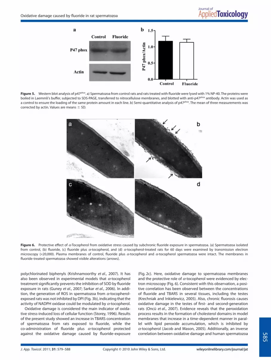
In addition, we determined p47phox, a subunit NADPH oxidaseby western blotting. The amount of p47phox protein in spermato-zoa was not modified by fluoride exposure as compared with thecontrol group (Fig. 5). These results suggest that ROS generationby NADPH oxidase in the fluoride-exposed rats is due to a posi-tive modulation of this enzyme activity.
a-Tocopherol Protects Against the Oxidative DamageCaused by Subchronic Exposure to Fluoride in Spermatozoaas Shown by Transmission Electron Microscopy
Ultrastructural evaluations of spermatozoa from all groups wereperformed via transmission electron microscopy (TEM), and rep-resentative images are shown in Fig. 6. The control anda-tocopherol-exposed spermatozoa exhibited an intact plasmamembrane around the cell (Fig. 6a and d). This normal appear-ance was visibly altered in the plasma membrane along thesperm head spermatozoa from fluoride-treated rats (Fig. 6b). Theco-exposure to fluoride plus a-tocopherol showed a structuralprotection against fluoride-induced plasma membrane damage(Fig. 6c).
a-Tocopherol Prevented the in Vitro Fertilization IncapacityObserved in the Spermatozoa from Fluoride-treated Rats
Next, we examined the ability of spermatozoa co-treated withfluoride plus a-tocopherol to fertilize zona-intact eggs by in vitrofertilization (IVF). As shown in Figs 7 and 8, spermatozoa from ratsexposed to fluoride exhibited a significantly lower ability to fer-tilize eggs compared with the control group (13 � 5.10 vs 72 �4.69), while the co-treatment with fluoride plus a-tocopherolcaused a significant increase in the ability expressed in percent-age of spermatozoa to fertilize eggs compared with fluorideexposure.
Figure 2. a-Tocopherol protects against oxidative stress and oxidativedamage in spermatozoa caused by subchronic fluoride exposure. Effectafter 60 days of fluoride-exposure and fluoride plus a-tocopherol on (a)SOD activity measured by RANSOD assay, (b) ROS generation measuredby DHE/SYTOX green staining and flow cytometry and (c) TBARS forma-tion. Data represent the means � SD of 6 animals per group. *p < 0.05 vs.control, **p < 0.05 vs. fluoride.
583
Oxidative damage caused by fluoride in rat spermatozoa
J. App. Toxicol. 2011; 31: 579–588 wileyonlinelibrary.com/journal/jatwileyonlinelibrary.com/journal/jatCopyright © 2010 John Wiley & Sons, Ltd.

DISCUSSION
In the present study, we evaluated whether NADPH oxidase par-ticipates in the oxidative stress caused by fluoride and examinedthe protective role of a-tocopherol. Oxidative stress is involved inthe etiology of male infertility (Aitken and Baker, 2006). Malereproductive defects in humans (Ortiz-Pérez et al., 2003) andexperimental models have been associated with oxidative stressas a result of fluoride exposure (Ghosh et al., 2002), which pro-duces a significant reduction in the IVF capacity of spermatozoa(Izquierdo-Vega et al., 2008). Moreover, a-tocopherol protectstestes and male accessory sex organs from oxidative stresscaused by fluoride exposure (Sarkar et al., 2006).
We have previously reported that fluoride increased ROS pro-duction in the spermatozoa (Izquierdo-Vega et al., 2008).However, it is clear from the results presented in this study thatNADPH oxidase participates in the oxidative stress caused byfluoride because the generation of ROS in spermatozoa wasblocked by DPI (Fig. 3a). This was accompanied by a significantincrease in the catalytic activity of NADPH oxidase (Fig. 4),without a modification in the amount of p47phox protein in sper-
matozoa from fluoride-exposed rats (Fig. 5b). Many studies havepreviously evaluated the activity of NADPH oxidase in human,equine and rat spermatozoa through generation of ROS inducedby the addition of exogenous calcium ionophore and NADPH andinhibited by DPI (Aitken et al., 1992; 1997; Sabeur and Ball, 2007;Vernet et al., 2001). In addition to mitochondrial sources, an enzy-matic system for ROS generation located in the spermatozoaplasma membrane was identified as NOX. All subunits of NOX(gp91phox, p67 phox, p40phox and p47phox) with the exception ofp22phox were detected by western blot and confocal laser scanmicroscopy in rodent spermatozoa (Shukla et al., 2005); interest-ingly NOX5 does not require p22phox association in its activation(Kawahara et al., 2005). Previously, it was only known that mRNAof NOX5 is expressed in spermatocytes from human testes (Bánfiet al., 2001). Recently, proteins identified in human spermatozoausing LC-MS/MS analysis revealed the presence of DUOX2 inmature spermatozoa (Baker et al., 2007). Kawahara et al. (2007),showed the presence of genes DUOX1 and DUOX2 but not thepresence of the gene NOX5 in rodents. NOX5 and DUOX2 areactivated by calcium and do not appear to require subunits for itsactivation (Bedard and Krause, 2007). In addition, it has beenreported that fluoride increases the concentration of intracellularcalcium [Cai
2+], in spermatozoa (Chinoy et al., 1995). Recently, ithas been shown that hydrogen peroxide positively modulatesNOX5 activation by c-Abl through a calcium-mediated, redox-dependent signaling pathway (El Jamali et al., 2008). Consistentwith this observation, the generation of ROS is an importantmediator of fluoride-induced toxicity (Chouhan and Flora, 2008;Izquierdo-Vega et al., 2008; Sarkar et al., 2006).
In the present study, we evaluated the protective role ofa-tocopherol against oxidative stress caused by fluoride expo-sure. SOD acts as an important line of defense against ROS bycatalyzing the dismutation of O2
.- into oxygen and hydrogen per-oxide. We found that the SOD activity was significantly reducedas a result of fluoride exposure, while the co-administration offluoride plus a-tocopherol prevented the diminution in SODactivity observed in fluoride-exposed rats (Fig. 2a). Additionally,the co-treatment with fluoride plus a-tocopherol prevented theincrease in the generation of ROS observed in spermatozoa fromfluoride-exposed rats (Fig. 2b). In support of these results, it hasbeen previously demonstrated that a-tocopherol inhibits theproduction of ROS in the spermatozoa of rats exposed to
Figure 3. Participation of NADPH oxidase in the generation of ROS in spermatozoa. (a) Effect of fluoride-exposure plus DPI on ROS generation usingfluorescent dyes for flow cytometric analysis. (b) Effect of fluoride exposure plus a-tocopherol plus DPI on ROS generation. Data represent themeans � SD of 6 animals per group. *p < 0.05 vs. group without DPI.
Figure 4. NADPH oxidase activity assay. NADPH oxidase activity inspermatozoa was measured by inducing ROS production with the addi-tion of 150 mM NADPH. Using DHE and SYTOX green, the intensity offluorescence was measured by flow cytometer. These data represent themeans � SD of 6 animals per group. *p < 0.05 vs. control, **p < 0.05 vs.fluoride.
584
J. A. Izquierdo-Vega et al.
J. App. Toxicol. 2011; 31: 579–588wileyonlinelibrary.com/journal/jatwileyonlinelibrary.com/journal/jat Copyright © 2010 John Wiley & Sons, Ltd.
polychlorinated biphenyls (Krishnamoorthy et al., 2007). It hasalso been observed in experimental models that a-tocopheroltreatment significantly prevents the inhibition of SOD by fluorideexposure in rats (Guney et al., 2007; Sarkar et al., 2006). In addi-tion, the generation of ROS in spermatozoa from a-tocopherol-exposed rats was not inhibited by DPI (Fig. 3b), indicating that theactivity of NADPH oxidase could be modulated by a-tocopherol.
Oxidative damage is considered the main indicator of oxida-tive stress-induced loss of cellular function (Storey, 1996). Resultsof the present study showed an increase in TBARS concentrationof spermatozoa from rats exposed to fluoride, while theco-administration of fluoride plus a-tocopherol protectedagainst the oxidative damage caused by fluoride-exposure
(Fig. 2c). Here, oxidative damage to spermatozoa membranesand the protective role of a-tocopherol were evidenced by elec-tron microscopy (Fig. 6). Consistent with this observation, a posi-tive correlation has been observed between the concentrationsof fluoride and TBARS in several tissues, including the testes(Krechniak and Inkielewicz, 2005). Also, chronic fluorosis causesoxidative damage in the testes of first- and second-generationrats (Oncü et al., 2007). Evidence reveals that the peroxidationprocess results in the formation of cholesterol domains in modelmembranes that increase in a time-dependent manner in paral-lel with lipid peroxide accumulation, which is inhibited bya-tocopherol (Jacob and Mason, 2005). Additionally, an inversecorrelation between oxidative damage and human spermatozoa
Figure 5. Western blot analysis of p47phox. a) Spermatozoa from control rats and rats treated with fluoride were lysed with 1% NP-40. The proteins wereboiled in Laemmli’s buffer, subjected to SDS-PAGE, transferred to nitrocellulose membranes, and blotted with anti-p47phox antibody. Actin was used asa control to ensure the loading of the same protein amount in each line. b) Semi-quantitative analysis of p47phox. The mean of three measurements wascorrected by actin. Values are means � SD.
Figure 6. Protective effect of a-Tocopherol from oxidative stress caused by subchronic fluoride exposure in spermatozoa. (a) Spermatozoa isolatedfrom control, (b) fluoride, (c) fluoride plus a-tocopherol, and (d) a-tocopherol-treated rats for 60 days were examined by transmission electronmicroscopy (¥20,000). Plasma membranes of control, fluoride plus a-tocopherol and a-tocopherol spermatozoa were intact. The membranes influoride-treated spermatozoa showed visible alterations (arrows).
585
Oxidative damage caused by fluoride in rat spermatozoa
J. App. Toxicol. 2011; 31: 579–588 wileyonlinelibrary.com/journal/jatwileyonlinelibrary.com/journal/jatCopyright © 2010 John Wiley & Sons, Ltd.

motility has been shown, which was prevented by a-tocopherol.In human spermatozoa, the TBARS formation occurs as a result ofmitochondrial disruption in complex I, suggesting that this per-oxidative damage is induced once intra-mitochondrial antioxi-dant defenses have been lowered (Koppers et al., 2008). Weobserved that the diminution in SOD activity was accompaniedby an increase in the generation of ROS in the spermatozoa offluoride-exposed rats.
In this study, the a-tocopherol treatment did not modify theurinary fluoride concentration (Fig. 1). Similarly, Guney et al.,(2007) observed that the plasma fluoride concentration was notmodified by a-tocopherol, suggesting that the pharmacokineticof fluoride are not altered by this antioxidant. Unfortunately, wedid not evaluate fluoride concentration in spermatozoon.However, Inkielewicz and Krechniak (2003) showed that thetestes concentration of fluoride in rats that drank water with25 ppm of fluoride for 3 months was 4.93-fold greater than in thecontrol group. Interestingly levels of fluoride in rat testes weresimilar to rat urine fluoride levels. The accumulation of fluoride intestes suggests that fluoride is in contact with spermatozoon.
Spermatozoa quality is a major factor in successful IVF. Here,we observed that spermatozoa motility was significantly reducedas a result of fluoride exposure, while spermatozoa motility in theco-administration with fluoride plus a-tocopherol was not differ-ent compared with the control (Table 1), suggesting the partici-pation of ROS in spermatozoa motility. It has been shown thatROS affect spermatozoa motility and ATP concentration througha mechanism independent of oxidative phosphorylation(Armstrong et al., 1999), exerting a direct action on flagella (deLamirande and Gagnon, 1992). High levels of ROS are associatedwith reduced motility and fertilization potential (Aitken et al.,1998).
We previously found that fluoride exposure caused a decreasein spermatozoa IVF capability (Izquierdo-Vega et al., 2008). Here,the importance of the regulation of oxidative stress in fertilizationwas evaluated with the co-exposure of fluoride plusa-tocopherol. a-Tocopherol prevented the oxidative stress andoxidative damage and also the diminution of IVF (Figs 7 and 8).
Supporting these results, a-tocopherol has been shown toimprove sperm motility and fertility in men (Suleiman et al.,1996).
In conclusion, the results of this study indicate that NADPHoxidase participates in the oxidative damage caused by fluorideexposure in rat spermatozoa. a-Tocopherol protects against oxi-dative damage in spermatozoa caused by fluoride exposure.Further studies are required to elucidate the presence of NOXsisoforms present in spermatozoa that could participate as amediator of ROS production spermatozoa by fluoride exposure.
Acknowledgment
The authors are thankful to Angel Barrera-Hernández fromCINVESTAV Mexico for assistance in the animal care and Víctor H.
Figure 7. Effect of a-Tocopherol on infertility caused by fluoride expo-sure in spermatozoa. Spermatozoa isolated from each treatment group(control, fluoride, a-tocopherol and fluoride plus a-tocopherol) weretested for their ability to fertilize zona-intact eggs in vitro. A total of 120eggs were examined for each experimental group over the course ofthree independent experiments. *p < 0.05 vs. the control group,**p < 0.05 vs. fluoride.
Figure 8. Effect of a-Tocopherol on IVF capability. Representativephase contrast (a, c, e and g) and fluorescence (b, d, f and h) micrographs(¥4000). Zona-intact eggs were incubated for 4 h with spermatozoa fromrats of different experimental groups (see Materials and Methods), andstained with Hoechst 33342. (a, b) Egg fertilized with control spermato-zoa. (c, d) Egg fertilized with spermatozoa exposed to fluoride for 60 days;no evidence of swollen spermatozoa nuclei was observed in egg cyto-plasm. (e, f ) Egg fertilized with spermatozoa exposed to fluoride plusa-tocopherol for 60 days and (g, h) Egg fertilized with spermatozoaexposed to a-tocopherol for 60 days. Arrows show a swollen spermatozoanucleus in the egg cytoplasm.
586
J. A. Izquierdo-Vega et al.
J. App. Toxicol. 2011; 31: 579–588wileyonlinelibrary.com/journal/jatwileyonlinelibrary.com/journal/jat Copyright © 2010 John Wiley & Sons, Ltd.

Rosales-García for helping with flow cytometry. The authors alsothank Rodolfo Paredes-Díaz from Electronic Microscopic Unit ofCellular Physiology of UNAM, Mexico for helping with electronmicroscopic analysis. J.A.I.V. was recipient of a CONACYT Mexicoscholarship (170210).
REFERENCES
Aitken RJ, Baker MA. 2006. Oxidative stress, sperm survival and fertilitycontrol. Mol. Cell. Endocrinol. 250: 66–69.
Aitken RJ, Buckingham DW, West KM. 1992. Reactive oxygen species andhuman spermatozoa: analysis of the cellular mechanisms involved inluminol- and lucigenin-dependent chemiluminescence. J. Cell Physiol.151: 466–477.
Aitken RJ, Fisher HM, Fulton N, Gomez E, Knox W, Lewis B, Irvine S. 1997.Reactive oxygen species generation by human spermatozoa isinduced by exogenous NADPH and inhibited by the flavoproteininhibitors diphenylene iodonium and quinacrine. Mol. Reprod. Dev.47: 468–482.
Aitken RJ, Gordon E, Harkiss D, Twigg JP, Milne P, Jennings Z, Irvine DS.1998. Relative impact of oxidative stress on the functional compe-tence and genomic integrity of human spermatozoa. Biol. Reprod. 59:1037-1046.
Armienta MA, Segovia N. 2008. Arsenic and fluoride in the groundwater ofMexico. Environ. Geochem. Health 30: 345–353.
Armstrong JS, Rajasekaran M, Chamulitrat W, Gatti P, Hellstrom WJ, SikkaSC. 1999. Characterization of reactive oxygen species induced effectson human spermatozoa movement and energy metabolism. FreeRadic. Biol. Med. 26: 869–880.
Baker MA, Reeves G, Hetherington L, Müller J, Baur I, Aitken RJ. 2007.Identification of gene products present in Triton X-100 soluble andinsoluble fractions of human spermatozoa lysates using LC-MS/MSanalysis. Prot. Clin. Appl. 1: 524–532.
Bánfi B, Molnár G, Maturana A, Steger K, Hegedûs B, Demaurex N, KrauseKH. 2001. A Ca(2+)-activated NADPH oxidase in testis, spleen, andlymph nodes. J. Biol. Chem. 276: 37594–37601.
Bedard K, Krause KH. 2007. The NOX family of ROS-generating NADPHoxidases: physiology and pathophysiology. Physiol. Rev. 87: 245–313.
Bendahmane M, Zeng HT, Tulsiani DR. 2002. Assessment of acrosomalstatus in rat spermatozoa: studies on carbohydrate and non-carbohydrate agonists. Arch. Biochem. Biophys. 404: 38–47.
Bourges-Rodríguez H. 2008. Aspectos nutriológicos de la vitamina E. InRadicales libres y estrés oxidativo: Aplicaciones Médicas, KonigsbergFainstein M (ed.). Manual Moderno: México; 595–611.
Buege JA, Aust SD. 1978. Microsomal lipid peroxidation. Meth. Enzymol.52: 302–310.
Cachia O, Benna JE, Pedruzzi E, Descomps B, Gougerot-Pocidalo MA,Leger CL. 1998. Alpha-tocopherol inhibits the respiratory burst inhuman monocytes. Attenuation of p47(phox) membrane transloca-tion and phosphorylation. J. Biol. Chem. 273: 32801–32805.
Chinoy NJ, Narayana MV, Dalal V, Rawat M, Patel D. 1995. Amelioration offluoride toxicity in some accessory reproductive glands and sperma-tozoa of rat. Fluoride 28: 75–86.
Chouhan S, Flora SJ. 2008. Effects of fluoride on the tissue oxidative stressand apoptosis in rats: biochemical assays supported by IR spectros-copy data. Toxicology 254: 61–67.
Chlubek, D, Poland S. 2003. Fluoride and oxidative stress. Fluoride 36:217–228.
De Iuliis GN, Wingate JK, Koppers AJ, McLaughlin EA, Aitken RJ. 2006.Definitive evidence for the nonmitochondrial production of superox-ide anion by human spermatozoa. J. Clin. Endocrinol. Metab. 91: 1968–1975.
de Lamirande E, Gagnon C. 1992. Reactive oxygen species and humanspermatozoa. I. Effects on the motility of intact spermatozoa and onsperm axonemes. J. Androl. 13: 368–378.
Del Razo LM, Corona JC, García-Vargas G, Albores A, Cebrián ME. 1993.Fluoride levels in well-water from a chronic arsenicism area of North-ern Mexico. Environ. Pollut. 80: 91–94.
El Jamali A, Valente AJ, Lechleiter JD, Gamez MJ, Pearson DW, NauseefWM, Clark RA. 2008. Novel redox-dependent regulation of NOX5 bythe tyrosine kinase c-Abl. Free Radic. Biol. Med. 44: 861–881.
Freni SC. 1994. Exposure to high fluoride concentrations in drinking wateris associated with decreased birth rates. J. Toxicol. Environ. Health 42:109-112.
Ghosh D, Das (Sarkar) S, Maiti R, Jana D, Das UB. 2002. Testicular toxicity insodium fluoride treated rats; association with oxidative stress. Reprod.Toxicol. 16: 383–390.
Guney M, Oral B, Demirin H, Karahan N, Mungan T, Delibas N. 2007.Protective effects of vitamins C and E against endometrial damageand oxidative stress in fluoride intoxication. Clin. Exp. Pharmacol.Physiol. 34: 467–474.
Guo XY, Sun GF, Shenyang YS. 2003. Oxidative stress from fluoride-induced hepatotoxicity in rats. Fluoride 36: 25–29.
Gupta SK, Mithal A, Das BK. 1993. Skeletal scintigraphic findings inendemic skeletal fluorosis. Nucl. Med. Commun. 14: 384–390.
Hardisson A, Rodríguez MI, Burgos A, Díaz Flores L, Gutiérrez R, Várela H.2001. Fluoride levels in publicly supplied and bottled drinking waterin the island of Tenerife, Spain. Bull. Environ. Contam. Toxicol. 67: 163–170.
Inkielewicz I, Krechniak J. 2003. Fluoride content in soft tissues and urineof rats exposed to sodium fluoride in drinking water. Fluoride 36:263–266.
Izquierdo-Vega JA, Sánchez-Gutiérrez M, Del Razo LM. 2008. Decreased invitro fertility in male rats exposed to fluoride-induced oxidative stressdamage and mitochondrial transmembrane potential loss. Toxicol.Appl. Pharmacol. 230: 352–357.
Jacob RF, Mason RP. 2005. Lipid peroxidation induces cholesterol domainformation in model membranes. J. Biol. Chem. 280: 39380–39387.
Kawahara BT, Quinn MT, Lambeth JD. 2007. Molecular evolution of thereactive oxygen-generating NADPH oxidase (Nox/Duox) family ofenzymes. BMC Evol. Biol. 109: 1–21.
Kawahara T, Ritsick D, Cheng G, Lambeth JD. 2005. Point mutations in theproline-rich region of p22phox are dominant inhibitors of Nox1- andNox2-dependent reactive oxygen generation. J. Biol. Chem. 280:31859–31869.
Koppers AJ, De Iuliis GN, Finnie JM, McLaughlin EA, Aitken RJ. 2008. Sig-nificance of mitochondrial reactive oxygen species in the generationof oxidative stress in spermatozoa. J. Clin. Endocrinol. Metab. 93: 3199–3207.
Krechniak J, Inkielewicz I. 2005. Correlations between fluoride concentra-tion and free radical parameters in soft tissues of rats. Fluoride 38:293–296.
Krishnamoorthy G, Venkataraman P, Arunkumar A, Vignesh RC, AruldhasMM, Arunakaran J. 2007. Ameliorative effect of vitamins (alpha-tocopherol and ascorbic acid) on PCB (Aroclor 1254) induced oxida-tive stress in rat epididymal sperm. Reprod. Toxicol. 23: 239–245.
National Research Council. 2006. Fluoride in Drinking-water. A ScientificReview of EPA’s Standards. National Academies Press: Washington, DC.
Oncü M, Kocak A, Karaoz E, Darici H, Savik E, Gultekin F. 2007. Effect oflong-term fluoride exposure on lipid peroxidation and histology oftestes in first- and second-generation rats. Biol. Trace Elem. Res. 118:260–268.
Ortiz-Pérez D, Rodríguez-Martínez M, Martínez F, Borja-Aburto VH,Castelo J, Grimaldo JI, de la Cruz E, Carrizales L, Diaz-Barriga F. 2003.Fluoride-induced disruption of reproductive hormones in menEnviron. Res. 93: 20–30.
Pauwels H, Ahmed S. 2007. Fluoride in ground origin and health impacts.Géosciences 5: 68–72.
Sabeur K, Ball BA. 2007. Characterization of NADPH oxidase 5 in equinetestis and spermatozoa. Reproduction 134: 263–270.
Sarkar SD, Maiti R, Ghosh D. 2006. Management of fluoride induced tes-ticular disorders by calcium and vitamin-E co-administration in thealbino rat. Reprod. Toxicol. 22: 606–612.
Shanthakumari D, Srinivasalu S, Subramanian S. 2004. Effect of fluorideintoxication on lipid peroxidation and antioxidant status in experi-mental rats. Toxicology 204: 219–228.
Shivashankara AR, Shivaraja Shankara YM, Hanumanth Rao S,Gopalakrishna Bhat P. 2000. A clinical and biochemical study ofchronic fluoride toxicity in children of Kheru Thanda of gulbarga dis-trict, Karnataka India. Fluoride 33: 66–73.
Shukla S, Jha RK, Laloraya M, Kumar PG. 2005. Identification of nonmito-chondrial NADPH oxidase and the spatio-temporal organization of itscomponents in mouse spermatozoa. Biochem. Biophys. Res. Commun.331: 476–483.
Storey K. 1996. Oxidative stress: animal adaptations in nature. Brazilian J.Med. Biol. Res. 29: 1715–1733. 587
Oxidative damage caused by fluoride in rat spermatozoa
J. App. Toxicol. 2011; 31: 579–588 wileyonlinelibrary.com/journal/jatwileyonlinelibrary.com/journal/jatCopyright © 2010 John Wiley & Sons, Ltd.

Suleiman SA, Ali ME, Zaki ZM, el-Malik EM, Nasr MA. 1996. Lipid peroxi-dation and human sperm motility: protective role of vitamin E. J.Androl. 17: 530–537.
Varga Z, Kosaras E, Komodi E, Katko M, Karpati I, Balla J, Paragh G, Aisa MC,Galli F. 2008. Effects of tocopherols and 2,2’-carboxyethyl hydroxy-chromans on phorbol-ester-stimulated neutrophils. J. Nutr. Biochem.19: 320–327.
Vernet P, Fulton N, Wallace C, Aitken RJ. 2001. Analysis of reactive oxygenspecies generating systems in rat epididymal spermatozoa. Biol.Reprod. 65: 1102-1113.
Wang SX, Wang ZH, Cheng XT, Li J, Sang ZP, Zhang XD, Han LL, Qiao XY,Wu ZM, Wang ZQ. 2007. Arsenic and fluoride exposure in drinking
water: children’s IQ and growth in Shanyin County, Shanxi province,China. Environ. Health Perspect. 115: 643–647.
Wang YY, Zhao BL, Li XJ, Su Z, Xi WJ. 1997. Spin trapping techniquestudies on active oxygen radicals from human polymorphonuclearleukocytes during fluoride-stimulated respiratory burst. Fluoride 30:5–15.
WHO. 2001. Laboratory Manual for the Examination of Human Semen andSemen Cervical Mucus Interaction, 4th edn. Cambridge UniversityPress: Cambridge; 1–106.
588
J. A. Izquierdo-Vega et al.
J. App. Toxicol. 2011; 31: 579–588wileyonlinelibrary.com/journal/jatwileyonlinelibrary.com/journal/jat Copyright © 2010 John Wiley & Sons, Ltd.