tumor-associated trypsin participates in cancer cell-mediated degradation of extracellular matrix
TRANSCRIPT

1991;51:2107-2112. Published online April 1, 1991.Cancer Res Erkki Koivunen, Ari Ristimäki, Outi Itkonen, et al. Degradation of Extracellular MatrixTumor-associated Trypsin Participates in Cancer Cell-mediated
Updated Version http://cancerres.aacrjournals.org/content/51/8/2107
Access the most recent version of this article at:
Citing Articles http://cancerres.aacrjournals.org/content/51/8/2107#related-urls
This article has been cited by 13 HighWire-hosted articles. Access the articles at:
E-mail alerts related to this article or journal.Sign up to receive free email-alerts
SubscriptionsReprints and
[email protected] atTo order reprints of this article or to subscribe to the journal, contact the AACR Publications
To request permission to re-use all or part of this article, contact the AACR Publications
American Association for Cancer Research Copyright © 1991 on July 10, 2011cancerres.aacrjournals.orgDownloaded from
(CANCER RESEARCH 51. 2107-2112, April 15. 1991]
Tumor-associated Trypsin Participates in Cancer Cell-mediated Degradation ofExtracellular Matrix1
Erkki Koivunen,2 Ari Ristimäki,Outi Itkonen, Sirpa Osman, Matti Vuento, and Ulf-Hákan Stenman
Department of Obstetrics and Gynecology, Helsinki L'nirersity Central Hospital, Haartmaninkatu 2, SF-00290 Helsinki [E. K., A, R„O. I., S. O,, U-H. SJ, andDepartment of Biochemistry, L'nirersity of Helsinki, L'nioninkatu 35, SF-OOI70 Helsinki [M. V.I.Finland
ABSTRACT
We have recently demonstrated that many cancer cell lines produce anovel trypsinogen isoenzyme called tumor-associated trypsinogen 2(TAT-2). It was found during a search of the target protease for tumor-associated trypsin inhibitor (TATI). We now show that degradation ofsubendothelial cell extracellular matrix (ECM) by four different cell lines(COLO 205 colon carcinoma, K-562 erythroleukemia, CAPAN-1 pancreatic carcinoma, and HT I080 fibrosarcoma) can be partially inhibitedby TATI or neutralizing trypsin antibodies. When cells were cultured inserum-free medium on ECM, TATI and trypsin antibodies inhibited therelease of immunoreactive fibronectin fragments from ECM by 47-54and 40%, respectively. Degradation of isotopically labeled ([-'Miserine,|3H)proline, and (35S|sulfate) ECM was also significantly prevented by
TATI. At its maximum, it exerted a 57% inhibition on the degradationof [3H|serine-labeled ECM. Plasminogen added exogenously to the cul
ture medium further potentiated the proteolysis of ECM. Interestingly,addition of enteropeptidase, an activator of TAT-2, also enhanced cell-mediated proteolysis as assessed by degradation of purified fibronectincoated onto the surface of wells. Immunoblot analysis showed thatenteropeptidase-mediated proteolysis generated a pattern of fibronectinfragments similar to that obtained by digestion of purified fibronectin byTAT-2. These results demonstrate the existence of a proteolytic systemin tumor cells which is dependent on the activation of TAT-2. We suggestthat TAT-2 is involved in a protease cascade-stimulating tumor cellinvasion and degradation of extracellular matrix.
INTRODUCTION
Production of proteolytic enzymes by malignant tumor cellsis believed to be essential to the ability of the tumor to invadeand degrade extracellular matrix. Of the proteases secreted bytumor cells, collagenases (1), plasminogen activators (2), tran-sin/stromelysin (3), and cathepsins (4-6) have been the mostthoroughly studied. We have recently characterized a noveltumor-associated protease, TAT,3 (7, 8). It was initially identi
fied in the cyst fluid of mucinous ovarian tumors during asearch of the target protease for TATI, which is a marker formucinous ovarian cancer (9). TAT occurs as two isoenzymes,TAT-1 and TAT-2. They are identical to the correspondingpancreatic trypsins with respect to amino-terminal amino acidsequence, molecular weight, and immunoreactivity, but theydiffer with respect to substrate specificity, enzyme stability, andelution pattern in ion-exchange chromatography. At present itis not known whether these differences are explained by tissue-specific modification of trypsin or whether distinct genes codefor trypsinogens derived from tumors and pancreas.
The levels of type IV collagenase (10), plasminogen activators
Received 7/31/90; accepted 2/4/91.The costs of publication of this article were defrayed in part by the payment
of page charges. This article must therefore be hereby marked advertisement inaccordance with 18 U.S.C. Section 1734 solely to indicate this fact.
1This work was supported by grants from the Academy of Finland, the Finnish
Cancer Society, the Finnish Social Security Institution, the Jenny and AnttiWihuri Foundation, and the Ida Montin Foundation.
2To whom requests for reprints should be addressed.3The abbreviations used are: TAT, tumor-associated trypsin(ogen); TATI,
tumor-associated trypsin inhibitor; u-PA, urokinase-type plasminogen activator;ECM, extracellular matrix: EACA. «-aminocaproicacid; PBS. phosphate-bufferedsaline.
(11), and cathepsin B (4) have been found to correlate withinvasiveness and the metastatic ability of tumor cell lines. Inextracts of tumor tissues the levels of urokinase-type plasminogen activator (u-PA) generally correlate with the degree ofmalignancy of the tumors (12). It is interesting that TAT-2, themajor TAT isoenzyme, also shows close association with malignancy; the levels are higher in malignant and borderlineovarian tumor cyst fluids than in benign ones (13). In addition,purified TAT-1 and TAT-2 activate pro-u-PA in vitro (8). Theseresults suggest that TAT-2 may promote the dissemination oftumor cells.
TAT-2 zymogen is produced by many cancer cell lines, andit has been purified from the culture medium of COLO 205colon adenocarcinoma and HT 1080 fibrosarcoma cell lines(14). In the present study we have examined the role of TAT-2in cell-mediated degradation of extracellular matrix. We developed a method for determining degradation of fibronectin basedon analysis of immunoreactive fibronectin fragments liberatedby tumor cells growing on protein-coated surfaces. We alsoanalyzed the degradation of isotopically labeled ECM. Theresults show that tumor cell-mediated proteolysis can be significantly abrogated by inhibiting TAT-2.
MATERIALS AND METHODS
Chemicals. Reagents were obtained as follows: aprotinin from SigmaChemical Co. (St. Louis, MO); EACA from Fluka AG (Buchs, Switzerland); RPMI 1640, Medium 199, PBS (without magnesium andcalcium), and fetal bovine serum from Flow Laboratories (Irvine, Scotland); pooled human serum from the Finnish Red Cross Blood Transfusion Service (Helsinki, Finland); L-glutamine and antibiotics fromGIBCO Laboratories (Grand Island, NY); and gelatin from Merck(Darmstadt, Germany).
Proteinases. Human plasminogen and plasmin were obtained fromKabi Vitrum (Stockholm, Sweden). Porcine enteropeptidase and bovinetrypsin were obtained from Sigma. Enteropeptidase was dissolved inPBS and passed through an affinity column of TATI-Sepharose 4B toremove contaminating trypsin-like enzymes. TAT-2 was purified by amonoclonal antibody affinity column from serum-free culture mediumof COLO 205 cells as described (14). TAT-2 was obtained in zymogenform and autoactivated by neutralizing the pH.
Purification of TATI. TATI was purified by a novel affinity chromatography method from the urine of cancer patients. The pH of the urinewas adjusted to 7.4, and it was dialyzed against water with a hollowfiber dialyzer (15) until the conductivity corresponded to that of 50 mMTris, pH 7.4. Dialyzed urine was then applied at a flow rate of 60 ml/h at 4°Cto a Q Sepharose anion-exchange column (Pharmacia, Upps
ala, Sweden) and an affinity column of bovine trypsin-Sepharose 4Bpreviously equilibrated with Tris buffer. Most urine proteins wereremoved by the first anion-exchange column. After the trypsin-Sepharose column was washed with Tris buffer containing l M NaCI, 0.1%Brij 35, and 1% 2-propanol, TATI was eluted with 0.1 % trifluoroaceticacid. TATI was finally purified by reversed-phase chromatography ona C,«column with a linear acetonitrile gradient (from 10 to 60% in 20min) in 0.1% trifluoroacetic acid. By this method, 2 mg of TATI wereobtained from 1 liter of urine.
Anticatalytic Antibodies. The IgG fraction was prepared from a rabbitpreimmune serum and antiserum against human pancreatic trypsin 1
2107
American Association for Cancer Research Copyright © 1991 on July 10, 2011cancerres.aacrjournals.orgDownloaded from
TUMOR CELL TRYPSIN AND MATRIX DEGRADATION
(8) by sequential affinity Chromatograph) on Protein G-Sepharose andprotein A-Sepharose (Pharmacia). Anti-u-PA IgG was prepared fromgoat antiserum against human low-molecular-weight u-PA (BiopoolAB, Umeà , Sweden) by using protein A-Sepharose. Anti-trypsin IgGand anti-u-PA IgG were anticatalytic. because they inhibited the ami-dolytic activity of TAT-2 and u-PA, respectively.
Cell Culture. Human cancer cell lines COLO 205 colon adenocarci-noma (ATCC CCL 222). HT 1080 fibrosarcoma (ATCC CCL 121), K-562 erythroleukemia (ATCC CCL 243), and CAPAN-1 pancreaticadenocarcinoma (ATCC HTB 79) were obtained from the AmericanType Culture Collection (Rockville. MD). Cells were cultured in 800-ml Nunclon flasks (Roskilde, Denmark) and maintained in RPMI 1640supplemented with 10% heat-inactivated fetal bovine serum, 2 mM L-glutamine. 100 units/ml penicillin, and 100 ^g/ml streptomycin. Treatment with trypsin was not required for detachment of the adherentcells, possibly because the cells produce their own trypsin. After serumwas removed, and the flasks were washed twice with PBS, cells wereincubated for 10-15 min in PBS at room temperature. The flasks werevigorously shaken until all cells detached.
Preparation of Wells Coated with ECM and Kibronectin. Endothelialcells were isolated from human umbilical cord veins by the methodoriginally described by Jaffe et al. (16), and ECM was prepared essentially as described (17. 18). Endothelial cells were seeded in Nunclon24-well multidishes, which were previously coated with gelatin, andcultured in Medium 199 containing 20% human serum. Labelingmedium containing 5 ¿iCi/inlpHlserine, 5 ^Ci/ini ['Hjproline, or 20fiCi/ml [<5S]sulfate was supplemented with 20 ¿ig/mlfresh ascorbate
added daily. Labeling medium was replaced after 3 days of culture.After culturing for 5 to 7 days, cells were lysed by addition of 30 mMNHjOH for 30 min. The wells were washed with PBS, H2O. and 70%ethanol. To prepare wells coated with fibronectin, Nunclon 24-wellmultidishes were incubated with fibronectin (2 Mg/ml in PBS, 1 ml/well) overnight at 37°C.Before use, the wells were washed twice with
PBS. Fibronectin was purified from human plasma as described (19).Degradation of Fibronectin and Labeled ECM. Cells were plated at
varying densities in coated wells as described in the text and culturedin serum-free RPMI. In some experiments plasminogen and enteropep-tidase were added at final concentrations of 6 and I ¿ig/ml,respectively.Immune and preimmune IgG were used at concentrations of 10-25 /jg/ml. The concentrations of TATI and aprotinin were 1-10 ^g/ml, andthat of EACA was 330 ¿ig/ml.Aliquots of 50 and 200 ß\were collectedat various time points, centrifuged at 9000 rpm for 5 min, and analyzedby fibronectin immunoassay and ^-scintillation counting, respectively.
Detection of Fibronectin Immunoreactivity. Polyclonal antibodieswere prepared from rabbit antiserum against fibronectin (20) by Na2SO4precipitation and used to develop a time-resolved immunofluorometricassay according to methods previously described (21). In a typical assayof cell culture media, the samples were incubated for 60 min at 25°C
in microtiter wells coated with the fibronectin antibodies. After washing, the wells were incubated for 30 min with fibronectin antibodiesthat had been labeled with a europium chelate (Wallac Oy, Turku,Finland) (22). After washing of the wells and addition of an "enhancement solution" (22), the bound europium was measured with an Arcus
1230 time-resolved fluorometer (Wallac). Using a 50-^1 sample volume,the linear measuring range of the assay was 0.5-250 ng/ml of fibronectin, and the coefficient of variation was below 10% in this concentrationrange.
Characterization of Fibronectin Fragments. The culture media weredialyzed against 5 mM NH4HCO, for 2 h. lyophilized, and analyzed bygel filtration and immunoblotting. Gel filtration was performed on aSuperóse 12 column (Pharmacia) at a flow rate of 30 ml/h. using 50mM Tris-HCI buffer (pH 7.4) containing 0.1 M NaCI and 0.02% NaN3.Fractions of 400 n\ were collected and analyzed by fibronectin immunoassay. Sodium dodecyl sulfate gel electrophoresis was performed on7.5% polyacrylamide gels under nonreducing conditions (23). Proteinswere electrotransferred to nitrocellulose membranes (24), and fibronectin fragments were detected by incubating with polyclonal fibronectinantibodies followed by secondary peroxidase-conjugated swine anti-rabbit antibodies (Dakopatts, Glostrup, Denmark). Peroxidase stainingwas performed with 3.3'-diaminobenzine tetrahydrochloride (Fluka).
RESULTS
The adherent tumor cell lines used in this study can easily bedetached from culture dishes by removing the serum and incubating the cells in PBS. When COLO 205 cells collected thisway were plated in serum-free medium in fibronectin-coatedwells, immunoreactive fibronectin was released into the serum-free medium as detected by fibronectin immunoassay (Fig. 1).Control experiments showed that fibronectin immunoreactivitywas not released into the medium in the absence of cells. Inaddition, COLO 205 cells themselves were not found to secretefibronectin.
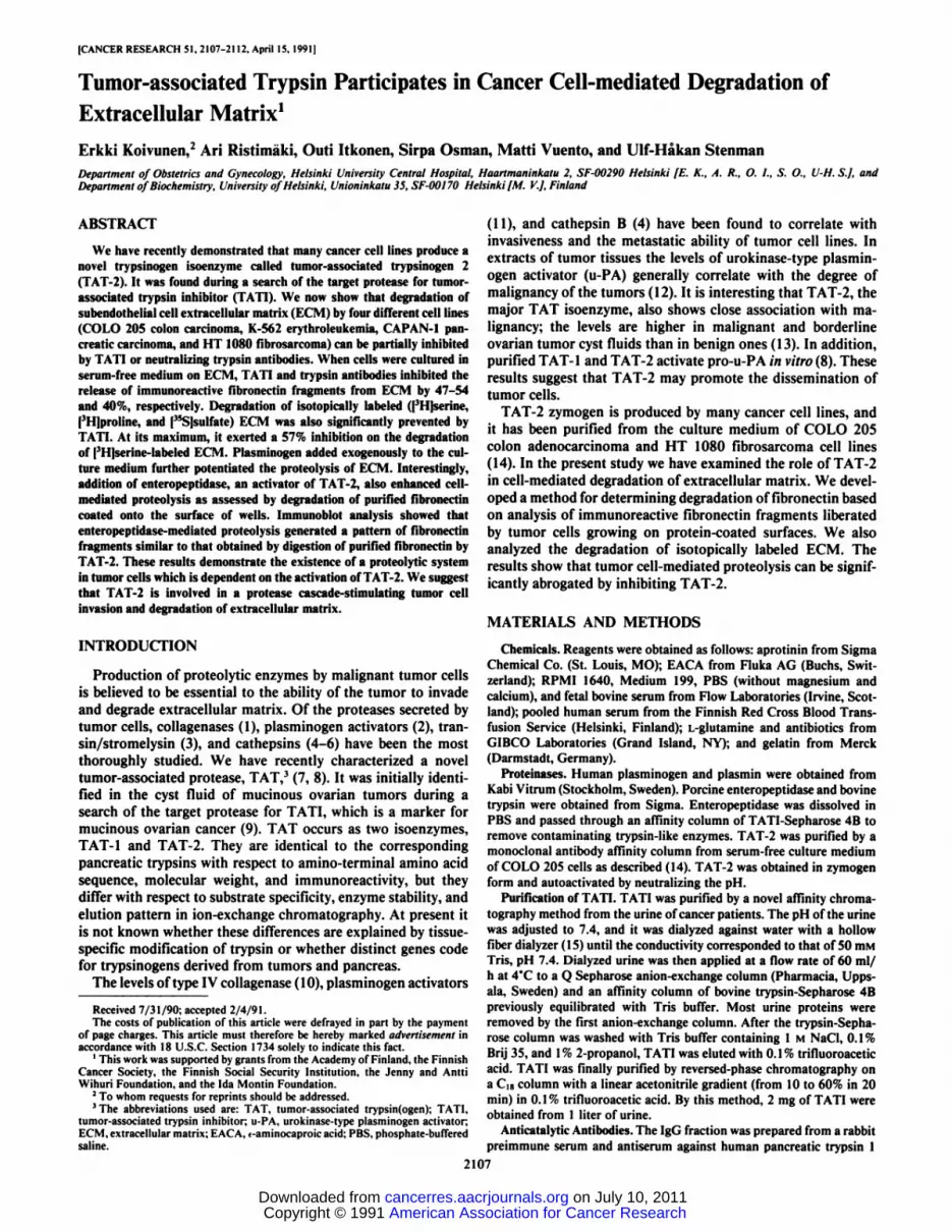
The cell-mediated release of coated fibronectin could besignificantly increased by adding plasminogen zymogen or active enteropeptidase to the culture medium (Fig. 1). Fibronectinimmunoreactivity was readily detectable after a 4-h culture andincreased further over 65 h. Control experiments indicated thatplasminogen and enteropeptidase alone at the low concentrations used (6 and 1 Mg/ml, respectively) did not cause degradation of coated fibronectin during a 24-h incubation. However,fibronectin appears to be sensitive to higher concentrations ofenteropeptidase (20 ¿ig/ml),as demonstrated by release of fibronectin immunoreactivity from the wells (not shown).
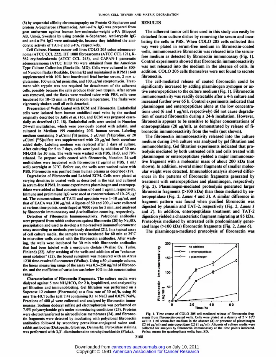
The fibronectin immunoreactivity released into the culturemedium during 24-h culture was analyzed by gel filtration andimmunoblotting. Gel filtration experiments indicated that pro-teolysis mediated by both untreated cells and cells treated withplasminogen or enteropeptidase yielded a major immunoreactive fragment with a molecular mass of about 200 kDa (notshown). In addition, several minor fragments of smaller molecular weight were detected. Immunoblot analysis showed differences in the patterns of fibronectin fragments generated bytreatment with enteropeptidase and plasminogen, respectively(Fig. 2). Plasminogen-mediated proteolysis generated largerfibronectin fragments (>100 kDa) than those mediated by enteropeptidase (Fig. 2, Lanes 4 and 5). A similar difference infragment pattern was found when purified fibronectin wasdigested by plasmin and TAT-2, respectively (Fig. 2, Lanes 1and 2). In addition, enteropeptidase treatment and TAT-2digestion yielded a characteristic fragment migrating at 85 kDa.Proteolysis mediated by untreated cells predominantly generated large (>100 kDa) fibronectin fragments (Fig. 2, Lane 6).
The plasminogen-mediated proteolysis of fibronectin was
oa4)wOC3EE
200
150
100
no additions
-f enteropeptidase
+ plasminogen
20 40Time(h)
60
Fig. I. Time course of COLO 205 cell-mediated release of fibronectin fragments from fibronectin-coated wells. Cells were plated at a density of 2 X IO*/well in 1 ml serum-free medium in the absence (X) or presence of plasminogen(O) (6 fjg/ml) and enteropeptidase (D) (l ¿ig/ml).Aliquots of culture media werecollected for analysis by fibronectin immunoassay at the time points indicated.Points, means for quadruplicate wells; bars. SD.
2108
American Association for Cancer Research Copyright © 1991 on July 10, 2011cancerres.aacrjournals.orgDownloaded from

TUMOR CELL TRYPSIN AND MATRIX DEGRADATION
inhibited by aprotinin, EACA, and anticatalytic polyclonal u-PA IgG, but not by TATI or anticatalytic polyclonal trypsinIgG (Table 1). By contrast, TATI and anti-trypsin IgG inhibitedboth the enteropeptidase-mediated proteolysis and the prote-olysis mediated by untreated cells (Table 1). TATI and anti-trypsin IgG caused 81 and 92% inhibition on the enteropeptidase-mediated proteolysis, respectively, and 75 and 32% inhibition on the proteolysis mediated by untreated cells, respectively. Preimmune IgG had no effect.
When COLO 205 cells were plated on ECM deposited byhuman umbilical vein endothelial cells, proteolysis of fibronec-tin was very rapid (Fig. 3). Fibronectin immunoreactivity wasdetected in the medium after a 1-h culture and further increasedfor 24 h. After a 6-h culture, large (>200 kDa) fibronectinfragments predominated in the culture medium, as detected by
500
5 6
200i
30
Fig. 2. Immunoblot analysis of fibronectin fragments released by cells fromcoated wells or generated by digestion of purified fibronectin with TAT-2 andplasmin. Lane I, fibronectin digested for l h with plasmin (0.025 casein unit);Lane 2, fibronectin digested for l h with TAT-2 (IO ng); Lane 3, untreatedfibronectin; Lane 4, culture medium of plasminogen-treated cells; Lane 5, culturemedium of enteropeptidase-treated cells; Lane 6. culture medium of untreatedcells. Asterisks, 85-kDa fragment characteristic of enteropeptidase- and TAT-2-mediated proteolysis.
Table 1 Effect of protease inhibitors and anticatalytic protease antibodies onCOLO 205 cell-mediated release of fibronectin fragments from fibronectin-coated
wellsci of inhibition with following additions to
serum-free culture medium"
Inhibitor" None Enteropeptidase Plasminogen
TATIAprotininEACAAnti-trypsin
IgGPreimmuneIgGAnti-u-PA
IgG7580N32306.2'8.4D"5.91.28.08182N92041.22.4D5.56.23.92
±3.589±6.968±0.40±0.8ND49
±1.5°TATI and aprotinin »ereused at a concentration of 1 ^g/ml. EACA at a
concentration of 330 fig/ml, and prcimmune and immune IgG at a concentrationof 10 fig/ml.
* Fibronectin immunoreactivity released into the culture medium was determined by the fibronectin immunoassay after culturing for 16-24 h. The inhibitionis given as the decrease of the immunoreactivity compared with that observed inthe absence of inhibitors. Cells (2 x 10*) were plated on fibronectin-coated wellsin the presence or absence of plasminogen and enteropeptidase in serum-freemedium.
c Mean ±SD for at least 2 experiments with 2 wells/experiment.d ND. not determined.
246Time (h)
Fig. 3. Inhibition of COLO 205 cell-mediated degradation of ECM fibronectinby trypsin antibodies. Cells were plated on ECM-coated wells at a density of 2 xlO'/well and incubated in 1 ml serum-free medium in the absence (O) or presenceof anti-trypsin IgG (•)and preimmune IgG (Ü).Control wells were incubatedwithout cells (X). The concentrations of immune and preimmune IgG were 25Mg/ml. Aliquots of culture media were collected for analysis by fibronectinimmunoassay at 2-h intervals. Points, means for quadruplicate measurements;ears. SD.
800
o 600-
E 400
_ 200
COLO 205 K-562 CAPAN-1
Cancer cell line
Fig. 4. Prevention of cancer cell-mediated degradation of ECM fibronectin byTATI. COLO 205. K-562. and CAPAN-I cells were seeded at densities of 2 x10'. 5 x IO5,and 2.5 x 10⁵/well. respectively, in 0.5 ml serum-free medium. Cells
were cultured in the absence (•)or presence (EH)of TATI, and the culture mediawere analyzed for fibronectin immunoreactivity. COLO 205 cells were incubatedfor 4 h, and K-562 and CAPAN-1 cells were incubated overnight. The concentration of TATI was 10 ^g/ml. Columns, means from triplicate wells; bars, SD.Untreated control wells did not release fibronectin immunoreactivity.
immunoblotting (not shown). TATI and anti-trypsin IgG inhibited degradation by 51 and 40%, respectively (Figs. 3 and 4).The proteolysis caused by two other cell lines which produceTAT-2, K-562 (14) and CAPAN-1 was similarly inhibited byTATI (Fig. 4). TATI prevented K-562 and CAPAN-1 cell-mediated proteolysis by 47 and 54%, respectively.
TATI was also effective in inhibiting the destruction of iso-topically labeled ECM, when cells were cultured under serum-free conditions in the absence of plasminogen. TATI added ata concentration of 10 Mg/ml reduced the release of ['H]prolinelabel from ECM by all three cell lines studied. After a 24-hincubation, TATI prevented the proteolysis mediated by COLO205, CAPAN-1, and HT 1080 by 40, 48, and 35%, respectively(Fig. SB). The degradation of ECM labeled with ['Hjserine
could be inhibited by TATI in COLO 205 and in CAPAN-1cells (Fig. 5A). The percentage of inhibition was 57 and 51%,respectively. However, TATI did not prevent HT 1080 cell-mediated degradation of ['Hjserine-labeled ECM (data not
shown). It is notable that of the three cell lines studied, HT1080 hydrolyzed the matrix most weakly, and it secreted thelowest amount of TAT-2 as analyzed by TAT-2 immunoassay(not shown). The degradation of [35S]sulfate-labeled ECM could
2109
American Association for Cancer Research Copyright © 1991 on July 10, 2011cancerres.aacrjournals.orgDownloaded from
TUMOR CELL TRYPSIN AND MATRIX DEGRADATION
2000
1500-
1000
500
COLO205 CAPAN-1 COLO 205 CAPAN 1 HT 1080 COLO205
Cancer cell line
Fig. 5. Inhibition of cancer cell-mediated destruction of isotopically labeledmatrices by TATI. A, ¡'HJserine-labeled ECM: B. [3H|proline-labeIed ECM: C.|35S]sulfate-labeled ECM. COLO 205 and HT 1080 cells were seeded at a densityof 5 x lO'/well, and CAPAN-1 cells at a density of 2.5 x lO'/well in 0.5 mlserum-free RPMI medium. Cells were cultured for 24-48 h in the absence (•)orpresence (D) of TATI, and the isotope label released into the medium wasmeasured by ¿-scintillation counting. The concentration of TATI was 10 ng/ml.Columns, means from triplicate wells; bars. SD. Untreated control wells released229 ±10, 123 ±21, and 73 ±4 cpm |'H]serine, |'H]proline, and [35S]sulfate
label, respectively.
4000Control
+ plasminogen
IH plasminogen + TATI
serine sulfateLabeled ECM
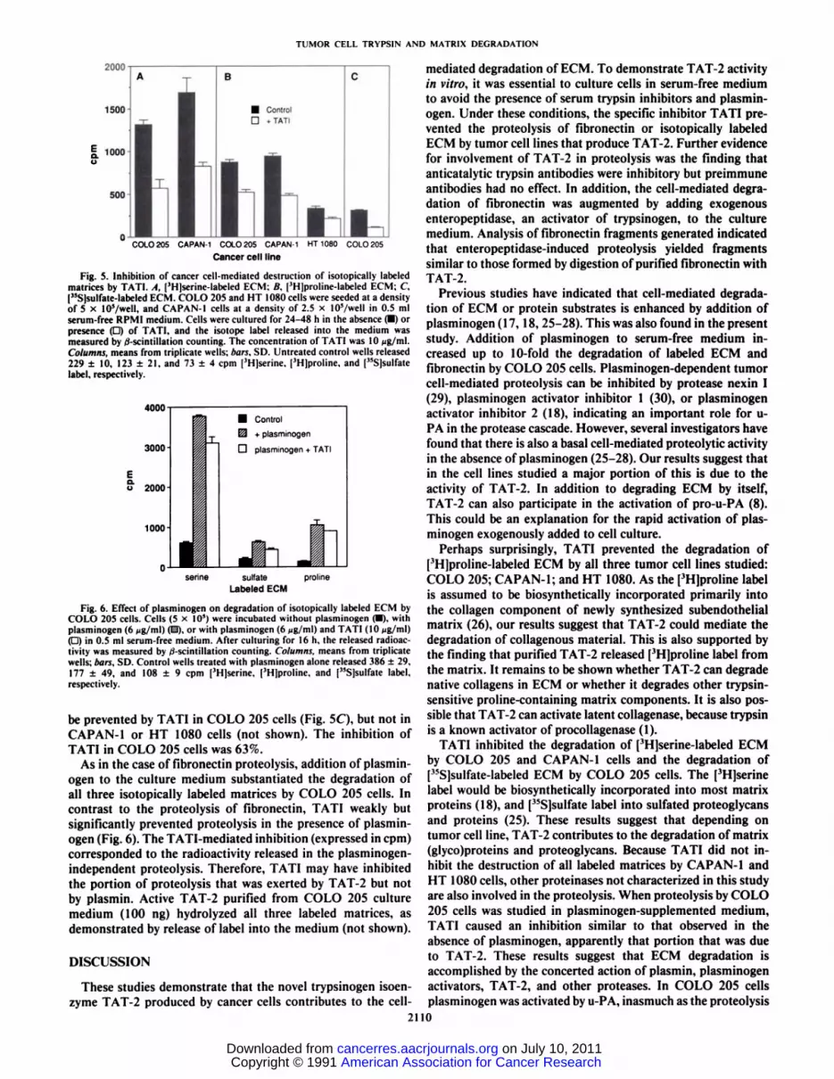
Fig. 6. Effect of plasminogen on degradation of isotopically labeled ECM byCOLO 205 cells. Cells (5 x 10') were incubated without plasminogen (•).withplasminogen (6 Mg/ml) (D), or with plasminogen (6 /jg/ml) and TATI (10 ng/ml)(D) in 0.5 ml serum-free medium. After culturing for 16 h, the released radioactivity was measured by /¿-scintillationcounting. Columns, means from triplicatewells; bars, SD. Control wells treated with plasminogen alone released 386 ±29,177 ±49, and 108 ±9 cpm [3H]serine, [3H|proline, and ("S]sulfate label,
respectively.
be prevented by TATI in COLO 205 cells (Fig. 5C), but not inCAPAN-1 or HT 1080 cells (not shown). The inhibition ofTATI in COLO 205 cells was 63%.
As in the case of fibronectin proteolysis, addition of plasminogen to the culture medium substantiated the degradation ofall three isotopically labeled matrices by COLO 205 cells. Incontrast to the proteolysis of fibronectin, TATI weakly butsignificantly prevented proteolysis in the presence of plasminogen (Fig. 6). The TATI-mediated inhibition (expressed in cpm)corresponded to the radioactivity released in the plasminogen-independent proteolysis. Therefore, TATI may have inhibitedthe portion of proteolysis that was exerted by TAT-2 but notby plasmin. Active TAT-2 purified from COLO 205 culturemedium (100 ng) hydrolyzed all three labeled matrices, asdemonstrated by release of label into the medium (not shown).
DISCUSSION
These studies demonstrate that the novel trypsinogen isoen-zyme TAT-2 produced by cancer cells contributes to the cell-
mediated degradation of ECM. To demonstrate TAT-2 activityin vitro, it was essential to culture cells in serum-free mediumto avoid the presence of serum trypsin inhibitors and plasminogen. Under these conditions, the specific inhibitor TATI prevented the proteolysis of fibronectin or isotopically labeledECM by tumor cell lines that produce TAT-2. Further evidencefor involvement of TAT-2 in proteolysis was the finding thatanticatalytic trypsin antibodies were inhibitory but preimmuneantibodies had no effect. In addition, the cell-mediated degradation of fibronectin was augmented by adding exogenousenteropeptidase, an activator of trypsinogen, to the culturemedium. Analysis of fibronectin fragments generated indicatedthat enteropeptidase-induced proteolysis yielded fragmentssimilar to those formed by digestion of purified fibronectin withTAT-2.
Previous studies have indicated that cell-mediated degradation of ECM or protein substrates is enhanced by addition ofplasminogen (17,18, 25-28). This was also found in the presentstudy. Addition of plasminogen to serum-free medium increased up to 10-fold the degradation of labeled ECM andfibronectin by COLO 205 cells. Plasminogen-dependent tumorcell-mediated proteolysis can be inhibited by protease nexin I(29), plasminogen activator inhibitor 1 (30), or plasminogenactivator inhibitor 2 (18), indicating an important role for u-PA in the protease cascade. However, several investigators havefound that there is also a basal cell-mediated proteolytic activityin the absence of plasminogen (25-28). Our results suggest thatin the cell lines studied a major portion of this is due to theactivity of TAT-2. In addition to degrading ECM by itself,TAT-2 can also participate in the activation of pro-u-PA (8).This could be an explanation for the rapid activation of plasminogen exogenously added to cell culture.
Perhaps surprisingly, TATI prevented the degradation of[3H]proline-labeled ECM by all three tumor cell lines studied:COLO 205; CAPAN-1; and HT 1080. As the [3H]proline label
is assumed to be biosynthetically incorporated primarily intothe collagen component of newly synthesized subendothelialmatrix (26), our results suggest that TAT-2 could mediate thedegradation of collagenous material. This is also supported bythe finding that purified TAT-2 released ['H]proline label from
the matrix. It remains to be shown whether TAT-2 can degradenative collagens in ECM or whether it degrades other trypsin-sensitive proline-containing matrix components. It is also possible that TAT-2 can activate latent collagenase, because trypsinis a known activator of procollagenase (1).
TATI inhibited the degradation of ['Hjserine-labeled ECM
by COLO 205 and CAPAN-1 cells and the degradation of["Sjsulfate-labeled ECM by COLO 205 cells. The ['Hjserine
label would be biosynthetically incorporated into most matrixproteins (18), and [35S]sulfate label into sulfated proteoglycans
and proteins (25). These results suggest that depending ontumor cell line, TAT-2 contributes to the degradation of matrix(glyco)proteins and proteoglycans. Because TATI did not inhibit the destruction of all labeled matrices by CAPAN-1 andHT 1080 cells, other proteinases not characterized in this studyare also involved in the proteolysis. When proteolysis by COLO205 cells was studied in plasminogen-supplemented medium,TATI caused an inhibition similar to that observed in theabsence of plasminogen, apparently that portion that was dueto TAT-2. These results suggest that ECM degradation isaccomplished by the concerted action of plasmin, plasminogenactivators, TAT-2, and other proteases. In COLO 205 cellsplasminogen was activated by u-PA, inasmuch as the proteolysis
2110
American Association for Cancer Research Copyright © 1991 on July 10, 2011cancerres.aacrjournals.orgDownloaded from
TUMOR CELL TRYPSIN AND MATRIX DEGRADATION
of fibronectin could be partially inhibited by anticatalytic uro-kinase antibodies. Recently, COLO 205 cells have been shown 5to express u-PA and its mRNA (18, 31).
Because TAT-2 is secreted in trypsinogen form from the cells(14), an obvious question is how it is activated. Part of TAT-2 6.is in active form in cell culture as indicated by inhibition ofproteolysis by TATI and trypsin antibodies. Enteropeptidase(32) and cathepsin B (33) are activators of pancreatic trypsin- 7.ogen. Purified TAT-2 is activated by enteropeptidase (14), andwe show here that it potentiates COLO 205 cell-mediateddegradation of fibronectin. The potentiation is due to activationof TAT-2, as the proteolysis can be inhibited by TATI and 9trypsin antibodies. It remains to be shown whether an entero-peptidase-like protease-activating TAT-2 exists in tumor cells. 10
This study demonstrates that TATI, also known previouslyas pancreatic secretory trypsin inhibitor (34), is a potential 11.inhibitor of tumor cell-mediated degradation of ECM. Eventhough the plasminogen-dependent proteolysis was poorly or 12.not at all inhibited by TATI, apparently because it is only aweak inhibitor of plasmin with a K¡of 9.7 nivi (35), TATI mayplay an important matrix-stabilizing role by controlling the 13.activation of TAT-2 zymogen. Coexpression of TATI withTAT-2 in the cyst fluids of ovarian tumors (13) and cancer cell 14.lines COLO 205 (14) and CAPAN-1 (36) further suggests aspecific role for TATI in controlling TAT-2. TATI was initially ,5identified as a tumor marker for ovarian cancer (37, 38), butthe levels in serum and urine are also elevated in patients with 16-
other types of advanced cancer (9, 37, 39). We suggest that theelevation of TATI is a reaction to TAT-2 expression with the n.aim of controlling TAT-2 activity and ECM degradation. However, TATI may also have other functions. TATI (or pancreatic is.secretory trypsin inhibitor) at concentrations present in serumis mitogenic on human fibroblasts (40, 41) and endothelial cells(42). Furthermore, it has been demonstrated that TATI binds 19.specifically to various cultured cells. A 140-kDa cell surfacereceptor protein mediating the binding could be characterized 20.by chemical cross-linking (43). It is not known whether cellsurface proteases are involved in the cell binding and mitogenic 2iactivities of TATI.
Degradation of ECM and basement membrane is an essentialstep at several stages of cancer invasion (44). The proteolysis 22.assay used in the present study can be envisaged as a simplemodel of endothelial basement membrane degradation occur- 23ring during intra- and extravasation of tumor cells. Our studiessuggest that TAT-2 may have a significant role in the degra- 24-dation of basement membranes by tumor cells. TAT-2 may beone component of the protease cascade that cells utilize for 25.migration through tissue barriers.
26.
ACKNOWLEDGMENTS
We thank Dr. Olii Saksela for helpful discussions and Lusa Airasfor technical assistance.
REFERENCES
27.
28.
29.
1. Tryggvason, K., Höyhtyä,M., and Salo. T. Proteolytic degradation of extracellular matrix in tumor invasion. Biochim. Biophys. Acta, 907: 191-217.1987. 30.
2. Saksela. O., and Rifkin, D. B. Cell-associated plasminogen activation: regulation and physiological functions. Annu. Rev. Cell Biol.. 4: 93-126. 1988.
3. Matrisian. L. Y., Bowden. G. T., Krieg. P.. Fürstenberger,G., Bilami. J. P.,Leroy, P., and Breathnach. R. The mRNA coding for the secreted protease 31.transin is expressed more abundantly in malignant than in benign tumors.Proc. Nati. Acad. Sci. USA, 83: 9413-9417, 1986.
4. Sloane, B. F., Dunn, J. R., and Honn. K. V. Lysosomal cathepsin B:
2111
correlation with metastatic potential. Science (Washington DC), 212: 1151-1153, 1981.Denhardt, D. T., Greenberg, A. H., Egan, S. E., Hamilton, R. T., and Wright,J. A. Cysteine protease cathepsin L expression correlates closely with themetastatic potential of H-ras-transformed murine fibroblasts. Oncogene, 2:55-59, 1987.Spyratos, F., Brouillet, J-P., Defrenne, A., Macene, K., Rouésse,J., Maude-londe. T., Brunei, M.. Andrieu, C., Desplaces, A., and Rochefort, H. Cathepsin D: an independent prognostic factor for metastasis of breast cancer.Lancet, 2: 1115-1118, 1989.Stenman, U-H., Koivunen, E., and Vuento, M. Characterization of a tumor-associated serine protease. Biol. Chem. Hoppe-Seyler, 369: 9-14, 1988.Koivunen, E., Huhtala, M-L., and Stenman, U-H. Human ovarian tumor-associated trypsin. Its purification and characterization from mucinous cystfluid and identification as an activator of pro-urokinase. J. Biol. Chem., 264:14095-14099, 1989.Halila. H., Lehtovirta, P., and Stenman, U-H. Tumour-associated trypsininhibitor (TATI) in ovarian cancer. Br. J. Cancer, 57: 304-307, 1988.Liotta, L. A., Tryggvason, K., Garbisa, S., Hart, I., Foltz, C. M., and Shafie,S. Metastatic potential correlates with enzymatic degradation of basementmembrane collagen. Nature (Lond.), 284:67-68, 1980.Carlsen, S. A., Ramshaw, I. A., and Warrington, R. C. Involvement ofplasminogen activator production with tumor metastasis in a rat model.Cancer Res., 44: 3012-3016, 1984.Hasui, Y., Suzumiya, J., Marutsuka, K., Sumiyoshi, A., Hashida, S., andIshikawa, E. Comparative study of plasminogen activators in cancers andnormal mucosae of human urinary bladder. Cancer Res., 49: 1067-1070,1989.Koivunen, E., Itkonen. O., Halila, H., and Stenman, U-H. Cyst fluid ofovarian cancer patients contains high concentrations of trypsinogen-2. Cancer Res., 50: 2375-2378, 1990.Koivunen. E., Saksela, O.. Itkonen. O., Osman, S.. Huhtala, M-L., andStenman, U-H. Human colon carcinoma, fibrosarcoma and leukemia celllines produce tumor-associated trypsinogen. Int. J. Cancer, in press, 1991.Stenman, U-H., Pesonen, K., and Huhtala, M-L. Rapid concentration ofurinary peptides and proteins. Anal. Biochem., ¡23:291-294, 1982.Jaffe, E. A., Nachman, R. L., Becker, C. G.. and Minick. C. R. Culture ofhuman endothelial cells derived from umbilical veins. Identification by morphologic and immunologie criteria. J. Clin. Invest., 52: 2745-2756, 1973.Jones, P. A., and DeClerck, Y. A. Destruction of extracellular matricescontaining glycoprotein. elastin, and collagen by metastatic human tumorcells. Cancer Res., 40: 3222-3227, 1980.Baker, M. S., Bleakley, P., Woodrow, G. C., and Doe, W. F. Inhibition ofcancer cell urokinase plasminogen activator by its specific inhibitor PAI-2and subsequent effects on extracellular matrix degradation. Cancer Res., 50:4676-4684, 1990.Vuento. M., and Vaheri. A. Purification of fibronectin from human plasmaby affinity chromatography under non-denaturing conditions. Biochem. J.,183: 331-337, 1979.Vuento, M., Salonen. E.. Salminen, K., Pasanen. M., and Stenman, U-H.Immunochemical characterization of human plasma fibronectin. Biochem.J., 191:119-727. 1980.Stenman, U-H.. Alfthan, H.. Ranta, T., Vartianinen, E., Jalkanen, J., andSeppalâ,M. Serum levels of human chorionic gonadotropin in nonpregnantwomen and men are modulated by gonadotropin-releasing hormone and sexsteroids. J. Clin. Endocrino!. Metab., 46: 730-736, 1987.Hemmilä, I., Dakubu, S., Mukkala. V-M., Siitari. H., and Lövgren, T.Europium as a label in time-resolved immunofluorometric assays. Anal.Biochem., 137: 335-343. 1984.I .animili. U-K. Cleavage of structural proteins during the assembly of thehead of bacteriophage T4. Nature (Lond.). 227: 680-685, 1970.Towbin, H., Staehelin, T., and Gordon, J. Electrophoretic transfer of proteinsfrom polyacrylamide gels to nitrocellulose sheets: procedure and some applications. Proc. Nati. Acad. Sci. USA, 76: 4350-4354, 1979.Kramer, R. H., Vogel, K. G., and Nicolson, G. L. Solubilization and degradation of subendothelial matrix glycoproteins and proteoglycans by metastatic tumor cells. J. Biol. Chem., 257: 2678-2686, 1982.Laug, W. E., DeClerk, Y. A., and Jones, P. A. Degradation of the subendothelial matrix by tumor cells. Cancer Res., 43: 1827-1834, 1983.Fairbairn, S., Gilbert. R., Ojakian, G., Schwimmer, R., and Quigley, J. P.The extracellular matrix of normal chick embryo fibroblasts: its effect ontransformed chick fibroblasts and its proteolytic degradation by the transformants. J. Cell Biol.. 101: 1790-1798, 1985.Rezaee, M., Chen, L., and Kramer, R. H. Measurement of plasminogenactivator activity from human fibrosarcoma cells by a new microassay. Int.J. Cancer, 40: 823-829. 1987.Bergman, B. L., Scott, R. W., Bajpai, A., Watts, S., and Baker, J. B. Inhibitionof tumor-cell-mediated extracellular matrix destruction by a fibroblast pro-teinase inhibitor, protease nexin I. Proc. Nati. Acad. Sci. USA, 83: 996-1000, 1986.Cajot. J. F.. Bamat. J., Bergonzelli. G. E., Kruithof. E. K. O., Medcalf, R.L., Testuz, J.. and Sordat, B. Plasminogen activator type I is a potentialnatural inhibitor of extracellular matrix degradation by fibrosarcoma andcolon carcinoma cells. Proc. Nati. Acad. Sci. USA, 87:6939-6943, 1990.Quax, P. H. A., van Leeuwen, R. J. T., Verspaget, H. W., and Verheijen, J.H. Protein and messenger RNA levels of plasminogen activators and inhibitors analyzed in 22 human tumor cell lines. Cancer Res.. 50: 1488-1494,1990.
American Association for Cancer Research Copyright © 1991 on July 10, 2011cancerres.aacrjournals.orgDownloaded from
TUMOR CELL TRVPSIN AND MATRIX DEGRADATION
32. Light, A., and Janska, H. Enterokinase (enteropeptidase): comparative as-pects. Trends Biochem. Sci., 14: 110-112, 1989.
33 Greenbaum, L. M., Hirshkowitz, A., and Shoichet, 1. The activation oftrypsinogen bv cathepsin B. J. Biol. Chem., 234: 2885-2890, 1959.
34. Greene, L. J.. Pubols, M. H., and Bartelt. D. C. Human pancreatic secretorytrypsin inhibitor. Methods Enzymol., 45: 8 13-856, 1976.
35. Turpeinen. U., Koivunen, E., and Stenman. U-H. Reaction of a tumor-r . , . . .... ... . • •. j -.K iassociated trypsm inhibitor with serine protemases associated with coagula-lion and tumor invasion. Biochem. J.. 254:9] 1-914, 1988.
36. Ogata. N. Demonstration of pancreatic secretory trypsin inhibitor in serum-free culture medium conditioned by the human pancreatic carcinoma cell lineCAPAN-l.J. Biol. Chem.. 263: 13427-13431, 1988.
37. Stenman. U-H., Huhtala, M-L., Koistinen. R.. and Seppälä,M. Immuno-chemical demonstration of an ovarian cancer-associated urinary peptide. Int.J. Cancer. 30: 53-57, 1982.
38. Huhtala. M-L., Pesonen. K., Kalkkinen. N., and Stenman. U-H. Purificationand characterization of a tumor-associated trypsin inhibitor from the urineof a patient with ovarian cancer. J. Biol. Chem., 257: 13713-13716, 1982.
39. Huhtala, M-L., Kahanpää,K.. Seppälä.M., Halila, H., and Stenman, U. H.
Excretion of a tumor-associated trypsin inhibitor (TATI) in urine of patientswith gynecological malignancy. Int. J. Cancer, 31: 71 1-714, 1983.
40. Ogawa, M., Tsushima, T., Ohbu, Y., Ogawa, N., Tanaka, S., Ishida, M., andMori, T. Stimulation of DNA synthesis in human fibroblasts by humanpancreatic secretory trypsin inhibitor. Res. Commun. Chem. Pathol. Phar-macol 50: 155-158 1986.
41. Hamilton I., Reynolds. G., W., Scott, G. K., Sharfe, N, and Tse, C. A.Effects of human and ovine pancreatic secretory trypsin inhibitors on theproliferation of norma| numa„fibroblasts. Biol. Chem. Hoppe-Seyler, 37 J:79-83 1990
^ McKeehan, W. L., Sakagami, Y., Hoshi. H., and McKeehan, K. Two appar-en( human endotheiia| ceMgrowth factors from human hepatoma cells aretumor-associated proteinase inhibitors. J. Biol. Chem., 261: 5378-5383,\986.
43 Niinobu, T., Ogawa, M., Murata, A., Nishijima, J-I., and Mori, T. Identifi-cation and characterization of receptors specific for human pancreatic secre-tory trypsin inhibitor. J. Exp. Med., 172: 1133-1 142, 1990.
44. Liotta, L. A., Rao, N.. and Wewer, U. M. Biochemical interactions of tumorcells with the basement membrane. Annu. Rev. Biochem., 55: 1037-1057,1986.
2112
American Association for Cancer Research Copyright © 1991 on July 10, 2011cancerres.aacrjournals.orgDownloaded from