incomplete nuclear transformation of human spermatozoa in
TRANSCRIPT
Incomplete nuclear transformation of human spermatozoa inoligo-astheno-teratospermia: characterization by indirectimmunofluorescence of chromatin and thiol status
L. Ramos1, G.W. van der Heijden1,3, A. Derijck1,4, J.H. Berden2, J.A.M. Kremer1,J. van der Vlag2 and P. de Boer1,5
1Department of Obstetrics and Gynaecology, Radboud University Nijmegen Medical Centre, PO Box 9101, 6500 HB Nijmegen, The
Netherlands; 2Nephrology Research, Radboud University Nijmegen Medical Centre, PO Box 9101, 6500 HB Nijmegen, The Netherlands;3Present address. Department of Embryology, Carnegie Institution of Washington, 3520 San Martin Drive, Baltimore, MD 21218, USA.;4Present address. Department of Pharmacology and Anatomy, Rudolf Magnus Institute of Neuroscience, University Medical Center
Utrecht, Universiteitsweg 100, 3584 CG, Utrecht, The Netherlands.
5Correspondence address. Tel: þ31-24-3610869; Fax: þ31-24-3668597; E-mail: [email protected]
BACKGROUND: Sperm heterogeneity in the human, as observed in oligo-astheno-teratozoospermia (OAT), is associ-ated with hypospermatogenesis. METHODS: The chromatin of sperm from OAT and normospermic males wascharacterized with antibodies specific for nucleosomes, the histone H3.1/H3.2 isoform, histone TH2B, apoptosis-associated H4 acetylation (KM-2) and protamines. Subsequently, sperm samples were stained with the thiol-specificfluorochrome monobromobimane (mBBr) before and after reduction with dithiotreitol (DTT) as most thiol groupsreside in the cysteine-rich protamines. We also used fluorescence-activated cell sorter (FACS) for sperm histogramsand sorting for high or low free and total thiol levels. These fractions were further analysed for DNA damage withthe TdT-UTP nick end-labelling (TUNEL) assay. RESULTS: OAT sperm nuclei stained higher for nucleosomesand KM2-epitopes, and lower for TH2B. For free, and total, thiol groups, OAT sperm were characterized by biphasicdistributions, reflecting incomplete thiol oxidation as well as overoxidation, and possibly reduced protamine contents.The TUNEL assay on sperm subfractions, for both control and OAT sperm, revealed that a lower level of freethiol groups is associated with a higher TUNEL incidence, and this relationship was also found for total thiollevels. Hence, both overoxidation and a low total number of thiol groups predestine for sperm apoptosis.CONCLUSIONS: Chromatin characteristics reflecting an incomplete nucleosome to protamine remodellingwere found in subfertile males. Sperm apoptosis is related to both incomplete remodelling and protamineoveroxidation.
Keywords: spermiogenesis; protamines; histones; chromatin condensation; OAT
Introduction
Mammalian sperm heterogeneity can be defined at many levels
such as overall morphology, motility, nuclear differentiation,
capacitation and receptor status. Compared with that of farm
and experimental animals, human spermatozoa are more vari-
able in overall morphology (Bedford et al., 1973). Sperm con-
centration in the ejaculate is inversely related to the incidence
of abnormal morphology and the variation in motility (Zollner
et al., 1996). Oligo-astheno-teratospermia (OAT) largely
occurs due to abnormal spermatogenesis (hypospermatogen-
esis or a combination of this condition with incomplete matu-
ration arrest) (Johnsen, 1970; Levin, 1979; Carrell et al.,
2007). The biological significance of the increased sperm het-
erogeneity observed in OAT has received more attention in
recent years due to the use of these sperm samples in classical
IVF and ICSI. For the latter application, visual selection of
motile and ‘normal’ looking sperm is the only parameter
used worldwide (Ramos and Wetzels, 2001; De Vos et al.,
2003; Ramos et al., 2004).
Nuclear elongation halfway during spermiogenesis is
accompanied by the transition of chromatin from a
nucleosome-based structure to a protamine-based structure
(for a review, see Dadoune, 2003), largely reducing the
nuclear volume and increasing chromatin compaction. In
human and mouse, two types of protamines (PRM1 and
PRM2) contribute to the compaction of chromatin. For this
purpose, histones are first exchanged for transition proteins 1
and 2 (TNP1 and TNP2), which are later replaced by PRM 1
# The Author 2007. Published by Oxford University Press on behalf of the European Society of Human Reproduction and Embryology.
All rights reserved. For Permissions, please email: [email protected]
259
Human Reproduction Vol.23, No.2 pp. 259–270, 2008 doi:10.1093/humrep/dem365
Advance Access publication on November 30, 2007
Dow
nloaded from https://academ
ic.oup.com/hum
rep/article/23/2/259/626861 by guest on 08 January 2022
and PRM2. During this process, DNA double strand breaks
occur (McPherson and Longo, 1993; Laberge and Boisson-
neault, 2005) and are subsequently repaired. When sperm is
passing from the testis to the epididymis, a further stabilization
of nuclear structure is achieved by thiol-oxidation of the
cysteine-rich protamines. In mouse and man cauda epididymis,
�95% of SH groups are converted into -S-S- bridges (Sao-
waros and Panyim, 1979; Pellicciari et al., 1983; Seligman
et al., 1994). Protamine thiol oxidation has been linked to the
stability of the DNA, yielding a shift from red to green acridine
orange (AO) fluorescence in rodents and man after acetic acid/alcohol fixation (Kosower et al., 1992). The replacement of his-
tones by protamines is less complete in the human (85%)
(Gatewood et al., 1987) compared with other mammals
(Bench et al., 1996). de Yebra and Oliva (1993) were the
first to notice with biochemical methods that over a range of
infertile patients higher PRM1/PRM2 ratios correlated with
higher histone levels. By fluorescence-activated cell sorter
(FACS) measurements of total sperm cell thiol levels (after
DTT reduction) (Rufas et al., 1991), an indication of the
reduced presence of the cysteine-rich PRM1 and PRM 2 pro-
teins in oligospermic patients had already been obtained.
A number of subsequent observations reinforce the con-
clusion of a nuclear differentiation defect during spermiogen-
esis in OAT males.
First, CMA3 fluorescence as an indicator for underprotami-
nation (Bianchi et al., 1993) or reduced protamine thiol cross-
linking is much more frequently observed in sperm samples
from OAT male patients (Iranpour et al., 2000). Furthermore,
sperm of infertile men are much more sensitive to DNase I,
highlighting the role of uncompacted (higher histone contain-
ing) chromatin in infertility (Sakkas et al., 2002).
Second, Steger et al. (2001) observed that the frequency of
PRM1 and PRM2 transcribing round spermatids is negatively
related to the severity of the spermatogenic defect. In concor-
dance with this finding, the fraction of elongating spermatids,
that were immunofluorescence positive for hyperacetylated
histone 4 (H4), marking the onset of the nucleosomal tran-
sition, was decreased by a factor 0.6–0.75 in azoospermic to
oligospermic patients (Sonnack et al., 2002).
Third, when the ratio of PRM1 to PRM2, which in normosper-
mic human samples is around 1 (Balhorn et al., 1988), is evalu-
ated in oligospermic subjects, often a shortage of PRM2 is found
in combination with the presence of PRM2 precursor proteins (de
Yebra et al., 1998). The same observation is made in mouse
models deficient for TNP1 and 2 (Yu et al., 2000; Zhao et al.,
2001). TNP1 and 2 and PRM2 deficiency lead to altered chroma-
tin compaction as demonstrated by electron microscopy (Yu
et al., 2000; Zhao et al., 2001; Cho et al., 2003).
In this study, we have used highly specific antibodies against
histones in a nucleosomal context, histone subtypes (DNA
replication-dependent H3.1/H3.2, TH2B) and histone modifi-
cations (acetylated forms of histone 4) in an in situ immuno-
fluorescence (IF) investigation of expanded sperm nuclei
from normal and OAT donors. As a follow up study, we charac-
terized SH profiles of human sperm samples by FACS and
selected subfractions to measure sensitivity for the TdT-UTP
nick end-labelling (TUNEL) reaction and to study chromatin
compaction by the use of a double-strand DNA (dsDNA) anti-
body on whole nuclei.
Evidence for an apoptotic chromatin imprint has been
obtained. By the use of an anti-dsDNA antibody, the conclusion
is also reached that chromatin compaction is compromised in
OAT sperm. For all parameters studied, the increased nuclear
heterogeneity in OAT sperm can be related to variability of
chromatin remodeling in elongating spermatids.
Our results complement insights generated by other investi-
gators and add to the conclusion that OAT sperm samples
quantitatively differ from normospermic samples by an incom-
plete nucleosome to protamine transition, the basis of which
likely is laid during the second half of pachytene meiosis.
Materials and Methods
Sperm samples
Sperm samples were obtained from 10 normospermic and 13 infertile
donors (Table I). Classification was based on World Health Organiz-
ation (WHO, 1999) criteria. Accordingly, all infertile patients were
classified as OAT. Morphology was assessed by the strict criteria
(Menkveld et al., 2001). Table I lists the spermiogram data. For the
histone characterizations (IF experiments) and FACS thiol fluor-
escence sorting experiments, pools of four normospermic donor
samples and four or five oligospermic donor samples were assessed
(see Table I for composition of pools). For the determination of
dsDNA immunofluorescence in thiol sorted samples, the sperm of
one normospermic donor and a pool of two OAT males were used.
All sperm samples were cryopreserved using a dilution 1:1 with
cryoprotectant freezing medium (TYB; Irvine Scientific, Santa Ana,
CA, USA) in liquid nitrogen vapour. The sperm concentration
varied between 5–20 � 106/straw in order to allow pilot experiments
to be executed on the same samples. Pooled samples were made after
thawing aliquots of individual donors and mixed for equivalent
numbers of cells per donor. Cryopreserved sperm were thawed at
room temperature and washed once with human tubal fluid medium
(HTF; Cambrex, Verviers, Belgium) supplemented with 10% human
plasma proteins (GPO; CLB, Amsterdam, The Netherlands) in order
to eliminate the cryopreservation medium by spinning for 5 min at
500g. The pellet was resuspended in 200 ml phosphate-buffered
saline (PBS; Sigma, St. Louis, MO, USA), pH 7.4.
Induction of nuclear expansion for IF
The sperm suspension (pools normospermic and OAT) was diluted 1:4
in MilliQ–water. Drops of 5 ml were placed on a glass slide and air
dried. An amount of 100 ml of freshly prepared decondensing mix
[25 mM dithiotreitol (DTT; Roche Biochemicals, Mannheim,
Germany); 0.2% Triton-X-100 (Sigma); 200 IU heparin/ml (Leo Lab-
oratories, The Netherlands) in PBS) was placed over the dried sperm
cells that were incubated in a humidified atmosphere for 12–18 min.
The speed and degree of nuclear decondensation was followed by
phase-contrast microscopy. When the majority of nuclei appeared
dull grey with roughly twice the surface area of the undecondensed
sperm heads, the slide was placed in a coplin jar with 4% paraformal-
dehyde (PFA in PBS, Sigma) at pH 7 for 15 min. Subsequently, slides
were washed thrice for 5 min in PBS and allowed to dry. A 2 ml cell
suspension of mouse spermatogenic cells was placed on one end of the
slide for an IF control.
Ramos et al.
260
Dow
nloaded from https://academ
ic.oup.com/hum
rep/article/23/2/259/626861 by guest on 08 January 2022
Antibodies
Monoclonal antibody PL2-3 recognizes H2A.H2B DNA in a nucleo-
somal context (dilution 1:2000) (Losman et al., 1992; Dieker et al.,
2005); monoclonal antibody KM-2 (1:3000) (Dieker et al., 2007)
recognizes H4acK8, 12 and 16 with a preference for apoptotic
nuclei and apoptotic bodies; monoclonal antibody #34 (1:1500)
recognizes the replication-dependent histone 3 isoform H3.1/H3.2
(van der Heijden et al., 2005; Henikoff and Ahmad, 2005); mono-
clonal antibody #36 (1:200) (Smeenk et al., 1988) recognizes
dsDNA. The tyrosine hydroxylase antibody (1:100) (van Roijen
et al., 1998) recognizes hTSH2B (Zalensky et al., 2002). Hup1N
and Hup2B monoclonal antibodies (Stanker et al., 1987) specific
for, respectively, PRM1 and PRM2 were applied in a 1:3000 dilution.
Mouse monoclonal antibody gH2AX (Upstate #05-636) was used at
1:1000.
Secondary antibodies A11001 fluor 488 goat anti-mouse IgG
(HþL) and A11012 fluor 594 goat anti-rabbit IgG (HþL) from Mol-
ecular Probes, (Oregon, USA) were, for the detection of primary anti-
bodies, used at a 1:500 dilution. IF was performed as described
previously (Baart et al., 2000).
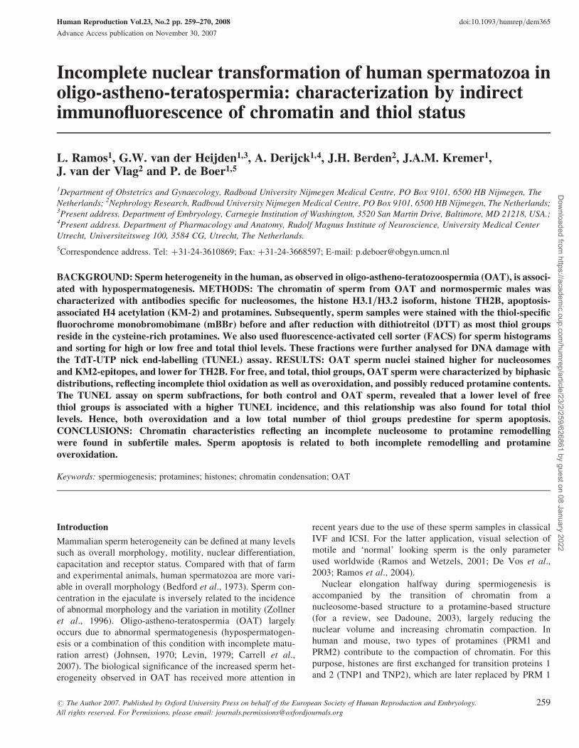
Images were obtained with a Zeiss axioplan fluorescence micro-
scope and captured by a Zeiss AxioCam MR camera with Axiovision
3.1 software (Carl Zeiss). The expanded sperm nuclear grades þþ
and þþþ (see Fig. 1A) showed uniform staining when probed wit
mab #36 specific for dsDNA. Fluorescence intensities for these expan-
sion classes were subjectively scored in five grades. No signal detected
(2); some small specks of low intensity not covering the entire
nucleus (þ/2); small specks of low intensity covering the entire
nucleus (þ); overall signal with increasing brightness (þþ) and
overall strong signal up to covering the entire nucleus (þþþ)
(Fig. 1B). For numerical representations, these grades received the
values 0–4 [corresponding to (2); (þ/2); (þ); (þþ) and
(þþþ), respectively].
Thiol characterizations by FACS and cell separation
For the monobromobimane (mBBr, Calbiochem, CA,USA) staining of
free thiol (SH2) groups, sperm cells were fixed for 15 min in 1% PFA
in PBS at room temperature and washed twice in PBS (spinning
10 min at 500g). The sperm pellet was resuspended in 100 ml mBBr
solution [50 mM mBBr in 20 mM Tris-buffered saline (TBS), pH
Figure 1: (A) Decondensation of sperm nuclei after DTT/heparintreatment and IF resultsThe degree of decondensation was subjectively assessed as (2) for nodecondensation; (þ) for minimal decondensation; (þþ) for openchromatin and (þþþ) for highly open chromatin. Only spermnuclei with sufficient decondensation (category þþ and þþþ)were subjectively evaluated. (B) IF intensity was assessed as: nosignal (2); only a few weak foci (þ/2); foci all over the nucleusbut still of weak intensity (þ); foci all over the nucleus with strongintensity (þþ) and highly fluorescent nuclei (þþþ)
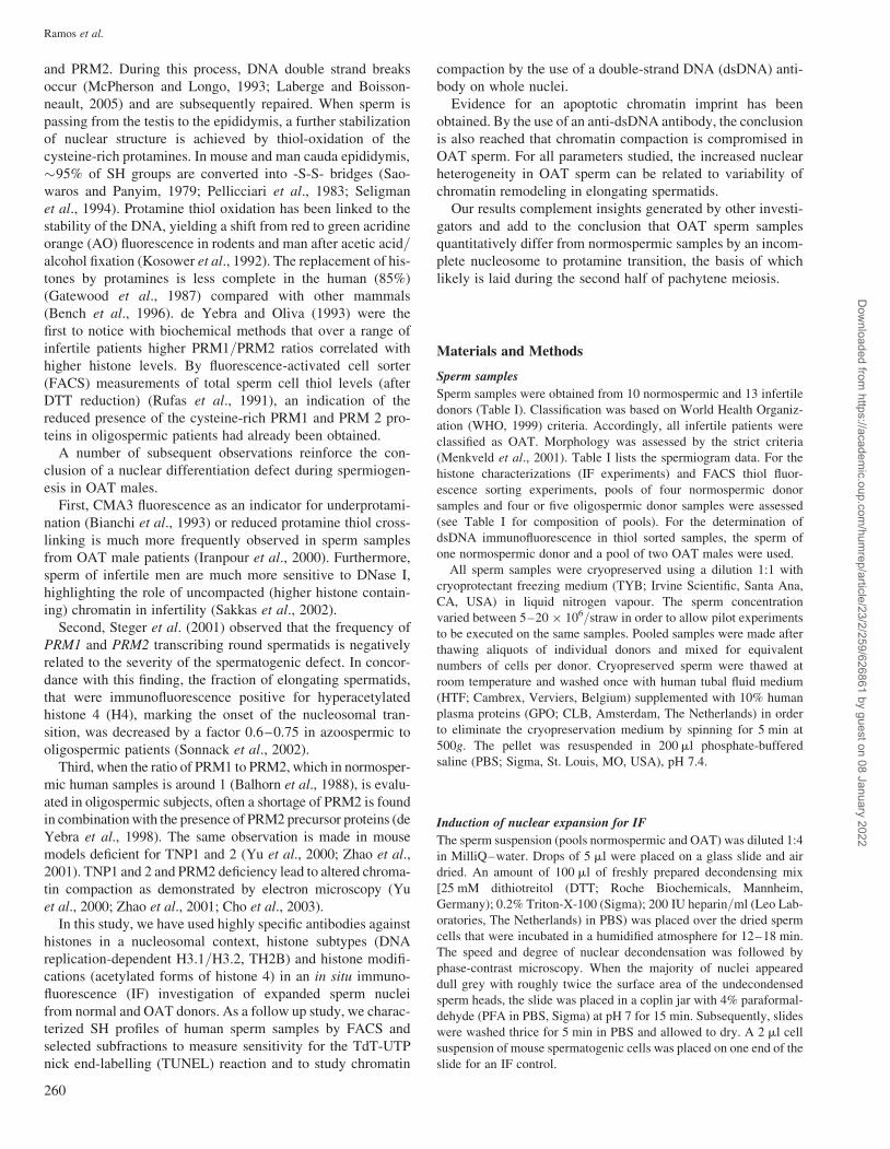
Table I. Sperm analysis of the samples included in this study and experiments carried out with each sample.
Samples Code Volume (ml) Concentration �106 % motility % normalmorphology
Experiment 1 Experiment 2
Normospermicdonors
N-1 2.2 195 80 47 FACS –N-2a 2.4 185 60 22 FACS TUNELN-3a 3.8 200 80 33 FACS IF-dsDNAþTUNELN-4a 2.5 100 70 25 FACS TUNELN-5a 3.6 85 55 35 FACS TUNELN-6 2.3 130 60 17 IF-chromatin –N-7 4.2 150 75 12 IF-chromatin –N-8 1.5 130 75 25 IF-chromatin –N-9 2.4 100 65 12 IF-chromatin –N-10 3.1 100 50 15 IF-chromatin –
OAT donors O-1b 1.5 30 15 2 FACS IF-dsDNAO-2b 1.0 45 20 3O-3c,e 7.3 5 25 2 FACS IF-dsDNAþTUNELO-4c 3.6 40 25 7O-5d 5.0 7 30 1 FACS –O-6d,e 4.2 7 25 5 TUNELO-7e 7.9 15 10 1 FACS TUNELO-8e 3.7 20 35 7 FACS TUNELO-9 8.4 5 50 5 IF-chromatin –O-10 1.1 3.8 10 1 IF-chromatin –O-11 6.3 1.0 15 2 IF-chromatin –O-12 5.3 5.0 40 3 IF-chromatin –O-13 1.7 1.9 20 2 IF-chromatin –
Experiments 1 and 2: flow cytometry, FACS; immunofluorescence for chromatin markers, IF-chromatin; immunofluorescence with monoclonal antibody againstds-DNA, IF-dsDNA; TdT UTP-nick end labelling, TUNEL. apooled samples for the final FACS sorting experiment (Normo pool); b,c,dpooled samples formeasurements with FACS; epooled samples for the final FACS sorting experiment (OAT pool).
Incomplete sperm nuclear transformation in OAT
261
Dow
nloaded from https://academ
ic.oup.com/hum
rep/article/23/2/259/626861 by guest on 08 January 2022
7.4] and kept in the dark for 20 min at room temperature (Kosower and
Kosower, 1987). The sample was washed once more in PBS and the
pellet was resuspended in 500 ml PBS for fluorescence measurements
and sorting by FACS.
For the determination of the total amount of SH2groups in the
cells, one sperm aliquot was reduced using DTT. First, samples
were fixed in 4% PFA in PBS for 10 min at room temperature. The
cell suspension was washed once in PBS and the pellet resuspended
in 500 ml 1 mM DTT in TBS, pH 9.5 (15 min at room temperature).
Reduced sperm samples were washed twice in PBS (5 min, 500g)
before staining with mBBr.
Cells were analysed and sorted on an Altra HyperSort flowcyt-
ometer (Beckman Coulter, Miami, USA). A 408-nm Vioflame laser
running at 25 mW was used for excitation of mBBr and a 525-nm
band-pass filter for emission. Spermatozoa were first gated on
forward scatter versus side scatter to discriminate sperm from debris
and other cells as much as possible (Gate A, Fig. 2). Depending on
the final concentration per sample, a minimum of 150 000–500 000
Figure 2: (A and B) FACS plot representation of unstained normospermic and OAT poolsGate ‘A’ represents the sperm population analyzed and sorted for further experiments. (C and D) mBBr stained sperm detecting free thiol groups. (E andF) total thiol content in the DTT reduced mBBr stained sperm samples. Horizontal bars in C–F (R and S populations) give selection criteria for sorting
Ramos et al.
262
Dow
nloaded from https://academ
ic.oup.com/hum
rep/article/23/2/259/626861 by guest on 08 January 2022
particles was sorted for high or low fluorescence in the mBBr and
DTT–mBBr stained samples.
The gate A population and stained fractions from the A-gated popu-
lation were collected in 500 ml HTF medium and spun for 10 min at
1500 g. Sperm cell populations were fractionated (sorted) by high
and low free and total SH levels (see Fig. 2). After centrifugation,
pellets were resuspended in 30 ml HTF for further processing, such
as the determination of DNA breakage by the TUNEL reaction and
chromatin compaction by measuring accessibility of the DNA to the
dsDNA-specific antibody #36.
TUNEL staining
The TUNEL assay (Cell Death Detection kit, Roche Biochemicals)
was executed following the manufacturer’s specifications with
minor modifications (Ramos et al., 2002). Briefly, air-dried spermato-
zoa were fixated in 1% PFA in PBS for 10 min at room temperature
and rinsed twice with PBS followed by permeabilization with 0.2%
Triton X-100 in PBS, for 10 min. Nuclei were exposed to the
TdT-labelled nucleotide mix for 60 min at 378C. Slides were rinsed
twice (5 min) in PBS and the sperm nuclei were counterstained with
DAPI (0.01 mg/l in PBS). Nuclei were mounted in 25 ml Vectashield.
The total number of DAPI blue staining sperm nuclei per field was
counted first. A minimum of 200, but mostly between 300 and 400,
nuclei per fraction was scored by two observers.
Nuclear condensation patterns with IF
Sperm fractions were embedded in a fibrin clot (Hunt et al., 1995) by
mixing 1 ml of cell suspension with 3 ml of fibrin (Catalogue number:
341573, Calbiochem) on a precleaned coverslip, after which 1 ml of
thrombin (Catalogue number: T-6634, Sigma) was added. A clotting
reaction induced by body heat follows within one minute. The clot
was washed briefly in PBS. Subsequently, the cells were fixed in
0.5% PFA in PBS for 5 min, treated with 1 mM DTT for 30 min
and refixed for 30 min with ice-cold methanol (modified from
Zalensky et al., 2002).
Statistics
Chi-square analysis was used to test for independency of variables
such as observers and staining patterns within samples. The Spearman
rank correlation coefficient was used as an estimate for the congruence
between observers. In the case of more than two groups, one-way
analysis of variance was used. P-values ,0.05 were considered stat-
istically different. Statistical analysis was carried out with the SSPS
12.0 software package (SPSS Inc., Chicago, IL, USA).
Results
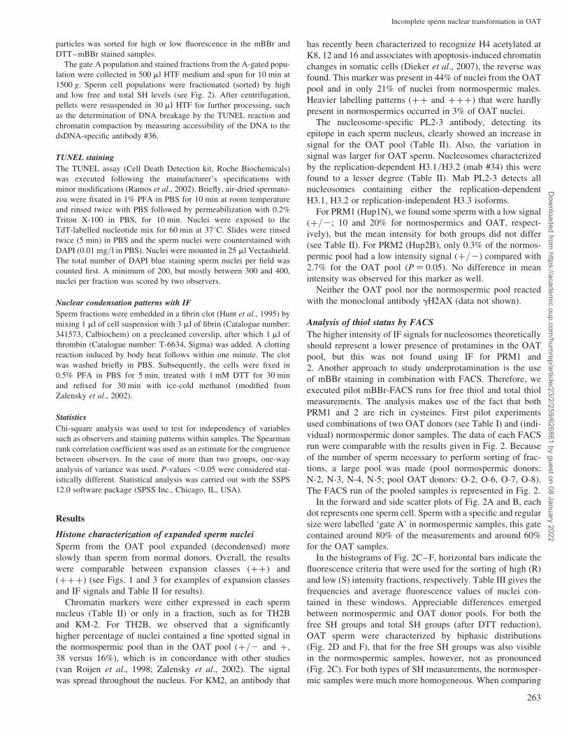
Histone characterization of expanded sperm nuclei
Sperm from the OAT pool expanded (decondensed) more
slowly than sperm from normal donors. Overall, the results
were comparable between expansion classes (þþ) and
(þþþ) (see Figs. 1 and 3 for examples of expansion classes
and IF signals and Table II for results).
Chromatin markers were either expressed in each sperm
nucleus (Table II) or only in a fraction, such as for TH2B
and KM-2. For TH2B, we observed that a significantly
higher percentage of nuclei contained a fine spotted signal in
the normospermic pool than in the OAT pool (þ/2 and þ,
38 versus 16%), which is in concordance with other studies
(van Roijen et al., 1998; Zalensky et al., 2002). The signal
was spread throughout the nucleus. For KM2, an antibody that
has recently been characterized to recognize H4 acetylated at
K8, 12 and 16 and associates with apoptosis-induced chromatin
changes in somatic cells (Dieker et al., 2007), the reverse was
found. This marker was present in 44% of nuclei from the OAT
pool and in only 21% of nuclei from normospermic males.
Heavier labelling patterns (þþ and þþþ) that were hardly
present in normospermics occurred in 3% of OAT nuclei.
The nucleosome-specific PL2-3 antibody, detecting its
epitope in each sperm nucleus, clearly showed an increase in
signal for the OAT pool (Table II). Also, the variation in
signal was larger for OAT sperm. Nucleosomes characterized
by the replication-dependent H3.1/H3.2 (mab #34) this were
found to a lesser degree (Table II). Mab PL2-3 detects all
nucleosomes containing either the replication-dependent
H3.1, H3.2 or replication-independent H3.3 isoforms.
For PRM1 (Hup1N), we found some sperm with a low signal
(þ/2; 10 and 20% for normospermics and OAT, respect-
ively), but the mean intensity for both groups did not differ
(see Table II). For PRM2 (Hup2B), only 0.3% of the normos-
permic pool had a low intensity signal (þ/2) compared with
2.7% for the OAT pool (P ¼ 0.05). No difference in mean
intensity was observed for this marker as well.
Neither the OAT pool nor the normospermic pool reacted
with the monoclonal antibody gH2AX (data not shown).
Analysis of thiol status by FACS
The higher intensity of IF signals for nucleosomes theoretically
should represent a lower presence of protamines in the OAT
pool, but this was not found using IF for PRM1 and
2. Another approach to study underprotamination is the use
of mBBr staining in combination with FACS. Therefore, we
executed pilot mBBr-FACS runs for free thiol and total thiol
measurements. The analysis makes use of the fact that both
PRM1 and 2 are rich in cysteines. First pilot experiments
used combinations of two OAT donors (see Table I) and (indi-
vidual) normospermic donor samples. The data of each FACS
run were comparable with the results given in Fig. 2. Because
of the number of sperm necessary to perform sorting of frac-
tions, a large pool was made (pool normospermic donors:
N-2, N-3, N-4, N-5; pool OAT donors: O-2, O-6, O-7, O-8).
The FACS run of the pooled samples is represented in Fig. 2.
In the forward and side scatter plots of Fig. 2A and B, each
dot represents one sperm cell. Sperm with a specific and regular
size were labelled ‘gate A’ in normospermic samples, this gate
contained around 80% of the measurements and around 60%
for the OAT samples.
In the histograms of Fig. 2C–F, horizontal bars indicate the
fluorescence criteria that were used for the sorting of high (R)
and low (S) intensity fractions, respectively. Table III gives the
frequencies and average fluorescence values of nuclei con-
tained in these windows. Appreciable differences emerged
between normospermic and OAT donor pools. For both the
free SH groups and total SH groups (after DTT reduction),
OAT sperm were characterized by biphasic distributions
(Fig. 2D and F), that for the free SH groups was also visible
in the normospermic samples, however, not as pronounced
(Fig. 2C). For both types of SH measurements, the normosper-
mic samples were much more homogeneous. When comparing
Incomplete sperm nuclear transformation in OAT
263
Dow
nloaded from https://academ
ic.oup.com/hum
rep/article/23/2/259/626861 by guest on 08 January 2022
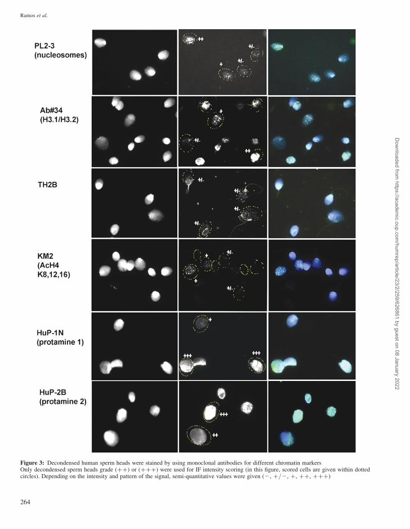
Figure 3: Decondensed human sperm heads were stained by using monoclonal antibodies for different chromatin markersOnly decondensed sperm heads grade (þþ) or (þþþ) were used for IF intensity scoring (in this figure, scored cells are given within dottedcircles). Depending on the intensity and pattern of the signal, semi-quantitative values were given (2, þ/2, þ, þþ, þþþ)
Ramos et al.
264
Dow
nloaded from https://academ
ic.oup.com/hum
rep/article/23/2/259/626861 by guest on 08 January 2022
the right borders (upper limits) that give an indication of
maximum fluorescence yields, the OAT samples are higher
with respect to free thiol groups (Fig. 2D), whereas the normos-
permics are higher for total SH content (Fig. 2E).
Considering the mean intensity after DTT reduction [normo
pool (R): 3602 arbitrary units] as the 100% level of the free
SH-groups in a sperm population, the percentage of free
thiols per sample was estimated. Using this approach, the
normal percentage of free thiols in human sperm cells was
�5%, but was increased in OAT sperm to 13% of the total.
Assuming that protamine cysteine thiols are the main contribu-
tors to sperm thiols, a lower total SH–content after DTT
reduction indicates a lower nuclear protamine content (2790
versus 3602 arbitrary units for the R windows of OAT and nor-
mospermics, respectively). Because in the OAT pool DTT–
mBBr intensity was overall lower than in the normo pool
(Table III), it is assumed that chromatin remodelling in OAT
sperm is incomplete or delayed during spermiogenesis.
TUNEL measurements
Table IV gives the outcomes of the TUNEL measurements of
mBBr-sorted sperm. TUNEL was evaluated by two indepen-
dent observers, between which no statistical differences
emerged. The TUNEL scores before or after FACS show that
in the normospermic pool, the staining and passage of sperm
through the sorter had little effect on the outcome. However,
a greater effect of presumably gating was observed in the
OAT patients. Both for normospermic and OAT pools, a
lower level of free thiol groups was associated with a higher
TUNEL score and vice versa (Table IV). Hence, the larger
fraction of low free thiol sperm in the OAT sample (Fig. 2D)
is responsible for the higher TUNEL level in OAT ejaculates.
Likewise, when fractionating for total thiol levels, in both
samples the TUNEL score of the low fraction was about
twice that of the high fraction.
IF detection of ds DNA in whole mount sperm nuclei
Underprotamination predicts a more open chromatin configur-
ation. In order to establish if we could detect a more open chro-
matin configuration with IF, we applied to a subset of the
samples (see Table I), a monoclonal antibody against dsDNA
to the mBBr–FACS sorted spermatozoa. The various patterns
and intensities obtained are given in Fig. 4. In both the normos-
permic and OAT samples, there was considerable variation in
antibody penetration-dependent DNA detection. The antibody
reacted more intensely at the nuclear periphery, and always
accentuated nuclear vacuoles (Fig. 4C). In nuclei with
background- or low fluorescence, a ‘negative’ band could be
observed (Fig. 4B) situated in the posterior nucleus close to
the attachment of the axonema. For the FACS-selected
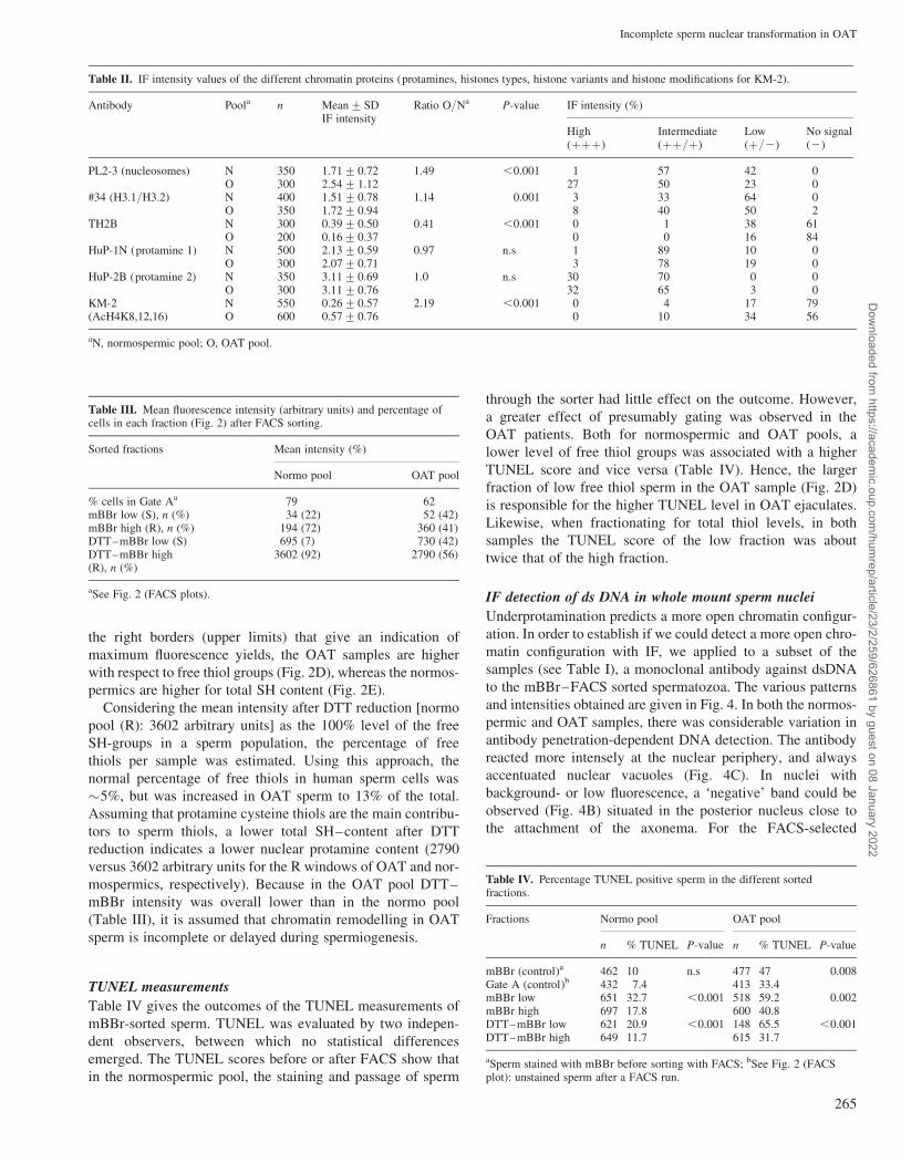
Table II. IF intensity values of the different chromatin proteins (protamines, histones types, histone variants and histone modifications for KM-2).
Antibody Poola n Mean+SDIF intensity
Ratio O/Na P-value IF intensity (%)
High(þþþ)
Intermediate(þþ/þ)
Low(þ/2)
No signal(2)
PL2-3 (nucleosomes) N 350 1.71+0.72 1.49 ,0.001 1 57 42 0O 300 2.54+1.12 27 50 23 0
#34 (H3.1/H3.2) N 400 1.51+0.78 1.14 0.001 3 33 64 0O 350 1.72+0.94 8 40 50 2
TH2B N 300 0.39+0.50 0.41 ,0.001 0 1 38 61O 200 0.16+0.37 0 0 16 84
HuP-1N (protamine 1) N 500 2.13+0.59 0.97 n.s 1 89 10 0O 300 2.07+0.71 3 78 19 0
HuP-2B (protamine 2) N 350 3.11+0.69 1.0 n.s 30 70 0 0O 300 3.11+0.76 32 65 3 0
KM-2(AcH4K8,12,16)
N 550 0.26+0.57 2.19 ,0.001 0 4 17 79O 600 0.57+0.76 0 10 34 56
aN, normospermic pool; O, OAT pool.
Table III. Mean fluorescence intensity (arbitrary units) and percentage ofcells in each fraction (Fig. 2) after FACS sorting.
Sorted fractions Mean intensity (%)
Normo pool OAT pool
% cells in Gate Aa 79 62mBBr low (S), n (%) 34 (22) 52 (42)mBBr high (R), n (%) 194 (72) 360 (41)DTT–mBBr low (S) 695 (7) 730 (42)DTT–mBBr high(R), n (%)
3602 (92) 2790 (56)
aSee Fig. 2 (FACS plots).
Table IV. Percentage TUNEL positive sperm in the different sortedfractions.
Fractions Normo pool OAT pool
n % TUNEL P-value n % TUNEL P-value
mBBr (control)a 462 10 n.s 477 47 0.008Gate A (control)b 432 7.4 413 33.4mBBr low 651 32.7 ,0.001 518 59.2 0.002mBBr high 697 17.8 600 40.8DTT–mBBr low 621 20.9 ,0.001 148 65.5 ,0.001DTT–mBBr high 649 11.7 615 31.7
aSperm stained with mBBr before sorting with FACS; bSee Fig. 2 (FACSplot): unstained sperm after a FACS run.
Incomplete sperm nuclear transformation in OAT
265
Dow
nloaded from https://academ
ic.oup.com/hum
rep/article/23/2/259/626861 by guest on 08 January 2022
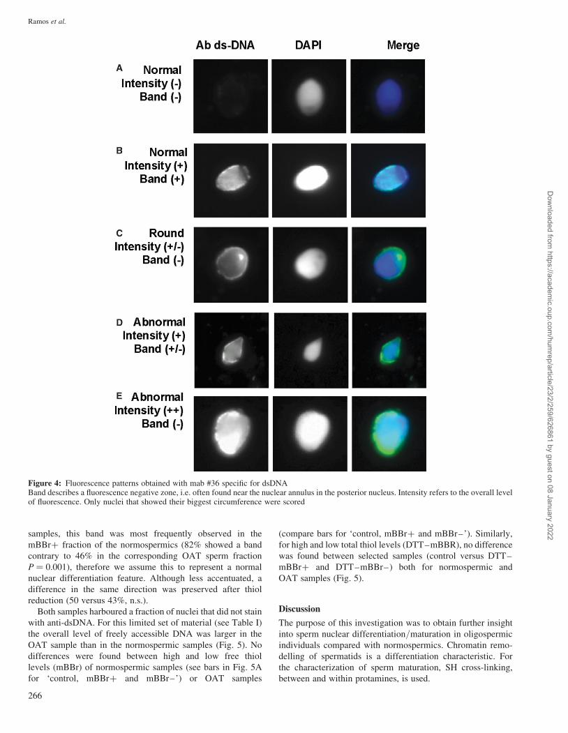
samples, this band was most frequently observed in the
mBBrþ fraction of the normospermics (82% showed a band
contrary to 46% in the corresponding OAT sperm fraction
P ¼ 0.001), therefore we assume this to represent a normal
nuclear differentiation feature. Although less accentuated, a
difference in the same direction was preserved after thiol
reduction (50 versus 43%, n.s.).
Both samples harboured a fraction of nuclei that did not stain
with anti-dsDNA. For this limited set of material (see Table I)
the overall level of freely accessible DNA was larger in the
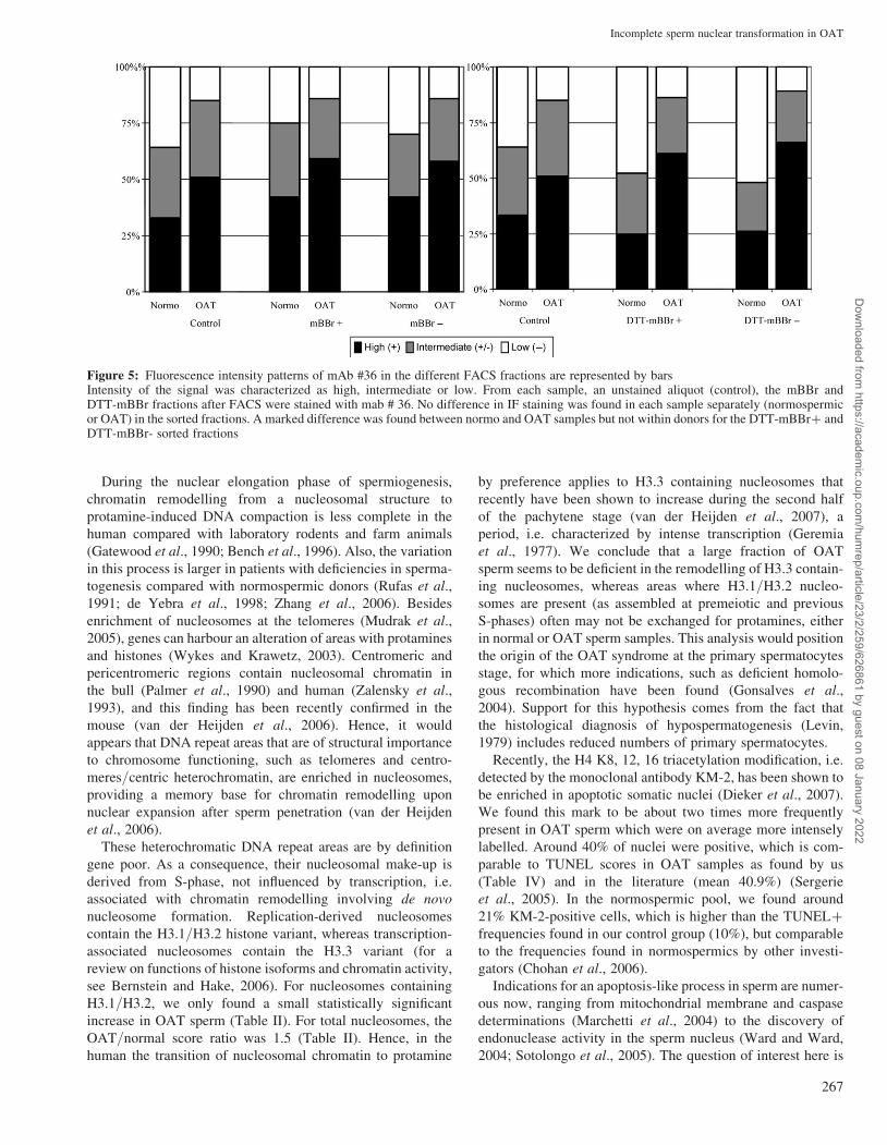
OAT sample than in the normospermic samples (Fig. 5). No
differences were found between high and low free thiol
levels (mBBr) of normospermic samples (see bars in Fig. 5A
for ‘control, mBBrþ and mBBr–’) or OAT samples
(compare bars for ‘control, mBBrþ and mBBr–’). Similarly,
for high and low total thiol levels (DTT–mBBR), no difference
was found between selected samples (control versus DTT–
mBBrþ and DTT–mBBr–) both for normospermic and
OAT samples (Fig. 5).
Discussion
The purpose of this investigation was to obtain further insight
into sperm nuclear differentiation/maturation in oligospermic
individuals compared with normospermics. Chromatin remo-
delling of spermatids is a differentiation characteristic. For
the characterization of sperm maturation, SH cross-linking,
between and within protamines, is used.
Figure 4: Fluorescence patterns obtained with mab #36 specific for dsDNABand describes a fluorescence negative zone, i.e. often found near the nuclear annulus in the posterior nucleus. Intensity refers to the overall levelof fluorescence. Only nuclei that showed their biggest circumference were scored
Ramos et al.
266
Dow
nloaded from https://academ
ic.oup.com/hum
rep/article/23/2/259/626861 by guest on 08 January 2022
During the nuclear elongation phase of spermiogenesis,
chromatin remodelling from a nucleosomal structure to
protamine-induced DNA compaction is less complete in the
human compared with laboratory rodents and farm animals
(Gatewood et al., 1990; Bench et al., 1996). Also, the variation
in this process is larger in patients with deficiencies in sperma-
togenesis compared with normospermic donors (Rufas et al.,
1991; de Yebra et al., 1998; Zhang et al., 2006). Besides
enrichment of nucleosomes at the telomeres (Mudrak et al.,
2005), genes can harbour an alteration of areas with protamines
and histones (Wykes and Krawetz, 2003). Centromeric and
pericentromeric regions contain nucleosomal chromatin in
the bull (Palmer et al., 1990) and human (Zalensky et al.,
1993), and this finding has been recently confirmed in the
mouse (van der Heijden et al., 2006). Hence, it would
appears that DNA repeat areas that are of structural importance
to chromosome functioning, such as telomeres and centro-
meres/centric heterochromatin, are enriched in nucleosomes,
providing a memory base for chromatin remodelling upon
nuclear expansion after sperm penetration (van der Heijden
et al., 2006).
These heterochromatic DNA repeat areas are by definition
gene poor. As a consequence, their nucleosomal make-up is
derived from S-phase, not influenced by transcription, i.e.
associated with chromatin remodelling involving de novo
nucleosome formation. Replication-derived nucleosomes
contain the H3.1/H3.2 histone variant, whereas transcription-
associated nucleosomes contain the H3.3 variant (for a
review on functions of histone isoforms and chromatin activity,
see Bernstein and Hake, 2006). For nucleosomes containing
H3.1/H3.2, we only found a small statistically significant
increase in OAT sperm (Table II). For total nucleosomes, the
OAT/normal score ratio was 1.5 (Table II). Hence, in the
human the transition of nucleosomal chromatin to protamine
by preference applies to H3.3 containing nucleosomes that
recently have been shown to increase during the second half
of the pachytene stage (van der Heijden et al., 2007), a
period, i.e. characterized by intense transcription (Geremia
et al., 1977). We conclude that a large fraction of OAT
sperm seems to be deficient in the remodelling of H3.3 contain-
ing nucleosomes, whereas areas where H3.1/H3.2 nucleo-
somes are present (as assembled at premeiotic and previous
S-phases) often may not be exchanged for protamines, either
in normal or OAT sperm samples. This analysis would position
the origin of the OAT syndrome at the primary spermatocytes
stage, for which more indications, such as deficient homolo-
gous recombination have been found (Gonsalves et al.,
2004). Support for this hypothesis comes from the fact that
the histological diagnosis of hypospermatogenesis (Levin,
1979) includes reduced numbers of primary spermatocytes.
Recently, the H4 K8, 12, 16 triacetylation modification, i.e.
detected by the monoclonal antibody KM-2, has been shown to
be enriched in apoptotic somatic nuclei (Dieker et al., 2007).
We found this mark to be about two times more frequently
present in OAT sperm which were on average more intensely
labelled. Around 40% of nuclei were positive, which is com-
parable to TUNEL scores in OAT samples as found by us
(Table IV) and in the literature (mean 40.9%) (Sergerie
et al., 2005). In the normospermic pool, we found around
21% KM-2-positive cells, which is higher than the TUNELþ
frequencies found in our control group (10%), but comparable
to the frequencies found in normospermics by other investi-
gators (Chohan et al., 2006).
Indications for an apoptosis-like process in sperm are numer-
ous now, ranging from mitochondrial membrane and caspase
determinations (Marchetti et al., 2004) to the discovery of
endonuclease activity in the sperm nucleus (Ward and Ward,
2004; Sotolongo et al., 2005). The question of interest here is
Figure 5: Fluorescence intensity patterns of mAb #36 in the different FACS fractions are represented by barsIntensity of the signal was characterized as high, intermediate or low. From each sample, an unstained aliquot (control), the mBBr andDTT-mBBr fractions after FACS were stained with mab # 36. No difference in IF staining was found in each sample separately (normospermicor OAT) in the sorted fractions. A marked difference was found between normo and OAT samples but not within donors for the DTT-mBBrþ andDTT-mBBr- sorted fractions
Incomplete sperm nuclear transformation in OAT
267
Dow
nloaded from https://academ
ic.oup.com/hum
rep/article/23/2/259/626861 by guest on 08 January 2022
at what stage during spermiogenesis H4 becomes modified, i.e.
when does the nucleus receive signals for the apoptotic
pathway. Hyperacetylation of H4 is the normal mark at the
onset of the histone to protamine change (Sonnack et al.,
2002). KM-2 is likely to sense more sites than just the classified
epitopes H4K8, 12 and 16, such as the homologous (acetylated)
region in H2A (Dieker et al., 2007).
The frequency of positivity for TH2B in normospermics was
comparable with data from the literature (van Roijen et al.,
1998; Singleton et al., 2007). Extrapolating from the finding
of the latter group that sperm which are positive for TH2B
have superior nuclear decondensation after gamete fusion,
the lower fraction of TH2B-positive sperm in OAT indicates
a lower fraction of optimally differentiated sperm in this
syndrome.
We have used FACS measurements of mBBr to substantiate
the IF data that were indicative of an increased level of nucleo-
somes in OAT sperm. Thiol fluorescence of human sperm
mainly represents the nucleus, although a low percentage (neg-
ligible for the total measurements) of the total mBBr fluor-
escence is derived from tails (Seligman et al., 1991). When
total thiol levels are determined by mBBr fluorescence, the his-
tograms obtained by us agreed with those of Rufas et al., 1991.
Under-representation of PRM2 in OAT (for a review, see
Carrell et al. 2007) is the main candidate for explaining the
higher total thiol fluorescence in normospermic samples
(Fig. 2E). We did not find lower protamine levels by the sub-
jective scoring of IF intensities, pointing to the fact that
when epitope levels are high, IF is not a suitable method for
quantification. A ratio of around 0.8 (OAT/normo) of total
thiol fluorescence in the ‘high windows’ of donor pools
(Table III) may well be in line with expectation, although in
gel systems such a ratio is not always found (Mengual et al.,
2003). The higher maximum of free thiol in the OAT sample
(Fig. 2D) is likely due to a delay in, or an interruption to,
thiol oxidation during nuclear maturation after spermiation.
The excess of OAT sperm with low free thiol levels could
originate from oxidative stress due to ROS production or apop-
totic processes, i.e. one aspect of incompletely transformed
immature human sperm (Bennetts and Aitken, 2005). The
fact that thiol oxidation functions in biological signalling
(Moran et al., 2001) and has been implicated in sperm capaci-
tation (de Lamirande and Gagnon, 2003), labels sperm with
low free thiol levels as pathogenic. In line with this interpret-
ation, we found TUNEL readings to be increased in fractions
with low free SH groups (Table IV), again indicating oxidative
stress (Greco et al., 2005; Aitken and Baker, 2006) during epi-
didymal transit as one aspect of the induction of apoptosis.
Protection of the genetic material is always mentioned as one
of the prime functions of chromatin remodelling during sper-
miogenesis and subsequent maturation/compaction of the
nucleus by thiol oxidation. Underprotamination by definition
entails a lower degree of compaction as does delayed or incom-
plete nuclear thiol oxidation (Kosower et al., 1992). We have
asked ourselves if immunofluorescence with an antibody that
detects dsDNA can serve to evaluate the degree of nuclear
compaction (hence its accessibility). For this purpose, we
adopted a protocol that does not include nuclear expansion
and preserves nuclear structures. To achieve this, we avoided
settling of sperm cells on glass by encapsulating the spermato-
zoa in a fibrin clot. Our results show the feasibility of this
approach. The nuclear periphery is most reactive, which
could partly involve the fact that telomeric histone containing
chromatin is adjacent to the nuclear envelope (Zalenskaya
et al., 2000). Vacuoles were faithfully recorded, which would
argue for proper penetration of the antibody. Overall in this
experiment, OAT sperm were by penetration of the antibody
less compacted, which is in agreement with the higher nucleo-
somal content and CMA3 scores (Iranpour et al., 2000). Within
pools, no difference was found between oxidized (low free SH)
and non-oxidized (high free SH) sperm. When we selected for
the sperm fractions with a high total thiol level, hence sperm
with higher protamine content, in both pools we did not
observe a striking improvement of ‘compaction’. This would
argue for another aspect of nuclear structure, likely involving
the nuclear matrix, to be not properly developed in OAT
sperm, as for instance could be demonstrated by the slower
decondensation kinetics in this study.
In conclusion, by the application of various chromatin anti-
bodies, we were able to suggest that H3.1/H3.2 containing
nucleosomes may be the most resistant to chromatin remodel-
ling at nuclear elongation during spermiogenesis. Therefore,
H3.3 containing nucleosomes that grow more numerous
during the second half of pachytene in the mouse (van der
Heijden et al., 2007) may be the major ones that are incomple-
tely exchanged for protamines in OAT sperm.
The free and total thiol status relates to the likelihood of
apoptotic development by TUNEL standard, with lower thiol
levels relating to a higher likelihood of apoptotic development.
Low levels of free thiol groups are indicative of oxidative stress
and therefore also predictive for apoptosis. Within normo and
OAT pools, free and total thiol levels do not predict the
amount of nuclear compaction as measured by the penetrability
of a monoclonal antibody for dsDNA. Hence other aspects of
nuclear differentiation also differ between normo and OAT
sperm.
Acknowledgements
We would like to remember the late Arie Pennings of the Haematol-ogy Department of the UMC St Radboud for setting up the FACSsorting conditions and thank Ingrid Punte for help in the initialstages of this project. We would like to thank Dr R Balhorn for thegenerous gift of PRM antibodies and Willy Baarends of Erasmus Uni-versity Medical Center Rotterdam for the gift of the TH2B detectingantibody.
References
Aitken RJ, Baker MA. Oxidative stress, sperm survival and fertility control.Mol Cell Endocrinol 2006;250:66–69.
Baart EB, de Rooij DG, Keegan KS, de Boer P. Distribution of Atr protein inprimary spermatocytes of a mouse chromosomal mutant: a comparison ofpreparation techniques. Chromosoma 2000;109:139–147.
Balhorn R, Reed S, Tanphaichitr N. Aberrant protamine 1/protamine 2 ratios insperm of infertile human males. Experientia 1988;44:52–55.
Bedford JM, Bent MJ, Calvin H. Variations in the structural character andstability of the nuclear chromatin in morphologically normal humanspermatozoa. J Reprod Fertil 1973;33:19–29.
Ramos et al.
268
Dow
nloaded from https://academ
ic.oup.com/hum
rep/article/23/2/259/626861 by guest on 08 January 2022
Bench GS, Friz AM, Corzett MH, Morse DH, Balhorn R. DNA and totalprotamine masses in individual sperm from fertile mammalian subjects.Cytometry 1996;23:263–271.
Bennetts LE, Aitken RJ. A comparative study of oxidative DNA damage inmammalian spermatozoa. Mol Reprod Dev 2005;71:77–87.
Bernstein E, Hake SB. The nucleosome: a little variation goes a long way.Biochem Cell Biol 2006;84:505–517.
Bianchi PG, Manicardi GC, Bizzaro D, Bianchi U, Sakkas D. Effect ofdeoxyribonucleic acid protamination on fluorochrome staining and in situnick-translation of murine and human mature spermatozoa. Biol Reprod1993;49:1083–1088.
Carrell DT, Liu L, Christensen G. Polyploidy in mouse embryos derived fromin vivo and in vitro fertilization is dependent on the timing of pregnant mareserum gonadotropin (PMSG) injection. Fertil Steril 2007;87:1470–1472.
Cho C, Jung Ha H, Willis WD, Goulding EH, Stein P, Xu Z, Schultz RM, HechtNB, Eddy EM. Protamine 2 deficiency leads to sperm DNA damage andembryo death in mice. Biol Reprod 2003;69:211–217.
Chohan KR, Griffin JT, Lafromboise M, De Jonge CJ, Carrell DT. Comparisonof chromatin assays for DNA fragmentation evaluation in human sperm. JAndrol 2006;27:53–59.
Dadoune JP. Expression of mammalian spermatozoal nucleoproteins. MicroscRes Tech 2003;61:56–75.
de Lamirande E, Gagnon C. Redox control of changes in protein sulfhydryllevels during human sperm capacitation. Free Radic Biol Med2003;35:1271–1285.
De Vos A, Van De Velde H, Joris H, Verheyen G, Devroey P, Van SteirteghemA. Influence of individual sperm morphology on fertilization, embryomorphology, and pregnancy outcome of intracytoplasmic sperm injection.Fertil Steril 2003;79:42–48.
de Yebra L, Oliva R. Rapid analysis of mammalian sperm nuclear proteins.Anal Biochem 1993;209:201–203.
de Yebra L, Ballesca JL, Vanrell JA, Corzett M, Balhorn R, Oliva R. Detectionof P2 precursors in the sperm cells of infertile patients who have reducedprotamine P2 levels. Fertil Steril 1998;69:755–759.
Dieker JW, Sun YJ, Jacobs CW, Putterman C, Monestier M, Muller S, van derVlag J, Berden JH. Mimotopes for lupus-derived anti-DNA andnucleosome-specific autoantibodies selected from random peptide phagedisplay libraries: facts and follies. J Immunol Methods 2005;296:83–93.
Dieker JW, Fransen JH, van Bavel CC, Briand JP, Jacobs CW, Muller S,Berden JH, van der Vlag J. Apoptosis-induced acetylation of histones ispathogenic in systemic lupus erythematosus. Arthritis Rheum2007;56:1921–1933.
Gatewood JM, Cook GR, Balhorn R, Bradbury EM, Schmid CW.Sequence-specific packaging of DNA in human sperm chromatin. Science1987;236:962–964.
Gatewood JM, Cook GR, Balhorn R, Schmid CW, Bradbury EM. Isolation offour core histones from human sperm chromatin representing a minor subsetof somatic histones. J Biol Chem 1990;265:20662–20666.
Geremia R, Boitani C, Conti M, Monesi V. RNA synthesis in spermatocytesand spermatids and preservation of meiotic RNA during spermiogenesis inthe mouse. Cell Differ 1977;5:343–355.
Gonsalves J, Sun F, Schlegel PN, Turek PJ, Hopps CV, Greene C, Martin RH,Pera RA. Defective recombination in infertile men. Hum Mol Genet2004;13:2875–2883.
Greco E, Iacobelli M, Rienzi L, Ubaldi F, Ferrero S, Tesarik J. Reduction of theincidence of sperm DNA fragmentation by oral antioxidant treatment. JAndrol 2005;26:349–353.
Henikoff S, Ahmad K. Assembly of variant histones into chromatin. Annu RevCell Dev Biol 2005;21:133–153.
Hunt P, LeMaire R, Embury P, Sheean L, Mroz K. Analysis of chromosomebehavior in intact mammalian oocytes: monitoring the segregation of aunivalent chromosome during female meiosis. Hum Mol Genet1995;4:2007–2012.
Iranpour FG, Nasr-Esfahani MH, Valojerdi MR, al Taraihi TM. ChromomycinA3 staining as a useful tool for evaluation of male fertility. J Assist ReprodGenet 2000;17:60–66.
Johnsen SG. Testicular biopsy score count—a method for registration ofspermatogenesis in human testes: normal values and results in 335hypogonadal males. Hormones 1970;1:2–25.
Kosower NS, Kosower EM. Thiol labeling with bromobimanes. MethodsEnzymol 1987;143:76–84.
Kosower NS, Katayose H, Yanagimachi R. Thiol-disulfide status andacridine orange fluorescence of mammalian sperm nuclei. J Androl1992;13:342–348.
Laberge RM, Boissonneault G. On the nature and origin of DNA strand breaksin elongating spermatids. Biol Reprod 2005;73:289–296.
Levin HS. Testicular biopsy in the study of male infertility: its currentusefulness, histologic techniques, and prospects for the future. Hum Pathol1979;10:569–584.
Losman MJ, Fasy TM, Novick KE, Monestier M. Monoclonal autoantibodiesto subnucleosomes from a MRL/Mp(2)þ/þ mouse. Oligoclonality ofthe antibody response and recognition of a determinant composed ofhistones H2A, H2B, and DNA. J Immunol 1992;148:1561–1569.
Marchetti C, Gallego MA, Defossez A, Formstecher P, Marchetti P. Staining ofhuman sperm with fluorochrome-labeled inhibitor of caspases to detectactivated caspases: correlation with apoptosis and sperm parameters. HumReprod 2004;19:1127–1134.
McPherson SM, Longo FJ. Nicking of rat spermatid and spermatozoaDNA: possible involvement of DNA topoisomerase II. Dev Biol1993;158:122–130.
Mengual L, Ballesca JL, Ascaso C, Oliva R. Marked differences in protaminecontent and P1/P2 ratios in sperm cells from percoll fractions betweenpatients and controls. J Androl 2003;24:438–447.
Menkveld R, Wong WY, Lombard CJ, Wetzels AM, Thomas CM, Merkus HM,Steegers-Theunissen RP. Semen parameters, including WHO and strictcriteria morphology, in a fertile and subfertile population: an effort towardsstandardization of in-vivo thresholds. Hum Reprod 2001;16:1165–1171.
Moran LK, Gutteridge JM, Quinlan GJ. Thiols in cellular redox signalling andcontrol. Curr Med Chem 2001;8:763–772.
Mudrak O, Tomilin N, Zalensky A. Chromosome architecture in thedecondensing human sperm nucleus. J Cell Sci 2005;118:4541–4550.
Palmer DK, O’Day K, Margolis RL. The centromere specific histone CENP-Ais selectively retained in discrete foci in mammalian sperm nuclei.Chromosoma 1990;100:32–36.
Pellicciari C, Hosokawa Y, Fukuda M, Manfredi-Romanini MG.Cytofluorometric study of nuclear sulphydryl and disulphide groups duringsperm maturation in the mouse. J Reprod Fertil 1983;68:371–376.
Ramos L, Wetzels AMM. Low rates of DNA fragmentation in selected motilehuman spermatozoa assessed by the TUNEL assay. Hum Reprod2001;16:1703–1707.
Ramos L, Kleingeld P, Meuleman E, van Kooy R, Kremer J, Braat D, WetzelsA. Assessment of DNA fragmentation of spermatozoa that were surgicallyretrieved from men with obstructive azoospermia. Fertil Steril2002;77:233–237.
Ramos L, de Boer P, Meuleman EJ, Braat DD, Wetzels AM. Evaluation ofICSI-selected epididymal sperm samples of obstructive azoospermic malesby the CKIA system. J Androl 2004;25:406–411.
Rufas O, Fisch B, Seligman J, Tadir Y, Ovadia J, Shalgi R. Thiol status inhuman sperm. Mol Reprod Dev 1991;29:282–288.
Sakkas D, Moffatt O, Manicardi GC, Mariethoz E, Tarozzi N, Bizzaro D.Nature of DNA damage in ejaculated human spermatozoa and the possibleinvolvement of apoptosis. Biol Reprod 2002;66:1061–1067.
Saowaros W, Panyim S. The formation of disulfide bonds in human protaminesduring sperm maturation. Experientia 1979;35:191–192.
Seligman J, Shalgi R, Oschry Y, Kosower NS. Sperm analysis by flowcytometry using the fluorescent thiol labeling agent monobromobimane.Mol Reprod Dev 1991;29:276–281.
Seligman J, Kosower NS, Weissenberg R, Shalgi R. Thiol-disulfide status ofhuman sperm proteins. J Reprod Fertil 1994;101:435–443.
Sergerie M, Laforest G, Bujan L, Bissonnette F, Bleau G. Sperm DNAfragmentation: threshold value in male fertility. Hum Reprod2005;20:3446–3451.
Singleton S, Mudrak O, Morshedi M, Oehninger S, Zalenskaya I, Zalensky A.Characterisation of a human sperm cell subpopulation marked by thepresence of the TSH2B histone. Reprod Fertil Dev 2007;19:392–397.
Smeenk RJ, Brinkman K, van den Brink HG, Westgeest AA. Reaction patternsof monoclonal antibodies to DNA. J Immunol 1988;140:3786–3792.
Sonnack V, Failing K, Bergmann M, Steger K. Expression of hyperacetylatedhistone H4 during normal and impaired human spermatogenesis. Andrologia2002;34:384–390.
Sotolongo B, Huang TT, Isenberger E, Ward WS. An endogenous nuclease inhamster, mouse, and human spermatozoa cleaves DNA into loop-sizedfragments. J Androl 2005;26:272–280.
Stanker LH, Wyrobek A, Balhorn R. Monoclonal antibodies to humanprotamines. Hybridoma 1987;6:293–303.
Steger K, Failing K, Klonisch T, Behre HM, Manning M, Weidner W, Hertle L,Bergmann M, Kliesch S. Round spermatids from infertile men exhibitdecreased protamine-1 and -2 mRNA. Hum Reprod 2001;16:709–716.
Incomplete sperm nuclear transformation in OAT
269
Dow
nloaded from https://academ
ic.oup.com/hum
rep/article/23/2/259/626861 by guest on 08 January 2022
Van Der Heijden GW, Dieker JW, Derijck AA, Muller S, Berden JH, Braat DD,van der Vlag J, de Boer P. Asymmetry in histone H3 variants and lysinemethylation between paternal and maternal chromatin of the early mousezygote. Mech Dev 2005;122:1008–1022.
van der Heijden GW, Derijck AA, Ramos L, Giele M, van der Vlag J, de BoerP. Transmission of modified nucleosomes from the mouse male germline tothe zygote and subsequent remodeling of paternal chromatin. Dev Biol2006;298:458–469.
Van Der Heijden GW, Derijck AA, Posfai E, Giele M, Pelczar P, Ramos L,Wansink DG, van der Vlag J, Peters AH, de Boer P. Chromosome-widenucleosome replacement and H3.3 incorporation during mammalianmeiotic sex chromosome inactivation. Nat Genet 2007;39:251–258.
van Roijen HJ, Ooms MP, Spaargaren MC, Baarends WM, Weber RF,Grootegoed JA, Vreeburg JT. Immunoexpression of testis-specifichistone 2B in human spermatozoa and testis tissue. Hum Reprod1998;13:1559–1566.
Ward MA, Ward WS. A model for the function of sperm DNA degradation.Reprod Fertil Dev 2004;16:547–554.
WHO. World Health Organization: WHO Laboratory Manual for theExamination of Human Semen and Sperm-Cervical Mucus Interaction,34th ed. edn. Cambridge University Press, 1999.
Wykes SM, Krawetz SA. The structural organization of sperm chromatin. JBiol Chem 2003;278:29471–29477.
Yu YE, Zhang Y, Unni E, Shirley CR, Deng JM, Russell LD, Weil MM,Behringer RR, Meistrich ML. Abnormal spermatogenesis and reduced
fertility in transition nuclear protein 1-deficient mice. Proc Natl Acad SciUSA 2000;97:4683–4688.
Zalenskaya IA, Bradbury EM, Zalensky AO. Chromatin structure oftelomere domain in human sperm. Biochem Biophys Res Commun2000;279:213–218.
Zalensky AO, Breneman JW, Zalenskaya IA, Brinkley BR, Bradbury EM.Organization of centromeres in the decondensed nuclei of mature humansperm. Chromosoma 1993;102:509–518.
Zalensky AO, Siino JS, Gineitis AA, Zalenskaya IA, Tomilin NV, Yau P,Bradbury EM. Human testis/sperm-specific histone H2B (hTSH2B).Molecular cloning and characterization. J Biol Chem 2002;277:43474–43480.
Zhang X, San Gabriel M, Zini A. Sperm nuclear histone to protamine ratio infertile and infertile men: evidence of heterogeneous subpopulations ofspermatozoa in the ejaculate. J Androl 2006;27:414–420.
Zhao M, Shirley CR, Yu YE, Mohapatra B, Zhang Y, Unni E, Deng JM,Arango NA, Terry NH, Weil MM et al. Targeted disruption of thetransition protein 2 gene affects sperm chromatin structure and reducesfertility in mice. Mol Cell Biol 2001;21:7243–7255.
Zollner U, Schleyer M, Steck T. Evaluation of a cut-off value for normal spermmorphology using strict criteria to predict fertilization after conventionalin-vitro fertilization and embryo transfer in asthenozoospermia. HumReprod 1996;11:2155–2161.
Submitted on August 17, 2007; resubmitted on October 5, 2007; accepted onOctober 18, 2007
Ramos et al.
270
Dow
nloaded from https://academ
ic.oup.com/hum
rep/article/23/2/259/626861 by guest on 08 January 2022