dna integrity in human spermatozoa: relationships with semen quality
TRANSCRIPT
33
Journal of Andrology, Vol. 21, No. 1, January/February 2000Copyright q American Society of Andrology
DNA Integrity in Human Spermatozoa: Relationships WithSemen Quality
D. STEWART IRVINE, JEREMY P. TWIGG, EMMA L. GORDON, NORMA FULTON, PHILIP A. MILNE, ANDR. JOHN AITKEN
From the MRC Reproductive Biology Unit, Centre for Reproductive Biology, Edinburgh, Scotland.
ABSTRACT: The literature contains conflicting evidence regardingthe existence of DNA damage in spermatozoa from infertile male pa-tients. To examine this phenomenon, we have studied ejaculatedspermatozoa from normozoospermic semen donors and from a groupof the unselected male partners of couples attending an infertility clinicfor initial investigation. Classical semen analysis according to WorldHealth Organization (WHO) guidelines was undertaken with comput-er-assisted sperm analysis (CASA). Spermatozoa were prepared bysequential washing and centrifugation and were analyzed for DNAfragmentation using three assays: 1) a single-cell gel electrophoresis(comet) assay, 2) in situ nick translation with prior chemical decon-densation (ISNT-decondensed), and 3) in situ nick translation withoutprior chemical decondensation (ISNT-condensed). In addition, reac-tive oxygen species (ROS) generation by spermatozoa was mea-
sured, and seminal plasma was analyzed for its total reactive antiox-idant potential (TRAP). When the donor and patient groups were com-pared, the latter had lower levels of semen quality and higher levelsof DNA damage, which was particularly apparent using the cometassay. Highly significant negative correlations were observed betweenDNA fragmentation, detected by all three assays, and semen quality,particularly sperm concentration. In addition, multiple regression anal-ysis indicated that other attributes of semen quality, such as spermmovement and ROS generation, were also related to DNA damage.We conclude that a significant proportion of infertile men have ele-vated levels of DNA damage in their ejaculated spermatozoa.
Key words: Comet assay, in situ nick translation, infertility, sper-matogenesis, ICSI, CASA.
J Androl 2000;21:33–44
Infertility remains a major clinical problem, with recentdata from Europe suggesting that as many as one in
four couples trying to conceive now experience difficul-ties (Schmidt et al, 1995). Moreover, in this and otherstudies, the most prevalent causative factor appears to bedefective semen quality (Thonneau et al, 1991; Hull,1992; Templeton, 1992). While there can be no doubt thatthe recent introduction of microassisted fertilization tech-niques has revolutionized the management of coupleswith male-factor infertility (Van Steirteghem et al,1993a,b), concerns over the safety of this approach re-main (Cummins and Jequier, 1995; Foresta et al, 1996).Painstaking follow-up studies of children born after mi-croassisted fertilization have generated broadly reassuringdata, with no evidence of an increase in the incidence ofmajor congenital malformations amongst children bornafter intracytoplasmic sperm injection (ICSI; Bonduelleet al, 1996a,b; Wisanto et al, 1996). This view has re-cently been challenged, however (Kurinczuk and Bower,1997).
An additional concern arises from our understanding ofthe etiology of defective sperm function in couples with
Correspondence to: Dr D. Stewart Irvine, MRC Reproductive BiologyUnit, Centre for Reproductive Biology, 37 Chalmers St, Edinburgh, EH39EW, United Kingdom. (e-mail: [email protected]).
Received for publication February 18, 1998; accepted for publicationJuly 13, 1999.
male infertility. One feature of the semen of infertile men,particularly those with oligoasthenozoospermia, is theproduction of excessive levels of reactive oxygen species(ROS) (Aitken et al, 1989a,b, 1991). It has been shownthat these excessive ROS can emanate from contaminat-ing leukocytes (Aitken et al, 1994, 1995) or from dys-functional spermatozoa (Aitken et al, 1992; Iwasaki andGagnon, 1992; Gomez et al, 1996). One consequence ofexcessive ROS generation is peroxidative damage to theplasma membrane, which leads to an impairment ofsperm function that is reflected in decreased pregnancyrates in vivo as well as in impaired fertilization in vitro(Aitken et al, 1989b; 1991; Sukcharoen et al, 1996).While it is known that ROS have the capacity to damagelipids in the sperm plasma membrane, it is also knownthat ROS can damage other cellular structures, particu-larly DNA (Dizdaroglu, 1992; Altman et al, 1995; Pal-omba et al, 1996; Lloyd et al, 1997). In the context ofspontaneous pregnancy or conception following in vitrofertilization (IVF), spermatozoa exposed to ROS becomedysfunctional as a consequence of this peroxidative dam-age to the plasma membrane and are unable to initiatefertilization. Under these circumstances, any genetic dam-age induced by exposure of spermatozoa to oxidativestress is unlikely to be transmitted to the embryo. In thecontext of ICSI, however, these safeguards are removed,and there is nothing to prevent spermatozoa with dam-
34 Journal of Andrology · January/February 2000
aged DNA being injected directly into the oocyte. Be-cause DNA damage appears to have no impact upon theability of the spermatozoon to achieve fertilization fol-lowing ICSI (Twigg et al, 1997), the probability that theprocedure might result in the transmission of geneticdamage to the embryo cannot be discounted. In a recentstudy, we have exposed spermatozoa from normozoos-permic men to oxidizing environments created by coin-cubation with hydrogen peroxide, reduced nicotinamideadenine dinucleotide phosphate (NADPH), or activatedwhite cells. The subsequent ability of the spermatozoa todecondense in vitro was examined using sequential in-cubations in EDTA, dithiothreitol, and sodium dodecylsulphate, and the amount of DNA strand breakage wereassessed by an in situ nick translation (ISNT) protocol.Finally, cells exposed to hydrogen peroxide, NADPH, andactivated leukocytes were microinjected into hamster oo-cytes, and their ability to decondense and form normalpronuclei was determined. The results indicated that hu-man sperm chromatin becomes cross-linked under con-ditions of oxidative stress and exhibits increased DNAstrand breakage, yet the rate of pronucleus formation wasno different from that of untreated control cells. At pre-sent, very little is known of the level of DNA damageencountered in human spermatozoa or the relationship be-tween such damage and other attributes of semen quality.Previous work has established relationships between ROSgeneration and impaired sperm maturity, measured by cy-toplasmic retention and other measures (Huszar and Vi-gue, 1994; Gomez et al, 1996; Lalwani et al, 1996), andit is possible that markers of sperm maturity such asG6PDH, creatine phosphokinase, and lactate dehydroge-nase-x may modulate the relationship between semenquality and the ROS-mediated decline of DNA integrity.
A number of approaches to the assessment of DNAdamage have been described, including nick-end labeling(Baccetti et al, 1996), ISNT (Sakkas et al, 1995), single-cell gel electrophoresis or the comet assay (McKelvey-Martin et al, 1993), and measurement by high-pressureliquid chromatography of oxidized DNA (Fraga et al,1996). Application of these assays to human spermatozoahas generated conflicting results. Thus, Sun and col-leagues (Sun et al, 1997), using the nick-end labeling orTUNEL assay in a study of unselected men undergoingin vitro fertilization treatment, have suggested that a neg-ative relationship exists between the observed extent ofDNA damage in human sperm and the conventional pa-rameters of semen quality. In contrast, Hughes and co-workers (Hughes et al, 1996), using the comet assay,found no difference in the extent of underlying DNAdamage when infertile men were compared with normalcontrols. Previous work with other cell types has tendedto suggest that the comet assay is the most sensitive (Le-roy et al, 1996).
Because of this discrepancy in the literature and be-cause of the potential clinical importance of sperm DNAdamage in the context of assisted reproduction, the pre-sent study was undertaken to evaluate DNA fragmenta-tion in the spermatozoa of both normozoospermic donorsand infertile men attending an infertility clinic as part oftheir routine investigation. A range of techniques has beenused to assess DNA damage, the extent of which has thenbeen correlated with various attributes of semen quality.It should be noted that the conventional criteria for semenquality (World Health Organization [WHO], 1992) rep-resent measurements made on populations of cells,whereas the assays used to quantify DNA integrity arenormally at the level of the single cell.
Materials and MethodsThis study was approved by the local ethical committee, and allpatients and volunteers gave written informed consent to the useof their gametes for research study. The laboratory that con-ducted the semen analysis was participating in one national andone international externally assessed quality control schemethroughout the duration of this study.
Subjects and Sample CollectionSemen samples were collected from an unselected group of menattending the Infertility Clinic of the Royal Infirmary of Edin-burgh for the investigation of infertility. Normozoospermic vol-unteer donors participating in our research program and who hadbeen carefully screened for the absence of significant reproduc-tive disease provided control samples. In all cases, after 3–4 daysof sexual abstinence, samples were collected by masturbationinto wide-mouthed sterile containers and were delivered to thelaboratory within 1 hour of ejaculation.
Assessment of the Conventional Criteria of SemenQualitySemen samples were allowed to liquefy at 378C, followingwhich an aliquot was removed in order to construct a conven-tional semen profile composed of ejaculate volume (in millili-ters), sperm concentration (3 106 per milliliter), overall and pro-gressive motility (percentage), and normal morphology (per-centage) using the guidelines promulgated by the WHO (1992).Duplicate assessment of sperm concentration was made by usinga positive-displacement pipette to add 10 mL of semen to 190mL of sperm-diluting fluid (SDF; 25g NaHCO3 in 5 mL w/v40% formalin, made up to 500 mL with distilled H2O), followingwhich the preparation was thoroughly mixed and loaded onto ahemocytometer (Improved Neubauer; Weber Scientific Instru-ments, Sussex, United Kingdom). The loaded chamber was al-lowed to settle for 5–15 minutes in a humidified chamber beforecounting at 310 magnification. For the assessment of motility,10 mL of semen was placed on a microscope slide prewarmedto 378C and was covered with a 19-mm 3 19-mm coverslip. Thepreparation was then examined at 378C at 3100 magnificationwith phase–contrast optics with the aid of a grid on an eyepiece
35Irvine et al · DNA Integrity in Human Spermatozoa
graticule. Spermatozoa in WHO categories a, b, and c were con-sidered motile, and those in categories a and b were consideredprogressively motile.
For the assessment of sperm morphology, a positive-displace-ment pipette was used to take 50 mL of fresh semen after liq-uefaction; to this, 950 mL of SDF was added, mixed, and leftfor 5–10 minutes prior to analysis. Ten microliters of this prep-aration was then placed on a clean slide, covered by a 22-mm3 22-mm coverslip, and examined at 3400 under phase–contrastoptics. An HTM-SAM (Hamilton Thorne Research, Beverly,Mass) semiautomated counter was used to facilitate scoring andrecording of data, with 100 cells on each slide being scoredaccording to WHO guidelines.
Human Spermatozoa PreparationIn order to obtain an unselected population of cells, the remain-ing semen sample was placed into a centrifuge tube and spun at500 3 g for 5 minutes to pellet the spermatozoa. The seminalplasma was aspirated and kept aside for the measurement of totalreactive antioxidant potential (TRAP assay). The pellet of sper-matozoa was overlaid with 4 mL of Biggers-Whitten-Whitingh-am (BWW) medium (Biggers et al, 1971) containing 20 mM N-(2-hydroxyethyl) piperazine-N9-(2-ethan sulfonic acid) (HEPES;Gibco, United Kingdom) and 0.3% human serum albumin(HSA) and was processed by centrifuge again at 500 3 g for 5minutes. The supernatant was removed, and 2 mL BWW wasadded. A further centrifugation was performed, after which thecells were resuspended in 500 mL of BWW medium. Cell den-sity was recorded with an improved Neubauer hemocytometer,and the correct amount of BWW was added to resuspend thecells at 20 3 106 mL. Aliquots were then taken for the followingassays.
Total Reactive Antioxidant PotentialThe TRAP assay was performed according to the method de-scribed by Smith (Smith et al, 1996) but with some modification.For the generation of peroxyl radicals, a solution of 30 mM 2,2-azobis-(2-amidinopropane) (ABAP) (Polysciences, Warrington,Pa) dissolved in phosphate-buffered saline (PBS) was used. Fiveminutes prior to each assay run, a 100-mM luminol stock solu-tion in dimethyl sulfoxide was diluted 1 in 1000 in PBS/ABAP.Aliquots (400 mL each) of this mixture were placed in cuvettesin a Berthold 9505 biolumat luminometer and were allowed toequilibrate to the operating temperature (378C) for 5 minuteswhile chemiluminescence was monitored. Seminal plasma wasdiluted 1:4 with PBS prior to addition to the ABAP/PBS solu-tion. Once the peroxyl radical–generating system demonstrateda steady-state level of chemiluminescence, a 4-mL aliquot of theseminal plasma/PBS solution was added to quench the chemi-luminescent signal. The time to 50% recovery of the initial sig-nal was then calculated as the TRAP time. A standard curve wascreated for each run with 6-hydroxy-2,5,7,8-tetramethyl-chro-man-2-carbonsaeure (TROLOX), and the results were expressedin TROLOX equivalents.
ROS ChemiluminescenceThe methodology used was that described by Aitken et al (1992).Briefly, 200 mL of sperm suspension in BWW were placed in
cuvettes of a luminometer with 2 mL of 100 mM luminol and 4mL of a 2 mg/mL horseradish peroxidase solution (Sigma) togive a spontaneous chemiluminescent signal. After 10 minutes,a 1-mL aliquot of 10 mM N-formylmethionyl-leucyl-phenylala-nine (FMLP) (Sigma) was added to detect leukocyte contami-nation, and after a further 10 minutes, 2 mL of a 1-mM stocksolution of 12-myristate-13-acetate phorbol ester (PMA; Calbi-ochem, Nottingham, United Kingdom) was added to assess thetotal ROS-generating capacity of the ejaculate (leukocytes 1spermatozoa). ROS chemiluminescence was then expressed as a5-minute integral for each cycle (spontaneous, FMLP and PMA).
Computer-Assisted Sperm AssessmentThe movement characteristics of the spermatozoa were examinedwith a Hamilton Thorne IVOS Motility Analyzer, version 10.5K,at an incubation temperature of 378C. Each sample was loadedonto 20-mm-deep microcell slides (Microm, Thame, UnitedKingdom), and analysis was performed using a 310 objectiveon five random fields from each sample. The analysis was per-formed in triplicate, and the mean of the three measurementswas calculated. Parameter settings were as follows: frames ac-quired, 30; frame rate, 50 Hz; minimum contrast, 10; minimumcell size, 4; nonmotile head size, 12; nonmotile head intensity,130; magnification, 1.96; static size limits, 0.59–2.87; and staticintensity, 0.471–1.77. A wide range of different movement char-acteristics were assessed, including average path velocity (VAP);straight-line velocity (VSL), curvilinear velocity (VCL), linear-ity (LIN 5 VSL/VCL), straightness (STR 5 VSL/VAP), per-centage rapid (VAP .25 mm/s), percentage medium speed (VAP5 10–24 mm/s), percentage slow (VAP 5 1–9 mm/s), percentagestatic, percentage overall motility (rapid 1 medium speed 1slow), percentage progressive motility (STR .75%), beat crossfrequency (BCF), and amplitude of lateral sperm head displace-ment (ALH).
DNA Damage AssessmentThree assays for the determination of DNA damage were uti-lized: 1) single-cell gel electrophoresis (COMET) assay for sin-gle-stranded DNA; 2) ISNT utilizing streptavidin-fluoresceinisothiocyanate (SA-FITC) for detection of biotin-16-dUTP in-corporation; 3) modified ISNT assay with decondensation ofspermatozoa prior to ISNT and utilization of an alkaline phos-phatase (AP), 5-bromo-4-chloro-3-indoyl phosphate (BCIP):ni-troblue tetrazolium (NBT) method for detection of incorporatedbiotin-16-dUTP (Twigg et al, 1997).
Single-Cell Gel Electrophoresis (Comet) AssayThe alkaline single-cell gel electrophoresis (comet) assay wasbased on existing methods described by Singh (1996), Mc-Kelvey-Martin et al (1993), and Hughes et al (1996), modifiedas indicated below. Unless otherwise stated, molecular-grade,DNAse-free reagents (Sigma, Poole, United Kingdom) wereused throughout. Select microscope slides (Chance Proper,Smethwick, United Kingdom) were used for the assay, and eachslide was prepared as follows: the slide was dipped in a solutionof 1% multipurpose agarose (Boehringer Mannheim, Lewes,United Kingdom) dissolved in 0.01 M PBS and air-dried over-night at room temperature. One hundred sixty microliters of a

36 Journal of Andrology · January/February 2000
solution of 0.6% multipurpose agarose, dissolved in PBS, wasthen placed on the slide, covered with a coverslip measuring 243 50 mm (Chance Proper), and left to solidify at room temper-ature. Ten microliters of spermatozoa at 6.6 3 106 mL in BWWwere then mixed with 75 mL of a 0.5% low-melt agarose solu-tion (Amresco, Solon, Ohio) dissolved in PBS. This was thenadded to the slide, overlaid with a coverslip, and allowed tosolidify on a prechilled (48C) tray for several minutes. A finallayer of 0.5% low-melt agarose was added to the slide and al-lowed to solidify at 48C for at least 1 hour.
Following removal of the coverslip, the slides were immersedin lysis buffer (2.5 M NaCl, 100 mM ethylenediaminetetraaceticacid [EDTA], 10 mM Tris [hydroxymethyl] aminomethane hy-drochloride [TRIS-HCl; Sigma], 10% dimethylsulfoxide[DMSO], and 1% Triton X-100, pH 10) for 1 hour at 48C. Thelysis solution was drained from the slides and replaced with asolution of proteinase K (Amresco) (100 mg/mL in 2.5 M NaCl,100 mM EDTA, 10% DMSO, pH 7.4) and incubated overnightat 378C. After draining the proteinase K solution, the slides wereimmersed in a horizontal gel tank filled with alkaline buffer (300mM sodium hydroxide [NaOH], 1 mM EDTA, pH 12.3) for 20minutes to allow the DNA to unwind. The buffer level was ad-justed to a covering height of approximately 0.25 cm above theslides, and electrophoresis was carried out for 4 minutes at 25V (0.862 V cm21). Slides were then placed in Coplin jars filledwith 0.4 M Tris-HCl (pH 7.4) for 5 minutes and were washedthree times with fresh buffer. After rinsing, the slides weredrained and immersed in Coplin jars containing 100% ethanolfor 5 minutes in order to precipitate the DNA and dehydrate theagarose. The slides were then air-dried overnight and stored ina box prior to scoring. Ethidium bromide was used to stain thesperm DNA (50 mL at 20 mg/mL dissolved in distilled water),which were then imaged on a Leitz fluorescent microscope (ex-citation filter 515–560 nm; dichroic filter, 580 nm; and suppres-sion filter, 580 nm) connected to a charge couple device (CCD)camera. For each sample, four replicate slides were prepared,and 50 randomly selected cells were scored on each slide. Thepercentage of head DNA, tail DNA, and the olive tail moment(OTM) were evaluated with the Komet image analysis system,version 1.0 (Kinetic Imaging, Liverpool, United Kingdom) run-ning on an IBM-compatible PC (Windows 3.1); the data weredownloaded into an Excel spreadsheet (Microsoft, Wokingham,United Kingdom) for analysis.
In Situ Nick Translation of Spermatozoa
The method for ISNT was that described by Bianchi et al (Bian-chi et al, 1993), with minor modifications. Spermatozoa wereprocessed by centrifugation at 500 3 g for 5 minutes, and thesupernatant was removed. Fixative (ethanol:glacial acetic acid[Analar grade] 3:1) was added to bring the cells to a concentra-tion of 10 3 106 mL. A 10-mL aliquot was then spotted onto ademarcated area on a clean microscope slide and allowed to airdry. Cells were rinsed once in tap water and twice in PBS beforebeing overlaid with 50 mL DNA polymerase mix made up of905 mL distilled water; 10 mL of 0.1 M dithiothreitol [DTT, Calbio-chem]; 10 mL of 1 M MgSO4; 50 mL of 1 M Tris-HCl, pH 7.2;10 mL of 1 mM biotin 16-dUTP (Boehringer Mannheim); 10 mLof a 1 mM dATP, dCTP, and dGTP mix (Promega, Southampton,
United Kingdom); and 5 mL DNA polymerase 1 (Promega). In-cubation was carried out at room temperature (258C) for 30 min-utes. After this, slides were rinsed again and overlaid with asolution of SA-FITC (900 mL distilled water; 90 mL 1 M Tris-HCl, pH 7.4; and 90 mL SAHR-FITC) for 30 minutes at roomtemperature. Once this incubation time had elapsed, the slideswere rinsed as above and dried with a tissue. Ten microliters ofmounting medium (3:1 distilled water:glycerol) were thenplaced on each slide and covered with a coverslip. Two hundredcells at 3400 magnification were scored for FITC fluorescenceon a Leitz fluorescence microscope (excitation filter, 450–490nm; dichroic filter, 510 nm; suppression filter, 520 nm), and thepercentage was entered into an Excel (Microsoft) spreadsheet.
Modified In Situ Nick Translation of SpermatozoaThe protocol for this modified ISNT technique was that de-scribed by Twigg et al (1997). Briefly, spermatozoa were pre-pared at a concentration of 10 3 106 mL and were taken throughtwo cycles of centrifugation and resuspension in BWW contain-ing 6 mM ethylenediaminetetraacetic acid (EDTA; BDH, Poole,United Kingdom) and 2 mM dithiothreitol (DTT; Calbiochem),respectively. The cells were incubated at 378C in 95% air and5% CO2 for 60 minutes, pelleted (500 3 g for 5 minutes), andfinally resuspended in 3:1 ethanol:glacial acetic acid (Analargrade; BDH). Ten microliters of this suspension were droppedonto a demarcated area of a clean glass microscope slide andallowed to air dry. A small volume of BWW containing 0.05%sodium dodecyl sulphate (SDS; Promega) was then added toeach slide and allowed to stand for 2 minutes. In order to blockany endogenous biotin/avidin-binding sites within the sperma-tozoa (Wood and Warnke, 1981), a blocking step was introducedthat was composed of sequential 20-minute incubations with0.001% biotin (Sigma) and 0.01% avidin (Sigma) in distilledwater. ISNT was performed with the enzyme DNA polymerase1 (Promega). Biotin incorporation was detected by an AP, BCIP:NBT (Boehringer Mannheim) method (De Jong et al, 1985). Theslides were overlaid with a coverslip and incubated in a humid-ified chamber at room temperature for 16 hours. Following in-cubation, the slides were rinsed in tap water for 5 minutes andpassed through 100% ethanol before being mounted under Per-tex (Cellpath, Hemel Hempstead, United Kingdom). Fifty ran-domly selected sperm cells were captured at 3400 magnificationon an Olympus BH2 microscope and were scored for opticaldensity per sperm head by measuring each pixel of the digitizedsperm head image on an arbitrary scale of 256 levels of gray onan IBM-compatible personal computer using Image Pro Plus forWindows, version 1.3.2. Data was saved in an Excel (Microsoft)spreadsheet before analysis.
Statistical AnalysisData were analyzed with the Statistical Package for the SocialSciences software program (SPSS Inc., Chicago, Ill) on a PowerMacintosh 8500 computer. The distributions of the variableswere examined, and where necessary, they were normalized bylog or square root transformation. Differences between groupswere examined by means of the Mann-Whitney U-test or byunpaired t-test, as appropriate. Relationships between variableswere examined by simple linear regression or by stepwise mul-

37Irvine et al · DNA Integrity in Human Spermatozoa
Table 1. Semen quality assessed by WHO semen analysis and by CASA*
Variable
Donor Group
Median IQR
Patient Group
Median IQR P
WHO semen profileEjaculate volume (mL)Sperm concentration (3106 per mL)Overall motility (%) (5 WHO criteria a1b1c)Progressive motility (%)Normal morphology (%)
39463.1751.7843.3
2–4.176.8–123.858.1–67.0
49.32–54.338.2–51.3
2.240.542.630.4540.4
1.3–3.226.4–81.531.7–5419.4–40.232.3–54.9
NS0.00260.00030.0008
NS
CASA profileOverall motility (%)Progressive motility (%)ALH (mm)LinearityStraightnessVAP (mm/s)VCL (mm/s)VSL (mm/s)
75.5323.85
46.572.536.756.3528.25
69.5–81.026.0–38.53.6–4.6
45.0–48.570.0–73.033.1–42.151.6–64.524.3–33.0
33104
446933.353.422.1
26.0–55.06.0–20.03.1–4.7
37.0–48.063.0–71.027.3–34.345.6–60.319.5–25.7
0.00110.001
NSNSNSNSNS
0.0183
* WHO indicates World Health Organization; CASA, computer-assisted sperm analysis; ALH, amplitude of lateral sperm head displacement; VAP,average path velocity; VCL, curvilinear velocity; VSL, straight-line velocity; NS, not significant; IQR, interquartile range.
tiple linear regression. Data are presented as medians with in-terquartile range (IQR).
Results
Semen QualitySamples were collected from an unselected group of pa-tients consulting for infertility and from a cohort of vol-unteer donors in order to provide a wide, dynamic rangeof semen quality attributes. In all, data on semen quality,ROS production, seminal plasma TRAP, and sperm DNAfragmentation were collected on 12 normal volunteer do-nors and on 29 male patients. In addition, CASA datawere collected on a subgroup of 9 donors and 17 patients.All members of the donor group had normal values forsperm concentration (more than 20 3 106/mL), overallmotility (.40% motile), and morphology (.30%; WHO,1992), as defined for our local population (Irvine andTempleton, 1994). Three donor samples (25%) fell belowthe WHO threshold of 50% progressive motility. In thepatient group, 7 of 29 (24%) were oligozoospermic (,203 106/mL), 13 of 29 (44.8%) had low overall motility(,40%), 23 of 29 (79%) had low progressive motility byWHO standards (,50%), and 3 of 29 (10.3%) had terato-zoospermia (,30% normal forms).
Descriptive statistics for both groups are presented inTable 1. The median sperm concentration for the donorswas 94.0 3 106/mL (76.8–123.8, IQR), whereas that forthe patient group was significantly lower at 40.5 3 106/mL (26.4–81.5 106/mL) (P 5 .0026). Similarly, the pa-tient population were significantly more asthenozoos-permic than the donor population, with a median pro-gressive motility of 30.4% (19.4–40.2%), compared with
51.7% (49.3–54.3%) (P 5 .0003). There were no differ-ences between the groups with respect to ejaculate vol-ume or normal morphology. In the subgroup of subjectson whom CASA data was collected, the same differenceswere apparent, with the patient population having signif-icantly lower values for sperm concentration and for over-all and progressive motility compared with the donor co-hort. There were no differences in any other attribute ofsperm movement, with the exception of VSL, which wassignificantly lower in the patient cohort.
DNA Damage and Semen QualityThere were no significant differences between the groupsin terms of ROS production, measured either in the rest-ing state or after stimulation with FMLP or PMA, andthere were no differences in the antioxidant potential ofseminal plasma, measured either as TRAP time or asTROLOX equivalents (Table 2). In contrast, there weresignificant differences between the donor and patientgroups with respect to DNA damage, with patients havinghigher levels of damage as determined by the comet as-say. For the latter, the median percentage tail DNA was10.44% (6.6–14.9) in the donor group and 19.8% (13.2–26.1) in the patient group (P 5 .002), and similar differ-ences were seen for the OTM. Neither of the ISNT assayswere significantly different between patient and donors.
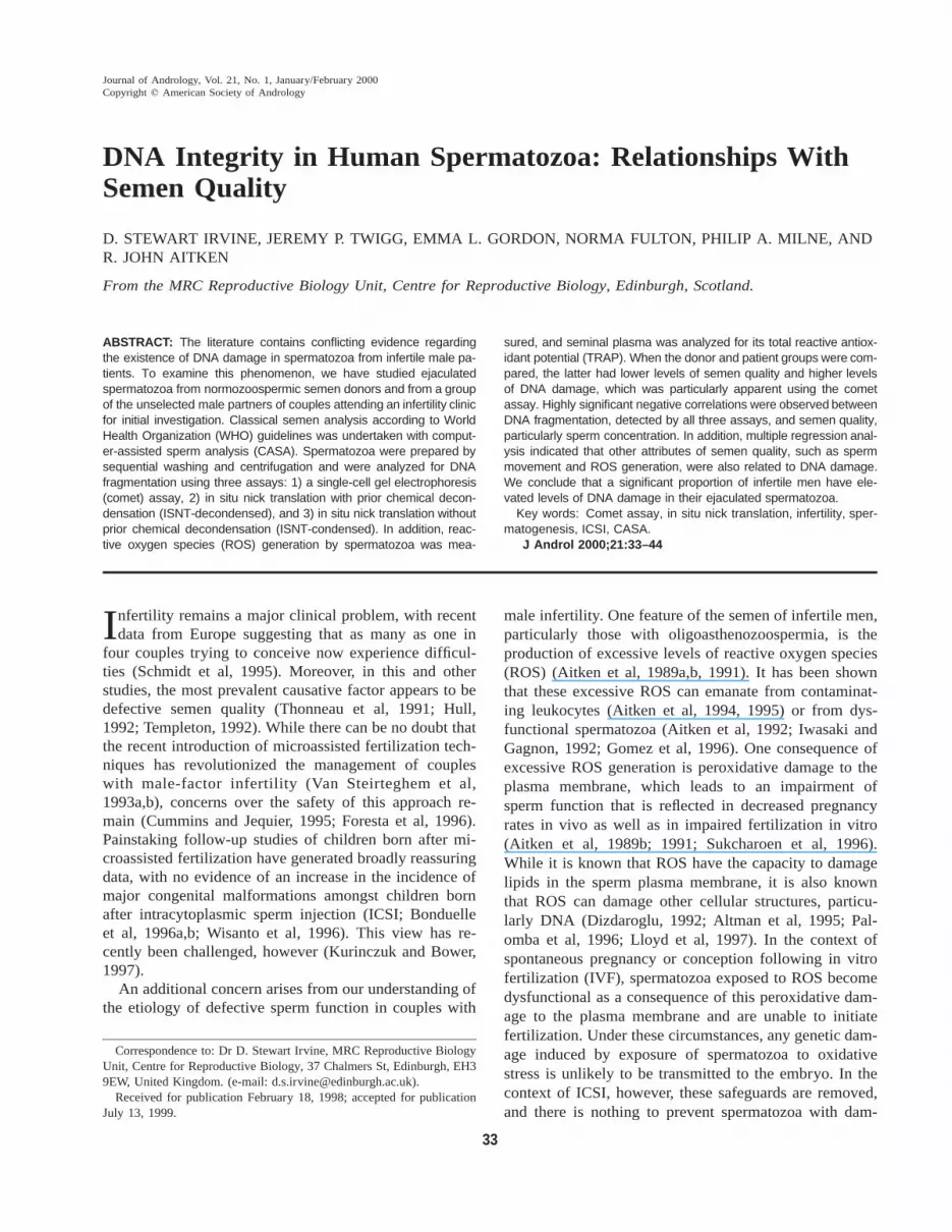
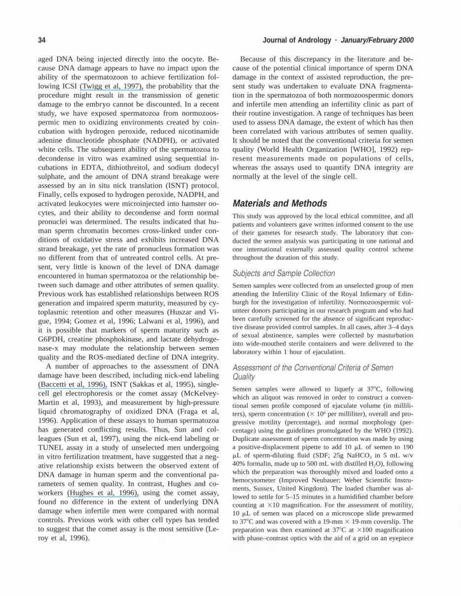
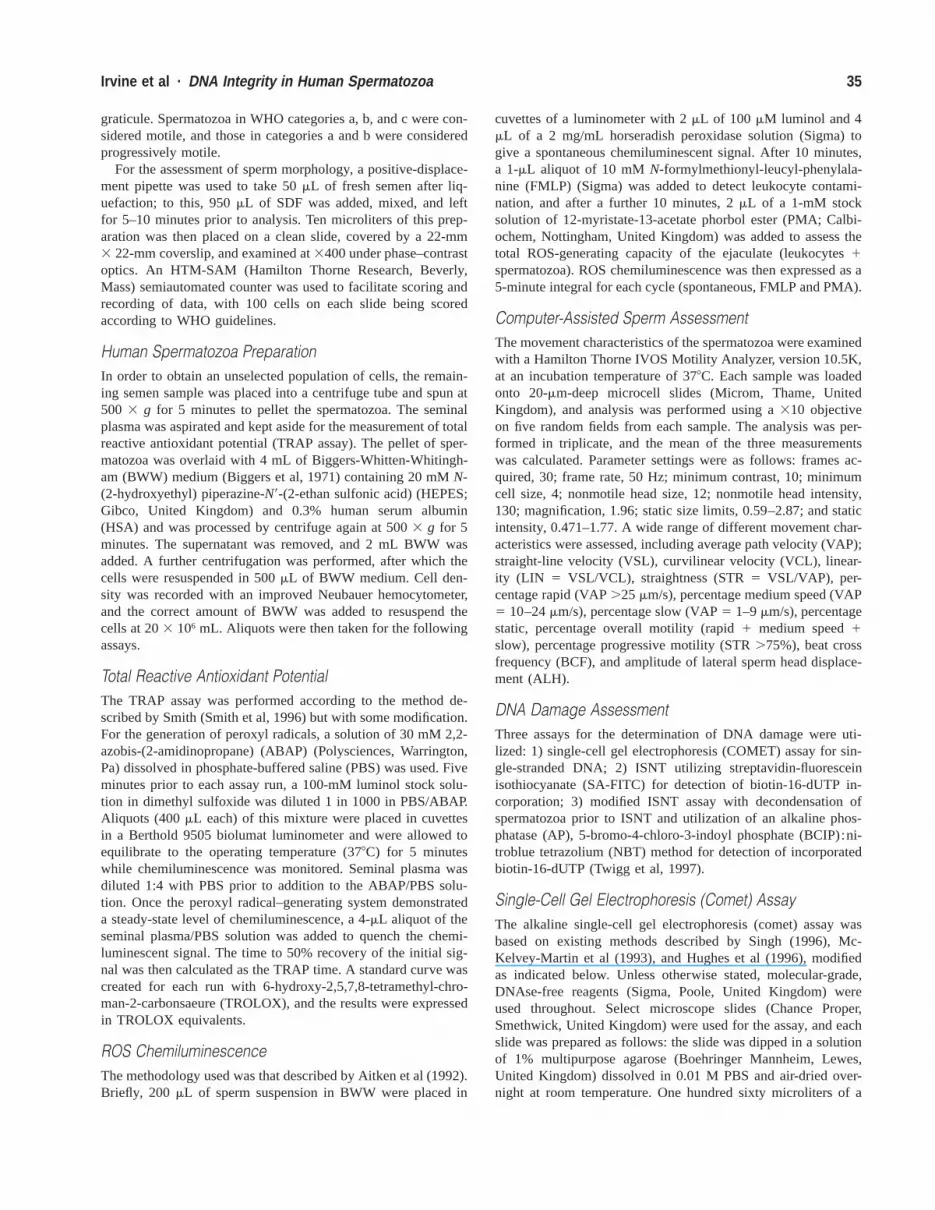
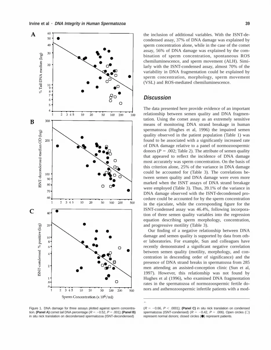
Correlative analyses undertaken to examine the factorsassociated with DNA damage revealed a strong relation-ship with semen quality. A highly significant negativecorrelation was observed between sperm concentration(square root transformed) and DNA damage, as assessedby all three assays: comet (R 5 20.52, P 5 .001), ISNT-decondensed (R 5 20.66, P , .0001), and ISNT-con-

38 Journal of Andrology · January/February 2000
Table 2. Reactive oxygen species production, seminal plasma antioxidant capacity, and DNA damage
Variable
Donor Group
Median IQR*
Patient Group
Median IQR P
Reactive oxygen productionSpontaneous (cpm 3 106)FMLP stimulated (cpm 3 106)PMA stimulated (cpm 3 106)
0.990.652.31
0.64–1.960.39–1.070.70–9.67
1.360.651.65
0.66–2.080.37–1.600.61–2.93
NSNSNS
Total reactive antioxidant potentialTRAP timeTROLOX equivalent
2.52.85
2.1–3.22.45–3.15
2.452.4
1.85–3.451.85–3.65
NSNS
DNA damage: comet assayMedian tail DNA (%)Median olive tail moment
10.441.68
6.6–14.90.9–2.8
19.813.67
13.2–26.12.4–5.4
0.0020.0035
DNA damage: in situ nick translation-condensedCells positive (%) 2.03 0.99–3.11 3.63 1.78–7.22 NS
DNA damage: in situ nick translation-decondensedMedian optical density 110.18 96.7–142.1 144.92 100.5–173.8 NS
* IQR indicates interquartile range; NS, not significant; FMLP, N-formylmethionyl-leucyl-phenylalanine; PMA, 12-myristate-13-acetate phorbol ester;TRAP, total reactive antioxidant potential; TROLOX, 6-hydroxy-2,5,7,8-tetramethyl-chroman-2-carbonsaeure.
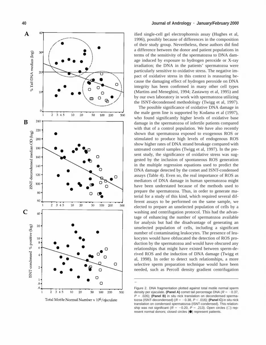
densed (R 5 20.43, P 5 .006; Figures 1A through C).When the number of morphologically normal, motile cellsin the ejaculate was calculated (Glazener et al, 1987), itwas found to be negatively correlated with DNA damageas measured by the single-cell gel electrophoresis andISNT-decondensed assays (R 5 20.37, P 5 .026; R 520.38, P 5 .016); Figures 2A and B. However, althoughthe same negative relationship was demonstrated with theISNT-condensed assay, it was not statistically significant(R 5 20.20, P 5 .213; Figure 2C).
Comparison of Techniques for Assessing DNA DamageThe relationships between DNA damage measured by thecomet assay and ISNT-decondensed and ISNT-condensedare shown in Figure 2 (parts A, B, and C, respectively).It is clear that the comet assay had higher resolution forDNA fragmentation in that it allowed separation of sub-populations of individuals (Figure 2A). The donor pop-ulation are clearly localized in the lower right-hand areaof this scatterplot, with low levels of DNA damage andgood semen quality. Several of the unselected patientpopulation colocalized to this region, suggesting thatthese are normal men with both normal semen quality andlow-level spermatozoal DNA fragmentation. A distinctlyseparate population of patients localized to the upper leftregion of the scatterplot, demonstrating poorer semenquality with higher levels of DNA fragmentation. Inter-estingly, a small population of individuals also displayedpoor semen quality with low levels of DNA damage (Fig-ure 2A).
While there were relationships between attributes ofsemen quality measured by CASA and DNA damage,these generally reflected the pattern that had emerged
from the conventional semen profile of negative relation-ships between sperm number and motility. There were nomeaningful relationships between observed levels ofDNA damage and individual attributes of sperm move-ment (data not shown). Furthermore, no relationshipswere observed between levels of ROS production or sem-inal plasma antioxidant activity and DNA fragmentationin this population.
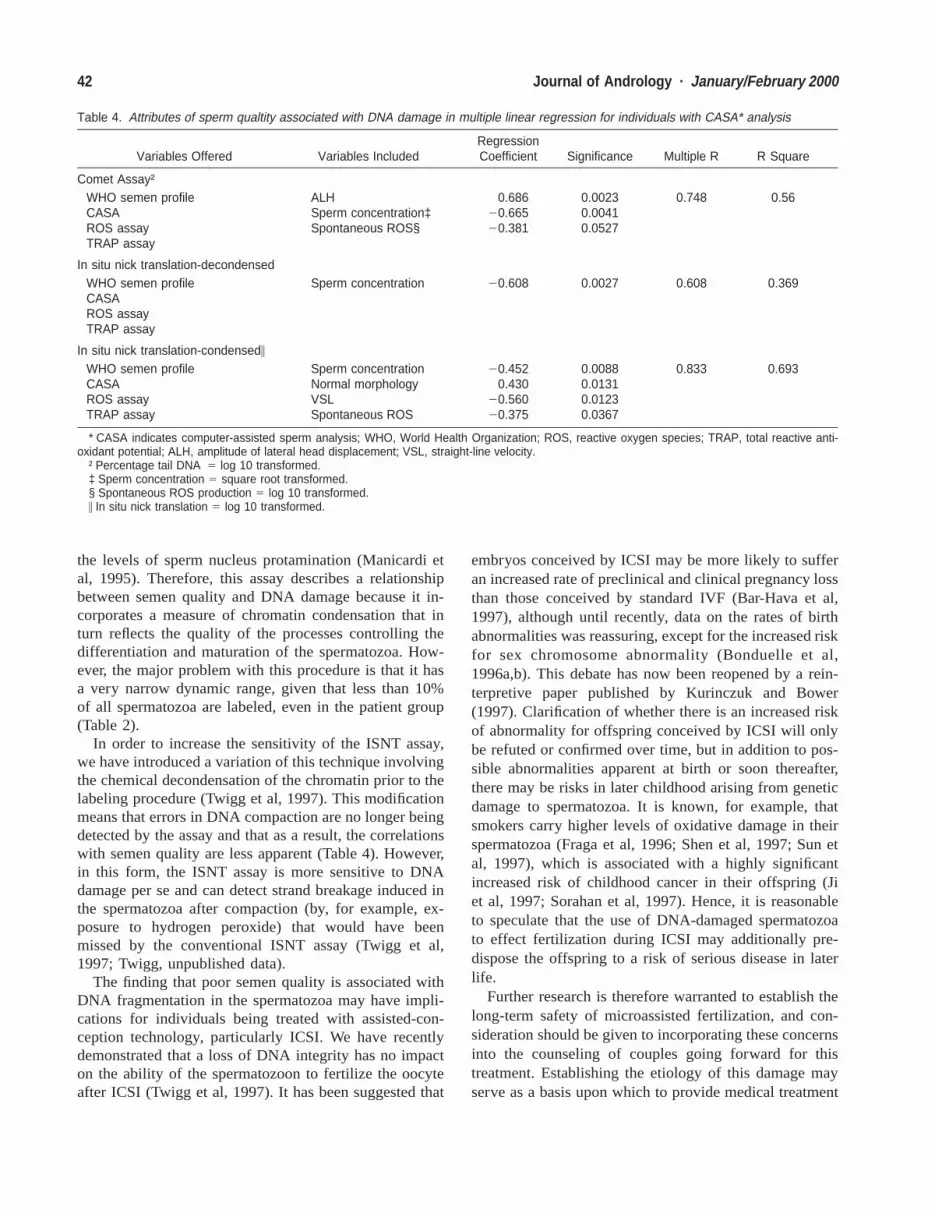
Prediction of DNA Damage From Conventional SemenCountFinally, to examine the relationships between semen qual-ity and DNA damage in more detail, a series of multiplelinear regression analyses (with transformed variableswhere appropriate) were performed, the results of whichare summarized in Tables 3 and 4. When DNA damageaccording to the comet assay was used as the dependentvariable, with data from the WHO semen profile, the ROSchemiluminescence assay, and the TRAP assay entered asstepwise independent variables, it was seen that 25% ofthe variation in DNA damage could be accounted for bysperm concentration alone (Table 3). This relationship re-mained for the ISNT-decondensed assay, with 39% of thevariability being explained by this variable alone. In asimilar analysis for ISNT-condensed, 46% of the variationin DNA damage was accounted for with data derivedfrom the conventional criteria of semen quality alone(morphology, sperm concentration, and progressive mo-tility).
Similar analyses were then undertaken on the subset ofpatients and donors for whom CASA data was available.The results (Table 4) showed that a substantial proportionof the observed DNA damage could be accounted for by
39Irvine et al · DNA Integrity in Human Spermatozoa
Figure 1. DNA damage for three assays plotted against sperm concentra-tion. (Panel A) comet tail DNA percentage (R 5 20.52, P 5 .001); (Panel B)in situ nick translation on decondensed spermatozoa (ISNT-decondensed)
←
(R 5 20.66, P , .0001); (Panel C) in situ nick translation on condensedspermatozoa (ISNT-condensed) (R 5 20.42, P 5 .006). Open circles (V)represent normal donors; closed circles (v) represent patients.
the inclusion of additional variables. With the ISNT-de-condensed assay, 37% of DNA damage was explained bysperm concentration alone, while in the case of the cometassay, 56% of DNA damage was explained by the com-bination of sperm concentration, spontaneous ROSchemiluminescence, and sperm movement (ALH). Simi-larly with the ISNT-condensed assay, almost 70% of thevariability in DNA fragmentation could be explained bysperm concentration, morphology, sperm movement(VSL) and ROS-mediated chemiluminescence.
Discussion
The data presented here provide evidence of an importantrelationship between semen quality and DNA fragmen-tation. Using the comet assay as an extremely sensitivemeans of monitoring DNA strand breakage in humanspermatozoa (Hughes et al, 1996) the impaired semenquality observed in the patient population (Table 1) wasfound to be associated with a significantly increased rateof DNA damage relative to a panel of normozoospermicdonors (P 5 .002; Table 2). The attribute of semen qualitythat appeared to reflect the incidence of DNA damagemost accurately was sperm concentration. On the basis ofthis criterion alone, 25% of the variance in DNA damagecould be accounted for (Table 3). The correlations be-tween semen quality and DNA damage were even moremarked when the ISNT assays of DNA strand breakagewere employed (Table 3). Thus, 39.1% of the variance inDNA damage observed with the ISNT-decondensed pro-cedure could be accounted for by the sperm concentrationin the ejaculate, while the corresponding figure for theISNT-condensed assay was 46.4%, following incorpora-tion of three semen quality variables into the regressionequation describing sperm morphology, concentration,and progressive motility (Table 3).
Our finding of a negative relationship between DNAdamage and semen quality is supported by data from oth-er laboratories. For example, Sun and colleagues haverecently demonstrated a significant negative correlationbetween semen quality (motility, morphology, and con-centration in descending order of significance) and thepresence of DNA strand breaks in spermatozoa from 285men attending an assisted-conception clinic (Sun et al,1997). However, this relationship was not found byHughes et al (1996), who examined DNA fragmentationrates in the spermatozoa of normozoospermic fertile do-nors and asthenozoospermic infertile patients with a mod-
40 Journal of Andrology · January/February 2000
←
Figure 2. DNA fragmentation plotted against total motile normal spermdensity per ejaculate. (Panel A) comet tail percentage DNA (R 5 20.37,P 5 .026); (Panel B) in situ nick translation on decondensed sperma-tozoa (ISNT-decondensed) (R 5 20.38, P , .016); (Panel C) in situ nicktranslation on condensed spermatozoa (ISNT-condensed). This relation-ship was not significant (R 5 20.20, P 5 .213). Open circles (V) rep-resent normal donors; closed circles (v) represent patients.
ified single-cell gel electrophoresis assay (Hughes et al,1996), possibly because of differences in the compositionof their study group. Nevertheless, these authors did finda difference between the donor and patient populations interms of the sensitivity of the spermatozoa to DNA dam-age induced by exposure to hydrogen peroxide or X-rayirradiation; the DNA in the patients’ spermatozoa wereparticularly sensitive to oxidative stress. The negative im-pact of oxidative stress in this context is reassuring be-cause the damaging effect of hydrogen peroxide on DNAintegrity has been confirmed in many other cell types(Martins and Meneghini, 1994; Zastawny et al, 1995) andby our own laboratory in work with spermatozoa utilizingthe ISNT-decondensed methodology (Twigg et al, 1997).
The possible significance of oxidative DNA damage inthe male germ line is supported by Kodama et al (1997),who found significantly higher levels of oxidative basedamage in the spermatozoa of infertile patients comparedwith that of a control population. We have also recentlyshown that spermatozoa exposed to exogenous ROS orstimulated to produce high levels of endogenous ROSshow higher rates of DNA strand breakage compared withuntreated control samples (Twigg et al, 1997). In the pre-sent study, the significance of oxidative stress was sug-gested by the inclusion of spontaneous ROS generationin the multiple regression equations used to predict theDNA damage detected by the comet and ISNT-condensedassays (Table 4). Even so, the real importance of ROS asmediators of DNA damage in human spermatozoa mighthave been understated because of the methods used toprepare the spermatozoa. Thus, in order to generate ma-terial for a study of this kind, which required several dif-ferent assays to be performed on the same sample, weelected to prepare an unselected population of cells by awashing and centrifugation protocol. This had the advan-tage of enhancing the number of spermatozoa availablefor analysis but had the disadvantage of generating anunselected population of cells, including a significantnumber of contaminating leukocytes. The presence of leu-kocytes would have obfuscated the detection of ROS pro-duction by the spermatozoa and would have obscured anyrelationships that might have existed between sperm-de-rived ROS and the induction of DNA damage (Twigg etal, 1998). In order to detect such relationships, a moreselective sperm preparation technique would have beenneeded, such as Percoll density gradient centrifugation

41Irvine et al · DNA Integrity in Human Spermatozoa
Table 3. Attributes of semen qualtity associated with DNA damage in multiple linear regression
Variables Offered Variables IncludedRegressionCoefficient Significance Multiple R R Square
Comet assay†WHO* semen profileROS assayTRAP assay
Sperm concentration‡ 20.498 0.0028 0.498 0.248
In situ nick translation-decondensedWHO semen profileROS assayTRAP assay
Sperm concentration 20.625 0.000 0.625 0.391
In situ nick translation-condensed§WHO semen profileROS assayTRAP assay
Normal morphologySperm concentrationProgressive motility
0.41820.29620.374
0.00350.0550.018
0.681 0.464
* WHO indicates World Health Organization; ROS, reactive oxygen species; TRAP, total reactive antioxidant potential.† Percentage tail DNA 5 log 10 transformed.‡ Sperm concentration 5 square root transformed.§ In situ nick translation 5 log 10 transformed.
followed by treatment with anti-CD45–coated magneticbeads (Aitken et al, 1989b; 1996; Agarwal et al, 1994).Further studies are clearly required to fully establish thecause of the excessive DNA damage detected in the sper-matozoa of infertile men. However, because both ROSgeneration and oxidative DNA base damage are elevatedin the spermatozoa of infertile patients (Aitken et al,1992; Kodama et al, 1997) and because ROS productionwas selected as a predictor of DNA damage in the presentstudy, a rational hypothesis would be that the etiology ofthis damage involves oxidative stress within the male re-productive tract. One possibility would be the release,through defective spermiation of immature spermatozoacarrying excessive residual cytoplasm (Gomez et al,1996), which have been shown to suffer from excessiveexogenous oxidative stress (Huszar and Vigue, 1994;Huszar et al, 1998).
Given the existence of high levels of DNA fragmen-tation in the spermatozoa of infertile patients, importantquestions remain with regard to the optimal means of de-tecting this damage and its clinical significance. In termsof methodology, three different techniques were em-ployed in the present study to detect DNA strand break-age in human spermatozoa. The comet assay has beenshown to be one of the most sensitive assays for the de-tection of DNA damage (Leroy et al, 1996; Singh, 1996).Sensitivity to the detection of single-strand DNA break-age comes from the use of alkaline lysis buffer, whichreverses DNA supercoiling and separates the DNA duplexinto single strands. Further sensitivity to subtle changesin DNA is gained by incubation of the cells in the pres-ence of proteinase K, which removes protamines that oth-erwise impede DNA migration through the agarose. Mi-nor variations in the protocol employed during this assay,
such as the inclusion of antioxidants in electrophoresisbuffer, alterations in electrophoresis time, and differencesin pH can all influence the sensitivity of the assay. Thecomet assay may even overestimate true DNA strandbreakage in spermatozoa because of artificial damage in-duced at alkali-labile sites within the DNA strand (Singhet al, 1989). Nevertheless, in the context of human sper-matozoa, this assay would appear to be the most sensitiveof all the procedures assessed for detecting DNA frag-mentation, since this technique alone was able to discrim-inate the central trends in DNA damage between the pa-tient and donor populations (Table 2).
Although the comet assay was extremely sensitive, itdid not show the same levels of correlation with semenquality that were recorded for the ISNT-condensed pro-cedure (Table 4). Almost 70% of the variation in DNAdamage recorded with the ISNT-condensed assay couldbe accounted for on the basis of semen quality as reflectedin sperm concentration, sperm morphology, VSL, andspontaneous ROS generation. The high correlations ob-served between the ISNT-condensed assay and semenquality may reflect the fact that this assay measures morethan just DNA damage; it also provides an assessment ofthe efficiency of DNA compaction. It has been suggestedby other groups that clinically significant differences existbetween cells in sperm chromatin packaging (Sakkas etal, 1995, 1996), an observation that would be consistentwith the finding that sperm from infertile men are moresusceptible to DNA damage induced by exogenous fac-tors such as irradiation or hydrogen peroxide (Hughes etal, 1996). The ISNT-condensed assay is reliant on theaccess DNA polymerase 1 gains to the genome as a resultof incomplete compaction of the sperm nucleus, the levelof DNA nick-end labeling being highly correlated with
42 Journal of Andrology · January/February 2000
Table 4. Attributes of sperm qualtity associated with DNA damage in multiple linear regression for individuals with CASA* analysis
Variables Offered Variables IncludedRegressionCoefficient Significance Multiple R R Square
Comet Assay†WHO semen profileCASAROS assayTRAP assay
ALHSperm concentration‡Spontaneous ROS§
0.68620.66520.381
0.00230.00410.0527
0.748 0.56
In situ nick translation-decondensedWHO semen profileCASAROS assayTRAP assay
Sperm concentration 20.608 0.0027 0.608 0.369
In situ nick translation-condensed\
WHO semen profileCASAROS assayTRAP assay
Sperm concentrationNormal morphologyVSLSpontaneous ROS
20.4520.430
20.56020.375
0.00880.01310.01230.0367
0.833 0.693
* CASA indicates computer-assisted sperm analysis; WHO, World Health Organization; ROS, reactive oxygen species; TRAP, total reactive anti-oxidant potential; ALH, amplitude of lateral head displacement; VSL, straight-line velocity.
† Percentage tail DNA 5 log 10 transformed.‡ Sperm concentration 5 square root transformed.§ Spontaneous ROS production 5 log 10 transformed.\ In situ nick translation 5 log 10 transformed.
the levels of sperm nucleus protamination (Manicardi etal, 1995). Therefore, this assay describes a relationshipbetween semen quality and DNA damage because it in-corporates a measure of chromatin condensation that inturn reflects the quality of the processes controlling thedifferentiation and maturation of the spermatozoa. How-ever, the major problem with this procedure is that it hasa very narrow dynamic range, given that less than 10%of all spermatozoa are labeled, even in the patient group(Table 2).
In order to increase the sensitivity of the ISNT assay,we have introduced a variation of this technique involvingthe chemical decondensation of the chromatin prior to thelabeling procedure (Twigg et al, 1997). This modificationmeans that errors in DNA compaction are no longer beingdetected by the assay and that as a result, the correlationswith semen quality are less apparent (Table 4). However,in this form, the ISNT assay is more sensitive to DNAdamage per se and can detect strand breakage induced inthe spermatozoa after compaction (by, for example, ex-posure to hydrogen peroxide) that would have beenmissed by the conventional ISNT assay (Twigg et al,1997; Twigg, unpublished data).
The finding that poor semen quality is associated withDNA fragmentation in the spermatozoa may have impli-cations for individuals being treated with assisted-con-ception technology, particularly ICSI. We have recentlydemonstrated that a loss of DNA integrity has no impacton the ability of the spermatozoon to fertilize the oocyteafter ICSI (Twigg et al, 1997). It has been suggested that
embryos conceived by ICSI may be more likely to sufferan increased rate of preclinical and clinical pregnancy lossthan those conceived by standard IVF (Bar-Hava et al,1997), although until recently, data on the rates of birthabnormalities was reassuring, except for the increased riskfor sex chromosome abnormality (Bonduelle et al,1996a,b). This debate has now been reopened by a rein-terpretive paper published by Kurinczuk and Bower(1997). Clarification of whether there is an increased riskof abnormality for offspring conceived by ICSI will onlybe refuted or confirmed over time, but in addition to pos-sible abnormalities apparent at birth or soon thereafter,there may be risks in later childhood arising from geneticdamage to spermatozoa. It is known, for example, thatsmokers carry higher levels of oxidative damage in theirspermatozoa (Fraga et al, 1996; Shen et al, 1997; Sun etal, 1997), which is associated with a highly significantincreased risk of childhood cancer in their offspring (Jiet al, 1997; Sorahan et al, 1997). Hence, it is reasonableto speculate that the use of DNA-damaged spermatozoato effect fertilization during ICSI may additionally pre-dispose the offspring to a risk of serious disease in laterlife.
Further research is therefore warranted to establish thelong-term safety of microassisted fertilization, and con-sideration should be given to incorporating these concernsinto the counseling of couples going forward for thistreatment. Establishing the etiology of this damage mayserve as a basis upon which to provide medical treatment
43Irvine et al · DNA Integrity in Human Spermatozoa
(Kodama et al, 1997) to individuals with high baselinedamage prior to assisted conception.
AcknowledgmentsWe are grateful to the staff of the Clinical Andrology Unit, Royal Infir-mary of Edinburgh for the management of the volunteers and patientswho participated in this study.
ReferencesAgarwal A, Ikemoto I, Loughlin KR. Effect of sperm washing on levels
of reactive oxygen species in semen. Arch Androl 1994;33:157–162.Aitken RJ, Buckingham DW, Brindle J, Gomez E, Baker HWG, Irvine
DS. Analysis of sperm movement in relation to the oxidative stresscreated by leukocytes in washed sperm preparations and seminal plas-ma. Hum Reprod 1995;10:2061–2071.
Aitken RJ, Buckingham DW, West KM. Reactive oxygen species andhuman spermatozoa—analysis of the cellular mechanisms involved inluminol-dependent and lucigenin-dependent chemiluminescence. JCell Physiol 1992;151:466–477.
Aitken RJ, Buckingham DW, West K, Brindle J. On the use of paramag-netic beads and ferrofluids to assess and eliminate the leukocytic con-tribution to oxygen radical generation by human sperm suspensions.Am J Reprod Immunol 1996;35:541–551.
Aitken RJ, Clarkson JS, Fishel S. Generation of reactive oxygen species,lipid-peroxidation, and human-sperm function. Biol Reprod 1989a;41:183–197.
Aitken RJ, Clarkson JS, Hargreave TB, Irvine DS, Wu FCW. Analysisof the relationship between defective sperm function and the gener-ation of reactive oxygen species in cases of oligozoospermia. J Androl1989b;10:214–220.
Aitken RJ, Irvine DS, Wu FC. Prospective analysis of sperm-oocyte fu-sion and reactive oxygen species generation as criteria for the diag-nosis of infertility. Am J Obstet Gynecol 1991;164:542–551.
Aitken RJ, West K, Buckingham D. Leukocytic infiltration into the hu-man ejaculate and its association with semen quality, oxidative stress,and sperm function. J Androl 1994;15:343–352.
Altman SA, Zastawny TH, RandersEichhorn L, Cacciuttolo MA, AkmanSA, Dizdaroglu M, Rao G. Formation of DNA-protein cross-links incultured mammalian cells upon treatment with iron ions. Free RadBiol Med 1995;19:897–902.
Baccetti B, Collodel G, Piomboni P. Apoptosis in human ejaculated spermcells. J Submicrosc Cytol Pathol 1996;28:587–596.
Bar-Hava I, Ashkenazi J, Shelef M, Schwartz A, Brengauz M, FeldbergD, Orvieto R, Ben-Rafael Z. Morphology and clinical outcomes ofembryos after in vitro fertilization are superior to those after intra-cytoplasmic sperm injection. Fertil Steril 1997;68:653–657.
Bianchi PG, Manicardi GC, Bizzaro D, Bianchi U, Sakkas D. Effect ofdeoxyribonucleic acid protamination on fluorochrome staining and insitu nick-translation of murine and human mature spermatozoa. BiolReprod 1993;49:1083–1088.
Biggers J, Whitten W, Whittingham D. The Culture of Mouse Embryosin Vitro. San Francisco: Freeman; 1971.
Bonduelle M, Legein J, Buysse A, Van Assche E, Wisanto A, DevroeyP, Van Steirteghem AC, Liebaers I. Prospective follow-up study of423 children born after intracytoplasmic sperm injection. Hum Reprod1996a;11:1558–1564.
Bonduelle M, Wilikens A, Buysse A, Van Assche E, Wisanto A, DevroeyP, Van Steirteghem AC, Liebaers I. Prospective follow-up study of877 children born after intracytoplasmic sperm injection (ICSI), withejaculated epididymal and testicular spermatozoa and after replace-
ment of cryopreserved embryos obtained after ICSI. Hum Reprod(suppl 4) 1996b;11:131–155.
Cummins JM, Jequier AM. Concerns and recommendations for intracy-toplasmic sperm injection (ICSI) treatment. Hum Reprod 1995;10:138–143.
De Jong ASH, Van Kessel-Van Vark M, Raap AK. Sensitivity of variousvisualisation methods for peroxidase and alkaline phosphatase activityin immunoenzyme histochemistry. Histochem J 1985;17:1119–1130.
Dizdaroglu M. Oxidative damage to DNA in mammalian chromatin. Mu-tat Res 1992;275:331–342.
Foresta C, Rossato M, Garolla A, Ferlin A. Male infertility and ICSI: arethere limits? Hum Reprod 1996;11:2347–2348.
Fraga CG, Motchnik PA, Wyrobek AJ, Rempel DM, Ames BN. Smokingand low antioxidant levels increase oxidative damage to sperm DNA.Mutat Res 1996;351:199–203.
Glazener CM, Kelly NJ, Weir MJ, David JS, Cornes JS, Hull MG. Thediagnosis of male infertility—prospective time-specific study of con-ception rates related to seminal analysis and post-coital sperm-mucuspenetration and survival in otherwise unexplained infertility. Hum Re-prod 1987;2:665–671.
Gomez E, Buckingham DW, Brindle J, Lanzafame F, Irvine DS, AitkenRJ. Development Of an image-analysis system to monitor the reten-tion of residual cytoplasm by human spermatozoa—correlation withbiochemical markers of the cytoplasmic space, oxidative stress, andsperm function. J Androl 1996;17:276–287.
Hughes CM, Lewis SEM, McKelvey-Martin VJ, Thompson W. A com-parison of baseline and induced DNA damage in human spermatozoafrom fertile and infertile men, using a modified comet assay. MolReprod Dev 1996;2:613–619.
Hull MGR. The causes of infertility and relative effectiveness of treat-ment. In: Templeton AA, Drife JO, eds. Infertility. Berlin: Springer-Verlag; 1992:33–57.
Huszar G, Patrizio P, Vigue L, Willets M, Wilker C, Adhoot D, JohnsonL. Cytoplasmic extrusion and the switch from creatine kinase B to Misoform are completed by the commencement of epididymal transportin human and stallion spermatozoa. J Androl 1998;19:11–20.
Huszar G, Vigue L. Correlation between the rate of lipid peroxidationand cellular maturity as measured by creatine kinase activity in humanspermatozoa. J Androl 1994;15:71–77.
Irvine DS, Templeton AA. Donor insemination. In: Hargreave, TB, ed.Male Infertility. Edinburgh, United Kingdom: Churchill Livingstone;1994:427–451.
Iwasaki A, Gagnon C. Formation of reactive oxygen species in sperma-tozoa of infertile patients. Fertil Steril 1992;57:409–416.
Ji BT, Shu XO, Linet MS, Zheng W, Wacholder S, Gao YT, Ying DM,Jin F. Paternal cigarette smoking and the risk of childhood canceramong offspring of nonsmoking mothers. J Natl Cancer Inst 1997;89:238–244.
Kodama H, Yamaguchi R, Fukuda J, Kasai H, Tanaka T. Increased oxi-dative deoxyribonucleic acid damage in the spermatozoa of infertilemale patients. Fertil Steril 1997;68:519–524.
Kurinczuk J, Bower C. Birth defects in infants conceived by intracyto-plasmic sperm injection: an alternative interpretation. Br Med J 1997;315:1260–1263.
Lalwani S, Sayme N, Vigue L, Corrales M, Huszar G. Biochemical mark-ers of early and late spermatogenesis: relationship between the lactatedehydrogenase-X and creatine kinase-M isoform concentrations in hu-man spermatozoa. Mol Reprod Dev 1996;43:495–502.
Leroy T, Van Hummelen P, Anard D, Castelain P, KirschVolders M, Lau-werys R, Lison D. Evaluation of three methods for the detection ofDNA single-strand breaks in human lymphocytes: alkaline elution,nick translation, and single-cell gel electrophoresis. J Toxicol EnvHealth 1996;47:409–422.
Lloyd DR, Phillips DH, Carmichael PL. Generation of putative intras-
44 Journal of Andrology · January/February 2000
trand cross-links and strand breaks in DNA by transition metal ion-mediated oxygen radical attack. Chem Res Toxicol 1997;10:393–400.
Manicardi G, Bianchi P, Pantano S, Azzoni P, Bizzaro D, Bianchi U,Sakkas D. Presence of endogenous nicks in DNA of ejaculated humanspermatozoa and its relationship to chromonycin A3 accessibility. BiolReprod 1995;52:864–867.
Martins EAL, Meneghini R. Cellular DNA damage by hydrogen peroxideis attenuated by hypotonicity. Biochem J 1994;299:137–140.
McKelvey-Martin VJ, Green MHL, Schmezer P, Pool-Zobel BL, De MeoMP, Collins A. The single cell gel electrophoresis assay (comet assay):a European review. Mutat Res 1993;288:47–63.
Palomba L, Brambilla L, Brandi G, Sestili P, Cattabeni F, Cantoni O. Lowlevels of hydrogen peroxide and L-histidine induce DNA double-strand breakage and apoptosis. Eur J Pharmacol 1996;318:167–173.
Sakkas D, Manicardi G, Bianci P, Bizzaro D, Bianchi U. Relationshipbetween the presence of endogenous nicks and sperm chromatin pack-aging in maturing and fertilising mouse spermatozoa. Biol Reprod1995;52:1149–1155.
Sakkas D, Urner F, Bianchi PG, Bizzaro D, Wagner I, Jaquenoud N,Manicardi G, Campana A. Sperm chromatin anomalies can influencedecondensation after intracytoplasmic sperm injection. Hum Reprod1996;11:837–843.
Schmidt L, Munster K, Helm P. Infertility and the seeking of infertilitytreatment in a representative population. Brit J Obstet Gynaecol 1995;102:978–984.
Shen HM, Chia SE, Ni ZY, New AL, Lee BL, Ong CN. Detection ofoxidative DNA damage in human sperm and the association withcigarette smoking. Reprod Toxicol 1997;11:675–680.
Singh NP. Microgel electrophoresis of DNA. In: G. P. Pfeifer, ed. Tech-nologies for Detection of DNA Damage and Mutations. New York:Plenum Press; 1996:3–24.
Singh NP, Danner DB, Tice RR, McCoy MC, Collins GD, Schneider EL.Abundant alkali labile sites in DNA of human and mouse sperm. ExpCell Res 1989;184:461–470.
Smith R, Vantman D, Ponce J, Escobar J, Lissi E. Total antioxidant ca-pacity of human seminal plasma. Hum Reprod 1996;11:1655–1660.
Sorahan T, Lancashire RJ, Hulten MA, Peck I, Stewart AM. Childhoodcancer and parental use of tobacco: deaths from 1953 to 1955. Br JCancer 1997;75:134–138.
Sukcharoen N, Keith J, Irvine DS, Aitken RJ. Prediction of the in-vitrofertilization (IVF) potential of human spermatozoa using sperm func-
tion-tests—the effect of the delay between testing and IVF. Hum Re-prod 1996;11:1030–1034.
Sun JG, Jurisicova A, Casper RF. Detection of deoxyribonucleic acidfragmentation in human sperm: correlation with fertilization in vitro.Biol Reprod 1997;56:602–607.
Templeton AA. The epidemiology of infertility. In: Templeton AA, DrifeJO, eds. Infertility. Berlin: Springer-Verlag; 1992:23–34.
Thonneau P, Marchand S, Tallec A, Ferial M-L, Ducot B, Lansac J, LopesP, Tabaste J-M, Spira A. Incidence and main causes of infertility in aresident population (1,850,000) of three French regions (1988–1989).Hum Reprod 1991;6:811–816.
Twigg J, Irvine D, Aitken R. Oxidative damage to DNA in human sper-matozoa does not preclude pronucleus formation at ICSI. Hum Reprod1997;13:1864–1871.
Twigg J, Irvine DS, Houston P, Fulton N, Michael L, Aitken RJ. Iatro-genic DNA damage induced in human spermatozoa during spermpreparation: protective significance of seminal plasma. Mol Hum Re-prod 1998;4:439–445.
Van Steirteghem A, Liu J, Joris H, Nagy Z, Janssenswillen C, TournayeH, Derde M, Van Assche E, Devroey P. Higher success rate by intra-cytoplasmic sperm injection than by subzonal insemination. Reportof a second series of 300 consecutive treatment cycles. Hum Reprod1993a;8:1055–1060.
Van Steirteghem A, Nagy Z, Joris H, Liu J, Staessen C, Smitz J, WisantoA, Devroey P. High fertilization and implantation rates after intracy-toplasmic sperm injection. Hum Reprod 1993b;8:1061–1066.
Wisanto A, Bonduelle M, Camus M, Tournaye H, Magnus M, LiebaersI, Van Steirteghem A, Devroey P. Obstetric outcome of 904 pregnan-cies after intracytoplasmic sperm injection. Hum Reprod 1996;11:121–129.
Wood G, Warnke R. Suppresion of endogenous avidin-binding activityin tissues and its relevance to biotin-avidin detection systems. J His-tochem Cytochem 1981;29:1196–1204.
World Health Organization (1992). WHO Laboratory Manual for the Ex-amination of Human Semen and Semen-Cervical Mucus Interaction.3rd ed. Cambridge, United Kingdom: Cambridge University Press;1987.
Zastawny TH, Altman SA, Randers-Eichhorn L, Madurawe R, LumpkinJA, Didzaroglu M, Rao G. DNA base modifications and membranedamage in cultured mammalian cells treated with iron ions. Free RadBiol Med 1995;18:1013–1022.