bovine semen preservation under epididymal conditions and
TRANSCRIPT
Bovine semen preservation under epididymal conditions and assessment of sperm quality by means of a sperm-oviduct binding assay
Ingrid De Pauw

FACULTEIT DIERGENEESKUNDE
Vakgroep Voortplanting, Verloskunde en Bedrijfsdiergeneeskunde
Bovine semen preservation under epididymal conditions
and assessment of sperm quality by means of a
sperm-oviduct binding assay
Ingrid De Pauw
Proefschrift ter verkrijging van de graad van Doctor in de Diergeneeskundige
Wetenschappen (PhD) aan de Faculteit Diergeneeskunde, Universiteit Gent, 2003
Promotor: Prof. Dr. A. Van Soom
Co-promotor: Prof. Dr. Dr. h. c. A. de Kruif
Deze uitgave kwam tot stand met de medewerking van:
Foto omslag: stier “Jof” (Lay-out: Maarten Demunster)
Druk: Plot-it, Merelbeke
TABLE OF CONTENTS
List of abbreviations
CHAPTER 1 Introduction
1.1 History of sperm preservation............................................................................. 2
1.2 Aims of the study ................................................................................................ 5
CHAPTER 2 The role of the epididymis for sperm conservation in vivo
2.1 Introduction ......................................................................................................... 8
2.2 Functions of the epididymis ................................................................................ 9
2.1.1 Transport and maturation of spermatozoa in the epididymis............... 10
2.1.2 Production of epididymal plasma ........................................................ 11
2.1.3 Storage of mature spermatozoa in the cauda epididymidis.................. 11
2.3 Biochemical composition of cauda epididymal plasma...................................... 13
2.4 Conclusions ......................................................................................................... 14
2.5 References ........................................................................................................... 15
Adapted from Vlaams Diergeneeskundig Tijdschrift 2000; 69: 159-167
CHAPTER 3 Suitability of sperm function tests for the evaluation of sperm
quality and bull fertility
3.1 Introduction ......................................................................................................... 20
3.2 The evaluation of bovine sperm quality characteristics by means of
fluorescent stainings............................................................................................ 22
3.2.1 Membrane integrity.............................................................................. 22
3.2.2 Capacitation.......................................................................................... 23
3.2.3 Acrosome reaction ............................................................................... 26
3.2.4 Mitochondrial function......................................................................... 27
3.2.5 Apoptosis.............................................................................................. 28
3.3 Sperm motility assessment ................................................................................. 29
3.3.1 Motility assessment by computer assisted sperm analysis systems ..... 30
3.3.2 Motility assessment by sperm migration assays .................................. 31
3.3.3 Motility assessment by sperm chemotaxis assays................................ 32
3.4 Sperm binding and performance assessment ..................................................... 33
3.4.1 Sperm-oviduct binding ........................................................................ 34
3.4.2 Sperm-zona binding............................................................................. 35
3.4.3 In vitro fertilization.............................................................................. 36
3.5 Conclusions ........................................................................................................ 37
3.6 References .......................................................................................................... 39
Review in preparation
CHAPTER 4 In vitro survival of bovine spermatozoa stored at room temperature
under epididymal conditions. ....................................................................... 49
Theriogenology 2003; 59: 1093-1107
CHAPTER 5 Effect of sperm coating on the survival and penetrating ability of
in vitro stored bovine spermatozoa............................................................... 71
Theriogenology 2003; 59: 1109-1122
CHAPTER 6 Hormonal regulation of bovine secretory proteins derived from caput
and cauda epididymal epithelial cell cultures............................................... 91
Accepted for publication in Journal of Andrology 2003; 24 (3)
CHAPTER 7 Sperm binding to epithelial oviduct explants in bulls with different
non-return rates investigated with a new in vitro model. ............................. 107
Biology of Reproduction 2002; 67: 1073-1079
CHAPTER 8 Sperm-oviduct binding capacity of cooled bovine spermatozoa
stored for several days in different diluents.................................................. 131
Manuscript in preparation
CHAPTER 9 General discussion ....................................................................................... 149
CHAPTER 10 Summary – samenvatting ............................................................................. 171
Dankwoord
Curriculum vitae
List of abbreviations
AI artificial insemination ATP adenosine triphosphate BSA bovine serum albumin CASA computer assisted sperm analysis CEP cauda epididymal plasma CI confidence interval CTC chlortetracycline COC cumulus oocyte complex DHT dihydrotestosterone 2D-SDS-PAGE two-dimensional sodium dodecyl sulphate polyacrylamide gel electrophoresis DNA deoxyribonucleic acid FCS fetal calf serum FITC fluorescein isothiocyanate Hepes N-2-Hydroxyethylpiperazine-N'-2-ethanesulfonic acid HF Holstein Friesian hpi hours post insemination HZA hemizona binding assay IVF in vitro fertilization IVM in vitro maturation JC-1 5,5’,6,6’-tetrachloro-1,1’13,3’-tetraethylbenzimidazolyl carbocyanine
iodide LDL low-density lipoproteins MW molecular weight NRR non-return rate PBS phosphate buffered saline PCM principal cell medium pHi intracellular pH PI propidium iodide PI isoelectric point PS phosphatidylserine PSA Pisum sativum agglutinin PNA Peanut agglutinin PVP polyvinyl-pyrrolidone r correlation coefficient ROS reactive oxygen species RT room temperature SD standard deviation SEM standard error of the mean TALP tyrode solution supplemented with albumin, lactate, pyruvate TCM tissue culture medium TUNEL terminal deoxynucleotidyl transferase-mediated dUTP nick end labeling UV ultraviolet ZBA zona binding assay ZP zona pellucida

__________________________________________________ 1.
INTRODUCTION
1.1 History of sperm preservation
1.2 Aims of the study
_____________________________________________________________________________________________________________
2 Chapter 1
1.1 History of sperm preservation
The spread of genital diseases by natural service is a constant threat in bovine.
Moreover, keeping herd bulls is expensive and represents a potential danger for the herd
manager. From this point of view, the artificial insemination (AI) industry has developed at
the cost of natural service over the past 50 years. Artificial insemination is now used in
almost every country in the world (Thibier and Guerin, 2000). One of the main factors
contributing to its success is the rapid and widespread diffusion of improved genotypes and
the exchange of genotypes without transmitting diseases, so that AI can be performed
without risks (Leboeuf et al., 2000). This is mainly due to the high standard of health
surveillance at the AI centers. Artificial insemination has now a major impact on cattle
breeding, through its use of bulls of high genetic merit and by selective rearing of calves of
high breeding merit (Vishwanath and Shannon, 2000). While most semen is processed for
use in AI, semen can also be stored for the preservation of genetic material of endangered
species, as part of an active conservation program (Thibier and Guerin, 2000).
The demand for semen from bulls of high genetic merit has been the main
motivation for developing and refining storage technologies (Vishwanath and Shannon,
2000). In most developed countries, AI was introduced on a small scale during the 1940s
and 1950s and carried out with fresh semen or semen stored at room temperature (RT).
During the 1960s deep-frozen semen started to be processed while donor agencies
encouraged the introduction of highly specialized and expensive AI establishments. A
more recent survey has shown that the total number of doses of semen produced exceeded
200 million, with more than 95% processed as a frozen product (Chupin and Thibier,
1995). Current tendencies in some countries have moved towards the reintroduction of
semen stored at RT because selected progeny-tested sires can be used more efficiently in
this way.
The relative advantages and disadvantages of frozen-thawed and liquid stored
semen are reviewed by Vishwanath et al. (1996). Frozen semen made it possible to
distribute sperm worldwide and reach areas where insemination with liquid semen is not
practical (Foote and Parks, 1993). Furthermore, in a deep-frozen state, semen can be stored
for years without any significant decrease in semen quality. Nevertheless, the
cryopreservation process causes detrimental effects on bovine spermatozoa due to
temperature reduction, cellular dehydration, freezing and thawing. This long-term
preservation at low temperature causes irreversible damage to sperm membranes, killing a
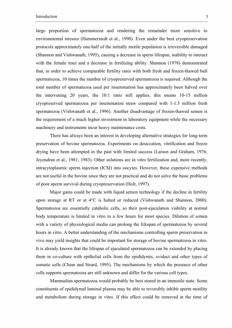
Introduction 3
large proportion of spermatozoa and rendering the remainder more sensitive to
environmental stresses (Hammerstedt et al., 1990). Even under the best cryopreservation
protocols approximately one-half of the initially motile population is irreversible damaged
(Shannon and Vishwanath, 1995), causing a decrease in sperm lifespan, inability to interact
with the female tract and a decrease in fertilizing ability. Shannon (1978) demonstrated
that, in order to achieve comparable fertility rates with both fresh and frozen-thawed bull
spermatozoa, 10 times the number of cryopreserved spermatozoa is required. Although the
total number of spermatozoa used per insemination has approximately been halved over
the intervening 20 years, the 10:1 ratio still applies; this means 10-15 million
cryopreserved spermatozoa per insemination straw compared with 1-1.5 million fresh
spermatozoa (Vishwanath et al., 1996). Another disadvantage of frozen-thawed semen is
the requirement of a much higher investment in laboratory equipment while the necessary
machinery and instruments incur heavy maintenance costs.
There has always been an interest in developing alternative strategies for long-term
preservation of bovine spermatozoa. Experiments on desiccation, vitrification and freeze
drying have been attempted in the past with limited success (Larson and Graham, 1976;
Jeyendran et al., 1981; 1983). Other solutions are in vitro fertilization and, more recently,
intracytoplasmic sperm injection (ICSI) into oocytes. However, these expensive methods
are not useful in the bovine since they are not practical and do not solve the basic problems
of poor sperm survival during cryopreservation (Holt, 1997).
Major gains could be made with liquid semen technology if the decline in fertility
upon storage at RT or at 4°C is halted or reduced (Vishwanath and Shannon, 2000).
Spermatozoa are essentially catabolic cells, so their post-ejaculation viability at normal
body temperature is limited in vitro to a few hours for most species. Dilution of semen
with a variety of physiological media can prolong the lifespan of spermatozoa by several
hours in vitro. A better understanding of the mechanisms controlling sperm preservation in
vivo may yield insights that could be important for storage of bovine spermatozoa in vitro.
It is already known that the lifespan of ejaculated spermatozoa can be extended by placing
them in co-culture with epithelial cells from the epididymis, oviduct and other types of
somatic cells (Chian and Sirard, 1995). The mechanisms by which the presence of other
cells supports spermatozoa are still unknown and differ for the various cell types.
Mammalian spermatozoa would probably be best stored in an immotile state. Some
constituents of epididymal luminal plasma may be able to reversibly inhibit sperm motility
and metabolism during storage in vitro. If this effect could be removed at the time of
4 Chapter 1
insemination, such a procedure may allow storage of spermatozoa at RT or even at body
temperature for extended periods. Future methods for preserving sperm fertility could also
be directed at altering or reducing the catabolic metabolism of spermatozoa, increasing the
resistance of sperm membranes to thermal and other environmental insults (Foote and
Parks, 1993), and overcoming the effects of sperm ageing.
Introduction 5
1.2 Aims of the study
The use of liquid-stored bovine semen, as an alternative to frozen storage would
have obvious advantages in widespread selection schemes or in extensive production
systems. Research is necessary to increase the time during which semen can be stored in
liquid state whilst maintaining its fertilizing capacity. The fact that the cauda epididymidis
has the capacity to store densely packed bovine spermatozoa in a potentially fertilizing
condition for several weeks in vivo suggests that a study of the effects of epididymal
constituents on spermatozoa may be useful for the formulation of better diluents for
preserving spermatozoa at RT or at 4°C.
The general aims of the present thesis are to improve sperm storage in vitro and to
develop alternative methods for the evaluation of sperm quality and bull fertility.
In order to achieve these aims, the study comprises the following experiments:
1. Examination of in vitro survival of bovine spermatozoa stored at room temperature
under epididymal conditions.
2. Study of the effect of sperm coating on the survival and penetrating ability of in vitro
stored bovine spermatozoa.
3. Comparison of the effect of hormones on protein secretion of caput and cauda
epididymal epithelial cell culture in the bovine.
4. Investigation of the binding of spermatozoa from bulls with different non-return rates
to epithelial oviduct explants by means of a new in vitro model.
5. Use of a sperm-oviduct binding assay for cooled bovine spermatozoa stored for
several days in different diluents.
6 Chapter 1
References
Chian RC, Sirard MA. Fertilizing ability of bovine spermatozoa cocultured with oviduct epithelial
cells. Biol Reprod 1995; 52: 156-162.
Chupin D, Thibier M. Survey of the present status of the use of artificial insemination in developed
countries. World Anim Rev 1995; 82: 58-68.
Foote RH, Parks JE. Factors affecting preservation and fertility of bull sperm: a brief review.
Reprod Fertil Dev 1993; 5: 665-673.
Hammerstedt RH, Graham JK, Nolan JP. Cryopreservation of mammalian sperm: what we ask
them to survive. J Androl 1990; 11: 73-88.
Holt WV. Alternative strategies for the long-term preservation of spermatozoa. Reprod Fertil Dev
1997; 9: 309-319.
Jeyendran RS, Graham EF, Schmehl MK. Fertility of dehydrated bull semen. Cryobiology 1981;
18: 292-300.
Jeyendran RS, Hunter AG, Graham EF. Alteration of seminal proteins during freeze-drying of
bovine semen. J Dairy Sci 1983; 66: 887-891.
Larson EV, Graham EF. Freeze-drying of spermatozoa. Dev Biol Stand 1976; 36: 343-348.
Leboeuf B, Restall B, Salamon S. Production and storage of goat semen for artificial insemination.
Anim Reprod Sci 2000; 62: 113-141.
Shannon P. Factors affecting semen preservation and conception rates in cattle. J Reprod Fertil
1978; 54: 519-527.
Shannon P, Vishwanath R. The effect of optimal and suboptimal concentrations of sperm on the
fertility of fresh and frozen bovine sperm and a theoretical model to explain the fertility
differences. Anim Reprod Sci 1995; 39: 1-10.
Thibier M, Guerin B. Hygienic aspects of storage and use of semen for artificial insemination.
Anim Reprod Sci 2000; 62: 233-251.
Vishwanath R, Pitt CP, Shannon P. Sperm numbers, semen age and fertility in fresh and frozen
bovine semen. Proc NZ Soc Anim Prod 1996; 56: 31-34.
Vishwanath R, Shannon P. Storage of bovine semen in liquid and frozen state. Anim Reprod Sci
2000; 62: 23-53.
__________________________________________________ 2.
THE ROLE OF THE EPIDIDYMIS FOR SPERM CONSERVATION IN VIVO
2.1 Introduction
2.2 Functions of the epididymis
2.1.1 Transport and maturation of spermatozoa in the epididymis
2.1.2 Production of epididymal plasma
2.1.3 Storage of mature spermatozoa in the cauda epididymidis
2.3 Biochemical composition of cauda epididymal plasma
2.4 Conclusions
2.5 References
Adapted from Vlaams Diergeneeskundig Tijdschrift 2000; 69: 159-167
De Pauw I., Van Soom A., Verberckmoes S. and de Kruif A.
8 Chapter 2
2.1 Introduction
Spermatozoa are produced in the testis, outside the abdominal cavity in a pouch of
skin called the scrotum. The scrotum protects the testicle against both extremes of
temperature (Setchell, 1998). This is essential for normal sperm formation, which occurs
only at a temperature several degrees below normal body temperature. Each testis is
constituted of tubuli seminiferi containing spermatogonia, which are responsible for the
production and the differentiation of spermatocytes into spermatids and finally into
spermatozoa. This process takes several days and is called spermatogenesis. After
formation, spermatozoa are transported into larger tubules to form the rete testis at the
centre of the testis. Arising from the rete testis are 12 or more out-going ducts, the vasa
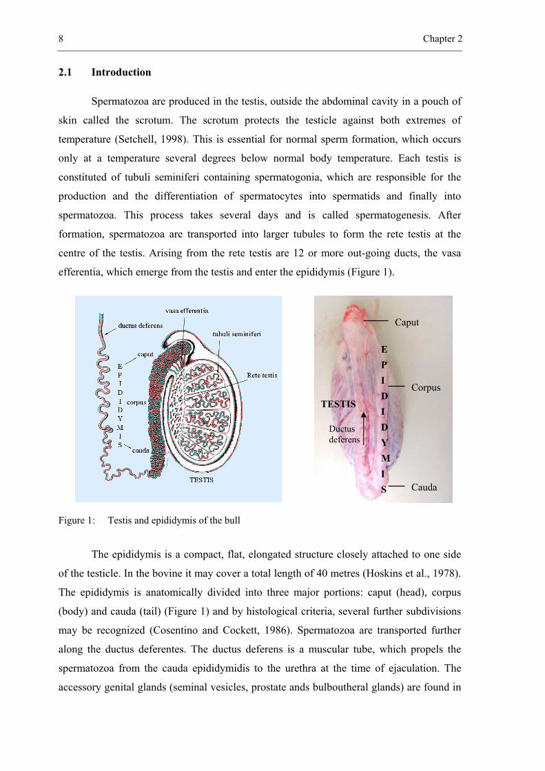
efferentia, which emerge from the testis and enter the epididymis (Figure 1).
Figure 1: Testis and epididymis of the bull
The epididymis is a compact, flat, elongated structure closely attached to one side
of the testicle. In the bovine it may cover a total length of 40 metres (Hoskins et al., 1978).
The epididymis is anatomically divided into three major portions: caput (head), corpus
(body) and cauda (tail) (Figure 1) and by histological criteria, several further subdivisions
may be recognized (Cosentino and Cockett, 1986). Spermatozoa are transported further
along the ductus deferentes. The ductus deferens is a muscular tube, which propels the
spermatozoa from the cauda epididymidis to the urethra at the time of ejaculation. The
accessory genital glands (seminal vesicles, prostate ands bulboutheral glands) are found in
E P I D I D Y M I S
Caput
Corpus
Cauda
Ductus deferens
TESTIS
The role of the epididymis for sperm conservation in vivo 9
the region where the left and right ductus deferentes unite into the urethra. These glands
produce the secretions that make up most of the liquid portion of the semen of the bull. At
ejaculation, the opening between the bladder and the beginning of the urethra closes. At the
same time the spermatozoa and the accessory glands secretion mix together and enter the
urethra to be forced out of the external opening at the apex of the penis. The volume of
semen and the number of spermatozoa ejaculated by different bulls varies considerably.
However, most bulls will ejaculate 3 to 5 ml of semen containing 1 billion spermatozoa per
ml, or 3 to 5 billion spermatozoa per ejaculate.
Specific characteristics of spermatozoa confer to them the peculiarity of being a
‘terminal cell’. The spermatozoon is a haploid cell, almost devoid of cytoplasm and other
cellular organelles, except for the nucleus, the acrosome, and a series of mitochondria in an
end-to-end helical arrangement located at the anterior region of the flagellum (Eddy and
O’Brien, 1994). In the nucleus, the chromosomes are highly condensed and thus impede
any transcriptional activity to replace proteins. The acrosome allows the spermatozoon to
interact with and penetrate the oocyte at fertilization. The mitochondria provide ATP,
which is mostly used to maintain motility. Spermatozoa lack the intracellular transport and
storage systems of the endoplasmic reticulum and Golgi apparatus. To maintain functional
cell membranes and to undergo maturational changes that confer to the sperm motility and
ability to fertilize, sperm rely on the absorption of molecules from the surrounding
environment (Amann et al., 1993; Yanagimachi, 1994).
2.2 Functions of the epididymis
The epididymis is an ‘androgen target tissue’, in which metabolism, epithelial
secretion and maturation of the spermatozoa is regulated by androgens (Brooks, 1983;
Robaire and Hermo, 1988). Androgen concentrations of the epididymis, especially of the
caput are very high in comparison with the concentration present in serum (Pujol et al.,
1976). Three major functions occur in the epididymis:
1. transport and maturation of spermatozoa
2. production of epididymal plasma
3. storage of mature spermatozoa in the cauda epididymidis until ejaculation
10 Chapter 2
2.1.1 Transport and maturation of spermatozoa in the epididymis
Transport of spermatozoa in the epididymis
The time interval which spermatozoa require to pass along the entire length of the
epididymal duct varies according to species, frequency of ejaculation and certain other
conditions. The mean transit time of spermatozoa through caput and corpus epididymidis is
rather consistent among most males of a species and is not influenced by frequency of
ejaculation (Amann et al., 1976) (Table 1). Spermatozoa are transported through the cauda
epididymidis in 3 - 13 days in most species (Amann et al., 1976; Robaire and Hermo,
1988). This time is dependent on the length of the epididymis and the storage capacity of
the cauda. This period can be shortened by increasing the frequency of semen collection,
but at the risk of depleting the reserve of spermatozoa in the cauda.
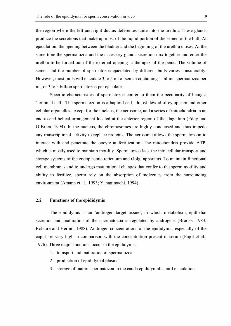
Table 1: Species differences in spermatozoal transit time through the epididymis (in days) (Amann et al., 1976)
Animal species Caput Corpus Cauda
Bull Stallion
Ram Boar Rat
Rabbit
2.5 1.9 2.4 2.2 2.1 2.2
0.6 2.2 1.2 3.2 0.8 0.8
5.2 9.8 12.8 6.4 9.8 9.7
Maturation of spermatozoa in the epididymis
Mammalian spermatozoa leaving the testis do not have the ability to recognize and
bind to oocytes, nor to fuse with the oolemma (Moore and Akhondi, 1996). To acquire
these abilities, they have to undergo numerous changes during epididymal transit. These
include: morphological alterations (migration of cytoplasmic droplet down the tail),
stabilization of nuclear chromatin by disulphide bonds, changes to the composition of
membranes, increasing susceptibility to cold shock damage, development of motility and
acquisition of fertility (Cooper, 1986; Bedford and Hoskins, 1990).
One of the most prominent changes in spermatozoa during epididymal maturation
is the development of sperm’s ability to move. This could be caused by biochemical and
The role of the epididymis for sperm conservation in vivo 11
physical changes of the sperm plasma membrane in combination with an increase of
cAMP-concentration, a decrease of intracellular pH (pHi), changes in free Ca²+- ions and
the velocity of glucose transportation in the spermatozoa (Hiipakka and Hammerstedt,
1978; Amann et al., 1993).
Spermatozoa are functionally mature in the cauda epididymidis but undergo
additional membrane changes during ejaculation by components present in the seminal
plasma. The process ends in the female reproductive tract, in which spermatozoa become
capacitated.
2.1.2 Production of epididymal plasma
The most important functions of epididymal epithelium are the maintenance of a
suitable environment for the maturation of spermatozoa in the caput and the storage of
fertile spermatozoa in the cauda. More than 95% of the epididymal plasma is reabsorbed in
the first part of the epididymis, leading to a sharp rise in spermatocrit (the volume of
spermatozoa as a percentage of the volume of the plasma), and changes in the composition
of the luminal fluid (Crabo, 1965). Between caput and cauda epididymidis, important
changes occur in the composition of plasma (Crabo, 1965; Levine and Marsh, 1971;
Setchell and Hinton, 1981). In fact, a large proportion of small molecules (amino acids,
ions, sugars and water), large molecules (proteins and steroids) and other components of
the testicular fluid are transported from the lumen of the epididymal duct to the interstitium
of the epididymis. Some of the present components are produced by the concentrated
epididymal spermatozoa themselves or reach the epididymis by blood or lymph.
2.1.3 Storage of mature spermatozoa in the cauda epididymidis
In mammals, the storage of spermatozoa occurs in the caudal part of the epididymis
whereas in most reptiles spermatozoa are stored in the ductus deferens for many months
(Licht, 1984). In most species, spermatozoa remain viable for several weeks (Table 2). The
most extended sperm storage occurs in bats (10 months), owing to their hibernation
(Gustafson, 1979). In birds, on the other hand, spermatozoa normally spend only about
24 h in the epididymis and ductus deferens before ejaculation (Clulow and Jones, 1982).
Sperm production is almost continuous, although for some species seasonal variations
occur (Setchell et al., 1993). In both cases, the number of spermatozoa present in the
epididymis remains constant (Table 3). Approximately 55-65% of total epididymal
12 Chapter 2
spermatozoa are stored in the cauda epididymidis (Amann et al., 1976) (Table 3). In the
cauda of the epididymis, spermatozoa of different ages are present (Orgebin-Crist, 1965).
This explains why individual spermatozoa from the same ejaculate capacitate and undergo
the acrosome reaction at different time points (Cuasnicu and Bedford, 1989).
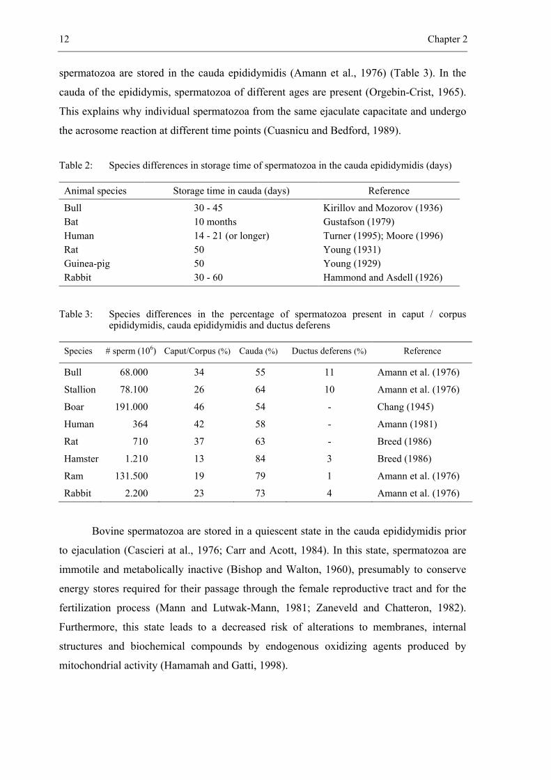
Table 2: Species differences in storage time of spermatozoa in the cauda epididymidis (days)
Animal species Storage time in cauda (days) Reference
Bull Bat Human Rat Guinea-pig Rabbit
30 - 45 10 months 14 - 21 (or longer) 50 50 30 - 60
Kirillov and Mozorov (1936) Gustafson (1979) Turner (1995); Moore (1996) Young (1931) Young (1929) Hammond and Asdell (1926)
Table 3: Species differences in the percentage of spermatozoa present in caput / corpus epididymidis, cauda epididymidis and ductus deferens
Species # sperm (106) Caput/Corpus (%) Cauda (%) Ductus deferens (%) Reference
Bull 68.000 34 55 11 Amann et al. (1976) Stallion 78.100 26 64 10 Amann et al. (1976) Boar 191.000 46 54 - Chang (1945)
Human 364 42 58 - Amann (1981) Rat 710 37 63 - Breed (1986) Hamster 1.210 13 84 3 Breed (1986) Ram 131.500 19 79 1 Amann et al. (1976) Rabbit 2.200 23 73 4 Amann et al. (1976)
Bovine spermatozoa are stored in a quiescent state in the cauda epididymidis prior
to ejaculation (Cascieri at al., 1976; Carr and Acott, 1984). In this state, spermatozoa are
immotile and metabolically inactive (Bishop and Walton, 1960), presumably to conserve
energy stores required for their passage through the female reproductive tract and for the
fertilization process (Mann and Lutwak-Mann, 1981; Zaneveld and Chatteron, 1982).
Furthermore, this state leads to a decreased risk of alterations to membranes, internal
structures and biochemical compounds by endogenous oxidizing agents produced by
mitochondrial activity (Hamamah and Gatti, 1998).
The role of the epididymis for sperm conservation in vivo 13
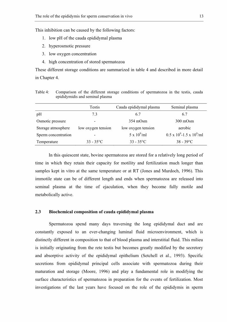
This inhibition can be caused by the following factors:
1. low pH of the cauda epididymal plasma
2. hyperosmotic pressure
3. low oxygen concentration
4. high concentration of stored spermatozoa
These different storage conditions are summarized in table 4 and described in more detail
in Chapter 4.
Table 4: Comparison of the different storage conditions of spermatozoa in the testis, cauda epididymidis and seminal plasma
Testis Cauda epididymal plasma Seminal plasma
pH 7.3 6.7 6.7 Osmotic pressure - 354 mOsm 300 mOsm Storage atmosphere low oxygen tension low oxygen tension aerobic Sperm concentration - 5 x 10⁹/ml 0.5 x 10⁹-1.5 x 10⁹/ml Temperature 33 - 35°C 33 - 35°C 38 - 39°C
In this quiescent state, bovine spermatozoa are stored for a relatively long period of
time in which they retain their capacity for motility and fertilization much longer than
samples kept in vitro at the same temperature or at RT (Jones and Murdoch, 1996). This
immotile state can be of different length and ends when spermatozoa are released into
seminal plasma at the time of ejaculation, when they become fully motile and
metabolically active.
2.3 Biochemical composition of cauda epididymal plasma
Spermatozoa spend many days traversing the long epididymal duct and are
constantly exposed to an ever-changing luminal fluid microenvironment, which is
distinctly different in composition to that of blood plasma and interstitial fluid. This milieu
is initially originating from the rete testis but becomes greatly modified by the secretory
and absorptive activity of the epididymal epithelium (Setchell et al., 1993). Specific
secretions from epididymal principal cells associate with spermatozoa during their
maturation and storage (Moore, 1996) and play a fundamental role in modifying the
surface characteristics of spermatozoa in preparation for the events of fertilization. Most
investigations of the last years have focused on the role of the epididymis in sperm
14 Chapter 2
maturation, however less research investments have been made in the understanding of
sperm storage (Turner, 1995). The fact that the cauda epididymidis can store viable sper-
matozoa for several days in vivo suggests that some epididymal constituents could be very
interesting in the formulation of diluents for preserving spermatozoa in vitro. Research on
epididymal storage conditions can be performed by different approaches, such as the
analysis of different physicochemical conditions or of the specific ionic and protein com-
position of the epididymal plasma, or by studying membrane differences between caput,
corpus and cauda epididymal spermatozoa and ejaculated spermatozoa (Jones, 1998) or by
examining in vitro interactions between spermatozoa and epididymal epithelial cells in
culture (Moore, 1996). Chemical analysis of the composition of epididymal plasma of the
bull has been investigated in different studies (Crabo, 1965; Cascieri et al., 1976; Setchell
et al., 1993; Verberckmoes et al., in preparation). In our laboratory, analyses were perfor-
med with cauda epididymal plasma collected by making small incisions into the tubuli of
post mortem material. The composition of this epididymal plasma was used to develop a
new completely defined diluent named CEP-diluent (Verberckmoes et al., in preparation).
2.4 Conclusions
In mammals, spermatozoa leaving the testes are incapable of fertilizing a female
gamete. Sperm maturation is the term given to the process by which mammalian
spermatozoa undergo numerous changes during epididymal transit. The most important
changes occurring in the spermatozoa are the development of motility and the acquisition
of fertilizing ability, which are largely the result of interactions with epididymal epithelium
and epididymal plasma. Spermatozoa spend many days traversing the long epididymal duct
and they are finally stored in the cauda epididymidis in a quiescent state for several weeks
awaiting ejaculation. In vivo, all these processes are coordinated with remarkable precision
to ensure production of fully viable and fertile spermatozoa.
Although most investigations of the last decades have focused on the role of the
epididymis in sperm maturation, it seems that research investment into the understanding
of other major epididymal functions, especially sperm storage, should also be encouraged.
There is practically no information on the effects of epididymal constituents on sperm
survival and storage. In this respect, an interesting approach would probably be to search
for a procedure that reduced sperm motility whilst maintaining its fertilizing capacity
during storage but then allowed full development of motility when needed.
The role of the epididymis for sperm conservation in vivo 15
2.5 References
Amann RP, Johnson L, Thompson DL, Pickett BW. Daily spermatozoal production, epididymal
spermatozoal reserves and transit time of spermatozoa through the epididymis of the rhesus
monkey. Biol Reprod 1976; 15: 586-592.
Amann RP. A critical review of methods for evaluation of spermatogenesis from seminal
characteristics. J Androl 1981; 2: 37-58.
Amann RP, Hammerstedt RH, Veeramachaneni DN. The epididymis and sperm maturation:
a perspective. Reprod Fertil Dev 1993; 5: 361-381.
Bedford JM, Hoskins DD. The mammalian spermatozoon: morphology, biochemistry, and
physiology. Laming GE (ed). In: Marshall’s Physiology of Reproduction. Churchill
Livingston; Londen 1990: 379-568.
Bishop MWH, Walton A. Metabolism and motility of mammalian spermatozoa. In: Parkes AS
(ed). Marshall's Physiology of Reproduction. Longmans Green en Co; Edinburgh 1960:
264-309.
Breed WG. Comparative morphology and evolution of the male reproductive tract in the Australian
hydromyine rodents (Muridae). J Zool (London) 1986; 209: 607-629.
Brooks DE. Epididymal functions and their hormonal regulation. Aust J Biol Sci 1983; 36:
205-221.
Carr DW, Acott TS. Inhibition of bovine spermatozoa by caudal epididymal fluid: I. Studies of a
sperm motility quiescence factor. Biol Reprod 1984; 30: 913-925.
Cascieri M, Amann RP, Hammerstedt RH. Adenine nucleotide changes at initiation of bull sperm
motility. J Biol Chem 1976; 251: 787-793.
Chang MC. Distribution of sperm in genital tract of the ram. J Agric Sci 1945; 35: 243-246.
Clulow J, Jones RC. Production, transport, maturation, storage and survival of spermatozoa in the
Japanese quail, Coturnix coturnix. J Reprod Fertil 1982; 64: 259-266.
Cooper TG. Epididymis, sperm maturation and fertilization. Springer-verlag; Berlin 1986.
Cosentino MJ, Cockett AT. Structure and function of the epididymis. Urol Res 1986; 14: 229-240.
Crabo B. Studies on the composition of epididymal content in bulls and boars. Acta Vet Scand
Suppl 1965; 6: 1-94.
Cuasnicu PS, Bedford JM. The effect of moderate epididymal aging on the kinetics of the
acrosome reaction and fertilizing ability of hamster spermatozoa. Biol Reprod 1989; 40:
1067-1073.
16 Chapter 2
Eddy EM, O’Brien DA. The spermatozoon. In: Knobil E, Neill JD (eds). The Physiology of
Reproduction. Raven Press; New York 1994: 29-77.
Gustafson AW. Mate reproductive patterns in hibernating bats. J Reprod Fertil 1979; 46: 195-202.
Hamamah S, Gatti JL. Role of the ionic environment and internal pH on sperm activity. Hum
Reprod 1998; 13 Suppl 4: 20-30.
Hammond J, Asdell SA. The vitality of the spermatozoa in the male and female reproductive tracts.
Br J Exp Biol 1926; 4: 155-185.
Hiipakka RA, Hammerstedt RH. Changes in 2-deoxyglucose transport during epididymal
maturation of ram sperm. Biol Reprod 1978; 19: 1030-1035.
Hoskins DD, Brandt H, Acott TS. Initiation of sperm motility in the mammalian epididymis.
Fed Proc 1978; 37: 2534-2542.
Jones RC, Murdoch RN. Regulation of the motility and metabolism of spermatozoa for storage in
the epididymis of eutherian and marsupial mammals. Reprod Fertil Dev 1996; 8: 553-568.
Jones R. Plasma membrane structure and remodelling during sperm maturation in the epididymis.
J Reprod Fertil Suppl 1998; 53: 73-84.
Kirillov VS, Mozorov VA. Duration of survival of bull spermatozoa in an epididymis isolated from
the testis. Anim Breed 1936; Abstr 5: 22.
Levine N, Marsh DJ. Micropuncture studies of the electrochemical aspects of fluid and electrolyte
transport in individual seminiferous tubules, the epididymis and the vas deferens in rats.
J Physiol 1971; 213: 557-570.
Licht P. Reptiles. In: Lamming GE (ed). Marshall's Physiology of Reproduction. Churchill
Livingstone; London 1984: 206-282.
Mann T, Lutwak-Mann C. Male reproductive function and semen. Springer-verlag; Berlin 1981.
Moore HDM. The influence of the epididymis on human and animal sperm maturation and storage.
Hum Reprod Natl Suppl 1996; 11: 103-110.
Moore HDM, Akhondi MA. In vitro maturation of mammalian spermatozoa. Rev Reprod 1996; 1:
54-60.
Orgebin-Crist MC. Passage of spermatozoa labelled with thymidine-3-H through the ductus
epididymidis of the rabbit. J Reprod Fertil 1965; 10: 241-51.
Pujol A, Bayard F, Louvet JP, Boulard C. Testosterone and dihydrotestosterone concentrations in
plasma, epididymal tissues and seminal fluid of adult rats. Endocrinology 1976; 98: 111-113.
The role of the epididymis for sperm conservation in vivo 17
Robaire B, Hermo L. Efferent ducts, epididymis and vas deferens: structure, functions and their
regulation. In: Knobil E, O'Neill J (eds). The Physiology of Reproduction. Raven Press; New
York 1988: 999-1080.
Setchell BP, Hinton BT. The effects on spermatozoa of changes in the composition of luminal fluid
as it passes along the epididymis. Prog Reprod Biol 1981; 8: 58-66.
Setchell BP, Sánchez-Partida LG, Chairussyuhur A. Epididymal constituents and related
substances in the storage of spermatozoa: a review. Reprod Fertil Dev 1993; 5: 601-612.
Setchell BP. The Parkes Lecture. Heat and the testis. J Reprod Fertil 1998; 114: 179-194.
Turner TT. On the epididymis and its role in the development of the fertile ejaculate. J Androl
1995; 16: 292-298.
Verberckmoes S, Van Soom A, De Pauw I, Dewulf J, de Kruif A. Ionic composition of cauda
epididymal plasma in the bull. In preparation.
Yanagimachi R. Mammalian fertilization. In: Knobil E, Neill JD (eds). The Physiology of
Reproduction. Raven Press; New York 1994: 189-317.
Young WC. A study of the function of the epididymis II. The importance of an aging process in
sperm for the length of the period during which fertilizing capacity is retained by sperm
isolated in the epididymis of the guinea-pig. J Morphol Physiol 1929; 48: 475-491.
Young WC. Functional changes undergone by spermatozoa during their passage through the
epididymis and vas deferens in the guinea-pig. J Exp Biol 1931; 8: 151-164.
Zaneveld LJD, Chatterton RT. Biochemistry of Mammalian Reproduction. Wiley-Interscience Pub;
New York 1982; 38.

__________________________________________________ 3.
SUITABILITY OF SPERM FUNCTION TESTS FOR THE EVALUATION OF
SPERM QUALITY AND BULL FERTILITY
3.1 Introduction
3.2 The evaluation of bovine sperm quality characteristics by means of
fluorescent stainings
3.2.1 Membrane integrity
3.2.2 Capacitation
3.2.3 Acrosome reaction
3.2.4 Mitochondrial function
3.2.5 Apoptosis
3.3 Sperm motility assessment
3.3.1 Motility assessment by computer assisted sperm analysis systems
3.3.2 Motility assessment by sperm migration assays
3.3.3 Motility assessment by sperm chemotaxis assays
3.4 Sperm binding and performance assessment
3.4.1 Sperm-oviduct binding
3.4.2 Sperm-zona binding
3.4.3 In vitro fertilization
3.5 Conclusions
3.6 References
Review in preparation
De Pauw I., Van Soom A., Verberckmoes S. and de Kruif A.
20 Chapter 3
3.1 Introduction
Assessment of the functional capacity of spermatozoa in vitro is of particular
interest in order to select sires with good sperm quality and fertility. Much research has
already been done to use in vitro technologies to measure the fertilizing ability of
spermatozoa. In many laboratories, the evaluation of semen is limited to the measurement
of sperm concentration, microscopic examination of progressive motility and to the
evaluation of morphology and membrane integrity by means of eosin/nigrosin staining.
Methods for sperm analysis have considerably increased in recent years. It is now possible
to replace subjective motility assessment by Computer Assisted Sperm Analysis (CASA)
(Malmgren, 1997; Verstegen et al., 2002) and the former eosin/nigrosin staining method is
now being superseded by fluorescent dyes that bind to various regions of the cell to
demonstrate particular functional characteristics of spermatozoa (Harrison and Vickers,
1990; Johnson et al., 1996). Moreover, other technological changes over the past decade
have made it possible to apply either new techniques such as flow cytometry (Garner et al.,
1986; Graham et al., 1990) or more specific functional in vitro tests which determine
sperm performance such as sperm migration (Kummerfeld et al., 1981; Anilkumar et al.,
2001; Verberckmoes et al, 2002), sperm binding to oviduct epithelial explants (De Pauw
et al., 2002) or to the zona pellucida (ZP) (Fazeli et al., 1997; Zhang et al., 1998) and in
vitro fertilization (IVF) (Larsson and Rodriguez-Martinez, 2000) for the assessment of
sperm quality and fertility. Several studies have tried to find a correlation between one or
more sperm parameters and in vivo bull fertility (Table 1). Moreover, in some studies, a
prediction of bull fertility was attempted by means of regression analysis, which tended to
be more successful when multiple sperm parameters were taken into account. All studies
had in common that in vivo bull fertility was assessed on a high number of inseminations,
but for technical reasons it was difficult to include more than 20 bulls in this kind of
research. Our aims were to discuss recent progress, which has been made in assessment of
sperm quality in vitro, and to determine to what extent these relatively new tests, can be
used for evaluation of in vivo bull fertility (Table 1).

Sperm function tests for evaluation of sperm quality and bull fertility 21
22 Chapter 3
3.2 The evaluation of bovine sperm quality characteristics by means of fluorescent
stainings
Spermatozoa consist of several membrane compartments (i.e. plasma membrane,
acrosomal membrane, mitochondrial membrane). Membrane integrity of all these
compartments is of fundamental importance for the maintenance of the fertilizing potential
of spermatozoa. Therefore, over the past 10-15 years many fluorescent stains have been
developed that can be used to evaluate subcellular components indicating specific sperm
functions (Amann, 1989; Oehninger et al., 1992; Critser and Noiles, 1993). The functional
aspects that could give greater reliability to the estimation of the fertilizing capacity of
spermatozoa which include plasma membrane integrity, capacitation, acrosome reaction,
mitochondrial membrane potential and DNA integrity, are described below.
3.2.1 Membrane integrity
For many years, a number of procedures has been proposed to assess the membrane
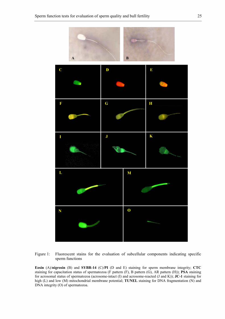
integrity of spermatozoa. The eosin/nigrosin staining (Figure 1A and B) has been used to
assess both membrane integrity and morphology of the spermatozoa (Blom, 1950). This
single association staining procedure is based on the degree of membrane permeability of
dead spermatozoa in which heads take a pink (eosinophilic) colouration. It has been argued
that this method is unreliable because, in some species, spermatozoa show partial staining
(Hancock, 1957). Another disadvantage is that some ingredients such as glycerol present in
commonly used cryopreservation media interfere with the staining (Mixner and Saroff,
1954).
In recent years several fluorescent probes have been developed to determine
membrane integrity. The most used fluorophore is SYBR®-14 proposed by Garner et al.
(1994). This fluorescent probe is used in combination with propidium iodide (PI). Both
dyes have an affinity for nucleic acids. The membrane-permeant stain SYBR®-14 labels
the DNA of all spermatozoa bright green, whereas PI has been used to detect only
membrane-damaged spermatozoa (red fluorescence) (Garner et al., 1986). Upon cell death,
spermatozoa lose their ability to resist the influx of the membrane-impermeant dye PI,
which enters the spermatozoa through pores in the nuclear membrane that are located in
the posterior region of the sperm head (Oko et al., 1976) and replaces or quenches the
SYBR®-14 staining. Johnson et al. (1996) have discussed these cell colour changes in
Sperm function tests for evaluation of sperm quality and bull fertility 23
terms of ‘DNA viability’ but in this instance the mode of action remains unclear (Holt,
2000). Normally there are three populations: living - SYBR®-14 stained (Figure 1C), dead
- PI stained (Figure 1D), and a third population, which is double stained and represents
dying spermatozoa (Figure 1E).
The advantages of this staining are its relative rapidity, accuracy and reliability.
The staining time is not as critical as enzyme-based stains and background staining is
virtually nonexistent (Garner and Johnson, 1995; Chalah and Brillard, 1998). The
proportion of intact/damaged cells, sometimes called the live/dead ratio, can be estimated
by either fluorescence microscopy or flow cytometry. Using this combination of dyes,
membrane integrity of a sperm sample can be analysed with visible-light excitation,
avoiding the harmful effects of ultraviolet (UV) exposure. Furthermore, Garner et al.
(1996) have shown that fluorescent staining of boar spermatozoa with SYBR®-14 neither
affected their ability to fertilize oocytes, nor the developmental competence of the resultant
embryos.
Two other commonly used fluorescent stains are the bisbenzimide stains Hoechst
33342 and 33258, which excite in UV (365 nm) and require UV laser-equipped flow
cytometry systems (Garner and Johnson, 1995). Also dual staining with enzyme-based
staining combinations carboxyfluorescein diacetate and PI have been used, but the results
are too variable for routine use (Garner and Johnson, 1995).
Comparisons of SYBR®-14/PI with eosin/nigrosin in sperm of different species
have shown that the former stain is much more sensitive for detection of early membrane
damage after centrifugation (dog: Rijsselaere et al., 2002) or after freeze-thawing (poultry:
Chalah and Brillard, 1998; bull: De Pauw et al., 1999). It has been shown that post-thaw
membrane integrity, as assessed by SYBR®-14/PI staining, is significantly correlated with
field fertility. Furthermore, it is a better single predictor of fertility (r²=0.39) than post-
thaw motility, the proportion of capacitated spermatozoa and the proportion of induced
acrosome-reacted spermatozoa (Januskauskas et al., 1999).
3.2.2 Capacitation
Although mammalian spermatozoa are actively motile after ejaculation, they are
not yet able to fertilize eggs. To achieve this, they need to be activated in the female
genital tract by two specific events called capacitation and acrosome reaction
(Yanagimachi, 1994).
24 Chapter 3
Capacitation can only be detected at the molecular level and not at the structural
level. Several aspects of sperm capacitation have already been characterized including
membrane and intracellular ionic modifications (de Lamirande et al., 1993; Yanagimachi,
1994; Visconti et al, 1998; Topper et al., 1999). These alternations involve removal or
inactivation of decapacitation factors on the sperm surface; changes in localization,
molecular structure and lateral mobility of membrane proteins; adsorption onto the sperm
surface of proteins from the female tract (Yanagimachi, 1994); alterations in membrane
lipid composition, in particular in the cholesterol/phospholipid ratio (Cross, 1998); ionic
deregulation manifested as increases in internal Ca2+, Na+ and pH (Fraser, 1995; Parrish et
al., 1999); generation of reactive oxygen species (de Lamirande et al., 1997); and an
increase in cAMP and protein tyrosine phosphorylation (Visconti et al., 1998). Capacitated
spermatozoa are generally accepted to be fragile, unstable and short-lived (Hunter, 1987).
The fluorescent antibiotic chlortetracycline (CTC) has been utilized to assess the
degree of destabilization of the sperm membrane (Fraser, 1995). This direct, single step
staining assay is based on the transfer of neutral and uncomplexed CTC across the cell
membranes into intracellular compartments containing high levels of free Ca²+.
Chlortetracycline binds to Ca²+ becoming more fluorescent as a result (Tsien, 1989). These
CTC-Ca²+ complexes preferentially bind to hydrophobic regions of the cell membrane
resulting in a pattern of membrane staining characteristics of various transitional phases of
destabilization. Bovine spermatozoa stained with CTC display 3 different fluorescent
patterns representing different stages of the capacitation process (Lee et al., 1987), which
are indicated in literature by the letters F (uncapacitated, acrosome intact: Figure 1F),
B (capacitated, acrosome intact: Figure 1G) and AR (acrosome reacted: Figure 1H). These
patterns are quite difficult to distinguish by fluorescence microscopy at least in bull semen,
and their classification is therefore rather subjective (De Pauw, unpublished observation).
Moreover, the CTC staining method is found to be a laborious technique because it cannot
be analyzed by flow cytometry (Rathi et al., 2001). The flow cytometric approach is based
upon the intensity of the fluorescence of spermatozoa alone and lacks the precise
delineation of specific fluorescence categories, which renders this method unsatisfactory
for practical use (Maxwell and Johnson, 1997). Because of this disadvantage and the fact
that the molecular basis of the interaction between CTC and the plasma membrane is still
unclear, research is being undertaken to find alternatives for the evaluation of the
capacitation status of spermatozoa.
Sperm function tests for evaluation of sperm quality and bull fertility 25
Figure 1: Fluorescent stains for the evaluation of subcellular components indicating specific sperm functions
Eosin (A)/nigrosin (B) and SYBR-14 (C)/PI (D and E) staining for sperm membrane integrity; CTC staining for capacitation status of spermatozoa (F pattern (F), B pattern (G), AR pattern (H)); PSA staining for acrosomal status of spermatozoa (acrosome-intact (I) and acrosome-reacted (J and K)); JC-1 staining for high (L) and low (M) mitochondrial membrane potential; TUNEL staining for DNA fragmentation (N) and DNA integrity (O) of spermatozoa.
A B
26 Chapter 3
According to Rathi et al. (2001), merocyanine 540 staining is a better method for
the evaluation of the early events of capacitation. Merocyanine 540 is a hydrophobic dye
that stains sperm membranes more intensely if their lipid components are in a higher state
of disorder (Williamson et al., 1983; Langner et al., 1993). Another alternative could be
filipin, a polyene antibiotic that forms 25-30 nm complexes with 3-ß-hydroxysterols.
Filipin observes the cholesterol efflux in the sperm plasma membrane, one of the events
during capacitation (Visconti et al., 1999). The distribution of filipin-sterol complexes can
be visualized in freeze-fractured sperm membranes (Verkleij et al., 1973) or by UV-
fluorescence due to the intrinsic fluorescent properties of filipin.
Nevertheless, CTC-staining has generally been accepted to be useful to evaluate the
percentage of viable uncapacitated spermatozoa in frozen-thawed bull semen. The
percentage of viable, uncapacitated spermatozoa in an AI-semen batch is positively
correlated with its fertility after AI (Thundathil et al., 1999).
3.2.3 Acrosome reaction
Capacitated spermatozoa with ‘intact’ acrosomes may pass through the cumulus
(Storey et al., 1984; Talbot, 1985), but are unable to pass through the ZP. The acrosome
reaction, a modified form of exocytosis, is an absolute requirement for successful sperm
passage through the ZP (Austin and Bishop, 1958). It involves the occurrence of point
fusions between the outer acrosomal membrane and the overlying plasma membrane,
followed by membrane vesiculation and extrusion of the acrosomal enzymatic contents,
and exposure of inner acrosomal membrane antigens (Yanagimachi, 1994). The acrosomal
content, containing mainly hydrolytic enzymes, starts to disperse and digest the ZP.
Different lectins interacting with glycoconjugates of the outer acrosomal membrane
(Peanut agglutinin, PNA) or matrix (Pisum sativum agglutinin, PSA), have been tested for
use in acrosomal status evaluation (Cross et al., 1986; Mortimer et al., 1987). Labeling
permeabilized spermatozoa with fluorescein isothiocyanate-PSA (FITC-PSA) renders
acrosome-intact cells brightly fluorescent over the entire acrosomal region of the sperm
head (Figure 1I), while acrosome-reacted spermatozoa have either no acrosomal labeling,
or only an equatorial band of label (Figure 1J and K). Peanut agglutinin binds to the
anterior regions of the head. This binding is maybe more avidly than FITC-PSA, so that
reacted spermatozoa with diminished FITC-PNA labeling are not as easily detected by
visual inspection (Cross and Watson, 1994).
Sperm function tests for evaluation of sperm quality and bull fertility 27
The integrity of the acrosome can also be determined morphologically, usually at
the light microscopically level, in unstained samples or with different empirical stains such
as Giemsa stain. However, fluorescent probes are generally superior to coloured dyes
because fluorescence provides greater intensity and greater contrast between acrosomal
and nonacrosomal material (Cross and Meizel, 1989). Moreover, FITC-PSA-staining has
several other advantages over conventional stains: its simplicity, its objectiveness, its
rapidity, its low cost, and more importantly many samples can be processed in parallel
(Margalit et al., 1997).
The percentage of spermatozoa having an intact acrosome and being able to
perform the acrosome reaction upon triggering is regarded as an important semen
characteristic (de Leeuw et al., 1991). Acrosome reaction can be induced by treatment with
calcium ionophores, and significant correlations have been found between the degree of
induced AR and in vivo fertility of bulls, as assessed by non-return rates (Whitfield and
Parkinson, 1995; Januskauskas et al., 2000).
3.2.4 Mitochondrial function
Mitochondria play an essential role in the life cycle of spermatozoa through the
control of energy production in the form of adenosine triphosphate (ATP). The generation
of ATP by mitochondria is vital to the survival of spermatozoa because it is the only source
of ATP production and thus essential for cellular homeostasis (Moyes et al., 1998).
Mitochondrial membrane potential (∆ψm) is a sensitive indicator of the functional
status of mitochondria and is used as a marker for assessing overall mitochondrial function
(Cortopassi and Wong, 1999). Mitochondrial membrane potential is reduced when energy
metabolism is disrupted, notably in apoptosis (Shapiro, 2000).
Several fluorescent stains such as Rhodamine 123 and Mitotracker Green
selectively stain mitochondria in the midpiece of spermatozoa regardless of mitochondrial
membrane potential. Another fluorescent stain, the lipophilic, cationic compound 5,5’,6,6’-
tetrachloro-1,1’13,3’-tetraethylbenzimidazolyl carbocyanine iodide (JC-1) can be used to
evaluate mitochondrial function since it has been reported to differentially label
mitochondria with high and low mitochondrial membrane potential (Reers et al., 1991;
Cossarizza et al., 1993). JC-1 can form monomers in mitochondria with low membrane
potential (green fluorescence; Figure 1M) and possesses the ability to form multimers
known as J-aggregates after accumulation in mitochondria with high membrane potential
28 Chapter 3
(orange fluorescence; Figure 1L). However, the intensity of JC-1 staining is dependent
upon its concentration. This is one of the characteristics of JC-1 that makes it very difficult
to get a proper interpretation of flow cytometric data. Furthermore, JC-1 also stains the
head of the spermatozoa green. This staining at the membrane level contributes to the
overall staining and makes its interpretation more difficult. Therefore, additional studies
are needed to get a better understanding of the various aspects of the staining phenomena.
It is obvious that JC-1 is a very interesting candidate molecule to include in sperm function
testing. However, since no discernible relationship was found between mitochondrial
membrane potential, measured with JC-1, and pre-freeze sperm motility (Garner et al.,
1997), it may be useful to include more precise measurements of several motility
characteristics obtained by CASA systems (Thomas et al., 1998). Until now, no studies are
available linking JC-1 to in vivo bull fertility.
3.2.5 Apoptosis
Because spermatogenesis is a continuous process, a mechanism is needed to control
sperm production. Apoptosis has been observed in male germ cells, which are phago-
cytosed by Sertoli cells in the testis (Xu et al., 1999). The failure of Sertoli cells to remove
apoptotic spermatozoa results in their release into the lumen of seminiferous tubuli and,
consequently, in an increased number of abnormal spermatozoa in semen, provoking
reduced fertility (Anzar, 2002). Apoptosis, physiologically programmed cell death, affects
single spermatozoon without any associated inflammation in the surrounding tissues
(Wyllie et al., 1980). The process is characterized by a series of cellular, morphological
and biochemical alternations in cells, including phosphatidylserine (PS) externalization,
chromatin fragmentation and cell shrinkage (Wyllie et al., 1980; Earnshaw, 1995; Martin
et al., 1995).
To measure apoptosis in spermatozoa two flow cytometric methods can be used.
The first method is based on an assay for the measurement of PS translocation across the
plasma membrane. Phosphatidylserine, normally present on the inner leaflet of the plasma
membrane of healthy cells, is translocated and exposed on the outer leaflet during early
apoptosis (Martin et al., 1995). Annexin V is a Ca²+-dependent, phospholipid-binding
protein (35-36 kDa) with a high affinity for PS (Vermes et al., 1995) and conjugated to
fluorescein isothiocyanate (FITC) fluorochrome it can serve as a sensitive probe that can
be used for flow cytometric detection of apoptosis. According to Anzar et al. (2002), the
Sperm function tests for evaluation of sperm quality and bull fertility 29
Annexin V/PI assay is more precise and reliable than the common live/dead SYBR®-14/PI
assay because the staining is sensitive to alterations in the sperm plasma membrane at the
molecular level. SYBR®-14 stains apoptotic as well as live spermatozoa in a population,
because in both cases the plasma membrane is intact. In contrast, Annexin V/PI assay is
able to distinguish between apoptotic and live spermatozoa.
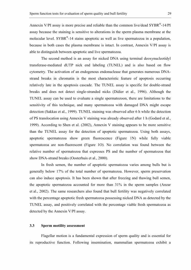
The second method is an assay for nicked DNA using terminal deoxynucleotidyl
transferase-mediated dUTP nick end labeling (TUNEL) and is also based on flow
cytometry. The activation of an endogenous endonuclease that generates numerous DNA-
strand breaks in chromatin is the most characteristic feature of apoptosis occurring
relatively late in the apoptosis cascade. The TUNEL assay is specific for double-strand
breaks and does not detect single-stranded nicks (Didier et al., 1996). Although the
TUNEL assay can be used to evaluate a single spermatozoon, there are limitations to the
sensitivity of this technique, and many spermatozoa with damaged DNA might escape
detection (Sakkas et al., 1999). TUNEL staining was observed after 6 h while the detection
of PS translocation using Annexin V staining was already observed after 1 h (Godard et al.,
1999). According to Shen et al. (2002), Annexin V staining appears to be more sensitive
than the TUNEL assay for the detection of apoptotic spermatozoa. Using both assays,
apoptotic spermatozoa show green fluorescence (Figure 1N) while fully viable
spermatozoa are non-fluorescent (Figure 1O). No correlation was found between the
relative number of spermatozoa that expresses PS and the number of spermatozoa that
show DNA-strand breaks (Oosterhuis et al., 2000).
In fresh semen, the number of apoptotic spermatozoa varies among bulls but is
generally below 17% of the total number of spermatozoa. However, sperm preservation
can also induce apoptosis. It has been shown that after freezing and thawing bull semen,
the apoptotic spermatozoa accounted for more than 31% in the sperm samples (Anzar
et al., 2002). The same researchers also found that bull fertility was negatively correlated
with the percentage apoptotic fresh spermatozoa possessing nicked DNA as detected by the
TUNEL assay, and positively correlated with the percentage viable fresh spermatozoa as
detected by the Annexin V/PI assay.
3.3 Sperm motility assessment
Flagellar motion is a fundamental expression of sperm quality and is essential for
its reproductive function. Following insemination, mammalian spermatozoa exhibit a
30 Chapter 3
sequence of different types of motility modulated by the female reproductive tract.
Vigorous motility is not only necessary for successful sperm migration to the site of
fertilization but also for the transit through the cumulus cell investments and the zona
pellucida of the oocyte.
Motility evaluation is based upon subjective estimates of the percentage of
spermatozoa exhibiting any kind of movement and the proportion of spermatozoa moving
progressively forward. This assessment is mainly based on subjective optical microscopic
evaluation which is cheap and easy to perform. However, sperm motility estimates vary
among examiners even when examing the same sperm sample. These variations are not
only dependent on the biological state of the spermatozoa but also on the conditions of
observation (Drobnis and Katz, 1990), such as the physical properties of the suspending
fluid, the depth of the observation chamber and the observers’ training.
Subjective evaluation of sperm progressive motility is used to determine the quality
of bull semen intended for AI. However, subjectively assessed motility is not a good
predictor of the fertility level of the AI-semen dose (Soderquist et al., 1991), particularly
when motility values are within ranges around 50% (Stalhammar et al., 1994). This was
confirmed by den Daas (1997) who concluded that the measurement of motility
characteristics of serially diluted sperm, after thawing or after thawing followed by a
thermo-resistance or endurance test, cannot be used to predict individual bull fertility.
Measurement of sperm motility is entering a new era in which objective, efficient
and rapid methods are becoming accessible. Nowadays sperm motility can be assessed by
computerized measurements, but also by approaching motility in a more functional way:
by analyzing sperm migration through a viscous fluid or by evaluating the percentage of
spermatozoa displaying chemotaxis.
3.3.1 Motility assessment by computer assisted sperm analysis (CASA) systems
Due to the variation between subjective estimates of sperm motility, more emphasis
has been put upon the development of objective methods for quantifying sperm motion.
Time-lapse photomicrography, multiple-exposure photomicrography, and frame playback
video-micrography have been used (Malmgren, 1997). However these methods are labor-
intensive. One recent development is the application of computerized image analysis
systems, which offer an automated, rapid and objective approach of more specific
characteristics of spermatozoa motion, such as straight-line velocity (VSL), curvilinear
Sperm function tests for evaluation of sperm quality and bull fertility 31
velocity (VCL), average path velocity (VAP), linearity (LIN), straightness (STR),
circularly motile spermatozoa (CIR), lateral head displacement (LHD), and beat cross
frequency (BCF). The main problems are the high cost of the equipment, and
standardization and optimization of the system and procedures (Verstegen, 2002). Before
any practical use is possible, careful validation and checking of the setup are very
important since using a wrong setup might lead to a wrong estimation of motility.
Computerized systems can provide precise and accurate information on sperm motion
characteristics for different species. However a lack of uniformity among users and
instruments makes it difficult to define standard accepted values for normal and abnormal
sperm motion (Verstegen, 2002).
It is still not clear which sperm movement characteristic will be of clinical value for
the prediction of in vivo fertility and fertility rates (Aitken et al., 1985; Amann, 1989).
Farrell et al. (1998) have found that the combination of percentages of motility and
velocity was highly correlated with fertility (r²=0.87), despite the fact that bulls differ
largely in all variables related to motility and velocity mainly caused by the large
interanimal variation.
3.3.2 Motility assessment by sperm migration assays
Spermatozoa are selected both quantitatively and qualitatively during their
migration through the cervical mucus and during their transit in the female genital tract
(Jouannet and Feneux, 1987). Cervical mucus is a physiological barrier controlling sperm
access to the upper female reproductive tract. Spermatozoa with anomalies of the flagellum
and particularly of the middle piece are not able to migrate through the cervical mucus
(Jeulin et al., 1985). Furthermore, the pattern of sperm movement and amplitude of the
lateral head displacement are also very important for cervical mucus penetration (Aitken
et al., 1985).
In human, sperm migration tests have been extensively refined and validated
(Biljan et al., 1994; Clarke et al., 1998). In contrast to human (Alexander, 1981), no
correlation has been found between bull fertilizing capacity of spermatozoa and sperm
migration capacity (Kummerfeld et al., 1981; Verberckmoes et al., 2002). There are
several explanations for this lack of correlation (Verberckmoes et al., 2002). First, it could
be due to the small difference in fertility between bulls. Sperm migration tests are
performed with spermatozoa of bulls used for AI; whereas in human, it is possible to
32 Chapter 3
compare spermatozoa of fertile donors with spermatozoa of patients with infertility
problems. Another explanation could be the fact that NRRs are determined after AI. In this
case spermatozoa don’t have to migrate through the cervical mucus as they are deposited
directly in the uterine body or in the uterine horns.
3.3.3 Motility assessment by sperm chemotaxis assays
A unique feature of sperm chemotaxis is the directional change of movement
towards the source of an attractant or retreat from a repellent. The most commonly used
technique for studying sperm chemotaxis in mammals is an ‘accumulation assay’ in which
spermatozoa sense an ascending gradient of the attractant and accumulate near or at its
source. Another commonly used assay is a ‘choice assay’ in which spermatozoa choose
between two wells: one containing the attractant and the other containing the buffer as a
control (Eisenbach, 1999).
Sperm chemotaxis to follicular fluid has already been established both in human
(Cohen-Dayag et al., 1994; Ralt et al., 1994) and mouse (Giojalas and Rovasio, 1998;
Oliviera et al., 1999). However, the fraction of chemotactic spermatozoa in the total sperm
population is very small (2-12% in human (Cohen-Dayag et al., 1994) and approximately
10% in mouse (Giojalas and Rovasio, 1998; Oliviera et al., 1999)). According to Makler
et al. (1992, 1995), chemotaxis between human sperm and ova does not exist because the
fraction of these responsive spermatozoa is too small, the chemotactic responsiveness is
only temporary and many ‘chemotaxis assays’ have failed to distinguish between
chemotaxis and other accumulation-causing processes.
Many other reasons prevented the acquisition of conclusive evidence to the concept
of mammalian sperm chemotaxis. In mammals, many ejaculated spermatozoa are able to
reach the egg by chance. False results could be obtained because the signal-to-noise ratio
in the measurements is very low (Cohen-Dayag et al., 1994; Oliveira et al., 1999), the
variability between samples is too large or the used attractant concentration is too high
(Eisenbach, 1999). Furthermore, mammalian sperm attractants have not been identified
and the molecular mechanism of sperm chemotaxis is still unknown. Until now, only one
in vitro study has been published which is indicative for a possible chemo-attractive role of
the cumulus cells during bovine fertilization (Chian et al., 1996).
In our laboratory attempts were made to investigate sperm chemotaxis to cumulus-
oocytes complexes (COC) and to medium conditioned by mature COC by means of a
Sperm function tests for evaluation of sperm quality and bull fertility 33
choice assay in bovine. However, no conclusions could be made since it is rather difficult
to count the low concentration of chemotactic spermatozoa and to make a functional
migration assay without interference of physical forces. More research is therefore needed
to refine comparative assays to test for chemotactic substances secreted by cumulus cells
(Van Soom et al., 2002).
3.4 Sperm binding and performance assessment
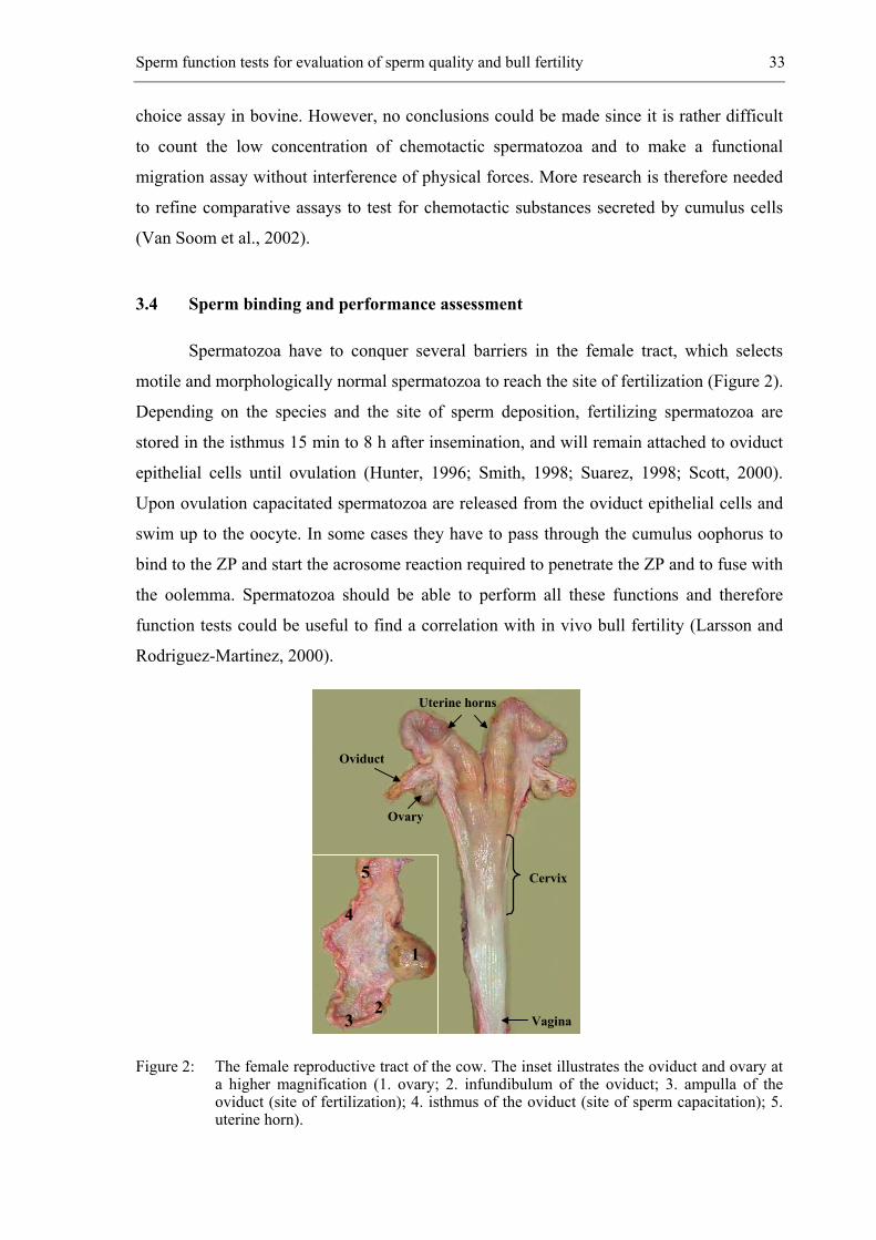
Spermatozoa have to conquer several barriers in the female tract, which selects
motile and morphologically normal spermatozoa to reach the site of fertilization (Figure 2).
Depending on the species and the site of sperm deposition, fertilizing spermatozoa are
stored in the isthmus 15 min to 8 h after insemination, and will remain attached to oviduct
epithelial cells until ovulation (Hunter, 1996; Smith, 1998; Suarez, 1998; Scott, 2000).
Upon ovulation capacitated spermatozoa are released from the oviduct epithelial cells and
swim up to the oocyte. In some cases they have to pass through the cumulus oophorus to
bind to the ZP and start the acrosome reaction required to penetrate the ZP and to fuse with
the oolemma. Spermatozoa should be able to perform all these functions and therefore
function tests could be useful to find a correlation with in vivo bull fertility (Larsson and
Rodriguez-Martinez, 2000).
Figure 2: The female reproductive tract of the cow. The inset illustrates the oviduct and ovary at a higher magnification (1. ovary; 2. infundibulum of the oviduct; 3. ampulla of the oviduct (site of fertilization); 4. isthmus of the oviduct (site of sperm capacitation); 5. uterine horn).
4
Oviduct
5
3
Cervix
Uterine horns
Ovary
Vagina
1
2
34 Chapter 3
3.4.1 Sperm-oviduct binding
In most mammalian species, except for humans, a considerable fraction of
ejaculated spermatozoa is retained with reduced motility in the lower segment of the
oviduct. The high mucus-containing narrow lumen of the oviductal isthmus impedes their
forward progression. Spermatozoa bind strongly to carbohydrate moieties on glycoproteins
or glycolipids on the surface of the oviductal epithelium and are stored there (Suarez,
1998). In cattle, it takes about 6 to 8 hours before a sufficient number of spermatozoa has
reached this sperm reservoir to ensure fertilization (Hunter and Wilmut, 1982). The period
from the onset of oestrus to the completion of ovulation may be as long as 30 h, therefore
the population of spermatozoa capable of fertilization may spend up to 22 h in the isthmus.
When ovulation occurs, some spermatozoa in this reservoir resume high motility and travel
the distance between this storage site and the fertilization site at the oviductal ampulla
within minutes (Hunter, 1993). It is obvious that when a bull is unable to populate the
sperm reservoir with a sufficient number of spermatozoa, this might interfere with its
fertility. It is likely that differences exist among bulls in their capacity to establish a sperm
reservoir after mating or insemination. However, it is difficult to detect these differences
after matings and collection of oviducts, because the number of spermatozoa reported to
reach the oviduct in vivo varied considerably between studies and within experiments
(Parker et al., 1975; Mburu et al., 1996; Suarez et al., 1997). Our laboratory has optimized
an in vitro approach to study sperm binding to oviduct explants, which can be used to
determine whether the capacity to establish a reservoir is indicative of fertility (De Pauw et
al., 2002). By means of this in vitro model it has been shown that the capacity of
spermatozoa to bind to oviduct explants in vitro varies among bulls and evidence was
obtained that sperm binding to oviduct explants is related to in vivo fertility of the donor.
Further refinements to this sperm-oviduct binding assay could be made when the molecular
interaction of this sperm function is better understood. In this respect, measurement of the
expression of sperm surface adhesion molecules by means of antibodies or specific ligands
would be an option. Levels of fucose-binding protein, which can be detected with a fucose-
BSA-FITC conjugate, on the surface of uncapacitated spermatozoa (Ignotz et al., 2001)
may be associated with the capacity of a given sperm sample to bind to oviduct epithelial
cells.
Sperm function tests for evaluation of sperm quality and bull fertility 35
3.4.2 Sperm-zona binding
Before a spermatozoon penetrates the oocyte, it binds selectively to the
homologous ZP, which is an important preliminary step in the fertilization process (Gould
et al., 1983). The ability of spermatozoa to bind to the ZP reflects multiple functions such
as membrane integrity, motility, morphology, acrosomal status and the ability to penetrate
oocyte investments (Burkman et al., 1988; Kaskar et al., 1994; Liu and Baker, 1994).
Two types of sperm-ZP binding assays have been tested for bull spermatozoa: one
using intact (not cleaved) homologous oocytes (zona binding assay, ZBA) (Fazeli et al.,
1993; Zhang et al, 1995), the other using bisected hemizonae (hemizona binding assay,
HZA) (Fazeli et al., 1997; Franken et al., 1997). The latter implies a micro-bisection of the
oocytes into two matching hemizonas; each of one is incubated with spermatozoa from a
test bull and a fertile control bull, respectively (Franken et al., 1997).
In human, it has been shown that homologous test systems based on sperm-ZP
binding can be used to predict the outcome of IVF (Burkman et al., 1988; Liu et al., 1988).
In contrast, no significant relation between the ZBA and AI-fertility of bull semen samples
was found (Fazeli et al., 1997; Zhang et al., 1998). These assays have shown large intra-
and interassay variations in the amount of sperm binding, probably as a consequence of the
quality or maturational status of the oocytes (Mahadevan et al., 1987; Familiari et al.,
1988, Oehninger et al., 1989). However, significant correlations with AI-fertility were only
found when the ZBA was performed with an increased number of intact oocytes and
testing a large population of frozen semen batches from AI bulls (Fazeli et al., 1997, Zhang
et al., 1998). A significant relationship was also found between HZA and in vivo fertility
of bull and stallion spermatozoa (Fazeli et al., 1995; Zhang et al., 1998).
An interesting alternative could be the use of commercially available synthetic
beads coated with recombinant ZP proteins. Preliminary experiments by Whitmarsh et al.
(1996) have revealed significantly higher levels of sperm binding to rhuZP3 beads
compared with controls. Further refinements are needed so that a solid-phase sperm
function test based on rhuZP3-coated beads is likely to be developed in the near future. A
modification to HZA has been reported whereby acrosomal dyes have been incorporated to
provide the indication of the acrosomal status of the bound spermatozoa. Zona pellucida
penetration has also been described as an adjunct to the basic assay (De Jonge, 1999).
36 Chapter 3
3.4.3 In vitro fertilization
In vitro fertility can be assessed by penetration tests (Wheeler and Seidel, 1987;
Tatemoto et al., 1994; Matás et al., 1996; Hay et al., 1997; Gadea et al., 1998). For
penetration of the oocyte in vitro spermatozoa need to be motile, capacitated, and able to
bind to the ZP, to acrosome react, to penetrate the ZP, to fuse with the oocyte and be able
to show decondensation of the nucleus. The oocyte penetration rate is measured as the
number of oocytes with a visible (decondensed) sperm head in the cytoplasm of the oocyte
18 h after onset of fertilization. The limitation of this penetration test is the variability of
oocyte maturation between different oocyte batches, which adds substantially to the
variation between tests. Therefore evaluation can only be performed by comparing results
of bulls on the same group of oocytes (den Daas, 1997). Bulls were found to vary in the
total number of oocytes penetrated as well as in the increase of the penetration rate after
raising the sperm oocyte ratio. However, oocyte penetration rate in vitro cannot be used for
the prediction of fertility results after insemination (den Daas, 1997). This could be
explained by differences between the in vivo and in vitro situation. Firstly, an
undifferentiated population of spermatozoa is presented in vitro whereas in vivo, only a
selected population of spermatozoa is able to reach the fertilization site. Secondly, the
ovulated bovine oocyte encounters only a few free-swimming spermatozoa at the site of
fertilization (sperm:oocyte ratio is close to 1:1) (Hunter, 1993). In most IVF systems,
sperm concentration ranges from 0.5 x 10⁶ to 5 x 10⁶ spermatozoa/ml (Gordon, 1994).
Finally, in vivo the oocyte is denuded during or shortly after ovulation in contrast to most
in vitro systems where spermatozoa have to traverse the COC to reach the ZP (Ward et al.,
2002). A recent study elucidated marked differences in the kinetics of sperm penetration
between sires of different in vitro and in vivo fertility (Ward et al., 2002). Faster
penetrating bulls were ranked higher in field fertility. After only 3 h of gamete co-
incubation a correlation coefficient of 0.827 was found between NRR and oocytes
penetration whereas after 12 h the correlation was only 0.67 and not significant.
A different approach for testing bull spermatozoa in a zona-free oocyte penetration
test was described by Henault et al. (1995). Spermatozoa from bulls differing in in vivo
fertility were labeled with fluorescent dyes with colour patterns specific for a given bull, in
order to assess their relative abilities to penetrate the same zona-free oocytes. The results
showed that spermatozoa from highly fertile bulls were superior to spermatozoa from low-
fertile bulls. The authors concluded that this penetration assay is a reliable method for
Sperm function tests for evaluation of sperm quality and bull fertility 37
detecting differences in relative bull fertility.
The percentage of oocytes developing to the blastocyst stage is a better measure of
fertility than the percentage of oocytes penetrated by spermatozoa because two-thirds of
oocytes fail to reach the blastocyst stage (Gordon, 1994) and would not result in
pregnancy. The endpoints are cleaved oocytes and/or their development to morulae or
blastocysts.
The correlation between IVF and fertility after AI using bull semen has been
studied showing a positive correlation in some studies (Marquant-LeGuienne et al., 1990,
1992; Shamsuddin and Larsson, 1993; Zhang et al., 1997) while no correlation in others
(Schneider et al., 1999). Ward et al. (2001) have shown that kinetics of early cleavage can
be used to distinguish between bulls of high and low fertility. They observed the strongest
correlation at 33 hpi. This is a perfect example of how sufficient refining and running a test
can yield significant correlations between in vitro tests and in vivo fertility. It can serve as
a model for adaptations of other function tests for practical applications.
However, being an expensive and time-consuming procedure, IVF cannot be
routinely used as a screening test. Furthermore, if fertilization fails the problem could be in
any of the preceding steps or may actually be due to the oocytes rather than to the
spermatozoa.
3.5 Conclusions
Several laboratory assays have been developed with the aim of directly analyzing
fertilization processes. However, the complexity of the fertilization process makes
individual assays far less than optimal. The combination of several semen quality
measurements should increase the accuracy of fertility estimates. Combinations of
fluorescent stains can be used to simultaneously assess several different functional
characteristics of spermatozoa, thereby increasing the likelihood of reflecting their
fertilizing capability. However, results obtained from different evaluation techniques can
be contradictory, and consequently misinterpreted. This makes routine semen analysis not
a valid predictor of the ejaculate’s fertilizing ability.
Functional tests, which provide valuable knowledge about selection of spermatozoa
along their route to the oocyte or about sperm-oocyte interaction, undergo continual
development in an effort to accurately estimate fertility. However, until now most of these
complementary tests are only able to exclude the most subfertile bulls. Also confusion and
38 Chapter 3
disagreement still remains concerning the usefulness of some assays and the correct
interpretation of their results.
Refinement, standardization and validation of sperm function assays are therefore
very important to improve their predictive capacity. A better understanding and further
clarification of the complex processes of capacitation, acrosome reaction, sperm-oviduct
binding and sperm-oocyte interactions could lead to the identification of specific molecules
important in these sperm functions.
Sperm function tests for evaluation of sperm quality and bull fertility 39
3.6 References
Aitken RJ, Sutton M, Warner P, Richardson DW. Relationship between the movement
characteristics of human spermatozoa and their ability to penetrate cervical mucus and zona-
free hamster oocytes. J Reprod Fertil 1985; 73: 441-449.
Alexander NJ. Evaluation of male infertility with an in vitro cervical mucus penetration test. Fertil
Steril 1981; 36: 201-208.
Amann RP. Can the fertility potential of a seminal sample be predicted accurately? J Androl 1989;
10: 89-98.
Anilkumar R, Devanathan TG, Pattabiraman SR, Edwin MJ. Correlation between the spermatozoal
characteristics and sperm penetration distance in polyacrylamide gel and bovine cervical
mucus. Theriogenology 2001; 55: 685-691.
Anzar M, He L, Buhr MM, Kroetsch TG, Pauls KP. Sperm apoptosis in fresh and cryopreserved
bull semen detected by flow cytometry and its relationship with fertility. Biol Reprod 2002;
66: 354-360.
Austin CR, Bishop MWH. Role of the rodent acrosome and perforatorium in fertilization. Proc R
Soc 1958; 149: 241-248.
Biljan MM, Taylor CT, Manasse PR, Joughin EC, Kingsland CR, Lewis-Jones DI. Evaluation of
different sperm function tests as screening methods for male fertilization potential-the value
of the sperm migration test. Fertil Steril 1994; 62: 591-598.
Blom E. A one minute live-dead sperm stain by means of eosin-nigrosin. Fertil Steril 1950; 1: 176-
177.
Burkman LJ, Coddington CC, Franken DR, Krugen TF, Rosenwaks Z, Hogen GD. The hemizona
assay (HZA): development of a diagnostic test for the binding of human spermatozoa to the
human hemizona pellucida to predict fertilization potential. Fertil Steril 1988; 49: 688-697.
Chalah T, Brillard JP. Comparison of assessment of fowl sperm viability by eosin-nigrosin and
dual fluorescence (SYBR-14/PI). Theriogenology 1998; 50: 487-493.
Chian RC, Park CK, Sirard MA. Cumulus cells act as a sperm trap during in vitro fertilization of
bovine oocytes. Theriogenology 1996; 45: 258.
Clarke GN, Garrett C, Baker HW. Quantitative sperm mucus penetration: modified formulae for
calculating penetration efficiency. Hum Reprod 1998; 13: 1255-1259.
40 Chapter 3
Cohen-Dayag A, Ralt D, Tur-Kaspa I, Manor M, Makler A, Dor J, Mashiach S, Eisenbach M.
Sequential acquisition of chemotactic responsiveness by human spermatozoa. Biol Reprod
1994; 50: 786-790.
Cortopassi GA, Wong A. Mitochondria in organismal aging and degeneration. Biochim Biophys
Acta 1999; 1410: 183-193.
Cossarizza A, Baccarani-Contri M, Kalashnikova G, Franceschi C. A new method for the
cytofluorimetric analysis of mitochondrial membrane potential using the J-aggregate forming
lipophilic cation 5,5',6,6'-tetrachloro-1,1',3,3'-tetraethylbenzimidazolcarbocyanine iodide
(JC-1). Biochem Biophys Res Commun 1993; 197: 40-45.
Critser JK, Noiles EE. Bioassays of sperm function. Semin Reprod Endocrinol 1993; 11: 1-16.
Cross NL, Morales P, Overstreet JW, Hanson FW. Two simple methods for detecting acrosome-
reacted human sperm. Gamete Res 1986; 15: 213-226.
Cross NL, Meizel S. Methods for evaluating the acrosomal status of mammalian sperm. Biol
Reprod 1989; 41: 635-641.
Cross NL, Watson SK. Assessing acrosomal status of bovine sperm using fluoresceinated lectins.
Theriogenology 1994; 42: 89-98.
Cross NL. Role of cholesterol in sperm capacitation. Biol Reprod 1998; 59: 7-11.
De Jonge CJ. The role of sperm function testing in infertility management. In: Gagnon G (ed). The
male gamete: from basic science to clinical applications. Cache River Press; Vienna 1999:
371-378.
de Lamirande E, Eiley D, Gagnon C. Inverse relationship between the induction of human sperm
capacitation and spontaneous acrosome reaction by various biological fluids and the
superoxide scavenging capacity of these fluids. Int J Androl 1993; 16: 258-266.
de Lamirande E, Jiang H, Zini A, Kodama H, Gagnon C. Reactive oxygen species and sperm
physiology. Rev Reprod 1997; 2: 48-54.
de Leeuw AM, den Daas JH, Woelders H. The fix vital stain method. Simultaneous determination
of viability and acrosomal status of bovine spermatozoa. J Androl 1991; 12: 112-118.
den Daas JHG. Prediction of bovine male fertility. Ph.D. Thesis, University of Wageningen, The
Netherlands, 1997.
De Pauw I, Verberckmoes S, Vanroose G, Van Soom A, de Kruif A. Comparison of eosin/nigrosin
and SYBR-14/PI staining for the assessment of the membrane status of bull sperm. 15e
European Embryo Transfert Association. Lyon 1999: 140.
Sperm function tests for evaluation of sperm quality and bull fertility 41
De Pauw I, Van Soom A, Laevens H, Verberckmoes S, de Kruif A. Sperm binding to epithelial
oviduct explants in bulls with different non-return rates investigated with a new in vitro
model. Biol Reprod 2002; 67: 1073-1079.
Didier M, Bursztajn S, Adamec E, Passani L, Nixon RA, Coyle JT, Wei JY, Berman SA. DNA
strand breaks induced by sustained glutamate excitotoxicity in primary neuronal cultures. J
Neurosci 1996; 16: 2238-2250.
Drobnis EZ, Katz DF. Videomicroscopy of mammalian fertilization. In: Wassarman P (ed).
Handbook on Mammalian Gamete Biology. CRC Press; New York 1990: 269-300.
Earnshaw WC. Nuclear changes in apoptosis. Curr Opin Cell Biol 1995; 7: 337-343.
Eisenbach M. Sperm chemotaxis. Rev Reprod 1999; 4: 56-66.
Familiari G, Nottola SA, Micara G, Aragona C, Motta PM. Is the sperm-binding capability of the
zona pellucida linked to its surface structure? A scanning electron microscopic study of
human in vitro fertilization. J In Vitro Fert Embryo Transf 1988; 5: 134-143.
Farrell PB, Presicce GA, Brockett CC, Foote RH. Quantification of bull sperm characteristics
measured by computer-assisted sperm analysis (CASA) and the relationship to fertility.
Theriogenology 1998; 49: 871-879.
Fazeli AR, Steenweg W, Bevers MM, de Loos FA, van den Broek J, Colenbrander B. Development
of a sperm zona pellucida binding assay for bull semen. Vet Rec 1993; 132: 14-16.
Fazeli AR, Steenweg W, Bevers MM, van den Broek J, Bracher V, Parlevliet J, Colenbrander B.
Relation between stallion sperm binding to homologous hemizonae and fertility.
Theriogenology 1995; 44: 751-760.
Fazeli A, Zhang BR, Steenweg W, Larsson B, Bevers MM, van den Broek J, Rodriguez-Martinez
H, Colenbrander B. Relationship between sperm-zona pellucida binding assays and the 56-
day nonreturn rate of cattle inseminated with frozen-thawed semen. Theriogenology 1997;
48: 853-863.
Franken DR, Bastiaan HS, Kidson A, Wranz P, Habenicht UF. Zona pellucida mediated acrosome
reaction and sperm morphology. Andrologia 1997; 29: 311-317.
Fraser LR. Cellular biology of capacitation and the acrosome reaction. Hum Reprod 1995; 10
Suppl 1: 22-30.
Gadea J, Matas C, Lucas X. Prediction of porcine semen fertility by homologous in vitro
penetration (hIVP) assay. Anim Reprod Sci 1998; 54: 95-108.
Garner DL, Pinkel D, Johnson LA, Pace MM. Assessment of spermatozoal function using dual
fluorescent staining and flow cytometric analyses. Biol Reprod 1986; 34: 127-138.
42 Chapter 3
Garner DL, Johnson LA, Yue ST, Roth BL, Haugland RP. Dual DNA staining assessment of
bovine sperm viability using SYBR-14 and propidium iodide. J Androl 1994; 15: 620-629.
Garner DL, Johnson LA. Viability assessment of mammalian sperm using SYBR-14 and propidium
iodide. Biol Reprod 1995; 53: 276-284.
Garner DL, Dobrinsky JR, Welch GR, Johnson LA. Porcine sperm viability, oocyte fertilization
and embryo development after staining spermatozoa with SYBR-14. Theriogenology 1996;
45: 1103-1113.
Garner DL, Thomas CA, Joerg HW, DeJarnette JM, Marshall CE. Fluorometric assessments of
mitochondrial function and viability in cryopreserved bovine spermatozoa. Biol Reprod
1997; 57: 1401-1406.
Giojalas LC, Rovasio RA. Mouse spermatozoa modify their motility parameters and chemotactic
response to factors from the oocyte microenvironment. Int J Androl 1998; 21: 201-206.
Godard T, Deslandes E, Lebailly P, Vigreux C, Sichel F, Poul JM, Gauduchon P. Early detection of
staurosporine-induced apoptosis by comet and annexin V assays. Histochem Cell Biol 1999;
112: 155-161.
Gordon I. Laboratory production of cattle embryos. In: Biotechnology in agriculture. Wallingford:
CAB International, 1994.
Gould JE, Overstreet JW, Yanagimachi H, Yanagimachi R, Katz DF, Hanson FW. What functions
of the sperm cell are measured by in vitro fertilization of zona-free hamster eggs? Fertil
Steril 1983; 40: 344-352.
Graham JK, Kunze E, Hammerstedt RH. Analysis of sperm cell viability, acrosomal integrity, and
mitochondrial function using flow cytometry. Biol Reprod 1990; 43: 55-64.
Hancock JL. The morphology of boar spermatozoa. J Roy Micr Soc 1957; 76: 84-97.
Harrison RA, Vickers SE. Use of fluorescent probes to assess membrane integrity in mammalian
spermatozoa. J Reprod Fertil 1990; 88: 343-352.
Hay MA, King WA, Gartley CJ, Leibo SP, Goodrowe KL. Effects of cooling, freezing and glycerol
on penetration of oocytes by spermatozoa in dogs. J Reprod Fertil Suppl 1997; 51: 99-108.
Henault MA, Killian GJ, Kavanaugh JF, Griel LC Jr. Effect of accessory sex gland fluid from bulls
of differing fertilities on the ability of cauda epididymal sperm to penetrate zona-free bovine
oocytes. Biol Reprod 1995; 52: 390-397.
Holt WV. Basic aspects of frozen storage of semen. Anim Reprod Sci 2000; 62: 3-22.
Sperm function tests for evaluation of sperm quality and bull fertility 43
Hunter RHF, Wilmut I. The rate of functional sperm transport into the oviducts of mated cows.
Anim Reprod Sci 1982; 5: 167-173.
Hunter RHF. The timing of capacitation in mammalian spermatozoa - a reinterpretation. Res
Reprod 1987; 19: 3-4.
Hunter RHF. Sperm:egg ratios and putative molecular signals to modulate gamete interactions in
polytocous mammals. Mol Reprod Dev 1993; 35: 324-327.
Hunter RH. Ovarian control of very low sperm/egg ratios at the commencement of mammalian
fertilisation to avoid polyspermy. Mol Reprod Dev 1996; 44: 417-422.
Ignotz GG, Lo MC, Perez CL, Gwathmey TM, Suarez SS. Characterization of a fucose-binding
protein from bull sperm and seminal plasma that may be responsible for formation of the
oviductal sperm reservoir. Biol Reprod 2001; 64: 1806-11.
Januskauskas A, Söderquist L, Häärd MG, Häärd M Ch, Johannisson A, Rodriguez-Martinez H.
Effect of cooling rates on post-thaw sperm motility, membrane integrity, capacitation status
and fertility of dairy bull semen used for artificial insemination in Sweden. Theriogenology
1999; 52: 641-658.
Januskauskas A, Johannisson A, Soderquist L, Rodriguez-Martinez H. Assessment of sperm
characteristics post-thaw and response to calcium ionophore in relation to fertility in
Swedish dairy AI bulls. Theriogenology 2000; 53: 859-875.
Jeulin C, Soumah A, Jouannet P. Morphological factors influencing the penetration of human
sperm into cervical mucus in vitro. Int J Androl 1985; 8: 215-223.
Johnson LA, Maxwell WMC, Dobrinsky JR, Welch GR. Staining sperm for viability assessment.
Reprod Domest Anim 1996; 31: 37-47.
Jouannet P, Feneux D. Sperm analysis. Ann Biol Clin (Paris) 1987; 45: 335-339.
Kaskar K, Franken DR, van der Horst G, Oehninger S, Kruger TF, Hodgen GD. The relationship
between morphology, motility and zona pellucida binding potential of human spermatozoa.
Andrologia 1994; 26: 1-4.
Kummerfeld HL, Vosburgh JK, Lorton SP, Foote RH. Influence of bovine cervical mucus samples
and storage conditions on sperm migration in vitro. Fertil Steril 1981; 35: 218-221.
Langner M, Hui SW. Merocyanine interaction with phosphatidylcholine bilayers. Biochim Biophys
Acta 1993; 1149: 175-179.
Larsson B, Rodriguez-Martinez H. Can we use in vitro fertilization tests to predict semen fertility?
Anim Reprod Sci 2000; 60-61: 327-336.
44 Chapter 3
Lee MA, Trucco GS, Bechtol KB, Wummer N, Kopf GS, Blasco L, Storey BT. Capacitation and
acrosome reactions in human spermatozoa monitored by a chlortetracycline fluorescence
assay. Fertil Steril 1987; 48: 649-658.
Liu DY, Lopata A, Johnston WI, Baker HW. A human sperm-zona pellucida binding test using
oocytes that failed to fertilize in vitro. Fertil Steril 1988; 50: 782-788.
Liu DY, Baker HWG. A new test for the assessment of sperm-zona pellucida penetration:
relationship with results of other sperm tests and fertilization in vitro. Hum Reprod 1994; 9:
489-496.
Mahadevan MM, Trounson AO, Wood C, Leeton JF. Effect of oocyte quality and sperm
characteristics on the number of spermatozoa bound to the zona pellucida of human oocytes
inseminated in vitro. J In Vitro Fert Embryo Transf 1987; 4: 223-227.
Makler A, Reichler A, Stoller J, Feigin PD. A new model for investigating in real-time the
existence of chemotaxis in human spermatozoa. Fertil Steril 1992; 57: 1066-1074.
Makler A, Stoller J, Reichler A, Blumenfeld Z, Yoffe N. Inability of human sperm to change their
orientation in response to external chemical stimuli. Fertil Steril 1995; 63: 1077-1082.
Malmgren L. Assessing the quality of raw semen: a review. Theriogenology 1997; 48: 523-530.
Margalit I, Rubinstein S, Breitbart H. A novel method for evaluating the acrosomal status of
mammalian spermatozoa. Arch Androl 1997; 39: 87-99.
Marquant-LeGuienne B, Humblot P, Thibault M, Thibault C. Evaluation of bull semen fertility by
homologous in vitro fertilization tests. Reprod Nutr Dev 1990; 30: 259-266.
Marquant-LeGuienne B, Humblot P, Guillon N, Gerard O, Thibier M. In vitro fertilization as a tool
to evaluate fertility in the bovine. Proc 12th Int Congr Anim Reprod, The Hague 1992; 2:
662-664.
Martin SJ, Reutelingsperger CP, McGahon AJ, Rader JA, van Schie RC, LaFace DM, Green DR.
Early redistribution of plasma membrane phosphatidylserine is a general feature of apoptosis
regardless of the initiating stimulus: inhibition by overexpression of Bcl-2 and Abl. J Exp
Med 1995; 182: 1545-1556.
Matás C, Martinínez E, Vázquez JM, Roca J, Gadea J. In vitro penetration assay of boar sperm
fertility: effect of various factors on the penetrability of immature pig oocytes.
Theriogenology 1996; 46: 503-513.
Maxwell WM, Johnson LA. Chlortetracycline analysis of boar spermatozoa after incubation, flow
cytometric sorting, cooling, or cryopreservation. Mol Reprod Dev 1997; 46: 408-418.
Sperm function tests for evaluation of sperm quality and bull fertility 45
Mburu JN, Einarsson S, Lundeheim N, Rodriguez-Martinez H. Distribution, number and
membrane integrity of spermatozoa in the pig oviduct in relation to spontaneous ovulation.
Anim Reprod Sci 1996; 45: 109-121.
Mixner JP, Saroff J. Interference by glycerol with differential staining of bull spermatozoa. J Dairy
Sci 1954; 37: 652.
Mortimer D, Curtis EF, Miller RG. Specific labelling by peanut agglutinin of the outer acrosomal
membrane of the human spermatozoon. J Reprod Fertil 1987; 81: 127-135.
Moyes CD, Battersby BJ, Leary SC. Regulation of muscle mitochondrial design. J Exp Biol 1998;
201: 299-307.
Oehninger S, Scott RT, Coddington CC, Franken DR, Acosta AA, Hodgen GD. Validation of the
hemizona assay in a monkey model: influence of oocyte maturational stages. Fertil Steril
1989; 51: 881-885.
Oehninger S, Stecker JF, Acosta AA. Male infertility: the impact of assisted reproductive
technologies. Curr Opin Obstet Gynecol 1992; 4: 185-196.
Oko RJ, Costerton JW, Coulter GH. An ultrastructural study of the head region of bovine
spermatozoa. Can J Zool 1976; 54: 1326-1340.
Oliveira RG, Tomasi L, Rovasio RA, Giojalas LC. Follicular and oviductal fluids enhance velocity
and induce chemotactic response in mouse spermatozoa. J Reprod Fertil 1999; 115: 23-27.
Oosterhuis GJ, Mulder AB, Kalsbeek-Batenburg E, Lambalk CB, Schoemaker J, Vermes I.
Measuring apoptosis in human spermatozoa: a biological assay for semen quality? Fertil
Steril 2000; 74: 245-250.
Parker WG, Sullivan JJ, First NL. Sperm transport and distribution in the mare. J Reprod Fertil
Suppl 1975; 23: 63-66.
Parrish JJ, Susko-Parrish JL, Graham JK. In vitro capacitation of bovine spermatozoa: role of
intracellular calcium. Theriogenology 1999; 51: 461-472.
Ralt D, Manor M, Cohen-Dayag A, Tur-Kaspa I, Ben-Shlomo I, Makler A, Yuli I, Dor J, Blumberg
S, Mashiach S. Chemotaxis and chemokinesis of human spermatozoa to follicular factors.
Biol Reprod 1994; 50: 774-785.
Rathi R, Colenbrander B, Bevers MM, Gadella BM. Evaluation of in vitro capacitation of stallion
spermatozoa. Biol Reprod 2001; 65: 462-470.
Reers M, Smith TW, Chen LB. J-aggregate formation of a carbocyanine as a quantitative
fluorescent indicator of membrane potential. Biochemistry 1991; 30: 4480-4486.
46 Chapter 3
Rijsselaere T, Van Soom A, Maes D, de Kruif A. Effect of centrifugation on in vitro survival of
fresh diluted canine spermatozoa. Theriogenology 2002; 57: 1669-1681.
Sakkas D, Mariethoz E, Manicardi G, Bizzaro D, Bianchi PG, Bianchi U. Origin of DNA damage
in ejaculated human spermatozoa. Rev Reprod 1999; 4: 31-37.
Schneider CS, Ellington JE, Wright RW Jr. Relationship between bull field fertility and in vitro
embryo production using sperm preparation methods with and without somatic cell co-
culture. Theriogenology 1999; 51: 1085-1098.
Scott MA. A glimpse at sperm function in vivo: sperm transport and epithelial interaction in the
female reproductive tract. Anim Reprod Sci 2000; 60-61: 337-348.
Shamsuddin M, Larsson B. In vitro development of bovine embryos after fertilization using semen
from different donors. Reprod Domest Anim 1993; 28: 77-84.
Shapiro HM. Membrane potential estimation by flow cytometry. Methods 2000; 21: 271-279.
Shen HM, Dai J, Chia SE, Lim A, Ong CN. Detection of apoptotic alterations in sperm in subfertile
patients and their correlations with sperm quality. Hum Reprod 2002; 17: 1266-1273.
Smith TT. The modulation of sperm function by the oviductal epithelium. Biol Reprod 1998; 58:
1102-1104.
Soderquist L, Rodriguez-Martinez H, Janson L. Post-thaw motility, ATP content and cytochrome C
oxidase activity of AI bull spermatozoa in relation to fertility. Zentralbl Veterinarmed A
1991; 38: 165-174.
Stalhammar EM, Janson L, Philipsson J. The impact of sperm motility on non-return rate in
preselected dairy bulls. Reprod Nutr Dev 1994; 34: 37-45.
Storey BT, Lee MA, Muller C, Ward CR, Wirtshafter DG. Binding of mouse spermatozoa to the
zonae pellucidae of mouse eggs in cumulus: evidence that the acrosomes remain
substantially intact. Biol Reprod 1984; 31: 1119-1128.
Suarez SS, Brockman K, Lefebvre R. Distribution of mucus and sperm in bovine oviducts after
artificial insemination: the physical environment of the oviductal sperm reservoir. Biol
Reprod 1997; 56: 447-453.
Suarez SS. The oviductal sperm reservoir in mammals: mechanisms of formation. Biol Reprod
1998; 58: 1105-1107.
Talbot P. Sperm penetration through oocyte investments in mammals. Am J Anat 1985; 174: 331-
346.
Sperm function tests for evaluation of sperm quality and bull fertility 47
Tatemoto H, Horiuchi T, Maeda T, Terada T, Tsutsumi Y. Penetration by bull spermatozoa into the
zona pellucida of dead bovine oocytes recovered from frozen-thawed ovaries.
Theriogenology 1994; 42: 465-474.
Thomas CA, Garner DL, DeJarnette JM, Marshall CE. Effect of cryopreservation of bovine sperm
organelle function and viability as determined by flow cytometry. Biol Reprod 1998; 58:
786-793.
Thundathil J, Gil J, Januskauskas A, Larsson B, Soderquist L, Mapletoft R, Rodriguez-Martinez H.
Relationship between the proportion of capacitated spermatozoa present in frozen-thawed
bull semen and fertility with artificial insemination. Int J Androl 1999; 22: 366-373.
Topper EK, Killian GJ, Way A, Engel B, Woelders H. Influence of capacitation and fluids from the
male and female genital tract on the zona binding ability of bull spermatozoa. J Reprod Fertil
1999; 115: 175-183.
Tsien RY. Fluorescent indicators of ion concentration. In: Taylor Dl, Wang Y-L (eds). Methods in
Cell Biology. Academic Press Inc; New York 1989: 142-144.
Van Soom A, Tanghe S, De Pauw I, Maes D, De Kruif A. Function of the cumulus oophorus
before and during mammalian fertilization. Reprod Domest Anim 2002; 37: 144-151.
Verberckmoes S, Van Soom A, De Pauw I, Dewulf J, de Kruif A. Migration of bovine spermatozoa
in a synthetic medium and its relation to in vivo bull fertility. Theriogenology 2002; 58:
1027-1037.
Verkleij AJ, De Kruijff B, Gerritsen WF, Demel RA, Van Deenen LL, Ververgaert PH. Freeze-etch
electron microscopy of erythrocytes, Acholeplasma laidlawii cells and liposomal membranes
after the action of filipin and amphotericin B. Biochim Biophys Acta 1973; 291: 577-581.
Vermes I, Haanen C, Steffens-Nakken H, Reutelingsperger C. A novel assay for apoptosis. Flow
cytometric detection of phosphatidylserine expression on early apoptotic cells using
fluorescein labelled Annexin V J Immunol Methods 1995; 184: 39-51.
Verstegen J, Iguer-Ouada M, Onclin K. Computer assisted semen analyzers in andrology research
and veterinary practice. Theriogenology 2002; 57: 149-179.
Visconti PE, Galantino-Homer H, Moore GD, Bailey JL, Ning X, Fornes M, Kopf GS. The
molecular basis of sperm capacitation. J Androl 1998; 19: 242-248.
Visconti PE, Ning XP, Fornes MW, Alvarez JG, Stein P, Connors SA, Kopf GS. Cholesterol
efflux-mediated signal transduction in mammalian sperm: cholesterol release signals an
increase in protein tyrosine phosphorylation during mouse sperm capacitation. Dev Biol
1999; 214: 429-443.
48 Chapter 3
Ward FA, Rizos D, Corridan D, Quinn K, Boland MP, Lonergan P. Paternal influence on the time
of the first embryonic cleavage post insemination and the implications for subsequent bovine
embryo development in vitro and fertility in vivo. Mol Reprod Dev 2001; 60: 47-55.
Ward FA, Enright B, Rizos D, Boland M, Lonergan P. Optimization of in vitro bovine embryo
production: effect of duration of maturation, length of gamete co-incubation, sperm
concentration and sire. Theriogenology 2002; 57: 2105-2117.
Wheeler MB, Seidel GE Jr. Zona pellucida penetration assay for capacitation of bovine sperm.
Gamete Res 1987; 18: 237-250.
Whitfield CH, Parkinson TJ. Assessment of the fertilizing potential of frozen bovine spermatozoa
by in vitro induction of acrosome reactions with calcium ionophore (A23187).
Theriogenology 1995; 44: 413-422.
Whitmarsh AJ, Woolnough MJ, Moore HD, Hornby DP, Barratt CL. Biological activity of
recombinant human ZP3 produced in vitro: potential for a sperm function test. Mol Hum
Reprod 1996; 2: 911-919.
Williamson P, Mattocks K, Schlegel RA. Merocyanine 540, a fluorescent probe sensitive to lipid
packing. Biochim Biophys Acta 1983; 732: 387-393.
Wyllie AH, Kerr JF, Currie AR. Cell death: the significance of apoptosis. Int Rev Cytol 1980; 68:
251-306.
Xu JP, Li X, Mori E, Guo MW, Matsuda I, Takaichi H, Amano T, Mori T. Expression of Fas-Fas
ligand in murine testis. Am J Reprod Immunol 1999; 42: 81-88.
Yanagimachi R. Mammalian fertilization. In: Knobil E, Neill J (eds). Physiology of Reproduction.
Raven press; New York 1994: 189-317.
Zhang BR, Larsson B, Rodriguez-Martinez H. Influence of batches of bovine oocytes on the
outcome of an intact zona pellucida binding assay and in vitro fertilization. Int J Androl
1995; 18: 213-220.
Zhang BR, Larsson B, Lundeheim N, Rodriguez-Martinez H. Relationship between embryo
development in vitro and 56-day nonreturn rates of cows inseminated with frozen-thawed
semen from dairy bulls. Theriogenology 1997; 48: 221-231.
Zhang BR, Larsson B, Lundeheim N, Rodriguez-Martinez H. Sperm characteristics and zona
pellucida binding in relation to field fertility of frozen-thawed semen from dairy AI bulls.
Int J Androl 1998; 21: 207-216.
__________________________________________________ 4.
IN VITRO SURVIVAL OF BOVINE SPERMATOZOA STORED
AT ROOM TEMPERATURE UNDER EPIDIDYMAL CONDITIONS
Theriogenology 2003; 59: 1093-1107
De Pauw I., Van Soom A., Mintiens K., Verberckmoes S. and de Kruif A.
50 Chapter 4
Abstract
In this study, environmental conditions mimicking those prevailing in the
epididymis were used for storing ejaculated bull spermatozoa in vitro during 4 days at
room temperature (RT). These conditions were low pH, high osmolarity, high sperm
concentration and low oxygen tension. Hepes-TALP was used as basic storage medium.
Ejaculated spermatozoa were stored at a concentration of 10 x 106 spermatozoa/ml in
Hepes-TALP of different pH (4, 5, 6, 7 or 8) and osmolarity (100, 300, 400, 500, 600 or
800 mOsm) and under different atmospheric conditions (nitrogen-gassed or aerobic).
Spermatozoa were also stored undiluted or at different concentrations: 10 x 106, 100 x 106,
500 x 106 or 1 x 10⁹ spermatozoa/ml. Sperm parameters such as membrane integrity,
motility, mitochondrial membrane potential or DNA fragmentation were used to assess
semen quality after storage. Adjustment of the pH of Hepes-TALP to pH 6 yielded
significantly better results than storage at all other pH values. Isotonic Hepes-TALP had a
less detrimental effect on spermatozoa than the hypo- and hyperosmotic versions. No
differences in sperm parameters were observed when spermatozoa were incubated under
aerobic or under nitrogen-gassed storage conditions. Optimal sperm concentration in vitro
is 10 x 106 spermatozoa/ml. This is in contrast with the in vivo situation, where
spermatozoa are stored at a high concentration. Better results at high sperm concentrations
were obtained when spermatozoa were diluted for less than 5 minutes in Triladyl®-diluent
supplemented with egg yolk and glycerol immediately after ejaculation.
In vitro survival of bovine spermatozoa stored under epididymal conditions 51
Introduction
Ejaculated spermatozoa diluted in vitro remains viable for a limited period of time.
Much research has been done, especially for bovine semen, to understand the effect of
different factors and to design a useful storage medium to enhance sperm survival in a non-
frozen state. Despite the development of numerous diluents (reviewed by Vishwanath and
Shannon, 2000) the fertile lifespan of spermatozoa is maintained for only 3-5 days when
stored in a liquid environment at high dilutions at ambient temperatures (Vishwanath and
Shannon, 1997). In contrast to their limited lifespan in vitro, spermatozoa can remain
viable for several weeks when stored in the cauda of the epididymis (Setchell et al., 1993).
During epididymal transit, mammalian spermatozoa acquire the capacity for motility and
for fertilization. When they reach the caudal part of the epididymis, they must be
maintained in a quiescent state awaiting ejaculation, presumably to conserve energy
required for the fertilization process (Mann and Lutwak-Mann, 1981; Zaneveld and
Chatterton, 1982). In this quiescent state, bovine spermatozoa are immotile and
metabolically inactive (Cascieri et al., 1976). Spermatozoa are stored for a relatively long
period in this state, during which they retain their capacity for motility and the fertilization
process much longer than samples kept in vitro at the same temperature (Jones and
Murdoch, 1996). It is obvious that the environment in the cauda epididymidis favours the
lifespan of the spermatozoa. The cauda epididymidis is characterized by low pH (Acott and
Carr, 1984; Carr et al., 1985) and hyperosmotic pressure of the plasma (Liu and Foote,
1998), spermatozoa are stored at high concentrations (Cascieri et al., 1976; Amann et al.,
1976) and at low oxygen tension (Free et al., 1976).
Until now it has been very difficult to mimic these epididymal conditions in vitro,
although some reports have shown that spermatozoa can exhibit some fertilizing ability
after being stored for a few days in cooled epididymides of different species (Kikuchi et
al., 1998; Browne et al., 2001; Sankai et al., 2001). Unfortunately, this is not a practical
alternative for sperm storage, so we have chosen to investigate the influence of different
epididymal conditions upon sperm survival in diluent at RT. Since it is known that sperm
fertility declines much faster than sperm motility (Vishwanath and Shannon, 1997, 2000),
less conventional sperm characteristics were also examined such as mitochondrial
membrane potential and the prevalence of DNA fragmentation in stored spermatozoa.
Fragmentation of genomic DNA can be detected by means of terminal deoxynucleotid
transferase mediated dUTP nick end labeling (TUNEL), and mitochondrial membrane
52 Chapter 4
potential, which is related to the presence of a low mitochondrial transmembrane potential
which can be assessed by 5,5',6,6'-tetrachloro-1,1',3,3'-tetraethylbenzimidazolyl-
carbocyanine iodide (JC-1) staining. These characteristics of apoptosis might be related to
sperm fertility, as it has been shown in human semen samples (Sun et al., 1997; Donnelly
et al., 2000).
In our study, the simple salt solution Hepes-TALP was used as the principal storage
medium. The effect of pH, osmolarity, storage atmosphere and sperm concentration on
membrane integrity, motility and mitochondrial function of bull spermatozoa stored for
3 or 4 days at RT was examined. Sperm nuclear DNA fragmentation was determined in
order to quantify possible apoptosis, which might occur during prolonged storage of
spermatozoa at RT. The sperm collection method was adapted in order to “coat”
spermatozoa with egg yolk-glycerol to improve subsequent sperm survival.
Materials and Methods
Collection and preparation of semen
Semen was collected from a 2-year-old Red Pied bull using an artificial vagina at 1-
week intervals. Immediately after collection, spermatozoa were separated from the seminal
plasma by centrifugation (720 x g, 10 min) at RT. The sperm concentration was
determined using a Bürker chamber and subsequently diluted to a concentration of 10 x 106
spermatozoa/ml in Hepes-buffered TALP media of different pH or osmolarity, except for
the experiment where the effect of sperm concentration itself was examined. Spermatozoa
were stored at RT in darkness under different incubation conditions according to the
experiment (72 or 96 h). After storage of the spermatozoa under the different conditions,
samples were taken and evaluated microscopically or by flow cytometric analysis for
functional parameters such as membrane integrity, motility, mitochondrial membrane
potential, morphology and DNA fragmentation.
Media
Chemicals and media were obtained from Sigma (Bornem, Belgium) and
LifeTechnologies, Gibco BRL® products (Merelbeke, Belgium).
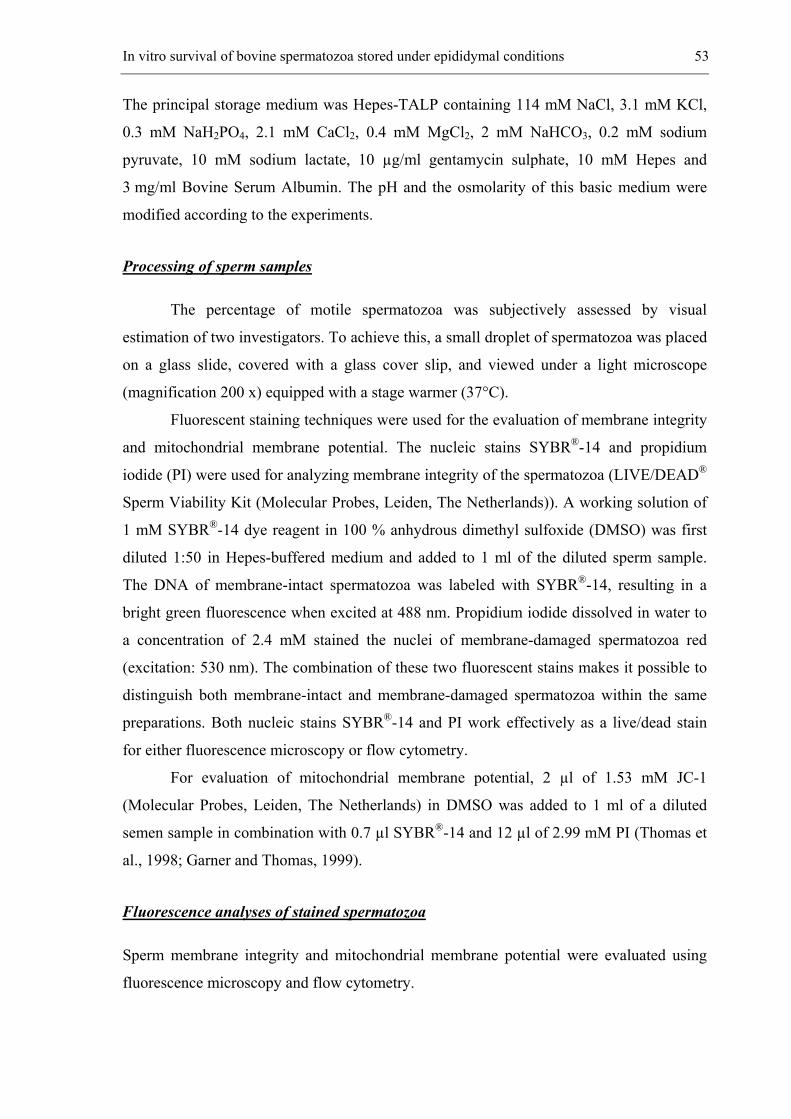
In vitro survival of bovine spermatozoa stored under epididymal conditions 53
The principal storage medium was Hepes-TALP containing 114 mM NaCl, 3.1 mM KCl,
0.3 mM NaH2PO4, 2.1 mM CaCl2, 0.4 mM MgCl2, 2 mM NaHCO3, 0.2 mM sodium
pyruvate, 10 mM sodium lactate, 10 µg/ml gentamycin sulphate, 10 mM Hepes and
3 mg/ml Bovine Serum Albumin. The pH and the osmolarity of this basic medium were
modified according to the experiments.
Processing of sperm samples
The percentage of motile spermatozoa was subjectively assessed by visual
estimation of two investigators. To achieve this, a small droplet of spermatozoa was placed
on a glass slide, covered with a glass cover slip, and viewed under a light microscope
(magnification 200 x) equipped with a stage warmer (37°C).
Fluorescent staining techniques were used for the evaluation of membrane integrity
and mitochondrial membrane potential. The nucleic stains SYBR®-14 and propidium
iodide (PI) were used for analyzing membrane integrity of the spermatozoa (LIVE/DEAD®
Sperm Viability Kit (Molecular Probes, Leiden, The Netherlands)). A working solution of
1 mM SYBR®-14 dye reagent in 100 % anhydrous dimethyl sulfoxide (DMSO) was first
diluted 1:50 in Hepes-buffered medium and added to 1 ml of the diluted sperm sample.
The DNA of membrane-intact spermatozoa was labeled with SYBR®-14, resulting in a
bright green fluorescence when excited at 488 nm. Propidium iodide dissolved in water to
a concentration of 2.4 mM stained the nuclei of membrane-damaged spermatozoa red
(excitation: 530 nm). The combination of these two fluorescent stains makes it possible to
distinguish both membrane-intact and membrane-damaged spermatozoa within the same
preparations. Both nucleic stains SYBR®-14 and PI work effectively as a live/dead stain
for either fluorescence microscopy or flow cytometry.
For evaluation of mitochondrial membrane potential, 2 µl of 1.53 mM JC-1
(Molecular Probes, Leiden, The Netherlands) in DMSO was added to 1 ml of a diluted
semen sample in combination with 0.7 µl SYBR®-14 and 12 µl of 2.99 mM PI (Thomas et
al., 1998; Garner and Thomas, 1999).
Fluorescence analyses of stained spermatozoa
Sperm membrane integrity and mitochondrial membrane potential were evaluated using
fluorescence microscopy and flow cytometry.
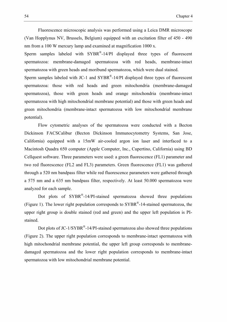
54 Chapter 4
Fluorescence microscopic analysis was performed using a Leica DMR microscope
(Van Hopplynus NV, Brussels, Belgium) equipped with an excitation filter of 450 - 490
nm from a 100 W mercury lamp and examined at magnification 1000 x.
Sperm samples labeled with SYBR®-14/PI displayed three types of fluorescent
spermatozoa: membrane-damaged spermatozoa with red heads, membrane-intact
spermatozoa with green heads and moribund spermatozoa, which were dual stained.
Sperm samples labeled with JC-1 and SYBR®-14/PI displayed three types of fluorescent
spermatozoa: those with red heads and green mitochondria (membrane-damaged
spermatozoa), those with green heads and orange mitochondria (membrane-intact
spermatozoa with high mitochondrial membrane potential) and those with green heads and
green mitochondria (membrane-intact spermatozoa with low mitochondrial membrane
potential).
Flow cytometric analyses of the spermatozoa were conducted with a Becton
Dickinson FACSCalibur (Becton Dickinson Immunocytometry Systems, San Jose,
California) equipped with a 15mW air-cooled argon ion laser and interfaced to a
Macintosh Quadra 650 computer (Apple Computer, Inc., Cupertino, California) using BD
Cellquest software. Three parameters were used: a green fluorescence (FL1) parameter and
two red fluorescence (FL2 and FL3) parameters. Green fluorescence (FL1) was gathered
through a 520 nm bandpass filter while red fluorescence parameters were gathered through
a 575 nm and a 635 nm bandpass filter, respectively. At least 50.000 spermatozoa were
analyzed for each sample.
Dot plots of SYBR®-14/PI-stained spermatozoa showed three populations
(Figure 1). The lower right population corresponds to SYBR®-14-stained spermatozoa, the
upper right group is double stained (red and green) and the upper left population is PI-
stained.
Dot plots of JC-1/SYBR®-14/PI-stained spermatozoa also showed three populations
(Figure 2). The upper right population corresponds to membrane-intact spermatozoa with
high mitochondrial membrane potential, the upper left group corresponds to membrane-
damaged spermatozoa and the lower right population corresponds to membrane-intact
spermatozoa with low mitochondrial membrane potential.
In vitro survival of bovine spermatozoa stored under epididymal conditions 55
Figure 1. Dot plot of SYBR®-14/PI stained spermatozoa showing three distinct sperm populations: membrane-damaged spermatozoa (red-stained: PI) (upper left), membrane-intact spermatozoa (green-stained: SYBR®-14) (lower right), and moribund spermatozoa stained both red and green (doubly-stained) (upper right).
Figure 2. Dot plot of JC-1/SYBR®-14/PI- stained spermatozoa, showing three populations: membrane-intact spermatozoa with high mitochondrial membrane potential (upper right) or low mitochondrial membrane potential (lower right) and membrane-damaged spermatozoa (upper left).
Measurement of sperm DNA fragmentation using the TUNEL assay
DNA fragmentation was detected with a TUNEL assay using the In-Situ Cell
Detection Kit from Boehringer Mannheim (Mannheim, Germany).
Spermatozoa were washed twice in 1 ml PVP solution (1 mg/ml polyvinyl-
pyrrolidone (PVP) in phosphate-buffered saline (PBS)) by centrifugation at RT (720 x g,
5 min). The final pellet was resuspended in 1 ml PVP solution and 10 µl of 10 x 106
spermatozoa/ml was smeared onto a polylysine-coated glass microscope slide and air
dried. The slides were washed in PVP solution (5 min) and fixed with 4%
paraformaldehyde in PVP solution (pH 7.4) at RT for 30 min. This was followed by
permeabilization with a solution of 0.1% Triton X-100 and 0.1% sodium citrate for 2 min
on ice. Slides were rinsed twice with PVP solution and air dried. Diluted terminal
deoxynucleotidyl transferase (TdT) (25 µl) was mixed with 225 µl nucleotide mixture.
56 Chapter 4
Twenty-five microliter of this TUNEL reaction mixture was added to the spermatozoa on
each slide. The samples were incubated in a humidified chamber at 37°C for 60 min in
darkness.
Negative (nucleotide solution without TdT), and positive (incubation with 1 mg
Dnase I/ml (Pharmacia LKB Biotech) for 10 min at RT before staining with TUNEL
reaction mixture) controls were included in all experiments. The slides were rinsed three
times with PVP solution and incubated with 25 µl Hoechst 33342 (1µl/ml PVP solution) at
RT for 5 min. After washing twice in PVP solution, 100 spermatozoa in different
microscopic fields were analyzed by means of fluorescence microscopy. Hoechst stained
spermatozoa were first counted (bright blue fluorescence) under ultraviolet light and the
percentage of FITC-labelled spermatozoa with fragmented DNA (intense green nuclear
fluorescence) was determined by means of Leica DMR epifluorescence microscopy.
Experimental design
Experiment 1: Evaluation of the effect of pH
In the first experiment, the effect of different pH-values of Hepes-TALP on
motility, membrane integrity and mitochondrial membrane potential of stored spermatozoa
was tested. The pH of Hepes-TALP was measured with a pH-meter (WTW, Weilheim,
Germany), which was first calibrated with a pH 7 and a pH 4 standard solution. The initial
pH of the medium (pH 7) was adjusted to pH 4, 5 and 6 by the addition of 1 M HCl or to
pH 8 by the addition of 1 M NaOH. The amounts of 1M HCl needed to obtain the different
pH values were 10, 5 and 3 µl of 1 M HCl/ml medium respectively; pH 8 was reached by
the addition of 4 µl of 1 M NaOH/ml medium. The pH-values of the different media were
monitored every day. The experiment was repeated three times at 1-week intervals.
Experiment 2: Evaluation of the effect of osmolarity
Different osmolarities of the storage medium were used to test the effect on
motility, membrane integrity and morphology of the spermatozoa. Hepes-TALP at pH 6
was adjusted to different osmolarity values. Osmolarity of the solution was evaluated by
means of a FISKE ONE-TEN Osmometer (Fiske Associates, Norwood, Massachusetts,
USA). The osmolarity of Hepes-TALP (300 mOsm) was adjusted to 400, 500, 600 and
800 mOsm by the addition of sorbitol (Sigma, Bornem, Belgium), or diluted with purified
In vitro survival of bovine spermatozoa stored under epididymal conditions 57
water to obtain 100 mOsm. The amounts of sorbitol needed to obtain these osmolarities
were respectively 0.020, 0.039, 0.056 and 0.093 g/ml medium. Because the addition of
sorbitol interfered with flow cytometry, only fluorescence microscopy could be used to
analyze the membrane integrity of spermatozoa. Additionally, it was not possible to
evaluate the mitochondrial membrane potential by means of flow cytometry.
The osmolarity of each solution was assayed in duplicate. Because the osmolarity
has an effect upon sperm morphology, especially tail configuration, sperm morphology
was evaluated in this experiment. A smear of spermatozoa was stained with eosin/nigrosin
and 100 spermatozoa were evaluated for morphology under a light microscope (Leica
DMR) at magnification 1000 x. The experiment was repeated three times at 1-week
intervals.
Experiment 3: Evaluation of the effect of sperm storage athmosphere
For this experiment, spermatozoa were prepared as described above and stored in
Hepes-TALP at pH 6 and 300 mOsm either under nitrogen-gassed or aerobic conditions.
One milliliter of the diluted sperm sample was stored in a 15 ml tube at RT in darkness for
4 days. For storage of spermatozoa under nitrogen-gassed conditions, the tube was filled
with dry nitrogen. After 4 days of storage, motility, membrane integrity, mitochondrial
membrane potential and DNA integrity of the spermatozoa were investigated. The
experiment was repeated three times at 1-week intervals.
Experiment 4: Evaluation of the effect of sperm concentration and of sperm coating during
collection
After collection, spermatozoa were separated from seminal plasma by
centrifugation (720 x g, 10 min) at RT and the concentration was adjusted with Hepes-
TALP to 10 x 106, 100 x 106, 500 x 106 and 1 x 10⁹ spermatozoa/ml or remained undiluted.
The concentration was determined by means of a Bürker chamber.
Because sperm survival seems to be severely influenced by sperm concentration, a
second series of sperm collection was assessed. Therefore a Triladyl®-diluent (Minitüb,
Germany) containing 20% egg yolk and 6.7% glycerol was used to “coat” the sperm
membrane during ejaculation in order to protect it from hazardous influences of the
seminal plasma. Spermatozoa were collected in a tube containing 5 ml of Triladyl®-diluent
(“coated” spermatozoa). Diluent and seminal plasma were separated from the spermatozoa
58 Chapter 4
by centrifugation at 720 x g for 10 min. The same sperm concentrations as above were
used.
Statistical Analysis
Differences in membrane integrity, motility, mitochondrial membrane potential,
and DNA fragmentation were examined using analysis of variance (ANOVA) methods
through Generalized Linear Models (SAS V8, SAS Institute Inc.) for the conditions that
were mimicked in the different experimental designs. The agreement between the results
obtained by fluorescence microscopic analysis and flow cytometric analysis was
investigated with weighted kappa measure of agreement (SAS V8, SAS Institute Inc.) after
results were divided into five classes with increasing ranges of 20%.
Results
Experiment 1: Evaluation of the effect of pH
Both fluorescence microscopy and flow cytometry were used for the assessment of
membrane integrity of the spermatozoa (Figure 3). The agreement between the results
obtained by fluorescence microscopy and by flow cytometry was 87% (SEM = 0.03). This
agreement was sufficiently high to limit the assessment of the membrane integrity of
spermatozoa to flow cytometry only, which is the preferred method since large numbers of
spermatozoa can be analyzed at the same time. As an extra control, 100 spermatozoa were
evaluated by means of fluorescence microscopy.
At pH 5, spermatozoa could survive for only 2 days, while at pH 4 the sperm
membrane was damaged immediately. After 72 h of incubation, the membrane integrity
was best preserved at pH 6 (> 50%) (Figure 3). This percentage was significantly higher
than for all other pH - values (P < 0.05).
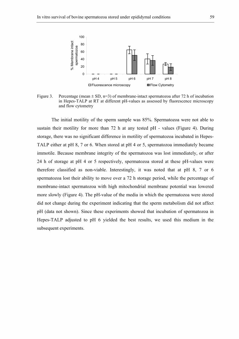
In vitro survival of bovine spermatozoa stored under epididymal conditions 59
Figure 3. Percentage (mean ± SD, n=3) of membrane-intact spermatozoa after 72 h of incubation in Hepes-TALP at RT at different pH-values as assessed by fluorescence microscopy and flow cytometry
The initial motility of the sperm sample was 85%. Spermatozoa were not able to
sustain their motility for more than 72 h at any tested pH - values (Figure 4). During
storage, there was no significant difference in motility of spermatozoa incubated in Hepes-
TALP either at pH 8, 7 or 6. When stored at pH 4 or 5, spermatozoa immediately became
immotile. Because membrane integrity of the spermatozoa was lost immediately, or after
24 h of storage at pH 4 or 5 respectively, spermatozoa stored at these pH-values were
therefore classified as non-viable. Interestingly, it was noted that at pH 8, 7 or 6
spermatozoa lost their ability to move over a 72 h storage period, while the percentage of
membrane-intact spermatozoa with high mitochondrial membrane potential was lowered
more slowly (Figure 4). The pH-value of the media in which the spermatozoa were stored
did not change during the experiment indicating that the sperm metabolism did not affect
pH (data not shown). Since these experiments showed that incubation of spermatozoa in
Hepes-TALP adjusted to pH 6 yielded the best results, we used this medium in the
subsequent experiments.
0
20
40
60
80
100
pH 4 pH 5 pH 6 pH 7 pH 8
% M
embr
ane
inta
ct
sper
mat
ozoa
Fluorescence microscopy Flow Cytometry
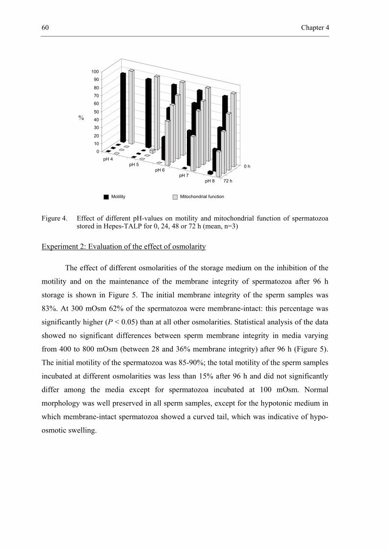
60 Chapter 4
Figure 4. Effect of different pH-values on motility and mitochondrial function of spermatozoa stored in Hepes-TALP for 0, 24, 48 or 72 h (mean, n=3)
Experiment 2: Evaluation of the effect of osmolarity
The effect of different osmolarities of the storage medium on the inhibition of the
motility and on the maintenance of the membrane integrity of spermatozoa after 96 h
storage is shown in Figure 5. The initial membrane integrity of the sperm samples was
83%. At 300 mOsm 62% of the spermatozoa were membrane-intact: this percentage was
significantly higher (P < 0.05) than at all other osmolarities. Statistical analysis of the data
showed no significant differences between sperm membrane integrity in media varying
from 400 to 800 mOsm (between 28 and 36% membrane integrity) after 96 h (Figure 5).
The initial motility of the spermatozoa was 85-90%; the total motility of the sperm samples
incubated at different osmolarities was less than 15% after 96 h and did not significantly
differ among the media except for spermatozoa incubated at 100 mOsm. Normal
morphology was well preserved in all sperm samples, except for the hypotonic medium in
which membrane-intact spermatozoa showed a curved tail, which was indicative of hypo-
osmotic swelling.
Motility Mitochondrial function
100
pH 4
72 h
0 h
0 10 20 30 40 50 60 70 80 90
pH 5 pH 6
pH 7pH 8
%
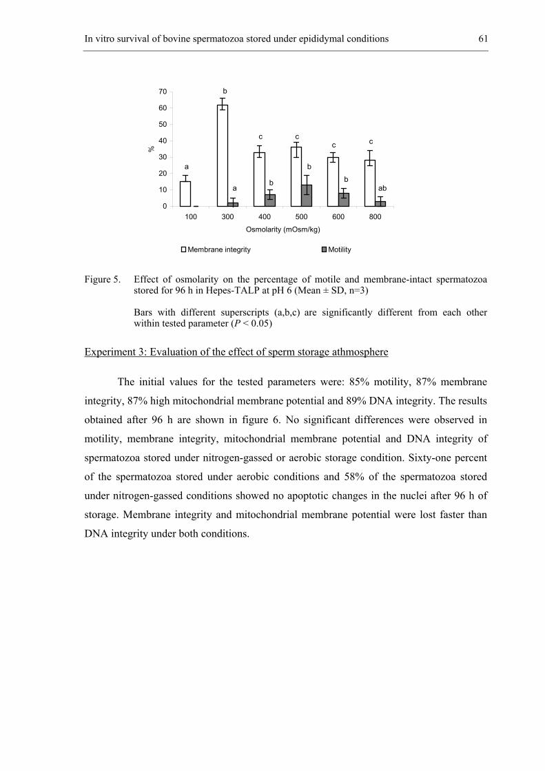
In vitro survival of bovine spermatozoa stored under epididymal conditions 61
Figure 5. Effect of osmolarity on the percentage of motile and membrane-intact spermatozoa stored for 96 h in Hepes-TALP at pH 6 (Mean ± SD, n=3)
Bars with different superscripts (a,b,c) are significantly different from each other within tested parameter (P < 0.05)
Experiment 3: Evaluation of the effect of sperm storage athmosphere
The initial values for the tested parameters were: 85% motility, 87% membrane
integrity, 87% high mitochondrial membrane potential and 89% DNA integrity. The results
obtained after 96 h are shown in figure 6. No significant differences were observed in
motility, membrane integrity, mitochondrial membrane potential and DNA integrity of
spermatozoa stored under nitrogen-gassed or aerobic storage condition. Sixty-one percent
of the spermatozoa stored under aerobic conditions and 58% of the spermatozoa stored
under nitrogen-gassed conditions showed no apoptotic changes in the nuclei after 96 h of
storage. Membrane integrity and mitochondrial membrane potential were lost faster than
DNA integrity under both conditions.
0
10
20
30
40
50
60
70
100 300 400 500 600 800
Osmolarity (mOsm/kg)
%
Membrane integrity Motility
a ab
b
b b
a
c c c c
b
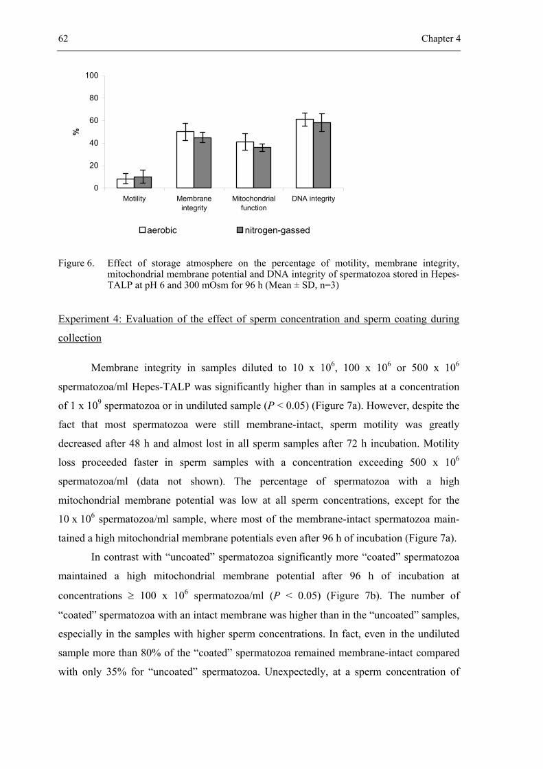
62 Chapter 4
0
20
40
60
80
100
Motility Membraneintegrity
Mitochondrialfunction
DNA integrity
%
aerobic nitrogen-gassed
Figure 6. Effect of storage atmosphere on the percentage of motility, membrane integrity, mitochondrial membrane potential and DNA integrity of spermatozoa stored in Hepes-TALP at pH 6 and 300 mOsm for 96 h (Mean ± SD, n=3)
Experiment 4: Evaluation of the effect of sperm concentration and sperm coating during
collection
Membrane integrity in samples diluted to 10 x 106, 100 x 106 or 500 x 106
spermatozoa/ml Hepes-TALP was significantly higher than in samples at a concentration
of 1 x 10⁹ spermatozoa or in undiluted sample (P < 0.05) (Figure 7a). However, despite the
fact that most spermatozoa were still membrane-intact, sperm motility was greatly
decreased after 48 h and almost lost in all sperm samples after 72 h incubation. Motility
loss proceeded faster in sperm samples with a concentration exceeding 500 x 106
spermatozoa/ml (data not shown). The percentage of spermatozoa with a high
mitochondrial membrane potential was low at all sperm concentrations, except for the
10 x 106 spermatozoa/ml sample, where most of the membrane-intact spermatozoa main-
tained a high mitochondrial membrane potentials even after 96 h of incubation (Figure 7a).
In contrast with “uncoated” spermatozoa significantly more “coated” spermatozoa
maintained a high mitochondrial membrane potential after 96 h of incubation at
concentrations ≥ 100 x 106 spermatozoa/ml (P < 0.05) (Figure 7b). The number of
“coated” spermatozoa with an intact membrane was higher than in the “uncoated” samples,
especially in the samples with higher sperm concentrations. In fact, even in the undiluted
sample more than 80% of the “coated” spermatozoa remained membrane-intact compared
with only 35% for “uncoated” spermatozoa. Unexpectedly, at a sperm concentration of
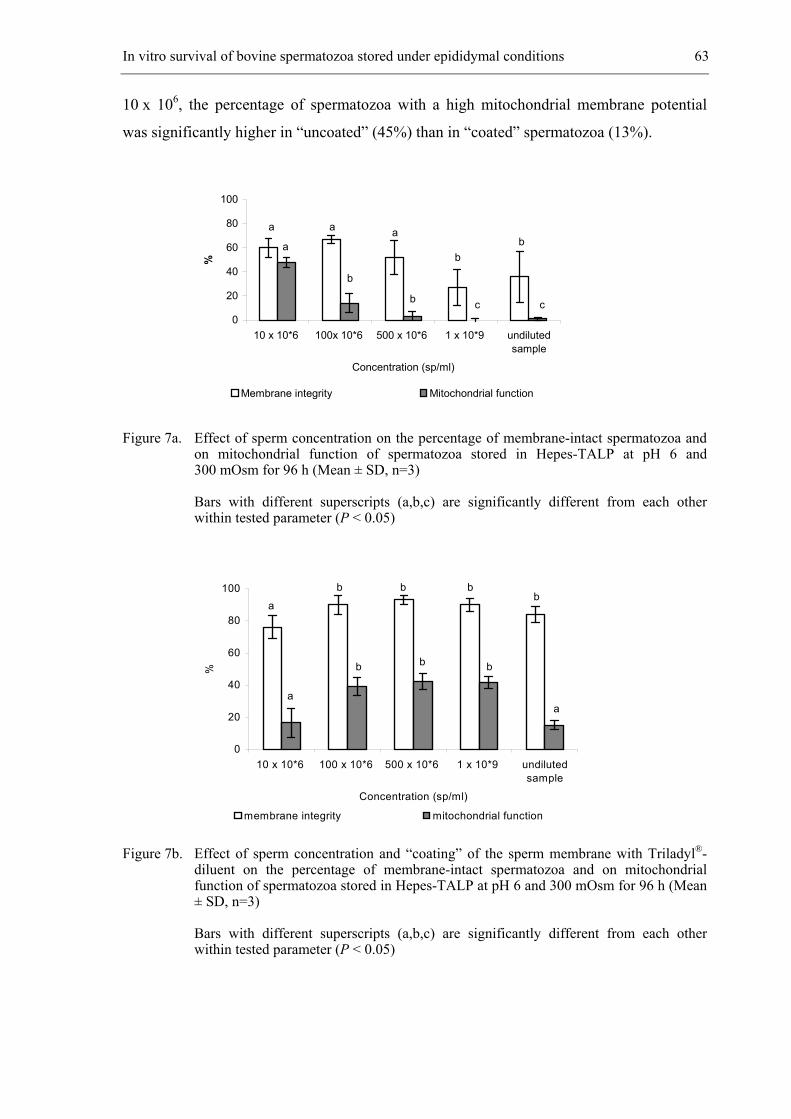
In vitro survival of bovine spermatozoa stored under epididymal conditions 63
10 x 106, the percentage of spermatozoa with a high mitochondrial membrane potential
was significantly higher in “uncoated” (45%) than in “coated” spermatozoa (13%).
Figure 7a. Effect of sperm concentration on the percentage of membrane-intact spermatozoa and on mitochondrial function of spermatozoa stored in Hepes-TALP at pH 6 and 300 mOsm for 96 h (Mean ± SD, n=3)
Bars with different superscripts (a,b,c) are significantly different from each other within tested parameter (P < 0.05)
Figure 7b. Effect of sperm concentration and “coating” of the sperm membrane with Triladyl®- diluent on the percentage of membrane-intact spermatozoa and on mitochondrial function of spermatozoa stored in Hepes-TALP at pH 6 and 300 mOsm for 96 h (Mean ± SD, n=3)
Bars with different superscripts (a,b,c) are significantly different from each other within tested parameter (P < 0.05)
0
20
40
60
80
100
10 x 10*6 100x 10*6 500 x 10*6 1 x 10*9 undilutedsample
Concentration (sp/ml)
%
Membrane integrity Mitochondrial function
a
a
c b
b
c
a a b
b
0
20
40
60
80
100
10 x 10*6 100 x 10*6 500 x 10*6 1 x 10*9 undilutedsample
Concentration (sp/ml)
%
membrane integrity mitochondrial function
a
b b
b b
b b b
a a
64 Chapter 4
Discussion
Despite the fact that sperm membrane integrity remains fairly well preserved for
several days during liquid storage, motility declines very quickly. Irreversible damage of
the motility apparatus and of mitochondrial and nuclear DNA is probably the main reason
why semen stored at RT has a declining fertilizing potential. Therefore, we assessed not
only sperm membrane integrity and motility, but also investigated sperm mitochondrial
membrane potential by means of dye JC-1 and the prevalence of DNA fragmentation in
stored spermatozoa as assessed by TUNEL staining. In our study, spermatozoa were stored
during 3 or 4 days at RT in a medium based on the zwitterionic organic buffer Hepes.
Sperm membrane integrity and mitochondrial membrane potential were preserved
longest when stored in Hepes-TALP at pH 6 for up to 3 days. A decrease in pH of the
medium to 6 was well tolerated by spermatozoa, but a lower pH was spermicidal and
caused immediate cessation of motility. Interestingly the plasma membrane of the
spermatozoa was more able to resist damage caused by pH-changes than the mechanisms
responsible for sperm motility. A possible explanation could be that the intracellular pH
(pHi) of the spermatozoa decreased to the extracellular pH (pHe), which is the case in
human, ram and boar semen (Gatti et al., 1993; Olmstedt et al., 2000). This suggests that
spermatozoa have minimal ability to maintain their pH in acidic environment and that their
membrane is permeable to protons. There is evidence that pHi regulates sperm motility in
invertebrate species (Nishioka and Cross, 1978) although no important regulatory
mechanisms for controlling pHi, such as Na+/H+ or K+/H+ exchange are present in
mammalian spermatozoa (Hamamah and Gatti, 1998; Jones and Bavister, 2000). Carr et al.
(1985) suggested that the low pH of the cauda epididymal plasma in combination with the
presence of a permeable weak acid like lactic acid are responsible for the short-term
quiescence of the epididymal spermatozoa by depressing their pHi. However, when
ejaculated spermatozoa are stored for longer than 48 h, reactivation of sperm motility is
much more difficult after immobilization by low pH (Jones and Bavister, 2000).
The second tested parameter in this study was the osmolarity of the medium.
Sorbitol was used to increase the osmolarity of the medium: this osmolyte may cross the
plasma membrane to a limited extent and is an appropriate metabolic substrate for
spermatozoa (Clegg et al., 1986; Crichton et al., 1994). Although caudal epididymal
plasma of most animals is hyperosmotic to blood (300 mOsm) with an average of
354 mOsm (Verberckmoes et al., 2001), increasing the osmotic pressure had no positive
In vitro survival of bovine spermatozoa stored under epididymal conditions 65
influence on membrane integrity of bull spermatozoa and actually even caused a
significant drop in percentage surviving spermatozoa compared to isotonic values.
Hyperosmotic conditions are present in epididymal plasma of hibernating bats (Crichton et
al., 1994) and it has been hypothesized that dehydration of bat spermatozoa would reduce
their metabolic needs and thereby prolong their survival. This could not be replicated with
bovine spermatozoa because dehydration of the spermatozoa in hyperosmotic solutions
caused membrane damage. Hypo-osmotic solutions (< 300 mOsm) were even more
deleterious to bull sperm motility because the influx of water causes the mitochondria in
the tail to swell, an increase in volume and bursting (Drevius and Ericksson, 1966). This
explains the decrease in membrane integrity and motility of the spermatozoa stored in
Hepes-TALP of 100 mOsm.
Although spermatozoa are stored under low oxygen tension in the cauda
epididymidis, no significant differences were observed in motility, mitochondrial
membrane potential and membrane integrity of spermatozoa stored under nitrogen-gassed
conditions compared to aerobic conditions after 4 days. In an analogous study performed
by Krzyzosiak et al. (2001), membrane integrity of spermatozoa was better preserved when
stored in CAPROGEN® (Livestock Improvement Corporation, Hamilton, New Zealand)
under nitrogen-gassed conditions, however no significant differences were observed in the
motility and fertility of the spermatozoa. Our study showed that ageing of spermatozoa in
vitro leads to a greater incidence of injury in mitochondria than in nuclear DNA. This
confirms that changes in mitochondrial membrane potential occur during the early stages
of apoptosis and could be detectable before DNA fragmentation occurs (Donnelly et al.,
2000). The decline could also be explained by the inability of mtDNA to auto-repair and
by the fact that the rate of mitochondrial ageing is faster than that of nuclear DNA
(Cummins et al., 1994). In contrast to expectations, no differences were observed in DNA
damage of spermatozoa stored for 4 days under either aerobic or nitrogen-gassed
conditions. Since the generation of reactive oxygen species (ROS) and consequently DNA
damage is likely to be higher under aerobic conditions, it was surprising to find no
significant differences in mitochondrial and DNA fragmentation in spermatozoa stored
under either aerobic or nitrogen-gassed conditions. This may indicate that either ROS
formation was not reduced under nitrogen-gassed conditions, or that its effect on apoptosis
is minimal. Both hypotheses need further investigation. Because storage under nitrogen-
gassed conditions caused no important improvement in sperm quality parameters, aerobic
storage conditions were used in the last experiment.
66 Chapter 4
Sperm concentration also greatly influences sperm survival. In the epididymis,
spermatozoa are stored at a concentration of 1 x 10⁹ to 3 x 10⁹ spermatozoa/ml. Ejaculated
and stored spermatozoa on the contrary, preserve their membrane integrity best at lower
sperm concentrations. The reason may be related to the reduced output of toxic metabolic
products or to a slower exhaustion of substrate at sperm high dilutions. In vitro, substrates
become depleted rapidly and toxic waste products accumulate, whereas in vivo inputs are
limited to easily metabolizable substrates from the external environment and toxic products
are disposed off systematically (Van Demark and Couturier, 1958). The most important
toxic products accumulated during in vitro storage are ROS formed by the oxidative
phosphorylation system of the inner mitochondrial membrane (Miquel and Fleming, 1986).
In our study, it is evidenced that storage of spermatozoa at high concentrations was less
detrimental at all when spermatozoa were “coated” with Triladyl®- diluent before being
stored. Collection of spermatozoa in Triladyl®- diluent during ejaculation has been the
method of choice to limit the contact of spermatozoa with seminal plasma. Bovine seminal
plasma is notorious for its negative influence on sperm survival in vitro (Shannon and
Curson, 1983, 1987), because it contains a group of acidic proteins (Bovine seminal
plasma (BSP) proteins) that bind to the spermatozoa (Desnoyers and Manjunath, 1992).
Their interaction with spermatozoa leads to the efflux of choline phospholipids and
cholesterol from the sperm plasma membrane, which appears to be an important step in the
capacitation process (Therien et al., 1998; Moreau et al., 1999). Since cholesterol is
recognized to have a stabilizing effect on membranes (Yeagle, 1985), this efflux would be
expected to cause destabilization of the membrane. Other cationic peptides present in
seminal plasma are also known to bind to spermatozoa (which has a net negative charge)
(Yanagimachi et al., 1972) and to destabilize the plasma membrane. The water-soluble
cationic fraction of egg yolk protects against the detrimental effect of seminal plasma
components (Shannon and Curson, 1972,1983) by competing in binding to the sperm
membrane. The low-density lipoprotein fraction of egg yolk has also been shown to reduce
the loss of acrosomal enzymes and to prevent degenerative changes in the acrosome during
liquid storage (Salamon and Maxwell, 2000).
The very simple action of “coating” spermatozoa for less than 5 min with egg yolk
containing diluent during collection had an important effect upon several characteristics
after 4 days of storage in Hepes-TALP at pH 6 and 300 mOsm. These results are very
promising for the improvement of preservation and fertilizing ability of liquid bovine
semen in the future. Consequently, the effect of sperm “coating” during ejaculation on the
In vitro survival of bovine spermatozoa stored under epididymal conditions 67
fertilizing ability of spermatozoa stored in vitro during several days, is actually under
investigation in our laboratory.
Acknowledgments
This research was supported by the Ministry of Agriculture of the Belgian
government, grant n° S6012 and by the Research Fund of Ghent University, grant n°
011B8698. We also thank Dr. M. Coryn for critical reading, and G. Spaepen and
J. Mestach for technical assistance.
68 Chapter 4
References
Acott TS, Carr DW. Inhibition of bovine spermatozoa by caudal epididymal fluid: II. Interaction of
pH and a quiescence factor. Biol Reprod 1984; 30: 926-935.
Amann RP, Johnson L, Thompson DL, Pickett BW. Daily spermatozoal production, epididymal
spermatozoal reserves and transit time of spermatozoa through the epididymis of the rhesus
monkey. Biol Reprod 1976; 15: 586-592.
Browne RK, Clulow J, Mahony M. Short-term storage of cane toad (Bufo marinus) gametes.
Reproduction 2001, 121: 167-173.
Carr DW, Usselman MC, Acott TS. Effects of pH, lactate and viscoelastic drag on sperm motility:
a species comparison. Biol Reprod 1985; 33: 588-593.
Cascieri M, Amann RP, Hammerstedt RH. Adenine nucleotide changes at initiation of bull sperm
motility. J Biol Chem 1976; 251: 787-793.
Clegg ED, Sakai CS, Voytek PE. Assessment of reproductive risks. Biol Reprod 1986; 34: 5-16.
Crichton EG, Hinton BT, Pallone TL, Hammerstedt RH. Hyperosmolality and sperm storage in
hibernating bats: prolongation of sperm life by dehydration. Am J Physiol 1994; 267: 1363-
1370.
Cummins JM, Jequier AM, Raymond K. Molecular biology of human male infertility: Links with
aging, mitochondrial genetics, and oxidative stress? Mol Reprod Dev 1994; 37: 345-362.
Desnoyers L, Manjunath P. Major proteins of bovine seminal plasma exhibit novel interactions
with phospholipid. J Biol Chem 1992; 267: 10149-10155.
Donnelly ET, O’Connell M, McClure N, Lewis SE. Differences in nuclear DNA fragmentation and
mitochondrial integrity of semen and prepared human spermatozoa. Hum Reprod 2000; 15:
1552-1561.
Drevius LO, Ericksson H. Osmotic swelling of mammalian spermatozoa. Exp Cell Res 1966; 42:
136-156.
Free MJ, Schluntz GA, Jaffe RA. Respiratory gas tensions in tissues and fluids of the male rat
reproductive tract. Biol Reprod 1976; 14: 481-488.
Garner DL, Thomas CA. Organelle-specific probe JC-1 identifies membrane potential differences
in the mitochondrial function of bovine sperm. Mol Reprod Dev 1999; 53: 222-229.
Gatti JL, Chevrier C, Paquignon M, Dacheux JL. External ionic conditions, internal pH and
motility of ram and boar spermatozoa. J Reprod Fertil 1993; 98: 439-449.
In vitro survival of bovine spermatozoa stored under epididymal conditions 69
Hamamah S, Gatti JL. Role of the ionic environment and internal pH on sperm activity. Hum
Reprod 1998; 13 Suppl 4: 20-30.
Jones RC, Murdoch RN. Regulation of the motility and metabolism of spermatozoa for storage in
the epididymis of eutherian and marsupial mammals. Reprod Fertil Dev 1996; 8: 553-568.
Jones JM, Bavister BD. Acidification of intracellular pH in bovine spermatozoa suppresses motility
and extends viable life. J Androl 2000; 21: 616-624.
Kikuchi K, Nagai T, Kashiwazaki N, Ikeda H, Noguchi J, Shimada A, Soloy E, Kaneko H.
Cryopreservation and ensuing in vitro fertilization ability of boar spermatozoa from
epididymides stored at 4 degrees C. Theriogenology 1998; 50: 615-623.
Krzyzosiak J, Molan P, McGowan L, Vishwanath R. Effect of sperm number and oxygenation state
of the storage media on in vitro fertility of bovine sperm stored at ambient temperature.
Theriogenology 2001; 55: 1401-1415.
Liu Z, Foote RH. Bull sperm motility and membrane integrity in media varying in osmolality. J
Dairy Sci 1998; 81: 1868-1873.
Mann T, Lutwak-Mann C. Biochemistry of spermatozoa: chemical and functional correlations in
ejaculated semen. Andrological aspects. In Mann T, Lutwak-Mann C (eds). Male
Reproductive Function and Semen. Springer-Verlag; New York 1981: 205-217.
Miquel J, Fleming J. Theoretical and experimental support for an “oxygen radical-mitochondrial
injury” hypothesis of cell aging. In Johnson JE, Walford R, Harman D, Miquel J (eds). Free
radicals, aging, and degenerative diseases. Alan R. Liss; New York 1986: 51-74.
Moreau R, Frank PG, Perreault C, Marcel YL, Manjunath P. Seminal plasma choline phospholipid-
binding proteins stimulate cellular cholesterol and phospholipid efflux. Biochim Biophys
Acta 1999; 1438: 38-46.
Nishioka D, Cross N. The role of external sodium in sea urchin fertilization. In: Dirksen ER,
Prescott D, Fox CF (eds): Cell Reproduction. Academic Press; New York 1978: 403-413.
Olmstedt SS, Dubin NH, Cone RA, Moench TR. The rate at which human sperm are immobilized
and killed by mild acidity. Fertil Steril 2000; 73: 687-693.
Salamon S, Maxwell WMC. Storage of ram semen. Anim Reprod Sci 2000; 62: 77-111.
Sankai T, Tsuchiya H, Ogonuki N. Short-term nonfrozen storage of mouse epididymal
spermatozoa. Theriogenology 2001; 55: 1759-1768.
Setchell BP, Sánchez-Partida LG, Chairussyuhur A. Epididymal constituents and related
substances in the storage of spermatozoa: a review. Reprod Fertil Dev 1993; 5: 601-612.
70 Chapter 4
Shannon P, Curson B. Toxic effect and action of dead sperm on diluted bovine semen. J Dairy Sci
1972; 55: 614-620.
Shannon P, Curson B. Effect of egg yolk levels on the fertility of diluted bovine sperm stored at
ambient temperatures. N Z J Agric Res 1983; 26: 187-189.
Shannon P, Curson B, Molan PC. Cationic peptides obtained by reversible disaggregation of
antibacterial proteins of bovine seminal plasma. N Z J Agric Res 1987; 30: 195-202.
Sun JG, Jurisicova A, Casper RF. Detection of deoxyribonucleic acid fragmentation in human
sperm: correlation with fertilization in vitro. Biol Reprod 1997; 56: 602-607.
Therien I, Moreau R, Manjunath P. Major proteins of bovine seminal plasma and high-density
lipoprotein induce cholesterol efflux from epididymal sperm. Biol Reprod 1998; 59: 768-
776.
Thomas CA, Garner DL, Dejarnette JM, Marshall CE. Effect of cryopreservation on bovine sperm
organelle function and viability as determined by flow cytometry. Biol Reprod 1998; 58:
786-793.
Van Demark NL, Couturier LR. Flow dialysis as a means of preserving bovine semen at room
temperature. J Dairy Sci 1958; 41: 530-536.
Verberckmoes S, De Pauw I, Van Soom A, de Kruif A. Ionic composition and osmolarity of caudal
epididymal plasma in the bull. Theriogenology 2001; 55: 449 (abstr.).
Vishwanath R, Shannon P. Do sperm cells age? A review of the physiological changes in sperm
during storage at ambient temperatures. Reprod Fertil Dev 1997; 9: 321-331.
Vishwanath R, Shannon P. Storage of bovine semen in liquid and frozen state. Anim Reprod Sci
2000; 62: 23-53.
Yanagimachi R, Noda YD, Fujimoto M, Nicolson GL. The distribution of negative surface charges
on mammalian spermatozoa. Am J Anat 1972; 135: 497-519.
Yeagle PL. Cholesterol and the cell membrane. Biochim Biophys Acta 1985; 822: 267-287.
Zaneveld LJD, Chatteron RT. In: Biochemistry of mammalian reproduction. Wiley-Interscience
Pub; New York 1982: 38.
__________________________________________________ 5.
EFFECT OF SPERM COATING ON THE SURVIVAL AND PENETRATING
ABILITY OF IN VITRO STORED BOVINE SPERMATOZOA
Theriogenology 2003; 59: 1109-1122
De Pauw I., Van Soom A., Maes D., Verberckmoes S. and de Kruif A.
72 Chapter 5
Abstract
The aim of this study was to examine the effect of sperm “coating” on the survival
and penetrating ability of in vitro stored bovine spermatozoa. Semen was collected by
means of an artificial vagina connected with a tube containing 5 ml of the commercial
Triladyl®-diluent. Both diluent and seminal plasma were removed by centrifugation
(720 x g, 10 min) and spermatozoa were stored under different in vitro storage conditions.
In the first and second experiment, “control” and “coated” spermatozoa were stored in
Hepes-TALP (pH 6 and 7) at room temperature (RT). After 4 days of storage, the
progressive motility, membrane integrity, mitochondrial membrane potential and DNA
integrity of the spermatozoa were evaluated before and after Percoll®-centrifugation. The
in vitro oocyte penetration rate of spermatozoa was examined only after Percoll®-
centrifugation. A significantly (P < 0.05) positive effect of sperm “coating” was observed
on the tested sperm characteristics and oocyte penetration rate of spermatozoa when they
were stored in Hepes-TALP at pH 7, but not at pH 6. In the last experiment, the effect of
storage medium: Hepes-TALP (pH 7) or Triladyl®-diluent was investigated on motility,
membrane integrity, mitochondrial membrane potential and in vitro oocyte penetration
potential of “coated” spermatozoa stored at RT or at 4°C for up to 4, 5 and 6 days. After
6 days of storage in Triladyl®, a significantly (P < 0.05) higher percentage of motile and
membrane intact spermatozoa with high mitochondrial membrane potential was obtained at
both temperatures leading to a significantly higher in vitro oocyte penetration rate. These
results indicate that sperm “coating” could preserve sperm characteristics and oocyte
penetrating capacity of bovine spermatozoa stored in egg yolk containing diluent for up to
6 days.
Effect of sperm "coating" on the survival and penetrating ability of bovine spermatozoa 73
Introduction
Liquid semen can be used as an alternative for artificial insemination with frozen-
thawed spermatozoa, on the condition that its fertilizing potential can be maintained for a
minimum of 2 days and preferably 4 days to provide for easy transport and use in distant
locations (Vishwanath and Shannon, 2000). Much research has already been done to
prolong the in vitro viability and fertilizing potential of stored liquid semen by altering or
reducing the catabolic metabolism of spermatozoa or by increasing the stability of sperm
membranes to thermal and other environmental insults (Foote and Parks, 1993). However,
until now no important improvements have been made to increase the fertilizing potential
of spermatozoa longer than 3 to 5 days when they were stored in liquid environment at
high dilutions at ambient temperatures (Vishwanath and Shannon, 1997).
A novel method to minimize the damage of ejaculated spermatozoa was obtained
by “coating” spermatozoa for less than 5 minutes during collection with the commercial
Triladyl®-diluent (De Pauw et al., 2003). With this method, the contact between
spermatozoa and seminal plasma was limited by collecting spermatozoa in a tube
containing 5 ml of Triladyl®-diluent, which was connected to an artificial vagina. The
water-soluble cationic fraction of egg yolk is able to protect both sperm plasma and
acrosome membrane against the detrimental effect of seminal plasma components
(Shannon and Curson, 1972, 1983). This competing in binding with seminal plasma
components prevents the sperm plasma membrane against the efflux of choline
phospholipids and cholesterol, which causes destabilization of the membrane. Furthermore,
the protective action of low-density lipoprotein binding could also be responsible for the
stabilization of the plasma membrane through the critical zones (Vishwanath et al., 1992)
and for the improvement of motility after storage (Moussa et al., 2002).
Furthermore, it would also be valuable to assess deterioration due to changes
caused by ageing of stored spermatozoa (Maxwell and Salamon, 1993; Paulenz et al.,
2002) resulting in sperm plasma membrane disruption, loss of mitochondrial membrane
potential and DNA fragmentation. These are all hallmarks of apoptosis (Donnelly et al.,
2000) leading to a progressive loss of fertilizing ability over storage time (Cumming et al.,
1994).
The aims of the study were 1) to analyze the effect of sperm “coating” during
collection on the progressive motility, membrane integrity, mitochondrial membrane
potential and DNA integrity of spermatozoa stored in Hepes-TALP at pH 6 or 7 at RT for
74 Chapter 5
4 days, 2) to examine the in vitro oocyte penetration rate of 4 days old “control” and
“coated” spermatozoa stored at RT, 3) to evaluate the effect of the storage medium (Hepes-
TALP (pH 7) and Triladyl®-diluent) on the oocyte penetration rate of “coated”
spermatozoa stored at 4°C and at RT for up to 4, 5 and 6 days.
Materials and Methods
Media
Chemicals and media were obtained from Sigma (Bornem, Belgium) and GIBCO™
Invitrogen Corporation (Merelbeke, Belgium).
Hepes-TALP containing 114 mM NaCl, 3.1 mM KCl, 0.3 mM NaH2PO4, 2.1 mM CaCl2,
0.4 mM MgCl2, 2 mM NaHCO3, 0.2 mM sodium pyruvate, 10 mM sodium
lactate, 10 µg/ml gentamycin sulphate, 10 mM Hepes and 3 mg/ml Bovine
Serum Albumin.
Triladyl®-diluent (Minitüb, Germany), a TRIS-based diluent supplemented with 20% egg
yolk and 6.7 % of glycerol. Final concentrations of antibiotics per 100 ml
were: 5 mg tylosin, 25 mg gentamycin, 30 mg spectinomycin and 15 mg
lincomycin.
Maturation medium consisting of modified bicarbonate buffered TCM199 medium
supplemented with 20% heat-inactivated foetal calf serum (N.V. HyClone
Europe S.A., Erembodegem, Belgium), 0.2 mM sodium pyruvate, 50 µg/ml
gentamycin sulphate and 0.4 mM glutamin.
Fertilization medium containing Tyrode’s solution without Hepes supplemented with
25 mM NaHCO3, 0.2 mM sodium pyruvate, 10 mM sodium lactate, 10 µg/ml
gentamycin sulphate, 6 mg/ml fatty acid-free BSA and 10 µg/ml heparin.
Collection and preparation of semen
Spermatozoa from a 3-year-old Red Pied bull were collected by means of an
artificial vagina connected with an empty tube (“control” spermatozoa). Within 5 minutes,
a second ejaculate was collected following the same method but in a tube containing 5 ml
Triladyl®-diluent (“coated” spermatozoa). Immediately after collection, both ejaculates
were centrifuged (720 x g, 10 min) at RT to remove the supernatant. The sperm
Effect of sperm "coating" on the survival and penetrating ability of bovine spermatozoa 75
concentration of the pellet was determined using a Bürker chamber and subsequently
diluted to a concentration of 10 x 106 spermatozoa/ml in Hepes-TALP (pH 6 or 7) or in
Triladyl®-diluent according to the experiment. Spermatozoa were stored both at RT and
4°C in darkness until fertilization. They were either analyzed directly (Experiment 1) or
after Percoll®-centrifugation (Experiment 2 and 3). Motile and membrane-intact
spermatozoa were separated by centrifugation (720 x g, 30 min) at RT on a discontinuous
Percoll®-gradient (Pharmacia, Uppsala, Sweden) composed of 2 ml each of 90% and 45%
Percoll solutions in Hepes buffered Sp-TALP without BSA. After removal of Percoll®
supernatant, the sperm pellet was diluted in 5 ml Hepes buffered Sp-TALP supplemented
with BSA and centrifuged (180 x g, 10 min).
Processing and analysis of sperm samples
Motility
The percentage of total and progressive motile spermatozoa was subjectively
assessed by visual estimation of two investigators. Eight microliter of the sperm sample
was placed on a prewarmed glass slide, covered with a glass cover slip, and viewed under a
Leica DMR light microscope (Van Hopplynus NV, Brussels, Belgium) (magnification
200 x) equipped with a stage warmer (37°C).
Membrane integrity
The nucleic stains SYBR®-14 and propidium iodide (PI) were used for analyzing
membrane integrity of the spermatozoa (LIVE/DEAD® Sperm Viability Kit (Molecular
Probes)) and evaluated by means of fluorescence microscopy (magnification 1000 x) at
37°C. SYBR®-14 labeled DNA of membrane-intact spermatozoa, resulting in a bright
green fluorescence when excited at 488 nm, while PI only stained the nuclei of membrane-
damaged spermatozoa red (excitation: 530 nm). The membrane integrity of at least 100
spermatozoa was evaluated.
Mitochondrial membrane potential
For evaluation of mitochondrial membrane potential, spermatozoa were labeled
with JC-1 (Molecular Probes, Leiden, The Netherlands). This fluorophore exhibits
76 Chapter 5
potential-dependent accumulation in mitochondria, and can reversibly change its emission
from green to orange with increasing transmembrane electrical potential. This staining was
combined with SYBR®-14 and PI. Two microliters of 1.53 mM JC-1 (Molecular Probes) in
DMSO was added to 1 ml of the diluted sperm sample in combination with 0.7 µl SYBR®-
14 and 12 µl of 2.99 mM PI (Thomas et al., 1998; Garner and Thomas, 1999).
DNA fragmentation
Measurement of sperm DNA fragmentation was performed using the TUNEL assay
(In-situ Cell Detection Kit, Boehringer Mannheim, Mannheim, Germany).
Spermatozoa were washed twice in PVP solution (1 mg/ml polyvinyl-pyrrolidone
(PVP) in phosphate-buffered saline)) by centrifugation at RT (720 x g, 5 min). The final
pellet was resuspended in PVP solution and 10 µl of 10 x 106 sp/ml was smeared onto a
polylysine-coated glass microscope slide and air dried. The slides were washed in PVP
solution (5 min) and fixed with 4% paraformaldehyde in PVP solution (pH 7.4) at RT for
30 min. This was followed by permeabilization with a solution of 0.1% Triton X-100 and
0.1% sodium citrate for 2 min on ice. Slides were rinsed twice with PVP solution and air
dried. Twenty-five microliters diluted terminal deoxynucleotidyl transferase (TdT) was
mixed with 225 µl nucleotide mixture. Twenty-five µl of this TUNEL reaction mixture
was added to the spermatozoa on each slide. The samples were incubated in a humidified
chamber at 37°C for 60 min in darkness.
Negative (nucleotide solution without TdT), and positive (incubation with 1 mg
Dnase I/ml (Pharmacia LKB Biotech) for 10 min at RT before staining with TUNEL
reaction mixture) controls were included in all experiments. The slides were rinsed three
times with PVP solution and incubated with 25 µl Hoechst 33342 (1µl/ml PVP solution) at
RT for 5 min. After washing twice in PVP solution, 100 spermatozoa in different
microscopic fields were analyzed by means of fluorescence microscopy. Hoechst stained
spermatozoa were first counted (bright blue fluorescence) under ultraviolet light and the
percentage of FITC-labelled cells with fragmented DNA (intense green nuclear
fluorescence) was determined by means of Leica DMR epifluorescence microscopy
(magnification 1000 x).
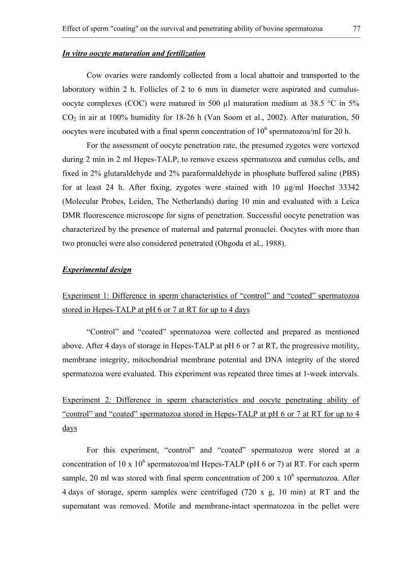
Effect of sperm "coating" on the survival and penetrating ability of bovine spermatozoa 77
In vitro oocyte maturation and fertilization
Cow ovaries were randomly collected from a local abattoir and transported to the
laboratory within 2 h. Follicles of 2 to 6 mm in diameter were aspirated and cumulus-
oocyte complexes (COC) were matured in 500 µl maturation medium at 38.5 °C in 5%
CO2 in air at 100% humidity for 18-26 h (Van Soom et al., 2002). After maturation, 50
oocytes were incubated with a final sperm concentration of 106 spermatozoa/ml for 20 h.
For the assessment of oocyte penetration rate, the presumed zygotes were vortexed
during 2 min in 2 ml Hepes-TALP, to remove excess spermatozoa and cumulus cells, and
fixed in 2% glutaraldehyde and 2% paraformaldehyde in phosphate buffered saline (PBS)
for at least 24 h. After fixing, zygotes were stained with 10 µg/ml Hoechst 33342
(Molecular Probes, Leiden, The Netherlands) during 10 min and evaluated with a Leica
DMR fluorescence microscope for signs of penetration. Successful oocyte penetration was
characterized by the presence of maternal and paternal pronuclei. Oocytes with more than
two pronuclei were also considered penetrated (Ohgoda et al., 1988).
Experimental design
Experiment 1: Difference in sperm characteristics of “control” and “coated” spermatozoa
stored in Hepes-TALP at pH 6 or 7 at RT for up to 4 days
“Control” and “coated” spermatozoa were collected and prepared as mentioned
above. After 4 days of storage in Hepes-TALP at pH 6 or 7 at RT, the progressive motility,
membrane integrity, mitochondrial membrane potential and DNA integrity of the stored
spermatozoa were evaluated. This experiment was repeated three times at 1-week intervals.
Experiment 2: Difference in sperm characteristics and oocyte penetrating ability of
“control” and “coated” spermatozoa stored in Hepes-TALP at pH 6 or 7 at RT for up to 4
days
For this experiment, “control” and “coated” spermatozoa were stored at a
concentration of 10 x 106 spermatozoa/ml Hepes-TALP (pH 6 or 7) at RT. For each sperm
sample, 20 ml was stored with final sperm concentration of 200 x 106 spermatozoa. After
4 days of storage, sperm samples were centrifuged (720 x g, 10 min) at RT and the
supernatant was removed. Motile and membrane-intact spermatozoa in the pellet were
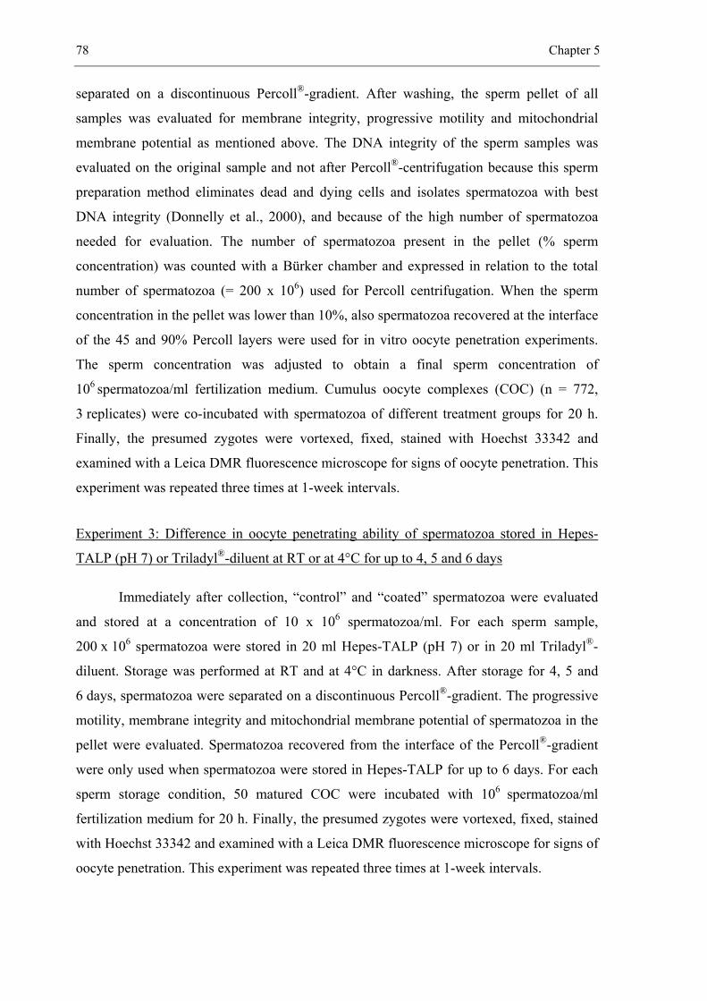
78 Chapter 5
separated on a discontinuous Percoll®-gradient. After washing, the sperm pellet of all
samples was evaluated for membrane integrity, progressive motility and mitochondrial
membrane potential as mentioned above. The DNA integrity of the sperm samples was
evaluated on the original sample and not after Percoll®-centrifugation because this sperm
preparation method eliminates dead and dying cells and isolates spermatozoa with best
DNA integrity (Donnelly et al., 2000), and because of the high number of spermatozoa
needed for evaluation. The number of spermatozoa present in the pellet (% sperm
concentration) was counted with a Bürker chamber and expressed in relation to the total
number of spermatozoa (= 200 x 106) used for Percoll centrifugation. When the sperm
concentration in the pellet was lower than 10%, also spermatozoa recovered at the interface
of the 45 and 90% Percoll layers were used for in vitro oocyte penetration experiments.
The sperm concentration was adjusted to obtain a final sperm concentration of
106 spermatozoa/ml fertilization medium. Cumulus oocyte complexes (COC) (n = 772,
3 replicates) were co-incubated with spermatozoa of different treatment groups for 20 h.
Finally, the presumed zygotes were vortexed, fixed, stained with Hoechst 33342 and
examined with a Leica DMR fluorescence microscope for signs of oocyte penetration. This
experiment was repeated three times at 1-week intervals.
Experiment 3: Difference in oocyte penetrating ability of spermatozoa stored in Hepes-
TALP (pH 7) or Triladyl®-diluent at RT or at 4°C for up to 4, 5 and 6 days
Immediately after collection, “control” and “coated” spermatozoa were evaluated
and stored at a concentration of 10 x 106 spermatozoa/ml. For each sperm sample,
200 x 106 spermatozoa were stored in 20 ml Hepes-TALP (pH 7) or in 20 ml Triladyl®-
diluent. Storage was performed at RT and at 4°C in darkness. After storage for 4, 5 and
6 days, spermatozoa were separated on a discontinuous Percoll®-gradient. The progressive
motility, membrane integrity and mitochondrial membrane potential of spermatozoa in the
pellet were evaluated. Spermatozoa recovered from the interface of the Percoll®-gradient
were only used when spermatozoa were stored in Hepes-TALP for up to 6 days. For each
sperm storage condition, 50 matured COC were incubated with 106 spermatozoa/ml
fertilization medium for 20 h. Finally, the presumed zygotes were vortexed, fixed, stained
with Hoechst 33342 and examined with a Leica DMR fluorescence microscope for signs of
oocyte penetration. This experiment was repeated three times at 1-week intervals.
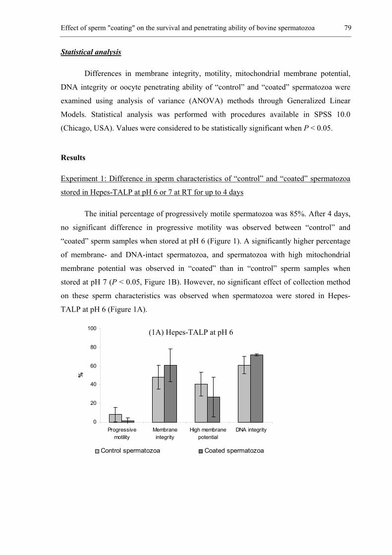
Effect of sperm "coating" on the survival and penetrating ability of bovine spermatozoa 79
Statistical analysis
Differences in membrane integrity, motility, mitochondrial membrane potential,
DNA integrity or oocyte penetrating ability of “control” and “coated” spermatozoa were
examined using analysis of variance (ANOVA) methods through Generalized Linear
Models. Statistical analysis was performed with procedures available in SPSS 10.0
(Chicago, USA). Values were considered to be statistically significant when P < 0.05.
Results
Experiment 1: Difference in sperm characteristics of “control” and “coated” spermatozoa
stored in Hepes-TALP at pH 6 or 7 at RT for up to 4 days
The initial percentage of progressively motile spermatozoa was 85%. After 4 days,
no significant difference in progressive motility was observed between “control” and
“coated” sperm samples when stored at pH 6 (Figure 1). A significantly higher percentage
of membrane- and DNA-intact spermatozoa, and spermatozoa with high mitochondrial
membrane potential was observed in “coated” than in “control” sperm samples when
stored at pH 7 (P < 0.05, Figure 1B). However, no significant effect of collection method
on these sperm characteristics was observed when spermatozoa were stored in Hepes-
TALP at pH 6 (Figure 1A).
0
20
40
60
80
100
Progressivemotility
Membraneintegrity
High membranepotential
DNA integrity
%
Control spermatozoa Coated spermatozoa
(1A) Hepes-TALP at pH 6
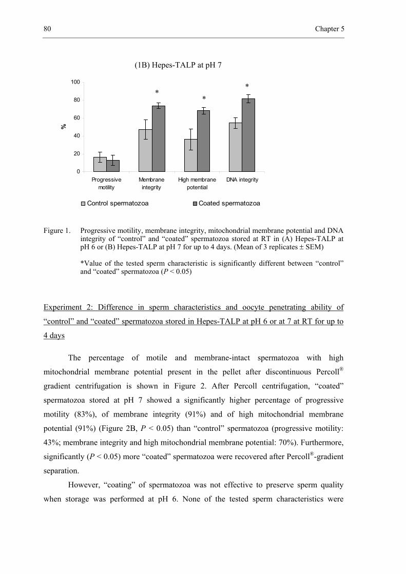
80 Chapter 5
Figure 1. Progressive motility, membrane integrity, mitochondrial membrane potential and DNA integrity of “control” and “coated” spermatozoa stored at RT in (A) Hepes-TALP at pH 6 or (B) Hepes-TALP at pH 7 for up to 4 days. (Mean of 3 replicates ± SEM)
*Value of the tested sperm characteristic is significantly different between “control” and “coated” spermatozoa (P < 0.05)
Experiment 2: Difference in sperm characteristics and oocyte penetrating ability of
“control” and “coated” spermatozoa stored in Hepes-TALP at pH 6 or at 7 at RT for up to
4 days
The percentage of motile and membrane-intact spermatozoa with high
mitochondrial membrane potential present in the pellet after discontinuous Percoll®
gradient centrifugation is shown in Figure 2. After Percoll centrifugation, “coated”
spermatozoa stored at pH 7 showed a significantly higher percentage of progressive
motility (83%), of membrane integrity (91%) and of high mitochondrial membrane
potential (91%) (Figure 2B, P < 0.05) than “control” spermatozoa (progressive motility:
43%; membrane integrity and high mitochondrial membrane potential: 70%). Furthermore,
significantly (P < 0.05) more “coated” spermatozoa were recovered after Percoll®-gradient
separation.
However, “coating” of spermatozoa was not effective to preserve sperm quality
when storage was performed at pH 6. None of the tested sperm characteristics were
(1B) Hepes-TALP at pH 7
(A) Hepes-TALP at pH 6
(B) Hepes-TALP at pH
0
20
40
60
80
100
Progressivemotility
Membraneintegrity
High membranepotential
DNA integrity
%
Control spermatozoa Coated spermatozoa
**
*
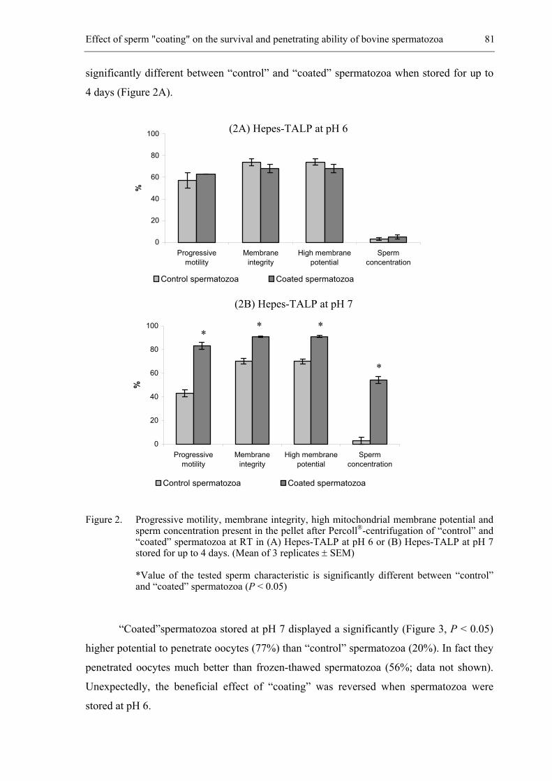
Effect of sperm "coating" on the survival and penetrating ability of bovine spermatozoa 81
significantly different between “control” and “coated” spermatozoa when stored for up to
4 days (Figure 2A).
0
20
40
60
80
100
Progressivemotility
Membraneintegrity
High membranepotential
Spermconcentration
%
Control spermatozoa Coated spermatozoa
0
20
40
60
80
100
Progressivemotility
Membraneintegrity
High membranepotential
Spermconcentration
%
Control spermatozoa Coated spermatozoa
Figure 2. Progressive motility, membrane integrity, high mitochondrial membrane potential and sperm concentration present in the pellet after Percoll®-centrifugation of “control” and “coated” spermatozoa at RT in (A) Hepes-TALP at pH 6 or (B) Hepes-TALP at pH 7 stored for up to 4 days. (Mean of 3 replicates ± SEM)
*Value of the tested sperm characteristic is significantly different between “control” and “coated” spermatozoa (P < 0.05)
“Coated”spermatozoa stored at pH 7 displayed a significantly (Figure 3, P < 0.05)
higher potential to penetrate oocytes (77%) than “control” spermatozoa (20%). In fact they
penetrated oocytes much better than frozen-thawed spermatozoa (56%; data not shown).
Unexpectedly, the beneficial effect of “coating” was reversed when spermatozoa were
stored at pH 6.
(2A) Hepes-TALP at pH 6
(2B) Hepes-TALP at pH 7
** *
*
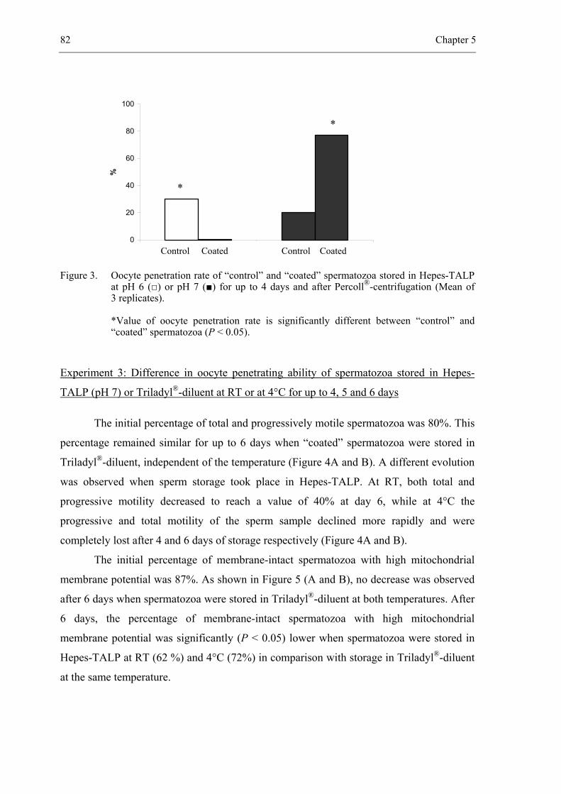
82 Chapter 5
0
20
40
60
80
100%
Figure 3. Oocyte penetration rate of “control” and “coated” spermatozoa stored in Hepes-TALP at pH 6 (□) or pH 7 (■) for up to 4 days and after Percoll®-centrifugation (Mean of 3 replicates).
*Value of oocyte penetration rate is significantly different between “control” and “coated” spermatozoa (P < 0.05).
Experiment 3: Difference in oocyte penetrating ability of spermatozoa stored in Hepes-
TALP (pH 7) or Triladyl®-diluent at RT or at 4°C for up to 4, 5 and 6 days
The initial percentage of total and progressively motile spermatozoa was 80%. This
percentage remained similar for up to 6 days when “coated” spermatozoa were stored in
Triladyl®-diluent, independent of the temperature (Figure 4A and B). A different evolution
was observed when sperm storage took place in Hepes-TALP. At RT, both total and
progressive motility decreased to reach a value of 40% at day 6, while at 4°C the
progressive and total motility of the sperm sample declined more rapidly and were
completely lost after 4 and 6 days of storage respectively (Figure 4A and B).
The initial percentage of membrane-intact spermatozoa with high mitochondrial
membrane potential was 87%. As shown in Figure 5 (A and B), no decrease was observed
after 6 days when spermatozoa were stored in Triladyl®-diluent at both temperatures. After
6 days, the percentage of membrane-intact spermatozoa with high mitochondrial
membrane potential was significantly (P < 0.05) lower when spermatozoa were stored in
Hepes-TALP at RT (62 %) and 4°C (72%) in comparison with storage in Triladyl®-diluent
at the same temperature.
Control Coated Control Coated
*
*
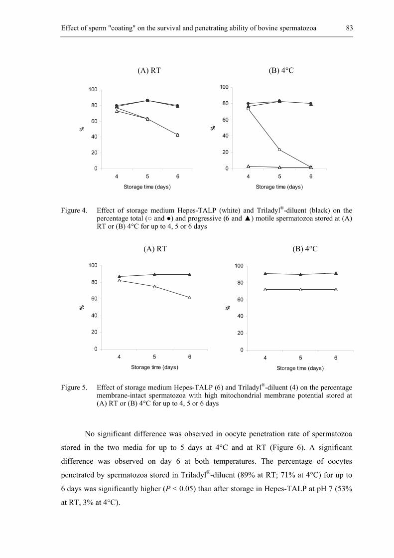
Effect of sperm "coating" on the survival and penetrating ability of bovine spermatozoa 83
Figure 4. Effect of storage medium Hepes-TALP (white) and Triladyl®-diluent (black) on the percentage total (○ and ●) and progressive (6 and ▲) motile spermatozoa stored at (A) RT or (B) 4°C for up to 4, 5 or 6 days
Figure 5. Effect of storage medium Hepes-TALP (6) and Triladyl®-diluent (4) on the percentage membrane-intact spermatozoa with high mitochondrial membrane potential stored at (A) RT or (B) 4°C for up to 4, 5 or 6 days
No significant difference was observed in oocyte penetration rate of spermatozoa
stored in the two media for up to 5 days at 4°C and at RT (Figure 6). A significant
difference was observed on day 6 at both temperatures. The percentage of oocytes
penetrated by spermatozoa stored in Triladyl®-diluent (89% at RT; 71% at 4°C) for up to
6 days was significantly higher (P < 0.05) than after storage in Hepes-TALP at pH 7 (53%
at RT, 3% at 4°C).
(A) RT (B) 4°C
0
20
40
60
80
100
4 5 6
Storage time (days)
%
0
20
40
60
80
100
4 5 6
Storage time (days)
%
0
20
40
60
80
100
4 5 6
Storage time (days)
%
0
20
40
60
80
100
4 5 6
Storage time (days)
%
(A) RT (B) 4°C
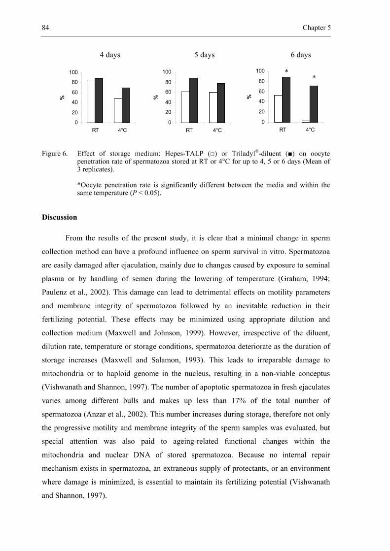
84 Chapter 5
Figure 6. Effect of storage medium: Hepes-TALP (□) or Triladyl®-diluent (■) on oocyte penetration rate of spermatozoa stored at RT or 4°C for up to 4, 5 or 6 days (Mean of 3 replicates).
*Oocyte penetration rate is significantly different between the media and within the same temperature (P < 0.05).
Discussion
From the results of the present study, it is clear that a minimal change in sperm
collection method can have a profound influence on sperm survival in vitro. Spermatozoa
are easily damaged after ejaculation, mainly due to changes caused by exposure to seminal
plasma or by handling of semen during the lowering of temperature (Graham, 1994;
Paulenz et al., 2002). This damage can lead to detrimental effects on motility parameters
and membrane integrity of spermatozoa followed by an inevitable reduction in their
fertilizing potential. These effects may be minimized using appropriate dilution and
collection medium (Maxwell and Johnson, 1999). However, irrespective of the diluent,
dilution rate, temperature or storage conditions, spermatozoa deteriorate as the duration of
storage increases (Maxwell and Salamon, 1993). This leads to irreparable damage to
mitochondria or to haploid genome in the nucleus, resulting in a non-viable conceptus
(Vishwanath and Shannon, 1997). The number of apoptotic spermatozoa in fresh ejaculates
varies among different bulls and makes up less than 17% of the total number of
spermatozoa (Anzar et al., 2002). This number increases during storage, therefore not only
the progressive motility and membrane integrity of the sperm samples was evaluated, but
special attention was also paid to ageing-related functional changes within the
mitochondria and nuclear DNA of stored spermatozoa. Because no internal repair
mechanism exists in spermatozoa, an extraneous supply of protectants, or an environment
where damage is minimized, is essential to maintain its fertilizing potential (Vishwanath
and Shannon, 1997).
0
20
40
60
80
100
RT 4°C
%
0
20
40
60
80
100
RT 4°C
%
0
20
40
60
80
100
RT 4°C
%
4 days 5 days 6 days
**
Effect of sperm "coating" on the survival and penetrating ability of bovine spermatozoa 85
In our experiments, an attempt was made to prolong the survival of stored
spermatozoa by stabilizing the sperm plasma membrane and by protecting them against the
destructive activity of seminal plasma. Bovine seminal plasma can cause permanent
changes in ejaculated spermatozoa (Graham, 1994), even after exposure times of less than
15 min (Dott et al., 1979). Prolonged exposure markedly reduces sperm quality and
permanently diminishes their fertilizing capacity (Mortimer, 1994). Therefore, it is
essential that seminal plasma is removed quickly and efficiently. Washing spermatozoa by
centrifugal sedimentation and resuspension in fresh medium is generally held to be the
quickest and most effective method of removing seminal plasma, however this method can
cause damage to the spermatozoa (Harrison and White, 1972). In our experiments, the
contact time between seminal plasma and spermatozoa was limited by collecting and
diluting bull semen directly in Triladyl®-diluent supplemented with egg yolk and glycerol.
The protective mechanism of egg yolk against the detrimental components of seminal
plasma seems to rely on the low-density lipoproteins, which protect spermatozoa (Shannon
and Curson, 1983) by competing in binding to the sperm membrane. This binding of egg
yolk lipoproteins is rather effective, since its outcome was still visible after 4 days of
storage in a simple defined salt solution Hepes-TALP (pH 7). This was evidenced by the
fact that approximately 30% more spermatozoa of the “coated” sperm sample maintained
an intact membrane, a high mitochondrial membrane potential and intact DNA. This
beneficial effect could not be replaced by collecting semen in egg yolk free diluent and
could therefore not be attributed to mere seminal plasma dilution. However, this simple
action of sperm “coating”, which was obvious after 4 days of storage, was not efficient in
preventing the rapid decline in the percentage of motile spermatozoa. The reduction in
sperm movement is probably the result of mitochondrial damage because sperm motility is
associated with healthy mitochondria (Donnelly et al., 2000). This implicates also that the
motility apparatus was not protected by “coating”, which was confirmed by the finding that
mitochondrial injury, was greater than that of nuclear DNA. This also corroborates with
the fact that a number of crucial events in apoptosis commence in the mitochondria and
could be detectable before DNA fragmentation occurs (Donnelly et al., 2000).
Coating of spermatozoa can therefore be used to store liquid semen in a simple salt
solution without any further addition of membrane protective agents. In our hands, this
leads to a significant higher recovery (55%) of progressively motile and membrane- intact
spermatozoa with a high mitochondrial membrane potential selected after density gradient
centrifugation. The oocyte penetration potential of these superior “coated” spermatozoa
86 Chapter 5
became obvious by their significantly higher oocyte penetration rate, which exceeded that
of “control” spermatozoa with 57%.
Unexpectedly, after 4 days of storage no improvement on sperm characteristics and
even a decline in oocyte penetration rate was observed when “coated” spermatozoa were
stored in Hepes-TALP at pH 6. Under these conditions, a decline in mitochondrial
membrane potential was observed without a corresponding decrease in membrane and
DNA integrity. This damage to the regulatory mechanisms responsible for motility, leads
to a very low concentration (5%) of superior spermatozoa obtained after Percoll®-
centrifugation, whereas most of the spermatozoa were recovered at the interface of the
45 and 90% Percoll®-layers and were nonmotile. It is difficult to speculate to the causes of
this finding since it is known that egg yolk has a constant pH of about 6.0, which normally
does not change much during storage.
Our results indicate that sperm “coating” seems to reduce storage-dependent ageing
processes of spermatozoa stored in a simple salt solution during 4 days. The oocyte
penetration rate (80%) was preserved for up to 4 days at RT, but decreased to 50% at day
6. These rates were comparable to the oocyte penetration rates after aerobic sperm storage
into CAPROGEN® commercial diluent supplemented with 5% egg yolk (Livestock
Improvement Corporation, Hamilton, New Zealand) (88% after 3 days and 52% after
7 days) (Krzyzosiak et al., 2001). The decrease in oocyte penetration rate after storage for
up to 6 days could be explained by the fact that all spermatozoa were recovered at the
interface of the 45 and 90% Percoll®-layers, indicating that less motile spermatozoa were
available for in vitro oocyte penetration. Therefore, it was investigated whether the
replacement of Hepes-TALP by Triladyl®-diluent could improve sperm survival. Low-
density lipoproteins of egg yolk bind firmly with the sperm plasma membrane and stabilize
this membrane both during storage of spermatozoa at RT and during cold shock
(Vishwanath et al., 1992). Salamon and Maxwell (2000) have also shown that egg yolk
reduces the loss of acrosomal enzymes and prevents degenerative changes in the acrosome
during liquid storage. This was confirmed by the fact that in our study, no decrease in
oocyte penetration was found after 6 days of storage in Triladyl®-diluent.
Another important factor that affects sperm function after ejaculation is the
temperature at which spermatozoa were stored after dilution. Care must be taken not to
subject spermatozoa to cold shock, which occurs when freshly ejaculated semen is cooled
quickly to temperatures below 16°C (Halangk et al., 1982). The evidence of the importance
of protecting spermatozoa during cold shock was confirmed in the last experiment.
Effect of sperm "coating" on the survival and penetrating ability of bovine spermatozoa 87
Previously stabilizing the membrane of spermatozoa before cooling was not sufficient to
protect spermatozoa during storage in Hepes-TALP at 4°C. In this case, only the
membrane integrity of “coated” spermatozoa was protected, without a corresponding
protection of the regulatory mechanisms responsible for motility, leading to a sharp decline
in motility after 4 days and in oocyte penetration rate after 6 days. Therefore, it remains
necessary to protect the sperm plasma membrane, with low-density lipoproteins of egg
yolk and glycerol (Vishwanath et al., 1992) through the critical temperature zones and
during storage at 4°C for longer than 5 days.
In conclusion, spermatozoa “coated” during ejaculation for less than 5 minutes with
Triladyl®-diluent yield significantly higher oocyte penetration rates than “control”
spermatozoa after storage for 4 days in Hepes-TALP at pH 7. Further improvements were
made by additional protection of spermatozoa during storage in egg yolk containing
diluent. These results, in combination with a better understanding of the causes of sperm
ageing, are very promising to improve preservation and oocyte penetrating ability of liquid
bovine semen.
Acknowledgments
This research was supported by the Ministry of Agriculture of the Belgian
government, grant n° S6012 and by the Research Fund of Ghent University, grant
n° 011B8698. We also thank Dr. M. Coryn for critical reading, and G. Spaepen and
J. Mestach for technical assistance.
88 Chapter 5
References
Anzar M, He L, Buhr MM, Kroetsch TG, Pauls KP. Sperm apoptosis in fresh and cryopreserved
bull semen detected by flow cytometry and its relationship with fertility. Biol Reprod 2002;
66: 354-360.
Cumming JM, Jequier AM, Raymond K. Molecular biology of human male infertility: Links with
aging, mitochondrial genetics, and oxidative stress? Mol Reprod Dev 1994; 37: 345-362.
De Pauw IMC, Van Soom A, Mintiens K, Verberckmoes S, de Kruif A. In vitro survival of bovine
spermatozoa stored at room temperature under epididymal conditions. Theriogenology 2003;
59: 1093-1107.
Donnelly ET, O’Connell M, McClure N, Lewis SEM. Differences in nuclear DNA fragmentation
and mitochondrial integrity of semen and prepared human spermatozoa. Hum Reprod 2000;
15: 1552-1561.
Dott HM, Harrison RAP, Foster GCA. The maintenance of motility and the surface properties of
epididymal spermatozoa from bull, rabbit and ram in homologous seminal and epididymal
plasma. J Reprod Fertil 1979; 55: 113-124.
Foote RH, Parks JE. Factors affecting preservation and fertility of bull sperm: a brief review.
Reprod Fertil Dev 1993; 5: 665-673.
Garner DL, Thomas CA. Organelle-specific probe JC-1 identifies membrane potential differences
in the mitochondrial function of bovine sperm. Mol Reprod Dev 1999; 53: 222-229.
Graham JK. Effect of seminal plasma on the motility of epididymal and ejaculated spermatozoa of
the ram and bull during the cryopreservation process. Theriogenology 1994; 41: 1151-1162.
Halangk W, Frank K, Bohnensack R. Effect of temperature decrease on the intactness of the cell
membrane of ejaculated bull spermatozoa. Acta Biol Med Ger 1982; 41: 715-719.
Harrison RA and White IG. Glycolytic enzymes in the spermatozoa and cytoplasmic droplets of
bull, boar and ram, and their leakage after shock. J Reprod Fertil 1972; 30: 105-115.
Krzyzosiak J, Molan P, McGowan L, Vishwanath R. Effect of sperm number and oxygenation state
of the storage media on in vitro fertility of bovine sperm stored at ambient temperature.
Theriogenology 2001; 55: 1401-1415.
Maxwell WM, Salamon S. Liquid storage of ram semen: a review. Reprod Fertil Dev 1993; 5: 613-
638.
Maxwell WM, Johnson LA. Physiology of spermatozoa at high dilution rates: the influence of
seminal plasma. Theriogenology 1999; 52: 1353-1362 (review).
Effect of sperm "coating" on the survival and penetrating ability of bovine spermatozoa 89
Mortimer D. Sperm recovery techniques to maximize fertilizing capacity. Reprod Fertil Dev 1994;
6: 25-31.
Moussa M, Martinet V, Trimeche A, Tainturier D, Anton M. Low density lipoproteins extracted
from hen egg yolk by an easy method: cryoprotective effect on frozen-thawed bull semen.
Theriogenology 2002; 57: 1695-1706.
Ohgoda O, Niwa K, Yuhara M, Takahashi S, Kanoya K. Variations in penetration rates in vitro of
bovine follicular oocytes do not reflect conception rates after artificial insemination using
frozen semen from different bulls. Theriogenology 1988; 29: 1375-1381.
Paulenz H, Söderquist L, Pérez-Pé R, Andersen Berg K. Effect of different extenders and storage
temperatures on sperm viability of liquid ram semen. Theriogenology 2002; 57: 823-836.
Salamon S, Maxwell WM. Storage of ram semen. Anim Reprod Sci 2000; 62: 77-111 (review).
Shannon P, Curson B. Toxic effect and action of dead sperm on diluted bovine semen. J Dairy Sci
1972; 55: 614-620.
Shannon P, Curson B. Effect of egg yolk levels on the fertility of diluted bovine sperm stored at
ambient temperatures. N Z J Agric Res 1983; 26: 187-189.
Thomas CA, Garner DL, Dejarnette JM, Marshall CE. Effect of cryopreservation on bovine sperm
organelle function and viability as determined by flow cytometry. Biol Reprod 1998; 58:
786-793.
Van Soom A, Yuan YQ, Peelman LJ, de Matos DG, Dewulf J, Laevens H, de Kruif A. Prevalence
of apoptosis and inner cell allocation in bovine embryos cultured under different oxygen
tensions with or without cysteine addition. Theriogenology 2002; 57: 1453-1465.
Vishwanath R, Shannon P, Curson B. Cationic extracts of egg yolk and their effects on motility,
survival and fertilising ability of bull sperm. Anim Reprod Sci 1992; 29: 185-194.
Vishwanath R, Shannon P. Do sperm cells age? A review of the physiological changes in sperm
during storage at ambient temperatures. Reprod Fertil Dev 1997; 9: 321-331.
Vishwanath R, Shannon P. Storage of bovine semen in liquid and frozen state. Anim Reprod Sci
2000; 62: 23-53.

__________________________________________________ 6.
HORMONAL REGULATION OF BOVINE SECRETORY PROTEINS DERIVED
FROM CAPUT AND CAUDA EPIDIDYMAL EPITHELIAL CELL CULTURES.
Accepted for publication in Journal of Andrology 2003; 24 (3)
De Pauw I., Goff A., Van Soom A., Verberckmoes S. and de Kruif A.
92 Chapter 6
Abstract
The aim of this study was to investigate the effect of hormones (testosterone,
dihydrotestosterone (DHT) and hydrocortisone) on protein secretion of caput and cauda
epididymal epithelial cells cultured in principal cell medium (PCM). A confluent
monolayer of caput and cauda epididymal epithelial cells was obtained in serum-
containing PCM in presence or absence of hormones after 7 days of culture at 38.5°C
(5% CO2 in air). Protein secretion of epididymal epithelial monolayers incubated in serum-
free PCM for 3 days was examined. The secreted proteins were separated by two-
dimensional sodium dodecyl sulphate-polyacrylamide gel electrophoresis (2D-SDS-
PAGE). Comparison of the different protein patterns showed 61 spots in total of which 11
were only secreted in the presence of hormones, 3 appeared to show hormone-related
changes and 25 were region specific. Most of these secreted proteins were low molecular
weight acidic proteins. To obtain evidence of the epididymal origin of the secreted
proteins, proteins present in caput and cauda epididymal plasma were analysed. In
conclusion, our data indicate that hormones influence the synthesis of a number of caput
and cauda epididymal proteins. Some of these proteins could be important for improving
our understanding of sperm maturation and storage, and their acquisition of fertilizing
ability.
Hormonal regulation of proteins secreted by epididymal epithelial cell cultures 93
Introduction
The mammalian epididymis is a complex organ where spermatozoa are matured
and stored. The composition of luminal fluid varies along the length of the epididymis.
This internal milieu is derived from rete testis fluid and is modified by the absorptive and
secretory activity of the epididymal epithelium (Moore et al, 1990; Setchell et al, 1994).
Specific secretions from epididymal principal cells associate with spermatozoa during their
maturation and storage (Sylvester et al., 1991; Moore, 1996) and play a fundamental role
in modifying the surface characteristics of spermatozoa in preparation for the events of
fertilization. Most of these epididymal processes seem to be dependent on androgens,
which are derived from the rete testis and the blood circulation. The most active regulators
responsible for maintaining epididymal structure and other epididymal functions are
testosterone and 5α-reductase metabolite dihydrotestosterone (DHT) (Robaire and Viger,
1995). These androgens play an important role in controlling the synthesis of a number of
specific proteins by the epididymis (Brooks and Higgins, 1980; Jones et al, 1981, 1982).
Some of these proteins are absorbed or integrated into the sperm plasma membrane during
epididymal transit. Glycoproteins are the most prominent of these macromolecules
(Bongso and Trounson, 1996). On the other hand several sperm plasma membrane proteins
of testicular origin are lost or altered during epididymal transit of the spermatozoa.
Because the regulation of epididymal epithelial functions and their effects on spermatozoa
are still poorly understood, cell cultures of epididymal epithelial cells can provide useful
information on epididymis and spermatozoal interaction as evidenced from studies in
human and laboratory animals (Moore et al, 1986, 1992; Bongso and Trounson, 1996;
Moore, 1996; Akhondi et al, 1997). Akhondi and Moore (1993) have shown that principal
cells can remain polarized and continue to secrete proteins for many days, sometimes
weeks, in the presence of androgens and there is seldom fibroblast overgrowth. Effect of
androgens on protein synthesis by the epididymis of laboratory animals and humans has
already been demonstrated by many investigators, as summarized by Holland and Orgebin-
Crist (1988). However, similar data on the bovine epididymis are lacking.
Recently, prolonged cultures of epididymal cells have also been established in
cattle (Gagnon et al, 2000; Reyes-Moreno et al, 2000). Moreover, it has been shown that
motility of frozen-thawed spermatozoa was partially preserved for 48 h after co-culture
with either caput, corpus or cauda epididymal cells, whereas conditioned medium (Gagnon
et al, 2000) or bovine epididymal plasma (Reyes-Moreno et al, 2002) was only effective in
94 Chapter 6
preserving sperm motility for 6 h. It was concluded that epididymal epithelial cells secrete
one or more beneficial compounds, which prolong sperm viability. This compound must be
a common factor present in epididymal plasma and secreted by epididymal epithelial cells
cultured in vitro. A number of epididymal proteins may be involved with the protection of
ejaculated spermatozoa during in vitro storage (Reyes-Moreno et al, 2002). The aim of our
study was to investigate which proteins:
1) are influenced by the presence of testosterone, dihydrotestosterone and
hydrocortisone in the culture medium,
2) are common to conditioned media of caput and cauda epididymal epithelial cell
cultures,
3) are common to conditioned media of caput or cauda epididymal cell cultures, and
epididymal plasma.
Materials and Methods
Medium
Principal cell medium (PCM) consisted of RPMI-1640 medium (Life Technologies,
Gibco BRL products (Merelbeke, Belgium)) supplemented with 10% fetal calf serum
(FCS), 100 nM insulin, 1 mM sodium pyruvate, 5 µg/ml transferrin, 200 nM
hydrocortisone, 200 nM testosterone, 1 µM dihydrotestosterone, 1 µg/ml retinol and
50 µg/ml gentamycin. Testosterone and DHT were used because they control the synthesis
and secretion of a number of specific proteins by the epididymis and maintain the
epididymal stucture and functions (Robaire and Viger 1995). Hydrocortisone promotes cell
attachment (Ballard and Tomkins 1969) and cell proliferation (Guner et al. 1977). Principal
cell medium without hydrocortisone, testosterone and DHT was used as control.
Collection of caput and cauda epididymal plasma
Epididymides of beef bulls aged 2-2.5 years were collected at a local
slaughterhouse. After removing superficial blood and tissue fluid contamination, the
pressure in the epididymal ducts was increased by clamping two pincers on the proximal
and distal part of the caput and cauda epididymidis. Incisions were made in the connective
tissue avoiding small blood vessels. Epididymal plasma was oozed out, aspirated into a
fine pipette and transferred into a small tube. After 5 min of centrifugation at 3.214 x g, the
Hormonal regulation of proteins secreted by epididymal epithelial cell cultures 95
epididymal plasma was removed, checked visually for the absence of spermatozoa and
frozen at - 20°C.
Epididymal epithelial cell culture
The epididymal epithelial cell culture was prepared by a modified protocol
according to Moore et al (1986). The epididymal tissue was obtained from bulls
slaughtered in a local slaughterhouse. On arrival in the laboratory, the caput and cauda
epididymidis from one bull were dissected free of the testis, fat and connective tissue and
washed in RPMI-1640 to remove blood. Both regions were minced in small segments of
1-2 mm using scissors and placed in PCM (Moore et al, 1992). Spermatozoa within tubule
segments were teased out using forceps. The tubule segments were then incubated in PCM
containing 1.5 mg/ml collagenase type II (Sigma, Bornem, Belgium) at 38.5°C in 5% CO2
in air for 2 h. After enzymatic digestion, tubule segments were dissected free of
surrounding collagen with a needle, washed again in PCM to remove remaining
spermatozoa and transferred to fresh medium. They were slit open longitudinally and cut
into small fragments. These fragments were prepared from both caput and cauda
epididymidis and cultured separately in PCM supplemented with fibronectin (2µl/ml)
(Sigma, Bornem, Belgium) and in PCM without hormones, and supplemented with
fibronectin (2µl/ml) (control) at 38.5°C and 5% CO2 in air. The culture medium was
changed every other day. The fragments formed irregular contiguous spheres of epithelial
cells with apical surface facing outwards and remained free floating in culture during the
first days. These spheres became attached to the dish and the epithelial cells spread out
(Moore et al, 1992). Attached explants were carefully removed with a 26-gauge needle
connected to a 1-ml syringe. Epithelial cells continued to divide forming a monolayer after
5 to 7 days of culture. The medium was then replaced by serum-free PCM with or without
hormones. After 3 days of incubation, cultures were examined for epithelial cell
detachment by means of inverted light microscopy. Epithelial and fibroblast cell
concentration of the monolayer was examined by immunohistochemistry. The medium was
collected from the different groups and stored at -20°C until analysis by two-dimensional
sodium dodecyl sulphate polyacrylamide gel electrophoresis (2D-SDS-PAGE).
96 Chapter 6
Immunohistochemistry
Monolayers cultured on glass coverslips are rinsed with phosphate-buffered saline
(PBS), fixed for 10 min in acetone and air-dried. After rinsing the slides 3 times with PBS,
the cells were incubated for 2 h with an anticytokeratin antibody (Keratin Pan Ab-1,
NeoMarkers, CA, USA) diluted 1/500 in PBS at 37°C, to establish the proportion of
epithelial cells (Henriksen et al, 1990). Followed by washing the epithelial cells for 10 min
in PBS and incubating with a biotin-conjugated goat anti-mouse immunoglobulin antibody
at RT for 30 min. After washing the slides in PBS for 10 min, the cells were incubated with
Strept-ABComplex/horseradish peroxidase at RT for 30 min. Horseradish peroxidase
activity was visualised by incubating the slides with 3,3’-diaminobenzidine
tetrahydrochloride (DAB tablets, Sigma) 0.5 mg/ml Tris-HCl buffer, pH 7.6 containing
0.02% hydrogen peroxide for 10 s resulting in brown staining. After washing, cells were
counterstained with Mayer’s Hemaluin solution (VWR International, Leuven, Belgium),
washed, coversliped and viewed with light microscopy (Leica DMR, Van Hopplynus N.V.,
Brussels, Belgium). Another replicate of epididymal cultures was probed with antibody
against vimentin (Vimentin Ab-2 (V9), NeoMarkers, CA, USA) to establish the proportion
of fibroblasts. The epithelial and fibroblast cell concentration was measured with the
Image Database Program of Leica (Van Hopplynus NV, Brussels, Belgium).
Two-Dimensional Gel Electrophoresis
Before separation and analysis of the proteins by means of 2D-SDS-PAGE, the
proteins of the conditioned media were concentrated using Ultrafree-15 concentrators
(5000 MW cutoff; Millipore). The protein concentration of the different samples was
measured using the Bio-Rad protein assay reagent. Proteins were then added to IPG buffer
(8M Urea, 2% CHAPS, 0.5% IPG Buffer (pH 3-10), bromophenol blue, 65 mM
Dithiothreitol (DTT)) to give a final concentration of 2µg/100µl IPG buffer. The separation
in first dimension was carried out using immobiline DryStrips that had been rehydrated in
250 µl of the sample/IPG buffer solution (5 µg total protein) for at least 10 h in an
Immobiline™ Drystrip Reswelling Tray. The samples were then separated on a MultiPhore
II flatbed system for 16 h at 15°C. The voltage was 300 V for the first 3 h, from 300 to
2000 V during the following 5 h, and finally 8 h at 2000 V. Before the second dimension
was performed, the dry strips were first equilibrated for 10 min in Equilibration solution 1
(0.5 M Tris/HCl (pH 6.8) containing 0.36 g/ml urea, 10 mg/ml SDS, 2,5 mg/ml (DTT) and
Hormonal regulation of proteins secreted by epididymal epithelial cell cultures 97
26% glycerol) and another 10 min in Equilibration solution 2 (0.5 M Tris/HCl (pH 6.8)
containing 0.36 g/ml urea, 10 mg/ml SDS, 45 mg/ml iodoacetamid and 26% glycerol). The
second dimension was performed after placing the strips on Pharmacia ExcelGel® XL SDS
12-14 using the MultiPhore II flatbed system for 3-4 h at 15°C. After running the gels, they
were immediately immersed in fixing solution (50% methanol, 10% acetic acid in water)
and stained with silver nitrate (Silver Staining Kit, Amersham Pharmacia Biotech AB, Baie
d’Urfé, Canada). Gels were compared and the molecular weight (MW) and isoelectric
point (pI) of the proteins were calculated using the Phoretix 2-D SDS PAGE Analysis
Software (Phoretix, Newcastle-upon-Tyne, England).
This experiment was repeated with three different caput and cauda epithelial cell
cultures. A representative protein pattern of each culture condition was obtained by
comparing scans of three gels of each culture, and only spots present on the three gels were
taken into account. The concentration of each spot was expressed as its spot volume, which
is the product of the area of the spot and its total optical density. Differences in spot
density between cultures was determined by Students t-test.
Results
Formation of cell cultures
A confluent monolayer of caput and caudal part of the epididymis was successfully
obtained after 5 to 7 days of culture in serum-containing PCM at 38.5°C in 5% CO2 in air
with or without the presence of hormones. From day 7 until day 10 the medium was
replaced by serum-free PCM with or without hormones. Under all cell culture conditions,
there was no epithelial cell detachment for the first 10 days in culture. Primary cell cultures
of caput and cauda epithelial cells can be maintained for up to 10 days without substantial
overgrowth of fibroblasts, as determined with antibodies to vimentin. More than 90% of
the cells in the monolayer were epithelial cells as identified by the presence of cytokeratin
(results not shown). No significant difference in mean epithelial cell concentration of
3 replicates was observed between hormone-containing cultures (caput = 398040 epithelial
cells/1.77 cm²; cauda = 346680 epithelial cells/1.77 cm²) and hormone-free cultures (caput
= 369792 epithelial cells/1.77 cm²; cauda = 333840 epithelial cells/1.77 cm²) (ANOVA,
SPSS 10.0 for Windows).
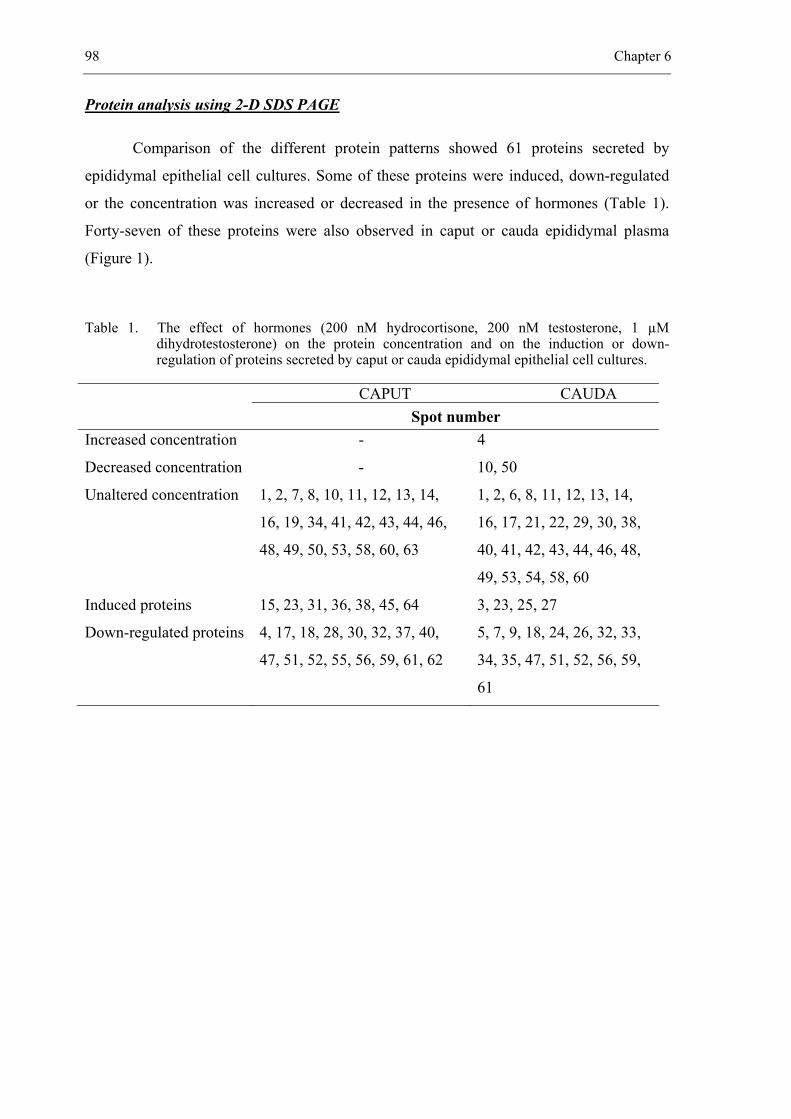
98 Chapter 6
Protein analysis using 2-D SDS PAGE
Comparison of the different protein patterns showed 61 proteins secreted by
epididymal epithelial cell cultures. Some of these proteins were induced, down-regulated
or the concentration was increased or decreased in the presence of hormones (Table 1).
Forty-seven of these proteins were also observed in caput or cauda epididymal plasma
(Figure 1).
Table 1. The effect of hormones (200 nM hydrocortisone, 200 nM testosterone, 1 µM dihydrotestosterone) on the protein concentration and on the induction or down-regulation of proteins secreted by caput or cauda epididymal epithelial cell cultures.
CAPUT CAUDA Spot number
Increased concentration - 4
Decreased concentration - 10, 50
Unaltered concentration 1, 2, 7, 8, 10, 11, 12, 13, 14,
16, 19, 34, 41, 42, 43, 44, 46,
48, 49, 50, 53, 58, 60, 63
1, 2, 6, 8, 11, 12, 13, 14,
16, 17, 21, 22, 29, 30, 38,
40, 41, 42, 43, 44, 46, 48,
49, 53, 54, 58, 60
Induced proteins 15, 23, 31, 36, 38, 45, 64 3, 23, 25, 27
Down-regulated proteins 4, 17, 18, 28, 30, 32, 37, 40,
47, 51, 52, 55, 56, 59, 61, 62
5, 7, 9, 18, 24, 26, 32, 33,
34, 35, 47, 51, 52, 56, 59,
61
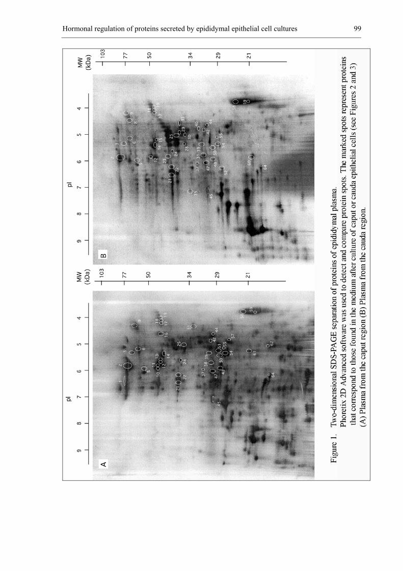
Hormonal regulation of proteins secreted by epididymal epithelial cell cultures 99
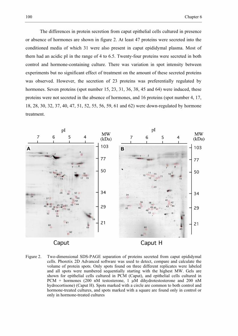
100 Chapter 6
The differences in protein secretion from caput epithelial cells cultured in presence
or absence of hormones are shown in figure 2. At least 47 proteins were secreted into the
conditioned media of which 31 were also present in caput epididymal plasma. Most of
them had an acidic pI in the range of 4 to 6.5. Twenty-four proteins were secreted in both
control and hormone-containing culture. There was variation in spot intensity between
experiments but no significant effect of treatment on the amount of these secreted proteins
was observed. However, the secretion of 23 proteins was preferentially regulated by
hormones. Seven proteins (spot number 15, 23, 31, 36, 38, 45 and 64) were induced, these
proteins were not secreted in the absence of hormones, and 16 proteins (spot number 4, 17,
18, 28, 30, 32, 37, 40, 47, 51, 52, 55, 56, 59, 61 and 62) were down-regulated by hormone
treatment.
Figure 2. Two-dimensional SDS-PAGE separation of proteins secreted from caput epididymal cells. Phoretix 2D Advanced software was used to detect, compare and calculate the volume of protein spots. Only spots found on three different replicates were labeled and all spots were numbered sequentially starting with the highest MW. Gels are shown for epithelial cells cultured in PCM (Caput), and epithelial cells cultured in PCM + hormones (200 nM testosterone, 1 µM dihydrotestosterone and 200 nM hydrocortisone) (Caput H). Spots marked with a circle are common to both control and hormone-treated cultures, and spots marked with a square are found only in control or only in hormone-treated cultures
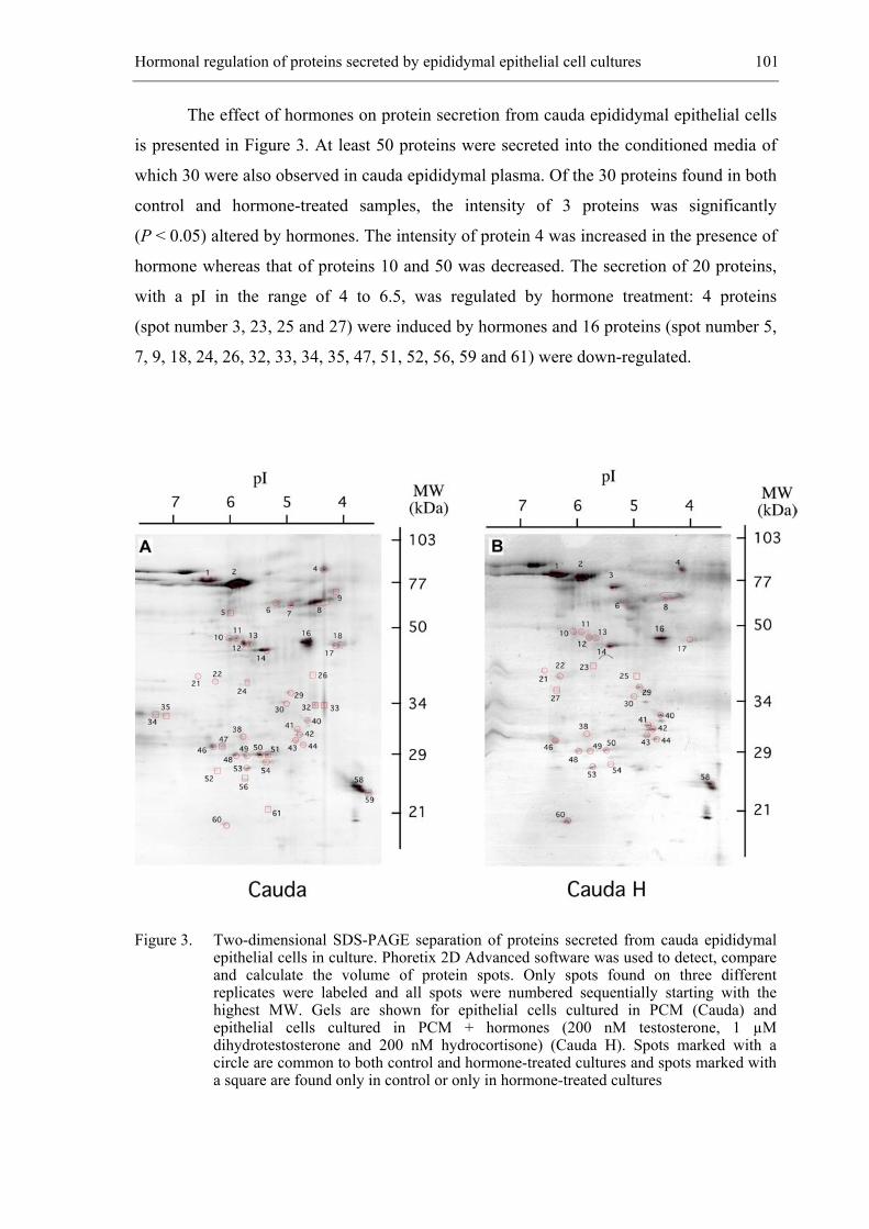
Hormonal regulation of proteins secreted by epididymal epithelial cell cultures 101
The effect of hormones on protein secretion from cauda epididymal epithelial cells
is presented in Figure 3. At least 50 proteins were secreted into the conditioned media of
which 30 were also observed in cauda epididymal plasma. Of the 30 proteins found in both
control and hormone-treated samples, the intensity of 3 proteins was significantly
(P < 0.05) altered by hormones. The intensity of protein 4 was increased in the presence of
hormone whereas that of proteins 10 and 50 was decreased. The secretion of 20 proteins,
with a pI in the range of 4 to 6.5, was regulated by hormone treatment: 4 proteins
(spot number 3, 23, 25 and 27) were induced by hormones and 16 proteins (spot number 5,
7, 9, 18, 24, 26, 32, 33, 34, 35, 47, 51, 52, 56, 59 and 61) were down-regulated.
Figure 3. Two-dimensional SDS-PAGE separation of proteins secreted from cauda epididymal epithelial cells in culture. Phoretix 2D Advanced software was used to detect, compare and calculate the volume of protein spots. Only spots found on three different replicates were labeled and all spots were numbered sequentially starting with the highest MW. Gels are shown for epithelial cells cultured in PCM (Cauda) and epithelial cells cultured in PCM + hormones (200 nM testosterone, 1 µM dihydrotestosterone and 200 nM hydrocortisone) (Cauda H). Spots marked with a circle are common to both control and hormone-treated cultures and spots marked with a square are found only in control or only in hormone-treated cultures
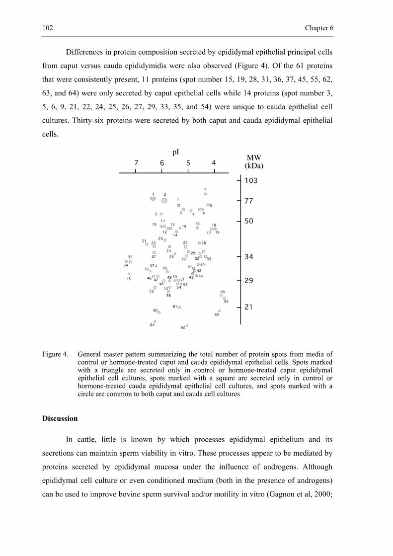
102 Chapter 6
Differences in protein composition secreted by epididymal epithelial principal cells
from caput versus cauda epididymidis were also observed (Figure 4). Of the 61 proteins
that were consistently present, 11 proteins (spot number 15, 19, 28, 31, 36, 37, 45, 55, 62,
63, and 64) were only secreted by caput epithelial cells while 14 proteins (spot number 3,
5, 6, 9, 21, 22, 24, 25, 26, 27, 29, 33, 35, and 54) were unique to cauda epithelial cell
cultures. Thirty-six proteins were secreted by both caput and cauda epididymal epithelial
cells.
Figure 4. General master pattern summarizing the total number of protein spots from media of control or hormone-treated caput and cauda epididymal epithelial cells. Spots marked with a triangle are secreted only in control or hormone-treated caput epididymal epithelial cell cultures, spots marked with a square are secreted only in control or hormone-treated cauda epididymal epithelial cell cultures, and spots marked with a circle are common to both caput and cauda cell cultures
Discussion
In cattle, little is known by which processes epididymal epithelium and its
secretions can maintain sperm viability in vitro. These processes appear to be mediated by
proteins secreted by epididymal mucosa under the influence of androgens. Although
epididymal cell culture or even conditioned medium (both in the presence of androgens)
can be used to improve bovine sperm survival and/or motility in vitro (Gagnon et al, 2000;
Hormonal regulation of proteins secreted by epididymal epithelial cell cultures 103
Reyes-Moreno et al, 2000), this approach is no practical alternative for the development of
a diluent for prolonged storage of liquid bovine semen.
Evidence for viability and normal differentiation of our caput and cauda epididymal
epithelial cell cultures was given by the fact that both regions displayed the same capacity
to form a confluent monolayer of epithelial cells in PCM and remained viable in vitro for
at least 10 days. In contrast with the results of Gagnon et al (2000) and Moore et al (1992),
no signs of cell degeneration, reduced growth or contamination with fibroblast were
apparent in caput and cauda epididymal epithelial cell culture in the absence of hormones.
The cultured epithelial cells exhibited less than 10% contamination with other cell types, as
examined by staining with cytokeratin. This percentage was similar with the observations
made by Gagnon et al (2000).
To improve our insight in the molecular mechanism of sperm protection, we have
focused our attention on proteins present in conditioned media from epididymal cultures of
caput or cauda epithelial cells, in the presence or absence of hormones. We have shown
that 66% and 60% of the proteins present in respectively caput or cauda conditioned media
correspond to proteins secreted in caput or cauda epididymal plasma, which confirms their
epididymal origin. Proteins present in conditioned media while absent in epididymal
plasma could correspond to proteins that are integrated into the sperm plasma membrane
during epididymal transit, or which are degraded or reabsorbed rapidly after secretion.
The majority of secreted proteins had molecular masses between 20 and 80 kDa
and an acidic pI value between 4 and 7. Besides the hormone dependency of protein
synthesis by epididymal cells, substantial differences in protein patterns from caput and
cauda epididymal regions could be detected (Figure 4). In addition to proteins that were
induced uniquely by hormones, secretion of only a few proteins was either enhanced or
reduced by these hormones.
The precise function of these secreted proteins in sperm maturation and storage
processes remains to be established. Indirect evidence for a function can come from their
relative abundance and from their site-specific expression along the epididymal duct. A
further approach could be the identification of some major epididymal proteins by means
of protein microsequencing, recombinant DNA techniques, use of specific antisera (Syntin
et al, 1996), or by investigating their sequence similarities to proteins or protein families of
known functions, and their immunolocalization on the sperm surface (Kirchhoff, 1998).
The concentration of a protein present in epididymal plasma from different regions is not
necessarily related to its level of secretion (Syntin et al, 1996). Proteins present at low
104 Chapter 6
concentration in epididymal plasma may be those that are degraded, reabsorbed or
integrated into the sperm membrane (Syntin et al, 1996) and thus are possibly of greater
interest than more abundant proteins. This is in contrast with the presence of proteins in
conditioned media, where most important proteins were probably those most abundantly
secreted because no reabsorbing or integrating into the sperm membrane could occur.
Proteins found uniquely in caput may be important in influencing specific maturational
changes in spermatozoa as they transit the duct, whereas cauda-specific proteins may be
important for the storage of spermatozoa. Such proteins may also be involved in regulating
the structural and functional integrity of the epididymis itself.
Our results are consistent with the findings of Reyes-Moreno et al (2002), however
no protein identification was done in our study. Reyes-Moreno et al (2002) have
characterized 5 bovine proteins, secreted in cauda epididymal plasma, which could play a
role in sperm protection in vivo. One protein identified as beta-adrenergic receptor kinase 2
corresponds probably to protein 14 in the present study. Protein spots 10 and 11 (of about
48 kDa) may correspond to antithrombin-III and fibrinogen gamma-B chain found by
Reyes Moreno and coworkers (2002). In our study, these proteins were secreted in both
regions of the epididymis. Beta (36-kDa spot) chain of clusterin, which corresponds to
protein number 25 in our study, was only secreted by cauda epididymal epithelial cells in
the presence of androgens. In vivo studies in the bull have shown similar regional secretion
of the beta chain of clusterin (Howes et al., 1998).
It is concluded that androgens are involved in the regulation of protein synthesis by
the epididymis. Identification and localisation of these proteins on spermatozoa could
provide useful information on how spermatozoa can survive and preserve their metabolic
quiescent state in the epididymis. To fully understand the regulation of the epididymis, a
combination of molecular interactions between sperm surface and epididymal epithelium,
ionic composition and physiological conditions of the epididymal plasma must also be
taken into account.
Acknowledgements
This research was supported by the Ministry of Agriculture of the Belgian
Government, grant n° S6012 and by the Flemish Cattle Breeding Association VRV. This
research was also possible by the Bilateral Collaboration Flanders-Quebec BIL 97/ 174 B
2298. The authors wish to thank J. Mestach and G. Spaepen for technical assistance.
Hormonal regulation of proteins secreted by epididymal epithelial cell cultures 105
References
Akhondi MA, Moore HDM. Rat epididymal spermatozoa acquire a 44 kDa component during
maturation in vitro in the presence of epididymal epithelium primary cultures. J Reprod
Fertil Abstr Ser 1993; 12: 106.
Akhondi MA, Chapple C, Moore HD. Prolonged survival of human spermatozoa when co-
incubated with epididymal cell cultures. Hum Reprod 1997; 12: 514-522.
Ballard Pl, Thomkins GM. Dexamethasone and cell adhesion. Nature. 1969; 244: 344-345.
Bongso A, Trounson A. Evaluation of motility, freezing ability and embryonic development of
murine epididymal sperm after coculture with epididymal epithelium. Hum Reprod 1996;
11: 1451-1456.
Brooks DE, Higgins SJ. Characterization and androgen-dependence of proteins associated with
luminal fluid and spermatozoa in the rat epididymis. J Reprod Fert 1980; 59: 363-375.
Gagnon A, Sullivan R, Sirard MA. Epididymal epithelial cells cultured in vitro prolong the motility
of bovine sperm. J Androl 2000; 21: 842-847.
Guner M, Freshney RI, Morgan D, Freshney MG, Thomas DGT, Graham DI. Effects of
dexamethasone and betamethasone on in vitro cultures from human astrocytoma. Br J
Cancer. 1997; 35: 439-447.
Henriksen T, Tanbo T, Abyholm T, Oppedal BR, Claussen OP, Hovig T. Epithelial cells from
human fallopian tube in culture. Hum Reprod 1990; 5: 25-31.
Holland MK, Orgebin-Crist MC. Characterization and hormonal regulation of protein synthesis by
the murine epididymis. Biol Reprod 1988; 38: 487-496.
Howes EA, Hurst S, Laslop A, Jones R. Cellular distribution and molecular heterogeneity of
MAC393 antigen (clusterin, beta-chain) on the surface membrane of bull spermatozoa. Mol
Hum Reprod. 1998; 4: 673-681.
Jones R, von Glos KI, Brown CR. Characterization of hormonally regulated secretory proteins
from the caput epididymidis of the rabbit. Biochem J 1981; 15: 105-114.
Jones R, Fournier-Delpech S, Willadsen SA. Identification of androgen-dependent proteins
synthesized in vitro by the ram epididymis. Reprod Nutr Dev 1982; 22: 495-504.
Kirchhoff C. Molecular characterization of epididymal proteins. Rev Reprod 1998; 3: 86-95.
Moore HDM, Hartman TD, Smith CA. In vitro culture of hamster epididymal epithelium and
induction of sperm motility. J Reprod Fertil 1986; 78: 347-352.
106 Chapter 6
Moore HDM, Curry M, Pryor JP. In vitro culture of epididymal epithelium for the study of
mammalian sperm maturation. In: Lauria A, Gandolfi F (eds). In vitro approaches to
mammalian gamete maturation and embryonic development. Serovet; Rome 1990: 1-9.
Moore HDM, Curry MR, Penfold LM, Pryor JP. The culture of human epididymal epithelium and
in vitro maturation of epididymal spermatozoa. Fertil Steril 1992; 58: 776-783.
Moore HDM. The influence of the epididymis on human and animal sperm maturation and storage.
Hum Reprod Natl Suppl 1996; 11: 103-110.
Reyes-Moreno C, Gagnon A, Sullivan R, Sirard MA. Addition of specific metabolites to bovine
epididymal cell culture medium enhances survival and motility of cryopreserved sperm. J
Androl 2000; 21: 876-886.
Reyes-Moreno C, Boilard M, Sullivan R, Sirard MA. Characterization and identification of
epididymal factors that protect ejaculated bovine sperm during in vitro storage. Biol Reprod
2002; 66: 159-166.
Robaire B, Viger RS. Regulation of epididymal epithelial cell functions. Biol Reprod 1995; 52:
226-236.
Setchell BP, Maddocks S, Brooks DE. Anatomy, vasculature, innervation and fluids of the male
reproductive tract. In: Knobil E, Neill JD (eds). Physiology of Reproduction. Raven Press;
New York 1994: 1063-1176.
Sylvester SR, Morales C, Oko R, Griswold MD. Localization of sulfated glycoprotein-2 (clusterin)
on spermatozoa and in the reproductive tract of the male rat. Biol Reprod 1991; 45: 195-207.
Syntin P, Dacheux F, Druart X, Gatti JL, Okamura N, Dacheux JL. Characterization and
identification of proteins secreted in the various regions of the adult boar epididymis. Biol
Reprod 1996; 55: 956-974.
__________________________________________________ 7.
SPERM BINDING TO EPITHELIAL OVIDUCT EXPLANTS IN BULLS WITH
DIFFERENT NON-RETURN RATES INVESTIGATED WITH A NEW IN VITRO
MODEL
Biology of Reproduction 2002; 67: 1073-1079
De Pauw I., Van Soom A., Laevens H., Verberckmoes S. and de Kruif A.
108 Chapter 7
Abstract
A new in vitro method was developed for analyzing the capacity of spermatozoa to
bind to oviductal epithelium to determine whether this binding capacity could be used to
predict non-return rates (NRR). Sperm binding was evaluated by counting 5,5',6,6'-
tetrachloro-1,1',3,3'-tetraethylbenzimidazolyl-carbocyanine iodide (JC-1)-labeled sperma-
tozoa attached to oviduct explants and by measuring the surface area of oviduct explants
by means of an image analysis program. Hepes + Tyrode albumin lactate pyruvate (TALP)
was a more useful medium than in vitro fertilization (IVF)-TALP, TCM-199 medium +
10% fetal calf serum, and TCM-199 medium alone for the investigation of sperm binding
to oviduct explants. Oviduct explants with a surface area of < 20 000 µm² provided more
consistent results than did explants with a surface area of > 100 000 µm². A positive
association was found between the loge-transformed number of spermatozoa bound to
0.1 mm² oviductal epithelium and the NRR of the respective sires when evaluated after
24 h of sperm-oviduct incubation, provided that the membrane integrity of the sperm
sample was > 60%. Determination of the capacity of spermatozoa to bind to oviduct
explants could become a reliable in vitro method for predicting the NRR of a given sire.
Sperm - oviduct binding assay 109
Introduction
In most mammals, ejaculated spermatozoa pass through the female reproductive
tract to form a functional sperm reservoir in the lower segment of the oviduct
(Yanagimachi, 1994). In cattle, it takes about 6-8 h before enough spermatozoa have
reached this sperm reservoir to ensure fertilization (Hunter and Wilmut, 1982).
Spermatozoa attach to the apical plasma membrane of the ciliated and secretory epithelial
cells (Suarez et al., 1990). This attachment is mediated by fucose recognition (Lefebvre et
al., 1997). Other factors that could be involved in the establishment of a sperm reservoir
are impedance of sperm movement by oviductal mucus (Suarez et al., 1997) and low
patency of the oviduct due to edematous mucosa and tightly contracted myosalpinx
(Hunter, 1997).
The reservoir acts to ensure that enough fertile spermatozoa are available in the
oviduct when ovulation occurs. Bovine spermatozoa can remain arrested in the isthmus for
≥ 18 h and only detach from the epithelium near the time of ovulation (Hunter and Wilmut,
1984). This prolonged sperm survival is especially important in species with longer estrous
periods, such as the horse (Day, 1942), but might also be of interest for cattle, which are
inseminated early in estrus. If a bull is unable to populate the sperm reservoir with a
sufficient number of spermatozoa or for a sufficient period of time, fertility could be
adversely affected. The field fertility of a given bull is expressed by the non-return rate
(NRR), which is defined as the proportion of cows that were inseminated and did not
return for another service within 56 days (den Daas et al., 1998). Differences probably
exist among bulls in their capacity to establish a sperm reservoir after mating or
insemination, but it is difficult to detect these differences after matings and collection of
oviducts because the number of spermatozoa reported to reach the oviduct in vivo has
differed considerably among studies and within experiments (Parker et al., 1975; Mburu
et al., 1996; Suarez et al., 1997).
To determine whether the capacity to establish a reservoir is indicative of fertility,
we searched for an in vitro approach to study the capacity of spermatozoa to bind to
oviductal epithelium. In vitro sperm membrane integrity can be extended by coincubating
spermatozoa with oviductal epithelium (Pollard et al., 1991). To investigate this sperm
binding in more detail, we needed a reliable method to determine the number of
spermatozoa attached to oviduct explants. The aim of this study was to set up a model to
quantify sperm binding to oviduct explants by combining fluorescent staining of
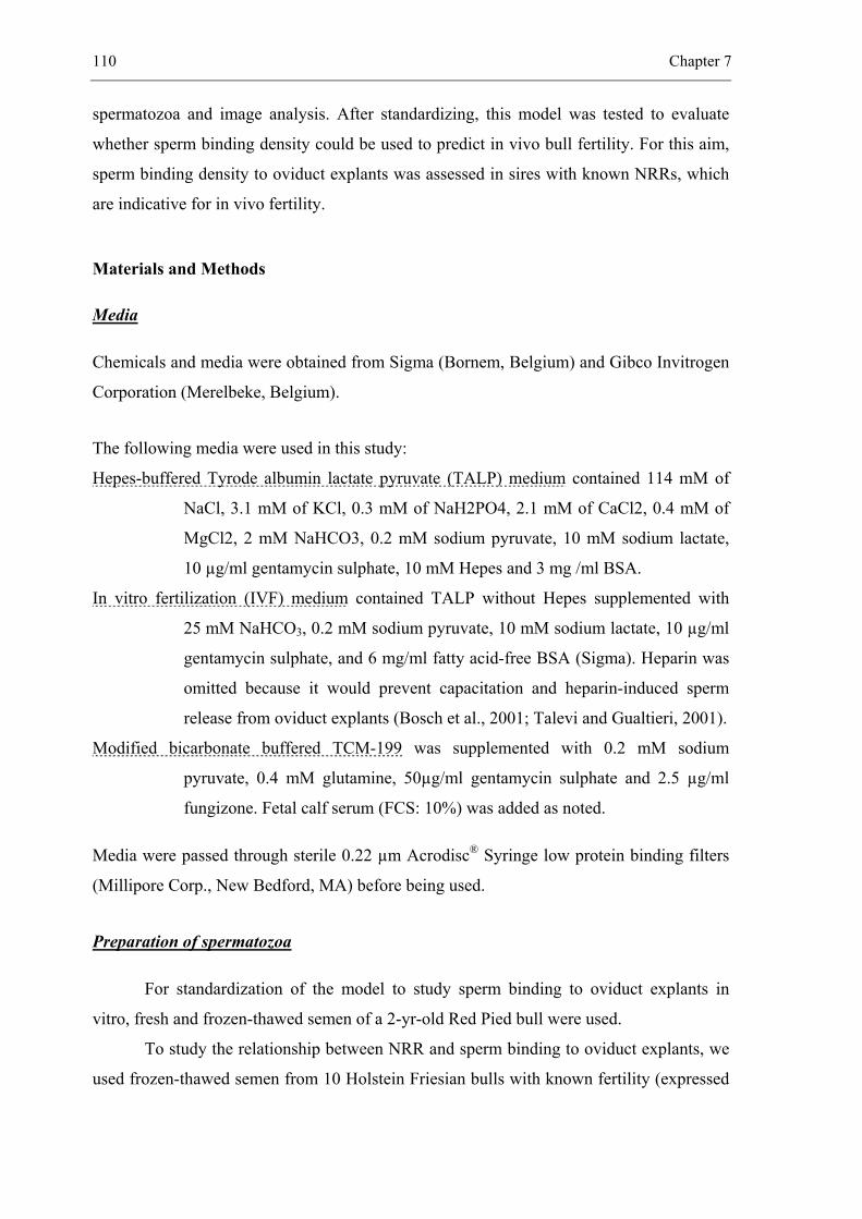
110 Chapter 7
spermatozoa and image analysis. After standardizing, this model was tested to evaluate
whether sperm binding density could be used to predict in vivo bull fertility. For this aim,
sperm binding density to oviduct explants was assessed in sires with known NRRs, which
are indicative for in vivo fertility.
Materials and Methods
Media
Chemicals and media were obtained from Sigma (Bornem, Belgium) and Gibco Invitrogen
Corporation (Merelbeke, Belgium).
The following media were used in this study:
Hepes-buffered Tyrode albumin lactate pyruvate (TALP) medium contained 114 mM of
NaCl, 3.1 mM of KCl, 0.3 mM of NaH2PO4, 2.1 mM of CaCl2, 0.4 mM of
MgCl2, 2 mM NaHCO3, 0.2 mM sodium pyruvate, 10 mM sodium lactate,
10 µg/ml gentamycin sulphate, 10 mM Hepes and 3 mg /ml BSA.
In vitro fertilization (IVF) medium contained TALP without Hepes supplemented with
25 mM NaHCO3, 0.2 mM sodium pyruvate, 10 mM sodium lactate, 10 µg/ml
gentamycin sulphate, and 6 mg/ml fatty acid-free BSA (Sigma). Heparin was
omitted because it would prevent capacitation and heparin-induced sperm
release from oviduct explants (Bosch et al., 2001; Talevi and Gualtieri, 2001).
Modified bicarbonate buffered TCM-199 was supplemented with 0.2 mM sodium
pyruvate, 0.4 mM glutamine, 50µg/ml gentamycin sulphate and 2.5 µg/ml
fungizone. Fetal calf serum (FCS: 10%) was added as noted.
Media were passed through sterile 0.22 µm Acrodisc® Syringe low protein binding filters
(Millipore Corp., New Bedford, MA) before being used.
Preparation of spermatozoa
For standardization of the model to study sperm binding to oviduct explants in
vitro, fresh and frozen-thawed semen of a 2-yr-old Red Pied bull were used.
To study the relationship between NRR and sperm binding to oviduct explants, we
used frozen-thawed semen from 10 Holstein Friesian bulls with known fertility (expressed
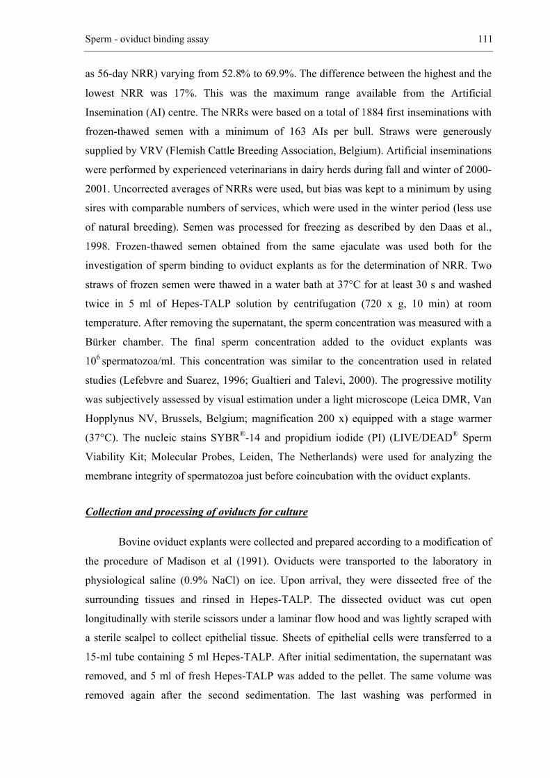
Sperm - oviduct binding assay 111
as 56-day NRR) varying from 52.8% to 69.9%. The difference between the highest and the
lowest NRR was 17%. This was the maximum range available from the Artificial
Insemination (AI) centre. The NRRs were based on a total of 1884 first inseminations with
frozen-thawed semen with a minimum of 163 AIs per bull. Straws were generously
supplied by VRV (Flemish Cattle Breeding Association, Belgium). Artificial inseminations
were performed by experienced veterinarians in dairy herds during fall and winter of 2000-
2001. Uncorrected averages of NRRs were used, but bias was kept to a minimum by using
sires with comparable numbers of services, which were used in the winter period (less use
of natural breeding). Semen was processed for freezing as described by den Daas et al.,
1998. Frozen-thawed semen obtained from the same ejaculate was used both for the
investigation of sperm binding to oviduct explants as for the determination of NRR. Two
straws of frozen semen were thawed in a water bath at 37°C for at least 30 s and washed
twice in 5 ml of Hepes-TALP solution by centrifugation (720 x g, 10 min) at room
temperature. After removing the supernatant, the sperm concentration was measured with a
Bürker chamber. The final sperm concentration added to the oviduct explants was
106 spermatozoa/ml. This concentration was similar to the concentration used in related
studies (Lefebvre and Suarez, 1996; Gualtieri and Talevi, 2000). The progressive motility
was subjectively assessed by visual estimation under a light microscope (Leica DMR, Van
Hopplynus NV, Brussels, Belgium; magnification 200 x) equipped with a stage warmer
(37°C). The nucleic stains SYBR®-14 and propidium iodide (PI) (LIVE/DEAD® Sperm
Viability Kit; Molecular Probes, Leiden, The Netherlands) were used for analyzing the
membrane integrity of spermatozoa just before coincubation with the oviduct explants.
Collection and processing of oviducts for culture
Bovine oviduct explants were collected and prepared according to a modification of
the procedure of Madison et al (1991). Oviducts were transported to the laboratory in
physiological saline (0.9% NaCl) on ice. Upon arrival, they were dissected free of the
surrounding tissues and rinsed in Hepes-TALP. The dissected oviduct was cut open
longitudinally with sterile scissors under a laminar flow hood and was lightly scraped with
a sterile scalpel to collect epithelial tissue. Sheets of epithelial cells were transferred to a
15-ml tube containing 5 ml Hepes-TALP. After initial sedimentation, the supernatant was
removed, and 5 ml of fresh Hepes-TALP was added to the pellet. The same volume was
removed again after the second sedimentation. The last washing was performed in
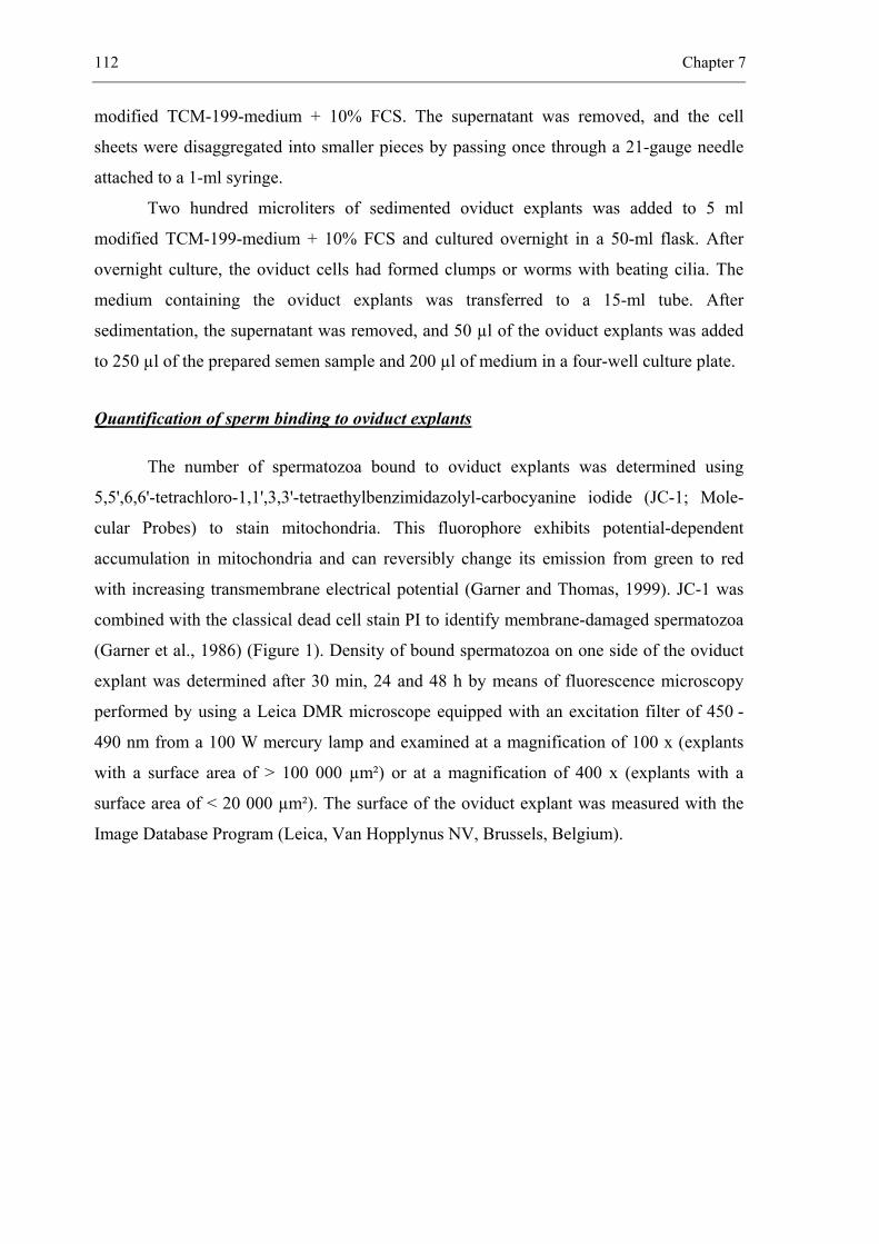
112 Chapter 7
modified TCM-199-medium + 10% FCS. The supernatant was removed, and the cell
sheets were disaggregated into smaller pieces by passing once through a 21-gauge needle
attached to a 1-ml syringe.
Two hundred microliters of sedimented oviduct explants was added to 5 ml
modified TCM-199-medium + 10% FCS and cultured overnight in a 50-ml flask. After
overnight culture, the oviduct cells had formed clumps or worms with beating cilia. The
medium containing the oviduct explants was transferred to a 15-ml tube. After
sedimentation, the supernatant was removed, and 50 µl of the oviduct explants was added
to 250 µl of the prepared semen sample and 200 µl of medium in a four-well culture plate.
Quantification of sperm binding to oviduct explants
The number of spermatozoa bound to oviduct explants was determined using
5,5',6,6'-tetrachloro-1,1',3,3'-tetraethylbenzimidazolyl-carbocyanine iodide (JC-1; Mole-
cular Probes) to stain mitochondria. This fluorophore exhibits potential-dependent
accumulation in mitochondria and can reversibly change its emission from green to red
with increasing transmembrane electrical potential (Garner and Thomas, 1999). JC-1 was
combined with the classical dead cell stain PI to identify membrane-damaged spermatozoa
(Garner et al., 1986) (Figure 1). Density of bound spermatozoa on one side of the oviduct
explant was determined after 30 min, 24 and 48 h by means of fluorescence microscopy
performed by using a Leica DMR microscope equipped with an excitation filter of 450 -
490 nm from a 100 W mercury lamp and examined at a magnification of 100 x (explants
with a surface area of > 100 000 µm²) or at a magnification of 400 x (explants with a
surface area of < 20 000 µm²). The surface of the oviduct explant was measured with the
Image Database Program (Leica, Van Hopplynus NV, Brussels, Belgium).
Sperm - oviduct binding assay 113
Figure 1: Fluorescence image of spermatozoa bound to oviduct explant stained with JC-1, which labels the mitochondria of spermatozoa orange and that of oviduct cells green, and with PI, which stains the heads of membrane-damaged spermatozoa red (magnification x 400)
Figure 2: Bull spermatozoa are bound to oviduct explants and labeled with JC-1 and PI. A) explant surface area < 20 000 µm² (× 400). B) explant surface of > 100 000 µm² (magnification × 100)
114 Chapter 7
Experimental design
Experiment 1: Standardization of a model to study sperm binding to oviduct explants
Effect of media and sperm preservation method on binding of fresh and frozen-thawed
spermatozoa to oviduct explants
Frozen-thawed and fresh spermatozoa from a Red Pied bull were washed and
incubated separately with oviduct explants at a concentration of 106 spermatozoa/ml
medium. Small and large sized oviduct explants were equally distributed between fresh
and frozen-thawed spermatozoa.
The different media, Hepes-TALP, IVF-TALP, TCM-199 + 10 % FCS, and TCM-
199 were incubated at 38.5°C in 5% CO2 except for Hepes-TALP (38.5°C, in air). After
30 min, 24 and 48 h of coincubation, at least 10 oviduct explants were stained with JC-1
and PI for 15 min. The oviduct explants with bound spermatozoa were first transferred
twice to fresh medium by means of a micropipette (Unopette Capillary Pipettes, Becton
Dickinson, Franklin Lakes, NJ) to remove unbound spermatozoa and then placed on a
glass slide and viewed under a Leica DMR fluorescence microscope (magnification x 100
or x 400) equipped with a stage warmer (37°C). The surface area of 10 oviduct explants
per point of time and per medium was measured, and the number of spermatozoa bound to
one side of the oviduct explant was calculated. The size of the explants varied between
9200 µm² and 137 219 µm². The number of spermatozoa bound to 0.1 mm² of explant
surface was used as the parameter of binding capacity. The experiment was repeated three
times. Each replicate was performed with oviduct explants from the same batch.
Effect of the size of oviduct explants on sperm binding
The binding of frozen-thawed spermatozoa to 10 oviduct explants with a surface
area of < 20 000 µm² (Figure 2A) and to 10 oviduct explants with a surface area of
> 100 000 µm² (Figure 2B) was compared after 30 min of coincubation. Sperm binding
density was expressed as the number of spermatozoa bound to 0.1 mm² of oviduct
epithelium. The experiment was repeated three times. Each replicate was performed with
oviduct explants from the same batch.
Sperm - oviduct binding assay 115
Experiment 2: Relationship between NRR and capacity of spermatozoa to bind to oviduct
epithelial explants
The optimized model was used to investigate whether there is a difference in
capacity to bind to oviduct explants for frozen-thawed spermatozoa from 10 Holstein
Friesian bulls with different NRRs (52.8-69.9%). The influence of the initial membrane
integrity on the association between the loge transformed number of spermatozoa bound to
0.1 mm² of oviduct epithelium and the NRR was also investigated. Membrane integrity of
spermatozoa from each sperm sample was evaluated by fluorescence microscopy just
before addition to oviduct explants (LIVE/DEAD® Sperm Viability Kit). This experiment
was repeated 3 times.
Frozen-thawed spermatozoa were used for this experiment because all AIs were
carried out with frozen-thawed spermatozoa. Only oviduct explants with a surface area of
< 20 000 µm² (obtained by passing twice through a 26-gauge needle) were used. The mean
values and ranges of size of oviduct explants were comparable for each bull.
Statistical analysis
The effect of different media and sperm preservation methods (fresh versus frozen-
thawed) on capacity of spermatozoa to bind to epithelial oviduct explants was analyzed
with the Mixed procedure of SAS version 8 (SAS Institute, Cary, NC). The outcome
variable was expressed as the loge transformed number of spermatozoa bound per 0.1 mm²
oviduct explant. Explanatory variables were the media and the sperm preservation method.
Both explanatory variables were included as class variables. Time was considered a
repeated measure and batch a random effect.
The effect of the size (small = < 20 000 µm², large = > 100 000 µm²) of oviduct
epithelial explants on sperm binding was analyzed by means of a two-sample t-test.
Differences were considered significant at P = 0.05.
The effect of different bulls with given NNRs and membrane integrity rates on
capacity of spermatozoa to bind to epithelial oviduct explants was analyzed with the Mixed
procedure of SAS version 8. The outcome variable was expressed as the loge transformed
number of spermatozoa bound per 0.1 mm² of oviduct explant. Explanatory variables were
the membrane integrity rate (%) and the NRR (%). Both explanatory variables were
included as continuous variables. Time was considered a repeated measure, and bull was a
116 Chapter 7
random effect. Regression lines of the number of spermatozoa bound to epithelial oviduct
explants after 24 h against the NRR and for different membrane integrity rates (40%, 45%,
… 85%) were calculated by using the ESTIMATE statement of the Mixed procedure.
Results
Experiment 1: Standardization of a model to study sperm binding to oviduct explants
An additional experiment was conducted to investigate whether the number of
oviduct explants present in a 50-µl sample could affect the number of sperm binding to
each oviduct explant. The number of oviduct explants present in a 50-µl sample taken from
a mixture of both large and small oviduct explants or from small oviduct explants was
counted. The results indicated that the mean number of oviduct explants was not
significantly different between replicate samples (large and small oviduct explants:
mean ± SEM = 182 ± 3, coefficient of variation = 0.06, n=10; small oviduct explants:
mean ± SEM = 247 ± 5, coefficient of variation = 0.06, n=10).
Spermatozoa (106) were then coincubated with a culture of 200, 250, 275 and 300
small oviduct explants. The number of spermatozoa bound to 10 oviduct explants from
each culture was counted after 30 min, 24 and 48 h. At each time point, no significant
difference in the number of spermatozoa per 0.1 mm² of oviduct explant was observed
when the number of oviduct explants ranged between 200 and 300 (ANOVA, Scheffé test;
data not shown).
Effect of media and sperm preservation method on binding of fresh and frozen-thawed
spermatozoa to oviduct explants
Significantly (P < 0.05; Table 1) more fresh spermatozoa were bound to oviduct
explants incubated in Hepes-TALP or IVF-TALP than in the other media. Fresh
spermatozoa remained bound to oviduct explants in Hepes-TALP in substantial numbers
even after 48 h of coincubation. For frozen-thawed spermatozoa, differences in sperm-
oviduct binding were observed between Hepes-TALP and TCM-199 with or without
addition of 10 % FCS (P < 0.05; Table 1). The decrease in the number of frozen-thawed
bound spermatozoa in Hepes-TALP differed (P < 0.05) from that in IVF-TALP over time.
The binding capacity of fresh and frozen-thawed spermatozoa was not different (P = 0.12).
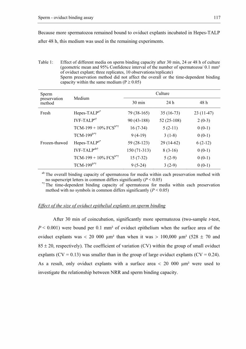
Sperm - oviduct binding assay 117
Because more spermatozoa remained bound to oviduct explants incubated in Hepes-TALP
after 48 h, this medium was used in the remaining experiments.
Table 1: Effect of different media on sperm binding capacity after 30 min, 24 or 48 h of culture (geometric mean and 95% Confidence interval of the number of spermatozoa/ 0.1 mm² of oviduct explant; three replicates, 10 observations/replicate)
Sperm preservation method did not affect the overall or the time-dependent binding capacity within the same medium (P ≥ 0.05)
Culture Sperm preservation method
Medium 30 min 24 h 48 h
Fresh Frozen-thawed
Hepes-TALPa* IVF-TALPa† TCM-199 + 10% FCSb*† TCM-199b*† Hepes-TALPa* IVF-TALPab† TCM-199 + 10% FCSb*† TCM-199b*†
79 (38-165) 90 (43-188)
16 (7-34) 9 (4-19)
59 (28-123) 150 (71-313)
15 (7-32) 9 (5-24)
35 (16-73) 52 (25-108)
5 (2-11) 3 (1-8)
29 (14-62) 8 (3-16) 5 (2-9) 3 (2-9)
23 (11-47) 2 (0-3) 0 (0-1) 0 (0-1)
6 (2-12) 0 (0-1) 0 (0-1) 0 (0-1)
ab The overall binding capacity of spermatozoa for media within each preservation method with no superscript letters in common differs significantly (P < 0.05)
*† The time-dependent binding capacity of spermatozoa for media within each preservation method with no symbols in common differs significantly (P < 0.05)
Effect of the size of oviduct epithelial explants on sperm binding
After 30 min of coincubation, significantly more spermatozoa (two-sample t-test,
P < 0.001) were bound per 0.1 mm² of oviduct epithelium when the surface area of the
oviduct explants was < 20 000 µm² than when it was > 100,000 µm² (528 ± 70 and
85 ± 20, respectively). The coefficient of variation (CV) within the group of small oviduct
explants (CV = 0.13) was smaller than in the group of large oviduct explants (CV = 0.24).
As a result, only oviduct explants with a surface area < 20 000 µm² were used to
investigate the relationship between NRR and sperm binding capacity.
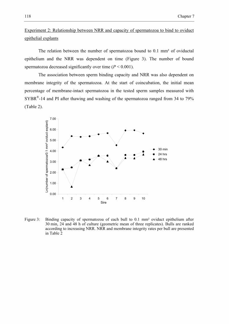
118 Chapter 7
Experiment 2: Relationship between NRR and capacity of spermatozoa to bind to oviduct
epithelial explants
The relation between the number of spermatozoa bound to 0.1 mm² of oviductal
epithelium and the NRR was dependent on time (Figure 3). The number of bound
spermatozoa decreased significantly over time (P < 0.001).
The association between sperm binding capacity and NRR was also dependent on
membrane integrity of the spermatozoa. At the start of coincubation, the initial mean
percentage of membrane-intact spermatozoa in the tested sperm samples measured with
SYBR®-14 and PI after thawing and washing of the spermatozoa ranged from 34 to 79%
(Table 2).
0.00
1.00
2.00
3.00
4.00
5.00
6.00
7.00
1 2 3 4 5 6 7 8 9 10Sire
Ln(n
umbe
r of s
perm
atoz
oa/0
.1 m
m² o
vidu
ct e
xpla
nt)
30 min24 hrs48 hrs
Figure 3: Binding capacity of spermatozoa of each bull to 0.1 mm² oviduct epithelium after 30 min, 24 and 48 h of culture (geometric mean of three replicates). Bulls are ranked according to increasing NRR. NRR and membrane integrity rates per bull are presented in Table 2
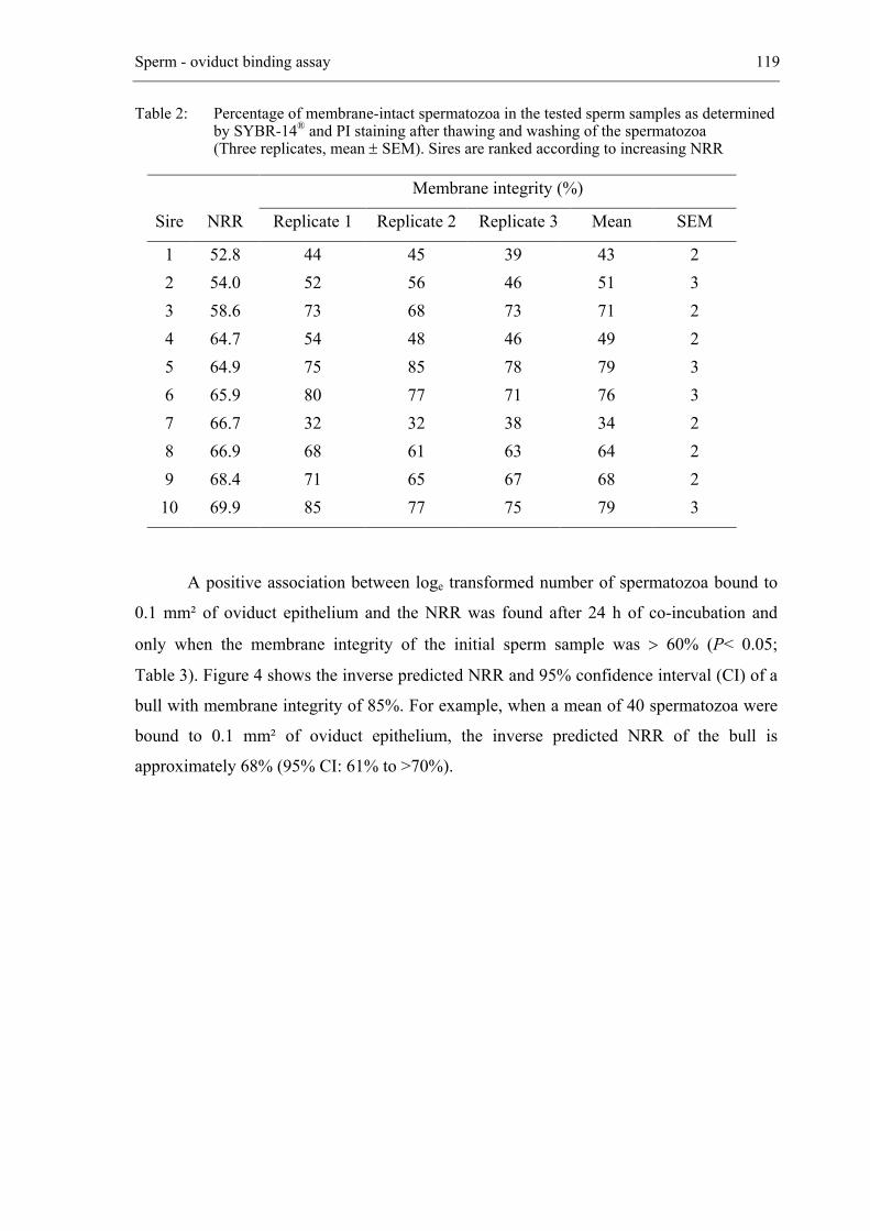
Sperm - oviduct binding assay 119
Table 2: Percentage of membrane-intact spermatozoa in the tested sperm samples as determined by SYBR-14® and PI staining after thawing and washing of the spermatozoa
(Three replicates, mean ± SEM). Sires are ranked according to increasing NRR
Membrane integrity (%)
Sire NRR Replicate 1 Replicate 2 Replicate 3 Mean SEM
1 2 3 4 5 6 7 8 9 10
52.8 54.0 58.6 64.7 64.9 65.9 66.7 66.9 68.4 69.9
44 52 73 54 75 80 32 68 71 85
45 56 68 48 85 77 32 61 65 77
39 46 73 46 78 71 38 63 67 75
43 51 71 49 79 76 34 64 68 79
2 3 2 2 3 3 2 2 2 3
A positive association between loge transformed number of spermatozoa bound to
0.1 mm² of oviduct epithelium and the NRR was found after 24 h of co-incubation and
only when the membrane integrity of the initial sperm sample was > 60% (P< 0.05;
Table 3). Figure 4 shows the inverse predicted NRR and 95% confidence interval (CI) of a
bull with membrane integrity of 85%. For example, when a mean of 40 spermatozoa were
bound to 0.1 mm² of oviduct epithelium, the inverse predicted NRR of the bull is
approximately 68% (95% CI: 61% to >70%).
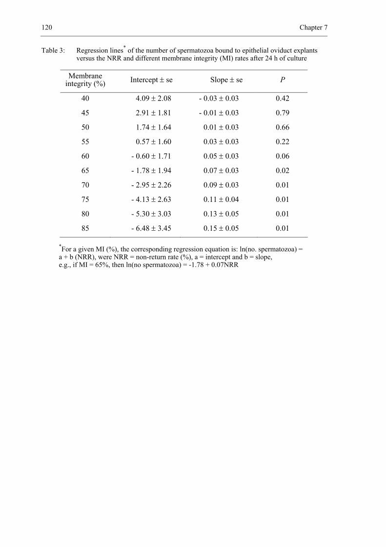
120 Chapter 7
Table 3: Regression lines* of the number of spermatozoa bound to epithelial oviduct explants versus the NRR and different membrane integrity (MI) rates after 24 h of culture
Membrane integrity (%) Intercept ± se Slope ± se P
40 4.09 ± 2.08 - 0.03 ± 0.03 0.42
45 2.91 ± 1.81 - 0.01 ± 0.03 0.79
50 1.74 ± 1.64 0.01 ± 0.03 0.66
55 0.57 ± 1.60 0.03 ± 0.03 0.22
60 - 0.60 ± 1.71 0.05 ± 0.03 0.06
65 - 1.78 ± 1.94 0.07 ± 0.03 0.02
70 - 2.95 ± 2.26 0.09 ± 0.03 0.01
75 - 4.13 ± 2.63 0.11 ± 0.04 0.01
80 - 5.30 ± 3.03 0.13 ± 0.05 0.01
85 - 6.48 ± 3.45 0.15 ± 0.05 0.01
*For a given MI (%), the corresponding regression equation is: ln(no. spermatozoa) = a + b (NRR), were NRR = non-return rate (%), a = intercept and b = slope, e.g., if MI = 65%, then ln(no spermatozoa) = -1.78 + 0.07NRR
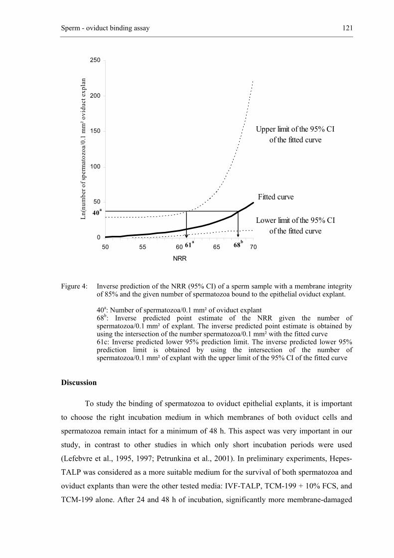
Sperm - oviduct binding assay 121
Figure 4: Inverse prediction of the NRR (95% CI) of a sperm sample with a membrane integrity of 85% and the given number of spermatozoa bound to the epithelial oviduct explant.
40a: Number of spermatozoa/0.1 mm² of oviduct explant 68b: Inverse predicted point estimate of the NRR given the number of
spermatozoa/0.1 mm² of explant. The inverse predicted point estimate is obtained by using the intersection of the number spermatozoa/0.1 mm² with the fitted curve
61c: Inverse predicted lower 95% prediction limit. The inverse predicted lower 95% prediction limit is obtained by using the intersection of the number of spermatozoa/0.1 mm² of explant with the upper limit of the 95% CI of the fitted curve
Discussion
To study the binding of spermatozoa to oviduct epithelial explants, it is important
to choose the right incubation medium in which membranes of both oviduct cells and
spermatozoa remain intact for a minimum of 48 h. This aspect was very important in our
study, in contrast to other studies in which only short incubation periods were used
(Lefebvre et al., 1995, 1997; Petrunkina et al., 2001). In preliminary experiments, Hepes-
TALP was considered as a more suitable medium for the survival of both spermatozoa and
oviduct explants than were the other tested media: IVF-TALP, TCM-199 + 10% FCS, and
TCM-199 alone. After 24 and 48 h of incubation, significantly more membrane-damaged
0
50
100
150
200
250
50 55 60 65 70
NRR
Ln(n
umbe
r of s
perm
atoz
oa/0
.1 m
m² o
vidu
ct e
xpla
n
Fitted curve
Upper limit of the 95% CI of the fitted curve
Lower limit of the 95% CI of the fitted curve
61a 68b
40a
122 Chapter 7
and capacitated spermatozoa were observed in the other media than in Hepes-TALP
(P < 0.05; data not shown). Furthermore, more spermatozoa survived while bound to
oviduct explants in Hepes-TALP in the presence of oviduct epithelial cells. All tested
media except Hepes-TALP were bicarbonate buffered: it is known that bicarbonate induces
capacitation of spermatozoa (Yanagimachi, 1994; Harrison et al., 1996). Capacitation
appears to be involved in the release of bovine spermatozoa from oviduct epithelium
(Lefebvre and Suarez, 1996) and in combination with decreased sperm survival is
responsible for the important drop in sperm binding to oviduct explants after 24 h of
coincubation in IVF-TALP. TCM-199 with or without the addition of 10% FCS was useful
only for the incubation of oviduct explants but not for incubation of spermatozoa
(unpublished data). Only a few spermatozoa survived in TCM-199, which is why binding
capacity was lower in this medium. One could argue that dead spermatozoa could exert a
negative effect on binding of remaining living spermatozoa to oviduct explants by the
release of enzymes or other substances (Lefebvre and Suarez, 1996). However, this effect
was not substantiated by a change in pH in any of the media. The tendency of frozen-
thawed spermatozoa to have a lower binding capacity and shorter survival than fresh
spermatozoa may be due to cell damage and the induction of capacitation-like changes
during freezing and thawing procedures (Cormier et al., 1997). Moreover, capacitation is
known to destabilize the sperm plasma membrane (Langlais and Roberts, 1985;
Yanagimachi, 1994), and thus reduce the lifespan of spermatozoa (Watson, 1995).
Sperm binding should be investigated with oviduct explants smaller than
20 000 µm2 to provide more repeatable results. Spermatozoa are not evenly distributed
over the surface. They are spaced closely in some areas, and sparsely in others and are
absent in a few areas, as has been observed previously (Lefebvre and Suarez, 1996). Sperm
heads bind preferentially to cilia or in deeper regions of ciliated epithelial cells between
cilia and not to secretory epithelial cells (Pollard et al., 1991; Petrunkina et al., 2001).
Further experiments using electron microscopy are needed to determine whether oviduct
explants with a small surface area consist of more ciliated epithelial cells and less secretory
cells than do oviduct explants with a large surface area.
To investigate the binding density of bovine spermatozoa to oviduct epithelial
explants, two very important criteria have to be met: sperm counting must be performed
easily and repeatedly, and sperm binding must mimic the in vivo situation as closely as
possible. The use of fluorescent dyes for the evaluation of sperm binding has two
advantages. First, staining with JC-1, which labels the mitochondria of both spermatozoa
Sperm - oviduct binding assay 123
and oviduct cells, makes it easy to count the tails of spermatozoa bound to oviduct explants
and to measure the surface area of the oviduct explant. Second, PI stains only membrane-
damaged spermatozoa. In this way membrane-intact and membrane-damaged spermatozoa
can be distinguished so that only membrane-intact spermatozoa are counted, which is not
possible with videomicroscopy. Several methods for studying binding of spermatozoa to
oviduct epithelial cells in mammals have been described (Thomas and Ball, 1996; Gualtieri
and Talevi, 2000). Sperm binding density is usually evaluated on oviduct explants
(Raychoudhury and Suarez, 1991; Thomas et al., 1994a; Lefebvre et al., 1995, 1997;
Lefebvre and Suarez, 1996) or on oviduct monolayers (Pollard et al., 1991; Thomas et al.,
1994b; Thomas and Ball, 1996; Ellington et al., 1999a; Gualtieri and Talevi, 2000). In our
study, oviduct explants were used as a model for oviduct epithelial cells in vivo because of
the maintenance of most of their morphological characteristics (Lefebvre et al., 1995).
Because morphological differentiation and polarization of epithelial cells is more
pronounced in polarized explants, there is higher sperm binding density in explants than in
epithelial culture monolayers, as shown in humans (Baillie et al., 1997). The number of
spermatozoa bound to living or fixed cocultures has been counted by means of scanning
electron microscopy analysis (Pollard et al., 1991; Gualtieri and Talevi, 2000),
videomicroscopy and image analysis (Lefebvre and Suarez, 1996; Lefebvre et al., 1995,
1997) or by labeling spermatozoa with the fluorochrome Hoechst 33342 followed by
counting attached spermatozoa by means of image processing and analysis of fluorescent
video images (Thomas et al., 1994a). Thomas at al. (1994a) were the first to develop a
cytofluorescent assay for counting large numbers of labeled spermatozoa attached to
somatic cell monolayers. However, the application of Hoechst 33342 leads to nuclear
staining of both spermatozoa and oviduct explant cells, which makes it more difficult to
distinguish spermatozoa from oviduct cells. This problem does not occur with JC-1.
Although sperm heads and mitochondria in the oviduct cells also stain faintly positive, this
staining does not interfere with the counting of spermatozoa.
Linking in vivo fertility with a relatively fast and cheap in vitro evaluation method
would be very valuable for the prediction of fertility of a sire. The standard procedure for
evaluating the fertility of semen from sires is to determine pregnancy data following AI.
This procedure is time consuming and expensive because of the large number of young
bulls entering the breeding program (Zhang et al., 1999). Several studies have already been
done to find a simple and reliable test for fertility (Larsson and Rodriguez-Martinez, 2000).
Despite the fact that a number of studies have focused on single sperm traits such as sperm

124 Chapter 7
morphology (Barth, 1993), sperm motility (Stalhammar et al., 1994; Holt et al., 1997), and
the presence of intact acrosomes (Cumming, 1995), none of these traits were correlated
significantly with in vivo fertility. Because fertilization requires several sperm activities, it
would be better to combine different sperm traits to achieve a better correlation between in
vitro tests and in vivo fertility (Amann and Hammerstedt, 1993; Farrell et al., 1998; Zhang
et al., 1999). With the in vitro model that we optimized, we established an association
between density of sperm binding to oviduct explants and NRR. Spermatozoa that can bind
to oviduct explants are characterized by an uncapacitated status (Lefebvre and Suarez,
1996), an intact acrosome (Gualtieri and Talevi, 2000), a superior morphology (Thomas et
al., 1994b), and a normal chromatin structure (Ellington et al., 1999b). Thundathil et al.
(1999) demonstrated that the proportion of uncapacitated spermatozoa present in frozen-
thawed bull semen varies among bulls and, more important, that the presence of
uncapacitated spermatozoa is positively correlated with fertility. Uncapacitated
spermatozoa have an advantage over capacitated spermatozoa during their transit to the site
of fertilization in the oviduct because they are more likely to survive. If capacitation were
to occur before spermatozoa reached the oviduct, the sperm population available for
fertilization may be reduced, causing an adverse effect on fertilization. When the
percentage of uncapacitated spermatozoa in a sperm sample is high, more spermatozoa are
able to bind to oviduct explants, which may result in a higher fertility rate. Differences
among individual animals in the capacity of spermatozoa to bind to oviduct explants in
vitro have already been reported for stallions and boars (Thomas and Ball, 1996;
Petrunkina et al., 2001). However in these studies no data were available on the
comparative ranking of fertility, so the relationship between sperm-oviduct binding index
and fertility could not be established.
Using our in vitro model, we found that the number of spermatozoa bound to
oviduct explants coincubated for 24 h is positively associated with in vivo bull fertility
only when the membrane integrity of the initial sperm sample > 60%. No association
between the number of spermatozoa bound to oviduct explants and NRR was found after
30 min and 48 h of coincubation and after 24 h of coincubation when the membrane
integrity of the sperm sample was < 60%.
The model can be used for predicting NRR of bulls when sperm samples with
membrane integrity rates > 60% are coincubated with oviduct explants for 24 h. However,
the width of the 95% CI is large and could be narrowed by collecting more data to predict
Sperm - oviduct binding assay 125
NRR and by controlling for factors that can bias NRR, such as technician and age and
parity of the cow. Moreover, testing of multiple ejaculates per bull could also increase the
accuracy of the model.
In the present study, labeling of both spermatozoa and oviduct explants with the
fluorescent carbocyanine dye JC-1 in combination with PI without fixing provided a rapid,
reliable, and reproducible method for counting the number of membrane-intact
spermatozoa bound to living oviduct explants. The results of this analysis indicate that the
capacity of spermatozoa bound to oviduct explants in vitro varies among bulls and that the
number of spermatozoa bound to oviduct epithelial explants is positively associated with
the NRR when evaluated after 24 h of sperm-oviduct incubation provided that the
membrane integrity of the initial sperm sample is > 60%.
Acknowledgements
This research was supported by the Ministry of Agriculture of the Belgian
government (Grant S6012), by the Flemish Cattle Breeding Association VRV, and by the
Research Fund of Ghent University (grant 011B8698). We thank the VRV for supplying
the batches of frozen semen and for providing the NRR. We also thank Dr. Coryn for
critical reading, and K. Goeman, G. Spaepen and J. Mestach for technical assistance.
126 Chapter 7
References
Amann RP, Hammerstedt RH. In vitro evaluation of sperm quality: an opinion. J Androl 1993; 14:
397-406.
Baillie HS, Pacey AA, Warren MA, Scudamore IW, Barratt CL. Greater numbers of human
spermatozoa associate with endosalpingeal cells derived from the isthmus compared with
those from the ampulla. Hum Reprod 1997; 12: 1985-1992.
Barth AD. Factors affecting fertility with artificial insemination. Vet Clin North Am Food Anim
Pract 1993; 9: 275-289.
Bosch P, de Avila JM, Ellington JE, Wright RW Jr. Heparin and Ca2+-free medium can enhance
release of bull sperm attached to oviductal epithelial cell monolayers. Theriogenology 2001;
56: 247-260.
Cormier N, Sirard MA, Bailey JL. Premature capacitation of bovine spermatozoa is initiated by
cryopreservation. J Androl 1997; 18: 461-468.
Cumming IR. Suitability of the intact acrosome method for the prediction of fertility in bovine
artificial insemination. Vet Rec 1995; 136: 289-291.
Day FT. Survival of sperm in the genital tract of the mare. J Agric Sci 1942; 32: 108-111.
den Daas JHG, De Jong G, Lansbergen LMTE, Van Wagtendonk-De Leeuw AM. The relationship
between the number of spermatozoa inseminated and the reproductive efficiency of
individual dairy bulls. J Dairy Sci 1998; 81: 1714-1723.
Ellington JE, Samper JC, Jones AE, Oliver SA, Burnett KM, Wright RW. In vitro interactions of
cryopreserved stallion spermatozoa and oviduct (uterine tube) epithelial cells or their
secretory products. Anim Reprod Sci 1999a; 56: 51-65.
Ellington JE, Evenson DP, Wright RW Jr, Jones AE, Schneider CS, Hiss GA, Brisbois RS. Higher
quality human sperm in a sample selectively attach to oviduct (fallopian tube) epithelial cells
in vitro. Fertil Steril 1999b; 71: 924-929.
Farrell PB, Presicce GA, Brockett CC, Foote RH. Quantification of bull sperm characteristics
measured by computer-assisted sperm analysis (CASA) and the relationship to fertility.
Theriogenology 1998; 49: 871-879.
Garner DL, Pinkel D, Johnson LA, Pace MM. Assessment of spermatozoal function using dual
fluorescent staining and flow cytometric analyses. Biol Reprod 1986; 34: 127-138.
Garner DL, Thomas CA. Organelle-specific probe JC-1 identifies membrane potential differences
in the mitochondrial function of bovine sperm. Mol Reprod Dev 1999; 53: 222-229.
Sperm - oviduct binding assay 127
Gualtieri R, Talevi R. In vitro-cultured bovine oviductal cells bind acrosome-intact sperm and
retain this ability upon sperm release. Biol Reprod 2000; 62: 1754-1762.
Harrison RA, Ashworth PJ, Miller NG. Bicarbonate/CO2, an effector of capacitation, induces a
rapid and reversible change in the lipid architecture of boar sperm plasma membrane.
Mol Reprod Dev 1996; 45: 378-391.
Holt C, Holt WV, Moore HD, Reed HC, Curnock RM. Objectively measured boar sperm motility
parameters correlate with the outcomes of on-farm inseminations: results of two fertility
trials. J Androl 1997; 18: 312-323.
Hunter RHF, Wilmut I. The rate of functional sperm transport into the oviducts of mated cows.
Anim Reprod Sci 1982; 5: 167-173.
Hunter RH, Wilmut I. Sperm transport in the cow: peri-ovulatory redistribution of viable cells
within the oviduct. Reprod Nutr Dev 1984; 24: 597-608.
Hunter RHF. Sperm dynamics in the female genital tract: interactions with fallopian tube
microenvironments. In: Motta PM (ed). Microscopy of Reproduction and Development: a
dynamic approach. Antonio Delfino Editore; Rome 1997: 35-45.
Langlais J, Roberts KD. A molecular membrane model of sperm capacitation and the acrosome
reaction of mammalian spermatozoa. Gamete Research 1985; 12: 183-224.
Larsson B, Rodriguez-Martinez H. Can we use in vitro fertilization tests to predict semen fertility?
Anim Reprod Sci 2000; 60: 327-336.
Lefebvre R, Chenoweth PJ, Drost M, LeClear CT, MacCubbin M, Dutton JT, Suarez SS.
Characterization of the oviductal sperm reservoir in cattle. Biol Reprod 1995; 53: 1066-
1074.
Lefebvre R, Suarez SS. Effect of capacitation on bull sperm binding to homologous oviductal
epithelium. Biol Reprod 1996; 54: 575-582.
Lefebvre R, Lo MC, Suarez SS. Bovine sperm binding to oviductal epithelium involves fucose
recognition. Biol Reprod 1997; 56: 1198-1204.
Madison V, Greve T, Avery B, Wamberg T. The effect of endotoxin-contaminated medium on in
vitro fertilization and development of bovine oocytes matured in vitro. Reprod Nutr Dev
1991; 32: 159-165.
Mburu JN, Einarsson S, Lundeheim N, Rodriguez-Martinez H. Distribution, number and
membrane integrity of spermatozoa in the pig oviduct in relation to spontaneous ovulation.
Anim Reprod Sci 1996; 45: 109-121.
128 Chapter 7
Parker WG, Sullivan JJ, First NL. Sperm transport and distribution in the mare. J Reprod Fertil
Suppl 1975; 23: 63-66.
Petrunkina AM, Gehlhaar R, Drommer W, Waberski D, Töpfer-Petersen E. Selective sperm
binding to pig oviductal epithelium in vitro. Reproduction 2001; 121: 889-896.
Pollard JW, Plante C, King WA, Hansen PJ, Betteridge KJ, Suarez SS. Fertilizing capacity of
bovine sperm may be maintained by binding to oviductal epithelial cells. Biol Reprod 1991;
44: 102-107.
Raychoudhury SS, Suarez SS. Porcine sperm binding to oviductal explants in culture.
Theriogenology 1991; 36: 1059-1070.
Stalhammar EM, Janson L, Philipsson J. The impact of sperm motility on non-return rate in
preselected dairy bulls. Reprod Nutr Dev 1994; 34: 37-45.
Suarez SS, Drost M, Redfern K, Gottlieb W. Sperm motility in the oviduct. In: Bavister BD,
Cummins J, Roldan ERS (eds). Fertilization in mammals. Serono Symposia; Norwell MA
1990: 111-124.
Suarez SS, Brockman K, Lefebvre R. Distribution of mucus and sperm in bovine oviducts after
artificial insemination: the physical environment of the oviductal sperm reservoir. Biol
Reprod 1997; 56: 447-453.
Talevi R, Gualtieri R. Sulfated glycoconjugates are powerful modulators of bovine sperm adhesion
and release from the oviductal epithelium in vitro. Biol Reprod 2001; 64: 491-498.
Thomas PG, Ball BA, Brinsko SP. Interaction of equine spermatozoa with oviduct epithelial cell
explants is affected by estrous cycle and anatomic origin of explant. Biol Reprod 1994a; 51:
222-228.
Thomas PG, Ball BA, Miller PG, Brinsko SP, Southwood L. A subpopulation of morphologically
normal, motile spermatozoa attach to equine oviductal epithelial cell monolayers.
Biol Reprod 1994b; 51: 303-309.
Thomas PG, Ball BA. Cytofluorescent assay to quantify adhesion of equine spermatozoa to oviduct
epithelial cells in vitro. Mol Reprod Dev 1996; 43: 55-61.
Thundathil J, Gil J, Januskauskas A, Larrson B, Soderquist L, Mapletoft R, Rodriguez-Martinez.
Relationship between the proportion of capacitated spermatozoa present in frozen-thawed
bull semen and fertility with artificial insemination. Int J Androl 1999; 22: 366-373.
Watson PF. Recent developments and concepts in the cryopreservation of spermatozoa and the
assessment of their post-thawing function. Reprod Fertil Dev. 1995; 7: 871-891.
Sperm - oviduct binding assay 129
Yanagimachi R. Mammalian fertilization. In: Knobil E, Neill J (eds). Physiology of Reproduction.
Raven press; New York 1994: 189-317.
Zhang BR, Larsson B, Lundeheim N, Haard MG, Rodriguez-Martinez H. Prediction of bull fertility
by combined in vitro assessments of frozen-thawed semen from young dairy bulls entering
an AI-programme. Int J Androl 1999; 22: 253-260.

__________________________________________________ 8.
SPERM-OVIDUCT BINDING CAPACITY OF COOLED BOVINE
SPERMATOZOA STORED FOR SEVERAL DAYS IN DIFFERENT DILUENTS
Manuscript in preparation
De Pauw I., Van Soom A., Verberckmoes S., Laevens H. and de Kruif A.
132 Chapter 8
Abstract
In this in vitro study, a previously developed sperm-oviduct binding assay is used
to analyze oviduct binding capacity of spermatozoa after storage in three different diluents
(CAPROGEN®, Triladyl® and CEP). Bovine semen was collected by means of an artificial
vagina connected with a tube containing 5 ml Triladyl®-diluent. Both diluent and seminal
plasma were removed by centrifugation (720 x g, 10 min). The sperm concentration was
adjusted to 10 x 106 spermatozoa/ml. Spermatozoa were used immediately (D0-
spermatozoa) or after storage in the three diluents at 4°C for up to 2, 4 and 6 days (D2-, D4-
and D6-spermatozoa). This experiment was performed using oviduct explants composed of
membrane-intact oviduct cells with high mitochondrial membrane potential and with
beating cilia. No significant effect of diluent on sperm-oviduct binding capacity of D2-, D4-
and D6-spermatozoa was observed after 30 min, 24 and 48 h of co-culture. Sperm-oviduct
binding capacity of D0- spermatozoa was different over time from the binding capacity of
D2-, D4- and D6-spermatozoa previously stored in the three diluents (P < 0.001). In this
respect, the number of D2-, D4- and D6-spermatozoa attached to oviduct explants decreased
significantly (P < 0.001; Figure 3) during the time of co-culture. In contrast, more than
90% of D0-spermatozoa attached to oviduct explants were released before 24 h of co-
culture and 15% of these progressively motile released spermatozoa reattached to oviduct
explants between 24 and 48 h of co-culture.
In an additional experiment, it was shown that oocyte penetration rate of
spermatozoa stored at 4°C for up to 6 days was also not significantly affected by diluent
type.
Oviduct binding capacity of cooled bovine spermatozoa stored in different diluents 133
Introduction
Fertilization involves a highly coordinated sequence of cellular interactions
between male and female gametes. To maximize fertilization success, it is important that
an appropriate number of spermatozoa meets the oocyte under proper physiological
fertilization conditions at the right time and at the right place (Töpfer-Petersen, 1999). In
order to obtain this optimal event, spermatozoa are selected quantitatively and qualitatively
during their transit in the female reproductive tract (Jouannet and Feneux, 1987). This
means that only morphologically normal and progressively motile spermatozoa are able to
migrate through the cervical mucus and reach the oviduct (Jeulin et al., 1985), where they
are stored until ovulation (Hunter, 1996; Suarez, 1998).
Recently, an in vitro bio-assay to quantify the number of 5,5',6,6'-tetrachloro-
1,1',3,3'-tetraethylbenzimidazolyl-carbocyanine iodide (JC-1)-labeled spermatozoa bound
to a defined oviduct explant surface area was developed in our laboratory (De Pauw et al.,
2002). It was shown that this in vitro system provides a rapid, reliable and reproducible
method for counting the number of spermatozoa bound to oviduct explants. By means of
this assay, a clear difference in sperm binding capacity among individual bulls was
observed and evidence was obtained that sperm binding ability to oviduct explants is
related to in vivo fertility of the sperm donor.
The first part of the present study was focused on further refinement of this new in
vitro model. The aim of the second part was to use this sperm-oviduct binding assay as a
functional test to examine sperm quality after storage in different sperm diluents for
several days. The effect of different oviduct explant cultures on the binding capacity of 4-
days stored spermatozoa in Cauda Epididymal Plasma (CEP)-diluent at 5°C was
investigated first in order to increase the accuracy of the assay. This system was then used
in the second experiment to compare oviduct binding capacity of spermatozoa stored in
three different diluents (CEP, CAPROGEN® and Triladyl®) for several days (0, 2, 4 and 6
days). CAPROGEN® is a commercially available diluent extensively used for liquid
storage of bovine semen at ambient temperature (Shannon, 1965; Vishwanath and
Shannon, 2000). Triladyl® is a TRIS-based diluent commonly used for frozen semen. Good
results have also been obtained with liquid semen stored at room temperature (RT) or at
4°C (De Pauw et al., 2003a). The Cauda Epididymal Plasma (CEP)-diluent, was recently
developed in our laboratory for storage of bovine spermatozoa at 4°C (Verberckmoes et al,
in preparation). In the final experiment, in vitro oocyte penetration rate of spermatozoa
134 Chapter 8
previously stored in the same three diluents at 4°C for up to 6 days was examined in order
to get extra information on the quality of spermatozoa stored in the different diluents.
Materials and Methods
Chemicals and media
Unless otherwise stated, chemicals and media were obtained from Sigma (Bornem,
Belgium).
Media
Hepes buffered TALP medium contained 114 mM NaCl, 3.1 mM KCl, 0.3 mM NaH2PO4,
2.1 mM CaCl2, 0.4 mM MgCl2, 2 mM NaHCO3, 0.2 mM sodium pyruvate, 10
mM sodium lactate, 10 µg/ml gentamycin sulphate, 10 mM Hepes and 3 mg
/ml bovine serum albumin.
Modified bicarbonate buffered TCM-199 (Gibco BRL Life Technologies, Merelbeke,
Belgium) supplemented with 0.2 mM sodium pyruvate, 0.4 mM glutamine,
50µg/ml gentamycin sulphate, 2.5 µg/ml fungizone (Gibco BRL Life
Technologies, Merelbeke, Belgium) and 10% fetal calf serum was used for
overnight culture of oviduct cells.
Maturation medium consisted of modified bicarbonate buffered TCM-199 medium (Gibco
BRL Life Technologies, Merelbeke, Belgium) supplemented with 20% heat-
inactivated foetal calf serum (FCS) (N.V. HyClone Europe S.A.,
Erembodegem, Belgium), 0.2 mM sodium pyruvate, 50 µg/ml gentamycin
sulphate and 0.4 mM glutamine.
Fertilization medium consisted of Tyrode’s solution without Hepes supplemented with
25 mM NaHCO3, 0.2 mM sodium pyruvate, 10 mM sodium lactate, 10 µg/ml
gentamycin sulphate, 6 mg/ml fatty acid-free BSA and 10 µg/ml heparin.
Media were passed through sterile 0.22 µm Acrodisc® Syringe low protein binding filters
(Millipore Corporation, New Bedford, MA) before being used.
Oviduct binding capacity of cooled bovine spermatozoa stored in different diluents 135
Sperm diluents
Three diluents were used in the present study:
Triladyl® (Minitüb, Tiefenbach b. Landshut, Germany) a TRIS-based diluent is
prepared by adding 20% egg yolk to the Triladyl concentrate
containing 6.7% glycerol.
CAPROGEN® (Livestock Improvement Corporation, Hamilton, New Zealand)
commercial diluent containing caproic acid (0.025g/100 ml) and 5%
egg yolk. The diluent was purged with nitrogen to reduce its
concentration of oxygen (Krzyzosiak, 2000).
CEP a simple sperm diluent based on the composition of bovine cauda
epididymal plasma (Verberckmoes et al., in preparation) containing
10% egg yolk.
Sperm preparation
Ejaculates were collected from a 3-year-old Red Pied bull by means of an artificial
vagina connected with a tube containing 5 ml Triladyl®-diluent. Immediately after
collection, spermatozoa were separated from the seminal plasma and diluent by
centrifugation (720 x g, 10 min) at RT. The progressive motility was subjectively assessed
by visual estimation under a light microscope (Leica DMR, Van Hopplynus NV, Brussels,
Belgium) (magnification x 200) equipped with a stage warmer (37°C). The nucleic stains
SYBR® 14 and propidium iodide (PI) (LIVE/DEAD® Sperm Viability Kit (Molecular
Probes, Leiden, The Netherlands)) were used for analyzing membrane integrity of
spermatozoa by means of fluorescence microscopy. Sperm concentration was determined
using a Bürker chamber and then adjusted to a concentration of 10 x 106 spermatozoa/ml
diluent. After storage at 4°C, spermatozoa were washed with Hepes-TALP by
centrifugation (720 x g, 10 min). Sperm concentration of the pellet was determined and
subsequently diluted to a concentration of 1 x 10⁶ spermatozoa/ml Hepes-TALP.
Collection and processing of oviducts for culture
Oviducts were obtained from local abattoirs and transported immediately to the
laboratory in ice-cold physiological saline (0.9% NaCl). After rinsing in Hepes-TALP,
oviducts were carefully dissected free of the surrounding tissue and rinsed again in Hepes-
136 Chapter 8
TALP. The lumen was cut open longitudinally with sterile scissors and lightly scraped with
a sterile scalpel to collect epithelial tissue. Sheets of epithelial cells were washed three
times with 5 ml Hepes-TALP by sedimentation followed by supernatant removal. The last
washing was performed in modified TCM-199 medium + 10% FCS. After supernatant
removal, the cell sheets were disaggregated into smaller pieces by passing twice through a
26-gauge needle connected to a 1-ml syringe.
Two hundred microlitres of sedimentated oviduct explants were added to 5 ml
modified TCM-199 medium + 10% FCS and cultured overnight in a 50-ml flask at 38.5°C
in 5% CO2 in air. After overnight culture, the oviduct cells had formed clumps or worms
with beating cilia. The medium containing oviduct explants was transferred to a 15-ml tube
and the supernatant was removed after sedimentation. Two hundred oviduct explants with
a surface smaller than 20.000 µm² were co-cultured with 1 x 10⁶ spermatozoa/ml Hepes-
TALP at 38.5°C in air in a four-well culture plate.
Quantification of sperm binding to oviduct explants
After co-culture, minimum 10 oviduct explants were rinsed in Hepes-TALP to
remove loosely attached spermatozoa, and transferred to fresh Hepes-TALP. The oviduct
explants were stained with JC-1 (Molecular Probes, Leiden, The Netherlands) to label
mitochondria of both spermatozoa and oviduct explants in order to count the number of
bound spermatozoa, and with PI to identify membrane-damaged spermatozoa. Density of
bound spermatozoa on one side of the oviduct explant was determined by means of
fluorescence microscopy performed by using a Leica DMR microscope (Van Hopplynus
N.V., Brussels, Belgium) equipped with an excitation filter of 450 - 490 nm from a 100 W
mercury lamp and examined at a magnification 400 x. The surface of the oviduct explant
was measured with the Image Database Program of Leica (Van Hopplynus NV, Brussels,
Belgium). The number of spermatozoa bound to 0.1 mm² of oviduct explant surface was
used as the parameter of binding capacity.
In vitro oocyte maturation and fertilization
Bovine ovaries collected from a local abattoir were transported to the laboratory
within 2 hours. Follicles of 2 to 6 mm in diameter were gently aspirated and cumulus-
oocyte complexes (COC) were matured in 500 µl maturation medium at 38.5 °C in 5%

Oviduct binding capacity of cooled bovine spermatozoa stored in different diluents 137
CO2 in air at 100% humidity for 24-26 hours. After maturation, 50 COC were incubated
with a final sperm concentration of 1 x 10⁶ spermatozoa/ml for 20 h.
For the assessment of oocyte penetration rate, the presumed zygotes were vortexed
in 2 ml Hepes-TALP for 2 min to remove excess spermatozoa and cumulus cells, and fixed
in 2% glutaraldehyde and 2% paraformaldehyde in phosphate buffered saline for at least
24 h. After fixing, zygotes were stained with 10 µg/ml Hoechst 33342 (Molecular Probes,
Leiden, The Netherlands) for 10 min and evaluated with a Leica DMR fluorescence
microscope for signs of oocyte penetration. Successful oocyte penetration was
characterized by the presence of maternal and paternal pronuclei (fertilization rate).
Oocytes with more than two pronuclei (polyspermy rate) were also considered penetrated.
Experimental design
Experiment 1: Effect of oviduct explant cultures on the binding capacity of spermatozoa
stored in CEP-diluent at 4°C for up to 4 days
This experiment was performed with 5 oviduct explant cultures derived from
5 different oviducts. Spermatozoa were collected and processed as described above.
Spermatozoa were stored at a concentration of 10 x 106 spermatozoa/ml CEP-diluent at
4°C in darkness for up to 4 days. After storage, spermatozoa were washed with Hepes-
TALP by centrifugation (720 x g, 10 min). Two hundred oviduct explants with a surface
smaller than 20 000 µm² were co-cultured with 1 x 10⁶ spermatozoa/ml Hepes-TALP at
38.5°C in air in a four-well culture plate. After 30 min of co-culture, the number of
spermatozoa bound to 10 oviduct explants were counted and expressed as the number of
spermatozoa bound to 0.1 mm² oviduct explant.
Experiment 2: Effect of diluent and duration of storage on the binding capacity of
spermatozoa
Every other day, ejaculates were collected in 5 ml Triladyl®-diluent to obtain
spermatozoa of different ages. Spermatozoa were used immediately (D0- spermatozoa) or
after storage in 3 different diluents (CEP, CAPROGEN® and Triladyl®) at a concentration
of 10 x 106 sp/ml at 4°C in darkness for 2 (D2-spermatozoa), 4 (D4-spermatozoa) and
6 (D6-spermatozoa) days. After storage, spermatozoa were washed with Hepes-TALP by
centrifugation (720 x g, 10 min). Membrane integrity (SYBR®-14/PI) and progressive
138 Chapter 8
motility (subjectively) of all sperm samples were determined. Spermatozoa were then co-
cultured with two hundred oviduct explants with a surface smaller than 20 000 µm²,
derived from the same oviduct explant batch, at 38.5°C in air in a four-well culture plate.
After 30 min, 24 and 48 h of co-culture, at least 10 oviduct explants were stained with JC-1
and PI for 15 min. The number of spermatozoa bound to 10 oviduct explants were counted
and expressed as the number of spermatozoa bound to 0.1 mm² oviduct explant. This
experiment was repeated three times.
Experiment 3: In vitro oocyte penetration rate of spermatozoa stored in different diluents at
4°C for up to 6 days
Spermatozoa were collected in 5 ml Triladyl®-diluent and processed as described
above. After storage in CEP, CAPROGEN® and Triladyl® at 4°C in darkness for 6 days,
spermatozoa were washed with Hepes-TALP by centrifugation at RT (720 x g, 10 min)
and the supernatant was removed. Motile and membrane-intact spermatozoa from the
pellet were separated on a discontinuous Percoll®-gradient. The sperm concentration was
adjusted with fertilization medium to obtain a final sperm concentration of 1 x
106 spermatozoa/ml. Cumulus oocyte complexes (n = 946, 3 replicates) were co-cultured
with spermatozoa, previously stored in the three diluents at 4°C for up to 6 days, for 20 h.
Finally, presumed zygotes were vortexed, fixed, stained with Hoechst 33342 and examined
with a Leica DMR fluorescence microscope for signs of penetration. This experiment was
repeated three times.
Statistical analysis
Difference in mean number of spermatozoa attached to 10 oviduct explants from
5 different cultures was examined using analysis of variance (ANOVA) methods through
Generalized Linear Models (SPSS 10.0, Chicago, USA). Differences were considered
significant at P = 0.05.
Difference in membrane integrity and progressive motility of D0-, D2-, D4- and D6-
spermatozoa stored in CEP, CAPROGEN® and Triladyl® at 4°C was examined using
analysis of variance (ANOVA) methods through Generalized Linear Models (SPSS 10.0,
Chicago, USA). Differences were considered significant at P = 0.05.
The effect of diluent and duration of storage on sperm binding capacity to oviduct
explants was analyzed with the Mixed procedure of SAS V8 (SAS Institute Inc.). The
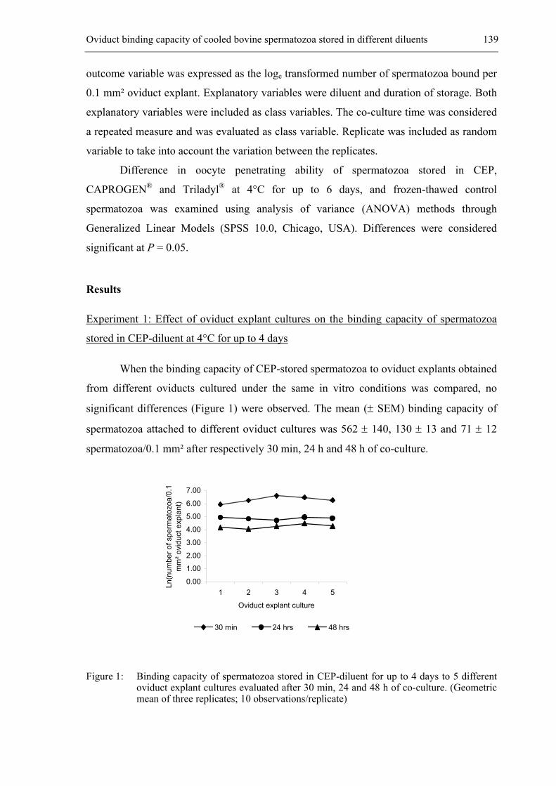
Oviduct binding capacity of cooled bovine spermatozoa stored in different diluents 139
outcome variable was expressed as the loge transformed number of spermatozoa bound per
0.1 mm² oviduct explant. Explanatory variables were diluent and duration of storage. Both
explanatory variables were included as class variables. The co-culture time was considered
a repeated measure and was evaluated as class variable. Replicate was included as random
variable to take into account the variation between the replicates.
Difference in oocyte penetrating ability of spermatozoa stored in CEP,
CAPROGEN® and Triladyl® at 4°C for up to 6 days, and frozen-thawed control
spermatozoa was examined using analysis of variance (ANOVA) methods through
Generalized Linear Models (SPSS 10.0, Chicago, USA). Differences were considered
significant at P = 0.05.
Results
Experiment 1: Effect of oviduct explant cultures on the binding capacity of spermatozoa
stored in CEP-diluent at 4°C for up to 4 days
When the binding capacity of CEP-stored spermatozoa to oviduct explants obtained
from different oviducts cultured under the same in vitro conditions was compared, no
significant differences (Figure 1) were observed. The mean (± SEM) binding capacity of
spermatozoa attached to different oviduct cultures was 562 ± 140, 130 ± 13 and 71 ± 12
spermatozoa/0.1 mm² after respectively 30 min, 24 h and 48 h of co-culture.
0.00
1.00
2.00
3.00
4.00
5.00
6.00
7.00
1 2 3 4 5
Oviduct explant culture
Ln(n
umbe
r of s
perm
atoz
oa/0
.1
mm
² ovi
duct
exp
lant
)
30 min 24 hrs 48 hrs
Figure 1: Binding capacity of spermatozoa stored in CEP-diluent for up to 4 days to 5 different oviduct explant cultures evaluated after 30 min, 24 and 48 h of co-culture. (Geometric mean of three replicates; 10 observations/replicate)
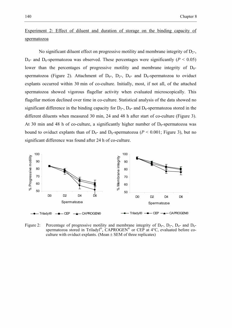
140 Chapter 8
Experiment 2: Effect of diluent and duration of storage on the binding capacity of
spermatozoa
No significant diluent effect on progressive motility and membrane integrity of D2-,
D4- and D6-spermatozoa was observed. These percentages were significantly (P < 0.05)
lower than the percentages of progressive motility and membrane integrity of D0-
spermatozoa (Figure 2). Attachment of D0-, D2-, D4- and D6-spermatozoa to oviduct
explants occurred within 30 min of co-culture. Initially, most, if not all, of the attached
spermatozoa showed vigorous flagellar activity when evaluated microscopically. This
flagellar motion declined over time in co-culture. Statistical analysis of the data showed no
significant difference in the binding capacity for D2-, D4- and D6-spermatozoa stored in the
different diluents when measured 30 min, 24 and 48 h after start of co-culture (Figure 3).
At 30 min and 48 h of co-culture, a significantly higher number of D0-spermatozoa was
bound to oviduct explants than of D4- and D6-spermatozoa (P < 0.001; Figure 3), but no
significant difference was found after 24 h of co-culture.
Figure 2: Percentage of progressive motility and membrane integrity of D0-, D2-, D4- and D6-spermatozoa stored in Triladyl®, CAPROGEN® or CEP at 4°C, evaluated before co-culture with oviduct explants. (Mean ± SEM of three replicates)
50
60
70
80
90
100
D0 D2 D4 D6
Spermatozoa
% M
embr
ane
inte
grity
Triladyl® CEP CAPROGEN®
50
60
70
80
90
100
D0 D2 D4 D6
Spermatozoa
% P
rogr
essi
ve m
otili
ty
Triladyl® CEP CAPROGEN®
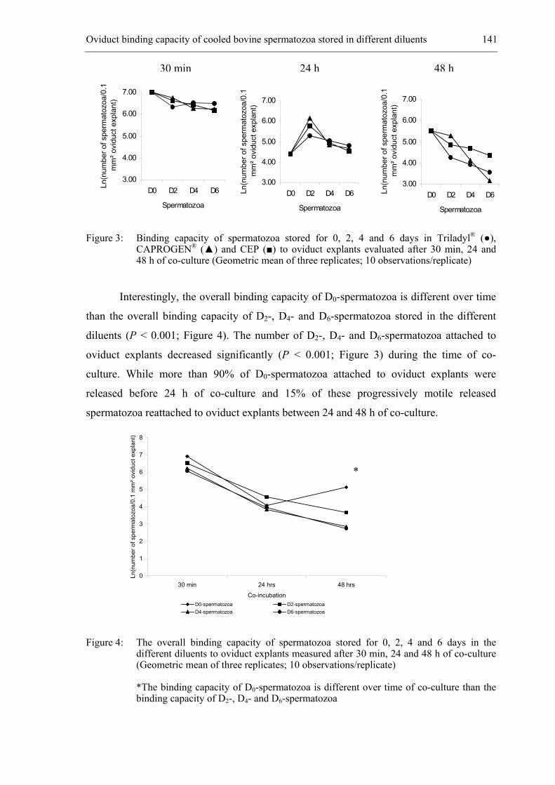
Oviduct binding capacity of cooled bovine spermatozoa stored in different diluents 141
Figure 3: Binding capacity of spermatozoa stored for 0, 2, 4 and 6 days in Triladyl® (●), CAPROGEN® (▲) and CEP (■) to oviduct explants evaluated after 30 min, 24 and 48 h of co-culture (Geometric mean of three replicates; 10 observations/replicate)
Interestingly, the overall binding capacity of D0-spermatozoa is different over time
than the overall binding capacity of D2-, D4- and D6-spermatozoa stored in the different
diluents (P < 0.001; Figure 4). The number of D2-, D4- and D6-spermatozoa attached to
oviduct explants decreased significantly (P < 0.001; Figure 3) during the time of co-
culture. While more than 90% of D0-spermatozoa attached to oviduct explants were
released before 24 h of co-culture and 15% of these progressively motile released
spermatozoa reattached to oviduct explants between 24 and 48 h of co-culture.
0
1
2
3
4
5
6
7
8
30 min 24 hrs 48 hrs
Co-incubation
Ln(n
umbe
r of s
perm
atoz
oa/0
.1 m
m² o
vidu
ct e
xpla
nt)
D0-spermatozoa D2-spermatozoaD4-spermatozoa D6-spermatozoa
Figure 4: The overall binding capacity of spermatozoa stored for 0, 2, 4 and 6 days in the different diluents to oviduct explants measured after 30 min, 24 and 48 h of co-culture (Geometric mean of three replicates; 10 observations/replicate)
*The binding capacity of D0-spermatozoa is different over time of co-culture than the binding capacity of D2-, D4- and D6-spermatozoa
30 min 24 h 48 h
3.00
4.00
5.00
6.00
7.00
D0 D2 D4 D6
Spermatozoa
Ln(n
umbe
r of s
perm
atoz
oa/0
.1
mm
² ovi
duct
exp
lant
)
3.00
4.00
5.00
6.00
7.00
D0 D2 D4 D6
Spermatozoa
Ln(n
umbe
r of s
perm
atoz
oa/0
.1
mm
² ovi
duct
exp
lant
)
3.00
4.00
5.00
6.00
7.00
D0 D2 D4 D6
Spermatozoa
Ln(n
umbe
r of s
perm
atoz
oa/0
.1m
m² o
vidu
ct e
xpla
nt)
*
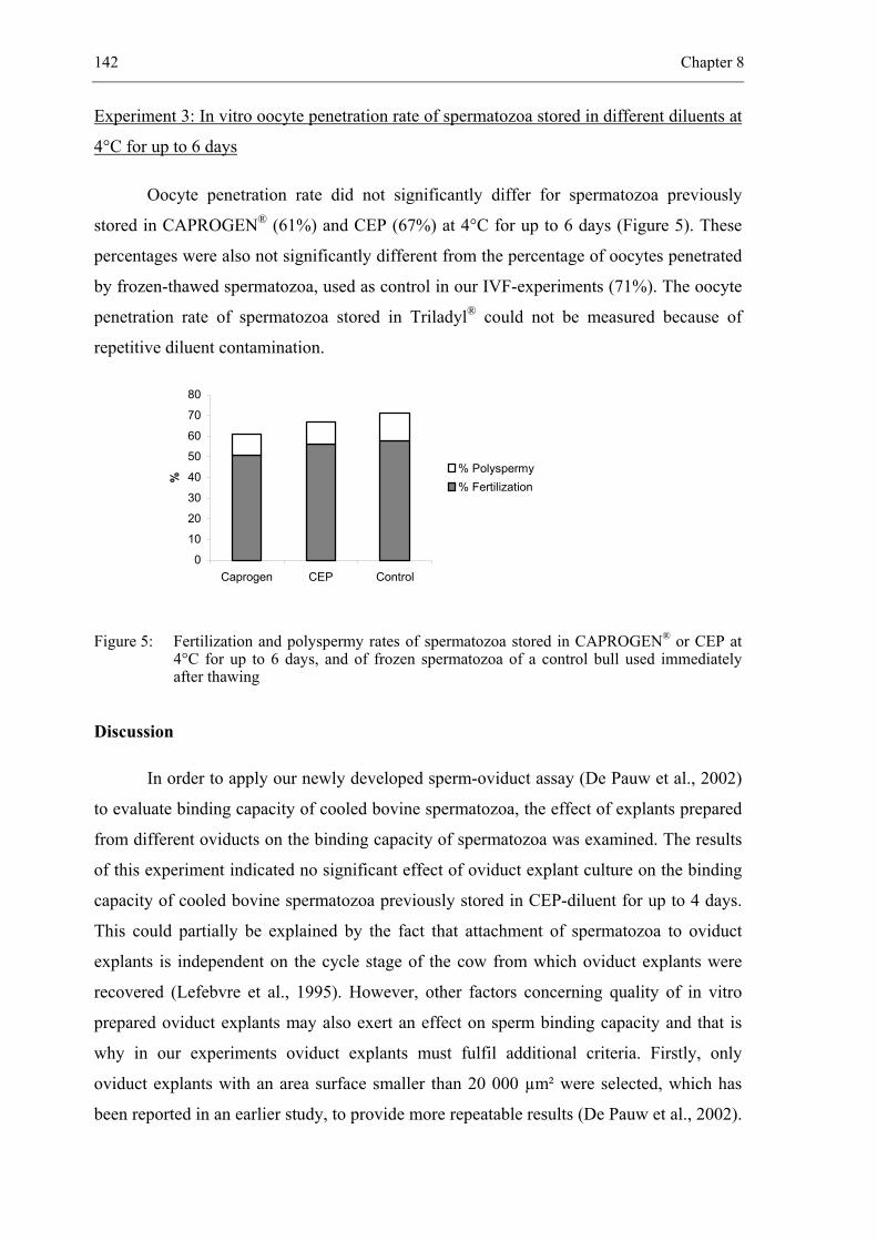
142 Chapter 8
Experiment 3: In vitro oocyte penetration rate of spermatozoa stored in different diluents at
4°C for up to 6 days
Oocyte penetration rate did not significantly differ for spermatozoa previously
stored in CAPROGEN® (61%) and CEP (67%) at 4°C for up to 6 days (Figure 5). These
percentages were also not significantly different from the percentage of oocytes penetrated
by frozen-thawed spermatozoa, used as control in our IVF-experiments (71%). The oocyte
penetration rate of spermatozoa stored in Triladyl® could not be measured because of
repetitive diluent contamination.
0
10
20
30
40
50
60
70
80
Caprogen CEP Control
%
% Polyspermy% Fertilization
Figure 5: Fertilization and polyspermy rates of spermatozoa stored in CAPROGEN® or CEP at 4°C for up to 6 days, and of frozen spermatozoa of a control bull used immediately after thawing
Discussion
In order to apply our newly developed sperm-oviduct assay (De Pauw et al., 2002)
to evaluate binding capacity of cooled bovine spermatozoa, the effect of explants prepared
from different oviducts on the binding capacity of spermatozoa was examined. The results
of this experiment indicated no significant effect of oviduct explant culture on the binding
capacity of cooled bovine spermatozoa previously stored in CEP-diluent for up to 4 days.
This could partially be explained by the fact that attachment of spermatozoa to oviduct
explants is independent on the cycle stage of the cow from which oviduct explants were
recovered (Lefebvre et al., 1995). However, other factors concerning quality of in vitro
prepared oviduct explants may also exert an effect on sperm binding capacity and that is
why in our experiments oviduct explants must fulfil additional criteria. Firstly, only
oviduct explants with an area surface smaller than 20 000 µm² were selected, which has
been reported in an earlier study, to provide more repeatable results (De Pauw et al., 2002).
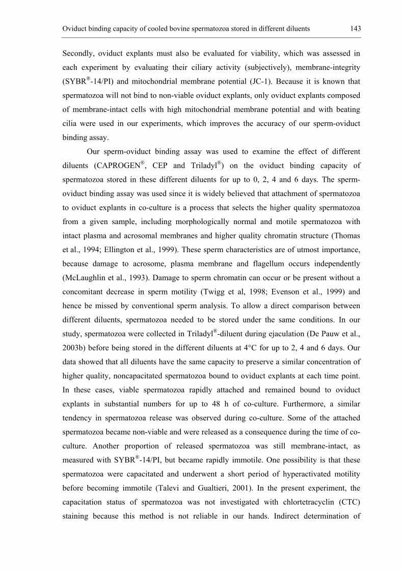
Oviduct binding capacity of cooled bovine spermatozoa stored in different diluents 143
Secondly, oviduct explants must also be evaluated for viability, which was assessed in
each experiment by evaluating their ciliary activity (subjectively), membrane-integrity
(SYBR®-14/PI) and mitochondrial membrane potential (JC-1). Because it is known that
spermatozoa will not bind to non-viable oviduct explants, only oviduct explants composed
of membrane-intact cells with high mitochondrial membrane potential and with beating
cilia were used in our experiments, which improves the accuracy of our sperm-oviduct
binding assay.
Our sperm-oviduct binding assay was used to examine the effect of different
diluents (CAPROGEN®, CEP and Triladyl®) on the oviduct binding capacity of
spermatozoa stored in these different diluents for up to 0, 2, 4 and 6 days. The sperm-
oviduct binding assay was used since it is widely believed that attachment of spermatozoa
to oviduct explants in co-culture is a process that selects the higher quality spermatozoa
from a given sample, including morphologically normal and motile spermatozoa with
intact plasma and acrosomal membranes and higher quality chromatin structure (Thomas
et al., 1994; Ellington et al., 1999). These sperm characteristics are of utmost importance,
because damage to acrosome, plasma membrane and flagellum occurs independently
(McLaughlin et al., 1993). Damage to sperm chromatin can occur or be present without a
concomitant decrease in sperm motility (Twigg et al, 1998; Evenson et al., 1999) and
hence be missed by conventional sperm analysis. To allow a direct comparison between
different diluents, spermatozoa needed to be stored under the same conditions. In our
study, spermatozoa were collected in Triladyl®-diluent during ejaculation (De Pauw et al.,
2003b) before being stored in the different diluents at 4°C for up to 2, 4 and 6 days. Our
data showed that all diluents have the same capacity to preserve a similar concentration of
higher quality, noncapacitated spermatozoa bound to oviduct explants at each time point.
In these cases, viable spermatozoa rapidly attached and remained bound to oviduct
explants in substantial numbers for up to 48 h of co-culture. Furthermore, a similar
tendency in spermatozoa release was observed during co-culture. Some of the attached
spermatozoa became non-viable and were released as a consequence during the time of co-
culture. Another proportion of released spermatozoa was still membrane-intact, as
measured with SYBR®-14/PI, but became rapidly immotile. One possibility is that these
spermatozoa were capacitated and underwent a short period of hyperactivated motility
before becoming immotile (Talevi and Gualtieri, 2001). In the present experiment, the
capacitation status of spermatozoa was not investigated with chlortetracyclin (CTC)
staining because this method is not reliable in our hands. Indirect determination of
144 Chapter 8
capacitation status was performed by means of Pisum Sativum agglutinin staining, which
measures the acrosome reaction, the next step in the capacitation process. The fact that a
higher proportion of released spermatozoa were acrosome-reacted and the fact that this
proportion increased when spermatozoa were stored for a longer period of time (data not
shown), may suggest that these spermatozoa were released due to changes in their
capacitation status. Furthermore, these released spermatozoa were not able to reattach to
oviduct explants anymore. In contrast to this observation, we found that sperm detachment
and reattachment to oviduct explants occur readily when spermatozoa were added to
oviduct explants during ejaculation without storage (D0-spermatozoa). Such transient
attachment and reattachment was also observed in human spermatozoa (Ellington et al.,
1998). In our study, the weaker binding of these D0-spermatozoa to oviduct explants is
probably due to the collection of spermatozoa into egg yolk containing diluent (Triladyl®-
diluent) immediately after ejaculation (De Pauw et al., 2003b). This sperm collection
method limits the contact time between spermatozoa and seminal plasma within seconds.
(De Pauw et al., 2003b). Manjunath et al. (2002) discovered that the low-density
lipoproteins (LDL) of egg yolk especifically bind detrimental BSP proteins of seminal
plasma. This binding is rapid, specific, saturable and stable even after ultracentrifugation
of semen, and could avoid the binding of BSP proteins on the sperm membrane.
Furthermore, LDL are believed to act at the level of sperm membrane and in this way it
can also avoid or reduce the binding of BSP proteins on the sperm membrane (Vishwanath
et al., 1992).
It has been shown that one of these BSP proteins, identified as PDC-109, associates
with the sperm surface during ejaculation (Desnoyers and Manjunath, 1993). This seminal
plasma protein is responsible for the binding of spermatozoa to fucosylated molecules on
oviductal epithelium (Ignotz et al., 2001; Suarez et al., 2002). Its importance in the binding
of spermatozoa to oviduct epithelium could also be illustrated by the fact that only 7% of
epididymal spermatozoa, which are not associated with PDC-109, was able to attach to
oviduct epithelial cells, whereas 75% of ejaculated spermatozoa from the same bull
attached (Ellington et al., 1999). This is a very important observation since in our study, a
high proportion of D0-spermatozoa were rapidly detached without a significant drop in
motility. A possible reason could be a decreased presence or complete absence of PDC-109
proteins on the surface of “coated” spermatozoa resulting in a weaker oviduct binding,
which can easily be overcome by mechanical forces exerted by D0-spermatozoa, exhibiting
excellent progressive motility. The fact that BSP proteins bind to LDP of egg yolk could
Oviduct binding capacity of cooled bovine spermatozoa stored in different diluents 145
suggest that this interaction could be responsible for the binding of egg yolk coated
spermatozoa to oviduct explants. However, further experiments are needed to confirm this
hypothesis. The significantly lower values of different motility characteristics of D2-, D4-
and D6-spermatozoa than of D0-spermatozoa (Verberckmoes et al., in preparation) could
probably be the reason why D2-, D4- and D6-spermatozoa remain bound to oviduct explants
and could only be released due to capacitation or when they became non-viable.
In the final experiment it was examined whether prolonged storage of spermatozoa
in the different diluents affected oocyte penetration rates of D6-spermatozoa. No significant
difference in oocyte penetration rate was observed for D6-spermatozoa stored in CEP
(67%) and CAPROGEN® (61%). Unfortunately, the penetration rate of spermatozoa stored
in Triladyl®-diluent could not be measured because of repetitive diluent contamination.
However, it was earlier reported that “coated” spermatozoa stored in Triladyl® yield a
penetration rate of 71% (De Pauw et al., 2003a).
In summary, the use of high quality oviduct explants further improves the accuracy
of our sperm-oviduct binding assay and provides a rapid, reliable and reproducible method
for counting the number of spermatozoa bound to oviduct explants in vitro. Using this
sperm-oviduct binding assay, the three tested diluents yielded similar concentrations of
high quality spermatozoa. This could be concluded from the fact that for the three diluents
no significant difference was observed at the level of sperm-oviduct binding or oocyte
penetration rate. Whether this sperm-oviduct binding assay can be used as an indicator for
in vitro fertilizing capacity of a sperm sample needs to be investigated by using semen
from several bulls with pronounced differences in in vitro fertilization.
Acknowledgements
This research was supported by the Ministry of Agriculture of the Belgian
Government, grant n° S6012 and by the Flemish Cattle Breeding Association VRV.
146 Chapter 8
References
De Pauw I, Van Soom A, Laevens H, Verberckmoes S, de Kruif A. Sperm binding to epithelial
oviduct explants in bulls with different non-return rates investigated with a new in vitro
model. Biol Reprod 2002; 67: 1073-1079.
De Pauw I, Van Soom A, Maes M, Verberckmoes S, de Kruif A. Effect of sperm coating on the
survival and penetrating ability of in vitro stored bovine spermatozoa. Theriogenology
2003a; 59: 1109-1122.
De Pauw I, Van Soom A, Mintiens K, Verberckmoes S, de Kruif A. In vitro survival of bovine
spermatozoa stored at room temperature under epididymal conditions. Theriogenology
2003b; 59: 1093-1107.
Desnoyers L, Manjunath P. Interaction of a novel class of phospholipid-binding proteins of bovine
seminal fluid with different affinity matrices. Arch Biochem Biophys 1993; 305: 341-349.
Ellington JE, Jones AE, Davitt CM, Schneider CS, Brisbois RS, Hiss GA, Wright RW Jr. Human
sperm function in co-culture with human, macaque or bovine oviduct epithelial cell
monolayers. Hum Reprod 1998; 13: 2797-2804.
Ellington JE, Evenson DP, Wright RW Jr, Jones AE, Schneider CS, Hiss GA, Brisbois. Higher-
quality human sperm in a sample selectively attach to oviduct (fallopian tube) epithelial cells
in vitro.RS. Fertil Steril 1999; 71: 924-929.
Evenson DP, Jost LK, Marshall D, Zinaman MJ, Clegg E, Purvis K, de Angelis P, Claussen OP.
Utility of the sperm chromatin structure assay as a diagnostic and prognostic tool in the
human fertility clinic. Hum Reprod 1999; 14: 1039-1049.
Hunter RH. Ovarian control of very low sperm/egg ratios at the commencement of mammalian
fertilisation to avoid polyspermy. Mol Reprod Dev 1996; 44: 417-422.
Ignotz GG, Lo MC, Perez CL, Gwathmey TM, Suarez SS. Characterization of a fucose-binding
protein from bull sperm and seminal plasma that may be responsible for formation of the
oviductal sperm reservoir. Biol Reprod 2001; 64: 1806-1811.
Jeulin C, Soumah A, Jouannet P. Morphological factors influencing the penetration of human
sperm into cervical mucus in vitro. Int J Androl 1985; 8: 215-223.
Jouannet P, Feneux D. Sperm analysis. Ann Biol Clin (Paris) 1987; 45: 335-339.
Lefebvre R, Chenoweth PJ, Drost M, LeClear CT, MacCubbin M, Dutton JT, Suarez SS.
Characterization of the oviductal sperm reservoir in cattle. Biol Reprod 1995; 53: 1066-
1074.
Oviduct binding capacity of cooled bovine spermatozoa stored in different diluents 147
Manjunath P, Nauc V, Bergeron A, Menard M. Major proteins of bovine seminal plasma bind to
the low-density lipoprotein fraction of hen's egg yolk. Biol Reprod 2002; 67: 1250-1258.
McLaughlin EA, Ford WC, Hull MG. Effects of cryopreservation on the human sperm acrosome
and its response to A23187. J Reprod Fertil 1993; 99: 71-76.
Shannon P. Contribution of seminal plasma, sperm numbers and gas phase to dilution effects of
bovine spermatozoa. J. Dairy Sci 1965; 48: 1357-1361.
Suarez SS. The oviductal sperm reservoir in mammals: mechanisms of formation. Biol Reprod
1998; 58: 1105-1107.
Suarez SS. Formation of a reservoir of sperm in the oviduct. Reprod Domest Anim 2002; 37: 140-
143.
Talevi R, Gualtieri R. Sulfated glycoconjugates are powerful modulators of bovine sperm adhesion
and release from the oviductal epithelium in vitro. Biol Reprod 2001; 64: 491-498.
Thomas PG, Ball BA, Miller PG, Brinsko SP, Southwood L. A subpopulation of morphologically
normal, motile spermatozoa attach to equine oviductal epithelial cell monolayers. Biol
Reprod 1994; 51: 303-309.
Topfer-Petersen E. Molecules on the sperm's route to fertilization. J Exp Zool 1999; 285: 259-266.
Twigg J, Fulton N, Gomez E, Irvine DS, Aitken RJ. Analysis of the impact of intracellular reactive
oxygen species generation on the structural and functional integrity of human spermatozoa:
lipid peroxidation, DNA fragmentation and effectiveness of antioxidants. Hum Reprod 1998;
13: 1429-1436.
Verberckmoes S, Van Soom A, De Pauw I, Dewulf J, de Kruif A. Ionic composition of epididymal
plasma of the bull. In preparation.
Vishwanath R, Shannon P, Curson B. Cationic extracts of egg yolk and their effects on motility,
survival and fertilising ability of bull sperm. Anim Reprod Sci 1992; 29: 185-194.
Vishwanath R, Shannon P. Storage of bovine semen in liquid and frozen state. Anim Reprod Sci
2000; 62: 23-53.


__________________________________________________ 9.
GENERAL DISCUSSION
________________________________________________________________________
150 Chapter 9
General discussion
During the 1950s and 1960s, prolonged storage of liquid bovine semen was the
subject of many investigations (review: Vishwanath and Shannon, 2000). However, since
the commercialization of CAPROGEN®, a diluent developed in the early 1980s (Shannon
and Curson, 1984), no further research has been performed on this subject. Until now,
many diluents have been designed, ranging from a simple salt solution to a more complex
buffered medium supplemented with egg yolk, glycerol, antibiotics and energy substrates.
Most of these diluents are used for the storage of spermatozoa at a low temperature (4°C)
to reduce sperm metabolic activity and maintain sperm quality. Alternative methods of
metabolic inhibition for stored spermatozoa are CO2-gassing (Illini Variable Temperature
(IVT-) diluent; Van Demark and Sharma, 1957) and N2-gassing (CAPROGEN® diluent;
Shannon and Curson, 1984). In field trials, both diluents have been proven to maintain the
fertilizing ability of spermatozoa stored for three days at room temperature (RT).
To design a new diluent for sperm storage of up to several days, two important
factors which influence the quality of spermatozoa after ejaculation and during in vitro
storage must be taken into account: the composition of the storage medium and the
temperature at which semen is stored after dilution.
Concerning the composition of the storage medium, we started with the idea that
the luminal environment of the cauda epididymidis has the capacity to store densely
packed bovine spermatozoa in a potentially fertilizing condition for several weeks prior to
ejaculation (Cascieri et al., 1976). This phenomenon suggests that cauda epididymal
plasma could be responsible for both reversible suppression of motility and prolonged
viability observed in vivo. This encouraged us to hypothesize that cauda epididymal
constituents may help in the formulation of a better diluent for preserving spermatozoa at
RT or at 4°C (chapter 2 and 4). Therefore, specific physicochemical conditions, ionic
composition and protein secretions of bovine cauda epididymidis were investigated. In
chapter 4, spermatozoa were stored in the simple salt solution Hepes-TALP at RT for up
to 4 days. This medium was modified to obtain physicochemical conditions similar as in
cauda epididymal plasma (chapter 2). In our study, the pH of Hepes-TALP (pH 7) was
decreased to pH 4, 5 or 6 by addition of 1 M HCl (strong acid). From the results it was
clear that a decrease in pH below 6 was spermicidal and caused immediate cessation of
sperm motility. When spermatozoa were stored at pH 6 or 7, which corresponds to in vivo
values of cauda epididymal plasma and seminal plasma (pH 6.7), sperm motility was less
General discussion 151
than 30% after 2 days and was lost after 3 days of storage. Sperm motility could no longer
be reactivated and therefore, these spermatozoa were assumed nonviable. Jones and
Bavister (2000) used the weak acid CO2 in their experiments to lower the pH of the
medium to 6.8. They found that spermatozoa remained completely immobilized and that
sperm motility could be completely reactivated for a maximum period of 2 days. In
conclusion, reversible immobilization of sperm motility by using a strong acid is not
possible. CO2-gassing, which is less practical, could be used if the spermatozoa were
stored for less than 2 days.
Hyperosmotic storage conditions (compared to both seminal and blood plasma:
300 mOsm) used to store bovine spermatozoa (354 mOsm) were also ineffective for long-
term preservation in vitro. In hibernating bats, sperm storage in the epididymis occurs at
extreme osmolarities of about 1500 mOsm, which is 5 times as high as that of plasma.
Hyperosmolarity leads to temporary dehydration and is a well-recognized mechanism by
which certain organisms remain viable in times of environmental stress. However, by using
the same osmolyte (sorbitol) as Crichton et al. (1994) used in their in vitro studies, we were
not able to obtain a reduction of bull sperm motility or to maintain sperm quality. Until
now, the osmolyte responsible for increasing osmolarity in hibernating bats has not been
identified. According to Crichton et al. (1994), several osmolytes such as simple sugars
and sugar alcohols (including glycerol), amino acids, urea and carnitine present in an
effective concentration can already be eliminated. The improvement of sperm survival by
dehydration still remains a very interesting approach. In his review, Holt (1997) presents a
series of novel ideas that may provide useful directions for further research. However, the
lack of important information regarding the spermatozoon's fundamental cryobiological
properties, including osmotic and membrane permeability characteristics, has hindered the
progress in alternative strategies such as desiccation, vitrification and freeze drying of
long-term storage of bovine spermatozoa.
Besides being exposed to subphysiological pH and supraphysiological osmolarity,
oxygen levels in the epididymis are much lower than in air. It is generally assumed that
oxygen could play a role in reducing sperm quality during liquid storage (Maxwell and
Stojanow, 1996). During respiration, incomplete reduction of dioxygen results in the
formation of reactive oxygen species (ROS) such as hydrogen peroxide, superoxide anion
and hydroxyl radical. Increased levels of these extracellular ROS can result in lipid
peroxidation of the sperm membranes. However, the importance of reduced oxygen levels
for sperm storage has recently been questioned since it was shown that spermatozoa may
152 Chapter 9
lose their fertility regardless of the presence or absence of oxygen (Krzyzosiak et al.,
2001). This lack of effect of storage atmosphere in sperm preservation was also obvious
from our study in which no significant differences were observed in motility, membrane
integrity, mitochondrial membrane potential or DNA integrity of spermatozoa stored either
under nitrogen-gassed or aerobic storage conditions (chapter 4). We also found that
addition of catalase, a scavenger of ROS, does not improve the fertilizing ability of bull
spermatozoa stored at 4°C (Verberckmoes et al., in preparation). Many of the classical
antioxidants that are perfectly effective against extracellular oxidative attack (such as
catalase) are ineffective for the protection of spermatozoa against intracellular ROS which
are generated by mitochondria and could lead to progressive decrease in mitochondrial
function, sperm motility and DNA integrity (Twigg et al., 1998). In this respect, the search
for scavengers that are able to permeate biological membranes easily and that counteract
the intracellular ROS or the search for the mechanism which can reduce the level of
endogenous phosphorylation, could have practical implications for improving storage of
spermatozoa.
Packing spermatozoa at high concentrations as it occurs in the epididymis was
detrimental for sperm viability. The lack of certain nutrients (such as glucose or fructose)
in Hepes-TALP and the inability to remove toxic waste products, which are harmful to
remaining viable spermatozoa, could probably explain why at lower sperm concentrations
the percentage of membrane-intact spermatozoa with a high mitochondrial membrane
potential was higher than when spermatozoa were stored at higher concentrations. In the
epididymis, nutrients are limited to easily metabolizable substrates and toxic products are
disposed of systematically. However, for economical and practical reasons, it is not
possible to establish a dynamic in vitro sperm storage system. An alternative method could
be to exchange the medium at the moment when motility parameters decrease
significantly. A positive effect of medium exchange on recovery of fertile dog semen has
already been observed (Iquer-ouada and Verstegen; 2001). It would be very interesting to
know whether this is also beneficial for bovine sperm preservation. In conclusion, mere
mimicking of epididymal physicochemical conditions in a simple medium such as Hepes-
TALP was obviously not efficient as an approach to reversibly immobilize bovine
spermatozoa and maintain their fertilizing capacity for several days.
Therefore, the effect of the unique ionic composition of cauda epididymal plasma
on the regulation of sperm motility and viability was also investigated. There have been
numerous studies examining the effect of different epididymal constituents (Crabo, 1965;
General discussion 153
Cascieri et al., 1976; Carr and Acott, 1984; Setchell et al., 1993). However, mimicking the
ion concentration which prevails in epididymal plasma (high [K+], low [Na+], low [Ca²+]
and high [Mg²+]) has not reversibly immobilized mammalian spermatozoa. This may be
explained by the fact that most studies have tested only one ion at a time, rather than
investigating the overall interaction of principal ions on sperm quality and fertilizing
capacity of stored spermatozoa. In our laboratory, research is going on, in which a new
completely defined diluent has been developed, based on the composition of cauda
epididymal plasma (CEP-diluent: Verberckmoes et al., in preparation). Before the CEP-
diluent can be applied in the field, its efficiency in preserving sperm viability needs to be
tested in vitro. From preliminary results, it could be concluded that after 6 days of storage
in CEP-diluent at 4°C spermatozoa were still able to penetrate oocytes (penetration rate =
67%) (chapter 8). In comparison with other diluents for storage of bull spermatozoa
(CAPROGEN® and Triladyl®) the linear motility was better preserved when spermatozoa
were stored in CEP-diluent (Verberckmoes et al., in preparation). This is important
because linear motility is a sperm characteristic, which is correlated to the in vivo fertility
of spermatozoa (Zhang et al., 1998). Despite the fact that the CEP-diluent was successful
for maintaining sperm penetrating ability for up to 6 days, our experiments were still
unsuccessful in demonstrating the mechanism responsible for reversible suppression of
sperm motility and prolonged viability as observed in vivo. This could mean that
mimicking the unique ionic composition of cauda epididymal plasma is not sufficient to
reversibly suppress sperm motility.
Another approach could be the use of a geometric method to search the best
medium for sperm preservation. Such a geometric method has already been used to study
the joint effects of different components on the development of outbred CF1 mouse
zygotes to the blastocyst stage (Lawitts and Biggers, 1992) resulting in a successful
Simplex Optimized Medium for the culture of embryos.
Another possibility for the preservation of spermatozoa may be found in the
addition of epididymis-secreted proteins, which play an important role in sperm protection
in vivo. Sperm binding proteins secreted uniquely by cauda epididymal epithelial cells,
could be interesting candidates for the survival of spermatozoa, while caput-specific
proteins may be important for sperm maturational changes (Hinton and Palladino, 1995).
However, despite the fact that we were able to culture caput and cauda epididymal
epithelial cells in vitro which secreted hormone- and region-dependent proteins
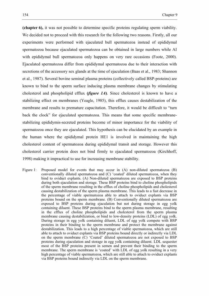
154 Chapter 9
(chapter 6), it was not possible to determine specific proteins regulating sperm viability.
We decided not to proceed with this research for the following two reasons. Firstly, all our
experiments were performed with ejaculated bull spermatozoa instead of epididymal
spermatozoa because ejaculated spermatozoa can be obtained in large numbers while AI
with epididymal bull spermatozoa only happens on very rare occasions (Foote, 2000).
Ejaculated spermatozoa differ from epididymal spermatozoa due to their interaction with
secretions of the accessory sex glands at the time of ejaculation (Baas et al., 1983; Shannon
et al., 1987). Several bovine seminal plasma proteins (collectively called BSP-proteins) are
known to bind to the sperm surface inducing plasma membrane changes by stimulating
cholesterol and phospholipid efflux (figure 1A). Since cholesterol is known to have a
stabilizing effect on membranes (Yeagle, 1985), this efflux causes destabilization of the
membrane and results to premature capacitation. Therefore, it would be difficult to “turn
back the clock” for ejaculated spermatozoa. This means that some specific membrane-
stabilizing epididymis-secreted proteins become of minor importance for the viability of
spermatozoa once they are ejaculated. This hypothesis can be elucidated by an example in
the human where the epididymal protein HE1 is involved in maintaining the high
cholesterol content of spermatozoa during epididymal transit and storage. However this
cholesterol carrier protein does not bind firmly to ejaculated spermatozoa (Kirchhoff,
1998) making it impractical to use for increasing membrane stability.
Figure 1: Proposed model for events that may occur in (A) non-diluted spermatozoa (B) conventionally diluted spermatozoa and (C) ‘coated’ diluted spermatozoa, when they bind to oviduct explants. (A) Non-diluted spermatozoa are exposed to BSP proteins during both ejaculation and storage. These BSP proteins bind to choline phospholipids of the sperm membrane resulting in the efflux of choline phospholipids and cholesterol causing destabilization of the sperm plasma membrane. This leads to a fast decrease in the percentage of viable spermatozoa able to attach to oviduct explants via BSP proteins bound on the sperm membrane. (B) Conventionally diluted spermatozoa are exposed to BSP proteins during ejaculation but not during storage in egg yolk containing diluent. These BSP proteins bind to the sperm plasma membrane, resulting in the efflux of choline phospholipids and cholesterol from the sperm plasma membrane causing destabilization, or bind to low-density proteins (LDL) of egg yolk. During storage in egg yolk containing diluent, LDL of egg yolk compete with BSP proteins in their binding to the sperm membrane and protect the membrane against destabilization. This leads to a high percentage of viable spermatozoa, which are still able to attach to oviduct explants via BSP proteins bound directly or indirectly via LDL on the sperm membrane (C) ‘Coated’ diluted spermatozoa are not exposed to BSP proteins during ejaculation and storage in egg yolk containing diluent. LDL sequester most of the BSP proteins present in semen and prevent their binding to the sperm membrane. The sperm membrane is ‘coated’ with LDL of egg yolk resulting in a very high percentage of viable spermatozoa, which are still able to attach to oviduct explants via BSP proteins bound indirectly via LDL on the sperm membrane.

General discussion 155
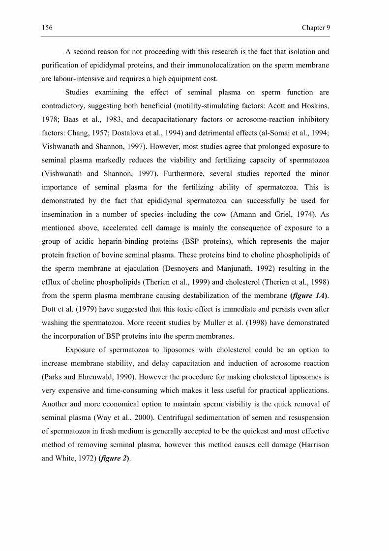
156 Chapter 9
A second reason for not proceeding with this research is the fact that isolation and
purification of epididymal proteins, and their immunolocalization on the sperm membrane
are labour-intensive and requires a high equipment cost.
Studies examining the effect of seminal plasma on sperm function are
contradictory, suggesting both beneficial (motility-stimulating factors: Acott and Hoskins,
1978; Baas et al., 1983, and decapacitationary factors or acrosome-reaction inhibitory
factors: Chang, 1957; Dostalova et al., 1994) and detrimental effects (al-Somai et al., 1994;
Vishwanath and Shannon, 1997). However, most studies agree that prolonged exposure to
seminal plasma markedly reduces the viability and fertilizing capacity of spermatozoa
(Vishwanath and Shannon, 1997). Furthermore, several studies reported the minor
importance of seminal plasma for the fertilizing ability of spermatozoa. This is
demonstrated by the fact that epididymal spermatozoa can successfully be used for
insemination in a number of species including the cow (Amann and Griel, 1974). As
mentioned above, accelerated cell damage is mainly the consequence of exposure to a
group of acidic heparin-binding proteins (BSP proteins), which represents the major
protein fraction of bovine seminal plasma. These proteins bind to choline phospholipids of
the sperm membrane at ejaculation (Desnoyers and Manjunath, 1992) resulting in the
efflux of choline phospholipids (Therien et al., 1999) and cholesterol (Therien et al., 1998)
from the sperm plasma membrane causing destabilization of the membrane (figure 1A).
Dott et al. (1979) have suggested that this toxic effect is immediate and persists even after
washing the spermatozoa. More recent studies by Muller et al. (1998) have demonstrated
the incorporation of BSP proteins into the sperm membranes.
Exposure of spermatozoa to liposomes with cholesterol could be an option to
increase membrane stability, and delay capacitation and induction of acrosome reaction
(Parks and Ehrenwald, 1990). However the procedure for making cholesterol liposomes is
very expensive and time-consuming which makes it less useful for practical applications.
Another and more economical option to maintain sperm viability is the quick removal of
seminal plasma (Way et al., 2000). Centrifugal sedimentation of semen and resuspension
of spermatozoa in fresh medium is generally accepted to be the quickest and most effective
method of removing seminal plasma, however this method causes cell damage (Harrison
and White, 1972) (figure 2).
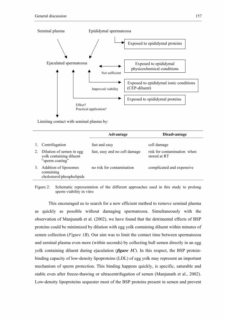
General discussion 157
Advantage Disadvantage
1. Centrifugation fast and easy cell damage 2. Dilution of semen in egg
yolk containing diluent “sperm coating”
fast, easy and no cell damage risk for contamination when stored at RT
3. Addition of liposomes containing cholesterol/phospholipids
no risk for contamination complicated and expensive
Figure 2: Schematic representation of the different approaches used in this study to prolong sperm viability in vitro
This encouraged us to search for a new efficient method to remove seminal plasma
as quickly as possible without damaging spermatozoa. Simultaneously with the
observation of Manjunath et al. (2002), we have found that the detrimental effects of BSP
proteins could be minimized by dilution with egg yolk containing diluent within minutes of
semen collection (Figure 1B). Our aim was to limit the contact time between spermatozoa
and seminal plasma even more (within seconds) by collecting bull semen directly in an egg
yolk containing diluent during ejaculation (figure 1C). In this respect, the BSP protein-
binding capacity of low-density lipoproteins (LDL) of egg yolk may represent an important
mechanism of sperm protection. This binding happens quickly, is specific, saturable and
stable even after freeze-thawing or ultracentrifugation of semen (Manjunath et al., 2002).
Low-density lipoproteins sequester most of the BSP proteins present in semen and prevent
Limiting contact with seminal plasma by:
Exposed to epididymal proteins
Not sufficient
Exposed to epididymal physicochemical conditions
Exposed to epididymal ionic conditions(CEP-diluent) Improved viability
Exposed to epididymal proteins Effect? Practical application?
Ejaculated spermatozoa
Seminal plasma Epididymal spermatozoa
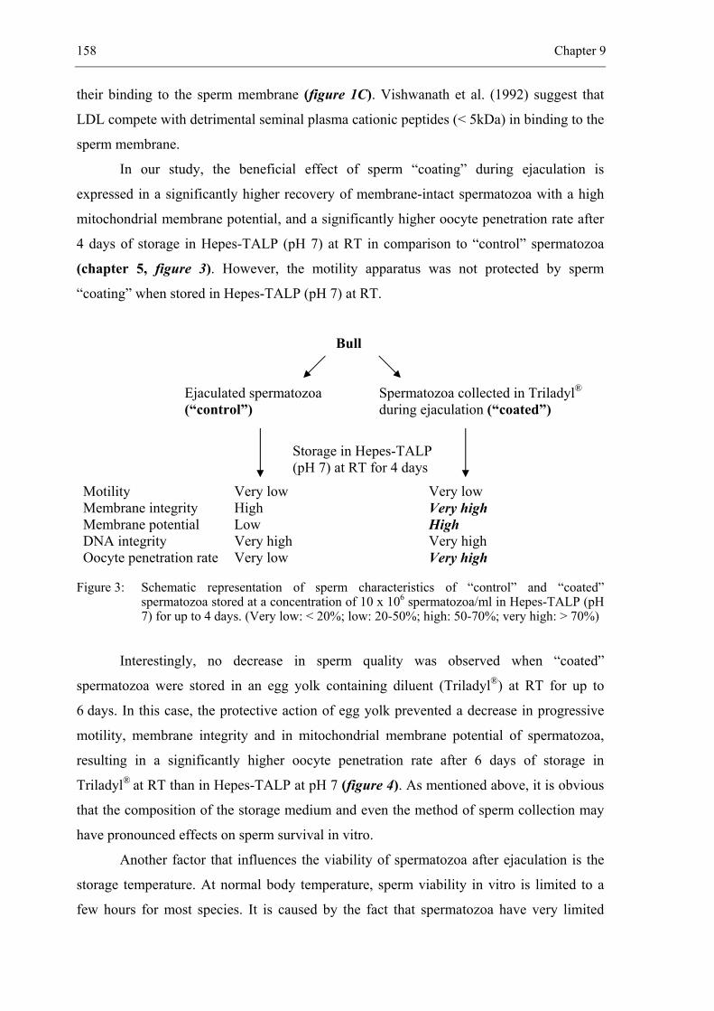
158 Chapter 9
their binding to the sperm membrane (figure 1C). Vishwanath et al. (1992) suggest that
LDL compete with detrimental seminal plasma cationic peptides (< 5kDa) in binding to the
sperm membrane.
In our study, the beneficial effect of sperm “coating” during ejaculation is
expressed in a significantly higher recovery of membrane-intact spermatozoa with a high
mitochondrial membrane potential, and a significantly higher oocyte penetration rate after
4 days of storage in Hepes-TALP (pH 7) at RT in comparison to “control” spermatozoa
(chapter 5, figure 3). However, the motility apparatus was not protected by sperm
“coating” when stored in Hepes-TALP (pH 7) at RT.
Bull
Ejaculated spermatozoa Spermatozoa collected in Triladyl®
(“control”) during ejaculation (“coated”)
Storage in Hepes-TALP (pH 7) at RT for 4 days
Motility Very low Very low Membrane integrity High Very high Membrane potential Low High DNA integrity Very high Very high Oocyte penetration rate Very low Very high
Figure 3: Schematic representation of sperm characteristics of “control” and “coated” spermatozoa stored at a concentration of 10 x 106 spermatozoa/ml in Hepes-TALP (pH 7) for up to 4 days. (Very low: < 20%; low: 20-50%; high: 50-70%; very high: > 70%)
Interestingly, no decrease in sperm quality was observed when “coated”
spermatozoa were stored in an egg yolk containing diluent (Triladyl®) at RT for up to
6 days. In this case, the protective action of egg yolk prevented a decrease in progressive
motility, membrane integrity and in mitochondrial membrane potential of spermatozoa,
resulting in a significantly higher oocyte penetration rate after 6 days of storage in
Triladyl® at RT than in Hepes-TALP at pH 7 (figure 4). As mentioned above, it is obvious
that the composition of the storage medium and even the method of sperm collection may
have pronounced effects on sperm survival in vitro.
Another factor that influences the viability of spermatozoa after ejaculation is the
storage temperature. At normal body temperature, sperm viability in vitro is limited to a
few hours for most species. It is caused by the fact that spermatozoa have very limited
General discussion 159
biosynthetic activity (Hammerstedt and Andrews, 1997). Spermatozoa must be deep-
frozen below -130°C to halt their metabolic processes, so that thermally driven chemical
reactions cannot take place (Medeiros et al., 2002). However, the freezing and thawing
processes cause irreversible damage to a large proportion of bull spermatozoa and render
the remainder more sensitive to environmental stress (Hammerstedt et al., 1990; Parks and
Graham, 1992; Shannon and Vishwanath, 1995). This kind of long-term preservation was
no option in our study and therefore, semen stored at RT or at 4°C was used as an
alternative for frozen-thawed semen.
From our results it was clear that semen stored at 4°C in Hepes-TALP (pH 7)
during 4 days showed a very low oocyte penetration capacity in comparison to semen
stored in Hepes-TALP (pH 7) at RT (figure 4). This difference in oocyte penetration rate
could not be explained by the decline in motility of stored spermatozoa since it was low in
both groups. It is probably due to destabilization of the sperm membranes, as a
consequence of cold shock injuries, inducing capacitation or acrosome reaction.
“Coated” spermatozoa Stored during 6 days
RT 4°C
Hepes-TALP Triladyl® Hepes-TALP Triladyl®
Total motility Low Very high Low Very high
Progressive motility Very low Very high Very low Very high
Membrane integrity High Very high Very high Very high
Membrane potential High Very high Very high Very high
Oocyte penetration rate High Very high Very low Very high
Figure 4: Schematic representation of sperm characteristics of “coated” spermatozoa stored at a concentration of 10 x 106 spermatozoa/ml in Hepes-TALP (pH 7) or in Triladyl® at RT or at 4°C for up to 6 days. (Very low: < 20%; low: 20-50%; high: 50-70%; very high: > 70%)
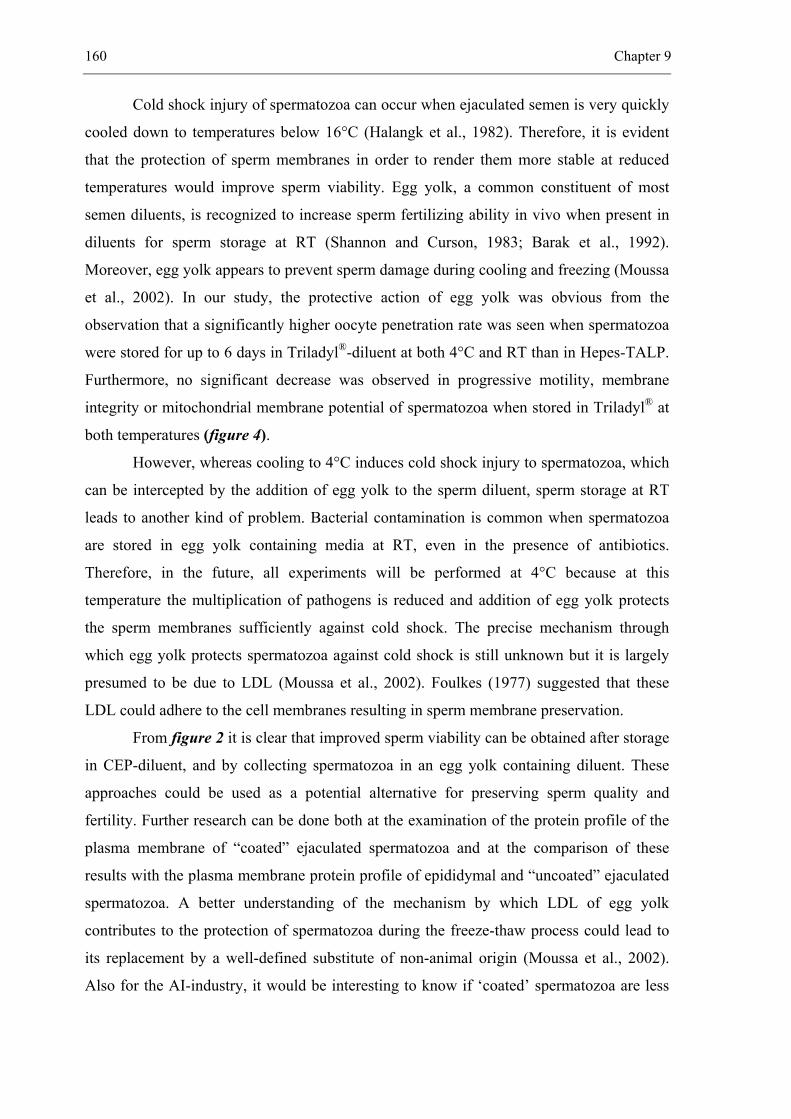
160 Chapter 9
Cold shock injury of spermatozoa can occur when ejaculated semen is very quickly
cooled down to temperatures below 16°C (Halangk et al., 1982). Therefore, it is evident
that the protection of sperm membranes in order to render them more stable at reduced
temperatures would improve sperm viability. Egg yolk, a common constituent of most
semen diluents, is recognized to increase sperm fertilizing ability in vivo when present in
diluents for sperm storage at RT (Shannon and Curson, 1983; Barak et al., 1992).
Moreover, egg yolk appears to prevent sperm damage during cooling and freezing (Moussa
et al., 2002). In our study, the protective action of egg yolk was obvious from the
observation that a significantly higher oocyte penetration rate was seen when spermatozoa
were stored for up to 6 days in Triladyl®-diluent at both 4°C and RT than in Hepes-TALP.
Furthermore, no significant decrease was observed in progressive motility, membrane
integrity or mitochondrial membrane potential of spermatozoa when stored in Triladyl® at
both temperatures (figure 4).
However, whereas cooling to 4°C induces cold shock injury to spermatozoa, which
can be intercepted by the addition of egg yolk to the sperm diluent, sperm storage at RT
leads to another kind of problem. Bacterial contamination is common when spermatozoa
are stored in egg yolk containing media at RT, even in the presence of antibiotics.
Therefore, in the future, all experiments will be performed at 4°C because at this
temperature the multiplication of pathogens is reduced and addition of egg yolk protects
the sperm membranes sufficiently against cold shock. The precise mechanism through
which egg yolk protects spermatozoa against cold shock is still unknown but it is largely
presumed to be due to LDL (Moussa et al., 2002). Foulkes (1977) suggested that these
LDL could adhere to the cell membranes resulting in sperm membrane preservation.
From figure 2 it is clear that improved sperm viability can be obtained after storage
in CEP-diluent, and by collecting spermatozoa in an egg yolk containing diluent. These
approaches could be used as a potential alternative for preserving sperm quality and
fertility. Further research can be done both at the examination of the protein profile of the
plasma membrane of “coated” ejaculated spermatozoa and at the comparison of these
results with the plasma membrane protein profile of epididymal and “uncoated” ejaculated
spermatozoa. A better understanding of the mechanism by which LDL of egg yolk
contributes to the protection of spermatozoa during the freeze-thaw process could lead to
its replacement by a well-defined substitute of non-animal origin (Moussa et al., 2002).
Also for the AI-industry, it would be interesting to know if ‘coated’ spermatozoa are less
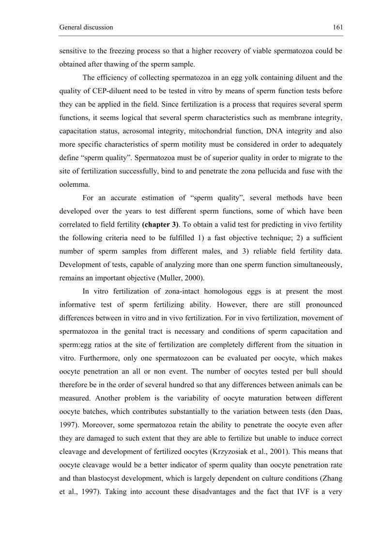
General discussion 161
sensitive to the freezing process so that a higher recovery of viable spermatozoa could be
obtained after thawing of the sperm sample.
The efficiency of collecting spermatozoa in an egg yolk containing diluent and the
quality of CEP-diluent need to be tested in vitro by means of sperm function tests before
they can be applied in the field. Since fertilization is a process that requires several sperm
functions, it seems logical that several sperm characteristics such as membrane integrity,
capacitation status, acrosomal integrity, mitochondrial function, DNA integrity and also
more specific characteristics of sperm motility must be considered in order to adequately
define “sperm quality”. Spermatozoa must be of superior quality in order to migrate to the
site of fertilization successfully, bind to and penetrate the zona pellucida and fuse with the
oolemma.
For an accurate estimation of “sperm quality”, several methods have been
developed over the years to test different sperm functions, some of which have been
correlated to field fertility (chapter 3). To obtain a valid test for predicting in vivo fertility
the following criteria need to be fulfilled 1) a fast objective technique; 2) a sufficient
number of sperm samples from different males, and 3) reliable field fertility data.
Development of tests, capable of analyzing more than one sperm function simultaneously,
remains an important objective (Muller, 2000).
In vitro fertilization of zona-intact homologous eggs is at present the most
informative test of sperm fertilizing ability. However, there are still pronounced
differences between in vitro and in vivo fertilization. For in vivo fertilization, movement of
spermatozoa in the genital tract is necessary and conditions of sperm capacitation and
sperm:egg ratios at the site of fertilization are completely different from the situation in
vitro. Furthermore, only one spermatozoon can be evaluated per oocyte, which makes
oocyte penetration an all or non event. The number of oocytes tested per bull should
therefore be in the order of several hundred so that any differences between animals can be
measured. Another problem is the variability of oocyte maturation between different
oocyte batches, which contributes substantially to the variation between tests (den Daas,
1997). Moreover, some spermatozoa retain the ability to penetrate the oocyte even after
they are damaged to such extent that they are able to fertilize but unable to induce correct
cleavage and development of fertilized oocytes (Krzyzosiak et al., 2001). This means that
oocyte cleavage would be a better indicator of sperm quality than oocyte penetration rate
and than blastocyst development, which is largely dependent on culture conditions (Zhang
et al., 1997). Taking into account these disadvantages and the fact that IVF is a very
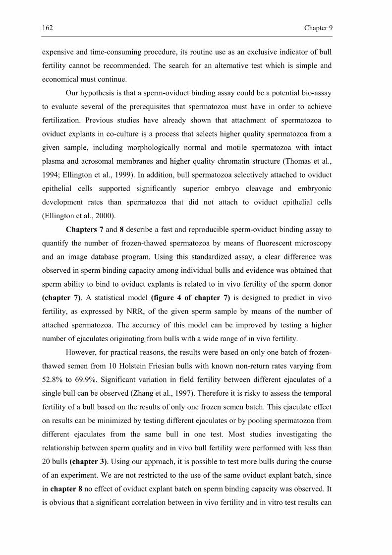
162 Chapter 9
expensive and time-consuming procedure, its routine use as an exclusive indicator of bull
fertility cannot be recommended. The search for an alternative test which is simple and
economical must continue.
Our hypothesis is that a sperm-oviduct binding assay could be a potential bio-assay
to evaluate several of the prerequisites that spermatozoa must have in order to achieve
fertilization. Previous studies have already shown that attachment of spermatozoa to
oviduct explants in co-culture is a process that selects higher quality spermatozoa from a
given sample, including morphologically normal and motile spermatozoa with intact
plasma and acrosomal membranes and higher quality chromatin structure (Thomas et al.,
1994; Ellington et al., 1999). In addition, bull spermatozoa selectively attached to oviduct
epithelial cells supported significantly superior embryo cleavage and embryonic
development rates than spermatozoa that did not attach to oviduct epithelial cells
(Ellington et al., 2000).
Chapters 7 and 8 describe a fast and reproducible sperm-oviduct binding assay to
quantify the number of frozen-thawed spermatozoa by means of fluorescent microscopy
and an image database program. Using this standardized assay, a clear difference was
observed in sperm binding capacity among individual bulls and evidence was obtained that
sperm ability to bind to oviduct explants is related to in vivo fertility of the sperm donor
(chapter 7). A statistical model (figure 4 of chapter 7) is designed to predict in vivo
fertility, as expressed by NRR, of the given sperm sample by means of the number of
attached spermatozoa. The accuracy of this model can be improved by testing a higher
number of ejaculates originating from bulls with a wide range of in vivo fertility.
However, for practical reasons, the results were based on only one batch of frozen-
thawed semen from 10 Holstein Friesian bulls with known non-return rates varying from
52.8% to 69.9%. Significant variation in field fertility between different ejaculates of a
single bull can be observed (Zhang et al., 1997). Therefore it is risky to assess the temporal
fertility of a bull based on the results of only one frozen semen batch. This ejaculate effect
on results can be minimized by testing different ejaculates or by pooling spermatozoa from
different ejaculates from the same bull in one test. Most studies investigating the
relationship between sperm quality and in vivo bull fertility were performed with less than
20 bulls (chapter 3). Using our approach, it is possible to test more bulls during the course
of an experiment. We are not restricted to the use of the same oviduct explant batch, since
in chapter 8 no effect of oviduct explant batch on sperm binding capacity was observed. It
is obvious that a significant correlation between in vivo fertility and in vitro test results can
General discussion 163
only be obtained if the range of in vivo fertility is large. However, variation in NRR
between bulls that are used for AI is generally in the order of 10 to 15% (Uwland, 1984).
In our study the widest possible range in NRR of bulls available from the AI center was
17%. A wide range in fertility is difficult to obtain, since bulls that have been preselected
as having inadequate semen characteristics after collection are not evaluated for field
fertility. The accuracy of field fertility data also had a profound effect on the interpretation
of the results. It is well known that factors related to sire, cow, inseminator, season and
herd health management influence field fertility (de Kruif, 1978; den Daas, 1997).
Therefore, in our study only sires of the same age with comparable numbers of services,
and cows inseminated by experienced inseminators during autumn and winter were used.
In conclusion, further refinement of our in vitro model is needed before it can be concluded
whether the sperm-oviduct binding assay is a reliable tool to predict in vivo bull fertility.
From the results of chapter 8 it is clear that the sensitivity of the sperm-oviduct
binding assay was comparable to that of the oocyte penetration assay for the assessment of
sperm quality of cooled bovine spermatozoa stored in CEP, CAPROGEN® or Triladyl® for
up to 6 days. It was not possible to investigate the correlation between sperm-oviduct
binding assay and in vivo fertility of cooled semen. For practical reasons, it is not feasible
to obtain data of NRR after insemination with liquid spermatozoa from different bulls in
Belgium. This can only be performed in New Zealand, where a unique system of seasonal
breeding exists. In this country more than 85% of all inseminations are performed with
liquid stored semen during spring and summer (Vishwanath et al., 1996; Vishwanath and
Shannon, 2000). Furthermore, it is also difficult to use the same ejaculate for both AI and
sperm-oviduct binding assay.
164 Chapter 9
Concluding remarks
From the results described in this thesis, four conclusions can be drawn:
1. improved sperm quality can be obtained by collecting semen within seconds after
ejaculation in egg yolk containing diluent. This approach could be used as potential
alternative for preserving sperm quality and in vivo bull fertility.
2. despite the fact that we were able to culture caput and cauda epididymal epithelial
cells in vitro which secrete hormone- and region-dependent proteins, it was not
possible to determine specific proteins that regulate sperm viability.
3. our newly developed sperm-oviduct binding assay could become a promising
functional test for the assessment of frozen-thawed sperm quality.
4. the sensitivity of the sperm-oviduct binding assay for the assessment of sperm quality
of cooled bovine spermatozoa was comparable to that of the oocyte penetration
assay.
General discussion 165
References
Acott TS, Hoskins DD. Bovine sperm forward motility protein. Partial purification and
characterization. J Biol Chem 1978; 253: 6744-6750.
al-Somai N, Vishwanath R, Molan PC, Shannon P. Anionic and cationic components from protein
aggregates in bovine seminal plasma and their effects on sperm motility. Mol Reprod Dev
1994; 39: 328-336.
Amann RP, Griel LC Jr. Fertility of bovine spermatozoa from rete testis, cauda epididymidis, and
ejaculated semen. J Dairy Sci 1974; 57: 212-219.
Baas JW, Molan PC, Shannon P. Factors in seminal plasma of bulls that affect the viability and
motility of spermatozoa. J Reprod Fertil 1983; 68: 275-280.
Barak Y, Amit A, Lessing JB, Paz G, Homonnai ZT, Yogev L. Improved fertilization rate in an in
vitro fertilization program by egg yolk-treated sperm. Fertil Steril 1992; 58: 197-198.
Carr DW, Acott TS. Inhibition of bovine spermatozoa by caudal epididymal fluid: I. Studies of a
sperm motility quiescence factor. Biol Reprod 1984; 30: 913-925.
Cascieri M, Amann RP, Hammerstedt RH. Adenine nucleotide changes at initiation of bull sperm
motility. J Biol Chem 1976; 251: 787-793.
Chang MC. A detrimental effect of seminal plasma on the fertilizing capacity of sperm. Nature
1957; 179: 258-259.
Crabo B. Studies on the composition of epididymal content in bulls and boars. Acta Vet Scand
Suppl 1965; 6: 1-94.
Crichton EG, Hinton BT, Pallone TL, Hammerstedt RH. Hyperosmolality and sperm storage in
hibernating bats: prolongation of sperm life by dehydration. Am J Physiol 1994; 267: 1363-
1370.
de Kruif A. Factors influencing the fertility of a cattle population. J Reprod Fertil 1978; 54: 507-
518.
den Daas JHG. Prediction of bovine male fertility. Ph.D. Thesis, University of Wageningen, The
Netherlands, 1997.
Desnoyers L, Manjunath P. Major proteins of bovine seminal plasma exhibit novel interactions
with phospholipid. J Biol Chem 1992; 267: 10149-10155.
Dostalova Z, Calvete JJ, Sanz L, Hettel C, Riedel D, Schoneck C, Einspanier R, Topfer-Petersen E.
Immunolocalization and quantitation of acidic seminal fluid protein (aSFP) in ejaculated,
swim-up, and capacitated bull spermatozoa. Biol Chem Hoppe Seyler 1994; 375: 457-461.
166 Chapter 9
Dott HM, Harrison RA, Foster GC. The maintenance of motility and the surface properties of
epididymal spermatozoa from bull, rabbit and ram in homologous seminal and epididymal
plasma. J Reprod Fertil 1979; 55: 113-124.
Ellington JE, Evenson DP, Wright RW Jr, Jones AE, Schneider CS, Hiss GA, Brisbois RS. Higher-
quality human sperm in a sample selectively attach to oviduct (fallopian tube) epithelial cells
in vitro. Fertil Steril 1999; 71: 924-929.
Ellington JE, Evenson DP, DeAvila J, Jost LK, Wright RW Jr. Bull sperm that attach to oviduct
cells in vitro support superior embryonic development as compared to sperm that do not
attach. Proceedings of the ICAR Stockholm, Sweden 2000.
Foote RH. Fertilizing ability of epididymal sperm from dead animals. J Androl 2000; 21: 355.
Foulkes JA. The separation of lipoproteins from egg yolk and their effect on the motility and
integrity of bovine spermatozoa. J Reprod Fertil 1977; 49: 277-284.
Halangk W, Frank K, Bohnensack R. Effect of temperature decrease on the intactness of the cell
membrane of ejaculated bull spermatozoa. Acta Biol Med Ger 1982; 41: 715-719.
Hammerstedt RH, Graham JK, Nolan JP. Cryopreservation of mammalian sperm: what we ask
them to survive. J Androl 1990; 11: 73-88.
Hammerstedt RH, Andrews JC. Metabolic support of normothermia. In: Karow AM, Critser JK
(eds). Reproductive tissue banking. Academic Press; San Diego 1997: 139-166.
Harrison RA, White IG. Some methods for washing spermatozoa from bull, boar and ram:
a comparison using biochemical and other criteria. J Reprod Fertil 1972; 29: 271-284.
Hinton BT, Palladino MA. Epididymal epithelium: its contribution to the formation of a luminal
fluid microenvironment. Microsc Res Tech 1995; 30: 67-81.
Holt WV. Alternative strategies for the long-term preservation of spermatozoa. Reprod Fertil Dev
1997; 9: 309-319.
Iguer-ouada M, Verstegen JP. Long-term preservation of chilled canine semen: effect of
commercial and laboratory prepared extenders. Theriogenology 2001; 55: 671-684.
Jones JM, Bavister BD. Acidification of intracellular pH in bovine spermatozoa suppresses motility
and extends viable life. J Androl 2000; 21: 616-624.
Kirchhoff C. Molecular characterization of epididymal proteins. Rev Reprod 1998; 3: 86-95.
Krzyzosiak J, Molan P, McGowan L, Vishwanath R. Effect of sperm number and oxygenation state
of the storage media on in vitro fertility of bovine sperm stored at ambient temperature.
Theriogenology 2001; 55: 1401-1415.
General discussion 167
Lawitts JA, Biggers JD. Joint effects of sodium chloride, glutamine, and glucose in mouse
preimplantation embryo culture media. Mol Reprod Dev 1992; 31: 189-194.
Manjunath P, Nauc V, Bergeron A, Menard M. Major proteins of bovine seminal plasma bind to
the low-density lipoprotein fraction of hen's egg yolk. Biol Reprod 2002; 67: 1250-1258.
Maxwell WM, Stojanow T. Liquid storage of ram semen in the absence or presence of some
antioxidants. Reprod Fertil Dev 1996; 8: 1013-1020.
Medeiros CM, Forell F, Oliveira AT, Rodrigues JL. Current status of sperm cryopreservation: why
isn't it better? Theriogenology 2002; 57: 327-344.
Moussa M, Martinet V, Trimeche A, Tainturier D, Anton M. Low density lipoproteins extracted
from hen egg yolk by an easy method: cryoprotective effect on frozen-thawed bull semen.
Theriogenology 2002; 57: 1695-1706.
Muller P, Erlemann KR, Muller K, Calvete JJ, Topfer-Petersen E, Marienfeld K, Herrmann A.
Biophysical characterization of the interaction of bovine seminal plasma protein PDC-109
with phospholipid vesicles. Eur Biophys J 1998; 27: 33-41.
Muller CH. Rationale, interpretation, validation, and uses of sperm function tests. J Androl 2000;
21: 10-30.
Parks JE, Ehrenwald E. Cholesterol efflux from mammalian sperm and its potential role in
capacitation. In: Bavister BD, Cummins J, Roldan ERS (eds). Fertilization in Mammals.
Norwell; Massachusetts 1990: 155-167.
Parks JE, Graham JK. Effects of cryopreservation procedures on sperm membranes.
Theriogenology 1992; 38: 209-222.
Setchell BP, Sánchez-Partida LG, Chairussyuhur A. Epididymal constituents and related
substances in the storage of spermatozoa: a review. Reprod Fertil Dev 1993; 5: 601-612.
Shannon P, Curson B. Effect of egg yolk levels on the fertility of diluted bovine sperm stored at
ambient temperatures. N Z J Agric Res 1983; 26: 187-189.
Shannon P, Curson B. Effect of storage temperature on the viability and fertility of bovine sperm
diluted and stored in Caprogen. N Z J Agric Res 1984; 27: 173-177.
Shannon P, Curson B, Molan PC. Cationic peptides obtained by reversible disaggregation of
antibacterial proteins of bovine seminal plasma. N Z J Agric Res 1987; 30: 195-202.
Shannon P, Vishwanath R. The effect of optimal and suboptimal concentrations of sperm on the
fertility of fresh and frozen bovine sperm and a theoretical model to explain the fertility
differences. Anim Reprod Sci 1995; 39: 1-10.
168 Chapter 9
Therien I, Moreau R, Manjunath P. Major proteins of bovine seminal plasma and high-density
lipoprotein induce cholesterol efflux from epididymal sperm. Biol Reprod 1998; 59: 768-
776.
Therien I, Moreau R, Manjunath P. Bovine seminal plasma phospholipid-binding proteins stimulate
phospholipid efflux from epididymal sperm. Biol Reprod 1999; 61: 590-598.
Thomas PG, Ball BA, Miller PG, Brinsko SP, Southwood L. A subpopulation of morphologically
normal, motile spermatozoa attach to equine oviductal epithelial cell monolayers. Biol
Reprod 1994; 51: 303-309.
Twigg J, Fulton N, Gomez E, Irvine DS, Aitken RJ. Analysis of the impact of intracellular reactive
oxygen species generation on the structural and functional integrity of human spermatozoa:
lipid peroxidation, DNA fragmentation and effectiveness of antioxidants. Hum Reprod 1998;
13: 1429-1436.
Uwland J. Correlation between the number of inseminated spermatozoa and results of fertilization
in dairy cattle. Tijdschr Diergeneeskd 1984; 109: 405-412.
Van Demark NL, Sharma UD. Preliminary fertility results from the preservation of bovine semen
at room temperature. J Dairy Sci 1957; 45: 375-382.
Verberckmoes S, Van Soom A, De Pauw I, Dewulf J, de Kruif A. Ionic composition of epididymal
plasma of the bull. In preparation.
Vishwanath R, Shannon P, Curson B. Cationic extracts of egg yolk and their effects on motility,
survival and fertilising ability of bull sperm. Anim Reprod Sci 1992; 29: 185-194.
Vishwanath R, Pitt CP, Shannon P. Sperm numbers, semen age and fertility in fresh and frozen
bovine semen. Proc N Z Soc Anim Prod 1996; 56: 31-34.
Vishwanath R, Shannon P. Do sperm cells age? A review of the physiological changes in sperm
during storage at ambient temperature. Reprod Fertil Dev 1997; 9: 321-331.
Vishwanath R, Shannon P. Storage of bovine semen in liquid and frozen state. Anim Reprod Sci
2000; 62: 23-53.
Way AL, Griel LC Jr, Killian GJ. Effects of accessory sex gland fluid on viability, capacitation,
and the acrosome reaction of cauda epididymal bull spermatozoa. J Androl 2000; 21: 213-
219.
Yeagle PL. Cholesterol and the cell membrane. Biochim Biophys Acta 1985; 822: 267-287.
Zhang BR, Larsson B, Lundeheim N, Rodriguez-Martinez H. Relationship between embryo
development in vitro and 56-day nonreturn rates of cows inseminated with frozen-thawed
semen from dairy bulls. Theriogenology 1997; 48: 221-231.
General discussion 169
Zhang BR, Larsson B, Lundeheim N, Rodriguez-Martinez H. Sperm characteristics and zona
pellucida binding in relation to field fertility of frozen-thawed semen from dairy AI bulls.
Int J Androl 1998; 21: 207-216.


_________________________________________________ 10.
SUMMARY / SAMENVATTING
________________________________________________________________________
172 Chapter 10
Summary
The goal of the cattle AI-industry is to maximize the number of offspring produced
by selected genetically superior bulls. This is accomplished by maximizing the number of
inseminated cows per ejaculate without a reduction in fertility. The ability of spermatozoa
to fertilize when inseminated depends mostly upon its quality after storage. In this respect,
both biochemical composition of the diluent and storage temperature are key factors
affecting the lifespan of spermatozoa during storage. In the 20th century, two major storage
technologies for liquid and frozen semen were developed, both with advantages and
disadvantages (chapter 1).
The general aims of this thesis were to maintain the quality and fertilizing ability of
stored bovine spermatozoa by means of knowledge obtained by investigating different
physicochemical conditions, and ionic and protein composition of cauda epididymal
plasma (chapter 4, 5 and 6), and to set up a new sperm-oviduct binding assay in order to
use it as a new sperm function test for the prediction of in vivo bull fertility (chapter 7 and
8).
In chapter 2, the role of the epididymis for sperm conservation in vivo is
described. Special attention is given to the transport, maturation and storage of
spermatozoa in the epididymis, and to the biochemical composition of cauda epididymal
plasma.
In chapter 3, different methods for the evaluation of sperm quality such as
fluorescent dyes, motility assessment assays, and sperm binding and performance
assessment assays are discussed.
Ejaculated spermatozoa diluted in vitro remain viable only for a limited period of
time. Despite the development of numerous diluents for storage at 4°C or at room
temperature (RT), it is still not possible to preserve the fertilizing potential of spermatozoa
longer than 3 to 5 days. In chapter 4, an experiment is described in which ejaculated bull
spermatozoa were stored in the simple salt solution Hepes-TALP at RT for up to 4 days.
This medium was modified to obtain physicochemical conditions similar to those
prevailing in the cauda epididymidis. The effect of change in pH (4-8) or change in
osmolarity (100-800 mOsm) of Hepes-TALP, storage atmosphere (nitrogen-gassed or
aerobic) and sperm concentration (10 x 106-1 x 10⁹ spermatozoa/ml or undiluted) on
Summary / Samenvatting 173
different sperm characteristics such as membrane integrity, motility, mitochondrial
membrane potential or DNA integrity of the sperm sample was investigated in order to
assess sperm quality after storage. From the results it is clear that decreasing the pH of the
medium by addition of the strong acid HCl, increasing the osmolarity by addition of
sorbitol and storage of highly concentrated spermatozoa under nitrogen-gassed conditions
are not effective methods for long-term preservation of spermatozoa in vitro. In this
respect, it was not possible to mimic the luminal environment of the bovine cauda
epididymidis in order to store densely packed bovine spermatozoa in a potentially
fertilizing condition for several days after ejaculation. The main problems were the lack of
certain nutrients such as glucose or fructose in Hepes-TALP and the inability to remove
toxic waste products which are harmful for remaining viable spermatozoa.
In chapter 5, an attempt was made to maintain the quality of stored spermatozoa
by protecting them against detrimental bovine seminal plasma (BSP) proteins. These
proteins are known to bind to choline phospholipids of the sperm membrane at ejaculation
resulting in the efflux of choline phospholipids and cholesterol and causing destabilization
of the sperm plasma membrane. In our approach, bull semen was collected in egg yolk
containing diluent during ejaculation, which made it possible to limit the contact time
between spermatozoa and seminal plasma within seconds. The BSP protein-binding
capacity of the low-density lipoproteins (LDL) of egg yolk may represent an important
mechanism of sperm protection. In this respect, LDL sequester most of the BSP proteins
present in semen and prevent their binding to the sperm membrane. In our study, the
beneficial effect of LDL of egg yolk is expressed as a significantly higher recovery of
membrane-intact spermatozoa with a high mitochondrial membrane potential, and in a
significantly higher oocyte penetration rate even after storage in Hepes-TALP at pH 7 at
RT for 4 days. When these “coated” spermatozoa were stored for longer than 4 days at RT
or at 4°C, it remained necessary to protect spermatozoa by addition of egg yolk to the
storage medium. The higher oocyte penetration rates of stored spermatozoa obtained by
sperm “coating”, makes our approach a potential alternative for preserving sperm quality
and fertility.
Specific hormone-dependent protein secretions from epididymal principal cells are
associated with spermatozoa during their maturation and storage and play a fundamental
role in modifying the surface characteristics of spermatozoa in preparation for the events of
174 Chapter 10
fertilization. Therefore, in chapter 6, the effect of hormones (testosterone,
dihydrotestosterone and hydrocortisone) on protein secretions of caput and cauda
epididymal epithelial cells cultured in principal cell medium (PCM) was determined in
order to identify proteins important for sperm survival. A confluent monolayer of caput
and cauda epididymal epithelial cells was obtained in serum-containing PCM after 7 days
of culture at 38.5°C (5% CO2 in air) in the presence or absence of hormones. Neither
epithelial detachment nor substantial overgrowth of fibroblasts were observed during
10 days of culture. The secreted proteins were separated by means of 2D-SDS-PAGE.
Comparison of the different protein patterns showed 61 spots in total, 11 of which were
only secreted in the presence of hormones, 3 of which appeared to show hormone-related
changes and 25 of which were caput or cauda specific. Most of these secreted proteins
were low molecular weight acidic proteins. More than 60% of these proteins present in
caput or cauda cultures correspond to proteins secreted in caput or cauda epidiymal
plasma, which confirms their epididymal origin. Despite the fact that we were able to
culture caput and cauda epididymal epithelial cells in vitro which secrete hormone- and
region-dependent proteins it was not possible to determine specific proteins that regulate
sperm viability. Additional research is needed to identify and isolate specific proteins and
investigate their immunolocalization on the sperm membrane. Furthermore, it is possible
that some specific membrane-stabilizing epididymis-secreted proteins are of minor
importance to the viability of ejaculated spermatozoa, which differ from epididymal
spermatozoa due to their interaction with secretions of the accessory sex glands at the time
of ejaculation.
Fertilization is a complex process that requires several sperm functions. Therefore
it seems logical that different sperm characteristics such as membrane integrity,
capacitation status, acrosomal integrity, mitochondrial function, DNA integrity and also
more specific characteristics of sperm motility must be considered in order to adequately
define “sperm quality”. To be able to estimate “sperm quality” accurately, several sperm
function assays have been developed over the years to test different aspects of semen
quality, some of which have been correlated to field fertility (chapter 3). However, more
attention is now given to the development of tests analyzing more than one sperm function
at the same time. Our hypothesis is that a sperm-oviduct binding assay could be a potential
bio-assay to evaluate several of the prerequisites that sperm must have in order to achieve
fertilization. In chapter 7, an in vitro bio-assay was set up to quantify the number of
Summary / Samenvatting 175
frozen-thawed spermatozoa bound to oviduct explants in order to determine whether this
assay could be used to predict in vivo bull fertility. Sperm binding capacity was evaluated
by counting JC-1-labelled spermatozoa attached to defined oviduct explants. From the
results it is clear that Hepes-TALP is a more useful medium than IVF-TALP, TCM-199 +
10% FCS and TCM-199 for the investigation of sperm binding to oviduct explants, and
that oviduct explants with a surface smaller than 20 000 µm² provided more consistent
results than explants with a surface larger than 100 000 µm². Using this bio-assay,
differences were observed in the sperm binding capacity among individual bulls. Evidence
was obtained that sperm binding capacity to oviduct explants is related to in vivo fertility
of the sperm donor, when measured after 24 h of sperm-oviduct culture, and provided that
the membrane integrity of the sperm sample was higher than 60%. Further refinement of
our in vitro model, by testing different ejaculates from more bulls, is needed before it can
be concluded whether the sperm-oviduct binding assay is a reliable tool to predict in vivo
bull fertility.
The sperm-oviduct binding assay was further refined in order to make it possible to
test more bulls during the course of an experiment. We found that we are not restricted to
the use of the same oviduct explant batch, since in chapter 8 no difference on sperm
binding capacity was observed between different batches of high quality oviduct explants.
This sperm-oviduct binding assay was then used to analyze the oviduct binding capacity of
cooled bovine spermatozoa stored in three different diluents (CEP, CAPROGEN® and
Triladyl®) for up to 2, 4 and 6 days (D2-, D4- and D6-spermatozoa). No significant effect of
diluent on sperm-oviduct binding capacity of D2-, D4- and D6-spermatozoa was observed
after 30 min, 24 and 48 h of co-culture. In an additional experiment, it was shown that
oocyte penetration rate of cooled bovine spermatozoa stored for up to 6 days was also not
significantly affected by diluent type. This means that the sensitivity of the sperm-oviduct
binding assay was comparable to that of the oocyte penetration assay for the assessment of
sperm quality of cooled bovine spermatozoa stored in CEP, CAPROGEN® or Triladyl® for
up to 6 days.
176 Chapter 10
The general discussion and conclusions are presented in chapter 9.
From the results described in this thesis, four conclusions can be drawn:
1. improved sperm quality can be obtained by collecting semen within seconds after
ejaculation in egg yolk containing diluent. This approach could be used as potential
alternative for preserving sperm quality and in vivo bull fertility.
2. despite the fact that we were able to culture caput and cauda epididymal epithelial
cells in vitro which secrete hormone- and region-dependent proteins, it was not
possible to determine specific proteins that regulate sperm viability.
3. our newly developed sperm-oviduct binding assay could become a promising
functional test for the assessment of frozen-thawed sperm quality.
4. the sensitivity of the sperm-oviduct binding assay for the assessment of sperm quality
of cooled bovine spermatozoa was comparable to that of the oocyte penetration
assay.
Summary / Samenvatting 177
Samenvatting
Eén van de doelstellingen van kunstmatige inseminatie (KI) is het verkrijgen van
een maximaal aantal nakomelingen per ejaculaat van genetisch hoogwaardige fokstieren
zonder dat er een daling van de in vivo fertiliteit optreedt. De slaagkansen op bevruchting
na KI zijn hoofdzakelijk afhankelijk van de spermakwaliteit. Daarbij zijn zowel de
biochemische samenstelling van de verdunner als de bewaartemperatuur de belangrijkste
factoren voor de overleving van het sperma. In de loop van de vorige eeuw werden reeds
verschillende verdunners voor het invriezen van sperma bij -196°C of voor het bewaren
van sperma bij kamertemperatuur (KT) of bij 4°C ontwikkeld. De voor- en de nadelen van
het invriezen en bewaren van sperma bij KT of bij 4°C staan beschreven in hoofdstuk 1.
De studie had twee doelstellingen:
- het verlengen van de levensduur van stierensperma door het effect van fysisch-
chemische condities, en de ionen- en proteïnensamenstelling van het epididymaal
plasma op de overleving van spermatozoa na te gaan (hoofdstuk 4, 5 and 6).
- het op punt stellen van een nieuwe in vitro spermafunctietest om de in vivo
fertiliteit van stieren te kunnen voorspellen (hoofdstuk 7 and 8).
In hoofdstuk 2 wordt een overzicht gegeven van het transport, de rijping en de
bewaring van spermatozoa in de epididymis, en van de biochemische samenstelling van
het epididymaal plasma.
In hoofdstuk 3 worden de verschillende methodes besproken die gebruikt worden
voor de evaluatie van de spermakwaliteit zoals fluorescerende kleurstoffen,
motiliteitsbepalingen, spermabindingstesten en in vitro fertilisatie experimenten.
Geëjaculeerde spermatozoa hebben slechts een zeer korte levensduur. Ondanks de
ontwikkeling van verschillende verdunners voor de bewaring van spermatozoa bij 4°C of
bij KT, is het tot op heden niet mogelijk gebleken om het bevruchtend vermogen van
geëjaculeerde spermatozoa van de stier te verlengen tot meer dan 3 dagen. In hoofdstuk 4
wordt een onderzoek beschreven voor het bewaren van geëjaculeerde spermatozoa
gedurende 4 dagen in een eenvoudige zoutoplossing Hepes-TALP bij KT. Het effect van
de pH (4-8) en van de osmolariteit (100-800 mOsm) van het medium, van de atmosfeer
(stikstof of aëroob) en van de spermaconcentratie (10 x 106-1 x 10⁹ spermatozoa/ml of
178 Chapter 10
onverdund sperma) op de spermakwaliteit werd onderzocht. Hiertoe werden verschillende
spermakarakteristieken zoals membraanintegriteit, beweeglijkheid, mitochondriale
membraanpotentiaal en DNA-integriteit van het spermastaal geëvalueerd. Uit de resultaten
kan besloten worden dat een daling van de pH van het medium door toevoeging van HCl,
een stijging van de osmolariteit van het medium door toevoeging van sorbitol, een
doorgassing van het medium met stikstof of de bewaring van sterk geconcentreerde
spermatozoa geen doeltreffende methoden zijn om de overleving van spermatozoa te
bevorderen. Uit dit onderzoek bleek dat het op deze manier niet mogelijk was om de
leefbaarheid van hooggeconcentreerde spermatozoa gedurende verschillende dagen te
verlengen. De grootste problemen waren de afwezigheid van de nodige voedingsstoffen in
Hepes-TALP en het onvermogen om toxische afvalproducten te verwijderen.
In hoofdstuk 5 wordt beschreven hoe op een alternatieve en meer praktische
manier de overleving van stierensperma verbeterd kan worden. Dit gebeurde door
spermatozoa zowel tijdens als na de ejaculatie te beschermen tegen de schadelijke ‘bovine
seminal plasma’ (BSP) proteïnen. Deze BSP proteïnen binden aan cholinefosfolipiden van
de spermamembraan, waardoor cholinefosfolipiden en cholesterol verwijderd worden en
de membraan minder stabiel wordt. Onze methode houdt in dat geëjaculeerde spermatozoa
rechtstreeks opgevangen worden in een eidooierbevattende verdunner. Aldus kan het
contact tussen spermatozoa en zaadplasma beperkt worden tot enkele seconden. Een
belangrijke beschermende eigenschap van eidooier is gebaseerd op de aanwezigheid van
lipoproteïnen met een lage densiteit (LDL). Door hun capterend vermogen verhinderen
deze LDL de binding van BSP proteïnen aan de spermamembraan. Toepassing van deze
nieuwe spermacollectiemethode resulteerde in een significant hoger percentage membraan-
intacte spermatozoa met een hoge mitochondriale membraanpotentiaal en in een significant
hoger percentage eicellen die gepenetreerd werden door spermatozoa die 4 dagen bewaard
werden in Hepes-TALP (pH 7) bij KT. Wanneer deze “gecoate” spermatozoa langer dan 4
dagen bewaard moeten worden bij KT of bij 4°C, is het noodzakelijk om eveneens eidooier
toe te voegen aan het medium. De betere spermakwaliteit en het hoger percentage
eicelpenetratie verkregen na bewaring van “gecoate” spermatozoa, maakt deze methode
zeer waardevol.
Specifieke hormoonafhankelijke proteïnen die gesecreteerd worden door
epididymale epitheelcellen binden aan de spermatozoa tijdens hun rijpings- en
Summary / Samenvatting 179
bewaarproces in de epididymis en spelen een fundamentele rol in de modificatie van de
spermacelmembraan ter voorbereiding van de bevruchting. In hoofdstuk 6 wordt een
onderzoek beschreven naar de invloed van hormonen (testosteron, dihydrotestosteron en
hydrocortisone) op de secretie van proteïnen door caput en cauda epididymale
epitheelcelculturen. Het was de bedoeling specifieke proteïnen die belangrijk zijn voor de
overleving van het sperma te identificeren. Een monolaag van caput of cauda epididymale
epitheelcellen in serumbevattend ‘Principal Cell Medium’ werd verkregen na 7 dagen
incubatie bij 38.5°C en 5% CO2 in lucht en dit zowel in de af- als in de aanwezigheid van
hormonen. Loskomende epitheelcellen of overgroei met fibroblasten werd niet
waargenomen gedurende de eerste 10 dagen na het opstarten van de monolaagcultuur. De
gesecreteerde proteïnen werden vervolgens gescheiden door 2D-SDS-PAGE. Na
vergelijking van de verschillende proteïnengels werden 61 spots nader bekeken waarvan
11 spots enkel gescreteerd werden in de aanwezigheid van hormonen. Drie spots vertonen
hormoonafhankelijke veranderingen en 25 spots werden enkel gescreteerd in de caput of
cauda cultuur. De meeste van deze proteïnen hebben een laag moleculair gewicht en een
pI-waarde tussen 4 en 6,5. Meer dan zestig procent van de proteïnen die gesecreteerd
werden in een caput of cauda cultuur correspondeerde met proteïnen aanwezig in caput of
cauda epididymaal plasma waarmee hun epididymale oorsprong werd bevestigd. Ondanks
het feit dat we in staat waren een een monolaag van epididymale epitheelcellen te kweken
die hormoon- en plaatsafhankelijke proteïnen secreteerde, was het onmogelijk om
specifieke proteïnen te vinden die belangrijk zijn voor de overleving van het sperma.
Daarvoor is een bijkomend onderzoek nodig teneinde proteïnen te kunnen identificeren, te
isoleren en hun localisatie op de spermamembraan te bepalen. Anderzijds moet rekening
gehouden worden met het feit dat bepaalde membraanstabiliserende proteïnen die
gesecreteerd worden door de epididymis waarschijnlijk minder belangrijk zijn voor de
overleving van geëjaculeerde spermatozoa, omdat deze verschillen van epididymale
spermatozoa door hun interactie met componenten uit het zaadplasma tijdens de ejaculatie.
Een bevruchting is een gecompliceerd proces waarvoor verschillende
spermafuncties vereist zijn. Om het bevruchtend vermogen van een spermastaal zo
nauwkeurig mogelijk te kunnen voorspellen is het noodzakelijk om zoveel mogelijk
spermakarakteristieken van het te onderzoeken spermastaal na te gaan. Meer bepaald gaat
het hierbij om de membraanintegriteit, de capacitatiestatus, de acrosoomstatus, de DNA
integriteit en meer specifieke bewegingskarakteristieken van het spermastaal. Al deze
180 Chapter 10
spermakenmerken kunnen bepaald worden aan de hand van specifieke
spermafunctietesten: sommigen van deze kenmerken zijn gecorreleerd met de in vivo
fertiliteit (hoofdstuk 3). Tegenwoordig wordt meer en meer gezocht naar nieuwe
spermafunctietesten waarmee verschillende spermakarakteristieken tegelijkertijd
geanalyseerd kunnen worden. In hoofdstuk 7 wordt een nieuwe test beschreven die het
mogelijk maakt het aantal ontdooide spermatozoa, van een bepaald ejaculaat, gebonden
aan oviductexplanten op een nauwkeurige manier te tellen teneinde de in vivo fertiliteit
van de betreffende stier te kunnen voorspellen. Deze spermabindingscapaciteit werd
geëvalueerd door het aantal JC-1-gelabelde spermatozoa gebonden aan oviductexplanten
met bekende oppervlakte te tellen. Uit de resultaten kan besloten worden dat Hepes-TALP
een geschikter medium is voor de sperma-oviduct co-cultuur dan IVF-TALP, TCM-199 en
TCM-199 + 10% serum. Tevens verbeterde de herhaalbaarheid van de test wanneer
oviductexplanten met een oppervlakte kleiner dan 20 000 µm² werden gebruikt. Aan de
hand van deze test kunnen verschillen in spermabindingscapaciteit aan oviductexplanten
vastgesteld worden. Bovendien kon bewezen worden dat de spermabindingscapaciteit aan
oviductexplanten gerelateerd is met de in vivo fertiliteit van de spermadonor, wanneer
bepaald 24 uren na de start van sperma-oviduct co-incubatie, mits de membraanintegriteit
van het spermastaal hoger is dan 60%. Een bijkomende verfijning van dit in vitro model is
echter nodig (door verschillende ejaculaten van verschillende stieren te testen) om te
kunnen besluiten of deze sperma-oviduct bindingstest een betrouwbare methode is om de
in vivo fertiliteit van stieren te kunnen voorspellen.
In hoofdstuk 8 werden explanten afkomstig van verschillende oviducten gebruikt
om na te gaan of verschillende oviductexplantculturen kunnen gebruikt worden om sperma
van verschillende stieren te testen binnen hetzelfde experiment. Er werd geen significant
verschil in spermabindingscapaciteit tussen de oviductexplantculturen gevonden. Aldus is
het mogelijk om de resultaten van verschillende experimenten te vergelijken. De sperma-
oviductbindingstest werd vervolgens gebruikt om de bindingscapaciteit van stierensperma
na bewaring bij 4°C in 3 verschillende verdunners (CEP, CAPROGEN® en Triladyl®)
gedurende 2, 4 en 6 dagen (D2, D4 en D6-spermatozoa) te testen. Uit de resultaten werd
geen significant effect van de verdunner op de sperma-oviductbindingscapaciteit van D2,
D4 en D6-spermatozoa na 30 min, 24 en 48 co-cultuur gevonden. In een bijkomend
experiment werd aangetoond dat de eicelpenetratie van sperma bewaard bij 4°C gedurende
6 dagen, eveneens niet significant beinvloed wordt door de gebruikte verdunner. De
Summary / Samenvatting 181
conclusie die hieruit getrokken kan worden is dat de sperma-oviductbindingstest even
gevoelig is als de eicelpenetratietest voor de evaluatie van de kwaliteit van het sperma dat
gedurende 6 dagen bewaard werd in CEP, CAPROGEN® of Triladyl® bij 4°C.
Hoofdstuk 9 bevat de algemene discussie en de conclusies.
De conclusies van dit onderzoek zijn dat:
1. de overleving van het sperma kan verbeterd worden door geëjaculeerde spermatozoa
rechtstreeks op te vangen in een eidooierbevattende verdunner. Op deze manier
wordt het contact tussen spermatozoa en zaadplasma beperkt tot enkele seconden.
2. niettegenstaande er een monolaag van epididymale epitheelcellen kan worden
gekweekt die hormoon- en plaatsafhankelijke proteïnen secreteerde, het onmogelijk
was om specifieke proteïnen te vinden die belangrijk zijn voor de overleving van het
sperma.
3. de sperma-oviductbindingstest een veelbelovende functietest is voor de evaluatie van
de spermakwaliteit.
4. de sperma-oviductbindingstest even gevoelig is als de eicelpenetratietest voor de
evaluatie van de kwaliteit van gekoeld stierensperma.

Dankwoord
Dankwoord
Deze thesis, die het resultaat is van vier jaar onderzoek, kon enkel tot stand komen dankzij
de hulp van een groot aantal mensen die ik via deze weg wil bedanken.
Een eerste woord van dank gaat uit naar mijn promotor Prof. Dr. A. Van Soom. An, het
waren vier schitterende jaren waar ik met veel plezier dikwijls zal aan terugdenken. Ik heb
ontzettend veel bijgeleerd en samen hebben we dit doctoraat tot een goed einde gebracht.
Ik wil je bedanken voor uw vertrouwen, uw steun, alle aanmoedigingen en de vele nuttige
tips en ideeën. Mijn onderzoeksactiviteiten werden steeds nauwkeurig opgevolgd. Bedankt
voor alles en nog veel succes met het labo!
Prof. Dr. A. de Kruif, mijn co-promotor, wil ik bedanken voor de mogelijkheid die hij mij
geboden heeft om dit onderzoek uit te voeren. Bedankt omdat u, ondanks uw zeer drukke
agenda, steeds bereid was mijn artikels en dit proefschrift zorgvuldig na te lezen en te
becommentariëren.
Steven, samen hebben wij vier jaar hard gewerkt aan ons project. De samenwerking verliep
uitstekend, al moest ik voor alles en nog wat op mijn hoede blijven. Bedankt voor die leuke
tijd in het labo, op het bureau en in het buitenland. Het project ligt nu volledig in uw
handen voor de finale afwerking, veel succes!
Steven, ik ben je nog het één en het ander verschuldigd, gelukkig help jij me dit regelmatig
herinneren, ik zal er werk van maken!
Sofie en Tom wil ik bedanken voor de aangename werksfeer in ons bureau. We hebben er
keihard gewerkt maar toch ook veel plezier gehad. Jullie waren een extra stimulans als het
schrijven eventjes niet vlotte, en voor alle vragen en problemen kon ik steeds bij jullie
terecht. Sofie, bedankt voor die fijne tijd die we samen doorbrachten in Canada en
Schotland. Eventjes onze grenzen verleggen, het blijven goede herinneringen! Tom, samen
met twee vrouwen in één bureau, het was waarschijnlijk niet altijd even gemakkelijk, maar
je hebt je mannetje goed kunnen staan. Bedankt omdat ik steeds op jullie kon rekenen,
jullie waren meer dan collega’s! Veel succes met jullie onderzoek.
Verder wil ik speciaal het vroegere Ministerie van Middenstand en Landbouw en het
huidige Ministerie van Volksgezondheid, alsook de Vlaamse Rundvee Verbetering (VRV)
bedanken: zij zorgden voor de financiële steun waardoor dit onderzoek mogelijk werd.
Dankwoord
Geert H., Jan, Olivier, Philippe en Steven, bedankt voor die vele keren, zelfs op ongepaste
momenten of tijdens het weekend, dat jullie bereid waren sperma af te nemen, en vooral
voor jullie geduld als de stieren er niet direct zin in hadden. Kristof, Lucske, Marnik,
Michel en Rudy wil ik bedanken omdat ze telkens bereid waren te helpen bij de sperma-
afname, en voor de verzorging van de dieren. Els en Veronique bedankt voor het maken
van de spermaverdunners.
Mijn belangrijkste spermaleverancier “Jof”, vier jaar lang heb je het traject tussen uw stal
en de dekruimte ontzettend veel keren afgelegd, zin of geen zin, die ene sprong was telkens
zo belangrijk voor mijn onderzoek. Ongelukkig kwam je aan je einde, dit had je helemaal
niet verdiend.
Dominiek, Hans en Koen, statistiek is een vak apart en jullie beheersen het. Bedankt voor
de verwerking van al mijn gegevens, ook al maakten mijn proefopzetten het jullie niet
altijd even gemakkelijk. Marc C., Geert H., Nadia en Sharon wil ik bedanken voor hun
bijdrage bij het lezen en kritisch verbeteren van mijn teksten. Roger, dank je wel voor het
oplossen van al mijn computerproblemen en voor uw hulp bij het verzorgen van de lay-out
van deze thesis. Griet, Joke en Katrien wil ik bedanken voor hun hulp bij het uitvoeren van
de experimenten. Leila, Nadine en Sandra, bedankt voor het regelen van alle
administratieve zaken.
De overige collega’s en oud-collega’s van de vakgroep wil ik bedanken voor hun directe
en indirecte bijdrage aan dit werk en de aangename werksfeer. Aan de jonge onderzoekers,
veel succes gewenst. Het is hard werken maar zeker en vast de moeite waard.
Special thanks to Prof. Alan Goff for giving me the opportunity to work in his lab and for
the pleasant stay in Canada.
De leden van de examencommissie: Prof. Dr. A. Christophe, Prof. Dr. E. Meyer, Dr. I.
Moyaert, Prof. Dr. J.P. Remon, Prof. Dr. P. Simons, Prof. Dr. G.C. van der Weijden en
Prof. Dr. A. Van Zeveren wil ik bedanken voor hun waardevolle opmerkingen tijdens de
laatste fase van dit onderzoek.
Dank ook aan de directie, de dierenartsen en het personeel van de slachthuizen (Brugge,
Eeklo, Melle, Rekkem, Zele) omdat zij steeds bereid waren eierstokken en testikels te
collecteren.
Dankwoord
Vier jaar hard werken kan niet zonder de nodige ontspanningen. Dit is dan ook een unieke
kans om een aantal vrienden en in het bijzonder mijn familie te bedanken.
Door het vele schrijven was het wekelijks badmintonuurtje meer dan welkom. Ik wil dan
ook iedereen bedanken voor de spannende wedstrijdjes en de vele ondernomen pogingen
om me de smash-techniek onder de knie te doen krijgen.
Mijn buren ben ik ontzettend veel dank verschuldigd voor de gezellige avondjes. Jullie
zorgden voor de dagelijkse bemoedigingen en stimulansen tijdens mijn schrijfavonden.
Bedankt voor jullie vriendschap en begrip. Sarah, tevens bedankt voor de vele lekkere
etentjes, voor onze gesprekken en de zotte momenten. Never forget the rainbow!
Ik wil langs deze weg ook alle medestudenten van de kookles bedanken voor het
gezamenlijk klaarmaken en degusteren van onze lekkere gerechten. Kookles is meer dan
koken alleen, dat werd voor sommigen onder ons vlug duidelijk. Bijzondere woorden van
dank gaan hierbij uit naar Bernard, Johan, Joke, Luc, Lut, Matthias, Sabine en Tom voor
die gezellige babbels en de ambiance in den “Angulus”, dit dikwijls tot in de late uurtjes.
Bedankt voor jullie interesse in mijn stier en in mijn werk, en voor de jaarlijkse
Caiperinha-avond en dropping, de etentjes en alle andere activiteiten. Het blijven allemaal
leuke herinneringen. Directeur Tim, bedankt omdat je bereid was de receptie te verzorgen.
Joke, ik heb héél veel aan jou te danken, die vele mooie momenten, geen mens zou ooit
begrijpen.... Bedankt!
Katrien, bedankt voor onze jarenlange vriendschap. We hadden op die mooie zomerdag op
het Sint-Baafsplein geen enkel vermoeden van de vele uren die we samen zouden
doorbrengen tijdens onze studententijd en daarna. Tevens bedankt voor de leuke reis naar
Firenze, en onze wekelijkse gesprekken. Katrien, bedankt voor alles, omdat je me zo goed
begrijpt!
En dan zijn er nog de vele vrienden die ik hier niet genoemd heb. Bedankt voor de vele
activiteiten, de vriendschap, het begrip en de steun.
Dankwoord
En als laatste, de mensen die mij het nauwst aan het hart liggen...
Meter, nonkels en tantes ontzettend bedankt voor alles wat jullie voor mij reeds gedaan
hebben en nog steeds doen. De vakanties mocht ik steeds bij jullie ‘op den buiten’
doorbrengen. Telkens opnieuw keek ik er zo naar uit, er viel zoveel te beleven, die twee
maanden waren voor mij altijd véél te kort. Ik besef maar al te goed hoe mooi mijn kinder-
en jeugdjaren waren, dit heeft ongetwijfeld bijgedragen tot de persoon die ik vandaag ben.
Eveline en Frederik, bedankt om voor al mijn snoezies te zorgen. Voor dit alles wil ik
jullie, vanuit de grond van mijn hart, bedanken.
Tenslotte wil ik héél speciaal mijn ouders, zus Carine en Maarten bedanken voor de nooit
aflatende steun gedurende al die jaren, maar vooral tijdens de laatste jaren van studeren en
onderzoek. Jullie permanente interesse voor mijn werk en voor mijn doen en laten was
steeds een grote stimulans. De vele aanmoedigingen, liefde, geduld, begrip en
bezorgdheid; dit alles betekende zoveel. En ik weet het, ik zeg het véél te weinig maar
daarom des te meer: ik zie jullie ontzettend graag! Bedankt voor alles, voor al die mooie
jaren! Bij jullie voel ik me goed!
Ingrid
Curriculum vitae
Curriculum vitae
PERSONALIA
Ingrid De Pauw werd geboren op 24 april 1976 te Gent. In 1994 beëindigde zij haar
humaniora-opleiding aan het Visitatiehumaniora te Gent, richting Economische. In 1996
behaalde zij het diploma van kandidaat in de Farmaceutische Wetenschappen en in 1998
het diploma van licentiaat in de Biotechnologie. Onmiddellijk daarna trad zij in dienst als
wetenschappelijk medewerker bij de vakgroep Voortplanting, Verloskunde en
Bedrijfsdiergeneeskunde op het onderzoeksproject getiteld: “Onderzoek naar diepe
inseminatie met lage spermadoses en naar in vivo en in vitro fertiliteit van vers verdund
stierensperma.” Dit project werd gefinancierd door het Ministerie van Middenstand en
Landbouw en de Vlaamse Rundvee Verbetering. In 2003 behaalde zij het getuigschrift van
de doctoraatsopleiding in de diergeneeskundige wetenschappen.
Curriculum vitae
PUBLICATIES
Publicaties in internationale wetenschappelijke tijdschriften
Van Soom A., Tanghe S., De Pauw I., Maes D., de Kruif A. (2002). Function of the
cumulus oophorus before and during mammalian fertilization. Reproduction in Domestic
Animals 37: 144-151.
Verberckmoes S., Van Soom A., De Pauw I., De Wulf J., de Kruif A. (2002). Migration of
bovine spermatozoa in a synthetic medium and its correlation to in vivo bull fertility.
Theriogenology 58: 1027-1037.
De Pauw I., Van Soom A., Laevens H., Verberckmoes S., de Kruif A. (2002). Sperm
binding to epithelial oviduct explants in bulls with different non-return rates investigated
with a new in vitro model. Biology of Reproduction 67: 1073-1079.
De Pauw I., Van Soom A., Mintiens K., Verberckmoes S., de Kruif A. (2003). In vitro
survival of bovine spermatozoa stored at room temperature under epididymal conditions.
Theriogenology 59: 1093-1107.
De Pauw I., Van Soom A., Maes D., Verberckmoes S., de Kruif A. (2003). Effect of
sperm coating on the survival and penetrating ability of in vitro stored bovine spermatozoa.
Theriogenology 59: 1109-1122.
De Pauw I., Goff AK., Van Soom A., Verberckmoes S., de Kruif A. (2003). Hormonal
regulation of bovine secretory proteins derived from caput and cauda epididymal epithelial
cell cultures. Journal of Andrology 24 (3): Accepted.
Verberckmoes S., Van Soom A., De Pauw I., Dewulf J., de Kruif A. Assessment of a new
utero tubal junction insemination device in dairy cattle under field conditions.
Theriogenology: Submitted.
Verberckmoes S., Van Soom A., De Pauw I., Dewulf J., de Kruif A. Technical aspects of
a new utero tubal junction insemination device: effect of the insemination catheter on
sperm quality and fertility. Theriogenology: Submitted.
Curriculum vitae
Verberckmoes S., Van Soom A., De Pauw I., Dewulf J., Rijsselaere T., de Kruif A.
Influence of whole blood and serum on bovine sperm quality and in vitro fertilization
capacity. Theriogenology: Submitted.
De Pauw I., Van Soom A., Verberkmoes S., de Kruif A. Suitability of sperm function tests
for the evaluation of sperm quality and bull fertility – a review. Review in preparation.
De Pauw I., Van Soom A., Verberckmoes S., Laevens H., de Kruif A. Sperm-oviduct
binding capacity of cooled bovine spermatozoa stored for several days in different diluents.
Manuscript in preparation.
Verberckmoes S., Van Soom A., De Pauw I., Dewulf J., de Kruif A. Cauda epididymal
plasma (CEP) as base for a new diluter for fresh bovine semen. Theriogenology: in
preparation.
Publicaties in nationale wetenschappelijke tijdschriften
De Pauw I., Van Soom A., Verberckmoes S., de Kruif A. (2000). De overleving van
sperma in de epididymis van de stier: een model voor spermaopslag in vitro? Vlaams
Diergeneeskundig Tijdschrift 69; 159-167.
Verberckmoes S., De Pauw I., Vanroose G., Laevens H., de Kruif A. (2001). Cervicale
inseminatie in sheep. Vlaams Diergeneeskundig Tijdschrift 70; 475-480.
Verberckmoes S., Van Soom A., De Pauw I., Van Cruchten S., de Kruif A. (2002).
Freemartinism in sheep: a case report. Vlaams Diergeneeskundig Tijdschrift 71: 211-215.
Abstracts
De Pauw I., Verberckmoes S., Vanroose G., Van Soom A., de Kruif A. (1999).
Comparison of eosin/nigrosin and SYBR-14/PI staining for the assessment of the
membrane status of bull sperm. In: Proceedings of the 15e European Embryo Transfert
Association; 140.
Curriculum vitae
Verberckmoes S., De Pauw I., Vanroose G., Laevens H., Van Soom A., de Kruif A.
(2000). Influence of ultracentrifugation on motility and membrane integrity of fresh bull
sperm. Theriogenology 53; 490.
De Pauw I., Verberckmoes S., Mintiens K., Van Soom A., de Kruif A. (2000). Membrane
status of bovine spermatozoa diluted in media with different pH. Journal of Reproduction
and Fertility, abstract series 25; 86.
De Pauw I., Goeman K., Van Soom A., Laevens H., Verberckmoes S., de Kruif A. (2000).
Use of image analysis to investigate sperm-oviduct interactions in 4 different media in the
bovine. In: Proceedings of the 16e European Embryo Transfert Association; 132.
De Pauw I., Van Soom A., Verberckmoes S., Laevens H., de Kruif A. (2000). Binding of
bovine spermatozoa depends on the size of the oviduct vesicles. Journal of Reproduction
and Fertility, abstract series 26; 27.
De Pauw I., Verberckmoes S., Van Soom A., Laevens H., de Kruif A. (2001).
Relationship between sperm-oviduct binding and non-return rates in six bulls.
Theriogenology 55; 423.
Verberckmoes S., De Pauw I., Van Soom A., de Kruif A. (2001). Ionic composition and
osmolarity of caudal epididymal plasma in the bull. Theriogenology 55; 449.
De Pauw I., Laevens H, Van Soom A, Verberckmoes S., de Kruif. (2001). An in vitro
model to study sperm-oviduct interactions in bulls with different non-return rates. In:
Proceedings of Gamete function and interaction leading towards embryo development; 18.
Verberckmoes S., De Pauw I., Van Soom A., de Kruif A. (2001). Correlation between
sperm quality parameters and bovine fertility. In: Proceedings of Gamete function and
interaction leading towards embryo development; 41.
Verberckmoes S., Van Soom A., De Pauw I., Laevens H., de Kruif A. (2001).
Repeatability of bovine sperm migration assay in a synthetic medium containing methyl
cellulose. In Proceedings of the 5th Annual Conference of the European Society for
Domestic Animal Reproduction; 83.
Curriculum vitae
Verberckmoes S., De Pauw I., Van Soom A., Dewulf J., de Kruif A. (2002). Cannulation
of the ductus deferens in ram and bull. Biotech Agron Soc Environ 6; 14.
De Pauw I., Verberckmoes S., Van Soom A., de Kruif A. (2002). Fresh diluted bovine
semen can maintain in vitro fertilizing ability after 4 days of storage at ambient
temperature. Theriogenology 57; 660.
De Pauw I., Goff AK., Van Soom A., Verberckmoes S., de Kruif A. (2002). Influence of
hormones on protein secretion of caput and caudal epididymal epithelium culture in the
bovine. In: Proceedings of the IIIrd International Conference on the Epididymis; 29.
Verberckmoes S., De Pauw I., Van Soom A., de Kruif A. (2002). Assessment of feasibility
of deep intra uterine insemination in dairy cattle under field conditions. In: Proceedings of
theVIth International Symposium of Reproduction in Domestic Ruminants; A 87.
Verberckmoes S., De Pauw I., Van Soom A., Dewulf J., de Kruif A. (2002). New CEP-
diluter improves storage capacity of fresh bovine semen. In: Proceedings of the XXII
World buiatrics congress; 87.
Verberckmoes S., De Pauw I., Van Soom A., de Kruif A. (2002). Technical aspects of a
new deep intra-uterine insemination device in dairy cattly. In: Proceedings of the 3rd
Biannual Meeting of the Association for Applied Animal Andrology; 37.
Verberckmoes S., De Pauw I., Van Soom A., de Kruif A. (2002). Assessment of a new
deep intra uterine insemination device in dairy cattle under field conditions. Reproduction
in Domestic Animals 37; 255.
Verberckmoes S., De Pauw I., Van Soom A., Dewulf J., de Kruif A. (2002). Influence of
whole blood and blood serum on bovine sperm quality. Reproduction in Domestic Animals
37; 255.
Verberckmoes S., De Pauw I., Van Soom A., Dewulf J., de Kruif A. (2002). Influence of
whole blood and blood serum on in vitro fertilizing capacity of bovine spermatozoa.
Reproduction in Domestic Animals 37; 255.
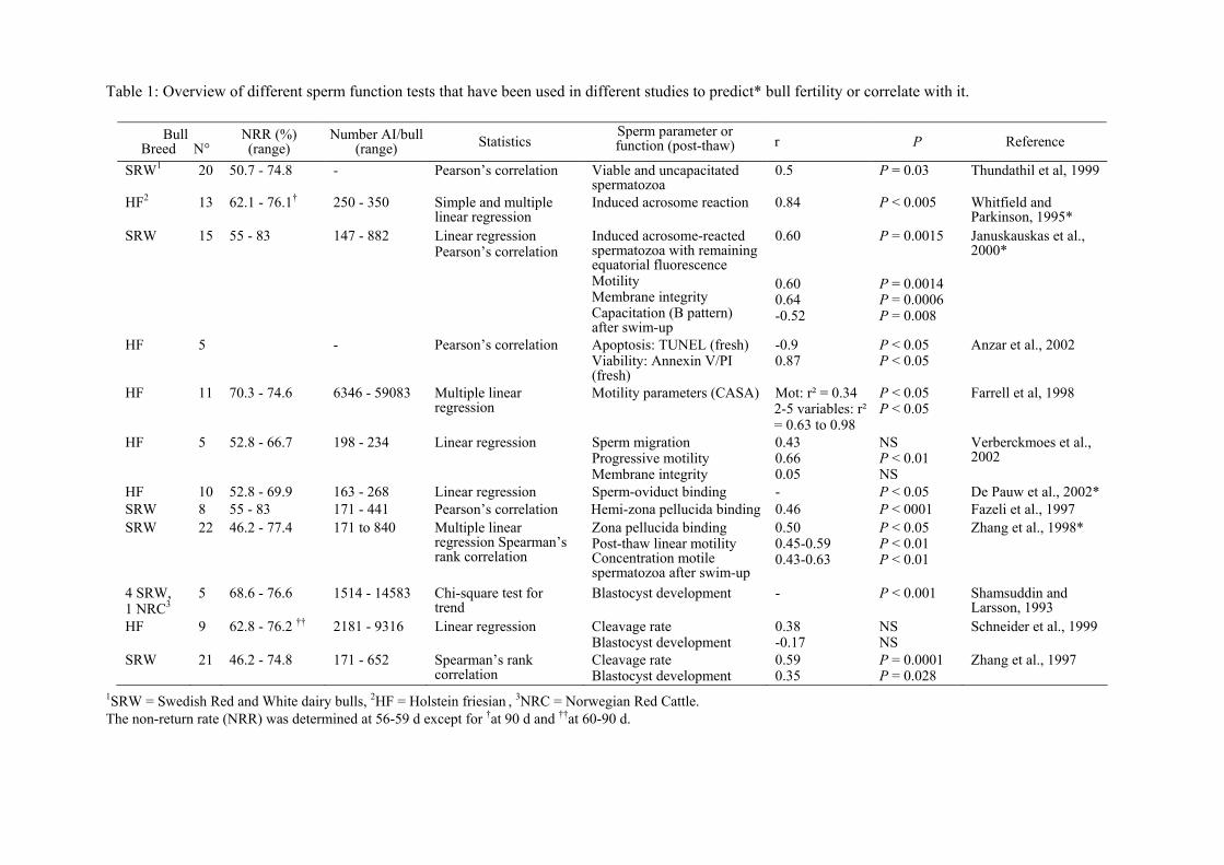
Table 1: Overview of different sperm function tests that have been used in different studies to predict* bull fertility or correlate with it.
1SRW = Swedish Red and White dairy bulls, 2HF = Holstein friesian , 3NRC = Norwegian Red Cattle. The non-return rate (NRR) was determined at 56-59 d except for †at 90 d and ††at 60-90 d.
Bull Breed N°
NRR (%) (range)
Number AI/bull (range) Statistics
Sperm parameter or function (post-thaw) r P Reference
SRW1 20 50.7 - 74.8 - Pearson’s correlation Viable and uncapacitated spermatozoa
0.5 P = 0.03 Thundathil et al, 1999
HF2 13 62.1 - 76.1† 250 - 350
Simple and multiple linear regression
Induced acrosome reaction
0.84
P < 0.005
Whitfield and Parkinson, 1995*
SRW 15 55 - 83 147 - 882
Linear regression Pearson’s correlation
Induced acrosome-reacted spermatozoa with remaining equatorial fluorescence Motility Membrane integrity Capacitation (B pattern) after swim-up
0.60 0.60 0.64 -0.52
P = 0.0015 P = 0.0014 P = 0.0006 P = 0.008
Januskauskas et al., 2000*
HF 5 - Pearson’s correlation Apoptosis: TUNEL (fresh) Viability: Annexin V/PI (fresh)
-0.9 0.87
P < 0.05 P < 0.05
Anzar et al., 2002
HF 11 70.3 - 74.6 6346 - 59083 Multiple linear regression
Motility parameters (CASA) Mot: r² = 0.34 2-5 variables: r² = 0.63 to 0.98
P < 0.05 P < 0.05
Farrell et al, 1998
HF 5 52.8 - 66.7 198 - 234 Linear regression Sperm migration Progressive motility Membrane integrity
0.43 0.66 0.05
NS P < 0.01 NS
Verberckmoes et al., 2002
HF 10 52.8 - 69.9 163 - 268 Linear regression Sperm-oviduct binding - P < 0.05 De Pauw et al., 2002* SRW 8 55 - 83 171 - 441 Pearson’s correlation Hemi-zona pellucida binding 0.46 P < 0001 Fazeli et al., 1997 SRW 22 46.2 - 77.4 171 to 840
Multiple linear regression Spearman’s rank correlation
Zona pellucida binding Post-thaw linear motility Concentration motile spermatozoa after swim-up
0.50 0.45-0.59 0.43-0.63
P < 0.05 P < 0.01 P < 0.01
Zhang et al., 1998*
4 SRW, 1 NRC3
5
68.6 - 76.6 1514 - 14583 Chi-square test for trend
Blastocyst development - P < 0.001 Shamsuddin and Larsson, 1993
HF 9 62.8 - 76.2 †† 2181 - 9316 Linear regression Cleavage rate Blastocyst development
0.38 -0.17
NS NS
Schneider et al., 1999
SRW 21 46.2 - 74.8 171 - 652 Spearman’s rank correlation
Cleavage rate Blastocyst development
0.59 0.35
P = 0.0001 P = 0.028
Zhang et al., 1997
