molecular resolution images of the surfaces of natural zeolites by atomic force microscopy
TRANSCRIPT
www.elsevier.com/locate/micromeso
Microporous and Mesoporous Materials 61 (2003) 79–84
Molecular resolution images of the surfaces of naturalzeolites by atomic force microscopy
M. Voltolini a,*, G. Artioli a,b, M. Moret c
a Dipartimento di Scienze della Terra, Universit�aa di Milano, via Botticelli 23, I-20133 Milano, Italyb Istituto Sperimentale CNR per la Dinamica dei Processi Ambientali, Sezione di Milano, via Mangiagalli 34, I-20133 Milano, Italy
c Dipartimento di Scienza dei Materiali, Universit�aa di Milano-Bicocca, via Cozzi 53, I-20125 Milano, Italy
Received 22 July 2002; received in revised form 4 December 2002; accepted 4 December 2002
Abstract
Atomic force microscopy (AFM) was used to characterize the morphological and cleavage surfaces in a number of
natural zeolites. The investigated zeolites (stilbite, heulandite, thomsonite, yugawaralite, laumontite, and a few others)
show rather interesting and sample-dependent microtopographical features related to the mechanisms involved in the
surface growth processes at the molecular level. The results obtained by AFM on stilbite, heulandite, and yugawaralite
during the preliminary surface characterization are presented, and the images show that molecular resolution can be
achieved and crystallographically interpreted by careful preparation of the sample.
� 2003 Elsevier Inc. All rights reserved.
Keywords: AFM surface imaging; Natural zeolites; Yugawaralite; Stilbite; Heulandite
1. Introduction
Studies on zeolite surfaces are aimed to under-
stand the crystallography, the crystal chemistry,
and the microtopography of the surface layers, andeventually to interpret the underlying mechanisms
of surface growth and transformation. As the
crystal surface is the interface where chemisorp-
tion, transport, diffusion, and chemical reactions
take place, the in situ investigation of crystal sur-
faces has a fundamental importance in under-
standing all physico-chemical processes involving
the crystal–fluid interactions. In the specific caseof zeolites, the understanding and the optimization
* Corresponding author.
1387-1811/03/$ - see front matter � 2003 Elsevier Inc. All rights rese
doi:10.1016/S1387-1811(03)00357-3
of all applications involving absorption, cation
or water exchange, molecular selectivity, crystalli-
zation or dissolution, must necessarily be based
on the appropriate understanding of surface phe-
nomena.Most zeolite studies are concerned with the bulk
structure and properties of the material, because
of its internal controlled microporosity. How-
ever, many processes such as for example crystal
nucleation and growth, are actually governed
by surface properties and chemical absorption.
Atomic force microscopy (AFM) plays a key-role
in the characterization of crystal surfaces, becauseit allows access to the structural and microstruc-
tural features at nanometric resolution. Further-
more many experiments can also be performed
in situ, with the crystal in contact with the solution.
rved.
80 M. Voltolini et al. / Microporous and Mesoporous Materials 61 (2003) 79–84
The evolution of the microtopographical features
can therefore be followed during surface reactions
[1,2].
Typically, AFM investigations on zeolites and
microporous materials pose a number of problems
caused by the strong tip–surface interactions andby the great surface reactivity, which implies an
almost universal attraction of impurity molecules
on the surface, with subsequent degradation of the
image quality. At present only a limited number of
papers have been published containing images of
zeolite surfaces at the nanoscopic scale. The pio-
neering work of Mac Dougall et al. [3] provides a
few images of natural scolecite, stilbite and fauja-site. Other studied natural zeolites are stilbite [4],
heulandite [2,5,6,8], and mordenite [7–9]. Some
attempts of imaging the surfaces of synthetic zeo-
lites such as LTA and FAU failed to achieve good
lateral resolution at the molecular level [8,10–13].
Interestingly, adsorption phenomena in zeolites
have already been tackled by using AFM tech-
niques. The systems studied are clinoptiloliteand heulandite [14–16]. Moreover, very recently a
comparison between zeolites crystals grown in
microgravity conditions and under standard grav-
ity has been undertaken [17].
As a preliminary work towards the character-
ization of natural zeolite surfaces for chemical re-
actions, the present study investigated the crystal
surfaces of three zeolites having similar lamellarmorphology. Stilbite, heulandite, and yugawaralite
all exhibit tabular habit with the {0 1 0} form being
the most developed. The (0 1 0) face is of course a
perfect cleavage plane, fit to be investigated by
AFM.
2. Experimental
2.1. Materials
Natural crystals of zeolites from secondary
crystallization assemblages in basaltic rocks were
used as source materials. Provenance localities are:
heulandite from Osilo, Sardegna, Italy, yugaw-
aralite from Poona, India, and stilbite from Fun-ningsfjordur, Faer Øer Islands, Denmark. All
crystals have well developed tabular habit, and
they were used without any prior chemical treat-
ment. Sample identification and purity was con-
trolled by XRPD on crystals ground from the same
specimens.
2.2. Sample preparation and instrumental parame-
ters
All the crystal samples were mechanically
cleaved with a stainless steel blade just before in-
sertion into the AFM sample cell. AFM mea-
surements were mostly performed in air, although
a few tests were also performed with the sample
submerged in de-ionized water and in dilutedH2SO4 (0.05 M), HCl (0.1 M), NaOH (0.1 M). No
substantial differences were obtained in the two
cases. The best results in terms of lateral resolution
were obtained in air or in sodium hydroxide so-
lutions; the use of acidic solutions produced sev-
eral problems of surface contamination. The
quality of the cleaved surface is crucial for the
imaging experiment: surfaces that allowed highresolution imaging in air also provided high
quality images in water or NaOH solutions.
The instrument used was a Nanoscope III
(Digital Instruments, Santa Barbara, CA) oper-
ated in contact mode. Commercially available
Si3N4 triangular cantilevers with pyramidal tips
were used, with nominal spring constant k varying
from 0.06 to 0.6 N/m. The forces applied on thesurfaces were variable (depending on k and the
environment); a proper set point value was always
chosen to minimize the interaction forces between
tip and crystal surface. A key factor was the choice
of appropriate values of the scan rate: during this
study the scan rate was set at values in the range 3–
40 Hz, using values in the range 12–40 Hz for
optimal results when trying to obtain molecularresolution. Images were also recollected using
different scan angles in order to check for instru-
mental artefacts.
The x and y-directions of the D piezo-scanner
(scan size: 12.5 lm) used for our experiment were
calibrated with the conventional calibration grid
provided by Digital Instruments; the z-axis was
calibrated using the elementary steps on an etchedsynthetic mica foil. The calibration was checked by
imaging the mica sample at molecular resolution.
M. Voltolini et al. / Microporous and Mesoporous Materials 61 (2003) 79–84 81
To avoid artefacts due to external vibrations the
instrument is operating in a quiet place and posi-
tioned on a table supported on a specially designed
pneumatic suspension system.
3. Results
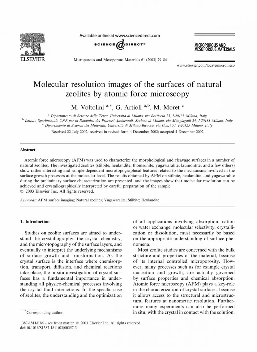
3.1. Heulandite
Heulandite crystals present a well developed
(0 1 0) morphological pinacoid, and after careful
cleavage they provide an excellent surface for
AFM studies. On the cleaved faces there are flatareas of many lm2 showing elementary steps
about 0.91(2) nm high. The measured thickness of
these steps nicely corresponds to b=2 (heulandite
Fig. 1. Surface structure of heulandite. The first image (a) is an unfilte
a unit cell outlined, the third (c) is the calculated surface structure an
cell: a ¼ 17:70, b ¼ 17:94, c ¼ 7:42 �AA, b ¼ 116:4�[18]). Experiments in various environments were
carried out on the Sardinian heulandite crystals,
although the best images were obtained in NaOH
0.1 M. They are shown in Fig. 1.
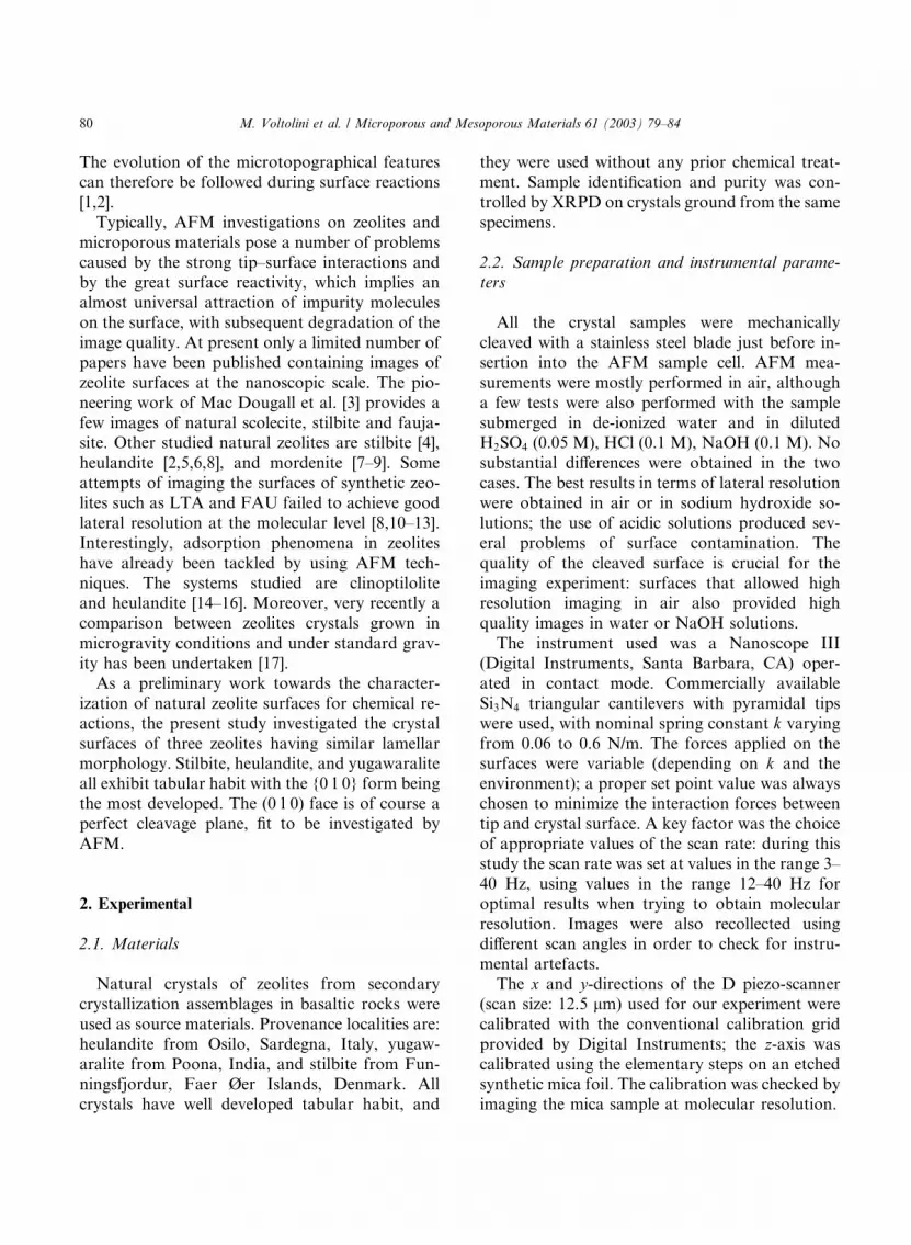
3.2. Yugawaralite
Yugawaralite is here investigated for the first
time by AFM. Yugawaralite is another zeo-
lite providing very good clavage surfaces with
some b=2 high elementary steps. The measured
step is about 0.72(2) nm (yugawaralite cell: a ¼6:73, b ¼ 13:95, c ¼ 10:03 �AA, b ¼ 111:5� [18]).Growth spirals are also frequently observed. The
images shown in Fig. 2 were taken in de-ionized
water.
red AFM image, the second one (b) is a FTT filtered image with
d the last (d) is the FFT of the surface image.
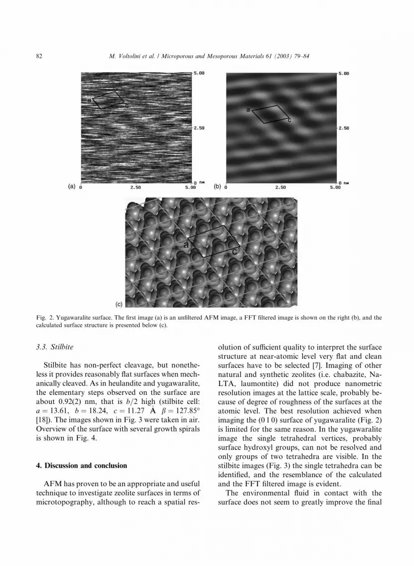
Fig. 2. Yugawaralite surface. The first image (a) is an unfiltered AFM image, a FFT filtered image is shown on the right (b), and the
calculated surface structure is presented below (c).
82 M. Voltolini et al. / Microporous and Mesoporous Materials 61 (2003) 79–84
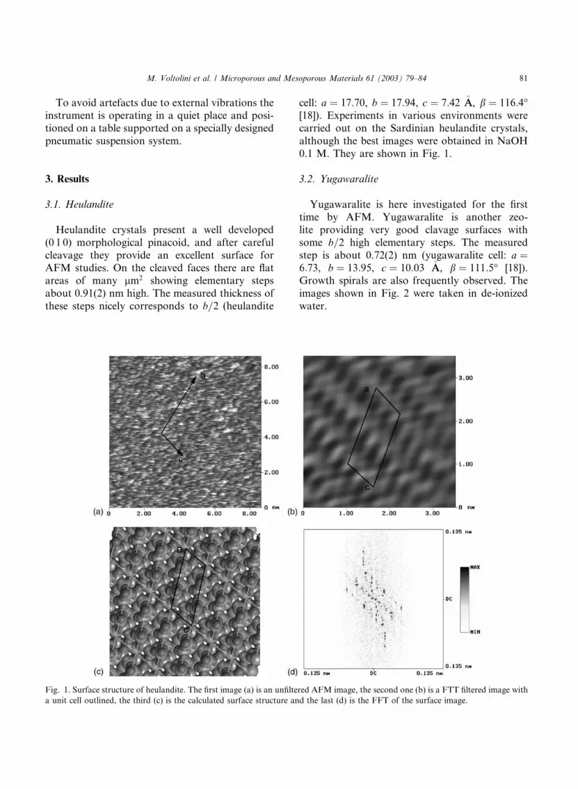
3.3. Stilbite
Stilbite has non-perfect cleavage, but nonethe-
less it provides reasonably flat surfaces when mech-
anically cleaved. As in heulandite and yugawaralite,
the elementary steps observed on the surface are
about 0.92(2) nm, that is b=2 high (stilbite cell:
a ¼ 13:61, b ¼ 18:24, c ¼ 11:27 �AA b ¼ 127:85�[18]). The images shown in Fig. 3 were taken in air.
Overview of the surface with several growth spiralsis shown in Fig. 4.
4. Discussion and conclusion
AFMhas proven to be an appropriate and useful
technique to investigate zeolite surfaces in terms of
microtopography, although to reach a spatial res-
olution of sufficient quality to interpret the surface
structure at near-atomic level very flat and cleansurfaces have to be selected [7]. Imaging of other
natural and synthetic zeolites (i.e. chabazite, Na-
LTA, laumontite) did not produce nanometric
resolution images at the lattice scale, probably be-
cause of degree of roughness of the surfaces at the
atomic level. The best resolution achieved when
imaging the (0 1 0) surface of yugawaralite (Fig. 2)
is limited for the same reason. In the yugawaraliteimage the single tetrahedral vertices, probably
surface hydroxyl groups, can not be resolved and
only groups of two tetrahedra are visible. In the
stilbite images (Fig. 3) the single tetrahedra can be
identified, and the resemblance of the calculated
and the FFT filtered image is evident.
The environmental fluid in contact with the
surface does not seem to greatly improve the final

Fig. 3. Stilbite surface. The first image (a) is a (0 1 0) area at molecular resolution, the second image (b) was FFT filtered. The cal-
culated structure (c) and its FFT (d) are shown below.
Fig. 4. Stilbite (0 1 0) cleavage surface. The microtopographic image (a) at the left exhibits elementary steps having b=2 height, which
are generated by several dislocation sources. The image at the right (b) represents one of such steps at molecular resolution.
M. Voltolini et al. / Microporous and Mesoporous Materials 61 (2003) 79–84 83
quality of the images with respect to those imaged
in air, provided that the selected surface is flat and
clean. In general, an acceptable spatial resolution
can be obtained in air, although working with a
fluid cell can provide more stability and limit the
forces acting on the surface. The use of acidic
84 M. Voltolini et al. / Microporous and Mesoporous Materials 61 (2003) 79–84
fluids, even if very diluted and operating on
chemically resistant zeolites, always produces a
little surface etching. In spite of keeping the sur-
face clean, some reaction products are formed at
the surface, quickly degrading the quality of the
images. Basic solutions like NaOH 0.1 M seem toproduce better results, as previously observed by
other authors [19].
AFM studies of the (0 1 0) surfaces of three
natural zeolites at molecular resolution were com-
pleted, as a preliminary step into interpreting the
behaviour of zeolite surfaces in contact with
aqueous solutions. The scarcity of high resolution
images of zeolites in the literature is to be related tothe difficulty of obtaining appropriate surfaces for
AFM imaging. The study will progress by follow-
ing in situ chemical reactions such as adsorption
and dissolution on zeolite surfaces.
Acknowledgements
The work has been carried out in the frame of
the project ‘‘Mineral surface chemical reactions:
intercalation and sorption processes’’ (Coordina-
tor Prof. G. Artioli), and financed by MURST
COFIN 2000.
References
[1] S. Yamamoto, S. Sugiyama, O. Matsuoka, K. Kohmura,
T. Honda, Y. Banno, H. Nozoye, J. Phys. Chem. 100
(1996) 18474.
[2] M. Voltolini, M. Moret, G. Artioli, in: R. Aiello,
G. Giordano, F. Testa (Eds.), Impact of Zeolites and
Other Porous Materials on the New Technologies at the
Beginning of the New Millennium, Studies in Surface
Science and Catalysis, vol. 142, Elsevier Science, Amster-
dam, 2002, p. 1721.
[3] J.E. Mac Dougall, S.D. Cox, G.D. Stucky, A.L. Weisen-
horn, P.K. Hansma, W.S. Wise, Zeolites 11 (1991)
429.
[4] M. Komiyama, K. Tsujimichi, Y. Oumi, M. Kubo, A.
Miyamoto, Appl. Surf. Sci. 121/122 (1997) 543.
[5] S. Yamamoto, S. Sugiyama, O. Matsuoka, T. Honda, Y.
Banno, H. Nozoye, Micropor. Mesopor. Mater. 21 (1998)
1.
[6] G. Binder, L. Scandella, A. Schumacher, N. Kruse, R.
Prins, Zeolites 16 (1996) 2.
[7] S. Yamamoto, O. Matsuoka, S. Sugiyama, T. Honda,
Y. Banno, H. Nozoye, Chem. Phys. Lett. 260 (1996)
208.
[8] S. Sugiyama Ono, O. Matsuoka, S. Yamamoto, Micropor.
Mesopor. Mater. 48 (2001) 103.
[9] S. Sugiyama, S. Yamamoto, O. Matsuoka, T. Honda, H.
Nozoye, S. Qiu, J. Yu, O. Terasaki, Surf. Sci. 377–379
(1997) 140.
[10] J.R. Agger, N. Pervaiz, A.K. Cheetham, M.W. Anderson,
J. Am. Chem. Soc. 120 (1998) 10754.
[11] M.W. Anderson, J.R. Agger, J.T. Thornton, N. Forsyth,
Angew. Chem. Int. Ed. Engl. 35 (1996) 1210.
[12] S. Sugiyama, S. Yamamoto, O. Matsuoka, H. Nozoye,
J. Yu, G. Zhu, S. Qiu, O. Terasaki, Micropor. Mesopor.
Mater. 28 (1999) 1.
[13] M.W. Anderson, J.R. Agger, N. Hanif, O. Terasaky,
Micropor. Mesopor. Mater. 48 (2001) 1.
[14] A.L. Weisenhorn, J.E. Mac Dougall, S.A.C. Gould, S.D.
Cox, W.S. Wise, J. Massie, P. Maivald, V.B. Elings, G.D.
Stucky, P.K. Hansma, Science 247 (1990) 1330.
[15] E.J. Sullivan, D.B. Hunter, R.S. Bowman, Clays Clay
Mineral. 45 (1997) 42.
[16] M. Komiyama, T. Shimaguchi, T. Koyama, M. Gu, J.
Phys. Chem. 100 (1996) 15198.
[17] J. Warzywoda, M. Valcheva-Traykova, G.A. Rossetti,
N. Bac�, R. Joesten, S.L. Suib, A. Sacco, J. Crystal Growth
220 (2000) 150.
[18] G. Gottardi, E. Galli, Natural Zeolites, Springer-Verlag,
Berlin, 1985.
[19] L. Scandella, N. Kruse, R. Prins, Surf. Sci. Lett. 281 (1993)
L331.