investigation of adsorbed humic substances using atomic force microscopy
TRANSCRIPT
Colloids and Surfaces A: Physicochem. Eng. Aspects 248 (2004) 17–23
Investigation of adsorbed humic substances usingatomic force microscopy
Shoeleh Assemia,c, Patrick G. Hartleyb,d, Peter J. Scalesb, Ronald Becketta,∗a Water Studies Center and School of Chemistry, Monash University, Wellington Rd., Clayton 3800, Australia
b Advanced Mineral Products Research Center, University of Melbourne, Parkville 3052, Australiac Department of Geology and Geophysics, University of Utah, Salt Lake City, UT 84112, USA
d CSIRO Molecular Science, Bag 10, Clayton South 3169, Australia
Received 14 September 2003; accepted 10 August 2004
Abstract
Atomic force microscopy (AFM) was used to study the effect of adsorption of humic substances onto a goethite-coated mica surface, ont d in stronga osure time.F coated micas nm from thef ich has directr©
K
1
gmacspotlTec
tes for
-ticlehree-singbe-
n andrcen, air
ageshin-ationd fromablyolec-e foring
0d
he forces between the goethite surface and a silica colloidal probe. The positively charged goethite surface (at pH < 7) resultedsorption of humic substances. The adsorption process could be controlled by altering the solution concentration, pH and exporce versus distance curves were measured directly by AFM between a silica sphere colloidal probe and the planar goethite-urface, with and without the presence of humic substances. The thickness of the adsorbed layer was estimated to be about 5orce curves. These experiments demonstrate the influence of adsorbed humic substances on the surface charge of goethite, whelevance to the colloid stability of natural aquatic particles.
2004 Elsevier B.V. All rights reserved.
eywords:Atomic force microscopy; Humic substances; Goethite; Colloid stability; Surface charge
. Introduction
Humic substances are heterogeneous, polydispersed or-anic molecules derived from the decay of natural organicatter in the environment. Humic substances have an over-ll negative charge in the pH of natural waters where theyan be found at concentrations of 1–50 mg L−1. Humic sub-tances are abundant in nature, and in freshwater often com-rise about 40–60% of the dissolved organic carbon[1]. Onef the major roles of humic substances in aquatic systems is
he modification of the surface charge of suspended particu-ate matter by imparting a negative charge to their surfaces.his modification of the surface properties of minerals influ-nces important phenomena, such as colloidal stability andontaminant adsorption. For this reason humic-coated model
∗ Corresponding author. Tel.: +61 3 990 54555; fax: +61 3 990 54196E-mail address:[email protected] (R. Beckett).
particles (such as goethite) have been used as surroganatural particles in laboratory studies[2–5].
Atomic force microscopy (AFM)[6] can provide information on the structure of interfaces and on interparforces. It has sub-nanometer resolution and can provide tdimensional topography of the surface. AFM is a promitechnique for investigation of humic–mineral interactionscause of its non-destructive and direct nature of operatioits ability to probe interparticle forces. The imaging or fomeasurement experiments can be carried out in solutioor vacuum.
There is considerable interest in obtaining accurate imof humic molecules using AFM. Results so far have beendered by potential aggregation during the sample preparstage. The requirements are that molecules are adsorbesolution without changing ‘aggregation states’ on a suitsmooth surface. Muscovite mica has an exceptional mularly smooth surface, which makes it an ideal substratAFM imaging and has been the most popular for imag
927-7757/$ – see front matter © 2004 Elsevier B.V. All rights reserved.oi:10.1016/j.colsurfa.2004.08.035
18 S. Assemi et al. / Colloids and Surfaces A: Physicochem. Eng. Aspects 248 (2004) 17–23
humic molecules[7–14]. However, mica surface acquires anegative charge in aqueous solution (above pH 2), which in-hibits the adsorption of small humic molecules, in particularaquatic humic substances. Weakly attached humic moleculescan be immobilized by the AFM tip when imaging in solu-tion, resulting in imaging artifacts. Therefore, images of hu-mic substances on mica have so far been obtained either afterdrying the humic sample on mica and imaging in the air[7,8],or incubation of mica in the humic solution for a certain time(usually 24 h), washing the excess humic and imaging in theair [11] or in solutions with NaCl concentration of 0.01 or0.1 mol L−1 [10,15].
Namjesnik–Dejanovic and Maurice[14] used AFM to ob-tain images of a fulvic acid adsorbed onto mica (either bypartial drying or after 2 h of incubation) in solutions of dif-ferent pH, ionic strength and salt type. At pH = 3 additionof Ca2+ ions resulted in increased adsorption of the humicmolecules on mica. However, stable images of natural or-ganic matter (NOM) in NaCl solutions (0.001–0.1 mol L−1)could not be obtained at pH 3. Only some scattered spheresthat were easily swept away by the AFM tip were observed.Using 0.1 mol L−1 LiCl at pH 3 resulted in weak adsorption ofNOM. Using hematite as a substrate resulted in strong adsorp-tion of the humic molecules but only aggregates withx–ydi-mensions of 0.8–1�m were observed. The smaller moleculesc ot bem atite
mon-s mea-s n in-v highs s tos sub-s houtn
thes rcesb et al.[ drichh r ad-d eent sedb
e-t acter-i FMc ex-t erents hiss ac d nat-u da ndm re anda geo t that
the surfaces may have not been homogeneously coated withiron oxide. Therefore, the forces reported are probably thosebetween iron oxide patches and silica or silica and naturalorganic matter. They observed that at low ionic strengths theinterparticle forces were dominated by electrostatic interac-tions, but at high ionic strengths where the electrostatic forceswere absent, steric repulsive forces were dominant. The ap-proach and separation force curves showed no hysteresis forNOM-free case. After addition of NOM, hysteresis and dis-continuities, indicative of NOM bridging between surfaces,were observed in separation curves.
This paper focuses on the development of a goethite sur-face, which can retain humic substances, especially at pH val-ues below its isoelectric point (IEP). This surface represents asuitable model for the surface of certain aquatic colloids. Sur-face force measurements between a spherical silica colloidprobe and the goethite surface, with and without the presenceof Suwannee river fulvic acid (SRFA), have been used to char-acterize the surface chemical properties of both coated anduncoated goethite surfaces. An estimate of the thickness ofthe adsorbed fulvic acid layer can be obtained using the forceversus distance curves before and after addition of SRFA.
2. Experimental
2
ub-s rela-t rized[ e a1 fNg oak-i tionf BS-3 ationc sur-f piousq owc nge ight,f wa-t ).Me usedw
2
medo ntaB an-t Thes afer
ould not be observed and the vertical dimension could neasured because of the high surface roughness of hemThese results, among the others in the literature, de
trate that use of muscovite mica as a substrate in AFMurements is extremely difficult and sample preparatioolves steps such as drying or long incubation times, oralt concentration of the solution. One of our goals waynthesize a smooth surface that could anchor humictances in moderate solution pH and ionic strength witeed for long incubation times.
One important feature of AFM that can be used intudy of humic substances is its ability to measure the foetween the AFM tip and the sample surface. Plaschke
13] measured the adhesion forces between a humic (Alumic-acid) coated tip and a mica surface before and afteition of Eu(III) and noticed that the adhesion force betw
he mica and the humic-coated silicon nitride tip increay nearly one order of magnitude.
Ducker et al.[16] demonstrated that colloidal forces bween surfaces of choice could be measured and charzed in solution by attaching a spherical colloid to the Aantilever tip. The colloidal probe technique has beenensively used to characterize the forces between diffurfaces[17–21]. To the authors’ best knowledge, only ttudy and a recent study by Mosley et al.[22] have usedolloidal probe to measure forces between surfaces anral organic matter. Mosley et al.[22] coated a silica tip ansilica surface in an AFM liquid cell with iron oxide aeasured the forces between two coated surfaces befofter incubation in NOM (incubation time 16 h). The imabtained from the surface and the force curves sugges
..1. Sample preparation
SRFA was purchased from the International Humic Stances Society (IHSS). SRFA was chosen because it isively mono-dispersed and has been extensively characte23–26]. SRFA was dissolved in deionized water to mak00 ppm solution. The pH was adjusted to∼6 by addition oaOH (final concentration of NaOH was 8× 10⁻⁴ M). Alllassware used in experiments were initially cleaned by s
ng in 10% nitric acid and then subjected to ultrasonicaor at least 1 h in a 1% RBS-35/20% ethanol solution. R5 (Pierce Chemicals) is a commercial cleaning preparonsisting of a basic solution of anionic and non-ionicactants. The cleaned glassware was then rinsed with couantities of deionized water, prior to drying in a laminar fllean air cabinet. The Teflon® containers used in the coatixperiments were cleaned by soaking in 10% HCl overnollowed by the procedure explained above. Deionizeder was obtained from a Milli-Q filtration system (Milliporeeasured resistivity was not below 18.2 M� cm in any of the
xperiments. Other reagents were analytical grade andithout further purification.
.2. Atomic force microscopy measurements
Atomic force microscopy measurements were perforn a digital instrument, Nanoscope IIIa AFM (Veeco, Saarbara, CA). V-shaped, gold-coated silicon nitride c
ilevers were obtained from digital instruments (Veeco).pring constant of randomly picked cantilevers from the w
S. Assemi et al. / Colloids and Surfaces A: Physicochem. Eng. Aspects 248 (2004) 17–23 19
were determined by the method of Cleveland et al.[27], whichrelies on monitoring the shifts in resonance frequency as afunction of known masses attached to the cantilevers.
Colloidal silica spheres were obtained from Allied Signal,Illinois. The size of the spheres typically ranged from 4–6�min diameter. Silica spheres with a diameter of 5�m were usedin these experiments. One silica sphere was attached to theapex of the standard silicon nitride (NP-S) AFM cantileversusing an extremely small amount of Ardalite epoxy resin.Scanning electron microscopy indicated that the glue wasconfined to the interface between the cantilever and the silicasphere and that the sphere was not contaminated with theglue. Properties of this silica and its preparation for use indirect force measurements are described in detail by Hartleyet al.[20].
2.3. Preparation of goethite-coated mica surface
100 mL of deionized water was placed in a Teflon® con-tainer and purged with nitrogen gas for 20 min. The pH waslowered to 3, 3.5, or 4 using 0.1 M HNO3. A 1 cm × 1 cmpiece of freshly cleaved mica was immersed in the solutionand 0.1 M Fe(NO3)3 was added in 50�L volume incrementsto the solution at constant temperature (20◦C) to reach a finalpreset concentration (10−3 or 10−4 M) of Fe(NO3)3. The pHwas kept constant by adding 1 M NaOH using an autotitrator( ,t ed insf re-m d thes
o tes Oa tinge
2f
FMf fluidc inw ure-m ents,t solu-t
3
3s
a
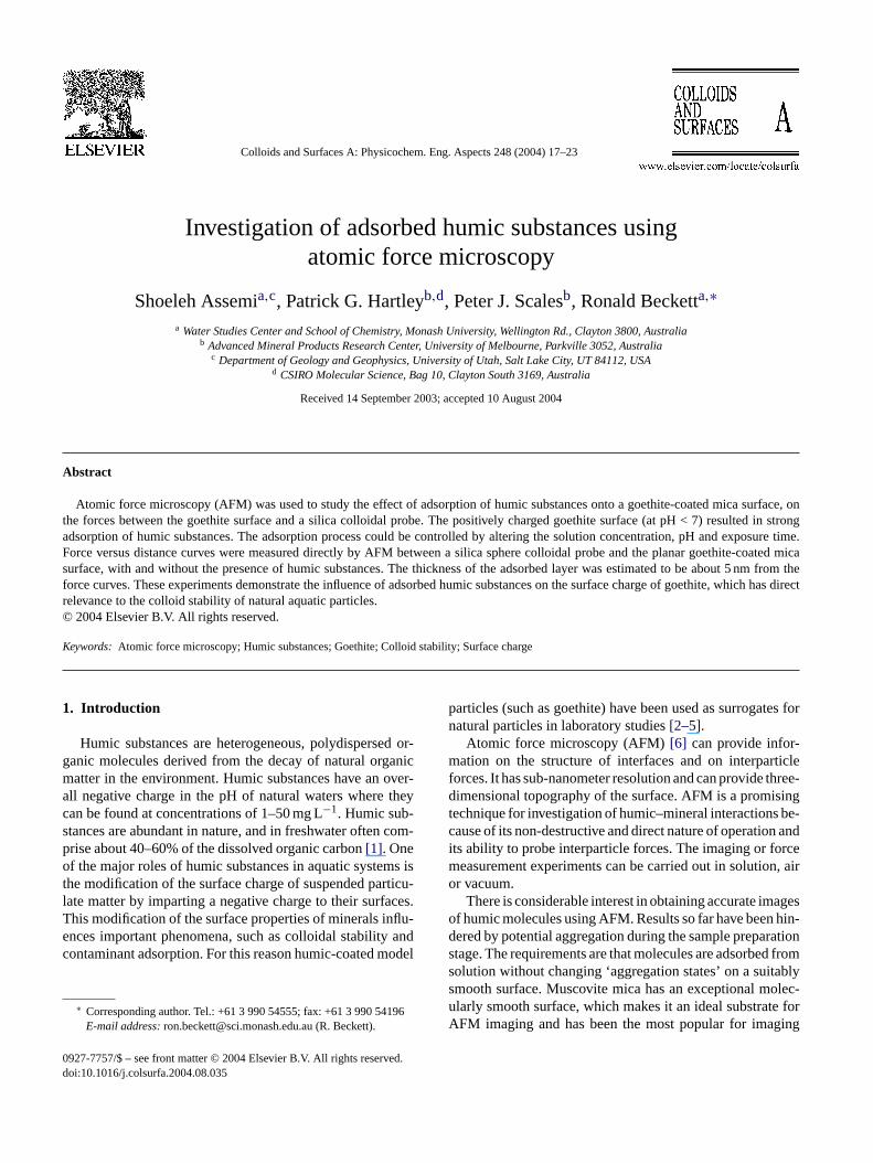
Fig. 1. Tapping mode AFM image of the goethite-coated mica surface. (a)scan size 1�m× 1�m, (b) scan size 300 nm× 300 nm and (c) topographicalsection of the surface between the points marked on the images in (a) and(b).
goethite-coated mica surface recorded in air. It appears asif the goethite was either deposited on the mica plate as dis-crete particles or that surface nucleation and crystal growthoccurred. The particles were fairly homogeneous in size andquite regular in shape. The coverage of the underlying mica
Radiometer). When the addition of Fe(NO3)3 was completehe container containing the coated mica plate (immersolution) was heated at 60◦C in an oven for 24 h to allowormation of goethite[28]. The goethite-coated mica wasoved and rinsed with deionized water several times an
urface topography was analyzed by AFM.The experiment resulting in 10−3 M final concentration
f added Fe(NO3)3 at pH 4 was found to result in compleurface coverage. Therefore, this concentration of Fe(N3)3nd a solution pH of 4 were chosen for all further coaxperiments.
.4. Adsorption of SRFA on goethite surfaces for AFMorce measurements
The adsorption of SRFA onto goethite surfaces for Aorce measurements was performed in situ in the AFMell (Digital Instruments). A solution of 100 ppm SRFAater was injected into the fluid cell and force measents were performed immediately. In some experim
he system was subsequently flushed with SRFA freeion (10−4 M NaNO3) and analyzed further.
. Results and discussion
.1. Characterization of the goethite-coated micaurface
Fig. 1(a) and (b) respectively show a 1�m× 1�m and300 nm× 300 nm TappingModeTM AFM image of the
20 S. Assemi et al. / Colloids and Surfaces A: Physicochem. Eng. Aspects 248 (2004) 17–23
surface by the goethite coating was complete. Thex–y di-mension of the particles appears to be about 30–50 nm.
In order to obtain an indication of the surface roughness,a horizontal scan of the tip across the section indicated inFig. 1(a) was performed. The vertical displacement of the tipduring the scan is plotted inFig. 1(c). The maximum differ-ence in vertical displacement was 15.5 nm, measured at thepoints indicated by arrows inFig. 1(a). Typically in AFM,the vertical displacement can be measured with greater pre-cision than horizontal distance. However, the maximum ver-tical displacement would not give an accurate measurementof the particle size in this case, as the particles were denselypacked on the surface, preventing the movement of the tiparound them.
Roughness calculations based on the 1�m × 1�m scansize image produced an RMS value for the surface of∼3 nm(Fig. 1(c)). This is considerably rougher than the underlyingmica surface (RMS roughness typically <0.1 nm), suggestingthat the goethite substrate obtained was not smooth enoughfor imaging small humic substances.
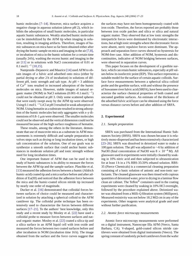
Fig. 2 shows data derived from AFM surface force mea-surements between a silica colloid probe and the goethite-coated mica surface in solutions of different pH. Since theisoelectric point of the silica colloid probe attached to theAFM cantilever is below pH 2[20], the probe was expectedt howni ive ad face.
atedt . Thed n 22a reti-
F olloidp
cal Debye length of 30.5 nm for the simple 1× 10⁻⁴ M 1:1electrolyte system studied here. Thus, the interaction may beattributed to an electrostatic repulsion operating between thesilica and goethite surfaces, which indicate that the goethitesurface was negatively charged at these pH values.
Below pH 7.4, electrostatic attractive forces were observed(again deduced from a Debye-like decay length) indicatingthat the goethite surface had reversed its surface charge, yield-ing a net positive surface. The isoelectric point of the goethitesurface used in this study was deduced to be between pH 6.5and 7.4. This is at the low end of the IEP range of 7–9.4found for goethite using electrophoretic mobility measure-ments[3,29–32]. However, IEP values of around pH 7 arenot uncommon[4,33,34]. Carbonate or silicate adsorptioncan be responsible for lower IEP values for goethite[35],which is a possibility in our experiments.
Detailed fitting of the experimental data shown inFig. 2using DLVO theory was complicated by the weakness of therepulsive forces at high pH. In assuming negative surface po-tential of the magnitude of−35 to−57 mV for the goethite(values taken from zeta potential measurements in the liter-ature[36]), apparent surface potentials in the range of−5 to−10 mV were obtained for the silica colloid probe at pHs be-tween 7.4 and 9. This is considerably lower than the expectedsurface potential of the silica under these conditions (about−
ouslyi t ofg rcem mentsw n thep e thea ofi bse-q aliza-t pli-c rface.
31
acef rfacea lya -a n ofS th ofi ublel iono
at-t tedg FA,t l-s tatici of an
o carry a negative surface charge at all the pH values sn the figure. Thus, the AFM force measurements can girect indication of the surface charge of the goethite sur
At pH values greater than 6.5, a repulsive force dominhe interactions at all but very small surface separationsecay length for the repulsion was found to be betweend 27 nm, which agrees reasonably well with the theo
ig. 2. AFM force measurements versus distance between a silica crobe and goethite-coated mica surface in solutions of different pH.
100 mV for silica–silica)[20].The reasons for these low values cannot be unambigu
dentified, but may include: (i) transfer of a small amounoethite particles onto the colloid probe during the AFM foeasurements. This is despite the fact that the measureere commenced at high pH, where the force betweerobe and the goethite was repulsive, in order to reducffinity of goethite for the silica surface, (ii) dissolution
ron hydroxy species from the goethite surface, and suuent re-adsorption on the silica surface, causing neutr
ion of anionic groups on the silica surface, and (iii) comations due to the surface roughness of the goethite su
.2. Incubation of the goethite-coated mica surface in00 ppm SRFA solution
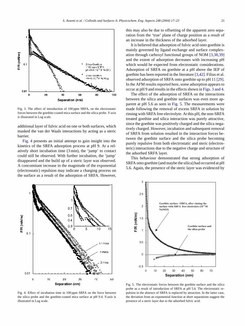
The impact of the introduction of fulvic acid on the surforces between a silica colloid probe and the goethite sut pH∼ 9 is shown inFig. 3. The figure shows that after onshort period of incubation (∼4 min, pH∼ 9.4) the inter
ction forces were affected considerably. After adsorptioRFA there was a significant decrease in the decay leng
nteraction, indicating compression of the electrical doayer[16]. This would normally be the result of the additf background electrolyte to the system.
A ‘jump’ into contact, where short range van der Waalsractive forces dominate[37] can be observed in the uncoaoethite–silica interaction. Following addition of the SR
his ‘jump’ was no longer apparent (Fig. 3). Instead a repuive deviation from the curve expected for pure electrosnteractions was observed. This suggested the presence
S. Assemi et al. / Colloids and Surfaces A: Physicochem. Eng. Aspects 248 (2004) 17–23 21
Fig. 3. The effect of introduction of 100 ppm SRFA, on the electrostaticforces between the goethite-coated mica surface and the silica probe.Y-axisis illustrated in Log scale.
additional layer of fulvic acid on one or both surfaces, whichmasked the van der Waals interactions by acting as a stericbarrier.
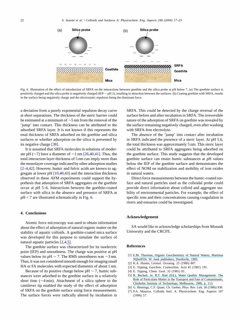
Fig. 4 presents an initial attempt to gain insight into thekinetics of the SRFA adsorption process at pH 9. At a rel-atively short incubation time (3 min), the ‘jump’ to contactcould still be observed. With further incubation, the ‘jump’disappeared and the build up of a steric layer was observed.A concomitant increase in the magnitude of the exponential(electrostatic) repulsion may indicate a charging process onthe surface as a result of the adsorption of SRFA. However,
F eenti
this may also be due to offsetting of the apparent zero sepa-ration from the ‘true’ plane of charge position as a result ofan increase in the thickness of the adsorbed layer.
It is believed that adsorption of fulvic acid onto goethite ismainly governed by ligand exchange and surface complex-ation through carboxyl functional groups of NOM[3,38,39]and the extent of adsorption decreases with increasing pHwhich would be expected from electrostatic considerations.Adsorption of SRFA on goethite at a pH above the IEP ofgoethite has been reported in the literature[3,42]. Filius et al.observed adsorption of SRFA onto goethite up to pH 11[29].In the AFM results reported here, some adsorption appears tooccur at pH 9 and results in the effects shown inFigs. 3 and 4.
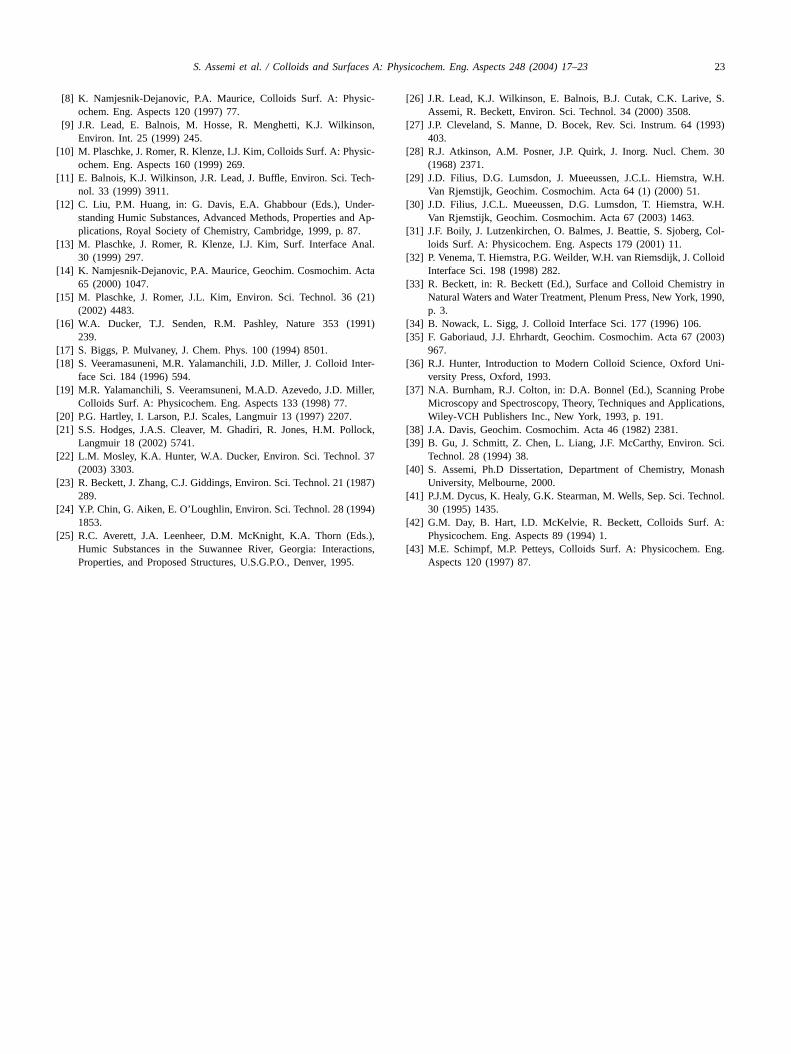
The effect of the adsorption of SRFA on the interactionsbetween the silica and goethite surfaces was even more ap-parent at pH 5.6 as seen inFig. 5. The measurements weremade following the removal of excess SRFA in solution byrinsing with SRFA free electrolyte. At this pH, the non-SRFAtreated goethite and silica interaction was purely attractive,since the goethite was positively charged and the silica nega-tively charged. However, incubation and subsequent removalof SRFA from solution resulted in the interaction forces be-tween the goethite surface and the silica probe becomingpurely repulsive from both electrostatic and steric (electros-teric) interactions due to the negative charge and structure oft
n ofS at pH5 ed by
F silicap ic re-p case,t st thepresence of a steric layer due to the adsorbed fulvic acid.
ig. 4. Effect of incubation time in 100 ppm SRFA on the force betwhe silica probe and the goethite-coated mica surface at pH 9.4.Y-axis isllustrated in Log scale.
he adsorbed SRFA layer.This behaviour demonstrated that strong adsorptio
RFA onto goethite (and maybe the silica) had occurred.6. Again, the presence of the steric layer was evidenc
ig. 5. The electrostatic forces between the goethite surface and therobe as a result of introduction of SRFA at pH 5.6. The electrostatulsion in the absence of SRFA is replaced by attraction. In the latter
he deviation from an exponential function at short separations sugge
22 S. Assemi et al. / Colloids and Surfaces A: Physicochem. Eng. Aspects 248 (2004) 17–23
Fig. 6. Illustration of the effect of introduction of SRFA on the interactions between goethite and the silica probe at pH below 7. (a) The goethite surface ispositively charged and the silica probe is negatively charged (IEP∼ pH 2), resulting in attraction between the surfaces. (b) Coating goethite with SRFA, resultsin the surface being negatively charge and the electrostatic repulsion being the dominant force.
a deviation from a purely exponential repulsion decay curveat short separations. The thickness of the steric barrier couldbe estimated at a minimum of∼5 nm from the removal of the‘jump’ into contact. This thickness can be attributed to theadsorbed SRFA layer. It is not known if this represents thetotal thickness of SRFA adsorbed on the goethite and silicasurfaces or whether adsorption on the silica is prevented byits negative charge[38].
It is assumed that SRFA molecules in solutions of moder-ate pH (∼7) have a diameter of∼1 nm[26,40,41]. Thus, thetotal interaction layer thickness of 5 nm can imply more thanthe monolayer coverage indicated by other adsorption studies[2–4,42]. However, humic and fulvic acids are known to ag-gregate at lower pH[10,40,43]and the interaction thicknessobserved in these AFM experiments could support the hy-pothesis that adsorption of SRFA aggregates on the goethiteoccur at pH 5–6. Interactions between the goethite-coatedsurface with silica in the absence and presence of SRFA atpH < 7 are illustrated schematically inFig. 6.
4. Conclusions
Atomic force microscopy was used to obtain informationabout the effect of adsorption of natural organic matter on thes acew e ofn
ctricp t pHvT mallH nm.
-s tivelys thec tiono ents.T n in
SRFA. This could be detected by the charge reversal of thesurface before and after incubation in SRFA. The irreversiblenature of the adsorption of SRFA on goethite was revealed bythe surface remaining negatively charged, even after washingwith SRFA-free electrolyte.
The absence of the ‘jump’ into contact after incubationin SRFA indicated the presence of a steric layer. At pH 5.6,the total thickness was approximately 5 nm. This steric layercould be attributed to SRFA aggregates being adsorbed onthe goethite surface. This study suggests that the developedgoethite surface can retain humic substances at pH valuesbelow the IEP of the goethite surface and demonstrates theeffect of NOM on stabilization and mobility of iron oxidesin natural waters.
Direct force measurements between the humic-coated sur-face and natural particles used as the colloidal probe couldprovide direct information about colloid and aggregate sta-bility of environmental particles. For example, the effect ofspecific ions and their concentrations causing coagulation inrivers and estuaries could be investigated.
Acknowledgement
SA would like to acknowledge scholarships from MonashU
R
inus
Theants,
930.107
tability of aquatic colloids. A goethite-coated mica surfas developed for this purpose to simulate the surfacatural aquatic particles[2,4,5].
The goethite surface was characterized for its isoeleoint (IEP) and smoothness. The charge was positive aalues below its pH∼ 7. The RMS smoothness was∼3 nm.hus, it was not considered smooth enough for imaging sA or FA molecules with expected diameters of about 1Because of its positive charge below pH∼ 7, humic sub
tances were adsorbed to the goethite surface in a relahort time (∼4 min). Attachment of a silica sphere toantilever tip enabled the study of the effect of adsorpf SRFA on the goethite surface using force measuremhe surface forces were radically altered by incubatio
niversity and the CRCFE.
eferences
[1] E.M. Thurman, Organic Geochemistry of Natural Waters, MartNijhoff/Dr. W. Junk publishers, Dordrecht, 1985.
[2] K.A. Hunter, Limnol. Oceanog. 25 (1980) 807.[3] E. Tipping, Geochim. Cosmochim. Acta 45 (1981) 191.[4] E. Tipping, Chem. Geol. 33 (1981) 81.[5] R. Beckett, in: B.T. Hart (Ed.), Water Quality Management:
Role of Particulate Matter in the Transport and Fate of ContaminChisholm Institute of Technology, Melbourne, 1986, p. 113.
[6] G. Binnings, C.F. Quate, Ch. Gerber, Phys. Rev. Lett. 56 (1986)[7] P.A. Maurice, Colloids Surf. A: Physicochem. Eng. Aspects
(1996) 57.
S. Assemi et al. / Colloids and Surfaces A: Physicochem. Eng. Aspects 248 (2004) 17–23 23
[8] K. Namjesnik-Dejanovic, P.A. Maurice, Colloids Surf. A: Physic-ochem. Eng. Aspects 120 (1997) 77.
[9] J.R. Lead, E. Balnois, M. Hosse, R. Menghetti, K.J. Wilkinson,Environ. Int. 25 (1999) 245.
[10] M. Plaschke, J. Romer, R. Klenze, I.J. Kim, Colloids Surf. A: Physic-ochem. Eng. Aspects 160 (1999) 269.
[11] E. Balnois, K.J. Wilkinson, J.R. Lead, J. Buffle, Environ. Sci. Tech-nol. 33 (1999) 3911.
[12] C. Liu, P.M. Huang, in: G. Davis, E.A. Ghabbour (Eds.), Under-standing Humic Substances, Advanced Methods, Properties and Ap-plications, Royal Society of Chemistry, Cambridge, 1999, p. 87.
[13] M. Plaschke, J. Romer, R. Klenze, I.J. Kim, Surf. Interface Anal.30 (1999) 297.
[14] K. Namjesnik-Dejanovic, P.A. Maurice, Geochim. Cosmochim. Acta65 (2000) 1047.
[15] M. Plaschke, J. Romer, J.L. Kim, Environ. Sci. Technol. 36 (21)(2002) 4483.
[16] W.A. Ducker, T.J. Senden, R.M. Pashley, Nature 353 (1991)239.
[17] S. Biggs, P. Mulvaney, J. Chem. Phys. 100 (1994) 8501.[18] S. Veeramasuneni, M.R. Yalamanchili, J.D. Miller, J. Colloid Inter-
face Sci. 184 (1996) 594.[19] M.R. Yalamanchili, S. Veeramsuneni, M.A.D. Azevedo, J.D. Miller,
Colloids Surf. A: Physicochem. Eng. Aspects 133 (1998) 77.[20] P.G. Hartley, I. Larson, P.J. Scales, Langmuir 13 (1997) 2207.[21] S.S. Hodges, J.A.S. Cleaver, M. Ghadiri, R. Jones, H.M. Pollock,
Langmuir 18 (2002) 5741.[22] L.M. Mosley, K.A. Hunter, W.A. Ducker, Environ. Sci. Technol. 37
(2003) 3303.[23] R. Beckett, J. Zhang, C.J. Giddings, Environ. Sci. Technol. 21 (1987)
[ 94)
[ s.),tions,5.
[26] J.R. Lead, K.J. Wilkinson, E. Balnois, B.J. Cutak, C.K. Larive, S.Assemi, R. Beckett, Environ. Sci. Technol. 34 (2000) 3508.
[27] J.P. Cleveland, S. Manne, D. Bocek, Rev. Sci. Instrum. 64 (1993)403.
[28] R.J. Atkinson, A.M. Posner, J.P. Quirk, J. Inorg. Nucl. Chem. 30(1968) 2371.
[29] J.D. Filius, D.G. Lumsdon, J. Mueeussen, J.C.L. Hiemstra, W.H.Van Rjemstijk, Geochim. Cosmochim. Acta 64 (1) (2000) 51.
[30] J.D. Filius, J.C.L. Mueeussen, D.G. Lumsdon, T. Hiemstra, W.H.Van Rjemstijk, Geochim. Cosmochim. Acta 67 (2003) 1463.
[31] J.F. Boily, J. Lutzenkirchen, O. Balmes, J. Beattie, S. Sjoberg, Col-loids Surf. A: Physicochem. Eng. Aspects 179 (2001) 11.
[32] P. Venema, T. Hiemstra, P.G. Weilder, W.H. van Riemsdijk, J. ColloidInterface Sci. 198 (1998) 282.
[33] R. Beckett, in: R. Beckett (Ed.), Surface and Colloid Chemistry inNatural Waters and Water Treatment, Plenum Press, New York, 1990,p. 3.
[34] B. Nowack, L. Sigg, J. Colloid Interface Sci. 177 (1996) 106.[35] F. Gaboriaud, J.J. Ehrhardt, Geochim. Cosmochim. Acta 67 (2003)
967.[36] R.J. Hunter, Introduction to Modern Colloid Science, Oxford Uni-
versity Press, Oxford, 1993.[37] N.A. Burnham, R.J. Colton, in: D.A. Bonnel (Ed.), Scanning Probe
Microscopy and Spectroscopy, Theory, Techniques and Applications,Wiley-VCH Publishers Inc., New York, 1993, p. 191.
[38] J.A. Davis, Geochim. Cosmochim. Acta 46 (1982) 2381.[39] B. Gu, J. Schmitt, Z. Chen, L. Liang, J.F. McCarthy, Environ. Sci.
Technol. 28 (1994) 38.[40] S. Assemi, Ph.D Dissertation, Department of Chemistry, Monash
University, Melbourne, 2000.[ nol.
[ A:
[ ng.
289.24] Y.P. Chin, G. Aiken, E. O’Loughlin, Environ. Sci. Technol. 28 (19
1853.25] R.C. Averett, J.A. Leenheer, D.M. McKnight, K.A. Thorn (Ed
Humic Substances in the Suwannee River, Georgia: InteracProperties, and Proposed Structures, U.S.G.P.O., Denver, 199
41] P.J.M. Dycus, K. Healy, G.K. Stearman, M. Wells, Sep. Sci. Tech30 (1995) 1435.
42] G.M. Day, B. Hart, I.D. McKelvie, R. Beckett, Colloids Surf.Physicochem. Eng. Aspects 89 (1994) 1.
43] M.E. Schimpf, M.P. Petteys, Colloids Surf. A: Physicochem. EAspects 120 (1997) 87.