organophosphorus pesticides as modulating substances of
TRANSCRIPT
Citation: Camacho-Pérez, M.R.;
Covantes-Rosales, C.E.;
Toledo-Ibarra, G.A.;
Mercado-Salgado, U.;
Ponce-Regalado, M.D.;
Díaz-Resendiz, K.J.G.; Girón-Pérez,
M.I. Organophosphorus Pesticides as
Modulating Substances of
Inflammation through the
Cholinergic Pathway. Int. J. Mol. Sci.
2022, 23, 4523. https://doi.org/
10.3390/ijms23094523
Academic Editor: Roberta Benfante
Received: 31 March 2022
Accepted: 14 April 2022
Published: 20 April 2022
Publisher’s Note: MDPI stays neutral
with regard to jurisdictional claims in
published maps and institutional affil-
iations.
Copyright: © 2022 by the authors.
Licensee MDPI, Basel, Switzerland.
This article is an open access article
distributed under the terms and
conditions of the Creative Commons
Attribution (CC BY) license (https://
creativecommons.org/licenses/by/
4.0/).
International Journal of
Molecular Sciences
Review
Organophosphorus Pesticides as Modulating Substances ofInflammation through the Cholinergic PathwayMilton Rafael Camacho-Pérez 1 , Carlos Eduardo Covantes-Rosales 1 , Gladys Alejandra Toledo-Ibarra 1,Ulises Mercado-Salgado 1, María Dolores Ponce-Regalado 2, Karina Janice Guadalupe Díaz-Resendiz 1,*and Manuel Iván Girón-Pérez 1,*
1 Laboratorio Nacional de Investigación para la Inocuidad Alimentaria (LANIIA)-Unidad Nayarit,Universidad Autónoma de Nayarit, Tepic 63173, Mexico; [email protected] (M.R.C.-P.);[email protected] (C.E.C.-R.); [email protected] (G.A.T.-I.);[email protected] (U.M.-S.)
2 Centro Universitario de los Altos, Departamento de Ciencias de la Salud, Universidad de Guadalajara,Guadalajara 47610, Mexico; [email protected]
* Correspondence: [email protected] (K.J.G.D.-R.); [email protected] (M.I.G.-P.);Tel.: +52-(311)-202-5728 (K.J.G.D.-R.); +52-(311)-119-3096 (M.I.G.-P.)
Abstract: Organophosphorus pesticides (OPs) are widespread insecticides used for pest controlin agricultural activities and the control of the vectors of human and animal diseases. However,OPs’ neurotoxic mechanism involves cholinergic components, which, beyond being involved in thetransmission of neuronal signals, also influence the activity of cytokines and other pro-inflammatorymolecules; thus, acute and chronic exposure to OPs may be related to the development of chronicdegenerative pathologies and other inflammatory diseases. The present article reviews and discussesthe experimental evidence linking inflammatory process with OP-induced cholinergic dysregulation,emphasizing the molecular mechanisms related to the role of cytokines and cellular alterations inhumans and other animal models, and possible therapeutic targets to inhibit inflammation.
Keywords: organophosphorus pesticides; cholinergic system; inflammation
1. Organophosphorus Pesticides
In recent years, the application of pesticides has increased, as these substances allowpest and disease control in agriculture and livestock, reducing losses in food production,and allowing better control of vectors of human and veterinary diseases [1]. Currently,the most commonly used pesticides worldwide are organophosphorus pesticides [2–4],which are insecticides derived from phosphoric or phosphorothioic acid. In 2019, approx-imately 2 million tons of pesticides were applied globally; in 2020, pesticides reachedup to 3.5 million tons, of which approximately one-third consisted of organophosphoruspesticides [5,6].
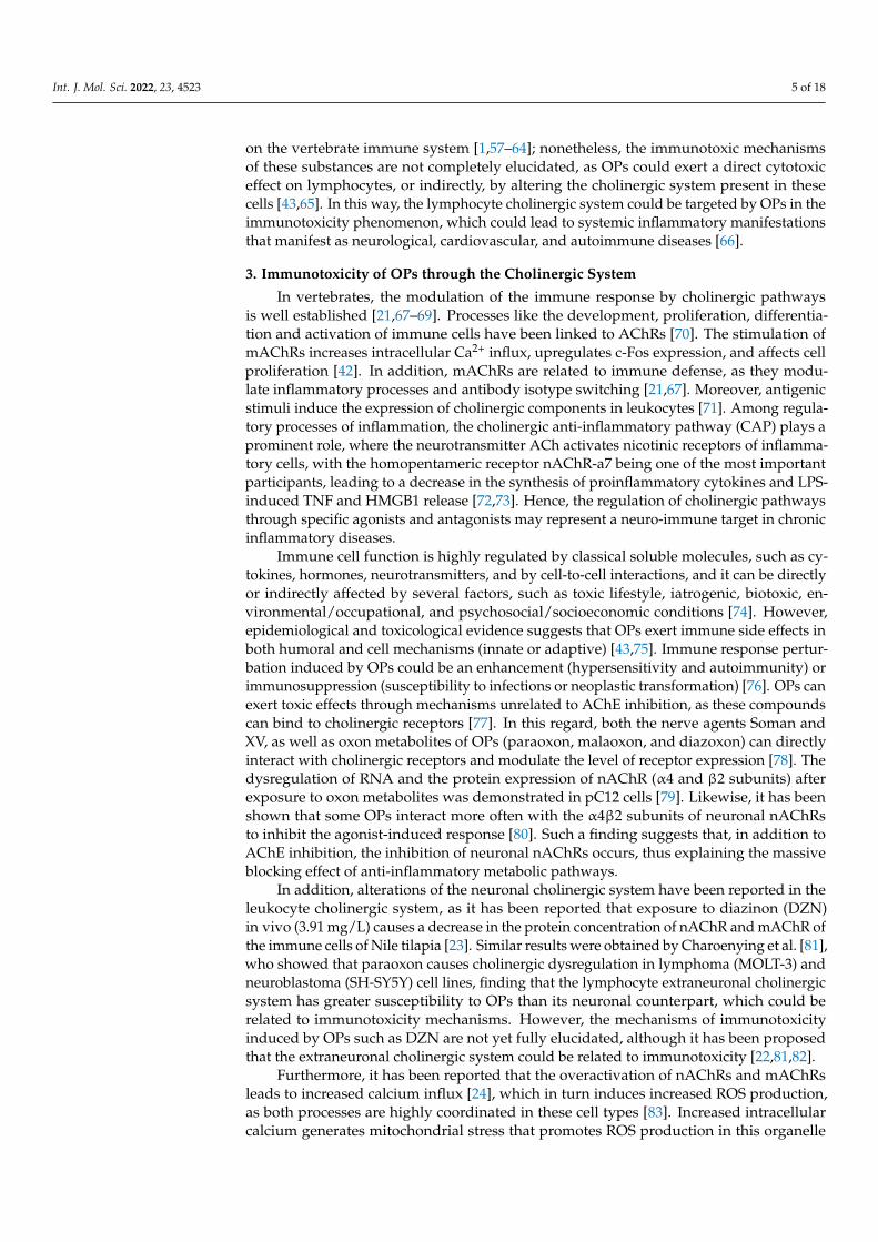
Organophosphorus pesticides (OPs) are widely used as insecticides, and the use of OPshas replaced organochlorine pesticides, as OPs have limited environmental persistence [1,7];however, the incorrect handling of these substances during storage, transport, applicationand the disposal of residues may cause toxic effects on non-target organisms, such as aquaticorganisms, domestic and wild fauna, and even humans [8]. Worldwide, more than 3 millionacute intoxications and up to 250,000 deaths caused by pesticides are reported annually [9];OPs reach organisms via inhalation, dermal and oral exposure, the most common beingthe last one [10]; once inside the organism, these substances are biotransformed (Figure 1a)to highly toxic metabolites (oxon) by the metabolic activation of cytochrome P450 [11],through the elimination of sulfur bound to phosphorus and the insertion of an oxygenatom (oxidative desulfurization). Oxons are detoxified through dearylation and hydrolysis
Int. J. Mol. Sci. 2022, 23, 4523. https://doi.org/10.3390/ijms23094523 https://www.mdpi.com/journal/ijms
Int. J. Mol. Sci. 2022, 23, 4523 2 of 18
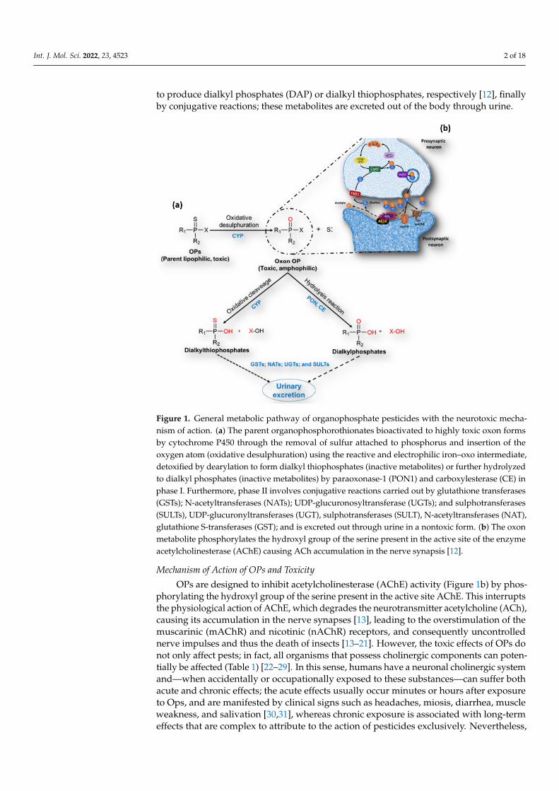
to produce dialkyl phosphates (DAP) or dialkyl thiophosphates, respectively [12], finallyby conjugative reactions; these metabolites are excreted out of the body through urine.
Int. J. Mol. Sci. 2021, 22, x FOR PEER REVIEW 4 of 23
Figure 1. General metabolic pathway of organophosphate pesticides with the neurotoxic
mechanism of action. (a) The parent organophosphorothionates bioactivated to highly toxic oxon
forms by cytochrome P450 through the removal of sulfur attached to phosphorus and insertion of
the oxygen atom (oxidative desulphuration) using the reactive and electrophilic iron–oxo
intermediate, detoxified by dearylation to form dialkyl thiophosphates (inactive metabolites) or
further hydrolyzed to dialkyl phosphates (inactive metabolites) by paraoxonase-1 (PON1) and
carboxylesterase (CE) in phase I. Furthermore, phase II involves conjugative reactions carried out
by glutathione transferases (GSTs); N-acetyltransferases (NATs); UDP-glucuronosyltransferase
(UGTs); and sulphotransferases (SULTs), UDP-glucuronyltransferases (UGT), sulphotransferases
(SULT), N-acetyltransferases (NAT), glutathione S-transferases (GST); and is excreted out through
urine in a nontoxic form. (b) The oxon metabolite phosphorylates the hydroxyl group of the serine
present in the active site of the enzyme acetylcholinesterase (AChE) causing ACh accumulation in
the nerve synapsis [12].
Therefore, several studies have reported that the alteration of the cholinergic system induced by OPs can trigger an inflammatory response and, consequently, pathophysio-logical alterations [19,44–47]. Thus, acute OP intoxication has been reported to stimulate an instantaneous and premature robust inflammatory response, whereas chronic expo-sure to low concentrations of OPs increases inflammatory mediators in a slow but sus-tained manner [44], or that it could be related to the development of inflammatory dis-eases such as organophosphate-induced delayed neuropathy (OPIDN) [19], rheumatoid arthritis [45,46], and neuroinflammation [47]. Further studies have shown that exposure to OPs leads to processes of cellular hyperreactivity, synergism with allergens, and the dysregulation of lung physiology, thus promoting susceptibility to asthma development [48,49]. In addition, recent research indicates that exposure to OPs may promote the de-velopment of early-stage diabetes mellitus [50].
Figure 1. General metabolic pathway of organophosphate pesticides with the neurotoxic mecha-nism of action. (a) The parent organophosphorothionates bioactivated to highly toxic oxon formsby cytochrome P450 through the removal of sulfur attached to phosphorus and insertion of theoxygen atom (oxidative desulphuration) using the reactive and electrophilic iron–oxo intermediate,detoxified by dearylation to form dialkyl thiophosphates (inactive metabolites) or further hydrolyzedto dialkyl phosphates (inactive metabolites) by paraoxonase-1 (PON1) and carboxylesterase (CE) inphase I. Furthermore, phase II involves conjugative reactions carried out by glutathione transferases(GSTs); N-acetyltransferases (NATs); UDP-glucuronosyltransferase (UGTs); and sulphotransferases(SULTs), UDP-glucuronyltransferases (UGT), sulphotransferases (SULT), N-acetyltransferases (NAT),glutathione S-transferases (GST); and is excreted out through urine in a nontoxic form. (b) The oxonmetabolite phosphorylates the hydroxyl group of the serine present in the active site of the enzymeacetylcholinesterase (AChE) causing ACh accumulation in the nerve synapsis [12].
Mechanism of Action of OPs and Toxicity
OPs are designed to inhibit acetylcholinesterase (AChE) activity (Figure 1b) by phos-phorylating the hydroxyl group of the serine present in the active site AChE. This interruptsthe physiological action of AChE, which degrades the neurotransmitter acetylcholine (ACh),causing its accumulation in the nerve synapses [13], leading to the overstimulation of themuscarinic (mAChR) and nicotinic (nAChR) receptors, and consequently uncontrollednerve impulses and thus the death of insects [13–21]. However, the toxic effects of OPs donot only affect pests; in fact, all organisms that possess cholinergic components can poten-tially be affected (Table 1) [22–29]. In this sense, humans have a neuronal cholinergic systemand—when accidentally or occupationally exposed to these substances—can suffer bothacute and chronic effects; the acute effects usually occur minutes or hours after exposureto Ops, and are manifested by clinical signs such as headaches, miosis, diarrhea, muscleweakness, and salivation [30,31], whereas chronic exposure is associated with long-termeffects that are complex to attribute to the action of pesticides exclusively. Nevertheless,
Int. J. Mol. Sci. 2022, 23, 4523 3 of 18
scientific evidence is growing that these substances induce mutations, epigenetic modifica-tions, tumors, and several types of cancer, as well as cognitive and functional alterationsin several physiological systems such as the renal, circulatory, respiratory, endocrine andimmune systems [32–37]. Currently, several non-neuronal cells, such as pancreatic alphacells, endothelial cells, placental cells, thrombocytes, and lymphocytes express cholinergiccomponents, which make those cells a target for OPs [38–43].
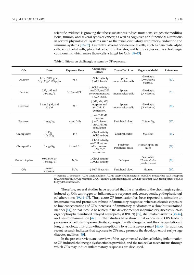
Table 1. Effects on cholinergic systems by OP exposure.
OPs Dose Exposure Time CholinergicEffects Tissue/Cell Line Organism Model References
Diazinon LC50-7.830 ppm,1/2 LC50-3.915 ppm 96 h ↓ AChE activity
↑ ACh levelsSpleen
mononuclear cells
Nile tilapia(Oreochromis
niloticus)[22].
Diazinon 0.97, 1.95 and3.91 mg/L 6, 12, and 24 h
↓ AChE activity ↓mAChR, nAChRconcentration and↑ ACh levels.
Spleenmononuclear cells
Nile tilapia(O. niloticus) [23].
Diazoxon 1 nm, 1 µM, and10 µM 24 h
↓ (M3, M4, M5)receptors and
nAChR β2expression.
Spleenmononuclear cells
Nile tilapia(O. niloticus) [24].
Paraoxon 1 mg/kg 6 and 24 h
↓mAChR M2function↑ ACh levels.↑mAChR M3
stimulation
Peripheral blood Guinea Pig [25].
Chlorpyrifos LD501/3 LD50
48 h ↓ ChAT activity↓ AChE activity Cerebral cortex Male Rat [26].
Chlorpyrifos 1 mg/Kg 1 h and 6 h
↓ ChAT activity,nAChR α4, andα7 expression↓ VAChT
expression
ForebrainPeripheral blood
Human apoE-TRmice [27].
Monocrotophos 0.01, 0.10, or1.00 mg/L N/A ↓ ChAT activity
↓ AChE activity EmbryosSea urchin
(Hemicentrotuspulcherrimus)
[28].
OPs Acuteexposure N/A ↓ BuChE activity Peripheral blood Human [29].
↑ increase ↓ decrease; ACh: acetylcholine; AChE: acetylcholinesterase; mAChR: muscarinic ACh receptor;nAChR: nicotinic ACh receptor; ChAT: choline acetyltransferase; VAChT: vesicular ACh transporter; BuChE:butyrylcholinesterase.
Therefore, several studies have reported that the alteration of the cholinergic systeminduced by OPs can trigger an inflammatory response and, consequently, pathophysiologi-cal alterations [19,44–47]. Thus, acute OP intoxication has been reported to stimulate aninstantaneous and premature robust inflammatory response, whereas chronic exposureto low concentrations of OPs increases inflammatory mediators in a slow but sustainedmanner [44], or that it could be related to the development of inflammatory diseases such asorganophosphate-induced delayed neuropathy (OPIDN) [19], rheumatoid arthritis [45,46],and neuroinflammation [47]. Further studies have shown that exposure to OPs leads toprocesses of cellular hyperreactivity, synergism with allergens, and the dysregulation oflung physiology, thus promoting susceptibility to asthma development [48,49]. In addition,recent research indicates that exposure to OPs may promote the development of early-stagediabetes mellitus [50].
In the present review, an overview of the experimental evidence linking inflammationto OP-induced cholinergic dysfunction is provided, and the molecular mechanisms throughwhich OPs may induce inflammatory responses are discussed.
Int. J. Mol. Sci. 2022, 23, 4523 4 of 18
2. Cholinergic System
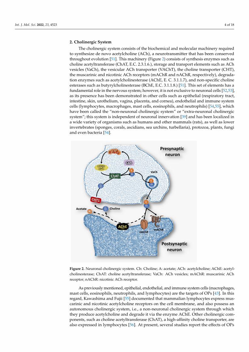
The cholinergic system consists of the biochemical and molecular machinery requiredto synthesize de novo acetylcholine (ACh), a neurotransmitter that has been conservedthroughout evolution [51]. This machinery (Figure 2) consists of synthesis enzymes such ascholine acetyltransferase (ChAT, E.C. 2.3.1.6.), storage and transport elements such as AChvesicles (VaCh), the vesicular ACh transporter (VAChT), the choline transporter (CHT),the muscarinic and nicotinic ACh receptors (mAChR and nAChR, respectively), degrada-tion enzymes such as acetylcholinesterase (AChE, E. C. 3.1.1.7), and non-specific cholineesterases such as butyrylcholinesterase (BChE, E.C. 3.1.1.8.) [51]. This set of elements has afundamental role in the nervous system; however, it is not exclusive to neuronal cells [52,53],as its presence has been demonstrated in other cells such as epithelial (respiratory tract,intestine, skin, urothelium, vagina, placenta, and cornea), endothelial and immune systemcells (lymphocytes, macrophages, mast cells, eosinophils, and neutrophils) [54,55], whichhave been called the “non-neuronal cholinergic system” or “extra-neuronal cholinergicsystem”; this system is independent of neuronal innervation [39] and has been localized ina wide variety of organisms such as humans and other mammals (rats), as well as lowerinvertebrates (sponges, corals, ascidians, sea urchins, turbellaria), protozoa, plants, fungiand even bacteria [54].
Int. J. Mol. Sci. 2021, 22, x FOR PEER REVIEW 5 of 23
In the present review, an overview of the experimental evidence linking inflamma-tion to OP-induced cholinergic dysfunction is provided, and the molecular mechanisms through which OPs may induce inflammatory responses are discussed.
2. Cholinergic System The cholinergic system consists of the biochemical and molecular machinery re-
quired to synthesize de novo acetylcholine (ACh), a neurotransmitter that has been con-served throughout evolution [51]. This machinery (Figure 2) consists of synthesis enzymes such as choline acetyltransferase (ChAT, E.C. 2.3.1.6.), storage and transport elements such as ACh vesicles (VaCh), the vesicular ACh transporter (VAChT), the choline trans-porter (CHT), the muscarinic and nicotinic ACh receptors (mAChR and nAChR, respec-tively), degradation enzymes such as acetylcholinesterase (AChE, E. C. 3.1.1.7), and non-specific choline esterases such as butyrylcholinesterase (BChE, E.C. 3.1.1.8.) [51]. This set of elements has a fundamental role in the nervous system; however, it is not exclusive to neuronal cells [52,53], as its presence has been demonstrated in other cells such as epithe-lial (respiratory tract, intestine, skin, urothelium, vagina, placenta, and cornea), endothe-lial and immune system cells (lymphocytes, macrophages, mast cells, eosinophils, and neutrophils) [54,55], which have been called the “non-neuronal cholinergic system” or “extra-neuronal cholinergic system”; this system is independent of neuronal innervation, [39] and has been localized in a wide variety of organisms such as humans and other mammals (rats), as well as lower invertebrates (sponges, corals, ascidians, sea urchins, turbellaria), protozoa, plants, fungi and even bacteria [54].
Figure 2. Neuronal cholinergic system. Ch: Choline; A: acetate; ACh: acetylcholine; AChE:
acetylcholinesterase; ChAT: choline acetyltransferase; VaCh: ACh vesicles; mAChR: muscarinic
ACh receptor; nAChR: nicotinic ACh receptor.
Figure 2. Neuronal cholinergic system. Ch: Choline; A: acetate; ACh: acetylcholine; AChE: acetyl-cholinesterase; ChAT: choline acetyltransferase; VaCh: ACh vesicles; mAChR: muscarinic AChreceptor; nAChR: nicotinic ACh receptor.
As previously mentioned, epithelial, endothelial, and immune system cells (macrophages,mast cells, eosinophils, neutrophils, and lymphocytes) are the targets of OPs [43]. In thisregard, Kawashima and Fujii [55] documented that mammalian lymphocytes express mus-carinic and nicotinic acetylcholine receptors on the cell membrane, and also possess anautonomous cholinergic system, i.e., a non-neuronal cholinergic system through whichthey produce acetylcholine and degrade it via the enzyme AChE. Other cholinergic com-ponents, such as choline acetyltransferase (ChAT), a high-affinity choline transporter, arealso expressed in lymphocytes [56]. At present, several studies report the effects of OPs
Int. J. Mol. Sci. 2022, 23, 4523 5 of 18
on the vertebrate immune system [1,57–64]; nonetheless, the immunotoxic mechanismsof these substances are not completely elucidated, as OPs could exert a direct cytotoxiceffect on lymphocytes, or indirectly, by altering the cholinergic system present in thesecells [43,65]. In this way, the lymphocyte cholinergic system could be targeted by OPs in theimmunotoxicity phenomenon, which could lead to systemic inflammatory manifestationsthat manifest as neurological, cardiovascular, and autoimmune diseases [66].
3. Immunotoxicity of OPs through the Cholinergic System
In vertebrates, the modulation of the immune response by cholinergic pathwaysis well established [21,67–69]. Processes like the development, proliferation, differentia-tion and activation of immune cells have been linked to AChRs [70]. The stimulation ofmAChRs increases intracellular Ca2+ influx, upregulates c-Fos expression, and affects cellproliferation [42]. In addition, mAChRs are related to immune defense, as they modu-late inflammatory processes and antibody isotype switching [21,67]. Moreover, antigenicstimuli induce the expression of cholinergic components in leukocytes [71]. Among regula-tory processes of inflammation, the cholinergic anti-inflammatory pathway (CAP) plays aprominent role, where the neurotransmitter ACh activates nicotinic receptors of inflamma-tory cells, with the homopentameric receptor nAChR-a7 being one of the most importantparticipants, leading to a decrease in the synthesis of proinflammatory cytokines and LPS-induced TNF and HMGB1 release [72,73]. Hence, the regulation of cholinergic pathwaysthrough specific agonists and antagonists may represent a neuro-immune target in chronicinflammatory diseases.
Immune cell function is highly regulated by classical soluble molecules, such as cy-tokines, hormones, neurotransmitters, and by cell-to-cell interactions, and it can be directlyor indirectly affected by several factors, such as toxic lifestyle, iatrogenic, biotoxic, en-vironmental/occupational, and psychosocial/socioeconomic conditions [74]. However,epidemiological and toxicological evidence suggests that OPs exert immune side effects inboth humoral and cell mechanisms (innate or adaptive) [43,75]. Immune response pertur-bation induced by OPs could be an enhancement (hypersensitivity and autoimmunity) orimmunosuppression (susceptibility to infections or neoplastic transformation) [76]. OPs canexert toxic effects through mechanisms unrelated to AChE inhibition, as these compoundscan bind to cholinergic receptors [77]. In this regard, both the nerve agents Soman andXV, as well as oxon metabolites of OPs (paraoxon, malaoxon, and diazoxon) can directlyinteract with cholinergic receptors and modulate the level of receptor expression [78]. Thedysregulation of RNA and the protein expression of nAChR (α4 and β2 subunits) afterexposure to oxon metabolites was demonstrated in pC12 cells [79]. Likewise, it has beenshown that some OPs interact more often with the α4β2 subunits of neuronal nAChRsto inhibit the agonist-induced response [80]. Such a finding suggests that, in addition toAChE inhibition, the inhibition of neuronal nAChRs occurs, thus explaining the massiveblocking effect of anti-inflammatory metabolic pathways.
In addition, alterations of the neuronal cholinergic system have been reported in theleukocyte cholinergic system, as it has been reported that exposure to diazinon (DZN)in vivo (3.91 mg/L) causes a decrease in the protein concentration of nAChR and mAChR ofthe immune cells of Nile tilapia [23]. Similar results were obtained by Charoenying et al. [81],who showed that paraoxon causes cholinergic dysregulation in lymphoma (MOLT-3) andneuroblastoma (SH-SY5Y) cell lines, finding that the lymphocyte extraneuronal cholinergicsystem has greater susceptibility to OPs than its neuronal counterpart, which could berelated to immunotoxicity mechanisms. However, the mechanisms of immunotoxicityinduced by OPs such as DZN are not yet fully elucidated, although it has been proposedthat the extraneuronal cholinergic system could be related to immunotoxicity [22,81,82].
Furthermore, it has been reported that the overactivation of nAChRs and mAChRsleads to increased calcium influx [24], which in turn induces increased ROS production,as both processes are highly coordinated in these cell types [83]. Increased intracellularcalcium generates mitochondrial stress that promotes ROS production in this organelle
Int. J. Mol. Sci. 2022, 23, 4523 6 of 18
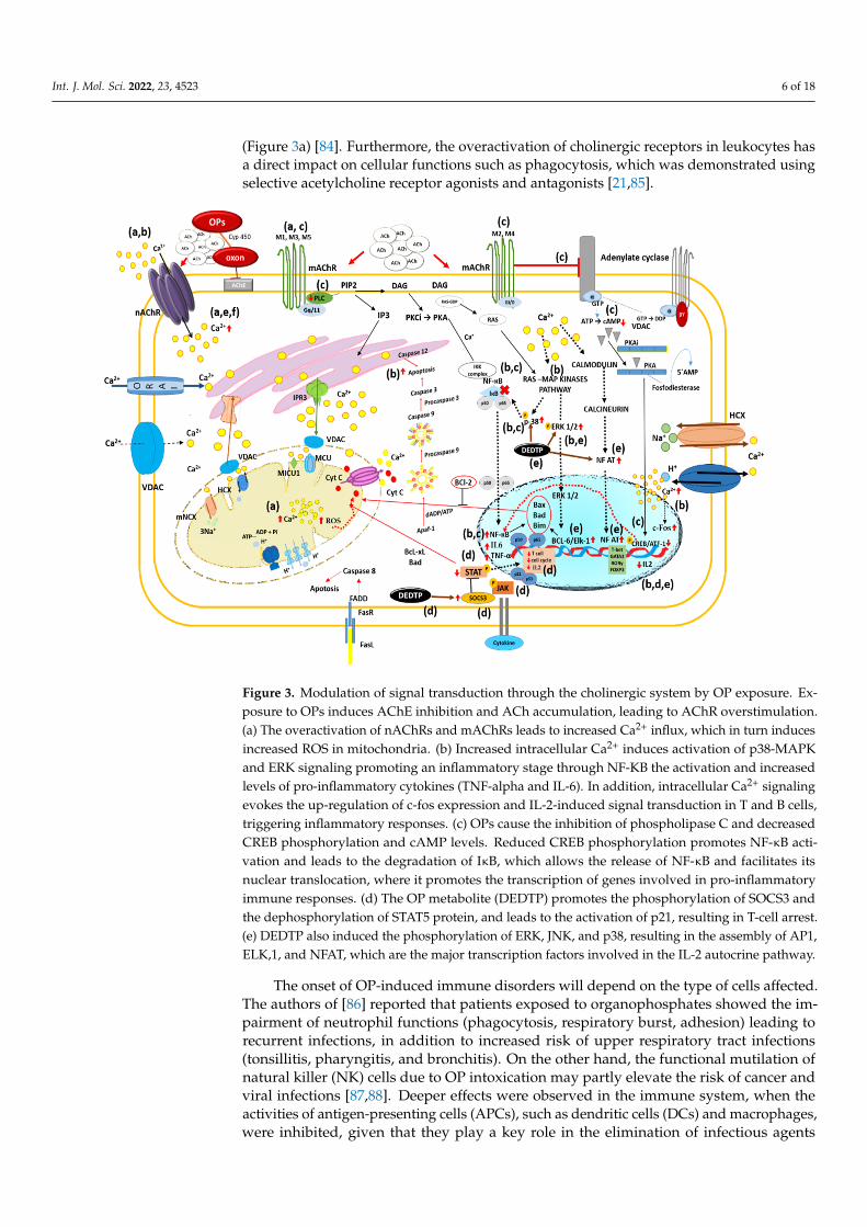
(Figure 3a) [84]. Furthermore, the overactivation of cholinergic receptors in leukocytes hasa direct impact on cellular functions such as phagocytosis, which was demonstrated usingselective acetylcholine receptor agonists and antagonists [21,85].
Int. J. Mol. Sci. 2021, 22, x FOR PEER REVIEW 7 of 23
receptor expression [78]. The dysregulation of RNA and the protein expression of nAChR (ɑ4 and β2 subunits) after exposure to oxon metabolites was demonstrated in pC12 cells [79]. Likewise, it has been shown that some OPs interact more often with the ɑ4β2 subunits of neuronal nAChRs to inhibit the agonist-induced response [80]. Such a finding suggests that, in addition to AChE inhibition, the inhibition of neuronal nAChRs occurs, thus explaining the massive blocking effect of anti-inflammatory metabolic pathways.
In addition, alterations of the neuronal cholinergic system have been reported in the leukocyte cholinergic system, as it has been reported that exposure to diazinon (DZN) in vivo (3.91 mg/L) causes a decrease in the protein concentration of nAChR and mAChR of the immune cells of Nile tilapia [23]. Similar results were obtained by Charoenying et al. [81], who showed that paraoxon causes cholinergic dysregulation in lymphoma (MOLT-3) and neuroblastoma (SH-SY5Y) cell lines, finding that the lymphocyte extraneuronal cholinergic system has greater susceptibility to OPs than its neuronal counterpart, which could be related to immunotoxicity mechanisms. However, the mechanisms of immunotoxicity induced by OPs such as DZN are not yet fully elucidated, although it has been proposed that the extraneuronal cholinergic system could be related to immunotoxicity [22,81,82].
Furthermore, it has been reported that the overactivation of nAChRs and mAChRs leads to increased calcium influx [24], which in turn induces increased ROS production, as both processes are highly coordinated in these cell types [83]. Increased intracellular calcium generates mitochondrial stress that promotes ROS production in this organelle (Figure 3a) [84]. Furthermore, the overactivation of cholinergic receptors in leukocytes has a direct impact on cellular functions such as phagocytosis, which was demonstrated using selective acetylcholine receptor agonists and antagonists [21,85].
Figure 3. Modulation of signal transduction through the cholinergic system by OP exposure. Ex-posure to OPs induces AChE inhibition and ACh accumulation, leading to AChR overstimulation.(a) The overactivation of nAChRs and mAChRs leads to increased Ca2+ influx, which in turn inducesincreased ROS in mitochondria. (b) Increased intracellular Ca2+ induces activation of p38-MAPKand ERK signaling promoting an inflammatory stage through NF-KB the activation and increasedlevels of pro-inflammatory cytokines (TNF-alpha and IL-6). In addition, intracellular Ca2+ signalingevokes the up-regulation of c-fos expression and IL-2-induced signal transduction in T and B cells,triggering inflammatory responses. (c) OPs cause the inhibition of phospholipase C and decreasedCREB phosphorylation and cAMP levels. Reduced CREB phosphorylation promotes NF-κB acti-vation and leads to the degradation of IκB, which allows the release of NF-κB and facilitates itsnuclear translocation, where it promotes the transcription of genes involved in pro-inflammatoryimmune responses. (d) The OP metabolite (DEDTP) promotes the phosphorylation of SOCS3 andthe dephosphorylation of STAT5 protein, and leads to the activation of p21, resulting in T-cell arrest.(e) DEDTP also induced the phosphorylation of ERK, JNK, and p38, resulting in the assembly of AP1,ELK,1, and NFAT, which are the major transcription factors involved in the IL-2 autocrine pathway.
The onset of OP-induced immune disorders will depend on the type of cells affected.The authors of [86] reported that patients exposed to organophosphates showed the im-pairment of neutrophil functions (phagocytosis, respiratory burst, adhesion) leading torecurrent infections, in addition to increased risk of upper respiratory tract infections(tonsillitis, pharyngitis, and bronchitis). On the other hand, the functional mutilation ofnatural killer (NK) cells due to OP intoxication may partly elevate the risk of cancer andviral infections [87,88]. Deeper effects were observed in the immune system, when theactivities of antigen-presenting cells (APCs), such as dendritic cells (DCs) and macrophages,were inhibited, given that they play a key role in the elimination of infectious agents
Int. J. Mol. Sci. 2022, 23, 4523 7 of 18
and the deployment of cell-mediated immunity [44]. The functional impairment of DCsand macrophages induced by OPs occurs through negative regulation of co-stimulatorymolecules (CD80 and CD86), effector molecules (human leukocyte antigen), MHC expres-sion, and phenotypic modulation [89–91].
Pesticide-induced immunosuppression is evidently a risk factor for the clinical compli-cation of inflammatory diseases, especially in occupationally or environmentally exposedindividuals, which occurs in developing countries. In this context, the current knowledgeof molecular mechanisms suggests a direct effect of the exposure to OPs on immunity andinflammatory processes; however, new experiments with epidemiological approaches arekey to demonstrating the existing correlation of the exposure to toxic substances, such asOPs, with the degree of susceptibility of organisms to inflammatory diseases.
4. Cytokine-Mediated Modulation of the Inflammatory Process by OP Exposure
The consequences of chronic or early-life exposure to pesticides may be extendedbeyond innate immune dysfunction to the increased risk of late-life chronic inflammatory-based diseases. Immune cells can release a variety of inflammation mediators, activatingpro- and anti-inflammatory processes and regulating intracellular pathways [92].
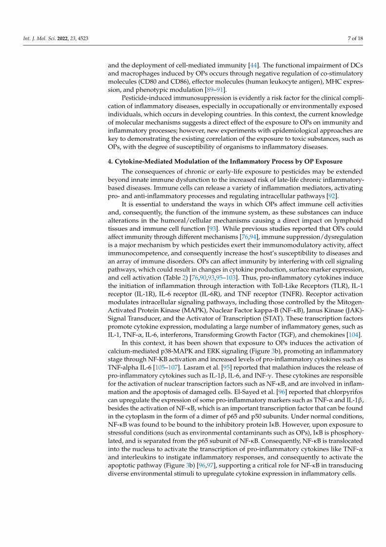
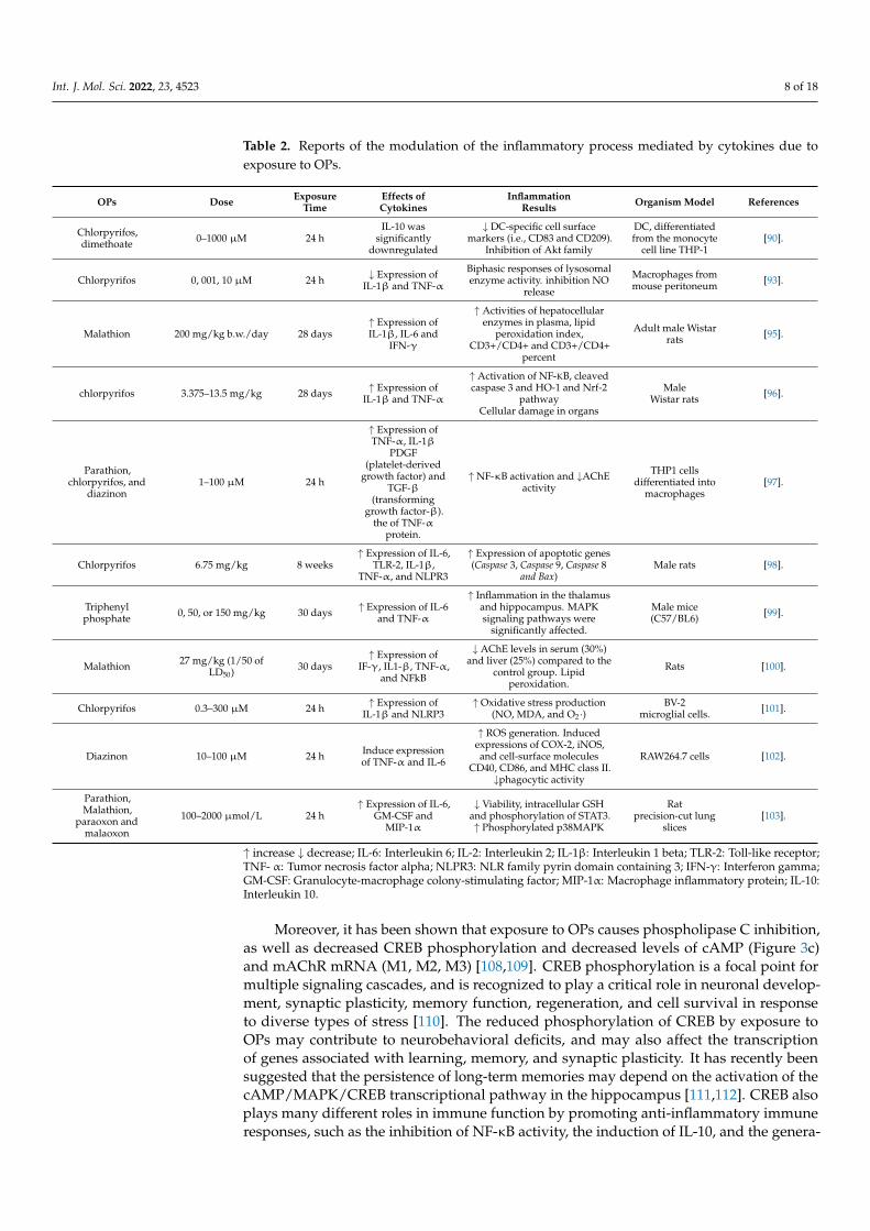
It is essential to understand the ways in which OPs affect immune cell activitiesand, consequently, the function of the immune system, as these substances can inducealterations in the humoral/cellular mechanisms causing a direct impact on lymphoidtissues and immune cell function [93]. While previous studies reported that OPs couldaffect immunity through different mechanisms [76,94], immune suppression/dysregulationis a major mechanism by which pesticides exert their immunomodulatory activity, affectimmunocompetence, and consequently increase the host’s susceptibility to diseases andan array of immune disorders. OPs can affect immunity by interfering with cell signalingpathways, which could result in changes in cytokine production, surface marker expression,and cell activation (Table 2) [76,90,93,95–103]. Thus, pro-inflammatory cytokines inducethe initiation of inflammation through interaction with Toll-Like Receptors (TLR), IL-1receptor (IL-1R), IL-6 receptor (IL-6R), and TNF receptor (TNFR). Receptor activationmodulates intracellular signaling pathways, including those controlled by the Mitogen-Activated Protein Kinase (MAPK), Nuclear Factor kappa-B (NF-κB), Janus Kinase (JAK)-Signal Transducer, and the Activator of Transcription (STAT). These transcription factorspromote cytokine expression, modulating a large number of inflammatory genes, such asIL-1, TNF-α, IL-6, interferons, Transforming Growth Factor (TGF), and chemokines [104].
In this context, it has been shown that exposure to OPs induces the activation ofcalcium-mediated p38-MAPK and ERK signaling (Figure 3b), promoting an inflammatorystage through NF-KB activation and increased levels of pro-inflammatory cytokines such asTNF-alpha IL-6 [105–107]. Lasram et al. [95] reported that malathion induces the release ofpro-inflammatory cytokines such as IL-1β, IL-6, and INF-γ. These cytokines are responsiblefor the activation of nuclear transcription factors such as NF-κB, and are involved in inflam-mation and the apoptosis of damaged cells. El-Sayed et al. [96] reported that chlorpyrifoscan upregulate the expression of some pro-inflammatory markers such as TNF-α and IL-1β,besides the activation of NF-κB, which is an important transcription factor that can be foundin the cytoplasm in the form of a dimer of p65 and p50 subunits. Under normal conditions,NF-κB was found to be bound to the inhibitory protein IκB. However, upon exposure tostressful conditions (such as environmental contaminants such as OPs), IκB is phosphory-lated, and is separated from the p65 subunit of NF-κB. Consequently, NF-κB is translocatedinto the nucleus to activate the transcription of pro-inflammatory cytokines like TNF-αand interleukins to instigate inflammatory responses, and consequently to activate theapoptotic pathway (Figure 3b) [96,97], supporting a critical role for NF-κB in transducingdiverse environmental stimuli to upregulate cytokine expression in inflammatory cells.
Int. J. Mol. Sci. 2022, 23, 4523 8 of 18
Table 2. Reports of the modulation of the inflammatory process mediated by cytokines due toexposure to OPs.
OPs Dose ExposureTime
Effects ofCytokines
InflammationResults Organism Model References
Chlorpyrifos,dimethoate 0–1000 µM 24 h
IL-10 wassignificantly
downregulated
↓ DC-specific cell surfacemarkers (i.e., CD83 and CD209).
Inhibition of Akt family
DC, differentiatedfrom the monocyte
cell line THP-1[90].
Chlorpyrifos 0, 001, 10 µM 24 h ↓ Expression ofIL-1β and TNF-α
Biphasic responses of lysosomalenzyme activity. inhibition NO
release
Macrophages frommouse peritoneum [93].
Malathion 200 mg/kg b.w./day 28 days↑ Expression ofIL-1β, IL-6 and
IFN-γ
↑ Activities of hepatocellularenzymes in plasma, lipid
peroxidation index,CD3+/CD4+ and CD3+/CD4+
percent
Adult male Wistarrats [95].
chlorpyrifos 3.375–13.5 mg/kg 28 days ↑ Expression ofIL-1β and TNF-α
↑ Activation of NF-KB, cleavedcaspase 3 and HO-1 and Nrf-2
pathwayCellular damage in organs
MaleWistar rats [96].
Parathion,chlorpyrifos, and
diazinon1–100 µM 24 h
↑ Expression ofTNF-α, IL-1β
PDGF(platelet-derived
growth factor) andTGF-β
(transforminggrowth factor-β).
the of TNF-αprotein.
↑ NF-κB activation and ↓AChEactivity
THP1 cellsdifferentiated into
macrophages[97].
Chlorpyrifos 6.75 mg/kg 8 weeks↑ Expression of IL-6,
TLR-2, IL-1β,TNF-α, and NLPR3
↑ Expression of apoptotic genes(Caspase 3, Caspase 9, Caspase 8
and Bax)Male rats [98].
Triphenylphosphate 0, 50, or 150 mg/kg 30 days ↑ Expression of IL-6
and TNF-α
↑ Inflammation in the thalamusand hippocampus. MAPKsignaling pathways were
significantly affected.
Male mice(C57/BL6) [99].
Malathion 27 mg/kg (1/50 ofLD50) 30 days
↑ Expression ofIF-γ, IL1-β, TNF-α,
and NFkB
↓ AChE levels in serum (30%)and liver (25%) compared to the
control group. Lipidperoxidation.
Rats [100].
Chlorpyrifos 0.3–300 µM 24 h ↑ Expression ofIL-1β and NLRP3
↑ Oxidative stress production(NO, MDA, and O2·)
BV-2microglial cells. [101].
Diazinon 10–100 µM 24 h Induce expressionof TNF-α and IL-6
↑ ROS generation. Inducedexpressions of COX-2, iNOS,and cell-surface molecules
CD40, CD86, and MHC class II.↓phagocytic activity
RAW264.7 cells [102].
Parathion,Malathion,
paraoxon andmalaoxon
100–2000 µmol/L 24 h↑ Expression of IL-6,
GM-CSF andMIP-1α
↓ Viability, intracellular GSHand phosphorylation of STAT3.↑ Phosphorylated p38MAPK
Ratprecision-cut lung
slices[103].
↑ increase ↓ decrease; IL-6: Interleukin 6; IL-2: Interleukin 2; IL-1β: Interleukin 1 beta; TLR-2: Toll-like receptor;TNF- α: Tumor necrosis factor alpha; NLPR3: NLR family pyrin domain containing 3; IFN-γ: Interferon gamma;GM-CSF: Granulocyte-macrophage colony-stimulating factor; MIP-1α: Macrophage inflammatory protein; IL-10:Interleukin 10.
Moreover, it has been shown that exposure to OPs causes phospholipase C inhibition,as well as decreased CREB phosphorylation and decreased levels of cAMP (Figure 3c)and mAChR mRNA (M1, M2, M3) [108,109]. CREB phosphorylation is a focal point formultiple signaling cascades, and is recognized to play a critical role in neuronal develop-ment, synaptic plasticity, memory function, regeneration, and cell survival in responseto diverse types of stress [110]. The reduced phosphorylation of CREB by exposure toOPs may contribute to neurobehavioral deficits, and may also affect the transcriptionof genes associated with learning, memory, and synaptic plasticity. It has recently beensuggested that the persistence of long-term memories may depend on the activation of thecAMP/MAPK/CREB transcriptional pathway in the hippocampus [111,112]. CREB alsoplays many different roles in immune function by promoting anti-inflammatory immuneresponses, such as the inhibition of NF-κB activity, the induction of IL-10, and the genera-
Int. J. Mol. Sci. 2022, 23, 4523 9 of 18
tion of T-regs [113]. However, reduced CREB phosphorylation induced by OP exposurepromotes NF-κB activation causing a cascade of signaling events that ultimately lead to thedegradation of IκB (Figure 3c), which allows NF-κB release and facilitates NF-κB nucleartranslocation, where it promotes the transcription of genes involved in pro-inflammatoryimmune responses [113–115].
Macrophages also play an important role in OP-induced inflammation [103].Ogasawara et al. [102] showed that OPs not only enhance the production of pro-inflammatorymarkers such as IL-6 and TNF-α but also the number of macrophages, and increase theexpression of cyclooxygenase (COX)-2 and inducible nitric oxide synthase enzymes as amajor source of ROS. In this way, oxidative stress can stimulate the expression of inflam-matory transcription factors, which are crucial regulatory components in the induction ofinflammatory responses [96].
Nevertheless, in the cells, there are a plethora of negative regulators of inflammatorysignaling pathways that operate in a negative feedback fashion (i.e., those pathways whichare inducible by inflammatory signals). These include the suppressor of cytokine signaling(SOCS) proteins, negative regulators of Janus kinase–signal transducer and activator oftranscription (JAK-STAT) signaling, and A20, a negative regulator of nuclear factor-kB (NF-kB) signaling [116]. However, this plethora of negative regulators of inflammatory signalingpathways can also be modulated by OP exposure [117–121]. Esquivel-Sentíes et al. [117]proposed that the alteration of the function and components of the immune system maybe related to the sequence and intensity of the phosphorylation and dephosphorylation ofprotein kinases, an essential mechanism that controls the function of the immune system.SOCS3 (suppressor of cytokine signaling 3) is a critical molecule in this process, as itfunctions as a negative regulator of cytokine signaling. SOCS3 regulates STAT by inhibitingthe phosphorylation of STAT5 affecting cell proliferation [118,119].
Recent reports have shown that metabolites (dialkyl phosphates) generated by thebiotransformation of OPs as diethyl thiophosphate (DETP) and diethyl dithiophosphate(DEDTP) modify the phosphorylation status of STAT5 (Figure 3d) proteins, and thus pro-duce several immunomodulatory effects, for instance, the reduction of CD25 and CD4expression, the reduced secretion of IL2, and the altered signalization of IL-2R [117,120].Esquivel-Sentíes et al. [117] reported that DEDTP treatment in human T lymphocytesincreases SOCS3 phosphorylation and decreases STAT5 phosphorylation, resulting in thearrest of T cell proliferation (Figure 3d). On the other hand, Lima et al. [121] reported thatDEDTP can trigger SOCS3-mediated cell cycle arrest that initiates a feedback mechanismassociated with the expression of p21 and p53. DEDTP also induced the phosphory-lation of ERK, JNK, and p38 [117], which results in the assembly of AP1, ELK,1, andNFAT, which are the main transcription factors involved in the autocrine IL-2 pathway(Figure 3e) [117,121,122].
Regarding the cholinergic system, acute OP poisoning induces the overstimulation ofcholinergic receptors due to the accumulation of ACh at immunological synapse, evokingintracellular Ca2+ signaling, the upregulation of c-fos expression (Figure 3b), and IL-2-induced signal transduction in T cells and B cells, as well as triggering inflammatoryresponses in macrophages [44,55,76]. In contrast, chronic OP poisoning through the down-regulation of cholinergic receptors may trigger cholinergic anti-inflammatory pathways,which result in the suppression of T-cell activity, predisposition to cancer, and certaininfections [44,76,82,83].
5. Therapeutic Strategies to Mitigate the Long-Term Inflammatory Effects of AcuteOP Intoxication
The canonical mechanism of the neurotoxicity of OPs is AChE inhibition [123]; thus,acute AChE inhibition (>60 to 80%) can induce a clinical condition termed cholinergiccrisis [47], which is characterized by peripheral parasympathetic symptoms, the depressionof central breathing control, seizures that can quickly progress to status epilepticus (SE),and the death of the intoxicated individual [124,125]. The conventional treatment to control
Int. J. Mol. Sci. 2022, 23, 4523 10 of 18
OP-induced cholinergic seizures is based on the use of drugs such as atropine (a peripheralmuscarinic receptor antagonist) [47], pralidoxime (a reactivator of AChE activity) [126]and benzodiazepine (which reduces seizure activity) [124,127]; however, in severe cases ofOP poisoning, these agents are not effective. Furthermore, OP intoxication can result inlong-term alterations, which are manifested by cognitive dysfunction, affective disorders, orspontaneous recurrent seizures (SRS) [30,128–132], which are linked to neuroinflammatoryprocesses [128].
In the neuroinflammatory disorder induced by OPs, microglia cells play a centralrole in regulating the production of pro-inflammatory cytokines that eventually damageneurons and exacerbate the course of neurodegenerative alterations [133]. Therefore,new pharmacological therapies should focus urgently on the inactivation of microgliaand the inhibition of the inflammatory response. In this regard, it has been shown thatblocking intracellular Ca2+ release, inhibiting NLRP3-inflammasome (NF-κB and MAPKblockers), and controlling ROS production (NADPH oxidase inhibitors -Nox1, Nox2, andNox4) may be important therapeutic targets to counteract the neuronal damage caused byOPs [47,66,134–139].
6. Lower Vertebrates as a Biomedical Model
Lower vertebrates have become relevant in the field of biomedical research, given thatsuch vertebrates offer advantages over different study models (e.g., mice). An example ofthese are fish, which belong to the phylogenetically oldest group of vertebrates, includingmore than half of the vertebrates on the planet; the vast majority of fishes are teleosts(teleosts, possessing a bony skeleton), and some are highlighted for both their ecologicaland economic significance, while other species are widely used as biological models forgenomic studies and developmental biology [65,140]. Furthermore, as these organismsare the first to present adaptive immune mechanisms, the study of the immune systemin these organisms is of great relevance, as it provides information on the evolution ofthe immune system in vertebrates, thus supporting the knowledge of basic aspects ofimmunology, and thus the possible treatment of emerging diseases in humans and otheranimals. Wilson [141] proposed that teleost fishes can be a good model for translationalresearch because they possess mechanisms of innate and adaptive immunity (TLR toll-likereceptors, cytokines, complement molecules, B cells, T cells, and immunological memory)which are very similar to those of higher mammals.
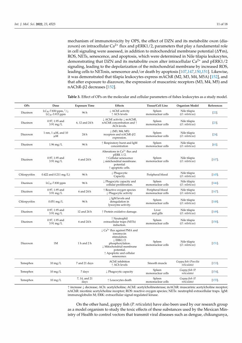
Furthermore, teleost fish have also been used as bioindicators of pollution, as theycan respond to environmental pollution through alterations in physiology or through thestorage of pollutants [142,143]. The use of fish as bioindicators is of great importancefor several reasons, due to their sensitivity to environmental stressors, wide geographicdistribution, presence in the food chain, and ease of adaptation to captivity, which permitsthe evaluation of the effect of environmental stressors under controlled conditions [144].Given this background, our research group has used Nile tilapia (Oreochromis niloticus) andguppy fish (Poecilia reticulata) as bioindicator organisms and biomedical study models, toelucidate the mechanism of immunotoxicity by OPs (Table 3).
Initial studies demonstrated that OPs (chlorpyrifos and diazinon) cause immuno-toxic effects by altering the physiological parameters of leukocytes, such as decreasedphagocytic capacity [145–147], increased respiratory burst [61], and the dysregulation ofIgM concentration and lysozyme activity [61,148], in addition to oxidative damage inliver and gill proteins [149]. Subsequently—derived from Kawashima and Fujii [55], whoreported that mammalian lymphocytes possessed all of the biochemical and molecularmachinery necessary to synthesize ACh de novo—we were prompted to search for thischolinergic system in the mononuclear cells of Nile tilapia, demonstrating not only thepresence of the extraneuronal cholinergic system in these cells but also that when theorganisms were exposed to DZN, the activity of AChE was inhibited and the concentrationof ACh increased [23], suggesting that the lymphocyte cholinergic system could be targetedby OPs in the immunotoxicity phenomenon [43]. Later, in order to elucidate a possible
Int. J. Mol. Sci. 2022, 23, 4523 11 of 18
mechanism of immunotoxicity by OPS, the effect of DZN and its metabolite oxon (dia-zoxon) on intracellular Ca2+ flux and pERK1/2, parameters that play a fundamental rolein cell signaling were assessed, in addition to mitochondrial membrane potential (∆Ψm),ROS, NETs, senescence, and apoptosis, which were determined in Nile tilapia leukocytes,demonstrating that DZN and its metabolite oxon alter intracellular Ca2+ and pERK1/2signaling, leading to the depolarization of the mitochondrial membrane by increased ROS,leading cells to NETosis, senescence and/or death by apoptosis [107,147,150,151]. Likewise,it was demonstrated that tilapia leukocytes express mAChR (M2, M3, M4, M5A) [152], andthat after exposure to diazoxon, the expression of muscarinic receptors (M3, M4, M5) andnAChR-β2 decreases [152].
Table 3. Effect of OPs on the molecular and cellular parameters of fishes leukocytes as a study model.
OPs Dose Exposure Time Effects Tissue/Cell Line Organism Model References
Diazinon LC50-7.830 ppm, 1/2LC50-3.915 ppm 96 h ↓ AChE activity
↑ ACh levelsSpleen
mononuclear cellsNile tilapia
(O. niloticus) [22].
Diazinon 0.97, 1.95 and3.91 mg/L 6, 12 and 24 h
↓ AChE activity ↓mAChR,nAChR concentration and ↑
ACh levels.
Spleenmononuclear cells
Nile tilapia(O. niloticus) [23].
Diazoxon 1 nm, 1 µM, and 10µM 24 h
↓ (M3, M4, M5)receptors and nAChR-β2
expression.
Spleenmononuclear cells
Nile tilapia(O. niloticus) [24].
Diazinon 1.96 mg/L 96 h ↑ Respiratory burst and IgMconcentration
Spleenmononuclear cells
Nile tilapia(O. niloticus) [61].
Diazinon 0.97, 1.95 and3.91 mg/L 6 and 24 h
Alterations in Ca2+ flux andpERK 1/2.
↑ Cellular senescence↓mitchondrial membrane.
potential↑ apoptotic cells.
Spleenmononuclear cells
Nile tilapia(O. niloticus) [107].
Chlorpyrifos 0.422 and 0.211 mg/L) 96 h ↓ PhagocyticCapacity. Peripheral blood Nile tilapia
(O. niloticus) [145].
Diazinon LC50-7.830 ppm 96 h ↓ Phagocytic capacity andcellular proliferation.
Spleenmononuclear cells
Nile tilapia(O. niloticus) [146].
Diazinon 0.97, 1.95 and3.91 mg/L 6 and 24 h ↑ Reactive oxygen species
↓ Phagocytic activityPeripheral blood
mononuclear cellsNile tilapia
(O. niloticus) [147].
Chlorpyrifos 0.051 mg/L 96 h↓ IgM levels andderegulation in
lysozyme activity.
Spleenmononuclear cells
Nile tilapia(O. niloticus) [148].
Diazinon 0.97, 1.95 and3.91 mg/L 12 and 24 h ↑ Protein oxidative damage. Liver
and gillsNile tilapia
(O. niloticus) [149].
Diazinon 0.97, 1.95 and3.91 mg/L 6 and 24 h
↑ Neutrophilextracellular traps (NETs)
induction.
Spleenmononuclear cells
Nile tilapia(O. niloticus) [150].
Diazoxon 1M 1 h and 2 h
↓ Ca2+ flux against PMA andionomycinstimulation.↓ ERK1/2
phosphorylation.↓Mitochondrial membrane
potential.↑ Apoptotic and cellular
senescence.
Spleenmononuclear cells
Nile tilapia(O. niloticus) [151].
Temephos 10 mg/L 7 and 21 days AChE inhibition↑ ACh levels Smooth muscle Guppy fish (Poecilia
reticulata) [153].
Temephos 10 mg/L 7 days ↓ Phagocytic capacity Spleenmononuclear cells
Guppy fish (P.reticulata) [154].
Temephos 10 mg/L 7, 14, and 21days ↑ Leucocytes death Spleen
mononuclear cellsGuppy fish (P.
reticulata) [155].
↑ increase ↓ decrease; ACh: acetylcholine; AChE: acetylcholinesterase; mAChR: muscarinic acetylcholine receptor;nAChR: nicotinic acetylcholine receptor; ROS: reactive oxygen species; NETs: neutrophil extracellular traps. IgM:immunoglobulin M; ERK: extracellular signal-regulated kinase.
On the other hand, guppy fish (P. reticulata) have also been used by our research groupas a model organism to study the toxic effects of these substances used by the Mexican Min-istry of Health to control vectors that transmit viral diseases such as dengue, chikungunya,
Int. J. Mol. Sci. 2022, 23, 4523 12 of 18
and Zika. The results of these investigations indicate that exposure (7 and 21 days) in vivoto temephos (0.5 mg/L), an OP, causes cholinergic alterations (the inhibition of AChE andthe accumulation of the neurotransmitter ACh) in muscle tissue [153]. In addition, it causesa decrease in phagocytic capacity [154] and a decrease in leukocyte viability, inducing apop-tosis and necrosis. The data even reveal that temephos induces apoptosis up to 35 dayspost-exposure, indicating recovery up to 70 days [155].
At present, our research group is working on the effect of diazinon and its metaboliteoxon on key molecules involved in cell signaling, aiming to elucidate a possible mechanismof immunotoxicity by these substances. In this sense, we are focusing on the effects ofOPs on the expression of cytokines (anti-inflammatory and pro-inflammatory) and mastertranscription factors (T-bet, GATA-3, RORγt, and FOXP3), as well as on the phosphorylationof JAK/STAT, and levels of cAMP, DAG, and IP3.
7. Conclusions
In conclusion, the present review clearly shows that OPs are substances that, despitebeing designed for insect control, affect the physiology of non-target organisms, includinghumans. Due to the mechanism of action of OPs, these substances alter the activity ofthe cholinergic system, which significantly influences the transcription, synthesis, andrelease of inflammatory mediators such as cytokines. Consequently, acute and chronicexposure to OPs may be related to the development of chronic degenerative pathologies, aswell as allergies or immunosuppression phenomena, alterations in which inflammatorycomponents play a central role.
Author Contributions: M.R.C.-P.: writing of the manuscript; C.E.C.-R.: writing of the manuscriptand editing; G.A.T.-I.: comments on the final version of the paper; U.M.-S.: writing of the manuscript;M.D.P.-R.: comments on the final version of the paper; K.J.G.D.-R.: conceptualization, coordination,and writing of the manuscript; M.I.G.-P.: conceptualization and comments on the final version of thepaper. All authors have read and agreed to the published version of the manuscript.
Funding: This research received funding from CONACyT-Mexico (Project number CB-A1-S-53561-2017–2018).
Institutional Review Board Statement: Not applicable.
Informed Consent Statement: Not applicable.
Data Availability Statement: Not applicable.
Acknowledgments: Camacho-Pérez M.R. is a Masters student from the program at the UniversidadAutónoma de Nayarit en Ciencias Biologico-Agropecuarias.
Conflicts of Interest: The authors report no conflict of interest.
References1. Al-Ghanim, K.A. Acute toxicity and effects of sub-lethal malathion exposure on biochemical and haematological parameters of
Oreochromis niloticus. Sci. Res. Essays 2012, 7, 1674–1680. [CrossRef]2. Sidhu, G.K.; Singh, S.; Kumar, V.; Dhanjal, D.S.; Datta, S.; Singh, J. Toxicity, monitoring and biodegradation of organophosphate
pesticides: A review. Crit. Rev. Environ. Sci. Technol. 2019, 49, 1135–1187. [CrossRef]3. International Code of Conduct on the Distribution and Use of Pesticides. Available online: http://apps.who.int/iris/
bitstream/handle/10665/70602/WHO_HTM_NTD_WHOPES_2010.7_spa.pdf;jsessionid=6BF8CBB80C94FB71C65499FC33ACB281?sequence=1 (accessed on 28 February 2022).
4. Cancer Clusters. Available online: https://www.cdc.gov/nceh/clusters/fallon/organophosfaq.htm#:~:text=Organophosphates%20are%20the%20most%20widely,in%20the%20body%20called%20acetylcholinesterase (accessed on 28 February 2022).
5. Sharma, A.; Kumar, V.; Shahzad, B.; Tanveer, M.; Sidhu, G.P.S.; Handa, N.; Kohli, S.K.; Yadav, P.; Bali, A.S.; Parihar, R.D.; et al.Worldwide pesticide usage and its impacts on ecosystem. SN Appl. Sci. 2019, 1, 1446. [CrossRef]
6. Tudi, M.; Ruan, H.D.; Wang, L.; Lyu, J.; Sadler, R.; Connell, D.; Chu, C.; Phung, D. Agriculture Development, Pesticide Applicationand Its Impact on the Environment. Int. J. Environ. Res. Public Health 2021, 18, 1112. [CrossRef]
7. Torres-Palma, R.; Serna-Galvis, E. Sonolysis; Academic Press: Cambridge, MA, USA, 2018; pp. 177–213. [CrossRef]8. Fernández, A.; Mancipe, L.; Fernández, A. Intoxicación por organofosforados. Rev. Med. 2010, 18, 84–92. [CrossRef]9. Chowdhary, S.; Bhattacharyya, R.; Banerjee, D. Acute organophosphorus poisoning. Clin. Chim. Acta 2014, 431, 66–76. [CrossRef]
Int. J. Mol. Sci. 2022, 23, 4523 13 of 18
10. Nkinsa, P.N.; Muckle, G.; Ayotte, P.; Lanphear, B.P.; Arbuckle, T.E.; Fraser, W.D.; Bouchard, M.F. Organophosphate pesticidesexposure during fetal development and IQ scores in 3 and 4-year old Canadian children. Environ. Res. 2020, 190, 110023.[CrossRef]
11. Abbas, R.; Leister, C.; El-Gaaloul, M.; Chalon, S.; Sonnichsen, D. Ascending Single-Dose Study of the Safety Profile, Tolerability,and Pharmacokinetics of Bosutinib Coadministered with Ketoconazole to Healthy Adult Subjects. Clin. Therap. 2012, 34,2011–2019. [CrossRef]
12. Kaur, K.; Besnier, F.; Glover, K.A.; Nilsen, F.; Aspehaug, V.T.; Fjørtoft, H.B.; Horsberg, T.E. The mechanism (Phe362Tyr mutation)behind resistance in Lepeophtheirus salmonis pre-dates organophosphate use in salmon farming. Sci. Rep. 2017, 7, 12349.[CrossRef]
13. Oruc, E.; Usta, D. Evaluation of oxidative stress responses and neurotoxicity potential of diazinon in different tissues of Cyprinuscarpio. Environ. Toxicol. Pharmacol. 2007, 23, 48–55. [CrossRef]
14. Fulton, M.H.; Key, P.B. Acetylcholinesterase inhibition in estuarine fish and invertebrates as an indicator of organophosphorusinsecticide exposure and effects. Environ. Toxicol. Chem. 2001, 20, 37–45. [CrossRef] [PubMed]
15. Profile for Diazinon. Available online: https://www.atsdr.cdc.gov/toxprofiles/tp86.pdf (accessed on 28 February 2022).16. Elersek, T.; Filipic, M. Organophosphorous Pestidices Mechanism of Their Toxicity Pesticides—The Impact of Pesticides Expusure; Intech:
Rijeka, Croatia, 2011. [CrossRef]17. Robb, E.L.; Baker, M.B. Organophosphathe Toxicity. In StatPearls; StatPearls Publishing: Treasure Island, FL, USA, 2021; PMID:
29261901.18. Vásquez, M.O. Intoxicación por organofosforados. Rev. Med. Sinerg. 2020, 5, e558. [CrossRef]19. King, A.M.; Aaron, C.K. Organophosphate and Carbamate Poisoning. Emerg. Med. Clin. N. Am. 2015, 33, 133–151. [CrossRef]
[PubMed]20. Mileson, B.E.; Chambers, J.E.; Chen, W.L.; Dettbarn, W.; Ehrich, M.; Eldefrawi, A.T.; Gaylor, D.W.; Hamernik, K.; Hodgson, E.;
Karczmar, A.G.; et al. Common Mechanism of Toxicity: A Case Study of Organophosphorus Pesticides. Toxicol. Sci. 1998, 41,8–20. [CrossRef]
21. Kawashima, K.; Fujii, T.; Moriwaki, Y.; Misawa, H.; Horiguchi, K. Reconciling neuronally and nonneuronally derived acetylcholinein the regulation of immune function. Life Sci. 2012, 1261, 1027–1032. [CrossRef]
22. Girón-Pérez, M.I.; Zaitseva, G.; Casas-Solis, J.; Santerre, A. Effects of diazinon and diazoxon on the lymphoproliferation rate ofsplenocytes from Nile tilapia (Oreochromis niloticus): The immunosuppresive effect could involve an increase in acetylcholinelevels. Fish Shellfish Immunol. 2008, 25, 517–521. [CrossRef] [PubMed]
23. Toledo-Ibarra, G.; Díaz-Resendiz, K.; Pavón-Romero, L.; Rojas-García, A.; Medina-Díaz, I.; Girón-Pérez, M. Effects of diazinonon the lymphocytic cholinergic system of Nile tilapia fish (Oreochromis niloticus). Vet. Immunol. Immunopathol. 2016, 176, 58–63.[CrossRef]
24. Toledo-Ibarra, G.; Girón-Pérez, M.; Covantes-Rosales, C.; Ventura-Ramón, G.; Pérez-Sánchez, G.; López-Torres, A.;Diaz-Resendiz, K.; Becerril-Villanueva, E.; Pavón, L. Alterations in the non-neuronal cholinergic system induced by in-vitro exposure to diazoxon in spleen mononuclear cells of Nile tilapia (O. niloticus). Fish Shellfish Immunol. 2021, 108, 134–141.[CrossRef]
25. Proskocil, B.; Bruun, D.A.; Thompson, C.M.; Fryer, A.; Lein, P.J. Organophosphorus Pesticides Decrease M2 Muscarinic ReceptorFunction in Guinea Pig Airway Nerves via Indirect Mechanisms. PLoS ONE 2010, 5, e10562. [CrossRef]
26. Poojary, A.; Basha, P.M. Cold stress interaction on organophosphate insecticide poisoning: Age-related assessment in rat cerebralcortex. Indian J. Exp. Biol. 2012, 50, 110–116.
27. Basaure, P.; Guardia-Escote, L.; Cabré, M.; Peris-Sampedro, F.; Sánchez-Santed, F.; Domingo, J.L.; Colomina, M.T. Postnatalchlorpyrifos exposure and apolipoprotein E (APOE) genotype differentially affect cholinergic expression and developmentalparameters in transgenic mice. Food Chem. Toxicol. 2018, 118, 42–52. [CrossRef] [PubMed]
28. Zhang, X.; Li, S.; Wang, C.; Tian, H.; Wang, W.; Ru, S. Effects of monocrotophos pesticide on cholinergic and dopaminergicneurotransmitter systems during early development in the sea urchin Hemicentrotus pulcherrimus. Toxicol. Appl. Pharmacol.2017, 328, 46–53. [CrossRef] [PubMed]
29. Xu, C.; Zhang, X.-G.; Yang, X.; He, Y.-Z. The diagnostic value of butyrylcholinesterase in acute organophosphorus pesticidepoisoning. Zhongguo Wei Zhong Bing Ji Jiu Yi Xue= Chin. Crit. Care Med. = Zhongguo Weizhongbing Jijiuyixue 2010, 22, 193–196.
30. Figueiredo, T.H.; Apland, J.P.; Braga, M.F.M.; Marini, A.M. Acute and long-term consequences of exposure to organophosphatenerve agents in humans. Epilepsia 2018, 59, 92–99. [CrossRef]
31. Bayrami, M.; Hashemi, T.; Malekirad, A.A.; Ashayeri, H.; Faraji, F.; Abdollahi, M. Electroencephalogram, cognitive state,psychological disorders, clinical symptom, and oxidative stress in horticulture farmers exposed to organophosphate pesticides.Toxicol. Ind. Health 2012, 28, 90–96. [CrossRef]
32. Perry, J.; Cotton, J.; Rahman, M.; Brumby, S. Organophosphate exposure and the chronic effects on farmers: A narrative review.Rural Remote Health 2020, 20, 4508. [CrossRef]
33. De Silva, H.J.; Samarawickrema, N.A.; Wickremasinghe, A.R. Toxicity due to organophosphorus compounds: What about chronicexposure? Trans. R. Soc. Trop. Med. Hyg. 2006, 100, 803–806. [CrossRef]
34. Thrasher, J.D.; Heuser, G.; Broughton, A. Immunological Abnormalities in Humans Chronically Exposed to Chlorpyrifos. Arch.Environ. Health Int. J. 2002, 57, 181–187. [CrossRef]
Int. J. Mol. Sci. 2022, 23, 4523 14 of 18
35. Zafar, R.; Munawar, K.; Nasrullah, A.; Haq, S.; Ghazanfar, H.; Sheikh, A.B.; Khan, A.Y. Acute Renal Failure due to Organophos-phate Poisoning: A Case Report. Cureus 2017, 9, e1523. [CrossRef]
36. Cavari, Y.; Landau, D.; Sofer, S.; Leibson, T.; Lazar, I. Organophosphate Poisoning-Induced Acute Renal Failure. Pediatr. Emerg.Care 2013, 29, 646–647. [CrossRef]
37. Ortega-Miller, J.G.; Yezioro-Rubinsky, S.; Benavides-Pinto, B.C.; Báez-Quintero, L.C. Efectos teratogénicos de insecticidasorganofosforados en la etiología de labio y paladar hendido: Revisión de literatura. Rev. Nac. De Odontol. 2017, 13, 13. [CrossRef]
38. Rodriguez-Diaz, R.; Dando, R.; Jacques-Silva, M.C.; Fachado, A.; Molina, J.; Abdulreda, M.H.; Ricordi, C.; Roper, S.D.;Berggren, P.-O.; Caicedo, A. Alpha cells secrete acetylcholine as a non-neuronal paracrine signal priming beta cell functionin humans. Nat. Med. 2011, 17, 888–892. [CrossRef] [PubMed]
39. Wessler, I.; Kirkpatrick, C.J.; Racké, K. Non-neuronal acetylcholine, a locally acting molecule, widely distributed in biologicalsystems: Expression and function in humans. Pharmacol. Ther. 1998, 77, 59–79. [CrossRef]
40. Bhuiyan, M.; Murad, F.; Fant, M. The placental cholinergic system: Localization to the cytotrophoblast and modulation of nitricoxide. Cell Commun. Signal. 2006, 4, 4–7. [CrossRef]
41. Lev-Lehman, E.; Deutsch, V.; Eldor, A.; Soreq, H. Immature Human Megakaryocytes Produce Nuclear-Associated Acetyl-cholinesterase. Blood 1997, 89, 3644–3653. [CrossRef]
42. Kawashima, K.; Fujii, T. The lymphocytic cholinergic system and its biological function. Life Sci. 2003, 72, 2101–2109. [CrossRef]43. Resendiz, K.J.G.D.; Toledo-Ibarra, G.A.; Girón-Pérez, M.I. Modulation of Immune Response by Organophosphorus Pesticides:
Fishes as a Potential Model in Immunotoxicology. J. Immunol. Res. 2015, 2015, 213836. [CrossRef]44. Banks, C.N.; Lein, P.J. A review of experimental evidence linking neurotoxic organophosphorus compounds and inflammation.
NeuroToxicology 2012, 33, 575–584. [CrossRef]45. Koureas, M.; Rachiotis, G.; Tsakalof, A.; Hadjichristodoulou, C. Increased Frequency of Rheumatoid Arthritis and Allergic
Rhinitis among Pesticide Sprayers and Associations with Pesticide Use. Int. J. Environ. Res. Public Health 2017, 14, 865. [CrossRef]46. Meyer, A.; Sandler, D.P.; Freeman, L.E.B.; Hofmann, J.; Parks, C.G. Pesticide Exposure and Risk of Rheumatoid Arthritis among
Licensed Male Pesticide Applicators in the Agricultural Health Study. Environ. Health Perspect. 2017, 125, 077010. [CrossRef]47. Andrew, P.M.; Lein, P.J. Neuroinflammation as a Therapeutic Target for Mitigating the Long-Term Consequences of Acute
Organophosphate Intoxication. Front. Pharmacol. 2021, 12, 12. [CrossRef] [PubMed]48. Hernández, A.F.; Parrón, T.; Alarcón, R. Pesticides and asthma. Curr. Opin. Allergy Clin. Immunol. 2011, 11, 90–96. [CrossRef]
[PubMed]49. Benka-Coker, W.; Loftus, C.; Karr, C.; Magzamen, S. Association of Organophosphate Pesticide Exposure and a Marker of Asthma
Morbidity in an Agricultural Community. J. Agromed. 2020, 25, 106–114. [CrossRef] [PubMed]50. Chung, Y.-L.; Hou, Y.-C.; Wang, I.-K.; Lu, K.-C.; Yen, T.-H. Organophosphate pesticides and new-onset diabetes mellitus: From
molecular mechanisms to a possible therapeutic perspective. World J. Diabetes 2021, 12, 1818–1831. [CrossRef]51. Wessler, I.K.; Kirkpatrick, C.J. The Non-neuronal Cholinergic System: An Emerging Drug Target in the Airways. Pulm. Pharmacol.
Ther. 2001, 14, 423–434. [CrossRef]52. Abreu-Villaça, Y.; Filgueiras, C.; Manhães, A.C. Developmental aspects of the cholinergic system. Behav. Brain Res. 2011, 221,
367–378. [CrossRef]53. Nizri, E.; Brenner, T. Modulation of inflammatory pathways by the immune cholinergic system. Amino Acids 2011, 45, 73–85.
[CrossRef]54. Wessler, I.; Kirkpatrick, C.J. Acetylcholine beyond neurons: The non-neuronal cholinergic system in humans. J. Cereb. Blood Flow
Metab. 2008, 154, 1558–1571. [CrossRef]55. Kawashima, K.; Fujii, T. Extraneuronal cholinergic system in lymphocytes. Pharmacol. Ther. 2000, 86, 29–48. [CrossRef]56. Mokarizadeh, A.; Faryabi, M.R.; Rezvanfar, M.A.; Abdollahi, M. A comprehensive review of pesticides and the immune
dysregulation: Mechanisms, evidence and consequences. Toxicol. Mech. Methods 2015, 25, 258–278. [CrossRef]57. Wang, X.; Yang, Z.; Xue, B.; Shi, H. Activation of the Cholinergic Antiinflammatory Pathway Ameliorates Obesity-Induced
Inflammation and Insulin Resistance. Endocrinology 2011, 152, 836–846. [CrossRef] [PubMed]58. El-Bouhy, Z.; El-Hakim, Y.A.; Mohamed, E. Chronic Effect of Chlorpyrifos on Biochemical, Immunological Changes and DNA
Damage in Juvenile Nile Tilapia (Oreochromis niloticus). Zagazig Vet. J. 2018, 46, 51–59. [CrossRef]59. Ahmadi, K.; Mirvaghefei, A.R.; Banaee, M.; Vosoghei, A.R. Effects of long-term diazinon exposure on some immunological and
haematological parameters in rainbow trout Oncorhynchus mykiss (Walbaum, 1792). Toxicol. Environ. Health Sci. 2014, 6, 1–7.[CrossRef]
60. Castillo-Sosa, Y.; Sierra-Fonseca, A.; Martínez-Martínez, A.; Plenge-Tellechea, F. Efecto del diazinón sobre el cultivo de linfocitosde sangre periférica de human. Tecnoscience 2009, 3, 97–106. Available online: https://vocero.uach.mx/index.php/tecnociencia/article/view/734 (accessed on 2 March 2022).
61. Girón-Pérez, M.; Velázquez-Fernández, J.B.; Díaz-Resendiz, K.J.G.; Díaz-Salas, F.; Canto-Montero, C.; Medina-Díaz, I.; Robledo-Marenco, M.; Rojas-García, A.; Zaitseva, G. Immunologic parameters evaluations in Nile tilapia (Oreochromis niloticus) exposed tosublethal concentrations of diazinon. Fish Shellfish Immunol. 2009, 27, 383–385. [CrossRef]
62. Banaee, M.; Sureda, A.; Mirvaghefi, A.; Ahmadi, K. Effects of diazinon on biochemical parameters of blood in rainbow trout(Oncorhynchus mykiss). Pestic. Biochem. Physiol. 2011, 99, 1–6. [CrossRef]
Int. J. Mol. Sci. 2022, 23, 4523 15 of 18
63. Alluwaimi, A.M.; Hussein, Y. Diazinon immunotoxicity in mice: Modulation of cytokines level and their gene expression.Toxicology 2007, 236, 123–131. [CrossRef]
64. Khoshbavar-Rostami, H.; Soltani, M.; Hassan, H. Immune response of great sturgeon (Huso huso) subjected to long-term exposureto sublethal concentration of the organophosphate, diazinon. Aquaculture 2006, 256, 88–94. [CrossRef]
65. Toledo-Ibarra, G.A.; Rojas-Mayorquín, A.E.; Girón-Pérez, M.I. Influence of the Cholinergic System on the Immune Response ofTeleost Fishes: Potential Model in Biomedical Research. Clin. Dev. Immunol. 2013, 2013, 536534. [CrossRef]
66. Naughton, S.X.; Terry, A.V. Neurotoxicity in acute and repeated organophosphate exposure. Toxicology 2018, 408, 101–112.[CrossRef]
67. Pavlov, V.A.; Wang, H.; Czura, C.J.; Friedman, S.G.; Tracey, K.J. The Cholinergic Anti-inflammatory Pathway: A Missing Link inNeuroimmunomodulation. Mol. Med. 2003, 9, 125–134. [CrossRef] [PubMed]
68. Fujii, T.; Mashimo, M.; Moriwaki, Y.; Misawa, H.; Ono, S.; Horiguchi, K.; Kawashima, K. Expression and Function of theCholinergic System in Immune Cells. Front. Immunol. 2017, 8, 1085. [CrossRef] [PubMed]
69. Torrealba, D.; Balasch, J.C.; Criado, M.; Tort, L.; Mackenzie, S.; Roher, N. Functional evidence for the inflammatory reflex inteleosts: A novel α7 nicotinic acetylcholine receptor modulates the macrophage response to dsRNA. Dev. Comp. Immunol. 2018,84, 279–291. [CrossRef] [PubMed]
70. de la Torre, E.; Davel, L.; Jasnis, M.A.; Gotoh, T.; de Lustig, E.S.; Sales, M.E. Muscarinic receptors participation in angiogenicresponse induced by macrophages from mammary adenocarcinoma-bearing mice. Breast Cancer Res. 2005, 7, R345–R352.[CrossRef]
71. Kawashima, K. Expression of non-neuronal acetylcholine in lymphocytes and its contribution to the regulation of immunefunction. Front. Biosci. 2004, 9, 2063–2085. [CrossRef]
72. Pavlov, V.A.; Ochani, M.; Yang, L.-H.; Gallowitsch-Puerta, M.; Ochani, K.; Lin, X.; Levi, J.; Parrish, W.R.; Rosas-Ballina, M.;Czura, C.J.; et al. Selective α7-nicotinic acetylcholine receptor agonist GTS-21 improves survival in murine endotoxemia andsevere sepsis. Crit. Care Med. 2007, 35, 1139–1144. [CrossRef]
73. Wang, H.; Yu, M.; Ochani, M.; Amella, C.A.; Tanovic, M.; Susarla, S.; Li, J.H.; Wang, H.; Yang, H.; Ulloa, L.; et al. Nicotinicacetylcholine receptor α7 subunit is an essential regulator of inflammation. Nature 2003, 421, 384–388. [CrossRef]
74. Kostoff, R.N.; Briggs, M.B.; Porter, A.L.; Hernández, A.F.; Abdollahi, M.; Aschner, M.; Tsatsakis, A. The under-reported role oftoxic substance exposures in the COVID-19 pandemic. Food Chem. Toxicol. 2020, 145, 111687. [CrossRef]
75. Galloway, T.; Handy, R. Immunotoxicity of Organophosphorous Pesticides. Ecotoxicology 2003, 12, 345–363. [CrossRef]76. Mitra, A.; Sarkar, M.; Chatterjee, C. Modulation of immune response by organophosphate pesticides: Mammals as potential
model. Proc. Zoologic. Soc. 2019, 72, 13–24. [CrossRef]77. Bakry, N.M.S.; El-Rashidy, A.H.; Eldefrawi, A.T.; Eldefrawi, M.E. Direct actions of organophosphate anticholinesterases on
nicotinic and muscarinic acetylcholine receptors. J. Biochem. Toxicol. 1988, 3, 235–259. [CrossRef] [PubMed]78. Trailovic, S.M.; Marjanovic, D.S.; Uzelac, T.V.; Milovanovic, M.; Trailovic, J.N. Two opposite dose-dependent effects of diazinon
on the motor activity of the rat ileum. Res. Vet. Sci. 2017, 112, 18–25. [CrossRef] [PubMed]79. Mehrani, H.; Golmanesh, L. Changes in mRNA and protein levels of nicotinic acetylcholine receptors in Diazoxon exposed pC12
cells. Toxicol. In Vitro 2008, 22, 1257–1263. [CrossRef] [PubMed]80. Smulders, C.J.G.M.; Bueters, T.J.H.; Vailati, S.; Van Kleef, R.G.D.M.; Vijverberg, H.P.M. Block of Neuronal Nicotinic Acetylcholine
Receptors by Organophosphate Insecticides. Toxicol. Sci. 2004, 82, 545–554. [CrossRef] [PubMed]81. Charoenying, T.; Suriyo, T.; Thiantanawat, A.; Chaiyaroj, S.C.; Parkpian, P.; Satayavivad, J. Effects of paraoxon on neuronal and
lymphocytic cholinergic systems. Environ. Toxicol. Pharmacol. 2011, 31, 119–128. [CrossRef]82. Tarkowski, M.S.; Lutz, W.; Birindelli, S. The lymphocytic cholinergic system and its modulation by organophosphorus pesticides.
Int. J. Occup. Med. Environ. Health 2004, 17, 325–337.83. Görlach, A.; Bertram, K.; Hudecova, S.; Krizanova, O. Calcium and ROS: A mutual interplay. Redox Biol. 2015, 6, 260–271.
[CrossRef]84. Adam-Vizi, V.; Starkov, A.A. Calcium and Mitochondrial Reactive Oxygen Species Generation: How to Read the Facts. J.
Alzheimer’s Dis. 2010, 20, S413–S426. [CrossRef]85. Buccafusco, J.J.; Beach, J.W.; Terry, A. Desensitization of Nicotinic Acetylcholine Receptors as a Strategy for Drug Development. J.
Pharmacol. Exp. Ther. 2008, 328, 364–370. [CrossRef]86. Wang, C.; Wu, C.; Tsan, Y.; Hsu, J.; Hung, D.-Z.; Wang, C.-H. Early onset pneumonia in patients with cholinesterase inhibitor
poisoning. Respirology 2010, 15, 961–968. [CrossRef]87. Faustini, A.; Settimi, L.; Pacifici, R.; Fano, V.; Zuccaro, P.; Forastiere, F. Immunological changes among farmers exposed to phenoxy
herbicides: Preliminary observations. Occup. Environ. Med. 1996, 53, 583–585. [CrossRef] [PubMed]88. Luebke, B. Pesticide-Induced Immunotoxicity: Are Humans at Risk? Hum. Ecol. Risk Assess. Int. J. 2002, 8, 293–303. [CrossRef]89. Dietert, R.R. Developmental immunotoxicology (DIT): Windows of vulnerability, immune dysfunction and safety assessment. J.
Immunotoxicol. 2008, 5, 401–412. [CrossRef] [PubMed]90. Schäfer, M.; Koppe, F.; Stenger, B.; Brochhausen, C.; Schmidt, A.; Steinritz, D.; Thiermann, H.; Kirkpatrick, C.J.; Pohl, C. Influence
of organophosphate poisoning on human dendritic cells. Chem. Interact. 2013, 206, 472–478. [CrossRef] [PubMed]91. Xia, C.; Wang, M.; Liang, Q.; Yun, L.; Kang, H.; Fan, L.; Wang, D.; Zhang, G. Changes in monoclonal HLA-DR antigen expression
in acute organophosphorus pesticide-poisoned patients. Exp. Ther. Med. 2014, 7, 137–140. [CrossRef]
Int. J. Mol. Sci. 2022, 23, 4523 16 of 18
92. Costa, C.; Briguglio, G.; Catanoso, R.; Giambò, F.; Polito, I.; Teodoro, M.; Fenga, C. New perspectives on cytokine pathwaysmodulation by pesticide exposure. Curr. Opin. Toxicol. 2020, 19, 99–104. [CrossRef]
93. Helali, I.; Ferchichi, S.; Maaouia, A.; Aouni, M.; Harizi, H. Modulation of macrophage functionality induced in vitro bychlorpyrifos and carbendazim pesticides. J. Immunotoxicol. 2016, 13, 745–750. [CrossRef]
94. Li, Q.; Kobayashi, M.; Kawada, T. Organophosphorus pesticides induce apoptosis in human NK cells. Toxicology 2007, 239, 89–95.[CrossRef]
95. Lasram, M.M.; Lamine, A.J.; Dhouib, I.B.; Bouzid, K.; Annabi, A.; Belhadjhmida, N.; Ben Ahmed, M.; El Fazaa, S.; Abdelmoula, J.;Gharbi, N. Antioxidant and anti-inflammatory effects of N-acetylcysteine against malathion-induced liver damages and immuno-toxicity in rats. Life Sci. 2014, 107, 50–58. [CrossRef]
96. El-Sayed, N.M.; Ahmed, A.A.M.; Selim, M.A.A. Cytotoxic effect of chlorpyrifos is associated with activation of Nrf-2/HO-1system and inflammatory response in tongue of male Wistar rats. Environ. Sci. Pollut. Res. 2018, 25, 12072–12082. [CrossRef][PubMed]
97. Proskocil, B.J.; Grodzki, A.C.G.; Jacoby, D.B.; Lein, P.J.; Fryer, A.D. Organophosphorus Pesticides Induce Cytokine Release fromDifferentiated Human THP1 Cells. Am. J. Respir. Cell Mol. Biol. 2019, 61, 620–630. [CrossRef] [PubMed]
98. Kianpour, F.; Mohseni, M.; Beigmohamadi, M.; Yazdinezhad, A.; Ramazani, A.; Hosseini, M.-J.; Sharafi, A. The protective effectsof Ziziphora tenuior L. against chlorpyrifos induced toxicity: Involvement of inflammatory and cell death signaling pathways. J.Ethnopharmacol. 2021, 272, 113959. [CrossRef]
99. Liu, X.; Zhao, X.; Wang, Y.; Hong, J.; Shi, M.; Pfaff, D.; Guo, L.; Tang, H. Triphenyl phosphate permeates the blood brain barrierand induces neurotoxicity in mouse brain. Chemosphere 2020, 252, 126470. [CrossRef] [PubMed]
100. Ince, S.; Arslan-Acaroz, D.; Demirel, H.H.; Varol, N.; Ozyurek, H.A.; Zemheri, F.; Kucukkurt, I. Taurine alleviates malathioninduced lipid peroxidation, oxidative stress, and proinflammatory cytokine gene expressions in rats. Biomed. Pharmacother. 2017,96, 263–268. [CrossRef] [PubMed]
101. Weis, G.C.C.; Assmann, C.E.; Mostardeiro, V.B.; Alves, A.D.O.; da Rosa, J.R.; Pillat, M.M.; de Andrade, C.M.; Schetinger, M.R.C.;Morsch, V.M.M.; da Cruz, I.B.M.; et al. Chlorpyrifos pesticide promotes oxidative stress and increases inflammatory states inBV-2 microglial cells: A role in neuroinflammation. Chemosphere 2021, 278, 130417. [CrossRef] [PubMed]
102. Ogasawara, N.; Matsushima, M.; Kawamura, N.; Atsumi, K.; Yamaguchi, T.; Ochi, H.; Kusatsugu, Y.; Oyabu, S.; Hashimoto, N.;Hasegawa, Y.; et al. Modulation of immunological activity on macrophages induced by diazinon. Toxicology 2017, 379, 22–30.[CrossRef]
103. Tigges, J.; Worek, F.; Thiermann, H.; Wille, T. Organophosphorus pesticides exhibit compound specific effects in rat precision-cutlung slices (PCLS): Mechanisms involved in airway response, cytotoxicity, inflammatory activation and antioxidative defense.Arch. Toxicol. 2021, 96, 321–334. [CrossRef]
104. Fioranelli, M.; Roccia, M.; Flavin, D.; Cota, L. Regulation of Inflammatory Reaction in Health and Disease. Int. J. Mol. Sci. 2021,22, 5277. [CrossRef]
105. Farkhondeh, T.; Mehrpour, O.; Buhrmann, C.; Pourbagher-Shahri, A.M.; Shakibaei, M.; Samarghandian, S. OrganophosphorusCompounds and MAPK Signaling Pathways. Int. J. Mol. Sci. 2020, 21, 4258. [CrossRef]
106. Mostafalou, S.; Eghbal, M.A.; Nili-Ahmadabadi, A.; Baeeri, M.; Abdollahi, M. Biochemical evidence on the potential role oforganophosphates in hepatic glucose metabolism toward insulin resistance through inflammatory signaling and free radicalpathways. Toxicol. Ind. Health 2012, 28, 840–851. [CrossRef]
107. Díaz-Resendiz, K.J.G.; Ortiz-Lazareno, P.; Rosales, C.E.C.; Trujillo-Lepe, A.; Toledo-Ibarra, G.; Ventura, H.; Girón-Pérez, M. Effectof diazinon, an organophosphate pesticide, on signal transduction and death induction in mononuclear cells of Nile tilapia fish(Oreochromis niloticus). Fish Shellfish Immunol. 2019, 89, 12–17. [CrossRef] [PubMed]
108. Verma, S.K.; Raheja, G.; Gill, K.D. Role of muscarinic signal transduction and CREB phosphorylation in dichlorvos-inducedmemory deficits in rats: An acetylcholine independent mechanism. Toxicology 2009, 256, 175–182. [CrossRef]
109. Falkenburger, B.H.; Jensen, J.B.; Hille, B. Kinetics of M1 muscarinic receptor and G protein signaling to phospholipase C in livingcells. J. Gen. Physiol. 2010, 135, 81–97. [CrossRef] [PubMed]
110. Lonze, B.; Ginty, D.D. Function and Regulation of CREB Family Transcription Factors in the Nervous System. Neuron 2002, 35,605–623. [CrossRef]
111. Eckel-Mahan, K.; Phan, T.; Han, S.; Wang, H.; Chan, G.C.-K.; Scheiner, Z.S.; Storm, D.R. Circadian oscillation of hippocampalMAPK activity and cAMP: Implications for memory persistence. Nat. Neurosci. 2008, 11, 1074–1082. [CrossRef] [PubMed]
112. Caughlan, A.; Newhouse, K.; Namgung, U.; Xia, Z. Chlorpyrifos Induces Apoptosis in Rat Cortical Neurons that is Regulated bya Balance Between p38 and ERK/JNK MAP Kinases. Toxicol. Sci. 2004, 78, 125–134. [CrossRef] [PubMed]
113. Wen, A.Y.; Sakamoto, K.M.; Miller, L.S. The Role of the Transcription Factor CREB in Immune Function. J. Immunol. 2010, 185,6413–6419. [CrossRef] [PubMed]
114. Ghosh, S.; Hayden, M. New regulators of NF-κB in inflammation. Nat. Rev. Immunol. 2008, 8, 837–848. [CrossRef]115. Medzhitov, R.; Horng, T. Transcriptional control of the inflammatory response. Nat. Rev. Immunol. 2009, 9, 692–703. [CrossRef]116. Medzhitov, R. The spectrum of inflammatory responses. Science 2021, 374, 1070–1075. [CrossRef]117. Esquivel-Sentíes, M.; Barrera, I.; Ortega, A.; Vega, L. Organophosphorous pesticide metabolite (DEDTP) induces changes in the
activation status of human lymphocytes by modulating the interleukin 2 receptor signal transduction pathway. Toxicol. Appl.Pharmacol. 2010, 248, 122–133. [CrossRef] [PubMed]
Int. J. Mol. Sci. 2022, 23, 4523 17 of 18
118. Dimitriou, I.D.; Clemenza, L.; Scotter, A.J.; Chen, G.; Guerra, F.M.; Rottapel, R. Putting out the fire: Coordinated suppression ofthe innate and adaptive immune systems by SOCS1 and SOCS3 proteins. Immunol. Rev. 2008, 224, 265–283. [CrossRef] [PubMed]
119. Cohney, S.J.; Sanden, D.; Cacalano, N.A.; Yoshimura, A.; Mui, A.; Migone, T.S.; Johnston, J.A. SOCS-3 Is Tyrosine Phosphorylatedin Response to Interleukin-2 and Suppresses STAT5 Phosphorylation and Lymphocyte Proliferation. Mol. Cell. Biol. 1999, 19,4980–4988. [CrossRef] [PubMed]
120. Magnarelli, G.; Fonovich, T. Protein phosphorylation pathways disruption by pesticides. Adv. Biol. Chem. 2013, 03, 460–474.[CrossRef]
121. Lima, A.; Vega, L. Methyl-parathion and organophosphorous pesticide metabolites modify the activation status and interleukin-2secretion of human peripheral blood mononuclear cells. Toxicol. Lett. 2005, 158, 30–38. [CrossRef]
122. Malek, T.R. The Biology of Interleukin-2. Annu. Rev. Immunol. 2008, 26, 453–479. [CrossRef]123. Costa, L.G. Organophosphorus Compounds at 80: Some Old and New Issues. Toxicol. Sci. 2018, 162, 24–35. [CrossRef]124. Eddleston, M.; Buckley, N.; Eyer, P.; Dawson, A. Management of acute organophosphorus pesticide poisoning. Lancet 2008, 371,
597–607. [CrossRef]125. Hulse, E.J.; Davies, J.; Simpson, A.J.; Sciuto, A.M.; Eddleston, M. Respiratory Complications of Organophosphorus Nerve Agent
and Insecticide Poisoning. Implications for Respiratory and Critical Care. Am. J. Respir. Crit. Care Med. 2014, 190, 1342–1354.[CrossRef]
126. Kaushal, J.; Khatri, M.; Arya, S.K. A treatise on Organophosphate pesticide pollution: Current strategies and advancements intheir environmental degradation and elimination. Ecotoxicol. Environ. Saf. 2021, 207, 111483. [CrossRef]
127. Bird, S.B.; Gaspari, R.; Dickson, E.W. Early Death Due to Severe Organophosphate Poisoning Is a Centrally Mediated Process.Acad. Emerg. Med. 2003, 10, 295–298. [CrossRef] [PubMed]
128. Roldan-Tapia, M.; Nieto-Escamez, F.A.; del Águila, E.; Laynez, F.; Parron, T.; Sanchez-Santed, F. Neuropsychological sequelaefrom acute poisoning and long-term exposure to carbamate and organophosphate pesticides. Neurotoxicol. Teratol. 2006, 28,694–703. [CrossRef] [PubMed]
129. Dassanayake, T.; Weerasinghe, V.; Dangahadeniya, U.; Kularatne, K.; Dawson, A.; Karalliedde, L.; Senanayake, N. Cognitiveprocessing of visual stimuli in patients with organophosphate insecticide poisoning. Neurology 2007, 68, 2027–2030. [CrossRef][PubMed]
130. Chen, Y. Organophosphate-induced brain damage: Mechanisms, neuropsychiatric and neurological consequences, and potentialtherapeutic strategies. NeuroToxicology 2012, 33, 391–400. [CrossRef] [PubMed]
131. Furtado, M.D.A.; Rossetti, F.; Chanda, S.; Yourick, D. Exposure to nerve agents: From status epilepticus to neuroinflammation,brain damage, neurogenesis and epilepsy. NeuroToxicology 2012, 33, 1476–1490. [CrossRef]
132. Pereira, E.F.R.; Aracava, Y.; DeTolla, L.J.; Beecham, E.J.; Basinger, G.W.; Wakayama, E.J.; Albuquerque, E.X. Animal Models ThatBest Reproduce the Clinical Manifestations of Human Intoxication with Organophosphorus Compounds. J. Pharmacol. Exp. Ther.2014, 350, 313–321. [CrossRef]
133. Xanthos, D.N.; Sandkühler, J. Neurogenic neuroinflammation: Inflammatory CNS reactions in response to neuronal activity. Nat.Rev. Neurosci. 2014, 15, 43–53. [CrossRef]
134. Olas, B.; Wachowicz, B. Resveratrol and vitamin C as antioxidants in blood platelets. Thromb. Res. 2002, 106, 143–148. [CrossRef]135. Zaidi, S.R.; Banu, N. Antioxidant potential of vitamins A, E and C in modulating oxidative stress in rat brain. Clin. Chim. Acta
2004, 340, 229–233. [CrossRef]136. Ojha, A.; Srivastava, N. Redox imbalance in rat tissues exposed with organophosphate pesticides and therapeutic potential of
antioxidant vitamins. Ecotoxicol. Environ. Saf. 2012, 75, 230–241. [CrossRef]137. Deshpande, L.; Blair, R.E.; Huang, B.A.; Phillips, K.F.; DeLorenzo, R.J. Pharmacological blockade of the calcium plateau provides
neuroprotection following organophosphate paraoxon induced status epilepticus in rats. Neurotoxicol. Teratol. 2016, 56, 81–86.[CrossRef] [PubMed]
138. El-Ebiary, A.A.; Elsharkawy, R.E.; Soliman, N.A.; Soliman, M.A.; Hashem, A.A. N-acetylcysteine in Acute OrganophosphorusPesticide Poisoning: A Randomized, Clinical Trial. Basic Clin. Pharmacol. Toxicol. 2016, 119, 222–227. [CrossRef] [PubMed]
139. Fang, J.; She, J.; Lin, F.; Wu, J.-C.; Han, R.; Sheng, R.; Wang, G.; Qin, Z.-H. RRx-001 Exerts Neuroprotection Against LPS-InducedMicroglia Activation and Neuroinflammation Through Disturbing the TLR4 Pathway. Front. Pharmacol. 2022, 13, 1219. [CrossRef]
140. Lin, C.-Y.; Chiang, C.-Y.; Tsai, H.-J. Zebrafish and Medaka: New model organisms for modern biomedical research. J. Biomed. Sci.2016, 23, 19. [CrossRef]
141. Wilson, A.B. MHC and adaptive immunity in teleost fishes. Immunogenetics 2017, 69, 521–528. [CrossRef]142. Saha, A.; Ghosh, R.K.; Jesna, P.-K.; Choudhury, P.P. Bioindicators of Pesticide Contaminations. Sustain. Agric. Rev. 2021, 48,
185–231. [CrossRef]143. Medina-Garza, H. Uso de Biomarcadores en Peces Como Herramienta Para Evaluar la Exposición y Efecto de Contaminantes
Ambientales en Cuerpos de Agua. Master’s Thesis, Universidad Autonoma de San Luis Potosí, México, December 2012.144. Gonçalves, C.; Marins, A.T.; Amaral, A.M.B.D.; Nunes, M.E.M.; Müller, T.E.; Severo, E.; Feijó, A.; Rodrigues, C.C.; Zanella, R.;
Prestes, O.D.; et al. Ecological impacts of pesticides on Astyanax jacuhiensis (Characiformes: Characidae) from the Uruguay river,Brazil. Ecotoxicol. Environ. Saf. 2020, 205, 111314. [CrossRef]
Int. J. Mol. Sci. 2022, 23, 4523 18 of 18
145. Girón-Pérez, M.I.; Barcelós-García, R.; Vidal-Chavez, Z.G.; Romero-Bañuelos, C.A.; Robledo-Marenco, M.L. Effect of Chlorpyrifoson the Hematology and Phagocytic Activity of Nile Tilapia Cells (Oreochromis niloticus). Toxicol. Mech. Methods 2006, 16, 495–499.[CrossRef]
146. Girón-Pérez, M.I.; Santerre, A.; Gonzalez-Jaime, F.; Casas-Solis, J.; Hernández-Coronado, M.; Peregrina-Sandoval, J.; Takemura, A.;Zaitseva, G. Immunotoxicity and hepatic function evaluation in Nile tilapia (Oreochromis niloticus) exposed to diazinon. FishShellfish Immunol. 2007, 23, 760–769. [CrossRef]
147. Covantes-Rosales, C.E.; Trujillo-Lepe, A.; Díaz-Resendiz, K.J.G.; Toledo-Ibarra, G.; Ventura, H.; Ortiz-Lazareno, P.; Girón-Pérez, M. Phagocytosis and ROS production as biomarkers in Nile tilapia (Oreochromis niloticus) leukocytes by exposure toorganophosphorus pesticides. Fish Shellfish Immunol. 2019, 84, 189–195. [CrossRef]
148. Díaz-Resendiz, K.G.J.; Girón-Pérez, M.I. Effect of chlorpyrifos on the immune response of Nile tilapia (Oreochromis niloticus). Rev.BioCiencias. 2014, 3, 59–64. [CrossRef]
149. Toledo-Ibarra, G.A.; Díaz-Resendiz, K.J.G.; Ventura-Ramón, G.H.; González-Jaime, F.; Vega-López, A.; Becerril-Villanueva, E.;Pavón, L.; Girón-Pérez, M.I. Oxidative damage in gills and liver in Nile tilapia (Oreochromis niloticus) exposed to diazinon. Comp.Biochem. Physiol. Part A Mol. Integr. Physiol. 2016, 200, 3–8. [CrossRef] [PubMed]
150. Covantes-Rosales, C.E.; Toledo-Ibarra, G.A.; González-Navarro, I.; Agraz-Cibrian, J.M.; Girón-Pérez, D.A.; Ventura-Ramon, G.H.;Diaz-Resendiz, K.J.G.; Bueno-Durán, A.Y.; Ponce-Regalado, M.D.; Girón-Pérez, M.I. Diazinon acute exposure induces neutrophilextracellular traps in Nile tilapia (Oreochromis niloticus). Food Agric. Immunol. 2020, 31, 1004–1013. [CrossRef]
151. Díaz-Resendiz, K.J.G.; Bernal-Ortega, J.; Covantes-Rosales, C.E.; Ortiz-Lazareno, P.C.; Toledo-Ibarra, G.A.; Ventura-Ramon, G.H.;Girón-Pérez, M.I. In-vitro effect of diazoxon, a metabolite of diazinon, on proliferation, signal transduction, and death inductionin mononuclear cells of Nile tilapia fish (Oreochromis niloticus). Fish Shellfish Immunol. 2020, 105, 8–15. [CrossRef] [PubMed]
152. Covantes-Rosales, C.; Toledo-Ibarra, G.; Díaz-Resendíz, K.; Ventura-Ramón, G.; Girón-Pérez, M. Muscarinic acetylcholine receptorexpression in brain and immune cells of Oreochromis niloticus. J. Neuroimmunol. 2019, 328, 105–107. [CrossRef]
153. Toledo-Ibarra, G.A.; Rodríguez-Sánchez, E.J.; Ventura-Ramón, H.G.; Resendiz, K.J.G.D.; Giron, M.I. Cholinergic alterations byexposure to pesticides used in control vector: Guppies fish (Poecilia reticulta) as biological model. Int. J. Environ. Health Res. 2017,28, 79–89. [CrossRef]
154. Girón-Pérez, D.A.; Hermosillo-Escobedo, A.T.; Macias-Garrigos, K.; Díaz-Resendiz, K.J.G.; Toledo-Ibarra, G.A.; Ventura-Ramón, G.H.;Girón-Pérez, M.I. Altered phagocytic capacity due to acute exposure and long-term post-exposure to pesticides used for vector-borne disease as dengue. Int. J. Environ. Health Res. 2020, 32, 455–462. [CrossRef]
155. Díaz-Resendiz, K.J.G.; Hermosillo-Escobedo, A.T.; Ventura-Ramón, G.H.; Toledo-Ibarra, G.A.; Girón-Pérez, D.A.; Bueno-Durán, A.Y.;Girón-Pérez, M.I. Death of guppy fish (Poecilia reticulata) leukocytes induced by in vivo exposure to temephos and spinosad. Int.J. Environ. Health Res. 2020, 32, 701–711. [CrossRef]