modulation of oxidative stress by rooibos (aspalathus linearis)
TRANSCRIPT
MODULATION OF OXIDATIVE STRESS BY ROOIBOS (ASPALATHUS LINEARIS)
HERBAL TEA, CHINESE GREEN (CAMELLIA SINENSIS) TEA AND COMMERCIAL TEA
SUPPLEMENTS USING A RODENT MODEL
by
BARTOLOMEU DAVID CANDA
Thesis submitted in fulfillment of the requirements for the degree
Master of Technology: Biomedical Technology
In the Faculty of Health and Wellness Sciences
At the Cape Peninsula University of Technology
Supervisor: Prof Jeanine L. Marnewick
Co-supervisor: Prof Oluwafemi O. Oguntibeju
Bellville Campus
November 2012
CPUT copyright information
The thesis may not be published either in part (in scholarly, scientific or technical
journals), or as a whole (as a monograph), unless permission has been obtained from
the University
ii
DECLARATION
I, Bartolomeu D. Canda, declare that the contents of this thesis represent my own work, and
that the thesis has not previously been submitted for academic examination towards any
qualification. Furthermore, it represents my own opinions and not necessarily those of the
Cape Peninsula University of Technology.
___________________________ ______________________ Signed Date
.
iii
ABSTRACT
Human and experimental animal studies have shown that biomarkers of oxidative damage
are elevated in subjects with certain diseases or risk factors. Consequently, it is
hypothesized that oxidative stress plays an important role in the pathogenesis of these
diseases and that dietary intake of, or supplementation with antioxidants may be protective
or be useful therapeutic targets. This study was designed to investigate the modulatory
effect of Camellia sinensis (Chinese green tea), Aspalathus linearis (rooibos herbal tea) and
the two commercial supplements on the antioxidant status of the liver and kidney of tert-butyl
hydroperoxide (t-BHP)-induced oxidative stress male Wistar rats. Rooibos and green tea are
beverages well-known for their antioxidant content.
Based on the specific beverage consumed, sixty male Wistar rats were randomly assigned
into six groups, i.e. fermented rooibos (FRT), unfermented rooibos (URT), Chinese green tea
(CGT), rooibos supplement (RTS), Chinese green tea supplement (GTS) and control (CTL).
The animals had free access to the respective beverages and standard diet for 10 weeks,
while oxidative stress was induced during the last 2 weeks via intraperitoneal injection of 30
µM of t-BHP per 100 g body weight.
Among all the beverage and/or supplement preparations, the commercial rooibos
supplement had the highest total polyphenol content and antioxidant activity while fermented
rooibos, as previously shown, had a lower antioxidant content and potency when compared
to its unfermented counterpart. The ability of these beverages and/or supplements to
modulate the antioxidant status in tissues was organ specific and varied according to the
assessment method. When considering the liver, the intake of unfermented rooibos, Chinese
green tea and the commercial rooibos supplement significantly (P<0.05) restored the t-BHP-
induced reduction and increased the antioxidant status with regards to oxygen radical
absorbance capacity and trolox equivalent antioxidant capacity (TEAC) levels. All the
beverages and/or supplements also significantly (P<0.05) enhanced the renal antioxidant
capacity as assessed by the TEAC assay. In what may be an indication of decreased
oxidative stress, all the beverages were associated with a general decline in activities of the
antioxidant enzymes which reached significant levels in renal superoxidase dismutase
activity. Generally, the beverages did not impact significantly on lipid peroxidation (LPO)
although there were differing trends in the two LPO markers assessed. While thiobarbituric
iv
acid reactive substances levels showed a declining trend in both tissues, the conjugated
dienes were generally elevated.
In conclusion, this study confirms Camellia sinensis and Aspalathus linearis as well as their
two supplements as good sources of dietary antioxidants and results demonstrated that
rooibos and green tea improved the liver and kidney antioxidant capacity of oxidative stress-
induced rats. Their impact on antioxidant status in rats was shown to vary between organs
and according to the method of assessment. Hence multi-method, multi-organ assessment
may be a more informative approach in in vivo antioxidant studies.
v
ACKNOWLEDGEMENTS
I would like to express my sincere gratitude to the following persons /institution for their
support during my Magister Technologiae research at CPUT:
Cape Peninsula University of Technology for the bursary and opportunity to be part of this
institution.
My supervisor, Prof, J. L. Marnewick for her support, encouragement and guidance; always
combining “smile and strictness”, you have provided the tools to achieve this goal. For this l
will be forever grateful.
My co-supervisor, Prof, O. O. Oguntibeju for his inputs, motivation and belief that it is
possible if you want to. Your words “that research is a mix of happiness and disappointments
that one should learn from”. For this will be forever grateful.
Mr F. Rautenbach: Oxidative Stress Research Centre Laboratory Manager for his expert
training and support on all the relevant laboratory assays and selfless sharing of his
knowledge.
Mr H. Neethling, Miss B. Alinde and Mr I. Francisco for their great assistance in the course of
my training.
To Mr D. Awoniyi, for sharing his knowledge on the topic with me.
My family for their belief, psychological support and uplifting encouragement when the future
was grey. Mrs Sancha Canda without you I would not be writing this thesis.
Mr M. Macharia, the angel that God brought along the way, I will be forever grateful.
The completion of this thesis was indirectly supported by many people who could not be
mentioned here. You boosted my belief that “it is possible” and helped make this a reality. To
all of you I say thank you and may God bless you.
vi
DEDICATION
To Sancha, Hannelor, Yussara, Roney, Claver, Natercia and my mother for their sacrifice,
patience and endless love when I was physically and psychologically away.
To God for His abundant love and blessings
“Whatever you do, work at it with all your heart”
Colossians, 3: 23
vii
TABLE OF CONTENTS
Declaration ii
Abstract iii
Acknowledgement v
Dedication vi
Glossary of abbreviations xii
CHAPTER ONE: INTRODUCTION 1
CHAPTER TWO: LITERATURE REVIEW 4
2.1 Oxidants and oxidative stress 4
2.2 Pathological conditions associated with oxidative stress 6
2.2.1 Cancer and oxidative stress 7
2.2.2 Chronic kidney disease and oxidative stress 9
2.2.3 Coronary heart disease and oxidative stress 11
2.2.4 Liver disorders and oxidative stress 12
2.2.4.1 Non alcoholic fatty liver disease 13
2.2.4.2 Hepatitis C viral infection (HCVI) 13
2.3 The use of biomarkers and antioxidant levels to estimate oxidative stress
15
2.3.1 Lipid biomarkers 16
2.3.2 DNA damage 18
2.3.3 Protein biomarkers 20
2.4 Antioxidant defence system 22
2.4.1 Endogenous antioxidant defence system and oxidative stress 22
2.4.2 Exogenous antioxidant defence system and oxidative stress 23
viii
2.4.2.1 Carotenoids, vitamins and selenium 24
2.4.2.2 Polyphenolic compounds 25
2.4.2.3 Polyphenol in the diet 26
2.4.2.4 Absorption and metabolism of polyphenol 27
2.4.2.5 Mechanism of action of polyphenol 28
2.4.3 Camellia sinensis (Chinese green tea) 29
2.4.3.1 Biochemical composition of Camellia sinensis 30
2.4.3.2 Bioavailability and bioactivity of Camellia sinensis
31
2.4.3.3 Toxicity
33
2.4.4 Aspalathus linearis (Rooibos herbal tea) 33
2.4.4.1 Biochemical composition of Aspalathus linearis 34
2.4.4.2 Bioavailability and mechanism of action of Aspalathus linearis 34
2.4.4.3 Toxicity
37
2.4.5 Combination of antioxidant supplements 38
CHAPTER THREE: MATERIAL AND METHODS 41
3.1 Study design and ethical approval 41
3.2 Preparation of beverages and supplements 41
3.3 Experimental animals and diet 42
3.4 Analytical methods 42
3.4.1 Quantification of total polyphenol and flavanoid content of the aqueous herbal tea and tea supplements
42
3.4.1.1 Total polyphenol content 42
3.4.1.2 Flavanol content 43
3.4.1.3 Flavonol content 44
3.4.2 Determination of the antioxidant capacity of the various tea and supplements samples
44
3.4.2.1 Ferric reducing antioxidant power (FRAP) determination 44
3.4.2.2 Oxygen radical absorbance capacity (ORAC) determination 45
3.4.2.3 Trolox equivalent antioxidant capacity (TEAC) determination 46
ix
3.4.3 Antioxidant enzymes determination 47
3.4.3.1 Catalase (CAT) 47
3.4.3.2 Glutathione peroxidase (GPx) 48
3.4.3.3 Superoxide dismutase (SOD) 49
3.4.4 Determination of non-enzymatic antioxidants 49
3.4.4.1 Glutathione (GSH) 49
3.4.5 Determination of lipid peroxidation (LPO) 50
3.4.5.1 Conjugate dienes (CDs) determination 51
3.4.5.2 Thiobarbituric acid reactive substances (TBARS) determination 51
3.4.6 Protein determination 52
3.5 Statistical analysis 53
CHAPTER FOUR: RESULTS 54
4.1 Antioxidant profile of the beverages 54
4.2 Daily beverage intakes and body weight gains 54
4.3 Effects of beverage intervention on the oxidative stress status of the liver and kidney in the experimental animals
55
4.3.1 Antioxidant capacity 55
4.3.2 Antioxidant enzymes 57
4.3.3 Protein content and glutathione levels 58
4.3.4 Lipid peroxidation 59
CHAPTER FIVE: DISCUSSION AND CONCLUSIONS 61
CHAPTER SIX: REFERENCES 67
x
LIST OF FIGURES
Figure 2.1: Mechanism of oxidative damage 15
Figure 2.2: The flavan structure upon which flavonoid are based 25
Figure 2.3: Classification of polyphenols 26
Figure 4.1: Oxygen radical absorbance capacity activities in liver and kidney of rats consuming rooibos, green tea and commercial tea supplements after t-BHP-induced oxidative stress
57
LIST OF TABLES
Table 2.1: Free radicals, non radical oxidants and non radical thiol-reactive species
5
Table 2.2: Sources of oxidants 5
Table 2.3: Components of the antioxidants defence system 6
Table 2.4: Diseases and conditions associated with oxidative stress 7
Table 2.5: Oxidative modifications of proteins 21
Table 2.6: Classes and dietary sources of flavanoids 27
Table 2.7: Activities of flavanoids and the species involved 29
Table 2.8: Composition (%) of green and black teas 30
Table 2.9: Phenolic profile of fermented and unfermented rooibos 34
Table 4.1: Polyphenol content and antioxidant activity of the beverages and supplements
54
Table 4.2: Body weight gains daily intakes of t-BHP-induced oxidative stress rats
55
Table 4.3: Antioxidant capacity in the liver of rats consuming RT, GT and commercial tea supplements after t-BHP-induced oxidative stress
56
Table 4.4: Antioxidant capacity in the kidney of rats consuming RT, GT and commercial tea supplements after t-BHP-induced oxidative stress
56
Table 4.5: Antioxidant enzyme activity in the liver tissue of oxidatively stressed rats consuming the various antioxidant beverages and supplements
57
xi
Table 4.6: Antioxidant enzyme activity in the kidney tissue of oxidatively stressed rats consuming the various antioxidant beverages and supplements
58
Table 4.7:
Protein content and glutathione levels in the liver tissue of oxidatively stressed rats consuming the various antioxidant beverages and supplements
59
Table 4.8:
Protein content and glutathione levels in kidney tissue of oxidatively stressed rats consuming the various antioxidant beverages and supplements
59
Table 4.9:
Lipid peroxidation levels in the liver tissue of oxidatively stressed rats consuming the various beverages and supplements 60
Table 4.10:
Lipid peroxidation levels in the kidney tissue oxidatively stressed rats consuming the various beverages and supplements 60
ADDENDUM
110
Ethics certificate
110
xii
GLOSSARY OF ABBREVIATIONS
Abbreviation/Acronyms/Term Definition/Explanation
4-HNE 4-hydroxy-2-nonenal
6-HD 6- hydroxidopamine
ACE Angiotensin-converting enzyme
ACP Acid phosphates
ALP Alkaline phosphatase
AOPPs Advanced oxidative protein products
ATP Adenosine tri-phosphate
BCA Pierce bicinchoninic acid
BER Base excision repair
CAT Catalase
CCl4 Carbon tetra chloride
CD Conjugated diene
CGT Chinese green tea
GTS Green tea supplement
CKD Chronic kidney disease
CVD Cardiovascular disease
DETAPAC Diethylenetriaminepentaacetic acid
DMACA 4-dimethylaminocinnamaldehyde
DNA Deoxyribonucleic acid
xiii
DNPH Dinitrophenylhydrazine
DPPH 2,2-diphenyl-1-picrylhydrazyl radical
EC Epicatechin
ECG Epicatechin-3-gallate
EE Energy expenditure
EGC Epigallocatechin
EGCG Epigallocatechin-3-gallate
ELISA Enzyme-linked immunosorbent assay
F2-Iso F2-isoprostanes
FC Folin-Ciocalteu
FCR Folin Ciocalteu reagent
FR Free radicals
FRAP Ferric reducing antioxidant power
FRT Fermented rooibos
GCMS Gas chromatography-mass spectrometry
GGT Gamma-glutamyl transferase
GPx Glutathione peroxidation
GSH Reduced glutathione
GSSG Oxidised glutathione
GST Glutathione–S-transferase
GTE Green tea extract
GTP Green tea polyphenol
HCV Hepatitis C virus
xiv
HDL High density lipoprotein
HPLC High-performance liquid chromatography
LDL Low density lipoprotein
LMWA Low molecular weight antioxidants
LPO Lipid peroxidation
MCP-1 Monocyte chemotactic protein-1
M-CSF Macrophage colony-stimulating factor
MDA Malondialdehyde
MDA-TBA Malondialdehyde- thiobarbituric acid
MMP-9 Matrix metaloproteinase-9
NAFLD Non alcoholic fatty liver disease
NADPH Nicotinamide adenine dinucleotide phosphate
NASH Non-alcoholic steatohepatitis
Nox2 NADPH Oxidase 2 protein
ORAC Oxygen radical absorbance capacity
oxLDL Oxidised LDL
PUFA Poly unsaturated fatty acid
RNS Reactive nitrogen species
ROS Reactive oxygen species
RS Reactive species
RTS Rooibos supplement
SOD Superoxide dismutase
TBA Thiobarbituric acid
xv
TBARS Thiobarbituric acid reactive substances
t-BHP Tert-butyl hydroperoxide
TEAC Trolox equivalent antioxidant capacity
TNB 5-thionitrobenzoic acid
TPTZ 2,4,6-tri [2-pyridyl]-s-triazine
TRX L-arginine, thioredoxin
UDP-GT Uridine diphosphate glucuronyltransferase
UL Upper level
URT Unfermented rooibos
USDA United States Department of Agriculture
UV Ultra violet light
VCAM-1 Vascular cell adhesion molecule-1
WHO World Health Organisation
CCHHAAPPTTEERR OONNEE IINNTTRROODDUUCCTTIIOONN
1
CHAPTER ONE
INTRODUCTION
The importance of oxidative stress (OS) has been widely reported in many studies and is
generally defined as “a disturbance in the redox balance in cells in favour of oxidants, with
the imbalance resulting in oxidative damage to cellular components” (Powers & Jackson,
2008). These oxidants/reactive intermediate species (i.e. free radicals) may be generated
intra cellularly or extra cellularly through a variety of processes either from exogenous or
endogenous sources as a result of exposure to some environmental insults. The most
important source of reactive species results from metabolic reactions during energy
production (ATP production) in mitochondria. Although moderate accumulation produces
signalling for physiological function, overproduction may lead to serious damage to important
cellular components (Nohl et al., 2005). However, this overproduction is counteracted by
antioxidant systems either endogenously or exogenously in order to maintain homeostasis.
When this homeostasis is compromised, important molecules in cellular components such
as proteins, lipids and nucleic acids (DNA/RNA) may be damaged, which could lead to the
development of many diseases in a variety of organs (Jabs, 1999).
Numerous types of free radicals are formed in the course of metabolism. The most important
reactants are free radical oxygen and its radical derivatives (also referred to as reactive
oxygen species – ROS), superoxide, hydroxyl radical and hydrogen peroxide produced via
the electron transport chain (Cheeseman & Slater, 1993; Agarwal et al., 2005). Equally
important are nitrogen radical derivates (also referred to as reactive nitrogen species – RNS)
such as nitric oxide (NO) and peroxinitryte which are formed during the conversion of L-
arginine to L-citrulline (Agarwal et al., 2005). Other highly reactive molecules include
hypochlorous acid and transition metals. However, it should be noted that these molecules
also play a role in other physiological functions, i.e. enzyme reactions, electron transport in
mitochondria, signal transduction and gene expression (activation of nuclear transcription
factors and cell differentiation), immunity, defence against micro-organisms, regulation of
vascular tone, and the aging process (Cadenas, 1997; Evans & Halliwell, 2001; Georgieva,
2005). Nevertheless, the presence of OS in biological tissues is not only a matter of
imbalance between reactive species production and antioxidant action, but a combination of
various factors such as, type of reactive species, time of exposure, nature of tissue individual
ability to provide and release endogenous antioxidants and intake of exogenous antioxidants
(Salganik, 2001; Finkel, 2003; Wang & Kim, 2007).
CCHHAAPPTTEERR OONNEE IINNTTRROODDUUCCTTIIOONN
2
For homeostasis, living systems should prevent the production of excessive reactive species
while simultaneously maintaining an adequate level. This can be done through exogenous
(compounds present in the diet) and endogenous (physiological compounds) antioxidants
which may result in protection against the formation of oxidants (ROS/RNS), interception of
damaging species and amelioration of resultant damage (Sies, 1997). Antioxidants are
loosely divided into two groups (i) enzymatic antioxidants such as superoxide dismutase,
catalase, glutathione peroxidase, thioredoxin reductase heme oxygenase-1, eosinophil
peroxidase, metallothionein and glutathione reductase and (ii) non-enzymatic antioxidants
which include dietary supplements or synthetic antioxidants including vitamin A (retinol),
vitamin C (ascorbic acid), vitamin E (tocopherols and tocotrienols), selenium, zinc, taurine,
hypotaurine, glutathione, β-carotene, carotene, polyamines, melatonin, NAPH, adenosine,
urate, coenzyme Q-10, polyphenols, phyto-estrogens, cystein, homocysteine, methionine,
nitroxides (Yamauchi, 1997; Matés & Sánchez-Jiménez, 2000; Agarwal et al., 2005).
Experimental evidence indicate that ROS are involved in the pathology of various diseases
e.g. kidney disease where studies suggest that uraemia induces tubulointerstitial damage
(Ichikawa et al., 1994; Guo et al., 2008) and heart failure, where ROS is involved in
deterioration of cardiac hypertrophy and vascular dysfunction (Blum, 2009). In male albino
rats, supplementation with bread fortified with green tea greatly improved renal function in a
chronic renal failure model (El-Megeid et al., 2009).
Our diet may therefore play an important role in the improvement of many
diseases/conditions. Human diet is a potential source of numerous oxidant and antioxidant
compounds that are important for normal metabolism. Health professionals, nutritionists and
dieticians generally agree that fruits, vegetables and beverages such as teas and herbal
teas have health promoting properties and that regular consumption of these products
reduces the risk of diseases such as heart disease and cancer (Papas, 1999). The role of
our diet as a source of antioxidants or pro-oxidants depends on its components. Some
factors can also affect the antioxidant status, i.e. food processing and storage, food additives
and nutritional supplements, absorption and bioavailability (Papas, 1999). Tea has been
used since ancient times in traditional medicine and is also the most popular beverage
consumed worldwide. The most significant and widely investigated components with
pharmacological/antioxidant relevance in tea are polyphenols. Their levels vary depending
on the genetic makeup of the plant (Camellia sinensis), processing of the plant material
(leaves are processed differently to produce green, black and oolong tea) and environmental
factors (Kuroda & Hara, 1999; Khan & Mukhtar, 2007). Epidemiology data partly credit the
CCHHAAPPTTEERR OONNEE IINNTTRROODDUUCCTTIIOONN
3
longevity of communities in countries such as Japan, China and India to the regular
consumption of tea (Cabrera et al., 2006; Zhou et al., 2010).
Determination of the antioxidant status and oxidative damage is important when assessing
the oxidative stress status. Many reports have described a number of assays to assess both
the antioxidant capacity and oxidative damage but no clear guidelines exists for the selection
and interpretation of specific methods to investigate the impact of antioxidants on the redox
status of various tissues. Current assessment methods are challenging due to various
influencing factors, e.g. the short biological half-lives of most of the biomarkers measured;
molecules measured are also not homogenously distributed among the various
organs/tissues and poor bioavailability of the active compound. It is well known that organs
in the same animal may react differently when exposed to the same environment, due to
differences in tissue and enzyme specificities within these organs. The recommendation
therefore is that a battery of assays should be done when assessing the oxidant-antioxidant
balance (Trevisan et al., 2001; Gedik et al., 2002; Blumberg, 2004; Collins, 2004; Powers &
Jackson, 2008).
In the scientific environment, little evidence exists regarding the potential modulation of
oxidative stress by the indigenous South-African herbal tea, rooibos (Aspalathus linearis)
when compared with green tea (Camellia sinensis) and supplements in important organs
involved in the metabolic processes. In this study the impact of various beverages on
oxidative status in liver and kidney tissues of Wistar rats will be investigated. These
beverages will define the various experimental groups to be included viz., fermented rooibos
(FRT), unfermented rooibos (URT), Chinese green tea (CGT), rooibos supplement (RTS),
green tea supplement (GTS) and water.
Chapter one introduces the concept of oxidative stress, its implications in disease and the
possible amelioration by dietary intervention. These points are discussed in more depth in
chapter two as well as a selection of relevant diseases where oxidative stress is strongly
implicated. In the same chapter, the use of biomarkers as evidence for oxidative damage
and the importance dietary antioxidants or supplementation are discussed. Chapter three
describes the animals, beverages and methods used in the study while the results are
presented in chapter four. In chapter five the results are discussed and conclusions were
drawn with future research directions suggested.
CCHHAAPPTTEERR TTWWOO LLIITTEERRAATTUURREE RREEVVIIEEWW
4
CHAPTER TWO
LITERATURE REVIEW
2.1 Oxidants and oxidative stress
The term “oxidative stress” was introduced and defined by Helmut Sies as a disturbance in
the oxidant-antioxidant equilibrium in favour of the former leading to damage (Sies, 1991). It
has recently been redefined simply as a “disruption of redox signalling and control” to
account for the observation that oxidative stress includes individual signalling and control
process occurring via discrete redox pathways rather than via mechanisms directly attributed
to a systematic balance (Azzi, 2007;Jones, 2008). Oxidative stress results from decreased
antioxidant levels and an elevated release of reactive species (oxidants).
“Reactive species” (RS) is used here as a collective term describing both free radicals and
non-radical reactive derivatives. Free radicals (FRs) are chemical species capable of
independent existence that contain one or more unpaired electrons (Willcox et al., 2004).
The presence of unpaired electrons makes FRs unstable and very reactive towards other
molecules in an attempt to gain stability. Reactive species (RS) can be categorised as
oxygen- (ROS) or non oxygen centered where the central atom can be nitrogen (RNS),
sulfur, or halide. Major ROS radicals include superoxide (O2•-), hydroxyl (OH•) and peroxyl
(RO2•) radicals, while hydrogen peroxide (H2O2) and singlet oxygen (1O2) are examples of
non-radical ROS (Table 2.1, page 5). Nitric oxide (NO•) and nitrogen dioxide (NO2•) are
common RNS radicals and peroxynitrite (ONOO-) is an example of a non radical RNS, while
epoxides, malondialdehydes and metal ions are examples of non radical thiol-reactive
chemicals (Jones, 2008).
Exposure of humans and other aerobic organisms to reactive species (RS) is unavoidable
since the mitochondrial production of energy (as ATP) from molecular oxygen requires and
generates O2•- which leaks into the cytosol. These species may cause damage to important
biomolecules (lipids, proteins and DNA) by initiating chain reactions in which FRs are passed
from one molecule to another or they may directly damage the biomolecules while forming
various bioactive oxidation products.
CCHHAAPPTTEERR TTWWOO LLIITTEERRAATTUURREE RREEVVIIEEWW
5
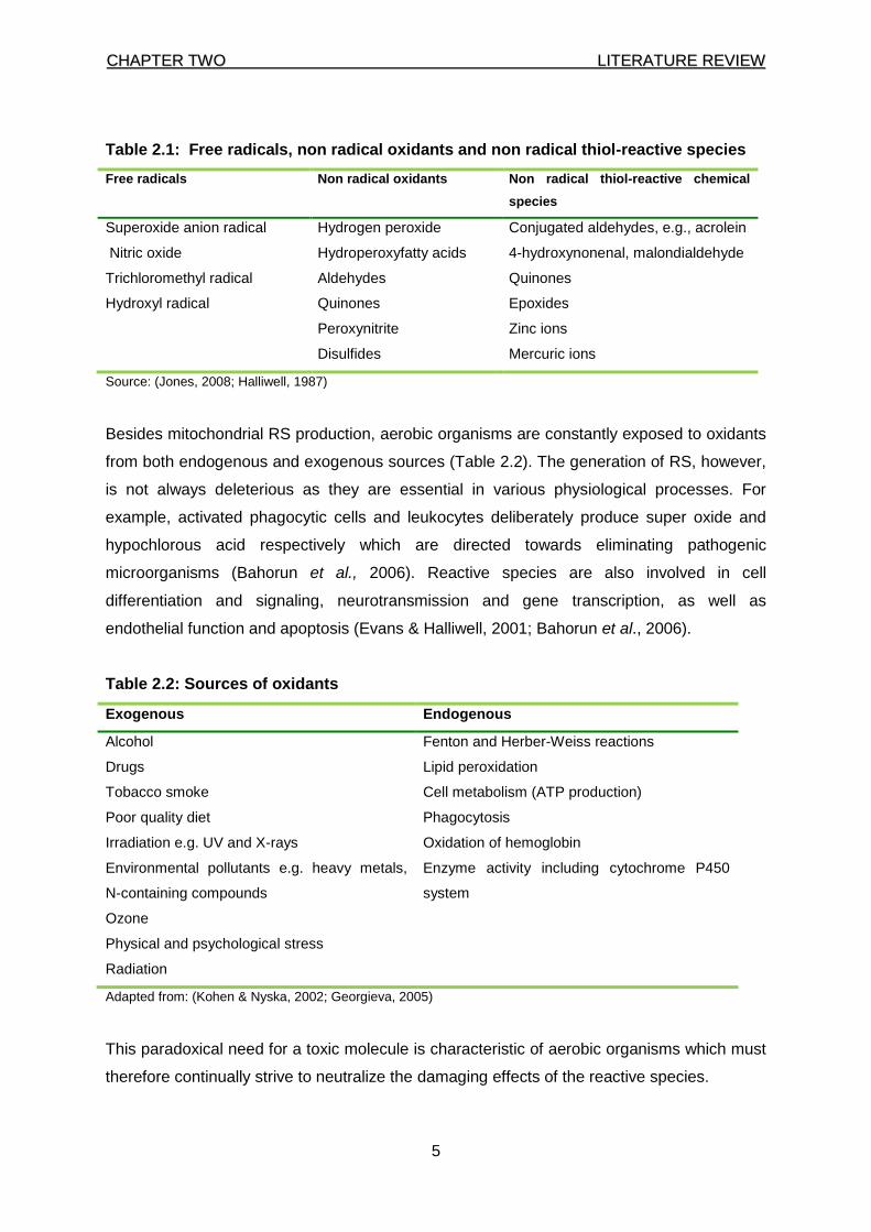
Table 2.1: Free radicals, non radical oxidants and non radical thiol-reactive species
Free radicals Non radical oxidants Non radical thiol-reactive chemical
species
Superoxide anion radical Hydrogen peroxide Conjugated aldehydes, e.g., acrolein
Nitric oxide Hydroperoxyfatty acids 4-hydroxynonenal, malondialdehyde
Trichloromethyl radical Aldehydes Quinones
Hydroxyl radical Quinones Epoxides
Peroxynitrite Zinc ions
Disulfides Mercuric ions
Source: (Jones, 2008; Halliwell, 1987)
Besides mitochondrial RS production, aerobic organisms are constantly exposed to oxidants
from both endogenous and exogenous sources (Table 2.2). The generation of RS, however,
is not always deleterious as they are essential in various physiological processes. For
example, activated phagocytic cells and leukocytes deliberately produce super oxide and
hypochlorous acid respectively which are directed towards eliminating pathogenic
microorganisms (Bahorun et al., 2006). Reactive species are also involved in cell
differentiation and signaling, neurotransmission and gene transcription, as well as
endothelial function and apoptosis (Evans & Halliwell, 2001; Bahorun et al., 2006).
Table 2.2: Sources of oxidants
Exogenous Endogenous
Alcohol Fenton and Herber-Weiss reactions
Drugs Lipid peroxidation
Tobacco smoke Cell metabolism (ATP production)
Poor quality diet Phagocytosis
Irradiation e.g. UV and X-rays Oxidation of hemoglobin
Environmental pollutants e.g. heavy metals,
N-containing compounds
Enzyme activity including cytochrome P450
system
Ozone
Physical and psychological stress
Radiation
Adapted from: (Kohen & Nyska, 2002; Georgieva, 2005)
This paradoxical need for a toxic molecule is characteristic of aerobic organisms which must
therefore continually strive to neutralize the damaging effects of the reactive species.
CCHHAAPPTTEERR TTWWOO LLIITTEERRAATTUURREE RREEVVIIEEWW
6
The neutralization is achieved by a complex antioxidant defense system made of various
endogenous and exogenous components (Table 2.3). If the oxidative damage/products
formation exceeds the capacity of the defense system (antioxidant system- Table 2.3), it
may initiate and/or hasten disease processes of e.g. cardiovascular disease (CVD), cancer,
diabetes as well as neurological, immune and ocular diseases (Table 2.4, on page 7).
Increased oxidation of low density lipoprotein (LDL), for example, is suggested as a
mechanism linking oxidative stress with CVD (Willcox et al., 2004; Vogiatzi et al., 2009),
while direct damage to DNA, suppression of apoptosis by RS and oxidative damage to DNA
repair enzymes may contribute to carcinogenesis (Halliwell, 2007; Oka et al., 2008).
Table 2.3: Components of the antioxidants defence system
Endogenous Exogenous
Non enzymatic Carotenoids e.g. β-carotene
Thiols (glutathione, lipoic acid,
N-acetyl cysteine) Vitamin C
Ubiquinones (coenzyme Q10) Vitamin E
Uric acid Plant phenols (flavonoids and other phenols)
Bilirubin Metals (copper, manganese, selenium and Zinc)
Tranferrin
Albumin
Enzymes
Catalase
Glutathione peroxidase
Glutathione S-transferase
Glutathione reductase
Superoxide dismutase
Adapted from: (Willcox et al., 2004; Valko et al., 2007; Halliwell, 2009)
2.2 Pathological conditions associated with oxidative stress
Elevated oxidative damage has been associated with numerous pathological conditions as
shown in Table 2.4 on page 7, (Halliwell, 1987; Haddad, 2002; Harrison et al., 2003; Willcox
et al., 2004; Han et al., 2006; Valko et al., 2007; Wood & Granger, 2007). Although a clear
correlation between disease and oxidative stress has not been proven for most of these
conditions, this link appears plausible in several conditions. A few of those relevant to the
current study are briefly discussed here.
CCHHAAPPTTEERR TTWWOO LLIITTEERRAATTUURREE RREEVVIIEEWW
7
Table 2.4: Diseases and conditions associated with oxidative stress
Aging Malnutrition
Atherosclerosis Multiple sclerosis
Arthritis and inflammatory diseases Neonatal lipoprotein oxidation
Cancer Neurodegenerative diseases
Cardiovascular diseases Pancreatitis
Diabetes Pulmonary dysfunction
Eclampsia Renal diseases
Eye diseases (cataracts) Shock and ischaemia
Liver dysfunction (non alcoholic fatty liver diseases) Sickle-cell anaemia
Skin lesion
2.2.1 Cancer and oxidative stress
Cancer is a collective term for diseases in which a group of cells derived from one cell grow
in uncontrolled manner due to the loss of control in the normal cell growth mechanism and/or
development (Gallagher et al., 2009; American Cancer Society, 2011). Cancer is thought to
have a genetic basis where changes introduced by various carcinogens (e.g. tobacco,
sunlight and infections) may produce mutations that affect normal cell growth and division
(Brash & Cairns, 2009; American Cancer Society, 2011).
According to the World Health Organization (WHO), cancer is the world’s second leading
cause of death after CVD (WHO, 2007). About 40% of all cancer deaths may be avoided by
taking preventive measures such as immunization against hepatitis B and human papilloma
virus and by choosing better lifestyle habits such as eating a healthy diet, exercising
regularly and eliminating exposure to carcinogens such as cigarette smoking and smoke.
Cancer progression is a stepwise process where the initiated cells evolve further and
become progressively more malignant. Oxidative stress is widely believed to play an
important role in the various stages of cancer including initiation, promotion and progression.
Although the exact mechanism remains unclear, human studies support the hypothesis that
oxidative DNA damage is an important mutagenic and carcinogenic trigger (Giustarini, et al.,
2009). The division of cells with oxidatively damaged DNA can result in mutations which, if
mutagenic, can lead to cancer. Although all four bases can be modified by RS, mutations are
CCHHAAPPTTEERR TTWWOO LLIITTEERRAATTUURREE RREEVVIIEEWW
8
usually associated with changes in guanine (G) cytosine (C) pair while that of adenine (A)
thymine (T) seldom result in mutations (De Bont & Van Larebeke, 2004). The most frequent
mutations are base pair substitutions, while deletions and insertions are less common.
Evidence for RS involvement in the initiation of cancer in humans is further reinforced by the
presence of oxidative DNA modifications in cancer tissue (De Bont & Van Larebeke, 2004).
Cigarette smoking is by far the main contributor to the development lung cancer which is the
most common cancer globally in terms of both incidence and mortality (Rudin et al., 2009).
Cigarette smoke, a rich source of RS and known carcinogens (e.g. nitrosamines and
polycyclic aromatic hydrocarbons), has been consistently associated with increased
accumulation of 8-hydroxydeoxyguanosine (8- OHdG), a marker of oxidative stress, in lungs
and urine which may be partly induced by FRs (Waris & Ahsan, 2006). Increased RS-
mediated DNA base damage has also been reported in breast cancer thus suggesting an
involvement of oxidative damages in the pathologies of these cancers. Similarly, 8-OHdG
has also been shown to accumulate in liver cancer (Waris & Ahsan, 2006). This cancer is
often linked to chronic infection with hepatitis B or C viruses or ingestion of aflatoxins and the
oxidative stress induced by these viruses and/or agents is thought to be one of the
intracellular carcinogenic triggers (Wild & Hall, 2000). G→T transition has been shown to be
one of the more common types of mutations produced by aflatoxin lesion and RS damage to
DNA (Waris & Ahsan, 2006).
Measuring markers of oxidative status is useful for monitoring cancer development. Several
studies have consistently reported a strong correlation between cancer and levels of
biomarkers of oxidative stress. For example, Manimaran & Rajneesh (2009) reported low
levels of catalase (CAT), superoxide dismutase (SOD), vitamin C and vitamin E in blood of
ovarian cancer patients. Another study found that malondialdehyde (MDA) levels were
higher in hepatocytes with oxidative damage (Lu et al., 2008). Other studies, however, did
not find a correlation between markers of oxidative stress and cancer. In a study assessing
the relation of oxidative stress to the risk breast cancer, urinary excretion of isoprostanes did
not differ between cases and controls, although, among overweight women, levels of
isoprostanes were positively associated with this type of cancer risk (Dai et al., 2009).
As there is no cure for cancer, prevention and control programmes including immunization,
reducing alcohol consumption/tobacco use and improving diet, are important measures to
decrease the burden. It is estimated that one third of human cancers are attributed to dietary
habits, and therefore dietary consideration is recognized as a plausible preventive strategy
CCHHAAPPTTEERR TTWWOO LLIITTEERRAATTUURREE RREEVVIIEEWW
9
against cancer (De Mejia et al., 2009). Moreover, chemotherapy and radiotherapy have
numerous side effects, which may impact on the quality of life.
According to the WHO (2007), cancer remains one of the leading causes of morbidity and
mortality worldwide. It is predicted that by 2020, the number of new cases of cancer in the
world will increase to more than 15 million, with deaths increasing to 12 million; much of this
burden will occur in the developing world (WHO, 2007). It is therefore imperative to devise
policies to halt the burden of chronic diseases including cancer. Such policies will include
lifestyle changes and modification of diet if there is to be any success in the global fight
against cancer as a public health problem (WHO, 2007).
Epidemiological data show an inverse correlation between tea consumption and cancer
prevalence. In one study evaluating the involvement of green tea compounds in the course
of prostatic cancer development, Bettuzzi and co-workers (2006) enrolled 60 men aged
between 45-70 years old with high-grade prostate intraepithelial neoplasia and reported a
positive effect on low urinary tract symptoms, suggesting potential benefits with
administration of tea polyphenols. One meta-analysis concluded that consumption of green
tea also protects against the development of breast cancer (Sun et al., 2006). Rooibos is
another herbal tea with reported antimutagenic properties. A study investigating the
modulating effect of rooibos and honeybush herbal tea on tumour promotion in mouse skin
reported a reduction of the mean number of tumors per mouse in the processed and
unprocessed rooibos groups, suggesting protective effect of this herbal tea (Marnewick et
al., 2005). Similar results were found in a study comparing the antimutagenic properties of
aqueous extracts of Aspalathus linearis (rooibos tea) and Camellia sinensis (green tea) (Van
der Merwe et al., 2006).
2.2.2 Chronic kidney disease and oxidative stress
Chronic kidney disease (CKD) is now a major cause of cardiovascular mortality and
morbidity globally (Beaglehole & Yach, 2003; Atkins, 2005; Vanholder et al., 2005). In South
Africa and other developing countries, the rise of CKD prevalence parallels that of diabetes
and hypertension and is expected to replace infectious diseases in the list of leading causes
of mortality and morbidity (Atkins, 2005).
There is ample evidence for increased oxidative stress in uremia - one of the key features of
CKD. In uremic patients, there is increased plasma protein oxidation (thiol residue oxidation
CCHHAAPPTTEERR TTWWOO LLIITTEERRAATTUURREE RREEVVIIEEWW
10
and carbonyl formation) and decreased circulating and intracellular antioxidants (Coppo et
al., 2010; KDOQI, 2000). Experimental data suggest that uremia induces tubulointerstitial
damage, while experimental proteinuric models such as puromycin-induced nephrosis have
been associated with advanced oxidation protein products (AOPPs) (Guo et al., 2008).
AOPPs induce intracellular superoxide generation in podocytes via mechanisms involving
NADPH oxidase (Coppo et al., 2010). Moreover, LDL from uremic patients was shown to be
more susceptible to oxidation in vivo than LDL from healthy volunteers (Maggi et al., 1994).
The oxidative stress in uraemia may result from the typical features of the CKD patient such
as advanced age, diabetes, chronic inflammation, malnutrition or as a consequence of
dialysis treatment. Several studies have shown that various indicators of oxidative stress e.g.
lipid peroxidation, AOPPs, 8- OHdG and F2-isoprostones are increased in CKD (Salomon et
al., 2000; Tarng et al., 2000; Handelman et al., 2001; Ikizler et al., 2002). Increased oxidative
stress may contribute to the excessive CVD incidence and mortality that accompany CKD.
An association between AOPP and carotid arteriosclerosis was reported in CKD patients, a
finding that was supported by a study evaluating AOPP as an independent risk factor for
coronary artery disease (CAD) in the general population (Kaneda et al., 2002). Furthermore,
oxLDL and plasmalogen were associated with increased cardiovascular mortality in patients
with advanced chronic kidney disease (CKD) (Bayes et al., 2003; Stenvinkel et al., 2004).
Although increased oxidative stress is associated with many complications of CKD (such as
amyloidosis, anaemia, hypertension and malnutrition), no large prospective epidemiological
studies have yet demonstrated a link between oxidative stress and patient outcome.
Several studies have investigated the potential benefits of tea beverages against kidney
disease. Uličná et al. (2006) reported a reduction of MDA levels in liver tissues, plasma and
lens of diabetic rats after administration of aqueous and alkaline extract of rooibos tea while
green tea catechins reduced haemodialysis-induced production of hydrogen peroxide and
hypochlorus acid (Hsu et al., 2007). Additionally, feeding male albino rats with green tea-
fortified bread improved kidney function against CKD induced by excessive dietary arginine
(El-Megeid et al., 2009). Green tea extract was also shown to stabilize gentamicin-induced
oxidative stress and histopathological abnormalities in rats (Abdel-Raheem et al., 2009).
Collectively, these findings suggest that rooibos herbal tea and green tea may be beneficial
dietary components with regard to renal health which therefore merits further investigations.
CCHHAAPPTTEERR TTWWOO LLIITTEERRAATTUURREE RREEVVIIEEWW
11
2.2.3 Coronary heart disease and oxidative stress
Cardiovascular disease (CVD), the leading cause of death globally, is a group of disorders
affecting the heart and blood vessels and includes coronary heart disease, cerebrovascular
disease, peripherial arterial disease, rheumatic heart disease, congenital heart disease,
deep vein thrombosis and pulmonary embolism. Coronary heart disease (CHD), the most
predominant form of CVD accounting for 42.1% of CVD mortality (WHO, 2009), results from
atherosclerosis.
Oxidative stress is now believed to play an important role in both initiation and progression of
the atherosclerotic process by enhancing the release of factors which lead to the attraction,
adhesion to the arterial wall and the ultimate differentiation of monocytes to macrophage
(Traverso, 2001; Vogiatzi et al., 2009). These factors include monocyte chemotactic protein-
1 (MCP-1), vascular cell adhesion molecule molecule-1 (VCAM-1) and macrophage colony-
stimulating factor (M-CSF). Firstly, the production of free radicals from a variety of sources
may trigger LDL oxidation which increases macrophage mobilization which in turn releases
superoxide as a defence mechanism. The release of superoxide by macrophages
aggravates LDL oxidation and consume NO by formation of peroxynitrite (NOO-) which may
lead to endothelial dysfunction via various processes (Vogiatzi et al., 2009). Further
exposure to superoxide from other sources, may activate the nuclear factor kB (NF- kB)
regulatory complex and activate the transcription of atherosclerosis-related genes, namely
tumour necrosis factor, VCAM-1, intracellular adhesion molecules-1 (ICAM-1), MCP-1,
matrix metaloproteinase-9 (MMP-9) and pro-coagulant tissue factor (Tardif, 2005; Vogiatzi et
al., 2009), this series of events results in the aggregation of macrophages into the arterial
wall which further incorporates oxidized LDL (Tardif, 2005).
A study investigating biomarkers of oxidative stress in patients between 45-60 years with
evidence of acute myocardial infarction reported an increase in levels of ceruloplasmin and
MDA, while levels of SOD, catalase, glutathione peroxidase, glutathione, vitamin C, vitamin
E and β-carotene were significantly decreased when compared to control subjects (Bansilar
et al., 2007; Pasupathi et al., 2009).
Like cancer, the prevalence and incidence of CVD is now a global phenomenon and not
limited to industrialised countries due to globalization and technological advancement (Singh
et al., 2002; WHO, 2009). According to the WHO (2009), approximately 80% of premature
CHD events can be prevented by modifying diet and lifestyle. The low CHD incidence in
Japan, China, Switzerland, Spain, and France is attributed to the diets consumed in these
CCHHAAPPTTEERR TTWWOO LLIITTEERRAATTUURREE RREEVVIIEEWW
12
countries (Stampfer et al., 2000). Epidemiological data have suggested that large intake
levels of vitamin E and vitamin C in a diet can lower the risk of coronary artery disease
(Shaikh & Suryakar, 2009). Generally, however, clinical trials have failed to demonstrate any
beneficial effect of the isolated use of vitamin E in primary or secondary prevention of CVD
in the general population (Gaziano, 2004; Lee et al., 2005; Farbstein et al., 2010). One
reason for this failure may be the fact that as is true for many pharmaceutical agents, vitamin
E supplementation would be predicted to show benefit only in those individuals in which it is
needed. This highlights the need for proper patient selection as was demonstrated in a study
investigating the cardio-protective effect of vitamin E supplementation in angiographically-
proven CVD patients. This study reported a lower incidence of the combined end-point (non-
fatal myocardial infarction and cardiovascular death) in patients taking 400 or 800 IU of
vitamin E per day (Nuttall et al., 1999). In contrast, a large trial with healthy women age 45
years or older with no previous history of cardio or cerebrovascular disease and no use of
aspirin or non-steroidal antinflammatory) failed to show any effect on CVD mortality (Lee et
al., 2005). These women were taking 600 IU of vitamin E from natural sources.
Plant polyphenols have attracted significant attention as cardio-beneficial agents. A study
investigating the protective effect of tea polyphenols in hypertensive rats reported a
protective effect against hypertension in the groups taking green and black tea (Negishi et
al., 2004). Persson and co-workers (2010) concluded that green herbal tea and rooibos tea
may have cardio-protective effects through the inhibition of angiotensin-converting enzyme
(ACE) activity as an oral intake of a single dose of rooibos significantly inhibited ACE activity
after 30 min and 60 minutes in healthy volunteers. When consumed for a longer period of
time by adults at risk for developing CVD, rooibos was recently reported to significantly
improve the lipid profile and redox status, both relevant to heart disease (Marnewick et al.,
2011). Similarly, green tea (daily consumption of 4 cups of green tea beverage or extracts)
was related to the reduction of plasma amyloid alpha, an independent CVD risk factor in
metabolic syndrome (Basu et al., 2011).
2.2.4 Liver disorders and oxidative stress
The liver is one of the most important organs in the body where it has numerous important
functions, including storage (e.g. glycogen), excretion and decomposition (e.g. erythrocytes)
synthesis (e.g. hormones, plasma proteins and cholesterol) and detoxification.
There are many kinds of liver disorders with different etiologies including infection, exposure
to toxic compounds, autoimmune processes, genetic defects or cancerous pathology.
CCHHAAPPTTEERR TTWWOO LLIITTEERRAATTUURREE RREEVVIIEEWW
13
Lifestyle factors (e.g. alcohol abuse, cigarette smoking) have contributed to the increase in
liver diseases in general. In 2008, more than 43,000 people died of liver disease in the
United States of America (American Liver Foundation, 2008). In developing countries,
figures may be underestimated due to limitations in screening and diagnosis/data collection
capabilities (Das et al., 2010). Although, the molecular pathways differ amongst the various
diseases, experimental and clinical evidences suggest a central role of oxidative stress in
the pathogenesis of many of them. The mechanisms underlying two main disorders will be
highlighted.
2.2.4.1 Non alcoholic fatty liver diseases (NAFLD)
NAFLD encompasses a range of conditions varying from simple steatosis to non-alcoholic
steatohepatitis (NASH), the extreme form of NAFLD. The mechanisms involved in the
development and pathogenesis of NAFLD are not completely understood but oxidative
stress and insulin resistance (IR) are believed to play a major role (Hijona et al., 2010).
Insulin resistance is a key feature of NAFLD as it leads to elevated serum free fatty acids
which provide fuel for accelerated triglyceride synthesis (Sanyal et al., 2001; Hijona et al.,
2010). Accumulation of triglyceride in the liver enhances oxidative stress in the hepatocytes
as has been shown in several animal models (Browning & Horton, 2004). Subsequently, a
strong association between severity of NASH and degree of oxidative stress has been
shown in animal models and human studies (Sanyal et al., 2001; Chalasani et al., 2004;
Yesilova et al., 2005). In the livers of NAFLD patients, for example, the level of 3-
nitrotyrosine, a marker of hepatic protein nitration, was found to be elevated when compared
to controls (Sanyal et al., 2001).
2.2.4.2 Hepatitis C viral infection (HCVI)
Hepatitis C virus (HCV) is a major cause of viral hepatitis which affects approximately
130−170 million people globally with approximately 350,000 deaths occurring per year due
to HCV-related liver disease (Shepard et al., 2005). Chronic HCVI often leads to severe liver
diseases including liver cirrhosis and liver cancer. The mechanism of the pathogenesis of
HCVI is unclear but as in NAFLD, oxidative stress has been strongly implicated as the
infection is characterized by increased markers of oxidative stress including lipid (MDA and
4-Hydroxynonenal [HNE]) and DNA (8-OHdG) damage products as well as decreased
antioxidant enzymes [SOD] (Li et al., 2004; Maki et al., 2007).
CCHHAAPPTTEERR TTWWOO LLIITTEERRAATTUURREE RREEVVIIEEWW
14
The elevated oxidative stress in HCV infection may be due to chronic inflammation which
enhances and sustains a continuous generation of RS via NAD(P)H oxidase 2 (NOX 2)
protein- mediated disruption of hepatic phagocyte function. This may also enhance systemic
oxidative stress since a damaged liver will not effectively export the key antioxidant–GSH-to
other tissues. Additionally, the excess iron deposits which have been shown to accompany
HCV infections are likely to promote further generation of free radicals via the Fenton
reaction. Finally, it has been shown that HCV can directly induce oxidative stress in
hepatocytes when its core protein associates with the outer mitochondrial membrane via its
COOH-terminal region. This association results in mitochondrial dysfunction by facilitating
Ca2+ accumulation which then impairs electron transport and promotes ROS production at
complex I of the electron transport chain (Choi & Ou, 2006).
The management strategies for liver disease range from prevention where this is possible
(hepatitis and alcoholic liver disease) to transplantation when the long term-consequences of
ongoing liver damage leave no other option. Prevention includes vaccination as well as
lifestyle and dietary changes (Riley III & Bhatti, 2001; WHO, 2008).
Several studies evaluating the hepato-protective effect of various beverages suggest a
beneficial potential for Aspalathus linearis and Camellia sinensis. A long term administration
of green tea stabilized activity of lactate dehydrogenate (LDH), gamma-glutamyl transferase
(GGT), acid phosphates (ACP), alkaline phosphatase (ALP) and bilirubin and simultaneously
decreased lipid peroxidation in adult Wistar rats (Hamden et al., 2009). Indeed, several
previous studies had earlier reported on the hepato-protective effect of epigallocatechin-3-
gallate (EGCG) – a component of green tea (Alessio et al., 2002; Bose et al., 2008). A
recent phase I clinical trial, however, concluded that a high dose of green tea polyphenol
(GTP) did not alter liver and kidney functions (Frank et al., 2009). Furthermore, two
unpublished cases of green tea hepato-toxicity were reported in a recent systematic review
although these were suggested to result from concurrent medication with diclofenac and
progestogens (Mazzanti et al., 2009).
Similar to green tea, rooibos herbal tea was also shown to enhance activities of important
phase II enzymes, glutathione-S-transferase–α and uridine 5'-diphospho-
glucuronosyltransferase, (Marnewick et al., 2003) and additionally, to sustain levels of the
endogenous antioxidant, coenzyme Q9 (Kucharská et al., 2004). More recently, Uličná and
co-workers (2008) showed that drinking rooibos tea prevented liver from large scale fibrosis.
CCHHAAPPTTEERR TTWWOO LLIITTEERRAATTUURREE RREEVVIIEEWW
15
Although, as stated earlier, a clear connection between disease and oxidative stress remains
to be established, the link appears probable in several conditions like the four discussed
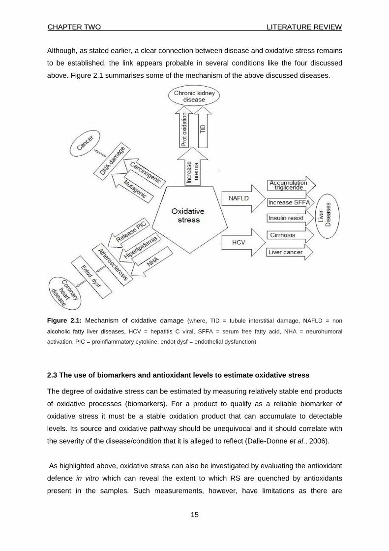
above. Figure 2.1 summarises some of the mechanism of the above discussed diseases.
Figure 2.1: Mechanism of oxidative damage (where, TID = tubule interstitial damage, NAFLD = non
alcoholic fatty liver diseases, HCV = hepatitis C viral, SFFA = serum free fatty acid, NHA = neurohumoral
activation, PIC = proinflammatory cytokine, endot dysf = endothelial dysfunction)
2.3 The use of biomarkers and antioxidant levels to estimate oxidative stress
The degree of oxidative stress can be estimated by measuring relatively stable end products
of oxidative processes (biomarkers). For a product to qualify as a reliable biomarker of
oxidative stress it must be a stable oxidation product that can accumulate to detectable
levels. Its source and oxidative pathway should be unequivocal and it should correlate with
the severity of the disease/condition that it is alleged to reflect (Dalle-Donne et al., 2006).
As highlighted above, oxidative stress can also be investigated by evaluating the antioxidant
defence in vitro which can reveal the extent to which RS are quenched by antioxidants
present in the samples. Such measurements, however, have limitations as there are
CCHHAAPPTTEERR TTWWOO LLIITTEERRAATTUURREE RREEVVIIEEWW
16
significant differences between the physiological environment and the in vitro assay
conditions; in addition, antioxidants present in the in vitro sample are metabolized in the
body which might alter their antioxidative potency.
Three categories of biomarkers-lipid, DNA and protein-will be discussed briefly in the
following section.
2.3.1 Lipid biomarkers
Lipids are biomolecules characterized by solubility in organic solvents but not in water. They
function in cellular metabolism, energy storage and signal transduction and as structural
components of cell membranes (Sherwood, 2010). In the body, the key lipid-rich points are
lipoproteins and cell membranes. Most lipids have an arrangement of fatty acids that are
composed of a chain of methyl groups with a carboxyl group at one end. Lipid peroxidation
(LPO) starts with the removal of a hydrogen atom from a methylene group next to a carbon
double bond producing a carbon-centred radical which is stabilised by a rearrangement of
the double bond to form a conjugated diene (CD) (Halliwell & Chirico, 1993). Oxygen, which
is abundant in the cell membranes, can easily react with the carbon-centred radical forming
the peroxyl radical which reacts further with another phospholipid/triglyceride-linked fatty
acid forming a hydroperoxy group and a new carbon-centred radical. Further reactions by
the lipid hydroperoxide will produce cyclic peroxide, cyclic endoperoxide and finally
aldehydes e.g. malondialdehyde (MDA). This sequence of reactions yields other important
products including aldehydes (MDA and 4-hydroxy-2-nonenal [4-HNE]), hydrocarbons
(pentane and ethane), transconjugated dienes, isoprostanes and cholesteroloxides
(Esterbauer et al., 1989; Abuja & Albertini, 2001; Hwang & Kim, 2007). In vivo, MDA, 4-HNE
and other aldehydes can form cross-linkages with DNA and proteins which alters the
function of these biomolecules. The aldehydes diffuse more readily than free radicals
thereby transferring the damage to distant parts of the body. The three most commonly
assessed LPO products (MDA, CDs and isoprostanes) will be discussed in this section.
As indicated above, the oxidation of polyunsaturated lipids releases several end products
including MDA. The measurement of MDA as thiobarbituric acid-reactive substances
(TBARS) is one of the most frequently utilized methods to assess LPO. In this assay, MDA is
heated with thiobarbituric acid (TBA) under acidic conditions and the resultant reaction yields
a relatively stable product that can be quantified by spectrophotometry (at 532 nm) or HPLC
(Halliwell & Chirico, 1993; Hwang & Kim, 2007). This method is easy and fast to perform but
it has attracted immense criticism for several reasons. First, it is not specific as many
CCHHAAPPTTEERR TTWWOO LLIITTEERRAATTUURREE RREEVVIIEEWW
17
products other than MDA (e.g. biliverdin, acetaldehyde, sucrose, reducing sugars and other
aldehydes) may react with TBA to yield products similar to the MDA-TBA adduct even in
peak absorbance. Secondly, there is no guarantee that the MDA measured is from oxidative
processes since degradation of fatty acids can occur in the heating step of the analysis.
Thirdly, MDA levels vary greatly even in blood samples from the same individual depending
on the anticoagulant used. Finally, it is difficult to compare results from the literature since
the exact intensity of the colour formed during the reaction depends on the type and strength
of the acid that is used yet different laboratories use different TBA assays. It is not surprising
therefore that the assay’s popularity is waning and is increasingly modified to include high-
performance liquid chromatography (HPLC) methodology where the MDA-TBA adducts is
separated from interfering chromophores thus improving specificity (Hwang & Kim, 2007).
Still, since lipid hydroperoxides and aldehydes are obtained from the diet and then excreted
in urine, measurements of MDA in plasma or urine is unreliable as an index of whole-body
LPO unless the diet is strictly controlled.
Conjugated dienes (CDs) are primary products of LPO which result from rearrangement of
the double bond when a hydrogen atom is abstracted from a fatty acyl methylene group
(Gϋmϋşlϋ et al., 1997; Kohen & Nyska, 2002). CDs are measured spectrophotometrically at
230–235 nm which in itself is a notable drawback since many other physiological substances
(e.g. poly unsaturated fatty acid-PUFAs) absorb in the same UV-range. Furthermore, there is
no suitable reference material available yet as this is essential to account for the PUFA
absorption. As in the case of MDAs, the diet should be strictly controlled since plasma CDs
content is >90% derived from 9, 11 diene-conjugated linoleic acid from dietary dairy products
(Romieu et al., 2008).
Isoprostanes (IsoP) are free radical oxidation products of arachidonic acid, esterified to lipids
then cleaved and released into the circulation by phospholipases. They are considered the
most consistent markers of LPO in vivo (Morrow, 2005). Up to 64 F2-isoprostanes can be
formed in vivo but 8-iso-IPF2a (15- F2-IsoP) is one of the more abundant and to-date one of
the most comprehensively studied biomarkers of oxidative stress (Lawson et al., 1999). At
present, F2-IsoPs analysis is commonly done by hyphenated chromatography as well as
enzyme- and radio immunoassays. Normal plasma levels are low as only a small proportion
of isoprostanes exist freely in plasma while the rest is esterified to lipids. Levels are
nonetheless elevated in situations like atherosclerosis where lipid peroxidation plays an
important role (Morrow, 2005). In plasma, however, isoprostanes have a short half life (about
18 minutes) and are excreted rapidly which poses an analytical dilemma as it is not possible
to measure them over a period of time. To measure isoprostanes formed over a given time
CCHHAAPPTTEERR TTWWOO LLIITTEERRAATTUURREE RREEVVIIEEWW
18
period, urine samples must be used although local kidney peroxidation presents a problem.
This is in turn can be overcome by measuring urinary levels of 2, 3-dinor 8-iso PGF1, the
metabolite of iso-PFG2a (Morrow et al., 1999). Like in the MDA and CDs assays, diet has a
confounding effect in the analysis of isoprostanes but this is circumvented by correcting for
levels of arachidonic acid (Gopaul et al., 2000).
2.3.2 DNA damage
Deoxyribonucleic acid (DNA) is a molecule often described as an organism’s blueprint as it
contains all the information used to manage the organism’s function, behaviour and
development. Despite relative stability, DNA may be attacked by RS producing damage
which, if not efficiently cleared/repaired, may lead to permanent modification that serves as
the first step in the path to mutagenicty (Cecarini et al., 2007). Oxidative damage to DNA is
mainly affected by the highly reactive hydroxyl radical (•OH) by addition to double bonds of
DNA bases and by abstraction of an H atom from the methyl group of thymine and C-H
bonds of 2-deoxyribose. The damage generates a range of DNA lesions, including strand
breaks and modified bases, many of which have potential to breach the integrity of the
genome (Dizdaroglu, 1991).
One of the most abundant and extensively studied modified bases is 8-oxo-7, 8-dihydro-2’-
deoxyguanosine (8-oxo-dG) which results from the oxidation of guanine. It pairs
preferentially with adenine thus generating guanine/cytosine (GC) to thymine/adenine (TA)
mutations after replication. This damage is usually repaired (mainly by base excision repair
[BER] enzymes) before the cell reaches replication phase (Jaruga & Dizdaroglu, 1996).
Variations in repair capability is thought to affect the amount of oxidative DNA damage as
several human diseases characterized by defects in DNA repair mechanisms (e.g.
xeroderma pigmentosum, ataxia telangiectasia, Bloom's syndrome and Fanconi's anaemia)
are also characterized by an elevated amount of 8-oxo-dG (Degan et al., 1995; Evans et al.,
2000). Other abundant oxidatively modified bases include formamidopyrimidine adducts of
adenine and guanine, e.g., 4,6-diamino- 5-formamidopyrimidine (FapyAdenine) and 2,6-
diamino-4-hydroxy-5-formamidopyrimidine (FapyGuanine) (Burgdorf & Carell, 2002) which is
readily formed in the absence of oxygen by ionizing radiation and other RS producing agents
(Ono et al., 1995). 5-Hydroxyuracil and uracil glycol are oxidative deamination products of
deoxycytosine (dC) and are detected in comparable levels to 8-oxo-dG in human DNA
(Burcham, 1999). The thymine analogue (thymine glycol) pairs with adenosine yielding a C
to T transition which is weakly mutagenic and it blocks transcription and replication (Basu et
CCHHAAPPTTEERR TTWWOO LLIITTEERRAATTUURREE RREEVVIIEEWW
19
al., 1989; Dianov et al., 2000; Marnett & Plastaras, 2001). Important pyrimidine-derived
lesions include 5-hydroxyuracil (5-OH-Ura) and 5-hydroxycytosine (5-OH-Cyt) the latter of
which is thought to be the most mutagenic product of oxidative DNA damage (Feig et al.,
1994). The hydroxyl radical (•OH) that is oxidized to produce these adducts plays a major
role in various DNA-protein interactions including the binding of transcription factors to DNA
which is therefore disrupted in this process (Rogstad et al., 2002). If not effectively cleared,
this non-mutagenic lesion can trigger apoptosis as a consequence of chromosomal
breakage thus indirectly leading to deletion mutations (Rogstad et al., 2002).
Several methods are available for the estimation of DNA damage including immunological
(Santella, 1999), adducts measurement by chromatography e.g. high-performance liquid
chromatography (HPLC) and gas chromatography-mass spectrometry (GCMS) (Phillips et
al., 2000) and comet assay (Gedik et al., 2002). All these may be easily done in various
samples including urine, plasma as well as tissue homogenates but the high cost of
measuring DNA adducts has limited the routine use of this approach (Phillips et al., 2000).
Immunoassays to detect DNA damage evolved from the hypothesis that DNA could become
immunogenic if linked to a carrier molecule. Several antibodies are now available
commercially for measurement of DNA adducts in urine but 8-oxo-dGuo is the target of most
assays since its presence is independent of dietary influence (Kadiiska et al., 2005). The
immunogen is fixed to the microplate wells and 8-oxo-dGuo competes with the solid-phase
antigen for the antibody. Free antibody is then bound to a peroxidase-labelled secondary
antibody thus, the lower the final absorbance the higher the concentration in the urine. The
assay has comparable sensitivity to HPLC of 8-oxo-dGuo (Phillips et al., 2000; Gedik et al.,
2002).
High-performance liquid chromatography (HPLC) is widely used for the measurement of 8-
oxo-dGuo in tissues or lymphocytes. Using appropriate non-ionic detergents and or
detergents, cells are lysed (or tissue homogenised), nuclei separated by centrifugation and
DNA finally released by adding sodium dodecyl sulphate followed by digestion of RNA and
proteins and ethanol precipitation. Samples are then separated on the column. Samples can
be stored frozen for many months but with caution to preserve viability especially in liquid
nitrogen or at -80 °C or, preferably, freezing after DNA extraction. Oxidation of guanine,
during any stage of sample preparation/storage, is a key concern but this can be minimized
by the use of antioxidants. The method is limited in that it does not definitely identify
individual adducts in a mixture (Phillips et al., 2000; Dotan et al., 2004).
CCHHAAPPTTEERR TTWWOO LLIITTEERRAATTUURREE RREEVVIIEEWW
20
The single–cell gel electrophoresis assay, also referred to as the comet assay, has emerged
as the standard method to measure DNA damage due to its robustness, simplicity,
sensitivity, cost effectiveness and safety as no labeling with radioisotopes and other
hazardous markers is necessary (Collins, 2004). It is essentially a method for detecting DNA
strand breaks but can be adapted to measure oxidized bases by incorporating a step to
recognize and remove damaged bases in the DNA (Collins, 2004). Besides the use in basic
DNA damage and repair research, the assay has found wide applications in genotoxicity
testing, nutritional studies, molecular epidemiology and ecogenotoxicology (Collins, 2004).
The assay’s main drawback has been the concern that the damage detected in leukocytes,
the preferred sample material for the assay, may not reflect the damage in the target tissues
(Collins, 2004). Nonetheless, the assay has a huge advantage over other assays requiring
extensive sample preparation in that there is little chance for spurious oxidation of guanine to
occur. Lymphocytes frozen according to protocols have a viability of at least one year
without significant increase in damage (Gedik et al., 1992; Singh, 2000; Collins, 2004).
Lymphocytes are embedded in agarose, lysed in a detergent then subjected to an alkaline
electrophoresis. Negatively charged DNA is attracted to the anode, but only the loops of
DNA possessing a break are free to migrate, resulting in an image appearing as a comet
when seen under the fluorescence microscope after suitable staining. As it is not possible to
include an internal standard, a standard lymphocyte sample should be run alongside the
experimental samples which can then be used for quantification especially in population
studies (Gedik et al., 1992; Singh, 2000; Collins, 2004; Kumari et al., 2008).
2.3.3 Protein biomarkers
Protein oxidation implies covalent modification induced either directly by RS or indirectly by
reaction with a by-product of oxidative stress. Such damage can impact on numerous protein
functions such as enzymatic and binding activities, increased susceptibility to aggregation
and proteolysis, increased or decreased uptake by cells, and altered immunogenicity
(Shacter, 2000). Although cysteine and methionine are the most susceptible to oxidative
attack due to their vulnerable sulfur atoms, all amino acid side chains can be modified via
one or more of the many protein oxidation mechanisms yielding numerous products as
shown in Table 2.5 (Hu, 1994; Vogt, 1995).
CCHHAAPPTTEERR TTWWOO LLIITTEERRAATTUURREE RREEVVIIEEWW
21
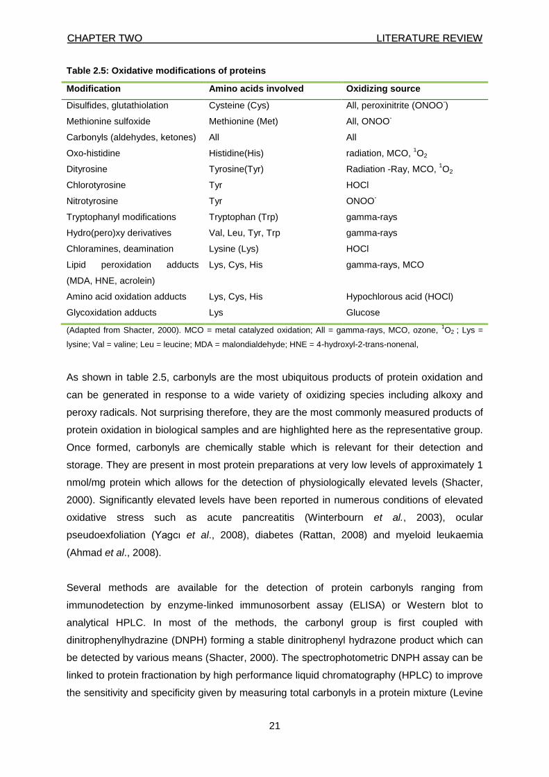
Table 2.5: Oxidative modifications of proteins
Modification Amino acids involved Oxidizing source
Disulfides, glutathiolation Cysteine (Cys) All, peroxinitrite (ONOO-)
Methionine sulfoxide Methionine (Met) All, ONOO-
Carbonyls (aldehydes, ketones) All All
Oxo-histidine Histidine(His) radiation, MCO, 1O2
Dityrosine Tyrosine(Tyr) Radiation -Ray, MCO, 1O2
Chlorotyrosine Tyr HOCl
Nitrotyrosine Tyr ONOO-
Tryptophanyl modifications Tryptophan (Trp) gamma-rays
Hydro(pero)xy derivatives Val, Leu, Tyr, Trp gamma-rays
Chloramines, deamination Lysine (Lys) HOCl
Lipid peroxidation adducts
(MDA, HNE, acrolein)
Lys, Cys, His gamma-rays, MCO
Amino acid oxidation adducts Lys, Cys, His Hypochlorous acid (HOCl)
Glycoxidation adducts Lys Glucose
(Adapted from Shacter, 2000). MCO = metal catalyzed oxidation; All = gamma-rays, MCO, ozone, 1O2 ; Lys =
lysine; Val = valine; Leu = leucine; MDA = malondialdehyde; HNE = 4-hydroxyl-2-trans-nonenal,
As shown in table 2.5, carbonyls are the most ubiquitous products of protein oxidation and
can be generated in response to a wide variety of oxidizing species including alkoxy and
peroxy radicals. Not surprising therefore, they are the most commonly measured products of
protein oxidation in biological samples and are highlighted here as the representative group.
Once formed, carbonyls are chemically stable which is relevant for their detection and
storage. They are present in most protein preparations at very low levels of approximately 1
nmol/mg protein which allows for the detection of physiologically elevated levels (Shacter,
2000). Significantly elevated levels have been reported in numerous conditions of elevated
oxidative stress such as acute pancreatitis (Winterbourn et al., 2003), ocular
pseudoexfoliation (Yagcı et al., 2008), diabetes (Rattan, 2008) and myeloid leukaemia
(Ahmad et al., 2008).
Several methods are available for the detection of protein carbonyls ranging from
immunodetection by enzyme-linked immunosorbent assay (ELISA) or Western blot to
analytical HPLC. In most of the methods, the carbonyl group is first coupled with
dinitrophenylhydrazine (DNPH) forming a stable dinitrophenyl hydrazone product which can
be detected by various means (Shacter, 2000). The spectrophotometric DNPH assay can be
linked to protein fractionation by high performance liquid chromatography (HPLC) to improve
the sensitivity and specificity given by measuring total carbonyls in a protein mixture (Levine
CCHHAAPPTTEERR TTWWOO LLIITTEERRAATTUURREE RREEVVIIEEWW
22
et al., 1990). The hydrazine carbonyl product can also be detected semi-quantitatively with
antibodies as has been done in ELISA and Western blot analysis which has the advantages
of high sensitivity, small sample volume, reproducibility and large sample throughput.
Samples have been shown to be viable for 3 months at – 80 °C (Dalle-Done et al., 2003).
2.4 Antioxidant defence system
In order to counteract the damaging effects of RS, living organisms have evolved a complex
network of antioxidants. An antioxidant is defined as any substance that delays, prevents or
removes oxidative damage to a substrate (Halliwell, 2007). Numerous substances have
been shown to have antioxidant properties in vitro which, however, do not necessarily
translate to in vivo activity as it may be lost in the course of metabolism. The antioxidant
potential of a particular substance is usually influenced by factors related to its uniqueness
including its antioxidant mechanism, substrate, site of action (extra- or intra-cellular) and the
concentration required for its activity (Halliwell, 2007).
Whether synthesized in the body (endogenous) or originating exogenously from the diet
(Table 2.3, page 6), antioxidants in the body form a synergistic defence system against
reactive species operating at three main levels (Rotilio et al., 1995; Willcox, 2004; Barkin &
Hersh, 2008):
(i) Preventive antioxidants that suppress the formation of reactive species
forming other components (e.g., reducing hydroperoxide to water).
(ii) Interceptors/scavengers that act against free radicals either physically or
chemically before they can damage cellular molecules (e.g., carotenoids
scavenging singlet oxygen; SOD convert superoxide to hydrogen peroxide
and phenolic /aromatic amines scavenging free radicals).
(iii) Those that repair damaged biomolecules e.g. transferases, lipases, proteases
and DNA repair enzymes.
2.4.1 Endogenous antioxidant defence system and oxidative stress
This category of antioxidants comprise of enzymes and non enzymatic (metal binding
proteins and small molecule) species as previously shown in Table 2.3, page 6. The
enzymes include catalase (CAT), glutathione peroxidase (GPx) and superoxide dismutase
(SOD) family, while the non enzymatic antioxidants (low molecular weight antioxidants
[LMWA]) include lipoic acid, glutathione (GSH), L-arginine, thioredoxin (TRX), coenzyme
CCHHAAPPTTEERR TTWWOO LLIITTEERRAATTUURREE RREEVVIIEEWW
23
Q10, melatonin, uric acid, billirubin, transferrin, ceruloplasmin, albumin and lactoferrin
(Percival, 1998; Pham-Huy et al., 2008). The antioxidant enzymes intercept RS before they
can react with biomolecules or other RS to generate more stable, long-acting oxidants. The
LMWA especially the metal binding proteins such as transferrin and ceruloplasmin, prevent
the occurrence of the Fenton reaction in which transition metals (e.g. iron) react with RS
producing the highly reactive hydroxyl radical (Young & Woodside, 2001). Glutathione,
another LMWA, is a tripeptide present in all eukaryote and prokaryote cells, synthesized by
enzymes, glutamate-cysteine ligase and glutathione synthetase. Glutathione plays a role in a
variety of physiological functions, including acting as a core member of the antioxidant
system and acting as a cofactor for GPx, a defence mechanism against peroxides forming
glutathione disulfide-oxidased glutathione (GSSG) that is further transformed to reduced
glutathione (GSH) in the presence of nicotinamide adenine dinucleotide phosphate-oxidase
(NADPH) and glutathione reductase (GR) (Livingstone & Davis, 2007). In homeostatic
conditions, the cellular ratio between GSH/GSSG is 100:1; however, during oxidative stress
this ratio may shift to 4:1, which is utilised as a biomarker of oxidative stress (Han et al.,
2006). Glutathione has numerous roles including scavenging FRs in an aqueous milieu,
acting as a cofactor in enzyme reactions, maintenance of intercellular communication,
preventing oxidation in –SH, chelating copper ions and participating in protein folding
degradation and cross-linking (Niki,1987; Kohen & Nyska, 2002; Pham-Huy et al., 2008).
2.4.2 Exogenous antioxidant defence system and oxidative stress
Exogenous (dietary) antioxidants are important components of the antioxidant system as
they complement the endogenous members. The most studied are α-tocopherol (vitamin E),
ascorbic acid (vitamin C), pro-vitamin A (β-carotene), flavonoids and trace minerals (see
table 2.3). Their mechanisms of action are varied but synergy between them is well
recognised as exemplified by the interactions between catechins, vitamin E and vitamin C.
The tocopheryl radical formed when vitamin E neutralises a free radical, accepts a hydrogen
ion from catechins or vitamin C and is restored back to the active α–tocopherol form (Kohen
& Nyska, 2002; Bouayed & Bohn, 2010). Several studies have reported elevated circulating
levels of vitamin E after a high dietary intake (Bendich et al., 1984; Burton et al., 1990;
Igarashi et al., 1991).
CCHHAAPPTTEERR TTWWOO LLIITTEERRAATTUURREE RREEVVIIEEWW
24
2.4.2.1 Carotenoids, vitamins and selenium
Carotenoids are a group of over 600 pigmented compounds principally found in plants where
they give the bright colours to leaves, fruits and flowers as well as the hue of some birds,
insects, fish and crustaceans (Palace et al., 1999). Animals cannot synthesize carotenoids in
vivo, but they ingest carotenoids in their diets where they may have various uses. Of the
approximately 40 carotenoids that form part of the human diet, lycopene and, especially, β-
carotene are the best studied. β-carotene, converted to retinol which is important for vision,
is proposed as an effective peroxyl radical scavenger (Paiva & Russell, 1999; Pham-Huy et
al., 2008). A daily recommended intake between 2500 and 5000 IU has been suggested
(Meyer, 2011; USDA, 2011) but the routine use as a supplement has raised some concerns
due to the relative increased risk of heart diseases (Pham-Huy et al., 2008). Rich dietary
sources of lycopene and β-carotene include tomatoes, carrots, fruits, spinach, grain and oils
(Paiva & Russell, 1999; Pham-Huy et al., 2008, Meyer, 2011; USDA, 2011).
Another antioxidant vitamin is vitamin C. The first report of the benefits of vitamin C was in
1747 when James Lind conducted a “clinical trial” using lemon and oranges for treatment of
scurvy (Bartholomew, 2002). It is considered a first line of defense as the major water
soluble antioxidant and acts synergistically with α–tocopherol by reducing α-tocopheroxyl
radicals rapidly to regenerate α-tocopherol and possibly inhibits α-tocopheroxyl-mediated
radical propagation. (Niki et al.,1995). Sources of vitamin C include fresh fruits, such as
lemon, orange, grape fruit, watermelon, papaya, mangos and many vegetables. In addition,
it is the most widely used vitamin supplement with a recommended intake range from 75-90
mg/day (Naidu, 2003).
The fat-soluble vitamin E exists in eight forms, namely; α-, β-, γ- and δ- tocotrienols and α-,
β-, γ- and δ tocopherols (Niki, 1995; Pham-Huy, 2008). Alpha-tocopherol is the most
bioactive of the isomers and has received much attention due to its radical-scavenging
capability in the lipophilic phase and alleged beneficial association with several diseases
such as cancer, CVD as well as ocular and inflammatory diseases (Pham-Huy et al., 2008;
Niki, 2010). Good dietary sources include vegetable oils, whole grain, fruits, grain, and meat
but the final content may be affected by cooking and storage conditions (Pham-Huy, 2008).
The daily reference intake ranges from 9-15 IU (7-10 mg) while the tolerable upper level (UL)
is about 1000 mg (Hathcock, 2005; Meyer, 2011; USDA, 2011).
Selenium is a mineral that plays key roles in physiological reactions as a co-factor for many
enzymes, including glutathione peroxidase and superoxide dismutases, hence enhancing
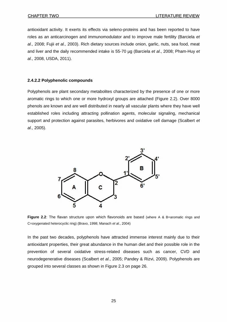
CCHHAAPPTTEERR TTWWOO LLIITTEERRAATTUURREE RREEVVIIEEWW
25
antioxidant activity. It exerts its effects via seleno-proteins and has been reported to have
roles as an anticarcinogen and immunomodulator and to improve male fertility (Barciela et
al., 2008; Fujii et al., 2003). Rich dietary sources include onion, garlic, nuts, sea food, meat
and liver and the daily recommended intake is 55-70 µg (Barciela et al., 2008; Pham-Huy et
al., 2008, USDA, 2011).
2.4.2.2 Polyphenolic compounds
Polyphenols are plant secondary metabolites characterized by the presence of one or more
aromatic rings to which one or more hydroxyl groups are attached (Figure 2.2). Over 8000
phenols are known and are well distributed in nearly all vascular plants where they have well
established roles including attracting pollination agents, molecular signaling, mechanical
support and protection against parasites, herbivores and oxidative cell damage (Scalbert et
al., 2005).
Figure 2.2: The flavan structure upon which flavonoids are based (where A & B=aromatic rings and
C=oxygenated heterocyclic ring) (Bravo, 1998; Manach et al., 2004)
In the past two decades, polyphenols have attracted immense interest mainly due to their
antioxidant properties, their great abundance in the human diet and their possible role in the
prevention of several oxidative stress-related diseases such as cancer, CVD and
neurodegenerative diseases (Scalbert et al., 2005; Pandey & Rizvi, 2009). Polyphenols are
grouped into several classes as shown in Figure 2.3 on page 26.
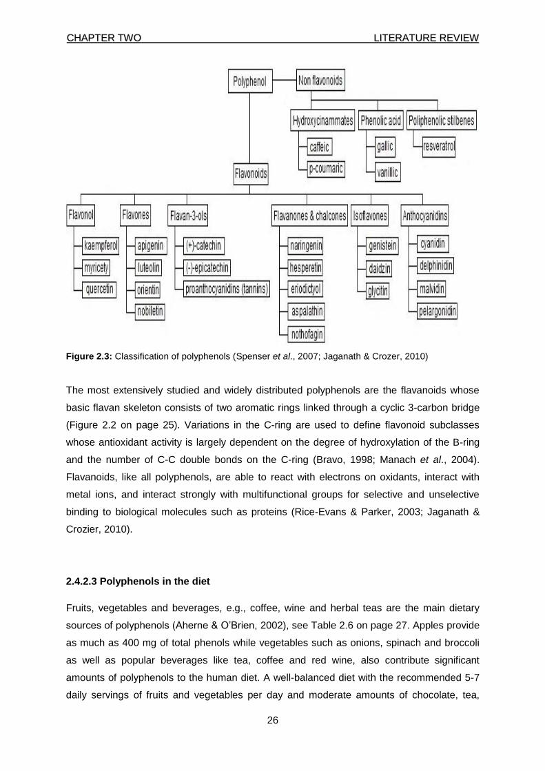
CCHHAAPPTTEERR TTWWOO LLIITTEERRAATTUURREE RREEVVIIEEWW
26
Figure 2.3: Classification of polyphenols (Spenser et al., 2007; Jaganath & Crozer, 2010)
The most extensively studied and widely distributed polyphenols are the flavanoids whose
basic flavan skeleton consists of two aromatic rings linked through a cyclic 3-carbon bridge
(Figure 2.2 on page 25). Variations in the C-ring are used to define flavonoid subclasses
whose antioxidant activity is largely dependent on the degree of hydroxylation of the B-ring
and the number of C-C double bonds on the C-ring (Bravo, 1998; Manach et al., 2004).
Flavanoids, like all polyphenols, are able to react with electrons on oxidants, interact with
metal ions, and interact strongly with multifunctional groups for selective and unselective
binding to biological molecules such as proteins (Rice-Evans & Parker, 2003; Jaganath &
Crozier, 2010).
2.4.2.3 Polyphenols in the diet
Fruits, vegetables and beverages, e.g., coffee, wine and herbal teas are the main dietary
sources of polyphenols (Aherne & O’Brien, 2002), see Table 2.6 on page 27. Apples provide
as much as 400 mg of total phenols while vegetables such as onions, spinach and broccoli
as well as popular beverages like tea, coffee and red wine, also contribute significant
amounts of polyphenols to the human diet. A well-balanced diet with the recommended 5-7
daily servings of fruits and vegetables per day and moderate amounts of chocolate, tea,
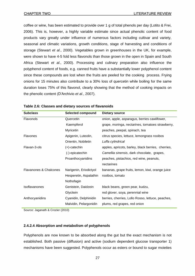
CCHHAAPPTTEERR TTWWOO LLIITTEERRAATTUURREE RREEVVIIEEWW
27
coffee or wine, has been estimated to provide over 1 g of total phenols per day (Lotito & Frei,
2006). This is, however, a highly variable estimate since actual phenolic content of food
products vary greatly under influence of numerous factors including cultivar and variety,
seasonal and climatic variations, growth conditions, stage of harvesting and conditions of
storage (Stewart et al., 2000). Vegetables grown in greenhouses in the UK, for example,
were shown to have 4-5 fold less flavonols than those grown in the open in Spain and South
Africa (Stewart et al., 2000). Processing and culinary preparation also influence the
polyphenol content of foods, e.g. canned fruits have a substantially lower polyphenol content
since these compounds are lost when the fruits are peeled for the cooking process. Frying
onions for 15 minutes also contribute to a 30% loss of quercetin while boiling for the same
duration loses 75% of this flavonol, clearly showing that the method of cooking impacts on
the phenolic content (D’Archivio et al., 2007).
Table 2.6: Classes and dietary sources of flavanoids
Subclass Selected compound Dietary source
Flavonols Quercetin
Kaempferol
Myricetin
onion, apple, asparagus, berries cawlifower,
grape, moringa, nectarines, tomatoes strawberry,
peaches, peepal, spinach, tea
Flavones Apigenin, Luteolin,
Orientin, Nobiletin
citrus species, lettuce, lemongrass rooibos
Luffa cylindrical
Flavan-3-ols (+)-catechin
(-)-epicatechin
Proanthocyanidins
apples, apricots, barley, black berries, cherries,
Camellia sinensis, dark chocolate, grapes,
peaches, pistachios, red wine, peanuts,
nectarines
Flavanones & Chalcones Narigenin, Eriodictyol
Hesperetin, Aspalathin
Nothofagin
bananas, grape fruits, lemon, kiwi, orange juice
rooibos, tomato
Isoflavanones Genistein, Daidzein
Glycitein
black beans, green pear, kudzu,
red glover, soya, perennial wine
Anthocyanidins Cyanidin, Delphinidin
Malvidin, Pelargonidin
berries, cherries, Lollo Rosso, lettuce, peaches,
plums, red grapes, red onion
Source: Jaganath & Crozier (2010)
2.4.2.4 Absorption and metabolism of polyphenols
Polyphenols are now known to be absorbed along the gut but the exact mechanism is not
established. Both passive (diffusion) and active (sodium dependent glucose transporter 1)
mechanisms have been suggested. Polyphenols occur as esters or bound to sugar moieties
CCHHAAPPTTEERR TTWWOO LLIITTEERRAATTUURREE RREEVVIIEEWW
28
(glycosides) and must first, with the exception of anthocyanins, be hydrolysed for absorption
to occur. Anthocyanins’ positive charge is thought to facilitate their direct absorption (Walle,
2004). The hydrolysis occurs chiefly in the colon and, to a much less extent, in the small
intestines and the oral cavity (Saura-Calixto et al., 2007).
The absorption of polyphenols is influenced by several factors including the molecular weight
and the extent and type of esterification. Proanthocyanidins in fruits and cocoa, for example,
are hardly absorbed in the gut due to their large molecular sizes while the extent of catechin
absorption decreases with increasing esterification with gallic acid (Holt et al., 2002; Scalbert
et al., 2002). The absorption of quercetin from onion is four fold greater than that from apples
which highlights the matrix effect on absorption (Hollman et al., 1997). Some studies have
reported the negating matrix effect of milk while others have refuted such claims asserting
that the observed influence may depend on the assay method used to estimate the
antioxidant effect (Arts et al., 2002; Roura et al., 2008; Sharma et al., 2008).
Once absorbed, polyphenols are bound to albumin and transported to the liver where they
are extensively metabolized after conjugation (glucuronidation, sulphation or methylation) or
catabolized to smaller phenolic compounds before excretion in urine or bile (Cabrera et al.,
2006; Mejia et al., 2009). These processes seem to vary with the type of polyphenols; the
flavonol EGCG, for example, is more likely to go through biliary excretion with a plasma half-
life of about 2-3 hours, while the flavanones, naringenin and hesperetin are mainly excreted
via urine (Mejia et al., 2009; Krogholm et al., 2010).
2.4.2.5 Mechanism of action of polyphenols
Table 2.7 on page 29, summarizes the proposed mechanisms via which polyphenols
function as antioxidants (Nijveldt et al., 2001; Rice-Evans & Parker, 2003; Manach, 2004;
Patel, 2008). They include:
(i) direct radical scavenging of free radicals. The hydroxyl group in the B ring
allow polyphenols to react with superoxide or with nitric-oxide resulting in a
less reactive radical or its inactivation;
(ii) interference with inducible nitric-oxide synthase activity;
(iii) inhibition of xanthine oxidase activity; flavonols pathway is a xanthine
oxidase, and xantine oxidase and xantine dehydrogenase play a cardinal role
in a transformation of xanthine to uric acid;
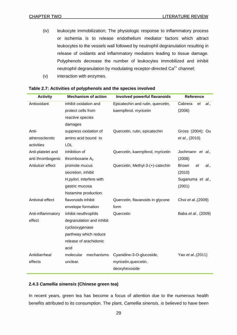
CCHHAAPPTTEERR TTWWOO LLIITTEERRAATTUURREE RREEVVIIEEWW
29
(iv) leukocyte immobilization; The physiologic response to inflammatory process
or ischemia is to release endothelium mediator factors which attract
leukocytes to the vessels wall followed by neutrophil degranulation resulting in
release of oxidants and inflammatory mediators leading to tissue damage.
Polyphenols decrease the number of leukocytes immobilized and inhibit
neutrophil degranulation by modulating receptor-directed Ca2+ channel;
(v) interaction with enzymes.
Table 2.7: Activities of polyphenols and the species involved
Activity Mechanism of action Involved powerful flavanoids Reference
Antioxidant inhibit oxidation and
protect cells from
reactive species
damages
Epicatechin and rutin, quercetin,
kaempferol, myricetin
Cabrera et al.,
(2006)
Anti-
atherosclerotic
activities
suppress oxidation of
amino acid bound to
LDL
Quercetin, rutin, epicatechin Gross (2004); Ou
et al., (2010)
Anti-platelet and
anti thrombogenic
inhibition of
thromboxane A2
Quercetin, kaempferol, myricetin Jochmann et al.,
(2008)
Antiulcer effect
promote mucus
secretion, inhibit
H.pylori, interfere with
gastric mucosa
histamine production.
Quercetin, Methyl-3-(+)-catechin Brown et al.,
(2010)
Suganuma et al.,
(2001)
Antiviral effect flavonoids inhibit
envelope formation
Quercetin, flavanoids in glycone
form
Choi et al.,(2009)
Anti-inflammatory
effect
inhibit neuthrophils
degranulation and inhibit
cyclooxygenase
parthway which reduce
release of arachidonic
acid
Quercetin Baba et al., (2009)
Antidiarrheal
effects
molecular mechanisms
unclear.
Cyanidine-3-O-glucoside,
myricetin,quercetin,
deoxyhexoside
Yao et al.,(2011)
2.4.3 Camellia sinensis (Chinese green tea)
In recent years, green tea has become a focus of attention due to the numerous health
benefits attributed to its consumption. The plant, Camellia sinensis, is believed to have been
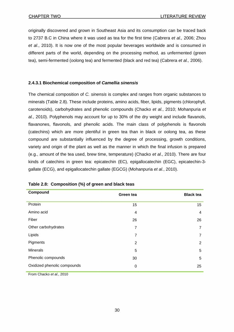
CCHHAAPPTTEERR TTWWOO LLIITTEERRAATTUURREE RREEVVIIEEWW
30
originally discovered and grown in Southeast Asia and its consumption can be traced back
to 2737 B.C in China where it was used as tea for the first time (Cabrera et al., 2006; Zhou
et al., 2010). It is now one of the most popular beverages worldwide and is consumed in
different parts of the world, depending on the processing method, as unfermented (green
tea), semi-fermented (oolong tea) and fermented (black and red tea) (Cabrera et al., 2006).
2.4.3.1 Biochemical composition of Camellia sinensis
The chemical composition of C. sinensis is complex and ranges from organic substances to
minerals (Table 2.8). These include proteins, amino acids, fiber, lipids, pigments (chlorophyll,
carotenoids), carbohydrates and phenolic compounds (Chacko et al., 2010; Mohanpuria et
al., 2010). Polyphenols may account for up to 30% of the dry weight and include flavanols,
flavanones, flavonols, and phenolic acids. The main class of polyphenols is flavonols
(catechins) which are more plentiful in green tea than in black or oolong tea, as these
compound are substantially influenced by the degree of processing, growth conditions,
variety and origin of the plant as well as the manner in which the final infusion is prepared
(e.g., amount of the tea used, brew time, temperature) (Chacko et al., 2010). There are four
kinds of catechins in green tea: epicatechin (EC), epigallocatechin (EGC), epicatechin-3-
gallate (ECG), and epigallocatechin gallate (EGCG) (Mohanpuria et al., 2010).
Table 2.8: Composition (%) of green and black teas
Compound Green tea Black tea
Protein 15 15
Amino acid 4 4
Fiber 26 26
Other carbohydrates 7 7
Lipids 7 7
Pigments 2 2
Minerals 5 5
Phenolic compounds 30 5
Oxidized phenolic compounds 0 25
From Chacko et al., 2010
CCHHAAPPTTEERR TTWWOO LLIITTEERRAATTUURREE RREEVVIIEEWW
31
2.4.3.2 Bioavailability and bioactivity of Camellia sinensis
The potential health effects of green tea depend not only on the amount of tea consumed
but also on the bioavailability of its bioactive constituents. The four main green tea catechins
have been detected in rat portal vein after oral ingestion indicating that tea catechins are
absorbed from the intestine (Okushio et al., 1996). In rats given 0.6% green tea polyphenols
(GTP) in their drinking water for 4 weeks, plasma levels of EGCG were much lower than
those of EGC or EC. But in mice given the same extract, plasma levels of EGCG were
much higher than those of EGC and EC. These findings suggest that there may be species
differences in the bioavailability of EGCG compared to the other catechins (Kim et al., 2000).
In humans, catechin levels in plasma reach their peak 2 to 4 h after ingestion but there is
little published data regarding their distribution in tissues. In the study mentioned earlier
where rats were given 0.6% GTP, significant levels of EGC and EC were found in the
esophagus, large intestine, kidney, bladder, lung, and prostate. In contrast, EGC and EC
levels were comparatively low in liver, spleen, heart, and thyroid; EGCG levels were higher
in the esophagus and large intestine, but lower in other organs, perhaps as a result of poor
systemic absorption of EGCG. Studies in rats have shown that EGCG is mainly excreted
through the bile, while EGC and EC are excreted via urine and bile. Evaluation of the actual
bioavailability of metabolites in tissues may be more crucial than simply evaluating their
levels in plasma, but data are still very limited even in animals. Although many health
benefits have been attributed to green tea consumption since ancient times, scientific
investigations on this beverage and its constituents have only intensified in the last three
decades (McKay & Blumberg, 2007). Many studies have indicated GTP possesses
antioxidant, antimutagenic, antidiabetic, antibacterial, anti-inflammatory, and
hypocholesterolemic properties (Cabrera et al., 2006).
Antioxidant activity is one of the key attributes of green tea. Green tea is rich in polyphenols
as well as other antioxidant components such as minerals. These compounds may increase
the tea’s antioxidant potential. Green tea contributes antioxidant activity in vitro by
scavenging oxidant molecules and chelating transition metal ions thus preventing their
participation in Fenton and Haber-Weiss reactions (McKay & Blumberg, 2007). They may
also function indirectly as antioxidants via several mechanisms such as inhibiting
redoxsensitive transcription factors, inhibiting of ‘pro-oxidant’ enzymes, such as inducible
nitric oxide synthase, and finally by induction of antioxidant enzymes, such as SOD (Cabrera
et al., 2006). In various human studies, intake of green tea has been associated with
increased antioxidant status and reduced oxidative damage in biomolecules. These reports
suggest that green tea contribute to defenses against oxidative damage (Wu & Wei, 2002).
CCHHAAPPTTEERR TTWWOO LLIITTEERRAATTUURREE RREEVVIIEEWW
32
Animal models and cell culture studies support a protective role for green tea against cancer
(Chung et al., 2003). There are reports that green tea inhibit carcinogenesis in various
organs including skin, lung, oral cavity, esophagus, stomach, liver, kidney and prostate
(Bianchi et al., 2000; Lambert & Yang, 2003). The antimutagenic effects of green tea are
hypothesized to be mediated by several factors including its antioxidant action, the specific
induction of detoxifying enzymes, its molecular regulatory functions on cellular signaling,
selective improvement in the function of the intestinal bacteria flora and breaking the
carcinogen-DNA links that lead to mutations (Cabrera et al., 2006). In women, green tea
drinkers showed a significantly reduced risk of breast and ovarian cancers (Zhang et al.,
2002; Wu et al., 2003). Similarly in men Jian et al. (2004) showed that the risk for prostate
cancer declined with increasing frequency, duration and quantity of green tea consumption
suggesting that the tea is protective against prostate cancer. Not all studies have been
supportive of green tea’s anti-cancer ability. Hoshiyama et al. (2004) and Koizumi et al.
(2003) found no association between green tea consumption and the risk of stomach cancer
and instead suggested implicated other factors such as age, smoking, socioeconomic status,
Helicobacter pylori infection, history of pectic ulcer, and family history of stomach cancer
along with certain dietary components. Clearly, more work is needed to clarify the anti-
mutagenic properties of green tea.
An inverse association of green tea with CVD has been shown in many epidemiological
studies but a mechanistic explanation for this observation has been firmly established. One
of the most widely considered explanation is that green tea may affect the cardiovascular
function by reducing the oxidation of LDL-cholesterol and related events such as foam cell
formation, endothelial cytotoxicity and induction of proinflammatory cytokines (McKay &
Blumberg, 2007). Others suggested mechanisms include increasing HDL-cholesterol levels
as well as reduction of plasma cholesterol levels and the rate of cholesterol absorption by
interfering with the micellar solubilization of cholesterol in the digestive tract (Raederstoff et
al., 2003). Green tea has been reported to have many other health roles including
antibacterial and antiviral activity, increasing bone mineral density (hence protecting against
fractures), anti-fibrotic properties on the skin and on the arteries (which is closely related to
the progression of liver fibrosis in chronic liver diseases), anti-inflammatory properties as
well as the preservation of the lens’s antioxidant system thus preventing progression of lens
opacity (Cabrera et al., 2006).
CCHHAAPPTTEERR TTWWOO LLIITTEERRAATTUURREE RREEVVIIEEWW
33
2.4.3.3 Toxicity
Green tea is generally considered a safe, non-toxic beverage and consumption is usually
without side effects. Several isolated cases, however, have been reported, which may be
related to caffeine and aluminum content of green tea and the interaction between
polyphenols and iron (Mennen et al., 2005; Mohanpuria et al., 2010). The average cup of
green tea contains from 10-50 mg of caffeine, and over-consumption may cause irritability,
insomnia, nervousness, and tachycardia. A study investigating the toxicity of green tea
extracts and their constituents in rat hepatocytes in primary culture associated high level of
green tea extract with hepatotoxicity attributed to low bioavailability of EGCG (Schmidt et al.,
2005). Ingestion of green tea on an empty stomach has also been associated with increased
activity of liver enzymes (Sarma et al., 2008).
2.4.4 Aspalathus linearis (Rooibos herbal tea)
Rooibos (Aspalathus linearis also known as a Aspalathus contaminata, Aspalathus
corymbosus, Borbonia pinifolia and Psoralea linearis) (Mckay & Blumberg, 2007) is a South
Africa indigenous shrub native to the Cedarberg Mountain and neighbouring areas in the
Western Cape province. It is characterized by a strong two metre-long taproot, reddish-
brown branches and bright green, needle-shaped leaves (Mckay & Blumberg, 2007; Van
Nierkerk & Viljoen, 2008). The native Khoi-khoi people were the first to use it as a beverage
three centuries ago due to the agreeable taste and aroma. Today, rooibos is marketed
globally for its suggested health benefits (Joubert et al., 2008; Van Nierkerk & Viljoen, 2008;
Marnewick, 2009). Two types of the herbal tea are produced depending on the processing
method chosen, fermented (red) and unfermented (green) rooibos. Fermented rooibos is
made by cutting the stems and leaves into fine pieces, wetting and piling them in heaps for
12-24 hrs to trigger chemical oxidation during which rooibos attains its characteristic aroma
and red colour. The oxidation/fermentation process may lead to a loss of important
components such as aspalathin and nothofagin which impacts on the overall antioxidant
capacity (Joubert, 1996; Mckay & Blumberg, 2007). “Green” or unfermented rooibos is made
by immediately oven-drying the fresh harvest to prevent oxidation before cutting into pieces
and sieving.
CCHHAAPPTTEERR TTWWOO LLIITTEERRAATTUURREE RREEVVIIEEWW
34
2.4.4.1 Biochemical composition of Aspalathus linearis
Several phenolic compounds have been isolated from rooibos including flavonols flavones
and dihydrochalcones as well as several phenolic acids including protocatechuic,
hydroxybenzoic, vanillic, coumaric, caffeic and ferulic acids (Joubert & Ferreira, 1996;
Bramati et al., 2003). Aspalathin, isoorientin, orientin and rutin are the most abundant
polyphenols in both the fermented and unfermented type of the herbal tea as shown in table
2.9. It is clear from the table that processing has a definite impact on the levels of these
polyphenolic compounds.
Table 2.9: Phenolic profile of fermented and unfermented rooibos
Compound Traditional/fermented rooibos
mg/g ± SD
Green/unfermented rooibos
mg/g ± SD
Chrysoeriol 0.022 ± 0.001 0.0079 ± 0.0004
Luteolin 0.029 ± 0.001 0.022 ± 0.002
Quercetin 0.107 ± 0.002 0.042 ± 0.006
Isovitexin 0.265 ± 0.002 0.659 ± 0.005
Vitexin 0.330 ± 0.002 0.504 ± 0.002
Isoquercitrin and hyperoside 0.429 ± 0.002 0.326 ± 0.006
Isoorientin 0.833 ± 0.007 3.57 ± 0.18
Orientin 1.003 ± 0.010 2.336 ± 0.049
Rutin 1.269 ± 0.006 1.69 ± 0.14
Aspalathin 1.234 ± 0.010 49.92 ± 0.80
Nothofagin* 0.17 1.08
Total polyphenols 5.521 ± 0.055 59.08 ± 0.59
Note: The extracts were prepared using 1 g of rooibos in 60 mL of hot distilled water, steeped for 10 minutes.
After removal of the tea leaves, the solution was cooled and filtered. The table gives the amounts of analytes in
mg per g of extract. *= from Joubert et al. (2008), S.D not given. Other values: adapted from Bramati et al.
(2003).
2.4.4.2 Bioavailability and mechanism of action of Aspalathus linearis
Consumed in its native South Africa for many years, rooibos tea has in recent years become
an increasingly popular health drink worldwide due to its low tannin content and caffeine free
status; subsequently, there has been a parallel increase in studies to substantiate the health
benefits attributed to this herbal beverage (Morton, 1983; Joubert et al., 2008). Recent
studies have shown rooibos tea to have anti-mutagenic, anti-carcinogenic, anti-inflammatory
and antiviral properties all of which are mainly attributed to the anti-oxidative properties of its
CCHHAAPPTTEERR TTWWOO LLIITTEERRAATTUURREE RREEVVIIEEWW
35
polyphenols which have a strong ability to quench radicals generated in the water phase and
confer high thermal stability against deep frying (Fukasawa et al., 2009).
The main polyphenol, aspalathin has only been found in rooibos. Similar to other phenolic
compounds, aspalathin is absorbed in the gut and undergoes bacterial action in the
intestines forming free aglycones. Recent studies investigating in vivo aspalathin metabolites
following oral administration of rooibos, found methylated aspalathin in the urine, but not in
plasma, 7 and 11 days after intake (Kreuz et al., 2008).
This finding was later corroborated by Stalmach and co-workers (2009) who demonstrated
O-linked methyl sulphate, glucuronide metabolites, O-methyl-aspalathin-O-glucuronide
(unfermented tea) and an eriodictyol-O-sulphate (following fermented tea) in human urine.
The principal mechanism of biotransformation includes glucuronidation, sulfation or
methylation of phenolic hydroxyl groups and the heterogeneity of metabolites may be due to
the restriction in bioavailability of some rooibos compounds as a result of chemical variability
(Bravo, 1998). More recently Breiter et al. (2011) have confirmed the bioavailability of
rooibos compounds in humans and identified methylated aspalathin as the main excreted
metabolite. In their study, seven metabolites of aspalathin and nothofagin were identified in
urine samples, as well as intact aspalathin and nothofagin. Sulphated, glucuronidated,
methylated, both glucuronidated and methylated aspalathin, and glucuronidates of the
aglycones of aspalathin and nothofagin were detected.
Similar to green tea, the fermentation process lowers the antioxidant potency of rooibos due
to the transformation of the two main dihydrochalcones, aspalathin and nothofagin. The bulk
of aspalathin is converted to orientin and iso-orientin which are both less efficient radical
scavengers in vitro (Joubert et al., 2004). Subsequently, higher antioxidant activities have
been shown in unfermented rooibos and green Camellia sinensis teas compared to their
fermented equivalents (Von Gadow et al., 1997a, Pazdzioch-Czochra & Widenska, 2002;
Schulz et al., 2003). Using the 2, 2-diphenyl-1-picrylhydrazyl (DPPH) radical test system, the
in vitro scavenging effectiveness of several key rooibos constituents was shown (Von
Gadow et al., 1997b; Joubert et al., 2004; Snijman, 2007). These included quercetin,
aspalathin, orientin, luteolin and isoquercitrin. Notably, aspalathin was more effective than α-
tocopherol, which is one of the most potent and important lipophilic antioxidants in the body.
A possible anti-atherosclerotic role for rooibos was suggested by the demonstration of a
protective effect of rooibos against lipid peroxidation using a mouse liver microsomal model
(Marnewick et al., 2005). Both “green” and fermented rooibos decreased MDA formation
CCHHAAPPTTEERR TTWWOO LLIITTEERRAATTUURREE RREEVVIIEEWW
36
substantially (91% and 65%, respectively) and also significantly increased the ratio of
reduced to oxidized glutathione (GSH: GSSG). Lipid peroxidation in mice liver was also
inhibited by purified orientin (Uma Devi et al., 2000) while rutin appeared to maintain the
strength of capillary walls (MacLennan et al., 1994). These findings were recently confirmed
in humans when Marnewick and colleagues (2011) reported that the consumption of
fermented rooibos significantly improved the lipid profile as well as redox status in adults at
risk for developing CHD. In the study, a significant increase in plasma total polyphenols
levels and GSH:GSSG ratio and a significant decrease in plasma markers of lipid
peroxidation after a daily consumption of six cups of rooibos for six weeks were observed.
Recently, Villano et al. (2010) assessed the effect of rooibos herbal tea intake on total
antioxidant capacity, lipid triacylglycerols, and cholesterol and glycaemia plasma levels in
humans. Although they did not find any change on the lipid profile, they were among the first
to show that both fermented and unfermented rooibos teas are able to boost plasma
antioxidant defences in humans.
Interestingly, a 10-week intake of rooibos by male Fisher 344 rats had no impact on the total
antioxidant capacity in the liver, but it significantly increased the GSH: GSSG ratio and
enhanced the activities of two xenobiotic metabolizing enzymes, GST-alpha and Uridine
Diphosphate glucuronyltransferase (UDP-GT) (Marnewick et al., 2003). These effects were
not shared by green and black teas. Later, histological analysis of livers from rats exposed to
the potent pro-oxidant, carbon tetra chloride (CCl4), showed that rooibos intake diminished
CCl4–induced hepatic steatosis and cirrhosis and decreased the production of hepatic MDA,
triacyglycerols and cholesterol as well as plasma aminotransferase, alkaline phosphatase
and bilirubin (Uličná, et al., 2003). In one of the first reports mentioning the effect of rooibos
on glutathione status and lipid peroxidation in humans, Nikolova et al. (2007) investigated
the effect of rooibos on the antioxidant status in men occupationally exposed to lead.
Consumption of rooibos for 8-weeks was reported to significantly enhance plasma GSH
levels by 48% and to reduce LPO measured as MDA. In a recent study, Persson et al.
(2010) added another mechanism by which rooibos may benefit cardiovascular health. In the
study where healthy adults consumed 400 mL of various beverages, rooibos herbal tea was
found to inhibit angiotensin converting enzyme (ACE). Inhibitors of ACE are used as
treatment for hypertension and congestive heart failure as they decrease tension of blood
vessels and blood volume. An imbalance in adrenocorticosteroids is associated with
numerous CVD risk conditions including hypertension, metabolic syndrome, insulin
resistance and type 2 diabetes. In a very recent study, the inhibitory effects of rooibos,
aspalathin and nothofagin on adrenal steroidogenesis were investigated (Schloms et al.,
2012). Under both basal and stimulated conditions, the total amount of steroids produced in
CCHHAAPPTTEERR TTWWOO LLIITTEERRAATTUURREE RREEVVIIEEWW
37
H295R cells, a human adrenal carcinoma cell line was significantly decreased in the
presence of rooibos, aspalathin and nothofagin. These results suggest that rooibos and its
two main constituents may modulate adrenal steroids biosynthesis thus possibly contributing
to the alleviation of negative effects resulting from an elevation of these hormones.
The anticancer activities of rooibos have been well investigated and the beverage has been
shown to confer in vitro protection against genotoxicity induced by various agents in Chinese
hamster ovary cells (Sasaki et al., 1993), Chinese hamster lung fibroblasts (Edenharder et
al., 2002), mouse embryo fibroblast cells (Komatsu et al., 1994) and in the Salmonella
typhimurium assay (Marnewick et al., 2000). An assessment of the mutagenic activity of the
major flavonoids of rooibos reported luteolin and chrysoeriol as the most potent
antimutagens of rooibos (Snijman et al., 2007). Luteolin and quercetin have also been linked
to in vitro induced-apoptosis and reduced proliferation of thyroid and colon cancers
respectively although it is doubtful whether these activities can be extrapolated in humans
due to the low amounts of these two flavonols in rooibos (Yin et al., 1999; Mori et al., 2001).
Rooibos extracts were shown to inhibit tumour promotion in mouse skin and to protect
against fumonisin B1 promotion in rat liver model utilizing diethylnitrosamine as the cancer
initiator (Marnewick et al., 2005; Marnewick et al., 2009). More recently the chemoprotective
potential of rooibos was evaluated in esophageal cancer and the herbal tea was shown to
reduce the mean total papilloma size by 87%. The reduction correlated with the total
polyphenol (r = 0.79; P < 0.02) and fermentation of herbal beverages reduced the inhibitory
effects on papilloma development associated with a reduction in the polyphenolic
constituents (Sissing et al., 2011).
Rooibos has also been cited as an anti-hypertensive, anti-stress, anti diabetic, immune
stimulant, laxative and sedative agent although these claims are in various stages of
substantiation (Kawano et al., 2009; Villano et al., 2010; Marnewick et al., 2011; Sissing et
al., 2011; Schloms et al., 2012).
2.4.4.3 Toxicity
Although rooibos research has intensified in the last decade, most of the studies are in vitro
or animal models in design. A single case reports Salmonella contamination from rooibos
which was nonetheless suggested to be from lizard origin (Swanepoel, 1987). Generally, the
plant appears to have a low toxicity profile, and the herbal tea is a good option for those who
wish to drink a mild, non-stimulating or sedating tea.
CCHHAAPPTTEERR TTWWOO LLIITTEERRAATTUURREE RREEVVIIEEWW
38
Data from animal and human studies attest to the plant’s safety (Marnewick et al., 2003;
Marnewick et al., 2004; Marnewick et al., 2005; Khan & Gilani, 2006; Kreuz et al., 2008;
Marnewick et al., 2011). It should be noted, however, that these studies did not look at the
possible interaction with drugs likely to produce hepatotoxicity or nephrotoxicity which is a
potential scenario among patients of chronic or infectious diseases. An isolated but puzzling
case of hepatotoxicity was recently reported in a 42-year-old woman with Waldesnstrom’s
macroglobulinemia, a low-grade B-cell malignancy (Sinisalo et al., 2010). For maintenance,
she was on rituximab every 3 months and daily prednisolone and she was also taking co-
trimoxazole for pneumonia, alendronic acid and calcium carbonate with vitamin D as
prophylaxis for osteoporosis and daily supplements of potassium chloride. Two weeks prior
to a scheduled clinic visit she started drinking rooibos one litre per day. Routine evaluation
revealed grossly elevated liver enzymes which were restored by withdrawing rooibos and the
treatments. According to the authors, it is unknown whether this side effect was due to the
combination with one or several drugs or it was a reflection of the patient’s genetically
predisposed vulnerability to the drug or herbal tea or even that the rooibos used by the
patient was contaminated by some hepatotoxic compound, such as a pyrrolizidine alkaloid
(Sinisalo et al., 2010). This case clearly illustrates the need for more human research studies
on rooibos, and indeed all herbs, to firmly ascertain their toxicity profiles.
2.4.5 Combination of antioxidant supplements
The diet plays important roles in the pathophysiology of numerous diseases and there have
been many attempts to understand the precise mechanisms involved. Effective and
informative nutritional studies are difficult to conduct due to the qualitative and quantitative
complexity in food and food products, heterogeneity of study population as well as
differences in individuals’ dietary habits (Graham, 1992; Cabrera et al., 2006; Dewettinck et
al., 2008). In addition, it is often very difficult to identify the specific compound responsible
for any benefit seen in a given pathophysiological condition. The isolation of the individual
compounds may facilitate the assessment of the direct cause- and dose-effect relationship;
however, the absorption and bioavailability of the isolated compounds may be different in
situ. Indeed, the metabolites that reach tissues and cells are usually chemically and/or
functionally distinct from the dietary form (Kroom et al., 2004). The situation can be further
complicated if, as is often the case, the apparent benefit result from the synergistic
interaction between two or more compounds. Two well documented synergistic interactions
involve α–tocopherol and ascorbic acid as well as α–tocopherol and flavonoids (Pedrielli &
CCHHAAPPTTEERR TTWWOO LLIITTEERRAATTUURREE RREEVVIIEEWW
39
Skibsted, 2002). The tocopheryl radical (formed when vitamin E reacts with radicals) accepts
a hydrogen ion from vitamin C or flavonoids and in the process is regenerated to the active
vitamin E (Kadoma et al., 2006; Jacobs et al., 2010). Tantcheva et al, (2003) has reported
an improved protection against influenza virus infection when vitamins C and E are
administered as a combination in experimental animals.
Various antioxidant combinations have been formulated and studied in different situations
and populations. One study investigated the efficacy of caffeine and catechin-rich green tea
extract (GTE) in energy expenditure (EE) and fat oxidation in humans. The supplementation
resulted in a significant increase in EE and 24-hours urinary nor-epinephrine excretion as
well as a decrease in respiratory quotient (RQ). When caffeine equivalent to the amount in
the supplement was given alone, there was no change in the EE or RQ suggesting an
interaction between the two components (Dulloo et al., 1999). In a more recent study, the
ant-inflammatory effect of a dietary mix of resveratrol, GTE, tomato extract, α-tocopherol,
vitamin C and n-3 (omega-3) polyunsaturated fatty acid was investigated in overweight men.
Plasma levels of adiponectin, an anti-inflammatory and anti-diabetic hormone secreted by
adipocytes were increased by 7% while C-reactive protein was unaffected. Oxidative stress
biomarkers (isoprostanes and uric acid) were decreased and endothelial function was
improved (Bekker et al., 2010). Similarly, in a double-blinded trial enrolling 1078 HIV-infected
pregnant women, multivitamin supplements were able to delay the progression of the
infection as well as the mortality (Fawzi et al., 2004).
Collectively, these data suggest an increased effectiveness of combined antioxidants. Such
data has led to the steady rise in the use of dietary supplements in the modern, health-
conscious society. In the USA for example, over one-half of the adult population take one or
more supplements everyday (Gahche et al., 2011). However, the toxicity of supplements and
isolated compounds has always been a concern and results from several studies caution
against their excessive usage. Vitamin C, for example, may lead to lung cancer in smokers
and people with asbestosis at intakes above 33 000 UI/day or it may lead to yellowing of the
skin, diarrhoea, arthralgia and gastric upset at intake of 2 000 mg/day (Wooltorton, 2003).
The hypothesis that moderate levels of free radicals induce an endogenous response which
protects against exogenous radicals (and possibly other toxic compounds) and prolong the
life span has recently been shown in Caenorhabditis elegans (Tapia, 2006; Schulz et al.,
2007; Ristow & Zarse, 2010). The paradoxical implication here is that excessive intake of
antioxidants may prevent life-extending and other health-promoting effects.
CCHHAAPPTTEERR TTWWOO LLIITTEERRAATTUURREE RREEVVIIEEWW
40
The supplements (refers to page 41 for composition) used in this study are new products
thus there is no data available regarding their beneficial or harmful effects. Data regarding
these supplements will be a useful output from this study and will compare the supplements
against two relatively well investigated beverages.
CCHHAAPPTTEERR TTHHRREEEE MMAATTEERRIIAALLSS AANNDD MMEETTHHOODDSS
41
CHAPTER THREE
MATERIALS AND METHODS
3.1 Study design and ethical approval
This was an experimental intervention study organised in two phases; which included
1. quantification of polyphenols and antioxidant activity of the various beverages used in
the study. The beverages included rooibos (Aspalathus linearis), Chinese green tea
(Camellia sinensis) and two tea supplements,
2. assessment of the effects of the beverages and supplements on t-butyl hydroperoxide-
induced-oxidative stress in the liver and kidney of male Wister rats .
Ethical approval was granted by the Faculty of Health & Wellness Sciences Research Ethics
Committee, Cape Peninsula University of Technology (Project number: CPUT/HWS-REC
2010/A001, Appendix A) as an extension of the previously approved project which results
are published in Awoniyi et al. (2011).
3.2 Preparation of beverages and supplements
Aqueous extracts of the various plant materials which included fermented rooibos
(“oxidised”), “green” rooibos (“unoxidised”), Chinese green tea, were freshly prepared by
adding 2 g of herbal tea to 100 ml of boiled tap water. The two commercial tea supplements
(one containing a green tea extract and the other containing a rooibos extract) were crushed
using a pestle and mortar, and prepared at a concentration of 2 g/100 mL. The concentration
of the rooibos and Chinese green tea used in this study were identical to the ones used in
previous studies (Marnewick et al., 2009; Sissing et al., 2011). The rooibos and Chinese
green teas were allowed to stand for 30 minutes at room temperature before filtering
(Whatman N 4), cooling and serving it to the experimental animals. Aliquots of the freshly
prepared rooibos, Camellia sinensis tea and supplements were also stored at -20°C for
quantitative antioxidant analyses. Fermented and “green” rooibos (superior grade) were a
general gift from Rooibos Ltd (Clanwilliam, South Africa), while the Chinese green tea as
well as the two commercial tea supplements were purchased from a local drug store (Cape
Town, South Africa). Each green tea capsule contained 100 mg of epigallocatechin gallate
(EGCG), while each rooibos tablet supplement contained: 175 mg of 20% aspalathin-rich
extract, 500 µg vitamin A, 150 mg vitamin C, 5 mg Vitamin E and 25 µg selenium.
CCHHAAPPTTEERR TTHHRREEEE MMAATTEERRIIAALLSS AANNDD MMEETTHHOODDSS
42
3.3 Experimental animals and diet
Sixty male Wistar rats obtained from the Animal Unit of the University of Cape Town (South
Africa) weighing 120-150 g were randomly allocated to one of the six study groups. Each
group consisted of ten rats and were maintained as described by Awoniyi (2010). Briefly,
animals were housed in Perspex houses with stainless steel wire-bottomed cages in a
controlled environment of 24-25°C with 12 hours light-dark cycles and 50% humidity for 1
week. The rats had free access to diet (standard rat chow) and to the various beverages
respectively (fermented and “green” rooibos, and Chinese green tea and tea supplement)
and tap water for the control group for 10 weeks as a sole source of drinking fluid. Body
weights were monitored twice a week till the end of the experiment. Oxidative stress was
induced in all intervention and positive control rats via an intraperitoneal (i.p) injection of 30
µM of t-butyl hyroperoxide per 100 g body weight in the final two weeks of the study. At the
end of the intervention, the animals (non-fasted) were sacrificed using pentobarbital
anaesthesia injected i.p. at 0.4 mL/kg body weight. Thereafter, livers and kidneys were
excised and immediately snap frozen in nitrogen and then kept at -80°C. Prior to analysis,
the various organs were minced, randomly weighed and homogenised according to the
specific assay protocol described below.
3.4 Analytical methods
3.4.1 Quantification of total polyphenol and flavonoid content of the aqueous herbal
tea and tea extracts and supplements
3.4.1.1 Total polyphenol content
The most frequently used method for total polyphenol determination is the Folin-Ciocalteu
(FC) assay which is based on the reaction of the aromatic group in polyphenols with Folin
Ciocalteu reagent (FCR) yielding a blue-coloured product that maximally absorbs at 765 nm
(Waterhouse, 2002). In alkaline conditions, phenol groups ionize completely to their
phenolate form which can be readily oxidized by the FCR changing colour from yellow to
blue. This colour change can be monitored with a spectrophotometer at 765 nm and
quantified using a standard. Several modifications have been made to the original Singleton
and Rossi (1965) method. This study used the method described by Waterhouse (2002).
Briefly, the frozen tea beverage and supplements samples were thawed and then diluted
ten-fold with distilled water. An 800 mg/L gallic acid stock standard solution was prepared by
dissolving 0.040 g of gallic acid (Sigma, SA) in 50 mL of 10% ethanol. This was used to
CCHHAAPPTTEERR TTHHRREEEE MMAATTEERRIIAALLSS AANNDD MMEETTHHOODDSS
43
prepare the standard series of 0, 20, 50, 100, 250 µg/L, and 500 µg/L using distilled water as
a diluent. Folin working solution was prepared by diluting 1 mL of FCR reagent (Merck, SA)
with 9 mL of distilled water. The reaction was done in a clear 96 well microplate, and the
mixture in each microplate well consisted of 25 μL of standard or sample, 125 μL FCR and
100 μL of Na2CO3 (7.5%) added after 5 minutes. The microplate was then incubated for 2
hours at room temperature before reading it in a Multiskan spectrum (Thermo Electron
Corporation, USA) set at 25°C and 765 nm. Data processing and calculations were done on
Microsoft® Excel 2003 based on a calibration curve plotted using the standard series and
absorbance readings. Values of samples were calculated using the regression equation
(Y=mX+C) between gallic acid concentration and absorbance, where X=unknown sample
concentration; m = slope, Y = absorbance of sample and C = Y-intercept. Results are
expressed as mg gallic acid equivalents per litre of beverage or supplement (mg GAE/L).
3.4.1.2 Flavanol content
The flavanols or flavan-3-ols are the most abundant family of bioactive compound present in
wine, green tea, red grapes, apples and nuts (Rice-Evans & Packer, 2003; Jaganath &
Croizier, 2010). In this study, they were estimated using the method described by Treutter
(1989) which is based on the reaction of 4-dimethylaminocinnamaldehyde (DMACA) with
flavanols to form a characteristic blue colour that is measured at 640 nm. Hydrochloric acid
32% (25ml) and absolute methanol 75% (HCl-MeOH), both from, Merck, SA, were used
(1:4); to prepare the DMACA solution 0.015 g of DMACA in 30 mL of HCl-MeOH solution.
The catechin standard was prepared by dissolving 0.0145 g of catechin hydrate in 50 mL
MeOH solution and then used as a stock solution to prepare the standard series (0, 1, 2, 5,
10 µg/L and 20µg/L). The various beverage samples were thawed and diluted ten-fold for
the assay which was done in triplicate. Each clear microplate well contained 50 µL of
sample/standard and 250 µL of DMACA solution and the plate was then incubated for 30
minutes at room temperature before taking the reading in the Multiskan spectrum (Thermo
Electron Corporation, USA) set at 25 0C and 640 nm. Data capturing and calculations were
done on Microsoft® Excel 2003 based on a calibration curve plotted using the standard
series and absorbance readings. Values of samples were calculated using the regression
equation (Y=mX+C) between catechin concentration and absorbance, where X = unknown
sample concentration; m=slope, Y = absorbance of sample and C = Y-intercept. Results are
expressed as mg catechin equivalents per litre of beverage or supplement (mg CE/L).
CCHHAAPPTTEERR TTHHRREEEE MMAATTEERRIIAALLSS AANNDD MMEETTHHOODDSS
44
3.4.1.3 Flavonol content
The estimation of the flavonol content in this study was done by the method described by
Mazza & co-workers (1999), which exploit the reaction of HCl with flavonols using quercetin
as a standard. Absolute ethanol (Merck, SA) was diluted to make 10% and 95% ethanol
solutions. Two different HCl concentrations (2% with distilled water and 0.1% with 95%
ethanol) were prepared. The standard series of 0, 5, 10, 20, 40, and 80 µg/L were freshly
prepared from stoke solution containing 4 mg quercetin (Sigma-Aldrich, SA) dissolved in 50
mL 95% ethanol. Beverage samples were diluted ten-fold with distilled water. In designated
wells of a clear 96 well microplate, 12.5 μL of the quercetin standard series and/or diluted
beverages were added to each well in triplicate, followed by 12.5 μL of 0.1% HCl in 95%
ethanol. Thereafter, 225 μL of 2% HCl was added and the plate incubated for 30 minutes at
room temperature before measuring the absorbance at 360 nm (25°C) on Multiskan
spectrum plate reader (Thermo Electron Corporation, USA). Data capturing and calculations
were done on Microsoft® Excel 2003 based on a calibration curve plotted using the standard
series and absorbance readings. Values of samples were calculated using the regression
equation (Y=mX+C) between quercetin concentration and absorbance, where X = quercetin
concentration; m = slope, Y = absorbance and C = unknown concentration. Results are
expressed as mg querectin equivalents per litre of beverage or supplement (mg QE/L).
3.4.2 Determination of the antioxidant capacity of the various tea and supplement
samples
The most frequently used methods to assess antioxidant capacity include ferric reducing
ability of plasma (FRAP), oxygen radical absorbance capacity (ORAC) and Trolox equivalent
antioxidant capacity (TEAC).
3.4.2.1 Ferric reducing antioxidant power (FRAP) determination
This assay was first described by Benzie & Strain in 1996 and assesses the ability of a
sample to reduce the 2,4,6-tri [2-pyridyl]-s-triazine (Fe3+) to the ferrous (Fe2+- TPTZ complex)
state at low pH forming an intense blue-coloured complex with optimal absorbance at 593
nm. The reaction is non-specific and any half-reaction with less-positive redox potential
under the reaction conditions than the Fe3+/Fe2+-TPTZ half-reaction, will drive Fe3+ reduction
(Benzi & Strain, 1996). Nevertheless, it is a simple measurement that gives fast,
reproducible results with plasma or tissue samples. Changes in absorbance are directly
CCHHAAPPTTEERR TTHHRREEEE MMAATTEERRIIAALLSS AANNDD MMEETTHHOODDSS
45
related to the reducing power of the electron-donating antioxidants present in the reaction
mixture. In the present study, a ten-fold dilution of the thawed beverage and supplement
samples was used, while 300 mg of the various tissues was homogenised (high-speed
blender from OMNI International TH, SA) with 1.2 mL of freshly prepared (75 mM phosphate
buffer, pH 7.0) and centrifuged (Eppendorf 5810R, Eppendorf, Germany) for 30 minutes,
4000 rpm (4°C). The buffer was constituted using sodium acetate and glacial acetic acid
(Merck, SA). To prepare the stock standard solution, 0.0088 g ascorbic acid (Sigma Aldrich,
SA) was dissolved in 50 mL distilled water and was used to prepare the standard series (0,
50, 100, 200, 500, 1000 µM) using distilled water as the diluent. The FRAP reagent was
prepared by mixing 30 mL of 300 mM acetate buffer, 3 mL of TPTZ solution (10 mM in 40
mM of HCl), 3 mL of ferric chloride (Merck, SA) solution (20 mM) and 6.6 mL of distilled
water to yield a straw coloured solution. Beverages, supplement and tissue samples were
diluted ten-fold. Each reaction well in a clear 96 well plate consisted of 10 µL of
standard/sample and 300 µL of FRAP reagent. The assay was done in triplicate. The plate
was then incubated at 37°C for 30 min before reading in Multiskan spectrum (Thermo
Electron Corporation, USA) set at 25°C and 593 nm. Data capture and calculations were
done in Microsoft® Excel 2003 based on calibration curve plotted using the standard series.
Values of samples were calculated using the regression equation (Y=mX+C) between
ascorbic acid concentration and absorbance, where X = unknown sample concentration; m =
slope, Y = absorbance of sample and C=Y-intercept. Results are expressed as a µmole
ascorbic acid equivalent per L of beverage, supplement or mg of tissue.
3.4.2.2 Oxygen radical absorbance capacity (ORAC) determination
The ORAC assay was first described in 1989 by Glazer based on the discovery that
fluorescence of phycoerythrin changes over time upon inhibition caused by peroxyl-radical
attack. Cao & co-workers (1998) modified the method to include the advantageous attribute
of driving the reaction to completion thus differing from Glazer’s method that only used “the
flat period”. In 2001, the method was further modified by Ou et al. to replace phycoerythrin
fluorescein as the fluorescent probe
Beverage and supplement samples were thawed and diluted twenty-fold (kidney ten-fold)
with phosphate buffer (75 mM, pH 7.4), while tissue samples (300 mg) were homogenised
(high-speed blender homogeniser from OMNI International TH, SA) in phosphate buffer
(75mM, pH 7.0) (ratio of 1:4 g/ml) and then centrifuged (Eppendorf 5810R, Eppendorf,
Germany) at 12 000 rpm, 10 min and 4°C, followed by deproteinization with 5% PCA (1:1)
CCHHAAPPTTEERR TTHHRREEEE MMAATTEERRIIAALLSS AANNDD MMEETTHHOODDSS
46
and centrifugation at 14 000 rpm, 4°C for 15 min. Di-sodium hydrogen orthophosphate
dehydrate and di-hydrogen orthophosphate-1-hydrate was obtained from Merck (SA),
fluorescein (FI) sodium salt, 2,2’-azobis (2-methylpropionamidine) dihydrochloride (AAPH)
and trolox were all obtained from Sigma-Aldrich (SA). A fluorescein stock solution was
prepared by dissolving 0.00225 g of the salt in 50 mL of 75 mM of phosphate buffer to yield a
0.0011 M solution. Thereafter, 10 µL of this solution was added to 2 000 µL of phosphate
buffer (75 mM, pH 7.4) and finally 240 µL from that solution was added to 15 mL of
phosphate buffer to make the working fluorescein solution. A standard stock solution of
trolox was prepared by dissolving 0.00625 g of trolox in 50 mL of phosphate buffer, aliquoted
into 2 mL eppendorf tubes and then frozen at –40°C to be used throughout the assays. A
fresh aliquot was used to prepare the standard series (5, 10, 15, 20, 25 µM) needed to
generate the calibration curve. The AAPH used as a radical generator was prepared by
dissolving 0.125 g of AAPH in 5 mL of phosphate buffer using a 15 mL screw cap conical
tube. Beverages, supplement and tissue samples were diluted twenty-fold (kidney ten-fold).
The assay was done in triplicate in a black 96-well microplate with each reaction well
containing 12 µL of sample or standard, 138 µL of FI and 50 µL of AAPH solution. The plate
was immediately read in a fluoroskan ascent (Thermo Electron Corporation, USA) reader at
37°C and programmed to record every 5 minutes for a period of 2 hours. Data capture and
analyses were done in Microsoft® Excel 2003 considering standard curve obtained by
plotting the different concentration of trolox standard and the area under the curve (AUC),
and the value of sample was calculated using regression equation; results expressed as
micromole trolox equivalent per millilitre of beverage or supplement (µmol TE/mL) or
milligram or µmol TE/mg) tissue, respectively.
3.4.2.3 Trolox equivalent antioxidant capacity (TEAC) determination
The original version of this method was described by Miller et al. in 1993 and was based on
the ability of antioxidant molecules to quench the long-lived 2-2’-azino-bis (3-
ethylbenzothiazoline-6-sulfonic acid (ABTS•+) radical, a blue–green chromophore with
absorption at 734 nm. The addition of an antioxidant reduces the preformed radical cation to
ABTS leading to its decolourization (Pellegrini et al., 2003). This has been criticized because
the faster reacting antioxidants might also reduce the ferryl myoglobin radical and the added
hydrogen peroxide could oxidize antioxidants before the measurement. To circumvent this
limitation Re & co-workers (1999) improved the technique by mixing ABTS with potassium
perssulphate such that the radical is generated directly in a stable form prior to the reaction
with the antioxidant. This stable stock solution of ABTS•+ was produced by adding 88 µl of
CCHHAAPPTTEERR TTHHRREEEE MMAATTEERRIIAALLSS AANNDD MMEETTHHOODDSS
47
140 mM potassium peroxodisulphate (Merck, SA) to an ABTS solution (0.0192 g dissolved in
5 mL distilled water) and allowing the mixture to stand in the dark at room temperature for 24
hours before use. Beverage and supplement samples were thawed at room temperature and
a ten-fold dilution used. For the tissues, 300 mg was homogenised (high-speed blender from
OMNI International TH, SA) in 1.2 mL of a freshly prepared phosphate buffer (75mM, pH
7.0) and centrifuged (Eppendorf 5810R, Eppendorf, Germany) for 30 minutes, 4000 rpm and
4°C. To prepare the trolox standard solution, 0.0125 g trolox was dissolved in 50 mL
absolute ethanol and used as a stock solution to prepare a standard series (0, 50, 100, 150,
250, 500 µM) using absolute ethanol as a diluent. The reaction mixture consisted of 25 µL of
each standard or sample (done in triplicate) and 300 µL of the ABTS mixture solution. The
clear 96 well microplates were left to incubate at room temperature for 30 minutes before
reading the absorbance in the Multiskan spectrum (Thermo Electron Corporation, USA) set
up at 25°C and 734 nm. Data analyses was done in Microsoft® Excel 2003 based on a
calibration curve plotted using standard series and the results expressed as µmole of trolox
per millilitre beverage or supplement sample or mg of tissue sample.
3.4.3 Antioxidant enzymes determination
3.4.3.1 Catalase (CAT)
Catalase mediates the removal of H2O2 from the cell by catalyzing the conversion of two
molecules of hydrogen peroxide to molecular oxygen and two molecules of water. The
estimation of catalase activity was assessed spectrophotometrically by the modified method
of Ellerby & Bredesen (2000) which is based on the reaction of catalase in the presence of
an optimal concentration of H2O2. The reaction mixture consisted of freshly prepared
phosphate buffer (50 mM Na-Pi, 0.5%, and Triton X-100, pH 7.5, both from Sigma, SA).
Hydrogen peroxide stock solution was made by mixing 10 mL potassium dihydrogen
orthophosphate (50 mM, pH 7.0) and 34 µL of 30 % hydrogen peroxide (Sigma, SA).
Tissues sample were prepared by homogenising (high-speed blender from OMNI
International TH, SA) 0.3 g of the sample in 2 mL of phosphate buffer. The homogenates
were then centrifuged (Eppendorf 5810R, Eppendorf, Germany) at 15 000 g for 10 minutes
at 4°C and the supernatant thereafter transferred to newly marked tubes and stored at -
70°C. A clear 96-well plate was used and each reaction well contained 170 µL of phosphate
buffer, 75 µL of hydrogen peroxide stock solution and 5 µL of sample (liver hundred-fold
dilution; kidney twenty-fold dilution) and then read in a Multiskan spectrophotometer
(Multiskan Spectrum, Thermo Electron Corporation, USA) at 25°C and 240 nm for two
CCHHAAPPTTEERR TTHHRREEEE MMAATTEERRIIAALLSS AANNDD MMEETTHHOODDSS
48
minutes. Data analysis was done in Microsoft® Excel 2007 and catalase activity in the
sample was calculated based on the rate of decomposition of hydrogen peroxide which is
proportional to the reduction of absorbance at 240 nm wavelength and results expressed as
a H2O2 µmole/min/ µg protein.
3.4.3.2 Glutathione peroxidase (GPx)
Using glutathione (GSH) as a reducing agent, GPx catalyzes the reduction of hydrogen
peroxide and organic peroxides (R-OOH) to water and the corresponding stable alcohol thus
inhibiting the formation of free radicals. The glutathione peroxidase activity was determined
by the method of Flohé & Gϋnzler (1984) by indirectly coupling the reaction with glutathione
reductase (GR). Oxidized glutathione (GSSG) produced upon the reduction of hydroperoxide
by GPx, is recycled to its reduced state, GSH by GR with the addition of NADPH. The
consumption of NADPH is directly proportional to the enzyme activity, which, in a sample
corresponds to a decrease in optical density which can be monitored at 340 nm. The assay
was freshly prepared using sodium phosphate (50 mM) and Triton X-100 (0.5%) at pH 7.5
(all from Sigma, SA). The GPx assay buffer was constituted with sodium phosphate buffer
50 mM and 1 mM EDTA, pH 7.0 (both from Sigma, SA). GSH solution (Boehringer
Mannheim, Germany) was made up by dissolving 30.7 mg GSH per ml of distilled water; 187
U/mL glutathione reductase (Sigma, SA) where after 1.7 µL of this mixture was diluted in
998 µL of assay buffer for a final solution of 0.1U/mL. Subsequently, 12.5 mg/ml of β-
NADPH was dissolved in phosphate buffer above described; hydrogen peroxide solution
(1.5mM), from a 30% commercial solution; sodium azide 0.0065 g, 100 mM (Sigma, SA) was
dissolved in 480 µL distilled water. Tissues were homogenized as described in the catalase
assay above. The GPx assay was done in triplicate in a clear 96-well microplate and each
reaction cocktail consisted of 210 µL buffer, 2.5 µL GSH, 2.5 µL NADPH, 2.5 µL sodium
azide, 2.5 µL glutathione reductase and 2.5 µL of sample (twenty- and two-fold dilution for
liver and kidney respectively). The plate was first read in the Multiskan spectrum (Multiskan,
Thermo Electron Corporation, USA) 340 nm (25°C) for 3 minutes and a second reading after
adding 50 µL hydrogen peroxide for 2 minutes. Data analysis was done on Microsoft® Excel
2003 and glutathione peroxidase activity in the sample calculated based on the rate of
decrease in absorbance at 340 nm wavelength over the time using the molar extinction
coefficient of glutathione peroxidase and corrected for pathlength.
CCHHAAPPTTEERR TTHHRREEEE MMAATTEERRIIAALLSS AANNDD MMEETTHHOODDSS
49
3.4.3.3 Superoxide dismutase (SOD)
This metallo-enzyme converts the highly reactive superoxide to the less reactive peroxide.
The rate of the reduction with O2 is linear to the xanthine oxidase (XO) activity and is
inhibited by SOD. The IC50 (50% inhibition activity of SOD or SOD-like material) can be
determined by colorimetric method. One unit of SOD is defined as the amount of enzyme
required to inhibit 50% of the rate of cytochrome C reduction (Flohé & Gϋnzler, 1984). The
method used in this study was described by Ellerby & Bredesen (1984). The phosphate
buffer and the preparation of tissues were as earlier described in the catalase assay. The
SOD assay buffer was prepared using 50 mM NaH2PO2 and 1 mM EDTA, pH 7.0 (both from
Sigma, SA). The 6-hydroxydopamine, 1.6 mM (6-HD) (Sigma, SA) was freshly prepared by
dissolving 4g in 10 mL of water containing 50µl H2O2. The 0.1 mM
diethylenetriaminepentaacetic acid (DETAPAC) (Sigma, SA) was prepared by dissolving 0.4
mg in 10 mL SOD assay buffer. The reaction mixture done in duplicate consisted of 6 µL of
sample (liver hundred and fifty-fold dilution; kidney hundred-fold dilution), 85 µL DETAPAC
and 15 µL 6-HD. The plate was incubated for 5 min then read in the Multiskan spectrum
(Thermo Electron Corporation, USA) set at 25°C and 490 nm for 12 minutes at 30 seconds
interval. Data analysis was done in Microsoft® Excel 2007 using a standard curve with SOD
at different concentrations and results expressed as a SOD units/min/ µg protein.
3.4.4 Determination of non-enzymatic antioxidants
3.4.4.1 Glutathione (GSH)
In living cell, glutathione is an antioxidant eliminating a variety of toxins (Kohen & Nyska,
2002). Many studies have found GSH alteration in acute and chronic diseases and therefore
a good predictive marker of oxidative stress. Despite being the most predominant
intracellular antioxidant, its level varies among tissues. It oxidises rapidly at alkaline pH, thus
levels may differ depending on the type of tissue, processing and storage of samples (Jones
et al., 1998). This study used the spectrophotometric method described by Asensi et al.,
(1999). In this method, determination of GSH as well as glutathione disulfide (GSSG) levels
include the glutathione reductase (GR) method that use 5, 5’–dithiobis (2-nitrobenzoic acid)
(DTNB) in a recycling assay. Glutathione disulfide is measured by reduction to GSH in the
presence of GR and NADPH. The difference between the total and initial GSH values is
used to calculate GSSG concentration in each sample. The thiol scavenging reagent, 1-
CCHHAAPPTTEERR TTHHRREEEE MMAATTEERRIIAALLSS AANNDD MMEETTHHOODDSS
50
methyl-2-vinylpyridinium (M2VP) is used to scavenge GSH (masking agent for GSH) without
interfering with other enzymatic assays (Ates et al., 2008). In the assay, the thiol reagent
DTNB reacts with GSH to form the chromophore, 5-thionitrobenzoic acid (TNB) and a
disulfide product, GS-TNB, which is reduced by GR in the presence of NADPH releasing the
second TNB molecule and recycling the GSH. The level of GSSG initially formed in the
mixture is converted to GSH, therefore measured as a total glutathione level (GSHt). The 50
mM phosphate buffer, pH 7.5, containing 1 mM ethylenetriaminetetraacetic acid (EDTA)
used as diluent for all working solutions in the assay (buffer A) was prepared using sodium
di-hydrogen orthophosphate mono hydrate, di-sodium hydrogen orthophosphate dehydrate
and EDTA, all purchased from Merck (SA). The 0.3 mM 5, 5’-dithiobis (2-nitrobenzoic acid)
(DTNB) purchased from Sigma– Aldrich (SA), was used as the chromogen while 1 mM
NADPH used to initiate the reaction was purchased from Merck (SA). A GSH standard
(Merck, SA) was used to make a 3 µM stock solution from which the standard series were
prepared (0, 1.5 and 3.0 µM). To prepare the assay enzyme, 32 µl of GR (168U/mg, Merck,
SA) was diluted with 1968 µL buffer A, while 30 mM M2VP solution in 0.1 N HCl was used
as the scavenger.
Tissue samples were prepared by homogenising (higher spider blender from OMNI
International TH, SA) 200 mg with 15% trichloro acetic acid (TCA) containing 1 mM EDTA
(1:10), while 200 mg sample for GSSG was homogenised in 6% perchloric acid (PCA)
containing 3 mM M2VP and 1 mM EDTA. The samples were then centrifuged (Eppendorf
5810R, Germany) at 15 000 g, 4°C for five minutes and the supernatant stored at -70°C.
Assays were done in triplicate and each well of 96 microplate consisted of 50 µL of standard
or sample (liver 300-fold dilution), 50 µL of DTNB and 50 µL of GR. This mixture was
incubated at room temperature for five minutes and then 50 µL of NDPH was added to start
the reaction. This was immediately read in a Multiskan spectrum (Thermo Electron
Corporation, USA) set at 25°C and 412 nm for 5 minutes at 30 second intervals. Data
analyses were done in Microsoft® Excel 2007 and GSH concentration was calculated using a
3-point calibration curve of GSH at different concentration, and results expressed as a GSH
concentration activity µmole/ µg tissue.
3.4.5 Determination of lipid peroxidation (LPO)
The two markers of LPO used in this study were (i) conjugated dienes and (ii) MDA
measured as TBARS.
CCHHAAPPTTEERR TTHHRREEEE MMAATTEERRIIAALLSS AANNDD MMEETTHHOODDSS
51
3.4.5.1 Conjugated dienes (CDs) determination
Conjugated dienes result from the rearrangement of double bonds in lipid hydroperoxides,
the primary products of lipid peroxidation and are therefore an important indicator of the LPO
process (Kohen & Nyska, 2002). Several methods are available for their estimation; in this
study, they were estimated using the method described by Recknagel & Glende (1984).
Briefly, the minced tissue (liver and kidney) where defrosted for homogenate preparation
with homogeniser (higher spider blender from OMNI International TH, SA), which consisted
in addition of (10% w/v) phosphate-buffered saline 0.1 M, pH 7.4. Thereafter, chloroform and
methanol (2:1) (both obtained from Merck, SA) were added, vortexed and then centrifuged
(Eppendorf 5810R, Germany) at 3000 rpm for ten minutes, 4°C for first extraction; the
chloroform layer was removed and the pellet underwent second extraction to remove large
particles to which final extraction-aliquot was rapidly stored at -70°C until needed. Using
oxygen-free nitrogen, thawed homogenate aliquots (25 µL) were then evaporated in reaction
tubes in a light protected environment. This dried sample was dissolved in 400 µL
cyclohexane (Merck, SA), 150 µL transferred onto a clear 96-well plate and the absorbace
read at 234 nm against a cyclohexane blank in the Multiskan spectrum (Thermo Electron
Corporation, USA). All samples were done in duplicate and concentration of CDs formation
was calculated based on the difference of absorbance of sample and blank at 234 nm
wavelength, where appropriated factoring value was adjusted for a volume of 150 µL,
expressed as µmol/mL of extracted tissue.
3.4.5.2 Thiobarbituric acid reactive substances (TBARS) determination
Thiobarbituric acid reactive substances (TBARS) reflect the production of malondialdehyde
(MDA), an oxidation product of lipid oxidation (Hwang & Kim, 2007). The TBARS assay has
gained popularity due to its low cost and relative ease of execution but it lacks specificity as
other substances unrelated to LPO can also be detected in the analysis. The use of HPLC
with fluorescence detection-based method was done in this study, due to its improved
specificity. Samples were prepared as done in the CD determination described above, and
the thawed aliquots (250 µl) were pipetted into 1.5 mL reaction tubes containing 50 µL, 6 M
sodium hydroxide then incubated at 60°C for thirty minutes for protein hydrolysis. Thereafter,
125 µL of 35% perchloric acid was added to precipitate proteins and the mixture was spun at
4°C, 2800 rpm for ten min. Samples (100 µL) were pipetted into new reaction tubes with 250
µL 40 mM TBA and 750 µL 0.2 M orthophosphoric acid. This mixture was boiled at 100°C for
1 hour, cooled on ice and centrifuged (Eppendorf AG 5810R, Germany) at 14 000 rpm for
CCHHAAPPTTEERR TTHHRREEEE MMAATTEERRIIAALLSS AANNDD MMEETTHHOODDSS
52
four minutes. The MDA standard was prepared by dissolving 25 µL 1,1,3,3, tetra-
ethoxypropane (TEP) in 100 mL water to give a 1 mM stock solution. Working standards
were prepared by mixing 1 mL TEP stock solution in 50 mL 1% sulfuric acid incubated for 2
hours at room temperature. The resulting MDA standard of 20 µmol/L was further diluted
with 1% sulphuric acid for the final concentration of (0.5, 1, 2, 5, and 10 µM) to draw a
standard curve for the estimation of MDA. The reaction mixture (20 µL) was injected into the
Agilent Technology 1200 Series HPLC (Germany) system for separation MDA-TBA complex.
The mobile phases used were, A: 100% methanol and B KH2PO4 buffer (50 mM, 5.8 pH) in a
ratio (A/B) 60%/ 40%. A flow rate of 0.8 mL /min and fluorescent detection with excitation
wavelength set at 532 nm and emission wavelength set at 552 nm were used. The series of
standard solution against the area was used to construct the standard curve and linear
regression used to calculate the concentration of MDA formation, expressed as nmol/mL in
the tissue extract sample.
3.4.6 Protein determination
Proteins were determined using the Pierce bicinchoninic acid (BCA) protein Assay which is
based on the reaction between copper ions and peptides to produce a purple end product.
This reagent is relatively stable under alkaline condition (Stoscheck, 1990). The purple-
coloured reaction produced by the chelating of two molecules of BCA with one cuprous ion,
and exhibits a strong absorbance monitored at 562 nm. Because the reaction is an end point
the incubation time allows that a large number of samples can be assayed. Phosphate buffer
50 mM and tissues samples were prepared as previously done for the enzyme assays. A
stock solution of BSA (2 mg/ml) was used to make the standard series of 125, 500, 1000,
and 2000 µg/ml. The working reagent was prepared by mixing BCA reagent A (1000 mL
containing sodium carbonate, sodium bicarbonate, bicinchoninic acid and sodium tartrate in
0.1 M sodium hydroxide) with reagent B ( 25 mL, containing 4% cupric sulphate) in a 50:1
ratio (all reagents from Sigma, SA). The reaction mixture in each microplate well consisted of
25 µL of standard/sample (kidney twenty-fold dilution and liver five-fold dilution), 200 µL of
working reagent, all in triplicate, then incubated at 37°C for thirty minutes before taking the
reading in the Multiskan spectrum plate reader (Thermo Electron Corporation, USA) set at
25°C and 562 nm. Data analyses and calculations was done in Microsoft® Excel 2003
based on calibration curve plotted using a regression equation between standard series
and absorbance of sample, and the results are expressed as µg protein/mL extracted
tissue.
CCHHAAPPTTEERR TTHHRREEEE MMAATTEERRIIAALLSS AANNDD MMEETTHHOODDSS
53
Statistical analyses
Results were expressed as a means ± standard deviation (S.D) for each group. Two way
analysis of variance (ANOVA); Duncan Bonferroni Alpha for multiple comparison and
Spearman’s Rank Correlation Coefficient tests were used to compare the groups and assess
the strength of their associations. In all analyses, the level of significance was considered if
P< 0.05.
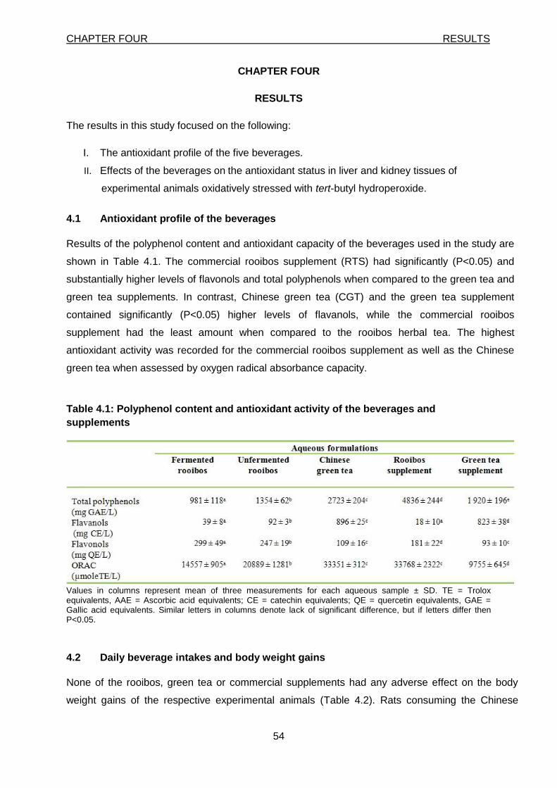
CHAPTER FOUR RESULTS
54
CHAPTER FOUR
RESULTS
The results in this study focused on the following:
I. The antioxidant profile of the five beverages.
II. Effects of the beverages on the antioxidant status in liver and kidney tissues of
experimental animals oxidatively stressed with tert-butyl hydroperoxide.
4.1 Antioxidant profile of the beverages
Results of the polyphenol content and antioxidant capacity of the beverages used in the study are
shown in Table 4.1. The commercial rooibos supplement (RTS) had significantly (P<0.05) and
substantially higher levels of flavonols and total polyphenols when compared to the green tea and
green tea supplements. In contrast, Chinese green tea (CGT) and the green tea supplement
contained significantly (P<0.05) higher levels of flavanols, while the commercial rooibos
supplement had the least amount when compared to the rooibos herbal tea. The highest
antioxidant activity was recorded for the commercial rooibos supplement as well as the Chinese
green tea when assessed by oxygen radical absorbance capacity.
Table 4.1: Polyphenol content and antioxidant activity of the beverages and
supplements
Values in columns represent mean of three measurements for each aqueous sample ± SD. TE = Trolox equivalents, AAE = Ascorbic acid equivalents; CE = catechin equivalents; QE = quercetin equivalents, GAE = Gallic acid equivalents. Similar letters in columns denote lack of significant difference, but if letters differ then P<0.05.
4.2 Daily beverage intakes and body weight gains
None of the rooibos, green tea or commercial supplements had any adverse effect on the body
weight gains of the respective experimental animals (Table 4.2). Rats consuming the Chinese
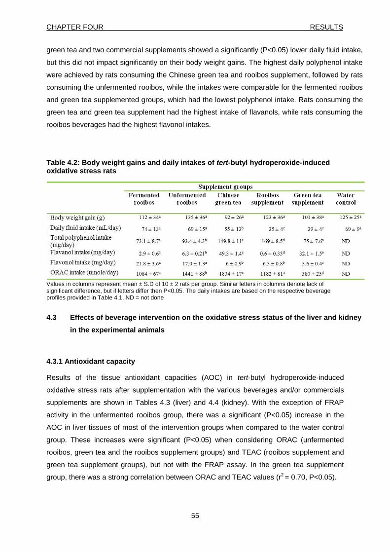
CHAPTER FOUR RESULTS
55
green tea and two commercial supplements showed a significantly (P<0.05) lower daily fluid intake,
but this did not impact significantly on their body weight gains. The highest daily polyphenol intake
were achieved by rats consuming the Chinese green tea and rooibos supplement, followed by rats
consuming the unfermented rooibos, while the intakes were comparable for the fermented rooibos
and green tea supplemented groups, which had the lowest polyphenol intake. Rats consuming the
green tea and green tea supplement had the highest intake of flavanols, while rats consuming the
rooibos beverages had the highest flavonol intakes.
Table 4.2: Body weight gains and daily intakes of tert-butyl hydroperoxide-induced oxidative stress rats
Values in columns represent mean ± S.D of 10 ± 2 rats per group. Similar letters in columns denote lack of significant difference, but if letters differ then P<0.05. The daily intakes are based on the respective beverage profiles provided in Table 4.1, ND = not done
4.3 Effects of beverage intervention on the oxidative stress status of the liver and kidney
in the experimental animals
4.3.1 Antioxidant capacity
Results of the tissue antioxidant capacities (AOC) in tert-butyl hydroperoxide-induced
oxidative stress rats after supplementation with the various beverages and/or commercials
supplements are shown in Tables 4.3 (liver) and 4.4 (kidney). With the exception of FRAP
activity in the unfermented rooibos group, there was a significant (P<0.05) increase in the
AOC in liver tissues of most of the intervention groups when compared to the water control
group. These increases were significant (P<0.05) when considering ORAC (unfermented
rooibos, green tea and the rooibos supplement groups) and TEAC (rooibos supplement and
green tea supplement groups), but not with the FRAP assay. In the green tea supplement
group, there was a strong correlation between ORAC and TEAC values (r2 = 0.70, P<0.05).
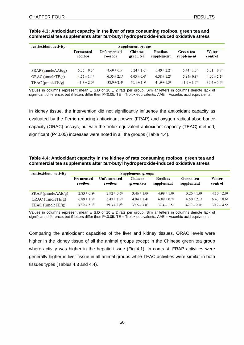
CHAPTER FOUR RESULTS
56
Table 4.3: Antioxidant capacity in the liver of rats consuming rooibos, green tea and commercial tea supplements after tert-butyl hydroperoxide-induced oxidative stress
Values in columns represent mean ± S.D of 10 ± 2 rats per group. Similar letters in columns denote lack of significant difference, but if letters differ then P<0.05. TE = Trolox equivalents, AAE = Ascorbic acid equivalents
In kidney tissue, the intervention did not significantly influence the antioxidant capacity as
evaluated by the Ferric reducing antioxidant power (FRAP) and oxygen radical absorbance
capacity (ORAC) assays, but with the trolox equivalent antioxidant capacity (TEAC) method,
significant (P<0.05) increases were noted in all the groups (Table 4.4).
Table 4.4: Antioxidant capacity in the kidney of rats consuming rooibos, green tea and commercial tea supplements after tert-butyl hydroperoxide-induced oxidative stress
Values in columns represent mean ± S.D of 10 ± 2 rats per group. Similar letters in columns denote lack of significant difference, but if letters differ then P<0.05. TE = Trolox equivalents, AAE = Ascorbic acid equivalents
Comparing the antioxidant capacities of the liver and kidney tissues, ORAC levels were
higher in the kidney tissue of all the animal groups except in the Chinese green tea group
where activity was higher in the hepatic tissue (Fig 4.1). In contrast, FRAP activities were
generally higher in liver tissue in all animal groups while TEAC activities were similar in both
tissues types (Tables 4.3 and 4.4).
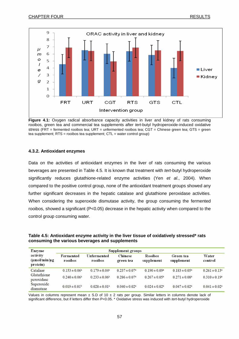
CHAPTER FOUR RESULTS
57
Figure 4.1: Oxygen radical absorbance capacity activities in liver and kidney of rats consuming rooibos, green tea and commercial tea supplements after tert-butyl hydroperoxide-induced oxidative stress (FRT = fermented rooibos tea; URT = unfermented rooibos tea; CGT = Chinese green tea; GTS = green
tea supplement; RTS = rooibos tea supplement; CTL = water control group)
4.3.2. Antioxidant enzymes
Data on the activities of antioxidant enzymes in the liver of rats consuming the various
beverages are presented in Table 4.5. It is known that treatment with tert-butyl hydroperoxide
significantly reduces glutathione-related enzyme activities (Yen et al., 2004). When
compared to the positive control group, none of the antioxidant treatment groups showed any
further significant decreases in the hepatic catalase and glutathione peroxidase activities.
When considering the superoxide dismutase activity, the group consuming the fermented
rooibos, showed a significant (P<0.05) decrease in the hepatic activity when compared to the
control group consuming water.
Table 4.5: Antioxidant enzyme activity in the liver tissue of oxidatively stressed* rats consuming the various beverages and supplements
Values in columns represent mean ± S.D of 10 ± 2 rats per group. Similar letters in columns denote lack of significant difference, but if letters differ then P<0.05. * Oxidative stress was induced with tert-butyl hydroperoxide
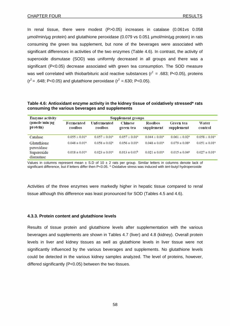
CHAPTER FOUR RESULTS
58
In renal tissue, there were modest (P>0.05) increases in catalase (0.061vs 0.058
µmol/min/µg protein) and glutathione peroxidase (0.079 vs 0.051 µmol/min/µg protein) in rats
consuming the green tea supplement, but none of the beverages were associated with
significant differences in activities of the two enzymes (Table 4.6). In contrast, the activity of
superoxide dismutase (SOD) was uniformly decreased in all groups and there was a
significant (P<0.05) decrease associated with green tea consumption. The SOD measure
was well correlated with thiobarbituric acid reactive substances (r2 = .683; P<0.05), proteins
(r2 = .648; P<0.05) and glutathione peroxidase (r2 =.630; P<0.05).
Table 4.6: Antioxidant enzyme activity in the kidney tissue of oxidatively stressed* rats consuming the various beverages and supplements
Values in columns represent mean ± S.D of 10 ± 2 rats per group. Similar letters in columns denote lack of significant difference, but if letters differ then P<0.05. * Oxidative stress was induced with tert-butyl hydroperoxide
Activities of the three enzymes were markedly higher in hepatic tissue compared to renal
tissue although this difference was least pronounced for SOD (Tables 4.5 and 4.6).
4.3.3. Protein content and glutathione levels
Results of tissue protein and glutathione levels after supplementation with the various
beverages and supplements are shown in Tables 4.7 (liver) and 4.8 (kidney). Overall protein
levels in liver and kidney tissues as well as glutathione levels in liver tissue were not
significantly influenced by the various beverages and supplements. No glutathione levels
could be detected in the various kidney samples analyzed. The level of proteins, however,
differed significantly (P<0.05) between the two tissues.
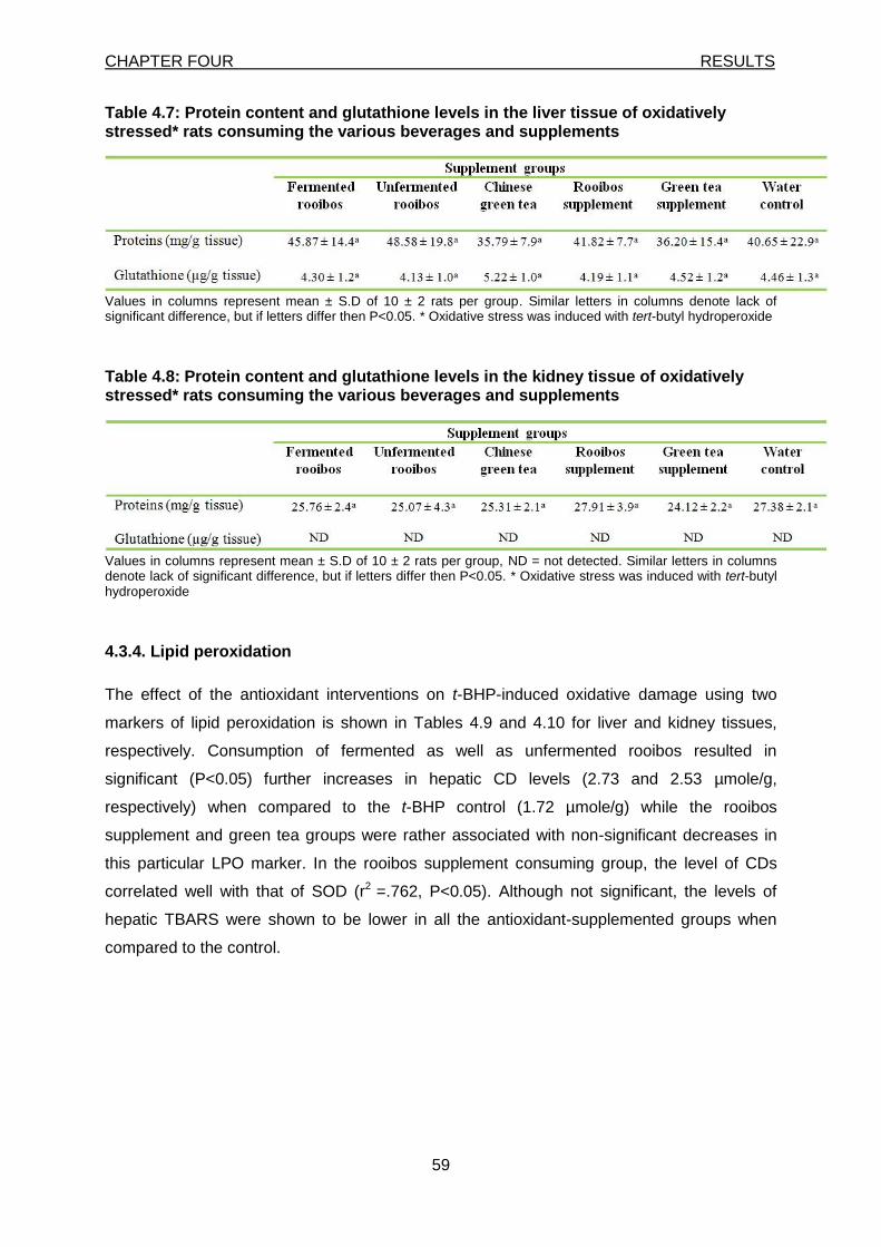
CHAPTER FOUR RESULTS
59
Table 4.7: Protein content and glutathione levels in the liver tissue of oxidatively stressed* rats consuming the various beverages and supplements
Values in columns represent mean ± S.D of 10 ± 2 rats per group. Similar letters in columns denote lack of significant difference, but if letters differ then P<0.05. * Oxidative stress was induced with tert-butyl hydroperoxide
Table 4.8: Protein content and glutathione levels in the kidney tissue of oxidatively stressed* rats consuming the various beverages and supplements
Values in columns represent mean ± S.D of 10 ± 2 rats per group, ND = not detected. Similar letters in columns denote lack of significant difference, but if letters differ then P<0.05. * Oxidative stress was induced with tert-butyl hydroperoxide
4.3.4. Lipid peroxidation
The effect of the antioxidant interventions on t-BHP-induced oxidative damage using two
markers of lipid peroxidation is shown in Tables 4.9 and 4.10 for liver and kidney tissues,
respectively. Consumption of fermented as well as unfermented rooibos resulted in
significant (P<0.05) further increases in hepatic CD levels (2.73 and 2.53 µmole/g,
respectively) when compared to the t-BHP control (1.72 µmole/g) while the rooibos
supplement and green tea groups were rather associated with non-significant decreases in
this particular LPO marker. In the rooibos supplement consuming group, the level of CDs
correlated well with that of SOD (r2 =.762, P<0.05). Although not significant, the levels of
hepatic TBARS were shown to be lower in all the antioxidant-supplemented groups when
compared to the control.
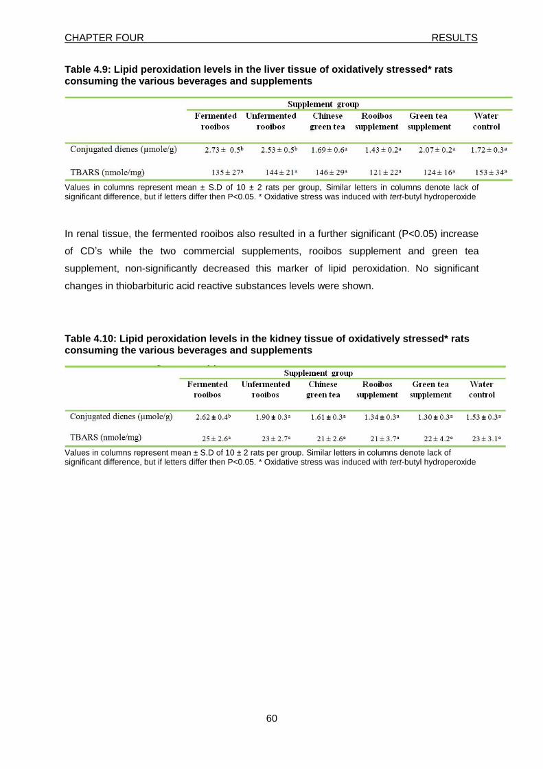
CHAPTER FOUR RESULTS
60
Table 4.9: Lipid peroxidation levels in the liver tissue of oxidatively stressed* rats consuming the various beverages and supplements
Values in columns represent mean ± S.D of 10 ± 2 rats per group, Similar letters in columns denote lack of significant difference, but if letters differ then P<0.05. * Oxidative stress was induced with tert-butyl hydroperoxide
In renal tissue, the fermented rooibos also resulted in a further significant (P<0.05) increase
of CD’s while the two commercial supplements, rooibos supplement and green tea
supplement, non-significantly decreased this marker of lipid peroxidation. No significant
changes in thiobarbituric acid reactive substances levels were shown.
Table 4.10: Lipid peroxidation levels in the kidney tissue of oxidatively stressed* rats consuming the various beverages and supplements
Values in columns represent mean ± S.D of 10 ± 2 rats per group. Similar letters in columns denote lack of significant difference, but if letters differ then P<0.05. * Oxidative stress was induced with tert-butyl hydroperoxide
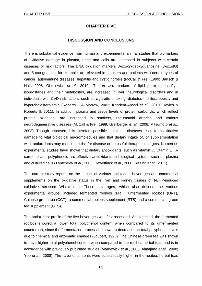
CHAPTER FIVE DISCUSSION & CONCLUSIONS
61
CHAPTER FIVE
DISCUSSION AND CONCLUSIONS
There is substantial evidence from human and experimental animal studies that biomarkers
of oxidative damage in plasma, urine and cells are increased in subjects with certain
diseases or risk factors. The DNA oxidation markers 8-oxo-2'-deoxyguanosine (8-oxodG)
and 8-oxo-guanine, for example, are elevated in smokers and patients with certain types of
cancer, autoimmune diseases, hepatitis and cystic fibrosis (McCall & Frei, 1999; Bartsch &
Nair, 2006; Obtulowicz et al., 2010). The in vivo markers of lipid peroxidation, F2 -
isoprostanes and their metabolites, are increased in liver, neurological disorders and in
individuals with CVD risk factors, such as cigarette smoking, diabetes mellitus, obesity and
hypercholesterolemia (Roberts II & Morrow, 2002; Khadem-Ansari et al., 2010; Davies &
Roberts II, 2011). In addition, plasma and tissue levels of protein carbonyls, which reflect
protein oxidation, are increased in smokers, rheumatoid arthritis and various
neurodegenerative diseases (McCall & Frei, 1999; Greilberger et al., 2008; Mitsumoto et al.,
2008). Though unproven, it is therefore possible that these diseases result from oxidative
damage to vital biological macromolecules and that dietary intake of, or supplementation
with, antioxidants may reduce the risk for disease or be useful therapeutic targets. Numerous
experimental studies have shown that dietary antioxidants, such as vitamin C, vitamin E, ß-
carotene and polyphenols are effective antioxidants in biological systems such as plasma
and cultured cells (Tantcheva et al., 2003; Dewettinck et al., 2008; Sissing et al., 2011).
The current study reports on the impact of various antioxidant beverages and commercial
supplements on the oxidative status in the liver and kidney tissues of t-BHP-induced
oxidative stressed Wistar rats. These beverages, which also defined the various
experimental groups, included fermented rooibos (FRT), unfermented rooibos (URT),
Chinese green tea (CGT), a commercial rooibos supplement (RTS) and a commercial green
tea supplement (GTS).
The antioxidant profile of the five beverages was first assessed. As expected, the fermented
rooibos showed a lower total polyphenol content when compared to its unfermented
counterpart, since the fermentation process is known to decrease the total polyphenol levels
due to chemical and enzymatic changes (Joubert, 1996). The Chinese green tea was shown
to have higher total polyphenol content when compared to the rooibos herbal teas and is in
accordance with previously published studies (Marnewick et al., 2003; Almajano et al., 2008;
Yoo et al., 2008). The flavonol contents were substantially higher in the rooibos herbal teas
CHAPTER FIVE DISCUSSION & CONCLUSIONS
62
(as well as the commercial rooibos supplement) when compared to the green tea (and
commercial green tea supplement), while the flavanol content was again highest in the green
tea when compared to the rooibos herbal teas, all in agreement with previous reports
(Marnewick et al., 2009; Awoniyi et al., 2012; Pantsi et al., 2011). When comparing the two
commercial supplements, it was the rooibos supplement showing the highest polyphenol
content and antioxidant capacity (ORAC). The green tea supplement (GTS) only contain one
polyphenol component i.e. EGCG and showed, as expected, a low level of antioxidant
capacity as measured by ORAC assay since synergistic interactions may contribute to an
increased antioxidant capacity. At the time of the study no commercial supplement was
available containing only the major rooibos polyphenolic compound, i.e. aspalathin, while a
commercial supplement could be found containing the major green tea polyphenolic
compound, i.e. EGCG (100 mg EGCG). The rooibos supplement used in the current study
contained 175 mg aspalathin, 150 mg vitamin C, 5 mg vitamin E, 500 µg vitamin A and 25 µg
selenium. This mixture of different antioxidants may explain this supplement’s higher
antioxidant profile since antioxidants are known to interact with each other synergistically
through overlapping or complementary effects (Bendich et al., 1984; Pedrielli & Skibsted,
2002; Jain et al., 2011).
The modulation of the antioxidant status in liver and kidney tissues of the t-BHP-induced
oxidative stress experimental animals was evaluated using three different antioxidant
capacity assays, i.e. ORAC, FRAP and TEAC, as these assays lack specificity (Lotito & Frei,
2006). Previous authors have recommended a battery of assays rather than a single assay
approach, keeping in mind the specific research question to be addressed (Griffiths et al.,
2002; Collins, 2005; Dilis & Trichopoulou, 2010). In the liver tissue, none of the antioxidant
beverages/supplements significantly influenced the FRAP measures. However, the intake of
unfermented rooibos, Chinese green tea and the rooibos supplement significantly restored
the t-BHP-induced reduction (Kumar & Muralidhara, 2002; Hwang et al., 2005) and increased
the antioxidant status with regards to ORAC and TEAC levels. A previous study by
Marnewick et al. (2009) also showed unfermented rooibos to restore and enhance the injured
liver’s ORAC in rats exposed to diethylnitrosamine. In the current study, it would be difficult to
deduce exactly which group of compounds were responsible for this enhanced antioxidant
capacity, but it could be suggested that the polyphenolic compounds did contribute to this
effect. Further studies will need to be conducted to ascribe this effect to one particular or
combination of compounds. In kidney tissue, the intervention did not significantly influence
the AOC as evaluated by FRAP and ORAC assays, but with the TEAC method, significant
(P<0.05) increases were noted in all the groups when compared to the oxidative stress-
induced control group. Differences between the tissues may reflect organ-related metabolic
CHAPTER FIVE DISCUSSION & CONCLUSIONS
63
differences which can influence various factors such as bioavailability at tissue and cellular
level (Chen et al., 1997; Scalbert et al., 2002). This also underlies the recommendation to
use at least two methods when assessing the antioxidant capacity. Although not significant,
the groups consuming the rooibos herbal teas and the Chinese green tea were all associated
with lower FRAP activity than the control group in the kidneys, which may be a consequence
of FRAP’s inability to measure the SH-group-containing antioxidants such as glutathione
(Prior & Cao, 1999).
Antioxidant enzymes are part of the endogenous defense system and protect against
oxidative stress via mechanisms discussed earlier. In this study, the enzymatic activity was
assessed by measuring catalase, glutathione peroxidase and superoxide dismutase activity.
Previous studies have associated Camellia sinensis teas with increased tissue and plasma
levels of these enzymes when using models of oxidative stress (El-Behbishy, 2005;
Marnewick et al., 2009). In this study however, neither the green tea nor green tea
supplement were associated with increased enzyme levels in tissues from both organs, but
the results of Marnewick et al. (2009) were confirmed by the current study results in that
none of the rooibos herbal teas affected any of the antioxidant enzymes measured in the
study. When considering SOD, a decreasing trend was noted, which was underlined by a
significant (P<0.05) decrease in renal SOD activity in the Chinese green tea group. An
increase in enzymatic activity may be prompted by increased oxidative stress in a negative
feedback-like relation. Conversely, decreased enzyme activity may reflect a favorable
oxidative status balance; i.e. enzyme levels decrease as the degree of oxidative stress
(requiring neutralization) diminishes. This relation was supported by the positive and
relatively strong correlation between the SOD activity and TBARS (r2 = 0.683; P<0.05).
Between the three enzymes, the relation may be most profound with regard to SOD since it
is the very first line of defense against oxidative stress.
The enzyme activity results parallel, to some extent, the protein and GSH levels in the
current study. While also not significantly influenced by any of the interventions, the protein
levels in kidney tissues showed a decreasing trend except in the group consuming the
rooibos supplement. As in the case of the SOD and the other antioxidant enzymes, GSH
levels may be reflective of the oxidative stress situation. The enzymes and GSH are
components of the tissue proteins and their collective decrease will affect the level of
proteins. Sohn et al. (1994) and Marnewick et al. (2003) previously reported a lack of
influence on GSH levels by Chinese green and black teas as well as rooibos and honeybush
herbal teas using experimental animal models. More recently, Marnewick et al. (2011) have
CHAPTER FIVE DISCUSSION & CONCLUSIONS
64
reported on a significant increase in the levels of reduced glutathione in adults at risk for
developing heart disease, consuming 6 cups of fermented rooibos daily for 6 weeks, in
agreement with results from another human study reporting a ~46% increase in the levels of
GSH after consumption of rooibos (Nikolova et al., 2007). The current study also reports that
no GSH (below limit of detection of the assay) could be detected in the kidney tissue of the
experimental animals, and could have been as a result of the high levels of gamma-glutamyl
transpeptidase in this type of tissue, which cleaves GSH as the tissue is thawed (Anderson,
1985). It is recommended that future studies using kidney tissue should process this type of
tissue immediately without prior freezing.
Markers of lipid peroxidation in liver and kidney tissues of the rats were assessed by the CDs
and TBARS assays which have been widely used for determining oxidative stress in
mammalian tissues. Many studies have associated intake of tea and other dietary
antioxidants with decreased LPO in animal tissues (Hamden et al., 2009; Raneva &
Shimasaki, 2005). In this study the beverages did not impact significantly on lipid
peroxidation, although, there were differing trends in the two LPO markers assessed. While
TBARS levels showed a declining trend in both tissues, the CDs were generally elevated
reaching significance in the fermented rooibos group in both tissues. As discussed in the
previous chapter, CDs and TBARS measures different phases of lipid oxidation; CDs are
intermediate products while MDA are accumulated end products. This may account for the
apparent increase in CDs in contrast to the decreased TBARS. It may be possible that the
beverages affect these LPO products differently.
Antioxidants in tissues act as free radical scavengers against lipid free radicals thus
countering the initiation and propagation of lipid peroxidation. Levels of antioxidant enzymes
are therefore important determinants of tissue antioxidant status, however, increased tissue
LPO products are able to inactivate certain antioxidant enzymes, mainly glutathione
peroxidase, and inhibit the protective action of glutathione as has been previously reported
(Bosch-Morell et al., 1999; Reneva & Shimasaki, 2005). However, the impact of dietary
intervention may vary in different tissues as was shown in a study evaluating the effect of
dietary restriction on antioxidant status (Dubnov et al., 2000).
Results discussed in this thesis are part of a broader study doing a comprehensive
evaluation of the influence of Chinese tea and rooibos herbal teas on various aspects in
oxidatively stressed rats. Results already published indicate that both the rooibos herbal
teas, the Chinese green tea and the commercial rooibos supplement restored and enhanced
CHAPTER FIVE DISCUSSION & CONCLUSIONS
65
the t-BHP-induced decrease in glutathione and certain antioxidant enzymes while reducing
lipid peroxidation (TBARS) in rat testicular tissue and sperm (Awoniyi et al., 2011; Awoniyi et
al., 2012) which again points towards tissue/organ variability in responses. A possible source
of this variability may be selective toxicity resulting from the agent used to induce the
oxidative stress. Oh et al. (2012) have recently showed a time and dose dependent
hepatotoxicity in rats treated with t-BHP which was used in this study. This agent may
therefore not be the ideal model for multi-organ induced oxidative stress models, an
important fact to keep in mind when planning future studies.
It can be concluded from this study that:
The commercial rooibos supplement had the highest polyphenol content and
antioxidant capacity which was likely due to its rich mixture of antioxidant compounds.
Fermented rooibos, as expected, had lower total polyphenol content and antioxidant
potency compared to its unfermented counterpart. Contrary to expectation, however,
unfermented rooibos had more polyphenols and higher antioxidant capacity than the
Chinese green tea used in this study, but again this could be as a result of species
differences, seasonal differences and different agricultural practices, all influencing
factors on the quality of the end product.
Organ and methodological differences were observed in the antioxidant
measurements. In the liver tissue, supplementation with unfermented rooibos and the
commercial rooibos supplement was associated with significant increase in ORAC
levels but not in the other two antioxidant capacity assays. All the beverages were
associated with significant antioxidant boost in renal tissue by the TEAC assay but
not the other two methods. This underscores the caveat to use at least two methods
for these measurements.
All the beverages were associated with a general decline in activities of the
antioxidant enzymes; this trend was underlined by a significant decrease in SOD
activity in kidney (Chinese green tea) tissues. As discussed earlier, this may be an
indication of decreased oxidative stress challenge.
Generally, the beverages did not impact significantly on lipid peroxidation although
there were differing trends in the two LPO markers assessed. While TBARS levels
showed a declining trend in both tissues, the CDs were generally elevated reaching
significance in the fermented rooibos group in both tissues.
CHAPTER FIVE DISCUSSION & CONCLUSIONS
66
Strengths and limitations
A key strength in this study is the use of several methods to assess antioxidant capacity
and lipid peroxidation. This approach displays results which would otherwise have been
missed if only single assays were used. Another innovative strategy was the use of a
multi-antioxidant supplement containing a broad combination of antioxidants aiming to
resemble the spectrum of antioxidants commonly consumed in a daily intake of fruit and
vegetables.
An important missing link in the current study is plasma and/or erythrocyte levels of
antioxidants and LPO products. This could have provided a basis to compare tissue
levels to the circulating (systemic) situation which could reveal whether the increasing
trend of CDs in tissues is only a reflection of enhanced clearance from the circulation. It is
possible that proteins or DNA may have been damaged more severely in this model;
unfortunately none of these samples were made available for analyses in this study, as
they were utilized elsewhere. It is also worth noting that these results represent one point
in time and future studies are recommended to include a time-course evaluation.
Although this intervention was conducted in experimental rats, it is hoped that these
results may have relevance in humans since results for many studies (such as those
examining the effects of cholesterol lowering drugs) that were originally done in rodents
have subsequently been extended to humans. Despite these limitations, we were able to
show the impact of these beverages on important oxidative status aspects in liver and
kidney tissues of experimental animals. It is highly recommended that these be confirmed
in a series of clinical human intervention studies.
67
REFERENCES
Abdel-Raheem, I.T, El-Sherbiny, G. A. & Taye, A. 2009. Green tea ameliorates renal oxidative damage induced by gentamicin in rats. Pakistan Journal of Pharmaceutical Sciences, 23: 21-28.
Abuja, P. M. & Albertini, R. 2001. Methods for monitoring oxidative stress, lipid peroxidation and oxidation resistance of lipoproteins. Clinica Chemica Acta, 306:1-17.
AETC National Research Center (n.d.) HIV Classification: CDC and WHO staging system [Online] Available from: http://www.aids-ed.org/aidsetc?page=cm-105_disease#S6X (Accessed: 8 Jan 2011).
Agarwal, A., Gupta, S. & Sharma, R.K. 2005. Role of oxidative stress in female reproduction. Reproductive Biology and Endocrinology, 3: 28.
Aghdassi, E. & Allard, J.P. 2000. Breath alkanes as a marker of oxidative stress in different clinical conditions. Free Radical Biology & Medicine, 28: 880-886.
Aherne, S.A. & O’Brien, N.M. 2002. Dietary flavanols: Chemistry, food content, and metabolism. Nutrition, 18:75-81.
Ahmad, R., Tripathi, A. K., Singh, S., Singh, R. & Singh, R. K. 2008. Malondialdehyde and protein carbonyl as biomarkers of oxidative stress and disease progression in patients with chronic myeloid leukaemia. In Vivo, 22: 525-528. Ahmed, S. 2010. Green tea polyphenol epigallalocatechin 3-gallate in arthritis: progress and promise. Arthritis Research & Therapy. 12:208.
Ahmed, S., Rahman, A., Hasnain, A., Lalonde, M., Goldberg, V.M. & Haqqi, T.M. 2002. Green tea polyphenol epigallalocatechin-3-gallate inhibits the IL-1beta-induced activity and expression of cyclooxygenase-2 and nitric oxide synthase-2 in human chondrocytes. Free Radical Biology Medicine, 33:1097-1105.
Akohoue, S.A., Shankar, S., Milne, G. L., Morrow, J., Chen, K.Y., Ajayi, W. U. & Bushowski, M.S. 2007. Energy expenditure, inflammation, and oxidative stress in steady-state adolescents with sickle cell anemia. Paediatric Research, 61: 233-237. Albano, E. 2007. Oxidative mechanism in the pathogenesis of alcoholic liver diseases. Molecular Aspects of Medicine, 29: 9-16. Alberti, K. G. M. M., Zimmet, P. & Shaw, J. 2007. International Diabetes Federation: a consensus on type 2 diabetes prevention. Diabetic Medicine, 24:451-463.
Alessio, H. M., Hagerman, A. E., Romanello, M., Carando, S., Threlkeld, M. S., Rogers, J., Dimitrova, Y., Muhammed, S., & Wiley, R.L. 2002. Consumption of green tea protects rats from exercise-induced oxidative stress in kidney and liver. Nutritional Research, 22: 1177-1188.
Aletaha, D., Neogi, T., Silman, A. J., Funovits, J., Felson, D.T., Bingham III, C. O. et al., 2010. Rheumatoid arthritis classification criteria. Arthritis & Rheumatism, 62: 2569-2581. Aliyu, Z. Y., Kato, G.J., Taylor IV, J., Babadoko, A., Mamman, V. I., Gordeuk, V.R. & Gladwin, M.T. 2008. Sickle cell disease and pulmonary hypertention in Africa: A global perspective and review of epidemiology, pathophysiology, and management. American Journal of Hematology, 83:63-70.
68
Almajano, M. P., Carbó, R., Jiménez, A. L. & Gordon, M. H. 2008. Antioxidant and antimicrobial activities of tea infusions. Food Chemistry, 108:55-63. Amarowicz, R., Pegg, R.B. & Bautista, D.A. 2000. Antibacterial activity of green tea polyphenols against Echerichia coli K 12. Die Nahrung, 44:50-62. American Cancer Society (ACS). 2011. The history of cancer [Online] Available from: http://www.cancer.org/acs/groups/cid/documents/webcontent/002048-pdf.pdf (Accessed: Oct 7, 2011). American Heart Association (N.D), Kidneys and kidney function [Online] Available from:http://www.americanheart.org/presenter.jhtml?identifier=4647 (Accessed: Aug 21, 2010). American Liver Foundation (2008). 2008 Annual Report [Online] Available from: http://www.liverfoundation.org/downloads/alf_download_653. pdf (Accessed: Jan 7, 2011). Anderson, M. E. 1985. Determination of glutathione and glutathione disulfide in biological samples. Methods in Enzymology, 113:548-555.
Anyanwu, S. N. C. 2008. Temporal trends in breast cancer presentation in the third world. BioMed Central [Online] Available from: www.jeccr.com/content/27/1/17 (Accessed: Jan 12, 2011). Appay, V. & Sauce, D. 2008. Immune activation and inflammation in HIV-1 infection: cause and consequences. Journal of Pathology, 214: 231-241.
Aquaro, S., Muscoli, C., Ranazzi, A., Pollicita, M., Granato, T., Masuelli, L., Modesti, A., Perno, C-F & Mollace, V. 2007. The contribution of peroxynitrite generation in HIV replication in human primary macrophages. Retrovirology, 4:76 [Online] Available from: http://www.retrovirology.com/content/4/1/76 (Accessed: Feb 12, 2011). Aquaro, S., Scopelliti, F., Pollicita, M. & Perno, C. F. 2008. Oxidative stress and HIV infection: target pathways for novel therapies? [Online] Available from: http://www.medscape.com/viewarticle/584032 (Accessed: Jan 8, 2011).
Argüelles, S., Garcia, S., Maldonado, M., Machado, A. & Ayala, A. 2004. Do the serum oxidative stress biomarkers provide a reasonable index of the general oxidative stress status? Biochimica et Biophysica Acta, 1674:251-259.
Arlt, S., Müller-Thomsen, T. & Beisiegel, U. 2002. Use of vitamin C and E in treatment of Alzheimer’s Disease. Drug Development Research, 56:452-457.
Arts, M. J., Haenen, G. R., Willms, L. C., Beetstra, S. A. Heijnen, C. G., Voss, H.-P. & Bast, A. 2002. Interactions between flavonoids and proteins: effect on the total antioxidant capacity. Journal of Agricultural and Food Chemistry, 50:1184-1187.
Aruoma, O. I. 1998. Free radicals, oxidative stress and antioxidants in human health and disease. Journal of the American Oil Chemists’ Society, 2:199-212.
Asensi, M., Sastre, J., Pallardo, F. V., Lloret, A., Lehner, M., Garcia-de-la Asuncion, J. & Viňa, J. 1999. Ratio of reduced to oxidized glutathione as indicator of oxidative stress status and DNA damage. Methods in Enzymology, 299: 267-276.
69
Ates, B., Ercal, B. C., Manda, K., Abraham, L. & Ercal, N. 2008. Determination of glutathione disulfide levels in biological samples using thiol-disulfide exchanging agent, dithiothreitol. Biomedical Chromatography, 23: 119-123.
Atkins, R. C. 2005. The epidemiology of chronic kidney disease. Kidney International, 67: S14-S18.
Awoniyi, D. O. 2010. The role of rooibos (Aspalathus linearis), green tea (Camellia sinensis) and commercially available rooibos and green tea antioxidant supplements on rat testicular and epididymal function. CPUT Thesis & Dissertations. Paper 373 [Online] Available from: http://dk.cput.ac.za/td_cput/373(Accessed: May 20, 2011).
Awoniyi, D. O., Aboua, Y. G., Marnewick, J. L., Du Plesis, S. S. & Brooks, N. L. 2011. Protective effects of rooibos (Aspalathus linearis), green tea (Camellia sinensis) and commercial supplements on testicular tissue of oxidative stress-induced rats. African Journal of Biotechnology, 10: 17317-17322.
Awoniyi, D. O., Aboua, Y. G., Marnewick, J. & Brooks, N. 2012. The effects of rooibos (Aspalathus linearis), green tea (Camellia sinensis) and commercial supplements on epididymal sperm in oxidative stress-induced rats. Phytotherapy Research. Published online in Wiley Online Library (wileyonlinelibrary.com) DOI: 10.1002/ptr.3717.
Azzi, A. 2007. Oxidative stress: A dead end or laboratory hypothesis? Biochemical and Biophysical Research Communications, 362:230-232.
Baba, H., Ohtsuka, Y., Haruna, H., Lee, T., Nagata, S., Maeda, M., Yamashiro, Y., & Shimizu, T. 2009. Studies of anti-inflammatory effects of rooibos tea in rats. Pediatrics International, 51: 700-704.
Bagchi, K. & Puri, S. 1998. Free radical and antioxidant in health. Eastern Mediterranean Health Journal, 4:350-360.
Bahorum, T., Soobrattee, M. A., Luximon-Ramma, V. & Aruoma, O. I. 2006. Free radicals and antioxidants in cardiovascular health and disease. Internet Journal of Medical Update, 1:1-17.
Bakker, G. C. M., Van Erk, M. J., Pellis, L., Wopereis, S., Rubingh, C. M., Cnubben, N. H. P., Kooistra, T., Van Ommen, B. & Hendriks, H. F. J. 2010. An anti-inflammatory dietary mix modulates inflammation and oxidative and metabolic stress in overweight men: a nutrigenomics approach. The American Journal of Clinical Nutrition, 91:1044-1059.
Bandebuche, S. & Melinkeri, R. R. 2011. Oxidative stress and antioxidant status in patients of ovarian cancer. Biomedical Research, 22:193-197. Bansilar, Ali, N., Afzal, N., Khan, T. S. & Shahjahan, S. 2007. Antioxidant Status in coronary heart disease patients with type 2 diabetes mellitus. Journal of Ayub College Abbottabad, 19:98-101.
Barciela, J., Herrero, C., Garcia-Martin, S. & Rosa, M. P. 2008. A brief study of the role of seleniun as antioxidant. Electronic Journal of Environmental, Agriculture and Food Chemistry, 7:3151-3155.
Barker, P. E. & Murthy, M. 2009. Biomarker Validation for Aging: Lessons from mtDNA Heteroplasmy Analyses in Early Cancer Detection. Biomarker Insights, 4:165–179.
70
Barkin, W. & Hersh, T. 2008. The balance of power [Online] Available from: http:// www.quantumhealthbeverages.com/documents/ the balance of power, theodore hersh, wendy barkin, (Accessed: Feb 2, 2011).
Bartholomew, M. 2002. James Lind’s treatise of scurvy (1753). Postgraduate Medical Journal, 78:695-696.
Bartsch, H. & Nair, J. 2006. Chronic inflammation and oxidative stress in the genesis and perpetuation of cancer: role of lipid peroxidation, DNA damage, and repair. Langenbeck’s Archives of Surgery, 391:499-510.
Basu, A., Du, M., Sanchez, K., Leyva, M. J. et al., 2011. Green tea minimally affects biomarkers of inflammation in obese subjects with metabolic syndrome. Nutrition, 27:206-213. Basu, A. K., Loechler, E. L., Leadon, S.A. & Essigmann, J. M. 1989. Genetic effect of thymine glycol: site specific mutagenesis and molecular modelling studies. Proceedings of the National Academy of Science of the USA, 86: 7677-7681. Baud, L. & Ardaillou, R. 1986. ROS production and role in the kidney. American Journal of Physiology, 251:F765-776. Bayes, B., Pastor, M. C., Bonal, J., Junca, J., Hernandez, J. M., Riutort, N., Foraster, A. & Romero, R. 2003. Homocysteine, C-reactive protein, lipid peroxidation and mortality in haemodialysis patients. Nephrology Dialysis Transplantation, 18:106-112.
Bayir, H. and Kagan, V. E. 2008. Bench- to- bedside review: Mitochondrial injury, oxidative stress and apoptosis-there is nothing more practical than a good theory. Critical Care, 2008, 12:206
Baynes, J. W. 1991. Role of oxidative stress in development of complication in diabetes. Diabetes, 40:405-412.
Beaglehole, R. & Yach, D. 2003. Globalisation and the prevention and control of non-communicable disease: the neglected chronic diseases of adults. The Lancet, 362: 903-08
Bednarek, J., Wysocki, H. & Sowinski, J. 2004. Oxidation products and antioxidant markers in plasma of patients with Graves’ Disease and toxic multinodular goiter: Effect of methimazole treatment. Free Radical Research, 38:659-664. Bendich, A., Diapolito, P., Gabriel, E. & Machlin, L. J. 1984. Interaction of dietary vitamin C and vitamin E on guinea pig immune response to mitogens. Journal of Nutrition, 114:1588-1593. Benzie, I. F. F. & Strain, J. J. 1996. The ferric reducing ability of plasma (FRAP) as a measure of “Antioxidant Power”: The FRAP assay. Analytical Biochemistry, 239:70-76.
Bergamini, C. M., Gambetti, S., Dondi, A. & Cervellati, C. 2004. Oxygen, reactive oxygen species and tissue damage. Current Pharmaceutical Design, 10:1611–26
Bettuzzi, S., Brausi, M., Rizzi, F., Castagnetti, G., Peracchia, G. & Corti, A. 2006. Chemoprevention of human prostate cancer by oral administration of green tea catechins in volunteers with high-grade prostate intraepithelial neoplasia: A preliminary report from a one year proof-of-principle study. Cancer Research, 66:1234-1240.
71
Bhattacharya, K., Alink, G. & Dopp, E. 2007. Oxidative stress and changed gene expression profiles in fiber-/particle-induced carcinogenesis. International Journal of Human Genetics, 7: 1-21.
Bianchi, G., Cerhan, J., Parker, A., Putnam, S., See, W., Lynchi, C. & Cantor, K. 2000. Tea consumption and risk of bladder and kidney cancers in population-based case-control study. American Journal of Epidemiology, 151:377-383.
Bishop, N. 2006. Reactive oxygen species and the regulation of gene expression, part one: How the Yin and the Yang of free radicals (ROS) may impact the Naturopathic Vis Medicatrix Naturae Naturopathy Digest, July 2006, Vol. 1, No. 7. Blantz, R. C., Deng, A., Miracle, C. M. & Thomson, S. C. 2007. Regulation of kidney function and metabolism: A question of supply and demand. Transactions of the American Clinical and Climatological Association, 118: 23-42. Blum, A. 2009. Heart failure-new insights. Israel Medical Association Journal, 11:105-111.
Blumberg, J. 2004. Use of biomarkers of oxidative stress in research studies. The Journal of Nutrition, 134:3188S-3189S.
Bosch-Morell, F., Floché, L., Marίn, N. & Romero, F. J. 1999. 4-Hydroxynonenal inhibits glutathione peroxidase: protection by glutathione. Free Radical Biology & Medicine, 26:1383-1387.
Bose, M., Lambert, J. D., Ju, J., Reuhl, K. R., Shapses, S. A. & Yang, C. S. 2008. The major green tea polyphenol, (-)-Epigallocatechin-3-Gallate, inhibits disesein high-fatty-fed Mice. The Journal of Nutrition, 138:1677-1683.
Bouayed, J. & Bohn, T. 2010. Exogenous antioxidants –double-edged swords in cellular redox state: Health beneficial effects at physiologic doses versus deleterious effects at high doses. Oxidative Medicine and Cellular Longevity, 3: 228-237.
Bradford, M.M. 1976. A rapid and sensitive method for the quantitation of microgram quantities of protein utilizing the principle of protein-dye binding. Analytical Biochemistry, 72:248-254.
Braidy, N., Grant, R., Adams, S. & Guillemin G. J. 2010. Neuroprotective effects of naturally occurring polyphenols on quinolinic acid-induced excitotoxicity in human neurons. The Federal European Biochemical Societies Journal, 277: 368-382. Bramati, L., Aquilano, F. & Pietta, P. 2003. Unfermented rooibos tea: Quantitative characterization of flavonoids by HPLC-UV and determination of the total antioxidant activity. Journal of Agricultural and Food Chemistry, 51:7472-7474.
Bramati, L., Minoggio, M., Gardana, C., Simonetti, P., Mauri, P. & Pietta, P. 2002. Quantitative characterization of flavonoid compounds in rooibos tea (Aspalathus linearis) by LC-UV/DAD. Journal of Agricultural and Food Chemistry, 50:5513-5519.
Brandes, R. P. & Janiszewski, M. 2005. Direct detection of reactive oxygen species ex vivo. Kidney International, 67: 1662-1664.
Brash, D. & Cairns, J. 2009. The mysterious steps of carcinogenesis. British Journal of Cancer, 101:379-380.
72
Bravo, L. 1998. Polyphenols: Chemistry, dietary sources, metabolism, and nutritional significance. Nutrition Reviews, 56: 317-333.
Breet, P., Kruger, S., Jerling, J. C. & Oosthuizen, W. 2005. Action of black tea and rooibos on iron status of primary school children. Nutrition Research, 25:983-994.
Breiter, T., Laue, C., Kressel, G., Gröll, S., Engelhardt, U. H & Hahn, A. 2011. Bioavailability and antioxidant potential of rooibos flavonoids in humans following the consuption of different rooibos formulations. Food Chemistry, 128:338-347.
Brown, J.C., Wang, J., Kasman, L., Jiang, X. Haley-Zitlin, V. 2010. Activity of muscadine grape skin and quercetin against Helicobacter pylori infection in mice. Journal of Applied Microbiology, 110: 139-146. Brown, M. D. 1999. Green tea (Camellia sinensis) extract and its possible role in the prevention of cancer. Alternative Medicine Review, 5:360-370.
Brown, R. C. Lockwood, A. H. & Sonawane, B. R. 2005. Neurodegenerative disease: An overview of environmental risk factors. Environmental Health Perspective, 113:1250-1256. Browning, J.D. & Horton, J. D. 2004. Molecular mediators of hepatic steatosis and liver injury. The Journal of Clinical Investigation, 114:147–52. Bulletin of the World Health Organization. 2004. Developing countries face double burden of disease. Bulletin of the World Health Organization [Online] Available from: http://goliath.ecnext.com/coms2/gi_0199-296183/Developing -countries-face-double-burden-of-diseases.html (Accessed: Dec 04, 2010). Burcham, P. C. 1999. Internal hazards: baseline DNA damage by endogenous product of normal metabolism. Mutation Research, 443:11-36. Burgdorf, L.T. & Carell, T. 2002. Synthesis, stability, and conformation of the formamidopyrimidine G DNA lesion. Chemistry, 8:293-301. Burton, G. W., Wronska, U. & Stone, L. 1990. Biokinetics of dietary RRR-α-tocopherol in the male guinea pig at three dietary levels of vitamin C and two levels of vitamin E. Evidence that vitamin C does not “spare” vitamin E in vivo. Lipids, 22:199-210. Buss, H., Chan, T. P. Sluis, K. B., Domigan, N. M. & Winterbourn, C. C. 1997. Protein carbonyl measurement by sensitive ELISA method. Free Radical Biology and Medicine, 23:361-366. Cabrera, C., Artacho, R. & Giménez, R. 2006. Beneficial effects of green tea-a review. Journal of the American College of Nutrition, 25:79-99.
Cadenas, E. 1989. Biochemistry of oxygen toxicity. Annual Review of Biochemistry, 58:79-110.
Cadenas, E. 1997. Basic mechanism of antioxidant activity. BioFactors, 6: 391-397.
Cao, G. & Prior, R. L. 1998. Comparison of different analytical methods for assessing total antioxidant capacity of human serum. Clinical Chemistry, 44:1309-1315.
Cao, G., Booth, S. L., Sadowski, J. A., Prior, R. L. 1998. Increase in human plasma antioxidant capacity after consumption of controlled diets high in fruits and vegetables. The American Journal of Clinical Nutrition, 68:1081-1087.
73
Cecarini, V., Gee, J., Fioretti, E., Amici, M., Angeletti, M., Eleuteri, A. M. & Keller, J.N. 2007. Protein oxidation and cellular homeostasis: Emphasis on metabolism. Biochimica et Biophysica Acta, 1773:93-104.
Ceriello, A. & Motz, E. 2004. Is oxidative stress the pathogenesis mechanism underlying insulin resistance, diabetes, and cardiovascular disease? The common soil hypothesis revisited. Arteriosclerosis, Thrombosis, and Vascular Biology, 24: 816-823.
Chacko, S.M. Thambi, P.T. Kuttan, R. Nishigaki, I. 2010. Beneficial effects of the green tea: A literature review. BioMed Central [Online] Available from: http://www.cmjournal.org/content/5/1/13 (Accessed: 27 Jan 2011). Chalasani, N., Deeg, M.A. & Crab, D.W. 2004. Systemic levels of lipid peroxidation and its metabolic and dietary correlates in patients with non-alcoholic steatohepatitis. American Journal of Gastroenterology, 99:1497–502.
Cheeseman, K.H. & Slater, T.F. 1993. An introduction to free radical biochemistry. British Medical Bulletin, 49:481-493.
Chen, L., Lee, M. J., Li, H. & Yang, C. S. 1997. Absorption, distribution, elimination of tea polyphenols in rats. Drug Metabolism and Disposition: Drug Metabolism & Disposition, 25:1045-1050.
Choi, H.-H., Kim, J.-H., Lee, C.-H., Ahn, Y.-J., Song, J.-H., Baek, S.-H. & Kwon, D.-H. 2009. Antiviral activity of quercetin 7-rhamnoside against porcine epidemic diarrhea virus. Antiviral Research, 81:77-81. Choi, J. & Ou, J.-H.J. 2006. Mechanism of liver injury.III. Oxidative stress in the pathogenesis of hepatites C virus. American Journal of Physiology- Gastrointestinal and Liver Physiology, 290:G847-G851. Chou, C. C., Lin, L.L. & Chung, K. T. 1999. Antimicrobial activity of tea as affected by the degree of fermentation and manufacturing season. International Journal of Food Microbiology, 48:125-130. Chowning, J. T. 2000. Sickle cell anaemia: a case study approach to teaching high school genetics [Online] Available from: http://genetics-education-partneship.mbt.washington.edu/download/file.html (Accessed: 20 Jan 2011). Chung, F. L., Schwartz, J., Herzog, C. R. & Yang, Y. M. 2003. Tea and cancer prevention: Studies in animals and humans. The Journal of Nutrition, 133: 3268-3274.
Cleveland Clinic. 2011. Heart and vascular health prevention: antioxidants vitamin E, beta carotene, and cardiovascular diseases [Online] Available from: http://my.clevelandclinic.org/heart/disorders/cad/vitamin_e.aspx (Accessed: October 29, 2011). Cobb, J. P. Hotchkiss, R. S., Karl, I. E. & Buchman, T.G. 1996. Mechanism of cell injury and death. British Journal of Anaesthesia, 77:3-10. Cohen, G., Riahi, Y., Alpert, E., Gruzman, A. & Sasson, S. 2007. The role of hyperglycaemia and oxidative stress in the rise and collapse of natural protective mechanism against vascular endothelial cell dysfunction in diabetes. Archives of Physiology and Biochemistry, 113:259-267.
74
Collins, A. R. 2004. The comet assay for DNA damage and repair: principles, application, and limitation. Molecular Biotechnology, 26:249-261.
Collins, A. R. 2005. Assays for oxidative stress and antioxidant status: applications to research into biological effectiveness of polyphenols. The American Journal of Clinical Nutrition, 81:S261-267S.
Coppo, R., Camilla, R., Amore, A. & Peruzzi, L. 2010. Oxidative stress in IgA nephropathy. Nephron.Clinical Practice, 116:c196-c199.
D’Archivio, M., Filesi, C., Di Benedetto, R., Gargiulo, R., Giovannini, C. & Massella, R. 2007. Polyphenols, dietary sources and bioavailability. Annali dell’Instituto Superiore di Sanitá, 43:348-361.
Dai, Q., Gao, Y.-T., Shu, X.-O., Yang, G., Milne, G., Cai, Q., Wen, W., Rothman, N., Cai, H., Li, H., Xiang, Y., Chow, W.-H. & Zheng, W. 2009. Oxidative stress, obesity, and breast cancer risk: results from the Shangai women’s health study. Journal of Clinical Oncology, 27:2482-2488.
Dalle–Donne, I., Rossi, R., Colombo, R., Giustarini, D. & Milzani, A. 2006. Biomarkers of oxidative damage in human disease. Clinical Chemistry, 52:601-623.
Dalle-Donne, I., Rossi, R., Giustarini, D., Milzani, A. & Colombo, R. 2003. Protein carbonyl groups as a biomarker of oxidative stress. Clinica Chimica Medica, 329:23-38.
Danne, T. & Kordonouri, O. 2007. What is so different about diabetes in children? Diabetes Voice, 52:16-19.
Das, K., Das, K., Mukherjee, P. S., Ghosh, A., et al., 2010. Nonobese population in a developing country has a high prevalence of non alcoholic fatty liver and significant liver disease. Hepatology. 51:1593-1602.
Das, S. K., Varadhan, S., Dhanya, L., Mukherjee, S. & Vasudevan, D. M. 2008. Effects of chronic ethanol exposure on renal function tests and oxidative stress in kidney. Indian Journal of Clinical Biochemistry, 23:341-344.
Davies, S.S. & Roberts II, L.J. 2011. F2-Isoprostane as an indicator and risk factor for coronary heart disease. Free Radical in Biology and Medicine, 50:559-566.
Dayton, A. 2008. Hitting HIV where it hides. BioMed Central [Online] Available from: http:// www.retrovirology.com/content/5/1/15(Accessed: Jan 7, 2 011).
De Bont, R. & Van Larebeke, N. 2004. Endogenous DNA damage in humans: a review of quantitative data. Mutagenesis, 19:169±185.
De Groot, H. & Rauen, U. 1997. Tissue injury by reactive oxygen species and the protective effects of flavonoids. Fundamental Clinical Pharmacology, 12:249-255.
De Mejia, E. G., Ramirez-Mares, M. V. & Puangpraphant, S. 2009. Bioactive components of tea: Cancer, inflammation and behaviour. Brain, Behaviour, and Immunity, 23:721-731.
Degan, P., Bonassi, S., De Caterina, M., Korkina, L. G., Pinto., L., Scopacasa, F., Zatterate, A., Calzone, R. & Pagano, G. 1995. In vivo accumulation of 8-hydroxi-2’-deoxyguanosine in DNA correlates with release of reactive oxygen species in Fanconi’s anaemia families. Carcinogenesis, 16:735-742.
75
Demaurex, N. & Scorrano, L. 2009. Oxygen reactive species are noxious for neurons. Nature Neuroscience, 12:819-820.
Deshmane, S. L., Mukerjee, R., Fan, S., Dell Valle, L., Michiels, C., Sweet, T., Rom, I., Khalili, K., Rappaport, J., Amini, S. & Sawaya, B.E. 2009. Activation of oxidative stress pathway by HIV-1 Vpr leads to induction of hypoxia-inducible factor 1α expression. The Journal of Biological Chemistry, 284:11364-11373. Dewettinck, K., Van Bockstaele, F., Kϋhne, B., Van de Walle, D., Courtens, T. M. & Gellynck, X. 2008. Nutritional value of bread: influence of processing, food interaction and consumer perception. Journal of Cereal Science, 48: 243-257. Dianov, G. L., Thybo, T., Dianova, I. I., Lipinski, J. & Bohr, V. A. 2000. Single nucleotide patch base excision repair is the major pathway for removal of thymine glycol from DNA in human cells extracts. The Journal of Biological Chemistry, 16:11809-11813. Dilis, V. & Trichopoulou, A. 2010. Antioxidant intakes and food sources in Greek adults. The Journal of Nutrition, 140:1274-1279. Dixon, R.A. & Passinetti, G.M. 2010. Flavanoids and isoflavanoids: From plant biology to agriculture and neuroscience. Plant Physiology, 154: 453-457.
Dizdaroglu, M. 1991. Chemical determination of free radical-induced damage to DNA. Free Radical Biology & Medicine, 10:225–242. Doi, M., Yamaoka, I., Fukunaga, T. & Nakayama, M., 2003. Isoleucine, a potent plasma glucose-lowering aminoacid, stimulates glucose uptake in C2 C12 myotubes. Biochemical and Biophysical Research Communications, 312:1111-1117.
Dotan, Y., Lichtenberg, D. & Pinchuk, I. 2004. Lipid peroxidation cannot be used as a universal criterion of oxidative stress. Progress in Lipid Research, 43:200-227.
Dubnov, G., Kohen, R. & Berry, E. M. 2000. Diet restriction in mice causes differential tissue responses in total reducing power and antioxidant compounds. European Journal of Nutrition, 39:18-30.
Dulloo, A. G., Duret, C., Rohrer, D, Girardier, L., Mensi, N., Fathi, M., Chantre, P. & Vandermander, J. 1999. Efficacy of a green tea extract rich in catechin polyphenols and caffeine in increasing 24-h energy expenditure and fat oxidation in humans. The American Journal of Clinical Nutrition, 70:1040-1045.
Ebrahim, S. & Smeeth, L. 2005. Non-communicable disease in low and middle-income countries: a priority or distraction? International Journal of Epidemiology, 34: 961-966.
Edenharder, R., Sanger, J.W., Glatt, H., Muckel, E. & Platt, K. L. 2002. Protection by beverages, fruits, vegetables herbs, and flavonoids against genotoxicity of 2-acetylaminofluorene and 2-amino-1-methyl-6-phenylimidazo [4, 5-b] pyridine (PhIP) in metabolically competent V79 cells. Mutation Research, 521:57-72.
Ellerby, L. M. & Bredesen, D. E. 2000. Measurement of cellular oxidation, reactive oxygen species, and antioxidant enzymes during apoptosis. Methods in Enzymology, 322:413-421.
El-Beshbishy, H. A. 2005. Hepatoprotective effect of green tea (Camellia sinensis) extract against tamoxifen–induced liver injury in rats. Journal of Biochemistry and Molecular Biology, 38:562-570.
76
El-Megeid, A. A. A., AbdAllah, I. Z. A., Elsadek, M. F. & El-Moneim, Y. F. A. 2009. The protective effect of fortified bread with green tea against chronic renal failure induced by excessive dietary arginine in male albino rats. World Journal of Dairy & Food Sciences, 4:107-117.
El-Nekeety, A. A., El-Kady, A.A., Soliman, M. S., Hassan, N. S. & Abdel-Wahhab, M. A. 2009. Protective effect of Aquilegia vulgaris (L.) against lead acetate–induced oxidative stress in rats. Food and Chemical Toxicology, 47:2209-2215.
Engel, R. H. & Evens, A. M. 2006. Oxidative stress: a new treatment paradigm in cancer. Frontiers in Bioscience, 11:300. Erickson, L. 2003. Rooibos tea: research into antioxidants and antimutagenic properties. American Botanical Council, 59:34-45.
ESA, Inc. (n.d.). Malondialdehyde. Application notes [Online] Available from: http://www.esainc.com/download/?id=98 (Accessed: May 20 2011).
Esterbauer, H., Striegl, G., Puhl, H. & Rotheneder, M. 1989. Continuous monitoring of in vitro oxidation of human low density lipoprotein. Free Radical Research, 6:67-75.
European Medicine Agency (n.d.) Biomarkers [Online] Available from: http://www.ema.europa.eu/ema/index.jsp?curl=pages/regulation/document_listing/document_listing_000319.jsp&murl=menus/regulations/regulations.jsp&mid=WC0b01ac0580022bb0&jsenabled=true (Accessed: 22 January 2011). Evans, J. L. Goldfine, I. D., Maddux, B. A. & Grodsky, G. M. 2003. Are oxidative stress –activated signalling pathways mediators of insulin resistance and B-cell dysfunction? Diabetes, 52:1-8.
Evans, M. D., Cooke, M.S., Akil, M., Samanta, A. & Lunec, L. 2000. Aberrant processing of oxidative DNA damage in systemic lupus erythematosus. Biochemical and Biophysical Research Communications, 273:894-898.
Evans, P. & Halliwell, B. 2001. Micronutrients: Oxidant/antioxidant status. British Journal of Nutrition, 85:S68-S74.
Fan, L.-Y. & Chiu, M.-J. 2010. Pharmacological treatment for Alzheimer’s Diseases: Current approaches and future strategies. Acta Neurologica Taiwanica, 19:228-245.
Farbstein, D., Kozak-Blickstein, A. & Levy, A. P. 2010. Antioxidant virtamins and their use in preventing cardiovascular disease. Molecules, 15:8098-8110.
Fawzi, W. W., Msamanga, G., Spiegelman, D., Wei, R. et al., 2004. A randomized trial of multivitamin supplements and HIV disease progression and mortality. The New England Journal of Medicine, 351:23-32.
Feijóo, M., Tunez, I., Ruiz, A., Tasset, I., Munoz, E. & Collantes, E. 2010. Oxidative stress biomarkers as indicator of chronic inflammatory joint diseases stage. Rheumatologia Clinica, 6:91-94.
77
Feig, D. I., Sowers, L. C. & Loeb, l. A., 1994. Reverse chemical mutagenesis: identification of the mutagenic lesion resulting from reactive oxygen species-mediated damage to DNA. Proceedings of the National Academy of Science of the USA, 91:6609-6613.
Ferguson, L. R., Philpott, M. & Karunashinghe, N. 2006. Oxidative DNA damage and repair: significance and biomarkers. The Journal of Nutrition, 136:2687S-2689S.
Ferreira, D., Marais, C., Steenkamp, J. A. & Joubert, E. 1995. Rooibos tea as a likely health food supplement. In proceedings of recent development of technologies on fundamental foods for health. Korean Society of Food, Science and Technology: Seoul, Korea: 73-88.
Finkel, T. 2003. Oxidant signals and antioxidative stress. Current Opinion in Cell Biology, 15: 247-254.
Firuzi, O., Fuksa, L., Spadaro, C., Bousova, I., Riccieri, V., Spadaro, A, Petrucci, R., Marrosu, G. & Saso, L. 2006. Oxidative stress parameters in different systemic rheumatic diseases. The Journal of Pharmacy and Pharmacology, 58:951-957.
Flohé, L. & Gϋnzler, W. A. 1984. Assays of glutathione peroxidase. Methods in Enzymology, 105: 114-121.
Forbes, J. M., Coughlan, M.T. & Copper, M. E. 2008. Oxidative stress as a major culprit in kidney disease in diabetes. Diabetes, 57:1446-1454. Forster, L., Golberg, R., Scott, G. B., & Lipshultz, S. E. 2010. Biomarkers of vascular dysfunction in children infected with human immune deficiency virus-1. Journal of Acquired Immune Deficiency Syndromes, 55:182-188. Frank, J. George, T. W., Lodge, J. K., Rodrigues-Mateos, A. M., Spense, J. P. E., Minihane, A. M. & Rimbach, G. 2009. Daily consumption of an aqueous green tea extract supplement does not impair liver function or alter cardiovascular disease risk biomarker in health men. The Journal of Nutrition, 139:58-62. Frei, B. & Higdon, J. V. 2003. Antioxidant activity of tea polyphenols in vivo: Evidence from animal studies. The Journal of Nutrition, 133:3275S-3284S. Fu, L., Xu, B.-T., Xu, X.-R., Qin, X.-S., Gan, R.-Y. & Li, H.-B. 2010. Antioxidant capacities and total polyphenolic contents of 56 wild fruits from South China. Molecules, 15:8602-8617. Fujii, J., Iuchi, Y., Matsuki, S. & Ishii, T. 2003. Cooperative function of antioxidant and redox system against oxidative stress in male reproductive tissues. Asian Journal of Andrology, 5: 231-242. Fukasawa, R., Kanda, A. & Hara, S. 2009. Antioxidant effects of rooibos tea extract autoxidation and thermal oxidation of lipids. Journal of Oleo Science, 58:275-28.
Gahche, J., Bailey, R., Burt, V., Hughes, J. et al., 2011. Dietary supplement use among U.S. adults has increased since NHANES III (1988-1994). NCHS Data Brief, 61:1-8.
Gallagher, C. J., Lister, T. A., Johnson, M. J. & Davis, A. 2009. Malignant diseases. In Kumar, P. & Clark, M. 2009 (Eds). Clinical Medicine. (7th ed., pp. 449-498). Spain: Saunders Elsevier.
78
Garcia-Cohen, E. C., Marin, J., Diez-Pacazo, L. D., Baena, A. B., Salaices, M., & Rodriguez-Matinez, M. A. 2000. Oxidative stress induced by tert-butyl hydroperoxide causes vasoconstriction in the aorta from hypertensive and aged rats: role of cyclooxygenase -2 isoform. The Journal of Pharmacology and Experimental Therapeutics, 293: 75-81.
Gaziano, J. M. 2004. Vitamin E and cardiovascular disease. Annals of the New York Academy of Sciences, 1031:280-291.
Gedik, C. M., Boyle, S. P., Wood, S. G., Vaughan, N. J. & Collins, A. R. 2002. Oxidative stress in human: validation of biomarkers of DNA damage. Carcinogenesis, 23:1441-1446.
Gedik, C. M., Ewen, S. W. B. & Collins, A. R. 1992. Single-cell gel electrophoresis applied to the analysis of UV—C damage and its repair in human cells. International Journal of Radiation Biology, 62:313-320.
Georgieva, N. V. 2005. Oxidative stress as a factor of disrupted ecological oxidative balance biological systems-a review. Bulgarian Journal of Veterinary Medicine, 8:1-11.
Giustarini, D., Dalle-Donne, I., Tsikas, D. & Rossi R. 2009. Oxidative stress and human diseases: Origin, link, measurement, mechanisms, and biomarkers. Critical Reviews in Clinical Laboratory Science, 46:241–281. Gladine, C., Morad, C., Rock, E., Gruffat, D., Bauchart, D. & Durand, D. 2007. The oxidative effect of plant extracts rich in polyphenols differs between liver and muscle tissues in rats fed n-3 PUFA rich diets. Animal Feed Science and Technology, 139:257-272. Glavinas, H., Kis, E., Pál, Á., Bollók, M., Vági, E., Needs, P., Grigorov, M., Williamson, G. & Krajcsi, P. (n.d.) Interaction of quercetin and its metabolites with the transporter ABCC2 (cMOAT, MRT2)[Online] Available from: http://www.solvo.jp/pdfs/Nutrition%20Poster.pdf (Accessed: Feb 2, 2011).
Gmitterova, K., Heinemann, U., Gawinecka, J., Varges, D., Ciesielczyk, B., Valkovik, P., Benetin, J. & Zerr, I. 2009. 8-OHdG in cerebrospinal fluid as a biomarker of oxidative stress in various neurodegenerative diseases. Neurodegenerative Diseases, 6:5-6.
Gongora, M. C., Qin, Z., Laude, K., Kim, H. W., McCann, L., Folz, J. R., Dikalov, S., Fukai, T. & Harrison, D.G. 2006. Role of extracellular superoxide dismutase in hypertension. Journal of the American Heart Association, 48:473-481.
Gopaul, N. K., Halliwell, B. & Änggård, E. E., 2000. Measurement of plasma F2-isoprostanes as an index of lipid peroxidation does not appear to be confounded by diet. Free Radical Research, 33:115–127. Goswani, K. & Ray, D. 2011, Putative pathogenic effect of oxidative stress in sickle cell disorder. Biomedical Research, 22:23-27. Graham, H. N. 1992. Green tea composition, consumption and polyphenol chemistry. Preventive Medicine, 21:334-350. Greilberger, J., Koidl, C., Greilberger, M., Lamprecht, M., Schroecksnadel, K., Leblhuber, L., Fuchs, D. & Oettl, K. 2008. Malondialdehyde, carbonyl proteins and albumin-disulphide as useful oxidative markers in mild cognitive impairment and Alzheimer's disease. Free Radical Research, 42:633-638. Griffiths, H. R., Moller, L., Bartosz, G., Bast, A., Bertoni-Freddari, C., Collins, A., Cooke, M. et al., 2002. Biomarkers. Molecular Aspects of Medicine, 23: 101-208.
79
Gross, M. 2004. Flavonoids and cardiovascular disease. Pharmaceutical Biology, 42:21-35. Guo, Z. J., Niu, H. X., Hou, F. F., Zhang, L. et al., 2008. Advanced oxidation products activate vascular endothelial cells via a RANGE-mediated signaling pathway. Antioxidants & Redox Signaling, 10:1699-1712.
Gutteridge, J. M. C. 1995. Lipid peroxidation and antioxidants as biomarkers of tissue damage. Clinical Chemistry, 12:1819-1828.
Gutteridge, J. M. C. & Halliwell, B. 2000. Free radical and antioxidants i the year 2000. A historical look to the future. Annals of the New York Academy of Sciences, 899:136-147.
Gϋmϋşlϋ, S., Serteser, M., Ozben, T., Balkan, S. & Balkan, E. 1997. Inhibitory role of N omega-nitro-L-arginine methyl ester (L-NAME), a potent nitric oxide synthase inhibitor, on brain malondialdehyde and conjugated dienes level during focal cerebral ischemia in rats. Clinica Chimica Acta, 267:213-223. Haddad, J. 2002. Science review. Redox and oxygen-sensitive transcription factors in the regulation of oxidant-mediated lung injury: role for nuclear-kB. Clinical Care, 6:481-490. Halder, J., Tamuli, P. & Bhaduri, A. N. 1998. Isolation and characterisation of polyphenol oxidase from Indian tea leaf (Camellia sinensis). The Journal of Nutritional Biochemistry, 9:75-80. Hall, D.J. 2008. The definition of biomarkers. [Online] Available from http://www.brighthub.com/science/medical/articles/13227.aspx (Accessed: Dec 3, 2010). Halliwell, B. 1987.Oxidants and human disease: some new concepts. The Journal of the Federation of American Society for Experimental Biology, 1:358-364. Halliwell, B. & Chirico, S. 1993. Lipid peroxidation: its mechanism, measurement, and significance. American Journal of Clinical Nutrition, 57:715S-725S.
Halliwell, B. & Whiteman, M. 2004. Measuring reactive species and oxidative damage in vivo and in cell culture: how should you do it and what do the results means? British Journal of Pharmacology, 142:231-255. Halliwell, B. 2006. Reactive species and antioxidants. Redox biology is a fundamental theme of aerobic life. Plant Physiology, 141:312-322. Halliwell, B. 2007. Oxidative stress and cancer: have we moved forward? Biochemical Journal, 401:1-11. Halliwell, B. 2009.The wanderings of free radical. Free Radical Biology & Medicine, 46:531-542.
Hamden, K., Carreau, S., Marki, F. A. Masmoudi, H. El Feki, A. 2009. Positive effects of green tea on hepatic dysfunction, lipid peroxidation and antioxidant defence depletion induced by cadmium. African Journal of Biotechnology, 8:4233-4238.
Han, D., Hanawa, N., Saberi, B. & Kaplowitz, N. 2006. Mechanism of liver injury. III. Role of gluthathione redox status in liver injury. The American Journal of Physiology-Gastrointestinal and Liver Physiology, 291:G1-G7.
80
Han, D., Shinohara, M., Ybanez, M. D., Saberi, B. & Kaplowitz, N. 2010. Signal transduction pathways involved in drug-induced liver injury. Handbook of Experimental Pharmacology, 196: 267-310.
Handelman, G. J., Walter, M. F., Adhikarla, R., Gross, J., Dallal, G. E., Levin, N. W. & Blumberg, J. B. 2001. Elevated plasma F2-isoprostanes in patients on long-term hemodialysis. Kidney International, 59:1960-1966. Handique, J. G. & Baruah, J. B. 2002. Polyphenolic compound: An overview. Reactive & Functional Polymers, 52:163-188.
Handy, D. E., Lubos, E., Yang, Y., Galbraith, J. D., Kelly, N., Zhang, Y.-Y., Leopold, J. A. & Loscalzo, J. 2009. Glutathione Peroxidase-1 regulates mitochondrial function to modulate redox dependent cellular responses. The Journal of Biology Chemistry, 284:11913-11921.
Harbone, J. B. (1989) in Bravo, L. 1998. Polyphenols: Chemistry, dietary sources, metabolism, and nutritional significance. Nutrition Reviews, 56: 317-333.
Harrison, D., Griendling, K. K., Landmesser, U., Hornig, B. & Drexler, H. 2003. Role of oxidative stress in atherosclerosis. American Journal of Cardiology, 91:7A-11A. Hathcock, J. N., Azzi, A., Blumberg, J., Bray, T., Dickson, A., Frey, B., Jialal, I., Johnston, C.L., Kelly, F.J., Kraemer, K., Packer, L., Parthsarathy, S., Sies, H. & Traber, M.G. 2005. Vitamins E and Care safe across a broad range of intakes. American Journal of Clinical Nutrition, 81:736-745. Hazleton, J.E., Berman, J.W. & Eugenin, E.A. 2010. Novel mechanisms in HIV infection. HIV/AIDS- Research and Palliative Care. [Online] Available from: http://www.dovepress.com/getfile.php?fileID=5894 (Accessed: Jan 7, 2010). Health Supplement Information Service (HSIS) (n.d.) Antioxidants [Online] Available from: http://www.hsis.org/pressoffice/ebulletinPDF/Antioxidants.pdf (Accessed: Jan 20, 2011). Hegarty, V. M., May, H. M. & Khaw, K-T. 2000. Tea drinking and bone mineral density in older women. The American Journal of Clinical Nutrition, 71:1003-1007.
Heim, K. E., Tagliaferro, A. R. & Bobylia, D. J. 2002. Flavonoid antioxidants: chemistry, metabolism and structure-activity relationships. The Journal of Nutritional Biochemistry, 13:572-584.
Henning, S. M., Aronson, W., Niu, Y., Conde, F., Lee, N. H., Seeran, N. P., Lee, R.-P. , Lu, J., Harris, M. D., Moro, A., Hong, J., Park-Shan, L., Barnard, R. J., Ziaee, H. G., Csathy, G., Go, V. L. W., Wang, H. & Heber, D. 2006. American Society for Nutrition [Online] Available from: http://jn.nutrition.org (Accessed: Jan 27, 2011). Hertog, M. G. L., Kromhout, D., Aravanis, C., Blackburn, H., Buzina, R., Fidanza, F., Giampaoli, S., Jansen, A., Menotti, A., Nedeljkovic, S., Pekkarinen, M., Simic, B. S., Toshima, H., Feskens, E. J. M., Hollman, P. C. H. & Katan, M. B. 1995. Flavonoid intake and long-term risk of coronary heart disease and cancer in the seven countries study. Archives of Internal Medicine, 155:381-386.
Hesseling, P. B., Klopper, J. F., Van Heerden, P. D. 1979. The effect of rooibos tea on iron absorption. South Africa Medical Journal, 55: 631-632.
81
Higdon, J.V. & Frei, B. 2003. Tea catechins and polyphenols: health effects, metabolism, and antioxidant functions. Critical Reviews in Food Science and Nutrition, 43:89-143.
Higgins, G. C., Beart, P. M., Shin, Y. S., Chen, M. J., Cheung, N. S. & Nagley, P. 2010.Oxidative stress: Emerging mitochondrial and cellular themes and variation in neuronal injury. Journal of Alzheimer’s Disease, 20: S453-S473.
Hijona, E., Hijona, L., Arenas, J. I. & Bujanda, L. 2010. Inflammatory mediators of hepatic steatosis. Mediators of Inflammation, 2010:1-8.
Hillmore, R., Jackson, D., Lusznat, S., McAdam, K. & Regan, C. 2005. Dictionary of Medical Terms (Fourth Edit), London. A & C Black Publishers.
Hollman, P. C., Van Trijp, J. M. P., Buysman, M. C. P., Gaag,M. S., Menguelers, M. J. B., De Vries, J. H. M. & Katan, M. B. 1997. Relative bioavailability of the antioxidant flavonoid quercetin from various foods in man. Federation of European Biochemical Societies Letters, 418:152-156.
Hollman, P. C. H. 2004. Absorption, bioavailability and metabolism of flavanoids. Pharmaceutical Biology, 42:74-83.
Holt, M. P. & Ju, C. 2006. Mechanisms of drug-induced liver injury. The American Association of Pharmaceutical Sciences Journal [Online] Available from: http://www.aaps.org.(Accessed: Jan 12, 2011). Holt, R. R. Lazarus, S. A., Cameron, S. M., Zhu, Q. Y., Schramm, D. D., Hemmerstone, J. F., Fraga, C. G. & Keen C. L. 2002. Procyanidin dimer B2 [epicatechin-(4β-8)-epicatchin in human plasma after the consumption of a flavanol-rich cocoa. The American Journal of Clinical Nutrition, 76:798-804. Hongmei, S. 2011. Evaluation of Antioxidant activity of polysaccharides isolated from Camellia sinensis (tea) in exhausting training mice. Journal of Medicinal Plants Research, 5:791-795. Hoshiyama, Y., Kawagushi, T., Miura, Y., Mizou, T., et al., 2004. A nested case-control study of stomach cancer in relation to green tea consumption in Japan. British Journal of Cancer, 90:135-138. Hsu, S-P., Wu, M-S., Yang, C-C., Huang, K-C., Liou, S-Y., Hsu, S-M. & Chien, C-T. 2007. Chronic green tea extract supplementation reduces hemodialyalis–enhanced production of hydrogen peroxide and hypochlorous acid, atherosclerotic factors, and proinflammatory cytokines. The American Journal of Clinical Nutrition, 86:1539-1547.
Hu, M.-L. 1994. Measurement of protein thiol groups and glutathione in plasma. Methods in Enzymology, 233:380–385.
Hwang, E.-S. & Kim, G-H. 2007. Biomarkers for oxidative stress status of DNA, lipids, and proteins in vitro and in vivo cancer research. Toxicology, 229:1-10. Hwang, E-S. 2004. Determination of oxidative DNA damage in cancer research: biological significance and methods of analysis. Food Science and Biotechnology, 13:253-261. Hwang, J.-M., Wang, C.-J., Chou, F.-P., Tseng, T.-H., Hsieh, Y.-S., Hsu, J.-D., Chu, C.-Y. 2005. Protective effect of baicalin on tert-butyl hydroperoxide-induced rat hepatotoxicity. Archives of Toxicology, 79:102-109.
82
ICH International Conference of Harmonization , M3 (R2) Guidance on non clinical safety studies for the conduct of human clinical trials and marketing authorization for pharmaceuticals [Online] Available from: http://www.fda.gov/downloads/Drugs/GuidanceComplianceRegulatoryInformation/Guidances/UCM194490.pdf (Accessed: Jun 17, 2011).
Ichikawa, I., Kiyama, S. & Yoshioka, T. 1994. Renal antioxidant enzymes: their regulation and function. Kidney International, 45:1-9. Igarashi, O., Yonekawa, Y. & Fujiyama-Fujiyara, Y. 1991. Synergistic action of vitamin E and vitamin C in vivo using a new mutant of Wistar-strain rats, ODS unable to synthesize vitamin C. Journal of Nutritional Science and Vitaminology (Tokyo), 37:359-369. Ihde, A. J. (n.d.), History of Free radical [Online] Available from: http://www.iupac.org/publications/pac/pdf/1967/pdf/1501x0001.pdf (Accessed: Jan 24, 2011).
Ikizler, T. A., Morrow, J. D., Roberts, L. J., Evanson, J. A., Becker, B., Hakim, R. M., Shyr, Y. & Himmelfarb, J. 2002. Plasma F2-isoprostane levels are elevated in chronic hemodialysis patients. Clinical Nephrology, 58:190-197. Inanami, O., Asanuma, T., Inukai, N., Jin, T., Shimokawa, S., Kasai, N., Nakano, M., Sato, F. & Kuwabara, M. 1995. The suppression of age-related accumulation of lipid peroxides in rat brain by administration of rooibos tea (Aspalathus linearis). Neuroscience Letters, 196:85-88.
Ischiropoulos, H. & Beckman, J. S. 2003. Oxidative stress nitration in neurodegeneration: cause effect or association? The Journal of Clinical Investigation, 111:163-169. Jabs, T. 1999. Reactive oxygen intermediates as a mediator of programmed cell death in plants and animals. Biochemical Pharmacology, 57:231-245.
Jacobs, H., Moalin, M., Bast, A., Van der Vijgh, W. J. F. & Haenen, G. R. M. M. 2010. An essential difference between the flavonoids MonoHER and quercetin in their interplay with the endogenous antioxidant network. PLoS One, 5:e13880.
Jaganath, I. B. & Crozer, A. 2010. Dietary flavanoids and phenolic compounds. In plant phenolic and human health: Biochemistry, Nutrition and Pharmacology (Ed) Glasgow. UK.
Jain, D. P., Pancholi, S. S. & Patel, R. 2011. Synergistic antioxidant activity of green tea with some herbs. Journal of Advanced Pharmaceutical Tehnology & Research, 2:177-183.
James, L. P., Mayeux, P. R. & Hinson, J. A. 2003. The American Society for Pharmacology and Experimental Therapeutics, 31:1499-1505. Janero, D. R. 1990. Malondialdehyde and thiobarbituric acid-reactivity as a diagnostic indices of lipid peroxidation and peroxidative tissue injury. Free Radical Biology & Medicine, 9:515-540. Jankun, J., Selman, S.H., Swiercz, R. & Skrzypczak-Jankun, E. 1997. Why drinking green tea could prevent cancer. Nature, 387:561. Jaruga, P. & Dizdaroglu, M. 1996. Repair of products of oxidative DNA base damage in human cells. Nucleic Acids Research, 24:1389-1394.
83
Jian, L., Xie, L. P., Lee, A. H. & Binns, C. W. 2004. Protective effect of green tea against prostate cancer: A case –control study in southeast China. Internanational Journal of Cancer, 108:130-135. Jochmann, N., Lorenz, M., Von Krosigk, A., Martus, P., Bohm, V., Baumann, G., Stangl, K. & Stangl, V. 2008. The efficacy of black tea in ameliorating endothelial function is equivalent to that of green tea. British Journal of Nutrition, 99:863-868.
Johansen, J. S., Haris, A. K., Rychly, D. J. & Ergul, A. 2005. Oxidative stress and the use of antioxidants in diabetes: Linking basic science to clinical practice. Cardiovascular Diabetology, 4:5.
Jomova, K., Vondrakova, D., Lawson, M. & Valko, M. 2010. Metals, oxidative stress and neurodegenerative disorders. Molecular Cellular Chemistry, 345:91-104.
Jones, D. P. 2008. Radical-free biology of oxidative stress. American Journal of Physiology-Cell Physiology, 295:C849-C868.
Jones, D. P., Carlson, J.L.Samiec, P. S., Stemberg Jr, P., Mody Jr, V. C., Reed, R. L., Brown, L. A. 1998. Glutathione measurement in human plasma. Evaluation of sample collection, storage, and derivation condition for analyses of dansyl derivatives by HPLC. Clinica Chimica Acta, 275:175-184.
Joubert, E. & Ferreira, D.1996. Antioxidant of rooibos tea-a possivel explanation for its health promotion properties? The South Africa Journal of Food Science and Nutrition, 8:79-83. Joubert, E. 1996. HPLC quantification of the dihydrochalcones, aspalathin and nothofagin in rooibos tea (Aspalathus linearis) as affected by processing. Journal of Food Chemistry, 55:403-411. Joubert, E., Gelderblom, W. C. A., Louw, A. & De Beer, D. 2008. South African herbal teas: Aspalathus linearis, Cyclopia spp. and Athrixia phylicoides - A review. Journal of Ethnopharmacology, 119:376-412.
Joubert, E., Winterton, P., Britz, T.J. & Ferreira, D. 2004. Superoxide anion and alpha, alpha-diphenyl-beta-picrylhydrazyl radical scavenging capacity of rooibos (Aspalathus linearis) aqueous extracts crude phenolic fraction, tannin and flavonoids. Food Research International, 37:133-138.
Kadiiska, M. B., Gladen, B. C., Baird, D. D., Gremolec, D., et al., 2005. Biomarkers of oxidative stress study II. Are oxidation products of lipids, proteins and DNA markers of CCl4 poisoning? Free Radical Biology & Medicine, 38. 698-710.
Kadoma, Y., Ishihara, M. & Fujisawa, S. 2006. A quantitative approach to the free radical interaction between alpha-tocopherol and the coantioxidant s eugenol, resveratrol or ascorbate. In Vivo, 20:61-67.
Kadowaki, T. Yamauchi, T., Kubota, N., Hara, K., Ueki, K. & Tobe, K. 2006. Adiponectin and adiponectin receptors in insulin resistance, diabetes, and the metabolic syndrome. The Journal of Clinical Investigation, 116:1784-1792.
Kalantari, P., Vunta, H., Palempalli, U. D., Reddy, C.C., Henderson, A. J., & Prabhu, K. S. 2007. Nutritional control of HIV-AIDS- A case for selenium. American Journal of Infectious Disease, 3:195-201.
84
Kanazawa, A., Sawa, T. A. & Maeda, H. 2002. Dietary lipid peroxidation products and DNA damage in colon carcinogenesis. European Journal of Lipid Science and Technology, 104:439-447.
Kaneda, H., Taguchi, J., Ogasawara, K., Aizawa, T. & Ohno, M. 2002. Increased level of advanced oxidation protein products in patients with coronary artery disease. Atherosclerosis, 162:221-225. Kang, H. M. & Saltveit, M. E. 2002. Antioxidant capacity of lettuce leaf tissue increases after wounding. Journal of Agriculture Food & Chemistry, 50:7536-7541.
Karori, S. M., Wachira, F. N., Wanyoko, J. K. & Ngure, R. M. 2007. Antioxidant capacity of different types of tea products. African Journal of Biotechnology, 6: 2287-2296. Katalinic, V., Modun, D., Music, I. & Boban, M. 2005. Gender differences in antioxidant capacirty of rat tissues determined by 2, 2’azinobis (3-ethilbenzothiazoline 6-sulfonate; ABTS) and ferric reducing antioxidant power (FRAP) assays. Comparative Biochemistry and Physiology, Part C, 140:47-52.
Katiyar, S. K., Matsui, M. S., Elmets, C. A. & Mukhtar, H. 1999. Poliphenolic antioxidant (-)-epigallocatechin -3-gallate from green tea reduces UVB-induced inflammatory responses and infiltration of leucocytes in human skin. Photochemistry and Photobiology, 69:148-153.
Katz, R. 2004. Biomarkers and surrogate markers: An FDA perspective. The Journal of the American Society for Experimental NeuroTherapeutics, 1:189-195. Kawano, A., Nakamura, H., Hata, S.-i., Minakawa, Y. M. Yagasaki, K. 2009. Hypoglycemic effect of aspalathin, a rooibos tea component from Aspalathus linearis, in type 2 diabetic models db/db mice. Phytomedicine, 16:437-443. KDOQI, 2000. KDOQI clinical practice guideline for chronic kidney disease: evaluation, classification, and stratification [Online] Available from: http://www.kidney.org/professionals/kdoqi/guidelines_ckd/p4_class_g1.hotm (Accessed:14 Dec 2010).
Kim, S., Lee, M. J., Hong, J., Li, C., Smith, T. J., Yang, G. Y., Seril, D. N. & Yang, C. S. 2000. Plasma and tissue levels of tea catechins in rats and mice during chronic consumption of green tea polyphenols. Nutrition and Cancer, 37:41-48.
Khadem-Ansari, M.-H., Shahsavari, Z., Rasmi, Y. & Mahmoodlo, R. 2010. Elevated levels of urinary 8-hydroxy-2′-deoxyguanosine and 8-isoprostane in esophageal squamous cell carcinoma. Journal of Carcinogenesis, 10:14.
Khan, A.-U. & Gilani, A. H. 2006. Selective broncodilatory effect of rooibos tea (Aspalathus linearis) and its flavonoid, chrysoeriol. European Journal of Nutrition, 45:463-469. Khan, N. & Mukhtar, H. 2007. Tea polyphenol for health promotion. Life Sciences, 81:519-533.
Knight, J. A. 1998. Free radicals: their history and current status in aging and diseases. Annals of Clinical and Laboratory Science, 28:331-46.
Kohen, R. & Nyska, A. 2002. Oxidation of biological systems: Oxidative stress phenomena, antioxidants, redox reaction and methods for their quantification. Toxicological Pathology, 30:630-650.
85
Koizumi, Y., Tsubono, Y., Nakaya, N., Nishino, Y., Shibuya, D., Matsuoka, H. & Tsuji, I. 2003. No association between green tea and the risk of gastric cancer: Pooled analysis of two prospective studies in Japan. Cancer Epidemiology, Biomarkers & Prevention, 12:472-473. Komatsu, K., Kator, K., Mitsuda, Y., Mine, M. & Okumura, Y. 1994. Inhibitory effects of rooibos tea, Aspalathus linearis, on X-ray-induced C3H10T1/2 cell transformation. Cancer Letters, 77:33-38.
Koren, E., Zverev, I., Ginsburg, I. & Kohen, R. 2008. Supplementation with antioxidants fails to increase the total antioxidant capacity of several cell lines in culture. Biomedicine & Pharmacotherapy, 62:179-188.
Korontzi, M. I., Papolois, A., Kouerinis, I., Lazaris, A.C., Theodoropoulos, G. & Zografos, G. 2010. International Journal of Medicine and Medical Sciences, 2:200-209.
Kow, Y. W. (nd). Review-Oxidative Stress, DNA damage and human diseases [Online] Available from: http://www.dogindo.com/home/index.php?option=com_content&view=article7id=83:10 (Accessed: Aug 23, 2010).
Kreuz, S., Joubert, E., Waldmann, K. H. & Temes, W. 2008. Aspalathin, a flavanoid in Aspalathus linearis (rooibos), is absorbed by pig intestine as a C-glycoside. Nutrition Research, 28:690-701. Kris-Etherton, P. M., Lichtenstein, A. H., Howard, B. V., Steinberg, D. & Witztum, J. F. 2004. Antioxidant vitamin supplements and cardiovascular disease. Circulation, 110:637-641. Krogholm, K. S., Bredsdorff, L., Knuthsen, P., Haraldsdottir, J. & Rasmussen, S. E. 2010. Relative bioavailability of the flavanoid quercetin, hesperetin and naringenin given simultaneously through diet. European Journal of Clinical Nutrition, 64:432-435. Kroom, P. A., Clifford, M. N., Crozier, A., Day, A. J., Donovan, J. L., Manach, C. & Williamson, G. 2004. How should we assess the effect of exposure to dietary polyphenols in vitro? The American Journal of Clinical Nutrition, 80: 15-21. Kucharská, J., Uličná, O., Gvozdjáková, A., Sumbalová, Z., Vančová, O., Božek, P. & Nakano, M., Greksák, M. 2004. Regeneration of coenzyme Q9 redox state and inhibition of oxidative stress by rooibos tea (Aspalathus linearis) administration in cabon tetrachloridre liver damage. Physiology Research, 53:515-521.
Kültz, D. 2004. Hypeosmolarity triggers oxidative damage in kidney cells. Proceeding of the National Academy of Science, 101:9177-9178. Kumar, A. 2010. Cardiovascular risk factors in normolipidic acute myocardial infarct patients on admission –dietary fruits and vegetables offer any benefits? Online Journal of Health and Allied Sciences. 9 [Online] Available from : http://www.ojhas.org/issue35/2010-3-3.htm (Accessed: Feb 2, 2011). Kumar, T. R. & Muralidhara, B.K. 2007. Induction of oxidative stress by organic hydroperoxides in testis and epididymal sperm of rats in vivo. Journal of Andrology, 28:77-85.
Kumar, T.R. & Muralidhara, 2002. Oxidative stress response of rat testis to model prooxiants in vitro and its modulation. Toxicology in Vitro, 16: 675-682.
86
Kumari, S., Rastogi, R. P., Singh, K.L., Singh, S.P. & Sinha, R.P. 2008. DNA damage: detection strategies. Experimental and Clinical Science Journal, International Online Journal for Advances in Sciences, 7:44-62.
Kunishiro, K., Tai, A. & Yamamoto, I. 2001. Effects of rooibos tea extract on antigen-specific antibody production and cytokine generation in vitro and in vivo. Bioscience, Biotechnology, and Biochemistry, 10:2137-2145.
Kuriyama, S., Hozawa, A., Ohmori, K., Shimazu, T., Matsui, T., Ebihara, S., Awata, S., Nagatomi, R., Arai, H. & Tsuji, I. 2006. Green tea consumption and cognitive function: a cross-sectional study from the Tsurugaya project. The American Journal of Clinical Nutrition, 83:355-361. Kuroda, Y. & Hara, Y. 1999. Antimutagenic and anticarcinogenic activity of tea polyphenols. Mutation Research, 436:69-97.
Labbé, D., Tremblay, A. & Bazinet, L. 2006. Effect of brewing temperature and duration on green tea catechin solubilization: basis for production of EGC and EGCG-enriched fractions. Separation and Purification Technology, 49:1-9. Lagathu, C., Eustace, B., Prot, M., Frantz, D., Gu, Y., Bastard, J-P., Maachi, M., Azoulay, S., Briggs, M., Caron, M. & Capeau, J. 2007. Some HIV antiretrovirals increase oxidative stress and alter chemokine, cytokine or adiponectin production in human adipocytes and macrophages. Antiviral Therapy, 12:489-500.
Lambert, J. D. & Yang, C. S. 2003. Mechanisms of cancer prevention by tea constituints. The Journal of Nutrition, 133:3262-3267.
Langseth, L. 1995. Oxidants, antioxidants and disease prevention. ILSI Europe concise monographs [Online] Available from: http://www.aarasuperfood.com/pdf/sci%20study%208.pdf (Accessed: Jan 24, 2012).
Lawson, J. A., Rokach, J. & FitzGerald, G. A. 1999. Isoprostane: formation, analysis and use of indices of lipid peroxidation in vivo. The Journal of Biological Chemistry, 274:24441-24444.
Lee, C.-C., Chung, P.-C. & Chen, S.-H. 2005. Classification of liver disease from Ct images using robust radial basis function neural network [Online] Available from: http://www.fujipress.jp/finder/xslt.php?mode=present&inputfile=JACII001100040006.xml (Accessed: Jan 7, 2011). Lee, I-Min, Cook, N. R., Gaziano, J. M., Gordon, D., Ridker, P. M. Manson, J. E., Hennekens, C. H. & Buring, J. E. 2005. Vitamin E in the primary prevention of cardiovascular disease and cancer. The women’s health study: a randomised controlled trial. The Journal of the American Medical Association, 294:56-65.
Lee, J., Koo, N. & Min, D. B. 2004. Reactive oxygen species, aging and antioxidative nutraceuticals. Comprehensive Reviews in Food Science and Food Safety, 3:21-33.
Lee, K. W., Kim, Y. J., Lee, H. J. & Lee, C. Y. 2003. Cocoa has more phenolic phytochemicals and higher antioxidant capacity than teas and red wine. The Journal of Agricultural and Food Chemistry, 51:7292-7295.
Legrand-Poels, S., Hoebeke, M., Vaira, D., Rentier, B. & Piette, J. 1993. HIV-1 Promoter activation following an oxidative stress mediated by singlet oxygen. Journal of Photochemistry and Photobiology, 17:229-237.
87
Lenzen, S. 2008. Oxidative stress: the vulnerable β-cell. Biochemical Society Transactions, 36:343-347.
Levine, R., Garland, D., Oliver, C., Amici, A., Lenz, C., Ahn, B., Shaltiel, S. & Stadtman, E. R. 1990. Determination of carbonyl content in oxidatively modified proteins. Methods in Enzymology, 186:464–478. Li, C. & Jackson, M. 2002. Reactive species mechanism of cellular hypoxia-reoxygenation injury. American Journal of Physiology – Cell Physiology, 2:C227-C241.
Li, H. & Forstemann, U. 2000. Nitric Oxide in the pathogenesis of vascular disease. Journal of Pathology, 190:244-254.
Li, R. W., Douglas, T. D., Maiyoh, G. K., Adeli, K. & Theriault, A. G. 2006. Green tea leaf extract improves lipid and glucose homeostasis in a fructose-fed insulin-resistant in hamster model. Journal of Ethnopharmacology, 104:24-31.
Li, S., Shin, H. J., Ding, E.L. & Van Dan, R. M. 2009. Adiponectin levels and risk of type 2 diabetes: a systematic review and meta-analysis. The Journal of the American Medical Association, 302:179-188.
Li, Y.-M., Zhang, X.-G., Zhou, H.-L., Chen, S.-H., Zhang, Y. & Yu, C.-H. 2004. Effects of tea polyphenols on hepatic fibrosis in rats with alcoholic liver disease. Hepatobiliary & Pancreatic Disease International, 3:577-579.
Lin, J. K., Lin, C.L., Liang, Y. C., Lin-Shiau, S. Y. & Juan, I. M. 1998. Survey of catechis, gallic acid and methylxanthines in green, oolong, pu-erh and black teas. Journal of Agricultural and Food Chemistry, 46: 3635-3642.
Linares, V., Alonso, V., Albina, M. L., Bellés, M., Sirvent, J. J., Domingo, J. L. & Sancheéz, D. J. 2009. Lipid peroxidation and antioxidant status in kidney and liver of rats treated with sulfazalazine. Toxicology, 256:152-154.
Liu, J., Wang, X., Shigenaga, M. K., Yeo, H. C., Mori, A. & Ames, B. N. 1996. Immobilization stress causes oxidative damage to lipid, protein, and DNA in brain of rats. The Federation of American Societies of Experimental Biology Journal, 10:1532-1538.
Liu, J., Yeo, H. C., Övervik-Douki, E., Hagen, T., Doniger, S. J., Chu, D. W. Brooks, G. A. & Ames, B. N. 2000. Chronically and acutely exercised rats: biomarker of oxidative stress and endogenous antioxidants. Journal of Applied Physiology, 89:21-28.
Livingstone, C. & Davis, J. 2007. Targeting therapeutics against glutathione depletion in diabetes and its complications. The British Journal of Diabetes and Vascular Diseases, 7:258-265.
Ljubuncic, P., Tanne, Z. & Bonzon, A. 2000. Evidence of a systemic phenomenon for oxidative stress in cholestattic liver disease. Gut, 47:710-716.
Lorenz, M., Urban, J., Engelbardt, U., Baumann, G., Stangl, K. & Stangl, V. 2009. Green tea and black tea are equally potent stimuli of NO production and vasodilatation: New insight into ingredients involved. Basic Research in Cardiology, 104:100-110.
Lotito, S. B. & Frei, B. 2006. Consumption of flavonoid-rich foods and increased plasm antioxidant capacity in human. Cause consequence or epiphenomenon? Free Radical Biology & Medicine, 41:1727-1746.
88
Lou, F. Q., Zhang, M. F., Zhang, X. G., Liu, J. M. & Yuan, W. L. 1989. A study on tea-pigment in prevention of atherosclerosis. Chinese Medical Journal (England), 102:579-83.
Lu, Y., Liu, C., Yu, L., Liu, X., Wang, H., Jiang, A. & Zhang, X. 2008. Mechanism of hepatocellular damage in rat caused by low serum selenium. Frontiers of Medicine in China, 2:255-258.
Lysaght, M. J. 2002. Maintenance dialyses population dynamic: current trends and long-term implications. Journal of American Society of Nephrology, 13:37-40.
MacLennan, W. J., Wilson, J., Ruttenhuber, V., Dikland, W.J., Vanderdonckt, J. & Moriau, M. 1994. Hydroxyethylrutosides in elderly patients with chronic venous insufficiency: its efficacy and tolerability. Gerontology, 40:45-52.
Madamanchi, N. R., Vendrov, A. & Runge, M. S. 2005. Oxidative stress and vascular disease. Arteriosclerosis, Thrombosis and Vascular Biology, 25:29-38. Maggi, E., Chiesa, R., Melissano, G., Castellano, R., Astore, D., Grossi, A., Finardi, G. & Bellomo, G. 1994. LDL oxidation in patients with severe carotid atherosclerosis. A study of in vitro and in vivo oxidation markers. Arteriosclerosis, Thrombosis, and Vascular Biology, 14: 1892-1899. Maki, A., Kono, H., Gupta, M., Asakawa, M., Suzuki, T., Matsuda, M., Fujii, H. & Rusyn, I. 2007. Predictive power of biomarkers of oxidative stress and inflammation in patients with hepatitis C virus-associated hepatocellular carcinoma. Annals of Surgical Oncology, 14:1182-1190.
Manach, C., Scalbert, A., Morand, C., Remesy, C. & Jimenez, L. 2004. Polyphenols: food sources and bioavailability. The American Journal of Clinical Nutrition, 79:727–47.
Manfredini, V., Lazzaretti, L. L., Griebeler, I. H., Santin, A.P., Brandão, V. D. M., Wagner, S., Castro, S. M., Peralba, M.C.R. & Benfato, M.S. 2008. Blood antioxidant parameter in sickle cell anaemia patients in steady state. Journal of the National Medical Association, 100:897-902. Manimaran, A. & Rajneesh, C. P. 2009. Activities of antioxidant enzymes and lipid peroxidation in ovarian cancer patients. Academic Journal of Cancer Research, 2:68-72. Mapp, P.I., Grootveld, M.C. & Blake D. R. 1995. Hypoxia, oxidative stress and rheumatoid arthritis. British Medical Bulletin, 51:419-436.
Marius-Daniel, R., Dragomir, C. & Stelian, S. 2010. The effect of acute physical exercise on liver and kidney in the Wistar rat. Romanian Biotechnological Letters, 15: 51-55.
Marnett, L. J. & Plastaras, J. P. 2001. Endogenous DNA damage and mutation. Trends in Genetics, 17:214-221.
Marnewick, J. L., Joubert, E., Swart, P., Van der Westhuizen, F. & Gelderblom, W. C. 2003. Modulation of hepatic drug metabolizing enzymes and oxidative status by rooibos (Aspalathus linearis) and honeybush (Cyclopia intermidia), green and black (Camellia sinensis) teas in rats. Journal of Agricultural and Food Chemistry, 51:8113-8119.
Marnewick, J. L., Batembug, W. Swart, P., Joubert, E., Swanevelder, S. & Gelderblom, W.C.A. 2004. Ex vivo modulation of chemical-induced mutagen by subcellular liver fractions of rats treated with rooibos (Aspalathus linearis) tea, honeybush (Cyclopia intermidia) tea, as well as green and black (Camellia sinensis) teas. Mutation Research, 558:145-154.
89
Marnewick, J. L., Gerderblom, W. C. A. & Joubert, E. 2000. An investigation on the antimutagenic properties of South African herbal teas. Mutation Research, 471:157-166.
Marnewick, J. L., Joubert, E., Joseph, S., Swanevelder, S., Swart, P. & Gelderblom, W. 2005. Inhibition of tumour promotion in mouse skin by extracts of rooibos (Aspalathus linearis) and honeybush (Cyclopia intermedia), unique South Africa herbal teas. Cancer Letters, 224:193-202.
Marnewick, J. L., Rautenbach, F., Venter, I., Neethling, H., Blackhurst, D. M., Wolmarans, P. & Macharia, M. 2011. Effects of rooibos (Aspalathus linearis) on oxidative stress and biochemical parameters in adults at risk for cardiovascular disease. Journal of Ethnopharmacology, 133:46-52.
Marnewick, J. L., Van der Westhuizen, F. H., Joubert, E., Swanevelder, S., Swart, P., Gelderblom, W. C. A. 2009. Chemoproctective properties of rooibos tea (Aspalathus linearis), honeybush (Cyclopia intermedia) herbal and green and black (Camellia sinesis) teas against cancer promotion induced by fumosis B1 in rat liver. Food and Chemical Toxicology, 47:220-229.
Martin, K. R. & Appel, C. L. 2010. Polyphenol a dietary supplements: a double-edged sword. Nutrition and Dietary Supplements, 2:1-12.
Martίnez-Cayuela, M. 1995. Oxygen free radicals and human diseases. Biochimie, 77:147-161.
Matés, J. M., Segura, J. A., Alonso, F. J. & Márquez, J. 2008. Intracellular redox status and oxidative stress: implications for cell proliferation, apoptosis and carcinogenesis. Archives of Toxicology, 82:273-299.
Matés, J. M., & Sánchez-Jiménez, F. M. 2000. Role of reactive oxygen species in apoptosis: implications for cancer therapy. The International Journal of Biochemistry & Cell Biology, 32:157-170.
Mayo Clinic, (n.d.). Kidney cross section [Online] Available from: http://www.mayoclinic.com/health/medical/IM00767 (Accessed: Jan 24, 2011).
Mazza, G., Fukumoto, L., Delaquis, P., Girald, B. & Ewert, B. 1999. Anthocyanins phenolic and color of Cabernet Franc, Merlot, and Pinot Noir wines from British Columbia. The Journal of Agricultural and Food Chemistry, 47:4009-4017.
Mazzanti, G., Menniti-Ippolito, F., Moro, P.A., Casseti, F., Rascheti, R., Santuccio, C. & Mastrangelo, S. 2009. Hepatotoxicity from green tea: a review of the literature and two unpublished cases. European Journal of Clinical Pharmacology, 65:331-341.
McCall, M. R. & Frei, B. 1999. Can antioxidant vitamins materially reduce oxidative damage in human? Free Radical Biology and Medicine, 26:1034-1053.
McCord, J. M. 2000. The evolution of free radicals and oxidative stress. The American Journal of Medicine, 108:652-659.
McKay, L. D. & Blumberg, J. B. 2007. A review of the bioactivity of South African herbal teas: Rooibos (Aspalathus linearis) and Honeybuch (Cyclopia intermidia). Phytotherapy Research, 21:1-16.
90
McKersie, B.D. 1996. Activation of oxygen [Online] Available from: http://www.plantstress.com/Articles/Oxidative%20Stress.htm (Accessed: Jan 23, 2011). McMahon, B.J. 2009. The future burden of chronic liver disease and global opportunity for therapeutic intervention [Online] Available from: http://www.ihlpress.com/pdf%20files/hepdart09_presentations/global_burden/2_mcmahonDART.pdf (Accessed Feb 10, 2011). Mejia, E.G., Ramires-Mares, M. V. & Puangpraphant, S. 2009. Bioactive components of tea: cancer, inflammation and behaviour. Brain, Behaviour and Immunity, 23:721-731. Mennen, L. I., Walker, R., Bernnetau-Pelissero, C. & Scalbert, A. 2005. Risks and safety of polyphenol consumption. The American Journal of Clinical Nutrition, 81:326S-329S. Mensah, G.A. & Brown, D.W. 2007. An overview of cardiovascular disease burden in the United States. Health Affairs, 26:38-48. Meyer, M. 2011. RDA-Recommended dietary allowance of nutritional elements [Online] Available from: http://www.anyvitamins.com/rda.htm (Accessed: October 29, 2011). Mgbekem, M.A., John, M.E., Umoh, I. B., Eyong, E.U., Ukam, N. & Omotola, B.D. 2011. Plasma antioxidant micronutrients and oxidative stress in people living with HIV. Pakistan Journal of Nutrition, 10: 214-219. Miller, N. J., Rice-Evans, C., Davies, M. J., Gopinathan, V. & Milner, A. 1993. A novel method for measuring antioxidant capacity and its application to monitoring the antioxidant status in premature neonates. Clinical Science, 84: 407-412. Mitsumoto, H., Santella, R.M., Bogdanov, M., Zipprich, J. et al., 2008. Oxidative stress biomarkers in sporadic ALS. Amyotrophic Lateral Sclerosis, 9:177-183.
Mittler, R. 2002. Oxidative stress, antioxidants and stress tolerance. Trends in Plant Science, 9:405-410.
Mohanpuria, P., Kumar, V. & Yadav, K. 2010. Tea caffeine: metabolism, function and reduction strategies. Food Science and Biotechnology, 19:275-287. Mohora, M., Greabu, M., Muscurei, C., Duta, C. & Totan, A. 2007. The sources and the targeted of oxidative stress in the etiology of diabetic complications. Romanian Journal of Biophysics, 17:63-84. Monnier, L., Mas, E., Ginet, C., Michel, F., Vilon, L., Cristol, J.-P. & Colette, C. 2006. Activation of oxidative stress by glucose fluctuation compared with sustained chronic hyperglycemia in patients with type 2 diabetes. Journal of American Medical Association, 296:1681-1687.
Montuschi, P., Barnes, P. J. & Roberts II, L. J. 2004. Isoprostanes: Markers and mediators of oxidative stress. The Journal of the Federation of American Societies for Experimental Biology, 18:1791-1800.
Moore, R. 2011. Epidemiology of HIV infection in the United States: implication for linkage to care. Clinical Infectious Disease, 52:S208-S213.
Morel, Y. & Barouki, R. 1999. Repression of gene expression by oxidative stress. Biochemical Journal, 342:481-496.
91
Mori, H., Niwa, K., Zheng, Q., Yamada, Y., Sakata, K. & Yoshimi, N. 2001. Cell proliferation in cancer prevention: effects of preventative agents on estrogen-related endometrial carcinogenesis model and on an in vitro model in human colorectal cells. Mutation Research, 480-481:201-207.
Morrow, J. D. 2005. Quantification of isoprostanes as indices of oxidant stress and the risk of atherosclerosis in humans. Arteriosclerosis, Thrombosis and Vascular Biology, 25:279–286. Morrow, J. D., Zackert, W. E., Yang, J. P., Kurhts, E. H., Callewaert, D., Dworski, R., Kanai, K., Taber, D., Moore, K., Oates, J. A., Roberts, L. J., 1999. Quantification of the major urinary metabolite of 15-F2-isoprostane (8-iso-PGF2alpha) by a stable isotope dilution mass spectrometric assay. Analytical Biochemistry, 269:326–331. Morton, J. F. 1983. Rooibos tea, Aspalathus linearis, a caffeineless, low-tannin beverage. Economic Botany, 2:164-173.
Moussa, S. A. 2008. Oxidative stress in diabetes mellitus. Romanian Journal of Biophysics, 18:225-2008.
Muhammad, Y. S., Choi, J., Quadri, I., Friedman, J.E. & Sanyal, A. J. 2008. Hepatitis C virus infection: Molecular pathway, metabolic syndrome. Hepatology, 47:2127-2133.
Mustata, J.T., Rosca M., Biemel, M. K., Reihl, O., Smith, M. A., Vishwanathan, A., Strausch, C., Du, Y., Tang, J., Kern, T. S., Lederer, M. O., Brawnlee, M., Weiss, M. F. & Monnier, V. M. 2005. Paradoxal effects of green tea (Camellia sinensis) and antioxidants vitamins in diabetic rats. Improved retinopathy and renal mitochondrial defects but deterioration of collagen matrix glycoxidation and cross-linking. Diabetes, 54:517-526.
Nagendrappa, G. 2005. An appreciation of free radical. Part I- Introduction. Resonance, 10:91-79. Naidu, K. A. 2003. Vitamin C in human health and diseases is still a mystery? An overview. Nutrition Journal, 2:7. Nakamura, Y., Watanabe, S., Miyake, N., Kohno, H. Osawa, T. 2003. Dihydrochalcones: Evaluation as novel radical scavenging antioxidants. Journal of Agricultural and Food Chemistry, 51: 3309-3312. Nathan, D. M., Buse, J. B., Davidson, M.B., Ferrannini, E., Holman, R. R., Sherwin, R., & Zinman, B. 2009. Medical management of hyperglycemia in type 2 diabetes: a consensus algorithm for the initiation and adjustment of therapy. Diabetes Care, 32:193-203.
Nathan, D. M., Davidson, M. B., DeFronzo, R.A., Heine, R. J., Henry, R. R., Pratley, R. & Zinman, B. 2007. Impaired fasting glucose in impaired glucose tolerance. Diabetes Care, 30:753-759.
National Institute of Health Science (NIEHS). 2007. Assays: Biomarker of oxidative stress study [Online] Available from: http://www.niehs.nih.gov/research/resources/databases/bosstudy/history.cfm (Accessed: Jan 29, 2011).
92
Navab, M., Ananthramaiah, G. M., Reddy, S.T., Van Lenten, B. J., Ansell, B. J., Fonarow, G. C., Vahabzadeh, K., Hama, S., Hough, G., Kamranpour, N., Berliner, J. E., lusis, A. J. & Fogelman, A. M. 2004. The oxidation hypothesis of atherogenesis: The role of oxidized phospholipids and HDL. Journal of Lipid Research, 45:993-1007. Ndhlala, A. R., Moyo, M. & Van Staden, J. 2010. Natural antioxidants: Fascinating or mythical biomolecules? Molecules, 15:6905-6930. Negishi, H., Xu, Jin-Wen, Ikeda, K., Njelekela, M., Nara, Y. & Yamori, Y. 2004. Black and green tea polyphenols attenuate blood pressure increases in stroke - prone spontaneously hypertensive rats. The Journal of Nutrition, 134:38-42.
Neville, K. A. Panepinto, J.A. (n.d.) Pharmacotherapy of sickle cell disease [Online] Available from: http://www.who.int/selection_medicines/committees/expert/18/applications/Sicklecell.pdf (Accessed:20 Jan 2011). News Medical, (n.d). What are lipids [Online] Available from:http://www.news-medical.net/health/What-are-Lipids.aspx(Accessed: 25 January 2011). Nicolova, V., Petrova, S., Petkova, V., Pavlova, S., Michailova, A. & Georgieva, T. 2007. Antioxidant effects of rooibos tea on workers occupationally exposed to lead. Toxicology Letters, 172S: S120-S121. Nie, G., Jin, C., Cao, Y. Shen, S. & Zhao, B. 2001. Distinct effect of tea catechins on 6-hydroxydopamine-induced apoptosis in PC12 cells. Archives of Biochemistry and Biophysics, 397:84-90. Nijveldt, R. J., Van Nood, E., Van Hoorn, D. E. C., Boelens, P. G., Van Norren, K. & Van Leewen, P. A. M. 2001. Flavonoids: a review of probable mechanisms of action and potential applications. The American Journal of Clinical Nutrition, 74:418-425.
Niki, E. 1987. Lipid antioxidants: how they may act in biological system. British Journal of Cancer, 55 Suppl. VIII: 153-157.
Niki, E. 2010. Assessment of antioxidant capacity in vitro and in vivo. Free Radical & Medicine, 49:503-515.
Niki, E., Nogushi, N., Tsuchihashi, H. & Gotoh, N. 1995. Interaction among vitamin C, vitamin E and B-carotene. American Journal of Clinical Nutrition, 62 (supplement): 1322S-1326S.
Nishida, S., Akay, F., Iwasaki, H., Hosokawa, K., Kusunoki, T., Suzuki, K., Taniquchi, N., Hashimoto, S. & Tamura, T.T. 1993. Manganese superoxide dismutase content and localisation in human tyroid tumours. The Journal of Pathology, 169:341-345.
Noble, W. & Bums, M. P. 2010. Challenges in Neurodegeneration research. Frontiers in Psychiatry, 1: 1-2. Nohl, H., Gille, L. & Staniek, K. 2005. Intracellular generation of reactive oxygen species by mitochondria. Biochemical Pharmacology, 69:719-723. Nuffield Council on Bioethics. 2005. The ethics of research involving animals [Online] Available from: http://www.nuffieldbioethics.org (Accessed: Jun 18, 2011).
93
Nutrition information Centre University of Stellenbosh (NICUS), (n.d). Arthritis [Online] Available from: http://sun025.sun.ac.za/portal/page/portal/Health_Sciences/English/Centres%20and%20Institutions/Nicus/ (Accessed: 15 Dec 2010). Nuttall, S. L., Kendall, M. J. & Martin, U. 1999. Antioxidant therapy for prevention of cardiovascular disease. QJM: An International Journal of Medicine, 92:239-244. O’Leary, K. A., Day, A. J., Needs, P. W., Mellon, F. A., O’brien, N.M. & Williamso,G. 2003. Metabolism of quercetin-7-and quercetin-3-glucuronides by in vitro hepatic model: the role of human β- glucuronidase, sufotransferase, catechol-O-methyltranferase and multi-resistant protein 2 (MRP2). Biochemical Pharmacology, 65:479-491. Obtulowicz, T., Swoboda, M., Speina, E., Gackowski, D. et al., 2010. Oxidative stress and 8-oxoguanine repair are enhanced in colon adenoma and carcinoma patients. Mutagenesis, 25:463-471. Odegaard, A. O., Pereira, M. A., Koh, W-P. Arakawa, K., Lee, H-P. & Yu, M. C. 2008. Coffee, tea, and incident type 2 diabetes: the Singapore Chinese health study. American Journal of Clinical Nutrition, 88:979-985.
Oguntibeju, O. O., Van den Heever, W. M. J. & Van Schalkwyk, F. E. 2004. The potential effect of nutritional supplement on the health status of HIV-positive/AIDS patients. Medical Technology SA, 18:5-8.
Oh, J. M., Jung, Y. S., Jeon, B. S., Yoon, B. II., Lee, K. S., Kim, B. E., Oh, S. J. & Kim, S. K. 2012. Evaluation of hepatotoxicity and oxidative stress in rats treated with tert-butyl hydroperoxide. Food and Chemical Toxicology, 50:1215-1221. Ohnishi, S.T., Ohnishi, T. & Ogunmola, G. B. 2001. Green tea extract and aged garlic extract inhibit anion transport and sickle cell dehydration in vitro. Blood cells, Molecules and Diseases, 27:148-157. Oka, S., Ohno, M., Tsuchimoto, D., Sakumi, K., Furuichi, M. & Nakabeppu, Y. 2008. Two distinct parthways of cell death triggered by oxidative damage to nuclear and mitochondrial DNAs. The European Molecular Biology Organization Journal, 27:421-432. Okochi, V. I. & Okpuzor, J. 2005. Micronutrients as therapeutic tools in the management of sickle cell disease, malaria and diabetes. African Journal of Biotechnology, 4:1568-1579.
Okpuzor, J., Adebesin, O., Ogbunogafor, H. & Amadi, I. 2008. International Journal of Biomedical and Health Sciences, 4:47-51. Okushio, K., Matsumoto, N., Kohri, T., Suzuki, M., Nanjo, F. & Hara, Y. 1996. Absorption of tea catechins into rat portal vein. Biological & Pharmaceutical Bulletin, 19:326-329.
Ono, T., Negishi, K. & Hayatsu, H. 1995. Spectra of superoxide-induced mutation in the lacI gene of wild-type and a mutM strain of Escherichia coli K-12. Mutation Research, 326:175-183. Ou, B., Hampsch-Woodill, M. & Prior, R. L. 2001. Development and validation of an improved oxygen radical absorbance capacity assay using fluorescein as the fluorescent probe. Journal of Agricultural and Food Chemistry, 49: 4619-4626.
94
Ou, H.-C., Song, T.-Y., Yeh, Y.-C., Huang, C.-Y., et al., 2010. EGCG protects against oxidized LDL-induced endothelial dysfunction by inhibiting LOX-1-mediated signalling. Journal of Applied Physiology, 108:1745-1756. Paiva, S. A. R., Robert, M. & Russell, M. D. 1999. B-carotene and other carotenoids as antioxidants. Journal of the American College of Nutrition, 18:426-433. Packer, L. & Cadenas, H. 2003. Oxidative stress and diseases in “Flavanoids in health and disease”. Marcel Dekker, Inc, New York. Pala, F. S. & Tabakçioğlu, K. 2007. Free radicals: our enemies or friends? Advances in Molecular Biology, 1:63-69. Palace, V. P., Khaper, N., Qin, Q. & Singal, P. K. 1999. Antioxidant potential of vitamin A and carotenoids and their relevance to heart disease. Free Radical Biology and Medicine, 26:746-761. Palanduz, S., Ademoglu, E. & Gokkusu, C. 2001. Plasma antioxidant and type 2 diabetes mellitus. Pharmacology, 109:309-318.
Pambianco, G., Costacou, T., Ellis, D., Becker, D. J., Klein. R. & Orchard, T. J. 2006. The 30-year natural history of type 1 diabetes complications: The Pittsburgh epidemiology of diabetes complication study experiences. Diabetes, 55:1463-1469.
Pandey, K. B. & Rizvi, S. I. 2009. Plant polyphenols as dietary antioxidant in human health and diet. Oxidative Medicine & Cellular Longevity, 2:270-278.
Pantsi, W. G., Marnewick, J. L., Esterhuyse, A. J., Rautenbach, F. & Van Rooyen, J. 2011. Rooibos (Aspalathus linearis) offers cardiac protection against ischaemia/reperfusion in isolated perfused rat heart. Phytomedicine, 18:1220-1228.
Papas, A. M. 1999. Diet and antioxidant status. Food and Chemical Toxicology, 37:999-1007.
Paravicini, T. & Touyz, R. M. 2008. NADPH oxidase, reactive oxygen species, and hypertension. Diabetes Care, 31:S170-S180.
Park, S. Y., Shin, S. W., Le, S.-M. & Park, J.-W. 2008. Hypochlorous acid-induced modulation of cellular redox status in HeLa cells. Archives of Pharmacal Research, 31:905-910. Pasupathi, P., Rao, Y.Y., Farook, J., Saravanan, G., Bakthavathsalam, G. 2009. Oxidative stress and cardiac biomarkers in patients with acute myocardial infarction. European Journal of Scientific Research, 27:275-285. Patel, J. M. 2008. A review of potential health benefits of flavanoids. Lethbridge Undergraduate Research Journal. 3. [Online] available from: http://w.w.w.lurj.org/article.php/vol3n2/flavanoids.xml (Accessed: 27 Jan 2011). Patra, R. C., Swarup, D. & Dwivedi, K. 2001. Antioxidant effects of α–tocopherol ascorbic acid and L-methionine on lead induced oxidative stress to the liver, kidney and brain in rats. Toxicology, 162:81-88. Patra, S. K., Rizzi, F., Silva, A., Rugina, D. O. & Bettuzzi, S. 2008. Molecular targets of (-) epigallocatechin-3-gallate (EGCG): specificity and interaction with membrane lipid rafts. Journal of Physiology and Pharmacology, 59, Supplement 9: 217-235.
95
Pazdzioch-Czochra, M. & Widenska, A. 2002. Spectrofluorimetric determination of hydrogen scavenging activity. Analytical Chimica Acta, 452: 177-184. Pecorari, M., Villaño, D., Francesca, T., Schmid, M. & Serafini, M. 2010. Biomarkers of antioxidant status following ingestión of green teas at different concentrations and antioxidant capacity in human volunteers. Molecular Nutrition & Food Research, 54:S278-282. Pedrielli, P. & Skibsted, L. H. 2002. Antoxidant synergy an regeneration effect of quercetin, (-)-epicatechin and (+)-catechin on α-tocopherol in homogenous solutions of peroxidating methyl linoleate. Journal of Agricultual and Food Chemistry, 50:7138-7144. Pejić, S., Kasapović, J., Todorović, A., Stojiljković, V., Pajović, S.B. 2006. Lipid peroxidation and antioxidant status in blood of patients with uterin myoma, endometrial polypus, hyperplastic and malignant endometrium. Biological Research, 39: 619-629. Pellegrini, N., Serafini, M., Colombi, B., Del Rio, D., Salvatore, S., Bianchi, M. & Brighenti, F. 2003. Total antioxidant capacity of plant foods beverages and oils consumed in Italy accessed by three different in vivo assays. The Journal of Nutrition, 133:2812-2819.
Percival, M. 1997. Phytonutrients and detoxification. Clinical Nutrition Insight, 5:1-4. Percival, M. 1998. Antioxidants. Clinical Nutrition Insights, 31:1-4.
Perez, V. I., Bokov, A., Van Remmen, H., Mele, J., Ran, Q., Ikeno, Y. & Richardson, A. 2009. Is the oxidative stress theory of aging dead? Biochemica et Biophysica Acta, 1790:1005-1014.
Persson, I. A-L, Persson, K., Hägg, S. & Andersson, R. G. G. 2010. Effects of green tea, black tea and Rooibos tea on angiotensin-converting enzyme and nitric oxide in healthy volunteers. Public Health Nutrition, 13:730-737. Pham-Huy, L. A., He, H. & Pham-Huyc, C. 2008. Free radical, antioxidant in disease and health. International of Biomedical Science, 4:89-96.
Phillips, D. H., Farmer, P. B., Beland, F. A., Nath, R. G., Poirier, M. C., Reddy, M. V. & Turteltaub, K.W. 2000. Method of DNA adduct determination and their application to testing compound for genotoxicity. Environment and Molecular Mutagenesis, 35:222-233.
Phipps, S. M., Sharaf, M. H. M. & Butterweck, V. 2007. Assessing antioxidant activity in botanical and other dietary supplements. Pharmacopeial Forum, 33:810-814.
Phung, O. I., Baker, W. I., Matthews, L. J., Lanosa, M., Thome, A. & Coleman, C. I. 2010. Effect of green tea catechins with or without caffeine on anthropometric measures: a systematic review and a meta-analysis. The American journal of Clinical Nutrition, 91:73-81.
Pincemail, J., Meurisse, M., Limet, R., Defraigne, J. O. (n.d.) Measument and use of antioxidants in human medicine [Online] Available from: http://www.probiox.com/de/html/documents/measumentanduseofantioxidants-000.pdf(Accessed: Oct 10, 2010).
Poli, G. 2000. Pathogenesis of liver fibrosis: role of oxidative stress. Molecular Aspects of Medicine, 21:49-98.
96
Ponist, S., Mihalova, D., Jancinova, V., Snir, V., Ondrejickova, O., Mascia, C., Poli, G., Stancikova, M., Nosal, R. & Bauerová, K. 2010. Reduction of oxidative stress in adjuvant arthritis. Comparison of two pyridoindoles: stobaldine dipalmitate and SNe1.2HCl. Acta Biochemica Polonica, 57:223-228.
Powers, S. K. & Jackson, M. J. 2008. Exercise-induced oxidative stress: Cellular mechanism and impact on muscle force production. Physiological Review, 88: 1243-1276.
Prior, L. R. & Cao, G. 1999. In vivo total antioxidant capacity: comparison of different analytical methods. Free Radical Biology & Medicine, 27:1173-1181.
Prior, R. L. 2003. Fruits and vegetables in the prevention of cellular oxidative damage. The American Journal of Clinical Nutrition, 78:570S-8S.
Prior, R. L. 2004. Free Radical: The pros and cons of antioxidants. Plasma antioxidant measurements. The Journal of Nutrition, 134:3184S-3185S.
Prior, R. L., Hoang, H., Gu, L., Wu, X., Bacchiocca, M., Howard, L., Hampsch-Woodil, M., Huang, D., Ou, B., & Jacob R. 2003. Assay for Hydrophilic and lipophilic antioxidant capacity (ORACFL) of plasma and other biological and food samples. Journal of Agricultural and Food Chemistry, 51:3273-3279.
Raederstoff, D. D., Schlachter, M. F., Elste, V. & Weber, P. 2003. Effect of EGCG on lipid absorption and plasma lipid levels in rats. The Journal of Nutritional Biochemistry, 14: 326-332.
Rahman, I. & Adcock, I.M. 2006. Oxidative stress and redox regulation of lung inflammation in COPD. European Respiratory Journal, 28:219-242.
Raneva, V. G. & Shimasaki, H. 2005. Green tea catechins decrease lipid peroxidation in plasma and organs of C57BL/6J mice fed atherogenic diet. Journal of Oleo Science, 54:641-648.
Raphael, R. I. & Vichinsky, E. P. 2005. Pathophysiology and treatment of sickle cell disease. Clinical Advances in Haematology & Oncology, 3:492-505. Rasheed, Z., Anbazhagan, A. N., Akhtar, N., Ramamurthy, S., Voss, F. R. & Haqqi, T. M. 2009. Green tea polyphenol epigallocatechin-3-gallate inhibits advanced glycation end product-induced expression of tumor necrosis factor-α and matrix metalloproteinase-13 in human chondrocytes. Arthiritis Research & Therapy. 11 [Online] Available from: http://arthritis-research.com/content/11/3/R71(Accessed: Dec 1, 2010).
Rask-Madsen, C. & King, G. L. 2007. Mechanisms of disease: endothelial dysfunction in insulin resistance and diabetes. Nature Reviews Endocrinology, 3:46-56.
Rattan, R. 2008. High levels of plasma malondialdehyde, protein carbonyl, and fibrinogen have prognostic potential to predict poor outcomes in patients with diabetic foot wounds: A preliminary communication. International Journal of Lower Extremity Wounds, 7:198-203. Re, R., Pellegrini, Proteggente, A., Pannala, A., Yang, M., Rice –Evans, C. 1999. Antioxidant capacity applying an improved ABTS radical cation decolorization assay. Free Radical Biology & Medicine, 26:1231-1237.
Recknagel, R. O. & Glender Jr, E. A. 1984. Spectrophotometric detection of lipid conjugated dienes. Methods in Enzymology, 105:331-337.
97
Rehm, J., Mathers, C., Papova, S., Thavorncharoensap, M., Teerawattananom, Y. & Patra, J. 2009. Alcohol and global health1. Global burden of disease and injury and economic cost attributable to alcohol use and alcohol-use disorders. The Lancet, 373:2223-2233. Renke, J., Popadiuk, S., Korzon, M., Bugajczyk, B. & Wozniak, M. 2002. Protein carbonyl groups content as a useful clinical marker of oxidant barrier impairment in plasma of children with juvenile chronic arthritis. Free Radical Biology & Medicine, 29:101-104. Reynaert, N. L., Aesif, S. W., McGovern, T., Brown, A., Wounters, E. F. M., Irvin, C. G. & Janssen-Heininger, Y.M.W. 2007. Catalase over-expression fails to attenuate allergic airways diseases in the mouse. The Journal of Immunology, 178:3814-3821. Reynolds, A., Laurie, C., Mosley, L. & Gemdelman, H. E. 2007. Oxidative stress and the pathogenesis of neurodegenerative disorders. International Review of Neurobiology, 82:297-325. Rezai-Zadeh, K. Shytle, D., Sun, N., Mori, T., Hou, H., Jeanniton, D., Ehrhart, J., Townsend, K., Zeng, J., Morga, D., Hardy, J., Town, T. & Tan, J. 2005. Green tea epigallocatechin-3-allate (EGCG) modulates amyloid precursor protein cleavage and reduces cerebral amyloidosis in Alzheimer transgenic mice. The Journal of Neuroscience, 25:8814-8807. Rice-Evans, C. & Parker, L. 2003. Absorption metabolism and bioavailability in Flavanoids in health and diseases. Marcel Dekker, Inc, New York. Rice-Evans, C. A., Miller, N.J. & Paganga, G. 1997. Antioxidant properties of phenolic compounds. Trends in Plants Science, 2:152-159. Riley III, T.R. & Bhatti, A.M. 2001. Preventive strategies for chronic liver disease: Part II. Cirrhosis. American Family Physician, 15:1735-1740. Ristow, M. & Zarse, K. 2010. How increased oxidative stress promote longevity and metabolic health: the concept of mitochondrial hormesis (mitohormesis). Experimental Gerontology, 45:410-418. Roberts II, L. J. & Morrow, J. D. 2002. Product of the isoprostane unique bioactive compound and markers of lipid peroxidation. Cellular and Molecular Life Sciences, 59:808–820. Rogstad, D. K., Liu, P., Burdzy, A., Lin, S. S., Sowers, L. C. 2002. Endogenous DNA lesions can inhibit the binding of the AP-1(c-Jun) transcription factors. Biochemistry, 41:8093-8102. Romieu, I., Castro-Giner, F., Kunzli, N. & Sunyer, J. 2008. Air pollution, oxidative stress and dietary supplementation: a review. European Respiratory Journal, 31:179-196. Rotilio, G., Rossi, L. & De Martino, A. 1995. Free radicals, metals ions and oxidative stress: chemical mechanisms of damage and protection in living systems. Journal of the Brazilian Chemical Society, 6:221-227. Roura, E., Andres-Lacueva, C., Estruch, R. & Lamuela-Raventos, R. M. 2006. Total polyphenol intake estimated by a modified Folin-Ciacalteu assay of urine. Clinical Chemistry, 52:749-752. Rrapo, E., Zhu, Y., Tian, J., Hou, H., Smith, A., Fernandez, F., Tan, J. & Giunta, B. 2009. Green Tea-EGCG reduces GFAP associated neuronal loss in HIV-1 Tat transgenic mice. American Journal of Translational Research, 1:72-79.
98
Rudin, C. M., Avila-Tang, E and Samet, J. M. 2009. Lung cancer in never smokers: A call to action. Clinical Cancer Research, 15:5622-5625. Şahin, E. & Gϋmϋşlϋ, S. 2007. Stress dependent induction of protein oxidation, lipid peroxidation and antioxidants in peripheral tissues of rats: Comparison of three stress models (immobilization, cold and immobilization-cold). Clinical and Experimental Pharmacology and Physiology, 34:425-431.
Sakida, I., Thomas, A.P. & Farber, J.L. 1991. Increase in cytosolic calcium ion concentration can be disassociated from the killing of cultured hepatocytes by t-butylhydroperoxide. The Journal of Biology Chemistry, 266: 717-722.
Salganik, R. I. 2001. The benefits and hazard of antioxidants: Controlling apoptosis and other protective mechanisms in cancer patients and the human population. Journal of the American College of Nutrition, 20:464S-472S.
Salomon, R. G., Batyreva, E., Kaur, K., Sprecher, D. L., Schreiber, M. J., Crabb, J. W., Penn, M.S., DiCorletoe, A.M., Hazen, S.L. & Podrez, E.A. 2000. Isolevuglandin-protein adducts in humans: products of free radical-induced lipid oxidation through the isoprostane pathway. Biochimica et Biophysica Acta, 1485:225-235.
Santella, R. M. 1999. Immunological methods for detection of carcinogen-DNA damage in humans. Cancer Epidemiology, Biomarkers & Prevention, 8: 733-739.
Sanyal, A. J., Campbell-Sargent, C., Mirshahi, F. et al., 2001. Non-alcoholic steatohepatitis: association of insulin resistance and mitochondrial abnormalities. Gastroenterology, 120:1183-1192.
Saremi, A. & Arora, R. 2010. Vitamin E and cardiovascular disease. American Journal of Therapeutics, 17:e56-65.
Sarma, D. N., Barret, M. L., Chavez, M.L., Gardiner, P., Ko, R., Mahady, G. B., Marles, R. J., Pellicore, L. S., Giancaspro, G. I. & Tieraona, L. D. 2008. Safety of green tea extracts: A systematic review by the US pharmacopeia. Drug Safety, 31:469-484.
Sasaki, Y. F., Yamada, H., Shimoi, K., Kator, K. & Kinae, N. 1993. The clastogen-suppressing effect of green tea, Po-lei tea and rooibos tea in CHO cells, and mice. Mutation Research, 286:221-232.
Saura-Calixto, F., Serrano, J. & Goni, I. 2007. Intake and bioaccessibility of total polyphenols in a whole diet. Food Chemistry, 101:492-501.
Sayre, L. M., Perry, G. & Smith, M. A. 2008. Oxidative stress and neurotoxicity. Chemical Research in Toxicology, 21:172-188.
Scalbert, A., Johnson, I. T, Saltmarsh, M. 2005. Polyphenols: antioxidants and beyond. American Journal of Clinical Nutrition, 81:215S-217S.
Scalbert, A., Morand, C., Manach, C., Remesy, C. 2002. Absorption and metabolism of polyphenols in the gut and impact on health. Biomedicine & Pharmacotherapy, 56:276-282.
Schieppati, A. & Remuzzi, G. 2005. Chronic renal disease as a public health problem: epidemiology, social, and economic implications. Kidney International, 68:S7-S10.
99
Schloms, L., Storbeck, K.-H., Swart, P., Gelderblom, W. C. A. & Swart, A. C. 2012. The influence of Aspalathus linearis (Rooibos) and dihydrochalcones on arenal steroidogenesis: quantification of steroid intermediates and end product in H295R cells. The Journal of Steroids Biochemistry and Molecular Biology, 128:128-138.
Schmidt, M., Schmitz, H. J., Baumgart, A., Guedon, D., Netsch, M. I., Kreuter, M. H., Schmidlin, C. B. & Schrenk, D. 2005. Toxicity of green tea extracts and their constituents in rats hepatocytes in primary culture. Food & Chemical Toxicology, 43:307-314.
Schulz, H., Joubert, E. & Schutze, W. 2003. Quantification of quality parameters for reliable evaluation of green rooibos (Aspalathus linearis). European Food Research Technology, 216:539-543.
Schulz, T.J., Zarse, K., Voigt, A., Urban, N., Birringer, M. & Ristow, M. 2007. Glucose restriction extends Caenorhabditis elegans life span by inducing mitochondrial respiration and increasing oxidative stress. Cell Metabolism, 6:280-293.
Schwarz et al., 1983 in Braidy, N., Grant, R., Adams, S. & Guillemin G. J. 2010. Neuroprotective effects of naturally occurring polyphenols on quinolinic acid-induced excitotoxicity in human neurons. The Federal European Biochemical Societies Journal, 277:368-382. Serafini, M., Ghiselli, A. Ferro-Luzzi, A. 1996. In vivo antioxidant effect of green and black tea in a man. European Journal of Clinical Nutrition, 50:28-32. Seremi, A. & Arora, R. 2010. Vitamin E and cardiovascular disease. American Journal of Therapeutics, 17: e56-65. Serjeant, G. R. 2006. The case of dedicated sickle cell centres. Indian Journal of Human Genetics, 12:148-151. Shacter, E. 2000. Quantification and signification of protein oxidation in biological samples. Drug Metabolism Reviews, 32:307-326. Shahidi, F. & Zhong, Y. (n.d.). Lipid Oxidation: Measurement Methods [Online] Available from: http://uqu.edu.sa/files2/tiny_mce/plugins/filemanager/files/4281709/84607_08.pdf (Accessed: Feb 4, 2011). Shaikh, A.K. & Suryakar, A. N. 2009. Oxidative stress and antioxidant status before and after supplementation of A-Z antioxidant tablets in coronary artery disease. Biomedical Research, 20:136-140. Sharma, V., Kumar, H.V. & Rao, J. M. 2008. Influence of milk and sugar on antioxidant potential of black tea. Food Research International, 41:124-129.
Shea, T. B., Rogers, E., Ashline, D., Ortiz, D. Sheu, M.-S. 2003. Quantification of antioxidant activity in brain tissue homogenates using the ‘total equivalent antioxidant capacity’. Journal of Neuroscience Methods, 125:55-58.
Shepard, C. W., Finelli, L. & Alter, M. J. 2005. Global epidemiology of hepatitis C virus infection. The Lancet Infectious Diseases, 5:558-67.
Sherwood, L. 2010. Human physiology: From cells to systems. Brooks/Cole, Cengage Learning, (7th Edit). Canada.

100
Shimamura, T., Zhao, W.-H. & Hu, Z.-Q. 2007. Mechanism of action for use of tea catechin as an anti-infective agent. Anti-Infective Agents in Medicinal Chemistry, 6:57-62.
Sies, H. 1991. Oxidative stress: introduction. In Oxidative stress: Oxidants and antioxidants (Ed.) Sies, H., pp. XV-Xxii. Academic Press. London.
Sies, H. 1997. Oxidative stress: Oxidants and antioxidants. Experimental Physiology, 82: 291-295.
Sies, H. 1999. Glutathione and its role in cellular functions. Free Radical Biology & Medicine, 27: 916-921.
Singh, B. N., Shankar, S. & Srivastava, R. K. 2011. Green tea catechin, epigallocatechin-3-gallate (EGCG): mechanisms, perspectives and clinical applications. Biochemical Pharmacology, 82:1807-1821.
Singh, N. P. 2000. A simple method for accurate estimation of apoptotic cells. Experimental Cell Research, 256:328-337.
Singh, R. B., Mengi, S. A., Xu, Y.-J., Arneja, A. S. & Dhalla, N. S. 2002. Pathogenesis of atherosclerosis: A multi-factorial process. Experimental & Clinical Cardiology, 7: 40-53. Singh, R. P., Shared, S. & Kapur, S. 2004. Free radical and oxidative stress in neurodegenerative disease: relevance of dietary antioxidants. Journal Indian Academy of Clinical Medicine, 5:218-225. Singleton, V. L. & Rossi, J. A. Jr. Colorimetry of total phenolics with phosphomolybdic-phosphotungstic acid reagents. American Journal of Enology and Viticulture, 16:144-158. Sinisalo, M., Enkovaara, A.-L. & Kivistö, K. T. 2010. Possible hepatotoxic effect of rooibos tea: a case report. European Journal of Clinical Pharmacology, 66:427-428. Sipe, J. D. & Cohen, A. S. (2000). Review: History of the amyloid fibril. Journal of Structural Biology, 130:88–98. Sissing, L., Marnewick, L., De Kock, M., Swanevelder, S., Joubert, E. & Gelderblom, W. 2011. Modulating effects of rooibos and honeybush herlbal teas on the development of esophageal papillomas in rats. Nutrition and Cancer, 63:600-610. Skovronsky, D. M., Lee, V. M.-Y. & Trojanowski, J. Q. 2006. Neurodegeneraive diseases: New concepts and their therapeutic implications. The Annual Review of Pathology: Mechanisms of Diseases, 1:151-170. Snijman, P. W., Joubert, E., Ferreira, D., Li, X.-C., Ding, Y., Green, I. R., Gelderblom, W.C.A. 2003. Antioxidant activity of dihydrochalcones aspalathin and nothofagin and their corresponding flavones in relation to other rooibos (Aspalathus linearis) flavonoides, epigallocatechin gallate, and trolox. Journal of Agricultural and Food Chemistry, 57:6678-6684. Snijman, P. W., Swanevelder, S., Joubert, E., Green, I. R. & Gelderblom, W. C. A. 2007. The antimutagenic activity of the major flavonoids of rooibos (Aspalathus linearis). Some dose-response effects on mutagen activation–flavonoid interactions. Mutation Research, 631:111-123.
101
Sohn, O. S., Surace, A., Fiala, E. S., Richie, J. P. Colosimo, S., Zang, E. & Weisburger, J. H. 1994. Effects of green and black tea on hepatic xenobiotic metabolising systems in the male F344 rat. Xenobiotica, 24: 119-127. Spenser, J. P. E., Mohsen, M. M. A. E., Minihane, A.-M. & Mathers, J. C. 2007. Biomarkers of the intake of dietary polyphenols: strengths, limitations and application in nutrition research. British Journal of Nutrition, 99:12-22. Spiekermann, S., Landmesser, U., Dikalov, S., Bredt, M., Gamez, G., Tatge, H., Reepschlager, N., Hornig, B., Drexler, H. & Harrison, D. 2003. Electrons spin resonance characterization of vascular xantine and NAD (P)H oxidase activity in patients with coronary artery disease: relation to endothelium-dependent vasodilatation. Circulation, 103-1383-1389. Stalmach, A., Mullen, W., Pecorani, M., Serafini, M. & Crozier, A. 2009. Bioavailability of C-linked dihydrochalcone and flavones glucoside in human following ingestion of unfermented and fermented rooibos tea. Journal of Agriculture and Food Chemistry, 57:7104-7111. Stampfer, M. J., Hu, F. B., Manson, J. E., Rimm, E. B. & Willett, W. E. 2000. Primary prevention of coronary heart disease in women through diet and lifestyle. The New England Journal of Medicine, 343:16-22. Stenvinkel, P., Diczfalusy, U., Lindholm, B. & Heimburger, O. 2004. Phospholipid plasmalogen, a surrogate marker of oxidative stress, is associated with increased cardiovascular mortality in patients on renal replacement therapy. Nephrology Dialysis Transplantation, 18:106-112. Stephensen, C. B., Marquis, G. S., Douglas, S.D., Kruzich, L. A. & Wilson, C. M. 2007. Glutathione, Glutathione peroxidise, and selenium status in HIV-positive and HIV-negative adolescents and young adults. American Journal of Clinical Nutrition, 85:173-181. Stewart, A. Bozonnet, S., Mullen, W., Jenkins,G. I.,Lean, M. E. J. & Crozier, A. 2000. Occurrence of flavonols in tomatoes and tomato-based products. Journal of Agricultural and Food Chemistry, 48: 2663-2669. Stoscheck, C. M. 1990. Quantitation of proteins. Methods Enzymology, 182:50-68. Strazzullo, P. & Puig, J.G. 2007. Uric acid and oxidative stress: relative impact on cardiovascular risk. Nutrition, Metabolism & Cardiovascular Diseases, 17:409-414. Strong, K., Mathers, C., Epping-Jordan, J. & Beaglehole R. 2006, Preventing chronic disease: a priority for global health. International Journal of Epidemiology, 35:492-494. Suganuma, M., Kurusu, M. Okabe, S., Sueoka, N., Yoshida, M., Wakatsuki, Y. & Fujiki, H. 2001. Helicobacter pylori membrane protein 1: a new carcinogenic factor of Helicobacter pylori. Cancer Research, 61: 6356-6359. Sun, C.-L., Yuan, J.-M., Koh, W.-P. & Yu, M.C. 2006. Green tea, black tea, and breast cancer risk: a meta-analysis of epidemiological studies. Carcinogenesis, 27:1310-1315. Suresh, D. R., Annam, V., Pratibha, K., & Prasad, B. V. M. 2009. Total antioxidant capacity- a novel early biochemical markers of oxidative stress in HIV infected individuals. Journal of Biomedical Science, 16:1-4.
102
Suresh, D. R., Silvia, C. R. W. D. & Agarwal, R. 2008. Biochemical markers of oxidative stress in predialytic chronic renal failure patients. Hong Kong Journal of Nephrology, 10:69-73.
Sutherland, B. A., Rahman, R. M. A. & Appleton, I. 2006. Mechanism of action of green tea catechins with a focus on ischemia-induced neurodegeneration. Journal of Nutritional Biochemistry, 17: 291-306.
Svensk, A.-M., Soini, Y., Pääkkö, P., Hirvikoski, P. & Kinnula, V. L. 2004. Differential expression of superoxide dismutase in lung cancer. American Journal of Clinical Pathology, 122: 395-404.
Swanepoel, M. L. 1987. Salmonella isolated from rooibos tea. South African Medical Journal, 71: 21.
Sy, T. & Jamal, M.M. 2006. Epidemiology of Hepatitis C Virus (HCV) Infection. International Journal of Medical Science, 3:41-46.
Tabner, B. J., El-Anaft, O. M. A., German, M. J., Fullwood, N. J. & Allsop, D. 2005. Protein aggregation, metals and oxidative stress in neurodegenerative diseases. Biochemical Society Transaction, 33:1082-1086. Takasu, J., Uykimpang, R., Sunga, M. A., Amagase, H. & Niihara, Y. 2006. Aged garlic extract is a potential therapy for sickle-cell anaemia. The Journal of Nutrition, 136:803S-805S. Tantcheva, L. P., Stoeva, E. S., Galabov, A. S., Braykova, A. A., Savov, V. M. & Mileva, M. M. 2003. Effect of vitamin E and vitamin C combination on experimental influenza virus infection. Methods and Findings in Experimental Clinical Pharmacology, 25:259-264.
Tapia, P. C. 2006. Sublethal mitocondrial stress with an attendant stoichiometric augmentation of reactive oxygen species may precipitate many of the beneficial alteration in cellular psychology produced by caloric restriction, intermittent fasting, exercise and dietary phytonutrients: “Mitohormesis” for health and vitality. Medical Hypothesis, 66:832-843.
Tardif, J-C. 2005. Antioxidants and atherosclerosis: Emerging drug therapies. Current Atherosclerosis Reports, 7:71-77. Tarng, D. C., Huang, T. P., Wei, Y. H., Liu, T. Y., Chen, H. W., Wen Chen, T. & Yang, W. C. 2000. 8-hydroxy-2’-deoxyguanosine of leukocyte DNA as a marker of oxidative stress in chronic hemodialysis patients. American Journal of Kidney Diseases, 36:934-944.
Tarpey, M. M. & Fridovich, I. 2001. Methods of detection of vascular reactive species: Nitric oxide, superoxide, hydrogen peroxide, and peroxynitrite. Circulation Research, 89:224-236. Tarpey, M. M., Wink, D.A. & Grisham, M.B. 2004. Methods for detection of reactive metabolites of oxygen and nitrogen: in vitro and in vivo considerations. American Journal of Physiology-Regulatory, Integrative and Comparative Physiology, 286:R431-R444. Temple, N. J., 2000. Antioxidants and disease: More question than answers. Nutritional Research, 3:449-459.
Thannickal, V. J. & Fanburg, B. L. 2000. Reactive oxygen species in cell signalling. American Journal of Physiology Lung Cell & Molecular Physiology, 279:L1005-L1028.
103
The Chemogenesis web book, (n.d.): Radical chemistry: Formation of radical [Online] Available from: http://www.meta-synthesis.com/webbook/14_radical/radical.html (Accessed: Dec 3, 2010).
The Free Dictionary (n.d). Chronic renal failure [Online] Available from: http://medical-dictionary.thefreedictionary.com/Chronic+renal+failure (Accessed: Dec 3, 2010).
The Merck Manual of Medical Information. 2004. Digestive disorder: Biology of the digestive system. (2nd Home ed.). N.Y. Merck &Co., Inc.
The National Academies. 2000. Antioxidants’ role in chronic disease prevention still uncertain; huge dose considered risky [Online] Available from: http://www8.nationalacademies.org/onpinews/newsitem.aspx?RecordID=9810 (Accessed: 20 Jan 2011). Thielecke, F. & Boschmann, M. 2009. The potential role of green tea catechins in the prevention of the metabolic syndrome–a review. Phytochemistry, 70:11-24. Tiniakos, D. G., Vos, M.B. & Brunt, E. M. 2010. Non alcoholic fatty disease: Pathology and pathogenesis. Annual Review of Pathology: Mechanism of Diseases, 5:145-171. Tiwari, R. P., Bharti, S. K., Kaur, H. D., Dikshit, R.P. & Hoondal, G.S. 2005. Synergistic antimicrobial activity of tea & antibiotics. Indian Journal of Medical Research, 122:80-84.
Tkaczyk, J. & Vizek, M. 2007. Oxidative stress in lung tissue-source of reactive oxygen species and antioxidant defense. Prague Medical Report, 108:105-114. Toyokuni, S. 2008. Molecular mechanism of oxidative stress-induced carcinogenesis: From epidemiology to oxygenomics. International Union of Biochemistry and Molecular Biology Life (IUBMB), 60:441-447. Traverso, N. 2001. Oxidative elements in the pathogenesis of atherosclerosis. Italian Heart Journal, 2:37S-39S. Treutter, D., Feurcht, W. & Santos–Buelga, C. 1993. Determination of catechins and procyanidins in plant extracts - a comparison of methods. International Society for horticultural Science. 381. International Symposium on Natural Phenols in Plant Resistance (communication).
Trevisan, M., Browne, R., Ram, M., Multi, P., Freudnheim, J., Carossella, A. M., & Armstrong, D. 2001. Correlates of marker of oxidative status in the general population. American Journal of Epidemiology, 154:348-356. Uličná, O., Greksák, M, Vančová, O., Zlatoš, L., Galbavy, Š. Božek, P. & Nakano, M. 2003. Hepatoprotective effect of rooibos tea (Aspalathus linearis) on CCl4 -Inducede liver damage in rats. Physiological Research, 52:461-466.
Uličná, O., Vančová,O., Božek, P., Čársky,J., Šebeková, K., Boor, P.,Nakano,M. & Greksák, M. 2006. Rooibos tea (Aspalathus linearis) partially prevents oxidative stress in streptozotocin-induced diabetic rats. Physiological Research, 55:157-164.
Uličná, O., Waczuliková, I., Božek, P., Janega, P. Babal, P. Lisková, S. & Greksák, M. 2008. Does rooibos tea (Aspalathus linearis) support regeneration of rat liver after intoxication by carbon tetrachloride? General Physiology and Biophysics, 27:179-186.
104
Uma Devi, P., Ganasoundari, A., Vrinda, B., Srinivasan, K. K. & Unnikrishnan, M. K. 2000. Radiation protection by ocimum flavonoids orientin and vicenin: mechanism of action. Radiation Research, 154:455-460.
United States Department of Agriculture (USDA), 2003. Database for the flavanoid content of selected foods [Online] Available from: http://www.nal.usda.gov/fnic/foodcomp/Data/Flav/flav.pdf (Accessed: July 10, 2011).
United States Department of Agriculture (USDA), 2011. Dietary guidance [Online] Available from: http://fnic.nal.usda.gov/nal_display/index.php?info_center=4&tax_level=3&tax_subject=256&topic_id=1342&level3_id=5140 (Accessed: July 10, 2011).
University of Maryland, Medical Center (n.d.). HIV and AIDS [Online] Available from: http://www.umm.edu/altmed/articles/hiv-and-000083.htm (Accessed: Jan 7, 2011).
Valavanidis, A., Vlachogianni, T. & Fiotakis, C. 2009. 8-hydroxy-2’-deoxyguanosine (8-OHdG): A critical biomarker of oxidative stress and carcinogenesis. Journal of Environmental Science and Health Part C, 27:120-139.
Valcic, S., Muders, A., Jacobsen, N.E., Liebler, D. C. & Timmermann, B. N. 1999. Antioxidant chemistry of green tea catechins. Identification of products of the reaction of (-) epigallocatechin gallate with peroxyl radicals. Chemical Research in Toxicology, 12:382-386.
Valko, M., Leibfritz, D., Moncol, J., Cronin, M. T. D., Mazur, M. & Telser, J. 2007. Free radicals and antioxidants in normal physiological function and human disease. The International Journal of Biochemistry & Cell Biology, 39:44-84.
Valle, L. G.-D., González, I. Tarinas, A., Álvarez, A., Molina, R., Tápanez, R. & Pérez, J. 2004. Evaluation of oxidative stress in AIDS paediatric patients. Acta Farmacéutica Bonaerence, 23:466-471.
Van der Merwe, J. D., Joubert, E., Richards, E. S., Manley, M., Snijman, P. W., Marnewick, J. L. & Gelderblom, W. C. 2006. A comparative study on the mutagenic properties of aqueous extracts of Aspalathus linearis (rooibos) different Cyclopia spp. (honeybush) and Camellia sinensis teas. Mutation Research, 10:42-53.
Van Heeden, F. R., Van Wyk, B.-E. & Viljoen, A. M. 2003. Phenolic variation in wild population of Aspalathus linearis (rooibos tea). Biochemical Systematics and Ecology, 31:885-895.
Van Niekerk, C. & Viljoen, A. 2008. Indigenous South African Medicinal Plants, Part 11: Aspalathus linearis (‘Rooibos’). SA Pharmaceutical Journal, 75:41-42. Vanholder, R., Massy, Z., Argiles, A., Spasoviski, G., Verbeke, F. & Lameire, N. 2005. Chronic kidney disease as cause of cardiovascular morbidity and mortality. Nephrology Dialysis Transplantation, 20: 1048-1056.
Vaya, J. & Aviram, M. (n.d) Nutritional antioxidant: Mechanisms of action, analyses of activities and medical application [Online] Available from: http://www.bentham.org/cmciema/sample/cmciema1-1/vaya/vaya-ms.htm (Accessed: Sep 03, 2010).
Vaziri, N. D., Wang, X. Q., Oveisi, F. & Rad, B. 2000. Induction of oxidative stress by glutathione deplection causes severe hypertension in normal rats. Hypertension, 36:142-146.
105
Videla, L. A. 2009. Oxidative stress signalling underlying liver disease and hepatoprotective mechanisms. World Journal of Hepatology, 1:72-78.
Villano, D., Pecorari, M., Testa, M. F., Raguzzini, A., Stalmach, A., Crozier, A., Tubili, C. & Serafini, M. 2010. Unfermented and fermented rooibos teas (Aspalathus linearis) increase plasma total antioxidant capacity in healthy humans. Food Chemistry, 123:679-683. Vogiatzi, G., Tousoulis, D. & Stefanadis, C. 2009. The role of oxidative stress in atherosclerosis. Hellenic Journal of Cardiology, 50:402-409. Vogt, W. 1995. Oxidation of methionyl residues in proteins: Tools, targets, and reversal. Free Radical Biology and Medicine, 18:93–105. Von Gadow, A. Joubert, E. & Hansmann, C. F. 1997b. Comparison of the antioxidant activity of rooibos tea (Aspalathus linearis) with green, oolong and black tea. Food Chemistry, 60:73-77.
Von Gadow, A., Joubert, E. & Hansmann, C. 1997a. Comparison of the antioxidant activity of Aspalathin with that of other plant phenols of rooibos tea (Aspalathus linearis), α- tocopherol, BHT, and BHA. Journal of Agricultural and Food Chemistry, 45:632-638.
Wakamatsu, T. H., Dogru, M. & Tsubota, K. 2008. Tearful relations: oxidative stress, inflammation and eyes disease. Arquivos Brazileiros Oftalmologia, 71:72-79.
Walle, T., Otake, Y., Walle, U. K. & Wilson, F. A. 2000. Quercetin glucosides are completely hydrolised in ileostomy patients before absorption. Journal of Nutrition, 130: 2658-2661.
Walle, T. 2004. Absorption and metabolism of flavonoids. Free Radical Biology and Medicine, 36:829-837.
Wang, C. C., Chu, C. Y., Chu, K. O., Choy, K. W., Khaw, K. S., Rogers, M.S. & Pang, C. P. 2004. Trolox-equivalent antioxidant capacity assay versus oxygen radical absorbance capacity assay in plasma. Clinical Chemistry, 50:952-954.
Wang, E.-S. & Kim, G.-H. 2007. Biomarkers for oxidative stress status of DNA, lipids and proteins in vivo cancer research. Toxicology, 229:1-10.
Wang, J. F., Schramm, D. D., Holt, R. R., Jodi, L. E., Fraga, C. G., Schmitz, H. H. & Keen, C. L. 2000. A dose-response effect from chocolate consumption on plasma epicatechin and oxidative damage. The Journal of Nutrition, 130:2115S-2119S.
Wang, X., Chai, H., Yao, Q. & Chen, C. 2007. Molecular mechanisms of HIV inhibitor-induced endothelia dysfunction. Journal of Acquired Immune Deficiency Syndrome, 44. 493-499. Wang, Z.-M., Zhou, B., Wang, Y.-S., Gong, Q.-Y., Wang, Q.-M., Yan, J-J., Wei, G. & Wang, L.-S. 2011. Black and green tea consumption and the risk coronary artery disease: a meta-analysis. American Journal of Clinical Nutrition, 93:506-515.
Wang, Z. Y., Huang, M.-T., Ho, C.-T., Chang, R., Ma, W., Ferraro, T., Reuhl, K. R., Yang, C.S. & Conney, A. H. 1992. Inhibitory effect of green tea on the growth of established skin papillomas in mice. Cancer Research, 52:6657-6665.
106
Waring, W. S. 2002. Uric acid: an important antioxidant in acute ischemic stroke. The Quarterly Journal of Medicine Association of Physicians of Great Britain and Ireland, 95:691-693.
Waris, G. & Ahsan, H. 2006. Reactive oxygen species: role in the development of cancer and various chronic conditions. Journal of Carcinogenesis, 5:14. Warner, J. 2009. Green tea may prevent kidney stones [Online] Available from: http://www.medicinenet.com/script/main/art.asp?articlekey=107911 (Accessed: Feb 10, 2011).
Waterhouse, A. 2005. Folin-Ciocalteau method for total phenol in wine. Department of Viticulture & Enology, University of California, Davis [Online] Available from: http://waterhouse.ucdavis.edu/phenol/folinmicro (Accessed: October 5, 2011).
Waterhouse, A. L. 2002. Determination of total polyphenolic. Current Protocols in Food Analytical Chemistry, S6: 11.1.1-11.1.8.
Wei, Y., Liu, X.-m., Peyton, K. J., Wang, H., Johnson, F. K., Johnson, R. A. & Durante, W. 2009. Hypochlorous acid-induced heme oxygenase-1 gene expression promotes human endothelial cell survival. American Journal of Physiology Cell Physiology, 297:C907-C915.
Whitebread, S., Hamon, J., Bojanic, D. & Urban, L. 2005. In vitro safety pharmacology profiling: an essential tool for successful drug development. Drug Discovery Today, 10:1421-1433. Wienkers, L. C. & Heaht, T. G. 2005. Predicting in vivo, drug interaction from in vitro drug discovery data. Nature Review, 4:825-833. Wild, C. P. & Hall, A.J. 2000. Primary prevention of hepatocellular carcinoma in developing countries. Mutation Research, 462: 381-393. Willcox, J. K., Ahs, S. L. & Catignani, G. L. 2004. Antioxidant and prevention of chronic disease. Critical Reviews in Food Science and Nutrition, 44: 275-295. Winearls, C. G. Chronic renal failure in : Warell, D. A., Cox, T. M., Firth, J. D. & Benz, E. J. (Eds) Oxford text book of Medicine (4th Edn, pp, 263-278) Vol. 3. New York, Oxford University Press.
Winterbourn, C., Bonham, M., Buss, H., Abu-Zidan, F. M. & Windsor, J. A. 2003. Elevated Protein Carbonyls as Plasma Markers of Oxidative Stress in Acute Pancreatitis. Pancreatology, 3:375-382. Wolfram, S. 2007. Effects of Green Tea and EGCG on Cardiovascular and Metabolic Health. Journal of The American College of Nutrition, 26:373S-388S.
Wood, K. C. & Granger, D. N. 2007. Sickle cell disease: role of reactive oxygen and nitrogen metabolites. Clinical and Experimental Pharmacology and Physiology, 34:926-934.
Wooltorton, E. 2003. Too much of good things? Toxic effects of vitamin and mineral supplements. Canadian Medical Association Journal, 169:47-48. World Health Organization. 2006. Sickle-cell anaemia: report by the secretariat [Online] Available from: http://apps.who.int/gb/ebwha/pdf_files/WHA59/A59_9-en.pdf(Accessed: Jan 20, 2011).
107
World Health Organization. 2007. Fight against cancer: strategies that prevent, cure and
care [Online] Available from:
http://www.who.int/cancer/publicat/WHOCancerBrochure2007.FINALweb.pdf (Accessed:
Jan 12, 2011)
World Health Organization. 2008. Strategy to reduce the harmful use of alcohol [Online] Available from:http://www.who.int/mediacentre/events/2008/wha61/issues_paper3/en/index.html (Accessed: Jan 12, 2011). World Health organization. Cardiovascular diseases. Fact sheet No.317.Updated September 2009. WHO, Geneva, Switzerland [Online] Available from: http://www.who.int/mediacentre/factsheets/fs317/en/index.html (Accessed: Dec 22, 2010). Wu, A. H., Yu, M. C., Tseng, C. C., Hankin, J., Pike, M. C. 2003. Green tea and risk of breast cancer in Asian Americans. International Journal of Cancer, 106: 574-579. Wu, C. D. & Wei, G. X. 2002. Tea as a functional food for oral health. Nutrition, 18:443-444. Wu, J.-S. 2009. Classification of liver diseases. [Online] Available from: http://olddoc.tmu.edu.tw/chiaungo/profs/h-tis-03.htm (Accessed: Jan 13, 2011). Xia, E., Rao, G., Van Remmen, H., Heydari, A. R. & Richardson. A. 1995. Activities of antioxidant enzymes in various tissues of male Fischer 344 rats are altered by food restriction. The Journal of Nutrition, 125:195-201.
Yabe, Y., Nishikawa, M., Tamada, A., Takakura, Y. & Hashida, M. 1999. Target delivery and improved therapeutic potential of catalase by chemical modification: combination with superoxide dismutase derivatives. The Journal of Pharmacology and Experimental Therapeutics, 289:1176-1184.
Yagcı, R. Ersöz, I., Erdurmus, M., Gürel, A. & Duman, S., 2008. Protein carbonyl levels in the aqueous humour and serum of patients with pseudoexfoliation syndrome. Eye, 22:128–131.
Yamauchi, R. 1997. Vitamine E: mechanism of its antioxidant activity. Food Science and Technology International, Tokyo, 3: 301-309.
Yang, F., De Villiers, W. J. S., McClain, C.J. & Varilek, G. W. 1998. Green tea polyphenols block endotoxin-induced tumor necrosis factor-production and lethality in a murine model. The Journal of Nutrition, 128:2334-2340.
Yao, W. R., Wang, H. Y., Wang, S.T., Sun, S. L., Zhou, J. & Luan, Y. Y. 2011. Assessment of the antibacterial activity and antidiarrheal function of flavanoids from Bayberry fruit. Journal of Agricultural and Food Chemistry, 59:5312-5317.
Yesilova, Z., Yaman, H., Oktenli, C., Ozcan, A., et al., 2005. Systemic markers of lipid peroxidation and antioxidants in patient with nonalcoholic fatty liver disease. The American Journal of Gastroenterology, 50:1094:1104.
108
Yin, F., Giuliano, A.E. & Van Herle, A.J. 1999. Growth inhibitory effects of flavonoids in human thyroid cancer cell lines. Thyroid, 9:369-76.
Yoo, K. M., Lee, C. H., Lee, H., Moon, B. & Lee, C. Y. 2008. Relative antioxidant and cytoprotective activities of common herbs. Food Chemistry, 106:929-936.
Young, I. S. & Woodside, J. V. 2001. Antioxidants in health and diseases. Journal of Clinical Pathology, 54:176-186.
Yudoh, K., Van Trieu, N., Nakamura, H., Hongo-Masuko, K., Kato, T. & Nishioka, K. 2005. Potential involvement of oxidative stress in cartilage senescence and development of osteoarthritis: Oxidative stress induces chondrocyte telomere instability and downregulation of chondrocyte function. Arthritis Research and Therapy, 7:R380-R391.
Zaveri, N. T. 2005. Green teas and its polyphenolic catechins: Medicinal uses in cancer and non-cancer applications. Life Sciences, 78:2073-2080.
Zhang, M., Binns, C. V. & Lee, A. H. 2002. Tea comsuption and ovarian cancer risk: A case control study in China. Cancer Epidemiology, Biomarkers & Prevention, 11: 713-718.
Zhou, B., Chen, K., Wang, J., Wang, H., Zhang, S. & Zhen, W. 2010. Quality of life and related factors in the older rural and urban Chinese population in Zhejiang province [Online] Available from: http://jag.sagepub.com/content/early/2010/02/16/0733464810361346.abstract (Accessed: Jan 24, 2011).
Zusterzeel, P. L., Mulder, T. P., Peters, W. H., Wiseman, S. A. & Steegers, E. A. 2000. Plasma protein carbonyls in non pregnant, healthy pregnant and preeclamptic women. Free radical Research, 33:471-476.

109
ADDENDUM
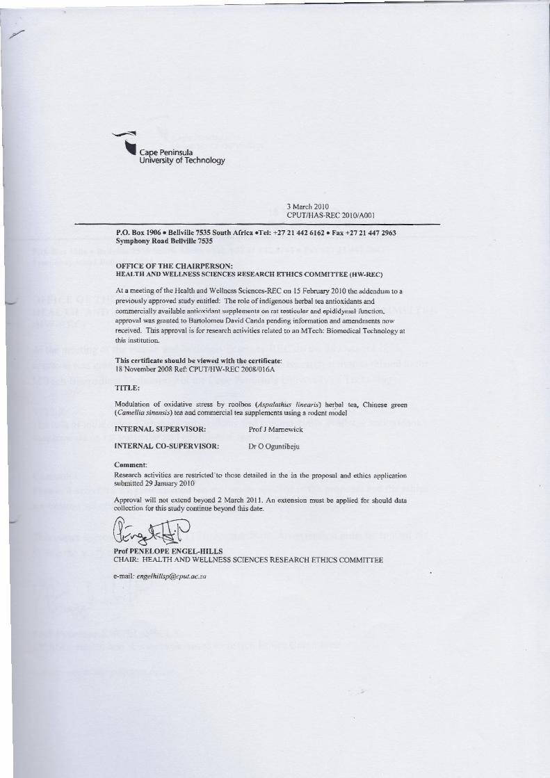
110
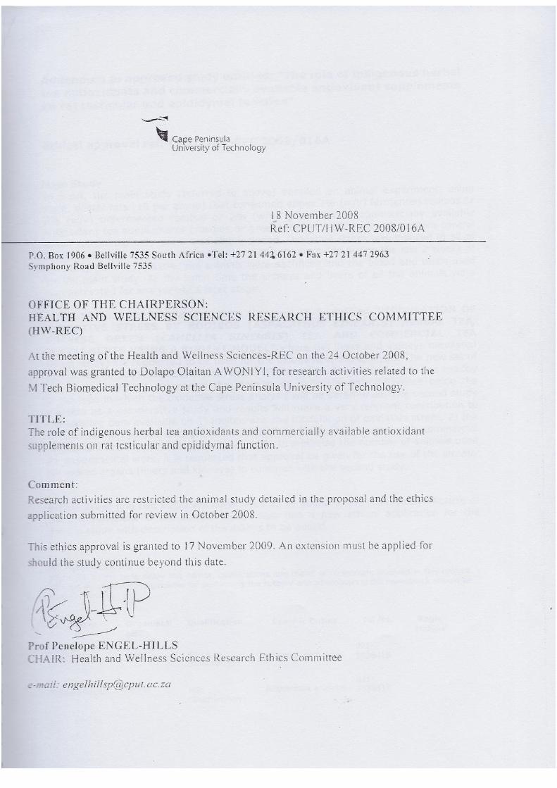
111
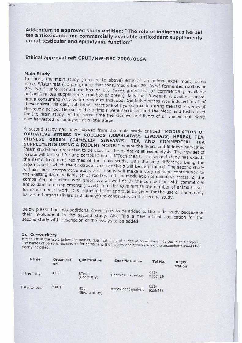
112

113