green chemistry oxidative modification of peptoids utilizing
TRANSCRIPT
University of Arkansas, FayettevilleScholarWorks@UARK
Theses and Dissertations
12-2017
Green Chemistry Oxidative Modification ofPeptoids Utilizing Bleach and TEMPOJesse Leland RobertsUniversity of Arkansas, Fayetteville
Follow this and additional works at: http://scholarworks.uark.edu/etd
Part of the Chemical Engineering Commons, Medicinal and Pharmaceutical ChemistryCommons, and the Medicinal-Pharmaceutical Chemistry Commons
This Thesis is brought to you for free and open access by ScholarWorks@UARK. It has been accepted for inclusion in Theses and Dissertations by anauthorized administrator of ScholarWorks@UARK. For more information, please contact [email protected], [email protected].
Recommended CitationRoberts, Jesse Leland, "Green Chemistry Oxidative Modification of Peptoids Utilizing Bleach and TEMPO" (2017). Theses andDissertations. 2581.http://scholarworks.uark.edu/etd/2581
Green Chemistry Oxidative Modification of Peptoids Utilizing Bleach and TEMPO
A thesis submitted in partial fulfillment of the requirements for the degree of
Master of Science in Engineering
by
Jesse Leland Roberts University of Arkansas
Bachelor of Science in Chemistry, 2016
December 2017 University of Arkansas
This thesis is approved for recommendation to the Graduate Council. _________________________________ Dr. Shannon Servoss Thesis Director
_________________________________ _________________________________ Dr. Jamie Hestekin Dr. Wei Shi Committee Member Committee Member
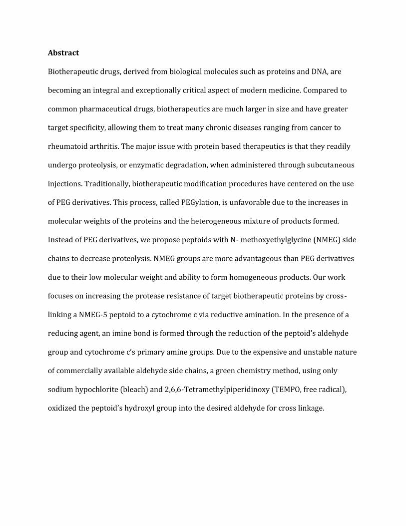
Abstract
Biotherapeutic drugs, derived from biological molecules such as proteins and DNA, are
becoming an integral and exceptionally critical aspect of modern medicine. Compared to
common pharmaceutical drugs, biotherapeutics are much larger in size and have greater
target specificity, allowing them to treat many chronic diseases ranging from cancer to
rheumatoid arthritis. The major issue with protein based therapeutics is that they readily
undergo proteolysis, or enzymatic degradation, when administered through subcutaneous
injections. Traditionally, biotherapeutic modification procedures have centered on the use
of PEG derivatives. This process, called PEGylation, is unfavorable due to the increases in
molecular weights of the proteins and the heterogeneous mixture of products formed.
Instead of PEG derivatives, we propose peptoids with N- methoxyethylglycine (NMEG) side
chains to decrease proteolysis. NMEG groups are more advantageous than PEG derivatives
due to their low molecular weight and ability to form homogeneous products. Our work
focuses on increasing the protease resistance of target biotherapeutic proteins by cross-
linking a NMEG-5 peptoid to a cytochrome c via reductive amination. In the presence of a
reducing agent, an imine bond is formed through the reduction of the peptoid’s aldehyde
group and cytochrome c’s primary amine groups. Due to the expensive and unstable nature
of commercially available aldehyde side chains, a green chemistry method, using only
sodium hypochlorite (bleach) and 2,6,6-Tetramethylpiperidinoxy (TEMPO, free radical),
oxidized the peptoid’s hydroxyl group into the desired aldehyde for cross linkage.
©2017 by Jesse Leland Roberts All Rights Reserved
Table of Contents
1. Introduction .................................................................................................................................... 1 1.1. Biotherapeutic Proteins ..................................................................................................... 2 1.2. Biotherapeutic Drug Delivery .......................................................................................... 3 1.3. PEGylation ............................................................................................................................... 8
1.3.1. Complications with PEGylation......................................................................................... 10 1.4. Peptoids (Poly-N-Substituted Glycines) .....................................................................13
1.4.1. Peptoid Synthesis ................................................................................................................... 15 1.4.2. NMEGylation ............................................................................................................................. 16
1.5. Green Chemistry .................................................................................................................18 1.5.1. Green Chemistry Oxidation ................................................................................................ 19 1.5.2. Bleach/TEMPO Oxidation ................................................................................................... 21
2. Research Rationale .....................................................................................................................22
3. Materials .........................................................................................................................................24
4. Methods ..........................................................................................................................................25 4.1. Peptoid Synthesis ...............................................................................................................25 4.2. Peptoid Purification ..........................................................................................................26 4.3. Peptoid Characterization .................................................................................................26 4.4. Oxidation of Alcohol to Aldehyde .................................................................................27 4.5. Cross-linkage Reaction .....................................................................................................27 4.6. Trypsin Degradation .........................................................................................................27
5. Results and Discussion ..............................................................................................................28 5.1. Peptoid Sequence Rationale and Characterization ................................................28 5.2. Oxidation of Alcohol to Aldehyde .................................................................................32 5.3. Cross-linkage (NMEGylation) .........................................................................................39
6. Conclusion and Future Work ..................................................................................................43
7. Acknowledgements ....................................................................................................................46
8. Works Cited ...................................................................................................................................47
1
1. Introduction
Biotherapeutic drugs derived from biological molecules are becoming an integral, and very
critical aspect of modern medicine. The term “biotherapeutics” can represent a large class
of treatments that are produced from cytokines, growth factors, hormones, antibodies, and
other regulatory proteins or peptides [1, 2]. These therapeutics are typically produced or
extracted using genetically engineered bacteria, yeast, fungi, and other cell types [3].
Biotherapeutic drugs have been used for decades to help treat multiple pathophysiological
illnesses including cancer, hemophilia, infectious diseases, inflammatory and autoimmune
diseases, and other rare diseases [4, 5, 6]. These larger biotherapeutic drugs are
significantly favored over common pharmaceuticals due to their increased size, advantages
in target specificity, and non-toxicity factors within the body [7]. The first biotherapeutic
drug, high-quality human insulin, was derived from recombinant DNA and produced by Eli
Lilly in 1982 [8]. Since, over 150 biotherapeutic medicines have been developed to improve
treatment options and patient quality of life [9]. These advancements are critical in
improving the accuracy and effectiveness of current treatment methods. For instance, since
the 1970’s the 10-year survival rate of cancer patients has nearly doubled from one in four
patients to one in two patients [10]. Although drastically increased, it leaves room for
further improvement, ultimately opening the door for research and the eventual use of
biotherapeutics as a treatment option.
2
1.1. Biotherapeutic Proteins The use of proteins as potential biotherapeutic agents is gaining interest at an intriguing
rate. In recent years, many distinct proteins have been discovered, and can be linked to the
underlying mechanistic pathways of several common diseases. Researchers have estimated
that between 25,000 and 40,000 functional genes that code for these proteins have been
discovered within the human genome [11]. Using alternative splicing of genes and
posttranslational modifications of proteins, these human genes have the potential to code
for the distinct proteins that are being found within disease mechanisms [12, 13, 14].
Biotherapeutics are typically classified into three main groups of proteins, based on their
physiological properties and course of treatment. The first group is made up of peptides
and small protein therapeutics including growth factors, hormones, and cytokines [15].
Two current therapeutics within this class are Epogen®, a form of erythropoietin protein
commonly used to increase the body’s production of red blood cells in anemia patients, and
Neupogen®, a protein used to boost the body’s production of white blood cells for
neutropenia patients [16, 17].
The second group consists of non-immune therapeutic proteins including replacement
enzymes, blood factors, anticoagulants, and other recombinant proteins [15]. The FDA
approved drug Myozyme uses a recombinant human α-glucosidase enzyme for treatment
in patients with Pompe Disease, an autosomal recessive myopathy that causes an abnormal
storage of glycogen in tissues, resulting in premature fatalities [18, 19]. Tissue plasminogen
activator (t-PA), one of the only successful treatment options for ischemic stroke victims,
falls under this class of biotherapeutics. The naturally occurring protein, t-PA, serves as an
3
anticoagulant by converting the inactive proenzyme plasminogen into an active serine
protease plasmin. In 1980, t-PA was first identified in melanoma cells, and later scientists
were able isolate and purify the protein, creating today’s biotherapeutic [20].
The third class of biotherapeutic proteins includes therapeutic antibodies and Fc-like
fusion proteins. Monoclonal antibodies (mAbs) have shown great success as
biotherapeutics for the treatment in autoimmune diseases due to the robust and flexible
nature of the immunoglobulin molecule and their highly specific antigen-binding
capabilities [15]. Immunoglobulin G (IgG) serves as one of the main types of antibodies
found in the blood and extracellular fluid, functioning as a control mechanism for infections
within body tissues. Antibodies and Fc-like fusion proteins serve as practical and viable
means for the treatment of cancers, autoimmune, and inflammatory diseases [21, 22]. All
three classes of biothereapeutic proteins provide excellent insight into the future of
medicine, thus many pharmaceutical and biotechnology companies are investing
substantial resources for their discovery and development. However, as promising as these
drugs may be, there are still limitations in the mode of action, manufacturing and
characterization techniques, and drug delivery methods.
1.2. Biotherapeutic Drug Delivery Although biotherapeutic proteins have proven successful as treatment options for various
diseases, there are still complications with delivery. Traditional routes, including oral, sub
mucosal (nasal), parenteral (injection), and transdermal (through the skin) [23], are not
feasible due to enzyme degradation and low absorption efficiency [24]. The oral delivery of
4
biotherapeutic proteins faces issues with poor absorbance within the gastrointestinal
system and chemical degradation due to harsh enzymes within the digestive system,
resulting in the loss of activity and function [25]. Proteins are extremely sensitive, where
even the smallest change in conformation can cause a complete loss of function [26]. It is
important to note that the pH within the colon and ileum is much higher than any other
region in the GI tract, so difficulties arise when developing pH-controlled therapeutics.
These pH-sensitive drugs are prone to degradation within the colon’s harsh environment
[27].
Nasal drug delivery is of interest due to the high vascularity and permeability within the
nasal mucosa [28, 29]. These desirable characteristics stem from the nasal cavity’s large
surface area, porous endothelial membrane, and highly vascularized epithelium [30]. Nasal
drug delivery may be great for small molecules, but issues arise with high molecular weight
compounds (above 1 kDa). There are also volume limitations in that the volume per dose
that can be permeated across the membrane is restricted to 25-200 microliters [31]. As
seen with other delivery methods, the body’s immune defense mechanism bodes an even
bigger issue. Mucocillary clearance is the most important physiological defense mechanism
inside the nasal cavity. If the biotherapeutic causes any irritation in the nasal mucosa, then
this mechanism will cause the drug to be rapidly diluted, increasing the clearance by
forming nasal mucus that will be eliminated from the nose [32].
Subcutaneous injections and transdermal administration routes are challenging due to
immunogenic potential and unwanted immune responses [33]. It has been reported that
5
subcutaneous degradation occurs with protein-based drugs due to the lymphatic transfer
of these proteins when delivered parenterally [34]. The lymphatic system directly affects
the absorption and distribution of therapeutic proteins after administration through T-cell
responses initiated by skin-derived dendritic cells [35]. No matter the delivery route, any
introduction of a foreign protein into the body has the potential to illicit an immune
response, triggering the production of antibodies. The immune system is extremely
sensitive, in that it can detect three-dimensional structural differences between the
proteins native to the body and those being introduced [36]. For this reason, drug delivery
systems and post-translational modifications are growing in interest to combat the
immunogenicity issues of protein therapeutics.
Currently, most drug delivery systems (DDS) are within the colloidal size range (1-
1000nm), and act to release the drug at a controlled rate for a prolonged period of time
[37]. The drug is typically kept within a solid inner matrix that is layered by a permeable
outer polymeric membrane through which the drug diffuses [38]. Research efforts have
been focused on three main classes of DDS, including nanoparticles, liposome and other
lipid-based carriers, and polymer-drug conjugates [39]. Poly(lactic-co-glycolic acid) (PLGA)
nanoparticles are attractive as DDSs due to their biodegradability and biocompatibility,
FDA approval in parenteral administration systems, well-described production and
characterization methods, protections from drug degradation, sustained release
capabilities, possibility to modify surface properties, and target specificity for desired
organs or cells [40]. The ability to modify surface properties is an important property in
combatting cellular immune response and increasing cellular uptake of the drug. Surface
6
charge plays a major role in the interaction of the DDS with the cell. Studies have shown
that positive charged nanoparticles allow a higher extent of cellular uptake due to the ionic
interactions with the negatively charge cell membrane [41, 42, 43]. Despite these desirable
properties, PLGA-nanoparticles have their limitations when dealing with certain protein
therapeutics. The synthesis process of these nanoparticles involves factors and processes
that may destabilize the proteins. When loading the protein into the nanoparticle, a double
emulsion method is currently required leading to the aggregation of most proteins.
Depending on the hydrophilicity of the protein, interactions between PLGA and the protein
may also lead to the denaturing and aggregation of proteins [44]. It has been shown that
immunogenicity can be minimized by ensuring stability, while limiting the formation of
higher molecular weight protein aggregates [45]. Another issue associated with the use of
nanoparticles is the complexity of cellular uptake and the unknown stability and
cytotoxicity of the nanoparticles following metabolism. Evidence proves that the exocytosis
of nanoparticles is drastically slower than endocytosis, but there is little information on the
metabolism and long-term effects of these particles [46, 47].
Of the three main classes of drug delivery systems, liposomes and lipid-based carriers have
already had a major impact on targeted therapeutic protein delivery. Liposomes are
defined as phospholipid vesicles consisting of multiple lipid bilayers enclosing discrete
aqueous spaces [48]. Liposomes and lipid-based carriers are advantageous as drug delivery
systems due to their biocompatibility, ability to self-assemble, extended drug circulation
time, and their ability to carry multiple drugs at once [49]. Unlike PLGA-nanoparticles,
liposomes possess the ability to encapsulate both hydrophilic and hydrophobic protein
7
drugs. Hydrophilic protein therapeutics can be trapped in the aqueous center, and
hydrophobic proteins can be encapsulated in the bilayer membrane [50]. Water-soluble
drugs can be loaded onto the liposome or lipid carriers through passive or active loading,
depending on the functional groups and chemical environment. Passive loading, involving
the formation of liposomes within an aqueous solution of the drug is the simplest, but least
efficient method due to the limited loading capacity and waste of solution [51]. Active
loading is more efficient, taking advantage of the pH difference between the external and
internal liposome environments to allow the passage of drugs with charged functional
groups through the membrane. Non-water soluble drugs can be incorporated directly into
the membrane of the liposome or lipid-carrier; however, the drug to lipid ratio of the
membrane is important to the dexterity of the liposome [52, 53]. For anticancer drugs, the
antitumor efficacy is directly related to the drug release rate, and previous research
demonstrated that by varying the drug to liposome ratio, optimal drug release rates could
be achieved [54]. However, as with other therapeutic drug delivery methods, liposomes
and lipid-carriers are still susceptible to enzyme degradation and macrophages, primarily
in the spleen and liver. Therefore, to be used as treatment options for cancer and
inflammatory diseases (inflammatory bowel disease, rheumatoid arthritis, etc) it is
important to develop long-circulating liposomes that avoid the reticulo-endothelial system
(RES) [55]. To achieve this passive drug delivery, liposomal surface modifications must be
made to provide a steric boundary to the liposome that prevents RES uptake and blocks
degradative enzymes from attaching [56, 57]. The use of ganglioside, GM1, to modify the
liposome surface created “stealth” liposomes that were not readily taken up by the RES;
ultimately, allowing the carrier to stay in circulation for a longer period of time [58]. A
8
second modification method, PEGylation, utilizes the addition of polyethylene glycol (PEG)
groups onto the liposomal membrane surface to drastically improve the carrier’s
circulation time [59].
1.3. PEGylation Since the 1970’s, polyethylene-glycol (PEG) has been a highly-investigated polymer for the
attachment and modification of biological macromolecules for multiple pharmaceutical
applications. PEGylation is the covalent or noncovalent attachment of PEG polymers to
macromolecules, most typically peptides, proteins, and antibody fragments [60]. Each
individual PEG exemplifies many desired characteristics, including the absence of toxicity
and immunogenicity, high water-solubility, and low mass-dependent elimination from the
kidney [61, 62, 63]. Once conjugated, PEG sterically shields the protein’s surface from
degradative agents and RES uptake, decreasing the protein’s immunogenic response, thus
improving body-residence time. Along with the shielding effect, the increased molecular
weight of the PEG-protein conjugate is advantageous in reducing renal clearance and
altering biodistribution, also improving the residence time [64]. Renal clearance works by
selectively filtering blood components through the glomerular filtration barrier (GFB). The
GFB’s permeability is often dependent on the size and charge of the blood components [65].
While most proteins are selectively retained in the blood by the GFB, certain low molecular
weight proteins and degraded protein fragments can undergo rapid renal clearance [66,
67]. Thus, by increasing the protein’s molecular weight and improving the resistance to
degradative agents in the body, PEGylation serves as a viable modification method for
prolonging the body-residence time of potential therapeutic proteins [68].
9
Initially, researchers were skeptical that PEG could to be attached to large molecular
weight proteins while maintaining biological activity; therefore, they directed their work
solely on catalase and bovine serum albumin. For both molecules, PEGylation enhanced
circulation times and eliminated immunological responses, while ultimately maintaining
optimal protein activity and structural integrity [69, 70]. From these results, the efficacy of
PEGylation in improving therapeutic drug delivery was deemed successful, sparking
interest to further develop new PEGylated macromolecules. Since then, new methods for
PEG conjugation have been introduced resulting in a wide array of macromolecules that
can potentially be modified. To attach PEG to a molecule, it is important to functionalize
one or both PEG termini with a functional group that is chosen based on the reactive group
on the molecule being PEGylated. Amine conjugation, considered first-generation PEG
chemistry, is the most common technique for the attachment of PEG molecules to proteins
[71]. Amine reactive PEG derivatives form secondary amine linkages by substituting with
multiple nucleophilic amino acid groups (lysine, serine, tyrosine, cysteine, and histidine)
found in the protein, thus forming a heterogeneous mixture of PEG-protein conjugates [72].
First-generation PEG chemistry methods were first referred to as “gentle chemistry” due to
the mild reaction conditions and use of simple, linear PEG molecules [73]. This simplicity,
however, is important in maintaining the activity and three-dimensional structure of
proteins. As previously mentioned, proteins are extremely sensitive to their environment,
so it of utmost importance to limit the amount of harsh chemical used during conjugation.
Second-generation PEGylation methods refer to any newly developed method of PEG
conjugation that typically uses more complex PEG derivatives and harsher chemical
10
conditions than first-generation methods. These PEG derivatives, unlike the linear amine
derivatives used in first-generation chemistry, contain multiple different functional groups
such as aldehyde, carboxylic acid, and thiol groups [74, 75]. Depending on the desired
product and degree of PEGylation, altering the chemical environment for most of the
second-generation PEG derivatives creates a more site-specific PEGylation method. For
instance, by changing the pH of the environment to acidic, mPEG-priopionaldehyde, a PEG
derivative containing a reactive aldehyde group, selectively reacts with a protein’s N-
terminal α-amine, because nucleophilic substitution will only occur when the pH of the
molecule is near the residue’s pKa [76, 77].
1.3.1. Complications with PEGylation Since the 1970’s, concrete evidence has proven PEGylation as a viable manipulation
method to improve pharmacokinetic properties of biotherapeutic proteins. PEG-protein
conjugates display the “stealth” properties that are desired for the optimal drug
deliverance of biotherapeutic agents [78]. PEGylation allows for the therapeutic protein to
“sneak” by the body’s immune system by sterically shielding the protein’s surface from
degradative agents, while also maintaining the water solubility and protein activity needed
to be a viable treatment option [79]. Although PEGylation seems to be an ideal
manipulation method, several limitations arise during the characterization and purification
processes for these newly formed conjugates. First-generation PEGylation relies on the
coupling of PEG derivatives to different reactive amino acids on the protein. Most
therapeutic proteins rely on non-specific PEGylation occurring through the between
hydroxyl- or aldehyde- functionalized PEG monomers and amine groups found on lysine
11
side chains or the N-terminus of the protein [80]. This coupling method, although highly
reactive at physiological conditions, occurs at random positions, ultimately producing a
heterogeneous mixture of PEG-conjugates. Since lysine makes up nearly 10% of all protein
amino acids, it is incredibly difficult to characterize exactly which and how many lysine
residues on the protein were PEGylated. These heterogeneous PEG-protein conjugates,
called “isomers”, differ in molecular weights, protein stabilities, and even in the level of
activities [81]. In 2003, the PEGylation of INF-α2a produced nine different isomers, each
differing in the level of bioactivity. The difference in bioactivity of these isomers was
theorized to directly affect the interferon receptor binding kinetics and stabilities [82]. The
heterogeneity of PEG-protein conjugates lowers molecular activity of the therapeutic
protein causing variations in treatment mechanisms and clinical side effects [83]. For FDA
approval of non-site specific PEGylated protein drugs, the individual PEG-protein
conjugates must be fully characterized, and biological analyses must be run on each
conjugate to determine their pharmacodynamic properties. The more homogenous the
product is, the better chance it has at getting approved by the FDA [63]. To achieve a
homogeneous PEG-protein conjugate, site-specific coupling or effective purification
methods must be incorporated. Purification methods are not only costly, but are inefficient
and difficult as well. The purification will need to separate three molecules (PEG, PEG-
protein, native protein), where the separation of PEG and native protein is simple, using
filtration and size-exclusion methods [84]. Difficulties arise when trying to isolate the
desired PEG-protein conjugate from the others due to similar characteristics between the
conjugates. As a result, several different methods, such as Ion Exchange Chromatography
and Hydrophobic Interaction Chromatography, are completed in succession to fully purify
12
the product [85]. With consideration to the high manufacturing costs associated with the
current production of therapeutic proteins, the production and purification of PEGylated
proteins is economically infeasible. The overall process to achieve a high purity product is
at the direct expense of high yield. To make up for the loss in yield, protein production will
need to be increased, ultimately driving up manufacturing costs exponentially. Therefore,
the more homogeneous the PEG-protein conjugate product formed, the less purification
needed, decreasing the amount of protein lost, while improving the cost of manufacturing.
To combat the heterogeneity issues with amine-coupling (first generation) researchers are
developing second generation PEGylation methods that are focused on a more site-specific
coupling of PEG derivatives to amino acids on the protein. The attachment of PEG to the
thiol group of cysteine is considered site-specific because it accounts for only 1% of the
total amino acid content in proteins. However, many of these cysteine groups will undergo
disulfide bonding with each other, lowering the number of active thiol groups suitable for
PEGylation. The undesired bonding between cysteine residues results in very few proteins
possessing active cysteine groups capable of reacting with PEG monomers. To fix this
dilemma, researchers are interested in introducing site-specific cysteines to the protein
sequence through genetic engineering, but little is known regarding the effect this will have
on protein activity [86]. Various other amino acids and functional groups have been of
interest for second generation PEGylation, but most, if not all, have limitations that void
them as suitable coupling agents. For instance, arginine is another amino acid that is less
abundant than lysine, but has similar reaction chemistry involving the coupling of PEG to
an amine group. Based on these two parameters, one could assume it to be a perfect option
13
for site-specific PEGylation producing an active, monodisperse PEG-protein conjugate.
These assumptions are proven to be false, as the coupling requires long reaction times that
drastically decrease protein stability and site specificity [87, 88]. Carboxyl groups can be
PEGylated, but only when amines are not present, virtually eliminating the use with
proteins and peptides [89]. Like carboxyl groups, hydroxyl groups are only suitable for
PEGylation for uses in non-peptide moieties such as matrices for chromatography and
biocompatible surfaces [90]. The second generation PEGylation methods have potential to
be effective, site-specific coupling mechanisms, but each method is limited to its own
specifications resulting in a narrow range of proteins that can be modified.
While PEGylation has a countless number of unique functionalities, it presents major
drawbacks in modifying biotherapeutic proteins due to the heterogeneity of PEG resulting
in characterization and purification limitations. Along with the characterization and
purification, being able to maintain native protein activity, while improving in-vivo drug
half-life is crucial in developing novel modifier of biotherapeutics.
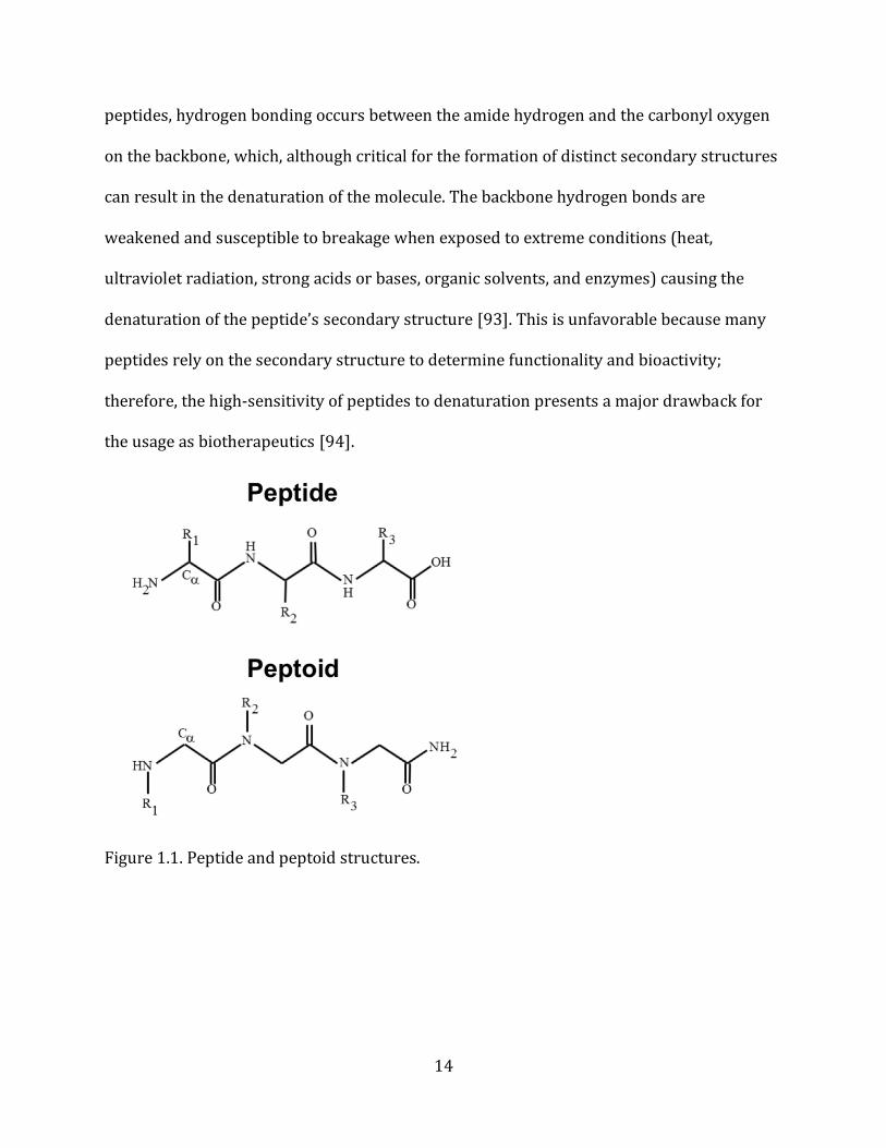
1.4. Peptoids (Poly-N-Substituted Glycines) Peptoids, or N-substituted glycines, are synthetic peptidomimetic oligomers that
structurally resemble 𝛼-peptides, but have side chains attached to the amide groups on the
backbone instead of the 𝛼-carbon as in peptides (Figure 1.1.) [91]. This structural
modification generates an achiral backbone that eliminates the potential for hydrogen
bonding, resulting in a protease-resistant polymer that exhibits good cell permeability and
protein binding characteristics resembling that of more “drug-like” molecules [92]. In
14
peptides, hydrogen bonding occurs between the amide hydrogen and the carbonyl oxygen
on the backbone, which, although critical for the formation of distinct secondary structures
can result in the denaturation of the molecule. The backbone hydrogen bonds are
weakened and susceptible to breakage when exposed to extreme conditions (heat,
ultraviolet radiation, strong acids or bases, organic solvents, and enzymes) causing the
denaturation of the peptide’s secondary structure [93]. This is unfavorable because many
peptides rely on the secondary structure to determine functionality and bioactivity;
therefore, the high-sensitivity of peptides to denaturation presents a major drawback for
the usage as biotherapeutics [94].
Figure 1.1. Peptide and peptoid structures.
15
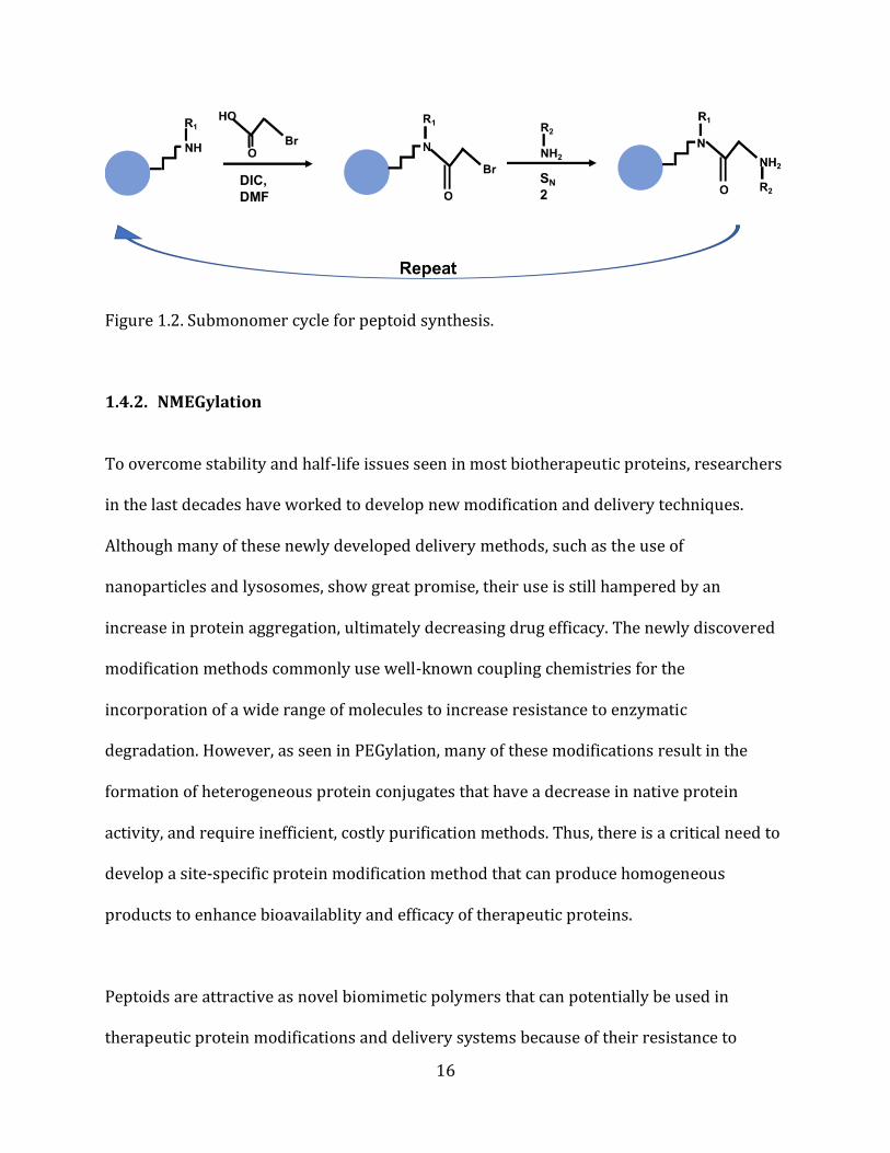
1.4.1. Peptoid Synthesis Peptoids can be produced via a sequence-specific, solid-phase synthesis method
comparable to that of peptides. Unlike peptide synthesis, where submonomers must be
protected prior to addition, peptoid synthesis allows for the precise addition of
unprotected submonomers greatly simplifying the process. The submonomer method is a
highly efficient, low cost synthesis technique that allows for the addition of a wide variety
of side chains as primary amines. Using a solid-phase support (Ex: Rink Amide Resin),
submonomers are added from carboxylic to amine termini via a submonomer “cycle” made
up of two-steps: (1) acylation and (2) amination (nucleophilic substitution) (Figure 1.2.)
[91]. The first reaction of the submonomer cycle, acylation, adds an activated carboxylic
acid derivative onto a receptive amine generating a tertiary amide bond. In general,
bromoacetic acid and diisopropylcarbodiimide (DIC) are used for acylation. The
bromoacetic acid is activated by DIC separately, and then added to the solid-phase support
[95]. The second step in the cycle, amination, involves the nucleophilic displacement of the
halide (typically bromine) by a primary or N-terminal secondary amine (side-chain). As the
halide group is removed from the haloacetamide, the primary nitrogen submonomer
attacks the alpha-carbon forming an ammonium salt. The halide ion then removes
hydrogen from the ammonium salt producing hydrogen bromide [95, 96]. The amination
step creates the molecular diversity that is present in peptoids due to the thousands of
commercially available amine side-chains.
16
Figure 1.2. Submonomer cycle for peptoid synthesis.
1.4.2. NMEGylation To overcome stability and half-life issues seen in most biotherapeutic proteins, researchers
in the last decades have worked to develop new modification and delivery techniques.
Although many of these newly developed delivery methods, such as the use of
nanoparticles and lysosomes, show great promise, their use is still hampered by an
increase in protein aggregation, ultimately decreasing drug efficacy. The newly discovered
modification methods commonly use well-known coupling chemistries for the
incorporation of a wide range of molecules to increase resistance to enzymatic
degradation. However, as seen in PEGylation, many of these modifications result in the
formation of heterogeneous protein conjugates that have a decrease in native protein
activity, and require inefficient, costly purification methods. Thus, there is a critical need to
develop a site-specific protein modification method that can produce homogeneous
products to enhance bioavailablity and efficacy of therapeutic proteins.
Peptoids are attractive as novel biomimetic polymers that can potentially be used in
therapeutic protein modifications and delivery systems because of their resistance to
17
degradative enzymes, great cell permeability, and low immunogenicity [97]. The peptoid
sequence is fully customizable through the addition of side chains with various chemistries.
By incorporating side chains with specific functional groups into the sequence, common
coupling chemistries can be used to cross-link the peptoid to peptides, proteins,
nanoparticles, and other molecules [98]. Protein modifications involving sequence specific
peptoids offers a promising means for overcoming stability and absorption issues
commonly displayed by current biotherapeutics.
Like PEGylation, the challenge still lies in forming homogenous products that maintain
native protein bioactivity and conformation [99]. Since PEG groups have traditionally
shown an increased stability to serum enzymes, but failed to produce homogeneous
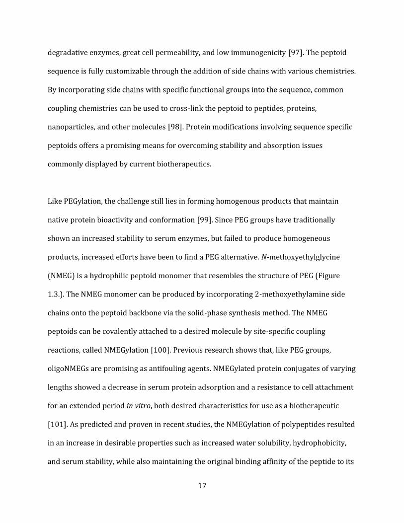
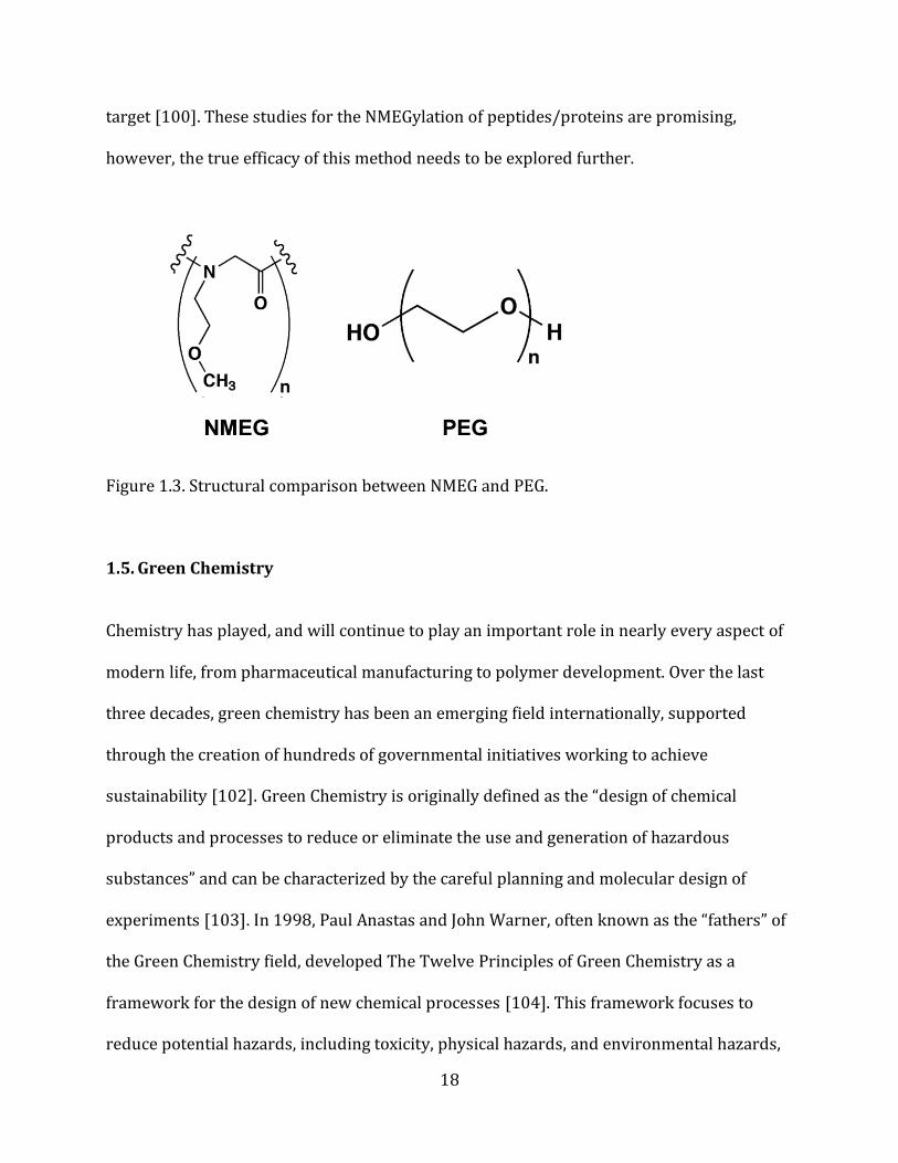
products, increased efforts have been to find a PEG alternative. N-methoxyethylglycine
(NMEG) is a hydrophilic peptoid monomer that resembles the structure of PEG (Figure
1.3.). The NMEG monomer can be produced by incorporating 2-methoxyethylamine side
chains onto the peptoid backbone via the solid-phase synthesis method. The NMEG
peptoids can be covalently attached to a desired molecule by site-specific coupling
reactions, called NMEGylation [100]. Previous research shows that, like PEG groups,
oligoNMEGs are promising as antifouling agents. NMEGylated protein conjugates of varying
lengths showed a decrease in serum protein adsorption and a resistance to cell attachment
for an extended period in vitro, both desired characteristics for use as a biotherapeutic
[101]. As predicted and proven in recent studies, the NMEGylation of polypeptides resulted
in an increase in desirable properties such as increased water solubility, hydrophobicity,
and serum stability, while also maintaining the original binding affinity of the peptide to its
18
target [100]. These studies for the NMEGylation of peptides/proteins are promising,
however, the true efficacy of this method needs to be explored further.
Figure 1.3. Structural comparison between NMEG and PEG.
1.5. Green Chemistry Chemistry has played, and will continue to play an important role in nearly every aspect of
modern life, from pharmaceutical manufacturing to polymer development. Over the last
three decades, green chemistry has been an emerging field internationally, supported
through the creation of hundreds of governmental initiatives working to achieve
sustainability [102]. Green Chemistry is originally defined as the “design of chemical
products and processes to reduce or eliminate the use and generation of hazardous
substances” and can be characterized by the careful planning and molecular design of
experiments [103]. In 1998, Paul Anastas and John Warner, often known as the “fathers” of
the Green Chemistry field, developed The Twelve Principles of Green Chemistry as a
framework for the design of new chemical processes [104]. This framework focuses to
reduce potential hazards, including toxicity, physical hazards, and environmental hazards,
19
across all stages in chemical processes [105]. The first principle, waste prevention, involves
the reduction in the amount of waste produced. Since the amount of waste produced is
often directly correlated to many of the remaining principles, it is considered to be the
most impactful in developing a “Green” chemical process. Chemical companies have started
investing in waste management techniques, in part due to the increased public awareness
in the environment, but mainly because of a governmental increase in the cost of waste
removal, amounting to nearly 40% of the overall production costs [106]. When analyzing
the waste management process, it is important to look at the efficiency of the chemical
reaction taking place. Yield, chemical selectivity, atom efficiency, energy spent, solvent
usage, and renewable raw materials are all impactful to the overall efficiency of the
reaction and can be managed to decrease waste production [107, 108, 109, 110].
1.5.1. Green Chemistry Oxidation Inspired by The Principles of Green Chemistry, the work of researchers has focused on
developing “greener” chemical reactions that reduce or eliminate the use and generation of
toxic chemicals. The original focus has been on waste elimination, or what is known as the
E factor (environmental factor). The E factor is a metric used to quickly assess the
environmental effect of manufacturing processes, and is typically measured in kg waste per
kg product [111]. To determine the true E-factor value and amount of waste produced, it is
important to look into the stoichiometric equation for the overall process [112]. Along with
the E factor, in 1991, using what is known as “atom economy”, scientists began
investigating the reaction efficiency and where in the process the waste content originates.
By analyzing waste production of inefficient processes (high E factor values), they found an
20
increased amount of organic salts, metal (Na, Mg, Zn, Fe) and metal hydride (LiAlH4,
NaBH4) reducing agents and oxidants such as permanganate, manganese dioxide, and
chromium (VI) reagents [113]. These results prove the important role that catalysis and
solvent choice play in the development of green chemistry reactions [114].
Traditionally, catalytic oxidation of alcohols (primary, secondary, allylic, propargylic, etc)
to aldehydes, ketones, and carboxylic acids require the incorporation of many harsh
reagents. Chromium (VI) oxides are well-known oxidants for the conversion of primary and
secondary alcohols to aldehydes and ketones; however, this method requires the use of
harsh organic solvents such as pyridine, dimethylformamide (DMF) with sulfuric acid, and
others [115, 116, 117, 118]. The handling of chromium (VI) compounds is crucial due to its
chronic toxicity and contamination of product. A study of workers exposed to chromium
(VI) compounds have reported the development of asthma and other signs of respiratory
distress, accompanied by a 20% decrease in forced expiratory volume of the lungs [119].
The Swern oxidation, typically using dimethylsulfoxide (DMSO), oxalyl chloride, and
trimethylamine in methylene chloride solvent does not produce heavy metal waste
products, thus, making it a more environmentally friendly option [120]. However,
drawbacks still exist in that oxalyl chloride is known to have toxic and corrosive properties,
and methylene chloride is carcinogenic, hepatopathic, and neuropathic [121]. Other
methods, such as the use of manganese dioxide (MnO), can oxidize allylic and benzylic
alcohols, but are still faced with hazardous and toxic reagents, and the potential for
residual metal contamination [122].
21
1.5.2. Bleach/TEMPO Oxidation The oxidation of primary alcohols to aldehydes and carboxylic acids is a fundamental
transformation in organic chemistry. To create a more green chemistry oxidation method
researchers have been investigating the use of nitroxyl radicals and transition metal salts.
Both nitroxyl radicals tetramethylpiperidine-N-oxyl (TEMPO) radical and phthalimide-N-
oxyl (PINO) radical in the presence of small amounts of manganese (II) nitrate and cobalt
(II) nitrate have displayed excellent results in the aerobic oxidation of benzyl alcohols to
aldehydes and carboxylic acids [123]. However, the oxidation with these reagents is limited
when dealing with less reactive aliphatic and allylic alcohols [124].
The development of Anelli’s oxidation procedure has proven that aliphatic primary
alcohols can be oxidized into aldehydes and carboxylic acids in a more efficient, green
chemistry manner, by reacting the alcohol with a dichloromethane-water mixture
containing bleach (sodium hypochlorite) in the presence of sodium bicarbonate, potassium
bromide, and a catalytic amount of TEMPO free radical [125, 126]. TEMPO free radical
serves as the primary oxidant for the transformation of alcohols into aldehydes by forming
reactive oxoammonium salts. The secondary oxidant, sodium hypochlorite, is typically used
to activate the TEMPO free radical by forming the oxoammonium salt, but subsequently
plays a major role as the primary oxidant in the conversion of aldehydes to carboxylic
acids. Typically, the reaction is done in an excess of sodium hypochlorite and a phase-
transfer catalyst forming to form a high yield of carboxylic acid [127]. However, by
eliminating the phase-transfer catalyst and lowering the amount of sodium hypochlorite
used the oxidation can produce aldehydes with low reaction times [128]. This modified
22
version of Anelli’s procedure allows for the fast, efficient, and low-cost oxidation of primary
alcohols to aldehydes.
2. Research Rationale
While knowledge of different types of protein delivery systems and modification methods
has grown in the last decades, the difficulty in characterization, susceptibility to enzymatic
degradation, loss of stability and bioactivity, and economic costs continue to hinder them
as viable treatment options. Thus, there is a critical need to develop a post-translational
modification method that produces homogeneous conjugates that can be easily
characterized and purified. This technique should increase protein resistance to enzymatic
degradation and maintain native protein stability and bioactivity. The covalent attachment
of NMEG-peptoids (NMEGylation) is a promising method to achieve this goal due to its
favorable properties, such as an increase in water solubility and serum stability. It is
believed that NMEGylation can fix the heterogeneity issues seen in PEGylation by
modifying proteins in a site-specific manner. Unlike PEG-conjugates, NMEG peptoids allow
for the precise positioning of specific chemical functional groups for attachment to reactive
amino acids on the protein.
The overall goal of this project is to develop a protein modification method that improves
the serum stability and efficacy of biotherapeutic proteins to be used as potential treatment
options. We hypothesize that the NMEGylation of a target protein will result in the
production of a homogenous protein-peptoid conjugate that withstands enzymatic
degradation and displays native protein conformation and activity. The modification will
23
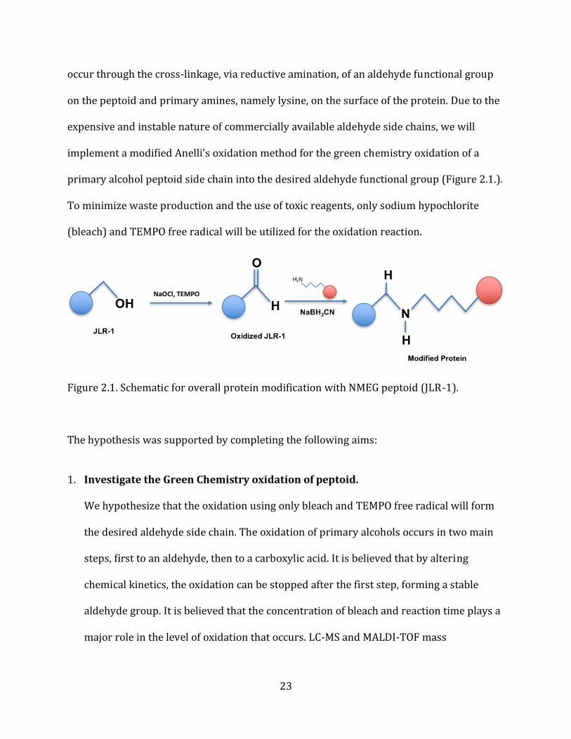
occur through the cross-linkage, via reductive amination, of an aldehyde functional group
on the peptoid and primary amines, namely lysine, on the surface of the protein. Due to the
expensive and instable nature of commercially available aldehyde side chains, we will
implement a modified Anelli’s oxidation method for the green chemistry oxidation of a
primary alcohol peptoid side chain into the desired aldehyde functional group (Figure 2.1.).
To minimize waste production and the use of toxic reagents, only sodium hypochlorite
(bleach) and TEMPO free radical will be utilized for the oxidation reaction.
Figure 2.1. Schematic for overall protein modification with NMEG peptoid (JLR-1).
The hypothesis was supported by completing the following aims: 1. Investigate the Green Chemistry oxidation of peptoid.
We hypothesize that the oxidation using only bleach and TEMPO free radical will form
the desired aldehyde side chain. The oxidation of primary alcohols occurs in two main
steps, first to an aldehyde, then to a carboxylic acid. It is believed that by altering
chemical kinetics, the oxidation can be stopped after the first step, forming a stable
aldehyde group. It is believed that the concentration of bleach and reaction time plays a
major role in the level of oxidation that occurs. LC-MS and MALDI-TOF mass
24
spectrometry will be used to determine the level of oxidation, and to confirm the
formation of a stable aldehyde side chain.
2. Investigate the cross-linkage of the NMEG peptoid to a target protein.
We hypothesize that, in the presence of a reducing agent, the NMEG peptoid can be
cross-linked to a target protein. The aldehyde group on the peptoid can be reacted, via
reductive amination, with primary amines of lysines on the protein. Due to the
abundance of lysine amino acids, it is predicted that multiple peptoid molecules will
cross-link to the protein, drastically improving the ability of the protein to withstand
protease degradation. By performing the reaction at physiological conditions, it is
believed that the native activity and conformation of the protein will be maintained.
Analytical HPLC and MALDI-TOS mass spectrometry will be used to confirm the cross-
linkage of peptoid to protein. Tricine SDS-PAGE gel and trypsin degradation assays will
be performed to assess enzymatic resistance and protein stability.
3. Materials
MBHA rink amide resin was purchased from NovaBiochem (Gibbstown, NJ). The amine
sub-monomer tert-butyl N-(4-aminobutyl) carbamate was purchased from CNH
Technologies Inc. (Woburn, MA), 2-methoxyethylamine and ethanolamine were purchased
from Acros Organics (Pittsburgh, PA). Piperidine was purchased from Sigma-Aldrich (St.
Louis, MO). Sodium hypochlorite came from original Clorox™ purchased at Walmart
(Bentonville, AR). 2,2,6,6-Tetramethylpiperidinooxy (TEMPO, free radical) was purchased
from BeanTown Chemical Corporation (Hudson, NH). Sodium cyanoborohydride was
purchased from Alfa Aesar (Haverhill, MA). All other reagents and consumables were
25
purchased from VWR (Radnor, PA) and were used without further modification, unless
otherwise specified.
4. Methods
4.1. Peptoid Synthesis Peptoids were synthesized via the solid-phase submonomer method on MBHA rink amide
resin [91]. Initially, the resin was swelled with dimethylformamide (DMF), then the Fmoc
protection group was removed by two separate incubations in 20% piperidine solutions in
DMF for 30 seconds and again for 12 minutes. The resin’s secondary amine was acylated
with a fresh solution of 0.4 M bromoacetic acid and N,N’-diisopropyl carbodiimide (DIC) at
a ratio of 4.25:0.8, mixing for 1 minute, forming a tertiary amine bond. The amine side
chains were attached via nucleophilic displacement at concentrations ranging from 0.5-1.0
M in DMF depending on the side chain. The two-step submonomer cycle was repeated until
the desired sequence was obtained (Figure 5.1.). The peptoid was cleaved from the resin by
mixing with 95% trifluoroacetic acid (TFA), 2.5% water, and 2.5% triisopropylsilane on an
orbital shaker (Belly Dancer, Stovall Life Sciences, Greensboro, NC). The acid was removed
using a Heidolph Laborota 4001 rotary evaporator (Elk Grove Village, IL), the peptoid was
dissolved in a 50:50 acetonitrile:water solution, and dried to a powder using a Labconco
lyophilyzer (Kansas City, MO).
26
4.2. Peptoid Purification The peptoid was reconstituted in a 25:75 acetonitrile:water solution and purified using a
Waters Delta 600 preparative reversed-phase high performance liquid chromatography
(HPLC) (Milford, MA) with a Duragel G C18 150 x 20 mm column (Peeke Scientific, Novato,
CA). Due to the hydrophilic nature of the NMEG peptoid, equilibration times were extended
from 20 minutes to 60 minutes, the injection volume was reduced to 1.2 mL per injection,
and a 10-minute delay following injection was necessary prior to the linear gradient of 0-
65% solvent B (94.9% acetonitrile, 5% water, 0.1% TFA) in A (99.9% water, 0.1% TFA)
over 65 minutes. Peptoids were confirmed to be >98% pure via analytical HPLC (Waters
2695 Separation Module) with a Duragel G C18 150 x 2.1 mm column (Peeke Scientific,
Novato, CA) using a gradient of 5-95% solvent D (99.9% acetonitrile, 0.1% TFA) in C
(99.9% water, 0.1% TFA) over 30 minutes. Purified peptoid fractions were combined,
lyophilized into a powder, and stored at -20 °C.
4.3. Peptoid Characterization Synthesis and purification of the desired peptoid sequences were confirmed via matrix
assisted laser desorption/ionization time of flight (MALDI-TOF; Bruker, Billerica, MA) mass
spectrometry using 2,5-dihydroxybenzoic acid as a matrix substance. The oxidation and
cross-linkage products were confirmed via a combination of liquid chromatography-mass
spectrometry (LC-MS) and MALDI-TOF mass spectrometry.
27
4.4. Oxidation of Alcohol to Aldehyde To form the desired aldehyde from the oxidation reaction, a stoichiometric equivalent
amount (1:1) of sodium hypochlorite to peptoid was used. The reaction solution contained
0.1% bleach, 1% of 1.4 M TEMPO free radical, and 25% peptoid in phosphate buffered
saline (PBS). The concentrations of bleach and peptoid were varied to optimize reaction
yield. The reaction time was varied from 0 to 3 hours at 23 °C and a pH of 7.4.
4.5. Cross-linkage Reaction Prior to the oxidation reaction, a 2 M amine reducing agent was prepared by dissolving
sodium cyanoborohydride in a 5 M NaOH solution and allowing it to incubate for 1 hour
before use. A 10% protein and water solution was prepared by a ten-fold dilution of 4
mg/mL protein solution. The 10% protein and sodium cyanoborohydride solutions were
added to the oxidated peptoid reaction solution. The reaction time was varied from 0 to 4
hours at 33 °C and a pH of 7.4. At hour increments, the reaction was assessed by analytical
HPLC using a gradient of 5 to 95% solvent D (99.9% acetonitrile, 0.1% TFA) in C (99.9%
water, 0.1% TFA) over 30 minutes. The reaction product at each time point was spotted
and the cross-linkage was confirmed by MALDI-TOF.
4.6. Trypsin Degradation The protein was dissolved in a 50 mM ammonium bicarbonate solution. A 500 mM 1,4-
Dithiothreitol (DTT) solution was added to the protein sample to a final concentration of 20
mM (1:25 dilution), then incubated at 60 °C for 1 hour. A fresh solution of 1 M 3-
28
indoleacetic acid (IAA) was prepared using 50 mM ammonium bicarbamate. The 1 M IAA
solution was added to the reduced protein sample to a final concentration of 40 mM (1:25
dilution) and allowed to incubate at room temperature for 30 minutes protected from light.
The alkylation reaction was quenched by adding 500 mM DTT to a final concentration of 10
mM (1:50 dilution). Trypsin solution was added to the sample to form a final protease to
protein ratio of 1:30 to 1:100 (w/w). The final solution was incubated at 37 °C for 24 hours
and stored at -20 °C to stop the digestion reactions. The digested fractions were analyzed
using MALDI-TOF and a Tricine-SDS-PAGE gel.
5. Results and Discussion
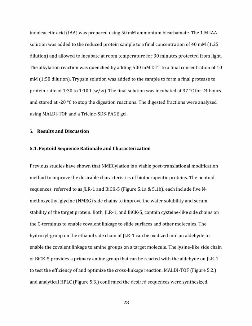
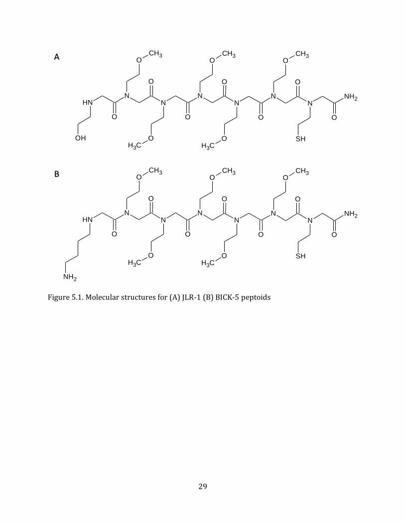
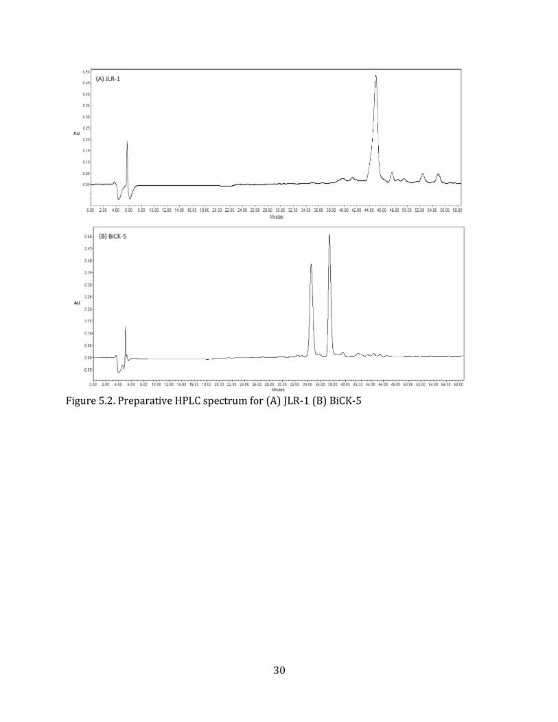
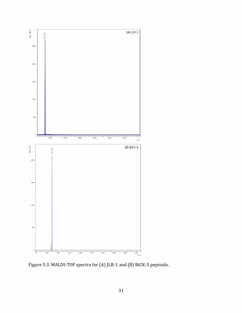
5.1. Peptoid Sequence Rationale and Characterization Previous studies have shown that NMEGylation is a viable post-translational modification
method to improve the desirable characteristics of biotherapeutic proteins. The peptoid
sequences, referred to as JLR-1 and BiCK-5 (Figure 5.1a & 5.1b), each include five N-
methoxyethyl glycine (NMEG) side chains to improve the water solubility and serum
stability of the target protein. Both, JLR-1, and BiCK-5, contain cysteine-like side chains on
the C-terminus to enable covalent linkage to slide surfaces and other molecules. The
hydroxyl-group on the ethanol side chain of JLR-1 can be oxidized into an aldehyde to
enable the covalent linkage to amine groups on a target molecule. The lysine-like side chain
of BiCK-5 provides a primary amine group that can be reacted with the aldehyde on JLR-1
to test the efficiency of and optimize the cross-linkage reaction. MALDI-TOF (Figure 5.2.)
and analytical HPLC (Figure 5.3.) confirmed the desired sequences were synthesized.
29
Figure 5.1. Molecular structures for (A) JLR-1 (B) BICK-5 peptoids
NHN
NN
NN
NNH2
O
O
O
O
O
O
O
SH
OCH3
OCH3
OCH3
OCH3
OCH3
NH2
NHN
NN
NN
NNH2
O
O
O
O
O
O
O
SH
OCH3
OCH3
OCH3
OCH3
OCH3
OH
A
B
30
Figure 5.2. Preparative HPLC spectrum for (A) JLR-1 (B) BiCK-5
AU
AU
31
Figure 5.3. MALDI-TOF spectra for (A) JLR-1 and (B) BiCK-5 peptoids.
32
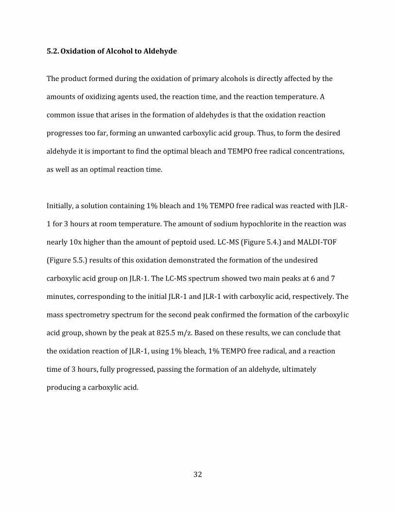
5.2. Oxidation of Alcohol to Aldehyde The product formed during the oxidation of primary alcohols is directly affected by the
amounts of oxidizing agents used, the reaction time, and the reaction temperature. A
common issue that arises in the formation of aldehydes is that the oxidation reaction
progresses too far, forming an unwanted carboxylic acid group. Thus, to form the desired
aldehyde it is important to find the optimal bleach and TEMPO free radical concentrations,
as well as an optimal reaction time.
Initially, a solution containing 1% bleach and 1% TEMPO free radical was reacted with JLR-
1 for 3 hours at room temperature. The amount of sodium hypochlorite in the reaction was
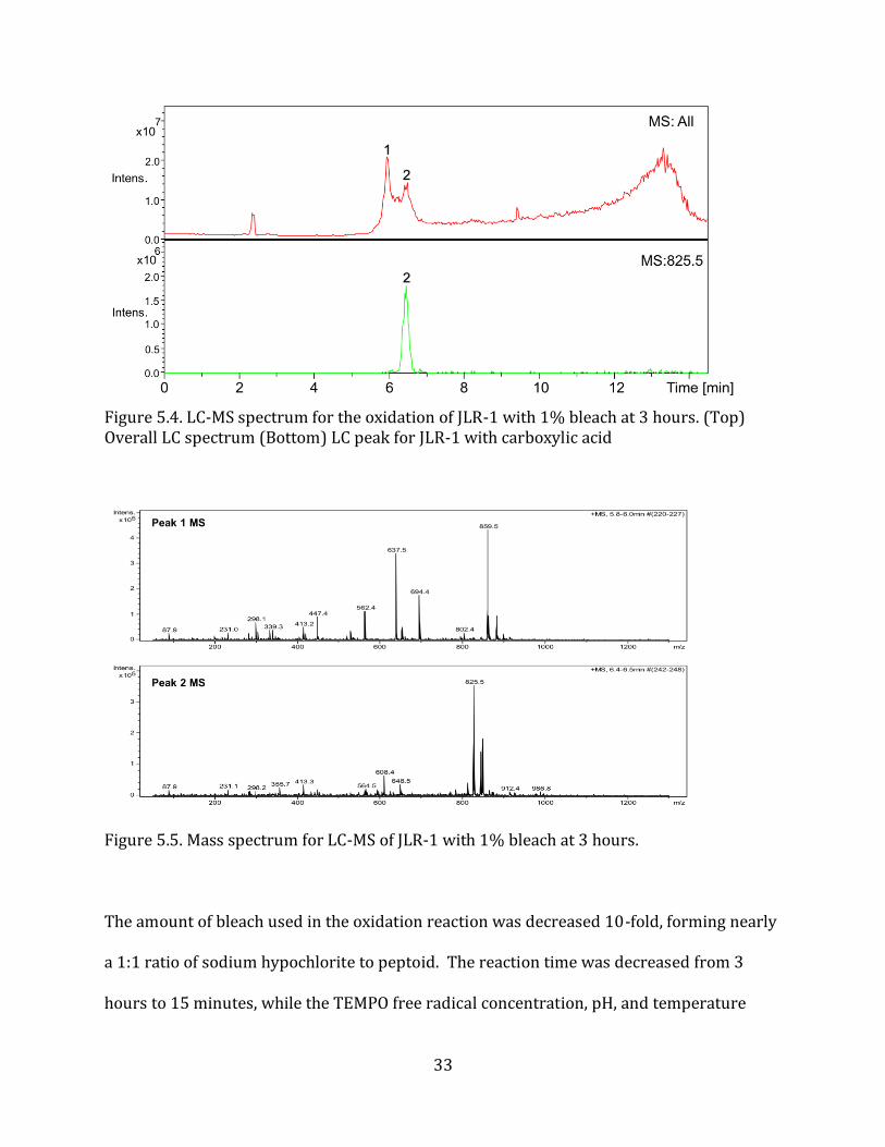
nearly 10x higher than the amount of peptoid used. LC-MS (Figure 5.4.) and MALDI-TOF
(Figure 5.5.) results of this oxidation demonstrated the formation of the undesired
carboxylic acid group on JLR-1. The LC-MS spectrum showed two main peaks at 6 and 7
minutes, corresponding to the initial JLR-1 and JLR-1 with carboxylic acid, respectively. The
mass spectrometry spectrum for the second peak confirmed the formation of the carboxylic
acid group, shown by the peak at 825.5 m/z. Based on these results, we can conclude that
the oxidation reaction of JLR-1, using 1% bleach, 1% TEMPO free radical, and a reaction
time of 3 hours, fully progressed, passing the formation of an aldehyde, ultimately
producing a carboxylic acid.
33
Figure 5.4. LC-MS spectrum for the oxidation of JLR-1 with 1% bleach at 3 hours. (Top) Overall LC spectrum (Bottom) LC peak for JLR-1 with carboxylic acid
Figure 5.5. Mass spectrum for LC-MS of JLR-1 with 1% bleach at 3 hours.
The amount of bleach used in the oxidation reaction was decreased 10-fold, forming nearly
a 1:1 ratio of sodium hypochlorite to peptoid. The reaction time was decreased from 3
hours to 15 minutes, while the TEMPO free radical concentration, pH, and temperature
34
were kept constant. To determine the time at which oxidation occurs, the reaction solution
was spotted for MALDI-TOF at time points of 0, 1, 5, and 15 minutes.
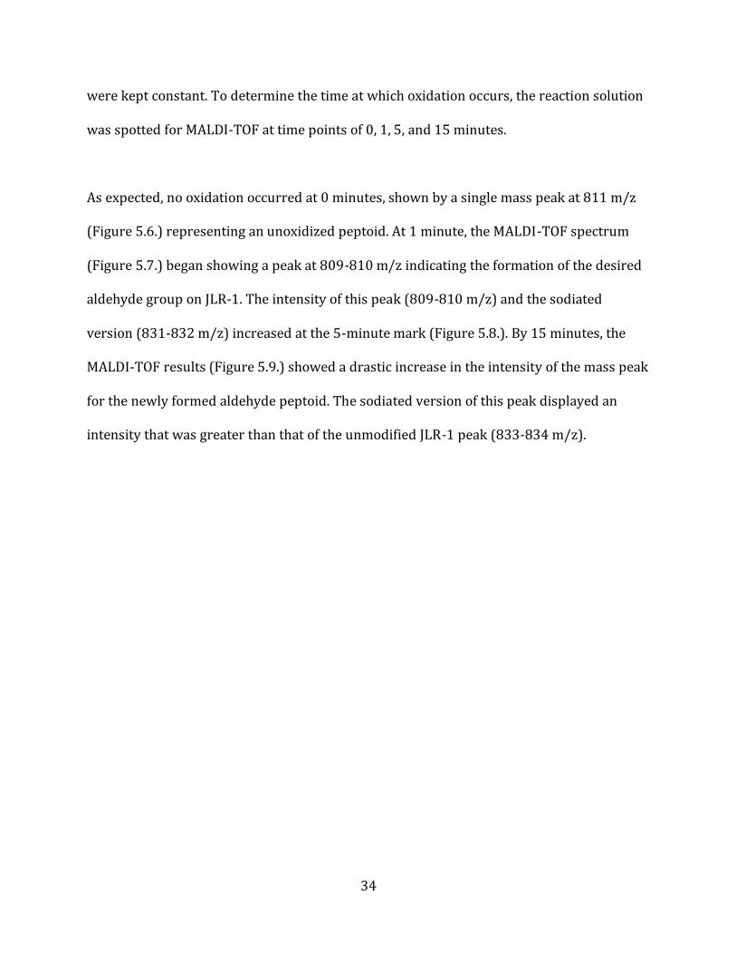
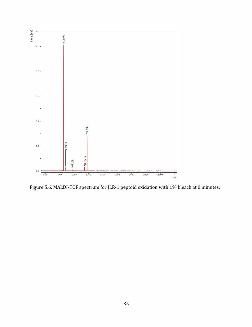
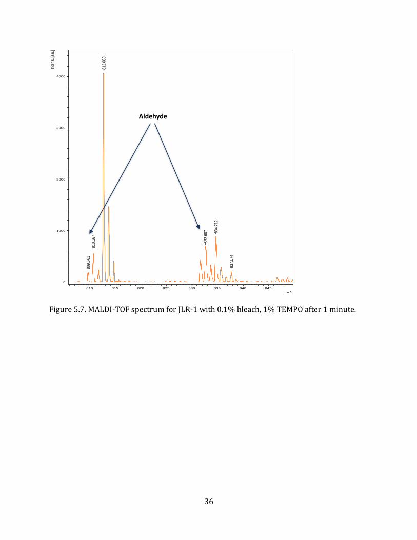
As expected, no oxidation occurred at 0 minutes, shown by a single mass peak at 811 m/z
(Figure 5.6.) representing an unoxidized peptoid. At 1 minute, the MALDI-TOF spectrum
(Figure 5.7.) began showing a peak at 809-810 m/z indicating the formation of the desired
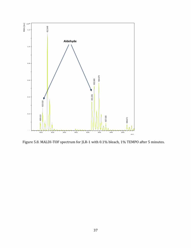
aldehyde group on JLR-1. The intensity of this peak (809-810 m/z) and the sodiated
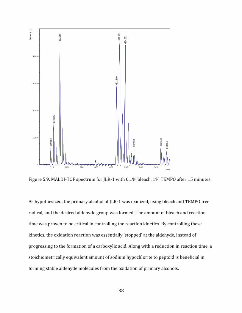
version (831-832 m/z) increased at the 5-minute mark (Figure 5.8.). By 15 minutes, the
MALDI-TOF results (Figure 5.9.) showed a drastic increase in the intensity of the mass peak
for the newly formed aldehyde peptoid. The sodiated version of this peak displayed an
intensity that was greater than that of the unmodified JLR-1 peak (833-834 m/z).
35
Figure 5.6. MALDI-TOF spectrum for JLR-1 peptoid oxidation with 1% bleach at 0 minutes.
811.
972
1222
.388
1178
.271
849.
979
964.
108
0.0
0.2
0.4
0.6
0.8
1.0
4x10In
tens
. [a.
u.]
500 750 1000 1250 1500 1750 2000 2250 2500
m/z
36
Figure 5.7. MALDI-TOF spectrum for JLR-1 with 0.1% bleach, 1% TEMPO after 1 minute.
812.
680
834.
712
810.
667
832.
687
809.
661
837.
674
0
1000
2000
3000
4000
Inte
ns. [
a.u.
]
810 815 820 825 830 835 840 845
m/z
Aldehyde
37
Figure 5.8. MALDI-TOF spectrum for JLR-1 with 0.1% bleach, 1% TEMPO after 5 minutes.
812.
649
834.
679
832.
669
831.
691
810.
618
809.
619
846.
671
837.
630
0.0
0.2
0.4
0.6
0.8
1.0
1.2
4x10In
tens
. [a.
u.]
810 815 820 825 830 835 840 845
m/z
Aldehyde
38
Figure 5.9. MALDI-TOF spectrum for JLR-1 with 0.1% bleach, 1% TEMPO after 15 minutes.
As hypothesized, the primary alcohol of JLR-1 was oxidized, using bleach and TEMPO free
radical, and the desired aldehyde group was formed. The amount of bleach and reaction
time was proven to be critical in controlling the reaction kinetics. By controlling these
kinetics, the oxidation reaction was essentially ‘stopped’ at the aldehyde, instead of
progressing to the formation of a carboxylic acid. Along with a reduction in reaction time, a
stoichiometrically equivalent amount of sodium hypochlorite to peptoid is beneficial in
forming stable aldehyde molecules from the oxidation of primary alcohols.
812.
644
834.
672
832.
654
831.
690
810.
585
809.
589
846.
669
837.
598
848.
654
0
1000
2000
3000
4000
Inte
ns. [
a.u.
]
810 815 820 825 830 835 840 845
m/z
39
5.3. Cross-linkage (NMEGylation) The reductive amination reaction was tested by cross-linking the JLR-1 and BiCK-5
peptoids. The aldehyde group on JLR-1 covalently attaches to the primary amine on BiCK-5
in the presence of a strong reducing agent (sodium cyanoborohydride). The reaction
product was analyzed by MALDI-TOF, which shows a mass peak at 1670 m/z that could
potentially indicate a successful cross-linkage (Figure 5.10.). The expected molecular
weight of the JLR-1 and BiCK-5 cross-linkage is 1631 Da; however, the 1670 m/z peak
could represent the cross-linked peptoid in the presence of a potassium ion (+39 m/z).
Unfortunately, the 1670 m/z peak could also be due to the undesired reaction between
thiol groups on the cysteines of both peptoids. This thiol linkage results in an expected
peptoid conjugate mass of 1647-1648 Da, which if sodiated, could also be representative of
the 1670 peak.
40
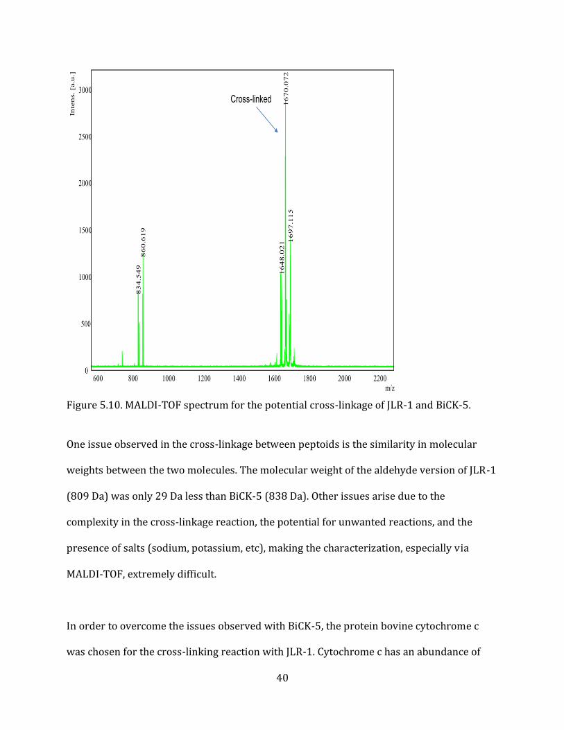
Figure 5.10. MALDI-TOF spectrum for the potential cross-linkage of JLR-1 and BiCK-5. One issue observed in the cross-linkage between peptoids is the similarity in molecular
weights between the two molecules. The molecular weight of the aldehyde version of JLR-1
(809 Da) was only 29 Da less than BiCK-5 (838 Da). Other issues arise due to the
complexity in the cross-linkage reaction, the potential for unwanted reactions, and the
presence of salts (sodium, potassium, etc), making the characterization, especially via
MALDI-TOF, extremely difficult.
In order to overcome the issues observed with BiCK-5, the protein bovine cytochrome c
was chosen for the cross-linking reaction with JLR-1. Cytochrome c has an abundance of
41
lysine amino acids, 18, that could be modified in the amination reaction, increasing the
chances for successful NMEGylation. Immediately following the oxidation of JLR-1, bovine
cytochrome c and sodium cyanoborohydride were added to the peptoid solution and
allowed to react for 4 hours at physiological conditions (temperature of 33 °C and a pH of
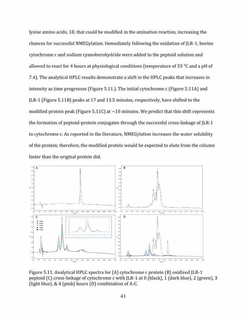
7.4). The analytical HPLC results demonstrate a shift in the HPLC peaks that increases in
intensity as time progresses (Figure 5.11.). The initial cytochrome c (Figure 5.11A) and
JLR-1 (Figure 5.11B) peaks at 17 and 13.5 minutes, respectively, have shifted to the
modified protein peak (Figure 5.11C) at ~10 minutes. We predict that this shift represents
the formation of peptoid-protein conjugates through the successful cross-linkage of JLR-1
to cytochrome c. As reported in the literature, NMEGylation increases the water solubility
of the protein; therefore, the modified protein would be expected to elute from the column
faster than the original protein did.
Figure 5.11. Analytical HPLC spectra for (A) cytochrome c protein (B) oxidized JLR-1 peptoid (C) cross-linkage of cytochrome c with JLR-1 at 0 (black), 1 (dark blue), 2 (green), 3 (light blue), & 4 (pink) hours (D) combination of A-C.
42
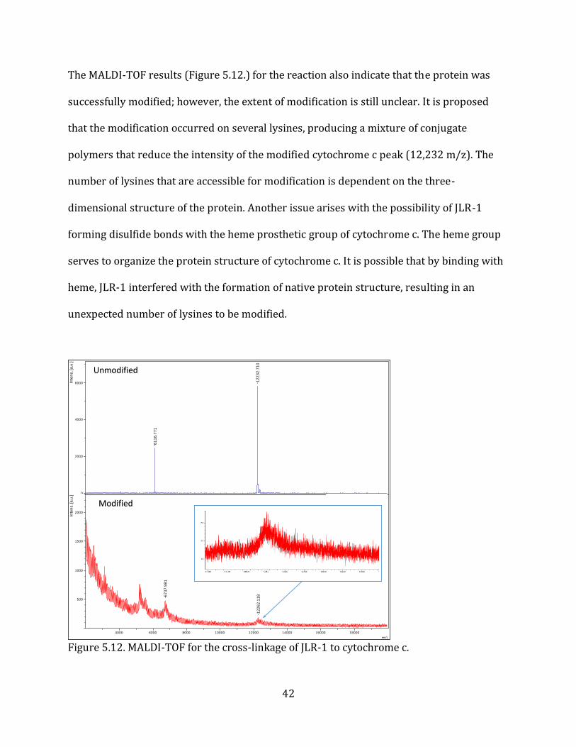
The MALDI-TOF results (Figure 5.12.) for the reaction also indicate that the protein was
successfully modified; however, the extent of modification is still unclear. It is proposed
that the modification occurred on several lysines, producing a mixture of conjugate
polymers that reduce the intensity of the modified cytochrome c peak (12,232 m/z). The
number of lysines that are accessible for modification is dependent on the three-
dimensional structure of the protein. Another issue arises with the possibility of JLR-1
forming disulfide bonds with the heme prosthetic group of cytochrome c. The heme group
serves to organize the protein structure of cytochrome c. It is possible that by binding with
heme, JLR-1 interfered with the formation of native protein structure, resulting in an
unexpected number of lysines to be modified.
Figure 5.12. MALDI-TOF for the cross-linkage of JLR-1 to cytochrome c.
12
23
2.7
10
61
16
.77
1
74331\cyc_12717_control\1Ref
0
2000
4000
6000Inte
ns. [a
.u.]
67
37
.98
1
12
26
2.1
18
74331\cyc_JLR1_12717\1SRef
0
500
1000
1500
2000
Inte
ns. [a
.u.]
4000 6000 8000 10000 12000 14000 16000 18000
m/z
Unmodified
Modified
43
6. Conclusion and Future Work
The need for accurate, effective, and long-lasting biotherapeutic treatment options has led
to the development of more intricate modification methods and drug delivery systems.
Biotherapeutics require evolutionary advancements in serum stability and drug efficacy to
be beneficial in future medicinal applications. The challenge lies in developing modification
methods that form homogeneous products in a manner that native protein conformation
and activity is maintained. We believe that the use of peptoids for the modifications of
biotherapeutic proteins provides a propitious mode to achieve such goals.
We were able to successfully design a green chemistry oxidation method that modified a
peptoid side chain to be used as a reactive functional group in the NMEGylation of a target
protein. We found that by altering the reaction time and concentrations of reagents, the
reaction rate can effectively be controlled and the desired oxidative product can be
obtained. We demonstrated this ability by forming a stable aldehyde functional group from
the oxidation of a primary alcohol using only bleach and TEMPO free radical as oxidative
agents. Initially, higher bleach concentrations and longer reaction times caused the
reaction to by-pass the aldehyde, and directly form a carboxylic acid. Through a ten-fold
dilution in bleach concentration and a reduction in reaction time to 15 minutes, we were
able to produce the desired aldehyde. Sodium hypochlorite activates the TEMPO free
radical which serves as the main oxidant for the reaction. We believe that the reduction in
sodium hypochlorite limited the amount of TEMPO free radical that was activated;
ultimately stopping the reaction at the aldehyde. It is important to note that although the
reaction was successful in producing the desired product, there is still room for
44
improvement in the efficiency of the reaction. It would be interesting to assess the effect of
lowering the TEMPO concentration on aldehyde formation. The ratios of TEMPO and bleach
can be varied to optimize the aldehyde production. If the amount of bleach and TEMPO can
be reduced even further, then it would make it more environmentally friendly, improving
the E-factor rating of the reaction. Also, instead of going straight into the cross-linkage it
would be intriguing to investigate purification methods, and stability tests for the newly
formed aldehyde peptoid.
We have studied the attachment of NMEG peptoid to a target protein to overcome stability
and absorption issues displayed by current biotherapeutics. We proposed that an aldehyde
group on the peptoid can be reductively aminated with primary amines on the target
protein to form a homogeneous NMEG peptoid-protein conjugate. It is believed that this
conjugate will carry some of the desirable properties seen in PEG and NMEG monomers,
such as increased water solubility and resistance to enzymatic degradation. We have
evaluated the use of NMEG peptoids for site-specific modification to biotherapeutic
proteins; however, are still limited in characterization techniques. Initially, the amination
reaction was tested between two peptoids, JLR-1 and BiCK-5, but due to similarities in
molecular weights the product could not be fully characterized. It is believed that the cross-
linked product shown on MALDI-TOF is an unwanted disulfide linkage formed between the
cysteine groups on both peptoids. To eliminate this possibility, our lab will further
investigate the synthesis and usage of peptoids without cysteine side chains. Also, the
molecular weight disparity can be increased by synthesizing peptoids with various NMEG
chain lengths (n=5, 10, 15).
45
We have demonstrated that the NMEGylation of target proteins results in many of the same
limitations that currently hinder PEGylation as a viable modification method. When
evaluating the cross-linkage of JLR-1 to cytochrome c, the mass spectrometry results
indicated that a cross-linkage between the two molecules possibly occurred, but the degree
of modification (number of lysines modified) could not be determined. From the MALDI-
TOF results in Figure 3.10., we believe that the number of lysines modified with peptoid
varied from molecule to molecule, forming a heterogeneous mixture of peptoid-protein
conjugates. The covalent bonding between thiol functional group on the cysteine side chain
of JLR-1 and the heme group of cytochrome c may play an important role in the
characterization issues. Any factor, whether it’s the binding to heme, or a change in
reaction conditions, can cause the protein to unfold, revealing hidden amino acids that are
now able to react. Depending on the reaction environment, each individual protein
molecule may undergo a unique conformational change, resulting in a wide array of
reactive groups. It is important to note that although heterogeneous conjugates were
potentially formed, the viability of NMEGylation as a biotherapeutic protein modification
method needs to be further investigated. It would be interesting to complete a more
thorough study on the cross-linkage of various NMEG peptoids to different target proteins.
To avoid potential heme binding, several target proteins, such as myoglobin, can be used in
place of cytochrome c. Other peptoid sequences can be adopted to pursue different
conjugation chemistries with the target protein. Trypsin assays and Tricine-SDS-PAGE gel
electrophoresis can be used to further analyze the reaction products formed and to test
resistance of the newly developed protein-peptoid conjugate to enzymatic degradation.
46
7. Acknowledgements
I would like to thank my advisor and mentor Dr. Shannon Servoss for her generous help,
providing me with the materials needed, and advice concerning the project. I would also
like to thank Dr. Tammy Rechtin for training within the lab, help with data analysis, and
guidance towards the project. In addition, I would like to thank my fellow graduate
students, Dr. German Perez-Bakovic, Helya Najafi, and Neda Mahmoudi for their generous
guidance and assistance in the lab, and for taking the time to help me when needed. I would
like to thank the thesis committee members Dr. Jamie Hestekin and Dr. Wei Shi for their
valued input and guidance towards this project and the completion of my degree. I would
also like to thank Dr. Rohana Liyanage and the Arkansas Statewide Mass Spectrometry
Facility for the use and consultation regarding MALDI and LC-MS.
47
8. Works Cited
[1] R. Langer, "Biomaterials in Drug Delivery and Tissue Engineering: One Laboratory's Experience," Accounts of Chemical Research, vol. 33, no. 2, pp. 94-101, 2000.
[2] J. Elvin, R. Couston and C. van der Walle, "Therapeutic antibodies: Market considerations, disease targets and bioprocessing," International Journal of Pharmaceutics, vol. 440, no. 1, pp. 83-98, 2012.
[3] F. Steinberg and J. Raso, "Biotech Pharmaceuticals and Biotherapy: An Overview," J Pharm Pharmaceut Sci, vol. 1, no. 2, pp. 48-59, 1998.
[4] C. May, P. Sapra and H.-P. Gerber, "Advances in bispecific biotherapeutics for the treatment of cancer," Biochemical Pharmacology, vol. 84, no. 9, pp. 1105-1112, 2012.
[5] I. Ivens, A. Baumann, T. McDonald, T. Humphries, L. Michaels and P. Mathew, "PEGylated therapeutic proteins for haemophilia treatment: a review for haemophilia caregivers," Haemophilia, vol. 19, no. 1, pp. 11-20, 2012.
[6] Y. Vugmeyster, J. Harrold and X. Xu, "Absorption, Distribution, Metabolism, and Excretion (ADME) Studies of Biotherapeutics for Autoimmune and Inflammatory Conditions," The AAPS Journal, vol. 14, no. 4, pp. 714-727, 2012.
[7] D. AlDeghaither, B. G. Smaglo and L. M. Weiner, "Beyond Peptides and mAbs - Current Status and Future Perspectives for Biotherapeutics with Novel Constructs," Journal of Clinical Pharmacology, vol. 55, no. 3, pp. 4-20, 2015.
[8] M. R. Ladisch and K. L. Kohlmann, "Recombinant Human Insulin," Biotechnology Progress, vol. 8, no. 6, pp. 469-478, 1992.
[9] M. The, "Human Insulin: DNA technology's first drug," Am J Hosp Pharm, vol. 46, no. 2, pp. 9-11, 1989.
[10] A. Jemal, B. F., C. M.M., F. J., E. Ward and D. Forman, "Global cancer statistics.," CA: A Cancer Journal for Clinicians. , vol. 61, no. 2, pp. 69-90, 2011.
[11] K. R. Chi, "The dark side of the human genome.," Nature, vol. 538, 2016.
[12] D. Brett, H. Pospisil, J. Valcarcel, J. Reich and P. Bork, "Alternative splicing and genome complexity," Nature Genetics, vol. 30, pp. 29-39, 2001.
[13] W. Gilbert, "Why genes in pieces?," Nature, vol. 271, no. 5645, p. 501, 1978.
48
[14] E. Kim, A. Goren and G. Ast, "Alternative splicing: current perspectives," Bioessays, vol. 30, no. 1, pp. 38-47, 2008.
[15] X. Zhong, P. Neumann, M. Corbo and E. Loh, Recent Advances in Biotherapeutics Drug Discovery and Development, InTech, 2011.
[16] C. Winearls, D. Oliver, M. Pippard, C. Reid, M. Downing and P. Cotes, "Effect of human erythropoietin derived from recombinant DNA on the anaemia of patients maintained by chronic haemodialysis," The Lancet, vol. 328, no. 8517, pp. 1175-1178, 1986.
[17] A. C. Herman, T. C. Boone and H. S. Lu, "Characterization, Formulation, and Stability of Neupogen® (Filgrastim), a Recombinant Human Granulocyte-Colony Stimulating Factor," in Pharmaceutical Biotechnology, vol. 7, Boston, MA: Springer, 2002, pp. 303-328.
[18] S. Pascual, "Phenotype Variations in Early Onset Pompe Disease: Diagnosis and Treatment Results with Myozyme," Advances in Experimental Medicine and Biology, vol. 652, no. 1, pp. 39-46, 2009.
[19] D. Orlikowski, N. Pellegrini, H. Prigent, P. Laforet, R. Carlier, B. Eymard, F. Lofaso and D. Annane, "Recombinant human acid alpha-glucosidase (rhGAA) in adult patients with severe respiratory failure due to Pompe disease.," Neuromuscular Disorder, vol. 21, no. 7, pp. 477-482, 2011.
[20] J. Loscalzo and E. Braunwald, "Tissue Plasminogen Activator," New England Journal of Medicine, vol. 319, pp. 925-931, 1988.
[21] S.-A. Kellermann and L. L. Green, "Antibody discovery: the use of transgenic mice to generate human monoclonal antibodies for therapeutics," Current Opinion in Biotechnology, vol. 13, no. 6, pp. 593-597, 2002.
[22] G. Fuh, "Synthetic antibodies as therapeutics," Expert Opinion on Biological Therapy, vol. 7, no. 1, pp. 73-87, 2007.
[23] D. S. Pisal, M. P. Kosloski and S. V. Balu-lyer, "Delivery of therapeutic proteins," Journal of Pharmaceutical Sciences, vol. 99, no. 6, pp. 2557-2575, 2010.
[24] E. Koren, L. Zuckerman and A. Mire-Sluis, " Immune Responses to Therapeutic Proteins in Humans - Clinical Significance, Assessment and Prediction," Current Pharmaceutical Biotechnology, vol. 3, no. 4, pp. 349-360, 2002.
49
[25] R. I. Mahato, A. S. Narang, L. Thoma and D. D. Miller, "Emerging Trends in Oral Delivery of Peptide and Protein Drugs," Critical Reviews in Therapeutic Drug Carrier Systems, vol. 20, no. 2-3, pp. 153-214, 2003.
[26] G. Lezzi, A. Frohlich, B. Ernst, F. Ampenberger, S. Saeland, N. Glaichenhaus and M. Kopf, "Lymp node resident rather than skin-derived dendritic cells initiate specific T cell responses after Leishmania major infection.," The Journal of Immunology, vol. 177, no. 2, pp. 1250-1256, 2006.
[27] A. K. Philip and B. Philip, "Colon Targeted Drug Delivery Systems: A Review on Primary and Novel Approaches," Oman Medical Journal, vol. 25, no. 2, pp. 79-87, 2010.
[28] Y. Chien and S. Chang, "Intranasal drug delivery for systemic medications.," Critical Reviews in Therapeutic Drug Carrier Systems, vol. 4, no. 2, pp. 67-194, 1987.
[29] M. Ugwoke, N. Verbeke and R. Kinget, "The biopharmaceutical aspects of nasal mucoadhesive drug delivery.," The Journal of Pharmacy and Pharmacology, vol. 53, no. 1, pp. 3-21, 2001.
[30] Y. Ozsoy, S. Gungor and E. Cevher, "Nasal Delivery of High Molecular Weight Drugs," Molecules, vol. 14, no. 9, pp. 3754-3779, 2009.
[31] P. Arora, S. Sharma and S. Garg, "Permeability issues in nasal drug delivery," Drug Discovery Today , vol. 7, no. 18, pp. 967-975, 2002.
[32] S. Gizurarson, "The Effect of Cilia and the Mucociliary Clearance on Successful Drug Delivery," Biological and Pharmaceutical Bulletin, vol. 38, no. 4, pp. 497-506, 2015.
[33] V. Knepp, J. Hadgraft and R. Guy, "Transdermal drug delivery: problems and possibilities.," Crit Rev Ther Drug Carrier Syst., vol. 4, no. 1, pp. 13-37, 1987.
[34] A. Fathallah, R. Bankert and S. Balu-lyer, "Immunogenecity of Subcutaneously Administered Therapeutic Proteins-a Mechanistic Perspective," American Association of Pharmaceutical Scientists, vol. 15, no. 4, pp. 897-900, 2013.
[35] C. Porter and S. Charman, "Lymphatic transport of proteins after subcutaneous administration," Journal of Pharmaceutical Sciences, vol. 89, no. 3, pp. 297-310, 2000.
[36] H. Schellekens, "Immunogenecity of Therapeutic Proteins: Clinical Implications and Future Prospects," Clinical Therapeutics, vol. 24, no. 11, pp. 1720-1740, 2002.
50
[37] V. Filipe, A. Hawe and W. Jiskoot, "Critical Evaluation of Nanoparticle Tracking Analysis (NTA) by NanoSight for the Measurement of Nanoparticles and Protein Aggregates," Pharmaceutical Research, vol. 27, no. 5, pp. 796-810, 2010.
[38] T. M. Allen and P. R. Cullis, "Drug Delivery Systems: Entering the Mainstream," Science, vol. 303, no. 5665, pp. 1818-1822, 2004.
[39] W. Jiskoot, R. M. van Schie, M. G. Carstens and H. Schellekens, "Immunological Risk of Injectable Drug Delivery Systems," Pharmaceutical Research, vol. 26, no. 6, pp. 1303-1314, 2009.
[40] F. Danhier, E. Ansorena, J. M. Silva, A. Le Breton and V. Preat, "PLGA-based nanoparticles: An overview of biomedical applications," Journal of Controlled Release, vol. 161, no. 2, pp. 505-522, 2012.
[41] S. M. Sadat, S. T. Jahan and A. Haddadi, "Effects of Size and Surface Charge of Polymeric Nanoparticles on in Vitro and in Vivo Applications," Journal of Biomaterials and Nanobiotechnology, vol. 7, pp. 91-108, 2016.
[42] I. Bala, S. Hariharan and M. Ravi Kumar, "PLGA Nanoparticles in Drug Delivery: The State of the Art," Critical Reviews in Therapeutic Drug Carrier Systems, vol. 21, no. 5, pp. 387-422, 2004.
[43] J. Panyam and V. Labhasetwar, "Biodegradable nanoparticles for drug and gene delivery to cells and tissue," Advanced Drug Delivery Reviews, vol. 55, no. 3, pp. 329-347, 2003.
[44] S. M. Samani and B. Taghipour, "PLGA micro and nanoparticles in delivery of peptides and proteins; problems and approaches.," Pharmaceutical Development and Technology, vol. 20, no. 4, pp. 385-393, 2014.
[45] A. S. Rosenberg, " Effects of Protein Aggregates: An Immunologic Perspective," The AAPS Journal , vol. 8, no. 3, pp. 501-507, 2006.
[46] E. Frohlich, "The role of surface charge in cellular uptake and cytotoxicity of medical nanoparticles," International Journal of Nanomedicine, vol. 7, pp. 5577-5591, 2012.
[47] G. Gulati, N. Shafiq and S. Malhotra, "Drug-Loaded PLGA Nanoparticles for Oral Administration: Fundamental Issues and Challenges Ahead," Critical Reviews in Therapeutic Drug Carrier Systems, vol. 29, no. 2, pp. 149-182, 2012.
[48] D. Lasic, "The mechanism of vesicle formation.," Biochemical Journal, vol. 256, pp. 1-11, 1988.
51
[49] J. C. Kraft, J. P. Freeling and Z. H. R. J. Wang, "Emerging Research and Clinical Development Trends of Liposome and Lipid Nanoparticle Drug Delivery Systems," Journal of Pharmaceutical Sciences, vol. 103, no. 1, pp. 29-52, 2014.
[50] L. Sercombe, T. Veerati, F. Moheimani, S. Y. Wu, A. K. Sood and S. Hua, "Advances and Challenges of Liposome Assisted Drug Delivery," Frontiers in Pharmacology, vol. 6, no. 127, pp. 1-13, 2015.
[51] A. S. Dean, "Liposomal Drug Delivery," Journal of Infusion Nursing , vol. 30, no. 2, pp. 89-95, 2007.
[52] A. Akbarzadeh, R. Rezaei-Sadabady, S. Davaran, S. Woo Joo, N. Zarghami, Y. Hanifehpour, M. Samiei, M. Kouhi and K. Nejati-Koshki, "Liposome: classification, preparation, and applications," Nanoscale Research Letters, vol. 8 , no. 102, 2013.
[53] G. Fuhrmann, A. Serio, M. Mazo, R. Nair and M. M. Stevens, "Active loading into extracellular vesicles significantly improves the cellular uptake and photodynamic effect of porphyrins," Journal of Controlled Release, vol. 205, pp. 35-44, 2015.
[54] M. J. Johnston, S. C. Semple, S. K. Klimuk, K. Edwards, M. L. Eisenhardt, E. C. Leng, G. Karlsson, D. Yanko and P. R. Cullis, "Therapeutically optimized rates of drug release can be achieved by varying the drug-to-lipid ratio in liposomal vincristine formulations," Biochimica et Biophysics Acta, vol. 1758, pp. 55-64, 2006.
[55] A. Gabizon, H. Shmeeda and S. Zalipsky, "Pros and cons of the liposome platform in cancer drug targeting.," Journal of Liposome Research, vol. 16, no. 3, pp. 175-183, 2006.
[56] M. I. Papisov, "Theoretical considerations of RES-avoiding liposomes: Molecular mechanics and chemistry of liposome interactions," Advanced Drug Delivery Reviews, vol. 32, no. 1-2, pp. 119-138, 1998.
[57] T. M. Allen, C. Hansen and J. Rutledge, "Liposomes with prolonged circulation times: factors affecting uptake by reticuloendothelial and other tissues," Biochimica et Biophysica Acta (BBA)- Biomembranes, vol. 981, no. 1, pp. 27-35, 1989.
[58] D. Liu, A. Mori and L. Huang, "Role of liposome size and RES blockade in controlling biodistribution and tumor uptake of GM1-containing liposomes," Biochimica et Biophysica Acta (BBA)- Biomembranes, vol. 1104, no. 1, pp. 95-101, 1992.
52
[59] K. Nakamura, K. Yamashita, Y. Itoh, K. Yoshino, S. Nozawa and H. Kasukawa, "Comparative studies of polyethylene glycol-modified liposomes prepared using different PEG-modification methods," Biochimica et Biophysica Acta (BBA)- Biomembranes, vol. 1818, no. 11, pp. 2801-2807, 2012.
[60] S. Ryan, G. Mantovani, X. Wang, D. Haddleton and D. Brayden, "Advances in PEGylation of Important Biotech Molecules: Delivery Aspects," Expert Opinion Drug Delivery, vol. 5, pp. 371-383, 2008.
[61] L. Canalle, D. Lowik and J. van Hest, "Polypeptide-polymer bioconjugates.," Chemical Society reviews, vol. 39, no. 1, pp. 329-353, 2010.
[62] C. Delgado, G. Francis and D. Fisher, "The uses and properties of PEG-linked proteins.," Crit Rev Ther Drug Carrier Syst, vol. 9, no. 3-4, pp. 249-304, 1992.
[63] F. M. Veronese and G. Pasut, "PEGylation, successful approach to drug delivery," Drug Discovery Today, vol. 10, no. 21, pp. 1451-1458, 2005.
[64] J. M. Harris and R. B. Chess, "Effect of pegylation on pharmaceuticals," Nature Reviews Drug Discovery, vol. 2, no. 3, p. 214+, 2003.
[65] R. L. Chang, W. M. Deen, C. R. Robertson and B. M. Brenner, "Permselectivity of the glomerular capillary wall: III. Restricted transport of polyanions," Kidney International, vol. 8, no. 4, pp. 212-218, 1975.
[66] R. P. Scott and S. E. Quaggin, "The cell biology of renal filtration," The Journal of Cell Biology, vol. 209, no. 2, p. 199, 2015.
[67] J. Peti-Peterdi and A. Sipos, "A High-Powered View of the Filtration Barrier," Journal of The American Society of Nephrology, vol. 21, no. 11, pp. 1835-1841, 2010.
[68] J. M. Harris, N. E. Martin and M. Modi, "Pegylation," Clinical Pharmacokinetics, vol. 40, no. 7, pp. 539-551, 2001.
[69] A. Abuchowski, J. R. McCoy, N. C. Palczuk, T. van Es and F. F. Davis, "Effect of Covalent Attachment of Polyethylene Glycol on Immunogenicity and Circulating Life of Bovine Liver Catalase*," The Journal of Biological Chemistry, vol. 252, no. 11, pp. 3582-3586, 1977.
[70] A. Abuchowski, J. McCoy, N. Palczuk, T. van Es and F. Davis, "Effect of covalent attachment of polyethylene glycol on immunogenicity and circulating life of bovine liver catalase.," The Journal of Biological Chemistry, vol. 252, no. 11, pp. 2582-2586, 1977.
53
[71] M. Swierczewska, K. C. Lee and S. Lee, "What is the future of PEGylated therapies?," Expert Opin Emerg Drugs, vol. 20, no. 4, pp. 531-536, 2015.
[72] M. Roberts, M. Bentley and J. Harris, "Chemistry for peptide and protein PEGylation," Advanced Drug Delivery Reviews, vol. 64, pp. 116-127, 2012.
[73] S. Zalipsky, "Chemistry of Polyethylene-Glycol Conjugates with Biologically-Active Molecules.," Adv. Drug Delivery Rev., vol. 16, pp. 157-182, 1995.
[74] A. Kozlowski and J. Harris, "Improvements in protein PEGylation: pegylated interferons for treatment of hepatitis C.," Journal of Controlled Release, vol. 72, no. 1-3, pp. 217-224, 2001.
[75] G. Walsh, "Second-generation biopharmaceuticals," European Journal of Pharmaceutics and Biopharmaceutics, vol. 58, no. 2, pp. 185-196, 2004.
[76] J. Harris and R. Herati, "Preparation and use of poly-ethylene glycol propionaldehyde". US Patent 5,252,714, 1993.
[77] O. Kinstler, D. Brems and S. Lauren, "Characterization and stability of N-terminally PEGylated rhG-CSF," Pharm Res, vol. 13, pp. 996-1002, 1996.
[78] S. Schöttler, G. Becker, S. Winzen, T. Steinbach, K. Mohr, K. Landfester, V. Mailänder and F. R. Wurm, "Protein adsorption is required for stealth effect of poly(ethylene glycol)- and poly(phosphoester)-coated nanocarriers," Nature Nanotechnology, vol. 11, pp. 372-377, 2016.
[79] A. Abuchowski, T. van Es, N. C. Palczuk and F. F. Davis, "Alteration of Immunological Properties of Bovine Serum Albumin by Covalent Attachment of Polyethylene Glycol*," The Journal of Biological Chemistry, vol. 252, no. 11, pp. 3578-3581, 1977.
[80] J. Gbadamosi, A. Hunter and S. Moghim, "PEGylation of microspheres generates a heterogeneous population of particles with differential surface characteristics and biological performance," FEBS Letters, vol. 532, no. 3, pp. 338-344, 2002.
[81] V. Gaberc-Porekar, I. Zore, B. Podobnik and V. Menart, "Obstacles and pitfalls in the PEGylation of therapeutic proteins," Current Opinion in Drug Discovery & Development, vol. 11, no. 2, pp. 242-250, 2008.
[82] S. Foser, A. Schacher, K. A. Weyer, D. Brugger, E. Dietel, S. Marti and T. Schreitmuller, "Isolation, structural characterization, and antiviral activity of positional isomers of monopegylated interferon α-2a (PEGASYS)," Protein Expression and Purification, vol. 30, no. 1, pp. 78-87, 2003.
54
[83] P. Bailon, A. Palleroni, C. A. Schaffer, C. L. Spence, W.-J. Fung, J. E. Porter, G. K. Ehrlich, W. Pan, Z.-X. Xu, M. W. Modi, A. Farid and W. Berthold, "Rational Design of a Potent, Long-Lasting Form of Interferon: A 40 kDa Branched Polyethylene Glycol-Conjugated Interferon r-2a for the Treatment of Hepatitis C," Bioconjugate Chemistry , vol. 12, pp. 195-202, 2001.
[84] C. Delgado, M. Malmsten and J. M. Van Alstine, "Analytical partitioning of poly(ethylene glycol)-modified proteins," Journal of Chromatography B: Biomedical Sciences and Applications, vol. 692, no. 2, pp. 263-272, 1997.
[85] C. J. Fee and J. M. Van Alstine, "PEG-proteins: Reaction engineering and separation issues," Chemical Engineering Science, vol. 61, pp. 924-939, 2006.
[86] S. B. Gunnoo and A. Madder, "Chemical Protein Modification through Cysteine," CHEMBIOCHEM, vol. 17, no. 7, pp. 529-553, 2016.
[87] J. Yankeelov Jr., "Modification of arginine by diketones," Methods in Enzymology, vol. 25, pp. 566-579, 1972.
[88] M. A. Gauthier and H.-A. Klok, "Arginine-Specific Modificatino of Proteins with Polyethylene Glycol," Biomacromolecules, vol. 12, no. 2, pp. 482-493, 2011.
[89] S. Zalipsky and S. Menon-Rudolph, "Hydrazide Derivatives of Poly(ethylene glycol) and Their Bioconjugates," ACS Symposium Series, vol. 680, no. 21, pp. 318-341, 1997.
[90] R. B. Greenwald, A. Pendri and D. Bolikal, "Highly Water Soluble Taxol Derivatives: 7-Polyethylene Glycol Carbamates and Carbonates," Journal of Organic Chemistry, vol. 60, no. 2, pp. 331-336, 1995.
[91] R. N. Zuckermann, J. M. Kerr, S. B. Kent and W. H. Moos, "Efficient method for the preparation of peptoids [oligo(N-substituted glycines)] by submonomer solid-phase synthesis," Journal Of The American Chemical Society, vol. 114, no. 26, pp. 10646-10647, 1992.
[92] R. J. Simon, R. Kania, R. Zuckermann, V. D. Huebner, D. Jewell, S. Banville, S. Ng, L. Wang, S. Rosenberg and C. Marlowe, "Peptoids: a modular approach to drug discovery.," PNAS, vol. 89, no. 20, pp. 9367-9371, 1992.
[93] S. C. Meredith, "Protein Denaturation and Aggregation: Cellular Responses to Denatured and Aggregated Proteins," Annals of the New York Academy of Sciences, vol. 1066, no. 1, pp. 181-221, 2006.
55
[94] W. Wang, S. K. Singh, N. Li, M. R. Toler, K. R. King and S. Nema, "Immunogenicity of protein aggregates—Concerns and realities," International Journal of Pharmaceutics, vol. 431, no. 1-2, pp. 1-11, 2012.
[95] G. M. Figliozzi, R. Goldsmith, S. C. Ng, S. C. Banville and R. N. Zuckermann, "Synthesis of N-substituted glycine peptoid libraries.," Methods in Enzymology, vol. 267, pp. 437-447, 1996.
[96] A. S. Culf and R. J. Ouellette, "Solid-Phase Synthesis of N-Substituted Glycine Oligomers (α-Peptoids) and Derivatives," Molecules, vol. 15, no. 8, pp. 5282-5335, 2010.
[97] Y.-U. Kwon and T. Kodadek, "Quantitative Evaluation of the Relative Cell Permeability of Peptoids and Peptides," Journal of American Chemical Society, vol. 129, no. 6, pp. 1508-1509, 2007.
[98] B.-C. Lee and R. N. Zuckermann, "Protein Side-Chain Translocation Mutagenesis via Incorporation of Peptoid Residues," ACS Chem. Biol., vol. 6, no. 12, pp. 1367-1374, 2011.
[99] A. J. Keefe and S. Jiang, "Poly(zwitterionic)protein conjugates offer increased stability without sacrificing binding affinity or bioactivity," Nature Chemistry, vol. 4, pp. 59-63, 2012.
[100] M. Park, T. S. Jardetzky and A. E. Barron, "NMEGylation: A novel modification to enhance the bioavailability of therapeutic peptides," Biopolymers, vol. 96, no. 5, pp. 688-693, 2011.
[101] A. R. Statz, R. J. Meagher, A. E. Barron and P. B. Messersmith, "New Peptidomimetic Polymers for Antifouling Surfaces," Journal Of The American Chemical Society, vol. 127, no. 22, pp. 7972-7973, 2005.
[102] C. Brundtland, "Our common future. The World Commission on Environmental Development," Oxford University Press, Oxford, 1987.
[103] P. T. Anastas and T. C. Williamson, "Green Chemistry: Designing Chemistry for the Environment," Journal of The American Chemical Society, vol. 118, no. 44, 1996.
[104] D. L. Hjeresen, D. L. Schutt and J. M. Boese, "Green chemistry and education," Journal of Chemical Education, vol. 77, no. 12, pp. 1543-1547, 2000.
[105] S. L. Tang, R. L. Smith and M. Poliakoff, "Principles of green chemistry: PRODUCTIVELY," Green Chemistry, vol. 7, no. 11, pp. 761-762, 2005.
56
[106] J. H. Clark and D. J. Macquarrie, Handbook of Green Chemistry and Technology, Wiley, 2002.
[107] R. A. Sheldon, "The E Factor: fifteen years on," Green Chemistry, vol. 9, no. 12, pp. 1261-1384, 2007.
[108] R. A. Sheldon, "Catalysis: The Key to Waste Minimization," Journal of Chemical Technology and Biotechnology, vol. 68, no. 4, pp. 381-388, 1997.
[109] P. Imhof and J. C. van der Waal, Catalytic Process Development for Renewable Materials, Weinheim: Wiley-VCH, 2013.
[110] B. Trost, "The atom economy-- a search for synthetic efficiency," Science, vol. 254, no. 5037, pp. 1471-1477, 1991.
[111] D. J. Constable, A. D. Curzons and V. L. Cunningham, "Metrics to 'green' chemistry- which are the best?," Green Chemistry, vol. 4, no. 6, pp. 521-527, 2002.
[112] B. M. Trost, "Atom Economy—A Challenge for Organic Synthesis: Homogeneous Catalysis Leads the Way," Angewandte Chemie, vol. 34, no. 3, pp. 259-281, 1995.
[113] R. A. Sheldon, "E factors, green chemistry, and catalysis: an odyssey," Chemical Communications, vol. 39, no. 29, pp. 3352-3365, 2008.
[114] P. T. Anastas, M. M. Kirchhoff and T. C. Williamson, "Catalysis as a foundational pillar of green chemistry," Applied Catalysis A: General, vol. 221, no. 1-2, pp. 3-13, 2001.
[115] J. Collins, W. Hess and F. Frank, "Dipyridine-chromium(VI) oxide oxidation of alcohols in dichloromethane," Tetrahedron Letters, vol. 9, no. 30, pp. 3363-3366, 1968.
[116] J. R. Holum, "Study of the Chromium (VI) Oxide-Pyridine Complex," Journal of Organic Chemistry, vol. 26, pp. 4814-4816, 1961.
[117] R. Sheldon, Metal-Catalyzed Oxidations of Organic Compounds, New York: Academic Press, 1981.
[118] G. Piancatelli, A. Scettri and M. D'Auria, "Pyridinium Chlorochromate: A Versatile Oxidant in Organic Synthesis," Synthesis, vol. 4, no. 24, pp. 245-258, 1982.
[119] J. Olaguibel and A. Basomba, "Occupational asthma induced by chromium salts," Allergologia et Immunopathologia, vol. 17, no. 3, pp. 133-136, 1989.
57
[120] K. Omura and D. Swern, "Oxidation of alcohols by “activated” dimethyl sulfoxide. a preparative, steric and mechanistic study," Tetrahedron, vol. 34, no. 11, pp. 1651-1660, 1978.
[121] T. Wakaki, K. Oisaki and M. Kanai, "Elementary and systemic views of the generation of toxic substances," Green Chemistry, vol. 18, no. 13, pp. 3681-3683, 2016.
[122] R. R. Gadde and H. A. Latinen, "Heavy metal adsorption by hydrous iron and manganese oxides," Analytical Chemistry , vol. 46, no. 13, pp. 2022-2026, 1974.
[123] F. Minisci, F. Recupero, A. Cecchetto, C. Gambarotti, C. Punta, R. Faletti, R. Paganelli and G. F. Pedulli, "Mechanisms of the Aerobic Oxidation of Alcohols to Aldehydes and Ketones, Catalysed under Mild Conditions by Persistent and Non-Persistent Nitroxyl Radicals and Transition Metal Salts Polar, Enthalpic, and Captodative Effects," European Journal of Organic Chemistry, vol. 2004, no. 1, pp. 109-119, 2004.
[124] R. A. Sheldon, I. W. Arends, G.-J. t. Brink and A. Dijksman, "Green, Catalytic Oxidations of Alcohols," Accounts of Chemical Research, vol. 35, no. 9, pp. 774-781, 2002.
[125] G. Tojo and M. I. Fernandex, Oxidation of Primary Alcohols to Carboxylic Acids: A Guide to Current Common Practice, New York: Springer Science & Business Media, 2007.
[126] P. L. Anelli, C. Biffi, F. Montanari and S. Quici, "Fast and selective oxidation of primary alcohols to aldehydes or to carboxylic acids and of secondary alcohols to ketones mediated by oxoammonium salts under two-phase conditions," The Journal of Organic Chemistry, vol. 52, no. 12, pp. 2559-2562, 1987.
[127] M. Zhao, J. Li, E. Mano, Z. Song, D. M. Tschaen, E. J. Grabowski and P. J. Reider, "Oxidation of Primary Alcohols to Carboxylic Acids with Sodium Chlorite Catalyzed by TEMPO and Bleach," Journal of Organic Chemistry, vol. 64, no. 7, pp. 2264-2566, 1999.
[128] C. Noula, V. Loukas and G. Kokotos, "An Efficient Method for the Synthesis of Enantiopure ω-Amino Acids with Proteinogenic Side Chains," Synthesis, vol. 24, no. 12, pp. 1735-1739, 2002.