mir-214 coordinates melanoma progression by upregulating alcam through tfap2 and mir-148b...
TRANSCRIPT

Tumor and Stem Cell Biology
miR-214 Coordinates Melanoma Progression byUpregulating ALCAM through TFAP2 and miR-148bDownmodulation
Elisa Penna1,2, Francesca Orso1,2,3, Daniela Cimino1,2,3, Irene Vercellino1,2, Elena Grassi1,2, Elena Quaglino1,2,Emilia Turco1,2, and Daniela Taverna1,2,3
AbstractMalignant melanoma is one of the most aggressive human cancers, but the mechanisms governing its
metastatic dissemination are not fully understood. Upregulation of miR-214 and ALCAM and the loss ofTFAP2 expression have been implicated in this process, with TFAP2 a direct target of miR-214. Here, we linkmiR-214 and ALCAM as well as identify a core role for miR-214 in organizing melanoma metastasis. miR-214 upregulated ALCAM, acting transcriptionally through TFAP2 and also posttranscriptionally throughmiR-148b (itself controlled by TFAP2), both negative regulators of ALCAM. We also identified several miR-214–mediated prometastatic functions directly promoted by ALCAM. Silencing ALCAM in miR-214–over-expressing melanoma cells reduced cell migration and invasion without affecting growth or anoikis in vitro,and it also impaired extravasation and metastasis formation in vivo. Conversely, cell migration andextravasation was reduced in miR-214–overexpressing cells by upregulation of either miR-148b or TFAP2.These findings were consistent with patterns of expression of miR-214, ALCAM, and miR-148b in humanmelanoma specimens. Overall, our results define a pathway involving miR-214, miR-148b, TFAP2, andALCAM that is critical for establishing distant metastases in melanoma. Cancer Res; 73(13); 4098–111. �2013AACR.
IntroductionMalignant melanoma represents the fifth most common
neoplasia in human and is one of the most invasive, therapy-resistant, and metastatic tumors, with only about 10%survival, 5 years after diagnosis (1). Over the past decades,its incidence has been increasing by 3% to 8% per year inWestern countries while mortality has stabilized. Therefore,it is essential to unravel the molecular events that regulatemelanoma aggressiveness and metastatic dissemination.Melanoma progresses rapidly through a radial growth phase,confined entirely in the epidermis, followed by a subsequentvertical growth phase, corresponding to the high-risk mel-anomas, characterized by invasion of the epidermis upperlayer, deep infiltration of the dermis and subcutaneoustissues and formation of lymph-nodal, cutaneous, and vis-ceral metastases in a vast majority of cases (2, 3). Thetransition from the noninvasive to the invasive and meta-
static stage is accompanied by specific and well-character-ized molecular changes, such as loss of the AP-2 transcrip-tion factors (TFAP2) and expression alterations for genesinvolved in adhesion, angiogenesis, invasion, and survival,including MCAM-MUC-18, ALCAM, E-cadherin, N-cadherin,VEGF, interleukin (IL)-8, matrix metalloproteinase (MMP)-2,and c-KIT (3). Moreover, BRAFV600E oncogenic mutation isone of the earliest and common molecular events thatcharacterize malignant melanoma (2).
The activated leukocyte cell adhesion molecule, ALCAM/CD166, is a transmembrane glycoprotein, member of theimmunoglobulin superfamily, involved in both homotypic andheterotypic (to CD6) cell adhesion (4). ALCAM cis-oligomer-ization on the cell surface and intercellular interactionssynergistically promote network formation at site of cell–cellcontacts (5, 6). ALCAM expression is altered in many types oftumors, including melanomas, where it is considered as aprognostic molecular marker for neoplastic progression (7).Indeed, while ALCAM expression is low or absent in nevi, insitu, and thin melanomas, ALCAM is detectable in the verticalgrowth phase melanomas and in metastatic lesions. Signifi-cantly, the fraction of ALCAM-positive lesions increasesaccording to invasiveness (Clark level) and thickness (Breslowindex) of the melanocytic tumor (8). It was shown that anyinterference with ALCAM function affects melanoma cellmovement and invasion (9, 10) and that ALCAM triggersMMP2 and MMP14 activity (11). However, it is still not knownhowALCAMoverexpression is induced inmelanomas and howit coordinates metastasis formation.
Authors' Affiliations: 1Molecular Biotechnology Center; 2Department ofMolecular Biotechnology and Health Sciences; and 3Center for MolecularSystems Biology, University of Torino, Torino, Italy
Note: Supplementary data for this article are available at Cancer ResearchOnline (http://cancerres.aacrjournals.org/).
Corresponding Author: Daniela Taverna, Molecular Biotechnology Cen-ter, Department of Molecular Biotechnology and Health Sciences, Univer-sity of Torino, Via Nizza, 52, 10126, Torino, Italy. Phone: 39-011-670-6497;Fax: 39-011-670-6432; E-mail: [email protected]
doi: 10.1158/0008-5472.CAN-12-3686
�2013 American Association for Cancer Research.
CancerResearch
Cancer Res; 73(13) July 1, 20134098
on March 3, 2016. © 2013 American Association for Cancer Research. cancerres.aacrjournals.org Downloaded from
Published OnlineFirst May 10, 2013; DOI: 10.1158/0008-5472.CAN-12-3686

miRNAs (miR) are small endogenous noncoding RNAs thatdeeply contribute to tumor formation and progression, fortheir ability to posttranscriptionally downregulate the expres-sion of specific target genes by binding to the 30-untranslatedregions (UTR) of their mRNAs, causing degradation or trans-lation inhibition (12–15). Several miRs, including miR-137,miR-221/222, miR-182, and miR-34a have been found to beinvolved in melanoma progression by regulating key genessuch asKIT,MITF, FOXO3, ITGB3,CCND1, andCDKN1B (16). Byusing a melanoma progression model (17), consisting of apoorly metastatic human melanoma A375P parental cell lineand its highly metastatic variants (MA-2, MC-1), we recentlyshowed that miR-214 coordinates melanoma metastasis dis-semination by increasing migration, invasion, extravasation,and survival of melanoma cells. In addition, we identified apathway coordinated by miR-214 and involving TFAP2A andTFAP2C as well as multiple adhesion molecules, includingALCAM (18). Here, we show that miR-214 mediates ALCAMupregulation by silencing TFAP2 and miR-148b, both negativeregulators of ALCAM. More importantly, we present evidencesthat some miR-214–mediated prometastatic functions aredirectly exerted by ALCAM.
Materials and MethodsCell cultureA375P, MA-2, and MC-1 cells were provided by R.O. Hynes
(Massachusetts Institute of Technology, Cambridge, MA) andmaintained as described previously (17, 18). WK-Mel, SK-Mel-28, and human umbilical vein endothelial cells (HUVEC)-GFPwere provided by P. Circosta (Molecular Biotechnology Center,Torino, Italy), L. Poliseno (Core Research Laboratory - IstitutoToscano Tumori, CRL-ITT, Pisa, Italy), and L. Primo (Institutefor Cancer Research and Treatment, Candiolo, Italy), respec-tively andmaintained as described (19–21).MDA-MB-231werefrom American Type Culture Collection. All the cell lines usedwere authenticated in the last 6 months by BMR Genomicsusing the CELL ID System (Promega). BRAFV600Emutation waspreviously described (21) or assessed by sequencing andpyrosequencing analyses as described in ref. 22. Transienttransfections and generation of stable cell lines by lentiviralinfections were conducted as described previously (18).
Reagents, antibodies, vectors, primers, RNA, protein,and human melanoma samples analysespLemiR-empty and pLemiR-214 expression vectors were
described in ref. 18. siALCAM#1 and #2 target 2 differentregions within ALCAM coding sequence (starting at positions1350 and 2123 of ALCAM gene, respectively) and were pur-chased from QIAGEN (Hs_ALCAM_5 and Hs_ALCAM_6 HighPurity-validated siRNAs). pLKO.1-shALCAM lentiviral vectortargets a region within ALCAM 30-UTR (at position 2624 ofALCAM gene) and was purchased from Open Biosystems (cat.no. RHS3979). miR precursors and inhibitors and assays forquantitative real-time PCR (qRT-PCR) miR detection werefrom Applied Biosystems. RNA (miR and mRNA) and proteinextraction and detection (qRT-PCR and Western blotting,respectively) were previously described in ref. 18. Luciferaseand chromatin immunoprecipitation (ChIP) assays were con-
ducted with the Dual-Luciferase Reporter System (Promega)and the EZ-Magna ChIP G (Millipore) kits, respectively. Mel-anoma samples were collected from the Institute Dermato-logic Clinic of the University of Torino (Torino, Italy) andapprovals were obtained for all samples; RNA extraction andanalyses were conducted as in ref. 18.
In vitro biologic assaysMigration, invasion, proliferation, anchorage-independent
growth, transendothelial migration, and anoikis assays werepreviously described in ref. 18.
In vivo metastasis assaysAll experiments carried out with live animals complied
with ethical animal care. For experimental metastasis assays,5� 105 MA-2, WK-Mel, and SK-Mel-28 cells were injected intothe tail vein of 6- to 8-week-old NOD/SCID/IL2Rgnull (NSG)immunocompromised mice and the animals were dissected 3(MA-2, SK-Mel-28) or 5 (WK-Mel) weeks later. Spontaneousmetastases were evaluated in 6- to 8-week-old NSG micesubcutaneously injected in the back with 5 � 106 MA-2 cellsand dissected 7 weeks later. Red fluorescent lung and liver(when present) metastases were evaluated and photographedin fresh total lungs using a Leica MZ16F fluorescence stereo-microscope. The area or the number of metastases was mea-sured on photographs using the ImageJ software (http://rsbweb.nih.gov/ij/). Micrometastases were evaluated with thePanoramic View program on paraffin-embedded and hema-toxylin and eosin (H&E)–stained specimens, scanned withPanoramic Desk (3DHistech; Euroclone).
In vivo extravasation assayA total of 1.5� 106MA-2 orWK-Mel cells, previously labeled
with CellTracker Orange CMRA (Molecular Probes, InvitrogenLife Technologies), were injected into the tail vein of 7-week-old female CD1 nudemice (Charles River Laboratories). Two or48 hours later, mice were sacrificed and 4% paraformaldeydewas injected into the trachea. Total lungs were dissected andphotographed using a Leica MZ16F fluorescence stereomicro-scope and red fluorescent cells were counted 48 hours follow-ing injections using the ImageJ software (http://rsbweb.nih.gov/ij/). Lungs were included in freezing resin (OCTKillik, Bio-Optica, IT) and cryostat-cut in 6-mm thick sections. Immuno-fluorescent stainings for blood vessels were conducted withanti-CD31 primary antibody (1:100 dilution) and specimensexamined using a Zeiss AxioObserver microscope with theApoTome Module (18).
In silico analysesTFAP2 putative binding sites were obtained by the classical
positional weight matrices (PWM) approach (23), as describedin ref. 24. TFAP2 matrix was obtained from the JASPAR COREdatabase and background nucleotide frequencies were derivedfrom the analyzed sequence. We considered high-affinity sitesthose with a score higher than 80% of the maximum score (i.e.,the score of the perfect match for the PWM) and low-affinitysites those less than 80% but more than 60%. To assess directinteractions between miR-214 and miR-148b, we evaluated
miR-214 Regulates ALCAM in Melanoma
www.aacrjournals.org Cancer Res; 73(13) July 1, 2013 4099
on March 3, 2016. © 2013 American Association for Cancer Research. cancerres.aacrjournals.org Downloaded from
Published OnlineFirst May 10, 2013; DOI: 10.1158/0008-5472.CAN-12-3686

2,000 nucleotides upstream and downstream of pre-miR-148b,considering the mean lengths of pri-miRs reported in ref. 25.RNAHybrid algorithm (26) was used, either with orwithout theoption that forces to obtain a perfect helix in the 2 to 7 region ofthe seed. The P valuewas estimated using as background eitherthe human intronic dinucleotides frequencies or the default 30-UTR–based ones. P > 0.05 was considered nonstatisticallysignificant. The result was confirmed by LALIGN manualalignment program.
Statistical analysesData are presented as mean � SD or as mean � SEM, as
indicated, and two-tailed Student t test was used for compar-ison, with �, P < 0.05; ��, P < 0.01; ���, P < 0.001 considered to bestatistically significant. ns indicates a nonstatistically signifi-cant P value.
All reagents, antibodies, vectors, and primer sequences usedin this study, as well as detailed experimental procedures aredescribed in the Supplementary Materials and Methods.
ResultsALCAM is upregulated by miR-214 in melanoma
As summarized in Supplementary Table S1, here and pre-viously (18), we proved that the ability of miR-214 to promotecell movement and metastasis formation in vitro is indepen-dent of BRAFV600E mutation status as for various melanomacell lines analyzed. Interestingly, when miR-214 was overex-pressed both in a transient (pre-miR-214, 48 and 72 hoursposttransfection) or stable (pLemiR-214) manner in theBRAFV600E-mutated A375P, MA-2, and SK-Mel-28 or in theBRAFWTWK-Melmelanoma cell lines (Supplementary Fig. S1Aand S1B), a significant ALCAM protein or mRNA upregulation
Figure 1. ALCAM is upregulated by miR-214 and controls melanoma cell movement. A and B, Western blot analyses of ALCAM protein levels in the indicatedmelanoma cell lines after transfections with miR-214 precursors or inhibitors or their negative controls (pre- and anti-miR-214 or -control, 72 or48 hours posttransfection, respectively) or in MA-2 cells stably transduced with pLemiR-empty or miR-214 overexpression (pLemiR-214) vectors. Proteinmodulations were calculated relative to controls, normalized on tubulin or hsp90 or GAPDH loading controls, and expressed as percentages. C and D,ALCAM mRNA levels measured by qRT-PCR in the indicated melanoma cells transfected or transduced as in A and B. Results were calculated as foldchanges (mean � SD of triplicates) relative to controls, normalized on 18S RNA level. E–L, Transwell migration (E–K) or Matrigel invasion (L) assaysfor MA-2, WK-Mel or SK-Mel-28 cells transiently transfected with either pLVX-empty or pLVX-ALCAM overexpression vectors (E), or with either control orALCAM-targeting siRNAs (si-control or si-ALCAM#1 or #2; F–H), or cotransduced with pLemiR-empty or pLemiR-214 vectors and with either empty orALCAM-targeting shRNA vectors (pLKO-empty or pLKO-shALCAM; I–L). Results are shown as mean � SEM of the area covered by migrated cells. At least2 independent experiments were carried out (in triplicate for C–L) and representative results are shown.
Penna et al.
Cancer Res; 73(13) July 1, 2013 Cancer Research4100
on March 3, 2016. © 2013 American Association for Cancer Research. cancerres.aacrjournals.org Downloaded from
Published OnlineFirst May 10, 2013; DOI: 10.1158/0008-5472.CAN-12-3686

(fromþ35% toþ280%) was observed as shown inWestern blot(Fig. 1A and Supplementary Fig. S2A) or qRT-PCR (Fig. 1C)analyses, compared with controls (pre-control or pLemiR-empty), independently of BRAF alterations. Consistently, asshown in Fig. 1B and D, ALCAM expression was decreased(�60%) both at protein and at mRNA level following miR-214inhibition by anti-miR-214 in transiently transfectedMC-1 cells(Supplementary Fig. S1E) compared with controls. ReducedALCAM expression was also observed in cells expressing miR-214–specific sponges (Orso and colleagues, unpublished data).Relevantly, correlation between miR-214 overexpression andALCAM upregulation was also observed in other tumor cells,such as the breast cancer MDA-MB-231 cells (SupplementaryFigs. S1A and S2A).
ALCAM upregulation controls miR-214–mediatedmelanoma cell movement, invasion, and metastaticdisseminationTo investigate the potential involvement of ALCAM in miR-
214 prometastatic functions, we first modulated ALCAM indifferent melanoma cells to obtain transient overexpression orsilencing, using respectively a cDNA-expression vector (pLVX-ALCAM, MA-2 cells) or 2 independent siRNAs (si-ALCAM#1and #2, MA-2 and SK-Mel-28 cells) and evaluated its biologicrelevance compared with control cells (pLVX-empty or si-control, respectively). As shown by Western blot analyses inSupplementary Fig. S1I, relevant ALCAM upregulation orsilencing was obtained. These cells were used to evaluate cellgrowth and migration in vitro and, while no effects wereobserved for serum-dependent proliferation (SupplementaryFig. S2B), a significant enhancement or reduction of cellmovement was observed for ALCAM overexpressing (Fig.1E) or silenced (Fig. 1F and G) cells, compared with controls,thus phenocopying miR-214 expression modulations (18). Toassess whether the prometastatic phenotype associated withmiR-214 overexpression inmelanoma cells could be rescued, atleast in part, by ALCAM silencing, we silenced ALCAM in atransient manner by siRNAs (Fig. 1H; WK-Mel cells) or stably(pLKO-shALCAM) in control (pLemiR-empty) or miR-214–overexpressing (pLemiR-214) MA-2, WK-Mel, and SK-Mel-28cells, compared with controls (si-control or pLKO-empty), asshown in Supplementary Fig. S1I. When we evaluated thebiologic properties of these cells in vitro, we observed a strongreduction (about 50% decrease) in cell migration or Matrigelinvasion following ALCAM silencing in miR-214–overexpres-sing cells (Fig. 1H–L), whereas no effect on MA-2 serum-dependent proliferation (Supplementary Fig. S2C) or anchor-age-independent growth in soft agar (Supplementary Fig. S2D)was found, leading us to conclude that ALCAM is able to rescuemiR-214–induced increase in cell movement.As previously shown for MA-2 cells (18) and in Fig. 2B and C
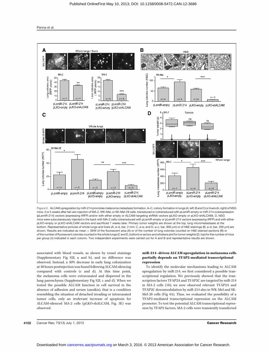
for WK-Mel and SK-Mel-28 cells, miR-214 overexpression(pLemiR-214) induces increased metastatic dissemination invivo, compared with empty controls. To evaluate the role ofALCAM in miR-214–mediated melanoma cell dissemination,miR-214–overexpressingMA-2 (Fig. 2A) orWK-Mel (Fig. 2B) ormiR-214–overexpressing SK-Mel-28 (Fig. 2C) cells (pLemiR-empty or pLemiR-214, turbo red fluorescent protein-tRFP–
positive), stably silenced (pLKO-shALCAM) or not (pLKO-empty) for ALCAM, were injected in the tail vein of severelyimmunocompromised NSGmice and metastatic nodules eval-uated 3 to 5 weeks postinjections in the lungs (and livers forMA-2 cells). As assessed by the quantitation ofmetastatic areasin the whole organs (Fig. 2A, a–d) or in H&E-stained sections(Supplementary Fig. S2F, a and b), or by counting metastaticcolonies in H&E-stained sections (Fig. 2B, a–d) or in wholelungs (Fig. 2C, a–e), colony formation for ALCAM-silencedmelanoma cells was significantly impaired compared withcontrols and, more importantly, ALCAM silencing in miR-214–overexpressing cells rescued miR-214–driven increasedmetastasis formation. In addition, spontaneous metastasisformation was evaluated following subcutaneous injection ofmiR-214–overexpressing ALCAM-silenced MA-2 cells in NSGmice, looking for fluorescent microscopic metastatic lesions 7weeks later. Primary tumor growthwas also evaluated here andno difference was found (Fig. 2D, top). However, as shownin Fig. 2D, bottom, a–c, the increased number of lung micro-metastases found for miR-214–overexpressing cells was sig-nificantly rescued when ALCAM was silenced. Very few met-astatic lesions were observed in the livers.
Because these in vitro and in vivo results were obtained for 3distinct melanoma cell lines and independently of BRAFV600E
mutation status (Supplementary Table S1), they indicate ageneral key role for miR-214–mediated ALCAM upregulationin melanoma metastasis. Interestingly, transient ALCAMsilencing (si-ALCAM#1) was able to significantly reduce invitro cellmovement comparedwith si-control also in theMDA-MB-231 breast cancer cell line (Supplementary Figs. S1I andS2E), suggesting a broader function for miR-214–drivenALCAM upregulation in tumor cell movement.
ALCAM controls melanoma cell extravasationTo evaluate the possible involvement of ALCAM in tumor
cell extravasation, we first analyzed in vitro transendothelialmigration by seeding CMRA-labeled (red) MA-2 cells previ-ously silenced (si-ALCAM#1) or not (si-control) for ALCAMin the upper chamber of Transwell covered by a confluentHUVECs-GFP monolayer. As shown in Fig. 3A, ALCAM down-modulation (b) decreased transendothelial migration com-pared with control (a). More relevantly, ALCAM transient(si-ALCAM#1; Supplementary Fig. S2G) or stable (pLKO-shALCAM) downmodulation in miR-214–overexpressing MA-2 cells (pLemiR-214, tRFP-positive) abolished miR-214–medi-ated enhancement of transendothelial migration (pLemiR-214þpLKO-empty) and reestablished migration levels compa-rable with controls (pLemiR-emptyþpLKO-empty; Fig. 3B, a–c). Transient ALCAM silencing was able to significantly reducetransendothelial migration also in MDA-MB-231 cells (Supple-mentary Fig. S2H), again suggesting a general role in tumorcell dissemination. We then evaluated ALCAM involvement inin vivo extravasation in the lungs of immunocompromisedmice following tail vein injections of miR-214–overexpressingMA-2 or WK-Mel cells (pLemiR-214) transduced with eitherpLKO-shALCAM or pLKO-empty control (Fig. 3C and D). Thelodging in lung vasculature was evaluated 2 hours post-injection (a and b) when the cells resulted localized inside or
miR-214 Regulates ALCAM in Melanoma
www.aacrjournals.org Cancer Res; 73(13) July 1, 2013 4101
on March 3, 2016. © 2013 American Association for Cancer Research. cancerres.aacrjournals.org Downloaded from
Published OnlineFirst May 10, 2013; DOI: 10.1158/0008-5472.CAN-12-3686
associated with blood vessels, as shown by vessel stainings(Supplementary Fig S2I, a and b), and no difference wasobserved. Instead, a 50% decrease in early lung colonizationat 48 hours postinjectionwas found followingALCAMsilencingcompared with controls (c and d). At this time point,the melanoma cells were extravasated and dispersed in thelung parenchyma (Supplementary Fig S2I, c and d). When wetested the possible ALCAM function in cell survival in theabsence of adhesion and serum (anoikis), that is a conditionresembling the situation of detached invading or intravasatedtumor cells, only an irrelevant increase of apoptosis forALCAM-silenced MA-2 cells (pLKO-shALCAM, Fig. 3E) wasobserved.
miR-214–driven ALCAMupregulation inmelanoma cellspartially depends on TFAP2-mediated transcriptionalrepression
To identify the molecular mechanisms leading to ALCAMupregulation by miR-214, we first considered a possible tran-scriptional regulation. We previously showed that the tran-scription factors TFAP2A and TFAP2C are targeted bymiR-214in MA-2 cells (18); we now observed relevant TFAP2A andTFAP2C downmodulation by miR-214 also inWK-Mel and SK-Mel-28 cells (Fig 4A). Thus, we evaluated the possibility of aTFAP2-mediated transcriptional repression on the ALCAMpromoter. To test the potential ALCAM transcriptional repres-sion by TFAP2 factors, MA-2 cells were transiently transfected
Figure 2. ALCAMupregulation bymiR-214promotesmelanomametastasis formation. A–C, colony formation in lungs (A, left; B andC) or livers (A, right) ofNSGmice, 3 or 5 weeks after tail vein injection of MA-2, WK-Mel, or SK-Mel-28 cells, transduced or cotransduced with pLemiR-empty or miR-214 overexpression(pLemiR-214) vectors (expressing tRFP) and/or with either empty or ALCAM-targeting shRNA vectors (pLKO-empty or pLKO-shALCAM). D, NSGmice were subcutaneously injected in the back with MA-2 cells cotransduced with pLemiR-empty or pLemiR-214 vectors (expressing tRFP) and with eitherpLKO-empty or pLKO-shALCAM vectors and sacrificed 7 weeks later. Primary tumor weights are shown at the top, lung micrometastases at thebottom. Representative pictures of whole lungs and livers (A, a–d, bar, 2 mm; C, a–e; and D, a–c, bar, 800 mm) or of H&E stainings (B, a–d, bar, 200 mm) areshown. Results are indicated as mean � SEM of the fluorescent area (A) or of the number of lung colonies counted on H&E-stained sections (B) orof the number offluorescent colonies counted in thewhole lungs (CandD,bottom) or asboxandwhiskersplot for tumorweights (D, top) for the number ofmiceper group (n) indicated in each column. Two independent experiments were carried out for A and B and representative results are shown.
Penna et al.
Cancer Res; 73(13) July 1, 2013 Cancer Research4102
on March 3, 2016. © 2013 American Association for Cancer Research. cancerres.aacrjournals.org Downloaded from
Published OnlineFirst May 10, 2013; DOI: 10.1158/0008-5472.CAN-12-3686

with TFAP2A or TFAP2C expression constructs [pSP(RSV)-TFAP2A, pSP(RSV)-TFAP2C], or with the combination of both,or with an empty control [pSP(RSV)-empty] and ALCAMmRNA and protein expression was analyzed by qRT-PCR andWestern blot analysis, respectively. As shown in Fig. 4B and C,TFAP2 overexpression resulted in 20% to 40% decreasedALCAM mRNA and protein expression, 48 and 72 hours post-transfection, respectively. A similar ALCAM protein down-modulation following TFAP2 overexpression was observed inWK-Mel cells (Fig. 4D). Consistently, in TFAP2C-silenced cells
(pLKO-shTFAP2C), we observed 20% increased ALCAM pro-tein expression compared with control (pLKO-empty), asshown in Fig. 4E. These results suggested that miR-214–drivenALCAM upregulation could depend on TFAP2A and/orTFAP2C decrease due to miR-214 targeting. In line with this,we observed lower ALCAM levels in miR-214–overexpressing(pLemiR-214) MA-2 cells when TFAP2A or, to a lesser extent,TFAP2C [pSP(RSV)-TFAP2A, pSP(RSV)-TFAP2C] or both wereoverexpressed compared with controls (Fig. 4F). The ALCAMpromoter region, from �900 to þ100 bp relative to the
Figure 3. ALCAM controlsmelanoma cell extravasation.A andB, transendothelial migrationassays of CMRA-labeled MA-2cells transiently transfected witheither control or ALCAM-targetingsiRNAs (si-control or si-ALCAM#1;A) or of MA-2 cells cotransducedwith pLemiR-empty or miR-214overexpression (pLemiR-214)vectors (expressing tRFP) and witheither empty or ALCAM-targetingshRNA vectors (pLKO-empty orpLKO-shALCAM; B), through afibronectin-coated Transwellmembrane covered by a confluentmonolayer of HUVECs-GFP.Transmigrated MA-2 cells on thelower side of the Transwell areshown in a–c. Bar, 50 mm. C and D,in vivo extravasation assays 2hours (a and b) or 48 hours (c and d)following tail vein injections in nudemice of MA-2 (C) or WK-Mel (D)cells cotransduced as in B.Representative pictures of wholelungs are shown; bar, 800 mm.E, anoikis assay for MA-2 cellstransducedas inCandplated in theabsence of adhesion and serum for72 hours. Cell death percentagewas evaluated by AnnexinV–allophycocyanin staining,displayed in bidimensional plots.Left quadrant, healthy population;right quadrant, apoptoticpopulation. Two or 3 independentexperiments were carried out (intriplicate for A, B, and E) andrepresentative results are shownasmean � SEM of the area coveredbymigrated cells (A and B) or of thenumber of extravasated cellsat 48 hours for n¼ 5mice per group(C and D) or as percentages ofcells (E).
miR-214 Regulates ALCAM in Melanoma
www.aacrjournals.org Cancer Res; 73(13) July 1, 2013 4103
on March 3, 2016. © 2013 American Association for Cancer Research. cancerres.aacrjournals.org Downloaded from
Published OnlineFirst May 10, 2013; DOI: 10.1158/0008-5472.CAN-12-3686

Figure 4. ALCAM is transcriptionally repressed by TFAP2 in melanoma. A–F, ALCAM, TFAP2A, and TFAP2C protein (A, C–F) or mRNA (B) expression levelswere analyzed respectively byWestern blot analysis andqRT-PCRat 48 (B) and72hours (A, C, D, andF) following transient transfection ofWK-Mel or SK-Mel-28 with miR-214 precursors or negative controls (pre-miR-214 or -control; A), or of MA-2 or WK-Mel cells (B–D) or of miR-214–overexpressing MA-2 cells(pLemiR-214, F) with pSP(RSV)-empty or TFAP2A or TFAP2C overexpression vectors [pSP(RSV)-TFAP2A or pSP(RSV)-TFAP2C] or the combination of both,or in MA-2 cells stably transducedwith either empty or TFAP2C-targeting shRNA vectors (pLKO-empty or pLKO-shTFAP2C, E). In B, results were calculatedas fold changes (mean � SD of triplicates) relative to controls, normalized on 18S RNA level. In A and C–F, protein modulations were calculated relative tocontrols, normalized on GAPDH or hsp90 loading controls and expressed as percentages. G, scheme showing human ALCAM gene promoter region,spanning from�900 toþ100 nucleotides around the TSS. Five putative TFAP2-binding sites (BS) are indicated by circles. The portion between�753 andþ35was cloned in pGL3-Basic luciferase reporter vector (pGL3-ALCAMprom; see I). H, ChIP.MA-2 cross-linked sheared chromatinwas immunoprecipitatedwitheither negative control (IgG) or anti-TFAP2 antibodies and the TFAP2-binding sites–containing region was PCR-amplified. TFAP2-binding enrichment wascalculated relative to IgG-negative control. I, luciferase assays 48 hours posttransfection in MA-2 cells cotransfected with pGL3-Basic reporter constructscontaining WT (pGL3-ALCAMprom) or all (del_all) or specific (del_5 to del_1 and del_3þ5, 2þ3, 2þ3þ5) TFAP2-binding site-deleted ALCAM promotersequence cloned upstream of the luciferase coding sequence, together with pSP(RSV)-empty or pSP(RSV)-TFAP2A or pSP(RSV)-TFAP2C expressionvectorsor the combination of both. Results are shownasmean�SDof Firefly luciferaseactivity relative to controls, normalized onRenilla luciferase andon theactivity measured for the empty pGL3-Basic vector in presence of TFAP2. Two or 3 independent experiments were carried out, in triplicate for B and I andrepresentative results are shown.
Penna et al.
Cancer Res; 73(13) July 1, 2013 Cancer Research4104
on March 3, 2016. © 2013 American Association for Cancer Research. cancerres.aacrjournals.org Downloaded from
Published OnlineFirst May 10, 2013; DOI: 10.1158/0008-5472.CAN-12-3686

transcription start site (TSS), was analyzedwith bioinformaticstools, using the canonical TFAP2-binding site PWM consistingof a 9 nucleotide long G/C-rich sequence, shared for TFAP2Aand TFAP2C. Five putative TFAP2-binding sites (BS) wereidentified, with high-affinity binding scores, at positions�316 (BS5), �249 (BS4), �208 (BS3), �163 (BS2), and �126(BS1; Fig. 4G). A ChIP experiment was carried out usingcross-linked MA-2 chromatin, with negative (immunoglobulinG, IgG) or positive (RNA polymerase II, PolII) control or anti-TFAP2 (recognizing both TFAP2A and TFAP2C) antibodies.Importantly, by PCR-amplifying the region containingthe 5 TFAP2-binding sites, more than 4-fold TFAP2-bindingenrichment was observed on this region relative toIgG-negative control (Fig. 4H). As a positive control PolII-immunoprecipitated DNA was tested with a glyceraldehyde-3-phosphate dehydrogenase (GAPDH) promoter PCR assay(Supplementary Fig. S3A). To test the direct effect of TFAP2on ALCAM promoter, the activity of the promoter regionbetween �753 and þ35 relative to the TSS, was assayed in aluciferase reporter vector (pGL3-ALCAMprom). MA-2 cellswere cotransfected with either pGL3-ALCAMprom or pGL3-empty and TFAP2A and/or TFAP2C expression constructs orempty control [pSP(RSV)-TFAP2A, pSP(RSV)-TFAP2C, or pSP(RSV)-empty] and luciferase activity was evaluated 48 hours(Fig. 4I) and 72 hours (Supplementary Fig. S3B) posttransfec-tion. pGL3-ALCAMprom luciferase activity was normalized onpGL3-empty activity in a condition of TFAP2 overexpression.As a positive control, a luciferase construct containing theknown TFAP2A-responsive ESDN promoter region (24) wasused (Supplementary Fig. S3C). pGL3-ALCAMprom activitywas significantly decreased in presence of TFAP2A or TFAP2Cor both (60% to 40% reduction), compared with pSP(RSV)-empty control, indicating a direct transcriptional repression ofTFAP2 on ALCAM promoter (Fig. 4I, left). The single deletionsof the 5 binding sites one-by-one (del_5 to del_1) were not ableto rescue the luciferase activity, whereaswhenwedeleted 2 or 3binding sites in combinations (del_3þ5, del_2þ3, del_2þ3þ5)or the entire promoter region included between �316 and�126 thus eliminating all the 5 binding sites (del_all), we wereable to abrogate the luciferase activity decrease in presence ofTFAP2 (Fig. 4I, right), indicating that the cooperation of the 5sites is probably required for TFAP2 repression of ALCAMtranscription.
miR-214–driven miR-148b downregulation controlsALCAM overexpression posttranscriptionally inmelanoma cellsIn addition to TFAP2-mediated transcriptional regulation,
we considered possible posttranscriptional mechanismsinvolving other miRs. Because miR-148b was predicted todirectly target ALCAM, according to TargetScan 5.2 algorithm(27), we evaluated ALCAM expression following miR-148boverexpression (pre-miR-148b; Supplementary Fig. S1C, S1G,and S1H) compared with pre-control in MA-2, WK-Mel, andSK-Mel-28 cells 72 hours posttransfection. Significantly, a 40%to 80% ALCAM protein reduction was found in Western blotanalyses (Fig. 5A–C). To investigate the direct binding of miR-148b on ALCAM, the first 605 bp of ALCAM 30-UTR (Fig. 5F),
containing miR-148b putative binding site (at position 272–277), was cloned in the pMIR-REPORT reporter vector down-stream of the luciferase coding sequence (ALCAM30-UTR).miR-148b–binding site was also mutated (point mutations ordeletion; shown in Fig. 5F) to generatemutant ALCAM30-UTRs(ALCAMmut and ALCAMdel). Luciferase activity of wild-type(WT) or mutant vectors was evaluated in MA-2 cells inpresence or absence of miR-148b overexpression (pre-miR-148b or -control). A significant decrease of luciferase activitywas observed for theWTbut not for themutants in presence ofmiR-148b compared with controls, indicating the specificregulation of miR-148b on ALCAM 30-UTR (Fig. 5G). As apositive control, a miR-148b sensor construct containing 3perfect bindings for miR-148b was used (Fig. 5G). Importantly,when we analyzed miR-148b levels in miR-214–overexpressing(pre-miR-214) MA-2, WK-Mel, SK-Mel-28, and MDA-MB-231cells, a significant reduction ofmiR-148b expression was foundat 24, 48 (Supplementary Fig. S3D;MA-2), and 72 hours (Fig. 5H,MA-2; Fig. 5K, SK-Mel-28; Supplementary Fig. S3E, WK-Mel;Supplementary Fig. S3F, MDA-MB-231 cells) compared withpre-control), as evaluated by qRT-PCR. A similar miR-148breduction was obtained in stable miR-214–overexpressing(pLemiR-214) MA-2 (Fig. 5I) or WK-Mel (Fig. 5J) cells, com-pared with empty control (pLemiR-empty). Consistently, miR-148b expression levels were increased in presence of anti-miR-214 in MC-1 cells, compared with anti-control–transfectedcells (Fig. 5L). Other unrelated miRs, for instance miR-31, werenot affected by miR-214 overexpression (Fig. 5M). These datasuggest a regulatory loop between miR-214 and miR-148b,which can affect ALCAM expression. Indeed, the concomitantoverexpression of miR-148b and miR-214 (pre-miR-214þpre-miR-148b transfection) inMA-2 cells (Supplementary Fig. S1C)or the reexpression ofmiR-148b in stablemiR-214–overexpres-sing cells (pLemiR-214þpre-miR-148b; Supplementary Fig.S1D) led to reduced ALCAMprotein (Fig. 5A and D) andmRNA(Fig. 5E) levels, similar to control cells (pre-control or pLemiR-emptyþpre-control, respectively).
miR-148b downregulation by miR-214 is partially due toTFAP2 control
To explore how miR-214 downregulates miR-148b, we con-ducted a bioinformatics analysis for miR-148b containinghuman genomic locus (2,000 nucleotides upstream and down-stream of pre-miR-148b), inside the intron 1 of COPZ1 protein-coding gene. First, as described in ref. 28, we used RNA Hybridalgorithm (26) to look for in silico evidences of a directinteraction between miR-214 and miR-148b transcript, poten-tially involved in controlling processing or stability. A singleputative 20-nucleotide long miR-214–binding site was found650 nucleotides downstreamof the pre-miR-148b locus (Fig. 6Aand B), but with a nonstatistically significant (ns) P value(independently of themethod used to estimate it, seeMaterialsandMethods and Fig. 6B), leading us to exclude its relevance inthis mechanism. Instead, when we looked for putative TFAP2-binding sites in pre-miR-148b–flanking regions, many low- and3 high-affinity sites were found, including one located 1,816nucleotides upstream of the pre-miR-148b, having 83% of themaximum PWM score, with a potential role in controlling its
miR-214 Regulates ALCAM in Melanoma
www.aacrjournals.org Cancer Res; 73(13) July 1, 2013 4105
on March 3, 2016. © 2013 American Association for Cancer Research. cancerres.aacrjournals.org Downloaded from
Published OnlineFirst May 10, 2013; DOI: 10.1158/0008-5472.CAN-12-3686

transcription (Fig. 6A and C, top). Importantly, as indicatedin Fig. 6C, bottom, aChIP-seq analysis previously conducted onHeLa cells [The ENCODE Project (29), SYDH-TFBS track],revealed TFAP2A and TFAP2C binding in correspondence ofthis binding site. In line with this, we carried out ChIP experi-ments in MA-2 cells, as described earlier, and confirmedTFAP2A/C-binding enrichment (1.9-fold) in this region (Fig.6D), compared with IgG-negative control. These results sug-gest that the miR-214 regulation on miR-148b is at leastpartially due to TFAP2-mediated control on miR-148b. Indeed,also miR-148b primary transcript (pri-miR-148b) evaluated byqRT-PCR, was about 30% reduced in miR-214 (pre-miR-214)overexpressing MA-2 cells (Fig. 6E) and, consistently, bothpri- (Fig. 6F and G) and mature (Fig. 6H and I) miR-148blevels were more than 50% increased following overexpres-
sion of TFAP2A and TFAP2C. Taken together, our dataindicate that TFAP2A has a dual role on ALCAM regulation,as summarized in Fig. 6J: it binds on ALCAM promoter andrepresses ALCAM transcription; it controls ALCAM-target-ing miR-148b expression.
miR-148b opposes miR-214–mediated prometastaticfunctions in melanoma
Because we observed that ALCAM upregulation by miR-214is at least in part due to miR-148b reduction, we explored thefunctions of miR-148b in melanoma cell movement. miR-148bwas transiently overexpressed in MA-2 and WK-Mel cells bypre-miR-148b transfection and cell migration was evaluated byTranswell assays. Significantly, miR-148b overexpression led todecreased cell movement compared with pre-control (Fig. 7A
Figure 5. miR-148b directly targets ALCAM and is downregulated by miR-214 in melanoma. A–E, ALCAM protein (A–D) and mRNA (E) expression levels wereevaluated by Western blot (A–D) or qRT-PCR (E) analyses, respectively, 48 to 72 hours following transient transfection of MA-2 (A), WK-Mel (B), SK-Mel-28cells (C), or of miR-214–overexpressing MA-2 cells (pLemiR-214; D and E) with miR-214 and/or miR-148b precursors or negative controls (pre-miR-214 or-148b or -control). Protein modulations were calculated relative to controls, normalized on hsp90 or GAPDH loading controls, and expressed aspercentages; mRNA modulations were calculated as fold changes (mean � SD of triplicates) relative to controls, normalized on 18S RNA level. F, schemeshowing WT or mutated (ALCAMmut) or deleted (ALCAMdel) miR-148b–binding site in human ALCAM 30-UTR (at position 273), paired with miR-148b seed.The portion of ALCAM 30-UTR cloned in pMIR luciferase reporter vector is up to nucleotide 605, starting from the stop codon. G, luciferase assays inMA-2 cells cotransfected with reporter constructs containing WT (ALCAM30-UTR) or mutant (ALCAMmut and ALCAMdel) ALCAM 30-UTRs or a syntheticsequence including 3 perfect miR-148b–binding sites (miR-148b-sensor), cloned downstream of the luciferase coding sequence, together withmiR-148b precursors or negative controls (pre-miR-148b or -control). Results are shown as mean � SD of Firefly luciferase activity relative to controls,normalized on Renilla luciferase activity. H–M, miR-148b (H–L) and miR-31 (M) expression levels were tested by qRT-PCR in MA-2 (H, I, and M), WK-Mel (J),SK-Mel-28 (K), or MC-1 (L) cells following transfection with miR-214 precursors (H, K, and M; 72 hours posttransfection) or inhibitors (L; 24 hoursposttransfection) or their negative controls (pre- or anti-miR-214 or -control), or transduction with pLemiR-empty or miR-214 overexpression (pLemiR-214)vectors (I and J). Results were calculated as fold changes (mean � SD of triplicates) relative to controls, normalized on U6 RNA level. Two or 3 independentexperiments were carried out in triplicate for E–M and representative results are shown.
Penna et al.
Cancer Res; 73(13) July 1, 2013 Cancer Research4106
on March 3, 2016. © 2013 American Association for Cancer Research. cancerres.aacrjournals.org Downloaded from
Published OnlineFirst May 10, 2013; DOI: 10.1158/0008-5472.CAN-12-3686

and B). Consistently, miR-148b silencing by anti-miR-148b(Supplementary Fig. S1F) resulted in increased cell migration,compared with anti-control–transfected cells (Fig. 7C). Moreimportantly, as shown in Fig. 7D, the concomitant overexpres-sion of miR-148b in miR-214–overexpressing MA-2 cells (pLe-miR-214þpre-miR-148b) was able to rescue miR-214–inducedincreased cell migration (pLemiR-214þpre-control) similarlyto control cells (pLemiR-emptyþpre-control). We alsoobserved that survival to anoikis was decreased (Supplemen-tary Fig. S3G). We then tested transendothelial migration, asdescribed earlier, and we observed decreased ability of MA-2cells to migrate through the HUVECs-GFP monolayer whenmiR-148b was overexpressed (pre-miR-148b) compared withpre-control, opposite to pre-miR-214–transfected cells (Fig.7E). Remarkably, concomitant miR-148b and miR-214 over-expression (pre-miR-214þpre-miR-148b) completely rescuedmiR-214–mediated transendothelial migration (Fig. 7E). When
we evaluated early lung colonization in vivo, 48 hours after tailvein injection of MA-2 cells in nude mice, while miR-214overexpression (pre-miR-214) significantly enhanced extrava-sation, miR-148b overexpression alone (pre-miR-148b) mod-erately reduced it, in comparison with pre-control transfectedcells, as shown in Fig. 7F. More importantly, when miR-148bwas upregulated in miR-214–overexpressing cells (pre-miR-214þpre-miR-148b), we observed a rescuing of miR-214–induced cell extravasation and early lung colonization (Fig.7F), suggesting a role for miR-148b in miR-214/ALCAM–medi-ated melanoma cell metastatic dissemination.
In conclusion, miR-148b is downregulated by miR-214, atleast in part via a TFAP2-driven mechanism, and these 2 smallRNAs exert opposite roles in melanoma cell dissemination, bycontrolling ALCAM expression. These observations arestrengthened by the analysis of miR-214, miR-148b, ALCAM,and TFAP2 in human patients with melanoma. We took
Figure 6. miR-148b downregulation by miR-214 is partially due to a TFAP2-mediated control. A, scheme showing the human pre-miR-148b locus(chromosome 12, intron 1 of COPZ1 protein-coding gene) and the predicted interactions with miR-214 and TFAP2 as shown in B and C. B,referring to A, predicted interaction of miR-214 on the pre-miR-148b locus (650 nucleotides downstream of pre-miR-148b), as obtained with the RNAHybridalgorithm. MFE, minimum free energy (DG) predicted for hybridization; the P values were estimated on different backgrounds and are nonstatisticallysignificant (ns). C, referring to A, high affinity (black rectangles; score >80%of themaximum) and low affinity (gray rectangles; score between 60%and 80%ofthe maximum) TFAP2-binding sites (BS) were obtained by the PWMmethod (top). The ENCODE Project SYDH-TFBS ChIP-seq analysis conducted on HeLacells shows TFAP2A and TFAPC but not immunoglobulin M (IgM; negative control) enrichment picks in pre-miR-148b flanking regions, for some putativeTFAP2 BS (bottom). D, ChIP. MA-2 cross-linked sheared chromatin was immunoprecipitated with either negative control (IgG) or anti-TFAP2 antibodies andthe TFAP2-binding site–containing miR-148b upstream region was PCR-amplified. TFAP2-binding enrichment was calculated relative to IgG-negativecontrol. E–I, miR-148b primary transcript (pri-miR-148b; E–G) and mature miR-148b (H and I) expression levels were tested by qRT-PCR in MA-2 cells72 hours following transfection withmiR-214 precursors or negative controls (pre-miR-214 or -control) or with pSP(RSV)-empty or pSP(RSV)-TFAP2A or pSP(RSV)-TFAP2C expression vectors. Results were calculated as fold changes (mean � SD of triplicates) relative to controls, normalized on 18S orU6 RNA level. At least 2 independent experiments were carried out in triplicate for E–I and are shown either as representative results (D, F–I) or as themean�SEM of 7 independent experiments (E). J, our data show that ALCAM is overexpressed in melanoma because miR-214 downregulates both TFAP2A/C(ALCAM transcriptional repressors) and miR-148b (ALCAM-targeting miR). Relevantly, TFAP2A/C positively regulate miR-148b expression.
miR-214 Regulates ALCAM in Melanoma
www.aacrjournals.org Cancer Res; 73(13) July 1, 2013 4107
on March 3, 2016. © 2013 American Association for Cancer Research. cancerres.aacrjournals.org Downloaded from
Published OnlineFirst May 10, 2013; DOI: 10.1158/0008-5472.CAN-12-3686

advantage of the same cohort of patients used in (18, 30). Asindicated in Fig. 7G, bottom, we previously showed that miR-214 was significantly more expressed, whereas TFAP2A and
TFAP2C were less expressed, in invasive melanoma samplescompared with noninvasive (in situ) ones, by qRT-PCR andimmunoistochemical analyses, respectively (18, 30). In a
Figure 7. miR-148b opposesmiR-214 prometastatic functions inmelanoma. A–E, Transwell migration through a poremembrane (A–D) or a fibronectin-coatedmembrane covered by a confluent monolayer of HUVECs-GFP (E; bar, 50 mm) was evaluated following transient transfection with miR-214 and/ormiR-148b precursors or inhibitors or their negative controls (pre-miR-214 or -148b or anti-miR-148b or -control) of MA-2 (A, C, and E; CMRA-labeled in E) orWK-Mel (B) cells or of miR-214–overexpressing MA-2 cells (pLemiR-214; D). F, in vivo extravasation assay 48 hours following tail vein injectionsin nudemice of CMRA-labeledMA-2 cells transfected as in E. Representative pictures of whole lungs are presented (a–d; bar, 800 mm). Results are shown asmean � SEM of the area covered by migrated cells or of the number of extravasated cells in the lungs, for n ¼ 5 mice per group. Two or 3independent experiments were carried out (in triplicate for A–E) and representative results are shown. G, ALCAMmRNA levels (top) and miR-148b/miR-214expression ratio (middle) were evaluated in a cohort of n ¼ 6 in situ versus n ¼ 16 invasive human melanomas. Relative protein-coding gene or miRexpression was obtained by qRT-PCR analyses, calculated using the median as reference, and normalized on GAPDH or U44 RNA levels, respectively. Thehorizontal line (top) represents the mean for the in situ melanomas. The Mann–Whitney nonparametric test was used for the statistical analysis ofALCAMexpression. TFAP2A, TFAP2C, andmiR-214 expression levels represented by gray scales (bottom) refer to our previously publishedworks (indicated)and were obtained by immunoistochemical and qRT-PCR analyses, respectively.
Penna et al.
Cancer Res; 73(13) July 1, 2013 Cancer Research4108
on March 3, 2016. © 2013 American Association for Cancer Research. cancerres.aacrjournals.org Downloaded from
Published OnlineFirst May 10, 2013; DOI: 10.1158/0008-5472.CAN-12-3686

subgroup of the same samples, we now evaluated ALCAMmRNA expression by qRT-PCR and found a significantincrease in invasive melanomas (n ¼ 16) compared within situ tumors (n ¼ 6; Fig. 7G, top), thus reinforcingour findings. Importantly, when we considered miR-148b/miR-214 expression ratio by qRT-PCR in this cohort (Fig. 7G,middle) and in a new independent cohort of patients withmelanoma (n ¼ 10 in situ and n ¼ 10 invasive; Supplemen-tary Fig. S4, top), we consistently found low miR-148b (lessthan 10%) and high miR-214 relative levels, indicating thatmiR-148b is maintained low in high miR-214–expressingmelanomas, at least partially by the earlier describedmiR-214–mediated mechanism.
DiscussionWe previously showed a prometastatic function for miR-214
in melanoma (18). The present work spotted light on thelink between miR-214 and ALCAM in the coordination ofmelanomaprogression. Here, we show that ALCAMexpressionincreases when miR-214 is upregulated in several differentmelanoma cell lines, independently of BRAFV600E mutationstatus. Previously, we found increased levels of miR-214 inmetastatic melanoma cells compared with their poorly malig-nant counterparts and in invasive or metastatic humanmelanomas but not in in situ samples (18). Supporting theconnection between miR-214 and ALCAM are the immuno-histochemical analyses for ALCAM in human melanoma sam-ples, which show a perfect correlation between higher levels ofALCAM and thicker lesions or later tumor stages, from com-monnevi tometastases (8). ALCAMexpressionwas specificallydetected at the invasive front ofmelanomas (8).Moreover, herewe highlighted increased ALCAMmRNAexpression in invasivehigh miR-214–expressing human melanomas, compared within situ lesions. In addition, ALCAM resulted to be highlyexpressed in xenotransplants and metastases originated inmice following injection of melanoma cell variants with highlevels of miR-214 (17). Finally, ALCAM expression associateswith poor prognosis or relapse for other human tumors,including colorectal, bladder, esophageal, and intraductalbreast carcinomas (7, 31, 32) and high ALCAM or miR-214serum levels are considered poor prognostic markers for somekind of neoplasia (33–36).ALCAM has a key role in normal tissue or tumor integrity, in
heterotypic tumor cell–endothelial cell interactions and inimmune cell attachment and transmigration through vessels(5, 6, 37), therefore it is fundamental for primary tumor massgrowth and tumor escaping. ALCAM connections are essentialfor cell movement (9), for efficient conversion of pro-MMP-2 toits active form in metastatic melanoma cells (11) and for theretention of intravascular breast tumor cell clusters in the lungvessels (38). Consistently, we showed that ALCAM overexpres-sion promotes cell migration, whereas silencing blocks it indifferent melanoma cell lines. More importantly, we provedthat miR-214 prometastatic functions are exerted via ALCAM,at least in part. In fact, ALCAM silencing in miR-214–over-espressing cells significantly reduces miR-214–mediatedincreased cell movement, invasion, and transendothelialmigration in vitro and efficiently impairs extravasation and
metastatic dissemination in mice, both following directinjection of melanoma cells in the blood circulation andwhen we analyzed lung metastatic lesions spontaneouslyoriginating from experimental tumors. Because these resultswere obtained for 3 distinct melanoma cell lines, indepen-dently of BRAFV600E mutation status, and for a breast cancercell line, we conclude that miR-214–mediated ALCAM upre-gulation has a broad-spectrum key role in tumor cell met-astatic dissemination.
Despite the clear correlation between ALCAM expressionand melanoma malignancy, the mechanisms leading toALCAM upregulation are still poorly understood. It is knownthat ALCAM expression is regulated by promoter CpG islandsmethylation (38) and by transcriptional regulation exerted byNF-kB (38) and GATA1 (39) transcription factors. Here, wepropose that miR-214 regulates ALCAM indirectly via a tran-scriptional and a posttranscriptional control. About the tran-scriptional control, we postulate that miR-214 upregulatesALCAM transcription by downregulating its targets TFAP2Aand TFAP2C. Loss of TFAP2 during melanoma progression iswell documented (40), and we showed here and previously (18)that it is governed, at least in part, by miR-214, in differentmelanoma cell lines.miR-214 directly targets TFAP2C,whereasTFAP2A is indirectly downregulated, probably via TFAP2C.Indeed, it is known that TFAP2C silencing reduces TFAP2Aprotein levels (unpublished data) and that TFAP2 familymembers regulate each other transcriptionally (41). Here, weshow that ALCAMmRNA and protein expression decreases inTFAP2-overexpressing MA-2 or WK-Mel cells. Moreover, weidentified 5 putative TFAP2-binding sites on ALCAMpromoterand proved TFAP2A/C-binding enrichment on this region, viaa ChIP experiment. Importantly, we observed a strong down-regulation of luciferase expression when the activity of ALCAMpromoter was tested in cells overexpressing TFAP2 familymembers, using a luciferase reporter. The deletion of eachsingle binding site or of combinations of them or of the entireregion encompassing the 5 sites suggests their cooperativeaction. In fact, the ablation of multiple or all sites was able toabrogate TFAP2-dependent luciferase activity reduction.Decreased levels of ALCAM, comparable with low miR-214–expressing cells, were seen in miR-214–overexpressing cellsfollowing TFAP2A and, partially, TFAP2C reexpression, indi-cating that the downmodulation of TFAP2 factors by miR-214,occurring during metastatic dissemination, is necessary toremove the direct transcriptional repression of TFAP2 onALCAM promoter. Importantly, we previously proved thatTFAP2C is responsible for many miR-214 prometastatic func-tions and that TFAP2C reexpression in miR-214–overexpres-sing cells was able to rescue miR-214–induced cell movementand early lung metastatic colonization (18). Interestingly, thisTFAP2-mediated regulation is similar to what previouslyshown for the ALCAM homolog MCAM-MUC-18 adhesionmolecule in melanoma (42).
About the posttranscriptional control, we showed that miR-214 upregulates ALCAM expression by reducing another smallnoncoding RNA, miR-148b, which was predicted to directlytarget ALCAM. In agreement with bioinformatics anticipa-tions, we observed significant downmodulation of ALCAM
miR-214 Regulates ALCAM in Melanoma
www.aacrjournals.org Cancer Res; 73(13) July 1, 2013 4109
on March 3, 2016. © 2013 American Association for Cancer Research. cancerres.aacrjournals.org Downloaded from
Published OnlineFirst May 10, 2013; DOI: 10.1158/0008-5472.CAN-12-3686

protein following miR-148b overexpression. Direct targetingwas proven by luciferase reporter assays on WT but not onmiR-148b seed-mutants. Importantly, we observed decreasedmiR-148b levels following miR-214 overexpression in differentmelanoma cell lines and, vice versa, increased levels in miR-214–silenced cells. Significantly, miR-148b overexpression inmiR-214–overexpressing cells was able to reestablish lowALCAM levels, as in control cells. Our results suggested amiR-on-miR regulatory loop between miR-148b and miR-214, in which miR-214 could control miR-148b transcriptionor processing or stability. Another miR-on-miR regulation waspreviously described for mouse miR-709, which binds on pri-miR-15a/16-1 thus preventing its processing (28). By using thesame approach, we looked for possible miR-214 recognitionsites around miR-148b locus, but found only a nonstatisticallysignificant putative site, located downstream of pre-miR-148b.Instead, the observation that also pri-miR-148b was down-modulated following miR-214 overexpression and that manyputative TFAP2-binding sites were present in miR-148b locus,including a high affinity one in the potential promoter region,led us to investigate a possible transcriptional regulation. Onthis line, we and others (The ENCODE project) proved TFAP2-binding enrichment on this site by ChIP analyses. In addition,pri- and mature miR-148b expression increased followingTFAP2A and TFAP2C overexpression. However, we cannotexclude additional regulations, such as posttranscriptionalfunctions of TFAP2 or, as it is well known that miR-148/152expression is controlled by promoter methylations (43), pos-sible miR-214–controlled epigenetic mechanisms.
In conclusion, our data indicate thatmiR-214–driven TFAP2downregulation occurring during melanomamalignancy, con-trols ALCAM expression in a dual way: directly, at the tran-scriptional level, as well as indirectly, by controlling theexpression of ALCAM-targeting miR-148b. These data arefurther reinforced by ours and others observations showingthat ALCAM expression increases, whereas TFAP2 decreasesand that high levels of miR-214 anticorrelate with poor miR-148b expression during melanoma progression, as assessed inmelanoma cell lines and tissues (18, 44). Relevantly, we recentlyshowed that miR-148b inhibits breast cancer progression,mainly by affecting cell movement and survival (45). Similarly,and opposite to what shown for miR-214 (18), miR-148bconsiderably opposes melanoma cell movement, transen-
dothelial migration and, to a lesser extent, survival to anoikis.More importantly, when miR-148b was upregulated in miR-214–overexpressing cells, it was able to rescue miR-214/ALCAM–mediated prometastatic effects on melanoma cellmovement and early lung metastatic colonization.
Taken together, our data indicate that miR-214–drivenALCAM upregulation in metastatic melanoma cells dependson transcriptional (mediated by TFAP2) and posttranscrip-tional (mediated by miR-148b, itself controlled by TFAP2)mechanisms. Furthermore, miR-214 and miR-148b, with theirdirect targets, respectively TFAP2 and ALCAM, have oppositeeffects onmelanoma tumor cell dissemination and are part of anew regulatory loop that could be explored for therapeuticattempts.
Disclosure of Potential Conflicts of InterestNo potential conflicts of interest were disclosed.
Authors' ContributionsConception and design: E. Penna, D. TavernaDevelopment of methodology: E. Penna, F. OrsoAcquisition of data (provided animals, acquired and managed patients,provided facilities, etc.):E. Penna, F. Orso, D. Cimino, I. Vercellino, E. Quaglino,E. TurcoAnalysis and interpretation of data (e.g., statistical analysis, biostatistics,computational analysis): E. Penna, F. Orso, D. Cimino, E. GrassiWriting, review, and/or revision of the manuscript: E. Penna, D. TavernaAdministrative, technical, or material support (i.e., reporting or orga-nizing data, constructing databases): I. Vercellino, D. TavernaStudy supervision: E. Turco, D. Taverna
AcknowledgmentsThe authors thank L. Xu, R. Hynes, L. Primo, L. Poliseno, and P. Circosta for
providing cell lines, H. Hurst for TFAP2 expression vectors, D. Cor�a for bioin-formatics support, F. Cristofani for providing NSGmice, A. Elia for fluorescence-activated cell sorting (FACS) analyses, M. Forni for microscope photographs, T.Venesio for BRAF mutation analyses, S. Osella-Abate for providing humanmelanoma samples, R. Coppo for help with mice experiments, A. Perino andA. Camporeale for qRT-PCR technical support.
Grant SupportThis work was supported by grants from the Compagnia San Paolo
(2008.1054/DT), PRIN 2008/DT, AIRC 2010 (IG10104/DT), and FIRB giovani2008 (RBFR08F2FS-002/FO). E. Penna is a FIRC fellow (2012–2014).
The costs of publication of this article were defrayed in part by thepayment of page charges. This article must therefore be hereby markedadvertisement in accordance with 18 U.S.C. Section 1734 solely to indicate thisfact.
Received September 27, 2012; revised February 26, 2013; accepted March 24,2013; published OnlineFirst May 10, 2013.
References1. ParkinDM,BrayF, Ferlay J, Pisani P.Global cancer statistics, 2002.CA
Cancer J Clin 2005;55:74–108.2. Chin L. The genetics ofmalignantmelanoma: lessons frommouse and
man. Nat Rev Cancer 2003;3:559–70.3. Melnikova VO, Bar-Eli M. Transcriptional control of the melanoma
malignant phenotype. Cancer Biol Ther 2008;7:997–1003.4. Bowen MA, Patel DD, Li X, Modrell B, Malacko AR, Wang WC, et al.
Cloning, mapping, and characterization of activated leukocyte-celladhesion molecule (ALCAM), a CD6 ligand. J Exp Med 1995;181:2213–20.
5. Swart GW, Lunter PC, Kilsdonk JW, Kempen LC. Activated leukocytecell adhesion molecule (ALCAM/CD166): signaling at the divide ofmelanoma cell clustering and cell migration? Cancer Metastasis Rev2005;24:223–36.
6. Weidle UH, Eggle D, Klostermann S, Swart GW. ALCAM/CD166:cancer-related issues. Cancer Genomics Proteomics 2010;7:231–43.
7. Ofori-Acquah SF, King JA. Activated leukocyte cell adhesion mole-cule: a new paradox in cancer. Transl Res 2008;151:122–8.
8. van Kempen LC, van den Oord JJ, van Muijen GN, Weidle UH,Bloemers HP, Swart GW. Activated leukocyte cell adhesionmolecule/CD166, a marker of tumor progression in primarymalignant melanoma of the skin. Am J Pathol 2000;156:769–74.
9. van Kilsdonk JW,Wilting RH, Bergers M, vanMuijen GN, Schalkwijk J,van Kempen LC, et al. Attenuation ofmelanoma invasion by a secretedvariant of activated leukocyte cell adhesion molecule. Cancer Res2008;68:3671–9.
Penna et al.
Cancer Res; 73(13) July 1, 2013 Cancer Research4110
on March 3, 2016. © 2013 American Association for Cancer Research. cancerres.aacrjournals.org Downloaded from
Published OnlineFirst May 10, 2013; DOI: 10.1158/0008-5472.CAN-12-3686

10. Jannie KM, Stipp CS, Weiner JA. ALCAM regulates motility, invasive-ness, and adherens junction formation in uveal melanoma cells. PLoSONE 2012;7:e39330.
11. Lunter PC, van Kilsdonk JW, van Beek H, Cornelissen IM,Bergers M, Willems PH, et al. Activated leukocyte cell adhesionmolecule (ALCAM/CD166/MEMD), a novel actor in invasive growth,controls matrix metalloproteinase activity. Cancer Res 2005;65:8801–8.
12. Bartel DP. MicroRNAs: target recognition and regulatory functions.Cell 2009;136:215–33.
13. Filipowicz W, Bhattacharyya SN, Sonenberg N. Mechanisms of post-transcriptional regulation bymicroRNAs: are the answers in sight? NatRev Genet 2008;9:102–14.
14. Croce CM. Causes and consequences of microRNA dysregulation incancer. Nat Rev Genet 2009;10:704–14.
15. Valastyan S, Weinberg RA. MicroRNAs: crucial multi-tasking compo-nents in the complex circuitry of tumor metastasis. Cell Cycle 2009;8:3506–12.
16. Mueller DW, Bosserhoff AK. Role of miRNAs in the progression ofmalignant melanoma. Br J Cancer 2009;101:551–6.
17. Xu L, Shen SS, Hoshida Y, Subramanian A, Ross K, Brunet JP, et al.Gene expression changes in an animalmelanomamodel correlatewithaggressiveness of human melanoma metastases. Mol Cancer Res2008;6:760–9.
18. Penna E, Orso F, Cimino D, Tenaglia E, Lembo A, Quaglino E, et al.microRNA-214 contributes to melanoma tumour progression throughsuppression of TFAP2C. EMBO J 2011;30:1990–2007.
19. Circosta P, Granziero L, Follenzi A, Vigna E, Stella S, Vallario A, et al. Tcell receptor (TCR) gene transfer with lentiviral vectors allows efficientredirection of tumor specificity in naive and memory T cells withoutprior stimulation of endogenous TCR. Hum Gene Ther 2009;20:1576–88.
20. Primo L, di Blasio L, Roca C, Droetto S, Piva R, Schaffhausen B, et al.Essential role of PDK1 in regulating endothelial cell migration. J CellBiol 2007;176:1035–47.
21. Poliseno L, Haimovic A, Christos PJ, Vega Y, Saenz de Miera EC,ShapiroR, et al. Deletion of PTENP1pseudogene in humanmelanoma.J Invest Dermatol 2011;131:2497–500.
22. Venesio T, Chiorino G, Balsamo A, Zaccagna A, Petti C, Scatolini M,et al. In melanocytic lesions the fraction of BRAF V600E alleles isassociated with sun exposure but unrelated to ERK phosphorylation.Mod Pathol 2008;21:716–26.
23. StormoGD, FieldsDS. Specificity, free energy and information contentin protein–DNA interactions. Trends Biochem Sci 1998;23:109–13.
24. Orso F, Cora D, Ubezio B, Provero P, Caselle M, Taverna D. Identi-fication of functional TFAP2A and SP1 binding sites in new TFAP2A-modulated genes. BMC Genomics 2010;11:355.
25. Saini HK, Griffiths-Jones S, Enright AJ. Genomic analysis ofhuman microRNA transcripts. Proc Natl Acad Sci U S A 2007;104:17719–24.
26. Rehmsmeier M, Steffen P, Hochsmann M, Giegerich R. Fast andeffective prediction of microRNA/target duplexes. RNA 2004;10:1507–17.
27. Lewis BP, Burge CB, Bartel DP. Conserved seed pairing, often flankedby adenosines, indicates that thousands of human genes are micro-RNA targets. Cell 2005;120:15–20.
28. Tang R, Li L, Zhu D, Hou D, Cao T, Gu H, et al. Mouse miRNA-709directly regulates miRNA-15a/16-1 biogenesis at the posttranscrip-tional level in the nucleus: evidence for a microRNA hierarchy system.Cell Res 2012;22:504–15.
29. The ENCODE Project Consortium. The ENCODE (ENCyclopedia OfDNA Elements) project. Science 2004;306:636–40.
30. Osella-Abate S, Novelli M, Quaglino P, Orso F, Ubezio B, Tomasini C,et al. Expression of AP-2alpha, AP-2gamma and ESDN in primarymelanomas: correlation with histopathological features and potentialprognostic value. J Dermatol Sci 2012;68:202–4.
31. Cimino D, Fuso L, Sfiligoi C, Biglia N, Ponzone R, Maggiorotto F, et al.Identification of new genes associated with breast cancer progressionby gene expression analysis of predefined sets of neoplastic tissues.Int J Cancer 2008;123:1327–38.
32. Tachezy M, Zander H, Gebauer F, Marx A, Kaifi JT, Izbicki JR, et al.Activated leukocyte cell adhesion molecule (CD166)—its prognos-tic power for colorectal cancer patients. J Surg Res 2012;177:e15–20.
33. Ihnen M, Kress K, Kersten JF, Kilic E, Choschzick M, Zander H, et al.Relevance of activated leukocyte cell adhesion molecule (ALCAM) intumor tissue and sera of cervical cancer patients. BMC Cancer2012;12:140.
34. Witzel I, Schroder C, Muller V, Zander H, Tachezy M, Ihnen M, et al.Detectionof activated leukocyte cell adhesionmolecule in the serumofbreast cancer patients and implications for prognosis. Oncology2012;82:305–12.
35. Tachezy M, Zander H, Marx AH, Stahl PR, Gebauer F, Izbicki JR, et al.ALCAM (CD166) expression and serum levels in pancreatic cancer.PLoS ONE 2012;7:e39018.
36. Schwarzenbach H, Milde-Langosch K, Steinbach B, Muller V, Pan-tel K. Diagnostic potential of PTEN-targeting miR-214 in the bloodof breast cancer patients. Breast Cancer Res Treat 2012;134:933–41.
37. Masedunskas A, King JA, Tan F, Cochran R, Stevens T, Sviridov D,et al. Activated leukocyte cell adhesionmolecule is a component of theendothelial junction involved in transendothelial monocyte migration.FEBS Lett 2006;580:2637–45.
38. King JA, Tan F, Mbeunkui F, Chambers Z, Cantrell S, Chen H, et al.Mechanisms of transcriptional regulation and prognostic significanceof activated leukocyte cell adhesion molecule in cancer. Mol Cancer2010;9:266.
39. Tan F, Ghosh S, Mbeunkui F, Thomas R, Weiner JA, Ofori-Acquah SF.Essential role for ALCAM gene silencing in megakaryocytic differen-tiation of K562 cells. BMC Mol Biol 2010;11:91.
40. Bar-Eli M. Gene regulation in melanoma progression by the AP-2transcription factor. Pigment Cell Res 2001;14:78–85.
41. Bauer R, Imhof A, Pscherer A, Kopp H, Moser M, Seegers S, et al. Thegenomic structure of the human AP-2 transcription factor. NucleicAcids Res 1994;22:1413–20.
42. Jean D, Gershenwald JE, Huang S, Luca M, Hudson MJ, Tainsky MA,et al. Loss of AP-2 results in up-regulation of MCAM/MUC18 and anincrease in tumor growth and metastasis of human melanoma cells.J Biol Chem 1998;273:16501–8.
43. Lehmann U, Hasemeier B, Christgen M, M€uller M, R€omermann D,L€anger F, et al. Epigenetic inactivation of microRNA gene hsa-mir-9-1in human breast cancer. J Pathol 2008;214:17–24.
44. Mueller DW, Rehli M, Bosserhoff AK. miRNA expression profiling inmelanocytes andmelanoma cell lines reveals miRNAs associatedwithformation and progression of malignant melanoma. J Invest Dermatol2009;129:1740–51.
45. Cimino D, De Pitta C, Orso F, Zampini M, Casara S, Penna E, et al.miR148b is a major coordinator of breast cancer progression in arelapse-associated microRNA signature by targeting ITGA5, ROCK1,PIK3CA, NRAS, and CSF1. FASEB J 2013;27:1223–35.
miR-214 Regulates ALCAM in Melanoma
www.aacrjournals.org Cancer Res; 73(13) July 1, 2013 4111
on March 3, 2016. © 2013 American Association for Cancer Research. cancerres.aacrjournals.org Downloaded from
Published OnlineFirst May 10, 2013; DOI: 10.1158/0008-5472.CAN-12-3686

2013;73:4098-4111. Published OnlineFirst May 10, 2013.Cancer Res Elisa Penna, Francesca Orso, Daniela Cimino, et al. ALCAM through TFAP2 and miR-148b DownmodulationmiR-214 Coordinates Melanoma Progression by Upregulating
Updated version
10.1158/0008-5472.CAN-12-3686doi:
Access the most recent version of this article at:
Material
Supplementary
http://cancerres.aacrjournals.org/content/suppl/2013/05/13/0008-5472.CAN-12-3686.DC1.html
Access the most recent supplemental material at:
Cited articles
http://cancerres.aacrjournals.org/content/73/13/4098.full.html#ref-list-1
This article cites 45 articles, 12 of which you can access for free at:
Citing articles
http://cancerres.aacrjournals.org/content/73/13/4098.full.html#related-urls
This article has been cited by 1 HighWire-hosted articles. Access the articles at:
E-mail alerts related to this article or journal.Sign up to receive free email-alerts
Subscriptions
Reprints and
To order reprints of this article or to subscribe to the journal, contact the AACR Publications Department at
Permissions
To request permission to re-use all or part of this article, contact the AACR Publications Department at
on March 3, 2016. © 2013 American Association for Cancer Research. cancerres.aacrjournals.org Downloaded from
Published OnlineFirst May 10, 2013; DOI: 10.1158/0008-5472.CAN-12-3686