mir-192 regulates dihydrofolate reductase and cellular proliferation through the p53-microrna...
TRANSCRIPT

miR-192 Regulates Dihydrofolate Reductase and CellularProliferation Through the p53-miRNA Circuit
Bo Song#,1, Yuan Wang†,#,1, Kenji Kudo*, Elaine J. Gavin*, Yaguang Xi*, and JingfangJu#,†,‡
#Translational Research Laboratory, Department of Pathology, Stony Brook University Medical Center,Stony Brook, 11794
†School of Medicine, Wuhan University, P. R. China
*Cancer Genomics Laboratory, Mitchell Cancer Institute, University of South Alabama, Mobile, AL 36688
AbstractPurpose—The purpose of this study is to investigate the molecular mechanism of miR-192 in coloncancer.
Experimental Design—Human colon cancer cell lines with different p53 status were used as ourmodel system to study the impact of miR-192 on cell proliferation, cell cycle control and mechanismof regulation.
Results—Our results show that one of the key miR-192 target genes is dihydrofolate reductase(DHFR). miR-192 impacts cellular proliferation through the p53-miRNA circuit. Westernimmunoblot analyses indicated that the expression of DHFR was significantly decreased bymiR-192. Further investigation revealed that such suppression was due to translational arrest ratherthan mRNA degradation. More profound inhibition of cellular proliferation was observed by ectopicexpression of miR-192 in colon cancer cell lines containing wild type p53 than cells containing mutantp53. Thus, the effect of miR-192 on cellular proliferation is mainly p53 dependent. Over-expressionof miR-192 triggered both G1 and G2 arrest in HCT-116 (wt-p53) cells but not in HCT-116 (null-p53) cells. The cell cycle check point control genes p53 and p21 were highly over-expressed in cellsthat over-expressed miR-192. Endogenous miR-192 expression was increased in HCT-116 (wt-p53)and RKO (wt-p53) cells treated with methotrexate, which caused an induction of p53 expression.Chromatin immunoprecipitation (ChIP)-qPCR analysis revealed that the p53 protein interacted withthe miR-192 promoter sequence.
Conclusion—These results indicate that miR-192 may be another miRNA candidate that isinvolved in the p53 tumor suppressor network with significant impact on cell cycle control and cellproliferation.
‡Corresponding Author: Jingfang Ju, Ph.D., Associate Professor, Co-Director of Translational Research Laboratory, BST L9, Room185, Department of Pathology, Stony Brook University Medical Center, Stony Brook, NY 11794-8691, Office phone: (631)-444-3598,Office fax: (631)-444-3424, E-mail: [email protected](Bo Song and Yuan Wang contribute equally to this work)Statement of Translational Relevance.We provide experimental evidence in this study that miR-192 is another candidate microRNA that is directly involved in the regulationof a key anticancer target Dihydrofolate Reductase (DHFR). Over expression of miR-192 suppresses the cellular proliferation and restoredcell cycle control. The expression and function of the miR-192 is largely dependent on the presence of functional wild type p53. Thisraises the potential of using miR-192 as a novel therapeutic option for treating cancer via an effective delivery system either alone or incombination with anti-folate compounds. miR-192 may also serve as a potential biomarker for clinical prognosis.
NIH Public AccessAuthor ManuscriptClin Cancer Res. Author manuscript; available in PMC 2009 December 15.
Published in final edited form as:Clin Cancer Res. 2008 December 15; 14(24): 8080–8086. doi:10.1158/1078-0432.CCR-08-1422.
NIH
-PA Author Manuscript
NIH
-PA Author Manuscript
NIH
-PA Author Manuscript

KeywordsmiR-192; dihydrofolate reductase; p53; cell proliferation
INTRODUCTIONmiRNAs are non-coding, single-stranded RNAs of ∼22 nucleotides, processed from larger pre-miRNAs by the RNase III enzyme, Dicer, into miRNA duplex complexes (1). One strand ofthis duplex can associate with the RNA-induced silencing complex (RISC), with the otherstrand generally degraded by cellular nucleases (1). The miRNA-RISC complex has beenshown to bind to specific mRNA targets resulting in translational repression or cleavage ofthese mRNA sequences. miRNAs modulate protein expression by promoting RNAdegradation, inhibiting mRNA translation, and also affecting transcription. Currently, there areover 450 human miRNAs that have been identified and the final number will likely be muchhigher (2). Although miRNA-mediated mRNA degradation occurs in mammals, in most cases,the impact of miRNA on its targets are thought to use a secondary mechanism of gene regulationvia imperfect base-pairing to the 3′-untranslated regions (3′-UTRs) of the mRNA targets. Thisresults in the repression of target gene expression post-transcriptionally, most likely at thetranslational level of gene expression (3-5). Such translational regulation provides the cell witha more precise, immediate and energy-efficient way of controlling expression of a given protein(6). Such regulation can induce rapid changes in protein synthesis without the need fortranscriptional activation and subsequent steps in mRNA processing. Additionally, thetranslational control of gene expression has the advantage of being readily reversible, providingthe cell with great flexibility in responding to various cytotoxic stresses (7). Clearly, it isessential to know not only the expression levels of individual mRNAs, but also to what extentmRNAs are being translated into their corresponding proteins.
Post-transcriptional control mediated by miRNAs has become an area of intense research overthe last few years. The notion of miRNAs mediating gene expression at the translational levelis based on the study of the first two identified miRNAs, lin-4 and let-7, in Caenorhabditiselegans (8). The lin-4 miRNA attenuates the translation, but not the mRNA level, of two targetgenes, lin-14 and lin-28, by imperfect base-pairing to complementary sequences in the 3′-UTR of the target mRNAs (3,8). Although the exact function of many newly discoveredmiRNAs are just emerging, their ability to regulate cell proliferation and cell death has beenrecently demonstrated (9). Recent reports have shown that the expression of miRNAs is alteredin cancer cells, and that some miRNAs can function as translational attenuators (5).
The first indication that miRNAs may function as tumor suppressors was derived from a studyby Calin et al., who found that miR-15a and miR-16-1 were commonly deleted in >65% ofpatients with B-cell chronic lymphocytic leukemia (CLL) (10). Further, Cimmino et al. showedthat miR-15a and miR-16-1 negatively regulated Bcl2, an anti-apoptotic protein that is oftenover-expressed in many tumor types (11). Several expression profiling studies have alsoreported de-regulated miRNAs in colon cancer, breast cancer and other types of solid tumors(2,12-14).
miRNAs are shown to be critical in oncogenesis, and alterations in miRNA expression arefound to be associated with several human cancers (9,10,14-16). miRNAs can act as eitheroncogenes or tumor suppressor genes based on their mRNA targets (14). We have previouslyidentified that the expression of a number of miRNAs was altered due to the loss of p53 tumorsuppressor gene (12). Subsequently, a number of groups have reported that miR-34 was directlyinvolved in the p53 tumor suppressor network and regulated directly by p53 (17-19). Basedon these results, we reasoned that there should be additional miRNAs that are involved in the
Song et al. Page 2
Clin Cancer Res. Author manuscript; available in PMC 2009 December 15.
NIH
-PA Author Manuscript
NIH
-PA Author Manuscript
NIH
-PA Author Manuscript

p53 tumor suppressor network. By performing an in silico analysis coupled with experimentalvalidations to search for these potential miRNAs, we report here the discovery that themiR-192 promoter contains a conserved p53 binding site and that one of the key miR-192 targetgenes is dihydrofolate reductase (DHFR), an important target for antifolate based anti-cancerchemotherapy such as methotrexate (MTX) (20). We also experimentally confirmed that thedownstream pathway of miR-192 in cell cycle control is mediated through increased p53expression by the induction of p21. Many studies have indicated that expression of DHFR isregulated at least in part at the translational level (21-26). Recently, Mishra et al. reported thata miR-24 binding-site polymorphism in DHFR gene led to MTX resistance (27). Translationalcontrol provides cells with acute response to growth condition changes and it is readilyreversible (28). We provide evidence that miR-192 is another player in the p53-miRNA circuitand contributes to the regulation of cell cycle control and cellular proliferation.
Materials and MethodsCell Culture and Reagents
The human colon cancer cell lines HCT-116 (wt-p53) and HCT-116 (null-p53) were gifts fromProfessor Bert Vogelstein (The Johns Hopkins University), and were maintained in McCoy’s5A medium (Gibco Laboratories). The other two human colon cancer cell lines, RKO (wt-p53)and HT-29 (mut-p53) were obtained from the American Type Culture Collection. The HT-29(mut-p53) cell line has a missense mutation in codon 273 of p53 resulting in an Arg to Hissubstitution. RKO (wt-p53) and HT-29 (mut-p53) cells were maintained in Eagle’s MinimumEssential Medium and Iscove’s Modified Dulbecco’s Medium at ATCC respectively. All themedia were supplemented with 10% dialyzed fetal bovine serum (HyClone Laboratories, Inc.).All cell lines were grown at 37°C in a humidified incubator with 5% CO2. MTX was purchasedfrom Sigma-Aldrich.
Transfections of miRNA and siRNA Specific to Dihydrofolate ReductaseHCT-116 (wt-p53), HCT-116 (null-p53), RKO (wt-p53), and HT-29 cells (2×10⁵) were platedin six-well plates, and transfected with 100 nM of either miR-192, miR-24 precursors or non-specific control miR (Ambion) after 24 h with Oligofectamine (Invitrogen) according to themanufacturer’s instructions. siRNA specific to DHFR (ON-TARGET plus SMARTpoolL-008799-00-0010, human DHFR, NM_000791) was purchased from Dharmacon andtransfected with Oligofectamine (Invitrogen) according to the manufacturer’s protocols at afinal concentration of 100 nM. siRNA specific to DHFR was used as the positive control.miR-24, a recently reported miRNA that also targets DHFR (27), was also used as a positivecontrol.
RNA IsolationTotal RNA, including miRNA, was isolated from cell lines by using TRIzol reagent(Invitrogen) according to the manufacturer’s instructions at 24 h after transfection.
Real Time qRT-PCR Analysis of miRNAcDNA synthesis was carried out with the High Capacity cDNA synthesis kit (AppliedBiosystems) using 10 ng of total RNA as template. The miRNA sequence-specific RT-PCRprimers for miR-192, miR-24 and endogenous control RNU6B were purchased from Ambion.Real-time quantitative reverse transcription-PCR (qRT-PCR) analysis was carried out usingApplied Biosystems 7500 Real-Time PCR System. The PCR master mix containing TaqMan2× Universal PCR Master Mix (No Amperase UNG), 10× TaqMan assay and RT products in20 μl volume were processed as follows: 95°C for 10 min, and then 95°C for 15 sec, 60°C for60 sec for up to 40 cycles (n=3). Signal was collected at the endpoint of every cycle. The gene
Song et al. Page 3
Clin Cancer Res. Author manuscript; available in PMC 2009 December 15.
NIH
-PA Author Manuscript
NIH
-PA Author Manuscript
NIH
-PA Author Manuscript

expression ΔCT values of miRNAs from each sample were calculated by normalizing withinternal control RNU6B and relative quantitation values were plotted.
Real Time qRT-PCR Analysis of mRNA ExpressioncDNA was synthesized with the High Capacity cDNA synthesis kit (Applied Biosystems) using2μg of total RNA as the template and random primers. Real-time qRT-PCR analysis was doneon the experimental mRNAs. The PCR primers and probes for DHFR, and the internal controlgene GAPDH were purchased from Applied Biosystems. qRT-PCR was performed on an ABI7500HT instrument under the following conditions: 50°C, 2 min of reverse transcription; 95°C, 10 min; 95°C, 15 s; 60°C, 1 min for up to 40 cycles (n =3).
Western Immunoblot AnalysisAt 48 h after transfection with miR-192, miR-24 precursors or non-specific control miRNA,the cells were scraped and lysed in RIPA buffer (Sigma). Equal amount of proteins wereresolved by SDS-PAGE on 12% gels by the method of Laemmli (29), and transferred topolyvinylidene fluoride membranes (BIO-RAD Laboratories). The membranes were thenblocked by 5% nonfat milk in TBS-T (Tris-buffered saline and 0.5% Tween-20) at roomtemperature for 1 h. The primary antibodies used for the analysis included mouse anti-DHFRmAb (1:250, BD Bioscience), mouse anti-p53 mAb (1:1000, DO-1), mouse anti-p21mAb(1:1000, F-5), and mouse anti-α-tubulin mAb (1:1000, TU-02) purchased from Santa CruzBiotechnology. Horseradish peroxidase-conjugated antibodies against mouse or rabbit(1:1000, Santa Cruz Biotechnology) were used as the secondary antibodies. Protein bands werevisualized with a chemiluminescence detection system using the Super Signal substrate.
MTX CytotoxicityHCT-116 (wt-p53) cells were plated in 96-well plates at 1×10³ cells/well in triplicate. Theywere transfected with miR-192 precursor, non-specific control miRNA, or siRNA againstDHFR in 100 μl of medium. Twenty-four hours later, MTX in 100 μl medium (finalconcentration 25 nM) was added, and incubated for 72 h. 10 μl of WST-1 (Roche AppliedScience) was added to each well. After 2 h incubation, absorbance was measured at 450 and630 nm respectively (n=3). Non-specific control miRNA alone was used as a negative control,and siRNA incubation with MTX was used as a positive control.
Cell Proliferation AnalysisHCT-116 (wt-p53), HCT-116 (null-p53), RKO (wt-p53), and HT-29 cells were plated in 96-well plates in triplicate at 1×10³ cells/well after transfection with miR-192 precursor or non-specific control miRNA. Cells were cultured for 24, 48, 72, 96 h. The absorbance at 450 and630 nm was measured after incubation with 10 μl of WST-1 for 2 h.
Cell Cycle AnalysisHCT-116 (wt-p53) and HCT-116 (null-p53) cells were transfected with miR-192 precursor andthe non-specific control miRNA described as above. At 36 h after transfection, cells wereharvested and resuspended at 0.5-1×10⁵ cells/ml in modified Krishan buffer (17,18) containing0.1% sodium citrate and 0.3% NP-40 and kept at 4°C. Before being analyzed by flowcytometry, cells were treated with 0.02mg/ml RNase H and stained with 0.05mg/ml propidiumiodide (Sigma).
MTX TreatmentHCT-116 (wt-p53), HCT-116 (null-p53), RKO (wt-p53), and HT-29 cells were seeded in six-well plates at a density of 2×10⁵ cells per well and then incubated with or without MTX (25
Song et al. Page 4
Clin Cancer Res. Author manuscript; available in PMC 2009 December 15.
NIH
-PA Author Manuscript
NIH
-PA Author Manuscript
NIH
-PA Author Manuscript

nM). The cells were collected at 24 h after incubation, and total RNA and protein were extractedrespectively. The subsequent real-time qRT-PCR of miRNA and western immunoblot analysisfor p53 and α-tubulin were performed as described above.
Chromatin Immunoprecipitation (ChIP) and qPCR AnalysisTo show that p53 protein directly interacts with the miR-192 promoter region, we performedChIP-qPCR analysis using p21, a known cell cycle regulator transcriptionally regulated byp53, as a positive control. Mouse monoclonal antibody (DO-1) against p53 (Santa CruzBiotechnology) was used for immunoprecipitation of the p53 binding complex. Non-relatedantibody α-tubulin (TU-02, Santa Cruz Biotechnology) was used as a negative control.Immunoprecipitation was performed based on the manufacturer’s protocols of Active Motif.The primer sequences for the miR-192 promoter and the p21 promoter are listed as follows:
miR-192 promoter (forward primer): 5′-AGCACCTCCCATGTCACC-3′
miR-192 promoter (reverse primer): 5′-CAAGGCAGAGCCAGAGC-3′
p21 promoter (forward primer): 5′-GCTGGTGGCTATTTTGTCCTTGGGC-3′
p21 promoter (reverse primer): 5′-CAGAATCTGACTCCCAGCACACACTC-3′
Conserved P53 Binding Mir-192 Promoter Reporter Activity AssayLuciferase reporter assay was used to determine the transcriptional activation of conserved p53binding promoter of miR-192. pGL3-Basic promoterless luciferase reporter plasmid(Promega) was used in this study. Double stranded DNA oligonucleotides of conserved p53binding sequence of miR-192 was synthesized and annealed and cloned upstream of fireflyluciferase in the pGL3-Basic plasmid (miR-192-pGL3). The p53 binding site oligonucleotide(underline) contains MluI at 5′-end and BglII sequence (italic) at the 3′-end (5′-ACGCGTCCATGTCACCACCAGGGGTCGCCATGCCTCCTGGCCTTGCCCAGCAGATCT-3′). Control vector and miR-192-pGL3 vector were transfected into both HCT-116 (wt-p53) cells and HCT116 (null-p53) cells. To further induce p53 expression, tranfected HCT-116(wt-p53) cells and HCT116 (null-p53) cells were also treated with 5 μM 5-FU for 24h. Thepromoter activity of each construct was quantified by dual luciferase assay (Promega) 24 hafter transfection. Firefly luciferase activity was normalized with Renilla luciferase internalcontrol under each condition.
Statistical analysisAll experiments were repeated at least twice. Statistical significance was evaluated byStudent’s t test (two tailed) comparison between two groups of data. Asterisks indicatesignificant differences of experimental groups compared with the corresponding controlcondition. Statistical analysis was done using GraphPad Prism software (GraphPad, Inc.).Differences were considered statistically significant at P < 0.05.
Results and DiscussionmiRNA regulates the mRNA translation rate by perfect or imperfect base pairing with the 3′-UTR regions of their targets (1). It has been predicted that one miRNA can potentially regulatetranslation of up to hundreds of mRNAs (1). This has created a challenge for experimentallyvalidating miRNA specific targets. Previous studies from our laboratory have discovered thatnearly half of the miRNAs promoter regions contain putative p53 binding site(s) (12). Tofurther validate the significance of some of these candidate miRNAs, we took a systematicapproach by first analyzing which miRNA may target critical anticancer target genes. We alsoselect the miRNA candidates with high predicted ranking scores of p53 binding promoter. Thisallows us to focus on miR-192 with DHFR as one of its predictive target, an important
Song et al. Page 5
Clin Cancer Res. Author manuscript; available in PMC 2009 December 15.
NIH
-PA Author Manuscript
NIH
-PA Author Manuscript
NIH
-PA Author Manuscript

anticancer target. DHFR is the key enzyme responsible for intracellular folate metabolism anda target of MTX (30). The regulation of DHFR is complex and includes post-transcriptionalcontrol by an auto feedback mechanism (31).
Translational regulation of DHFR expression by miR-192We investigated the roles of miRNAs in regulating the expression of DHFR. Based on thestructural analysis of 3′-UTR of the DHFR gene and miRNA target analysis (TargetScan,PicTar, miRnaDa), we identified several miRNAs which potentially interact with the 3′-UTRregion of DHFR mRNA. Bioinformatic analysis of the secondary structure of the 3′-UTR ofthe DHFR mRNA and miRNA binding sites led us to reduce the candidate miRNAs to a smallnumber. This allows us to efficiently identify miRNAs that involved in regulating key targetslike DHFR.
Fig. 1A shows the target sequence of 3′-UTR of the DHFR mRNA that interacts withmiR-192. To experimentally confirm that the expression of DHFR was regulated by miR-192, amiR-192 precursor was transfected into HCT-116 (wt-p53) cells. A non-specific miR was usedas a negative control. It has been reported that the expression of DHFR is regulated bymiR-24 (27). We thus used both a DHFR siRNA and miR-24 as positive controls. Over-expression of miR-192 and miR-24 was confirmed by real time qRT-PCR analysis using U6RNA to normalize the expression (supplemental data file 1). The expression of DHFR proteinwas analyzed using Western immunoblot analysis and the results are shown in Fig. 1B. Over-expression of miR-192 clearly decreased the expression of DHFR protein (Fig. 1B, lane 4).The potency of miR-192 for decreasing DHFR expression was comparable to miR-24 (Fig. 1B,lane 5). We also analyzed the expression level of DHFR mRNA using real time qRT-PCRanalysis and the results (Fig. 1C) indicated that there was no reduction in DHFR mRNAexpression by miR-192 (lane 4) and miR-24 (lane 5). Thus, the suppression of DHFR expressionwas regulated at the translational level without the degradation of DHFR mRNA. By contrast,the decreased expression of DHFR by siRNA was clearly caused by mRNA degradation (lane3, Fig. 1C).
miR-192 sensitizes HCT-116 (wt-p53) cells to MTXBecause DHFR is a target for MTX, the increased expression of miR-192 may also contributeto the sensitivity to MTX treatment. With equal molar concentration of MTX at 25 nM (IC-10),cell proliferation was reduced by 10% in non specific control miR treated cells (Fig. 2, lane 2).However, cell proliferation was reduced by nearly 70% in cells transfected with miR-192,demonstrating a synergistic effect in combination with MTX (Fig. 2, lane 4). By contrast, cellstreated with siRNA against DHFR were inhibited by 55% (Fig. 2, lane 3). The more potenteffect of miR-192 plus MTX compared to siRNA targeting specifically to DHFR suggests thatmiR-192 may also target additional mRNA targets through imperfect base pairing. Futurestudies are clearly needed to systematically identify additional miR-192 regulated mRNAtranscripts.
Effect of overexpression of miR-192 on cellular proliferationTo assess the functional significance of miR-192, we evaluated the impact of miR-192 oncellular proliferation using HCT-116 (wt-p53), HCT-116 (null-p53), RKO (wt-p53) and HT-29(mut-p53) colon cancer cell lines. A non-specific miR was used as a negative control. Ourresults show that the overexpression of miR-192 can suppress cellular proliferation in HCT-116(wt-p53) cells by over 55% (n=3) (Fig. 3A) and RKO (wt-p53) cells by 48% (n=3) (Fig. 3B),with less impact on HCT-116 (null-p53) (15%, n=3) (Fig. 3C) and HT-29 cell lines (24%, n=3)(Fig. 3D). By contrast, the non-specific control miR has no effect on cellular proliferation,indicating that this effect caused by miR-192 is highly specific. These results clearly show thatthe effect of miR-192 on the inhibition of cellular proliferation is more potent in colon cancer
Song et al. Page 6
Clin Cancer Res. Author manuscript; available in PMC 2009 December 15.
NIH
-PA Author Manuscript
NIH
-PA Author Manuscript
NIH
-PA Author Manuscript

cell lines containing wild type p53 than in the p53-null or mutant p53 cell lines, furtherindicating that miR-192’s function depends on the status of p53.
Impact of cell cycle control by miR-192To determine whether the miR-192’s impact on cellular proliferation was related to cell cyclecontrol, we analyzed the effect of miR-192 on cell cycle control by flow cytometry usingHCT-116 (wt-p53) and HCT-116 (null-p53) cells transfected with non-specific control miR ormiR-192. Our results show that miR-192 increases G1/S ratio (>2-fold) and G2/S ratio (>3-fold) in HCT-116 (wt-p53) cells (Fig. 4A). By contrast, this effect has not been observed inHCT-116 (null-p53) cells (Fig. 4B). Thus, the cell cycle analysis data is highly consistent withthe cell proliferation results that the function of miR-192 is dependent on the presence of wildtype p53 for cell cycle control.
Effect of miR-192 on cell cycle control genesTo further analyze the cell cycle control genes involved in miR-192 overexpression, weanalyzed a number of cell cycle control genes (p53, p21, Bax). Fig. 5 shows the results ofWestern immunoblot analysis in HCT-116 (wt-p53) cells (Fig. 5A) and RKO (wt-p53) cells(Fig. 5B). Ectopic expression of miR-192 increased the expression of the p53 protein (Fig. 5A,lane 4) over 10-fold and p21 10-fold. By contrast, siRNA against DHFR (Fig. 5A, lane 3) didnot cause an increase in expression of p53 and p21. The expression of Bax was not altered bymiR-192, indicating that miR-192 may not trigger apoptosis. Similar results were obtained forRKO (wt-p53) cells (Fig. 5B, lane 3, miR-192, lane 1, non-specific miR; lane 2, siRNA ofDHFR). It has been well characterized that the induction of the p53 dependent cell cycle checkpoint control gene p21 is the key to trigger cell cycle arrest at both the G1 and G2 phase (32,33). Thus, our results further confirm the notion that the function of miR-192 is clearlydependent on the status of wild type p53.
miR-192 expression was dependent on p53With ectopic over-expression of miR-192 by transfection, both HCT-116 (wt-p53) and RKO(wt-p53) cells undergo cell cycle arrest at the G1 and G2 phase leading to decreased cellularproliferation. Bioinformatic analysis also reveals that there is a putative p53 binding site in themiR-192 promoter region. To confirm the direct regulatory relationship with p53, we performedthe following experiments. First, the expression of the p53 protein was induced by treatmentwith MTX. The induction of p53 protein expression by MTX treatment in HCT-116 (wt-p53)and RKO (wt-p53) cells (Fig 6A) caused a significant increase of miR-192 expression (Fig6B). By contrast, MTX treatment in HCT-116 (null-p53) and HT-29 (mut-p53) cells did notcause any change in the expression of miR-192 (Fig 6B). Furthermore, the expression level ofmiR-192 in HCT116 (wt-p53) cells was nearly 4-fold higher than HCT-116 (null-p53) cells(supplemental data file 4). These results suggest that the endogenous expression of miR-192depends on the wild type p53 after genotoxic stress by MTX treatment. These findingscombined with the data in Fig. 5, which shows that p53 is induced by ectopic expression ofmiR-192, indicate that there might be a positive feedback loop between p53 and miR-192 toensure proper cell cycle control. miR-192 may down regulate some key cell cycle related genesto trigger p53 induction. Further studies are currently underway to define the detailedmechanism of this positive feedback loop between miR-192 and p53.
We have predicted in our previous study that the promoter site of miR-192 contains a wellconserved p53 binding sequence (12). The binding sequence is 5′-CGCCATGCCT...GGCCTTGCCC-3′ with a 3-base gap with a ranking site score of 90 basedon TFBS (34). Our results are subsequently confirmed by a recent report using a similar typeof analysis which shows that the promoter of miR-192 contains a p53 binding site (35). Toexperimentally confirm a direct interaction between the p53 protein and the miR-192 promoter,
Song et al. Page 7
Clin Cancer Res. Author manuscript; available in PMC 2009 December 15.
NIH
-PA Author Manuscript
NIH
-PA Author Manuscript
NIH
-PA Author Manuscript

we utilized ChIP-qPCR analysis to isolate p53 bound chromosome DNA. The isolated p53specific binding DNA was PCR amplified using primers which span the predicted p53 bindingsites of the miR-192 promoter or the positive control p21 promoter transcriptionally regulatedby p53 protein. We show that the p53 protein directly interacts with the miR-192 promoterbased on ChIP-qPCR analysis with a 4-fold enriched signal with p53 specific monoclonalantibody compared to the non-specific antibody control DNA (supplemental data file 2A-B).These results validate our bioinformatic prediction of the existence of a conserved p53 bindingsite at the promoter region of miR-192 (12). We further demonstrated directly that the conservedp53 binding site at the promoter region of miR-192 can activate luciferase expression only inHCT116 (wt-p53) cells. The activation was further enhanced by induced p53 expression inHCT (wt-p53) cells treated with 5-FU. By contrast, the induction of luciferase activity wascompletely absent from the HCT116 (null-p53) cells (supplemental data file 3). This suggeststhat miR-192, like miR-34, is another miRNA that is involved in the p53 tumor suppressornetwork. It is well established that p53 is one of the most frequent altered tumor suppressorgene in colorectal cancer (36). We hypothesize that the potential function of multiple miRNAsinvolved in p53 tumor suppressor network is to provide the p53 with greater flexibility inrapidly responding to different growth condition changes, perhaps by having unique miRNAs(e.g. miR-34, miR-192) mediate the regulation of the key mRNA targets. We also discoveredrecently that the expression of miR-192 was decreased in a panel of colorectal tumor specimenscompared to the adjacent normal tissues (data not show). This is consistent with a recent reportby Schetter et al. showed that miR-192 was one of the miRNAs with reduced expression in alarge cohort of colon cancer patient samples, further supporting the potential impact and clinicalrelevance of miR-192 in colon cancer (37). We speculate that the decrease or loss of thesuppressive function of miR-192 in colon cancer may be an important factor related to cellcycle control and chemosensitivity to anti-folate based therapy.
In conclusion, our study provides evidence that miR-192 is another candidate microRNA thatis directly involved in the regulation of a key anticancer target DHFR. The expression andfunction of the miR-192 is largely dependent on the presence of functional wild type p53. Thisraises the potential of using miR-192 as a novel therapeutic option for treating cancer via aneffective delivery system either alone or in combination with anti-folate compounds. Furtherstudies are needed to identify additional miR-192 mediated targets and its relationship withother miRNAs involved in the same pathway.
Supplementary MaterialRefer to Web version on PubMed Central for supplementary material.
ACKNOWLEDGEMENTSThis project was supported by the Mitchell Cancer Institute Start-up Funds and by NIH CA114043 to (J. Ju) andMH075020 (J. Ju).
REFERENCES1. Bartel DP. MicroRNAs: genomics, biogenesis, mechanism, and function. Cell 2004;116:281–97.
[PubMed: 14744438]2. Cummins JM, He Y, Leary RJ, et al. The colorectal microRNAome. Proc Natl Acad Sci U S A
2006;103:3687–92. [PubMed: 16505370]3. Lee RC, Feinbaum RL, Ambros V. The C. elegans heterochronic gene lin-4 encodes small RNAs with
antisense complementarity to lin-14. Cell 1993;75:843–54. [PubMed: 8252621]4. Pillai RS, Bhattacharyya SN, Artus CG, et al. Inhibition of translational initiation by Let-7 MicroRNA
in human cells. Science 2005;309:1573–6. [PubMed: 16081698]5. Ruvkun G. Clarifications on miRNA and cancer. Science 2006;311:36–7. [PubMed: 16400132]
Song et al. Page 8
Clin Cancer Res. Author manuscript; available in PMC 2009 December 15.
NIH
-PA Author Manuscript
NIH
-PA Author Manuscript
NIH
-PA Author Manuscript

6. Dony C, Kessel M, Gruss P. Post-transcriptional control of myc and p53 expression duringdifferentiation of the embryonal carcinoma cell line F9. Nature 1985;317:636–9. [PubMed: 2414665]
7. Sheikh MS, Fornace AJ Jr. Regulation of translation initiation following stress. Oncogene1999;18:6121–8. [PubMed: 10557103]
8. Wightman B, Ha I, Ruvkun G. Posttranscriptional regulation of the heterochronic gene lin-14 by lin-4mediates temporal pattern formation in C. elegans. Cell 1993;75:855–62. [PubMed: 8252622]
9. Chan JA, Krichevsky AM, Kosik KS. MicroRNA-21 is an antiapoptotic factor in human glioblastomacells. Cancer Res 2005;65:6029–33. [PubMed: 16024602]
10. Calin GA, Dumitru CD, Shimizu M, et al. Frequent deletions and down-regulation of micro- RNAgenes miR15 and miR16 at 13q14 in chronic lymphocytic leukemia. Proc Natl Acad Sci U S A2002;99:15524–9. [PubMed: 12434020]
11. Cimmino A, Calin GA, Fabbri M, et al. miR-15 and miR-16 induce apoptosis by targeting BCL2.Proc Natl Acad Sci U S A 2005;102:13944–9. [PubMed: 16166262]
12. Xi Y, Shalgi R, Fodstad O, Pilpel Y, Ju J. Differentially regulated micro-RNAs and actively translatedmessenger RNA transcripts by tumor suppressor p53 in colon cancer. Clin Cancer Res 2006;12:2014–24. [PubMed: 16609010]
13. Volinia S, Calin GA, Liu CG, et al. A microRNA expression signature of human solid tumors definescancer gene targets. Proc Natl Acad Sci U S A 2006;103:2257–61. [PubMed: 16461460]
14. Zhang B, Pan X, Cobb GP, Anderson TA. microRNAs as oncogenes and tumor suppressors. DevBiol. 2006
15. Calin GA, Ferracin M, Cimmino A, et al. A MicroRNA signature associated with prognosis andprogression in chronic lymphocytic leukemia. N Engl J Med 2005;353:1793–801. [PubMed:16251535]
16. Calin GA, Sevignani C, Dumitru CD, et al. Human microRNA genes are frequently located at fragilesites and genomic regions involved in cancers. Proc Natl Acad Sci U S A 2004;101:2999–3004.[PubMed: 14973191]
17. Tarasov V, Jung P, Verdoodt B, et al. Differential Regulation of microRNAs by p53 Revealed byMassively Parallel Sequencing: miR-34a is a p53 Target That Induces Apoptosis and G(1)-arrest.Cell Cycle 2007:6.
18. He L, He X, Lim LP, et al. A microRNA component of the p53 tumour suppressor network. Nature2007;447:1130–4. [PubMed: 17554337]
19. Chang TC, Wentzel EA, Kent OA, et al. Transactivation of miR-34a by p53 broadly influences geneexpression and promotes apoptosis. Mol Cell 2007;26:745–52. [PubMed: 17540599]
20. Banerjee D, Mayer-Kuckuk P, Capiaux G, Budak-Alpdogan T, Gorlick R, Bertino JR. Novel aspectsof resistance to drugs targeted to dihydrofolate reductase and thymidylate synthase. Biochim BiophysActa 2002;1587:164–73. [PubMed: 12084458]
21. Chu E, Takimoto CH, Voeller D, Grem JL, Allegra CJ. Specific binding of human dihydrofolatereductase protein to dihydrofolate reductase messenger RNA in vitro. Biochemistry 1993;32:4756–60. [PubMed: 8490020]
22. Tai N, Ding Y, Schmitz JC, Chu E. Identification of critical amino acid residues on humandihydrofolate reductase protein that mediate RNA recognition. Nucleic Acids Res 2002;30:4481–8.[PubMed: 12384595]
23. Tai N, Schmitz JC, Chen TM, Chu E. Characterization of a cis-acting regulatory element in the protein-coding region of human dihydrofolate reductase mRNA. Biochem J 2004;378:999–1006. [PubMed:14664697]
24. Tai N, Schmitz JC, Chen TM, O’Neill MB, Chu E. Identification of a cis-acting element of humandihydrofolate reductase mRNA. Biochem Biophys Res Commun. 2007
25. Tai N, Schmitz JC, Liu J, et al. Translational autoregulation of thymidylate synthase and dihydrofolatereductase. Front Biosci 2004;9:2521–6. [PubMed: 15353304]
26. Ercikan E, Banerjee D, Waltham M, Schnieders B, Scotto KW, Bertino JR. Translational regulationof the synthesis of dihydrofolate reductase. Adv Exp Med Biol 1993;338:537–40. [PubMed:8304175]
Song et al. Page 9
Clin Cancer Res. Author manuscript; available in PMC 2009 December 15.
NIH
-PA Author Manuscript
NIH
-PA Author Manuscript
NIH
-PA Author Manuscript

27. Mishra PJ, Humeniuk R, Longo-Sorbello GS, Banerjee D, Bertino JR. A miR-24 microRNA binding-site polymorphism in dihydrofolate reductase gene leads to methotrexate resistance. Proc Natl AcadSci U S A 2007;104:13513–8. [PubMed: 17686970]
28. Ju J, Pedersen-Lane J, Maley F, Chu E. Regulation of p53 expression by thymidylate synthase. ProcNatl Acad Sci U S A 1999;96:3769–74. [PubMed: 10097112]
29. Laemmli UK. Cleavage of structural proteins during the assembly of the head of bacteriophage T4.Nature 1970;227:680–5. [PubMed: 5432063]
30. Hillcoat BL, Swett V, Bertino JR. Increase of dihydrofolate reductase activity in cultured mammaliancells after exposure to methotrexate. Proc Natl Acad Sci U S A 1967;58:1632–7. [PubMed: 4230391]
31. Ercikan-Abali EA, Banerjee D, Waltham MC, Skacel N, Scotto KW, Bertino JR. Dihydrofolatereductase protein inhibits its own translation by binding to dihydrofolate reductase mRNA sequenceswithin the coding region. Biochemistry 1997;36:12317–22. [PubMed: 9315871]
32. Bunz F, Dutriaux A, Lengauer C, et al. Requirement for p53 and p21 to sustain G2 arrest after DNAdamage. Science 1998;282:1497–501. [PubMed: 9822382]
33. Waldman T, Kinzler KW, Vogelstein B. p21 is necessary for the p53-mediated G1 arrest in humancancer cells. Cancer Res 1995;55:5187–90. [PubMed: 7585571]
34. Lenhard B, Wasserman WW. TFBS: Computational framework for transcription factor binding siteanalysis. Bioinformatics 2002;18:1135–6. [PubMed: 12176838]
35. Sinha AU, Kaimal V, Chen J, Jegga AG. Dissecting microregulation of a master regulatory network.BMC Genomics 2008;9:88. [PubMed: 18294391]
36. Vogelstein B, Kinzler KW. p53 function and dysfunction. Cell 1992;70:523–6. [PubMed: 1505019]37. Schetter AJ, Leung SY, Sohn JJ, et al. MicroRNA expression profiles associated with prognosis and
therapeutic outcome in colon adenocarcinoma. JAMA 2008;299:425–36. [PubMed: 18230780]
Song et al. Page 10
Clin Cancer Res. Author manuscript; available in PMC 2009 December 15.
NIH
-PA Author Manuscript
NIH
-PA Author Manuscript
NIH
-PA Author Manuscript

Figure 1.The miR-192 binding site at 3′-UTR of DHFR mRNA (A). Western immunoblot analysis ofDHFR protein expression levels in HCT-116 (wt-p53) cells transfected with miR-192 (lane 1,vehicle control; lane 2, non specific-miR control; lane 3, DHFR siRNA positive control; lane4, miR-192; lane 5, positive control miR-24), α-tubulin was used as protein loading control(B). Expression analysis of DHFR mRNA in HCT-116 (wt-p53) cells by real time qRT-PCRanalysis (lane 1, vehicle control; lane 2, non-specific miR control; lane 3, DHFR siRNA positivecontrol; lane 4, miR-192; lane 5, positive control miR-24). GAPDH was used as internalstandard for normalization (C) (*** P < 0.0001).
Song et al. Page 11
Clin Cancer Res. Author manuscript; available in PMC 2009 December 15.
NIH
-PA Author Manuscript
NIH
-PA Author Manuscript
NIH
-PA Author Manuscript

Figure 2.Impact of miR-192 on cell proliferation with MTX treatment in HCT-116 (wt-p53) cellstransfected with DHFR specific siRNA or miR-192 (lane 1, non-specific control miR; lane 2,100 nM non-specific control miR + 25 nM MTX; lane 3, 100 nM DHFR siRNA + 25 nM MTX;lane 4, 100 nM miR-192 + 25 nM MTX) (* P < 0.006).
Song et al. Page 12
Clin Cancer Res. Author manuscript; available in PMC 2009 December 15.
NIH
-PA Author Manuscript
NIH
-PA Author Manuscript
NIH
-PA Author Manuscript

Figure 3.Impact of miR-192 on cell proliferation using WST1 assay in HCT-116 (wt-p53) cells (A) (*P < 0.001), RKO (wt-p53) cells (B) (* P < 0.001), HCT-116 (null-p53) cells (* P < 0.02) (C),HT-29 (mut-p53) cells (D) (* P < 0.029). Each cell type was transfected with 100 nM non-specific control miR or miR-192; cell numbers were determined by the WST-1 assay.
Song et al. Page 13
Clin Cancer Res. Author manuscript; available in PMC 2009 December 15.
NIH
-PA Author Manuscript
NIH
-PA Author Manuscript
NIH
-PA Author Manuscript

Figure 4.Cell cycle analysis by flow cytometry in HCT-116 (wt-p53) (A) or HCT-116 (null-p53) (B)transfected with 100 nM non-specific miR or miR-192. The bar graphs show the fold increaseof G1/S and G2/S ratio in both HCT-116 (wt-p53) and HCT (null-p53) cells transfected withmiR-192.
Song et al. Page 14
Clin Cancer Res. Author manuscript; available in PMC 2009 December 15.
NIH
-PA Author Manuscript
NIH
-PA Author Manuscript
NIH
-PA Author Manuscript

Figure 5.Western immunoblot analysis of p53, p21 and Bax expression in HCT-116 (wt-p53) cells (lane1, vehicle control; lane 2, non specific control; lane 3, DHFR siRNA positive control; lane 4,100 nM miR-192). α-tubulin was used as a protein loading control. B. Western immunoblotanalysis of p53 and p21 expression in RKO (wt-p53) cells (lane 1, non-specific miR control;lane 2, DHFR siRNA positive control; lane 3, 100 nM miR-192). α-tubulin was used as a proteinloading control.
Song et al. Page 15
Clin Cancer Res. Author manuscript; available in PMC 2009 December 15.
NIH
-PA Author Manuscript
NIH
-PA Author Manuscript
NIH
-PA Author Manuscript
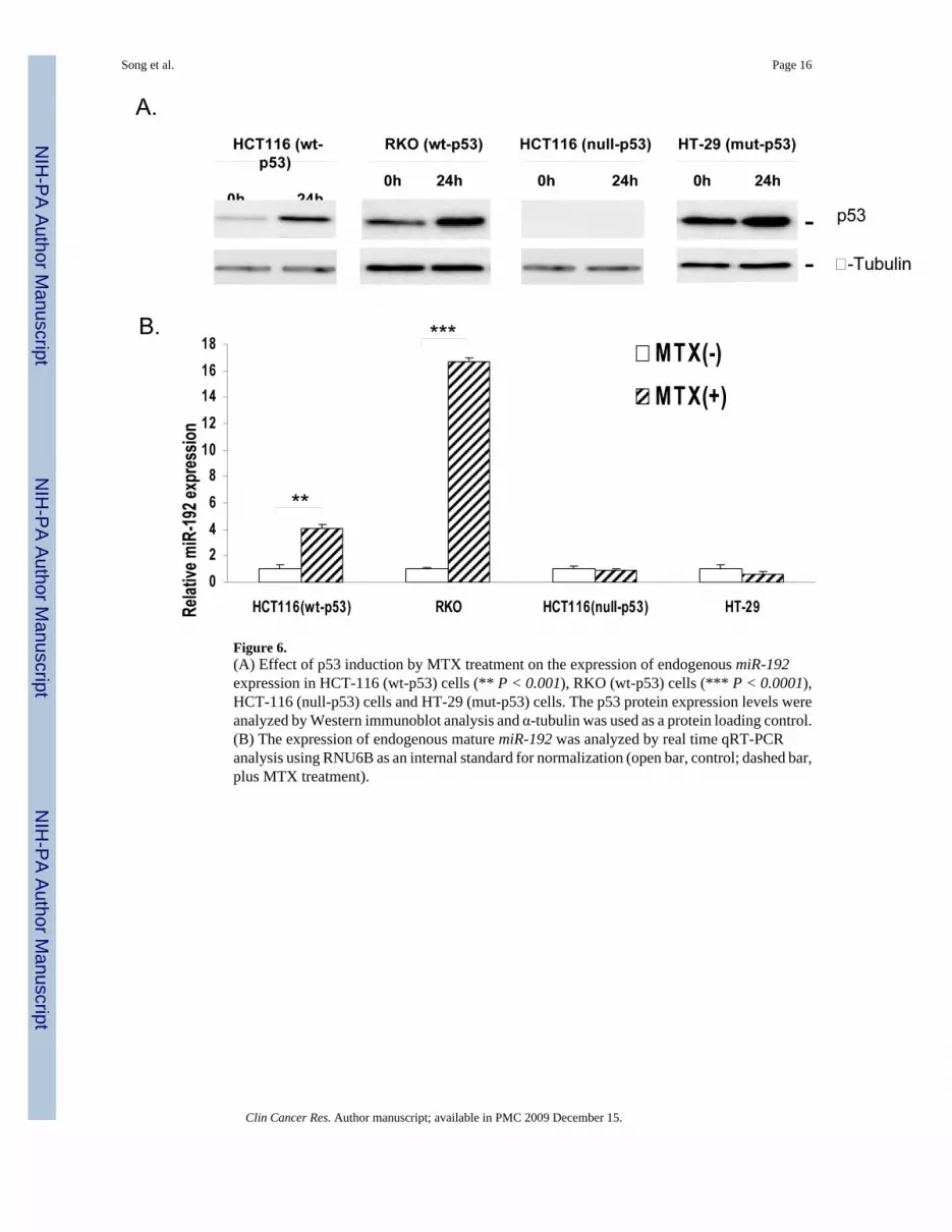
Figure 6.(A) Effect of p53 induction by MTX treatment on the expression of endogenous miR-192expression in HCT-116 (wt-p53) cells (** P < 0.001), RKO (wt-p53) cells (*** P < 0.0001),HCT-116 (null-p53) cells and HT-29 (mut-p53) cells. The p53 protein expression levels wereanalyzed by Western immunoblot analysis and α-tubulin was used as a protein loading control.(B) The expression of endogenous mature miR-192 was analyzed by real time qRT-PCRanalysis using RNU6B as an internal standard for normalization (open bar, control; dashed bar,plus MTX treatment).
Song et al. Page 16
Clin Cancer Res. Author manuscript; available in PMC 2009 December 15.
NIH
-PA Author Manuscript
NIH
-PA Author Manuscript
NIH
-PA Author Manuscript